Evidence of Recombination in Intrapatient Populations of Hepatitis C Virus
16
Evidence of Recombination in Intrapatient Populations of Hepatitis C Virus Vicente Sentandreu 1 , Nuria Jime ´ nez-Herna ´ ndez 1,2 , Manuela Torres-Puente 1 , Marı ´a Alma Bracho 1,2 , Ana Valero 1 , Marı´a Jose ´ Gosalbes 1,2 , Enrique Ortega 3 , Andre ´ s Moya 1,2 , Fernando Gonza ´ lez-Candelas 1,2 * 1 Institut Cavanilles de Biodiversitat i Biologia Evolutiva, Universitat de Vale `ncia Valencia, Spain, 2 CIBER Epidemiologı ´a y Salud Pu ´ blica (CIBERESP), Barcelona, Spain, 3 Unidad de Enfermedades Infecciosas, Hospital General Universitario, Valencia, Spain Abstract Hepatitis C virus (HCV) is a major cause of liver disease worldwide and a potential cause of substantial morbidity and mortality in the future. HCV is characterized by a high level of genetic heterogeneity. Although homologous recombination has been demonstrated in many members of the family Flaviviridae, to which HCV belongs, there are only a few studies reporting recombination on natural populations of HCV, suggesting that these events are rare in vivo. Furthermore, these few studies have focused on recombination between different HCV genotypes/subtypes but there are no reports on the extent of intra-genotype or intra-subtype recombination between viral strains infecting the same patient. Given the important implications of recombination for RNA virus evolution, our aim in this study has been to assess the existence and eventually the frequency of intragenic recombination on HCV. For this, we retrospectively have analyzed two regions of the HCV genome (NS5A and E1-E2) in samples from two different groups: (i) patients infected only with HCV (either treated with interferon plus ribavirin or treatment naı ¨ve), and (ii) HCV-HIV co-infected patients (with and without treatment against HIV). The complete data set comprised 17712 sequences from 136 serum samples derived from 111 patients. Recombination analyses were performed using 6 different methods implemented in the program RDP3. Recombination events were considered when detected by at least 3 of the 6 methods used and were identified in 10.7% of the amplified samples, distributed throughout all the groups described and the two genomic regions studied. The resulting recombination events were further verified by detailed phylogenetic analyses. The complete experimental procedure was applied to an artificial mixture of relatively closely viral populations and the ensuing analyses failed to reveal artifactual recombination. From these results we conclude that recombination should be considered as a potentially relevant mechanism generating genetic variation in HCV and with important implications for the treatment of this infection. Citation: Sentandreu V, Jime ´ nez-Herna ´ndez N, Torres-Puente M, Bracho MA, Valero A, et al. (2008) Evidence of Recombination in Intrapatient Populations of Hepatitis C Virus. PLoS ONE 3(9): e3239. doi:10.1371/journal.pone.0003239 Editor: Nina Papavasiliou, The Rockefeller University, United States of America Received December 4, 2007; Accepted August 3, 2008; Published September 18, 2008 Copyright: ß 2008 Sentandreu et al. This is an open-access article distributed under the terms of the Creative Commons Attribution License, which permits unrestricted use, distribution, and reproduction in any medium, provided the original author and source are credited. Funding: This work was supported by the Conselleria de Sanitat i Consum Generalitat Valenciana (Spain) and project BFU2005 00503 from Ministerio de Educacion y Ciencia. Competing Interests: The authors have declared that no competing interests exist. * E-mail: [email protected] Introduction Hepatitis C virus (HCV) infection affects about 170 million people worldwide, about 3% of the world’s population [1], and is the major cause of liver disease and a potential cause of substantial morbidity and mortality in the future [2]. Hepatitis C virus is the only species of the genus Hepacivirus within the family Flaviviridae. It has a single stranded, positive-sense, nonsegmented RNA genome of about 9600 nucleotides (nt) with a single, long open reading frame encoding a polyprotein of about 3000 amino acids with the gene order C-E1-E2-p7-NS2-NS3-NS4A-NS4B-NS5A-NS5B. The structural proteins are C (core) and E1 and E2 (envelope glycoproteins). The function of the p7 product is presently unknown. The NS2 through NS5 regions encode the non- structural proteins [3]. Six major HCV genotypes and about 50 subtypes have been described [4,5] based on levels of sequence divergence. HCV genotypes have been shown to be distributed over distinct geographical areas and although they share most basic biological features, there seems to be some differences in their susceptibility to interferon (IFN)-based therapies [6,7]. Genotype 1 is the predominant variant in developed countries and shows the poorest response to therapy. Patients with genotypes 2 and 3 are also common in Europe, although with a lower frequency than genotype 1, and show the best response to IFN therapy. HCV is mainly transmitted by parenteral routes and differences in their transmission rates can be an important factor to explain the differences in prevalence of a genotype/subtype in different geographic regions [8–10]. Needle sharing among intravenous drug users (IDUs) currently represents the most common route of acquisition of HCV in the developed world [11]. Because HCV and human immunodeficiency virus (HIV) share blood-borne transmission routes, HIV/HCV co-infection is relatively frequent, especially in regions such as Spain, where the major proportion of newly diagnosed AIDS patients belong to the IDU category (44.2%) [12]. Like HIV-1, HCV is characterized by high levels of genetic heterogeneity [13,14] which impact heavily on different aspects such as HCV persistence, susceptibility to treatment, progression of infection, among others [15–17]. Although it is well known that recombinant forms of HIV-1 have a relatively high prevalence all over the world [18], there has been limited evidence of HCV PLoS ONE | www.plosone.org 1 September 2008 | Volume 3 | Issue 9 | e3239
Transcript of Evidence of Recombination in Intrapatient Populations of Hepatitis C Virus
Evidence of Recombination in Intrapatient Populationsof Hepatitis C VirusVicente Sentandreu1, Nuria Jimenez-Hernandez1,2, Manuela Torres-Puente1, Marıa Alma Bracho1,2, Ana
Valero1, Marıa Jose Gosalbes1,2, Enrique Ortega3, Andres Moya1,2, Fernando Gonzalez-Candelas1,2*
1 Institut Cavanilles de Biodiversitat i Biologia Evolutiva, Universitat de Valencia Valencia, Spain, 2 CIBER Epidemiologıa y Salud Publica (CIBERESP), Barcelona, Spain,
3 Unidad de Enfermedades Infecciosas, Hospital General Universitario, Valencia, Spain
Abstract
Hepatitis C virus (HCV) is a major cause of liver disease worldwide and a potential cause of substantial morbidity andmortality in the future. HCV is characterized by a high level of genetic heterogeneity. Although homologous recombinationhas been demonstrated in many members of the family Flaviviridae, to which HCV belongs, there are only a few studiesreporting recombination on natural populations of HCV, suggesting that these events are rare in vivo. Furthermore, thesefew studies have focused on recombination between different HCV genotypes/subtypes but there are no reports on theextent of intra-genotype or intra-subtype recombination between viral strains infecting the same patient. Given theimportant implications of recombination for RNA virus evolution, our aim in this study has been to assess the existence andeventually the frequency of intragenic recombination on HCV. For this, we retrospectively have analyzed two regions of theHCV genome (NS5A and E1-E2) in samples from two different groups: (i) patients infected only with HCV (either treated withinterferon plus ribavirin or treatment naıve), and (ii) HCV-HIV co-infected patients (with and without treatment against HIV).The complete data set comprised 17712 sequences from 136 serum samples derived from 111 patients. Recombinationanalyses were performed using 6 different methods implemented in the program RDP3. Recombination events wereconsidered when detected by at least 3 of the 6 methods used and were identified in 10.7% of the amplified samples,distributed throughout all the groups described and the two genomic regions studied. The resulting recombination eventswere further verified by detailed phylogenetic analyses. The complete experimental procedure was applied to an artificialmixture of relatively closely viral populations and the ensuing analyses failed to reveal artifactual recombination. From theseresults we conclude that recombination should be considered as a potentially relevant mechanism generating geneticvariation in HCV and with important implications for the treatment of this infection.
Citation: Sentandreu V, Jimenez-Hernandez N, Torres-Puente M, Bracho MA, Valero A, et al. (2008) Evidence of Recombination in Intrapatient Populations ofHepatitis C Virus. PLoS ONE 3(9): e3239. doi:10.1371/journal.pone.0003239
Editor: Nina Papavasiliou, The Rockefeller University, United States of America
Received December 4, 2007; Accepted August 3, 2008; Published September 18, 2008
Copyright: � 2008 Sentandreu et al. This is an open-access article distributed under the terms of the Creative Commons Attribution License, which permitsunrestricted use, distribution, and reproduction in any medium, provided the original author and source are credited.
Funding: This work was supported by the Conselleria de Sanitat i Consum Generalitat Valenciana (Spain) and project BFU2005 00503 from Ministerio deEducacion y Ciencia.
Competing Interests: The authors have declared that no competing interests exist.
* E-mail: [email protected]
Introduction
Hepatitis C virus (HCV) infection affects about 170 million
people worldwide, about 3% of the world’s population [1], and is
the major cause of liver disease and a potential cause of substantial
morbidity and mortality in the future [2]. Hepatitis C virus is the
only species of the genus Hepacivirus within the family Flaviviridae. It
has a single stranded, positive-sense, nonsegmented RNA genome
of about 9600 nucleotides (nt) with a single, long open reading
frame encoding a polyprotein of about 3000 amino acids with the
gene order C-E1-E2-p7-NS2-NS3-NS4A-NS4B-NS5A-NS5B.
The structural proteins are C (core) and E1 and E2 (envelope
glycoproteins). The function of the p7 product is presently
unknown. The NS2 through NS5 regions encode the non-
structural proteins [3].
Six major HCV genotypes and about 50 subtypes have been
described [4,5] based on levels of sequence divergence. HCV
genotypes have been shown to be distributed over distinct
geographical areas and although they share most basic biological
features, there seems to be some differences in their susceptibility
to interferon (IFN)-based therapies [6,7]. Genotype 1 is the
predominant variant in developed countries and shows the poorest
response to therapy. Patients with genotypes 2 and 3 are also
common in Europe, although with a lower frequency than
genotype 1, and show the best response to IFN therapy.
HCV is mainly transmitted by parenteral routes and differences
in their transmission rates can be an important factor to explain
the differences in prevalence of a genotype/subtype in different
geographic regions [8–10]. Needle sharing among intravenous
drug users (IDUs) currently represents the most common route of
acquisition of HCV in the developed world [11]. Because HCV
and human immunodeficiency virus (HIV) share blood-borne
transmission routes, HIV/HCV co-infection is relatively frequent,
especially in regions such as Spain, where the major proportion of
newly diagnosed AIDS patients belong to the IDU category
(44.2%) [12].
Like HIV-1, HCV is characterized by high levels of genetic
heterogeneity [13,14] which impact heavily on different aspects
such as HCV persistence, susceptibility to treatment, progression
of infection, among others [15–17]. Although it is well known that
recombinant forms of HIV-1 have a relatively high prevalence all
over the world [18], there has been limited evidence of HCV
PLoS ONE | www.plosone.org 1 September 2008 | Volume 3 | Issue 9 | e3239

recombination between different genotypes/subtypes [19], sug-
gesting that these events are rare in vivo and that the resulting
recombinants are usually not viable [14,20,21]. In the last few
years, a few natural intergenotypic recombinants of HCV have
been identified (RF1_2k/1b, RF2_1a/1b and RF3_2b/1b) and
the crossover points have been mapped to the NS2, NS5B and
NS3 regions, respectively [22–25]. Very recently, a new natural
intergenotypic (2/5) recombinant of HCV has been found whose
crossover point is located between genes NS2 and NS3 [26]. All
these reports have described HCV recombination between
different genotypes/subtypes but, to date, there is only a single
case of an HCV intra-subtype recombinant strain, detected by
analysis of NS5A sequences from intra-patient populations
belonging to six patients undergoing anti-viral therapy [27].
Given the important role that recombination seems to play in
the evolution of RNA viruses [28,29], by creating genetic
variation, and the important implications that the production of
new pathogenic recombinant strains could have, for example, on
the development of vaccines to control RNA viruses, our aim in
this study has been to assess the extent and, eventually, the
frequency of intragenic recombination on HCV. For this, we have
retrospectively analyzed a large data set (over 17700) of HCV
sequences from intra-patient viral populations. These sequences
were obtained from two separate studies of our group none of
which was specifically designed for this objective [10, 78, 79, and
unpublished results]. One included only HCV-monoinfected
patients and the other HCV/HIV coinfected patients, with a
common genome region for both studies (E1-E2 region) and
another, the NS5A region, analyzed only in the former study. Both
studies included treatment-naıve and patients non-responding to
antiviral treatment. We found evidence of intrapatient recombi-
nation in HCV sequences from over 10% of the patients thus
revealing that recombination in HCV can be a much more
common phenomenon than previously recognized. The possibility
of this result arising from artifacts during the experimental
procedure has been considered by performing an ‘‘ad hoc’’
experiment in which serum samples from two closely related, but
clearly differentiated patients were mixed in equal proportions and
the resulting mixture was subjected to the same experimental
procedure used in the previous analyses. No evidence of artifactual
recombination was found.
Results
Sequences used in this work were derived from two previous
studies on HCV genetic variation before and after antiviral
treatment. One study included HCV-monoinfected patients
whereas the other analyzed HCV/HIV-coinfected individuals.
None of these studies was designed with the goal of detecting
recombination in HCV. In both cases the E1-E2 region of the
HCV genome was analyzed by sequencing viral clones and the
former study also included clones from the NS5A region. We
obtained sequences from the E1-E2 region from 110 patients and
a total of 136 samples (Table 1), since two samples (before and
after antiviral treatment) were available for 26 patients, all from
the HCV-monoinfection study. For the NS5A region we obtained
cloned sequences from 78 patients and a total of 98 samples, since
amplification failed for 4 and 1 samples from the pre- and post-
treatment groups, respectively.
The average number of clones sequenced for each region, group
and patient is described in Table 1. For the E1-E2 region, 11746
cloned sequences were obtained and average number of
sequenced clones per sample for the treatment-naıve mono-
infected group (HCV 0) was 105.74621.43 whereas the average
for HCV treated mono-infected patients who did not respond to a
combined antiviral treatment with IFN-a plus ribavirin (HCV T)
was 106.1565.24. In the case of HCV/HIV coinfected patients,
an average of 28.6963.86 and 28.8867.32 E1-E2 viral sequences
were obtained for treatment naıve (HCV 0-0) and HIV treated
patients (HCV 0-T), respectively. The data set for the NS5A
region comprised 5966 clonal sequences. The average number of
NS5A clones sequenced per sample for the non-treated group
(HCV 0) was 63.33618.51 and 53.72618.00 for the antiviral
treated group (HCV T).
Putative recombination events between the viral strains
infecting the same patient were found in 20 of the 111 patients
studied (18.01%). These intragenic recombination events were
detected in 25 of the 234 independent samples analyzed (10.7%).
The detected recombination events belonged to HCV samples
from all the infection groups described and the two genomic
regions analyzed (E1-E2 and NS5A), as well as to different HCV
subtypes (1a, 1b and 3a). Five events were detected among the E1-
E2 sequences derived from HCV/HIV coinfected patients, 11
corresponded to events in the E1-E2 region from HCV-
Table 1. Summary of patients, sequence data sets and recombination analysis results.
Source Patients GroupAmplifiedsamples
Average sequence/sample SD Total seqs
Positivecases Freq.
E1-E2 region HCV 78 HCV0 77 105.74 21.43 8142 7 0.091
HCVT 26 106.15 5.24 2680 4 0.154
HCV-HIV 16 HCV0-0 16 28.69 3.86 433 2 0.125
17 HCV0-T 17 28.88 7.32 491 3 0.176
TOTAL 111 136 11746 16 0.118
NS5A region HCV 78 HCV0 73 63.33 18.51 4623 7 0.096
HCVT 25 53.72 18.00 1343 2 0.080
TOTAL 78 98 5966 9 0.091
The ‘‘source’’ column represents the study (HCV-monoinfection, HCV/HIV-coinfection) in which samples were obtained originally. For the HCV-monoinfection study,samples were obtained from 78 patients and the numbers indicated in the ‘‘amplified samples’’ column yielded successful amplificates before (HCV0) and after (HCVT)antiviral treatment. For the HCV/HIV study, the two groups correspond to patients without any treatment (HCV0-0) or having been treated for HIV infection (HCV0-T). SDindicates standard deviation of the number of sequences obtained from each sample and ‘‘Freq.’’ denotes the frequency of samples in which at least one recombinationevent has been detected.doi:10.1371/journal.pone.0003239.t001
Intrapatient Recombination HCV
PLoS ONE | www.plosone.org 2 September 2008 | Volume 3 | Issue 9 | e3239

monoinfected patients, and the remaining 9 were detected in the
NS5A region. No differences between the recombination frequen-
cies from the two genomic regions analyzed were found neither for
treatment-naıve (Mann-Whitney test: z = 20.104, p-value = 0.917)
nor for interferon plus ribavirin treated groups (Mann-Whitney
test: z = 20.810, p-value = 0.418). Figures 1–4 show in detail the
results of recombination analyses only for samples in which the
presence of putatively significant recombination events was
detected. Intragenic recombination analyses were performed for
each locus independently using 6 different methods for the
detection of recombination implemented in the program RDP 3.0.
We considered as significant recombination events only those for
which the corrected probability for simultaneous inference of the
event was lower than 0.05 and were significantly detected by at
least 3 of the 6 methods used.
A large proportion (84%, 21/25) of the samples in which
recombination was detected included more than one recombina-
tion event (Figures 1–4). Each recombination event detected in a
sample was described according to the following features: i)
average p-value for the event and analysis method, ii) most likely
parental and daughter sequences as well as similarity among them,
and iii) most likely limits for the location of breakpoint(s) in the
sequenced fragment.
We found 46 single recombination events (17 in the NS5A and
29 in the E1-E2 region) and 12 double recombination events (3 in
the NS5A and 9 in the E1-E2 region) in the 25 alignments where
recombination was detected. A single recombination event was
defined by a single, significant breakpoint detected in the daughter
sequence following the described methodology, while a double
event was defined as two significant breakpoints identified in the
Figure 1. Summary of positive recombination results in the E1-E2 region (HCV-monoinfected patients). The columns represent: i)patient group before (HCV 0) or after (HCV T) antiviral treatment; ii) patient code; iii) amplified sequences for this patient and region; iv) HCVgenotype; v) p-values for the different recombination detection methods implemented in RDP 3.0 (1 = RDP, 2 = Geneconv, 3 = Bootscan, 4 = Maxchi,5 = Chimera, and 6 = Siscan) using the following color coding: non-significant p-values (white filling), p,0.05 (grey) and p,0.01 (black); vi)recombination event number; vii) Bkpt 1, location range for breakpoint 1; viii) Bkpt 2, location range for breakpoint 2; ix) parent1 sequence (%similarity to daughter sequence); x) parent2 sequences(% similarity to daughter sequence); and xi) daughter sequence(s). Only events detected assignificant by at least three methods are shown.doi:10.1371/journal.pone.0003239.g001
Intrapatient Recombination HCV
PLoS ONE | www.plosone.org 3 September 2008 | Volume 3 | Issue 9 | e3239

same daughter sequence, delimiting the extension of the
recombinant fragment. When one of the parental sequences
implicated in an event could not be inferred within the sequence
alignment, it was denoted as ‘‘unknown parental’’.
Among the methods used, those detecting a larger number of
significant events were Siscan, Chimera, and Maxchi, followed by
Genconv and the phylogenetic methods, Bootscan and RDP,
which detected very few events.
Maximum likelihood phylogenetic trees were constructed for
the two regions delimited for each single breakpoint event
identified. For the double events detected, three maximum
likelihood phylogenetic trees were constructed; one was derived
from the alignment for the region involved in the recombinant
segment (delimited by the two breakpoints detected) and the others
from the two resulting flanking segments. Two different tests,
Shimodaira–Hasegawa (SH) and Expected Likelihood Weight
(ELW), were applied to the two or three resulting topologies for
each single or double event, respectively, to further verify the
results obtained with RDP3. Tests for double events were made
between the recombinant segment topology and each of the two
topologies derived from the flanking segments independently.
These tests resulted in significant differences between both
topologies in all single and double event cases (detailed results of
the tests are available in the Supporting Information Table S2).
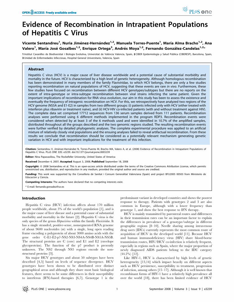
Recombination in the E1-E2 RegionStatistically significant recombination events were detected by at
least three methods in 11.8% (16/136) of the amplified samples in
the E1-E2 region. The total number of events identified in these
recombinant samples was 38, corresponding to 29 single and 9
double events, belonging to 15 patients from all the studied groups.
The breakpoints detected were located mainly in the segments
flanking the HVR1 (Figure 5). No differences in the distribution of
breakpoints were found between the different studied groups, viral
subtypes or treatment response. The frequency of double
recombination events detected in this region was 23.7% (9/38)
and the average length of the derived recombinant fragments was
147.5 nt (ranging from 66.5 to 212 nt), representing between
27.2% and 31.2% of the total region sequenced (543 nt for HCV/
HIV co-infected and 472 nt for HCV single-infected patients). In
8 of the 9 double recombination event cases the derived
recombinant regions comprised a large portion of the complete
HVR1, while in the remaining case the recombinant fragment
involved the entire HVR3 region (Figure 5).
In the analysis of HCV-monoinfected samples (103 E1-E2
amplified samples from 78 patients), the frequency of significant
recombination cases was 9.1% (7/77) for treatment-naıve patients
(HCV 0) and 15.4% (4/26) for IFN-a plus ribavirin treated patients
(HCV T, Table 1). All samples in which recombination events were
detected were derived from patients infected with genotype 1b of
HCV, including both responders and non-responders to antiviral
treatment, with the only exception of a sample from an untreated
patient infected with genotype 1a. The frequencies of samples with
detected recombination events among coinfected patients were
12.5% (2/16) and 17.64% (3/17) for treatment-naıve (HCV 0-0)
and HAART-treated (HCV 0-T) patients, respectively (Table 1).
Figure 2. Summary of positive recombination results in the E1-E2 region (HCV-HIV coinfected patients). See further details in legend toFigure 1.doi:10.1371/journal.pone.0003239.g002
Intrapatient Recombination HCV
PLoS ONE | www.plosone.org 4 September 2008 | Volume 3 | Issue 9 | e3239

These five samples were obtained from individuals infected with
genotype 3a (3 patients) and genotype 1b (2 patients) of HCV. No
significant differences were found in the frequencies of recombina-
tion-positive samples from naıve and treated groups for HCV mono-
infected and HCV/HIV co-infected patients (Mann-Whitney test for
single infected patients: z = 20.894 p-value = 0.371; and z = 20.406
and p-value = 0.685, for co-infected patients), although a larger
proportion of recombination events was detected among samples
from treated patients both in single-infected and in co-infected
individuals. Similarly, no differences were found between HCV/HIV
co-infected and HCV single infected, treatment-naıve groups
(z = 20.417, p-value = 0.676). Complete Mann-Whitney tests results
are available in Table S3 of the Supporting Information.
Figure 6 shows an example of a recombination event detected
by phylogenetic analysis in the E1-E2 region reflected in the
incongruence (reciprocal tests with SH and ELW were highly
significant, see Supplementary material) between the maximum
likelihood trees derived from the two regions (delimited by
nucleotides 1–264 and 265–534, respectively) defined by the
breakpoints assigned to the recombination event from sample
EC5703. Variable positions in the alignment of the viral sequences
involved in this recombination event are shown in Figure 7,
marking the recombinant parental sequences involved in the
detected recombination event.
E1-E2 Region. Analysis of Two Time-Point SamplesTwo samples were available for 24 HCV-monoinfected patients
who did not respond to antiviral treatment. One sample was taken
before the onset of treatment (T0 sample) and the second one was
obtained when it was discontinued, 6 or 12 months later (T1
sample). For these cases an additional analysis was performed by
simultaneously considering all the sequences obtained from both
samples. We detected significant recombination events in 4
patients, C29, A21, G16 and G26.
One recombination event was detected in the joint analysis of
the two samples from patient C29 (C29T0-T1).The parental
sequences from this event were derived one from the T0 sample
(before treatment) and the other from the T1 sample (taken 6
months later), while the daughter sequences were detected only in
the T0 sample. The same event, involving the same daughter
sequences, was also detected in the analysis of sequences obtained
only from the T0 sample, although in this case only one parental
sequence was identified while the other was described as
‘‘unknown parental’’ in the analysis (Figure 1).
Similarly, in the joint analysis of the two samples from patient
A21, we detected one significant recombination event whose
corresponding breakpoint was located between positions 210 and
214. This event was originated by two parental sequences from the
T0 sample while the resulting daughter sequences from such event
(A21_T1-47 and A21_T1-80) corresponded to sequences from the
sample obtained after six months of unsuccessful HCV antiviral
treatment (Figure 4). The same event was detected in the analysis
of sequences derived only from the T1 sample but, obviously, the
identified parental sequences belonged also to this sample.
However, when the parental similarities for both events (the one
detected only with T1 sequences and the other joining T0 and T1
Figure 3. Summary of positive recombination results in the NS5A region. See further details in legend to Figure 1.doi:10.1371/journal.pone.0003239.g003
Intrapatient Recombination HCV
PLoS ONE | www.plosone.org 5 September 2008 | Volume 3 | Issue 9 | e3239

sequences) were compared, the similarity between daughter and
the corresponding parental sequences for both cases were lower in
the T1 sample (97.4 and 99.4% for the two putative parental
sequences) than in the joint T0-T1 analysis (98.4 and 100%). In
consequence, T1 daughter sequences derived from this recombi-
nation event had been more likely generated by parental
sequences from the T0 sample than from those in the T1 one.
This would imply that the recombination event occurred at the
earlier time point and the resulting daughter sequences were able
to persist in the viral population six months later and under
antiviral treatment. Finally, in the case of patients G16 and G26,
all significant events detected in the joint analysis involved
sequences from the same time-point and were also detected in
the analysis of only this sample.
Recombination in the NS5A RegionSignificant events of recombination were detected by at least three
methods in 9.18% (9/98) of the amplified samples for the NS5A
region. The total number of recombination events detected in these
samples was 20, with 17 single and 3 double events, and they were
identified in samples from 8 patients, all infected with HCV genotype
1b, including treatment-naıve and non-responder patients. The
breakpoint(s) detected for each event were located mainly in a
segment flanking the PKR-BD (protein-kinase binding domain) and
within the ISDR (interferon sensitivity-determining region) regions
(Figure 8). The frequency of double events detected in this region was
15% (3/20) and the average recombinant fragment length was 315 nt
(ranging from 305 to 337 nt) corresponding to the 42.4% of the total
sequenced region (743 nt). In 2 of the 3 double event cases, the
recombinant region comprised the complete PKR-BD region,
including also the ISDR, while in the other case the recombinant
region involved only the non ISDR fragment of PKR-BD region
(Figure 8). No differences were observed in the distribution of
breakpoints between treated and non-treated samples or between
treatment responses. Similar proportions of recombination events
were detected in samples from treatment-naıve patients (9.6%, 7/73)
than from IFN-a plus ribavirin treated patients (8.3%, 2/24) and no
significant differences due to the HCV treatment were found
(z = 20.236 p-value = 0.813). Figure 9 presents an example of
recombination event detected in the NS5A region from the T0
sample of patient G08. In this case, the event corresponded to a single
recombination breakpoint located between positions 345 and 420 of
this fragment. Maximum likelihood trees obtained for each region
using the GTR model of evolution showed a clear phylogenetic
incongruence between the two derived region trees (Table S2 and
File S1 in Supplementary material). Variable positions in the
alignment of the viral sequences involved in this recombination
event are shown in Figure 10.
Figure 4. Summary of the joint analysis of two time-point samples from the same patients resulting in positive detection ofrecombination. See further details in legend to Figure 1.doi:10.1371/journal.pone.0003239.g004
Intrapatient Recombination HCV
PLoS ONE | www.plosone.org 6 September 2008 | Volume 3 | Issue 9 | e3239
NS5A Region: Analysis of Two Time-Points SamplesTwo samples, one taken before (T0) and the other after
unsuccessful antiviral treatment (6 or 12 months later, T1 or T2),
were available for 24 HCV-monoinfected patients. The joint
analysis of sequences from the two samples of each patient resulted
in the detection of recombination events in only two cases (8.33%),
for patients C29 and G07.
The joint analysis of samples from patient C29 revealed several
recombination events that involved parental sequences from
samples T0 and T1 and daughter sequences only from the T0
sample. All these events were also detected in the analysis of the
T0 sample but, again, with presumed parental sequences from this
sample time point.
For patient G07, different recombination events in the joint
analysis of sequences from samples T0 and T2 were found. Two of
these events were particularly interesting because they involved
daughter sequences from the T2 sample with inferred parental
sequences from the T0 sample which was obtained one year earlier.
These parental sequences (T0-66 and T0-55) were involved in other
recombination events detected in the T0 sample and the T2
resulting daughters (except T2-52, only detected in the joint
analysis), T2-58 and T2-76 were detected in the T2 sample too, but
parental inferred sequences for these events had lower similarity or
were unknown (see Figures 3 and 4). Similarly to the previously
commented case of E1-E2 sequences from two time-points samples
of patient A21, the T2 daughter sequences obtained from this event
were more likely generated by parental sequences from the T0
sample. Again, this indicates that the recombination event most
likely took place at T0 and the resulting daughter sequences were
able to persist for one year under HCV treatment.
Joint Analysis of Recombination in the E1-E2 and NS5ARegions
We obtained sequences for the E1-E2 and NS5A regions from
73 patients. These were all from the HCV monoinfected, non-
treated group (HCV 0). Given the observed frequency of
recombination events in the two analyzed regions, we calculated
the expected probability of detecting recombination events
simultaneously in both genome regions for the same patient.
The frequency of samples with significant recombination events
detected was 9.1% (7/77) and 9.6% (7/73), for the E1-E2 and
NS5A regions, respectively (Table 1). Hence, the expected
frequency of obtaining a patient with recombination events
detected in the two genomic regions if these events were
independent was 0.87%, whereas the observed frequency was
4.1% (3/73), 4.45 times higher than the expected value. Fisher’s
exact probability test resulted in a significant value with p = 0.03.
We have also compared the frequencies of the recombinant
sequences with those of the other sequence clones. A summary of
these results is shown in Table 2. While the frequencies of detected
recombinant sequences varied widely, from a minimum of 0.003
to a maximum of 0.516, none of the recombinant sequences was
present in more than 2 copies in the corresponding population,
which corresponded to frequencies from 0.001 up to 0.074.
Nevertheless, these figures must be compared with those
corresponding to the frequency of the most common variant in
the corresponding population, which varied between 0.030 and
0.20. In the joint analyses of two samples from the same patient in
which recombinant sequences were detected, the highest frequen-
cy of a recombinant haplotype was 0.010. The most common
sequence in this patient was only three times larger (0.031) but the
largest number of identical sequences (111) and the highest
haplotype frequency (0.547) were found in this group.
Experimental Analysis of Artifactual RecombinationIn order to evaluate the level and extent of artifactual
generation of recombinant sequences during the reverse tran-
scription and PCR amplification, we performed an additional
experiment using the same conditions previously described. The
only difference was the starting sample used which, in this case,
Figure 5. Location of recombination breakpoints detected in the E1E2region.doi:10.1371/journal.pone.0003239.g005
Intrapatient Recombination HCV
PLoS ONE | www.plosone.org 7 September 2008 | Volume 3 | Issue 9 | e3239

was a mixture of sera derived from two patients. These were
chosen to maximize the possibility of detection of recombinant
sequences, which requires clearly differentiated viral populations,
while simultaneously maximizing the possibility of contiguous
pairing enabling RNA polymerase shifting from one template to
another while keeping the same position and reading-frame. This
was achieved by selecting two patients HCV-1b infected from a
common source who had within-patient nucleotide diversities of
0.0046 and 0.0007, respectively, with a net nucleotide differen-
tiation of 0.0247, as estimated from a previous analysis of 10 clones
from each sample (unpublished results).
To ensure equimolar amounts of viral RNAs from both samples
in the final mixture, we set up several mixtures with varying
amounts of each sample that were subjected to the same RT and
PCR-amplification procedures for the E1-E2 region described
above. The resulting amplificates were directly sequenced and one
mixture with equal peak heights in the automated sequencer
electrophoregram in the polymorphic positions was chosen for
further analysis. We cloned and sequenced PCR products from the
selected mixture as described, obtaining 142 sequences. These were
analyzed using the 6 methods implemented in RDP as described
above and none of them detected any putative recombination event.
Figure 6. Example of recombination detection in the E1E2 region (EC5703 sample). The phylogenetic tree on the left corresponds to theanalysis of the complete region whereas the other two are derived from the two regions defined by the detected recombination event.doi:10.1371/journal.pone.0003239.g006
Figure 7. Example of recombination detection in the E1E2 region (EC5703 sample). Variable nucleotide positions in the daughter (EC5703-46) and two parental sequences (EC5703-34 and EC5703-36).doi:10.1371/journal.pone.0003239.g007
Intrapatient Recombination HCV
PLoS ONE | www.plosone.org 8 September 2008 | Volume 3 | Issue 9 | e3239

Discussion
Several studies have reported recombination in different
Flaviviruses [30–33], but until recently no evidence for recombi-
nation in natural populations of HCV had been found. Since the
first identification of an intergenotypic (2k/1b) HCV recombinant
in St. Petersburg [22], several intergenotypic and intragenotypic
HCV recombinant strains have been identified [23–26,34,35]
therefore incorporating recombination as a mechanism generating
genetic variation in HCV. More importantly, the identification of
these recombinant strains demonstrates that HCV is capable of
successfully completing all the stages (simultaneous infection of the
same cell, simultaneous replication of both viral genomes, template
shift by the viral RNA polymerase while keeping the correct
reading frame, encapsidation and release of the recombinant
genomes) in the process. The resulting products will then be
subjected to the same population processes governing the
maintenance, expansion or disappearance of new variants in a
heterogeneous viral population.
All these reports have focused on HCV recombination between
different genotypes/subtypes, but to date, there is only one single
case of putative HCV intra-subtype recombinant strain, detected
by the analysis of NS5A sequences from six intra-patient
populations undergoing antiviral therapy [27]. In the present
study we have identified a high frequency of intrasubtype
recombination events (18.01% of the analyzed patients 20/111)
analyzing a large data set of HCV sequences from intra-patient
viral populations obtained from patients belonging to different
groups: HCV-monoinfected patients, naıve and non-responding to
antiviral treatment, and HCV/HIV-coinfected patients, treat-
ment-naıve and under HAART. The relevance of recombination
in HCV for its long-term evolution and its incidence in different
aspects of HCV infection have not been explored yet, but these
findings support a potentially significant role for recombination in
the evolution of HCV by creating genetic variation through the
reshuffling of independently arisen variants.
Although these studies have firmly established the possibility of
recombination in HCV, as in other Flaviviruses, no general
mechanism has been proposed yet (but see below), and despite
extensive analysis of genetic variation in HCV there has been only
one report of recombination between strains from the same
subtype. Hence, it seems adequate to start discussing our findings
in terms of them being real or an artefact and, since we naturally
accept the first option, why it has been so difficult to detect.
The first question is how to discard the possibility that the
detected recombination events were false positives, resulting from
PCR-mediated recombination, especially since some experiments
have failed to experimentally induce and detect recombination in
HCV [36]. We have performed one further such experiment, also
with negative results. This is no proof of absence of artifacts in our
experimental results, but they clearly indicate that, if present,
recombinant sequences arising from the experimental procedures
cannot account for the reported results. This conclusion is based
on the following arguments, derived from considering different
possible estimates of the artifactual recombination rate when no
such event has been observed. If no false recombination event is
observed among 143 clones then the probability of any such event
must be lower than 1/143 (0.0075). With this upper limit, which
would be our worst case scenario, under a Poisson distribution we
would have expected to observe no false recombination events in
138.5 of the 234 independent samples analyzed with an average
size of 75 sequences each. The actual number of samples with no
recombinant sequences detected was 210; hence the presumed
rate of artifactual recombination must be lower than 0.0075. If we
had based our estimate of artifactual recombination rate on the
number of negative cases (210), the inferred rate using again a
Figure 8. Location of recombination breakpoints detected in the NS5a region.doi:10.1371/journal.pone.0003239.g008
Intrapatient Recombination HCV
PLoS ONE | www.plosone.org 9 September 2008 | Volume 3 | Issue 9 | e3239

Poisson distribution would be 0.0014. But with this rate it is not
possible to account for the observation of 21 cases with two or
more recombination events (Figures 1–3) nor of up to 9 events
when sequences from the same patient taken at two different times
were combined in a single analysis (Figure 4). Finally, should we
consider the 58 events observed among 17712 sequences analyzed
to be artifacts then we would expect 183 and 45 samples with none
and one recombination event, respectively, while we obtained 210
and 3 cases. The discrepancy for cases with 2 or more events is
even larger (5, 0.5 and 0, for 2, 3 and 4 events, while 13, 4 and 4
have been observed, respectively). In summary, the observed
number and distribution of recombination events cannot be
explained by artifacts during the experiments. Additional argu-
ments for this conclusion are discussed next.
There are two points in our experimental procedure that have
been instrumental in obtaining the reported results and building
our confidence in that they are not artifacts. First, instead of
analyzing the full length master sequence of the viral distribution
in each sample, we concentrated our efforts in sequencing a large
number of clones in each of two genome regions. These were
chosen on the basis of their biological relevance and not on the
location of breakpoints identified in previous reports of recombi-
nation in HCV [23–26,34,35]. Secondly, we minimized the
chances of detecting false, artifactual recombination by using long
Figure 9. Example of recombination detection in the NS5a region (NG08T0 sample). The phylogenetic tree on the left corresponds to theanalysis of the complete region whereas the other two are derived from the two regions defined by the detected recombination event.doi:10.1371/journal.pone.0003239.g009
Figure 10. Example of recombination detection in the NS5a region (NG08T0 sample). Variable nucleotide positions in the daughter(NG08-47K) and two parental sequences (NG08-33K and NG08-63K).doi:10.1371/journal.pone.0003239.g010
Intrapatient Recombination HCV
PLoS ONE | www.plosone.org 10 September 2008 | Volume 3 | Issue 9 | e3239

extension times [37] and a proofreading DNA polymerase (Pfu)
[38] in our PCRs. Additional support for the actual occurrence of
recombination events within HCV infected patients is provided by
the following points: i) breakpoints or recombinant regions
implicated in the recombinant events are not distributed at
random along the analyzed genome regions (Figures 5 and 8). On
the contrary, the same breakpoints have been detected in HCV
samples from different patients, indicating the presence of
recombination hotspots, and entire biologically relevant regions
are comprised within the recombinant fragments detected. ii) We
have found evolution of the daughter sequences derived from a
recombination event, with similarity to the parental sequences
lower than the expected 100% if the recombination event was
PCR-mediated. In addition, all recombination generated sequenc-
es preserve the polyprotein reading frame, which is not expected in
PCR-mediated recombination where selection cannot purge
deleterious mutants. iii) We have detected several recombination
events in which the most likely parental sequences belonged to a
previous time sample. Through the joint analysis of sequences
from the two sample time points from a single patient, we have
identified recombinant daughter sequences resulting from the
cross-over between sequences sampled six or twelve months before
under HCV treatment (see Figure 4, results for A21_T0-T1 and
G07_T0-T2). Parental and recombinant sequences were amplified
Table 2. Summary of frequency analysis of recombinant sequences in the different data sets where intrapatient recombination ofHCV was detected.
Group PatientTotalSeqs.
Diff.Haplotypes
Numb. ofrecomb
Freq.recomb. Recombinants Non-recombinants
Seqs. Freq. Seqs. Freq.
NS5a region HCV 0 A28 55 53 2 0.036 1 0.018 2 0.036
C29 87 32 5 0.057 1 0.011 16 0.184
C35 55 53 4 0.073 1 0.018 2 0.036
C36 75 67 2 0.027 1 0.013 3 0.040
G05 62 50 3 0.048 2 0.032 3 0.048
G07 49 45 7 0.143 1 0.020 2 0.041
G08 55 32 2 0.036 1 0.018 17 0.309
HCV T C05T1 43 39 10 0.233 1 0.023 2 0.047
G07T2 49 45 13 0.265 1 0.020 2 0.041
E1-E2 region
HCV 0 A16 99 92 4 0.040 1 0.010 4 0.040
A21 100 72 2 0.020 1 0.010 14 0.140
A28 100 87 4 0.040 1 0.010 3 0.030
C28 99 77 2 0.020 1 0.010 3 0.030
C29 100 37 2 0.020 1 0.010 20 0.200
C32 99 80 4 0.040 1 0.010 5 0.051
G08 104 93 6 0.058 1 0.010 4 0.038
HCV T A21 108 84 5 0.046 2 0.019 6 0.056
G17 100 81 3 0.030 1 0.010 6 0.060
G16 100 79 3 0.030 1 0.010 6 0.060
G26 102 58 1 0.010 1 0.010 20 0.196
HCV 0-0 C06 25 21 10 0.400 2 0.080 3 0.120
V035 31 26 16 0.516 2 0.065 2 0.065
HCV 0-T C23 29 29 2 0.069 1 0.034 1 0.034
C30 27 19 6 0.222 2 0.074 3 0.111
C57 41 31 1 0.024 1 0.024 8 0.195
Two joint samples
NS5A C29T0-T1 147 59 4 0.027 1 0.007 34 0.231
G07T0-T2 98 89 13 0.133 1 0.010 3 0.031
E1-E2 A21T0-T1 209 156 2 0.010 2 0.010 14 0.067
C29T0-T1 200 71 3 0.015 1 0.005 20 0.100
G16T0-T1 203 82 2 0.010 1 0.005 111 0.547
G26T0-T1-T2 302 31 1 0.003 1 0.003 85 0.281
For each patient and sample(s) considered the table reports the total number of sequences analyzed, the number of different haplotypes, the total number ofrecombinant sequences and their frequency in the sample. Additional information is given on the absolute and relative frequencies of the most frequent haplotypeamong recombinant and non-recombinant sequences.doi:10.1371/journal.pone.0003239.t002
Intrapatient Recombination HCV
PLoS ONE | www.plosone.org 11 September 2008 | Volume 3 | Issue 9 | e3239

and sequenced independently from the two different samples.
Hence, these events probably occurred in the period between the
two samples, and therefore the recombinant sequences were able
to persist in the viral population under treatment and immuno-
logical selection pressures. iv) Finally, we have found patients with
evidence for recombination events in the two genome regions
analyzed 4.3 times more often than expected from the assumption
of complete independence between both events. In PCR-mediated
recombination we would expect a similar frequency of indepen-
dent recombination events in both regions, but the joint
expectation would be their product. A substantially larger value
such as that observed might indicate the existence of some
features, probably in the viral RNA-dependent RNA-polymerase
(protein NS5B in HCV) that might either facilitate or prevent
recombination, thus leading to the observed discrepancy.
Intergenotype recombination in HCV has been related to
homologous recombination during minus-strand synthesis via
template switching [23], although this proposal relies on the
presence of two hairpin structures in the vicinity of the inferred
breakpoint of recombination. Recombination breakpoints for the
E1-E2 region were mainly located in the conserved region
between hypervariable regions 1 (HVR1) and 3 (HVR3), and
the recombinant fragments from the double recombinant
sequences detected spanned the entire HVR1 or HVR3 regions.
For the NS5A region, breakpoints were concentrated mainly at the
end of the ISDR and the PKR-BD, and double recombinant
fragments comprised the entire ISDR or PKR-BD regions. This
distribution of breakpoints could be explained by the operation in
HCV, like in other DNA or RNA viruses, of an intermolecular
homologous replicative recombination system. This mechanism is
associated with extensive nucleotide sequence identity between the
two parental genomes around the cross-over site and copy choice
or template switching during the replication process, that involves
detachment from a template of the polymerase complex with a
nascent product, and continuation of the copying process at the
same position of another template molecule.
This is the first study where recombination in HCV has been
detected at the intrapatient intragenic level (the lowest possible
level of diversity) by analyzing a large number of HCV sequences
(17712) and samples (234) from 111 patients assigned to different
clinical groups. The detection of recombinant strains in 18% of the
HCV-infected patients studied implies that recombination events
between the viral strains infecting the same patient may be
relatively frequent, and still more if we consider that this might be
an underestimate of the true frequency of HCV recombination
because of the difficulty in detecting recombination events if they
occur between genetically very similar variants of the same
subtype or in conserved genome regions.
The frequent detection of recombination events in all patient
groups described makes the capability of HCV to produce
recombinant forms not only relatively frequent but also effective
and, depending on the recombinant strains produced, it might be
selectively advantageous. However, we did not find any evidence
for an increased frequency of recombinant sequences which might
be explained by their presumed selective advantage. A more
adequate analysis of positive selection on this same set of sequences
does not show any indication of a selective advantage of these
recombinant sequences (Sentandreu et al., in prep.).
Given the previously reported results, a higher frequency of
recombinant HCV strains than actually identified, with only 5
inter-genotypic and 1 intra-genotypic recombinants reported,
might be expected. This might be explained by three different
factors: Firstly, in recombination events between subtype viral
strains, such as those reported here, there is a trade-off between
the capability of homologous replicative recombination event to
occur, which likely depend on the intrinsic recombination rate of
HCV, and the intra-patient viral diversity, because homologous
recombination requires a minimum length of sequence identity.
Secondly, there is another trade-off between the intra-patient viral
diversity and the power for the detection of recombination by the
different methods used. Finally, recombination events between
different genotypes/subtypes co-infecting the same patient are
most probably easier to detect but, on the other hand, they are less
likely to occur, given the higher differentiation between strains of
different subtypes than those from the same subtype resulting in
less likely template switching and, additionally, if a recombination
event does happen it will likely generate recombinant sequences
less viable than the parental ones. The action of some or even all
these factors thus provides an explanation for the low frequency of
recombinant HCV sequences reported up to date.
We have detected recombination events in all the genotypes/
subtypes analyzed. However, given the direct relationship between
intrapatient genetic variability and our ability to detect recombi-
nation events, those viral populations with higher rates of
intrapatient genetic variation are more likely to be involved in
the detection of a recombination event if it ever occurs. This large
genetic variation at the intrapatient level is usually associated to
long persistent infections and/or to coinfection and superinfection
with a strain from the same or a different subtype/genotype [39–
41]. Furthermore, there is increasing evidence for the presence of
compartmentalization of HCV populations within infected
patients [42–47], which would further facilitate lineage divergence
within a patient. Despite some failed attempts to induce
superinfection in cell cultures infected with HCV [48,49] this
and previous reports of recombination in HCV clearly demon-
strate that this process is not fully blocked, and more research is
certainly necessary to establish under what circumstances and in
which cellular types is superinfection with HCV more likely to
occur.
Our aim in this study has been to detect the presence of
intragenic recombination and also to assess the extension and the
frequency of these recombination events at the subtype level,
analyzing two genome regions, E1-E2 and NS5A, from HCV-
infected patients with different clinical and epidemiological
backgrounds: mono-infected with HCV or coinfected with HIV,
with or without antiviral treatment, and responder or non-
responders to this treatment. No significant differences in the
frequency of recombination events were detected between the two
genomic regions studied (9.1% E1-E2 and 9.6 % NS5A, Table 1)
for the treatment-naıve, HCV-monoinfected patients group (HCV
0). On the contrary, large differences were found between these
two regions for the group of HCV infected patients who did not
respond to interferon plus ribavirin treatment therapy (HCV T).
The observed recombination frequencies, 15.4% and 8.0% for the
E1-E2 and NS5A regions, respectively, were not statistically
significant due to the low sample size of the HCV T group but
hinted to a larger genetic variation being generated in the E1-E2
region of non-responder patients. A positive relationship between
genetic variability in this region [50–54] and in the whole HCV
genome [55] with lack of response to antiviral treatment and
progression of the infection has been reported, although there are
contradictory observations [17,56–58]. In consequence, if this
association is real, an increased rate of recombination in this
region might contribute to viral resistance to treatment and,
consequently, to a higher probability of detection of recombina-
tion in non-responder patients. To date, the sensitivity of
recombinant forms of HCV to pegylated interferon-based therapy
is still unknown, but recombinant forms for HIV do not have the
Intrapatient Recombination HCV
PLoS ONE | www.plosone.org 12 September 2008 | Volume 3 | Issue 9 | e3239

same sensitivity to anti-retroviral therapy than wild type HIV-1
clade B isolates [59]. Furthermore, it has been recently suggested
[60] that recombination plays an important role in the evolution of
drug resistance in HIV-1 under various realistic scenarios. No
significant differences were found in the frequency of recombina-
tion events in the E1-E2 region between the two treatment groups
in HIV coinfected patients. Nevertheless, a higher proportion of
cases of recombination were detected in the HAART treated
group (12.5% for HCV 0-0 and 17.6% for HCV 0-T). This might
be related to an increase in the selection pressure due to the
decrease in HIV load and the restoration of the immune system in
these patients.
There are also reports correlating the degree of variability of the
ISDR and responsiveness to interferon treatment [61–63], again
without a complete consensus. However, the association in this
case is an opposite one to that found in the E1-E2 region.
Departure from a canonical sequence at ISDR has been associated
to decreased response to interferon treatment, mainly in Japanese
populations [64–66], but opposite results have been obtained for
European and American ones [67–74]. Nevertheless, recent meta-
analyses of these reports have provided further support for this
relationship [75–77]. Hence, the reversed relationship between
genetic variability (departure from the canonical sequence) at
ISDR and response to interferon treatment might be counterbal-
anced by recombination, which would allow the maintenance of
the canonical sequence at ISDR while maintaining high levels of
variation at other genome locations.
Given the biological relevance described about the regions
involved in the recombinant fragments, and the distribution of the
recombinant cross-over points, it is clearly that the reported
intragenic recombinant exploratory activity producing new
genomic combinations could play an important role in the HCV
evolution with significant consequences for treatment efficiency
and the development of vaccines.
Given the obtained results with a high frequency of HCV
intragenic recombinant detected strains from patients belonging to
the different described groups and the biological relevance related
with the regions involved in this recombinant events, we conclude
that, recombination must be considered as a potentially important
mechanism generating genetic variation in HCV with serious
implications in the vaccine and drug treatment optimal develop-
ment and the response to antiviral therapy.
Materials and Methods
Patients and Samples136 serum samples from 111 HCV-infected patients were
analyzed in this study. Patients belonged to two different groups: (i)
infected only with HCV, either treated with IFN-a plus ribavirin
(denoted HCV T) or treatment naıve (HCV 0), and (ii) HCV-HIV
co-infected patients with (HCV 0-T) and without (HCV 0-0)
highly active antiretroviral treatment (HAART) against HIV.
Samples from the former group were included in a molecular
epidemiology study of HCV in the Comunidad Valenciana and
have been described in detail elsewhere [10,78,79]. Samples from
the second group were obtained from the Hospital General de
Valencia (Valencia, Spain) and informed, written consent was
obtained from all the patients. Both studies were approved by the
corresponding ethics committees of the institutions involved.
Treatment response for all HCV treated patients is shown in
Table S1.1 (Supplementary Material). For non-responder patients
from the HCV T group a second serum sample taken after
interruption of treatment (6 or 12 months after its start) was
available and included in the study.
HCV genotyping was initially performed by a commercial
reverse hybridization genotyping assay (Inno-LIPA HCV II;
Innogenetics) and later confirmed by nucleotide sequence
comparison in the analyzed genome regions. Genotype 1b
represented 61.5% of the total HCV-monoinfected patients
analyzed, and genotype 1a the remainder 38.5% whereas for
HCV/HIV-coinfected patients, the frequency of the different
HCV genotypes were 39.4%, 30.3%, 3.0%, 18.1% and 9.1% for
genotypes 1a, 1b, 2b, 3a and 4, respectively.
Two HCV genome regions were considered in this study. The
first one corresponds to a fragment encompassing the genes
encoding envelope glycoproteins E1 and E2. This fragment
spanned from positions 1322 to 1793 in the HCV reference
genome sequence [GenBank accession no. AF009606, 80] for
HCV mono-infected samples (472 nt) and up to position 1855 in
HCV-HIV co-infected samples (534 nt). This region will be
referred to as ‘‘E1–E2 region’’. The second region corresponded to
a 743 nt fragment from gene NS5A (positions 6742–7484 in the
HCV reference genome), referred to as ‘‘NS5A region’’.
These two genome fragments were chosen because of the
biological relevance of the regions included therein. On the one
hand, three hypervariable regions are included in the E1-E2 region:
HVR1, which seems to be involved in target cell recognition and
virus attachment [81]; HVR2, which could be involved in cell
surface receptor binding [82]; and HVR3, which could play a role
in the process of binding with host cell receptors and virus entry into
host cells [83]. On the other hand, two remarkable domains are
included in the NS5A region: the V3 domain, seemingly involved in
responsiveness to interferon [70,84], and PKR-BD, which contains
the putative interferon sensitivity determining region (ISDR) and
seems to be involved in blocking the cellular antiviral response
induced by interferon [61,85–87].
RNA Extraction, cDNA Synthesis and AmplificationVirus RNA was extracted from 200 ml serum by using a High
Pure Viral RNA kit (Roche). Reverse transcriptions were
performed in a 20 ml volume containing 5 ml eluted RNA, 4 ml
56 RT buffer, 0.5 mM each deoxynucleotide, 0.5 ml random
hexamers, 100 U Moloney murine leukaemia virus reverse
transcriptase (Promega) and 20 U RNasin ribonuclease inhibitor
(Promega). The reactions were incubated at 42uC for 45 min,
followed by 3 min at 95uC.
A first PCR round was then carried out in a 100 ml volume
containing 10 ml of the reverse transcription product, 0.2 mM each
dNTP, 400 nM genomic primer, 400 nM antigenomic primer and
1.25 U Pfu DNA polymerase (Promega). For the first set of samples,
i.e. those obtained from HCV-monoinfected patients, we used the
primers detailed in Table 1 of [88] unless specified otherwise. These
primers yielded a 472 nt fragment for the E1–E2 region, while a
543 nt fragment was obtained in this region from HCV/HIV
coinfected samples using primers 1-Em1 (59-CGCATGGCHTG-
GRAYATGAT), 1-Em2 (59-GGRATATGATRAATGAAYTG-
GTC) and 1-Em1 (59-GGRGTGAARCARTAYACYGG) for
genotypes 1a, 1b, 2b and 4, and primers 3-Eg1 (59-CGWATGG-
CTTGGGAYATGAT), 3-Eg2 (59-GGGAYATCATGATGAA-
YTGGT), 3-Ea1 (59- GGRGTRAAGCAGTABACRGG) for geno-
type 3. For region NS5A, subtype 1a: 1-Ng1, 2-Ng1, 1-Ng2, 2-Ng2,
1-Na and 2-Na. For the NS5A region we used primers Ng1 (59-
TGGAYGGRGTRCGGYTGCACAGGTA), Ng2 (59-CAGG-
TACGCTCCRGYRTGCA) and Na (59-CCYTCRAGGGGGGG-
CAT), which yielded a 743 nt fragment. This region was analyzed
only in HCV-monoinfected samples. In all cases, PCRs were
performed in an Applied Biosystems 2400 thermal cycler as described
[88].
Intrapatient Recombination HCV
PLoS ONE | www.plosone.org 13 September 2008 | Volume 3 | Issue 9 | e3239

Cloning and Sequencing of Viral PopulationsAmplified DNA products for each region were purified and
cloned directly into EcoRV-digested pBluescript II SK(+) phage-
mid (Stratagene). Cloned products for the E1–E2 region or NS5A
region were sequenced by using vector-based primers KS and SK
(Stratagene).
Sequencing was carried out by using the ABI PRISM BigDye
Terminator v3.0 system (Applied Biosystems) on an ABI 3700
automated sequencer. Sequences were verified and both strands
were assembled using the STADEN package [89]. HCV
sequences obtained in this study have been deposited in GenBank
and the corresponding accession numbers are shown on Table S1
in the supplementary material along with the numbers of
previously determined sequences [10,78,79].
Sequence and Phylogenetic AnalysesSequence alignments were obtained with CLUSTALX v1.81
[90]. Optimal models of nucleotide substitution were assessed
using the maximum likelihood approach implemented in Mod-
eltest v3.7 [91]. Likelihood scores for each model were estimated
in PAUP*4.0b10 [92] and the best model was determined using
the Akaike Information Criterion (AIC) [93]. Maximum likelihood
phylogenetic trees were obtained with PHYML 2.4.4 [94] using
the previously determined models of nucleotide substitution for
each genome region and sample, and support for the nodes were
evaluated by bootstrapping with 1000 pseudorreplicates.
Intrapatient RecombinationPutative recombination events in intrapatient sequence align-
ments of the two genome regions were detected using RDP 3.0b03
[95]. This program implements several methods for the identifi-
cation of recombinant sequences and recombination breakpoints.
We choose six of them: two phylogenetic methods, which infer
recombination when different parts of the genome result in
discordant topologies, RDP [96]; and Bootscanning [97]; and four
nucleotide substitution methods, which examine the sequences
either for a significant clustering of substitutions or for a fit to an
expected statistical distribution: Maxchi [98], Chimaera [98],
GeneConv [99] and Sis-scan [100].
We only considered recombination events that were identified
by at least three methods. Common settings for all methods were
to consider sequences as linear, to require phylogenetic evidence,
to polish breakpoints and to check alignment consistency.
Statistical significance was set at the P,0.05 level, after
considering Bonferroni correction for multiple comparisons as
implemented in RDP. Consensus daughter sequences and break-
points were determined whenever possible.
In order to test the phylogenetic congruence of the two ML
trees derived from each of the segments identified by the
recombination breakpoints reported, we used TreePuzzle v.5.2
[101] to compare both phylogenetic trees using the SH [102] and
the ELW tests [103].
Supporting Information
Table S1 Detailed information on patients, samples, sequences
and accession numbers used in this research
Found at: doi:10.1371/journal.pone.0003239.s001 (0.07 MB
XLS)
Table S2 Summary of SH and ELW tests for alternative
topologies derived from the recombination events detected
Found at: doi:10.1371/journal.pone.0003239.s002 (0.19 MB
PDF)
Table S3 Summary of Mann-Whitney tests for differences in
recombination frequency between clinical groups considered in
this study
Found at: doi:10.1371/journal.pone.0003239.s003 (0.09 MB
DOC)
File S1 Zip-compressed file with all the maximum likelihood
trees used in the analyses
Found at: doi:10.1371/journal.pone.0003239.s004 (0.13 MB ZIP)
Acknowledgments
We thank D. Martin for his help with RDP, J. Abellan for comments on
the statistical analyses and two anonymous reviewers for their suggestions.
Author Contributions
Conceived and designed the experiments: VS AM FGC. Performed the
experiments: VS NJH MTP MAB AV MJG. Analyzed the data: VS FGC.
Contributed reagents/materials/analysis tools: EO. Wrote the paper: VS
MAB AM FGC.
References
1. Shepard CW, Finelli L, Alter MJ (2005) Global epidemiology of hepatitis Cvirus infection. Lancet Infect Dis 5: 558–567.
2. Simmonds P (2004) Genetic diversity and evolution of hepatitis C virus - 15
years on. J Gen Virol 85: 3173–3188.
3. Schulze zur Wiesch J, Schmitz H, Borowski E, Borowski P (2003) The proteinsof the Hepatitis C virus: Their features and interactions with intracellular
protein phosphorylation. Arch Virol 148: 1247–1267.
4. Simmonds P, Holmes EC, Cha TA, Chan SW, McOmish F, et al. (1993)Classification of hepatitis C virus into six major genotypes and a series of
subtypes by phylogenetic analysis of the NS-5 region. J Gen Virol 74(Pt 11):2391–2399.
5. Simmonds P, Bukh J, Combet C, Deleage G, Enomoto N, et al. (2005)
Consensus proposals for a unified system of nomenclature of hepatitis C virusgenotypes. Hepatology 42: 962–973.
6. Hadziyannis SJ, Sette H Jr, Morgan TR, Balan V, Diago M, et al. (2004)
Peginterferon-alpha2a and ribavirin combination therapy in chronic hepatitisC: a randomized study of treatment duration and ribavirin dose. Ann Intern
Med 140: 346–355.
7. Hnatyszyn HJ (2005) Chronic hepatitis C and genotyping: the clinicalsignificance of determining HCV genotypes. Antiviral Therapy 10: 1–
11.
8. Pybus OG, Cochrane A, Holmes EC, Simmonds P (2005) The hepatitis C virusepidemic among injecting drug users. Infect Genet Evol 5: 131–139.
9. Thomson BJ, Finch RG (2005) Hepatitis C virus infection. Clin Microbiol
Infect 11: 86–94.
10. Jimenez-Hernandez N, Torres-Puente M, Bracho MA, Garcia-Robles I,Ortega E, et al. (2007) Epidemic dynamics of two coexisting hepatitis C virus
subtypes. J Gen Virol 88: 123–133.
11. Lauer GM, Walker BD (2001) Hepatitis C virus infection. N Engl J Med 345:41–52.
12. Centro Nacional de Epidemiologıa (2006) Vigilancia epidemiologica del SIDAen Espana. Registro Nacional de casos de SIDA Informe Semestral nu 2:
Accessible at http://cne.iscii.es.
13. Okamoto H, Kurai K, Okada SI, Yamamoto K, Iizuka H, et al. (1992) Full-length sequence of hepatitis C virus genome having poor homology to reported
isolates: Comparative study of four distinct genotypes. J Gen Virol 188: 331–341.
14. Smith DB, Pathirana S, Davidson F, Lawlor E, Power J, et al. (1997) The origin
of hepatitis C virus genotypes. J Gen Virol 78: 321–328.
15. Zein NN (2000) Clinical significance of hepatitis C virus genotypes. Clin
Microbiol Rev 13: 223–235.
16. Farci P, Shimoda A, Coiana A, Diaz G, Peddis G, et al. (2000) The outcome ofacute hepatitis C predicted by the evolution of the viral quasispecies. Science
288: 339–344.
17. Farci P, Purcell RH (2000) Clinical significance of hepatitis C virus genotypes
and quasispecies. Semin Liver Dis 20: 103–126.
18. Peeters M, Courgnaud V, Abela B (2001) Genetic Diversity of Lentiviruses inNon-Human Primates. AIDS Rev 3: 3–10.
19. Yun Z, Lara C, Johansson B, Lorenzana dR I, Sonnerborg A (1996)
Discrepancy of hepatitis C virus genotypes as determined by phylogenetic
analysis of partial NS5 and core sequences. J Med Virol 49: 155–160.
Intrapatient Recombination HCV
PLoS ONE | www.plosone.org 14 September 2008 | Volume 3 | Issue 9 | e3239
20. Simmonds P, Alberti A, Alter HJ, Bonino F, Bradley DW, et al. (1994) A
proposed system for the nomenclature of hepatitis C viral genotypes.Hepatology 19: 1321–1324.
21. Viazov S, Widell A, Nordenfelt E (2000) Mixed infection with two types of
hepatitis C virus is probably a rare event. Infection 28: 21–25.
22. Kalinina O, Norder H, Mukomolov S, Magnius LO (2002) A naturalintergenotypic recombinant of hepatitis C virus identified in St. Petersburg.
J Virol 76: 4034–4043.
23. Kalinina O, Norder H, Magnius LO (2004) Full-length open reading frame of a
recombinant hepatitis C virus strain from St Petersburg: proposed mechanism
for its formation. J Gen Virol 85: 1853–1857.
24. Colina R, Casane D, Vasquez S, Garcia-Aguirre L, Chunga A, et al. (2004)
Evidence of intratypic recombination in natural populations of hepatitis C
virus. J Gen Virol 85: 31–37.
25. Kageyama S, Agdamag DM, Alesna ET, Leano PS, Heredia AM, et al. (2006)
A natural inter-genotypic (2b/1b) recombinant of hepatitis C virus in the
Philippines. J Med Virol 78: 1423–1428.
26. Legrand-Abravanel F, Claudinon J, Nicot F, Dubois M, Chapuy-Regaud S, et
al. (2007) A new natural intergenotypic (2/5) recombinant of Hepatitis C virus.
J Virol 81: 4357–4362.
27. Moreno MP, Casane D, Lopez L, Cristina J (2006) Evidence of recombination
in quasispecies populations of an Hepatitis C Virus patient undergoing anti-
viral therapy. Virol J 3: 87.
28. Worobey M, Holmes EC (1999) Evolutionary aspects of recombination in
RNA viruses. J Gen Virol 80: 2535–2543.
29. Moya A, Holmes EC, Gonzalez-Candelas F (2004) The population genetics
and evolutionary epidemiology of RNA viruses. Nat Rev Micro 2: 279–288.
30. Becher P, Orlich M, Thiel HJ (2001) RNA recombination between persisting
pestivirus and a vaccine strain: generation of cytopathogenic virus and
induction of lethal disease. J Virol 75: 6256–6264.
31. Worobey M, Rambaut A, Holmes EC (1999) Widespread intra-serotype
recombination in natural populations of dengue virus. Proceedings of theNational Academy of Sciences U S A 96: 7352–7357.
32. Worobey M, Holmes EC (2001) Homologous recombination in GB Virus C/
Hepatitis G Virus. Mol Biol Evol 18: 254–261.
33. Twiddy SS, Holmes EC (2003) The extent of homologous recombination in
members of the genus Flavivirus. J Gen Virol 84: 429–440.
34. Noppornpanth S, Lien TX, Poovorawan Y, Smits SL, Osterhaus ADME, et al.
(2006) Identification of a naturally occurring recombinant genotype 2/6
hepatitis C virus. J Virol 80: 7569–7577.
35. Moreau I, Hegarty S, Levis J, Sheehy P, Crosbie O, et al. (2006) Serendipitous
identification of natural Intergenotypic recombinants of hepatitis C in Ireland.
Virol J 3: 95.
36. Bernardin F, Herring B, Page-Shafer K, Kuiken C, Delwart E (2006) Absenceof HCV viral recombination following superinfection. J Viral Hep 13: 532–537.
37. Judo MS, Wedel AB, Wilson C (1998) Stimulation and suppression of PCR-
mediated recombination. Nuc Acids Res 26: 1819–1825.
38. Shafikhani S (2002) Factors affecting PCR-mediated recombination. Environ
Microbiol 4: 482–486.
39. Accapezzato D, Fravolini F, Casciaro MA, Paroli M (2002) Hepatitis C flare
due to superinfection by genotype 4 in an HCV genotype 1b chronic carrier.
Eur J Gastroenterol Hepatol 14: 879–881.
40. Blackard JT, Sherman KE (2007) Hepatitis C virus coinfection and
superinfection. J Infect Dis 195: 519–524.
41. Herring BL, Page-Shafer K, Tobler LH, Delwart EL (2004) Frequent hepatitis
C virus superinfection in injection drug users. J Infect Dis 190: 1396–1403.
42. Forton DM, Karayiannis P, Mahmud N, Taylor-Robinson SD, Thomas HC
(2004) Identification of unique hepatitis C virus quasispecies in the central
nervous system and comparative analysis of internal translational efficiency of
brain, liver, and serum variants. J Virol 78: 5170–5183.
43. Dahari H, Feliu A, Garcia-Retortillo M, Forns X, Neumann AU (2005) Second
hepatitis C replication compartment indicated by viral dynamics during liver
transplantation. J Hepatol 42: 491–498.
44. Roque-Afonso AM, Ducoulombier D, Di Liberto G, Kara R, Gigou M, et al.
(2005) Compartmentalization of hepatitis C virus genotypes between plasmaand peripheral blood mononuclear cells. J Virol 79: 6349–6357.
45. Di Liberto G, Roque-Afonso AM, Kara R, Ducoulombier D, Fallot G, et al.
(2006) Clinical and therapeutic implications of hepatitis C virus compartmen-
talization. Gastroenterology 131: 76–84.
46. Minosse C, Calcaterra S, Abbate I, Selleri M, Zaniratti MS, et al. (2006)
Possible compartmentalization of hepatitis C viral replication in the genital
tract of HIV-1-coinfected women. J Infect Dis 194: 1529–1536.
47. Blackard JT, Hiasa Y, Smeaton L, Jamieson DJ, Rodriguez I, et al. (2007)Compartmentalization of hepatitis C virus (HCV) during HCV/HIV
coinfection. J Infect Dis 195: 1765–1773.
48. Schaller T, Appel N, Koutsoudakis G, Kallis S, Lohmann V, et al. (2007)
Analysis of hepatitis C virus superinfection exclusion by using novel
fluorochrome gene-tagged viral genomes. J Virol 81: 4591–4603.
49. Tscherne DM, Evans MJ, von Hahn T, Jones CT, Stamataki Z, et al. (2007)
Superinfection exclusion in cells infected with hepatitis C virus. J Virol 81:
3693–3703.
50. Okada S, Akahane Y, Suzuki H, Okamoto H, Mishiro S (1992) The degree ofvariability in the amino terminal region of the E2/NS1 protein of hepatitis C
virus correlates with responsiveness to interferon therapy in viremic patients.
Hepatology 16: 619–624.
51. Koizumi K, Enomoto N, Kurosaki M, Murakami T, Izumi N, et al. (1995)
Diversity of quasispecies in various disease stages of chronic hepatitis C virus
infection and its significance in interferon treatment. Hepatology 22: 30–35.
52. Le Guen B, Squadrito G, Nalpas B, Berthelot P, Pol S, et al. (1997) Hepatitis C
virus genome complexity correlates with response to interferon therapy: a study
in French patients with chronic hepatitis C. Hepatology 25: 1250–1254.
53. Polyak SJ, Faulkner G, Carithers RL, Corey L, Gretch DR (1997) Assessment
of hepatitis C virus quasispecies heterogeneity by gel shift analysis: correlation
with response to interferon therapy. J Infect Dis 175: 1101–1107.
54. Chambers TJ, Fan X, Droll DA, Hembrador E, Slater T, et al. (2005)
Quasispecies heterogeneity within the E1/E2 region as a pretreatment variable
during pegylated interferon therapy of chronic hepatitis C virus infection.
J Virol 79: 3071–3083.
55. Donlin MJ, Cannon NA, Yao E, Li J, Wahed A, et al. (2007) Pretreatment
sequence diversity differences in the full-length Hepatitis C Virus open reading
frame correlate with early response to therapy. J Virol 81: 8211–8224.
56. Weiner AJ, Geysen HM, Christopherson C, Hall JE, Mason TJ, et al. (1992)
Evidence for immune selection of hepatitis C virus (HCV) putative envelope
glycoprotein variants: potential role in chronic HCV infections. Proc Natl Acad
Sci U S A 89: 3468–3472.
57. Kato N, Sekiya H, Ootsuyama Y, Nakazawa T, Hijikata M, et al. (1993)
Humoral immune response to hypervariable region 1 of the putative envelope
glycoprotein (gp70) of hepatitis C virus. J Virol 67: 3923–3930.
58. Taniguchi S, Okamoto H, Sakamoto M, Kojima M, Tsuda F, et al. (1993) A
structurally flexible and antigenically variable N-terminal domain of the
hepatitis C virus E2/NS1 protein: implication for an escape from antibody.
Virology 195: 297–301.
59. Fleury HJ, Toni T, Lan NT, Hung PV, Deshpande A, et al. (2006)
Susceptibility to antiretroviral drugs of CRF01_AE, CRF02_AG, and subtype
C viruses from untreated patients of Africa and Asia: comparative genotypic
and phenotypic data. AIDS Res Hum Retroviruses 22: 357–366.
60. Carvajal-Rodriguez A, Crandall KA, Posada D (2007) Recombination favors
the evolution of drug resistance in HIV-1 during antiretroviral therapy. Infect
Genet Evol 7: 476–483.
61. Enomoto N, Sakuma I, Asahina Y, Kurosaki M, Murakami T, et al. (1995)
Comparison of full-length sequences of interferon-sensitive and resistant
hepatitis C virus 1b. Sensitivity to interferon is conferred by amino acid
substitutions in the NS5A region. J Clin Invest 96: 224–230.
62. Squadrito G, Leone F, Sartori M, Nalpas B, Berthelot P, et al. (1997) Mutations
in the nonstructural 5A region of hepatitis C virus and response of chronic
hepatitis C to interferon alfa. Gastroenterology 113: 567–572.
63. Saiz JC, Lopez-Labrador FX, Ampurdanes S, Dopazo J, Forns X, et al. (1998)
The prognostic relevance of the nonstructural 5A gene interferon sensitivity
determining region is different in infections with genotype 1b and 3a isolates of
hepatitis C virus. J Infect Dis 177: 839–847.
64. Chayama K, Tsubota A, Kobayashi M, Okamoto K, Hashimoto M, et al.
(1997) Pretreatment virus load and multiple amino acid substitutions in the
interferon sensitivity-determining region predict the outcome of interferon
treatment in patients with chronic genotype 1b hepatitis C virus infection.
Hepatology 25: 745–749.
65. Watanabe H, Enomoto N, Nagayama K, Izumi N, Marumo F, et al. (2001)
Number and position of mutations in the interferon (IFN) sensitivity-
determining region of the gene for nonstructural protein 5A correlate with
IFN efficacy in hepatitis C virus genotype 1b infection. J Infect Dis 183:
1195–1203.
66. Murayama M, Katano Y, Nakano I, Ishigami M, Hayashi K, et al. (2007) A
mutation in the interferon sensitivity-determining region is associated with
responsiveness to interferon-ribavirin combination therapy in chronic hepatitis
patients infected with a Japan-specific subtype of hepatitis C virus genotype 1B.
J Med Virol 79: 35–40.
67. Hofgartner WT, Polyak SJ, Sullivan DG, Carithers RL Jr, Gretch DR (1997)
Mutations in the NS5A gene of hepatitis C virus in North American patients
infected with HCV genotype 1a or 1b. J Med Virol 53: 118–126.
68. Khorsi H, Castelain S, Wyseur A, Izopet J, Canva V, et al. (1997) Mutations of
hepatitis C virus 1b NS5A 2209-2248 amino acid sequence do not predict the
response to recombinant interferon-alfa therapy in French patients. J Hepatol
27: 72–77.
69. Zeuzem S, Lee JH, Roth WK (1997) Mutations in the nonstructural 5A gene of
European hepatitis C virus isolates and response to interferon alfa. Hepatology
25: 740–744.
70. Duverlie G, Khorsi H, Castelain S, Jaillon O, Izopet J, et al. (1998) Sequence
analysis of the NS5A protein of European hepatitis C virus 1b isolates and
relation to interferon sensitivity. J Gen Virol 79: 1373–1381.
71. Rispeter K, Lu M, Zibert A, Wiese M, de Oliveira JM, et al. (1998) The
‘‘interferon sensitivity determining region’’ of hepatitis C virus is a stable
sequence element. J Hepatol 29: 352–361.
72. Chung RT, Monto A, Dienstag JL, Kaplan LM (1999) Mutations in the NS5A
region do not predict interferon-responsiveness in american patients infected
with genotype 1b hepatitis C virus. J Med Virol 58: 353–358.
73. Ibarrola N, Moreno-Monteagudo JA, Saiz M, Garcia-Monzon C, Sobrino F, et
al. (1999) Response to retreatment with interferon-alpha plus ribavirin in
Intrapatient Recombination HCV
PLoS ONE | www.plosone.org 15 September 2008 | Volume 3 | Issue 9 | e3239
chronic hepatitis C patients is independent of the NS5A gene nucleotide
sequence. Am J Gastroenterol 94: 2487–2495.
74. Squadrito G, Orlando ME, Cacciola I, Rumi MG, Artini M, et al. (1999) Long-
term response to interferon alpha is unrelated to ‘‘interferon sensitivity
determining region’’ variability in patients with chronic hepatitis C virus-1b
infection. J Hepatol 30: 1023–1027.
75. Witherell GW, Beineke P (2001) Statistical analysis of combined substitutions in
nonstructural 5A region of hepatitis C virus and interferon response. J Med
Virol 63: 8–16.
76. Pascu M, Martus P, Hohne M, Wiedenmann B, Hopf U, et al. (2004) Sustained
virological response in hepatitis C virus type 1b infected patients is predicted by
the number of mutations within the NS5A-ISDR: a meta-analysis focused on
geographical differences. Gut 53: 1345–1351.
77. Schinkel J, Spaan WJM, Kroes ACM (2001) Mutations in the NS5A gene and
hepatitis C virus resistance to interferon therapy: an end to the controversy. 8th
International Symposium of Hepatitis C Virus and Related Viruses. P-266.
78. Torres-Puente M, Cuevas JM, Jimenez N, Bracho MA, Garcıa-Robles I, et al.
(2007) Contribution of insertions and deletions to the variability of hepatitis C
virus populations. J Gen Virol 88: 2198–2203.
79. Torres-Puente M, Cuevas JM, Jimenez N, Bracho MA, Garcıa-Robles I, et al.
(2008) Genetic variability in hepatitis C virus and its role in antiviral treatment
response. J Viral Hep 15: 188–199.
80. Kuiken C, Combet C, Bukh J, Shin-i T, Deleage G, Mizokami M, et al. (2006)
A comprehensive system for consistent numbering of HCV sequences, proteins
and epitopes. Hepatology 44: 1355–1361.
81. Penin F, Combet C, Germanidis G, Frainais PO, Deleage G, et al. (2001)
Conservation of the conformation and positive charges of hepatitis C virus E2
envelope glycoprotein hypervariable region 1 points to a role in cell
attachment. J Virol 75: 5703–5710.
82. Yagnik AT, Lahm A, Meola A, Roccasecca RM, Ercole BB, et al. (2000) A
model for the hepatitis C virus envelope glycoprotein E2. Proteins 40: 355–366.
83. Troesch M, Meunier I, Lapierre P, Lapointe N, Alvarez F, et al. (2006) Study
of a novel hypervariable region in hepatitis C virus (HCV) E2 envelope
glycoprotein. Virology 352: 357–367.
84. Durante ME, Forton DM, Ruggiero G, Karayiannis P (2003) Hepatitis C virus
E2 and NS5A region variability during sequential treatment with two
interferon-alpha preparations. J Med Virol 70: 62–73.
85. Enomoto N, Sakuma I, Asahina Y, Kurosaki M, Murakami T, et al. (1996)
Mutations in the nonstructural protein 5A gene and response to interferon in
patients with chronic hepatitis C virus 1b infection. New Engl J Med 334:
77–81.
86. Gale MJ, Korth MJ, Tang NM, Tan SL, Hopkins DA, et al. (1997) Evidence
that hepatitis C virus resistance to interferon is mediated through repression of
the PKR protein kinase by the nonstructural 5A protein. Virology 230:
217–227.87. Gale M Jr, Katze MG (1998) Molecular mechanisms of interferon resistance
mediated by viral-directed inhibition of PKR, the interferon-induced protein
kinase. Pharmacol Ther 78: 29–46.88. Bracho MA, Garcıa-Robles I, Jimenez N, Torres-Puente M, Moya A, et al.
(2004) Effect of oligonucleotide primers in determining viral variability withinhosts. Virol J 1. pp 13.
89. Staden R, Beal K, Bonfield J (1999) The Staden package, 1998. In: Misener S,
Krawetz S, eds. Computer Methods in Molecular Biology. Totowa: TheHumana Press Inc. pp 115–130.
90. Thompson JD, Gibson TJ, Plewniak F, Jeanmougin F, Higgins DG (1997) TheCLUSTAL_X windows interface: flexible strategies for multiple sequence
alignment aided by quality analysis tools. Nucl Acids Res 25: 4876–4882.91. Posada D, Crandall KA (1998) Modeltest: testing the model of DNA
substitution. Bioinformatics 14: 917–918.
92. Swofford DL (2002) PAUP*. Phylogenetic Analysis Using Parsimony (* andOther Methods), version 4.0beta [computer program]. Sunderland, MA:
Sinauer Associates.93. Akaike H (1974) A new look at the statistical model identification. IEEE Trans
Autom Control 19: 716–723.
94. Guindon S, Gascuel O (2003) A simple, fast, and accurate algorithm toestimate large phylogenies by maximum likelihood. Syst Biol 52: 696–704.
95. Martin DP, Williamson C, Posada D (2005) RDP2: recombination detectionand analysis from sequence alignments. Bioinformatics 21: 260–262.
96. Martin D, Rybicki E (2000) RDP: detection of recombination amongst alignedsequences. Bioinformatics 16: 562–563.
97. Salminen MO, Carr JK, Burke DS, McCutchan FE (1995) Identification of
breakpoints in intergenotypic recombinants of HIV type 1 by bootscanning.AIDS Res Hum Retroviruses 11: 1423–1425.
98. Smith JM (1992) Analyzing the mosaic structure of genes. J Mol Evol 34:126–129.
99. Padidam M, Sawyer S, Fauquet CM (1999) Possible emergence of new
geminiviruses by frequent recombination. Virology 265: 218–225.100. Gibbs MJ, Armstrong JS, Gibbs AJ (2000) Sister-scanning: a Monte Carlo
procedure for assessing signals in recombinant sequences. Bioinformatics 16:573–582.
101. Schmidt HA, Strimmer K, Vingron M, von Haeseler A (2002) TREE-PUZZLE: maximum likelihood phylogenetic analysis using quartets and
parallel computing. Bioinformatics 18: 502–504.
102. Shimodaira H, Hasegawa M (1999) Multiple comparisons of log-likelihoodswith applications to phylogenetic inference. Mol Biol Evol 16: 1114–1116.
103. Strimmer K, Rambaut A (2002) Inferring confidence sets of possiblymisspecified gene trees. Proc R Soc Lond Ser B 269: 137–142.
Intrapatient Recombination HCV
PLoS ONE | www.plosone.org 16 September 2008 | Volume 3 | Issue 9 | e3239