Drought enhances folivory by shifting foliar metabolomes in Quercus ilex trees

Epob
MLFVa
Cb
Uc
Ud
P
a
A
R
R
3
A
A
K
S
G
M
D
O
v
1h
e n v i r o n m e n t a l t o x i c o l o g y a n d p h a r m a c o l o g y 3 7 ( 2 0 1 4 ) 195–201
Available online at www.sciencedirect.com
ScienceDirect
jo ur nal homepage: www.elsev ier .com/ locate /e tap
valuation of the protective effect of Ilexaraguariensis and Camellia sinensis extractsn the prevention of oxidative damage causedy ultraviolet radiation
arlon Barga, Gislaine T. Rezinb, Daniela D. Leffac, Fernanda Balbinot c,ara M. Gomesa,d, Milena Carvalho-Silvaa,d, Francieli Vuoloa,d,abricia Petronilhob, Felipe Dal-Pizzola,d, Emilio L. Strecka,d,anessa M. Andradec,∗
Laboratório de Bioenergética, Programa de Pós-Graduacão em Ciências da Saúde, Universidade do Extremo Sulatarinense, Criciúma, SC, BrazilLaboratório de Fisiopatologia Clínica e Experimental, Programa de Pós-Graduacão em Ciências da Saúde,niversidade do Sul de Santa Catarina, Tubarão, SC, BrazilLaboratório de Biologia Celular e Molecular, Programa de Pós-Graduacão em Ciências da Saúde,niversidade do Extremo Sul Catarinense, Criciúma, SC, BrazilInstituto Nacional de Ciência e Tecnologia Translacional em Medicina, Hospital de Clínicas de Porto Alegre,orto Alegre, RS, Brazil
r t i c l e i n f o
rticle history:
eceived 26 March 2013
eceived in revised form
November 2013
ccepted 28 November 2013
vailable online 6 December 2013
eywords:
a b s t r a c t
We evaluated the effects green and mate teas on oxidative and DNA damages in rats exposed
to ultraviolet radiation. Were utilized 70 adult male Wistar rats that received daily oral or
topic green or mate tea treatment during exposed to radiation by seven days. After, animals
were killed by decapitation. Thiobarbituric acid-reactive species levels, protein oxidative
damage were evaluated in skin and DNA damage in blood. Our results show that the rats
exposed to ultraviolet radiation presented DNA damage in blood and increased protein car-
bonylation and lipid peroxidation in skin. Oral and topic treatment with green tea and mate
tea prevented lipid peroxidation, both treatments with mate tea also prevented DNA dam-
kin cancer
reen tea
ate tea
NA damage
xidative damage
age. However, only topic treatment with green tea and mate tea prevented increases in
protein carbonylation. Our findings contribute to elucidate the beneficial effects of green
tea and mate tea, here in demonstrated by the antioxidant and antigenotoxic properties
presented by these teas.
© 2013 Elsevier B.V. All rights reserved.
∗ Corresponding author at: Laboratório de Biologia Celular e Molecular,ersitária, 1105, 88806-000 Criciúma, SC, Brazil. Tel.: +55 48 3431 2757.
E-mail address: [email protected] (V.M. Andrade).382-6689/$ – see front matter © 2013 Elsevier B.V. All rights reserved.ttp://dx.doi.org/10.1016/j.etap.2013.11.028
Universidade do Extremo Sul Catarinense, UNESC, Avenida Uni-
d p h
196 e n v i r o n m e n t a l t o x i c o l o g y a n1. Introduction
Over the years, changes in lifestyle significantly increasedhuman exposure to solar radiation, leading to a dramatic risein the incidence of skin cancer (Gruijl, 1999). Photo-inducedcancers are due to a complex multistage phenomenon, medi-ated by alterations in several cellular mechanisms (Sarasin,1999).
Increased ultraviolet radiation exposure is considered themain cause of the rising prevalence of skin cancer (Vrieset al., 2005). In humans and animal models, both ultraviolet Aand B radiations can cause gene mutations (Agar et al., 2004;Melnikova and Ananthaswamy, 2005) and immunity suppres-sion (Halliday et al., 2004; Ullrich, 2005). These two biologicalevents are related to skin cancer caused by ultraviolet radia-tion (Granstein and Matsui, 2004).
Ultraviolet A and B radiations are responsible forirreversible genetic alterations, which ultimately lead toDNA mutations (Sarasin, 1999). The implication of ultravio-let A radiation to this genotoxic damage has been recentlyproven, although its accurate biological effects are still unclear(Bachelor and Bowden, 2004).
The reactive oxygen species production by both ultravioletA and B radiations also contributes to inflammation, immuno-suppression, gene mutation and carcinogenesis (Halliday,2005; Kvam and Tyrrell, 1997). In addition, studies have shownthat oxidative stress also can cause DNA damage (Wang et al.,1998; Watt et al., 2007). DNA damage can include chemicaland structural modifications to purine and pyrimidine basesand 2′-deoxyribose, apart from the formation of single- anddouble-strand breaks. Strand breaks within DNA can occureither directly, due to damage from reactive oxygen speciesexposure, or indirectly, due to cleavage of the DNA backboneduring DNA base excision repair (El-Khamisy and Caldecott,2006; Emerit, 1994).
Previous studies have shown that polyphenols present ingreen tea (Camellia sinensis) can reduce ultraviolet radiations-induced skin cancer in animal model (Mantena et al., 2005).Green tea is manufactured from the fresh leaves of the plant C.sinensis. The leaves of this plant are immersed in boiling watera process that, for the most part, prevents oxidation and poly-merization of the plant’s polyphenols. It is these compoundsthat are thought to be the major chemopreventive mediators(Yusuf et al., 2007).
Another tea presenting antioxidant properties and protec-tion against DNA oxidation is mate tea or yerba mate (Ilexparaguariensis). Mate tea extract, made from dried leaves of I.paraguariensis, is a tea-like beverage consumed in South Amer-ica, mainly in Argentina, Brazil, Paraguay and Uruguay. Its pop-ularity is widely increasing in Europe since it is a commerciallyavailable product sold as antirheumatic, diuretic and centralnervous system stimulant (Bracesco et al., 2003; Gugliucci,1996). Yerba mate is rich in several bioactive compounds suchas caffeine, phenolic acids and saponins, which are absorbedby the body and may act as antioxidants or as free radical scav-
engers (Bastos et al., 2006; Filip et al., 2000; Gugliucci, 1996;Ramirez-Mares et al., 2004; Schinella et al., 2000).Considering that ultraviolet radiation exposure inducesgene mutations and leads to an increased reactive oxygen
a r m a c o l o g y 3 7 ( 2 0 1 4 ) 195–201
species production, which enhances this harmful effect, andthat green tea and mate tea possibly present antioxidant prop-erties and protection against DNA damage, the present workevaluates the effect of green tea and mate tea on thiobarbituricacid-reactive species levels. Oxidative damage to proteins andDNA damage in rats exposed to ultraviolet radiation were alsoassessed.
2. Materials and methods
2.1. Animals
Adult male Wistar rats (250–300 g) obtained from Central Ani-mal House of Universidade do Extremo Sul Catarinense werehoused in seven groups of ten animals totaling 70 individ-uals with free access to food and water, and maintained ona 12-h light–dark cycle (lights on at 7:00 a.m.) at 22 ◦C ± 1 ◦C.All experimental procedures were carried out in accordancewith the National Institutes of Health Guide for the Care andUse of Laboratory Animals and the Brazilian Society for Neu-roscience and Behavior (SBNeC) recommendations for animalcare, with the approval of local Ethics Committee.
2.2. Experimental procedure
Animals were divided into seven groups, as follows: (1)control group, (2) oral saline + ultraviolet radiation, (3) basegel + ultraviolet radiation, (4) oral green tea + ultraviolet radi-ation, (5) green tea gel + ultraviolet radiation, (6) oral matetea + ultraviolet radiation, (7) mate tea gel + ultraviolet radia-tion. Groups 2, 4, and 6 were treated for seven days orallywith saline, green tea and mate tea (respectively). Groups 3,5, and 7 were treated for seven days topically with base gel,green tea gel and mate tea gel. After treatment, animals wereexposed to ultraviolet radiation for 1 h, once a day. Rats werekilled 24 h after the last exposure session to ultraviolet radi-ation. Blood was collected to evaluate DNA damage and thewhole skin, was removed of the animal to evaluate oxidativedamage parameters.
2.3. Oral administration of the extracts
Green and mate tea dry extracts was obtained from Embra-farma. The solutions were prepared daily in agreement withthe Brazilian Pharmacopeia. Adding 15 mL of boiling waterto 1.875 g of green tea extract and 20 g of mate tea extractto 100 mL of boiling water. After a 15-min extraction period,the solutions were filtered and administered to animals. Theanimals received 0.70 g/kg of mate tea and 0.45 g/kg green teasolutions daily by gavage, 30 min before exposure to ultravioletradiation. Control groups received saline (NaCl 0.9%).
2.4. Topic administration of the extracts
The aqueous extracts were obtained as described above and
were incorporated to base gel (5%, w:w). One gram of gel wasapplied daily on a 6 cm × 3 cm skin area previously shaven oneach rat’s back 30 min before exposure to ultraviolet radiation.Control groups received base gel without any extract.
e n v i r o n m e n t a l t o x i c o l o g y a n d p h a r m a c o l o g y 3 7 ( 2 0 1 4 ) 195–201 197
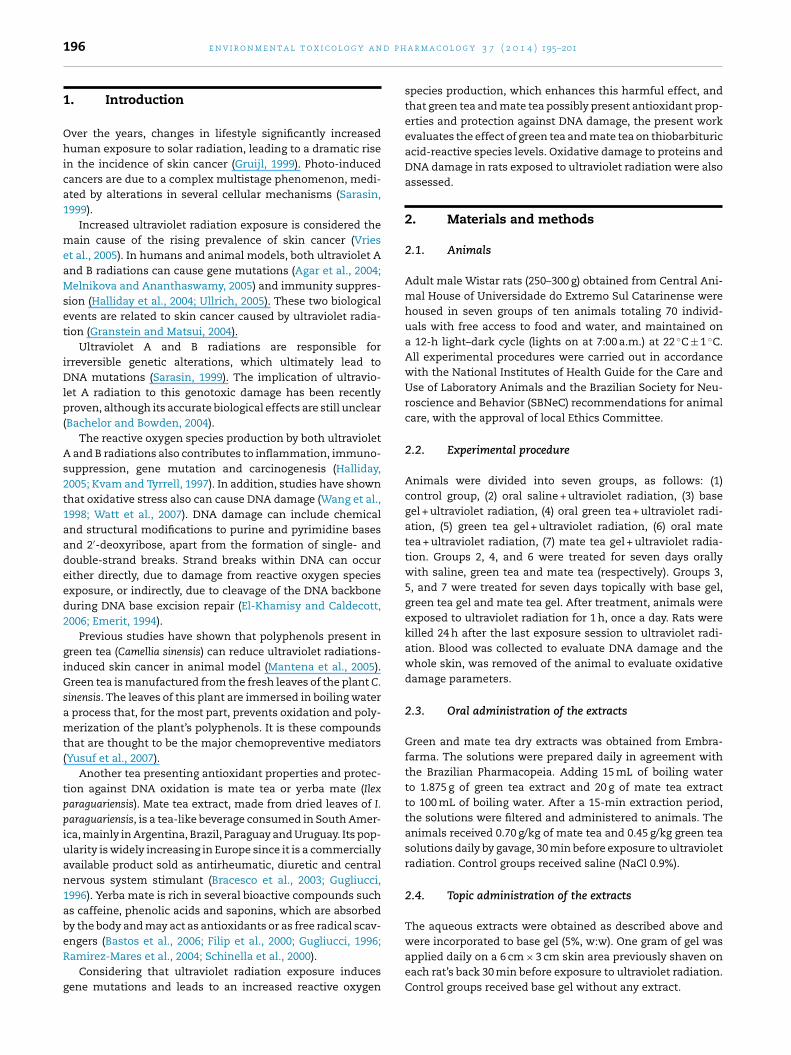
Fig. 1 – Comet assay. Evaluation of DNA damage usingethidium bromide (400×). The cells are assessed visuallyand received scores from 0 (undamaged) to 4 (maximallyd
2
AdtbRtk1
2
Awbafnel1fp12wDdfmaa(aT0ddb
2
AoaoB1
Fig. 2 – Mean (±SD) values of damage index observed inperipheral blood of rats treated with green tea and mate tea,oral and topic, and submitted to radiation. *Data significant
amaged), according to the size and shape of the tail.
.5. Exposure to radiation
nimals were exposed to ultraviolet A and B radiations for 7ays. Ultraviolet A and B lamps were used with emission spec-rum of 280–320 nm for UVB, and 320–400 nm for UVA. The rats’ack were at a distance of 60 cm from the fluoresans lamps.ats were irradiated for 1 h per day, all at the same time. Inotal, 70 rats were used in the present study. All animals wereept in cages throughout the experiment (5 animals per cage,4 cages).
.6. Comet assay
standard protocol for Comet assay preparation and analysisas adopted from Tice et al. (2000). The slides were prepared
y mixing 5 �L of whole blood with 95 �L of low-melting-pointgarose (0.75%). The mixture (cells/agarose) was added to aully frosted microscope slide coated with a layer of 500 �L oformally melting agarose (1%). After solidification, the cov-rslip was gently removed, and the slides were placed inysis solution (2.5 M NaCl, 100 mM EDTA, and 10 mM Tris, pH0.0–10.5, with freshly added 1% Triton X-100 and 10% DMSO)or 1 day. Subsequently, the slides were incubated in freshlyrepared alkaline buffer (300 mM NaOH and 1 mM EDTA, pH3) for 20 min. The DNA was electrophoresed for 15 min at5 V (0.90 V/cm) and 300 mA. After electrophoresis, the slidesere neutralized with Tris buffer (0.4 M; pH 7.5). Finally, theNA was stained with ethidium bromide. Images of 100 ran-omly selected cells (50 cells from each of two replicate slides)rom each animal were blindly analyzed using a fluorescence
icroscope equipped with an excitation filter of BP546/12 nmnd a 590 nm barrier filter. Cells were scored from 0 (undam-ged) to 4 (maximally damaged) according to the tail intensitysize and shape), resulting in a single DNA damage score (dam-ge index) for each sample and, consequently, for each group.hus, a damage index (DI) of the group could range from
(completely undamaged 100 = cells × 0) to 400 (maximumamage = 100 cells × 4) (Collins, 2004) (Fig. 1). The percentageamage frequency (DF) was calculated for each sample on theasis of the number of cells with a tail versus with no tail.
.7. Thiobarbituric acid reactive species (TBARS)
s an index of oxidative damage, we used the formationf TBARS during an acid-heating reaction, which is widely
dapted as a sensitive method for measurement of lipid per-xidation, as previously described (Draper and Hadley, 1990).riefly, samples were mixed with 1 mL of trichloroacetic acid0% (TCA) and 1 mL of thiobarbituric acid 0.67% (TBA), andin relation to control at P < 0.05 (Kruskal–Wallis, Dunn).
then heated in a boiling water bath for 15 min. TBARS weredetermined by absorbance at 535 nm. Results are expressedas malondialdehyde (MDA) equivalents (pmol/mg protein).
2.8. Measurement of protein carbonyls
The oxidative damage to proteins was assessed by thedetermination of carbonyl groups based on the reactionwith dinitrophenylhydrazine (DNPH) as previously described(Levine et al., 1990). Briefly, proteins were precipitated by theaddition of 20% trichloroacetic acid and redissolved in DNPH,and the absorbance read at 370 nm. Results are expressed asprotein carbonyls (pmol/mg protein).
2.9. Statistical analysis
For the analyses of DNA and oxidative damage parameters, alldata were expressed as mean ± standard deviation (SD).
Differences among experimental groups were determinedby one-way analysis of variance (ANOVA) when normal distri-bution was observed. Multiple comparisons were performedby Duncan’ test. When non normal distribution was observed;comparisons were made using the Kruskal–Wallis test withDunn’s test as post hoc.
In all experiments, P < 0.05 was considered to indicate sta-tistical significance. All analyses were performed using the 5.0BioEstat software.
3. Results
The present study showed that ultraviolet radiation expo-sure presented higher levels of DNA damage in peripheralblood in both parameters of the comet assay, damage index(Fig. 2) and damage frequency (Fig. 3) (P < 0.05; Kruskal–Wallis,Dunn). We also showed that was increased thiobarbituric acid-reactive species levels (Fig. 4), and protein carbonylation (Fig. 4)(P < 0.05; ANOVA – Duncan).
Results for animals treated with both types of herbs in thetwo administration routes showed that only mate tea can sig-nificantly reverse DNA damage caused by UV radiation, unlike
green tea, which does not present these protective effects forthe genome, as observed in the high levels of DNA damagewhen orally administered, in comparison to the control group
198 e n v i r o n m e n t a l t o x i c o l o g y a n d p h a r m a c o l o g y 3 7 ( 2 0 1 4 ) 195–201
Fig. 3 – Mean (±SD) values of damage frequency observedin peripheral blood of rats treated with green tea and matetea, oral and topic, and submitted to radiation. *Datasignificant in relation to control at P < 0.05 (Kruskal–Wallis,
Fig. 5 – Mean (±SD) values of protein carbonyl content inskin of rats treated with green tea and mate tea, oral andtopic, and submitted to radiation. *Data significant in
Dunn).(P < 0.05; Kruskal–Wallis, Dunn). However, both parameters ofthe comet assay (Figs. 2 and 3) revealed that this herb led to asimilar decrease in DNA damage in animals topically treatedwith mate tea.
Furthermore, we observed that oral and topic treatmentswith green tea or mate tea prevented the increased lipid per-oxidation induced by ultraviolet radiation exposure (Fig. 4)(P < 0.05; ANOVA – Duncan). However, protein carbonylationwas only prevented by topic treatment with green tea gel andmate tea gel. Oral treatment with green or mate teas did notprevent the increase in protein carbonylation caused by ultra-violet radiation exposure (Fig. 5) (P < 0.05; ANOVA – Duncan).
4. Discussion
Exposure to solar ultraviolet radiation induces inflammatoryresponses, oxidative stress, immunosuppression, DNA dam-age and gene mutations, events related to skin diseases,including skin cancer (Katiyar et al., 2000, 2007; Katiyar, 2006).Multiple studies have demonstrated a relationship betweenultraviolet exposure and increased risk of developing skin
cancer. It was postulated that exposure of the skin to solarultraviolet light is a major risk factor for the development ofmalignant melanoma and non-melanoma skin cancer, andFig. 4 – Mean (±SD) values of thiobarbituric acid reactivesubstances (TBARS) in skin of rats treated with green teaand mate tea, oral and topic, and submitted to radiation.*Data significant in relation to control at P < 0.05 (one-wayANOVA, Duncan test).
relation to control at P < 0.05 (one-way ANOVA, Duncan test).
that this effect is mediated by a chain of events, includingDNA damage by ultraviolet light and subsequent formationof mutations (Runger, 2003).
In the life of a cell, DNA damage represents a greatthreat to genome stability, leading to loss or amplificationof chromosomal activity, which may result in carcinogene-sis or tissue aging (Nakanishi et al., 2009). Genome integrityis continuously threatened by both endogenous (e.g. reac-tive oxygen species produced by normal metabolism, DNAreplication ‘errors’) and exogenous (ultraviolet light, ionizingradiations and chemical carcinogens) factors (Mocellin et al.,2009).
In the present study we observed the occurrence of DNAdamage in peripheral blood and oxidative damage in the skinof rats exposed to radiation. On the other hand, we showedthat green and mate teas administered through two differentroutes (oral and topic gel) have prevented radiation-inducedlipid peroxidation. However, protein carbonylation was pre-vented only by green and mate teas used as gel. Also, onlytopically administered mate tea prevented the harmful effectsof UV radiation to genetic material.
The study Türkoglu et al. (2010) showed that no UV-inducederythema was observed at the black and green tea gel sites inany of the subjects. UV-induced erythema was consistentlypresent in various grades at caffeine gel, carbomer gel, andcontrol sites. Therefore, tea extracts were found to be promis-ing candidates for their ability to protect against the harmfuleffects of UV radiation, such as erythema and premature agingof the skin.
Studies showed that consumption of green tea polyphe-nols in drinking water prevents photocarcinogenesis in mice;however, the molecular mechanisms underlying this effecthave not been fully elucidated (Meeran et al., 2009). Study ofXu et al. (2010) showed that green tea polyphenols effectivelysuppressed the decrease in viability of the UVB stressedretinal pigment epithelial cells and the UVB suppressionof surviving gene expression level. Green tea polyphenolsalleviated mitochondria dysfunction and DNA fragmentationinduced by UVB.
Polyphenols isolated from the leaves of green tea havea number of beneficial healthy effects, including anti-carcinogenic activity, which has been demonstrated in various

p h a r
t2anutgadtaea2
ppc2srs2gca
sita
aai(ecpsw
trDDsaac2
lco1mAwt
r
principles, applications, and limitations. Mol. Biotechnol. 26,
e n v i r o n m e n t a l t o x i c o l o g y a n d
umor models (Katiyar and Mukhtar, 1996; Yang et al.,002). It has been also shown that oral administration ofn aqueous extract of green tea or green tea polyphe-ols (a mixture of polyphenols) in drinking water inhibitsltraviolet radiation-induced skin carcinogenesis in mice inerms of tumor incidence, tumor multiplicity and tumorrowth/size (Mantena et al., 2005; Wang et al., 1992). Inddition, UV exposure causes physical changes to the skinue to alterations that occur in the connective tissue viahe formation of lipid peroxides, cell contents and enzymesnd reactive oxygen species (Benaiges et al., 1998; Kaurt al., 2006). Other studies showed that green tea also exertsnti-oxidant activity (Mantena et al., 2005; Okayasu et al.,003).
Flavonoids and phenolic compounds were proved to beresent in mate tea leaves, and the consumption of these com-ounds has been associated with the prevention of age-relatedhronic conditions, including cancer (McKay and Blumberg,002; Weisburger, 2002). Various phytochemical constituents,uch as flavonoids, have been isolated from mate plants andelated species, and some of these compounds may be respon-ible for the observed antioxidant activity (Schinella et al.,000). Furthermore, data from in vitro and in vivo studies sug-est a potential beneficial effect of tea polyphenols againstancer at most stages of development (Hirose et al., 2002; Sund Arab, 2002; Tapiero et al., 2002).
In this scenery, study of Chandra and De Mejia (2004)howed that the concentration of polyphenols in green teas major when compared with mate tea. Thus, the concentra-ion of green and mate tea used in the present study result inmounts polyphenols similar in both teas.
Conversely, many studies in cell culture models as wells in animals seem to converge to show an antimutagenicnd DNA protecting effect for I. paraguariensis extracts and itsndividual components, chlorogenic acid, rutin and quercetinBracesco et al., 2003; Miranda et al., 2008). Study of Leonardt al. (2010) indicated that mate scavenged hydroxyl radi-als and superoxide radicals. This study indicates that mateossesses potent antioxidant effects against hydroxyl anduperoxide radicals in chemical and cell culture systems, asell as DNA-protective properties.
Besides, study of Miranda et al. (2008) showed that mateea is not genotoxic in liver, kidney and bladder cells. Theegular ingestion of mate tea increased the resistance ofNA to H2O2-induced DNA strand breaks and improved theNA repair after H2O2 challenge in liver cells. These resultsuggest that mate tea could protect against DNA damagend enhance the DNA repair activity. Protection may bettributed to the antioxidant activity of the mate bioactiveompounds cited above (Bracesco et al., 2003; Miranda et al.,008).
However, study of Wnuk et al. (2009) in vitro cultured humanymphocytes indicates that mate infusion may cause bothytotoxic and genotoxic effects. Differences concentrationsf mate from 1 to 1000 �g/mL was evaluated and found that0 �g/mL of mate significantly increased the frequency oficronuclei and decreased the nuclear division index value.
t the higher concentrations of 100 and 1000 �g/mL, mateas responsible for an augmentation in the level of apop-otic and necrotic cells. Moreover, no genotoxic effect of mate
m a c o l o g y 3 7 ( 2 0 1 4 ) 195–201 199
(175–1400 �g/mL) on human lymphocytes was reported byAlves et al. (2008).
In conclusion, we showed that the exposure of rats to radia-tion led to DNA and oxidative damage, and that green tea andmate tea have prevented these effects. Our data are in linewith previous works that demonstrated the anti-carcinogenicand antioxidant activities of green tea and mate tea.
Conflict of interest statement
The authors declare that there are no conflicts of interest.
Acknowledgment
This research was supported by grants from Universidade doExtremo Sul Catarinense – UNESC (Brazil).
Appendix A. Supplementary data
Supplementary data associated with this article can be found,in the online version, at http://dx.doi.org/10.1016/j.etap.2013.11.028.
e f e r e n c e s
Agar, N.S., Halliday, G.M., Barnetson, R.S., Ananthaswamy, H.N.,Wheeler, M., Jones, A.M., 2004. The basal layer in humansquamous tumors harbors more UVA than UVB fingerprintmutations: a role for UVA in human skin carcinogenesis. Proc.Natl. Acad. Sci. U.S.A. 101, 4954–4959.
Alves, R.J., Jotz, G.P., do Amaral, V.S., Montes, T.M., Menezes, H.S.,de Andrade, H.H., 2008. The evaluation of maté (Ilexparaguariensis) genetic toxicity in human lymphocytes by thecytokinesis-block in the micronucleus assay. Toxicol. In Vitro22, 695–698.
Bachelor, M.A., Bowden, G.T., 2004. UVA-mediated activation ofsignaling pathways involved in skin tumor promotion andprogression. Semin. Cancer Biol. 14, 131–138.
Bastos, D.H., Ishimoto, E.Y., Marques, M.O.M., Ferri, A.F., Torres,E.A.F.S., 2006. Essential oil and antioxidant activity of greenmate and mate tea (Ilex paraguariensis) infusions. J. FoodCompos. Anal. 19, 538–543.
Benaiges, A., Marcet, P., Armengol, R., Betes, C., Gironés, E.A.,1998. Study of the refirming effect of a plant complex. Int. J.Cosmet. Sci. 20, 223–233.
Bracesco, N., Dell, M., Rocha, A., Behtash, S., Menini, T., Gugliucci,A., Nunes, E., 2003. Antioxidant activity of a botanical extractpreparation of Ilex paraguariensis: prevention of DNAdouble-strand breaks in Saccharomyces cerevisiae and humanlow-density lipoprotein oxidation. J. Altern. ComplementMed. 9, 379–387.
Chandra, S., De Mejia, G.E., 2004. Polyphenolic compounds,antioxidant capacity, and quinone reductase activity of anaqueous extract of Ardisia compressa in comparison to mate(Ilex paraguariensis) and green (Camellia sinensis) teas. J. Agric.Food Chem. 52, 3583–3589.
Collins, A.R., 2004. The comet assay for DNA damage and repair:
249–326.Draper, H.H., Hadley, M., 1990. Malondialdehyde determination as
index of lipid peroxidation. Methods Enzymol. 186, 421–431.

d p h
200 e n v i r o n m e n t a l t o x i c o l o g y a nEl-Khamisy, S.F., Caldecott, K.W., 2006. TDP1-dependent DNAsingle-strand break repair and neurodegeneration.Mutagenesis 21, 219–224.
Emerit, I., 1994. Reactive oxygen species, chromosome mutation,and cancer: possible role of clastogenic factors incarcinogenesis. Free Radic. Biol. Med. 16, 99–109.
Filip, R., Lotito, S., Ferraco, G., Raga, C., 2000. Antioxidant activityof Ilex paraguariensis and related species. Nutr. Res. 20,1437–1446.
Granstein, R.D., Matsui, M.S., 2004. UV radiation-inducedimmunosuppression and skin cancer. Cutis 74, 4–9.
Gruijl, F.R., 1999. Skin cancer and solar UV radiation. Eur. J.Cancer 35, 2003–2009.
Gugliucci, A., 1996. Antioxidant effects of Ilex paraguariensis:induction of decreased oxidability of human LDL in vivo.Biochem. Biophys. Res. Commun. 224, 338–344.
Halliday, G.M., Byrne, S.N., Kuchel, J.M., Poon, T.S., Barnetson, R.S.,2004. The suppression of immunity by ultraviolet radiation:UVA, nitric oxide and DNA damage. Photochem. Photobiol.Sci. 3, 736–740.
Halliday, G.M., 2005. Inflammation, gene mutation andphotoimmunosuppression in response to UVR-inducedoxidative damage contributes to photocarcinogenesis. Mutat.Res. 571, 107–120.
Hirose, M., Nishikawa, A., Shibutani, M., Imai, T., Shirai, T., 2002.Chemoprevention of heterocyclic amine-induced mammarycarcinogenesis in rats. Environ. Mol. Mutagen. 39,271–278.
Katiyar, S., Elmets, C.A., Katiyar, S.K., 2007. Green tea and skincancer: photoimmunology, angiogenesis and DNA repair. J.Nutr. Biochem. 18, 287–296.
Katiyar, S.K., Matsui, M.S., Mukhtar, H., 2000. Kinetics of UVlight-induced cyclobutane pyrimidine dimers in human skinin vivo: an immunohistochemical analysis of both epidermisand dermis. Photochem. Photobiol. 72, 788–793.
Katiyar, S., Mukhtar, H., 1996. Tea in chemoprevention of cancer.Int. J. Oncol. 8, 221–238.
Katiyar, S.K., 2006. Oxidative stress and photocarcinogenesis:strategies for prevention. In: Singh, K.K. (Ed.), Oxidative StressDisease and Cancer. Imperial College Press, London, pp.933–964.
Kaur, G., Jabbar, Z., Athar, M., Alam, M.S., 2006. Punica granatum(pomegranate) flower extract possesses potent antioxidantactivity and abrogates Fe-NTA induced hepatotoxicity in mice.Food Chem. Toxicol. 44, 984–993.
Kvam, E., Tyrrell, R.M., 1997. Induction of oxidative DNA basedamage in human skin cells by UV and near visible radiation.Carcinogenesis 18, 2379–2384.
Leonard, S.S., Hogans, V.J., Coppes-Petricorena, Z., Peer, C.J.,Vining, T.A., Fleming, D.W., Harris, G.K., 2010. Analysis offree-radical scavenging of Yerba Mate (Ilex paraguariensis)using electron spin resonance and radical-induced DNAdamage. J. Food Sci. 75, 14–20.
Levine, R.L., Garland, D., Oliver, C.N., Amici, A., Climent, I., Lenz,A.G., Ahn, B.W., Shaltie, l.S., Stadtman, E.R., 1990.Determination of carbonyl content in oxidatively modifiedproteins. Methods Enzymol. 186, 464–478.
Mantena, S.K., Meeran, S.M., Elmets, C.A., Katiyar, S.K., 2005.Orally administered green tea polyphenols prevent ultravioletradiation-induced skin cancer in mice through activation ofcytotoxic T cells and inhibition of angiogenesis in tumors. J.Nutr. 135, 2871–2877.
McKay, D.L., Blumberg, J.B., 2002. The role of tea in human health:an update. J. Am. Coll. Nutr. 21, 1–13.
Meeran, S.M., Akhtar, S., Katiyar, S.K., 2009. Inhibition ofUVB-induced skin tumor development by drinking green tea
polyphenols is mediated through DNA repair and subsequentinhibition of inflammation. J. Invest. Dermatol. 129,258–1270.a r m a c o l o g y 3 7 ( 2 0 1 4 ) 195–201
Melnikova, V.O., Ananthaswamy, H.N., 2005. Cellular andmolecular events leading to the development of skin cancer.Mutat. Res. 571, 91–106.
Miranda, D.D., Arcari, D.P., Pedrazzoli Jr., J., Carvalho Pde, O.,Cerutti, S.M., Bastos, D.H., Ribeiro, M.L., 2008. Protectiveeffects of mate tea (Ilex paraguariensis) on H2O2-induced DNAdamage and DNA repair in mice. Mutagenesis 23,261–265.
Mocellin, S., Verdi, D., Nitti, D., 2009. DNA repair genepolymorphisms and risk of cutaneous melanoma: asystematic review and meta-analysis. Carcinogenesis 30,1735–1743.
Nakanishi, M., Niida, H., Murakami, H., Shimada, M., 2009. DNAdamage responses in skin biology – implications in tumorprevention and aging acceleration. J. Dermatol. Sci. 56,76–81.
Okayasu, H., Suzuki, F., Satoh, K., Shioda, S., Dohi, K., Ikeda, Y.,Nakashima, H., Komatsu, N., Fujimaki, M., Hashimoto, K.,Maki, J., Sakagami, H., 2003. Comparison of cytotoxicity andradical scavenging activity between tea extracts and Chinesemedicines. In Vivo 17, 577–581.
Ramirez-Mares, M.V., Chandra, S., de Mejia, E.G., 2004. In vitrochemopreventive activity of Camellia sinensis, Ilexparaguariensis and Ardisia compressa tea extracts and selectedpolyphenols. Mutat. Res. 4, 53–65.
Runger, T.M., 2003. Ultraviolet light. In: Bolognia, J.L., Jorizzo, J.L.,Rapini, R.P. (Eds.), Dermatology. Mosby Press, London, pp.1353–1363.
Sarasin, A., 1999. The molecular pathways of ultraviolet-inducedcarcinogenesis. Mutat. Res. 428, 5–10.
Schinella, G.R., Troiani, G., Dávila, V., de Buschiazzo, P.M.,Tournier, H.A., 2000. Antioxidant effects of an aqueous extractof Ilex paraguariensis. Biochem. Biophys. Res. Commun. 269,357–360.
Su, L.J., Arab, L., 2002. Tea consumption and the reduced risk ofcolon cancer – results from a national prospective cohortstudy. Public Health Nutr. 5, 419–426.
Tapiero, H., Tew, K.D., Ba, G.N., Mathé, G., 2002. Polyphenols: dothey play a role in the prevention of human pathologies?Biomed. Pharmacother. 56, 200–207.
Tice, R.R., Agurell, E., Anderson, D., Burlinson, B., Hartmann, A.,Kobayashi, H., Miyamae, Y., Rojas, E., Ryu, J.C., Sasaki, Y.F.,2000. Single CellGel/Comet Assay: guidelines for in vitro andin vivo genetic toxicology testing. Environ. Mol. Mutagen. 35,206–221.
Türkoglu, M., Ugurlu, T., Gedik, G., Yılmaz, A.M., Süha Yalcin, A.,2010. In vivo evaluation of black and green tea dermalproducts against UV radiation. Drug Discov. Ther. 4, 362–367.
Ullrich, S.E., 2005. Mechanisms underlying UV-induced immunesuppression. Mutat. Res. 571, 185–205.
Vries, de E., Van de Poll-Franse, L.V., Louwman, W.J., de Gruijl,F.R., Coebergh, J.W., 2005. Predictions of skin cancer incidencein the Netherlands up to 2015. Br. J. Dermatol. 152, 481–488.
Wang, D., Kreutzer, D.A., Essigmann, J.M., 1998. Mutagenicity andrepair of oxidative DNA damage: insights from studies usingdefined lesions. Mutat. Res. 400, 1155–1199.
Wang, Z.Y., Huang, M.T., Ferraro, T., Wong, C.Q., Lou, Y.R., Reuhl,K., Iatropoulos, M., Yang, C.S., Conney, A.H., 1992. Inhibitoryeffect of green tea in the drinking water on tumorigenesis byultraviolet light and 12-O-tetradecanoylphorbol-13-acetate inthe skin of SKH-1 mice. Cancer Res. 52, 1162–1170.
Watt, N.T., Routledge, M.N., Wild, C.P., Hooper, N.M., 2007.Cellular prion protein protects againstreactive-oxygen-species-induced DNA damage. Free Radic.Biol. Med. 43, 959–967.
Weisburger, J.H., 2002. Lifestyle health and disease prevention:
the underlying mechanisms. Eur. J. Cancer Prev. 11, 1–7.Wnuk, M., Lewinska, A., Oklejewicz, B., Bugno, M., Slota, E.,Bartosz, G., 2009. Evaluation of the cyto- and genotoxic

p h a r
X
e n v i r o n m e n t a l t o x i c o l o g y a n d
activity of yerba mate (Ilex paraguariensis) in humanlymphocytes in vitro. Mutat. Res. 679, 18–23.
u, J.Y., Wu, L.Y., Zheng, X.Q., Lu, J.L., Wu, M.Y., Liang, Y.R., 2010.Green tea polyphenols attenuating ultraviolet B-induceddamage to human retinal pigment epithelial cells in vitro.Invest. Ophthalmol. Vis. Sci. 51, 6665–6670.
m a c o l o g y 3 7 ( 2 0 1 4 ) 195–201 201
Yang, C.S., Maliakal, P., Meng, X., 2002. Inhibition ofcarcinogenesis by tea. Annu. Rev. Pharmacol. Toxicol. 42,
25–54.Yusuf, N., Irby, C., Katiyar, S.K., Elmets, C.A., 2007.Photoprotective effects of green tea polyphenols.Photodermatol. Photoimmunol. Photomed. 23, 48–56.
Copyright © 2022 FDOKUMEN