Leukocytic-Vascular Endothelial Growth Factor and Integrin ...
RESEARCH Open Access
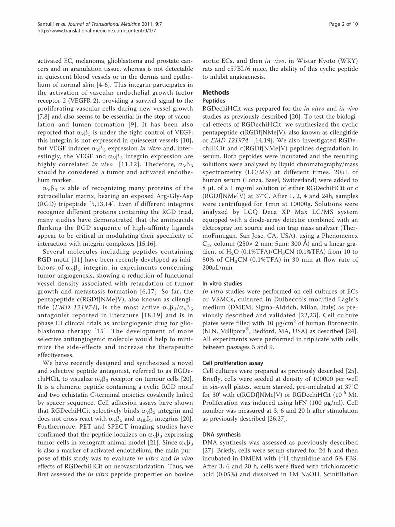
Evaluation of the anti-angiogenic properties ofthe new selective aVb3 integrin antagonistRGDechiHCitGaetano Santulli1, Maria Felicia Basilicata1, Mariarosaria De Simone2, Carmine Del Giudice1, Antonio Anastasio1,Daniela Sorriento1, Michele Saviano3, Annarita Del Gatto4, Bruno Trimarco1, Carlo Pedone2, Laura Zaccaro4,Guido Iaccarino1*
Abstract
Background: Integrins are heterodimeric receptors that play a critical role in cell-cell and cell-matrix adhesionprocesses. Among them, aVb3 integrin, that recognizes the aminoacidic RGD triad, is reported to be involved inangiogenesis, tissue repair and tumor growth. We have recently synthesized a new and selective ligand of aVb3receptor, referred to as RGDechiHCit, that contains a cyclic RGD motif and two echistatin moieties.
Methods: The aim of this study is to evaluate in vitro and in vivo the effects of RGDechiHCit. Therefore, weassessed its properties in cellular (endothelial cells [EC], and vascular smooth muscle cells [VSMC]) and animalmodels (Wistar Kyoto rats and c57Bl/6 mice) of angiogenesis.
Results: In EC, but not VSMC, RGDechiHCit inhibits intracellular mitogenic signaling and cell proliferation.Furthermore, RGDechiHCit blocks the ability of EC to form tubes on Matrigel. In vivo, wound healing is delayed inpresence of RGDechiHCit. Similarly, Matrigel plugs demonstrate an antiangiogenic effect of RGDechiHCit.
Conclusions: Our data indicate the importance of RGDechiHCit in the selective inhibition of endothelial aVb3integrin in vitro and in vivo. Such inhibition opens new fields of investigation on the mechanisms of angiogenesis,offering clinical implications for treatment of pathophysiological conditions such as cancer, proliferative retinopathyand inflammatory disease.
IntroductionAngiogenesis is a complex multistep phenomenon con-sisting of the sprouting and the growth of new capillaryblood vessels starting from the pre-existing ones. Itrequires the cooperation of several cell types such asendothelial cells (ECs), vascular smooth muscle cells(VSMCs), macrophages, which should be activated, pro-liferate and migrate to invade the extracellular matrixand cause vascular remodeling [1,2]. The angiogenicprocess is finely tuned by a precise balance of growthand inhibitory factors and in mammalians it is normallydormant except for some physiological conditions, suchas wound healing and ovulation. When this balance is
altered, excessive or defective angiogenesis occur andthe process becomes pathological. Excessive angiogen-esis gives also rise to different dysfunctions, includingcancer, eye diseases, rheumatoid arthritis, atherosclero-sis, diabetic nephropathy, inflammatory bowel disease,psoriasis, endometriosis, vasculitis, and vascular malfor-mations [3]. Therefore the discovery of angiogenesisinhibitors would contribute to the development of thera-peutic treatments for these diseases.The integrins are cell adhesion receptors that mediate
cell-cell and cell-matrix interactions and coordinate sig-naling allowing a close regulation of physiological phe-nomena including cellular migration, proliferation anddifferentiation. In particular, the aV integrins, combinedwith distinct b subunits, participate in the angiogenicprocess. An extensively studied member of this receptorclass is integrin aVb3, that is strongly overexpressed in
* Correspondence: [email protected] of Clinical Medicine, Cardiovascular & Immunologic Sciences,“Federico II” University of Naples, ItalyFull list of author information is available at the end of the article
Santulli et al. Journal of Translational Medicine 2011, 9:7http://www.translational-medicine.com/content/9/1/7
© 2011 Santulli et al; licensee BioMed Central Ltd. This is an Open Access article distributed under the terms of the Creative CommonsAttribution License (http://creativecommons.org/licenses/by/2.0), which permits unrestricted use, distribution, and reproduction inany medium, provided the original work is properly cited.
activated EC, melanoma, glioblastoma and prostate can-cers and in granulation tissue, whereas is not detectablein quiescent blood vessels or in the dermis and epithe-lium of normal skin [4-6]. This integrin participates inthe activation of vascular endothelial growth factorreceptor-2 (VEGFR-2), providing a survival signal to theproliferating vascular cells during new vessel growth[7,8] and also seems to be essential in the step of vacuo-lation and lumen formation [9]. It has been alsoreported that aVb3 is under the tight control of VEGF:this integrin is not expressed in quiescent vessels [10],but VEGF induces aVb3 expression in vitro and, inter-estingly, the VEGF and aVb3 integrin expression arehighly correlated in vivo [11,12]. Therefore, aVb3should be considered a tumor and activated endothe-lium marker.aVb3 is able of recognizing many proteins of the
extracellular matrix, bearing an exposed Arg-Gly-Asp(RGD) tripeptide [5,13,14]. Even if different integrinsrecognize different proteins containing the RGD triad,many studies have demonstrated that the aminoacidsflanking the RGD sequence of high-affinity ligandsappear to be critical in modulating their specificity ofinteraction with integrin complexes [15,16].Several molecules including peptides containing
RGD motif [11] have been recently developed as inhi-bitors of aVb3 integrin, in experiments concerningtumor angiogenesis, showing a reduction of functionalvessel density associated with retardation of tumorgrowth and metastasis formation [6,17]. So far, thepentapeptide c(RGDf[NMe]V), also known as cilengi-tide (EMD 121974), is the most active avb3/avb5
antagonist reported in literature [18,19] and is inphase III clinical trials as antiangiogenic drug for glio-blastoma therapy [15]. The development of moreselective antiangiogenic molecule would help to mini-mize the side-effects and increase the therapeuticeffectiveness.We have recently designed and synthesized a novel
and selective peptide antagonist, referred to as RGDe-chiHCit, to visualize aVb3 receptor on tumour cells [20].It is a chimeric peptide containing a cyclic RGD motifand two echistatin C-terminal moieties covalently linkedby spacer sequence. Cell adhesion assays have shownthat RGDechiHCit selectively binds aVb3 integrin anddoes not cross-react with aVb5 and aIIbb3 integrins [20].Furthermore, PET and SPECT imaging studies haveconfirmed that the peptide localizes on aVb3 expressingtumor cells in xenograft animal model [21]. Since aVb3is also a marker of activated endothelium, the main pur-pose of this study was to evaluate in vitro and in vivoeffects of RGDechiHCit on neovascularization. Thus, wefirst assessed the in vitro peptide properties on bovine
aortic ECs, and then in vivo, in Wistar Kyoto (WKY)rats and c57BL/6 mice, the ability of this cyclic peptideto inhibit angiogenesis.
MethodsPeptidesRGDechiHCit was prepared for the in vitro and in vivostudies as previously described [20]. To test the biologi-cal effects of RGDechiHCit, we synthesized the cyclicpentapeptide c(RGDf[NMe]V), also known as cilengitideor EMD 121974 [14,19]. We also investigated RGDe-chiHCit and c(RGDf[NMe]V) peptides degradation inserum. Both peptides were incubated and the resultingsolutions were analyzed by liquid chromatography/massspectrometry (LC/MS) at different times. 20μL ofhuman serum (Lonza, Basel, Switzerland) were added to8 μL of a 1 mg/ml solution of either RGDechiHCit or c(RGDf[NMe]V) at 37°C. After 1, 2, 4 and 24h, sampleswere centrifuged for 1min at 10000g. Solutions wereanalyzed by LCQ Deca XP Max LC/MS systemequipped with a diode-array detector combined with anelctrospray ion source and ion trap mass analyzer (Ther-moFinnigan, San Jose, CA, USA), using a PhenomenexC18 column (250× 2 mm; 5μm; 300 Ǻ) and a linear gra-dient of H2O (0.1%TFA)/CH3CN (0.1%TFA) from 10 to80% of CH3CN (0.1%TFA) in 30 min at flow rate of200μL/min.
In vitro studiesIn vitro studies were performed on cell cultures of ECsor VSMCs, cultured in Dulbecco’s modified Eagle’smedium (DMEM; Sigma-Aldrich, Milan, Italy) as pre-viously described and validated [22,23]. Cell cultureplates were filled with 10 μg/cm2 of human fibronectin(hFN, Millipore®, Bedford, MA, USA) as described [24].All experiments were performed in triplicate with cellsbetween passages 5 and 9.
Cell proliferation assayCell cultures were prepared as previously described [25].Briefly, cells were seeded at density of 100000 per wellin six-well plates, serum starved, pre-incubated at 37°Cfor 30’ with c(RGDf[NMe]V) or RGDechiHCit (10-6 M).Proliferation was induced using hFN (100 μg/ml). Cellnumber was measured at 3, 6 and 20 h after stimulationas previously described [26,27].
DNA synthesisDNA synthesis was assessed as previously described[27]. Briefly, cells were serum-starved for 24 h and thenincubated in DMEM with [3H]thymidine and 5% FBS.After 3, 6 and 20 h, cells were fixed with trichloraceticacid (0.05%) and dissolved in 1M NaOH. Scintillation
Santulli et al. Journal of Translational Medicine 2011, 9:7http://www.translational-medicine.com/content/9/1/7
Page 2 of 10
liquid was added and [3H]thymidine incorporation wasassessed as previously described [27].
VEGF quantificationVEGF production was measured as previously described[26]. Briefly, ECs were seeded at a density of 600000 perwell in six well plates, serum starved overnight, seededwith c(RGDf[NMe]V) or RGDechiHCit (10-6 M) andthen stimulated with hFN for 6 hours. Cultured mediumwas collected and VEGF production was revealed bywestern blot.
Endothelial Matrigel assayThe formation of network-like structures by ECs on anextracellular matrix (ECM)-like 3D gel consisting ofMatrigel® (BD Biosciences, Bedford, MA, USA), wasperformed as previously described and validated [27,28].The six-well multidishes were coated with growth fac-tor-reduced Matrigel in according to the manufacturer’sinstructions. ECs (5×10⁴) were seeded with c(RGDf[NMe]V) or RGDechiHCit (10-6 M), in the absence(negative control) or presence (100 μg/ml) of hFN [24].Cells were incubated at 37°C for 24h in 1 ml of DMEM.After incubation, ECs underwent differentiation intocapillary-like tube structures. Tubule formation wasdefined as a structure exhibiting a length four times itswidth [27]. Network formation was observed using aninverted phase-contrast microscope (Zeiss). Representa-tive fields were taken, and the average of the total num-ber of complete tubes formed by cells was counted in15 random fields by two independent investigators.
Western blotImmunoblot analyses were performed as previouslydescribed and validated [23,28]. Mouse monoclonalantibodies to extracellular signal regulated kinase(ERK2) and phospho-ERK, anti-rabbit VEGF and actinwere from Santa Cruz Biotecnology (Santa Cruz, CA,USA). Levels of VEGF were determined using an anti-body raised against VEGF-165 (Santa Cruz Biotechnol-ogy) [26]. Experiments were performed in triplicate toensure reproducibility. Data are presented as arbitrarydensitometry units (ADU) after normalization for thetotal corresponding protein or actin as internal control[24].
In vivo studiesWound healing assay was performed on 14-week-old(weight 293 ± 21 g) normotensive WKY male rats(Charles River Laboratories, Calco (LC), Italy; n = 18),and Matrigel plugs experiments were carried out on 16-week-old (weight 33 ± 4 g) c57BL/6 mice (Charles RiverLaboratories, Milan, Italy; n = 13). All animal proce-dures were performed in accordance with the Guide for
the Care and Use of Laboratory Animals published bythe National Institutes of Health in the United States(NIH Publication No. 85- 23, revised 1996) andapproved by the Ethics Committee for the Use of Ani-mals in Research of “Federico II” University [23].
Wound HealingThe rats (n = 18) were anesthetized using vaporized iso-flurane (4%, Abbott) and maintained by mask ventila-tion (isoflurane 1.8%) [29]. The dorsum was shaved byapplying a depilatory creme (Veet, Reckitt-Benckiser,Milano, Italy) and disinfected with povidone iodinescrub. A 20 mm diameter open wound was excisedthrough the entire thickness of the skin, including thepanniculus carnosus layer, as described and validated[1,28]. Pluronic gel (30%) containing (10-6 M) c(RGDf[NMe]V) (n = 6), RGDechiHCit (n = 7), or saline (n = 5)was placed daily directly onto open wounds, then cov-ered with a sterile dressing. Two operators blinded to theidentity of the sample examined and measured woundareas every day, for 8 days. Direct measurements ofwound region were determined by digital planimetry(pixel area), and subsequent analysis was performedusing a computer-assisted image analyzer (ImageJ soft-ware, version 1.41, National Institutes of Health,Bethesda, MD, USA). Wound healing was quantified as apercentage of the original injury size. Eight days afterwounding, rats were euthanized. Wounds did not showsign of infection. The lesion and adiacent normal skinwere excised, fixed by immersion in phosphate bufferedsaline (PBS, 0.01 M, pH 7.2-7.4)/formalin and thenembedded in paraffin to be processed for immunohistol-ogy, as described [1].
Matrigel PlugsMice (n = 13), anesthetized as described above, weresubcutaneously injected midway on the dorsal side,using sterile conditions, with 0.2 ml of Matrigel® base-ment matrix, pre-mixed with 10-6M VEGF and 10-5M c(RGDf[NMe]V) (n = 4), 10-6M VEGF and 10-5M RGDe-chiHCit (n = 5), or 10-6M VEGF alone (n = 4). Afterseven days, mice were euthanized and the implantedplugs were harvested from underneath the skin, fixed in10% neutral-buffered formalin solution and thenembedded in paraffin. Invading ECs were identified andquantified by analysis of lectin immunostained sections,as described [1,2].
HistologyAll tissues were cut in 5 μm sections and slides werecounterstained with a standard mixture of hematoxylinand eosin. For Masson’s trichrome staining of collagenfibers, useful to assess the scar tissue formation, slideswere stained with Weigert Hematoxylin (Sigma-Aldrich,
Santulli et al. Journal of Translational Medicine 2011, 9:7http://www.translational-medicine.com/content/9/1/7
Page 3 of 10
St. Louis, MO, USA) for 10 minutes, rinsed in PBS(Invitrogen) and then stained with Biebrich scarlet-acidfuchsin (Sigma-Aldrich) for 5 minutes. Slides wererinsed in PBS and stained with phosphomolybdic/phos-photungstic acid solution (Sigma-Aldrich) for 5 minutesthen stained with light green (Sigma-Aldrich) for 5 min-utes [30]. ECs were identified by lectin immunohisto-chemical staining (Sigma-Aldrich) [2] and quantitativeanalysis was performed using digitized representativehigh resolution photographic images, with a dedicatedsoftware (Image Pro Plus; Media Cybernetics, Bethesda,MD, USA) as previously described [28].
Data presentation and statistical analysisAll data are presented as the mean value ± SEM. Statis-tical differences were determined by one-way or two-way ANOVA and Bonferroni post hoc testing was per-formed where applicable. A p value less than 0.05 wasconsidered to be significant. All the statistical analysisand the evaluation of data were performed using Graph-Pad Prism version 5.01 (GraphPad Software, San Diego,CA, USA).
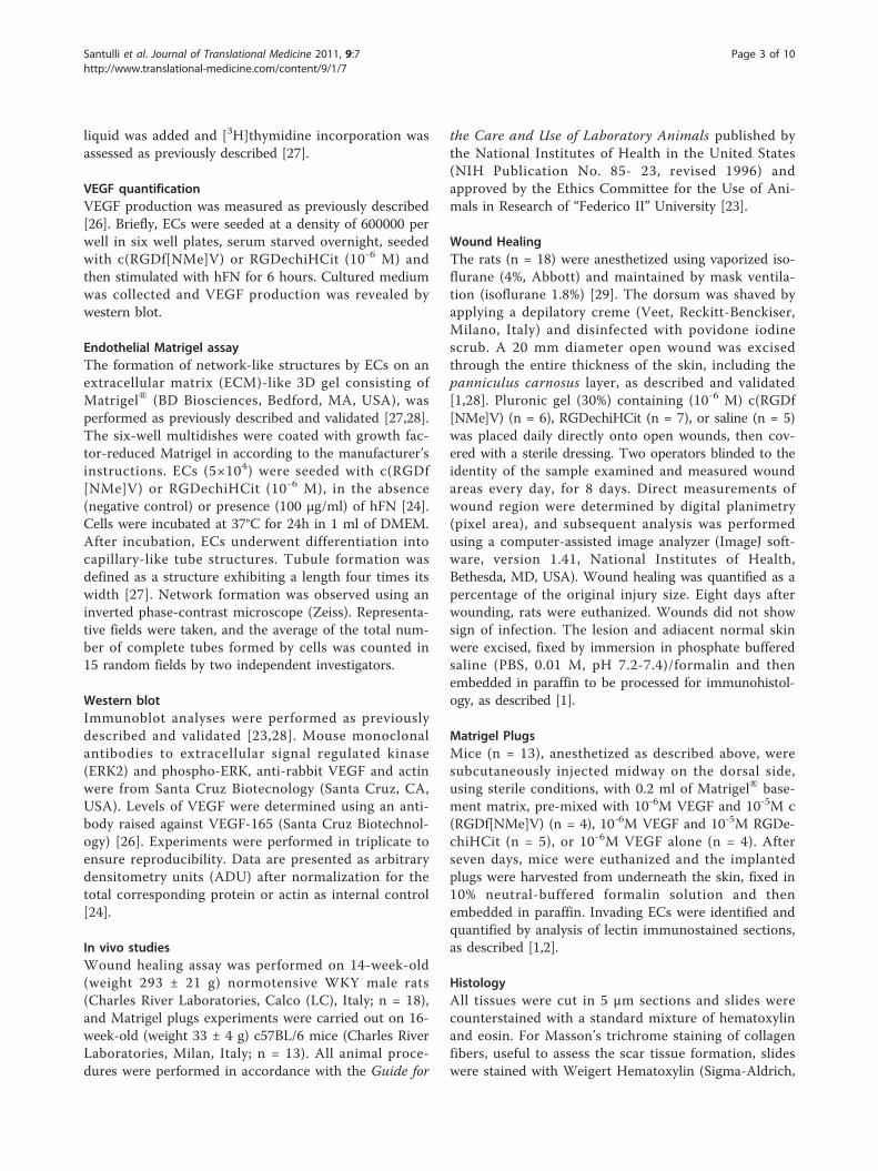
ResultsPeptidesRGDechiHCit and c(RGDf[NMe]V) peptides stabilitieswere evaluated in serum. The degradation of the pep-tides were followed by LC/MS. The reversed-phase highperformance liquid chromatography (RP-HPLC) ofRGDechiHCit before the serum incubation showed asingle peak at tr = 11.82 min corresponding to the com-plete sequence (theoretical MW = 2100.1 g mol-1) asindicated by the [M+H]+, [M+2H]2+ and [M+3H]+3
molecular ion adducts in the MS spectrum (Figure 1A).After 1h, chromatography showed two peaks, ascribableto RGDechiHCit and to a fragment of the completesequence (theoretical MW = 1929.1 g mol-1), respec-tively, as confirmed by MS spectrum. Finally, after 24h afurther peak at tr = 10.93 min corresponding to anotherRGDechiHCit degradation product (theoretical MW =1775.8 g mol-1) appeared, as indicated by the molecularion adducts in the MS spectrum, although the peaksattributed to the RGDechiHCit and to the first fragmentwere still present (Figure 1B).In contrast with RGDechiHCit, c(RGDf[NMe]V)
showed high stability in serum. The RP-HPLC profile ofthe peptide before the incubation showed a single peakat tr = 16.64 min, ascribable to the complete sequenceby the MS spectrum (Figure 1C). After 24h of incuba-tion chromatogram and mass profiles failed to identifyany degradation product (Figure 1D).Since RGDechiHCit showed a low stability, we replen-
ished antagonists every six hours in experiments invol-ving chronic exposure.
In vitro experiments
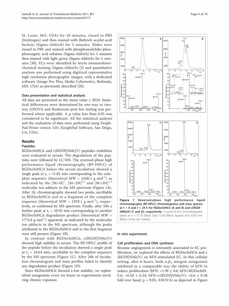
Cell proliferation and DNA synthesisBecause angiogenesis is intimately associated to EC pro-liferation, we explored the effects of RGDechiHCit and c(RGDf[NMe]V) on hFN-stimulated EC. In this cellularsetting, after 6 hours, both avb3 integrin antagonistsinhibited in a comparable way the ability of hFN toinduce proliferation (hFN: +1.98 ± 0.6; hFN+RGDechiH-Cit: +0.58 ± 0.24; hFN+c(RGDf[NMe]V): +0.6 ± 0.38fold over basal; p < 0.05, ANOVA) as depicted in Figure
Figure 1 Reversed-phase high performance liquidchromatography (RP-HPLC) chromatograms and mass spectraat t = 0 and t = 24 h for RGDechiHCit (A and B) and c(RGDf[NMe]V) (C and D), respectively. In panel B the chromatographicpeaks at tr = 11.70 (Black Star), 12.04 (Black Square) and 10.93 min(Black Circle) are marked.
Santulli et al. Journal of Translational Medicine 2011, 9:7http://www.translational-medicine.com/content/9/1/7
Page 4 of 10
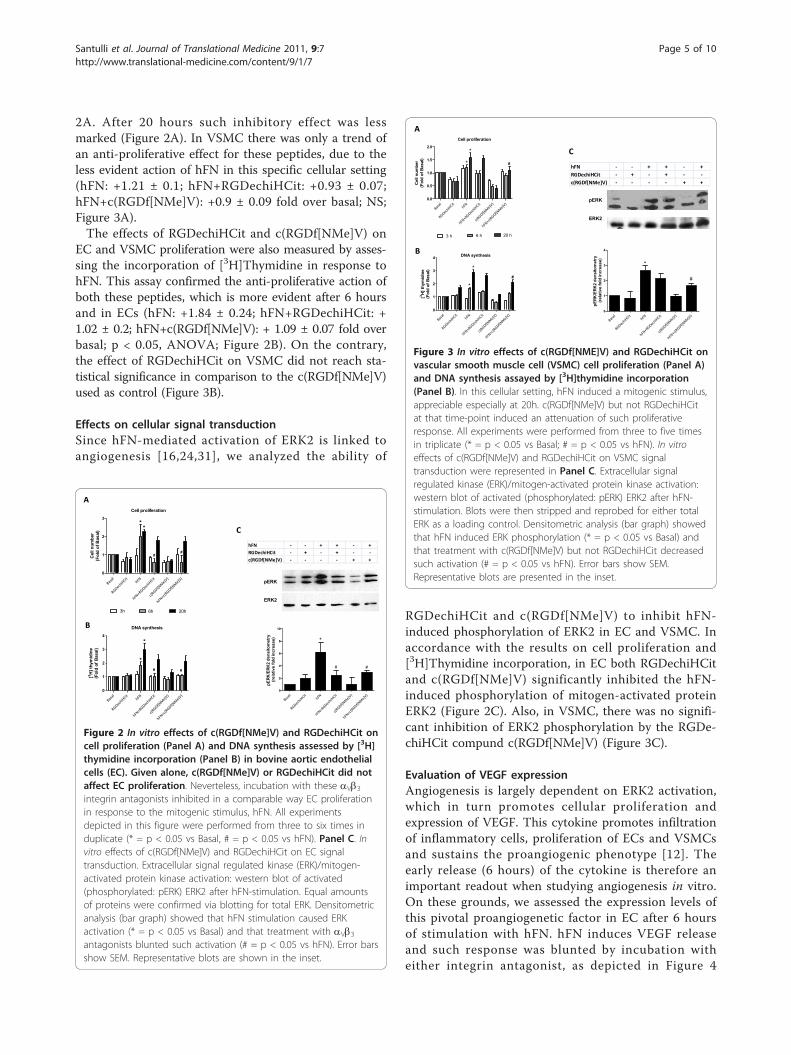
2A. After 20 hours such inhibitory effect was lessmarked (Figure 2A). In VSMC there was only a trend ofan anti-proliferative effect for these peptides, due to theless evident action of hFN in this specific cellular setting(hFN: +1.21 ± 0.1; hFN+RGDechiHCit: +0.93 ± 0.07;hFN+c(RGDf[NMe]V): +0.9 ± 0.09 fold over basal; NS;Figure 3A).The effects of RGDechiHCit and c(RGDf[NMe]V) on
EC and VSMC proliferation were also measured by asses-sing the incorporation of [3H]Thymidine in response tohFN. This assay confirmed the anti-proliferative action ofboth these peptides, which is more evident after 6 hoursand in ECs (hFN: +1.84 ± 0.24; hFN+RGDechiHCit: +1.02 ± 0.2; hFN+c(RGDf[NMe]V): + 1.09 ± 0.07 fold overbasal; p < 0.05, ANOVA; Figure 2B). On the contrary,the effect of RGDechiHCit on VSMC did not reach sta-tistical significance in comparison to the c(RGDf[NMe]V)used as control (Figure 3B).
Effects on cellular signal transductionSince hFN-mediated activation of ERK2 is linked toangiogenesis [16,24,31], we analyzed the ability of
RGDechiHCit and c(RGDf[NMe]V) to inhibit hFN-induced phosphorylation of ERK2 in EC and VSMC. Inaccordance with the results on cell proliferation and[3H]Thymidine incorporation, in EC both RGDechiHCitand c(RGDf[NMe]V) significantly inhibited the hFN-induced phosphorylation of mitogen-activated proteinERK2 (Figure 2C). Also, in VSMC, there was no signifi-cant inhibition of ERK2 phosphorylation by the RGDe-chiHCit compund c(RGDf[NMe]V) (Figure 3C).
Evaluation of VEGF expressionAngiogenesis is largely dependent on ERK2 activation,which in turn promotes cellular proliferation andexpression of VEGF. This cytokine promotes infiltrationof inflammatory cells, proliferation of ECs and VSMCsand sustains the proangiogenic phenotype [12]. Theearly release (6 hours) of the cytokine is therefore animportant readout when studying angiogenesis in vitro.On these grounds, we assessed the expression levels ofthis pivotal proangiogenetic factor in EC after 6 hoursof stimulation with hFN. hFN induces VEGF releaseand such response was blunted by incubation witheither integrin antagonist, as depicted in Figure 4
Basal
RGDechiHCit
hFN
hFN+RGDechiHCit
c(RGDf[NMe]V)
hFN+c(RGDf[NMe]V)
0
1
2
3
3h 6h 20h
**
##
Cel
l num
ber
(Fol
d of
Bas
al)
Cell proliferation
Basal
RGDechiHCit
hFN
hFN+RGDechiHCit
c(RGDf[NMe]V)
hFN+c(RGDf[NMe]V)
0
1
2
3
4
*
*
# #
DNA synthesis
[3 H] t
hym
idin
e(F
old
of B
asal
)
Basal
RGDechiHCit
hFN
hFN+RGDechiHCit
c(RGDf[NMe]V)
hFN+c(RGDf[NMe]V)
0
2
4
6
8
10
*
# #
pERK
/ERK
2 de
nsito
met
ry (r
elat
ive
fold
incr
ease
)
pERK
ERK2
hFN - - + + - +RGDechiHCit - + - + - -c(RGDf[NMe]V) - - - - + +
C
A
B
Figure 2 In vitro effects of c(RGDf[NMe]V) and RGDechiHCit oncell proliferation (Panel A) and DNA synthesis assessed by [3H]thymidine incorporation (Panel B) in bovine aortic endothelialcells (EC). Given alone, c(RGDf[NMe]V) or RGDechiHCit did notaffect EC proliferation. Neverteless, incubation with these aVb3integrin antagonists inhibited in a comparable way EC proliferationin response to the mitogenic stimulus, hFN. All experimentsdepicted in this figure were performed from three to six times induplicate (* = p < 0.05 vs Basal, # = p < 0.05 vs hFN). Panel C. Invitro effects of c(RGDf[NMe]V) and RGDechiHCit on EC signaltransduction. Extracellular signal regulated kinase (ERK)/mitogen-activated protein kinase activation: western blot of activated(phosphorylated: pERK) ERK2 after hFN-stimulation. Equal amountsof proteins were confirmed via blotting for total ERK. Densitometricanalysis (bar graph) showed that hFN stimulation caused ERKactivation (* = p < 0.05 vs Basal) and that treatment with aVb3antagonists blunted such activation (# = p < 0.05 vs hFN). Error barsshow SEM. Representative blots are shown in the inset.
Basal
RGDechiHCit
hFN
hFN+RGDechiHCit
c(RGDf[NMe]V)
hFN+c(RGDf[NMe]V)
0.0
0.5
1.0
1.5
2.0
3 h 6 h 20 h
*
*
#
Cel
l num
ber
(Fol
d of
Bas
al)
Cell proliferation
Basal
RGDechiHCit
hFN
hFN+RGDechiHCit
c(RGDf[NMe]V)
hFN+c(RGDf[NMe]V)
0
1
2
3
4
*
*
#
[3 H] t
hym
idin
e(F
old
of B
asal
)
DNA synthesis
Basal
RGDechiHCit
hFN
hFN+RGDechiHCit
c(RGDf[NMe]V)
hFN+c(RGDf[NMe]V)
0
1
2
3
4
*
#
pERK
/ERK
2 de
nsito
met
ry (r
elat
ive
fold
incr
ease
)
pERK
ERK2
hFN - - + + - +RGDechiHCit - + - + - -c(RGDf[NMe]V) - - - - + +
A
B
C
Figure 3 In vitro effects of c(RGDf[NME]V) and RGDechiHCit onvascular smooth muscle cell (VSMC) cell proliferation (Panel A)and DNA synthesis assayed by [3H]thymidine incorporation(Panel B). In this cellular setting, hFN induced a mitogenic stimulus,appreciable especially at 20h. c(RGDf[NMe]V) but not RGDechiHCitat that time-point induced an attenuation of such proliferativeresponse. All experiments were performed from three to five timesin triplicate (* = p < 0.05 vs Basal; # = p < 0.05 vs hFN). In vitroeffects of c(RGDf[NMe]V) and RGDechiHCit on VSMC signaltransduction were represented in Panel C. Extracellular signalregulated kinase (ERK)/mitogen-activated protein kinase activation:western blot of activated (phosphorylated: pERK) ERK2 after hFN-stimulation. Blots were then stripped and reprobed for either totalERK as a loading control. Densitometric analysis (bar graph) showedthat hFN induced ERK phosphorylation (* = p < 0.05 vs Basal) andthat treatment with c(RGDf[NMe]V) but not RGDechiHCit decreasedsuch activation (# = p < 0.05 vs hFN). Error bars show SEM.Representative blots are presented in the inset.
Santulli et al. Journal of Translational Medicine 2011, 9:7http://www.translational-medicine.com/content/9/1/7
Page 5 of 10
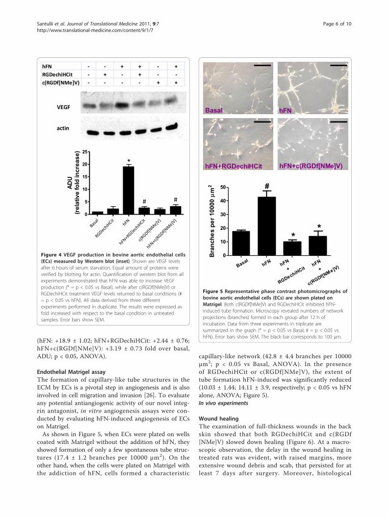
(hFN: +18.9 ± 1.02; hFN+RGDechiHCit: +2.44 ± 0.76;hFN+c(RGDf[NMe]V): +3.19 ± 0.73 fold over basal,ADU; p < 0.05, ANOVA).
Endothelial Matrigel assayThe formation of capillary-like tube structures in theECM by ECs is a pivotal step in angiogenesis and is alsoinvolved in cell migration and invasion [26]. To evaluateany potential antiangiogenic activity of our novel integ-rin antagonist, in vitro angiogenesis assays were con-ducted by evaluating hFN-induced angiogenesis of ECson Matrigel.As shown in Figure 5, when ECs were plated on wells
coated with Matrigel without the addition of hFN, theyshowed formation of only a few spontaneous tube struc-tures (17.4 ± 1.2 branches per 10000 μm2). On theother hand, when the cells were plated on Matrigel withthe addiction of hFN, cells formed a characteristic
capillary-like network (42.8 ± 4.4 branches per 10000μm2; p < 0.05 vs Basal, ANOVA). In the presenceof RGDechiHCit or c(RGDf[NMe]V), the extent oftube formation hFN-induced was significantly reduced(10.03 ± 1.44; 14.11 ± 3.9, respectively; p < 0.05 vs hFNalone, ANOVA; Figure 5).In vivo experiments
Wound healingThe examination of full-thickness wounds in the backskin showed that both RGDechiHCit and c(RGDf[NMe]V) slowed down healing (Figure 6). At a macro-scopic observation, the delay in the wound healing intreated rats was evident, with raised margins, moreextensive wound debris and scab, that persisted for atleast 7 days after surgery. Moreover, histological
hFN - - + + - +RGDechiHCit - + - + - -c(RGDf[NMe]V) - - - - + +
VEGF
actin
Basal
RGDechiHCit
hFN
hFN+RGDechiHCit
c(RGDf[NMe]V)
hFN+c(RGDf[NMe]V)
0
5
10
15
20
25
*
##
ADU
(rel
ativ
e fo
ld in
crea
se)
Figure 4 VEGF production in bovine aortic endothelial cells(ECs) measured by Western blot (inset). Shown are VEGF levelsafter 6 hours of serum starvation. Equal amount of proteins wereverified by blotting for actin. Quantification of western blot from allexperiments demonstrated that hFN was able to increase VEGFproduction (* = p < 0.05 vs Basal), while after c(RGDf[NMe]V) orRGDechiHCit treatment VEGF levels returned to basal conditions (#= p < 0.05 vs hFN). All data derived from three differentexperiments performed in duplicate. The results were expressed asfold increased with respect to the basal condition in untreatedsamples. Error bars show SEM.
0
10
20
30
40
50
*
hFN hFN+
RGDechiHCithFN
+
c(RGDf[NMe]V)
*
Basal
#
Bra
nche
s pe
r 100
00m
2
Basal hFN
hFN+RGDechiHCit hFN+c(RGDf[NMe]V)
____
____
Figure 5 Representative phase contrast photomicrographs ofbovine aortic endothelial cells (ECs) are shown plated onMatrigel. Both c(RGDf[NMe]V) and RGDechiHCit inhibited hFN-induced tube formation. Microscopy revealed numbers of networkprojections (branches) formed in each group after 12 h ofincubation. Data from three experiments in triplicate aresummarized in the graph (* = p < 0.05 vs Basal; # = p < 0.05 vshFN). Error bars show SEM. The black bar corresponds to 100 μm.
Santulli et al. Journal of Translational Medicine 2011, 9:7http://www.translational-medicine.com/content/9/1/7
Page 6 of 10
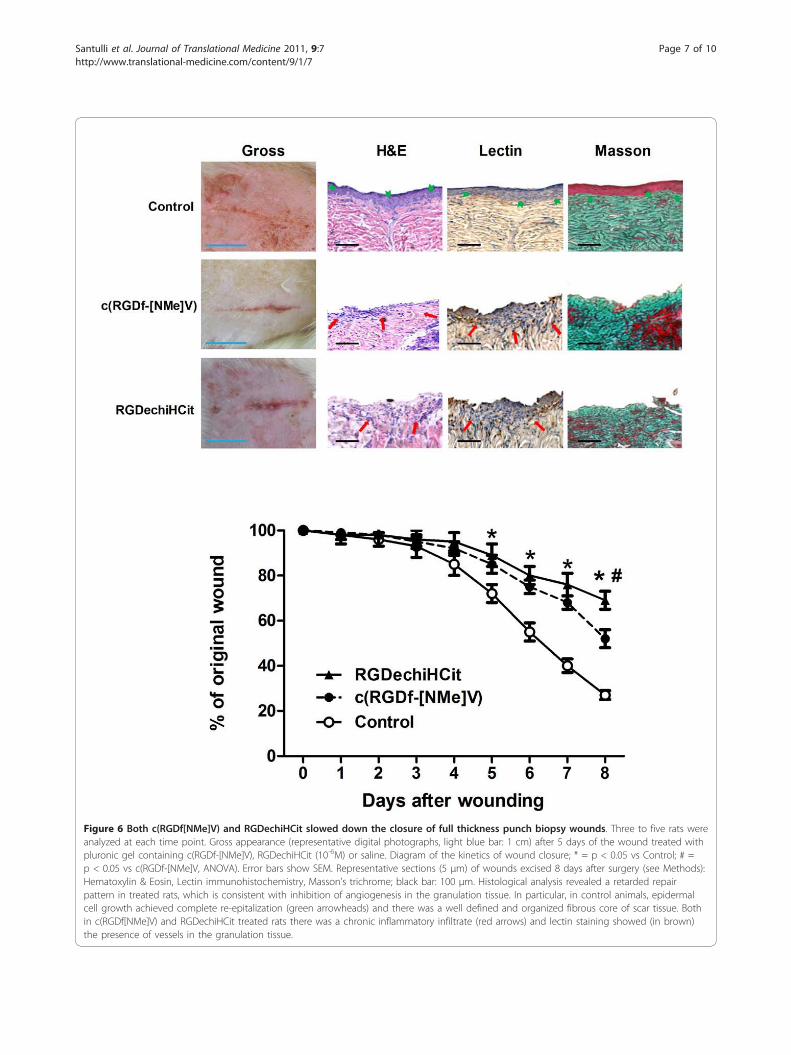
Figure 6 Both c(RGDf[NMe]V) and RGDechiHCit slowed down the closure of full thickness punch biopsy wounds. Three to five rats wereanalyzed at each time point. Gross appearance (representative digital photographs, light blue bar: 1 cm) after 5 days of the wound treated withpluronic gel containing c(RGDf-[NMe]V), RGDechiHCit (10-6M) or saline. Diagram of the kinetics of wound closure; * = p < 0.05 vs Control; # =p < 0.05 vs c(RGDf-[NMe]V, ANOVA). Error bars show SEM. Representative sections (5 μm) of wounds excised 8 days after surgery (see Methods):Hematoxylin & Eosin, Lectin immunohistochemistry, Masson’s trichrome; black bar: 100 μm. Histological analysis revealed a retarded repairpattern in treated rats, which is consistent with inhibition of angiogenesis in the granulation tissue. In particular, in control animals, epidermalcell growth achieved complete re-epitalization (green arrowheads) and there was a well defined and organized fibrous core of scar tissue. Bothin c(RGDf[NMe]V) and RGDechiHCit treated rats there was a chronic inflammatory infiltrate (red arrows) and lectin staining showed (in brown)the presence of vessels in the granulation tissue.
Santulli et al. Journal of Translational Medicine 2011, 9:7http://www.translational-medicine.com/content/9/1/7
Page 7 of 10
analysis showed that while control rats presented adermal scar tissue consisting of a well defined andorganized fibrous core with minimal chronic inflam-matory cells, skin wounds exposed to RGDechiHCit orc(RGDf[NMe]V) exhibited a retarded repair pattern.Indeed, there was an intense inflammatory infiltrate,extended from the wound margin into the region ofthe panniculus carnosus muscle and hypodermis. More-over, the basal epidermis was disorganized and epidermalcell growth failed to achieve re-epithelialization, as shownin Figure 6.
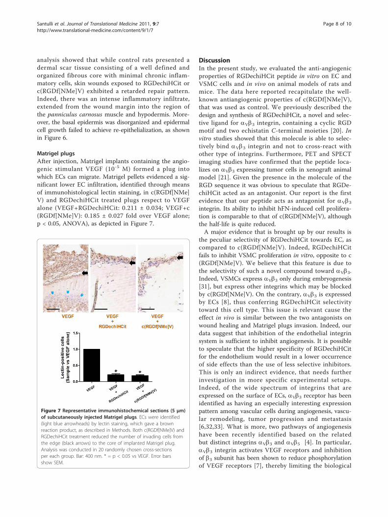
Matrigel plugsAfter injection, Matrigel implants containing the angio-genic stimulant VEGF (10-5 M) formed a plug intowhich ECs can migrate. Matrigel pellets evidenced a sig-nificant lower EC infiltration, identified through meansof immunohistological lectin staining, in c(RGDf[NMe]V) and RGDechiHCit treated plugs respect to VEGFalone (VEGF+RGDechiHCit: 0.211 ± 0.034; VEGF+c(RGDf[NMe]V): 0.185 ± 0.027 fold over VEGF alone;p < 0.05, ANOVA), as depicted in Figure 7.
DiscussionIn the present study, we evaluated the anti-angiogenicproperties of RGDechiHCit peptide in vitro on EC andVSMC cells and in vivo on animal models of rats andmice. The data here reported recapitulate the well-known antiangiogenic properties of c(RGDf[NMe]V),that was used as control. We previously described thedesign and synthesis of RGDechiHCit, a novel and selec-tive ligand for aVb3 integrin, containing a cyclic RGDmotif and two echistatin C-terminal moieties [20]. Invitro studies showed that this molecule is able to selec-tively bind aVb3 integrin and not to cross-react withother type of integrins. Furthermore, PET and SPECTimaging studies have confirmed that the peptide loca-lizes on aVb3 expressing tumor cells in xenograft animalmodel [21]. Given the presence in the molecule of theRGD sequence it was obvious to speculate that RGDe-chiHCit acted as an antagonist. Our report is the firstevidence that our peptide acts as antagonist for aVb3integrin. Its ability to inhibit hFN-induced cell prolifera-tion is comparable to that of c(RGDf[NMe]V), althoughthe half-life is quite reduced.A major evidence that is brought up by our results is
the peculiar selectivity of RGDechiHCit towards EC, ascompared to c(RGDf[NMe]V). Indeed, RGDechiHCitfails to inhibit VSMC proliferation in vitro, opposite to c(RGDf[NMe]V). We believe that this feature is due tothe selectivity of such a novel compound toward aVb3.Indeed, VSMCs express aVb3 only during embryogenesis[31], but express other integrins which may be blockedby c(RGDf[NMe]V). On the contrary, aVb3 is expressedby ECs [8], thus conferring RGDechiHCit selectivitytoward this cell type. This issue is relevant cause theeffect in vivo is similar between the two antagonists onwound healing and Matrigel plugs invasion. Indeed, ourdata suggest that inhibition of the endothelial integrinsystem is sufficient to inhibit angiogenesis. It is possibleto speculate that the higher specificity of RGDechiHCitfor the endothelium would result in a lower occurrenceof side effects than the use of less selective inhibitors.This is only an indirect evidence, that needs furtherinvestigation in more specific experimental setups.Indeed, of the wide spectrum of integrins that areexpressed on the surface of ECs, aVb3 receptor has beenidentified as having an especially interesting expressionpattern among vascular cells during angiogenesis, vascu-lar remodeling, tumor progression and metastasis[6,32,33]. What is more, two pathways of angiogenesishave been recently identified based on the relatedbut distinct integrins aVb3 and aVb5 [4]. In particular,aVb3 integrin activates VEGF receptors and inhibitionof b3 subunit has been shown to reduce phosphorylationof VEGF receptors [7], thereby limiting the biological
Figure 7 Representative immunohistochemical sections (5 μm)of subcutaneously injected Matrigel plugs. ECs were identified(light blue arrowheads) by lectin staining, which gave a brownreaction product, as described in Methods. Both c(RGDf[NMe]V) andRGDechiHCit treatment reduced the number of invading cells fromthe edge (black arrows) to the core of implanted Matrigel plug.Analysis was conducted in 20 randomly chosen cross-sectionsper each group. Bar: 400 nm. * = p < 0.05 vs VEGF. Error barsshow SEM.
Santulli et al. Journal of Translational Medicine 2011, 9:7http://www.translational-medicine.com/content/9/1/7
Page 8 of 10
effects of VEGF [1]. Further, Mahabeleshwar and cowor-kers have shown the intimate interaction occurringbetween aVb3 integrin and the VEGFR-2 in primaryhuman EC [12]. The relevance of this molecule toangiogenesis and its potential as a therapeutic targethas, therefore, been well established [34,35] and in thisreport we show that its activity is highly critical for bothhFN or VEGF-stimulated ECs proliferation.Our results concerning RGDechiHCit in angiogenic
processes are of immediate translational importance,because deregulation of angiogenesis is involved in sev-eral clinical conditions including cancer, ischemic, andinflammatory diseases (atherosclerosis, rheumatoidarthritis, or age-related macular degeneration) [34-36].Therefore, the research for drugs able to modulateangiogenesis constitutes a crucial investigation field.Since RGDechiHCit is rapidly removed in serum it ispossible to increase its effect by engineering the mole-cule to elongate its lifespan. In the present paper we cir-cumvented this issue by increasing the times ofapplication of the drug both in vitro and in vivo, or byreducing the times of observation. This issue can besolved by the use of a more stable aromatic pharmaco-phore that recapitulates the binding properties of RGDe-chiHCit. Clearly, further investigations are also neededto fully understand the basic cell biological mechanismsunderlying growth factor receptors and integrin functionduring angiogenesis. The knowledge of molecular basisof this complex mechanism remains a challenge of fasci-nating interest, with clinical implications for treatmentof a large number of pathophysiological conditionsincluding but not limited to solid tumors [17,37], dia-betic retinopathy [38,39] and inflammatory disease [36].
ConclusionsThe present study indicates the importance of RGDe-chiHCit in the selective inhibition of endothelial aVb3integrin. Such inhibition opens new fields of investiga-tion on the mechanisms of angiogenesis, offering clinicalimplications for the treatment of several conditions suchas proliferative retinopathy, inflammatory disease andcancer.
Author details1Department of Clinical Medicine, Cardiovascular & Immunologic Sciences,“Federico II” University of Naples, Italy. 2Department of Biological Sciences,“Federico II” University of Naples, Italy. 3Institute of Crystallography (ConsiglioNazionale delle Ricerche, CNR), Bari, Italy. 4Institute of Biostructures andBioimaging (Consiglio Nazionale delle Ricerche, CNR), Naples, Italy.
Authors’ contributionsGS and GI designed research; GS, MFB, MDS, CDG, AA, and DS carried outthe experiments; GS and GI performed the statistical analysis; GS, GI and LZdrafted the manuscript; GS, MS, ADG, BT, CP and GI supervised the project;GS and MFB equally contributed to this work. All authors read and approvedthe final manuscript.
Competing interestsWe have no financial or personal relationships with other people ororganizations that would bias our work. No benefits in any form have beenreceived or will be received from a commercial party related directly orindirectly to the subject of our article.
Received: 28 June 2010 Accepted: 13 January 2011Published: 13 January 2011
References1. Santulli G, Ciccarelli M, Palumbo G, Campanile A, Galasso G, Ziaco B,
Altobelli GG, Cimini V, Piscione F, D’Andrea LD, et al: In vivo properties ofthe proangiogenic peptide QK. J Transl Med 2009, 7:41.
2. Bonauer A, Carmona G, Iwasaki M, Mione M, Koyanagi M, Fischer A,Burchfield J, Fox H, Doebele C, Ohtani K, et al: MicroRNA-92a controlsangiogenesis and functional recovery of ischemic tissues in mice. Science2009, 324:1710-1713.
3. Desgrosellier JS, Cheresh DA: Integrins in cancer: biological implicationsand therapeutic opportunities. Nat Rev Cancer 2010, 10:9-22.
4. Hood JD, Frausto R, Kiosses WB, Schwartz MA, Cheresh DA: Differentialalphav integrin-mediated Ras-ERK signaling during two pathways ofangiogenesis. J Cell Biol 2003, 162:933-943.
5. Takahashi S, Moser M, Montanez E, Nakano T, Seo M, Backert S, Inoue I,Awata T, Katayama S, Komoda T, Fassler R: The fibronectin RGD motif isrequired for multiple angiogenic events during early embryonicdevelopment. Arterioscler Thromb Vasc Biol 2010, 30:e1.
6. Castel S, Pagan R, Garcia R, Casaroli-Marano RP, Reina M, Mitjans F, Piulats J,Vilaro S: Alpha v integrin antagonists induce the disassembly of focalcontacts in melanoma cells. Eur J Cell Biol 2000, 79:502-512.
7. Soldi R, Mitola S, Strasly M, Defilippi P, Tarone G, Bussolino F: Role ofalphavbeta3 integrin in the activation of vascular endothelial growthfactor receptor-2. Embo J 1999, 18:882-892.
8. Lu H, Murtagh J, Schwartz EL: The microtubule binding drug laulimalideinhibits vascular endothelial growth factor-induced human endothelialcell migration and is synergistic when combined with docetaxel(taxotere). Mol Pharmacol 2006, 69:1207-1215.
9. Bayless KJ, Salazar R, Davis GE: RGD-dependent vacuolation and lumenformation observed during endothelial cell morphogenesis in three-dimensional fibrin matrices involves the alpha(v)beta(3) and alpha(5)beta(1) integrins. Am J Pathol 2000, 156:1673-1683.
10. Brooks PC, Stromblad S, Sanders LC, von Schalscha TL, Aimes RT, Stetler-Stevenson WG, Quigley JP, Cheresh DA: Localization of matrixmetalloproteinase MMP-2 to the surface of invasive cells by interactionwith integrin alpha v beta 3. Cell 1996, 85:683-693.
11. Abumiya T, Lucero J, Heo JH, Tagaya M, Koziol JA, Copeland BR, delZoppo GJ: Activated microvessels express vascular endothelial growthfactor and integrin alpha(v)beta3 during focal cerebral ischemia. J CerebBlood Flow Metab 1999, 19:1038-1050.
12. Mahabeleshwar GH, Feng W, Reddy K, Plow EF, Byzova TV: Mechanisms ofintegrin-vascular endothelial growth factor receptor cross-activation inangiogenesis. Circ Res 2007, 101:570-580.
13. Xiong JP, Stehle T, Zhang R, Joachimiak A, Frech M, Goodman SL,Arnaout MA: Crystal structure of the extracellular segment of integrinalpha Vbeta3 in complex with an Arg-Gly-Asp ligand. Science 2002,296:151-155.
14. Aumailley M, Gurrath M, Muller G, Calvete J, Timpl R, Kessler H: Arg-Gly-Aspconstrained within cyclic pentapeptides. Strong and selective inhibitorsof cell adhesion to vitronectin and laminin fragment P1. FEBS Lett 1991,291:50-54.
15. Schottelius M, Laufer B, Kessler H, Wester HJ: Ligands for mappingalphavbeta3-integrin expression in vivo. Acc Chem Res 2009, 42:969-980.
16. Eliceiri BP, Klemke R, Stromblad S, Cheresh DA: Integrin alphavbeta3requirement for sustained mitogen-activated protein kinase activityduring angiogenesis. J Cell Biol 1998, 140:1255-1263.
17. Bai J, Zhang J, Wu J, Shen L, Zeng J, Ding J, Wu Y, Gong Z, Li A, Xu S, et al:JWA regulates melanoma metastasis by integrin alpha(V)beta(3)signaling. Oncogene 2010, 29:1227-1237.
18. Eskens FA, Dumez H, Hoekstra R, Perschl A, Brindley C, Bottcher S,Wynendaele W, Drevs J, Verweij J, van Oosterom AT: Phase I andpharmacokinetic study of continuous twice weekly intravenousadministration of Cilengitide (EMD 121974), a novel inhibitor of the
Santulli et al. Journal of Translational Medicine 2011, 9:7http://www.translational-medicine.com/content/9/1/7
Page 9 of 10
integrins alphavbeta3 and alphavbeta5 in patients with advanced solidtumours. Eur J Cancer 2003, 39:917-926.
19. Dechantsreiter MA, Planker E, Matha B, Lohof E, Holzemann G, Jonczyk A,Goodman SL, Kessler H: N-Methylated cyclic RGD peptides as highlyactive and selective alpha(V)beta(3) integrin antagonists. J Med Chem1999, 42:3033-3040.
20. Del Gatto A, Zaccaro L, Grieco P, Novellino E, Zannetti A, Del Vecchio S,Iommelli F, Salvatore M, Pedone C, Saviano M: Novel and selective alpha(v)beta3 receptor peptide antagonist: design, synthesis, and biologicalbehavior. J Med Chem 2006, 49:3416-3420.
21. Zannetti A, Del Vecchio S, Iommelli F, Del Gatto A, De Luca S, Zaccaro L,Papaccioli A, Sommella J, Panico M, Speranza A, et al: Imaging ofalphavbeta3 expression by a bifunctional chimeric RGD peptide notcross-reacting with alphavbeta5. Clin Cancer Res 2009, 15:5224-5233.
22. Ciccarelli M, Cipolletta E, Santulli G, Campanile A, Pumiglia K, Cervero P,Pastore L, Astone D, Trimarco B, Iaccarino G: Endothelial beta2 adrenergicsignaling to AKT: role of Gi and SRC. Cell Signal 2007, 19:1949-1955.
23. Iaccarino G, Ciccarelli M, Sorriento D, Cipolletta E, Cerullo V, Iovino GL,Paudice A, Elia A, Santulli G, Campanile A, et al: AKT participates inendothelial dysfunction in hypertension. Circulation 2004, 109:2587-2593.
24. Illario M, Cavallo AL, Monaco S, Di Vito E, Mueller F, Marzano LA,Troncone G, Fenzi G, Rossi G, Vitale M: Fibronectin-induced proliferation inthyroid cells is mediated by alphavbeta3 integrin through Ras/Raf-1/MEK/ERK and calcium/CaMKII signals. J Clin Endocrinol Metab 2005,90:2865-2873.
25. Iaccarino G, Smithwick LA, Lefkowitz RJ, Koch WJ: Targeting Gbeta gammasignaling in arterial vascular smooth muscle proliferation: a novelstrategy to limit restenosis. Proc Natl Acad Sci USA 1999, 96:3945-3950.
26. Iaccarino G, Ciccarelli M, Sorriento D, Galasso G, Campanile A, Santulli G,Cipolletta E, Cerullo V, Cimini V, Altobelli GG, et al: Ischemicneoangiogenesis enhanced by beta2-adrenergic receptoroverexpression: a novel role for the endothelial adrenergic system. CircRes 2005, 97:1182-1189.
27. Ciccarelli M, Santulli G, Campanile A, Galasso G, Cervero P, Altobelli GG,Cimini V, Pastore L, Piscione F, Trimarco B, Iaccarino G: Endothelial alpha1-adrenoceptors regulate neo-angiogenesis. Br J Pharmacol 2008,153:936-946.
28. Sorriento D, Ciccarelli M, Santulli G, Campanile A, Altobelli GG, Cimini V,Galasso G, Astone D, Piscione F, Pastore L, et al: The G-protein-coupledreceptor kinase 5 inhibits NFkappaB transcriptional activity by inducingnuclear accumulation of IkappaB alpha. Proc Natl Acad Sci USA 2008,105:17818-17823.
29. Sorriento D, Santulli G, Fusco A, Anastasio A, Trimarco B, Iaccarino G:Intracardiac Injection of AdGRK5-NT Reduces Left VentricularHypertrophy by Inhibiting NF-{kappa}B-Dependent Hypertrophic GeneExpression. Hypertension 2010, 56:696-704.
30. Santulli G, Illario M, Palumbo G, Sorriento D, Cipolletta E, Trimarco V, DelGiudice C, Ciccarelli M, Trimarco B, Iaccarino G: CaMK4 partecipates in thesettings of the hypertensive phenotype: a human genome wide analysissupported by animal model. Eur Heart J 2009, 30(Suppl.1):161.
31. Astrof S, Hynes RO: Fibronectins in vascular morphogenesis. Angiogenesis2009, 12:165-175.
32. Zaccaro L, Del Gatto A, Pedone C, Saviano M: Peptides for tumour therapyand diagnosis: current status and future directions. Curr Med Chem 2009,16:780-795.
33. Verbisck NV, Costa ET, Costa FF, Cavalher FP, Costa MD, Muras A, Paixao VA,Moura R, Granato MF, Ierardi DF, et al: ADAM23 negatively modulatesalpha(v)beta(3) integrin activation during metastasis. Cancer Res 2009,69:5546-5552.
34. Laitinen I, Saraste A, Weidl E, Poethko T, Weber AW, Nekolla SG,Leppanen P, Yla-Herttuala S, Holzlwimmer G, Walch A, et al: Evaluation ofalphavbeta3 integrin-targeted positron emission tomography tracer 18F-galacto-RGD for imaging of vascular inflammation in atheroscleroticmice. Circ Cardiovasc Imaging 2009, 2:331-338.
35. Furundzija V, Fritzsche J, Kaufmann J, Meyborg H, Fleck E, Kappert K,Stawowy P: IGF-1 increases macrophage motility via PKC/p38-dependentalphavbeta3-integrin inside-out signaling. Biochem Biophys Res Commun2010, 394:786-791.
36. Vanderslice P, Woodside DG: Integrin antagonists as therapeutics forinflammatory diseases. Expert Opin Investig Drugs 2006, 15:1235-1255.
37. Tani N, Higashiyama S, Kawaguchi N, Madarame J, Ota I, Ito Y, Ohoka Y,Shiosaka S, Takada Y, Matsuura N: Expression level of integrin alpha 5 ontumour cells affects the rate of metastasis to the kidney. Br J Cancer2003, 88:327-333.
38. Crawford TN, Alfaro DV, Kerrison JB, Jablon EP: Diabetic retinopathy andangiogenesis. Curr Diabetes Rev 2009, 5:8-13.
39. Santulli RJ, Kinney WA, Ghosh S, Decorte BL, Liu L, Tuman RW, Zhou Z,Huebert N, Bursell SE, Clermont AC, et al: Studies with an orallybioavailable alpha V integrin antagonist in animal models of ocularvasculopathy: retinal neovascularization in mice and retinal vascularpermeability in diabetic rats. J Pharmacol Exp Ther 2008, 324:894-901.
doi:10.1186/1479-5876-9-7Cite this article as: Santulli et al.: Evaluation of the anti-angiogenicproperties of the new selective aVb3 integrin antagonist RGDechiHCit.Journal of Translational Medicine 2011 9:7.
Submit your next manuscript to BioMed Centraland take full advantage of:
• Convenient online submission
• Thorough peer review
• No space constraints or color figure charges
• Immediate publication on acceptance
• Inclusion in PubMed, CAS, Scopus and Google Scholar
• Research which is freely available for redistribution
Submit your manuscript at www.biomedcentral.com/submit
Santulli et al. Journal of Translational Medicine 2011, 9:7http://www.translational-medicine.com/content/9/1/7
Page 10 of 10
Copyright © 2022 FDOKUMEN