Evaluation of kinetic programs in various automated perimeters
8
CLINICAL INVESTIGATION Evaluation of kinetic programs in various automated perimeters Shigeki Hashimoto 1 • Chota Matsumoto 1 • Mariko Eura 1 • Sachiko Okuyama 1 • Yoshikazu Shimomura 1 Received: 14 July 2016 / Accepted: 7 March 2017 Ó Japanese Ophthalmological Society 2017 Abstract Purpose Kinetic programs in four automated perimeters were evaluated and compared for their clinical usefulness using four simulated visual field (VF) patterns. Methods Using the results of conventional Goldmann manual kinetic perimetry (MKP), simulated fields with concentric contraction, a temporal residual island only, a small central island with a temporal island, and a ring scotoma were created. Four kinetic programs, Humphrey 750i Kinetic Test (Humphrey), OCULUS Twinfield 2 Kinetic Perimetry (OCULUS), OCTOPUS 900 Goldmann Kinetic Perimetry (OCTOPUS GKP), and Kowa AP-7000 Isopter (Kowa) were tested by the 4 simulated defect pat- terns using stimuli of V/4e, I/4e, I/3e, I/2e, and I/1e at speeds of 3 and 5°/s. Results Except Humphrey, OCULUS, OCTOPUS GKP, and Kowa could obtain isopters nearly comparable to those of Goldmann MKP. However, their results were consider- ably influenced by the examiner’s skill. Besides, Humphrey had restrictions on target presentation, and OCULUS and Kowa had problems in isopter drawing and in filling in the scotoma. OCTOPUS GKP was the only method that could correctly detect and depict all four defect patterns. It also had relatively shorter test durations among the three methods excluding Humphrey, which did not have a built- in function for test duration measurement. The perimeters’ test ranges for the periphery were 90° for Humphrey, OCULUS, and OCTOPUS GKP, and 80° for Kowa. Conclusion To assess kinetic fields with various defect patterns, OCTOPUS GKP seems to be the most useful method. Keywords Automated kinetic perimetry Á Humphrey 750i Kinetic Test Á OCULUS Twinfield 2 Kinetic Perimetry Á OCTOPUS 900 Goldmann Kinetic Perimetry Á Kowa AP- 7000 Isopter Introduction There are two methods to evaluate the visual field (VF) in perimetry, kinetic and static. In kinetic perimetry, target size and luminance are fixed and the target is moved to determine an isopter. In static perimetry the target is fixed at a determined test point and the luminance is changed to measure the visibility at each test point [1]. Currently, static perimetry is the mainstream due to the spread of automated perimeters and has gained more popularity than kinetic perimetry for a few practical reasons. Because static perimetry is usually used to evaluate the central 30° of the VF, the test duration is significantly shortened. This is clinically useful as it is difficult to measure the entire VF within a limited test duration. Moreover, sensitivity results fluctuate more in the peripheral VF than in the central 30° and thus, test results by static perimetry are more reliable than by a kinetic method. On the other hand, kinetic perimetry remains an important method to evaluate the VF in patients with advanced glaucoma, retinal diseases, and other optic neu- ropathy [2, 3]. Manual kinetic perimetry (MKP) using a Goldmann perimeter [4] is widely used owing to its capability in measuring the entire VF, including the periphery and center, in a relatively shorter duration. With & Shigeki Hashimoto [email protected] 1 Department of Ophthalmology, Faculty of Medicine, Kindai University, Ohnohigashi, Osakasayama City, Osaka 589-8511, Japan 123 Jpn J Ophthalmol DOI 10.1007/s10384-017-0516-y
-
Upload
khangminh22 -
Category
Documents
-
view
0 -
download
0
Transcript of Evaluation of kinetic programs in various automated perimeters
CLINICAL INVESTIGATION
Evaluation of kinetic programs in various automated perimeters
Shigeki Hashimoto1 • Chota Matsumoto1 • Mariko Eura1 • Sachiko Okuyama1 •
Yoshikazu Shimomura1
Received: 14 July 2016 / Accepted: 7 March 2017
� Japanese Ophthalmological Society 2017
Abstract
Purpose Kinetic programs in four automated perimeters
were evaluated and compared for their clinical usefulness
using four simulated visual field (VF) patterns.
Methods Using the results of conventional Goldmann
manual kinetic perimetry (MKP), simulated fields with
concentric contraction, a temporal residual island only, a
small central island with a temporal island, and a ring
scotoma were created. Four kinetic programs, Humphrey
750i Kinetic Test (Humphrey), OCULUS Twinfield 2
Kinetic Perimetry (OCULUS), OCTOPUS 900 Goldmann
Kinetic Perimetry (OCTOPUS GKP), and Kowa AP-7000
Isopter (Kowa) were tested by the 4 simulated defect pat-
terns using stimuli of V/4e, I/4e, I/3e, I/2e, and I/1e at
speeds of 3 and 5�/s.Results Except Humphrey, OCULUS, OCTOPUS GKP,
and Kowa could obtain isopters nearly comparable to those
of Goldmann MKP. However, their results were consider-
ably influenced by the examiner’s skill. Besides, Humphrey
had restrictions on target presentation, and OCULUS and
Kowa had problems in isopter drawing and in filling in the
scotoma. OCTOPUS GKP was the only method that could
correctly detect and depict all four defect patterns. It also
had relatively shorter test durations among the three
methods excluding Humphrey, which did not have a built-
in function for test duration measurement. The perimeters’
test ranges for the periphery were 90� for Humphrey,
OCULUS, and OCTOPUS GKP, and 80� for Kowa.
Conclusion To assess kinetic fields with various defect
patterns, OCTOPUS GKP seems to be the most useful
method.
Keywords Automated kinetic perimetry � Humphrey 750i
Kinetic Test � OCULUS Twinfield 2 Kinetic Perimetry �OCTOPUS 900 Goldmann Kinetic Perimetry � Kowa AP-
7000 Isopter
Introduction
There are two methods to evaluate the visual field (VF) in
perimetry, kinetic and static. In kinetic perimetry, target
size and luminance are fixed and the target is moved to
determine an isopter. In static perimetry the target is fixed
at a determined test point and the luminance is changed to
measure the visibility at each test point [1]. Currently,
static perimetry is the mainstream due to the spread of
automated perimeters and has gained more popularity than
kinetic perimetry for a few practical reasons. Because static
perimetry is usually used to evaluate the central 30� of theVF, the test duration is significantly shortened. This is
clinically useful as it is difficult to measure the entire VF
within a limited test duration. Moreover, sensitivity results
fluctuate more in the peripheral VF than in the central 30�and thus, test results by static perimetry are more reliable
than by a kinetic method.
On the other hand, kinetic perimetry remains an
important method to evaluate the VF in patients with
advanced glaucoma, retinal diseases, and other optic neu-
ropathy [2, 3]. Manual kinetic perimetry (MKP) using a
Goldmann perimeter [4] is widely used owing to its
capability in measuring the entire VF, including the
periphery and center, in a relatively shorter duration. With
& Shigeki Hashimoto
1 Department of Ophthalmology, Faculty of Medicine, Kindai
University, Ohnohigashi, Osakasayama City,
Osaka 589-8511, Japan
123
Jpn J Ophthalmol
DOI 10.1007/s10384-017-0516-y

Goldmann MKP, the VF can be diagnosed at a glance in its
entirety. However, Goldmann MKP has many disadvan-
tages including examiner bias, intra-examiner differences
in choosing stimulus velocity [5], and lack of standard-
ization, autocalibration, and permanent documentation of
test procedures and results. Automated kinetic perimetry
can help minimize problems related to perimetric technique
and thus reduces examiner bias [6–11]. Starting with the
revolutionary Fieldmaster 5000 [12–15] and Perimetron
[16, 17], various types of automated kinetic perimeters
have been developed and several have become commer-
cially available. At the same time, Goldmann MKP is still
the standard kinetic method [4].
To find an automated kinetic method that has advantages
over MKP and produces results comparable to those of
Goldmann MKP, we evaluated the clinical usefulness of
the following four extant kinetic programs in automated
perimeters: (1) Humphrey 750i Kinetic Test (Humphrey;
Carl Zeiss Meditec, Dublin, CA, USA) [18, 19], (2)
OCULUS Twinfield 2 Kinetic Perimetry (OCULUS;
Oculus Inc., Wetzlar, Germany) [11], (3) OCTOPUS 900
Goldmann Kinetic Perimetry (OCTOPUS GKP; Haag-
Streit Inc., Koeniz, Switzerland) [6–10], and (4) Kowa AP-
7000 Isopter (Kowa Inc., Nagoya, Japan) [20]. To test these
four methods, four different VF defect patterns (concentric
contraction, a temporal residual island only, a small central
island with a temporal island, and a ring scotoma) were
created and the test results, range, and duration of the four
methods were compared. To our knowledge, no such
comparison and evaluation have been made previously.
Materials and methods
Using previous test results of conventional Goldmann
MKP, we created four virtual VF patterns: concentric
contraction (Fig. 1a), a temporal residual island of vision
only (Fig. 1b), a small central island with a temporal island
(Fig. 1c), and a ring scotoma (Fig. 1d). The patterns were
made using shading filters and were fixed on an eye cup
made of clear plastic material (Fig. 2a, b). We first mapped
the Goldmann MKP test results of the eye cup by matching
the visual angles in the temporal, nasal, superior and
inferior fields. Using the filter, a defect pattern of the shape
and size of the defect depicted by Goldmann MKP was
then cut out and attached to the eye cup. The shading filters
used Zero Black Film (Mirareed Corporation, Tokyo,
Japan) with 0.004% light transmission; the examinee could
not perceive any stimulus through this film. The subject of
this study was a healthy 30-year-old woman volunteer with
visual acuity of 20/20 who had previous experience with
kinetic perimetry. The subject put on the eye cup to test the
4 perimeters with each of the 4 VF patterns in turn, a total
of 16 tests. An opaque occluder covered the untested eye
during the perimetric test. All the examinations were per-
formed by the same examiner (SH).
The four commercially-available kinetic programs tes-
ted in this study were Humphrey (Carl Zeiss Meditec),
OCULUS (OCULUS Optikgerate) OCTOPUS GKP (Haag
Streit AG), and Kowa (Kowa Inc.). Accoding to the man-
ufacturers’ specifications, the 4 perimeters have similar
specifications except the maximum luminance (Humphrey,
3183 cd/m2; OCULUS, 318 cd/m2; OCTOPUS GKP,
1910 cd/m2; and Kowa, 3183 cd/m2). The background
luminance is 10 cd/m2 for all 4 programs. While Hum-
phrey and Kowa have built-in kinetic software, the hemi-
spherical bowls of OCTOPUS and OCULUS are connected
to and controlled by separated external personal computers
with the perimeters’ own kinetic software. All four kinetic
programs have both semi-automated and fully-automated
testing options. In fully-automated testing, stimuli are
automatically moved along fixed meridians determined
before the test. The examiner cannot add isopters in real
time even if abnormalities are found in some areas of the
VF. The program also automatically depicts the final
isopter. In semi-automated testing, the examiner has to
determine the initial/end points and direction of the vec-
tors, manually connect all the response points on the
computer screen to depict an isopter, and add additional
vectors if necessary. To obtain more detailed test results for
better comparison, we only used the semi-automated test-
ing option in the four programs.
In this study, 5 stimuli (V/4e, I/4e, I/3e, I/2e, and I/1e)
were used to assess the VF loss. Stimulus speeds of 3 and
5�/s were, respectively, used within and beyond 30�eccentricity [4]. At a constant stimulus speed, stimuli of a
selected size and at a luminance level according to the
Goldmann classification [4] were generated by the kinetic
programs. The generated stimuli were continously moved
from nonvisible areas towards visible areas of the VF along
user-defined vectors that were drawn manually by the
examiner with a computer mouse or an electronic pen
directly on the computer touch screen either before or
during the perimetric test. During perimetry, the examiner
had to select the initial and end points, direction, and length
of the vectors for the three methods excluding Humphrey.
In the Humphrey program, the end point is always fixed at
(x, y) = (0,0) and thus, the examiner only needs to select
the initial point. The stimulus movement along each vector
was terminated by the response of the subject, who was
instructed to look straight ahead at the fixation point and to
press the response button as soon as the stimulus was
perceived. Upon the subject’s response, the stimulus
location was automatically marked on the screen and
finally, the examiner manually connected all the response
points on the computer screen to depict an isopter. Vectors
S. Hashimoto et al.
123

Fig. 1 Results of conventional Goldmann MKP for the four VF patterns. a Concentric contraction. b A temporal residual island only. c A small
central island with a temporal island. d A ring scotoma
Fig. 2 a Eye cup. b Eye cup
with shading filter
Evaluation of kinetic programs in various automated perimeters
123

were presented about 20� apart in a random order from the
periphery towards the central field, and additional vectors
were manually added by the examiner to the areas indi-
cating abnormality depending on the depth and shape of the
VF pattern. In a scotoma, the stimulus was presented inside
the region of the field loss and moved outwards until the
stimulus was perceived to determine the border of the
scotoma. In this study, the subject’s fixation was monitored
by a video camera, except for OCTOPUS which has a
built-in fixation monitoring mode. Test duration was
automatically measured in the three programs excluding
Humphrey, which did not have a built-in function for
measuring test duration. Test duration in these semi-auto-
mated programs included the time for the examiner to
select the initial/end points, direction, and length of vec-
tors. Test results were automatically digitalized and
recorded in the four programs.
This study protocol was approved by the Ethics Com-
mittee of Kindai University Faculty of Medicine and all the
experiments were performed in accordance with the
Declaration of Helsinki. Informed consent was obtained
from the subject.
Results
Figure 3 shows the results of the 4 kinetic programs for
concentric contraction and all 4 methods could obtain
results comparable to the result by Goldmann MKP. The
recorded test durations, except for Humphrey, were
8.58 min. (OCULUS), 4.35 min. (OCTOPUS GKP), and
9.31 min.s (Kowa). OCTOPUS GKP had a considerably
shorter test dutration than the other two methods. Figure 4
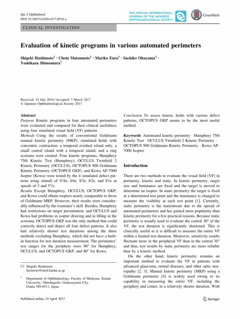
shows the results for the VF with a temporal residual island
only. Humphrey depicted an incomplete isopter with the
joined temporal residual island and central point (Fig. 4a).
The results of the other three methods were comparable to
the Goldmann MKP result. The recorded test durations
were 7.51 min. (OCULUS), 7.95 min. (OCTOPUS GKP),
and 6.90 min. (Kowa). Figure 5 shows the results for a
Fig. 3 Results for the pattern with concentric contraction. a Humphrey. b OCULUS. c OCTOPUS GKP. d Kowa. The four results for this
pattern were close equivalents
S. Hashimoto et al.
123
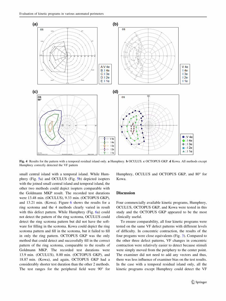
small central island with a temporal island. While Hum-
phrey (Fig. 5a) and OCULUS (Fig. 5b) depicted isopters
with the joined small central island and temporal island, the
other two methods could depict isopters comparable with
the Goldmann MKP result. The recorded test durations
were 13.48 min. (OCULUS), 9.33 min. (OCTOPUS GKP),
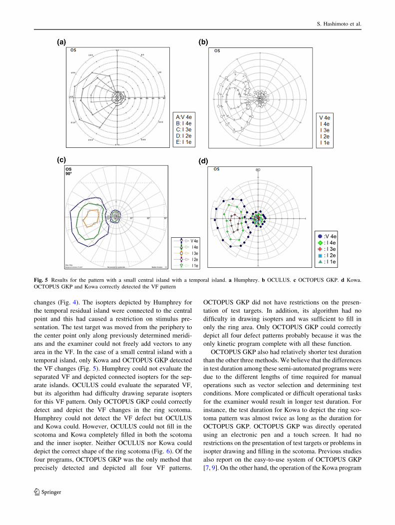
and 13.21 min. (Kowa). Figure 6 shows the results for a
ring scotoma and the 4 methods clearly varied in result
with this defect pattern. While Humphrey (Fig. 6a) could
not detect the pattern of the ring scotoma, OCULUS could
detect the ring scotoma pattern but did not have the soft-
ware for filling in the scotoma. Kowa could depict the ring
scotoma pattern and fill in the scotoma, but it failed to fill
in only the ring pattern. OCTOPUS GKP was the only
method that could detect and successfully fill in the correct
pattern of the ring scotoma, comparable to the results of
Goldmann MKP. The recorded test durations were
13.9 min. (OCULUS), 8.00 min. (OCTOPUS GKP), and
18.87 min. (Kowa), and again, OCTOPUS GKP had a
considerably shorter test duration than the other 2 methods.
The test ranges for the peripheral field were 90� for
Humphrey, OCULUS and OCTOPUS GKP, and 80� for
Kowa.
Discussion
Four commercially available kinetic programs, Humphrey,
OCULUS, OCTOPUS GKP, and Kowa were tested in this
study and the OCTOPUS GKP appeared to be the most
clinically useful.
To ensure comparability, all four kinetic programs were
tested on the same VF defect patterns with different levels
of difficulty. In concentric contraction, the results of the
four progams were close equivalents (Fig. 3). Compared to
the other three defect patterns, VF changes in concentric
contraction were relatively easier to detect because stimuli
were simply moved from the periphery to the center point.
The examiner did not need to add any vectors and thus,
there was less influence of examiner bias on the test results.
In the case with a temporal residual island only, all the
kinetic programs except Humphrey could detect the VF
Fig. 4 Results for the pattern with a temporal residual island only. a Humphrey. b OCULUS. c OCTOPUS GKP. d Kowa. All methods except
Humphrey correctly detected the VF pattern
Evaluation of kinetic programs in various automated perimeters
123
changes (Fig. 4). The isopters depicted by Humphrey for
the temporal residual island were connected to the central
point and this had caused a restriction on stimulus pre-
sentation. The test target was moved from the periphery to
the center point only along previously determined meridi-
ans and the examiner could not freely add vectors to any
area in the VF. In the case of a small central island with a
temporal island, only Kowa and OCTOPUS GKP detected
the VF changes (Fig. 5). Humphrey could not evaluate the
separated VF and depicted connected isopters for the sep-
arate islands. OCULUS could evaluate the separated VF,
but its algorithm had difficulty drawing separate isopters
for this VF pattern. Only OCTOPUS GKP could correctly
detect and depict the VF changes in the ring scotoma.
Humphrey could not detect the VF defect but OCULUS
and Kowa could. However, OCULUS could not fill in the
scotoma and Kowa completely filled in both the scotoma
and the inner isopter. Neither OCULUS nor Kowa could
depict the correct shape of the ring scotoma (Fig. 6). Of the
four programs, OCTOPUS GKP was the only method that
precisely detected and depicted all four VF patterns.
OCTOPUS GKP did not have restrictions on the presen-
tation of test targets. In addition, its algorithm had no
difficulty in drawing isopters and was sufficient to fill in
only the ring area. Only OCTOPUS GKP could correctly
depict all four defect patterns probably because it was the
only kinetic program complete with all these function.
OCTOPUS GKP also had relatively shorter test duration
than the other three methods.We believe that the differences
in test duration among these semi-automated programs were
due to the different lengths of time required for manual
operations such as vector selection and determining test
conditions. More complicated or difficult operational tasks
for the examiner would result in longer test duration. For
instance, the test duration for Kowa to depict the ring sco-
toma pattern was almost twice as long as the duration for
OCTOPUS GKP. OCTOPUS GKP was directly operated
using an electronic pen and a touch screen. It had no
restrictions on the presentation of test targets or problems in
isopter drawing and filling in the scotoma. Previous studies
also report on the easy-to-use system of OCTOPUS GKP
[7, 9]. On the other hand, the operation of the Kowa program
Fig. 5 Results for the pattern with a small central island with a temporal island. a Humphrey. b OCULUS. c OCTOPUS GKP. d Kowa.
OCTOPUS GKP and Kowa correctly detected the VF pattern
S. Hashimoto et al.
123

was more time-consuming. Furthermore, its drawing soft-
ware was not entirely capable of depicting the ring scotoma
and hence the test duration was further extended. Humphrey,
OCULUS, andOCTOPUShad almost the same test range for
the peripheral VF as Goldmann MKP, while Kowa had a
slightly narrower range due to the structure of its cupola.
This study had some limitations. We only simulated four
defect patterns this time. In the future, more defect patterns
with distinctive features could further differentiate various
kinetic methods. In addition, in order not to extend the test
duration, there was only one subject in the plastic eye cup
trial with the four defect patterns and all the tests were
performed by the same examiner to minimize bias. In
future the number of subjects needs to be increased for
better reproducibility as well as to test if there are any
intra-individual differences in test duration or results.
Although we could successfully monitor the subject’s fix-
ation in this study, other examiners might find it difficult to
monitor the subject’s fixation through a plastic eye cup
with a defect pattern made of a light transmission filter.
Careful fixation confirmation will be necessary.
We tested and compared the four methods with the same
VF patterns in this study to evaluate the latest available
automated kinetic methods. Automated kinetic perimetry
has some common drawbacks such as examiner bias since
additional meridians and the final isopter are determined
and drawn by the examiner. Inevitably, the examiner’s skill
level and experience substantially affects the test precision.
Such problems can only be solved by the establishment of a
fully automated kinetic method, a system completely free
from examiner’s bias.
Acknowledgements The authors wish to thank Ms. Reiyo Tahara
and Ms. Yukiko Mimuro for their editorial helps.
Conflicts of interest All authors declare that they have no competing
interest.
References
1. Aulhorn E. Glaukoma-Gesichtsfield. Ophthalmologica.
1968;158:469–87 (in German).
Fig. 6 Results for the pattern with a ring scotoma. a Humphrey. b OCULUS. c OCTOPUS GKP. d Kowa. Only OCTOPUS GKP correctly
detected the VF pattern
Evaluation of kinetic programs in various automated perimeters
123

2. Grover S, Fishman GA, Brown J Jr. Patterns of visual field
progression in patients with retinitis pigmentosa. Ophthalmology.
1988;105:1069–75.
3. Chauhan BC, Drance SM. The relationship between intraocular
pressure and visual field progression in glaucoma. Graefes Arch
Clin Expo Ophthalmol. 1992;230:521–6.
4. Goldmann H. Ein selbstregistrierendes Projektionskugelperime-
ter. Ophthalmologica. 1945;109:71–9 (in German).5. Bittner AK, Iftikhar MH, Dagnelie G. Test-retest, within-visit
variability of Goldmann visual fields in retinitis pigmentosa.
Invest Ophthalmol Vis Sci. 2011;11:8042–6.
6. Schiefer U, Strasburger H, Becker ST, Vonthein R, Schiller J,
Dietrich TJ, et al. Reaction time in automated kinetic perimetry:
effects of stimulus luminance, eccentricity, and movement
direction. Vision Res. 2001;41:2157–64.
7. Nowomiejska KE, Vonthein R, Paetzold J, Zagorski Z, Kardon R,
Schiefer U. Comparison between semiautomated kinetic
perimetry and conventional Goldmann manual kinetic perimetry
in advanced visual field loss. Ophthalmoly. 2005;112:1343–54.
8. Schiefer U, Nowomiejska K, Krapp E, Paetzold J, Johnson CA.
K-Train- a computer-based, interactive training propram with an
incorporated certification system for practicing kinetic perimetry:
evaluation of acceptance and success rate. Graefes Arch Clin
Expo Ophthalmol. 2006;244:1300–9.
9. Nevalainen J, Paetzold J, Krapp E, Vonthein R, Johnson CA,
Schiefer U. The use of semi-automated kinetic perimetry (SPK)
to monitor advanced glaucomatous visual field loss. Graefes Arch
Clin Expo Ophthalmol. 2008;246:1331–9.
10. Nowomiejska K, Vonthein R, Paetzold J, Zagorski Z, Kardon R,
Schiefer U. Reaction time during semi-automated kinetic
perimetry (SPK) in patients with advanced visual field loss. Acta
Ophthalmol. 2010;88:65–9.
11. Wilscher S, Wabbels B, Lorenz B. Feasibility and outocome of
automated kinetic perimetry in children. Graefes Arch Clin Exp
Ophthalmolo. 2010;248:1493–500.
12. Damgaard-Jenson L. Vertical steps in isopters at the hemianopic
border in normal and glacomatous eye. Acta Ophthalmol.
1977;55:111–21.
13. Stewart WC, Shields MB, Ollie AR. Peripheral visual field test-
ing by automated kinetic perimetry in glaucoma. Arch Ophthal-
mol. 1988;106:202–6.
14. Miller KN, Shields MB, Ollie AR. Automated kinetic perimetry
with two peripheral isopters in glaucoma. Arch Ophthalmol.
1989;107:1316–20.
15. Gilpin LB, Stewart WC, Shields MB, Miller KN. Hemianopic
offsets in the visual field of patients with glaucoma. Graefes Arch
Clin Expo Ophthalmol. 1990;228:450–3.
16. Portney GL, Krohn MA. Automated perimetry, background,
insutruments and methods. Sury Ophthalmol. 1978;22:271–8.
17. Heijl A, Drance SM. A clinical comparison of three computerized
automatic perimeters in the detection of glaucoma defects. Arch
Ophthalmol. 1981;99:832–6.
18. Lynn JR, Swanson WH, Fellmann RL. Evaluation of automated
kinetic perimetry (AKP) with the Humphrey Field Analyser.
Perimetry Update. 1991;1990(1991):433–52.
19. Ballon BJ, Echelman DA, Shields MB, Ollie AR. Peripheral
visual field testing in glaucoma by automated kinetic perimetry
with the Humphrey Field Analyzer. Arch Ophthalmol.
1992;110:1730–2.
20. Omodaka K, Kunimatsu-Sanuki S, Morin R, Tsuda S, Yokoyama
Y, Takahashi H, et al. Development of a new strategy of visual
field testing for macular dysfunction in patients with open angle
glaucoma. Jpn J Ophthalmol. 2013;57:457–62.
S. Hashimoto et al.
123