Evaluation of ELISA coupled with Western blot as a surveillance tool for Trichinella infection in...
12
Veterinary Parasitology 199 (2014) 179–190 Contents lists available at ScienceDirect Veterinary Parasitology jo u r nal homep age: www.elsevier.com/locate/vetpar Evaluation of ELISA coupled with Western blot as a surveillance tool for Trichinella infection in wild boar (Sus scrofa) Leigh Cuttell a,∗ , Maria Angeles Gómez-Morales b , Beth Cookson c , Peter J. Adams d , Simon A. Reid e , Paul B. Vanderlinde f , Louise A. Jackson g , C. Gray h , Rebecca J. Traub a a School of Veterinary Science, The University of Queensland, Gatton, QLD 4343, Australia b European Reference Laboratory for Parasites, Instituto Superiore di Sanità, Viale Regina Elena 299, 00161 Rome, Italy c Australian Department of Agriculture, Fisheries and Forestry, DAFF Biosecurity Northern Region, Box 96, Cairns International Airport, Cairns, QLD 4878, Australia d School of Veterinary and Life Sciences, Murdoch University, Murdoch, WA 6150, Australia e School of Population Health, The University of Queensland, Herston, QLD 4006, Australia f Australian Department of Agriculture, Fisheries & Forestry, PO Box 222, Hamilton Central, QLD 4007, Australia g Department of Agriculture, Fisheries & Forestry, Biosecurity Sciences Laboratory, Coopers Plains, QLD 4108, Australia h Peninsula School of Medicine & Dentistry, Plymouth University, Plymouth PL4 8AA, United Kingdom a r t i c l e i n f o Article history: Received 18 August 2013 Received in revised form 8 October 2013 Accepted 15 October 2013 Keywords: Trichinella ELISA Western blot Serosurveillance Wildlife Real-time PCR a b s t r a c t Trichinella surveillance in wildlife relies on muscle digestion of large samples which are logistically difficult to store and transport in remote and tropical regions as well as labour- intensive to process. Serological methods such as enzyme-linked immunosorbent assays (ELISAs) offer rapid, cost-effective alternatives for surveillance but should be paired with additional tests because of the high false-positive rates encountered in wildlife. We inves- tigated the utility of ELISAs coupled with Western blot (WB) in providing evidence of Trichinella exposure or infection in wild boar. Serum samples were collected from 673 wild boar from a high- and low-risk region for Trichinella introduction within mainland Australia, which is considered Trichinella-free. Sera were examined using both an ‘in-house’ and a commercially available indirect-ELISA that used excretory–secretory (E/S) antigens. Cut-off values for positive results were determined using sera from the low-risk popula- tion. All wild boar from the high-risk region (352) and 139/321 (43.3%) of the wild boar from the low-risk region were tested by artificial digestion. Testing by Western blot using E/S antigens, and a Trichinella-specific real-time PCR was also carried out on all ELISA- positive samples. The two ELISAs correctly classified all positive controls as well as one naturally infected wild boar from Gabba Island in the Torres Strait. In both the high- and low-risk populations, the ELISA results showed substantial agreement (k-value = 0.66) that increased to very good (k-value = 0.82) when WB-positive only samples were compared. The results of testing sera collected from the Australian mainland showed the Trichinella seroprevalence was 3.5% (95% C.I. 0.0–8.0) and 2.3% (95% C.I. 0.0–5.6) using the in-house and commercial ELISA coupled with WB respectively. These estimates were significantly higher (P < 0.05) than the artificial digestion estimate of 0.0% (95% C.I. 0.0–1.1). Real-time PCR test- ing of muscle from seropositive animals did not detect Trichinella DNA in any mainland animals, but did reveal the presence of a second larvae-positive wild boar on Gabba Island, ∗ Corresponding author. Tel.: +61 7 54601851; fax: +61 7 54601922. E-mail addresses: [email protected] (L. Cuttell), [email protected] (M.A. Gómez-Morales), [email protected] (B. Cookson), [email protected] (P.J. Adams), [email protected] (S.A. Reid), [email protected] (P.B. Vanderlinde), [email protected] (L.A. Jackson), [email protected] (C. Gray), [email protected] (R.J. Traub). 0304-4017/$ – see front matter © 2013 Elsevier B.V. All rights reserved. http://dx.doi.org/10.1016/j.vetpar.2013.10.012
Transcript of Evaluation of ELISA coupled with Western blot as a surveillance tool for Trichinella infection in...

Esb
LPCa
b
c
Cd
e
f
g
h
ARRA
KTEWSWR
p(
0h
Veterinary Parasitology 199 (2014) 179– 190
Contents lists available at ScienceDirect
Veterinary Parasitology
jo u r nal homep age: www.elsev ier .com/ locate /vetpar
valuation of ELISA coupled with Western blot as aurveillance tool for Trichinella infection in wildoar (Sus scrofa)
eigh Cuttell a,∗, Maria Angeles Gómez-Moralesb, Beth Cooksonc,eter J. Adamsd, Simon A. Reide, Paul B. Vanderlindef, Louise A. Jacksong,. Grayh, Rebecca J. Trauba
School of Veterinary Science, The University of Queensland, Gatton, QLD 4343, AustraliaEuropean Reference Laboratory for Parasites, Instituto Superiore di Sanità, Viale Regina Elena 299, 00161 Rome, ItalyAustralian Department of Agriculture, Fisheries and Forestry, DAFF Biosecurity Northern Region, Box 96, Cairns International Airport,airns, QLD 4878, AustraliaSchool of Veterinary and Life Sciences, Murdoch University, Murdoch, WA 6150, AustraliaSchool of Population Health, The University of Queensland, Herston, QLD 4006, AustraliaAustralian Department of Agriculture, Fisheries & Forestry, PO Box 222, Hamilton Central, QLD 4007, AustraliaDepartment of Agriculture, Fisheries & Forestry, Biosecurity Sciences Laboratory, Coopers Plains, QLD 4108, AustraliaPeninsula School of Medicine & Dentistry, Plymouth University, Plymouth PL4 8AA, United Kingdom
a r t i c l e i n f o
rticle history:eceived 18 August 2013eceived in revised form 8 October 2013ccepted 15 October 2013
eywords:richinellaLISAestern blot
erosurveillanceildlife
eal-time PCR
a b s t r a c t
Trichinella surveillance in wildlife relies on muscle digestion of large samples which arelogistically difficult to store and transport in remote and tropical regions as well as labour-intensive to process. Serological methods such as enzyme-linked immunosorbent assays(ELISAs) offer rapid, cost-effective alternatives for surveillance but should be paired withadditional tests because of the high false-positive rates encountered in wildlife. We inves-tigated the utility of ELISAs coupled with Western blot (WB) in providing evidence ofTrichinella exposure or infection in wild boar. Serum samples were collected from 673wild boar from a high- and low-risk region for Trichinella introduction within mainlandAustralia, which is considered Trichinella-free. Sera were examined using both an ‘in-house’and a commercially available indirect-ELISA that used excretory–secretory (E/S) antigens.Cut-off values for positive results were determined using sera from the low-risk popula-tion. All wild boar from the high-risk region (352) and 139/321 (43.3%) of the wild boarfrom the low-risk region were tested by artificial digestion. Testing by Western blot usingE/S antigens, and a Trichinella-specific real-time PCR was also carried out on all ELISA-positive samples. The two ELISAs correctly classified all positive controls as well as onenaturally infected wild boar from Gabba Island in the Torres Strait. In both the high- andlow-risk populations, the ELISA results showed substantial agreement (k-value = 0.66) thatincreased to very good (k-value = 0.82) when WB-positive only samples were compared.
The results of testing sera collected from the Australian mainland showed the Trichinella seroprevalence was 3.5% (95% C.I. 0.0–8.0) and 2.3% (95% C.I. 0.0–5.6) using the in-house andcommercial ELISA coupled with WB respectively. These estimates were significantly higher(P < 0.05) than the artificial digestion estimate of 0.0% (95% C.I. 0.0–1.1). Real-time PCR test-ing of muscle from seropositive animals did not detect Trichinella DNA in any mainlandanimals, but did reveal the presence of a second larvae-positive wild boar on Gabba Island,∗ Corresponding author. Tel.: +61 7 54601851; fax: +61 7 54601922.E-mail addresses: [email protected] (L. Cuttell), [email protected] (M.A. Gómez-Morales), [email protected] (B. Cookson),
[email protected] (P.J. Adams), [email protected] (S.A. Reid), [email protected] (P.B. Vanderlinde), [email protected]. Jackson), [email protected] (C. Gray), [email protected] (R.J. Traub).
304-4017/$ – see front matter © 2013 Elsevier B.V. All rights reserved.ttp://dx.doi.org/10.1016/j.vetpar.2013.10.012

180 L. Cuttell et al. / Veterinary Parasitology 199 (2014) 179– 190
supporting its utility as an alternative, highly sensitive method in muscle examination. Theserology results suggest Australian wildlife may have been exposed to Trichinella parasites.However, because of the possibility of non-specific reactions with other parasitic infections,more work using well-defined cohorts of positive and negative samples is required. Evenif the specificity of the ELISAs is proven to be low, their ability to correctly classify thesmall number of true positive sera in this study indicates utility in screening wild boar
ctive s
populations for rea1. Introduction
Parasitic nematodes of the genus Trichinella are thecausative agents of trichinellosis – a global, food-bornezoonosis that is also a recognised public health problemin South-East Asia (SE Asia) and some Indo-Pacific regions(Conlan et al., 2011; Khumjui et al., 2008; Kusolsuk et al.,2010; Owen et al., 2005). Trichinella parasites have neverbeen reported on mainland Australia although there arenearby foci of Trichinella papuae in Papua New Guinea(PNG) and an uninhabited island in the Torres Strait, aswell as the Australian genotype of Trichinella pseudospi-ralis in the south-eastern Australian island of Tasmania(Cuttell et al., 2012a; Obendorf et al., 1990; Pozio et al.,1999). The close geographical proximity of these reser-voirs to mainland Australia suggests there is potential forintroduction via natural or illegal movements of infectedanimals or meat products. To date, surveillance data forthe presence of Trichinella infection in Australian wildlifehas relied on extensive testing of muscle samples by artifi-cial digestion (AD) from game animals for export and to alesser extent from samples collected during limited sur-veys in wildlife (AHA, 2009; Bearup, 1949; DAFF, 2007;Oakwood and Spratt, 2000; Waddell, 1969). Surveillancein Australia could be enhanced through improved diagnos-tic capacity for Trichinella infections in animals as well astargeting surveillance to areas at high risk of introduction.
Testing for Trichinella infection in animals in Australiais currently performed by muscle examination by AD oflarge (minimum of 10 g but greater is recommended)samples to ensure adequate sensitivity. However, theprocess is labour-intensive and it can be logistically dif-ficult to transport and store muscle samples, particularlyin remote, tropical areas. Serological methods such asenzyme-linked immunosorbent assays (ELISAs) may offera rapid, cost-effective alternative and are the most com-monly used method in serodiagnosis of Trichinella infectionin animals (Gamble et al., 2004). In domestic pigs, ELISAsprovide acceptable levels of sensitivity and specificity aswell as cost-efficiency, however they should be appliedwith caution in wildlife because of insufficient infor-mation regarding their diagnostic performance (Gambleet al., 2004; OIE, 2012). The high levels of sensitivityattained with ELISAs (pigs with worm burdens as low as0.01 larvae/g are detectable) (Gamble et al., 1983) indi-cate their usefulness in detecting low-level infections suchas those encountered in wildlife. Test specificity may
be reduced however because of the possibility of para-sitic, fungal, bacterial and viral infections in wildlife thatincreases the risk of non-specific reactions (Gamble et al.,2004).era which can be followed up with additional testing.© 2013 Elsevier B.V. All rights reserved.
When ELISAs have been used to screen wild animals,seroprevalence is often reported at higher levels than esti-mates obtained by AD, which is attributed to either theincreased sensitivity or reduced specificity of the serolog-ical method (Chavez-Larrea et al., 2005; Davidson et al.,2009; Richomme et al., 2010; Wacker et al., 1999). Thepairing of ELISAs with a test such as Western blot (WB)to improve Trichinella diagnosis has been proposed andapplied in humans (Gomez-Morales et al., 2008) as wellas domestic and wild animals (Davidson et al., 2009; Freyet al., 2009; Noeckler et al., 2009; Pozio et al., 2002). West-ern blot offers higher specificity by allowing visualisationof specific Trichinella proteins reacting with host antibod-ies, but is too costly to perform with large sample sizes orfor routine screening. WB using E/S antigens offers greaterspecificity and ease of interpretation compared to blotsusing crude worm extracts. Distinctive immunogenic pro-files for Trichinella infection in pig sera have described athree-band pattern between 40 and 53 kDa (Wee et al.,2001) or 48–72 kDa (Gómez-Morales et al., 2012).
As indirect methods, serological tests cannot be usedto gauge actual zoonotic risk but have been proposed assuitable for epidemiological studies when the objectiveis to estimate the level of exposure to Trichinella para-sites (Noeckler and Kapel, 2007). Therefore, the aim of thisstudy was to establish the value of serological methods inTrichinella surveillance of wild boar (Sus scrofa) in Australia.This was achieved by comparing the performances of acommercial and in-house E/S antigen indirect-ELISA andfurther investigation of ELISA-positive sera by WB. Serasamples were paired with muscle and also tested by AD.An alternative direct method of detection using Trichinella-specific real-time PCR for amplification of DNA extractedfrom 10 g of muscle was also applied in ELISA-positive sera.
2. Materials and methods
2.1. Sera and muscle sample collection
Details of the sera used in the study are provided inTable 1. Sera from 321 wild boar from south-westernWestern Australia (WA) were classified as ‘low-risk’ forTrichinella infection because it is an isolated region withlimited opportunity for the introduction of Trichinella andno history of the existence of pigs prior to European coloni-sation in the 1800s (Fig. 1). One hundred and thirty nine
(43.3%) sera had matching samples of diaphragm tissue(5–10 g) which were tested using the pooled magneticstirrer AD method with a sieve modification describedpreviously (Zimmer et al., 2008). There was insufficient
L. Cuttell et al. / Veterinary Parasitology 199 (2014) 179– 190 181
Table 1Description of the wild boar and control samples examined in the study.
Category Origin of sample and other parasitic infections Samples collected
Sera Muscle Muscle tested
Positivea (spf pig) European Union Reference Laboratory for Parasites 4 –Negative (spf pig) European Union Reference Laboratory for Parasites 3 –Low-risk (Wild boar) South-western Western Australia 321 139 (43%) 5–10 g
Metastrongylus spp 103High-risk (Wild boar) Cape York Peninsula 340 340 (100%) 20 g
Torres Strait 12 12 (100%) 20 gStephanurus dentatus 119
–30,000
ml1wnlBlww
otleAm
FwAo
Macracanthorhynchus hirudinaceus
Spirometra sp.
a Specific pathogen-free (spf) pigs experimentally infected with 20,000
aterial to test the remaining 182 samples. Metastrongy-us sp. (lungworm) infections were detected in the lungs of03/321 (32.0%) of the pigs from WA. Other parasites thatere detected in the WA population, but for which data areot available for individuals, included Ascaris sp. (preva-
ence ∼20%) and protozoans including Cryptosporidium sp.,alantidium sp., Blastocystis sp. and Entamoeba sp. (preva-
ence ∼5–15%) (P. Adams, pers. comm.). Of 311/321 (96.8%)ild boar for which age was estimated, 55.6% (173/311)ere adults and 44.3% (138/311) were juveniles.
Sera were collected from 352 wild boars from a regionf northern Australia that is assumed to be at high-risk forhe introduction of Trichinella. Sampling sites included 7ocations within the Cape York Peninsula (CYP) of north-
astern Australia and 2 islands in the Torres Strait (Fig. 1).sample of approximately 50 g of diaphragm or tongueuscle was collected from each animal and stored for 1–3
ig. 1. Study areas from which 673 Australian wild boar sera examined by enzymere collected. Australian continent inset shows the location of collection of 32ustralia (1) and collection of 352 sera from wild boar in an assumed ‘high-risk’ ref collection of test sera in each collection area of the high-risk region are shown
4088
T. spiralis larvae.
years at −20 ◦C before testing. For each animal, 20 g ofmuscle was then tested for the presence of Trichinella asdescribed previously (Cuttell et al., 2012a). Data including∼age, sex and body condition were recorded for all ani-mals. Data of infection with other nematodes was availablefor approximately 70% of the wild boar. A total of 43.0%were infected with Stephanurus dentatus (kidney worm),14.5% with Macracanthorhynchus hirudinaceus (Thorny-headed worm) and 32.0% with Spirometra sp. (sparganosis)(Table 1). Of 344/350 (98%) wild boar for which age wasestimated, 89.5% (308/344) were adults and 10% (36/344)were juveniles.
Positive and negative reference sera used in both theELISA and WB were obtained from the European Union
Reference Laboratory for Parasites (EURLP), Rome, Italy.Positive sera were collected from 4 specific pathogen free(spf) pigs experimentally infected with 20,000–30,000 T.e-linked immunosorbent assay and Western blot for Trichinella infection1 sera from wild boar in an assumed ‘low risk’ region in south-westerngion in the Cape York Peninsula and Torres Strait (2). The origin and year
.

Parasito
ean dulicate h
182 L. Cuttell et al. / Veterinary
spiralis muscle larvae. Negative sera were collected from 3non-infected spf pigs. Serum from a wild boar from the low-risk population and confirmed as larvae-negative by AD of10 g of diaphragm was also used as a negative control inthe WB.
2.2. Western blot
Purified E/S antigen was obtained from T. spiralis musclelarvae prepared at the EURLP according to an establishedprotocol (Gamble et al., 1988). E/S antigens produced at theEURLP have previously been evaluated for cross-reactivitywith Trichuris suis, Oesophagostomum sp., Metastrongylussp., Ascaris suum and the protozoa Eimeria with negativeresults (Gomez-Morales et al., 2009). E/S proteins wereseparated on pre-cast 10% Tris–HCl Ready-Gels (Bio-RadPty Ltd, Hercules, CA) under reducing conditions beforetransfer to nitrocellulose (pore size 0.45 �m, Bio-Rad).
A pre-stained protein standard (Precision Plus ProteinWesternC Standard, Bio-Rad Pty Ltd) evaluated the qualityof the transfer and protein size. The membrane was blockedwith 5% skim milk in PBS with 1% Tween-20 (PBS-T), cutinto strips and then each strip incubated with individualsera at a 1:50 dilution in blocking solution for 1 h at RT.After washing 3 times with PBS-T for 15 min, strips wereincubated for 1 h with a 1:5000 dilution of goat anti-swineIgG-horseradish peroxidase conjugate (Bio-Rad Pty Ltd) inPBS-T. Reactive protein bands were revealed by Immun-Star HRP chemiluminescent substrate (Bio-Rad Pty Ltd).The banding pattern of a Trichinella-positive serum by WBwas established using sera from 4 T. spiralis experimen-tally infected pigs and the naturally infected wild boar fromthe Torres Strait. Profiles of the signal intensities and rela-tive migration values of seroreacting bands were comparedwith a positive and negative control included on the sameblot. A subset of 7 ELISA- and WB-positive samples from thehigh-risk population, including 6 sera from the CYP and theserum from the larvae-positive animal from Gabba Islandwere also forwarded to the EURLP for testing by ELISA andWB under validated conditions.
2.3. In-house ELISA
The in-house ELISA used a standard methodology(Gomez-Morales et al., 2009) with some modifications andE/S antigens were the same as used in WB. The proto-col included coating plates (96-well micro-titre, GreinerBio One Pty Ltd, Frickenhausen, Germany) for 1 h with100 �l/well of E/S antigen (5 �g/mL) in carbonate bufferedsaline (pH 9.6). Plates were blocked (2% BSA, 0.05% Tween20) at 37 ◦C for 1 h before 100 �l/well of an optimalserum dilution of 1:200 was added in duplicate and the
S/P = OD mOD mean dup
plates incubated at 37 ◦C for 30 min. After manually wash-ing Plates 3 times in PBS containing 0.05% Tween 20(PBS-T), 100 �l/well of anti-swine IgG horseradish peroxi-dase labelled antibody diluted 1:5000 (Kirkegaard & Perry
logy 199 (2014) 179– 190
Laboratories, Gaithersburg, MD) was added and the platesfurther incubated at 37 ◦C for 1 h. Plates were washed afinal time before 100 �l/well of 1× tetramethylbenzidine(TMB) Ready-Set-Go substrate (eBioscience, San Diego CA)was added and plates incubated at room temperature for10 min. 50 �l/well of 5 N H2SO4 stop solution was added towells and the adsorbance read at a wavelength of 450 nm.Two positive and two negative duplicated reference serawere included on each plate, as well as duplicated blankwells with no serum added. OD values were normalisedbetween plates by expressing the results as a function ofthe reactivity of the positive control serum with the high-est value of the two controls included in each run of theassay. The mean OD values of the control sera, as well as themean OD values of the duplicated test sera were then cal-culated, and for each serum, an ELISA sample/positive ratio(S/P) expressed as a percentage of positivity was calculatedaccording to the following formula:
plicate sample − OD mean duplicate blanksighest positive control − OD mean duplicate blanks
× 100
If the coefficient of variation (CV) of a duplicated ODmean for any sample exceeded 10%, it was repeated.
2.4. Commercial ELISA
Sera were tested using the commercially producedPrioCHECK Trichinella-antibody indirect-ELISA kit (Prion-ics Pty Ltd, Zurich, Switzerland) approximately one yearafter samples were tested with the in-house ELISA. Thekit was used according to the manufacturer’s instructionsexcept the S/P cut-off was determined using methodol-ogy below. Briefly, sera were diluted to 1/50 and incubatedon E/S antigen pre-coated plates for 30 min. A peroxidase-labelled anti-swine secondary antibody was added for30 min before TMB substrate added to visualise the reac-tion. The absorbance of each duplicated sample was readat 450 nm.
2.5. Molecular methods
Diaphragm muscle was prepared and tested by thereal-time PCR method described in Cuttell et al. (2012b).This test generically targets the small subunit of theribosomal RNA (SSU-rRNA) gene of Trichinella parasitesand the analytical sensitivity is 0.1 larvae/g when 10 gof muscle is tested. Trichinella species identification ofreal-time PCR positive samples was performed using thesame DNA preparation (a co-extraction of genomic par-asite and host DNA) and a PCR assay that targets theexpansion segment five (ES5) region of the large sub-unit ribosomal DNA (LSU rRNA) (Zarlenga et al., 1999;Zarlenga and Dame, 1992). Amplicons were sequencedand aligned with ES5 sequences of reference strains
of T. papuae originating from the Bula Plain (Bens-bach River region) (GenBank accession no. FJ493493)and Kikori region (GenBank accession no. FJ493494) ofPNG.
L. Cuttell et al. / Veterinary Parasitology 199 (2014) 179– 190 183
Table 2Frequency of band pattern for the five Trichinella-specific proteins detected in Western-blot positive test sera.
Population Sample number Protein size (kDa)
40 43 50 60 63
2 (1014 (116 (1
2
ctaEilwaiSTu
2
tPwtw‘siiato(2ittt(t
3
3
wrrAcaw
Low-risk 2 2 (100%)
High-risk 14 14 (100%)
Total 16 16 (100%)
.6. Cut-off values
The initial cut-off values for the in-house and commer-ial ELISA were estimated by averaging the S/P ratios ofhe low-risk population and adding three standard devi-tions plus 10% for additional robustness. In the in-houseLISA, 316 sera were used in the analysis and 289 sera usedn the commercial ELISA due to insufficient materials. Theow-risk sera were screened with the results that samples
ith S/P ratios below the cut-off were considered negative,nd values above the cut-off considered positive. Seropos-tive samples, as well as borderline samples (within 1% of/P cut-off), were tested by WB to confirm the presence ofrichinella-specific proteins and the optimum cut-off val-es selected from these results.
.7. Data analysis
Two tailed t-tests, correlation analysis and Chi-squaredests were performed in GraphPad Prism (Version 5, Graph-ad Software, San Diego, CA, USA, www.graphpad.com)ith a significance level of 5% (P value = 0.05) for all
ests. Seroprevalence estimates when random samplingas assumed were calculated using EpiTools and the
estimated true prevalence and predictive values fromurvey testing’ function with a selected test sensitiv-ty and specificity of 0.9 and Wilson’s 95% confidencenterval (Sergeant, 2009). The selected test sensitivitynd specificity was a conservative estimate based onhe International Commission for Trichinellosis’ guidelinesn the use of serological tests for Trichinella infectionsGamble et al., 2004). The WinPepi program (Abramson,011) and the ‘estimate prevalence using cluster, strat-
fied or pooled samples’ function and Cochran’s methodo estimate 95% confidence intervals were used for clus-ered sampling. The Kappa statistic (�-value) to measureest agreement was calculated in GraphPad QuickCalcshttp://www.graphpad.com/quickcalcs/) and the interpre-ation by Landis and Koch (1977) followed.
. Results
.1. Artificial digestion results
None of the samples of muscle from south-western WAild boar were found to contain Trichinella larvae. The
esults of the AD testing of wild boar from the high-riskegion were previously published by Cuttell et al. (2012a).
ll wild boar were larvae-negative except for an animalollected from Gabba Island in the Torres Strait which hadlarval burden of ∼14 larvae/g in diaphragm. The isolateas characterised by molecular methods as T. papuae.
0%) 2 (100%) 2 (100%) 2 (100%)00%) 14 (100%) 10 (71%) 11 (78%)00%) 16 (100%) 12 (75%) 13 (81%)
3.2. Western blot
The common profile for Trichinella infection by WBwas derived from 4 domestic pigs experimentally infectedwith T. spiralis and the wild boar naturally infectedwith T. papuae identified by the AD testing in whichthere was a clear consensus with a ‘triplicate’ group ofbands localising at around 40, 45 and 49 kDa. One orboth of a further two bands at 60 and 65 kDa werealso present in a proportion of the sera. The combinedresults of WB-positive sera showed 100% recognised allthree bands between 40 and 49 kDa, 75% recognisedthe 60 kDa band and 81% recognised the 65 kDa band(Table 2).
3.3. ELISA performance and cut-off values
The cut-off value for positive sera was established usingsera collected from the low-risk population. The initial cut-off for the in-house ELISA was determined to be 18.4% ofwhich 2 (0.6%) out of 316 sera were reactive and yieldeda 40–49 kDa banding pattern by WB that was consistentwith the Trichinella-positive controls (Fig. 2). No muscletissue was available for these two samples and thereforethe presence of Trichinella larvae could not be confirmed.In the commercial ELISA, an initial cut-off of 11.4% wasselected. The two sera from the low-risk population thattested positive with the in-house ELISA were also posi-tive in the commercial test although one of these sera wasborderline (S/P ratio 11.1%). The cut-off values were re-calculated following WB analysis and set as 25% for thein-house ELISA and 11% for the commercial assay. Graphicaldisplay of the distribution of the investigated sera con-firmed the validity of the cut-offs (Fig. 3). There were nosignificant correlations between ELISA positivity and thepresence of either S. dentatus, M. hirudinaceus or Spirome-tra sp. in the high-risk population and Metastrongylus sp.in the low-risk population (R2 range: −0.036 to 0.048,P > 0.05).
The mean OD and S/P ratios of the in-house ELISAwere significantly higher than the commercial ELISA val-ues in both the low- and high-risk populations (unpairedt-test, P < 0.05). A higher proportion of samples from thehigh-risk population were positive (including borderlinesamples) when tested with the in-house ELISA (25/352,7.1%) compared to the commercial test (14/321, 4.3%).There was substantial agreement between the results of thetwo ELISAs when used to test sera from both the high- and
low-risk populations (�-value = 0.66). The level of agree-ment between the tests increased when only ELISA- andWB-positive samples were included in the analysis (�-value = 0.82).
184 L. Cuttell et al. / Veterinary Parasitology 199 (2014) 179– 190
roteinsle. Samlation. P
Fig. 2. Signal intensities and relative migration values of cross-reacting pblot. Panels ‘A’ are samples from a single blot and are therefore comparabblot-positive sera (PA3240 and PA3310) from the assumed low-risk popufrom the assumed high-risk population.
3.4. Investigation of serum from wild boar in a high-riskregion
The distribution of ELISA S/P values in wild boar serafrom the CYP and Torres Strait are shown in Fig. 4. Theresults of each diagnostic method and estimates of preva-lence are provided in Table 3. In the small number of
samples collected from the Torres Strait, 2/12 (16.6%) serafrom wild boar of Gabba Island tested positive in both thein-house and commercial ELISA and WB. One of these reac-tive sera was the larvae-positive animal from Gabba Islandin wild boar sera to Trichinella excretory–secretory antigens by Westernples include enzyme-linked immunosorbent assay (ELISA)- and Westernanels B are comparable samples from a single blot and include test sera
identified in Cuttell et al. (2012a) (Fig. 2) and which alsotested positive by ELISA and WB at the EURLP. There wereno positive sera from Prince of Wales Island which was theonly other island tested in the Torres Strait.
In the Cape York Peninsula of the Australian mainland,the apparent Trichinella seroprevalence was higher usingthe in-house ELISA (6.8%) than the commercial ELISA (3.9%).
Western blot eliminated 47.8% and 41.6% of ELISA-positivesera in the in-house and commercial test respectively.The seroprevalence estimates using either ELISA coupledwith WB (2.3–3.5%) were significantly higher than the AD
L. Cuttell et al. / Veterinary Parasitology 199 (2014) 179– 190 185
F (A) andf cut-off
l
etwcEstW
sCcPw(m
ig. 3. Scatterplot of signal to positive (S/P) ratio values from the in-houserom the low-risk population. Lines indicate the cut-off (c) and borderlineabelled.
stimate of 0.0% (95% C.I 0.0–1.1) (P ≤ 0.05). Three sampleshat were in-house ELISA-/WB-positive were negativehen tested by the commercial ELISA. There was insuffi-
ient material to test 2 other sera using the commercialLISA that were WB-positive in the in-house test. Of theubset of ELISA- and WB-positive sera sent for analysis athe EURLP, 5/7 animals tested positive by both ELISA and
B.The estimated seroprevalence (using either ELISA) was
ignificantly higher in the north-eastern regions (Princessharlotte Bay, Silver Plains and Lockhart River) of the CYPompared to other regions (unpaired t-test: in-house ELISA
= 0.006; Commercial P = 0.02) (Fig. 5). Only in Aurukunas the seroprevalence estimate using the in-house ELISA
13%, 95% C.I. 4.5–32.1) significantly higher than the esti-ate from the commercial ELISA (0%, 95% C.I. 0.0–18.4)
commercial (B) enzyme-linked immunosorbent assays in wild boar sera(b). Seroreacting samples also subsequently positive by Western blot are
(P < 0.05). All wild boar positive by WB, irrespective of ELISAresult, were adults and there was no significant differencein prevalence between males (56.2%) and females (43.7%)(unpaired t-test, P ≥ 0.05).
Real-time PCR was performed on muscle samples fromall ELISA-positive and borderline samples except for 2 of4 borderline high-risk region samples identified by the in-house ELISA (but which were WB negative). Only 2 (7.7%)out of 26 samples produced positive signals by real-timePCR, including the larvae-positive wild boar from Gabba Is.and a second, newly identified animal from Gabba Is. thatwas weakly positive (Ct 30.52 ± 0.1). A single melt curve
peak in this sample was matched to a Trichinella positivecontrol, and ES5 PCR and sequence analysis identified thespecies as T. papuae, Kikori strain. This animal had beensampled in 2008 and 20 g of diaphragm muscle tested by
186 L. Cuttell et al. / Veterinary Parasitology 199 (2014) 179– 190
l to positrait forsitive by
Fig. 4. Scatterplot of enzyme-linked immunosorbent assay (ELISA) signain the assumed high-risk regions of the Cape York Peninsula and Torres Sand borderline cut-off (b). Two samples identified as Trichinella larvae-po
AD in 2010 did not detect the presence of Trichinella musclelarvae. A further 10 g of tissue from the animal’s tongue
was re-tested by AD following the real-time PCR result butthis also failed to detect intact larvae. No seropositive wildboar from the Australian mainland was positive using thereal-time PCR method.tive (S/P) ratio values for wild boar sera arranged by geographic location the in-house (A) and commercial (B) ELISA. Lines indicate the cut-off (c)
direct methods of either artificial digestion or real-time PCR are labelled.
4. Discussion
In mainland Australia there is a possibility thatTrichinella parasites will enter based on the presence ofnearby non-encapsulated Trichinella reservoirs in south-western PNG and the southern Australian island state

L. Cuttell et al. / Veterinary Parasitology 199 (2014) 179– 190 187
Table 3Results and prevalence estimates of Trichinella infection in wild pigs from a high-risk region of Australia using direct and indirect methods.
Study area Method n Result Apparent Prevalence %(95 C.I.)a
Paired method Result Adjusted Prevalence %(95 C.I.)a
Cape York Peninsula ADb 340 0 – – – 0.0 (0.0–1.1)In-house ELISA 340 19 5.6 (3.6–8.6) Western blot 12* 3.5 (2.0–6.1)
Real-time PCRc 0 0Commercial ELISA 323 11 3.4 (1.9–6.0) Western blot 7 2.2 (1.1–4.4)
Real-time PCR 0 0Torres Strait ADb 12 1 – – – 8.3 (1.5–35.4)
In-house ELISA 12 2 16.7 (4.7–44.8) Western blot 2 16.7 (4.7–44.8)Real-time PCR 2 16.7 (4.7–44.8)
Commercial ELISA 12 2 16.7 (4.7–44.8) Western blot 2 16.7 (4.7–44.8)Real-time PCR 2 16.7 (4.7–44.8)
* Includes one borderline sample.a Apparent and adjusted prevalence and confidence intervals (C.I.) calculated for serological tests with sensitivity and specificity approximate to 90%.
For artificial digestion, sensitivity at 90% and specificity at 99% were used.
oiaaccsidmtiT
Fbt
b Artificial digestion of 20 g.c Testing of 10 g.
f Tasmania, coupled with the potential movements ofnfected wildlife or human-mediated exchange of infectednimals or meat products. Targeted surveillance in regionst high risk of introduction and the application of ELISAsould improve Australia’s ability to provide more rapid,ost-effective and sensitive analyses for monitoring pos-ible Trichinella incursions. A serosurvey for Trichinellanfection in wild boar in north-eastern Australia was con-ucted in order to assess the diagnostic value of serologicalethods for use in surveillance. This study also represents
he first large-scale serological investigation for Trichinellanfection in mainland Australia and some islands of theorres Strait.
ig. 5. Seroprevalence estimates for Trichinella infection by location in wild boarlot-positive result from the high-risk region of mainland Australia and the Torrehan 0% in either ELISA. (For interpretation of the references to colour in this figu
The results from this study provided serological evi-dence of exposure to Trichinella parasites in wild boar of theAustralian mainland although there was a lack of evidenceof infection using AD or real-time PCR. The discrepancybetween serology and AD has been noted in previous stud-ies including in red foxes (Wacker et al., 1999) and pigsin Ecuador (Chavez-Larrea et al., 2005). Overestimation ofthe true prevalence by serological methods is sometimesattributed to differences between the persistence of mus-cle larvae and the antibody response. Persistent immune
responses have been described in experimental infectionsof the non-encapsulated T. pseudospiralis in domestic andwild pigs where antibodies were still detectable at 40sera with an enzyme-linked immunosorbent assay (ELISA) and Westerns Strait. Red shaded boxes indicate regions with a seroprevalence greaterre legend, the reader is referred to the web version of this article.)
Parasito
188 L. Cuttell et al. / Veterinaryweeks post infection (p.i.) even though larval persistencehad decreased to 0 or very low (<0.003 larvae/g) (Kapel,2000, 2001). Failure of larvae to establish in muscle mayalso occur when sylvatic Trichinella species are poorlyadapted to swine yet anti-Trichinella antibody titres maybe detectable (Kapel et al., 1998). Of the two most likelyTrichinella species to be introduced to Australia, T. papuaeappears sufficiently well-adapted to swine hosts based onits common isolation from wild pigs and role in severaltrichinellosis outbreaks in SE Asia (Khumjui et al., 2008;Kusolsuk et al., 2010). In contrast, an experimental infec-tion study has suggested the Australian genotype of T.pseudospiralis is poorly infective to swine hosts (Kapel,2001) which is supported by the absence of this parasite ina domestic cycle in Tasmania, despite its high prevalence(30%) in sylvatic carnivorous marsupials (Jackson, 1996;Obendorf et al., 1990).
In this study, the failure to recover muscle larvae byAD in seroreactors could also be related to lower test sen-sitivity or a loss in quality of the muscle samples due tothe extended freezing before testing. Long term storage ofmuscle by freezing is thought to affect larval integrity andmay have been a factor in the inability to recover intactlarvae (International Commission for Trichinellosis, 2012).This possibility is supported by the fact that no musclelarvae were recovered from one Gabba Island wild pig iden-tified as positive by serology and real-time PCR despitedigestion of up to 40 g of tissue. The use of a more sen-sitive direct method of detection such as real-time PCR,which is also able to detect Trichinella DNA in samplesthat are frozen for long periods of time or are degraded,gives added confidence of the absence of Trichinella mus-cle larvae in the majority of the wild pigs sampled in thisstudy.
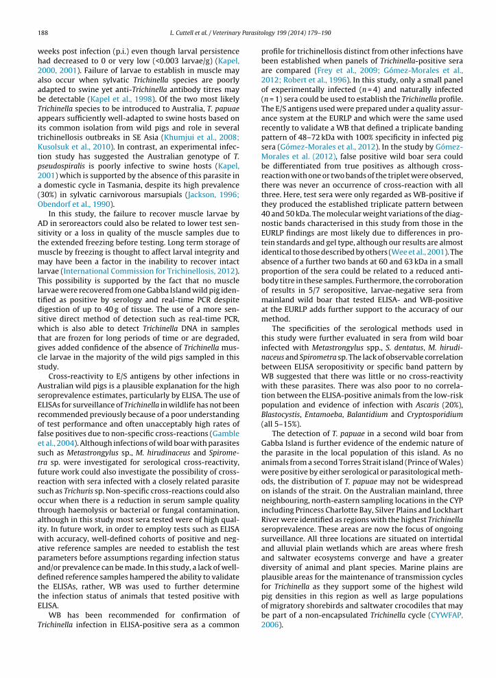
Cross-reactivity to E/S antigens by other infections inAustralian wild pigs is a plausible explanation for the highseroprevalence estimates, particularly by ELISA. The use ofELISAs for surveillance of Trichinella in wildlife has not beenrecommended previously because of a poor understandingof test performance and often unacceptably high rates offalse positives due to non-specific cross-reactions (Gambleet al., 2004). Although infections of wild boar with parasitessuch as Metastrongylus sp., M. hirudinaceus and Spirome-tra sp. were investigated for serological cross-reactivity,future work could also investigate the possibility of cross-reaction with sera infected with a closely related parasitesuch as Trichuris sp. Non-specific cross-reactions could alsooccur when there is a reduction in serum sample qualitythrough haemolysis or bacterial or fungal contamination,although in this study most sera tested were of high qual-ity. In future work, in order to employ tests such as ELISAwith accuracy, well-defined cohorts of positive and neg-ative reference samples are needed to establish the testparameters before assumptions regarding infection statusand/or prevalence can be made. In this study, a lack of well-defined reference samples hampered the ability to validatethe ELISAs, rather, WB was used to further determine
the infection status of animals that tested positive withELISA.WB has been recommended for confirmation ofTrichinella infection in ELISA-positive sera as a common
logy 199 (2014) 179– 190
profile for trichinellosis distinct from other infections havebeen established when panels of Trichinella-positive seraare compared (Frey et al., 2009; Gómez-Morales et al.,2012; Robert et al., 1996). In this study, only a small panelof experimentally infected (n = 4) and naturally infected(n = 1) sera could be used to establish the Trichinella profile.The E/S antigens used were prepared under a quality assur-ance system at the EURLP and which were the same usedrecently to validate a WB that defined a triplicate bandingpattern of 48–72 kDa with 100% specificity in infected pigsera (Gómez-Morales et al., 2012). In the study by Gómez-Morales et al. (2012), false positive wild boar sera couldbe differentiated from true positives as although cross-reaction with one or two bands of the triplet were observed,there was never an occurrence of cross-reaction with allthree. Here, test sera were only regarded as WB-positive ifthey produced the established triplicate pattern between40 and 50 kDa. The molecular weight variations of the diag-nostic bands characterised in this study from those in theEURLP findings are most likely due to differences in pro-tein standards and gel type, although our results are almostidentical to those described by others (Wee et al., 2001). Theabsence of a further two bands at 60 and 63 kDa in a smallproportion of the sera could be related to a reduced anti-body titre in these samples. Furthermore, the corroborationof results in 5/7 seropositive, larvae-negative sera frommainland wild boar that tested ELISA- and WB-positiveat the EURLP adds further support to the accuracy of ourmethod.
The specificities of the serological methods used inthis study were further evaluated in sera from wild boarinfected with Metastrongylus spp., S. dentatus, M. hirudi-naceus and Spirometra sp. The lack of observable correlationbetween ELISA seropositivity or specific band pattern byWB suggested that there was little or no cross-reactivitywith these parasites. There was also poor to no correla-tion between the ELISA-positive animals from the low-riskpopulation and evidence of infection with Ascaris (20%),Blastocystis, Entamoeba, Balantidium and Cryptosporidium(all 5–15%).
The detection of T. papuae in a second wild boar fromGabba Island is further evidence of the endemic nature ofthe parasite in the local population of this island. As noanimals from a second Torres Strait island (Prince of Wales)were positive by either serological or parasitological meth-ods, the distribution of T. papuae may not be widespreadon islands of the strait. On the Australian mainland, threeneighbouring, north-eastern sampling locations in the CYPincluding Princess Charlotte Bay, Silver Plains and LockhartRiver were identified as regions with the highest Trichinellaseroprevalence. These areas are now the focus of ongoingsurveillance. All three locations are situated on intertidaland alluvial plain wetlands which are areas where freshand saltwater ecosystems converge and have a greaterdiversity of animal and plant species. Marine plains areplausible areas for the maintenance of transmission cyclesfor Trichinella as they support some of the highest wild
pig densities in this region as well as large populationsof migratory shorebirds and saltwater crocodiles that maybe part of a non-encapsulated Trichinella cycle (CYWFAP,2006).
Parasito
EtrtpoitEmhicattt
fsaoeTfalaffrosoroaaTDYliriaTmswaoiwt
iacs
L. Cuttell et al. / Veterinary
There was generally good agreement between the twoLISAs used in the study. However, the in-house ELISA iden-ified a number of seroreacting samples in the Aurukunegion which were not corroborated by the commercialest. Aurukun is another large estuarine embayment com-rising of tidal flats and swamps that support high densitiesf wild pigs. It is possible that the poor test agreementn these samples was due to the lower analytical sensi-ivity of the commercial ELISA compared to the in-houseLISA. Lower test sensitivity using an E/S antigen com-ercial assay compared to an in-house developed assay
as also been reported previously (Akisu et al., 2006) andndeed, the results of this study showed that the commer-ial ELISA produced a significantly lower mean S/P ratio inll populations tested. In addition, it is also possible thathere had been a reduction in antibody titre in sera fromhis area that were collected over a prolonged period ofime and stored at −20 ◦C for several years.
Evidence of Trichinella exposure in wild pigs originatingrom the low-risk population in south-western WA was aurprising result. This population was originally selecteds a negative reference cohort based on the remotenessf the area in the Australian continent, but the pres-nce of two seroreactors showed that however unlikely,richinella exposure in wildlife could not be ruled out. Pigsrom this population were collected from Jarrah forest inn area incorporating numerous water catchments andand uses including forestry, bauxite mining and recre-tion. The region also supports an abundance of nativeauna as well as introduced species such as feral cats,oxes and wild pigs (Williams and Mitchell, 2001). Theelative proximity of this region to the metropolitan cityf Perth gives rise to the possibility of Trichinella para-ite introduction via illegal human-mediated movementf infected animals or meat products from other endemicegions (Spencer and Hampton, 2005). Historical recordsf national disease surveillance data have also reportedn isolated case of trichinosis in 1963–1964 from WA,lthough more information regarding the exact location,richinella species or host cannot be obtained (Nationalisease Surveillance Report 1917–1971, Commonwealthearbook, unpublished data). In addition, although the
arge biomass of T. pseudospiralis in the south-easternsland state of Tasmania is thought to be geographicallyestricted, transmission via migrating carnivorous birdsnfected with T. pseudospiralis from Tasmania could be
possibility (Obendorf and Clarke, 1992). In particular,. pseudospiralis infections have been reported in Tas-anian swamp harriers (Circus approximans), which is a
pecies that migrates annually from Tasmania to over-inter in a broad swath of eastern Australia as far north
s the CYP (Baker-Gabb and Steele, 1996). The presencef a second, isolated breeding population of this speciesn south-western WA, that also undertakes annual north-
ards migrations, could give rise to the possibility ofransmission to WA via returning harriers.
In conclusion, the diagnostic value of E/S antigen ELISAs
n wildlife surveillance in Australia can only be properlyssessed when adequate numbers of positive and negativeontrol sera are available to establish estimates of test sen-itivity and specificity. WB was a sensitive, specific methodlogy 199 (2014) 179– 190 189
that eliminated false-positive sera to provide a better esti-mate of the seroprevalence. Confirmation regarding thepresence of Trichinella parasites on the Australian main-land, and subsequently establishing actual zoonotic risk,must rely on muscle examination by direct methods. Therewas no evidence of current infection of wild boar on theAustralian mainland detected through this survey, whichis supported by the lack of confirmed human cases oftrichinellosis in Australia despite widespread pig hunt-ing and consumption practises in some areas of northernAustralia. For future surveillance, real-time PCR may be asuitable option to test seroreactors for current Trichinellainfections as it offers greater sensitivity and requires asmaller sample size than AD.
Acknowledgements
This work was funded by the Australian Biosecu-rity Cooperative Research Centre for Emerging InfectiousDiseases (AB-CRC). Sera samples were collected under Ani-mal Ethics Committee approvals CA 2008/02/240 and CA2011/03/495 by staff from the Northern Australia Quar-antine Strategy program of the Australian GovernmentDepartment of Fisheries, Forestry and Agriculture.
References
Abramson, J.H., 2011. WINPEPI updated: computer programs for epidemi-ologists, and their teaching potential. Epidemiol. Perspect. Innovat. 8,1.
AHA, 2009. Animal Health in Australia: Trichinellosis factsheet.http://nahis.animalhealthaustralia.com.au/pmwiki/pmwiki.php?n=Factsheet.80-2?skin=factsheet (accessed 09.10.13).
Akisu, C., Delibas, S.B., Ozkoc, S., Pozio, E., 2006. Serodiagnosis of trichinel-losis: in-house versus commercial ELISA. Parasite 13, 262–263.
Baker-Gabb, D., Steele, W.K., 1996. Monitoring the Relative Abundance,Distribution and Movements of Australian Birds of Prey. AustralianNature Conservation Agency Project Number: FPP110, Victoria.
Bearup, A.J., 1949. Examination of wild rats for Trichinella spiralis. Aust.Vet. J. 25, 229–230.
Chavez-Larrea, M.A., Dorny, P., Moeller, L., Benitez-Ortiz, W., Barrionuevo-Samaniego, M., Rodriguez-Hidalgo, R., Ron-Roman, J., Proano-Perez,F., Victor, B., Brandt, J., Kapel, C., de Borchgrave, J., 2005. Survey onporcine trichinellosis in Ecuador. Vet. Parasitol. 132, 151–154.
Conlan, J.V., Sripa, B., Attwood, S., Newton, P.N., 2011. A review of parasiticzoonoses in a changing Southeast Asia. Vet. Parasitol. 182, 22–40.
Cuttell, L., Cookson, B., Jackson, L.A., Gray, C., Traub, R.J., 2012a. First reportof a Trichinella papuae infection in a wild pig (Sus scrofa) from an Aus-tralian island in the Torres Strait region. Vet. Parasitol. 185, 343–345.
Cuttell, L., Corley, S.W., Gray, C.P., Vanderlinde, P.B., Jackson, L.A., Traub,R.J., 2012b. Real-time PCR as a surveillance tool for the detection ofTrichinella infection in muscle samples from wildlife. Vet. Parasitol.188, 285–293.
CYWFAP, 2006. Cape York Peninsula Pest Management Plan 2006–2011.Cape York Weeds and Feral Animal Program http://www.cywafap.org.au/pdfdocs/Part 2 CYP PMP 2006-2011 References.pdf (accessed13.09.13).
DAFF, 2007. AQIS Meat Notice Number: 2007/13 Testing for Trichinellain meat exported to the European Union. Australian Govern-ment Department of Fisheries, Forestry and Agriculture, Canberrahttp://www.daff.gov.au/ data/assets/pdf file/0019/401482/2007 13.pdf (accessed 09.10.13).
Davidson, R.K., Orpetveit, I., Moller, L., Kapel, C.M.O., 2009. Serologicaldetection of anti-Trichinella antibodies in wild foxes and experimen-
tally infected farmed foxes in Norway. Vet. Parasitol. 163, 93–100.Frey, C.F., Schuppers, M.E., Noeckler, K., Marinculic, A., Pozio, E., Kihm, U.,Gottstein, B., 2009. Validation of a Western Blot for the detection ofanti-Trichinella spp. antibodies in domestic pigs. Parasitol. Res. 104,1269–1277.

Parasito
ralis: comparison of gap sequences within the genus. Mol. Biochem.
190 L. Cuttell et al. / Veterinary
Gamble, H.R., Anderson, W.R., Graham, C.E., Murrell, K.D., 1983. Diagnosisof swine trichinosis by enzyme-linked immunosorbent assay using anexcretory–secretory antigen. Vet. Parasitol. 13, 349–362.
Gamble, H.R., Pozio, E., Bruschi, F., Noeckler, K., Kapel, C.M.O., Gajadhar,A.A., 2004. International Commission on Trichinellosis: recommen-dations on the use of serological tests for the detection of Trichinellainfection in animals and man. Parasite 11, 3–13.
Gamble, H.R., Rapic, D., Marinculic, A., Murrell, K.D., 1988. Evaluation ofexcretory–secretory antigens for the serodiagnosis of swine trichinel-losis. Vet. Parasitol. 30, 131–138.
Gómez-Morales, M.A., Ludovisi, A., Amati, M., Blaga, R., Zivojinovic, M.,Ribicich, M., Pozio, E., 2012. A distinctive western blot pattern to rec-ognize Trichinella infections in humans and pigs. Int. J. Parasitol. 42,1017–1023.
Gomez-Morales, M.A., Ludovisi, A., Amati, M., Cherchi, S., Pezzotti, P.,Pozio, E., 2008. Validation of an enzyme-linked immunosorbent assayfor diagnosis of human trichinellosis. Clin. Vaccine Immunol. 15,1723–1729.
Gomez-Morales, M.A., Ludovisi, A., Pezzotti, P., Amati, M., Cherchi, S., Lalle,M., Pecoraro, F., Pozio, E., Ring Trial, P., 2009. International ring trialto detect anti-Trichinella IgG by ELISA on pig sera. Vet. Parasitol. 166,241–248.
International Commission for Trichinellosis, 2012. Recommendationsfor quality assurance in digestion testing programs for Trichinella.ICT Quality Assurance Committee Appendix 1 (Part 3). Inter-national Commission for Trichinellosis http://www.trichinellosis.org/uploads/PART 3 final - PT 7Feb2012.pdf (accessed 13.09.13).
Jackson, B., 1996. A diaphragm digest survey of Tasmanian pigs. Depart-ment of Primary Industry and Fisheries of Tasmania, Kings Meadows.
Kapel, C.M.O., 2000. Host diversity and biological characteristics of theTrichinella genotypes and their effect on transmission. Vet. Parasitol.93, 263–278.
Kapel, C.M.O., 2001. Sylvatic and domestic Trichinella spp. in wild boars;infectivity, muscle larvae distribution, and antibody response. J. Para-sitol. 87, 309–314.
Kapel, C.M.O., Webster, P., Lind, P., Pozio, E., Henriksen, S.A., Murrell, K.D.,Nansen, P., 1998. Trichinella spiralis, T. britovi and T. nativa: infectivity,larval distribution in muscle, and antibody response after experimen-tal infection of pigs. Parasitol. Res. 84, 264–271.
Khumjui, C., Choomkasien, P., Dekumyoy, P., Kusolsuk, T., Kongkaew, W.,Chalamaat, M., Jones, J.L., 2008. Outbreak of trichinellosis caused byTrichinella papuae, Thailand, 2006. Emerg. Infect. Dis. 14, 1913–1915.
Kusolsuk, T., Kamonrattanakun, S., Wesanonthawech, A., Dekumyoy, P.,Thaenkham, U., Yoonuan, T., Nuamtanong, S., Sa-nguankiat, S., Pubam-pen, S., Maipanich, W., Panitchakit, J., Marucci, G., Pozio, E., Waikagu,J., 2010. The second outbreak of trichinellosis caused by Trichinellapapuae in Thailand. Trans. R. Soc. Trop. Med. Hyg. 104, 433–437.
Landis, J.R., Koch, G.G., 1977. The measurement of observer agreement forcategorical data. Biometrics 33 (1), 159–174.
Noeckler, K., Kapel, C.M., 2007. Detection and surveillance for Trichinella:meat inspection and hygiene, and legislation. In: Dupouy-Camet, J.,Murrell, K.D. (Eds.), FAO/WHO/OIE guidelines for the surveillance,management, prevention and control of trichinellosis. World Organi-sation for Animal Health Press, Paris, France, pp. 69–97.
Noeckler, K., Reckinger, S., Broglia, A., Mayer-Scholl, A., Bahn, P., 2009.Evaluation of a Western Blot and ELISA for the detection of anti-Trichinella-IgG in pig sera. Vet. Parasitol. 163, 341–347.
Oakwood, M., Spratt, D.M., 2000. Parasites of the northern quoll, Dasyu-
rus hallucatus (Marsupialia: Dasyuridae) in tropical savanna, NorthernTerritory. Aust. J. Zool. 48, 79–90.Obendorf, D.L., Clarke, K.P., 1992. Trichinella pseudospiralis infectionsin free-living Tasmanian birds. J. Helminthol. Soc. Washington 59,144–147.
logy 199 (2014) 179– 190
Obendorf, D.L., Handlinger, J.H., Mason, R.W., Clarke, K.P., Forman, A.J.,Hooper, P.T., Smith, S.J., Holdsworth, M., 1990. Trichinella pseudospi-ralis infection in Tasmanian Australian wildlife. Aust. Vet. J. 67,108–110.
OIE, 2012. Manual of Diagnostic Tests and Vaccines for TerrestrialAnimals 2012 (Web version). In: Trichinellosis. World Organisa-tion for Animal Health, Paris (Chapter 2.1.16) http://www.oie.int/international-standard-setting/terrestrial-manual/access-online/(accessed 13.09.13).
Owen, I.L., Morales, M.A.G., Pezzotti, P., Pozio, E., 2005. Trichinella infectionin a hunting population of Papua New Guinea suggests an ancientrelationship between Trichinella and human beings. Trans. R. Soc. Trop.Med. Hyg. 99, 618–624.
Pozio, E., Owen, I.L., La Rosa, G., Sacchi, L., Rossi, P., Corona, S., 1999.Trichinella papuae n.sp. (Nematoda), a new non-encapsulated speciesfrom domestic and sylvatic swine of Papua New Guinea. Int. J. Parasi-tol. 29, 1825–1839.
Pozio, E., Sofronic-Milosavljevic, L., Gomez Morales, M.A., Boireau, P.,Nöckler, K., 2002. Evaluation of ELISA and Western Blot Analysis usingthree antigens to detect anti-Trichinella IgG in horses. Vet. Parasitol.108, 163–178.
Richomme, C., Lacour, S.A., Ducrot, C., Gilot-Fromont, E., Casabianca, F.,Maestrini, O., Vallee, I., Grasset, A., van der Giessen, J., Boireau, P., 2010.Epidemiological survey of trichinellosis in wild boar (Sus scrofa) andfox (Vulpes vulpes) in a French insular region, Corsica. Vet. Parasitol.172, 150–154.
Robert, F., Weil, B., Kassis, N., Dupouy-Camet, J., 1996. Investigation ofimmunofluorescence cross-reactions against Trichinella spiralis byWestern blot (immunoblot) analysis. Clin. Diagn. Lab. Immunol. 3,575–577.
Sergeant, E., 2009. Epitools epidemiological calculators, Available from:http://epitools.ausvet.com.au (accessed 13.09.13).
Spencer, P.B.S., Hampton, J.O., 2005. Illegal translocation and geneticstructure of feral pigs in Western Australia. J. Wildl. Manage. 69,377–384.
Wacker, K., Rodriguez, E., Garate, T., Geue, L., Tackmann, K., Selhorst,T., Staubach, C., Conraths, F.J., 1999. Epidemiological analysis ofTrichinella spiralis infections of foxes in Brandenburg, Germany. Epi-demiol. Infect. 123, 139–147.
Waddell, A.H., 1969. The search for Trichinella spiralis in Australia. Aust.Vet. J. 45, 207.
Wee, S.H., Lee, C.G., Joo, H.D., Kang, Y.B., 2001. Enzyme-linked immunosor-bent assay for detection of Trichinella spiralis antibodies and thesurveillance of selected pig breeding farms in the Republic of Korea.Korean J. Parasitol. 39, 261–264.
Williams, K., Mitchell, D., 2001. Jarrah Forest 1 (JF1 – Northern Jar-rah Forest subregion). In: McKenzie, N.L., May, J.E., McKenna,S. (Eds.), A Biodiversity Audit of Western Australia’s 53 Bio-geographic Subregions in 2002. Department of Environmentand Conservation, Perth, Australia, p. 113 http://www.dec.wa.gov.au/pdf/science/bio audit/2002 bio summary.pdf
Zarlenga, D.S., Chute, M.B., Martin, A., Kapel, C.M.O., 1999. A multi-plex PCR for unequivocal differentiation of all encapsulated andnon-encapsulated genotypes of Trichinella. Int. J. Parasitol. 29,1859–1867.
Zarlenga, D.S., Dame, J.B., 1992. The identification and characterizationof a break within the large subunit ribosomal RNA of Trichinella spi-
Parasitol. 51, 281–289.Zimmer, I.A., Hunter, S.J., Morgan, C.P., Hunt, K.R., Smith, G.C., Howell, M.,
Taylor, M.A., 2008. Detection and surveillance for animal trichinellosisin GB. Vet. Parasitol. 151, 233–241.