Evaluation of DNA Damage Induced by Environmental Exposure to Mercury in Liza aurata Using the Comet...
11
Evaluation of DNA Damage Induced by Environmental Exposure to Mercury in Liza aurata Using the Comet Assay Carla Sofia Alves Pereira Sofia Isabel Antunes Gomes Guilherme Carlos Miguel Miguez Barroso Luc Verschaeve Ma ´rio Guilherme Garce ˆs Pacheco So ´nia Alexandra Leite Velho Mendo Received: 20 October 2008 / Accepted: 27 April 2009 Ó Springer Science+Business Media, LLC 2009 Abstract Mercury (Hg) is one of the major aquatic con- taminants even though emissions have been reduced over the years. Despite the relative abundance of investigations carried out on Hg toxicity, there is a scarcity of studies on its DNA damaging effects in fish under realistic exposure conditions. This study assessed the Hg genotoxicity in Golden grey mullets (Liza aurata) at Laranjo basin, a par- ticularly contaminated area of Ria de Aveiro (Portugal) well known for its Hg contamination gradient. (1) Fish were seasonally caught at Laranjo basin and at a reference site (S. Jacinto), and (2) animals from the reference site were transplanted and caged (at bottom and surface), for 3 days, in two different locations within Laranjo basin. Using the comet assay, blood was analyzed for genetic damage and apoptotic cell frequency. The seasonal survey showed greater DNA damage in the Hg-contaminated area for all sampling seasons excluding winter. The temporal variation pattern of DNA lesions was: summer & autumn [ win- ter [ spring. Fish caged at Laranjo also exhibited greater DNA damage than those caged at the reference site, high- lighting the importance of gill uptake on the toxicity of this metal. No increased susceptibility to apoptosis was detected in either wild or caged fish, indicating that mercury dam- ages DNA of blood cells by a nonapoptotic mechanism. Both L. aurata and the comet assay proved to be sensitive and suitable for genotoxicity biomonitoring in mercury- contaminated coastal systems. The aquatic environment, estuaries in particular, constantly receives genotoxic chemicals from either industrial or municipal waste waters (Rank and Jensen 2003) which may cause damage to biota. The assessment of DNA in indi- vidual cells, following exposure to pollutants, has been used as a valuable ecotoxicological tool concerning molecular genotoxicity biomarkers (Mitchelmore et al. 1998; Cough- lan et al. 2002). In this context, the single-cell gel electro- phoresis (SCGE) or comet assay appears to be a quick, simple, reliable, and sensitive technique to detect and measure genetic damage in almost any type of eukaryotic cell, using a small number of cells (Collins et al. 1997). This technique has proved its applicability in monitoring the effects of DNA-damaging agents in several marine and freshwater fish (Nacci et al. 1996; Belpaeme et al. 1998; Sumathi et al. 2001; Lee and Steinert 2003; Kim and Hyun 2006). The alkaline version developed by Singh et al. (Singh et al. 1988) allows for the detection of DNA single- strand breaks (SSBs), alkali-labile sites, DNA-DNA/DNA- protein cross-linking, and SSBs associated with incomplete excision repair sites (Tice et al. 2000). These events can lead to DNA fragmentation, which also occurs when cells die via apoptosis (Tice et al. 2000; Meintieres et al. 2003), also detectable by the comet assay (Meintieres et al. 2003). Heavy metals are an important group of environmental contaminants that should be considered when assessing genotoxicity in aquatic organisms, including fish (Ayllon C. S. A. Pereira S. I. A. G. Guilherme C. M. M. Barroso M. G. G. Pacheco S. A. L. V. Mendo (&) CESAM & Department of Biology, University of Aveiro, Campus universita ´rio de Santiago, 3810-193 Aveiro, Portugal e-mail: [email protected] L. Verschaeve Department of Epidemiology and Toxicology, Scientific Institute of Public Health, Brussels, Belgium L. Verschaeve Department of Biomedical Sciences, University of Antwerp, Antwerp, Belgium 123 Arch Environ Contam Toxicol DOI 10.1007/s00244-009-9330-y
-
Upload
independent -
Category
Documents
-
view
5 -
download
0
Transcript of Evaluation of DNA Damage Induced by Environmental Exposure to Mercury in Liza aurata Using the Comet...

Evaluation of DNA Damage Induced by Environmental Exposureto Mercury in Liza aurata Using the Comet Assay
Carla Sofia Alves Pereira Æ Sofia Isabel Antunes Gomes Guilherme ÆCarlos Miguel Miguez Barroso Æ Luc Verschaeve ÆMario Guilherme Garces Pacheco Æ Sonia Alexandra Leite Velho Mendo
Received: 20 October 2008 / Accepted: 27 April 2009
� Springer Science+Business Media, LLC 2009
Abstract Mercury (Hg) is one of the major aquatic con-
taminants even though emissions have been reduced over
the years. Despite the relative abundance of investigations
carried out on Hg toxicity, there is a scarcity of studies on its
DNA damaging effects in fish under realistic exposure
conditions. This study assessed the Hg genotoxicity in
Golden grey mullets (Liza aurata) at Laranjo basin, a par-
ticularly contaminated area of Ria de Aveiro (Portugal) well
known for its Hg contamination gradient. (1) Fish were
seasonally caught at Laranjo basin and at a reference site
(S. Jacinto), and (2) animals from the reference site were
transplanted and caged (at bottom and surface), for 3 days,
in two different locations within Laranjo basin. Using the
comet assay, blood was analyzed for genetic damage and
apoptotic cell frequency. The seasonal survey showed
greater DNA damage in the Hg-contaminated area for all
sampling seasons excluding winter. The temporal variation
pattern of DNA lesions was: summer & autumn [ win-
ter [ spring. Fish caged at Laranjo also exhibited greater
DNA damage than those caged at the reference site, high-
lighting the importance of gill uptake on the toxicity of this
metal. No increased susceptibility to apoptosis was detected
in either wild or caged fish, indicating that mercury dam-
ages DNA of blood cells by a nonapoptotic mechanism.
Both L. aurata and the comet assay proved to be sensitive
and suitable for genotoxicity biomonitoring in mercury-
contaminated coastal systems.
The aquatic environment, estuaries in particular, constantly
receives genotoxic chemicals from either industrial or
municipal waste waters (Rank and Jensen 2003) which may
cause damage to biota. The assessment of DNA in indi-
vidual cells, following exposure to pollutants, has been used
as a valuable ecotoxicological tool concerning molecular
genotoxicity biomarkers (Mitchelmore et al. 1998; Cough-
lan et al. 2002). In this context, the single-cell gel electro-
phoresis (SCGE) or comet assay appears to be a quick,
simple, reliable, and sensitive technique to detect and
measure genetic damage in almost any type of eukaryotic
cell, using a small number of cells (Collins et al. 1997). This
technique has proved its applicability in monitoring the
effects of DNA-damaging agents in several marine and
freshwater fish (Nacci et al. 1996; Belpaeme et al. 1998;
Sumathi et al. 2001; Lee and Steinert 2003; Kim and Hyun
2006). The alkaline version developed by Singh et al.
(Singh et al. 1988) allows for the detection of DNA single-
strand breaks (SSBs), alkali-labile sites, DNA-DNA/DNA-
protein cross-linking, and SSBs associated with incomplete
excision repair sites (Tice et al. 2000). These events can
lead to DNA fragmentation, which also occurs when cells
die via apoptosis (Tice et al. 2000; Meintieres et al. 2003),
also detectable by the comet assay (Meintieres et al. 2003).
Heavy metals are an important group of environmental
contaminants that should be considered when assessing
genotoxicity in aquatic organisms, including fish (Ayllon
C. S. A. Pereira � S. I. A. G. Guilherme �C. M. M. Barroso � M. G. G. Pacheco � S. A. L. V. Mendo (&)
CESAM & Department of Biology, University of Aveiro,
Campus universitario de Santiago, 3810-193 Aveiro, Portugal
e-mail: [email protected]
L. Verschaeve
Department of Epidemiology and Toxicology, Scientific Institute
of Public Health, Brussels, Belgium
L. Verschaeve
Department of Biomedical Sciences, University of Antwerp,
Antwerp, Belgium
123
Arch Environ Contam Toxicol
DOI 10.1007/s00244-009-9330-y
and Garcia-Vazquez 2000). Mercury (Hg) is one of the most
toxic metals described (Ayllon and Garcia-Vazquez 2000);
it accumulates in fish tissues mainly in the form of meth-
ylmercury (MeHg) (Wiener et al. 2002). The widespread
geographic extension and adverse consequences of pollu-
tion by this metal are still an issue of concern and continue
to prompt considerable scientific investigation. Hg’s tox-
icity and clastogenicity have been described in various
studies but little has been done regarding in vivo genotox-
icity assessment in fish. For example, mercury nitrate and
metallic mercury (Hg0) were demonstrated to be micronu-
cleus (MN) inducers in mollie (Poecilia latipinna) (Ayllon
and Garcia-Vazquez 2000) and carp (Cyprinus carpio)
(Nepomuceno et al. 1997), respectively. In addition, MeHg
proved to be teratogenic as well as a MN and chromosomal
aberration inducer in killifish (Fundulus heteroclitus)
embryos (Weis and Weis 1977; Perry et al. 1988).
Testing different substances for their genotoxicity in fish
is often performed under laboratory conditions. Such labo-
ratory experimentation compromises the relevance of data
since a reliable prevision of the substances’ toxicant effects
is complicated by a deviation from real exposure situations.
On the other hand, field data interpretation by itself is often a
very difficult task. Thus, the combination of field-survey
studies with in situ caging experiments has been proposed as
the ideal biomonitoring strategy from an ecotoxicological
point of view (Pacheco et al. 2005). In the present study, the
DNA damage induced by environmental Hg was assessed in
wild and transplanted Golden grey mullets (Liza aurata)
along a contaminated area of Ria de Aveiro, Laranjo basin
(LAR; Portugal), using the comet assay. This species is
abundant in the Atlantic and Mediterranean European
coastal waters, easy to catch, and well represented in both
clean and contaminated areas. Additionally, heavy metal
accumulation was previously proved to occur in members of
the Mugilidae family, namely, L. aurata (Blasco et al. 1998).
In the present work we aimed (i) to detect DNA damage
induction, as well as susceptibility to apoptosis, in L. au-
rata blood cells due to the presence of mercury at LAR, in
either wild or in situ caged fish; (ii) to evaluate the envi-
ronmental health status of the study area and the risk of Hg
contamination for fish populations; and (iii) to propose the
comet assay applied to L. aurata as a biomonitoring
methodology to evaluate metal genotoxicity.
Materials and Methods
Study Area
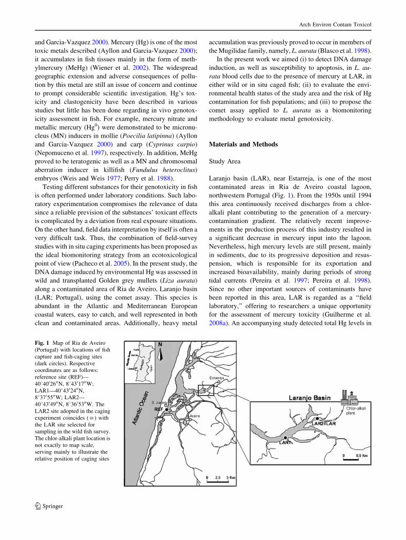
Laranjo basin (LAR), near Estarreja, is one of the most
contaminated areas in Ria de Aveiro coastal lagoon,
northwestern Portugal (Fig. 1). From the 1950s until 1994
this area continuously received discharges from a chlor-
alkali plant contributing to the generation of a mercury-
contamination gradient. The relatively recent improve-
ments in the production process of this industry resulted in
a significant decrease in mercury input into the lagoon.
Nevertheless, high mercury levels are still present, mainly
in sediments, due to its progressive deposition and resus-
pension, which is responsible for its exportation and
increased bioavailability, mainly during periods of strong
tidal currents (Pereira et al. 1997; Pereira et al. 1998).
Since no other important sources of contaminants have
been reported in this area, LAR is regarded as a ‘‘field
laboratory,’’ offering to researchers a unique opportunity
for the assessment of mercury toxicity (Guilherme et al.
2008a). An accompanying study detected total Hg levels in
Fig. 1 Map of Ria de Aveiro
(Portugal) with locations of fish
capture and fish-caging sites
(dark circles). Respective
coordinates are as follows:
reference site (REF)—
4084002600N, 884301700W;
LAR1—4084302400N,
883705500W; LAR2—
4084304900N, 883605300W. The
LAR2 site adopted in the caging
experiment coincides (:) with
the LAR site selected for
sampling in the wild fish survey.
The chlor-alkali plant location is
not exactly to map scale,
serving mainly to illustrate the
relative position of caging sites
Arch Environ Contam Toxicol
123

the sediment from 3.0 (LAR1) up to 7.7 (LAR2) ng/mg
(dry weight) (Guilherme et al. 2008a), as well as elevated
total Hg tissue loads in L. aurata inhabiting this area (e.g.,
0.57 and 2.4 lg/g wet weight, respectively, in blood and
liver) (Guilherme et al. 2008a, b).
S. Jacinto was selected as the reference (REF) site due to
its proximity to the lagoon entrance, the distance to the
main polluting sources (Fig. 1), and its low contamination
load (Pacheco et al. 2005).
Study Organism: Characterization and Sampling
The Golden grey mullet (Liza aurata), one of the most
common fish in Ria de Aveiro, is a pelagic species that
frequently contacts with sediments, feeding on small ben-
thic organisms, detritus, and, occasionally, insects and
plankton. Juvenile specimens were used to minimize the
interference of variables such as gender and levels of
accumulated contaminants.
Fish with an average weight of 13.5 ± 0.9 g and length
of 12.1 ± 0.6 cm were caught at the REF site and con-
taminated areas during low tide, using a traditional beach-
seine net called a chincha. After catching, fish were dis-
sected and blood was collected from the posterior cardinal
vein using heparinized Pasteur pipettes, stored in micro-
tubes, and kept on ice. In the laboratory, samples were kept
in the dark, diluted in saline solution (B. Braun Medical,
Lda.), and then used for comet assay.
Experimental Design
Two experimental components were considered in this
study: the first corresponding to a seasonal analysis of wild
fish and the second corresponding to a field-caging
experiment (Fig. 2). In both components, comet assay was
performed in fish blood cells to assess DNA damage and
the incidence of apoptotic cells (ACs). Blood of control
animals (REF site) was exposed to hydrogen peroxide
(75 lM H2O2, 1 h) (Sigma) and used as a positive control.
Wild Fish Survey Ten fish of similar dimensions (size
and weight) per sampling moment were caught, as previ-
ously described, during the winter of 2004 (December) and
the spring (March), summer (July), and autumn (Septem-
ber) of 2005 at LAR and S. Jacinto (REF site) (Fig. 1).
Field-Caging Experiment Fish were caught at the REF
site (S. Jacinto; Fig. 1), transported to the laboratory in
oxygenated saltwater (from the fishing site), and allowed to
stabilize for 2 weeks prior to experimentation in order to
reduce interindividual differences, to allow adaptation to
confinement, and to reduce the levels of chemicals taken up
previously. During stabilization, fish were kept in 80-L
aquaria at natural temperature and photoperiod, in aerated
(dissolved oxygen level = 8.4 ± 0.2 mg � L-1) and filtered
artificial seawater (Salsera, France; 23 ± 0.1 g � L-1 salin-
ity). Fish were fed daily with polychaete worms (Nereis sp.)
collected in a clean area of Ria de Aveiro.
Subsequently, mullets were transferred to the field in
oxygenated artificial seawater and caged (10 fish per 80-L
cage), during 3 days, in two different locations at LAR,
positioned at different relative distances from the metal
contamination source (LAR1 and LAR2) (Figs. 1 and 2).
To study the effect of mercury uptake and genotoxicity,
two cages were placed at two positions along the water
column as follows: one at the surface and the other close to
the sediment. Surface cages were maintained in a sub-
merged position (30-cm depth) with a buoy-anchor system.
Bottom cages were set at 15 cm from the sediment to avoid
direct contact with it. Reference groups were caged at
S. Jacinto. When transferred to cages, the animals were
visually checked to be in perfect condition. During field
exposure, fish were kept without any food supply. On the
day of transfer to cages, 10 fish were sacrificed for analysis,
constituting the t0 group. This part of the study was per-
formed in December 2004.
Comet Assay
The alkaline version of SCGE—the comet assay—was per-
formed with slight modifications of the Koppen and Vers-
chaeve (Koppen and Verschaeve 1996) methodology. All
procedures were performed under dim yellow light to prevent
extra UV light-induced DNA damage. Agarose solutions
were prepared in phosphate-buffered saline (without Ca2?
and Mg2?, pH 7.4; Bio Wittaker Europe, Cambrex Co.).
Slide Preparation
Briefly, microscope slides were precoated with 1% normal-
melting-point (NMP) agarose (Bio-Rad; 5 min at room
temperature), and then a layer of cell suspension in 0.5%
low-melting-point (LMP) agarose (Sigma) was added
between a bottom layer of 0.8% NMP agarose and a topFig. 2 Schematic representation of the experimental design adopted
for the caging experiment
Arch Environ Contam Toxicol
123

layer of 0.8% LMP agarose. After solidification on ice,
microscope slides were immersed in cold lysing solution
(2.5 M NaCl [Merck], 0.1 M Na2EDTA [Merck], 0.01 M
Tris base [Qbiogene], pH 10, set with NaOH [Merck], 1%
Na-lauroylsarcosinate [Sigma], 10% DMSO [Sigma], and
1% Triton X-100 [Sigma] in MQ water [Millipore]), in the
dark, for a period of 1–24 h and then placed for 15 min in
freshly prepared alkaline denaturing electrophoresis buffer
(0.3 M NaOH, 0.001 M Na2EDTA, 0.1% 8-hydroxyquin-
oline [Sigma], and 2% DMSO in MQ water at 17�C).
Electrophoresis ran at 0.7–1 V/cm (300 mA) for 10 min,
after which slides were washed in cold Tris-HCl (0.4 M,
pH 7.5), stained with ethidium bromide (Qbiogene; 20 lg �mL-1), and stored in moistened boxes, with light protec-
tion, at 4�C, until observation.
Image Analysis
The alkaline comet assay allows for detection of DNA
damage occurring as SBs by measuring the migration of
DNA fragments from the nucleoid, visually resembling a
comet. A LEICA DMLS fluorescence microscope (400 9
magnification) was used for slide analysis. For each fish,
two slides were prepared. Fifty cells (25 per individual)
were randomly scored using a public domain NIH-Image
program (Helma and Uhl 2000).
Image analysis was mainly made on the increased
fluorescence in the tail region, referring to the percentage
of DNA in the tail (TD%), the tail length (TL), or the
product of both, called the tail moment (TM). TM was the
chosen parameter for the comparative analysis between
conditions due to its responsiveness. Data on TD and TL
were measured (but not shown) in a preanalysis phase
because, when using a derived parameter such as TM, the
original parameters should be considered as suggested by
Tice et al. (Tice et al. 2000). Control comet cells are rep-
resented by the nucleoid core only, normally with minimal
DNA migration (Fig. 3, class 1). Any healthy cell typically
contains a certain proportion of SBs in its DNA, the result
of either spontaneous damage or DNA breakage necessary
to DNA functions such as its synthesis (Koppen 1999).
Besides TM, the frequency of ACs was also accessed
through the comet assay data. The mean head intensity and
area were considered together to better define an AC in this
work. An interval of acceptance was defined by the
mean ± standard deviation for both comet parameters, and
values below this limit were highlighted and analyzed in
detail. A positive result was considered if both parameters
(mean head intensity and area) were demarked and confir-
mation was made by evaluation of the respective comet image
in comparison to the fan-like pattern previously described by
Tice et al. (Tice et al. 2000) or Meintieres et al. (Meintieres
et al. 2003) (Fig. 3, class 5). These cells were included in the
counting of 50 comets per individual but excluded from any
TM image analysis or statistic calculations, as they represent
dead/dying cells (Speit and Hartmann 2004).
Statistical Analysis
The significance of the differences, either between spatial
distributions or between seasons, was evaluated using the
nonparametric Kruskal-Wallis ANOVA and a posteriori
the Mann-Whitney U-test, referring to STATISTICA 6
software (StatSoft, Inc., Tulsa, OK, USA).
Results
The hydrological parameters (temperature, dissolved oxy-
gen, salinity, pH, turbidity, and depth) measured in the
study areas at Ria de Aveiro concerning the wild fish
survey and the caging experiment are reported in Tables 1
and 2, respectively. These parameters showed, in general,
no important intersite differences within the same sampling
season for temperature, dissolved oxygen, or pH. In winter
and spring, LAR showed a lower salinity and depth, and a
higher turbidity, compared to REF. A similar pattern was
observed in the caging experiment comparing REF and
LAR sites. Table 1 also reports common seasonal varia-
tions for most of the parameters.
DNA Damage in Wild Fish
From the analysis of TM data (Fig. 4A), it is evident that
animals from the contaminated site (LAR) showed signif-
icantly (p \ 0.05) higher levels of DNA damage compared
to those at REF in all sampling seasons excluding winter.
The TM levels at LAR were as high as 2.0, 2.8, and 2.7
times the reference levels during spring, summer, and
autumn, respectively.
Analyzing the temporal variation patterns of DNA lesions
in L. aurata, for both REF and LAR fish, the following order
is seen: summer & autumn [ winter [ spring. At both the
REF and the LAR sites, DNA damage decreased signifi-
cantly (p \ 0.05) from winter to spring, increased in sum-
mer, and were maintained at a high level in autumn.
Fig. 3 Comet scale. A five-class classification based on tail DNA
percentage (TD) adopted from Mitchelmore and Chipman (Mitchel-
more and Chipman 1998). Comet classes: 1, no or minimal damage
(\5%); 2, low damage (5–20%); 3, mid damage (20–40%); 4, high
damage (40–75%); 5, extreme damage ([75%)
Arch Environ Contam Toxicol
123
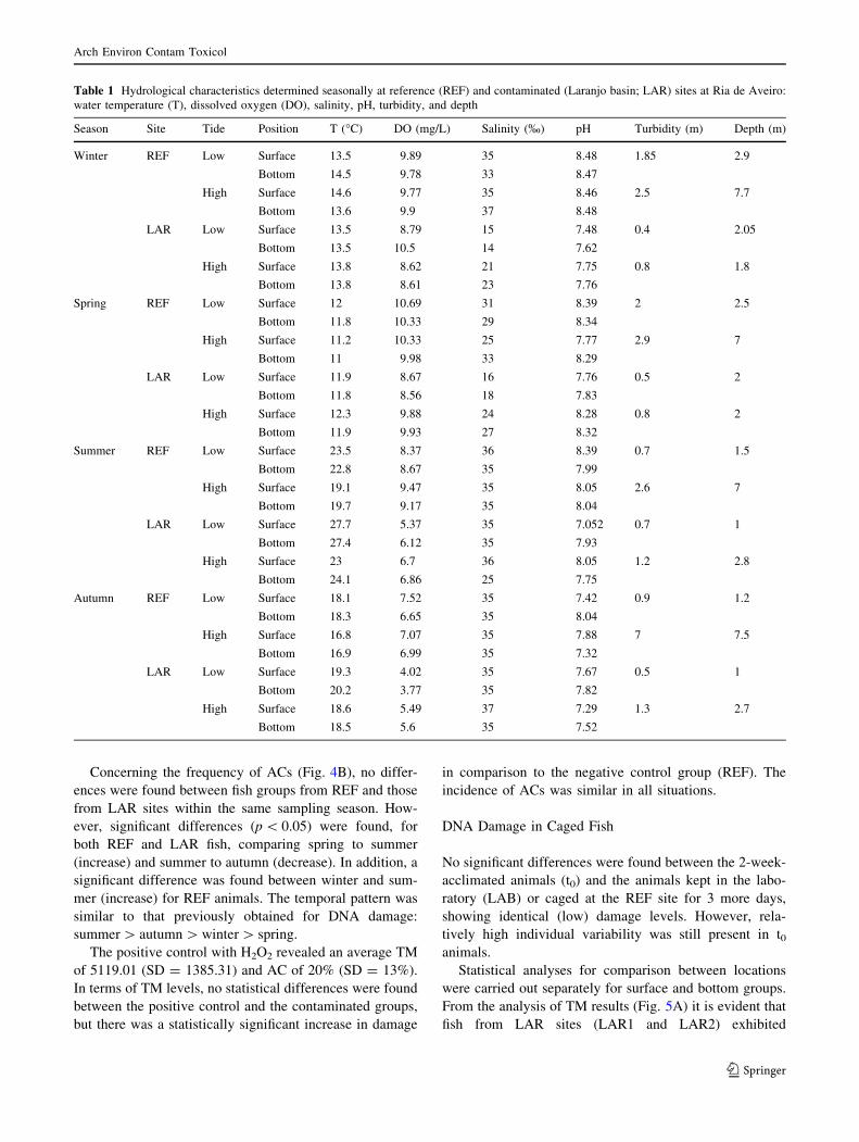
Concerning the frequency of ACs (Fig. 4B), no differ-
ences were found between fish groups from REF and those
from LAR sites within the same sampling season. How-
ever, significant differences (p \ 0.05) were found, for
both REF and LAR fish, comparing spring to summer
(increase) and summer to autumn (decrease). In addition, a
significant difference was found between winter and sum-
mer (increase) for REF animals. The temporal pattern was
similar to that previously obtained for DNA damage:
summer [ autumn [ winter [ spring.
The positive control with H2O2 revealed an average TM
of 5119.01 (SD = 1385.31) and AC of 20% (SD = 13%).
In terms of TM levels, no statistical differences were found
between the positive control and the contaminated groups,
but there was a statistically significant increase in damage
in comparison to the negative control group (REF). The
incidence of ACs was similar in all situations.
DNA Damage in Caged Fish
No significant differences were found between the 2-week-
acclimated animals (t0) and the animals kept in the labo-
ratory (LAB) or caged at the REF site for 3 more days,
showing identical (low) damage levels. However, rela-
tively high individual variability was still present in t0animals.
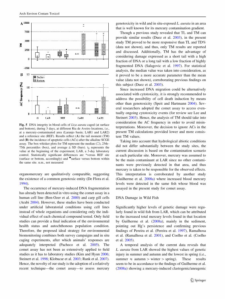
Statistical analyses for comparison between locations
were carried out separately for surface and bottom groups.
From the analysis of TM results (Fig. 5A) it is evident that
fish from LAR sites (LAR1 and LAR2) exhibited
Table 1 Hydrological characteristics determined seasonally at reference (REF) and contaminated (Laranjo basin; LAR) sites at Ria de Aveiro:
water temperature (T), dissolved oxygen (DO), salinity, pH, turbidity, and depth
Season Site Tide Position T (�C) DO (mg/L) Salinity (%) pH Turbidity (m) Depth (m)
Winter REF Low Surface 13.5 9.89 35 8.48 1.85 2.9
Bottom 14.5 9.78 33 8.47
High Surface 14.6 9.77 35 8.46 2.5 7.7
Bottom 13.6 9.9 37 8.48
LAR Low Surface 13.5 8.79 15 7.48 0.4 2.05
Bottom 13.5 10.5 14 7.62
High Surface 13.8 8.62 21 7.75 0.8 1.8
Bottom 13.8 8.61 23 7.76
Spring REF Low Surface 12 10.69 31 8.39 2 2.5
Bottom 11.8 10.33 29 8.34
High Surface 11.2 10.33 25 7.77 2.9 7
Bottom 11 9.98 33 8.29
LAR Low Surface 11.9 8.67 16 7.76 0.5 2
Bottom 11.8 8.56 18 7.83
High Surface 12.3 9.88 24 8.28 0.8 2
Bottom 11.9 9.93 27 8.32
Summer REF Low Surface 23.5 8.37 36 8.39 0.7 1.5
Bottom 22.8 8.67 35 7.99
High Surface 19.1 9.47 35 8.05 2.6 7
Bottom 19.7 9.17 35 8.04
LAR Low Surface 27.7 5.37 35 7.052 0.7 1
Bottom 27.4 6.12 35 7.93
High Surface 23 6.7 36 8.05 1.2 2.8
Bottom 24.1 6.86 25 7.75
Autumn REF Low Surface 18.1 7.52 35 7.42 0.9 1.2
Bottom 18.3 6.65 35 8.04
High Surface 16.8 7.07 35 7.88 7 7.5
Bottom 16.9 6.99 35 7.32
LAR Low Surface 19.3 4.02 35 7.67 0.5 1
Bottom 20.2 3.77 35 7.82
High Surface 18.6 5.49 37 7.29 1.3 2.7
Bottom 18.5 5.6 35 7.52
Arch Environ Contam Toxicol
123

significantly higher genetic damage levels (p \ 0.001)
compared to fish from the REF site. The observed increase
was 3.3, 1.7, and 2.5 times for LAR1 bottom, LAR2
surface, and LAR2 bottom, respectively. No significant
differences were found between LAR1 and LAR2 bottom
groups. In addition, a higher variability of results was
observed at LAR sites.
Little can be said about the influence of the relative
position in the water column on the incidence of genetic
damage in these animals. The comparison is not feasible
for LAR1, as the surface cage was lost; no significant
differences were found between surface and bottom groups
at LAR2. However, S. Jacinto (REF) bottom-caged fish
showed significantly lower damage compared with surface-
caged fish.
Data on the occurrence of ACs (Fig. 5B) did not reveal
any significant or conclusive result in comparisons between
REF and LAR groups, as well as surface and bottom
groups at the same site.
Discussion
Although mercury’s spatial and biological distribution at
Laranjo basin (LAR; Ria de Aveiro, Portugal) is well
documented (Pereira et al. 1997; Coelho et al. 2005), there
are some gaps in understanding its effects at the organism,
population, and ecosystem levels. Various studies on
mercury compounds and their genotoxic effects have been
performed, evaluating a panoply of genetic endpoints.
Clastogenic effects mostly associated with the spindle
mechanism disturbance and the generation of reactive
oxygen species, accompanied by glutathione depletion,
which might also contribute to its genotoxicity, have been
reported (De Flora et al. 1994). Organomercury compounds
are the most predominant, as they are generally more toxic
to aquatic organisms than the inorganic forms (Boening
2000). Nevertheless, the genetic effects of inorganic and
Table 2 Hydrological characteristics of reference (REF) and contaminated (Laranjo basin; LAR1, LAR2) sites on the caging experiment at Ria
de Aveiro: water temperature (T), dissolved oxygen (DO), salinity, pH, turbidity, and depth
Site Tide Position T (8C) DO (mg/L) Salinity (%) pH Turbidity (m) Depth (m)
REF Low Surface 13.5 9.89 35 8.48 1.85 2.9
Bottom 14.5 9.78 33 8.47
High Surface 14.6 9.77 35 8.46 2.5 7.7
Bottom 13.6 9.9 37 8.48
LAR1 Low Surface 13.8 8.64 9 7.42 0.35 2.7
Bottom 13.8 8.9 17 7.69
High Surface 13.8 8.83 23 7.78 0.85 1.8
Bottom 13.5 9.66 25 7.95
LAR2 Low Surface 13.5 8.79 15 7.48 0.4 2.05
Bottom 13.5 10.5 14 7.62
High Surface 13.8 8.62 21 7.75 0.8 1.8
Bottom 13.8 8.61 23 7.76
Fig. 4 DNA integrity in blood cells of wild Liza aurata collected
seasonally at a mercury-contaminated site (LAR) and a reference site
(REF) in the Ria de Aveiro. Results reflect (A) the tail moment (TM)
and (B) the incidence of apoptotic cells (ACs) after the alkaline SCGE
assay. The box-whisker plots for TM represent the median (h), 25th–
75th percentiles (box), and average ± SD (bars). Data on ACs
represent the average ± SD. Statistically significant differences are
*versus REF site within each sampling season and aversus winter,bversus spring, and cversus summer
Arch Environ Contam Toxicol
123
organomercury are qualitatively comparable, suggesting
the existence of a common genotoxic entity (De Flora et al.
1994).
The occurrence of mercury-induced DNA fragmentation
has already been detected in vitro using the comet assay in a
human cell line (Ben-Ozer et al. 2000) and carp gill cells
(Arabi 2004). However, these studies have been conducted
under artificial laboratorial conditions using cell lines
instead of whole organisms and considering only the indi-
vidual effect of each chemical compound tested. Only field
studies can provide a final indication of the environmental
health status and autochthonous population condition.
Therefore, the proposed ideal strategy for environmental
biomonitoring combines both survey campaigns and in situ
caging experiments, after which animals’ responses are
adequately interpreted (Pacheco et al. 2005). The
comet assay has not been as extensively applied to field
studies as it has to laboratory studies (Kim and Hyun 2006;
Steinert et al. 1998; Klobucar et al. 2003; Rank et al. 2007).
Hence, the novelty of our study is the adoption of a relatively
recent technique—the comet assay—to assess mercury
genotoxicity in wild and in situ-exposed L. aurata in an area
that is well known for its mercury contamination gradient.
Though a previous study revealed that TL and TM can
provide similar results (Duez et al. 2003), in the present
study TM proved to be more responsive than TL and TD%
(data not shown), and thus, only TM results are reported
and discussed. Additionally, TM has the advantage of
considering damage expressed as a short tail with a high
fraction of DNA or a long tail with a low fraction of highly
fragmented DNA (Salagovic et al. 1997). For statistical
analysis, the median value was taken into consideration, as
it proved to be a more accurate parameter than the mean
value (data not shown), corroborating previous findings on
this subject (Duez et al. 2003).
Since increased DNA migration could be alternatively
associated with cytotoxicity, it is strongly recommended to
address the possibility of cell death induction by means
other than genotoxicity (Speit and Hartmann 2004). Sev-
eral researchers adopted the comet assay to access even-
tually ongoing cytotoxicity events (for review see Lee and
Steinert 2003). Hence, the analysis of TM should take into
consideration the AC frequency in order to avoid misin-
terpretations. Moreover, the decision to ignore ACs in the
present TM calculations provided lower and more consis-
tent TM values.
Taking into account that hydrological abiotic conditions
did not differ substantially between the study sites, the
current discussion is based on the contamination scenario
at each particular site. Moreover, mercury was assumed to
be the main contaminant at LAR since no other contami-
nants were previously detected in that area, and thus
mercury is taken to be responsible for the observed effects.
This interpretation is corroborated by another study
(Guilherme et al. 2008a) where increased blood mercury
levels were detected in the same fish whose blood was
assayed in the present study for comet assay.
DNA Damage in Wild Fish
Significantly higher levels of genetic damage were regu-
larly found in wild fish from LAR, which can be attributed
to the increased total mercury levels found in that location
by Guilherme et al. (2008a), mainly in the sediment,
pointing out Hg’s persistence and confirming previous
findings of Pereira et al. (Pereira et al. 1997), Ramalhosa
et al. (Ramalhosa et al. 2001), and Coelho et al. (Coelho
et al. 2005).
A temporal analysis of the current data reveals that
L. aurata from LAR showed the highest values of genetic
injury in summer and autumn and the lowest in spring (i.e.,
summer & autumn [ winter [ spring). These results
seem to be in accordance to the findings of Guilherme et al.
(2008a) showing a mercury-induced clastogenic/aneugenic
Fig. 5 DNA integrity in blood cells of Liza aurata caged (at surface
and bottom), during 3 days, at different Ria de Aveiro locations, i.e.,
at a mercury-contaminated area (Laranjo basin; LAR1 and LAR2)
and a reference site (REF). Results reflect (A) the tail moment (TM)
and (B) the incidence of apoptotic cells (ACs) after the alkaline SCGE
assay. The box-whisker plots for TM represent the median (h), 25th–
75th percentiles (box), and average ± SD (bars). t0 represents the
value at the beginning of the experiment; LAB, a 3-day laboratory
control. Statistically significant differences are *versus REF site
(surface or bottom, accordingly) and rsurface versus bottom within
the same site. n.m., not measured
Arch Environ Contam Toxicol
123

action, measured as erythrocytic nuclear abnormality
induction, only in summer and autumn. It must be taken
into consideration that the interpretation of temporal
response patterns, in the case of LAR fish, may be com-
promised by seasonal fluctuations in environmental mer-
cury levels. However, the same temporal pattern (though
less evident) is also perceptible in fish from the REF site,
pointing out the modulatory effect of seasonal climatic
variations on fish physiology. In this context, it must be
emphasized that TM values measured along the year at the
REF site perfectly matched the water temperature fluctu-
ations recorded. This is in accordance with Venier et al.
(Venier et al. 1997), who demonstrated that water tem-
perature may influence cell replication rates and DNA
repair of poikilothermal organisms, and with Buschini et al.
(Buschini et al. 2003), who found a positive correlation
between water temperature and DNA integrity loss in zebra
mussels. Consequently, the interference of water tempera-
ture as an additive factor on the mercury-induced
DNA integrity loss observed in LAR fish cannot be
underestimated.
Data on AC incidence demonstrated that LAR fish have
a slightly higher frequency compared to REF fish, though it
was not possible to distinguish the groups based on sta-
tistically significant differences. On the other hand, sig-
nificant temporal variations were detected for both control
and mercury-exposed groups, which may indicate that the
incidence of ACs depends on the seasonal fish health
condition rather than on the environmental levels of Hg.
The joint analysis of the current TM and AC data supports
the idea that mercury damages blood cell DNA by a no-
napoptotic mechanism, which is in accordance with the
results obtained previously in human cell lines exposed to
mercuric chloride (Ben-Ozer et al. 2000).
DNA Damage in Caged Fish
The significant DNA damage induction observed in fish
caged at LAR1 and LAR2 should be regarded as an addi-
tional indication of a genotoxic action of mercury, rein-
forcing the current wild fish survey findings. The absence
of erythrocytic nuclear abnormality induction observed in
LAR1 and LAR2 fish by Guilherme et al. (2008a) suggests
the higher suitability of the comet assay for detection of
mercury genotoxicity.
The diet is expected to be a major source of Hg con-
tamination in fish. However, considering that L. aurata
feeding was almost completely limited due to caging, the
current results emphasize the importance of uptake through
the gills in mercury toxicity to fish. Furthermore, it was
demonstrated that 3 days of exposure is enough for
significant mercury uptake and expression of its genotoxic
potential measured as DNA integrity loss in blood cells.
The Hg contamination gradient is more evident in the
sediment, decreasing toward the sea, away from the con-
tamination source. However, total Hg water content does
not necessarily indicate total bioavailable Hg. The combi-
nation of different conditions favors both the Hg avail-
ability to biota and their vulnerability to Hg effects.
Mercury bioaccumulation—and thus bioavailability—
depend mainly on MeHg formation and uptake (Wiener
et al. 2002). The uptake of MeHg by aquatic organisms is
more rapid and extensive than that of inorganic mercury
(Biesinger et al. 1982), and its formation, though also
occurring in fish gills and gut (Boening 2000), depends
mainly on soil-living, sulfur-reducing bacteria that are
directly affected by environmental factors including redox
potential, pH, salinity, and temperature (Wiener et al. 2002;
Davis et al. 2003). In turn, other aquatic organisms such as
fish are also affected (Boening 2000).
LAR2 had clearly higher levels of total Hg in the water
and sediment in relation to LAR1 (Guilherme et al. 2008a).
Nonetheless, exposure at LAR1 and LAR2 induced similar
levels of genetic damage in L. aurata blood cells. Con-
sidering the previous statements, it can be hypothesized
that the levels of biologically available Hg are similar at
both sites, despite the reported differences in total Hg
environmental levels, and thus induce comparable genetic
damage.
The oxic-anoxic interface where MeHg formation
occurs is very near the sediment surface (1 to 10 cm deep)
(Davis et al. 2003). Changes in water and sediment
movements (e.g., tides, freshwater influx, and erosion) may
lead to the remobilization of contaminated sediment that
has been deposited for decades, affecting adjacent areas. In
addition, the sediment conditions are more suitable for Hg
methylation (anoxia, low pH). Total mercury content at
LAR2 was definitely higher in the bottom water (Guil-
herme et al. 2008a). Surprisingly, no significant differences
related to water column position were found in the present
study except for the REF site, where DNA integrity was
higher at the bottom. This difference might just be a con-
sequence of the high homogeneity of the REF samples,
enhancing the power of the statistical tests, which was not
observed at the contaminated sites. Even so, the levels of
genetic damage in REF fish remain at the control level
(LAB) in both the surface and the bottom groups.
AC frequencies remained very low in all groups, rein-
forcing the previous result from a wild L. aurata survey
that, although mercury may damage DNA, apoptosis does
not seem to be markedly involved. Moreover, it seems that
the stress induced by handling and caging did not affect AC
Arch Environ Contam Toxicol
123

incidence, which is a positive aspect of this experimental
approach.
General Discussion
L. aurata proved its sensitivity as a model species for
monitoring metal genotoxicity at different degrees of
contamination. A relatively high baseline of DNA lesions
was found in wild L. aurata. On the other hand, trans-
planted fish had a 2-week recovery period prior to caging
and exposure, allowing for the reduction of existing dam-
age induced by chemicals taken up previously.
Blood is an easily accessible tissue with central physi-
ologic functions, providing an important source of cells for
genotoxicity studies (Singh 2000). In the present study,
blood proved to be suitable for the purpose of monitoring
Hg genotoxicity, as it significantly responded to Hg
contamination.
Despite the restriction of exposure routes in caged fish
(only via water) in comparison to wild fish (via water and
food), a similar increase (two to three times) in DNA
damage from REF to LAR sites was found in both studies
in the present work. It seems that, independently of
the time and type of exposure, Hg damage induction in
L. aurata reached a plateau.
Metal detoxification processes such as cellular seques-
tration, influx-efflux balance, and a combination of the two
(Kraemer et al. 2005), together with DNA repair systems,
are responsible for the prevention of DNA damage, thereby
determining fish adaptation to contaminated environments.
Hence, although L. aurata is commonly found at LAR, the
occurrence of increased DNA damage levels suggests that
the combined action of the previous defense mechanisms
was not entirely effective.
Both experiments performed in the present work
revealed Hg persistence and contamination at LAR, clearly
affecting the studied population in terms of genetic integ-
rity. The relatively rapid response (in 3 days) emphasizes
the acute toxicity of Hg, revealing that the levels found at
LAR should undoubtedly be of great concern when con-
sidering the local species’ conservation and biodiversity.
The genotoxic potential of Hg already demonstrated by
in vitro experiments with gill cell suspensions measured by
the comet assay (Ben-Ozer et al. 2000; Arabi and Alaed-
dini 2005) was confirmed in both parts of the present study.
Mercury contamination issues in Aveiro lagoon were
previously reported to be confined to LAR (Coelho et al.
2005). The REF site chosen within the lagoon (S. Jacinto)
fits the clean area prerequisites, as fish caged at that site did
not show significant differences from what was observed in
the laboratory controls (LAB and t0). Furthermore, it was
demonstrated that cage confinement did not constitute an
extra stress factor for the considered biomarker, allowing
the cause-effect linkage of mercury contamination and
DNA damage.
However, it is recommended that appropriate cytotox-
icity tests should be adopted as a complement of geno-
toxicity assays. The alkaline comet assay is not as sensitive
or reliable in detecting cytotoxicity (Meintieres et al. 2003)
and the neutral version has been indicated for that purpose
(e.g., Tice et al. 2000). However, the results presented here
should not be underestimated. Moreover, current overall
results support the idea of Ben-Ozer et al. (Ben-Ozer et al.
2000) that DNA damage in surviving cells is a more sen-
sitive indicator of environmental insult than apoptosis.
Conclusion
This study provides new information on mercury geno-
toxicity under realistic exposure conditions, evaluated by
the comet assay, as higher DNA damage was found in both
wild and caged fish. No increased susceptibility to apop-
tosis was detected, indicating that mercury damages DNA
of blood cells by nonapoptotic mechanisms. Mercury
uptake from the water per se (dissolved and associated with
suspended particulate matter) was shown to be sufficient to
increase genetic damage significantly. The comet assay
applied to L. aurata blood cells was shown to be sensitive
and suitable for genotoxicity biomonitoring in mercury-
contaminated coastal systems. Finally, the results point out
the environmental risk to native ichthyofauna at LAR due
to mercury contamination.
Acknowledgment The authors are very thankful for the technical
assistance of Mr. Aldiro Pereira.
References
Arabi M (2004) Analyses of impact of metal ion contamination on
carp (Cyprinus carpio L.) gill cell suspensions. Biol Trace Elem
Res 100(3):229–246. doi:10.1385/BTER:100:3:229
Arabi M, Alaeddini MA (2005) Metal-ion-mediated oxidative stress
in the gill homogenate of rainbow trout (Oncorhynchusmykiss)—antioxidant potential of manganese, selenium, and
albumin. Biol Trace Elem Res 108(1–3):155–168. doi:
10.1385/BTER:108:1-3:155
Ayllon F, Garcia-Vazquez E (2000) Induction of micronuclei and
other nuclear abnormalities in European minnow Phoxinusphoxinus and mollie Poecilia latipinna: an assessment of the fish
micronucleus test. Mutat Res 467:177–186
Belpaeme K, Cooreman K, Kirsch-Volders M (1998) Development
and validation of the in vivo alkaline comet assay for detecting
genomic damage in marine flatfish. Mutat Res 415:167–184
Ben-Ozer EY, Rosenspire AJ, McCabe MJ Jr, Worth RG, Kindzelskii
AL, Warra NS, Petty HR (2000) Mercuric chloride damages cellular
DNA by a non-apoptotic mechanism. Mutat Res 470:19–27
Biesinger KE, Anderson LE, Eaton JG (1982) Chronic effects of
inorganic and organic mercury on Daphnia magna: toxicity,
Arch Environ Contam Toxicol
123
accumulation, and loss. Arch Environ Contam Toxicol 11:769–
774. doi:10.1007/BF01059166
Blasco J, Rubio JA, Forja J, Gomez-Parra A, Establier R (1998)
Heavy metals in some fishes of the Mugilidae family from salt-
ponds of Cadiz bay, SW Spain. Ecotoxicol Environ Restoration
1(2):71–77
Boening DW (2000) Ecological effects, transport, and fate of mercury:
a general review. Chemosphere 40:1335–1351. doi:10.1016/
S0045-6535(99)00283-0
Buschini A, Carboni P, Martino A, Poli P, Rossi C (2003) Effects of
temperature on baseline and genotoxicant-induced DNA damage
in haemocytes of Dreissena polymorpha. Mutat Res 537:81–92
Coelho JP, Pereira ME, Duarte A, Pardal MA (2005) Macroalgae
response to a mercury contamination gradient in a temperate
coastal lagoon (Ria de Aveiro, Portugal). Estuar Coast Shelf Sci
65:492–500. doi:10.1016/j.ecss.2005.06.020
Collins AR, Dobson VL, Duinska M, Kennedy G, Stetina R (1997)
The comet assay: what can it really tell us? Mutat Res 375:
183–193. doi:10.1016/S0027-5107(97)00013-4
Coughlan BM, Hartl MGJ, O’Reilly SJ, Sheehan D, Morthersill C,
van Pelt F, O’Halloran J, O’Brien NM (2002) Detecting
genotoxicity using the Comet assay following chronic exposure
of Manila clam Tapes semidecussatus to polluted estuarine
sediments. Mar Pollut Bull 44:1359–1365. doi:10.1016/S0025-
326X(02)00254-0
Davis JA, Yee D, Collins JN, Schwarzbach SE, Luoma SN (2003)
Potential for increased mercury accumulation in the estuary food
web. In: Brown LR (ed) Issues in San Francisco estuary tidal
wetlands restoration. San Francisco Estuary and Watershed
Science 1(1):article 4
De Flora S, Bennicelli C, Bagnasco M (1994) Genotoxicity of
mercury compounds. A review. Mutat Res 317:57–79
Duez P, Dehon G, Kumps A, Dubois J (2003) Statistics of the comet
assay: a key to discriminate between genotoxic effects. Muta-
genesis 2(18):159–166. doi:10.1093/mutage/18.2.159
Guilherme S, Valega M, Pereira ME, Santos MA, Pacheco M (2008a)
Erythrocytic nuclear abnormalities in wild and caged fish (Lizaaurata) along an environmental mercury contamination gradient.
Ecotoxicol Environ Saf 70(3):411–421. doi:10.1016/j.ecoenv.
2007.08.016
Guilherme S, Valega M, Pereira ME, Santos MA, Pacheco M (2008b)
Antioxidant and biotransformation responses in Liza aurataunder environmental mercury exposure—relationship with mer-
cury accumulation and implications for public health. Mar Pollut
Bull 56(5):845–859. doi:10.1016/j.marpolbul.2008.02.003
Helma C, Uhl M (2000) A public domain image-analysis program for
the single-cell gel-electrophoresis (comet) assay. Mutat Res
466:9–15
Kim I-Y, Hyun C-K (2006) Comparative evaluation of the alkaline
comet assay with the micronucleus test for genotoxicity monitor-
ing using aquatic organisms. Ecotoxicol Environ Saf 64(3):288–
297. doi:10.1016/j.ecoenv.2005.05.019
Klobucar GIV, Pavlica M, Erben R, Papes D (2003) Application of the
micronucleus and comet assays to mussel Dreissena polymorphahaemocytes for genotoxicity monitoring of freshwater environ-
ments. Aquat Toxicol 64:15–23. doi:10.1016/S0166-445X(03)00
009-2
Koppen G (1999) Single cell gel electrophoresis/comet assay for
plants—a tool to assess DNA integrity. Mol. Ph.D. thesis.
Vlaamse Instelling voor Technologisch Onderzoek (VITO),
Belgium
Koppen G, Verschaeve L (1996) The alkaline comet test on plant
cells: a new genotoxicity test for DNA strand breaks in Viciafaba root cells. Mutat Res 360:193–200
Kraemer LD, Campbell PGC, Hare L (2005) Dynamics of Cd, Cu and
Zn accumulation in organs and sub-cellular fractions in field
transplanted juvenile yellow perch (Perca flavescens). Environ
Pollut 138:324–337. doi:10.1016/j.envpol.2005.03.006
Lee RF, Steinert S (2003) Use of the single cell gel electrophoresis/
comet assay for detecting DNA damage in aquatic (marine and
freshwater) animals (Review). Mutat Res 544:43–64. doi:
10.1016/S1383-5742(03)00017-6
Meintieres S, Nesslany F, Pallardy M, Marzin D (2003) Detection of
ghost cells in the standard alkaline comet assay is not a good
measure of apoptosiss. Environ Mol Mutagen 41:260–269. doi:
10.1002/em.10156
Mitchelmore CL, Chipman JK (1998) Detection of DNA strand
breaks in brown trout (Salmo trutta) hepatocytes and blood cells
using the single cell gel electrophoresis (comet) assay. Aquat
Toxicol 41:161–182. doi:10.1016/S0166-445X(97)00064-7
Mitchelmore CL, Birmelim C, Livingstone DR, Chipman JK (1998)
Detection of DNA strand breaks in isolated mussel (Mytilusedulis L.) digestive gland cells using the comet assay. Ecotoxicol
Environ Saf 41:51–58. doi:10.1006/eesa.1998.1666
Nacci DE, Cayula S, Jackim E (1996) Detection of DNA damage in
individual cells from marine organisms using the single cell gel
assay. Aquat Toxicol 35(3–4):197–210. doi:10.1016/0166-445X
(96)00016-1
Nepomuceno JC, Ferrari I, Spano MA, Centeno AJ (1997) Detection
of micronuclei in peripheral erythrocytes of Cyprinus carpioexposed to metallic mercury. Environ Mol Mutagen 30:293–297.
doi:10.1002/(SICI)1098-2280(1997)30:3\293::AID-EM7[3.0.
CO;2-M
Pacheco M, Santos MA, Teles M, Oliveira M, Rebelo JE, Pombo L
(2005) Biotransformation and genotoxic biomarkers in mullet
species (Liza sp.) from a contaminated coastal lagoon (Ria de
Aveiro, Portugal). Environ Monit Assess 107:133–153. doi:
10.1007/s10661-005-5308-z
Pereira ME, Duarte AC, Millward GE, Abreu SN, Reis MC (1997)
Distribution of mercury and other heavy metals in the Ria de
Aveiro. Quimica Analitica 16(S1):S31–S35
Pereira ME, Duarte AC, Millward G, Vale C, Abreu SN (1998) Tidal
export of particulate mercury from the most contaminated area
of Aveiro’s Lagoon, Portugal. Sci Total Environ 213:157–163.
doi:10.1016/S0048-9697(98)00087-4
Perry DM, Weis JS, Weis P (1988) Cytogenetic effects of methyl-
mercury in embryos of the killifish, Fundulus heteroclitus. Arch
Environ Contam Toxicol 17(5):569–574. doi:10.1007/BF0105
5824
Ramalhosa E, Monterroso P, Abreu S, Pereira E, Vale C, Duarte A
(2001) Storage and export of mercury from a contaminated bay
(Ria de Aveiro, Portugal). Wetlands Ecol Manage 9:311–316.
doi:10.1023/A:1011864702531
Rank J, Jensen K (2003) Comet assay on gill cells and hemocytes
from the blue mussel Mytilus edulis. Ecotoxicol Environ Saf
54:323–329. doi:10.1016/S0147-6513(02)00006-4
Rank J, Lehtonen KK, Strand J, Laursen M (2007) DNA damage,
acetylcholinesterase activity and lysosomal stability in native
and transplanted mussels (Mytilus edulis) in areas close to
coastal chemical dumping sites in Denmark. Aquat Toxicol
84(1):50–61. doi:10.1016/j.aquatox.2007.05.013
Salagovic J, Maes A, Van Gorp U, Verschaeve L, Kalina I (1997) The
cell cycle positions influence DNA migration as measured with
the alkaline comet assay in stimulated human lymphocytes. Folia
Biol 43(2):79–82
Singh NP (2000) Microgels for estimation of DNA strand breaks,
DNA protein crosslinks and apoptosis. Mutat Res 455:111–127.
doi:10.1016/S0027-5107(00)00075-0
Singh NP, McCoy T, Tice RR, Schneider EL (1988) A simple
technique for quantitation of low levels of DNA damage in
individual cells. Exp Cell Res 175:184–192. doi:10.1016/0014-
4827(88)90265-0
Arch Environ Contam Toxicol
123

Speit G, Hartmann A (2004) The comet assay—a sensitive test for the
detection of DNA damage repair. Methods Mol Biol 291:85–96
Steinert SA, Streib-Montee R, Leather JM, Chadwick DB (1998)
DNA damage in mussels at sites in San Diego Bay. Mutat Res
399(1):65–85. doi:10.1016/S0027-5107(97)00267-4
Sumathi M, Kalaiselvi K, Palanivel M, Rajaguru P (2001) Genotox-
icity of textile dye effluent on fish (Cyprinus carpio) measured
using the comet assay. Bull Environ Contam Toxicol 66(3):407–
414. doi:10.1007/s00128-001-0020-3
Tice RR, Agurel E, Anderson D, Burlinson B, Hartman A, Kobayashi
H, Rojas E, Ryu J-C, Sasaki YF (2000) Single cell gel/comet
assay: guidelines for in vitro and in vivo genetic toxicology
testing. Environ Mol Mutagen 35:206–221. doi:10.1002/(SICI)
1098-2280(2000)35:3\206::AID-EM8[3.0.CO;2-J
Venier P, Maron S, Canova S (1997) Detection of micronuclei in gill
cells and haemocytes of mussels exposed to benzo[a]pyrene.
Mutat Res 390:33–44
Weis P, Weis JS (1977) Methylmercury teratogenesis in the killifish,
Fundulus heteroclitus. Teratology 16(3):317–325. doi:10.1002/
tera.1420160311
Wiener JG, Krabbenhoft DP, Heinz GH, Scheuhammer AM (2002)
Ecotoxicology of mercury. In: Hoffman DJ, Rattner BA, Burton
GA Jr, Cairns J Jr (eds) Handbook of ecotoxicology, 2nd edn.
CRC Press, Boca Raton, pp 409–463
Arch Environ Contam Toxicol
123