Synergism from combined immunologic and pharmacologic inhibition of HER2 in vivo

UNCO
RREC
TED
PROO
F
JVAC 5756 1–10
Vaccine xxx (2005) xxx–xxx
Evaluation of different plasmid DNA delivery systemsfor immunization against HER2/neu in a transgenic
murine model of mammary carcinoma
3
4
5
Arianna Smorlesi a, Francesca Papalini a, Augusto Amici c, Fiorenza Orlandob,Sara Pierpaoli a, Chiara Mancini a, Mauro Provinciali a,b,!
6
7
a Laboratory of Tumor Immunology, Immunology Center, INRCA Gerontology Research Department, Via Birarelli 8, 60121 Ancona, Italy8b Experimental Animal Models for Aging Unit, INRCA Research Department, Via Birarelli 8, I-60121 Ancona, Italy9
c Laboratory of DNA vaccination, University of Camerino, Italy10
Received 26 April 2005; received in revised form 15 September 2005; accepted 10 October 2005
11
Abstract12
Studies of DNA vaccination against HER2/neu showed the effectiveness of immunization protocols in models of transplantable or spon-taneous tumors; scarce information, however, has been provided to identify the procedure of DNA administration that more effectivelycontributes to the activation of immune system against spontaneously arising HER2/neu-positive tumors. We compared the effectiveness ofdifferent procedures of DNA vaccine delivery (intradermic injection (ID), gene gun (GG) delivery and intramuscular injection (IM) aloneor with electroporation) in amurine transgenic model of mammary carcinoma overexpressing HER2/neu. We highlighted the role of DNAdelivery system in the success of DNA vaccination showing that, among the analysed methods, intramuscular injection of the vaccine, par-ticularly when associated to electroporation, elicits a better protection against HER2/neu spontaneous tumor development inducing antibodyand cell-mediated immune responsiveness against HER2/neu and a Th1 polarization of the immune response.
13
14
15
16
17
18
19
20
© 2005 Published by Elsevier Ltd.21
Keywords: DNA vaccination; DNA delivery system; HER2/neu; Mammary tumor; Immune response22
23
1. Introduction24
In the last years experimental evidence has been provided25
on the potential of genetic vaccination as immunotherapic26
strategy against cancer. DNA vaccine model is based on the27
possibility to induce a potent immunity against an antigen28
expressed by malignant cells. Direct injection into mouse29
muscle or skin of plasmid DNA encoding a selected anti-30
gen results in the expression of the gene product and can31
elicit an immune response against the antigen of interest32
[1]. DNA vaccine model represents a promising, practical33
! Corresponding author at: Laboratory of Tumor Immunology, Immunol-ogy Center, INRCA Gerontology Research Department, Via Birarelli 8,60121 Ancona, Italy. Tel.: +39 071 8004213; fax: +39 071 206791.
E-mail address: [email protected] (M. Provinciali).
and effective way to elicit immune responses and, compared 34
to other type of vaccination strategies, has many benefits: 35
DNA vaccines are not MHC-restricted and induce both cel- 36
lular and humoral immune response, are safe because not 37
infectious, only express the antigens of interest, can be eas- 38
ily manipulated and, finally, are not expensive to produce. 39
Thus, as the goal of a vaccine is to generate a protective 40
immune response specific to the antigen of interest and to 41
sustain this response over a long period of time, DNA vacci- 42
nation represents a promising, practical and effective way to 43
elicit immune responses against several infectious diseases 44
and against cancer. Many experimental examples show that 45
DNA vaccines directed against tumor associated antigens are 46
able to induce protective immunity to tumor challenge aswell 47
as to hamper spontaneous carcinogenesis in murine models 48
[2,3]. Several studies of DNA vaccination have been per- 49
1 0264-410X/$ – see front matter © 2005 Published by Elsevier Ltd.2 doi:10.1016/j.vaccine.2005.10.022

UNCO
RREC
TED
PROO
F
JVAC 5756 1–10
2 A. Smorlesi et al. / Vaccine xxx (2005) xxx–xxx
formed to find the strategy that more effectively activates50
the immune system against mammary tumors overexpressing51
HER2/neu, an oncogene coding for a transmembrane pro-52
tein (p185neu) and belonging to the family of tyrosine kinase53
growth factor receptors. HER2/neu gene amplification and54
consequent over expression of HER2/neu receptor have been55
observed in a significant proportion of human cancers includ-56
ing carcinoma of the breast, prostate, ovary, uterus, stomach57
and adenocarcinoma of the lung and is intimately associated58
with malignant phenotype and aggressiveness of the malig-59
nancy [4]. We and others [5–20] demonstrated the efficacy of60
genetic vaccination in different models of transplantable and61
spontaneously arising tumors overexpressing HER2/neu and62
various approaches of DNA vaccination have been shown63
to be effective in inducing immunity against this tumor64
antigen.65
An issue in developing tumor DNA vaccines is to design66
protocols that can be translated from murine models to large67
animal models and clinical human use without losing their68
potency [21,22].69
Therefore, studies on genetic vaccination against70
HER2/neu are focused on improving DNA vaccines to71
induce strong effector and memory responses to HER2/neu.72
Attempts to enhance the immune response to DNA vaccines73
can be performed by using immune and genetic adjuvants or74
“prime and boost” regimens. The use of cytokines [5,10,15]75
or other immune-modulating molecules [8,9,11,16] has been76
indeed proposed to enhance the strength of DNA vaccines77
against HER2/neu and widely studied.78
The quality of the immune response elicited by a DNA79
vaccine is also dependent by the procedure of DNA deliv-80
ery that influences the mechanisms of DNA uptake, trans-81
gene expression, and transgene product processing [23]. The82
understanding of the immune mechanisms associated to the83
success of themethod ofDNAvaccination could provide use-84
ful information to determine the requirements of an optimal85
DNA vaccine. Thus, the role of the DNA delivery system86
on the outcome of the vaccine should be considered in the87
elaboration of a HER2/neu DNA vaccine. Several DNA vac-88
cines to HER2/neu were given by intramuscular injection89
(IM) [8,12,14,17,18,20] a method that, as recently shown,90
can be evidently improved by the attracting technology of91
electroporation of DNA [6]. In some other cases cutaneous92
delivery, such as intradermal injection [9,10,11,16] or gene93
gun (GG) delivery [7,24], has been preferred to intramus-94
cular delivery for DNA plasmids administration. Only in95
few cases, between the cited studies, different systems of96
vaccination have been compared in the same experimen-97
tal model [6,9,16] and, with the exception for the com-98
parison of intramuscular vaccine with electroporation [6]99
these investigations were performed in models of implanted100
tumors that consist of the challenge of mice with a bolus101
of tumor cells giving rise to a fast and unnaturally grow-102
ing tumor. On the contrary, transgenic mice reproduce the103
more complex spontaneous progression of a preneoplastic104
lesion and their use provides information that may be more105
relevant to cancer development in humans where the tumor 106
is initiated by the clonal expansion from a single cell in 107
vivo. 108
In the present study, with the aim of understanding 109
how DNA delivery system can affect the immune response 110
elicited by a HER2/neu vaccine and, consequently, its effi- 111
cacy in preventing the appearance and the development of 112
HER2/neu spontaneous tumors, various type of DNA immu- 113
nisations were compared in a model of spontaneously arising 114
HER2/neu-positive tumors: intramuscular delivery of DNA 115
followed or not by electroporation and cutaneous delivery of 116
the vaccine through intradermal injection or gene gun deliv- 117
ery were used in FVB/neu-T transgenic mice and the type 118
of immune response elicited by the vaccines was correlated 119
with their protective potential. 120
2. Materials and methods 121
2.1. Animals 122
FVB/neu-T female mice [25] transgenic for the activated 123
rat HER2/neu oncogene (HER2/neu oncomice with H-2q 124
FVB/n background) were purchased from Charles River 125
(Hollister, CA, USA) and maintained under specific- 126
pathogen-free conditions in our animal facility. Mice were 127
housed in plastic non-galvanised cages (4–6 mice per 128
cage) maintained at a constant temperature (20± 1 "C) and 129
humidity (50± 5%) on a 12 h light/12 h dark cycle and 130
fed with standard pellet food (Nossan, Italy) and tap water 131
“ad libitum”. 132
2.2. Plasmids 133
pCMV-ECDTM plasmid, encoding extracellular and 134
transmembrane region of HER2/neu antigen under the con- 135
trol of theCMVearly promoter/enhancer,was used for immu- 136
nizations; pCMV-!-gal plasmid, encoding unrelated antigen 137
(!-galactosidase) and obtained by cloning !-galactosidase 138
sequence in a pCDNA3 vector, was used to treat controlmice. 139
Large scale preparation of plasmid DNA was carried out by 140
Giga kit (Qiagen, Milan, Italy,) according to the manufac- 141
turer’s instructions. 142
2.3. Cells 143
N202/1A is a cloned cell line overexpressing rat 144
HER2/neu oncogene that was established in vitro from a 145
lobular carcinoma spontaneously arising in FVBneu-N mice 146
[26]. N202/1A cells were cultured inDMEM (Life Technolo- 147
gies, Milan, Italy) penicillin (100U/ml) and streptomycin 148
(100"g/ml; penicillin–streptomycin; GIBCO, Milan, Italy) 149
and supplemented with 20% FBS (Life Technologies, Milan, 150
Italy). Before their use for in vitro lymphocyte stimulation 151
assayN202/1A cells were treatedwilhmitomycin (60"g/ml; 152
SIGMA, Milan, Italy) for 30min.

UNCO
RREC
TED
PROO
F
JVAC 5756 1–10
A. Smorlesi et al. / Vaccine xxx (2005) xxx–xxx 3
2.4. Immunisation protocol153
Two months old FVBneu-T female were randomly154
selected for immunisation with DNA vaccine (pCMV-155
ECDTM, pCMV-!-gal) by intramuscular injection without156
or with electroporation (IM or IM+E), intradermic injection157
(ID) or gene gun; animals were routinely immunised by three158
administrations carried out at 8, 12 and 16 weeks of age. For159
IM injection each mouse was anaesthetised and DNA was160
delivered into quadriceps muscle previously exposed; each161
mouse received 100"g DNA in 100"l saline solution. For162
IM+E injection 50"g of DNA dissolved in H2O contain-163
ing 6mg/ml l-glutammate and 150mM NaCl were given to164
each animal, previously anaesthetised, through two injections165
in the tibial muscle followed by three electric pulses (field166
strength = 200V/cm; pulse length = 25ms; ECM 830 field167
generator, BTXDivision, Genetronix, SanDiego, CA,USA).168
For ID injection animals were anaesthetised and received169
100"g DNA in 100"l saline solution into the derma of170
the back. For gene gun injection DNA-coated gold particles171
were delivered to the shaved abdominal region of mice using172
a helium-driven gene gun (Bio-Rad Laboratories Inc., Her-173
cules, CA,USA)with a discharge pressure of 400 psi; at every174
administration eachmouse received2"gDNA.Protocols and175
regimens of vaccination used were previously established so176
that the amount of DNA injected differs for every method of177
DNA delivery depending on the different efficacy of in vivo178
transfection and on the type of procedure typical of every179
technique used.180
2.5. Analysis of tumor growth181
After immunisation, treated and control mice were con-182
trolled twice per week to evaluate incidence and growth of183
tumors. Mice with no evidence of tumor at the end of the184
observation period were classified as tumor-free whereas185
mice with a tumor of at least 3mmmean diameter were clas-186
sified as tumor bearers. Number of tumor masses/animal was187
also registered.Allmice bearing neoplasticmasses exceeding188
10mm mean diameter were killed for humane reasons.189
2.6. Preparation and culture conditions of spleen cells190
Spleen was teased through a 60mesh sieve in Ca2+and191
Mg2+ free phosphate buffered saline (PBS,GIBCO,Gaithers-192
burg, MD, USA) solution. Spleen cells were then repeatedly193
washed with PBS and resuspended in RPMI 1640 (Life194
Technologies, Milan, Italy) containing penicillin (100U/ml)195
and streptomycin (100"g/ml), 10% FBS and 50U/ml196
interleukin-2 (IL-2; Chiron Corporation, Emeryville, CA,197
USA).198
2.7. Cytotoxic assay199
Splenocytes obtained from mice spleen were incubated200
at 37 "C and 5% CO2 in RPMI medium containing 10%201
FCS in the presence of mitomycin-treated N202/1A tumor 202
cells as stimulators (20:1 ratio stimulators:lymphocytes) for 203
5 days. Cytotoxic assay was performed using a fluorimet- 204
ric method as previously reported [27]. Briefly, a stock 205
solution of carboxyfluorescein diacetate (c’FDA, Molecu- 206
lar Probes, OR, USA) (20mg/ml acetone, stored at #20 "C) 207
was diluted in PBS to give a final concentration of 75"g/ml. 208
N202/1A tumor cells were washed twice with PBS and then 209
labelled with c’FDA by resuspending the cells in 1ml work- 210
ing solution and incubating at 37 "C in a humidified 5% CO2 211
incubator for 30min. Target cells were then washed three 212
times in PBS containing 1% BSA (SIGMA) and suspended 213
in RPMI + 10% FCS at a concentration of 0.5$ 105 ml#1. 214
0.5$ 104 c’FDA-labelled tumor target cells were incubated 215
with effector spleen cells in 200"l total volume in 96-well 216
roundmicrotiter plates (Nunc, Roskilde, Denmark). Effector: 217
target cell ratios from 100:1 to 12.5:1 were tested in tripli- 218
cate. The plates were kept at 37 "C in a humidified, 5% CO2 219
incubator for 3 h and then centrifuged at 700 g for 5min. 220
The supernatant was separated from the cellular fraction by 221
rapidly inverting the plate and flicking the supernatants out. 222
Then, 100"l of 1% triton X100 in 0.05M borate buffer, pH 223
9.2was added to eachwell. The platewas kept for 20 h at 4 "C 224
to allow for solubilization and then was read for fluorescence 225
with a 1420 VICTOR2 multilabel counter (Wallac, Turku, 226
Finland). The percentage of specific lysis was calculated as 227
follows: 228
%Specific lysis =!Fmed # Fexp
Fmed
"$ 100 229
where F represents the fluorescence of the solubilized cells 230
after the supernatant has been removed; med =F from target 231
incubated inmediumalone; and exp =F from target incubated 232
with effector cells. 233
2.8. Intracellular cytokine staining 234
Splenocytes obtained from mice spleen were incubated 235
overnight at 37 "C and 5% CO2 in RPMI medium contain- 236
ing 10% FCS in the presence of mitomycin-treated N202/1A 237
tumor cells as stimulators (20:1 ratio stimulators lympho- 238
cytes) for 2 days; cells were harvested and stained in PBS 239
buffer containing 5% FCS and 0.01% NaN3, with anti-CD4 240
or anti-CD8 monoclonal antibodies (BD Pharmingen) FITC- 241
conjugated; cells were then fixed in 2% formaline, succes- 242
sively stained in a PBS buffer containing 5% FCS and 0.05% 243
Saponin (SIGMA) with anti-IL10, anti-IL4 or anti-IFN-# 244
(CALTAG) PE-conjugated, and finally analysed by a Coulter 245
XL flow cytometer. 246
2.9. Rat-pl85neu specific antibody analysis 247
Sera of treated and control mice were harvested 2 weeks 248
after the end of immunisation protocols and stored at#80 "C 249
until use. In order to assess the presence p185neu specific 250
antibody the ability of sera to bind p185neu was evalu- 251
UNCO
RREC
TED
PROO
F
JVAC 5756 1–10
4 A. Smorlesi et al. / Vaccine xxx (2005) xxx–xxx
ated by flow cytometry. 2$ 105 N202/1A cells were washed252
twice with cold PBS supplemented with 2% BSA and 0.05%253
sodium azide. Cells were then stained in a standard indirect254
immunefluorescence procedure with 50"l of 1:10 dilution255
in PBS-azide-BSA of control or immune sera. A fluorescein-256
conjugated rabbit anti-mouse Ig (Calbiochem, Milan, Italy)257
or anti-mouse IgGl, anti-mouse IgG3, anti-mouse IgG2a and258
anti-mouse IgG2b (Caltag Laboratories, Burlingame, CA,259
USA) were used as second step Ab. The cells were resus-260
pended in Isoton II (Coulter, Hialeah, FL, USA) and eval-261
uated through a Coulter XL flow cytometer. The specific262
202/1A binding potential (SBP) of the sera was calculated263
as follows: [(% positive cells with test serum) (fluorescence264
mean)$ serum dilution], as previously described in detail265
[28].266
2.10. Adoptive transfer of sera267
In order to test the capacity of anti-p185neu sera to inter-268
fere with the development of p185-overexpressing tumor269
cells, 2 months old FVBneu-T female received sera from270
treated and control mice. One hundred fifty micro litre of271
pooled sera of mice belonging to each one of the treatment272
groups were injected intraperitoneally in each animal (five273
animals/treatment sera). Twenty-four hours after the treat-274
ment with sera mice were s.c. challenged with 105 N202/1A275
tumor cells and successively monitored to register the kinetic276
of development of injected tumors.277
2.11. Statistical analysis278
Differences in tumor incidence were evaluated by the279
Mantel–Haenszel log-rank test; differences in tumor mul-280
tiplicity were evaluated by Student’s t-test. Differences in281
immune parameters were evaluated by ANOVA followed282
by Student–Newman–Keuls post-hoc test when appropriate.283
Differences were considered statistically significant when284
p< .05.285
3. Results286
3.1. Effect of the vaccine on the kinetics of growth of287
spontaneous mammary tumors in FVBneu-T mice288
Immunisation experiments were performed in order to289
analyse the effect of the vaccine on the kinetics of growth290
of spontaneous mammary tumors in FVB/neu-T oncomice291
following the different protocols of vaccine administration.292
FVBneu-T mice were preventively immunised starting at 8293
weeks of age, when they are still free of palpable tumors and294
hyperplasia of themammary tissue is not yet present ([29] and295
data not shown), through gene gun, intradermal injection or296
intramuscular injection alone or followed by electroporation297
(IM+E) with a plasmid encoding the extracellular and trans-298
membrane domain of HER2/neu receptor (pCMV-ECDTM).299
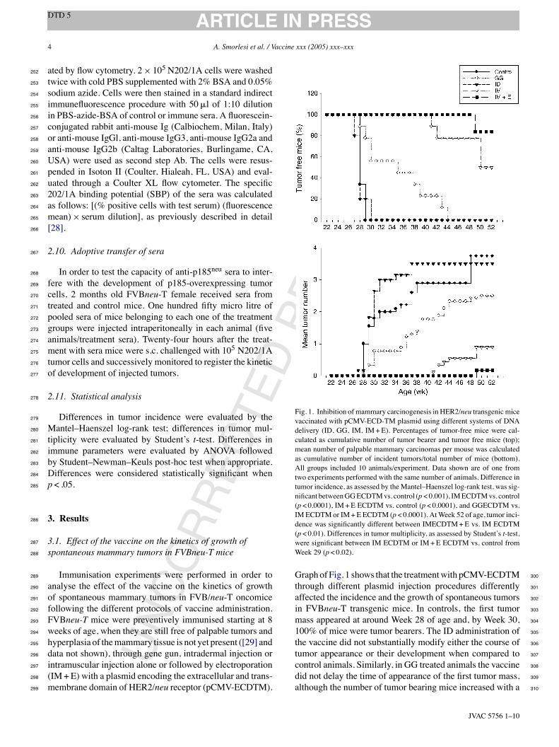
Fig. 1. Inhibition of mammary carcinogenesis in HER2/neu transgenic micevaccinated with pCMV-ECD-TM plasmid using different systems of DNAdelivery (ID, GG, IM, IM+E). Percentages of tumor-free mice were cal-culated as cumulative number of tumor bearer and tumor free mice (top);mean number of palpable mammary carcinomas per mouse was calculatedas cumulative number of incident tumors/total number of mice (bottom).All groups included 10 animals/experiment. Data shown are of one fromtwo experiments performed with the same number of animals. Difference intumor incidence, as assessed by the Mantel–Haenszel log-rank test, was sig-nificant betweenGGECDTMvs. control (p< 0.001), IMECDTMvs. control(p< 0.0001), IM+E ECDTM vs. control (p< 0.0001), and GGECDTM vs.IMECDTMor IM+E ECDTM (p< 0.0001). AtWeek 52 of age, tumor inci-dence was significantly different between IMECDTM+E vs. IM ECDTM(p< 0.01). Differences in tumor multiplicity, as assessed by Student’s t-test,were significant between IM ECDTM or IM+E ECDTM vs. control fromWeek 29 (p< 0.02).
Graphof Fig. 1 shows that the treatmentwith pCMV-ECDTM 300
through different plasmid injection procedures differently 301
affected the incidence and the growth of spontaneous tumors 302
in FVBneu-T transgenic mice. In controls, the first tumor 303
mass appeared at around Week 28 of age and, by Week 30, 304
100% of mice were tumor bearers. The ID administration of 305
the vaccine did not substantially modify either the course of 306
tumor appearance or their development when compared to 307
control animals. Similarly, in GG treated animals the vaccine 308
did not delay the time of appearance of the first tumor mass, 309
although the number of tumor bearing mice increased with a 310

UNCO
RREC
TED
PROO
F
JVAC 5756 1–10
A. Smorlesi et al. / Vaccine xxx (2005) xxx–xxx 5
Fig. 2. Number of mammary carcinomas in FVBneu-T transgenic mice vac-cinated with pCMV-ECDTM plasmid following different system of DNAdelivery. The percentage of tumor-free mice and of mice having 2> tumoror 3> tumors is shown.
slower kinetic in comparison with control mice, and only at311
Week 44 of age 100% of mice presented tumors (p< 0.001312
versus control group). In animals which received DNA by IM313
injection, the time of tumor onset was drastically delayed: in314
IM group first tumors appeared in only 20%ofmice at around315
Weeks 40–42, and 45% of mice were still tumor free at Week316
52 (p< 0.0001 versus control group). Electroporation signifi-317
cantly improved the outcome of IM delivered vaccine so that318
at Week 48 of age 100% of mice were still tumor free and319
at Week 52 of age only the 20% of mice presented tumors320
(p< 0.0001 versus control group; p< 0.01 versus IM). The321
kinetics of tumor appearance in IM or IM+E treated mice322
was significantly different from that of GG treated animals323
(p< 0.0001). Theprotective effect of the vaccinewas assessed324
also by monitoring the variation of mean number of tumor325
masses per mouse. As shown in Fig. 1 (bottom) and in Fig. 2,326
the reduction of tumor incidence obtained in IM immunized327
mice was associated to a decrease of the number of tumor328
masses arising in treated mice. In both IM and IM+E mice329
groups, a significantly lower tumor multiplicity was found in330
comparison with control animals (p< 0.02). The mean num-331
ber of tumor masses in IM+E mice group was significantly332
lower when compared to IM mice group from Week 44 of333
age (p< 0.05). In all ID treated mice a high number of masses334
appeared so that 80% of animals presented more than three335
tumors, as control mice. On the contrary, in IM and IM+E336
groups the few tumor bearingmice did not developmore than337
one tumormass.GG injection leaded to an intermediate result338
since only 20% of mice developed more than three masses339
and the mean number of tumors/mouse remained lower than340
control and ID injected animals.341
3.2. Production of antibodies against rat-p185neu342
Humoral immunity elicited by the vaccine was analysed343
in the different groups of treatment. Sera of treated and344
Fig. 3. Humoral immunity in FVBneu-T transgenic mice vaccinated withpCMV-ECDTM plasmid following different systems of DNA delivery. Spe-cific binding potential of sera from treated and control animals to p185protein (A) and anti-p185neu IgGs isotytpes (B) are shown. Sbp was evalu-ated byflowcytometry after indirect immunofluorescence.Asterisk indicatesstatistical significant difference vs. control.
control mice were harvested 2 weeks after the end of the 345
immunization protocol and successively analysed to assess 346
the induction of a specific humoral immune response. As 347
shown in Fig. 3(A), tumor-specific antibodies were observed 348
in all not vaccinated FVB/neu-T oncomice that naturally 349
develop a humoral immunity against ratHER2/neu transgene. 350
In particular our analysis showed that immunoglobulines of 351
IgGidiotype released in control mice were characterised by 352
a prevalence of IgGl subclass, that is typical of a Th2-type 353
immunity. Except for ID injection, all other treatments sig- 354
nificantly increased the titre of anti-HER2/neu antibodies 355
although the amount of produced antibodies only partially 356
correlated with the outcome of vaccination. On the contrary 357
the quality of the humoral response seems to be important 358
for the efficacy of the vaccine. Fig. 3(B) shows the IgG iso- 359
type distribution in mice immunised with different delivery 360
systems. 361
Interestingly, GG vaccination did not change the IgG pat- 362
tern observed in not immunised mice; in IM sera, differently, 363
IgG1 titre decreased in comparison to control mice (p< 0.05) 364
while a prevalence of Th1-like IgGs (IgG2a, IgG2b, IgG3) 365
was observed. IM+E vaccination promoted a high increase 366

UNCO
RREC
TED
PROO
F
JVAC 5756 1–10
6 A. Smorlesi et al. / Vaccine xxx (2005) xxx–xxx
Fig. 4. In vivo anti-tumor effect of anti-p185 sera from immunized mice.The capacity of anti-p185neu sera to interfere with the development of p185-overexpressing 202/1A tumors cells is shown. Sera from vaccinated andcontrol mice were i.p. injected in 2 month old FVBneuNT mice; 24 h aftermice were s.c. challenged with 105 N202/1A tumor cells. Difference intumor incidence, as assessed by the Mantel–Haenszel log-rank test, was sig-nificant between GGECDTM vs. control (p< 0.01), IM ECDTM vs. control(p< 0.01), and IM+E ECDTM vs. control (p< 0.01).
of IgG2a (p< 0.01 versus control and IM), suggesting a cor-367
relation between the effectiveness of the vaccine and the368
induction of a Th1-like immunity.369
3.3. Adoptive transfer of immune sera370
The ability of sera obtained from HER2/neu immunised371
mice to protect against tumor growth in vivo was assessed372
by administrating sera from treated mice to untreated ani-373
mals that received a s.c. injection of N202/1A tumor cells.374
As shown in Fig. 4, tumor-specific sera were able to delay375
the time of appearance and development of N202/1A tumors.376
Sera from GG treated mice lightly protected animals from377
the growth of N202/1A tumors (p< 0.01 versus control378
group). Sera harvested from intramuscularly immunised379
mice, both without or with electroporation, more evidently380
protected animals from N202/1A challenge (p< 0.01 ver-381
sus control group). The protection afforded by sera from382
IM and IM+E mice did not show statistically significant383
difference.384
3.4. Cytotoxic activity and cytokine production385
In order to analyse the cellular immunity induced by the386
different methods of immunization, spleen cells from immu-387
nized mice were analyzed for their cytotoxicity and cytokine388
production after in vitro incubation with HER2/neu overex-389
pressing tumor cells.As shown in Fig. 5 all DNA vaccination390
methods against HER2/neu increased anti-HER2/neu spe-391
cific cytotoxic activity of lymphocytes in comparison with392
control group (p< 0.05). In particular, the efficacy of the vac-393
cine was evident in the case of IM+E vaccinated mice where394
the cytotoxicitv was 10-fold increased compared to controls395
(p< 0.05); a less evident, but still significant augmentation396
of cytotoxic activity in comparison with the other groups of397
mice was observed (p< 0.05). In all groups the activation of398
an HER2/neu-specific immunity was associated to a slight399
Fig. 5. Cell-mediated cytotoxic activity in HER-2/neu transgenic mice vac-cinated with pCMV-ECDTM plasmid following different systems of DNAdelivery. Cytotoxicitv was performed through a fluorimetric assay. Data arereported as percent of lysis obtained at different E:T ratios and as numberof lytic units (LU20/107). Difference in cytotoxicity was significant betweenGG, ID, IM or IM+E vs. control group (*) p< 0.05 and between IM+E vs.GG, ID, or IM groups (**) p< 0.05.
Fig. 6. Effect of different systems ofDNAdelivery on the percentage of CD4or CD8 T cells. Spleen cells frommice vaccinated using different systems ofDNA delivery were stained with anti-CD4 or anti-CD8monoclonal antibod-ies and analyzed at a flow cytometer. Difference in the percentage of CD4T cells was significant between IM or IM+E and control group (p< 0.01).Difference in the percentage of CD8 T cells was significant between IM+Eand control group (p< 0.03).

UNCO
RREC
TED
PROO
F
JVAC 5756 1–10
A. Smorlesi et al. / Vaccine xxx (2005) xxx–xxx 7
increase of the percentage of CD4+ and CD8+ T cells in400
comparison with control group, that, however, was signifi-401
cant only for IM and IM+E groups in the case of CD4 cells402
(p< 0.0l) and only for IM+E group for CD8 cells (p< 0.03)403
(Fig. 6). As shown in Fig. 7, a significant augmentation of404
IFN-# production by CD8 T cells was observed for IM and405
Fig. 7. Intracellular cytokine staining in HER2/neu transgenic mice vacci-nated with pCMV-ECDTM plasmid following different systems of DNAdelivery. The percentage of CD8 T cells containing IFN-#. and the per-centage of CD4 T cells containing IL10 or IL4 was determined throughdouble-staining flow cytometry after in vitro lymphocyte incubation withmitomycin-treated N202/lA tumor cells. All data are expressed as percent-age of positive cells. Difference in the percentage of IFN-#-producing CD8T cells was significant between IM or IM+E and control group (p< 0.05).
IM+E groups with respect to controls (p< 0.05). In vacci- 406
nated mice, compared to controls, an increased number of 407
IL10-producing CD4 T cells was found in IM and IM+E 408
groups; no delivery system stimulated the production of the 409
Th2-like cytokine IL4 by CD4 T cells. 410
4. Discussion 411
By virtue of its versatility and efficacy in inducing immu- 412
nity against tumor antigens DNA vaccination represents a 413
suitable approach for the treatment of tumors [1–3]. Many 414
strategies of DNA vaccination against HER2/neu have been 415
proposed by our and other groups to prevent the develop- 416
ment of HER2/neu-positive mammary carcinomas in various 417
experimental models including transplantable and sponta- 418
neously arising transgenic tumors [5–20]. In view of the 419
translation of DNA vaccination from murine to large animal 420
models and clinical human trials the goal of all investigators 421
is to identify the way to improve the potency of HER2/neu 422
DNA vaccines. The use of different adjuvants in association 423
with DNA vaccines proposed against HER2/neu tumor anti- 424
gen has been studied [5–20], but prior to modify vaccine 425
formula, the vaccine could be improved by optimising the 426
procedure of DNA delivery to favour the process of DNA 427
uptake by host cells [23]. 428
Several studies demonstrated the efficacy of various sys- 429
tems ofDNA immunisation in animalmodels and highlighted 430
that diverse protocols of DNA immunisation result in the 431
activation of different immune responses and exert different 432
protective effects [30–37]. An interesting study on the out- 433
come of various protocols of DNA vaccine delivery showed, 434
for example, that DNA immunisation via intramuscular and 435
intradermal routes elicits immune responses of differentmag- 436
nitude and duration. Ito et al. [32] demonstrated that while 437
intradermal injection of DNA induces an higher Ab and CTL 438
response, intramuscular delivery of the vaccine provides a 439
persistent antigen production of the antigen by long-term 440
transfected host cells that supports a longer lasting, although 441
weaker, immunity. 442
In the case of HER2/neu DNA vaccination no compar- 443
ison of the different systems of DNA vaccine delivery via 444
intramuscular or cutaneous administrations proposed for the 445
prevention of spontaneous carcinogenesis in transgenic mod- 446
els of mammary carcinoma has been done. 447
The present study, where intramuscular delivery of DNA, 448
followed or not by electroporation, was compared with its 449
cutaneous delivery through gene gun or intradermal injection, 450
has been performed in the transgenic model of FVB/neu-T 451
mice that develop spontaneous mammary carcinomas fol- 452
lowing the overexpression of HER2/neu oncogene in mam- 453
mary tissue of sexually mature females [25]. In these mice, 454
where an immune response specific for HER2/neu express- 455
ing tumors, although not protective, is naturally elicited 456
by the overexpression of the oncogene [29], DNA vac- 457
cination through intramuscular delivery resulted promis- 458

UNCO
RREC
TED
PROO
F
JVAC 5756 1–10
8 A. Smorlesi et al. / Vaccine xxx (2005) xxx–xxx
ing [17,19] to hamper spontaneous carcinogenesis. Thus,459
we performed this study in order to understand in which460
terms the method of DNA delivery is relevant to induce461
a protective immunity in FVB/neu-T mice and if the suc-462
cess of intramuscular immunisation was correlated with463
the induction of particular immune mechanisms against464
HER2/neu.465
Data presented here confirmed that in this transgenic466
model of spontaneous mammary carcinoma the efficacy of467
pCMV-ECDTM DNA vaccine against HER2/neu is influ-468
enced by the method of release of DNA. We showed that the469
vaccine delivery methods analysed elicited diverse immune470
mechanisms that differently prevented the appearance and471
the development of spontaneous mammary carcinomas. In472
particular, IM+E injection that, in another model of spon-473
taneous carcinogenesis, was recently shown to be successful474
in inhibiting multifocal preneoplastic lesions already present475
at the time of vaccination, in our model of tumor prevention476
resulted in the best antitumoral effect and in the generation477
of a Th1-type immune response.478
In FVBneu-T HER2/neu mice [25] the process of car-479
cinogenesis induced by the overexpression of activated rat-480
p185 in all mammary glands leads, by 30 weeks of age,481
to the appearance of clinical tumors with a fast kinetic482
of growth in all mice. When intradermically administered,483
pCMV-ECDTM vaccine did not altered substantially the484
kinetic of tumor growth and when given through gene gun485
delivery exerted only a partial protective effect. Differently,486
intramuscular immunization resulted in the best outcome487
since in IMmice pCMV-ECDTM vaccine was able to dras-488
tically delay both tumor onset and the mean number of489
tumor masses arising in treated mice. The efficacy of IM490
delivered vaccine was significantly improved by the pro-491
cedure of electroporation that further delayed the time of492
appearance of tumors and increased from 45 to 80%, the493
percentage of mice that were still tumor free at 1 year of494
age.495
In order to correlate the effects of the different delivery496
systems to the immune response, we performed some exper-497
iments to investigate humoral and cellular immunity elicited498
by the vaccine in the different groups of treatments.499
Anti-HER2/neu antibodies were found in the sera of all500
control and treated mice. In particular, all treatment, except501
for ID vaccination, induced a significant increase of the titre502
of anti-HER2/neu antibodies. Moreover, while the amount503
of produced antibodies only partially correlated with the out-504
come of vaccination, the quality of humoral response seems505
to be determinant for the success of vaccination. The effi-506
cacy of IM or IM+E vaccine treatments were associated507
to the production of a Th1 pattern of IgGs. The role of508
tumor-specific antibodies in the protection of animals against509
tumor development was also demonstrated by the fact that510
the adoptive transfer of immune sera hampered the growth511
of HER2/neu-positive tumors injected s.c. in not immunized512
mice. As it was argued elsewhere [38], is plausible that513
HER2/neu-specific antibodies acquire a relevant protective514
role, since HER2/neu oncoprotein, besides being a target 515
tumor antigen, is a receptor involved in regulation of cell 516
proliferation. 517
A tumor specific cellular immunity was also elicited by all 518
treatments in vaccinated mice. The augmentation of IFN-# 519
production by CD8 T cells, that resulted statistically signifi- 520
cant for IM and IM+E groups, was associated to an increase 521
of the in vitro cytototoxic activity of lymphocytes from all 522
vaccinated mice. Although in all groups the capacity to kill 523
HER2/neu expressing tumor cells was significantly increased 524
compared to control mice, the influence exerted by electro- 525
poration on intramuscular injection of the vaccine was, as 526
expected, clearly evident with increases of cytotoxicity sig- 527
nificantly higher than those obtained by IM immunization 528
alone. Surprisingly in lymphocytes obtained from mice of 529
IM and IM+E groups, where the vaccine exerted the best 530
protective effect, we observed an increased production of 531
IL10 by CD4 T cells. IL10 is a cytokine with pleiotropic 532
effects that is mainly known for its potent immunosuppres- 533
sive properties, but, in some instances, it has been shown 534
to paradoxically augment tumor immunity [39]. In partic- 535
ular, IL10 producing CD4 T cells have been demonstrated 536
to exert antitumor effects acting as regulatory cells rather 537
than typical Th2 cells [40]. Interestingly, in a recent study 538
[41] we observed a significant production of IL10 by CD4 T 539
cells associated to the adjuvant action of an immunostimulant 540
molecule used to improve the efficacy of a HER2/neu DNA 541
vaccine in the same model of transgenic mice. These obser- 542
vations, through needing to be confirmed by further studies, 543
suggest that the activation of IL10-producing CD4 T cells 544
could be a determinant event to build an immune response 545
able to hamper spontaneous carcinogenesis in FVB/neu-T 546
trangenie mice. In conclusion, intramuscular delivery was 547
confirmed the better route to elicit a complete immune 548
response of Th1 type and mediated by different mecha- 549
nisms that conferred a high protective potential to the vac- 550
cine. The efficiency of intramuscularly injected vaccine was 551
clearly improved by electroporation procedure that, favour- 552
ing the uptake of DNA into host cells and the expression 553
of the encoded protein [42], increased the intensity of the 554
immune response.On the contrary, cutaneous administrations 555
of the vaccine were not so successful: the Th1-like immu- 556
nity induced by ID injected vaccine was missing a strong 557
humoral response and was quantitatively too low to oppose 558
tumor development; GGdelivery, although is an efficient pro- 559
cedure that require a minimal amount of DNA to induce an 560
immune reaction against the tumor, mainly elicited a Th2- 561
like immune response that only partially protected mice from 562
carcinogenesis. 563
The identification of the best procedure of DNA delivery 564
represents a basic step for the optimisation of genetic immu- 565
nization protocols that can be further improved by the use of 566
immune adjuvant approaches to develop vaccines that could 567
be used also in immune-depressed individuals such as aged 568
subjects where the ability of immune system to hamper the 569
development of a tumor is impaired [24,43].

UNCO
RREC
TED
PROO
F
JVAC 5756 1–10
A. Smorlesi et al. / Vaccine xxx (2005) xxx–xxx 9
Acknowledgement570
This work was supported in part by a grant from Associ-571
azione Italiana Ricerca sul Cancro (AIRC).572
References573
[1] Tang D, De Vit M, Johnston SA. Genetic immunization is a sim-574
ple method for eliciting an immune response. Nature 1992;356:575
152–4.576
[2] Stevenson FK, Ottensmeier CH, Johnson P, Zhu D, Buchan SL,577
McCann KJ, et al. DNA vaccines to attack cancer. Proc Natl Acad578
Sci USA 2004;101(Suppl. 2):14646–52.579
[3] Prud’homme GJ. DNA vaccination against tumors. J Gene Med580
2005;7:3–17.581
[4] Hynes NE, Stem DF. The biology of erbB2/neu/HER2 and its role582
in cancer. BBA 1994;1198(2–3):165–84.583
[5] Chang SY, Lee KC, Ko SY, Ko HJ, Kang CY. Enhanced efficacy of584
DNA vaccination against Her2/neu tumor antigen by genetic adju-585
vants. Int J Cancer 2004;111:86–95.586
[6] Quaglino E, Iezzi M, Mastini C, Amici A, Pericle F, Di Carlo587
E, et al. Electroporated DNA vaccine clears away multifocal588
mammary carcinomas in her-2Aieu transgenic mice. Cancer Res589
2004;64(8):2858–64.590
[7] Quaglino E, Rolla S, Iezzi M, Spadaro M, Musiani P, De Giovanni591
C, et al. Concordant xmorphologic and gene expression data show592
that a vaccine halts HER2/neu preneoplastic lesions. J Clin Invest593
2004;113:709–17.594
[8] Cappello P, Triebel F, Iezzi M, Caorsi C, Quaglino E, Lollini PL, et595
al. LAG-3 enables DNA vaccination to persistently prevent mammary596
carcinogenesis in HER-2/neu transgenic BALB/c mice. Cancer Res597
2003;63(10):2518–25.598
[9] Disis ML, Scholler N, Dahlin A, Pullman J, Knutson KL, Hellstrom599
KE, et al. Plasmid-based vaccines encoding rat neu and immune600
stimulatory molecules can elicit rat neu-specific immunity. Mol Can-601
cer Ther 2003;2:995–1002.602
[10] Disis ML, Shiota FM, McNeel DG, Knutson KL. Soluble cytokines603
can act as effective adjuvants in plasmid DNA vaccines targeting604
self tumor antigens. Immunobiology 2003;207(3):179–86.605
[11] Renard V, Sonderbye L, Ebbehoj K, Rasmussen PB, Gregorius K,606
Gottschalk T, et al. HER-2 DNA and protein vaccines contain-607
ing potent Th cell epitopes induce distinct protective and thera-608
peutic antitumor responses in HER2 transgenic mice. J Immunol609
2003;171(3):1588–95.610
[12] Di Carlo E, Rovero S, Boggio K, Quaglino E, Amici A, Smorlesi A,611
et al. Inhibition of mammary carcino genesis by systemic interleukin612
12 or p185neu DNA vaccination in Her-2/neu transgenic BALB/c613
mice. Clin Cancer Res 2001;7(Suppl. 3):830s–7s.614
[13] Foy TM, Bannink J, Sutherland RA, McNeill PD, Moulton GG,615
Smith J, et al. Vaccination with Her-2/neu DNA or protein subunits616
protects against growth of a Her-2/neu-express ing murine tumor.617
Vaccine 2001;19(17–19):2598–606.618
[14] Pupa SM, Invernizzi AM, Forti S, Di Carlo E, Musiani P, Nanni P, et619
al. Prevention of spontaneous neu-expressing mammary tumor devel-620
opment in mice transgenic for rat proto-neu by DNA vaccination.621
Gene Ther 2001;8(1):75–9.622
[15] Rovero S, Boggio K, Carlo ED, Amici A, Quaglino E, Porced-623
daP, et al. Insertion of the DNA for the 163–171 peptide of ILl-624
beta enables aDNA vaccine encodingpl85(neu) to inhibit mammary625
carcinogenesis in Her-2/neu transgenic BALB/c mice. Gene Ther626
2001;8(6):447–52.627
[16] Lachman LB, Rao XM, Kremer RH, Ozpolat B, Kiriakova G, Price628
JE. DNA vaccination against neu reduces breast cancer incidence629
and metastasis in mice. Cancer Gene Ther 2001;8(4):259–68.630
[17] Amici A, Smorlesi G, Noce G, Santoni P, Cappelletti L, Capparuc- 631
cia R, et al. DNA vaccination with full-length or truncated Neu 632
induced protective immunity against the development of sponta- 633
neous mammary tumors in HER-2/neu transgenic mice. Gene Ther 634
2000;7:703–6. 635
[18] Piechocki MP, Pilon SA, Wei WZ. Complementary immunity 636
induced by plasmid DNA encoding secreted and cytopasmic human 637
ErbB-2. J Immunol 2001;167:3367–74. 638
[19] Amici A, Venanzi FM, Concetti A. Genetic immunization against 639
neu/erbB2 transgenic breast cancer. Cancer Immunol Immunother 640
1998;47(4):183–90. 641
[20] Chen Y, Hu D, Eling DJ, Robbins J, Kipps TJ. DNA vaccines encod- 642
ing full-length or truncated neu induce protective immunity against 643
neu-expressing mammary tumors. Cancer Res 1998;58:1965–71. 644
[21] Timmerman JM, Singh G, Hermanson G, Hobart P, Czerwinski DK, 645
Taidi B, et al. Immunogenicity of a plasmid DNA vaccine encoding 646
chimeric idiotype in patients with B-cell lymphoma. Cancer Res 647
2002;62:5845–52. 648
[22] Conry RM, Curiel DT, Strong TV, Moore SE, Allen KO, Barlow DL, 649
et al. Safety and immunogenicity of DNA vaccine encoding carci- 650
noembryonic antigen and hepatitis B surface antigen in colorectal 651
carcinoma patients. Clin Cancer Res 2002;8:2782–7. 652
[23] Kirman JR, Seder RA. DNA vaccination: the answer to stable, pro- 653
tective T-cell memory? Curr Opin Immunol 2003;15(4):471–6. 654
[24] Provinciali M, Smorlesi A, Donnini A, Bartozzi B, Amici A. Low 655
effectiveness of DNA vaccination against HER-2/neu in ageing. Vac- 656
cine 2003;21(9–10):843–8. 657
[25] Guy CT, Cardiff RD, Muller WJ. Activated neu induces rapid tumor 658
progression. J Biol Chem 1996;271:7673–8. 659
[26] Lollini PL, Nicoletti G, Landuzzi L, De Giovanni C, Rossi I, Di 660
Carlo E, et al. Down regulation of major histocompatibility complex 661
class I expression in mammary carcinoma of HER2/neu transgenic 662
mice. Int J Cancer 1998;77:937–41. 663
[27] Provinciali M, Di Stefano G, Fabris N. Optimization of cytotoxic 664
assay by target cell retention of the fluorescent dye carboxyfluo- 665
rescein diacetate (CFDA) and comparison with conventional 51CR 666
release assay. J Immunol Methods 1992;155:19–24. 667
[28] Rovero S, Amici A, Di Carlo E, Bei R, Nanni P, Quaglino E, et 668
al. DNA vaccination against rat HER2/neu p185 more effectively 669
inhibits carcinogenesis than transplantable carcinomas in transgenic 670
BALB/c mice. J Immunol 2000;165:5133–42. 671
[29] Takeuchi N, Hiraoka S, Zhou XY, Nagafuku M, Ono S, Tsujimura 672
T, et al. Anti-HER2/neu immune responses are induced before the 673
development of clinical tumors, but declined following tumorigenesis 674
in transgenic mice. Cancer Res 2004;64:7588–95. 675
[30] Torres CA, Iwasaki A, Barber BH, Robinson HLO. Differential 676
dependence on target tissue for gene gun and intramuscular DNA 677
immunisation. J Immunol 1997;158:4591–632. 678
[31] Trimble C, Lin CT, Hung CF, Pai S, Juang J, He L, et al. Comparison 679
of the CD8+ T cell responses and anti-tumor effects generated by 680
DNA vaccine administered through gene gun, biojector and syringe. 681
Vaccine 2003;21:4036–41. 682
[32] Ito K, Shinohara N, Kato S. DNA immunization via intramus- 683
cular and intradermal routes using a gene gun provides different 684
magnitudes and durations on immune response. Mol Immunother 685
2003;39(14):847–54. 686
[33] Hynes JR. Particle-mediated DNA vaccine delivery to the skin. 687
Expert Opin Biol Ther 2004;4(6):889–900. 688
[34] Dileo J, Miller TE, Chesnoy S, Huang L. Gene transfer to sub- 689
dermal tissues via a new gene gun design 2003. Hum Gene Ther 690
2003;14(l):79–87. 691
[35] Turing T. The immunology of cutaneous DNA immunization. Curr 692
Opin Mol Ther 1999;l(2):216–25. 693
[36] Ulmer JB, Deck RR, Dewitt CM, Donnhly JI, Liu MA. Generation 694
of MHC class I-restricted cytotoxic T lymphocytes by expression of 695
a viral protein in muscle cells: antigen presentation by non-muscle 696
cells. Immunology 1996;89:59–67. 697

UNCO
RREC
TED
PROO
F
JVAC 5756 1–10
10 A. Smorlesi et al. / Vaccine xxx (2005) xxx–xxx
[37] Corr M, von Damm A, Lee DJ, Tighe H. In vivo priming by698
DNA injection occurs predominantly by antigen transfer. J Immunol699
1999;163:4721–7.700
[38] Lollini PL, Forni G. Cancer immunoprevention: tracking down per-701
sistent tumor antigens. Trends Immunol 2003;24:62–6.702
[39] Berman RM, Suzuki T, Tahara H, Robbins PD, Nahura SK, Lotze703
MT. Systemic administration of cellular IL-10 induces an effective,704
specific, and long-lived immune response against established tumors705
in mice. J Immunol 1996;157:231–7.706
[40] Segal BM, Glass DD, Shevach EM. Cutting edge: IL-10-producing707
CD4 T cells mediate tumor rejection. J Immunol 2002;168:1–4.
[41] Smorlesi A, Papalini F, Orlando F, Donnini A, Re F, Provinciali 708
M. Imiquimod and S-27609 as adjuvants of DNA vaccination in a 709
transgenic murine model of HER2/neu-positive mammary carcinoma. 710
Gene Ther 2005;12(17):1324–32. 711
[42] Widera G, Austin M, Rabussay D, Goldbeck C, Barnett SW, Chen 712
M, et al. Increased DNA vaccine delivery and immunogenicity by 713
electroporation in vivo. J Immunol 2000;164:4635–40. 714
[43] Provinciali M, Smorlesi A. Immunoprevention and immunoi- 715
herapy of cancer in ageing. Cancer Immunol Immunother 716
2005;54(2):93–106. 717
Copyright © 2022 FDOKUMEN