Evaluating non-invasive x-ray techniques for material science investigations of rock art: Community...
21
Palaeoart and Materiality The Scientific Study of Rock Art edited by Robert G. Bednarik, Danae Fiore, Mara Basile, Giriraj Kumar and Tang Huisheng Archaeopress Archaeology Copyrighted material: no unauthorised reproduction in any medium
Transcript of Evaluating non-invasive x-ray techniques for material science investigations of rock art: Community...
Palaeoart and
Materiality The Scientific Study of Rock Art
edited by
Robert G. Bednarik, Danae Fiore, Mara Basile, Giriraj Kumar
and Tang Huisheng
Archaeopress Archaeology
Copyrighted material: no unauthorised reproduction in any medium
Archaeopress Publishing LtdGordon House
276 Banbury RoadOxford OX2 7ED
www.archaeopress.com
ISBN 978 1 78491 429 5ISBN 978 1 78491 430 1 (e-Pdf)
© Archaeopress and the authors 2016
Cover image: Part of the Huashan site in Guangxi Province, southern China, the largest rock painting site in the world. Photograph by R. G. Bednarik.
All rights reserved. No part of this book may be reproduced, in any form or by any means, electronic, mechanical, photocopying or otherwise,
without the prior written permission of the copyright owners.
Printed in England by Oxuniprint, OxfordThis book is available direct from Archaeopress or from our website www.archaeopress.com
Copyrighted material: no unauthorised reproduction in any medium
i
Contents
Contents ......................................................................................................................................................................... i
Relevant Issues for the Design of a Protocol for the Interdisciplinary Study of Rock Art ...........................................1Eugenia P. Tomasini, Mara Basile, Marta S. Maier, Norma Ratto
Superimpositions and Attitudes Towards Pre-existing Rock Art: a Case Study in Southern Patagonia ...................15Anahí Re
Pigments Used in Rock Paintings from the East and West of the Iberian Peninsula Analysed by X-ray Fluorescence: Analogies and Differences ............................................................................................................31A. Martín Sánchez, C. Roldán García, M. J. Nuevo, J. Oliveira, S. Murcia Mascarós, C. Oliveira
The Material Scientific Investigation of Rock Art: Contributions from non-Invasive X-ray Techniques ...................41Jillian Huntley, Clive Freeman Galamban
Methodological Approach to the Materiality of Rock Paintings Based on Their Physicochemical Characterisation. Proposal and Reflections from Their Study in Chile ................................................................59Marcela Sepúlveda
Step Forwards in the Archaeometric Studies on Rock Paintings in the Bogotá Savannah, Colombia. Analysis of Pigments and Alterations ...................................................................................................................73Judith Trujillo T.
What Should We Do or Not Do for the Preservation or Remedial Action in Prehistoric Painted Caves? ................85F. Bourges, P. Genthon, D. Genty, M. Lorblanchet, France, E. Mauduit, D. D’Hulst, E. David, N. Ferrer
Forensic Replication Work with Australian Cave Art .................................................................................................99Robert G. Bednarik, Yann-Pierre Montelle
Experimental Rock Art Studies. Replication of Pictographs from La Primavera Locality (Santa Cruz, Argentina) .113Rocío V. Blanco, Natalia G. Barreto
Measurements and Replications of Hand Stencils: a Methodological Approach for the Estimation of the Individuals’ Age and Sex .....................................................................................................................................129Natalia Carden, Rocío Blanco
Time and Rock Art Production: Explorations on the Material Side of Petroglyphs in the Semiarid North of Chile ......................................................................................................................................................147Francisco Vergara, Andrés Troncoso, Francisca Ivanovic
Taphonomy of the Early Petroglyphs at Daraki-Chattan and in the Region Around It in Chambal Basin, India ....161Giriraj Kumar, Ram Krishna, Robert G. Bednarik
The Tribology of Petroglyphs .................................................................................................................................... 171Robert G. Bednarik
Understanding the Technology of Very Small Cupules in Daraki-Chattan, India ....................................................187Ram Krishna, Giriraj Kumar
Copyrighted material: no unauthorised reproduction in any medium
ii
Evidence of Collaboration Among Art-Makers in Twelve Upper Palaeolithic Caves ...............................................195Leslie Van Gelder
A Survey of Developments in Dating Australian Rock-Markings .............................................................................205Graeme K. Ward
A New Cold Plasma System for Pictogram 14C Sampling ..........................................................................................217Marvin W. Rowe, Eric Blinman, John C. Martin, J. Royce Cox, Mark MacKenzie, Lukas Wacker
Direct Dating of Bolivian Petroglyphs Using Indirect Microerosion Analysis ..........................................................225Robert G. Bednarik
Use of Theodolite and Photographic Techniques in the Recording and Analysis of the Geographical and Astronomical Entorno (surrounding) ..................................................................................................................235Patricio Bustamante, Ricardo Moyano, Daniela Bustamante
Copyrighted material: no unauthorised reproduction in any medium
41
Introduction
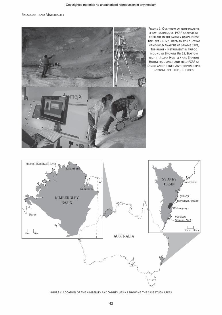
The importance of low impact methods in contemporary archaeology/heritage management is reflected in the growing application of non-invasive techniques from the physical sciences (Demetrios and Karydas 2009; Joyce 2011: 199). This appetite for non-invasive analytical methods has been fuelled by increased accessibility due to the decreasing price of commercial instrumentation (Heginbotham et al. 2010: 178). Non-invasive techniques are attractive to both researchers and the Indigenous peoples on whose heritage they work. Investigating priceless cultural heritage items such as rock art can generate serious ethical dilemmas for Indigenous communities, other stakeholders and researchers (Bednarik 1992; Clottes 1992; Watchman 1992). The prospect of non-invasively gathering data relating to the materiality of art panels and associated site fabrics like subsurface/surface ochres finds, mineral accretions and rock shelters matrices, can ameliorate ethical considerations. Here, we describe our application of two such non-invasive x-ray based techniques to the materiality of rock art sites: portable x-ray fluorescence spectrometry (hereafter PXRF) and micro-computed tomography (hereafter µ-CT) (Figure 1). Examples from research in the Sydney and Kimberley Basins in
the southeast and far northwest of Australia respectively (Figure 2) illustrate how PXRF and µ-CT can be integrated into regional rock art research programs, and the potential of these methods as conservation/management tools. A further, evolving example from the Sydney Basin is discussed to highlight the prospects of non-invasive methods for community-driven conservation and management.
This chapter introduces current generation, non-invasive x-ray analytics and their potential/limitations for rock art research, conservation and management. The basic principles of each technique are outlined, followed by short case studies illustrating the useful information that can be generated. As with all techniques from the physical sciences, non-invasive methods have advantages, complexities and restrictions that must be evaluated for selected applications. We present what we consider the major opportunities and constraints of PXRF and µ-CT for rock art research, conservation and management (Tables 2 and 4). We see the preferences of Indigenous peoples as a central impetus for the continuing development and use of non-invasive methods in rock art research. We conclude by briefly outlining the evolving interaction of Australian Indigenous peoples with material science, particularly non-invasive techniques. Agreeing, as Smith
The Material Scientific Investigation of Rock Art: Contributions from non-Invasive X-ray Techniques
Jillian HuntleyAustralia, [email protected]
Clive Freeman GalambanAustralia, [email protected]
Understanding the physical (cultural and environmental) processes that have created and continue to act upon rock art is funda-mental to research and management. We used portable x-ray fluorescence spectrometry and micro-computed tomography to inves-tigate the materiality of rock art and associated site fabrics in the Sydney and Kimberley Basins, on opposite sides of the Australian continent. These case studies illustrate the types of information that can be generated using non-invasive techniques and demon-strate their application in developing an understanding of paint composition, rock art panel taphonomy and the physical properties of pigments (ochre), mineral accretions and shelter substrates. We highlight the power of non-invasive material scientific tech-niques as tools in cultural heritage management and the role such techniques can play in emergent community-driven research.
La investigación científica material del arte rupestre: contribuciones de técnicas no invasivas de rayos X
Conocer los procesos físicos (culturales y medioambientales) que han creado y continúan afectando el arte rupestre es fundamen-tal para su investigación y manejo. Hemos usado equipos de espectrometría de fluorescencia de rayos X y tomografía micro-com-putada para investigar la materialidad del arte rupestre y estructuras de sitios asociadas en las cuencas de Sydney y Kimberley, en lados opuestos del continente australiano. Estos casos de estudio ilustran los tipos de información que pueden ser generados usando técnicas no invasivas y demuestran su aplicación en el desarrollo de conocimiento sobre la composición de la pintura, la tafonomía de los paneles de arte rupestre, y las propiedades físicas de pigmentos (ocre), acreciones minerales y sustratos de abrigos rocosos. Enfatizamos el poder de las técnicas científicas no invasivas para el estudio de materiales, como herramientas en el manejo del patrimonio cultural y el rol que dichas técnicas pueden jugar en la emergente investigación orientada por las comunidades locales.
Copyrighted material: no unauthorised reproduction in any medium
42
Palaeoart and Materiality
Figure 1. Overview of non-invasive x-ray techniques. PXRF analysis of rock art in the Sydney Basin, NSW:
top left - Clive Freeman conducting hand-held analysis at Baiame Cave;
Top right - Instrument in tripod mound at Browns Rd 29; Bottom
right - Jillian Huntley and Sharon Hodgetts using hand-held PXRF at
Dingo and Horned Anthropomorph. Bottom left - The µ-CT used.
Figure 2. Location of the Kimberley and Sydney Basins showing the case study areas.
Copyrighted material: no unauthorised reproduction in any medium
43
Jillian Huntley and Clive Freeman Galamban: The Material Scientific Investigation of Rock Art
et al. (2003) did with Powell (2000), that there are lessons to be learned from working with Indigenous Australians that have currency and applicability to international community involvement in rock art research and management. We are not necessarily advocating the use of the specific techniques discussed. Our intention is to stimulate Indigenous communities and researchers to critically consider the potential of non-invasive methods for investigating the materiality of rock art.
Elemental and structural data are powerful tools for understanding the material properties of rock art and associated site fabrics. PXRF and µ-CT have an inverse relationship with regard to the types of data they produce. XRF, including PXRF, characterises the elemental composition of materials, their chemical rather than crystallographic or compound (structural) nature (Liritzis and Zacharias 2011:119). Conversely computed tomography (CT), including µ-CT, produces images of the (internal) structure1 of objects using the differential mass of the material scanned (Ghysels 2003). PXRF has a ~40 year history in archaeometric research, but the last decade has seen a veritable explosion in its use for material science investigations in cultural heritage (Guilherme et al. 2008:444; Speakman et al. 2011:3483). Similarly, CT has been a staple structural analytic tool for palaeoanthropological specimens since the late 1970s/early 1980s. More recently, novel archaeological and conservation science CT applications have emerged for a variety of materials (Ghysels 2003; Wade et al. 2011; McBride and Mercher 2012).
PXRF: background, example and evaluation
XRF characterises the chemical composition of material analysed. Primary x-rays from an x-ray tube or radioactive source strike atoms in the surface of the sample with enough force to eject inner shell electrons (K, L and M). The vacancy created is filled by a higher energy, outer shell electron. Excess energy from the higher energy electron de-excites once the vacancy is filled producing secondary, fluorescent x-rays. Fluorescent x-rays have characteristic energies diagnostic of elements present qualitatively, identifying composition. The detector of the instrument counts the number of fluorescent x-rays quantifying element concentrations (Keoing et al. in press 2013; Pollard et al. 2007: 101). XRF (including pXRF) is a bulk analytic measurement where characterisations are an average of the entire analyte volume (Shackley 2011: 10). PXRF therefore estimates ‘… the bulk composition of the sample from what is essentially a surface measurement’ (Potts 2008: 10). This is especially
1 In Australia high resolution synchrotron CT techniques that can simultaneously image structural and elemental data are available — x-ray absorption near-edge structural imaging and XRF imaging with CT reconstruction (Etschmann et al. 2010; Howard et al. 2012). Thus far applications in heritage and geoscience have been limited and they are yet to be applied to rock art investigations.
true in relation to the light z elements we measured in case studies.
A fundamental principle of XRF that must be considered for rock art, indeed all unprepared materials, is differential critical depth penetration. The depth at which different elements excite, producing characteristic x-rays, varies depending on their atomic weight, resulting in analyte volumes that increase commensurate with the atomic weight of elements (see Jenkins 1999 for a discussion of XRF principles and Liangquan et al. 2005: 80 for an equation to calculate critical depth penetration in natural minerals such as aluminosilicate rock art/ochres). In silicate materials such as the sandstone rock shelters and clay rich ochres we analysed, critical depth penetration of potassium is ~30 µm whereas critical depth penetration of iron is ~170 µm (Potts et al. 1997: 33). Therefore, critical depth penetration may differ by more than a factor of five between elements at the start of the spectra and those at the end (e.g. Figure 7). The infinitely thin2 nature of pictograms must be accounted for when interpreting rock art analysis and an understanding of exactly ‘what’ is included in the measurement must be demonstrated. Rock art pigment, the rock shelter matrix and any associated weathering products such as precipitate mineral skins, chemical alteration from biofilms, mineralogical segregation like oxidation and chemical enrichment/depletion are all encapsulated in PXRF analytes. Understanding what is being measured is equally crucial where rock art is infinitely thick (taking up the entire analyte), because PXRF cannot differentiate superimposed pigments (Wesley et al. in press 2014; Liritzis and Zacharias 2011: 132).
Attenuation effects re-introduced by in situ analysis of unprepared materials must also be given due consideration. Classic preparation for XRF involves crushing and milling of samples to create a fused bead for major elements, or a pressed powder pellet for trace elements. When analysing unprepared materials, loss of either incident or fluorescent x-ray signal, or both, can result from surface morphology, mineralogical segregations/heterogeneity, the grain size of the materials and varying x-ray path geometry (Forster et al. 2011; Huntley 2012 for further discussion). These attenuation effects have to be acknowledged and should be minimised or mitigated to reduce their impact on PXRF of unprepared samples where possible.
PXRF methodology
We used a Bruker Tracer III-V PXRF equipped with a rhodium tube, Peltier-cooled Si-PIN detector and a 1024 channel analyser at a resolution of approximately 170eV
2 “When the penetration depth of the incident radiation emitted by the exciting source, and the secondary x-ray emitted by the sample, is larger than (or of the same order of magnitude as) the sample thickness … the sample is considered … infinitely thin…” (Cesareo 2008: 209).
Copyrighted material: no unauthorised reproduction in any medium
44
Palaeoart and Materiality
FHWM at the manganese Kα peak (5.9 keV at 1000 counts per second). Analyses were optimised for light element excitation, measuring 1.3 to 9.5 KeV for Al to Zn. Parameters were 12 keV, 20 µA, using a 0.0254 mm titanium filter in the x-ray path. Spectra were collected for 300 pulse counts (live seconds) at 185 FWHM. A titanium filter was used to reduce incident rhodium L line x-rays because measurement of fluorescent chlorine and sulphur K line x-rays was desirable. To minimise attenuation of incident x-rays, a vacuum pump removed air from the instrument between the x-ray tube and the beryllium window, and the beryllium window and the detector beam paths (Figure 1).
Spectra were processed using Bruker supplied software: X-RayOps to adjusting tube operating voltage and current settings; S1PXRF for count rate, signal acquisition and live evaluation of data; and Spectra 7.1 for calculation of Net Peak Areas (NPAs) — semi-quantitative measurement of element concentrations via standard-less fundamental parameters3. NPA calculations were undertaken using nine correction cycles for escape and background peaks between 0.6 KeV and 10.5 KeV. Elements selected for fundamental parameters correction were based on the evaluation of ‘raw’4 in situ spectra and legacy scanning electron microscopy (including energy dispersive x-ray analysis, hereafter SEM), x-ray diffraction and particle induced x-ray/particle induced gamma-ray data from the same rock art panel (Huntley et al. 2011: 87; Ford 2006: A4–1).
Case study: evidence for geomorphic processes (chemical weathering) in sandstone shelters containing rock art; PXRF as a novel conservation and management tool
Material science investigations are powerful tools for informing conservation/management of rock art and associated site fabrics. This potential is demonstrated by the results of pXRF analysis on a rock art panel at the site Browns Rd 29 on the Woronora Plateau, above Wollongong in the Sydney Basin, NSW (Figure 2). The taphonomy of rock art must be a primary concern for heritage management strategies that attempt to comprehensively record and conserve this finite cultural resource (Mirmehdi et al. 2001: 1329). Here, the chemical indices of geological weathering observed by light element optimised pXRF expand on a previous report of paint constituents by highlight conservation/management implications (Huntley 2012). Silicate rocks are a common canvas for the production of rock art and a common fabric for other cultural heritage items such as monuments and buildings (Ogburn et al. 2012; Prikryl et al. 2007: 409). This case study focused on the chemical indices of weathering processes catalysed by
3 Heginbothham et al. 2010 provide a comprehensive review of quantitative measures in PXRF.4 Unprocessed spectra viewed during acquisition via S1PXRF.
salts. The case study has implications for identifying and monitoring salts in silicate geological environments and sandstone heritage objects internationally (Walderhaug 1998; Prikryl et al. 2007; Bera et al. 2011; Bwasiri 2011; Vařilová et al. 2011; Ogburn et al. 2013).
Chemical weathering is the alteration of the chemical and/or mineralogical structure of rock and is achieved in various ways as no mineral is absolutely chemically inert (Twidale 1968: 142). Usually chemical weathering takes place through the agency of water, a process referred to as hydrolysis, the combining of a mineral with hydroxyl (OH) producing a chemical change that contributes to the weakening the rock (Twidale 1968: 142). Indeed, experimental research has shown that water from natural precipitation and human application (as was an historic practice in rock art recording to ‘enhance’ photography) promotes chemical and subsequent physical changes to painted stone surfaces (Mirmehdi et al. 2001:1329). Physical weathering breaks the rock down into fragments or particles. Chemical weathering actually breaks the bonds between atoms within minerals (Twidale and Campbell 1993: 98 –99). The parent rock, particularly its mineral composition, has great influence on the type and course of geological, especially chemical, weathering (Twidale 1968: 136–137). In the Hawkesbury Sandstone of the Sydney Basin, shelters are dominantly formed by chemical weathering processes generically grouped under the term ‘cavernous weathering’ (Young 1987). Salt (sodium chloride) has been identified as a primary catalyst, and accelerant, for cavernous weathering in this geological formation (Lambert 1980; Young 1987; Young and Young 1992).
Precipitous mineralisation takes several forms. Precipitation of secondary minerals through hydrolysis and their deposition at, or near, the rock shelter substrate surface results in a less permeable, structurally harder, mineralised layer termed casehardening (Young and Young 1992: 74; Prikryl et al. 2007: 419). Microflora such as lichens can contribute to this type of mineralogical segregation, altering rock chemistry by extracting iron and minor amounts of associated elements from minerals within the lithology, concentrating them immediately at or below the substrate surface (Twidale 1968: 144; Nanson and Young 1983; Mitchell et al. 1997). Mineral precipitation in silicate rocks also produces silica-rich accretions, commonly known as amorphous silica skins/hydrated amorphous silicon dioxide, SiO2.nH2O (Watchman 1996: ii). Siliceous accretions have been observed throughout rock art sites in the Hawkesbury Sandstone (Lovering 1952). These are the geological weathering product best known to rock art researchers for their potential to contribute chronological information (absolute age determinations) (Watchman 1993; Campbell et al. 1996; Watchman et al. 1997; 2000; 2001 and 2005; Steelman et al. 2002; Ruiz et al. 2006). Soluble salts catalyse and accelerate precipitous mineralisation,
Copyrighted material: no unauthorised reproduction in any medium
45
Jillian Huntley and Clive Freeman Galamban: The Material Scientific Investigation of Rock Art
enhancing dissolution of minerals (Prikryl et al. 2007: 409; Mol and Viles 2011: 302). Salts particularly catalyse iron, clay and calcite precipitation and probably silica as well, though there has been little specific research (Young and Young 1992: 74). Percolation of salts through the rock matrix by hydrolysis can result in their deposition on the surface, weakening the substrate, making it susceptible to processes such as honeycomb weathering and granular disintegration (Twidale and Campbell 1993: 497).
Clarke and North have noted that accumulations of salt affect pigment rock art in various ways (1991: 92). Most relevant to this case study, salts cause a chemical reaction with pigments, ionic exchange leading to failure particularly in clay-based paints. They observed physical weathering from salts can obscure art through the deposition of crystals. Subsequent crystal growth and/or the expansion/contraction of crystals during wetting and drying cycles physically disrupt paint layers causing exfoliation. The physical properties of the rock art paint in the case study, being porous and clay based, indicated they could have an altered chemical composition due to the incorporation of precipitous minerals, particularly silicates and iron-oxides (Young and Young 1992: 74).
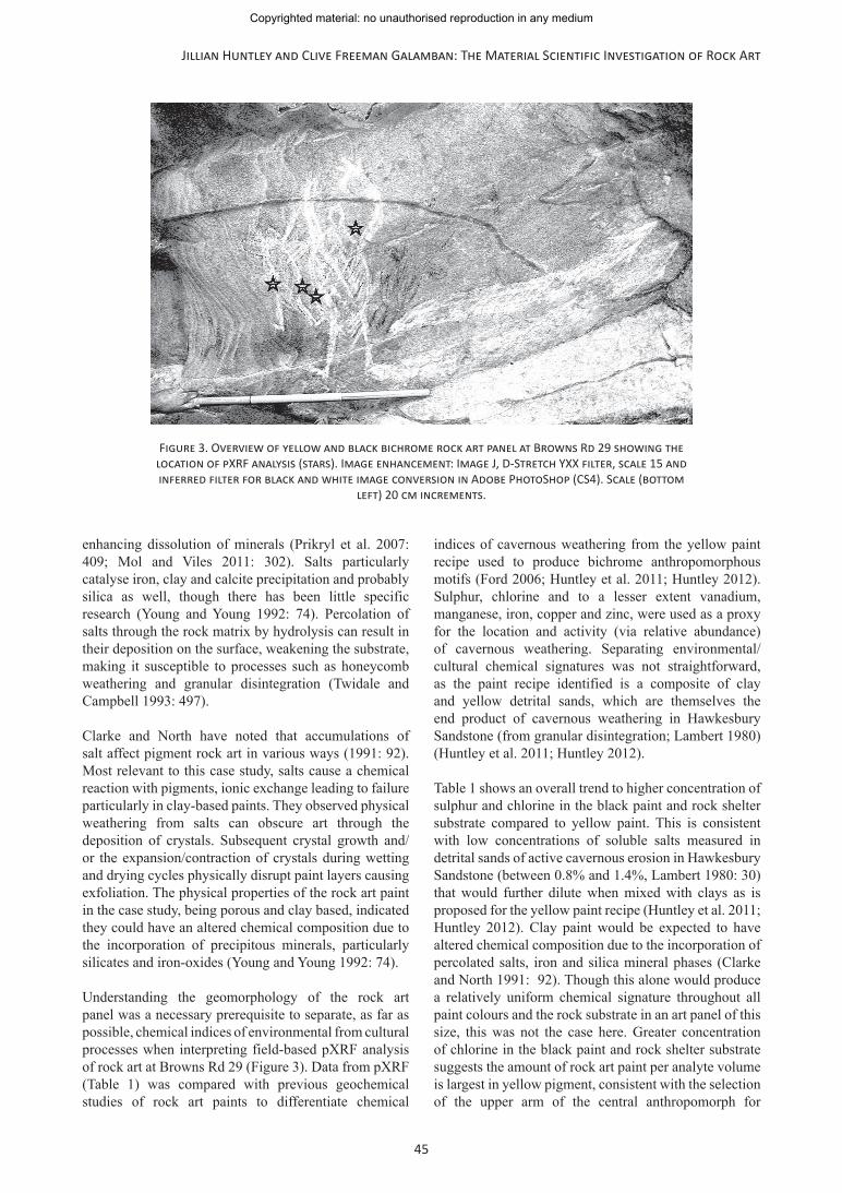
Understanding the geomorphology of the rock art panel was a necessary prerequisite to separate, as far as possible, chemical indices of environmental from cultural processes when interpreting field-based pXRF analysis of rock art at Browns Rd 29 (Figure 3). Data from pXRF (Table 1) was compared with previous geochemical studies of rock art paints to differentiate chemical
indices of cavernous weathering from the yellow paint recipe used to produce bichrome anthropomorphous motifs (Ford 2006; Huntley et al. 2011; Huntley 2012). Sulphur, chlorine and to a lesser extent vanadium, manganese, iron, copper and zinc, were used as a proxy for the location and activity (via relative abundance) of cavernous weathering. Separating environmental/cultural chemical signatures was not straightforward, as the paint recipe identified is a composite of clay and yellow detrital sands, which are themselves the end product of cavernous weathering in Hawkesbury Sandstone (from granular disintegration; Lambert 1980) (Huntley et al. 2011; Huntley 2012).
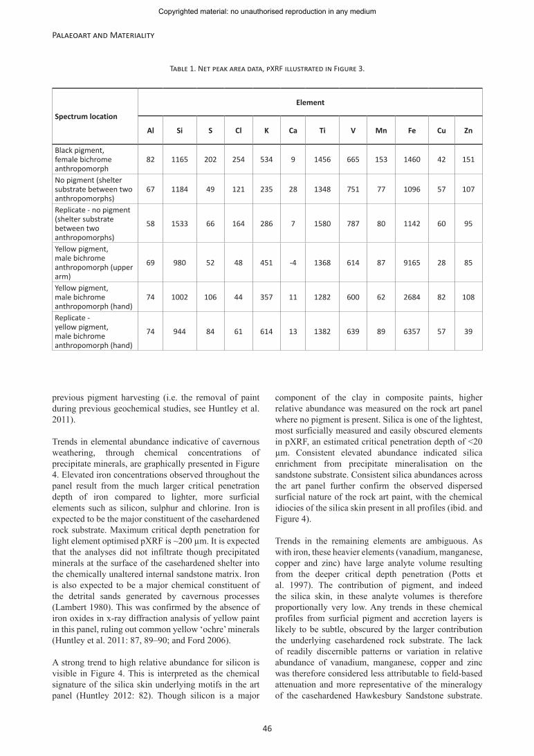
Table 1 shows an overall trend to higher concentration of sulphur and chlorine in the black paint and rock shelter substrate compared to yellow paint. This is consistent with low concentrations of soluble salts measured in detrital sands of active cavernous erosion in Hawkesbury Sandstone (between 0.8% and 1.4%, Lambert 1980: 30) that would further dilute when mixed with clays as is proposed for the yellow paint recipe (Huntley et al. 2011; Huntley 2012). Clay paint would be expected to have altered chemical composition due to the incorporation of percolated salts, iron and silica mineral phases (Clarke and North 1991: 92). Though this alone would produce a relatively uniform chemical signature throughout all paint colours and the rock substrate in an art panel of this size, this was not the case here. Greater concentration of chlorine in the black paint and rock shelter substrate suggests the amount of rock art paint per analyte volume is largest in yellow pigment, consistent with the selection of the upper arm of the central anthropomorph for
Figure 3. Overview of yellow and black bichrome rock art panel at Browns Rd 29 showing the location of pXRF analysis (stars). Image enhancement: Image J, D-Stretch YXX filter, scale 15 and inferred filter for black and white image conversion in Adobe PhotoShop (CS4). Scale (bottom
left) 20 cm increments.
Copyrighted material: no unauthorised reproduction in any medium
46
Palaeoart and Materiality
previous pigment harvesting (i.e. the removal of paint during previous geochemical studies, see Huntley et al. 2011).
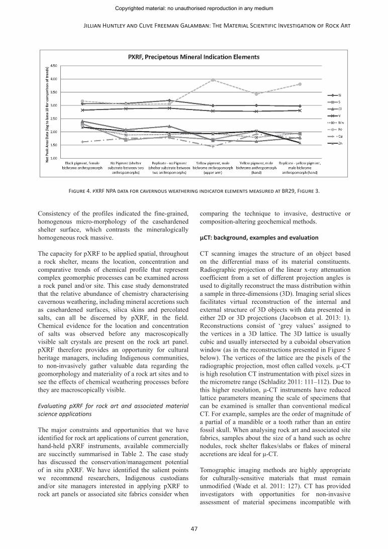
Trends in elemental abundance indicative of cavernous weathering, through chemical concentrations of precipitate minerals, are graphically presented in Figure 4. Elevated iron concentrations observed throughout the panel result from the much larger critical penetration depth of iron compared to lighter, more surficial elements such as silicon, sulphur and chlorine. Iron is expected to be the major constituent of the casehardened rock substrate. Maximum critical depth penetration for light element optimised pXRF is ~200 µm. It is expected that the analyses did not infiltrate though precipitated minerals at the surface of the casehardened shelter into the chemically unaltered internal sandstone matrix. Iron is also expected to be a major chemical constituent of the detrital sands generated by cavernous processes (Lambert 1980). This was confirmed by the absence of iron oxides in x-ray diffraction analysis of yellow paint in this panel, ruling out common yellow ‘ochre’ minerals (Huntley et al. 2011: 87, 89–90; and Ford 2006).
A strong trend to high relative abundance for silicon is visible in Figure 4. This is interpreted as the chemical signature of the silica skin underlying motifs in the art panel (Huntley 2012: 82). Though silicon is a major
component of the clay in composite paints, higher relative abundance was measured on the rock art panel where no pigment is present. Silica is one of the lightest, most surficially measured and easily obscured elements in pXRF, an estimated critical penetration depth of <20 µm. Consistent elevated abundance indicated silica enrichment from precipitate mineralisation on the sandstone substrate. Consistent silica abundances across the art panel further confirm the observed dispersed surficial nature of the rock art paint, with the chemical idiocies of the silica skin present in all profiles (ibid. and Figure 4).
Trends in the remaining elements are ambiguous. As with iron, these heavier elements (vanadium, manganese, copper and zinc) have large analyte volume resulting from the deeper critical depth penetration (Potts et al. 1997). The contribution of pigment, and indeed the silica skin, in these analyte volumes is therefore proportionally very low. Any trends in these chemical profiles from surficial pigment and accretion layers is likely to be subtle, obscured by the larger contribution the underlying casehardened rock substrate. The lack of readily discernible patterns or variation in relative abundance of vanadium, manganese, copper and zinc was therefore considered less attributable to field-based attenuation and more representative of the mineralogy of the casehardened Hawkesbury Sandstone substrate.
Table 1. Net peak area data, pXRF illustrated in Figure 3.
Spectrum location
Element
Al Si S Cl K Ca Ti V Mn Fe Cu Zn
Black pigment, female bichrome anthropomorph
82 1165 202 254 534 9 1456 665 153 1460 42 151
No pigment (shelter substrate between two anthropomorphs)
67 1184 49 121 235 28 1348 751 77 1096 57 107
Replicate - no pigment (shelter substrate between two anthropomorphs)
58 1533 66 164 286 7 1580 787 80 1142 60 95
Yellow pigment, male bichrome anthropomorph (upper arm)
69 980 52 48 451 -4 1368 614 87 9165 28 85
Yellow pigment, male bichrome anthropomorph (hand)
74 1002 106 44 357 11 1282 600 62 2684 82 108
Replicate - yellow pigment, male bichrome anthropomorph (hand)
74 944 84 61 614 13 1382 639 89 6357 57 39
Copyrighted material: no unauthorised reproduction in any medium
47
Jillian Huntley and Clive Freeman Galamban: The Material Scientific Investigation of Rock Art
Consistency of the profiles indicated the fine-grained, homogenous micro-morphology of the casehardened shelter surface, which contrasts the mineralogically homogeneous rock massive.
The capacity for pXRF to be applied spatial, throughout a rock shelter, means the location, concentration and comparative trends of chemical profile that represent complex geomorphic processes can be examined across a rock panel and/or site. This case study demonstrated that the relative abundance of chemistry characterising cavernous weathering, including mineral accretions such as casehardened surfaces, silica skins and percolated salts, can all be discerned by pXRF, in the field. Chemical evidence for the location and concentration of salts was observed before any macroscopically visible salt crystals are present on the rock art panel. pXRF therefore provides an opportunity for cultural heritage managers, including Indigenous communities, to non-invasively gather valuable data regarding the geomorphology and materiality of a rock art sites and to see the effects of chemical weathering processes before they are macroscopically visible.
Evaluating pXRF for rock art and associated material science applications
The major constraints and opportunities that we have identified for rock art applications of current generation, hand-held pXRF instruments, available commercially are succinctly summarised in Table 2. The case study has discussed the conservation/management potential of in situ pXRF. We have identified the salient points we recommend researchers, Indigenous custodians and/or site managers interested in applying pXRF to rock art panels or associated site fabrics consider when
comparing the technique to invasive, destructive or composition-altering geochemical methods.
µCT: background, examples and evaluation
CT scanning images the structure of an object based on the differential mass of its material constituents. Radiographic projection of the linear x-ray attenuation coefficient from a set of different projection angles is used to digitally reconstruct the mass distribution within a sample in three-dimensions (3D). Imaging serial slices facilitates virtual reconstruction of the internal and external structure of 3D objects with data presented in either 2D or 3D projections (Jacobson et al. 2013: 1). Reconstructions consist of ‘grey values’ assigned to the vertices in a 3D lattice. The 3D lattice is usually cubic and usually intersected by a cuboidal observation window (as in the reconstructions presented in Figure 5 below). The vertices of the lattice are the pixels of the radiographic projection, most often called voxels. μ-CT is high resolution CT instrumentation with pixel sizes in the micrometre range (Schladitz 2011: 111–112). Due to this higher resolution, μ-CT instruments have reduced lattice parameters meaning the scale of specimens that can be examined is smaller than conventional medical CT. For example, samples are the order of magnitude of a partial of a mandible or a tooth rather than an entire fossil skull. When analysing rock art and associated site fabrics, samples about the size of a hand such as ochre nodules, rock shelter flakes/slabs or flakes of mineral accretions are ideal for μ-CT.
Tomographic imaging methods are highly appropriate for culturally-sensitive materials that must remain unmodified (Wade et al. 2011: 127). CT has provided investigators with opportunities for non-invasive assessment of material specimens incompatible with
Figure 4. pXRF NPA data for cavernous weathering indicator elements measured at BR29, Figure 3.
Copyrighted material: no unauthorised reproduction in any medium
48
Palaeoart and Materiality
macroscopic techniques exemplified by human remains, for example wrapped mummies. The staple use of conventional CT in heritage has been paleoanthropological research, it is therefore little surprise that pathological paleoanthropological investigations were some of the earliest adaptations of µ-CT. Recently µ-CT has been shown to have wider utility in conservation treatments for cultural heritage materials (Bugani et al. 2009; Mizuno et al. 2010; Mahabadi et al. 2012). Of particular relevance to rock art research, a recent study used high resolution tomography to investigate the anthropogenic nature of striations on ironstone slabs from Wonderwerk Cave in South Africa, thought to be incised by early hominins and thus provides evidence of symbolic behaviour. Neutron Tomography was used on the near surface layers of the iron stone slabs to investigate if striations were associated with internal fissures in the rock, thus differentiating natural fissures from hominin made incisions (Jabobson et al. 2013).
We integrated novel µ-CT analysis of site fabrics associated with rock art in the Mitchell Plateau area of the northwest Kimberley (Figure 2) into a larger archaeometric program examining the materiality of this regional rock art assemblage. µ-CT was an initial investigatory technique within an incremental analytic program, used in effect as a screening tool. Rapid scans were performed to examine the internal structure of specimens. Tomographic reconstructions were
used to assess specimens, targeting areas of analytic interest and avoiding undesirable attributes in relation to subsequently destructive and composition-altering analyses (SEM and conventional x-ray diffraction and synchrotron powder diffraction). Relative density data of material constituents from µ-CT was used in the interpretation of complementary non-invasive pXRF analysis of archaeological ochres.
µ-CT methodology
Scans were performed using a General Electric Phoenix x-ray model Vtomex s 240D with a 512 × 512 square pixel flat panel CCD detector (Figure 1). The x-ray source is a ‘direct’ serviceable evacuated tube. X-rays are generated by focusing a beam of electrons onto a water-cooled tungsten target, creating a cone shaped (divergent) incident angle. This polychromatic beam has intensity and wavelengths relative to the voltage and amperage applied to the target. Therefore keV and uA parameters can be adjusted to optimise for the material being scanned. Parameters for the scans we conducted are given in Table 3.
We initially trialled ‘conventional’ or ‘full’ scan mode where the sample is stationary at each point before radiographs are recorded, scan times running to hours. Our initial intent was that µ-CT would eliminate subsequent preparation of samples for destructive,
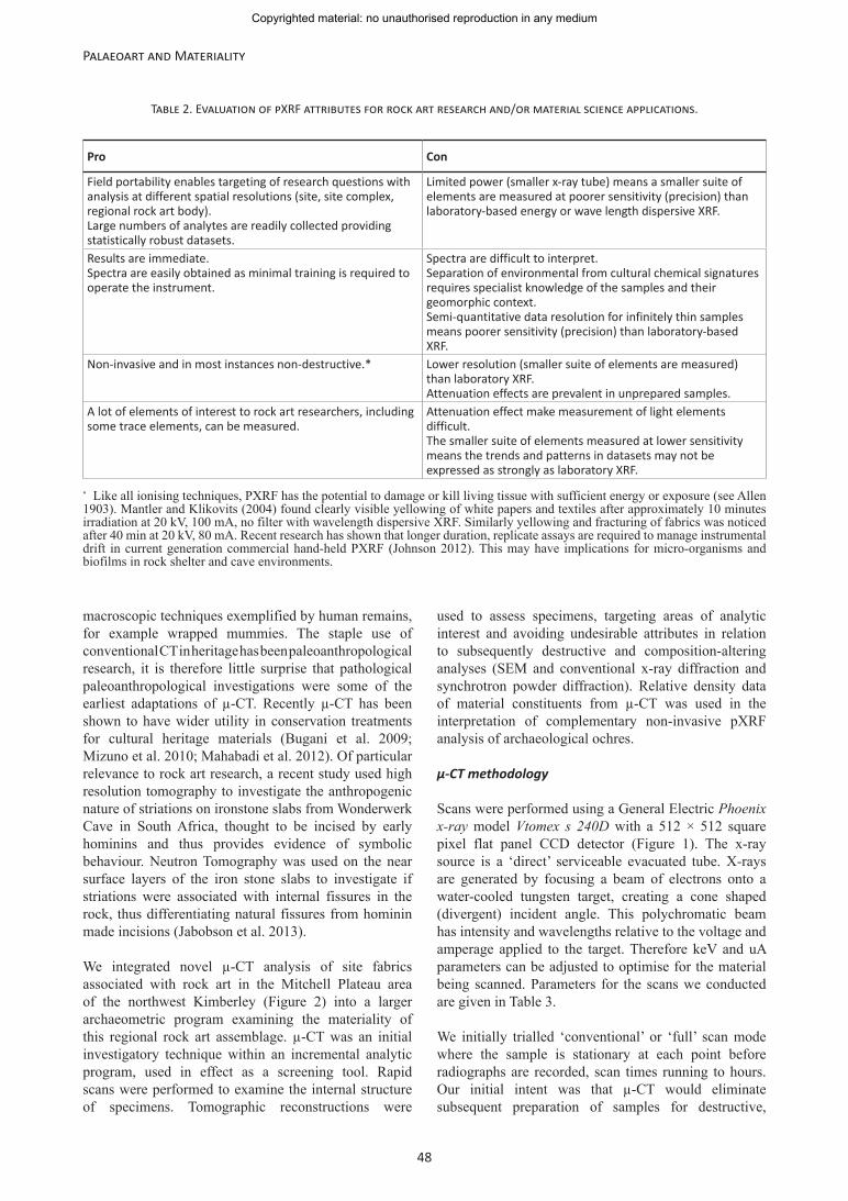
Table 2. Evaluation of pXRF attributes for rock art research and/or material science applications.
Pro Con
Field portability enables targeting of research questions with analysis at different spatial resolutions (site, site complex, regional rock art body).Large numbers of analytes are readily collected providing statistically robust datasets.
Limited power (smaller x-ray tube) means a smaller suite of elements are measured at poorer sensitivity (precision) than laboratory-based energy or wave length dispersive XRF.
Results are immediate.Spectra are easily obtained as minimal training is required to operate the instrument.
Spectra are difficult to interpret.Separation of environmental from cultural chemical signatures requires specialist knowledge of the samples and their geomorphic context.Semi-quantitative data resolution for infinitely thin samples means poorer sensitivity (precision) than laboratory-based XRF.
Non-invasive and in most instances non-destructive.* Lower resolution (smaller suite of elements are measured) than laboratory XRF.Attenuation effects are prevalent in unprepared samples.
A lot of elements of interest to rock art researchers, including some trace elements, can be measured.
Attenuation effect make measurement of light elements difficult.The smaller suite of elements measured at lower sensitivity means the trends and patterns in datasets may not be expressed as strongly as laboratory XRF.
* Like all ionising techniques, PXRF has the potential to damage or kill living tissue with sufficient energy or exposure (see Allen 1903). Mantler and Klikovits (2004) found clearly visible yellowing of white papers and textiles after approximately 10 minutes irradiation at 20 kV, 100 mA, no filter with wavelength dispersive XRF. Similarly yellowing and fracturing of fabrics was noticed after 40 min at 20 kV, 80 mA. Recent research has shown that longer duration, replicate assays are required to manage instrumental drift in current generation commercial hand-held PXRF (Johnson 2012). This may have implications for micro-organisms and biofilms in rock shelter and cave environments.
Copyrighted material: no unauthorised reproduction in any medium
49
Jillian Huntley and Clive Freeman Galamban: The Material Scientific Investigation of Rock Art
composition-altering SEM analysis. Upon examining the resolution of the full scans of ochre nodules, however, it became obvious the relative density of constituents in aluminosilicate materials was similar enough, that internal micro-structure imaged by µ-CT was commensurate with only coarse microscopy. Data from ‘fast-scan’ was sufficient to indicate areas of analytic interest and locate undesirable attributes to inform further SEM examinations. We therefore integrated ‘fast scans’ as a screening tool within our broader analytic program.
Phoenix x-ray proprietary acquisition software was used in ‘fast scan’ mode whereby images are taken as the sample constantly rotates, markedly decreasing time of acquisition. Total scan times were ~12 minutes per sample. Radiograph reconstruction was carried out using Phoenix x-ray proprietary reconstruction software and a graphical processing unit cluster. The algorithms used offset the effects of non-centred objects, systematic changes in object placement and/or x-ray tube elongation or beam hardening during the scan. Three images per projection were averaged for reconstructions.
Case study: structure of ochres and mineral accretions; integration of µ-CT in the analysis of rock art materiality
Any conservation/management investigation must examine the material properties of rock art and associated site fabrics in order to begin to understand the effects of taphonomic processes and predict physiochemical deterioration. We considered the power and potential of structural characterisation investigations in this framework integral to our ongoing research of the material properties of rock art sites in the northwest Kimberley. The utility of µ-CT for characterising the internal structure of archaeological pigments and a mineral accretion associated with rock art is demonstrated by this case study. A red ochre pebble (Figure 5, left); a fragment from a ‘quarried’ mulberry pigment source (Figure 5, right); and a piece of white/light pink/purple mineral accretion from the edge of a rock art panel (Figure 7) are illustrated. These specimens were collected from different shelters containing rock art
and other archaeological surface evidence such as lithic technologies and anthropogenically modified ochres. The sites are all located in the same complex of the lower Mitchell (Kandiwal) River catchment, immediately east of the Mitchell Plateau, northwest Kimberley, Western Australia (Figure 2) (Ross and Travers 2013: 56–59).
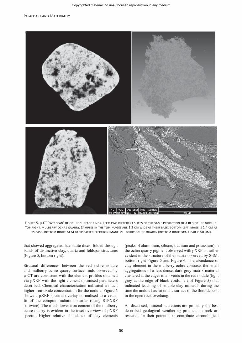
The internal structure of a surface-find red ochre nodule was determined to be a dense, brightly charging mass of compact semi-spherical structures, shown in the µ-CT projections left of Figure 5. The ‘bright’ white charging of the disc structures in these left hand images indicates a material with a high x-ray attenuation coefficient, consistent with iron. The scan also shows air voids in the specimen and small areas of less dense, differently structured materials are present within the specimen especially at the edges of the voids. These are illustrated by the black and darker grey internal sections of Figure 5 respectively. The distinctive clustered disc structures are interpreted as diagnostic of the mineral haematite (Sepulveda et al. 2012). Disc structures are commonly observed in iron-oxide minerals, with dehydrated structure typically having an aggregated but jumbled orientation such as that observed in the ochre nodule, while hydrated forms typically have an ordered orientation characterised by aggregated chains of parallel adjoining discs. Both dehydrated (haematite) and hydrated (goethite/ limonite) iron-oxide mineral are known to have been used throughout the world to produce predominantly red hued, pigment rock art (Rowe 2001; Wadley 2013). The internal porosity of the red ochre shown by the black voids, and the limited areas of lower density darker grey material indicated that the nodule has high iron-oxide content with little supporting matrix (again, left of Figure 5). The voids and clusters of less dense material in the nodule contrast the consistent density of pigment from the mulberry ochre quarry (Figure 5, top right). The ochre quarry is located in the adjacent cave ~50 metres from the location of the ochre nodule find. The internal structure of the ochre quarry specimen revealed by µ-CT is an admixture of two different density materials, without voids. This structure was confirmed and further explained by subsequent SEM
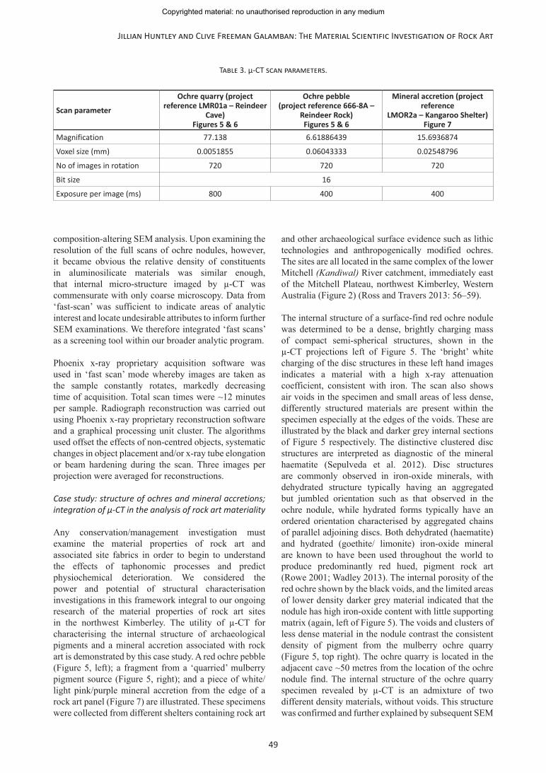
Table 3. µ-CT scan parameters.
Scan parameter
Ochre quarry (project reference LMR01a – Reindeer
Cave) Figures 5 & 6
Ochre pebble(project reference 666-8A –
Reindeer Rock) Figures 5 & 6
Mineral accretion (project reference
LMOR2a – Kangaroo Shelter)Figure 7
Magnification 77.138 6.61886439 15.6936874
Voxel size (mm) 0.0051855 0.06043333 0.02548796
No of images in rotation 720 720 720
Bit size 16
Exposure per image (ms) 800 400 400
Copyrighted material: no unauthorised reproduction in any medium
50
Palaeoart and Materiality
that showed aggregated haematite discs, folded through bands of distinctive clay, quartz and feldspar structures (Figure 5, bottom right).
Strutural differences between the red ochre nodule and mulberry ochre quarry surface finds observed by µ-CT are consistent with the element profiles obtained via pXRF with the light element optimised perameters described. Chemical characterisation indicated a much higher iron-oxide concentration for the nodule. Figure 6 shows a pXRF spectral overlay normalised to a visual fit of the compton radiation scatter (using S1PXRF software). The much lower iron content of the mulberry ochre quarry is evident in the inset overview of pXRF spectra. Higher relative abundance of clay elements
(peaks of aluminium, silicon, titanium and potassium) in the ochre quarry pigment observed with pXRF is further evident in the structure of the matrix observed by SEM, bottom right Figure 5 and Figure 6. The abundance of clay element in the mulberry ochre contrasts the small aggregations of a less dense, dark grey matrix material clustered at the edges of air voids in the red nodule (light grey at the edge of black voids, left of Figure 5) that indicated leaching of soluble clay minerals during the time the nodule has sat on the surface of the floor deposit in the open rock overhang.
As discussed, mineral accretions are probably the best described geological weathering products in rock art research for their potential to contribute chronological
Figure 5. µ-CT ‘fast scan’ of ochre surface finds. Left: two different slices of the same projection of a red ochre nodule. Top right: mulberry ochre quarry. Samples in the top images are 1.2 cm wide at their base, bottom left image is 1.4 cm at
its base. Bottom right: SEM backscatter electron image mulberry ochre quarry (bottom right scale bar is 50 µm).
Copyrighted material: no unauthorised reproduction in any medium

51
Jillian Huntley and Clive Freeman Galamban: The Material Scientific Investigation of Rock Art
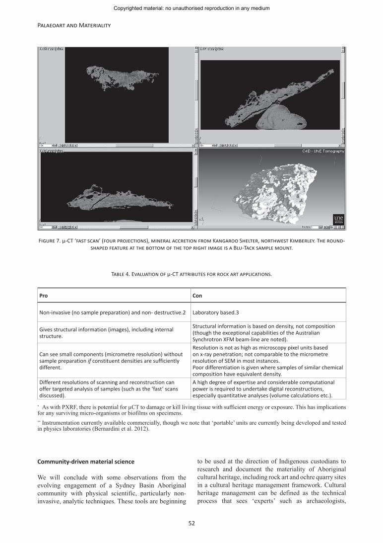
information via numerical age determinations of radioisotope components either encased by, or in these rock coatings (e.g. Watchman et al. 1993; Mardaga-Campbell et al. 2001; Aubert et al. 2007; Taçon et al. 2011; Aubert 2012; Pike et al. 2012). We investigated the materiality of one such pink/purple mineral accretion taken from a rock art panel, adjacent to, but not directly associated with the art, using a µ-CT ‘fast scan’ illustrated in Figure 7. The scan revealed not only that material of at least two different densities was present in the sample, but also that there were cracks (horizontal and at 45°) in the centre of the specimen and importantly, air voids at its surface. These observations provided information regarding the formation history of the coating, and acted as something of a palaeoenvironmental proxy for the rock art panel/site. Layering of materials of different densities indicated the deposition of different mineral composition at different times throughout the formation of the crust. The location of air voids in the top layer of the mineral accretion suggests rapid formation of the coating, at least during this most recent period. Finally, the location of faults or cracks such as those shown in the µ-CT scan provide important contextual information when preparing samples for subsequent destructive/composition-altering analyses, such structural information enabling the avoidance of undesirable attributes (such as voids) or targeting of attributes of interest (such as materials of differential density and/or large grain inclusions).
The internal structure of ochres and mineral accretion observed by µ-CT demonstrates how compositional
and/or taphonomic information can be gained through purely qualitative visual imaging of the Kimberley Basin samples. This provided evidence for taphonomic occurrences such as the leaching of clay matrix minerals from exposed ochre surface finds and the rapid formation of mineral accretions. The different relative density of materials within the two ochre scans was used to identify compositionally discrete pigment constituents, further confirmed by SEM and pXRF. Finally, µ-CT showed that in two of the specimens (the red ochre nodule and mineral accretion) voids are present. This could be critically important contextual information when interpreting the results of complimentary pXRF, which is sensitive to attenuation effects from air (Forster et al. 2011; Huntley 2012). Samples with the same chemical composition could easily return different pXRF profiles due to x-ray attenuation.
Evaluating µ-CT for rock art and associated material science applications
Table 4 summaries the major constraints and opportunities that we have identified for rock art applications of current generation, hand-held µ-CT instruments, available commercially. As with our earlier evaluation of pXRF, we have outlined the salient points we recommend researchers, Indigenous custodians and/or site managers consider in relation to applying this technique to partial rock art panels, such as natural spall flakes, or associated site fabrics such as the ochre and mineral accretion examples discussed.
Figure 6. pXRF spectral overlay of red ochre nodule (light grey) and mulberry ochre quarry (dark grey).
Copyrighted material: no unauthorised reproduction in any medium
52
Palaeoart and Materiality
Community-driven material science
We will conclude with some observations from the evolving engagement of a Sydney Basin Aboriginal community with physical scientific, particularly non-invasive, analytic techniques. These tools are beginning
to be used at the direction of Indigenous custodians to research and document the materiality of Aboriginal cultural heritage, including rock art and ochre quarry sites in a cultural heritage management framework. Cultural heritage management can be defined as the technical process that sees ‘experts’ such as archaeologists,
Figure 7. µ-CT ‘fast scan’ (four projections), mineral accretion from Kangaroo Shelter, northwest Kimberley. The round-shaped feature at the bottom of the top right image is a Blu-Tack sample mount.
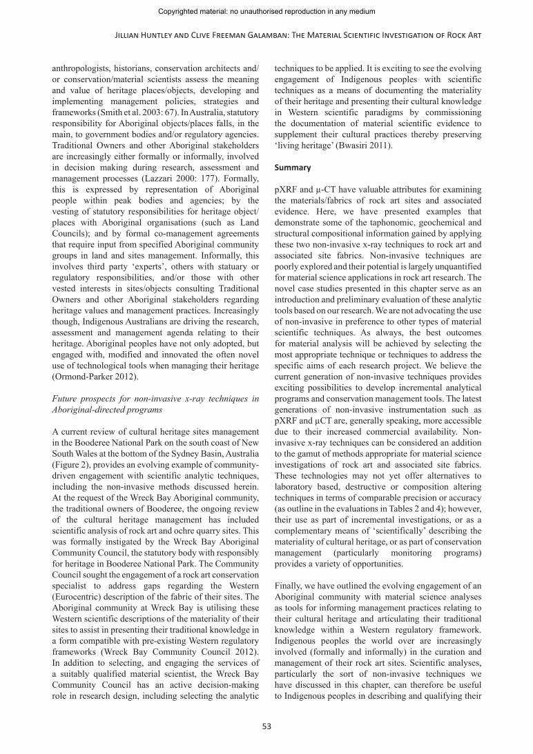
Table 4. Evaluation of µ-CT attributes for rock art applications.
Pro Con
Non-invasive (no sample preparation) and non- destructive.2 Laboratory based.3
Gives structural information (images), including internal structure.
Structural information is based on density, not composition (though the exceptional capabilities of the Australian Synchrotron XFM beam-line are noted).
Can see small components (micrometre resolution) without sample preparation if constituent densities are sufficiently different.
Resolution is not as high as microscopy pixel units based on x-ray penetration; not comparable to the micrometre resolution of SEM in most instances.Poor differentiation is given where samples of similar chemical composition have equivalent density.
Different resolutions of scanning and reconstruction can offer targeted analysis of samples (such as the ‘fast’ scans discussed).
A high degree of expertise and considerable computational power is required to undertake digital reconstructions, especially quantitative analyses (volume calculations etc.).
* As with PXRF, there is potential for µCT to damage or kill living tissue with sufficient energy or exposure. This has implications for any surviving micro-organisms or biofilms on specimens.** Instrumentation currently available commercially, though we note that ‘portable’ units are currently being developed and tested in physics laboratories (Bernardini et al. 2012).
Copyrighted material: no unauthorised reproduction in any medium
53
Jillian Huntley and Clive Freeman Galamban: The Material Scientific Investigation of Rock Art
anthropologists, historians, conservation architects and/or conservation/material scientists assess the meaning and value of heritage places/objects, developing and implementing management policies, strategies and frameworks (Smith et al. 2003: 67). In Australia, statutory responsibility for Aboriginal objects/places falls, in the main, to government bodies and/or regulatory agencies. Traditional Owners and other Aboriginal stakeholders are increasingly either formally or informally, involved in decision making during research, assessment and management processes (Lazzari 2000: 177). Formally, this is expressed by representation of Aboriginal people within peak bodies and agencies; by the vesting of statutory responsibilities for heritage object/places with Aboriginal organisations (such as Land Councils); and by formal co-management agreements that require input from specified Aboriginal community groups in land and sites management. Informally, this involves third party ‘experts’, others with statuary or regulatory responsibilities, and/or those with other vested interests in sites/objects consulting Traditional Owners and other Aboriginal stakeholders regarding heritage values and management practices. Increasingly though, Indigenous Australians are driving the research, assessment and management agenda relating to their heritage. Aboriginal peoples have not only adopted, but engaged with, modified and innovated the often novel use of technological tools when managing their heritage (Ormond-Parker 2012).
Future prospects for non-invasive x-ray techniques in Aboriginal-directed programs
A current review of cultural heritage sites management in the Booderee National Park on the south coast of New South Wales at the bottom of the Sydney Basin, Australia (Figure 2), provides an evolving example of community-driven engagement with scientific analytic techniques, including the non-invasive methods discussed herein. At the request of the Wreck Bay Aboriginal community, the traditional owners of Booderee, the ongoing review of the cultural heritage management has included scientific analysis of rock art and ochre quarry sites. This was formally instigated by the Wreck Bay Aboriginal Community Council, the statutory body with responsibly for heritage in Booderee National Park. The Community Council sought the engagement of a rock art conservation specialist to address gaps regarding the Western (Eurocentric) description of the fabric of their sites. The Aboriginal community at Wreck Bay is utilising these Western scientific descriptions of the materiality of their sites to assist in presenting their traditional knowledge in a form compatible with pre-existing Western regulatory frameworks (Wreck Bay Community Council 2012). In addition to selecting, and engaging the services of a suitably qualified material scientist, the Wreck Bay Community Council has an active decision-making role in research design, including selecting the analytic
techniques to be applied. It is exciting to see the evolving engagement of Indigenous peoples with scientific techniques as a means of documenting the materiality of their heritage and presenting their cultural knowledge in Western scientific paradigms by commissioning the documentation of material scientific evidence to supplement their cultural practices thereby preserving ‘living heritage’ (Bwasiri 2011).
Summary
pXRF and µ-CT have valuable attributes for examining the materials/fabrics of rock art sites and associated evidence. Here, we have presented examples that demonstrate some of the taphonomic, geochemical and structural compositional information gained by applying these two non-invasive x-ray techniques to rock art and associated site fabrics. Non-invasive techniques are poorly explored and their potential is largely unquantified for material science applications in rock art research. The novel case studies presented in this chapter serve as an introduction and preliminary evaluation of these analytic tools based on our research. We are not advocating the use of non-invasive in preference to other types of material scientific techniques. As always, the best outcomes for material analysis will be achieved by selecting the most appropriate technique or techniques to address the specific aims of each research project. We believe the current generation of non-invasive techniques provides exciting possibilities to develop incremental analytical programs and conservation management tools. The latest generations of non-invasive instrumentation such as pXRF and µCT are, generally speaking, more accessible due to their increased commercial availability. Non-invasive x-ray techniques can be considered an addition to the gamut of methods appropriate for material science investigations of rock art and associated site fabrics. These technologies may not yet offer alternatives to laboratory based, destructive or composition altering techniques in terms of comparable precision or accuracy (as outline in the evaluations in Tables 2 and 4); however, their use as part of incremental investigations, or as a complementary means of ‘scientifically’ describing the materiality of cultural heritage, or as part of conservation management (particularly monitoring programs) provides a variety of opportunities.
Finally, we have outlined the evolving engagement of an Aboriginal community with material science analyses as tools for informing management practices relating to their cultural heritage and articulating their traditional knowledge within a Western regulatory framework. Indigenous peoples the world over are increasingly involved (formally and informally) in the curation and management of their rock art sites. Scientific analyses, particularly the sort of non-invasive techniques we have discussed in this chapter, can therefore be useful to Indigenous peoples in describing and qualifying their
Copyrighted material: no unauthorised reproduction in any medium
54
Palaeoart and Materiality
cultural knowledge and customary practices, or as a means of investigating the materiality of their heritage.
Acknowledgments
We acknowledge the traditional custodians of the Dharawal nation in the Sydney Basin and the Illawarra Local Aboriginal Land Council, administrative peak body for the Woronora Plateau. We thank the Mindaribba, Wanaruha and Darkinjung Local Aboriginal Land councils and Sharon Hodgetts for permission to reproduce the images in Figure 1. Kimberley Basin samples were collected under Department of Indigenous Affairs, Section 16: 2011-2012 Permit No 490 and Department of Environment and Conservation Authority 4: 2011 CE003254 as part of ARC Linkage project (LP0991845) Change & continuity: chronology, archaeology and art in the north Kimberley, northwest Australia; chief investigators: Mike Morwood, June Ross and Kira Westaway. We acknowledge the traditional owners of the Mitchell Plateau, the Wunambal Gaambera people and the support of the Kandiwal Aboriginal Corporation, Kimberley Foundation of Australia, Slingair and Heliwork Pty Ltd and the Western Australian Department of Conservation.
This chapter is based on a presentation in the conservation management session of the International Federation of Rock Art Organisations (IFRAO) Conference 2012 in La Paz. JH acknowledges the support of the Kimberley Foundation of Australia and the School of Humanities, University of New England, in attending the meeting. CF’s attendance was supported by an artist’s residency at the NSW Art Gallery and Henric Nicholas. This chapter is based on methods developed as part of a larger project designed by JH in the Sydney Basin funded by the Australian Geographic Society and the Kimberley Basin funded by the Kimberley Foundation Australia.
µ-CT analyses were conducted with the assistance of Dr Matthew K. Tighe, Mr Timothy I. McLaren and especially Mr Richard J. Flavel at the Glasshouse Complex, School of Environmental and Rural Sciences, University of New England. SEM was undertaken with the assistance of David Phelan at Newcastle University EMX. pXRF was made available through the Archaeomaterials Science Hub, University of New England.
Lucas Huntley drafted Figures 2 and 6.
References
Allen, S. W. 1903. The effect of x-rays on living tissue. The Journal of Medical Research IX: 462–474.
Aubert, M. 2012. A review of rock art dating in the Kimberley, Western Australia. Journal of Archaeological Science 39(3): 573–577.
Aubert, M., S. O’Connor, M. McCulloch, G. Mortimer, A. Watchman and M. Richerlafl`eche 2007. Uranium-series dating rock art in East Timor. Journal of Archaeological Science 34: 991–996.
Bednarik, R. G. 1992. Ethics in rock art research and conservation. Rock Art Quarterly 3: 17–22.
Bera, B ., S. K. Mitra and D. Vickb 2011. Understanding the micro structure of Berea Sandstone by the simultaneous use of micro-computed tomography (micro-CT) and focused ion beam-scanning electron microscopy (FIB-SEM). Micron 42: 412–418.
Bernardini, F., C. Tuniz, A. Coppa, L. Mancini, D. Dreossi, D. Eichert, G. Turco, M. Biasotto, F. Terrasini, N. De Cesare, Q. Hua and V. Levchenko 2012. Beeswax as dental filling on a Neolithic human tooth. PLOS ONE, 10.1371/journal.pone.0044904.
Bugani, S., F. Modugno, J. J. Łucejko, G. Giachi, S. Cagno, P. Cloetens, K. Janssens and L. Morselli 2009. Study on the impregnation of archaeological waterlogged wood with consolidation treatments using synchrotron radiation microtomography. Analytical and Bioanalytical Chemistry 395: 1977–1985.
Bwasiri, E. J. (2011). The implications of the management of indigenous living heritage: the case study of Themongomi Wa Kolo Rock Paintingsworld Heritage Site, central Tanzania. South African Archaeological Bulletin 66(193): 60–66.
Cesareo, R., S. Ridolf, M. Marabelli, A. Castellano, G. Buccolieri, M. Donativi, G. E. Gigante, A. Brunetti and M. A. Rosales Medina 2008. Portable systems for energy-dispersive x-ray flourescence analysis of works of art. In P. J. and West Potts, M. (eds), Portable x-ray fluorescence spectrometry: capabilities for in situ analysis, pp. 206–246. RSC Publishing, Cambridge.
Clarke, J. and N. North 1991. Chemistry of deterioration of post-esturine rock art in Kakadu National Park. In C. Pearson and B. K. Swartz, Jr (eds), Rock art and posterity: conserving, managing and recording rock art, pp. 88–92. Occasional AURA publications 4, Australian Rock Art Association, Melbourne.
Clottes, J. 1992. ‘Professional vandalism’ and ‘indigenous ownership’: an alternative perspective of several ethical issues in rock art research. Rock Art Quarterly 3: 22–24.
Demetrios, A and A. G. Karydas 2009. Non-destructive and microanalytical techniques in art and cultural heritage (Technart 2009). Analytical and Bioanalytical Chemistry 395(7): 1947–1948.
Etschmann, B. E., C. G. Ryan, J. Brugger,R. Kirkham, R. M. Hough, G. Moorhead et al. 2010. Reduced as components in highly oxidized environments: evidence from full spectral XANES imaging using the Maia massively parallel detector. American Mineraologist 95: 884–887.
Ford, J. A. 2006. Painting contact: characterising the paints of the south Woronora Plateau rock art
Copyrighted material: no unauthorised reproduction in any medium
55
Jillian Huntley and Clive Freeman Galamban: The Material Scientific Investigation of Rock Art
assembledge, Wollonong, NSW. Australian National University, Canberra.
Forster, N., P. Grave, N. Vickery and L. Kealhofer 2011. Non-destructive analysis using PXRF: methodology and application to archaeological ceramics. X-Ray Spectrometry 40(5): 389–398.
Ghysels, M. 2003. CT scans in artwork appraisal. Art Tribal, Winter 2003(04): 116–131.
Guilherme, A., A. Cavaco, S. Pessanha, M. Costa and M. L. Carvalho 2008. Comparison of portable and stationary x-ray fluorescence spectrometers in the study of ancient metallic artefacts. X-Ray Spectrometry 39: 444–449.
Heginbotham, A., A. Bezur, M. Bouchard, J. M. Davis, K. Ermin, J. H. Franz et al. 2010. An evaluation of inter-laboratory reproducibility for quantitative XRF of historic copper alloys. In C. Chemello, P. Mardikian, C. Watters, and P. Hull (eds), Metal 2010: Interim Meeting of the International Council of Museums Committee for Conservation Metal Working Group, October 11–15 2010, Charleston, South Carolina, USA, pp. 178–188. Clemson University, Charleston, SC.
Howard, D. L., M. D. de Jonge, D. Lau, M. Varcoe Cocks, C. G. Ryan, R. Kirkham et al. 2012. High-definition x-ray fluorescence elemental mapping of paintings. Analytical Chemistry 84: 3278−3286.
Huntley, J. 2012. Taphonomy or paint recipe: in situ portable x-ray fluorescence analysis of two anthropomorphic motifs from the Woronora Plateau, New South Wales. Australian Archaeology 75: 78–94.
Huntley, J., A. Watchman and J. Dibden 2011. Characteristics of a pigment art sequence: Woronora Plateau, New South Wales. Rock Art Research 28(1): 85–97.
Jacobson, L., F. C. de Beer, R. Nshimirimana, L. K. Horwitz and M. Chazan 2013. Neutron tomographic assessment of incisions on prehistoric stone slabs: a case study from Wonderwerk Cave, South Africa. Archaeometry 55(1): 1–13.
Johnson, J. 2012. Accurate measurements of low z elements in sediments and archaeological ceramics using portable x-ray fluorescence (PXRF). Journal of Archaeological Method and Theory DOI 10.1007/s10816-012-9162-3, 1–26.
Joyce, R. A. 2011. Is there a future for XRF in twenty-first century archaeology. In M. S. Shackley (ed), X-ray flourescence spectrometry (XRF) in geoarchaeology, pp. 193–202. Springer, New York.
Koenig, C. W., A. M. Castañeda, C. E. Boyd, M. W. Rowe and K. L. Steelman in press 2013. Portable x-ray fluorescence spectroscopy of pictographs: a case study from the Lower Pecos Canyonlands, Texas. Archaeometry DOI: 10.1111/arcm.12060.
Ge, L., W. Lai, Y. Lin and S. Zhou 2005. In situ applications of PXRF techniques in mineral exploration. In In situ applications of X ray fluorescence techniques: Final
report of a coordinated research project 2000–2003, pp. 61–120. International Atomic Energy Agency, Vienna.
Lambert, D. 1980. The influence of ground water salts in the formation of sandstone shelters near Gosford, NSW. Institute for the Conservation of Cultural Materials Bulletin 6: 29–34.
Lazzari, M. 2011. Tangible interventions: the lived landscapes of contemporary archaeology. Journal of Material Culture 16(2): 171–191.
Liritzis, I. and N. Zacharias 2011. Portable XRF of archaeological artifacts: current research, potentials and limitations. In M. S. Shackley (ed.), X-ray flourescence spectrometry (XRF) in geoarchaeology. Springer, New York.
Lovering, J. F 1952. Epigenetic common opal from the Hawkesbury Sandstone Formation of the Sydney Basin. Records of the Australian Museum 23(1): 29–32.
Mahabadi, O.K., N. X. Randall, Z. Zong and G. Grasselli 2012. A novel approach for micro-scale characterization and modelling of geomaterials incorporating actual material heterogeneity. Geophysical Research Letters 39(2): 1–6.
Mantler, M. and J. Klikovits 2004. Analysis of art objects and other delicate samples: is XRF really non-destructive? Advances in X-ray analysis (AXA): the proceedings of the Denver X-ray Conferences, Vol. 47, pp. 42–46. The International Centre for Diffraction Data, Denver, CO.
Machill, S., K. Althaus, W. E. Krumbein and W. E. Steger 1997. Identification of organic compounds extracted from black weathered surfaces of Saxonean sandstones, correlation with atmospheric input and rock inhabiting microflora. Organic Geochemistry 27: 79–97.
Mirmehdi, M., A. Chalmers, L. Barham and L. Griffith 2001. Automated analysis of environmental degradation of paint residues. Journal of Archaeological Science 28: 1329–1338.
Mardaga-Campbell, M., A. Watchman and J. B. Campbell 2001. On the rocks and buried: comparative assessment of the mineralogical, geochemical and archaeological evidence at Walkunder Arch, Chillagoe, north-eastern Australia. In M. Sheppard and P. Jones (eds), Australasian connections and new directions: proceedings of the 7th Australasian Archaeometry, pp. 197–211. University of Auckland, Auckland.
McBride, R. A. and G. D. Mercer 2012. Assessing damage to archaeological artefacts in compacted soil using microcomputed tomography scanning. Archaeological Prospection 19: 7–19.
Mol, L. and H. Viles 2011. The role of rock surface hardness and internal moisture in tafoni development in sandstone. Earth Surface Processes and Landforms 37(3): 301–314.
Copyrighted material: no unauthorised reproduction in any medium
56
Palaeoart and Materiality
Nanson, G. C. and R. W. Young 1983. Aspects of Australian sandstone landscapes. Australian and New Zealand Geomorphology Group, Sydney.
Ormond-Parker, L. and R. Sloggett 2012. Local archives and community collecting in the digital age. Archival Science 12: 91–212.
Ogburn, D., B. Sillar and J. César Sierra 2013. Evaluating effects of chemical weathering and surface contamination on the in situ provenance analysis of building stones in the Cuzco region of Peru with portable XRF. Journal of Archaeological Science 40: 1823–1837.
Pike, A. W. G. , D. L. Hoffmann, M. García-Diez, P. B. Pettitt, J. Alcolea, R. de Balbín et al. 2012. U-series dating of Paleolithic art in 11 caves in Spain. Science 336: 1409–1413.
Pollard, M., C. Batt, B. Stern and S. M. M. Young (eds) 2007. Analytical chemistery in archaeology. Cambridge University Press, Cambridge.
Potts, P. J. 2008. Introduction. Analytical instrumentation and application overview. In P. J. Potts and M. West (eds), Portable x-ray flourescence spectrometry: capabilities for in situ analysis, pp. 1–12. The Royal Sociatey of Chemistry, RSC Publishing, Cambridge, UK.
Potts, P. J., O. Williams-Thorpe and P. C. Webb 1997. The bulk analysis of silicate rocks by portable x-ray fluorescence: effect of sample mineralogy in relation to the size of the excited volume. The Journal of Geostandards and Geoanalysis 21(1): 29–41.
Powell, J. 2000.Expanding horizons: environmental and cultural values within natural boundaries. International Journal of Heritage Studies 6(1): 49–65
Prikryl, R., Z. Varĭlova and Z. Weishauptova 2007. Spatial relationships of salt distribution and related physical changes of underlying rocks on naturally weathered sandstone exposures (Bohemian Switzerland National Park, Czech Republic). Environmental Geology 52: 409–420.
Ross, J and M. E. Travers 2013. ‘Ancient mariners’ in northwest Kimberley rock art: an analysis of watercraft and crew depictions. The Great Circle 35(2): 55–82.
Rowe, M. W. 2001. Physical and chemical analysis. In D. Whitley (ed.), Handbook of rock art research, pp. 190–220. Altamira Press, Lanham.
Schladitz, K. 2011. Quantitative micro-CT. Journal of Microscopy 243: 111–117.
Sepulveda, M., E. Laval, L. Cornejo and J. Acarapi 2012. Elemental characterisation of pre-Hispanic rock art and arsenic in northern Chile. Rock Art Research 29(1): 93–107.
Shackley, M. S. 2011. An introduction to x-ray flourescence spectrometry (XRF) analysis in archaeology. In M. S. Shackley (ed.), X-ray flourescence spectrometry (XRF) in geoarchaeology, pp. 7–44. Springer, New York.
Smith, L. J., A. Morgan and A. van der Meer 2003. Community-driven research in cultural heritage management: the Waanyi Women’s History Project. International Journal of Heritage Studies 9(1): 65–80.
Speakman, R. J., N. C. Little, D. Creel, M. R. Miller, J. G. Iñañez 2011. Sourcing ceramics with portable XRF spectrometers? A comparison with INAA using Mimbres pottery from the American Southwest. Journal of Archaeological Science 38(1): 3483–3496.
Taçon, P. S. C., M. Aubert, Li G., Yang D., Liu H., S. K. May et al. 2011. Uranium-series age estimates for rock art in southwest China. Journal of Archaeological Science 39: 492–499.
Twidale, C. R 1968. Geomorphology: with special reference to Australia. Nelson, Melbourne.
Twidale, C. R. and E. M. Campbell 1993. Australian landforms: structure, process and time. Gleneagles Publishing, Adelaide.
Vařilová, Z., T. Navrátil and I. Dobešová 2011. Recent atmospheric deposition and its effects on sandstone cliffs in Bohemian Switzerland National Park, Czech Republic. Water Air Soil Pollution 220: 117–130.
Wade, A. D., D. W. Holdsworth and G. J. Garvin 2011. CT and micro-CT analysis of a case of Paget’s Disease (osteitis deformans) in the Grant Skeletal Collection. International Journal of Osteoarchaeology 21: 127–135.
Wadley, L. 2013. Recognizing complex cognition through innovative technology in Stone Age and Palaeolithic sites. Cambridge Archaeology Journal 23: 163–183.
Walderhaug, O. 1998. Chemical weathering at rock art sites in western Norway: which mechanisms are active and how can they be retarded? Journal of Archaeological Science 25: 789–800.
Watchman, A. 1992. A conservation scientist’s perspective on rock art sampling standards. Rock Art Quarterly 3: 17–22.
Watchman, A. 1996. Properties and dating of silica skins associated with rock art. University of Canberra, Canberra.
Watchman, A., B. David, I. J. McNiven and J. Flood 2000. Micro-archaeology of engraved and painted rock surface crusts at Yiwarlarlay (the Lightning Brothers site), Northern Territory, Australia. Journal of Archaeological Science 27: 315–325.
Watchman, A., I. Ward, R. Jones and S. O’Conor 2001. Spatial and compositional variations within finely laminated mineral crusts at Carpenter’s Gap, an archaeological site in tropical Australia. Geoarchaeology: An International Journal 16(7): 803–824.
Watchman, A., S. O’Connor and R. Jones 2005. Dating oxalate minerals 20–45 ka. Journal of Archaeological Science 32: 369–374.
Wesley, D., T. Jones and C. Reepmeyer in press. Pigment geochemistry as chronological marker: The case of
Copyrighted material: no unauthorised reproduction in any medium
57
Jillian Huntley and Clive Freeman Galamban: The Material Scientific Investigation of Rock Art
lead pigment in rock art in the Urrmarning ’Red Lily Lagoon’ rock art precinct, western Arnhem Land. Australian Archaeology.
Wreck Bay Community Council 2012. http://www.wbacc.gov.au/content/aboriginal-land-grant-jervis-bay-territory-act-1986. Accessed December 2013.
Young, R. W. 1987. Salts as an agent in the development of cavernous weathering. Geology 15: 962–966.
Young, R. W. and A.Young 1992. Sandstonelandforms, Vol. 11. Springer-Verlag, Berlin.
Jillian Huntley, University of New England, Australia, [email protected]
Clive Freeman. Galamban, Wreck Bay Aboriginal Community, Australia, [email protected]
Copyrighted material: no unauthorised reproduction in any medium