Evaluación genotóxica y del daño oxidativo inducido ... - SEDICI
285
Universidad Nacional de La Plata Facultad de Ciencias Naturales y Museo Trabajo para optar por el título de Doctor en Ciencias Naturales “Evaluación genotóxica y del daño oxidativo inducido en Cnesterodon decemmaculatus (Pisces:Poecilidae) por herbicidas” Lic. Celeste Ruiz de Arcaute Directores de Tesis: Dres. Sonia Soloneski y Marcelo L. Larramendy 2018
-
Upload
khangminh22 -
Category
Documents
-
view
0 -
download
0
Transcript of Evaluación genotóxica y del daño oxidativo inducido ... - SEDICI

Universidad Nacional de La Plata
Facultad de Ciencias Naturales y Museo
Trabajo para optar por el título de Doctor en Ciencias Naturales
“Evaluación genotóxica y del daño oxidativo inducido en Cnesterodon decemmaculatus
(Pisces:Poecilidae) por herbicidas”
Lic. Celeste Ruiz de Arcaute
Directores de Tesis: Dres. Sonia Soloneski y Marcelo L. Larramendy
2018

Lic. Celeste Ruiz de Arcaute
Agradecimientos 1
Agradecimientos
A mis directores, el Dr. Marcelo Larramendy y la Dra. Sonia Soloneski, por haberme
guiado estos años, compartir sus conocimientos, por estar pendientes en cada paso
y asegurarse que llegase a destino.
A mis compañeros, Juan Manuel, Milagros, Noelia, Josefina y Wanessa por su
amistad, ayuda desinteresada y momentos compartidos.
A la Dra. Natalia Ossana y a todos los integrantes del Programa de Ecofisiología
Aplicada (PRODEA, UNLu) por su predisposición y colaboración en la realización
de las mediciones enzimáticas.
Al Dr. Guillermo Natale y la Dra. Alicia Ronco por haber abierto las puertas de su
laboratorio desinteresadamente, iniciándome en el camino de la Ecotoxicología.
A todos los integrantes de la cátedra de Genética, por su apoyo y compañerismo.
A mis padres, Daniela y Joaquín por haberme dado todo y más,
incondicionalmente. Por su sacrificio, amor y confianza en mí.
A mis hermanos, Iñaki, Miranda, Trinidad y Timoteo por su apoyo, aliento y
aguante todos estos años.
A mis suegros y cuñados, Alejandra, Carlos, Eugenia, Tato, Julieta, Javier, Maru,
Juan, Juani y Jesi por haberme acompañado e incentivado a no bajar los brazos
todo este tiempo, y a mis sobrinos, Toto, Lucas, Alma, Vicente, Irene y Amina por
ser la alegría a la familia.
A mis abuelos, por todo el amor y los recuerdos atesorados. Aunque ya no estén
físicamente, siempre están presentes.

Lic. Celeste Ruiz de Arcaute
Agradecimientos 2
Al resto de mi familia por su cariño, especialmente a mi madrina por estar siempre
pendiente.
A mis amigas, Agustina, Ariadna, Lucía, Carla y Emilia, por ser parte de mi vida y
acompañarme a través de éste como otros tantos momentos.
A Fernando, por su amor, paciencia y aliento.
A Clara y Olivia, porque aún sin saberlo, le dan sentido a todo.
Lic. Celeste Ruiz de Arcaute
Índice 3
Índice
Resumen ........................................................................................................ 8
Abstract .......................................................................................................... 12
Lista de publicaciones .................................................................................... 15
Abreviaturas ................................................................................................... 16
Índice de Tablas ............................................................................................. 20
Índice de Figuras ............................................................................................ 22
1. Introducción
1.1 Modelo agrícola moderno ............................................................................... 23
1.2 Plaguicidas. Definición y clasificaciones ........................................................... 29
1.2.1 De acuerdo al organismo blanco sobre el cual actúa .............................. 30
1.2.2 De acuerdo a su estructura química ....................................................... 30
1.2.3 De acuerdo al grado de riesgo que presentan ........................................ 33
1.3 Herbicidas ....................................................................................................... 34
1.3.1 Clasificación de los herbicidas ................................................................ 36
1.3.2 Herbicidas auxínicos .............................................................................. 40
1.3.2.1 Dicamba ....................................................................................... 41
1.3.2.2 Ácido 2,4-diclorofenoxiacético (2,4-D) .......................................... 50
1.4 Peces como modelo experimental .................................................................. 62
1.4.1 Cnesterodon decemmaculatus (Jenyns, 1842),

Lic. Celeste Ruiz de Arcaute
Índice 4
(Pisces, Poeciliidae) ................................................................................ 64
1.5 Ensayos biológicos .......................................................................................... 67
1.5.1 Uso de biomarcadores en monitoreos ambientales ............................... 68
1.5.1.1 Alteraciones del comportamiento ................................................. 70
1.5.1.2 Ensayo de micronúcleos ............................................................... 71
1.5.1.3 Electroforesis en gel de células únicas.
Variedad alcalina del Ensayo Cometa ............................................ 75
1.5.1.4 EC modificado empleando enzimas de
restricción ..................................................................................... 78
1.5.1.5 Biomarcadores enzimáticos y no enzimáticos ............................... 79
2. Hipótesis y Objetivos
2.1 Hipótesis ......................................................................................................... 82
2.2 Objetivos
2.2.1 Objetivo general .................................................................................... 82
2.2.2 Objetivos específicos ............................................................................. 82
3. Materiales y Métodos
3.1 Muestreo y aclimatación ................................................................................. 84
3.2 Compuestos químicos empleados ................................................................... 85
3.3 Mediciones analíticas de las concentraciones utilizadas .................................. 87
3.4 Bioensayos
3.4.1 Bioensayos agudos ................................................................................. 87
3.4.2 Bioensayos crónicos ............................................................................... 89
3.5 Biomarcadores ................................................................................................ 90
3.5.1 Determinación de la concentración letal media
(CL50) .................................................................................................... 90
3.5.2 Análisis de comportamiento .................................................................. 91
3.5.3 Cuantificación de la frecuencia de micronúcleos y
anormalidades nucleares ....................................................................... 91
3.5.4 Cuantificación de la inducción de daño primario
en el ADN mediante electroforesis en gel de

Lic. Celeste Ruiz de Arcaute
Índice 5
células únicas: Ensayo Cometa (EC) variante alcalina ............................. 93
3.5.5 Cuantificación de la inducción de lesiones
primarias en el ADN mediante electroforesis en gel
de células únicas o Ensayo Cometa alcalino
modificado con enzimas de restricción .................................................. 95
3.5.6 Biomarcadores enzimáticos y no enzimáticos de
estrés oxidativo ..................................................................................... 97
3.5.7 Biomarcador de neurotoxicidad ............................................................. 99
3.5.8 Determinación de la toxicidad aguda de mezclas de
los herbicidas
dicamba y ácido 2,4-diclorofenoxiacético (2,4-D) ................................... 99
3.6 Estadística ....................................................................................................... 101
4. Resultados
4.1 Mediciones analíticas de las concentraciones utilizadas .................................. 103
4.2 Bioensayos agudos
4.2.1 Efectos letales. Determinación de la concentración
letal media (CL50) .................................................................................. 103
4.2.2 Efectos subletales. Análisis en los patrones de
comportamiento .................................................................................... 105
4.2.3 Estudios a nivel citogenético. Cuantificación de la
frecuencia de micronúcleos y anormalidades
nucleares ............................................................................................... 109
4.2.4 Cuantificación de la inducción de lesiones primarias
en la molécula de ADN mediante electroforesis en
gel de células únicas o Ensayo Cometa (variante alcalina) ...................... 114
4.2.5 Cuantificación de la inducción de lesiones primarias
en el ADN mediante electroforesis en gel de células
únicas o Ensayo Cometa alcalino modificado con
enzimas de restricción ........................................................................... 117
4.2.6 Biomarcadores de estrés oxidativo ........................................................ 123
4.2.7 Biomarcadores de neurotoxicidad ......................................................... 123

Lic. Celeste Ruiz de Arcaute
Índice 6
4.2.8 Toxicidad aguda de mezclas binarias de los
herbicidas dicamba y el ácido 2,4-diclorofenoxi-
acético (2,4-D) ....................................................................................... 126
4.3 Ensayos crónicos. Cuantificación de la inducción de
lesiones primarias en el ADN mediante el ensayo cometa............................... 129
5. Discusión
5.1 Evaluación de la letalidad inducida por dicamba y el ácido
2,4-diclorofenoxiacético (2,4-D) en sus formulaciones
comerciales Banvel® y DMA® ........................................................................... 139
5.2 Evaluación de los efectos en el comportamiento inducidos
por dicamba y el ácido 2,4-diclorofenoxiacético (2,4-D)
en sus formulaciones comerciales Banvel® y DMA® ......................................... 146
5.3 Evaluación de la genotoxicidad inducida por dicamba y
2,4-diclorofenoxiacético (2,4-D) en sus formulaciones
comerciales Banvel® y DMA® ........................................................................... 150
5.3.1 Inducción de micronúcleos .................................................................... 150
5.3.2 Inducción de daño primario en la cadena de ADN
medidas mediante el EC ......................................................................... 155
5.4 Evaluación de la inducción de lesiones primarias en la
molécula de ADN mediante el Ensayo Cometa modificado
con enzimas de con enzimas de restricción ..................................................... 159
5.5 Evaluación de las alteraciones en biomarcadores
enzimáticos y de neurotoxicidad ..................................................................... 161
5.6 Evaluación de la letalidad inducida por mezclas de
dicamba y ácido 2,4-diclorofenoxiacético (2,4-D) en sus
formulaciones comerciales Banvel® y DMA® .................................................... 167
5.7 Cuantificación de la inducción de lesiones primarias en el
ADN luego de exposición crónica .................................................................... 172
5.8 Consideraciones generales .............................................................................. 175
5.9 Conclusiones finales ........................................................................................ 177

Lic. Celeste Ruiz de Arcaute
Índice 7
6. Bibliografía ............................................................................................................... 180
7. Anexo I ..................................................................................................................... 213

Lic. Celeste Ruiz de Arcaute
Resumen 8
Resumen
El objetivo del presente trabajo de Tesis Doctoral fue evaluar los efectos letales
y subletales inducidos por dos herbicidas auxínicos ampliamente utilizados tanto en
Argentina como en el agro mundial, el dicamba (DIC) y el ácido 2,4-
diclorofenoxiacético contenidos en las formulaciones nacionales Banvel® (57,7% DIC) y
DMA® (58,4% 2,4-D), respectivamente. Es ampliamente conocido que en las últimas
décadas, la cantidad de plaguicidas utilizados en los agroecosistemas aumentó
significativamente. La aplicación masiva e intensiva, sumado a falta de regulación del
uso de estos agroquímicos representa una preocupación para la salud humana y
ambiental, ya que pueden producir múltiples consecuencias negativas a nivel de los
organismos, poblaciones, comunidades y ecosistemas perturbando funciones vitales
como el comportamiento, reproducción, evasión de predadores y recolección de
alimento, entre otros, afectando su la supervivencia y la biodiversidad finalmente de
las especies. Los efectos inducidos por los mismos fueron evaluados sobre el poecílido
Neotropical Cnesterodon decemmaculatus mediante el empleo de una batería de
biomarcadores de efecto tales como letalidad, comportamiento, genotoxicidad y de
estrés oxidativo en ensayos agudos de laboratorio. Adicionalmente, se evaluó el efecto
letal de las posibles interacciones (aditividad, sinergismo o antagonismo) resultantes
de la aplicación conjunta de ambos herbicidas usando mezclas equitóxicas y no
equitóxicas de los mismos. Asimismo, se evaluó el efecto genotóxico inducido por DIC
y 2,4-D luego de una exposición a largo plazo de los peces a estos herbicidas. Como
biomarcador de letalidad se utilizó la determinación de la concentración letal 50
(CL50), la cual en adición a ser un parámetro útil y ampliamente utilizado para
establecer la toxicidad de un compuesto, fue empleada para determinar las
concentraciones a utilizar en los ensayos subletales a realizar en la presente Tesis

Lic. Celeste Ruiz de Arcaute
Resumen 9
Doctoral. Las alteraciones en el comportamiento evaluadas fueron aglomeración en el
fondo del acuario (AFA), movimiento lento (ML), reacción lenta (RL) y natación
anormal (NA) y se procedió al cálculo de parámetros como NOEC y LOEC para las
mismas. En adición, para la evaluación de los posibles efectos genotóxicos se utilizaron
diferentes biomarcadores de efecto, como el ensayo de MNs, la inducción de otras
anormalidades nucleares y la variante alcalina del ensayo cometa (EC) utilizando
células sanguíneas de este poecílido. Posteriormente, se empleó la modificación del EC
con enzimas de restricción con el objetivo de determinar el posible mecanismo de
acción ejercido por DIC y 2,4-D por tratamiento con endunucleasas Endo III y Fpg,
enzimas que detectan oxidación de bases pirimidínicas y púricas, respectivamente. Por
otro lado, se analizó la posible respuesta antioxidante inducida por ambos herbicidas
auxínicos empleando biomarcadores enzimáticos y no enzimáticos como la detección
de variaciones en los niveles de catalasa (CAT), glutatión-S-transferasa (GST) y
glutatión (GSH). Adicionalmente, se evaluó la variación en los niveles de
acetilcolinesterasa (AChE) como biomarcador de neurotoxicidad. Con el objetivo de
analizar las posibles interacciones resultado de la aplicación conjunta de ambos
herbicidas y el efecto que podrían ejercer sobre C. decemmaculatus, se realizaron
ensayos de toxicidad de mezclas a concentraciones equitóxicas y no equitóxicas de
ambos herbicidas. Finalmente, con el objetivo de evaluar el efecto deletéreo a largo
plazo ejercido por los herbicidas DIC y 2,4-D, se evaluó la inducción de lesiones
primarias en el ADN en células sanguíneas de C. decemmaculatus mediante el EC en
ejemplares expuestos de manera crónica a estos herbicidas. Los resultados de los
estudios de letalidad revelaron que el formulado Banvel®, produjo efectos letales con
un valor de CL5096h de 1639 mg/L mientras que el formulado DMA®, presentó un valor
de CL5096h de 1008 mg/L. Luego de la exposición a ambos herbicidas, se observaron
alteraciones en los patrones de comportamiento que difirieron significativamente con
respecto a los valores controles para los cuatro tiempos de exposición, en el lapso de
0-96 h. Los resultados del ensayo de MNs revelaron un aumento significativo de la
frecuencia de MNs a las 48 h de exposición para la concentración más alta ensayada
del formulado de DIC (1229 mg/L), pero dicha diferencia no fue observada luego de las
96 h de exposición al mismo. En los peces expuestos a 2,4-D, un aumento significativo
fue observado con todas las concentraciones ensayadas, en el rango de 252-756 mg/L,
Lic. Celeste Ruiz de Arcaute
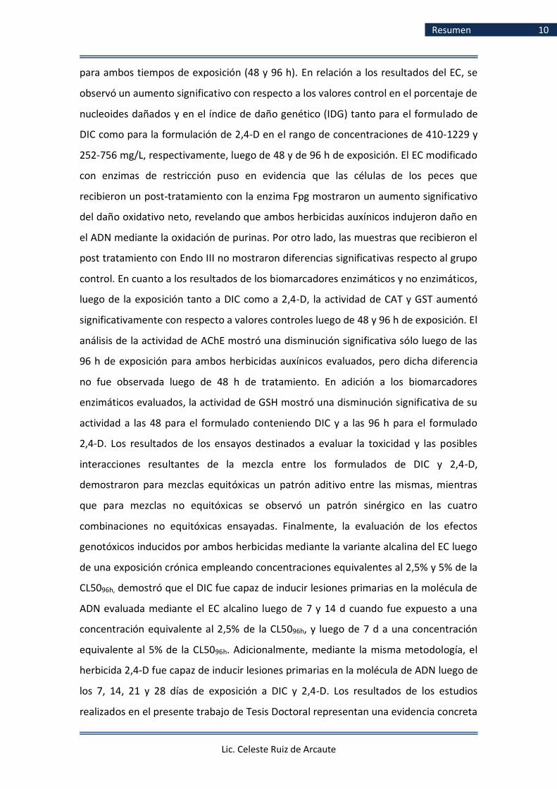
Resumen 10
para ambos tiempos de exposición (48 y 96 h). En relación a los resultados del EC, se
observó un aumento significativo con respecto a los valores control en el porcentaje de
nucleoides dañados y en el índice de daño genético (IDG) tanto para el formulado de
DIC como para la formulación de 2,4-D en el rango de concentraciones de 410-1229 y
252-756 mg/L, respectivamente, luego de 48 y de 96 h de exposición. El EC modificado
con enzimas de restricción puso en evidencia que las células de los peces que
recibieron un post-tratamiento con la enzima Fpg mostraron un aumento significativo
del daño oxidativo neto, revelando que ambos herbicidas auxínicos indujeron daño en
el ADN mediante la oxidación de purinas. Por otro lado, las muestras que recibieron el
post tratamiento con Endo III no mostraron diferencias significativas respecto al grupo
control. En cuanto a los resultados de los biomarcadores enzimáticos y no enzimáticos,
luego de la exposición tanto a DIC como a 2,4-D, la actividad de CAT y GST aumentó
significativamente con respecto a valores controles luego de 48 y 96 h de exposición. El
análisis de la actividad de AChE mostró una disminución significativa sólo luego de las
96 h de exposición para ambos herbicidas auxínicos evaluados, pero dicha diferencia
no fue observada luego de 48 h de tratamiento. En adición a los biomarcadores
enzimáticos evaluados, la actividad de GSH mostró una disminución significativa de su
actividad a las 48 para el formulado conteniendo DIC y a las 96 h para el formulado
2,4-D. Los resultados de los ensayos destinados a evaluar la toxicidad y las posibles
interacciones resultantes de la mezcla entre los formulados de DIC y 2,4-D,
demostraron para mezclas equitóxicas un patrón aditivo entre las mismas, mientras
que para mezclas no equitóxicas se observó un patrón sinérgico en las cuatro
combinaciones no equitóxicas ensayadas. Finalmente, la evaluación de los efectos
genotóxicos inducidos por ambos herbicidas mediante la variante alcalina del EC luego
de una exposición crónica empleando concentraciones equivalentes al 2,5% y 5% de la
CL5096h, demostró que el DIC fue capaz de inducir lesiones primarias en la molécula de
ADN evaluada mediante el EC alcalino luego de 7 y 14 d cuando fue expuesto a una
concentración equivalente al 2,5% de la CL5096h, y luego de 7 d a una concentración
equivalente al 5% de la CL5096h. Adicionalmente, mediante la misma metodología, el
herbicida 2,4-D fue capaz de inducir lesiones primarias en la molécula de ADN luego de
los 7, 14, 21 y 28 días de exposición a DIC y 2,4-D. Los resultados de los estudios
realizados en el presente trabajo de Tesis Doctoral representan una evidencia concreta

Lic. Celeste Ruiz de Arcaute
Resumen 11
que los herbicidas DIC y 2,4-D en sus formulaciones comerciales Banvel® y DMA® deben
ser considerados agentes inductores tanto de mortalidad como de efectos subletales
tales como alteraciones en el comportamiento, efectos cito y genotóxicos y como
agentes capaces de generar alteraciones en el balance oxidativo, tanto enzimáticos y
no enzimáticos en C. decemmaculatus. Adicionalmente, estos herbicidas deberían ser
clasificados según los valores de CL5096h obtenidos como compuestos nocivos
(categoría III) para C. decemmaculatus de acuerdo a los patrones internacionales de
clasificación de riesgo toxicológico para especies acuáticas. Es relevante destacar que
las concentraciones empleadas en el presente trabajo de Tesis Doctoral se encuentran
en el orden de mg/L, mientras que hasta el presente, las detectadas en el ambiente
acuático se encuentran en el orden de μg/L. Aun así, ante las condiciones
predominantes en el modelo de producción actual y al escaso control gubernamental
existente en nuestro país, no podría descartarse la posibilidad de que ambos
herbicidas auxínicos, DIC y 2,4-D, puedan ingresar y contaminar los diferentes
compartimentos ambientales ejerciendo efectos deletéreos y comprometiendo la
salud no sólo para la especie evaluada, C. decemmaculatus, sino también de otros
organismos, incluyendo los seres humanos.

Lic. Celeste Ruiz de Arcaute
Abstract 12
Abstract
The aim of the current Doctoral Thesis was to evaluate the lethal and sublethal
effects induced by two auxinic herbicides employed worldwide, dicamba (DIC) and 2,4-
dichlorophenoxyacetic acid (2,4-D) contained in the Banvel® (57.7% DIC) and DMA®
(58.4% 2,4-D) formulations marketed in our country. In the last decades, the amount
of pesticides used in agroecosystems has significantly increased. The widespread,
intensive and non regulated application of these agrochemicals represent a serious
concern for human and environmental health, as they can produce multiple
consequences at organism, population, community and ecosystem levels, disturbing
vital functions of organisms such as behavior, reproduction, predators evasion and
food gathering, among others, and thus the survival and biodiversity. The effects
induced by these herbicides were evaluated on the Neotropical poecilid Cnesterodon
decemmaculatus by biomarkers of lethality, behavior, genotoxicity and oxidative
damage under acute laboratory assays. Additionally, the possible interactions
(additivity, synergism or antagonism) resulting from the joined application of both
herbicides were evaluated by lethality assays using equitoxic and non-equitox
mixtures. Furthermore, the genotoxic effect induced by DIC and 2,4-D after a long-
term exposure through the alkaline single cell gel electrophoresis assay (SCGE, comet
assay) was assayed. The determination of the LC50 was employed as a biomarker for
lethality and for determining the concentrations of the sublethal assays in the present
Doctoral Thesis. Behavioral changes as gathering at the bottom of the aquarium (GBA),
slowness in motion (SM), slow reaction (SR) and abnormal swimming (AS) were
evaluated and employed for calculating NOEC and LOEC parameters. In addition, for
evaluating the possible genotoxic effects, different biomarkers of effect were
employed such as the micronuclei (MNs) test, the induction of other nuclear

Lic. Celeste Ruiz de Arcaute
Abstract 13
abnormalities and the alkaline comet assay using blood cells of this poecilid. The
modified SCGE methodology with the addition of restriction enzymes was employed in
order to determine the possible mechanism of action exerted by DIC and 2,4-D,
through the detection of oxidated pyrimidine and purine bases by enzymes Endo III
and Fpg, respectively. Moreover, the possible antioxidant response induced by both
auxinic herbicides was analyzed using enzymatic and non-enzymatic biomarkers such
as the detection of variations in the levels of catalase (CAT), glutathione-S-transferase
(GST), acetylcholinesterase (AChE) and glutathione (GSH). In addition, in order to
analyze the possible interactions resulting from the joined application of both
herbicides and the effect that they could exert on C. decemmaculatus, acute toxicity
tests of mixtures were carried out employing equitoxic and non-equitoxic
concentrations of both herbicides. Finally, to evaluate the chronic effects induced by
the herbicides DIC and 2,4-D, the induction of primary lesions in the DNA in blood cells
of C. decemmaculatus was assayed by the SCGE assay. Lethality results revealed that
the DIC-based formulation Banvel® exerted lethal effects with a LC5096h value of 1639
mg/L while the 2,4-D-based formulation DMA® had a LC5096h a value of 1008 mg/L.
Results of behavioral changes on individuals exposed to DIC and 2,4-D showed
significant variations respect to the control values within the 0-96 h exposure range.
The results reveal an increased frequency of MNs in those fish exposed to the highest
concentration (1229 mg/L) of DIC during 48 h but not in fish exposed to the herbicide
for 96 h, regardless of the concentration of herbicide assayed. In 2,4-D- exposed fish,
the enhancement in MNs frequency was observed at all concentrations assayed within
the range 252-756 mg/L for both exposure times (48 and 96 h). In addition, both
herbicides were able to induce single-strand breaks and/or alkali sensitive sites into
DNA, revealed by the SCGE assay regardless of the concentration and exposure time.
Furthermore, the modified comet assay showed that cells post-treated with the
restriction enzyme Fpg showed a significant increase of the oxidative damage,
revealing that both auxinic herbicides induced DNA damage through the oxidation of
purines. On the other hand, post treatment with restriction enzyme Endo III did not
showed significant differences with their respective control group. Regarding the
results of the enzymatic and non-enzymatic biomarkers, significant increased activities
of the enzymes CAT and GST were observed in fish exposed to DIC- and 2,4-D-based

Lic. Celeste Ruiz de Arcaute
Abstract 14
formulations for 48 and 96 h, respectively. The analysis of AChE activity showed a
significant decrease only after 96 h of exposure for both herbicides but such difference
was not observed after 48 h of exposure. Additionally to the aforementioned altered
enzymatic markers, a decrease of GSH was registered in specimens treated for 48 and
96 h with either DIC- and 2,4-D-herbicide formulations. The results of the tests aimed
to evaluate the toxic effects exerted when C. decemmaculatus was exposed
simultaneously to the DIC- and 2,4-D-based formulations Banvel® and DMA®,
respectively, showed for equitoxic mixtures an additive pattern, while for non-
equitoxic mixtures a synergistic pattern was observed in the four non-equitoxic
combinations tested.
Finally, the chronic sublethal effects showed an increased frequency of DNA breaks
estimated by the alkaline SCGE in fish exposed to 2.5% of DIC LC5096h for 7 and 14 days
whereas this enhancement was observed only after 7 days of exposure to a
concentration equivalent to 5% of the LC5096h. In individuals chronically exposed to
concentrations equivalent to 2.5 and 5% of 2,4-D LC5096h increased the frequencies of
breaks in DNA estimated by the alkaline SCGE in fish exposed for 7, 14, 21 and 28 days.
Overall, the results obtained in the present Doctoral Thesis represent a concrete
evidence that the auxinic herbicides DIC and 2,4-D present in their commercial
formulations Banvel® and DMA®, respectively, should be considered as oxidizing agents
able to induce both mortality and sub-lethal effects such as alterations in behavior,
genotoxic effects and alterations of enzymatic and non-enzymatic biomarkers in C.
decemmaculatus. Furthermore, these herbicides should be classified according to the
values of LC5096h obtained as harmful compounds (category III), for C. decemmaculatus
according to the international standards of toxicological risk classification for aquatic
species. Given the prevailing conditions in the current agronomic-production model, it
cannot be rule out the possibility that both auxinic herbicides can enter and
contaminate different environmental compartments, exerting deleterious effects and
compromising the health not only of our test species, C. decemmaculatus, but also of
other organisms, among which human beings are included.

Lic. Celeste Ruiz de Arcaute
Publicaciones 15
Listado de publicaciones
científicas
El presente trabajo de Tesis Doctoral dio lugar a los siguientes trabajos científicos. Los
mismos se encuentran contenidos en el Anexo I y son referidos a lo largo del texto.
I. Ruiz de Arcaute C; Soloneski S y Larramendy ML. 2014. Evaluation of the
genotoxicity of a herbicide formulation containing 3,6-dichloro-2-
metoxybenzoic acid (dicamba) in circulating blood cells of the tropical fish
Cnesterodon decemmaculatus. Mutation Research. 773, 1-8.
II. Ruiz de Arcaute C; Soloneski S y Larramendy ML. 2016. Toxic and genotoxic
effects of the 2,4-dichlorophenoxyacetic acid (2,4-d)-based herbicide on the
neotropical fish Cnesterodon decemmaculatus. Ecotoxicology and
Environmental Safety. 128, 222-229.
III. Ruiz de Arcaute C; Soloneski S y Larramendy ML. 2018. Synergism of
mixtures of dicamba and 2,4-dichlorophenoxyacetic acid herbicide
formulations on the neotropical fish Cnesterodon decemmaculatus (Pisces,
Poeciliidae). Environmental Pollution, 236, 33-39.
IV. Ruiz de Arcaute, C; Larramendy, ML y Soloneski, S. 2018. Genotoxicity by
long-term exposure to the auxinic herbicides 2,4-dichlorophenoxyacetic
acid and dicamba on Cnesterodon decemmaculatus (Pisces: Poeciliidae).
Environmental Pollution, 243, 670-678.
V. Ruiz de Arcaute, C; Pérez-Iglesias, JM; Ossana, NA; Soloneski, S y
Larramendy, ML. 2018. Auxinic herbicides induce oxidative stress in
Cnesterodon decemmaculatus. Ecological Indicators, Enviado.

Lic. Celeste Ruiz de Arcaute
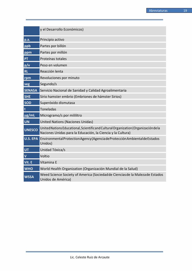
Abreviaturas 16
Abreviaturas
ºC Grados Celsius
2,4-D Ácido 2,4-diclorofenoxiacético
µl Microlitro/s
ABA Ácido abscísico
ACC 1-aminociclopropano-1-ácido carboxílico
AChE Acetilcolinesterasa
ADN Ácido desoxirribonucleico
AFA Aglomeración en el fondo del acuario
AP Antes del presente
APFB Agarosa de punto de fusión bajo
APFN Agarosa de punto de fusión normal
ASTM American Society for Testing and Materials (Sociedad Americana para Pruebas y Materiales)
ATA Ácido tricloroacético
ATP adenosín trifosfato
ATSDR Agency for Toxic Substances and Disease Registry (Agencia para Sustancias Tóxicas y el Registro de Enfermedades)
BEnz Buffer enzimas
BSA Albúmina suero bovino (Bovine Serum Albumin)
BSI British Standards Institution (Instituto Británico de Normalización)
BHPM Buffer de homogenización post mitocondrial
CASAFE Cámara de Sanidad Agropecuaria y Fertilizantes
CAS Chemical Abstract Service Registry Number
CAT Catalasa
CB Célula binucleada
CDNB 1-cloro-2,4-dinitrobenceno
CE50 Concentración efectiva 50
CF Ciclofosfamida
CN Control negativo

Lic. Celeste Ruiz de Arcaute
Abreviaturas 17
CL5096h Concentración letal cincuenta en 96 horas
cm Centímetro/s
cm2 Centímetro/s cuadrado/s
cm3 Centímetro/s cúbico/s
CONICET Consejo Nacional de Investigaciones Científicas y Técnicas
ChE Colinesterasa/s
CP Control positivo
d Día/s
DAPI 4´,6-diamidino-2-fenilindol diclorhidrato
DIC Dicamba
DDT Dicloro-difenilo-tricloroetano
DMSO Dimetilsulfóxido
DO Daño oxidativo neto
DTNB ácido 5,5´-ditiobis-2-dinitrobenzoico
DTT Ditiotreitol
EC Ensayo cometa
ECHA European Chemical Agency (Agencia Europea de Sustancias y Mezclas Químicas)
EDTA Ácido etilendiaminotetraacético
EE Error estándar
EN Escotadura nuclear
Endo III Endonucleasa III
ERO Especies reactivas de oxígeno
EU European Union (Unión Europea)
FAO Food and Agriculture Organization (Organización de las Naciones Unidas para la Agricultura y la Alimentación)
Fpg Formamidopirimidina-DNA glicosilasa
g Gramo/s
GPx Glutatión peroxidasa
GSH Glutatión reducido
GR Glutatión reductasa
GST Glutatión-S-transferasa
H2O2 Peróxido de hidrógeno
H2Od Agua destilada
h Hora/s
ha Hectárea

Lic. Celeste Ruiz de Arcaute
Abreviaturas 18
HPLC Cromatografía líquida de alta resolución
IARC International Agency for Research on Cancer (Agencia Internacional de Investigación en Cáncer)
IDG Índice de daño genético
INCHEM
Chemical Safety Information from Intergovernmental Organizations (Información sobre Seguridad Química para Organizaciones Intergubernamentales)
IPC Índice de proliferación celular
IPCS International Programme on Chemical Safety (Programa Internacional para la Seguridad Química)
kg kilogramo/s
Km Kilómetro/s por hora
Kow Coeficiente de partición octanol/agua
L Litro/s
LC Límite/s de confianza
LOEC Lowest Observed Effect Concentration (Concentración más baja a la cual se observa efecto)
mA Miliamperio
MCPA Ácido 2-metil-4-clorofenoxiacético (2-methyl-4-chlorophenoxyacetic acid)
MCPP Ácido propiónico, 2-(4-cloro-2-metilfenoxi)l (methylchlorophenoxypropionic acid)
MG Modificaciones genéticas
mg/L Miligramo/s por litro
min Minuto/s
ML Movimiento lento
mM Milimolar
MN Micronúcleo
msnm Metros sobre el nivel del mar
NA Natación anormal
NAcr Naranja de acridina
ng/L Nanogramo por litro
NL Núcleo lobulado
nm Nanometro
NOEC No Observed Effect Concentration (concentración más baja a la cual no se observa efecto)
OECD Organization for Economic Co-operation and Development (Organización para la Cooperación
Lic. Celeste Ruiz de Arcaute
Abreviaturas 19
y el Desarrollo Económicos)
p.a. Principio activo
ppb Partes por billón
ppm Partes por millón
PT Proteínas totales
p/v Peso en volumen
RL Reacción lenta
rpm Revoluciones por minuto
seg Segundo/s
SENASA Servicio Nacional de Sanidad y Calidad Agroalimentaria
SHE Sirio hamster embrio (Embriones de hámster Sirios)
SOD Superóxido dismutasa
t Toneladas
µg/mL Microgramo/s por mililitro
UN United Nations (Naciones Unidas)
UNESCO United Nations Educational, Scientific and Cultural Organization (Organización de la Naciones Unidas para la Educación, la Ciencia y la Cultura)
U.S. EPA Environmental Protection Agency (Agencia de Protección Ambiental de Estados Unidos)
UT Unidad Tóxica/s
V Voltio
Vit. E Vitamina E
WHO World Health Organization (Organización Mundial de la Salud)
WSSA Weed Science Society of America (Sociedad de Ciencias de la Maleza de Estados Unidos de América)

Lic. Celeste Ruiz de Arcaute
Índice Tablas 20
Índice de Tablas
Tabla 1.1 Plaguicidas más utilizados en el sector agrícola de
EE.UU. del año 2012 .............................................................................. 25
Tabla 1.2 Clasificaciones toxicológicas................................................................... 34
Tabla 1.3 Clasificaciones de herbicidas según su modo de
acción .................................................................................................... 37
Tabla 1.4 Familia de herbicidas auxínicos .............................................................. 41
Tabla 1.5 Formulados comerciales en Argentina conteniendo
dicamba como principio activo .............................................................. 43
Tabla 1.6 Información toxicológica del formulado comercial
Banvel® (57,7% dicamba) ....................................................................... 50
Tabla 1.7 Formulados comerciales en Argentina conteniendo
2,4-D como principio activo ................................................................... 54
Tabla 1.8 Información toxicológica del formulado comercial
DMA® (58,4% 2,4-D) ............................................................................... 62
Tabla 3.1 Compuestos químicos utilizados ............................................................ 86
Tabla 4.1 Efectos letales luego de la exposición aguda a
dicamba ................................................................................................. 104
Tabla 4.2 Efectos letales luego de la exposición aguda a 2,4-D .............................. 105
Tabla 4.A Individuos afectados con alteraciones comportamentales ................................................................................ 106
Tabla 4.3 Efectos en el comportamiento luego de exposición
aguda a dicamba .................................................................................... 107

Lic. Celeste Ruiz de Arcaute
Índice Tablas 21
Tabla 4.4 Efectos en el comportamiento luego de exposición
aguda a 2,4-D ......................................................................................... 108
Tabla 4.5 Frecuencia de MNs y AN luego de exposición aguda
a dicamba .............................................................................................. 111
Tabla 4.6 Frecuencia de MN y AN luego de exposición aguda a 2,4-D ................... 113
Tabla 4.7 Análisis del daño en el ADN mediante EC en
individuos expuestos a dicamba............................................................. 115
Tabla 4.8 Análisis del daño en el ADN mediante EC en
individuos expuestos a 2,4-D ................................................................. 117
Tabla 4.9 Análisis del daño en el ADN mediante EC modificado
en individuos expuestos a dicamba ........................................................ 120
Tabla 4.10 Análisis del daño en el ADN mediante EC modificado
en individuos expuestos a 2,4-D ............................................................. 122
Tabla 4.11 Análisis de estrés oxidativo en Cnesterodon
decemmaculatus expuestos a dicamba y 2,4-D ...................................... 125
Tabla 4.12 Efectos letales luego de exposición aguda a mezclas
equitóxicas de dicamba y 2,4-D .............................................................. 126
Tabla 4.13 Efectos letales luego de exposición aguda a mezclas
no equitóxicas de dicamba y 2,4-D ......................................................... 127
Tabla 4.14 Análisis del daño en el ADN mediante EC en
individuos expuestos de manera crónica a dicamba ............................... 131
Tabla 4.15 Análisis del daño en el ADN mediante EC en
individuos expuestos de manera crónica a 2,4-D.................................... 134
Tabla 5.1 CL5096h reportadas en organismos expuestos a
dicamba y 2,4-D ..................................................................................... 141
Tabla 5.2 CL5096h reportadas en Cnesterodon
decemmaculatus expuestos a distintos compuestos .............................. 145

Lic. Celeste Ruiz de Arcaute
Índice Tablas 22

Lic. Celeste Ruiz de Arcaute
Índice Figuras 23
Índice de Figuras
Figura 1.1 Evolución Mercado Fitosanitario Argentino ........................................... 26
Figura 1.2 Estructura química del herbicida dicamba.............................................. 42
Figura 1.3 Estructura química del herbicida 2,4-D .................................................. 50
Figura 1.4 Ejemplares de Cnesterodon decemmaculatus ........................................ 65
Figura 1.5 Micronúcleo en eritrocito circulante de Cnesterodon
decemmaculatus .................................................................................... 71
Figura 1.6 Anormalidades nucleares ....................................................................... 73
Figura 1.7 Nucleoide sin daño y cometa ................................................................. 76
Figura 1.8 Categorías de daño utilizadas en el EC ................................................... 77
Figura 3.1 Sitio de muestreo, Parque Pereyra Iraola ............................................... 85
Figura 3.2 Diseño experimental de los ensayos agudos .......................................... 88
Figura 3.3 Diseño experimental de los ensayos crónicos ........................................ 89
Figura 3.4 Protocolo del ensayo cometa alcalino .................................................... 94
Figura 3.5 Categorías de daño utilizadas en el EC ................................................... 95
Figura 3.6 Esquema isobolograma .......................................................................... 101
Figura 4.1 Frecuencias de nucleoides en individuos expuestos
de manera aguda a dicamba .................................................................. 114
Figura 4.2 Frecuencias de nucleoides en individuos expuestos
de manera aguda a 2,4-D ....................................................................... 116
Figura 4.3 Daño oxidativo neto inducido por dicamba y 2,4-D ................................ 118
Figura 4.4 Isobolograma ilustrando UT de mezclas no
Lic. Celeste Ruiz de Arcaute
Índice Figuras 24
equitóxicas de dicamba y 2,4-D .............................................................. 128
Figura 4.5 Frecuencias de nucleoides en individuos expuestos
de manera crónica a dicamba ................................................................ 130
Figura 4.6 Frecuencias de nucleoides en individuos expuestos
de manera crónica a 2,4-D ..................................................................... 133

Lic. Celeste Ruiz de Arcaute
Introducción 25
1. Introducción
1.1 Modelo agrícola moderno
El control de plagas en cultivos es una práctica tan antigua como la agricultura
misma. Partiendo 12.000 años A.P. con la remoción manual de plantas no deseadas, su
evolución fue lenta en el comienzo. En el 8.000 A.P. se reportó el uso de las primeras
herramientas para la labranza manual y posteriormente en el 4500 A.P. se registró el
primer uso intencional de plaguicidas, cuando los sumerios utilizaron compuestos
azufrados para repeler insectos y ácaros. A través de los años, el conocimiento acerca
del uso de plaguicidas fue incrementando y 2000 años A.P. los romanos emplearon
diferentes tipos de sales para combatir las malezas. Posteriormente, en el siglo XVIII,
durante la revolución agrícola europea, entre 1750 y 1780, se comenzaron a aplicar en
los cultivos diferentes sustancias con capacidad insecticida tal como el piretro,
obtenido del crisantemo (Chrysanthemum cinerariaefolium) (Taylor y col., 2006). A
partir del 1800 se comienzan a emplear elementos extraídos de la naturaleza tales
como el azufre, cobre, arsénico y fosforo y se da inicio a la utilización de las primeras
fumigaciones con plaguicidas orgánicos, tales como derivados del petróleo,
clorofenoles, naftaleno, el famoso caldo bordelés (sulfato de cobre y cal), el verde de
París (acetoarsenito de cobre), entre otros. Ya en la década de 1940, durante el
comienzo de la Segunda Guerra Mundial se introdujo el control químico de plagas con
el desarrollo de los primeros plaguicidas sintéticos como el organoclorado DDT, muy

Lic. Celeste Ruiz de Arcaute
Introducción 26
utilizado debido a su gran estabilidad y acción prolongada contra una amplia variedad
de plagas entre las que se incluyen la mosca doméstica, piojos y mosquitos, entre otros
(Ware y Whitacre, 2004). Posteriormente, en 1947, se introdujo el control químico,
con la entrada al mercado de los plaguicidas orgánicos sintéticos, tales como el
clordano, eldrín, dieldrín, paration, lindano, ácido 2-metil-4-cloro fenoxiacético (MCPA)
y el ácido 2,4-diclorofenoxiacético (2,4-D), cuya mezcla con el 2,4,5-T se denominó
agente naranja, masivamente empleado como herbicida y defoliante durante la Guerra
de Vietnam (Hay, 1974; Unsworth, 2010). Estos nuevos productos tenían la ventaja de
ser efectivos y económicos. Luego de la Segunda Guerra Mundial y junto a las
tecnologías desarrolladas bajo el concepto de Revolución Verde, se produjo un
importante incremento en la productividad agrícola mediante la utilización de
tecnologías innovadoras aplicadas en la producción de plaguicidas, fertilizantes,
mecanismos de riego y variedades de cultivos de alto rendimiento resistentes a climas
extremos y plagas (Conway, 1997). Este nuevo modelo productivo, asociado a la
práctica del monocultivo a gran escala, trajo como consecuencia una producción
basada en el consumo de elevadas cantidades de insumos, principalmente herbicidas,
al igual que el uso extendido de maquinaria agrícola. Desde la década del '80
conjuntamente con la introducción de los cultivos genéticamente modificados (GM)
tales como soja, maíz y algodón, dichas prácticas agrícolas tuvieron un gran desarrollo
tanto en Argentina como en Sudamérica y en el resto del mundo (FAO, 2013; Martin-
Guay y col., 2018).
Particularmente en nuestro país, en los últimos 40 años se produjo una
expansión de la superficie destinada a la agricultura, encontrándose actualmente
dentro de los países mega productores de cultivos genéticamente modificados.
Argentina se ubica en el tercer puesto de países de mayor producción con 23,8
millones de ha cultivadas, de las cuales al año 2016, 18,7 fueron de soja, 4,7 de maíz y
0,4 de algodón (ISAAA, 2016; Sasal y col., 2016). El 96% de la producción de soja se
exporta a China (tanto poroto como aceite de soja), Europa (harina y pellet de soja),
India (aceite de soja) y Estados Unidos (biodiésel) (Pontón, 2008; Silvetti y Cáceres,
2015). Esta gran demanda en el mercado de las oleaginosas condujo
concomitantemente a un aumento en la importación y utilización de numerosos
productos agroquímicos (Figura 1.1), tendencia observada también a nivel global. En el

Lic. Celeste Ruiz de Arcaute
Introducción 27
año 2012, el consumo mundial de plaguicidas fue aproximadamente de dos millones
de t por año, de los cuales el 45% fue utilizado en Europa, 25% en Estados Unidos y el
30% restante en el resto del mundo (De y col., 2014). Del total de ventas de productos
fitosanitarios, los herbicidas lideran el ranking con aproximadamente el 50% de las
ventas, seguidos por los fumigantes, insecticidas y fungicidas, respectivamente (USEPA,
2017)(Tabla 1.1).
Tabla 1.1 Plaguicidas más utilizados en el sector agrícola de EE.UU. del año 2012
Ingrediente activo
Tipoa
Puesto
Glifosato
H
1
Atrazina H 2
S-Metolaclor
H
3
Dicloropropeno Fum 4
Ácido 2,4-diclorofenoxiacético (2,4-D) H
5
Metam sodio Fum 6
Acetoclor
H
7
Metam potasio Fum 8
Cloropicrina
Fum
9
Clorotaloni F 10
Pendimetalin
H
11
Etefon RCP 12
Mancozeb
F
13
Clorpirifós I 14
Metolaclor
H
15
Hidróxido de calcio F 16
Propanil
H
17
Dicamba H 18
Trifluralin
H
19
Decan-l-ol RCP 20
Hidróxido de cobre
F
21
Acefato I 22
Paraquat
H
23
Metil bromidio Fum 24
Glufosinato
H
25
a, Tipos de plaguicidas. H: herbicidas; Fum: fumigantes; F: fungicidas;
I: insecticidas; RCP: reguladores del crecimiento de las plantas

Lic. Celeste Ruiz de Arcaute
Introducción 28
En el caso de Argentina y dentro del contexto de un sostenido aumento de la
productividad, según información de la Cámara de Sanidad Agropecuaria y Fertilizantes
(CASAFE) en cuanto al consumo de productos fitosanitarios, el 64% de las ventas
correspondieron a herbicidas, seguido por los insecticidas (17%) y los fungicidas (15%)
(CASAFE, 2017b). Cabe destacar que la mayor proporción de plaguicidas fue empleada
para el tratamiento de malezas en barbecho químico, seguido de una marcada
demanda de estos compuestos para el tratamiento en cultivos de oleaginosas y
cereales y en menor proporción para el tratamiento de malezas en cultivos de vid,
caña de azúcar, frutas de pepita y carozo, entre otros.
Es ampliamente conocido que el uso indiscriminado de plaguicidas inflige
numerosas consecuencias negativas para el medio ambiente y para los seres vivos
tales como el aumento de resistencia de patógenos y pestes, la reducción de la
biodiversidad del suelo, la disminución de las poblaciones de especies favorables a los
cultivos por destrucción de sus hábitats, entre otros efectos adversos (Tilman y col.,
2002; Duhan y col., 2017). De esta manera, se ven afectadas no sólo las especies
blanco para las cuales fueron diseñadas en primera instancia, sino también organismos
no blanco tales como la biota acuática, incluyendo desde microorganismos hasta
plantas y mamíferos al igual que numerosos organismos terrestres (Liang y col., 2013;
Meffe y de Bustamante, 2014). Este problema, en países subdesarrollados como
Argentina, se ve agravado por la falta de controles por parte del estado que regulen la
Figura 1.1: Evolución del Mercado Fitosanitario Argentino. Fuente: (CASAFE, 2017b)
Lic. Celeste Ruiz de Arcaute
Introducción 29
aplicación, manejo y el tratamiento de los agroquímicos al igual que de los residuos
generados de dichas actividades (Ministerio de Salud, 2007).
Desafortunadamente, en este modelo de producción es muy difícil disminuir el
uso de agroquímicos sin disminuir el rendimiento de los cultivos (McLaughlin y
Kinzelbach, 2015). La necesidad de alimentar a las estimadas 8 mil millones de
personas que vivirán en la tierra para el año 2024, hace que la producción de
alimentos tenga que aumentar al menos un 50%, sino aún más del 100% en los
próximos 30 años, un desafío intimidante teniendo en cuenta que el aumento en el
rendimiento de los cultivos se ha estancado o incluso ha disminuido en la última
década (FAO-WHO, 2006; Camargo y col., 2017). Los incrementos en el rendimiento a
través de las mejoras utilizando métodos convencionales han estado por debajo del
requerimiento del 2,4% por año proyectado hasta el 2050. Las modificaciones
genéticas introducidas en distintas variedades vegetales, principalmente tolerancia a
herbicidas y resistencia a los insectos ha sido transformadora, pero aún queda un largo
camino por recorrer en términos de obtener el máximo rendimiento intrínseco
(Griffiths y Paul, 2017).
En la actualidad, se está avanzando firmemente en la aplicación de agricultura de
precisión, que propone una revolución tecnológica conocida como la Nueva
Revolución Verde (FAO, 2018). Esta nueva revolución agrícola plantea combinar la
tecnología moderna y el conocimiento tradicional, poniendo énfasis en los sistemas
agrícolas, sociales y agroecológicos, así como en el máximo rendimiento de los
cultivos. Al mismo tiempo, pone énfasis en los enfoques alternativos y en la mejora de
los sistemas de manejo por aplicación de nuevas tecnologías con el fin de minimizar el
daño ambiental de los insumos externos y beneficiando a los agricultores más
humildes y a las áreas marginales no tenidas en cuenta por la Revolución Verde
original (FAO, 2013; Martin-Guay y col., 2018). Asimismo, tiene como finalidad poseer
una mayor precisión en el manejo integrado de los cultivos mediante la utilización de
diferentes tipos de herramientas asociadas con diferentes sistemas de información a
fin de minimizar el daño ambiental debido al consumo excesivo de insumos. Estas
características traen un mayor beneficio a los agricultores dado que incrementan su
productividad empleando una menor cantidad de agroquímicos y generando un bajo
nivel de impacto negativo sobre las márgenes aledañas a los cultivos. Todos los

Lic. Celeste Ruiz de Arcaute
Introducción 30
procesos que involucran desde la siembra hasta la cosecha final de los cultivos se
realizan utilizando maquinaria agrícola de precisión que incluyen nuevas
modificaciones y equipamiento, tales como programas de computación específicos,
drones, pequeños helicópteros e imágenes satelitales que permiten, por medio de
Internet, hacer un seguimiento en tiempo real de las necesidades del cultivo. Esto
tiene como objetivo, durante la siembra, obtener una mayor precisión en la
dosificación y en la profundidad; realizar un mejor trazado de surcos, evitar
superposiciones y fertilizar según los requerimientos específicos. En la etapa de control
del cultivo, permite realizar una dosificación variable de los agroquímicos, un control
selectivo de malezas y riego según la necesidad detectada. En la etapa de cosecha se
pretende disminuir las pérdidas, monitorear el rendimiento, determinar la humedad
exacta evitando que se empaquen productos no preparados, y registrar la calidad por
zona en tiempo real (Menéndez y col., 2015).
La Revolución Verde introdujo y propagó la idea de la modificación del ambiente
para explotar el máximo potencial de unas pocas variedades vegetales, conocido como
monocultivo, que se tradujo en altos costos sociales y ambientales, en adición a los
económicos y tecnológicos (Sarandón y Marasas, 2017). Actualmente el mundo
enfrenta una crisis alimentaria silenciosa. Según la FAO, 815 millones de personas se
encuentran en estado de desnutrición y 52 millones de niños menores de 5 años se
ven afectados por desnutrición aguda en el mundo en desarrollo (FAO, 2017b). A pesar
de lo augurado para las nuevas tecnologías, este problema sigue sin resolverse, y
aquello que en principio suponía la utilización de plaguicidas para aumentar la
producción mediante un control total de plagas, estos no sólo no las han erradicado,
sino que se han vuelto cada vez más necesarios y su dosificación cada vez más alta.
Se ha estimado que, del total de herbicidas aplicados en el ambiente, menos del
0,1% llega al organismo blanco, dejando una gran cantidad de residuos tóxicos libres
en los distintos compartimentos ambientales (Pimentel y col., 1993; Zhou, 2008; WHO-
FAO, 2009; Song y col., 2017). La contaminación por plaguicidas puede afectar a todos
los compartimentos ambientales como consecuencia de su fabricación, aplicación,
escorrentía y drenaje de aguas contaminadas y/o el transporte de agroquímicos a
través de los suelos y hacia las aguas subterráneas, entre otros (Pimentel y col., 1993;
Solomon y Thompson, 2003; Soloneski y col., 2016). Adicionalmente, en los últimos

Lic. Celeste Ruiz de Arcaute
Introducción 31
años se le ha dado especial interés a la preparación y limpieza de los residuos de
plaguicidas como otra fuente de contaminación ambiental (Higginbotham y col., 1999;
Suciu y col., 2011; Osborne y col., 2015), un aspecto poco regulado que eleva las
concentraciones de los mismos en aguas subterráneas y de escorrentía (Gan y col.,
1996).
En el contexto mundial actual, resulta prácticamente imposible que las especies
animales y vegetales no se encuentren expuestas directa o indirectamente en algún
momento de su ciclo de vida a diferentes tipos de agroquímicos, los cuales
representan un problema a nivel ecológico y de salud pública, por su persistencia,
bioacumulación y toxicidad.
1.2 Plaguicidas. Definición y clasificaciones
Según la FAO, un plaguicida puede definirse como “cualquier sustancia o mezcla
de sustancias destinadas a prevenir, destruir o controlar cualquier plaga, incluyendo los
vectores de enfermedades humanas o de los animales, las especies no deseadas de
plantas o animales que causan perjuicio o que interfieren de cualquier otra forma en la
producción, elaboración, almacenamiento, transporte o comercialización de alimentos,
productos agrícolas, madera y productos de madera o alimentos para animales, o que
pueden administrarse a los animales para combatir insectos, arácnidos u otras plagas
en o sobre sus cuerpos. El término incluye las sustancias destinadas a utilizarse como
reguladoras del crecimiento de las plantas, defoliantes, desecantes, agentes para
reducir la densidad de fruta o agentes para evitar la caída prematura de la fruta, y las
sustancias aplicadas a los cultivos antes o después de la cosecha para proteger el
producto contra la deterioración durante el almacenamiento y transporte” (OECD,
1997) (FAO, 2002). Dentro de los plaguicidas se incluyen los insecticidas, fungicidas y
herbicidas, y otros compuestos como desinfectantes, repelentes, preservantes y
curasemillas (USEPA, 2018).
Los plaguicidas pueden clasificarse de acuerdo a diferentes criterios, presentados
a continuación.

Lic. Celeste Ruiz de Arcaute
Introducción 32
1.2.1 De acuerdo al organismo blanco sobre el cual actúa
El ingrediente activo de un plaguicida determinado se describe principalmente
según el tipo de plaga sobre la que actúa. Entre los más conocidos se incluyen los
insecticidas, herbicidas, rodenticidas y fungicidas. La siguiente lista abarca el amplio
rango existente de tipos de plaguicidas (USEPA, 2018).
Acaricidas: Eliminan ácaros que se alimentan de plantas y animales.
Alguicidas: Eliminan algas en lagos, canales, tanques y otros cuerpos de agua.
Antiincrustantes: Actúan repeliendo o eliminando organismos que se adhieren a superficies sumergidas.
Antimicrobianos: Eliminan microorganismos tales como bacterias y virus.
Biocidas: Eliminan organismos considerados nocivos.
Defoliantes: Causan la pérdida de hojas o follaje de una planta, usado en generalmente para facilitar la cosecha.
Desecantes: Promueven la deshidratación de tejidos vivos.
Desinfectantes: Matan o inactivan microorganismos productores de enfermedades.
Fungicidas: Eliminan hongos.
Herbicidas: Eliminan hierbas y otras plantas que crecen en áreas no deseadas.
Reguladores del crecimiento de insectos:
Interrumpen la muda, maduración de la pupa, u otros procesos del ciclo de vida de los insectos.
Insecticidas: Eliminan insectos y otros artrópodos.
Molusquicidas: Eliminan caracoles y babosas.
Nematicidas: Eliminan nematodos.
Ovicidas: Eliminan huevos de insectos y ácaros.
Reguladores del crecimiento de plantas:
Alteran la tasa de crecimiento, de reproducción o la floración de las plantas.
Repelentes: Repelen pestes entre las que se encuentran insectos y aves.
Rodenticidas: Controlan ratones y otros roedores.
1.2.2 De acuerdo a su estructura química
Los plaguicidas, según su estructura química, pueden categorizarse en los
siguientes grupos principales:
Organoclorados: Son compuestos sintéticos de estructura cíclica. Se encuentran
conformados por moléculas de cloro y en algunos casos, oxígeno y azufre. Son
altamente tóxicos, lipofílicos, estables y persistentes en el ambiente, y tales como el

Lic. Celeste Ruiz de Arcaute
Introducción 33
DDT, aldrín, endrín, dieldrín y endosulfán en la actualidad se encuentran en su mayoría
prohibidos (SENASA, 2011). Han sido utilizados como insecticidas en una amplia
variedad de cultivos, tales como uva, lechuga, tomate, alfalfa, maíz, arroz, sorgo y
algodón y para el control de vectores en la erradicación de enfermedades como la
malaria y el dengue. Su modo de acción es por inhibición de la enzima citocromo
oxidasa afectando la apertura de los canales de Na+ dependientes de voltaje lo cual
genera síntomas de inestabilidad a nivel del sistema nervioso (Jayaraj y col., 2016).
Organofosforados: Son ésteres derivados del ácido fosfórico, en su mayoría
fosforotiatos, combinados con oxígeno, azufre, carbono y nitrógeno. Son utilizados
principalmente como insecticidas en una amplia variedad de cultivos de importancia
económica. Actúan sobre el sistema nervioso central inhibiendo la enzima
acetilcolinesterasa (AChE), interrumpiendo el impulso nervioso. Su principal efecto es,
por consiguiente, neurotóxico, causando en humanos síntomas como la pérdida de
reflejos, cefalea, mareos, náuseas, convulsiones, coma e incluso la muerte. Son
altamente tóxicos y de baja persistencia en el ambiente. Dentro de los más utilizados
se encuentran el clorpirifós, diclorvos, malatión y paratión (Elersek y Filipic, 2011;
Jayaraj y col., 2016).
Carbamatos: Son ésteres derivados del ácido N-metil-carbámico, con una serie
de radicales que le dan acción anticolinesterásica. En el caso de añadir un radical
bencénico al oxígeno del éster o bien un hidrógeno, se da lugar a la formación de los
metil y dimetilcarbamatos. Estos compuestos han sido utilizados principalmente como
insecticidas desde 1950. Son compuestos de menor persistencia en el ambiente que
los organoclorados y los organofosforados y al igual que estos últimos, actúan
inhibiendo la AChE de manera transitoria. Dentro de este grupo se encuentran el
carbofurán, zineb y pirimicarb (USEPA, 2007; Soloneski y col., 2015; Jayaraj y col.,
2016).
Piretroides: Estos compuestos fueron desarrollados a partir de las piretrinas,
sustancias naturales que actúan como insecticidas obtenidas del crisantemo o piretro
(C. cinerariaefolium). Su modo de acción es neurotóxico, provocando cambios en la
dinámica de los canales de Na+ en la membrana de las neuronas. Esto genera
alteraciones del sistema nervioso, parálisis de la musculatura e incluso la muerte en
insectos y vertebrados. En adición, algunos piretroides presentan actividad hormonal y

Lic. Celeste Ruiz de Arcaute
Introducción 34
han sido clasificados como disruptores endocrinos. Dentro de este grupo se
encuentran la cipermetrina, permetrina y fenvalerato (Jayaraj y col., 2016).
Ácidos fenoxiacéticos: son derivados del ácido fenoxiacético y de acción
herbicida. Actúan emulando a las fitohormonas auxínicas, como reguladoras del
crecimiento vegetal, estimulando el crecimiento a bajas concentraciones e
inhibiéndolo a altas. En vertebrados, son altamente tóxicos ya que afectan al sistema
nervioso periférico. Dentro de este grupo se encuentran el 2,4-D, el ácido 2,4,5
triclorofenoxiacético (2,4,5-T, agente naranja) y el mecoprop [ácido 2-(4-cloro-2-
metilfenoxi) propanoico, MCPP], entre otros (Grossmann, 2000; 2010).
Ácidos benzoicos: Son derivados del ácido benzoico y se incluyen junto al grupo
de los ácidos fenoxiacéticos dentro de los llamados herbicidas hormonales, por su
acción fitohormonal auxínica. La mayoría de los compuestos son utilizados como
herbicidas, pero también hay representantes que se utilizan como fungicidas e
insecticidas. Este grupo de herbicidas es levemente tóxico y pueden causar irritación
por contacto en piel y mucosas. Dentro de este grupo se encuentran el clorfenac,
cloramben, dicamba y tricamba (Grossmann, 2000; 2010; Jayaraj y col., 2016).
Fosfonoaminoácidos: Son compuestos con efecto herbicida por interferir con la
síntesis normal de aminoácidos en la planta. Son compuestos de acción foliar, no
selectivos. Son considerados de baja toxicidad para mamíferos, aunque se han
reportado efectos perjudiciales en humanos y otros organismos tales como mamíferos,
peces e insectos (Van Bruggen y col., 2018). Dentro de este grupo se encuentran, entre
otros el glifosato, glufosinato y fosamina (Casarett y Doull´s, 2008).
Triazinas: Son compuestos heterocíclicos cuya estructura contiene tres átomos
de nitrógeno. Actúan inhibiendo el transporte electrones en los fotosistemas de las
plantas. A pesar de poseer baja toxicidad para mamíferos presentan gran capacidad de
bioacumulación y actúan como disruptores endocrinos. Dentro de este grupo se
encuentran atrazina y prometrina, entre otros (Jayaraj y col., 2016).
Bipiridilos: Son compuestos conformados por dos anillos bipiridilos. Son
utilizados como desecantes foliares, causando el marchitamiento de las hojas. Son
altamente tóxicos para mamíferos. Producen efectos severos tanto si son ingeridos
como si absorbidos por la piel. Dentro de este grupo se encuentran diquat y paraquat,
entre otros (Jayaraj y col., 2016).

Lic. Celeste Ruiz de Arcaute
Introducción 35
1.2.3 De acuerdo al grado de riesgo que presentan
Los plaguicidas se clasifican según el grado de riesgo que presentan, entendiendo
por riesgo a la probabilidad de que ocurra un efecto adverso en un organismo, sistema
o población bajo circunstancias específicas por exposición a un agente (WHO, 2004).
Previo al registro de los formulados de plaguicidas para su comercialización, las
autoridades requieren información experimental sobre su toxicología, biología y
degradación química y bioquímica, teniendo en cuenta los posibles efectos adversos
sobre la calidad del agua y del aire, microorganismos del suelo y vida silvestre. Aunque
los formulados comerciales están conformados por varios compuestos, los ensayos
toxicológicos se realizan teniendo en cuenta el p.a., el cual es el componente que se
cree afecta el organismo blanco. Los criterios para evaluar los posibles efectos
deletéreos de los plaguicidas teniendo en cuenta su grado de seguridad tanto en
humanos como en otros integrantes de la biota y el ambiente son su toxicidad
(incluyendo carcinogenicidad, mutagenicidad, disrupción endocrina y anormalidades
en la reproducción y desarrollo), biomagnificación y persistencia en el ambiente
(Radosevich y col., 2007).
En el presente trabajo de Tesis Doctoral se tendrán en consideración las
clasificaciones toxicológicas provistas por la Organización Mundial de la Salud (WHO) la
cual realiza la clasificación de acuerdo a la peligrosidad del plaguicida, entendiendo a
esta como su capacidad de producir daño agudo a la salud humana cuando se
producen una o múltiples exposiciones en un tiempo relativamente corto (WHO,
2009); por la Agencia de Protección Ambiental de los Estados Unidos (U.S. EPA), la cual
realiza la clasificación siguiendo los mismos criterios que la WHO; por la Unión Europea
(EU), la cual se realiza de acuerdo a los valores de CL50 basada en estudios de
toxicidad aguda en organismos acuáticos (Mazzatorta y col., 2002) y la provista por las
Naciones Unidas (UN) la cual realiza la clasificación toxicológica teniendo en
consideración los mismos criterios que la EU (Tabla 1.2).

Lic. Celeste Ruiz de Arcaute
Introducción 36
Tabla 1.2. Clasificaciones toxicológicas Clasificaciones toxicológicas propuestas por Organismos Internacionales para clasificar la peligrosidad de los plaguicidas de acuerdo a la Concentración Letal 50 (CL50)
Organismo
Categoría
Valor CL50 mg/L
Fuente
WHO Ia Extremadamente peligroso <5 WHO, 2009
Ib Altamente peligroso
5 - 50
II Moderadamente peligroso 50 - 2000
III Ligeramente peligroso
> 2000
U Improbable de presentar riesgo agudo > 5000
U.S. EPA Clase I Altamente tóxico < 0,2 USEPA, 2001
Clase II Moderadamente tóxico
0,2 - 2
Clase III Ligeramente tóxico 2 - 20
Clase IV Prácticamente no tóxico
> 20
EU I Muy tóxico < 1 Mazzatorta et al., 2002
II Tóxico
1 - 10
III Perjudicial 10 - 100
IV Efectos adversos a largo plazo
> 100
UN Categoría 1 Muy tóxico ≤ 1 UN, 2011
Categoría 2 Tóxico
1 - 10
Categoría 3 Nocivo 11- 100
1.3 Herbicidas
Los herbicidas son compuestos químicos que tienen como finalidad controlar
plantas indeseables o malezas (OECD, 1997). Se utilizan principalmente en cultivos de
importancia económica, pero también en manejo forestal al igual que en áreas
urbanas, periurbanas y rurales. Los herbicidas se designan con nombres comunes
aprobados por la Sociedad de Ciencias de la Maleza de Estados Unidos de América
(WSSA) o por el Instituto Británico de Normalización (BSI). Las plantas no deseadas
ejercen numerosos efectos perjudiciales en los cultivos, como la disminución de
recursos nutricionales, hídricos y lumínicos, alojar organismos dañinos para los cultivos
y provocar la contaminación de las cosechas, motivos por los cuales se estima que la
producción mundial agrícola potencial se ve disminuida en un 20-40% (FAO, 2017a).
Dentro del mercado de plaguicidas, el segmento de los herbicidas lidera las
ventas a nivel mundial, donde el glifosato es el herbicida más comercializado en todo
el mundo, seguido por atrazina, S-metolacloro, 2,4-D, acetoclor, pendimetalin,

Lic. Celeste Ruiz de Arcaute
Introducción 37
metolaclor, propanil, dicamba y trifluralin (USEPA, 2017) (Tabla 1.1). Dentro de los
plaguicidas, los herbicidas representan la mayor proporción de plaguicidas utilizados.
Se emplean en agricultura, silvicultura y para otras funciones tales como el control de
la vegetación en los sitios urbanos e industriales. Con el número y variedad de cultivos
GM para ser tolerantes a herbicidas, el uso de estos productos en Argentina aumentó
más del 100% en los últimos años diez años (INTA, 2012). Hasta ahora, el herbicida
más utilizado en cultivos GM ha sido el glifosato (Duke y Powles, 2008), pero otros
cultivos han sido modificados por resistencia a otros clases de plaguicidas tales como
el 2,4-D y productos relacionados (Mortensen y col., 2012). El aumento del uso de
cultivos GM y sus plaguicidas asociados, lleva a aumentos concomitantes en el
potencial de exposición en el ambiente, particularmente en organismos acuáticos
(Solomon y col., 2014).
La mayoría de los plaguicidas no pueden utilizarse tal cual son sintetizados
durante el proceso industrial, sino que deben ser preparados en formulaciones o
presentaciones aptas para su comercialización. Formando parte de las formulaciones
comerciales se encuentran ingredientes activos e inertes. El ingrediente o principio
activo (p.a.) previene, destruye, repele o mitiga las pestes; particularmente en las
plantas actúa como regulador, defoliador, disecante o estabilizador de nitrógeno. El
resto de los componentes presentes en una formulación comercial son los ingredientes
inertes, también conocidos como excipientes, que no poseen una acción biocida per se
y que actúan como solventes, coadyuvantes, emulsionantes, tensoactivos y/o
conservantes del p.a., mejorando su penetración en la planta, extendiendo su vida útil
o minimizando su degradación en la planta (Beggel y col., 2009). Numerosos estudios
han demostrado que la toxicidad de estos compuestos inertes, que forman parte de las
formulaciones comerciales, pueden presentar una mayor toxicidad que la del p.a. por
sí solo (Brühl y col., 2011; USEPA, 2012; Nikoloff y col., 2014b; Pérez-Iglesias y col.,
2014; Ruiz de Arcaute y col., 2014a). Por consiguiente, hay que tener en cuenta que las
propiedades tóxicas de un determinado formulado no dependen sólo del p.a. presente
sino también de las posibles interacciones resultantes entre éste y los compuestos
inertes presentes en dicha formulación comercial. Este hecho fue informado por la U.S.
EPA en 1982, recomendando el estudio no solo de los p.a. sino de los formulados
comerciales que los contienen (USEPA, 1982). A pesar de los numerosos estudios que

Lic. Celeste Ruiz de Arcaute
Introducción 38
demuestran la toxicidad que presentan los excipientes dentro de las formulaciones
comerciales, tanto en Argentina como en otros países, la identidad de estos
compuestos inertes continúa formando parte del secreto comercial dado que es
información que el fabricante se reserva por normativa legal.
1.3.1 Clasificación de los herbicidas
Los herbicidas pueden agruparse según su selectividad, su momento de
aplicación al igual que por su mecanismo de acción. Estas clasificaciones serán
desarrolladas a continuación.
Según su selectividad:
- No selectivos: Eliminan o actúan sobre la totalidad de las especies presentes. Ejemplo:
glifosato y paraquat.
- Selectivos: Actúan sobre determinadas especies, preservando el cultivo donde se
aplica. Esta selectividad depende de varios factores, tales como la tasa, tiempo o
técnica de aplicación, retención diferencial, absorción, entre otros. Ejemplo: atrazina,
2,4-D y dicamba.
Según el momento de aplicación:
- Preemergentes: Son aplicados antes de la emergencia de los cultivos. Controlan las
malezas en los primeros estadios del ciclo de vida, durante la germinación de las
semillas y la emergencia de las plántulas. En cultivos anuales estos herbicidas son
aplicados luego de la siembra, pero antes de la emergencia de cultivos y de las plantas
no deseadas. Su objetivo es impedir la competencia desde los primeros estados de
desarrollo, minimizando pérdidas en el rendimiento del cultivo. Ejemplo: prometrina,
diuron.
- Postemergentes: Son aplicados luego que las malezas y el cultivo emergen del suelo.
Atacan las raíces de las especies no deseadas. Son eficaces contra la mayoría de las
especies más comunes de malezas, siendo más compleja la eliminación de especies
con raíces profundas. Ejemplo: glifosato, dicamba y 2,4-D.

Lic. Celeste Ruiz de Arcaute
Introducción 39
Según su modo de acción (Tabla 1.3)
Tabla 1.3
Modo de acción Familia química Principio activo
Inhibición de la acetil coenzima (ACCasa)
Aryloxifenoxipropionatos clodinafop-propargil, butil-cihalofop, metil-diclofop, etil-P-fenoxaprop, butil-P-fluazifop, metil-R-Haloxip, propaquizafop, etil-P-quizalofop
Cyclohexanodionas cletodim, tralkoxidim, aloxidim, butroxidim, clefoxidim, cicloxidim, profoxidim, setoxidim, tepraloxidim
Inhibición de la acetolactato sintetasa (ALS)
Sulfanilureas amidosulfuron, azimsulfuron, bensulfuron-metil, cinosulfuron, clorimuron-metil, clorsulfuron, ciclosulfamuron, etametsulfuron-metil, etoxisulfuron, flazasulfuron, mesosulfuron, metsulfuron-metil, nicosulfuron, oxasulfuron, primisulfuron-metil, prosulfuron, pirazosulfuron-etil, rimsulfuron, sulfometuron-metil, sulfosulfuron, tinfesulfuron-metil, flupirsulfuron-metil-Na, foramsulfuron, halosulfuron-metil, imazosulfuron, iodosulfuron
Imidazolinonas imazametabenz-metil, imazamox, imazapic, imazapir, imazaquin, imazetapir
Triazolpirimidinas cloransulam-metil, diclosulam, florasulam, flumetsulam, metosulam, penoxsulam
Pirimidinil tiobenzoatos bispiribac-Na, piribenzoxim, piriftalid, piriminobac-metil, piritiobac-Na
Sulfonilamino-carboniltriazolinonas
flucarbazone-Na, propoxicarbazone-Na
Inhibición de la fotosíntesis en el fotosistema II
Triazinas ametrina, atrazina, cianazina, desmetrina, dimetametrina, prometon, prometrina, propazina, simazina, simetrina, terbumeton, terbutilazina, terbutrina, trietazina
Triazonas hexazinona, metamitron, metribuzina
Urazilos bromacil, lenacil, terbacil
Piridazinona pirazon=cloridazon
Fenil-carbamatos desmedifam, fenmedifam
Triazolinonas amicarbazone
Ureas clorobromuron, clorotoluron, cloroxuron,
dimefuron, diuron, etidimuron, fenuron, fluometuron, (grupo F3) isoproturon, isouron, linuron, metabenztiazuron, metobromuron, metoxuron, monolinuron, neburon, siduron, tebutiuron, tidiazuron
Amida propanil, pentanoclor
Nitrilos bromofenoxim (grupo M), bromoxinil (grupo M), ioxinil (grupo M)
Benzotiadiazinonas bentazon
Fenil-piridazinas piridato, piridafo

Lic. Celeste Ruiz de Arcaute
Introducción 40
Desviación del flujo electrónico en el fotosistema
Bipiridilos diquat, paraquat
Inhibición de la protoporfirinógeno oxidasa (PPO)
Difenil-éteres acifluorfen-Na, bifenox, clometoxifen, fluoroflicofen-etil, fomesafen, halosafen, lactofen, oxifluorfen
Fenilpirazoles fluazolato, piraflufen-etil
N-fenilftalimidas cinidon-etil, flumioxazin, flumiclorac-pentil
Tiadiazoles flutiacet-metil, tidiazimin
Oxadiazoles oxadiazon, oxadiargil
Triazinolinonas azafenidin, carfentrazone-etil, sulfentrazona
Oxazolidinedionas pentoxazona
Pirimidindionas benzfendizona, butafenacil
Otros flufenpir, profluazol, piraclonil
Inhibición de la síntesis de los carotenoides a nivel de la fitoeno desaturasa (PDS)
Piridazinonas norflurazon
Piridinecarboxamidas diflufenican, picolinafen
Otros beflubutamida, fluridona, flurocloridona,
flurtamona
Decoloración: inhibición de la 4-hidroxifenil-piruvato-dioxigenasa(4-HPPD)
Triquetonas mesotrione, sulcotrione
Isoxasoles isoxaclortole, isoxaflutole
Pirazoles benzofenap, pirazolinato, pirazoxifen
Otros benzobiciclon
Inhibición de la síntesis de los carotenoides (lugar desconocido)
Isoxazolidionas clomazone
Ureas fluometuron (Grupo C2)
Triazoles amitrol
Difenil-éteres aclonifen
Inhibición de la EPSP sintetasa
Glicinas glifosato = sulfosato, trimesio
Inhibición de la glutamino sintetasa
Ácidos fosfínicos glufosinato de amonio = bilanafos
Inhibición de la sinteis de folato
Carbamatos asulam
Inhibición del ensamblaje de microtúbulos o de la polimerización
Dinitroanilinas benefin=benfluralin, butralina, dinitramina, etalfluralina, orizalina, pendimetalin, trifluralina, fosforoamidas, aminoprofos-metil, butamifos

Lic. Celeste Ruiz de Arcaute
Introducción 41
de la tubulina
Fosforoamidas aminoprofos-metil, butamifos
Piridinas ditiopir, tiazopir
Acido bencempdocarboxílicos
DCPA = clortal-dimetil
Benzamidas pronamida, tebutam
Inhibición de la organización de los microtúbulos
Carbamatos carbetamida, clorprofam, profam
Inhibición de la división celular
Cloroacetamidas acetocloro, alacloro, butacloro, dimetacloro, dimetanamida, metazacloro, metolacloro, S-metolacloro, patoxamida, pretilacloro, propacloro, propisocloro, tenilcloro
Oxiadetamidas flufenacet, mefenacet
Acetamidas difenamida, napropamida, naproanilida
Tetrazolinoras fentrazamida
Otros anilofos, cafenstrole, piperofos
Inhibición de la síntesis de celulosa
Nitrilos diclobenil, clortiamida
Benzamidas isoxaben
Triazolocarboxamidas flupoxam
Inhibición de la síntesis de ácidos grasos de cadena larga
Tiocarbamatos butilato, cicloato, dimepiperato, EPTC, esprocarb, molinato, orbencarb, pebulato, prosulfocarb, tiobencarb=bentiocarb, tiocarbazil, triallato, vernolato
Benzofuranos benfurasato, etofumesato
Fosforoditioatos bensulide
Acidos cloro- carbónicos dalapon, flupropanato, TCA
Auxinas sintéticas (Tipo Auxina)
Ácidos fenoxicarboxílicos clomeprop, 2,4-D, 2,4-DB, diclorprop=2,4-DP, MCPA, MCPB, mecoprop=MCPP=CMPP
Ácido benzoico cloramben, dicamba, TBA
Ácidos piridincarboxílicos clopiralid, fluroxipir, picloram, triclopir
Ácidos quinolincarboxílicos
quinclorac (Grupo L), quinmerac
Otros benanzolin-metil
Inhibición del transporte de auxinas
Ftalamatos naptalam, diflufenzopir-Na
Semicarbazones
Desconocido Acidos arilamino-propionicos
flamprop-M-metil/ isopropil
Pirazolium difenzoquat
Organoarsenicales DSMA, MSMA
Otros ácido oleico, ácido pelargónico, bromobutida,

Lic. Celeste Ruiz de Arcaute
Introducción 42
cloro-flurenol, cumiluron, dazomet, dimron=daimuron, etobenzanida, fosamina, indanofan, metam, metil-dimuron, oxaziclomefone, piributicarbo
Fuente: Anzalone, 2007 ; CASAFE, 2017
1.3.2 Herbicidas auxínicos
Los herbicidas auxínicos u hormonales fueron sintetizados por primera vez de
manera artificial en el año 1940, encontrándose entre ellos el ácido 1-naftalenacético
(1-NAA), MCPA y el 2,4-D (Grossmann, 2010). Actúan imitando la acción de las
hormonas naturales auxinas, siendo el ácido índoleacético (AIA) la forma
predominante de las mismas. Estos herbicidas, en la planta, tienen una estabilidad
mayor que las hormonas naturales, ya que estas últimas están sujetas a una rápida
inactivación a través de conjugación y degradación en las mismas.
A bajas concentraciones, los herbicidas auxínicos estimulan la división y el
crecimiento celular, promoviendo de este modo el rápido crecimiento de tallos,
pecíolos y hojas (Bunch y col., 2012). Opuestamente, a concentraciones elevadas
funcionan como herbicidas, en otras palabras, inhibiendo el crecimiento de las hojas.
Eliminan selectivamente a las dicotiledóneas sin afectar a las monocotiledóneas
(Grossmann, 2003) mediante un mecanismo que todavía no está totalmente
dilucidado (Song, 2014). Las plantas expuestas a herbicidas auxínicos presentan
crecimiento anormal, destrucción del parénquima, cambium y floema y su efecto
culmina con la muerte del organismo (Grossmann, 2003; CASAFE, 2009; 2017a).
Dentro de los síntomas que presentan los organismos vegetales expuestos a altas
concentraciones de estos herbicidas se incluyen la necrosis en hojas terminales,
epinastia y malformaciones morfológicas (Strachan y col., 2010; Egan y Mortensen,
2012). La desregulación del crecimiento de la planta se produce luego que el herbicida
ingresa en la misma, al aumentar la concentración de AIA en los tejidos. Este proceso
puede dividirse en tres fases: fase de estimulación, fase de inhibición y fase de
decaimiento (Grossmann, 2010).
Fase de estimulación: ocurre en las primeras horas luego de la aplicación.
Incluye la activación de procesos metabólicos, como la estimulación de biosíntesis de
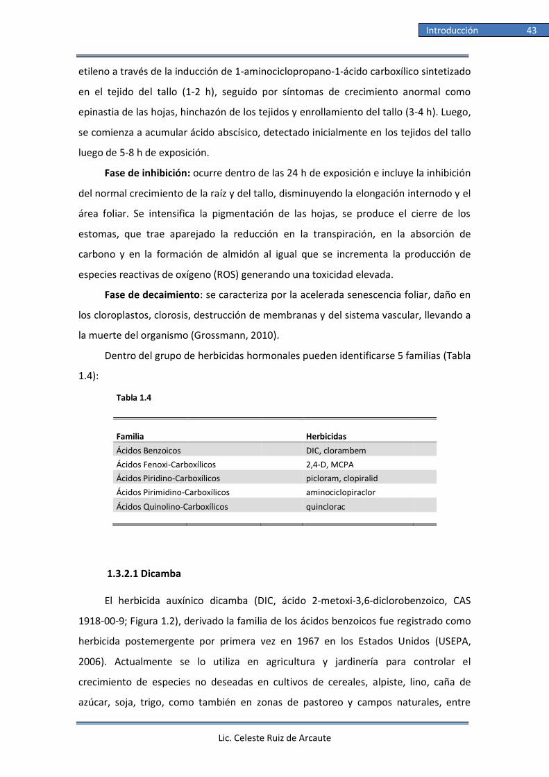
Lic. Celeste Ruiz de Arcaute
Introducción 43
etileno a través de la inducción de 1-aminociclopropano-1-ácido carboxílico sintetizado
en el tejido del tallo (1-2 h), seguido por síntomas de crecimiento anormal como
epinastia de las hojas, hinchazón de los tejidos y enrollamiento del tallo (3-4 h). Luego,
se comienza a acumular ácido abscísico, detectado inicialmente en los tejidos del tallo
luego de 5-8 h de exposición.
Fase de inhibición: ocurre dentro de las 24 h de exposición e incluye la inhibición
del normal crecimiento de la raíz y del tallo, disminuyendo la elongación internodo y el
área foliar. Se intensifica la pigmentación de las hojas, se produce el cierre de los
estomas, que trae aparejado la reducción en la transpiración, en la absorción de
carbono y en la formación de almidón al igual que se incrementa la producción de
especies reactivas de oxígeno (ROS) generando una toxicidad elevada.
Fase de decaimiento: se caracteriza por la acelerada senescencia foliar, daño en
los cloroplastos, clorosis, destrucción de membranas y del sistema vascular, llevando a
la muerte del organismo (Grossmann, 2010).
Dentro del grupo de herbicidas hormonales pueden identificarse 5 familias (Tabla
1.4):
Tabla 1.4
Familia Herbicidas
Ácidos Benzoicos DIC, clorambem
Ácidos Fenoxi-Carboxílicos 2,4-D, MCPA Ácidos Piridino-Carboxílicos picloram, clopiralid
Ácidos Pirimidino-Carboxílicos aminociclopiraclor
Ácidos Quinolino-Carboxílicos quinclorac
1.3.2.1 Dicamba
El herbicida auxínico dicamba (DIC, ácido 2-metoxi-3,6-diclorobenzoico, CAS
1918-00-9; Figura 1.2), derivado la familia de los ácidos benzoicos fue registrado como
herbicida postemergente por primera vez en 1967 en los Estados Unidos (USEPA,
2006). Actualmente se lo utiliza en agricultura y jardinería para controlar el
crecimiento de especies no deseadas en cultivos de cereales, alpiste, lino, caña de
azúcar, soja, trigo, como también en zonas de pastoreo y campos naturales, entre

Lic. Celeste Ruiz de Arcaute
Introducción 44
otros (USEPA, 2006; CASAFE, 2009). Es
comercializado mundialmente en varias
formulaciones, ya sea siendo el único componente
activo o en combinación con otros herbicidas,
como el 2,4-D, MCPP, MCPA (ácido 2-metil-4-
clorofenoxiacético), sulfonilureas y triazinas (FAO,
2001) y de esta forma controlar un espectro
mayor de malezas. En Argentina, según el último
reporte realizado por el Servicio Nacional de
Sanidad y Calidad Agroalimentaria (SENASA),
existen 83 formulaciones conteniendo DIC como
p.a. (SENASA, 2018) (Tabla 1.5), ubicándose en el noveno puesto entre los herbicidas
más utilizados a nivel mundial (USEPA, 2017) (Tabla 1.1).
Una vez aplicado, DIC es absorbido por la planta a través de las raíces y las hojas.
Ciertas características compartidas con otros herbicidas ácidos, como la alta
solubilidad en agua y la baja volatilidad, junto a su uso actualmente muy extendido,
facilitan su incorporación en el ambiente. Posee un bajo coeficiente de partición del
carbono orgánico (Koc 1,42) y, por lo tanto, una baja afinidad por las partículas al suelo
y sedimento en suspensión, características que hacen al mismo un compuesto de alta
movilidad (Comfort y col., 1992). La vida media de DIC en suelo es de 6 y 144 d para
condiciones aeróbicas y anaeróbicas, respectivamente (USEPA, 2006). Adicionalmente,
también posee un bajo coeficiente de partición octanol/agua (Kow 2.21; Hansch y col.,
1995), una presión de vapor de 4,5 mPa a 25ºC y se caracteriza por ser un compuesto
moderadamente volátil en aire (Waite y col., 2005; Bunch y col., 2012), dependiendo
de varios factores, entre los que se encuentran la cantidad de herbicida aplicado, la
temperatura y humedad ambiental, la formulación química del producto y la superficie
sobre la cual fue aplicado (Waite y col., 2005).
Figura 1.2: Estructura química del
herbicida dicamba (NCBI, 2017)

Intro
du
cción
45
Lic. Celeste R
uiz d
e Arcau
te
Tabla 1.5 Formulados comerciales en Argentina conteniendo al herbicida dicamba como principio activo
Empresa
Marca Comercial
Componente activo
C.T.a
Conc. (% p.a.)
AGM ARGENTINA S.A. DICAMBA 58 AGM DICAMBA II 58
AGM ARGENTINA S.A.
SUPERB 70
DICAMBA EQUIVALENTE ACIDO
III
77/70
AGRICULTORES FEDERADOS ARG. S.C.L. DICAMBA 57,7 AFA DICAMBA IV 57,7
AGRICULTORES FEDERADOS ARG. S.C.L.
DICAMBA AFA 57,7
DICAMBA EQUIVALENTE ACIDO
III
57,7/48
AGRO IYASI S.A. DICAMBA 57,7% IYASI DICAMBA EQUIVALENTE ACIDO III 57,7/47,9
AGRO MAX S.R.L.
DICAMBA SUMAGRO
DICAMBA
III
57,7
AGROEMPRESA COLON S.A. DICAMBA KEMSURE DICAMBA IV 57,7
AGROFACIL S.R.L.
DICAMBA CONTROLER
DICAMBA EQUIVALENTE ACIDO
III
57,8/48
AGROFINA S.A. TOLAN DICAMBA IV 57,8
AGROSERVICIOS PAMPEANOS S.A.
KUARTEL ASP
DICAMBA
IV
57.51
AGROSERVICIOS PAMPEANOS S.A. KUARTEL II DICAMBA EQUIVALENTE ACIDO IV 57,8/48
ALFREDO FERRERO S.R.L.
FERRERO DICAMBA
DICAMBA
IV
57,8
ARANAMI S.A. ARAKAMBA 58 DICAMBA II 58
ASOCIACION DE COOP. ARG. COOP.LTD.
DICAMAX ACA 58
DICAMBA
III
58
ATANOR S.C.A. DICAMBA ATANOR DICAMBA III 57,8
ATANOR S.C.A.
DICAMBA ATANOR A
DICAMBA EQUIVALENTE ACIDO
III
57,8/3,6
BASF ARGENTINA S.A. ATECTRA BV DICAMBA III 70,8
BONQUIM S.A.
DICAMBA MELTHIS
DICAMBA EQUIVALENTE ACIDO
III
57,8/48
BORCHES Y CIA S.A. COMPASS DICAMBA IV 57,8
BORCHES Y CIA S.A.
DICAMBA MAX
DICAMBA
II
57,8
BORCHES Y CIA S.A. NAVIGATOR DICAMBA II 57,8
CIAGRO S.A.
COWBOY
DICAMBA
III
57,8
CIBELES ARGENTINA S.A. DOMBELL 48 SL DICAMBA IV 57,8
CIBELES ARGENTINA S.A.
DICAMBA SUMAGRO
DICAMBA EQUIVALENTE ACIDO
IV
57,8/48
CIBELES ARGENTINA S.A. DICAMBA PHILAGRO DICAMBA IV 57,8
CIBELES ARGENTINA S.A.
STARGAN
DICAMBA
IV
57,8
COMPAÑÍA ARGENTINA DE GRANOS S.A. DICAMBA LYCSA DICAMBA IV 57,71
COMPAÑÍA ARGENTINA DE GRANOS S.A.
GREEN DICAMBA 57,8
DICAMBA EQUIVALENTE ACIDO
IV
57,8/48
CROPMAX S.A. CROPMAX DICAMBA DICAMBA EQUIVALENTE ACIDO IV 57,8/48
CHEMOTECNICA S.A.
GALO´PE
DICAMBA EQUIVALENTE ACIDO
III
57,8/48

Intro
du
cción
46
Lic. Celeste R
uiz d
e Arcau
te
ENRIQUE M. BAYA CASAL S.A. DICAMBA ARN EBC DICAMBA II 58
FALCROP S.A.
DICAMBA FALCROP
DICAMBA EQUIVALENTE ACIDO
III
57,8/48
FARMCHEM S.A. DICANFA 58 DICAMBA EQUIVALENTE ACIDO II 58/48,2
FORMULAGRO S.R.L.
DICAMBA 58 FORMULAGRO
DICAMBA EQUIVALENTE ACIDO
IV
58/48,2
GENBRA ARGENTINA S.A. DICAMBA 48,5 SL DICAMBA IV 57,8
GENBRA ARGENTINA S.A.
WINTER
DICAMBA
IV
57,71
GLEBA S.A. ZURCO DICAMBA III 57,71
GREEN CROPS S.A.
SUPERCAMBA
DICAMBA
III
57,71
GREEN CROPS S.A. GREEN DICAMBA DICAMBA EQUIVALENTE ACIDO III 57,71/48
GRUPO AGROS S.A.
HUNTER AGROS
DICAMBA EQUIVALENTE ACIDO
IV
57,71/48
HANDELSGESELLSCHAFT DETLEF VON APPEN mbH DICAMBA 58 DVA DICAMBA II 58
HELM ARGENTINA S.R.L.
DICAMBA 48 HELM
DICAMBA
III
57,8
HELM ARGENTINA S.R.L. BEMUS DICAMBA IV 57,7
INSUMOS AGROQUIMICOS S.A.
DICAMBA BLOCK
DICAMBA
II
57,8
JNG & ASOCIADOS S.A. DICAMBA JNG DICAMBA EQUIVALENTE ACIDO III 57,8/48
LANTHER QUIMICA S.A. DICAMBA LQ DICAMBA III 57,87
MH LATAM S.R.L.
AGROVERDE DICAMBA
DICAMBA EQUIVALENTE ACIDO
III
57,7/47,9
MONSANTO ARGENTINA S.R.L. XTENDICAM DICAMBA EQUIVALENTE ACIDO IV 51,5/34,9
NIDERA S.A.
DICAMBA ZAMBA
DICAMBA
III
57,8
NIDERA S.A. DICAMBA II ZAMBA DICAMBA EQUIVALENTE ACIDO III 57,8/48
NUFARM S.A.
KAMBA
DICAMBA
IV
57,71
NUFARM S.A. KAMBA DRY DICAMBA EQUIVALENTE ACIDO III 77/70
NUFARM S.A.
KAMBA FULL
DICAMBA EQUIVALENTE ACIDO
III
88,8/75
PHILAGRO S.A. PHILCAMBA DICAMBA IV 57,7
PROTEGRAN S.A.
PROCAMBA 57,8
DICAMBA EQUIVALENTE ACIDO
III
57,8/48
RAINBOW AGROSCIENSES S.A. DICAMBA 57,8 RAINBOW DICAMBA IV 57,8
RAINBOW AGROSCIENSES S.A.
KUARTEL 70 SG
DICAMBA EQUIVALENTE ACIDO
III
77/70
RAINBOW AGROSCIENSES S.A. TRANVEL DICAMBA III 77/70
RAINBOW AGROSCIENSES S.A.
DICAGOOD 48
DICAMBA EQUIVALENTE ACIDO
IV
57,8/48
RAINBOW AGROSCIENSES S.A. DICAGOOD SL DICAMBA EQUIVALENTE ACIDO IV 57,8/48
RED SURCOS S.A.
COWBOY FG
DICAMBA EQUIVALENTE ACIDO
IV
66,3
RED SURCOS S.A. COWBOY RED SURCOS DICAMBA EQUIVALENTE ACIDO III 57,8/48
RED SURCOS S.A.
COWBOY 57,8 SL
DICAMBA EQUIVALENTE ACIDO
IV
57,8/48
RED SURCOS S.A. COWBOY ELITE DICAMBA III 20

Intro
du
cción
47
Lic. Celeste R
uiz d
e Arcau
te
REOPEN S.A.
DICAMBA DAARGUS 57,7
DICAMBA EQUIVALENTE ACIDO
IV
57,7/47,9
SIGMA AGRO S.A. SIGMA DICAMBA DICAMBA EQUIVALENTE ACIDO III 57,6/47,8
SIGMA AGRO S.A.
DICAMBA SIGMA
DICAMBA EQUIVALENTE ACIDO
III
57,8/48
SINER S.A. KAMBA DICAMBA IV 57,8
SINOCHEM AGRO ARGENTINA S.A.
PYRU
DICAMBA EQUIVALENTE ACIDO
III
57,8/48
SINTESIS QUIMICA S.A.I.C. VENDAVAL DICAMBA DICAMBA IV 57,71
STOCKTON S.A.
DICAMBA 57,8 STOCKTON
DICAMBA EQUIVALENTE ACIDO
IV
57,8/48
SURCROPS S.R.L. DICAMBA SURCROPS DICAMBA EQUIVALENTE ACIDO III 57,8/48
SYNGENTA AGRO S.A.
BANVEL
DICAMBA
III
57,71
SYNGENTA AGRO S.A. CAIMAN DICAMBA III 87,5
SYNGENTA AGRO S.A.
CAIMAN PLUS
DICAMBA EQUIVALENTE ACIDO
III
57,8/48
SYNGENTA AGRO S.A. GLADER DICAMBA EQUIVALENTE ACIDO III 57,8/48
TECNOMYL S.A.
CONTROLE
DICAMBA
IV
57,8/48
TECNOMYL S.A. YPF DICAMBA HD DICAMBA EQUIVALENTE ACIDO IV 57,8/48
TECNOMYL S.A.
DICAM - BAC
DICAMBA EQUIVALENTE ACIDO
IV
57,8/48
TECNOMYL S.A. CONTROLE 77 SG DICAMBA EQUIVALENTE ACIDO IV 77/70
TERRIUM ARGENTINA S.A.
DICAMBA 58 TERRIUM
DICAMBA
II
58
UPL ARGENTINA S.A. RECAMBA DICAMBA IV 57,8
UPL ARGENTINA S.A.
DICAMBA ICONA
DICAMBA
IV
57,8
YPF S.A. H DICAMBA 58 DICAMBA II 58
a, CT: Clasificaciones toxicológicas según la WHO. Fuente: CASAFE, (CASAFE, 2017a)
Lic. Celeste Ruiz de Arcaute
Introducción 48
En Canadá, DIC fue detectado como contaminante en aguas superficiales de
ecosistemas rurales, urbanos y mixtos en concentraciones entre 0,89 ng/L a 50 μg/L
(Cessna y Elliot, 2004; Woudneh y col., 2007; Glozier y col., 2012), y en plantas de
tratamiento de aguas residuales en concentraciones de 185 ng/L (Westlund y Yargeau,
2017). En EE. UU. fue detectado en reservorios superficiales de agua para consumo
humano en concentraciones de 1,04-16 ng/L (Donald y col., 2007), en agua de
locaciones urbanas o residenciales en niveles máximos de 3,1 μg/L (Ensminger y col.,
2013) y en aguas superficiales en concentraciones máximas de 2,1 μg/L (Phillips y
Bode, 2004). Es de destacar que, hasta el presente, la única información sobre
concentraciones ambientales de DIC en Argentina es de 157 ng/L, reportada para agua
superficial en la laguna La Brava (Prov. Bs. As.) (Corcoran y col., 2017). No obstante, las
concentraciones recomendadas de aplicación de este herbicida para nuestro país están
comprendidas entre 100-1500 cm3/ha (CASAFE, 2017a).
La información sobre los efectos toxicológicos que ejerce DIC en plantas
acuáticas y en organismos terrestres se consideran suficientes para evaluar su efecto y
riesgos posibles ejercidos en humanos y en otras especies (USEPA, 2006).
Los ensayos de toxicidad aguda informaron valores de DL50 oral para abejas
entre 3,6-10 µg/abeja (Bunch y col., 2012). Para especies acuáticas, DIC fue
ligeramente tóxico para peces e invertebrados, presentando estos últimos valores
medios de CE50 de 34,6 mg/L (USEPA, 2013). Adicionalmente, los valores de CL5096h
reportados para peces fueron de 465 mg/L para el pez mosquito (Gambusia affinis)
(Johnson, 1978), 180 mg/L para la petota (Cyprinodon variegatus) (USEPA, 2013), 153
mg/L para la trucha arcoíris (Oncorhynchus mykiss) (USEPA, 2013) y 50 mg/L para la
trucha degollada (O. clarkii) (Woodward, 1982). En cuanto a su efecto agudo en aves,
las sales de DIC son consideradas prácticamente no tóxicas, aunque el DIC en su
variante ácida puede ser considerado moderadamente tóxico para estos organismos
(USEPA, 2006). En adición, para pequeños mamíferos DIC ha sido clasificado por la U.S.
EPA (2006) como prácticamente no tóxico luego de la exposición oral aguda. La DL50
oral reportada en ratas fue mayor a 2,74 mg/kg. Para plantas acuáticas vasculares, DIC
resultó prácticamente no tóxico, pero sí potencialmente peligroso para plantas no
vasculares afectando su crecimiento y desarrollo (USEPA, 2006). Según lo expuesto, la
U.S. EPA estableció, luego de una revisión realizada en el año 2006, que el herbicida

Lic. Celeste Ruiz de Arcaute
Introducción 49
DIC no ejerce toxicidad aguda en especies acuáticas amenazadas y/o en peligro de
extinción tales como peces e invertebrados de agua dulce (U.S. Dept. of
Agriculture/Forest Service, 2004; USEPA, 2006). Los efectos de DIC luego de una
exposición crónica fueron evaluados en un estudio realizado en ratas donde se
analizaron efectos reproductivos multigeneracionales. En ellos se establecieron valores
de NOEC y LOEC de 45 y 136 mg/kg/d, respectivamente. En el caso de la concentración
LOEC, como efecto adicional se registró una reducción significativa en el peso de las
crías.
Cuando se analizó la actividad mutagénica inducida por este herbicida auxínico
en sistemas bacterianos, DIC ha mostrado efectos mutagénicos en cepas de Salmonella
typhimurium (Simmon, 1979; Plewa y col., 1984; Kier y col., 1986). De manera similar,
se han registrado la inducción de daño en el ADN de Bacillus subtilis (Leifer y col.,
1981) y de Escherichia coli (Simmon, 1979; Leifer y col., 1981; Waters y col., 1981). En
levaduras de la especie Saccharomyces cervisiae se reportaron resultados negativos
para los ensayos de recombinación mitótica y conversión génica (Zimmermann y col.,
1984) luego de la exposición a DIC. Adicionalmente, los resultados indicaron en S.
cervisiae que DIC podría actuar como disruptor endócrino, teniendo efectos
antiestrogénicos y antiandrogénico, mostrando un comportamiento pleiotrópico,
como ha sido reportado para otros plaguicidas, incluído 2,4-D (Kojima y col., 2003;
Orton y col., 2009; Li y Gramatica, 2010). Adicionalmente, resultados positivos y
negativos fueron informados para la de síntesis de ADN no programada en células
humanas, en presencia o ausencia de un sistema de activación metabólica proveniente
de hígado de ratas (Simmon, 1979; Perocco y col., 1990).
En ensayos in vitro empleando la línea celular CHO se reportó la inducción de
rupturas de cadena simple y/o sitios sensibles al álcali en el ADN estimados mediante
la metodología del ensayo cometa (González y col., 2007), así como la inducción de
micronúcleos (MN) (González y col., 2011). Observaciones similares fueron reportadas
por Sorensen y col. (2004; 2005) en esta línea celular. Tanto González y col. (2006;
2007; 2009) como Perocco y col. (1990) demostraron la capacidad de DIC de inducir
intercambios de cromátidas hermanas en células CHO y en linfocitos humanos. La
inducción de alteraciones en la progresión del ciclo celular en diferentes tipos
celulares, tales como las células CHO y los linfocitos humanos, fueron reportados luego

Lic. Celeste Ruiz de Arcaute
Introducción 50
de la exposición in vitro de DIC (Sorensen y col., 2004; González y col., 2006; 2007;
2009). Con respecto a estudios in vivo, DIC fue capaz de inducir recombinación
homóloga y mutaciones puntuales en la crucífera Arabidopsis thaliana (Filkowski y col.,
2003). En cuanto a la inducción de lesiones en el ADN, se reportó que la exposición a
DIC no produjo mutaciones letales recesivas ligadas al sexo en Drosophila
melanogaster (Waters y col., 1981; Lee y col., 1983). Adicionalmente, se reportó un
aumento en la frecuencia de la tasa de desenrollamiento del ADN mediante
espectrofotometría en hepatocitos de ratas tratadas intraperitonealmente (Perocco y
col., 1990) y un aumento de la frecuencia de aberraciones cromosómicas en células
meristemáticas radiculares de la cebada Hordeum vulgare así como en células de la
medula ósea de ratas expuestas a DIC (Hrelia y col., 1994). Por otro lado, DIC no indujo
un aumento de la frecuencia de reordenamientos cromosómicos en el trigo Tritricum
turgidum (Satyavathi y col., 2004). Finalmente, en sistemas vegetales, DIC indujo un
aumento dosis-dependiente en la frecuencia de MNs de Tradescantia sp. clon 03
(Mohammed y Ma, 1999). Adicionalmente, en la papa, Solanum tuberosum, DIC indujo
disrupción de membranas celulares, aumento de actividad enzimática de la glutatión-
S-transferasa (GST) y una disminución del contenido intracelular de adenosín trifosfato
(ATP) (Peixoto y col., 2003; 2008b). En modelos animales como las ratas, Espandari y
col. (1995) informaron que DIC indujo la proliferación de peroxisomas, mientras que
Peixoto y col. (2003) reportaron la pérdida de la actividad fosforilativa en las
mitocondrias. Adicionalmente se informó la inducción de actividad promotora de un
modelo de hepatocarcinogénesis en ratas tratadas con DIC (Espandiari y col., 1999).
En cuanto a su potencial carcinogénico, la U.S. EPA propone a DIC como un
compuesto perteneciente a la clase D (no carcinogénico para el ser humano). Sin
embargo, en personas laboralmente expuestas a DIC se observó una asociación
positiva con el desarrollo de mielomas múltiples (Burmeister, 1990), linfoma no-
Hodgkin (McDuffie y col., 2001) y cáncer de pulmón y de colon (Alavanja y col., 2004;
Samanic y col., 2006).
La mayoría de los estudios que informaron resultados genotóxicos positivos en
personas ocupacionalmente expuestas a DIC involucraron en su mayoría mezclas de
formulaciones de diferentes agroquímicos, que imposibilita la evaluación de la

Lic. Celeste Ruiz de Arcaute
Introducción 51
genotoxicidad inducida exclusivamente por el herbicida DIC (Barry y col., 2012;
Schinasi y Leon, 2014).
Teniendo en cuenta los efectos tóxicos agudos inducidos, la Organización
Mundial de la Salud (WHO, 2009) clasificó a DIC como un compuesto perteneciente a
la Clase II, compuesto ligeramente peligroso para la salud humana, mientras que la
U.S. EPA (2006) lo clasificó como un compuesto tipo III, elemento moderadamente
tóxico. En adición, siguiendo la clasificación ecotoxicológica propuesta por la U.S. EPA
(2008), DIC es considerado prácticamente como un compuesto no tóxico para
organismos acuáticos.
Formulado comercial Banvel®: Este compuesto es comercializado en Argentina
por la empresa Syngenta Agro S.A. como concentrado soluble del herbicida DIC (57,7%
p/v sal dimetilamina del ácido 2-metoxi-3,6-diclorobenzoico), empleada para controlar
malezas de hoja ancha sobre una amplia variedad de cultivos entre los que se pueden
nombrar la soja, trigo, maíz, cebada, caña de azúcar, sorgo y mijo, entre otros. Se
aplica sólo una vez por ciclo de cultivo, en concentraciones que van de 100 a 120 L
H2O/ha para aplicaciones terrestres, o de 20 a 40 L en aplicaciones aéreas. En estas
últimas, se recomienda suspender las aplicaciones con vientos superiores a 10 Km/h.
Adicionalmente, aconsejan aplicar de 20-30 gotas/cm2 sobre las hojas de las plantas no
deseadas hasta 30 d antes de la cosecha. Puede utilizarse sólo o en combinación con
otros herbicidas, como por ejemplo el 2,4-D, MCPA y bromoxinil, ya que es compatible
con la mayoría de los plaguicidas orgánicos y soluciones acuosas de fertilizantes
nitrogenados (CASAFE, 2017b). Luego de la aplicación, es absorbido por hojas y raíces
traslocándose a toda la planta. La hoja de seguridad del producto recomienda no
utilizar el caldo remanente luego de la aplicación y seguir las instrucciones de triple
lavado según la Norma IRAM 12.069 que indica lavar el envase con agua tres veces
antes de ser descartado o, en caso contrario, inutilizarlo. En la Tabla 1.6 se detallan los
datos toxicológicos del herbicida informados por la empresa Syngenta Agro S.A.

Lic. Celeste Ruiz de Arcaute
Introducción 52
Tabla 1.6 Información toxicológica del formulado comercial Banvel® (57,7% dicamba)
Parámetro Organismo Valor
Toxicidad oral aguda Rata DL50: 2467 mg(Kg
Toxicidad dermal aguda Conejo DL50: >4000 mg/Kg
Toxicidad inhalatoria aguda Rata (4h) DL50: >5,4 mg/L
Irritación dermal aguda Conejo No irritante
Irritación ocular aguda Conejo Irritante
Sensibilización dermal Cobayo No sensibilizante
Toxicidad aguda para peces Oncorhynchus mykiss (96h) DL50: >1000 mg/L
Inhibición de crecimiento en algas Selensatrum caprocornutum (96 h) CL50: >250 mg/L
Toxicidad en invertebrados acuáticos Daphnia magna (48h) >1600 mg/L
Toxicidad a bacterias Sedimentos cloacales (3 h) CL50: >500 mg/L
Fuente: Hoja de Seguridad de Producto (Syngenta Agro S.A.)
Por lo expuesto, son necesarias investigaciones complementarias para poder
determinar un perfil toxicológico claro del herbicida DIC, más aun teniendo en cuenta
su creciente utilización en el agro y con esto, su continua incorporación en los
ambientes tanto acuáticos como terrestres circundantes.
1.3.2.2 Ácido 2,4-diclorofenoxiacético (2,4-D)
Dentro de la familia de los ácidos fenoxi-
carboxílicos se encuentra el 2,4-D (CAS 2008-39-1), un
herbicida sistémico pre- y post-emergente
ampliamente utilizado mundialmente (Liu y col.,
2013). Fue el primer herbicida sintético desarrollado y
comercializado y ha sido utilizado por más de 70 años
para el control de especies no deseadas en diversos
cultivos, silvicultura, limpieza de caminos y líneas de
ferrocarriles, entre otros (CASAFE, 2017a).
Es comercializado en formulaciones sólo o en
combinación con otros p.a., tales como DIC, MCPP,
MCPA, mecoprop-P (enantiómero de mecoprop), Figura 1.3: Estructura química del
herbicida ácido 2,4-diclorofenoxi-
acético (2,4-D)(NCBI, 2017)

Lic. Celeste Ruiz de Arcaute
Introducción 53
picloram (ácido-4-amino-3,5,6-tricloropiridina-2-carboxílico) y clopiralid (ácido 3,6-
dicloro piridina-2-carboxílico) (NCBI, 2017). Adicionalmente, el 2,4-D es extensamente
usado en combinación con glifosato en cultivos modificados genéticamente y
resistentes, por consiguiente, a ambos herbicidas (Wright y col., 2010).
A nivel mundial, existen más de 600 formulaciones contiendo 2,4-D como p.a.
que actualmente están siendo comercializadas (Wafa y col., 2011; Song, 2014). En
adición de la forma ácida del compuesto, el 2,4-D forma parte de las formulaciones
mayormente en la forma de sal amina en solución acuosa o en su forma éster como
concentrado emulsionable. En Argentina y de acuerdo con el SENASA, en abril de 2018
se reportó la comercialización de más de 178 formulaciones conteniendo 2,4-D como
p.a. (SENASA, 2018) (Tabla 1.7), ubicándose en el cuarto puesto entre los herbicidas
más utilizados a nivel mundial (USEPA, 2017) (Tabla 1.1).
El 2,4-D es altamente soluble en agua, con un Kow 2,1 (Howard, 1991). En el
ambiente acuático, la vida media del 2,4-D varía entre 7-21 d bajo condiciones
aeróbicas. No obstante, bajo condiciones anaeróbicas puede exceder los 120 d (USEPA,
2005; Wafa y col., 2012). En suelo es poco persistente, presentando una vida media
entre 7-13 d y tiene una residualidad de 1-4 semanas (Wauchope y col., 1992; USEPA,
2005). Debido a procesos de deriva y volatilización del compuesto puede ser detectado
en el aire y en el ambiente acuático. Los niveles guía para el agua de consumo
establecidos por la WHO (2011) proponen un valor máximo de 0,03 mg 2,4-D/L. Sin
embargo, la presencia de 2,4-D en el ambiente fue reportado en México en
concentraciones máximas de 0,04 mg/L en aguas superficiales (Félix-Cañedo y col.,
2013). En Canadá, fueron reportadas concentraciones medias de 700 ng/L en zonas
urbanas (Tierney y col., 2011; Glozier y col., 2012) mientras que concentraciones entre
1,77-30,9 ng/L en sitios con actividad agrícola (Woudneh y col., 2007) y hasta una
concentración máxima de 751 ng/L en plantas de tratamiento de aguas residuales
(Westlund y Yargeau, 2017). En aguas subterráneas de Europa fueron reportadas
concentraciones de 12 ng/L (Loos y col., 2010). En reservorios superficiales de agua
para consumo humano de EE.UU. fueron informadas concentraciones promedio de
0,17 y 75 ng/L por Tagert y col. (2014) y Donald y col. (2007), respectivamente.
Particularmente en Argentina, en un estudio realizado en 2017, se registró una
concentración máxima de 2,4-D equivalente a 0,99 µg/L en aguas superficiales en el

Lic. Celeste Ruiz de Arcaute
Introducción 54
arroyo El Crespo ubicado en el Partido de Balcarce (Provincia de Buenos Aires) (Pérez y
col., 2017). Adicionalmente, en la laguna La Brava (Prov. Bs. As.) se reportaron
concentraciones de 157 ng/L para agua superficial (Corcoran y col., 2017). De acuerdo
con CASAFE, las concentraciones recomendadas de aplicación a campo de 2,4-D varían
entre 1,25-2,25 L/ha (CASAFE, 2017a).
La información toxicológica sobre 2,4-D en base a ensayos de toxicidad aguda
reportó una DL50 oral de 639-1646 mg/Kg para ratas y de > 2000 mg/Kg para conejos,
dependiendo del tratamiento con la variante amina, éster o ácida del 2,4-D (USEPA,
2005). Para ratones, la toxicidad reportada en DL50 fue 138 mg/Kg (Tomlin, 2006).
Para abejas, el 2,4-D fue considerado prácticamente no tóxico presentando una
CL5024h de 115 μg/abeja (USEPA, 2005). La toxicidad para peces e invertebrados
acuáticos es variable dependiendo básicamente de la variante química de 2,4-D
utilizada. Una mayor toxicidad se observó luego del tratamiento con la variante éster
del 2,4-D debido a la mayor tasa de absorción del mismo a través de las branquias
donde es hidrolizada a su forma ácida (WHO, 1989).
En estudios in vitro, cuando se emplearon sistemas bacterianos, tales como el
test de Ames en distintas cepas de S. typhimurium y el test de mutación reversa en B.
subtilis mostraron resultados negativos luego del tratamiento con 2,4-D (Grabińska-
Sota y col., 2002). En levaduras de la especie S. cerevisiae, el 2,4-D indujo la formación
de aductos en el ADN (Teixeira y col., 2004) y mostró tener actividad antiestrogénica y
antiandrogénica mediante los ensayos de detección de estrógenos y andrógenos en
levaduras (Westlund y Yargeau, 2017). Cuando se evaluó el potencial carcinogénico de
2,4-D se obtuvieron resultados positivos mediante el ensayo de transformación en la
línea celular SHE (embriones de hámster Sirio) (Maire y col., 2007). Adicionalmente, se
evaluó la inducción lesiones primarias del ADN estimadas por el ensayo cometa en su
variante alcalina (González y col., 2005; Maire y col., 2007; Sandal y Yilmaz, 2011;
Bokán y col., 2013). En este ensayo se observó un aumento de lesiones primarias en el
ADN en células CHO y SHE como así también en linfocitos humanos y en líneas
celulares de peces. Por otro lado, resultados negativos fueron observados con estos
mismos ensayos por otros investigadores (Sorensen y col., 2005; Sandal y Yilmaz,
2011). Maire y col. (2007) informaron que el 2,4-D no fue capaz de inducir
fragmentación del ADN en células SHE. Tanto González y col. (2005), Soloneski y col.
Lic. Celeste Ruiz de Arcaute
Introducción 55
(2007) y Korte y col. (1982) demostraron la capacidad del 2,4-D de inducir
intercambios de cromátidas hermanas en células CHO al igual que en linfocitos
humanos tratados in vitro.

Intro
du
cción
56
Lic. Celeste R
uiz d
e Arcau
te
Tabla 1.7 Formulados comerciales en Argentina conteniendo al herbicida ácido 2,4-diclorofenoxiacético (2,4-D) como principio activo.
Empresa
Marca Comercial
Componente activo
C.T.a
Conc. (% p.a.)
ATANOR S.C.A.
2,4D AMINA
2,4-D SAL AMINA
II
57,8
ATANOR S.C.A. HERBIFEN AMINA 70 2,4-D SAL AMINA II 84,3
ATANOR S.C.A.
HERBIFEN AMINA TD
2,4-D SAL AMINA
II
58,4
ATANOR S.C.A. HERBIFEN SUPER 2,4-D II 97
SINTESIS QUIMICA S.A.I.C.
VENDAVAL 2,4D AMINA
2,4-D
III
58,4
DOW AGROSCIENCES ARGENTINA S.R.L. ESTERON ULTRA 2,4-D II 100
DISTRI CAMPO S.R.L.
SELECTIVO DC
2,4-D
III
60,2
DOW AGROSCIENCES ARGENTINA S.R.L. DMA 2,4-D III 58,4
NIDERA S.A.
2,4 D ZAMBA
2,4-D
II
100
TERRIUM ARGENTINA S.A. 2,4D TERRIUM 2,4-D II 100
NIDERA S.A.
2,4D AMINA ZAMBA
2,4-D
III
58,4
NUFARM S.A. NAVAJO 2,4-D III 96,3
AGM ARGENTINA S.A.
2,4D AGM
2,4-D
II
100
ATANOR S.C.A. NV NO VOLATIL ATANOR 2,4-D MEZCLA SALES AMINAS DEL
ACIDO II 60 HANDELSGESELLSCHAFT DETLEF VON APPEN mbH SUC. ARG. 2,4D AMINA DVA
2,4-D
II
60
CHEMOTECNICA S.A. ACADEMIX 2,4-D II 100
AGM ARGENTINA S.A.
2,4D AMINA AGM
2,4-D
II
60
CHEMOTECNICA S.A. ASI MAX 50 2,4-D EQUIVALENTE ACIDO II 60
CHEMOTECNICA S.A.
ASI MAX 72
2,4-D EQUIVALENTE ACIDO
II
72
BIESTERFELD ARGENTINA S.A. 24D AMINA BIESTERFELD 2,4-D II 60
BIESTERFELD ARGENTINA S.A.
24D ESTER BIESTERFELD
2,4-D
II
100
NUFARM S.A. WEEDONE 2,4-D ESTER BUTILICO II 97
NIDERA S.A.
2,4D PLUS ZAMBA
2,4-D
II
80,4
TERRIUM ARGENTINA S.A. 2,4D AMINA TERRIUM 2,4-D II 60
DOW AGROSCIENCES ARGENTINA S.R.L.
ESTERON ULTRA S
2,4-D
II
97
ATANOR S.C.A. GRANERO 2,4-D ESTER 2,4-D ESTER BUTILICO II 97
UPL ARGENTINA S.A.
2,4D AMINA ICONA 70 SL
2,4-D
II
84,3
UPL ARGENTINA S.A. 2,4D AMINA ICONA SL 2,4-D II 58,4
UPL ARGENTINA S.A.
2,4D ICONA CE
2,4-D EQUIVALENTE ACIDO
II
97/77,4

Intro
du
cción
57
Lic. Celeste R
uiz d
e Arcau
te
ARANAMI S.A. ARAMINA 2,4D 60 2,4-D II 60
GRUPO AGROS S.A.
BASIC AGROS
2,4-D
II
60
AGM ARGENTINA S.A. SALIX 80 2,4-D EQUIVALENTE ACIDO III 95/80
RED SURCOS S.A.
DEDALO
2,4-D EQUIVALENTE ACIDO
III
60/50
STOCKTON S.A. CAMPEON 100 STK 2,4-D II 100
3 LOMAS S.A.
2,4D AMINA MAXX
2,4-D
III
60
TERRIUM ARGENTINA S.A. SALIX TERRIUM 2,4-D III 95
TECNOMYL S.A.
FENOMYL
2,4-D
II
60
STOCKTON S.A. FENIX GOLD 2,4-D II 72
STOCKTON S.A.
FENIX
2,4-D
II
60,2
DOW AGROSCIENCES ARGENTINA S.R.L. DMA 48 2,4-D II 48
PHILAGRO S.A.
PHILAMINA
2,4-D EQUIVALENTE ACIDO
III
58,4/48,5
BORCHES Y CIA S.A. 2,4D AMNA MAX 2,4-D III 60
CHEMINOVA AGRO DE ARGENTINA S.A.
KING LIX
2,4-D
III
95
GRUPO AGROS S.A. BASIC AGROS LOSS CONTROLED 2,4-D II 84,3
CHEMOTECNICA S.A.
ASIMAX 80
2,4-D
III
95
CAMPO CROP S.A. 2,4 D CAMPO 2,4-D II 60
AGRO MAX S.R.L.
2,4D AMINA SUMAGRO
2,4-D
III
60,2
CHEMINOVA AGRO DE ARGENTINA S.A. KING 50 2,4-D II 60,2
YPF S.A.
YPF 2,4-D AMINA HA
2,4-D
II
80,4
ADAMA ARGENTINA S.A. ADAMA ESSENTIALS 2,4 D AMINA 2,4-D II 60,2
ADAMA ARGENTINA S.A.
ADAMA ESSENTIALS 2,4 D ESTER
2,4-D ESTER BUTILICO DEL ACIDO
II
100
SHARDA WORLDWIDE EXPORTS PVT LTD. WEEDISHIDE 2,4-D II 86,77
RAINBOW AGROSCIENSES S.A.
CLEANSPRAY SL
2,4-D SAL DIMETILAMINA
II
60
RAINBOW AGROSCIENSES S.A. 2,4-D 100 RAINBOW 2,4-D BUTIL ESTER III 100
GLEBA S.A.
DEGLEX
2,4-D
II
60
RAINBOW AGROSCIENSES S.A. CLEANSPRAY SS 2,4-D III 95
HELM ARGENTINA S.R.L.
2,4D AMINA 60 HELM
2,4-D
II
60
RED SURCOS S.A. DEDALO 60% SL 2,4-D II 60
RED SURCOS S.A.
DEDALO SUPER
2,4-D
III
100
ARANAMI S.A. ARAMINA 2,4D PLUS 2,4-D III 58,4
GREEN CROPS S.A.
2,4-D 100 GREEN CROPS
2,4-D BUTIL ESTER
III
100
AGROSERVICIOS PAMPEANOS S.A. ASP AMINA 60 2,4-D II 60
INSUMOS AGROQUIMICOS S.A.
MATRIX
2,4-D
III
95
AGROEMPRESA COLON S.A. 2,4-D AMINA KEMSURE 2,4-D II 58,4

Intro
du
cción
58
Lic. Celeste R
uiz d
e Arcau
te
AGROSERVICIOS PAMPEANOS S.A.
ASP ESTER 100
2,4-D
III
100
GREEN CROPS S.A. SOLUTION 2,4-D III 95
CROPMAX S.A.
CROPMAX 2,4D AMINA 50
2,4-D
II
60,2 ASOCIACION DE COOPERATIVAS ARGENTINAS COOP.LTD. 2,4 D AMINA ACA 2,4-D III 58,4
REOPEN S.A.
DAARGUS 2,4D AMINA
2,4-D
III
60,27
RED SURCOS S.A. DEDALO RED SURCOS 2,4-D III 60
WECAN AGRO S.A.
CONTROL 2,4-D
2,4-D
II
60
JNG & ASOCIADOS S.A. AMINA JNG 2,4-D III 58,4
CHEMOTECNICA S.A.
ASIMAX 60 TOP
2,4-D
II
72,2
GENBRA ARGENTINA S.A. MACHETE D 72 2,4-D II 72
BONQUIM S.A.
2,4D 60 MELTHIS
2,4-D
III
60
SINER S.A. D-70 2,4-D II 84,3
SINER S.A.
D-AMINA
2,4-D EQUIVALENTE ACIDO
II
58,4
RAINBOW AGROSCIENSES S.A. CLEANSPRAY EHE 2,4-D III 102,5
TERRIUM ARGENTINA S.A.
NOVOLESTER 68
2,4-D EQUIVALENTE ACIDO
II
68/45,17
RAINBOW AGROSCIENSES S.A. CLEANSPRAY 2,4-D EQUIVALENTE ACIDO II 96,3/80
FARMCHEM S.A.
ALLGRAM 2,4DA
2,4-D EQUIVALENTE ACIDO
II
86,7/72
FALCROP S.A. ARADOR ESTER 100 2,4-D III 100
NIDERA S.A.
2,4D AMINA ZAMBA
2,4-D
II
60
SEMBRADO S.A. D-SEM AMINA 60 2,4-D EQUIVALENTE ACIDO III 60/50
AGM ARGENTINA S.A.
NOVOLESTER AGM
2,4-D
III
102,5
COMPAÑÍA ARGENTINA DE GRANOS S.A. COMPAÑÍA 2,4-D AMINA 2,4-D SAL DIMETILAMINA II 60
AGROSERVICIOS PAMPEANOS S.A.
ASP ESTER LV
2,4-D EQUIVALENTE ACIDO
III
102,5/68
TRAPALCO S.A. MORTAL 2,4-D EQUIVALENTE ACIDO III 100/79,8
FORMULAGRO S.R.L.
2,4D AMINA FORMULAGRO
2,4-D EQUIVALENTE ACIDO
II
60/49,8
AGM ARGENTINA S.A. SMART 2,4-D EQUIVALENTE ACIDO II 96/80
NUFARM S.A.
WEEDAR DRY
2,4-D EQUIVALENTE ACIDO
II
96,3/80
COMPAÑÍA ARGENTINA DE GRANOS S.A. GREEN 2,4D SG 2,4-D EQUIVALENTE ACIDO III 96,3/80
RAINBOW AGROSCIENSES S.A.
ESTERCIDE
2,4-D EQUIVALENTE ACIDO
III
100/79,8
TERRIUM ARGENTINA S.A. NOVOLESTER 97 2,4-D EQUIVALENTE ACIDO II 97/64,3
AGROEMPRESA COLON S.A.
2,4D ESTER KEMSURE
2,4-D EQUIVALENTE ACIDO
III
100/79,8
NUFARM S.A. 2,4D 96,3 NUFARM 2,4-D III 96,3
PHILAGRO S.A.
PHILAMINA 50
2,4-D EQUIVALENTE ACIDO
III
60,2/50
CAMPO CROP S.A. 2,4 D AMINA 85 2,4-D EQUIVALENTE ACIDO II 85/70,6

Intro
du
cción
59
Lic. Celeste R
uiz d
e Arcau
te
ADAMA ARGENTINA S.A.
ADAMA ESSENTIALS 2,4 D AMINA
2,4-D EQUIVALENTE ACIDO
II
60,2/50
NUFARM S.A. WEEDAR 2,4-D III 60,2
RAINBOW AGROSCIENSES S.A.
ESTERCIDE LV
2,4-D
III
102,5
BORCHES Y CIA S.A. 2,4D AMINA MAX 60 2,4-D II 60
HUAGRO S.A.
GREEN CHOICE 2,4D AMINA
2,4-D
II
60
CAMPO CROP S.A. 2,4D CAMPO ESTER 2,4-D EQUIVALENTE ACIDO II 97/77,4
RAINBOW AGROSCIENSES S.A.
DRACO
2,4-D
III
95
AGRO IYASI S.A. 2,4D AMINA 60% IYASI 2,4-D EQUIVALENTE ACIDO III 60/50
BONQUIM S.A.
2,4D ESTER MELTHIS
2,4-D EQUIVALENTE ACIDO
II
97/77,4
RED SURCOS S.A. DEDALO ELITE 2,4-D II 30
ARYSTA LIFESCIENCE ARGENTINA S.A.
2,4D AMINA LINE
2,4-D EQUIVALENTE ACIDO
II
60,2/50
ENRIQUE M. BAYA CASAL S.A. ACTION 2,4D 2,4-D EQUIVALENTE ACIDO III 60/50
BORCHES Y CIA S.A.
2,4D AMINA MAX 50
2,4-D
II
60
NIDERA S.A. 2,4D ZAMBA 2,4-D II 97
AGRICULTORES FEDERADOS ARGENTINOS S.C.L.
2,4D AMINA 60 AFA
2,4-D
II
60
AGRO MAX S.R.L. 2,4D AMINA SUMAGRO SL 2,4-D EQUIVALENTE ACIDO II 58,4/48,5
RURALCO SOLUCIONES S.A.
AMINA 50 RURALCO
2,4-D EQUIVALENTE ACIDO
II
60,2/50
RAINBOW AGROSCIENSES S.A. CLEANSPRAY SP 2,4-D EQUIVALENTE ACIDO II 96,3/80
STOCKTON S.A.
FENIX STK
2,4-D EQUIVALENTE ACIDO
III
60,2/50
ADAMA ARGENTINA S.A. ADAMA ESSENTIALS PARADIGM 2,4-D EQUIVALENTE ACIDO III 100/79,8
GREEN CROPS S.A.
2,4-D 60 GREEN CROPS
2,4-D
III
60
TERRIUM ARGEMTINA S.A. XILAS 2,4-D II 96,3
NUFARM S.A.
WEEDAR FULL
2,4-D
II
83,5
CHEMPRO S.A. 2,4D CHEMPRO 2,4-D EQUIVALENTE ACIDO II 95/80
MONSANTO ARGENTINA S.R.L.
2,4D AMINA NV EQUIPAGRO
2,4-D EQUIVALENTE ACIDO
II
80,4/60
FALCROP S.A. ARADOR 60 2,4-D II 60
RAINBOW AGROSCIENSES S.A.
TOPSPRAY
2,4-D EQUIVALENTE ACIDO
III
95/80
SINTESIS QUIMICA S.A.I.C. VENDAVAL 2,4D ESTER ISOBUTILICO 2,4-D II 97
RAINBOW AGROSCIENSES S.A.
CAESTER
2,4-D EQUIVALENTE ACIDO
III
100/79,8
GRUPO AGROS S.A. BASIC AGROS LOSS 2,4-D EQUIVALENTE ACIDO II 84,3/70
RAINBOW AGROSCIENSES S.A.
SWIFT SPRAY
2,4-D EQUIVALENTE ACIDO
II
96,3/80 LABORATORIO SAN PABLO PRODUCTOS BIOLOGICOS S.R.L. AMINA SP 2,4-D EQUIVALENTE ACIDO II 60,2/50
FARMCHEM S.A.
ALLGRAM 2,4 D SDA 60
2,4-D EQUIVALENTE ACIDO
II
60
CHEMOTECNICA S.A. ASI MAX 80 WP 2,4-D EQUIVALENTE ACIDO II 95/80,4

Intro
du
cción
60
Lic. Celeste R
uiz d
e Arcau
te
CHEMPRO S.A.
ENTRIX
2,4-D EQUIVALENTE ACIDO
II
95/80,4
AGROSERVICIOS PAMPEANOS S.A. ASP ESTER XTRA 2,4-D II 97
LANTHER QUIMICA S.A.
2,4-D AMINA 60 LQ
2,4-D EQUIVALENTE ACIDO
II
60/50
AGROSERVICIOS PAMPEANOS S.A. ASP AMINA XTRA 2,4-D EQUIVALENTE ACIDO II 80,4/60
MONSANTO ARGENTINA S.R.L.
LA TIJERETA 2,4 D
2,4-D EQUIVALENTE ACIDO
II
80,4/60
TECNOMYL S.A. MORTAL 2,4-D EQUIVALENTE ACIDO II 72/60
PROTEGRAN S.A.
PRO D 97 ESTER
2,4-D EQUIVALENTE ACIDO
II
97/77,4
GREEN CROPS S.A. SOLUTION FULL 2,4-D EQUIVALENTE ACIDO II 86,7/72
RAINBOW AGROSCIENSES S.A.
MODELO LV
2,4-D EQUIVALENTE ACIDO
III
102,5/68
REOPEN S.A. DAARGUS 2,4 D AMINA FULL 2,4-D II 84,3
AGROFINA S.A.
2,4 D ESTER AGROFINA
2,4-D EQUIVALENTE ACIDO
II
97/77,4
SIGMA AGRO S.A. 2,4 D 60 SIGMA 2,4-D EQUIVALENTE ACIDO II 60/50
ARANAMI S.A.
ARA 2,4 D
2,4-D EQUIVALENTE ACIDO
III
60,2/50
CHEMINOVA AGRO DE ARGENTINA S.A. KING SL 2,4-D II 60,2
FACYT I+D S.A.
FACYT 2,4 D
2,4-D
II
30
DOW AGROSCIENCES ARGENTINA S.R.L. ENLIST 2,4-D EQUIVALENTE ACIDO II 66,9/45,6
ATANOR S.C.A.
GRANERO 2,4-D AMINA NV
2,4-D EQUIVALENTE ACIDO
II
80,4/60
ATANOR S.C.A. NV ATANOR B 2,4-D EQUIVALENTE ACIDO II 80,4/60
ATANOR S.C.A.
NV ATANOR C
2,4-D EQUIVALENTE ACIDO
II
80,4/60
RAINBOW AGROSCIENSES S.A. TOWERSPRAY LV 2,4-D EQUIVALENTE ACIDO III 102,5/68
RAINBOW AGROSCIENSES S.A.
QUECLENA LV
2,4-D EQUIVALENTE ACIDO
III
102,5/68
SINTESIS QUIMICA S.A.I.C. VENDAVAL 2,4D ESTER 2,4-D EQUIVALENTE ACIDO II 100/79,8
ATANOR S.C.A.
HERBIFEN AD
2,4-D EQUIVALENTE ACIDO
II
97/64,3
RAINBOW AGROSCIENSES S.A. ASISTENTE LV 2,4-D EQUIVALENTE ACIDO III 102,5/68
NUFARM S.A.
2,4-D 70 NUFARM
2,4-D EQUIVALENTE ACIDO
II
83,5/70
ATANOR S.C.A. VENCEWEED EXTRA B 2,4-D EQUIVALENTE ACIDO II 93,1/76
ATANOR S.C.A.
VENCEWEED EXTRA C
2,4-D EQUIVALENTE ACIDO
II
93,1/76
LOS GROBO AGROPECUARIA S.A. 2,4-D ESTER LGA 2,4-D EQUIVALENTE ACIDO II 97/77,4
AAGROCO S.A.
WEEDAMINA 72 SL
2,4-D
II
72
RAINBOW AGROSCIENSES S.A. EXCEMINE 2,4-D EQUIVALENTE ACIDO II 60/50
RAINBOW AGROSCIENSES S.A.
COMPRE LV
2,4-D EQUIVALENTE ACIDO
III
102,5/68
RAINBOW AGROSCIENSES S.A. AUXILIAR LV 2,4-D EQUIVALENTE ACIDO III 102,5/68
PROTEGRAN S.A.
ALLISTER 2,4D
2,4-D EQUIVALENTE ACIDO
II
97/77,4
PHILAGRO S.A. D-ESTER 2,4-D EQUIVALENTE ACIDO II 97/77,4
ATANOR S.C.A.
HERBIFEN EH A
2,4-D EQUIVALENTE ACIDO
II
97/64,3

Intro
du
cción
61
Lic. Celeste R
uiz d
e Arcau
te
GREEN CROPS S.A. AMINA 86,7 FULL GC 2,4-D EQUIVALENTE ACIDO II 86,7/72
AGROFINA S.A.
2,4 D ESTER 97
2,4-D
II
97
TRAPALCO S.A. MORTAL BV 2,4-D II 89 HANDELSGESELLSCHAFT DETLEF VON APPEN mbH SUC. ARG. POTRERO ULTRA
2,4-D
II
96
ATANOR S.C.A. HERBIFEN AMINA A 2,4-D EQUIVALENTE ACIDO II 84,3/70
a, CT: Categoría toxicológica según la WHO. Fuente: CASAFE, 2017

Lic. Celeste Ruiz de Arcaute
Introducción 62
Por otro lado, también se reportó un aumento en la frecuencia de aberraciones
cromosómicas en células V79 y en linfocitos humanos tratados in vitro en
presencia/ausencia de sistema de activación metabólica microsomal S9 (Pilinskaia,
1974; Korte y Jalal, 1982; Pavlica y col., 1991; Zeljezic y Garaj-Vrhovac, 2004). Zeljezic y
Garaj-Vrhovac (2004) reportaron la inducción de MN en linfocitos humanos in vitro.
También fueron reportadas alteraciones en la progresión del ciclo celular de diferentes
sistemas celulares incluyendo plantas, células V79, linfocitos humanos y células
bovinas (Pavlica y col., 1991; Soloneski y col., 2007; Soloneski y Larramendy, 2017).
Puntos finales similares fueron estudiados en sistemas in vivo. Se reportó la
inducción de mutaciones puntuales en plantas (Kumari y Vaidyanath, 1989) y en
insectos (Graf y Würgler, 1996), pero no en ratones (Knapp y col., 2003). Ateeq y col.
(2005) informaron el potencial genotóxico de 2,4-D mediante aumento de daño
primario en la cadena de ADN en eritrocitos de peces. En linfocitos periféricos de
trabajadores ocupacionalmente expuestos fueron reportados aumento de rupturas de
cadena simple y aberraciones cromosómicas (Yoder y col., 1973; Garaj-Vrhovac y
Zeljezic, 2001; IARC, 2015). Adicionalmente, 2,4-D aumentó la frecuencia de
intercambios de cromátidas hermanas en embriones de pollo y en células de
mamíferos in vitro, incluyendo linfocitos humanos de trabajadores ocupacionalmente
expuestos (Amer y Ali, 1974; Adhikari y Grover, 1988; Schop y col., 1990; Pavlica y col.,
1991; Venkov y col., 2000; Garaj-Vrhovac y Zeljezic, 2001). Cuando se analizó la
inducción de MNs, se encontraron resultados positivos en peces (Ateeq y col., 2002;
Farah y col., 2003; 2006) al igual que en linfocitos humanos tratados con 2,4-D (Garaj-
Vrhovac y Zeljezic, 2001), pero los resultados fueron negativos en células de la médula
ósea de ratones expuestos (Schop y col., 1990). Finalmente, resultados incongruentes
fueron informados para el análisis de la progresión del ciclo celular cuando fue usado
como punto final la citotoxicidad inducida. Resultados positivos fueron reportados
luego de la exposición a 2,4-D en plantas, embriones de pollo y en células de la médula
ósea de ratones (Pavlica y col., 1991; Venkov y col., 2000; Arias, 2007). Por otro lado,
resultados negativos fueron reportados luego de la exposición in vivo a 2,4-D en
células de la médula ósea y espermatogonias de ratones y en linfocitos de personas no
fumadoras (Madrigal-Bujaidar y col., 2001; Zeljezic y Garaj-Vrhovac, 2004). En cuanto a
la inducción de alteraciones enzimáticas, se han reportado resultados diversos

Lic. Celeste Ruiz de Arcaute
Introducción 63
empleando distintas especies de organismos acuáticos. A modo de ejemplo, en peces
da Fonseca y col. (2008) informaron disminución de actividad de AChE en la boga
(Leporinus obtusidens) luego de la exposición a 2,4-D, mientras que Cattaneo y col.
(2008) informaron en el bagre sapo, también llamado bagre negro (Rhamdia quelen)
un aumento significativo en cerebro y una concomitante disminución en músculo. Por
último, Karasu Benli y col. reportaron la inducción de alteraciones histopatológicas en
hígado y branquias así como un aumento de la peroxidación lipídica en el cangrejo de
río (Astacus leptodactylus) (Karasu Benli y col., 2016). Cabe destacar que, de manera
similar a lo ocurrido en DIC, los estudios que informaron resultados genotóxicos
positivos en personas ocupacionalmente expuestas involucraron en su mayoría
mezclas de distintos compuestos, lo que imposibilita la evaluación de la genotoxicidad
inducida de forma exclusiva por 2,4-D (Bhatti y col., 2010; Harris y col., 2010; Zhang y
col., 2011).
El 2,4-D ha sido clasificado por la Organización Mundial de la Salud como un
compuesto Clase II (moderadamente peligroso) según su toxicidad aguda (WHO, 2009)
y como perteneciente a la Categoría II-III (ligeramente/moderadamente tóxico) por la
U.S. EPA (1974). Adicionalmente, la Agencia Internacional para la Investigación del
Cáncer (IARC) ha clasificado recientemente al 2,4-D como un agente posiblemente
carcinógeno para humanos (Grupo 2B) (IARC, 2015).
Formulado comercial DMA®: Este compuesto es comercializado en Argentina por
la empresa Dow Agrosciences S.A. como concentrado soluble del herbicida 2,4-D
(58,4% p/v sal dimetilamina del ácido 2,4-diclorofenoxiacético) para controlar malezas
en cultivos de interés económico tales como arroz, caña de azúcar, cebada, centeno,
trigo, sorgo, maíz y gramíneas, entre otros. Adicionalmente se utiliza para el
tratamiento pre-cosecha y limpieza de caminos. Se recomienda la aplicación de la
variante amina del 2,4-D por sobre la variante éster dado que la primera es
considerada menos volátil. Se utiliza en concentraciones que van desde los 60 a 80
L/ha para aplicaciones terrestres o de 15 a 25 L/ha en aplicaciones aéreas. En estas
últimas, se recomienda suspender las aplicaciones con vientos superiores a 10-20
Km/h. Adicionalmente, y al igual que para el herbicida auxínico Banvel®, se aconseja
aplicar de 20-30 gotas/cm2 sobre las hojas de las malezas. Por otra parte, la etiqueta
del producto advierte sobre la suspensión de las aplicaciones cuando existan riesgos

Lic. Celeste Ruiz de Arcaute
Introducción 64
que la aspersión del herbicida sea arrastrada por el viento. El periodo de carencia
antes de la cosecha depende del cultivo sembrado, variando entre 7-20 d. En nuestro
país, se aplica sólo o en mezclas principalmente con glifosato y/o DIC para tratar
malezas resistentes o para abarcar un mayor espectro de control a las mismas
(CASAFE, 2017b). En la Tabla 1.8 se detallan los datos toxicológicos del herbicida
informados por la empresa Dow Agrosciences S.A.
Tabla 1.8 Información toxicológica del formulado comercial DMA® (58,4% 2,4-D)
Parámetro Organismo Valor
Toxicidad oral aguda Rata DL50: 2698 mg(Kg
Toxicidad dermal aguda Ratón DL50: >4000 mg/Kg
Toxicidad inhalatoria aguda Ratón (14 d) DL50: >8,03 mg/L
Irritación dermal aguda
Ligeramente irritante
Irritación ocular aguda Irritante
Toxicidad aguda para peces Poecilia reticulata (96 h) DL50: >2000 mg/L
Toxicidad aguda para aves Codorniz (96 h) DL50: 1069,4 mg/Kg
Toxicidad aguda para abejas
CL50 oral: 18 μg p.a./abeja
Fuente: Hoja de Seguridad de Producto (Dow Agrosciences S.A.)
Por lo expuesto, para el herbicida 2,4-D, al igual que para el herbicida DIC en su
formulación comercial Banvel®, son necesarias investigaciones complementarias para
poder determinar un perfil toxicológico claro, más aun teniendo en cuenta su creciente
utilización en el agro y con esto, su incorporación en los ambientes acuáticos y
terrestres circundantes.
1.4 Peces como modelo experimental
En las últimas décadas, las poblaciones de peces se han visto notablemente
disminuidas a nivel mundial como resultado de la actividad antropogénica y al ingreso
de los contaminantes asociados al ambiente acuático como último destino de los
mismos (Tierney y col., 2010; Ogada, 2014). Este fenómeno, en la mayoría de los casos
relacionado con la contaminación con plaguicidas de las áreas naturales y agrícolas,
tornó a la fauna ictícola como modelo idóneo para detectar efectos tóxicos y
genotóxicos ejercidos por los mismos (Çavaş, 2017). Numerosos estudios demostraron

Lic. Celeste Ruiz de Arcaute
Introducción 65
que estos vertebrados, distribuidos prácticamente en todas las zonas del ambiente
acuático, son excelentes indicadores de efectos tóxicos, fisiológicos, genotóxicos,
mutagénicos y carcinogénicos, entre otros, por su sensibilidad a los cambios en el
ambiente y su capacidad de metabolizar y acumular contaminantes ambientales
(Schlenk y col., 2012; Benitez y col., 2014; Annabi y col., 2015; Greene y col., 2015;
Soloneski y Larramendy, 2017). Por este motivo, han sido utilizados durante muchos
años como modelos experimentales para detectar toxicidad ambiental mediante
bioensayos tanto agudos como crónicos. Esta capacidad radica tanto en el rol de los
peces en la cadena trófica como por su sensibilidad a las bajas concentraciones de
contaminantes tanto emergentes como clásicos (McKenzie y col., 2007; Cavalcante y
col., 2008; Lazartigues y col., 2013; Zhao y col., 2014). Adicionalmente, los peces
responden muy frecuentemente a los tóxicos de manera similar a vertebrados
superiores (Tierney y col., 2010; Schlenk y col., 2012; King y col., 2013; Benitez y col.,
2014; Ogada, 2014; Çavaş, 2017) e incluso igual que algunos mamíferos (Al-Sabti y
Metcalfe, 1995; Udroiu, 2006). Por este motivo resultan modelos experimentales aptos
para ser empleados en la evaluación toxicológica de compuestos potencialmente
mutagénicos, carcinogénicos y/o teratogénicos tanto para humanos como para la biota
en general (USEPA, 2002a).
Los teleósteos representan más del 50% de todas las especies conocidas de
peces y especialmente los de pequeño tamaño, debido a su fácil manutención y
exposición a contaminantes en forma experimental, han sido ampliamente adoptados
tanto en ensayos de laboratorio como en ensayos in situ. Dentro de las cualidades de
este tipo de organismos podemos mencionar que son de fácil obtención ya sea en
ambientes naturales como en acuarios o criaderos y de fácil exposición a los
contaminantes acuáticos ya que la misma se puede realizar en la mayoría de los casos
de forma directa y eficiente por difusión simple a través de las agallas, tracto digestivo
y tegumento (Çavaş, 2017). Por todo lo anteriormente expuesto, el empleo de peces
en el monitoreo del ambiente acuático ha aumentado en las últimas décadas, siendo
más de cuarenta las especies marinas y dulceacuícolas reportadas y utilizadas en
diferentes tipos de bioensayos (Çavaş, 2017). A nivel mundial, entre las especies que
han sido empleadas como modelo de estudio en diferentes ensayos de toxicidad
pueden mencionarse: pez cebra (Danio rerio), medaka común (Oryzias latipes), O.

Lic. Celeste Ruiz de Arcaute
Introducción 66
mykiss, lebistes (Poecilia reticulata), pez dorado (Carassius auratus), carpa común
(Cyprinus carpio) y el pez cabeza de serpiente (Channa punctatus), entre otros.
La contaminación de los ambientes acuáticos puede interferir con el crecimiento
y desarrollo normal de los peces y predisponerlos a enfermedades, pero la inducción
de daño genético en el ADN es el efecto más relevante luego de la exposición aguda
y/o crónica a diferentes xenobióticos, incluyendo plaguicidas. En estudios de
genotoxicología acuática, los tipos celulares más utilizados son los eritrocitos de sangre
periférica, las células epiteliales de agallas, los hepatocitos, las células de riñón y las
epiteliales de aletas. Particularmente, los eritrocitos de sangre periférica presentan
ciertas ventajas metodológicas, por su estructura nucleada y la practicidad y facilidad
de obtención (Çavaş, 2017).
1.4.1 Cnesterodon decemmacultaus (Jenyns, 1842) (Pisces, Poeciliidae)
La familia Poeciliidae, perteneciente al orden Cyprinodontiformes, se encuentra
conformada mayormente por peces pequeños de agua dulce o salobre. La misma
incluye especies conocidas de acuario como P. reticulata, pez molly (P. sphenops), pez
platy (Xiphophorus maculatus) y pez espada (X. helleri). Originalmente su distribución
comprendía desde el sur de los Estados Unidos hasta el norte del Río de La Plata, así
como también África (incluyendo Madagascar). Sin embargo, como resultado del
amplio uso y liberación al ambiente de especies como Poecilia y Gambusia para el
control de mosquitos, su distribución ha aumentado abarcando actualmente aéreas
tropicales y subtropicales alrededor del mundo. El tamaño máximo que alcanzan los
individuos pertenecientes a esta familia es de 10 cm y en la mayoría de las especies
existe en dimorfismo sexual franco siendo las hembras de mayor tamaño que los
machos (Soloneski y Larramendy, 2017).
Entre los Poecillidae, la especie Cnesterodon decemmaculatus cuenta con una
extensa distribución neotropical, presentándose en altas densidades en una gran
variedad de cuerpos de agua en la zona del Río de La Plata y otras cuencas
sudamericanas, entre ellas la Laguna dos Patos en Brasil, las cuencas del Río Salado y
Río Negro en Argentina, y arroyos costeros de ambos países (Ferrari, 2017; Soloneski y
Larramendy, 2017). Es un pez pequeño que se presenta tanto en ambientes lóticos
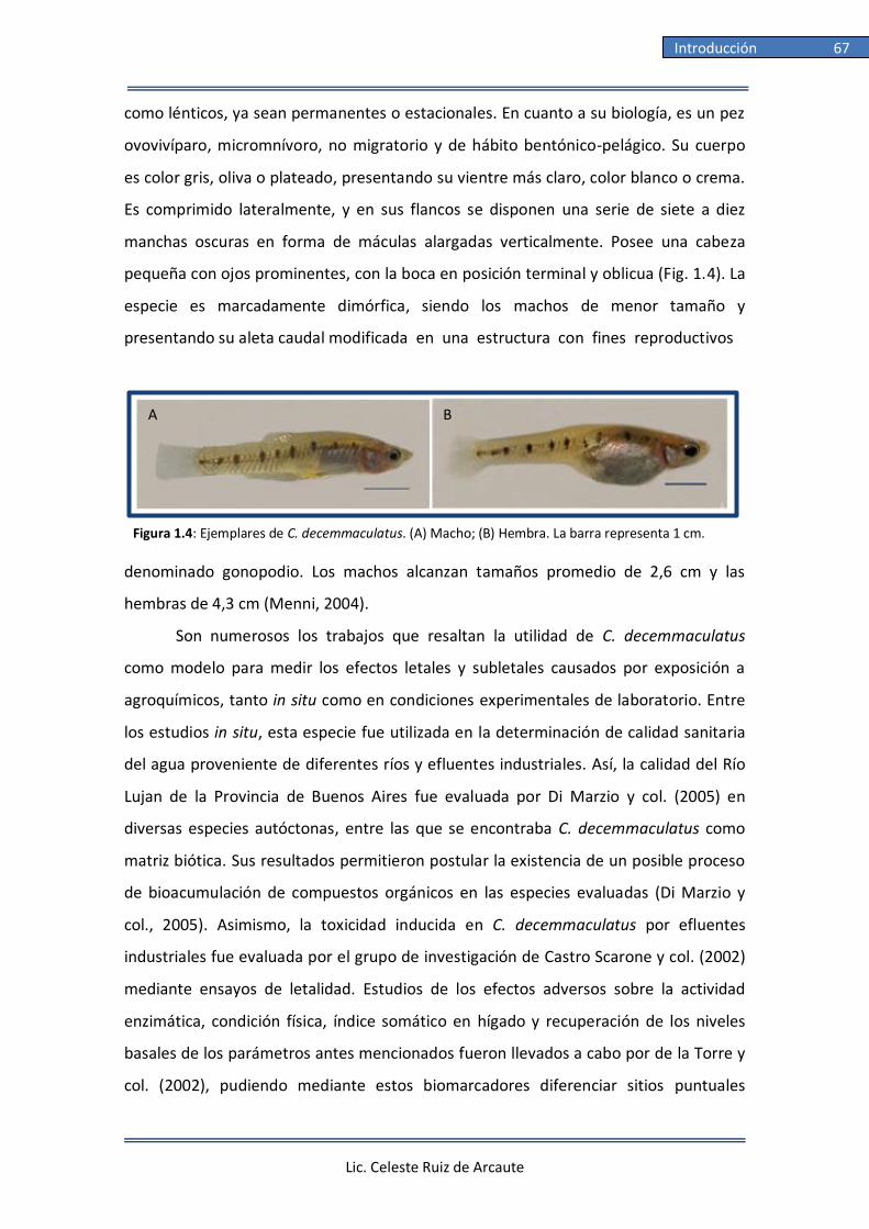
Lic. Celeste Ruiz de Arcaute
Introducción 67
como lénticos, ya sean permanentes o estacionales. En cuanto a su biología, es un pez
ovovivíparo, micromnívoro, no migratorio y de hábito bentónico-pelágico. Su cuerpo
es color gris, oliva o plateado, presentando su vientre más claro, color blanco o crema.
Es comprimido lateralmente, y en sus flancos se disponen una serie de siete a diez
manchas oscuras en forma de máculas alargadas verticalmente. Posee una cabeza
pequeña con ojos prominentes, con la boca en posición terminal y oblicua (Fig. 1.4). La
especie es marcadamente dimórfica, siendo los machos de menor tamaño y
presentando su aleta caudal modificada en una estructura con fines reproductivos
denominado gonopodio. Los machos alcanzan tamaños promedio de 2,6 cm y las
hembras de 4,3 cm (Menni, 2004).
Son numerosos los trabajos que resaltan la utilidad de C. decemmaculatus
como modelo para medir los efectos letales y subletales causados por exposición a
agroquímicos, tanto in situ como en condiciones experimentales de laboratorio. Entre
los estudios in situ, esta especie fue utilizada en la determinación de calidad sanitaria
del agua proveniente de diferentes ríos y efluentes industriales. Así, la calidad del Río
Lujan de la Provincia de Buenos Aires fue evaluada por Di Marzio y col. (2005) en
diversas especies autóctonas, entre las que se encontraba C. decemmaculatus como
matriz biótica. Sus resultados permitieron postular la existencia de un posible proceso
de bioacumulación de compuestos orgánicos en las especies evaluadas (Di Marzio y
col., 2005). Asimismo, la toxicidad inducida en C. decemmaculatus por efluentes
industriales fue evaluada por el grupo de investigación de Castro Scarone y col. (2002)
mediante ensayos de letalidad. Estudios de los efectos adversos sobre la actividad
enzimática, condición física, índice somático en hígado y recuperación de los niveles
basales de los parámetros antes mencionados fueron llevados a cabo por de la Torre y
col. (2002), pudiendo mediante estos biomarcadores diferenciar sitios puntuales
A B
Figura 1.4: Ejemplares de C. decemmaculatus. (A) Macho; (B) Hembra. La barra representa 1 cm.

Lic. Celeste Ruiz de Arcaute
Introducción 68
caracterizados por presentar altos niveles de contaminación. Adicionalmente,
demostraron que los niveles basales enzimáticos y del hígado pueden ser recuperados
luego de trasladar los ejemplares a un medio no contaminado (de la Torre y col., 2007).
El estado sanitario del Río Luján junto con un pulso de cadmio fue estudiado por
Ossana y col. (2016) mediante un ensayo agudo bajo condiciones experimentales de
laboratorio durante una exposición por un lapso de 96 h en el que evaluaron
alteraciones en biomarcadores de genotoxicidad y enzimáticos tales como MNs y AN,
GSH, GST, CAT y SOD (Ossana y col., 2016).
También se evaluó la toxicidad ejercida por metales pesados, determinándose
la CL50 para C. decemmaculatus luego de ser expuestos a metales como cadmio (de la
Torre, 1997; Eissa y col., 2010), zinc (Gómez y col., 1998), cobre (Villar y col., 2000;
Casares y col., 2012), cromo (Vera-Candioti y col., 2011), plata (Casares y col., 2013) y
sales como cloruro de zinc (Di Marzio, 1999) y el dicromato de potasio (Di Marzio,
1999). Particularmente, estudios empleando agroquímicos fueron llevados a cabo por
Di Marzio y Tortorelli (1994) y Di Marzio y col. (1994; 1998) quienes evaluaron la
inducción de efectos letales y alteraciones enzimáticas del paraquat en ejemplares de
la especie. También fue determinada la toxicidad de los plaguicidas pirimicarb (Vera-
Candioti y col., 2015), metamidofós (Di Marzio, 1999), ciflutrina (Di Marzio, 1999),
endosulfán (Mugni y col., 2012b), clorpirifós (Mugni y col., 2012a; 2012b; Vera-
Candioti y col., 2014; Paracampo y col., 2015), cipermetrina (Di Marzio, 1999;
Carriquiriborde y col., 2007; Mugni y col., 2012b; Brodeur y col., 2016) y glifosato (Di
Marzio, 1999; Vera-Candioti y col., 2013a; Brodeur y col., 2016). Efectos involucrando
alteraciones en el comportamiento, en marcadores de genotoxicidad al igual que de
estrés oxidativo fueron evaluados en ensayos de laboratorio en esta especie. Estudios
empleando el insecticida clorpirifós fueron reportados por Vera-Candioti y col. (2013b;
2014), quienes reportaron la inducción de daño genotóxico evaluado mediante el
ensayo de MNs y EC luego de una exposición aguda (Vera-Candioti y col., 2013b;
2014). Empleando el mismo compuesto, Bonifacio y col. (2016; 2017) informaron
alteraciones en el comportamiento y enzimáticas luego de 48 h de exposición a
concentraciones ambientales mientras que Bernal-Rey y col. (2017) reportaron
inhibición de la actividad de AChE en individuos expuestos por 96 h a este compuesto
(Bernal-Rey y col., 2017). Vera-Candioti y col. reportaron inducción de MNs y daño en

Lic. Celeste Ruiz de Arcaute
Introducción 69
el ADN medido mediante el EC empleando los insecticidas pirimicarb (Vera-Candioti y
col., 2013b; 2015) y clorpirifós (Vera-Candioti y col., 2013b; 2014) al igual que el
herbicida glifosato (Menéndez-Helman y col., 2012; Vera-Candioti y col., 2013a;
2013b). Estudios evaluando los efectos inducidos por glifosato fueron llevados a cabo
por Menéndez-Helman y col. (2012), quienes reportaron inhibición de la enzima AChE
luego de la exposición aguda a este herbicida (Menéndez-Helman y col., 2012).
Adicionalmente, luego de la exposición crónica a concentraciones ambientales de
glifosato, Bonifacio y col. (2016) informaron alteraciones enzimáticas como GST y
fosfatasa alcalina en ejemplares de C. decemmaculatus. Finalmente, estudios
evaluando los efectos inducidos por 17α-etinilestradiol en C. decemmaculatus fueron
llevados a cabo y demostrando la inducción del desarrollo de ovocitos testiculares
luego de una exposición prolongada al xenoestrógeno (Young y col., 2017).
El uso de C. decemmaculatus como modelo experimental se ve explicado por su
fácil cría y mantenimiento en laboratorio, alta tasa de fecundidad, rápido desarrollo y
por contar con un amplio rango de tolerancia a parámetros tales como temperatura,
salinidad y pH, que hace a la especie adecuada para bioensayos en laboratorio (Menni
y col., 1996; Menni, 2004; Ferrari, 2017). Adicionalmente, su manutención es
económica y son de fácil cuidado en un número de individuos elevado. Su alta tasa de
fecundidad y su corto tiempo generacional hacen de esta especie un modelo valioso en
el empleo de ensayos de laboratorio.
1.5 Ensayos biológicos
Los ensayos biológicos son métodos analíticos empleados para determinar la
concentración o la potencia de una sustancia por su efecto sobre organismos de
prueba o sistemas vivientes, tanto in vivo como in vitro o in situ. Son empleados para
detectar peligros biológicos o para evaluar la calidad de una mezcla, para monitorear
calidad del agua al igual que la descarga de aguas residuales y su potencial impacto.
También se utilizan para evaluar el impacto ambiental y la seguridad de las nuevas
tecnologías.
En 1940 se introdujo el uso de bioensayos con peces y durante 1950 se
comenzaron a utilizar como modelos experimentales a los invertebrados y algas. El

Lic. Celeste Ruiz de Arcaute
Introducción 70
concepto de bioensayo deriva de la toxicología clásica, el cual ha sido adaptado para el
diagnóstico ambiental. Son llevados a cabo en condiciones experimentales de
laboratorio, específicas y controladas (USEPA, 2002b). Los efectos estudiados pueden
ser letales o subletales, ubicándose dentro de este último grupo efectos en el
comportamiento, crecimiento, reproducción, cambios morfológicos, fisiológicos o
histológicos (Díaz Báez y col., 2004). Se utilizan metodologías estandarizadas para
maximizar la reproducibilidad y confiabilidad de los resultados, que permiten su
comparación con los obtenidos por otros grupos de investigación. Dentro de los
ensayos de toxicidad, las pruebas de letalidad son una herramienta útil para la
determinación de niveles de concentración de compuestos que resultan tóxicos o
dañinos para la biota y el ambiente (USEPA, 1975; Díaz Báez y col., 2004). Los
resultados son utilizados por organismos gubernamentales tales como la Agencia
Europea de Sustancias y Mezclas Químicas (ECHA), U.S. EPA, IARC y WHO para
establecer parámetros y niveles guía que garanticen la seguridad y sanidad del
ambiente y del agua para consumo humano.
Los agentes que producen alteraciones en el material genético son estudiados
por la Genética Toxicológica, la cual se define como “la ciencia que trata acerca de la
transmisión, expresión y evolución de los genes, las moléculas que controlan el
funcionamiento, el desarrollo y la apariencia final de los individuos” (Mudry y Carballo,
2006). Esta ciencia se ocupa del estudio del efecto mutagénico, tanto de alteraciones
mutacionales puntuales como macromutaciones, de agentes químicos, físicos y/o
biológicos, así como las consecuencias que los mismos provocan sobre los
ecosistemas, ambiente, seres vivos y la salud humana (Mudry y Carballo, 2006). La
Genética Toxicológica tiene dos objetivos principales: detectar y analizar el potencial
efecto deletéreo de aquellos agentes capaces de dañar el ADN e investigar los
mecanismos de acción a través de los cuales causan toxicidad (Çavaş, 2017).
1.5.1 Uso de biomarcadores en monitoreos ambientales
En el ambiente, los organismos se encuentran expuestos a un amplio abanico de
compuestos y condiciones que pueden ser perjudiciales para su desarrollo, aunque un
gran número de especies toleren estas situaciones (Mudry y Carballo, 2006). Para que
un contaminante ambiental cause un efecto deletéreo, primero tiene que ingresar al
Lic. Celeste Ruiz de Arcaute
Introducción 71
organismo, vencer las defensas de protección del mismo y una cantidad de ese
xenobiótico interactuar con un blanco celular o tisular donde se inicia el efecto
biológico (Mudry y Carballo, 2006). Cabe destacar que el término xenobiótico se
refiere al término derivado del griego ξένος (xenos) = extraño, extranjero y βίος (bios,
vios) = vida, en adición al sufijo griego para adjetivos -τικός, -ή, -ό (tic). Muchos de los
xenobióticos en general, son manufacturados por el hombre y en la mayoría de los
casos, resultan necesarios para preservar y mejorar la calidad de la salud humana y
ambiental.
Un biomarcador es un cambio a nivel celular, tisular, fisiológico, bioquímico o de
comportamiento en un organismo vivo, el cual es usado de manera cuantitativa con la
finalidad de identificar y caracterizar de manera significativa la exposición a un
determinado contaminante (Newman y Clements, 2008; Gupta, 2014). Son indicadores
de la ocurrencia de un cambio que podría más tarde llevar al desarrollo de una
enfermedad con expresión clínica, por lo cual se utilizan como herramientas de alerta
temprana. Los biomarcadores son utilizados para detectar la exposición a un
contaminante, determinar las consecuencias biológicas de la misma, detectar los
estados iniciales e intermedios de un proceso patológico, identificar los individuos
sensibles de una población y fundamentar la decisión de intervenir, tanto a nivel
individual como ambiental (Mudry y Carballo, 2006). Estos estimadores, pueden ser
clasificados como biomarcadores de exposición, de efecto y de susceptibilidad.
Un biomarcador de exposición señala la presencia de un xenobiótico que puede
ser medido dentro de un compartimento del organismo. Estos marcadores se obtienen
midiendo su concentración en muestras de fluidos o tejidos. Entre estos se encuentran
el análisis de distintos parámetros como la estimación de niveles de creatina quinasa -
MB, detección de niveles de diferentes sustancias como tolueno, tricloroetano,
pentaclorofenol o metales ya sea en sangre u orina, entre otros (Gupta, 2014; Kaviraj y
col., 2014).
Un biomarcador de susceptibilidad es indicador de la sensibilidad particular de
un individuo, heredada o adquirida, al efecto de un xenobiótico particular o al de
varios grupos cuando el mismo es una mezcla compleja. Entre estos biomarcadores se
encuentran por ejemplo, la detección de polimorfismos enzimáticos y genéticos, entre
otros (Gupta, 2014; Kaviraj y col., 2014).

Lic. Celeste Ruiz de Arcaute
Introducción 72
Por último, los biomarcadores de efecto son indicadores de alteraciones
bioquímicas, fisiológicas o genéticas resultado de la exposición a un agente
xenobiótico. Estas alteraciones pueden ser definidas como un daño o como una
patología, dependiendo de su magnitud. Entre estos biomarcadores se encuentran,
por ejemplo, los que evalúan efectos letales como la mortalidad, y subletales como
marcadores bioquímicos entre los cuales podemos mencionar la medición de
isoenzimas, marcadores moleculares como la medición de fragmentos de restricción
polimórficos (RFLP), fragmentos polimórficos de ADN amplificados al azar (RAPD),
minisatélites y microsatélites, marcadores fisiológicos como la expresión y actividad de
enzimas, citotóxicos como la detección de procesos apoptóticos, genotóxicos como la
estimación de la frecuencia de intercambios de cromátidas hermanas, de MNs y la
estimación de lesiones primarias en el ADN mediante la aplicación del EC, entre otros
(Grisolia y Starling, 2001; Gupta, 2014; Kaviraj y col., 2014; Ossana y col., 2016;
Bonifacio y col., 2017).
Los biomarcadores permiten, al ser indicadores de un efecto biológico temprano,
poder intervenir oportunamente antes de que se produzca un daño irreversible. Para
evaluar el alcance del efecto adverso inducido, se realizan estudios empleando los
mencionados biomarcadores de efecto que entre los evaluados en la presente Tesis
Doctoral se encuentran:
1.5.1.1 Alteraciones del comportamiento. Los ensayos agudos de letalidad, tales
como los propuestos por la U.S. EPA (1975; 2002a) proporcionan un índice simple de
toxicidad de un contaminante determinado. Sin embargo, estas pruebas por sí solas
brindan poca información sobre los efectos que producen los tóxicos en la biota ya que
ignoran otros efectos o alteraciones subletales que puede tener impactos ecológicos
serios, como por ejemplo el comportamiento. Adicionalmente, estas alteraciones
ocurren, a menudo, en concentraciones muy inferiores a las que causan una
mortalidad significativa (Cabrera y col., 1998; Gaworecki y Klaine, 2008). Las
alteraciones del comportamiento son utilizados como puntos finales de toxicidad
subletal y sirven como una herramienta en la evaluación de riesgo ambiental y el
análisis del impacto toxicológico de un determinado contaminante (Kane y col., 2004;
Faimili y col., 2006).

Lic. Celeste Ruiz de Arcaute
Introducción 73
La capacidad de natación es crucial para los peces y determinante en su
supervivencia dentro del medio acuático (Wolter y Arlinghaus, 2003). Por lo
anteriormente expuesto, gran parte de las investigaciones sobre los efectos de los
contaminantes ambientales en los peces se han centrado en estas alteraciones como
punto final (Bretaud y col., 2001; Dogan y Can, 2011; Pereira y col., 2012). Sin
embargo, como resultado de la exposición a ciertos xenobióticos los peces también
pueden exhibir otras alteraciones como aquellas observadas durante el
comportamiento social las que afectan su habilidad en la búsqueda de alimento y en
evitar predadores, poniendo en riesgo su supervivencia (Sarikaya y Yilmaz, 2003;
Yilmaz y col., 2004).
Para ser relevantes como biomarcadores, las respuestas comportamentales
deben ser bien definidas, mensurables, ecológicamente relevantes y sensibles a una
variedad de sustancias tóxicas. Estos puntos finales comportamentales han sido
utilizados como indicadores tempranos de contaminación ambiental (Quarcoo y col.,
2014), pudiendo ser adaptados para la evaluación de la toxicidad de plaguicidas. La
importancia de la utilización de biomarcadores comportamentales radica en su
confiabilidad, facilidad de aplicación y que, en ocasiones, se manifiestan como la
primera respuesta de un organismo a la exposición a un xenobiótico. En adición,
brindan información sobre las consecuencias poblacionales y ecológicas de la
exposición a xenobióticos.
1.5.1.2 Ensayo de Micronúcleos. Es un
bioensayo que permite la evaluación del daño a
nivel cromosómico inducido por un xenobiótico y
que puede ser determinado en distintos tipos de
células proliferantes mediante la inducción de
daño clastogénico o aneugénico. Fue descripto por
primera vez por Evans y col. (1959) quienes
utilizaron como modelo raíces de Vicia faba
expuestas a radiación ionizante. Esta técnica se
basa en analizar la presencia de macrolesiones a nivel de la molécula de ADN donde el
material genético desprendido da origen a los denominados micronúcleos (MN), que
son pequeñas porciones de material extranuclear rodeados por membrana nuclear
Figura 1.5: Micronúcleo (cabeza de
flecha) en eritrocito circulante de C.
decemmaculatus

Lic. Celeste Ruiz de Arcaute
Introducción 74
detectados en células en interfase celular. El MN es un indicador indirecto de daño
cromosómico que puede originarse por una o varias rupturas de la molécula de ADN la
cual alcanza su máximo grado de compactación durante la metafase mitótica dando
lugar a la formación de un cromosoma que puede generar uno o a varios fragmento(s)
acéntrico(s) cromosómicos (efecto clastogénico) o pueden estar formados por
cromátidas enteras o por un cromosoma completo el cual no migró normalmente
durante la anafase celular por alteraciones acontecidas en el aparato mitótico (efecto
aneugénico) (Fenech y col., 2011; Çavaş, 2017). Por esto, requieren al menos una
división celular para expresarse y pueden ser visualizados con una tinción convencional
en células en el estado de interfase celular. Adicionalmente, pueden ser observados y
por consiguiente, cuantificados en diferentes modelos experimentales, incluyendo
células vegetales, animales e inclusive humanas y empleando distintos tipos celulares,
dependiendo del modo de acción del agente a caracterizar y del objeto de estudio
(Mudry y Carballo, 2006).
Los criterios establecidos para identificar un MN en una célula interfásica son:
tener una morfología redondeada, un diámetro menor a 1/3 respecto al núcleo
principal, no ser refringente, mostrar menor o la misma intensidad en la coloración
que el núcleo principal, no estar conectado al núcleo principal, no estar solapado al
núcleo principal, tener los bordes definidos y bien diferenciables del núcleo principal al
igual que no existir más de 6 MNs en una misma célula según los criterios establecidos
por Fenech (2000; 2007) (Fig.1.5).
Particularmente, el análisis de frecuencia de MNs en eritrocitos circulantes de
peces expuestos a diferentes tóxicos, al igual que en otros organismos acuáticos se ha
tornado una metodología aceptada y utilizada ampliamente a nivel mundial dentro del
campo de la genotoxicología (Çavaş y Ergene-Gözükara, 2003; Gökalp Muranli y Güner,
2011; Pérez-Iglesias y col., 2014). Aún más, se ha convertido en una herramienta de
estudio obligatoria durante el proceso de aprobación de productos farmacéuticos y
químicos (Mudry y Carballo, 2006). Entre las tinciones más utilizadas para este método
se encuentran la coloración con Giemsa, coloración con naranja de acridina (NAcr),
DAPI, ioduro de propidio, entre otras (Çavaş, 2017). El uso de Giemsa, formado por una
mezcla de colorantes neutros y básicos, es la metodología de tinción más extendida,
aunque con este método, algunos artefactos no conformados por material nuclear

Lic. Celeste Ruiz de Arcaute
Introducción 75
pueden ser teñidos y confundidos con MNs por un observador no experimentado. Por
otro lado, la NAcr es un fluorocromo catiónico específico para ácidos nucleicos, vale
decir pone en evidencia tanto ADN como ARN, siendo considerada por consiguiente
una técnica más confiable que la de Giemsa, aunque con la desventaja de precisar
equipamiento más sofisticado, como microscopia con epifluorescencia y el empleo de
filtros de excitación adecuados en el equipo empleado.
Asimismo, cabe destacar que, aun no existiendo en la literatura un consenso
universal para su significancia y clasificación, se describieron y se propusieron diversas
anormalidades nucleares (AN), diferentes a los MNs, como signo de daño inducido en
el material genético por diferentes agentes xenobióticos (Carrasco y col., 1990). Las
mismas pueden resumirse en las siguientes variedades o tipos designadas
vernáculamente como “otras alteraciones nucleares” siguiendo los criterios
establecidos por diversos autores (Çavaş y Ergene-Gözükara, 2003; Strunjak-Perovic y
col., 2009; Nikoloff y col., 2014b).
B
Figura 1.6. Anormalidades nucleares (cabeza de flecha). (A) Brote nuclear
(B) Célula binucleada (C) Célula lobulada y (D) Escotadura nuclear
A
C D
B

Lic. Celeste Ruiz de Arcaute
Introducción 76
Dentro de dichas alteraciones nucleares pueden incluirse a los eritrocitos
binucleados (EB), cuando la misma presenta dos núcleos de tamaño similar con su
membrana nuclear intacta; brotes nucleares (BN- buds), cuando el núcleo celular
presenta una pequeña evaginación de material genético que sobresale de la
membrana nuclear. Esta evaginación contiene en su interior eucromatina y se
encuentra unida al núcleo por un pedúnculo de material genético cuyo ancho puede
ser ligeramente menor al ancho del BN o mucho más delgado, dependiendo del
momento del proceso de extrusión del material; núcleos lobulados (NL), cuando el
núcleo de la célula presenta una o más evaginaciones de la membrana nuclear, más
grandes que los BN y escotaduras nucleares (EN), cuando los núcleos presentan
vacuolas y una invaginación pronunciada de su membrana nuclear (Çavaş y Ergene-
Gözükara, 2003; Strunjak-Perovic y col., 2009; Nikoloff y col., 2014b)(Fig. 1.6).
Se ha sugerido que la aparición de algunas AN y MNs son resultado de eventos
genotóxicos relacionados con diferentes causas tales como problemas durante la
segregación de los cromosomas que se encuentran alineados de manera incorrecta en
la placa metafásica o que permanecen unidos entre ellos durante la división celular
(Shimizu y col., 1998; Seriani y col., 2012; Çavaş, 2017), aparición de aneuploidías
(Fernandes y col., 2007) y generación de procesos de degradación celular (Ateeq y col.,
2002). También se ha sugerido que los NL y BN podrían originarse durante el proceso
de eliminación de exceso ADN generado por amplificación génica a través de ciclo de
ruptura-fusión-puente (RFP). Este ADN en exceso se ubicaría en la periferia del núcleo
y sería posteriormente eliminado por un proceso de extrusión, formando un MN o, en
el caso de que este proceso se vea interrumpido, la generación de un BN o un NL.
Análisis estadísticos probaron una correlación positiva entre la aparición de BN y la
formación de MNs (Çavaş, 2017). Fernandes y col. (2007) han sugerido que las EB y EN
podrían ser indicadores de xenobióticos que actuarían mediante una mecanismo
aneugénico. Se ha reportado, asimismo, que este efecto podría suceder por fallas
durante la polimerización de las tubulinas y en consecuencia, la generación de
alteraciones a nivel del aparato mitótico (Walia y col., 2013). Adicionalmente, Ateeq y
col. (2002) sugirieron que los xenobióticos generan condiciones hipóxicas que resultan
en la disminución de los niveles de ATP, que conduce a malformaciones morfológicas

Lic. Celeste Ruiz de Arcaute
Introducción 77
en los eritrocitos por alteraciones en el ciclo normal de polimerización de tubulinas,
proteínas motrices y rectoras del aparato mitótico.
El ensayo de MNs en peces fue reportado por primera vez en 1982 por
Hooftman y de Raat (1982) empleando eritrocitos circulantes de Umbra pygmaea
tratados con etilmetanosulfonato. Luego fue utilizado por Al-Sabti (1986) empleado en
tres especies de ciprínidos con el fin de estudiar los efectos de la exposición a
diferentes compuestos mutagénicos. Manna y Sadhukhan (1986) implementaron el
ensayo de MNs en agallas e hígado de tilapia (Oreochromis mossambicus) expuestas a
rayos X y cloruro de cadmio. Posteriormente, Williams y Metcalfe (1992) desarrollaron
un ensayo de MNs in vivo en células hepáticas de O. mykiss y Arkhipchuk y Garanko
(2005) lo utilizaron empleando células epiteliales provenientes de las aletas de C.
carpio. Finalmente, Fassbender y Braunbeck (2013) realizaron una variante del ensayo
empleando células gonadales de D. rerio expuestas a metilmetanosulfonato.
1.5.1.3 Electroforesis en gel de células únicas. Variedad alcalina del Ensayo
Cometa. El ensayo cometa (EC) es una metodología sensible, rápida, simple y visual
utilizada para analizar rupturas en la molécula de ADN de simple o doble cadena, sitios
sensibles al álcali, uniones cruzadas ADN-ADN y ADN-proteína y rupturas de cadena
simple asociadas a mecanismos de reparación del ADN (Singh, 1996; Çavaş, 2017). Esta
técnica permite analizar el daño genotóxico inducido por un xenobiótico en células
individuales embebidas en una capa de gel de agarosa. Estas células, luego de la
exposición a un tóxico, son embebidas en agarosa la cual es colocada sobre un
portaobjetos, entre otras dos capas de agarosa y posteriormente lisadas para remover
las membranas celulares al igual que las proteínas que se encuentran unidas al ADN,
dejando al núcleo desprovisto de su membrana nuclear, tomando la denominación de
“nucleoide”. Luego del desenrollamiento del ADN en condiciones alcalinas, el mismo es
sometido a una electroforesis induciendo a los fragmentos rotos o dañados de ADN
migrar alejándose del nucleoide. Los fragmentos de ADN cargados negativamente
migran libremente hacia el ánodo a través de un campo eléctrico en una solución de
electroforesis (Azqueta y col., 2017; Çavaş, 2017). Luego de la neutralización y la
tinción con un colorante con especificidad por el ADN, la imagen resultante a analizar
se asemeja a un cometa, con una zona más definida denominada “cabeza” en la región

Lic. Celeste Ruiz de Arcaute
Introducción 78
nuclear, y una región conteniendo el ADN fragmentado migrado, la “cola” del
nucleoide (Fig. 1.7). La migración del ADN es directamente proporcional al daño que
presenta el mismo (Fig. 1.8).
El EC tiene la ventaja de poder ser aplicado a cualquier población de células
eucariotas, sin tener en cuenta su actividad mitótica o el período del ciclo celular
donde se encontraban en el momento de la exposición al xenobiótico. Más aún, sólo
se requiere una pequeña cantidad de tejido o de células de un tejido y adicionalmente
a los métodos de categorización visual del daño, tiene la opción de ser analizado
utilizando programas de computación. Los resultados pueden ser obtenidos en un
lapso breve de análisis por parte de un observador (Tice y col., 2000; Collins, 2004;
Azqueta y Collins, 2013).
El EC fue introducido por primera vez por Ostling y Johanson (1984), quienes
empleando una línea celular tumoral de mamíferos irradiada, utilizaron la metodología
con una solución de electroforesis a pH neutro (9,5) la que permitió la detección de
lesiones de ADN de cadena doble. Posteriormente, Singh en sus investigaciones sobre
el efecto de la radiación con rayos X y H2O2 desarrolló la variante alcalina de la técnica
mediante el empleo de una solución de pH≥12 durante la electroforesis a la que son
sometidas las muestras, lo cual adicionó la detección de lesiones de simple cadena y
sitios sensibles al álcali (Singh y col., 1988). Entre las tinciones utilizadas se encuentran
la impregnación argéntica y el uso de fluorocromos tales como el bromuro de etidio,
Figura 1.7: (A) Nucleoide sin daño (B) Nucleoide presentando rupturas en sus moléculas de ADN,
evidenciados por su migración luego de la electroforesis adoptando el nucleoide la forma de un
cometa.

Lic. Celeste Ruiz de Arcaute
Introducción 79
ioduro de propidio, SybrGreen y DAPI, entre los más empleados (Collins y col., 2008;
Christofoletti y col., 2009; Vera-Candioti y col., 2013b; Mustafa y col., 2015).
La cuantificación visual del daño en el ADN se realiza y expresa en unidades
arbitrarias basadas en la clasificación de los nucleoides en cinco categorías (0-IV) de
acuerdo a la extensión de la cola, propuesta por Çavaş y Konen (2007). Los nucleoides
tipo 0-I agrupan a los nucleoides no dañados, los tipo II incluyen a los de nivel de daño
leve, los de tipo III a aquellos nucleoides que presentan un nivel de daño medio y los
nucleoides tipo IV, incluyen a los que presentan un grado de daño máximo (Fig. 1.8).
En el análisis automatizado de los cometas, los programas de computación tales como
el OpenComet y NIH image permiten el cálculo de más de 20 parámetros usando
imágenes obtenidas en el momento de su estimación o capturadas con anterioridad
(Çavaş, 2017). Entre los parámetros más utilizados y adoptados mundialmente en el
campo de la Genética Toxicológica para expresar el nivel de daño en el ADN se
encuentran el largo de la cola, el porcentaje de ADN contenido en la cola y el momento
Olive. El largo de la cola, generalmente, incluye la distancia de migración del ADN
desde el nucleoide. El porcentaje de ADN de la cola se calcula sustrayendo de la
cantidad total del ADN del nucleoide el porcentaje de ADN presente en la cabeza del
cometa. Finalmente, el momento Olive se define como el producto del largo de la cola
y la fracción total de ADN presente en la misma (Dogan y Can, 2011; Martínez-Paz y
col., 2013).
El EC fue aplicado por primera vez en peces por Pandrangi y col. (1995) con el fin
de evaluar los efectos de la contaminación en ambientes marinos en eritrocitos del pez
gato (Ameirus nebulosus) y en C. carpio. Posteriormente, la técnica fue empleada en
hígado y agallas de D. rerio (Deventer, 1996), así como en hepatocitos del trucha reo
Figura 1.8: Categorías de daño utilizadas. (A y B) Nucleoides sin daño (cometas tipo 0–I),
(C) nucleoides con daño leve (cometas tipo II), (D) daño intermedio (cometas tipo III), y (E)
daño máximo (cometas tipo IV).

Lic. Celeste Ruiz de Arcaute
Introducción 80
(Salmo trutta) (Mitchelmore y Chipman, 1998) y en el lenguado de invierno
(Pleuronectes americanus) (Nacci y col., 1996).
Tanto el ensayo de micronúcleos (MNs) como el ensayo cometa (EC) se han
convertido en metodologías ampliamente usadas para la evaluar la genotoxicidad al
igual que la citotoxicidad de muchos xenobióticos, incluyendo plaguicidas, debido a su
sensibilidad, confiabilidad y sencillez. Adicionalmente, su aplicación para la
determinación de efectos genotóxicos en peces expuestos particularmente a
herbicidas está ampliamente documentada, reportando la inducción de efectos
genotóxicos en peces expuestos a glifosato como en el pacú blanco (Piaractus
mesopotamicus) (Leveroni y col., 2017), C. punctatus (Nwani y col., 2013), la anguila
europea (Anguilla anguilla) (Guilherme y col., 2010), la coridoras pimienta (Corydoras
paleatus) (de Castilhos Ghisi y Cestari, 2013), el sábalo común (Prochilodus lineatus)
(Calcavante y col., 2008), O. niloticus (Alvarez-Moya y col., 2013), C. auratus (Çavaş y
Könen, 2007) y C. decemmaculatus (Vera-Candioti, 2013b), entre otros; a atrazina en
peces como C. punctatus (Nwani y col., 2011), C. auratus (Çavaş, 2011) y R. quelen
(Dante y col., 2015); a 2,4-D en peces como el pez gato andador (C. batrachus) (Ateeq y
col., 2005); a butaclor en peces como C. carpio (Mashinchian Moradi y col., 2012) y C.
batrachus (Ateeq y col., 2005); a triclopir en peces como A. anguilla (Guilherme y col.,
2015) y a cipermetrina en peces como P. lineatus (Simoniello y col., 2009) y D. rerio
(Paravani y col., 2018); entre otros.
1.5.1.4 EC modificado empleando enzimas de restricción. Una modificación
introducida en la metodología clásica del EC alcalino permite detectar, adicionalmente
a las rupturas de cadena inducidas por xenobiótico, el daño ejercido en bases tanto
púricas como pirimidínicas introducidas en la molécula de ADN. Esta modificación se
logra a través de la adición de un período de incubación con endonucleasas de
restricción capaces de reconocer específicamente diferentes tipos de bases dañadas
en la molécula de ADN (Azqueta y Collins, 2013). En esta modificación, luego de la lisis,
los geles de agarosa conteniendo los nucleoides son incubados con endonucleasas
específicas o enzimas de restricción que reconocen y transforman lesiones en bases
del ADN, en cortes de cadena, originando sitios apurínicos. Por consiguiente, de existir
bases dañadas, la endonucleasa específica reconoce dichos sitios, produce los cortes y
esto será analizado por el observador como una mayor densidad (mayor momento de

Lic. Celeste Ruiz de Arcaute
Introducción 81
cola) debido al aumento del número de fragmentos, o a una mayor longitud de la cola
del cometa dado a una mayor migración de fragmentos de menor tamaño. Dentro de
las enzimas de restricción más utilizadas se encuentran la endonucleasa III (Endo III) y
la formamidopirimidina-ADN glicosilasa (Fpg) (Collins y col., 1996; Collins, 2004). La
Endo III detecta pirimidinas oxidadas, escinde a las mismas e induce a la formación d
sitios AP, lo que conlleva a la formación de cortes en el ADN. Por su parte, la Fpg
reconoce y remueve purinas oxidadas del ADN como por ejemplo 8-oxoguanina, Fapy-
Gua, Fapy-Ade y en menor medida, 8-oxoadenina, que posteriormente se escinden por
su actividad AP liasa dando lugar a una brecha en la cadena de ADN (sitio-AP). Esta
metodología ha sido aplicada en numerosos estudios por ser un biomarcador sensible
de la estimación de daño oxidativo a nivel de la molécula de ADN (Collins y col., 1996;
Wong y col., 2005; Kushwaha y col., 2011; Collins y Azqueta, 2012) y su aplicación ha
permitido dilucidar los mecanismos de acción de compuestos genotóxicos, investigar
enfermedades derivadas de la situación de estrés oxidativo, evaluar estrés oxidativo
generado por ejercicio en el hombre o en animales, estudiar efectos antioxidantes en
la dieta así como en la evaluación del estado ambiental (Azqueta y col., 2017). En
peces, son escasos los estudios que han incluido Endo III y Fpg en la detección de bases
oxidadas, entre ellos como ejemplo podemos citar la evaluación del insecticida
monocrotofós en C. auratus (Zhao y col., 2015), del herbicida glifosato en A. anguilla
(Marques y col., 2014), del estado de ríos y cuencas en el alburno (Alburnus alburnus)
(Jovanović y col., 2017) y del efecto de condiciones hipóxicas en C. carpio (Mustafa y
col., 2015), entre otros.
1.5.1.5 Biomarcadores enzimáticos y no enzimáticos. Los biomarcadores
enzimáticos proveen información acerca de los procesos de detoxificación de
xenobióticos o sus metabolitos en los organismos. Estos procesos de detoxificación
involucran la conversión catalizada enzimáticamente de los xenobióticos a formas más
solubles, las que puedan ser eliminadas de los organismos más fácilmente. En
ocasiones, los xenobióticos o sus metabolitos pueden generar compuestos
intermediarios que son aún más tóxicos que el contaminante original, produciendo
una condición fisiológica anormal conocida como estrés oxidativo (Liu y col., 2006;
Monteiro y col., 2006).

Lic. Celeste Ruiz de Arcaute
Introducción 82
Las ROS son especies que están presentes en todas las células y se originan por
procesos como la respiración celular, así también como parte de la respuesta celular a
la presencia de determinados niveles de xenobióticos. Los productos de la reducción
del O2 son los radicales libres tales como el superóxido (O2.-) e hidroxilo (.OH) al igual
que la formación de peróxido de hidrógeno (H2O2) proveniente del metabolismo
celular. Se denominan radicales libres a aquellas moléculas que en su estructura
presentan un electrón impar en su órbita externa, que les otorga una alta
inestabilidad, pudiendo actuar como agentes oxidantes o reductores. Estas especies
son muy reactivas y potencialmente deletéreas para los sistemas biológicos. Por lo
expuesto, ante la elevada generación de ROS los organismos presentan diferentes
sistemas de defensa antioxidante, los enzimáticos (ej. catalasa -CAT-, superóxido
dismutasa -SOD-, peroxidasas, glutatión S-transferasa –GST-, entre otras) y los no
enzimáticos (ej. vitaminas E -Vit. E- y glutatión -GSH-) (van der Oost, 2003; Lushchak,
2011). Estos actúan con el fin de disminuir la toxicidad del xenobiótico y facilitar su
excreción (Santos y Martinez, 2012). De no ser efectivos estos mecanismos de
detoxificación, se produce un desbalance entre la generación de ROS y su eliminación
por los antioxidantes que resulta en una situación de estrés oxidativo (Cooke y col.,
2006; Valko y col., 2006) que puede provocar diferentes efectos deletéreos tales como
aumento de la peroxidación lipídica, inactivación enzimática, oxidación de hormonas y
daños en el ADN, entre otros efectos adversos (Cavalcante y col., 2008; Newman y
Clements, 2008; Amado y col., 2009; Limón-Pacheco y Gonsebatt, 2009; Pandey y col.,
2011).
Entre los diferentes sistemas de defensa antioxidante que actúan como sistemas
reguladores del balance oxidativo pueden mencionarse:
Catalasa (CAT): Constituye la primera barrera enzimática de detoxificación ante
la presencia de ROS catalizando la reducción de H2O2 a H2O y O2. Esta enzima actúa
sobre uno de los productos que genera la SOD, una de las primeras enzimas que
actúan frente a los ROS. SOD cataliza la conversión de aniones superóxido a peróxido
de hidrógeno (H2O2). Éste es luego detoxificado por acción de la CAT, específica para
este sustrato, y la glutatión peroxidasa (GPx).
Glutatión reducido (GSH): El GSH es un tripéptido formado por los aminoácidos
glutamina, cisteína y glicina que posee un rol importante en el sistema antioxidante de

Lic. Celeste Ruiz de Arcaute
Introducción 83
la mayoría de las células. En el proceso de detoxificación, la GPx cataliza el
metabolismo del H2O2 en H2O, oxidando concomitantemente glutatión reducido (GSH),
el cual es uno de los antioxidantes no enzimáticos más importantes de la célula (van
der Oost, 2003). Otra enzima antioxidante que participa junto con el GPx en la
regulación del GSH es la glutatión reductasa (GR) que reduce el glutatión oxidado
(GSSG) y mantiene los niveles de GSH en valores fisiológicos normales (Hermes-Lima,
2004). Los xenobióticos pueden disminuir el contenido de GSH y alterar el balance
entre GSH y su forma oxidadada, GSSG (Machado y col., 2008).
Glutatión S-transferasa (GST): La GST es una familia de enzimas cuya principal
función es catalizar las reacciones de fase II del metabolismo en los procesos de
detoxificación de xenobióticos. Cataliza la unión de GSH a una variedad de compuestos
tóxicos, incrementando su solubilidad y facilitando su excreción en medio acuoso,
protegiendo proteínas y ADN. Adicionalmente, detoxifica productos de peroxidación
lipídica derivados del estrés oxidativo.
Acetilcolinesterasa (AChE): Es una enzima cuya función es hidrolizar el
neurotransmisor acetilcolina luego de que éste haya sido liberado y realizado su
función en la transmisión del impulso nervioso. La AChE inactiva la acetilcolina,
disminuyendo su función. Su actividad puede ser inhibida por distintos tipos de
compuestos, causando una sobre estimulación de las fibras musculares llevando a una
parálisis, e incluso muerte de los organismos (Ferrari y col., 2007). La medición de la
actividad de AChE, en cerebro, músculo o sangre, ha sido utilizada en los últimos años
para evaluar los efectos neurotóxicos inducidos por ciertos xenobióticos (Menéndez-
Helman y col., 2012; Santos y Martinez, 2012).

Lic. Celeste Ruiz de Arcaute
Hipótesis y Objetivos 84
2. Hipótesis y Objetivos
2.1 Hipótesis
Los herbicidas 2,4-D y DIC contenidos en sus formulaciones comerciales DMA® y
Banvel® respectivamente, empleados de manera única o en mezclas binarias, producen
efectos letales, comportamentales, genotóxicos y cambios en el balance oxidativo en
Cnesterodon decemmaculatus (Jenyns, 1842) (Pisces: Poeciliidae).
2.2 Objetivos
2.2.1 Objetivo general
El objetivo general del presente trabajo de Tesis Doctoral es evaluar los efectos
letales, comportamentales, genotóxicos y del daño oxidativo inducido por
formulaciones comerciales de los herbicidas DIC y 2,4-D en ejemplares de C.
decemmaculatus (Jenyns, 1842) (Pisces: Poeciliidae) expuestos en condiciones
experimentales de laboratorio.
2.2.2 Objetivos específicos
1- Estimar la toxicidad inducida por los herbicidas DIC y 2,4-D en sus formulaciones
comerciales Banvel® y DMA® mediante ensayos de mortalidad y alteraciones en el
comportamiento luego de una exposición aguda en laboratorio en individuos de
C. decemmaculatus.
2- Evaluar la genotoxicidad ejercida luego de una exposición aguda por
formulaciones comerciales de herbicidas DIC y 2,4-D mediante el análisis de
micronúcleos en eritrocitos circulantes y de lesiones primarias en la molécula de

Lic. Celeste Ruiz de Arcaute
Hipótesis y Objetivos 85
ADN mediante el EC en células sanguíneas de C. decemmaculatus expuestos en
condiciones experimentales de laboratorio.
3- Evaluar daño oxidativo ejercido por DIC y 2,4-D mediante la detección de bases
púricas y pirimidínicas oxidadas en el ADN en células sanguíneas de C.
decemmaculatus expuestos en condiciones experimentales de laboratorio.
4- Evaluar la toxicidad de DIC y 2,4-D mediante biomarcadores enzimáticos de
estrés oxidativo como el contenido de glutatión-S-transferasa, catalasa y AChE y
no enzimáticos como el contenido de glutatión reducido en hígado de C.
decemmaculatus expuestos en condiciones experimentales de laboratorio.
5- Evaluar si la interacción ejercida por mezclas de los herbicidas mencionados
incrementa la letalidad inducida por los mismos en individuos de C.
decemmaculatus expuestos en condiciones experimentales de laboratorio.
6- Evaluar la genotoxicidad ejercida por formulaciones comerciales de herbicidas
DIC y 2,4-D luego de una exposición crónica mediante el análisis de lesiones
primarias en la molécula de ADN mediante el EC en células sanguíneas de C.
decemmaculatus expuestos en condiciones experimentales de laboratorio.
Lic. Celeste Ruiz de Arcaute
Materiales y Métodos 86
3. Materiales y Métodos
3.1 Muestreo y aclimatación
En el presente trabajo de Tesis Doctoral se emplearon ejemplares adultos de la
especie C. decemmaculatus (Jenyns, 1842, Pisces: Poeciliidae; Fig. 1.8) recolectados en
el arroyo San Juan, tributario ubicado dentro del Parque Provincial Pereyra Iraola
(Reserva de la Biósfera, UNESCO), Partido de Berazategui, Provincia de Buenos Aires,
Argentina (Latitud -34º 85' 95,60'' (S), Longitud -58º 12' 54,59'' (O); Fig. 3.1). Los
ejemplares fueron capturados con un copo de mano, mantenidos con agua del mismo
sitio de captura y transportados inmediatamente al laboratorio en un contenedor
plástico. Allí, fueron colocados en peceras de vidrio de 40 L de capacidad con agua
proveniente del lugar de captura. Posteriormente, cada 48 h el 25% del agua
proveniente del lugar de captura fue reemplazada por agua de ensayo declorinada
previamente (48 h) por burbujeo constante (pH 7,55 ± 0,1; oxígeno disuelto, 6,3 ± 0,3
mg/L; amonio (NH4+) <0,2 mg/L; dureza, 143 ± 23,5 mg CaCO3/L) hasta reemplazar la
totalidad de la misma. Luego, el mantenimiento del acuario fue realizado una vez por
semana. Los individuos fueron aclimatados a estas condiciones con un ciclo diario 16:8
h luz/oscuridad a 20 ± 1°C con aireación artificial constante, durante al menos durante
20 d antes del comienzo de cada experimento, según lo sugerido por Di Marzio y de
acuerdo a las normas IRAM (Di Marzio, 1999; IRAM, 2008). Los peces fueron
alimentados diariamente ad libitum con alimento en escamas para peces (TetraFin®,
TetraWerke, Melle, Alemania). El cuidado y mantenimiento de los individuos se realizó
siguiendo las recomendaciones propuestas en la guía 617/2002 de SENASA para la
realización de pruebas biológicas (SENASA, 2013). El peso y longitud promedio de los

Lic. Celeste Ruiz de Arcaute
Materiales y Métodos 87
individuos utilizados en los experimentos fue de 0,26 ± 0,1 g y 29,5 ± 2,7 mm,
respectivamente.
3.2 Compuestos químicos empleados
En el presente trabajo de Tesis Doctoral se utilizaron formulados de
agroquímicos actualmente registrados y comercializados en nuestro país de acuerdo a
SENASA (2018). El formulado comercial Banvel® (57,7% DIC, CAS 1918-00-9) fue
provisto por la empresa Syngenta Agro S.A. (Buenos Aires, Argentina) y el formulado
DMA® (58,4% 2,4-D, CAS 2008-39-1) fue proporcionado por la empresa Dow
Agrosciences Argentina (Buenos Aires, Argentina). La lista completa de los químicos
utilizados durante los ensayos se encuentra detallada en la Tabla 3.1.
Para la obtención de las soluciones de ensayo a utilizar de cada agroquímico, se
procedió en primer lugar a realizar una solución madre de cada herbicida, diluyendo
una cantidad determinada del mismo en agua de ensayo declorinada. A partir de esta
solución madre, se realizaron diluciones de la misma hasta alcanzar las
concentraciones establecidas para llevar a cabo los experimentos. En simultáneo, se
prepararon las soluciones para los controles positivos y negativos. Todas las soluciones
Figura 3.1: Sitio de recolección de ejemplares de C. decemmaculatus. Parque Provincial Pereyra
Iraola, Partido de Berazategui, Provincia de Buenos Aires, Argentina.

Lic. Celeste Ruiz de Arcaute
Materiales y Métodos 88
fueron preparadas inmediatamente antes del inicio de cada experimento y de la
renovación de las mismas según las necesidades de los diferentes modelos
experimentales utilizados.
Tabla 3.1 Reactivos químicos utilizados en la presente Tesis Doctoral
Reactivo CAS/ Número de
catálogo Origen
Aceite de inmersión HX08819102 Merck KgaA
Ácido tricloroacético 76-03-9 Sigma Chemical Co. (St. Louis, MO, EE.UU.)
Albúmina de suero bovino A-7638 Sigma Chemical Co.
Agarosa bajo punto de fusión A9414 Sigma Chemical Co.
Agarosa punto de fusión normal A9539 Sigma Chemical Co.
CDNB (2,4-dinitroclorobenceno) 24438 Sigma Chemical Co.
Carbonato de sodio A0841492 520 Merck KgaA
Ciclofosfamida 6055-19-2 Sigma Chemical Co.
Cloruro de potasio 7447-40-7 Sigma Chemical Co.
Cloruro de sodio 7647-14-5 Sigma Chemical Co.
DAPI H1200 Vector Laboratories (Burlingame, CA, EE.UU.)
Dicromato de potasio 7778-50-9 Merck KgaA
Dimetilsulfóxido 67-68-5 Sigma Chemical Co.
Reactivo de Ellman D8130-16 Sigma Chemical Co.
Ditiotreitol D9779-1G Sigma Chemical Co.
Endonucleasa III MFCD02265700 Sigma Chemical Co.
Fosfato diácido de potasio P-5379 Sigma Chemical Co.
Fosfato diácido de sodio S-8282 Sigma Chemical Co.
Fosfato dipotásico 1551128 Sigma Chemical Co. Formamidopirimidina-ADN glicosilasa 78783-53-6 Sigma Chemical Co.
Giemsa HX933732 Merck KgaA
Glutation reducido G4251-5G Sigma Chemical Co.
Hidróxido de sodio B0002198 Merck KgaA
Ioduro acetiltiocolina 1866-15-5 Sigma Chemical Co.
Metanol K39504909 Merck KgaA Ácido etilendiaminotetraacético disódico 6381-92-6 Sigma Chemical Co.
Peróxido de hidrógeno 1072100250 Merck KgaA (Darmstad, Alemania)
Reactivo de Folin Ciocalteau F9252 Sigma Chemical Co.
Sulfato de Cobre A0629491 435 Merck KgaA
Tartrato de sodio 3930-19 J.T. Baker (Phillipsburg, NJ, EE.UU)
Tris pH 7,5 T1378 Sigma Chemical Co.
Trizma®(ácido clorhídrico) T3253 Sigma Chemical Co.
Triton X-100 9002-93-1 Sigma Chemical Co.
Lic. Celeste Ruiz de Arcaute
Materiales y Métodos 89
3.3 Mediciones analíticas de las concentraciones utilizadas
Las concentraciones de los agroquímicos en las diferentes soluciones de ensayo
fueron determinadas por el laboratorio QV Chem (La Plata, Buenos Aires, Argentina),
siguiendo los protocolos establecidos por el U.S. Geological Survey Report 01-4134
(Furlong y col., 2011). Los niveles de DIC y 2,4-D presentes en las formulaciones
comerciales Banvel® y DMA®, respectivamente, fueron analizados por cromatografía
líquida de alta eficacia usando un detector ultravioleta. Inmediatamente luego de la
preparación (0 h) y a las 24 h luego del inicio del experimento se tomó una muestra de
las soluciones a utilizar en cada ensayo. El límite de detección de DIC y 2,4-D fue de 0,5
mg/L. Las mediciones de cromo en las soluciones utilizadas se realizaron
inmediatamente luego de su preparación y las mismas fueron analizadas con
espectrometría de absorción atómica mediante aspiración directa en llama aire-
acetileno (Método 3111 B) (APHA y col., 1998). Todas las concentraciones empleadas
en la presente Tesis Doctoral fueron expresadas en mg de p.a./L que representan la
concentración nominal de p.a. presente en cada formulación comercial o solución
empleada y seleccionada para dichos estudios.
3.4 Bioensayos
3.4.1 Bioensayos agudos
Los experimentos agudos letales y subletales fueron realizados por un período de
96 h siguiendo los métodos estandarizados propuestos por la U.S. EPA para ensayos de
toxicidad aguda en peces (USEPA, 1975; 2002a; IRAM, 2008), con modificaciones
menores previamente reportadas para la especie según lo descripto por nosotros en
las Publicaciones I y II y aquellas disponibles en la bibliografía (Fig. 3.2) (Vera-Candioti y
col., 2010; 2013b; 2015). Durante los experimentos agudos las soluciones fueron
reemplazadas cada 24 h y los peces fueron mantenidos sin aireación y sin
alimentación. Para la evaluación de los puntos finales los individuos fueron sacrificados

Lic. Celeste Ruiz de Arcaute
Materiales y Métodos 90
siguiendo los criterios propuestos por la Guía para el Cuidado y Uso de Animales de
Laboratorio (Garber y col., 2011) y considerando los procedimientos éticos del Comité
de Ética de la Universidad Nacional de La Plata (Código11/N619), el Marco Ético de
Referencia para la Investigación Biomédica: Principios Éticos para la Investigación con
Laboratorios, Granjas y Animales Silvestres (CONICET, 2005) y la Guía para el Cuidado y
Uso de Animales para Experimentación (INTA, 2008). Bajo este modelo experimental
se realizaron los siguientes bioensayos: determinación de la CL50, alteraciones
comportamentales, ensayo de MNs, Ensayo Cometa, Ensayo cometa modificado con
enzimas de restricción, determinación de parámetros enzimáticos y no enzimáticos
relacionados con la inducción de estrés oxidativo y de neurotoxicidad y la
determinación de la toxicidad de mezclas de DIC y 2,4-D.
Figura 3.2: Diseño experimental de los ensayos agudos.

Lic. Celeste Ruiz de Arcaute
Materiales y Métodos 91
3.4.2 Bioensayos Crónicos
Los experimentos crónicos fueron realizados durante un período de 28 d
(Fig.3.3). Durante los experimentos las soluciones fueron reemplazadas cada 72 h y los
peces fueron mantenidos a lo largo de todo el ensayo con aireación constante y
alimentados 1 h previa al recambio de soluciones.
Este modelo experimental se realizó para evaluar el efecto genotóxico de los
herbicidas en estudio luego de una exposición crónica mediante la metodología de la
variante alcalina del EC. Los especímenes fueron expuestos a dos concentraciones de
cada herbicida, equivalentes al 2,5 y 5% de la CL5096h (Fig. 3.3). Las concentraciones
ensayadas fueron 41 y 82 mg/L de DIC, y 25,2 y 50,4 mg/L de 2,4-D contenidas en sus
formulaciones comerciales Banvel® y DMA®, respectivamente. Cincuenta especímenes
por concentración fueron mantenidos en cuatro acuarios de 5 L de volumen final con
aireación continua. En simultáneo con la exposición a los herbicidas se preparó un
quinto acuario con agua de red declorinada que fue utilizado como control negativo.
Cada siete días, diez individuos fueron sustraídos de cada acuario (acuario control,
acuarios con 2,5% y 5% de la CL5096h de DIC y acuarios con 2,5% y 5% de la CL5096h de
2,4-D) y se procedió a la obtención de las muestras de sangre para realizar el EC
siguiendo el protocolo detallado en la sección 3.6.4.

Lic. Celeste Ruiz de Arcaute
Materiales y Métodos 92
3.5 Biomarcadores
3.5.1 Determinación de la concentración letal media (CL50)
Las CL50 fueron calculadas luego de la exposición aguda a cada herbicida.
Adicionalmente se calcularon los parámetros de la concentración máxima en la cual no
se observaron efectos (NOEC) inducidos por DIC y 2,4 D, así como la concentración
mínima a la cual se observaron efectos (LOEC) inducidos por ambos herbicidas
auxínicos estudiados.
Para determinar el rango de concentraciones de los herbicidas se realizaron
ensayos preliminares siguiendo las recomendaciones propuestas por la U.S. EPA
(2002a). Cada experimento consistió en exponer 10 especímenes elegidos al azar a las
concentraciones de los herbicidas en estudio durante 96 h en un volumen final de 1 L
de agua declorinada (Vera-Candioti y col., 2013b; 2013a; 2014; 2015; Mugni y col.,
2016; Ruiz de Arcaute y col., 2016). En la exposición de los individuos de C.
decemmaculatus al herbicida DIC se utilizaron nueve concentraciones (816, 1152,
1296, 1440, 1584, 1728, 2208, 2688, y 2880 mg/L) según lo descripto en la Publicación
I. Para el herbicida 2,4-D, se utilizaron 10 concentraciones (100, 250, 400, 800, 1100,
1200, 1300, 1400, 1600 y 2200 mg/L) según lo comunicado en la Publicación II. El
Figura 3.3: Diseño experimental del ensayo crónico.
Lic. Celeste Ruiz de Arcaute
Materiales y Métodos 93
control negativo utilizado fue agua corriente declorinada (pH 7,55 ± 0,1; oxígeno
disuelto, 6,3 ± 0,3 mg/L; NH4+<0,2 mg/L; dureza, 143 ± 23,5 mg CaCO3/L), mientras que
el control positivo consistió en una solución de cromo (21,4 mg Cr(VI)/L) (Vera-Candioti
y col., 2011). Ambos controles (negativo y positivo) fueron realizados en el mismo
momento que la exposición a los herbicidas. Se realizaron tres réplicas independientes
hechas en simultáneo, en ellas la letalidad fue determinada cada 24 h por observación
directa. Los individuos fueron considerados muertos en ausencia tanto de
movimientos respiratorios como de respuesta frente al estímulo táctil suave de una
pipeta Pasteur. Los valores de CL50 fueron determinados para las 24, 48, 72 y 96 h de
exposición.
3.5.2 Análisis del comportamiento
El análisis del comportamiento fue realizado en los mismos individuos expuestos
y no expuestos a los xenobióticos que se utilizaron en los experimentos de letalidad
aguda para la determinación de la CL50. Los cambios comportamentales de los peces
expuestos a las nueve concentraciones de DIC (816, 1152, 1296, 1440, 1584, 1728,
2208, 2688, y 2880 mg/L), a las diez concentraciones de 2,4-D (100, 250, 400, 800,
1100, 1200, 1300, 1400, 1600 y 2200 mg/L) y los de los peces sanos pertenecientes al
grupo del control negativo fueron monitoreados y determinados cada 24 h, antes del
reemplazo de las soluciones. Los cambios fueron registrados luego de agitar el agua
suavemente con una pipeta Pasteur 5 veces y observando la actividad de los individuos
expuestos al herbicida durante 1 min, contrastando la respuesta de los mismos con los
pertenecientes al control negativo. Las respuestas optomotoras que diferían de las
observadas en los controles negativos fueron clasificadas como “anormales” siguiendo
el criterio propuesto por Sarikaya y Yilmaz (2003) y Yilmaz y col. (2004). Esta
clasificación incluyó cuatro categorías: movimiento lento (ML), cuando los
movimientos del pez eran extremadamente pausados; aglomeración en el fondo del
acuario (AFA), cuando los individuos se congregaban en la parte más baja de la

Lic. Celeste Ruiz de Arcaute
Materiales y Métodos 94
columna de agua; reacción lenta (RL), cuando la respuesta a los estímulos decaía; y
natación anormal (NA), cuando la habilidad natatoria de los individuos decrecía o se
volvía errática. La prevalencia de un tipo de categoría fue calculada dividiendo el
número de individuos con una respuesta particular por el número de individuos
observados.
3.5.3 Cuantificación de la frecuencia de micronúcleos y anormalidades
nucleares
El ensayo de MNs fue realizado luego de la exposición aguda en eritrocitos de la
sangre periférica circulante de acuerdo al procedimiento descripto previamente por
Vera-Candioti (2013a; 2013b; 2014; 2015) y reportado por nosotros en detalle en las
Publicaciones I y II. Cada experimento consistió en exponer 5 especímenes elegidos al
azar en contenedores de 1 L de capacidad a tres concentraciones equivalentes al 25,
50 y 75 % de la CL5096h. En el caso de DIC las concentraciones utilizadas fueron 410,
820 y 1229 mg/L. En la exposición a 2,4-D, las concentraciones utilizadas fueron 252,
504 y 756 mg/L. De manera simultánea a los tratamientos con DIC y 2,4-D, se
ensayaron controles negativos utilizando agua de red declorinada y positivos utilizando
ciclofosfamida (CF, 10 mg/L). La frecuencia de MNs fue determinada en eritrocitos
periféricos maduros luego de 48 y 96 h de iniciado el experimento, siguiendo las
recomendaciones propuestas para el ensayo de MNs en peces (Çavaş y Ergene-
Gözükara, 2005; de Lemos y col., 2008; Vera-Candioti y col., 2010; 2013a; 2013b). Los
experimentos fueron realizados por triplicado y en simultáneo para cada punto
experimental.
Transcurrido el tiempo de exposición, los individuos fueron anestesiaron por
inmersión en agua helada a 2-4 ºC (Summerfelt y Smith, 1990; Ackerman y col., 2005) y
una muestra de sangre fue obtenida mediante un corte transversal a la altura del
opérculo (Vera-Candioti y col., 2010; 2013a; 2013b; 2015). Se realizaron dos
extendidos de sangre sobre portaobjetos por cada individuo, que luego fueron secadas
al aire a temperatura ambiente, siguiendo técnicas citogenéticas convencionales.
Posteriormente, los preparados fueron fijados en etanol frío (4°C;100% v/v) durante 20
min y luego teñidos con una solución de Giemsa al 5% durante 12 min. Los preparados
fueron numerados y observados bajo microscopía óptica (Olympus BX50/CX35) con un

Lic. Celeste Ruiz de Arcaute
Materiales y Métodos 95
aumento 1000X. Se analizaron 1500 eritrocitos por cada ejemplar y los datos fueron
expresados como el número total de MNs en 1000 células analizadas, como fuera
previamente sugerido por otros autores (Cabagna y col., 2006; Vera-Candioti y col.,
2010; Pérez-Iglesias y col., 2014). La identificación de los MNs se realizó siguiendo los
criterios reportados previamente por otros autores (Fenech, 2000; 2007) y detallados
en la Sección 1.5.1.2 del Capítulo I de la presente Tesis Doctoral.
En los mismos 1500 eritrocitos analizados para la cuantificación de MNs, fueron
analizadas otras anormalidades nucleares. Para ello, se siguieron los criterios de
examinación establecidos por Çavaş y Ergene-Gözükara (2003), Nikoloff y col. (2014b)
y Strunjak-Perovic y col. (2009), clasificando las anormalidades en cuatro categorías:
eritrocitos binucleados (EBs), brotes nucleares (BN- buds), núcleos lobulados (NLs) y
escotaduras nucleares (ENs) según lo descripto en la Sección 1.5.1.2 del Capítulo I (Fig.
1.6).
3.5.4. Cuantificación de la inducción de daño primario en el ADN mediante
electroforesis en gel de células únicas: Ensayo Cometa (EC) variante alcalina
La cuantificación del efecto genotóxico de los herbicidas a nivel de ADN se llevó a
cabo empleando la metodología de electroforesis en gel de células únicas o ensayo
cometa (EC). Se realizó siguiendo el procedimiento alcalino propuesto por Singh y col.
(1996) con modificaciones menores reportadas previamente en detalle por nosotros
en las Publicaciones I y II al igual que por otros autores (Nikoloff y col., 2014b; Pérez-
Iglesias y col., 2014; Ruiz de Arcaute y col., 2014a; 2014b). Para el análisis del efecto
genotóxico se utilizaron los mismos individuos expuestos a concentraciones
equivalentes al 25, 50 y 75% de la CL50 96h que luego fueron empleados para el ensayo
de MNs (ver Sección 3.6.3). En simultáneo con la exposición a los herbicidas DIC y 2,4-
D, se utilizaron como controles negativos y positivos agua de red declorinada y CF (10
mg/L), respectivamente. En adición a la muestra de sangre obtenida para el ensayo de
MNs mediante un corte transversal a la altura del opérculo, otra muestra fue tomada
para la realización el EC. La misma fue diluida en 1 ml de buffer fosfato salino (pH 7,4;
temperatura ambiente). Luego de ser centrifugada (2000 rpm, 8 min), una alícuota de
30 μl de la misma fue mezclada con 70 μL de agarosa de bajo punto de fusión (ABPF) al

Lic. Celeste Ruiz de Arcaute
Materiales y Métodos 96
0,5%. La mezcla resultante se extendió formando una capa sobre un portaobjetos al
que previamente se le había realizado una capa con 100 μL de agarosa punto de fusión
normal (APFN) 0,5%. Posteriormente al extendido de la segunda capa, se cubrió la
misma con un cubreobjetos y se colocó a 4°C por 10 min. Pasado este tiempo y
comprobada la solidificación del gel, se removió el cubreobjetos y las muestras fueron
cubiertas por una tercera capa de 50 μL de ABPF al 0,5%. El preparado fue cubierto
nuevamente con un cubreobjetos y colocado a 4°C hasta su solidificación.
Seguidamente, se procedió a remover suavemente los cubreobjetos y los preparados
fueron colocados en una solución de lisis (1% sarcocinato de sodio; 2,5 M NaCl; 100
mM Na2EDTA; 10 mM Tris, pH 10,0; 1% TritonX-100; 10% DMSO) por un período de 1 h
a 4°C. Luego, las preparaciones fueron colocadas en una cuba de electroforesis
horizontal junto con el buffer de electroforesis (1 mM Na2EDTA; 300 mM NaOH) por 15
min a 4°C, para permitir el desenrollamiento del ADN. Posteriormente, se realizó la
Figura 3.4: Protocolo del ensayo cometa (variante alcalina).

Lic. Celeste Ruiz de Arcaute
Materiales y Métodos 97
electroforesis en el mismo buffer y temperatura por 10 min a 25 V y 250 mA (0,8
V/cm). Pasado este tiempo, las muestras fueron neutralizadas con una solución de
Tris–HCl (0,4 M; pH 7.5), y luego coloreadas con DAPI (4´,6-diamidino-2-
fenilindol diclorhidrato; Vectashield mounting medium H1200) para su posterior
análisis mediante microscopía de fluorescencia (Fig. 3.4).
Los preparados fueron examinados con un aumento 1000X, en un microscopio
de fluorescencia Olympus BX50 equipado con los filtros adecuados. El daño en el ADN
fue determinado visualmente en 100 nucleoides no solapados entre sí elegidos al azar
y el daño en el ADN fue cuantificado según el patrón de migración del ADN, esto es, la
longitud de la cola respecto de la cabeza del cometa (Fig. 3.5). El daño observado fue
clasificado en cuatro clases (Çavaş y Könen, 2007): células sin daño (cometas tipo 0–I),
sin espículas alrededor de la cabeza y sin cola (Clase 0, Fig. 3.5 A) o con espículas
alrededor de la cabeza y sin cola (Clase I, Fig. 3.5 B); células con daño leve (cometas
tipo II), en los que el largo de la cola es menor al diámetro de la cabeza (Fig. 3.5 C);
células con daño intermedio (cometas tipo III), en los que el largo de la cola es hasta
dos veces el diámetro de la cabeza (Fig. 3.5 D) y células con daño máximo (cometas

Lic. Celeste Ruiz de Arcaute
Materiales y Métodos 98
tipo IV) en los que el largo de la cola es mayor a dos veces el diámetro de la cabeza
(Fig. 3.5 E). Los resultados fueron expresados como porcentaje de cada categoría de
nucleoide analizado (tipo 0 y I, tipo II, tipo III y tipo IV) y como el número medio de
células dañadas (suma de las clases II, III, y IV). El Índice de Daño Genético (IDG) fue
calculado para cada tratamiento realizado siguiendo las recomendaciones de Pitarque
y col. (1999) utilizando la siguiente fórmula:
IDG = (1xI + 2xII + 3xIII + 4xIV) / N(0–IV)
donde 0-IV representan las clases de nucleoides y N(0-IV) representa el total de los
nucleoides analizados.
3.5.5 Cuantificación de la inducción de lesiones primarias en el ADN mediante
electroforesis en gel de células únicas o Ensayo Cometa alcalino modificado con
enzimas de restricción
El daño oxidativo causado por los herbicidas DIC y 2,4-D a nivel de ADN se
cuantificó empleando la metodología de la variante alcalina de la electroforesis en gel
de células únicas (EC) modificado con endonucleasas de restricción, según lo descripto
por Collins y col. (1996) y Guilherme y col. (2012), con algunas modificaciones menores
propuestas oportunamente (Pérez-Iglesias y col., 2017). Cada experimento consistió en
Figura 3.5: Categorías de daño utilizadas. (A y B) Nucleoides sin daño (cometas tipo 0–I), (C)
nucleoides con daño leve (cometas tipo II), (D) daño intermedio (cometas tipo III) y (E) daño máximo
(cometas tipo IV). La línea blanca indica la longitud de la cola con respecto a la cabeza del cometa.

Lic. Celeste Ruiz de Arcaute
Materiales y Métodos 99
exponer 5 individuos elegidos al azar en contenedores de 1 L de capacidad durante 96
h al 25 % de la CL5096h, tanto de DIC (410 mg/L) como de 2,4-D (252 mg/L). Como
control negativo fue empleado agua declorinada y como control positivo H2O2 (50 µL,
50 µM, 5 min, 4 ºC). Las condiciones de ensayo fueron las mismas que se utilizaron
para el ensayo de MNs y el EC (ver Secciones 3.4.3 y 3.4.4). El EC fue realizado
siguiendo el protocolo descrito en la sección 3.4.4. hasta el momento de la lisis. Luego
de la misma, los preparados fueron lavados tres veces con un buffer para enzimas
(BEnz) (HEPES 40 mM; KCl 1 M; EDTA 0,5 mM; albúmina de suero bovino 0,2 mg/mL;
pH 8,0), durante 5 min entre cada lavado y a temperatura ambiente. Realizado este
paso y siguiendo las recomendaciones de Collins (2004) y Collins y col. (1993), se
colocó sobre cada preparado 50 µL de una solución conteniendo las enzimas de
restricción Endo III (0,5 U) o Fpg (0,13 U) y luego fueron cubiertos con un cubreobjetos
para su posterior incubación. Previo a su uso, las enzimas Endo III y Fpg fueron diluidas
en el BEnz en una proporción de 1:1000 o 1:3000, respectivamente. Adicionalmente a
la incubación con las enzimas de restricción, se realizaron dos tipos de controles para
cada tratamiento empleado, un control negativo que se incubó con H2Od y otro que se
incubó 30 o 45 min con 50 µL del BEnz con la finalidad de comprobar si el buffer tiene
la capacidad de dañar per se a la molécula del ADN. Luego de realizadas dichas
incubaciones, las preparaciones fueron nuevamente colocadas en cámara húmeda (37
°C) durante, 30 o 45 min en presencia de las enzimas Endo III o Fpg, respectivamente.
Pasado el tiempo de incubación, se procedió a retirar el cubreobjetos y colocar las
preparaciones en la cuba de electroforesis donde se realizó el desenrollamiento y la
posterior corrida electroforética, siguiendo el protocolo detallado en la sección 3.4.4.
Los datos fueron expresados utilizando el número medio de células dañadas y el
Índice de Daño Genético (IDG) (ver Sección 3.4.4). Adicionalmente, se calculó el Daño
Oxidativo Neto ejercido en el ADN (DO) para cada tratamiento utilizando la siguiente
fórmula (Domijian y col., 2006; Mikloš y col., 2009; Collins y Azqueta, 2012; Soloneski y
Larramendy, 2017):
DO = [(%IDG buffer + %IDG enzima + %IDG herbicida) - (%IDG buffer +
%IDG herbicida)]
3.5.6 Biomarcadores enzimáticos y no enzimáticos de estrés oxidativo

Lic. Celeste Ruiz de Arcaute
Materiales y Métodos 100
Los mismos individuos utilizados para la realización del EC modificado con
enzimas de restricción fueron empleados para la determinación del estrés oxidativo.
Las concentraciones de exposición de los herbicidas utilizadas fueron 410 mg/L y 252
mg/L de DIC y 2,4-D, respectivamente, ambas equivalentes al 25 % de la CL5096h. Luego
del sacrificio y la obtención de sangre para el EC modificado con enzimas de
restricción, la sección media de los especímenes (correspondiente a los tejidos
viscerales) fue conservada en nitrógeno líquido (-195,8 °C) hasta el momento de su
procesamiento. Llegado el momento, los tejidos fueron descongelados y pesados.
Luego se procedió a realizar un homogenato con un buffer de homogeneización post
mitocondrial (BHPM) conteniendo NaH2PO4 (0,1 M), KCl (0,15 M), EDTA (1mM), DTT (1
mM) y 10% v/v glicerol, a pH 8. Para ello se utilizó un homogeneizador vidrio/teflón en
frío a 3000 rpm. A continuación, se procedió a centrifugar el homogenato a 10000 x g
durante 10 min a 4 °C para obtener la fracción sobrenadante, la cual fue alicuotada y
conservada a -80 °C. Al momento de realizar la medición, las muestras fueron
descongeladas y los siguientes biomarcadores enzimáticos fueron determinados:
- Catalasa (CAT) (EC1.11.1.6). La actividad de CAT fue medida siguiendo el
método propuesto por Baudhuin y col. (1964). La mezcla de reacción
consistió en 10 µL de homogenato, 1,5 mL de buffer fosfato (NaH2PO4 0,05
M; pH 7,2) y 9 µl de H2O2 (17,8 mM). Se registraron los cambios en la
absorbancia a 25 ºC 240 nm cada 10 s durante 1 min utilizando una cubeta
de cuarzo. La actividad enzimática fue expresada en μmoles H2O2
consumidos/min mg proteína.
- Glutatión-S-transferasa (GST) (EC2.5.1.18). Fue determinada siguiendo el
protocolo propuesto por Habig y col. (1974). La mezcla de reacción consistió
en 10 µL de homogenato, 1,1 mL buffer fosfato (NaH2PO4 0,1 M; pH 6,5),
140 µL GSH (10 mM) en agua miliQ y 70 µL 1-cloro-2,4-dinitrobenceno
(CDNB; 20 mM) como sustrato. Se registraron los cambios en la absorbancia
a 25 °C a 340 nm cada 30 s durante 2 min utilizando una cubeta de cuarzo.
La actividad enzimática fue expresada como μmoles de CDNB
conjugado/min mg proteína. Por último, se realizó una estimación de la
diferencia entre la actividad total registrada - actividad de los blancos,
obteniéndose la actividad enzimática neta.
Lic. Celeste Ruiz de Arcaute
Materiales y Métodos 101
- Glutatión (GSH). Fue determinado utilizando el método descripto por
Ellman (1959). El homogenato fue tratado con 10% de ácido tricloroacético
(ATA) en una relación 1:1 y centrifugado a 10000x g por 10 min a 4 °C. El
sobrenadante fue utilizado para realizar la estimación de GSH. La mezcla de
reacción consistió en 100 µL del sobrenadante, 1,1 mL de ATA 5% y 1 mL de
DTNB (ácido 5,5´-ditiobis-2-dinitrobenzoico, 1,5 mM en buffer fosfato 0,25
M; pH 8,0). Luego de una incubación de 15 min, se determinó la formación
de aniones tiolato a temperatura ambiente, a 412 nm, comparándose con la
curva estándar de GSH. Dicha curva fue generada con GSH (0,1 mM) en ATA
5%, con alícuotas de 10, 20, 40, 80 y 100 nmoles. La GSH fue determinada
como ácidos tioles solubles y los resultados fueron expresados como
μmoles de tioles solubles/g de tejido.
- El contenido de proteínas en tejido (PT) fue determinado de acuerdo a
Lowry y col. (1951) usando albúmina de suero bovino (BSA) como estándar.
Se incubó 2 µL de homogenato en tubos conteniendo 1 mL del Reactivo C
(50 mL Na2CO3 2% en NaOH 0,1 N; 1 mL de CuSO4.5H2O 0,5% en tartrato de
Na 1%) y se llevó a 1200 µL con agua miliQ. Luego se incubó a temperatura
ambiente durante 10 min. A continuación, se adicionaron 100 µL de
reactivo de Folin Ciocalteau previamente diluido 1:1 con agua miliQ y se
incubó durante 30 min a temperatura ambiente. Pasado el tiempo se
procedió a la lectura a 750 nm, y luego se realizó una recta patrón usando
10-30 µg/µL de BSA.
Todas las medidas de los biomarcadores utilizados fueron realizadas por
triplicado en cada muestra y las estimaciones fueron hechas sobre el porcentaje
promedio de valores normalizados. La actividad de cada enzima fue calculada en
términos del contenido de proteína de la muestra. Los datos fueron expresados como
media ± EE.
3.5.7 Biomarcador de neurotoxicidad

Lic. Celeste Ruiz de Arcaute
Materiales y Métodos 102
Los individuos utilizados para la realización del EC modificado con enzimas de
restricción y biomarcadores de estrés oxidativo también fueron empleados para la
determinación acetilcolinesterasa (AChE). Las concentraciones de exposición de los
herbicidas utilizadas fueron 410 mg/L y 252 mg/L de DIC y 2,4-D, respectivamente,
ambas equivalentes al 25 % de la CL5096h. La preparación de los homogenatos fue
realizada según lo detallado en la sección 3.6.6. La determinación de AChE fue
realizada según el método de Ellman y col. (1961) utilizando 10 μL homogenato,
incubados con 3 mL buffer fosfato (K2HPO4/KH2PO4; 0,1 M; pH 8), 100 µL de DTNB
(0.01 M en buffer fosfato pH 7) y 20 µL del sustrato (ioduro de acetiltiocolina, 0,075
M). La mezcla se homogenizó y se registraron los cambios en la absorbancia a 412 nm
cada 30 s durante 2 min. Luego se procedió a calcular la actividad enzimática neta
(actividad total registrada - actividad de blancos). Los resultados fueron expresados
como nmoles de sustrato hidrolizado/min mg proteína. La actividad enzimática se
calculó utilizando un coeficiente de extinción molar de 13,6 mM/cm.
3.5.8. Determinación de la toxicidad aguda de mezclas de los herbicidas
dicamba y ácido 2,4-diclorofenoxiacético (2,4-D)
Con la finalidad de evaluar el efecto de la toxicidad de los herbicidas como parte
de combinaciones equitóxicas y no equitóxicas se emplearon las formulaciones de los
herbicidas DIC y 2,4-D, Banvel® y DMA® respectivamente, de acuerdo a la metodología
propuesta por Warne (2003) y detallada por nosotros en detalle en la Publicación III.
Dicha metodología se basa en el concepto de la “Unidad Tóxica”, definida como la
concentración de una mezcla de por lo menos dos xenobióticos capaces de inducir un
efecto determinado. De esta manera, una unidad tóxica (UT) fue asignada
arbitrariamente como la concentración de los plaguicidas que fuera capaz de inducir el
50% de la mortalidad en C. decemmaculatus luego de 96 h de exposición (valor
equivalente a la CL5096h inducida por la mezcla). Se evaluó la toxicidad de la mezcla
conformada por las formulaciones de los herbicidas DIC y 2,4-D. La exposición aguda
fue realizada durante 96 h, en recipientes de 1 L de capacidad donde fueron
mantenidos 10 individuos adultos elegidos al azar. Se realizaron tres réplicas
independientes hechas en simultáneo en las cuales la letalidad fue determinada cada

Lic. Celeste Ruiz de Arcaute
Materiales y Métodos 103
24 h por observación directa. Los valores de CL50 fueron determinados para las 24, 48,
72 y 96 h de exposición.
Para la realización de las mezclas equitóxicas, los formulados fueron mezclados
de tal forma que cada uno de los mismos aportase a la misma el mismo efecto tóxico.
De esta manera, se realizaron 9 mezclas en las cuales la suma de UT de ambos
herbicidas fue igual a 0,125; 0,25; 0,5; 1; 1,5; 2; 2,5; 3 y 4 UT, como fue sugerido por
varios autores (Warne, 2003; Brodeur y col., 2014; 2016). Así, por ejemplo, si la
concentración de la mezcla de los plaguicidas es igual a 1 UT, comprende 0,5 UT del
formulado que contiene DIC (en nuestro caso, 50% de la CL5096h) y 0,5 del formulado
que contiene 2,4-D (en nuestro caso, 50% de la CL5096h). Para las mezclas no
equitóxicas se realizaron cuatro series de experimentos, que incluyeron cinco
concentraciones de exposición cada uno, siguiendo las recomendaciones de Warne
(2003) y Brodeur y col. (2014; 2016). Se llevaron a cabo dos series de experimentos,
comprendiendo entre ambos un total de 10 mezclas. En una serie, la concentración del
formulado de DIC fue mantenida fija en 0,33 UT, y en la otra 0,66 UT. En cada una de
estas series la concentración del formulado de 2,4-D fue 0,01, 0,05, 0,1, 1 o 2 UT. De
manera similar, se realizaron otras dos series de experimentos en las que la
concentración del formulado de 2,4-D se mantuvo fija en 0,33 o 0,66 UT, mientras que
la concentración del formulado de DIC fue 0,01; 0,05; 0,1; 1 o 2 UT. Se realizaron tres
réplicas independientes de cada serie que fueron hechas en simultáneo. La letalidad
fue determinada cada 24 h por observación directa. Los individuos fueron
considerados muertos en ausencia tanto de movimientos respiratorios como de
respuesta al estímulo suave de una pipeta Pasteur. En base a los resultados de
mortalidad se calculó el valor en UT que causó el 50% de la mortalidad (UT50). Los
resultados de las mezclas no equitóxicas se representaron en un isobolograma, un
gráfico de dos dimensiones en el que cada eje representa la concentración (en UTs) de
los xenobióticos A y B (en este caso, DIC y 2,4-D). La línea diagonal que cruza el gráfico
se denomina isobola, que une los valores de 1 UT de cada eje, y corresponde al efecto
de adición de concentraciones de los dos xenobióticos. Si las combinaciones que
ejercen el 50% de la mortalidad caen debajo y a la izquierda de la línea de aditividad, la
mezcla se considera sinérgica, mientras que si caen arriba y a la derecha de la línea de
aditividad, el efecto de la mezcla se considera antagónico (Fig. 3.6).

Lic. Celeste Ruiz de Arcaute
Materiales y Métodos 104
3.6 Estadística
Los análisis estadísticos se realizaron utilizando el programa de computación
Statistica 7.0. Para comparar las mediciones analíticas de las concentraciones utilizadas
de cada herbicida, se realizó un test de t de Student. El análisis de los datos de
mortalidad y el cálculo de las CL50 se realizó usando el programa de computación de
análisis estadístico Probit (U. S. EPA, versión 1.5), basado en el método de Finney
(1971) o por un método de interpolación lineal cuando el programa Probit no era
aplicable (USEPA, 1899). Para analizar la relación concentración-respuesta se
estimaron las curvas de dosis–respuesta a 96 h con sus límites de confianza al 95%. Los
coeficientes de regresión y correlación fueron calculados para estimar la significancia
de cada curva (Zar, 2010). Para el análisis de los efectos inducidos por los herbicidas
utilizados respecto al control positivo en los ensayos agudos se realizó un ANOVA de
una vía junto con el test a posteriori de Tukey. De la misma manera, para comparar los
efectos observados inducidos por las concentraciones evaluadas de cada herbicida con
el control negativo se realizó un ANOVA de una vía junto con el test a posteriori de
Dunnet, lo que permitió estimar los valores de LOEC y NOEC. Para el análisis de los
Figura 3.6: Isobolograma mostrando la línea de aditividad (isobola) y las regiones que representan
sinergismo (a la izquierda de la isobola) y antagonismo (a la derecha de la isobola)

Lic. Celeste Ruiz de Arcaute
Materiales y Métodos 105
efectos inducidos por los herbicidas luego del ensayo crónico se realizó un ANOVA de
dos vías junto con el test a posteriori de Tukey.
Los supuestos del ANOVA fueron corroborados con el test de Bartlett de
homogeneidad de varianza y un test de χ2 para la normalidad. En los casos en que
estos supuestos no fueron cumplidos los datos se analizaron realizando el método no
paramétrico Kruskal-Wallis. Para los puntos finales letales y subletales (mortalidad,
comportamiento, MNs, CBs, BNs, NLs, y ENs, células dañadas, IDG y biomarcadores
enzimáticos) se calculó la proporción de individuos afectados por cada acuario.
Posteriormente, se realizó una transformación angular de cada proporción utilizando
la transformación arcoseno. Para evaluar la relación entre las concentraciones y los
resultados se realizó una regresión linear simple y un análisis de correlación.
Finalmente, el análisis para realizar la comparación en el EC modificado con enzimas de
restricción del DO producido por cada enzima respecto al buffer de la enzima se realizó
un test de t de Student para diferencia de medias (Zar, 2010). El nivel de significancia
tomado para todos los análisis fue 0,05; a menos que se indicara lo contrario.

Lic. Celeste Ruiz de Arcaute
Resultados 106
4. Resultados
Las Tablas y Figuras presentadas en las secciones 4.2.1-4.2.4 y 4.2.7
corresponden a las presentadas en los trabajos publicados incluidos en el Anexo I de la
presente Tesis Doctoral. Las mismas han sido graficadas nuevamente en la presente
Sección para facilitar la lectura e interpretación de los resultados.
4.1 Mediciones analíticas de las concentraciones utilizadas (Publicaciones I, II,
III, IV y V)
A lo largo del presente trabajo de Tesis Doctoral se realizaron los
correspondientes análisis químicos de las soluciones preparadas al inicio de cada
experimento (tiempo 0 h) como a las 24 h de su preparación. El test de t de Student
realizado entre los resultados obtenidos y brindados por la empresa encargada de su
determinación revelaron que no hubieron diferencias significativas entre las
concentraciones del ingrediente activo, tanto para el herbicida DIC como para 2,4-D.
(rango de concentración, 97 ± 5% recuperación) (P > 0,05).
4.2 Bioensayos agudos
4.2.1 Efectos letales. Determinación de la concentración letal media
(CL50) (Publicaciones I y II)
El análisis de la mortalidad mediante el método Probit permitió la determinación
de la CL50 tanto para el p.a. DIC como para el 2,4-D presentes en las formulaciones
comerciales Banvel® y DMA®, respectivamente. Las estimaciones fueron realizadas a las
24, 48, 72 y 96 h de exposición.

Lic. Celeste Ruiz de Arcaute
Resultados 107
Los datos de mortalidad obtenidos en ejemplares de C. decemmaculatus luego
del tratamiento con el herbicida DIC presente en la formulación comercial Banvel® se
presentan en la Tabla 4.1 (ver Publicación I). Los resultados mostraron los siguientes
valores de CL50: CL5024h= 2358 mg/L (rango, 714–7812), CL5048h= 2473 mg/L (rango,
2144–3087), CL5072h= 1941 mg/L (rango, 1725–2216) y CL5096h= 1639 mg/L (rango,
1471–1808).
Tabla 4.1 Efectos letales observados en Cnesterodon decemmaculatus luego de la exposición aguda a
dicamba presente en su formulación comercial Banvel®
Número de individuos
Concentración (mg/L)
Mortalidad (número de individuos muertos)
24 h
48 h
72 h
96 h
30 CNa 1 1 1 1 30
816
3
3
3
3
30 1152 2 2 2 7 30
1296
2
3
9
12
30 1440 2 6 10 12 30
1584
5
6
10
13
30 1728 0 13 16 17 30
2208
3
9
16
18
30 2688 3 13 21 27 30
2880
15
22
24
30
30 CPb 4 8 11 14
a, CN: Control negativo; b, CP: Control positivo: 21,4 mg Cr(VI) /L.
Un análisis de regresión reveló que los valores de CL50 no fueron afectados por
el tiempo de exposición a DIC (r = −0,90, P > 0,05).
Los datos de mortalidad en C. decemmaculatus luego de la exposición al
formulado comercial basado en el herbicida 2,4-D, DMA®, se presentan en la Tabla 4.2
(ver Publicación II). Las CL50 obtenidas fueron las siguientes: CL5024h= 1647 mg/L
(rango, 1556–1777), CL5048h= 1247 mg/L (rango, 1192–1295), CL5072h= 1081 mg/L
(rango, 1007– 1142) y CL5096h= 1008 mg/L (rango, 929–1070).

Lic. Celeste Ruiz de Arcaute
Resultados 108
Tabla 4.2 Efectos letales observados en Cnesterodon decemmaculatus luego de la exposición aguda a
ácido 2,4-diclorofenoxiacético (2,4-D) presente en la formulación comercial DMA®
Número de individuos
Concentración (mg/L)
Mortalidad (número de individuos muertos)
24 h
48 h
72 h
96 h
30 CNa 0 0 0 0 30
100
0
0
0
0
30 250 0 0 0 0 30
400
0
0
0
0
30 800 3 4 5 7 30
1100
3
8
12
15
30 1200 3 15 23 25 30
1300
7
14
20
23
30 1400 6 20 25 28 30
1600
8
30
30
30
30 2200 29 30 30 30
30
CPb
4
8
11
14
a, CN: Control negativo; b, CP: Control positivo: Cr(VI) (21,4 mg/L).
Analizando los valores de mortalidad en función del tiempo, se puede observar
una tendencia a la disminución de la CL50 a lo largo del tratamiento continuo con 2,4-
D, pero la misma no alcanza valores estadísticamente significativos luego de realizado
el análisis de regresión (r = 0,94, P > 0,05).
4.2.2 Efectos subletales. Análisis en los patrones de comportamiento
(publicaciones I y II)
Se evaluaron cambios en el comportamiento en ejemplares de C.
decemmaculatus expuestos a distintas concentraciones de DIC y 2,4-D presentes en las
formulaciones comerciales Banvel® y DMA®, respectivamente. Dentro de las
alteraciones comportamentales se analizaron los siguientes patrones: aglomeración en
el fondo del acuario (AFA), movimientos lentos (ML), natación anormal (NA) y reacción
lenta (RL). Los individuos afectados con cada tipo de alteración comportamental para
los herbicidas DIC y 2,4-D se presentan en la Tabla 4.A.

Resu
ltado
s 109
Lic. Celeste R
uiz d
e Arcau
te
Tabla 4.A Individuos afectados con alteraciones en el comportamiento registrados en Cnesterodon decemmaculatus expuestos al herbicida dicamba en su
formulación comercial Banvel®a y al formulado 2,4-D en su formulación comercial DMA®
Concentración (mg/L)
Compuesto
24 h
48 h
72 h
96 h
ML AFA RL NA ML AFA RL NA ML AFA RL NA ML AFA RL NA
DIC 0 0 12 0 0 0 15 0 0 0 15 0 0 0 15 0 0
1200 0 15 0 0
0 15 0 0
6 24 24 9
6 9 6 9
1350 3 15 3 0 15 15 0 0 21 21 15 9 6 24 6 9
1500 15 30 9 0
6 9 0 0
18 18 12 12
27 27 12 9
1650 15 18 3 0 3 15 6 6 18 24 24 15 21 21 21 21
1800 30 30 18 12
12 15 6 9
12 12 12 3
12 12 12 9
2300 30 30 30 12 24 24 21 21 18 21 21 9 18 18 18 9
2800 30 27 18 12
21 21 21 21
18 18 18 12
9 9 9 9
2,4-D 0 0 15 0 0 0 15 0 0 0 15 0 0 0 15 0 0
800 14 24 10 16
13 24 14 16
15 25 7 17
7 22 8 15
1100 13 20 9 14 11 19 15 16 12 16 14 17 5 6 7 14
1200 15 17 15 21
10 11 9 16
5 5 7 7
2 4 2 5
1300 13 15 18 18 8 9 15 15 9 9 9 9 2 6 2 6
1400 12 10 13 13
0 4 6 10
2 1 2 5
1 1 2 2
1600 6 6 13 18 0 0 0 0 0 0 0 0 0 0 0 0
2200 0 0 1 1
0 0 0 0
0 0 0 0
0 0 0 0
AFA, aglomeración en el fondo del acuario; ML, movimiento lento; RL, reacción lenta; NA, natación anormal.

Lic. Celeste Ruiz de Arcaute
Resultados 110
Los valores de NOEC y LOEC de cada cambio comportamental observado para los
herbicidas DIC y 2,4-D se presentan en la Tabla 4.3 y 4.4, respectivamente.
Tabla 4.3 Efectos en el comportamiento de Cnesterodon decemmaculatus
expuestos al herbicida dicamba en su formulación comercial Banvel®a
Tiempo de exposición
(h) Efecto
NOEC LOEC
Número de
animales analizados
Valora
Número de
animales analizados
Valora
24 AFA 28 1296 28 1440***
ML 28 1296 28 1440***
RL 28 1296 28 1440**
NA 25 1584 30 1728***
48 AFA 24 1584 17 1728*
ML 28 1152 27 1296**
RL 17 1728 21 2208***
NA 24 1440 24 1584*
72 AFA 29 0 28 1152***
ML 28 1152 21 1296***
RL 29 0 28 1152***
NA 29 0 28 1152***
96 AFA 23 1152 18 1296*
ML 18 1296 18 1440***
RL 18 1296 18 1440*
NA 29 0 23 1152*
a, Resultados expresados en mg/L. AFA, aglomeración en el fondo
del acuario; ML, movimiento lento; RL, reacción lenta; NA, natación
anormal. *, P < 0,05; **, P < 0,01; ***, P < 0,001; diferencias significativas con respecto al control negativo.
Luego de la exposición a DIC (ver Publicación I), se observó el efecto AFA con
valores de NOEC y LOEC de 1296 y 1440 mg/L; 1584 y 1728 mg/L; 0 y 1152 mg/L; 1152
y 1296 mg/L de DIC en peces expuestos por 24 (P < 0,001), 48 (P < 0,05), 72 (P < 0,001)
y 96 h (P < 0,05), respectivamente. Para ML, los valores de NOEC y LOEC encontrados
fueron de 1296 y 1440 mg/L para 24 y 96 h (P < 0,001) y de 1152 y 1296 mg/L para las
48 (P < 0,01) y 72 h de exposición (P < 0,001), respectivamente. El efecto RL fue
observado con valores de NOEC y LOEC de 1296 y 1440 mg/L, 1728 y 2208 mg/L; 0 y
1152 mg/L; 1296 y 1440 mg/L de DIC en especímenes expuestos por 24 (P < 0,01), 48

Lic. Celeste Ruiz de Arcaute
Resultados 111
(P < 0,001), 72 h (P < 0,001) y 96 h (P < 0,05), respectivamente. Finalmente, NA fue
observada a todos los tiempos, obteniendo valores de NOEC y LOEC de 1584 y 1728
mg/L para 24 h de tratamiento (P < 0,001), 1440 y 1584 mg/L para 48 h de exposición
(P < 0,05) y de 0 y 1152 para 72 (P < 0,001) y 96 h (P < 0,05) de tratamiento (Tabla 4.3).
Luego de la exposición de C. decemmaculatus al herbicida 2,4-D (ver Publicación
II), se observaron cambios en AFA sólo en aquellos peces expuestos por 24 h (P < 0,01)
con valores de NOEC y LOEC de 400 y 800 mg/L 2,4-D, respectivamente. El efecto AFA
no fue observado en los peces expuestos por 48, 72 ni 96 h de exposición (P > 0,05).
Valores similares de NOEC y LOEC fueron obtenidos para ML tras 24 h (P < 0,01) y 48 h
(P < 0,001) de exposición, no siendo observado este efecto a las 72 ni 96 h de
exposición (P > 0,05). El efecto RL fue observado con valores de NOEC y LOEC de 400 y
800 mg/L para 24 (P < 0,001) y 48 h (P < 0,01) de exposición; y 800 y 1100 mg/L 2,4-D
para 72 h (P < 0,001), respectivamente. El efecto RL no fue observado a las 96 h de
exposición (P > 0,05).
Por último, la única anormalidad observada durante todo el experimento fue la
presencia de NA (P < 0,01) con valores de NOEC y LOEC de 400 y 800 mg/L 2,4-D para
24, 48, 72 y 96 h de exposición (Tabla 4.4).
Tabla 4.4 Efectos en el comportamiento de Cnesterodon decemmaculatus
expuestos al
herbicida ácido 2,4-diclorofenoxiacético (2,4-D) en su formulación comercial DMA®a
Tiempo de exposición
(h)
Efecto
NOEC
LOEC
Número de
animales analizados
Valora
Número de
animales analizados
Valora
24 AFA 30 400 27 800**
ML
30 400
27 800**
RL 30 400 27 800***
NA
30 400
27 800**
48 AFA - -
ML
30 400
26 800***
RL 30 400 26 800**
NA
30 400
26 800***
72 AFA - - - -

Lic. Celeste Ruiz de Arcaute
Resultados 112
ML
- -
- -
RL 25 800 18 1100***
NA
30 400
25 800***
96 AFA - - - -
ML
- -
- -
RL - - - -
NA
30 400
23 800**
a, Resultados expresados en mg/L. ML, movimiento lento; AFA, aglomeración en el fondo del acuario; RL, reacción lenta; NA, natación anormal. **, P < 0,01; ***, P < 0,001; diferencias significativas con respecto al control negativo.
4.2.3 Estudios a nivel citogenético. Cuantificación de la frecuencia de
micronúcleos y anormalidades nucleares (publicaciones I y II)
Los resultados del análisis de MNs en eritrocitos circulantes de C.
decemmaculatus expuestos a 410, 820 y 1229 mg/L de DIC en su formulado comercial
Banvel® se muestran en la Tabla 4.5 (ver Publicación I). Se observó un aumento
significativo de la frecuencia de MNs en los eritrocitos circulantes de los peces
expuestos a CF (control positivo, 5 mg/L) a las 48 h de tratamiento (P < 0,05) pero no
en los individuos expuestos durante 96 h (P > 0,05). Luego del tratamiento con
diferentes concentraciones de DIC se observó que los peces expuestos a la
concentración más alta (1229 mg/L) de DIC por 48 h mostraron un aumento en la
frecuencia de MNs (P < 0,05), pero dicho incremento no se observó en peces
expuestos por 96 h (P > 0,05) en ninguna de las concentraciones ensayadas. Cuando se
analizaron otras ANs diferentes a los MNs, se observó un aumento significativo
únicamente a 48 h de tratamiento. Analizando la frecuencia de ENs, se observó un
incremento significativo en peces expuestos a las tres concentraciones ensayadas 410,
820 y 1229 mg/L de DIC (P < 0,001, P < 0,05, and P < 0,001, respectivamente).
Adicionalmente, sólo el tratamiento con 1229 mg/L del herbicida mostró un aumento
en la frecuencia de EBs (P < 0,01) y NLs (P < 0,01). Por otra parte, el tratamiento con el
herbicida DIC no modificó la frecuencia de BN cuando fue comparado con los controles
negativos e independientemente de la concentración ensayada (P > 0,05). Finalmente,
a 96 h de exposición, no se observaron diferencias significativas entre las frecuencias
de ANs con respecto a los controles negativos (P > 0,05). Sin embargo, cuando se
analizaron las frecuencias de NL en peces expuestos a 1229 mg/L de DIC, se pudo ver

Lic. Celeste Ruiz de Arcaute
Resultados 113
una tendencia a su incremento cuando fueron comparadas con el control negativo,
aunque esta diferencia no llegó a ser estadísticamente significativa (P > 0,05) (Tabla
4.5).

Resu
ltado
s 114
Lic. Celeste R
uiz d
e Arcau
te
Tabla 4.5 Frecuencia (%₀) de MNs y otras anormalidades nucleares en eritrocitos de sangre periférica de Cnesterodon decemmaculatus expuestos al herbicida dicamba en su
formulación comercial Banvel®a
Tiempo de exposición
(h)
Concentración (mg/L)
Número de animales
analizados
Número de células
analizadas MNs b
Otras Anormalidades Nucleares b
ENs NLs EBs BN
48 CNc 15 22599 0,09 ± 0,04 1,86 ± 0,34 0,04 ± 0,04 0,00 ± 0,00 1,77 ± 0,33
410 15 23032 0,09 ± 0,09 6,13 ± 0,98*** 0,18 ± 0,12 0,00 ± 0,00 3,10 ± 0,88
820 15 23132 0,22 ± 0,10 3,94 ± 0,92* 0,17 ± 0,07 0,04 ± 0,04 3,54 ± 0,84
1229 15 22837 0,44 ± 0,14* 8,43 ± 1,55*** 0,70 ± 0,28** 0,35 ± 0,15** 4,63 ± 1,30
CPd 15 22632 0,44 ± 0,15* 4,52 ± 0,81* 0,09 ± 0,06 0,00 ± 0,00 4,07 ± 0,59
96 CNc 15 22583 0,13 ± 0,07 2,96 ± 0,71 0,04 ± 0,04 0,00 ± 0,00 1,77 ± 0,44
410 15 22771 0,48 ± 0,22 7,00 ± 2,42 0,22 ± 0,18 0,04 ± 0,04 4,12 ± 1,21
820 15 22664 0,22 ± 0,08 4,35 ± 0,93 0,53 ± 0,23 0,00 ± 0,00 2,78 ± 0,77
1229 14 22731 0,13 ± 0,07 7,15 ± 1,87 0,81 ± 0,34 0,04 ± 0,04 3,56 ± 0,77
CPd 15 23035 0,47 ± 0,16 8,24 ± 2,18 0,44 ± 0,16 0,13 ± 0,09 4,10 ± 1,09
a, Los resultados son expresados como el número medio de anormalidades/1000 células ± EE. b, MNs, micronúcleos; ENs, escotaduras nucleares; NLs, núcleos lobulados; EBs, eritrocitos binucleados; BNs: brotes nucleares. c, CN: Control negativo. d, CP: Control positivo: ciclofosfamida (CF, 10 mg/L). *, P < 0,05; **, P < 0,01; ***, P < 0,001; diferencias significativas con respecto al control negativo

Lic. Celeste Ruiz de Arcaute
Resultados 115
Los resultados del análisis de MNs en eritrocitos circulantes de C.
decemmaculatus expuestos a 252, 504 y 756 mg/L de 2,4-D en su formulado comercial
DMA® se muestran en la Tabla 4.6 (ver Publicación II). Los resultados mostraron un
aumento en la frecuencia de MNs en peces expuestos a CF (control positivo, 10 mg/L)
al cabo de 48 h de tratamiento (P < 0,05), pero no en aquellos peces expuestos a CF
por 96 h (P > 0,05). Luego del tratamiento con 2,4-D y al cabo 48 h de exposición, se
observó un aumento significativo en la frecuencia de MNs en peces tratados con 252,
504 y 756 mg/L de 2,4-D respecto a los valores del control negativo (P < 0,05; P < 0,01
y P < 0,001 respectivamente). Cuando realizamos la misma comparación a las 96 h de
exposición, se registró un aumento significativo en la frecuencia de MNs en todas las
concentraciones ensayadas (P < 0,001) (Tabla 4.6). Un análisis de regresión reveló que
el incremento en la frecuencia de MNs no fue afectado por la concentración de 2,4-D a
las 48 (P > 0,05; r = 0,24) ni a 96 h de exposición (P > 0,05; r = 0,05). Cuando otras ANs
diferentes de MNs fueron analizadas, se observó un aumento en la frecuencia de ENs
únicamente en aquellos individuos expuestos a 504 mg/L 2,4-D durante 96 h (P < 0,01).
Adicionalmente, un aumento en la frecuencia de BNs fue observada, luego de 48 h de
tratamiento, sólo en aquellos ejemplares expuestos a 756 mg/L (P < 0,05), y luego de
96 h de tratamiento en peces expuestos a 504 mg/L (P < 0,05). Luego del tratamiento
con 2,4-D contenido en la formulación comercial DMA® no se registraron variaciones
en las frecuencias de NLs y CBs, independientemente de la concentración y del tiempo
de exposición (P > 0,05; Tabla 4.6).

Resu
ltado
s 116
Lic. Celeste R
uiz d
e Arcau
te
Tabla 4.6 Frecuencia (%₀) de MNs y otras anormalidades nucleares en eritrocitos de sangre periférica de Cnesterodon decemmaculatus expuestos al herbicida su formulación
comercial DMA®a en su formulación comercial DMA®a
Tiempo de exposición
(h)
Concentración (mg/L)
Número de
animales analizados
Número de células analizadas
MNs b
Otras Anormalidades Nucleares b
ENs NLs EBs BN
48 CNc 15 22814 0,09 ± 0,06 3,78 ± 0,95 0,00 ± 0,00 0,00 ± 0,00 1,01 ± 0,24
252 15 22571 0,58 ± 0,15* 3,15 ± 0,71 0,09 ± 0,06 0,04 ± 0,04 0,93 ± 0,25
504 15 22189 0,94 ± 0,26** 4,55 ± 1,15 0,09 ± 0,09 0,00 ± 0,00 1,27 ± 0,38
756 15 22568 1,06 ± 0,21*** 3,06 ± 0,85 0,00 ± 0,00 0,09 ± 0,08 3,77 ± 1.19*
CPd 15 22595 0,53 ± 0,11* 4,74 ± 0,78 0,09 ± 0,06 0,00 ± 0,00 3,94 ± 0,60**
96 CNc 15 22601 0,13 ± 0,07 2,12 ± 0,65 0,04 ± 0,04 0,00 ± 0,00 1,33 ± 0,42
252 15 22586 1,06 ± 0,23*** 3,54 ± 0,73 0,00 ± 0,00 0,00 ± 0,00 2,08 ± 0,49
504 15 22621 1,46 ± 0,25*** 5,43 ± 0,82** 0,66 ± 0,47 0,09 ± 0,08 3,48 ± 0,82*
756 14 21130 1,37 ± 0,29*** 2,94 ± 0,47 0,14 ± 0,10 0,05 ± 0,04 1,14 ± 0,34
CPd 15 22921 0,43 ± 0,16 8,88 ± 2,13 0,44 ± 0,16 0,09 ± 0,08 4,29 ± 1.09**
a, Los resultados son expresados como el número medio de anormalidades/1000 células ± EE. b, MNs, micronúcleos; ENs, escotaduras nucleares; NLs, núcleos lobulados; EBs, eritrocitos binucleados; BN: brotes nucleares. c, CN: Control negativo. d, CP: Control positivo: ciclofosfamida (CF, 10 mg/L). *, P < 0,05; **, P < 0,01; ***, P < 0,001; diferencias significativas con respecto al control negativo.

Lic. Celeste Ruiz de Arcaute
Resultados 117
4.2.4 Cuantificación de la inducción de lesiones primarias en la
molécula de ADN mediante electroforesis en gel de células únicas o
Ensayo Cometa (variante alcalina)(publicaciones I y II)
Los resultados del EC obtenidos para células sanguíneas circulantes de C.
decemmaculatus expuestos durante 48 y 96 h a 410, 820 y 1229 mg/L del herbicida DIC
en su formulación comercial Banvel® se presentan en la Tabla 4.7. Asimismo, los
valores medios de las frecuencias de nucleoides con distintos grados de daño se
grafican en la Figura 4.1 (ver Publicación I).
Los peces tratados con CF (control positivo, 20 mg/L) mostraron un aumento en el IDG
y en el porcentaje de nucleoides dañados tanto en individuos expuestos por 48 como
por 96 h (P < 0,001). En individuos expuestos a DIC, se observó un aumento
significativo del IDG en todos los tratamientos, tanto a las 48 como a las 96 h de
Figura 4.1: Frecuencias de nucleoides en individuos de C.
decemmaculatus expuestos al herbicida dicamba en su formulación
comercial Banvel® luego de 48 (A) y 96 h (B). Barra negra: nucleoides no
dañados, tipo 0-I; barra azul: tipo II; barra blanca: tipo III y barra
celeste: tipo IV.

Lic. Celeste Ruiz de Arcaute
Resultados 118
exposición (P < 0,001). Esta alteración en el IDG se debió, a las 48 h de exposición, a un
aumento en la frecuencia de los nucleoides tipo II y III (P < 0,001) al igual que a una
disminución concomitante de los nucleoides tipo 0-I (P < 0,001).
Además, se observó un aumento en los nucleoides tipo IV en los peces tratados
por 410 mg/L por 48 h (P < 0,05). A las 96 h de exposición, esta alteración se debió al
aumento en la frecuencia de los nucleoides tipo II y III en el rango de concentraciones
de 410-1229 mg/L (P < 0,05) y a una disminución de los nucleoides tipo 0-I (P < 0,01).
Adicionalmente, aumentó la frecuencia de los nucleoides tipo IV (P < 0,01) en los
individuos tratados con las concentraciones 820 y 1229 mg/L de DIC durante 96 h.
Finalmente, un análisis de regresión demostró que el IDG aumentó en función de la
concentración de DIC a las 96 h (r = −0,40; P < 0,01), pero no así a las 48 h de
tratamiento (r = 0,12; P > 0,05).
Tabla 4.7 Análisis del daño en el ADN medido con el Ensayo Cometa en células sanguíneas de Cnesterodon
decemmaculatus expuestos al herbicida dicamba en su formulado comercial
Banvel®
Compuestos Concentración
(mg/L)
Tiempo de
exposición (h)
Número de
animales analizados
Número de
nucleoides analizados
% de nucleoides
dañados (II+III+IV)
IDG ± EEa
CNb 48 13 1126 6,93 0,73 ± 0,08
96 15 1607 9,89
0,67 ± 0,06
Dicamba 410 48 15 1531 43,04 *** 1,65 ± 0,15***
96 15 1442 26,63 ** 1,23 ± 0,09***
820 48 15 1631 35,07 *** 1,21 ± 0,14***
96 15 1596 49,37 *** 1,60 ± 0,11***
1229 48 15 1647 38,68 *** 1,28 ± 0,09***
96 13 1345 48,03 *** 1,70 ± 0,14***
CPc 10 48 15 1528 58,57 *** 1,90 ± 0,12***
96 15 1567 74,98 *** 2,14 ± 0,09***
a, IDG: Índice de daño genético. EE: Error estándar b, CN: Control negativo. c, CP: Control positivo: ciclofosfamida (CF, 10 mg/L). ***P < 0.001; diferencias significativas con respecto al control negativo

Lic. Celeste Ruiz de Arcaute
Resultados 119
Los resultados obtenidos en el EC luego de la exposición a 252, 504 y 756 mg/L
de 2,4-D en su formulación comercial DMA® durante 48 y 96 h de tratamiento se
muestran en la Tabla 4.8, y la media de las frecuencias de nucleoides con distintos
grados de daño se grafican en la Fig. 4.2 (ver Publicación II). El tratamiento con CF
indujo un aumento en el IDG y en el porcentaje de nucleoides daños luego de 48 y 96 h
de exposición (P < 0,001). En los peces expuestos a 2,4-D se observó un aumento
significativo del IDG en todos los tratamientos (252, 504 y 756 mg/L) tanto a las 48
como a 96 h de exposición (P < 0,001). En especímenes expuestos por 48 h, esta
alteración fue debida a un aumento en la frecuencia de los nucleoides tipo II y III (P <
0,001) en todas las concentraciones ensayadas, a un aumento en la frecuencia de
nucleoides tipo IV en los peces tratados con 504 mg/L durante 48 h (P < 0,01) y a una
disminución de los nucleoides tipo 0-I (P < 0,001).
Figura 4.2: Frecuencias de nucleoides en individuos de C. decemmaculatus
expuestos al herbicida 2,4-D en su formulación comercial DMA® luego de 48 (A)
y 96 h (B). Barra negra: nucleoides no dañados, tipo 0-I; barra azul: tipo II;
barra blanca: tipo III y barra celeste: tipo IV.

Lic. Celeste Ruiz de Arcaute
Resultados 120
Cuando el mismo análisis se realizó al cabo de 96 h de exposición, la alteración
del IDG fue debida al aumento de la frecuencia de los nucleoides tipo II y III para todos
los tratamientos efectuados (P < 0,001) y a una disminución de los nucleoides tipo 0-I
(P < 0,001). Un análisis de regresión demostró que la variación del IDG fue
dependiente de la concentración para las 48 h de exposición, pero no para las 96 h (P <
0,01; P > 0,05; respectivamente).
Tabla 4.8 Análisis del daño en ADN medido con el ensayo cometa en células sanguíneas de Cnesterodon decemmaculatus expuestos de manera aguda al herbicida ácido 2,4-diclorofenoxiacético (2,4-D) en su formulación DMA®
Compuestos Concentración
(mg/L)
Tiempo de exposición
(h)
Número de
animales analizados
Número de nucleoides analizados
% de nucleoides dañados (II+III+IV)
IDG ± EEa
CNb 48 15 1547 11,25 0,69 ± 0,09
96 15 1601 6,75 0,60 ± 0,05
2,4-D 252 48 15 1639 58,08*** 1,57 ± 0,08***
96 15 1575 84,70*** 2,26 ± 0,12***
504 48 15 1414 74,40*** 2,29 ± 0,09***
96 15 1704 82,22*** 2,00 ± 0,10***
756 48 15 1728 78,59*** 1,85 ± 0,10***
96 15 1584 80,30*** 2,13 ± 0,07***
CPc 10 48 15 1633 58,67*** 1,83 ± 0,09***
96 15 1463 75,60*** 2,26 ± 0,08***
a, IDG: Índice de daño genético. EE: Error estándar b, CN: Control negativo. c, CP: Control positivo: ciclofosfamida (CF, 10 mg/L). ***P < 0,001; diferencias significativas con respecto al control negativo.
4.2.5 Cuantificación de la inducción de lesiones primarias en el ADN
mediante electroforesis en gel de células únicas o Ensayo Cometa
alcalino modificado con enzimas de restricción (publicación V)
Los resultados del EC modificado con la incorporación de las endonucleasas de
restricción Endo III y Fpg obtenidos para eritrocitos circulantes de C. decemmaculatus,
expuestos por 48 y 96 h a 410 mg/L DIC o 252 mg/L 2,4-D se presentan en la Tabla 4.9

Lic. Celeste Ruiz de Arcaute
Resultados 121
y en la Tabla 4.10, respectivamente, y los niveles de daño oxidativo (DO), se grafican en
la Figura 4.3 (ver publicación V). Se verificó la capacidad de Endo III y Fpg para
reconocer bases oxidadas en el ADN de ejemplares de C. decemmaculatus mediante la
incubación de los nucleoides con un agente clastógenico de reconocida capacidad de
inducción de daño oxidativo. Para ello, se utilizó 50 μM de H2O2 como control positivo.
Luego del tratamiento con H2O2, el análisis demostró un aumento en el porcentaje de
nucleoides dañados, IDG y niveles de DO en los preparados tratados con el CP que
fueron posteriormente incubados con las enzimas Endo III y Fpg en relación a los
valores obtenidos en las preparaciones no tratadas con las endonucleasas (P < 0,001)
(Tabla 4.9; Fig.4.3).
En los individuos expuestos a la formulación comercial de DIC, el valor de IDG fue
4,06 y 2,54 veces mayor respecto al control negativo luego de 48 y 96 h de
tratamiento, respectivamente (P < 0,001; Tabla 4.9). Estas diferencias resultaron del
aumento en la frecuencia de todas las categorías de nucleoides dañados (tipos II—IV;
0,01 > P < 0,001) y a una disminución en la frecuencia de los nucleoides no dañados
para ambos tiempos de exposición (tipo 0—I; P < 0,001; Tabla 4.9).
Figura 4.3: Daño oxidativo neto (DO) inducido por los herbicidas dicamba y ácido 2,4-
diclorofenoxiacético (2,4-D) en sus formulados comerciales Banvel® y DMA®, respectivamente.
Barra celeste: post-tratamiento con Endo III; Barra azul oscuro: post-tratamiento con Fpg. El
DO fue calculado como la diferencia entre los IDG obtenidos luego de la incubación con la
enzima y con el buffer. H2O2 fue usado como control positivo. &, P < 0,05; &&, P < 0,01;
diferencias significativas con el respectivo buffer de enzima.

Lic. Celeste Ruiz de Arcaute
Resultados 122
Luego del post-tratamiento con Fpg se observó un aumento en el IDG y en el DO
cuando los resultados fueron comparados con los observados en ausencia de
endonucleasas (células tratadas sólo con buffer-Fpg) en peces expuestos a DIC durante
tanto 48 (P < 0,01) como 96 h (P < 0,05) (Tabla 4.9; Fig. 4.3). Las diferencias observadas

Resu
ltado
s 123
Lic. Celeste R
uiz d
e Arcau
te
Tabla 4.9
Análisis del daño en el ADN medido con el Ensayo Cometa modificado en células de Cnesterodon decemmaculatus expuestos de manera aguda al herbicida dicamba en su formulado comercial Banvel®
Compuestos Tiempo de exposición
(h) Tratamiento
Nº de animales
analizados
Nº de nucleoides analizados
% de
nucleoides dañados (II+III+IV)
IDGa ± EE % de nucleoides por categoría de daño
Tipo 0+I Tipo II Tipo III Tipo IV
CNb 48 h - 10 1036 92,47 7,24 0,29 0,00 7,53 0,49 ± 0,12
Buffer Endo III 10 1045 57,51# 38,76# 3,73# 0,00 42,49# 1,46 ± 0,04#
Endo III 10 1014 54,73# 43,00# 2,27 0,00 45,27# 1,48 ± 0,04#
Buffer Fpg 10 1056 58,90# 33,52# 7,58# 0,00 41,10# 1,48 ± 0,03#
Fpg 10 1024 51,86# 42,29# 5,85# 0,00 48,14# 1,53 ± 0,06#
96 h - 9 946 91,86 7,72 0,42 0,00 8,14 0,48 ± 0,12
Buffer Endo III 10 1016 59,84# 36,02# 4,14# 0,00 40,16# 1,44 ± 0,04#
Endo III 10 1036 54,63# 42,08# 3,29 0,00 45,37# 1,48 ± 0,04#
Buffer Fpg 10 1022 61,25# 34,15# 4,60# 0,00 38,75# 1,43 ± 0,02#
Fpg 10 1070 47,29# 46,64# 6,07# 0,00 52,71# 1,58 ± 0,05#
Dicamba 48 h 410 mg/L 10 1001 40,46*** 27,27*** 18,18*** 14,09*** 59,54*** 1,99 ± 0,16***
Buffer Endo III 10 1046 15,87*** 41,59 30,11*** 12,43*** 84,13*** 2,39 ± 0,13***
Endo III 10 1019 15,80*** 41,51 30,32*** 12,37*** 84,20*** 2,40 ± 0,06***
Buffer Fpg 10 1023 15,84*** 41,35 30,11*** 12,70*** 84,16*** 2,40 ± 0,13***
Fpg 10 1011 8,51*** 19,09***&& 40,36***& 32,04***&& 91,49*** 2,96 ± 0,06***&&
96 h 410 mg/L 10 1100 73,64*** 16,73** 8,27*** 1,36 26,36*** 1,22 ± 0,11***
Buffer Endo III 10 1011 37,88** 44,21** 16,72*** 1,19*** 62,12*** 1,81 ± 0,03***
Endo III 10 1008 33,33** 49,50** 14,29*** 2,88*** 66,67*** 1,87 ± 0,03***
Buffer Fpg 9 890 33,82** 51,01*** 14,27*** 0,90*** 66,18*** 1,82 ± 0,04***
Fpg 10 999 23,62*** 45,55** 29,43***& 1,40*** 76,38*** 2,09 ± 0,11***&
CPc 5 min 50 µM 10 1027 70,89* 21.52* 7.59* 0,00 29.11*** 1,21 ± 0.15***
Buffer Endo III 10 1014 14,69*** 45,96 34.22** 5.13** 85.31*** 2,25 ± 0.14***
Endo III 10 1117 4,85*** 38,77 37.59*** 18.79*** 95.15*** 2,72 ± 0.16***&
Buffer Fpg 9 899 19,13*** 44,38 32.26*** 4.23** 80.87*** 2,21 ± 0.13***
Fpg 9 907 6,95*** 26.90** 43.00*** 23.15*** 93.05***&& 2,84 ± 0.17***&&
a, IDG: Índice de daño genético; b, CN: Control negativo; c, CP: Control positivo: peróxido de hidrógeno (H2O2, 50 µM). #, P < 0.001; diferencias significativas con respecto a los valores de control negativo. *, P < 0,05; **P < 0,01; ***P < 0,001; diferencias significativas con su respectivo grupo
control. &, P < 0.01; &&, P < 0.01; diferencias significativas con su respectivo control de buffer de enzima.

Lic. Celeste Ruiz de Arcaute
Resultados 124
en el daño en el ADN en peces expuestos a DIC por 48 h con la incubación con Fpg
fueron debido al aumento de la frecuencia de los nucleoides tipo II, III y IV (0,05 > P <
0,01). Adicionalmente, luego de 96 h de exposición, se observó un aumento de los
nucleoides tipo III (P < 0,05) (Tabla 4.9). Contrariamente, el post-tratamiento con Endo
III no indujo ninguna variación en el nivel de daño en el ADN, IDG y DO en peces
expuestos a DIC, para ambos tiempos de exposición, cuando los resultados fueron
comparados con el grupo tratado con buffer-Endo III (Tabla 4.9; Fig. 4.3).
En peces expuestos a la formulación comercial de 2,4-D, el valor de IDG fue 3,84
y 5,10 veces mayor al control negativo luego de 48 y 96 h de exposición,
respectivamente (P < 0,001; Tabla 4.10). Estas diferencias resultaron del aumento en la
frecuencia de todas las categorías de nucleoides dañados, tipos II (P < 0,001), III (P <
0,001) y IV (P < 0,01), y una disminución de la frecuencia de los nucleoides no dañados
(tipo 0-I; P < 0,001; Tabla 4.10).
Luego del post-tratamiento con Fpg se observó un aumento en el IDG y en el DO,
cuando los resultados fueron comparados contra el grupo sin tratamiento con la
endonucleasa (células tratadas sólo con buffer-Fpg), en peces expuestos a 2,4-D
durante tanto 48 como 96 h (P < 0,01) (Tabla 4.10; Fig. 4.3). Las diferencias observadas
en el daño en el ADN en peces expuestos a 2,4-D tanto luego de 48 como de 96 h de
exposición fue debido al aumento de la frecuencia de los nucleoides tipo II y IV (0,05 <
P < 0,001; Tabla 4.10).
Por otro lado, el post-tratamiento con Endo III no indujo diferencias en el nivel
de daño en el ADN, IDG y DO en nucleoides expuestos a 2,4-D tanto por 48 como por
96 h, cuando los resultados fueron comparados con aquellas preparaciones solamente
expuestas al buffer-Endo III (Tabla 4.10; Fig. 4.3)

Resu
ltado
s 125
Lic. Celeste R
uiz d
e Arcau
te
Tabla 4.10 Análisis del daño en el ADN medido con el Ensayo Cometa modificado en células de Cnesterodon decemmaculatus expuestos de manera aguda al herbicida ácido 2,4-
diclorofenoxiacético (2,4-D) en su formulado comercial DMA®
Compuestos Tiempo de exposición
(h) Tratamiento
Nº de animales
analizados
Nº de nucleoides analizados
% de nucleoides
dañados (II+III+IV)
GDIa ± EE % de nucleoides por categoría de daño
Tipo 0+I Tipo II Tipo III Tipo IV CNb 48 h - 10 1036 92,47 7,24 0,29 0,00 7,53 0,49 ± 0,12
Buffer Endo III 10 1045 57,51# 38,76# 3,73# 0,00 42,49# 1,46 ± 0,04#
Endo III 10 1014 54,73# 43,00# 2,27 0,00 45,27# 1,48 ± 0,04#
Buffer Fpg 10 1056 58,90# 33,52# 7,58# 0,00 41,10# 1,48 ± 0,03#
Fpg 10 1024 51,86# 42,29# 5,85# 0,00 48,14# 1,53 ± 0,06#
96 h - 9 946 91,86 7,72 0,42 0,00 8,14 0,48 ± 0,12
Buffer Endo III 10 1016 59,84# 36,02# 4,14# 0,00 40,16# 1,44 ± 0,04#
Endo III 10 1036 54,63# 42,08# 3,29 0,00 45,37# 1,48 ± 0,04#
Buffer Fpg 10 1022 61,25# 34,15# 4,60# 0,00 38,75# 1,43 ± 0,02#
Fpg 10 1070 47,29# 46,64# 6,07# 0,00 52,71# 1,58 ± 0,05#
2,4-D 48 h 252 mg/L 10 1101 33,61*** 43,51*** 20,53*** 2,35** 66,39*** 1,88 ± 0,09***
Buffer Endo III 10 1068 12,73*** 45,13 39,61*** 2,53** 87,27*** 2,32 ± 0,04***
Endo III 10 1042 14,40*** 43,86 30,52*** 11,22** 85,60*** 2,38 ± 0,03***
Buffer Fpg 10 1032 16,76*** 47,09 33,14*** 3,01*** 83,24*** 2,22 ± 0,03***
Fpg 10 1021 15,77*** 39,18& 32,32*** 12,73***&&& 84,23*** 2,42 ± 0,06***&&
96 h 252 mg/L 10 1058 13,80*** 37,24*** 35,73*** 13,23*** 86,20*** 2,45 ± 0,16***
Buffer Endo III 10 1048 9,35*** 29,01* 39,31*** 22,33*** 90,65*** 2,74 ± 0,05***
Endo III 10 1007 9,04*** 20,16*** 45,68*** 25,12*** 90,96*** 2,87 ± 0,06***
Buffer Fpg 10 1048 9,26*** 28,82*** 40,17*** 21,75*** 90,74*** 2,74 ± 0,05***
Fpg 10 1021 6,37*** 20,47***& 41,82*** 31,34***& 93,63*** 2,98 ± 0,06***&&
CPb 5 min 50 µM 10 1027 70,89* 21,52* 7,59* 0,00 29,11*** 1,21 ± 0,15***
Buffer Endo III 10 1014 14,69*** 45,96 34,22** 5,13** 85,31*** 2,25 ± 0,14***
Endo III 10 1117 4,85*** 38,77 37,59*** 18,79*** 95,15*** 2,72 ± 0,16***&
Buffer Fpg 9 899 19,13*** 44,38 32,26*** 4,23** 80,87*** 2,21 ± 0,13***
Fpg 9 907 6,95*** 26.90** 43.00*** 23.15*** 93.05***&& 2,84 ± 0.17***&&
a, IDG: Índice de daño genético; b, CN: Control negativo; b, CP: Control positivo: peróxido de hidrógeno (H2O2, 50 µM). #, P < 0.001; diferencias significativas con respecto a los valores de control negativo. *, P < 0.05; **P < 0.01; ***P < 0.001; diferencias significativas con su respectivo grupo control. &, P < 0.05; &&, P < 0.01; &&&, P < 0.001; diferencias significativas con su respectivo control de buffer de enzima.

Lic. Celeste Ruiz de Arcaute
Resultados 126
4.2.6 Biomarcadores de estrés oxidativo (publicación V)
Los resultados del estrés oxidativo mediante la determinación de la actividad
enzimática y no enzimática del estrés oxidativo estimados en la región media de C.
decemmaculatus expuestos a 410 y 252 mg/L de DIC y 2,4-D, concentraciones
equivalentes al 25% de las respectivas CL5096h, contenidos en sus formulaciones
comerciales Banvel® y DMA®, respectivamente, se encuentran detallados en la Tabla
4.11 (ver publicación V).
Los valores promedio de la actividad de CAT registrados para el control negativo
fueron 12,86 ± 2,34 y 13,52 ± 2,96 µmol H2O2 hidrolizado/min mg proteína para las 48
y las 96 h de exposición, respectivamente (P > 0,05). La actividad de la enzima CAT
aumentó significativamente en los individuos expuestos durante 48 y 96 h tanto a DIC
(P < 0,001) como a 2,4-D (P < 0,001 Tabla 4.10).
Los valores control registrados para la actividad de GST fueron 0,04 ± 0,01 µmol
CDNB conjugado/min mg proteína tanto para 48 como para 96 h de tratamiento (P =
1,00). En peces expuestos tanto a DIC como a 2,4-D la actividad de GST se vio
aumentada de manera significativa comparada con los valores registrados para el
control negativo tanto a las 48 (0,05 > P < 0,01) como a las 96 h de exposición (P <
0,001; Tabla 4.11).
Al analizar los valores de los controles, la actividad promedio de GSH fue 3,17 ±
0,56 y 2,60 ± 0,16 µmol/g peso húmedo a las 48 y 96 h de exposición, respectivamente
(P > 0,05). En peces expuestos a DIC, la concentración de GSH decreció con respecto a
los valores control a las 48 h de exposición (P < 0,05) pero no a las 96 h (P > 0,05). Al
analizar la actividad de GSH en peces expuestos a 2,4-D se observó un resultado
opuesto. Luego de 48 h de tratamiento no se observaron diferencias significativas
entre los valores observados entre tratados y controles (P > 0,05). Contrariamente en
aquellos individuos expuestos durante 96 h se registró una disminución en la actividad
de GSH con respecto a valores control (P < 0,01: Tabla 4.11).
4.2.7 Biomarcador de neurotoxicidad
Los resultados de la inducción de neurotoxicidad mediante la determinación de
la AChE estimada en la región media de C. decemmaculatus expuestos a 410 y 252

Lic. Celeste Ruiz de Arcaute
Resultados 127
mg/L de DIC y 2,4-D, concentraciones equivalentes al 25% de las respectivas CL5096h,
contenidos en sus formulaciones comerciales Banvel® y DMA®, respectivamente, se
encuentran detallados en la Tabla 4.11 (ver publicación V). La actividad promedio
registrada para enzima AChE fue 384,71 ± 17,33 y 372,21 ± 37,25 nmol de substrato
hidrolizado/min mg proteína tanto para aquellos individuos expuestos por 48 como 96
h (P > 0,05). La actividad de AChE fue inhibida con respecto a los valores control en
peces expuestos tanto a DIC como a 2,4-D luego de 96 h de exposición (P < 0,01) pero
no al cabo de 48 h de tratamiento (P < 0,05; Tabla 4.11).

Resu
ltado
s 128
Lic. Celeste R
uiz d
e Arcau
te
Tabla 4.11 Análisis de parámetros de estrés oxidativo y neurotoxicidad en células de Cnesterodon decemmaculatus expuestas al
herbicida dicamba en su formulado comercial Banvel® y al herbicida ácido 2,4-diclorofenoxiacético (2,4-D) en su formulado comercial DMA®
Compuestos
Tiempo de
exposición (h)
Biomarcadores (Media ± EE)a
Pr CAT GST GSH AChE
CNb 48 106,82 ± 13,18 12,86 ± 2,34 0,04 ± 0,01 3,17 ± 0,56 384,71 ± 17,33 DIC 98,22 ± 7,78 51,59 ± 6,40*** 0,09 ± 0,01** 1,56 ± 0,15* 343,87 ± 89,81 2,4-D
80,30 ± 8,45 68,91 ± 20,27*** 0,07 ± 0,01* 2,08 ± 0,13 267,11 ± 45,79
CNb 96 115,58 ± 16,18 13,52 ± 2,96 0,04 ± 0,01 2,60 ± 0,16 372,21 ± 37,25 DIC
81,02 ± 7,85* 30,08 ± 3,82*** 0,07 ± 0,01*** 2,06 ± 0,25 217,48 ± 65,07**
2,4-D 57,18 ± 4,81*** 41,55 ± 6,57*** 0,10 ± 0,01*** 1,04 ± 0,03** 164,24 ± 34,54**
a, Biomarcadores: Pr: contenido de proteínas expresado como mg/g; CAT: catalasa como µmoles H2O2 hidrolizados/min mg Pr; GST: glutation-S-transferasa como µmoles CDNB conjugado/min mg Pr; GSH: contenido de glutation como tioles ácidos solubles µmoles/g. AChE: nmol de sustrato hidrolizado/min mg Pr; EE: Error estándar. b, CN: Control negativo

Lic. Celeste Ruiz de Arcaute
Resultados 129
4.2.8 Toxicidad aguda de mezclas binarias de los herbicidas dicamba y
el ácido 2,4-diclorofenoxiacético 2,4-D (Publicación III)
Como se mencionara en el acápite 3.6.7 de la Sección Materiales y Métodos del
presente trabajo de Tesis Doctoral, para la interpretación de los resultados del efecto
de mezclas binarias de los agroquímicos por nosotros estudiados se decidió
arbitrariamente definir a la Unidad Tóxica inducida por un determinado herbicida, sea
tanto DIC como 2,4-D, a la concentración del p.a. capaz de inducir la mortalidad del
50% de los ejemplares expuestos bajo estudio al cabo de un lapso determinado de
exposición. En nuestros estudios, en particular, la misma fue equivalente a la CL5096h.
Los resultados de los ensayos de mortalidad realizados para analizar este aspecto
permitieron calcular las UT que provocaron el 50% de mortalidad de los peces tratados
con distintas concentraciones de mezclas, tanto equitóxicas como no equitóxicas. Los
datos de mortalidad de las mezclas equitóxicas se presentan en la Tabla 4.12. La
supervivencia del grupo control fue del 100%. Los resultados revelaron una UT50 96h de
1,02 (rango, 0,96-1,08) para la mezcla equitóxica de DIC y 2,4-D en sus formulaciones
comerciales Banvel®-DMA® (ver Publicación III).
Tabla 4.12 Efectos letales observados en Cnesterodon decemmaculatus luego de la exposición aguda a mezclas binarias equitóxicas de dicamba y el ácido 2,4-diclorofenoxiacético 2,4-D en sus formulaciones comerciales Banvel® y DMA®, respectivamente.
Concentración (UT)a Número de individuos
Mortalidad (número de individuos muertos)
24 h 48 h 72 h 96 h
CNb 30 0 0 0 0 DIC
2,4-D
0,0625 0,0625 30 0 0 0 1 0,125
0,125
30
1 1 2 3
0,25 0,25 30 1 1 2 2 0,5
0,5
30
2 4 9 14
0,75 0,75 30 4 23 28 28 1
1
30
2 27 30 30
1,25 1,25 30 12 30 30 30 1,5
1,5
30
30 30 30 30
2 2 30 30 30 30 30
a, Unidad Tóxica (UT), concentración de un plaguicida que causa el 50% de la mortalidad. b, CN: Control negativo

Lic. Celeste Ruiz de Arcaute
Resultados 130
Los datos detallados de la mortalidad inducida en las mezclas no equitóxicas se
presentan en la Tabla 4.13. El isobolograma que se muestra en la Figura 4.4 resume los
datos obtenidos para las 4 series de mezclas no equitóxicas realizadas. La línea
diagonal que cruza el gráfico se denomina isobola, que une los valores de 1 UT de cada
eje, y corresponde al efecto de adición de concentraciones de los dos xenobióticos. El
isobolograma incluido en la Figura 4.4 claramente demuestra que las combinaciones
que ejercen el 50% de la mortalidad caen debajo y a la izquierda de la línea de
aditividad, por lo que se demuestra un sinergismo en las combinaciones no equitóxicas
de las formulaciones comerciales de DIC y 2,4-D por nosotros desarrolladas (Fig. 4.4).
Tabla 4.13
Efectos letales observados en Cnesterodon decemmaculatus luego de la exposición aguda a mezclas binarias no equitóxicas de dicamba y el ácido 2,4-diclorofenoxiacético (2,4-D) en sus formulaciones comerciales Banvel ® y DMA®, respectivamente.
Concentración (UT)a Número de individuos
Mortalidad (número de individuos muertos)
24 h 48 h 72 h 96 h
CNb 30 0 0 0 0 DIC
2,4-D
0,33 0,01 30 0 5 6 8 0,33
0,05
30
0 1 4 8
0,33 0,1 30 1 6 7 8 0,33
0,5
30
4 10 12 16
0,33 1 30 6 15 30 30 0,33
2
30
30 30 30 30
0,66 0,01 30 1 3 6 8 0,66
0,05
30
1 4 6 8
0,66 0,1 30 1 3 5 9 0,66
0,5
30
5 9 12 15
0,66 1 30 5 14 22 22 0,66
2
30
30 30 30 30
2,4-D
DIC 0,33 0,01 30 2 6 8 9
0,33
0,05
30
7 8 10 10 0,33 0,1 30 1 6 8 10 0,33
0,5
30
2 8 9 12
0,33 1 30 7 8 15 24 0,33
2
30
16 27 30 30
0,66 0,01 30 1 2 3 5 0,66
0,05
30
3 4 5 6
0,66 0,1 30 4 6 8 10 0,66
0,5
30
4 6 11 14
0,66 1 30 8 11 23 28 0,66
2
30
27 30 30 30
a, Unidad Tóxica (UT), concentración de un plaguicida que causa el 50% de la mortalidad. b, CN: Control negativo.
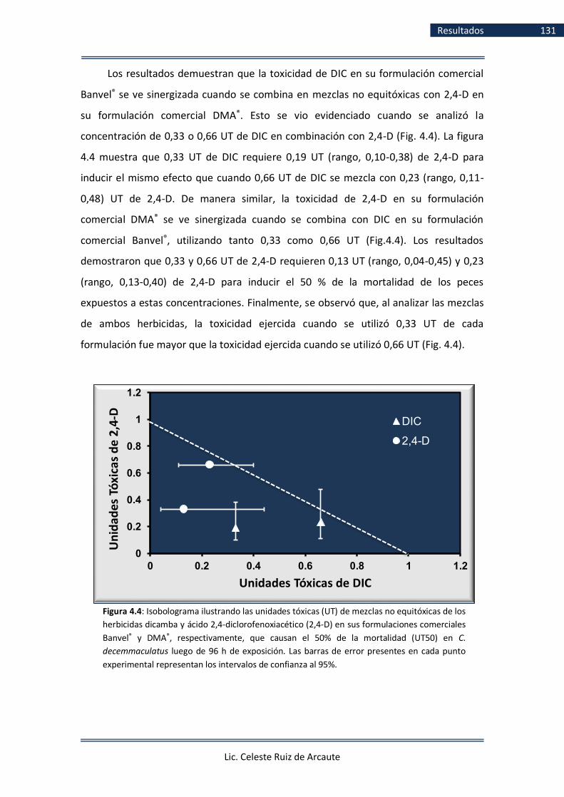
Lic. Celeste Ruiz de Arcaute
Resultados 131
Los resultados demuestran que la toxicidad de DIC en su formulación comercial
Banvel® se ve sinergizada cuando se combina en mezclas no equitóxicas con 2,4-D en
su formulación comercial DMA®. Esto se vio evidenciado cuando se analizó la
concentración de 0,33 o 0,66 UT de DIC en combinación con 2,4-D (Fig. 4.4). La figura
4.4 muestra que 0,33 UT de DIC requiere 0,19 UT (rango, 0,10-0,38) de 2,4-D para
inducir el mismo efecto que cuando 0,66 UT de DIC se mezcla con 0,23 (rango, 0,11-
0,48) UT de 2,4-D. De manera similar, la toxicidad de 2,4-D en su formulación
comercial DMA® se ve sinergizada cuando se combina con DIC en su formulación
comercial Banvel®, utilizando tanto 0,33 como 0,66 UT (Fig.4.4). Los resultados
demostraron que 0,33 y 0,66 UT de 2,4-D requieren 0,13 UT (rango, 0,04-0,45) y 0,23
(rango, 0,13-0,40) de 2,4-D para inducir el 50 % de la mortalidad de los peces
expuestos a estas concentraciones. Finalmente, se observó que, al analizar las mezclas
de ambos herbicidas, la toxicidad ejercida cuando se utilizó 0,33 UT de cada
formulación fue mayor que la toxicidad ejercida cuando se utilizó 0,66 UT (Fig. 4.4).
0
0.2
0.4
0.6
0.8
1
1.2
0 0.2 0.4 0.6 0.8 1 1.2
Un
idad
es T
óxi
cas
de
2,4
-D
Unidades Tóxicas de DIC
DIC
2,4-D
Figura 4.4: Isobolograma ilustrando las unidades tóxicas (UT) de mezclas no equitóxicas de los
herbicidas dicamba y ácido 2,4-diclorofenoxiacético (2,4-D) en sus formulaciones comerciales
Banvel® y DMA®, respectivamente, que causan el 50% de la mortalidad (UT50) en C.
decemmaculatus luego de 96 h de exposición. Las barras de error presentes en cada punto
experimental representan los intervalos de confianza al 95%.
The isobole linking the 1-TU values from both axes is the line of concentration
addition.
Isobologram depicting the toxic units (TUs) of non-equitoxic mixtures of
dicamba-based formulation Banvel® and 2,4-dichlorophenoxyacetic acid (2,4-D)-based
formulation DMA® that cause 50% mortality (TU50) in C. decemmaculatus after 96 h of

Lic. Celeste Ruiz de Arcaute
Resultados 132
4.3 Ensayos crónicos. Cuantificación de la inducción de lesiones primarias en el
ADN mediante el Ensayo cometa (publicación IV)
Los resultados del EC crónico obtenidos luego de la exposición a DIC con
concentraciones de 2,5 y 5% de la CL5096h se presentan en la Tabla 4.14 y Figura 4.5
(ver publicación IV). Luego de la exposición a 2,5% CL5096h (41 mg/L DIC), se observó
un aumento significativo del IDG luego de 7 (P < 0,001), 14 (P < 0,001) y 21 d (P < 0,05).
En el caso de los individuos expuestos al 5% CL5096h (82 mg/L DIC), tal incremento fue
observado sólo luego de 7 d de exposición (P < 0,001; Tabla 4.13). Adicionalmente, se
observó un aumento significativo en el porcentaje de nucleoides dañados en los peces
tratados con el 2,5% y 5 % de la CL5096h luego de 7 d de exposición (P < 0,001),
mientras que luego de 14 d de exposición tal aumento fue observado sólo en los
individuos tratados con el 2,5% CL5096h (P < 0,001).
En el tratamiento con el 2,5% de la CL5096h de DIC, el mayor porcentaje de
nucleoides dañados fue observado luego de 14 d de exposición con un valor de
58,30%, mientras que este porcentaje disminuyó hasta alcanzar el valor mínimo de
21,04% luego de 28 d de exposición. El porcentaje de nucleoides dañados para el
tratamiento del 5% de la CL5096h de DIC tuvo su valor máximo (77,20%), a los 7 d de
exposición, mientras que el valor mínimo observado fue 20,04% luego de 21 d de
exposición. El daño en el ADN observado para los peces expuestos a DIC fue tiempo-
dependiente, tanto para la concentración de 2,5% de la CL5096h (r = -0,74, P < 0,05)
como para 5% de la CL5096h (r = -0,84, P < 0,01). La variación en los nucleoides dañados
fue dada por un aumento de los nucleoides II y III (P < 0,001) a los 7 d de exposición. A
los 14 d de tratamiento, el aumento en los nucleoides dañados fue debido al
incremento de los nucleoides tipo III (0.01 > P < 0,001). Adicionalmente, luego de 14 d
se observó un aumento en la frecuencia de los nucleoides tipo II en los individuos
tratados con el 2,5% (P < 0,001). Finalmente, se observó una disminución significativa
de los nucleoides 0-I en los individuos tratados con 2,5% luego de 7 y 14 d de
exposición y en los individuos tratados con 5% sólo luego de 7 d de tratamiento (P <
0,001; Fig. 4.5).
Los valores de IDG observados en el tratamiento con el 5% de la CL5096h de DIC
mostraron un valor máximo de 2,07 a los 7 d de exposición y un valor mínimo de 0,95 a

Lic. Celeste Ruiz de Arcaute
Resultados 133
los 28 d de exposición. Para el tratamiento de 2,5% de la CL5096h de DIC el valor
máximo de IDG fue 1,66, observado luego de 14 d de exposición. Adicionalmente, al
valor mínimo de IDG fue 0,99, observado luego de 28 d de exposición (Tabla 4.14).
Los valores de IDG observados en el tratamiento con el 5% de la CL5096h de DIC
mostraron un valor máximo de 2,07 a los 7 d de exposición y un valor mínimo de 0,95 a
los 28 d de exposición.
Figura 4.5: Frecuencias de nucleoides en individuos de C. decemmaculatus expuestos al herbicida
dicamba en su formulación comercial Banvel® luego de 7, 14, 21 y 28 d. Barra negra: nucleoides no
dañados, tipo 0-I; barra azul: tipo II; barra blanca: tipo III y barra celeste: tipo IV. CN: Control
negativo. *P < 0,05; **P < 0,01; ***P < 0,001; diferencias significativas con respecto al control
negativo.

Resu
ltado
s 134
Lic. Celeste R
uiz d
e Arcau
te
Tabla 4.14 Análisis del daño en el ADN medido con el Ensayo Cometa en células sanguíneas de Cnesterodon decemmaculatus expuestos de manera crónica al
herbicida dicamba en su formulado comercial Banvel®
Compuestos Concentración
(mg/L)
Tiempo de exposición
(d)
Número de animales
analizados
Número de nucleoides analizados
% de nucleoides
dañados (II+III+IV)
IDG ± EE a Categorías de los nucleoides % ± EE
Tipo 0+I Tipo II Tipo III Tipo IV
CNb 7 10 1027 89,58 9,93 0,49 0 10,42 0,69 ± 0,05
14 10 1003 87,24 11,46 1,10 0,20 12,76 0,77 ± 0,07
21 10 1010 88,91 9,70 1,39 0 11,09 0,72 ± 0,11
28 10 1048 86,07 13,17 0,76 0 13,93 0,74 ± 0,07
Dicamba 2,5% CL5096h 7 10 1177 56,84*** 31,6*** 10,37*** 1,19 43,16*** 1,38 ± 0,11***
14 10 1163 41,7*** 41,27*** 17,03*** 0 58,30*** 1,66 ± 0,08***
21 10 1018 78,68 20,04 1,28 0 21,32 1,11 ± 0,04**
28 10 1112 78,96 19,24 1,80 0 21,04 0,99 ± 0,09
5% CL5096h 7 10 1013 22,8*** 49,46*** 25,96*** 1,78 77,20*** 2,07 ± 0,06***
14 10 1064 69,08 23,59 7,33** 0 30,92 1,15 ± 0,14
21 10 1048 79,96 18,89 1,15 0 20,04 0,99 ± 0,08
28 9 947 76,98 22,18 0,84 0 23,02 0,95 ± 0,06
a, IDG: Índice de daño genético. EE: Error estándar. b, CN: Control negativo. *P < 0,05; **P < 0,01; ***P < 0,001; diferencias significativas con respecto al control negativo

Lic. Celeste Ruiz de Arcaute
Resultados 135
Los resultados obtenidos luego de la exposición crónica a 2,4-D en su
formulación comercial DMA® se presentan en la Tabla 4.15 y en la Figura 4.6 (ver
publicación IV). En peces tratados con ambas concentraciones de 2,4-D (2,5 y 5% de la
CL5096h) se observó un aumento significativo del porcentaje de nucleoides dañados a
los 7, 14, 21 y 28 d de exposición con respecto al control negativo (P < 0,001) (Tabla
4.15). En ambas concentraciones ensayadas, a partir del daño observado en el día 7
(83,67% y 81,56%, para 2,5% y 5% de la CL5096h, respectivamente) se observó un
aumento gradual y significativo en el daño hasta el día 21 de tratamiento, momento en
el cual se observó el nivel máximo de daño (90,05% y 89,67% para 2,5% y 5% de la
CL5096h, respectivamente). Sin embargo, el nivel de daño observado disminuyó desde
el día 21 hasta el día 28 de iniciada la exposición al herbicida (74,49% y 71,99% para
2,5% and 5%, respectivamente), aunque continuó siendo significativamente mayor al
daño observado en el grupo control. El daño en el ADN observado en los peces
expuestos a 2,4-D fue tiempo-independiente, tanto para la concentración de 2,5% (r =
-0,48, P > 0,05) como para la concentración de 5% de la CL5096h (r = -0,41, P > 0,05). El
aumento en los nucleoides dañados fue dado por un aumento significativo de los
nucleoides tipo II, III y IV (P < 0,001) a los 7 d de exposición. A los 14 d, el incremento
estuvo dado por el aumento significativo de los nucleoides tipo II y III (P < 0,001).
Adicionalmente, a los 14 d de exposición se observó un aumento de los nucleoides tipo
IV en los individuos tratados con la concentración equivalente al 2,5% de la CL5096h (P
< 0,01) (Fig. 4.6). En los peces expuestos por 21 d la variación en la frecuencia de
nucleoides dañados fue dada por un aumento en la frecuencia de los nucleoides tipo II,
III y IV (P < 0,001). Por último, a los 28 d el incremento del porcentaje de daño estuvo
dado por un aumento significativo en los nucleoides tipo II-IV y II-III en los individuos
expuestos al 2,5% y 5% de la CL5096h, respectivamente (P < 0,001). Finalmente, a los 7,
14 21 y 28 d se observó una disminución significativa de los nucleoides no dañados,
tipo 0-I (P < 0,001) (Fig. 4.6).
De manera similar, se observó un aumento significativo del IDG tanto para el
tratamiento con el 2,5% de la CL5096h de 2,4-D como para el realizado empleando una
concentración del 5% de la CL5096h, para los cuatro tiempos de exposición evaluados,
7, 14, 21 y 28 d (P < 0,001) (Tabla 4.15).
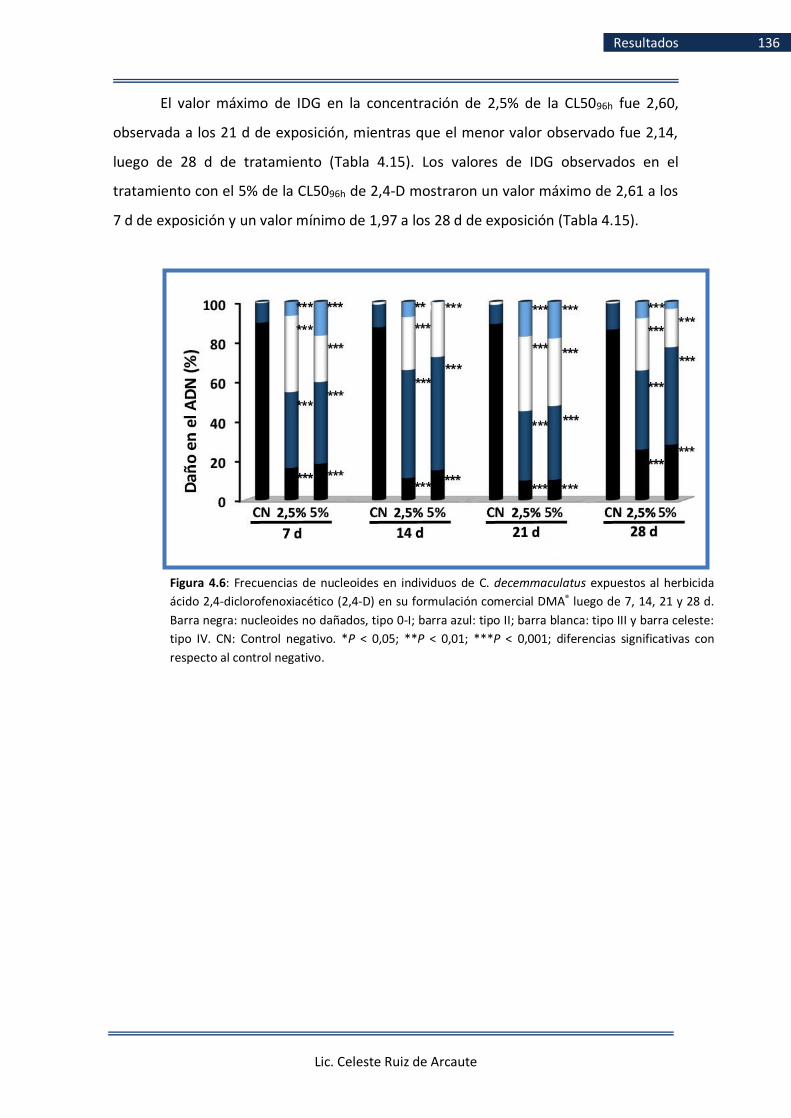
Lic. Celeste Ruiz de Arcaute
Resultados 136
El valor máximo de IDG en la concentración de 2,5% de la CL5096h fue 2,60,
observada a los 21 d de exposición, mientras que el menor valor observado fue 2,14,
luego de 28 d de tratamiento (Tabla 4.15). Los valores de IDG observados en el
tratamiento con el 5% de la CL5096h de 2,4-D mostraron un valor máximo de 2,61 a los
7 d de exposición y un valor mínimo de 1,97 a los 28 d de exposición (Tabla 4.15).
Figura 4.6: Frecuencias de nucleoides en individuos de C. decemmaculatus expuestos al herbicida
ácido 2,4-diclorofenoxiacético (2,4-D) en su formulación comercial DMA® luego de 7, 14, 21 y 28 d.
Barra negra: nucleoides no dañados, tipo 0-I; barra azul: tipo II; barra blanca: tipo III y barra celeste:
tipo IV. CN: Control negativo. *P < 0,05; **P < 0,01; ***P < 0,001; diferencias significativas con
respecto al control negativo.

Resu
ltado
s 137
Lic. Celeste R
uiz d
e Arcau
te
Tabla 4.15
Análisis del daño en el ADN medido con el Ensayo Cometa en células sanguíneas de Cnesterodon decemmaculatus expuestos de manera crónica al herbicida ácido 2,4-diclorofenoxiacético (2,4-D) en su formulado comercial DMA®
Compuestos Concentración
(mg/L)
Tiempo de
exposición (h)
Número de
animales analizados
Número de
nucleoides analizados
% de nucleoides
dañados (II+III+IV)
IDG ± EE a Categorías de los nucleoides % ± EE
Tipo 0+I Tipo II Tipo III Tipo IV
CNb 7 10 1027 89,58 9,93 0,49 0 10,42 0,69 ± 0,05
14 10 1003 87,24 11,47 1,10 0,20 12,76 0,77 ± 0,07
21 10 1010 88,91 9,70 1,39 0 11,09 0,72 ± 0,11
28 10 1048 86,07 13,17 0,76 0 13,93 0,74 ± 0,07
2,4-D 2,5% CL5096h 7 10 1078 16,33*** 38,22*** 38,40*** 7,05*** 83,67*** 2,36 ± 0,09***
14 10 1046 11,19*** 54,49*** 26,67*** 7,65** 88,81*** 2,30 ± 0,06***
21 10 1256 9,95*** 34,95*** 37,58*** 17,52*** 90,05*** 2,60 ± 0,12***
28 10 1184 25,51*** 39,95*** 26,26*** 8,28*** 74,49*** 2,14 ± 0,06***
5% CL5096h 7 9 976 18,44*** 41,19*** 23,26*** 17,11*** 81,56*** 2,39 ± 0,10***
14 10 1135 15,15*** 57,09*** 27,58*** 0,18 84,85*** 2,13 ± 0,04***
21 10 1220 10,33*** 37,21*** 34,02*** 18,44*** 89,67*** 2,61 ± 0,11***
28 10 989 28,01*** 49,14*** 19,31*** 3,54 71,99*** 1,97 ± 0,09***
a, IDG: Índice de daño genético. EE: Error estándar. b, CN: control negativo. *P < 0,05; **P < 0,01; ***P < 0,001; diferencias significativas con respecto al control negativo
Lic. Celeste Ruiz de Arcaute
Discusión 138
5. Discusión
Los resultados del presente trabajo de Tesis Doctoral aportan información sobre
los efectos deletéreos ejercidos por los herbicidas auxínicos DIC y 2,4-D, contenidos en
las formulaciones comerciales Banvel® (57,7% p.a.) y DMA® (58,4% p.a.),
respectivamente, los que son ampliamente utilizados en la agricultura argentina. Los
posibles efectos nocivos inducidos por estos herbicidas auxínicos fueron evaluados
mediante biomarcadores de efecto tanto letales como subletales. Entre los primeros
se emplearon ensayos de mortalidad agudos y entre los subletales se analizaron
efectos deletéreos en el comportamiento, genotóxicos, enzimáticos y no enzimáticos
en ejemplares adultos de la especie C. decemmaculatus (Pisces, Poeciliidae), especie
objeto de estudio, expuestos en condiciones experimentales de laboratorio. Asimismo,
se realizaron bioensayos con el objetivo de esclarecer los posibles mecanismos de
daño a nivel de la molécula de ADN que dichos compuestos ejercen. Del mismo modo,
se analizaron los posibles efectos por interacción deletéreos inducidos en C.
decemmaculatus luego de la aplicación de mezclas de ambos compuestos y, por
último, se evaluaron, asimismo, los efectos genotóxicos inducidos en el ADN de C.
decemmaculatus luego de una exposición crónica a los herbicidas.
Los resultados de los estudios de letalidad mediante la estimación de la CL50
revelaron que el formulado DMA®, conteniendo 58,4% de 2,4-D, indujo una toxicidad
1,62 veces mayor a la ejercida por el formulado Banvel®, conteniendo 57,7% de DIC en
su presentación comercial. En cuanto a los resultados del análisis de la alteración en
los patrones de comportamiento, los individuos de C. decemmaculatus fueron
expuestos a las mismas concentraciones utilizadas para la determinación de la CL50

Lic. Celeste Ruiz de Arcaute
Discusión 139
tanto para DIC como para 2,4-D. Luego de la exposición, se observaron patrones de
comportamiento que difirieron significativamente con respecto a los valores controles
para los cuatro tiempos de exposición comprendidos en el lapso de 0-96 h
(Publicaciones I y II).
Con respecto a los resultados del análisis de la genotoxicidad inducida por ambas
formulaciones comerciales, el ensayo de MNs reveló un aumento significativo del
efecto genotóxico únicamente a las 48 h de exposición para la concentración más alta
ensayada del formulado de DIC (1229 mg/L), equivalente al 75% de la CL5096h respecto
de los valores controles. Adicionalmente, para el formulado basado en 2,4-D, la
inducción de MNs fue observada para ambos tiempos de exposición (48 y 96 h) en las
tres concentraciones ensayadas, equivalentes al 25, 50 y 75% de la CL5096h (252, 504 y
756 mg/L). En cuanto a la inducción de daño genotóxico estimado mediante el EC, se
observó un aumento significativo con respecto a los valores controles en el porcentaje
de nucleoides dañados y en el índice de daño genético (IDG) tanto para el formulado
de DIC como para el de 2,4-D, con todas las concentraciones ensayadas luego de 48 y
de 96 h de exposición (Publicaciones I y II).
En peces tratados con el 25% de la CL5096h tanto de DIC como de 2,4-D, el
análisis del EC modificado con enzimas de restricción puso en evidencia que
únicamente las células de los peces que recibieron un post-tratamiento con la enzima
Fpg mostraron un aumento significativo del daño oxidativo (DO), no siendo así en los
tratados con Endo III, tanto luego de 48 como de 96 h (Publicación V).
En cuanto a los resultados de los biomarcadores enzimáticos y no enzimáticos,
luego de la exposición aguda a los formulados de DIC y 2,4-D, tanto la actividad de CAT
como GST aumentaron significativamente con respecto a valores controles tanto a las
48 como a las 96 h de exposición. Por el contrario, la actividad de GSH mostró una
tendencia hacia la disminución de su actividad a las 48 y a las 96 h, siendo la misma
significativa para el formulado conteniendo DIC sólo a las 48 h y para el formulado 2,4-
D sólo a las 96 h. Por último, el análisis de la actividad de AChE demostró una
tendencia hacia la disminución de su actividad luego del tratamiento con ambas
formulaciones de auxínicos tanto a las 48 como a las 96 h, llegando a ser significativa
sólo luego de las 96 h de exposición (Publicación V).

Lic. Celeste Ruiz de Arcaute
Discusión 140
Los resultados concernientes a evaluar la toxicidad y posibles interacciones
resultantes de la mezcla entre los formulados de DIC y 2,4-D, Banvel® y DMA®,
respectivamente, demostraron para mezclas equitóxicas un patrón aditivo, mientras
que para mezclas no equitóxicas se observó un patrón sinérgico en las cuatro
combinaciones no equitóxicas ensayadas (Publicación III).
Finalmente, la evaluación de los efectos genotóxicos inducidos por ambos
herbicidas mediante la variante alcalina del EC luego de una exposición crónica
empleando concentraciones equivalentes al 2,5% y 5% de la CL5096h, demostró que
ambos compuestos fueron capaces de inducir lesiones primarias en el ADN, luego de
los 7, 14, 21 y 28 d de exposición a DIC y 2,4-D (Publicación IV).
Es importante destacar que en la presente Tesis Doctoral se emplearon los
herbicidas DIC y 2,4-D en sus formulaciones comerciales Banvel® (57,7% p.a.) y DMA®
(58,4% p.a.), respectivamente. Es sabido que, en la agricultura, los plaguicidas son
usualmente comercializados y empleados en formulados, en los cuales el p.a. se
encuentra combinado con otros componentes tales como solventes, emulsionantes y
tensioactivos, entre otros, que mejoran la penetración y rendimiento del plaguicida
(WHO, 1990). Aunque las formulaciones de plaguicidas son desarrolladas bajo
procesos estrictamente regulados para que su impacto en el ser humano y en el
ambiente sea el mínimo posible, serias dudas han surgido en torno a los riesgos que
dichos compuestos pueden ejercer en la salud de los trabajadores ocupacionales,
residentes de las áreas adyacentes a las zonas de aplicación al igual que sobre la biota
en general, así como por los residuos potenciales generados y de los cuales se han
detectado niveles de los mismos presentes en alimentos y agua para consumo humano
(WHO, 1990).
Desde hace varias décadas se han reportado numerosas investigaciones
demostrando que los excipientes presentes en las formulaciones comerciales tienen la
habilidad de inducir toxicidad y daño celular por sí mismos, en adición a los efectos
inherentes al p.a. (USEPA, 1982; Mann y Bidwell, 1999; Soloneski y Larramendy, 2010;
Molinari y col., 2013; Nikoloff y col., 2014a). Por lo tanto, las evaluaciones de riesgo
sobre la biota también deben considerar los efectos inducidos por estos excipientes.
Desafortunadamente, la identidad de los compuestos presentes en los formulados
comerciales Banvel® y DMA® no han sido informados por los fabricantes, ya que, en

Lic. Celeste Ruiz de Arcaute
Discusión 141
nuestro país, la identidad de los excipientes que conforman una determinada
formulación comercial se encuentran protegidos por ley y son considerados secreto
comercial.
Por lo expuesto anteriormente, resulta de importancia vital, en adición a la
determinación de los efectos inducidos por los p.a., la evaluación de los efectos
deletéreos de las formulaciones de plaguicidas tal cual son comercializadas y
empleadas tanto en la agricultura a gran escala como en zonas urbanas o peri-urbanas,
ya que, en definitiva, son a las que están expuestas los organismos en el medio
ambiente, incluyendo los seres humanos.
Durante los últimos diez años nuestro grupo de trabajo ha empleado ejemplares
de vertebrados acuáticos, entre ellos de C. decemmaculatus, como especies de estudio
para evaluar los efectos letales y subletales de diversas formulaciones comerciales de
agroquímicos usados tanto en la agricultura mundial como en Argentina, en lo
particular. Entre los compuestos evaluados en ejemplares adultos de C.
decemmaculatus podemos citar las formulaciones Lorsban® 48E y CPF Zamba®, ambos
conformados por el insecticida clorpirifós al 48% (Vera-Candioti y col., 2014), las
formulaciones Aficida® y Patton Flow®, ambas conteniendo el insecticida carbámico
pirimicarb al 50% (Vera-Candioti y col., 2010; 2011; 2015) y dos formulaciones del
herbicida glifosato al 48%, Panzer® y Credit® (Vera-Candioti y col., 2013a). En todos los
casos, la estimación de la CL50 se utilizó como biomarcador de letalidad, mientras que
la inducción de MNs y rupturas en la cadena de ADN analizadas mediante el EC se
utilizaron como biomarcadores de genotoxicidad. Adicionalmente, variaciones en la
relación eritrocitos/eritroblastos en muestras de sangre periférica de ejemplares
expuestos fue utilizada como biomarcador de citotoxicidad (Vera-Candioti y col.,
2013b). Por último, recientemente nuestro grupo de trabajo evaluó la toxicidad
inducida por mezclas de los herbicidas glifosato, DIC y 2,4-D, mediante estudios de
letalidad aguda en C. decemmaculatus (Ruiz de Arcaute y col., 2018).
Cabe resaltar que nuestras investigaciones constituyen la primera evidencia del
uso de biomarcadores de efecto genotóxico y de estrés oxidativo para evaluar la
capacidad deletérea ejercida por los herbicidas DIC y 2,4-D en sus variantes
comerciales Banvel® y DMA®, respectivamente, así como también en la evaluación de la
letalidad inducida por la mezcla de ambos compuestos utilizadas actualmente en el

Lic. Celeste Ruiz de Arcaute
Discusión 142
agro de nuestro país, empleando el poecílido C. decemmaculatus como modelo
experimental de estudio.
5.1 Evaluación de la letalidad inducida por dicamba y el ácido 2,4-
diclorofenoxiacético (2,4-D) en sus formulaciones comerciales Banvel® y
DMA®
El peligro potencial que un xenobiótico representa para el ecosistema acuático es
determinado luego de la estimación de la toxicidad aguda para mamíferos, la toxicidad
aguda para bacterias, toxicidad aguda para peces y de la realización de ensayos de
disociación biológica del compuesto (Yilmaz y col., 2004). Aunque los ensayos de
toxicidad inicialmente se llevaban a cabo empleando bacterias e invertebrados tales
como rotíferos y cladóceros, éstos no pueden reemplazar a los ensayos de toxicidad
empleado vertebrados tales como peces, debido a que éstos constituyen
generalmente el último eslabón dentro de la cadena trófica del ambiente acuático
(Castano y col., 1996; Yilmaz y col., 2004).
Los ensayos de toxicidad aguda brindan información acerca de los efectos
inducidos a corto plazo por un compuesto en organismos de prueba con el objetivo de
determinar su sensibilidad y luego poder compararla con la manifestada en otras
especies (USEPA, 1975; ASTM, 2007; Casarett y Doull´s, 2008). Estos resultados son
luego utilizados para implementar el diseño y la selección de las concentraciones a
utilizar en ensayos subletales posteriores realizados bajo condiciones experimentales
de laboratorio (Casarett y Doull´s, 2008). Asimismo, es dable esperar que dichos
resultados sean tomados como referencia por diferentes instituciones
gubernamentales para determinar, en última instancia, niveles guía de calidad y de
consumo del agua de los seres humanos (WHO, 2008). En términos generales, un
ensayo de toxicidad aguda consiste en exponer a un grupo de individuos a diversas
concentraciones de un contaminante por un plazo corto de tiempo para luego evaluar
los efectos inducidos por el mismo. Entre los efectos evaluados, se encuentran la
mortalidad y varios efectos subletales, por ejemplo, malformaciones, alteraciones del
comportamiento al igual que distintos efectos fisiológicos, entre otros (ASTM, 2007;
Casarett y Doull´s, 2008). El parámetro más utilizado para expresar los resultados de

Lic. Celeste Ruiz de Arcaute
Discusión 143
mortalidad es la CL50, la cual es definida como la concentración de una sustancia que
produce la muerte del 50% de los individuos expuestos a la misma en un lapso de
exposición determinado. También es utilizado el cálculo de la concentración efectiva
50 (CE50), que determina la concentración de una sustancia que produce un
determinado efecto en el 50% de los individuos expuestos a la misma. Asimismo,
podemos incluir el NOEC, el cual determina de entre las concentraciones testeadas, la
concentración más alta a la cual no fue observado un determinado efecto adverso y el
LOEC, que determina la concentración más baja a partir de la cual se comenzó a
observar dicho efecto (USEPA, 1975; ASTM, 2007; Casarett y Doull´s, 2008). Dichos
parámetros son ampliamente utilizados en la evaluación de la toxicidad de un
compuesto y en su posterior comparación con respecto a los efectos inducidos por
otros contaminantes.
Los efectos letales observados en el presente trabajo de Tesis Doctoral
(Publicación I y II), inducidos por los formulados comerciales de DIC y 2,4-D, Banvel® y
DMA®, respectivamente, permitieron determinar que la formulación conteniendo 2,4-
D fue más tóxica que el formulado conteniendo DIC. El análisis de la letalidad reveló
una CL5096h de 1639 mg/L (rango, 1471-1808 mg/L) para DIC en su formulación
comercial Banvel®. Por otra parte, cuando se analizó la letalidad inducida por el
herbicida 2,4-D en su formulación DMA®, la CL5096h observada fue 1008 mg/L (rango,
929-1070 mg/L). De esta manera, cuando se expusieron ejemplares de C.
decemmaculatus durante 96 h a la formulación del herbicida DIC, fue necesario utilizar
1,62 veces más que de la formulación conteniendo 2,4-D para obtener el mismo
efecto, en este caso, el 50% de la mortalidad. Una evaluación de los datos reportados
hasta el presente de la letalidad inducida por DIC y 2,4-D, estimados como valores de
CL5096h, pone en evidencia que el efecto letal de estos herbicidas ha sido analizado en
diferentes matrices bióticas distintas a la especie empleada en nuestro trabajo de Tesis
Doctoral. En la Tabla 5.1 se encuentran resumidos los estudios previos realizados en
distintas especies expuestas a DIC y 2,4-D.

Lic. Celeste Ruiz de Arcaute
Discusión 144
Tabla 5.1 CL5096h reportadas para distintos organismos luego de la exposición a los herbicidas auxínicos
dicamba y al ácido 2,4- diclorofenoxiacético (2,4-D)
Compuesto Grupo Especie CL5096h
(mg/L) Referencia
DIC Crustáceos Amphipoda 3,9 Mayer (1974)
CAS
Paleomonetes pugio > 100 U.S. EPA (1992)
1918009
Paleomonetes kadiakensis > 560 Mayer y Ellersieck (1986)
Uca pugilator > 180 U.S. EPA (1992)
Caecidotea brevicauda > 100 Mayer y Ellersieck (1986)
Gammarus lacustris 3,9
Sanders (1989), U.S. EPA (1992)
Gammarus fasciatus >100 Mayer y Ellersieck (1986)
Anfibios Limnodynastes peronii 106 Johnson (1976)
Adelotus brevis 185 Johnson (1976)
Peces Lepomis macrochirus 135,3 U.S. EPA (1992)
>500 Mayer y Ellersieck (1986)
Cyprinodon variegatus > 180 U.S. EPA (1992)
Oncorhynchus mykiss 135,4 U.S. EPA (1992)
153 Johnson (1978)
Oncorhynchus clarkii 50 Lorz y col. (1971)
Gambusia affinis 465 Johnson (1978)
Cnesterodon decemmaculatus 1639
Presente trabajo de Tesis Doctoral, Publicación I
2,4-D Crustáceos Caridina laevis 872 Sucahyo y col. (2008)
CAS
Daphnia magna 35 Alexander 1985
2008391
Procambarus clarkii 185 Green y Abdelghani (2004)
1389 Cheah y col. (1980)
Procambarus sp. 750,1 Abdelghani y Col. (1997)
Gammarus fasciatus > 100 Mayer y Ellersieck (1986)
Micropterus dolomieu 236 Mayer y Ellersieck (1986)
Trichogaster pectoralis 153 Ooi (1988)
Anfibios Limnodynastes peronii 287 Johnson (1976)
Pseudacris triseriata 100 Sanders (1970)
Rhinella marina 282,84 Figueiredo y col. (2014)
288 Johnson (1976)
Adelotus brevis 200 Johnson (1976)
Physalaemus centralis 515,75 Figueiredo y col. (2014)
Peces Pimephales promelas 266-800 Mayer & Ellersieck (1986)
106-318 U.S. EPA (1992)
344 Alexander y col. (1985)
Tinca tinca L. 48 Sarikaya y col. (2003)
Ictalurus punctatus 100-395 Mayer y Ellersieck (1986)
Lepomis macrochirus 100-177 Mayer y Ellersieck (1986)
Menidia beryllina 469 U.S. EPA (1992)
Oncorhynchus tshawytscha > 100 Mayer y Ellersieck (1986)
Oncorhynchus mikiss 420 Mayer y Ellersieck (1986)

Lic. Celeste Ruiz de Arcaute
Discusión 145
250 U.S. EPA (1992)
707 Fairchild y col. (2008)
117 Bills y col. (1981)
Cnesterdodon decemmaculatus 1009
Presente trabajo de Tesis Doctoral, Publicación II
Del análisis de la presente Tabla se puede observar que la evaluación de la
toxicidad inducida por el herbicida DIC mediante la determinación de la CL5096h fue
realizada sólo en cinco especies de peces. Para la mojarra oreja azul (Lepomis
macrochirus), la CL5096h reportada por la U.S. EPA (1992) es de 135,3 mg/L p.a. DIC y
por Mayer y Ellersieck (1986) mayor a 500 mg/L p.a. DIC. El valor de CL5096h reportada
para C. variegatus, fue mayor a 180 mg/L p.a. DIC (USEPA, 1992). Para O. mykiss, la
CL5096h reportada es de 135,4 y 153 mg/L p.a. DIC (Johnson, 1978; USEPA, 1992). Para
O. clarkii el valor de CL5096h reportado por Lorz y col. (Lorz y col., 1979) es de 50 mg/L
p.a. DIC, y para G. affinis el valor de CL5096h es de 465 mg/L de una formulación no
especificada (Johnson, 1978).
En cuanto al herbicida 2,4-D, se ha evaluado la toxicidad en siete especies de
peces. Entre ellos, la carpita cabezona (Pimephales promelas) con un valor de CL5096h
reportado por Mayer y Ellersieck (1986) de 266-800 mg/L p.a. 2,4-D, por la U.S. EPA
(1992) de 106-318 mg/L p.a. 2,4-D y por Alexander y col. (1985) de 344 mg/L p.a. 2,4-
D. Los valores de CL5096h reportados para la especie Tinca L. son de 48 mg/L 2,4-D
(Sarikaya y Yilmaz, 2003), y para el pez gato americano (Ictalurus punctatus) de 100-
395 mg/L p.a. 2,4-D (Mayer y Ellersieck, 1986). Para L. macrochirus, el valor reportado
de CL5096h es de entre 100 a 177 mg/L p.a. 2,4-D (Mayer y Ellersieck, 1986).
Adicionalmente, para el plateadito salado (Menidia beryllina), la CL5096h reportada fue
de 469 mg/L (USEPA, 1992; compuesto no especificado) y para el salmón real (O.
tshawytscha) mayor a 100 mg/L p.a. 2,4-D (Mayer y Ellersieck, 1986). Por último, para
la especie O. mykiss el valor de CL5096h reportada en distintos estudios fue de 420
mg/L p.a. 2,4-D (Mayer y Ellersieck, 1986), 117 (Bills y col., 1981), 250 (USEPA, 1992) y
707 mg/L (Fairchild y col., 2008). Ésta última concentración es la única obtenida para
O. mykiss empleando un formulado comercial de 2,4-D (56,7% Nufarm), el resto fueron
obtenidas ensayando el p.a.

Lic. Celeste Ruiz de Arcaute
Discusión 146
Nuestros resultados mostraron un valor de CL5096h de 1639 mg/L para el
herbicida DIC en su formulado comercial Banvel®. Teniendo en cuenta estos resultados
y considerando las categorías de toxicidad propuestas por la EU (Mazzatorta y col.,
2002), DIC pertenecería a la categoría de compuestos que pueden causar efectos
adversos a largo plazo (categoría IV) (Mazzatorta y col., 2002). De la misma manera,
podría ser clasificado como un compuesto clase IV (“prácticamente no tóxico”) para la
U.S. EPA (2001) y como un compuesto clase II (“moderadamente peligroso”) para la
WHO (2009). Adicionalmente, según la clasificación para organismos acuáticos
propuesta por la U.S. EPA, DIC podría ser clasificado como un compuesto
prácticamente no tóxico (USEPA, 2001).
En cuanto al herbicida 2,4-D en su formulación comercial DMA®, el valor de
CL5096h obtenido en el presente trabajo de Tesis Doctoral fue de 1008 mg/L. Siguiendo
el mismo criterio, el herbicida 2,4-D según el valor de CL5096h obtenido puede ser
clasificado según la U.S. EPA (2001) como un compuesto prácticamente no tóxico
(clase IV). De acuerdo con la UN (2011), puede ser clasificado como un compuesto
nocivo (categoría III). Finalmente, según los criterios propuestos por la EU (Mazzatorta
y col., 2002) el 2,4-D puede ser clasificado como un compuesto que puede causar
daños a largo plazo para la vida acuática (categoría IV), y como un compuesto
“moderadamente peligroso” según la WHO (2009). Adicionalmente, siguiendo la
clasificación para organismos acuáticos propuesta por la U.S. EPA, el herbicida 2,4-D
podría ser clasificado, al igual de DIC, como un compuesto prácticamente no tóxico
para C. decemmaculatus (USEPA, 2008).
Si comparamos los valores de CL5096h reportados en el presente trabajo de Tesis
Doctoral para el herbicida DIC en su formulación comercial Banvel® con aquellos en la
literatura para distintas especies de peces expuestas a DIC (Tabla 5.1), C.
decemmaculatus puede ser considerada como la especie menos sensible reportada
hasta el momento, ubicándose detrás de G. affinis, con una CL5096h de 465 mg/L
(Johnson, 1978) y de L. macrochirus, con una CL5096h superior a los 500 mg/L (Mayer y
Ellersieck, 1986). En conclusión, la formulación Banvel®, conteniendo 57,7% de DIC
ejercería una mortalidad mucho menor que otras formulaciones comerciales con este
herbicida como p.a.
Lic. Celeste Ruiz de Arcaute
Discusión 147
Es relevante destacar que en general, las curvas de sensibilidad indicarían que las
especies nativas de Argentina son más sensibles con respecto a las especies modelo
utilizadas del hemisferio norte. A modo de ejemplo, Iturburu y col. (2017) reportaron
para Australoheros facetus, otra especie sudamericana una CL5096h <10 mg/L de
imidacloprid en su formulado comercial Imida Nova 351 (NOVA) (35% p.a.), mientras
que para el mismo insecticida, la CL5096h reportada para O. mykiss y C. caprio es de 211
y 280 mg/L (Tisler y col. 2009). Específicamente para C. decemmaculatus, tal
afirmación no debería ser considerada como válida, dado que se ha observado que la
especie muestra una sensibilidad mayor o menor que especies boreales tomadas como
modelo experimental, dependiendo el compuesto evaluado. Tal es el caso del
glifosato, para el cual se reportó una CL5096h de 15,68-100 mg/L para C.
decemmaculatus (Vera-Candioti y col. 2013; Carriquiriborde y col. 2007; Brodeur y col.
2016). Opuestamente, en especies nativas del hemisferio norte como C. variegatus, C.
carpio y O. mykiss, la CL5096h reportada para este herbicida fue de 240 mg/L (USEPA,
1992), 11-520,77 mg/L (Ma y Li, 2015; Sun, 1987) y 7,6-240 mg/L (Mayer y Ellersieck,
1986), respectivamente (Tabla 5.2). Una respuesta de sensibilidad semejante se
obtuvo para el insecticida clorpirifós. La CL5096h reportada para C. decemmaculatus es
de 0,03-0,21 mg/L (Paracampo y col., 2015; Vera-Candioti y col. 2014), mientras que
para C. punctatus 0,81 mg/L (Phipps y Holcombe, 1985) y para L. macrochirus 0,88
mg/L (USEPA, 2013). Por otro lado, estudios evidenciando una menor sensibilidad de C.
decemmaculatus fueron reportados para cipermetrina por Brodeur y col. (2016) con
una CL5096h 0,19-0,43 mg/L, mientras que ha sido determinada una CL5096h de 0,0017,
0.0071 y 0,001 mg/L para C. carpio (Saha y col. 2008), O. mykiss (Lakota y col. 1987) y
L. macrochirus (El-Sabae y col. 1983; USEPA, 2013), respectivamente. Resultados
similares fueron obtenidos para el insecticida pirimicarb, para el que Vera-Candioti
reportó una CL5096h de 88-225 mg/L, mientras que para especies como C. variegatus,
P. promelas y O. mykiss las CL5096h reportadas fueron > 100, >101 y 29-300 mg/L
(USEPA, 2013), respectivamente.
Al realizar esta comparación para el herbicida 2,4-D en su formulación comercial
DMA®, C. decemmaculatus puede ser considerada, al igual que para DIC, como la
especie menos sensible reportada hasta el momento evaluando 2,4-D ubicándose
luego de O. mykiss, con una CL5096h máxima reportada de 707 mg/L (Fairchild y col.,

Lic. Celeste Ruiz de Arcaute
Discusión 148
2008). En base a estos resultados, podría afirmarse que el herbicida 2,4-D en su
formulación comercial DMA®, compuesta por el 58,4% de p.a., ejercería una
mortalidad mucho menor que las formulaciones comerciales evaluadas hasta el
momento con este herbicida como p.a.
Es importante tener en cuenta que al realizar estas comparaciones sobre la
sensibilidad de C. decemmaculatus con respecto a otras especies estudiadas, las
mismas son aproximadas, ya que si bien todos los ensayos fueron llevados a cabo
exponiendo a los individuos a la misma variante química de cada compuesto, cada
ensayo fue llevado a cabo empleando un determinado p.a. y/o formulado comercial,
cuyas cantidades tanto de p.a. como de excipientes difieren dentro de cada
composición comercial estudiada.
Los valores de CL5096h informados por estudios previos realizados en C.
decemmaculatus expuestos a distintos compuestos se encuentran resumidos en la
Tabla 5.2. De acuerdo con resultados reportados en el presente trabajo de Tesis
Doctoral, C. decemmaculatus es aproximadamente 0,6 veces más sensible al herbicida
2,4-D que a DIC, pudiendo ser considerados ambos herbicidas auxínicos (ya sean
pertenecientes a la familia de los ácidos benzoicos, como DIC; o a la familia de los
ácidos fenoxi-carboxílicos, como el 2,4-D) compuestos de menor toxicidad para C.
decemmaculatus que cualquier otro agroquímico estudiado hasta el momento (Tabla
5.2). Estudios adicionales deberían ser llevados a cabo para verificar si esta afirmación
se puede extender a las otras familias de herbicidas auxínicos (ácidos piridino-
carboxílicos, pirimidino-carboxílicos y quinolino-carboxílicos), o es particular de los
formulados estudiados en el presente trabajo.
Tabla 5.2 LC5096h reportadas para la especie Cnesterodon decemmaculatus
Compuesto CL5096ha Referencia
Cloruro de zinc 41 Gómez y col. (1998)
Sulfato de cobre 0,16 Villar y col. (2000)
0,65 Casares y col. (2012)
Cromo 21,4 Vera-Candioti y col. (2011)
Pirimicarb 225,5 Vera-Candioti y col. (2010)
88 Vera-Candioti y col. (2015)
Clorpirifós 0,21 Vera-Candioti y col. (2014)
0,03 Vera-Candioti y col. (2014)

Lic. Celeste Ruiz de Arcaute
Discusión 149
0,10 Paracampo y col. (2015)
Endosulfán 0,005 Mugni y col. (2012)
Paraquat 52,48-67,4 Di Marzio y col. (1994)
Cipermetrina 0,43 Carriquiriborde y col. (2007)
0.19 Brodeur y col. (2016)
0.26 Brodeur y col. (2016)
Glifosato >100 Carriquiriborde y col. (2007)
15,68 Vera-Candioti y col. (2013)
91,73 Vera-Candioti y col. (2013)
41 Brodeur y col. (2016)
53 Brodeur y col. (2016)
Dicamba 1639
Presente trabajo de Tesis Doctoral, Publicación I
2,4-D 1008
Presente trabajo de Tesis Doctoral, Publicación II
a, Valor expresado en mg/L
5.2 Evaluación de los efectos en el comportamiento inducidos por
dicamba y el ácido 2,4-diclorofenoxiacético (2,4-D) en sus formulaciones
comerciales Banvel® y DMA®
El comportamiento se define como la secuencia de acciones cuantificables que
implican efectos acumulativos de diversos procesos biológicos como genéticos,
bioquímicos y fisiológicos que se manifiestan en un individuo y que apuntan a la
aptitud máxima y la supervivencia del organismo. Es una manifestación de la conexión
entre la fisiología y la ecología de un organismo y su entorno (Little y Brewer, 2001),
por lo cual resulta un indicador importante de la presencia de tóxicos y cambios
ambientales. Los cambios comportamentales, en general, son estimadores más
sensibles que otros marcadores como la letalidad o el crecimiento (Little y Finger,
1990). Es importante destacar que, aunque la mortalidad no es un comportamiento, la
mayoría de los síntomas del comportamiento pueden culminar eventualmente con la
muerte del individuo (Quarcoo y col., 2014).
En el presente trabajo de Tesis Doctoral, se estudiaron las alteraciones en el
comportamiento de C. decemmaculatus expuestos a concentraciones subletales de los
herbicidas DIC y 2,4-D en sus formulaciones comerciales Banvel® y DMA®. Los
siguientes efectos fueron observados luego del tratamiento y durante los distintos

Lic. Celeste Ruiz de Arcaute
Discusión 150
tiempos de exposición (24, 48, 72 y 96 h): AFA, aglomeración en el fondo del acuario;
ML, movimiento lento; RL, reacción lenta y NA, natación anormal.
En los individuos expuestos al herbicida DIC, se pudo observar que a medida que
aumenta el tiempo de exposición, la concentración a la cual se comienzan a observar
los efectos en el comportamiento (LOEC) son cada vez menores. Así, a las 24 h de
iniciado el tratamiento, las alteraciones en el comportamiento comienzan a observarse
a partir de la concentración de 1440 mg/L, a las 48 h en la concentración de 1296
mg/L, y a las 72 y 96 h de exposición en la concentración de 1152 mg/L. Esto podría
explicarse por el deterioro del estado de los individuos a medida que aumenta el
tiempo de exposición debido a la mayor toxicidad inducida por el compuesto. Es
importante destacar que el presente estudio es el primer reporte de inducción de
alteraciones en el comportamiento en vertebrados acuáticos ejercidos por el herbicida
DIC.
En los individuos expuestos al herbicida 2,4-D, se puede observar que
prácticamente todos los efectos, con todos los tiempos analizados, se registran a partir
de la concentración de 800 mg/L. Esto es coincidente con lo observado en el ensayo de
letalidad, en el que concentraciones menores a 800 mg/L no indujeron efecto alguno,
probando ser insuficientes para causar toxicidad relacionada a estos puntos finales.
Por otro lado, a medida que transcurre el tiempo de exposición se observan menor
cantidad de alteraciones luego del análisis en los patrones de comportamiento. Así, a
las 24 h de exposición se observaron las cuatro alteraciones comportamentales (AFA,
ML, RL y NA), a las 48 h sólo se observaron tres (ML, RL y NA), a las 72 h sólo dos (RL y
NA) y a luego de 96 h de tratamiento la única alteración observada fue NA. Este patrón
de comportamiento podría sugerirse de acuerdo a un proceso de aclimatación de los
organismos a la exposición al herbicida, como ha sido reportado en distintas especies
de peces expuestas a plaguicidas, entre ellas el pez C. auratus luego de la exposición a
nicosulfuron (Saglio y col., 2003), a O. mykiss expuestos a deltametrina (Goulding y
col., 2013) y permetrina (Kumaraguru y Beamish, 1983). Futuros estudios son
necesarios para confirmar esta teoría y dilucidar los posibles mecanismos por los
cuales 2,4-D inducen las alteraciones en el comportamiento observadas y como los
individuos de C. decemmaculatus podrían adquirir una determinada tolerancia luego
de la exposición al herbicida en estudio.

Lic. Celeste Ruiz de Arcaute
Discusión 151
En cuanto a la inducción de alteraciones en el comportamiento por el herbicida
2,4-D en peces, Sarikaya y Yilmaz (2003) informaron efectos en C. carpio luego de la
exposición aguda a 2,4-D en su variante éster en el rango de concentraciones de 4,8-
144 mg/L. Entre los efectos en el comportamiento observados se encontraron
movimientos repentinos, pérdida del equilibrio, natación anormal, dificultades
respiratorias, aglomeración en la superficie del acuario, secreción excesiva de mucus al
igual que alteraciones en la pigmentación. El formulado de 2,4-D utilizado en dicho
estudio fue Ester´H (48% éster 2,4-D), y la CL5096h reportada por estos autores fue de
64,24 mg/L. Algunos de estos efectos son coincidentes con los observados por
nosotros en el presente estudio, aunque las concentraciones efectivas fueron
menores, tanto para los efectos letales como para los comportamentales. En adición,
Hansen y col. (1969, 1972) estudiaron los efectos de 2,4-D en concentraciones de 1 y
10 mg/L en C. variegatus (Hansen, 1969) y G. affinis (Hansen y col., 1972) observando
comportamientos tendientes a evitar la exposición al contaminante.
En relación a los efectos adversos en el comportamiento observados en el
presente trabajo de Tesis Doctoral, otros autores han reportado que bajo condiciones
de estrés como las que implican la exposición a plaguicidas, los individuos afectados en
el patrón normal de comportamiento podrían ser menos activos en la competencia
inter e intraespecífica, manifestando una menor aptitud física durante la evasión de
sus posibles depredadores o en una menor eficiencia durante la obtención de alimento
(Quarcoo y col., 2014). Esto los conduciría a una limitación en la energía disponible
para procesos metabólicos normales, afectando patrones como el crecimiento y la
reproducción de los organismos, pudiendo, en última instancia, causar efectos letales.
A nivel ecosistema, estos efectos adversos llevarían a una disminución poblacional, con
graves consecuencias en distintos procesos tanto ecológicos como evolutivos (Gliwicz,
1994; Preston y col., 1999; Relyea y Hoverman, 2006).
Diferentes tipos de alteraciones en el comportamiento inducidas por plaguicidas
fueron evaluadas desde el comienzo del uso masivo de los mismos. Broley (1958)
reportó en áreas rociadas con DDT, patrones anormales de anidación, cortejo y
reproducción en el águila calva (Haliaeetus leucocephalus) (Broley, 1958). Varios
estudios demostraron que plaguicidas organofosforados, organoclorados y carbamatos
alteraron el comportamiento en peces (Bell, 2001; Tierney y col., 2008), renacuajos

Lic. Celeste Ruiz de Arcaute
Discusión 152
(Relyea, 2012), aves (Holmes y Boag, 1990) y mamíferos (Vom Saal y col., 1995; Porter
y col., 1999; Flynn y col., 2001). Específicamente para peces, se observaron
alteraciones en la natación, pérdida del equilibrio, dificultades en la respiración,
cambios en la pigmentación, entre otros efectos, reportados para C. carpio luego de la
exposición a 2,4-D en su variante éster (Sarikaya y Yilmaz, 2003). Alteraciones en la
natación también reportada por Wang y col. (2018) en individuos de D. rario expuestos
a paraquat, por Qiu y col. (2017) en O. latipes expuestos a clorpirifós, Otros efectos,
como comportamientos tendientes a evitar la exposición a contaminantes fueron
observados en varias especies, entre ellas por ejemplo las ya mencionadas C.
variegatus (Hansen, 1969) y G. affinis (Hansen y col., 1972) expuestos a 2,4-D y en O.
mykiss expuestos a glifosato en concentraciones de 40 mg/L (Hildebrand y col., 1982;
Tierney y col., 2007; 2013), entre otros. Efectos relacionados con patrones natatorios y
sociales como agrupamiento o la búsqueda de refugio fueron observados en el pez C.
auratus expuestos al herbicida atrazina en concentraciones de 0,5 a 50 μg/L (Saglio y
Trijasse, 1998). Específicamente en ejemplares de C. decemmaculatus, Paracampo y
col. (2015) reportaron efectos adversos como natación anormal, menor actividad
natatoria y patrones respiratorios anormales luego de la exposición a clorpirifós en el
rango de concentraciones de 30-135 μg/L. Adicionalmente, Bonifacio y col. (2017)
informaron alteraciones durante la natación y uso del espacio en C. decemmaculatus y
en la mojarra de agua dulce (Cheirodon interruptus) expuestos a clorpirifós.
Gromley y col. (2003) evaluaron los efectos inducidos por el insecticida
endosulfán en huevos y juveniles de O. latipes luego una exposición por 24 h. Estos
autores reportaron que, en ambos casos, los individuos mostraron alteraciones en
comportamientos natatorios así como en los reproductivos manifestados como
variaciones en la cantidad de huevos producidos. Esto remarca que la exposición a
plaguicidas puede tener consecuencias negativas en los organismos que exceden el
tiempo de exposición (Gormley y Teather, 2003). Efectos similares fueron observados
por Frank y col. (2018) quienes reportaron que D. rerio que durante la exposición
aguda a concentraciones del insecticida bifentrin no manifestaron efectos inmediatos
en el comportamiento, pero sí tiempo después, una vez concluida la fase de exposición
(Frank y col., 2018).
Aunque varios estudios han reportado alteraciones en los patrones de

Lic. Celeste Ruiz de Arcaute
Discusión 153
comportamiento frente a la exposición a distintos herbicidas, la importancia y
consecuencias de estas alteraciones en la supervivencia, crecimiento, desarrollo y
reproducción o sustentabilidad de las poblaciones de peces no han sido cuantificadas.
Es conocido que los efectos sobre el comportamiento de las especies expuestas
pueden ser adaptativos y puede ocurrir un fenómeno de habituación a las nuevas
condiciones ambientales (Allen, 2010). En este sentido, son necesarios más estudios
tanto a nivel poblacional como de las comunidades de las cuales forman parte para
evaluar los efectos y consecuencias de estas alteraciones luego de la exposición a
xenobióticos.
5.3 Evaluación de la genotoxicidad inducida por dicamba y ácido 2,4-
diclorofenoxiacético (2,4-D) en sus formulaciones comerciales Banvel® y
DMA®
5.3.1 Inducción de micronúcleos
Los efectos genotóxicos son considerados como uno de los efectos subletales
más graves de los productos agroquímicos. Si un compuesto reacciona con el ADN
nuclear, puede tener efectos mutagénicos y carcinogénicos en los organismos
expuestos elevando el riesgo de producir enfermedades genéticas hereditarias,
carcinogénesis, disfunción reproductiva y defectos de nacimiento, entre otros (Ahmad
y Ahmad, 2016). En la actualidad uno de los biomarcadores de efecto más utilizados a
nivel mundial desarrollado para el estudio y caracterización de agentes genotóxicos es
el análisis de la frecuencia de MNs, ya sea en ensayos in vitro e in vivo, como también
en estudios de monitoreo ambiental (Mudry y Carballo, 2006). Este biomarcador
permite la detección de pequeños fragmentos cromosómicos o cromosomas
acéntricos en células interfásicas inducidos por agentes clastogénicos, o cromosomas
enteros que han quedado rezagados en la anafase-telofase luego de la acción de un
agente aneugénico afectando la segregación normal de los cromosomas durante la
división celular (OECD, 2010). Dada las características de esta metodología, el análisis
de la frecuencia de MNs debe ser restringido a células que atravesaron la primera
división mitótica después del tratamiento con el agente en estudio, ya que las lesiones
inducidas en el ADN, consecuencia de un efecto clastogénico y/o aneugénico, quedan

Lic. Celeste Ruiz de Arcaute
Discusión 154
excluidas de los núcleos de las nuevas células como pequeñas porciones de material
extranuclear luego de la metafase-anafase mitótica (Fenech, 2007).
Los resultados de la presente Tesis Doctoral demostraron que el herbicida DIC
aumentó significativamente la frecuencia de MNs en eritrocitos periféricos de los
individuos de C. decemmaculatus expuestos por 48 h únicamente luego del
tratamiento con la concentración más alta ensayada (1229 mg/L). Con relación a este
comportamiento, podría sugerirse que las concentraciones menores a 1229 mg/L de
DIC no fueron lo suficientemente altas para inducir daño cito o genotóxico, que lleve a
la formación de MNs durante el período de tratamiento agudo empleado (48 h). Por
otro lado, luego de 96 h de exposición, ninguna de las tres concentraciones ensayadas
fue capaz de inducir un aumento significativo de la frecuencia de MNs. Los resultados
demuestran que la concentración más alta ensayada (1229 mg/L) fue capaz de
aumentar la frecuencia de MNs significativamente después de 48 h, pero no luego de
96 h de exposición. Esto efecto podría deberse a que algunos xenobióticos pueden
alterar el normal funcionamiento del proceso de eritropoyesis, llevado a cabo en estos
organismos principalmente por el riñón, provocando un aumento desmesurado de
eritrocitos circulantes normales que trata de llevar a una compensación entre las
poblaciones afectadas y las no afectadas, con una consecuente disminución de su
frecuencia relativa, generando un efecto de “dilución” de las células alteradas dentro
de las poblaciones sanguíneas circulantes y analizadas (Udroiu, 2006). En adición,
cuando estos organismos son expuestos a un xenobiótico, los eritrocitos circulantes
son constantemente removidos del torrente sanguíneo por un órgano hemocaterético
como el bazo (Rabello-Gay, 1991; Ali y col., 2008). Por lo expuesto, la menor frecuencia
de MNs observada luego del tratamiento en la concentración más alta del formulado
de DIC luego de 96 h de exposición podría deberse a la generación de un patrón de
tolerancia de los individuos expuestos al tratamiento continuo.
La inducción de MNs ejercido por el herbicida DIC ha sido reportada previamente
en la bibliografía. González y col. (2011) utilizaron este ensayo en sistemas in vitro
empleando células CHO-K1 para la evaluación de DIC y el formulado Banvel®,
observando inducción de MNs y BN en el rango de concentraciones de 50-400 µg/L y
50-500 µg/L para DIC y Banvel®, respectivamente luego de un tratamiento agudo de 24
h de exposición. Del mismo modo, en modelos vegetales, Mohammed y Ma (1999)

Lic. Celeste Ruiz de Arcaute
Discusión 155
informaron la inducción significativa de MNs en Tradescantia expuesta a
concentraciones de 50-200 mg/L por 6 h.
En relación con las AN, solo fueron observadas modificaciones significativas en su
frecuencia luego de 48 h de exposición, siendo la mayor proporción de ellas
encontradas en la concentración más alta empleada, en concordancia con lo
observado luego del análisis de MNs. Se observaron alteraciones de las EN con las tres
concentraciones ensayadas, 410, 820 y 1229 mg/L mientras que NL y EB, sólo en la
concentración más elevada (1229 mg/L). Estudios previos demostraron que estas
anormalidades morfológicas nucleares pueden ser consideradas indicadoras de
inestabilidad o daño genético (Çavaş y Ergene-Gözükara, 2005; Gökalp Muranli y
Güner, 2011), siendo nuestros resultados coincidentes con estas observaciones.
Asimismo, algunos autores han sugerido que los BN podrían originarse como
consecuencia de eliminación del ADN generado durante procesos de amplificación
génica nuclear (Fenech y col., 2003; 2011). El ADN amplificado se localiza
selectivamente en sitios específicos en la periferia del núcleo y se elimina mediante
extrusión durante la fase S del ciclo celular. Asimismo, se ha sugerido que también
pueden originarse como consecuencia de mecanismos de reparación del ADN que
eliminan algunas regiones del material genético al igual que por la pérdida de
cromosomas duplicados generados en células aneuploides (Fenech y col., 2011). Los
BN se caracterizan por tener la misma morfología que un micronúcleo, con la
excepción de que están unidos al núcleo principal por un pedúnculo de material
nucleoplásmico BN pueden formarse cuando un puente nucleoplásmico entre dos
núcleos se rompe, debido a procesos de tensión y los restos se retraen hacia los
núcleos (Fenech y col., 2011 y referencias citadas allí). Adicionalmente, se ha sugerido
que fragmentos de ADN sin la presencia de centrómeros se encuentra con mayor
prevalencia en BN que en MNs, mientras que ADN centromérico o telomérico se
encuentra con mayor prevalencia en MNs que en BN (Fenech y col., 2011). Con
relación a esto, se reportó que la inducción de MNs y AN como los BN en eritrocitos de
peces están fuertemente correlacionados (Carrasco y col., 1990; Ergene y col., 2007;
Çavaş, 2008).
Actualmente es aceptado que los MNs pueden originarse a partir de fragmentos
de cromosomas acéntricos, fragmentos de cromátidas acéntricas o cromosomas

Lic. Celeste Ruiz de Arcaute
Discusión 156
completos que no se unieron correctamente al huso mitótico durante el proceso de
segregación en la anafase y como resultado no fueron incluidos en los núcleos hijos
(Fenech y col., 2011). Es conocido que los fragmentos de cromosomas acéntricos
pueden originarse a través de diversos mecanismos, como la reparación defectuosa de
la doble cadena de ADN, lo cual puede conducir tanto a intercambios -simétricos o
asimétricos de cromátidas o de cromosomas como a fragmentos de dichas estructuras.
Además, los fragmentos acéntricos de cromosomas también pueden originarse a
través de rupturas de ADN que no han sido correctamente reparadas. Del mismo
modo, se ha sugerido que los MNs podrían generarse como consecuencia de procesos
de reparación del ADN mediante la escisión de bases dañadas (por ejemplo, 8-oxo-
deoxiguanosina) así como la eliminación de uracilos que podrían llevar a la generación
de rupturas en la doble cadena de ADN y a la formación de MNs. Recientemente se
demostró que los MNs también pueden originarse de fragmentos de material
cromosómico durante la formación y ruptura de puentes nucleoplásmicos en la
telofase. Además, se ha propuesto que la formación de MNs generados durante la
segregación anómala de cromosomas enteros durante la anafase (proceso
aneugénico), estaría relacionado con varios mecanismos responsables de su formación
(Fenech y col., 2011). Entre ellos, podemos citar la hipometilación de citosinas en
secuencias repetidas tanto centroméricas como pericentroméricas. En condiciones
normales, el ADN satélite está altamente metilado en sus residuos de citosina,
situación que puede cambiar a causa de distintos síndromes o tratamientos que
afectan el proceso de metilación normal. Esta situación puede llevar a la segregación
defectuosa de esos cromosomas o a su pérdida de cromatina bajo la forma de MNs
debido a un ensamblaje incorrecto entre las proteínas del cinetocoro con las tubulinas
polimerizadas durante la formación del huso mitótico. Por este motivo, las alteraciones
que afectan la dinámica entre el cinetocoro y los microtúbulos pueden ser causa de
formación de MNs debido a la pérdida cromosómica(s) durante la anafase. A modo de
ejemplo, se ha sugerido que los cromosomas dicéntricos que resultan de fusiones
terminales teloméricas pueden estar involucrados a eventos de mal segregación
cromosómica durante la anafase mitótica (Fenech y col., 2011).
En un estudio previo llevado a cabo por nuestro grupo de investigación González
y col. (2011), evaluaron mediante ensayos in vitro en células CHO-K1 los posibles

Lic. Celeste Ruiz de Arcaute
Discusión 157
mecanismos vinculados a la inducción de MNs ejercidos tanto por el herbicida DIC en
su presentación pura como por el formulado Banvel®. Para esto, los autores emplearon
el ensayo de citoma para estimar la frecuencia de MNs inducida en células binucleadas
en combinación con el empleo de anticuerpos anti-proteínas cinetocóricas (CREST) con
el fin de dilucidar el origen clastogénico y/o aneugénico de los MNs. Al mismo tiempo,
analizaron un índice de daño relacionando la frecuencia de puentes nucleoplásmicos
con la frecuencia de MNs y asumiendo que los puentes nucleoplásmicos se originan
principalmente por reordenamientos cromosómicos complejos que ocurren por efecto
clastogénico en tanto los MNs podrían ser inducidos por actividad aneugénica o
clastogénica (Thomas y col., 2003). Los resultados de los ensayos realizados
permitieron sugerir que el origen de MNs inducidos por el herbicida generado por
ambos mecanismos, tanto aneugénico como clastogénico (González y col., 2011).
En cuanto a los resultados del ensayo de MNs luego de la exposición aguda al
herbicida 2,4-D en su formulación comercial DMA®, los resultados de la presente Tesis
Doctoral demostraron un aumento significativo de la frecuencia de MNs en eritrocitos
periféricos de los individuos de C. decemmaculatus luego de la exposición por 48 y 96
h, con todas las concentraciones de exposición ensayadas (252, 504 y 756 mg/L).
Resultados similares en los que se observó la inducción de MNs luego de 48 y 96 h de
exposición fueron reportados para peces expuestos a distintos xenobióticos, entre
ellos metales como el mercurio en C. carpio; sales como cloruro de cadmio en el rohu
(Labeo rohita); insecticidas como clorpirifós en C. punctatus (Ali y col., 2009) y en C.
decemmaculatus (Vera-Candioti y col., 2014), y al herbicida glifosato en especímenes
de C. auratus (Çavaş y Könen, 2007) y C. decemmaculatus (Vera-Candioti y col., 2013a),
entre otras. Específicamente para 2,4-D, en ensayos concordantes con nuestros
resultados fueron obtenidos tanto en ensayos in vitro como in vivo. Dentro de los
primeros, se reportó la inducción de MNs en linfocitos humanos a concentraciones de
0.4 and 4 μg/mL (Zeljezic y Garaj-Vrhovac, 2004). En ensayos in vivo, se observó la
inducción de MNs en linfocitos humanos de personas ocupacionalmente expuestas a
2,4-D (Garaj-Vrhovac y Zeljezic, 2001). Además, se observó un incremento en la
frecuencia de Mn en diferentes organismos acuáticos como en larvas de anfibios como
la rana toro (Lithobates catesbeianus) tratados con una concentración de 1,97 mg/L
por 3, 5 y 9 días (Mesak y col., 2018) y en peces como C. punctatus tratados con

Lic. Celeste Ruiz de Arcaute
Discusión 158
concentraciones de 25, 50 y 75 mg/L de 2,4-D, siendo la máxima frecuencia observada
para la concentración más alta luego de 96 h de tratamiento (Farah y col., 2003).
Adicionalmente, Ateeq y col. reportaron inducción de MNs en individuos de C.
batrachus expuestos a concentraciones de 25 mg/L por 96 h (Ateeq y col., 2002).
Se ha sugerido que éste incremento significativo en la frecuencia de MNs podría
deberse tanto a la generación y acumulación de eritrocitos micronucleados como a
una deficiente actividad hemocaterética por parte del bazo para eliminar a los
eritrocitos dañados los cuales permanecerían en circulación sin ser eliminados de
manera inmediata (Çavaş y Könen, 2007; 2008; Çavaş, 2008; Ali y col., 2009). Por otro
lado, este efecto también podría explicarse, tal como lo propone Udroiu (2006) por
una actividad eritropoyética deficiente, por lo que los eritrocitos dañados no serían
reemplazados eficientemente por eritrocitos sanos.
Adicionalmente, la formulación de 2,4-D también indujo AN luego de ambos
tiempos de exposición, poniendo en evidencia la capacidad de la formulación DMA® de
inducir daño e inestabilidad génica. Luego de 48 h de exposición se observó
alteraciones en la frecuencia de BN con la concentración más alta ensayada (756
mg/L). En adición, luego de 96 h sólo se observó una inducción significativa de AN con
la concentración de 504 mg/L, con la modificación de las frecuencias de EN y BN. Estos
resultados de AN son coincidentes con la mayor inducción de MNs observada en la
concentración de 504 mg/L (1,46 MNs/1000 eritrocitos) indicando una toxicidad del
herbicida que estaría interfiriendo con el balance entre los procesos de
hemopoyético/hemocaterético de estos peces.
5.3.2 Inducción de daño primario en la cadena de ADN medidas mediante el EC
El EC en su versión alcalina permite detectar diferentes tipos de daño ejercido en
la molécula de ADN tales como rupturas de cadena doble, rupturas de cadena simple,
al igual que otro tipo de lesiones como sitios sensibles al álcali y uniones cruzadas ya
sean ADN-ADN y/o ADN-proteínas (Fenech, 2000). Este ensayo es ampliamente
utilizado en la caracterización de las propiedades genotóxicas de distintos
xenobióticos, tanto en modelos in vitro como in vivo (Collins y Azqueta, 2012; Nikoloff
y col., 2014a; Azqueta y col., 2017). Es un método rápido y sencillo, con una
sensibilidad mayor que otros biomarcadores en la detección de daño genotóxico,

Lic. Celeste Ruiz de Arcaute
Discusión 159
incluyendo al ensayo de MNs. Reportes avalando esta afirmación fueron publicados
tanto en ensayos in vitro como in vivo. Dentro de los primeros podemos incluir los
estudios realizados en linfocitos humanos de sangre periférica evaluando la
genotoxicidad inducida por el herbicida terbutrin (Moretti y col., 2002) y los estudios
realizados en células CHO-K1 evaluando los efectos inducidos por el herbicida
flurocloridona (Nikoloff y col., 2012), entro otros muchos trabajos publicados al
respecto. Dentro de los estudios in vivo evaluando los efectos genotóxicos que
encontraron una sensibilidad mayor luego del empleo de la metodología del EC
podemos incluir los estudios llevados a cabo por Cavalcante y col. (Cavalcante y col.,
2008), quienes emplearon ambas metodologías en el estudio de los efectos
genotóxicos inducidos por glifosato en P. lineatus; los llevados a cabo por Vera-
Candioti y col. (2014) en el estudio de los efectos del insecticida clorpirifós y el
herbicida glifosato en C. decemmaculatus (Vera-Candioti y col., 2013a), por Pérez-
Iglesias y col. (2015) quienes evaluaron los efectos en inducidos por el herbicida
imazetapir en renacuajos de la rana del zarzal (Boana pulchella), Orozco-Hernández y
col. (2017) quienes evaluaron mediante estas metodologías los efectos genotóxicos
inducidos por 17β-estradiol en células sanguíneas de C. carpio, entre otros.
Los resultados obtenidos en el presente trabajo de Tesis Doctoral demostraron
que la exposición aguda a todas las concentraciones ensayadas de DIC (410-1229
mg/L) aumentaron de manera significativa la frecuencia de lesiones primarias en el
ADN estimadas mediante el EC en su variante alcalina. En los peces expuestos tanto a
48 como a 96 h, se observó un aumento significativo de la frecuencia de células
dañadas con una concomitante disminución de las no dañadas, así como un aumento
significativo en la frecuencia de IDG ejercido por DIC en su formulado comercial
Banvel® con respecto a los valores control. Cabe recordar que la frecuencia de
nucleoides dañados es indicadora del número de células que han sufrido daño en su
ADN, mientras que el IDG representa, además de las células dañadas, la magnitud de
dicho daño, ponderando los niveles de daño observados en el cálculo del índice.
En ambos tiempos de exposición evaluados la exposición aguda a DIC en el rango
de 410-1229 mg/L aumentó la frecuencia de lesiones primarias en el ADN estimadas
con el EC alcalino, resultado que difiere de lo observado al analizar el ensayo de MNs.
Una explicación posible para esto puede estar relacionada al diferente estado celular

Lic. Celeste Ruiz de Arcaute
Discusión 160
de las células blanco que fueron empleadas para cada punto final. Mientras el daño en
el ADN es estimado por el EC en una población celular circulante heterogénea, los MNs
son determinados en células proliferativas con lesiones que han pasado por al menos
un ciclo de división celular, por lo que es probable que 48 h resulte un tiempo
demasiado corto para que este proceso suceda y el daño se manifieste, como fue
sugerido con anterioridad (Nikoloff y col., 2014b). La capacidad genotóxica de DIC
evaluada por Perocco y col. (1990) en ratas expuestas a este herbicida, por Sorensen y
col. (2005), quienes observaron que este herbicida no indujo daño primario en la
cadena de ADN de células CHO luego de la exposición a concentraciones de 10 µM- 10
mM durante 4 h mediante el empleo del EC. Por el contrario, estudios llevados a cabo
por nuestro grupo de trabajo en el pasado demostraron que DIC aumentó el
porcentaje de células dañadas y el IDG en células CHO-K1 expuestas durante 90 min a
un rango de concentraciones de 50–500 µg/mL (González y col., 2007). Esta diferencia
de resultados podría deberse a las mayores concentraciones y tiempo de exposición
empleados por González y col. (2007) y a la mayor citotoxicidad observada en el
primero de los estudios (Sorensen y col., 2005). Por último, González y col. (2009)
evaluaron el rol protector de la vitamina E mediante los ensayos ICHs, progresión del
ciclo celular y viabilidad celular en células CHO-K1 luego de un tratamiento de 12-24
con DIC como p.a. y en su formulado comercial Banvel®. Sus resultados permitieron
postular que el daño inducido por DIC como p.a. sería a través de mecanismos
oxidativos, ya que el tratamiento con vitamina E previno la inducción de muerte
celular, retrasos en el ciclo celular y la inducción de ICHs. En adición, observaron que el
formulado comercial Banvel® ejercería daño a través de otros mecanismos ya que las
muestras tratadas con vitamina E mostraron una protección incompleta (González y
col., 2009). Es importante destacar que hasta el momento no hay realizados estudios
en peces empleando el herbicida auxínico DIC, por lo cual nuestros resultados
representan la primera evidencia in vivo de inducción de daño primario en el ADN
ejercido por DIC empleando el EC en eritrocitos de peces.
En cuanto a los resultados del EC en peces expuestos a 2,4-D en su formulación
comercial DMA®, mostraron que la exposición aguda a todas las concentraciones de
exposición ensayadas (252, 504 y 756 mg/L) aumentó la frecuencia de lesiones
primarias en el ADN estimadas mediante el EC alcalino, tanto luego de 48 como de 96

Lic. Celeste Ruiz de Arcaute
Discusión 161
h. Este resultado es coincidente con los análisis realizados utilizando las mismas
concentraciones de 2,4-D en el ensayo de MNs. La inducción de lesiones primarias en
el ADN evaluando los efectos inducidos por el herbicida auxínico 2,4-D fue reportada
en diversos estudios in vitro e in vivo. Resultados positivos fueron observados in vitro
en células SHE expuestas a 11,5 µg/mL (Maire y col., 2007), en células CHO-K1
expuestas a 2,4-D puro en el rango de concentraciones de 200 µM – 4 mM (Sorensen y
col., 2005) y a concentraciones de 2 µg/mL (González y col., 2005). En el estudio
llevado a cabo por González y col. (2005), adicionalmente se estudió al herbicida 2,4-D
en su formulado comercial DMA®, el cual también ejerció efectos genotóxicos en la
misma población celular estudiada (González y col., 2005). En adición se han reportado
la inducción de lesiones primarias en el ADN tanto en cultivos de linfocitos humanos de
personas fumadoras que fueron tratados con concentraciones de 10 µM de 2,4-D
(Sandal y Yilmaz, 2011) como en cultivos de “Epithelioma Papulosum Cyprini” (EPC),
una línea celular de peces derivada de la carpa común, C. carpio tratados con este
herbicida auxínico (Bokán y col., 2013). En modelos vegetales, se reportó la inducción
de daño utilizando el EC en Phaseolus vulgaris luego de la exposición a
concentraciones 0,01 mg/L 2,4-D (Cenkci y col., 2010).
La información referente a la inducción de lesiones primarias en el ADN
evaluadas in vivo en peces inducidas por 2,4-D es escasa. Se ha reportado la inducción
de rupturas en el ADN evaluado con el EC empleando 2,4-D en C. batrachus expuestos
a concentraciones de 25 mg/L 2,4-D (Ateeq y col., 2005) y en O. mykiss en
concentraciones de 5 mg/L (Martínez-Tabche y col., 2004).
Los resultados de nuestros estudios demostraron que 2,4-D puede ser
considerado un agente deletéreo con efectos genotóxicos tanto a nivel cromosómico
como a nivel de la molécula de ADN para C. decemmaculatus. Adicionalmente,
nuestras observaciones revelaron que el EC fue una metodología más sensible que el
ensayo de MNs en la detección temprana daño en el ADN cuando las mismas
concentraciones fueron ensayadas, avalando lo propuesto en la bibliografía como se
mencionó previamente. Recientemente, la IARC evaluó el riesgo de exposición de tres
insecticidas, DDT, lindano y 2,4-D. En esta evaluación, el potencial genotóxico de 2,4-D
ha sido confirmado por diversos ensayos in vivo e in vitro que incluyen la capacidad de

Lic. Celeste Ruiz de Arcaute
Discusión 162
inducir daño cromosómico y de ADN en diferentes matrices bióticas (Soloneski y
Larramendy, 2011; IARC, 2015 y referencias citadas allí).
El ensayo del EC se ha convertido en un valioso biomarcador en peces, tanto
para estudiar áreas contaminadas in situ (Barbosa y col., 2010; Otter y col., 2012),
como para estudiar los efectos in vivo de xenobióticos luego de exposición directa o
indirecta (Cavalcante y col., 2008; Ali y col., 2009; Vera-Candioti y col., 2013b). La
ventaja que presenta este ensayo es que las células no necesitan atravesar un ciclo de
duplicación celular para manifestar el daño ejercido, a diferencia de otras técnicas
empleadas en citogenética como el ensayo de MNs, ICHs o aquellas para detectar
aberraciones cromosómicas (Lee y Steinert, 2003; Ali y col., 2009). Los resultados del
presente trabajo de Tesis Doctoral representan la primera evidencia in vivo de
inducción de daño primario en el ADN ejercido por 2,4-D utilizando el EC en eritrocitos
de C. decemmaculatus, poniendo de manifiesto el potencial genotóxico de este
herbicida auxínico. Finalmente, estos hallazgos apoyan el concepto de que el EC es un
método altamente sensible para detectar daño en el ADN inducido por contaminantes
ambientales.
5.4 Evaluación de la inducción de lesiones primarias en la molécula
de ADN mediante el Ensayo Cometa modificado con enzimas de
restricción
En el presente trabajo de Tesis Doctoral se llevó a cabo la metodología del
ensayo EC modificado que incluyó, luego de la exposición a los herbicidas auxínicos DIC
y 2,4-D, el post-tratamiento de las muestras de sangre de C. decemmaculatus con
endonucleasas de restricción que digieren lesiones específicas de bases del ADN.
Mediante esta metodología, la determinación y cuantificación de bases oxidadas
mediante el aumento del daño observado en el ADN proporciona información sobre
alteraciones genotóxicas y situaciones de estrés oxidativo resultantes de la exposición
a uno o a varios xenobióticos. En adición, la introducción de endonucleasas específicas
como Fpg o Endo III con la capacidad de reconocer las bases oxidadas en la
metodología del EC en su versión alcalina, brinda información adicional sobre el

Lic. Celeste Ruiz de Arcaute
Discusión 163
posible mecanismo de daño que ejerce un xenobiótico sobre la molécula de ADN
(Collins y col., 1993; Azqueta y col., 2017; Soloneski y col., 2017).
En el presente trabajo de Tesis Doctoral, luego de verificar el potencial
genotóxico de los herbicidas DIC y 2,4-D en C. decemmaculatus, se realizó esta
metodología con el objetivo de obtener información adicional e intentar dilucidar el
posible mecanismo por el cual DIC y 2,4-D, como p.a. de los formulados comerciales
Banvel® y DMA® respectivamente, ejercen su efecto e inducen daño en el ADN. Para
ello se realizó, luego del período de lisis de las muestras, una incubación con dos
enzimas de restricción: Endo III y Fpg, que empleadas en combinación con el EC
permiten revelar la presencia de bases pirimidínicas o purinas oxidadas,
respectivamente.
Nuestros resultados demostraron, en concordancia con los resultados
obtenidos previamente para el EC alcalino, que tanto la exposición a concentraciones
de 410 mg/L DIC como a 252 mg/L 2,4-D fueron capaces de inducir daño en el ADN de
células sanguíneas de C. decemmaculatus. Adicionalmente mediante esta técnica, se
pudo comprobar que parte de este daño está producido por oxidación de purinas,
siendo esto evidenciado luego del post tratamiento con Fpg. Contrariamente, luego del
post tratamiento con Endo III no se registró un aumento significativo en los nucleoides
dañados, por lo que los herbicidas auxínicos empleados no inducirían la oxidación de
bases pirimidínicas. Nuestros resultados revelaron adicionalmente que el post
tratamiento con el buffer de enzimas aumentó el nivel de daño cuando fue comparado
con el CN, siendo estos resultados coincidentes con lo reportado por otros
investigadores como Collins y Azqueta (2012), Pérez-Iglesias y col. (2017), Soloneski y
col. (2016; 2017).
Hasta el momento en la bibliografía son pocos los trabajos que emplearon la
metodología del EC modificado para tratar de dilucidar el tipo de daño que producen
los plaguicidas sobre el ADN de peces. Entre ellos y en concordancia con nuestras
observaciones, resultados positivos fueron informados luego del post tratamiento con
la endonucleasa Fpg en A. anguilla expuesta a glifosato en su formulación comercial
Roundup® (Guilherme y col., 2012) y en A. alburnus cuando se evaluó la contaminación
del río Velika Morava (Jovanović y col., 2017). El único antecedente empleando esta
metodología con el fin de evaluar el mecanismo por el cual produce daño el herbicida
Lic. Celeste Ruiz de Arcaute
Discusión 164
2,4-D fue realizado sobre otro vertebrado acuático, el sapo común (Rhinella arenarum)
(Lajmanovich y col., 2015). En este trabajo se informaron resultados negativos tanto
para la inducción de daño en el ADN mediante el EC (versión alcalina) como por el EC
modificado con Fpg en individuos expuestos a 2,4-D en su formulación comercial Asi
Max 50® (50% 2,4-D) (Lajmanovich y col., 2015). Estos resultados no son coincidentes
con lo observado por nosotros en C. decemmaculatus. Hasta el momento no tenemos
una respuesta clara para explicar estos resultados, sin embargo, es importante
destacar que estas observaciones obtenidas por Lajmanovich y col (2015) fueron
obtenidas luego de emplear concentraciones cerca de 12 veces menores que las
empleadas en el presente trabajo de Tesis Doctoral.
Por otro lado, es importante destacar que hasta el momento no hay
antecedentes de estudios que evalúen el tipo de daño inducido con DIC mediante la
metodología del EC modificado con enzimas de restricción en ninguna matriz biótica.
Adicionalmente, para 2,4-D, no fueron realizados hasta el momento estudios utilizando
esta metodología en peces, por lo cual este estudio demuestra el daño oxidativo en el
ADN causado por la exposición a estos herbicidas auxínicos no sólo en el pez
Neotropical C. decemmaculatus, sino para peces a nivel global.
5.5 Evaluación de las alteraciones en biomarcadores enzimáticos y de
neurotoxicidad
Muchos xenobióticos, incluidos los herbicidas, pueden producir ROS a través de
distintos mecanismos como la inactivación de enzimas antioxidantes, el agotamiento
de los antioxidantes celulares no enzimáticos, peroxidación lipídica de membranas,
entre otros (Kaya y Yigit, 2012). La producción de ROS puede sobrepasar la capacidad
antioxidante de las células lo que conduce a una situación de estrés oxidativo y al
agotamiento de la defensa antioxidante de los organismos (Lushchak, 2011;
Muthulakshmi y col., 2018) causando daño irreversible en macromoléculas y afectando
así la función celular (Costantini y Verhulst, 2009; Poletta y col., 2016). Esta situación
puede finalmente culminar en la inducción de daño a nivel de ADN, células, tejidos y
órganos (Hulbert y col., 2007; Poletta y col., 2016) y afectar, en algunos casos, los
patrones de comportamiento a nivel individual.

Lic. Celeste Ruiz de Arcaute
Discusión 165
En el presente trabajo de Tesis Doctoral se evaluó el estrés oxidativo inducido en
individuos de C. decemmaculatus expuestos a DIC y 2,4-D en sus formulaciones
comerciales Banvel® (57.7% DIC) y DMA® (58.4% 2,4-D), respectivamente, mediante la
evaluación de alteraciones en la actividad de marcadores antioxidantes como CAT,
GSH y GST. El análisis de los resultados evidenció un aumento significativo en la
actividad de las enzimas antioxidantes CAT y GST en los peces expuestos durante 48 y
96 h a DIC y 2,4-D. El aumento en la actividad de CAT, ha sido reportado previamente
en la bibliografía como una respuesta habitual en peces expuestos a varios plaguicidas,
incluyendo herbicidas, en especies como D. rerio luego de la exposición a diquat (Wang
y col., 2018), en R. quelen (Persch y col., 2017) y L. obtusidens (Glusczak y col., 2011)
expuestos a las formulación basada en glifosato Roundup® y en la perca trepadora
(Anabas testudineus) y el pez gato de aguijón (Heteropneustes fossilis) expuestos a la
formulación basada en glifosato, Excel Mera 71 (Samanta y col., 2014). Observaciones
similares fueron reportadas en P. lineatus expuestos a atrazina (Paulino y col., 2012) y
a C. carpio expuestos a simazina (Stara y col., 2012). De manera similar, la actividad de
GST aumentó luego de 48 y 96 h de exposición a los herbicidas DIC y 2,4-D. Se ha
reportado que el incremento en la actividad de GST podría estar relacionado a
situaciones de estrés oxidativo. Estudios avalando esto han sido informados
empleando O. niloticus y C. carpio expuestos a 2,4-D y azinfosmetil (Oruç y Uner, 2002;
Oruc y col., 2004) así como a los compuestos terbutilazina y metribuzina (Hostovsky y
col., 2012). Adicionalmente, un aumento en la actividad de GST fue reportada para el
pez híbrido Leiarius marmoratus × Pseudoplatystoma reticulatum expuesto a glifosato
en su formulación Roundup Original® (de Moura y col., 2017) así como a P. lineatus
expuesto a atrazina (Paulino y col., 2012). Por lo expuesto, el aumento en la actividad
de CAT y GST podría ayudar a prevenir el daño oxidativo en C. decemmaculatus
causado por la exposición a los herbicidas DIC y 2,4-D.
Asimismo y ampliando lo expuesto previamente, cabe mencionar que ha sido
sugerido en estudios realizados en ejemplares de O. mykiss expuesto a 2,4-D que la
GST tendría un rol determinante en la detoxificación de xenobióticos incluido este
pesticida en el medio acuático (Dierickx, 1985). Del mismo modo, estudios realizados
in vitro en hepatocitos de rata expuestos a 2,4-D y a diferentes ácidos fenoxiacéticos,
demostrarían que estos herbicidas se conjugarían directamente con el GST. De esta

Lic. Celeste Ruiz de Arcaute
Discusión 166
forma, se ha sugerido que la unión GST-herbicida fenoxiacético ejercería un efecto
protector del efecto deletéreo inducido por estos compuestos (Dierickx, 1983).
Finalmente cabe mencionar que se desconoce acciones o efectos semejantes para el
caso del otro herbicida auxínico evaluado en este trabajo de Tesis Doctoral como es el
DIC.
Con respecto al tripéptido GSH, los resultados del presente trabajo de Tesis
Doctoral revelaron una disminución en su actividad tanto en peces tratados por 48
como por 96 h a los herbicidas DIC y 2,4-D en sus respectivas formulaciones, Banvel® y
DMA®. El glutatión desempeña un papel esencial en el metabolismo y eliminación de
xenobióticos y forma parte de la primera línea de defensa frente al estrés oxidativo
(Finkel, 2000). Actúa como cofactor de ciertas enzimas desintoxicantes, participa en el
transporte, actúa como eliminador de ROS, protege los lípidos de membrana de los
agentes oxidantes y también regenera las vitaminas E y C, ampliamente conocidas por
su capacidad antioxidante a sus formas reactivas. Los xenobióticos que entran en
contacto con la célula pueden ser removidos por conjugación con GSH mediante un
mecanismo facilitado por GST, lo cual reduce los niveles de GSH (Zhang y col., 2004). La
inhibición de las enzimas antioxidantes puede favorecer la reacción de Haber-Weiss,
así como la acumulación de oxiradicales que se combinan con H2O2 para formar el
radical hidroxilo (% OH) que a su vez genera la oxidación de biomoléculas esenciales
como los lípidos y las proteínas. Resultados similares a los observados por nosotros
fueron reportados en peces luego de la exposición a plaguicidas. Entre ellos el
eperlano del delta (Hypomesus transpacificus) expuesto a flurocloridona y a glifosato
(Jin y col., 2018) así como a O. mykiss expuesto a linuron (Topal y col., 2017), entre
otros.
Adicionalmente, se evalúo la alteración del biomarcador de neurotoxicidad,
AChE. La inhibición de AChE conduce a una sobre estimulación del sistema nervioso del
organismo, por lo que estos herbicidas poseerían potencial neurotóxico. Es vasta la
bibliografía que sostiene que AChE es un parámetro sensible para evaluar
neurotoxicidad ejercida por xenobióticos, como plaguicidas organofosforados,
piretroides y carbamatos (Kumar y col., 2009; Salbego y col., 2010). Se ha reportado
una disminución dosis-dependiente en la actividad AChE de C. punctatus expuestos a
los insecticida λ-cialotrina y a cipermetrina (Kumar y col., 2009), en L. obtusidens luego

Lic. Celeste Ruiz de Arcaute
Discusión 167
de la exposición a glifosato en su formulación Roundup® (Salbego y col., 2010; Glusczak
y col., 2011) y en H. transpacificus expuesto a flurocloridona, penoxsulam y imazamox
en las formulaciones comerciales Sonar® AS, Galleon® y Clearcast®, respectivamente (Jin
y col., 2018). El estudio de alteraciones enzimáticas y del comportamiento ha proveído
medidas integradas entre la neurotoxicidad mediada por la inhibición de la AChE en el
tejido nervioso y las alteraciones comportamentales exhibidas por los organismos
expuestos a distintos plaguicidas (Brewer y col., 2001; Kavitha y Rao, 2008). En peces,
la actividad de la AChE posee un rol importante en la capacidad motora, de predación
y en las interacciones sociales (Rodríguez-Fuentes y col., 2015). En la bibliografía se ha
reportado una asociación positiva entre la disminución de la actividad de esta enzima y
el deterioro en el comportamiento luego de la exposición a plaguicidas (Sandahl y col.,
2005; Baldwin y col., 2009; Rodrigues y col., 2012), afectando entre otros, la
locomoción y el equilibrio (Saglio y col., 1996; Rodrigues y col., 2012). Adicionalmente,
las alteraciones en AChE podrían alterar el desarrollo temprano de los organismos
(Behra y col., 2002). De acuerdo con estas observaciones, la disminución en la
actividad de AChE observada en el presente trabajo de Tesis Doctoral podría
relacionarse con las alteraciones en los patrones de comportamiento informados
previamente en organismos de C. decemmaculatus expuestos de manera aguda a
concentraciones subletales de DIC y 2,4-D (Sección 5.2), efecto observado y reportado
previamente en estudios llevados a cabo con distintos plaguicidas en otras especies.
Entre ellos, Ballesteros y col. (2009), observaron una disminución en la actividad
natatoria que fue asociada con una inhibición de la AChE del músculo luego de una
exposición de 24 h al insecticida endosulfán. Otros estudios llevados a cabo en I.
punctatus y en el pez serrano (Serranus scriba) informaron una correlación entre
alteraciones comportamentales y la inhibición de la AChE signo de intoxicación aguda
por carbamatos y organofosforados, respectivamente (Bretaud y col., 2001 y
bibliografía citada allí), siendo coincidentes con las observaciones realizadas por
nosotros en el presente trabajo de Tesis Doctoral.
Por último, se observó una disminución en el contenido total de proteínas,
indicando que los herbicidas auxínicos empleados causan alteraciones en el
metabolismo de estas macromoléculas en los individuos expuestos. La disminución del
contenido de proteínas en distintos tejidos en órganos como el hígado, los riñones, el

Lic. Celeste Ruiz de Arcaute
Discusión 168
cerebro y las branquias de peces expuestos a distintos xenobióticos se encuentra bien
documentando en la bibliografía. La reducción en los niveles de proteínas ha sido
observada en peces expuestos a plaguicidas. Entre ellos a insecticidas como
fenvalerato (Tripathi y Verma, 2004), clorpirifós (Rao y Ramaneswari, 2000; Naqvi y
col., 2017), malation (Lal y col., 2013; Naqvi y col., 2017), α-cipermetrina (Tripathi y
Singh, 2013), cipermetrina (Kumar y col., 2009; Ullah y col., 2014; Naqvi y col., 2017), λ-
cialotrina (Kumar y col., 2009; Naqvi y col., 2017), permetrina (Sapana Devi y Gupta,
2014), σ-metrina (Sapana Devi y Gupta, 2014), fenitrotion (Sancho y col., 1998;
Katsiadaki y col., 2006), diazinon (Ozcan Oruç y col., 2006) y endosulfán (Tripathi y
Verma, 2004) y a herbicidas como atrazina (Persch y col., 2017), linuron (Katsiadaki y
col., 2006), bentiocarb (Seshagiri y col., 1987), glifosato (Glusczak y col., 2011; Samanta
y col., 2014), monocrotofos (Rao y Ramaneswari, 2000), fipronil (Ghazanfar y col.,
2018), buprofezin (Ghazanfar y col., 2018) y quinclorac (Persch y col., 2017), entre
otros. Distintas explicaciones fueron propuestas con el fin de explicar este fenómeno,
entre ellas que la disminución de los niveles de proteínas podrían estar dados por una
respuesta fisiológica del organismo para compensar el estrés inducido por el
xenobiótico, generando un aumento en la actividad proteolítica y/o una reducción en
la síntesis de proteínas (Naqvi y col., 2017). Adicionalmente se ha sugerido que la
exposición a ciertos xenobióticos podría producir un desbalance hormonal que afecta
los niveles de contenido proteico en los tejidos, o daño en el ADN que conduciría a
necrosis celular y a una alteración de la actividad de las enzimas (Al-Harbi y col., 2014;
Naqvi y col., 2017). En base a los resultados expuestos, no podemos proponer ninguna
explicación como la responsable del efecto observado por nosotros. Sí podemos
afirmar que la disminución en el contenido de proteínas fue mayor en los individuos
expuestos a 2,4-D que en los expuestos a DIC, y que esta disminución aumentó con el
tiempo de exposición para ambos herbicidas estudiados.
Es sabido que si la generación de ROS excede la capacidad antioxidante de la
célula puede causar tanto cambios en el ADN como su degradación (Cadet y col., 2003;
Reinecke y Reinecke, 2004; Deyashi y Chakraborty, 2016). Se ha reportado que los
efectos perjudiciales producidos por DIC y 2,4-D podrían estar asociados con la
generación de ROS (Espandiari y col., 1998; Peixoto y col., 2008a). Los resultados
obtenidos en el presente trabajo de Tesis Doctoral luego del análisis de la oxidación de

Lic. Celeste Ruiz de Arcaute
Discusión 169
purinas en el ADN demostrado mediante el EC modificado (Sección 5.3) y el análisis de
las alteraciones de enzimas antioxidantes se encuentran en concordancia con esta
afirmación. Las alteraciones observadas en las actividades de las enzimas antioxidantes
podrían atribuirse al hecho de que, bajo estrés oxidativo severo, las células intentan
contrarrestar los efectos oxidantes activando o silenciando genes que codifican
diversas enzimas del sistema de defensa, factores de transcripción y proteínas
estructurales (Deyashi y Chakraborty, 2016).
Para 2,4-D se ha reportado en la bibliografía evidencia de su participación en la
producción de estas especies reactivas (Oruç y Uner, 2002; Romero-Puertas y col.,
2004; Bukowska, 2006). Estrés oxidativo inducido por 2,4-D ha sido reportado en
estudios empleando tanto sistemas in vitro como in vivo. En ensayos in vitro con
linfocitos humanos se observó el aumento de SOD y GPx luego del tratamiento con
2,4-D en el rango de concentraciones de 10-500 mg/L (IARC, 2015) y bibliografía citada
allí). En este estudio la concentración de 500 mg/L redujo el nivel de GSH comparado
con los valores control (Bukowska 2003). Adicionalmente, en ensayos utilizando
levaduras como S. cerevisiae una formulación comercial de 2,4-D indujo aumentos de
radicales hidroxilos (IARC, 2015 y bibliografía citada allí). En hepatocitos de ratas se
observó la disminución de GSH y la inducción de peroxidación lipídica. En sistemas in
vivo, es vasta la información empleando ratas como objeto de estudio, donde se
detectó el aumento de los niveles de enzimas antioxidantes, peroxidación lipídica y
oxidación proteica, así como efectos deletéreos en riñón e hígado luego de la
exposición a 2,4-D (IARC, 2015 y bibliografía citada allí). Específicamente para peces, la
inducción de estrés oxidativo ha sido reportado en C. auratus (Atamaniuk y col., 2013;
Kubrak y col., 2013; Matviishyn y col., 2014), D. rerio (Li y col., 2009), C. carpio (Oruc y
col., 2004) y O. niloticus (Oruc y col., 2004).
Con respecto a la información evaluando la inducción de estrés oxidativo
inducido por el herbicida DIC, se observó en plantas como S. tuberosum el aumento de
la actividad de la enzima antioxidante GST (Peixoto y col., 2003; 2008b) y en el amor de
hortelano (Galium aparine) la inducción de daño tisular y muerte celular por
peroxidación lipídica (Grossmann y col., 2001). En modelos animales como las ratas, se
informó el aumento de peroxidación lipídica luego de la exposición a DIC (Espandiari y

Lic. Celeste Ruiz de Arcaute
Discusión 170
col., 1995) y la pérdida de la actividad fosforilativa en mitocondrias (Peixoto y col.,
2003).
Los resultados demuestran que ambos herbicidas auxínicos DIC y 2,4-D, en sus
formulaciones comerciales Banvel® y DMA® actúan como agentes oxidantes en
organismos no blanco tales como el pez Neotropical C. decemmaculatus. En adición,
los resultados destacan que los biomarcadores empleados son valiosos para la
evaluación de ambientes acuáticos contaminados, al menos cuando se emplean peces
tales como C. decemmaculatus.
5.6 Evaluación de la letalidad inducida por mezclas de dicamba y ácido
2,4-diclorofenoxiacético (2,4-D) en sus formulaciones comerciales
Banvel® y DMA®
En el modelo de agricultura actual, el empleo de varios plaguicidas al mismo
tiempo formando parte de diferentes combinaciones llamadas mezclas, es una
estrategia comúnmente utilizada para controlar y prevenir la aparición de plantas no
deseadas (Ge y col., 2014). Esta estrategia se aplica como consecuencia del aumento
de resistencia a plaguicidas, con el objetivo de aumentar su espectro de acción. Cada
formulado tiene su requerimiento de manejo y preparación, y para ser utilizadas en
mezclas se deben realizar pruebas de compatibilidad, mediante las cuales se decide el
orden de mezclado de los diferentes plaguicidas al igual que otros aspectos a tener en
cuenta como el volumen de pulverización, las características del agua de dilución y la
temperatura ambiente, entre otros (Herzfeld y Sargent, 2011).
En la presente Tesis Doctoral, las posibles interacciones (sinergismo,
antagonismo o aditividad) prevalentes en mezclas equitóxicas y no equitóxicas entre
las formulaciones comerciales de DIC y 2,4-D fueron analizadas en C. decemmaculatus
expuestos en condiciones controladas de laboratorio utilizando un modelo
experimental agudo semiestático (USEPA, 1975; 2002b; IRAM, 2008).
De la aplicación de una mezcla de dos, o más sustancias deletéreas sobre una
misma matriz biótica podrían ser definidas tres tipos de interacciones básicas
resultantes: aditividad, cuando la toxicidad de la mezcla es igual a la esperada de la
suma de las toxicidades conocidas de cada compuesto individual que está presente en

Lic. Celeste Ruiz de Arcaute
Discusión 171
dicha mezcla (ej. 1+1=2); sinergismo, cuando la toxicidad de la mezcla es mayor a la
esperada si la mezcla fuese aditiva (ej. 1+1>2) y antagonismo, cuando la toxicidad de la
mezcla es menor a la esperada si la mezcla fuese aditiva (ej. 1+1<2) (Calamari y
Alabaster, 1980; Calabrese, 1991; Warne, 2003; Brodeur y col., 2016). Uno de los
modelos para evaluar la toxicidad inducida por una mezcla de compuestos en
laboratorio fue propuesta por Warne (2003) mediante la utilización de mezclas
equitóxicas y no equitóxicas. Para esto, introdujo el término y concepto de Unidad
Tóxica (UT). De acuerdo con el autor, su valor es asignado como equivalente a la
concentración de un compuesto determinado que es capaz de inducir el 50% de la
mortalidad en el organismo en estudio (1UT=CL5096h). En las mezclas equitóxicas,
siguiendo este modelo y tomando como ejemplo más simple el de una mezcla binaria,
o en otras palabras mezcla de 2 tóxicos, cada compuesto participa en una
concentración equivalente expresada en UT. De esta forma, si la mezcla tiene un valor
final de UT de 1, significa que en la misma está formada por 0,5 UT del compuesto A y
0,5 UT del compuesto B. Inversamente, en el caso de las mezclas no equitóxicas, los
componentes no están presentes en la mezcla aportando la misma toxicidad en
función de sus UT. Si la mezcla no equitóxica tiene un valor final de UT de 1, significa
que, a modo de ejemplo, en la misma podrían participar 0,2 UT del compuesto A y 0,8
UT del compuesto B, para que en conjunto ejerzan un efecto equivalente a 1 UT. Este
método permite estimar en ensayos de exposición aguda, la toxicidad de una mezcla
de dos xenobióticos mediante la determinación de la letalidad, por ejemplo, o
cualquier otro punto final que se pretenda evaluar con la condición de que a dicho
efecto se le asigne arbitrariamente un valor de 1 UT independientemente de las
concentraciones reales de los compuestos participantes en la misma (Warne, 2003).
Los resultados obtenidos en el presente trabajo de Tesis Doctoral demostraron
un valor de UT50 de 1,02 (rango, 0,96-1,08) para la mezcla equitóxica de los formulados
comerciales de DIC y 2,4-D, Banvel® y DMA®, respectivamente. Teniendo en cuenta que
esta UT50 es cercana a la unidad, podría sugerirse que la formulación analizada ejerce
una interacción aditiva para la mezcla binaria de DIC y 2,4-D. Cuando se analizaron las
mezclas no equitóxicas de los herbicidas para evaluar su posible toxicidad, nuestros
resultados demostraron la existencia de interacciones sinérgicas en las cuatro
combinaciones de las formulaciones comerciales de DIC y 2,4-D, Banvel® y DMA®,

Lic. Celeste Ruiz de Arcaute
Discusión 172
respectivamente. En consecuencia, estos resultados sugieren que las mezclas de las
formulaciones de DIC y 2,4-D son más tóxicas cuando se aplican en combinaciones no
equitóxicas que la suma de sus toxicidades individuales.
De acuerdo con el diseño experimental empleado, la determinación de UT5096h
se logra con una única mezcla equitóxica capaz de inducir la mortalidad del 50% del
organismo expuesto. Sin embargo, cuando se emplean mezclas no equitóxicas, y de
acuerdo con las sugerencias de Warne (2003), el tipo de interacción toxicológica que
prevalece en la mezcla resulta de al menos cuatro observaciones experimentales
independientes. Por lo tanto, las conclusiones que se obtienen a partir de los ensayos
no equitóxicos deberían ser consideradas más representativas de la situación que tiene
lugar cuando dos agroquímicos se aplican conjuntamente en el ambiente.
Durante los últimos años se publicaron distintos trabajos reportando los efectos
tóxicos y genotóxicos ejercidos por mezclas de plaguicidas. Entre ellos, se informó un
sinergismo leve en las mezclas entre glifosato e imazapir, triclopir o un surfactante de
aceite vegetal en Ceriodaphnia dubia y P. promelas (Tatum y col., 2012). De manera
similar, los efectos tóxicos de la mezcla entre glifosato y dimetoato se evaluó en la
lombriz Eisenia andrei, el isópodo Porcellionides pruinosus y en semillas de nabo
(Brassica rapa) (Santos y col., 2011). Más aún, los estudios reportados por Santos y col.
(2011) mostraron un efecto sinérgico cuando ejemplares del invertebrado P. pruinosus
fueron expuestos a 5 o 10 veces la concentración usada a campo. Sin embargo, cuando
se estudió la capacidad de germinación de B. rapa a concentraciones realistas se
observó un patrón sinérgico, mientras que a concentraciones 5 o 10 veces mayores
que las concentraciones de campo, el efecto observado fue antagónico (Santos y col.,
2011). De manera similar, se reportaron efectos sinérgicos en la abeja europea (Apis
mellifera) expuestas a mezclas binarias de imidacloprid con 7 agroquímicos distintos
(Zhu y col., 2017) y en el pez cebra D. rerio expuestos a combinaciones binarias de
clorpirifós y β-cipermetrina (Zhang y col., 2017). Adicionalmente, en un trabajo
reciente nuestro grupo de investigación (2016) reportó un patrón sinérgico para la
mezcla de formulaciones comerciales de glifosato (Credit® 48%) en combinación con el
formulando comercial basado en DIC Banvel® evidenciado mediante el EC en células
circulantes de renacuajos de R. arenarum (Soloneski y col., 2016). De los trabajos
existentes evaluando la toxicidad de mezclas, solo tres se basaron en el mismo diseño

Lic. Celeste Ruiz de Arcaute
Discusión 173
experimental empleado en el presente trabajo, el descrito por Warne empleando el
diseño experimental de UT50 (Warne, 2003). Brodeur y col. (Brodeur y col., 2014;
Brodeur y col., 2016) analizaron mezclas binarias de formulaciones basadas en
glifosato y cipermetrina en R. arenarum y en C. decemmaculatus. Sus resultados
demostraron efectos opuestos dependiendo de la especie empleada. Para R.
arenarum, la mezcla de glifosato y cipermetrina tuvo efectos sinérgicos, mientras que
para C. decemmaculatus el efecto observado fue antagónico, por inhibición de la
toxicidad de la cipermetrina por parte del glifosato. En adición a estos trabajos,
recientemente nuestro grupo evaluó la interacción de glifosato con DIC y 2,4-D en C.
decemmaculatus, observando una interacción antagónica para ambas mezclas
equitóxicas, mientras que para mezclas no equitóxicas se observó una interacción
antagónica para la mezcla entre glifosato y DIC, mientras que para la mezcla entre
glifosato y 2,4-D se observó un efecto sinérgico.
Aunque los tratamientos in vivo de este ensayo cubrieron un amplio rango de
concentraciones, la menor concentración ensayada de DIC y 2,4-D en mezclas no
equitóxicas fue de 0,01 UT, valores que corresponden a 17,07 mg/L y 10,08 mg/L para
DIC y 2,4-D, respectivamente. Por este motivo, estos valores se encuentran dentro de
un rango de concentraciones relativamente alto con respecto a los valores detectados
en EE. UU. de 2,1 μg/L de DIC para el agua de escorrentía superficial y 24 μg/L de 2,4-D
para las aguas superficiales (Phillips y Bode, 2004; Tagert y col., 2014), incluso
considerando las tasas de aplicación recomendadas de 1500 cm3/hL para DIC y 3,5
L/ha para 2,4-D, en Argentina (CASAFE, 2017a). Por lo expuesto, se esperaría que las
concentraciones empleadas de ambos herbicidas sean casi improbables de
encontrarse en el ambiente, tal vez sólo observadas en eventos específicos (por
ejemplo, aplicación directa o descarga accidental). Sin embargo, no podemos descartar
que las poblaciones de peces, y también los trabajadores y población de áreas rurales
así como la biota en general, puedan estar expuestos el algún momento de su ciclo de
vida a este rango de concentraciones de agroquímicos de manera accidental.
La utilización de mezclas trae como consecuencia que a los distintos
compartimentos ambientales lleguen e interaccionen mezclas de sustancias o
compuestos cuyo efecto y potencial toxicidad puede variar significativamente de la
que pueden presentar los mismos al actuar de manera individual (Cox y Surgan, 2006;

Lic. Celeste Ruiz de Arcaute
Discusión 174
Soloneski y col., 2016). Cabe destacar que en los ensayos realizados en la presente
Tesis Doctoral se utilizaron dos formulaciones de herbicidas comercializados en
Argentina, Banvel® (57,7% DIC) y DMA® (58,4% 2,4-D) por lo que, teniendo en cuenta
que la toxicidad de un p.a. puede diferir significativamente de la de la formulación que
lo contiene, como fue expuesto anteriormente, se debe tener precaución con el
concepto de mezclas binarias de las dos formulaciones comerciales que fueron
evaluadas. Las mezclas ensayadas involucraron formulaciones completas de
herbicidas, que incluyen las mezclas de varios componentes por sí mismas como la
presencia de excipientes (USEPA, 1982). En adición, no podemos asumir que la
proporción de cada constituyente de las mezclas que usamos siga un mismo perfil en
sistemas acuáticos ya que los diferentes componentes pueden sufrir diferentes
patrones de transformación o degradación en el medio ambiente. Por lo tanto, en el
ambiente se podrían producir diferentes patrones de ocurrencia, divergiendo de los
que evaluamos. Más estudios son necesarios para revelar si el patrón de toxicidad
ejercido por las mezclas de Banvel® y DMA® en el C. decemmaculatus es atribuible a los
p.a. por sí mismos o es resultado de la presencia de otros compuestos presentes en las
formulaciones ensayadas. Por último, los resultados de las combinaciones equitóxicas
de ambos herbicidas podrían indicar que la interacción de la mezcla es aditiva. Sin
embargo, los resultados demostraron la existencia de interacción sinérgica cuando se
ensayaron combinaciones no equitóxicas de herbicidas formulados con Banvel® o
DMA®. Los resultados obtenidos en el presente trabajo de Tesis Doctoral representan
la primera evidencia de la letalidad ejercida por una mezcla de dos herbicidas auxínicos
ampliamente utilizados, DIC y 2,4-D, no solo para el pez C. decemmaculatus sino
también para otras matrices bióticas.
5.7 Cuantificación de la inducción de lesiones primarias en el ADN luego
de exposición crónica
La importancia del empleo de estudios de toxicidad crónica en organismos
acuáticos radica en poder para identificar aquellos xenobióticos con la capacidad de

Lic. Celeste Ruiz de Arcaute
Discusión 175
inducir daño en el ADN luego de una exposición prolongada, que podría aumentar la
aparición de potenciales enfermedades, entre ellas la carcinogénesis. Los resultados
demostraron que luego de la exposición crónica a concentraciones de 25,2 y 50,4 mg/L
de 2,4-D, equivalentes al 2,5% y 5% de los valores de CL5096h reportados previamente
(Sección 5.1, Publicación II, ver Anexo I), aumentaron las frecuencias de rupturas en el
ADN estimadas utilizando el EC en peces expuestos por 7, 14, 21 y 28 d. Por otro lado,
la exposición a las concentraciones de 41 y 82 mg/L DIC, aumentaron la frecuencia de
rupturas en el ADN en los peces expuestos por 7-14 d y 7 d para las concentraciones
2,5% y 5%, respectivamente. Estos resultados concuerdan con los resultados
informados previamente (Sección 5.2.2), que demuestran que después de una
exposición aguda de 96 h a los formulados comerciales de DIC y 2,4-D en el rango de
concentración 410-1229 y 252-756 mg/L, respectivamente, se observó una mayor
frecuencia de rupturas en el ADN, independientemente de la duración del tratamiento
(Publicaciones I y II).
La frecuencia de células dañadas observadas luego de la exposición crónica a DIC,
equivalente al 2,5% de los valores de CL5096h para este herbicida, fue
significativamente mayor que la del CN luego de 7 y 14 d de tratamiento. Por otra
parte, la frecuencia de células dañadas aumentó significativamente luego de 7 d de
tratamiento en los individuos expuestos al 5% de la CL5096h. Adicionalmente, los
resultados del DIC revelaron que con un período de tan solo 7 d fue suficiente para
inducir rupturas en el ADN en las dos concentraciones de DIC analizadas. Sin embargo,
el tiempo requerido para la inducción del nivel máximo de daño en el ADN varió
dependiendo de la concentración de DIC. Cuando se empleó 82 mg/L DIC, este nivel
fue observado en el día 7, mientras que para la concentración de 41 mg/L DIC, el nivel
más alto de daño en el ADN se observó el día 14. Luego del pico máximo de daño, éste
fue disminuyendo al transcurrir 21 y 28, pero sin alcanzar los valores del CN. Esta
disminución en la genotoxicidad inducida por el herbicida DIC podría deberse al hecho
que los organismos expuestos podrían generar un mecanismo de tolerancia al efecto
genotóxico a lo largo del periodo de exposición, respuesta similar que fuera observada
durante la inducción de MNs, donde se observó un incremento significativo de la
inestabilidad genómica al cabo de 48 h de exposición pero no luego de 96 h luego del
tratamiento continuo con DIC.

Lic. Celeste Ruiz de Arcaute
Discusión 176
De manera similar, los resultados revelaron un aumento significativo en la
inducción de daño en el ADN en muestras de C. decemmaculatus expuestas
crónicamente a las dos concentraciones subletales de 2,4-D analizadas. La frecuencia
de células dañadas observadas en C. decemmaculatus luego de la exposición crónica a
concentraciones equivalentes al 2,5% y 5% de CL5096h, fueron significativamente más
altas que las del grupo control en todos los tiempos analizados (7-28 d). Estos hallazgos
concuerdan con estudios previos reportados para el herbicida. La toxicidad crónica de
2,4-D ha sido reportada en la mosca de la fruta D. melanogaster al aumentar la
frecuencia de mutaciones cuando se empleó un ensayo de mutación y recombinación
somática (Kaya y col., 1999). En ratas y ratones expuestos con 2,4-D, Charles y col.
(Charles y col., 1996) informaron una baja toxicidad y alteraciones oncogénicas
después de la ingesta prolongada de 2,4-D. De manera similar, se observaron
alteraciones en las células germinales de ratas expuestas crónicamente a la
formulación comercial conteniendo 2,4-D, Tordon 75D®, y daño testicular y reducción
de células espermáticas en sus crías (Oakes y col., 2002). Hasta el momento, no se han
reportado estudios en peces expuestos de manera crónica a 2,4-D y DIC, por lo que
estos resultados demuestran por primera vez el efecto genotóxico de estos dos
herbicidas en un organismo acuático, como C. decemmaculatus, después de las
condiciones de exposición a largo plazo. Los resultados revelaron que tan sólo en un
período de 7 d se observó inducción de rupturas en el ADN en peces tratados con 2,4-
D. Adicionalmente, el nivel máximo de daño en el ADN fue observado a los 21 d, en
ambas concentraciones de 2,4-D. A los 28 d de exposición, el daño disminuyó en
relación con el observado el día 21, pero nunca alcanzó los valores del CN. El nivel de
daño en el ADN observado en las células sanguíneas de C. decemmaculatus no fue
dependiente del tiempo para ambas concentraciones de 2,4-D evaluadas entre 7 y 28
d.
Los resultados obtenidos concuerdan con investigaciones previas reportadas en
sistemas in vitro e in vivo expuestos a varios plaguicidas donde se ha observado un
mecanismo adaptativo de tolerancia (Çavaş y Könen, 2007; Chang y col., 2009;
Mezzelani y col., 2018). En líneas generales se observa una tendencia a la disminución
del daño en el ADN hacia el final del período de exposición. Una explicación a este
efecto podría ser debido a un mecanismo de tolerancia que genera la adaptación al

Lic. Celeste Ruiz de Arcaute
Discusión 177
estrés inducido en órganos hematopoyéticos de C. decemmaculatus, que aumenta su
resistencia en condiciones de exposición crónica. Es sabido que una exposición
continua a xenobióticos ambientales puede aumentar la resistencia de los organismos
expuestos, incluso a concentraciones letales (Campana y col., 1999; de Lemos y col.,
2001; Mezzelani y col., 2018). Algunos autores han demostrado que después de
tratamientos crónicos, la frecuencia del daño del ADN en las células sanguíneas de los
peces, así como en los hemocitos de los mejillones, disminuye después de 15 y 60 d de
exposición, respectivamente (Campana y col., 1999; de Lemos y col., 2001; Mezzelani y
col., 2018). Esto parece estar relacionado con el reemplazo celular continuo de las
células dañadas con el fin de mantener las condiciones fisiológicas normales (Mersch y
col., 1996; Çavaş y Könen, 2007). Otra explicación posible podría sugerirse, la que
estaría relacionada con la perturbación de los procesos enzimáticos participantes en
los mecanismos de desintoxicación (Kumar y col., 2012), o en la activación génica de
enzimas como P450 en diversos tejidos, que proporcionaría un mecanismo de defensa
contra la inducción de daño en el ADN después de la exposición (Espandiari y col.,
1995; 1999). Para poder confirmar estas hipótesis más estudios serían necesarios.
Los xenobióticos con un patrón evidente de toxicidad crónica pueden tener
consecuencias indeseables en la biota en concentraciones muy inferiores a las que
producen efectos agudos (Aronzon y col., 2011). Los resultados muestran que el uso
del EC en células sanguíneas puede detectar lesiones de ADN a concentraciones bajas
de herbicidas auxínicos después de tratamiento crónico. Adicionalmente, este estudio
confirma que el EC es un biomarcador sensible para evaluar las propiedades
genotóxicas crónicas de los herbicidas auxínicos, específicamente DIC y 2,4-D, al menos
cuando se emplea un pez pequeño como C. decemmaculatus como el modelo
experimental.
5.8 Consideraciones generales
A nivel mundial distintas poblaciones de especies, particularmente de
vertebrados acuáticos, se encuentran en un proceso de declive (Tierney y col., 2010;
Ogada, 2014). Esta situación en la mayoría de los casos está asociada a las prácticas
agrícolas actuales, que tienen como base el uso indiscriminado de plaguicidas, y que

Lic. Celeste Ruiz de Arcaute
Discusión 178
como consecuencia principal provocan la transformación y contaminación del
ambiente (Mann y col., 2009). Entre las consecuencias de la intensificación de la
agricultura junto con el empleo masivo de agroquímicos podemos mencionar la
pérdida y/o modificación del hábitat, las enfermedades, el cambio climático, la
introducción de especies exóticas, la sobreexplotación del suelo, entre otros (Howard,
1991). En los últimos años, los peces se han convertido en un modelo de estudio
ampliamente utilizado y validado para la evaluación del riesgo potencial existente en el
ambiente debido a las diferentes actividades antrópicas. Como fue expuesto
anteriormente, está ampliamente documentado en la bibliografía que distintos
xenobióticos, entre los que se encuentran los plaguicidas, ejercen numerosos efectos
deletéreos sobre las poblaciones ictícolas (McKenzie y col., 2007; Cavalcante y col.,
2008; Lazartigues y col., 2013; Zhao y col., 2014).
DIC y 2,4-D pueden ingresar al ambiente a través de diferentes efluentes y
derrames resultantes de su fabricación, almacenamiento y transporte, o directamente
a través de su aplicación como agentes herbicidas. Por su rápida degradación, estos
herbicidas no se bioacumulan en organismos terrestres o acuáticos, pero sí en algunas
algas (WHO, 2003). Residuos de DIC y 2,4-D pueden ingresar a los cuerpos de agua de
diversas maneras, incluyendo aplicación directa o deriva accidental, a través de
depósitos en lechos de secado, fondos de estanque o canales de irrigación, escorrentía
de suelos y lixiviación a través de la columna de suelo (Norris, 1981). La contribución
del agua subterránea a los diferentes cuerpos de agua superficial depende del tipo de
suelo. En suelos arenosos de grano grueso y bajo contenido orgánico se espera que
lixivien hacia las aguas subterráneas (Ghassemi y col., 1981; Norris, 1981).
Adicionalmente, si bien la contaminación del suelo y aguas subterráneas por
escorrentía y transporte de los plaguicidas a través de los diferentes compartimentos
ambientales, ha sido el foco de muchas investigaciones, hay evidencias de que
procesos previos y posteriores a la utilización de los productos, tales como el
transporte, la preparación y limpieza de los tanques pueden actuar como una fuente
puntual de contaminación para el medio ambiente (Gan y col., 1996; Helweg y col.,
1999; Higginbotham y col., 1999; Poggi-Varaldo, 1999; Suciu y col., 2011).
La aplicación de plaguicidas en el campo involucra generalmente tres procesos:
la preparación de la solución del plaguicida en el tanque, la aplicación de la

Lic. Celeste Ruiz de Arcaute
Discusión 179
preparación en las tierras de cultivo, y por último la limpieza del tanque y los envases
con los cuales se comercializa y transporta a los plaguicidas. Es conocido que la
solución de aplicación tiene el potencial para introducir en el medio ambiente
volúmenes de plaguicidas altamente concentrados, pero en los últimos años surgieron
evidencias que la fase de limpieza posterior a la aplicación podría también ser fuente
de contaminación que no debe ser subestimada (Castillo y col., 2008; Osborne y col.,
2015). En relación a esto, se han reportado concentraciones de plaguicidas más altas
en el suelo debajo de la preparación de plaguicidas y las áreas de limpieza que en otras
áreas del terreno, debido a una combinación de derrames, goteos y liberación de
enjuagues (Higginbotham y col., 1999; Helweg y col., 2002). Particularmente para DIC y
2,4-D, concentraciones entre 3-3,6 mg/L han sido reportadas para mediciones
realizadas en Colorado, Estados Unidos luego del segundo lavado de los elementos
utilizados en su aplicación (Osborne y col., 2015).
Para la utilización de todos los formulados de plaguicidas se indica a los
trabajadores agrícolas seguir de manera estricta las pautas de preparación descriptas
en el marbete del producto. En particular para los herbicidas auxínicos DIC y 2,4-D,
debido a la alta volatilización que presentan, en algunos países se ha establecido que
los trabajadores agrícolas deban estar capacitados para minimizar posibles efectos
colaterales en otras zonas o cultivos aledaños. Por lo tanto, ya sea por una ineficiente
dilución o degradación, después de aplicarse pueden alcanzar concentraciones que
podrían presentar efectos tóxicos en la biota acuática. Desafortunadamente, es poca o
nula la información en la literatura sobre los niveles de DIC y 2,4-D existentes en
cuerpos de agua de nuestro país. Sólo un reporte informa niveles de 2,4-D de 0,99 µg/L
en el arroyo El Crespo, en el Partido de Balcarce, donde fue detectado en el 83,87% de
las muestras analizadas (Pérez y col., 2017). Teniendo en cuenta esto, los tratamientos
realizados en estos estudios, aunque abarcaron un amplio rango de concentraciones,
son mayores a las encontradas en el ambiente y a las recomendadas para el uso de
estos formulados comerciales. Aun así, aunque las concentraciones de herbicidas
auxínicos utilizadas en estos estudios podrían ser consideradas improbables en el
ambiente, las mismas podrían ser encontradas durante eventos puntuales como en el
caso de aplicación directa, descargas accidentales o de la generación de residuos
derivados del manejo inadecuado. Por lo expuesto no podemos descartar la
Lic. Celeste Ruiz de Arcaute
Discusión 180
posibilidad que aplicaciones reiteradas de los herbicidas auxínicos DIC y 2,4-D
empleados en diferentes cultivos del agro de nuestro país puedan ingresar en los
ambientes acuáticos, pudiendo ocasionar efectos deletéreos no sólo sobre los peces
sino sobre otras poblaciones de organismos acuáticos.
5.9 Conclusiones finales
En base a los objetivos planteados y los resultados obtenidos en el presente
trabajo de Tesis Doctoral, podemos llegar a las siguientes consideraciones finales:
Los herbicidas auxínicos evaluados, DIC y 2,4-D en sus formulaciones comerciales
Banvel® y DMA® fueron capaces de ejercer efectos letales y subletales como
alteraciones en el comportamiento, efectos genotóxicos y alteraciones en los
niveles basales de enzimas antioxidantes en C. decemmaculatus.
Las formulaciones comerciales Banvel® (57,4% DIC) y DMA® (58,4% 2,4-D) en base
a los resultados de los ensayos de letalidad deberían ser consideradas como
compuestos nocivos (categoría III) (UN, 2011) para C. decemmaculatus de
acuerdo con los patrones internacionales de clasificación de riesgo toxicológico
para especies acuáticas.
El formulado comercial DMA® basado en 2,4-D ejerció un efecto tóxico mayor
que el formulado comercial Banvel® basado en el herbicida DIC sobre C.
decemmaculatus.
Ambas formulaciones comerciales evaluadas produjeron alteraciones en el
comportamiento, las cuales fueron observadas durante todos los tiempos de
exposición.
La formulación comercial Banvel® (DIC 57,7%) fue capaz de aumentar la
frecuencia de MNs en la concentración más alta evaluada sólo a las 48 h de
exposición. Por otro lado, la formulación comercial DMA® (58,4% 2.4-D) fue capaz
de incrementar la frecuencia de MNs tanto a las 48 como a las 96 h en todas las
concentraciones ensayadas. El ensayo de MNs es un biomarcador validado en el
campo de la Genética Toxicológica para evaluar y para detectar respuestas
tempranas luego de la exposición a concentraciones subletales de distintos
xenobióticos, entre ellos los herbicidas. Estos resultados confirman su utilidad

Lic. Celeste Ruiz de Arcaute
Discusión 181
para evaluar efectos a nivel citogenético en especies autóctonas y realizar
evaluaciones y monitoreos ambientales, al menos de vertebrados acuáticos.
Ambas formulaciones demostraron ser capaces de inducir efectos genotóxicos
evidenciado por un incremento de lesiones primarias en la cadena de ADN de
células sanguíneas de ejemplares adultos de C. decemmaculatus expuestos en
condiciones de laboratorio y evaluadas y cuantificadas empleando el EC en su
versión alcalina. Los resultados del presente trabajo de Tesis Doctoral
concuerdan con los antecedentes que proponen al EC como una metodología
más sensible que el ensayo de MNs para detectar efectos deletéreos del ADN por
exposición a bajas concentraciones de los herbicidas auxínicos DIC y 2,4-D, al
menos en sus formulaciones comerciales Banvel® y DMA®, respectivamente.
Mediante la metodología del EC modificado mediante tratamientos con enzimas
de restricción fue posible comprobar la capacidad del herbicida DIC en su
formulado comercial Banvel® y del herbicida 2,4-D en su formulado comercial
DMA® de inducir daño oxidativo en bases púricas del ADN, pero no pirimidínicas.
Adicionalmente, ambos herbicidas auxínicos produjeron alteraciones en los
parámetros antioxidantes de C. decemmaculatus, pudiendo concluir que tanto
DIC como 2,4-D deberían ser considerados agentes oxidantes. La evaluación de
mezclas equitóxicas y no equitóxicas de las formulaciones comerciales basadas
en DIC y 2,4-D permitió determinar que en mezclas equitóxicas la interacción
dominante entre ambos compuestos es la aditividad mientras que en mezclas no
equitóxicas se observó un comportamiento sinérgico entre las mismas.
El herbicida DIC fue capaz de inducir lesiones primarias en el ADN luego de una
exposición crónica de 7 y 14 d a una concentración equivalente al 2,5% de la
CL5096h y luego de 7 d a una concentración equivalente a 5% de la CL5096h. El
herbicida 2,4-D fue capaz de inducir lesiones primarias en la molécula de ADN
luego de una exposición crónica durante un lapso de 7-28 d a concentraciones
equivalentes al 2,5% y 5% de la CL5096h, observándose la máxima inducción de
daño luego del tratamiento en los menores tiempos de exposición.
Ambos herbicidas auxínicos ejercieron efectos letales y subletales, entre ellos
efectos cito y genotóxicos y efectos indicadores de estrés oxidativo en el
organismo de prueba empleado, C. decemmaculatus.

Lic. Celeste Ruiz de Arcaute
Discusión 182
Nuestras investigaciones constituyen la primera evidencia del empleo del
poecílido Neotropical C. decemmaculatus utilizando los ensayos de MNs y el EC
como biomarcadores de efecto a la exposición de dos formulaciones comerciales
de herbicidas auxínicos ampliamente usados en Argentina y a nivel mundial.
Finalmente, es importante destacar que si bien los ensayos en laboratorio no
reproducen las mismas condiciones del medio natural ni las interacciones complejas
que ocurren en él, los estudios realizados en el presente trabajo de Tesis Doctoral han
demostrado mediante los diferentes ensayos realizados que los herbicidas auxínicos
DIC y 2,4-D presentes en las formulaciones Banvel® y DMA® son capaces de inducir
efectos letales, genotóxicos, citotóxicos y el efecto de estrés oxidativo en el pez
Neotropical C. decemmaculatus. Es relevante destacar que las concentraciones
empleadas en el presente trabajo de Tesis Doctoral se encuentran en el orden de
mg/L, mientras que hasta el presente, las detectadas en el ambiente acuático se
encuentran en el orden de μg/L. Aun así, debido a la amplia utilización de DIC y 2,4-D
en el agro argentino, a la poca información sobre sus concentraciones en el ambiente
en nuestro país, sumado al prácticamente nulo control del estado sobre las prácticas
de manejo agrícola, no podemos descartar la posibilidad que estos herbicidas lleguen
al ambiente acuático ejerciendo efectos deletéreos no sólo sobre la especie evaluada,
C. decemmaculatus, sino también sobre otros organismos acuáticos.
Estudios futuros son necesarios para poder determinar claramente el perfil
toxicológico de DIC y 2,4-D en sus formulaciones comerciales a fin de evaluar el riesgo
no sólo para organismos expuestos, sino para la salud humana.
Lic. Celeste Ruiz de Arcaute
Bibliografía 183
Bibliografía
Ackerman, P.A., Morgan, J.D. y Iwama, G.K. (2005) Anesthetics. In: Canadian Council on Animal
Care (Ed.), The Care and use of Fish in Research, Teaching and Testing. Canadian Council
on Animal Care, Ottawa, CA. 53-54.
Adhikari, N. y Grover, I.S. (1988) Genotoxic effects of some systemic pesticides: in vivo
chromosomal aberrations in bone marrow cells in rats. Environmental and Molecular
Mutagenesis 12, 235-242.
Ahmad, I. y Ahmad, M. (2016) Fresh water fish, Channa Punctatus, as a model for
pendimethalin genotoxicity testing: a new approach toward aquatic environmental
contaminants. Environmental Toxicology 31, 1520-1529.
Al-Harbi, Mohammad, S., El-Rahman, A., Fawzia, A.A., El-Shenawy, Nahla, S., Al-Mutrafi y
Warda, M. (2014) The beneficial effects of ascorbic acid during chlorpyrifos-induced
oxidative stress and histopathological changes in Oreochromis spilurus. Toxicology and
Environmental Health Sciences 6, 203-216.
Al-Sabti, K. (1986) Comparative micronucleated erythrocyte cell induction in three cyprinids by
five carcinogenic-mutagenic chemicals. Cytobios 47, 147-154.
Al-Sabti, K. y Metcalfe, C.D. (1995) Fish micronuclei for assessing genotoxicity in water.
Mutation Research 343, 121-135.
Alavanja, M.C.D., M, Samanic, C., Lubin, J.H., Lynch, C.F., Knott, C., Barker, J., Hoppin, J.A.,
Sandler, D.P., Coble, J., Thomas, K. y Blair, A. (2004) Pesticides and lung cancer risk in the
agriculture health study cohort. American Journal of Epidemiology 160, 876-885.
Alexander, H.C., Gersich, F.M. y Mayes, M.A. (1985) Acute toxicity of four phenoxy herbicides
to aquatic organisms. Bulletin of Environmental Contamination and Toxicology 35, 314-
321.
Ali, D., El-Shehawi, A.M. y Sheehy, M.A. (2008) Micronucleus test in fish genome: a sensitive
monitor for aquatic pollution. African Journal of Biotechnology 7, 606-612.
Ali, D., Nagpure, N.S., Kumar, S., Kumar, R., Kushwaha, B. y Lakra, W.S. (2009) Assessment of
genotoxic and mutagenic effects of chlorpyrifos in freshwater fish Channa punctatus
(Bloch) using micronucleus assay and alkaline single-cell gel electrophoresis. Food and
Chemical Toxicology 47, 650-656.
Alvarez-Moya, C., Reynoso Silva, M., Valdez Ramírez, C., Gómez Gallardo, D., León Sánchez, R.,
Canales Aguirre, A. y Feria Velasco, A. (2013) Comparison of the in vivo and in vitro
Lic. Celeste Ruiz de Arcaute
Bibliografía 184
genotoxicity of glyphosate isopropylamine salt in three different organisms. Genetics
and Molecular Biology 37, 105-110.
Allen, S.L. (2010) Regulatory aspects of acute neurotoxicity assessment, in: R I Krieger, J Doull, J
J van Hemmen, E Hodgson, H I Maibach, L Ritter, J Ross y W Slikker (Ed.), Handbook of
Pesticide Toxicology Elsevier, Burlington, MA, pp. 587–602.
Amado, L.L., Garcia, M.L., Ramos, P.B.F., R F, Zafalon, B., Ferreira, L.R., Yunes, J.S. y Monserrat,
J.M. (2009) A method to measure total antioxidant capacity against peroxyl radicals in
aquatic organisms. Application to evaluate microcystins toxicity. Science of The Total
Environment 409, 2115-2123.
Amer, S.M. y Ali, E.M. (1974) Cytological effects of pesticides V. Effects of some herbicides on
Vicia faba. Cytologia (Tokio) 39, 633-643.
Annabi, A., Said, K. y Messaoudi, I. (2015) Monitoring and assessment of environmental
disturbance on natural Gambusia affinis populations - Histopathological analysis.
Environmental Monitoring and Assessement 187, 318-326.
Anzalone, A. (2007) Herbicidas. Modos y mecanismos de acción en plantas. Universidad Centro
Occidental "Lisandro Alvarado" 80 pág.
APHA, AWWA y WEF (1998) Standard methods for the examination of water and waste water.
American Public Health Association 20th Edition.
Arias, E. (2007) Cytogenetic effects of short- and long- term exposure of chick embryos to the
phenoxyherbicide 2,4-D. Environmental and Molecular Mutagenesis 48, 462-466.
Arkhipchuk, V.V. y Garanko, N.N. (2005) Using the nucleolar biomarker and the micronucleus
test on in vivo fish fin cells. Ecotoxicology and Environmental Safety 62, 42-52.
Aronzon, C.M., Sandoval, M.T., Herkovits, J. y Pérez-Coll, C.S. (2011) Stage-dependent toxicity
of 2,4-dichlorophenoxyacetic on the embryonic development of a South American toad,
Rhinella arenarum. Environmental Toxicology 26, 373-381.
ASTM (2007) Standard guide for conducting acute toxicity tests with fishes,
macroinvertebrates, and amphibians. Biological effects and environmental fate.
American Society for Testing and Materials E 729-96.
Atamaniuk, T.M., Kubrak, O.I., Storey, K.B. y Lushchak, V.I. (2013) Oxidative stress as a
mechanism for toxicity of 2,4-dichlorophenoxyacetic acid (2,4-D): studies with goldfish
gills. Ecotoxicology 22, 1498-1508.
Ateeq, B., Abul Farah, M., Niamat Ali, M. y Ahmad, W. (2002) Induction of micronuclei and
erythrocyte alterations in the catfish Clarias batrachus by 2,4-dichlorophenoxyacetic
acid and butachlor. Mutation Research 518, 135-144.
Ateeq, B., Farah, M.A. y Ahmad, W. (2005) Detection of DNA damage by alkaline single cell gell
electrophoresis in 2,4-dichlororphenoxyacetic-acid- and butachlor-exposed erythrocytes
of Clarias batrachus. Ecotoxicology and Environmental Safety 62, 348-354.
Azqueta, A. y Collins, A.R. (2013) The essential comet assay: a comprehensive guide to
measuring DNA damage and repair. Archives of Toxicology 87, 949-968.
Azqueta, A., Costa-Amaral, I.C. y Collins, A.R. (2017) Chapter 2: High-throughput measurement
of DNA breaks and oxidised bases with the comet assay, The comet assay in toxicology,
pp. 67-92.
Baldwin, D.H., Spromberg, J.A., Collier, T.K. y Scholz, N.I. (2009) A fish of many scales:
extrapolating sublethal pesticide exposures to the productivity of wild salmon
populations. Ecological Applications 19, 2004-2015.
Lic. Celeste Ruiz de Arcaute
Bibliografía 185
Ballesteros, M.L., Durando, P.E., Nores, M.L., Diaz, M.P., Bistoni, M.A. y Wunderlin, D.A. (2009)
Endosulfan induces changes in spontaneous swimming activity and acetylcholinesterase
activity of Jenynsia multidentata (Anablepidae, Cyprinodontiformes). Environmental
Pollution 157, 1573-1580.
Barbosa, J.S., Cabral, T.M., Ferreira, D.N., Agnez-Lima, L.F. y Batistuzzo de Medeiros, S.R.
(2010) Genotoxicity assessment in aquatic environment impacted by the presence of
heavy metals. Ecotoxicology and Environmental Safety 73, 320-325.
Barry, K.H., Koutros, S., Andreotti, G., Sandler, D.P., Bourdette, L.A., Yeager, M., Beane
Freeman, L.E.L., J H, Ma, X., Zheng, T., Alavanja, M.C.R. y Berndt, S.I. (2012) Genetic
variation in nucleotide escision repair pathway genes, pesticide exposure and prostate
cancer risk. Carcinogenesis 33, 331-337.
Baudhuin, P., Beaufy, H., Rahman-Li, Y., Sellinger, O.Z., Wattiaux, R., Jacques, P. y De Duve, C.
(1964) Tissue fractionation studies. 17. Intracellular distribution of monoamine oxidase,
alanine aminotransferase, D-amino acid oxidase and catalase in rat liver tissue.
Biochemistry Journal 92, 179-184.
Beggel, S., Werner, I., Connon, R.E. y Geist, J.P. (2009) Sublethal toxicity of commercial
insecticide formulations and their active ingredients to larval fathead minnow
(Pimephales promelas). Science of The Total Environment 408, 3169-3175.
Behra, M., Cousin, X., Bertrand, C., Vonesch, J.L., Biellmann, D., Chatonnet, A. y Strähle, U.
(2002) Acetylcholinesterase is required for neuronal and muscular development in the
zebrafish embryo. Nature Neuroscience 5, 111-118.
Bell, A.M. (2001) Effects of an endocrine disrupter on courtship and aggresive behavior of male
three-spined stickleback, Gasterosteus aculeatus. Animal Behaviour 62, 775-780.
Benitez, J.A., Cerón-Bretón, R.M., Cerón-Bretón, J.G. y Rendón-Von-Osten, J. (2014) The
environmental impact of human activities on the Mexican coast of the Gulf of Mexico:
Review of status and trends. WIT Transactions on Ecology and the Environment 181, 37-
50.
Bernal-Rey, D.L., Dos Santos Afonso, M. y Menendez-Helman, R.J. (2017) Effects of chlorpyrifos
on acetylcholinesterase activity in two freshwater fish species (Cnesterodon
decemmaculatus and Gambusia affinis). International Journal of Environment and
Health 8, 316-330.
Bhatti, P., Blair, A., Bell, E.M., Rothman, Lan, Q., Barr, D.B., Needham, L.L., Portengen, L., Figgs,
L.W. y Vermeulen, R. (2010) Predictors of 2,4-dichlorophenoxyacetic acid exposure
among herbicide applicators. Journal of Exposure Science & Environmental
Epidemiology 20, 160-168.
Bills, T.D., Marking, L.L. y Mauck, W.L. (1981) Polychlorinated biphenyl (Aroclor 1254) residues
in rainbow trout: effects on sensitivity to nine fishery chemicals. North American Journal
of Fisheries Management 1, 200-203.
Bokán, K., Syberg, K., Jensen, K. y Rank, J. (2013) Genotoxic potential of two herbicides and
their active ingredients assessed with comet assay on a fish cell line, epithelioma
papillosum cyprini (EPC). Journal of Toxicology and Environmental Health, Part A 76,
1129-1137.
Bonifacio, A.F., Ballesteros, M.L., Bonansea, R.I., Filippi, I., Amé, M.V. y Hued, A.C. (2017)
Environmental relevant concentrations of a chlorpyrifos commercial formulation affect

Lic. Celeste Ruiz de Arcaute
Bibliografía 186
two neotropical fish species, Cheirodon interruptus and Cnesterodon decemmaculatus.
Chemosphere 188, 486-493.
Bonifacio, A.F., Cazenave, J., Bacchetta, C., Ballesteros, M.L., De Los Ángeles Bistoni, M., Amé,
M.V., Bertrand, L. y Hued, A.C. (2016) Alterations in the general condition, biochemical
parameters and locomotor activity in Cnesterodon decemmaculatus exposed to
commercial formulations of chlorpyrifos, glyphosate and their mixtures. Ecological
Indicators 67, 88-97.
Bretaud, S., Saglio, P. y Toutant, J.P. (2001) Behavioral dysfunctions correlate to altered
physiology in rainbow trout (Oncorhynchus mykiss) exposed to cholinesterase-inhibiting
chemicals. Archives of Environmental and Contamination Toxicology 40, 70-76.
Brewer, S.K., Little, E.E., DeLonay, A.J., Beauvais, S.L., Jones, S.B. y Ellersieck, M.R. (2001)
Behavioral dysfunctions correlate to altered physiology in rainbow trout (Oncorynchus
mykiss) exposed to cholinesterase-inhibiting chemicals. Archives of Environmental
Contamination and Toxicology 40, 70-76.
Brodeur, J.C., Melpel, S., Anglesio, A.B., Cristos, D., D'Andrea, M.A. y Poliserpi, M.B. (2016)
Toxicities of glyphosate- and cypermethrin-based pesticides are antagonic in the
tenspotted livebearer fish (Cnesterodon decemmaculatus). Chemosphere 155, 429-435.
Brodeur, J.C., Poliserpi, M.B., D'Andrea, M.F. y Sanchez, M. (2014) Synergy between
glyphosate- and cypermethrin-based pesticides during acute exposures in tadpoles of
the common South American toad Rhinella arenarum. Chemosphere 112, 70-76.
Broley, C. (1958) The plight of the American bald eagle. Audobon Magazine 60, 162-171.
Brühl, C.A., Pieper, S. y Weber, B. (2011) Amphibians at risk? Susceptibility of terrestrial
amphibian life stages to pesticides. Environmental Toxicology and Chemistry 30, 2465-
2472.
Bukowska, B. (2006) Toxicity of 2,4-Dichlorophenoxyacetic acid - molecular mechanisms. Polish
Journal of Environmental Studies 15, 365-374.
Bunch, T.R., Gervais, J.A., Buhl, K. y Stone, D. (2012) Dicamba Technical Fact Sheet. National
Pesticide Information Center, Oregon State University Extension Services
http://npic.orst.edu/factsheets/archive/dicamba_tech.html.
Burmeister, L.F. (1990) Cancer in Iowa farmers: recent results. American Journal of Industrial
Medicine 18, 295-301.
Cabagna, M.C., Lajmanovich, R.C., Peltzer, P.M., Attademo, A.M. y Ale, E. (2006) Induction of
micronuclei in tadpoles of Odontophrynus americanus (Amphibia: Leptodactylidae) by
the pyrethroid insecticide cypermethrin. Toxicological and Environmental Chemistry 88,
729-737.
Cabrera, C., Ortega, E., Lorenzo, M.L. y Lopez, M.D.C. (1998) Cadmium contamination of
vegetable crops, farmlands, and irrigation waters. Reviews of Environmental
Contamination and Toxicology 154, 55-81.
Cadet, J., Douki, T., Gasparutto, D. y Ravanat, J.L. (2003) Oxidative damage to DNA: formation,
measurement and biochemical features. Mutation Research 531, 5-23.
Calabrese, E.J. (1991) Multiple Chemical Interactions. Lewis Publisher, Chelsea, Mi, USA. pag.
Calamari, D. y Alabaster, J.S. (1980) An approach to theoretical models in evaluating the effects
of mixtures of toxicants in the aquatic environment. Chemosphere 9, 533-538.
Calcavante, D., Bueno dos Reis Martinez, C. y Sofia, S.H. (2008) Genotoxic effects of Roundup®
on the fish Prochilodus lineatus. Mutation Research 655, 41-46.

Lic. Celeste Ruiz de Arcaute
Bibliografía 187
Camargo, F.A.O., Silva, L.S., Merten, G.H., Carlos, F.S., Baveye, P.C. y Triplett, E.W. (2017)
Brazilian agriculture in perspective: Great expectations vs reality. Advances in Agronomy
141, 53-114.
Campana, M.A., Panzeri, A.M., Moreno, V.J. y Dulout, F.N. (1999) Genotoxic evaluation of the
pyrethroid lambda-cyhalothrin using the micronucleus test in erythrocytes of the fish
Cheirodon interruptus interruptus. Mutation Research 438, 155-161.
Carrasco, K.R., Tilbury, K.L. y Mayers, M.S. (1990) Assessment of the piscine micronuclei test as
an in situ biological indicator of chemical contaminants effects. Canadian Journal of
Fisheries and Aquatic Sciences 47, 2123-2136.
Carriquiriborde, P., Díaz, J., Mugni, H., Bonneto, C. y Ronco, A.E. (2007) Impact of cypermethrin
on stream fish populations under field-use in biotech-soybean production. Chemosphere
68, 613-621.
CASAFE (2009) La Argentina 2050. La revolución tecnológica del agro. Hacia el desarrollo
integral de nuestra sociedad. Cámara de Sanidad Agropecuaria y Fertilizantes, 744.
CASAFE (2017a) Guía de Productos Fitosanitarios para la Republica Argentina, 18 ed. Cámara
de Sanidad Agropecuaria y Fertilizantes, Buenos Aires. pag.
CASAFE (2017b) Mercado de productos fitosanitarios / Año 2011 vs 2012.
http://www.casafe.org/publicaciones/estadisticas/. Last accesed March 2018, p. 9.
Casares, M.V., de Cabo, L.I., Seoane, R. y Natale, O. (2013) Acute silver toxicity to Cnesterodon
decemmaculatus (Pisces: Poeciliidae) in a river with extreme water-quality
characteristics Acta Toxicológica Argentina 21, 93-101.
Casares, M.V., De Cabo, L.I., Seoane, R.S., Natale, O.E., Castro Rios, M., Weigandt, C., De Iorio,
A.F. y p. (2012) Measured copper toxicity to Cnesterodon decemmaculatus (Pisces:
Poeciliidae) and predicted by biotic ligand model in Pilcomayo River water: A step for a
cross-fish-species extrapolation. Journal of Toxicology 2012, 1-11.
Casarett, L.J. y Doull´s, J. (2008) Toxicology: The basic science of poisons. McGraw-Hill. 1280
pag.
Castano, A., Cantarino, M.J., Castillo, P. y Tarazona, J.V. (1996) Correlation between the RTG-2
cytotoxicity test EC50 and in vivo LC50 rainbow trout bioassay. Chemosphere 32, 2141–
2157.
Castillo, M.D.P., Torstensson, L. y Stenstrom, J. (2008) Biobeds for environmental protection
from pesticide use-A review. Journal of Agricultural and Food Chemistry 56, 6206-6219.
Castro Scarone, S., Espínola Moltedo, J.C., Migues Caramés, D. y Viana Matturro, F. (2002) Los
bioensayos como herramienta de evaluación de la toxicidad de los efluentes industriales
en Uruguay, Informe final. International Development Research Centre (IDRC), File
04464, Canadá. pag.
Cattaneo, R., Loro, V.L., Spanevello, R., Silveira, F.A., Luzb, L., Miron, D.S., Fonseca, M.B.,
Moraes, B.S. y Clasen, B. (2008) Metabolic and histological parameters of silver catfish
(Rhamdia quelen) exposed to commercial formulation of 2,4-dichlorophenoxiacetic acid
(2,4-D) herbicide. Pesticide Biochemistry and Physiology 92, 133-137.
Cavalcante, D.G., Martinez, C.B. y Sofia, S.H. (2008) Genotoxic effects of Roundup on the fish
Prochilodus lineatus. Mutation Research 655, 41-46.
Çavaş, T. (2008) In vivo genotoxicity of mercury chloride and lead acetate: Micronucleus test
on acridine orange stained fish cells. Food and Chemical Toxicology 46, 352-358.

Lic. Celeste Ruiz de Arcaute
Bibliografía 188
Çavaş, T. (2011) In vivo genotoxicity evaluation of atrazine and atrazine–based herbicide on
fish Carassius auratus using the micronucleus test and the comet assay. Food and
Chemical Toxicology 49, 1431-1435.
Çavaş, T. (2017) The use of fish as model aquatic organisms in genotoxicity studies. Non-
traditional aquatic models, in: Larramendy, M (Ed.), Ecotoxicology and Genotoxicology.
The Royal Society of Chemistry, Cambridge, pp. 243-277.
Çavaş, T. y Ergene-Gözükara, S. (2003) Micronuclei, nuclear lesions and interphase silver-
stained nucleolar organizer regions (AgNORs) as cyto-genotoxicity indicators in
Oreochromis niloticus exposed to textile mill effluent. Mutation Research 538, 81-91.
Çavaş, T. y Ergene-Gözükara, S. (2005) Induction of micronuclei and nuclear abnormalities in
Oreochromis niloticus following exposure to petroleum refinery and chromium
processing plant effluents. Aquatic Toxicology 74, 264-271.
Çavaş, T. y Könen, S. (2007) Detection of cytogenetic and DNA damage in peripheral
erythrocytes of goldfish (Carassius auratus) exposed to a glyphosate formulation using
the micronucleus test and the comet assay. Mutagenesis 22, 263-268.
Çavaş, T. y Könen, S. (2008) In vivo genotoxicity testing of the amnesic shellfish poison (domoic
acid) in piscine erythrocytes using the micronucleus test and the comet assay. Aquatic
Toxicology 90, 154-159.
Cenkci, S., Yildiz, M., Ciğerci, I.H., Bozdağ, A., Terzi, H. y Terzi, E.S. (2010) Evaluation of 2,4-D
and dicamba genotoxicity in bean seedlings using comet and RAPD assays. Ecotoxicology
and Environmental Safety 73, 1558-1564.
Cessna, A.J. y Elliot, J.A. (2004) Seasonal variation of herbicide concentrations in prairie farm
dugouts. Journal of Environmental Quality 33, 302-315.
Collins, A.R. (2004) The comet assay for DNA damage and repair: principles, applications, and
limitations. Molecular Biotechnology 26, 249-261.
Collins, A.R. y Azqueta, A. (2012) DNA repair as a biomarker in human biomonitoring studies;
further applications of the comet assay. Mutation Research 736, 122-129.
Collins, A.R., Dusinská, M., Gedik, C.M. y Stetina, R. (1996) Oxidative damage to DNA: do we
have a reliable biomarker? Environmental Health Perspectives 104, 465-469.
Collins, A.R., Duthie, S.J. y Dobson, V.L. (1993) Direct enzymic detection of endogenous
oxidative base damage in human lymphocyte DNA. Carcinogenesis 14, 1733-1735.
Collins, A.R., Oscoz, A.A., Brunborg, G., Gaivão, I., Giovannelli, L., Kruszewski, M., Smith, C.C. y
Stetina, R. (2008) The comet assay: topical issues. Mutagenesis 23, 143-151.
Comfort, S.D., Inskeep, W.P. y Macur, R.E. (1992) Degradation and transport of dicamba in a
clay soil. Journal of Environmental Quality 21, 653-658.
CONICET (2005) Reference ethical framework for biomedical research: ethical principles for
research with laboratory, farm and wild animals, in: CONICET (Ed.).
Conway, G. (1997) The doubly Green revolution: food for all in the twenty-first century. Cornell
University Press, Nueva York. 360 pp.
Cooke, M.S., Olinski, R. y Evans, M.D. (2006) Does measurement of oxidative damage to DNA
have clinical significance? Clinica Chimica Acta 365, 30-49.
Corcoran, S., Menone, M.L., Amé, M.V., Lukaszewicz, G., Garanzini, D., Iturburu, F.G., Metcalfe,
C. (2017) Detection of current- use pesticides in Argentinian surface waters using polar
organic chemical integrative samplers (POCIS). SETAC Latin America 12th Biennial
Meeting. Santos, Brasil, 7-10 Septiembre 2017.

Lic. Celeste Ruiz de Arcaute
Bibliografía 189
Costantini, D. y Verhulst, S. (2009) Does high antioxidant capacity indicate low oxidative stress?
Functional Ecology 23, 506-509.
Cox, C. y Surgan, M. (2006) Unidentified inert ingredients in pesticides: implications for human
and environmental health. Environmental Health Perspectives 114, 1803-1806.
Chang, J.K., Li, C.J., Liao, H.J., Wang, C.K., Wang, G.J. y Ho, M.L. (2009) Anti-inflammatory drugs
suppress proliferation and induce apoptosis through altering expressions of cell cycle
regulators and pro-apoptotic factors in cultured human osteoblasts. Toxicology 258,
148-156.
Charles, J.M., Bond, D.M., Jeffries, T., Yano, B.L., Stott, W.T. y Johnson, K.A. (1996) Chronic
dietary toxicity/oncogenicity studies on 2,4-dichlorophenoxyacetic acid in rodents.
Fundaments of Applied Toxicology 33, 166-172.
Christofoletti, C.A., David, J.A. y Fontanetti, C.S. (2009) Application of the comet assay in
erythrocytes of Oreochromis niloticus (Pisces): A methodological comparison. Genetics
and Molecular Biology 32, 155-158.
da Fonseca, M.B., Glusczak, L., Moraes, B.S., de Menezes, C.C., Pretto, A., Tierno, M.A., Zanella,
R., Gonçalves, F.F. y Loro, V.L. (2008) The 2,4-D herbicide effects on acetylcholinesterase
activity and metabolic parameters of piava freshwater fish (Leporinus obtusidens).
Ecotoxicology and Environmental Safety 69, 416-420.
Dante, L., Piancini, S., Souza Santos, G., Tincani, F.H. y Cestari, M.M. (2015) Piscine
micronucleus test and the comet assay reveal genotoxic effects of Atrazine herbicide in
the neotropical fish Rhamdia quelen. Ecotoxicology and Environmental Contamination
10, 55-60.
De, A., Bose, R., Kumar, A. y Mozumdar, S. (2014) Worldwide pesticide use, in: De A., Bose R.,
Kumar A., Mozumdar S. (Ed.), Targeted delivery of pesticides using biodegradable
polymeric nanoparticles. Springer, India, pp. 5-6.
de Castilhos Ghisi, N. y Cestari, M.M. (2013) Genotoxic effects of the herbicide Roundup® in the
fish Corydoras paleatus (Jenyns 1842) after short-term, environmentally low
concentration exposure. Environmental Monitoring and Assessement 185, 3201-3207.
de la Torre, F.R. (1997) Toxicity of Reconquista river water: bioassays with juvenile
Cnesterodon decemmaculatus. Bulletin of Environmental Contamination and Toxicology
58, 558-565.
de la Torre, F.R., Ferrari, L. y Salibián, A. (2002) Freshwater pollution biomarker: response of
brain acetylcholinesterase activity in two fish species. Comparative Biochemistry and
Physiology Part C 131, 271–280.
de la Torre, F.R., Salibián, A. y Ferrari, L. (2007) Assessment of the pollution impact on
biomarkers of effect of a freshwater fish. Chemosphere 68, 1582–1590.
de Lemos, C.T., Iranço, F.d.A., de Oliveira, N.C., de Souza, G.D. y Fachel, J.M. (2008)
Biomonitoring of genotoxicity using micronuclei assay in native population of Astyanax
jacuhiensis (Characiformes: Characidae) at sites under petrochemical influence. Science
of The Total Environment 406, 337-343.
de Lemos, C.T., Rödel, P.M., Terra, N.R. y Erdtmann, B. (2001) Evaluation of basal micronucleus
frequency and hexavalent chromium effects in fish erythrocytes. Environmental
Toxicology and Chemistry 20, 1320-1324.

Lic. Celeste Ruiz de Arcaute
Bibliografía 190
de Moura, F.R., Brentegani, K.R., Gemelli, A., Sinhorin, A.P. y Sinhorin, V.D.G. (2017) Oxidative
stress in the hybrid fish jundiara (Leiarius marmoratus x Pseudoplatystoma reticulatum)
exposed to Roundup Original®. Chemosphere 185, 445-451.
Deventer, K. (1996) Detection of genotoxic effects on cells of liver and gills of B. rerio by means
of single cell gel electrophoresis. Bulletin of Environmental Contamination and
Toxicology 56, 911-918.
Deyashi, M. y Chakraborty, S.B. (2016) Pesticide induced oxidative stress and the role of
antioxidant defense system in animal body. Harvest 2, 1-14.
Di Marzio, W.D. (1999) Estudio de los efectos de sustancias contaminantes sobre peces de
agua dulce. Facultad de Ciencias Naturales y Museo, Universidad Nacional de La Plata.
Tesis Doctoral.
Di Marzio, W.D., Alberdi, J.L., Sáenz, M.E. y Tortorelli, M.D.C. (1998) Effects of paraquat
(Osaquat® formulation) on survival and total cholinesterase activity in male and female
adults of Cnesterodon decemmaculatus (Pisces, Poeciliidae). Environmental Toxicology
and Water Quality 13, 55-59.
Di Marzio, W.D., Sáenz, M., Alberdi, J., Tortorelli, M. y Galassi, S. (2005) Risk assessment of
domestic and industrial effluents unloaded into a freshwater environment.
Ecotoxicology and Environmental Safety 61, 380–391.
Di Marzio, W.D. y Tortorelli, M.C. (1994) Effects of paraquat on survival and total
cholinesterase activity in fry of Cnesterodon decemmaculatus (Pisces, Poeciliidae).
Bulletin of Environmental Contamination and Toxicology 52, 274-278.
Díaz Báez, M.C., Pica Granados, Y., Ronco, A., Sobrero, C., Bulus Rossini, G., Feola, G., Forget, G.
y Sánchez-Bain, A. (2004) Ensayos toxicológicos y métodos de evaluación de calidad de
aguas. Estandarización, intercalibración, resultados y aplicaciones. Instituto Mexicano de
Tecnología del Agua, Mexico - Canadá. 189 pp.
Dierickx P.J. (1983) Interaction of chlorophenoxyalkyl acid herbicides with rat-liver
glutathione S-transferases. Food and Chemical Toxicology 21, 575-579
Dierickx P.J. (1985) Hepatic glutathione S-transferases in rainbow trout and their interaction
with 2,4-dichlorophenoxyacetic acid and 1,4-benzoquinone. Comparative Biochemistry
and Physiology part C 82, 495-500
Dogan, D. y Can, C. (2011) Hematological, biochemical, and behavioral responses of
Oncorhynchus mykiss to dimethoate. Fish Physiology and Biochemistry 37, 951-995.
Domijian, A., Zeljezic, D., Kopjar, N. y Peraica, M. (2006) Standard and Fpg-modified comet
assay in kidney cells of ochratoxic A- and fumonisin B1-treated rats. Toxicology 222, 53-
59.
Donald, D.B., Cessna, A.J., Sverko, E. y Glozier, N.E. (2007) Pesticides in surface drinking-water
supplies of the northern Great Plains. Environmental Health Perspectives 115, 1183-
1191.
Duhan, K.S., Kumar, R., Kumar, N., Kaur, P., Nehra, K. y Surekha (2017) Nanotechnology: The
new perspective in precision agriculture. Biotechnology Reports 24, 11-23.
Duke, S.O. y Powles, S.B. (2008) Glyphosate: A once-in-a-century herbicide. Pest Management
Science 64, 319-325.
Egan, J.F. y Mortensen, D.A. (2012) Quantifying vapor drift of dicamba herbicides applied to
soybean. Environmental Toxicology and Chemistry 31, 1023-1031.

Lic. Celeste Ruiz de Arcaute
Bibliografía 191
Eissa, B.L., Ossana, N.A., Ferrari, L. y Salibián, A. (2010) Quantitative behavioral parameters as
toxicity biomarkers: fish responses to waterborne cadmium. Archives of Environmental
and Contamination Toxicology 58, 1032-1039.
El-Sebae, A. H., El-Bakary, A. S., Le Patourel, J., Kadous, E. and Macklad, M. F. (1983) Effect of Photoperiodism on Fish Susceptibility to Insecticides. In: Proceedings of the 2nd International Conference on Photochemistry and Photobiology, Alexandria, Egypt 2: 961-966
Elersek, T. y Filipic, M. (2011) Organophosphorous pesticides - Mechanisms of their toxicity, in:
Stoytcheva, Prof. Margarita (Ed.), Pesticides - The impactos of pesticides exposure.
InTech, 446 pp.
Ellman, G.L. (1959) Tissue sulfhydryl groups. Archives Biochemistry Biophysics 82, 70-77.
Ellman, G.L., Courtney, K.D., Andreas, V.Jr. y Feathersone, R.M. (1961) A new and rapid
colimetric determination of acetylcholinesterase activity. Biochemistry and
Pharmacology 7, 88-95.
Ensminger, M., Budd, R., Kelley, K. y Goh, K. (2013) Pesticide ocurrence and aquatic benchmaro
exceedaces in urban surface waters and sediments in three urban areas of California,
USA, 2008-2011. Environmental Monitoring and Assessment 185, 3697-3710.
Ergene, S., Cavas, T., Celik, A., Koleli, N., Kaya, F. y Karahan, A. (2007) Monitoring of nuclear
abnormalities in peripheral erythrocytes of three fish species from the goksu delta
(Turkey): Genotoxic damage in relation to water pollution. Ecotoxicology and
Environmental Safety 16, 385-391.
Espandiari, P., Glauert, H.P., Lee, E.Y. y Robertson, L.W. (1999) Promoting activity of the
herbicide dicamba (2-methoxy-3, 6-dichlorobenzoic acid) in two stage
hepatocarcinogenesis International Journal of Oncology 14, 79-84.
Espandiari, P., Ludewig, G., Glauert, H.P. y Robertson, L.W. (1998) Activation of hepatic NF-κB
by the herbicide dicamba (2-methoxy-3,6-dichlorobenzoic acid) in female and male rats.
Journal of Biochemistry and Molecular Toxicology 12, 339-344.
Espandiari, P., Thomas, V.A., Glauert, H.P., O'Brien, M., Noonan, D. y Robertson, L.W. (1995)
The herbicide dicamba (2-methoxy-3,6-dichlorobenzoic acid) is a peroxisome
proliferator in rats. Fundaments of Applied Toxicology 26, 85-90.
Evans, H.J., Nearya, G.J. y Williamson, F.S. (1959) The relative biological efficiency of single
doses of fast neutrons and Gamma-rays on Vicia faba roots and the effect of oxygen.
Part II. Cromosome damage: the production of micronuclei. International Journal of
Radiation Biology 1, 216-229.
Faimili, M., Garaventa, F., Piazza, V.G., G, Corra, C., Magillo, F., Pittore, M., Giacco, E., Gallus, L.,
Falugi, C. y Tagliafierro, G. (2006) Swimming speed alteration of larvae of Balanus
amphitrite (Darwin) as a behavioral end-point toxicological bioassays. Marine Biology
149, 87-96.
Fairchild, J.F., Allert, A., Sappington, L.S., Nelson, K.J. y Valle, J. (2008) Using accelerated life
testing procedures to compare the relative sensitivity of rainbow trout and the federally
listed threatened bull trout to three commonly used rangeland herbicides (picloram,
2,4-D, and clopyralid). Environmental Toxicology and Chemistry 27, 623-630.
FAO-WHO (2006) Manual on development and use of FAO and WHO specifications for
pesticides FAO/WHO Joint Meeting on Pesticide Specifications (JMPS).

Lic. Celeste Ruiz de Arcaute
Bibliografía 192
FAO (2001) Integrated Pest Management (IPM). 2001.03-13,
http://www.fao.org/ag/agp/agpp/ipm/.
FAO (2002) Código Internacional de conducta para la distribución y utilización de plaguicidas.
http://www.fao.org/3/a-a0220s.pdf. Último acceso: Junio 2018.
FAO (2013) Towards a new green revolution.
http://www.fao.org/docrep/x0262e/x0262e06.htm. Último acceso, Junio 2018.
FAO (2017a) Efectos económicos de las plagas y enfermedades transfronterizas.
http://www.fao.org/docrep/003/x9800s/x9800s16.htm. Ultimo acceso Mayo 2018.
FAO (2018) Towards a new green revolution.
http://www.fao.org/docrep/x0262e/x0262e06.htm. Último acceso Mayo 2018.
FAO, F., OMS, PMA y UNICEF (2017b) El estado de la seguridad alimentaria y la nutrición en el
mundo 2017. http://www.fao.org/3/a-I7695s.pdf. Último acceso Junio 2018.
Farah, M.A., Ateeq, B. y Ahmad, W. (2006) Antimutagenic effect of neem leaves extract in
freshwater fish, Channa punctatus evaluated by cytogenetic tests. Science of the Total
Environment 364, 200-214.
Farah, M.A., Bushra-Ateeq, M., Niamat-Ali, M. y Ahmad, W. (2003) Evaluation of genotoxicity
of PCP and 2,4-D by micronucleus test in freshwater fish Channa punctatus.
Ecotoxicology and Environmental Safety 54, 25-29.
Fassbender, C. y Braunbeck, T. (2013) Assessment of genotoxicity in gonads, liver and gills of
zebrafish (Danio rerio) by use of the comet assay and micronucleus test after in vivo
exposure to methyl methanesulfonate. Bulletin of Environmental Contamination and
Toxicology 91, 89-95.
Félix-Cañedo, T.E., Durán-Álvarez, J.C. y Jiménez-Cisneros, B. (2013) The occurrence and
distribution of a group of organic micropollutants in Mexico City’s water sources.
Science of the Total Environment 454-455, 109-118.
Fenech, M. (2000) The in vitro micronucleus technique. Mutation Research 455, 81-95.
Fenech, M. (2007) Cytokinesis-block micronucleus cytome assay. Nature Protocols 2, 1084-
1104.
Fenech, M. y Crott, J.W. (2002) Micronuclei, nucleoplasmic bridges and nuclear buds induced
in folic acid deficient human lymphocytes-evidence for breakage-fusion-bridge cycles in
the cytokinesis-block micronucleus assay. Mutation Research 504, 131-136.
Fenech, M., Chang, W.P., Kirsch-Volders, M., Holland, N., Bonassi, S. y Zeiger, E. (2003) HUMN
project: detailed description of the scoring criteria for the cytokinesis-block
micronucleus assay using isolated human lymphocyte cultures. Mutation Research 534,
65-75.
Fenech, M., Kirsch-Volders, M., Natarajan, A.T., Surralles, J., Crott, J.W., Parry, J., Norppa, H.,
Eastmond, D.A., Tucker, J.D. y Thomas, P. (2011) Molecular mechanisms of
micronucleus, nucleoplasmic bridge and nuclear bud formation in mammalian and
human cells. Mutagenesis 26, 125-132.
Fernandes, T.C.C., Mazzeo, D.E. y Marin-Morales, M. (2007) Mechanism of micronuclei
formation in poliploidizated cells of Allium cepa exposed to trifluralin herbicida.
Pesticide Biochemistry and Physiology 88, 252-259.
Ferrari, A., Venturino, A. y D´Angelo, A.M.P. (2007) Muscular and brain cholinesterase
sensitivities to azinphos methyl and carbaryl in the juvenile rainbow trout Oncorhynchus
mykiss. Comparative Biochemistry and Physiology 146C, 308-313.
Lic. Celeste Ruiz de Arcaute
Bibliografía 193
Ferrari, L. (2017) Manual de procedimiento básico para la cría de Cnesterodon
decemmaculatus en laboratorio. EdUnLu, Luján. 49 pp.
Filkowski, J., Besplug, J., Burke, P., Kovalchuk, I. y Kovalchuk, O. (2003) Genotoxicity of 2,4-D
and dicamba revealed by transgenic Arabidopsis thaliana plants harboring
recombination and point mutation markers. Mutation Research 542, 23-32.
Finkel, T.H., N J (2000) Oxidants, oxidative stress and the biology of aging. Nature 408, 239-
247.
Finney, D.J. (1971) Probit Analysis. Cambridge Univ. Press, London. pag.
Flynn, K.M., Delclos, K.B. y Newbold, R.R.F., S A (2001) Behavioural responses of rats exposed
to long-term dietary vinclozolin. Journal of Agricultural Food Chemistry 49, 1658-1665.
Frank, D.F., Miller, G.W., Harvey, D.J., Brander, S.M., Geist, J., Connon, R.E. y Lein, P.J. (2018)
Bifenthrin causes transcriptomic alterations in mTOR and ryanodine receptor-dependent
signaling and delayed hyperactivity in developing zebrafish (Danio rerio). Aquatic
Toxicology 200, 51-61.
Furlong, E.T., Anderson, B.D., Werner, S.L., Soliven, P.P., Coffey, L.J. y Burkhardt, M.R. (2011)
Methods of analysis by the US Geological Survey National Water Quality Laboratory -
Determination of pesticides in water by graphitized carbon-based solid-phase extraction
and high-performance liquid chromatography/mass spectrometry. US Geological Survey
Water Resources Investigations Report 01-4134, 73 pp.
Gan, J., Becker, L., Koskinen, C. y Buhler, D.D. (1996) Degradation of atrazine in two soils as a
function of concentration. Journal of Environmental Quality 25, 1064-1072.
Garaj-Vrhovac, V. y Zeljezic, D. (2001) Cytogenetic monitoring of croatian population
occupationally exposed to a complex mixture of pesticides. Toxicology 165, 153-162.
Garber, J.C., Barbee, R.W. y Bielitzki, J.T. (2011) Guide for the care and use of laboratory
animals. Committee for the update of the guide for the care and use of laboratory
animals. Washington, DC: National Academies Press, 246 pp.
Gaworecki, K.M. y Klaine, S.J. (2008) Behavioral and biochemical responses of hybrid striped
bass during and after fluoxetine exposure. Aquatic Toxicology 88, 207-213.
Ge, H., Lin, Z., Yao, Z., Gao, Z., Cong, Y. y Yu, H. (2014) Balance between herbicidal activity and
toxicity effect: a case study of the joint effects of triazine and phenylurea herbicides on
Selenastrum capricornutum and Photobacterium phosphoreum. Aquatic Toxicology 150,
165-174.
Ghassemi, M., Fargo, L., Painter, P., Quinlivan, S., Scofield, R. y Takata, A. (1981) Environmental
Fates and Impacts of Major Forest Use Pesticides. Report P. A-101-148. U.S.
Environmental Protection Agency. Office of Pesticides and Toxic Substances.
Washington D.C, 501 pp.
Ghazanfar, M., Shahid, S. y Qureshi, I.Z. (2018) Vitamin C attenuates biochemical and genotoxic
damage in common carp (Cyprinus carpio) upon joint exposure to combined toxic doses
of fipronil and buprofezin insecticides. Aquatic Toxicology 196, 43–52.
Gliwicz, Z.M. (1994) Relative significance of direct and indirect effects of predation by
planktivorous fish on zooplankton. Hydrobiologia 272, 201-210.
Glozier, N.E., Struger, J., Cessna, A.J., Gledhill, M., Rondeau, M. y Ernst, W.R. (2012) Occurrence
of glyphosate and acidic herbicides in select urban rivers and streams in Canada.
Environmental Science and Pollution Research 19, 821–834.

Lic. Celeste Ruiz de Arcaute
Bibliografía 194
Glusczak, L., Loro, V.L., Pretto, A., Moraes, B.S., Raabe, A., Duarte, M.F., da Fonseca, M.B., de
Menezes, C.C. y Valladao, D.M.D. (2011) Acute exposure to glyphosate herbicide affects
oxidative parameters in piava (Leporinus obtusidens). Archives of Environmental
Contamination and Toxicology 61, 624-630.
Gökalp Muranli, F.D. y Güner, U. (2011) Induction of micronuclei and nuclear abnormalities in
erythrocytes of mosquito fish (Gambusia affinis) following exposure to the pyrethroid
insecticide lambda-cyhalothrin. Mutation Research 726, 104-108.
Gómez, S., Villar, C. y Bonetto, C. (1998) Zinc toxicity in the fish Cnesterodon decemmaculatus
in the Paraná River and Río de La Plata estuary. Environmental Pollution 99, 159-165.
González, M., Soloneski, S., Reigosa, M.A. y Larramendy, M.L. (2005) Genotoxiciy of the
herbicide 2,4-dichlorophenoxyacetic acid and a commercial formulation, 2,4-
dichlorophenoxyacetid acid dimethylamine salt. I. Evaluation of DNA damage and
cytogenetic endopoints in Chinese Hamster Ovary (CHO) cells. Toxicology in Vitro 19,
289-297.
González, N., Soloneski, S. y Larramendy, M. (2011) A combination of the cytokinesis-block
micronucleus cytome assay and centromeric identification for evaluation of the
genotoxicity of dicamba. Toxicology Letters 207, 204-212.
González, N.V., Soloneski, S. y Larramendy, M.L. (2006) Genotoxicity analysis of the phenoxy
herbicide dicamba in mammalian cells in vitro. Toxicology in Vitro 20, 1481-1487.
González, N.V., Soloneski, S. y Larramendy, M.L. (2007) The chlorophenoxy herbicide dicamba
and its commercial formulation banvel induce genotoxicity in Chinese hamster ovary
cells. Mutation Research 634, 60-68.
González, N.V., Soloneski, S. y Larramendy, M.L. (2009) Dicamba-induced genotoxicity on
Chinese hamster ovary (CHO) cells is prevented by vitamin E. Journal of Hazardous
Materials 163, 337-343.
Gormley, K.L. y Teather, K.L. (2003) Developmental, behavioral, and reproductive effects
experienced by Japanese medaka (Oryzias latipes) in response to short-term exposure to
endosulfan. Ecotoxicology and Environmental Safety 54, 330-338.
Goulding, A.T., Shelley, L.K., Ross, P.S. y Kennedy, C.J. (2013) Reduction in swimming
performance in juvenile rainbow trout (Oncorhynchus mykiss) following sublethal
exposure to pyrethroid insecticides. Comparative Biochemistry and Physiology Part C
157, 280-286.
Grabińska-Sota, E., Wiśniowska, E. y KalkaBeata Scieranka, J. (2002) Genotoxicological effects
of some phenoxyherbicides and their metabolites on Bacillus subtilis M45 Rec− and H17
Rec+ strain. Chemosphere 47, 81-85.
Graf, U. y Würgler, F.E. (1996) The somatic white-ivory eye spot test does not detect the same
spectrum of genotoxic events as the wing somatic mutation and recombination test in
Drosophila melanogaster. Environmental and Molecular Mutagenesis 27, 219-226.
Greene, C., Kuehne, L., Rice, C., Fresh, K. y Penttila, D. (2015) Forty years of change in forage
fish and jellyfish abundance across greater Puget Sound, Washington (USA):
Anthropogenic and climate associations. Marine Ecology Progress Series 525, 153-170.
Griffiths, C.A. y Paul, M.J. (2017) Targeting carbon for crop yield and drought resilience. Journal
of the Science of Food and Agriculture 97, 4663-4671.
Grisolia, C.K. y Starling, F.L. (2001) Micronuclei monitoring of fishes from Lake Paranoá, under
influence of sewage treatment plant discharges. Mutation Research 491, 39-44.

Lic. Celeste Ruiz de Arcaute
Bibliografía 195
Grossmann, K. (2000) Mode of action of auxinic herbicides: a new ending to a long, drawn out
story. Trends in Plant Science 5, 506-508.
Grossmann, K. (2003) Mediation of herbicide effects by hormone interactions. Journal of Plant
Growth Regulation 22, 109-122.
Grossmann, K. (2010) Auxinic herbicides: current status of mechanism and mode of action.
Pest Management Science 66, 113-120.
Grossmann, K., Kwiatkowski, J. y Tresch, S. (2001) Auxin herbicides induce H(2)O(2)
overproduction and tissue damage in cleavers (Galium aparine L.). Journal of
Experimental Botany 52, 1811-1816.
Guilherme, S., Gaivão, I., Santos, M.A. y M, P. (2010) European eel (Anguilla anguilla) genotoxic
and pro-oxidant responses following short-term exposure to Roundup-a glyphosate-
based herbicide. Mutagenesis 25, 523-530.
Guilherme, S., Santos, M.A., Barroso, C., Gaivão, I. y Pacheco, M. (2012) Differential
genotoxicity of RoundUp formulation and its constituents in blood cells of fish (Anguilla
anguilla): considerations of chemical interactions and DNA damaging mechanisms.
Ecotoxicology 21, 1381-1390.
Guilherme, S., Santos, M.A., Gaivão, I. y Pacheco, M. (2015) Genotoxicity evaluation of the
herbicide Garlon® and its active ingredient (triclopyr) in fish (Anguilla anguilla L.) using
the comet assay. Environmental Toxicology 30, 1073-1081.
Gupta, R.C. (2014) Biomarkers in Toxicology. Academic Press, Elsevier. 1152 pp.
Habig, W.H., Pabst, M.J. y Jakoby, W.B. (1974) Glutathione S-transferases: the first enzymatic
step in mercapturic acid formation. Journal of Biological Chemistry 249, 7130-7139.
Hansch, C., Leo, A. y Hoekman, D. (1995) Exploring QSAR: hydrophobic, electronic and steric
constants. American Chemical Society, Washington, DC. 348 pp.
Hansen, B.H. (1969) Avoidance of pesticides by untrained sheepshead minnows. Transactions
of the American Fisheries Society 98, 426-429.
Hansen, D.J., Matthews, E., Nall, S.L. y Dumas, D.P. (1972) Avoidance of pesticides by untrained
mosquitofish. Gambusia affinis. Bulletin of Environmental Contamination and Toxicology
8, 46–51.
Harris, S.A., Villeneuve, P.J., Crawley, C.D., Mays, J.E., Yeary, R.A., Hurto, K.A. y Meeker, J.D.
(2010) National study of exposure to pesticides among professional applicators: an
investigation based on urinary biomarkers. Journal of Agricultural and Food Chemistry
58, 10253-10261.
Hay, J.R. (1974) Gains to the grower from weed science. Weed Science 22, 439-442.
Helweg, A., Bay, H., Hansen, H.P.B., Rabolle, M., Sonnenborg, A. y Stenvang, L. (2002) Pollution
at and below sites used for mixing and loading of pesticides. International Journal of
Environmental Analytical Chemistry 82, 583-590.
Helweg, A., Fomsgaard, I.S., Reffstrup, T.K. y Sorensen, H. (1999) Degradation of mecoprop and
isoproturon in soil influence of initial concentration. International Journal of
Environmental Analytical Chemistry 70, 133-148.
Hermes-Lima, M. (2004) Oxygen in biology and biochemistry: role of free radical, in: Storey, K B
(Ed.), Functional metabolism: regulation and adaptation. John Wiley and Soons Inc.,
New York, pp. 319-368.

Lic. Celeste Ruiz de Arcaute
Bibliografía 196
Herzfeld, D. y Sargent, K. (2011) Chapter 9. Pesticide application, proceduras and equipment,
in: Goodman, N (Ed.), Private Pesticide Applicator Safety Education Training Manual,
19th ed. University of Minnesota Extension, pp. 1-35.
Higginbotham, S., Jones, R.L., Gatzweiler, E. y Mason, P.J. (1999) Ponting-source pesticide
contamination: Quantification and practical solutions. Brighton Conference: Weeds 1-3,
681-686.
Hildebrand, L.D., Sullivan, D.S. y Sullivan, T.P. (1982) Experimental studies of rainbow trout
populations exposed to field applications of Roundup herbicide. Archives of
Environmental Contamination and Toxicology 11, 93–98.
Holmes, S.B. y Boag, P.T. (1990) Effects of the organophosphorus pesticide fenitrothion on
behavior and reproduction in zebra finches. Environmental Research 53, 62-75.
Hooftman, R.N. y de Raat, W.K. (1982) Induction of nuclear anomalies (micronuclei) in the
peripheral blood erythrocytes of the eastern mudminnow Umbra pygmaea by ethyl
methanesulphonate. Mutation Research 104, 147-152.
Hostovsky, M., Blahova, J., Plhalova, L., Stepanova, S., Praskova, E., Marsalek, P. y Svobodova,
Z. (2012) Oxidative stress parameters in early developmental stages of common carp
(Cyprinus carpio L.) after subchronic exposure to terbuthylazine and metribuzin. Neuro
Endocrinology Letters 33, 124-129.
Howard, P.H. (1991) Handbook of Environmental Fate and Exposure Data for Organic
Chemicals. Pesticides. Lewis Publishers, Chelsea, MI. pag.
Hrelia, P.V., F, Maffei, F., Morotti, M., Colacci, A., Perocco, P., Grilli, S. y Cantelli-Forti, G. (1994)
Genetic safety evaluation of pesticides in different short-term tests. Mutation Research
321, 219-228.
Hulbert, A.J., Pamplona, R., Buffenstein, R. y Buttemer, W.A. (2007) Life and death:metabolic
rate, membrane composition, and life span of animals. Physiological Reviews 87, 1175–
1213.
IARC (2015) Monographs on the Evaluation of Carcinogenic Risk of Chemicals to Man. Some
Organochlorine Insecticides and Some Chlorphenoxy Herbicides. International Agency
for Research on Cancer, Lyon. 501 pp.
INTA (2008) Guia para cuidado y uso de animales para experimentación. Centro de
Investigaciones en Ciencias Veterinarias, Instituto Nacional de Tecnología Agropecuaria
(INTA), Secretaría de Agricultura, Ganadería, Pesca y Alimentación, República Argentina.
INTA (2012) Evolución del mercado de herbicidas en
Argentina.https://inta.gob.ar/sites/default/files/script-tmp-inta-
_economa_y_desarrollo_agroind-_boletin1-2.pdf. Último acceso, Junio 2018.
IRAM (2008) Calidad ambiental-Calidad de agua. Determinación de la toxicidad letal aguda de
sustancias en peces de agua dulce. Métodos semiestático. Instituto Argentino de
Normalización y Certificación Norma 29112, 23 pp.
ISAAA (2016) International service for de acquisition of agri-biotech applications. Global Status
of Commercialized Biotech/GM Crops: 2016. ISAAA Brief. Ithaca, New York 52 pp.
Iturburu, F.G., Zömisch,M., Panzeri, A.M. Crupkin, A.C., Contardo-Jara,V., Pflugmacher, S. y Menone M.L.(2017) Uptake, distribution in different tissues, and genotoxicity of imidacloprid in the freshwater fish Australoheros facetus. Environmental Toxicology and Chemistry 36, 699–708

Lic. Celeste Ruiz de Arcaute
Bibliografía 197
Jayaraj, R., Megha, P. y Sreedev, P. (2016) Organochlorine pesticides, their toxic effects on
living organisms and their fate in the environment. Interdisciplinary Toxicology 9, 90-
100.
Jin, J., Kurobe, T., Ramírez-Duarte, W.F., Bolotaolo, M.B., Lam, C.H., Pandey, P.K., Hung, T.C.,
Stillway, M.E., Zweig, L., Caudill, J., Lin, L. y Teh, S.J. (2018) Sub-lethal effects of
herbicides penoxsulam, imazamox, fluridone and glyphosate on Delta Smelt (Hypomesus
transpacificus). Aquatic Toxicology 197, 79-88.
Johnson, C.R. (1978) Herbicide toxicities in the mosquito fish, Gambusia affinis. Proceedings of
the Royal Society of Queensland 89, 25-27.
Jovanović, J., Kolarević, S., Milošković, A., Radojković, N., Simić, V., Dojčinović, B., Kračun-
Kolarević, M., Paunović, M., Kostić, J., Sunjog, K., Timilijić, J., Djordjević, J., Gačić, Z.,
Žegura, B. y Vuković-Gačić, B. (2017) Evaluation of genotoxic potential in the Velika
Morava River Basin in vitro and in situ. Science of the Total Environment 15, 1289-1299.
Kane, A.S., James, D.S., Geoffrey, T.G., Timothy, C.A.M. y Colin, H. (2004) A videobased
movement analysis system to quantify behavioral stress responses of fish. Water
Research 38, 3993-4001.
Karasu Benli, A.C., Şahin, D., Selvi, M., Sarıkaya, R., Koçak Memmi, B. y Sepici Dinçel, A. (2016)
The sublethal effects of (2,4-Dichlorophenoxy) acetic acid (2,4-D) on narrow-clawed
crayfish (Astacus leptodactylus, Eschscholtz, 1823). Arhiv Za Higijenu Rada I Toksikologiju
67, 289-296.
Katsiadaki, I., Morris, S., Squires, C., Hurst, M.R., James, J.D. y Scott, A.P. (2006) Use of the
three-spined stickleback (Gasterosteus aculeatus) as a sensitive in vivo test for the
detection of environmental antiandrogens. Environmental Health Perspectives 114, 115-
121.
Kaviraj, A., Unlu, E., Gupta, A. y El Nemr, A. (2014) Biomarkers of Environmental Pollutants.
BioMed Research International.
Kavitha, P. y Rao, J.V. (2008) Toxic effects of chlorpyrifos on antioxidant enzymes and target
enzyme acetylcholinesterase interaction in mosquito fish, Gambusia affinis.
Environmental Toxicology and Pharmacology 26, 192-198.
Kaya, A. y Yigit, E. (2012) Interactions among glutathione s-transferase, glutathione reductase
activity and glutathione contents in leaves of Vicia faba L. subjected to flurochloridone.
Fresenius Environmental Bulletin 21, 1635–1640.
Kaya, B., Yanikoglu, A. y Marcos, R. (1999) Genotoxicity studies on the phenoxyacetates 2,4-D
and 4-CPA in the Drosophila wing spot test. Teratogenesis, Carcinogenesis, and
Mutagenesis 19, 305-312.
Kier, L.D.B., D J, Auletta, A.E., Von Halle, E.S., Brown, M.M., Simmon, V.F., Dunkel, V., McCann,
J., Mortelmans, K., Prival, M. y Rao, T.K. (1986) The Salmonella typhimurium /
mammalian microsomal assay: A report of the U.S. Environmental Protection Agency
Gene-Tox Program. Mutation Research 168, 69-240.
King, K.A., Grue, C.E., Grassley, J.M. y Hearsey, J.W. (2013) Pesticides in urban streams and
prespawn mortality of Pacific coho salmon. Archives of Environmental Contamination
and Toxicology 65, 546-554.
Knapp, G.W., Stezer, R.W. y Fuscoe, J.C. (2003) Quantitation of aberrant interlocus T-cells
receptor rearangements in mouse thymoscytes and the effect of the herbicide 2,4-
dichlorophenoxyacetic acid. Environmental and Molecular Mutagenesis 42, 37-43.

Lic. Celeste Ruiz de Arcaute
Bibliografía 198
Kojima, H., Katsura, E., Takeuchi, S., Niiyama, K. y Kobayashi, K. (2003) Screening for estrogen
and androgen receptor activities in 200 pesticides by in vitro reporter gene assays using
Chinese hamster ovary cells. Environmental Health Perspectives 112, 524–531.
Korte, C. y Jalal, S.M. (1982) 2,4-D induced clastogenicity and elevated rates of sister chromatid
exchanges in cultured human lymphocytes. Journal of Heredity 73, 224-226.
Kubrak, O.I., Atamaniuk, T.M., Husak, V.V. y Lushchak, V.I. (2013) Transient effects of 2,4-
dichlorophenoxyacetic acid (2,4-D) exposure on some metabolic and free radical
processes in goldfish white muscle. Food and Chemical Toxicology 59, 356-361.
Kumar, A., Rai, D.A., Sharma, B. y Pandeym, R.S. (2009) λ-cyhalothrin and cypermethrin
induced in vivo alterations in the activity of acetylcholinesterase in a fresh-water fish,
Channa punctatus (Bloch). Pesticide Biochemical and Physiology 93, 96-99.
Kumar, P., Kumar, R., Nagpure, N., Nautiyal, P., Dabas, A., Kushwaha, B. y Lakra, W.S. (2012)
Genotoxic and mutagenic assessment of hexavalent chromium in fish following in vivo
chronic exposure. Human and Ecological Risk Assessment 18, 855-870.
Kumaraguru, A.K. y Beamish, F.W. (1983) Bioenergetics of acclimation to permethrin (NRDC-
143) by rainbow trout. Comparative Biochemistry and Physiology Part C 75, 247-252.
Kumari, T.S. y Vaidyanath, K. (1989) Testing of genotoxic effects of 2,4-dichlorophenoxyacetic
acid (2,4-D) using multiple genetic assay systems of plants. Mutation Research 226, 235-
238.
Kushwaha, S., Vikram, A., Trivedi, P.P. y Jena, G.B. (2011) Alkaline, Endo III and FPG modified
comet assay as biomarkers for the detection of oxidative DNA damage in rats with
experimentally induced diabetes. Mutation Research 726, 242-250.
Lajmanovich, R.C., Attademo, A.M., Simoniello, M.F., Poletta, G.L., Junges, C.M., Peltzer, P.M.,
Grenón, P. y Cabagna-Zenklusen, M.C. (2015) Harmful effects of the dermal intake of
commercial formulations containing chlorpyrifos, 2,4-D, and glyphosate on the common
toad Rhinella arenarum (Anura: Bufonidae). Water, Air, and Soil Pollution 226, 1-12.
Lakota,S., Raszka, A., Utracki, T. and Chmiel, Z. (1897) Side-Effect of Deltamethrin and Cypermethrin in the Environment of Water Biocenoses. Organika 1987/1988, 71-77
Lal, B., Sarang, M.K. y Kumar, P. (2013) Malathion exposure induces the endocrine disruption
and growth retardation in the catfish, Clarias batrachus (Linn.). General and
Comparative Endocrinology 181, 139-145.
Lazartigues, A., Thomas, M., Banas, D., Brun-Bellut, J., Cren-Olivé, C. y Feidt, C. (2013)
Accumulation and half-lives of 13 pesticides in muscle tissue of freshwater fishes
through food exposure. Chemosphere 91, 530-535.
Lee, R.F. y Steinert, S. (2003) Use of the single cell gel electrophoresis/comet assay for
detecting DNA damage in aquatic (marine and freshwater) animals. Review. Mutation
Research 544, 43–64.
Lee, W.R., Abrahamson, S., Valencia, R., Von Halle, E.S., Würgler, F.E. y Zimmering, S. (1983)
The sex-linked recessive lethal test for mutagenesis in Drosophila melanogaster: A
report of the U.S. Environmental Protection Agency Gene-TOX program. Mutation
Research 123, 183-279.
Leifer, Z., Kada, T., Mendel, M., Zeiger, E., Stafford, R. y Rosenkranzm, H.S. (1981) An
evaluation of test using DNA repair-deficient bacteria for predicting genotoxicity and
carcinogenicity. A report of the U.S. EPA´s Gene-TOX program. Mutation Research 87,
211-297.
Lic. Celeste Ruiz de Arcaute
Bibliografía 199
Leveroni, F.A., Diana, J., etti, C. y Pastori, M.C. (2017) Genotoxic response of blood, gill and
liver cells of Piaractus mesopotamicus after an acute exposure to a glyphosate-based
herbicide International Journal of Cytology, Cytosystematics and Cytogenetics 70, 21-28.
Li, J. y Gramatica, P. (2010) QSAR classification of estrogen receptor binders and prescreening
of potential pleiotropic EDCs. QSAR in environmental research 21, 7-8.
Li, J., Li, Y. y Lu, J. (2009) Adsorption of herbicides 2,4-D and acetochlor on inorganic-organic
bentonites. Applied Clay Science 46, 314-318.
Liang, H.C., Razaviarani, V. y Buchanan, I. (2013) Pesticides and herbicides. Water Environment
Research 85, 1601-1645.
Limón-Pacheco, J. y Gonsebatt, M.E. (2009) The role of antioxidants and antioxidant-related
enzymes in protective responses to environmentally induced oxidative stress. Mutation
Research 674, 137-147.
Little, E.E. y Brewer, S.K. (2001) Neurobehavioral toxicity in fish, Target organ toxicity in marine
and freshwater teleost systems: new perspectives. Taylor and Francis, London and New
York, pp. 141-176.
Little, E.E. y Finger, S.E. (1990) Swimming behavior as an indicator of sublethal toxicity in fish.
Environmental and Toxicological Chemistry 9, 13-19.
Liu, W., Li, H., Tao, F., Li, S., Tian, Z. y Xie, H. (2013) Formation and contamination of PCDD/Fs,
PCBs, PeCBz, HzCBz and polychlorophenols in the production of 2,4-D products.
Chemosphere 92, 304-308.
Liu, Y., Zhang, Y., Liu, J. y Huang, D. (2006) The role of reactive oxygen species in the herbicide
acetachlor-induced DNA damage of Bufo-raddei tadpole liver. Aquatic Toxicology 78, 21-
26.
Loos, R., Locoro, G., Comero, S., Contini, S., Schwesig, D. y Werres, F. (2010) Pan-European
survey on the occurrence of selected polar organic persistent pollutants in ground
water. Water Research 44, 4115-4426.
Lorz, H.W., Glenn, S.W., Williams, R.H., Kunkel, C.M., Norris, L.A. y Loper, B.R. (1979) Effects of
selected herbicides on smolting of coho salmon, Corvallis, United States. 115 pp.
Lowry, O.H., Rosebrough, N.J., Farr, A.L. y Randal, R.J. (1951) Protein measurement with the
Folin phenol reagent. Journal of Biological Chemistry 193, 265-275.
Lushchak, V.I. (2011) Environmentally induced oxidative stress in aquatic animals. Aquatic
Toxicology 101, 13-30.
Ma, J. y Li, X. (2015) Alteration in the Cytokine Levels and Histopathological Damage in
Common Carp Induced by Glyphosate. Chemosphere 128, 293-298
Machado, M.V., Ravasco, P., Jesus, L., Marques-Vidal, P., Oliveira, C.R., Proenca, T., Baldeiras,
I., Camilo, M.E. y Cortez-Pinto, H. (2008) Blood oxidative stress markers in non-alcoholic
steatohepatitis and how it correlates with diet. Scandinavian Journal of
Gastroenterology 43, 95-102.
Madrigal-Bujaidar, E., Hernández-Ceruelos, A. y Chamorro, G. (2001) Induction of sister
chromatid exchanges by 2,4-dichlorophenoxyacetic acid in somatic and germ cells of
mice exposed in vivo. Food and Chemical Toxicology 39, 941-946.
Maire, M.A., Rast, C., Landkocz, Y. y Vasseur, P. (2007) 2,4-Dichlorophenoxyacetic acid: effects
on Syrian hamster embryo (SHE) cell transformation, c-Myc expression, DNA damage
and apoptosis. Mutation Research 631, 124-136.

Lic. Celeste Ruiz de Arcaute
Bibliografía 200
Mann, R.M. y Bidwell, J.R. (1999) The toxicity of glyphosate and several glyphosate
formulations to four species of southwestern Australian frogs. Archives of
Environmental and Contamination Toxicology 36, 193-199.
Mann, R.M., Hyne, R.V., Choung, C.B. y Wilson, S.P. (2009) Amphibians and agricultural
chemicals: review of the risks in a complex environment. Environmental Pollution 157,
2903-2927.
Manna, G.K. y Sadhukhan, A. (1986) Use of cells of gill and kidney of tilapia fish in micronucleus
test (MNT). Current Science 55, 498-501.
Marques, A., Guilherme, S., Gaivão, I., Santos, M.A. y Pacheco, M. (2014) Progression of DNA
damage induced by a glyphosate-based herbicide in fish (Anguilla anguilla) upon
exposure and post-exposure periods - Insights into the mechanisms of genotoxicity and
DNA repair. Comparative Biochemistry and Physiology, Part C 166, 126–133.
Martin-Guay, M., Paquette, A., Dupras, J. y Rivest, D. (2018) The new Green Revolution:
Sustainable intensification of agriculture by intercropping. Science of The Total
Environment 615, 767-772.
Martínez-Paz, P., Morales, M., Martínez-Guitarte, J.L. y Morcillo, G. (2013) Genotoxic effects of
environmental endocrine disruptors on the aquatic insect Chironomus riparius evaluated
using the comet assay. Mutation Research 758, 41-47.
Martínez-Tabche, L., Madrigal-Bujaidar, E. y Negrete, T. (2004) Genotoxicity and
lipoperoxidation produced by paraquat and 2,4-dichlorophenoxyacetic acid in the gills of
rainbow trout (Oncorhynchus mikiss). Bulletin of Environmental Contamination and
Toxicology 73, 146-152.
Mashinchian Moradi, A., Alidoust Salimi, M., Shahhosseini, M.H. y Alidoust Salimi, P. (2012)
Assessment of DNA damage induced by butachlor on Cyprinus carpio (L. 1758) using
single cell gel electrophoresis. International Journal of Marine Science Engineering 2,
233-238.
Matviishyn, T.M., Kubrak, O.I., Husak, V.V., Storey, K.B. y Lushchak, V.I. (2014) Tissue-specific
induction of oxidative stress in goldfish by 2,4-dichlorophenoxyacetic acid: mild in brain
and moderate in liver and kidney. Environmental Toxicology and Pharmacology 37, 861-
869.
Mayer, F.L. y Ellersieck, M.R. (1986) Manual of acute toxicity: interpretation and data base for
410 chemicals and 66 species of freshwater animals. USDI Fish and Wildlife Service,
Washington DC. pag.
Mazzatorta, P., Benfenati, E., Neagu, D. y Gini, G. (2002) The importance of scaling in data
mining for toxicity prediction. Journal of Chemical Information and Modeling 42, 1250-
1255.
McDuffie, H.H., Pahwa, P., McLaughlin, J.R., Spinelli, J.J., Fincham, S, Dosman, J.A., Robson, D.,
Skinnider, L.F. y Choi, M.W. (2001) Non-Hodking's lymphoma and specific pesticide
exposures in men: cross-Canada study of pesticides and health. Cander Epidemiology,
Biomarkers and Prevention 10, 1155-1163.
McKenzie, D.J., Garofalo, E., Winter, M.J., Ceradini, S., Verweij, F., Day, N., Hayes, R., Van Der
Oost, R., Butler, P.J., Chipman, J.K. y Taylor, E.W. (2007) Complex physiological traits as
biomarkers of the sub-lethal toxicological effects of pollutant exposure in fishes.
Philosophical Transactions of the Royal Society B: Biological Sciences 362, 2043-2059.

Lic. Celeste Ruiz de Arcaute
Bibliografía 201
McLaughlin, D. y Kinzelbach, W. (2015) Food security and sustainable resource management.
Water Resources Research 51, 4966-4985.
Meffe, R. y de Bustamante, I. (2014) Emerging organic contaminants in surface water and
groundwater: A first overview of the situation in Italy. Science of the Total Environment
481, 280-295.
Menéndez-Helman, R.J., Ferreyroa, G.V., dos Santos Afonso, M. y Salibián, A. (2012)
Glyphosate as an acetylcholinesterase inhibitor in Cnesterodon decemmaculatus.
Bulletin of Environmental Contamination and Toxicology 88, 6-9.
Menéndez, A., Scaramuzza, F., Vélez, J.P. y Villarroel, D. (2015) La agricultura de precisión
como sistema y la revolución tecnológica agropecuaria. INTA - Red Agricultura de
Precisión. https://inta.gob.ar/documentos/la-agricultura-de-precision-como-sistema-y-
la-revolucion-tecnologica-agropecuaria. Último acceso Mayo 2018.
Menni, R.C. (2004) Peces y ambientes en la Argentina continental. Monografías del Museo
Argentino de Ciencias Naturales 323 pp.
Menni, R.C., Gómez, S.E. y López Armengol, F. (1996) Subtle relationships: freshwater fishes
and the chemistry of water in southern South America. Hydrobiologia 328, 173-197.
Mersch, J., Beauvais, M.N. y Nagel, P. (1996) Induction of micronuclei in haemocytes and gill
cells of zebra mussels, Dreissena polymorpha, exposed to clastogens. Mutation Research
371, 47-55.
Mesak, C., de Oliveira Mendes, B., de Oliveira Ferreira, R. y Malafaia, G. (2018) Mutagenic
assessment of Lithobates catesbeianus tadpoles exposed to the 2,4-D herbicide in a
simulated realistic scenario. Environmental Science and Pollution Research 25, 15235-
15244.
Mezzelani, M., Gorbi, S., Fattorini, D., d'Errico, G., Consolandi, G., Milan, M., Bargelloni, L. y
Regoli, F. (2018) Long-term exposure of Mytilus galloprovincialis to diclofenac, ibuprofen
and ketoprofen: Insights into bioavailability, biomarkers and transcriptomic changes.
Chemosphere 198, 238-248.
Mikloš, M., Gajski, G. y Garaj-Vrhovac (2009) Usage of the standard and modified comet assay
in assessment of DNA damage in human lymphocytes after exposure to ionizing
radiation. Radiology and Oncology 43, 97-107.
Ministerio de Salud (2007) Presidencia de la Nación, Secretaría de Ambiente y Desarrollo
Sustentable. La problemática de los agroquímicos y sus envases, su incidencia en la salud
de los trabajadores, la población expuesta por el ambiente. Secretaría de Ambiente y
Desarrollo Sustentable, 312 pp.
Mitchelmore, C.L. y Chipman, J.K. (1998) Detection of DNA strand breaks in brown trout
(Salmo trutta) hepatocytes and blood cells using the single cell gel electrophoresis
(comet) assay. Aquatic Toxicology 41, 161-182.
Mohammed, K.B. y Ma, T.H. (1999) Tradescantia-micronucleus and -stamen hair mutation
assays on genotoxicity of the gaseous and liquid forms of pesticides. Mutation Research
426, 193-199.
Molinari, G., Kujawski, M., Scuto, A., Soloneski, S. y Larramendy, M.L. (2013) DNA damage
kinetics and apoptosis in ivermectin-treated Chinese hamster ovary cells. Journal of
Applied Toxicology 33, 1260-1267.
Monteiro, D.A., Almeida, J.A., Ratnin, E.T. y Kalinin, A.L. (2006) Oxidative biomarkers in the
freshwater characid fish, Brycon cephalus, exposed to organophosphorous insecticide

Lic. Celeste Ruiz de Arcaute
Bibliografía 202
Folisuper 600 (methyl parathion). Comparative Biochemistry and Phisiology - Part C 143,
141-149.
Moretti, M., Marcarelli, M., Villarini, M., Fatigoni, C., Scassellati-Sforzolini, G. y Pasquini, R.
(2002) In vitro testing for genotoxicity of the herbicide terbutryn: cytogenetic and
primary DNA damage. Toxicology in Vitro 16, 81-88.
Mortensen, D.A., Egan, J.F., Maxwell, B.D., Ryan, M.R. y Smith, R.G. (2012) Navigating a critical
juncture for sustainable weed management. Bioscience 62, 75-84.
Mudry, M.D. y Carballo, M.A. (2006) Genética toxicológica. De los Cuatro Vientos Editorial,
Buenos Aires. 672 pp.
Mugni, H., Demetrio, P., Paracampo, A., Pardi, M., Bulus, G. y Bonetto, C. (2012a) Toxicity
persistence in runoff water and soil in experimental soybean plots following chlorpyrifos
application. Bulletin of Environmental Contamination and Toxicology 89, 208-212.
Mugni, H., Paracampo, A., Demetrio, P., Scalise, A., Solís, M., Fanelli, S. y Bonetto, C. (2016)
Acute toxicity of endosulfan to the non-target organisms Hyalella curvispina and
Cnesterodon decemmaculatus. Bulletin of Environmental Contamination and Toxicology
70, 257-264.
Mugni, H.A., Paracampo, A., Marrochi, N. y Bonetto, C. (2012b) Cypermethrin, chlorpyrifos and
endosulfan toxicity to two non-target freshwater organisms. Fresenius Environmental
Bulletin 21, 2085-2089.
Mustafa, S.A., Karieb, S.S., Davies, S.J. y Jha, A.N. (2015) Assessment of oxidative damage to
DNA, transcriptional expression of key genes, lipid peroxidation and histopathological
changes in carp Cyprinus carpio L. following exposure to chronic hypoxic and subsequent
recovery in normoxic conditions. Mutagenesis 30, 107-116.
Muthulakshmi, S., Maharajan, K., Habibi, H.R., Kadirvelu, K. y Venkataramana, M. (2018)
Zearalenone induced embryo and neurotoxicity in zebrafish model (Danio rerio): Role of
oxidative stress revealed by a multi biomarker study. Chemosphere 198, 111-121.
Nacci, D.E., Cayula, S. y Jackim, E. (1996) Detection of DNA damage in individual cells from
marine organisms using the single cell gel assay. Aquatic Toxicology 35, 197-210.
Naqvi, G., Shoaib, N. y Ali, A.M. (2017) Pesticides impact on protein fish (Oreochromis
mossambicus) tissues. Indian Journal of Geo Marine Sciences 49, 1864-1868.
NCBI (2017) National Center for Biotechnology Information. PubChem Compound Database;
CID=1486.
Newman, M.C. y Clements, W.H. (2008) Ecotoxicology: a comprehensive treatment. Taylor &
Francis, Boca Raton, FL. 850 pp.
Nikoloff, N., Larramendy, M.L. y Soloneski, S. (2014a) Comparative evaluation of the in vitro
micronucleus and comet assay for evaluation of fluorochloridone-induced genotoxicity.
Environmental Toxicology 29, 884-892.
Nikoloff, N., Natale, G.S., Marino, D., Soloneski, S. y Larramendy, M. (2014b) Flurochloridone-
based herbicides induced genotoxicity effects on Rhinella arenarum tadpoles (Anura:
Bufonidae). Ecotoxicology and Environmental Safety 100, 275-281.
Nikoloff, N., Soloneski, S. y Larramendy, M.L. (2012) Genotoxic and cytotoxic evaluation of the
herbicide flurochloridone on Chinese hamster ovary (CHO-K1) cells. Toxicology in Vitro
26, 157-163.
Norris, L.A. (1981) The movement, persistence, and fate of the phenoxy herbicides and TCDD
in the forest. Residue Reviews 80, 65-135.

Lic. Celeste Ruiz de Arcaute
Bibliografía 203
Nwani, C.D., Nagpure, N.S., Kumar, R., Kushwaha, B., Kumar, P. y Lakra, W.S. (2011) Mutagenic
and genotoxic assessment of atrazine-based herbicide to freshwater fish Channa
punctatus (Bloch) using micronucleus test and single cell gel electrophoresis.
Environmental Toxicology and Pharmacology 31, 314-322.
Nwani, C.D., Nagpure, N.S., Kumar, R., Kushwaha, B. y Lakra, W.S. (2013) DNA damage and
oxidative stress modulatory effects of glyphosate-based herbicide in freshwater fish,
Channa punctatus. Environmental Toxicology and Pharmacology 36, 539-547.
Oakes, D.J., Webster, W.S., Brown-Woodman, P.D. y Ritchie, H.E. (2002) Testicular changes
induced by chronic exposure to the herbicide formulation, Tordon 75D (2,4-
dichlorophenoxyacetic acid and picloram) in rats. Reproductive Toxicology 16, 281-289.
OECD (1997) Glossary of Statistical Terms, Series F, No. 67. United Nations, Ney York.
OECD (2010) In vitro mammalian cell micronucleus test (MNvit).
http://www.oecd.org/env/chemicalsafetyandbiosafety/testingofchemicals/50108793.pd
f. OECD Guideline for testing of Chemicals, 487 pp.
Ogada, D.L. (2014) The power of poison: Pesticide poisoning of Africa's wildlife. Annals of the
New York Academy of Sciences 1322, 1-20.
Orozco-Hernández, L., Gutiérrez-Gómez, A.A., SanJuan-Reyes, N., Islas-Flores, H., García-
Medina, S., Galar-Martínez, M., Dublán-García, O., Natividad, R. y Gómez-Oliván, L.M.
(2017) 17β-Estradiol induces cyto-genotoxicity on blood cells of common carp (Cyprinus
carpio). Chemosphere 1991, 118-127.
Orton, F., Lutz, I., Kloas, W. y Routledge, E.J. (2009) Endocrine disrupting effects of herbicides
and pentachlorophenol: in vitro and in vivo evidence. Environmental Science and
Technology 43, 2144-2150.
Oruc, E.O., Sevgiler, Y.U. y Uner, N. (2004) Tissue-specific oxidative stress responses in fish
exposed to 2,4-D and azinphosmethyl. Comparative Biochemistry and Physiology, Part C
137, 43-61.
Oruç, E.O. y Uner, N. (2002) Marker enzyme assessment in the liver of Cyprinus carpio (L.)
exposed to 2,4-D and azinphosmethyl. Journal of Biochemical and Molecular Toxicology
16, 182-188.
Osborne, P.P., Xu, Z., Swanson, K.D., Walker, T. y Farmer, D.K. (2015) Dicamba and 2,4-D
residues following applicator cleanout: A potential point source to the environment and
worker exposure. Journal of the Air & Waste Management Association 65, 1153-1158.
Ossana, N.A., Eissa, B.L., Baudou, F.G., Castañé, P.M., Soloneski, S. y Ferrari, L. (2016)
Multibiomarker response in ten spotted live-bearer fish Cnesterodon decemmaculatus
(Jenyns, 1842) exposed to Reconquista river water. Ecotoxicology and Environmental
Safety 133, 73-81.
Ostling, O. y Johanson, K.J. (1984) Microelectrophoretic study of radiation-induced DNA
damages in individual mammalian cells. Biochemical and Biophysical Research
Communications 123, 291-298.
Otter, R.R., Meier, J., Kubach, K.M., Lazorchak, J.M. y Klaine, S.J. (2012) The effects of
urbanization on Lepomis macrochirus using the comet assay. Ecotoxicology and
Environmental Safety 84, 299-303.
Ozcan Oruç, E., Uner, N., Sevgiler, Y., Usta, D. y Durmaz, H. (2006) Sublethal effects of
organophosphate diazinon on the brain of Cyprinus carpio. Drug and Chemical
Toxicology 29, 57-67.

Lic. Celeste Ruiz de Arcaute
Bibliografía 204
Pandey, A.K., Nagpure, N.S., Trivedi, S.P., Kumar, R. y Kushwaha, B. (2011) Profenofos induced
DNA damage in freshwater fish, Channa punctatus (Bloch) using alkaline single cell gel
electrophoresis. Mutation Research 726, 209-214.
Pandrangi, R., Petras, M., Ralph, S. y Vrzoc, M. (1995) Alkaline single cell gel (comet) assay and
genotoxicity monitoring using bullheads and carp. Environmental and Molecular
Mutagenesis 26, 345-356.
Paracampo, A., Solis, M., Bonneto, C. y Mugni, H. (2015) Acute toxicity of chlorpyrifos to the
non-target organism Cnesterodon decemmaculatus. International Journal of
Environmental Health Research 25, 96-103.
Paravani, E.V., Simoniello, M.F., Poletta, G.L., Zolessi, F.R. y Casco, V.H. (2018) Cypermethrin:
Oxidative stress and genotoxicity in retinal cells of the adult zebrafish. Mutation
Research 8, 25-32.
Paulino, M.G., Souza, N.E.S. y Fernandes, M.N. (2012) Subchronic exposure to atrazine induces
biochemical and histopathological changes in the gills of a Neotropical freshwater fish,
Prochilodus lineatus. Ecotoxicology and Environmental Safety 80, 6-13.
Pavlica, M., Papes, D. y Nagy, B. (1991) 2,4-Dichlorophenoxyacetic acid causes chromatin and
chromosome abnormalities in plant cells and mutation in cultured mammalian cells.
Mutation Research 263, 77-81.
Peixoto, F.P., Gomes-Laranjo, J., Vicente, J.A. y Madeira, V.M.C. (2008a) Comparative effects of
the herbicides dicamba, 2,4-D and paraquat on non-green potato tuber calli. Journal of
Plant Physiology 165, 1125-1133.
Peixoto, F.P., Gomes-Laranjoa, J., Vicente, J.A. y Madeira, V.M. (2008b) Comparative effects of
the herbicides dicamba, 2,4-D and paraquat on non-green potato tuber calli. Journal of
Plant Physiology 165, 1125-1133.
Peixoto, F.P., Vicente, J.A. y Madeira, V.M. (2003) The herbicide dicamba (2-methoxi-3,6-
dichlorobenzoic acid) interacts with mitochondrial bioenergetic functions. Archives of
Toxicology 77, 403-409.
Pereira, V.M., Bortolotto, J.W., Kist, L.W., Azevedo, M.B.D., Fritsch, R.S., Oliveira, R.D.L.,
Pereira, T.C.B., Bonan, C.D., Vianna, M.R. y Bogo, M.R. (2012) Endosulfan exposure
inhibits bran AChE activity and impairs swimming performance in adult zebrafish (Danio
rerio). Neurotoxicology 33, 469-475.
Pérez-Iglesias, J.M., Ruiz de Arcaute, C., Natale, G.S.S., S E y Larramendy, M.L. (2017)
Evaluation of imazethapyr-induced DNA oxidative damage by alkaline Endo III- and Fpg-
modified single-cell gel electrophoresis assay in Hypsiboas pulchellus tadpoles (Anura,
Hylidae). Ecotoxicology and Environmental Safety 142, 503-508.
Pérez-Iglesias, J.M., Ruiz de Arcaute, C., Nikoloff, N., Dury, L., Soloneski, S., Natale, G.S. y
Larramendy, M.L. (2014) The genotoxic effects of the imidacloprid-based insecticide
formulation Glacoxan Imida on Montevideo tree frog Hypsiboas pulchellus tadpoles
(Anura, Hylidae). Ecotoxicology and Environmental Safety 104, 120-126.
Pérez-Iglesias, J.M., Soloneski, S., Nikoloff, N., Natale, G.S. y Larramendy, M.L. (2015) Toxic and
genotoxic effects of the imazethapyr-based herbicide formulation Pivot H® on
montevideo tree frog Hypsiboas pulchellus tadpoles (Anura, Hylidae). Ecotoxicology and
Environmental Safety 119, 15-24.

Lic. Celeste Ruiz de Arcaute
Bibliografía 205
Pérez, D.J., Okada, E., De Gerónimo, E. y Menone, M.L. (2017) Spatial and temporal trends and
flow dynamics of glyphosate and other pesticides within an agricultural watershed in
Argentina. Environmental Toxicology and Chemistry 36, 3206-3216.
Perocco, P., Ancora, G., Rani, P., Valenti, A.M., Mazzullo, M., Colacci, A. y Grilli, S. (1990)
Evaluation of genotoxic effects of the herbicide dicamba using in vivo and in vitro test
systems. Environmental and Molecular Mutagenesis 15, 131-135.
Persch, T.S.P., Weimer, R.N., Freitas, B.S. y Oliveira, G.T. (2017) Metabolic parameters and
oxidative balance in juvenile Rhamdia quelen exposed to rice paddy herbicides:
Roundup®, Primoleo®, and Facet®. Chemosphere 174, 98-109.
Phillips, P.J. y Bode, R.B. (2004) Pesticides in surface water runoff in south-eastern New York
State, USA: seasonal and stormflow effects on concentrations. Pest Management
Science 60, 531-543.
Phipps, G. L. y Holcombe, G. W. (1985) A method for aquatic multiple species toxicant testing: Acute toxicity of 10 chemicals to 5 vertebrates and 2 invertebrates. Environmental Pollution 38, 141-157
Pilinskaia, M.A. (1974) The cytogenetic effect of herbicide 2,4-D on human and animal
chromosomes. Tsitologiia Genetika 8, 202-206.
Pimentel, D., McLaughlin, L., Zepp, A., Lakitan, B., Kraus, T., Kleinman, P., Vancini, F., Roach,
W.J., Graap, E., Keeton, W.S. y Selig, G. (1993) Environmental and economic effects of
reducing pesticide use in agriculture. Agriculture. Ecosystems & Environment 46, 273-
288.
Pitarque, M., Vaglenov, A., Nosko, M., Hirvonen, A., Norppa, H., Creus, A. y Marcos, R. (1999)
Evaluation of DNA damage by the Comet assay in shoe workers exposed to toluene and
other organic solvents. Mutation Research 441, 115-127.
Plewa, M.J., Wagner, E.D., Gentile, G.J. y Gentile, J.M. (1984) An evaluation of the genotoxic
properties of herbicides following plant and animal activation. Mutation Research 136,
233-245.
Poggi-Varaldo, H.M. (1999) Agricultural wastes. Water Environment Research 71, 737-785.
Poletta, G.L., Simoniello, M.F. y Mudry, M.D. (2016) Biomarkers of oxidative damage and
antioxidant defense capacity in Caiman latirostris blood. Comparative Biochemistry and
Physiology, Part C 179, 29-36.
Pontón, R. (2008) La importancia de la soja para la Argentina. El balance de la economía
argentina. Bolsa de Comercio de Córdoba. Instituto de Investigaciones Económicas.
http://www.bolsacba.com.ar/files/C2208.pdf.
Porter, W.P., Jaeger, J.W. y Carlson, I.H. (1999) Endocrine, immune, and behavioural effects of
aldicarb (carbamate), atrazine (triazine) and nitrate (fertilizer) mixtures at groundwater
concentrations. Toxicology and Individual Health 15, 133-150.
Preston, B.J., Cecchine, G. y Snell, T.W. (1999) Effects of pentachlorophenol on predator
avoidance behavior of the rotifer Brachionus calyciflorus. Aquatic Toxicology 44, 201-
212.
Qiu, X., Nomichi, S., Chen, K., Honda, M., Kang, I.J., Shimasaki, Y. y Oshima, Y. (2017) Short-
term and persistent impacts on behaviors related to locomotion, anxiety, and startle
responses of Japanese medaka (Oryzias latipes) induced by acute, sublethal exposure to
chlorpyrifos. Aquatic Toxicology 192, 148-154.

Lic. Celeste Ruiz de Arcaute
Bibliografía 206
Quarcoo, F., Bonsi, C. y Tackie, N. (2014) Pesticides, the environment, and human helth, in:
Larramendy, M L, Soloneski, S (Eds.), Pesticides. Toxic aspects. InTech, Croatia, pp. 81-93.
Rabello-Gay, M.N. (1991) Teste do micronúcleo em medula óssea, in: SBG (Ed.), Mutagênese,
teratogênese e carcinogênese: métodos e critérios de avaliação, Ribeirão Preto, p. 246.
Radosevich, S.R., Holt, J.S. y Ghersa, C.M. (2007) Ecology of weeds and invasive plants:
Relationship to agriculture and natural resource management. Wiley-Interscience,
Hoboken, USA. pag.
Rao, L.M. y Ramaneswari, K. (2000) Effect of sublethal stress of endosulfan and
monocrotophos on the biochemical components of L. Rohita, M. Vittatus and C.
Punctata. Ecology and Environmental Conservation 6, 289–296.
Reinecke, S.A. y Reinecke, A.J. (2004) The comet assay as biomarker of heavy metal
genotoxicity in earthworms. Archives of Environmental Contamination and Toxicology
46, 208-215.
Relyea, R. y Hoverman, J. (2006) Assessing the ecology in ecotoxicology: a review and synthesis
in freshwater systems. Ecology Letters 9, 1157-1171.
Relyea, R.A. (2012) New effects of Roundup on amphibians: Predators reduce herbicide
mortality; herbicides induce antipredator morphology. Ecological Applications 22, 634-
647.
Rodrigues, S., Antunes, S.C., Brandão, F.P., Castro, B.B., Gonçalves, F. y Nunes, B. (2012)
Anticholinesterase drugs effects on biomarkers and behavior of pumpkinseed, Lepomis
gibbosus (Linnaeus, 1758). Journal of Environment Monitoring 14, 1638-1644.
Rodríguez-Fuentes, G., Rubio-Escalante, F.J., Noreña-Barroso, E., Escalante-Herrera, K.S. y
Schlenk, D. (2015) Impacts of oxidative stress on acetylcholinesterase transcription, and
activity in embryos of zebrafish (Danio rerio) following chlorpyrifos exposure.
Comparative Biochemistry and Physiology, Part C 172-173, 19-25.
Romero-Puertas, M.C., Rodríguez-Serrano, M., Corpas, F.J., Gómez, M., del Rio, L.A. y Sandalio,
L.M. (2004) Cd-induced subcellular accumulation of O2- and H2O2 in pea leaves. Plant,
Cell, and Environment 27, 1122-1134.
Ruiz de Arcaute, C., Pérez-Iglesias, J.M., Nikoloff, N., Natale, G.S., Soloneski, S. y Larramendy,
M.L. (2014a) Genotoxicity evaluation of the insecticide imidacloprid on circulating blood
cells of Montevideo tree frog Hypsiboas pulchellus tadpoles (Anura, Hylidae) by comet
and micronucleus bioassays. Ecological Indicators 45, 632-639.
Ruiz de Arcaute, C., Soloneski, S. y Larramendy, M. (2016) Toxic and genotoxic effects of the
2,4-dichlorophenoxyacetic acid (2,4-D)-based herbicide on the Neotropical fish
Cnesterodon decemmaculatus. Ecotoxicology and Environmental Safety 128, 222-229.
Ruiz de Arcaute, C., Soloneski, S. y Larramendy, M.L. (2014b) Evaluation of the genotoxicity of a
herbicide formulation containing 3,6-dichloro-2-metoxybenzoic acid (dicamba) in
circulating blood cells of the tropical fish Cnesterodon decemmaculatus. Mutation
Research 773, 1-8.
Ruiz de Arcaute, C., Soloneski, S. y Larramendy, M.L. (2018) Opposite effects of mixtures of
commercial formulations of glyphosate with auxinic herbicides on the ten spotted live-
bearer fish Cnesterodon decemmaculatus (Pisces, Poeciliidae). Environmental Pollution
240, 858-866.

Lic. Celeste Ruiz de Arcaute
Bibliografía 207
Saha,S. and Kaviraj, A. (2008) Acute Toxicity of Synthetic Pyrethroid Cypermethrin to Some Freshwater Organisms. Bulletin of Environmental Contamamination and Toxicology 80, 49-52
Saglio, P., Bretaud, S., Rivot, E. y Olsén, K.H. (2003) Chemobehavioral changes induced by
short-term exposures to prochloraz, nicosulfuron, and carbofuran in goldfish. Archives
of Environmental Contamination and Toxicology 45, 515-524.
Saglio, P. y Trijasse, S. (1998) Behavioral responses to atrazine and diuron in goldfish. Archives
of Environmental Contamination and Toxicology 35, 484–491.
Saglio, P., Trijasse, S. y Azam, D. (1996) Behavioral effects of waterborne carbofuran in
goldfish. Archives of Environmental Contamination and Toxicology 31, 232-238.
Salbego, J., Pretto, A., Gioda, C.R., Cavalheiro de Menezes, c., Lazzari, R., Radunz, J.,
Baldisseriotto, B. y Loro, V.L. (2010) Herbicide formulation with glyphosate affects
growth, acetylcholinesterase activity, and metabolic and haematological parameters in
piava (Leporinus obtusidens). Archives of Environmental Contamination and Toxicology
58, 740-745.
Samanic, C., Rusiecki, J., Dosemeci, M., Hou, L., Hoppin, J.A., Sandler, D.P., Lubin, J.H., Blair, A.
y Alavanja, M.C. (2006) Cancer incidence among pesticide applicators exposed to
dicamba in the agricultural health study. Environmental Health Perspectives 114, 1521-
1526.
Samanta, P., Pal, S., Mukherjee, A.K., Senapati, T., Kole, F. y Ghosh, A.R. (2014) Effects of almix
herbicide on profile of digestive enzymes of three freshwater teleostean fishes in rice
field condition. Toxicology Reports 1, 379-384.
Sancho, E., Ferrando, M.D., Lleó, C. y Andreu-Moliner, E. (1998) Pesticide toxicokinetics in fish:
accumulation and elimination. Ecotoxicology and Environmental Safety 41, 245-250.
Sandahl, J.F., David, H.B., Jeffrey, J.J. y Nathaniel, L.S. (2005) Comparative thresholds for
acetylcholinesterase inhibition and behavioral impairment in coho salmon exposed to
chlorpyrifos. Environmental Toxicology and Chemistry 24, 136-145.
Sandal, S. y Yilmaz, B. (2011) Genotoxic effects of chlorpyrifos, cypermethrin, endosulfan and
2,4-D on human peripheral lymphocytes cultured from smokers and nonsmokers.
Environmental Toxicology 26, 433-442.
Santos, M.J., Morgado, R., Ferreira, N.G., Soares, A.M. y Loureiro, S. (2011) Evaluation of the
joint effect of glyphosate and dimethoate using a small-scale terrestrial ecosystem.
Ecotoxicology and Environmental Safety 74, 1994-2001.
Santos, T.G. y Martinez, C.B.R. (2012) Atrazine promotes biochemical changes and DNA
damage in a Neotropical fish species. Chemosphere 89, 1118-1125.
Sapana Devi, M. y Gupta, A. (2014) Sublethal toxicity of commercial formulations of del-
tamethrin and permethrin on selected biochemical constituents and enzyme activities in
liver and muscle tissues of Anabas testudineus. Pesticide Biochemical and Physiology
115, 48-52.
Sarandon, S. y Marasas, M.E. (2017) Brief history of agroecology in Argentina: origins,
evolution, and future prospects. Agroecology and Sustainable Food Systems 41, 238-
255.
Sarikaya, R. y Yilmaz, M. (2003) Investigation of acute toxicity and the effect of 2,4-D (2,4-
dichlorophenoxyacetic acid) herbicide on the behavior of the common carp (Cyprinus
carpio L., 1758; Pisces, Cyprinidae). Chemosphere 52, 195-201.

Lic. Celeste Ruiz de Arcaute
Bibliografía 208
Sasal, M.C., Boizard, H., Andriulo, A.E., Wilson, M.G. y Léonard, J. (2016) Platy structure
development under no-tillage in the northern humid Pampas of Argentina and its impact
on runoff. Soil & Tillage Research 173, 33-41.
Satyavathi, V.V.J., P P, Elias, E.M. y Rao, M.B. (2004) Effects of growth regulators on in vitro
plant regeneration in durum wheat. Crop Science 44, 1839-1846.
Schinasi, L. y Leon, M.E. (2014) Non-Hodgkin Lymphoma and Occupational Exposure to
Agricultural Pesticide Chemical Groups and Active Ingredients: A Systematic Review and
Meta-Analysis. Internationa Journal of Environmental Research and Public Health 11,
4449-4527.
Schlenk, D., Lavado, R., Loyo-Rosales, J.E., Jones, W., Maryoung, L., Riar, N., Werner, I. y Sedlak,
D. (2012) Reconstitution studies of pesticides and surfactants exploring the cause of
estrogenic activity observed in surface waters of the San Francisco Bay Delta.
Environmental Science and Technology 46, 9106-9111.
Schop, R.N., Hardy, M.H. y Goldberg, M.T. (1990) Comparison of the activity of topically
applied pesticides and the herbicide 2,4-D in two short-term in vivo assays of
genotoxicity in the mouse. Fundaments of Applied Toxicology 15, 666-675.
SENASA (2011) Servicio Nacional de Sanidad y Calidad Agroalimentaria. Prohibición del
principio activo endosulfán y sus productos formulados. Resolución 511/2011, 3 p.
SENASA (2013) Servicio Nacional de Sanidad y Calidad Agroalimentaria.
http://www.senasa.gov.ar/contenido.php?to=n&in=1506&io=18122.
SENASA (2018) Servicio Nacional de Sanidad y Calidad Agroalimentaria.
www.senasa.gov.ar/informacion/prod-vet-fito-y-fertilizantes/prod-fitosanitarios-y-
fertili/registro-nacional-de-terapeutica-vegetal. Último acceso Junio, 2018.
Seriani, R., Abessa, D.M.S., Kirschbaum, A.A., Pereira, C.D.S., Ranzani-Paiva, M.J.T., Assunção,
A., Silveira, F.L., Romano, P. y Mucci, J.L.N. (2012) Water toxicity and cyto-genotoxicity
biomarkers in the fish Oreochromis niloticus (Cichlidae). Journal of the Brazilian Society
of Ecotoxicology 7, 67-72.
Seshagiri, R.K., Srinivas, M., Kashi Reddy, B., Swamy, K.S. y Chetty, C.S. (1987) Effect of
benthiocarb on protein metabolism of teleost, Sarotherodon mossambica. International
Journal of Environmental Health 29, 440-450.
Shimizu, N., Itoh, N., Utiyama, H. y Wahl, G.M. (1998) Selective entrapment of
extrachromosomally amplified DNA by nuclear budding and micronucleation during S
phase. Journal of Cell Biology 140, 1307-1320.
Silvetti, F. y Cáceres, D.M. (2015) La expansión de monocultivos de exportación en Argentina y
Costa Rica. Conflictos socioambientales y lucha campesina por la justicia ambiental.
Mundo Agrario 16 p.
Simmon, V.F. (1979) In vitro microbiological mutagenicity and unscheduled DNA synthesis
studies pf eighteen pesticides. Reaserch Triangle Park: U.S. Environmental Protection
Agency, Office of Reaserch and Development.
Simoniello, M.F., Gigena, F., Poletta, G., Loteste, A., Kleinsorge, E., Campana, M., Scagnetti, J. y
Parma, M.J. (2009) Alkaline comet assay for genotoxic effect detection in neotropical
fish Prochilodus lineatus (Pisces, Curimatidae). Bulletin of Environmental Contamination
and Toxicology 83, 155-158.

Lic. Celeste Ruiz de Arcaute
Bibliografía 209
Singh, N.P. (1996) Microgel electrophoresis of DNA from individual cells: principles and
methodology, in: Pfeifer, G P (Ed.), Technologies for Detection of DNA Damage and
Mutations. Plenum Press, New York, 3-24 pp.
Singh, N.P., McCoy, M.T., Tice, R.R. y Schneider, E.L. (1988) A simple technique for quantitation
of low levels of DNA damage in individual cells. Experimental Cell Research 175, 184-
191.
Solomon, K.R., Dalhoff, K., Volz, D. y Van der Kraak, G. (2014) Effects of herbicide on fish, in:
Keith B. Tierney, Anthony P. Farrell, Colin J. Brauner (Ed.), Fish Physiology: Organic
Chemical Toxicology of Fishes. Elsevier, New York, 369-409 pp.
Solomon, K.R. y Thompson, D.G. (2003) Ecological risk assessment for aquatic organisms from
over-water uses of glyphosate. Journal of Toxicology and Environmental Health, Part B 6,
289-324.
Soloneski, S., González, N.V., Reigosa, M.A. y Larramendy, M.L. (2007) Herbicide 2,4-
dichlorophenoxyacetic acid (2,4-D)-induced cytogenetic damage in human lymphocytes
in vitro in presence of erythrocytes. Cell Biology International 31, 1316-1322.
Soloneski, S., Kujawski, M., Scuto, A. y Larramendy, M.L. (2015) Carbamates: A study on
genotoxic, cytotoxic, and apoptotic effects induced in Chinese hamster ovary (CHO-K1)
cells. Toxicology in Vitro 29, 834-844.
Soloneski, S. y Larramendy, M.L. (2010) Sister chromatid exchanges and chromosomal
aberrations in Chinese hamster ovary (CHO-K1) cells treated with insecticide pirimicarb.
Journal of Hazardous Materials 174, 410-415.
Soloneski, S. y Larramendy, M.L. (2011) Herbicides in Argentina. Comparative evaluation of the
genotoxic and cytotoxic effects on mammalian cells exerted by auxinic members, in:
Kortekamp, A (Ed.), Herbicides and Environment. InTech Rijeka, Croatia, 515-530 pp.
Soloneski, S. y Larramendy, M.L. (2017) The use of the ten spotted live-bearer fish Cnesterodon
decemmaculatus (Jenyns, 1842) (Pisces, Poeciliidae) in the genotoxic evaluation of
environmental pollutants, in: Larramendy, Marcelo L. (Ed.), Ecotoxicology and
genotoxicology. - Non-traditional Aquatic Models. The Royal Society of Chemistry,
London, 327-346.
Soloneski, S., Ruiz de Arcaute, C. y Larramendy, M. (2016) Genotoxic effect of a binary mixture
of dicamba and glyphosate-based commercial herbicide formulations on Rhinella
arenarum (Hensel, 1867) (Anura, Bufonidae) late-stage larvae. Environmental Science
and Pollution Research 23, 17811-17821.
Soloneski, S., Ruiz de Arcaute, C., Nikoloff, N. y Larramendy, M. (2017) Genotoxicity of the
herbicide imazethapyr in mammalian cells by oxidative DNA damage evaluation using
the Endo III and Fpg alkaline comet assays. Environmental Science and Pollution
Research 24, 10292-10300.
Song, Y. (2014) Insight into the mode of action of 2,4-dichlorophenoxyacetic acid (2,4-D) as an
herbicide. Journal of Integrative Plant Biology 56, 106-113.
Song, Y., Chen, M. y Zhou, J. (2017) Effects of three pesticides on superoxide dismutase and
glutathione-S-transferase activities. Archives of Environmental Protection 43, 80-86.
Sorensen, K.C., Stucki, J.W., Warner, R.E., Wagner, E.D. y Plewa, M.J. (2005) Modulation of the
genotoxicity of pesticides reacted with redox-modified smectite clay. Environmental and
Molecular Mutagenesis 46, 174-181.

Lic. Celeste Ruiz de Arcaute
Bibliografía 210
Sorensen, K.C., Stucki, J.W. y Warner, R.E.P., M J (2004) Alteration of mammalian-cell toxicity
of pesticides by structural iron(II) in ferruginous smectite. Environmental Science and
Technology 38, 4373-4389.
Stara, A., Machova, J. y Velisek, J. (2012) Effect of chronic exposure to simazine on oxidative
stress and antioxidant response in common carp (Cyprinus carpio L.). Environmental
Toxicology and Pharmacology 33, 334-343.
Strachan, S., Casini, M., Heldreth, K., Scocas, J., Nissen, S., Bukun, B., Lindenmayer, R.B.,
Shaner, D., Westra, P. y Brunk, G. (2010) Vapor movement of synthetic auxin herbicides:
aminocyclopyrachlor, aminocyclopyrachlor-methyl ester, dicamba, and aminopyralid.
Weed Science 58, 103-108.
Strunjak-Perovic, I., Coz-Rakovac, R., Topic Popovic, N. y Jadan, M. (2009) Seasonality of
nuclear abnormalities in gilthead sea bream Sparus aurata (L.) erythrocytes. Fish
Physiology and Biochemistry 35, 287-291.
Suciu, N.A., Ferrari, T., Ferrari, F., Trevisan, M. y Capri, E. (2011) Pesticide removal from waste
spray-tank water by organoclay adsorption after field application to vineyards.
Environmental Science and Pollution Research 18, 1374-1383.
Summerfelt, R.C. y Smith, L.S. (1990) Anaesthesia, surgery and related techniques. In: Schreck,
C.B., Moyle, P.B. (Eds.), Methods for Fish Biology. American Fisheries Society, Bethesda
MD, 213-272.
Sun,F. (1987) Evaluating Acute Toxicity of Pesticides to Aquatic Organisms: Carp, Mosquito Fish
and Daphnids. Plant Protection Bulletin (Taichung) 29, 385-396
Tagert, M.L.M., Massey, J.H. y Shaw, D.R. (2014) Water quality survey of Mississippi´s Upper
Pearl River. Science of the Total Environment 481, 564-573.
Tatum, V.L., Borton, D.I., Streblow, W.R. y Louch, J.S., J P (2012) Acute toxicity of commonly
used forestry herbicide mixtures to Ceriodaphnia dubia and Pimephales promelas.
Environmental Toxicology 27, 671-684.
Taylor, E., Gordon Holley, A. y Kirk, M. (2006) Pesticides development: a brief loof at the
History. http://www.walterreeves.com/gardening-q-and-a/why-do-we-use-
poisons/attachment/pesticide-development-a-brief-look-at-the-history-2/. Último
acceso: Mayo 2018.
Teixeira, M.C., Telo, J.P., Duarte, N.F. y Sá-Correia, I. (2004) The herbicide 2,4-
dichlorophenoxyacetic acid induces the generation of free-radicals and associated
oxidative stress responses in yeast. Biochemistry and Biophysiology Research
Communications 324, 1101-1107.
Thomas, P., Umegaki, K. y Fenech, M. (2003) Nucleoplasmic bridges are a sensitive measure of
chromosome rearrangement in the cytokinesis-block micronucleus assay. Mutagenesis
18, 187-194.
Tice, R.R., Agurell, E., Anderson, D., Burlisnson, B., Hartmann, A., Kobayashi, H., Miyamae, Y.,
Rojas, E., Ryu, J.C. y Sasaki, Y.F. (2000) Single cell gel/Comet assay: guidelines for in vitro
and in vivo genetic toxicology testing. Environmental and Molecular Mutagenesis 35,
206-221.
Tierney, K.B., Baldwin, D.H., Hara, T.J., Ross, P.S., Scholz, N.L. y Kennedy, C.J. (2010) Olfactory
toxicity in fishes. Aquatic Toxicology 96, 2-26.
Tierney, K.B., Farrell, A.P. y Brauner, C.J. (2013) Fish Physiology. Organic Chemical Toxicology of
Fishes. Elsevier Inc, New York. 574 p.

Lic. Celeste Ruiz de Arcaute
Bibliografía 211
Tierney, K.B., Sampson, J.L., Ross, P.S. y Sekela, M.A.K., C J (2008) Salmon olfaction is impaired
by an environmentally realistic pesticide mixture. Environmental Science Technology 42,
4996-5001.
Tierney, K.B., Sekela, M.A., Cobbler, C.E., Xhabija, B., Gledhill, M., Ananvoranich, S. y Zielinski,
B.S. (2011) Evidence for behavioural preference toward environmental concentrations
of urban-use herbicides in a model adult fish. Environmental Toxicology and Chemistry
30, 2046-2054.
Tierney, K.B., Singh, C.R., Ross, P.S. y Kennedy, C.J. (2007) Relating olfactory neurotoxicity to
altered olfactory-mediated behaviors in rainbow trout exposed to three currently-used
pesticides. Aquatic Toxicology 81, 55-64.
Tilman, D., Cassman, K.G., Matson, P.A., Naylor, R. y Polasky, S. (2002) Agricultural
sustainability and intensive production practices. Nature 418, 671-677.
Tisler, T., Jemec, A., Mozetic, B., Trebse, P. (2009) Hazard identification of imidacloprid to aquatic environment. Chemosphere 76, 907–914.
Tomlin, C.D.S. (2006) The Pesticide Manual, A World Compendium, 14th ed. British Crop
Protection Council, Surry, England. 1949 p.
Topal, A., Alak, G., Altun, S., Erol, H.S. y Atamanalp, M. (2017) Evaluation of 8-hydroxy-2-
deoxyguanosine and NFkB activation, oxidative stress response, acetylcholinesterase
activity, and histopathological changes in rainbow trout brain exposed to linuron.
Environmental Toxicology and Pharmacology 49, 14-20.
Tripathi, G. y Singh, H. (2013) Impact of alphamethrin on biochemical parameters of Channa
punctatus. Journal of Environmental Biology 34, 227-230.
Tripathi, G. y Verma, P. (2004) Endosulfan-mediated biochemical changes in the freshwater
fish Clarias batrachus. Biomedical and Environmental Sciences 17, 47-56.
U.S. Dept. of Agriculture/Forest Service (2004) Human Health and Ecological Risk Assessment
for Dicamba (1918-00-9) SERA TR 04-43-17-06d, Appendix 6-3.
https://cfpub.epa.gov/ncea/iris/iris_documents/documents/subst/0223_summary.pdf.
Último acceso Mayo 2018. U.S. Deptartment of Agriculture.
Udroiu, I. (2006) The micronucleus test in piscine erythrocytes. Aquatic Toxicology 79, 201-204.
Ullah, R., Zuberi, A., Ullah, S. y Ullah Dawar, F. (2014) Cypermethrin induced behavioral and
biochemical changes in mahseer, Tor putitora. The Journal of Toxicological Sciences 39,
829-836.
UN (2011) Peligros para el medio ambiente. Naciones Unidas Parte 4, 229 - 258.
Unsworth, J. (2010) History of pesticide use.
https://agrochemicals.iupac.org/index.php?option=com_sobi2&sobi2Task=sobi2Details
&catid=3&sobi2Id=31. Último acceso Mayo 2018.
USEPA (1974) United States Environmental Protection Agency. Compendium of Registered
Pesticides U.S. Government Printing Office, Washington, DC.
USEPA (1975) Methods for acute toxicity tests with fish, macroinvertebrates, and amphibians.
U.S. Government Printing Office, Washington, DC 660/3-75-009, 62 p.
USEPA (1982) Pesticide Assessment Guidelines Subdivision E. Hazard evaluation: wildlife and
aquatic organisms United States Environmental Protection Agency, Washington DC EPA-
540/9-82-024.
USEPA, 1989. Short- term method for estimating the chronic toxicity of effluents and receiving
waters to freshwater organisms. Cincinnati: EPA/600/4-89/001

Lic. Celeste Ruiz de Arcaute
Bibliografía 212
USEPA (1992) Pesticide ecotoxicity database. https://cfpub.epa.gov/ecotox/. Ùltimo acceso
Junio 2018.
USEPA (2001) United States Environmental Protection Agency Report. Environmental Hazard
Assessment and Ecological Risk Assessment Methodology. U.S. Government Printing
Office, Washington, DC. Appendix H.
USEPA (2002a) Methods for measuring the acute toxicity of effluents and receiving waters to
freshwater and marine organisms. Fifth edition. U.S. Government Printing Office,
Washington, DC 821-R-02-012.
USEPA (2002b) Short-term methods for estimating the chronic toxicity of effluents and
receiving waters to freshwater organisms. U.S. Government Printing Office, Washington,
DC.
USEPA (2005) Reregistration eligibility desition (RED) for 2,4-D. U.S. Environmental Protection
Agency, Washington DC 738-R-05-002.
USEPA (2006) Reregistration eligibility decision for dicamba and associated salts. U.S.
Environmental Protection Agency, Washington, DC
www.epa.gov/oppsrrd1/REDs/dicamba_red.pdf.
USEPA (2007) Revised N-methyl carbamate cumulative risk assessment. Technical executive
summary. U.S. Government Printing Office, Washington, DC, 277.
USEPA (2008) Technical overview of ecological risk assessment - analysis phase: ecological
effects characterization. https://www.epa.gov/pesticide-science-and-assessing-
pesticide-risks/technical-overview-ecological-risk-assessment-0. Last accessed
September, 2017. U.S. Environmental Protection Agency, Washington, DC.
USEPA (2012) United States Code, Title 7 - Agriculture, Chapter 6 - Insecticides and
Environmental Pesticide Control, Subchapter II - Environmental Pesticide Control. U.S.
Government Printing Office, Washington, DC.
USEPA (2013) Pesticide Ecotoxicity Database (Formerly: Environmental Effects Database
(EEDB)). U.S. Environmental Protection Agency, Washington, DC.
USEPA (2017) Pesticides industry sales and usage. 2008-2012 Market estimates, in: U.S.
Government Printing Office, Washington, DC (Ed.), Washington, DC, 24 p.
USEPA (2018) Types of pesticide ingredients. https://www.epa.gov/ingredients-used-pesticide-
products/types-pesticide-ingredients. Último acceso, mayo 2018. U.S. Environmental
Protection Agency, Washington, DC.
Valko, M., Rhodes, C.J., Moncol, J., Izakovic, M. y Mazur, M. (2006) Free radicals, metals and
antioxidants in oxidative stress-induced cancer. Chemico-Biological Interactions 160, 1-
40.
Van Bruggen, A.H.C., He, M.M., Shin, K., Mai, V., Jeong, K.C., Finckh, M.R. y Morris, J.G. (2018)
Environmental and health effects of the herbicide glyphosate. Science of the Total
Environment 616–617, 255-268.
van der Oost, R. (2003) Fish bioaccumulation and biomarkers of oxidative stress in aquatic
organism in relation to toxic environmental pollutants. Ecotoxicology and
Envirnonmental Safety 64, 178-189.
Venkov, P., Topashka-Ancheva, M., Georgieva, M., Alexieva, V. y Karanov, E. (2000) Genotoxic
effect of substituted phenoxyacetic acids. Archives in Toxicology 74, 560-566.
Vera-Candioti, J., Natale, G., Soloneski, S., Ronco, A.E. y Larramendy, M.L. (2010) Sublethal and
lethal effects on Rhinella arenarum (Anura, Bufonidae) tadpoles exerted by the

Lic. Celeste Ruiz de Arcaute
Bibliografía 213
pirimicarb-containing technical formulation insecticide Aficida®. Chemosphere 78, 249–
255.
Vera-Candioti, J., Soloneski, S. y Larramendy, M.L. (2011) Acute toxicity of chromium on
Cnesterodon decemmaculatus (Pisces: Poeciliidae). Theoria 20, 85-93.
Vera-Candioti, J., Soloneski, S. y Larramendy, M.L. (2013a) Evaluation of the genotoxic and
cytotoxic effects of glyphosate-based herbicides in the ten spotted live-bearer fish
Cnesterodon decemmaculatus (Jenyns, 1842). Ecotoxicology and Environmental Safety
89, 166-173.
Vera-Candioti, J., Soloneski, S. y Larramendy, M.L. (2013b) Single-cell gel electrophoresis assay
in the ten spotted live-bearer fish, Cnesterodon decemmaculatus (Jenyns, 1842), as
bioassay for agrochemical-induced genotoxicity. Ecotoxicology and Environmental Safety
98, 368-373.
Vera-Candioti, J., Soloneski, S. y Larramendy, M.L. (2014) Chlorpyrifos-based insecticides
induced genotoxic and cytotoxic effects in the ten spotted live-bearer fish, Cnesterodon
decemmaculatus (Jenyns, 1842). Environmental Toxicology 29, 1390-1398.
Vera-Candioti, J., Soloneski, S. y Larramendy, M.L. (2015) Pirimicarb-based formulation-
induced genotoxicity and cytotoxicity on the fresh water fish Cnesterodon
decemmaculatus (Jenyns, 1842) (Pisces, Poeciliidae). Toxicology and Industrial Health
31, 1051-1060.
Villar, C.A., Gomez, S.E. y Bentos, C.A. (2000) Lethal concentration of Cu in the Neotropical fish
Cnesterodon decemmaculatus (Pisces, Cyprinodontiformes). Bulletin of Environmental
Contamination and Toxicology 65, 465-469.
Vom Saal, F.S., Nagel, S.C., Palanza, P., Boechler, M., Parmigiani, S. y Welshons, W.V. (1995)
Estrogenic pesticides: binding relative to estradiol in MCF-7 cells and effects of exposure
during fetal life on subsequent territorial behavior in male mice. Toxicological Letters 77,
343-350.
Wafa, T., Amel, N., Ikbal, C. y Mohammed, H. (2012) Oxidative stress induced by the 2,4-
dichlorophenoxyacetic herbicide, in: Lushchak, V (Ed.), Oxidative stress-environmental
induction and dietary antioxidants. InTech, Rijeka, Croatia, pp. 1-17.
Wafa, T., Ikbal, C. y Mohamed, H. (2011) Environmental fate and effects of 2,4-
dichlorophenoxyacetic herbicide, in: Piotrowski, K D (Ed.), Herbicides: Properties, Crop
Protection and Environmental Hazards, 161-187.
Waite, D.T., Bailey, P., Sproull, J.F., Quiring, D.V., Chau, D.F.B., J y Cessna, A.J. (2005)
Atmospheric concentrations and dry and wet deposits of some herbicides currently used
on the Canadian prairies. Chemosphere 58, 693-703.
Walia, G.K., Handa, D., Kaur, H. y Kalotra, R. (2013) Erythrocyte abnormalities in a freshwater
fish, Labeo rohita exposed to tannery industry effluent. International Journal of Pharma
and Bio Sciences 3, 287-295.
Wang, X.H., Souders, C.L., Zhao, Y.H. y Martyniuk, C.J. (2018) Mitochondrial bioenergetics and
locomotor activity are altered in zebrafish (Danio rerio) after exposure to the
bipyridylium herbicide diquat. Toxicology Letters 283, 13-20.
Warne, M.S.J. (2003) A review of the ecotoxicity of mixtures, approaches to, and
recommendations for, their management. Environment Protection & Health Council,
Adelaide, Australia. 251-276 pp.

Lic. Celeste Ruiz de Arcaute
Bibliografía 214
Waters, M.D.N., S, Simmon, V.F., Mitchell, A.D. y Jorgenson, T.A.V., R (1981) Pesticides:
mutagenic and carcinogenic potential, in: Bandal, SK, Marco, G J, Golberg, L, Leng, M L
(Eds.), Pesticide chemist and modern toxicology. American Chemical Society, Boston, pp.
89-113.
Wauchope, R.D., Buttler, T.M., Hornsby, A.G. y Augustijn Beckers, P.W.M.B., J P (1992)
SCS/ARS/CES Pesticide properties database for environmental decisionmaking. Reviews
of Environmental Contamination and Toxicology 123, 1-157.
Westlund, P. y Yargeau, V. (2017) Investigation of the presence and endocrine activities of
pesticides found in wastewater effluent using yeast-based bioassays. Science of the
Total Environment 607–608 744-751.
WHO-FAO (2009) Pesticides residues in food, FAO Plant Production and Protection Paper
World Health Organization and Food and Agriculture Organization of the United Nations,
Rome, 1-426 pp.
WHO (1989) Environmental Health Criteria 84, Environmental Aspects - 2,4-
Dichlorophenoxyacetic acid (2,4-D). International Programme on Chemical Safety, World
Health Organization: Geneva, Switzerland.
WHO (1990) Public health impacts of pesticides used in agriculture (WHO in collaboration with
the United Nations Environment Programme, Geneva, 1990), Genova, Switzerland.
WHO (2003) 2,4-D in drinking-water. Background document for development of WHO
guidelines for drinking-water quality. Report WHO/SDE/WSH/03.04/70, Genova,
Switzerland.
WHO (2004) IPCS Harmonization Project. Risk assessment terminology. World Health
Organization, Geneva, Switzerland, 122 pp.
WHO (2008) Guidelines for Drinking-water Quality.
http://www.who.int/water_sanitation_health/dwq/fulltext.pdf. WHO Library
Cataloguing-in-Publication Data, Geneva, Switzerland. pag.
WHO (2009) The WHO recommended classification of pesticides by hazard and guidelines to
classification: 2009. World Health Organization, Geneva, Switzerland, 81 pp.
WHO (2011) Guidelines for drinking water quality. World Health Organization.
Williams, R.C. y Metcalfe, C.D. (1992) Development of an in vivo hepatic micronucleus assay
with rainbow trout. Aquatic Toxicology 23, 192-202.
Wolter, C. y Arlinghaus, R. (2003) Navigation impacts on freshwater fish assemblages: the
ecological relevance of swimming performance. Reviews in Fish Biology and Fisheries 13,
63-89.
Wong, V.W.C., Szeto, Y.T., Collins, A.R. y Benzie, L.F.F. (2005) The comet assay: a biomonitoring
tool for nutraceutical reaserch. Current Topics in Nutraceutical Research 3, 1-14.
Woodward, D.F. (1982) Acute toxicity of mixtures of range management herbicides to
Cutthroat trout. Journal of Range Management 35, 539-540.
Woudneh, M.B., Sekela, M., Tuominen, T. y Gledhill, M. (2007) Acidic herbicides in surface
waters of Lower Fraser Valley, British Columbia, Canada. Journal of Chromatography. A
1139, 121-129.
Wright, T.R., Shan, G., Walsh, T.A., Lira, J.M., Cui, C., Song, P. y al., e. (2010) Robust crop
resistance to broadleaf and grass herbicides provided by aryloxyalkanoate dioxygenase
transgenes. Proceedings of the National Academy of Sciences of the United States of
America 107, 20240-202405.

Lic. Celeste Ruiz de Arcaute
Bibliografía 215
Yilmaz, M., Gül, A. y Karaköse, E. (2004) Investigation of acute toxicity and the effect of
cadmium chloride (CdCl2.H2O) metal salt on behavior of the guppy (Poecilia reticulata).
Chemosphere 56, 375-380.
Yoder, J., Watson, M. y Benson, W.W. (1973) Lymphocyte chromosome analysis of agricultural
workers during extensive occupational exposure to pesticides. Mutation Research 21,
335-340.
Young, B., López, G.C., Cristos, D., Crespo, D., Somoza, G.M. y Carriquiriborde, P. (2017)
Intersex and liver alterations induced by long-term sublethal exposure to 17α-
ethinylestradiol in adult male Cnesterodon decemmaculatus (Pisces, Poeciliidae).
Environmental Toxicology and Chemistry 7, 1738-1745.
Zar, J.H. (2010) Biostatistical Analysis, 5 ed. Prentice Hall, New Jersey. 944 pp.
Zeljezic, D. y Garaj-Vrhovac, V. (2004) Chromosomal aberrations, micronuclei and nuclear buds
induced in human lymphocytes by 2,4-dichlorophenoxyacetic acid pesticide formulation.
Toxicology 200, 39-47.
Zhang, J., Liu, L., Ren, L., Feng, W., Lv, P., Wu, W. y Yan, Y. (2017) The single and joint toxicity
effects of chlorpyrifos and beta-cypermethrin in zebrafish (Danio rerio) early life stages.
Journal of Hazardous Materials 334, 121-131.
Zhang, J., Shen, H., Wang, X., Wu, J. y Xue, Y. (2004) Effects of chronic exposure of 2,4-
dichlorophenol on the antioxidant system in liver of freshwater fish Carassius auratus.
Chemosphere 55, 167–174.
Zhang, X., Acevedo, S., Chao, Y., Chen, Z., Dinoff, T., Driver, J., Ross, J., Williams, R. y Krieger, R.
(2011) Concurrent 2,4-D and triclopyr biomonitoring of backpack applicators,
mixer/loader and field supervisor in forestry. Journal of Environmental Science and
Health, Part B 46, 281-293.
Zhao, F., Wang, B., Zhang, X.N., Tian, H., Wang, W. y Ru, S.G. (2015) DNA damage induced by
monocrotophos pesticide and the underlying mechanism in hepatic cells of goldfish
(Carassius auratus). Zhongguo Huanjing Kexue/China Environmental Science 35, 1553-
1569.
Zhao, Z., Wang, Y., Zhang, L., Cai, Y. y Chen, Y. (2014) Bioaccumulation and tissue distribution
of organochlorine pesticides (OCPs) in freshwater fishes: A case study performed in
Poyang Lake, China's largest lake. Environmental Science and Pollution Research 21,
8740-8749.
Zhou, F.B. (2008) Strenghten control on pollutant discharged of pesticide industry by
stipulating discharge standards. China Agrochemicals 4, 4-9.
Zhu, Y.C., Yao, J., Adamczyk, J. y Luttrel, R. (2017) Synergistic toxicity and physiological impact
of imidaclorpid alone and binary mixtures with seven representative pesticides on honey
bee (Apis mellifera). PLoS One 12, e0176837.
Zimmermann, F.K., von Borstel, R., Von Halle, E.S., Parry, J.M., Siebert, D., Zetterberg, G.,
Barale, R. y Loprieno, N. (1984) Testing of chemicals for genetic activity with
Saccharomyces cerevisiae: a report of the U.S. Environmental Protection Agency Gene-
TOX Program. Mutation Research 133, 199-244.

Mutation Research 773 (2014) 1–8
Contents lists available at ScienceDirect
Mutation Research/Genetic Toxicology andEnvironmental Mutagenesis
jo ur nal home page: www.elsev ier .com/ locate /gentoxComm uni t y ad dress : www.elsev ier .com/ locate /mutres
Evaluation of the genotoxicity of a herbicide formulation containing3,6-dichloro-2-metoxybenzoic acid (dicamba) in circulating bloodcells of the tropical fish Cnesterodon decemmaculatus
C. Ruiz de Arcautea,b, S. Soloneskia,b, M.L. Larramendya,b,∗
a Cátedra de Citología, Facultad de Ciencias Naturales y Museo, Universidad Nacional de La Plata, Calle 64 N◦ 3, B1904AMA La Plata, Argentinab Consejo Nacional de Investigaciones Científicas y Técnicas (CONICET), Argentina
a r t i c l e i n f o
Article history:Received 23 June 2014Received in revised form 7 August 2014Accepted 8 August 2014Available online 19 August 2014
Keywords:Banvel®
LethalityMicronucleiSCGE assaySublethal effectsDicamba-based commercial formulation
a b s t r a c t
Acute toxicity and genotoxicity of the dicamba-based commercial herbicide formulation Banvel® wereevaluated on Cnesterodon decemmaculatus (Pisces, Poeciliidae) exposed under laboratory conditions.A lethal effect was used as the end point for mortality, whereas frequency of micronuclei (MNs) andDNA single-strand breaks evaluated by the single cell gel electrophoresis assay were employed as endpoints for genotoxicity. Mortality studies revealed an LC50 96 h value of 1639 mg/L (range, 1471–1808) ofdicamba. Furthermore, behavioral changes, e.g., gathering at the bottom of the aquarium, slowness inmotion, abnormal swimming, and slow reaction, were observed. Whereas increased frequency of MNswas observed when 1229 mg/L dicamba was assayed for 48 h, no induction of MNs was observed in fishexposed to the herbicide for 96 h, regardless of the concentration of dicamba. Furthermore, other nuclearabnormalities, i.e., binucleated cells and lobed and notched nuclei, were induced in fish exposed for 48 hbut not 96 h. Increase in the genetic damage index was observed in those treatments (lasting for both 48and 96 h) within the 410–1229 mg/L dicamba concentration-range. This study represents the first evi-dence of acute lethal and sublethal effects exerted by dicamba on a piscine species native to Argentina.The results could indicate that dicamba-based formulation Banvel® is the less toxic emerging pollutantreported so far for C. decemmaculatus. Finally, our findings highlight the properties of this herbicide thatjeopardize nontarget living species exposed to this agrochemical.
© 2014 Elsevier B.V. All rights reserved.
1. Introduction
Nowadays, it is almost impossible for many countries todecrease their use of pesticides without reducing crop yields [1].These compounds can also be hazardous if not used appropriately,and many of them (if not all) may represent potential hazardsto the environment due to the contamination of food, water, andair [2]. Furthermore, increasing their jeopardizing effects, anthro-pogenic activities are continuously introducing extensive amounts
Abbreviations: AS, abnormal swimming; BL, blebbed nuclei; BN, binucleatederythrocyte; CP, cyclophosphamide; GBA, gathering at the bottom of the aquar-ium; GDI, genetic damage index; LC50, lethal concentration 50; LB, lobed nuclei;LOEC, lowest observed effect concentration; MN, micronucleus; NOEC, no observedeffect concentration; NT, notched nuclei; SCGE, single cell gel electrophoresis; SM,slowness in motion; SR, slow reaction.
∗ Corresponding author at: Facultad de Ciencias Naturales y Museo, Calle 64 Nro.3 (esq. 120), B1904AMA La Plata, Argentina. Tel.: +54 221 424 9049.
E-mail address: [email protected] (M.L. Larramendy).
of these compounds into the environment regardless of their per-sistence, bioaccumulation, and toxicity (www.epa.gov/pesticides/).However, it is well known that pesticides not only affect targetorganisms, but concomitantly also exert side effects on nontargetorganisms [3,4].
Auxinic herbicides are among the most used pesticides world-wide. They are employed as agrochemicals for their selectivecontrol on broadleaf weeds. They comprise several compoundsbelonging to four different chemical families, i.e., phenoxyalkanoicacids, pyridinecarboxilic acids, quinolinecarboxylic acids, and ben-zoic acids [5].
Dicamba is a chlorinated benzoic acid-derivate compound thatwas first registered in 1967 in the United States as a postemer-gent herbicide [6]. It is currently used in agriculture and gardeningto control the growth of different unwanted vegetable species,mainly in cereal grain crops, but also on sugar cane and soybeans,among others [7]. The herbicide is available in numerous commer-cial formulations as a single component or coformulated with awide variety of other herbicides, such as 2,4-d, sulfonylureas or
http://dx.doi.org/10.1016/j.mrgentox.2014.08.0011383-5718/© 2014 Elsevier B.V. All rights reserved.

2 C. Ruiz de Arcaute et al. / Mutation Research 773 (2014) 1–8
triazines, to provide a broader spectrum of weed control. Theseformulations are registered and sold in many countries all overthe world [8]. As dicamba shares characteristics with other acidicherbicides, e.g., high water solubility, low volatility, and heavy agri-cultural use, that facilitate their incorporation into the aqueousenvironment, a growing number of studies have been performedstressing its environmental effects. Dicamba has been detected as apollutant in agricultural, urban, and mixed agricultural/urban sites[9], in surface drinking-water reservoirs [10], and even in estua-rine waters [11]. Furthermore, much is known about its effects onaquatic plants and risks to terrestrial organisms [7]. Toxicologicaldata on this chlorinated acid herbicide are available and consid-ered adequate to assess its potential hazards not only to humans,but also to other living species [7].
So far, dicamba’s potential genotoxic hazard has been revealedby diverse in vivo [12–14] and in vitro assays [12,15,16]. Somerecent studies demonstrated that dicamba should be considereda DNA-damaging agent. Induction of sister chromatid exchanges(SCEs) and alteration in both cell-cycle progression and mitoticindices in human lymphocytes in vitro and Chinese hamster ovary(CHO-K1) cells have been reported [17,18]. Additionally, the her-bicide was reported to induce DNA-strand breaks revealed bythe single cell gel electrophoresis (SCGE) assay [18] as well asmicronuclei (MNs) in CHO-K1 cells [19]. Finally, positive resultswere also reported when applying the Tradescantia-micronucleusassay [14].
The dicamba DNA damage mechanism is not yet totally known.It is known that dicamba induces tissue damage and cell death incleavers (Gallium aparine L.) by lipid peroxidation [20]. Further-more, it was suggested recently that dicamba-induced lesions onDNA could be attributable to reactive oxygen species deliveredin vitro [17,20,21].
Fish are suitable aquatic vertebrates to be employed as reliableenvironmental lethality, cytotoxicity, and genotoxicity bioindica-tor organisms due to either their role in the aquatic trophic chainand their sensitivity to low concentrations of emerging pollutants,characteristic of polluted aquatic environments. Furthermore, thecapability of fish to efficiently metabolize and accumulate chemi-cal pollutants is well documented [22,23]. The MN as well as theSCGE tests, due to their sensitivity, reliability, and the simplicityof use, have become widely used in piscine erythrocytes to assessthe genotoxicity of many xenobiotics [22–32]. Genotoxicity in fishassociated with pesticide exposure analyzed using both the MNand the SCGE bioassays in fish erythrocytes is well documented[23–31].
The aim of the present study is to characterize the acute toxicityof the Argentinean dicamba-containing auxinic herbicide com-mercial formulation Banvel® (57.71% dicamba) on Cnesterodondecemmaculatus, an endemic species with an extensive distribu-tion in Neotropical America, exposed under laboratory conditionsusing a static acute experimental method. Whereas a lethal effectwas used as the end point for mortality, the frequency of MNsand DNA single-strand breaks evaluated by the SCGE assay wereemployed as end points for genotoxicity on circulating bloodcells.
2. Materials and methods
2.1. Chemicals
Dicamba (3,6-dichloro-2-methoxybenzoic acid, CAS 1918-00-9) commercial-grade trade formulation Banvel® (57.71% dicamba) was kindly provided by SyngentaAgro S.A. (Buenos Aires, Argentina). Cyclophosphamide (CP; CAS 6055-19-2) anddimethyl sulfoxide (CAS 67-68-5) were purchased from Sigma Chemical Co. (St.Louis, MO), whereas K2Cr2O7 [Cr(VI)] (CAS 7778-50-9) was obtained from MerckKGaA (Darmstadt, Germany). All other chemicals and solvents of analytical gradewere purchased from Sigma Chemical Co.
2.2. Quality control
Determination of the concentration levels of dicamba in the test solutions wasperformed by QV Chem Laboratory (La Plata, Buenos Aires, Argentina) according toU.S. Geological Survey Report 01-4134 [33]. Dicamba levels were analyzed by highperformance liquid chromatography using an ultraviolet detector. Active ingredientsamples from test solutions (820 mg/L) correspond to values obtained immedi-ately after preparation (0 h) and 24 h thereafter. The detection limit for dicambawas 0.5 mg/L. Concentrations assessed throughout the study represent the nominalconcentrations of active ingredient present within the dicamba-based formulationBanvel® .
2.3. Test organisms
C. decemmaculatus was selected as the target organism. This species is anendemic fish member of the family Poeciliidae with an extensive distribution inNeotropical America and which attains high densities in a large variety of waterbodies within the whole La Plata River and other South American basins. This isa small ovoviviparous, microomnivorous, benthic-pelagic and nonmigratory fish(maximum size, ≈25 mm and 45 mm for ♂♂ and ♀♀, respectively), and it is oftenthe most abundant and sometimes the only species present in small watercourses.This species is easy to handle and acclimate to laboratory conditions. Ranges of tol-erance of C. decemmaculatus to many environmental parameters, e.g., temperature,salinity, and pH, are comparatively large, requested conditions for toxicity testing[34]. Furthermore, several reports found this species suitable as a test organism inacute and chronic toxicity bioassays [25–30,35,36]. Specimens were collected from apermanent pond away from agricultural areas, in the vicinity of La Plata city (BuenosAires Province, Argentina). Adults were transported to the laboratory and then accli-matized for at least 20 days to 16/8 h light/dark cycle in aquaria at 20 ± 1 ◦C withdechlorinated tap water (pH 7.55 ± 0.1; dissolved oxygen, 6.3 ± 0.3 mg/L; ammo-nium (NH4
+) <0.2 mg/L; hardness, 143 ± 23.5 mg CaCO3/L) with artificial aerationand an ad libitum daily supply of commercially available fish food (TetraMin® ,TetraWerke, Melle, Germany) until the beginning of the experimental procedures.
2.4. Determination of LC50
Experiments were carried out for toxicity assessment following recommenda-tions proposed by the U.S. EPA standardized methods for acute piscine toxicitytesting [37,38] with minor modifications reported previously for the species[25,27–30]. The average body weight of the specimens used throughout the experi-ment was 0.26 ± 0.1 g, and the mean body length was 29.5 ± 2.7 mm. Briefly, for eachexperimental point, 10 specimens (5 ♂♂ and 5 nongravid ♀♀) were maintainedin a 1 L glass container and exposed to nine different concentrations of dicamba(816, 1152, 1296, 1440, 1584, 1728, 2208, 2688, and 2880 mg/L) for 96 h. Nega-tive (dechlorinated tap water, pH 7.55 ± 0.1; dissolved oxygen, 6.3 ± 0.3 mg/L; NH4
+
<0.2 mg/L; hardness, 143 ± 23.5 mg CaCO3/L) and positive controls (21.4 mg/L Cr(VI)-treated fish) [26] were conducted and run simultaneously with Banvel®-exposedfish. All test solutions were prepared immediately before use and replaced every24 h. Fish were not fed throughout the experiment. A lethal (mortality) effect wasdetermined every 24 h in three independent experiments run simultaneously foreach experimental point.
2.5. Sublethal end points
2.5.1. Behavioral changesThe behavioral changes of the fish subjected to different concentrations of
dicamba (816, 1152, 1296, 1440, 1584, 1728, 2208, 2688, and 2880 mg/L) and thoseof healthy fish were monitored and determined every 24 h before the application ofBanvel® test solutions in those fish employed for determining LC50 values. Optomo-tor responses were classified following the criteria reported elsewhere [39].
2.5.2. Micronuclei and other erythrocytic nuclear abnormalitiesMN assay was performed on peripheral circulating blood erythrocytes accord-
ing to the procedure described previously [25,27–30]. Experiments were performedfollowing the same experimental design described in Section 2.4, with specimensexposed to three different concentrations of Banvel® equivalent to 25%, 50%, and75% of the corresponding LC50 96 h value. To achieve these concentrations, specimenswere exposed to 410, 820, and 1229 mg/L of dicamba, respectively (see Section 2.4).Negative (dechlorinated tap water, see Section 2.4) and positive (10 mg/L CP) con-trols were conducted and run simultaneously with Banvel®-exposed fish. All testsolutions were prepared immediately before each experiment. The frequency ofMNs was determined in peripheral mature erythrocytes at 48 and 96 h after the ini-tial treatment. Experiments were performed in triplicate and run simultaneously foreach experimental point. Fish were killed according to American Society of Ichthy-ologists and Herpetologists [40] criteria. At the end of each experiment, anesthesiawas induced by immersion in ice water and blood samples were obtained by sec-tioning behind the operculum. Peripheral blood smears were performed for eachanimal onto clean slides, air dried, fixed with 100% (v/v) cold methanol (4 ◦C) for20 min, and then stained with 5% Giemsa solution for 12 min. Slides were coded andblind-scored by one researcher at 1000 × magnification. Data are expressed as the

C. Ruiz de Arcaute et al. / Mutation Research 773 (2014) 1–8 3
Table 1Lethal effects the dicamba-based herbicide Banvel® on Cnesterodon decemmaculatus exposed fish.
Number of individuals Concentration (mg/L) Mortality (number of dead individuals)
24 h 48 h 72 h 96 h
30 Control 1 1 1 130 816 3 3 3 330 1152 2 2 2 730 1296 2 3 9 1230 1440 2 6 10 1230 1584 5 6 10 1330 1728 0 13 16 1730 2208 3 9 16 1830 2688 3 13 21 2730 2880 15 22 24 3030 Positive controla 4 8 11 14
a Cr(VI) (21.4 mg/L) was used as positive control.
total number of MNs per 1000 cells, as suggested previously [25]. MN frequency wasdetermined following the examination criteria reported previously [25]. Briefly, thecriteria employed in identifying MNs were as follows: a diameter smaller than 1/3 ofthat of the main nuclei, nonrefractibility, the same staining intensity as or stainingintensity lighter than that of the main nuclei, no connection or link with the mainnuclei, no overlapping with the main nuclei, MN boundary distinguishable from themain nuclei boundary, and no more than four MNs associated with the nuclei.
Other erythrocytic nuclear abnormalities were blind-scored from 1000 eryth-rocytes per experimental point from each experiment at 1000× magnification.Examination criteria followed those established previously [41,42]. Briefly, cellswith two nuclei were considered binucleated cells (BNs), whereas cells with onenucleus presenting a relatively small evagination of the nuclear membrane contain-ing euchromatin were classified as blebbed nuclei (BLs). Nuclei with evaginationsof the nuclear membrane larger than that of a BL, which could have several lobes,were considered lobed nuclei (LBs). Finally, nuclei with vacuoles and appreciabledepth into a nucleus without containing nuclear material were recorded as notchednuclei (NTs).
2.5.3. Single cell gel electrophoresis assaySpecimens exposed for MN assay (see Section 2.5.1) were also employed
for SCGE assay. Negative (dechlorinated tap water, see Section 2.4) and positive(10 mg/L CP) controls were conducted and run simultaneously with dicamba-basedformulation Banvel®-exposed specimens. The SCGE assay was performed follow-ing the alkaline procedure described by Singh et al. [43] with minor modifications.Blood samples were diluted with 1 ml phosphate-buffered saline. An aliquot of30 �l the diluted samples was mixed with 70 �l 0.5% low-melting-point agaroseand was then layered on a slide precoated with 100 �l 0.5% normal-melting-pointagarose. The slide was covered with a coverslip and placed at 4 ◦C for 10 min. Aftersolidification, the coverslip was removed, and the slide was covered with a thirdlayer of 50 �l 0.5% low-melting-point agarose. After solidification, the coverslip wasremoved, and slides were immersed in ice-cold freshly prepared lysis solution (1%sodium sarcocinate, 2.5 m NaCl, 100 mm Na2EDTA, 10 mm Tris, pH 10.0, 1% TritonX-100, 10% DMSO) and then lysed at darkness for 1 h period (4 ◦C). Then, slides wereplaced in an electrophoresis buffer (1 mm Na2EDTA, 300 mm NaOH) for 25 min at4 ◦C to allow the cellular DNA to unwind, followed by electrophoresis in the samebuffer and temperature for 30 min at 25 V and 250 mA (0.8 V/cm). Finally, the slideswere neutralized with a solution comprising 0.4 m Tris–HCl, pH 7.5, stained with4′ ,6-diamino-2-phenylindole (DAPI; Vectashield mounting medium H1200; VectorLaboratories, Burlingame, CA, USA). Slides were examined under an Olympus BX50fluorescence photomicroscope equipped with an appropriate filter combination. Theextent of DNA damage was quantified by the length of DNA migration, which wasvisually determined in 100 randomly selected and nonoverlapping cells. DNA dam-age was classified in four classes (0–I, undamaged; II, minimum damage; III, mediumdamage; IV, maximum damage), as suggested previously [44]. Data are expressed asthe mean number of damaged cells (sum of Classes II, III, and IV) and the mean cometscore for each treatment group. The genetic damage index (GDI) was calculated foreach test compound following Pitarque et al. [45] using the formula GDI = [I (I) + 2(II) + 3 (III) + 4 (IV)/N (0–IV)], where 0–IV represents the nucleoid type, and N0–NIV
represents the total number of nucleoid scored.
2.6. Statistical analysis
A t-test was performed for comparisons in chemical analyses. Mortality datawere analyzed using the U.S. EPA Probit Analysis statistical software, version 1.5(http://www.epa.gov/nerleerd/stat2.htm.), based on Finney’s method [46]. The pro-portion of individuals affected per test chamber (n = 5) was calculated for lethaland sublethal end points (mortality, behavior, MNs, BNs, BLs, LBs, NTs, damagedcells, and GDI). Each proportion was angular transformed and a one-way ANOVAwith Dunnett’s test was performed, whereas a one-way ANOVA with Tukey’s testwas performed for comparison between negative and solvent control data. ANOVA
assumptions were corroborated with Barlett’s test for homogeneity of variances anda �2 test for normality. In cases which did not perform the assumptions of normalitywas made a Kruskal–Wallis test. No observed effect concentration (NOEC) and low-est observed effect concentration (LOEC) values were estimated for behavioral endpoints. The relationships between concentration and GDI and MN data were evalu-ated by simple linear regression and correlation analyses. Concentration–response(C–R) curves at 96 h were estimated with their 95% confidence limits. Regressionand correlation coefficients were calculated for each C–R curve. Tests of the signif-icance of the regressions and correlation coefficients were performed. The level ofsignificance chosen was 0.05 unless indicated otherwise.
3. Results
Results obtained from the t-test between chemical analysesshowed no significant changes (P > 0.05) in the concentration of thepure analyte in treatments during the 24 h interval renewals of thetesting solutions (concentration range, 97 ± 5% recovery).
3.1. Mortality
Probit analysis of the mortality data allowed determination ofthe LC50 values of dicamba present within the formulation Banvel®
after 24, 48, 72, and 96 h of exposure. Results revealed mean val-ues of LC50 24 h = 2358 mg/L (range, 714–7812), LC50 48 h = 2473 mg/L(range, 2144–3087), LC50 72 h = 1941 mg/L (range, 1725–2216), andLC50 96 h = 1639 mg/L (range, 1471–1808). As revealed by regres-sion analysis, LC50 values were not affected by the exposure time(r = −0.90, P > 0.05). Complete data of mortality are presented inTable 1.
3.2. Behavioral changes
The behavioral changes observed in those C. decemmaculatusexposed to different concentrations of dicamba present within theformulation Banvel® were gathering at the bottom of the aquar-ium (GBA), slowness in motion (SM), abnormal swimming (AS), andslow reaction (SR). NOEC and LOEC values for all observed changesare summarized in Table 2.
3.3. MN and other erythrocytic nuclear abnormality frequencies
Table 3 shows the results of the analysis of MNs in peripheralblood erythrocytes of C. decemmaculatus induced by the dicamba-based herbicide formulation Banvel®. An increased frequency ofMNs was observed in fish exposed to CP (positive control) for48 h (P < 0.05), but not in fish exposed for 96 h (P > 0.05). Theresults reveal that while increased frequency of MNs was observedin those fish exposed to the highest concentration (1229 mg/L)of dicamba present within the formulation Banvel® for 48 h(P < 0.001), no induction of MNs was observed in fish exposed tothe herbicide for 96 h (P > 0.05), regardless of the concentration

4 C. Ruiz de Arcaute et al. / Mutation Research 773 (2014) 1–8
Table 2Behavioral effects of Cnesterodon decemmaculatus exposed to the dicamba-basedherbicide Banvel® .a
Exposure time (h) Effect NOECb LOECb
24 SMc 1296 1440***
GBAc 1296 1440***
SRc 1296 1440**
Asc 1584 1728***
48 SM 1152 1296**
GBA 1584 1728*
SR 1728 2208***
AS 1440 1584*
72 SM 1152 1296***
GBA 0 1152***
SR 0 1152***
AS 0 1152***
96 SM 1296 1440***
GBA 1152 1296*
SR 1296 1440*
AS 0 1152*
a Fish were exposed to 816, 1152, 1296, 1440, 1584, 1728, 2208, 2688, and2880 mg/L of dicamba.
b Results are expressed in mg/L.c SM, slowness in motion; GBA, gathering at the bottom of the aquarium; SR, slow
reaction; AS, abnormal swimming.* P < 0.05.
** P < 0.01.*** P < 0.001; significant differences with respect to control values.
of herbicide assayed. When other nuclear abnormalities ratherthan MNs were analyzed, significant increased frequencies wereobserved only for treatments lasting 48 h. An increase in thefrequency of NTs was observed in fish exposed to 410, 820,and 1229 mg/L dicamba present within the formulation Banvel®
(P < 0.001, P < 0.05, and P < 0.001, respectively). In addition, only thetreatment with 1229 mg/L of the herbicide showed an increasedfrequency of BNs (P < 0.01) and LBs (P < 0.01). On the other hand,herbicide treatments, regardless of concentration, did not modifythe frequencies of BLs in regard to control values (P > 0.05). Finally,results revealed that, regardless of the concentration of the herbi-cide assayed, the frequency of other nuclear abnormalities ratherthan MNs in fish exposed to the herbicide for 96 h did not differ fromthat of negative controls (P > 0.05). However, a trend to an increasedfrequency of LBs was observed in fish exposed to 1229 mg/L of theherbicide, although not reaching statistical significance (P > 0.05)(Table 3).
Fig. 1. The dicamba-based herbicide formulation Banvel®-induced DNA damagemeasured by single cell gel electrophoresis assay in circulating blood cells from Cnes-terodon decemmaculatus (Pisces, Poeciliidae) exposed for 48 h (A) and 96 h (B). Thefrequencies of undamaged (type 0–I nucleoids; black bar), type II (stripped bar), typeIII (dotted bar), and type IV (gray bar) were determined by analyzing 100 nucleoidsfrom each specimen. Results are presented as percentages of pooled data from threeindependent experiments. Negative (untreated fish) and positive controls (CP, 10 mgcyclophosphamide/L-treated fish) were conducted and run simultaneously withBanvel®-exposed fish. *P < 0.05; **P < 0.01; ***P < 0.001; significant differences withrespect to control values.
3.4. DNA damage
The results of the SCGE assay obtained for peripheral blooderythrocytes of C. decemmaculatus exposed for 48 and 96 h tothe dicamba-based herbicide formulation Banvel® are presentedin Table 4, and the mean frequencies of cells from each damagegrade are depicted in Fig. 1. CP treatment (positive control) inducedan enhancement of the GDI as well as in the frequency of dam-aged cells compared to negative controls in specimens exposed for
Table 3Frequencies (‰) of MNs and other nuclear abnormalities in peripheral blood erythrocytes of Cnesterodon decemmaculatus exposed to the dicamba-based herbicide Banvel®.a
Exposure time(h)
Concentration(mg/L)
No. ofindividualsanalyzed
No. of cellsanalyzed
MNsb Other nuclear abnormalitiesb
NTs LBs BNs BLs
48 Control 15 22,599 0.09 ± 0.04 1.86 ± 0.34 0.04 ± 0.04 0.00 ± 0.00 1.77 ± 0.33Positive controlc 15 22,632 0.44 ± 0.15* 4.52 ± 0.81* 0.09 ± 0.06 0.00 ± 0.00 4.07 ± 0.59410 15 23,032 0.09 ± 0.09 6.13 ± 0.98*** 0.18 ± 0.12 0.00 ± 0.00 3.10 ± 0.88820 15 23,132 0.22 ± 0.10 3.94 ± 0.92* 0.17 ± 0.07 0.04 ± 0.04 3.54 ± 0.841229 15 22,837 0.44 ± 0.14* 8.43 ± 1.55*** 0.70 ± 0.28** 0.35 ± 0.15** 4.63 ± 1.30
96 Control 15 22,583 0.13 ± 0.07 2.96 ± 0.71 0.04 ± 0.04 0.00 ± 0.00 1.77 ± 0.44Positive controlc 15 23,035 0.47 ± 0.16 8.24 ± 2.18 0.44 ± 0.16 0.13 ± 0.09 4.10 ± 1.09410 15 22,771 0.48 ± 0.22 7.00 ± 2.42 0.22 ± 0.18 0.04 ± 0.04 4.12 ± 1.21820 15 22,664 0.22 ± 0.08 4.35 ± 0.93 0.53 ± 0.23 0.00 ± 0.00 2.78 ± 0.771229 14 22,731 0.13 ± 0.07 7.15 ± 1.87 0.81 ± 0.34 0.04 ± 0.04 3.56 ± 0.77
a Results are expressed as mean number of abnormalities/1000 cells ± SE.b MNs, micronuclei; NTs, notched nuclei; LBs, lobed nuclei; BNs, binucleated nuclei; BLs, blebbed nuclei.c Ciclophosphamide (10 mg/L) was used as positive control.* P < 0.05.
** P < 0.01.*** P < 0.001; significant differences with respect to control values.

C. Ruiz de Arcaute et al. / Mutation Research 773 (2014) 1–8 5
Table 4Analysis of DNA damage measured by comet assay in Cnesterodon decemmaculatus cells exposed to the dicamba-based herbicide Banvel®.
Chemicals Concentration(mg/L)
Exposure time(h)
No. ofindividualsanalyzed
No. of cellsanalyzed
% of damagedcells(II + III + IV)
GDI ± SEa
Negative control 48 13 1126 6.93 0.73 ± 0.0896 15 1607 9.89 0.67 ± 0.06
Banvel® 410 48 15 1531 43.04*** 1.65 ± 0.15***
96 15 1442 26.63** 1.23 ± 0.09***
820 48 15 1631 35.07*** 1.21 ± 0.14***
96 15 1596 49.37*** 1.60 ± 0.11***
1229 48 15 1647 38.68*** 1.28 ± 0.09***
96 13 1345 48.03*** 1.70 ± 0.14***
Positive controlb 10 48 15 1528 58.57*** 1.90 ± 0.12***
96 15 1567 74.98*** 2.14 ± 0.09***
a GDI: genetic damage index.b Cyclophosphamide (10 mg/L) was used as positive control.
* P < 0.05.** P < 0.01.
*** P < 0.001; significant differences with respect to negative control values.
both 48 and 96 h (P < 0.001) (Table 4, Fig. 1). In herbicide-exposedorganisms, a significant increase of the GDI was observed in alltreatments (410, 820, and 1229 mg/L) lasting for both 48 and 96 h(P < 0.001) (Table 4). In specimens exposed for 48 h, such alter-ation was due to an enhanced frequency of type II and III nucleoids(P < 0.001) and a concomitant decrease of type 0–I nucleoids(P < 0.001) (Fig. 1A). In addition, an increased frequency of typeIV nucleoids in fish treated with 410 mg/L for 48 h was observed(P < 0.05) (Fig. 1A). The results demonstrate that the alteration in theGDI found in specimens treated for 96 h was due to an increase inthe frequency of type II and III nucleoids within the 410–1229 mg/Lconcentration range (0.05 > P < 0.001) and a concomitant decreaseof type 0–I nucleoids (0.01 > P < 0.001) (Fig. 1B). In addition,enhanced frequencies of type IV nucleoids (P < 0.01) were alsoobserved in organisms treated with 810 and 1229 mg/L dicambafor 96 h (Fig. 1B). Overall, a regression analysis demonstrated thatwhereas the GDI varied as a function of the herbicide concentrationassayed in fish treated for 96 h, since a significant dose-dependentincrease in the GDI was observed (r = −0.4018, P < 0.01), no asso-ciation was observed in specimens exposed for 48 h (r = 0.1215,P > 0.05).
4. Discussion
C. decemmaculatus (Jenyns, 1842) (Pisces, Poeciliidae), the tenspotted live-bearer fish, attains high densities in a large varietyof water bodies within the whole La Plata River and other SouthAmerican basins [34]. Several reports found this species suitable asa test organism in acute and chronic toxicity bioassays [26,35,36].Recently we used C. decemmaculatus as a laboratory-exposed tar-get species to evaluate the lethal and sublethal toxicity of severalagrochemical commercial formulations used most widely for cerealand leguminous crop production as well as in garden control,not only in Argentina but also worldwide. Among them, two 48%chlorpyrifos-based insecticides (Lorsban 48E® and CPF Zamba®)[28], two 50% pirimicarb-based insecticides (Aficida® and PattonFlow®) [25,27], and two 48% glyphosate-based herbicides (Panzer®
and Credit®) [29] were included. In all cases, whereas LC50 estima-tion was employed as a biomarker for toxicity, induction of MNsand alterations in the erythrocyte/erythroblast frequencies wereemployed as biomarkers of genotoxicity and cytotoxicity, respec-tively. Furthermore, the induction of DNA single-strand breaks inperipheral blood cells of C. decemmaculatus exposed under labo-ratory conditions was also evaluated bythe SCGE assay to further
characterize the genotoxic effects of the aforementioned pesticidetechnical formulations [30].
Regarding the acute lethal effects, the studied pesticide can beconsidered an herbicide that may cause long-term adverse effectson C. decemmaculatus in the aquatic environment (category IV, fol-lowing the European Union directives) [47]. Furthermore, the acutelethality data of LC50 values reported here for herbicide contributenew information for this type of effect for C. decemmaculatus. Theherbicide increased the frequency of MNs in peripheral erythro-cytes of fish when exposed for 48 h to the highest dose assayed(1229 mg/L). On the other hand, regardless of the concentrationof the herbicide assayed, no alteration in the frequency of MNswas observed in fish exposed for 96 h. Furthermore, the herbicidewas able to induce other nuclear abnormalities, i.e., BN cells andNT and LB nuclei, in fish only when exposed for 48 h. Besides, weobserved that acute exposure to all dicamba concentrations withinthe 410–1229 mg/L range increased the frequencies of primary DNAlesions estimated by alkaline SCGE in those fish exposed both for48 h and 96 h. Additionally, our data revealed that the SCGE assaywas more sensitive than the MN test in detecting early DNA dam-age when the same herbicide concentrations were employed forfish exposure.
The variability in pesticide-induced toxicity to different fishspecies is a phenomenon known worldwide (for review, see[48] and references therein). If we compare the LC50 96 h val-ues reported here for dicamba with those from the literaturefor several fish species, C. decemmaculatus can be considered theleast sensitive fish reported so far. Previous dicamba-inducedfish lethality studies revealed LC50 96 h values of 465 mg/L forthe western mosquitofish (Gambusia affinis) [49], 180 mg/L forthe sheepshead minnow (Ciprinodon variegatus) [50], 153 mg/Lfor the rainbow trout (Oncorhynchus mykiss) [50], and as low as50 mg/L for the cutthrout trout (Oncorhynchus clarkii) [51]. Ourresults revealed a concentration of 1639 mg/L dicamba (range,1471–1808) as the LC50 96 h value for C. decemmaculatus. Previouslyreported maximum LC50 96 h values for adult exposed specimensinclude 87.80 mg/L for ZnCl2 [52], 21.40 mg/L for Cr(VI) [26], and0.157 mg/L for CuSO4 [53]. Similarly, pesticide-induced lethalityanalyses performed on C. decemmaculatus revealed LC50 96 h valuesof 91.73 mg/L and 15.68 mg/L for the glyphosate-based herbi-cides Credit® and Panzer®, respectively [29]; 67.40 mg/L for theparaquat-based herbicide Osaquat® [54]; 0.03 mg/L and 0.21 mg/Lfor the chlorpyrifos-based insecticides Lorsban 48E® and CPFZamba® [28], respectively; and, finally, values of 225.0 mg/L and88.00 mg/L for the pirimicarb-based insecticides Aficida® [25] andPatton Flow® [27], respectively. Accordingly, it seems evident that

6 C. Ruiz de Arcaute et al. / Mutation Research 773 (2014) 1–8
the herbicide dicamba is the least toxic emerging pollutant reportedso far for C. decemmaculatus. Furthermore, it could be valid tosuggest that the species seems to be more sensitive to pesticidesbelonging to the phosphonoglycine, bipyridylium, organophos-phate, and carbamate chemical groups than to chlorinated benzoicauxinic pesticides.
In our study, a dicamba-based herbicide containing only 57.71%of the active ingredient within the formulation, Banvel®, wasassayed, an aspect that should be considered further. It is wellknown that in agriculture, pesticides are usually applied in theirformulated forms, where the active ingredient is combined withorganic solvents and emulsifying and wetting agents, which affectthe pesticide penetration and performance [55]. The additivesmay synergize or antagonize the toxicity of the active ingredient.Although additive compounds frequently comprise part of a com-mercial pesticide formulation, they are not usually included in anydiscussion of the effects on living organisms, and their adverseeffects may exceed those of the active ingredient. Although pes-ticides are developed through very strict regulation processes tofunction with reasonable certainty and minimal impact on humanhealth and the environment, serious concerns have been raisedabout health risks resulting from occupational exposure and fromresidues in food and drinking water [55]. Several investigationshave demonstrated that the additive compounds present in pes-ticide commercial formulations have the ability to induce toxicityand cellular damage by themselves, separate from the active ingre-dient [56–59]. Hence, risk assessment must also consider additionaltoxic effects caused by the excipient(s). Unfortunately, the identi-ties of the additive compounds present in the commercial herbicideformulation Banvel® were not made available to us by the manu-facturers. It should be mentioned that according to the Argentineanadministration, the excipients present in any agrochemical arenot required to be listed on the agrochemical data sheet and canbe kept as a “trade secret.” Years ago, the U.S. EPA [60] claimedthat the acute toxicity of a technical active ingredient can dif-fer significantly from that of the end-use formulation containingthat active ingredient. Our results are in total agreement with thisconcept and pinpoint the necessity of further studies on C. decem-maculatus employing pure dicamba as test compound to revealwhether the observed jeopardizing effects we determined are spe-cific to dicamba or result from the presence of xenobiotic(s) withinthe formulated technical formulation Banvel®. Previous reports onCHO-K1 cells in vitro employing, comparatively, the active ingre-dient dicamba as well as Banvel® demonstrated that althoughthe dicamba-based commercial formulation mimics the genotoxiceffect inflicted by dicamba, it contains within other xenobioticagent(s) able to induce cytotoxicity and DNA damage by differentmechanism(s) [21].
Genotoxicity in fish associated with pesticide exposureusing the MN test in fish erythrocytes is well documented[23–25,27–29,31]. Furthermore, employing C. decemmaculatus asan experimental model, Vera-Candioti et al. [25,27–29] demon-strated the induction of MNs after exposure to glyphosate-basedherbicides and chlorpyrifos- and pirimicarb-based insecticides.Accordingly, our current results represent the first evidence of theacute genotoxic effects exerted by the dicamba-based commercialherbicide formulation Banvel® on C. decemmaculatus under labo-ratory conditions. The results reveal that only increased frequencyof MNs was observed in those fish exposed to the highest concen-tration (1229 mg/L) of dicamba for 48 h but not in fish exposed tothe herbicide for 96 h, regardless of the concentration of herbicideassayed. So far, we do not have any exact explanation for this find-ing. It could be suggested that concentrations lower than 1229 mg/Ldicamba are not sufficient enough to exert genotoxic/cytotoxicdamage leading to MN formation. Furthermore, the possibility ofinduction of a selective cell loss by herbicide-induced cell death
of the most damaged cells after treatment, leaving only a reducedproportion of undamaged cells capable of reaching the M1 statusnearly after 96 h of exposure, cannot be ruled out. Whether the lat-ter is true or not, the present results could confirm the importanceof studying complete agrochemical formulations in toxicity screen-ings because the excipient(s) may have toxicological propertiescompletely different from those of the active ingredients alone, andtheir impacts may be quite different, as demonstrated previously indifferent biotic matrices [57,58,61]. Finally, regardless of the afore-mentioned speculations, the present results highlight the genotoxiccapability of the test compound under study and verify previousstudies on vertebrate and plant cells when exposed to dicamba[14,19]. Finally, nuclear abnormalities other than MNs, i.e., BNsand LB as well as BL nuclei, were also induced after the herbicideexposure for 48 h in those fish exposed within the 410–1229 mg/Lconcentration range. Previous reports demonstrated that thesemorphological nuclear abnormalities can be considered geneticdamage indicators [22,62]. Our results agree well with these obser-vations.
The SCGE test has become extensively valuable as a biomarker infish to monitor contaminated areas (in situ assay) [63,64] as well asfor screening xenobiotics after direct or indirect exposure (in vivoassay) [23,24,30]. We observed that, regardless of the length oftreatment, dicamba acute exposure within the 410–1229 mg/L con-centration range increased the frequency of primary DNA lesionsestimated by alkaline SCGE, a result opposite that of the MN test.One possible explanation for this observation could be related tothe different cellular status of the target cells that are included foranalysis for each end point. Whereas DNA damage is estimated bySCGE in a heterogeneous circulating cell population, MNs are deter-mined on proliferating cells with lesions that have lasted for at leastone mitotic cell cycle and that probably retain their repair proper-ties, as suggested elsewhere [42]. Furthermore, our current resultsrepresent the first in vivo evidence of the induction of primary DNAdamage exerted by dicamba using the SCGE assay on piscine eryth-rocytes. Finally, our findings support the view that the SCGE assay isa highly sensitive method for the detection of DNA damage inducedby environmental pollutants.
Although the in vivo dicamba treatments in this study cov-ered a wide and high range of concentrations, the concentrationrange assayed represents a relatively high end of the thresholdvalues of up to 13 �g/L and even 1 × 10⁻⁵ �g/L dicamba foundin stream and ground waters, respectively (http://www.chem-online.org/agrochemical/dicamba.htm), even considering that therecommended application rates to foliage or soil range fromapproximately 50 to 320 g dicamba/ha [65], or as high as1500 cm3/hl reported for Argentina [66]. Thus, the concentrationsof dicamba employed in this investigation would be expected tobe almost improbable in the environment, perhaps only observedwhen specific events or circumstances occurred (e.g., direct applica-tion, in drainage ditches, or by accidental discharge). Although, wecannot rule out that piscine populations, and also occupationallyexposed human workers, could be exposed accidentally to theseagrochemicals at this range of concentration.
Conflict of interest statement
The authors declare that there are no conflicts of interest.
Acknowledgements
This study was supported by grants from the National Universityof La Plata (Grants 11/N699 and 11/N746) and the National Councilfor Scientific and Technological Research (CONICET, PIP N◦ 0344)from Argentina.

C. Ruiz de Arcaute et al. / Mutation Research 773 (2014) 1–8 7
References
[1] M.L. Larramendy, S. Soloneski, in: M.L. Larramendy, S. Soloneski (Eds.), Inte-grated Pest Management and Pest Control – Current and Future Tactics, InTech,Rijeka, Croatia, 2012, p. 668.
[2] WHO–FAO, Pesticides Residues in Food, FAO Plant Production and ProtectionPaper, World Health Organization and Food and Agriculture Organization ofthe United Nations, Rome, 2009, pp. 1–426.
[3] Y. Jiang, D. Swale, P.R. Carlier, J.A. Hartsel, M. Ma, F. Ekström, J.R. Bloomquist,Evaluation of novel carbamate insecticides for neurotoxicity to non-targetspecies, Pestic. Biochem. Physiol. 106 (2013) 156–161.
[4] B.B. Fischer, F. Pomati, R.I.L. Eggen, The toxicity of chemical pollutants indynamic natural systems: the challenge of integrating environmental factorsand biological complexity, Sci. Total Environ. 449 (2013) 253–259.
[5] J.L. Garraway, R.L. Wain, The design of auxin-type herbicides, in: E.J. Ariën (Ed.),Drug Design, Academic Press, New York, 1976, pp. 115–164.
[6] USEPA, Dicamba Technical Fact Sheet, USEPA, 1983, pp. 10–26 www.epa.gov/pesticides/about#what pesticide
[7] USEPA, Reregistration Eligibility Decision for Dicamba and Associated Salts,USEPA, 2003, www.epa.gov/oppsrrd1/REDs/dicamba red.pdf
[8] FAO, Integrated Pest Management (IPM), 2001 http://www.fao.org/ag/agp/agpp/ipm/
[9] M.B. Woudneh, M. Sekela, T. Tuominen, M. Gledhill, Acidic herbicides in surfacewaters of Lower Fraser Valley, British Columbia, Canada, J. Chromatogr. A 1139(2007) 121–129.
[10] D.B. Donald, A.J. Cessna, E. Sverko, N.E. Glozier, Pesticides in surface drinking-water supplies of the northern Great Plains, Environ. Health Perspect. 115(2007) 1183–1191.
[11] D. Bushek, M. Heidenreich, D. Porte, The effects of several common anthro-pogenic contaminants on proliferation of the parasitic oyster pathogenPerkinsus marinus, Mar. Environ. Res. 64 (2007) 535–540.
[12] P. Perocco, G. Ancora, P. Rani, A.M. Valenti, M. Mazzullo, A. Colacci, S. Grilli,Evaluation of genotoxic effects of the herbicide dicamba using in vivo and invitro test systems, Environ. Mol. Mutagen. 15 (1990) 131–135.
[13] J. Filkowski, J. Besplug, P. Burke, I. Kovalchuk, O. Kovalchuk, Genotoxicity of2,4-d and dicamba revealed by transgenic Arabidopsis thaliana plants harboringrecombination and point mutation markers, Mutat. Res. 542 (2003) 23–32.
[14] K.B. Mohammed, T.H. Ma, Tradescantia-micronucleus and -stamen hair muta-tion assays on genotoxicity of the gaseous and liquid forms of pesticides, Mutat.Res. 426 (1999) 193–199.
[15] M.J. Plewa, E.D. Wagner, G.J. Gentile, J.M. Gentile, An evaluation of the genotoxicproperties of herbicides following plant and animal activation, Mutat. Res. 136(1984) 233–245.
[16] K.C. Sorensen, J.W. Stucki, R.E. Warner, E.D. Wagner, M.J. Plewa, Modulationof the genotoxicity of pesticides reacted with redox-modified smectite clay,Environ. Mol. Mutagen. 46 (2005) 174–181.
[17] N.V. González, S. Soloneski, M.L. Larramendy, Genotoxicity analysis of the phe-noxy herbicide dicamba in mammalian cells in vitro, Toxicol. In Vitro 20 (2006)1481–1487.
[18] N.V. González, S. Soloneski, M.L. Larramendy, The chlorophenoxy herbicidedicamba and its commercial formulation banvel induce genotoxicity in Chinesehamster ovary cells, Mutat. Res. 634 (2007) 60–68.
[19] N.V. González, N. Nikoloff, S. Soloneski, L.M. L, A combination of the cytokinesis-block micronucleus cytome assay and centromeric identification for evaluationof the genotoxicity of dicamba, Toxicol. Lett. 207 (2011) 204–212.
[20] K. Grossmann, J. Kwiatkowski, S. Tresch, Auxin herbicides induce H(2)O(2)overproduction and tissue damage in cleavers (Galium aparine L.), J. Exp. Bot.52 (2001) 1811–1816.
[21] N.V. González, S. Soloneski, M.L. Larramendy, Dicamba-induced genotoxicityoh Chinese hamster ovary (CHO) cells is prevented by vitamin E, J. Hazard.Mater. 163 (2009) 337–343.
[22] T. Cavas , S. Ergene-Gözükara, Induction of micronuclei and nuclear abnormal-ities in Oreochromis niloticus following exposure to petroleum refinery andchromium processing plant effluents, Aquat. Toxicol. 74 (2005) 264–271.
[23] D.G. Cavalcante, C.B. Martinez, S.H. Sofia, Genotoxic effects of Roundup on thefish Prochilodus lineatus, Mutat. Res. 655 (2008) 41–46.
[24] D. Ali, N.S. Nagpure, S. Kumar, R. Kumar, B. Kushwaha, W.S. Lakra, Assessmentof genotoxic and mutagenic effects of chlorpyrifos in freshwater fish Channapunctatus (Bloch) using micronucleus assay and alkaline single-cell gel elec-trophoresis, Food Chem. Toxicol. 47 (2009) 650–656.
[25] J. Vera-Candioti, S. Soloneski, M.L. Larramendy, Genotoxic and cytotoxic effectsof the formulated insecticide Aficida® on Cnesterodon decemmaculatus (Jenyns,1842) (Pisces: Poeciliidae), Mutat. Res. 703 (2010) 180–186.
[26] J. Vera-Candioti, S. Soloneski, M.L. Larramendy, Acute toxicity of chromiumon Cnesterodon decemmaculatus (Pisces: Poeciliidae), Theoria 20 (2011)85–93.
[27] J. Vera-Candioti, S. Soloneski, M.L. Larramendy, Pirimicarb-based formulation-induced genotoxicity and cytotoxicity on the fresh water fish Cnesterodondecemmaculatus (Jenyns, 1842) (Pisces, Poeciliidae), Toxicol. Ind. Health (2013)(in press).
[28] J. Vera-Candioti, S. Soloneski, M.L. Larramendy, Chlorpyrifos-based insecti-cides induced genotoxic and cytotoxic effects in the ten spotted live-bearerfish, Cnesterodon decemmaculatus (Jenyns, 1842), Environ. Toxicol. (2013)(in press).
[29] J. Vera-Candioti, S. Soloneski, M.L. Larramendy, Evaluation of the genotoxic andcytotoxic effects of glyphosate-based herbicides in the ten spotted live-bearer
fish Cnesterodon decemmaculatus (Jenyns, 1842), Ecotoxicol. Environ. Saf. 89(2013) 166–173.
[30] J. Vera-Candioti, S. Soloneski, M.L. Larramendy, Single-cell gel electrophoresisassay in the ten spotted live-bearer fish, Cnesterodon decemmaculatus (Jenyns,1842), as bioassay for agrochemical-induced genotoxicity, Ecotoxicol. Environ.Saf. 98 (2013) 368–373.
[31] T. Cavas , In vivo genotoxicity evaluation of atrazine and atrazine-based herbi-cide on fish Carassius auratus using the micronucleus test and the comet assay,Food Chem. Toxicol. 49 (2011) 1431–1435.
[32] T. Cavas, In vivo genotoxicity of mercury chloride and lead acetate: micro-nucleus test on acridine orange stained fish cells, Food Chem. Toxicol. 46 (2008)352–358.
[33] E.T. Furlong, B.D. Anderson, S.L. Werner, P.P. Soliven, L.J. Coffey, M.R. Burkhardt,Methods of analysis by the U.S. Geological Survey National Water Quality Lab-oratory – Determination of liquid chromatography/mass spectrometry U.S.Geological Survey Water Resources Investigations Report 01-4134, 2011, 73pp.
[34] R.C. Menni, S.E. Gómez, F. López Armengol, Subtle relationships: freshwaterfishes and the chemistry of water in southern South America, Hydrobiologia328 (1996) 173–197.
[35] F.R. de la Torre, A. Salibián, L. Ferrari, Assessment of the pollution impact onbiomarkers of effect of a freshwater fish, Chemosphere 68 (2007) 1582–1590.
[36] W.D. Di Marzio, M. Sáenz, J. Alberdi, M. Tortorelli, S. Galassi, Risk assessmentof domestic and industrial effluents unloaded into a freshwater environment,Ecotoxicol. Environ. Saf. 61 (2005) 380–391.
[37] USEPA, Methods for Measuring the Acute Toxicity of Effluents and ReceivingWaters to Freshwater and Marine Organisms, fifth ed., USEPA, 2002, 821-R-02-012.
[38] USEPA, Methods for Acute Toxicity Tests with Fish, Macroinvertebrates, andAmphibians, USEPA, 1975, pp. 62, 660/3-75-009.
[39] R. Sarikaya, M. Yilmaz, Investigation of acute toxicity and the effect of 2,4-d (2,4-dichlorophenoxyacetic acid) herbicide on the behavior of the common carp(Cyprinus carpio L., 1758; Pisces, Cyprinidae), Chemosphere 52 (2003) 195–201.
[40] ASIH, Guidelines for Use of Live Amphibians and Reptiles in Field and Labo-ratory Research, Herpetological Animal Care and Use Committee of the ASIH,Washington, DC, 2004.
[41] I. Strunjak-Perovic, R. Coz-Rakovac, N. Topic Popovic, M. Jadan, Seasonality ofnuclear abnormalities in gilthead sea bream Sparus aurata (L.) erythrocytes,Fish Physiol. Biochem. 35 (2009) 287–291.
[42] N. Nikoloff, G.S. Natale, D. Marino, S. Soloneski, M. Larramendy,Flurochloridone-based herbicides induced genotoxicity effects on Rhinellaarenarum tadpoles (Anura: Bufonidae), Ecotoxicol. Environ. Saf. 100 (2014)275–281.
[43] N.P. Singh, Microgel electrophoresis of DNA from individual cells: prin-ciples and methodology, in: G.P. Pfeifer (Ed.), Technologies for Detectionof DNA Damage and Mutations, Plenum Press, New York, 1996, pp.3–24.
[44] T. Cavas , S. Könen, Detection of cytogenetic and DNA damage in peripheralerythrocytes of goldfish (Carassius auratus) exposed to a glyphosate formula-tion using the micronucleus test and the comet assay, Mutagenesis 22 (2007)263–268.
[45] M. Pitarque, A. Vaglenov, M. Nosko, A. Hirvonen, H. Norppa, A. Creus, R.Marcos, Evaluation of DNA damage by the Comet assay in shoe work-ers exposed to toluene and other organic solvents, Mutat. Res. 441 (1999)115–127.
[46] D.J. Finney, Probit Analysis, Cambridge Univ. Press, London, 1971.[47] P. Mazzatorta, E. Benfenati, D. Neagu, G. Gini, The importance of scaling in data
mining for toxicity prediction, J. Chem. Inf. Model. 42 (2002) 1250–1255.[48] K.B. Tierney, A.P. Farrell, C.J. Brauner, Fish Physiology. Organic Chemical Toxi-
cology of Fishes, Elsevier Inc., New York, 2013.[49] C.R. Johnson, Herbicide toxicities in the Mosquito fish, Gambusia affinis, Proc.
R. Soc. Queens. 89 (1978) 25–27.[50] USEPA, Pesticide Ecotoxicity Database (Formerly: Environmental Effects
Database (EEDB)), United States Environmental Protection Agency and Officeof Pesticide Programs, Washington, DC, 2013.
[51] D.F. Woodward, Acute toxicity of mixtures of range management herbicides toCutthroat trout, J. Range Manag. 35 (1982) 539–540.
[52] S. Gómez, C. Villar, C. Bonetto, Zinc toxicity in the fish Cnesterodon decemmacu-latus in the Paraná River and Río de La Plata estuary, Environ. Pollut. 99 (1998)159–165.
[53] C.A. Villar, S.E. Gómez, C.A. Bentos, Lethal concentration of Cu in the Neotropicalfish Cnesterodon decemmaculatus (Pisces, Cyprinodontiformes), Bull. Environ.Contam. Toxicol. 65 (2000) 465–469.
[54] W.D. Di Marzio, J.L. Alberdi, M.E. Sáenz, M.D.C. Tortorelli, Effects of paraquat(Osaquat® formulation) on survival and total cholinesterase activity in maleand female adults of Cnesterodon decemmaculatus (Pisces, Poeciliidae), Environ.Toxicol. Water Qual. 13 (1998) 55–59.
[55] WHO, Public Health Impacts of Pesticides Used in Agriculture (WHO in Col-laboration with the United Nations Environment Programme, Geneva, 1990),World Health Organization, 1990.
[56] R.M. Mann, J.R. Bidwell, The toxicity of glyphosate and several glyphosateformulations to four species of southwestern Australian frogs, Arch. Environ.Contam. Toxicol. 36 (1999) 193–199.
[57] S. Soloneski, M.L. Larramendy, Sister chromatid exchanges and chromosomalaberrations in Chinese hamster ovary (CHO-K1) cells treated with insecticidepirimicarb, J. Hazard. Mater. 174 (2010) 410–415.

8 C. Ruiz de Arcaute et al. / Mutation Research 773 (2014) 1–8
[58] N. Nikoloff, M.L. Larramendy, S. Soloneski, Comparative evaluation of thein vitro micronucleus and comet assay for evaluation of fluorochloridone-induced genotoxicity, Environ. Toxicol. 29 (2014) 884–892.
[59] G. Molinari, M. Kujawski, A. Scuto, S. Soloneski, M.L. Larramendy, DNA damagekinetics and apoptosis in ivermectin-treated Chinese hamster ovary cells, J.Appl. Toxicol. 33 (2013) 1260–1267.
[60] USEPA, Pesticide Assessment Guidelines Subdivision E. Hazard Evaluation:Wildlife and Aquatic Organisms, United States Environmental ProtectionAgency, Washington, DC, 1982, EPA-540/9-82-024.
[61] C. Cox, M. Surgan, Unidentified inert ingredients in pesticides: implicationsfor human and environmental health, Environ. Health Perspect. 114 (2006)1803–1806.
[62] F.D. Gökalp Muranli, U. Güner, Induction of micronuclei and nuclear abnor-malities in erythrocytes of mosquito fish (Gambusia affinis) following exposure
to the pyrethroid insecticide lambda-cyhalothrin, Mutat. Res. 726 (2011)104–108.
[63] R.R. Otter, J. Meier, K.M. Kubach, J.M. Lazorchak, S.J. Klaine, The effects of urban-ization on Lepomis macrochirus using the comet assay, Ecotoxicol. Environ. Saf.84 (2012) 299–303.
[64] J.S. Barbosa, T.M. Cabral, D.N. Ferreira, L.F. Agnez-Lima, S.R. Batistuzzo deMedeiros, Genotoxicity assessment in aquatic environment impacted by thepresence of heavy metals, Ecotoxicol. Environ. Saf. 73 (2010) 320–325.
[65] PMRA – Pest Management Regulatory Agency, Electronic Dossier, Delivery, andEvaluation System (EDDE): Electronic Levels: Search and Evaluation (ELSE),2005.
[66] CASAFE, Guía de Productos Fitosanitarios para la Republica Argentina, 14th ed.,Cámara de Sanidad Agropecuaria y Fertilizantes, Buenos Aires, 2011.

Toxic and genotoxic effects of the 2,4-dichlorophenoxyacetic acid(2,4-D)-based herbicide on the Neotropical fish Cnesterodondecemmaculatus
C. Ruiz de Arcaute a,b, S. Soloneski a,b, M.L. Larramendy a,b,n
a Cátedra de Citología, Facultad de Ciencias Naturales y Museo, Universidad Nacional de La Plata, Calle 64 Nº 3, B1904AMA, La Plata, Argentinab Consejo Nacional de Investigaciones Científicas y Técnicas (CONICET), Argentina
a r t i c l e i n f o
Article history:Received 1 December 2015Received in revised form24 February 2016Accepted 25 February 2016Available online 4 March 2016
Keywords:AgrochemicalsBehavioral changesLethalityMicronucleiSCGE assaySublethal effects
a b s t r a c t
Acute toxicity and genotoxicity of the 54.8% 2,4-D-based commercial herbicide DMAs were assayed onCnesterodon decemmaculatus (Pisces, Poeciliidae). Whereas lethal effect was used as the end point formortality, frequency of micronuclei (MNs), other nuclear abnormalities and primary DNA damageevaluated by the single cell gel electrophoresis (SCGE) assay were employed as end points for geno-toxicity. Mortality studies demonstrated an LC50 96 h value of 1008 mg/L (range, 929–1070) of 2,4-D.Behavioral changes, e.g., gathering at the bottom of the aquarium, slowness in motion, slow reaction andabnormal swimming were observed. Exposure to 2,4-D within the 252–756 mg/L range increased thefrequency of MNs in fish exposed for both 48 and 96 h. Whereas blebbed nuclei were induced intreatments lasting for 48 and 96 h, notched nuclei were only induced in fish exposed for 96 h. Regardlessof both concentration and exposure time, 2,4-D did not induce lobed nuclei and binucleated ery-throcytes. In addition, we found that exposure to 2,4-D within the 252–756 mg/L range increased thegenetic damage index in treatments lasting for either 48 and 96 h. The results represent the first ex-perimental evidence of the lethal and several sublethal effects, including behavioral alterations and twogenotoxic properties namely the induction of MNs and primary DNA strand breaks, exerted by 2,4-D onan endemic organism as C. decemmaculatus.
& 2016 Elsevier Inc. All rights reserved.
1. Introduction
It has been estimated that less than 0.1 percent of pesticidesapplied worldwide into crops reaches their specific targets, leavinglarge amount of toxic residues free to move into different en-vironmental compartments (Pimentel et al., 1993; WHO, 1990,2009). Pesticides are able to contaminate soil and air, as well assurface and ground water, affecting also non-target organismssuch as aquatic biota, plants, mammals and soil microorganisms,among others (Liang et al., 2013; Meffe and de Bustamante, 2014).Living species are inevitably exposed to pesticides, includingagrochemicals, and they represent both a significant ecological
and public health concern. Furthermore, pesticides are hazardousto the environment due to their persistence, bioaccumulation andtoxicity. Unfortunately, it is difficult to decrease the use of theseagrochemicals without reducing crop yields (McLaughlin andKinzelbach, 2015). In this sense, according to FAO (2006), theglobal agricultural production is growing and it must increasemore than 70% by 2050.
2,4-Dichlorophenoxyacetic acid, also commonly known as 2,4-D, is a widely employed pre- and post-emergence systemic her-bicide belonging to the phenoxyalkanoic acid (or phenoxyacetic)family of herbicides. It was the first synthetic herbicide to becommercially developed and has commonly been employedworldwide for almost 70 years for the selective control of broad-leaf weeds and plants in agriculture, forestry, right-of-way(e.g., roadside, rail track, and power line), lawn/turf, and aquaticweed control (www.pan-uk.org/pestnews/Actives/24d_pn65.htm).Whereas at low concentrations the main function of 2,4‐D is tomimic natural auxins to promote cell division and elongation, athigh concentrations functions as a herbicide controlling broad‐leafgrowth. It selectively kills dicots without affecting monocotsmimicking natural auxin at the molecular level (Grossmann,2003). Physiological responses of dicots sensitive to auxinic
Contents lists available at ScienceDirect
journal homepage: www.elsevier.com/locate/ecoenv
Ecotoxicology and Environmental Safety
http://dx.doi.org/10.1016/j.ecoenv.2016.02.0270147-6513/& 2016 Elsevier Inc. All rights reserved.
Abbreviations: AS, Abnormal swimming; BL, Blebbed nuclei; BN, Binucleated er-ythrocyte; CP, Cyclophosphamide; GBA, Gathering at the bottom of the aquarium;GDI, Genetic damage index; LC50, Lethal concentration 50; LB, Lobed nuclei; LOEC,Lowest observed effect concentration; MN, Micronucleus; NOEC, No observed effectconcentration; NT, Notched nuclei; SCGE, Single cell gel electrophoresis; SM,Slowness in motion; SR, Slow reaction
n Corresponding author at: Cátedra de Citología, Facultad de Ciencias Naturales yMuseo, Universidad Nacional de La Plata, Calle 64 Nro. 3 (esq. 120), B1904AMA, LaPlata, Argentina.
E-mail address: [email protected] (M.L. Larramendy).
Ecotoxicology and Environmental Safety 128 (2016) 222–229

herbicides include abnormal growth, senescence, and plant death(Grossmann, 2003). The identification of auxin receptors, auxintransport carriers, transcription factors response to auxin, andcross-talk among phytohormones provided new insights on themolecular action mode of 2,4-D as a herbicide (Song, 2014).However, the underlying molecular mechanism of how auxinicherbicides selectively kill dicots and spare monocots is not un-derstood yet (Song, 2014). 2,4-D has been classified, on the basis ofits acute toxicity, as a class II member (moderately hazardous) byWHO (http://www.who.int/ipcs/publications/pesticideshazard/en/)and slightly to moderately toxic (category II–III) by U.S. EPA (1974).Furthermore, based on mechanistic studies, the InternationalAgency for Research on Cancer (IARC) classified 2,4-D as a possiblycarcinogenic agent to humans (Group 2B) (IARC, 2015).
Fish, an integral part of aquatic biota are organisms sensitive toany change in the environment. Several studies have demon-strated that these vertebrates can be used as valid indicator spe-cies for environmental monitoring (Annabi et al., 2015; Benitezet al., 2014; Greene et al., 2015; Schlenk et al., 2012). The capabilityof fish to efficiently metabolize and accumulate chemical pollu-tants is well documented (Cavalcante et al., 2008; Lazartigueset al., 2013; Zhao et al., 2014). In recent decades, fish populationshave been reported to suffer a significant decline worldwide, aphenomenon in most cases committed to pollution of both naturaland agricultural areas with the use of pesticides. Although en-vironmental pollution might interfere with normal fish growth,development and susceptibility to disease, the induction of geneticdamage into DNA after acute and chronic exposure to agrochem-icals is ended the most relevant jeopardizing effect (Benitez et al.,2014; King et al., 2013; Ogada, 2014; Schlenk et al., 2012; Tierneyet al., 2010). Furthermore, a positive correlation between the de-cline of fish population and the use of agrochemicals has beenreported (Ogada, 2014; Tierney et al., 2010).
Fish represent important aquatic vertebrates as reliable en-vironmental lethality, cytotoxicity, and genotoxicity bioindicatororganisms due to both their role in the trophic chain and theirsensitivity to low concentrations of emerging pollutants (McKen-zie et al., 2007). The micronucleus (MN) as well as the single cellgel electrophoresis (SCGE) end points, due to their sensitivity, re-liability, and the simplicity of use, has become widely used inpiscine erythrocytes to assess the genotoxicity of many xenobio-tics, including pesticides. Furthermore, genotoxicity in fish asso-ciated with pesticide exposure analyzed using both the MN andthe SCGE bioassays in peripheral circulating erythrocytes is welldocumented (Ali et al., 2009; Cavalcante et al., 2008; Cavaş, 2011;Cavaş and Könen, 2008; Ruiz de Arcaute et al., 2014; Vera-Candiotiet al., 2010, 2011, 2013a, 2013b, 2013c, 2015).
Cnesterodon decemmaculatus (Jenyns, 1842) (Pisces, Poeciliidae)is an endemic member of the fish family Poeciliidae with an ex-tensive distribution in Neotropical America attaining high den-sities in a large variety of water bodies within the whole La PlataRiver and other South American basins. The species is easy tohandle and acclimate to laboratory conditions. Ranges of toleranceof C. decemmaculatus to many environmental parameters, e.g.,temperature, salinity, and pH, are comparatively large, requestedconditions for toxicity testing (Menni et al., 1996).
Previous studies have highlighted that C. decemmaculatus canbe considered suitable test aquatic vertebrate model in the riskassessment of lethal and sublethal effects exerted by severalagrochemicals. Among them, the insecticides pirimicarb (Vera-Candioti et al., 2010, 2013c, 2015), endosulfan (Mugni et al.,2012b), cypermethrin (Carriquiriborde et al., 2007; Mugni et al.,2012b), chlorpyrifos (Mugni et al., 2012a, 2012b; Vera-Candiotiet al., 2013a, 2013c) and paraquat (Di Marzio et al., 1998; Di Marzioand Tortorelli, 1994) as well as the herbicides glyphosate (Carri-quiriborde et al., 2007; Menendez-Helman et al., 2012; Vera-
Candioti et al., 2013b, 2013c), and dicamba (Ruiz de Arcaute et al.,2014) are included. To the best of our knowledge, no further re-ports have been published analyzing the relationship betweenother pesticide exposure and risk assessment in the species, par-ticularly 2,4-D exposition.
The aim of the present study is to characterize the acute toxi-city of the 2,4-D-based herbicide formulation DMAs (54.8% 2,4-D)on the native molly C. decemmaculatus exposed under laboratoryconditions using a static acute experimental method. A lethal ef-fect was used as the end point for mortality, whereas frequency ofMNs and other nuclear abnormalities as well as DNA single-strandbreaks evaluated by the SCGE were employed as end points forgenotoxicity. Our current results represent the first experimentalevidence of the lethal and several sublethal effects, including be-havioral alterations and two genotoxic properties namely the in-duction of MNs and primary DNA strand breaks, exerted by theauxinic herbicide 2,4-D on an endemic organism as C.decemmaculatus.
2. Materials and methods
2.1. Chemicals
2,4-D (2,4-dichlorophenoxyacetic acid, CAS 94-75-7) commer-cial-grade trade formulation DMAs (58.4% 2,4-D) was kindlyprovided by Dow AgroSciences Argentina S.A. (Buenos Aires, Ar-gentina). K2Cr2O7 [Cr(VI)] (CAS 7778-50-9) was obtained fromMerck KGaA (Darmstadt, Germany) whereas cyclophosphamide(CP, CAS 6055-19-2). All other chemicals and solvents of analyticalgrade were purchased from Sigma-Aldrich Co.
2.2. Quality control
Determination of the concentration levels of 2,4-D in the testsolutions was performed by QV Chem Laboratory (La Plata, BuenosAires, Argentina) according to U.S. Geological Survey Report 01-4134 (Furlong et al., 2011). 2,4-D levels were analyzed by highperformance liquid chromatography using an ultraviolet detector.Active ingredient samples from test solution (100, 250 and400 mg/L) correspond to values obtained immediately after pre-paration (0 h) and 24 h thereafter. The detection limit for 2,4-Dwas 0.5 mg/L. Concentrations assessed throughout the study re-present the nominal concentrations of active ingredient presentwithin the 2,4-D-based formulation DMAs.
2.3. Test organisms
Specimens were collected from a permanent pond away fromagricultural areas, in the vicinity of La Plata city (Buenos AiresProvince, Argentina). Adults were transported to the laboratoryand then acclimatized for at least 20 days to 16/8 h light/dark cyclein aquaria at 2071 °C with dechlorinated tap water (pH7.5570.1; dissolved oxygen, 6.370.3 mg/L; ammonium ( +NH4)o0.2 mg/L; hardness, 143723.5 mg CaCO3/L) with artificialaeration and an ad libitum daily supply of commercially availablefish food (TetraMins, TetraWerke, Melle, Germany) until the be-ginning of the experimental procedures. Individuals were caredaccording to SENASA (Argentinean National Service for Sanitaryand Quality of Agriculture and Food) guidelines 617/2002 forbiological testing (SENASA, 2013).
2.4. Determination of LC50
Bioassays were carried out for toxicity assessment followingrecommendations proposed by the U.S. EPA standardized methods
C. Ruiz de Arcaute et al. / Ecotoxicology and Environmental Safety 128 (2016) 222–229 223

for acute piscine toxicity testing (IRAM, 2008; USEPA, 1975, 2002)with minor modifications reported previously (Ruiz de Arcauteet al., 2014; Vera-Candioti et al., 2013c). The average body weightof the specimens used throughout the experiments was0.2070.1 g, and the mean body length was 25.572.2 mm. Foreach experimental point, 10 randomly selected specimens weremaintained in a 1 L glass container according recommendationsreported elsewhere (Mugni et al., 2016; Ruiz de Arcaute et al.,2014; Vera-Candioti et al., 2010, 2013a, 2013b, 2013c, 2015). Todetermine the concentrations used in the acute toxicity tests,preliminary assays were performed according the recommenda-tions proposed by the U.S. USEPA (2002). Then, test organismswere exposed to ten different concentrations of 2,4-D (100, 250,400, 800, 1100, 1200, 1300, 1400, 1600 and 2200 mg/L) for 96 h.Negative (dechlorinated tap water, pH 7.5570.1; dissolved oxy-gen, 6.370.3 mg/L; NH4
þ o0.2 mg/L; hardness, 143723.5 mgCaCO3/L) and positive controls (21.4 mg/L Cr(VI)-treated fish) (Vera-Candioti et al., 2013c) were conducted and run simultaneouslywith DMAs-exposed fish. All test solutions were prepared im-mediately before use and replaced completely every 24 h. Fishwere not fed throughout the experiment. Experiments were per-formed in triplicate and run simultaneously for each experimentalpoint. A lethal (mortality) effect was determined every 24 h inthree independent experiments run simultaneously for each ex-perimental point.
2.5. Sublethal end points
2.5.1. Behavioral changesBehavioral changes of the fish subjected to various concentra-
tions of DMAs and those of non-exposed fish were monitored andregistered every 24 h before the application of DMAs test solu-tions in those fish employed for determining LC50 values (Section2.4). Changes were registered after gently swirling the water fivetimes with a glass rod and observing for 1 min the swimmingactivity of each organism. Optomotor responses were classifiedfollowing the criteria reported elsewhere (Sarikaya and Yilmaz,2003; Yilmaz et al., 2004). Briefly, responses were classified asslow motion (SM), when the motility of fish become extremelyslow; gathering at the bottom of the aquarium (GBA), when in-dividuals congregate in the lower part of the water column; slowreaction (SR), when the response to stimulus decay; and abnormalswimming (AS), when the swimming ability of the individuals wasdecreased or erratic. The prevalence of a type of abnormality wascalculated by dividing the number of individuals with the parti-cular abnormality by the number of individuals examined.
2.5.2. Micronuclei and other erythrocytic nuclear abnormalitiesMN assay was performed on peripheral circulating blood ery-
throcytes according to the procedure described previously (Ruiz deArcaute et al., 2014; Vera-Candioti et al., 2013c). Experiments wereperformed following the same experimental conditions describedin Section 2.4. For each experimental point, 5 randomly selectedspecimens were maintained in a 1 L glass container and exposedto three different concentrations of DMAs equivalent to 25%, 50%,and 75% of the corresponding LC50 96 h value. To achieve theseconcentrations, specimens were exposed to 252, 504, and 756 mg/L of 2,4-D, respectively. Negative (dechlorinated tap water, seeSection 2.4) and positive (10 mg/L CP) controls were conductedand run simultaneously with DMAs-exposed fish. Experimentswere performed in triplicate and run simultaneously for each ex-perimental point. All test solutions were prepared immediatelybefore each experiment. The frequency of MNs was determined inperipheral mature erythrocytes at 48 and 96 h after the initialtreatment following recommendations reported elsewhere forpiscine micronuclei bioassay (Cavaş and Ergene-Gözükara, 2005;
de Lemos et al., 2008; Ruiz de Arcaute et al., 2014; Vera-Candiotiet al., 2010, 2013a, 2013b). At the end of each experiment, hy-pothermia, a non-chemical anesthetic method was performed byimmersion of fish in ice water (Ackerman et al., 2005; Summerfeltand Smith, 1990), and then, blood samples were obtained bysectioning behind the operculum according recommendationsreported elsewhere (Ruiz de Arcaute et al., 2014; Vera-Candiotiet al., 2010, 2013a, 2013b, 2013c, 2015). Peripheral blood smearswere performed for each animal onto clean slides, air dried, fixedwith 100% (v/v) cold methanol (4 °C) for 20 min, and then stainedwith 5% Giemsa solution for 12 min. Slides were coded and blind-scored by one researcher at 1000� magnification. Data are ex-pressed as the total number of MNs per 1,000 cells, as suggestedpreviously (Vera-Candioti et al., 2010). MN frequency was de-termined following the examination criteria reported previously(Fenech, 2007; Vera-Candioti et al., 2010). The criteria employed inidentifying MNs were as follows: a diameter smaller than 1/3 ofthat of the main nuclei, nonrefractibility, the same staining in-tensity as or staining intensity lighter than that of the main nuclei,no connection or link with the main nuclei, no overlapping withthe main nuclei, MN boundary distinguishable from the mainnuclei boundary, and no more than four MNs associated with thenuclei.
Other erythrocytic nuclear abnormalities were blind-scoredfrom 1000 erythrocytes per experimental point from each ex-periment at 1000� magnification. Examination criteria followedthose established previously (Cavaş and Ergene-Gözükara, 2003;Nikoloff et al., 2014; Strunjak-Perovic et al., 2009). Briefly, cellswith two nuclei were considered binucleated cells (BNs), whereascells with one nucleus presenting a relatively small evagination ofthe nuclear membrane containing euchromatin were classified asblebbed nuclei (BLs). Nuclei with evaginations of the nuclearmembrane larger than that of a BL, which could have several lobes,were considered lobed nuclei (LBs). Finally, nuclei with vacuolesand appreciable depth into a nucleus without containing nuclearmaterial were recorded as notched nuclei (NTs).
2.5.3. Single cell gel electrophoresis assaySpecimens exposed for MN assay (see Section 2.5.2) were also
employed for SCGE assay. The SCGE assay was performed follow-ing the alkaline procedure described by Singh (1996) with minormodifications reported elsewhere (Nikoloff et al., 2014; Ruiz deArcaute et al., 2014). Blood samples were diluted with 1 mlphosphate-buffered saline. An aliquot of 30 mL the diluted sampleswas mixed with 70 mL 0.5% low-melting-point agarose and wasthen layered on a slide precoated with 100 mL 0.5% normal-melt-ing-point agarose. The slide was covered with a coverslip andplaced at 4 °C for 10 min. After solidification, the coverslip wasremoved, and the slide was covered with a third layer of 50 mL 0.5%low-melting-point agarose. After solidification, the coverslip wasremoved, and slides were immersed in ice-cold freshly preparedlysis solution (1% sodium sarcocinate, 2.5 M NaCl, 100 mMNa2EDTA, 10 mM Tris, pH 10.0, 1% Triton X-100, 10% DMSO) andthen lysed at darkness for 1 h period (4 °C). Then, slides wereplaced in an electrophoresis buffer (1 mM Na2EDTA, 300 mMNaOH) for 25 min at 4 °C to allow the cellular DNA to unwind,followed by electrophoresis in the same buffer and temperaturefor 30 min at 25 V and 250 mA (0.8 V/cm). Finally, the slides wereneutralized with a solution comprising 0.4 M Tris–HCl, pH 7.5,stained with 4′,6-diamino-2-phenylindole (DAPI; Vectashieldmounting medium H1200; Vector Laboratories, Burlingame, CA,USA). Slides were examined under an Olympus BX50 fluorescencephotomicroscope equipped with an appropriate filter combina-tion. The extent of DNA damage was quantified by the length ofDNA migration, which was visually determined in 100 randomlyselected and nonoverlapping nucleoids. DNA damage was
C. Ruiz de Arcaute et al. / Ecotoxicology and Environmental Safety 128 (2016) 222–229224

classified in five classes (0–I, undamaged; II, minimum damage; III,medium damage; IV, maximum damage), as suggested previously(Cavaş and Könen, 2007). Data are expressed as the mean numberof damaged nuceoids (sum of Classes II, III, and IV) and the meancomet score for each treatment group. The genetic damage index(GDI) was calculated for each test compound following Pitarqueet al. (1999) using the formula GDI ¼ [1(I)þ2(II)þ3(III)þ4(IV)/N(0–IV)], where 0–IV represents the nucleoid type, and N0–NIV re-presents the total number of nucleoid scored.
2.6. Statistical analysis
A t-test was performed for comparisons in chemical analyses.Mortality data were analyzed using the U.S. EPA Probit Analysisstatistical software, version 1.5 (http://www.epa.gov/nerleerd/stat2.htm.), based on Finney (1971) method. The results wereanalyzed using the Statistica 7.0. software. The proportion of in-dividuals affected per test chamber (n¼10) was calculated for le-thal and sublethal end points (mortality, behavior, MNs, BNs, BLs,LBs, NTs, damaged nucleoids and GDI). Each proportion was an-gular transformed and a one-way ANOVA with Dunnett’s test wasperformed, whereas a one-way ANOVA with Tukey's test wasperformed for comparison between negative and solvent controldata. ANOVA assumptions were corroborated with Barlett's test forhomogeneity of variances and a χ2 test for normality. In caseswhich did not perform the assumptions of normality was made aKruskal–Wallis test. No observed effect concentration (NOEC) andlowest observed effect concentration (LOEC) values were esti-mated for behavioral end points. The relationships between con-centration and GDI and MN data were evaluated by simple linearregression and correlation analyses. Concentration–response (C–R)curves at 96 h were estimated with their 95% confidence limits.Regression and correlation coefficients were calculated for each C–R curve. Tests of the significance of the regressions and correlationcoefficients were performed. The level of significance chosen was0.05 unless indicated otherwise.
3. Results
3.1. Chemical analysis
Results obtained from the t-test between chemical analysesshowed no significant changes (P40.05) in the concentration ofthe pure analyte in treatments during the 24 h interval renewals ofthe testing solutions (concentration range, 9775% recovery).
3.2. Mortality
Probit analysis of the mortality data allowed determination ofthe LC50 values of 2,4-D present within the formulation DMAs
after 24, 48, 72, and 96 h of exposure. Results revealed meanvalues of LC50 24 h¼1647 mg/L (range, 1556–1777), LC50 48 h ¼1247 mg/L (range, 1192–1295), LC50 72 h¼1081 mg/L (range, 1007–1142), and LC50 96 h¼1008 mg/L (range, 929–1070). As revealed byregression analysis, a trend to LC50 values to be dependent of theexposure time was observed although not reaching statisticalsignificance (r¼�0.94, P40.05).
3.3. Behavioral changes
The behavioral changes observed in those C. decemmaculatusexposed to different concentrations of 2,4-D present within theformulation DMAs were GBA, SM, AS, and SR (Po0.01–Po0.001).NOEC and LOEC values for all observed changes are summarized inTable 1. GBA was observed only in fish after 24 h of exposure
(Po0.01, F¼5.92) with NOEC and LOEC values of 400 and 800 mg/L 2,4-D, respectively. With similar NOEC and LOEC values of 400and 800 mg/L 2,4-D, SM was detected in fish after 24 (Po0.01,F¼4.52) and 48 h (P o 0.001, F¼11.19) with of exposure, re-spectively. SR was observed in specimens exposed either during 24(Po0.001, F¼11.84), 48 (P o 0.01, F¼5.00) as well 72 h (Po0.001,F¼9.04) to the herbicide. Whereas NOEC and LOEC values were400 and 800 mg/L 2,4-D for 24 and 48 h treatments, respectively,800 and 1100 mg/L 2,4-D were determined as the a NOEC andLOEC values for 72 h-exposed fish, respectively. Finally, the onlyabnormality observed throughout the experiment was AS(Po0.01–Po0.001; F¼5.63, F¼15.84, F¼11.88, and F¼9.29, for24, 48, 72, and 96 h, respectively) with NOEC and LOEC values of400 and 800 mg/L 2,4-D for all experimental points (Table 1).
3.4. MN and other erythrocytic nuclear abnormality frequencies
Table 2 and Fig. 1 show the results of the analysis of MNs inperipheral blood erythrocytes of C. decemmaculatus induced by the2,4-D-based herbicide formulation DMA
s
. Results revealed an in-crease in the frequency of MNs in fish exposed to CP (positivecontrol) at 48 h (Po0.05, H¼15.69), but not in fish exposed for96 h (P40.05). After 48 h, a significant increase in the frequencyof MNs was found in fish exposed to 252, 504 and 756 mg/L 2,4-D-treated individuals with respect to negative control values(Po0.05, Po0.01 and Po0.001, respectively). Additionally, asignificant increase in the frequency of MNs was observed at 96 hof treatment for all tested concentrations of 2,4-D compared tonegative controls (Po0.001, F¼11.5) (Table 2). Overall, a regres-sion analysis revealed that the increase in MN frequency was notaffected by 2,4-D concentrations neither at 48 h (r¼�0.24,P40.05) nor at 96 h of exposure (r¼�0.05, P40.05).
When other nuclear abnormalities rather than MNs wereanalyzed, an increase in the frequency of NTs was observed only inindividuals exposed to 504 mg/L for 96 h (Po0.01). In addition,an increase in the frequency of BLs was observed after 48 h onlyin fish exposed to 756 mg/L 2,4-D (Po0.05, F¼3.3) and after 96 hin fish exposed to 504 mg/L 2,4-D (Po0.05, F¼3.3). 2,4-D
Table 1Behavioral effects of Cnesterodon decemmaculatus exposed to the 2,4-D-based for-mulation DMAsa.
Exposuretime (h)
Effect NOEC LOEC
No. of ani-malsanalyzed
Value No. of ani-malsanalyzed
Value
24 GBA 30 400 mg/L 27 800 mg/L**
SM 30 400 mg/L 27 800 mg/L**
SR 30 400 mg/L 27 800 mg/L***
AS 30 400 mg/L 27 800 mg/L**
48 GBA – –
SM 30 400 mg/L 26 800 mg/L***
SR 30 400 mg/L 26 800 mg/L**
AS 30 400 mg/L 26 800 mg/L***
72 GBA – – – –
SM – – – –
SR 25 800 mg/L 18 1100 mg/L***
AS 30 400 mg/L 25 800 mg/L***
96 GBA – – – –
SM – – – –
SR – – – –
AS 30 400 mg/L 23 800 mg/L**
GBA, gathering at the bottom of the aquarium; SM, slow motion; SR, slow reaction;AS, abnormal swimming.
** Po0.01; significant differences with respect to control values.*** Po0.001; significant differences with respect to control values.a Results are expressed in mg/L.
C. Ruiz de Arcaute et al. / Ecotoxicology and Environmental Safety 128 (2016) 222–229 225

treatments, regardless of both concentration and exposure time,did not modify the frequencies of LBs and BNs in regard to nega-tive control values (P40.05) (Table 2).
3.5. DNA damage
Table 3 and Fig. 2 show the results of the SCGE assay obtainedfor C. decemmaculatus after 2,4-D DMAs exposure. CP treatment(positive control) induced an enhancement of the GDI and in thefrequency of damaged nucleoids compared to 48 and 96 h nega-tive controls (Po0.001; F¼45.35 for 48 and F¼73.74 for 96 h,respectively) (Table 3, Fig. 2). In herbicide-exposed fish, a sig-nificant increase of the GDI was observed in all treatments (252,504, and 756 mg/L 2,4-D) lasting for both 48 and 96 h (Po0.001;F¼55.56 for 48 and F¼91.56 for 96 h, respectively) (Table 3). Inspecimens exposed for 48 h, such alteration was due to an en-hanced frequency of type II and III nucleoids (P o 0.001; F¼44.42for nucleoids type II and F¼30.20 for nucleoids type III, respec-tively). In addition, an increased frequency of type IV nucleoids infish treated with 504 mg/L for 48 h was observed (Po0.01;F¼5.45) (Fig. 2A). When the analysis was performed in fish ex-posed for 96 h, the alteration in the GDI was due to an increase inthe frequency of type II, III and IV nucleoids for all treatmentsperformed (Po0.001; F¼76.35, F¼60.83 and H¼24.13 for nu-cleoids type II, III and IV, respectively) (Fig. 2B). Overall, a regres-sion analysis demonstrated that the GDI varied as a dependentfunction of 2,4-D concentration in fish treated for 48 h (r¼�0.42,Po0.01), but not in fish exposed for 96 h (r¼0.14, P40.05).
Table 2Frequencies (%) of MNs and other nuclear abnormalities in peripheral blood erythrocytes of Cnesterodon decemmaculatus exposed to 2,4-D-based formulation DMAsa.
Exposure time (h) Concentration (mg/L) No. of cells analyzed MNs Other Nuclear Abnormalities
NTs LBs BNs BLs
48 Negative control 22814 0.0970.06 3.7870.95 0.0070.00 0.0070.00 1.0170.24Positive controlb 22595 0.5370.11* 4.7470.78 0.0970.06 0.0070.00 3.9470.60**
252 22571 0.5870.15* 3.1570.71 0.0970.06 0.0470.04 0.9370.25504 22189 0.9470.26** 4.5571.15 0.0970.09 0.0070.00 1.2770.38756 22568 1.0670.21*** 3.0670.85 0.0070.00 0.0970.08 3.7771.19*
96 Negative control 22601 0.1370.07 2.1270.65 0.0470.04 0.0070.00 1.3370.42Positive controlb 22921 0.4370.16 8.8872.13 0.4470.16 0.0970.08 4.2971.09**
252 22586 1.0670.23*** 3.5470.73 0.0070.00 0.0070.00 2.0870.49504 22621 1.4670.25*** 5.4370.82** 0.6670.47 0.0970.08 3.4870.82*
756 21130 1.3770.29*** 2.9470.47 0.1470.10 0.0570.04 1.1470.34
MNs, micronuclei; NTs, notched nuclei; LBs, lobed nuclei; BNs, binucleated nuclei; BLs, blebbed nuclei.The number of animals analyzed was 15 for all concentrations in both 48 and 96 h exposure time, but 14 in 756 mg/L 96 h concentration.
a Results are expressed as mean number of abnormalities/1000 cells7SE.b Cyclophosphamide (10 mg/L) was used as positive control.* Po0.05.** Po0.01.*** Po0.001; significant differences with respect to control values.
Fig. 1. Photomicrographs from blood smears of C. decemmaculatus showing ery-throcytes with normal nucleus (A), an erythrocyte with a micronucleus (B, arrow)and an erythrocyte depicting a notched nucleus (C, arrow). Cells were stained with5% Giemsa and viewed at 1000 times magnification. Cells are approximately 10 μmalong the long axis.
Table 3Analysis of DNA damage measured by comet assay in Cnesterodon decemmaculatus cells exposed to the 2,4-D-based formulation DMAs.
Chemicals Concentration (mg/L) Exposure time (h) No. of nucleoids analyzed % of damaged nucleoids (IIþ IIIþ IV) GDI7SEa
Negative control 48 1547 11.25 0.6970.0996 1601 6.75 0.6070.05
DMAs 256 48 1639 58.08*** 1.5770.08***
96 1575 84.70*** 2.2670.12***
504 48 1414 74.40*** 2.2970.09***
96 1704 82.22*** 2.0070.10***
756 48 1728 78.59*** 1.8570.10***
96 1584 80.30*** 2.1370.07***
Positive controlb 10 48 1633 58.67*** 1.8370.09***
96 1463 75.60*** 2.2670.08***
Number of animals analyzed: 15 for all concentrations in both 48 and 96 h exposure time.a GDI: Genetic damage index.b Cyclophosphamide (10 mg/L) was used as positive control.*** Po0.001; significant differences with respect to negative control values.
C. Ruiz de Arcaute et al. / Ecotoxicology and Environmental Safety 128 (2016) 222–229226
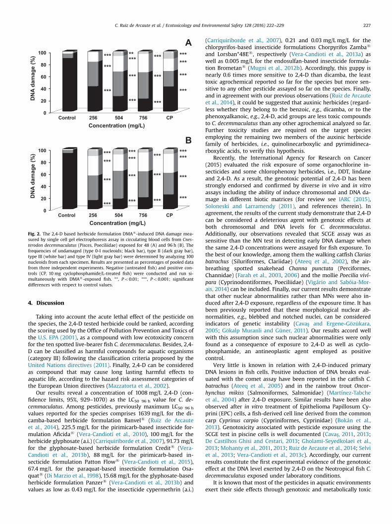
4. Discussion
Taking into account the acute lethal effect of the pesticide onthe species, the 2,4-D tested herbicide could be ranked, accordingthe scoring used by the Office of Pollution Prevention and Toxics ofthe U.S. EPA (2001), as a compound with low ecotoxicity concernfor the ten spotted live-bearer fish C. decemmaculatus. Besides, 2,4-D can be classified as harmful compounds for aquatic organisms(category III) following the classification criteria proposed by theUnited Nations directives (2011). Finally, 2,4-D can be consideredas compound that may cause long lasting harmful effects toaquatic life, according to the hazard risk assessment categories ofthe European Union directives (Mazzatorta et al., 2002).
Our results reveal a concentration of 1008 mg/L 2,4-D (con-fidence limits, 95%, 929–1070) as the LC50 96 h value for C. de-cemmaculatus. Among pesticides, previously maximum LC50 96 h
values reported for the species comprises 1639 mg/L for the di-camba-based herbicide formulation Banvels (Ruiz de Arcauteet al., 2014), 225.5 mg/L for the pirimicarb-based insecticide for-mulation Aficidas (Vera-Candioti et al., 2010), 100 mg/L for theherbicide glyphosate (a.i.) (Carriquiriborde et al., 2007), 91.73 mg/Lfor the glyphosate-based herbicide formulation Credits (Vera-Candioti et al., 2013b), 88 mg/L for the pirimicarb-based in-secticide formulation Patton Flows (Vera-Candioti et al., 2015),67.4 mg/L for the paraquat-based insecticide formulation Osa-quats (Di Marzio et al., 1998), 15.68 mg/L for the glyphosate-basedherbicide formulation Panzers (Vera-Candioti et al., 2013b) andvalues as low as 0.43 mg/L for the insecticide cypermethrin (a.i.)
(Carriquiriborde et al., 2007), 0.21 and 0.03 mg/L mg/L for thechlorpyrifos-based insecticide formulations Chorpyrifos Zambas
and Lorsban*48Es, respectively (Vera-Candioti et al., 2013a) aswell as 0.005 mg/L for the endosulfan-based insecticide formula-tion Brometans (Mugni et al., 2012b). Accordingly, this guppy isnearly 0.6 times more sensitive to 2,4-D than dicamba, the leasttoxic agrochemical reported so far for the species but more sen-sitive to any other pesticide assayed so far on the species. Finally,and in agreement with our previous observations (Ruiz de Arcauteet al., 2014), it could be suggested that auxinic herbicides (regard-less whether they belong to the benzoic, e.g., dicamba, or to thephenoxyalkanoic, e.g., 2,4-D, acid groups are less toxic compoundsto C. decemmaculatus than any other agrochemical analyzed so far.Further toxicity studies are required on the target speciesemploying the remaining two members of the auxinic herbicidefamily of herbicides, i.e., quinolinecarboxylic and pyrimidineca-rboxylic acids, to verify this hypothesis.
Recently, the International Agency for Research on Cancer(2015) evaluated the risk exposure of some organochlorine in-secticides and some chlorophenoxy herbicides, i.e., DDT, lindaneand 2,4-D. As a result, the genotoxic potential of 2,4-D has beenstrongly endorsed and confirmed by diverse in vivo and in vitroassays including the ability of induce chromosomal and DNA da-mage in different biotic matrices (for review see IARC (2015),Soloneski and Larramendy (2011), and references therein). Inagreement, the results of the current study demonstrate that 2,4-Dcan be considered a deleterious agent with genotoxic effects atboth chromosomal and DNA levels for C. decemmaculatus.Additionally, our observations revealed that SCGE assay was assensitive than the MN test in detecting early DNA damage whenthe same 2,4-D concentrations were assayed for fish exposure. Tothe best of our knowledge, among them the walking catfish Clariasbatrachus (Siluriformes, Clariidae) (Ateeq et al., 2002), the air-breathing spotted snakehead Channa punctata (Perciformes,Channidae) (Farah et al., 2003, 2006) and the mollie Poecilia viví-para (Cyprinodontiformes, Poeciliidae) (Vigário and Sabóia-Mor-ais, 2014) can be included. Finally, our current results demonstratethat other nuclear abnormalities rather than MNs were also in-duced after 2,4-D exposure, regardless of the exposure time. It hasbeen previously reported that these morphological nuclear ab-normalities, e.g., blebbed and notched nuclei, can be consideredindicators of genetic instability (Cavaş and Ergene-Gözükara,2005; Gökalp Muranli and Güner, 2011). Our results accord wellwith this assumption since such nuclear abnormalities were onlyfound as a consequence of exposure to 2,4-D as well as cyclo-phosphamide, an antineoplastic agent employed as positivecontrol.
Very little is known in relation with 2,4-D-induced primaryDNA lesions in fish cells. Positive induction of DNA breaks eval-uated with the comet assay have been reported in the catfish C.batrachus (Ateeq et al., 2005) and in the rainbow trout Oncor-hynchus mikiss (Salmoniformes, Salmonidae) (Martínez-Tabcheet al., 2004) after 2,4-D exposure. Similar results have been alsoobserved after in vitro treatment of Epithelioma Papillosum Cy-prini (EPC) cells, a fish-derived cell line derived from the commoncarp Cyprinus carpio (Cypriniformes, Cyprinidae) (Bokán et al.,2013). Genotoxicity associated with pesticide exposure using theSCGE test in piscine cells is well documented (Cavaş, 2011, 2013;De Castilhos Ghisi and Cestari, 2013; Gholami-Seyedkolaei et al.,2013; Mohanty et al., 2011, 2013; Ruiz de Arcaute et al., 2014; Selviet al., 2013; Vera-Candioti et al., 2013c). Accordingly, our currentresults constitute the first experimental evidence of the genotoxiceffect at the DNA level exerted by 2,4-D on the Neotropical fish C.decemmaculatus exposed under laboratory conditions.
It is known that most of the pesticides in aquatic environmentsexert their side effects through genotoxic and metabolically toxic
0
20
40
60
80
100
DN
A da
mag
e (%
)
Concentration (mg/L)
A
Control 256 504 756 CP
***
***
***
***
***
***
** ***
***
***
******
***
***
0
20
40
60
80
100
DN
A da
mag
e (%
)
Concentration (mg/L)
B
Control 256 504 756 CP
******
***
*** *** *** ***
*** *** ***
****** *** ***
***
Fig. 2. The 2,4-D based herbicide formulation DMAs-induced DNA damage mea-sured by single cell gel electrophoresis assay in circulating blood cells from Cnes-terodon decemmaculatus (Pisces, Poeciliidae) exposed for 48 (A) and 96 h (B). Thefrequencies of undamaged (type 0-I nucleoids; black bar), type II (dark gray bar),type III (white bar) and type IV (light gray bar) were determined by analyzing 100nucleoids from each specimen. Results are presented as percentages of pooled datafrom three independent experiments. Negative (untreated fish) and positive con-trols (CP, 10 mg cyclophosphamide/L-treated fish) were conducted and run si-multaneously with DMAs-exposed fish. **, Po0.01; ***, Po0.001; significantdifferences with respect to control values.
C. Ruiz de Arcaute et al. / Ecotoxicology and Environmental Safety 128 (2016) 222–229 227

mechanisms triggering, simultaneously, genotoxic damage, dis-ease, and even carcinogenesis (Könen and Cavaş, 2008; Ruiz deArcaute et al., 2014; Vera-Candioti et al., 2010, 2013a, 2013b,2013c, 2015). 2,4-D can be introduced into the environmentthrough effluents and spills resulting from its manufacture andtransport or directly through its application as a herbicide agent.The herbicide does not bioaccumulate in aquatic or terrestrial or-ganisms but some algae because of its rapid degradation (WHO,2003). Residues of 2,4-D can enter ponds and streams by severalmanners including direct application or accidental drift, inflow ofherbicide previously deposited in dry streambeds, pond bottomsor irrigation channels, runoff from soils as well as by leachingthrough the soil column (Norris, 1981). Groundwater contributionof 2,4-D residues into ponds and streams is dependent upon soiltype, with coarse-grained sandy soils with low organic contentexpected to leach 2,4-D into groundwater (Ghassemi et al., 1981;Norris, 1981). Therefore, 2,4-D, whether inefficiently diluted ordegraded after applied in the soil, may reach concentration levelsthat could pose genotoxic effects in aquatic biota. To the best ofour knowledge, no data regarding to the concentration level of theherbicide in different environments have been reported for Ar-gentina. Information on concentrations of 2,4-D in environmentalcompartments will be necessary to complete exposure profiles tobe included in future risk assessments studies when employing anative fish as C. decemmaculatus as experimental model.
Conflict of interest statement
The authors declare that there are no conflicts of interest.
Acknowledgments
This study was supported by grants from the National Uni-versity of La Plata (Grants 11/N699 and 11/N746) and the NationalCouncil for Scientific and Technological Research (CONICET, PIP No.0344) from Argentina.
References
Ackerman P.A., Morgan J.D., Iwama G.K. Anestethics, 2005. In: Canadian Council onAnimal Care (Ed.), The Care and use of Fish in Research, Teaching and Testing.Canadian Council on Animal Care, Ottawa, CA, pp. 1–22.
Ali, D., Nagpure, N.S., Kumar, S., Kumar, R., Kushwaha, B., Lakra, W.S., 2009. As-sessment of genotoxic and mutagenic effects of chlorpyrifos in freshwater fishChanna punctatus (Bloch) using micronucleus assay and alkaline single-cell gelelectrophoresis. Food Chem. Toxicol. 47, 650–656.
Annabi, A., Said, K., Messaoudi, I., 2015. Monitoring and assessment of environ-mental disturbance on natural Gambusia affinis populations - histopathologicalanalysis. Environ. Monit. Assess. 187, 318–326.
Ateeq, B., Abul Farah, M., Ahmad, W., 2005. Detection of DNA damage by alkalinesingle cell gel electrophoresis in 2,4-dichlorophenoxyacetic-acid- and buta-chlor-exposed erythrocytes of Clarias batrachus. Ecotoxicol. Environ. Saf. 62,348–354.
Ateeq, B., Abul Farah, M., Niamat Ali, M., Ahmad, W., 2002. Induction of micronucleiand erythrocyte alterations in the catfish Clarias batrachus by 2,4-di-chlorophenoxyacetic acid and butachlor. Mutat. Res. 518, 135–144.
Benitez, J.A., Cerón-Bretón, R.M., Cerón-Bretón, J.G., Rendón-Von-Osten, J., 2014.The environmental impact of human activities on the Mexican coast of the Gulfof Mexico: review of status and trends. WIT Trans. Ecol. Environ. 181, 37–50.
Bokán, K., Syberg, K., Jensen, K., Rank, J., 2013. Genotoxic potential of two herbicidesand their active ingredients assessed with comet assay on a fish cell line, epi-thelioma papillosum cyprini (EPC). J. Toxicol. Environ. Health Part A 76,1129–1137.
Carriquiriborde, P., Díaz, J., Mugni, H., Bonetto, C., Ronco, A.E., 2007. Impact of cy-permethrin on stream fish populations under field-use in biotech-soybeanproduction. Chemosphere. 68, 613–621.
Cavalcante, D.G., Martinez, C.B., Sofia, S.H., 2008. Genotoxic effects of Roundup onthe fish Prochilodus lineatus. Mutat. Res. 655, 41–46.
Cavaş, T., 2011. In vivo genotoxicity evaluation of atrazine and atrazine–basedherbicide on fish Carassius auratus using the micronucleus test and the comet
assay. Food Chem. Toxicol. 49, 1431–1435.Cavaş, T., 2013. In vivo genotoxicity evaluation of atrazine and atrazine-based her-
bicide on fish Carassius auratus using the micronucleus test and the cometassay. Food Chem. Toxicol. 49, 1431–1435.
Cavaş, T., Ergene-Gözükara, S., 2003. Micronuclei, nuclear lesions and interphasesilver-stained nucleolar organizer regions (AgNORs) as cyto-genotoxicity in-dicators in Oreochromis niloticus exposed to textile mill effluent. Mutat. Res.538, 81–91.
Cavaş, T., Ergene-Gözükara, S., 2005. Induction of micronuclei and nuclear ab-normalities in Oreochromis niloticus following exposure to petroleum refineryand chromium processing plant effluents. Aquat. Toxicol. 74, 264–271.
Cavaş, T., Könen, S., 2007. Detection of cytogenetic and DNA damage in peripheralerythrocytes of goldfish (Carassius auratus) exposed to a glyphosate formula-tion using the micronucleus test and the comet assay. Mutagenesis 22,263–268.
Cavaş, T., Könen, S., 2008. In vivo genotoxicity testing of the amnesic shellfishpoison (domoic acid) in piscine erythrocytes using the micronucleus test andthe comet assay. Aquat. Toxicol. 90, 154–159.
De Castilhos Ghisi, N., Cestari, M.M., 2013. Genotoxic effects of the herbicideRoundups in the fish Corydoras paleatus (Jenyns 1842) after short-term, en-vironmentally low concentration exposure. Environ. Monit. Assess. 185,3201–3207.
de Lemos, C.T., Iranço, F. d A., de Oliveira, N.C., de Souza, G.D., Fachel, J.M., 2008.Biomonitoring of genotoxicity using micronuclei assay in native population ofAstyanax jacuhiensis (Characiformes: Characidae) at sites under petrochemicalinfluence. Sci. Total Environ. 406, 337–343.
Di Marzio, W.D., Alberdi, J.L., Sáenz, M.E., Tortorelli, M.D.C., 1998. Effects of paraquat(Osaquats formulation) on survival and total cholinesterase activity in maleand female adults of Cnesterodon decemmaculatus (Pisces, Poeciliidae). Environ.Toxicol. Water Qual. 13, 55–59.
Di Marzio, W.D., Tortorelli, M.C., 1994. Effects of paraquat on survival and totalcholinesterase activity in fry of Cnesterodon decemmaculatus (Pisces, Poecilii-dae). Bull. Environ. Contam. Toxicol. 52, 274–278.
FAO-WHO, 2006. Manual on development and use of FAO and WHO specificationsfor pesticides FAO/WHO Joint Meeting on Pesticide Specifications (JMPS).
Farah, M.A., Ateeq, B., Ahmad, W., 2006. Antimutagenic effect of neem leaves ex-tract in freshwater fish, Channa punctatus evaluated by cytogenetic tests. Sci.Total. Environ. 364, 200–214.
Farah, M.A., Ateeq, B., Ali, M.N., Ahmad, W., 2003. Evaluation of genotoxicity of PCPand 2,4-D by micronucleus test in freshwater fish Channa punctatus. Ecotoxicol.Environ. Saf. 54, 25–29.
Fenech, M., 2007. Cytokinesis-block micronucleus cytome assay. Nat. Protoc. 2,1084–1104.
Finney, D.J., 1971. Probit Analysis. Cambridge Univ. Press, London.Furlong E.T., Anderson B.D., Werner S.L., Soliven P.P., Coffey L.J., Burkhardt M.R.,
2011. Methods of analysis but the US Geological Survey National Water QualityLaboratory – Determination of Pesticides in Water by Graphitized Carbon-based Solid-Phase Extraction and High-Performance Liquid Chromatography/Mass Spectrometry. US Geological Survey Water Resources Investigations Re-port 01-4134. pp. 73.
Ghassemi, M., Fargo, L., Painter, P., Quinlivan, S., Scofield, R., Takata, A., 1981. En-vironmental Fates and Impacts of Major Forest Use Pesticides. U.S. Environ-mental Protection Agency. Office of Pesticides and Toxic Substances, Wa-shington D.C..
Gholami-Seyedkolaei, S.J., Mirvaghefi, A., Farahmand, H., Kosari, A.A., Gholami-Seyedkolaei, S.J., Gholami-Seyedkolaei, S.J., 2013. Optimization of recoverypatterns in common carp exposed to roundup using response surface metho-dology: Evaluation of neurotoxicity and genotoxicity effects and biochemicalparameters. Ecotoxicol. Environ. Saf. 98, 152–161.
Gökalp Muranli, F.D., Güner, U., 2011. Induction of micronuclei and nuclear ab-normalities in erythrocytes of mosquito fish (Gambusia affinis) following ex-posure to the pyrethroid insecticide lambda-cyhalothrin. Mutat. Res. 726,104–108.
Greene, C., Kuehne, L., Rice, C., Fresh, K., Penttila, D., 2015. Forty years of change inforage fish and jellyfish abundance across greater Puget Sound, Washington(USA): Anthropogenic and climate associations. Mar. Ecol. Progress Ser. 525,153–170.
Grossmann, K., 2003. Mediation of herbicide effects by hormone interactions.Journal. Plant Growth Regul. 22, 109–122.
IARC, 2015. Monographs on the Evaluation of Carcinogenic Risk of Chemicals toMan. Some Organochlorine Insecticides and Some Chlorphenoxy Herbicides.International Agency for Research on Cancer, Lyon.
IRAM, 2008. Calidad ambiental-Calidad de agua. Determinación de la toxicidad letalaguda de sustancias en peces de agua dulce. Métodos semiestático. InsititutoArgentino de Normalización y Certificación, Buenos Aires, Argentina, p. 23.
King, K.A., Grue, C.E., Grassley, J.M., Hearsey, J.W., 2013. Pesticides in urban streamsand prespawn mortality of Pacific coho salmon. Arch. Environ. Contam. Toxicol.65, 546–554.
Könen, S., Cavaş, T., 2008. Genotoxicity testing of the herbicide trifluralin and itscommercial formulation Treflan using the piscine micronucleus test. Environ.Mol. Mutagen. 49, 434–438.
Lazartigues, A., Thomas, M., Banas, D., Brun-Bellut, J., Cren-Olivé, C., Feidt, C., 2013.Accumulation and half-lives of 13 pesticides in muscle tissue of freshwaterfishes through food exposure. Chemosphere 91, 530–535.
Liang, H.C., Razaviarani, V., Buchanan, I., 2013. Pesticides and herbicides. WaterEnviron. Res. 85, 1601–1645.
C. Ruiz de Arcaute et al. / Ecotoxicology and Environmental Safety 128 (2016) 222–229228

Martínez-Tabche, L., Madrigal-Bujaidar, E., Negrete, T., 2004. Genotoxicity and li-poperoxidation produced by paraquat and 2,4-dichlorophenoxyacetic acid inthe gills of rainbow trout (Oncorhynchus mikiss). Bull. Environ. Contam. Toxicol.73, 146–152.
Mazzatorta, P., Benfenati, E., Neagu, D., Gini, G., 2002. The importance of scaling indata mining for toxicity prediction. J. Chem. Inf. Model. 42, 1250–1255.
McKenzie, D.J., Garofalo, E., Winter, M.J., Ceradini, S., Verweij, F., Day, N., Hayes, R.,Van Der Oost, R., Butler, P.J., Chipman, J.K., Taylor, E.W., 2007. Complex phy-siological traits as biomarkers of the sub-lethal toxicological effects of pollutantexposure in fishes. Philos. Trans. R. Soc. B: Biol. Sci. 362, 2043–2059.
McLaughlin, D., Kinzelbach, W., 2015. Food security and sustainable resourcemanagement. Water Resour. Res. 51, 4966–4985.
Meffe, R., de Bustamante, I., 2014. Emerging organic contaminants in surface waterand groundwater: A first overview of the situation in Italy. Sci. Total Environ.481, 280–295.
Menendez-Helman, R.J., Ferreyroa, G.V., Alfonso, M.D.S., Salibian, A., 2012. Gly-phosate as an acetylcholinesterase inhibitor in Cnesterodon decemmaculatus.Bull. Environ. Contam. Toxicol. 88, 6–9.
Menni, R.C., Gómez, S.E., López Armengol, F., 1996. Subtle relationships: freshwaterfishes and the chemistry of water in southern South America. Hydrobiologia328, 173–197.
Mohanty, G., Mohanty, J., Garnayak, S.K., Rath, S.K., Dutta, S.K., 2013. Use of cometassay in the study of DNA break in blood and gill cells of rohu (Labeo rohita)after an exposure to furadan, a carbamate pesticide. Res. J. Biotechnol. 8, 83–89.
Mohanty, G., Mohanty, J., Nayak, A.K., Mohanty, S., Dutta, S.K., 2011. Application ofcomet assay in the study of DNA damage and recovery in rohu (Labeo rohita)fingerlings after an exposure to phorate, an organophosphate pesticide. Eco-toxicology 20, 283–292.
Mugni, H., Demetrio, P., Paracampo, A., Pardi, M., Bulus, G., Bonetto, C., 2012a.Toxicity persistence in runoff water and soil in experimental soybean plotsfollowing chlorpyrifos application. Bull. Environ. Contam. Toxicol. 89, 208–212.
Mugni, H., Paracampo, A., Demetrio, P., Scalise, A., Solís, M., Fanelli, S., Bonetto, C.,2016. Acute toxicity of endosulfan to the non-target organisms Hyalella cur-vispina and Cnesterodon decemmaculatus. Bull. Environ. Contam. Toxicol. 70,257–264.
Mugni, H.A., Paracampo, A., Marrochi, N., Bonetto, C., 2012b. Cypermethrin, chlor-pyrifos and endosulfan toxicity to two non-target freshwater organisms. Fre-senius Environ. Bull. 21, 2085–2089.
Nikoloff, N., Natale, G.S., Marino, D., Soloneski, S., Larramendy, M., 2014. Fluro-chloridone-based herbicides induced genotoxicity effects on Rhinella arenarumtadpoles (Anura: Bufonidae). Ecotoxicol. Environ. Saf. 100, 275–281.
Norris, L.A., 1981. The movement, persistence, and fate of the phenoxy herbicidesand TCDD in the forest. Residue Rev. 80, 65–135.
Ogada, D.L., 2014. The power of poison: Pesticide poisoning of Africa wildlife. Ann.N. Y. Acad. Sci. 1322, 1–20.
Pimentel, D., McLaughlin, L., Zepp, A., Lakitan, B., Kraus, T., Kleinman, P., Vancini, F.,Roach, W.J., Graap, E., Keeton, W.S., Selig, G., 1993. Environmental and economiceffects of reducing pesticide use in agriculture. Agric. Ecosyst. Environ. 46,273–288.
Pitarque, M., Vaglenov, A., Nosko, M., Hirvonen, A., Norppa, H., Creus, A., Marcos, R.,1999. Evaluation of DNA damage by the Comet assay in shoe workers exposedto toluene and other organic solvents. Mutat. Res. 441, 115–127.
Ruiz de Arcaute, C., Soloneski, S., Larramendy, M.L., 2014. Evaluation of the geno-toxicity of a herbicide formulation containing 3,6-dichloro-2-metoxybenzoicacid (dicamba) in circulating blood cells of the tropical fish Cnesterodon de-cemmaculatus. Mutat. Res. 773, 1–8.
Sarikaya, R., Yilmaz, M., 2003. Investigation of acute toxicity and the effect of 2,4-D(2,4-dichlorophenoxyacetic acid) herbicide on the behavior of the commoncarp (Cyprinus carpio L., 1758; Pisces, Cyprinidae). Chemosphere 52, 195–201.
Schlenk, D., Lavado, R., Loyo-Rosales, J.E., Jones, W., Maryoung, L., Riar, N., Werner,I., Sedlak, D., 2012. Reconstitution studies of pesticides and surfactants ex-ploring the cause of estrogenic activity observed in surface waters of the SanFrancisco Bay Delta. Environ. Sci. Technol. 46, 9106–9111.
Selvi, M., Cavaş, T., Cağlan Karasu Benli, A., Koçak Memmi, B., Çinkiliç, N., Dinçel, A.S., Vatan, O., Yilmaz, D., Sarikaya, R., Zorlu, T., Erkoç, F., 2013. Sublethal toxicityof esbiothrin relationship with total antioxidant status and in vivo genotoxicityassessment in fish (Cyprinus carpio L., 1758) using the micronucleus test andcomet assay. Environ. Toxicol. 28, 644–651.
SENASA, 2013. Argentinean National Service for Sanitary and Quality of Agriculture
and Food. ⟨http://www.senasa.gov.ar/contenido.php?to¼n&in¼1506&io¼18122⟩.
Singh, N.P., 1996. Microgel electrophoresis of DNA from individual cells: principlesand methodology. In: Pfeifer, G.P. (Ed.), Technologies for Detection of DNADamage and Mutations. Plenum Press, New York, pp. 3–24.
Soloneski, S., Larramendy, M.L., 2011. Herbicides in Argentina. Comparative eva-luation of the genotoxic and cytotoxic effects on mammalian cells exerted byauxinic members. In: Kortekamp, A. (Ed.), Herbicides and Environment. InTechRijeka, Croatia, pp. 515–530.
Song, Y., 2014. Insight into the mode of action of 2,4-dichlorophenoxyacetic acid(2,4-D) as an herbicide. J. Integr. Plant Biol. 56, 106–113.
Strunjak-Perovic, I., Coz-Rakovac, R., Topic Popovic, N., Jadan, M., 2009. Seasonalityof nuclear abnormalities in gilthead sea bream Sparus aurata (L.) erythrocytes.Fish Physiol. Biochem. 35, 287–291.
Summerfelt, R.C., Smith, L.S., 1990. Anaesthesia, surgery and related techniques. In:Schreck, C.B., Moyle, P.B. (Eds.), Methods for Fish Biology. American FisheriesSociety, Bethesda MD, pp. 213–272.
Tierney, K.B., Baldwin, D.H., Hara, T.J., Ross, P.S., Scholz, N.L., Kennedy, C.J., 2010.Olfactory toxicity in fishes. Aquat. Toxicol. 96, 2–26.
UN, 2011. Peligros para el medio ambiente. Naciones Unidas. Parte 4, pp. 229–258.USEPA, 1974. United States Environmental Protection Agency. Compendium of
Registered Pesticides U.S. Government Printing Office, Washington, DC.USEPA, 1975. Methods for Acute Toxicity Tests with Fish, Macroinvertebrates, and
Amphibians. U.S. Government Printing Office, Washington, DC, p. 62.USEPA, 2001. United States Environmental Protection Agency Report. Environ-
mental Hazard Assessment and Ecological Risk Assessment Methodology. U.S.Government Printing Office, Washington, DC, Appendix H.
USEPA, 2002. Methods for measuring the acute toxicity of effluents and receivingwaters to freshwater and marine organisms, Fifth edition. U.S. GovernmentPrinting Office, Washington, DC 821-R-02-012.
Vera-Candioti, J., Soloneski, S., Larramendy, M.L., 2010. Genotoxic and cytotoxiceffects of the formulated insecticide Aficida
s
on Cnesterodon decemmaculatus(Jenyns, 1842) (Pisces: Poeciliidae). Mutat. Res. 703, 180–186.
Vera-Candioti, J., Soloneski, S., Larramendy, M.L., 2011. Acute toxicity of chromiumon Cnesterodon decemmaculatus (Pisces: Poeciliidae). Theoria 20, 85–93.
Vera-Candioti, J., Soloneski, S., Larramendy, M.L., 2013a. Chlorpyrifos-based in-secticides induced genotoxic and cytotoxic effects in the ten spotted live-bearerfish, Cnesterodon decemmaculatus (Jenyns, 1842). Environ. Toxicol. 29,1390–1398.
Vera-Candioti, J., Soloneski, S., Larramendy, M.L., 2013b. Evaluation of the genotoxicand cytotoxic effects of glyphosate-based herbicides in the ten spotted live-bearer fish Cnesterodon decemmaculatus (Jenyns, 1842). Ecotoxicol. Environ. Saf.89, 166–173.
Vera-Candioti, J., Soloneski, S., Larramendy, M.L., 2013c. Single-cell gel electro-phoresis assay in the ten spotted live-bearer fish, Cnesterodon decemmaculatus(Jenyns, 1842), as bioassay for agrochemical-induced genotoxicity. Ecotoxicol.Environ. Saf. 98, 368–373.
Vera-Candioti, J., Soloneski, S., Larramendy, M.L., 2015. Pirimicarb-based formula-tion-induced genotoxicity and cytotoxicity on the fresh water fish Cnesterodondecemmaculatus (Jenyns, 1842) (Pisces, Poeciliidae). Toxicol. Ind. Health 31,1051–1060.
Vigário, A.F., Sabóia-Morais, S.M.T., 2014. Effects of the 2,4-D herbicide on gillsepithelia and liver of the fish Poecilia vivipara. Braz. J. Vet. Res. 34, 523–528.
WHO, 1990. Public Health Impacts of Pesticides used in Agriculture (WHO in Col-laboration with the United Nations Environment Programme, Geneva, 1990).World Health Organization, Geneva, Italy.
WHO, 2003. 2,4-D in Drinking-Water. Background Document for Development ofWHO Guidelines for Drinking-Water Quality. World Health Organization,Geneva.
WHO, 2009. The WHO Recommended Classification of Pesticides by Hazard 1.World Health Organization, Geneva, Italy, pp. 1–81.
Yilmaz, M., Gül, A., Karaköse, E., 2004. Investigation of acute toxicity and the effectof cadmium chloride (CdCl2.H2O) metal salt on behavior of the guppy (Poeciliareticulata). Chemosphere 56, 375–380.
Zhao, Z., Wang, Y., Zhang, L., Cai, Y., Chen, Y., 2014. Bioaccumulation and tissuedistribution of organochlorine pesticides (OCPs) in freshwater fishes: a casestudy performed in Poyang Lake, China largest lake. Environ. Sci. Pollut. Res. 21,8740–8749.
C. Ruiz de Arcaute et al. / Ecotoxicology and Environmental Safety 128 (2016) 222–229 229

Synergism of mixtures of dicamba and 2,4-dichlorophenoxyacetic acidherbicide formulations on the neotropical fish Cnesterodondecemmaculatus (Pisces, Poeciliidae)*
C. Ruiz de Arcaute a, b, S. Soloneski a, b, M.L. Larramendy a, b, *
a C�atedra de Citología, Facultad de Ciencias Naturales y Museo, Universidad Nacional de La Plata, Calle 64 Nº 3, B1904AMA La Plata, Argentinab Consejo Nacional de Investigaciones Científicas y T�ecnicas (CONICET), Argentina
a r t i c l e i n f o
Article history:Received 9 June 2017Received in revised form26 October 2017Accepted 17 January 2018
Keywords:Banvel®
DMA®
Lethal effectsEquitoxic mixturesNon-equitoxic mixtures
a b s t r a c t
Dicamba (DIC) and 2,4-dichlorophenoxyacetic acid (2,4-D) are two of the most applied auxinic herbicidesworldwide, both individually and as part of a mixture. However, the toxicity and interactions achievedwhen applied as a mixture have not yet been characterised. The equitoxic and non-equitoxic acutetoxicity exerted by binary mixtures of Banvel® (57.71% DIC) and DMA® (58.4% 2,4-D) on the Neotropicalfish Cnesterodon decemmaculatuswere evaluated. Results revealed mean values of 1.02 (range, 0.96e1.08)for the toxic unit (TU) that induced 50% mortality (TU50 96 h) to the fish exposed to binary equitoxicmixtures of the commercial formulations Banvel®eDMA®. These results suggest that the mixture isnearly concentration additive. Furthermore, results demonstrated the occurrence of synergistic inter-action when non-equitoxic combinations of Banvel®-or DMA®-formulated herbicides were assayed. Inthis context and regardless of their concentrations, either Banvel®- or DMA®-induced toxicity weresynergised by the presence of the counterpart within mixtures. The present study represents the firstevidence of the lethality exerted by mixtures of two auxinic herbicidesdnamely, DIC and 2,4-Ddreported to date for fish and other biotic matrices. When C. decemmaculatus is used as the targetorganism, a synergistic pattern is observed following exposure to a mixture of both herbicides.
© 2018 Elsevier Ltd. All rights reserved.
1. Introduction
Anthropogenic activities, especially agriculture, release largeamounts of pesticides daily into the soil, water and air (Ippolitoet al., 2015; Liaud et al., 2016). Annually, approximately fourmillion tons of pesticides are applied around the globe (Ippolitoet al., 2015) and disseminated by drift, surface runoff, anddrainage into the environment (Aparicio et al., 2013; Meffe and deBustamante, 2014). Accordingly, a mixture of products reaches notonly their target species, but also non-target ones, for example,aquatic and terrestrial biota (Liang et al., 2013; Meffe and deBustamante, 2014).
The application of mixtures of toxicants is a commonly usedstrategy to control and prevent weed resistance, despite the
potential effects that the mixture could exert on non-target or-ganisms (Ge et al., 2014). In addition to the additives that are pre-sent in the commercially available pesticide formulations, whichare known to modify the toxic effects of the pure compound (Coxand Surgan, 2006; Nikoloff et al., 2014b; Ruiz de Arcaute et al.,2014b), it is known that mixtures of different compounds caninteract with each. Those interactions can cause complex changesthat are different from the toxic effects exerted by individualcomponents on a biotic matrix (LeBlanc and Wang, 2006; Silvaet al., 2015; Svartz et al., 2016; Varona-Uribe et al., 2016). In gen-eral terms, a mixture of toxicants can exert complex interactionsbetween its components, including additivity, synergism andantagonism (Calamari and Alabaster, 1980; Calabrese, 1995; Warne,2003; Lydy et al., 2004; Brodeur et al., 2014). Therefore, it isnecessary to evaluate the joint effects of mixtures, especially thosewith interactions between at least two components (that is, binarymixtures) (Warne, 2003).
Since they were first synthesised after World War II, auxinicherbicides have been frequently used worldwide to eliminate un-wanted broadleaf dicotyledons in crops (USEPA, 2006). These
* This paper has been recommended for acceptance by Dr. Harmon SarahMichele.* Corresponding author. Facultad de Ciencias Naturales y Museo, Calle 64 Nro. 3
(esq. 120), B1904AMA La Plata, Argentina.E-mail address: [email protected] (M.L. Larramendy).
Contents lists available at ScienceDirect
Environmental Pollution
journal homepage: www.elsevier .com/locate/envpol
https://doi.org/10.1016/j.envpol.2018.01.0490269-7491/© 2018 Elsevier Ltd. All rights reserved.
Environmental Pollution 236 (2018) 33e39

herbicides include quinolinecarboxylic, benzoic, phenoxyalkanoicand pyridinecarboxilic acids (Garraway and Wain, 1976). Theymimic the natural phytohormone auxin, and they cause uncon-trolled growth, senescence and death (Grossmann, 2003; USEPA,2006). Among the benzoic acids, dicamba (DIC), a derivative ofchlorinated benzoic acid, is frequently used for controlling weeds incereal crops, sugar cane, soybeans, wheat, and in grazing and nat-ural fields (USEPA, 2006; CASAFE, 2009). Information regarding thetoxicity of DIC is sufficient to evaluate its potential hazards tohumans and other aquatic and terrestrial organisms (USEPA, 2006).Taking into account its acute toxic effects, DIC has been ranked bythe World Health Organisation (WHO) (2009) as a class II chemical(moderately hazardous) and by the U.S. Environmental ProtectionAgency (U.S. EPA) (2006) as a type III chemical (moderate toxic). Inaddition, following U.S. EPA's ecotoxicological classification foraquatic organisms (USEPA, 2008), DIC is considered to be a prac-tically nontoxic pesticide.
2,4-dichlorophenoxyacetic acid (2,4-D)is an auxinic herbicidethat belongs to the family of phenoxyalkanoic acids. It has beenclassified by the U.S. EPA as slightly to moderately toxic (categoryIIeIII) (USEPA, 1974), and by WHO as moderately hazardous (classII) (WHO, 2009). Moreover, in 2015, the International Agency forResearch on Cancer (IARC) classified 2,4-D as a possibly carcino-genic agent to humans (Group 2B) based on mechanistic studies(IARC, 2015). In addition, and according the ecotoxicological clas-sification proposed for aquatic organisms by the USEPA (2008), 2,4-D is classified as a practically nontoxic pesticide. There are over 172products containing 2,4-D currently on Argentina (SENASA, 2017)and it is used alone or in combination with other herbicides (forexample, in combination with DIC, 4-amino-3,5,6-trichloro-2-pyridinecarboxylic acid [picloran], 3,6-dichloro-2-pyridinecarboxylic acid [clopyralid] and methylchlorophenox-ypropionic acid [MCPP]) (NCBI, 2017).
Regarding the toxicity of DIC reported for different fish species,data reviewed in the literature demonstrated LC50 96h valuesranging from 50mg/L for the cutthroat trout (Oncorhynchus clarkii)to 465mg/L for the Western mosquitofish (Gambusia affinis)(USEPA, 2017). Similarly, LC50 96 h concentrations of 2,4-D that rangefrom 87mg/L to 707mg/L have been reported for common carp(Cyprinus carpio) and the rainbow trout (O. mykiss) (USEPA, 2017).When the acute toxicities of DIC, formulated as Banvel®, and 2,4-D,formulated as DMA®, were assayed, the LC50 96 h concentrationswere determined to be 1639mg/L and 1008mg/L, respectively, forthe ten spotted live-bearer (Cnesterodon decemmaculatus) (Ruiz deArcaute et al., 2014b, 2016). Thus, it could be pinpointed that thelatter represents the most resistant fish species reported so far forboth DIC and 2,4-D.
DIC and 2,4-D have been found in urban, agricultural andagriculturaleurban sites and in surface drinking-water reservoirs(Loos et al., 2010; Glozier et al., 2012; F�elix-Ca~nedo et al., 2013;Tagert et al., 2014). They contaminate both surface and ground-water because they are highly mobile and adsorb poorly onto soilparticles (Li et al., 2009). In the United States, DIC and 2,4-D havebeen detected in surfacewater runoff at a maximumvalue of 2.1 mg/L, in surface water at a maximum value of 24 mg/L (Phillips andBode, 2004; Tagert et al., 2014) and in drinking water reservoirsat values as low as 1.04 mg/L (Donald et al., 2007). Lower environ-mental concentrations were reported for water in Canada, withvalues of 0.04 mg/L for DIC and 0.31 mg/L for 2,4-D (Woudneh et al.,2007; Tierney et al., 2011; Glozier et al., 2012). Concentrations of2,4-D were found in surface and ground water in the range of0.005 mg/L to 0.04 mg/L in Mexico (F�elix-Ca~nedo et al., 2013), and ata concentration of 0.012 mg/L in European ground water (Loos et al.,2010). However, to the best of our knowledge, no data have beenpublished on the environmental concentrations of DIC and 2,4-D in
Argentina.It is worth mentioning that DIC and 2,4-D have extensive and
overlapping applications worldwide. Specifically for Argentina,these herbicides are recommended to be applied together inseveral crops (for example, sugar cane, cereal, corn, sorghum andother cultivars that are of commercial importance) (CASAFE, 2015).
Cnesterodon decemmaculatus (Cyprinodontiformes, Poeciliidae)is an endemic species for Argentina with an extensive distributionin Neotropical America. This is a small ovoviviparous, micro-omnivorous, benthic-pelagic and non-migratory fish (maximumsize is approximately 25mm for males and 45mm for females), andit is often present in high-density in watercourses. It has anextensive distribution in the La Plata River and other South Amer-ican basins (for review, see (Soloneski and Larramendy, 2017)). Inaddition, this species is easy to handle and acclimate to laboratoryconditions, and they comply to the conditions requested for toxicitytesting, such as a large range of tolerance to many environmentalparameters (Di Marzio et al., 2005; de la Torre et al., 2007; Ruiz deArcaute et al., 2014b, 2016; Vera-Candioti et al., 2015). The specieshas been employed as a suitable in vivomodel to analyse pesticide-induced toxicity among aquatic vertebrates. Some of the testedchemicals include the following: chlorpyrifos-based insecticidesLorsban 48E® and CPF Zamba® (Vera-Candioti et al., 2014), thepirimicarb-based insecticides Aficida® (Vera-Candioti et al., 2010)and Patton Flow® (Vera-Candioti et al., 2015), the paraquat-basedherbicide Osaquat® (Di Marzio et al., 1998), the glyphosate-basedherbicides Credit® and Panzer® (Vera-Candioti et al., 2013), theDIC-based herbicide Banvel® (Ruiz de Arcaute et al., 2014b) and the2,4-D-formulated product DMA® (Ruiz de Arcaute et al., 2016).
Although detailed testing has been undertaken in terms ofC. decemmaculatus exposed to individual chemicals (as citedabove), no analyses have been completed on the toxic effectsexerted when C. decemmaculatus is exposed simultaneously to theDIC-based formulation Banvel® and the 2,4-D-formulated productDMA®. This includes possible synergistic, antagonistic or additivepatterns. In the present study, we evaluated the acute toxicity andinteractions of equitoxic and non-equitoxic binary mixtures of thecommercial herbicide formulations Banvel® (57.71% DIC) andDMA® (58.4% 2,4-D) on C. decemmaculatus exposed under labora-tory conditions using a semi-static acute experimental model.
2. Material and methods
2.1. Chemicals
The DIC-based (3,6-dichloro-2-methoxybenzoic acid; CAS 1918-00-9) formulation Banvel® (57.7% DIC) was obtained from SyngentaAgro S.A. (Argentina). The 2,4-D-based (2,4-dichlorophenoxyaceticacid, CAS 2008-39-1) formulation DMA® (58.4% 2,4-D) was ob-tained from Dow Agrosciences Argentina S.A., Argentina. K2Cr2O7[Cr(VI); CAS 7778-50-9] was obtained from Merck KGaA (Darm-stadt, Germany). Chemicals and solvents of analytical grade wereobtained from Sigma Chemical Co. (St. Louis, MO).
2.2. Quality control
DIC and 2,4-D concentrations in test solutions were determinedby QV Chem Laboratory (La Plata, Buenos Aires, Argentina) usinghigh-performance liquid chromatography and an ultraviolet de-tector, following the U.S. Geological Survey Report 01-4134(Furlong et al., 2011). Active ingredient samples from test solutions(0.25 TU for Banvel® and DMA®, which is equivalent to 410mg/L ofBanvel® and 252mg/L of DMA®) corresponded to values obtainedimmediately after preparation (0 h) and after 24 h. The detectionlimit of the active ingredients was 0.5mg/L for both herbicide
C. Ruiz de Arcaute et al. / Environmental Pollution 236 (2018) 33e3934

formulations.
2.3. Test organism
C. decemmaculatus specimens were collected from a permanentpond near La Plata city (34�860S, 58�120W, Buenos Aires Province,Argentina). After collection, they were acclimatised for 20 daysunder laboratory conditions (16/8 h light/dark cycle; 20 ± 1 �C withdechlorinated tap water [pH 7.55± 0.1; dissolved oxygen,6.3± 0.3mg/L; ammonium (NH4þ) < 0.2 mg/L; hardness,143 ± 23.5 mg CaCO3/L]) following the recommendation of theUSEPA (1996) and other recommendations specifically reported forthe species (Menni, 2004; Somma et al., 2011). Artificial aerationand ad libitum fish food (TetraFin®, TetraWerke, Melle, Germany)were provided until the beginning of the experimental protocols.The average body weight and mean body length of the specimensused throughout the experiment was 0.25± 0.1 g and28.5± 2.3mm, respectively. Fish were maintained following therecommendations of the Argentinean National Service for Sanitaryand Quality of Agriculture and Food (SENASA) guidelines 617/2002for biological testing (2013). Specimens were collected with thepermission of the Flora and Fauna Direction from the Buenos AiresProvince (Buenos Aires, Argentina) (code 22500-22339/13) and theethical committee from the National University of La Plata (code 11/N754).
2.4. Acute toxicity of binary mixtures of DIC- and 2,4-D-basedformulations
The acute toxicities of commercial formulations of DIC and 2,4-Dwere determined in previous experiments assayed in our labora-tory (Ruiz de Arcaute et al., 2014b, 2016). The reported LC50 96 h was1639mg/L (1,471mg/L �1808mg/L) for DIC and 1008mg/L(929mg/L �1070mg/L) for 2,4-D (Ruiz de Arcaute et al., 2014b,2016).
For both equitoxic and non-equitoxic herbicide mixtures,toxicity was estimated as suggested by Warne (2003). Accordingly,a toxic unit (TU) was arbitrarily assigned to the concentration of thepesticide that was able to induce 50% mortality inC. decemmaculatus after 96 h of exposure (that is, the LC50 96 hvalue). The toxicity was evaluated for mixtures of DIC-based her-bicide formulation Banvel® and 2,4-D-based formulation DMA®.The lethal effect was determined every 24 h in three independentexperiments that were run simultaneously for each experimentalpoint.
2.4.1. Acute toxicity of equitoxic binary mixtures of DIC- and 2,4-D-based formulations
For equitoxic mixtures, herbicide formulations were combinedin equal concentrations to generate mixtures for which the sum ofthe TU of both components equalled 0.125, 0.25, 0.5, 1, 1.5, 2, 2.5, 3and 4 units, as suggested elsewhere (Warne, 2003; Brodeur et al.,2014, 2016). For example, if the test pesticide mixture had a valueof 1 TU, it was comprised of 0.5 TUs of DIC-based herbicideformulation Banvel® (50% of the LC50 96 h) and 0.5 TUs of 2,4-D-based formulation DMA® (50% of the LC50 96 h). Experiments wereperformed according to standardised methods recommended bythe USEPA (1975, 1982, 2002), with minor modifications reportedpreviously for native species (P�erez-Iglesias et al., 2015; Vera-Candioti et al., 2015; Ruiz de Arcaute et al., 2016). For each exper-imental point, 10 randomly selected specimens were maintained ina 1 L glass container and exposed to the nine herbicide mixtures for96 h. A negative control (dechlorinated tap water; pH 7.5± 0.1;hardness, 143± 23.5mg CaCO3/L) was run simultaneously with theexposed organisms. The test solutions were prepared and replaced
every 24 h. Fish were not fed during the experiment. Mortality wasregistered by visual observation every 24 h. The TU response rela-tionship was derived, and it was used to determine the TU valuethat caused 50% of mortality (TU50) for each experimental point.
2.4.2. Acute toxicity of non-equitoxic binary mixtures of DIC- and2,4-D- based formulations
For non-equitoxic mixtures, a series of four experimental pro-tocols were conducted following the recommendations of Warne(2003) and Brodeur et al., (2014, 2016). Each series included fivefish in the exposed group. In two of the experimental series, theconcentration of DIC-based herbicide formulation Banvel® wasremained constant at 0.33 TUs or 0.66 TUs, while the concentrationof 2,4-D-based formulation DMA® was either 0.01, 0.05, 0.1, 1 or 2TUs. In the other two series, the concentration of 2,4-D-basedformulation DMA® was set at 0.33 or 0.66 TUs, while the concen-tration of DIC-based herbicide formulation Banvel® was either 0.01,0.05, 0.1, 1 or 2 TUs. Fish were exposed to these pesticide mixturesfor 96 h according to the experimental protocol described above foracute toxicity of equitoxic binary mixtures (See section 2.4.1). Anegative control (dechlorinated tap water; pH 7.5± 0.1; hardness,143± 23.5mg CaCO3/L) was run simultaneously with the pesticide-exposed organisms. The TU response relationship was used todetermine the TU value that caused 50% of mortality (TU50) for eachexperimental point.
2.5. Statistical analyses
The concentrations of herbicides and the TU of themixtures thatcaused mortality of 50% of specimens were calculated by fitting afour-parameter logistic regression equation to the survival datausing the GraphPad Prism software version 6. The level of signifi-cance was 0.05 for all tests.
3. Results
3.1. Chemical analysis
No significant changes (p> .05) were observed in the concen-tration of the pure analyte in treatments during the 24 h intervalrenewals of the testing solutions (concentration range 97%± 5%recovery). Concentrations assessed throughout the experimentcorresponded to the nominal concentrations of the analyte presentwithin the DIC-based formulation Banvel® and 2,4-D-basedformulation DMA®.
Table 1Lethal effects of equitoxic binary mixtures of dicamba (DIC)-based Banvel® and 2,4-dichlorophenoxyacetic acid (2,4-D)-based DMA® commercial formulations onexposed Cnesterodon decemmaculatus.
Concentration(TUs)a
Number of individuals Mortality (number of deadindividuals)
24 h 48 h 72 h 96 h
Negative control 30 0 0 0 0
DIC 2,4-D0.0625 0.0625 30 0 0 0 10.125 0.125 30 1 1 2 30.25 0.25 30 1 1 2 20.5 0.5 30 2 4 9 140.75 0.75 30 4 23 28 281 1 30 2 27 30 301.25 1.25 30 12 30 30 301.5 1.5 30 30 30 30 302 2 30 30 30 30 30
a Toxic unit (TU), concentration of individual pesticide that causes 50% mortality.
C. Ruiz de Arcaute et al. / Environmental Pollution 236 (2018) 33e39 35

3.2. Acute toxicity of equitoxic binary mixtures of DIC- and 2,4-D-based formulations
Data of mortality are presented in Table 1. Survival of thenegative control group was 100%. Mortality results allowed for thedetermination of TUs of the equitoxic mixtures of DIC-basedformulation Banvel® and 2,4-D-based formulation DMA® thatcaused mortality in 50% of the exposed fish. Results revealed amean value of 1.02 (0.96e1.08) for the TU50 96 h of the binaryequitoxic mixtures of the commercial formulations Banvel® andDMA® (Fig. 1).
3.3. Acute toxicity of non-equitoxic binary mixtures of DIC- and 2,4-D-based formulations
Complete data of mortality are presented in Table 2 for non-equitoxic mixtures of the formulations Banvel® and DMA®. Sur-vival of the negative control group was 100%. Mortality resultsallowed for the determination of the TUs of the non-equitoxicmixtures that caused mortality in 50% of the exposed fish. Theisobologram shown in Fig. 2 summarises data obtained for theseries of non-equitoxic mixtures of the commercial formulationsBanvel® and DMA®. The diagonal isobole linking the values of 1 TUon both the x- and y-axes correspond to the line effect of a con-centration addition. The isobologram clearly demonstrates thatcombinations of chemicals that cause 50% mortality are below andto the left of the additivity line in the figure. Thus, results demon-strated a synergistic interaction for non-equitoxic combinations ofherbicides Banvel® and DMA® (Fig. 2).
Results demonstrate that DIC-based formulation Banvel®-induced toxicity either at a concentration of 0.33 or 0.66 TUs wassynergised by the presence of 2,4-D-based formulation DMA®
within the mixtures (Fig. 2). Fig. 2 highlights that 0.33 TUs of DICrequires 0.19 (0.10e0.38) TUs of 2,4-D to induce the same toxiceffect as when 0.66 TUswasmixedwith 0.23 (0.11e0.48) TUs of 2,4-D.
In addition, 2,4-D-based formulation DMA®-induced toxicitywas also synergised when mixed with DIC-based formulationBanvel® regardless of whether 0.33 or 0.66 TUs were assayed(Fig. 2). Results demonstrated that 0.33 and 0.66 TUs of 2,4-Drequired 0.13 (0.04e0.45) and 0.23 (0.13e0.40) TUs of DIC toreach 50% mortality.
Overall, the results also demonstrated that the toxicity exertedby 0.33 TUs of each formulation was slightly higher than the
toxicity exerted by 0.66 TUs during testing within mixtures (Fig. 2).
4. Discussion
We analysed the possible toxic interactionsdthat is, synergy,antagonism or additivitydprevailing in equitoxic and non-equitoxic binary mixtures between two herbicide formulations,the DIC-based Banvel® and the 2,4-D-based DMA®. The target
Fig. 1. Survival as a function of the sum of the toxic units (TUs) of equitoxic mixtures ofdicamba-based formulation Banvel® and 2,4-dichlorophenoxyacetic acid (2,4-D)-basedformulation DMA® in C. decemmaculatus after 96 h of exposure. The TU value of themixture that induces 50% mortality (LC50 96 h) is indicated with the 95% confidence inparenthesis. The striped area depicts the “Additivity Zone”.
Table 2Lethal effects of non-equitoxic binary mixtures of dicamba (DIC)-based Banvel® and2,4-dichlorophenoxyacetic acid (2,4-D)-based DMA® on exposed Cnesterodondecemmaculatus.
Concentration(TUs)a
Number of individuals Mortality (number of deadindividuals)
24 h 48 h 72 h 96 h
Negativecontrol
30 0 0 0 0
DIC 2,4-D 300.33 0.01 30 0 5 6 80.33 0.05 30 0 1 4 80.33 0.1 30 1 6 7 80.33 0.5 30 4 10 12 160.33 1 30 6 15 30 300.33 2 30 30 30 30 300.66 0.01 30 1 3 6 80.66 0.05 30 1 4 6 80.66 0.1 30 1 3 5 90.66 0.5 30 5 9 12 150.66 1 30 5 14 22 220.66 2 30 30 30 30 30
2,4-D DIC0.33 0.01 30 2 6 8 90.33 0.05 30 7 8 10 100.33 0.1 30 1 6 8 100.33 0.5 30 2 8 9 120.33 1 30 7 8 15 240.33 2 30 16 27 30 300.66 0.01 30 1 2 3 50.66 0.05 30 3 4 5 60.66 0.1 30 4 6 8 100.66 0.5 30 4 6 11 140.66 1 30 8 11 23 280.66 2 30 27 30 30 30
a Toxic unit (TU), concentration of individual pesticide that causes 50% mortality.
Fig. 2. Isobologram depicting the toxic units (TUs) of non-equitoxic mixtures ofdicamba-based formulation Banvel® and 2,4-dichlorophenoxyacetic acid (2,4-D)-basedformulation DMA® that cause 50% mortality (TU50) in C. decemmaculatus after 96 h ofexposure. The error bars from each experimental value represent 95% confidence in-tervals. The isobole linking the 1-TU values from both axes is the line of concentrationaddition.
C. Ruiz de Arcaute et al. / Environmental Pollution 236 (2018) 33e3936

organism, C. decemmaculatus (Pisces, Poeciliidae), was exposedunder laboratory conditions using a semi-static acute experimentalmodel for toxicity testing (USEPA, 1975, 2002; IRAM, 2008).
Years ago, we used C. decemmaculatus as a test species to assessthe lethal toxicity of different agrochemical commercial formula-tions of auxinic herbicides DIC and 2,4-D (Ruiz de Arcaute et al.,2014b, 2016). In these studies,Ruiz de Arcaute (2014b, 2016) ana-lysed the lethality and evaluated the micronuclei frequency and theinduction of DNA single-strand breaks by comet assay after an acuteexposure to the herbicides.
In the current study, the toxicity of the herbicide mixture wasestimated following the protocol described by Warne (2003) inwhich the TU model was used. According to Warne (2003), aclassical toxicity test when analysing equitoxic mixtures should beperformed using the method as if a single toxicant is being tested(for example, determination of 50% mortality). However, thetoxicity and the concentration of the test solution of the equitoxicmixturedthat is, factor Sdare expressed in terms of the TU;S isequivalent to the sum of the individual TU values of each toxicant inthe mixture (Warne, 2003). In these type of experiments, it hasbeen recommended that at least five different experimental treat-ments be performed: two treatments possessing S values greaterthan 1, one treatment having an S value equal to 1, and two treat-ments with an S value less than 1. Varying upon the results,whether the selected effect S is equal to 1, the equitoxic mixtureshould be considered as concentration additive. However, if S is lessthan or greater than 1, the mixture should be considered to besynergistic or antagonistic, respectively (Warne, 2003).
In current agricultural practice, the simultaneous application ofa mixture of agrochemical is common worldwide. Accordingly,mixtures of agrochemicals should be considered as an additionalimportant group of environmental contaminants. In agreementwith this concept, there has been an increase in recent years in thenumber of published reports that evaluate joint toxic and genotoxiceffects exerted by mixtures of pesticides. In this regard, a slightsynergism has been reported for the toxicity to the water fleaCeriodaphnia dubia and the fathead minnow fish Pimephalespromelas following exposure to glyphosate in combination withimazapir, triclopyr and a modified vegetable oil surfactant (Tatumet al., 2012). Likewise, the effects of a binary mixture of glypho-sate and dimethoate were evaluated in the earthworm Eiseniaandrei, the isopod Porcellionides pruinosus and in seeds of the fieldmustard Brassica rapa (Santos et al., 2011). Santos et al. (2011) re-ported an equivalent synergistic effect at enzymatic levels wheninvertebrates where exposed to 5- and 10-times the field concen-tration. When seeds of B. rapa were used as the biotic matrix forstudying germination capacity, a similar synergistic pattern wasobserved when realistic binary mix concentration was assayed, butan antagonistic effect at 5- and 10-times the field concentration(Santos et al., 2011). Similarly, a synergistic impact has been re-ported in the honey bee Apis mellifera following exposure to imi-dacloprid alone and in different binary mixtures with seven otheragrochemicals (Zhu et al., 2017), and joint toxicity effects have beenreported in the zebrafish Danio rerio following exposure to chlor-pyrifos and beta-cypermethrin (Zhang et al., 2017). Furthermore,we have recently demonstrated a synergistic pattern for a mixtureof glyphosate- and DIC-formulations in introducing primary DNAbreaks on circulating blood cells of South American common toadRhinella arenarum tadpoles (Soloneski et al., 2016).
So far, this report constitutes the first study analysing the in-teractions exerted by a binary mixture of two auxinic herbici-desdnamely DIC and 2,4-D, which are two of themost used auxinicherbicides worldwidedon the Neotropical fish C. decemmaculatus.We observed a mean TU50 value of 1.02 (0.96e1.08) for the binaryequitoxic mixture of DIC-based formulation Banvel® and 2,4-D-
based formulation DMA®. Taking into account that this TU50 valueis close to 1, it could be suggested that the formulationwe analysedexerts an additive interaction for the binary mixtures of DIC and2,4-D.
Although the in vivo treatments in this study covered a widerange of concentrations, the lowest concentration of DIC and 2,4-Dassayed was 0.01 TU in non-equitoxic combinations. These valuescorrespond to 17.07mg/L and 10.08mg/L for DIC and 2,4-D,respectively. However, these values constitute a relatively high endof the threshold values reported in other countriesdthat is, 2.1 mg/Lof DIC in surface runoff water and 24 mg/L of 2,4-D in surface watersin the United States (Phillips and Bode, 2004; Tagert et al., 2014),and application rates of 1500 cm3/hL for DIC and 3.5 L/ha for 2,4-Din Argentina (CASAFE, 2015). Thus, the concentrations of bothherbicides used in this investigation would be expected to bealmost improbable in the environment, perhaps only beenobserved when specific events occurred (for example, directapplication or by accidental discharge). Nevertheless, we cannotrule out that piscine populations, and also occupationally exposedhuman workers, could be exposed accidentally to these agro-chemicals at this range of concentrations.
According to the experimental design used in the present study,the determination of the TU50 96 h is achieved when only oneequitoxic mixture is able to induce mortality of 50% in the exposedorganism. However, when non-equitoxic mixtures are assayed, andin agreement with the suggestions of Warne (2003), the type oftoxicological interaction prevailing in the binary mixture resultsfrom at least four independent experimental observations. Thus, itseems evident that conclusions drawn from the latter should beconsidered more robust and even more representative of the realsituation taking place when two agrochemicals are appliedtogether to an exposed population or organism.
When non-equitoxic combinations of the herbicides were ana-lysed to assess the potential toxicity of the binary herbicide mix-tures, our observations demonstrated the occurrence of synergisticinteractions in all four non-equitoxic combinations of DIC-basedformulation Banvel® and 2,4-D-based formulation DMA®. Accord-ingly, these results suggest that the DIC-based and 2,4-D-basedformulation mixtures are more toxic when applied in non-equitoxic combinations compared to the sum of their individualtoxicities.
As previously stated, the effect of mixtures of pesticides hasbeen analysed by several studies. However, to the best of ourknowledge, only two of those studies have been based on the sameexperimental design used by our laboratory and described byWarne (2003). Brodeur et al. (2014, 2016) assayed binary mixturesof glyphosate- and cypermethrin-based formulations inR. arenarum and in C. decemmaculatus. The results clearly demon-strated opposite effects depending upon the species employed. ForR. arenarum, the mixture of glyphosate and cypermethrin hadsynergic effects; in contrast, for C. decemmaculatus, an antagonisticeffect was achieved by inhibition of the cypermethrin toxicity bythe former.
In the present study, a DIC-based herbicide containing 57.7% ofthe active ingredient within the formulation Banvel® and a 2,4-D-based herbicide containing 58.4% of the active ingredient withinthe formulation DMA® were assayed. Years ago, the USEPA (1982)claimed that the toxicity of an active ingredient can differ signifi-cantly from that of the formulation carrying that active ingredient.It is known that the additive compounds present in pesticidecommercial formulations have the capability to exert toxicity andcellular damage by themselves rather than that of the active prin-ciple ingredient (Mann and Bidwell, 1999; Grisolia et al., 2004;Soloneski et al., 2007; Belden et al., 2010; Soloneski andLarramendy, 2010; Brühl et al., 2013; Nikoloff et al., 2014a; P�erez-
C. Ruiz de Arcaute et al. / Environmental Pollution 236 (2018) 33e39 37

Iglesias et al., 2014; Ruiz de Arcaute et al., 2014a). Unfortunately, theidentities of the additive compounds present in the commercialformulations Banvel® and DMA® were not made available to us bythe manufacturers. It should be mentioned that according to ourArgentinean administration, the excipients present in any agro-chemical are not required to be listed on the agrochemical datasheet and can be kept as a “trade secret.” Accordingly, cautionshould be taken with the concept of the binary mixtures of the twocommercial formulations we assayed. In the strictest sense, themixtures we tested throughout the current study represent a muchmore complex scenario because they involved herbicide formula-tions, which, as we know, are mixtures of several components bythemselves. Furthermore, and extending this concept, we cannotassume that the proportion of each constituent in the mixtures weused follow the same or a similar profile in an environmentalaquatic system as the components of the formulations can sufferdifferent transformation or degradation patterns in the environ-ment. Thus, different patterns of occurrence in the water matrixcould be produced diverging from that we tested. Further studiesare required to reveal whether the pattern of toxicity exerted by themixtures of Banvel® and DMA® on the C. decemmaculatus weobserved is attributable to the active ingredients by themselves orresults from the presence of xenobiotic(s) within the technicalformulation assayed in our study.
Our study represents the first evidence of the lethality exertedby a mixture of two auxinic herbicides (namely, DIC and 2,4-D)reported to date, not only for the fish C. decemmaculatus, but alsofor other biotic matrices. Results of the equitoxic combinations ofboth herbicides could indicate that under this situation, themixture could cause an additive interaction. However, resultsdemonstrated the occurrence of a synergistic interaction whennon-equitoxic combinations of Banvel®-or DMA®-formulated her-bicides were assayed. In this context and regardless of their con-centration, either DIC- or 2,4-D-induced toxicity were synergisedby the presence of the counterpart within mixtures.
Taking into account the previous studies published by Brodeuret al. (2014, 2016) in which opposite results, namely antagonismor synergism, varied based on the target, further studies arerequired to corroborate whether the synergic effect we observed inC. decemmaculatus is a general effect exerted by the mixtureregardless of the target organism.
Conflicts of interest
The authors declare that there are no conflicts of interest.
Acknowledgements
The authors acknowledge Julie C. Brodeur, Ph.D., for statisticalsupport to analyse the raw data. Dicamba-based grade tradeformulation Banvel® and 2,4-D-based grade trade formulationDMA® were kindly provided by Syngenta Agro S.A. and DowAgrosciences Argentina S.A., from Argentina. This study was sup-ported by grants from the National University of La Plata (Grants 11/N746 and 11/N817), the National Council for Scientific and Tech-nological Research (CONICET, PIP N� 0344) and the National Agencyof Scientific and Technological Promotion (PICT 2015 N� 3059) inArgentina.
References
Aparicio, V.C., De Geronimo, E., Marino, D., Primost, J., Carriquiriborde, P., Costa, J.L.,2013. Environmental fate of glyphosate and aminomethylphosphonic acid insurface waters and soil of agricultural basins. Chemosphere 93, 1866e1873.
Belden, J., McMurry, S., Smith, L., Reilley, P., 2010. Acute toxicity of fungicide for-mulations to amphibians at environmentally relevant concentrations. Environ.
Toxicol. Chem. 29, 2477e2480.Brodeur, J.C., Melpel, S., Anglesio, A.B., Cristos, D., D'Andrea, M.A., Poliserpi, M.B.,
2016. Toxicities of glyphosate- and cypermethrin-based pesticides are antago-nic in the tenspotted livebearer fish (Cnesterodon decemmaculatus). Chemo-sphere 155, 429e435.
Brodeur, J.C., Poliserpi, M.B., D'Andrea, M.F., Sanchez, M., 2014. Synergy betweenglyphosate- and cypermethrin-based pesticides during acute exposures intadpoles of the common South American Toad Rhinella arenarum. Chemosphere112, 70e76.
Brühl, C.A., Schmidt, T., Pieper, S., Alscher, A., 2013. Terrestrial pesticide exposure ofamphibians: an underestimated cause of global decline? Sci. Rep. 3, 1e4.
Calabrese, E.J., 1995. Toxicological consequences of multiple chemical interactions: aprimer. Toxicology 105, 121e135.
Calamari, D., Alabaster, J.S., 1980. An approach to theoretical models in evaluatingthe effects of mixtures of toxicants in the aquatic environment. Chemosphere 9,533e538.
CASAFE, 2009. La Argentina 2050. La revoluci�on tecnol�ogica del agro. Hacia eldesarrollo integral de nuestra sociedad. C�amara de Sanidad Agropecuaria yFertilizantes, 744 pp.
CASAFE, 2015. Guía de Productos Fitosanitarios para la República Argentina. C�amarade Sanidad Agropecuaria y Fertilizantes, 1200 pp.
Cox, C., Surgan, M., 2006. Unidentified inert ingredients in pesticides: implicationsfor human and environmental health. Environ. Health Perspect. 114,1803e1806.
de la Torre, F.R., Salibi�an, A., Ferrari, L., 2007. Assessment of the pollution impact onbiomarkers of effect of a freshwater fish. Chemosphere 68, 1582e1590.
Di Marzio, W.D., Alberdi, J.L., S�aenz, M.E., Tortorelli, M.D.C., 1998. Effects of paraquat(Osaquat® formulation) on survival and total cholinesterase activity in male andfemale adults of (Pisces, Poeciliidae). Environ. Toxicol. Water Qual. 13, 55e59.
Di Marzio, W.D., S�aenz, M., Alberdi, J., Tortorelli, M., Galassi, S., 2005. Risk assess-ment of domestic and industrial effluents unloaded into a freshwater envi-ronment. Ecotoxicol. Environ. Saf. 61, 380e391.
Donald, D.B., Cessna, A.J., Sverko, E., Glozier, N.E., 2007. Pesticides in surfacedrinking-water supplies of the northern Great Plains. Environ. Health Perspect.115.
F�elix-Ca~nedo, T.E., Dur�an-�Alvarez, J.C., Jim�enez-Cisneros, B., 2013. The occurrenceand distribution of a group of organic micropollutants in Mexico City's watersources. Sci. Total Environ. 454e455, 109e118.
Furlong, E.T., Anderson, B.D., Werner, S.L., Soliven, P.P., Coffey, L.J., Burkhardt, M.R.,2011. Methods of analysis but the US geological survey national water qualitylaboratory e determination of pesticides in water by graphitized carbon-basedsolid-phase extraction and high-performance liquid chromatography/massspectrometry. US Geological Survey Water Resources Investigations Report01e4134, 73.
Garraway, J.L., Wain, R.L., 1976. The design of auxin-type herbicides. In: Ari€en, E.J.(Ed.), Drug Design. Academic Press, New York, pp. 115e164.
Ge, H., Lin, Z., Yao, Z., Gao, Z., Cong, Y., Yu, H., 2014. Balance between herbicidalactivity and toxicity effect: a case study of the joint effects of triazine andphenylurea herbicides on Selenastrum capricornutum and Photobacteriumphosphoreum. Aquat. Toxicol. 150, 165e174.
Glozier, N.E., Struger, J., Cessna, A.J., Gledhill, M., Roundeau, M., Ernst, W.R.,Sekela, M.A., Cagampan, S.J., Sverko, E., Murphi, C., Murray, J.L., Donald, D.B.,2012. Occurrence of glyphosate and acidic herbicides in select urban rivers andstreams in Canada, 2007. Environ. Sci. Pollut. Control Ser. 19, 821e834.
Grisolia, C.K., Bilich, M.R., Formigli, L.M., 2004. A comparative toxicologic andgenotoxic study of the herbicide arsenal, its active ingredient imazapyr, and thesurfactant nonylphenol ethoxylate. Ecotoxicol. Environ. Saf. 59, 123e126.
Grossmann, K., 2003. Mediation of herbicide effects by hormone interactions.J. Plant Growth Regul. 22, 109e122.
IARC, 2015. Monographs on the Evaluation of Carcinogenic Risk of Chemicals toMan. Some Organochlorine Insecticides and Some Chlorphenoxy Herbicides.International Agency for Research on Cancer, Lyon.
Ippolito, A., Kattwinkel, M., Rasmussen, J.J., Schafer, R.B., Fornaroli, R., Liess, M.,2015. Modeling global distribution of agricultural insecticides in surface waters.Environ. Pollut. 198, 54e60.
IRAM, 2008. Calidad ambiental-Calidad del agua. Determinaci�on de la toxicidadletal aguda de sustancias de peces de agua dulce. M�etodos semiest�atico. Insti-tuto Argentino de Normalizaci�on y Certificaci�on, Buenos Aires, Argentina, 23 pp.
LeBlanc, G.A., Wang, G., 2006. Chemical mixtures: greater-than-additive effects?Environ. Health Perspect. 114, A517eA518.
Li, J., Li, Y., Lu, J., 2009. Adsorption of herbicides 2,4-D and acetochlor in inorganic-organic bentonites. Appl. Clay Sci. 46, 314e318.
Liang, H.C., Razaviarani, V., Buchanan, I., 2013. Pesticides and herbicides. WaterEnviron. Res. 85, 1601e1645.
Liaud, C., Brucher, M., Schummer, C., Coscolla, C., Wolff, H., Schwartz, J., Yusa, V.,Millet, M., 2016. Utilization of long duration high-volume sampling coupled toSPME-GC-MS/MS for the assessment of airborne pesticides variability in anurban area (Strasbourg, France) during agricultural applications. J. Environ. Sci.Health 51, 703e714.
Loos, R., Locoro, G., Comero, S., Contini, S., Schwesig, D., Werres, F., Balsaa, P.,Gans, O., Weiss, S., Blaha, L., Bolchi, M., Gawlik, B.M., 2010. Pan-European surveyon the occurrence of selected polar organic persistent pollutants in groundwater. Water Res. 44, 4115e4126.
Lydy, M., Belden, J., Wheelock, C., Hammock, B., Denton, D., 2004. Challenges inregulating pesticide mixtures. Ecol. Soc. 9, 1e13.
C. Ruiz de Arcaute et al. / Environmental Pollution 236 (2018) 33e3938

Mann, R.M., Bidwell, J.R., 1999. The toxicity of glyphosate and several glyphosateformulations to four species of southwestern Australian frogs. Arch. Environ.Contam. Toxicol. 36, 193e199.
Meffe, R., de Bustamante, I., 2014. Emerging organic contaminants in surface waterand groundwater: a first overview of the situation in Italy. Sci. Total Environ.481, 280e295.
Menni, R., 2004. Peces y ambientes en la Argentina continental. Monogr. del Mus.Argent. Ciencias Nat. 5, 1e323.
NCBI, 2017. National Center for Biotechnology Information. PubChem CompoundDatabase; CID¼1486. https://pubchem.ncbi.nlm.nih.gov/compound/1486. Lastaccessed May, 2017.
Nikoloff, N., Larramendy, M.L., Soloneski, S., 2014a. Comparative evaluation of thein vitro micronucleus and comet assay for evaluation of fluorochloridone-induced genotoxicity. Environ. Toxicol. 29, 884e892.
Nikoloff, N., Natale, G.S., Marino, D., Soloneski, S., Larramendy, M.L., 2014b. Fluro-chloridone-based herbicides induced genotoxicity effects on Rhinella arenarumtadpoles (Anura: Bufonidae). Ecotoxicol. Environ. Saf. 100, 275e281.
P�erez-Iglesias, J.M., Ruiz de Arcaute, C., Nikoloff, N., Dury, L., Soloneski, S.,Natale, G.S., Larramendy, M.L., 2014. The genotoxic effects of the imidacloprid-based insecticide formulation Glacoxan Imida on Montevideo tree frog Hyp-siboas pulchellus tadpoles (Anura, Hylidae). Ecotoxicol. Environ. Saf. 104,120e126.
P�erez-Iglesias, J.M., Soloneski, S., Nikoloff, N., Natale, G.S.L., M L, 2015. Toxic andgenotoxic effects of the imazethapyr-based herbicide formulation Pivot H® onMontevideo tree frog Hypsiboas pulchellus tadpoles (Anura, Hylidae). Ecotoxicol.Environ. Saf. 119, 15e24.
Phillips, P.J., Bode, R.B., 2004. Pesticides in surface water runoff in south-easternNew York State, USA: seasonal and stormflow effects on concentrations. PestManag. Sci. 60, 531e543.
Ruiz de Arcaute, C., P�erez-Iglesias, J.M., Nikoloff, N., Natale, G.S., Soloneski, S.,Larramendy, M.L., 2014a. Genotoxicity evaluation of the insecticide imidaclo-prid on circulating blood cells of Montevideo tree frog Hypsiboas pulchellustadpoles (Anura, Hylidae) by comet and micronucleus bioassays. Ecol. Indicat.45, 632e639.
Ruiz de Arcaute, C., Soloneski, S., Larramendy, M., 2014b. Evaluation of the geno-toxicity of a herbicide formulation containing 3,6-dichloro-2-metoxybenzoicacid (dicamba) in circulating blood cells of the tropical fish Cnesterodondecemmaculatus. Mutat. Res. 773, 1e8.
Ruiz de Arcaute, C., Soloneski, S., Larramendy, M., 2016. Toxic and genotoxic effectsof the 2,4-dichlorophenoxyacetic acid (2,4-D)-based herbicide on theNeotropical fish Cnesterodon decemmaculatus. Ecotoxicol. Environ. Saf. 128,222e229.
Santos, M.J., Morgado, R., Ferreira, N.G., Soares, A.M., Loureiro, S., 2011. Evaluation ofthe joint effect of glyphosate and dimethoate using a small-scale terrestrialecosystem. Ecotoxicol. Environ. Saf. 74, 1994e2001.
SENASA, 2013. Argentinean National Service for Sanitary and Quality of Agricultureand Food. http://www.senasa.gov.ar/contenido.php?to¼n&in¼1506&io¼18122.
SENASA, 2017. Argentinean National Service for Sanitary and Quality of Agricultureand Food. www.senasa.gov.ar/informacion/prod-vet-fito-y-fertilizantes/prod-fitosanitarios-y-fertili/registro-nacional-de-terapeutica-vegetal. Last accessedMay, 2017.
Silva, A.R., Cardoso, D.N., Cruz, A., Lourenco, J., Mendo, S., Soares, A.M., Loureiro, S.,2015. Ecotoxicity and genotoxicity of a binary combination of triclosan andcarbendizim to Daphnia magna. Ecotoxicol. Environ. Saf. 115, 279e290.
Soloneski, S., Gonz�alez, N.V., Reigosa, M.A., Larramendy, M.L., 2007. Herbicide 2,4-dichlorophenoxyacetic acid (2,4-D)-induced cytogenetic damage in humanlymphocytes in vitro in presence of erythrocytes. Cell Biol. Int. 31, 1316e1322.
Soloneski, S., Larramendy, M.L., 2010. Sister chromatid exchanges and chromosomalaberrations in Chinese hamster ovary (CHO-K1) cells treated with insecticidepirimicarb. J. Hazard Mater. 174, 410e415.
Soloneski, S., Larramendy, M.L., 2017. In: Larramendy, M.L. (Ed.), The Use of the TenSpotted Live-bearer Fish Cnesterodon decemmaculatus (Jenyns, 1842) (Pisces,Poeciliidae) in the Genotoxic Evaluation of Environmental Pollutants. Ecotoxi-cology and Genotoxicology - Non-tradiational Aquatic Models, Cambridge,pp. 327e344.
Soloneski, S., Ruiz de Arcaute, C., Larramendy, M.L., 2016. Genotoxic effect of a bi-nary mixture of dicamba- and glyphosate-based commercial herbicide formu-lations on Rhinella arenarum (Hensel, 1867) (Anura, Bufonidae) late-stagelarvae. Environ. Sci. Pollut. Res. Int. 23, 17811e17821.
Somma, A., Mastr�angelo, M., Ferrari, L., 2011. Manual de producci�on de Cnesterodondecemmaculatus en laboratorio. Editorial Utopías, Buenos Aires.
Svartz, G.V., Aronzon, C.M., Perez Coll, C.S., 2016. Combined endosulfan andcypermethrin-induced toxicity to embryo-larval development of Rhinella are-narum. J. Toxicol. Environ. Health 79, 197e209.
Tagert, M.L.M., Massey, J.H., Shaw, D.R., 2014. Water quality survey of Mississippi'sUpper Pearl river. Sci. Total Environ. 481, 564e573.
Tatum, V.L., Borton, D.L., Streblow, W.R., Louch, J., Shepard, J.P., 2012. Acute toxicityof commonly used forestry herbicide mixtures to Ceriodaphnia dubia andPimephales promelas. Environ. Toxicol. 27, 671e684.
Tierney, K.B., Sekela, M.A., Cobbler, C.E., Xhabija, B., Gledhill, M., Ananvoranich, S.,Zielinski, B.S., 2011. Evidence for behavioural preference toward environmentalconcentrations of urban-use herbicides in a model adult fish. Environ. Toxicol.Chem. 30, 2046e2054.
USEPA, 1974. United States Environmental Protection Agency. Compendium ofRegistered Pesticides U.S. Government Printing Office, Washington, DC.
USEPA, 1975. Methods for Acute Toxicity Tests with Fish, Macroinvertebrates, andAmphibians. U.S. Government Printing Office, Washington, DC, 660/3-75-009,62.
USEPA, 1982. Pesticide Assessment Guidelines Subdivision E. Hazard Evaluation:Wildlife and Aquatic Organisms. United States Environmental ProtectionAgency, Washington DC. EPA-540/9-82-024.
USEPA, 1996. United States Environmental Protection Agency. Office of Prevention,Pesticides and Toxic Substances. Ecological Effects Test Guidelines OPPTS850.1075. Fish Acute Toxicity Test, Freshwater and Marine.
USEPA, 2002. Methods for Measuring the Acute Toxicity of Effluents and ReceivingWaters to Freshwater and Marine Organisms, fifth ed. U.S. Government PrintingOffice, Washington, DC. 821-R-02e012.
USEPA, 2006. Reregistration Eligibility Decision for Dicamba and Associated Salts.USEPA. www.epa.gov/oppsrrd1/REDs/dicamba_red.pdf.
USEPA, 2008. Technical Overview of Ecological Risk Assessment - Analysis Phase:Ecological Effects Characterization. United States Environmental ProtectionAgency, Washington DC. https://www.epa.gov/pesticide-science-and-assessing-pesticide-risks/technical-overview-ecological-risk-assessment-0.Last accessed September, 2017.
USEPA, 2017. ECOTOX Knowledgebase. United States Environmental ProtectionAgency, Washington DC. https://cfpub.epa.gov/ecotox/. Last accessedSeptember, 2017.
Varona-Uribe, M.E., Torres-Rey, C.H., Diaz-Criollo, S., Palma-Parra, R.M.,Narvaez, D.M., Carmona, S.P., Brice~no, L., Idrovo, A.J., 2016. Exposure to pesticidemixtures and DNA damage among rice field workers. Arch. Environ. Occup.Health 71, 3e9.
Vera-Candioti, J., Soloneski, S., Larramendy, M., 2015. Pirimicarb-based formulationinduced genotoxicity and cytotoxicity on the fresh water fish Cnesterodondecemmaculatus (Jenyns, 1842) (Pisces, Poeciliidae). Toxicol. Ind. Health 31,1051e1060.
Vera-Candioti, J., Soloneski, S., Larramendy, M.L., 2010. Genotoxicity and cytotoxiceffects of the formulated insecticide Aficida® on Cnesterodon decemmaculatus(Jenyns, 1842). Mutat. Res. 703, 180e186.
Vera-Candioti, J., Soloneski, S., Larramendy, M.L., 2013. Evaluation of the genotoxicand cytotoxic effects of glyphosate-based herbicides in the ten spotted live-bearer fish Cnesterodon decemmaculatus (Jenyns, 1842). Ecotoxicol. Environ.Saf. 89, 166e173.
Vera-Candioti, J., Soloneski, S., Larramendy, M.L., 2014. Chlorpyrifos-based in-secticides induced genotoxicity effects in the ten spotted live-bearer fish,Cnesterodon decemmaculatus (Jenyns, 1842). Environ. Toxicol. 29, 1390e1398.
Warne, M.S.J., 2003. A review of the ecotoxicity of mixtures, approaches to, andrecommendations for, their management. In: Langley, A., Gilbey, M., Kennedy, B.(Eds.), Proceedings of the Fifth National Workshop on the Assessment of SiteContamination. National Environment Protection Council Service Corporation,Adelaide, Australia.
WHO, 2009. The WHO Recommended Classification of Pesticides by Hazard andGuidelines to Classification: 2009. World Health Organization, Geneva,Switzerland, 81 pp.
Woudneh, M.B., Sekela, M., Tuominen, T., Gledhill, M., 2007. Acidic herbicides insurface waters of Lower Fraser Valley, British Columbia, Canada. J. Chromatogr.A 1139, 121e129.
Zhang, J., Liu, L., Ren, L., Feng, W., Lv, P., Wu, W., Yan, Y., 2017. The single and jointtoxicity effects of chlorpyrifos and beta-cypermethrin in zebrafish (Danio rerio)early life stages. J. Hazard Mater. 334, 121e113.
Zhu, Y.C., Yao, J., Adamczyk, J., Luttrell, R., 2017. Synergistic toxicity and physio-logical impact of imidacloprid alone and binary mixtures with seven repre-sentative pesticides on honey bee (Apis mellifera). PLoS One 12, e0176837.
C. Ruiz de Arcaute et al. / Environmental Pollution 236 (2018) 33e39 39

Genotoxicity by long-term exposure to the auxinic herbicides2,4-dichlorophenoxyacetic acid and dicamba on Cnesterodondecemmaculatus (Pisces: Poeciliidae)*
Celeste Ruiz de Arcaute a, b, Marcelo L. Larramendy a, b, Sonia Soloneski a, b, *
a C�atedra de Citología, Facultad de Ciencias Naturales y Museo, Universidad Nacional de La Plata, Calle 64 Nº 3, B1904AMA, La Plata, Argentinab Consejo Nacional de Investigaciones Científicas y T�ecnicas (CONICET), Argentina
a r t i c l e i n f o
Article history:Received 21 May 2018Received in revised form9 August 2018Accepted 7 September 2018Available online 8 September 2018
Keywords:Dicamba-based formulation Banvel®
Long-term assay2,4-D-based formulation DMA®
Sublethal effectsSCGE assay
a b s t r a c t
Long-term genotoxic effects of two auxinic herbicide formulations, namely, the 58.4% 2,4-dichlorophenoxyacetic acid (2,4-D)-based DMA® and the 57.7% dicamba (DIC)-based Banvel® wereevaluated on Cnesterodon decemmaculatus. Primary DNA lesions were analyzed by the single-cell gelelectrophoresis methodology. Two sublethal concentrations were tested for each herbicide corre-sponding to 2.5% and 5% of the LC5096h values. Accordingly, fish were exposed to 25.2 and 50.4mg/L or41 and 82mg/L for 2,4-D and DIC, respectively. Fish were continuously exposed for 28 days withreplacement of test solutions every 3 days. Genotoxicity was evaluated in ten individuals from eachexperimental point at the beginning of the exposure period (0 day) and at 7, 14, 21 and 28 daysthereafter. Results demonstrated for first time that 2,4-D-based formulation DMA® induced primary DNAstrand breaks after 7e28 days exposure on C. decemmaculatus regardless its concentration. On the otherhand, DIC-based formulation Banvel® exerted its genotoxic effect after exposure during 7e14 days and 7days of 2.5 and 5% LC5096h, respectively. The present study represents the first evidence of primary DNAlesions induced by twowidely employed auxinic herbicides on C. decemmaculatus, namely 2,4-D and DIC,following long-term exposure.
© 2018 Elsevier Ltd. All rights reserved.
1. Introduction
The increase in pollution due to the complexity of anthropizedenvironments makes the analysis of aquatic environments acontinuous challenge. The presence of pesticides and their detri-mental consequences on aquatic biota has been largely docu-mented (Larramendy, 2017). According to the Food and AgricultureOrganization (FAO) of the United Nations, the worldwide produc-tion of formulated pesticides has been increasing continuouslysince the 1950's, where the herbicides were the chemical groupthat expanded the most, followed by insecticides and fungicides(FAO, 2017).
Auxinic compounds were the first selective herbicides devel-oped. They are among the most employed pesticides, either alone
or in combination with other active ingredients, in commercialformulations. These herbicides mimic the properties of naturalphytohormones. Among them, indole-3-acetic acid constitutes themost common class of auxins (USEPA, 2006). At lowconcentrations,these phytochemicals stimulate cell division and growth, whereasat higher concentrations they cause several growth abnormalitiessuch as leaf epinasty, stem curvature, growth inhibition of root andshoot, foliar senescence and chloroplast damage with progressivechloroplast chlorosis, leading, consequently, to plant death(Grossmann, 2010). In spite of continuing to be an important groupof agrochemicals, their molecular mechanism is not yet well char-acterized. In plants, these chemicals emulate the action of auxinsmainly through manipulating the plant phytohormones responses(Gleason et al., 2011). However, no such mimicking of hormonalaction has been reported in other living species (Osterloh et al.,1983).
Among the synthetic plant-growth regulators, 2,4-dichlorophenoxyacetic acid (2,4-D) and dicamba (DIC; 3,6-dichloro-2-methoxybenzoic acid) are two of the most appliedherbicides worldwide, falling within the top ten agrochemicals in
* This paper has been recommended for acceptance by Dr. Harmon SarahMichele.* Corresponding author. Facultad de Ciencias Naturales y Museo, Calle 64 Nro. 3
(esq. 120), B1904AMA, La Plata, Argentina.E-mail address: [email protected] (S. Soloneski).
Contents lists available at ScienceDirect
Environmental Pollution
journal homepage: www.elsevier .com/locate/envpol
https://doi.org/10.1016/j.envpol.2018.09.0400269-7491/© 2018 Elsevier Ltd. All rights reserved.
Environmental Pollution 243 (2018) 670e678

Argentina (CASAFE, 2017). 2,4-D belongs to the phenoxy alkanoicacid group of auxinic herbicides, whereas DIC is included in thebenzoic acid chemical family (USEPA, 2005, 2006).
2,4-D has been classified by the World Health Organization(WHO) as a class II member (WHO, 2009) and it has been includedin the category II-III by the United States Environmental ProtectionAgency (U.S. EPA). Furthermore, the International Agency forResearch on Cancer (IARC) recently ranked 2,4-D as a Group 2Bmember (2017). Since large amounts of this chemical are releasedinto the environment daily, exposure of the general population andliving species may result from the presence of 2,4-D throughagricultural use, food products, or through its use for control ofaquatic weeds and woody vegetation. As an example, the presenceof 2,4-D through conifer release in forests and site preparation hasbeen well demonstrated (USEPA, 2005). For drinking water, themaximum contaminant level of 70 mg 2,4-D/L was established bythe U.S. EPA (USEPA, 2005). However, levels of 2,4-D have beendetected in surface, ground and potable water supplies, withoverall concentrations ranging from 0 to 14.4 mg/L (Loos et al., 2010;Glozier et al., 2012; F�elix-Ca~nedo et al., 2013). In Argentina, onlyone study reports a 2,4-D concentration of 0.99 mg/L found in ElCrespo River in Buenos Aires Province (P�erez et al., 2017). In aquaticenvironment, 2,4-D is most commonly found as free anion (Halter,1980). In surface waters, 2,4-D half-lives varied widely, rangingfrom 7 to 21 days under aerobic conditions. However, underanaerobic conditions, it can exceed 120 days (USEPA, 2005). For soil,the half-life of 2,4-D has been reported to be between 7 and 13 daysand it has a residuality of 1e4 weeks; thus, it is not considered to bea persistent chemical (USEPA, 2005).
Genotoxic properties of 2,4-D have been confirmed in severalbiotic matrices, in vitro and in vivo, including non-mammaliansystems, mammalian and human cells, as well as occupationally-exposed humans (IARC, 2017). The genotoxic effect of 2,4-D hasdemonstrated the ability to induce chromosomal aberrations inmouse bone marrow and spermatogonial cells, human lympho-cytes and kidney cells of fish Channa punctatus (Amer and Aly,2001; Garaj-Vrhovac and Zeljezic, 2002; Farah et al., 2006), sisterchromatid exchanges (SCEs) in CHO cells (Madrigal-Bujaidar et al.,2001; Arias, 2007; Soloneski et al., 2007), micronuclei formation infish such as Clarias batrachus and Cnesterodon decemmaculatus(Ateeq et al., 2002; Ruiz de Arcaute et al., 2016), DNA damageevaluated by the single-cell gel electrophoresis (SCGE) assay inCHO-K1 and SHE cells, in vitro human lymphocytes, epitheliomapapillosum cyprini (EPC) cell line and in fish blood cells (Martínez-Tabche et al., 2004; Ateeq et al., 2005; Gonz�alez et al., 2005; Sandaland Yilmaz, 2011; Bok�an et al., 2013). Additionally, mutagenic ef-fects where observed when employing the dominant-lethal assayin freshwater snails Biomphalaria glabrata (Estevam et al., 2006).Whereas no induction of micronuclei in human lymphocytes wasobserved for genotoxic studies, the generation of chromosomalaberrations was reported in workers who employed 2,4-D (IARC,2017). Additionally, in human lymphocytes from workers exposedto a pesticide mixture including 2,4-D, the induction of chromo-somal aberrations, micronuclei formation, SCEs as well as DNAsingle strand breaks were also reported (IARC, 2017). Finally,several studies suggested a positive association between 2,4-Dexposition and carcinogenesis development, including the occur-rence of non-Hodgkin's lymphoma in occupational workers (IARC,2017).
DIC is a selective herbicide classified by WHO (www.who.int/ipcs/publications/pesticides_hazard/en/), as a class II member andby U.S. EPA as a type III compound (2006). DIC is employed toeradicate broad-leaved plants growing in numerous urban andperi-urban areas including home grass, farms, golf courses, rights-of-way along roadsides and railways, among others. According to
U.S. EPA (2006) the maximum contaminant level of 200 mg DIC/Lwas established for drinking water. The half-life of DIC in soil isbetween 1 and 6 days and the herbicide has high water solubility(USEPA, 2006). It has been detected in surface waters with con-centrations ranging from 0.89 ng/L to 50 mg/L (Cessna and Elliot,2004; Donald et al., 2007; Woudneh et al., 2007; Glozier et al.,2012; Ensminger et al., 2013; Farenhorst et al., 2015), drinking-water reservoirs with concentrations ranging from to 1.04e16 ng/L (Donald et al., 2007; Ensminger et al., 2013) and in wastewatertreatment plants in concentrations of 185 ng/L (Westlund andYargeau, 2017). As far as we know, there is no information avail-able on the environmental concentrations of DIC in Argentina.
Although DIC is considered the third-most commonly usedbroadleaf herbicide, with a toxicity higher than the herbicideglyphosate, knowledge about its toxicity on living species is limited(USEPA, 2006). In fact, a few positive results have been reported onthe genotoxicity of DIC, such as the induction of SCEs in CHO-K1cells and human lymphocytes (Perocco et al., 1990; Gonz�alezet al., 2006), high level of DNA damage evaluated by SCGE assayas well as micronuclei induction in CHO-K1 cells and inC. decemmaculatus (Gonz�alez et al., 2007; Ruiz de Arcaute et al.,2014b). To the best of our knowledge and according to U.S. EPA(2006), the genotoxicity and the carcinogenicity potential of DIChave never been studied on occupational workers. In some epide-miologic studies, neither association between exposure to DIC andchildhood acute lymphoblastic leukemia nor non-Hodgkin lym-phoma risk was observed in residential exposure areas (Hartgeet al., 2005; Metayer et al., 2013). Similarly, in the evaluation ofcancer occurrence from workers exposed to a pesticide mixtureincluding DIC, no apparent risk for non-Hodgkin lymphoma wasreported. However, a low association between DIC exposure andlung and colon cancer was observed (Samanic et al., 2006).
The alkaline SCGE assay represents a rapid and highly sensitivetechnique frequently used and recommended as an end point forthe detection of DNA single-strand breaks (Bony et al., 2010; Ruiz deArcaute et al., 2014b; 2016). Fish genotoxicity analyzed by the SCGEassay after pesticide exposure is extensively documentedthroughout the literature, including C. decemmaculatus (Ali et al.,2009; Cavas, 2011; Vera-Candioti et al., 2013; Ruiz de Arcauteet al., 2014b; Rodrigues et al., 2016; Ruiz de Arcaute et al., 2016;Martins and Costa, 2017). This Neotropical small-fish species has awide range of tolerance, and it is easy to handle and adapt to lab-oratory conditions, whichmakes it a suitable test organism in lethaland sublethal toxicity bioassays (de la Torre et al., 2007; Ruiz deArcaute et al., 2014b; Vera-Candioti et al., 2015; Ruiz de Arcauteet al., 2016 and references therein).
We have previously analyzed the short-term effects of the 58.4%2,4-D-based DMA® and the 57.7% DIC-based Banvel® herbicideformulations on the toxicity, as well as the genotoxic effects, inadult C. decemmaculatus exposed during 96 h under semi-staticlaboratory conditions (Ruiz de Arcaute et al., 2014b, 2016). Inthese studies, where LC50 estimation was used as end point foracute lethal toxicity, both the primary DNA damage inductionrevealed by SCGE assay and the frequency of micronuclei, as well asother nuclear abnormalities detection, were used as end points forgenotoxicity. Results highlighted that both herbicides inducedgenomic instability by increasing both DNA single-strand breaks, aswell as the micronucleus, blebbed, lobed and notched nuclei fre-quencies. Furthermore, other sublethal effects, such as behavioralalterations, were also reported as a consequence of both 2,4-D andDIC exposure (Ruiz de Arcaute et al., 2014b, 2016).
This study aimed to assess the long-term genotoxicity of twoauxinic-based herbicidal-formulated products, DMA® (58.4% 2,4-D)and Banvel® (57.7% DIC) on C. decemmaculatus, exposed undercontrolled laboratory conditions employing a semi-static exposure
C. Ruiz de Arcaute et al. / Environmental Pollution 243 (2018) 670e678 671

procedure lasting 28 days. The frequency of primary breaks intro-duced into DNA on blood cells of exposed fish was used as thegenotoxicological end point after a prolonged treatment employingthese auxinic herbicides.
2. Materials and methods
2.1. Experimental species
Cnesterodon decemmaculatus is an ovoviviparous teleost, amember of the Poeciliidae family, extensively distributed inNeotropical America. Adult specimens were collected from an un-polluted stream, near La Plata city (Buenos Aires Province,Argentina). After collection, fish were immediately transported tolaboratory and placed in aquaria with dechlorinated tap water (pH7.55± 0.1; dissolved oxygen, 6.3± 0.3mg/L; ammonium(NH4
þ)< 0.2mg/L; hardness, 143± 23.5mg CaCO3/L) and artificialaeration. Acclimation was carried out for at least 20 days to 16/8 hlight/dark cycle, at 20± 1 �C. Fish were fed daily ad libitumwith fishfood flakes (TetraMin®, TetraWerke, Melle, Germany).
2.2. Chemicals and quality control
Herbicides included 2,4-D (CAS 94-75-7) commercial-gradetrade formulation DMA® (58.4% 2,4-D; Dow AgroSciencesArgentina S.A) and DIC (CAS, 1918-00-9) commercial-grade tradeformulation Banvel® (57.7% DIC; Syngenta Agro S.A.). All otherchemicals of analytical grade were purchased from Sigma ChemicalCo. (St. Louis, MO).
Concentration analyses of 2,4-D and DIC in the test solutionswere verified by QV Chem Laboratory (La Plata, Buenos Aires,Argentina). Active ingredient samples from test solutions corre-spond to values obtained immediately after preparation (0 h) andevery 24 h thereafter up to the replacement. Concentrationsassessed throughout the study represent the nominal concentra-tions of the analyte present within the formulations Banvel® andDMA®.
2.3. Experimental design
Specimens of C. decemmaculatus (weight 0.26± 0.1 g, length29.5± 2.7mm) were exposed for 28 days to two different concen-trations of each commercial formulation, DMA® and Banvel®,equivalent to 2.5% and 5% of the LC5096h. The LC5096h for bothherbicides were reported in previous studies performed in ourlaboratory (Ruiz de Arcaute et al., 2014b, 2016). The test concen-trations were 25.2 and 50.4mg/L 2,4-D for DMA® and 41 and82mg/L DIC for Banvel® (Ruiz de Arcaute et al., 2014b, 2016). Fiftyspecimens for each test concentrationwere selected at random andmaintained in 4 aquaria of 5 L with continuous aeration. Negativecontrol (dechlorinated tap water) was conducted and run simul-taneously with 2,4-D- and DIC-exposed fish. Herbicide solutionswere replaced every 3 days. Fish were fed 1 h prior to pesticidereplacement. Weekly, from day 0 up to 28 days thereafter, 10 in-dividuals were randomly taken from each experimental point, i.e.,herbicide- and non-herbicide (negative control)-exposed fish. Fishwere anaesthetized by immersion in ice water (Summerfelt andSmith, 1990; Ackerman et al., 2005). Afterwards, blood sampleswere obtained for the SCGE assay following recommendationspreviously reported (Ruiz de Arcaute et al., 2014b, 2016). Briefly,after severing the spinal column behind the opercula, two drops ofperipheral blood from each specimenwere collected and processedaccording to the procedure described in detailed elsewhere (Ruizde Arcaute et al., 2016).
2.4. Single-cell gel electrophoresis assay
The alkaline SCGE assay was performed following the recom-mendations of Singh (1996), with adjustments reported for thespecies elsewhere (Vera-Candioti et al., 2013; Ruiz de Arcaute et al.,2016). Nucleoids were classified in five classes (0–I, undamagednucleoids; II-IV, damaged nucleoids) as suggested previously(Çavas and K€onen, 2007). Data are expressed as the mean numberof damaged cells (sum of nucleoid classes II, III, and IV) and themean comet score for each treatment group. The genetic damageindex (GDI) was estimated for each experimental point according tothe algorithm of Pitarque et al. (1999).
2.5. Statistical analysis
The proportion of individuals affected per experimental pointwas calculated. Each proportion was angular-transformed and atwo-way ANOVA with Tukey's test was performed (Zar, 1999).ANOVA assumptions were corroborated with Barlett's test for ho-mogeneity of variances and a c2 test for normality. Cases in whichan assumption of normality was not performed underwent aKruskal-Wallis test. A simple linear regression and correlation an-alyses were made to evaluate relationships between concentrationand GDI results. The level of significance chosen was 0.05 unlessindicated otherwise.
3. Results
T-tests between chemical analyses showed no significantchanges (P> 0.05) in the concentration of the pure analyte intreatments during the interval renewals of the testing solutions(concentration range, 97± 5% recovery).
3.1. DNA damage
Results of the SCGE assay obtained from individuals exposed tothe 2,4-D are presented in Table 1 and in Fig. 1. Table 1 summarizesthe frequency of damaged nucleoids and GDI, whereas the differentcategories of nucleoids found for each experimental point aredepicted in Fig. 1. Overall, in treated specimens employing either2.5% or 5% of 2,4-D, significant increases in the frequency ofdamaged cells were observed in exposed fish sampled at 7, 14, 21and 28 days of treatment, irrespective of the control values(P< 0.001) (Table 1). After either 2.5% or 5% of 2,4-D treatments,and regardless of the exposure time, the percentage of damagednucleoids was significantly increased with respect to control values(P< 0.001). For both treatments, the lowest DNA damage wasobserved on day 7, and there was gradual increase in damage up today 21, where the maximum level of damage was observed (90.05%and 89.67% for 2.5% and 5%, respectively). However, DNA damagestarted to decline from the 21 day up to the 28 day of exposure(74.49% and 71.99% for 2.5% and 5%, respectively), although itremained significant when compared to the control values(P< 0.001). For both 2,4-D concentrations assayed, the DNA dam-age was found to be time-independent, showing a r value of �0.48(P> 0.05) and�0.41 (P> 0.05) for 2.5% and 5%, respectively. Tukey'stest demonstrated that, at 7 days 2,4-D-exposed fish, an enhancedfrequency of type II, III and IV nucleoids was observed (P< 0.001).When fish were exposed for 14 days, an enhancement of type II andIII nucleoids was registered (P< 0.001). In addition, an increasedfrequency of type IV nucleoids in individuals treated with 2.5% of2,4-D for 14 days was achieved (P< 0.01) (Fig. 1). In fish exposed for21 days, the variation in the frequency of nucleoids population wasdue to an increased frequency of type II, III and IV nucleoids(P< 0.001). Finally, when fish were analyzed at day 28, an increased
C. Ruiz de Arcaute et al. / Environmental Pollution 243 (2018) 670e678672

frequency of type II-IV and II-III nucleoids was observed in fishexposed to 2.5% and 5%, respectively (P< 0.001). Finally, aconcomitant decrease of type 0–I nucleoids was observed in all fourexposure times (P< 0.001) (Fig. 1). Likewise, a significant increaseof GDIwas observed in both 2.5% and 5% treatments for all exposuretimes (P< 0.001) (Table 1). For both 2,4-D concentrations, themaximum GDI value was at 21 days of exposure.
Results of the SCGE assay obtained from specimens exposed tothe DIC are presented in Table 1 and in Fig. 2. Table 1 summarizes
the frequency of damaged nucleoids and GDI, whereas the differentcategories of nucleoids found for each experimental point aredepicted in Fig. 2. Overall, in specimens exposed to 2.5% of DIC,significant increases in the frequency of damaged cells wereobserved in fish sampled at 7 and 14 days of treatment (P< 0.001).In specimens exposed to 5% of DIC significant increases in the fre-quency of damaged cells were observed only in fish sampled at 7days of treatment (P< 0.001) (Table 1). The maximum induction ofDNA damage was observed on day 14 (58.30%) at the lowest
Table 1Analysis of DNA damage measured by SCGE assay in Cnesterodon decemmaculatus cells exposed to 2,4-D-based herbicide DMA® and dicamba-based herbicide Banvel®.
Compound Exposure dosage (mg/L)
Exposure time(days)
Number of animalsobserved
Number of nucleoidsanalyzed
% of damaged nucleoids(II þ III þ IV)
GDIa ± SE
NegativeControl
7 10 1027 10.42 0.69±0.0514 10 1003 12.76 0.77±0.0721 10 1010 11.09 0.72±0.1128 10 1048 13.93 0.74±0.07
DMA® 2.5% LC5096h 7 10 1078 83.67 2.36±0.09***
14 10 1046 88.81*** 2.30±0.06***
21 10 1256 90.05*** 2.60±0.12***
28 10 1184 74.49*** 2.14±0.06***
5% LC5096h 7 9 976 81.56*** 2.39±0.10***
14 10 1135 84.85*** 2.13±0.04***
21 10 1220 89.67*** 2.61±0.11***
28 10 989 71.99*** 1.97±0.09***
Banvel® 2.5% LC5096h 7 10 1177 43.16*** 1.38±0.11***
14 10 1163 58.30*** 1.66±0.08***
21 10 1018 21.32 1.11±0.04*
28 10 1112 21.04 0.99±0.09
5% LC5096h 7 10 1013 77.20*** 2.07±0.06***
14 10 1064 30.92 1.15±0.1421 10 1048 20.04 0.99±0.0828 9 947 23.02 0.95±0.06
*P< 0.05.***P< 0.001; significant differences with respect to negative control values.
a GDI: Genetic damage index.
Fig. 1. DNA damage induced by 2,4-D-based herbicide formulation DMA® measured by single-cell gel electrophoresis assay in circulating blood cells from Cnesterodon decem-maculatus (Pisces: Poeciliidae). The frequencies of undamaged type 0-I nucleoids (black bar), type II (stripped bar), type III (dotted bar), and type IV (white bar) were determined byanalysing 100 nucleoids from each specimen. Results are presented as percentages of pooled data from three independent experiments. Negative control (untreated fish) wasconducted and run simultaneously with DMA® exposed fish. **, P < 0.01; ***, P< 0.001; significant differences with respect to control values.
C. Ruiz de Arcaute et al. / Environmental Pollution 243 (2018) 670e678 673

concentration employed (2.5% DIC), whereas the DNA induction for5% DIC was highest on day 7 (77.20%). For both concentrationsassayed, the DNA damage was found to be time-dependent,showing an r value of �0.74 (P< 0.05) and an r value of �0.84(P< 0.01) for 2.5% and 5% DIC, respectively. For both concentrationsassayed, in 7 days herbicide-exposed fish, a Tukey's test revealed anenhanced frequency of type II and III nucleoids (P< 0.001). Whenfish were exposed for 14 days, an enhancement of type III nucleoidswas observed (0.01> P< 0.001) (Fig. 2). In addition, an increasedfrequency of type II nucleoids in individuals treated with 2.5% DICfor 14 days was observed (P< 0.001). Finally, a concomitantdecrease of type 0–I nucleoids was observed in 7 and 14 days ofexposure in fish treatedwith 2.5% DIC, and after 7 days of treatmentwith concentration equivalent to 5% DIC (P< 0.001) (Fig. 2). In DIC-exposed specimens, a significant increase of GDI was observed infish exposed to 2.5% DIC at 7e21 days of treatment(0.05> P< 0.001) (Table 1). In addition, in fish exposed to 5% DIC,such increment was only observed at 7 days of treatment(P< 0.001). The maximum GDI value was achieved at day 14 for2.5% DIC and at day 7 for 5% DIC treatment (P< 0.001).
4. Discussion
In the present work, long-term sublethal effects of the 2,4-D-based herbicide DMA® (58.4% 2,4-D) and the DIC-based herbicideformulation Banvel® (57.7% DIC) were analyzed onC. decemmaculatus exposed under laboratory conditions using asemi-static experimental model for toxicity testing.
Under the experimental design employed, results showed thatexposure to concentrations of 25.2 and 50.4mg/L 2,4-D, equivalentto 2.5% and 5% of the LC5096h values reported for the species,increased the frequencies of breaks into DNA estimated by alkalineSCGE in those fish exposed at 7, 14, 21 and 28 days. However,exposure to concentrations of 41 and 82mg/L DIC, equivalent to2.5% and 5% of the LC5096h values reported for the species,increased the frequencies of breaks into DNA only in those fishexposed at 7 and 14 days and 7 days for 2.5% and 5% DIC, respec-tively. These results are in agreement with previous studies per-formed in our laboratory, demonstrating that after an acute
exposure to 2,4-D and DIC within the 252e756 and 410e1229mg/Lconcentration range lasting up to 96 h, respectively, an increasedfrequency of DNA single-strand breaks was achieved, regardless ofthe length of treatment (Ruiz de Arcaute et al., 2014b, 2016).Furthermore, in these studies, the frequency of micronucleus wasemployed as a another genotoxicity end point whereas the esti-mation of mortality and behavioral parameters were employed aslethal and other sublethal end points (Ruiz de Arcaute et al., 2014b,2016). The results demonstrated that 2,4-D contained in theformulated product DMA® increased the frequency of micronucleusafter exposurewithin the 252e756mg/L 2,4-D concentration range(Ruiz de Arcaute et al., 2016), as well as the frequency of micro-nucleus on DIC-based commercial formulation Banvel®-exposed-fish to 1229mg/L DIC for 48 h (Ruiz de Arcaute et al., 2014b).
Nowadays, due to the extensive use of agrochemicals world-wide, the employment of biomarkers to qualitatively and quanti-tatively estimate the exposure of the biota to pollutants is ofgrowing concern. Among these, the SCGE assay became one of themost employed biomarkers for detecting DNA damage, allowing anearly assessment on the response in the biota, e.g., in aquaticenvironment (Lee and Steinert, 2003). A variety of organismswithin this aquatic compartment, including fish, have been tested,and induction of DNA damage was reported after exposure toseveral xenobiotics, including agrochemicals (Martins and Costa,2017 and references therein).
Additionally, in agreement with our previous observations onthe lethal and sublethal effects exerted by 2,4-D and DIC whenemploying C. decemmaculatus as the test organism, acute DNAdamage analyzed by the SCGE assay has been reported in therainbow trout Oncorhynchus mikiss (Martínez-Tabche et al., 2004)and in the catfish C. batrachus (Ateeq et al., 2005). Similarly, DNAsingle-strand breaks were induced by 2,4-D or its commercialformulation DMA® in a carp cell line in vitro (Bok�an et al., 2013). Tothe best of our knowledge, no other studies addressing the gener-ation of DNA damage in fish using the herbicide DIC have beenreported so far.
Long-term toxicity studies in aquatic biota are relevant in orderto identify those plausible xenobiotics that could introduce DNAdamage in a greater extent after prolonged exposure, increasing the
Fig. 2. DNA damage induced by DIC-based herbicide formulation Banvel® measured by single-cell gel electrophoresis assay in circulating blood cells from Cnesterodon decem-maculatus (Pisces: Poeciliidae). The frequencies of undamaged type 0-I nucleoids (black bar), type II (stripped bar), type III (dotted bar), and type IV (white bar) were determined byanalysing 100 nucleoids from each specimen. Results are presented as percentages of pooled data from three independent experiments. Negative control (untreated fish) wasconducted and run simultaneously with Banvel® exposed fish. **, P < 0.01; ***, P< 0.001; significant differences with respect to control values.
C. Ruiz de Arcaute et al. / Environmental Pollution 243 (2018) 670e678674

potential appearances of chronic diseases, including carcinogen-esis. In this study, a significant increase in the generation of DNAsingle-strand breaks was detected in fish exposed for up to 28 daysto two sublethal concentrations of two auxinic herbicides, 2,4-Dand DIC. The frequency of damaged cells observed inC. decemmaculatus after long-term exposure to 2,4-D concentra-tions, equivalent to 2.5% and 5% of the LC5096h, were significantlyhigher than that of non-exposed group at all sampling times (7e28days). These findings are in concordance with previous studiesreported for the herbicide. Long-term toxicity of 2,4-D has beenreported in the common fruit fly Drosophila melanogaster byincreasing the frequency of large single and total spots when thewing-spot test was employed (Kaya et al., 1999). In rats and mice,Charles et al. (1996) reported low long-term toxicity and alterationsin the oncogenic pattern following chronic intake of 2,4-D. Simi-larly, alterations on germinal cells from rats chronically exposed tothe 2,4-D-based commercial formulation, Tordon 75D®, andtesticular damage and sperm cell reduction in their offspring wereobserved (Oakes et al., 2002). To the best of our knowledge, nostudies on 2,4-D- and DIC-long-term exposed fish have been re-ported so far. Thus, our study demonstrated for the first time thegenotoxic effect of these two herbicides in an aquatic organism, likeC. decemmaculatus, after exposure conditions for up to 28 days.
According to our experimental design, results revealed that aperiod as short as 7 days is required for the induction of breaks inDNA after both 2,4-D concentrations assayed. On the other hand,the time required for the induction of the maximum level of DNAdamage was the same for both 2,4-D concentrations tested, 21 daysof exposure. At 28 days of exposure, the damage decreased inrelation to that observed at day 21, but never reached controlvalues. However, the level of DNA damage observed in blood cells ofC. decemmaculatus was not time-dependent for both 2,4-D con-centrations assayed from 7 to 28 days.
Similarly, results also revealed a significant increase in the in-duction of DNA damage observed in C. decemmaculatus specimenslong-term exposed to the two sublethal concentrations of DICassayed. The frequency of damaged cells observed inC. decemmaculatus after exposure to concentration equivalent to2.5% DIC LC5096h values were significantly higher than that fromthe negative group at 7 and 14 days of treatment. On the otherhand, the frequency of damaged cells was only significantlyincreased at 7 days of treatment in fish exposed to the concentra-tion equivalent to 5% DIC LC5096h. Likewise 2,4-D, results revealedthat a period as short as 7 days is required for the induction ofbreaks in DNA after both DIC concentrations assayed. However, thetime required for the induction of the maximum level of DNAdamage varied depending upon the DIC concentration. When 5%DIC was employed, this level was observed on day 7, whereas for aconcentration of 2.5% DIC, the highest level of DNA damage wasfound on day 14. After the maximum peak of DNA damage, thedamage decreased in relation to that observed at day 14, reachingcontrol values. Overall, for both auxinic herbicides, the frequency ofDNA damage over the 28 days of exposition showed, then, abiphasic induction pattern. This observation could indicate that 2,4-D, as well DIC, would influence the blood cell turn-over. However,other plausible explanations to our observation could be suggested.The results obtained in our study could also be in agreement withthose previous long-term investigations reported by others inin vitro and in vivo biotic matrices exposed to pesticides, where anadaptive mechanism of tolerance has been observed (Çavas andK€onen, 2007; Chang et al., 2009; Mezzelani et al., 2018). In thepresent study, the genotoxicity exerted by the herbicides on theDNA is notably evident and tends to increase after long-termexposure conditions, thus possibly indicating their potentialbiotransformation into genotoxic breakdown products by
C. decemmaculatus. However, this hypothesis could be ruled outsince no plausible explanation for the decrease in DNA damageobserved at the end of the exposure period could be suggested.Another plausible hypothesis of our findings might be an adaptivemechanism of tolerance to chemical stress in C. decemmaculatushaematopoietic organs, resulting in an enhancement in resistanceunder long-term exposure conditions. It is known that a continuousexposure to environmental xenobiotics might increase the resis-tance of the biota, even to lethal concentrations (Campana et al.,1999; de Lemos et al., 2001; Mezzelani et al., 2018). Some authorshave demonstrated that after long-term treatments the frequencyof DNA damage in fish blood cells, as well as in mussel haemocytes,declines after 15 and 60 days of exposure, respectively (Campanaet al., 1999; de Lemos et al., 2001; Mezzelani et al., 2018). Thesedifferences seem to be related to the continuous cellular replace-ment of damaged cells in order to preserve normal physiologicalconditions (Mersch et al., 1996; Çavas and K€onen, 2007). Anotherputative explanation could be related to the perturbation of theenzymatic process toward detoxification (Kumar et al., 2012), or thegene activation of metabolizing enzymes, such as P450, in varioustissues that provides a defensive mechanism against the formationof DNA damage after auxinic exposure (Espandiari et al., 1995,1999). Toxic compounds such as pesticides and metals are knownto decrease different free-radical scavenging enzymes, e.g., gluta-thione reductase, catalase and glutathione peroxidases (Lobo et al.,2010). A possible explanation may be that DNA damage in bloodcells decreases with elevated activity of glutathione reductaseduring the DNA damage declining period, which could activate thereduced glutathione toward detoxification of the herbicides.Oxidative enzyme profiles in C. decemmaculatus at acute andchronic exposures of the herbicide are required to address thesepossibilities. Finally, it is worth mentioning that the time to recovera basal level of genetic damage can vary greatly depending uponthe species. Recovery periods as short as 72 h for fish (Mohantyet al., 2011), of 11e14 days for amphibians (Mouchet et al., 2015;P�erez-Iglesias et al., 2018), and up to 21 days for rats (Prado-Ochoaet al., 2016) have been reported. Our current results demonstratedthat a period of 14e21 days is enough to recover the basal level ofDIC-induced primary DNA damage. According to Mohanty et al.(2011), the explanation for this recovery process is associatedwith a selective loss of damaged cells, as previously stated, and/orthe onset of the DNA repair process. In this sense, several authorshave suggested that the rapid recovery from the genetic damageinduced by pesticides is related to the onset of DNA repair systemsby DNA repair enzymes through the process of base excision repairand/or nucleotide excision repair, acting in several aquatic organ-isms (Mohanty et al., 2011; Guilherme et al., 2014; Michel andVincent-Hubert, 2015; Mouchet et al., 2015; P�erez-Iglesias et al.,2018). Further studies are required employing C. decemmaculatusas fish model to solve this scientific conundrum.
In plants, the auxin-like mode of action of 2,4-D has been wellestablished by several investigations (IARC, 2017), but the mecha-nism bywhich 2,4-D induces toxicity in animals, including humans,remains poorly understood (Bukowska, 2006). Some authors havedemonstrated that 2,4-D contributes to the generation of reactiveoxygen species as well as the induction of oxidative damage indifferent organisms, including fish (Oruc et al., 2004; Tayeb et al.,2012). Genotoxic compounds have the ability to induce reactiveoxygen species by imbalances of free radicals, and then interactwith DNA to cause single- and double-breaks and other damageinside the DNA molecule. For DIC, it has been described as aperoxisome proliferator inducer and hepatocarcinogenesis pro-moter in rats (Espandiari et al., 1995, 1999). In plants, DIC inducestissue damage and cell death in Galium aparine by generation oflipid peroxidation (Grossmann et al., 2001). Additionally, DIC was
C. Ruiz de Arcaute et al. / Environmental Pollution 243 (2018) 670e678 675

suggested to cause decreases in loss of food intake and body weightand to induce abnormal and dead cells in the liver of 90-day-exposed rats (USEPA, 1984). Further toxicity studies are required onC. decemmaculatus to obtain comprehensive knowledge of theprecise mechanism(s) involved in genotoxic process exerted by theauxinic herbicides 2,4-D and DIC.
In agroecosystems, pesticides are commonly employed asformulated products, where the active ingredient is combined withorganic solvents and emulsifying and wetting agents, in order toenhance the pesticide penetration and performance (WHO, 1990).These additives may alter the toxicity of the active ingredient andtheir toxic effects can go beyond those of the pure compound.Several authors have proved and agree in demonstrating that theexcipients present in pesticide commercial products, includingherbicides, are able to induce both toxicity and cellular damage bythemselves rather than the pure compound either in vitro or in vivo(Mann and Bidwell, 1999; Vera-Candioti et al., 2010; Molinari et al.,2013; Nikoloff et al., 2014; P�erez-Iglesias et al., 2014; Ruiz deArcaute et al., 2014a; Mansano et al., 2016, 2018). It should bementioned that according to the Argentinean administration, theexcipients present in any agrochemical are not required to be listedon the agrochemical data sheet and can be kept as a “trade secret”.Thus, unfortunately the identities of the additive compounds pre-sent in the commercial herbicide formulations DMA® and Banvel®
were notmade available to us. Further studies should be required toreveal whether the sublethal damage exerted by both auxinic for-mulations assayed in our study results from the presence of xe-nobiotic(s) with genotoxic properties included in the formulatedproducts.
Xenobiotics with a significant toxicity pattern can cause unde-sirable chronic consequences on the biota at concentrations wellbelow those that produce acute effects (Aronzon et al., 2011). Thepresent investigation shows that the use of SCGE assay in bloodcells could detect DNA lesions at lower auxinic herbicide concen-trations after long-term conditions. Finally, the current study em-phasizes that the SCGE assay is a sensitive biological biomarker forevaluating the long-term genotoxic properties of the auxinic her-bicides, namely 2,4-D and DIC, especially in the aquatic freshwaterbiota, when employing a small fish like C. decemmaculatus as theexperimental model.
Conflict of interest statement
The authors declare that there are no conflicts of interest.
Acknowledgements
The authors would like to thank Syngenta Agro S.A. (BuenosAires, Argentina) for kindly providing the DIC-based commercial-grade formulation Banvel® and to Dow AgroSciences Argentina S.A.(Buenos Aires, Argentina) for kindly supplying the 2,4-D-basedcommercial-grade formulation DMA®. This study was supported bygrants from the National University of La Plata (Grants 11/N817 and11/N847) and the National Agency of Scientific and TechnologicalPromotion (PICT 2015 Number 3059) from Argentina.
References
Ackerman, P.A., Morgan, J.D., Iwama, G.K., 2005. Anestethics in: Canadian council onanimal care. In: The Care and Use of Fish in Research, Teaching and Testing.Canadian Council on Animal Care, Ottawa, CA.
Ali, D., Nagpure, N.S., Kumar, S., Kumar, R., Kushwaha, B., Lakra, W.S., 2009.Assessment of genotoxic and mutagenic effects of chlorpyrifos in freshwaterfish Channa punctatus (Bloch) using micronucleus assay and alkaline single-cellgel electrophoresis. Food Chem. Toxicol. 47, 650e656.
Amer, S.M., Aly, F.A., 2001. Genotoxic effect of 2,4-dichlorophenoxy acetic acid andits metabolite 2,4-dichlorophenol in mouse. Mutat. Res. 494, 1e12.
Arias, E., 2007. Cytogenetic effects of short- and long-term exposure of chick em-bryos to the phenoxyherbicide 2,4-D. Environ. Mol. Mutagen. 48, 462e466.
Aronzon, C.M., Sandoval, M.T., Herkovits, J., P�erez-Coll, C.S., 2011. Stage-dependenttoxicity of 2,4-dichlorophenoxyacetic on the embryonic development of aSouth American toad, Rhinella arenarum. Environ. Toxicol. 26, 373e381.
Ateeq, B., Farah, M.A., Ahmad, W., 2005. Detection of DNA damage by alkaline singlecell gell electrophoresis in 2,4-dichlororphenoxyacetic-acid- and butachlor-exposed erythrocytes of Clarias batrachus. Ecotoxicol. Environ. Saf. 62, 348e354.
Ateeq, B., Farah, M.A., Ali, M.N., Ahmad, W., 2002. Induction of micronuclei anderythrocyte alterations in the catfish Clarias batrachus by 2,4-dichlorophenoxyacetic acid and butachlor. Mutat. Res. 518, 135e144.
Bok�an, K., Syberg, K., Jensen, K., Rank, J., 2013. Genotoxic potential of two herbicidesand their active ingredients assessed with comet assay on a fish cell line,epithelioma papillosum cyprini (EPC). J. Toxicol. Environ. Health, Part A: 76,1129e1137.
Bony, S., Gaillard, I., Devaux, A., 2010. Genotoxicity assessment of two vineyardpesticides in zebrafish. Int. J. Environ. Anal. Chem. 90, 421e428.
Bukowska, B., 2006. Toxicity of 2,4-Dichlorophenoxyacetic acid-molecular mecha-nisms. Pol. J. Environ. Stud. 15, 365e374.
Campana, M.A., Panzeri, A.M., Moreno, V.J., Dulout, F.N., 1999. Genotoxic evaluationof the pyrethroid lambda-cyhalothrin using the micronucleus test in erythro-cytes of the fish Cheirodon interruptus interruptus. Mutat. Res. 438, 155e161.
CASAFE, 2017. C�amara de Sanidad agropecuaria y fertilizantes. Available at: http://www.casafe.org/. (Accessed 30 October 2017).
Cavas, T., 2011. In vivo genotoxicity evaluation of atrazine and atrazine-based her-bicide on fish Carassius auratus using the micronucleus test and the cometassay. Food Chem. Toxicol. 49, 1431e1435.
Çavas, T., K€onen, S., 2007. Detection of cytogenetic and DNA damage in peripheralerythrocytes of goldfish (Carassius auratus) exposed to a glyphosate formulationusing the micronucleus test and the comet assay. Mutagenesis 22, 263e268.
Cessna, A.J., Elliot, J.A., 2004. Seasonal variation of herbicide concentrations inprairie farm dugouts. J. Environ. Qual. 33, 302e315.
Chang, J.K., Li, C.J., Liao, H.J., Wang, C.K., Wang, G.J., Ho, M.L., 2009. Anti-inflam-matory drugs suppress proliferation and induce apoptosis through alteringexpressions of cell cycle regulators and pro-apoptotic factors in cultured humanosteoblasts. Toxicology 258, 148e156.
Charles, J.M., Bond, D.M., Jeffries, T., Yano, B.L., Stott, W.T., Johnson, K.A., 1996.Chronic dietary toxicity/oncogenicity studies on 2,4-dichlorophenoxyaceticacid in rodents. Fund. Appl. Toxicol. 33, 166e172.
de la Torre, F.R., Salibi�an, A., Ferrari, L., 2007. Assessment of the pollution impact onbiomarkers of effect of a freshwater fish. Chemosphere 68, 1582e1590.
de Lemos, C.T., R€odel, P.M., Terra, N.R., Erdtmann, B., 2001. Evaluation of basalmicronucleus frequency and hexavalent chromium effects in fish erythrocytes.Environ. Toxicol. Chem. 20, 1320e1324.
Donald, D., Cessna, A., Sverko, E., Glozier, N., 2007. Pesticides in surface drinking-water supplies of the northern Great Plains. Environ. Health Perspect. 115,1183e1191.
Ensminger, M., Budd, R., Kelley, K., Goh, K., 2013. Pesticide occurrence and aquaticbenchmark exceedances in urban surface waters and sediments in three urbanareas of California, USA, 2008-2011. Environ. Monit. Assess. 185, 3697e3710.
Espandiari, P., Glauert, H.P., Lee, E.Y., Robertson, L.W., 1999. Promoting activity of theherbicide dicamba (2-methoxy-3, 6-dichlorobenzoic acid) in two stage hep-atocarcinogenesis. Int. J. Oncol. 14, 79e84.
Espandiari, P., Thomas, V.A., Glauert, H.P., O'Brien, M., Noonan, D., Robertson, L.W.,1995. The herbicide dicamba (2-methoxy-3,6-dichlorobenzoic acid) is aperoxisome proliferator in rats. Fund. Appl. Toxicol. 26, 85e90.
Estevam, E.C., Nakano, E., Kawano, T., de Bragança Pereira, C.A., Amancio, F., deAlbuquerque Melo, A.M., 2006. Dominant lethal effects of 2,4-D in Biomphalariaglabrata. Mutat. Res. 611, 83e88.
FAO, 2017. Food and agriculture organization of the United Nations. Available at:http://www.fao.org/faostat/en/#home. (Accessed 16 October 2017).
Farah, M.A., Ateeq, B., Ahmad, W., 2006. Antimutagenic effect of neem leaves extractin freshwater fish, Channa punctatus evaluated by cytogenic tests. Sci. TotalEnviron. 364, 200e214.
Farenhorst, A., Andronak, L.A., McQuenn, R.D.A., 2015. Bulk deposition of pesticidesin a Canadian City: Part 1. Glyphosate and other agricultural pesticides. WaterAir Soil Pollut. 226, 1e11.
F�elix-Ca~nedo, T.E., Dur�an-�Alvarez, J.C., Jim�enez-Cisneros, B., 2013. The occurrenceand distribution of a group of organic micropollutants in Mexico City's watersources. Sci. Total Environ. 454e455, 109e118.
Garaj-Vrhovac, V., Zeljezic, D., 2002. Assessment of genome damage in a populationof Croatian workers employed in pesticide production by chromosomal aber-ration analysis, micronucleus assay and comet assay. J. Appl. Toxicol. 22,249e255.
Gleason, C., Foley, R., Singh, K., 2011. Mutant analysis in Arabidopsis provides insightinto the molecular mode of action of the auxinic herbicide dicamba. PLoS One 6,1721e1745.
Glozier, N.E., Struger, J., Cessna, A.J., Gledhill, M., Rondeau, M., Ernst, W.R., 2012.Occurrence of glyphosate and acidic herbicides in select urban rivers andstreams in Canada. Environ. Sci. Pollut. Res. Int. 19, 821e834.
Gonz�alez, M., Soloneski, S., Reigosa, M.A., Larramendy, M., 2005. Genotoxiciy of theherbicide 2,4-dichlorophenoxyacetic acid and a commercial formulation, 2,4-dichlorophenoxyacetid acid dimethylamine salt. I. Evaluation of DNA damageand cytogenetic endopoints in Chinese Hamster ovary (CHO) cells. Toxicol.In Vitro 19, 289e297.
C. Ruiz de Arcaute et al. / Environmental Pollution 243 (2018) 670e678676

Gonz�alez, N., Soloneski, S., Larramendy, M., 2006. Genotoxicity analysis of thephenoxy herbicide dicamba in mammalian cells in vitro. Toxicol. In Vitro 20,1481e1487.
Gonz�alez, N., Soloneski, S., Larramendy, M., 2007. The chlorophenoxy herbicidedicamba and its commercial formulation banvel induce genotoxicity in Chinesehamster ovary cells. Mutat. Res. 634, 60e68.
Grossmann, K., 2010. Auxinic herbicides: current status of mechanism and mode ofaction. Pest Manag. Sci. 66, 113e120.
Grossmann, K., Kwiatkowski, J., Tresch, S., 2001. Auxin herbicides induce H2O2overproduction and tissue damage in cleavers (Galium aparine L.). J. Exp. Bot.52, 1811e1816.
Guilherme, S., Santos, M.A., Gaiv~ao, I., Pacheco, M., 2014. Are DNA-damaging effectsinduced by herbicide formulations (Roundup® and Garlon®) in fish transientand reversible upon cessation of exposure? Aquat. Toxicol. 155, 213e221.
Halter, M.T., 1980. 2,4-D in the aquatic environment. In: Shearer, R. (Ed.), LiteratureReviews of Four Selected Herbicides: 2,4-D, Dichlobenil, Diquat, and Endothall.Municipality of Metropolitan Seattle., Seattle.
Hartge, P., Colt, J.S., Severson, R.K., Cerhan, J.R., Cozen, W., Camann, D., Zahm, S.H.,Davis, S., 2005. Residential herbicide use and risk of non-Hodgkin lymphoma.Cancer Epidemiol. Biomark. Prev. 14, 934e937.
IARC, 2017. IARC Monographs of the Evaluation of Carcinogenic Risks to Humans.DDT, Lindane and 2. D. International Agency for Research on Cancer, Lyon, p. 4.
Kaya, B., Yanikoglu, A., Marcos, R., 1999. Genotoxicity studies on the phenox-yacetates 2,4-D and 4-CPA in the Drosophila wing spot test. Teratog. Carcinog.Mutagen. 19, 305e312.
Kumar, P., Kumar, R., Nagpure, N., Nautiyal, P., Dabas, A., Kushwaha, B., Lakra, W.S.,2012. Genotoxic and mutagenic assessment of hexavalent chromium in fishfollowing in vivo chronic exposure. Hum. Ecol. Risk Assess. 18, 855e870.
Larramendy, M.L., 2017. Ecotoxicology and Genotoxicology: Non-traditional AquaticModels. The Royal Society of Chemistry, Cambridge, p. 538.
Lee, R.F., Steinert, S., 2003. Use of the single cell gel electrophoresis/comet assay fordetecting DNA damage in aquatic (marine and freshwater) animals. Mutat. Res.544, 43e64.
Lobo, V., Patil, A., Phatak, A., Chandra, N., 2010. Free radicals, antioxidants andfunctional foods: impact on human health. Phcog. Rev. 4, 118e126.
Loos, R., Locoro, G., Comero, S., Contini, S., Schwesig, D., Werres, F., 2010. Pan-Eu-ropean survey on the occurrence of selected polar organic persistent pollutantsin ground water. Water Res. 44, 4115e4426.
Madrigal-Bujaidar, E., Hern�andez-Ceruelos, A., Chamorro, G., 2001. Induction ofsister chromatid exchanges by 2,4-dichlorophenoxyacetic acid in somatic andgerm cells of mice exposed in vivo. Food Chem. Toxicol. 39, 941e946.
Mann, R.M., Bidwell, J.R., 1999. The toxicity of glyphosate and several glyphosateformulations to four species of southwestern Australian frogs. Arch. Environ.Contam. Toxicol. 36, 193e199.
Mansano, A.S., Moreira, R.A., Dornfeld, H.C., Diniz, L.G.R., Vieira, E.M., Daam, M.A.,Rocha, O., Seleghim, M.H.R., 2018. Acute and chronic toxicity of diuron andcarbofuran to the Neotropical cladoceran Ceriodaphnia silvestrii. Environ. Sci.Pollut. Res. 25, 13335e13346.
Mansano, A.S., Moreira, R.A., Pierozzi, M., Oliveira, T.M., Vieira, E.M., Rocha, O.,Regali-Seleghim, M.H., 2016. Effects of diuron and carbofuran pesticides in theirpure and commercial forms on Paramecium caudatum: the use of protozoan inecotoxicology. Environ. Pollut. 213, 160e172.
Martínez-Tabche, L., Madrigal-Bujaidar, E., Negrete, T., 2004. Genotoxicity and lip-operoxidation produced by paraquat and 2,4-Dichlorophenoxyacetic acid in thegills of rainbow trout (Oncorhynchus mikiss). Bull. Environ. Contam. Toxicol. 73,146e152.
Martins, M., Costa, P.M., 2017. The comet assay in aquatic (Eco)genotoxicology usingnon-conventional model organisms: relevance, constrains and prospects. In:Larramendy, M.L. (Ed.), Ecotoxicology and Genotoxicology: Non-traditionalAquatic Models. The Royal Society of Chemistry, Cambridge, p. 29.
Mersch, J., Beauvais, M.N., Nagel, P., 1996. Induction of micronuclei in haemocytesand gill cells of zebra mussels, Dreissena polymorpha, exposed to clastogens.Mutat. Res. 371, 47e55.
Metayer, C., Colt, J.C., Buffler, P.A., Reed, H., Selvin, S., Crouse, V., Ward, M.H., 2013.Exposure to herbicides in house dust and risk of childhood acute lymphoblasticleukemia. J. Expo. Sci. Environ. Epidemiol. 1, 1e8.
Mezzelani, M., Gorbi, S., Fattorini, D., d'Errico, G., Consolandi, G., Milan, M.,Bargelloni, L., Regoli, F., 2018. Long-term exposure of Mytilus galloprovincialis todiclofenac, ibuprofen and ketoprofen: insights into bioavailability, biomarkersand transcriptomic changes. Chemosphere 198, 238e248.
Michel, C., Vincent-Hubert, F., 2015. DNA oxidation and DNA repair in gills of zebramussels exposed to cadmium and benzo (a) pyrene. Ecotoxicology 24,2009e2016.
Mohanty, G., Mohanty, J., Nayak, A.K., Mohanty, S., Dutta, S.K., 2011. Application ofcomet assay in the study of DNA damage and recovery in rohu (Labeo rohita)fingerlings after an exposure to phorate, an organophosphate pesticide. Eco-toxicology 20, 283e292.
Molinari, G., Kujawski, M., Scuto, A., Soloneski, S., Larramendy, M.L., 2013. DNAdamage kinetics and apoptosis in ivermectin-treated Chinese hamster ovary(CHO-K1) cells. J. Appl. Toxicol. 33, 1260e1267.
Mouchet, F., Teaniniuraitemoana, V., Baudrimont, M., Daffe, G., Gauthier, L.,Gonzalez, P., 2015. Recovery capabilities of Xenopus laevis after exposure tocadmium and zinc. Chemosphere 139, 117e125.
Nikoloff, N., Natale, G.S., Marino, D., Soloneski, S., Larramendy, M.L., 2014. Fluro-chloridone-based herbicides induced genotoxicity effects on Rhinella arenarum
tadpoles (Anura: Bufonidae). Ecotoxicol. Environ. Saf. 100, 275e281.Oakes, D.J., Webster, W.S., Brown-Woodman, P.D., Ritchie, H.E., 2002. Testicular
changes induced by chronic exposure to the herbicide formulation, Tordon 75D(2,4-dichlorophenoxyacetic acid and picloram) in rats. Reprod. Toxicol. 16,281e289.
Oruc, E.O., Sevgiler, Y.U., Uner, N., 2004. Tissue-specific oxidative stress responses infish exposed to 2,4-D and azinphosmethyl. Biochem. Physiol. C 137, 43e61.
Osterloh, J., Lotti, M., Pond, S.M., 1983. Toxicologic studies in a fatal overdose of 2,4-D, MCPP, and chlorpyrifos. J. Anal. Toxicol. 7, 125e129.
P�erez-Iglesias, J.M., Natale, G.S., Soloneski, S., Larramendy, M.L., 2018. Are thedamaging effects induced by the imazethapyr formulation Pivot® H in Boanapulchella (Anura) reversible upon ceasing exposure? Ecotoxicol. Environ. Saf.148, 1e10.
P�erez-Iglesias, J.M., Ruiz de Arcaute, C., Nikoloff, N., Dury, L., Soloneski, S.,Natale, G.S., Larramendy, M., 2014. The genotoxic effects of the imidacloprid-based insecticide formulation Glacoxan Imida on Montevideo tree frog Hyp-siboas pulchellus tadpoles (Anura, Hylidae). Ecotoxicol. Environ. Saf. 104,120e126.
P�erez, D.J., Okada, E., De Ger�onimo, E., Menone, M.L., 2017. Spatial and temporaltrends and flow dynamics of glyphosate and other pesticides within an agri-cultural watershed in Argentina. Environ. Toxicol. Chem. 36, 3206e3216.
Perocco, P., Ancora, G., Rani, P., Valenti, A.M., Mazzullo, M., Colacci, A., Grilli, S., 1990.Evaluation of genotoxic effects of the herbicide dicamba using in vivo andin vitro test systems. Environ. Mol. Mutagen. 15, 131e135.
Pitarque, M., Creus, A., Marcos, R., Hughes, J.A., Anderson, D., 1999. Examination ofvarious biomarkers measuring genotoxic endpoints from Barcelona airportpersonnel. Mutat. Res. 440, 195e204.
Prado-Ochoa, M.G., Mu~noz-Guzm�an, M.A., V�azquez-Valadez, V.H., Vel�azquez-S�anchez, A.M., Salazar, A.M., Ramírez-Noguera, P., �Angeles, E., Alba-Hurtado, F.,2016. Genotoxicity and cytotoxicity assessment of new ethyl-carbamates withixodicidal activity. Mutat. Res. 807, 8e14.
Rodrigues, S., Antunes, S.C., Correia, A.T., Nunes, B., 2016. Acute and chronic effectsof erythromycin exposure on oxidative stress and genotoxicity parameters ofOncorhynchus mykiss. Sci. Total Environ. 545, 591e600.
Ruiz de Arcaute, C., P�erez-Iglesias, J.M., Nikoloff, N., Natale, G.S., Soloneski, S.,Larramendy, M., 2014a. Genotoxicity evaluation of the insecticide imidaclopridon circulating blood cells of Montevideo tree frog Hypsiboas pulchellus tadpoles(Anura, Hylidae) by comet and micronucleus bioassays. Ecol. Indicat. 45,632e639.
Ruiz de Arcaute, C., Soloneski, S., Larramendy, M., 2014b. Evaluation of the geno-toxicity of a herbicide formulation containing 3,6-dichloro-2-metoxybenzoicacid (dicamba) in circulating blood cells of the tropical fish Cnesterodondecemmaculatus. Mutat. Res. 773, 1e8.
Ruiz de Arcaute, C., Soloneski, S., Larramendy, M., 2016. Toxic and genotoxic effectsof the 2,4-dichlorophenoxyacetic acid (2,4-D)-based herbicide on theNeotropical fish Cnesterodon decemmaculatus. Ecotoxicol. Environ. Saf. 128,222e229.
Samanic, C., Rusiecki, J., Dosemeci, M., Hou, L., Hoppin, J.A., Sandler, D.P., Lubin, J.,Blair, A., Alavanja, M.C., 2006. Cancer incidence among pesticide applicatorsexposed to dicamba in the agricultural health study. Environ. Health Perspect.114, 1521e1526.
Sandal, S., Yilmaz, B., 2011. Genotoxic effects of chlorpyrifos, cypermethrin, endo-sulfan and 2,4-D on human peripheral lymphocytes cultured from smokers andnonsmokers. Environ. Toxicol. 26, 433e442.
Singh, N.P., 1996. Microgel electrophoresis of DNA from individual cells: principlesand methodology. In: Pfeifer, G.P. (Ed.), Technologies for Detection of DNADamage and Mutations. Plenum Press, New York, pp. 3e24.
Soloneski, S., Gonz�alez, N.V., Reigosa, M.A., Larramendy, M.L., 2007. Herbicide 2,4-Ddichlorophenoxyacetic acid (2,4-D)-induced cytogenetic damage in humanlymphocytes in vitro. Cell Biol. Int. 31, 1316e1322.
Summerfelt, R.C., Smith, L.S., 1990. Anaesthesia, surgery and related techniques. In:Schreck, C.B., Moyle, P.B. (Eds.), Methods for Fish Biology. American FisheriesSociety, Bethesda MD, pp. 213e272.
Tayeb, W., Nakbi, A., Chaieb, I., Hammami, M., 2012. Oxidative stress induced by the2,4-dichlorophenoxyacetic herbicide. In: Lushchak, V. (Ed.), Oxidative Stress-environmental Induction and Dietary Antioxidants. InTech, Rijeka, Croatia,pp. 115e130.
USEPA, 1984. Office of Pesticides and Toxic Substances. Summary of Results ofStudies Submitted in Support of the Registration of Dicamba. EnvironmentalProtection Agency, Washington DC, Washington, D.C. U.S.
USEPA, 2005. Reregistration Eligibility Decision (RED) for 2,4 D. U.S. EnvironmentalProtection Agency, Washington DC, 738-R-05-002.
USEPA, 2006. Reregistration Eligibility Decision for Dicamba and Associated Salts.U.S. Environmental Protection Agency, Washington DC. https://archive.epa.gov/pesticides/reregistration/web/pdf/dicamba_red.pdf.
Vera-Candioti, J., Soloneski, S., Larramendy, M., 2010. Genotoxic and cytotoxic ef-fects of the formulated insecticide Aficida® on Cnesterodon decemmaculatus(Jenyns, 1842) (Pisces: Poeciliidae). Mutat. Res. 703, 180e186.
Vera-Candioti, J., Soloneski, S., Larramendy, M., 2015. Pirimicarb-based formulation-induced genotoxicity and cytotoxicity on the fresh water fish Cnesterodondecemmaculatus (Jenyns, 1842) (Pisces, Poeciliidae). Toxicol. Ind. Health 31,1051e1060.
Vera-Candioti, J., Soloneski, S., Larramendy, M.L., 2013. Single-cell gel electropho-resis assay in the ten spotted live-bearer fish, Cnesterodon decemmaculatus(Jenyns, 1842), as bioassay for agrochemical-induced genotoxicity. Ecotoxicol.
C. Ruiz de Arcaute et al. / Environmental Pollution 243 (2018) 670e678 677

Environ. Saf. 98, 368e373.Westlund, P., Yargeau, V., 2017. Investigation of the presence and endocrine activ-
ities of pesticides found in wastewater effluent using yeast-based bioassays. Sci.Total Environ. 607, 608 744-751.
WHO, 1990. Public Health Impacts of Pesticides Used in Agriculture (WHO inCollaboration with the United Nations Environment Programme, Geneva, 1990).World Health Organization, p. 128.
WHO, 2009. The WHO Recommended Classification of Pesticides by Hazard andGuidelines to Classification: 2009. World Health Organization, Geneva,Switzerland, p. 81.
Woudneh, M.B., Sekela, M., Tuominen, T., Gledhill, M., 2007. Acidic herbicides insurface waters of Lower Fraser Valley. J. Chromatogr. A 1139, 121e129.
Zar, J.H., 1999. Biostatistical Analysis. Prentice Hall, New Jersey.
C. Ruiz de Arcaute et al. / Environmental Pollution 243 (2018) 670e678678

-1-
AUXINIC HERBICIDES INDUCE OXIDATIVE STRESS ON CNESTERODON
DECEMMACULATUS
Celeste Ruiz de Arcautea,c, Natalia A. Ossanab,c, Juan Manuel Pérez-Iglesiasa,c,
Sonia Soloneskia,c and Marcelo L. Larramendya,c
a Cátedra de Citología, Facultad de Ciencias Naturales y Museo, Universidad
Nacional de La Plata, Calle 64 Nº 3, B1904AMA La Plata, Argentina
b Programa de Ecofisiología Aplicada, Instituto de Ecología y Desarrollo
Sustentable (PRODEA-INEDES), Universidad Nacional de Lujan, C.C. 221,
Luján, Argentina
c Consejo Nacional de Investigaciones Científicas y Técnicas (CONICET),
Argentina
Address for Correspondence: Marcelo L. Larramendy, Ph.D. Facultad de Ciencias Naturales y Museo Calle 64 Nro. 3 (esq. 120) B1904AMA La Plata Argentina TE: +54-221-424 9049 E-mail: [email protected]

-2-
ABSTRACT Pesticides might increase reactive oxygen specicies (ROS) production.
Dicamba (DIC) and 2,4-dichlorophenoxyacetic acid (2,4-D) are auxinic
herbicides commonly applied in agroecosystems to control unwanted weeds.
We analized the oxidative damage exerted on the fish Cnesterodon
decemmaculatus by an acute exposure to DIC- and 2,4-D-based herbicides.
The Endo III- and Fpg-modified alkaline comet assay was used for detecting
DNA damage caused by oxidative stress, whereas enzymatic and non-
enzimatic biomarkers such as the activities of catalase (CAT), glutathione-S-
transferase (GST), acetylcholinesterase (AChE) and glutathione content (GSH)
were used to assess antioxidant response to these two herbicides. At DNA
level, results demonstrate that both auxinic herbicides induce oxidative damage
at purines but pyrimidines. An increase on CAT and GST activities were
detected in 48 h- and 96 h-treated specimens with both auxinics. GSH content
decreased in fish exposed to DIC during 48 h and to 2,4-D after 96 h of
exposure. Additionally, a diminished AChE activity in specimens treated with
DIC and 2,4-D only after 96 h. Total protein content decreased in fish exposed
to both auxinics during 96 h. These results represent the first evaluation of
oxidative damage related to DIC and 2,4-D exposure on a fish species as the
Neotropical freshwater teleost C. decemmaculatus.
KEYWORDS
Endo III- and Fpg-modified comet assay; catalase; glutathione; glutathione-S-
transferase; acetylcholinesterase

-3-
1. INTRODUCTION
Pesticides effectiveness together with their masive use at global scale
have rendered this chemicals a major issue for inducing environmental pollution
(Lazartigues et al., 2013). These xenobiotics eventually reach and pollute air,
soil and water compartments, where herbicides represent an important fraction
within the aquatic environmental stressors. Among the latter group are included
dicamba (DIC, 3,6-dichloro-2-methoxybenzoic acid) and 2,4-dichlorophenoxy
acetic acid (2,4-D), auxinic chemicals extensively employed to manage pre- and
post-emergent weeds (USEPA, 2017). Furthermore, these herbicides fall within
the top ten agrochemicals employed in Argentina (CASAFE, 2017).
DIC is part of the benzoic acid chemical family (USEPA, 2006) and 2,4-D
belongs to the phenoxy alkanoic acid family of herbicides (USEPA, 2005).
Several genotoxic studies have been performed in different biotic matrices to
assess the genotoxic effects induced by both DIC and 2,4-D. Among them,
induction of sister chromatid exchanges (SCEs) (Arias, 2007; González et al.,
2006; Madriagal-Bujaidar et al., 2001), micronuclei (MNs) (Ateeq et al., 2002;
Farah et al., 2006; González et al., 2011; Ruiz de Arcaute et al., 2014) and DNA
strand-breaks by the comet assay (Ateeq et al., 2005; Bokán et al., 2013;
González et al., 2005, 2007; Martínez-Tabche et al., 2004; Ruiz de Arcaute et
al., 2014; Sorensen et al., 2005). For 2,4-D, besides, induction of chromosomal
aberrations have been demonstrated (Adhikari and Grover, 1988; Amer and
Aly, 2001; Farah et al., 2006).
Environmental pollutants, including pesticides such as herbicides among
others, are known to cause oxidative damage by increasing reactive oxygen
species (ROS) production (Atamaniuk et al., 2013; Cadet et al., 2003).

-4-
Organisms have enzymatic and nonenzymatic mechanisms to balance the
production and undesirable effects of ROS (Lushchak, 2011; van der Oost,
2003). Whether natural cellular pro-oxidants exceed the anti-oxidants cellular
production, it results in oxidative stress (Cooke et al., 2006; Valko et al., 2006).
Additionally, ROS could interact with macromolecules leading to lipid
peroxidation, enzyme inactivation, hormone oxidation and jeopardize DNA
(Cavalcante et al., 2008; Newman and Clements, 2008).
In the last decade, the usefulness of a battery of biomarkers to detect
and quantify pesticide-induced deleterious effects on target and non-target
species is a well-documented aspect in ecotoxicological investigations. Among
them, one the most relevant methodologies adopted worldwide is the alkaline
comet assay (Azqueta et al., 2017) as well as several biochemical markers
such as alterations in the oxidative balance (Guilherme et al., 2012; Ossana et
al., 2016).
The comet assay is a frequent and recommended test to detect DNA
damage in diffent organisms, including fish (Bony et al., 2010; Martins and
Costa, 2017). The comet assay either in its alkaline and neutral version detect
several DNA lesions such as single- and double-strand breaks as well as
alkaline-labile sites (Dhawan and Anderson, 2017). Nevertheless, the addition
of lesion-specific endonucleases in the methodology will introduce DNA breaks
at the damaged bases these restriction enzymes identify, giving information,
then, about the type of lesion induced. Consequently, the modified comet assay
became considered a valuable and sensitive method to estimate oxidative
damage in genotoxicological evaluations (Azqueta et al., 2017; Collins and
Azqueta, 2012; Pérez-Iglesias et al., 2017). Briefly, after the lysis period of the

-5-
conventional comet assay methodology, a digestion with lesion-specific
endonucleases is included. Two examples of the aforementioned
endonucleases commonly employed are the formamidopyrimidine-DNA
glycosylase (Fpg) and endonuclease III (Endo III) (Collins, 2004; Collins et al.,
1996). Fpg identifies and eliminates damaged purines from DNA, e.g., 8-
oxoguanine, Fapy-Gua, Fapy-Ade, and to a lesser extent 8-oxoadenine. On the
other hand, Endo III transforms oxidized pyrimidines into strand breaks.
Among the most studied enzymatic and non-enzymatic antioxidant
biomarkers are included, e.g., catalase (CAT), which reduce H2O2 to H2O and
O2; the tripeptide reduced glutathione (GSH), a non enzymatic antioxidant
chemical capable to prevent damage to different cellular constituents caused
by ROS. GSH may act as a cofactor for ROS-detoxifying enzymes such as
gluthation-dependent peroxidases, or glutathione-S-transferases (GST). It is
oxidized in the reaction that metabolizes H2O2 into H2O and thus converted to its
oxidized form, glutathione disulfide. Once oxidized, it can be reduced back by
GSH reductase, being the relation between GSH and oxidized glutathione
usefull parameter in the evaluation of oxidative stress (van der Oost, 2003). The
GST catalyzes the union of GSH to toxic compounds, increasing their solubility
and facilitating their excretion. Additionally, acetylcholinesterase (AChE)
catalyses the breakdown of acetylcholine in cholinergic synapses. Some
pesticides inhibit AChE causing over-stimulation of the post-synaptic
membrane, mechanisms that could conclude in cellular death (Ferrari et al.,
2007; Varó et al., 2008).
Cnesterodon decemmaculatus is a small Poecillidae fish widely found in
the Neotropical region. Due to several intrinsic characteristics, such as its small

-6-
size, wide range of tolerance, facility to adjust to laboratory conditions, in
addition to the fact that it is a nonmigratory species frequently present in high
densities, it is commonly employed in bioassays (Soloneski and Larramendy,
2017, and references therein). Recently, it has been employed to analyze
pollutants-induce toxicity among aquatic vertebrates, including herbicides as
glyphosate, DIC and 2,4-D, insecticides as pirimicarb and chlorpyrifos
(Soloneski and Larramendy, 2017 and references therein) as well as heavy
metals such as chromium (Vera-Candioti et al., 2011), cadmium (Baudou et al.,
2017; Mastrángelo and Ferrari, 2013) and zinc (Gómez et al., 1998). The use of
C. decemmaculatus in in situ studies is also well documented, mostly in relation
with the salubrity of contaminated river waters (de la Torre et al., 2005;
Menéndez-Helman et al., 2015; Ossana et al., 2016).
In previous studies, the alkaline comet assay revealed the induction of
primary DNA damage in erythrocytes of C. decemmaculatus after exposure to
the commercial herbicide formulations 57.7% DIC-based Banvel® and the
58.4% 2,4-D-based DMA® (Ruiz de Arcaute et al., 2014, 2016). In spite of these
results, and to the best of our knowledge, oxidative stress exerted by these
auxinic herbicides on C. decemmaculatus has not been analyzed so far.
In order to assess oxidative damage in DIC- and 2,4-D-exposed C.
decemmaculatus fish, the modified version of comet assay using the
endonucleases Endo III and Fpg, was performed for the evaluation of oxidative
DNA damage. Furthermore, CAT, GST, GSH and AChE activities were also use
to assess the engagement of the antioxidant response to these two xenobiotics.

-7-
2. MATERIALS AND METHODS
2.1. Chemicals and quality control
Banvel® (57.71% DIC) and DMA® (58.4% 2,4-D) were provided by their
manufacturers, Syngenta Agro S.A. (Buenos Aires, Argentina) and Dow
AgroSciences Argentina S.A. (Buenos Aires, Argentina), repectively. Restriction
enzymes were acquired from New England Biolabs® Inc. (Ipswich, MA).
Hydrogen peroxide (H2O2) was purchased from Merck KGaA (Darmstadt,
Germany) and dimethyl sulfoxide as well as the rest of the reactives employed
were obtained from Sigma Chemical Co. (St. Louis, MO). Concentration
analyses of both auxinic herbicides in the test solutions were verified by QV
Chem Laboratory (La Plata, Buenos Aires, Argentina). Concentrations
assessed throughout the study represent the nominal concentrations of the
analyte present within the formulations Banvel® and DMA®.
2.2. Test species
Specimens of C. decemmaculatus were collected from an unpolluted
stream near La Plata city (Buenos Aires Province, Argentina) and immediately
transferred to the laboratory. Acclimatation was carried out in aquariums with
dechlorinated tap water (25.0 ± 1ºC; pH 7.5 ± 0.1; 6.4 ± 0.3 mg/L dissolved
oxygen; 994 ± 8.5 μS/cm; 142 ± 21.5 mg CaCO3/L) and artificial aeration for a
minimum of 20 days to 16/8 h light/dark cycle at 20 ± 1°C. Fish were fed with
commercial fish food (TetraFin®, TetraWerke, Melle, Germany).
2.3. Experimental design

-8-
Specimens were exposed in vivo for 48 and 96 h to a concentration
equivalent to 25% of the LC5096h of each commercial formulation Banvel® and
DMA®. The LC5096h for both herbicides were reported previously (Ruiz de
Arcaute et al., 2014, 2016). All experimental points were carried out employing
ten individuals in a 1 L glass aquarium and exposed for 48 and 96 h to a
concentration of 410 mg/L DIC- or 252 mg/L 2,4-D-based commercial
formulations Banvel® and DMA®, respectively. During the experiments,
herbicide solutions were renewed daily. No food supply was provided during the
experiments. Negative (dechlorinated tap water; Section 2.2) control was
performed in simultaneous with DIC- and 2,4-D-exposed fish. After 48 and 96 h
of treatment, fish were anaesthetized by immersion in ice water (Ackerman et
al., 2005; Summerfelt and Smith, 1990) and blood samples were obtained for
the alkaline SCGE assay following recommendations previously reported (Ruiz
de Arcaute et al., 2014, 2016; Vera-Candioti et al., 2013). For positive control,
after solidification of the agarose layers and before lysis, slides were treated
with 50 µM H2O2 (4°C, 5 min). Afterwards, fish were subsectioned, and the body
midsections up to the anuscorresponding to the visceral tissues were storaged
in freeze conditions (-80 ºC) for antioxidant biomarker determination
(Menéndez-Helman et al., 2012; Nunes et al., 2005; Varó et al., 2008). All end-
points were performed in triplicate and run simultaneously.
2.4. Endonuclease-modified alkaline comet assay
The alkaline endonuclease-modified comet assay was performed as
reported elsewhere (Collins et al., 1996; Guilherme et al., 2012; Soloneski et al.,
2017) with adjustments previously published for other small aquatic vertebrate

-9-
species (Pérez-Iglesias et al., 2017). Following Collins (2004) and Pérez-
Iglesias et al. (2017), slides were incubated at 37°C during 45 or 30 min with 50
µL of Endo III (0.5 U) or Fpg (0.13 U), respectively, immediately after the lysis
period. After the endonuclease incubation period, samples were processed
according to the standard comet assay method. In order to classified damage
level, nucleoids were ranked into five categories (0--I, undamaged nucleoids; II-
-IV, damaged nucleoids) following previous suggestions (Çavaş and Könen,
2007). Results for each treatment group are expressed as the mean number of
damaged nucleoids per 100 cells analyzed. The genetic damage index (GDI)
was estimated as suggested by Pitarque et al. (1999).
2.5. Antioxidant biomarkers determination
Body midsection tissues from each fish employed in the comet assay
experiments were homogenized with a glass/Teflon homogenizer on ice at 3000
rpm and 20 strokes. The resulting homogenate was mixed in a 1:8 w/v
proportion with buffer (0.1 M NaH2PO4, 0.15 M KCl, 1mM EDTA, 1mM DTT and
10% v/v glycerol, pH 8) in order to obtain a post-mitochondrial supernatant
(PMS). For samples committed to determination of AChE activity, PMS was
prepared from a 1/20 tissue w/v at pH 8.0. PMS final homogenate was
centrifuged at 10,000 xg (10 min, 4 °C). Resulting supernatant was employed
for biomarker measurements.
CAT (EC1.11.1.6) activity was measured spectrophotometrically at 240 nm
for 60s (25 °C) according the technique proposed by Baudhuin et al. (1964). 10-
20 µL of PMS were mixed with 0.05 M NaH2PO4 buffer (pH 7.2) and 17.8 mM

-10-
H2O2 in a final volume of 1.5 mL. Results were expressed as μmol H2O2
consumed/min mg protein.
GST (EC2.5.1.18) activity was measured according to the method
proposed by Habig et al. (1974) at 340 nm (25 ºC) during a period of 2 min. In a
final volume of 1.3 mL, 10 µL PMS were mixed with 0.1 M NaH2PO4 buffer
(pH6.5), 10 mM GSH and 20 mM 1-chloro-2,4-dinitrobenzene (CDNB). GST
activity was expressed as μmol CDNB conjugate formed/min mg protein.
GSH content was determined following the methodology of Ellman (1959).
PMS and 10% trichloroacetic acid (TCA) were mixed in the same proportion
and then centrifuged at 10,000 xg for 10 min (4 °C). GSH estimation was
carried out using 100 µL of the resulting supernatant and 1mL DTNB. Results
were obtained at 412 nm for 15 min (25 ºC) against a GSH standard curve.
Results were express in terms of acid-solublethiols (AST) as μmol of soluble
thiols/g of tissue.
AChE (E.C.3.1.1.7) determination was performed following the
methodology described by Ellman et al. (1961) at 412 nm at 8 sec intervals,
during 2 min. The mixture comprised 10 µL homogenate, 3 mL of 0.1M K2HPO4
buffer (pH8) and 100 µL of 5,5-dithiobis (2-nitrobenzoic acid) solution (10 mM
DTNB) and 10-25 µL substrate (0.075 M acetylthiocholineiodide). For enzyme
measurements the substrate acetylthiocholine iodide was employed with an
extinction coefficient of 13.6 mM/cm. Enzyme activity was expressed as nmol of
substrate hydrolyzed/ min mg protein.
Tissue protein (Pr) content was determined as described by Lowry et al.
(1951) employing bovine serum albumin as astandard.

-11-
All measurements were performed in triplicate for each sample. Values
were normalized, and estimations expressed as mean values ± standard error
of the mean.
2.6. Statistical analysis
For results of the modified comet assay analysis and antioxidant
biomarkers, a one-way ANOVA with Dunnett’s test was performed (Zar, 1999).
Herbicide induced OD for each antioxidant biomarker (respect buffer enzyme),
was compared with a t-test for the difference of means with equal variances
(Zar, 1999). A Kruskal-Wallis test was employed in the cases that did not
perform the assumptions of normality. The level of significance chosen was
0.05.

-12-
3. RESULTS
3.1 Endonuclease-modified alkaline SCGE assay
Concentration analyses revealed no significant variations (P > 0.05) of the
test solutions throughout the experiments (concentration range, 98 ± 5%
recovery).
Results from the modified comet assay in circulating blood cells of C.
decemmaculatus exposed fish to 410 mg/L DIC or 252 mg/L 2,4-D are
presented in Table 1 and net OD is illustrated in Fig. 1. The capability of Endo
III and Fpg to recognized oxidized DNA bases within our experimental protocol
was achieved by incubating nucleoids with 50 µM H2O2 (Positive control). H2O2-
nucleoid incubation induced an enhancement in the frequency of damaged
nucleoids, the GDI and OD levels in enzyme buffer treated-cells exposed to
Endo III and Fpg (P < 0.001) (Table 1; Fig.1).
In individuals exposed to DIC herbicide formulation, GDI value was
enhanced 4.06 and 2.54 fold-times over negative control values in experiments
lasting 48 and 96 h, respectively (P < 0.001; Table 1). Such differences resulted
from an increase in the frequency of all categories of damaged nucleoids (types
II--IV) (0.01 >P< 0.001) and a decrease in the frequency of undamaged
nucleoids (types0--I) (P < 0.001; Table 1). Post-treatment with Fpg induced an
enhancement in GDI and in the net OD compared with enzyme buffer-treated
cells in DIC-exposed fish during both 48 (P < 0.01) and 96 h (P < 0.05) (Table
1; Fig. 1). Alterations in DNA damage in nucleoids from fish exposed to DIC
during 48 h and post-treated with Fpg were related to an enhancement in the
frequency of type II, III and IV nucleoids (0.05< P < 0.01). Moreover, after 96 h
of exposure, an enhancement of type III nucleoids was observed (P< 0.05)

-13-
(Table 1). Oppositely and despite of the exposure time, the incubation with
Endo III did not produce any significant variation in DNA damage, GDI and,
thus, in the net OD in cells from DIC-exposed individuals when compared to
enzyme buffer treated-cells (P > 0.05) (Table 1; Fig. 1).
When the exposure with 2,4-D herbicide formulation was assessed, GDI
value was enhanced 3.84 and 5.10 fold-times over negative control values in
experiments lasting 48 and 96 h, respectively (P < 0.001; Table 1). Such
differences resulted from an increase in the frequency of all categories of
damaged nucleoids (types II--IV) (0.01 > P < 0.001) and a decreased frequency
of undamaged nucleoids (type 0--I) (P< 0.001; Table 1). Post-treatment with
Fpg induced a significant increase in both GDI as well as in the net OD
compared with enzyme buffer-treated cells from 2,4-D-exposed fish during both
48 and 96 h (P < 0.01) (Table 1; Fig. 1). Differences in DNA damage in 2,4-D-
exposed fish post-treated with Fpg for either 48 and 96 h were due to an
increased frequency of type II and IV nucleoids (0.05 < P < 0.001) (Table 1). On
the other hand, despite the exposure time, post-treatment with Endo III did not
modify DNA damage, GDI and net OD in cells from 2,4-D-exposed individuals in
relation to enzyme buffer treated-cells (Table 1; Fig. 1).
3.2 Antioxidant biomarkers determination
The results of body midsection enzyme activities are presented in Table 2.
The mean values of CAT activity in control fish were 12.86 ± 2.34 and 13.52 ±
2.96 µmol H2O2 hydrolyzed/min mg protein at 48 and 96 h, respectively (P >
0.05). CAT activity was significantly increased in fish exposed either to DIC (P <

-14-
0.001) or 2,4-D (0.01 > P < 0.001) commercial formulations exposed during 48
and 96 h, respectively (Table 2).
Control GST activity was 0.04 ± 0.01 µmol CDNB conjugated form/min mg
protein for both times of exposure (P > 0.05). In fish treated with both DIC- or
2,4-D-herbicide formulations, GST activity was enhanced when compared to
control values either at 48 (0.05 > P < 0.01) or 96 h of exposure (P < 0.001;
Table 2).
Additionally, mean values of GSH activity in control fish were 3.17 ± 0.56
and 2.60 ± 0.16 µmol/g ww at 48 and 96 h, respectively (P > 0.05). In DIC-
exposed fish, GSH concentration decreased after 48 h of exposure when
compared to control values (P < 0.05). However, such difference was not
observed in 96 h-exposed fish (P > 0.05). For 2,4-D-exposed organisms, an
opposite scenario was observed. While no significant differences where found
after 48 h of exposure (P > 0.05), a diminished GSH activity was detected in fish
exposed during 96 h (P < 0.01; Table 2).
Finally, control AChE activity was 384.71 ± 17.33 and 372.21 ± 37.25 nmol
of substrate hydrolyzed/min mg protein for both 48 and 96 h (P > 0.05). A
diminished AChE activity was observed in fish exposed either to DIC- or 2,4-D-
based formulations during 96 h (P < 0.01) but not in fish analyzed after 48 h of
exposure to both herbicides (P > 0.05; Table 2).

-15-
4. DISCUSSION
In this investigation, oxidative stress in C. decemmaculatus when
exposed to two commercial products containing the auxinic herbicides DIC and
2,4-D was analyzed using two widely employed bioassays such as
endonuclease-modified alkaline comet assay and the activity of several
antioxidant biomarkers.
Recently, results of the acute lethal and sublethal effects induced by DIC
and 2,4-D employing C. decemmaculatus as test organism were reported (Ruiz
de Arcaute et al., 2014, 2016). In these studies, the alkaline comet assay
demonstrated an increase of DNA damage in circulating blood cells of fish
continuously exposed up to 96 h to DIC- and 2,4-D-based formulations Banvel®
and DMA® within the 410--1229 and 252--756 mg/L concentration ranges,
respectively. It was also observed that such effect was independent of the
extent of the herbicide exposure period (Ruiz de Arcaute et al., 2014, 2016).
Besides, in these studies, the estimation of mortality, behavioral alterations and
the MN count were assayed as lethal and other sublethal biomarkers,
respectively. Results demonstrated that DIC-based commercial formulation
Banvel® induced an enhancement of the frequencies MNs and other nuclear
abnormalities in mature erythrocytes, i.e., binucleated cells, lobed and notched
nuclei, in fish exposed to 1229 mg/L DIC during 48 h but not in treatments
lasting 96 h (Ruiz de Arcaute et al., 2014). Similarly, an enhancement in the
frequency of MNs on circulating blood cells of 2,4-D formulated product DMA®-
exposed C. decemmaculatus specimens during both 48 and 96 h was achieved
within the 252--756 mg/L concentration range was also reported. In this content,
the frequency of blebbed nuclei was significantly enhanced in treatments lasting

-16-
for 48 and 96 h whereas notched nuclei were only observed in fish exposed for
96 h. Contrarelly, the frequencies of lobed and binucleated nuclei were not
affected after 48 or 96 h of exposure (Ruiz de Arcaute et al., 2016).
Several xenobiotics, such as herbicides, can alter ROS balance via
various mechanisms, such as alterations in enzymatic and non enzymatic
antioxidant defenses, membrane lipid peroxidation, among others, providing
mechanistic basis for their observed toxicity (Kaya and Yigit, 2012). The over
production of ROS can eventually overcome the antioxidant capacity of the
cells, situation that leads to oxidative stress and depletion of antioxidant
defense (Lushchak, 2011; Muthulakshmi et al., 2018), causing irreversible
damage in cellular macromolecules and affecting cellular function and viability
(Costantini and Verhulst, 2009; Poletta et al., 2016). Lipid damage affects cells
membrane structure, making them less permeable and jeopardizing their
integrity, and protein oxidation alters their conformation and could, depending
on their location and structure, compromise their function (Hulbert et al., 2007;
Poletta et al., 2016). Ultimately, this situation could lead, then, to tissue, cellular
and DNA damage as well (Hulbert et al., 2007).
It is believed that DIC and 2,4-D could mediate jeopardizing effects on
organisms associated with ROS generation (Espandiari et al., 1998; Peixoto et
al., 2008). For the auxinic herbicide 2,4-D, evidence of its contribution in the
production of ROS has been reported (Bukowska, 2003, 2006; Oruç and Üner,
2000; Romero-Puertas et al., 2004). Specifically for fish, oxidative stress have
been informed in the goldfish Carassius auratus (Atamaniuk et al., 2013;
Kubrak et al., 2013; Matviishyn et al., 2014), the zebrafish Danio rerio (Li et al.,
2017), the European carp Cyprinus carpio (Oruç et al., 2004) and the Nile tilapia
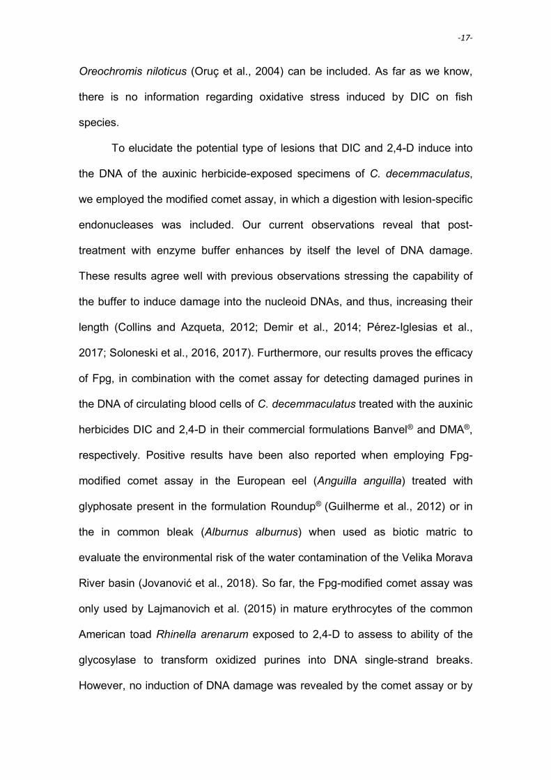
-17-
Oreochromis niloticus (Oruç et al., 2004) can be included. As far as we know,
there is no information regarding oxidative stress induced by DIC on fish
species.
To elucidate the potential type of lesions that DIC and 2,4-D induce into
the DNA of the auxinic herbicide-exposed specimens of C. decemmaculatus,
we employed the modified comet assay, in which a digestion with lesion-specific
endonucleases was included. Our current observations reveal that post-
treatment with enzyme buffer enhances by itself the level of DNA damage.
These results agree well with previous observations stressing the capability of
the buffer to induce damage into the nucleoid DNAs, and thus, increasing their
length (Collins and Azqueta, 2012; Demir et al., 2014; Pérez-Iglesias et al.,
2017; Soloneski et al., 2016, 2017). Furthermore, our results proves the efficacy
of Fpg, in combination with the comet assay for detecting damaged purines in
the DNA of circulating blood cells of C. decemmaculatus treated with the auxinic
herbicides DIC and 2,4-D in their commercial formulations Banvel® and DMA®,
respectively. Positive results have been also reported when employing Fpg-
modified comet assay in the European eel (Anguilla anguilla) treated with
glyphosate present in the formulation Roundup® (Guilherme et al., 2012) or in
the in common bleak (Alburnus alburnus) when used as biotic matric to
evaluate the environmental risk of the water contamination of the Velika Morava
River basin (Jovanović et al., 2018). So far, the Fpg-modified comet assay was
only used by Lajmanovich et al. (2015) in mature erythrocytes of the common
American toad Rhinella arenarum exposed to 2,4-D to assess to ability of the
glycosylase to transform oxidized purines into DNA single-strand breaks.
However, no induction of DNA damage was revealed by the comet assay or by

-18-
the Fpg-modified version when the 2,4-D-based commercial formulation Asi
Max 50® (50% 2,4-D) was employed in this anuran species (Lajmanovich et al.,
2015). So far we do not possess a clear answer to explain the contradictory
results obtained in the common toad R. arenarum in regard to our current
observations. However, although speculative, it could be suggested that they
might be most provably be committed to the dose of 2,4-D employed since
negative results were reported after employing a dose nearly 12 times lower
than that employed in our experimental design (Lajmanovich et al., 2015). As
far as we know, no studies employing the Fpg- and Endo III-modified alkaline
SCGE assay on DIC- and 2,4-D-exposed fish have been reported so far. Thus,
our study demonstrates for the first time DNA oxidative damage induced after
exposure to a pollutant employing the glycosylase-modified SCGE assay in an
aquatic organism like C. decemmaculatus.
In the present study increased activities of the enzymes CAT and GST
were observed in fish exposed for 48 and 96 h to both DIC- and 2,4-D. In
agreement to our observations, an enhancement in CAT activity has been
previously reported in fish exposed to several herbicides. Among them, higher
CAT levels in regard to non-exposed organisms have been reported in the
zebra fish D. rerio after treatment with bipyridylium herbicide diquat (Wang et
al., 2018), the three-barbeled catfish Rhamdia quelen (Persch et al., 2017) and
the characin Leporinus obtusidens (Glusczak et al., 2011) exposed to the
glyphosate-based formulation Roundup® as well as in the climbing perch
Anabas testudineus and the stinging catfish Heteropneustes fossilis as a
consequence of a glyphosate-based formulation Excel Mera 71 exposure
(Samanta et al., 2014). Similar observations were also reported for the streaked

-19-
prochilod Prochilodus lineatus exposed to atrazine (Paulino et al., 2012) and for
the common carp C. carpio after simazine treatment (Stara et al., 2012).
As observed for CAT, GST activity was also increased after 48 and 96 h
treatment with DIC- and 2,4-D-herbicide formulations. An increased activity of
GST could expose disorders related to an oxidative stress situation, as has
been previously confirmed in the Nile tilapia O. niloticus and the common carp
C.carpio exposed to a formulated product containing 2,4-D (trade name not
specified), to the azinphosmethyl-based formulation Guthion 20 EC (Oruç et al.,
2004; Oruç and Uner, 2002) and to the triazine compounds terbuthylazine and
metribuzin (Hostovsky et al., 2012). Besides, and in agreement with our
observations, similar raised GST activity has been also reported to occur,
among others, in the hybrid fish jundiara Leiarius
marmoratus × Pseudoplatystoma reticulatum exposed to the glyphosate-based
formulation Roundup Original® (de Moura et al., 2017) as well as in the streaked
prochilod P. lineatus exposed to atrazine (Paulino et al., 2012). Therefore, it
could be assumed that the increased CAT and GST activity could help
preventing oxidative damage caused in the specimens of C. decemmaculatus
exposed to both auxinic herbicides.
Additionally to the aforementioned altered markers for oxidative stress, a
decrease of GSH was registered in specimens treated for 48 and 96 h with
either DIC- or 2,4-D-herbicide formulations. GSH is part of the first defense of
organisms against oxidative stress (Finkel, 2000). Xenobiotics that make
contact with the cells are removed by conjugation with GSH or by means of
GST, process that decreases GSH levels (Elia et al., 2003). In agreement to our
results, a similar diminished GSH activity has been reported after exposure to

-20-
other herbicides in different fish species such as the Delta smelt Hypomesus
transpacificus exposed to the fluridone-based formulation Sonar® AS and to the
glyphosate-based formulated product Roundup® Custom (Jin et al., 2018) as
well as the rainbow trout Oncorhynchus mykiss exposed to commercial linuron
(formulation not specified) (Topal et al., 2017), among others. The diminished
GSH activity reported in our results could be due to either a high intracellular
demand of GSH associated with the endobiotic detoxification processes of DIC
and 2,4-D or by an increased production of ROS exerted by the herbicides
raising the general oxidative potential required for these processes (Mela et al.,
2013; Pamela and Richard, 1994). Previous investigations have reported that
GSH conjugates with environmental contaminants either by a spontaneous
manner or catalyzed by GST enzyme detoxification (Elia et al., 2003). Our
results agree with the pattern reported previously by Elia et al. (2003) in the
black bullhead catfish Ameiurus melas as a consequence of mercury exposure.
Authors observed an enhanced GST level and a concomitant decrease in the
GSH content, similar situation as we found in C. decemmaculatus after both
auxinic herbicide treatments reported in the current study.
Considering the diminished activity of AChE in auxinic herbicides-
exposed fish we observed, enzyme inhibition could be proposed as a possible
mechanism through which DIC- and 2,4-D-herbicide formulations could induce
oxidative stress. Although speculative, a plausible explanation for these findings
could be suggested. Accordingly, it could be considered that these auxinic
herbicides reduced liver metabolism, interfering with the synthesis of these
enzymes. The inhibition of AChE leads to an overstimulation of organism’s
nervous system, pointing out, thus, the potential neurotoxic effects of the

-21-
auxinic herbicides employed in this study as anticholinergic compounds.
Several investigations confirmed that AChE is a sensitive parameter for
neurotoxicity exerted by other xenobiotics such as pyrethroids and herbicides
rather than that very well known neurotoxic agents such as carbamates and
organophosphorus pesticides (Kumar et al., 2009). Extending this concept, it
has been reported a dose-dependent decrease in the levels of AChE in
the spotted snakehead Channa punctatus exposed to the insecticides λ-
cyhalothrin-based formulation Colt® 25 as well as to the cypermethrin-based
formulation REEVA-5, being the former more potent than the latter (Kumar et
al., 2009). Specifically, similar results have been found after exposure of the
characin L. obtusidens exposed to the herbicide glyphosate-based formulation
Roundup® (Glusczak et al., 2011; Salbego et al., 2010) and the Delta smelt
Hypomesus transpacificus exposed to the fluridone-based formulation Sonar®
AS, to the penoxsulam-based formulated product Galleon® SC or to Clearcast®,
an imazamox-based herbicide formulation (Jin et al., 2018). In fish, AChE
activity is important in motion capacity, predation and social interactions
(Rodríguez-Fuentes et al., 2015). In addition, AChE alteration could alter early
development as informed by Behra et al. (2002). In agreement, we have
previously observed the appearance of behavioral changes in C.
decemmaculatus specimens after auxinic herbicide exposure to sublethal
concentrations (Ruiz de Arcaute et al., 2014, 2016).
Diminished protein content was detected in exposed specimens,
indicating that the auxinic herbicides employed caused alterations in the protein
metabolism of exposed C. decemmaculatus. Pesticide-induce inhibitory effect
on protein level is a very well documented aspect in toxicological studies when

-22-
fish are employed as biotic matrix (Lal et al., 2013; Naqvi et al., 2017; Samanta
et al., 2014). Our results agree with several observations previously reported on
several fish species exposed to a wide variety of different active ingredients of
pesticides, including insecticides, e.g., fenvalerate (Tripathi and Verma, 2004b),
chloropyrifos (Naqvi et al., 2017), malathion (Lal et al., 2013; Naqvi et al., 2017),
α-cypermethrin (Tripathi and Singh, 2013), cypermethrin (Kumar et al., 2009;
Naqvi et al., 2017; Ullah et al., 2014), λ-cyhalothrin (Kumar et al., 2009; Naqvi et
al., 2017), permethrin (Sapana Devi and Gupta, 2014), σ-methrin (Sapana Devi
and Gupta, 2014), fenitrothion (Katsiadaki et al., 2006; Sancho et al., 1997),
diazinon (Ozcan Oruç et al., 2006), endosulfan (Tripathi and Verma, 2004a),
herbicides, e.g., atrazine (Persch et al., 2017), linuron (Katsiadaki et al., 2006),
benthiocarb (Seshagiri et al., 1987), glyphosate (Glusczak et al., 2011; Persch
et al., 2017; Samanta et al., 2014), quinclorac (Persch et al., 2017), among
others. It is worth mentioned, that similar results on total protein content were
observed recently in the Mozambique tilapia Oreochromis mossambicus after
exposure to several commercial available products (formulations not
specified) based on organophosphate and synthetic pyrethroid insecticides and
herbicides (Naqvi et al., 2017). These authors proposed several alternatives to
explain the diminution of protein content. Among them, a physiological response
of the organism to compensate xenobiotic-induced stress, an enhanced
proteolytic activity or reduced protein synthesis, hormonal imbalances affecting
normal tissue protein levels, cellular necrosis and/or altered enzymatic activities
due to DNA damage (Naqvi et al., 2017). So far, we do not possess clear
evidence allowing us to accept or reject any of the aforementioned plausible
explanations. However, our current results clearly highlight that inhibition of

-23-
protein increased in both DIC- and 2,4-D-exposed fish during 96 h as compare
to 48 h and than an stronger effect was achieved in 2-4-D-exposed C.
decemmaculatus than that exerted by DIC.
The physico-chemical properties of DIC and 2,4-D makes these
herbicides highly mobile in soil and thereby often present in the aquatic
environment (Glozier et al., 2012; Li et al., 2009). Previous observations
indicate that these auxinics have been detected in urban, peri-urban sites as
well as in agroecosystems (Félix-Cañedo et al., 2013; Glozier et al., 2012; Loos
et al., 2010; Tagert et al., 2014). Environmental concentrations of these auxinic
herbicides have been reported in countries such as United States, Canada and
Mexico in ranges from 0.04 µg/L up to 24 µg/L. In Argentina, only one study
reports a 2,4-D concentration of 0,99 µg/L found in El Crespo River in Buenos
Aires Province. As far as we know, there is no information available on the
environmental concentrations of DIC in Argentina. Although treatments in this
study comprise one concentration of DIC (410 mg/L) and 2,4-D (252 mg/L), they
represent a relatively high end of the threshold values found in the environment
of both herbicides and reported so far. Thus, it could be assumed that the
concentrations of the auxinic herbicides assayed in the present study would be
almost improbable to be found in the biosphere, possibly only after specific
incidents, such as direct application, accidental discharge and treatment of
application residues, among others. Due to the aforementioned situations, we
cannot discard the possibility that piscine populations of C. decemmaculatus, as
well as others living species, could be exposed to the auxinic herbicides
employed at these concentrations.

-24-
The present findings demonstrate that both auxinics DIC and 2,4-D in
their formulated products Banvel® and DMA®, respectively, acts as oxidizing
agents on non-target species such as the Neotropical fish C. decemmaculatus
is. Furthermore, our results emphasize that the biomarkers assayed are
valuable for evaluating contaminated aquatic environments, at least when
employing the fish C. decemmaculatus.

-25-
CONFLICT OF INTEREST STATEMENT
The authors declare that there are no conflicts of interest.
ACKNOWLEDGEMENTS
The authors thank Syngenta Agro S.A. (Buenos Aires, Argentina) and
Dow AgroSciences Argentina S.A. (Buenos Aires, Argentina) for kindly
providing the formulations Banvel® and DMA®, respectively. The experiments
performed in the present study were supported by grants from the National
University of La Plata (Grants 11/N746 11/N817 and 11/N847) and the National
Agency of Scientific and Technological Promotion (PICT 2015 Number 3059)
from Argentina.

-26-
Table 1
Table 1 Analysis of DNA damage measured by modified comet assay in Cnesterodon decemmaculatus cells exposed to dicamba-based
formulation Banvel® and 2,4-dichlorophenoxyacetic acid-based formulation DMA®
Chemicals Exposure time (h) Treatment
No. of animals
observed
No. of nucleoids analyzed
% of damaged nucleoids (II+III+IV)
GDIa ± SE Nucleoids Categories % ± SE Type
0+I Type II Type III Type IV
Negative 48 h - 10 1036
92.47
7.24
0.29
0.00
7.53 0.49 ± 0.12
control
Buffer Endo III 10 1045
57.51#
38.76#
3.73#
0.00
42.49# 1.46 ± 0.04#
Endo III 10 1014
54.73#
43.00#
2.27
0.00
45.27# 1.48 ± 0.04#
Buffer Fpg 10 1056
58.90#
33.52#
7.58#
0.00
41.10# 1.48 ± 0.03#
Fpg 10 1024
51.86#
42.29#
5.85#
0.00
48.14# 1.53 ± 0.06#
96 h - 9 946
91.86
7.72
0.42
0.00
8.14 0.48 ± 0.12
Buffer Endo III 10 1016
59.84#
36.02#
4.14#
0.00
40.16# 1.44 ± 0.04#
Endo III 10 1036
54.63#
42.08#
3.29
0.00
45.37# 1.48 ± 0.04#
Buffer Fpg 10 1022
61.25#
34.15#
4.60#
0.00
38.75# 1.43 ± 0.02#
Fpg 10 1070
47.29#
46.64#
6.07#
0.00
52.71# 1.58 ± 0.05#
DIC 48 h 410 mg/L 10 1001
40.46***
27.27***
18.18***
14.09***
59.54*** 1.99 ± 0.16***
Buffer Endo III 10 1046
15.87***
41.59
30.11***
12.43***
84.13*** 2.39 ± 0.13***
Endo III 10 1019
15.80***
41.51
30.32***
12.37***
84.20*** 2.40 ± 0.06***
Buffer Fpg 10 1023
15.84***
41.35
30.11***
12.70***
84.16*** 2.40 ± 0.13***
Fpg 10 1011
8.51***
19.09***&& 40.36***&
32.04***&&
91.49*** 2.96 ± 0.06***&&
96 h 410 mg/L 10 1100
73.64***
16.73**
8.27***
1.36
26.36*** 1.22 ± 0.11***

-27-
Buffer Endo III 10 1011
37.88**
44.21**
16.72***
1.19***
62.12*** 1.81 ± 0.03***
Endo III 10 1008
33.33**
49.50**
14.29***
2.88***
66.67*** 1.87 ± 0.03***
Buffer Fpg 9 890
33.82**
51.01***
14.27***
0.90***
66.18*** 1.82 ± 0.04***
Fpg 10 999
23.62***
45.55**
29.43***&
1.40***
76.38*** 2.09 ± 0.11***&
2,4-D 48 h 252 mg/L 10 1101
33.61***
43.51***
20.53***
2.35**
66.39*** 1.88 ± 0.09***
Buffer Endo III 10 1068
12.73***
45.13
39.61***
2.53**
87.27*** 2.32 ± 0.04***
Endo III 10 1042
14.40***
43.86
30.52***
11.22**
85.60*** 2.38 ± 0.03***
Buffer Fpg 10 1032
16.76***
47.09
33.14***
3.01***
83.24*** 2.22 ± 0.03***
Fpg 10 1021
15.77***
39.18&
32.32***
12.73***&&&
84.23*** 2.42 ± 0.06***&&
96 h 252 mg/L 10 1058
13.80***
37.24***
35.73***
13.23***
86.20*** 2.45 ± 0.16***
Buffer Endo III 10 1048
9.35***
29.01*
39.31***
22.33***
90.65*** 2.74 ± 0.05***
Endo III 10 1007
9.04***
20.16***
45.68***
25.12***
90.96*** 2.87 ± 0.06***
Buffer Fpg 10 1048
9.26***
28.82***
40.17***
21.75***
90.74*** 2.74 ± 0.05***
Fpg 10 1021
6.37***
20.47***&
41.82***
31.34***&
93.63*** 2.98 ± 0.06***&&
Positive 5 min 50 µM 10 1027
70.89*
21.52*
7.59*
0.00
29.11*** 1.21 ± 0.15***
controlb
Buffer Endo III 10 1014
14.69***
45.96
34.22**
5.13**
85.31*** 2.25 ± 0.14***
Endo III 10 1117
4.85***
38.77
37.59***
18.79***
95.15*** 2.72 ± 0.16***&
Buffer Fpg 9 899
19.13***
44.38
32.26***
4.23**
80.87*** 2.21 ± 0.13***
Fpg 9 907
6.95***
26.90**
43.00***
23.15***
93.05***&& 2.84 ± 0.17***&&
a, GDI: Genetic damage index. b, Hydrogen peroxide (H2O2, 50 µM) was used as positive control.
#, P < 0.001; significant differences with respect to negative control value. *, P < 0.05; **P < 0.01; ***P < 0.001; significant differences with their respective control group.
&, P < 0.01; significant difeerences with respective buffer-enzyme group.

-28-
Table 2
Table 2 Analysis of biomarkers in Cnesterodon decemmaculatus cells exposed to dicamba-based formulation Banvel® and
2,4-dichlorophenoxyacetic acid-based formulation DMA®
Chemicals Exposure time (h)
Biomarkers (Mean ± SE)a Pr CAT GST GSH AChE
Negative control 48 106.82 ± 13.18 12.86 ± 2.34 0.04 ± 0.01 3.17 ± 0.56 384.71 ± 17.33
DIC
98.22 ± 7.78 51.59 ± 6.40*** 0.09 ± 0.01** 1.56 ± 0.15* 343.87 ± 89.81
2,4-D
80.30 ± 8.45 68.91 ± 20.27*** 0.07 ± 0.01* 2.08 ± 0.13 267.11 ± 45.79
Negative control 96 115.58 ± 16.18 13.52 ± 2.96 0.04 ± 0.01 2.60 ± 0.16 372.21 ± 37.25
DIC
81.02 ± 7.85* 30.08 ± 3.82*** 0.07 ± 0.01*** 2.06 ± 0.25 217.48 ± 65.07**
2,4-D
57.18 ± 4.81*** 41.55 ± 6.57*** 0.10 ± 0.01*** 1.04 ± 0.03** 164.24 ± 34.54**
a, Biomarkers: Pr: protein content as mg g/ww; CAT: catalase as µmol H2O2 consumed/min mg protein;
GST: glutathione-S-transferase as µmol CDNB conjugated formed/min mg protein; GSH: Glutathione content as soluble acid thiols µmol/ g ww. AChE: nmol of substrate hydrolyzed/min mg protein.
*, P < 0.05; **P < 0.01; ***P < 0.001; significant differences with the control group.

-29-
Figure 1

-30-
LEGEND FOR THE FIGURE
Figure 1. DIC- and 2,4-D-based commercial formulations induced DNA damage
evaluated by the Endo III (black bars)- and Fpg (grey bars)-modified comet assay in
circulating blood cells of Cnesterodon decemmaculatus. Net oxidative damage was
expressed as the substraction between the score obtained after incubation with the
respective enzyme or with the buffer. Hydrogen peroxide (50 µm) was employed as
positive control. &, P < 0.05; &&, P < 0.01; significant differences with respective buffer-
enzyme.

-31-
REFERENCES
Ackerman, P. A., Morgan, J. D., Iwama, G. K., Anestethics. In: C. C. o. A. Care, (Ed.), The Care and use of Fish in Research, Teaching and Testing. Canadian Council on Animal Care, Ottawa, CA, 2005, pp. 1-22.
Adhikari, N., Grover, I. S., 1988. Genotoxic effects of some systemic pesticides: In vivo chromosomal aberrations in bone marrow cells in rats. Environmental and Molecular Mutagenesis. 12, 235-242.
Amer, S. M., Aly, F. A. E., 2001. Genotoxic effect of 2,4-dichlorophenoxy acetic acid and its metabolite 2,4-dichlorophenol in mouse. Mutation Research. 494, 1-12.
Arias, E., 2007. Cytogenetic effects of short- and long- term exposure of chick embryos to the phenoxyherbicide 2,4-D. Environmental and Molecular Mutagenesis. 48, 462-466.
Atamaniuk, T. M., Kubrak, O. I., Storey, K. B., Lushchak, V. I., 2013. Oxidative stress as a mechanism for toxicity of 2,4-dichlorophenoxyacetic acid (2,4-D): studies with goldfish gills. Ecotoxicology. 22, 1498-1508.
Ateeq, B., Farah, M. A., Ahmad, W., 2005. Detection of DNA damage by alkaline single cell gell electrophoresis in 2,4-dichlororphenoxyacetic-acid- and butachlor-exposed erythrocytes of Clarias batrachus. Ecotoxicology and Environmental Safety. 62, 348-354.
Ateeq, B., Farah, M. A., Ali, M. N., Ahmad, W., 2002. Clastogenicity of pentachlorophenol, 2,4-D and butachlor evaluated by Allium root tip test. Mutation Research. 514, 105-113.
Azqueta, A., Campos Costa-Amaral, I., Collins, A. R., High-throughput measurements of DNA breaks and oxidised bases with the comet assay. In: A. Dhawan, D. Anderson, Eds.), The Comet Assay in Toxicology. The Royal Society of Chemistry, Cambridge, UK, 2017, pp. 67-87.
Baudhuin, P., Beaufy, H., Rahman-Li, Y., Sellinger, O. Z., Wattiaux, R., Jacques, P., al., e., 1964. Tissue fractionation studies. 17. Intracellular distribution of monoamine oxidase, alanine aminotransferase, D-amino acid oxidase and catalase in rat liver tissue. Biochemical Journal. 91, 179-184.
Baudou, F. G., Ossana, N. A., Castañé, P. M., Mastrángelo, M. M., Ferrari, L., 2017. Cadmium effects on some energy metabolism variables in Cnesterodon decemmaculatus. Ecotoxicology. 26, 1250-1258.
Behra, M., Cousin, X., Bertrand, C., Vonesch, J. L., Biellmann, D., Chatonnet, A., Strähle, U., 2002. Acetylcholinesterase is required for neuronal and muscular development in the zebrafish embryo. Nature Neuroscience. 5, 111-118.
Bokán, K., Syberg, K., Jensen, K., Rank, J., 2013. Genotoxic potential of two Herbicides and their active ingredients assessed with comet assay on a fish cell line, Epithelioma Papillosum Cyprini (EPC). Journal of Toxicology and Environmental Health, Part A: Current Issues. 76, 1129-1137.
Bony, S., Gaillard, I., Devaux, A., 2010. Genotoxicity assessment of two vineyard pesticides in zebrafish. International Journal of Environmental Analytical Chemistry. 90, 421-428.
Bukowska, B., 2003. Effects of 2,4-D and its metabolite 2,4-dichlorophenol on antioxidant enzymes and level ofglutathione in human erythrocytes
Comparative Biochemistry and Physiology Part C. 135, 435–441. Bukowska, B., 2006. Toxicity of 2,4-dichlorophenoxyacetic acid-molecular mechanisms. Polish Journal of
Environmental Studies. 15, 365-374. Cadet, J., Douki, T., Gasparutto, D., Ravanat, J. L., 2003. Oxidative damage to DNA: formation,
measurement and biochemical features. Mutation Research. 531, 5-23. CASAFE, 2017. Guía de Productos Fitosanitarios para la República Argentina. Cámara de Sanidad
Agropecuaria y Fertilizantes, 1200 pp. Cavalcante, D. G., Martinez, C. B., Sofia, S. H., 2008. Genotoxic effects of Roundup on the fish
Prochilodus lineatus. Mutation Research. 655, 41-46. Çavaş, T., Könen, S., 2007. Detection of cytogenetic and DNA damage in peripheral erythrocytes of
goldfish (Carassius auratus) exposed to a glyphosate formulation using the micronucleus test and the comet assay. Mutagenesis. 22, 263-268.

-32-
Collins, A. R., 2004. The comet assay for DNA damage and repair. Molecular Biotechnology. 26, 249-261. Collins, A. R., Azqueta, A., 2012. DNA repair is a biomarker in human biomonitoring studies; further
applications of the comet assay. Mutation Research. 736, 122-129. Collins, A. R., Dusinká, M., Gedik, C. M., Stetina, R., 1996. Oxidative damage to DNA: do we have a
reliable biomarker? Environmental Health Perspectives. 104, 465-469. Cooke, M. S., Olinski, R., Evans, M. D., 2006. Does measurement of oxidative damage to DNA have
clinical significance? Clinica Chimica Acta. 365, 30-49. Costantini, D., Verhulst, S., 2009. Does high antioxidant capacity indicate low oxidative stress?
Functional Ecology. 23, 506-509. de la Torre, F. R., Ferrari, L., Salibián, A., 2005. Biomarkers of a native fish species (Cnesterodon
decemmaculatus) application to the water toxicity assessment of a peri-urban polluted river of Argentina. Chemosphere. 59, 577-583.
de Moura, F. R., Brentegani, K. R., Gemelli, A., Sinhorin, A. P., Sinhorin, V. D. G., 2017. Oxidative stress in the hybrid fish jundiara (Leiarius marmoratus x Pseudoplatystoma reticulatum) exposed to Roundup Original®. Chemosphere. 185, 445-451.
Demir, E., Akça, H., Kaya, B., Burgucu, D., Tokgün, O., Turna, F., Aksakal, S., Vales, G., Creus, A., Marcos, R., 2014. Zinc oxide nanoparticles: genotoxicity, interactions with UV-light and cell-transforming potential. Journal of Hazardous Materials. 264, 420-429.
Dhawan, A., Anderson, D., 2017. The Comet assay in toxicology. The Royal Society of Chemistry, Cambridge, UK.
Elia, A. C., Galarini, R., Taticchi, M. I., Dörr, A. J. M., Mantilacci, L., 2003. Antioxidant responses and bioaccumulation in Ictalurus melas under mercury exposure. Ecotoxicology and Environmental Safety. 55, 162-167.
Ellman, G. L., 1959. Tissue sulfhydryl groups. Archives of Biochemistry and Biophysics. 82, 70-77. Ellman, G. L., Courtney, K. D., Andreas, V. J., Feathersone, R. M., 1961. A new and rapid colorimetric
determination of acetylcholinesterase activity. Biochemestry Pharmacology. 7, 88-95. Espandiari, P., Ludewig, G., Glauert, H. P., Robertson, L. W., 1998. Activation of hepatic NF-κB by the
herbicide dicamba (2-methoxy-3,6-dichlorobenzoic acid) in female and male rats. Journal of Biochemistry and Molecular Toxicology. 12, 339-344.
Farah, M. A., Ateeq, B., Ahmad, W., 2006. Antimutagenic effect of neem leaves extract in freshwater fish, Channa punctatus evaluated by cytogenic tests. Science of the Total Environment. 364, 200-214.
Félix-Cañedo, T. E., Durán-Àlvarez, J. C., Jiménez-Cisneros, B., 2013. The ocurrence and distribution of a group of organic micropollutants in Mexico City´s water sources. Science of the Total Environment. 454-455, 109-118.
Ferrari, A., Venturino, A., D´angelo, A. M. P., 2007. Muscular and brain cholinesterase sensitivities to azinphos methyl and carbaryl in the juvenile rainbow trout Oncorhynchus mykiss. Comparative Biochemistry and Phisiology - Part C. 146, 308-313.
Finkel, T. H., N J, 2000. Oxidants, oxidative stress and the biology of aging. Nature. 408, 239-247. Glozier, N. E., Struger, J., Cessna, A. J., Gledhill, M., Roundeau, M., Ernst, W. R., Sekela, M. A., Cagampan,
S. J., Sverko, E., Murphi, C., Murray, J. L., Donald, D. B., 2012. Occurrence of glyphosate and acidic herbicides in select urban rivers and streams in Canada, 2007. Environmental Science and Pollution Research. 19, 821-834.
Glusczak, L., Loro, V. L., Pretto, A., Moraes, B. S., Raabe, A., Duarte, M. F., da Fonseca, M. B., de Menezes, C. C., Valladao, D. M. D., 2011. Acute exposure to glyphosate herbicide affects oxidative parameters in piava (Leporinus obtusidens). Archives of Environmental Contamination and Toxicology. 61, 624-630.
Gómez, S., Villar, C., Bonetto, C., 1998. Zinc toxicity in the fish Cnesterodon decemmaculatus in the Parana river and Rio de la Plata estuary. Environmental Pollution. 99, 159-165.
González, M., Soloneski, S., Reigosa, M. A., Larramendy, M., 2005. Genotoxiciy of the herbicide 2,4-dichlorophenoxyacetic acid and a commercial formulation, 2,4-dichlorophenoxyacetid acid

-33-
dimethylamine salt. I. Evaluation of DNA damage and cytogenetic endopoints in Chinese Hamster ovary (CHO) cells. Toxicology in vitro. 19, 289-297.
González, N., Soloneski, S., Larramendy, M., 2006. Genotoxicity analysis of the phenoxy herbicide dicamba in mammalian cells in vitro. Toxicology in vitro. 20, 1481-1487.
González, N., Soloneski, S., Larramendy, M., 2007. The chlorophenoxy herbicide dicamba and its commercial formulation banvel induce genotoxicity in Chinese hamster ovary cells. Mutation Research. 634, 60-68.
González, N., Soloneski, S., Larramendy, M., 2011. A combination of the cytokinesis-block micronucleus cytome assay and centromeric identification for evaluation of the genotoxicity of dicamba. Toxicology letters. 207, 204-212.
Guilherme, S., Santos, M. A., Barroso, C., Gaivão, I., Pacheco, M., 2012. Differential genotoxicity of RoundUp® formulation and its constituents in blood cells of fish (Anguilla anguilla): considerations on chemical interactions and DNA damaging mechanisms. Ecotoxicology. 21, 1381-1390.
Habig, W. H., Pabst, M. J., Jakoby, W. B., 1974. Glutathione S-transferases. The first enzimatic step in mercapturic acid formation. Journal of Biological Chemistry. 22, 7130-7139.
Hostovsky, M., Blahova, J., Plhalova, L., Stepanova, S., Praskova, E., Marsalek, P., Svobodova, Z., 2012. Oxidative stress parameters in early developmental stages of common carp (Cyprinus carpio L.) after subchronic exposure to terbuthylazine and metribuzin. Neuro Endocrinol Lett. 33, 124-129.
Hulbert, A. J., Pamplona, R., Buffenstein, R., Buttemer, W. A., 2007. Life and death:metabolic rate, membrane composition, and life span of animals. Physiological Reviews. 87, 1175–1213.
Jin, J., Kurobe, T., Ramírez-Duarte, W. F., Bolotaolo, M. B., Lam, C. H., Pandey, P. K., Hung, T.-C., Stillway, M. E., Zweig, L., Caudill, J., Lin, L., Teh, S. J., 2018. Sub-lethal effects of herbicides penoxsulam, imazamox, fluridone and glyphosate on Delta smelt (Hypomesus transpacificus). Aquatic Toxicology. 197, 79-88.
Jovanović, J., Kolarević, S., Milošković, A., Radojković, N., Simić, V., Dojčinović, B., Kračun-Kolarević, M., Paunović, M., Kostić, J., Sunjog, K., Timilijić, J., Djordjević, J., Gačić, Z., Žegura, B., Vuković-Gačić, B., 2018. Evaluation of genotoxic potential in the Velika Morava River Basin in vitro and in situ. Environmental Science and Pollution Research. 621, 1289-1299.
Katsiadaki, I., Morris, S., Squires, C., Hurst, M. R., James, J. D., Scott, A. P., 2006. Use of the three-spined stickleback (Gasterosteus aculeatus) as a sensitive in vivo test for detection of environmental antiandrogens. Environmental Health Perspectives. 114, 115-121.
Kaya, A., Yigit, E., 2012. Interactions among glutathione s-transferase, glutathione reductase activity and glutathione contents in leaves of Vicia faba L. subjected to flurochloridone. Fresenius Environmental Bulletin. 21, 1635–1640.
Kubrak, O. I., Atamaniuk, T. M., Husak, V. V., Lushchak, V. I., 2013. Transient effects of 2,4-dichlorophenoxyacetic acid (2,4-D) exposure on some metabolic and free radical processes in goldfish white muscle. Food and Chemical Toxicology. 59, 356-361.
Kumar, A., Rai, D. A., Sharma, B., Pandeym, R. S., 2009. λ-cyhalothrin and cypermethrin induced in vivo alterations in the activity of acetylcholinesterase in a fresh-water fish, Channa punctatus (Bloch). Pesticide Biochemical and Physiology. 93, 96-99.
Lajmanovich, R. C., Attademo, A. M., Simoniello, M. F., Poletta, G. L., Junges, C. M., Peltzer, P. M., Grenón, P., Cabagna-Zenklusen, M. C., 2015. Harmful Effects of the Dermal Intake of Commercial Formulations Containing Chlorpyrifos, 2,4-D, and Glyphosate on the Common Toad Rhinella arenarum (Anura: Bufonidae). Water, Air, and Soil Pollution. 226.
Lal, B., Sarang, M. K., Kumar, P., 2013. Malathion exposure induces the endocrine disruption and growth retardation in the catfish, Clarias batrachus (Linn.). General and Comparative Endocrinology. 181, 139-145.
Lazartigues, A., Thomas, M., Banas, D., Brun-Bellut, J., Cren-Olivé, C., Feidt, C., 2013. Accumulation and half-lives of 13 pesticides in muscle tissue of freshwater fish through food exposure. Chemosphere. 91, 530-535.

-34-
Li, J., Li, Y., Lu, J., 2009. Adsorption of herbicides 2,4-D and acetochlor on inorganic-organic bentonites. Applied Clay Science. 46, 314-318.
Li, K., Wu, J. Q., Jiang, L. L., Shen, L. Z., Li, J. Y., He, Z. H., Wei, P., Lv, Z., He, M. F., 2017. Developmental toxicity of 2,4-dichlorophenoxyacetic acid in zebrafish embryos. Chemosphere. 171, 40-48.
Loos, R., Locoro, G., Comero, S., Contini, S., Schwesig, D., Werres, F., Balsaa, P., Gans, O., Weiss, S., Blaha, L., Bolchi, M., Gawlik, B. M., 2010. Pan-European survey on the occurrence of selected polar organic persistent pollutants in ground water. Water Research. 44, 4115-4126.
Lowry, O. H., Rosebrough, N. J., Farr, A. L., Randall, R. J., 1951. Protein measurement with the Folin phenol reagent. Journal of Biological Chemistry. 193, 265-275.
Lushchak, V. I., 2011. Environmentally induced oxidative stress in aquatic animals. Aquatic Toxicology. 101, 13-30.
Madriagal-Bujaidar, E., Hernández-Ceruelos, A., Chamorro, G., 2001. Induction of sister chromatid exchanges by 2,4-dichlorophenoxyacetic acid in somatic and germ cells of mice exposed in vivo. Food and Chemical Toxicology. 39, 941-946.
Martínez-Tabche, L., Madriagal-Bujaidar, E., Negrete, T., 2004. Genotoxicity and lipoperoxidation produced by paraquat and 2,4-dichlorophenoxyacetic acid in the gills of rainbow trout (Oncorhynchis mikiss). Bulletin of Environmental Contamination and Toxicology. 73.
Martins, M., Costa, P. M., The comet assay in aquatic (eco)genotoxicity using non-conventional model organisms: relevance, constrains and prospects. In: M. L. Larramendy, (Ed.), Ecotoxicology and Genotoxicology: Non-traditional aquatic models. The Royal Society of Chemistry, Cambridge, 2017, pp. 3-32.
Mastrángelo, M. M., Ferrari, L., 2013. Cnesterodon decemmaculatus juveniles as test organisms in toxicity assessment: cadmium case. Bulletin of Environmental Contamination and Toxicology. 91, 49-54.
Matviishyn, T. M., Kubrak, O. I., Husak, V. V., Storey, K. B., Lushchak, V. I., 2014. Tissue-specific induction of oxidative stress in goldfish by 2,4-dichlorophenoxyacetic acid: mild in brain and moderate in liver and kidney. Environmetnal Toxicology and Pharmacology. 37, 861-869.
Mela, M., Guiloski, I. C., Doria, H. B., Randi, M. A., de Oliveira Ribeiro, C. A., Pereira, L., Maraschi, A. C., Prodocimo, V., Freire, C. A., Silva de Assis, H. C., 2013. Effects of the herbicide atrazine in neotropical catfish (Rhamdia quelen). Ecotoxicology and Environmental Safety. 93, 13-21.
Menéndez-Helman, R. J., Ferreyroa, G. V., Dos Santos Afonso, M., Salibián, A., 2012. Glyphosate as an acetylcholinesterase inhibitor in Cnesterodon decemmaculatus. Bulletin of Environmental Contamination and Toxicology. 88, 6-9.
Menéndez-Helman, R. J., Ferreyroa, G. V., dos Santos Afonso, M., Salibián, A., 2015. Circannual rhythms of acetylcholinesterase (AChE) activity in the freshwater fish Cnesterodon decemmaculatus. Ecotoxicology and Environmental Safety. 111, 236-241.
Muthulakshmi, S., Maharajan, K., Habibi, H. R., Kadirvelu, K., Venkataramana, M., 2018. Zearalenone induced embryo and neurotoxicity in zebrafish model (Danio rerio): Role of oxidative stress revealed by a multi biomarker study. Chemosphere. 198, 111-121.
Naqvi, G., Shoaib, N., Ali, A. M., 2017. Pesticides impact on protein fish (Oreochromis mossambicus) tissues. Indian Journal of Geo Marine Sciences. 49, 1864-1868.
Newman, M. C., Clements, W. H., 2008. Ecotoxicology. A comprehensive treatment. CRC Press, Boca Ratón, FL.
Nunes, B., Carvahlo, F., Guilhermino, L., 2005. Characterization and use of the total head soluble cholinesterases activity. Journal of Enzyme Inhibition and Medicinal Chemistry. 20, 369-376.
Oruç, E. O., Sevgiler, Y., Uner, N., 2004. Tissue-specific oxidative stress responses in fish exposed to 2,4-D and azinphosmethyl. Comparative Biochemistry and Physiology, Part C: Toxicology and Pharmacology. 137, 43-51.
Oruç, E. O., Uner, N., 2002. Marker enzyme assessment in the liver of Cyprinus carpio (L.) exposed to 2,4-D and azinphosmethyl. Journal of Biochemical and Molecular Toxicology. 16, 182-188.

-35-
Oruç, E. O., Üner, N., 2000. Combined effects of 2,4-D and azinphosmethyl on antioxidant enzymes and lipid peroxidation in liver of Oreochromis niloticus. Comparative Biochemistry and Physiology, Part C. 127, 291-296.
Ossana, N. A., Eissa, B. L., Baudou, F. G., Castañé, P. M., Soloneski, S., Ferrari, L., 2016. Multibiomarker response in ten spotted live-bearer fish Cnesterodon decemmaculatus (Jenyns, 1842) exposed to Reconquista river water. Ecotoxicology and Environmental Safety. 133, 73-81.
Ozcan Oruç, E., Uner, N., Sevgiler, Y., Usta, D., Durmaz, H., 2006. Sublethal effects of organophosphate diazinon on the brain of Cyprinus carpio. Drug and Chemical Toxicology. 29, 57-67.
Pamela, C. C., Richard, A. H., 1994. Biochemistry. Lippencott - Raven Publishers, Philadelphia, Pennsylvania.
Paulino, M. G., Souza, N. E. S., Fernandes, M. N., 2012. Subchronic exposure to atrazine induces biochemical and histopathological changes in the gills of a Neotropical freshwater fish, Prochilodus lineatus. Ecotoxicology and Environmental Safety. 80, 6-13.
Peixoto, F. P., Gomes-Laranjo, J., Vicente, J. A., Madeira, V. M. C., 2008. Comparative effects of the herbicides dicamba, 2,4-D and paraquat on non-green potato tuber calli. Journal of Plant Physiology. 165, 1125-1133.
Pérez-Iglesias, J. M., Ruiz de Arcaute, C., Natale, G. S., Soloneski, S., Larramendy, M. L., 2017. Evaluation of imazethapyr-induced DNA oxidative damage by alkaline Endo III- and Fpg-modified single-cell gel electrophoresis assay in Hypsiboas pulchellus tadpoles (Anura, Hylidae). Ecotoxicology and Environmental Safety. 142, 503-508.
Persch, T. S. P., Weimer, R. N., Freitas, B. S., Oliveira, G. T., 2017. Metabolic parameters and oxidative balance in juvenile Rhamdia quelen exposed to rice paddy herbicides: Roundup®, Primoleo®, and Facet®. Chemosphere. 174, 98-109.
Pitarque, M., Creus, A., Marcos, R., Hughes, J. A., Anderson, D., 1999. Examination of various biomarkers measuring genotoxic endpoints from Barcelona airport personnel. Mutation Research. 440, 195-204.
Poletta, G. L., Simoniello, M. F., Mudry, M. D., 2016. Biomarkers of oxidative damage and antioxidant defense capacity in Caiman latirostris blood. Comparative Biochemistry and Physiology, Part C. 179, 29-36.
Rodríguez-Fuentes, G., Rubio-Escalante, F. J., Noreña-Barroso, E., Escalante-Herrera, K. S., Schlenk, D., 2015. Impacts of oxidative stress on acetylcholinesterase transcription, and activity in embryos of zebrafish (Danio rerio) following chlorpyrifos exposure. Comparative Biochemistry and Physiology, Part C. 172-173, 19-25.
Romero-Puertas, M. C., Rodríguez-Serrano, M., Corpas, F. J., Gómez, M., del Rio, L. A., Sandalio, L. M., 2004. Cd-induced subcellular accumulation of O2
- and H2O2 in pea leaves. Plant, Cell, and Environment. 27, 1122-1134.
Ruiz de Arcaute, C., Soloneski, S., Larramendy, M., 2014. Evaluation of the genotoxicity of a herbicide formulation containing 3,6-dichloro-2-metoxybenzoic acid (dicamba) in circulating blood cells of the tropical fish Cnesterodon decemmaculatus. Mutation Research. 773, 1-8.
Ruiz de Arcaute, C., Soloneski, S., Larramendy, M., 2016. Toxic and genotoxic effects of the 2,4-dichlorophenoxyacetic acid (2,4-D)-based herbicide on the Neotropical fish Cnesterodon decemmaculatus. Ecotoxicology and Environmental Safety. 128, 222-229.
Salbego, J., Pretto, A., Gioda, C. R., Cavalheiro de Menezes, c., Lazzari, R., Radunz, J., Baldisseriotto, B., Loro, V. L., 2010. Herbicide formulation with glyphosate affects growth, acetylcholinesterase activity, and metabolic and haematological parameters in piava (Leporinus obtusidens). Archives of Environmental Contamination and Toxicology. 58, 740-745.
Samanta, P., Pal, S., Mukherjee, A. K., Ghosh, A. R., 2014. Biochemical effects of glyphosate based herbicide, Excel Mera 71 on enzyme activities of acetylcholinesterase (AChE), lipid peroxidation (LPO), catalase (CAT), glutathione-S-transferase (GST) and protein content on teleostean fishes. Ecotoxicology and Environmental Safety. 107, 120-125.
Sancho, E., Ferrando, M. D., Andreu, E., 1997. Sublethal Effects of an Organophosphate Insecticide on the European eel, Anguilla anguilla. Ecotoxicology and Environmental Safety. 36, 57-65.

-36-
Sapana Devi, M., Gupta, A., 2014. Sublethal toxicity of commercial formulations of deltamethrin and permethrin on selected biochemical constituents and enzyme activities in liver and muscle tissues of Anabas testudineus. Pesticide Biochemistry and Physiology. 115, 48-52.
Seshagiri, R., Sreenivasa, M. K., Reddy, K. B., Sreeramulu, C. C., 1987. Effect of benthiocarb on protein metabolism of freshwater teleost Sarotherodon mossambicus. Indian Journal of Environmental Health. 29, 45-51.
Soloneski, S., Larramendy, M. L., The use of the ten spotted live-bearer fish Cnesterodon decemmaculatus (Jenyns, 1842) (Pisces, Poeciliidae) in the genotoxic evaluation of environmental pollutants. In: M. L. Larramendy, (Ed.), Ecotoxicology and genotoxicology - Non-traditional Aquatic Models. The Royal Society of Chemistry, London, 2017, pp. 327-346.
Soloneski, S., Nikoloff, N., Larramendy, M. L., 2016. Analysis of possible genotoxicity of the herbicide flurochloridone and its commercial formulations: Endo III and Fpg alkaline comet assays in Chinese hamster ovary (CHO-K1) cells. Mutation Research. 797, 46-52.
Soloneski, S., Ruiz de Arcaute, C., Nikoloff, N., Larramendy, M. L., 2017. Genotoxicity of the herbicide imazethapyr in mammalian cells by oxidative DNA damage envaluation using the Endo III and Fpg alkaline comet assays. Environmental Science and Pollution Research. 24, 10292-10300.
Sorensen, K. C., Stucki, J. W., Wagner, E. D., Wagner, E. D., Plewa, M. J., 2005. Modulation of the Genotoxicity of Pesticides Reacted with Redox-Modified Smectite Clay. Environmental and Molecular Mutagenesis. 46, 174-181.
Stara, A., Machova, J., Velisek, J., 2012. Effect of chronic exposure to simazine on oxidative stress and antioxidant response in common carp (Cyprinus carpio L.). Environmental Toxicology and Pharmacology. 33, 334-343.
Summerfelt, R. C., Smith, L. S., 1990. Anaesthesia, surgery and related techniques. In: Schreck, C.B., Moyle, P.B. (Eds.), Methods for Fish Biology. American Fisheries Society, Bethesda MD. 213-272.
Tagert, M. L. M., Massey, J. H., Shaw, D. R., 2014. Water quality survey of Mississippi´s Upper Pearl River. Science of the Total Environment. 481, 564-573.
Topal, A., Alak, G., Altun, S., Erol, H. S., Atamanalp, M., 2017. Evaluation of 8-hydroxy-2-deoxyguanosine and NFkB activation, oxidative stress response, acetylcholinesterase activity, and histopathological changes in rainbow trout brain exposed to linuron. Environmental Toxicology and Pharmacology. 49, 14-20.
Tripathi, G., Singh, H., 2013. Impact of alphamethrin on biochemical parameters of Channa punctatus. Journal of Environmental Biology. 34, 227-230.
Tripathi, G., Verma, P., 2004a. Endosulfan-mediated biochemical changes in the freshwater fish Clarias batrachus. Biomedical and Environmental Sciences. 17, 47-56.
Tripathi, G., Verma, P., 2004b. Fenvalerate-induced changes in a catfish, Clarias batrachus: metabolic enzymes, RNA and protein. Comparative Biochemistry and Physiology. Part C, Toxicology & Pharmacology. 138, 75-79.
Ullah, R., Zuberi, A., Ullah, S., Ullah, I., Ullah Dawar, F., 2014. Cypermethrin induced behavioral and biochemical changes in mahseer, Tor putitora. The Journal of Toxicological Sciences. 39, 829-836.
USEPA, 2005. Reregistration Eligibility Decision (RED) for 2,4 D. U.S. Environmental Protection Agency, Washington DC. 738-R-05-002.
USEPA, 2006. Reregistration eligibility decision for dicamba and associated salts. U.S. Environmental Protection Agency, Washington DC. https://archive.epa.gov/pesticides/reregistration/web/pdf/dicamba_red.pdf.
USEPA, 2017. Pesticides industry sales and usage. 2008-2012 Market estimates. U.S. Environmental Protection Agency, Washington DC. 24.
Valko, M., Rhodes, C. J., Moncol, J., Izakovic, M., Mazur, M., 2006. Free radicals, metals and antioxidants in oxidative stress-induced cancer. Chemico-Biological Interactions. 160, 1-40.
van der Oost, R., 2003. Fish bioaccumulation and biomarkers of oxidative stress in aquatic organism in relation to toxic environmental pollutants. Ecotoxicology and Envirnonmental Safety. 64, 178-189.

-37-
Varó, I., Amat, F., Navarro, J. C., 2008. Acute toxicity of dichlorvos to Aphanius iberus (Cuvier & Valenciennes, 1846) and its anti-cholinesterase effects on this species. Aquatic Toxicology. 88, 53-61.
Vera-Candioti, J., Soloneski, S., Larramendy, M. L., 2011. Acute toxicity of chromium on Cnesterodon decemmaculatus (Pisces: Poeciliidae). Theoria. 1, 81-88.
Vera-Candioti, J., Soloneski, S., Larramendy, M. L., 2013. Single-cell gel electrophoresis assay in the ten spotted live-bearer fish, Cnesterodon decemmaculatus (Jenyns, 1842), as bioassay for agrochemical-induced genotoxicity. Ecotoxicology and Environmental Safety. 98, 368-373.
Wang, X. H., Souders, C. L., Zhao, Y. H., Martyniuk, C. J., 2018. Mitochondrial bioenergetics and locomotor activity are altered in zebrafish (Danio rerio) after exposure to the bipyridylium herbicide diquat. Toxicology Letters. 283, 13-20.
Zar, J. H., 1999. Biostatistical Analysis. Prentice Hall, New Jersey.