Coordination Chemistry Reviews - SEDICI
25
Click here to load reader
-
Upload
khangminh22 -
Category
Documents
-
view
0 -
download
0
Transcript of Coordination Chemistry Reviews - SEDICI
R
V
Ja
b
c
C
a
ARR1AA
KVBCIA
h0
Coordination Chemistry Reviews 301–302 (2015) 24–48
Contents lists available at ScienceDirect
Coordination Chemistry Reviews
j ourna l h omepage: www.elsev ier .com/ locate /ccr
eview
anadium compounds in medicine
oao Costa Pessoaa,∗, Susana Etcheverryb,∗, Dinorah Gambinoc,∗
Centro de Química Estrutural, Instituto Superior Técnico, Universidade de Lisboa, Av Rovisco Pais, 1049-001 Lisboa, PortugalCátedra de Bioquímica Patológica and CEQUINOR, Facultad de Ciencias Exactas, Universidad Nacional de La Plata, 47 y 115 1900 La Plata, ArgentinaCátedra de Química Inorgánica, Facultad de Química, Universidad de la República, Gral. Flores 2124, 11800 Montevideo, Uruguay
ontents
1. Introduction . . . . . . . . . . . . . . . . . . . . . . . . . . . . . . . . . . . . . . . . . . . . . . . . . . . . . . . . . . . . . . . . . . . . . . . . . . . . . . . . . . . . . . . . . . . . . . . . . . . . . . . . . . . . . . . . . . . . . . . . . . . . . . . . . . . . . . . . . . 251.1. Distribution, essentiality and potential toxicity of vanadium. . . . . . . . . . . . . . . . . . . . . . . . . . . . . . . . . . . . . . . . . . . . . . . . . . . . . . . . . . . . . . . . . . . . . . . . . . . . . . . . 25
2. Biological actions . . . . . . . . . . . . . . . . . . . . . . . . . . . . . . . . . . . . . . . . . . . . . . . . . . . . . . . . . . . . . . . . . . . . . . . . . . . . . . . . . . . . . . . . . . . . . . . . . . . . . . . . . . . . . . . . . . . . . . . . . . . . . . . . . . . . . 262.1. Enzyme inhibition. . . . . . . . . . . . . . . . . . . . . . . . . . . . . . . . . . . . . . . . . . . . . . . . . . . . . . . . . . . . . . . . . . . . . . . . . . . . . . . . . . . . . . . . . . . . . . . . . . . . . . . . . . . . . . . . . . . . . . . . . . . . . 262.2. Enzyme activation . . . . . . . . . . . . . . . . . . . . . . . . . . . . . . . . . . . . . . . . . . . . . . . . . . . . . . . . . . . . . . . . . . . . . . . . . . . . . . . . . . . . . . . . . . . . . . . . . . . . . . . . . . . . . . . . . . . . . . . . . . . . 262.3. Physiological roles of vanadium . . . . . . . . . . . . . . . . . . . . . . . . . . . . . . . . . . . . . . . . . . . . . . . . . . . . . . . . . . . . . . . . . . . . . . . . . . . . . . . . . . . . . . . . . . . . . . . . . . . . . . . . . . . . . . 27
3. Therapeutic applications of vanadium compounds (VCs) . . . . . . . . . . . . . . . . . . . . . . . . . . . . . . . . . . . . . . . . . . . . . . . . . . . . . . . . . . . . . . . . . . . . . . . . . . . . . . . . . . . . . . . . . . . 283.1. Uptake, speciation and biodistribution of vanadium compounds . . . . . . . . . . . . . . . . . . . . . . . . . . . . . . . . . . . . . . . . . . . . . . . . . . . . . . . . . . . . . . . . . . . . . . . . . . . 293.2. Vanadate/phosphate analogy . . . . . . . . . . . . . . . . . . . . . . . . . . . . . . . . . . . . . . . . . . . . . . . . . . . . . . . . . . . . . . . . . . . . . . . . . . . . . . . . . . . . . . . . . . . . . . . . . . . . . . . . . . . . . . . . . 303.3. Transport of vanadium in blood . . . . . . . . . . . . . . . . . . . . . . . . . . . . . . . . . . . . . . . . . . . . . . . . . . . . . . . . . . . . . . . . . . . . . . . . . . . . . . . . . . . . . . . . . . . . . . . . . . . . . . . . . . . . . . 303.4. Biodistribution in the body . . . . . . . . . . . . . . . . . . . . . . . . . . . . . . . . . . . . . . . . . . . . . . . . . . . . . . . . . . . . . . . . . . . . . . . . . . . . . . . . . . . . . . . . . . . . . . . . . . . . . . . . . . . . . . . . . . . 303.5. Vanadium in the treatment of diabetes . . . . . . . . . . . . . . . . . . . . . . . . . . . . . . . . . . . . . . . . . . . . . . . . . . . . . . . . . . . . . . . . . . . . . . . . . . . . . . . . . . . . . . . . . . . . . . . . . . . . . . 32
3.5.1. Insulin-mimetic (-enhancing) vanadium compounds . . . . . . . . . . . . . . . . . . . . . . . . . . . . . . . . . . . . . . . . . . . . . . . . . . . . . . . . . . . . . . . . . . . . . . . . . . . . . . 333.5.2. The mode of action of vanadate in glucose homeostasis . . . . . . . . . . . . . . . . . . . . . . . . . . . . . . . . . . . . . . . . . . . . . . . . . . . . . . . . . . . . . . . . . . . . . . . . . . . 333.5.3. Experiments with humans . . . . . . . . . . . . . . . . . . . . . . . . . . . . . . . . . . . . . . . . . . . . . . . . . . . . . . . . . . . . . . . . . . . . . . . . . . . . . . . . . . . . . . . . . . . . . . . . . . . . . . . . . . 343.5.4. Comments on IM therapeutic vanadium compounds . . . . . . . . . . . . . . . . . . . . . . . . . . . . . . . . . . . . . . . . . . . . . . . . . . . . . . . . . . . . . . . . . . . . . . . . . . . . . . 35
3.6. Vanadium in the treatment of cancer . . . . . . . . . . . . . . . . . . . . . . . . . . . . . . . . . . . . . . . . . . . . . . . . . . . . . . . . . . . . . . . . . . . . . . . . . . . . . . . . . . . . . . . . . . . . . . . . . . . . . . . . 353.7. Other medicinal uses . . . . . . . . . . . . . . . . . . . . . . . . . . . . . . . . . . . . . . . . . . . . . . . . . . . . . . . . . . . . . . . . . . . . . . . . . . . . . . . . . . . . . . . . . . . . . . . . . . . . . . . . . . . . . . . . . . . . . . . . . . 39
3.7.1. Antiparasitic vanadium compounds . . . . . . . . . . . . . . . . . . . . . . . . . . . . . . . . . . . . . . . . . . . . . . . . . . . . . . . . . . . . . . . . . . . . . . . . . . . . . . . . . . . . . . . . . . . . . . . . 393.7.2. Antiviral vanadium compounds . . . . . . . . . . . . . . . . . . . . . . . . . . . . . . . . . . . . . . . . . . . . . . . . . . . . . . . . . . . . . . . . . . . . . . . . . . . . . . . . . . . . . . . . . . . . . . . . . . . . 433.7.3. Antibacterial vanadium compounds . . . . . . . . . . . . . . . . . . . . . . . . . . . . . . . . . . . . . . . . . . . . . . . . . . . . . . . . . . . . . . . . . . . . . . . . . . . . . . . . . . . . . . . . . . . . . . . . 44
4. Conclusions . . . . . . . . . . . . . . . . . . . . . . . . . . . . . . . . . . . . . . . . . . . . . . . . . . . . . . . . . . . . . . . . . . . . . . . . . . . . . . . . . . . . . . . . . . . . . . . . . . . . . . . . . . . . . . . . . . . . . . . . . . . . . . . . . . . . . . . . . . . 45Acknowledgements . . . . . . . . . . . . . . . . . . . . . . . . . . . . . . . . . . . . . . . . . . . . . . . . . . . . . . . . . . . . . . . . . . . . . . . . . . . . . . . . . . . . . . . . . . . . . . . . . . . . . . . . . . . . . . . . . . . . . . . . . . . . . . . . . . 45References . . . . . . . . . . . . . . . . . . . . . . . . . . . . . . . . . . . . . . . . . . . . . . . . . . . . . . . . . . . . . . . . . . . . . . . . . . . . . . . . . . . . . . . . . . . . . . . . . . . . . . . . . . . . . . . . . . . . . . . . . . . . . . . . . . . . . . . . . . . . 45
r t i c l e i n f o
rticle history:eceived 1 October 2014eceived in revised form8 November 2014ccepted 2 December 2014vailable online 9 December 2014
a b s t r a c t
Vanadium is a transition metal that, being ubiquitously distributed in soil, crude oil, water and air, alsofound roles in biological systems and is an essential element in most living beings. There are also severalgroups of organisms which accumulate vanadium, employing it in their biological processes. Vanadiumbeing a biological relevant element, it is not surprising that many vanadium based therapeutic drugs havebeen proposed for the treatment of several types of diseases. Namely, vanadium compounds, in particularorganic derivatives, have been proposed for the treatment of diabetes, of cancer and of diseases caused
eywords:anadiumiological propertiesancer therapy
nsulin-enhancing agentsntiparasitic activity
by parasites. In this work we review the medicinal applications proposed for vanadium compounds withparticular emphasis on the more recent publications. In cells, partly due to the similarity of vanadateand phosphate, vanadium compounds activate numerous signaling pathways and transcription factors;this by itself potentiates application of vanadium-based therapeutics. Nevertheless, this non-specificbio-activity may also introduce several deleterious side effects as in addition, due to Fenton’s type reac-tions or of the reaction with atmospheric O2, VCs may also generate reactive oxygen species, therebyintroducing oxidative stress with consequences presently not well evaluated, particularly for long-term
∗ Corresponding authors. Tel.: +59 829249739; fax: +59 82 9241906.E-mail addresses: [email protected] (J.C. Pessoa), [email protected] (S. Etcheverry), [email protected] (D. Gambino).
ttp://dx.doi.org/10.1016/j.ccr.2014.12.002010-8545/© 2014 Elsevier B.V. All rights reserved.
J.C. Pessoa et al. / Coordination Chemistry Reviews 301–302 (2015) 24–48 25
administration of vanadium to humans. Notwithstanding, the potential of vanadium compounds to treattype 2 diabetes is still an open question and therapies using vanadium compounds for e.g. antitumor andanti-parasitic related diseases remain promising.
1
i1ttIvleBlo
1
vztvamiaca
bh
S
. Introduction
Progress in the chemistry of vanadium, namely in the search ofts therapeutic applications has been exponential during the last0–15 years, and several reviews have been published, a few ofhem during the last five years [1–13], as well as a few others in dis-inct areas, not so well covered in the more recent reviews [14–20].n this work we will make a review on medical applications ofanadium complexes, not trying to systematically cover all pub-ications, but to make a more comprehensive text. The recent bookdited by Michibata [20] includes chapters of ‘Vanadium Effects onone Metabolism’, ‘Vanadium in Cancer Prevention’, Cardiovascu-
ar Protection with Vanadium Compounds’ and ‘Inhalation Toxicityf Vanadium’, thus these topics will only be shortly addressed.
.1. Distribution, essentiality and potential toxicity of vanadium
Considering relative elemental abundance in our planet’s crust,anadium (0.019%) is the 18th element, thus not far from that ofinc (0.008%) [6]. It is ubiquitously distributed in soil, fossils (par-icularly in crude oil), water, air and living organisms. In sea wateranadium exists mainly in the form of H2VO4
− and is the 2nd mostbundant transition element, being about two orders of magnitudeore abundant than iron [6]. The usual V concentrations in drink-
ng water are in the 10 nM range, but groundwater from volcanicreas may contain much higher amounts, up to ca. 2.5 mM [6], andontamination is frequently observed in rivers, lakes and seas ([11]
nd references therein).Many metal ions have a general tendency to interact withiomolecules, therefore, it is not surprising that natural evolutionas incorporated many of them into performing a wide variety of
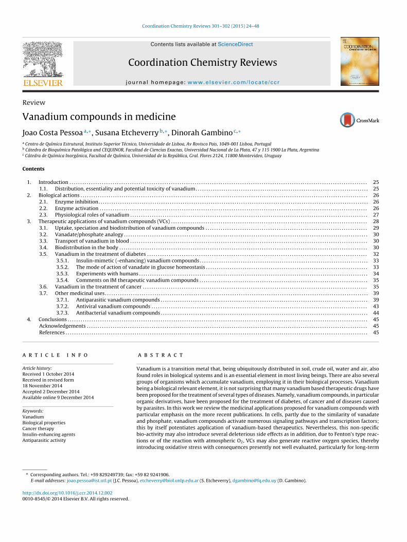
Fig. 1. Uptake and distribution of vanource: Adapted from Ref. [6].
© 2014 Elsevier B.V. All rights reserved.
tasks and playing crucial roles in living beings. Since nature hasmade such extensive use of metal ions, the questions: “Can metalions be incorporated into drugs? And can coordination chemistry beused with advantage for medicinal purposes?” naturally arise andare the subject of Medicinal Inorganic Chemistry [7,21]. Vanadium,being ubiquitously present in our planet’s crust also found roles inbiological systems, and this work focuses on the potential uses ofvanadium-based therapeutic compounds in Medicine.
Several groups of organisms accumulate vanadium and/oremploy vanadium in life processes. On the basis of its high amountfound in the earth’ crust, namely in pre-biotic conditions and whenlife first appeared, and of its known use in biochemistry, vanadiumis likely to have an essential role in most if not all living beings,namely in humans [2,6]. The average vanadium concentration inthe human body is approximately 0.3 mM and with equilibriumbetween the amount excreted from the body and the constant sup-ply of some vanadium via food and drinking water may easily bemaintained [22].
The main routes of vanadium uptake and distribution in thehuman body are sketched in Fig. 1 (adapted from [6]). Non-occupational vanadium exposure is predominantly from the foodsupply and typical daily doses consumed by humans have beenestimated at 0.01–0.03 mg V/day [5]. Dietary uptake is a quite inef-fective process because most of the dietary vanadium is usuallyexcreted with the feces (see below).
The main natural sources for vanadium loads in the atmosphereare continental dust evolving from weathering of soils, marine
adium compounds in the body.
aerosols and volcanic emissions [11,23,24]. Other major sources foraerial contamination are from mining, industrial enterprises andburning of crude oil. In non-polluted areas, the concentration ofvanadium oxides (VOx) is in the range of 50 ng m−3, but pollution,

2 emistr
wti
tecadcoft
aoCEba
2
2
mmie
vt
[ptvm
tlbbctppodsamttp[fe
cphtgt
6 J.C. Pessoa et al. / Coordination Ch
here combustion of petroleum and oil are the main contributorso aerial VOx, can raise the V levels up to >103 ng m−3 in urban andndustrial areas in particular [25,26].
Worker exposure to vanadium has largely resulted from inhala-ion of vanadium pentoxide in workplace dust [27]. Respiratoryffects are the most common problems thereof, but many organsan suffer adverse effects [28]. Vanadium oxides are readilybsorbed in the lungs and after solubilization in the form of vana-ate, H2VO4
−, enter the blood stream. The maximum allowableoncentration in breathing air amounts to 0.05 mg m−3. In case ofne-time exposure, the limit values for immediate danger to healthor an average human are 7 mg of vanadium (intravenous applica-ion), and 35 mg of V m−3 in breathing air (inhalation) [5,6].
Normally, food does not constitute a potential hazard. The aver-ge contents of V in food are ca. 30 mg kg−1; the no-effect level forral intake is 10 mg kg−1 per day [6]. For relevant references seeosta Pessoa and Tomaz [7], the two volumes of ‘Vanadium in thenvironment’ [29], the chapter of ‘Inhalation Toxicity of Vanadium’y Assem and Levy [30], the review of Olopade and Connor [13] andll references provided therein.
. Biological actions
.1. Enzyme inhibition
Many studies developed in vitro or in experimental animalodels have revealed that vanadium compounds display insulinimetic properties such as mitogenic effects, stimulatory and
nhibitory action on cell differentiation and numerous metabolicffects.
In vitro studies in cell-free systems have shown the effects ofanadium derivatives on the activity of many enzymes, especiallyhose related to phosphate reactions.
Vanadium inhibits several ATPases with different potency31]. Moreover, vanadium compounds also inhibit different phos-hatases such as alkaline phosphatase, acid phosphatase andyrosine–protein phosphatases [32]. Other enzymes inhibited byanadium are ribonucleases, phosphodiesterases, phosphogluco-utase and glucose-6-phosphatase [33].The PTPases activate or inhibit intracellular signaling pathways
riggering different biological effects in a cascade way. In bio-ogical systems the level of phosphorilated proteins is a balanceetween the activity of kinases and phosphatases, showing thatoth types of enzymes have an important role in the regulation ofellular processes. The phosphatases that hydrolyze phosphopro-eins are classified in two groups: with affinity for serine–threoninehosphate proteins (PS/TPases) and those with affinity to tyrosine-hosphate proteins (PTPases). All of them catalyze the hydrolysisf phosphate esters. The PS/TPases are metalloenzymes that pro-uce the direct attack of a water molecule to the P atom of theubstrate. On the contrary, the PTPases are not metalloenzymesnd their active site has a cysteine residue and in their catalyticechanism a fosfocysteine is formed as an intermediate. Moreover,
hese enzymes are structural and mechanistically different fromhe acid and alkaline phosphatases as well as those that hydrolizehosphate-esters of small molecules such as glucose-6-phosphate34]. Vanadium compounds can oxidize this cysteine residue by theormation of free radicals regulating in that way many biologicalvents [35–37].
The alkaline phosphatases are bound membrane glycoproteinsodified by different genes and classified as specific tissue phos-hatases and non-specific tissue phosphatases. These enzymes
ydrolyze phosphate monoesters from small molecules and pro-eins and catalyze the transference of phosphate groups to hydroxylroups of organic molecules. The mechanism of action occurshrough the phosphorylation of a serine residue of the active site,y Reviews 301–302 (2015) 24–48
followed by the transference of the phosphate group to a watermolecule or to an organic acceptor [38].
The ability of vanadium to inhibit these enzymes is closelyrelated to the physicochemical properties of vanadate and phos-phate (see Section 3.2). Structurally, vanadate is similar tophosphate but it is important to emphasize that vanadate showsa greater flexibility in its coordination geometry. Both anions par-ticipate in similar reactions. The generation of a stable transitionstate between the vanadate and the enzyme has been consid-ered the reason for its inhibitory effect to the enzyme activity.Studies by X-ray diffraction demonstrated that vanadate binds inthe active site of the enzyme with trigonal bipyramid geometry.Assays with vanadium compounds of different coordination geom-etry suggested that the complexes with five coordination weremore potent inhibitors than those with coordination numbers sixor seven [39]. Nevertheless, there are other factors that can affectthe enzymatic activity [39,40].
The VIVO2+ cation also inhibits different enzymes and in somecases with a greater potency than vanadate. One proposed mecha-nism for this inhibition is based on the possibility of VIVO to adopt atrigonal bipyramid, bind in the active site of the enzymes and thuscause inhibition of the activity [33,41].
2.2. Enzyme activation
In most cases, enzyme activation by vanadium follows an indi-rect mechanism. Vanadium can stimulate the enzyme activitythrough the formation of complexes with ligands that resemblethe structure of physiological substrates. For instance, the glucose-6-phosphate dehydrogenase is activated by vanadate. Anothermechanism to regulate enzymatic activity is related to the enzymesactivated or inhibited through the phosphorylation of tyrosineresidues. Vanadate also forms esters with Tyr residues mimick-ing their phosphorylation process with a great impact in severalbiological events. The activation by vanadate of some cytosolickinases and the insulin receptor (IR) has impact on the balancebetween phosphorylation–dephosphorylation processes. In vitroexperiments with cells in culture show that VCs may cause orenhance insulin mimetic actions such as stimulation of proteinphosphorylation in Tyr residues increasing the level of phos-phoinositides, stimulate cell proliferation in different cell lines,promote the uptake of small molecules and ions, etc. On the con-trary, vanadium compounds also can exhibit antitumor effectsas well as toxic and transforming actions in different cell lines.Moreover, it is worthy to know that globally the underlying mecha-nisms of action of vanadium compounds are still poorly understood[14,42–46].
Vanadium compounds are well-known for their antidiabeticeffects both on glucose and lipid metabolism, but the mechanismsare still not completely understood. It has been previously reportedthat VO(acac)2, VO(maltolato)2 and NaVO3, attenuated basal lipol-ysis in 3T3L1 adipocytes assessed by glycerol release as a markerof lipolysis [47]. Though both Akt and ERK pathways are activatedby the VCs, but only Akt activation by these three vanadium com-pounds plays a role in their antilipolytic effect. On the other hand,VO(acac)2 can block cell cycle progression at the G1/S phase via ahighly activated ERK signal pathway in human hepatoma HepG2cells. The results indicated that similar activated pathways maylead to differential biological consequences for cancer cells andadipocytes, indicating that VCs may be used in the preventionand treatment of both diabetes and cancer. The action of insulindepends on its binding to the hormone receptor and its activa-
tion. Then the transduction of the signal involves a series of proteinkinases such as MAPK and S6 kinases, as well as lipid kinases suchas PI3-K. The insulin mimetic effects of vanadium may be caused bythe stimulation of the autophosphorylation of the insulin receptor
emistr
bdlchbmipP
vpwtiiod3(
te
i[l
pptaMiachtei
gfdcdos
oc
amPt[vPa
temn
J.C. Pessoa et al. / Coordination Ch
ut in different manner than the hormone [48–53]. These effectsepend on the tissue and the animal species. For instance, in rat
ung cells and rabbit and mouse diaphragm cells vanadium did notause autophosphorylation of insulin receptor [54,55]. On the otherand, the inhibition of tyrosine kinase activity of insulin receptory vanadate did not suppress the effects of vanadate on glucoseetabolism, suggesting a post-receptor action [52]. VCs that show
nsulin-like effects, stimulate kinases of the signal transductionathways used by insulin beyond IR and IRS-1such as MAPKs andI3K.
In CHO cells, which do not express insulin receptors [56],anadate and oxidovanadium(IV) stimulate the phosphorylation ofroteins of the insulin pathway such as ERKs, p70s6k and p90rsk,hich in turn phosphorylate and regulate the action of transcrip-
ion factors, many of them related with cell proliferation. PI3-Ks other enzyme that regulates metabolic and mitogenic effects ofnsulin. Vanadium promotes the activity of this enzyme [57]. More-ver, the activation of the pathway ras-MAPK by VOSO4 seems toepend on the activity of PI3-K [58,59]. On the other hand, in SwissT3 mouse fibroblasts, vanadate stimulates S6 ribosomal kinasep90S6K or p90rsk) [60].
Most of the results support the hypothesis that PTPases inhibi-ion may be the main mechanism by which vanadium compoundsxert insulin mimetic or insulin enhancing effects [40,61–63].
It was also suggested that vanadate may cause some of thensulin effects through the activation of a cytosolic kinase (CytPTK)63]. This can be considered an alternative via to promote insulinike effects.
Another interesting field is the effects of vanadium on cellroliferation and differentiation. Growth factors stimulate cellroliferation though the activation of different receptors that inurn trigger intracellular signaling pathways which promote thection of different transcription factors. Insulin stimulates Ras-APK pathway and as a sequence cascade events, insulin exerts
ts mitogenic actions. In the same way, different agents able toctivate the signal transduction pathways of insulin can causeellular proliferation. Different in vitro studies of cells in cultureave demonstrated that VCs display a biphasic behavior towardhe mitogenesis: concentration between (1) 2.5 and 25 mM usuallynhanced cell proliferation while (2) concentrations over 50 mMnhibited this event [64,65].
Vanadate stimulates DNA synthesis in fibroblasts, behaves as arowth factor mimetic agent promoting the transition of the cellsrom G0 to G1 phase in the cell cycle [66,67]. Moreover, vana-ate potentiates the proliferative effect of IGF-I in osteoblasts inulture [68]. At the mitogenic doses, vanadate also showed a dose-ependent increase in alkaline phosphatase (a marker of maturesteoblasts) in cultured calvarial cells and stimulated bone collagenynthesis.
Different VIVO-complexes with organic ligands regulatesteoblast differentiation, displaying a stimulatory action onollagen type I synthesis [69].
The mitogenic actions of vanadium compounds may be medi-ted by the MAK pathway. In fact, as it has been previouslyentioned, V indirectly activates some kinases such as ERKs and
I3-K. Sequentially, the activated ERKs promote the action of someranscription factors and convey the cells to enter into the cell cycle58,59]. PI3K activation is also related to the mitogenic actions ofanadium because there is a crossing between the two pathways.I3K activates PKB and in this way it can support for the mitogenicctions of VCs [70].
Other pathways activated by vanadium that may be related
o its proliferative effect are those of phosphoinositides and het-rotrimeric protein G. At 100 mM, V increases the levels of inositolono-, di- and tri-phosphate through mechanisms dependent oron-dependent of tyrosine kinases [71]. On the other hand there
y Reviews 301–302 (2015) 24–48 27
are cases where vanadate stimulates the proteins G pathway butat very high concentrations (3–10 mM). These concentrations arevery high comparing with those that cause proliferative effects incell cultures [72].
2.3. Physiological roles of vanadium
The report of Cantley et al. [73] that endogenous vanadateinhibits Na+ and K+ ATPase in vitro was made in 1977 and since thenmany other physiological roles have been ascribed to vanadium,such as mitogenic actions, stimulation of bone cell proliferation,bone collagen synthesis and noradrenaline release from pulmonaryartery, inhibition of oxidative drug demethylation, stimulation ofoncogen expression and histamine release from mast cells, as wellas toxic and cytostatic actions, antidiabetic actions and neoplastictransformations. The possibility that vanadium compounds (VCs)may regulate osteogenesis and possibly be able of counteracting thebone damaging actions of glucocorticoids has also been suggested[74–76].
As mentioned, one relevant prospective role of VCs is in diabetesmellitus (DM) treatments. An important feature of vanadium’sinsulin mimetic (IM) action is the multiplicity of its effects and thesewere reviewed in e.g. [12,17,18,63,74–79]. In both spontaneouslyand chemically induced diabetic animals, vanadium alleviate notonly the primary symptoms of diabetes, high blood glucose andtriglyceride and cholesterol levels (although tissue vanadium lev-els normally do not correlate with the glucose-lowering effect),but also prevents or reverses several of the secondary com-plications: sorbitol accumulation [80,81], cataract development[82,83], impaired thyroid hormone [84,85], alterations in kidneymorphology [86,87] and adrenal hypertrophy [87]. In partially pan-createctomized rats [88] vanadium administration reversed insulinresistance by restoration of muscle glycogen synthesis; in STZ-diabetic rats it stimulated basal hexose uptake in muscle andliver [89] and even in non-diabetic rat muscle vanadium therapyincreased sensitivity to insulin stimulation of glycolysis and glyco-gen synthesis [90], impaired antioxidant status and excessive foodand fluid intake [91,92].
It was also reported that vanadate prevented the decline in car-diac performance due to diabetes [84] and cardiomyopathy [84].With streptozotocin-induced (STZ) diabetic rats, isolated work-ing heart parameters of left ventricular developed pressure andrate of pressure development also indicated that BMOV (1a, Fig. 2)administration resulted in significant attenuation of this heart dys-function [93].
Since secondary complications of diabetes are known to haveincreased oxidative stress as etiologic components [90], the possi-bility that vanadium’s insulin mimesis is partially due to changesin pro-oxidant/oxidant balance was also considered [94,95]. Animbalance of production of ROS over functional antioxidantdefenses has been associated with several pathological conditions[96], e.g. cardiovascular disease [97], rheumatoid and osteoarthritis[96], diabetes mellitus [98], tumor angiogenesis and proliferation[99], cataract formation [100], and inflammation [101].
Curcumin, a compound known for its neuroprotective action[102], has demonstrated efficacy in all of these pathological con-ditions [103], while VO(cur)2 was more effective as an anti-canceragent, compared to uncomplexed curcumin or VOSO4 alone, it wasmore than twice as effective as curcumin alone as an anti-arthriticagent, and was more than four times as effective as curcuminalone in inhibiting smooth muscle cell proliferation [104]. Prob-ably a relevant aspect deserving care is that neither curcumin
nor VO(cur)2 were effective in reducing plasma glucose levels inSTZ-diabetic rats. In contrast, there are reports on the capacity ofcurcumin to facilitate lowering of plasma glucose or to decreaseinsulin resistance. The difference might be that these experiments
28 J.C. Pessoa et al. / Coordination Chemistry Reviews 301–302 (2015) 24–48
e bee
birc
TbiaclthttItaosO
Fig. 2. Some vanadium compounds that hav
y Thompson et al. [104] were obtained upon acute oral admin-stration of curcumin and VO(cur)2 [104], while the data by otheresearchers are from oral gavage on a daily basis for 6–7 weeks ofurcumin to STZ rats [105] or db/db diabetic mice [106].
One interesting compound is VO(alx)2 (compound 2, Fig. 2).he group of Sakurai prepared several VCs and evaluated theiriological functions in vitro and in vivo [17,107], demonstrat-
ng that whether administration was by acute oral gavage or bycute i.p. injection, VO(alx)2 could be one of the most promisingomplexes for the treatment of diabetes [108]. The pharmaco-ogical functions of VO(alx)2 include hypoglycemic effects andhe improvement of hyperinsulinemia, hypercholesterolemia andypertension [108,109]. VO(alx)2 significantly induced V uptake inhe insulin-responsive tissues such as the adipose, liver and muscleissues [110]. VO(alx)2 also affects the tyrosine phosphorylation ofRb and IRS, leading to the activation of PI3K–Akt signaling and theranslocation of GLUT4 to the plasma membrane. Moreover, it was
lso demonstrated that VO(alx)2 regulates the DNA binding activityf the FoxO1 transcription factor, which in turn controls the expres-ion of gluconeogenesis genes in response to Akt activation [111].verall, VO(alx)2 improves diabetes, obesity and hypertension byn reported to exhibit insulin-like effects [7].
enhancing insulin sensitivity and leptin resistance in obesity-linkedtype 2 diabetic KKAy mice and was proposed as a promising VC totreat both type 1 and obesity-linked type 2 diabetes [109].
As a final comment the reader should also consider the interest-ing review by Aureliano and Crans [112], who summarized proteinsthat interact with vanadates, giving more focus to decavanadates,and showing that vanadate biological effects are not only due tomonomeric vanadate. Namely, decavanadate interacts with thepolyphosphate, nucleotide and inositol 3-phosphate binding sitesof enzymes.
3. Therapeutic applications of vanadium compounds (VCs)
Concerning applications of VCs as therapeutic agents, treatmentof diabetes has been one of the main focuses. In fact, vana-dium compounds have been reported to exhibit insulin-like effects
and among the VCs tested as small molecule insulin-mimetics,or insulin-enhancers, VIVO(maltolato)2 (1a, BMOV, Fig. 2), andVIVO(Etmaltolato)2 (1b, BEOV, Fig. 2), have been extensively stud-ied. BMOV and BEOV may be taken orally and both lower plasma
J.C. Pessoa et al. / Coordination Chemistry Reviews 301–302 (2015) 24–48 29
(A) (B)
0
20
40
60
80
100
3 5 7 9 11pH
% (V
O)
0
20
40
60
80
100
4 6 8 10pH
% (V
O)
VO
VO
VO(L)
VO(OH)−
VO(OH )3−VO(OH )3
−
(VO)2(OH)5−(VO)2(OH)5
−
F he saL IV-spe
gBea
t[Cs4n[(
3c
ufi(pl[rivi
iiipgsomV
vmsmIFp
ig. 3. (A) Distribution diagram of VIVO hydrolysis at 10 nM concentration. (B) T = 100 nM) with a conditional stability constant of 109; values of the hydrolytic V
lucose levels in streptozotocin-induced (STZ) diabetic rats [76],EOV having completed Phase I and IIa clinical trials. Althoughssentially successful, the clinical trials have provisionally beenbandoned [113].
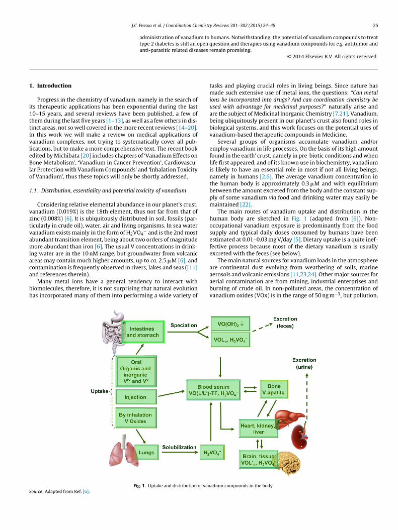
Examples of VCs that have been reported to have poten-ial as antitumor agents are the inorganic complex MetvanVIVO(SO4)(4,7-Mephen)2], vanadocene dichloride [(h5-p)2VIVCl2] [14,114], a V-cysteine complex [115] and aemicarbazone derivative [116] and VO(acac)2 (see Section). Several VCs have also been reported to have prospective useful-ess as anti-parasitic [117–122], spermicidal [123,124], anti-viral125], anti-HIV [126–130], and anti-tuberculosis [131–133] agentssee below).
.1. Uptake, speciation and biodistribution of vanadiumompounds
Fig. 1 summarizes some of the main aspects associated withptake and distribution of VCs in the human body. Dietaryorms of vanadium are either vanadate, H2VO4
−, present mainlyn drinking water, and VIVO-compounds {VIVOL}. Free VIVO2+
(VIVO(OH)5]2+) is not much relevant, since most of it wouldrecipitate as VIVO(OH)2, only allowing for very low (nanomo-
ar) concentration of ‘free’ VIVO2+ {actually [VIVO(OH)3]− and(VIVO)2(OH)5
−]n} at the best (see below). Vanadate(V) is partiallyeduced in the stomach, and in the slightly alkaline medium of thentestines precipitated in the form of VIVO(OH)2. Thus, most of theanadium supplied by nutritional sources is secreted with the fecesn the form of sparingly soluble vanadyl hydroxide VIVO(OH)2 [6].
Vanadium can also enter the blood stream by injection ornfusion, either intentionally when injected intravenously orntraperitoneally, or accidentally when present as a ‘contaminant’n infusion solutions [134]. Besides speciation, vanadium com-ounds ending up in the blood stream either after resorption in theastrointestinal tract, or via the lungs, or by infusion/injection, areubjected to redox interconversion between VV and VIV, dependingn the oxygen tension and the presence of redox-active agents. Theain transporter in blood for both anionic vanadate(V), cationic
IVO2+, and neutral or charged VIVOL species is transferring [135].Values in the range 0.2–15 nM were determined [136] for the
anadium concentration in human blood plasma. Upon oral treat-ent of human patients with VCs, the concentration of V in blood
erum depends on the regime and amount of compound given, and
uch higher values have been reported, ca. 1–10 mM [76,113,137].n rats treated with BMOV [138,139] or BEOV [140] (1a or 1b inig. 2) maximum values of ca. 40–60 mM were found in their bloodlasma.
me but including the possibility of forming a VIVO(L) complex (concentration ofcies were obtained from Ref. [147].
Plasma vanadium contents in rats may be considered to declinein three phases. The 1st phase is a rapid decline with a half-life t1/2of 1 h, followed by a second intermediate decline (t1/2 ≈ 26 h) and athird slow one with t1/2 ≈ 10 days. Vanadium contents in blood arethus reduced to about 30% within the first 24 h [5,134]. Clearingoccurs directly via urinary excretion, as well as after distributionover tissues, and about 50% of the vanadium is found in urine afterca. 12 days. The residence time of vanadium in bones, where itmay replace phosphorus in hydroxyapatite, is ca. 1 month [113],corresponding to a half-life of 4–5 days.
Vanadium in biological systems may be present in the oxidationstates +III (VIII), +IV (VIV) or +V (VV) [141]. For mammals VIV and VV
are normally considered more relevant as VIII is very susceptible tooxidation; however, VIII-hTF species (hTF = human serum transfer-rin) have been reported to be quite air-stable [142,143]. At the verylow concentrations such as those present in blood plasma VV existsmainly as H2VO4
− and HVO42−, often referred as VO3
−, or as mono-vanadate; at pH 7.4 and low concentrations of VV in blood plasma,and several potential ligands being present, it is not expected thatdivanadates (V2) or tetravanadates (c-V4) might form. In cells, usualphysiological vanadate concentrations are also too low to allowfor the formation of oligovanadates. Locally, however, concentra-tion enhancement or template-directed nucleation [144] may takeplace, and the oligovanadate(s) then formed can interact with pro-teins and DNA. Cellular targets and processes probably affectedby decavadates are: (1) membrane depolarization, (2) pumps,channels, receptors in cellular membrane and in endoplasmic reti-culum membrane, (3) contractile system, (4) cytoskeleton structureand function, (5) mitochondria bioenergetics, (6) ROSs specieschanges and antioxidants responses, and (7) probably also nucleous[112].
VIV normally exists as VIVO-species. VIVO2+ is quite suscep-tible to oxidation for pH > 3 and forms an insoluble hydroxidefor pH > 4 (Ksp ≈ 7.4 × 10⁻²³) [145,146]. At pH > 5 oligomeric vana-dium species may be relevant, even at concentrations as low as10 nM, see Fig. 3. In the pH range 5–8 and at concentrationsbelow 10 mM, VIVO2+ ( VIVO(H2O)5
2+) does not exist as such inthis pH range, but VIV is soluble and is mainly in the form ofVIVO(OH)3
− and [(VIVO)2(OH)5]n− [147] (Fig. 3). In blood serum
probably VIVO2+ is totally bound to high molecular mass andlow molecular mass bio-ligands also present; inside cells manybio-ligands are present that form complexes with VIVO2+ withbinding constants probably higher than 109, thus most of the
VIVO2+ should also be bound. However, the presence of a verysmall amount of VIVO(OH)3− (free or bound?) cannot be ruledout and its relevance in physiological media should be taken intoaccount.

30 J.C. Pessoa et al. / Coordination Chemistr
Fig. 4. Analogy between phosphate and vanadate. Besides monovanadate beingsos
3
d
dcs
-
-
artoiiTsdlbr
peVltgp
3
a
tructurally similar to phosphate, the acid-base equilibria operating and other typesf reactions (e.g. V and/or P ‘ester’ formation) are similar. However, there are alsoome structural and pKa differences (see text) [6,148].
.2. Vanadate/phosphate analogy
Monovanadate and phosphate are structural analog, monovana-ate being only slightly larger than phosphate (see Fig. 4) [24].
From a geometrical point of view the two anions are not muchifferent, vanadate(V) thus being a competitor of phosphate in sitesommonly occupied by phosphate [23]. However, there are alsoignificant differences:
due to the different pKa, at physiological pH and ionic strengths,vanadate is mostly present in the form of H2VO4
− {dependingon pH the amount of HVO4
2− is ca. 1/6 (at pH ∼ 7.4) to 1/25 (atpH ∼ 6.8) of the amount of H2VO4
−}, while HPO42− and H2PO4
−
exist in approximately equal amounts. phosphorus can only attain the coordination number 5 in tran-sitional states, while vanadium is much more versatile in thisrespect, easily forming stable complexes with coordination num-bers 5 or 6. Therefore, once incorporated into the active site ofa phosphate-dependent enzyme, taking the place of phosphate,the activity of this enzyme is inhibited [23].
Presently there is no solid basis for categorizing VCs, suchs those depicted in Fig. 2, as harmful when administered ineasonable amounts. Instead, as mentioned above, vanadium inhe form of H2VO4
−, is probably involved in the regulationf phosphate-dependent processes, such as metabolic processesnvolving phosphatases and kinases and the phosphate metabolismn general [32]. Thus, probably vanadium is an essential element.his plausible regulatory role of vanadium is certainly related to thetructural and chemical similarity between phosphate and vana-ate (see Fig. 4). Additionally, the participation of VIV and VV in
eveling reactive oxygen species (ROS) suggests that vanadium cane beneficial in the treatment of several diseases and malfunctionselated to ROS imbalances [23].
The competitive behavior of vanadate with respect to phos-hate is likely the clue for the insulin-mimetic/insulin-enhancingffect of VCs. As mentioned, for pH > 6 at low concentrations, freeIVO2+ exists significantly as VIVO(OH)3
−, being structurally simi-ar to VVO(OH)3 ( H3VO4). Inter-conversion between VIV and VV ishus fast and easy in physiological conditions, and it has been sug-ested that, besides monovanadate(V), VIVO(OH)3
− may also mimichosphate [6].
.3. Transport of vanadium in blood
Once in the blood stream, blood serum constituents will prob-bly dominate vanadium speciation, and consequently the final
y Reviews 301–302 (2015) 24–48
cellular uptake by tissue cells and the targeting pathways for vana-dium.
Speciation can encompass ligand exchange and/or redox-interconversion between VV and VIV (and, potentially, VIII)[143,149]. For ligand exchange, the low molecular mass con-stituents lactate and citrate, and the high molecular massconstituents, transferrin (hTF), albumin and immunoglobulin G, arethe more important ones, with hTF having a predominant role.Reducing agents such as ascorbate, glutathione and NADH caninduce reduction of VV to VIV, and oxidants such as NAD+, O2, O2
2−
and O2− can convert VIV to VV.
If vanadium is introduced in blood in the form of a prospectivedrug of composition VIVOLn (examples are some of the com-plexes of Fig. 2), part of the vanadium may cross the membraneof the erythrocyte cells [150], VV is reduced to VIV by cellularglutathione [151–154] and binds to hemoglobin [153]. The vana-dium compounds remaining in the blood plasma may bind to hTFforming either type 1 or type 2 species (see Fig. 5) or forming(VIVO)2hTF.
In holo-hTF, (FeIII)2hTF, the protein is in its ‘close conforma-tion’, thus it is recognized by the transferrin receptors of thecell membranes, and FeIII is up-taken by cells as (FeIII)2hTF bya receptor-mediated endocytosis process. Regarding the cellularuptake of vanadium, present in a prospective drug of compositionVIVOLn, several scenarios can apply for the up-take process:
- The complex is stable enough to remain intact, and sufficientlylipophilic to slowly cross the cellular membrane via diffusion.Pyridinone complexes such as 4 in Fig. 2 enter erythrocytes bydiffusion [150].
- The ligands are detached, oxidation occurs, thus forming H2VO4−,
and vanadium may enter the cells as monovanadate via anionchannels.
- The ligand system is maintained, or is replaced in part or com-pletely by transferrin to form VIVO(L)(hTF) (see right side ofFig. 5) and (VIVO)2hTF [7,155,156]. Namely (VIVO)2hTF is prob-ably recognized by the cellular hTF receptors and internalized viaendocytosis [149]; there is no information available regardingVIVO(L)(hTF), but in this case probably hTF does not form the‘closed conformation’ required for adequate binding to the cellreceptors.
- Either VIVO2+, or vanadates, or VIVO(L)n species, bind holo-hTF,at available donor atoms of the protein; these species are proba-bly recognized by the cellular hTF receptors and internalized viaendocytosis [143,157].
- VIII species may form, e.g. (VIII)2hTF and or (VIII)(FeIII)hTF, whichforms the ‘closed conformation’ and probably is recognized bythe cellular hTF receptors and also internalized via receptor-mediated endocytosis [143].
- The complex is stable enough to remain intact and contains oneor more functions in the outer coordination sphere that are recog-nized by cell receptors; receptor-mediated uptake is then againa potential transport route.
- Albumin receptors on the cell (e.g. adipocites) surface would alsobe likely vehicles if a VC binds to HSA [158].
It was also reported that human serum albumin (HSA) enhancedthe insulin-like activity of a few VCs more that transferrin [159].The VCs increased tyrosine phosphorilation in response to insulin,including IRb and IRS1 [160]. This may be due to enhanced uptakeby cells, but the subject deserves further studies.
3.4. Biodistribution in the body
If we aim for the clinical use of VCs in the future, the organdistribution and metallokinetic features of vanadium, as well as

J.C. Pessoa et al. / Coordination Chemistry Reviews 301–302 (2015) 24–48 31
F IV . The
d
S
o[bvfwribaoaa
0iottSg
Ma
F
ig. 5. Two distinct proposed types of binding for V O(carrier)2 complexes to hTFiffraction characterization of a lysozyme-VIVO(pic)2 adduct [155].
ource: Adapted from Ref. [143].
f the corresponding complex forms, have to be well understood16]. For these purposes, neutron activation analysis (NAA) haseen one of the important techniques used to determine the totalanadium levels in organs of rats given VOSO4 or VCs with dif-erent coordination modes. In rats treated with VOSO4 vanadiumas found in kidney > liver > bone > pancreas [161–163], while in
ats treated with VCs such as VO(5-ipa)2 (Fig. 6) it was detectedn bone > kidney > spleen > liver > pancreas [164]. Such differencesetween organ distribution of VOSO4 and VCs might suggest long-cting character of the complexes. Vanadium levels after 25 weeksf BMOV supplementation were comparable with those obtainedfter 10 weeks of VOSO4 feeding, but daily BMOV dose taken wasbout half [93].
BMOV treatment of diabetic rats at a concentration of.75 mg/mL in drinking water resulted in accumulation of V
n bone, kidney, liver, muscle, and fat, without any mortalityver a 25-week period, with a decrease of plasma glucose, %GHb,riglycerides and cholesterol. BMOV therapy effectively preventedhe development of the myocardial dysfunction associated withTZ-induced diabetes in rats, even in those rats whose bloodlucose levels remained above normal [93].
Total vanadium distribution in STZ-rats treated with VO(6-epic)2 was also examined by an NAA method [165,166]. V
ccumulated in almost all tissues, particularly in bone and kidney,
ig. 6. Several additional insulin-enhancing vanadium compounds [164–166,171].
possibility of the Type 1 binding to a protein was recently confirmed by the x-ray
in the mitochondrial fraction of the liver and in the supernatantof the kidney of STZ-rats receiving VO(6-Mepic)2 by i.p., injec-tions. Similar tendencies were observed in vanadium distributionin STZ-rats given the same VC by oral administration, exceptfor an accumulation in the mitochondrial fraction of the liver.V accumulation in the bone of rats receiving i.p. injections ofVO(6-Mepic)2 was remarkable: ≈3.3 times that in the kidneyand ≈11 times that in the liver. These results were somewhatsimilar to the distribution of vanadium in rats treated with i.p.injections of VO(pic)2, where accumulation in bone was approx-imately 4.7 times that in the kidney and 35 times that in theliver. In rats treated with complexes such as VO(5-ipa)2, by NAA[74,162,164,165] or radioisotope [167] determinations, V wasfound in kidney > liver > bone > pancreas.
In STZ rats treated with VO(5-ipa)2 by daily i.p. injectionsfor 14 days, and then without administration of the VC foradditional 14 days, the organ distribution of total V was:bone (0.32 mmol/g of wet tissue) > > kidney (0.08) > spleen(0.04) > adipose (0.02) ≈ liver ≈ pancreas > lung ≈ heart > bloodcell ≈ serum > brain [16].
The long-term action of VO(6-Mepic)2 and of other VCs by oraladministration is probably related to the accumulation of V mainlyin the bone. Vanadium accumulated in the bone may be releasedgradually to other organs via the blood-stream after stopping theadministration of the VC, and is able to normalize the blood glucoselevels for a long period.
To access the metallokinetic features of VO complexes the groupof Sakurai applied a BCM-EPR method, which allows the tracing ofparamagnetic species in the blood (Fig. 7) [16,168]. For example,VCs were given by a single i.v. injection to rats at 37 ◦C under anes-thesia with pentobarbital, and the VIV-EPR spectra were measuredat room temperature every 30 s. It was concluded that a major fac-tor for the disappearance of EPR signals in the circulating blood dueto VCs was not the participation of redox processes of VO speciesin the circulating blood, but their distribution to the tissues andelimination from the body [164]. Thus the VIVO species taken upinto the blood are distributed to the short- and long-stay tissues,and then accumulated there or re-distributed in the bone, liver andkidney.
The real-time EPR analysis of VIVO species revealed that interms of half-life (t1/2), their clearance rate from the blood of ratsgiven VOSO4 was higher than those given VCs, being 5 min in
VOSO4-treated rats and 7–30 min in VC-treated rats [164]. The slowclearance rate of the VCs suggests that a higher distribution of vana-dium in rat organs is obtained, which in turn indicates long-termacting normoglycemic effects upon stopping V administration.
32 J.C. Pessoa et al. / Coordination Chemistr
Fig. 7. Scheme depicting how in vivo blood circulation monitoring-electron para-magnetic resonance (BCM-EPR) studies on rats are carried out [168]. The methodwas used to measure the real-time disposition of spin probes in the circulating bloodof rats.
S
ipgattithaaos
bcosffwwtVf[
ftamvad
tgva
midal geometry of VV is supported by the proteins with preferenceover the square pyramidal geometry. This is an interesting result,but probably is not very surprising as most of these phosphatases
ource: Reproduced with permission from Ref. [168].
The slow elimination of V from the circulating blood of ratsndicated a different association of the VC with the blood com-onents such as serum proteins or erythrocytes between ratsiven VO(5-ipa)2 and those given other VCs. By metallokineticnalysis it was concluded that VIVO species tended to be dis-ributed in the peripheral tissues and gradually eliminated fromhe circulating blood. VCs with electron-withdrawing and donat-ng groups remained significantly at higher levels and longer, dueo their slower clearance rates from blood, suggesting that theigh exposure and longer residence of the VC is associated with
higher normoglycemic effect in diabetic animals. In vitro IMctivity, metallokinetic character and in vivo antidiabetic actionf VO–picolinate complexes thus appear related to their chemicaltructures [169,170].
Vanadium also accumulated in almost all tissues, especially inone > kidney > liver > pancreas in rats given VO(pyd)2 12. In sub-ellular distributions of vanadium in rats treated with VO(pyd)2r with VOSO4, vanadium was found most abundantly in theupernatant fraction of the kidney, while in the other threeractions no significant difference in vanadium distribution wasound [171]. The hepatic distribution of vanadium in rats treatedith VOSO4 differed from that in rats treated with VO(pyd)2:hile the highest vanadium concentration was determined in
he mitochondrial fraction of the liver in rats treated withOSO4, the distribution of vanadium showed no significant dif-
erence among the four fractions in rats treated with VO(pyd)2171].
For BMOV and VOSO4 [167] disappearance of vanadium wasastest from blood, and slowest from bone, followed by kidney,hen liver. The relatively slow clearance of V from plasma may bettributable to vanadium recirculation [140], as also seen with auch smaller dose of V by oral gavage [172,173]; disappearance of
anadium was fastest from soft tissues (11–132 h versus 15–73 h)nd slowest from bone (t1/2 = 24 days for NH4VO3 versus t1/2 = 30ays for BEOV) [140].
Chronic administration of a VO-compound has lead to boneissue accumulating the highest amounts of vanadium. Sug-estions have been made that V is probably in the form of
anadate(V) substituting phosphate in the mineralized hydroxy-patite structure of bone [174]. However, the measured boney Reviews 301–302 (2015) 24–48
tissue paramagnetism indicated that it also contains VIVO-species[175].
VIVO2+ may substitute Ca2+ and Mg2+ in bone tissue [176] anddiabetic individuals have a higher incidence of bone fractures thannon-diabetic ones. The effect of VO(acac)2 on bone structure, afterchronic daily treatment of alloxan-induced diabetic rats for 35consecutive days, and several parameters related to the bone char-acteristics were measured [177]. The authors reported that theplasma level of osteocalcin, a protein that may be used as a markerof osteoblast activity and is secreted by osteoblasts was decreasedin diabetic rats (7.5 ± 1.9 ng/mL), but was partially restored uponVO(acac)2 treatment (ca. 12.6 ± 1.1 ng/mL), though still lower thanthe baseline level in non-diabetic rats (ca. 15.4 ± 4.4 ng/mL) andin non-diabetic rats treated with vanadium (ca. 15.1 ± 1.8 ng/mL).Additionally, while the treatment of diabetic rats with VO(acac)2did not change the bone mineral density, overall it improved ornormalized their strength, trabecular thickness and the mineralapposition rate [79,177].
3.5. Vanadium in the treatment of diabetes
Background: Diabetes mellitus (diabetes), is associated withan impaired glucose and fatty acid metabolism, induced bya non-existent or insufficient insulin supply, or inadequateresponse to insulin. Insulin is produced by the b-cells of theLangerhans’ islets in the pancreas. Receptors on the b-cellsare activated by glucose (and, synergistically, fructose), stimu-lating the secretion of insulin [178] that then targets cellulartrans-membrane insulin receptors (IRs) to down-regulate glucoselevels.
Type 1 and type 2 are the main manifestations of diabetes.According to estimates [179], up to 10% of the world populationis suffering from diabetes. Type 1 (or juvenile) diabetes, corre-sponds to approximately 10% of diabetes cases, is the result ofgreatly reduced or even lacking production of insulin, commonlyas a result of an autoimmune reaction which destroys the b-cells,or a damage of the pancreas by an accident. Type 2 diabetes nor-mally develops in elderly (commonly > 60) individuals; insulin isstill produced, but the insulin receptors (IRs) of the tissue cells nolonger respond properly to insulin.
As mentioned in Section 2.2, the main potential of vanadiumfor the treatment of diabetes probably comes from the similar-ity between vanadate and phosphate highlighted above. However,unlike phosphate and phosphate esters, vanadate is not easilyreleased once bound at the enzyme’s active site. Additionally, incontrast to phosphorus, for which the 5-coordinate state, achievedon binding covalently to the active center, is just a transitional situa-tion, vanadium forms quite stable 5- or 6-coordination compounds;thus, once bound at the active site, vanadates are not easily releasedand hence block off this site for phosphate, thus resulting in aninhibition of the respective enzyme [6]. Interestingly, the knownexamples of protein structures with vanadium bound in the activesites globally show that both the square pyramidal and the trigonalbipyramidal geometries can support the transfer of the phosphorylgroup in phosphatases and other phosphorylases [180]. However,it is clear that there are much more reported examples of trigo-nal bipyramidal complexes in the active site than square pyramidalcomplexes. Considering that in small molecules the square pyrami-dal geometry is more common than the trigonal bipyramidal one,this observation supports the expectation that the trigonal bipyra-
are build-up to support a 5-coordinate phosphate transitionstate.

emistr
3
qmnyiihb
sevfthaatSa
aoacS
ooiiseiltsimtflt(4faib
poIcs
citbdio
t
triggered by activated GSK3b, polymerized to glycogen.
J.C. Pessoa et al. / Coordination Ch
.5.1. Insulin-mimetic (-enhancing) vanadium compoundsVanadium compounds that show insulin-like effects are fre-
uently designated by insulin mimetics (IM); however, since inost situations there is some residual insulin production, the desig-
ation insulin-enhancing compounds are more adequate. For manyears, in the treatment of diabetic animals and, sporadically, humanndividuals, VV and VIV compounds have been employed. VIVO2+
s quite susceptible for oxidation for pH > 3 and forms insolubleydroxide for pH > 4. At pH > 5 oligomeric vanadium species maye relevant, even at concentrations as low as 10 nM (see Fig. 3).
Mainly due to the very low oral bioavailability of inorganicalts (ca. 1–2%), VIV complexes with organic ligands have beenxtensively explored. Organic ligands allow for a fine-tuning of theanadium compound with respect to stability, rate of absorbancerom the GI tract (when applied orally), targeting of and internaliza-ion by the tissue cells, and toxicity. A key compound in this regardas been BMOV [181,182], 1a in Fig. 2, and derivatives thereof, suchs the VIV-complexes formed with ethylmaltol (BEOV), 1b [113]nd allixin (2) [108,183]. The related complex with kojato (3) andhe pyridinone complex 4 have also been prospective therapeutics.everal other complexes have been studied, such as VO(acac)2 6,nd several picolinato and dipicolinato derivatives (Fig. 6).
Maltol is a naturally occurring compound and an approved fooddditive in many countries. BMOV and BEOV have so far been thenly VCs to be subjected to clinical tests, essentially with encour-ging response [113]. Several other VCs have been tested with cellultures (in vitro) and with diabetic animals (in vivo), such as e.g.TZ rats and Zucker rats as models for diabetes 1 and 2, respectively.
Fig. 1 provides an overview of the uptake and distributionf vanadium compounds in the human body. When internalizedrally, VCs are subjected to rather strong acidic conditions (pH ≈ 2)n the stomach; the potentially ‘destructive’ effects of such acid-ty can be circumvented by drug encapsulation. The saliva and themall intestine are slightly alkaline. Therefore, in addition to thexpected speciation of the originally applied pro-drug, while cross-ng these organs with quite distinct pH conditions, the possibleoss of ligand(s), oxidation (mainly in the oral cavity) and reduc-ion (mainly in the GI tract) must be considered as unavoidableituations. Additionally, the original VC may have changes uponnteraction with low and high molecular mass constituents, which
ay behave as ligands. These are abundantly present in the nutri-ional components, as well as in body fluids and secretions of bodyuids stimulated by the intake of food. In fact, without participa-ion of ligands, VCs would be converted into simple compounds:i) VIVO2+ salts, probably then forming insoluble VIVO(OH)2 (for
< pH < 7.5), which will not be absorbed and is expelled with theeces, and (ii) VV-compounds, H2VVO4
− (in the pH range 7 ± 0.7)nd VVO2
+ (pH 2–3). The anion H2VVO4− is expected to be absorbed
n the small intestine and thus can be distributed throughout theody.
In addition to the oral uptake of inorganic vanadium com-ounds, inhalation and thus pulmonary ingestion of vanadiumxides, present in dust particles, is another possible route [184].n the lungs, a substantial part of the VOx may be solubilized beingonverted to vanadates and thus also introduced into the bloodtream.
Vanadium coordination compounds VO(carrier)n, with organicarrier ligands, such as those indicated in Figs. 2 and 6, will also benvolved in speciation when entering the body. At low and high pHhe loss of one or all carrier ligands may take place, the compoundseing converted to other distinct complexes or to inorganic vana-ium. Alternatively, or additionally, the body’s own ligands L0 can
nduce re-organization in the coordination sphere, forming VOL0n,
r mixed (carrier)/L0 species, thus changing absorption properties.The carrier ligand in VO(carrier)n compounds largely influences
he efficacy of a vanadium compound by determining resorption,
y Reviews 301–302 (2015) 24–48 33
transport, and stability of the complex, and thus the bioavailabilityof the true antidiabetic species, possibly vanadate. Underlin-ing the advantageous bioavailability and therapeutic efficacy ofVO(carrier)n compounds [113], these have indeed been more effec-tive than inorganic VCs.
The group of Sakurai tested several new drug delivery sys-tems trying direct intestinal administration (e.g. into the ileum),involving enteric-coated capsulation (ECC), gelatine capsules andVO-biopolymer (poly-g-glutamic acid, g-pga) complexes, to over-come several of the gastro-intestinal absorption and/or irritationproblems with vanadium inorganic salts [14,17,185,186]. Suchadministration forms indeed improved absorption, ECC and g-pgadelivery systems giving better results than gelatine capsules. How-ever, to our knowledge these promising results with diabetic micedid not lead to any clinical tests with humans.
Transdermal delivery has also been tried in diabetic ratsboth passively and by iontophoresis, using peroxovanadium, withand without complexation by 1,10-phenanthroline [76,187,188].Increased blood levels of vanadium were achieved, but blood glu-cose reduction was reported to be modest.
The VO(carrier)n compounds are inherently susceptible tohydrolysis and administration of these VCs is likely to result inloss of ligand, and extensive work has been carried out aimed atdetermining the actual active species [1,107,111,189–196]. Stud-ies using BMOV and BEOV demonstrated that upon administrationof the complex, the maltol ligand separates from the metal ion, andtransport proteins such as transferrin are likely to play key roles inthe distribution of vanadium intracellularly [1,7,113,194,197–199].
Due to transmetalation processes and cellular compartmenta-tion it is also likely that other ligands in addition to transferrinare involved in the biological effects of V compounds. Thus, mostprobably the VC taken will partially or completely hydrolyze in theGI and/or in blood serum, and vanadium will bind to bio-ligands,namely to hTF. Either by endocytosis, by diffusion, or by the anionchannels (vanadate), vanadium may be uptaken by cells and thenbe again subjected to speciation and redox leveling. Its availabilityfor subsequent therapeutic or toxicity effects will depend on sev-eral factors, namely the amount uptaken, the type of tissue and ifthe carrier ligand is still present or not, allowing leveling of effectsand/or targeting to the sites of therapeutic action. In any case thefinal intracellular break down of the complex most probably occursto allow for the display of vanadium’s physiological effects.
3.5.2. The mode of action of vanadate in glucose homeostasisMost cells contain insulin receptors incorporated in their mem-
branes. The IR is a tetrameric trans-membrane protein, beingtyrosine kinases, with two a subunits exposed to the outside andthe two b subunits (IRb) exposed to the cytosol. When insulin docksto the a subunits, the tyrosine residues of the intracellular b subunitIRb become phosphorylated, this corresponding to the activationof IRb. The cytosolic insulin-signaling cascade is initiated in thisway, and involves several post-receptor events, ending up with theactivation of the glucose transporter (GLUT4). Thus, the phospho-rilation of IRb triggers the phosphorylation of IR substrates, IRS,intracellular proteins containing tyrosine residues. The activationof the IRS in turn initiates a signaling cascade, in the course ofwhich kinases such as phosphatidylinositol 3-kinase (PI3K), pro-tein kinase B (PKB/Akt), and glycogen synthase kinase-3b (GSK3b)are activated [3,183]. PKBs, which bind phosphate through Tyr orSer residues, and then target GLUT4, the glucose transporter [3,6],which is translocated to the cell surface and intakes glucose. Oncein the cytosol, glucose is either broken down to CO2 and H2O, or
There is increasing consensus that the vanadate-phosphateanalogy is relevant for the insulin enhancing action and mode ofoperation of vanadium compounds, and the most widely accepted

34 J.C. Pessoa et al. / Coordination Chemistry Reviews 301–302 (2015) 24–48
Fig. 8. Simplified sketch of the possible mechanism of action of VCs. The internalization of glucose by the glucose transporter GLUT4, is triggered by the phosphorylatedinsulin receptor (IR). In the absence of insulin or insufficient insulin response, protein tyrosine phosphatase 1B (PTP-1B) dephosphorylates the IR, and the glucose intake iss ing pap wn as
o 4, and
mb
rIpdctititgB
3pasiuIttgvimBia
stssouVt
topped. By binding to PTP-1B vanadate may block PTP-1B, this restoring the signalhosphatidylinositol 3-kinase (PI3K, which activates protein kinase B, PKB, also knof the glucose transporter remains in operation, as well as (3) translocation of GLUT
ode of action for V compounds thus far is attributed to the inhi-ition of protein tyrosine phosphatases.
In the absence of insulin or in the case of inadequate insulinesponse of the IR, phosphorylation of the tyrosine residues in theRb subunits of the IRs is counteracted by a protein tyrosine phos-hatase (PTP-1B). As a result the signaling breaks down, and sooes the cellular uptake of glucose. If vanadate is present inside theell, as the result of uptake of a vanadium compound (see Fig. 8),hen VVO2(OH)2
− H2VO4− (and/or VIVO(OH)3
−) will be availablenside the cells. Due to its similarity to phosphate, vanadate bindso the active site of PTP-1B and consequently deactivates (inhibits)t, thus maintaining this enzyme in the phosphorylated state [200],hereby restoring/keeping active the signal transduction paths forlucose uptake [201] (Fig. 8). This possibility was confirmed for e.g.MOV [202] 1a and for VO(dhp)2 4 [203].
VO complexes inhibit PTP-1B and activate phosphatidylinositol-kinase → Akt signaling through the enhancement of tyrosinehosphorylation of IRb and IRS [3]. The particular case of BMOVction was reviewed by Srivastava and coworkers [204] whohowed that, besides being better than inorganic V-compoundsn inducing phosphorylation of PKB, GSK-3 and FOXO1, a keypstream transducer of BMOV is probably the transactivation of
GF-1R. In this way and through the activation of the PI3K pathway,he phosphorylation of PKB is mediated as well as its downstreamargets which regulate glucose transport, glycogen synthesis andluconeogenesis. The group of Crans [205] also suggested that acti-ation of IR signaling by both insulin and BMOV administrationnvolves increased association of IR with specialized nanoscale
embrane micro-domains. The observed insulin-like activity ofMOV (or of its decomposition products) would be due to changes
n cell-surface membrane lipid order rather than due to direct inter-ction with the IR.
In the case of VO(acac)2 it is not certain whether it directlytimulates the IR tyrosine kinase activity, or if its acts indirectlyhrough activation of other tyrosine kinases or inhibition of tyro-ine phosphatases [160]. In fact, VO(acac)2 was reported to exhibitynergism with insulin, to regulate the Tyr phosphorylation levels
f the IR and of IRS-1 [160,206] and in some conditions to act as anncompetitive inhibitor of PTP-1B [206]. It was also reported thatO(acac)2 in the presence of HSA also activates, and much morehan VOSO4, the phosphorylation of IRb [207].
th. (1) Phosphate remaining bound to IRb, the insulin receptor substrate (IRS), theAkt) remain phosphorilated, thus the signaling path is kept active and (2) activation
(4) cellular uptake of glucose by GLUT4.
Vanadate may bind to several other protein tyrosine phos-phatases inside the cell, inhibiting their action, or to proteinkinases, stimulating their action. However, kinase activation doesnot appear to be involved with the pharmacological effects ofvanadium, at least for autophosphorylation of the insulin receptor,other phosphotyrosine phosphatases, or PI3K [207–212]. WhetherVIVO(OH)3
− (≈H3VIVO4−) may indeed have a role similar to that
of VV vanadates (HnVVO4(3−n)−) is not known. If both can bind to
the O-atom of the Tyr side groups, then redox reactions involvingthe two forms may also be operating (the consequences of theseprocesses cannot be easily anticipated).
The mode of action for V compounds being associated to theinhibition of protein tyrosine phosphatases, it is also relevant tounderstand that some VCs are reversible inhibitors, whereas othersare irreversible by modifying the protein through redox processes[213]. The recent review of Crans et al. [180] of coordinationenvironment of vanadate bound to protein tyrosine phosphatasesconcluding that actual differences between the coordination envi-ronments are very small and presumably less critical than generallyanticipated, is an interesting observation whose consequences onthe subject under discussion cannot easily be anticipated.
The group of Crans et al. has also proposed that interactions ofVCs with cellular redox processes are important in the anti-diabeticeffects of VCs [190], VCs cause increases in ROS and RNS (reactivenitrogen species) via multiple mechanisms [214,215]. Thus, alter-natively, or additionally, ROS produced by vanadates (VIVO(OH)3
−
or H2VO4−) may be responsible for e.g. the inhibition of PTP-1B by
oxidatively targeting the Cys residue present in it. Cys has to bepresent in its reduced form for full activity of PTP-1B [6]. The groupof Crans also proposed that the compound stability and the abil-ity to interact with cellular redox reactions are key aspects for theinsulin-enhancing activity exerted by VCs [5]. Moreover, the possi-bility that membrane interactions are influenced by the ligand wasalso suggested, and that such membrane effects may affect uptakeand action of the VCs [207,216].
3.5.3. Experiments with humans
Both inorganic and organic forms of vanadium have been testedin human subjects [217–221]. Doses used are normally compar-atively lower than those in experimental animals, and thus onlymodest improvements in insulin and glucose metabolism were

emistr
sdsdat
tiwwivr
Bti(solhs
awthlfwdstie
aoiihgsja
toajatattd
3
oiiat
J.C. Pessoa et al. / Coordination Ch
een within a few weeks upon the start of the trials. The majorrawback reported was gastrointestinal (GI) distress. Since only amall amount of vanadium is absorbed, generally <2% of an oralose [222], a major goal of research has been the development ofppropriate ligands both to improve absorption and thus decreasehe dose required, and to attenuate the gastric irritation.
BEOV completed Phase I and then advanced to Phase IIa clinicalrials. In the Phase I trial, the safety and tolerability of orally admin-stered increasing single doses between 10 mg and 90 mg of BEOV
ere tested with 40 non-diabetic volunteers. No adverse effectsere found throughout the trial period, which included a compar-
son study between BEOV and VOSO4 upon comparable levels ofanadium intake, and demonstrated increased uptake and longeresidence time for the complex [76,117].
The results reported for the nonlinear pharmacokinetics ofMOV after oral intake, in which the maximum plasma concen-ration of vanadium (Cmax) and the area under the curve (AUC),ncreased in a non-proportional way with the five oral doses10–90 mg), and the apparent oral clearance (dose/AUC) decreasedignificantly as the dose was increased, suggesting that both theral absorption and first-pass elimination of BMOV are capacity-imited processes through the gastrointestine and liver. Feedingad quite negative effect on availability of BEOV, suggesting ligandubstitution by food components [76].
Other major observations in the Phase I study were the safetynd tolerability of pharmacologically relevant doses of BEOV: thereere no adverse health effects in any of the volunteers; gas-
rointestinal, liver and kidney function, blood parameters such asemoglobin and bilirubin levels, all remained within their normal
imits throughout the study. Overall bioavailability of vanadiumrom BEOV was three times that of VOSO4 [223], also consistentith previous data obtained with animals [224]. Noteworthy, vana-ium absorption after administration of 75 mg BEOV in the fastedtate was approximately 13 times higher than from administra-ion of the same dose in the fed state. Tolerability was comparablen both fed and fasted states, with no clinically significant adversevents or changes in the safety parameters assessed.
The objectives of the Phase IIa trial were to assess the safetynd efficacy of a 20 mg (equivalent, on a molar basis, to ∼12.5 mgf hydrated VOSO4, or to ∼3 mg as V), daily dose of orally admin-stered BEOV over a 28-day treatment period in type 2 diabeticndividuals, with a 14-day non-treatment follow-up. Glycosylatedemoglobin change, FPG, response to oral glucose tolerance andlycosylated hemoglobin (%HbA1c, percent hemoglobin A1c) wereome of the controlled outcome parameters included. Seven sub-ects were treated with BEOV in the fasted state and two were given
placebo control [76].Globally a positive treatment effect was observed in most of the
reated patients, such that reductions in fasting blood glucose werebserved when compared to the two placebo subjects. Response ton oral glucose load generally improved in the treated diabetic sub-ects compared to controls. Glycosylated hemoglobin increased as
percentage of total hemoglobin in the placebo controls. In thereated subjects the change in %HbAIc during treatment was vari-ble, but two of the patients showed consistent decrease. Accordingo the several measures done, overall, the dose of BEOV, given dailyo type 2 diabetic subjects for about one month, clearly affectediabetic symptomatology.
.5.4. Comments on IM therapeutic vanadium compoundsIn terms of pharmacological effects, as emphasized above,
ne very relevant action of vanadium, as monovanadate, is the
nhibition of the active sites of phosphatases and related enzymesnvolved in the hydrolysis of phosphate esters. Vanadium indeedppears to be a particularly effective inhibitor of the phosphatasehat deactivates IR’s kinase active site; moreover, it inhibits ay Reviews 301–302 (2015) 24–48 35
variety of phosphatases which control numerous cellular functions.Consequently, it is possible that vanadium may have deleteriousside effects at doses used in pharmacological studies [208].
Additionally, partly due to the ability to generate ROS, and alsobecause of the mixture of inhibiting and enhancing effects in severalbiologically relevant processes, which exert nonspecific effects ondifferent cell structures, VCs have many routes of action, sometimesdiametrically opposite. Namely they may have both antitumor andcarcinogenic properties. Additionally, not much is known regardingthe effect of VCs on the immune system and inflammatory reactions[11].
Given the potential toxicity of vanadium, the biotransforma-tions and biodistribution of vanadium-containing drugs are ofoutmost importance. A VC that would not readily hydrolyze in thegastrointestinal tract when given orally and could be directed tospecific tissues might presumably overcome the mentioned dele-terious side effects.
Thus, the potential of VCs to treat diabetes is still an openfield. Ideally to avoid or at least minimize adverse side effects,VCs that may be directed to insulin-sensitive tissues (adipocytes,hepatocytes, and skeletal muscle) need to be developed. Partial orcomplete hydrolysis of the chelate ligands in these tissues couldthen release the bioactive form locally.
Whether the tight connection between adverse effects and ben-eficial effects can be solved is not yet established, but if the toxic sideeffects of vanadium could be avoided it would have high potentialpharmacological interest for the treatment of diabetes.
3.6. Vanadium in the treatment of cancer
In recent years the anticancer properties of VCs have beennoticed, but the underlying mechanisms are also not well under-stood.
The main targets for the antitumor effects of vanadium are thedisruption of cellular metabolism through the generation of ROS,the alterations of cellular organelles such as lysosomes, mitochon-dria, the spindle proteins such as actin and tubulin, some signaltransduction pathways, cyclins and caspases which in turn play arole in cell cycle arrest and apoptosis. Moreover, cell proliferationcan also be disturbed by genotoxic effects of vanadium exerted atthe nuclei of the cells and on DNA damage.
In different cancer cell lines, some VCs act inhibiting cell pro-liferation in the whole range of tested concentrations. These VCswere then evaluated as potentially antitumor agents [14,44] Iden-tification of appropriate models for in vivo and in vitro preclinicaltesting of inhibitors of tumor angiogenesis and progression is vitalto the successful development of anticancer therapeutics. Althoughthe focus is on human molecular targets, most preclinical in vivoefficacy testing has been done with mice.
In an interesting study [225], hepatic pre-neoplasia was inducedin male Sprague-Dawley rats, and the levels of modified DNAbases 8-hydroxy-20-deoxyguanosine (8-OHdG), a potential markerinvolved in the initiation of carcinogenesis, were measured uponsupplementation of NH4VO3 in drinking water, at a dose of 0.5 ppmduring 4 consecutive weeks. The formation of 8-OHdG decreasedin the pre-neoplastic rat liver. Moreover, in a long-term DEN plusPB regimen, vanadium limits in situ MT expression with a con-comitant decrease in MT immunoreactivity. Treatment by NH4VO3restored hepatic levels of essential trace elements and decreasednodular incidence and nodule multiplicity in the rats treated withDEN plus PB. Globally, the study provided evidence supportingthe chemopreventive potential of vanadium in limiting neoplastic
transformation during the preneoplastic stages of hepatocarcino-genesis in rats [225].Tumor cells derived from human placenta HTB-14, the murinetumor cell line MDAY-D2 and endothelium murine cells EDMA

36 J.C. Pessoa et al. / Coordination Chemistry Reviews 301–302 (2015) 24–48
anti
wpV
btwlccoAopatcwvtcg
fsc
mawcK
Fig. 9. Several anti-tumor vanadium complexes or compounds which form
ere inhibited by 5–50 mM sodium vanadate; the cells in activeroliferation were more sensible to the inhibitory action of theV-derivatives [226].
Several VCs with potential application in cancer treatment haveeen synthesized. Among VIV-derivatives, Metvan (Fig. 9) was iden-ified as one of the most promising multitargeted anticancer VCith apoptosis-inducing activity. At nanomolar and low micromo-
ar concentrations, Metvan induces apoptosis in different tumoralell lines of human origin such as leukemia cells, multiple myelomaells and solid tumor cells derived from glioblastoma, breast cancer,varian, prostate and testicular cancer patients [14,114,227,228].nother advantage of Metvan is that it is highly effective againstvarian cancer and testicular cancer cell lines resistant to cis-latin. Its action is probably associated with the generation of ROSnd depletion of glutathione together with a loss of mitochondrialransmembrane potential. The metastatic properties of some tumorells such as human malignant glioblastoma and breast cancerere also highly inhibited or impaired by Metvan. Moreover, Met-
an showed advantages of significant antitumor activity, delayedumor progression and increased survival time in models of severeombined immunodeficient mouse xenograft of human malignantlioblastoma and breast cancer.
The broad spectrum anticancer activity of Metvan together withavorable pharmacodynamic features and lack of toxicity empha-izes that this VIV-compound has potential to be the first vanadiumomplex as an alternative to platinum-based chemotherapy [114].
On the other hand, Vanada- and niobatricarbadecaboranylonohalide complexes proved to be potent cytotoxic agents
gainst murine and human leukemia and lymphoma growth asell as HeLa suspended uterine carcinoma [229]. The vanadium
omplex decreased the proliferation of several tumor cell lines:B nasopharynx, Hepe liver, HCT-8 ileum and 1-A9 ovary solid
-tumor vanadium complexes [14,114,143,227,228,230,236,237,239–245].
carcinomas. In human HL-60 promyelocytic leukemia cells thecompound reduced the growth of KB nasopharynx, Hepe liver, HCT-8 ileum and 1-A9 ovary solid carcinomas. In these cells DNA andpurine de novo syntheses were significantly inhibited or suppressedactivities of DNA polymerase alpha and PRPP-amido transferase,but the activity of topoisomerase I and II was not inhibited. Thecomplex activated caspases 3, 6 and 9 which conveyed the cells toapoptosis.
Another interesting group of compounds with antitumoralproperties are three vanadocene derivatives (see e.g. 14 in Fig. 9)[230]. These induce apoptosis in human cancer cells by an as yetunknown mechanism. In human HepG2 cells, a several vanadocenecomplexes triggered activation of the c-fos promoter withoutaffecting the minimum promoter containing p53 response ele-ments or the GADD45 promoter. These results indicated that theapoptotic signal of vanadocenes is not triggered by primary DNAdamage and it does not require p53 induction, thereby disprov-ing the hypothesis that it mechanistically resembles the cytotoxicaction of cisplatin [231]. On the other hand, vanadocenes are effi-cient agents against human testicular cell lines [232].
In a systematic study of several metallocenes containing vana-dium (vanadocenes as well as of several other metals) were testedon the human testicular cancer cell lines Tera-2 and Ntera-2. Inter-estingly, only the VIV-containing metallocenes exhibited significantcytotoxicity leading the tumoral cells to apoptosis within 24 h[232].
To protect biomolecules against the attack of free radicalsand/or to suppress the resultant injure, there are numerous nat-
ural free radical scavengers and antioxidants. Among antiradicals,flavonoids are a group of natural occurring polyphenols predom-inantly synthesized by higher plants [233]. Chemically, flavonoidshave the general structure corresponding to 15 in Fig. 9, with
emistr
aaa
ofla
bdrp
fltm(osMt
oce
pacdtda(itccoptawtaa
dabSttp[
s1MINopaDt
J.C. Pessoa et al. / Coordination Ch
15-carbon skeleton, consisting of two phenyl rings (A and B)nd one heterocyclic ring (C). This carbon structure is frequentlybbreviated as C6-C3-C6.
Metal chelation may have important roles in the improvementf both the antioxidant properties and the antitumoral behavior ofavonoids [234] and this binding influences the antioxidant andntitumoral properties of flavonoids.
The phenolic OH substituents and the electronic resonanceetween A- and B rings of flavonoids are the basis for their antioxi-ant and biological activities. Besides, the OH groups located on Bing are the most significant factors that influence their scavengingroperties toward the reactive oxygen species (ROS) [234,235].
The antioxidant and antitumoral properties of a series ofavonoids (quercetin, hesperidin, morin, silibinin, chrysin) andheir complexes with oxidovanadium(IV) was investigated. In a
odel of two murine osteoblastic cell lines in culture, quercetin16), displayed antitumor properties since it caused an inhibitionf cell proliferation in the tumoral cell line UMR195 while it did nothow any deleterious action in the non-transformed osteoblastsC3T3-E1. Nevertheless, the VIV-complex was more deleterious in
he tumoral osteoblasts [236].The VIV-complex with hesperidin (17), when tested on the
steosarcoma cell line UMR106 as well as on the Caco-2 humanolon adenocarcinoma, showed promissory anticarcinogenic prop-rties [237].
Morin (18), is also a natural compound with attracting healthyroperties. Epidemiological studies already have indicated thatdequate intakes of flavonoid-rich foods may reduce the risk oforonary heart disease and certain cancers [238]. The antioxi-ant and anticancer activity of morin and the improvement ofhese capacities were investigated by chelation to oxidovana-ium(IV), and the antiproliferative effects, morphological changesnd ROS generation in normal (mouse) MC3T3-E1 and tumoralrat) UMR106 bone cell lines in culture were studied. Morin andts VIV-complex behaved as good antioxidant agents for some ofhe radicals and that the complexation improved this behavior. Aonsiderable variation in sensitivity was observed in the breastancer cells but non-specificity was found for the treatment ofsteosarcoma. Moreover, the compounds did not affect the normalroliferation of the breast epithelial mammal cells. The mechanis-ic studies demonstrated that the complex did not generate ROSnd did not produce any damage of DNA. The plasmatic membraneas observed to be damaged only in the SKBR3 cell line. In con-
rast, the perturbation of the mitochondrial membrane potentialnd the activation of caspase 3/7 for the breast tumor cells revealedn apoptotic cell death process.
Moreover, two new complexes of flavonoids with oxidovana-ium(IV) have thoroughly been investigated in the MG-63 cell line,
very good model for osteosarcoma, the most common form ofone cancer and to study the effects of potential anticancer drugs.ilibinin or silybin (19) is one of the popular dietary supplementshat has been extensively studied for its antioxidant, hepatopro-ective and anti-cancer properties. Silibinin has also demonstratedotent antiproliferative effects against various malignant cell lines239–242].
It was reported that in UMR106 rat osteosarcoma cell lineilibinin caused a decrease of cell proliferation ca. 40% at00 mM, while its effect on the non-transformed osteoblastsC3T3-E1 was less than 10% at the same concentration [243].
n MG-63 human osteosarcoma cell line, its VIV-complex,a2[VIVO(silibinin)2]·6H2O, VOsil, inhibited the viability of thesteosarcoma cells in a dose dependent manner with a greater
otency than silibinin or the inorganic VIV. The VOsil also displayedconcentration effect both in cyto- and genotoxicity processes.istribution of the redox status of the cells determined through
he increase in ROS level and the decrease in GSH/GSSG ratio were
y Reviews 301–302 (2015) 24–48 37
the main events involved in the deleterious effects of the complexon tumoral osteosarcoma cells. Moreover, the complex caused cellcycle arrest and the activation of caspase 3 that triggered apoptosis[244]. Comparing the antitumoral effectiveness of VOsil, with thoseof the reference metallo-drug (cisplatin) in MG-63 cells: (a) at thelower tested concentrations (2.5 and 10 mM), cisplatin and VOsildid not have any effect on cell viability; (b) at 25 mM, cisplatin andVOsil were similar as inhibitory agents of cell proliferation, sinceboth of them produced ca. 15% decrease in cell viability; (c) at themiddle tested concentration (50 mM), VOsil was less deleteriousthan cisplatin (60% versus 23% survival), whereas at 100 mM thedifferences were 33% versus 21% of surviving cells, respectively.
Another interesting flavonoid complex with potential antitumorproperties is with chrysin (20): [VIVO(chrysin)2EtOH]2 (VOchrys).Oxidovanadium(IV), chrysin and VOchrys caused a concentration-dependent inhibition of cell viability in MG-63 osteosarcoma cells,VOchrys being the strongest antiproliferative agent in this cell line[245] having a lower IC50 value (16 mM) than cisplatin (43 mM) inthis cell line (Etcheverry, unpublished results).
From the comparison of the antitumoral actions of VOchrys,VOsil and cisplatin in the human osteosarcoma cell line MG-63 itcould be concluded that VOchrys was the strongest antiprolifer-ative agent, ROS and the alterations in the GSH/GSSG ratio beingrelevant for the main mechanism of action of VOchrys. When theoxidative stress was diminished by additions of ROS scavengers(vitamin C plus vitamin E) or GSH, the viability experiments demon-strated beneficial effects. Besides, VOchrys caused disruption of themitochondria membrane potential (MMP), increased the level ofcaspase 3 and produced DNA fragmentation, as measured by thesub-G1 peak in cell cycle arrest experiments. All these events finallyconvey the human osteosarcomal cells to apoptosis.
Several other organically chelated vanadium compounds poten-tiate pharmacological actions over those of simple vanadium salts[246,247]. For example, vanadium complexes with quinoline andpyridinone ligands were synthesized and tested for the antiprolif-erative activity toward U937 cells. The antiproliferative activity ofquinoline complexes toward U937 cells showed little dependenceon the length and shape of the alkyl chain. In contrast, a good corre-lation was found between the IC50 values and partition coefficients(log P) values of pyridinone complexes. One member of the lattergroup showed an IC50 value smaller than cisplatin. Besides, thiscomplex triggered apoptosis but through a pathway independentof caspase activation since inhibitors of caspase-3, -8, and -9 didnot affect the antiproliferative activity [248].
The complex VO(acac)2 was reported to block cell cycleprogression permanently at G1 phase in a dose- and time-dependent manner in HepG2 cells. This was further evidencedby the growth regulatory signals during the G1 stage. Nei-ther O2
•− nor the H2O2 levels were increased, but perhapsnot surprisingly the levels of phosphorylated extracellularsignal-regulated protein kinase (ERK) and that of Akt werehighly activated. Globally, the authors suggested that VO(acac)2-induced proliferation inhibition is caused by G1/S cell cycle arrest,which resulted from the decreased level of phosphorylated pRbin its active hypophosphorylated form, via a highly activated ERKsignal in HepG2 cells. The results presented here provided newinsight into the development of vanadium compounds as potentialanticancer agents [249].
A vanadium(III)-l-cysteine compound showed significant anti-tumor effects on 3,4-benzopyrone induced soft tumors [250,251].This VIII-Cys compound, or its oxidation products, at a concen-tration of 10−3 M, showed inhibition of NEP activity as potent as
thiorphan at 10−3 M, while VIVOSO4 in the same concentrationexhibited less than 50% inhibitory activity than that of thiorphan at10−3 M. Additionally, the antimetastatic effects of the VIII-Cys com-pound, Cys and VIVOSO4 were examined on Wistar rats treated with
38 J.C. Pessoa et al. / Coordination Chemistry Reviews 301–302 (2015) 24–48
e of V
3lccm
tictvsca
paatmisomol
cV2ccUit(olapdpa
adpieUwp
Fig. 10. Structural formula
,4-benzopyrene. The results revealed that 1 prevents significantlyung metastases (only 9.5% of animals treated with the VIII-Cysompound showed metastases), whereas 47–52% of the rats of theontrol group and those treated with Cys and VIVOSO4 exhibitedetastases [115].Several VV-complexes were screened for their potential cyto-
oxic activity in two different cell lines [252]. The toxic effects werencubation time and concentration dependent and specific for eachompound and type of cells. The HeLa tumor cells were more sensi-ive to drug effects than the 3T3-L1 fibroblasts. According to the IC50alues and the results on reversibility to drug effects, the VV-MHCPEystem (MHCPE, 20 in Fig. 9) show higher toxicity in the tumorells than in non-tumor cells, this indicating potential antitumorctivity.
Another interesting group of vanadium compounds are com-lexes of oxidovanadium(IV) with ligands that hold multiple donortoms able to coordinate with metal centers. Strong chelating lig-nds are very important in living systems since they can facilitatehe uptake and transport of metals inside the cells. The role of low-
olecular-mass compounds involving multidentate oxygen donorss of great value. Oxodiacetate (oda), O(CH2COO−)2, is a very ver-atile ligand since the O donor atoms can be placed on differentrientations. Oda holds an OOO donor group and can coordinateetal ions by forming chelate rings [253]. Vanadium compounds
nce absorbed in mammals are stored mainly in bones with a half-ife of ca. 5 days [254].
These results enhanced the scientific interest in the biologi-al effects of vanadium in hard tissues. VIVO-complexes of oda,IVO(oda) 22, as well as VIVO(oda)(bipy) 23 and VIVO(oda)(phen)4 (Fig. 10), displayed important effects in bone related cells inulture. All these compounds were tested on two osteoblast-likeell lines in culture (MC3T3E1 derived from mouse calvaria andMR106 derived from a rat osteosarcoma cells). VIVO(oda) caused
nhibition of cellular proliferation in both cell lines, but the cyto-oxicity was stronger in the normal (MC3T3E1) than in the tumoralUMR106) osteoblasts. The compound also produced some effectsn cell differentiation tested through the specific activity of alka-ine phosphatase of the UMR106 cells because they expressed
high activity of this enzyme. As with other vanadium com-ounds, VIVO(oda) behaved as an inhibitory agent of osteoblastifferentiation [255]. Besides the dose-dependent decrease in cellroliferation, VIVO(oda) caused morphological and actin alter-tions.
VIVO(oda) increased the level of ROS which correlated with decreased in GSH/GSSG ratio. Besides, VIVO(oda) induced aissipation of the mitochondria membrane potential (MMP) andromoted an increase in ERK cascade phosphorylation, which is
nvolved in the regulation of cellular death and survival. All the
ffects were more pronounced in MC3T3-E1 than in UMR106 cells.sing specific inhibitors of different intracellular signaling path-ays, it could be demonstrated that VIVO(oda) stimulated ERKshosphorilation by induction of upstream kinases of ERK pathwayIVO-oda compounds [259].
by free radicals. Clearly, the results suggested that the apoptoticactions of this complex were mediated by the involvement ofoxidative stress, MMP alterations and ERK pathway [256].
Moreover, the related VO(oda)(phen) 24 in the same osteoblas-tic model caused inhibition of cellular proliferation in both celllines (MC3T3E1and UMR106), but the cytotoxicity was stronger inthe normal than in the tumoral osteoblasts [257]. On the contrary,VO(oda)(bipy) 23 was statistically stronger in the tumoral cells[258]. More recently the effects of VO(oda) (22), VO(oda)(bipy) (23)and VO(oda)(phen) (24) (Fig. 10) on MG-63 human osteosarcomacell line were reported [259].
All three complexes depicted in Fig. 10 caused a concentrationdependent inhibition of cell viability. The antiproliferative actionof 24 could be observed in the whole range of concentrations(from 2.5 to 100 mM), while that for 22 and 23 showed a decreaseof cell viability only at higher concentrations. VO(oda)(phen)caused strong cytotoxicity affecting lysosomes and mitochondriametabolism from the lowest tested dose (2.5 mM), while VO(oda)and VO(oda)(bipy) produced these effects at higher concentra-tions. No DNA damage could be detected by the comet assay withVO(oda) at 2.5–10 mM, while VO(oda)(phen) and VO(oda)(bipy)induced DNA damage at 2.5 and 10 mM, respectively (p < 0.01).ROS levels increased at 10 mM of VO(oda)(phen) and only at100 mM of VO(oda) or VO(oda)(bipy). Moreover, VO(oda)(phen)and VO(oda)(bipy) triggered apoptosis as a relevant mechanism ofcell death.
Nuclease activity of the three compounds 22–24 revealed thatDNA cleavage caused by VO(oda)(bipy) and VO(oda) was similar,while VO(oda)(phen) showed a stronger effect. Within this series ofcompounds a good relationship between the bioactivity of the com-plexes and their structures is noticed. VO(oda)(phen) presentedthe most potent antitumor action in human osteosarcoma cellsfollowed by VO(oda)bipy and then by VO(oda) according to thenumber of intercalating heterocyclic moieties [260].
Several VV-compounds were reported to exhibit antitumoralproperties in various experimental systems [11,14,44,201,261] andKorbecki et al. [11] critically reviewed some of these cases.
One interesting report used in vitro and in vivo models tocharacterize the anti-tumor properties of VV-complexes withhydroxilamido and amino acids (Fig. 11) [262].
The complexes VO(NH2O)2(Val) and VO(NH2O)2(Met) inhibitedthe proliferation of two osteoblast cell lines in culture, MC3T3-E1and UMR106 with different potency. Besides, the complexes weredeleterious on the viability of an in vivo model of zebrafish eggs. Agood correlation between the in vitro and in vivo experiments wassupported by the IC50 values for both complexes, considering theresults in the osteoblast studies and the IC50 values obtained withthe in vivo model of toxicity (FET test). Moreover, the cytotoxic-
ity in cell cultures showed a decrease in mitochondrial enzymaticactivities and DNA damage could also be observed by comet for-mation in MC3T3-E1 cells while this effect could not be observedin the osteosarcoma cells. Additionally, VO(NH2O)2(Val) greatly
J.C. Pessoa et al. / Coordination Chemistry Reviews 301–302 (2015) 24–48 39
eatlVftc
atap[
aep
nlkotcaepicctmwavaaaath
3
3
ptrpac
N
Nbipy
N
N
N
Ndppz
N Nphen
N N
NH2
aminophenN N
O
epoxyphenN N
N NH
HS
tdzpN N
O O
phendione
Fig. 12. Polypyridyl compounds selected as bidentate potentially intercalatingligands: dipyrido[3,2-a: 20 ,30-c]phenazine (dppz), 2,20-bipyridine (bipy), 1,10-phenanthroline (phen), 5-amine-1,10-phenanthroline (aminophen), 5,6-epoxy-5,6-
Fig. 11. Structure of VV-hydroxylamido-amino acid complexes [262].
nhanced ROS levels in MC3T3-E1 and UMR106 cells, suggestingn important role for oxidative stress in the toxicity mechanism ofhis compound. Since VO(NH2O)2(Val) was more stable as well asess cytotoxic, both in cell cultures and in the in vivo model thanO(NH2O)2(Met), it is considered a promissory compound to be
urther investigated as a potential antitumoral agent. Moreover,hese results emphasize the potential of zebrafish as an attractiveomplementary platform for drug development.
Globally the results concerning the anti-tumor properties of VCsre promising with respect to their possible introduction into theherapy due to their low IC50 (several mM, depending on cell linend VC), antiproliferative and proapoptotic effects. There are manyossible targets of VCs. The text above, the review of Korbecki et al.11] and references therein summarize most of the relevant data.
On the other hand, VCs may also have carcinogenic propertiesnd stimulate tumor development, as well as anti-apoptotic prop-rties on cancer cells, thus several mechanisms also existing for VCsresenting carcinogenic action.
The effects of vanadium compounds depend on many factors,amely the pro-apoptotic or anti-apoptotic effect of VCs depend
argely on the cell type, but also on the type of VC and its dose. Theey protein, defects of which may drastically change the effectsf VCs, is p53 (a large number of tumor cell types have defects inhe gene encoding this protein) [11]. In p53-defective cells (tumorells or non-tumor p53-knock out cells), VCs inhibit the cell cyclend thus induce apoptosis [263]. Activation of NF-kB by ROS gen-rated by VCs enhances the apoptotic effect [264]. In contrast, in53-functional cells, disturbed phosphorylation of p53 leads to
nhibition of apoptosis [265]. In addition, VCs stimulate the cellycle, thus inhibiting apoptosis, as both processes are mutuallyonnected [263]. Moreover, NF-kB activation inhibits apoptosis ofumor cells. One relevant issue is that frequently VCs cause much
ore DNA damage in tumor cells compared to non-tumor cellshen present at the same levels. Extensive DNA damage leads to
poptosis of tumor cells while a less intensive damage evoked byanadium compounds in non-tumor cells may stimulate synthesisnd activation of repair enzymes, thus protecting those cells frompoptosis [11]. The above processes promote tumor cell growtht early stages of the disease and have an antitumor effect in thedvanced stages of cancer. In fact, there are studies in animalsreated with carcinogens that suggest that VCs used at low levelsave selective effects on the tumor cells [266,267].
.7. Other medicinal uses
.7.1. Antiparasitic vanadium compoundsIn 2011 a comprehensive review on the current status of the
otentiality of vanadium compounds in the treatment of neglectedropical diseases and its background was published [9]. This work
eviewed the research carried out till end of 2010 on VCs asrospective agents against Trypanosoma cruzi, Leishmania spp.nd Entamoeba histolytica, the protozoan parasites that are theausative agents of American trypanosomiasis (Chagas disease),dihydro-1,10-phenanthroline (epoxyphen), 1,10-phenanthroline-5,6-dione (phen-dione), [1,2,5]thiadiazolo[3,4-f][1,10]phenanthroline (tdzp), included in complexeswith T. cruzi activity.
leishmaniasis and amoebiasis, respectively; a brief description ofthe diseases was included, thus it is the recommended reference forthis purpose. This review was the kickoff for further related workand was followed by the inclusion of the topic in recent reviewsand thematic chapters [6,10,268].
The present review includes further work carried out on thetopic. Although research efforts on the medicinal chemistry ofvanadium have mainly focused whether on improving biodis-tribution and tolerability of the vanadium insulin-enhancing coreor on developing potential anti-tumor compounds, in recent yearssome research groups dedicated efforts to the development ofprospective agents toward the potential treatment of parasitic dis-eases produced by trypanosomatid and E. histolityca parasites.
Vanadium compounds evaluated in vitro on a related protozoanparasite, Trypanosoma brucei, will be also addressed in the cur-rent review. Human African trypanosomiasis (sleeping sickness)is caused by parasites of the T. brucei complex (e.g. T. brucei gam-biense and T. brucei rhodesiense). It is transmitted by the bite ofthe tsetse fly. It represents a major disease burden in sub-Saharanregions of Africa. It is also considered by the World Health Organi-zation as one of the seventeen neglected tropical diseases togetherwith Chagas disease and leishmaniases. The disease affects mostlypoor populations living in remote rural areas of Africa. Untreated,it is usually fatal. Although controlled in the 1960s, the disease re-emerged due to lack in sustained surveillance in the 1980s. Morerecently, upon increased control the number of new reported caseshas been diminishing slowly.
Only four drugs are registered for the treatment of humanAfrican trypanosomiasis: pentamidine, suramin, melarsoprol andeflornithine. They give rise to undesirable collateral toxic effects,show limited and variable efficacy depending on the type or stageof the disease and suffer from parasite’s development of resistance[269–273].
In respect to anti-T. cruzi agents a typical medicinal chemistryresearch was carried out to rationally design prospective antitry-panosomal compounds that could be hits or lead compounds forfurther drug development. This research involved the design of twofamilies of VIVO-compounds including polypyridyl ligands (NN)bearing DNA intercalating capacity (Fig. 12). This strategy pointedout at DNA as a potential parasite target. It was inspired in thefact that compounds able to generate nucleic acids modificationshave received high attention in anti-tumor drug design and in thereported metabolic similarities between tumor cells and highlyproliferative trypanosomatid parasites. For instance, some com-
pounds that efficiently interact with DNA in an intercalative modeexert anti-trypanosomal activity [274–276].Through this strategy two families of structurally related com-pounds have been synthesized, fully characterized and evaluated
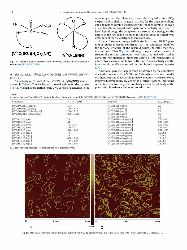
40 J.C. Pessoa et al. / Coordination Chemistr
Fc
o(
s[
was hypothesized that coordination to vanadium may in some way
TI
I
ig. 13. Schematic general structure of the two newly synthesized VIVO families ofompounds [117,118,277–280].
n the parasite: [VIVO(SO4)(H2O)2(NN)] and [VIVO(L-2H)(NN)]Fig. 13).
The activity on T. cruzi of the [VIVO(SO4)(H2O)2(NN)] series ishown in Table 1. The NN ligands showed activity on the parasite117,277]. Their coordination to the VIVO core led to activities in the
able 1n vitro activity on T. cruzi (Dm28c strain or Tulahuen 2 epimastigotes) of the [VIVO(SO4)(
Compound IC50 ± SD (mM)
[VIVO(SO4)(H2O)2(dppz)] ca. 3
[VIVO(SO4)(H2O)2(tdzp)] 15.6 ± 4.88
[VIVO(SO4)(H2O)2(phendione)] 7.24 ± 5.27
[VIVO(SO4)(H2O)2(epoxyphen)] 21.91 ± 9.94
[VIVO(L1-2H)(bipy)] 73
[VIVO(L2-2H)(bipy)] 84
[VIVO(L3-2H)(bipy)] 76.2 ± 9.7
[VIVO(L4-2H)(bipy)] 161 ± 57
[VIVO(L5-2H)(bipy)] 58.0 ± 14.8
[VIVO(L1-2H)(dppz)] 13
[VIVO(L2-2H)(dppz)] 19
[VIVO(L3-2H)(dppz)] 4.70 ± 3.35
[VIVO(L4-2H)(dppz)] 12.7 ± 8.27
[VIVO(L5-2H)(dppz)] 5.05 ± 1.60
C50, concentration required for inhibition of 50% of culture growth.
Fig. 14. AFM images showing the modifications suffered by pBR322 plasmid
y Reviews 301–302 (2015) 24–48
same range than the reference tripanocidal drug Nifurtimox (IC506.0 mM) and to slight changes in activity for the dppz, phendioneand epoxyphen complexes; noteworthy, the tdzp complex showeda significantly improved antitrypanosomal activity in respect tofree tdzp. Although the complexes are structurally analogous, thenature of the NN ligand included in the coordination sphere wasdeterminant for the antitrypanosomal activity.
Atomic force microscopy (AFM) studies using pBR322 plas-mid as model molecule confirmed that the complexes modifiedthe tertiary structure of the plasmid which indicates that theyinteract with DNA (Fig. 14). Although only a reduced series ofstructurally related compounds was compared and AFM studiesalone are not enough to judge the ability of the compounds toaffect DNA, a correlation between the anti-T. cruzi activity and theintensity of the effect observed on the plasmid appeared to exist[277].
Additional parasite targets could be affected by the complexesdue to the presence of the VIVO core. Although not demonstrated it
improve bioavailability by acting as a carrier system, improvingcell uptake due to changes in solubility and/or lipophilicity of thephenanthroline derivatives upon coordination.
H2O)2(NN)] and [VIVO(L-2H)(NN)] complexes [117,118,277–280].
Compound IC50 ± SD (mM)
[VIVO(L1-2H)(phen)] 2.0[VIVO(L2-2H)(phen)] 3.1[VIVO(L3-2H)(phen)] 2.3[VIVO(L4-2H)(phen)] 1.6[VIVO(L5-2H)(phen)] 3.8[VIVO(L1-2H)(aminophen)] 0.50 ± 0.02[VIVO(L2-2H)(aminophen)] 0.27 ± 0.09[VIVO(L3-2H)(aminophen)] 0.52 ± 0.02[VIVO(L4-2H)(aminophen)] 0.51 ± 0.02[VIVO(L5-2H)(aminophen)] 0.51 ± 0.02[VIVO(L1-2H)(epoxyphen)] 1.0 ± 0.1[VIVO(L2-2H)(epoxyphen)] 0.9 ± 0.2[VIVO(L3-2H)(epoxyphen)] 1.2 ± 0.3[VIVO(L4-2H)(epoxyphen)] 3.9 ± 0.6[VIVO(L5-2H)(epoxyphen)] 1.1 ± 0.3
DNA (a) due to the interaction with (b) [VIVO(SO4)(H2O)2(tdzp)] [277].

J.C. Pessoa et al. / Coordination Chemistr
N
O
V
O OHN
R2
NH2
N
NR1
[VO(L-2H)(N-N)]
OH
NNH
NH2
O
R2
R1
R1HHOCH3OCH2CH3OCH3
R2HBrHHBr
L1L2L3L4L5
Fi
psb
bmiii((umtp(attuccmhcstoJmi
different techniques: agarose gel electrophoresis, viscosity mea-
Fs
S
ig. 15. Tridentate salicylaldehyde semicarbazone derivatives selected as coligandsn [VIVO(L-2H)(NN)] compounds [278–280].
A larger series of heteroleptic bioactive [VIVO(L-2H)(NN)] com-ounds was obtained where L are tridentate salicylaldehydeemicarbazone derivatives depicted in Fig. 15, where NN is eitheripy, dppz, phen, epoxyphen or aminophen [278–280].
This series includes several compounds with very interestingiological properties. The in vitro anti-T. cruzi activity resultedainly dependent on the nature of the NN ligand and almost
ndependent of the chemical nature of the substituents R1 and R2n the salicylaldehyde moiety (Table 1 and Fig. 16). The approx-mate order of activity was [VIVO(L-2H)(bipy)] < [VIVO(L-2H)dppz)] < [VIVO(L-2H)(phen)] < [VIVO(L-2H)(epoxyphen)] < [VIVOL-2H)(aminophen)]. The semicarbazone ligands showed IC50 val-es higher than 100 mM and the VIVO-compounds were in generaluch more active than the polypyridyl compounds included in
heir coordination spheres. Seventeen of the twenty-five com-lexes were more active than the Nifurtimox (IC50 (mM): 6.0Dm28c strain), 7.7 (Tulahuen 2 strain)). The compounds includingminophen were the most active ones showing IC50 values inhe submicromolar range and being 9.6- to 18.5-fold more activehan aminophen itself, as measured by their IC50 values in mMnits. The selectivity to the parasite in respect to mammalianells was also studied for the most relevant compounds. The phenompounds showed selectivity indexes SI (IC50 mammalian cellodel/IC50 T. cruzi) ranging between 38 and 596 using THP-1
uman acute monocytic leukemia (ATCC) cells as mammalianell models [278]. The epoxyphen and aminophen compoundshowed SI values (IC50 J774 murine macrophages/IC50 T. cruzi inhe range 10–41 and 96–185, respectively. Furthermore, nonef the aminophen complexes displayed relevant toxicity against
-774 macrophages at doses down to 50 mM, depicting in all casesuch higher SI values than the corresponding value for aminophentself. According to accepted guidelines for the development of
ig. 16. Graphical comparison of the IC50 values on T. cruzi of the five families of [VIVO(L-train) 6.0 mM; (b) NN = epoxyphen and aminophen, IC50 Nfx (Tulahuen 2 strain) 7.7 mM.
ource: Values taken from Ref. [281].
y Reviews 301–302 (2015) 24–48 41
anti-T. cruzi drugs these compounds deserve further studies toestablish their therapeutic potentiality [281].
Having twenty-five structurally related compounds, a QSAR(quantitative structure activity relationship study) study was car-ried out to get a design guide for further antitrypanosomal drugdevelopment. This study is particularly relevant since QSAR stud-ies are still not very common in inorganic medicinal chemistryresearch due to the difficulty to get a high number of structurallyrelated compounds [280]. Lipophilic effects are among the factorscontrolling transport to and interaction with biological receptors.Lipophilicity was experimentally determined using reversed-phaseTLC experiments where the stationary phase, precoated TLC-C18,may be considered to simulate lipids of biological membranes orreceptors, and the mobile phase, MeOH:DMF:Tris–HCl buffer pH7.4 (85:5:10, v/v/v), resembles the aqueous biological milieu. TheRf values in this system were converted to RM via the relationship:RM = log10[(1/Rf) − 1] and reported. The complexes resulted morelipophilic than the NN ligands.
A statistically satisfactory correlation between IC50 on T. cruziand the parameter RM and the indicator variable Icol, a structuraldescriptor of the NN co-ligands, was obtained [280]:
IC50, T. cruzi = (506 ± 97) − (592 ± 137)RM + (205 ± 51)R2M − (31 ± 4) Icol
r2adj = 0.7847, n = 22, F = 26.52, p = 8 × 10⁻⁷
The indicator variable Icol takes into account the potentially dif-ferent biological behavior of the selected NN scaffolds. The highlyelectrophilic epoxyphen may react with biological nucleophilesfrom proteins and DNA through the epoxide-moiety, while thenucleophile aminophen may react with biological electrophilesthrough the amino-moiety. Moreover, the p-expanded systemsdppz, phen and phen derivatives may also intercalate to DNA.
In summary, the equation highlights the relevance of thelipophilic parameter on the desired bioactivity. Additionally it alsoreflects the significance of the structure of the co-ligand on theanti-T. cruzi effect. Accordingly complexes with DNA-modifying co-ligands with the higher Icol values, i.e. aminophen and epoxyphen,were the most cytotoxic for the parasite.
A parabolic relationship between biological response andlipophilicity was obtained and, from this correlation an optimumRM value was determined which may be a design guide for futuredevelopment of new compounds bearing a better biological profile.
When trying to access the mechanism of action of the com-pounds, DNA interaction of the complexes was assessed through
surement of DNA solutions and AFM [280].In addition, a metabolomic study was performed by incu-
bating the parasite with the two most active VIVO-compounds
2H)(NN)] compounds: (a) NN = bipy, dppz and phen, IC50 Nfx (Nifurtimox) (Dm28c

42 J.C. Pessoa et al. / Coordination Chemistry Reviews 301–302 (2015) 24–48
pyruvate lactate alanine
D-Glucose
succ inate pyruvate
Glycosom e
Cytoso l
Mitochondrion
TCA cycle
pyruvate acetat e glycine
succina te
F
[opboomitcdpmipNwpopTcAaoc[
a[b[
icsgwtr
Loepcao
which includes phen in its coordination sphere, showed activity on
ig. 17. Schematic representation of carbon source metabolism in trypanosomes.
VIVO(L2-2H)(epoxyphen)] and [VIVO(L2-2H)(aminophen)] inrder to study the changes in relevant biochemical pathwaysromoted by them. In particular, the consumption of glucosey trypanosomatid protozoa is characterized by the excretionf reduced products such as succinate, pyruvate or lactate notnly in anerobiosis, but also under aerobic conditions. A compart-entation of the glycolytic enzymes, most of which are placed
n an organelle called the glycosome, and a relative inefficacy ofhe respiratory chain has been described. Previous research on T.ruzi epimastigotes metabolism using [l-13C] glucose as substrateescribed the detection by 13C NMR of the carboxylates succinate,yruvate, acetate and lactate and the amino acids Ala and Gly asain catabolites of glucose [282]. Therefore, the modifications
n the excreted metabolites produced by the incubation of thearasite with the selected VIVO-compounds were followed by 1HMR spectroscopy. An increment of excreted Gly, acetate and Alaas observed. Acetate and Gly are catabolites produced in thearasite single mitochondrion. Alanine is produced in the cytosolf the parasite cell from pyruvate that is originated in a specificarasite organelle called glycosome, via phosphoenolpyruvate.his pyruvate is also used, into the mitochondrion, in the like-Krebsycle (Fig. 17) [283]. Consequently, an increment in the amount ofla could be indicative of an increment of cytosolic pyruvate due to
deficient mitochondrion uptake emerging of deleterious effectsn mitochondrion integrity. Consequently, the mitochondrionould be pointed out as a potential target for these complexes280].
The presence of the VIVO core together with the semicarbazonend NN co-ligands seemed to be determinant of the activity. TheVVO2(L-H)] complexes, where L are the salicylaldehyde semicar-azone derivatives L1–L5 showed no antitrypanosomal activity279].
Concerning the question if the vanadium center is neededn order to have bioactive compounds, the previously obtainedompounds with aromatic amine N-oxide derivatives showed aignificantly higher activity for the VIVO-compounds that for analo-ous CuII and PdII compounds [284]. These results are in agreementith the previously described observations which suggested that
he complexes probably not only interact with DNA but also affectelevant metabolic pathways in the parasite.
Recently, the genomes of the parasites T. cruzi, T. brucei andeishmania major have been decoded and showed a high degreef similarity [285]. Thus, clinically useful drugs affecting commonnzymatic targets or metabolic pathways in these trypanosomatidarasites are being sought [286]. The emerging broad-spectrum
hemical scaffolds could offer an innovative and less expensivepproach for anti-parasitic drug discovery [287]. Therefore, somef the VIVO-compounds described above were also tested eitherFig. 18. VIVO- and VVO2-complexes of the bioactive tridentate ligand 2-(benzothiazol-2-yl-hydrazonomethyl) phenol (L6) [288].
on T. brucei or on different Leishmania species in order to recog-nize metal-based moieties that could inhibit T. cruzi or T. brucei orLeishmania spp. growth by acting on common targets.
The series of five [VO(L-2H)(phen)] compounds were evaluatedon L. panamensis and L. chagasi promastigotes and intracellu-lar amastigotes. Only [VO(L1-2H)(phen)] and [VO(L3-2H)(phen)]showed promising antileishmanial activities. They were moreactive against the promastigote form than on the intracellularamastigote form of the parasites. In particular, they showed lowIC50 values on promastigotes of L. panamensis (IC50 2.74 and2.75 mM, respectively), on intracellular amastigotes of L. panamen-sis (IC50 19.52 and 20.75 mM, respectively) and showed low toxicityon THP-1 mammalian cells (IC50 188.55 and 88.13 mM, respec-tively) [278].
The [VIVO(L-2H)(dppz)] compounds with L = L3–L5 (Fig. 15)showed dose dependent growth inhibitory activity toward theinfective form of T. brucei brucei strain 427, cell line 449. Theyshow IC50 values in the low micromolar range (2.8 ± 0.1, 3.0 ± 0.2and 1.5 ± 0.1, respectively). On the other hand, the bipy analogouscomplexes showed no significant activity against the same parasite.Both series showed a similar behavior against T. cruzi and T. brucei.These results are encouraging in respect to the proposed strategyfor the development of compounds that could show activity towardboth genetically related parasites [279].
Using a similar strategy other mixed-ligand bioactive VIVO-compounds were developed including phen as co-ligand. For thispurpose the semicarbazone ONO co-ligand was changed by otherONO or NNO tridentate compounds.
In the case of the complexes depicted in Fig. 18 the mixed-ligandVIVO complex 26 showed a more than 10-fold improvement ofIC50 against T. cruzi (Dm28c) with respect to the ligand HL6 (IC50120.8 mM) and an IC50 value (10.7 mM) in the same order of thatof Nifurtimox (IC50 6.0 mM). The complex is almost ten times moreactive than [VVO(L6)] 25. To explain the high inhibitory activityagainst the parasite of 26 as compared to 25, the interaction of thiscompound with DNA due to the presence of the phen ligand waspostulated [288] (Fig. 19).
Two related VIVO N-acylhydrazone complexes [VIVO(L7-2H)(phen)]·(H2O) 27 and [VIVO(L7-2H)(H2O)] 28, where L7 =LASSBio1064 = (E)-N0-(2-hydroxybenzylidene-4-chlorobenzohy-drazide were synthesized and evaluated on T. cruzi and L. major[289]. In this case, being L7 active on T. cruzi, both VIVO-complexeswere also active in vitro against the epimastigote form of T. cruzi(Tulahuen 2 strain), showing IC50 values of the same order orsignificantly lower than that of Nifurtimox (IC50: L7 2.1; 27 6.0; 281.7 mM). On the other hand, L7 was inactive on L. major promastig-otes and therefore again only the mixed-ligand VIVO-complex 27,
L. major promastigotes (IC50 value 22.1 mM). In addition, the ligandand both metal compounds showed low toxicity on peritonealmacrophage cells used as mammalian cell models (IC50 > 100 mM).

J.C. Pessoa et al. / Coordination Chemistr
N
O
O
N
Cl
V
OH2O
[VO(L7-2H)(H2O)] 28N
O
O
N
Cl
V
O
N
N
[VO(L7-2H)(phe n)].(H2O) 27
N
HOO
NH
Cl LASSB io-1064 (L7 )
F=
IrtsTiaos
egdb
ImiTwnts(tosan
ig. 19. Oxidovanadium(IV) N-acylhydrazone complexes where L7 = LASSBio1064 (E)-N0-(2-hydroxybenzylidene-4-chlorobenzohydrazide [289].
t was hypothesized that the higher activity of these VCs may beelated to either an easier uptake and/or to their higher efficacyoward their targets. On the other hand, inside cells the vanadiumpeciation may yield species containing or not the original ligands.he phen moiety may indeed be relevant in this respect since itmproves the biological activity of the metal-based systems prob-bly by acting as DNA intercalator. Indeed, DNA interaction abilityf the phen complex was demonstrated by AFM, fluorescencepectroscopy and viscosity measurements.
There are two additional recent reports of VCs bearing inhibitoryffects on Leishmania spp. growth. One of them established therowth inhibition of L. tarentolae promastigotes in vitro due toecavanadate (NH4)6V10O28·6H2O. The authors suggested the inhi-ition of phosphatases as a probable mechanism of action [290].
The other report involves a completely different strategy [291].n the mammalian host, the Leishmania parasites live inside
acrophages. The resistance to Leishmania or the healing abil-ty of leishmaniasis is related to Th1 response by macrophages.hus, compounds that activate macrophages for the Th1 path-ay could show interesting leishmanicidal activity. In this context,ative or chemically modified polysaccharides have been studiedo potentiate the immune response. A previous research by theame authors showed that VIVO complexation of galactomannanGMPOLY), isolated from the lichen Ramalina celastri, exacerbatedhe cytotoxicity to intracellular Leishmania [9]. The in vitro effects
f galactomannan (GALMAN-A) isolated from seeds of Mimosacabrella and its oxidovanadium(IV/V) complex on macrophagectivation and leishmanicidal activity on Leishmania (L.) amazo-ensis intramacrophages amastigotes were confirmed. Both areFig. 20. VVO2-complexes of binucleating bis(dibasic ONS dono
y Reviews 301–302 (2015) 24–48 43
potent activators of macrophages favoring the upregulation of Th1response in macrophages, responsible of leishmanicidal effects.
In respect to VCs as prospective agents for the treatment ofamoebiasis, Maurya et al. developed a large series of vanadiumcompounds with activity against E. histolytica that was discussedin the previous review [9]. Expanding this work a new series ofVIVO- and VV-complexes of binucleating bis(dibasic tridentate ONSdonor) ligand systems showing antiamoebic activity was reported(Fig. 20) [292].
The synthesized VVO2-complexes along with the ligands werescreened for antiamoebic activity in vitro against the most commonHM1:IMSS a highly virulent strain of E. histolytica. The complexesand the ligands showed IC50 values in the micromolar range. In par-ticular, compounds 29, 30 and 31 (Fig. 20) are more active than thereference antiamoebic drug Metronizadol (IC50: 29 0.353; 30 0.092;31 0.85; Metronidazol 1.68 mM). The reason why the VV-complexesare more active than the ligands is not clear. Authors hypothesizedabout the possibility that complexation might favor permeationof the drugs through the lipid layer of the cell membrane. VIVO-complexes with the same ligands resulted poorly active.
3.7.2. Antiviral vanadium compoundsOnly scarce work has also been reported on vanadium com-
pounds showing activity on certain viruses, bacteria and fungi.
3.7.2.1. Anti-HIV vanadium complexes. The effect of VCs on humanimmune deficiency virus (HIV) was reviewed by Sun et al. [293].Studies showed that vanadium–thiourea complexes and vanadiumsubstituted polyoxotungstates exhibited potent anti-HIV proper-ties toward infected immortalized T-cells [127,294].
In particular, the vanadium-substituted polyoxidotungstate[(VIVO)2(VVO)(SbW9O33)2]11− exhibited antiviral activity not onlyagainst HIV-1 in vitro but also against the viruses causing influenza,Dengue fever and SARS (severe acute respiratory syndrome). Atphysiological conditions, this ‘Keggin sandwich’ decompose intovanadate(V) and tungstate(VI), which were suggested to be the realactive species [294,295].
As it is known that VIV-complexes are not much sta-ble under physiological conditions, porphyrinato ligands wereused to stabilize the VIVO-unit and to get water-solubleoxidovanadium(IV)-porphyrins [128]. Porphyrins have a rigidsquare planar scaffold which could prohibit the demetalation reac-tion. The VIV-porphyrins prepared are stable in GSH containingsolutions, and their inhibitory effects on HIV-1(BaL) replication inHut/CCR5 cells were evaluated. All of these complexes showed anti-viral activities compared to the vehicle control, but one of them,water soluble and containing aminosulfonyl functional groups (32in Fig. 21), exhibited the highest potency at the 5 mM level withover 97% inhibition.
HIV-1 reverse transcriptase (RT) is one of the major targetsfor anti-HIV drugs, and binding of VCs to RT has been reported[296]. The inhibitory power of 32 toward HIV-1RT was measuredusing an ELISA method, and significant RT inhibition was observed
r ligands) showing activity in vitro on E. histolytica [292].
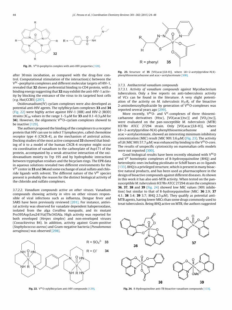
44 J.C. Pessoa et al. / Coordination Chemistry Reviews 301–302 (2015) 24–48
N
N
V
O
RN
NR
R
S NHO
O
N
R =
32
atVrbi(
p(s3b
prDivpdbiVrpt
3csSriPb((a
NV
O
O
NN
SH
RO
R = phenyl 35
4.1; 38 3.4; 39 3.7; 8HQ 2.5 mM). They qualify as potential anti-MTB agents, having lower MICs than some drugs commonly used totreat tuberculosis. Being 8HQ active on MTB, the authors suggested
R
Fig. 21. VIVO-porphyrin complex with anti-HIV properties [128].
fter 30 min incubation, as compared with the drug-free con-rol. Computational stimulation of the interaction(s) between theIV–porphyrin complexes and different molecular targets of HIV-1,evealed that 32 shows preferential binding to CD4 protein, with ainding energy suggesting that 32 may exhibit the anti-HIV-1 activ-
ty by blocking the entrance of the virus to its targeted host cellse.g. Hut/CCR5) [297].
Oxidovanadium(IV) cyclam complexes were also developed asotential anti-HIV agents. The xylylbicyclam complexes 33 and 34Fig. 22) were highly active against HIV-1 (IIIB) and HIV-2 (ROD)trains (IC50 values in the range 1–5 mM for 33 and 0.1–0.3 mM for4). However, the oligomeric VIVO–cyclam complexes showed toe inactive [129].
The authors proposed the binding of the complexes to a receptorrotein that HIV can use to infect T lymphocytes, called chemokineeceptor type 4 (CXCR-4), as the mechanism of antiviral action.ocking studies of the most active compound 33 showed that bind-
ng of it to a model of the human CXCR-4 receptor might occuria coordination of vanadium to the carboxylate of Asp171 of therotein, accompanied by a weak attractive interaction of the oxi-ovanadium moiety to Trp 195 and by hydrophobic interactionetween tryptophan residues and the bicyclam rings. The EPR data
n aqueous solutions revealed two different environments for theIV center in 33 and 34 and some exchange of axial sulfato and chlo-ide ligands with solvent. The different nature of the VIV speciesresent is probably the reason for the distinct biological activity ofhe chlorido and sulfato complexes.
.7.2.2. Vanadium compounds active on other viruses. Vanadiumompounds showing activity in vitro on other viruses respon-ible of viral infections such as influenza, Dengue fever andARS have been previously reviewed [201]. For instance, antivi-al activity was observed for vanadate dependent haloperoxidase,solated from the alga Corallina inaequalis, and its mutantro395Asp/Leu241Val/Thr343Ala. High activity was reported for
oth enveloped (Herpes simplex) and non-enveloped virusesCoxsackievirus B4). In addition, activity against Gram-positiveStaphylococcus aureus) and Gram-negative bacteria (Pseudomonaseruginosa) was observed [298].NH
NH
N
HN
V
O
R
NH
N
HN
HN
V
O
R
R = SO42- 33
R = Cl- 34
Fig. 22. VIVO-xylylbicyclam anti-HIV compounds [129].
Fig. 23. Structure of 35 [VO(acac)(L8-H)], where L8 = 2-acetylpyridine-N(4)-phenylthiosemicarbazone and acac = acetylacetonate [300].
3.7.3. Antibacterial vanadium compounds3.7.3.1. Activity of vanadium compounds against Mycobacteriumtuberculosis. Only a few reports on anti-tuberculosis activityof VCs can be found in the literature. A very slight potenti-ation of the activity on M. tuberculosis H37Rv of the bioactive2-aminobenzoylhydrazide by generation of VIVO-complexes wasreported several years ago [299].
More recently, VIVO- and VV-complexes of three thiosemi-carbazone derivatives (Htsc), [VO(acac)(tsc)] and [VO2(tsc)],were evaluated on the pan-susceptible M. tuberculosis (MTB)H37Rv ATCC 27294 strain. Only [VO(acac)(L8-H)], whereL8 = 2-acetylpyridine-N(4)-phenylthiosemicarbazone andacac = acetylacetonate, showed an interesting minimum inhibitoryconcentration (MIC) result (MIC 90% 3.6 mM) (Fig. 23). The activityof L8 (MIC 90% 57.7 mM) was enhanced by binding to the VIVO-core.The results of unspecific cytotoxicity on mammalian cells modelswere not reported [300].
Good biological results have been recently obtained with VIVOand VV homoleptic complexes of 8-hydroxyquinoline (8HQ) andheteroleptic ones including picolinato or Schiff bases as co-ligands[133]. 8HQ is a privileged structure, which is present in many bioac-tive natural products, and has been used as pharmacophore in thedesign of bioactive compounds against different diseases. As shownin this work it has also anti-MTB activity. When tested on the pan-susceptible M. tuberculosis H37Rv ATCC 27294 strain the complexes36, 37, 38 and 39 (Fig. 24) showed low MIC values (90% inhibi-tion) but similar to that of 8-hydroxyquinoline (MIC: 36 2.9; 37
Fig. 24. 8-Hydroquinoline anti-TB bioactive vanadium compounds [133].

emistr
aip
3fab
aacpK
Dc(GGoc
oaapP(ml
5inatf
4
otpipbtd
tiqsttibmkuad
J.C. Pessoa et al. / Coordination Ch
non-specific mechanism of action. The inclusion of 8HQ as ligandn VCs might improve its bioavailability enhancing the ability toenetrate the lipid-rich hydrophobic cell wall of the mycobacteria.
.7.3.2. Activity of vanadium compounds against other bacteria andungi. The activity of VCs against other bacteria and fungi has beenlso reported. Only some representative examples are includedelow.
The antibacterial activity of different polyoxometalates (PMs)gainst Helicobacter pylori, the Gram-negative bacterium which is
principal gastric pathogen of humans, was investigated [301]. Theompounds were evaluated on drugs-susceptible and resistant H.ylori strains. Among the studied compounds the polyoxometalateeggin-structural K5[SiVVW11O40] showed bacteriostatic activity.
Ciprofloxacin is a quinolone type antibiotic that targets theNA topoisomerase (gyrase) of bacterial type II. Heteroleptic VIVO-omplexes with this antibiotic as ligand and various amino acidsdl-Ala, l-Tyr, l-Trp, l-Glu and l-Leu) were evaluated againstram-positive (S. aureus, Baccilus subtilis, Serratia marcescens), andram-negative (P. aeruginosa, Escherichia coli) bacteria. In mostf the cases a significant increase of the antibacterial activity ofiprofloxacin due to complexation was observed [302].
Metal complexes of the Schiff base derived from condensationf 1H-indole-3-carboxaldehyde and 1-phenyl-2,3-dimethyl-4-mino-3-pyrazolin-5-one (L9) were tested for their in vitrontibacterial activity against Gram-positive bacteria (S. aureus, K.neumoniae and L. monocytogenes), Gram-negative bacteria (E. coli,. aeruginosa and Salmonella typhimurium) and anti-fungal activityCandida albicans and Aspergillus flavus) strains. The coordination to
etal ions and, in particular to VIVO-center in [VO(L9)2(H2O)]SO4,ed to an increase in antibacterial and antifungal activity [303].
VIVO-complexes with Schiff bases derived from condensation of-(phenyl/substituted phenyl)-2-hydrazino-1,3,4-thiadiazoles and
ndoline-2,3-dione showed higher in vitro antifungal (Aspergillusiger, Colletotrichum falcatum, Colletotrichum pallescence) andntibacterial against Gram-negative (Escherichia coli, Salmonellayphi) and Gram-positive (S. aureus, Bacillus subtilis) activities thanree ligands [304].
. Conclusions
Vanadium being relevant in several biological processes, manyf its complexes with organic derivatives have been proposed forhe treatment of several types of diseases, although no compound isresently used in the clinic. Namely, as reviewed here and reported
n many previous publications, vanadium compounds have beenroposed for the treatment of diabetes, cancer, diseases causedy parasites, for anti-viral and anti-bacteria use and for as diverseypes of therapeutic uses as cardio-protection and to improve car-iac performance.
There has been substantial progress with respect to the specia-ion of VCs in blood serum, but a lot remains to be done concerningn situ real-time investigation on the fate of a VC. In fact, besidesuestions on their speciation in the gastrointestinal tract, evenmall VCs cannot be expected to simply diffuse through membraneso get to the target tissues. They must undergo translocation fromhe bloodstream through the endothelium of capillaries into thenterstitial fluid [79]. Binding to transferrin and/or albumin mighte relevant in this respect, but further research must address theechanisms by which these processes might occur. Thus, a deeper
nowledge of the in vivo speciation of VCs should help to betternderstand the mechanisms of transport, targeting and the mode ofction, and give support to the design of efficient vanadium-basedrugs.
y Reviews 301–302 (2015) 24–48 45
Despite the many advances, the not yet well-established modeof action of VCs remains a challenge. Many of the reportedtherapeutic effects are related to the similarity of vanadate andphosphate, thus to the inhibition of the active sites of phosphatasesand related enzymes resulting thereof; kinase activation may alsobe relevant, as well as the ability to generate ROS.
As discussed in many publications, vanadium compounds acti-vate numerous signaling pathways and transcription factors, thissimultaneously potentiating application of vanadium compoundsin therapeutics, but also exerting nonspecific diametrically oppo-site effects on different cell structures. Additionally, still not muchis known regarding global toxicity issues and effects of VCs onthe immune system and inflammatory reactions, particularly iflong-term administration is required. Potential carcinogenicity andcorrelation between the use of VCs and effects on the free-radicalcell background are also issues to address.
Globally vanadium compounds have potential pharmacologi-cal interest for the treatment of diabetes, assuming that its toxicside effects can be overcome. Drugs that when given orally do notreadily hydrolyze in the gastrointestinal tract and could be directedto specific tissues might possibly overcome certain deleterious sideeffects. Thus, the use of VCs to treat type 2 diabetes is still an openquestion.
Given the importance of cancer and parasitic-related diseasessuch as Amoebiasis or Chagas’, the non-adequacy or increasingresistance of pathogens against the presently used therapies, andthe potentially lower costs of vanadium-based drugs, the prospec-tive use of VCs in these areas appears promising. The combinationof VCs with other drugs for the enhancement of the therapeuticeffects is also an area deserving much more attention.
The ability to overcome the balance existing between the ther-apeutic action and adverse effects exerted by VCs, by shifting thisbalance toward the beneficial side, is the crucial issue for the futureuse of vanadium compounds in Medicine.
Acknowledgements
The authors thank Fundac ão para a Ciência e Tecnologia (FCT)and PEst-OE/QUI/UI0100/2013 (Portugal) UNLP (11X690), CONICET(PIP1125) and ANPCyT (PICT 2008-2218) (Argentina) and PEDECIBAQuímica and ANII (Uruguay). SBE is member of the Carrera delInvestigador, CONICET, Argentina.
References
[1] T. Kiss, T. Jakusch, D. Hollender, A. Dornyei, E.A. Enyedy, J. Costa Pessoa, H.Sakurai, A. Sanz-Medel, Coord. Chem. Rev. 252 (2008) 1153.
[2] D. Rehder, Bioinorganic Vanadium Chemistry, John Wiley & Sons, New York,2008.
[3] H. Sakurai, Y. Yoshikawa, H. Yasui, Chem. Soc. Rev. 37 (2008) 2383.[4] D. Rehder, Dalton Trans. 42 (2013) 11749.[5] G.R. Willsky, L.H. Chi, M. Godzala, P.J. Kostyniak, J.J. Smee, A.M. Trujillo, J.A.
Alfano, W.J. Ding, Z.H. Hu, D.C. Crans, Coord. Chem. Rev. 255 (2011) 2258.[6] D. Rehder, Future Med. Chem. 4 (2012) 1823.[7] J. Costa Pessoa, I. Tomaz, Curr. Med. Chem. 17 (2010) 3701.[8] T. Jakusch, J. Costa Pessoa, T. Kiss, Coord. Chem. Rev. 255 (2011) 2218.[9] D. Gambino, Coord. Chem. Rev. 255 (2011) 2193.
[10] D. Rehder, in: A. Sigel, et al. (Eds.), Interrelations Between Essential MetalIons and Human Diseases, Metal Ions in Life Sciences, 13, Springer Sci-ence + Business Media, Dordrecht, 2014, http://dx.doi.org/10.1007/978-94-007-7500-8 5.
[11] J. Korbecki, I. Baranowska-Bosiacka, I. Gutowska, D. Chlubek, Acta Biochim.Polon. 59 (2012) 195.
[12] D.M. Smith, R.M. Pickering, G.T. Lewith, Q. Int. J. Med. 101 (2008) 351.[13] J.O. Olopade, J.R. Connor, Curr. Top. Toxicol. 7 (2011) 34.[14] A.M. Evangelou, Crit. Rev. Oncol./Hematol. 42 (2002) 249.
[15] Y. Shechter, I. Goldwaser, M. Mironchik, M. Fridkin, D. Gefel, Coord. Chem.Rev. 237 (2003) 3.[16] H. Sakurai, Y. Kojima, Y. Yoshikawa, K. Kawabe, H. Yasui, Coord. Chem. Rev.
226 (2002) 187.[17] H. Sakurai, A. Katoh, Y. Yoshikawa, Bull. Chem. Soc. Jpn. 79 (2006) 1645.

4 emistr
6 J.C. Pessoa et al. / Coordination Ch[18] (a) P. Hulley, A. Davison, J. Trace Elem. Exp. Med. 16 (2003) 281;(b) J.C. Dabrowiak, Metals in Medicine, Wiley & Sons, Chichester, UK, 2009.
[19] A.S. Tracey, G.R. Willsky, E.S. Takeuchi, Vanadium: Chemistry, Biochemistry,Pharmacology and Practical Applications, CRC Press, Boca Raton, 2007.
[20] H. Michibata (Ed.), Vanadium: Biochemical and Molecular BiologicalApproaches, Springer, Dordrecht, 2012.
[21] C. Orvig, M.J. Abrams, Chem. Rev. 99 (1999) 2201.[22] A.M. Kordowiak, P. Holko, Post. Biol. Kom. 36 (2009) 361.[23] D.G. Barcelaux, J. Toxicol. Clinic. Toxicol. 37 (1999) 265.[24] D. Rehder, in: A. Sigel, H. Sigel, R.K. Sigel (Eds.) Met. Ions Life Sci. 13 (2013)
139–159.[25] M.D. Cohen, ACS Symp. Ser. 974, 2007, p. 217.[26] T. Scior, A. Guevara-García, P. Bernard, Q.-T. Do, D. Domeyer, S. Laufer, Mini
Rev. Med. Chem. 5 (2005) 995.[27] G.B. Irsigler, P.J. Visser, P.A. Spangenberg, Am. J. Ind. Med. 35 (1999) 366.[28] W.N. Rom (Ed.), Environmental and Occupational Medicine Little, Brown and
Co., Boston, MA, 1992.[29] J.O. Nriagu (Ed.), Wiley Series in Advances in Environmental Science and
Technology, vol. 30, John Wiley & Sons, New York, 1998.[30] F.L. Assem, L.S. Levy, in: H. H Michibata (Ed.), Vanadium: Biochemical and
Molecular Biological Approaches, Springer, Dordrecht, 2012, pp. 209–224.[31] B.R. Nechay, J.P. Saunders, J. Environ. Pathol. Toxicol. 2 (1978) 247.[32] C.C. McLauchlan, J.D. Hooker, M.A. Jones, Z. Dymon, E.A. Backhus, B.A. Greiner,
N.A. Dorner, M.A. Youkhana, L.M. Manus, J. Inorg. Biochem. 104 (2010) 274.[33] P.J. Stankiewics, A. Tracey, in: H. Sigel, A. Sigel (Eds.), Metal Ions in Biological
Systems. Vanadium and Its Role in Life, vol. 31, Marcel Dekker, Inc., New York,1995, pp. 249–286.
[34] Z.Y. Zhang, Crit. Rev. Biochem. Mol. Biol. 33 (1998) 1.[35] D.C. Crans, in: A. Tracey, D.C. Crans (Eds.), Vanadium Compounds. Chemistry,
Biochemistry, and Therapeutic Applications, American Chemical Society,Washington, DC, 1998, pp. 82–103.
[36] B.I. Posner, C.R. Yang, A. Shaver, in: A. Tracey, D.C. Crans (Eds.), Vanadium Com-pounds. Chemistry, Biochemistry, and Therapeutic Applications, AmericanChemical Society, Washington, DC, 1998, pp. 316–328.
[37] L. Chang, M. Karin, Nature 410 (2001) 37.[38] V.O. Van Hoof, M.E. De Broe, Crit. Rev. Clin. Lab. Sci. 31 (1994) 197.[39] D.C. Crans, A. Tracey, in: A. Tracey, D.C. Crans (Eds.), Vanadium Compounds.
Chemistry, Biochemistry, and Therapeutic Applications, American ChemicalSociety, Washington, DC, 1998, pp. 2–29.
[40] K. Kustin, in: A. Tracey, D.C. Crans (Eds.), Vanadium Compounds. Chemistry,Biochemistry, and Therapeutic Applications, American Chemical Society,Washington, DC, 1998, pp. 170–185.
[41] K.G. Peters, M.G. Davis, B.W. Howard, M. Pokross, V. Rastogi, C. Diven, K.D.Greis, E. Eby-Wilkens, M. Maier, A. Evdokimov, S. Soper, F. Genbauffe, J. Inorg.Biochem. 96 (2003) 321.
[42] Y. Shechter, S.J. Karlish, Nature 284 (1980) 556.[43] A. Stern, X. Yin, S.S. Tsang, A. Davison, J. Moon, Biochem. Cell Biol. 71 (1993)
103.[44] C. Djordjevic, in: H. Sigel, A. Sigel (Eds.), Metal Ions in Biological Systems.
Vanadium and Its Role in Life, Marcel Dekker, Inc., New York, 1995, pp.595–615.
[45] J.L. Domingo, Mol. Cell. Biochem. 203 (2000) 185.[46] A.K. Srivastava, Mol. Cell. Biochem. 206 (2000) 177.[47] J.C. Liu, Y. Yu, G. Wang, K. Wang, X.G. Yang, Metallomics 5 (2013) 813.[48] I.G. Fantus, S. Kadota, G. Deragon, B. Foster, B.I. Posner, Biochemistry 28 (1989)
8864.[49] S. Tamura, T.A. Brown, R.E. Dubler, J. Larner, Biochem. Biophys. Res. Commun.
113 (1983) 80.[50] S. Tamura, T.A. Brown, J.H. Whipple, Y. Fujita-Yamaguchi, R.E. Dubler, K.
Cheng, J. Larner, J. Biol. Chem. 259 (1984) 6650.[51] M. Bernier, D.M. Laird, M.D. Lane, J. Biol. Chem. 263 (1988) 13626.[52] A. Shisheva, Y. Shechter, Biochemistry 31 (1992) 8059.[53] S. Kadota, I.G. Fantus, G. Deragon, H.J. Guyda, B. Hersh, B.I. Posner, Biochem.
Biophys. Res. Commun. 147 (1987) 259.[54] A.K. Srivastava, J.L. Chiasson, Biochim. Biophys. Acta 996 (1989) 13.[55] H.V. Strout, P.P. Vicario, R. Saperstein, E.E. Slater, Endocrinology 124 (1989)
1918.[56] S.K. Pandey, M.B. Anand-Srivastava, A.K. Srivastava, Biochemistry 37 (1998)
7006.[57] J.C. Molero, C. Martinez, A. Andres, J. Satrustegui, J.M. Carrascosa, FEBS Lett.
425 (1998) 298.[58] S.K. Pandey, J.L. Chiasson, A.K. Srivastava, Mol. Cell. Biochem. 153 (1995) 69.[59] S.K. Pandey, J.F. Theberge, M. Bernier, A.K. Srivastava, Biochemistry 38 (1999)
14667.[60] I. Novak-Hofer, G. Thomas, J. Biol. Chem. 260 (1985) 10314.[61] I.G. Fantus, G. Deragon, R. Lai, S. Tang, Mol. Cell. Biochem. 153 (1995) 103.[62] A. Nantel, K. Mohammad-Ali, J. Sherk, B.I. Posner, D.Y. Thomas, J. Biol. Chem.
273 (1998) 10475.[63] Y. Shechter, Diabetes 39 (1990) 1.[64] A.M. Cortizo, S.B. Etcheverry, Mol. Cell. Biochem. 145 (1995) 97.[65] S.B. Etcheverry, A.M. Cortizo, in: A. Tracey, D.C. Crans (Eds.), Vanadium Com-
pounds. Chemistry, Biochemistry, and Therapeutic Applications, AmericanChemical Society, Washington, DC, 1998, pp. 270–276.
[66] J.B. Smith, Proc. Natl. Acad. Sci. U.S.A. 80 (1995) 6162.[67] B. Wice, J. Milbrandt, L. Glaser, J. Biol. Chem. 262 (1987) 1810.[68] K.H. Lau, H. Tanimoto, D.J. Baylink, Endocrinology 123 (1988) 2858.
y Reviews 301–302 (2015) 24–48
[69] S.B. Etcheverry, D.A. Barrio, in: K. Kustin, J. Costa Pesoa, D.C. Crans (Eds.),Vanadium: The Versatile Metal. American Chemical Society Series 974, vol.15, 2007, p. 204.
[70] M.G. Wilkinson, J.B. Millar, FASEB J. 14 (2000) 2147.[71] P.A. Randazzo, J.S. Olshan, A.A. Bijivi, L. Jarett, Arch. Biochem. Biophys. 292
(1992) 258.[72] S. Paris, J. Pouyssegur, J. Biol. Chem. 262 (1987) 1970.[73] L.C. Cantley, L. Josephson, R. Warner, M. Yanagisawa, C. Lechene, G. Guidotti,
J. Biol. Chem. 252 (1977) 7421.[74] H. Sakurai, H. Yasui, Y. Adachi, Expert Opin. Invest. Drug 12 (2003) 1189.[75] K.H. Thompson, C. Orvig, J. Chem. Soc., Dalton Trans. (2000) 2885.[76] K.H. Thompson, C. Orvig, J. Inorg. Biochem. 100 (2006) 1925.[77] C. Orvig, K.H. Thompson, M. Battell, J.H. McNeill, Met. Ions Biol. Syst. 31 (1995)
575.[78] B.R. Nechay, Annu. Rev. Pharmacol. Toxicol. 24 (1984) 501.[79] M.W. Makinen, M. Salehitazangi, Coord. Chem. Rev. 279 (2014) 1.[80] J.W. Lohr, M.I. Bennett, M.A. Pochal, J. McReynolds, M. Acara, G.R. Willsky, Res.
Commun. Chem. Pathol. Pharmacol. 72 (1991) 191.[81] A.K. Saxena, P. Srivastava, R.K. Kale, N.Z. Baquer, Biochem. Int. 26 (1992) 59.[82] K.H. Thompson, J.H. McNeill, Res. Commun. Chem. Pathol. Pharmacol. 80
(1993) 187.[83] G. Ugazio, S. Bosia, E. Burdino, F. Grignolo, Res. Commun. Mol. Pathol. Pharma-
col. 85 (1994) 313.[84] C.E. Heyliger, A.G. Tahiliani, J.H. McNeill, Science 227 (1985) 1474.[85] S. Ramanadham, J.J. Mongold, R.W. Brownsey, G.H. Cros, J.H. McNeill, Am. J.
Physiol. 257 (1989) H904.[86] J.J. Mongold, G.H. Cros, L. Vian, A. Tep, S. Ramanadham, G. Siou, J. Diaz, J.H.
McNeill, J.J. Serrano, Pharmacol. Toxicol. 67 (1990) 192.[87] S. Dai, K.H. Thompson, E. Vera, J.H. McNeill, Pharmacol. Toxicol. 75 (1994) 265.[88] L. Rossetti, M.R. Laughlin, J. Clin. Invest. 84 (1989) 892.[89] J. Meyerovitch, Z. Farfel, J. Sack, Y. Shechter, J. Biol. Chem. 262 (1987)
6658.[90] R.A.J. Challiss, B. Leighton, F.J. Lozeman, L. Budohoski, E.A. Newsholme,
Biochem. Pharmacol. 36 (1987) 357.[91] W.O. Nelson, T.B. Karpishin, S.J. Rettig, C. Orvig, Can. J. Chem. 66 (1988) 123.[92] D.J. Clevette, C. Orvig, Polyhedron 9 (1990) 151.[93] V.G. Yuen, C. Orvig, K.H. Thompson, J.H. McNeill, Can. J. Phys. Pharmacol. 71
(1993) 270.[94] P. Srivastava, A.K. Saxena, R.K. Kale, N.Z. Baquer, Res. Commun. Chem. Pathol.
Pharmacol. 80 (1993) 283.[95] K.H. Thompson, J.H. McNeill, in: P.W.F. Fischer, M.R. L’Abbe, K.A. Cockell, R.S.
Gibson (Eds.), Trace Elements in Man and Animals 9: Proc. 9th Int. Symp. TraceElem. Man Animals, NRC Research Press, Ottawa, 1997, pp. 349–350.
[96] B. Halliwell, J.M.C. Gutteridge, Methods Enzymol. 186 (1990) 1.[97] A.J. Lusis, Nature 407 (2000) 233.[98] M. Brownlee, Nature 414 (2001) 813.[99] D.C. Han, M.-Y. Lee, K.D. Shin, S.B. Jeon, J.M. Kim, K.-H. Son, H.-C. Kim, H.-M.
Kim, B.-M. Kwon, J. Biol. Chem. 279 (2004) 6911.[100] C.A. Rice-Evans, A.T. Diplock, Free Radic. Biol. Med. 15 (1993) 77.[101] D.R. Blake, R. Allen, J. Lunec, Br. Med. J. 43 (1987) 371.[102] R.M. LoPachin, T. Gavin, B.C. Geohagen, L.H. Zhang, D. Casper, R. Lekhraj, D.S.
Barber, J. Neurochem. 116 (2011) 132.[103] NCI, DCPC chemoprevention branch and agent development committee, J.
Cell. Biochem. 26S (1996) 72.[104] K.H. Thompson, K. Bohmerle, E. Polishcuk, C. Martins, P. Toleikis, J. Tse, V.
Yuen, J.H. McNeil, C. Orvig, J. Inorg. Biochem. 98 (2004) 2063.[105] L.X. Na, Y.L. Zhang, Y. Li, L.Y. Liu, R. Li, T. Kong, C.H. Sun, Nutr. Metab. Cardio-
vasc. Dis. 21 (2011) 526.[106] K.I. Seo, M.S. Choi, U.J. Jung, H.J. Kim, J. Yeo, S.M. Jeon, M.K. Lee, Mol. Nutr.
Food Res. 52 (2008) 995.[107] K. Kawabe, Y. Yoshikawa, Y. Adachi, H. Sakurai, Life Sci. 78 (2006) 2860.[108] Y. Adachi, J. Yoshida, Y. Kodera, A. Katoh, J. Takada, H. Sakurai, J. Med. Chem.
49 (2006) 3251.[109] Y. Adachi, Y. Yoshikawa, J. Yoshida, Y. Kodera, A. Katoh, J. Takada, H. Sakurai,
Biochem. Biophys. Res. Commun. 345 (2006) 945.[110] H. Sakurai, A. Katoh, T. Kiss, T. Jakusch, M. Hattori, Metallomics 2 (2010)
670–682.[111] M. Hiromura, A. Nakayama, Y. Adachi, M. Doi, H. Sakurai, J. Biol. Inorg. Chem.
12 (2007) 1275.[112] M. Aureliano, D.C. Crans, J. Inorg. Biochem. 103 (2009) 536.[113] K.H. Thompson, J. Lichter, C. LeBel, M.C. Scaife, J.H. McNeil, C. Orvig, J. Inorg.
Biochem. 103 (2009) 554.[114] O.J. D’Cruz, F.M. Uckun, Expert Opin. Invest. Drug 11 (2002) 1829.[115] A. Papaioannou, M. Manos, S. Karkabounas, R. Liasko, A.M. Evangelou, I. Cor-
reia, V. Kalfakakou, J. Costa Pessoa, T.A. Kabanos, J. Inorg. Biochem. 98 (2004)959.
[116] P. Noblía, M. Vieites, B.S. Parajón-Costa, E.J. Baran, H. Cerecetto, P. Draper, M.Gonzalez, O.E. Piro, E.E. Castellano, A. Azqueta, A.L. Ceráin, A. Monge-Vega, D.Gambino, J. Inorg. Biochem. 99 (2005) 443.
[117] J. Benítez, L. Guggeri, I. Tomaz, J. Costa Pessoa, V. Moreno, J. Lorenzo, F.X.Aviles, B. Garat, D. Gambino, J. Inorg. Biochem. 103 (2009) 1386.
[118] J. Benítez, L. Guggeri, I. Tomaz, G. Arrambide, M. Navarro, J. Costa Pessoa, B.Garat, D. Gambino, J. Inorg. Biochem. 103 (2009) 609.
[119] K.M. Ananth, M. Kanthimathi, B.U. Nair, Trans. Met. Chem. 26 (2001) 333.[120] M.R. Maurya, A.A. Khan, I. Irfan, A. Azam, A. Kumar, J. Costa Pessoa, Eur. J.
Inorg. Chem. (2009) 5377.

emistr
[
[
[
[[
[[
[
[
[
[[[
[[[[
[
[
[
[[
[
[
[[[
[[
[
[[
[[
[
[
[[
[
[
[
[
[
[
[
[[
[
J.C. Pessoa et al. / Coordination Ch
121] M.R. Maurya, A.A. Khan, A. Azam, S. Ranjan, N. Mondal, A. Kumar, F. Avecilla,J. Costa Pessoa, Dalton Trans. 39 (2010) 1345.
122] M.R. Maurya, A. Kumar, A.R. Bhat, A. Azam, C. Bader, D. Rehder, Inorg. Chem.45 (2006) 1260.
123] P. Ghosh, O.J. D’Cruz, D.D. DuMez, J. Peitersen, F.M. Uckun, J. Inorg. Biochem.75 (1999) 135.
124] O.J. D’Cruz, B. Waurzyniak, F.M. Uckun, Toxicology 170 (2002) 31.125] S. Shigeta, S. Mori, T. Yamase, N. Yamamoto, N. Yamamoto, Biomed. Pharma-
cother. 60 (2006) 211.126] H. Yasui, Y. Adachi, A. Katoh, H. Sakurai, J. Biol. Inorg. Chem. 12 (2007) 843.127] O.J. D’Cruz, Y.H. Dong, F.M. Uckun, Biochem. Biophys. Res. Commun. 302
(2003) 253.128] S.Y. Wong, R.W.Y. Sun, N.P.Y. Chung, C.L. Lin, C.M. Che, Chem. Commun. (2005)
3544.129] A. Ross, D.C. Soares, D. Covelli, C. Pannecouque, L. Budd, A. Collins, N. Robert-
son, S. Parsons, E. De Clercq, P. Kennepohl, P.J. Sadler, Inorg. Chem. 49 (2010)1122.
130] A.R. Timerbaev, C.G. Hartinger, B.K. Keppler, Trends Anal. Chem. 25 (2006)868.
131] S. David, V. Barros, C. Cruz, R. Delgado, FEMS Microbiol. Lett. 251 (2005) 119.132] A. Maiti, S. Ghosh, J. Inorg. Biochem. 36 (1989) 131.133] I. Correia, P. Adão, S. Roy, M. Wahba, C. Matos, M.R. Maurya, F. Marques, F.R.
Pavan, C.Q.F. Leite, F. Avecilla, J. Costa Pessoa, J. Inorg. Biochem. 141 (2014)83.
134] G. Heinemann, B. Fichti, W. Vogt, Clin. Pharmacol. 55 (2003) 241.135] A. Gorzsás, I. Andersson, L. Pettersson, Eur. J. Inorg. Chem. (2006) 3559.136] G. Heinemann, W. Vogt, Clin. Chem. 42 (1996) 1275.137] G.R. Willsky, A.B. Goldfine, P.J. Kostyniak, J.H. McNeill, L.Q. Yang, H.R. Khan,
D.C. Crans, J. Inorg. Biochem. 85 (2001) 33.138] S.Q. Zhang, X.Y. Zhong, G.H. Chen, W.L. Lu, Q. Zhang, J. Pharm. Pharmacol. 60
(2008) 99.139] T. Kiss, T. Jakusch, S. Bouhsina, H. Sakurai, E. Enyedy, Eur. J. Inorg. Chem. (2006)
3607.140] K.H. Thompson, B.D. Liboiron, Y. Sun, K.D.D. Bellman, I.A. Setyawati, B.O.
Patrick, V. Karunaratne, G. Rawji, J. Wheeler, K. Sutton, S. Bhanot, C. Cassidy,J.H. McNeil, V.G. Yuen, C. Orvig, J. Biol. Inorg. Chem. 8 (2003) 66.
141] D. Rehder, Angew. Chem. Int. Ed. 30 (1991) 148.142] I. Bertini, G. Canti, C. Luchinat, Inorg. Chim. Acta: Bioinorg. Chem. 67 (1982)
L21–L23.143] J. Costa Pessoa, G. Gonc alves, S. Roy, I. Correia, S. Mehtab, M.F.A. Santos, T.
Santos-Silva, Inorg. Chim. Acta 420 (2014) 60.144] J. Schemberg, K. Schneider, U. Demmer, E. Warkentin, A. Müller, U. Ermler,
Angew. Chem. Int. Ed. 46 (2007) 2409.145] L. Ducret, Bull. Soc. Chim. Fr. (1948) 658.146] L. Ducret, Ann. Chim. S12 V6 (1951) 723.147] L.F. Vilas Boas, J. Costa Pessoa, in: G. Wilkinson, R.D. Gillard, J.A. McClev-
erty (Eds.), Comprehensive Coordination Chemistry, vol. 3, Pergamon Press,Oxford, 1987, pp. 453–583.
148] H. Schmidt, I. Andersson, D. Rehder, L. Pettersson, Chem. Eur. J. 7 (2001) 251.149] S. Mehtab, G. Gonc alves, S. Roy, A.I. Tomaz, T. Santos-Silva, M.F.A. Santos,
M.J. Romão, T. Jakusch, T. Kiss, J. Costa Pessoa, J. Inorg. Biochem. 121 (2013)187.
150] T.C. Delgado, A.I. Tomaz, I. Correia, J. Costa Pessoa, J.G. Jones, C.F. Geraldes,M.M. Castro, J. Inorg. Biochem. 99 (2005) 2328.
151] J. Costa Pessoa, I. Tomaz, T. Kiss, P. Buglyó, J. Inorg. Biochem. 84 (2001) 259.152] J. Costa Pessoa, I. Tomaz, T. Kiss, E. Kiss, P. Buglyó, J. Biol. Inorg. Chem. 7 (2002)
225.153] D. Sanna, M. Serra, G. Micera, E. Garribba, Inorg. Chem. 53 (2014) 1449.154] M. Garner, J. Reglinski, W.E. Smith, J. McMurray, I. Abdullah, R. Wilson, J. Biol.
Inorg. Chem. 2 (1997) 235.155] T. Jakusch, D. Hollender, É.A. Enyedy, C.S. González, M. Montes-Bayón, A. Sanz-
Medel, J. Costa Pessoa, I. Tomaz, T. Kiss, Dalton Trans. (2009) 2428.156] G. Gonc alves, M.M.C.A. Castro, F. Avecilla, T. Kiss, T. Jakusch, I. Tomaz, I. Correia,
L.F. Veiros, L. Palacio, M. Maestro, M.H.V. Garcia, J. Costa Pessoa, Dalton Trans.42 (2013) 11841.
157] D. Sanna, G. Micera, E. Garribba, Inorg. Chem. 52 (2013) 11975.158] E. Gouranton, C. El Yazidi, N. Cardinault, M.J. Amiot, P. Borel, J.F. Landrier, Food
Chem. Toxicol. 46 (2008) 3832.159] M.F.A. Santos, I. Correia, A.R. Oliveira, E. Garribba, J. Costa Pessoa, T. Santos-
Silva, Eur. J. Inorg. Chem. (2014) 3293.160] H. Ou, L. Yan, D. Mustafi, M.W. Makinen, M.J. Brady, J. Biol. Inorg. Chem. 10
(2005) 874.161] M. Nakai, H. Watanabe, C. Fujiwara, H. Kakegawa, T. Satoh, J. Takada, R. Mat-
sushita, H. Sakurai, Biol. Pharm. Bull. 18 (1995) 719.162] H. Sakurai, K. Tsuchiya, M. Nukatsuka, M. Sofue, J. Kawada, J. Endocrinol. 126
(1990) 451.163] H. Sakurai, K. Tsuchiya, M. Nukatsuka, J. Kawada, S. Ishikawa, H. Yoshida, M.
Komatsu, J. Clin. Biochem. Nutr. 8 (1990) 193.164] T. Takino, H. Yasui, A. Yoshitake, Y. Hamajima, R. Matsushita, J. Takada, H.
Sakurai, J. Biol. Inorg. Chem. 6 (2001) 133.165] S. Fujimoto, K. Fujii, H. Yasui, R. Matsushita, J. Takada, H. Sakurai, J. Clin.
Biochem. Nutr. 23 (1997) 113.166] A.S. Tracey, M.J. Gresser, Proc. Natl. Acad. Sci. U.S.A. 83 (1986) 609.167] I.A. Setyawati, K.H. Thompson, V.G. Yuen, Y. Sun, M. Battell, D.M. Lyster, C. Vo,
T.J. Ruth, S. Zeisler, J.H. McNeill, C. Orvig, J. Appl. Physiol. 84 (1998) 569.168] H. Yasui, K. Takechi, H. Sakurai, J. Inorg. Biochem. 78 (2000) 185.
y Reviews 301–302 (2015) 24–48 47
[169] H. Nakano, M. Nishioka, O. Sangen, Y. Yamamoto, J. Polym. Sci., Polym. Chem.Ed. 19 (1981) 2919.
[170] H. Yasui, A. Tamura, T. Takino, H. Sakurai, J. Inorg. Biochem. 91 (2002) 327.[171] H. Sakurai, H. Watanabe, H. Tamura, H. Yasui, R. Matsushita, J. Takada, Inorg.
Chim. Acta 283 (1998) 175.[172] J.J. DiStefano III, E.M. Laidlaw, Am. J. Physiol. (Reg. Int. Comp. Physiol. 15) 246
(1984) R651.[173] M.A. Al-Bayati, O.G. Raabe, S.B. Giri, J.B. Knaak, J. Am. Coll. Toxicol. 10 (1991)
233.[174] S.B. Etcheverry, M.C. Apella, E.J. Baran, J. Inorg. Biochem. 20 (1984) 269.[175] S.A. Dikanov, B.D. Liboiron, K.H. Thompson, E. Vera, V.G. Yuen, J.H. McNeill, C.
Orvig, J. Am. Chem. Soc. 121 (1999) 11004.[176] R.J.P. Williams, Eur. J. Biochem. 150 (1985) 231.[177] S.Q. Zhang, G.H. Chen, W.L. Lu, Q. Zhang, J. Bone Miner. Metab. 25 (2007) 293.[178] G.A. Kyriazis, M.M. Sounddarapandian, B. Tyrberg, Proc. Natl. Acad. Sci. U.S.A.
109 (2012) 2713.[179] T. Scully, Nature 458 (2012) S2.[180] D.C. Crans, M.L. Tarlton, C.C. McLauchlan, Eur. J. Inorg. Chem. 2014 (2014)
4450–4468.[181] J.H. McNeill, V.G. Yuen, H.R. Hoveyda, C. Orvig, J. Med. Chem. 35 (1992) 1489.[182] A. Levina, P.A. Lay, Dalton Trans. 40 (2011) 11675.[183] M. Hiromura, Y. Adachi, M. Machida, M. Hattori, H. Sakurai, Metallomics 1
(2009) 92.[184] E.A. Rondini, D.M. Walters, A.K. Bauer, Part. Fibre Toxicol. 7 (2010) 1.[185] H. Sakurai, Chem. Rec. 2 (2002) 237.[186] J. Fugono, H. Yasui, H. Sakurai, J. Pharm. Pharmacol. 53 (2001) 1247.[187] R.M. Brand, F.G. Hamel, Int. J. Pharm. 183 (1999) 117.[188] R.M. Brand, G. Duensing, F.G. Hamel, Int. J. Pharm. 146 (1997) 115.[189] D.C. Crans, M. Mahroof-Tahir, M.D. Johnson, P.C. Wilkins, L.Q. Yang, K. Robbins,
A. Johnson, J.A. Alfano, M.E. Godzala, L.T. Austin, G.R. Willsky, Inorg. Chim. Acta356 (2003) 365.
[190] P. Buglyo, D.C. Crans, E.M. Nagy, R.L. Lindo, L.Q. Yang, J.J. Smee, W.Z. Jin, L.H.Chi, M.E. Godzala, G.R. Willsky, Inorg. Chem. 44 (2005) 5416.
[191] G.R. Hanson, Y. Sun, C. Orvig, Inorg. Chem. 35 (1996) 6507.[192] K. Fukui, Y. Fujisawa, H. Ohya-Nishiguchi, H. Kamada, H. Sakurai, J. Inorg.
Biochem. 77 (1999) 215.[193] S.A. Dikanov, B.D. Liboiron, C. Orvig, J. Am. Chem. Soc. 124 (2002) 2969.[194] B.D. Liboiron, K.H. Thompson, G.R. Hanson, E. Lam, N. Aebischer, C. Orvig, J.
Am. Chem. Soc. 127 (2005) 5104.[195] G.R. Willsky, M.E. Godzalla, P.J. Kostyniak, L.H. Chi, R. Gupta, V.G. Yuen, J.H.
McNeill, M. Mahroof-Tahir, J.J. Smee, L.Q. Yang, A. Lobernick, S. Watson, D.C.Crans, in: K. Kustin, J.C. Pessoa, D.C. Crans (Eds.), ACS Symp. Ser. 974, Wash-ington, DC, 2007, p. 93.
[196] H. Sakurai, in: K. Kustin, J.C. Pessoa, D.C. Crans (Eds.), Vanadium: The VersatileMetal. ACS Symp. Ser. 974, Washington, DC, 2007, p. 110.
[197] W.R. Harris, Biochemistry 24 (1985) 7412.[198] A.-K. Bordbar, A.L. Creagh, F. Mohammadi, C.A. Haynes, C. Orvig, J. Inorg.
Biochem. 103 (2009) 643.[199] D. Sanna, G. Micera, E. Garribba, Inorg. Chem. 48 (2009) 5747.[200] Y. Wei, C. Zhang, P. Zhao, X. Yang, K. Wang, J. Inorg. Biochem. 105 (2011) 1081.[201] D. Rehder, Bioinorganic Vanadium Chemistry, John Wiley & Sons, Chichester,
UK, 2008.[202] M. Li, W. Ding, B. Baruah, D.C. Crans, R. Wang, J. Inorg. Biochem. 102 (2008)
1846.[203] N. Domingues, J. Pelletier, C.G. Ostenson, M.M.C.A. Castro, J. Inorg. Biochem.
135 (2014) 115.[204] G. Vardatsikos, M.Z. Mehdi, A.K. Srivastava, Int. J. Mol. Med. 24 (2009) 303.[205] P.W. Winter, A. Al-Qatati, A.L. Wolf-Ringwall, S. Schoeberl, P.B. Chatterjee,
B.G. Barisas, D.A. Roess, D.C. Crans, Dalton Trans. 41 (2012) 6419.[206] M.W. Makinen, S.E. Rivera, K.I. Zhou, M.J. Brady, in: K. Kustin, D.C. Crans,
J. Costa Pessoa (Eds.), Vanadium: The Versatile Metal. ACS Symp. Ser. 947,Washington, DC, 2007, pp. 82–92.
[207] J.P. Li, G. Elberg, D.C. Crans, Y. Shechter, Biochemistry 35 (1996) 8314.[208] J.B. Vincent, S. Love, Biochim. Biophys. Acta 1820 (2012) 362.[209] J. Chen, C.G. Ostenson, Biochem. Biophys. Res. Commun. 325 (2004) 555.[210] P.G. Drake, A. Balbis, J. Wu, J.J. Bergeron, B.I. Posner, Am. J. Physiol. Endocrinol.
Metab. 279 (2000) E266.[211] R.J. Comi, G. Grunberger, P. Gorden, J. Clin. Invest. 79 (1987) 453.[212] A. Hundberg, A. Vaag, J. Vinten, H. Beck-Nielson, Diabetologia 36 (1993) 668.[213] A. Salmeen, J.N. Andersen, M.P. Myers, T.C. Meng, J.A. Hinks, N.K. Tonks, D.
Barford, Nature 423 (2003) 769.[214] L.A. Minasi, G.R. Willsky, J. Bacteriol. 173 (1991) 834.[215] A.M. Cortizo, M. Caporossi, G. Lettieri, S.B. Etcheverry, Eur. J. Pharmacol. 400
(2000) 279.[216] D.C. Crans, A. Trujillo, P. Pharazyn, M. Cohen, Coord. Chem. Rev. 7 (2011) 2178.[217] G. Boden, X. Chen, J. Ruiz, G.D.V. van Rossum, S. Turco, Metabolism 45 (1996)
1130.[218] A.B. Goldfine, D.C. Simonson, F. Folli, M.E. Patti, C.R. Kahn, J. Clin. Endocrinol.
Metab. 80 (1995) 3311.[219] N. Cohen, M. Halberstam, P. Shlimovich, C.J. Chang, H. Shamoon, L. Rossetti, J.
Clin. Invest. 95 (1995) 2501.
[220] A.B. Goldfine, G.R. Willsky, C.R. Kahn, in: A.S. Tracey, D.C. Crans (Eds.), Vana-dium Compounds: Chemistry, Biochemistry and Therapeutic Applications,Oxford University Press, New York, 1998, pp. 353–367.
[221] M. Halberstam, N. Cohen, P. Shlimovich, C.J. Chang, H. Shamoon, L. Rossetti, J.Clin. Invest. 45 (1996) 659.

4 emistr
8 J.C. Pessoa et al. / Coordination Ch[222] F.H. Nielsen, in: H. Sigel, A. Sigel (Eds.), Metal Ions in Biological Systens, vol.31, Marcel Dekker, Inc., New York, 1995, pp. 543–574.
[223] M.M. James, S.C. Charles, J. Biol. Chem. 257 (1982) 4362.[224] A. Shisheva, D. Gefel, Y. Shechter, Diabetes 41 (1992) 982.[225] T. Chakraborty, A. Chatterjee, M.G. Saralaya, M. Chaterjee, J. Biol. Inorg. Chem.
11 (2006) 855.[226] T.F. Cruz, A. Morgan, W. Min, Mol. Cell. Biochem. 153 (1995) 161.[227] Y. Dong, R.K. Narla, E. Sudbeck, F.M. Uckun, J. Inorg. Biochem. 78 (2000) 321.[228] R.K. Narla, C.L. Chen, Y.H. Dong, F.M. Uckun, Clin. Cancer Res. 7 (2001) 1094.[229] I.H. Hall, R.W. Durham, M. Tram, S. Mueller, B.M. Rachandran, L.G. Sneddon,
J. Inorg. Biochem. 93 (2003) 125.[230] J. Vinklárek, I. Pavlík, Z. Cernosek, Met. Based Drug 4 (1997) 207.[231] J. Aubrecht, R.K. Narla, P. Ghosh, J. Stanek, F.M. Uckun, Toxicol. Appl. Pharma-
col. 154 (1999) 228.[232] P. Ghosh, O.J. D’Cruz, R.K. Narla, F.M. Uckun, Clin. Cancer Res. 6 (2000) 1536.[233] M.Y. Moridani, J. Pourahmad, H. Bui, A. Siraki, P.J. O’Brien, Free Rad. Biol. Med.
34 (2003) 243.[234] E.G. Ferrer, P.A.M. Williams, in: S.R. Pandi-Perumal, P. Daniel, Cardinali (Eds.),
Handbook on Flavonoids: Dietary Sources, Properties and Health Benefits,Nova Science Publishers, New York, 2011, pp. 145–190 (Chapter 3).
[235] J.D. Kim, L. Liu, W. Guo, M. Meydani, J. Nutr. Biochem. 17 (2006) 165.[236] E.G. Ferrer, M.V. Salinas, M.J. Correa, L. Naso, D.A. Barrio, S.B. Etcheverry, L.
Lezama, T. Rojo, P.A.M. Williams, J. Biol. Inorg. Chem. 11 (2006) 791.[237] S.B. Etcheverry, E.G. Ferrer, L. Naso, J. Rivadeneira, V. Salinas, P.A. Williams, J.
Biol. Inorg. Chem. 13 (2008) 435.[238] C.S. Yang, J.M. Landau, M.T. Huang, H.L. Newmark, Annu. Rev. Nutr. 21 (2001)
381.[239] Y. Ge, Y. Zhang, Y. Chen, Q. Li, J. Chen, Y. Dong, W. Shi, Int. J. Mol. Sci. 12 (2011)
4861.[240] C. Agarwal, R. Wadhwa, G. Deep, D. Biedermann, R. Gazák, V. Kren, R. Agarwal,
PLOS ONE 8 (2013) e60074.[241] R.P. Singh, R. Agarwal, Curr. Cancer Drug Targets 4 (2004) 1.[242] M.J. Mokhtari, N. Motamed, M.A. Shokrgozar, Cell Biol. Int. 32 (2008) 888.[243] L.G. Naso, E.G. Ferrer, N. Butenko, I. Cavaco, L. Lezama, T. Rojo, S.B. Etcheverry,
P.A.M. Williams, J. Biol. Inorg. Chem. 16 (2011) 653.[244] I.E. Leon, V. Porro, A.L. Di Virgilio, L.G. Naso, P.A. Williams, M. Bollati-Fogolín,
S.B. Etcheverry, J. Biol. Inorg. Chem. 19 (2014) 59.[245] I.E. Leon, A.L. Di Virgilio, V. Porro, C.I. Muglia, L.G. Naso, P.A. Williams, M.
Bollati-Fogolin, S.B. Etcheverry, Dalton Trans. 42 (2013) 11868.[246] Y. Goldwaser, S. Qian, E. Gershonov, M. Fridkin, Y. Shechter, Mol. Pharmacol.
58 (2000) 738.[247] M.N. Islam, A.A. Kumbhar, A.S. Kumbhar, M. Zeller, R.J. Butcher, M.B. Dusane,
B.N. Joshi, Inorg. Chem. 49 (2010) 8237.[248] T. Yamaguchi, S. Watanabe, Y. Matsumura, Y. Tokuoka, A. Yokoyama, Bioorg.
Med. Chem. 20 (2012) 3058.[249] Y. Fu, Q. Wang, X.G. Yang, X.D. Yang, K. Wang, J. Biol. Inorg. Chem. 13 (2008)
1001.[250] R. Liasko, T.A. Kabanos, S. Karkabounas, M. Malamas, A.T. Tasiopoulos, D.
Stefanou, P. Collery, A. Evangelou, Anticancer Res. 18 (1998) 3609.[251] A. Evangelou, S. Karkabounas, G. Kalpouzos, M. Malamas, R. Liasko, D. Ste-
fanou, A.T. Vlahos, T.A. Kabanos, Cancer Lett. 119 (1997) 221.[252] H. Faneca, V.A. Figueiredo, A.I. Tomaz, G. Gonc alves, F. Avecilla, M.C. Pedroso
de Lima, C.F.G. C-Geraldes, J. Costa Pessoa, M.M.C.A. Castro, J. Inorg. Biochem.103 (2009) 601.
[253] D. Del Río, A. Galindo, R. Vicente, C. Mealli, A. Ienco, D. Masi, Dalton Trans. 9(2003) 1813.
[254] K.H. Thompson, Y. Tsukada, Z. Xu, M. Battell, J.H. McNeill, C. Orvig, Biol. TraceElem. Res. 86 (2002) 31.
[255] J. Rivadeneira, D.A. Barrio, S.B. Etcheverry, E.J. Baran, Biol. Trace Elem. Res. 118(2007) 159.
[256] J. Rivadeneira, A.L. Di Virgilio, D.A. Barrio, C.I. Muglia, L. Bruzzone, S.B. Etchev-erry, Med. Chem. 6 (2010) 9.
[257] I.E. León, S.B. Etcheverry, B.S. Parajón-Costa, E.J. Baran, Biol. Trace Elem. Res.147 (2012) 403.
[258] I.E. León, S.B. Etcheverry, B.S. Parajón-Costa, E.J. Baran, J. Mex. Chem. Soc. 57(2013) 175.
[259] I.E. León, N. Butenko, A.L. Di Virgilio, C.I. Muglia, E.J. Baran, I. Cavaco, S.B.Etcheverry, J. Inorg. Biochem. 134 (2014) 106.
[260] M. Yodoshi, M. Odoko, N. Okabe, Chem. Pharm. Bull. 55 (2007) 853.[261] C. Djordjevic, G.L. Wampler, J. Inorg. Biochem. 25 (1985) 51.[262] I.E. León, A.L. Di Virgilio, D.A. Barrio, G. Arrambide, D. Gambino, S.B. Etchev-
erry, Metallomics 4 (2012) 1287.[263] Z. Zhang, F. Chen, C. Huang, X. Shi, J. Environ. Pathol. Toxicol. Oncol. 21 (2002)
223.
[264] R. Parrondo, A. de las Pozas, T. Reiner, P. Rai, C. Perez-Stable, Mol. Cancer 9(2010) 182.[265] A. Morita, S. Yamamoto, B. Wang, K. Tanaka, N. Suzuki, S. Aoki, A. Ito, T. Nanao,
S. Ohya, M. Yoshino, J. Zhu, A. Enomoto, Y. Matsumoto, O. Funatsu, Y. Hosoi,M. Ikekita, Cancer Res. 70 (2010) 257.
y Reviews 301–302 (2015) 24–48
[266] R.S. Ray, B. Ghosh, A. Rana, M. Chatterjee, Int. J. Cancer 120 (2007) 13.[267] T. Chakraborty, A.H. Swamy, A. Chatterjee, B. Rana, A. Shyamsundar, M. Chat-
terjee, J. Cell. Biochem. 101 (2007) 244.[268] D. Rehder, in: J. Reedijk, K. Poeppelmeier (Eds.), Comprehensive Inorganic
Chemistry II, vol. 3, Elsevier, Oxford, 2013, pp. 819–834.[269] http://www.who.int/trypanosomiasis african/en/ (accessed 19.09.14).[270] P.P. Simarro, A. Diarra, J.A. Ruiz Postigo, J.R. Franco, J.G. Jannin, PLoS Negl. Trop.
Dis. 5 (2011) e1007, http://dx.doi.org/10.1371/journal.pntd.0001007.[271] P.J. Hotez, D.H. Molyneux, A. Fenwick, J. Kumaresan, S. Ehrlich Sachs, J.D. Sachs,
L. Savioli, N. Engl. J. Med. 357 (2007) 1018.[272] V. Delespaux, H.P. de Koning, Drug Resist. Updates 10 (2007) 30.[273] K. Nussbaum, J. Honek, C.M.C.v.C. Cadmus, T. Efferth, Curr. Med. Chem. 17
(2010) 1594.[274] K. Kinnamon, E.A. Steck, E.S. Rane, Antimicrob. Agents Chemother. 15 (1979)
157.[275] R.A. Sánchez-Delgado, A. Anzellotti, L. Suárez, in: H. Sigel, A. Sigel (Eds.),
Metal Ions in Biological Systems, vol. 41, Marcel Dekker, New York, 2004,pp. 379–419.
[276] M. Navarro, G. Gabbiani, L. Messori, D. Gambino, Drug Discov. Today 15 (2010)1070.
[277] J. Benítez, I. Correia, L. Becco, M. Fernández, B. Garat, H. Gallardo, G. Conte,M.L. Kuznetsov, A. Neves, V. Moreno, J. Costa Pessoa, D. Gambino, Z. Anorg.Allg. Chem. 639 (2013) 1417.
[278] J. Benítez, L. Becco, I. Correia, S. Milena Leal, H. Guiset, J. Costa Pessoa, J.Lorenzo, S. Tanco, P. Escobar, V. Moreno, B. Garat, D. Gambino, J. Inorg.Biochem. 105 (2011) 303.
[279] M. Fernández, L. Becco, I. Correia, J. Benítez, O.E. Piro, G.A. Echeverria, A.Medeiros, M. Comini, M.L. Lavaggi, M. González, H. Cerecetto, V. Moreno, J.Costa Pessoa, B. Garat, D. Gambino, J. Inorg. Biochem. 127 (2013) 150.
[280] M. Fernández, J. Varela, I. Correia, E. Birriel, J. Castiglioni, V. Moreno, J. CostaPessoa, H. Cerecetto, M. González, D. Gambino, Dalton Trans. 42 (2013)11900.
[281] A.J. Romanha, S.de M. Lisboa de Castro, N. Correia Soeiro, J. Lannes-Vieira, I.Ribeiro, A. Talvani, B. Bourdin, B. Blum, B. Olivieri, C. Zani, C. Spadafora, E.Chiari, E. Chatelain, G. Chaves, J.E. Calzada, J.M. Bustamante, L.H. Freitas Jr., L.Romero, M.T. Bahia, M. Lotrowska, M. Soares, S. Gumes Andrade, T. Armstrong,W. Degrave, Z. de Araújo Andrade, Mem. Inst. Oswaldo Cruz 105 (2010) 233.
[282] J.J. Cazzulo, FASEB 6 (1992) 3153.[283] F. Bringaud, L. Rivière, V. Coustou, Mol. Biochem. Parasitol. 149 (2006) 1.[284] D. Benítez, M.L. Lavaggi, D. Gambino, M.H. Torre, H. Cerecetto, M. González,
Med. Chem. Res. 21 (2012) 1439.[285] N.M. El-Sayed, et al., Science 309 (2005) 404.[286] I. Ribeiro, A.M. Sevcsik, F. Alves, G. Diap, R. Don, M.O. Harhay, S. Chang, B.
Pecoul, PLoS Negl. Trop. Dis. 3 (2009) e484.[287] K. Chibale, ARKIVOC IX (2002) 93.[288] I. Machado, M. Fernández, L. Becco, B. Garat, R.F. Brissos, N. Zabarska, P. Gamez,
F. Marques, I. Correia, J. Costa Pessoa, D. Gambino, Inorg. Chim. Acta 420 (2014)39.
[289] J. Benítez, A. Cavalcanti de Queiroz, I. Correia, M. Amaral Alves, M.S. Alexandre-Moreira, E.J. Barreiro, L. Moreira Lima, J. Varela, M. González, H. Cerecetto, V.Moreno, J. Costa Pessoa, D. Gambino, Eur. J. Med. Chem. 62 (2013) 20.
[290] T.L. Turner, V.H. Nguyen, C.C. McLauchlan, Z. Dymon, B.M. Dorsey, J.D. Hooker,M.A. Jones, J. Inorg. Biochem. 108 (2012) 96.
[291] I.O. Adriazola, A.E. doAmaral, J.C. Amorim, B. Lourenc o Correia, C.L. OliveiraPetkowicz, A.L. Ramalho Mercê, G. Rodrigues Noleto, J. Inorg. Biochem. 132(2014) 45.
[292] M.R. Maurya, C. Haldar, A. Alam Khan, A. Azam, A. Salahuddin, A. Kumar, J.Costa Pessoa, Eur. J. Inorg. Chem. 15 (2012) 2560.
[293] R.W.Y. Sun, D.L. Ma, E.L.M. Wong, C.M. Che, Dalton Trans. (2007) 1.[294] S. Shigeta, S. Mori, E. Kodama, J. Kodama, K. Takahashi, T. Yamase, Antiviral
Res. 58 (2003) 265.[295] T. Yamase, E. Ishikawa, K. Fukaya, H. Nojiri, T. Taniguchi, T. Atake, Inorg. Chem.
43 (2004) 8150.[296] K.H. Thompson, C. Orvig, Coord. Chem. Rev. 219 (2001) 1033.[297] M. Totrov, R. Abagyan, Proteins (Suppl. 1) (1997) 215.[298] R. Renirie, A. Dewilde, C. Pierlot, R. Wever, D. Hober, J.-M. Aubry, J. Appl.
Microbiol. 105 (2008) 264.[299] A. Mait, S. Ghosh, J. Inorg. Biochem. 36 (1989) 131.[300] P.I. da, S. Maia, F.R. Pavan, C.Q. Leite, S.S. Lemos, G.F. de Sousa, A.A. Batista,
O.R. Nascimento, J. Ellena, E.E. Castellano, E. Niquet, V.M. Deflon, Polyhedron28 (2009) 398.
[301] M. Inoue, K. Segawa, S. Matsunaga, N. Matsumoto, M. Oda, T. Yamase, J. Inorg.Biochem. 99 (2005) 1023.
[302] M.N. Patel, S.H. Patel, M.R. Chhasatia, P.A. Parmar, Bioorg. Med. Chem. Lett. 18
(2008) 6494.[303] T. Rosu, E. Pahontu, D.C. Ilies, R. Georgescu, M. Mocanu, M. Leabu, S. Shova, A.Gulea, Eur. J. Med. Chem. 53 (2012) 380.
[304] M.K. Sahani, U. Yadava, O.P. Pandey, S.K. Sengupta, Spectrochim. Acta Part A:Mol. Biomol. Spectrosc. 125 (2014) 189.