Estudo da malária aviária e outros hemoparasitas em pinguins
251
-
Upload
khangminh22 -
Category
Documents
-
view
2 -
download
0
Transcript of Estudo da malária aviária e outros hemoparasitas em pinguins
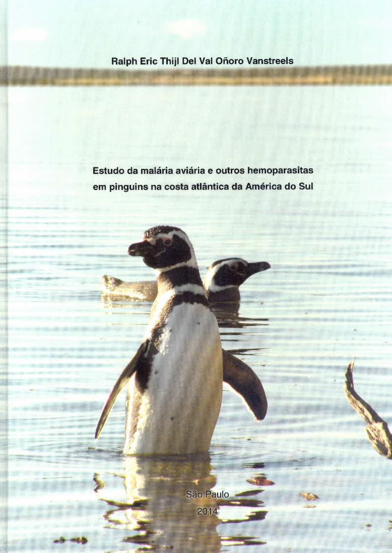

RALPH ERIC THIJL DEL VAL OÑORO VANSTREELS
Estudo da malária aviária e outros hemoparasitas em pinguins na costa atlântica da América do Sul
Tese apresentada ao Programa de
Pós-Graduação em Patologia
Experimental e Comparada da
Faculdade de Medicina Veterinária e
Zootecnia da Universidade de São
Paulo para obtenção do título de
Doutor em Ciências
Departamento: Patologia
Área de Concentração: Patologia Experimental e Comparada
Orientador: Prof. Dr. José Luiz Catão-Dias
São Paulo
2014
Autorizo a reprodução parcial ou total desta obra, para fins acadêmicos, desde que citada a fonte.
DADOS INTERNACIONAIS DE CATALOGAÇÃO-NA-PUBLICAÇÃO
(Biblioteca Virginie Buff D’Ápice da Faculdade de Medicina Veterinária e Zootecnia da Universidade de São Paulo)
T.2955 Vanstreels, Ralph Eric Thijl Del Val Oñoro FMVZ Estudo da malária aviária e outros hemoparasitas em pinguins na costa atlântica da
América do Sul / Ralph Eric Thijl Del Val Oñoro Vanstreels. -- 2014. 250 f. : il. \
Tese (Doutorado) - Universidade de São Paulo. Faculdade de Medicina Veterinária e Zootecnia. Departamento de Patologia, São Paulo, 2014.
Programa de Pós-Graduação: Patologia Experimental e Comparada. Área de concentração: Patologia Experimental e Comparada. Orientador: Prof. Dr. José Luiz Catão Dias. 1. Epidemiologia. 2. Patologia. 3. Plasmodium. 4. Conservação. 5. Reabilitação. I. Título.


FOLHA DE AVALIAÇÃO Nome: VANSTREELS, Ralph Eric Thijl Del Val Oñoro
Título: Estudo da malária aviária e outros hemoparasitas em pinguins na costa atlântica
da América do Sul
Tese apresentada ao Programa de Pós-Graduação em Patologia Experimental e Comparada da Faculdade de Medicina Veterinária e Zootecnia da Universidade de São Paulo para obtenção do título de Doutor em Ciências
Data: ____/____/_____
Banca examinadora
Prof. Dr.:
Instituição: Julgamento:
Prof. Dr.:
Instituição: Julgamento:
Prof. Dr.:
Instituição: Julgamento:
Prof. Dr.:
Instituição: Julgamento:
Prof. Dr.:
Instituição: Julgamento:
Dedico este trabajo a mi querida abuelita,
Angeles Oñoro Orozco,
de quien tengo orgullo de compartir sangre y espíritu.
AGRADECIMENTOS ACKNOWLEDGEMENTS
Aos pinguins, por suportarem todos os incômodos que lhes causei com um número relativamente pequeno de bicadas e arranhões. Espero que meus esforços os ajudem a continuarem sua brava luta pela sobrevivência e que seu maravilhoso ambiente marinho persevere à nossa passagem pelo planeta. E aos pinguins que eutanasiei e aos que não pude ajudar, que descansem em paz e me inspirem para que possa ajudar os que ficaram. À minha família, amigos e amigas que me acompanham na jornada do doutorado e da vida. Em especial, à minha mãe Mary, minha irmã Chris, minha abuelita Angeles, as eternas mulheres da minha vida, e ao meu cunhado e amigo Celso. Ao meu pai Frans, que tanto me ensinou e continua me ensinando. À minha querida Renata Hurtado, sempre companheira e amada, que fez meus dias de doutorado mais felizes e inspirados e me ajudou e apoiou sempre nas horas mais críticas! E aos meus amigos Luis Sansoni e ao Luiz Panigassi que me acompanharam de perto e me apoiaram nessa longa jornada. Ao Prof. Catão, por ser uma pessoa fantástica e o melhor orientador que eu poderia pedir. Por tolerar minhas excentricidades, me orientar nos momentos mais confusos e estar ao meu lado. É uma alegria e uma honra ter sido teu orientado. Obrigado por me aceitar e me orientar, nos sentidos mais plenos destas palavras! À Valeria Ruoppolo, minha eterna co-orientadora e amiga, que nunca hesitou em me abrir todas as portas para que eu crescesse e aprendesse. Tudo de bom que alcancei ou alcançarei com os pinguins, será sempre consequência direta ou indireta do seu apoio e de minha admiração pela sua personalidade e competência. Aos meus ídolos e professores, Andréa Adornes e Rodolfo Silva-Filho, pela amizade, pelas conversas sinceras e pelo apoio nos momentos mais difíceis. Por me ensinarem o que é o verdadeiro espírito da reabilitação de pinguins, e me mostrarem que com competência, seriedade e amor pelo trabalho é que se ajuda os animais. À Profa. Sabrina Epiphanio e às amigas Luana Ortolan, Michelle Sercundes e Silvia Portugal por me guiarem pelo mundo da biologia molecular e me ensinarem tantas coisas sobre malária dos mamíferos e que me deram muitos insights sobre a malária aviária nos pinguins. Trabalhar com biologia molecular não apenas requer profundo conhecimento e experiência, mas também uma fé inabalável na ciência!

À Profa. Érika Braga e aos amigos Francisco Ferreira e Nayara Belo pela ajuda e aprendizado nos mistérios do sequenciamento genético e pelas aventuras na identificação morfológica dos esfregaços. Acredito que ainda teremos um longo caminho juntos no estudo desses parasitas tão complicados! À Profa. Eliana Faquim e às amigas Camila Bizelli, Jéssica Costa, Marina Atzinger e Sandriana Ramos pela ajuda imensa e essencial no mundo da imunologia, por encararem comigo esse desafio e emprestarem seu conhecimento e sua energia para ajudarmos essas aves. Foi uma parceria de mundo tão afastados que nos mostrou como podemos colher frutos maravilhosos quando trabalhamos juntos! Aos Profs. Marcos Amaku e José Grisi-Filho pelo apoio nas análises dos dados, por me ajudarem a encontrar um caminho seguro no mundo estatístico nos momentos de incerteza. Aos amigos do Laboratório de Patologia Comparada de Animais Selvagens (LAPCOM): Profa. Eliana Matushima, Alexander Genoy-Puerto, Alice Oliveira, Angélica Sarmiento, Camila Molina, Carlos Sacristan, Catia Dejuste, Fabíola Prioste, Gustavo Bauer, Juliana Marigo, Kátia Groch, Marcelo Carvalho, Marina Bueno, Marina César, Omar Gonzalez-Viera, Pedro Oliveira, Renata Santos, Rosely Di Chiacchio, Samira Costa, Sávio Sant'Anna, Silmara Rossi, Stefanie Santos, Ticiana Zwarg, Thatiana Sanches e todos os visitantes, estagiários e colaboradores. E, em especial, ao Jorge Oyakawa por toda a ajuda e por impedir que implodíssemos o laboratório semanalmente! Pelas suas ajudas e companhia, sempre tivemos um ambiente de amizade e companheirismo que foi essencial para minha evolução pessoal e profissional! Aos amigos do Centro de Recuperação de Animais Marinhos (CRAM-FURG): Alice Meirelles-Leite, Aryse Martins, Lauro Barcellos, Paula Canabarro, Pedro Bruno-Filho, Roberta Petitet, Silvia Gastal, Vanessa Pedroso e todos os estagiários e voluntários. Tudo o que aprendi com vocês e todas as ajudas e colaborações não se pode pagar, mas prometo que me esforçarei ao máximo! Obrigado pela amizade e pelo apoio integral, me sinto honrado pela oportunidade de ter conhecido e aprendido tanto com vocês. A Cristiane Kolesnikovas e aos amigos da Associação R3 Animal e do CETAS Florianópolis, em especial Ariana Fernandes, Patrícia Serafini e todos os estagiários e voluntários. A colaboração de vocês foi muito importante para aprendermos juntos, e sou um grande admirador da luta de vocês pela reabilitação da fauna silvestre. A luta de vocês é difícil mas belíssima, e vocês têm meu apoio sempre que precisarem!
Aos amigos e parceiros da Aiuká, em especial Claudia Nascimento, Gelza Soares, Juliana Saviolli, Leo Francini, Paulo Valobra e Pedro Renato Gonçalves. Parabéns pelo trabalho de excelência e obrigado pela valiosa colaboração pela conservação dos pinguins, espero que possamos continuar juntos por muitos anos mais! Aos amigos dos Aquários de Guarujá, Santos, São Paulo, Peruíbe e Parque Sabina, em especial a Bruna Schwarz, Cristiane Lassálvia, Gustavo Dutra, Jéssica Ribeiro, Laura Ippólito, Laura Reisfeld, Pryscilla Maracini, Raphael Ramos e Thiago Nascimento. Vocês são prova de que, através do esforço pessoal e da competência, o cativeiro pode se envolver e contribuir à conservação da natureza. Aos amigos do Instituto de Pesquisa e Reabilitação de Animais Marinhos (IPRAM), em especial a Elaine Cruz, Laila Medeiros, Luis Felipe Mayorga, Renata Bhering e Tainan Oliveira. Agradeço as colaborações e desejo muita sorte aos seus esforços em ajudar os pinguins! Aos amigos da Universidade do Vale do Rio dos Sinos (UNISINOS), da Universidade Federal do Rio de Janeiro (UFRJ), da Universidade Estadual do Rio de Janeiro (UERJ) e da Wildlife Conservation Society, pela colaboração no estudo dos pinguins através do Programa Antártico Brasileiro (PROANTAR), em especial Ana Olívia Reis, Erli Costa, Flavia Miranda, Maria Virgina Petry, Roberta Piuco e Victor Valiati. Que este seja apenas o começo numa longa série de colaborações para o estudo e conservação dos pinguins antárticos! Às amigas da Universidade Federal do Rio Grande (FURG), Ângela Cabana e Melissa Xavier, pelas colaborações na eterna missão de desvendar os mistérios da aspergilose. Continuemos com a batalha e quem sabe um dia venceremos esse fungo tão teimoso e possamos ajudar outros tantos pinguins. A las amigas de Wildlife Conservation Society, Marcela Uhart y Virginia Rago, que me recibieron tan bien en nuestras colaboraciones y a quien aprendí a admirar muchísimo. Congratulaciones por vuestra competencia y calidad del trabajo, es un honor y una alegría trabajar con ustedes! A los amigos de la Fundación Mundo Marino, David Verón, Félix Capellino, Gastón Delgado, Julio Loureiro y Sérgio Heredia, por me trataren como un hermano y me recibieren como si estuviera en mi casa. Mucha suerte y seguimos luchando por estos bichos!
To Dr. Eric Woehler for welcoming me to Tasmania and giving me the opportunity to participate in some of your outstanding research and conservation efforts. I look forward to continue learning from you for many years to come! To all the friends in Tasmania and mainland Australia who have been so kind and made me feel at home even at the other side of the world! I am hugely thankful to the friends and colleagues at the University of Tasmania (Cecilia Villanueva, Mark Hindell, Natalie Bool, Perviz Marker, Stewart Nicol), Phillip Island Nature Park (Andre Chiaradia, Annett Finger, Leanne Renwick, Paula Wasiak, Peter Dann), New South Wales Office of Environment and Heritage (David Pridell, Nicholas Carlile), South Bruny Nature Park (Alena Hrasky, Peter Lingard), and to Caitlin Vertigan and Jason Jones. And, in particular I am hugely thankful to Peter Vertigan: I cannot overstate what an incredible person you are, thank you so much for all the help! To the friends at the Southern African Foundation for the Conservation of Coastal Birds (SANCCOB): Margaret Roestorf, Nola Parsons, Venessa Strauss and all the staff members, interns and volunteers. You have taught me so much and encouraged me to believe that well-intentioned people can unite and work together regardless how wide the oceans that separate them! To the friends at International Fund for Animal Welfare and International Bird Rescue, in particular Barbara Callahan and Jay Holcomb. I deeply admire your competence and history, and am hugely thankful for the opportunities and lessons you have given me! Aos amigos Claudio Arroyo e Luciano Bugalho do Laboratório de Histopatologia da FMVZ-USP pelos inúmeros galhos quebrados, ajudas e aprendizado! À Fundação de Amparo à Pesquisa do Estado de São Paulo (FAPESP) pela bolsa de doutorado direto (BP.DD 2009/53956-9) e pelo auxílio à pesquisa (AP.BTA.TEM 2010/51801-5), e à Coordenação de Aperfeiçoamento de Pessoal de Nível Superior (CAPES) pela bolsa de doutorado sanduíche no exterior (BEX 12505/12-9). À Profa. Margareth Capurro-Guimarães e à Ticiana Zwarg por cederem os controles positivos utilizados neste trabalho. E, por fim, aos inúmeros colegas e amigos que me ajudaram direta ou indiretamente a aprender sobre os pinguins e seus parasitas e a conduzir meu projeto de pesquisa. Pequenos conselhos e conversas aqui e ali fazem toda a diferença, e foram a origem de algumas das melhores ideias e do maior encorajamento para levar adiante esse trabalho!

“I have often had the impression that, to penguins, man is just another penguin...
different, less predictable, occasionally violent, but tolerable company when he sits still and minds his own business.”
- Bernard Stonehouse

RESUMO
VANSTREELS, R. E. T. D. V. O. Estudo da malária aviária e outros hemoparasitas em pinguins na costa atlântica da América do Sul. [Investigation of avian malaria and other blood parasites in penguins along the Atlantic coast of South America]. 2014. 250 f. Tese (Doutorado em Ciências) – Faculdade de Medicina Veterinária e Zootecnia, Universidade de São Paulo, São Paulo, 2014.
Embora não existam colônias reprodutivas de pinguins na costa do Brasil, o país é uma
importante área de invernada para o pinguim-de-Magalhães (Spheniscus magellanicus),
uma espécie nativa do sul da América do Sul. Quando encontradas debilitadas em
praias brasileiras, estas aves são comumente levadas a centros de reabilitação
especializados para receber cuidados veterinários e, posteriormente, serem liberadas à
natureza. Durante esta permanência em reabilitação, no entanto, enfermidades
infecciosas como a malária aviária podem ser importantes limitantes à recuperação
destas aves. A malária aviária é uma enfermidade causada por protozoários do gênero
Plasmodium (Apicomplexa: Haemosporida), veiculados às aves por meio da picada de
mosquitos. Enquanto estes parasitas são relativamente pouco patogênicos para a
maioria das espécies aviárias, algumas aves como os pinguins são excepcionalmente
mais suscetíveis a estes patógenos, podendo constituir uma significativa ameaça à sua
conservação. O presente estudo investiga a ocorrência de Plasmodium spp. e outros
hemoparasitas em pinguins-de-Magalhães em centros de reabilitação na costa
brasileira, assim como outras espécies de pinguins em vida livre nas Ilhas South
Shetland. Métodos diagnósticos morfológicos (esfregaços sanguíneos, histopatologia) e
moleculares (reação em cadeia de polimerase aninhada, sequenciamento genético)
foram utilizados para estudar pinguins reabilitados em diferentes instituições em seis
estados do Brasil entre 1999 e 2013. Um surto de malária aviária particularmente
relevante foi estudado em detalhes em um centro de reabilitação em Florianópolis, SC,
tendo sido demonstrado o envolvimento de três diferentes linhagens de Plasmodium
spp. em um único evento epizoótico, com elevada morbidade e mortalidade. Além disto,
a ocorrência de Plasmodium spp. foi documentada em pinguins-de-Magalhães em
centros de reabilitação ao longo de grande parte da costa brasileira, do extremo sul do
Rio Grande do Sul à Bahia, com uma prevalência estimada entre 6.6% e 13.5%. Estas

infecções apresentam marcante sazonalidade, incidindo exclusivamente nos meses
mais quentes do ano (outubro a abril), e podem envolver uma grande variedade de
linhagens de Plasmodium spp., algumas das quais nunca haviam sido reportadas em
pinguins como P. cathemerium, P. nucleophilum e P. tejerai. Em contraste não foram
encontrados hemoparasitas em pinguins amostrados nas Ilhas South Shetland, um
achado consistente com estudos anteriores. À parte destas investigações
epidemiológicas, foi realizada uma extensa revisão e compilação dos aspectos da
literatura científica acerca desta enfermidade e outras hemoparasitoses em pinguins
buscando estabelecer um panorama mais claro acerca da sua distribuição geográfica e
implicações epidemiológicas e para a conservação. Com base nisto, torna-se possível
desenvolver uma discussão crítica do atual estado da arte e apontar as atuais lacunas
de conhecimento que possam direcionar estudos futuros. Em suma, a malária aviária é
uma enfermidade relevante para as mais diversas espécies de pinguins em todo o
mundo, e possui particular importância para a reabilitação e conservação destas aves
na América do Sul.
Palavras-chave: Epidemiologia. Patologia. Plasmodium. Conservação. Reabilitação.
ABSTRACT
VANSTREELS, R. E. T. D. V. O. Investigation of avian malaria and other blood parasites in penguins along the Atlantic coast of South America. [Estudo da malária aviária e outros hemoparasitas em pinguins na costa atlântica da América do Sul]. 2014. 250 f. Tese (Doutorado em Ciências) – Faculdade de Medicina Veterinária e Zootecnia, Universidade de São Paulo, São Paulo, 2014.
Despite no penguin breeding colonies along the Brazilian coast, the country is an
important wintering area for the Magellanic penguin (Spheniscus magellanicus), a
species native to the south of South America. When Magellanic penguins are found
alive ashore on Brazilian beaches, they are taken to rehabilitation centers to receive
veterinary care and then are released back into the wild. However, while in
rehabilitation, infectious diseases such as avian malaria may become important limiting
factors for the recovery of these birds. Avian malaria is a disease caused by protozoa of
the genus Plasmodium (Apicomplexa: Haemosporida), which are transmitted to birds
through mosquitoes. While these parasites are relatively non-pathogenic for most avian
species, some birds such as penguins are exceptionally susceptible, such that
Plasmodium poses a significant conservation threat. This study investigates the
occurrence of Plasmodium spp. and other blood parasites in Magellanic penguins at
rehabilitation centers along the coast of Brazil, and in other species of penguins at the
South Shetland Islands, South Atlantic Ocean. In Brazil, a combination of morphological
(blood smears, histopathology) and molecular (nested polymerase chain reaction, gene
sequencing) diagnostic methods were employed to investigate the presence of
haemoparasites in penguins undergoing rehabilitation in six states between 1999 and
2013. A particularly significant avian malaria outbreak was studied in detail at a
rehabilitation center in Florianópolis, SC, where the involvement of three distinct
Plasmodium spp. in a single epizootic event was demonstrated, with resultant high
morbidity and mortality. The occurrence of Plasmodium spp. was documented in
Magellanic penguins at rehabilitation centers along most of the Brazilian coast, from
southernmost Rio Grande do Sul to Bahia, with an estimated prevalence between 6.6%
and 13.5% of captive penguins. These infections were markedly seasonal, with the
incidences exclusively restricted to the warmer months of the year (October to April),

and involving a broad variety of Plasmodium spp. lineages, some of which had not yet
been reported in penguins, such as P. cathemerium, P. nucleophilum and P. tejerai. In
contrast, no blood parasites were detected in the penguins sampled at the South
Shetland Islands, a finding that is consistent with previous studies. Aside from these
epidemiological investigations, an extensive revision and compilation of the scientific
literature was conducted for this disease and other penguins’ haemosporidioses, aiming
to establish an integrated understanding of their geographic distribution and
epidemiological and conservation implications. On this basis, it is possible to critically
examine the state of the art and identify knowledge gaps that can be addressed in future
studies. Avian malaria is a significant disease and conservation threat for most penguin
species throughout the world, and has particular importance for the rehabilitation and
conservation of penguins in South America.
Key-words: Epidemiology. Pathology. Plasmodium. Conservation. Rehabilitation.
LISTA DE FIGURAS
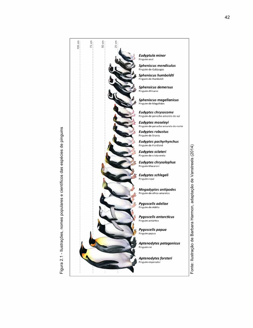
Figura 2.1 - Ilustrações, nomes populares e científicos das espécies de pinguins 42
Figura 2.2 - Distribuição geográfica das colônias reprodutivas de pinguins 45 Figura 2.3 - Diagrama do ciclo anual de vida do pinguim-de-Magalhães
(Spheniscus magellanicus) 46 Figura 2.4 - Fases do ciclo anual de vida do pinguim-de-Magalhães
(Spheniscus magellanicus) 48 Figura 2.5 - Características oceanográficas do mar Patagônico 50 Figura 2.6 - Distribuição geográfica das áreas de reprodução e alimentação
do pinguim-de-Magalhães (Spheniscus magellanicus) 51 Figura 3.1 - Ciclo de vida de Plasmodium spp. que infectam aves 70 Figura 3.2 - Ciclo de vida de Leucocytozoon spp. 71 Figura 3.3 - Insetos hematófagos que atuam como hospedeiros de
hemosporidianos aviários 73 Figura 3.4 - Distribuição do número de espécies registradas de
hemosporidianos nas diversas ordens aviárias 74 Figura 3.5 - Distribuição do número de espécies registradas de
hemosporidianos aviários, e respectivas prevalências médias, em função das regiões biogeográficas 77
Figura 3.6 - Diagrama conceitual da evolução da parasitemia na prima-
infecção plasmódica de uma ave suscetível 80 Figura 3.7 - Lesões palpebrais associadas a picadas de mosquitos em
pinguim-de-Magalhães (Spheniscus magellanicus) 84 Figura 3.8 - Achados necroscópicos típicos de malária aviária em pinguins 85 Figura 3.9 - Refringência dos grânulos de hemozoína à microscopia de luz
polarizada 87

Figura 3.10 - Plasmodium spp. em esfregaços sanguíneos delgados de pinguim-de-Magalhães (Spheniscus magellanicus) (Giemsa) 88
Figura 3.11 - Haemoproteus sp em esfregaço sanguíneo delgado de
corujinha-do-mato (Megascops choliba) (Rosenfeld) 88 Figura 3.12 - Leucocytozoon tawaki em esfregaço sanguíneo delgado de
pinguim-Africano (Spheniscus demersus) (Diff-Quick) 89 Figura 3.13 - Exemplos de artefatos e sujidades que podem assemelhar-se a
hemoparasitas em esfregaços sanguíneos delgados 90 Figura 3.14 - Babesia peircei em esfregaço sanguíneo delgado de pinguim-
Africano (Spheniscus demersus) (Diff-Quick) 90 Figura 3.15 - Meronte tecidual de Plasmodium sp em corte histológico de
baço de pinguim-de-Magalhães (Spheniscus magellanicus) (Hematoxilina-Eosina) 92
Figura 3.16 - Merontes teciduais de Plasmodium sp em decalque tecidual de
rim de pinguim-Africano (Spheniscus demersus) (Diff-Quick) 93 Figura 3.17 - Instalação de telas anti-mosquito para a prevenção da malária
aviária em espécies altamente suscetíveis 97 Figura 3.18 - Uso de repelentes de mosquito tipo roll-on aplicados à cabeça
de pinguins como estratégia para prevenir a malária aviária 99

LISTA DE FIGURAS EM LINGUA INGLESA
Figure 4.1 - Host distribution of intracellular blood parasites among penguin species 112
Figure 4.2 - Geographic distribution of records of intracellular blood
parasites in penguins (A) and of the sampling effort of studies investigating blood parasites in wild penguins (B). Blue areas correspond to the distribution of penguin breeding colonies 113
Figure 4.3 - Geographic distribution of records of intracellular blood
parasites in penguins in relation to their confirmed or presumed invertebrate hosts. Blue areas correspond to the distribution of penguin breeding colonies 114
Figure 4.4 - Latitudinal distribution of breeding penguins (gray bars, lower
axis) and penguins sampling effort of studies investigating blood parasites in wild penguins through different diagnostic methods (colored bars, upper axis) 135
Figure 5.1 - Maximum likelihood phylogenetic tree of the mitochondrial
cytochrome b gene of the studied hemosporidian lineages. Lineages identified in this study are emphasized in blue. When available, information on the morphospecies observed on the corresponding blood smear is provided. Branch lengths are drawn proportionally to evolutionary distance (scale bar is shown). Lower bootstrap values (< 50) are omitted 179
Figure 5.2 - Blood parasites in Giemsa-stained blood smears from
Magellanic penguins (Spheniscus magellanicus) 181 Figure 5.3 - Tissue meronts of Plasmodium (Haemamoeba) tejerai in
tissues of Magellanic penguins (Spheniscus magellanicus). Hematoxilin-Eosin, penguin 584 183
Figure 6.1 - Geographic distribution of the sampling effort, detection and
lineages of Plasmodium spp. in Magellanic penguins (Spheniscus magellanicus) undergoing rehabilitation along the coast of Brazil. Pie charts represent sampling effort (size) and percentage of positive results (red fraction). Blue areas represent the breeding (light blue) and historical foraging (darker blue) distribution of Magellanic penguins 203

Figure 6.2 - Monthly distribution of the incidence of Plasmodium infections in comparison to the number of penguins undergoing rehabilitation 208
Figure 6.3 - Phylogenetic tree of the Plasmodium spp. lineages identified in
Magellanic penguins undergoing rehabilitation along the coast of Brazil (red), published penguin-infecting lineages (blue) and reference lineages. Branch lengths are drawn proportionally to the amount of changes (scale bar is shown). Bootstrap values lower than 20 are omitted 213
Figure 6.4 - Histological findings associated with avian malaria in Magellanic
penguins (Spheniscus magellanicus). Hematoxilin-Eosin. Scale bars = 15 µm 214

LISTA DE QUADROS
Quadro 2.1 - Distribuição geográfica, população estimada, tendência demográfica e estado de conservação das espécies de pinguins 43
Quadro 3.1 - Sumário comparativo dos gêneros de hemosporidianos aviários 65 Quadro 3.2 - Lista de espécies de hemosporidianos aviários registrados na
região Neotropical 78 Quadro 3.3 - Protocolos terapêuticos para hemosporidioses aviárias 102
LISTA DE QUADROS EM LINGUA INGLESA
Table 5.1 - Individual history and diagnostic results for the studied Magellanic penguins (Spheniscus magellanicus). Taxonomic names within brackets indicate the taxon to which the species is presumed to correspond on the basis of phylogenetic analyses. Asterisks indicate individuals that died during the outbreak 172
Table 5.2 - Estimates of evolutionary distance (% expected base
substitutions per site) of cytochrome b mitochondrial gene sequences of hemosporidians identified in penguins in this study (1-6) and the literature (7-10), and reference lineages from the MalAvi database (11-19). Values lower than 4.0 are highlighted 180
Table 5.3 - Hematological results and quantification of blood parasites in
blood smears (Mean ± S.D.) 184 Table 6.1 - Sample sizes examined using different diagnostic tests to
screen for Plasmodium sp infections. Values within brackets indicate the number of positive samples. “†” indicates that sample collection was systematic, i.e. was not conducted in a manner that would favor sick or healthy individuals. “‡” indicates the collection of samples from penguins were rehabilitated at other facilities then transferred to Rio Grande do Sul but samples were collected upon arrival from transference 204
Table 6.2 - Details of the diagnostic results in relation to sample collection
and testing strategy, age group, oiling and survival 210 Table 7.1 - Differential leukocyte counts (Mean ± S.D.) for penguins at
Demay Point (DP), Keller Peninsula (KP) and Stinker Point (SP), January-February 2011 237

LISTA DE ABREVIATURAS E SIGLAS
ANOVA análise de variância (analysis of variance)
APs pinguins-africano (African penguins)
CETAS Centro de Triagem de Animais Silvestres
CRAM-FURG Centro de Recuperação de Animais Marinhos da Universidade
Federal do Rio Grande
DNA ácido desoxirribonucleico
EELC células jovens da linhagem eritrocítica (early erythrocytic lineage
cells)
ELISA ensaio imunoenzimático
FUNDAMAR Fundação Museu de História Pesquisa e Arqueologia do Mar
HLR Razão heterófilo-linfócito (heterophil-to-lymphocyte ratio)
HP histopatologia (histopathology)
Ig imunoglobulina
IPRAM Instituto de Pesquisa e Reabilitação de Animais Marinhos
Is. Ilha (Island)
MPs pinguins-de-Magalhães (Magellanic penguins)
NZ Nova Zelândia
PCR reação em cadeia da polimerase
PCV hematócrito (packed cell volume)
PROANTAR Programa Antártico Brasileiro
RNA ácido ribonucleico
rRNA ácido ribonucleico ribossomal
SANCCOB Southern African Foundation for the Conservation of Coastal Birds
TBS esfregaço sanguíneo delgado (thin blood smear)
TIS decalque tecidual (tissue impression smear)
SCAR Comitê Científico sobre Pesquisa Antártica (Scientific
Committee on Antarctic Research)

LISTA DE ESPÉCIES
Nome científico Nome popular Nome em língua inglesa
Aptenodytes forsteri Pinguim-imperador Emperor penguin
Aptenodytes patagonicus Pinguim-rei King penguin
Eudyptes chrysocome Pinguim-de-penacho-amarelo-do-Sul Southern rockhopper penguin
Eudyptes chrysolophus Pinguim-Macaroni Macaroni penguin
Eudyptes moseleyi Pinguim-de-penacho-amarelo-do-Norte Northern rockhopper penguin
Eudyptes pachyrhynchus Pinguim-de-Fiordland Fiordland penguin
Eudyptes robustus Pinguim-de-Snares Snares penguin
Eudyptes schlegeli Pinguim-royal Royal penguin
Eudyptes sclateri Pinguim-de-crista-ereta Erect-crested penguin
Eudyptula minor Pinguim-azul Little penguin
Megadyptes antipodes Pinguim-de-olhos-amarelos Yellow-eyed penguin
Pygoscelis adeliae Pinguim-de-Adélia Adélie penguin
Pygoscelis antarcticus Pinguim-antártico Chinstrap penguin
Pygoscelis papua Pinguim-papua Gentoo penguin
Spheniscus demersus Pinguim-africano African penguin
Spheniscus humboldti Pinguim-de-Humboldt Humboldt penguin
Spheniscus magellanicus Pinguim-de-Magalhães Magellanic penguin
Spheniscus mendiculus Pinguim-de-Galápagos Galapagos penguin

SUMÁRIO
1 INTRODUÇÃO GERAL ................................................................................... 33 1.1 APRESENTAÇÃO DOS CAPÍTULOS ............................................................. 35
REFERÊNCIAS ............................................................................................... 37
2 CONSIDERAÇÕES ACERCA DA BIOLOGIA E ECOLOGIA DOS PINGUINS ..... 41 2.1 INTRODUÇÃO ................................................................................................ 41
2.2 DISTRIBUIÇÃO GEOGRÁFICA ...................................................................... 44
2.3 CICLO DE VIDA .............................................................................................. 46
2.4 OCORRÊNCIA NO BRASIL ............................................................................ 59
2.5 ENFERMIDADES RELEVANTES PARA A CONSERVAÇÃO ........................ 53
REFERÊNCIAS ............................................................................................... 54
3 MALÁRIA AVIÁRIA E OUTROS HEMOSPORIDIANOS AVIÁRIOS ............. 64 3.1 INTRODUÇÃO ................................................................................................ 64
3.2 ETIOLOGIA ..................................................................................................... 65
3.3 EPIZOOTIOLOGIA .......................................................................................... 67
3.3.1 Ciclo de vida de Plasmodium e Haemoproteus .......................................... 68 3.3.2 Ciclo de vida de Leucocytozoon .................................................................. 70 3.4 EPIDEMIOLOGIA ............................................................................................ 72
3.4.1 Distribuição nos hospedeiros invertebrados ............................................. 72 3.4.2 Distribuição nas aves hospedeiras ............................................................. 74 3.4.3 Distribuição geográfica, sazonalidade e sincronicidade ........................... 76 3.5 PATOGENIA .................................................................................................... 78
3.6 CLÍNICA E DIAGNÓSTICO ............................................................................. 82
3.6.1 Sinais clínicos ................................................................................................ 83 3.6.2 Achados necroscópicos ............................................................................... 85 3.6.3 Esfregaço delgado ........................................................................................ 86

3.6.4 Histopatologia ................................................................................................ 91 3.6.5 Decalques teciduais ...................................................................................... 93 3.6.6 Diagnóstico molecular .................................................................................. 93 3.6.7 Inoculação experimental ............................................................................... 94 3.6.8 Outras técnicas diagnósticas ....................................................................... 95 3.7 PREVENÇÃO .................................................................................................. 96
3.7.1 Tela anti-mosquitos ....................................................................................... 96 3.7.2 Ventiladores ................................................................................................... 98 3.7.3 Repelentes ..................................................................................................... 98 3.7.4 Profilaxia medicamentosa ............................................................................ 99 3.7.5 Monitoramento com esfregaços delgados ............................................... 101 3.7.6 Vacinação ..................................................................................................... 101 3.8 TRATAMENTO .............................................................................................. 102
REFERÊNCIAS ............................................................................................. 104
4 INTRACELLULAR BLOOD PARASITES OF PENGUINS: A CRITICAL REVIEW AND META-ANALYSIS ................................................................. 110
4.1 ABSTRACT ................................................................................................... 110
4.2 INTRODUCTION ........................................................................................... 110
4.3 BABESIA (BABESIOSIS) ............................................................................. 115
4.3.1 Species recorded in penguins ................................................................... 115 4.3.2 Distribution among penguin hosts ............................................................ 115 4.3.3 Invertebrate hosts and geographic distribution ....................................... 116 4.3.4 Epidemiology and pathology ..................................................................... 116 4.4 HAEMOPROTEUS (HAEMOPROTEOSIS) ................................................. 117
4.4.1 Species recorded in penguins ................................................................... 117 4.4.2 Distribution among penguin hosts ............................................................ 118 4.4.3 Invertebrate hosts and geographic distribution ....................................... 119 4.4.4 Epidemiology and pathology ..................................................................... 119

4.5 LEUCOCYTOZOON (LEUCOCYTOZOONOSIS) ........................................ 120
4.5.1 Species recorded in penguins ................................................................... 120 4.5.2 Distribution among penguin hosts ............................................................ 121 4.5.3 Invertebrate hosts and geographic distribution ....................................... 121 4.5.4 Epidemiology and pathology ..................................................................... 122 4.6 PLASMODIUM (AVIAN MALARIA) .............................................................. 124
4.6.1 Species recorded in penguins ................................................................... 124 4.6.2 Distribution among penguin hosts ............................................................ 126 4.6.3 Invertebrate hosts and geographic distribution ....................................... 127 4.6.4 Epidemiology and pathology ..................................................................... 129 4.7 STUDIES WITH INCONCLUSIVE/QUESTIONABLE RESULTS .................. 133
4.8 CONSIDERATIONS ON STUDY METHODS AND FUTURE STUDIES ....... 135
4.9 IMPLICATIONS FOR CONSERVATION ....................................................... 137
REFERENCES .............................................................................................. 139
APPENDIXES ............................................................................................... 159
5 OUTBREAK OF AVIAN MALARIA ASSOCIATED TO MULTIPLE SPECIES OF PLASMODIUM IN MAGELLANIC PENGUINS UNDERGOING REHABILITATION IN SOUTHERN BRAZIL ...................... 168
5.1 ABSTRACT ................................................................................................... 168
5.2 INTRODUCTION ........................................................................................... 169
5.3 METHODS ..................................................................................................... 170
5.3.1 Ethics statements ........................................................................................ 170 5.3.2 Study population and sample collection ................................................... 170 5.3.3 Laboratory procedures ............................................................................... 173 5.3.4 Cytochrome b amplification and phylogenetic analysis ......................... 173 5.3.5 Statistical analyses ..................................................................................... 175 5.4 RESULTS ...................................................................................................... 176
5.4.1 Clinical signs and necropsy findings ........................................................ 176 5.4.2 Hemosporidian detection ........................................................................... 176 5.4.3 Parasite identification and phylogeny ....................................................... 177
5.4.4 Hematology and pathology ........................................................................ 182 5.5 DISCUSSION ................................................................................................ 185
REFERENCES .............................................................................................. 190
APPENDIXES ............................................................................................... 198
6 EPIDEMIOLOGY AND PATHOLOGY OF AVIAN MALARIA IN MAGELLANIC PENGUINS UNDERGOING REHABILITATION ALONG THE COAST OF BRAZIL .............................................................................. 200
6.1 ABSTRACT ................................................................................................... 200
6.2 INTRODUCTION ........................................................................................... 200
6.3 METHODS ..................................................................................................... 201
6.3.1 Study locations and data collection .......................................................... 201 6.3.2 Study design ................................................................................................ 203 6.3.3 Sample collection, hematology and pathology ........................................ 205 6.3.4 Molecular biology and phylogenetic analysis .......................................... 206 6.3.5 Statistical analysis ...................................................................................... 207 6.4 RESULTS ...................................................................................................... 207
6.4.1 Epidemiology in systematically sampled and PCR-tested individuals .... 209 6.4.2 Plasmodium species and lineages ............................................................ 209 6.4.3 Pathology ..................................................................................................... 211 6.5 DISCUSSION ................................................................................................ 215
6.5.1 Epidemiology of avian malaria in penguins at rehabilitation centers .... 214
6.5.2 Plasmodium lineages infecting penguins and their pathology .............. 217
6.5.3 Concurrent diseases ................................................................................... 219
6.5.4 Implications for rehabilitation and conservation ..................................... 220
REFERENCES .............................................................................................. 222
APPENDIXES ............................................................................................... 227

7 INVESTIGATION OF BLOOD PARASITES OF PYGOSCELID PENGUINS AT THE KING GEORGE AND ELEPHANT ISLANDS, SOUTH SHETLANDS ARCHIPELAGO, ANTARCTICA ........................................... 232
7.1 ABSTRACT ................................................................................................... 232
7.2 INTRODUCTION ........................................................................................... 233
7.3 MATERIALS AND METHODS ....................................................................... 234
7.4 RESULTS ...................................................................................................... 236
7.5 DISCUSSION ................................................................................................ 237
REFERENCES .............................................................................................. 240
8 CONSIDERAÇÕES FINAIS .......................................................................... 244
APÊNDICE .................................................................................................... 247

33
1 INTRODUÇÃO GERAL
Os pinguins são aves únicas. Além de seu particular carisma e seu significado
cultural como símbolos dos ecossistemas polares, os pinguins compõem entre 50 e
80% de toda a biomassa de aves do Oceano Antártico, desempenhando um papel vital
de transferência energética entre os ambientes marinho e terrestre no Hemisfério Sul
(AINLEY, 1985; COOPER; WOEHLER, 1994). Por isto, os pinguins estão intimamente
ligados ao equilíbrio do ambiente marinho e são suscetíveis às alterações ambientais
que nele ocorram, servindo como indicadores precoces de impactos ambientais e
desequilíbrios ecológicos no ambiente marinho (BOERSMA, 2008).
Embora seja residente da Argentina, Ilhas Falklands/Malvinas e Chile e não se
reproduza na costa brasileira, o pinguim-de-Magalhães (Spheniscus magellanicus)
pode ser encontrado às centenas ou milhares todos os anos ao longo do litoral
brasileiro, sobretudo nas regiões Sul e Sudeste durante o inverno (CEMAVE, 2011;
STOKES et al., 2014). Em muitos casos, pinguins enfermos poderão ser resgatados
ainda com vida e encaminhados a centros de reabilitação especializados para
receberem tratamento veterinário e, uma vez recuperados, serem liberados à natureza
(RUOPPOLO et al., 2004a; HEREDIA et al., 2008). Estas iniciativas de reabilitação são
importantes não apenas para mitigar a mortalidade destas aves que está direta ou
indiretamente relacionada aos impactos antrópicos sobre o ambiente marinho
(RUOPPOLO et al., 2004a; GARCÍA-BORBOROGLU et al., 2006, 2010), mas também
são vitais para garantir que estas aves recebam atendimento adequado e eticamente
responsável, além de evitar que o público leigo, em suas tentativas bem intencionadas
de resgatar estes animais carismáticos, seja exposto ao risco de lesões ou
enfermidades zoonóticas (ESTES, 1998). Apesar da frequência com que pinguins
mortos ou enfermos são encontrados na costa brasileira, ainda há relativamente poucos
dados sobre as doenças e as causas de mortalidade destas aves em nosso litoral
(TOURINHO et al., 2010; BRANDÃO et al., 2011). Além disto, durante o período de
permanência nos centros de reabilitação, as enfermidades que podem atingir estas

34
aves e prejudicar sua sobrevivência e reabilitação são numerosas e relativamente
pouco compreendidas (CLARKE; KERRY, 1993; SILVA-FILHO; RUOPPOLO, 2007).
Dentre as enfermidades infecciosas, a malária aviária tem sido considerada uma
das mais preocupantes ameaças para a conservação dos pinguins devido ao seu
desenvolvimento rápido e mortalidade elevada (CLARKE; KERRY, 1993; JONES;
SHELLAM, 1999b; LEVIN; PARKER, 2011). Causada por protozoários Plasmodium
spp. veiculados por mosquitos, a malária aviária é relativamente assintomática para a
maioria das aves, tornando-se porém significativamente patogênica em espécies
aviárias que não co-evoluíram com estes hemoparasitas, como os pinguins
(ATKINSON; VAN RIPER, 1991; VALKIŪNAS, 2005; ATKINSON, 2008).
Surtos de malária aviária são um problema recorrente em pinguins mantidos em
zoológicos, aquários e centros de reabilitação no Brasil e em todo o mundo (RODHAIN,
1939; GRINER; SHERIDAN, 1967; BAK et al., 1984; PENRITH et al., 1994; BUENO et
al., 2010). Em natureza, a infecção plasmódica foi identificada em pinguins no
Arquipélago de Galápagos, Ilha Gough, África do Sul e Nova Zelândia (FANTHAM;
PORTER, 1944; LAIRD, 1950; LEVIN et al., 2009). Embora tenha sido registrada em
centros de reabilitação na Argentina e no Chile (CARVAJAL; ALVARADO, 2009;
CAPELLINO et al., 2013), não está claro se a infecção plasmódica ocorre em pinguins-
de-Magalhães em natureza (JOVANI et al., 2001; QUILLFELDT et al., 2010). Assim,
quando esta doença é registrada em pinguins em reabilitação no litoral brasileiro
(RUOPPOLO et al., 2004b; MEIRELLES-LEITE et al., 2008; BALDASSIN et al., 2013),
não se sabe se a infecção é resultante da exposição a mosquitos no curto período de
cativeiro, ou se os animais já adentram ao centro de reabilitação infectados.
Considerando as lacunas de conhecimento sobre a malária aviária em pinguins
na América do Sul e a particular relevância desta enfermidade para a medicina e a
conservação destas aves, a presente tese se propõe a investigar e discutir aspectos
epidemiológicos e patológicos da malária aviária em pinguins, em especial no contexto
dos centros de reabilitação brasileiros.
35
1.1 APRESENTAÇÃO DOS CAPÍTULOS
Esta tese está organizada em oito capítulos, incluindo este capítulo introdutório.
Os capítulos 3 a 7 estão redigidos e formatados de forma compatível com a publicação
científica, sendo que os capítulos 4 a 7 estão em língua inglesa para publicação em
revistas científicas internacionais.
O Capítulo 2 apresenta uma breve revisão de literatura acerca da biologia e
ecologia de pinguins, com ênfase no pinguim-de-Magalhães. O objetivo não é oferecer
uma revisão completa do atual estado de conhecimento, mas sim uma abordagem
panorâmica das características biológicas e ecológicas destas aves que serão
pertinentes para a interpretação dos capítulos subsequentes.
O Capítulo 3 faz uma revisão geral da parasitologia, epidemiologia e clínica das
hemosporidioses aviárias. O texto objetiva uma abordagem direta e prática, voltada à
consulta de médicos veterinários atuando no cuidado clínico de aves em cativeiro ou
em reabilitação no Brasil, com ênfase especial em pinguins. Este capítulo foi submetido
para publicação, com coautoria da Dra. Nola J. Parsons, na forma de um capítulo da
segunda edição do livro “Tratado de animais selvagens – medicina veterinária”, editado
por Zalmir Silvino Cubas, Silva, Jean Carlos Ramos Silva e José Luiz Catão-Dias.
O Capítulo 4 apresenta uma compilação, revisão e meta-análise dos parasitas
intracelulares encontrados no sangue de pinguins, com a discussão crítica da validade
dos registros publicados na literatura, da distribuição geográfica destes parasitas, das
necessidades e prioridades para estudos futuros e das implicações para a conservação
destas aves. Este capítulo foi preparado com a perspectiva de submissão na forma de
uma revisão de literatura em duas partes, a serem encaminhadas às revistas científicas
Polar Biology e Journal of Wildlife Diseases com coautoria do Prof. José Luiz Catão-
Dias.
O Capítulo 5 investiga um surto de malária aviária em pinguins-de-Magalhães
em um centro de reabilitação no sul do Brasil, combinando abordagens clínicas,
hematológicas, biomoleculares e histopatológicas para investigar a epidemiologia e
patologia da infecção por Plasmodium spp. em pinguins durante um surto de elevada

36
morbidade e mortalidade. Este capítulo é apresentado no formato final no qual foi aceito
para publicação pela revista científica PLOS One, com co-autoria de Cristiane K. M.
Kolesnikovas, Sandro Sandri, Patrícia Silveira, Nayara O. Belo, Francisco C. Ferreira
Junior, Sabrina Epiphanio, Mário Steindel, Érika M. Braga e José Luiz Catão-Dias.
O Capítulo 6 apresenta um amplo estudo da ocorrência da malária aviária e
outras hemoparasitoses em pinguins-de-Magalhães em reabilitação em diversas
instituições ao longo da costa brasileira, buscando trazer uma nova compreensão sobre
a epidemiologia e patologia desta enfermidade e das suas consequências para a
reabilitação destas aves no país. Este capítulo foi preparado com a perspectiva de
submissão à revista científica Emerging Infectious Diseases, com coautoria de Rodolfo
Pinho da Silva-Filho, Cristiane K. M. Kolesnikovas, Luis Felipe S. P. Mayorga, Valeria
Ruoppolo, Sabrina Epiphanio, Marcos Amaku, Érika M. Braga e José Luiz Catão-Dias.
O Capítulo 7 relata a investigação de hemoparasitas em pinguins em ilhas
subantárticas do Arquipélago South Shetlands, próximo à Península Antártica, além de
apresentar resultados hematológicos e de ectoparasitas. Este capítulo é apresentado
no formato final no qual foi aceito para publicação pela revista científica Polar Biology,
com coautoria de Flavia R. Miranda, Valeria Ruoppolo, Ana Olívia de Almeida Reis, Erli
Schneider Costa, Adriana Rodrigues de Lira Pessôa, João Paulo Machado Torres,
Larissa Schmauder Teixeira da Cunha, Roberta da Cruz Piuco, Victor Hugo Valiati,
Daniel González-Acuña, Marcelo B. Labruna, Maria Virginia Petry, Sabrina Epiphanio e
José Luiz Catão-Dias.
O Capítulo 8 encerra esta tese com uma breve discussão das conclusões que
que resultam dos estudos apresentados nos capítulos anteriores, além de agregar os
apêndices relevantes para uma interpretação mais aprofundada dos métodos e
resultados laboratoriais.

37
REFERÊNCIAS AINLEY, D. G. Biomass of birds and mammals in the Ross Sea. In: SIEGFRIED, W.; CONDY, P. R.; LAWS, R. M. Antarctic nutrient cycles and food webs. Heidelberg: Springer, 1985. p. 498-515. ATKINSON, C. T.; LAPOINTE, D. A. Introduced avian diseases, climate change, and the future of Hawaiian Honeycreepers. Journal of Avian Medicine and Surgery, v. 23, n. 1, p. 53-63, 2009. ATKINSON, C. T. Avian Malaria. In: ATKINSON, C. T.; THOMAS, N. J.; HUNTER, D. B. Parasitic diseases of wild birds. Ames: Wiley-Blackwell, 2008. p. 35-53. BAK, Ung-Bok; PARK, Jae-Chan; LIM, Young-Jae. An outbreak of malaria in penguins at the Farm-land Zoo. Korean Journal of Parasitology, v. 22, n. 2, p. 267-272, 1984. BALDASSIN, P.; WERNECK, M. R.; TORRES, F.; CAMPOS, S. E.; ALMOSNY, N. Hemoparasite (Plasmodium spp.) in Spheniscus magellanicus, a case report. In: INTERNATIONAL PENGUIN CONFERENCE, 8; 2013, Bristol. Proceedings… Bristol: IPC, 2013. p. 72. BOERSMA, P. D. Penguins as marine sentinels. BioScience, v. 58, n. 7, p. 597-607, 2008. BRANDÃO, M. L.; BRAGA, K. M.; LUQUE, J. L. Marine debris ingestion by Magellanic penguins, Spheniscus magellanicus (Aves: Sphenisciformes), from the Brazilian coastal zone. Marine Pollution Bulletin, v. 62, p. 2246-2249, 2011. BUENO, M. G.; LOPEZ, R. P. G.; MENEZES, R. M. T.; COSTA-NASCIMENTO, M. J.; LIMA, G. F. M. C.; ARAÚJO, R. A. S.; GUIDA, F. J. V.; KIRCHGATTER, K. Identification of Plasmodium relictum causing mortality in penguins (Spheniscus magellanicus) from São Paulo Zoo, Brazil. Veterinary Parasitology, v. 173, n. 1-2, p. 123-127, 2010. CAPELLINO, F.; VANSTREELS, R. E. T.; RODRÍGUEZ-HEREDIA, S. A.; LOUREIRO, J.; CATÃO-DIAS, J. L. Avian malaria (Plasmodium sp) in Magellanic Penguins at Fundación Mundo Marino (San Clemente del Tuyú, Argentina). In: INTERNATIONAL PENGUIN CONFERENCE, 8; 2013, Bristol. Proceedings… Bristol: IPC, 2013. p. 161.
38
CARVAJAL, E. R.; ALVARADO, P. M. Pesquisa de Plasmodium spp. en pingüinos de Magallanes (Spheniscus magellanicus) de la Región de los Ríos: Malaria aviar como nueva patología de interés en la avifauna local. Boletín Veterinario Oficial, v. 10, p. 1-4, 2009. CEMAVE. CENTRO NACIONAL DE PESQUISA E CONSERVAÇÃO DE AVES SILVESTRES. Projeto Nacional de Monitoramento do Pinguim-de-Magalhães Spheniscus magellanicus. Cabedelo: CEMAVE, 2011. 34 p. CLARKE, J. R.; KERRY, K. R. Diseases and parasites of penguins. Korean Journal of Polar Research, v. 4, n. 2, p. 79-96, 1993. COOPER, J.; WOEHLER, E. J. Consumption of Antarctic krill (Euphasia superba) by seabirds during summer in the Prydz Bay region, Antarctica. In: EL-SAYED, S. Z. Southern Ocean Ecology: the BIOMASS perspective. Cambridge: Cambridge University Press, 1984. p. 247-260. ESTES, J. A. Concerns about rehabilitation of oiled wildlife. Conservation Biology, v. 12, n. 5, p. 1156-1157, 1998. FANTHAM, H. B.; PORTER, A. On a Plasmodium (Plasmodium relictum var. spheniscidae, n. var.), observed in four species of penguins. Proceedings of the Zoological Society of London, v. 114, p. 279-292, 1944. GARCÍA-BORBOROGLU, P.; BOERSMA, P. D.; RUOPPOLO, V.; REYES, L.; REBSTOCK, G. A.; GRIOT, K.; HEREDIA, S. R.; ADORNES, A. C.; SILVA-FILHO, R. P. Chronic oil pollution harms Magellanic penguins in the Southwest Atlantic. Marine Pollution Bulletin, v. 52, p. 193-198, 2006. GARCÍA-BORBOROGLU, P.; BOERSMA, P. D.; RUOPPOLO, V.; SILVA-FILHO, R. P.; ADORNES, A. C.; SENA, D. C.; VELOZO, R.; KOLESNIKOVAS, C. M.; DUTRA, G.; MARACINI, P.; NASCIMENTO, C. C.; RAMOS-JÚNIOR, V.; BARBOSA, L.; SERRA, S. Magellanic penguin mortality in 2008 along the SW Atlantic coast. Marine Pollution Bulletin, v. 60, n. 10, p. 1652-1657, 2010. GRINER, L. A.; SHERIDAN, B. W. Malaria (Plasmodium relictum) in penguins at the San Diego Zoo. Veterinary Clinical Pathology, v. 1, p. 7-17, 1967.

39
HEREDIA, S. A. R.; ALVAREZ, C. K.; LOUREIRO, J. Aves marinas empetroladas: Guía para su manejo y atención. San Clemente del Tuyú: Fundación Mundo Marino, 2008. 138 p. JONES, H. I.; SHELLAM, G. R. Blood parasites in penguins, and their potential impact on conservation. Marine Ornithology, v. 27, p. 181-184, 1999. JOVANI, R.; TELLA, J. L.; FORERO, M. G.; BERTELLOTTI, M.; BLANCO, G.; CEBALLOS, O.; DONÁZAR, J. A. Apparent absence of blood parasites in the Patagonian seabird community: is it related to the marine environment? Waterbirds, v. 24, n. 3, p. 430-433, 2001. LAIRD, M. Some blood parasites of New Zealand. Zoological Publications of Victoria University College, v. 5, p. 1-20, 1950. LEVIN, I. I.; PARKER, P. G. Hemosporidian parasites: impacts on avian hosts. In: MILLER, E.; FOWLER, M. Fowler’s zoo and wild animals medicine. Missouri: Elsevier Saunders, 2011. p. 356-363. LEVIN, I. I.; OUTLAW, D. C.; VARGAS, F. H.; PARKER, P. G. Plasmodium blood parasite found in endangered Galapagos penguins (Spheniscus mendiculus). Biological Conservation, v. 142, p. 3191-3195, 2009. MEIRELLES-LEITE, A. T.; XAVIER, M. O.; CABANA, A. L.; SILVA-FILHO, R. P. Principais doenças infecciosas em pinguins-de-Magalhães (Spheniscus magellanicus) em centro de reabilitação. In: CONGRESO LATINOAMERICANO DE REHABILITACIÓN DE FAUNA MARINA, 1; 2008, San Clemente del Tuyú. Proceedings… San Clemente del Tuyú: CLARFM, 2008. p. 71-72. PENRITH, M. L.; HUCHZERMEYER, F. W.; WET, S. C.; PENRITH, M. J. Concurrent infection with Clostridium and Plasmodium in a captive king penguin Aptenodytes patagonicus. Avian Pathology, v. 23, n. 2, p. 373-380, 1994. QUILLFELDT, P.; MARTÍNEZ, J.; HENNICKE, J.; LUDYNIA, K.; GLADBACH, A.; MASELLO, J. F.; RIOU, S.; MERINO, S. Hemosporidian blood parasites in seabirds: a comparative genetic study from Antartic to tropical habitats. Naturwissenschaften, v. 97, p. 809-817, 2010.

40
RODHAIN, J. L’infection a Plasmodium relictum chez les pingouins. Annales de Parasitologie, v. 17, n. 2, p. 139-157, 1939. RUOPPOLO, V.; ADORNES, A. C.; NASCIMENTO, A. C.; SILVA-FILHO, R. P. Reabilitação de pinguins afetados por petróleo. Clínica Veterinária, v. 9, n. 51, p. 78-83, 2004a. RUOPPOLO, V.; SILVA-FILHO, R. P.; ADORNES, A. C.; CATÃO-DIAS, J. L. Occurrence of Malaria in Magellanic Penguins (Spheniscus magellanicus) in a rehabilitation center in Southern Brazil. In: INTERNATIONAL PENGUIN CONFERENCE, 5; 2004, Ushuaia. Proceedings… Ushuaya: IPC, 2004b. SILVA-FILHO, R. P.; RUOPPOLO, V. Sphenisciformes. In: CUBAS, Z. S.; SILVA, J. C. R.; CATÃO-DIAS, J. L. Tratado de animais selvagens – medicina veterinária. São Paulo: Roca, 2007. p. 309-323. STOKES, D. L.; BOERSMA, P. D.; CASENAVE, J. L.; GARCÍA-BORBOROGLU, P. Conservation of migratory Magellanic penguins requires marine zoning. Biological Conservation, v. 170, p. 151-161, 2014. TOURINHO, P. S.; SUL, J. A. I.; FILLMANN, G. Is marine debris ingestion still a problem for the coastal marine biota of southern Brazil? Marine Pollution Bulletin, v. 60, p. 396-401, 2010. VALKIŪNAS, G. Avian malaria parasites and other haemosporidia. Boca Ratón: CRC Press, 2005. 932 p.

41
2 CONSIDERAÇÕES ACERCA DA BIOLOGIA E ECOLOGIA DOS PINGUINS 2.1 INTRODUÇÃO
É certo que, muito antes do desenvolvimento da escrita, os pinguins já eram
conhecidos por populações nativas da América do Sul, África e Oceania (MÜLLER-
SCHWARZE, 1984; KLOKLER et al., 2010). O primeiro relato documentado sobre estas
aves aparece nos registros da primeira expedição do português Vasco da Gama, na
primavera de 1498, em que é descrita a observação de “aves grandes como patos, que
não voam por não terem penas nas asas, e que zurram como asnos” no sul do
continente africano, referindo-se a pinguins-africanos (Spheniscus demersus) (KOPKE;
PAIVA, 1838). A primeira descrição científica de espécies de pinguins seria feita pelo
sueco Carolus Linnaeus em 1758 na décima edição do “Systema Naturae”, que
classificou o pinguim-africano no mesmo gênero que os albatrozes gigantes (Aves:
Procellariiformes), nomeando-o Diomedea demersa, e agrupou o pinguim-de-penacho-
amarelo-do-norte (Eudyptes moseleyi) aos rabos-de-palha (Aves: Phaethontiformes),
dando-lhe o nome de Phaethon demersus (LINNAEUS, 1758).
Os pinguins são os únicos representantes da ordem Sphenisciformes, sendo
atualmente classificados em 18 espécies, das quais 13 são consideradas ameaçadas
de extinção em algum grau (Figura 2.1, Quadro 2.1) (GARCÍA-BORBOROGLU;
BOERSMA, 2013; IUCN, 2014). Filogeneticamente, os pinguins são mais próximos dos
Procellariiformes, a ordem que compreende os petréis, albatrozes, fulmares, pardelas e
painhos, e acredita-se que tenham evoluído a partir de pequenas aves voadoras
capazes de nado e mergulho (SIMPSON, 1946; HACKETT et al., 2008; KSEPKA;
CLARKE, 2010).
42
Figu
ra 2
.1 -
Ilust
raçõ
es, n
omes
pop
ular
es e
cie
ntífi
cos
das
espé
cies
de
ping
uins
Fo
nte:
Ilus
traçã
o de
Bar
bara
Har
mon
, ada
ptaç
ão d
e V
anst
reel
s (2
014)

43
Qua
dro
2.1
- Dis
tribu
ição
geo
gráf
ica,
pop
ulaç
ão e
stim
ada,
tend
ênci
a de
mog
ráfic
a e
esta
do d
e co
nser
vaçã
o da
s es
péci
es d
e pi
ngui
ns
Fo
nte:
Will
iam
s (1
995)
, Kse
pka
et a
l. (2
006)
, Fre
twel
l et a
l. (2
012)
, Gar
cía-
Bor
boro
glu
e B
oers
ma
(201
3) e
IUC
N (2
014)
, ada
ptaç
ão d
e V
anst
reel
s (2
014)

44
As ameaças mais importantes à conservação destas aves são a depleção de
suas presas devido à sobrepesca, a contaminação dos oceanos por derivados do
petróleo e a perturbação ou destruição de seus habitats reprodutivos. Adicionalmente, a
captura incidental em redes de pesca, as colisões com embarcações, a poluição
marinha, as algas produtoras de biotoxinas, as enfermidades infecciosas, a caça
predatória e numerosos outros fatores também ameaçam estas aves (BOERSMA;
STOKES, 1995; GARCÍA-BORBOROGLU; BOERSMA, 2013).
2.2 DISTRIBUIÇÃO GEOGRÁFICA
Os pinguins distribuem-se exclusivamente no Hemisfério Sul, com colônias
reprodutivas no continente antártico, em ilhas subantárticas, na América do Sul, África,
Austrália e Nova Zelândia (Quadro 2.1 e Figura 2.2). Ao contrário da percepção pública
mais comum, apenas duas espécies podem ser consideradas verdadeiramente
antárticas: o pinguim-imperador (Aptenodytes forsteri) e o pinguim-de-Adélia
(Pygoscelis adeliae). A ampla maioria dos pinguins está distribuída em ambientes
temperados ou subantárticos, e inclusive há espécies que habitam ambientes tropicais
como os pinguim-de-Galápagos (Spheniscus mendiculus) e o pinguim-de-Humboldt
(Spheniscus humboldti) (WILLIAMS, 1995; ANCEL et al., 2013; GARCÍA-
BORBOROGLU; BOERSMA, 2013).
Embora tenham o comportamento de agrupar-se durante a estação reprodutiva,
nem todas as espécies de pinguins permanecem na proximidade de suas colônias
reprodutivas durante o restante do ano. Há espécies sedentárias, que permanecem nas
colônias reprodutivas durante todo o ano, como os pinguins-azuis (Eudyptula minor) ou
os pinguins-africanos; espécies migratórias, que retornam às colônias apenas no
período reprodutivo e permanecem o restante do ano no ambiente marinho, como os
pinguins-de-Magalhães; e espécies semi-migratórias, que permanecem nas colônias
por períodos irregulares, como os pinguins-rei (Aptenodytes patagonicus) (CLAUSEN;
PÜTZ, 2003; PÜTZ et al., 2007; GARCÍA-BORBOROGLU et al., 2013).

45
Durante as incursões ao mar para a alimentação ou invernada os pinguins
podem percorrer centenas a milhares de quilômetros em mar aberto, vagando em
função da disponibilidade de presas, e muitas vezes concentrando-se em áreas de
convergência, divergência ou ressurgência de correntes marinhas, como as
convergências subtropical e subantártica e a divergência polar (CLAUSEN; PÜTZ,
2003; PÜTZ et al., 2000, 2006, 2007; WILSON et al., 2005; BOERSMA et al., 2009).
Figura 2.2 - Distribuição geográfica das colônias reprodutivas de pinguins
Fonte: Williams (1995), IUCN (2014), DSEWPC (2012), Fretwell et al. (2012) e García-
Borboroglu e Boersma (2013), adaptação de Vanstreels (2014)
Chatham
Bounty e Antipodes
Macquarie Auckland e Campbell
Balleny e Scott
AUSTRÁLIA
Thurston
Heard e McDonald
Amsterdam e Saint Paul
Kerguelen
Crozet
Prince Edward e Marion Bouvet
Snares
Falkland/Malvinas
South Georgia
South Sandwich
South Orkney
South Shetland
Galápagos
Tristan da Cunha
Gough
AMÉR
ICA
DO S
UL
ÁFRICA
ANTÁRTIDA
Nova Zelândia Sul
Nova Zelândia Norte
Colônias)reprodu/vas)de)pinguins)
Divergência)antár/ca)
Convergência)antár/ca)
Convergência)subtropical)
46
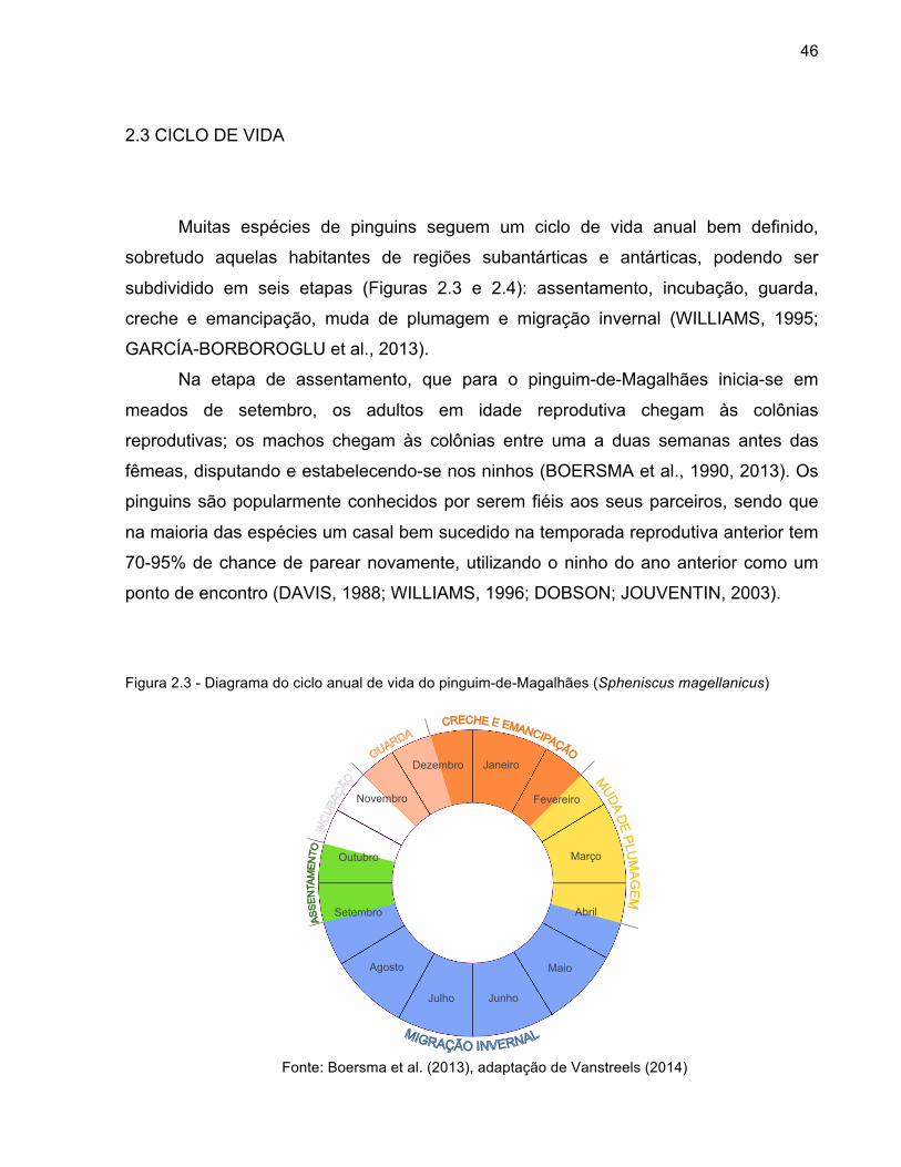
2.3 CICLO DE VIDA
Muitas espécies de pinguins seguem um ciclo de vida anual bem definido,
sobretudo aquelas habitantes de regiões subantárticas e antárticas, podendo ser
subdividido em seis etapas (Figuras 2.3 e 2.4): assentamento, incubação, guarda,
creche e emancipação, muda de plumagem e migração invernal (WILLIAMS, 1995;
GARCÍA-BORBOROGLU et al., 2013).
Na etapa de assentamento, que para o pinguim-de-Magalhães inicia-se em
meados de setembro, os adultos em idade reprodutiva chegam às colônias
reprodutivas; os machos chegam às colônias entre uma a duas semanas antes das
fêmeas, disputando e estabelecendo-se nos ninhos (BOERSMA et al., 1990, 2013). Os
pinguins são popularmente conhecidos por serem fiéis aos seus parceiros, sendo que
na maioria das espécies um casal bem sucedido na temporada reprodutiva anterior tem
70-95% de chance de parear novamente, utilizando o ninho do ano anterior como um
ponto de encontro (DAVIS, 1988; WILLIAMS, 1996; DOBSON; JOUVENTIN, 2003).
Figura 2.3 - Diagrama do ciclo anual de vida do pinguim-de-Magalhães (Spheniscus magellanicus)
Fonte: Boersma et al. (2013), adaptação de Vanstreels (2014)
Janeiro
Fevereiro
Março
Abril
Maio
Junho Julho
Agosto
Setembro
Outubro
Novembro
Dezembro

47
Após um breve período de corte e cópula ocorrerá a oviposição, dando início à
etapa de incubação. Pinguins produzem dois ovos por temporada reprodutiva, com a
exceção dos pinguins-imperadores e reis em que um único ovo é posto por temporada.
A incubação dos ovos é feita em turnos alternados, sendo 3 a 6 turnos com duração de
5-15 dias cada, em que um dos pais permanece no ninho incubando os ovos enquanto
o outro vai ao mar para alimentar-se; a exceção são os pinguins-imperadores, nos
quais não há turnos de incubação e esta é feita inteiramente pelo macho (WILLIAMS,
1995; CHIARADIA; KERRY, 1999; NUMATA et al., 2000).
A etapa de guarda dos filhotes inicia-se a partir da eclosão dos ovos, e neste
período os filhotes são funcionalmente ectotérmicos, dependendo de seus pais para
manterem-se aquecidos e também para a proteção contra eventuais predadores.
Durante a guarda, macho e fêmea se alternarão em turnos de 2-4 dias para
permanecer no ninho protegendo os filhotes ou se alimentar no mar (TAYLOR, 1986;
CHIARADIA; KERRY, 1999; NUMATA et al., 2000; DUCHAMP et al., 2002).
Com o crescimento dos filhotes e a aquisição da efetiva capacidade de
termorregulação, os filhotes se tornarão capazes de sobreviver por períodos curtos sem
os pais, iniciando-se a etapa de creche. Nessa etapa, os pais partem ao mar em
excursões cada vez mais longas, retornando para regurgitar alimento aos filhotes
periodicamente. Finalmente, quando os filhotes adquirem a plumagem juvenil
impermeável, os pais não retornam mais para alimentá-los e os filhotes buscam o mar
para se alimentarem sozinhos (PETTINGILL, 1960; WILLIAMS, 1995).
Após a reprodução, os pinguins adultos prosseguem à muda pós-nupcial, isto é,
a completa troca de sua plumagem por uma inteiramente nova. Este é um processo
desgastante, pois além do grande consumo energético necessário para a troca da
plumagem por duas a quatro semanas, durante todo o processo os pinguins serão
incapazes de nadar, passando por um jejum prolongado (WILLIAMS et al., 1989;
GAUTHIER-CLERC et al., 2002; WIENECKE et al., 2004; BOURGEON et al., 2007).
Concluída a muda de plumagem, os pinguins terão uma plumagem renovada e
plenamente impermeável e poderão retornar ao mar, iniciando a etapa de migração
invernal. Durante este período, os pinguins buscarão alimentar-se para repor e
acumular reservas corporais de gordura com o objetivo final de maximizar sua

48
reprodução na temporada reprodutiva subsequente (WILLIAMS et al., 1989; CHEREL;
FREBY, 1994; GAUTHIER-CLERC et al., 2002; WILSON et al., 2005).
Figura 2.4 - Fases do ciclo anual de vida do pinguim-de-Magalhães (Spheniscus magellanicus)
Fonte: (a) Michel Gunther / Biosphoto, (b) Mike Bingham, (c) David Hosking / Frank Lane Picture
Agency, (d) Daniel Gomez / SplashdownDirect.com, (e) Valeria Ruoppolo / IFAW, (f) Michael Booth / IFAW
Legenda: (a) assentamento e cópula, (b) incubação, (c) guarda, (d) creche, (e) muda de plumagem, (f) migração invernal

49
2.4 OCORRÊNCIA NO BRASIL
Embora nenhuma espécie de pinguim reproduza-se na costa brasileira, quatro
espécies de pinguins têm ocorrência registrada em território brasileiro: pinguim-de-
Magalhães, pinguim-de-penacho-amarelo-do-sul (Eudyptes chrysocome), pinguim-rei e
pinguim-Macaroni (Eudyptes chrysolophus). Enquanto as demais espécies são apenas
registradas esporadicamente, os pinguins-de-Magalhães estão presentes na costa
brasileira às centenas ou milhares todos os anos (SICK, 2001; BARQUETE et al., 2006;
CBRO, 2011). Assim como ocorre para as demais espécies de pinguins, a distribuição
geográfica dos pinguins-de-Magalhães está intimamente relacionada à disponibilidade
de suas presas. No caso das populações de pinguins-de-Magalhães residentes na
costa atlântica da América do Sul, a distribuição está determinada, sobretudo, pela
elevada produtividade biológica da convergência Brasil-Malvinas (WILSON et al., 2005;
FALABELLA et al., 2009).
A convergência Brasil-Malvinas corresponde ao encontro das águas frias da
Corrente das Malvinas com as águas quentes da Corrente do Brasil. As águas frias da
Corrente das Malvinas são ricas em nutrientes e, ao se encontrarem com as águas
quentes da Corrente do Brasil, produzem uma combinação de temperaturas e
nutrientes que resulta em um ambiente extremamente favorável para o acelerado
crescimento do fitoplâncton (Figura 2.5) (FALABELLA et al., 2009). Outros fatores que
contribuem à elevada produtividade biológica nesta região são o influxo de águas doces
provenientes de rios e lagoas (sobretudo do Rio da Prata) e a forte pluviosidade
costeira (GORDON, 1989; CIOTTI et al., 1995; PIOLA; MATANO, 2001).
A partir desta elevada produção biológica primária, resulta uma rica teia
alimentar sobre a qual numerosas espécies de peixes, crustáceos e moluscos se
nutrem e dos quais, por sua vez, os pinguins-de-Magalhães se alimentarão. A Figura
2.6 representa a distribuição geográfica das colônias reprodutivas e áreas de
alimentação dos pinguins-de-Magalhães, evidenciando a sobreposição de suas áreas
de reprodução e alimentação com as áreas de alta produtividade primária da
Convergência Brasil-Malvinas.

50
Figura 2.5 - Características oceanográficas do mar Patagônico
Fonte: Falabella et al. (2009), adaptação de Vanstreels (2014) Legenda: (a) representação esquemática das correntes marinhas, (b) temperatura de
superfície do mar, (c) concentração de nitratos, (d) produtividade primária durante a primavera (clorofila-α)
Durante a fase invernal os pinguins-de-Magalhães acompanham o contorno da
plataforma continental, mantendo-se a aproximadamente entre 50 e 200 km da costa,
tipicamente em grupos de 10 a 30 animais (WILLIAMS; BOERSMA, 1995; PÜTZ et al.,
2000, 2007). Em circunstâncias normais, os pinguins-de-Magalhães permanecerão em
c" d"
a" b"
23.7"
"5.1"
32.9"
"0"
°C"
μmol/kg"
22"
"0.2"
mg/m3"
CORRENTE"DO"BRASIL"
CORRENTE"CIRCUMPOLAR"ANTÁRTICA"

51
alto mar durante todo o período de invernada, retornando à terra apenas ao regressar
às áreas de reprodução. No entanto, o encontro de pinguins em praias brasileiras
ocorre com frequência e é conhecido como um fenômeno natural, com o aparecimento
anual de centenas a milhares de animais, principalmente no inverno e primavera
(VOOREN; BRUSQUE, 1999; SICK, 2001; PETRY; FONSECA, 2002; MÄDER et al.,
2010; SCHERER et al., 2011).
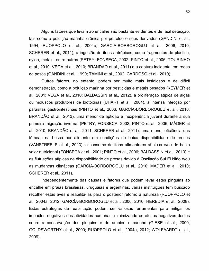
Os fatores que levam ao encalhe – e muitas vezes mortalidade – destas aves na
costa atlântica da América do Sul todavia não estão esclarecidos, e há razões para
considerar que este processo natural esteja sendo amplificado pelos impactos humanos
no ambiente marinho (GARCÍA-BORBOROGLU et al., 2006, 2010; CEMAVE, 2011).
Figura 2.6 - Distribuição geográfica das áreas de reprodução e alimentação do pinguim-de-Magalhães
(Spheniscus magellanicus)
Fonte: Boersma et al. (2013) e Stokes et al. (2014),
adaptação de Vanstreels (2014)
Área%de%reprodução%
Área%de%alimentação%
52
Alguns fatores que levam ao encalhe são bastante evidentes e de fácil detecção,
tais como a poluição marinha crônica por petróleo e seus derivados (GANDINI et al.,
1994; RUOPPOLO et al., 2004a; GARCÍA-BORBOROGLU et al., 2006, 2010;
SCHERER et al., 2011), a ingestão de itens antrópicos, como fragmentos de plástico,
nylon, metais, entre outros (PETRY; FONSECA, 2002; PINTO et al., 2006; TOURINHO
et al., 2010; VEGA et al., 2010; BRANDÃO et al., 2011) e a captura incidental em redes
de pesca (GANDINI et al., 1999; TAMINI et al., 2002; CARDOSO et al., 2010).
Outros fatores, no entanto, podem ser muito mais insidiosos e de difícil
demonstração, como a poluição marinha por pesticidas e metais pesados (KEYMER et
al., 2001; VEGA et al., 2010; BALDASSIN et al., 2012), a proliferação atípica de algas
ou moluscos produtores de biotoxinas (UHART et al., 2004), a intensa infecção por
parasitas gastrointestinais (PINTO et al., 2006; GARCÍA-BORBOROGLU et al., 2010;
BRANDÃO et al., 2013), uma menor de aptidão e inexperiência juvenil durante a sua
primeira migração invernal (PETRY; FONSECA, 2002; PINTO et al., 2006; MÄDER et
al., 2010; BRANDÃO et al., 2011; SCHERER et al., 2011), uma menor eficiência das
fêmeas na busca por alimento em condições de baixa disponibilidade de presas
(VANSTREELS et al., 2013), o consumo de itens alimentares atípicos e/ou de baixo
valor nutricional (FONSECA et al., 2001; PINTO et al., 2006; BALDASSIN et al., 2010) e
as flutuações atípicas de disponibilidade de presas devido à Oscilação Sul El Niño e/ou
às mudanças climáticas (GARCÍA-BORBOROGLU et al., 2010; MÄDER et al., 2010;
SCHERER et al., 2011).
Independentemente das causas e fatores que podem levar estes pinguins ao
encalhe em praias brasileiras, uruguaias e argentinas, várias instituições têm buscado
recolher estas aves e reabilitá-las para o posterior retorno à natureza (RUOPPOLO et
al., 2004a, 2012; GARCÍA-BORBOROGLU et al., 2006, 2010; HEREDIA et al., 2008).
Estas estratégias de reabilitação podem ser valiosas ferramentas para mitigar os
impactos negativos das atividades humanas, minimizando os efeitos negativos destas
sobre a conservação dos pinguins e do ambiente marinho (GIESE et al., 2000;
GOLDSWORTHY et al., 2000; RUOPPOLO et al., 2004a, 2012; WOLFAARDT et al.,
2009).

53
2.5 ENFERMIDADES RELEVANTES PARA A CONSERVAÇÃO
Há numerosos patógenos e enfermidades que podem constituir ameaças
relevantes à conservação dos pinguins, além de prejudicar os esforços de reabilitação
destas aves (CLARKE; KERRY, 1993; DUIGNAN, 2001; KERRY; RIDDLE, 2009).
Dentre as enfermidades de etiologia infecciosa, destacam-se a malária aviária (JONES;
SHELLAM, 1999a) e outras hemoparasitoses (HILL et al., 2010; YABSLEY et al., 2012),
além da aspergilose (OBENDORF; McCOLL, 1980; XAVIER et al., 2007), pasteurelose
(LISLE et al., 1990), poxvirose aviária (KANE et al., 2012; NIEMEYER et al., 2013),
herpesviroses aviárias (KINCAID et al., 1988) e clamidiose (JENCEK et al., 2012). Além
disto, a causa de muitas enfermidades que acometem os pinguins ainda não está
esclarecida, como é o caso da doença da perda das penas (Penguin feather loss
disorder) (KANE et al., 2010), da estomatite diftérica (ALLEY et al., 2004, 2005), e de
vários episódios de mortalidade em massa em natureza (HOCKEN, 2000, 2005;
KEYMER et al., 2001; BARBOSA; PALACIOS, 2009; KERRY; RIDDLE, 2009).
A malária aviária se sobressai dentre as enfermidades infecciosas por seu
histórico comprovado em afetar negativamente a conservação de aves que não co-
evoluíram com o parasita, como demonstrado por seus impactos dramáticos sobre a
conservação das aves nativas do Arquipélago do Havaí (VAN RIPER III et al., 1986;
ATKINSON; LAPOINTE, 2009). Os pinguins são notoriamente suscetíveis a esta
enfermidade, e desenvolvem rápida e elevada mortalidade quando expostos à infecção
por Plasmodium sp em cativeiro (GRINER; SHERIDAN, 1967; STOSKOPF; BEIER,
1979; FIX et al., 1988), e a presença deste patógeno em pinguins de vida livre também
traz grande preocupação acerca do seu potencial de causar surtos de mortalidade com
efeitos significativos para a conservação (BROSSY et al., 1999; JONES; SHELLAM,
1999b; MILLER et al., 2001; LEVIN et al., 2009).

54
REFERÊNCIAS ALLEY, M. R.; MORGAN, K. J.; GILL, J. M.; HOCKEN, A. G. Diseases and causes of mortality in yellow-eyed penguins, Megadyptes antipodes. Kokako, v. 11, n. 2, p. 18-23, 2004. ALLEY, M. R.; GARTRELL, B. D.; MORGAN, K. J. Severe outbreak of diphtheritic stomatitis in yellow-eyed penguins, Megadyptes antipodes. Kokako, v. 12, n. 1, p. 16, 2005. ANCEL, A.; BEAULIEU, M.; GILBERT, C. The different breeding strategies of penguins: a review. Comptes Rendus Biologies, v. 336, p. 1-12, 2013. ATKINSON, C. T.; LAPOINTE, D. A. Introduced avian diseases, climate change, and the future of Hawaiian Honeycreepers. Journal of Avian Medicine and Surgery, v. 23, n. 1, p. 53-63, 2009. BALDASSIN, P.; SANTOS, R. A.; CUNHA, J. M. M.; WERNECK, M. R.; GALLO, H. Cephalopods in the diet of Magellanic penguins Spheniscus magellanicus found on the coast of Brazil. Marine Ornithology, v. 38, p. 55-57, 2010. BALDASSIN, P.; TANIGUCHI, S.; GALLO, H.; SILVA, R. J.; MONTONE, R. C. Persistent organic pollutants in juvenile Magellan penguins (Spheniscus magellanicus) found on the northern shore of the state of São Paulo and southern shore of the state of Rio de Janeiro, Brazil. Marine Pollution Bulletin, v. 64, p. 2502-2506, 2012. BARBOSA, A.; PALACIOS, M. J. Health of Antarctic birds: a review of their parasites, pathogens and diseases. Polar Biology, v. 32, p. 1095-1115, 2009. BARQUETE, V.; BUGONI, L.; SILVA-FILHO, R. P.; ADORNES, A. C. Review of records and notes on king penguin (Aptenodytes patagonicus) and rockhopper penguin (Eudyptes chrysocome) in Brazil. Hornero, v. 21, p. 45-48, 2006. BOERSMA, P. D.; STOKES, D. L.; YORIO, P. M. Reproductive variability and historical change of Magellanic penguins (Spheniscus magellanicus) at Punta Tombo, Argentina. In: DAVIS, L. S.; DARBY, J. T. Penguin Biology. San Diego: Academic Press, 1990. p. 15-43.

55
BOERSMA, P. D.; REBSTOCK, G. A.; FRERE, E.; MOORE, S. Following the fish: penguins and productivity in the South Atlantic. Ecological Monographs, v. 79, n. 1, p. 59-76, 2009. BOURGEON, S.; VIERA, V. M.; RACLOT, T.; GROSCOLAS, R. Hormones and immunoglobulin levels in king penguins during moulting and breeding fasts. Ecoscience, v. 14, n. 4, p. 519-528, 2007. BRANDÃO, M. L.; BRAGA, K. M.; LUQUE, J. L. Marine debris ingestion by Magellanic penguins, Spheniscus magellanicus (Aves: Sphenisciformes), from the Brazilian coastal zone. Systematic Parasitology, v. 85, n. 1, p. 79-98, 2013. BRANDÃO, M. L.; LUQUE, J. L.; SCHOLZ, T.; KOSTADINOVA, J. L. New records and descriptions of digeneans from the Magellanic penguins Spheniscus magellanicus (Forster) (Aves: Sphenisciformes) on the coast of Brazil. Marine Pollution Bulletin, v. 62, p. 2246-2249, 2011. BOERSMA, P. D.; FRERE, E.; KANE, O.; POZZI, L. M.; PÜTZ, K.; RAYA-REY, A.; REBSTOCK, G. A.; SIMEONE, A.; SMITH, J.; VAN BUREN, A.; YORIO, P.; GARCÍA-BORBOROGLU, P. Magellanic penguin (Spheniscus magellanicus). In: GARCÍA-BORBOROGLU, P.; BOERSMA, P. D. Penguins: natural history and conservation. Seattle: Washington University Press, 2013. p. 145-152. BROSSY, J. J.; PLÖS, A. L.; BLACKBEARD, J. M.; KLINE, A. Diseases acquired by captive penguins: what happens when they are released into the wild? Marine Ornithology, v. 27, p. 185-186, 1999. CARDOSO, L. G.; BUGONI, L.; MANCINI, P. L.; HAIMOVICI, M. Gillnet fisheries as a major mortality factor of Magellanic penguins in wintering areas. Marine Pollution Bulletin, v. 62, n. 4, p. 840-844, 2011. CBRO. COMITÊ BRASILEIRO DE REGISTROS ORNITOLÓGICOS. Lista de aves do Brasil, 10a. edição, 2011. Disponível em: <http://www.cbro.org.br>. Acesso em: 12 dez. 2011. CEMAVE. CENTRO NACIONAL DE PESQUISA E CONSERVAÇÃO DE AVES SILVESTRES. Projeto Nacional de Monitoramento do Pinguim-de-Magalhães Spheniscus magellanicus. Cabedelo: CEMAVE, 2011. 34 p.

56
CHEREL, Y.; FREBY, F. Daily body-mass loss and nitrogen excretion during molting fast of Macaroni penguins. Auk, v. 111, n. 2, p. 492-495, 1994. CHIARADIA, A. F.; KERRY, K. R. Daily nest attendance and breeding performance in the little penguin Eudyptula minor at Phillip island, Australia. Marine Ornithology, v. 27, p. 13-20, 1999. CIOTTI, A. M.; ODEBRECHT, C.; FILLMANN, G.; MÖLLER JR, O. O. Freshwater outfollow and subtropical convergence influence on phytoplankton biomass on the southern Brazilian continental shelf. Continental Shelf Research, v. 15, n. 14, p. 1737-1756, 1995. CLARKE, J. R.; KERRY, K. R. Diseases and parasites of penguins. Korean Journal of Polar Research, v. 4, n. 2, p. 79-96, 1993. CLAUSEN, A.; PÜTZ, K. Winter diet and foraging range of gentoo penguins (Pygoscelis papua) from Kidney Cove, Falkland Islands. Polar Biology, v. 26, p. 32-40, 2003. DAVIS, L. S. Coordination of incubation routines and mate choice in Adélie penguins (Pygoscelis adeliae). Auk, v. 105, p. 428-432, 1988. DOBSON, F. S.; JOUVENTIN, P. Use of the nest site as a rendezvous in penguins. Waterbirds, v. 26, n. 4, p. 409-415, 2003. DUCHAMP, C.; ROUANET, J. L.; BARRÉ, H. Ontogeny of thermoregulatory mechanisms in king penguin chicks (Aptenodytes patagonicus). Comparative biochemistry and Physiology Part A, v. 131, p. 765-773, 2002. DUIGNAN, P. J. Diseases of penguins. Surveillance, v. 28, n. 4, p. 5-11, 2001. DSEWPC. DEPARTMENT OF SUSTAINABILITY, ENVIRONMENT, WATER, POPULATION AND COMMUNITIES. Species profile and threats database: Eudyptula minor — little penguin. 2012. Disponível em: <http://www.environment. gov.au/cgi-bin/sprat/public/publicspecies.pl?taxon_id=1085>. Acesso em: 12 dez. 2012. FALABELLA, V.; CAMPAGNA, C.; CROXALL, J. Atlas of the Patagonian sea: species and spaces. Buenos Aires: Wildlife Conservation Society, BirdLife International, 2009. 304 p.

57
FIX, A. S.; WATERHOUSE, C.; GREINER, E. C.; STOSKOPF, M. K. Plasmodium relictum as a cause of avian malaria in wild-caught Magellanic penguins (Spheniscus magellanicus). Journal of Wildlife Diseases, v. 24, n. 4, p. 610-619, 1988. FONSECA, V. S. S.; PETRY, M. V.; JOST, A. H. Diet of the Magellanic penguin on the coast of Rio Grande do Sul, Brazil. Waterbirds, v. 24, n. 2, p. 290-293, 2001. FRETWELL, P. T.; LARUE, M. A.; MORIN, P.; KOOYMAN, G. L.; WIENECKE, B.; RATCLIFFE, N.; FOX, A. J.; FLEMING, A. H.; PORTER, C.; TRATHAN, P. N. An Emperor penguin population estimate: the first global, synoptic survey of a species from space. PLOS One, v. 7, n. 4, e33751, 2012. GANDINI, P.; BOERSMA, P. D.; FRERE, E.; GANDINI, M.; HOLIK, T.; LICHTSCHEIN, V. Magellanic penguins (Spheniscus magellanicus) affected by chronic petroleum pollution along coast of Chubut, Argentina. Auk, v. 111, n. 1, p. 20-27, 1994. GANDINI, P. A.; FRERE, E.; PETTOVELLO, A. D.; CEDROLA, P. V. Interaction between Magellanic penguins and shrimp fisheries in Patagonia, Argentina. Condor, v. 101, p. 783-789, 1999. GARCÍA-BORBOROGLU, P.; BOERSMA, P. D. Penguins: natural history and conservation. Seattle: Washington University Press, 2013. 328 p. GARCÍA-BORBOROGLU, P.; BOERSMA, P. D.; RUOPPOLO, V.; REYES, L.; REBSTOCK, G. A.; GRIOT, K.; HEREDIA, S. R.; ADORNES, A. C.; SILVA-FILHO, R. P. Chronic oil pollution harms Magellanic penguins in the Southwest Atlantic. Marine Pollution Bulletin, v. 52, p. 193-198, 2006. GARCÍA-BORBOROGLU, P.; BOERSMA, P. D.; RUOPPOLO, V.; SILVA-FILHO, R. P.; ADORNES, A. C.; SENA, D. C.; VELOZO, R.; KOLESNIKOVAS, C. M.; DUTRA, G.; MARACINI, P.; NASCIMENTO, C. C.; RAMOS-JÚNIOR, V.; BARBOSA, L.; SERRA, S. Magellanic penguin mortality in 2008 along the SW Atlantic coast. Marine Pollution Bulletin, v. 60, n. 10, p. 1652-1657, 2010. GIESE, M.; GOLDSWORTHY, S. D.; GALES, R.; BROTHERS, N.; HAMILL, J. Effects of the Iron Baron oil spill on little penguins (Eudyptula minor). III. Breeding success of rehabilitated oiled birds. Wildlife Research, v. 27, p. 583-591, 2000.

58
GOLDSWORTHY, S. D.; GIESE, M.; GALES, R.; BROTHERS, N.; HAMILL, J. Effects of the Iron Baron oil spill on little penguins (Eudyptula minor). II. Post-release survival of rehabilitated oiled birds. Wildlife Research, v. 27, p. 573-582, 2000. GORDON, A. L. Brazil – Marvinas confluence – 1984. Deep-Sea Research, v. 36, n. 3, p. 359-384, 1989. GRINER, L. A.; SHERIDAN, B. W. Malaria (Plasmodium relictum) in penguins at the San Diego Zoo. Veterinary Clinical Pathology, v. 1, p. 7-17, 1967. HACKETT, S. J.; KIMBALL, R. T.; REDDY, S.; BOWIE, R. C. K.; BRAUN, E. L.; BRAUN, M. J.; CHOJNOWSKI, J. L.; COX, W. A.; HAN, Kin-Lan, HARSHMAN, J.; HUDDLESTON, C. J.; MARKS, B. D.; MIGLIA, K. J.; MOORE, W. S.; SHELDON, F. H.; STEADMAN, D. W.; WITT, C. C.; YURI, T. A phylogenomic study of birds reveals their evolutionary history. Science, v. 320, p. 1763-1768, 2008. HILL, A. G.; HOWE, L.; GARTRELL, B. D.; ALLEY, M. R. Prevalence of Leucocytozoon spp, in the endangered yellow-eyed penguin Megadyptes antipodes. Parasitology, v. 137, n. 10, p. 1477-1485, 2010. HOCKEN, A. G. Cause of death in blue penguins (Eudyptula m. minor) in North Otago, New Zealand. New Zealand Journal of Zoology, v. 27, p. 305-309, 2000. HOCKEN, A. G. Necropsy findings in yellow-eyed penguins (Megadyptes antipodes) from Otago, New Zealand. New Zealand Journal of Zoology, v. 32, p. 1-8, 2005. IUCN. INTERNATIONAL UNION FOR CONSERVATION OF NATURE AND NATURAL RESOURCES. IUCN Red List of Threatened Species Version 2013.2. 2014. Disponível em: <http://www.iucnredlist.org>. Acesso em: 20 mar. 2014. JENCEK, J. E.; BEAUFRÈRE, H.; TULLY-JR, T. N.; GARNER, M. M.; BASZLER, T. V. An outbreak of Chlamydophila psittaci in an outdoor colony of Magellanic penguins (Spheniscus magellanicus). Journal of Avian Medicine and Surgery, v. 26, n. 4, p. 225-231, 2012. JONES, H. I.; SHELLAM, G. R. The occurrence of blood-inhabiting protozoa in captive and free-living penguins. Polar Biology, v. 21, p. 5-10, 1999a.

59
JONES, H. I.; SHELLAM, G. R. Blood parasites in penguins, and their potential impact on conservation. Marine Ornithology, v. 27, p. 181-184, 1999b. KANE, O. J.; SMITH, J. R.; BOERSMA, P. D.; PARSONS, N. J.; STRAUSS, V.; GARCÍA-BORBOROGLU, P.; VILLANUEVA, C. Feather-loss disorder in African and Magellanic penguins. Waterbirds, v. 33, n. 3, p. 415-421, 2010. KANE, O. J.; UHART, M. M.; RAGO, V.; PEREDA, A. J.; SMITH, J. R.; BUREN, A. V.; CLARK, J. A.; BOERSMA, P. D. Avian pox in Magellanic penguins (Spheniscus magellanicus). Journal of Wildlife Diseases, v. 48, n. 3, p. 790-794, 2012. KERRY, K. R.; RIDDLE, M. J. Health of Antarctic wildlife: a challenge for science and policy. Heidelberg: Springer, 2009. 470 p. KEYMER, I. F.; MALCOLM, H. M.; HUNT, A.; HORSLEY, D. T. Health evaluation of penguins (Sphenisciformes) following mortality in the Falklands (South Atlantic). Diseases of Aquatic Organisms, v. 45, p. 159-169, 2001. KINCAID, A. L.; BUNTON, T. E.; CRANFIELD, M. Herpesvirus-like infection in Black-footed penguins (Spheniscus demersus). Journal of Wildlife Diseases, v. 24, n. 1, p. 173-175, 1988. KLOKLER, D.; VILLAGRÁN, X. S.; GIANNINI, P. C. F.; PEIXOTO, S.; DeBLASIS, P. Juntos na costa: zooarqueologia e geoarqueologia de sambaquis do litoral sul catarinense. Revista do Museu de Arqueologia e Etnologia, v. 20, p. 53-75, 2010. KOPKE, D.; PAIVA, A. C. Roteiro da viagem que em descobrimento da India pelo Cabo da Boa Esperança fez Dom Vasco da Gama em 1497. Porto: Typographia Commercial Portuense, 1838. 183 p. KSEPKA, D. T.; CLARKE, J. A. The basal penguin (Aves: Sphenisciformes) Perudyptes devriesi and a phylogenetic evaluation of the penguin fossil record. Bulletin of the American Museum of Natural History, v. 337, n. 1, p. 1-77, 2010. KSEPKA, D. T.; BERTELLI, S.; GIANNINI, N. P. The phylogeny of the living and fossil Sphenisciformes (penguins). Cladistics, v. 22, p. 412-441, 2006.

60
LEVIN, I. I.; OUTLAW, D. C.; VARGAS, F. H.; PARKER, P. G. Plasmodium blood parasite found in endangered Galapagos penguins (Spheniscus mendiculus). Biological Conservation, v. 142, p. 3191-3195, 2009. LINNAEUS, C. Systema Naturae per regna tria naturae: secundum classes, ordines, genera, species, cum characteribus, differentiis, synonymis, locis. Stockholm: Laurentius Salvius, 1758. 824 p. LISLE, G. W.; STANISLAWEK, W. L.; MOORS, P. J. Pasteurella multocida infections in rockhopper penguins (Eudyptes chrysocome) from Campbell Island, New Zealand. Journal of Wildlife Diseases, v. 26, n. 2, p. 283-285, 1990. MÄDER, A.; SANDER, M.; CASA-JR, G. Ciclo sazonal de mortalidade do pinguim-de-Magalhães, Spheniscus magellanicus, influenciado por fatores antrópicos e climáticos na costa do Rio Grande do Sul, Brasil. Revista Brasileira de Ornitologia, v. 18, n. 3, p. 228-233, 2010. MÜLLER-SCHWARZE, D. The behaviour of penguins: adapted to ice and tropics. Albany: State University of New York Press, 1984. 193 p. NIEMEYER, C.; FAVERO, C. M.; KOLESNIKOVAS, C. K. M.; BHERING, R. C. C.; BRANDÃO, P.; CATÃO-DIAS, J. L. Two different avipoxviruses associated with pox disease in Magellanic penguins (Spheniscus magellanicus) along the Brazilian coast. Avian Pathology, v. 42, n. 6, p. 546-551, 2013. NUMATA, M.; DAVIS, L. S.; RENNER, M. Prolonged foraging trips and egg desertion in little penguins (Eudyptula minor). New Zealand Journal of Zoology, v. 27, p. 277-289, 2000. OBENDORF, D. L.; McCOLL, K. Mortality in little penguins (Eudyptula minor) along the coast of Victoria, Australia. Journal of Wildlife Diseases, v. 16, n. 2, p. 251-260, 1980. PETTINGILL, O. S. Crèche behaviour and individual recognition in a colony of rockhopper penguins. The Wilson Bulletin, v. 72, n. 3, p. 213-221, 1960. PETRY, M. V.; FONSECA, V. S. S. Effects of human activities in the marine environment on seabirds along the coast of Rio Grande do Sul, Brazil. Ornitologia Neotropical, v. 13, p. 137-142, 2002.

61
PINTO, M. B. L. C.; SICILIANO, S.; DI BENEDITTO, A. P. M. Stomach contents of the Magellanic penguin Spheniscus magellanicus from the northern distribution limit on the atlantic coast of Brazil. Marine Ornithology, v. 35, p. 77-78, 2007. PIOLA, A. R.; MATANO, R. P. The South Atlantic Western boundary currents Brazil/Falkland (Malvinas) currents. In: STEELE, J. M.; THORPE, S. A.; TUREKIAN, K. K. Encyclopedia of Ocean Sciences. Waltham: Academic Press, 2001. p. 340-349. PÜTZ, K.; INGHAM, R. J.; SMITH, J. G. Satellite tracking of the winter migration of Magellanic Penguins Spheniscus magellanicus breeding in the Falkland Islands. Ibis, v. 142, p. 614-622, 2000. PÜTZ, K.; REY, A. R.; SCHIAVINI, A.; CLAUSEN, A. P.; LÜTHI, B. H. Winter migration of rockhopper penguins (Eudyptes c. chrysocome) breeding in the Southwest Atlantic: is utilisation of different foraging areas reflected in opposing population trends? Polar Biology, v. 29, p. 735-744, 2006. PÜTZ, K.; SCHIAVINI, A.; REY, A. R.; LÜTHI, B. H. Winter migration of Magellanic penguins (Spheniscus magellanicus) from the southernmost distributional range. Marine Biology, v. 152, p. 1227-1235, 2007. RUOPPOLO, V.; ADORNES, A. C.; NASCIMENTO, A. C.; SILVA-FILHO, R. P. Reabilitação de pinguins afetados por petróleo. Clínica Veterinária, v. 9, n. 51, p. 78-83, 2004. RUOPPOLO, V.; VANSTREELS, R. E. T.; WOEHLER, E. J.; HEREDIA, S. A. R.; ADORNES, A. C.; SILVA-FILHO, R. P.; MATUS, R.; POLESCHI, C.; GRIOT, K.; KOLESNIKOVAS, C. K. M.; SERAFINI, P. Survival and movements of Magellanic penguins rehabilitated from oil fouling along the coast of South America, 2000–2010. Marine Pollution Bulletin, v. 64, p. 1309-1317, 2012. SCHERER, J. F. M.; SCHERER, A. L.; PETRY, M. V. Ocorrênia de carcaças de aves marinhas no litoral do Rio Grande do Sul, Brasil. Revista Brasileira de Ornitologia, v. 19, n. 4, p. 505-513, 2011. SICK, H. Ornitologia brasileira. Rio de Janeiro: Nova Fronteira, 2001. 912 p. SIMPSON, G. G. Fossil penguins. Bulletin of the American Museum of Natural History, v. 87, p. 1-100, 1946.

62
STOKES, D. L.; BOERSMA, P. D.; CASENAVE, J. L.; GARCÍA-BORBOROGLU, P. Conservation of migratory Magellanic penguins requires marine zoning. Biological Conservation, v. 170, p. 151-161, 2014. STOSKOPF, M. K.; BEIER, J. Avian malaria in African black-footed penguins. Journal of the American Veterinary Medical Association, v. 175, n. 9, p. 944-947, 1979. TAMINI, L. L.; PEREZ, J. E.; CHIARAMONTE, G. E.; CAPPOZZO, H. L. Magellanic penguin Spheniscus magellanicus and fish as bycatch in the cornalito Sorgentina incisa fishery at Puerto Quequén, Argentina. Atlantic Seabirds, v. 4, n. 3, p. 109-114, 2002. TAYLOR, J. R. E. Thermal insulation of the down and feathers of pygoscelid penguins chicks and the unique properties of penguin feathers. Auk, v. 103, p. 160-168, 1986. TOURINHO, P. S.; SUL, J. A. I.; FILLMANN, G. Is marine debris ingestion still a problem for the coastal marine biota of southern Brazil? Marine Pollution Bulletin, v. 60, p. 396-401, 2010. UHART, M.; KARESH, W.; COOK, R.; HUIN, N.; LAWRENCE, K.; GUZMAN, L.; LIC, H. P.; PIZARRO, G.; MATTSSON, R.; MÖRNER, T. Paralytic shellfish poisoning in gentoo penguins (Pygoscelis papua) from the Falkland (Malvinas) islands. In: ANNUAL CONFERENCE OF THE AMERICAN ASSOCIATION OF ZOO VETERINARIANS, 37; 2004, San Diego. Proceedings… San Diego: AAZV, 2004. p. 447-482. VAN RIPER III, C.; VAN RIPER, S. G.; GOFF, M. L.; LAIRD, M. The epizootiology and ecological significance of malaria in Hawaiian land birds. Ecological Monographs, v. 56, n. 4, p. 327-344, 1986. VANSTREELS, R. E. T.; ADORNES, A. C.; CANABARRO, P. L.; RUOPPOLO, V.; AMAKU, M.; SILVA-FILHO, R. P.; CATÃO-DIAS, J. L. Female-biased mortality of Magellanic penguins (Spheniscus magellanicus) on the wintering grounds. Emu, v. 113, n. 2, p. 128-134, 2013. VEGA, C. M.; SICILIANO, S.; BARROCAS, P. R. G.; HACON, S. S.; CAMPOS, R. C.; JACOB, S. C.; OTT, P. H. Levels of cadmium, mercury, and lead in Magellanic penguins (Spheniscus magellanicus) stranded on the Brazilian coast. Archives of Environmental Contamination and Toxicology, v. 58, p. 460-468, 2010.

63
VOOREN, C. M.; BRUSQUE, L. F. As aves do ambiente costeiro do Brasil: biodiversidade e conservação. Rio Grande: Fundação Universidade Federal do Rio Grande, 1999. 58 p. WILLIAMS, T. D. The penguins: Spheniscidae. Oxford: Oxford University Press, 1995. 233 p. WILLIAMS, T. D. Mate fidelity in penguins. In: BLACK, J. M.; HULME, M. Partnerships in birds – the study of monogamy. New York: Oxford University Press, 1996. 430 p. WILLIAMS, T. D.; BOERSMA, P. D. Magellanic penguin (Spheniscus magellanicus). In: WILLIAMS, T. D. The penguins: Spheniscidae. Oxford: Oxford University Press, 1995. p. 249-258. WILLIAMS, G.; GHEBREMESKEL, K.; KEYMER, I. F.; HORSLEY, D. T. Plasma α-tocopherol, total lipids and total cholesterol in wild rockhopper, Magellanic and gentoo penguins before and after moulting. The Veterinary Record, v. 124, n. 2, p. 585-586, 1989. WILSON, R. P.; SCOLARO, J. A.; GRÉMILLET, D.; KIERSPEL, M. A. M.; LAURENTI, R. P.; UPTON, J.; GALLELLI, H.; QUINTANA, F.; FRERE, E.; MÜLLER, G.; STRATEN, M. T.; ZIMMER, I. How do Magellanic penguins cope with variability in their access to prey? Ecological Monographs, v. 75, n. 3, p. 379-401, 2005. WOLDFAART, A. C.; WILLIAMS, A. J.; UNDERHILL, L. G.; CRAWFORD, R. J. M.; WHITTINGTON, P. A. Review of the rescue, rehabilitation and restoration of oiled seabirds in South Africa, especially African penguins Spheniscus demersus and Cape gannets Morus capensis, 1983–2005. African Journal of Marine Science, v. 31, n.1, p. 31-54, 2009. XAVIER, M. O.; SOARES, M. P.; MEINERZ, A. R. M.; NOBRE, M. O.; OSÓRIO, L. G.; SILVA-FILHO, R. P.; MEIRELES, M. C. A. Aspergillosis: a limiting factor during recovery of captive Magellanic penguins. Brazilian Journal of Microbiology, v. 38, p. 480-484, 2007. YABSLEY, M. J.; PARSONS, N. J.; HORNE, E. C.; SHOCK, B. C.; PURDEE, M. Novel relapsing fever Borrelia detected in African penguins (Spheniscus demersus) admitted to two rehabilitation centers in South Africa. Parasitology Research, v. 110, n. 3, p. 1125-1130, 2012.

64
3 MALÁRIA AVIÁRIA E OUTROS HEMOSPORIDIANOS AVIÁRIOS 3.1 INTRODUÇÃO
Malária aviária é um termo genérico utilizado para descrever as enfermidades
causadas por uma grande variedade de hemoparasitas que acometem aves. Embora
estes parasitas sejam relacionados àqueles que causam a malária em humanos e
outros mamíferos, os significados clínico e epidemiológico da malária aviária são muito
distintos e demandam o seu estudo separado. O termo “malária aviária” é
tradicionalmente reservado à enfermidade causada pela infecção por protozoários do
gênero Plasmodium, porém outros hemosporidianos como Haemoproteus,
Leucocytozoon e Fallisia são frequentemente agrupados por sua proximidade
taxonômica e suas similaridades epidemiológicas e patológicas (PÉREZ-TRIS et al.,
2005; VALKIŪNAS, 2005).
As infecções por hemosporidianos em aves tipicamente são subclínicas ou
resultam apenas em alterações clínicas discretas mas, em casos particulares ou em
aves altamente suscetíveis, pode levar a quadros clínicos graves e até ao óbito. Há dois
grupos taxonômicos de aves que são particularmente sensíveis à infecção plasmódica:
os pinguins (Spheniscidae) e os “honeycreepers” do Havaí (Fringiliidae: Drepanidinae);
no entanto, casos clínicos severos e surtos epizoóticos também são esporadicamente
relatados em outras aves (VALKIŪNAS, 2005; ATKINSON, 2008b). Dentre as espécies
de aves com ocorrência no Brasil, a malária aviária recebe particular importância entre
os pinguins, em especial o pinguim-de-Magalhães (Spheniscus magellanicus), em que
é considerada uma das enfermidades de maior relevância durante a reabilitação e
manutenção em cativeiro (SILVA-FILHO; RUOPPOLO, 2007).

65
3.2 ETIOLOGIA
Hemosporidianos (Apicomplexa: Aconoidasida: Haemosporida) são um grupo de
protozoários obrigatoriamente heteroxênicos, cujo ciclo de vida envolve vertebrados e
insetos hematófagos (tipicamente, moscas hematófagas e mosquitos). Os
hemoparasitas piroplasmídeos (Babesia, Hepatozoon, Theileria, entre outros) são as
espécies filogeneticamente mais próximas. Há mais de duas centenas de espécies de
hemosporidianos que podem infectar aves, embora este número esteja em constante
rearranjo taxonômico e novas espécies ainda sejam descritas com frequência. Não há
relatos de hemosporidianos aviários sendo transmitidos a espécies não-aviárias, de
modo que se aceita que estes parasitas não apresentam risco zoonótico. Os
hemosporidianos aviários são divididos em quatro gêneros com características
epidemiológicas e patológicas distintas (Quadro 3.1): Haemoproteus (143 spp.),
Plasmodium (60 spp.), Leucocytozoon (36 spp.) e Fallisia (1 sp) (PEIRCE, 2005;
VALKIŪNAS, 2005; ATKINSON, 2008a,b; FORRESTER; GREINER, 2008).
Quadro 3.1 - Sumário comparativo dos gêneros de hemosporidianos aviários
Haemoproteus (Pseudo-malária)
Plasmodium (Malária aviária)
Leucocytozoon (Leucocitozoonose)
No. de espécies acometendo aves 143 60 36 No. de ordens de aves acometidas 19 20 18
Hospedeiro invertebrado Ceratopogonidae, Hippoboscidae Culicidae Ceratopogonidae,
Simuliidae Capacidade de alternar hospedeiros vertebrados Baixa Elevada Intermediária Patogenicidade Baixa - Moderada Baixa - Elevada Baixa - Moderada Merogonia eritrocítica Ausente Presente Ausente Formação de megalomerontes Presente Ausente Presente
O gênero Haemoproteus destaca-se por sua grande diversidade de espécies,
limitada flexibilidade em alternar espécies de hospedeiro, baixa patogenicidade e alta
prevalência. O gênero compreende um grande número de espécies, divididas em dois

66
subgêneros: Haemoproteus e Parahaemoproteus. Os subgênero Haemoproteus possui
poucas espécies (8 spp.), é veiculado por moscas da família Hippoboscidae, e acomete
apenas aves da ordem Columbiformes (pombos e rolinhas) e algumas aves marinhas
(Pelecaniformes e Charadriiformes). O subgênero Parahaemoproteus é mais numeroso
em espécies (137 spp.), é veiculado por moscas da família Ceratopogonidae e acomete
uma grande variedade de aves. Há similaridades morfológicas das formas parasitárias
nos hospedeiros vertebrados, porém os parasitas destes subgêneros são facilmente
distinguíveis quando nos hospedeiros invertebrados (VALKIŪNAS, 2005; ATKINSON,
2008a; LEVIN et al., 2011, 2012).
O gênero Plasmodium apresenta uma diversidade moderada de espécies e uma
grande flexibilidade em alternar espécies de hospedeiro, moderada a severa
patogenicidade e, geralmente, baixa prevalência. Estes parasitas são veiculados por
mosquitos da família Culicidae. Os plasmódios que infectam aves são divididos em
cinco subgêneros (Haemamoeba, Giovannolaia, Novyella, Bennettinia, Huffia). A
morfologia das formas parasitárias no hospedeiro vertebrado é distinta entre os
subgêneros, porém as diferenças epidemiológicas ou patológicas são muito sutis
(VALKIŪNAS, 2005; ATKINSON, 2008b).
O gênero Leucocytozoon possui uma diversidade moderada de espécies, uma
flexibilidade limitada em alternar espécies de hospedeiro, com moderada
patogenicidade e prevalência muito variável. As espécies que acometem as aves são
classificadas em dois subgêneros: Akiba e Leucocytozoon. O subgênero Akiba é
representado por uma única espécie, L. caulleryi, descrita em galinhas domésticas no
sudeste asiático e que se diferencia por ter moscas da família Ceratopogonidae como
hospedeiros invertebrados. O subgênero Leucocytozoon, por outro lado, conta com
uma grande quantidade de espécies (35 spp.) infectando uma ampla variedade de
espécies de aves, veiculadas por moscas da família Simuliidae (VALKIŪNAS, 2005;
FORRESTER; GREINER, 2008).
Por fim, o gênero Fallisia destaca-se por apresentar uma única espécie
acometendo aves, F. neotropicalis (subgênero Plasmodioides). A espécie é
considerada um achado raro, só tendo sido relatada na Venezuela, acometendo
Ciconiiformes e Columbiformes. A morfologia das formas parasitárias em trombócitos,
linfócitos e monócitos das aves é característica e facilmente diferenciada de outros

67
hemoparasitas. Seu hospedeiro invertebrado é desconhecido, especulando-se que seja
um mosquito pertencente à família Culicidae, Aedeomyia squamipennis (GABALDÓN et
al., 1985). Por ser incomum e relativamente pouco conhecido, este parasita não será
discutido extensivamente nesse capítulo, enquanto um maior enfoque será dado aos
demais gêneros.
Há alguma controvérsia acerca da terminologia “malária aviária”. Há autores que
consideram que as enfermidades resultantes das infecções por Plasmodium e
Haemoproteus devem ser denominadas desta forma, enquanto outros argumentam que
apenas moléstias associadas ao Plasmodium deveriam receber tal denominação
(RICKLEFS; FALLON, 2002; PÉREZ-TRIS et al., 2005). No contexto do presente
capítulo, adotaremos a distinção entre “malária aviária” (Plasmodium), “pseudo-malária”
(Haemoproteus) e, ainda, “leucocitozoonose” (Leucocytozoon).
3.3 EPIZOOTIOLOGIA
Nos hemosporidianos, o invertebrado é o hospedeiro definitivo no qual ocorre a
reprodução sexuada do parasita, enquanto a ave é o hospedeiro intermediário,
essencial ao desenvolvimento do parasita, porém no qual não há reprodução sexuada.
Tipicamente a infecção da ave é feita pela inoculação de esporozoítos durante a
hematofagia do hospedeiro invertebrado, porém a inoculação de sangue contendo
merontes eritrocíticos também pode ser uma via de infecção iatrogênica (apenas em
Plasmodium, uma vez que Haemoproteus e Leucocytozoon não possuem merontes
circulantes), podendo ocorrer acidentalmente em procedimentos clínicos ou
intencionalmente em estudos de infecção experimental (VALKIŪNAS, 2005;
ATKINSON, 2008a,b; FORRESTER; GREINER, 2008).

68
3.3.1 Ciclo de vida de Plasmodium e Haemoproteus
O ciclo do gênero Plasmodium será descrito primeiramente (Figura 3.1)
(VALKIŪNAS, 2005; ATKINSON, 2008b), e posteriormente serão destacadas as
variações que ocorrem em Haemoproteus. Durante a alimentação do invertebrado
hematófago, são inoculados esporozoítos (a) na ave. Os esporozoítos invadem células
endoteliais e mononucleares fagocíticas de vários tecidos (em especial baço e pele) (b)
e originam criptozoítos (c) que se desenvolvem por reprodução assexuada, e finalmente
se rompem liberando grandes quantidades de merozoítos (d); este ciclo é denominado
merogonia exoeritrocítica primária. Os merozoítos ganham a circulação sanguínea, e
atingem novas células endoteliais e mononucleares fagocíticas (sobretudo no pulmão,
baço, fígado e medula óssea), onde formam metacriptozoítos (também denominados
merontes teciduais ou esquizontes teciduais) (e), que se desenvolvem por reprodução
assexuada (f) e rompem-se liberando uma grande quantidade de merozoítos (g); este
ciclo é denominado merogonia exoeritrocítica secundária. Vale notar que, ao contrário
da malária humana, não há um estágio hepático em que o parasita se multiplique em
hepatócitos.
Os merozoítos (h) resultantes desta etapa poderão então seguir quatro destinos
distintos, dependendo de fatores endógenos e das condições do hospedeiro. Alguns
podem retornar ao ciclo de merogonia exoeritrocítica secundária. Outros podem invadir
células sanguíneas, tipicamente eritrócitos, desenvolvendo-se em trofozoítos (i) que por
reprodução assexuada transformam-se em merontes eritrocíticos (também
denominados esquizontes eritrocitários) (j), e finalmente rompem-se liberando
moderadas quantidades de merozoítos (k); este ciclo é denominado merogonia
eritrocítica. Outra opção que pode ser seguida pelos merozoítos é invadir células
endoteliais e mononucleares fagocíticas (desta vez, em uma variedade de tecidos) (l), e
desenvolver-se por reprodução assexuada em fanerozoítos (também denominados
merontes teciduais ou esquizontes teciduais) (m) e assim podem permanecer por
períodos prolongados, até que finalmente rompem-se e liberam quantidades muito
grandes de merozoítos (n); este ciclo secundário de reprodução assexuada é
denominado merogonia exoeritrocítica terciária. Finalmente, uma última opção de

69
destino dos merozoítos é de invadir células sanguíneas (tipicamente eritrócitos) e
desenvolver-se em trofozoítos (o), que posteriormente desenvolvem-se nas formas
sexuadas do parasita, macrogametócitos (p) e microgametócitos (q); esta fase de
desenvolvimento é denominada gametogonia. Os gametócitos permanecem nos
eritrócitos circulantes por períodos prolongados, mas não se desenvolvem
adicionalmente até serem ingeridos por um invertebrado hematófago que venha a
alimentar-se do sangue do vertebrado.
O ciclo de vida de Haemoproteus assemelha-se ao de Plasmodium, com
algumas diferenças importantes. A diferença mais relevante é de que apenas
Plasmodium realiza a merogonia eritrocítica, isto é, em Haemoproteus a reprodução
assexuada ocorre unicamente nas células endoteliais e mononucleares fagocíticas , e
não nos eritrócitos. Assim, as únicas formas parasitárias circulantes encontradas nas
infecções por Haemoproteus são os trofozoítos e gametócitos; a identificação de
merontes eritrocíticos (esquizontes) é evidência conclusiva de que há infecção por
Plasmodium. Além disso, durante a merogonia extraeritrocitária, Haemoproteus pode
alternativamente formar uma forma especial de merontes teciduais, os megalomerontes
(também denominados megaloesquizontes). Os megalomerontes são formados através
da invasão de células endoteliais e miofibroblastos pelos merozoítos (principalmente
em musculatura estriada), com a formação de merontes teciduais excepcionalmente
grandes, que levam 15-20 dias para desenvolver-se completamente, cada um com
dezenas de milhares de merozoítos. Os merozoítos que emergem destes
megalomerontes passam à gametogonia e resultam em grandes quantidades de
gametócitos, servindo como um mecanismo para potencializar a transmissão aos
hospedeiros invertebrados durante o período sazonal favorável (VALKIŪNAS, 2005;
ATKINSON, 2008a).

70
Figura 3.1 - Ciclo de vida de Plasmodium spp que infectam aves
Fonte: Valkiūnas (2005), adaptação de Vanstreels (2014) Legenda: (a) esporozoíto, (b) criptozoíto jovem, (c) criptozoíto maduro, (d) criptozoíto
rompe-se liberando merozoítos, (e) metacriptozoíto jovem, (f) metacriptozoíto maduro, (g) metacriptozoíto rompe-se liberando merozoítos, (h) merozoítos, (i) trofozoíto, (j) meronte eritrocítico, (k) meronte eritrocítico rompe-se liberando merozoítos, (l) fanerozoíto jovem, (m) fanerozoíto maduro, (n) fanerozoíto rompe-se liberando merozoítos, (o) trofozoítos, (p) macrogametócito, (q) microgametócito, (r) macrogameta, (s) microgameta exflagelado, (t) oocineto móvel, (u) oocineto móvel invade lâmina basal, (v) oocisto, (w) oocisto rompe-se liberando esporozoítos, (x) esporozoítos instalam-se nas glândulas salivares
3.3.2 Ciclo de vida de Leucocytozoon
O ciclo de Leucocytozoon possui diferenças importantes em relação aos de
Plasmodium e Haemoproteus (Figura 3.2) (VALKIŪNAS, 2005; FORRESTER;
GREINER, 2008). Durante a alimentação do invertebrado hematófago, esporozoítos (a)
são inoculados na ave. Os esporozoítos invadem hepatócitos (b) e originam merontes
hepáticos (também denominados esquizontes hepáticos) (c) que se desenvolvem por

71
reprodução assexuada, e finalmente se rompem (d) liberando grandes quantidades de
sincícios (e) e merozoítos (f); este ciclo é denominado merogonia primária. Os sincícios
são fragmentos multinucleados, enquanto os merozoítos são mononucleados. Os
sincícios são fagocitados por macrófagos em diversos tecidos (baço, fígado, encéfalo,
entre outros) (g) e originam megalomerontes (também denominados
megaloesquizontes) (h), que se multiplicam por reprodução assexuada, e finalmente se
rompem liberando milhares de merozoítos (i); este ciclo é denominado merogonia
secundária.
Figura 3.2 - Ciclo de vida de Leucocytozoon spp.
Fonte: Valkiūnas (2005), adaptação de Vanstreels (2014) Legenda: (a) esporozoíto, (b) meronte hepático jovem, (c) meronte hepático maduro, (d)
meronte hepático rompe-se liberando sincícios e merozoítos, (e) sincícios, (f) merozoítos, (g) sincício invade parede endotelial, (h) megalomeronte, (i) megalomeronte rompe-se liberando merozoítos, (j) merozoítos em eritrócitos, (k) macrogametócito arredondado, (l) microgametócito arredondado, (m) merozoítos em leucócitos, (n) macrogametócito fusiforme, (o)microgametócito fusiforme, (p) macrogameta, (q) microgameta exflagelado, (r) oocineto móvel, (s) oocineto móvel invade lâmina basal, (t) oocisto, (u) oocisto rompe-se liberando esporozoítos, (v) esporozoítos instalam-se nas glândulas salivares
72
Os merozoítos produzidos a partir da merogonia primária poderão retomar o ciclo
no qual se originaram, ou então proceder à invasão de eritrócitos (j) e desenvolverem-
se nas formas sexuadas arredondadas do parasita: o macrogametócito arredondado (k)
e o microgametócito arredondado (l). Esta etapa é denominada gametogonia
eritrocitária. Por outro lado, os merozoítos resultantes da merogonia secundária terão
um destino diferente, podendo retornar tanto às merogonias primária ou secundária, ou
ainda proceder à invasão de leucócitos mononucleados. Ao invadir os monócitos e
linfócitos circulantes (m), eles se desenvolverão nas formas sexuadas fusiformes do
parasita, o macrogametócito fusiforme (n) e o microgametócito fusiforme (o); esta etapa
é denominada gametogonia leucocitária. À semelhança do que ocorre nos demais
hemosporidianos, os gametócitos permanecem circulantes por períodos prolongados
até serem ingeridos por um inseto hematófago. As etapas subsequentes à ingestão dos
gametócitos pelo hospedeiro invertebrado seguem-se de modo idêntico ao descrito
anteriormente para Plasmodium, e as formas arredondadas e fusiformes de
gametócitos comportam-se de modo semelhante ao desenvolverem-se em macro e
microgametas (VALKIŪNAS, 2005).
3.4 EPIDEMIOLOGIA
3.4.1 Distribuição nos hospedeiros invertebrados
Apenas insetos dípteros (Arthropoda: Insecta: Diptera) foram identificados como
hospedeiros e transmissores dos hemosporidianos aviários (Figura 3.3). Embora outros
invertebrados hematófagos (piolhos, carrapatos, pulgas, etc.) tenham sido
extensivamente pesquisados, nunca foram identificados como hospedeiros ou vetores
competentes. O subgênero Parahaemoproteus é veiculado por moscas da família
Ceratopogonidae (miruim, mosquito-pólvora), gênero Culicoides. O subgênero
Haemoproteus é veiculado por moscas da família Hippoboscidae (mosca-de-pomba),
sobretudo Pseudolynchia, mas também Ornithomyia, Stilbometopa, Lynchia e

73
Microlynchia. O gênero Plasmodium é veiculado por mosquitos Culicidae (mosquito,
pernilongo), sobretudo Culex e também Mansonia, Culiseta e Aedeomyia; também foi
demonstrado que em condições laboratoriais Aedes, Anopheles e Armigeres podem ser
hospedeiros competentes. Leucocytozoon é veiculado por moscas da família Simulidae
(borrachudo, mosca-preta), sobretudo Simulium e Prosimulium, mas também Cnephia,
Austrosimulium e Eusimulium; exceção no seu grupo, Leucocytozoon caulleryi é
veiculado por moscas da família Ceratopogonidae, gênero Culicoides. (ATKINSON,
2008a,b; FORRESTER; GREINER, 2008)
É importante notar que uma espécie de hemosporidiano pode ser flexível e ter
vários hospedeiros invertebrados competentes, adaptando-se de modo oportunista à
disponibilidade destes insetos na região e época do ano. Por exemplo, Plasmodium
relictum, uma espécie cosmopolita e extensivamente estudada, já teve pelo menos 26
espécies de mosquitos identificados como potenciais hospedeiros (VALKIŪNAS, 2005).
Figura 3.3 - Insetos hematófagos que atuam como hospedeiros de hemosporidianos aviários
Fonte: Valkiūnas (2005), adaptação de Vanstreels (2014)

74
3.4.2 Distribuição nas aves hospedeiras
Estima-se que até o momento a ocorrência de hemosporidianos já tenha sido
investigada em cerca de metade das espécies de aves e, dentre estas, Haemoproteus
ocorre em ~50% das espécies aviárias pesquisadas, enquanto Plasmodium e
Leucocytozoon ocorrem em ~30% cada (Figura 3.4). Tem sido proposto que os
hemosporidianos, particularmente Haemoproteus, são parasitas de evolução recente e
paralela à divergência dos taxa modernos de aves (BENNETT et al., 1993; PEIRCE,
2005; VALKIŪNAS, 2005; HELLGREN et al., 2009).
Figura 3.4 - Distribuição do número de espécies registradas de hemosporidianos nas diversas ordens
aviárias
Fonte: Valkiūnas (2005) e Hackett et al. (2008), adaptação de Vanstreels (2014)

75
De modo geral, os parasitas do gênero Haemoproteus são mais específicos e
possuem alguma flexibilidade para alternar entre aves da mesma família taxonômica,
porém são raríssimos os casos de alternância entre ordens taxonômicas de hospedeiro.
A maioria das espécies de Leucocytozoon apresenta uma flexibilidade dentro da
mesma família ou ordem taxonômica, porém tipicamente a alternância entre ordens
seja considerada infrequente. No caso de Plasmodium, por outro lado, a flexibilidade de
alternância de hospedeiros é bem conhecida, e várias espécies podem possuir
hospedeiros competentes em ordens taxonômicas distintas. Por exemplo, P. relictum já
foi identificado em mais de 400 espécies de aves distribuídas em 11 ordens. Estas
diferenças de flexibilidade em alternar hospedeiros devem ser considerada para fins de
manejo e prevenção da enfermidade em populações cativas e em vida livre, pois
determinará a quantidade de hospedeiros reservatórios de infecção em potencial
(BENNETT et al., 1993; PEIRCE, 2005; VALKIŪNAS, 2005; HELLGREN et al., 2009).
Algumas aves são particularmente sensíveis à infecção por Plasmodium.
Embora haja relatos isolados de casos letais de infecção plasmódica em uma variedade
de aves domésticas e silvestres, casos epizoóticos são muito mais raros, e destacam-
se como altamente suscetíveis os pinguins (Sphenisciformes) e algumas aves nativas
do Havaí, os “honeycreepers” (Passeriformes: Drepanidinae) (VAN RIPER III et al.,
1986; ATKINSON et al., 2001; ATKINSON, 2008b). Sugere-se que a razão para esta
alta suscetibilidade esteja relacionada ao fato de que estas aves não terem convivido
evolutivamente com hemosporidianos e, por este motivo, não teriam desenvolvido as
respostas imunológicas apropriadas para lidar com estes parasitas (VALKIŪNAS,
2005). No caso dos pinguins, as espécies tipicamente envolvidas são P. relictum e P.
elongatum (a primeira tende a produzir quadros clínicos mais severos), mas também há
relatos de infecção por P. juxtanucleare (GRINER; SHERIDAN, 1967; GRACZYK et al.,
1994a; GRIM et al., 2003). Casos de morbidade ou mortalidade em massa associada a
Haemoproteus ou Leucocytozoon são mais raros, e geralmente estão associados a
condições atípicas de exposição de aves a hemosporidianos com os quais não
coevoluíram (VALKIŪNAS, 2005).

76
3.4.3 Distribuição geográfica, sazonalidade e sincronicidade
A epidemiologia dos hemosporidianos é complexa e está diretamente ligada à
sua dependência de hospedeiros invertebrados e vertebrados, cada um apresentando
condições e desafios distintos. No hospedeiro invertebrado, a temperatura é o principal
fator limitante. Considera-se que a temperatura ótima seja em torno de 25ºC para a
maioria das espécies, sendo que a esporogonia pode ser suspensa abaixo de 13ºC e a
deterioração dos oocistos ocorra abaixo dos 4ºC. No entanto, há espécies que realizam
esporogonia em temperaturas mais baixas (p.e. algumas espécies de Leucocytozoon
apresentam temperatura ótima de 15ºC). Em baixas temperaturas, os insetos
hospedeiros podem ter dificuldade para sobreviver e reproduzir, constituindo um
limitante adicional à ocorrência dos hemosporidianos em regiões ou estações frias. No
hospedeiro vertebrado, a resposta imune é o principal limitante. Além disso, muitos
hemosporidianos são capazes de ficar latentes por períodos prolongados e causar
relapsos em resposta a estímulos hormonais (sobretudo corticóides), alterações de
fotoperíodo ou condições de estresse ou desafios imunológicos. Este padrão é
particularmente evidente em muitas espécies de Haemoproteus, em que os “relapsos
de primavera” são um fator determinante na epidemiologia destes parasitas
(ATKINSON; VAN RIPER III, 1991; CRANFIELD et al., 1994; ATKINSON et al., 2001;
BENNING et al., 2002; VALKIŪNAS, 2005).
Por esta dependência da temperatura ambiente e da presença e contato direto
de hospedeiros invertebrados e vertebrados competentes, os hemosporidianos
possuem padrões complexos de distribuição de acordo com a sazonalidade e as
regiões geográficas. Em regiões tropicais, os hemosporidianos costumam disseminar-
se durante todo o ano. Em regiões temperadas, no entanto, essa distribuição tende a
tornar-se fortemente dependente da sazonalidade, com elevada parasitemia e
transmissão por invertebrados durante a primavera-verão, e um período de latência e
ausência de parasitemia durante o outono-inverno. A sazonalidade nos hábitos de vida
das aves migratórias também pode afetar profundamente o ciclo dos parasitas, sendo
que estes poderão se concentrar nos sítios de repouso invernal e permanecer em
latência durante a fase reprodutiva, ou vice-versa. Esta distribuição também é

77
modulada pela topografia e altitude: regiões de altitude elevada podem não apresentar
condições apropriadas à existência dos hemosporidianos, regiões de altitude
intermediária podem apresentar uma ocorrência estritamente sazonal de
hemosporidianos, e em regiões de baixa altitude os hemosporidianos podem ocorrer de
modo permanente. Por razões ainda pouco compreendidas, a região Neotropical
apresenta diversidade e prevalência consideravelmente baixas de hemosporidianos
aviários quando comparada às outras regiões tropicais, em especial no caso de
Haemoproteus e Leucocytozoon (Figura 3.5, Quadro 3.2) (VALKIŪNAS, 2005;
ATKINSON, 2008a,b; FORRESTER; GREINER, 2008).
Além disso, os hemosporidianos podem ter graus variáveis de ciclicidade e
sincronicidade em função do ciclo circadiano e da cronologia da infecção no hospedeiro
vertebrado. Em algumas espécies, pode ser observado um ciclo circadiano bem
demarcado, com picos de parasitemia em intervalos fixos de horas ou dias. A presença
desta ciclicidade, que varia muito entre espécies, pode influenciar a realização de
provas diagnósticas, resultando em parasitemia particularmente elevada ou reduzida
dependendo do horário da colheita sanguínea (VALKIŪNAS, 2005).
Figura 3.5 - Distribuição do número de espécies registradas de hemosporidianos aviários, e respectivas
prevalências médias, em função das regiões biogeográficas
Fonte: Valkiūnas (2005), adaptação de Vanstreels (2014)

78
Quadro 3.2 - Lista de espécies de hemosporidianos aviários registrados na região Neotropical
Gênero Subgênero Espécies
Haemoproteus
Parahaemoproteus H. archilochus, H. apodus, H. borgesi, H. bucconis, H. circumnuclearis, H. coatneyi, H. cracidarum, H. fallisi, H. formicarius, H. fringillae, H. furnarius, H. handai, H. noctuae, H. nettionis, H. ortalidum, H. passeris, H. quiscalus, H. souzalopesis, H. syrnii, H. trochili, H. trogonis, H. tyranni, H. vireonis, H. wenyoni, H. witti
Haemoproteus H. columbae, H. lutzi, H. sacharovi
Plasmodium
Haemamoeba P. cathemerium, P. lutzi, P. relictum, P. tejerai
Giovannolaia P. circumflexum, P. gabaldoni, P. pedioecetae, P. pinottii, P. polare
Novyella P. bertii, P. columbae, P. hexamerium, P. nucleophilum, P. paranucleophilum, P. vaughani
Bennettinia P. juxtanucleare
Huffia P. elongatum, P. huffi
Leucocytozoon Leucocytozoon L. danilewskyi, L. dubreuili, L. fringillinarum, L. majoris, L. marchouxi
Fallisia Plasmodioides F. neotropicalis
Fonte: Bennett et al. (1993) e Valkiūnas (2005), adaptação de Vanstreels (2014)
3.5 PATOGENIA
Será enfocada aqui primeiramente a patogenia das infecções por Plasmodium,
para posteriormente ser apresentadas as particularidades de Haemoproteus e
Leucocytozoon.
Após a inoculação de esporozoítos no hospedeiro vertebrado por meio da picada
do inseto, há um período pré-patente antes da emergência das primeiras formas
parasitárias sanguíneas. Este período é assintomático e varia de 5-10 dias. A partir do
surgimento das primeiras formas sanguíneas, a parasitemia cresce rapidamente até
atingir um pico, a fase de crise, aproximadamente no 21º dia pós-infecção (6-12 dias
após o início da parasitemia) (Figura 3.6). A crise persiste por aproximadamente 7 dias,
e então a parasitemia diminui gradualmente até chegar aos níveis crônicos de infecção.
Uma crise secundária pode ocorrer aproximadamente ao 35º dia pós-infecção, e outras
poderão ocorrer posteriormente de forma esporádica. É importante notar que esta

79
cronologia aplica-se a infecções experimentais em aves domésticas, e que aves
silvestres em condições de campo poderão apresentar um desenvolvimento cronológico
variável (ATKINSON; VAN RIPER III, 1991; ATKINSON et al., 2001; VALKIŪNAS,
2005).
Na fase de crise, tipicamente há boa correlação entre parasitemia periférica e a
apresentação clínica da doença. A parasitemia resulta em hemólise de eritrócitos
parasitados e não parasitados, levando a decréscimos de hematócrito e hemoglobina,
acompanhado de resposta regenerativa (aumento de eritrócitos jovens), e produção de
derivados da biliverdina, tornando as fezes esverdeadas. Durante este estágio, haverá
uma resposta aguda à infecção com aumento de γ-globulinas e linfócitos. Em algumas
aves observa-se hipertermia associada à síndrome febril, mas em outras prevalece a
hipotermia devido à inabilidade de termorregulação (FIX et al., 1988; ATKINSON; VAN
RIPER III, 1991; ATKINSON et al., 2001; WILLIAMS, 2005).
A hemólise intravascular e fagocitose de células parasitadas resultam no
acúmulo de hemossiderina em macrófagos viscerais e, em casos mais severos, à
hemossiderose, especialmente hepática. Paralelamente, a presença de merontes
teciduais em células endoteliais e macrófagos teciduais resulta em vasculite e
inflamação difusa do parênquima nos diversos tecidos acometidos. As lesões clássicas
incluem espleno e hepatomegalia por congestão, edema e hipercelularidade devido a
infiltrados mononucleares. Graus variáveis de inflamação aguda a crônica em resposta
à presença dos merontes teciduais podem ocorrer, com presença de infiltrado
heterofílico, congestão, edema e hemorragia. A circulação pobre de oxigênio devido à
redução de hemoglobina e hematócrito, em associação à aglutinação de eritrócitos
(tromboses e tromboembolismos) e ao bloqueio mecânico dos vasos por merontes
teciduais, podem levar a múltiplos focos diminutos de hipóxia tecidual e hemorragia,
acompanhados de aumento de permeabilidade vascular e edema. Estas alterações
multifocais de degeneração e necrose podem ocorrer em uma variedade de tecidos,
incluindo a musculatura esquelética, coração, baço, fígado, pulmões, rins, entre outros
(SONI; COX, 1974, 1975a,b,c; FIX et al., 1988; ATKINSON; VAN RIPER III, 1991;
VALKIŪNAS, 2005; WILLIAMS, 2005).

80
Figura 3.6 - Diagrama conceitual da evolução da parasitemia na prima-infecção plasmódica de uma ave suscetível
Fonte: Valkiūnas (2005), adaptação de Vanstreels (2014)
Em pinguins, a intensa inflamação do parênquima pulmonar, com marcante
infiltração heterofílica, congestão e edema severos, poderá se tornar tão severa a ponto
de levar à insuficiência respiratória e óbito. Outras alterações associadas, como
esplenite necrotizante e hepatite aguda, também comprometem o quadro. Finalmente,
em alguns casos a efusão pericárdica decorrente da vasculite pode tomar proporções
tão marcantes que prejudicará a função cardíaca, complicando ainda mais a perfusão
sanguínea. A pressão sanguínea reduzida, perda de eritrócitos e hemoglobina
circulantes, desequilíbrios eletrolíticos, retardo da coagulação e insuficiência respiratória
poderão se combinar e levar à hipoperfusão e ao quadro de choque. O bloqueio de
capilares cerebrais pelos merontes teciduais ou por trombos e tromboembolismos,
possivelmente associado à hipoperfusão generalizada e à anemia, também pode levar
à hipóxia cerebral, com sinais clínicos neurológicos como convulsão, paralisia e morte
(RODHAIN, 1939; GRINER; SHERIDAN, 1967; FLEISCHMAN et al., 1968a; BAK et al.,
1984; FIX et al., 1988; DINOHPL et al., 2011).
Passeriformes experimentalmente infectados por Plasmodium apresentam
respostas imunes humorais e celulares vigorosas, porém são incapazes de eliminar
completamente o parasita, desenvolvendo uma infecção crônica. Neste caso, a
parasitemia persistirá baixa (por vezes indetectável), com episódios esporádicos de
recrudescência. A recrudescência pode ocorrer em momentos de imunossupressão do

81
hospedeiro (corticosterona) ou em resposta à sazonalidade (melatonina e
corticosterona). Em infecções por Haemoproteus e Leucocytozoon, a ocorrência de
relapsos de primavera é bem definida e tem papel central na epidemiologia destes
parasitas. Para Plasmodium, episódios de estresse e tratamentos com corticóides
induzir a recrudescência em animais clinicamente curados e livres de parasitemia por
longos períodos (ATKINSON; VAN RIPER III, 1991; CRANFIELD et al., 1994;
ATKINSON et al., 2001).
Recrudescências e reinfecções são tipicamente menos severas que a prima-
infecção. Pouco se sabe sobre o papel da imunidade adquirida em conter recidivas ou
evitar que reinfecções sejam clinicamente severas, mas observa-se que pinguins-
Africanos (Spheniscus demersus) que entraram em contato com a doença apresentam
uma aparente resiliência à re-infecção, desenvolvendo parasitemias baixas e quadros
clínicos mais brandos. Testes imunoenzimáticos revelaram elevada reatividade cruzada
de anticorpos entre Plasmodium, Haemoproteus e Leucocytozoon, e é provável que
espécies distintas de Plasmodium também apresentem acentuada reatividade cruzada.
Não se sabe, no entanto, se a imunidade adquirida contra um destes parasitas se
reflete efetivamente em resiliência à infecção por outros hemosporidianos. Outros
fatores imunológicos também podem estar envolvidos, e coinfecções por vírus e
bactérias poderão atuar sinergicamente com os hemosporidianos e produzir quadros
potencialmente mais severos e letais (ATKINSON; VAN RIPER III, 1991; GRACZYK et
al., 1994a; CRANFIELD et al., 1994; ATKINSON et al., 2001; VALKIŪNAS, 2005).
A patogenia das infecções por Haemoproteus e Leucocytozoon foi menos
estudada, devido à menor severidade destes processos. O período pré-patente tende a
ser mais longo, cerca de 14 dias em Leucocytozoon, 7-17 dias no subgênero
Parahaemoproteus, e 17-37 dias no subgênero Haemoproteus. Predominam as lesões
musculares, com focos de hemorragia e infiltração mononuclear. Estudos de infecção
experimental com diversas espécies de Haemoproteus muitas vezes não encontraram
quaisquer lesões teciduais importantes. Mesmo nos casos em que os merontes
teciduais são abundantes em pulmão, baço, fígado e outros tecidos, tipicamente
observa-se apenas discreta inflamação com infiltrados mononucleares e figuras de
morte celular distribuídas espaçadamente; por vezes no baço observam-se macrófagos
repletos de grânulos de hemozoína derivados da hemólise extra-vascular de eritrócitos
82
parasitados. Por outro lado, os megalomerontes presentes nos tecidos musculares
esquelético e cardíaco tendem a produzir lesões mais evidentes, com estriações
hemorrágicas e/ou pálidas, semelhantes àquelas causadas por Sarcocystis sp,
correspondentes aos megalomerontes cercados por áreas de necrose, calcificação,
hemorragia e infiltrado inflamatório misto. Os megalomerontes têm morfologia variável e
não contribuem significativamente para a identificação das espécies parasitárias,
ressaltando-se a dificuldade de diferenciar megalomerontes de Haemoproteus e
Leucocytozoon. De todo modo, como enfatizado anteriormente, a pseudo-malária e a
leucocitozoonose muito raramente produzem lesões clinicamente importantes (HUFF,
1942; VALKIŪNAS, 2005; ATKINSON, 2008a; FORRESTER; GREINER, 2008).
3.6 CLÍNICA E DIAGNÓSTICO
O diagnóstico com frequência é dificultado pela ausência de sinais clínicos e pela
baixa parasitemia nas infecções por Plasmodium. Os sinais clínicos e achados de
necropsia podem ser sugestivos de infecção, mas o diagnóstico definitivo depende da
comprovação da presença do parasita através de esfregaços sanguíneos, provas
biomoleculares e/ou histopatologia. O diagnóstico pode ser apoiado pelas técnicas
biomoleculares para a identificação da espécie de parasita envolvido, porém isso
geralmente fica limitado à pesquisa científica. Outras ferramentas diagnósticas que
também estão disponíveis para uso em pesquisa incluem a inoculação em filhotes de
pato ou pintinhos para amplificar a parasitemia, o sorodiagnóstico e a imuno-
histoquímica.
83
3.6.1 Sinais clínicos
Os sinais clínicos geralmente estão ausentes nas espécies pouco suscetíveis.
Experimentos mais detalhados podem detectar diferenças sutis de longevidade ou de
sucesso reprodutivo de aves infectadas por Haemoproteus e Leucocytozoon, porém
estes resultados tendem a ser inconsistentes e altamente variáveis entre espécies
aviárias e contextos ecológicos, e não há sinais detectáveis ao exame clínico
veterinário clássico. Perda ou retardo no ganho de peso, claudicação, anemia,
prostração, anorexia, sinais neurológicos (desequilíbrio, dificuldade em andar) e penas
eriçadas são relatadas em infecções experimentais por Haemoproteus em
Columbiformes e Galliformes, embora estes sinais sejam tipicamente pouco severos.
Em Strigiformes, há relatos de anemia regenerativa severa associada a infecções por
H. syrnii (ATKINSON; VAN RIPER III, 1991; VALKIŪNAS, 2005; ATKINSON, 2008a).
Nas infecções por Plasmodium em espécies de aves altamente suscetíveis,
como os pinguins, tipicamente a morte ocorre de forma súbita, sem sinais clínicos ante-
mortem. Pode ocorrer mortalidade de até 60 a 90% de uma população de pinguins num
período de poucas semanas, especialmente na primavera ou verão (quando os
pinguins, mantidos em ambientes externos, têm maior probabilidade de entrar em
contato com mosquitos infectados). Muitas vezes, as primeiras aves a morrer são
justamente aquelas em melhor condição corporal e com boa saúde aparente, ao
contrário de outras enfermidades que tenderiam a afetar os animais mais fracos.
Quando presentes, os sinais clínicos relatados incluem: convulsão, letargia, anorexia,
penas eriçadas, dispneia, mucosas pálidas, diarreia, regurgitação, hipertermia ou
hipotermia. Estudos experimentais demonstram ainda que infecções plasmódicas
podem levar a diarreia com fezes esverdeadas (excesso de biliverdina associado à
hemólise), cuja evolução acompanha diretamente a progressão da parasitemia
(ATKINSON; VAN RIPER III, 1991; WILLIAMS, 2005; ATKINSON, 2008b).
Na rotina de reabilitação de pinguins-africanos na Southern African Foundation
for the Conservation of Coastal Birds (SANCCOB), os sinais clínicos mais
frequentemente observados são inespecíficos, tais como letargia, regurgitação,
anorexia e ocasionalmente febre (nadadeiras e patas quentes). A ocorrência destes

84
sinais é considerada particularmente suspeita nos meses de verão. Convulsões em
pinguins em boa condição corporal e adequadamente hidratados também são
fortemente sugestivos de malária aviária, porém neste estágio a doença já estará muito
avançada e a morte é inevitável. Porém, como mencionado anteriormente, com grande
frequência as mortes por malária aviária ocorrem sem quaisquer sinais clínicos prévios.
Adicionalmente, em pinguins as picadas de mosquitos se concentrarão mais
frequentemente nas pálpebras e pele próxima ao bico; a presença de lesões ou
vermelhidão nestas áreas pode apontar a possibilidade de exposição à malária aviária
(Figura 3.7). A anemia hemolítica regenerativa é um achado frequente durante o pico
de parasitemia, porém nem sempre estará presente. A leucocitose por linfocitose é
relatada com frequência em pinguins e outras aves. Outros achados laboratoriais
podem incluir a elevação de proteína plasmática, redução de albumina e α2-globulinas,
elevação de γ1/γ2-globulinas, elevação de aspartato transaminase, glutamato
desidrogenase e gama-glutamil transpeptidase, e redução de creatinina. Embora estes
achados sejam relatados em várias espécies, eles são inespecíficos e muitas vezes
inconsistentes, sendo ainda menos evidentes em aves com infecções crônicas
(WILLIAMS, 2005; ATKINSON, 2008b).
Figura 3.7 - Lesões palpebrais associadas a picadas de mosquitos em pinguim-de-Magalhães (Spheniscus magellanicus)
Fonte: Michael Booth / IFAW

85
3.6.2 Achados necroscópicos
Aves pouco suscetíveis podem não apresentar qualquer lesão à necropsia nas
infecções por Haemoproteus e Leucocytozoon. Raramente, a infecção por
Haemoproteus pode resultar em focos de miosite na musculatura torácica ou cardíaca.
Infecções fatais por Leucocytozoon podem ocasionar esplenomegalia, hepatomegalia,
palidez do baço e fígado, sangue aquoso e descorado. Pinguins que vêm a óbito por
Plasmodium apresentam classicamente um quadro de colapso circulatório, com
congestão generalizada da carcaça, acentuadas esplenomegalia e hepatomegalia,
intensa congestão e edemas esplênico e pulmonar, congestão hepática e muscular e
ingurgitação dos grandes vasos (Figura 3.8). O hidropericárdio, por vezes com volumes
de até 60 mL, também é um achado comum e fortemente sugestivo de malária aviária
nestas aves. Achados menos frequentes incluem sangue aquoso e descorado, trombos
e tromboembolismos (GRINER; SHERIDAN, 1967; FLEISCHMAN et al., 1968a; FIX et
al., 1988; ATKINSON; VAN RIPER III, 1991; SCHMIDT et al., 2003; ATKINSON,
2008b).
Figura 3.8 - Achados necroscópicos típicos de malária aviária em pinguins
Fonte: (a) Nola J. Parsons / SANCCOB, (b) Cristiane Kolesnikovas / Associação R3 Animal Legenda: (a) Hidropericárdio e hepatomegalia em pinguim-africano (Spheniscus demersus); (b)
esplenomegalia (seta) e congestão generalizada do cadáver em pinguim-de-Magalhães (Spheniscus magellanicus)

86
3.6.3 Esfregaço delgado
O esfregaço delgado é a técnica clássica de diagnóstico de hemosporidianos
aviários. Uma gota de sangue é colhida através de seringa ou microcapilar e o
esfregaço delgado é preparado e seco em temperatura ambiente por alguns minutos.
Preferencialmente deve-se utilizar sangue fresco ou heparinizado, uma vez que sangue
em EDTA tende a apresentar células deformadas ou descoradas. Uma vez seca, a
lâmina deve ser fixada em metanol absoluto por 15-30 segundos, e seca em
temperatura ambiente. Os esfregaços fixados em metanol podem ser estocados por
alguns dias (à sombra e em temperaturas frescas), mas preferencialmente devem ser
corados em um período inferior a uma semana; após duas semanas ou mais a
coloração não será tão boa e tenderá a ficar ciano-azulada. Os esfregaços podem ser
corados segundo a técnica de Giemsa, Wright-Rosenfeld, Diff-Quick, Wright, Wright
modificado, entre outras. A leitura da lâmina é feita sob magnificação de 500x ou 1000x
(objetivas de imersão), e recomenda-se que 20.000 a 30.000 eritrócitos
(aproximadamente 100 a 150 campos sob magnificação 1000x) sejam examinados
antes de declarar um animal negativo. Em alguns casos poderá haver uma correlação
direta entre as intensidades das lesões necroscópicas e da parasitemia. O esfregaço
espesso, preconizado para o diagnóstico de malária em mamíferos, não deve ser
utilizado para as aves devido à sobreposição dos núcleos dos eritrócitos (HAWKEY;
DENNETT, 1989; VALKIŪNAS, 2005; GARAMSZEGI, 2010).
A identificação das espécies de hemosporidianos ao esfregaço delgado é
complexa e requer extenso treinamento; recomendamos a consulta de Valkiūnas (2005)
para esta finalidade. Haemoproteus e Plasmodium apresentam o maior desafio na
diferenciação, dadas suas semelhanças morfológicas. Uma característica particular de
Haemoproteus e Plasmodium é que suas formas eritrocitárias adultas, merontes
eritrocitários e gametócitos, fazem o acúmulo intracelular de pigmento malárico
(hemozoína), a partir da degradação da hemoglobina; estes grânulos de pigmento
mostram-se refringentes à microscopia de luz polarizada, auxiliando a sua identificação
(Figura 3.9).

87
Figura 3.9 - Refringência dos grânulos de hemozoína à microscopia de luz polarizada
Fonte: Vanstreels e Zwarg (2014) Legenda: (a) Plasmodium sp, (b) Haemoproteus sp, (1) sob luz não-polarizada, (2) sob luz polarizada
A presença de merontes eritrocíticos é um achado característico da infecção por
Plasmodium; porém sua ausência não é suficiente para excluir a presença deste
agente, nem sua presença é suficiente para excluir a possibilidade de uma infecção
mista por Plasmodium e Haemoproteus (Figuras 3.10 e 3.11). Em infecções crônicas, a
baixa parasitemia poderá impossibilitar a identificação morfológica da espécie. Além
disso, infecções mistas por duas ou mais espécies de Plasmodium (ou Haemoproteus)
não são incomuns, dificultando o diagnóstico morfológico. É importante notar, ainda,
que em alguns casos a mesma espécie de hemosporidiano pode apresentar morfologia
marcadamente diferente, dependendo da ave infectada. Para fins de pesquisa
científica, as técnicas biomoleculares como o sequenciamento genético são
necessárias, em combinação à caracterização morfológica, para determinar a(s)
espécie(s) de hemosporidiano(s) e identificar com segurança a ocorrência de infecções
mistas ou crônicas (FALLON et al., 2003; VALKIŪNAS, 2005; VALKIŪNAS et al., 2006,
2008; BENSCH et al., 2009).

88
Figura 3.10 - Plasmodium spp. em esfregaços sanguíneos delgados de pinguim-de-Magalhães (Spheniscus magellanicus) (Giemsa)
Fonte: Vanstreels (2014) Legenda: (a) trofozoíto inicial de P. tejerai, (b) trofozoíto avançado de P. tejerai, (c,d) merontes
eritrocíticos de P. tejerai, (e) macrogametócito de P. tejerai, (f) microgametócito de P. tejerai. (g) macrogametócito de P. elongatum, (h) microgametócito de P. elongatum
Figura 3.11 - Haemoproteus sp em esfregaço sanguíneo delgado de corujinha-do-mato (Megascops
choliba) (Rosenfeld)
Fonte: Vanstreels e Zwarg (2014) Legenda: (a) trofozoíto jovem, (b) trofozoíto
avançado, (c) macrogametócito, (d) microgametócito
Leucocytozoon possui características morfológicas particulares (Figura 3.12),
ocupando não apenas eritrócitos, no quais exibe morfologia arredondada, mas também
leucócitos, nos quais apresenta aspecto alongado e fusiforme. A proporção de
gametócitos arredondados e fusiformes flutua ao longo do curso da infecção, sendo que

89
num período inicial há ampla predominância de formas arredondadas (8-14 dias),
enquanto as formas fusiformes surgem mais tardiamente (12-18 dias). Arredondados ou
fusiformes, os gametócitos são grandes e deformam completamente a célula
hospedeira, sendo prontamente identificáveis em suas formas masculinas
(microgametócitos) e femininas (macrogametócitos). Muitas vezes, no entanto, as
formas parasitárias de Leucocytozoon serão relativamente frágeis e sofrerão ruptura e
distorção durante a elaboração do esfregaço delgado, sobretudo os microgametócitos,
criando figuras atípicas que podem ser facilmente confundidas com artefatos e/ou
debris celulares (Figura 3.13) (VALKIŪNAS, 2005; FORRESTER; GREINER, 2008).
Figura 3.12 - Leucocytozoon tawaki em esfregaço sanguíneo delgado de pinguim-Africano (Spheniscus
demersus) (Diff-Quick)
Fonte: Vanstreels e Parsons (2014) Legenda: (a) macrogametócito arredondado, (b) microgametócito
arredondado, (c) macrogametócito parcialmente distorcido, (d) microgametócito parcialmente distorcido
Babesia é um hemoparasita que pode ser confundido com hemosporidianos
(Figura 3.14), sendo que, em especial, suas formas arredondadas assemelham-se aos
trofozoítos iniciais de Haemoproteus e Plasmodium; a diferenciação pode ser feita pela
ausência de grânulos de pigmento malárico e pela presença de formas parasitárias

90
alongadas típicas de Babesia, com os merozoítos formando desenhos característicos
de quatro extremidades projetando-se em cruz ou em guarda-chuva. Outros
hemoparasitas aviários, como Aegyptianella sp, Atoxoplasma sp, Erlichia sp,
Hepatozoon sp, Borrelia sp e outros apresentam morfologia distinta e podem ser
diferenciados dos hemosporidianos com relativa facilidade, porém artefatos e cristais de
hemoglobina podem ser confundidos com trofozoítos e outras formas parasitárias
(Figura 3.13) (HAWKEY; DENNET, 1989; PEIRCE, 2000; VALKIŪNAS, 2005).
Figura 3.13 - Exemplos de artefatos e sujidades que podem assemelhar-se a hemoparasitas em
esfregaços sanguíneos delgados
Fonte: Vanstreels (2014)
Figura 3.14 - Babesia peircei em esfregaço sanguíneo delgado de pinguim-Africano (Spheniscus
demersus) (Diff-Quick)
Fonte: Vanstreels e Parsons (2014) Legenda: (a,b) formas esféricas, (c,d) formas
alongadas, com quatro merozoítos

91
3.6.4 Histopatologia
A histopatologia viabiliza o diagnóstico post-mortem das hemosporidioses, e é a
única técnica que permite determinar se o óbito decorreu ou não desta infecção. A
técnica baseia-se no exame microscópico de secções histológicas de tecidos fixados
em formalina. Os tecidos são colhidos de carcaças frescas o mais rápido possível após
a morte. Se a necropsia não puder ser realizada imediatamente após a morte, a
carcaça deve ser mantida em refrigeração (4-8 ºC) por um máximo de 24-48 h. O
congelamento de carcaças é contraindicado por inviabilizar o adequado exame
histopatológico. Fragmentos de todos os órgão e tecidos, especialmente pulmão,
fígado, baço, rim, encéfalo e medula óssea, com 1-2 cm3 ou menores, devem ser
colocados em formol tamponado 10%, em uma proporção de 10 volumes de formalina
para cada volume de tecidos. O frasco deve ser mantido bem fechado e em
temperatura ambiente; uma vez fixadas, as amostras podem ser armazenadas por
vários meses ou anos. Porém, com vistas a preservar as amostras para exames imuno-
histoquímicos e/ou moleculares futuros, recomenda-se o emblocamento em parafina o
mais rápido possível. Colorações histoquímicas diversas podem ser utilizadas para
aumentar a probabilidade de detecção das formas parasitárias teciduais (como
Tomlinson-Grocott, Richard, Thomas, Wilson-Fairchild e Luna), embora a coloração
clássica de Hematoxilina-Eosina seja suficiente na maioria dos casos (LUNA, 1992;
SCHMIDT et al., 2003).
O diagnóstico é dado pela visualização de merontes teciduais em macrófagos,
miofibroblastos e células endoteliais de baço, pulmão, fígado, musculatura cardíaca,
rim, medula óssea e outros tecidos (Figura 3.15). Os merontes teciduais de
Plasmodium são tipicamente pequenos (5-20 µm) e compostos por uma estrutura
cística com dezenas a centenas de pequenas estruturas basofílicas arredondadas (~1
µm), os merozoítos. Tipicamente os merontes apresentarão pouca reação inflamatória
na sua periferia imediata, porém no caso de pinguins as lesões poderão ser severas:
congestão e edema pulmonar, esplênico e hepático, pneumonia granulocítica multifocal
ou difusa, esplenite difusa e por vezes necrótica, e hepatite multifocal ou difusa. É
importante notar que ocasionalmente os merontes teciduais podem ser raros e muito

92
difíceis de evidenciar, enquanto as lesões teciduais são difusas e severas. Além disso,
a visualização de eritrócitos parasitados, muitas vezes em marginação no endotélio
vascular, também pode apoiar o diagnóstico de infecção plasmódica, particularmente
em tecidos que têm baixa incidência de merontes (p.e. encéfalo). Neste sentido, o uso
de microscopia de luz polarizada pode ser de grande utilidade na detecção dos
eritrócitos infectados através da refringência dos grânulos de hemozoína dos parasitas
(FLEISCHMAN et al., 1968a; FIX et al., 1988; ATKINSON, 2008b).
No caso de Haemoproteus e Leucocytozoon, pode-se observar os
megalomerontes em células endoteliais de tecidos diversos, em especial na
musculatura esquelética e cardíaca. Os megalomerontes compartilham as
características descritas para os merontes teciduais de Plasmodium, porém são
consideravelmente maiores (50-500 µm) e contém vários milhares de merozoítos; suas
paredes podem ser espessas ou delgadas (HUFF, 1942; ATKINSON, 2008a;
FORRESTER; GREINER, 2008).
Figura 3.15 - Meronte tecidual de Plasmodium sp em corte histológico de baço de pinguim-de-Magalhães
(Spheniscus magellanicus) (Hematoxilina-Eosina)
Fonte: Vanstreels (2014) Nota: A área tracejada foi magnificada duas vezes no detalhe

93
3.6.5 Decalques teciduais
Os decalques teciduais podem ser utilizados detectar merontes teciduais no
baço, fígado, pulmões e rins, porém há uma elevada frequência de falsos negativos.
Para seu preparo, corta-se um pequeno fragmento de baço, fígado, pulmão ou rim,
seca-se o excesso de sangue em papel filtro, e a seguir a superfície do tecido é
suavemente pressionada contra a superfície de uma lâmina de microscópio, de modo a
deixar uma monocamada de células aderida à lâmina. O decalque é seco à temperatura
ambiente, fixado em metanol e corado do mesmo modo que o recomendado para
esfregaços sanguíneos delgados. Seu exame buscará a presença de merontes
teciduais aderidos à superfície da lâmina (Figura 3.16) (FIX et al., 1988; VALKIŪNAS,
2005).
Figura 3.16 - Merontes teciduais de Plasmodium sp em decalque tecidual de rim de pinguim-Africano
(Spheniscus demersus) (Diff-Quick)
Fonte: Vanstreels e Parsons (2014) Legenda: (a) meronte jovem, (b) meronte avançado
3.6.6 Diagnóstico molecular
A reação em cadeia da polimerase (PCR) tem ganhado destaque no diagnóstico
das hemosporidioses, sobretudo na pesquisa científica. A técnica pode ser aplicada

94
tanto a amostras de sangue de aves vivas (sangue total, coágulo ou papa de hemácias;
conservados por congelamento, papel filtro, EasyBlood® ou etanol absoluto) quanto a
amostras de tecidos de aves mortas (baço, fígado, pulmão, medula óssea; conservados
por congelamento ou etanol absoluto). Aceita-se de modo geral que esta técnica teria
uma sensibilidade diagnóstica consideravelmente mais elevada que o esfregaço
delgado, ou seja, maior capacidade de detectar hemosporidianos em parasitemia baixa,
particularmente em infecções crônicas. No entanto, há considerável discussão sobre a
ocorrência de reações cruzadas com outros protozoários, a ocorrência de falsos
negativos para algumas espécies, e a ocorrência de falsos positivos por contaminação
laboratorial ou amplificação inespecífica. Deste modo, o procedimento mais aceito é o
uso combinado do esfregaço delgado e da PCR para um diagnóstico mais seguro.
Existem duas abordagens principais para a PCR, podendo ser baseadas em RNA
ribossomal ou em DNA mitocondrial; não há consenso sobre qual delas é mais
recomendável, embora alguns autores argumentem que as técnicas voltadas ao DNA
mitocondrial tendam a ser mais confiáveis para fins diagnósticos. Devido às
similaridades genéticas entre esses hemosporidianos, não se conhecem atualmente
técnicas de PCR que permitam diferenciar com segurança as infecções por
Haemoproteus e por Plasmodium, e também há um risco de reações cruzadas com
Leucocytozoon. Por este motivo, para fins de pesquisa preconiza-se o sequenciamento
genético, como uma etapa subsequente à PCR, para garantir a identificação da espécie
de hemosporidiano envolvido, comparando-se as sequências genéticas com aquelas
depositadas em bancos de dados como o GeneBank ou o MalAvi (RICKLEFS;
FALLON, 2002; HELLGREN et al., 2004; VALKIŪNAS et al., 2006, 2008b; BENSCH et
al., 2009).
3.6.7 Inoculação experimental
A inoculação experimental de filhotes de pato ou pintinhos de um dia foi
preconizada por muito tempo para identificar e estudar infecções por Plasmodium.
Como Haemoproteus e Leucocytozoon não produzem merontes eritrocíticos, esta
95
técnica só pode ser utilizada para detectar Plasmodium. Utiliza-se sangue fresco
colhido com anticoagulantes, em inoculação subcutânea ou intravenosa; após um
período pré-patente variável, as aves inoculadas ampliam a infecção para uma maior
parasitemia, facilitando o diagnóstico através de esfregaço delgado e garantindo uma
melhor caracterização morfológica das formas eritrocíticas. Atualmente a inoculação
experimental não é mais utilizada de forma rotineira, mas ainda pode ser aplicada para
a pesquisa científica ou para esclarecer casos ambíguos às outras provas diagnósticas.
Amostras com elevada parasitemia podem ser armazenadas em glicerina ou dimetil-
sulfóxido e congeladas em nitrogênio líquido, de modo a manterem-se preservadas
para inoculações posteriores (CRANFIELD et al., 1994; VALKIŪNAS, 2005).
3.6.8 Outras técnicas diagnósticas
Testes rápidos (testes de lactato desidrogenase) utilizados para o diagnóstico de
malária humana podem ser usados para identificar a malária aviária em pinguins. Estes
testes utilizam anticorpos contra a enzima lactato desidrogenase de Plasmodium, e
apresentam uma fita colorida quando uma gota de sangue positiva é colocada no
aplicador. Estes testes são utilizados rotineiramente por alguns zoológicos e aquários,
porém a experiência da SANCCOB tem mostrado que são necessárias parasitemias
muito elevadas são necessárias para produzir resultados positivos, de modo que há um
grande número de falsos negativos.
A imuno-histoquímica utilizando anticorpos marcados para Plasmodium de
origem mamífera pode ser utilizada para esta finalidade, porém é necessária a
adequada padronização da técnica antes de sua aplicação. Provas soro-diagnósticas,
como o teste imuno-enzimático indireto (ELISA indireto), já foram desenvolvidas com
sucesso para detectar anticorpos contra Haemoproteus e Plasmodium em canários e
pinguins para fins experimentais, porém não estão disponíveis para o diagnóstico de
rotina e não servem para diferenciar as infecções ativas das infecções anteriores. A
técnica de Quantitative Buffy Coat foi testada para diagnosticar a infecção plasmódica

96
em pinguins, mas produziu resultados ambíguos e pouco promissores (GRACZYK et
al., 1994b; CRANFIELD, 2003).
3.7 PREVENÇÃO
Como os hemosporidianos aviários são geralmente prevalentes e enzoóticos na
avifauna nativa das áreas circundantes dos zoológicos e centros de reabilitação, é
inviável visar a erradicação destes parasitas na instituição. É mais razoável considerar
que a circulação destes parasitas no ambiente é inevitável e traz poucos impactos
negativos às aves nativas. Neste sentido, sempre que um inseto infectado neste ciclo
natural de infecção se alimentar de uma ave altamente suscetível, como um pinguim, o
risco de exposição existirá e, portanto, as estratégias de melhor custo-benefício para a
prevenção das hemosporidioses aviárias nestas aves consistem em interromper o
contato entre estas e os insetos hematófagos. Vários métodos preventivos serão
discutidos a seguir, e o controle efetivo geralmente dependerá de uma combinação de
várias destas estratégias.
3.7.1 Telas anti-mosquitos
Telas anti-mosquitos podem ajudar a reduzir a incidência das hemosporidioses.
No entanto, é difícil obter telas suficientemente estreitas devido ao pequeno tamanho
dos insetos transmissores, particularmente no caso de Haemoproteus e Plasmodium.
Testes em laboratório demonstraram que os mosquitos podem atravessar com relativa
facilidade a maioria das telas anti-mosquitos comercialmente disponíveis. Telas
impregnadas com inseticidas podem ter eficácia muito superior, porém são
consideravelmente mais caras, devem ser trocadas com regularidade (em geral uma ou
duas vezes ao ano) e protegidas da luz solar direta para manterem-se eficazes
(ANDRADE; CABRINI, 2010). Além disso, é importante considerar que na prática é

97
difícil manter uma tela completamente livre de frestas e buracos, requerendo uma
instalação muito cuidadosa e inspeções periódicas. Na SANCCOB, a instalação de
telas de 2 milímetros cobrindo todas as instalações de reabilitação de aves foi essencial
para reduzir dramaticamente a ocorrência de casos de malária aviária (Figura 3.17).
Embora neste caso a tela utilizada não seja à prova de mosquitos, ela é uma barreira
física que diminui consideravelmente o número de mosquitos que entram nas
instalações; telas mais estreitas e efetivamente à prova de mosquitos, além de muito
caras, bloqueiam excessivamente a luz solar. Figura 3.17 - Instalação de telas anti-mosquito para a prevenção da malária aviária em espécies
altamente suscetíveis
Fonte: Southern African Foundation for the Conservation of Coastal Birds (SANCCOB)

98
3.7.2 Ventiladores
Ventiladores também podem ser ferramentas úteis para o controle das
hemosporidioses, pois o vento tem grande eficácia em dispersar a pluma de gás
carbônico produzida pela respiração das aves e utilizada pelas fêmeas de mosquitos
para identificar suas fontes de alimentação. Além disso, o vento interfere com a
capacidade de vôo dos insetos, particularmente dos pequenos. Estudos em laboratório
e em campo indicam que mesmo ventos fracos, de 1 a 2 m/s, são suficientes pra
dificultar a habilidade dos mosquitos em encontrarem suas presas (HOFFMANN;
MILLER, 2003). Neste sentido, a instalação de ventiladores, em potência baixa a
moderada, em pontos diversos dos recintos e voltados aos animais poderia ajudar a
prevenir as infecções por hemosporidianos. No entanto, é preciso lembrar que a
constante exposição ao vento pode não ser adequada para aves em reabilitação,
particularmente para aves debilitadas ou petrolizadas que tenham dificuldade em
manter-se aquecidas.
3.7.3 Repelentes
Muitos repelentes de mosquitos estão disponíveis, e podem ser utilizados
rotineiramente para limitar os mosquitos pousando nas aves. Nos pinguins, a maioria
das picadas de mosquito ocorre nas áreas deplumadas em torno dos olhos e repelentes
tipo roll-on podem ser aplicados em pequena quantidade nas penas da parte superior
da cabeça. Esta prática, valendo-se de DEET (dietil toluamida), tem sido empregada
com sucesso pela SANCCOB (Figura 3.18). Devem-se utilizar apenas produtos cuja
composição não interfira com a impermeabilização das penas, e seu emprego deve
considerar a possibilidade de ocorrerem efeitos negativos aos animais, tais como
intoxicações, irritações de mucosa e alterações comportamentais.
Outros repelentes, como armadilhas de luz ultravioleta, repelentes de pastilha ou
vaporizadores, repelentes ultra-sônicos, plantas naturalmente repelentes (como

99
Eucalyptus e Pelargonium) e produtos de Citronella podem ser utilizados, porém há
poucas informações sobre a eficácia real destes produtos, e seu uso deve considerar
cuidadosamente a possibilidade de interferência com a saúde e comportamento das
aves.
Figura 3.18 - Uso de repelentes de mosquito tipo roll-on aplicados à cabeça de pinguins como estratégia
para prevenir a malária aviária
Fonte: Southern African Foundation for the Conservation of Coastal Birds (SANCCOB)
3.7.4 Profilaxia medicamentosa
Uma estratégia frequentemente adotada por zoológicos na América do Norte e
Europa é a terapia profilática, através do oferecimento diário de primaquina 1 a 1.25
mg/kg por via oral, através de comprimidos em peixe, durante toda a primavera e
verão para todos os pinguins mantidos ao ar livre. Esta profilaxia não evita que
episódios de morbidade ou mortalidade ocorram, mas reduz sua frequência e

100
severidade. Outra alternativa que tem sido utilizada por zoológicos na América do Norte
envolve o oferecimento diário de sulfadiazina 125 mg/animal e pirimetamina 4
mg/animal por via oral, também através de comprimidos em peixe durante toda a
primavera e verão. Protocolos alternativos utilizando a cloroquina (5 mg/kg, via oral,
diariamente) tem sido aplicados empiricamente, porém com eficácia menor. Há pouca
informação sobre o uso deste protocolo ou outros para prevenir infecções de
hemosporidianos em outras espécies aviárias, tampouco se sabe se as dosagens
deveriam ser diferentes entre espécies de pinguins. Aplicações preventivas de
primaquina (0.01 mg por ave, dose única, via subcutânea) foram utilizadas com
sucesso em experimentos com chapins-azuis (Parus caeruleus, massa corpórea ~10 g)
para reduzir a intensidade de parasitemia por Haemoproteus e a prevalência de
Leucocytozoon, porém estudos mais detalhados nesta área não foram conduzidos. Em
rapinantes, a profilaxia medicamentosa com doses semanais de cloroquina (7 - 25
mg/kg, via oral) e primaquina (1 - 1.3 mg/kg, via oral) têm sido utilizadas. De modo
geral, a profilaxia medicamentosa é iniciada um mês antes da temporada de mosquitos
e encerrada um mês depois de seu término. Considerando que altas doses de
primaquina por longos períodos podem causar lesões hepáticas, embora isto não
pareça ocorrer significativamente em pinguins, o oferecimento de suplementos
multivitamínicos pode ser indicado (STOSKOPF; BEIER, 1979; FIX et al., 1988;
CRANFIELD et al., 1994; MERINO et al., 2000; CRANFIELD, 2003; REMPLE, 2004).
A dificuldade de obtenção da primaquina, no entanto, tem limitado a
aplicabilidade desta profilaxia para animais mantidos no Brasil, e os protocolos
baseados unicamente em cloroquina têm sido utilizados. Por outro lado, a SANCCOB
opta por não utilizar a profilaxia medicamentosa devido à possibilidade de induzir
resistência às drogas e de mascaramento de infecções, além não ser factível no
tratamento de grandes números de pinguins em reabilitação durante o verão.

101
3.7.5 Monitoramento com esfregaços delgados
Considerando a frequente ausência de qualquer sinal clínico que permita o
diagnóstico precoce da enfermidade nas espécies sensíveis, preconiza-se a realização
de esfregaços delgados periódicos para detectar a parasitemia precocemente. Se
viável, a colheita de sangue semanal para realização de esfregaços delgados é
recomendável em pinguins submetidos à reabilitação, assim como após quaisquer
casos de morte súbita com achados necroscópicos compatíveis com malária aviária.
Outras técnicas diagnósticas como testes rápidos de malária também têm sido usados
em zoológicos e aquários.
3.7.6 Vacinação
Vacinas de DNA contra antígenos de P. gallinaceum e P. relictum foram
desenvolvidas com sucesso para canários e pinguins-Africanos. Estas vacinas
mostraram-se eficazes em reduzir a parasitemia e mortalidade em aves naturalmente
expostas à infecção plasmódica, porém ofereciam imunidade protetora por um período
de apenas um ano, requerendo vacinações atuais. Este protocolo de vacinação foi
testado na SANCCOB, porém mostrou baixa eficácia na prevenção das infecções.
Estas vacinas não estão mais comercialmente disponíveis nos Estados Unidos da
América ou em outros países. No Maryland Zoo (Baltimore, EUA), considerou-se mais
efetivo monitorar com esfregaços delgados os pinguins em seu primeiro ano de vida e
tratar quaisquer animais parasitêmicos, permitindo à ave desenvolver imunidade ativa
ao plasmódio que os protegerá nos anos subsequentes (GRIM et al., 2004).

102
3.8 TRATAMENTO
Na maioria dos casos não se preconiza medicação para as infecções por
Haemoproteus ou Leucocytozoon, enquanto que para Plasmodium o tratamento é
preconizado se houver relevância clínica (espécies suscetíveis, aves debilitadas, alta
parasitemia, sinais clínicos evidentes). O tratamento tipicamente envolve a combinação
de uma droga contra as formas parasitárias teciduais (primaquina) e outra contra as
formas parasitárias sanguíneas (cloroquina, derivados da quinina, trimetoprim-sulfa, ou
outros) (Quadro 3.3).
Quadro 3.3 - Protocolos terapêuticos para hemosporidioses aviárias
Droga Dosagem Posologia
Plasmodium Pinguins Cloroquina 10 mg/kg VO Dose de ataque, q6h apenas no 1º dia Cloroquina 5 mg/kg VO q24h a partir do 2º dia, por 10 dias Primaquina 1 mg/kg VO q24h a partir do 2º dia, por 10 dias Trimetoprim-sulfa 40 mg/kg VO Opcional, q24h a partir do 11º dia, por 10 dias
Rapinantes Cloroquina 20-25 mg/kg VO/IV Dose de ataque, dose única na hora zero Primaquina 0.75-1.3 mg/kg VO Dose de ataque, dose única na hora zero Cloroquina 15 mg/kg VO Três doses (12h, 24h, 48h)
Leucocytozoon Rapinantes Trimetoprim 6 mg/kg IM q24h, por 7 dias Sulfametoxazol 30 mg/kg IM q24h, por 7 dias
Haemoproteus Rapinantes Cloroquina 25 mg/kg VO Dose de ataque, dose única na hora zero Primaquina 0.75 mg/kg VO Dose de ataque, dose única na hora zero Cloroquina 15 mg/kg VO q24h, por 7 dias
Rapinantes Cloroquina 60 mg/kg VO q24h, por 7-10 dias Mefloquina 50 mg/kg VO q24h, por 7-10 dias
Fonte: Cranfield (2003), Remple (2004), AZA (2005) e Silveira et al. (2013), adaptação de Vanstreels (2014)
Antes de optar por um tratamento, é importante considerar se a influência
negativa do estresse resultante da contenção e manuseio dos animais, uma vez que a
maioria dos protocolos requer múltiplas contenções físicas para a administração das
drogas. Nestes casos, colocar as aves em gaiola com alta saturação de oxigênio por
103
20-30 minutos antes dos procedimentos e utilizar anestesia inalatória superficial pode
ajudar a minimizar a interferência do estresse na eficácia do tratamento.
Para pinguins, o protocolo clássico de cloroquina e primaquina estende-se por 10
dias e, para aves que persistem parasitêmicas, pode-se acrescentar mais dez dias de
tratamento com trimetoprim-sulfa. No Brasil, em virtude de um controle rigoroso na
aquisição e uso da primaquina, a maioria das instituições não tem logrado aplicar este
tratamento, de modo que muitas vezes o protocolo é aplicado unicamente com
cloroquina, que é menos efetivo, mas reduz a mortalidade. Para aves de rapina, doses
mais elevadas têm sido utilizadas empiricamente e a dose de ataque de cloroquina
pode ser administrada por via intravenosa nos casos graves (STOSKOPF; BEIER,
1979; FIX et al., 1988; REMPLE, 2004). Notar que há uma distinção entre as
concentrações da droga comercializada e a droga-base (100 mg de cloroquina contém
66 mg de cloroquina base ativa; 100 mg de primaquina contém 57 mg de primaquina
base ativa), e as dosagens aqui mencionadas referem-se à droga base ativa.
Há numerosos relatos de tratamentos com drogas antimaláricas (sobretudo
pirimetamina e mefloquina, mas também quinina, quinacrina, plasmoquina, doxiciclina,
atebrina, melarsomina, buparvaquone, entre outros), com protocolos adaptados a partir
daqueles utilizados em medicina humana ou veterinária, porém esta terapêutica ainda
não está bem estabelecida nem sua eficácia foi investigada (AZA, 2005). É importante
ter em mente que todos estes protocolos ainda são relativamente anecdotais e
empíricos, e para poucos deles foram feitos ajustes sistemáticos de dosagem ou de
testes com drogas mais efetivas. Deste modo, tentativas clínicas com drogas ou
dosagens diferentes podem produzir piores ou melhores resultados, devendo ser
aplicadas com cuidado e sob monitoramento veterinário intensivo.
Os tratamentos medicamentosos não apresentam sucesso completo, reduzindo
a morbidade e mortalidade, mas não as eliminando. Também foi demonstrado que o
tratamento não elimina formas teciduais quiescentes, de modo que episódios
posteriores de estresse ou tratamento com corticosteróides poderão ser acompanhados
de recrudescências, porém tipicamente menos severas que a infecção inicial. Portanto,
recomenda-se fazer controles periódicos com esfregaço sanguíneo e, se possível,
diagnóstico molecular nos animais sobreviventes, retomando o tratamento em animais
que voltem a ser positivos (CRANFIELD et al., 1994; CRANFIELD, 2003).

104
REFERÊNCIAS ANDRADE, C. F. S.; CABRINI, I. Comparative studies on Aedes aegypti and Aedes albopictus adult females trepassing commercial nets. Journal of the American Mosquito Control Association, v. 26, n. 1, p. 112-115, 2010. ATKINSON, C. T.; VAN RIPER III, C. Pathogenicity and epizootiology of avian haematozoa: Plasmodium, Leucocytozoon, and Haemoproteus. In: LOYE, J. E.; ZUK, M. Bird-parasite interactions: ecology, evolution and behaviour. New York: Oxford University Press, 1991. p. 19-48. ATKINSON, C. T.; DUSEK, R. J.; LEASE, J. K. Serological responses and immunity to superinfection with avian malaria in experimentally-infected Hawaii Amakihi. Journal of Wildlife Diseases, v. 37, n. 1, p. 20-27, 2001. ATKINSON, C. T. Haemoproteus. In: ATKINSON, C. T.; THOMAS, N. J.; HUNTER, D. B. Parasitic diseases of wild birds. Ames: Wiley-Blackwell, 2008a. p. 13-34 ATKINSON, C. T. Avian Malaria. In: ATKINSON, C. T.; THOMAS, N. J.; HUNTER, D. B. Parasitic diseases of wild birds. Ames: Wiley-Blackwell, 2008b. p. 35-53. AZA. AMERICAN ZOO AND AQUARIUM ASSOCIATION. Penguin husbandry manual. 2005. Disponível em: <http://aviansag.org/Husbandry/Unlocked/Care_Manuals/ Penguin_HB.pdf>. Acesso em: 30 mar. 2009. BAK, Ung-Bok; PARK, Jae-Chan; LIM, Young-Jae. An outbreak of malaria in penguins at the Farm-land Zoo. Korean Journal of Parasitology, v. 22, n. 2, p. 267-272, 1984. BENNETT, G. F.; BISHOP, M. A.; PEIRCE, M. A. Checklist of the avian species of Plasmodium Marchiafava & Celli, 1885 (Apicomplexa) and their distribution by avian family and Wallacean life zones. Systematic Parasitology, v. 26, p. 171-179, 1993. BENNING, T. L.; LAPOINTE, D.; ATKINSON, C. T.; VITOUSEK, P. M. Interactions of climate change with biological invasions and land use in the Hawaiian Islands: modeling the fate of endemic birds using a geographic information system. Proceedings of the National Academy of Science, v. 99, p. 14246-14249, 2002.

105
BENSCH, S.; HELLGREN, O.; PÉREZ-TRIS, J. MalAvi: a public database of malaria parasites and related haemosporidians in avian hosts based on mitochondrial cytochrome b lineages. Molecular Ecology Research, v. 9, p. 1353-1358, 2009. CRANFIELD, M. R.; GRACZYK, T. K.; BEALL, F. B.; IALEGGIO, D. M.; SHAW, M. L.; SKJOLDAGER, M. L. Subclinical avian malaria infections in African black-footed penguins (Spheniscus demersus) and induction of parasite recrudescence. Journal of Wildlife Diseases, v. 30, n. 3, p. 372-376, 1994. CRANFIELD, M. R. Sphenisciformes (Penguins). In: FOWLER, M. E.; MILLER, R. E. Fowler’s zoo and wild animal medicine. Philadelphia: W. B. Saunders, 2003. p. 103-110. DINHOPL, N.; MOSTEGL, M. M.; RICHTER, B.; NEDOROST, N.; MADERNER, A.; FRAGNER, K.; WEISSENBÖCK, H. Application of in-situ hybridization for the detection and identification of avian malaria parasites in paraffin wax-embedded tissues from captive penguins. Avian Pathology, v. 40, n. 3, p. 315-320, 2011. FALLON, S. M.; RICKLEFS, R. E.; SWANSON, B. L.; BERMINGHAM, E. Detecting avian malaria: an improved polymerase chain reaction diagnostic. Journal of Parasitology, v. 89, n. 5, p. 1044-1047, 2003. FIX, A. S.; WATERHOUSE, C.; GREINER, E. C.; STOSKOPF, M. K. Plasmodium relictum as a cause of avian malaria in wild-caught Magellanic penguins (Spheniscus magellanicus). Journal of Wildlife Diseases, v. 24, n. 4, p. 610-619, 1988. FLEISCHMAN, R. W.; SQUIRE, R. A.; SLADEN, W. J. L.; MELBY Jr., E. C. Malaria (Plasmodium elongatum) in captive African penguins (Spheniscus demersus). Journal of the American Veterinary Medical Association, v. 153, n. 7, p. 928-935, 1968. FORRESTER, D. J.; GREINER, E. C. Leucocytozoonosis. In: ATKINSON, C. T.; THOMAS, N. J.; HUNTER, D. B. Parasitic diseases of wild birds. Ames: Wiley-Blackwell, 2008. p. 54-107. GABALDÓN, A.; ULLOA, G.; ZERPA, N. Fallisia (Plasmodioides) neotropicalis subgen. nov. sp.nov. from Venezuela. Parasitology, v. 90, p. 217-225, 1985.

106
GARAMSZEGI, L. Z. The sensitivity of microscopy and PCR-based detection methods affecting estimates of prevalence of blood parasites in birds. Journal of Parasitology, v. 96, n. 6, p. 1197-1203, 2010. GRACZYK, T. K.; CRANFIELD, M. R.; McCUTCHAN, T. F.; BICKNESE, E. J. Characteristics of naturally acquired avian malaria infections in naive juvenile African black-footed penguins (Spheniscus demersus). Parasitological Research, v. 80, p. 634-637, 1994a. GRACZYK, T. D.; CRANFIELD, M. R.; SKJOLDAGER, M. L.; SHAW, M. L. An ELISA for detecting Anti-Plasmodium spp. antibodies in African black-footed penguins (Spheniscus demersus). Journal of Parasitology, v. 80, n. 1, p. 60-66, 1994b. GRIM, K. C.; van der MERWE, E.; SULLIVAN, M.; PARSONS, N.; MCCUTCHAN, T. F.; CRANFIELD, M. Plasmodium juxtanucleare associated with mortality in black-footed penguins (Spheniscus demersus) admitted to a rehabilitation center. Journal of Zoo and Wildlife Medicine, v. 34, n. 3, p. 250-255, 2003. GRIM, K. C.; McCUTCHAN, T.; LI, Jun; SULLIVAN, M.; GRACZYK, T. K.; McCONKEY, G.; CRANFIELD, M. Preliminary results of an anticircumsporozoite DNA vaccine trial for protection against avian malaria in captive African Black-footed penguins. Journal of Zoo and Wildlife Medicine, v. 35, n. 2, p. 154-161, 2004. GRINER, L. A.; SHERIDAN, B. W. Malaria (Plasmodium relictum) in penguins at the San Diego Zoo. Veterinary Clinical Pathology, v. 1, p. 7-17, 1967. HACKETT, S. J.; KIMBALL, R. T.; REDDY, S.; BOWIE, R. C. K.; BRAUN, E. L.; BRAUN, M. J.; CHOJNOWSKI, J. L.; COX, W. A.; HAN, Kin-Lan, HARSHMAN, J.; HUDDLESTON, C. J.; MARKS, B. D.; MIGLIA, K. J.; MOORE, W. S.; SHELDON, F. H.; STEADMAN, D. W.; WITT, C. C.; YURI, T. A phylogenomic study of birds reveals their evolutionary history. Science, v. 320, p. 1763-1768, 2008. HAWKEY, C. M.; DENNETT, T. B. A colour atlas of comparative veterinary haematology. Ipswich: Wolfe Publishing, 1989. 192 p. HELLGREN, O.; WALDENSTRÖM, J.; BENSCH, S. A new PCR assay for simultaneous studies of Leucocytozoon, Plasmodium, and Haemoproteus from avian blood. Journal of Parasitology, v. 90, n. 4, p. 797-802, 2004.

107
HELLGREN, O.; PÉREZ-TRIS, J.; BENSCH, S. A jack-of-all-trades and still a master of some: Prevalence and host range in avian malaria and related blood parasites. Ecology, v. 90, n. 10, p. 2840-2849, 2009. HOFFMANN, E. J.; MILLER, J. R. Reassessment of the role and utility of wind in suppression of mosquito (Diptera: Culicidae) host finding: stimulus dilution supported over flight limitation. Journal of Medical Entomology, v. 40, n. 5, p. 607-614, 2003. HUFF, C. G. Schizogony and gametocyte development in Leucocytozoon simondi, and comparisons with Plasmodium and Haemoproteus. Journal of Infectious Diseases, v. 71, n. 1, p. 18-32, 1942. LEVIN, I. I.; VALKIŪNAS, G.; SANTIAGO-ALARCON, D.; CRUZ, L. L.; IEZHOVA, T. A.; O'BRIEN, S. L.; HAILER, F.; DEARBORN, D.; SHCREIBER, E. A.; FLEISCHER, R. C.; RICKLEFS, R. E.; PARKER, P. G. Hippoboscid-transmitted Haemoproteus parasites (Haemosporida) infect Galapagos Pelecaniform birds: Evidence from molecular and morphological studies, with a description of Haemoproteus iwa. International Journal for Parasitology, v. 41, p. 1019-1027, 2011. LEVIN, I. I.; VALKIŪNAS, G.; IEZHOVA, T. A.; O'BRIEN, S. L.; PARKER, P. G. Novel Haemoproteus species (Haemosporida: Haemoproteidae) from the swallow-tailed gull (Lariidae), with remarks on the host range of Hippoboscid-transmitted avian hemoproteids. Journal of Parasitology, v. 98, n. 4, p. 847-854, 2012. LUNA, L. G. Histopathologic methods and color atlas of special stains and tissue artifacts. Gaitheresburg: American Histolabs, 1992. 767 p. PEIRCE, M. A. A taxonomic review of avian piroplasms of the genus Babesia Starcovici, 1893. Journal of Natural History, v. 34, p. 317-332, 2000. PEIRCE, M. A.; PRINCE, P. A. Hepatozoon albatrossi sp. nov. (Eucoccida: Hepatozoidae) from Diomedea spp. in the Antarctic. Journal of Natural History, v. 14, n. 3, p. 447-452, 1980. PÉREZ-TRIS, J.; HASSELQUIST, D.; HELLGREN, O.; KRIZANAUSKIENE, A.; WALDENSTRÖM, J.; BENSCH, S. What are malaria parasites? Trends in Parasitology, v. 21, n. 5, p. 209-211, 2005.

108
REMPLE, J. D. Intracellular Hematozoa of raptors: a review and update. Journal of Avian Medicine and Surgery, v. 18, p. 75-88, 2004. RICKLEFS, R. E.; FALLON, S. M. Diversification and host switching in avian malaria parasites. Proceedings of the Royal Society of London B, v. 269, p. 885-892, 2002. RODHAIN, J. L'infection a Plasmodium relictum chez les pingouins. Annales de Parasitologie Humaine et Comparée, v. 27, n. 2, p. 139-157, 1939. SCHMIDT, R. E.; REAVILL, D. R.; PHALEN, D. N. Pathology of pet and aviary birds. Ames: Blackwell Publishing, 2003. 234 p. SILVA-FILHO, R. P.; RUOPPOLO, V. Sphenisciformes. In: CUBAS, Z. S.; SILVA, J. C. R.; CATÃO-DIAS, J. L. Tratado de animais selvagens – medicina veterinária. São Paulo: Roca, 2007. p. 309-323. SILVEIRA, P.; BELO, N. O.; LACORTE, G. A.; KOLESNIKOVAS, C. K. M.; VANSTREELS, R. E. T.; STEINDEL, M.; CATÃO-DIAS, J. L.; VALKIŪNAS, G.; BRAGA, É. M. Parasitological and new molecular-phylogenetic characterization of the malaria parasite Plasmodium tejerai in South American penguins. Parasitology International, v. 62, p. 165-171, 2013. SONI, J. L.; COX, H. W. Pathogenesis of acute avian malaria: I. Immunologic reactions associated with anemia, splenomegaly, and nephritis. American Journal of Tropical Medicine and Hygiene, v. 23, n. 4, p. 577-585, 1974. SONI, J. L.; COX, H. W. Pathogenesis of acute avian malaria: II. Anemia mediated by a cold-active autohemagglutinin from the blood of chickens. American Journal of Tropical Medicine and Hygiene, v. 24, n. 2, p. 206-213, 1975a. SONI, J. L.; COX, H. W. Pathogenesis of acute avian malaria: III. Antigen and antibody complexes as a mediator of anemia in acute Plasmodium gallinaceum infections of chickens. American Journal of Tropical Medicine and Hygiene, v. 24, n. 3, p. 423-430, 1975b. SONI, J. L.; COX, H. W. Pathogenesis of acute avian malaria: IV. Immunologic factors in nephritis of acute Plasmodium gallinaceum infections of chickens. American Journal of Tropical Medicine and Hygiene, v. 24, n. 3, p. 431-438, 1975c.

109
STOSKOPF, M. K.; BEIER, J. Avian malaria in African black-footed penguins. Journal of the American Veterinary Medical Association, v. 175, n. 9, p. 944-947, 1979. VALKIŪNAS, G.; BENSCH, S.; IEZHOVA, T. A.; KRIŽANAUSKIENĖ, A.; HELLGREN, O.; BOLSHAKOV, C. V. Nested cytochrome B polymerase chain reaction diagnostics underestimate mixed infections of avian blood haemosporidian parasites: microscopy is still essential. Journal of Parasitology, v. 92, n. 2, p. 418-422, 2006. VALKIŪNAS, G.; ATKINSON, C. T.; BENSCH, S.; SEHGAL, R. N. M.; RICKLEFS, R. E. Parasite misidentifications in GenBank: how to minimize their number? Trends in Parasitology, v. 24, n. 6, p. 247-248, 2008. VALKIŪNAS, G. Avian malaria parasites and other haemosporidia. Boca Raton: CRC Press, 2005. 932 p. VAN RIPER III, C.; VAN RIPER, S. G.; GOFF, M. L.; LAIRD, M. The epizootiology and ecological significance of malaria in Hawaiian land birds. Ecological Monographs, v. 56, n. 4, p. 327-344, 1986. WILLIAMS, R. B. Avian malaria: clinical and chemical pathology of Plasmodium gallinaceum in the domesticated fowl Gallus gallus. Avian Pathology, v. 34, p. 29-47, 2005.

110
4 INTRACELLULAR BLOOD PARASITES OF PENGUINS: A CRITICAL REVIEW AND META-ANALYSIS 4.1 ABSTRACT
Blood parasites are among the most significant pathogens for captive and wild
penguins. Four genera of intracellular hemoparasites have been reported in penguins:
Babesia, Haemoproteus, Leucocytozoon and Plasmodium. We compile and critically
review the published records of such parasites in penguins, discussing the distribution
among penguin species and invertebrate hosts, epidemiology and pathology. By
juxtaposing the geographic distribution of penguin breeding colonies, records of blood
parasites in penguins, their suspected invertebrate hosts, and the sampling effort of
published studies, we provide insight on the current gaps of knowledge and the potential
for novel records in the future. Finally, considering the distribution, epidemiology and
pathology of these parasites, we conclude by discussing their potential as conservation
threats for wild penguins.
4.2 INTRODUCTION
Penguins are susceptible to a broad variety of infectious pathogens, among which
blood parasites are particular significant (CLARKE; KERRY, 1993; JONES; SHELLAM,
1999a,b; DUIGNAN, 2001). The following genera of intracellular blood parasites have
been reported in penguins: Plasmodium (SCOTT, 1927), Leucocytozoon (FALLIS et al.,
1976), Babesia (EARLÉ et al., 1992), and Haemoproteus (LEVIN et al., 2009). In
particular, great concern has been raised on the impact of protozoan diseases such as
avian malaria (Plasmodium) and leucocytozoonosis (Leucocytozoon) on the health and
survival of wild penguins (FANTHAM; PORTER, 1944; LEVIN et al., 2009; HILL et al.,
2010; ARGILLA et al., 2013). Extracellular blood parasites of penguins include Borrelia

111
(COLES, 1941; YABSLEY et al., 2012), Trypanosoma (JONES; WOEHLER, 1989) and
Onchorcercidae microfilariae (MERKEL et al., 2007; SIERS et al., 2010), which will not
be discussed here.
In this review we examine the published information on blood parasites of
penguins, as well as studies that attempted to detect them or that could have
incidentally done so (studies examining blood smears or histopathology with other aims
but could have detected blood parasites if they were present). Only records provided in
official institutional reports, theses, extended abstracts and peer-reviewed publications
are considered. We adopt the current taxonomy of penguins as accepted by IUCN
(2014), and the geographic distribution of penguin breeding colonies as compiled by
Williams (1995); DSEWPC (2012); Fretwell et al. (2012) and García-Borboroglu and
Boersma (2013). Geographic information systems were used to contrast the distribution
of records of blood parasites in penguins with that of the invertebrate hosts known to
transmit them, including: soft ticks (Argas spp., Carios spp.) (DIETRICH et al., 2011),
hard ticks (Amblyomma loculosum, Ixodes spp.) (BARBOSA et al., 2011; DIETRICH et
al., 2011), biting midges (Culicoides spp.) (MURRAY, 1975; SPINELLI; MARTINEZ,
1991; TABACHNICK, 2004; AYBAR et al., 2010), black flies (Austrosimulium spp.,
Cnephia spp., Prosimulium spp., Simulium spp.) (DUMBLETON, 1963; HILL et al., 2010;
ARGILLA et al., 2013) and mosquitoes (Culex pipiens, Culex quinquefasciatus) (WHITE,
1989).
Figure 4.1 presents the distribution of blood parasite genera among penguin
species and lists the scientific and common names of penguins. Figure 4.2A represents
the geographic location of studies and reports of blood parasites in penguins, and figure
4.2B illustrates the geographic distribution of sampling effort of studies on blood
parasites of penguins. Figure 4.3 represents the juxtaposition of the distribution of the
reports of blood parasites in penguins and their suspected or confirmed invertebrate
hosts. Figure 4.4 compares the latitudinal distribution of penguin colonies (adapted from
ANCEL et al., 2013) and that of the sampling effort of studies on blood parasites of
penguins.
Appendix A summarizes the records and/or studies on blood parasites in
penguins. A number of studies have employed serological methods to detect antibodies
against Babesia sp and Plasmodium sp in penguins. Because these studies do not

112
confirm whether the parasite actually developed in the host, we will not address them in
detail but only summarize them in appendix B.
Figure 4.1 - Host distribution of intracellular blood parasites among penguin species
Bab
esia
Hae
mop
rote
us
Leuc
ocyt
ozoo
n
Plas
mod
ium
Und
eter
min
ed
para
site
s
Aptenodytes forsteri (Emperor) Aptenodytes patagonicus (King) C Eudyptes chrysocome (Southern rockhopper) C Eudyptes chrysolophus (Macaroni) C C Eudyptes moseleyi (Northern rockhopper) W,C Eudyptes pachyrhynchus (Fiordland) W,R Eudyptes robustus (Snares) W Eudyptes schlegeli (Royal) Eudyptes sclateri (Erect-crested) Eudyptula minor (Little) W E W,C W Megadyptes antipodes (Yellow-eyed) W W W Pygoscelis adeliae (Adélie) Pygoscelis antarcticus (Chinstrap) C Pygoscelis papua (Gentoo) C Spheniscus demersus (African) W,R W W,C,R Spheniscus humboldti (Humboldt) C,R Spheniscus magellanicus (Magellanic) R C,R Spheniscus mendiculus (Galapagos) W W
Note: Records were classified according to the context in which the diagnosis was established: wild penguins (W), penguins undergoing rehabilitation (R), penguins captive in zoos and aquaria (C), penguins infected in experimental conditions (E)

113
Figure 4.2 - Geographic distribution of records of intracellular blood parasites in penguins (A) and of the sampling effort of studies investigating blood parasites in wild penguins (B). Blue areas correspond to the distribution of penguin breeding colonies
Source: Vanstreels (2014)

114
Figure 4.2 - Geographic distribution of records of intracellular blood parasites in penguins in relation to their confirmed or presumed invertebrate hosts. Blue areas correspond to the distribution of penguin breeding colonies
Source: Vanstreels (2014)

115
4.3 BABESIA (BABESIOSIS)
Babesia spp. (Piroplasmida: Babesiidae) parasitizes birds and mammals, with 13
avian-infecting species (PEIRCE, 2000, 2005; SCHNITTGER et al., 2012). In the avian
blood, these parasites can be found in the cytoplasm of erythrocytes either as round
forms (trophozoites and pre-gametocytes) or tetrameric elongated forms (meronts);
there are no hemozoin granules. There is no invasion of tissue cells, however these
parasites can occasionally be seen in the margin of blood vessels (PEIRCE, 2000;
CHAUVIN et al., 2009; SCHNITTGER et al., 2012).
4.3.1 Species recorded in penguins
Only one species, Babesia peircei, has been described in penguins (EARLÉ et
al., 1993). Coles (1941) observed intraerythrocytic inclusions in the blood smear of an
African penguin and speculated it to be Aegyptianella sp; this record was later reviewed
as Babesia sp (BENNETT et al., 1992). It is safe to assume this and other records of
Babesia sp in penguins correspond to B. peircei (PEIRCE, 2000). The remarkable
morphological similarity between B. peircei and B. poelea, a parasite of boobies (Sula
spp.), has led Peirce (2000) to suggest these taxa are synonymous.
4.3.2 Distribution among penguin hosts
Babesia was reported in African penguins in the wild and in rehabilitation
(COLES, 1941; EARLÉ et al., 1993; BROSSY et al., 1999; YABSLEY et al., 2012) and
in wild little penguins (CUNNINGHAM et al., 1993).

116
4.3.3 Invertebrate hosts and geographic distribution
It is generally accepted that hard ticks (Ixodidae) are the most relevant vectors of
avian Babesia spp., but soft ticks (Argasidae) are thought to play a more significant role
for colonial ground-nesting birds (PEIRCE, 2000). Hard ticks, particularly Ixodes uriae,
are the most probable invertebrate hosts of B. peircei to African penguins (EARLÉ et al.,
1993; PEIRCE, 2000), but the soft tick Carios capensis has also been speculated to play
a role (BROSSY et al., 1999). The hard tick Ixodes kohlsi may play a key role in the
transmission to little penguins at New South Wales, Australia (CUNNINGHAM et al.,
1993). Soft and hard seabird ticks are broadly distributed around the world, overlapping
with the distribution of penguins in numerous sub-Antarctic islands, South America,
South Africa, Australia, New Zealand, Antarctic Peninsula and some locations in the
Antarctic mainland (Figure 4.3A); the occurrence of Babesia spp. in penguins at these
locations is therefore plausible.
4.3.4 Epidemiology and pathology
Cunningham et al. (1993) observed B. peircei in 1.6 to 4.8% (blood smears) of
wild little penguins at New South Wales, Australia. Brossy (1992) reported B. peircei as
endemic at low prevalence (blood smears: 4%) in wild African penguins at Namibia and
southwestern South Africa, whereas a higher prevalence (blood smears: 11-15%) was
observed in African penguins undergoing rehabilitation at Cape Town, South Africa.
The clinical and pathological effects of B. peircei are not clear. Infected little
penguins can present mild regenerative anemia, but did not show any evident signs of
illness (CUNNINGHAM et al., 1993; SERGENT et al., 2004). Brossy et al. (1999)
considered that B. peircei “does not cause overt clinical symptoms except under stress
or in association with other debilitating diseases”. Co-infections by Babesia and Borrelia
occur in approximately 50% of African penguins undergoing rehabilitation in South Africa
(YABSLEY et al., 2012), which could indicate: (a) transmission by a shared invertebrate

117
host, (b) Babesia infections predispose penguins or ticks to Borrelia infections or vice-
versa, or (c) the poor health and immune status of penguins in rehabilitation predispose
them to both Babesia and Borrelia.
4.4 HAEMOPROTEUS (HAEMOPROTEOSIS)
Haemoproteus spp. (Haemosporida: Haemoproteidae) parasitize exclusively
birds. There are 143 recognized species, of which 137 in the subgenus
Parahaemoproteus and 8 in the subgenus Haemoproteus (VALKIŪNAS, 2005; LEVIN et
al., 2009, 2011, 2012; IEZHOVA et al., 2010, 2011; PARSONS et al., 2010;
VALKIŪNAS et al., 2010, 2013; LACORTE et al., 2013). These parasites can be found
in the cytoplasm of avian erythrocytes, as trophozoites and gametocytes; hemozoin
granules (dark-brown staining pigment) are present, but not erythrocytic meronts. In the
avian tissues, these parasites invade endothelial cells and macrophages to form
exoerythrocytic meronts; megalomeronts may be developed, which are much larger and
thick-walled (HUFF, 1942; PAPERNA; GILL, 2003; VALKIŪNAS, 2005).
4.4.1 Species recorded in penguins
Because these parasites were detected through molecular methods but were not
observed in blood smears, Haemoproteus sp lineages in penguins are yet unidentified.
Phylogenetic analyses indicate however the lineages obtained from Galapagos and
Magellanic penguins are distinct, and both belong to the subgenus Parahaemoproteus
(LEVIN et al., 2009; VANSTREELS et al., in press)1. The lineage identified in wild
Galapagos penguins is closely related to Haemoproteus spp. found in passerines,
1 VANSTREELS, R. E. T.; KOLESNIKOVAS, C. K. M.; SANDRI, S.; SILVEIRA, P.; BELO, N. O.; FERREIRA-JUNIOR, F. C.; EPIPHANIO, S.; STEINDEL, M.; BRAGA, É. M.; CATÃO-DIAS, J. L. Outbreak of avian malaria associated to multiple species of Plasmodium in Magellanic penguins undergoing rehabilitation in Southern Brazil. PLOS One, in press.

118
particularly Haemoproteus coatney and Haemoproteus passeris, whereas the lineage
identified in Magellanic penguins undergoing rehabilitation in Brazil is closely related to
lineages found in earless owls (Strix spp.), particularly Haemoproteus syrnii (LEVIN et
al., 2009; VANSTREELS et al., in press)2. This is surprising considering that
Haemoproteus spp. are generally considered host-specific to the order level
(VALKIŪNAS, 2005; ATKINSON, 2008a).
Considering that no parasites were seen in blood smears and that molecular tests
may produce false-positive results if DNA of recently-inoculated sporozoites is present in
the blood even when infection has not been successfully established (VALKIŪNAS et
al., 2009), it is possible that these Haemoproteus sp were not truly capable of infecting
penguins and instead represent incidental findings. Furthermore, as will be discussed in
section “Studies with inconclusive/questionable results”, the report of Haemoproteus sp
in little penguins provided by Cannell et al. (2013) is, in our opinion, questionable and
may requires additional data before it can be considered valid.
4.4.2 Distribution among penguin hosts
DNA from Haemoproteus sp has been detected in the blood of Galapagos
(LEVIN et al., 2009) and Magellanic penguins (VANSTREELS et al., in press)3. The
infection of little penguins (CANNELL et al., 2013) has yet to be confirmed.
2,3 VANSTREELS, R. E. T.; KOLESNIKOVAS, C. K. M.; SANDRI, S.; SILVEIRA, P.; BELO, N. O.; FERREIRA-JUNIOR, F. C.; EPIPHANIO, S.; STEINDEL, M.; BRAGA, É. M.; CATÃO-DIAS, J. L. Outbreak of avian malaria associated to multiple species of Plasmodium in Magellanic penguins undergoing rehabilitation in Southern Brazil. PLOS One, in press. 3

119
4.4.3 Invertebrate hosts and geographic distribution
H. (Parahaemoproteus) spp. are transmitted by biting midges Culicoides spp.
(Ceratopogonidae) (VALKIŪNAS, 2005; ATKINSON, 2008a). Eleven species of
Culicoides spp. have been associated with the transmission of these parasites
(VALKIŪNAS, 2005). Culicoides pusillus is the only species of its genus that occurs at
the Galapagos Archipelago (SINCLAIR, 2014), whereas a broad variety of species
occurs in Brazil (WIRTH; FELIPPE-BAUER, 1989; BORKENT, 2013). It is worth noting
that the Haemoproteus-positive penguin identified at the Galapagos Archipelago was
sampled at western Isabela Island (I.I. Levin, pers. comm.), whereas C. pusillus has
been recorded only at Santa Cruz Island (SINCLAIR, 2014). Even though DNA from
Haemoproteus sp was identified in the blood meals of mosquitoes Aedes
taeniorhynchus at the Galapagos Archipelago (BATAILLE et al., 2012), this was most
likely an incidental finding and probably did not correspond to actual infections.
There are no records of Haemoproteus in penguins in Peru, Namibia and South
Africa, however the distribution of biting midges indicates penguins breeding at those
regions might become exposed to these parasites (Figure 4.3B). The same can be said
for captive penguins in large areas of South America, Africa, Oceania and southern
Europe, Asia and North America.
4.4.4 Epidemiology and pathology
As previously discussed, it is not clear whether Haemoproteus sp is actually
successful in infecting penguins or if the detection of this parasite merely reflects recent
inoculation of sporozoites. In any case, the Haemoproteus-positive penguins studied by
Levin et al. (2009) and Vanstreels et al. (in press)4 had no external signs of disease.
4 4 VANSTREELS, R. E. T.; KOLESNIKOVAS, C. K. M.; SANDRI, S.; SILVEIRA, P.; BELO, N. O.; FERREIRA-JUNIOR, F. C.; EPIPHANIO, S.; STEINDEL, M.; BRAGA, É. M.; CATÃO-DIAS, J. L. Outbreak of avian malaria associated to multiple species of Plasmodium in Magellanic penguins undergoing rehabilitation in Southern Brazil. PLOS One, in press.

120
4.5 LEUCOCYTOZOON (LEUCOCYTOZOONOSIS)
Leucocytozoon spp. (Haemosporida: Leucocytozooidae) parasitize exclusively
birds. There are 36 recognized species, 35 in the subgenus Leucocytozoon and one,
Leucocytozoon caulleryi, in the subgenus Akiba. In the avian blood, trophozoites and
gametocytes of these parasites can be found in the cytoplasm of erythrocytes and
mononuclear leukocytes; there are neither erythrocytic meronts nor hemozoin granules.
In the avian tissues, these parasites will invade endothelial cells, macrophages and
hepatocytes; megalomeronts may be developed (HUFF, 1942; VALKIŪNAS, 2005).
4.5.1 Species recorded in penguins
Only Leucocytozoon (Leucocytozoon) tawaki has been described in penguins
(FALLIS et al., 1976), and only in penguins (PEIRCE, 2005; VALKIŪNAS, 2005). The
parasite was first described at South Island, New Zealand (FALLIS et al., 1976;
ALLISON et al., 1978), and when Earlé et al. (1992) and Peirce et al. (2005) observed
similar parasites in penguins in Europe and South Africa they did not hesitate to
conclude it corresponded to L. tawaki on the basis of the generally similar morphology
and the assumption that leucocytozoids are host-specific at the family level.
On the other hand, other authors have observed leucocytozoids in yellow-eyed
penguins at several New Zealand islands but preferred not to infer on the species
involved (HILL, 2008; HILL et al., 2010; ARGILLA et al., 2013). Phylogenetic analysis of
the mitochondrial cytochrome b gene indicated that these lineages belonged to the
subgenus Leucocytozoon, and that there might be at least two different phylogenetic
clusters of Leucocytozoon sp in yellow-eyed penguins: cluster A is limited to Enderby
Island and might be more pathogenic than cluster B, which was detected at Enderby,
Campbell, South and Stewart Islands (ARGILLA et al., 2013). It is unclear whether these
phylogenetic clusters are lineages within the same morphospecies or if they correspond
to distinct species.

121
4.5.2 Distribution among penguin hosts
Leucocytozoon has been identified in Fiordland (FALLIS et al., 1976; ALLISON et
al., 1978), yellow-eyed (HILL et al., 2010; ARGILLA et al., 2013), and African penguins
(FALLIS et al., 1976; EARLÉ et al., 1992; HILL, 2008) (Table 4.1). Additionally,
Macaroni penguins have been found to carry the infection in captivity, but Humboldt
penguins in the same collection did not harbour the parasite at the time (PEIRCE et al.,
2005). Because it is generally accepted that leucocytozooids are not transmitted among
birds of different taxonomic orders (VALKIŪNAS, 2005), it is unlikely that birds other
than penguins can serve as reservoirs of infection.
Allison et al. (1978) examined the blood smear of a juvenile little penguin
(Eudyptula minor) at Jackson Head (South Island, NZ), and it did not observed
circulating blood parasites. However, after being forcibly exposed to simuliid flies near
Leucocytozoon-infected Fiordland penguin chicks at the same location, the little penguin
also developed the infection with circulating gametocytes after 10 days, demonstrating
that the species is susceptible to the infection. As will be discussed in section “Studies
with inconclusive/questionable results”, there is reason to believe that Leucocytozoon sp
was present in wild little penguins at Penguin Island, Australia (CANNELL et al., 2013),
in a captive little penguin at North Island, New Zealand (VARNEY, 2006), and in a wild
yellow-eyed penguin at Otago Peninsula, New Zealand (ALLEY et al., 2004).
4.5.3 Invertebrate hosts and geographic distribution
Leucocytozoon (Leucocytozoon) spp. are transmitted by black flies (Simuliidae),
particularly Simulium spp. and Prosimulium spp., and also Cnephia spp. and
Austrosimulium spp. (VALKIŪNAS, 2005; FORRESTER; GREINER, 2008).
Cnephia spp., Simulium spp. and Prosimulium spp. are not present in New
Zealand (DUMBLETON, 1963), whereas Austrosimulium australense, A. dumbletoni and
A. ungulatum are abundant and competent vectors for the transmission of L. tawaki to

122
Fiordland penguins (FALLIS et al., 1976; ALLISON et al., 1978; DESSER; ALLISON,
1979). A. ungulatum is also very abundant in Stewart and South Islands, New Zealand,
where Leucocytozoon sp has been found in yellow-eyed penguins (HILL et al., 2010;
ARGILLA et al., 2013). On the other hand, A. campbellense and A. vexans are thought
to be responsible for the transmission in the Campbell and Auckland Islands,
respectively (ARGILLA et al., 2013). Austrosimilium spp. do not occur in South Africa
(DUMBLETON, 1963) and thus other simulid flies must be involved in the transmission
of this parasite to African penguins (EARLÉ et al., 1992); Cnephia spp. and Simulium
spp. are present in the region (DUMBLETON, 1963).
In New Zealand, besides Fiordland and yellow-eyed penguins, it is interesting to
note that black flies also occur at Snares Islands (Figure 4.3B), where Snares penguins
breed, and at Campbell and Auckland Islands, where small numbers of erect-crested
penguins (Eudyptes sclateri) are present (DAVIS, 2013). Fallis et al. (1976) sampled a
small number of Snares and erect-crested penguins at the Snares Islands but found no
blood parasites. The distribution of black flies largely overlaps with the distribution of
penguins in South America, so it is plausible that Leucocytozoon sp infects Humboldt
and/or Magellanic penguins. In particular, the humid coast of central and southern Chile
might provide adequate environmental conditions for these parasites considering the
occurrence of Austrosimulium sp, Simulium sp and Cnephia sp (DUMBLETON, 1963)
and as attested by the abundance of Leucocytozoon spp. in passerines at the region
(MERINO et al., 2008).
4.5.4 Epidemiology and pathology
Leucocytozoon sp prevalence is very high in Fiordland penguins at Jackson Head
(South Island) (blood smears: 77-94%) (FALLIS et al., 1976; ALLISON et al., 1978), and
much more variable in yellow-eyed penguins (polymerase chain reaction - PCR): 11% at
Otago Peninsula and Catlins (South Island), 21% at Campbell Island, 66% at Enderby
Island, and 83% at Stewart Island (HILL et al., 2010; ARGILLA et al., 2013).

123
Infection only occurs after chicks are 3 weeks or older, becoming acute and
disseminated in older chicks then progressing to a subclinical chronic infection in
adulthood (ALLISON et al., 1978; HILL et al., 2010; ARGILLA et al., 2013). Although
prevalence is similar in older chicks and adults (and possibly highest in moulting adults),
parasitemia tends to be considerably lower in adults (FALLIS et al., 1976; ALLISON et
al., 1978). In fact, parasitemia in adults may be so low as to be undiagnosed or
substantially underestimated by blood smears in comparison to molecular methods
(HILL et al., 2010; ARGILLA et al., 2013).
Leucocytozoon can be occasionally pathogenic for penguin chicks. One yellow-
eyed penguin chick found dead at Enderby Is. (n = 19) and two at Stewart Is. (n = 14)
were considered to have died from leucocytozoonosis. Necropsy findings included
disseminated petechial and ecchymotic hemorrhages, hepatomegaly, splenomegaly and
hydropericardium; megalomeronts were abundant in the liver, spleen, kidneys, lungs
and other tissues (HILL et al., 2010; ARGILLA et al., 2013). The tissues of additional
seven yellow-eyed penguins were PCR-positive for Leucocytozoon sp at Stewart Is.,
however it was not determined whether leucocytozoonosis was the cause of death or
not (HILL et al., 2010). Furthermore, a juvenile Fiordland penguin found at Muriwai
beach (North Island, NZ) died during rehabilitation after having been positive to
Leucocytozoon sp on blood smears, but it was impossible to determine whether
leucocytozoonosis was the cause of death (HILL, 2008).
Earlé et al. (1992) found that Leucocytozoon sp is uncommon amongst African
penguins undergoing rehabilitation (blood smears: 0.75%). It is speculated that the
infection also occurs in wild African penguins but, because the positive blood smears
examined in that study were prepared between 5 to 24 days after admission to the
rehabilitation center, it is impossible to determine whether these results faithfully
represent prevalence in the wild. The health effects of the infection in African and
Macaroni penguins are not known (EARLÉ et al., 1992; PEIRCE et al., 2005).

124
4.6 PLASMODIUM (AVIAN MALARIA)
Plasmodium spp. (Haemosporida: Plasmodiidae) are parasites of all vertebrates
taxa. More than 60 avian-infecting species have been identified to date, in five
subgenera: Bennettinia, Giovannolaia, Haemamoeba, Huffia and Novyella
(VALKIŪNAS, 2005; MARTINSEN; PERKINS, 2013). In the avian blood, these parasites
can be found in the cytoplasm of erythroblasts and erythrocytes – and occasionally
thrombocytes (SILVEIRA et al., 2009) –, in the form of trophozoites, erythrocytic meronts
or gametocytes; hemozoin granules are present. In the avian tissues, these parasites
will invade endothelial cells and macrophages; megalomeronts are absent
(VALKIŪNAS, 2005).
4.6.1 Species recorded in penguins
Six species have been reported in penguins: Plasmodium (Haemamoeba)
relictum, P. (Huffia) elongatum, P. (Bennettinia) juxtanucleare, P. (Haemamoeba)
tejerai, P. (Haemamoeba) cathemerium and P. (Novyella) nucleophilum. Early reports
refer to P. praecox or P. praecox relictum (RODHAIN, 1939; RODHAIN; ADRIANNE,
1952; GRÜNBERG; KUTZER, 1963), which were later found to be synonyms of P.
relictum (VALKIŪNAS, 2005).
P. relictum is generally considered the most relevant species for penguins, having
been identified in wild penguins in South Africa, New Zealand and sub-Antarctic islands
(FANTHAM; PORTER, 1944; LAIRD, 1950; BROSSY, 1992), and in captive penguins in
Europe (RODHAIN, 1939; FANTHAM; PORTER, 1944; RODHAIN; ADRIANNE, 1952;
GRÜNBERG; KÜTZER, 1963), North America (GRINER; SHERIDAN, 1967;
STOSKOPF; BEIER, 1979; FIX et al., 1988), Hawaii (LAIRD; VAN RIPER, 1981),
Eastern Asia (BAK et al., 1984), South Africa (PENRITH et al., 1994) and South America
(CARVAJAL; ALVARADO, 2009), and in penguins undergoing rehabilitation in South
Africa (BROSSY, 1992; BROSSY et al., 1999). In some reports, however, the

125
identification of P. relictum was based solely on the presence of large round
gametocytes and absence of rod-shaped pigment granules and/or no photomicrographs
or detailed morphological descriptions were provided (e.g. RODHAIN, 1939; RAETHEL,
1960; STOSKOPF; BEIER, 1979; FIX et al., 1988; CARVAJAL; ALVARADO, 2009).
Because P. relictum is remarkably pleomorphic and many other species of the subgenus
Haemamoeba share these characteristics (LAIRD; VAN RIPER, 1981; VALKIŪNAS,
2005), these records should be considered with caution. It is worth noting that the two
lineages reported by Bueno et al. (2010) as P. relictum were later argued to correspond
to P. elongatum and P. (Haemamoeba) sp closely related to P. lutzi (VANSTREELS et
al., in press)5.
P. elongatum has been reported primarily in penguins captive in North America
(HUFF; SHIROISHI, 1962; HERMAN et al., 1974) and, in most cases, outbreaks in
North American zoos were associated with both P. elongatum and P. relictum,
sometimes in mixed infections (FLEISCHMAN et al., 1968a,b; HERMAN et al., 1968,
1974; SLADEN et al., 1979; STOSKOPF; BEIER, 1979; BEIER; STOSKOPF, 1980;
BEIER; TRPIS, 1981). The species was also reported in penguins in captivity or
undergoing rehabilitation in Brazil (VANSTREELS et al., in press, in prep)5,6. P.
elongatum has been suggested to be less pathogenic to penguins than P. relictum
(BEIER; STOSKOPF, 1980; GRACZYK et al., 1994a).
P. juxtanucleare was reported by Grim et al. (2003) while examining African
penguins undergoing rehabilitation at Cape Town, South Africa. However, this record
demands caution. The photomicrographs and description of the parasites are not in
accordance with the morphological characteristics of this species, in which fully grown
gametocytes should not exceed the size of the nuclei of infected erythrocytes
(VALKIŪNAS, 2005). On the other hand, P. relictum has been reported at the same
facility (BROSSY, 1992; BROSSY et al., 1999) and its gametocytes do exceed the size
of the host cell nucleus (VALKIŪNAS, 2005). Grim et al. (2003) identified P.
5 VANSTREELS, R. E. T.; KOLESNIKOVAS, C. K. M.; SANDRI, S.; SILVEIRA, P.; BELO, N. O.; FERREIRA-JUNIOR, F. C.; EPIPHANIO, S.; STEINDEL, M.; BRAGA, É. M.; CATÃO-DIAS, J. L. Outbreak of avian malaria associated to multiple species of Plasmodium in Magellanic penguins undergoing rehabilitation in Southern Brazil. PLOS One, in press. 6 VANSTREELS, R. E. T.; SILVA-FILHO, R. P.; KOLESNIKOVAS, C. K. M.; MAYORGA, L. F. S. P.; RUOPPOLO, V.; EPIPHANIO, S.; AMAKU, M.; BRAGA, É. M.; CATÃO-DIAS, J. L. Epidemiology and pathology of avian malaria in penguins undergoing rehabilitation along the coast of Brazil. In preparation.

126
juxtanucleare on the basis of high sequence identity in BLAST search, a method shown
to be inadequate to identify avian hemosporidians (VALKIŪNAS et al., 2008;
VANSTREELS et al., in press)7. Because the gene sequences were not deposited in
public databases it is impossible to conduct further phylogenetic analyses that might
confirm the species identity.
P. tejerai was reported only in Magellanic penguins undergoing rehabilitation in
Brazil (SILVEIRA et al., 2013; VANSTREELS et al., in press, in prep.)7,8, as were P.
cathemerium and P. nucleophilum (VANSTREELS et al., in prep.)8. While P. tejerai
seems to be highly pathogenic for penguins and seems to be associated with substantial
mortality, the other two have been occasionally identified in clinically healthy individuals.
Because a number of studies have not conclusively identified all Plasmodium
lineages detected (REWELL, 1948; TOLLINI et al., 2000; KO et al., 2008; EJIRI et al.,
2009; LEVIN et al., 2009a, 2013; VAN RENSBURG, 2010; VANSTREELS et al., in
press, in prep.)7,8, it is probable that many other species of Plasmodium have yet to be
recorded in penguins. In the case of Van Rensburg (2010) it is possible to determine the
species belonged to the subgenus Haemamoeba because gametocytes were roundish,
oval or irregular, and their size markedly exceeded that of the erythrocyte nuclei. As will
be discussed in section “Studies with inconclusive/questionable results”, there is reason
to believe that the reports of Plasmodium sp provided by Alley et al. (2004) and Varney
(2006) are inaccurate.
4.6.2 Distribution among penguin hosts
Thirteen of the 18 penguin species have been shown to be susceptible to
Plasmodium in the wild or in captivity (Table 4.1). The exceptions include species
7 VANSTREELS, R. E. T.; KOLESNIKOVAS, C. K. M.; SANDRI, S.; SILVEIRA, P.; BELO, N. O.; FERREIRA-JUNIOR, F. C.; EPIPHANIO, S.; STEINDEL, M.; BRAGA, É. M.; CATÃO-DIAS, J. L. Outbreak of avian malaria associated to multiple species of Plasmodium in Magellanic penguins undergoing rehabilitation in Southern Brazil. PLOS One, in press. 8 VANSTREELS, R. E. T.; SILVA-FILHO, R. P.; KOLESNIKOVAS, C. K. M.; MAYORGA, L. F. S. P.; RUOPPOLO, V.; EPIPHANIO, S.; AMAKU, M.; BRAGA, É. M.; CATÃO-DIAS, J. L. Epidemiology and pathology of avian malaria in penguins undergoing rehabilitation along the coast of Brazil. In preparation.

127
seldom – if ever – maintained in captivity (Fiordland, Royal, Erect-crested penguins) and
Antarctic species that are generally maintained in mosquito-free acclimatized
environments (Adélie and Emperor penguins). It is therefore probable that all penguin
species are susceptible to Plasmodium.
It should be noted that even though Laird (1950) reports to have examined “E.
pachyrhynchus (drooping-crested penguin) collected at the Snares Islands”, this taxon
was later revised and specimens breeding in the Snares Archipelago are currently
considered a separate species, the Snares penguin (Eudyptes robustus) (OLIVER,
1953). Additionally, some ex-situ records refer to rockhopper penguins “Eudyptes
crestatus” (RAETHEL, 1960; SLADEN et al., 1979; LAIRD; VAN RIPER, 1981), but
because this species was later split into two species (E. chrysocome and E. moseleyi)
(JOUVENTIN et al., 2006), it is not always possible to determine to which species the
record corresponds. In the case of the Baltimore Zoo (SLADEN et al., 1979), records
indicate the penguins had been captured at the Falkland Islands (E. Brown, pers.
comm.) and therefore were E. chrysocome.
4.6.3 Invertebrate hosts and geographic distribution
It is well established that avian plasmodia are transmitted exclusively by
mosquitoes (Culicidae), particularly Culex spp., Mansonia spp., Culiseta spp. and
Aedeomyia spp.; additionally, Aedes spp., Anopheles spp. and Armigeres spp. can also
be competent hosts in experimental conditions (VALKIŪNAS, 2005; ATKINSON,
2008b).
Studies in zoos and aquaria indicate that Culex spp. play a key role in the
transmission of Plasmodium sp to captive penguins, particularly C. pipiens (RODHAIN,
1939; RAETHEL, 1960; GRÜNBERG; KUTZER, 1963; BEIER; TRPIS, 1981), C.
quinquefasciatus (=Culex fatigans) (LAIRD; VAN RIPER, 1981), C. tarsalis (HUFF;
SHIROISHI, 1962), C. restuans (BEIER; TRPIS, 1981) or C. (Culex) sp (BUENO et al.,
2010). Fantham and Porter (1944) found Plasmodium sp in C. quinquefasciatus
collected at Saldanha Bay, South Africa, where a wild penguin had been found dead

128
with a P. relictum infection. In New Zealand and in the Galapagos Archipelago, C.
quinquefasciatus, introduced during human colonization, is thought to be responsible for
the introduction of Plasmodium sp (TOMPKINS; GLEESON, 2006; LEVIN et al., 2009a;
LEVIN; PARKER, 2011). In the case of the Galapagos Archipelago, however, Aedes
taeniorhynchus still has to be investigated as a potential host, particularly because these
species has been recorded in Mexico carrying Plasmodium sp lineages similar to those
found in Galapagos penguins (Sphenicus mendiculus) (LEVIN et al., 2013).
All ex-situ and most in-situ records of Plasmodium sp in penguins fall within the
distribution of C. pipiens and C. quinquefasciatus (Figure 4.3A). However, a few in-situ
records are found outside of this distribution and merit further consideration. Even
though in New Zealand C. quinquefasciatus is restricted to the North Island (WHITE,
1989; HOLDER, 1999), C. pervigilans has been suspected to be responsible for the
transmission of Plasmodium sp in the South Islands and other New Zealand islands
(HOLDER, 1999; STURROCK; TOMPKINS, 2008), and could explain the records of
Plasmodium sp in yellow-eyed penguins at Fouveaux Strait and Campbell Island
(FANTHAM; PORTER, 1944; LAIRD, 1950) and in Snares penguins at Snares Island
(LAIRD, 1950). Ochlerotatus (=Aedes) australis is an invasive species at those locations
(HOLDER, 1999; SNELL, 2005) and could also play a role in the transmission.
The record of P. relictum in a Northern rockhopper penguins at Gough Island
provided by Fantham and Porter (1944) is surprising because this is an extremely
remote island in which there are no mosquitoes (GASTON et al., 2003). The only
neighboring archipelago, Tristan da Cunha, is also mosquito-free (MEDLOCK et al.,
2010). Two hypotheses can be proposed to explain this unusual record: (a) infection
was acquired while roaming elsewhere, possibly near the coast of South Africa, or (b) a
yet unrecorded mosquito host is present in the islands. Based on the morphological
description and illustrations, the record unequivocally corresponds to Plasmodium
(Haemamoeba) sp. Because the five individuals examined by Fantham and Porter
(1944) are the only wild Northern rockhopper penguins ever examined for blood
parasites, additional studies will be important to clarify the record.
Falkland, South Georgia, Marion and Macquarie Islands are reportedly free from
mosquitoes (LAIRD, 1952; HÄNEL et al., 1998; MEDLOCK et al., 2010) and the climate
of South Georgia, South Sandwich, Bouvet, Amsterdam, Saint Paul, Crozet, Kerguelen

129
and Peter I Islands is considered too adverse for mosquitoes (MEDLOCK et al., 2010). It
is therefore reasonable to assume that there is no Plasmodium sp transmission in those
locations, even if there have been little to no studies on blood parasites of penguins. The
same can be said for Antarctica, Antarctica and the South Shetland Islands, where all
studies have failed to identify Plasmodium sp in penguins.
On the other hand, the lack of reports of Plasmodium sp in wild penguins along
the coast of Peru and Chile likely reflects the insufficiency of studies, as the coast west
to the Andes provides adequate environment for mosquitoes (MITCHELL; DARSIE,
1985; WHITE, 1989; WRBU, 2014). Although it is unclear whether infection occurred in
the wild or in captivity, the occurrence of avian malaria in Magellanic penguins
undergoing rehabilitation at Valdivia, Chile (CARVAJAL; ALVARADO, 2009)
demonstrates the adequate environmental conditions for the transmission of avian
malarial parasites in the region. In fact, Plasmodium sp has been reported in passerines
as far south as Navarrino Island (54°56’S) (MERINO et al., 2008). To the east of the
Andes, Aedes albifasciatus and Culex pipiens are present in Chubut and Santa Cruz
provinces of Argentina, however climate and environmental conditions are generally
adverse and mosquitoes occur very scarcely (MITCHELL; DARSIE, 1985; WHITE, 1989;
WRBU, 2014).
4.6.4 Epidemiology and pathology
It is well established that avian malaria outbreaks in zoos and aquaria result from
local mosquitoes inoculating penguins with Plasmodium sp acquired from the native
birds in the surrounding of the penguin exhibit (BEIER; TRPIS, 1981; LAIRD; VAN
RIPER, 1981; CRANFIELD et al., 1994; GRACZYK et al., 1994a; EJIRI et al., 2009;
BUENO et al., 2010). Because mosquito abundance is markedly seasonal, cases of
avian malaria in captive penguins tend to concentrate in spring-summer, particularly late
summer (GRÜNBERG; KUTZER, 1963; GRINER; SHERIDAN, 1967; SLADEN et al.,
130
1979; BEIER; STOSKOPF, 1980; VANSTREELS et al., in prep.)9. Mosquitoes are most
active in penguin exhibits at night (BEIER; TRPIS, 1981). Whenever investigated at
zoos that recorded avian malaria outbreaks, the prevalence of Plasmodium in
mosquitoes near penguin exhibits is low (<1%) (BEIER; TRPIS, 1981; BUENO et al.,
2010). Similarly, Fantham and Porter (1944) found Plasmodium sp in 2% of C.
quinquefasciatus collected at Saldanha Bay, South Africa, where P. relictum was found
to infect wild penguins.
Outbreaks of avian malaria tend to occur relatively suddenly and/or in successive
waves. Mortality might depend on the species/lineage involved, whether there was prior
exposure to Plasmodium sp, and on the administration of drug treatment, with as many
as 10% to 83% penguins dying within a few weeks or months (FLEISCHMAN et al.,
1968a; STOSKOPF; BEIER, 1979; FIX et al., 1988; CRANFIELD et al., 1994;
GRACZYK et al., 1994a; BUENO et al., 2010; VANSTREELS et al., in press)10.
A similar epidemiological pattern has been observed in penguins undergoing
rehabilitation. At a rehabilitation center in Cape Town, South Africa, avian malaria
accounted for 19-29% of the deaths of African penguins, particularly during summer. A
considerable fraction of the penguins were already Plasmodium-positive upon admission
(30-35%), suggesting that avian malaria may play a role in debilitating penguins in the
wild which in turn resulted in them being brought to rehabilitation centers. Avian malaria
was more frequent in oiled penguins than in those not oiled, and most penguins that
died as a result of the infection were juveniles. Plasmodium-positive penguins stayed
70% longer in rehabilitation, presumably because avian malaria is a debilitating
condition that delays rehabilitation. Surprisingly, however, penguins that were diagnosed
with Plasmodium sp at some point during rehabilitation had similar release rates than
those in which the parasite was never detected (PARSONS; UNDERHILL, 2005). In
Magellanic penguins, on the other hand, approximately 10% of the specimens deceased
during rehabilitation in Brazil were Plasmodium-positive. Unlike their African
9 VANSTREELS, R. E. T.; SILVA-FILHO, R. P.; KOLESNIKOVAS, C. K. M.; MAYORGA, L. F. S. P.; RUOPPOLO, V.; EPIPHANIO, S.; AMAKU, M.; BRAGA, É. M.; CATÃO-DIAS, J. L. Epidemiology and pathology of avian malaria in penguins undergoing rehabilitation along the coast of Brazil. In preparation. 10 VANSTREELS, R. E. T.; KOLESNIKOVAS, C. K. M.; SANDRI, S.; SILVEIRA, P.; BELO, N. O.; FERREIRA-JUNIOR, F. C.; EPIPHANIO, S.; STEINDEL, M.; BRAGA, É. M.; CATÃO-DIAS, J. L. Outbreak of avian malaria associated to multiple species of Plasmodium in Magellanic penguins undergoing rehabilitation in Southern Brazil. PLOS One, in press.

131
counterparts, Magellanic penguins are thought to be Plasmodium-free on admission and
become infected during rehabilitation. Individuals that are not oiled and maintained
throughout summer being most at risk, and Plasmodium-positive individuals had a
mortality rate 44% higher than those Plasmodium-negative (VANSTREELS et al., in
prep.)11.
In wild penguins, Plasmodium has been detected with highly variable prevalence.
Plasmodium sp infections were detected in the blood smears of 0.7% of African
penguins at Dyer Island, 3% of African penguins at Saldanha Bay, South Africa, 10% of
yellow-eyed penguin at Fouveaux Strait, New Zealand, one of two yellow-eyed penguins
at Campbell Island, 10.7% of Snares penguins at Snares Island, and one of five
Northern rockhopper penguin at Gough Island (FANTHAM; PORTER, 1944; LAIRD,
1950; BROSSY, 1992). Using molecular methods, Levin et al. (2009, 2013) detected
Plasmodium in the blood of 5.4% of Galapagos penguins, with prevalence varying
between 2.1% and 42.9% among islands.
None of the wild penguins in which Plasmodium sp was detected had external
signs of disease, and parasitemia was relatively low or undetectable in blood smears
(FANTHAM; PORTER, 1944; LAIRD, 1950; BROSSY, 1992; LEVIN et al., 2009, 2013).
There are unconfirmed reports of wild little penguins suspected to have died from avian
malaria (AWHR, 2001), and tissue meronts compatible with Plasmodium sp were
observed in the liver and spleen of three deceased wild little penguin chicks (VAN
RENSBURG, 2010). Fantham and Porter (1944) detected Plasmodium sp in a deceased
wild African penguin however, because the penguin had multiple traumatic lesions, it
was not clear to what extent avian malaria may have contributed to its death.
Most penguins with avian malaria in captivity are in good body condition and do
not present clinical signs, dying suddenly; when clinical signs are present, they are not
specific and may include: anorexia, depression, lethargy, weakness, regurgitation, green
feces, hyperthermia, pale mucosa, and dyspnea (RODHAIN, 1939; GRINER;
SHERIDAN, 1967; SLADEN et al., 1979; STOSKOPF; BEIER, 1979; BAK et al., 1984;
11 VANSTREELS, R. E. T.; SILVA-FILHO, R. P.; KOLESNIKOVAS, C. K. M.; MAYORGA, L. F. S. P.; RUOPPOLO, V.; EPIPHANIO, S.; AMAKU, M.; BRAGA, É. M.; CATÃO-DIAS, J. L. Epidemiology and pathology of avian malaria in penguins undergoing rehabilitation along the coast of Brazil. In preparation.

132
FIX et al., 1988; VANSTREELS et al., in press)12. Hematology may reveal leukocytosis
with lymphocytosis and/or monocytosis (STOSKOPF; BEIER, 1979; FIX et al., 1988;
GRACZYK et al., 1994a; VANSTREELS et al., in press)12. Infected penguins often have
low parasitemia (<2%) (STOSKOPF; BEIER, 1979; GRACZYK et al., 1994a), however
occasionally much higher parasitemia may be observed, with up to 80% of erythrocytes
parasitized and multiple parasites per erythrocyte (FANTHAM; PORTER, 1944; BUENO
et al., 2010; VANSTREELS et al., in press)12.
Captive penguins deceased from avian malaria typically present hepatomegaly,
splenomegaly, lung congestion and hydropericardium (RODHAIN, 1939; BAK et al.,
1984; FIX et al., 1988; GRACZYK et al., 1994a; GRIM et al., 2003; KO et al., 2008;
CARVAJAL; ALVARADO, 2009). Tissue meronts are present in multiple tissues and
concentrate especially in the lungs, kidneys, brain, heart, liver and spleen (RODHAIN,
1939; FLEISCHMAN et al., 1968b; BAK et al., 1984; FIX et al., 1988; GRACZYK et al.,
1994a; GRIM et al., 2003; KO et al., 2008; SILVEIRA et al., 2013; VANSTREELS et al.,
in press, in prep.)12,13. Concurrent diseases are not uncommon, and aspergillosis is
frequently reported in captive penguins deceased from avian malaria (SCOTT, 1927;
RODHAIN, 1939; REWELL, 1948; GRÜNBERG; KUTZER, 1963; GRINER; SHERIDAN,
1967; SLADEN et al., 1979; FIX et al., 1988; CARVAJAL; ALVARADO, 2009;
VANSTREELS et al., in prep.)13. Septicemia (GRÜNBERG; KUTZER, 1963),
enteritis/diarrhea (SCOTT, 1927; FIX et al., 1988), infestation with gastrointestinal
helminthes (RODHAIN; ANDRIANNE, 1952; FIX et al., 1988; VANSTREELS et al., in
prep.)13, clostridiosis (PENRITH et al., 1994), babesiosis (YABSLEY et al., 2012) and
lung or liver helminthes (VANSTREELS et al., in prep.)13 have also been recorded
concurrently with avian malaria.
12 VANSTREELS, R. E. T.; KOLESNIKOVAS, C. K. M.; SANDRI, S.; SILVEIRA, P.; BELO, N. O.; FERREIRA-JUNIOR, F. C.; EPIPHANIO, S.; STEINDEL, M.; BRAGA, É. M.; CATÃO-DIAS, J. L. Outbreak of avian malaria associated to multiple species of Plasmodium in Magellanic penguins undergoing rehabilitation in Southern Brazil. PLOS One, in press. 13 VANSTREELS, R. E. T.; SILVA-FILHO, R. P.; KOLESNIKOVAS, C. K. M.; MAYORGA, L. F. S. P.; RUOPPOLO, V.; EPIPHANIO, S.; AMAKU, M.; BRAGA, É. M.; CATÃO-DIAS, J. L. Epidemiology and pathology of avian malaria in penguins undergoing rehabilitation along the coast of Brazil. In preparation.

133
4.7 STUDIES WITH INCONCLUSIVE/QUESTIONABLE RESULTS
A few published reports of blood parasites in penguins require particular
consideration, as their results were inconclusive or may be interpreted differently. It is
well established that only Leucocytozoon spp. invade hepatocytes to form tissue
meronts, whereas Haemoproteus spp. and Plasmodium spp. do not invade hepatocytes
but only endothelial cells and macrophages (ATKINSON; VAN RIPER III, 1991;
VALKIŪNAS, 2005). The preliminary records provided by Alley et al. (2004) and Varney
(2006) in wild yellow-eyed penguins at Otago Peninsula (South Island, New Zealand)
and captive little penguins at North Island (New Zealand) respectively are therefore
more compatible with Leucocytozoon sp and not to Plasmodium sp as originally
proposed; because these records are not detailed nor peer-reviewed we prefer not to
consider them in the current analysis.
Cannell et al. (2013) described the finding of Haemoproteus sp as the cause of
death of ten little penguins found dead on Penguin Island, Western Australia. However,
because they report that the parasites were present intracellularly in hepatocytes, their
histopathological findings are compatible with Leucocytozoon sp and there is a
possibility of co-infection or misdiagnosis. Furthermore, the nested PCR test had a very
high false negative rate (60%) and very poor repeatability. This might be explained by
the choice of an unusual combination of primers, which would be unable to produce a
proper nested amplification (HaemNF-HaemNR2 followed by HaemNFI-HaemNR3; see
BENSCH et al., 2009) and an increased number of thermal cycles in the
HaemNFI/HaemNR3 reaction (35 cycles instead of 20). As a result, non-specific
amplification and/or co-amplification of other parasites may have occurred (see
COSGROVE et al., 2006; SZÖLLŐSI et al., 2008). The early trophozoites observed in
erythrocytes are compatible with either Babesia sp, Haemoproteus sp, Leucocytozoon
sp or Plasmodium sp, and therefore do not contribute to species identification. The
authors did identify cytochrome b gene sequences that corroborate the presence of
Haemoproteus (Parahaemoproteus) sp. However, no information is provided on the
gene sequence of the Haemoproteus sp positive control to dismiss the possibility of a
laboratory contamination, which is not uncommon when using nested PCR methods. For

134
example, while using nested PCR methods to detect blood parasites in little penguins in
New Zealand, Van Rensburg (2010) reported that 9 out of 10 positive results had to be
discarded as false positives due to positive control contaminations. The cases reported
by Cannell et al. (2013) therefore, in our opinion, require further investigation to clarify
the identity of the parasite and to determine whether misdiagnosis or co-infection by
other blood parasites – particularly Leucocytozoon sp – may have occurred; until then,
we believe it is more judicious to consider this blood parasite as an undetermined
species.
Van Rensburg (2010) observed Babesia-like round intraerythrocytic inclusions in
the blood smears of 11% of wild little penguins sampled at Tiritiri Matangi Island, Cook
Strait, New Zealand (n = 79). Molecular analyses would be welcome to confirm the
identity of these parasites, as even though little penguins are known to be susceptible to
Babesia sp (CUNNINGHAM et al., 1993) there is no record of this parasite in New
Zealand. Because small round forms are also observed in early stages of
Haemoproteus sp, Leucocytozoon sp and Plasmodium sp, these parasites must also be
considered.
McDonald (2012) attempted to detect Plasmodium sp in wild yellow-eyed
penguins at Otago Peninsula, South Island, New Zealand, however obtained
inconclusive results. No parasites were observed in blood smears. While using a PCR
test targeting a rRNA gene, all samples were positive with clear electrophoretic bands of
the expected size. When the same samples were tested using a PCR test targeting the
TRAP gene, only a faint electrophoretic bands with sizes inconsistent with the expected
were produced. When another group of samples were subjected to a third PCR test,
weak electrophoretic bands were produced in 30% of the samples. Because primer sets
originally designed to be specific to avian plasmodia have been shown to occasionally
cross-amplify Leucocytozoon spp. (COSGROVE et al., 2006; SZÖLLŐSI et al., 2008), it
is possible that the results obtained by McDonald (2012) resulted from Leucocytozoon
infection. This is further corroborated by the fact that Argilla et al. (2013) documented a
relatively high prevalence of Leucocytozoon sp in South Island, including at Otago
Peninsula. Even so, the interference by other apicomplexan parasites should be
considered, particularly Babesia sp.

135
4.8 CONSIDERATIONS ON STUDY METHODS AND FUTURE STUDIES
In total, approximately 6000 penguins have been examined to date for blood
parasites, a relatively small number when compared for example to virological surveys
in birds (OLSÉN et al., 2006; HANSON et al., 2008). Substantial sampling effort has
been directed towards temperate and sub-Antarctic regions, between 30°S and 60°S,
which is consistent with the concentration of penguin populations in these latitudes
(Figure 4.4). However, there is a conspicuous lack of studies examining blood parasites
of penguins between 10°S and 30°S, which corresponds to breeding colonies of
Humboldt penguins in Peru and northern Chile and African penguins in Namibia.
Considering the occurrence of potential invertebrate hosts (Figure 4.3), future
investigations at those regions are likely to detect blood parasites. Similarly, populations
of penguins in the southern coast of Chile are possibly exposed to blood parasites
transmitted by mosquitoes and black flies, and penguins at several sub-Antarctic islands
and in the Antarctic Peninsula could be exposed to tick-borne blood parasites.
Figure 4.4 - Latitudinal distribution of breeding penguins (gray bars, lower axis) and penguins sampling
effort of studies investigating blood parasites in wild penguins through different diagnostic methods (colored bars, upper axis)

136
Approximately 75% of the penguins evaluated for blood parasites to date were
tested solely through morphological methods, predominantly blood smears but also
histopathology, tissue impression smears and scanning electron microscopy. Miller et al.
(2001) were the first to employ PCR for the diagnosis of blood parasites in penguins
and, since then, an increasing number of studies have been employed molecular
methods. Molecular methods (standard or nested PCR, in-situ hybridization, cloning,
gene sequencing) are considered highly sensitive diagnostic methods and provide the
most accurate estimates of parasite prevalence, even more so when low parasitemia
and/or low prevalence are thought to occur (RICHARD et al., 2002; GARAMSZEGI,
2010; DINOHPL et al., 2011). However, these methods require a priori definition of the
species/genus to be investigated, and have been demonstrated to produce both false
negatives and positives and perform poorly in identifying mixed infections (FREED;
CANN, 2003, 2006; COSGROVE et al., 2006; VALKIŪNAS et al., 2006, 2008;
SZÖLLŐSI et al., 2008). Despite the development of more sensitive molecular methods,
blood smears are still valuable because of their low cost and requirements in terms of
field equipment and logistics. Furthermore, blood smears do not require prior
determination of the blood parasites to be searched for and allowing for the detection
and morphological characterization of all parasite genera herein described (PEIRCE,
2005; VALKIŪNAS, 2005; VALKIŪNAS et al., 2006). As a result, the best strategy
currently available is the combination of both morphological and molecular methods,
which allows for the best sensitivity while combining the morphological and phylogenetic
analysis to identify the parasite species.
Serological methods have also been employed to study blood parasites in
penguins, particularly Plasmodium sp (Appendix B). However, there is marked
discrepancy between the high seroprevalence and the low prevalence of Plasmodium
indicated by direct diagnostic tests in the same populations, which some authors have
interpreted as an indication of possible inaccuracy of the serological assays
(STURROCK; TOMPKINS, 2007; McDONALD, 2012). In particular, the possibility of
cross-reactivity between antibodies against Leucocytozoon and helminthes with the
Plasmodium antigens used in serological assays remains to be evaluated, as these
parasites are abundant at locations where discrepantly high seroprevalence was
observed (i.e. New Zealand and Galápagos Is.). Possible alternatives strategies to

137
improve the specificity of serodiagnostic assays for penguins include the adaptation of
an immunoblot assay developed for passerines (ATKINSON et al., 2000), the
development of indirect immunofluorescence tests (HOOGESTEYN; CUNNINGHAM,
1996), or the use of monoclonal antibodies against penguin IgG (but not IgM or other
proteins of the penguin serum) as secondary antibodies for the ELISA test (see
SAMSON, 1986).
4.9 IMPLICATIONS FOR CONSERVATION
The conservation significance of B. peircei is not clear, and epidemiological,
clinical and pathological studies will be important to clarify their potential impacts on the
health and fitness of wild penguins (EARLÉ et al., 1993). The current evidence suggests
Haemoproteus spp. pose minor or no threat to penguin conservation, however this is a
parasite occasionally reported to be pathogenic (DONOVAN et al., 2008). On the other
hand, Leucocytozoon and Plasmodium have been clearly established as potentially
threatening to the conservation of these birds.
Plasmodium is recognized as a conservation-threatening pathogens due to its
well-documented impacts on the avifauna of Hawaii where, in association with other
environmental impacts, its introduction led to dramatic population decreases – and in
some cases extinction – of Hawaiian honeycreepers (VAN RIPER III et al., 1986;
ATKINSON; LAPOINTE, 2009). The high susceptibility of honeycreepers and penguins
is thought to result from a lack of physiological/immune adaptations to deal with the
infection, as they did not co-evolve with these parasites (VALKIŪNAS, 2005). The high
morbidity and mortality observed in penguins whenever they are exposed to avian
plasmodia in captivity has led to concern that if mosquitoes were to be introduced to
penguin breeding habitat where they had historically been absent, substantial morbidity
and mortality could ensue (JONES; SHELLAM, 1999a,b; MILLER et al., 2001).
This is concerning for the endangered Galápagos penguins, for which recent
population estimates indicate there are approximately 1800 individuals and demographic
modeling suggests avian malaria could have dramatic effects on the species’ probability

138
of persistence (MEILE et al., 2013). It is particularly concerning that extreme El Niño
events – perhaps potentiated by climate change (COLLINS et al., 2010) – might
simultaneously limit food availability for Galápagos penguins and heighten the
transmission of Plasmodium sp (LEVIN et al., 2009; MEILE et al., 2013). Furthermore,
Galápagos penguins clearly have not co-evolved with this parasite since the mosquito
C. quinquefasciatus was introduced to the Galápagos Archipelago only in the late 1980s
and Plasmodium sp was first detected only in the late 2000s (WHITEMAN et al., 2005;
LEVIN et al., 2009). Fortunately the Plasmodium sp lineages detected at the
Archipelago so far have failed to become established and produce significant disease in
Galápagos penguins (LEVIN et al., 2013), however this could change if other lineages
are introduced in the future. Penguin populations at other islands with relatively warm
climate such as Tristan da Cunha and Gough Is. could also become at risk if mosquitoes
become successfully established near human communities. On the other hand, while
Leucocytozoon is relatively abundant and likely co-evolved with penguins in New
Zealand (FALLIS et al., 1976; ALLISON et al., 1978; HILL et al., 2010; ARGILLA et al.,
2013), there is evidence to suggest this parasite can be significantly pathogenic to
penguins, particularly chicks (HILL, 2008; HILL et al., 2010; ARGILLA et al., 2013).
While the epidemiological effects of climate change on vector-borne pathogens
are difficult to predict due to the ecological complexity involved (REITER, 2008), it is
possible that the transmission of blood parasites to penguins will be become heightened
in the future (JONES; SHELLAM, 1999b). In this case, the incidence and impacts of
Plasmodium and Leucocytozoon could be amplified and have detrimental effects on the
populations of endangered penguins such as African and yellow-eyed penguins.

139
REFERENCES ALLEY, M. R.; MORGAN, K. J.; GILL, J. M.; HOCKEN, A. G. Diseases and causes of mortality in Yellow-eyed penguins, Megadyptes antipodes. Kokako, v. 11, n. 2, p. 18-23, 2004. ALLEY, M. R.; GARTRELL, B. D.; MORGAN, K. J. Severe outbreak of diphtheritic stomatitis in yellow-eyed penguins, Megadyptes antipodes. Kokako, v. 12, n. 1, p. 16, 2005. ALLISON, F. R.; DESSER, S. S.; WHITTEN, L. K. Further observations on the life cycle and vectors of the haemosporidian Leucocytozoon tawaki and its transmission to the Fiordland crested penguin. New Zealand Journal of Zoology, v. 5, p. 371-374, 1978. ANCEL, A.; BEAULIEU, M.; GILBERT, C. The different breeding strategies of penguins: A review. Comptes Rendus Biologies, v. 336, p. 1-12, 2013. ARGILLA, L. S.; HOWE, L.; GARTRELL, D.; ALLEY, M. R. High prevalence of Leucocytozoon spp. in the endangered yellow-eyed penguin (Megadyptes antipodes) in the sub-Antarctic regions of New Zealand. Parasitology, v. 140, n. 5, p. 672-682, 2013. ATKINSON, C. T. Haemoproteus. In: ATKINSON, C. T.; THOMAS, N. J.; HUNTER, D. B. Parasitic diseases of wild birds. Ames: Wiley-Blackwell, 2008. p. 13-34. ATKINSON, C. T.; LAPOINTE, D. A. Introduced avian diseases, climate change, and the future of Hawaiian honeycreepers. Journal of Avian Medicine and Surgery, v. 23, n. 1, p. 53-63, 2009. ATKINSON, C. T.; VAN RIPER III, C. Pathogenicity and epizootiology of avian haematozoa: Plasmodium, Leucocytozoon, and Haemoproteus. In: LOYE, J. E.; ZUK, M. Bird-parasite interactions: ecology, evolution and behaviour. New York: Oxford University Press, 1991. p. 19-48. ATKINSON, C. T.; DUSEK, R. J.; LEASE, J. K. Serological responses and immunity to superinfection with avian malaria in experimentally-infected Hawaii Amakihi. Journal of Wildlife Diseases, v. 37, n. 1, p. 20-27, 2001.

140
AYBAR, C. A. V.; JURI, M. J. D.; GROSSO, M. S. L. de; SPINELLI, G. R. Species diversity and seasonal abundance of Culicoides biting midges in northwestern Argentina. Medical and Veterinary Entomology, v. 24, p. 95-98, 2010. BAK, Ung-Bok; PARK, Jae-Chan; LIM, Young-Jae. An outbreak of malaria in penguins at the Farm-land Zoo. Korean Journal of Parasitology, v. 22, n. 2, p. 267-272, 1984. BARBOSA, A.; BENZAL, J.; VIDAL, V.; D'AMICO, V.; CORIA, N.; DIAZ, J.; MOTAS, M.; PALACIOS, M. J.; CUERVO, J. J.; ORTIZ, J.; CHITIMIA, L. Seabird ticks (Ixodes uriae) distribution along the Antarctic Peninsula. Polar Biology, v. 34, p. 1621-1624, 2011. BATAILLE, A.; FOURNIÉ, G.; CRUZ, M.; CEDEÑO, V.; PARKER, P. G.; CUNNINGHAM, A. A.; GOODMAN, S. J. Host selection and parasite infection in Aedes taeniorhynchus, endemic disease vector in the Galápagos Islands. Infection, Genetics and Evolution, v. 12, p. 1831-1841, 2012. BECKER, D. A.; HOLLOWAY, H. L. A survey for Haematozoa in Antarctic vertebrates. Transactions of the American Microscopical Society, v. 87, n. 3, p. 354-360, 1968. BEIER, J. C.; STOSKOPF, M. K. The epidemiology of avian malaria in black-footed penguins (Spheniscus demersus). Journal of Zoo Animal Medicine, v. 11, n. 4, p. 99-105, 1980. BEIER, J. C.; TRPIS, M. Incrimination of natural culicine vectors which transmit Plasmodium elongatum to penguins at the Baltimore Zoo. Canadian Journal of Zoology, v. 59, n. 3, p. 470-475, 1981. BENNETT, G. F.; EARLÉ, R. A.; DU TOIT, H.; HUCHZERMEYER, F. W. A host-parasite catalogue of the haematozoa of the sub-Saharan birds. Onderstepoort Journal of Veterinary Research, v. 59, n. 1, p. 1-73, 1992. BORKENT, A. World species of biting midges (Diptera: Ceratopogonidae). 2013. Disponível em: <http://wwx.inhs.illinois.edu/files/8413/4219/9566/Ceratopogonidae Catalog.pdf>. Acesso em: 04 abr. 2013. BROSSY, J. J. Malaria in wild and captive Jackass penguins Spheniscus demersus along the Southern African coast. Ostrich, v. 63, p. 10-12, 1992.

141
BROSSY, J. J. Haemoparasites in the African (Jackass) Penguin (Spheniscus demersus). Penguin Conservation, v. 6, n. 2, p. 20, 1993. BROSSY, J. J.; PLÖS, A. L.; BLACKBEARD, J. M.; KLINE, A. Diseases acquired by captive penguins: what happens when they are released into the wild? Marine Ornithology, v. 27, p. 185-186, 1999. BUENO, M. G.; LOPEZ, R. P. G.; MENEZES, R. M. T.; COSTA-NASCIMENTO, M. J.; LIMA, G. F. M. C.; ARAÚJO, R. A. S.; GUIDA, F. J. V.; KIRCHGATTER, K. Identification of Plasmodium relictum causing mortality in penguins (Spheniscus magellanicus) from São Paulo Zoo, Brazil. Veterinary Parasitology, v. 173, n. 1-2, p. 123-127, 2010. CANNELL, B. L.; KRASNEC, K. V.; CAMPBELL, K.; JONES, H. I.; MILLER, R. D.; STEPHENS, N. The pathology and pathogenicity of a novel Haemoproteus spp. infection in wild Little Penguins (Eudyptula minor). Veterinary Parasitology, v. 197, n. 1-2, p. 74-84, 2013. CARVAJAL, E. R.; ALVARADO, P. M. Pesquisa de Plasmodium spp. en pingüinos de Magallanes (Spheniscus magellanicus) de la Región de los Ríos: Malaria aviar como nueva patología de interés en la avifauna local. Boletín Veterinario Oficial, v. 10, p. 1-4, 2009. CHAUVIN, A.; MOREAU, E.; BONNET, S.; PLANTARD, O.; MALANDRIN, L. Babesia and its hosts: adaptation to long-lasting interactions as a way to achieve efficient transmission. Veterinary Research, v. 40, n. 37, p. 1-18, 2009. CLARKE, J. R.; KERRY, K. R. Diseases and parasites of Penguins. Korean Journal of Polar Research, v. 4, n. 2, p. 79-96, 1993. COLES, J. D. W. A. An epizootic in seabirds: A visit to Dassen and Malgas Islands. Journal of the South African Veterinary Medicine Association, v. 12, n. 1, p. 23-30, 1941. COLLINS, M.; AN, S.-I.; CAI, W.; GANACHAUD, A.; GUILYARDI, E.; JIN, F.-F.; JOCHUM, M.; LENGAIGNE, M.; POWER, S.; TIMMERMANN, A.; VECCHI, G.; WITTENBERG, A. The impact of global warming on the tropical Pacific Ocean and El Niño. Nature Geoscience, v. 3, p. 391-397, 2010.

142
COSGROVE, C. L.; DAY, K. P.; SHELDON, B. C. Coamplification of Leucocytozoon by PCR diagnostic tests for avian malaria: a cautionary note. Journal of Parasitology, v. 92, n. 6, p. 1362-1365, 2006. CRANFIELD, M. R.; GRACZYK, T. K.; BEALL, F. B.; IALEGGIO, D. M.; SHAW, M. L.; SKJOLDAGER, M. L. Subclinical avian malaria infections in African black-footed penguins (Spheniscus demersus) and induction of parasite recrudescence. Journal of Wildlife Diseases, v. 30, n. 3, p. 372-376, 1994. CROCKETT, D. E.; KEARNS, M. P. Northern little blue penguin mortality in Northland. Notornis, v. 22, n. 1, p. 69-72, 1975. CUNNINGHAM, M.; GIBBS, P.; ROGERS, T.; SPIELMAN, D.; WALRAVEN, E. Ecology and health of the little penguin Eudyptula minor near Sydney: a report prepared for the Water Board. Sydney: Taronga Zoo, 1993. 63 p. DAVIS, L. S. Erect-crested penguin (Eudyptes sclateri). In: GARCÍA-BORBOROGLU, P.; BOERSMA, P. D. Penguins: natural history and conservation. Seattle: Washington University Press, 2013. p. 145-152. DEHNHARD, N.; POISBLEAU, M.; DEMONGIN, L.; QUILLFELDT, P. Do leucocyte profiles reflect temporal and sexual variation in body condition over the breeding cycle in Southern Rockhopper Penguins? Journal of Ornithology, v. 152, p. 759-768, 2011a. DEHNHARD, N.; POISBLEAU, M.; DEMONGIN, L.; CHASTEL, O.; van NOORDWIJK, H. J.; QUILLFELDT, P. Leucocyte profiles and corticosterone in chicks of southern rockhopper penguins. Journal of Comparative Physiology B, v. 181, p. 83-90, 2011b. DESSER, S. S.; ALLISON, F. Aspects of the sporogonic development of Leucocytozoon tawaki of the Fiordland crested penguin in its primary vector, Austrosimulium ungulatum: an ultrastructural study. Journal of Parasitology, v. 65, n. 5, p. 737-744, 1979. DIETRICH, M.; GÓMEZ-DÍAZ, E.; McCOY, K. D. Worldwide distribution and diversity of seabird ticks: implications for the ecology and epidemiology of tick-borne pathogens. Vector-borne and Zoonotic Diseases, v. 11, n. 5, p. 453-470, 2011. DINHOPL, N.; MOSTEGL, M. M.; RICHTER, B.; NEDOROST, N.; MADERNER, A.; FRAGNER, K.; WEISSENBÖCK, H. Application of in-situ hybridization for the detection

143
and identification of avian malaria parasites in paraffin wax-embedded tissues from captive penguins. Avian Pathology, v. 40, n. 3, p. 315-320, 2011. DONOVAN, T. A.; SCHRENZEL, M.; TUCKER, T. A.; PESSIER, A. P.; STALIS, I. H. Hepatic hemorrhage, hemocoelom, and sudden death due to Haemoproteus infection in passerine birds: eleven cases. Journal of Veterinary Diagnostic Inverstigation, v. 20, p. 304-313, 2008. DSEWPC. DEPARTMENT OF SUSTAINABILITY, ENVIRONMENT, WATER, POPULATION AND COMMUNITIES. Species profile and threats database: Eudyptula minor — little penguin. 2012. Disponível em: <http://www.environment. gov.au/cgi-bin/sprat/public/publicspecies.pl?taxon_id=1085>. Acesso em: 12 dez. 2012. DUIGNAN, P. J. Diseases of penguins. Surveillance, v. 28, n. 4, p. 5-11, 2001. DUMBLETON, L. J. The classification and distribution of the Simuliidae (Diptera) with particular reference to the genus Austrosimulium. New Zealand Journal of Science, v. 6, p. 320-357, 1963. EARLÉ, R. A.; BENNETT, G. F.; BROSSY, J. J. First African record of Leucocytozoon tawaki (Apicomplexa: Leucocytozoidae) from the jackass penguin Spheniscus demersus. South African Journal of Zoology, v. 27, n. 2, 89-90, 1992. EARLÉ, R. A.; HUCHZERMEYER, F. W.; BENNETT, G. F.; BROSSY, J. J. Babesia peircei sp. nov. from the jackass penguin. African Zoology, v. 28, n. 2, p. 88-90, 1993. EJIRI, H.; SATO, Y.; SAWAI, R.; SASAKI, E.; MATSUMOTO, R.; UEDA, M.; HIGA, Y.; TSUDA, Y.; OMORI, S.; MURATA, K.; YUKAWA, M. Prevalence of avian malaria parasite in mosquitoes collected at a zoological garden in Japan. Parasitology Research, v. 105, n. 3, p. 629-633, 2009. FALLIS, A. M.; BISSET, S. A.; ALLISON, F. R. Leucocytozoon tawaki n.sp. (Eucoccida: Leucocytozoidae) from the penguin Eudyptes pachyrhynchus, and preliminary observations on its development in Austrosimulium spp. (Diptera: Simuliidae). New Zealand Journal of Zoology, v. 3, n. 1, p. 11-16, 1976. FANTHAM, H. B.; PORTER, A. On a Plasmodium (Plasmodium relictum var. spheniscidae, n. var.), observed in four species of penguins. Proceedings of the Zoological Society of London, v. 114, p. 279-292, 1944.

144
FIX, A. S.; WATERHOUSE, C.; GREINER, E. C.; STOSKOPF, M. K. Plasmodium relictum as a cause of avian malaria in wild-caught Magellanic penguins (Spheniscus magellanicus). Journal of Wildlife Diseases, v. 24, n. 4, p. 610-619, 1988. FLEISCHMAN, R. W.; SQUIRE, R. A.; SLADEN, W. J. L.; MOORE, J. Pathologic confirmation of malaria (Plasmodium elongatum) in African penguins (Spheniscus demersus). Bulletin of the Wildlife Disease Association, v. 4, p. 133-135, 1968a. FLEISCHMAN, R. W.; SQUIRE, R. A.; SLADEN, W. J. L.; MELBY Jr., E. C. Malaria (Plasmodium elongatum) in captive African penguins (Spheniscus demersus). Journal of the American Veterinary Medical Association, v. 153, n. 7, p. 928-935, 1968b. FORRESTER, D. J.; GREINER, E. C. Leucocytozoonosis. In: ATKINSON, C. T.; THOMAS, N. J.; HUNTER, D. B. Parasitic diseases of wild birds. Ames: Wiley-Blackwell, 2008. p. 54-107. FREED, L. A.; CANN, R. L. On polymerase chain reaction tests for estimating prevalence of malaria in birds. Journal of Parasitology, v. 89, n. 6, p. 1261-1264, 2003. FREED, L. A.; CANN, R. L. DNA quality and accuracy of avian malaria PCR diagnostics: a review. Condor, v. 108, p. 459-473, 2006. FRETWELL, P. T.; LARUE, M. A.; MORIN, P.; KOOYMAN, G. L.; WIENECKE, B.; RATCLIFFE, N.; FOX, A. J.; FLEMING, A. H.; PORTER, C.; TRATHAN, P. N. An Emperor penguin population estimate: the first global, synoptic survey of a species from space. PLOS One, v. 7, n. 4, e33751, 2012. GARAMSZEGI, L. Z. The sensitivity of microscopy and PCR-based detection methods affecting estimates of prevalence of blood parasites in birds. Journal of Parasitology, v. 96, n. 6, p. 1197-1203, 2010. GARCÍA-BORBOROGLU, P.; BOERSMA, P. D. Penguins: natural history and conservation. Seattle: Washington University Press, 2013. 328 p. GASTON, K. J.; JONES, A. G.; HÄNEL, C.; CHOWN, S. L. Rates of species introduction to a remote oceanic island. Proceedings of the Royal Society B, v. 270, p. 1091-1098, 2003.

145
GAUTHIER-CLERC, M.; JAULHAC, B.; FRENOT, Y.; BACHELARD, C.; MONTEIL, H.; LE MAHO, Y.; HANDRICH, Y. Prevalence of Borrelia burgdorferi (the Lyme disease agent) antibodies in king penguin Aptenodytes patagonicus in Crozet Archipelago. Polar Biology, v. 22, p. 141-143, 1999. GILL, J. M.; DARBY, J. T. Deaths in yellow-eyed penguins (Megadyptes antipodes) on the Otago Peninsula during the summer of 1990. New Zealand Veterinary Journal, v. 41, p. 39-42, 1993. GONZÁLEZ-ACUÑA, D.; HERNÁNDEZ, J.; MORENO, L.; HERRMAN, B.; PALMA, R.; LATORRE, A.; MEDINA-VOGEL, G.; KINSELLA, M. J.; MARTÍN, N.; ARAYA, K.; TORRES< I.; FERNANDEZ, N.; OLSEN, B. Health evaluation of wild gentoo penguins (Pygoscelis papua) in the Antarctic Peninsula. Polar Biology, v. 36, p. 1749-1760, 2013. GRACZYK, T. K.; CRANFIELD, M. R.; McCUTCHAN, T. F.; BICKNESE, E. J. Characteristics of naturally acquired avian malaria infections in naive juvenile African black-footed penguins (Spheniscus demersus). Parasitological Research, v. 80, p. 634-637, 1994a. GRACZYK, T. D.; CRANFIELD, M. R.; SKJOLDAGER, M. L.; SHAW, M. L. An ELISA for detecting anti-Plasmodium spp. antibodies in African black-footed penguins (Spheniscus demersus). Journal of Parasitology, v. 80, n. 1, p. 60-66, 1994b. GRACZYK, T. K.; BROSSY, J. J.; PLÖS, A.; STOSKOPF, M. K. Avian malaria seroprevalence in Jackass penguins (Spheniscus demersus) in South Africa. Journal of Parasitology, v. 81, n. 5, p. 703-707, 1995a. GRACZYK, T. K.; COCKREM, J. F.; CRANFIELD, M. R.; DARBY, J. T.; MOORE, P. Avian malaria seroprevalence in wild New Zealand penguins. Parasite, v. 2, p. 401-405, 1995b. GRACZYK, T. K.; CRANFIELD, M. R.; BROSSY, J. J.; COCKREM, J. F.; JOUVENTIN, P.; SEDDON, P. J. Detection of avian malaria infections in wild and captive penguins. Journal of the Helminthological Society of Washington, v. 62, n. 2, p. 135-141, 1995c. GRACZYK, T. K.; BROSSY, J. J.; SANDERS, M. I.; DUBEY, J. P.; PLÖS, A.; STOSKOPF, M. K. Immunological survey of babesiosis (Babesia peircei) and toxoplasmosis in Jackass penguins in South Africa. Parasite, v. 4, p. 313-319, 1996.

146
GRIM, K. C.; van der MERWE, E.; SULLIVAN, M.; PARSONS, N.; McCUTCHAN, T. F.; CRANFIELD, M. Plasmodium juxtanucleare associated with mortality in black-footed penguins (Spheniscus demersus) admitted to a rehabilitation center. Journal of Zoo and Wildlife Medicine, v. 34, n. 3, p. 250-255, 2003. GRINER, L. A.; SHERIDAN, B. W. Malaria (Plasmodium relictum) in penguins at the San Diego Zoo. Veterinary Clinical Pathology, v. 1, p. 7-17, 1967. GRÜNBERG, W.; KUTZER, E. Infektionen mit Plasmodium praecox bei Humboldt- (Spheniscus humboldti) und Brillenpinguinen (Spheniscus magellanicus). Zentralblatt für Bakteriologie, Parasitenkunde, Infektionskrankheiten und Hygiene, v. 189, p. 511-520, 1963. HÄNEL, C.; CHOWN, S. L.; DAVIES, L. Records of alien insect species from sub-Antarctic Marion and South Georgia Islands. African Entomology, v. 6, n. 2, p. 366-369, 1998. HANSON, B. A.; LUTTRELL, M. P.; GOEKJIAN, V. H.; NILES, L.; SWAYNE, D. E.; SENNE, D. A.; STALLKNECHT, D. E. Is the occurrence of avian influenza virus in Charadriiformes species and location dependent? Journal of Wildlife Diseases, v. 44, n. 2, p. 351-361, 2008. HARMON, W.; HARBECKER, A.; CLARK, W. A. Parasite studies in the Galapagos. In: ______. Report to the Charles Darwin research station. Puerto Ayora: Charles Darwin Research Station, 1985. p. 35-39. HARVEY, C.; ALLEY, M. R. Current veterinary laboratory surveillance of avian haemoparasitic diseases in New Zealand. Kokako, v. 15, n. 1, p. 15-19, 2008. HAWKEY, C. M.; HORSLEY, D. T.; KEYMER, I. F. Haematology of wild penguins (Sphenisciformes) in the Falkland islands. Avian Pathology, v. 18, n. 3, p. 495-502, 1989. HERMAN, C. M.; KOCAN, R. M.; SNYDER, E. L.; KNISLEY, J. O. Plasmodium elongatum from a penguin. Bulletin of the Wildlife Disease Association, v. 4, p. 132, 1968. HERMAN, C. M.; GRAY, C.; KNISLEY Jr., J. O.; KOCAN, R. M. Malarial infections in the avian collection of the National Zoo in Washington, D.C., USA and in indigenous birds.

147
In: INTERNATIONAL CONGRESS OF PARASITOLOGY, 3; 1974, Munich. Proceedings… Munich: ICOPA, 1974. p. 1677-1678. HILL, A. G. An investigation of Leucocytozoon in the endangered Yellow-eyed penguin (Megadyptes antipodes). 2008. 132 f. Thesis (Master of Veterinary Science) - Massey University, Turitea, 2008. HILL, A. G.; HOWE, L.; GARTRELL, B. D.; ALLEY, M. R. Prevalence of Leucocytozoon spp, in the endangered yellow-eyed penguin Megadyptes antipodes. Parasitology, v. 137, n. 10, p. 1477-1485, 2010. HOLDER, P. The mosquitoes of New Zealand and their animal disease significance. Surveillance, v. 26, n. 4, p. 12-15, 1999. HOOGESTEYN, A. L.; CUNNINGHAM, A. Development of an indirect immunofluorescent test for the detection of malaria antibodies in penguins (Sphenisciformes). In: ANNUAL CONFERENCE OF THE AMERICAN ASSOCIATION OF ZOO VETERINARIANS, 29; 1996, Puerto Vallarta. Proceedings… Puerto Vallarta: AAZV, 1996. p. 584-585. HUFF, C. G. Schizogony and gametocyte development in Leucocytozoon simondi, and comparisons with Plasmodium and Haemoproteus. Journal of Infectious Diseases, v. 71, n. 1, p. 18-32, 1942. HUFF, G. C.; SHIROISHI, T. S. Natural infection of Humboldt's penguin with Plasmodium elongatum. Journal of Parasitology, v. 48, n. 3, p. 495, 1962. IEZHOVA, T. A.; VALKIŪNAS, G.; LOISEAU, C.; SMITH, T. B.; SEHGAL, R. N. M. Haemoproteus cyanomitrae sp. nov. (Haemosporida: Haemoproteidae) from a widespread African songbird, the Olive sunbird, Cyanomitra olivaceae. Journal of Parasitology, v. 96, n. 1, p. 137-143, 2010. IEZHOVA, T. A.; DODGE, M.; SEHGAL, R. N. M.; SMITH, T. B.; VALKIŪNAS, G. New avian Haemoproteus species (Haemosporida: Haemoproteidae) from African birds, with a critique of the use of host taxonomic information in hemoproteid classification. Journal of Parasitology, v. 97, n. 4, p. 682-694, 2011.

148
IUCN. INTERNATIONAL UNION FOR CONSERVATION OF NATURE AND NATURAL RESOURCES. IUCN Red List of Threatened Species Version 2013.2. 2014. Disponível em: <http://www.iucnredlist.org>. Acesso em: 20 mar. 2014. JONES, H. I. Notes on parasites in penguins (Spheniscidae) and petrels (Procellariidae) in the Antarctic and Sub-antarctic. Journal of Wildlife Diseases, v. 24, n. 1, p. 166-167, 1988. JONES, H. I.; SHELLAM, G. R. The occurrence of blood-inhabiting protozoa in captive and free-living penguins. Polar Biology, v. 21, p. 5-10, 1999a. JONES, H. I.; SHELLAM, G. R. Blood parasites in penguins, and their potential impact on conservation. Marine Ornithology, v. 27, p. 181-184, 1999b. JONES, H. I.; WOEHLER, E. J. A new species of blood trypanosome from little penguins (Eudyptula minor) in Tasmania. Journal of Protozoology, v. 36, n. 4, p. 389-390, 1989. JOUVENTIN, P.; CUTHBERT, R. J.; OTTVALL, R. Genetic isolation and divergence in sexual traits: evidence for the northern rockhopper penguin Eudyptes moseleyi being a sibling species. Molecular Ecology, v. 15, p. 3413-3423, 2006. JOVANI, R.; TELLA, J. L.; FORERO, M. G.; BERTELLOTTI, M.; BLANCO, G.; CEBALLOS, O.; DONÁZAR, J. Á. Apparent absence of blood parasites in the Patagonian seabird community: is it related to the marine environment? Waterbirds, v. 24, p. 430-433, 2001. KARESH, W. B.; UHART, M. M.; FRERE, E.; GANDINI, P.; BRASELTON, E.; PUCHE, H.; COOK, R. A. Health evaluation of free-ranging rockhopper penguins (Eudyptes chrysocomes) in Argentina. Journal of Zoo and Wildlife Medicine, v. 30, n. 1, p. 25-31, 1999. KEYMER, I. F.; MALCOLM, H. M.; HUNT, A.; HORSLEY, D. T. Health evaluation of penguins (Sphenisciformes) following mortality in the Falklands (South Atlantic). Diseases of Aquatic Organisms, v. 45, p. 159-169, 2001. KO, Kyeong-Nam; KANG, Sang-Chul; JUNG, Ji-Youl; BAE, Jong-Hee; KIM, Jae-Hoon. Avian malaria associated with Plasmodium spp. infection in a penguin in Jeju Island. Korean Journal of Veterinary Research, vol. 48, n. 2, p. 197-201, 2008.
149
LACORTE, G. A.; FÉLIX, G. M. F.; PINHEIRO, R. R. B.; CHAVES, A. V.; ALMEIDA-NETO, G.; NEVES, F. S.; LEITE, L. O.; SANTOS, F. R.; BRAGA, É. M. Exploring the diversity and distribution of Neotropical avian malaria parasites – a molecular survey from Southeast Brazil. PLOS One, v. 8, n. 3, p. e57770, 2013. LAIRD, M. Some blood parasites of New Zealand. Zoological Publications of Victoria University College, v. 5, p. 1-20, 1950. LAIRD, M. Protozoological studies at Macquarie Islands. Transactions of the Royal Society of New Zealand, v. 79, n. 3-4, p. 583-588, 1952. LAIRD, M. A lack of avian and mammalian haematozoa in the Antarctic and Canandian Arctic. Canadian Journal of Zoology, v. 39, p. 209-213, 1961. LAIRD, M.; VAN RIPER, C. Questionable reports of Plasmodium from birds in Hawaii, with the recognition of P. relictum ssp. capistranoae (Russel, 1932) as the avian malaria parasite there. In: CANNING, E. U. Parasitological topics: a presentation volume to P.C.C. Garnham, F.R.S. on the occasion of his 80th birthday 1981. Lawrence: Allen Press, 1981. p. 159-165. LEVIN, I. I.; OUTLAW, D. C.; VARGAS, F. H.; PARKER, P. G. Plasmodium blood parasite found in endangered Galapagos penguins (Spheniscus mendiculus). Biological Conservation, v. 142, p. 3191-3195, 2009. LEVIN, I. I.; VALKIŪNAS, G.; SANTIAGO-ALARCON, D.; CRUZ, L. L.; IEZHOVA, T. A.; O'BRIEN, S. L.; HAILER, F.; DEARBORN, D.; SHCREIBER, E. A.; FLEISCHER, R. C.; RICKLEFS, R. E.; PARKER, P. G. Hippoboscid-transmitted Haemoproteus parasites (Haemosporida) infect Galapagos Pelecaniform birds: Evidence from molecular and morphological studies, with a description of Haemoproteus iwa. International Journal for Parasitology, v. 41, p. 1019-1027, 2011. LEVIN, I. I.; VALKIŪNAS, G.; IEZHOVA, T. A.; O'BRIEN, S. L.; PARKER, P. G. Novel Haemoproteus species (Haemosporida: Haemoproteidae) from the swallow-tailed gull (Lariidae), with remarks on the host range of Hippoboscid-transmitted avian hemoproteids. Journal of Parasitology, v. 98, n. 4, p. 847-854, 2012. LEVIN, I. I.; ZWIERS, P.; DEEM, S. L.; GEEST, E. A.; HIGASHIGUCHI, J. M.; IEZHOVA, T. A.; JIMÉNEZ-UZCÁTEGUI, G.; KIM, D. H.; MORTON, J. P.; PERLUT, N. G.; RENFREW, R. B.; SARI, E. H. R.; VALKIŪNAS, G.; PARKER, P. G. Multiple lineages of avian malaria parasites (Plasmodium) in the Galapagos islands and

150
evidence for arrival via migratory birds. Conservation Biology, v. 27, n. 6, p. 1366-1377, 2013. MARTINSEN, E. S.; PERKINS, S. L. The diversity of Plasmodium and other Haemosporidians: The interesection of taxonomy, phylogenetics and genomics. In: CARLTON, J. M.; PERKINS, S. L.; DEITSCH, K. W. Malaria parasites: comparative genomics, evolution and molecular biology. Norfolk: Caister Academic Press, 2013. p. 1-15. McCONKEY, G. A.; LI, J.; ROGERS, M. J.; SEELEY, D. C.; GRACZYK, T. K.; CRANFIELD, M. R.; McCUTCHAN, T. C. Parasite diversity in an endemic region for avian malaria and identification of a parasite causing penguin mortality. Journal of Eukaryotic Microbiology, v. 43, n. 5, p. 393-399, 1996. McDONALD, S. P. Parasitology of the Yellow-eyed penguin (Megadyptes antipodes). 2012. 214 f. Thesis (Master of Science) - University of Otago, Dunedin, 2012. McFARLANE, R. A. Some observations on Adelie penguin (Pygoscelis adeliae) mortality in East Antarctica. Avian Pathology, v. 25, n. 1, p. 187-190, 1996. MEDLOCK, J. M.; SCHAFFNER, F.; FONTENILLE, D. Invasive mosquitoes in the European associate continental and overseas territories. 2010. Disponível em: <http://www.ecdc.europa.eu/en/activities/sciadvice/_layouts/forms/Review_DispForm.aspx?ID=212&List=a3216f4c-f040-4f51-9f77-a96046dbfd72>. Acesso em: 16 feb. 2014. MEILE, R. J.; LACY, R. C.; VARGAS, F. H.; PARKER, P. G. Modeling Plasmodium parasite arrival in the Galapagos Penguin (Spheniscus mendiculus). Auk, v. 130, n. 3, p. 440-448, 2013. MERINO, S.; BARBOSA, A.; MORENO, J.; POTTI, J. Absence of haematozoa in a wild chinstrap penguin Pygoscelis antarctica population. Polar Biology, v. 18, p. 227-228, 1997. MERINO, S.; MORENO, J.; VÁSQUEZ, R. A.; MARTÍNEZ, J.; SÁNCHEZ-MONSÁLVEZ, I.; ESTADES, C. F.; IPPI, S.; SABAT, P.; ROZZI, R.; McGEHEE, S. Haematozoa in forest birds from southern Chile: Latitudinal gradients in prevalence and parasite lineage richness. Austral Ecology, v. 33, p. 329-340, 2008.

151
MERKEL, J.; JONES, H. I.; WHITEMAN, N. K.; GOTTDENKER, N.; VARGAS, H.; TRAVIS, E. K.; MILLER, R. E.; PARKER, P. G. Microfilariae in Galápagos penguins (Spheniscus mendiculus) and flightless cormorants (Phalacrocorax harrisi). Journal of Parasitology, v. 93, n. 3, p. 495-503, 2007. MILLER, G. D.; HOFKIN, B. V.; SNELL, H.; HAHN, A.; MILLER, R. D. Avian malaria and Marek's disease: potential threats to Galapagos penguins Spheniscus mendiculus. Marine Ornithology, v. 29, p. 43-46, 2001. MITCHELL, C. J.; DARSIE, R. F. Mosquitoes of Argentina: Part II. Geographic distribution and bibliography (Diptera, Culicidae). Mosquito Systematics, v. 17, n. 4, p. 279-360, 1985. MORENO, J.; LEÓN, A. de; FARGALLO, J. A.; MORENO, E. Breeding time, health and immune response in the chinstrap penguin Pygoscelis antarctica. Oecologia, v. 115, p. 312-319, 1998. MORENO, J.; YORIO, P.; GARCIA-BORBOROGLU, P.; POTTI, J.; VILLAR, S. Health state and reproductive output in Magellanic penguins (Spheniscus magellanicus). Ethology, Ecology and Evolution, v. 14, p. 19-28, 2002. MORTIMER, L.; LILL, A. Activity-related variation in blood parameters associated with oxygen transport and chronic stress in little penguins. Australian Journal of Zoology, v. 55, p. 249-256, 2007. MURRAY, M. D. Potential vectors of bluetongue in Australia. Australian Veterinary Journal, v. 51, 216-220, 1975. MYKYTOWYCZ, R.; HESTERMAN, E. R. A note on tick infestation of the fairy penguin, Eudyptula minor Forster. CSIRO Wildlife Research, v. 2, p. 165-166, 1957. MYRCHA, A.; KOSTELECKA-MYRCHA, A. Hematological studies on Antarctic birds: I. Hematological indices in some species of the birds studied during Australian summer. Polish Polar Research, v. 1, n. 2-3, p. 169-173, 1980. OBENDORF, D. L.; McCOLL, K. Mortality in little penguins (Eudyptula minor) along the coast of Victoria, Australia. Journal of Wildlife Diseases, v. 16, n. 2, p. 251-259, 1980.

152
OLIVER, W. R. B. The crested penguins of New Zealand. Emu, v. 53, p. 185-187, 1953. OLSEN, B.; MUNSTER, V. J.; WALLENSTEN, A.; WALDENSTRÖM, J.; OSTERHAUS, A. D. M. E.; FOUCHIER, R. A. M. Global patterns of Influenza A Virus in wild birds. Science, v. 312, p. 384-388, 2006. PALMER, J. L.; McCUTCHAN, T. F.; VARGAS, F. H.; DEEM, S. L.; CRUZ, M.; HARTMAN, D. A.; PARKER, P. G. Seroprevalence of malarial antibodies in Galapagos penguins (Spheniscus mendiculus). Journal of Parasitology, v. 99, n. 5, p. 770-776, 2013. PAPERNA, I.; GILL, H. Schizogonic stages of Haemoproteus from Wenyons Baghdad sparrows are also found in Passer domesticus biblicus in Israel. Parasitological Research, v. 91, p. 486-490, 2003. PARKER, P. G.; WHITEMAN, N. K.; MILLER, R. E. Conservation medicine on the Galapagos islands: partnerships among behavioral, population and veterinary scientists. Auk, v. 123, n. 3, p. 625-638, 2006. PARSONS, N. J.; UNDERHILL, L. G. Oiled and injured African penguins Spheniscus demersus and other seabirds admitted for rehabilitation. African Journal of Marine Science, v. 27, n. 1, p. 289-296, 2005. PARSONS, N. J.; PEIRCE, M. A.; STRAUSS, V. New species of haematozoa in Phalacrocoracidae and Stercorariidae in South Africa. Ostrich, v. 81, p. 103-108, 2010. PEIRCE, M. A. A taxonomic review of avian piroplasms of the genus Babesia Starcovici, 1893. Journal of Natural History, v. 34, p. 317-332, 2000. PEIRCE, M. A. A checklist of the valid avian species of Babesia (Apicomplexa: Piroplasmorida), Haemoproteus, Leucocytozoon (Apicomplexa: Haemosporida), and Hepatozoon (Apicomplexa: Haemogregarinidae). Journal of Natural History, v. 39, p. 3621-3632, 2005. PEIRCE, M. A.; PRINCE, P. A. Hepatozoon albatrossi sp. nov. (Eucoccida: Hepatozoidae) from Diomedea spp. in the Antarctic. Journal of Natural History, v. 14, n. 3, p. 447-452, 1980. PEIRCE, M. A.; GREENWOOD, A. G.; STIDWORTHY, M. F. Leucocytozoon in captive penguins. Veterinary Record, v. 157, n. 25, p. 819-820, 2005.

153
PENRITH, M.-L.; HUCHZERMEYER, F. W.; WET, S. C.; PENRITH, M. J. Concurrent infection with Clostridium and Plasmodium in a captive king penguin Aptenodytes patagonicus. Avian Pathology, v. 23, n. 2, p. 373-380, 1994. QUILLFELDT, P.; MARTÍNEZ, J.; HENNICKE, J.; LUDYNIA, K.; GLADBACH, A.; MASELLO, J. F.; RIOU, S.; MERINO, S. Hemosporidian blood parasites in seabirds: a comparative genetic study of species from Antarctic to tropical habitats. Naturwissenshcaften, v. 97, p. 809-817, 2010. RAETHEL, H. S. Plasmodieninfektionen bei Pinguinen des Berliner Zoologischen Gartens und ihre Bedeutung für die Pinguinhaltung. Kleintier-Praxis, v. 5, p. 64-70, 1960. REDROBE, S. Plasmodium infection in a group of captive penguins including rockhopper penguins, king penguins, gentoo penguins, Macaroni penguin. In: SCIENTIFIC MEETING OF THE EUROPEAN ASSOCIATION OF ZOO AND WILDLIFE VETERINARIANS, 3; 2000, Paris. Proceedings... Paris: EAZWV, 2000. p. 1-5. REITER, P. Climate change and mosquito-borne disease: knowing the horse before hitching the cart. Revue Scientifique et Technique (Office International des Épizooties), v. 27, n. 2, p. 383-398, 2008. REWELL, R. E. Report of the pathologist for the year 1947. Proceedings of the Zoological Society of London, v. 118, n. 2, p. 501-514, 1948. RICHARD, F. A.; SEHGAL, R. N. M.; JONES, H. I.; SMITH, T. B. A comparative analysis of PCR-based detection methods for avian malaria. Journal of Parasitology, v. 88, n. 4, p. 819-822, 2002. RODHAIN, J. L'infection a Plasmodium relictum chez les pingouins. Annales de Parasitologie Humaine et Comparée, v. 27, n. 2, p. 139-157, 1939. RODHAIN, J.; ANDRIANNE, V.-F. Deux nouveaux cas d'infestation par Plasmodium chez des pingouins. Annales de Parasitologie Humaine et Comparée, v. 27, n. 6, p. 573-577, 1952. SAMSON, A. C. R. The impact of monoclonal antibodies on virus diagnosis. BioEssays, v. 5, n. 6, p. 275-276, 1986.

154
SCHLATTER, R. P.; PAREDES, E.; ULLOA, J.; HARRIS, J.; ROMERO, A.; VASQUEZ, J.; LIZAMA, A.; HERNÁNDEZ, C.; SIMEONE, A. Mortandad de pinguino de Magallanes (Spheniscus magellanicus) en Queule, Región de la Araucanía, Chile. Boletín Chileno de Ornitología, v. 15, n. 2, p. 78-86, 2009. SCHNITTGER, L.; RODRIGUEZ, A. E.; FLORIN-CHRISTENSEN, M.; MORRISON, D. A. Babesia: a world emerging. Infection, Genetics and Evolution, v. 12, p. 1788-1809, 2012. SCHULTZ, A.; PETERSEN, S. L. Absence of haematozoa in breeding Macaroni Eudyptes chrysolophus and rockhopper E. chrysocome penguins at Marion Island. African Journal of Marine Science, v. 25, n. 1, p. 499-502, 2003. SCOTT, H. H. Report on the deaths occuring in the Society's Gardens during the year 1926. Proceedings of the Zoological Society of London, v. 97, n. 1, p. 173-198, 1927. SERGENT, N.; ROGERS, T.; CUNNINGHAM, M. Influence of biological and ecological factors on hematological values in wild little penguins, Eudyptula minor. Comparative Biochemistry and Physiology A, v. 138, p. 333-339, 2004. SIERS, S.; MERKEL, J.; BATAILLER, A.; VARGAS, F. H.; PARKER, P. G. Ecological correlates of microfilariae prevalence in endangered Galápagos birds. Journal of Parasitology, v. 96, n. 2, p. 259-272, 2010. SILVEIRA, P.; DAMATTA, R. A.; DAGOSTO, M. Hematological changes of chickens experimentally infected with Plasmodium (Bennettinia) juxtanucleare. Veterinary Parasitology, v. 162, p. 257-262, 2009. SILVEIRA, P.; BELO, N. O.; LACORTE, G. A.; KOLESNIKOVAS, C. K. M.; VANSTREELS, R. E. T.; STEINDEL, M.; CATÃO-DIAS, J. L.; VALKIŪNAS, G.; BRAGA, É. M. Parasitological and new molecular-phylogenetic characterization of the malaria parasite Plasmodium tejerai in South American penguins. Parasitology International, v. 62, p. 165-171, 2013. SINCLAIR, B. J. CDF Checklist of Galapagos Flies. In: BUNGARTZ, F.; HERRERA, H.; JARAMILLO, P.; TIRADO, N.; JIMÉNEZ-UZCÁTEGUI, G.; RUIZ, D.; GUÉZOU, A.; ZIEMMECK, F. Charles Darwin Foundation Galapagos Species Checklist. Puerto Ayora: Charles Darwin Foundation, 2014. Disponível em: <http://www.darwinfoundation. org/datazone/checklists/>. Acesso em: 19 feb. 2014.

155
SLADEN, W. J. L.; GAILEY-PHIPPS, J. J.; DIVERS, B. J. Medical problems and treatment of penguins at the Baltimore Zoo. International Zoo Yearbook, v. 19, p. 202-209, 1979. SMITH, K. M.; KARESH, W. B.; MAJLUF, P.; PAREDES, R.; ZAVALAGA, C.; REUL, A. H.; STETTER, M.; BRASELTON, W. E.; PUCHE, H.; COOK, R. A. Health evaluation of free-ranging Humboldt Penguins (Spheniscus humboldti) in Peru. Avian Diseases, v. 52, p. 130-135, 2008. SNELL, A. E. The discovery of the exotic mosquito Ochlerotatus australis and the endemic Opifex fuscus (Diptera: Culicidae) on North East Island, Snares Islands. Weta, v. 30, p. 10-13, 2005. SPINELLI, G. R.; MARTINEZ, M. E. The genus Culicoides in Uruguay (Diptera: Ceratopogonidae). Insecta Mundi, v. 5, n. 3-4, p. 175-179, 1991. STOSKOPF, M. K.; BEIER, J. Avian malaria in African black-footed penguins. Journal of the American Veterinary Medical Association, v. 175, n. 9, p. 944-947, 1979. STURROCK, H. J. W.; TOMPKINS, D. M. Avian malaria (Plasmodium spp) in yellow-eyed penguins: Investigating the cause of high seroprevalence but low observed infection. New Zealand Veterinary Journal, v. 55, n. 4, p. 158-160, 2007. STURROCK, H. J. W.; TOMPKINS, D. M. Avian malaria parasites (Plasmodium spp.) in Dunedin and on the Otago Peninsula, southern New Zealand. New Zealand Journal of Ecology, v. 32, n. 1, p. 98-102, 2008. SZÖLLŐSI, E.; HELLGREN, O.; HASSELQUIST, D. A cautionary note on the use of nested PCR for parasite screening – an example from avian blood parasites. Journal of Parasitology, v. 94, n. 2, p. 562-564, 2008. TABACHNICK, W. J. Culicoides and the global epidemiology of bluetongue virus infection. Veterinaria Italiana, v. 40, n. 3, p. 144-150, 2004. TELLA, J. L.; FORERO, M. G.; BERTELLOTTI, M.; DONÁZAR, J. A.; BLANCO, G.; CEBALLOS, O. Offspring body condition and immunocompetence are negatively affected by high breeding densities. Proceedings of the Royal Society of London B, v. 268, p. 1455-1461, 2001.

156
TOLLINI, J.; BROCKSEN, A.; SUREDA, N. Prevention and treatment of avian malaria in a captive penguin colony. Penguin Conservation, v. 13, n. 1, p. 28-31, 2000. TOMPKINS, D. M.; GLEESON, D. M. Relationship between avian malaria distribution and an exotic invasive mosquito in New Zealand. Journal of the Royal Society of New Zealand, v. 36, n. 2, p. 51-62, 2006. TRAVIS, E. K.; VARGAS, F. H.; MERKEL, J.; GOTTDENKER, N.; MILLER, R. E.; PARKER, P. G. Hematology, serum chemistry, and serology of Galapagos penguins (Spheniscus mendiculus) in the Galápagos Islands, Ecuador. Journal of Wildlife Diseases, v. 42, n. 3, p. 625-632, 2006. VALKIŪNAS, G. Avian malaria parasites and other haemosporidia. Boca Ratón: CRC Press, 2005. 932 p. VALKIŪNAS, G.; BENSCH, S.; IEZHOVA, T. A.; KRIŽANAUSKIENĖ, A.; HELLGREN, O.; BOLSHAKOV, C. V. Nested cytochrome B polymerase chain reaction diagnostics underestimate mixed infections of avian blood haemosporidian parasites: microscopy is still essential. Journal of Parasitology, v. 92, n. 2, p. 418-422, 2006. VALKIŪNAS, G.; ZEHTINDJIEV, P.; DIMITROV, D.; KRIŽANAUSKIENĖ, A.; IEZHOVA, T. A.; BENSCH, S. Polymerase chain reaction-based identification of Plasmodium (Huffia) elongatum, with remarks on species identity of haemosporidian lineages deposited in GenBank. Parasitological Research, v. 102, p. 1185-1193, 2008. VALKIŪNAS, G.; IEZHOVA, T. A.; LOISEAU, C.; SEHGAL, R. N. Nested cytochrome B polymerase chain reaction diagnostics detect sporozoites of hemosporidian parasites in peripheral blood of naturally infected birds. Journal of Parasitology, v. 95, p. 1512-1515, 2009. VALKIŪNAS, G.; SANTIAGO-ALARCON, D.; LEVIN, I. I.; IEZHOVA, T. A.; PARKER, P. G. A new Haemoproteus species (Haemosporida: Haemoproteidae) from the endemic Galapagos dove Zenaida galapagoensis, with remarks on the parasite distribution, vectors, and molecular diagnostics. Journal of Parasitology, v. 96, n. 4, p. 783-792, 2010. VALKIŪNAS, G.; IEZHOVA, T. A.; EVANS, E.; CARLSON, J. S.; MARTÍNEZ-GÓMEZ, J. E.; SEHGAL, R. N. M. Two new Haemoproteus species (Haemosporida: Haemoproteidae) from Columbiform birds. Journal of Parasitology, v. 99, n. 3, p. 513-521, 2013.
157
VAN RENSBURG, M. J. Parasitism, disease and breeding ecology of little blue penguins (Eudyptula minor) on Tiritiri Matangi Island, New Zealand. 2010. 229 f. Thesis (Master of Science) - Massey University, Auckland, 2010. VAN RIPER III, C.; VAN RIPER, S. G.; GOFF, M. L.; LAIRD, M. The epizootiology and ecological significance of malaria in Hawaiian land birds. Ecological Monographs, v. 56, n. 4, p. 327-344, 1986. VANSTREELS, R. E. T.; MIRANDA, F. R.; RUOPPOLO, V.; REIS, A. O. A.; COSTA, E. S.; PESSÔA, A. R. L.; TORRES, J. P. M.; CUNHA, L. S. T.; PIUCO, R. C.; VALIATI, V. H.; GONZÁLEZ-ACUÑA, D.; LABRUNA, M. B.; PETRY, M. V.; EPIPHANIO, S.; CATÃO-DIAS, J. L. Investigation of blood parasites of pygoscelid penguins at the King George and Elephant Islands, South Shetlands Archipelago, Antarctica. Polar Biology, v. 37, p. 135-139, 2014. VARNEY, K. 2006. Quarterly review of diagnostic cases – October to December 2005: Gribbles Veterinary Pathology. Surveillance, v. 33, n. 1, p. 11-14, 2006. VLECK, C. M.; VERTALINO, N.; VLECK, D.; BUCHER, T. L. Stress, corticosterone, and heterophil to lymphocyte ratios in free-living Adelie penguins. Condor, v. 102, p. 392-400, 2000. WALLACE, R. S.; TEARE, J. A.; DIEBOLD, E.; MICHAELS, M.; WILLIS, M. J. Hematology and plasma chemistry values in free-ranging Humboldt Penguins (Spheniscus humboldti) in Chile. Zoo Biology, v. 14, p. 311-316, 1995. WHITE, G. B. Malaria. In: WORLD HEALTH ORGANIZATION VECTOR BIOLOGY AND CONTROL DIVISION. Geographical distribution of arthropod-borne diseases and their principal vectors. Geneva: WHO, 1989. p. 7-22. WHITEMAN, N. K.; GOODMAN, S. J.; SINCLAIR, B. J.; WALSH, T.; CUNNINGHAM, A. A.; KRAMER, L. D.; PARKER, P. G. Establishment of the avian disease vector Culex quinquefasciatus Say, 1823 (Diptera: Culicidae) on the Galápagos Islands, Ecuador. Ibis, v. 147, n. 4, p. 844-847, 2005. WILLIAMS, T. D. The penguins: Spheniscidae. Oxford: Oxford University Press, 1995. 233 p.
158
WIRTH, W. W.; FELIPPE-BAUER, M. L. The Neotropical biting midges related to Culicoides paraensis (Diptera: Ceratopogonidae). Memórias do Instituto Oswaldo Cruz, v. 84, n. 4, p. 551-565, 1989. WRBU. WALTER REED BIOSYSTEMATICS UNIT. VectorMap: Know the vector, know the threat. 2014. Disponível em: <http://www.vectormap.org/>. Acesso em: 16 fev. 2014. YABSLEY, M. J.; PARSONS, N. J.; HORNE, E. C.; SHOCK, B. C.; PURDEE, M. Novel relapsing fever Borrelia detected in African penguins (Spheniscus demersus) admitted to two rehabilitation centers in South Africa. Parasitology Research, v. 110, n. 3, p. 1125-1130, 2012. ZINSMEISTER, V. A. P.; VANDERHEYDEN, M. J. N. Differential leucocyte cell counts from the pygoscelid penguins of Antarctica. Journal of Wildlife Diseases, v. 23, n. 3, p. 521-523, 1987.

159
App
endi
x A
- S
tudi
es th
at d
etec
ted,
atte
mpt
ed to
det
ect o
r em
ploy
ed d
iagn
ostic
met
hods
that
cou
ld h
ave
dete
cted
blo
od
para
site
s in
pen
guin
s. R
ecor
ds a
t ex-
situ
loca
tions
are
indi
cate
d by
an
arro
w a
nd it
alic
s

160

161
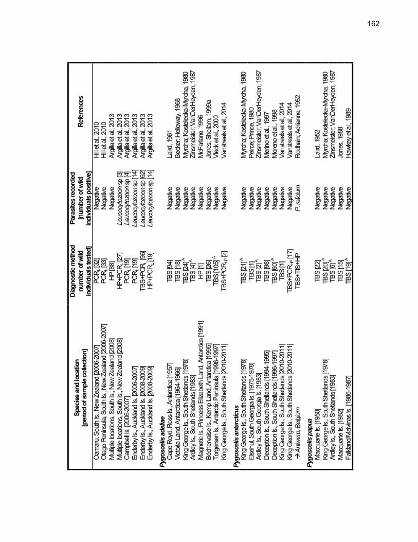
162

163

164

Le
gend
: TB
S =
thin
blo
od s
mea
rs, H
P =
his
topa
thol
ogy,
TIS
= ti
ssue
impr
essi
on s
mea
rs, P
CR
= p
olym
eras
e ch
ain
reac
tion
(sub
scrip
t let
ter
indi
cate
rea
ctio
n ta
rget
: PC
RH
P =
Hae
mop
rote
us/P
lasm
odiu
m, P
CR
L =
Leuc
ocyt
ozoo
n, P
CR
BO =
Bor
relia
), E
I = e
xper
imen
tal i
nocu
latio
n of
dom
estic
bird
s, S
EM
= s
cann
ing
elec
tron
mic
rosc
opy,
ISH
= in
-situ
hyb
ridiz
atio
n; n
s =
not
spec
ified
. Obs
.: A =
Stu
dy d
id n
ot s
peci
fical
ly a
im th
e de
tect
ion
of b
lood
par
asite
s, th
eref
ore
sub-
optim
al p
roto
cols
of b
lood
sm
ear
exam
inat
ion
may
hav
e be
en e
mpl
oyed
; B =
Fle
isch
man
et
al.,
1968
a, 1
968b
; H
erm
an e
t al
., 19
68;
Sla
den
et a
l.,
1979
; S
tosk
opf;
Bei
er,
1979
; B
eier
; S
tosk
opf,
1980
; B
eier
; Tr
pis,
198
1; C
ranf
ield
et
al.,
1994
; G
racz
yk e
t al
., 19
94a,
19
94b,
199
5; M
cCon
key
et a
l., 1
996;
Grim
et a
l., 2
004;
C a
nd D
= O
verla
ppin
g sa
mpl
ing
effo
rts.

App
endi
x B
- S
erol
ogic
al s
tudi
es ta
rget
ing
antib
odie
s ag
ains
t blo
od p
aras
ites
in p
engu
ins.
Rec
ords
at e
x-si
tu lo
catio
ns a
re
indi
cate
d by
an
arro
w a
nd it
alic
s

Leg
end:
PH
A =
pas
sive
hem
aggl
utin
atio
n, E
LIS
A =
enz
yme-
linke
d im
mun
osor
bent
ass
ay; n
s =
not s
peci
fied.

168
5 OUTBREAK OF AVIAN MALARIA ASSOCIATED TO MULTIPLE SPECIES OF PLASMODIUM IN MAGELLANIC PENGUINS UNDERGOING REHABILITATION IN SOUTHERN BRAZIL 5.1 ABSTRACT
Avian malaria is a mosquito-borne disease caused by Plasmodium spp. Avian
plasmodia are recognized conservation-threatening pathogens due to their potential to
cause severe epizootics when introduced to bird populations with which they did not co-
evolve. Penguins are considered particularly susceptible, as outbreaks in captive
populations will often lead to high morbidity and rapid mortality. We used a
multidisciplinary approach to investigate an outbreak of avian malaria in 28 Magellanic
penguins (Spheniscus magellanicus) at a rehabilitation center during summer 2009 in
Florianópolis, Brazil. Hemosporidian infections were identified by microscopic and
molecular characterization in 64% (18/28) of the penguins, including Plasmodium
(Haemamoeba) tejerai, Plasmodium (Huffia) elongatum, a Plasmodium (Haemamoeba)
sp. lineage closely related to Plasmodium cathemerium, and a Haemoproteus
(Parahaemoproteus) sp. lineage closely related to Haemoproteus syrnii. P. tejerai
played a predominant role in the studied outbreak and was identified in 72% (13/18) of
the hemosporidian-infected penguins, and in 89% (8/9) of the penguins that died,
suggesting that this is a highly pathogenic parasite for penguins; a detailed description
of tissue meronts and lesions is provided. Mixed infections were identified in three
penguins, and involved P. elongatum and either P. tejerai or P. (Haemamoeba) sp. that
were compatible with P. tejerai but could not be confirmed. In total, 32% (9/28) penguins
died over the course of 16 days despite oral treatment with chloroquine followed by
sulfadiazine-trimethoprim. Hemosporidian infections were considered likely to have
occurred during rehabilitation, probably from mosquitoes infected while feeding on local
native birds, whereas penguin-mosquito-penguin transmission may have played a role in
later stages of the outbreak. Considering the seasonality of the infection, rehabilitation

169
centers would benefit from narrowing their efforts to prevent avian malaria outbreaks to
the penguins that are maintained throughout summer.
5.2 INTRODUCTION
Plasmodium spp. are recognized as pathogens that may threaten the
conservation of avian communities, particularly when introduced to populations that did
not co-evolve with the parasite (BEADELL et al., 2006; ATKINSON; LAPOINTE, 2009).
In particular, two avian taxa have been shown to be highly susceptible to these
parasites, Hawaiian honeycreepers (Drepaniidae) and penguins (Spheniscidae) (FIX et
al., 1988; JONES; SHELLAM, 1999b; ATKINSON et al., 2001). Concerns that this
pathogen may be a significant conservation threat for penguins have been raised due to
the observation that avian malaria in captive penguins produces rapid and severe
outbreaks with as much as 50-80% mortality within few weeks (RODHAIN, 1939;
GRINER; SHERIDAN, 1967; FIX et al., 1988; BUENO et al., 2010). Four species of
Plasmodium sp. have been demonstrated to infect penguins: P. (Haemamoeba) relictum
(FANTHAM; PORTER, 1944), P. (Huffia) elongatum (HUFF; SHIROISHI, 1962), P.
(Bennettinia) juxtanucleare (GRIM et al., 2003) and P. (Haemamoeba) tejerai
(SILVEIRA et al., 2013). Additionally, there is an anecdotal report of P. (Haemamoeba)
cathemerium in penguins (LUERA-CARBO, 1965).
Magellanic penguins (Spheniscus magellanicus) are native to Argentina, Chile
and the Falkland Islands, and often winter along the coast of Uruguay and Brazil
(GARCÍA-BORBOROGLU et al., 2006, 2010). The susceptibility of this species to
malarial parasites has been documented in zoos and rehabilitation centers (FIX et al.,
1988; KO et al., 2008; BUENO et al., 2010; SILVEIRA et al., 2013), but not in the wild
(HAWKEY et al., 1989; JOVANI et al., 2001; KEYMER et al., 2001; QUILLFELDT et al.,
2010). Other studies have failed to detect Plasmodium sp. in wild penguins elsewhere in
South America or at the Falkland islands, whether in Southern rockhopper (Eudyptes
chrysocome) (HAWKEY et al., 1989; KARESH et al., 1999; KEYMER et al., 2001;

170
QUILLFELDT et al., 2010), Humboldt (S. humboldti) (WALLACE et al., 1995; JONES;
SHELLAM, 1999a; MILLER et al., 2001; SMITH et al., 2008) or Gentoo penguins
(Pygoscelis papua) (HAWKEY et al., 1989; KEYMER et al., 2001; QUILLFELDT et al.,
2010). In Galapagos penguins (Spheniscus mendiculus), on the other hand, DNA from
Plasmodium spp. has been detected at the Galapagos Archipelago, even though the
parasite was not observed in blood smears (LEVIN et al., 2009, 2013).
We had previously reported two Magellanic penguins that died due to infection by
P. tejerai during a mortality outbreak at a rehabilitation center in Florianópolis, Brazil, in
early 2009 (SILVEIRA et al., 2013). In the present paper we follow up on that
investigation and provide a broader study on the epidemiology and pathology of P.
tejerai as well as other hemosporidians involved in the same outbreak. We also discuss
the implications of these findings for the rehabilitation and conservation of penguins and
other highly susceptible taxa.
5.3 METHODS
5.3.1 Ethics statement
This study was conducted under approval by the Animal Use Ethics Committee of
the University of São Paulo (CEUA-USP 1757/2009) and was authorized by the
Brazilian wildlife authority (SISBIO 20825-3).
5.3.2 Study population, sample collection and treatment
We examined the Magellanic penguins maintained through austral summer 2008-
2009 at the Centro de Triagem de Animais Silvestres do Núcleo de Fauna do IBAMA,

171
Florianópolis, Brazil (27°31’35”S 48°25’44”W). The rehabilitation center is located near a
large freshwater lake (Lagoa da Conceição) and is surrounded by Atlantic forest
(lowland tropical moist forest), with average annual precipitation of 1524 mm and
average annual temperature ranging from 17.0 to 24.8 °C (RAMOS et al., 2009).
The year 2008 was atypical in presenting unusually high numbers of Magellanic
penguins becoming beach-cast along the Brazilian coast (GARCÍA-BORBOROGLU et
al., 2010), and the rehabilitation center received 387 animals. Most penguins were
successfully rehabilitated (following protocols by SILVA-FILHO; RUOPPOLO, 2007) and
released in 2008, however 23 were maintained through summer, and another 5 were
received in early 2009 (band numbers 508, 586, 587, 592 and 593); these 28 penguins
were the subjects of this study (Table 5.1). In a previous study (SILVEIRA et al., 2013)
we examined in detail two of these individuals (band numbers 506 and 520 are
respectively penguins A and B in that study). Metatarsal blood samples were collected
on 23/03/2009 (584, 593) and 25/03/2009 (remaining penguins) (Sampling A); penguins
that were still alive were sampled again on 05/04/2009 (Sampling B).
Veterinarians monitored penguins daily throughout the rehabilitation process, and
any clinical signs or abnormalities were recorded. After avian malaria was diagnosed,
penguins were orally treated with chloroquine on 01/04/2009 (10 mg/kg at hour zero; 5
mg/kg at hours 6, 18 and 24) until 10/04/2009 (5 mg/kg q24h), then with sulfadiazine-
trimethoprim (40 mg/kg q24h) from 11/04/2009 to 20/04/2009.

172
Tabl
e 5.
1 - I
ndiv
idua
l his
tory
and
dia
gnos
tic re
sults
for t
he s
tudi
ed M
agel
lani
c pe
ngui
ns (S
phen
iscu
s m
agel
lani
cus)
. Tax
onom
ic n
ames
w
ithin
bra
cket
s in
dica
te th
e ta
xon
to w
hich
the
spec
ies
is p
resu
med
to c
orre
spon
d on
the
basi
s of
phy
loge
netic
ana
lyse
s.
Ast
eris
ks in
dica
te in
divi
dual
s th
at d
ied
durin
g th
e ou
tbre
ak

173
5.3.3 Laboratory procedures
Two thin blood smears were performed immediately after each blood collection,
dried at room temperature, and fixed with methanol. Two heparin capillaries were also
collected immediately after collection. The remaining blood was stored in EDTA flasks
and refrigerated (4°C) for 2-4 hours, centrifuged at 1500 G for 10 minutes, supernatant
fluid was separated and both blood cells and plasma were frozen (-20°C). Packed Cell
Volume (PCV) was determined through centrifugation in heparin capillaries at 16000 G
for 5 min.
Blood smears were Giemsa-stained within a week of collection; at least 300
microscopic fields were examined for parasites in each smear, under 1000x
magnification (>50.000 erythrocytes). Blood parasites were morphologically
characterized (VALKIŪNAS, 2005) and quantified (GODFREY et al., 1987; GERING;
ATKINSON, 2004); differential parasite counts (trophozoites, meronts,
macrogametocytes and microgametocytes) were conducted for 100 parasites or for all
parasites observed. Differential leukocyte counts (heterophils, eosinophils, basophils,
lymphocytes, monocytes) were conducted for 200 leukocytes per slide and Early
Erythrocytic Lineage Cells (EELC) were estimated as a percentage of all red blood cells.
Heterophil-to-Lymphocyte Ratio (HLR) was calculated. Blood smear examination was
blind to PCR results and vice-versa.
Deceased animals were examined within 12 to 24h post-mortem, and organs and
tissues were fixed in 10% buffered formalin and embedded in paraffin. Sections of 5 µm
were obtained, stained with hematoxylin-eosin and examined under light microscopy.
5.3.4 Cytochrome b amplification and phylogenetic analysis
Frozen red blood cells in EDTA and frozen tissue samples were used for
molecular analyses. DNA extraction was conducted using the DNEasy Blood and Tissue

174
Kit (#69506, Qiagen, Venlo, Netherlands) following the manufacturer’s recommended
protocol. DNA extraction was verified and quantified through UV spectrophotometry
(Nanodrop 2000, Thermo Fisher Scientific, Wilmington, DE, USA). A nested PCR
targeting a 480 bp fragment of the cytochrome b (cyt-b) mitochondrial gene of
Haemoproteus sp. and Plasmodium sp. was used (HELLGREN et al., 2004) (each
reaction had 25 µL, with 75 ng of sample DNA; first reaction with primers HaemNFI and
HaemNR3, second reaction with primers HaemF and HaemR2). GoTaq Green Master
Mix (M7122, Promega, Madison, USA) was used for both reactions. Blood samples from
chicken experimentally infected with Plasmodium gallinaceum or raised in arthropod-
free environments were used as positive and negative controls, respectively.
Amplification products were visualized in 6% silver nitrate-stained polyacrylamide gels
and 2% agarose gel (SANGUINETTI et al., 1994; RIBEIRO et al., 2005).
PCR amplification products of positive samples were purified with Polyethylene
glycol 8000. Bi-directional sequencing with dye-terminator fluorescent labeling was
performed through automated sequencing (ABI Prism 3100, Applied Biosystems, Foster
City, USA). Forward and reversed chromatograms were aligned and sequences were
revised and edited using DNABaser (Heracle BioSoft SRL, Pitesti, Romania); cyt-b gene
resulting sequences were deposited in the GenBank database (Appendix A).
Phylogenetic relationships among the hemosporidian lineages identified in this study
and related hemosporidian parasites were inferred by using cyt-b gene sequences from
MalAvi database (BENSCH et al., 2009) for which morphospecies had been determined
using blood smears (Appendix B). Because GenBank contains misidentified sequences
of avian hemosporidians (VALKIŪNAS et al., 2008a), only lineages for which extensive
blood smear morphology data was available were included when BLAST search
indicated high identity and query cover (≥ 95%) with the sequences obtained in this
study (RICKLEFS; FALLON, 2002; ISHAK et al., 2008; CHAGAS et al., 2013;
KARADJIAN et al., 2013; YILDIRIM et al., 2013). In addition, we included hemosporidian
lineages recovered from penguins at the Galapagos Archipelago (LEVIN et al., 2009,
2013), São Paulo Zoological Park (BUENO et al., 2010) and Australia (CANNELL et al.,
2013). Sequences were aligned using ClustalW (THOMPSON et al., 1997) as
implemented in MEGA 5.2.2 (TAMURA et al., 2011). A maximum likelihood phylogenetic

175
tree for the parasite sequences was produced using MEGA 5.2.2 with the GTR +
gamma model of nucleotide evolution, as selected using ModelTest (POSADA;
CRANDALL, 1998), and 1000 bootstrap replications. There is on-going debate on which
taxon should be used to root phylogenetic trees of hemosporidians (OUTLAW;
RICKLEFS, 2011), we used L. schoutedeni merely for visualization purposes.
5.3.5 Statistical analyses
Two-sample t-tests were used to compare parasitemia and hematological
parameters in the blood smears of penguins that were deceased and those that
survived. Paired t-tests were used to compare hematological parameters (heterophils,
eosinophils, basophils, lymphocytes, monocytes, EELC, HLR, PCV) between positive
and negative individuals and between Sampling A and B, and mean difference tests
were used to determine whether these sampling date differences followed distinct
patterns between positive or negative individuals. Linear regression was used to
determine whether correlations existed between parasitemia and hematological
parameters. ANOVA was used to compare the percentage of parasite forms
(trophozoites, meronts, macrogametocytes, microgametocytes) in the blood smears of
penguins infected by different parasite lineages. Fisher’s exact test was used to
compare mortality between individuals infected with P. tejerai and other lineages.
Significance level was 0.05 for all tests, and the Dunn-Šidák correction was applied for
multiple comparisons.

176
5.4 RESULTS
5.4.1 Clinical signs and necropsy findings
In early March 2009, 28 Magellanic penguins were at the rehabilitation center,
and were considered clinically healthy. Penguins died on 23/03/2009 (band numbers
584 and 593), 27/03/2009 (506), 29/03/2009 (518, 520, 589), 03/04/2009 (587), and
07/04/2009 (503, 508). All deceased penguins had good body condition, normal appetite
and behavior on the days preceding their deaths, and shared similar necropsy findings:
hydropericardium, cardiomegaly, lung congestion, hepatomegaly and splenomegaly.
5.4.2 Hemosporidian detection
Table 5.1 details PCR, blood smear, histopathology and gene sequencing results
for the 28 penguins present at the rehabilitation center in April 2009. Eighteen penguins
had one or more positive samples (64.3%; 18/28) whether through blood smears alone
(50%; 2/4) or using a combination of blood smears and nested PCR (66.7%; 16/24). Of
the 14 samples positive to PCR for which blood smears were also available, only 7 were
identified as positive in the corresponding blood smears (50%). All PCR-negative
samples were negative to parasites in the corresponding blood smears. No blood
parasites other than hemosporidians were observed.
All twelve penguins tested by PCR at Sampling A were positive (100%). Nine of
these PCR-positive penguins also had blood smears examined: 4 were positive (44.4%).
An additional 14 penguins that had not been tested by PCR had blood smears examined
at Sampling A, of which 4 were positive (28.6%). Five of the 20 penguins tested by PCR
at Sampling B were positive (25%); of these, 3 had their blood smears identified as
positive (60%). An additional 3 penguins that had not been tested by PCR had blood

177
smears examined at Sampling B, of which one was positive (33.3%). As a result, 16 of
the 26 penguins had positive results at Sampling A (61.5%) and 6 of the 23 penguins
had positive results at Sampling B (26.1%).
5.4.3 Parasite identification and phylogeny
Mitochondrial cytochrome b gene sequences were obtained for 17 samples from
13 penguins. Phylogenetic analysis revealed four phylogenetic groups/lineages, three
Plasmodium sp. (A, B and C) and one Haemoproteus sp. (D) (Figure 5.1). Two penguins
had two samples that yielded two different sequences each (band numbers 508 and
511); one of these (511B) had eight nucleotide polymorphisms (mixed peaks on the
sequencing chromatogram) and was excluded from phylogenetic analyses. Two
samples were PCR-positive but failed to produce adequate sequencing results (504 and
588). Table 5.2 summarizes evolutionary distance within these groups and published
lineages.
Phylogenetic group A comprised sequences obtained from 13 samples (clade
bootstrap value = 94%; average evolutionary divergence within group = 0.007 base
substitutions per site), and had some of its samples morphologically identified as P.
tejerai (for detailed descriptions and photomicrographs see SILVEIRA et al., 2013).
Lineage B was recovered only from one penguin (516), and its morphospecies could not
be determined as only early trophozoites were present in the blood smear; this lineage
formed a clade with a known P. cathemerium lineage (bootstrap value = 88%;
evolutionary divergence = 0.015 base substitutions per site; 98.7% sequence identity).
Lineage C was recovered only from one penguin (508) and was morphologically
confirmed as P. elongatum (Figure 5.2a-i); this lineage formed a strongly supported
clade (bootstrap value = 99%; average evolutionary divergence within group = 0.004
base substitutions per site) with a known P. elongatum lineage (98.5% sequence
identity) and with a Plasmodium sp. lineage previously identified in Magellanic penguins
(98.0% sequence identity). Lineage D was recovered only from one penguin (586) and

178
its morphospecies could not be determined because no parasite forms were present in
the blood smear; this lineage formed a clade (bootstrap value = 81%; average
evolutionary divergence within group = 0.009 base substitutions per site) with H.
(Parahaemoproteus) sp. lineages obtained from earless owls (Strix spp.) in the Northern
Hemisphere, including a known Haemoproteus syrnii lineage (96.8% sequence identity).
The positive control used for the nested PCR reactions was identical to a known P.
gallinaceum lineage (100% sequence identity) and unrelated to the study lineages.
Mixed infections were identified in three individuals (band numbers 508, 517 and
588), and involved P. elongatum and either P. tejerai or P. (Haemamoeba) sp. that were
compatible with P. tejerai but could not be undoubtedly confirmed. In two cases (503
and 516) the parasites observed at Sampling B were severely degenerated, presumably
as a result of chloroquine treatment, producing atypical forms that at times bore little
resemblance to typical hemosporidian forms (Figure 5.2j-l).
Nine of 28 penguins (32.1%) died during the study period. Of these, eight (88.9%)
had been positive to hemosporidians, i.e. 8 of 18 penguins that obtained positive results
died during the study period (44.4%). All hemosporidian-positive deceased penguins
were infected with P. tejerai, one of which was co-infected with P. elongatum. Even
though 58.3% of the penguins confirmed to be infected only with P. tejerai died (7/12),
this was not significantly different to the mortality observed among individuals infected or
co-infected with other lineages (16.7%; 1/6) (P = 0.152).
Parasitemia ranged from 0.001% to 62% (Mean ± S.D. = 8.66% ± 18.83%). Even
though parasitemia was apparently higher in penguins that died (12.97% ± 22.22%) than
in those that survived (0.06% ± 0.10%), this difference did not reach significance (t =
1.642, df = 7, P = 0.072). Trophozoites (P < 0.001, R2 = 0.787), macrogametocytes (P <
0.001, R2 = 0.854) and microgametocytes (P = 0.008, R2 = 0.585) were unevenly
distributed among parasite lineages (Table 5.3), but not meronts (P = 0.390).

179
Figure 5.1 - Maximum likelihood phylogenetic tree of the mitochondrial cytochrome b gene of the studied hemosporidian lineages. Lineages identified in this study are emphasized in blue. When available, information on the morphospecies observed on the corresponding blood smear is provided. Branch lengths are drawn proportionally to evolutionary distance (scale bar is shown). Lower bootstrap values (< 50) are omitted
Source: Vanstreels (2014)

180
Tabl
e 5.
2 - E
stim
ates
of e
volu
tiona
ry d
ista
nce
(% e
xpec
ted
base
sub
stitu
tions
per
site
) of c
ytoc
hrom
e b
mito
chon
dria
l gen
e se
quen
ces
of h
emos
porid
ians
ide
ntifi
ed i
n pe
ngui
ns i
n th
is s
tudy
(1-
6) a
nd t
he l
itera
ture
(7-
10),
and
refe
renc
e lin
eage
s fro
m t
he
Mal
Avi
dat
abas
e (1
1-19
). V
alue
s lo
wer
than
4.0
are
hig
hlig
hted
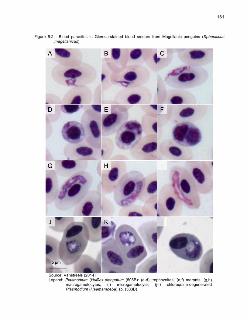
181
Figure 5.2 - Blood parasites in Giemsa-stained blood smears from Magellanic penguins (Spheniscus magellanicus)
Source: Vanstreels (2014) Legend: Plasmodium (Huffia) elongatum (508B): (a-d) trophozoites, (e,f) meronts, (g,h)
macrogametocytes, (i) microgametocyte; (j-l) chloroquine-degenerated Plasmodium (Haemamoeba) sp. (503B)

182
5.4.4 Hematology and pathology
Table 5.3 summarizes the average hematological results for blood smears of
penguins infected by different hemosporidian lineages. No significant differences were
observed in these hematological parameters between Sampling A and B, whether they
were positive to hemosporidians or not (all P > 0.05), nor between penguins that died or
those that survived (all P > 0.05). Parasitemia was positively correlated to lymphocytes
(P = 0.022, R2 = 0.364), monocytes (P = 0.043, R2 = 0.285) and PCV (P = 0.034, R2 =
0.642) and was negatively correlated to heterophils (P = 0.026, R2 = 0.348); no
significant correlation was observed between parasitemia and basophils, eosinophils,
EELC or HLR (all P > 0.05).
Histopathology was examined for three individuals (506, 584, 593), all of which
were infected only with P. tejerai. Tissue meronts were occasionally present in
macrophages, but were overall most common in endothelial cells. Meronts were most
frequent in the heart and kidneys, moderately frequent in the lungs and colon, and were
rare in other tissues (spleen, liver, testicles, brain, duodenum, pancreas, gastrointestinal
tract-associated lymphoid tissue, thyroid, parathyroid). Penguin 584 was considered to
have a higher number of tissue meronts (approx. 2-3 per 10 high magnification
microscope fields) than penguins 506 and 593 (less than 1 per 10 fields), whereas ante-
mortem parasitemia was considerably higher in 506 (62%) and 593 (30%) than in 584
(7.4%). Tissue meronts ranged between 10 and 80 µm in largest diameter, most
frequently from 15 to 30 µm. They were often round or oval, with merozoites distributed
near the external surface forming structures resembling arcs (Figure 5.3a) or semi-arcs
(Figure 5.3b), or were elongated and contained randomly scattered merozoites (Figure
5.3c). The envelope of tissue meronts was mildly eosinophilic, thin and poorly defined;
merozoites were round and densely stained with size of approximately 1 µm in diameter.
Each meront contained tens to hundreds of visible merozoites, although generally less
than 20 merozoites were visible in 3 µm-thick histological sections.

183
Major pathological processes included: moderate to severe diffuse interstitial
granulocytic pneumonia, moderate to severe pulmonary edema and congestion, severe
acute necrotizing splenitis, moderate multifocal to coalescent mixed or predominantly
mononuclear necrotizing hepatitis, mild to moderate multifocal interstitial sub-acute
nephritis, moderate diffuse iron deposits in the lungs, liver and spleen, mild to moderate
spleen histiocytosis, mild to moderate multifocal necrosis of gut-associated lymphoid
tissues, mild diffuse granulocytic myocarditis, multifocal to coalescent areas of
cardiomyolysis. Penguin 584 also had diffuse acute vasculitis associated to the
formation of intravascular cloths. Death probably resulted from cardiorespiratory
insufficiency secondary to pneumonia and, in the case of penguin 584, in association
with disseminated vasculitis and intravascular coagulation. No signs or lesions
suggestive of viral, bacterial or fungal co-infections were observed.
Figure 5.3 - Tissue meronts of Plasmodium (Haemamoeba) tejerai in tissues of Magellanic penguins
(Spheniscus magellanicus). Hematoxilin-Eosin, penguin 584
Source: Vanstreels (2014) Legend: (a) liver, (b) parathyroid, (c) heart

184
Tabl
e 5.
3 - H
emat
olog
ical
resu
lts a
nd q
uant
ifica
tion
of b
lood
par
asite
s in
blo
od s
mea
rs (M
ean
± S
.D.)
185
5.5 DISCUSSION
Penguins are notoriously susceptible to avian malaria, and Magellanic penguins
are not an exception (JONES; SHELLAM, 1999a). Fix et al. (1988) reported three
successive malaria outbreaks leading to the mortality of 15.2%, 51.3% and 57.9% of
Magellanic penguins captive at a zoo in Iowa, USA (cumulative mortality = 82.6%; n =
46). Bueno et al. (2010) reported an outbreak with 80% infection rate and 60% mortality
of Magellanic penguins captive at a zoo in São Paulo, Brazil (n = 5). In this study, we
observed 60.7% infection rate and 32.1% mortality of Magellanic penguins undergoing
rehabilitation during summer in Florianópolis, Brazil (n = 28).
The use of chloroquine and primaquine for the treatment of avian malaria in
penguins was first proposed by Stoskopf and Beier (1979), and is based on the
combined antiprotozoal effect of chloroquine on the circulating stages and of primaquine
on the tissue stages (REMPLE, 2004). Because we were unable to administer
primaquine the poor performance of the drug treatment in this outbreak was not entirely
unexpected. However, Fix et al. (1988) and Bueno et al. (2010) did administer the
combined drug treatment and still were confronted with high mortality rates, suggesting
a poor efficacy of these drugs, as has also been reported for other penguin species
(SLADEN et al., 1979; CRANFIELD et al., 1994). Our observation of degenerated
erythrocytic parasites in blood smears of some of the penguins subjected to five days of
chloroquine treatment suggests an effective antiprotozoal action of this drug on
circulating parasites. Unfortunately, because histopathology was conducted only for
individuals that died before drug treatment was initiated, we could not evaluate its
effects on the parasites’ tissue stages, if any.
Four lineages of hemosporidians were involved in the outbreak: Plasmodium
(Haemamoeba) tejerai, an unidentified Plasmodium (Haemamoeba) sp. (“lineage B”),
Plasmodium (Huffia) elongatum, and an unidentified Haemoproteus
(Parahaemoproteus) sp. Furthermore, because four lineages could not be conclusively
identified, it is possible that additional hemosporidian lineages/species were present.

186
The finding of P. tejerai in Magellanic penguins corresponds to the first cases of
this parasite other than its original description in domestic turkeys (Meleagris gallopavo)
in Venezuela (GABALDÓN; ULLOA, 1977). P. tejerai played a predominant role in the
studied outbreak, being identified in 72.2% of the hemosporidian-infected penguins.
Furthermore, the observation that 61.6% of the penguins diagnosed with P. tejerai have
died suggests this is a highly pathogenic parasite for penguins.
Necropsy and histopathology indicated the death of P. tejerai-infected penguins
was associated with a significant impairment of cardiorespiratory function and, at least in
one case, disseminated vasculitis and intravascular clotting. These findings are not
unlike those observed in other malaria outbreaks in penguins (HUFF; SHIROISHI, 1962;
GRÜNBERG; KUTZER, 1963; FLEISCHMAN et al., 1968b; HERMAN et al., 1968; BAK
et al., 1984; FIX et al., 1988), and are compatible with the mechanisms of avian malaria
pathogenesis described in other birds (ATKINSON; VAN RIPER III, 1991; VALKIŪNAS,
2005; WILLIAMS, 2005). The semi-arc or arc-shaped tissue meronts observed in these
infections seem unusual and distinct from those classically reported in avian
hemosporidians (HUFF, 1942; GARDINER et al., 1998; VALKIŪNAS, 2005), and could
be a characteristic morphological feature of P. tejerai, meriting further investigation. It
should be noted that such tissue meront morphology is paralleled by the parasite’s
morphology in erythrocytes: rosette-shaped erythrocytic meronts in which the
merozoites are distributed in an arc near the external border of the parasite’s cytoplasm
are one of the key morphological characteristics for the identification of P. tejerai in
blood smears (SILVEIRA et al., 2013).
Plasmodium sp. lineage B was identified in one penguin and could not be
identified on the basis of parasite morphology on blood smears because only early
trophozoites were present, however sequencing data suggests it belongs to the
subgenus Haemamoeba and is closely related to P. cathemerium. In a review on the
parasitology of zoo animals, Luera-Carbo (1965) mentions that P. cathemerium infects
king penguins (Aptenodytes patagonicus), presumably at the Barcelona Zoo, Spain;
however, because no details were provided on the methods and criteria for species
identification, this record cannot be confirmed. It should be considered that the
identification of P. cathemerium in penguins merits particular caution since there is
187
significant morphological similarity between this parasite and P. relictum (VALKIŪNAS,
2005), a species frequently reported in penguins worldwide (FANTHAM; PORTER,
1944; RAETHEL, 1960; GRINER; SHERIDAN, 1967; STOSKOPF; BEIER, 1979; FIX et
al., 1988). Furthermore, the morphology of avian hemosporidians is known to vary
considerably when transmitted to different hosts, contributing to confusion in their
identification (LAIRD; VAN RIPER, 1981; VALKIŪNAS, 2005). Because we were not
able to confirm the species identity through morphological analysis, we consider that
additional evidence is necessary before it may be conclusively demonstrated that P.
cathemerium infects penguins.
P. elongatum is a cosmopolitan parasite that may infect a broad variety of avian
hosts, particularly Passeriformes (VALKIŪNAS, 2005), and is not uncommon in
penguins captive in North America (FLEISCHMAN et al., 1968a; SLADEN et al., 1979;
STOSKOPF; BEIER, 1979; GRACZYK et al., 1994). Similarly to the reports from North
American zoos, P. elongatum played a secondary role and did not produce high
mortality during malaria outbreaks in penguins. All three cases of P. elongatum infection
herein examined had a second Plasmodium (Haemamoeba) sp. lineage, whether
confirmed as P. tejerai or not; this is also not unusual as co-infection by P. elongatum
and P. (Haemamoeba) sp. has been frequently reported in captive penguins in North
America (HUFF; SHIROISHI, 1962; STOSKOPF; BEIER, 1979; CRANFIELD et al.,
1994; GRACZYK et al., 1994).
Bueno et al. (2010) identified two lineages of Plasmodium sp. in Magellanic
penguins at São Paulo Zoo, and used BLAST to determine high sequence identity with
two lineages that had been registered on GenBank as P. relictum, AY733088 and
AY733089. However, Valkiūnas et al. (2008a) indicated that AY733088 had been
misidentified and probably corresponds to P. elongatum instead. Our results support this
interpretation, and suggest that the two lineages identified by Bueno et al. (2010)
correspond to two distinct species, of which one (HM031937) corresponds to P.
elongatum and the other (HM031936) corresponds to a Plasmodium (Haemamoeba) sp.
lineage closely related to P. lutzi. This corroborates the warnings that sequence identity
in GenBank/BLAST databases search is not an adequate method to conclusively
identify avian hemosporidians without complementary morphological analysis

188
(VALKIŪNAS et al., 2008a,b; BENSCH et al., 2009). On the other hand, our results
indicate there is no direct relationship between the Plasmodium sp. lineages herein
detected and those found in Galapagos penguins, which remain unidentified.
Sequencing results indicate that a Haemoproteus (Parahaemoproteus) sp.
lineage was present in one penguin. Haemoproteus sp. is a common avian
hemosporidian that is seldom considered pathogenic (ATKINSON; VAN RIPER III, 1991;
VALKIŪNAS, 2005). Only twice have these parasites been recorded in penguins: DNA
from this parasite has been detected in live Galapagos penguins at Galapagos
Archipelago (LEVIN et al., 2009) and in deceased little penguins (Eudyptula minor) in
Western Australia (CANNEL et al., 2013). All Haemoproteus spp. infections in penguins
were identified through gene sequencing analysis, as no circulating stages or only early
trophozoites were detected in blood smears. Similarly, we failed to detect circulating
parasites in the blood smears corresponding to the PCR-positive sample sequenced as
Haemoproteus sp. This may indicate a very low parasitemia, insufficient to allow for
blood smear detection (GARAMSZEGI, 2010), or could correspond to the detection of
DNA from recently inoculated sporozoites which may or not succeed to produce
infection (VALKIŪNAS et al., 2009; LEVIN et al., 2013). Phylogenetic analysis indicated
the Haemoproteus sp. lineage identified in this study was not closely related to those
previously identified in penguins, and instead was neatly clustered with those found in
earless owls (Strix spp.) in Europe and North America, including a Haemoproteus syrnii
lineage. Three Strix spp. are native to the Florianópolis region (S. huhula, S. hylophila,
and S. virgata) (CBRO, 2011; IUCN, 2014) and could have served as reservoirs of
infection.
Parasitemia was positively correlated to lymphocyte and monocyte counts and
inversely correlated to heterophil counts, corroborating the observation that avian
malaria elicits a predominantly mononuclear leukocytosis in penguins (GRACZYK et al.,
1994). On the other hand, parasitemia was positively correlated to PCV, which
contradicts classical findings that Plasmodium-induced hemolysis results in decreased
PCV (ATKINSON; VAN RIPER III, 1991; GRACZYK et al., 1994; WILLIAMS, 2005); this
could be explained by a false elevation of PCV due to dehydration as a result from
anorexia and febrile syndrome.

189
Avian-infecting Plasmodium spp. are exclusively transmitted by mosquitoes
(Culicidae), and gene sequencing data indicates all Haemoproteus spp. identified to
date in penguins belong to the subgenus Parahaemoproteus (LEVIN et al., 2009;
CANNEL et al., 2013), which is transmitted by biting midges (Ceratopogonidae)
(VALKIŪNAS, 2005). Both mosquitoes and biting midges are abundant in the studied
rehabilitation center during summer (subj. obs.). This, combined to the observation that
all deaths in the studied malaria outbreak occurred within a 16-days period during
summer whereas most penguins had been in the same facility for several months,
corroborates the interpretation that infection occurred during rehabilitation, as opposed
to a recrudescence/exacerbation of infections acquired in the wild. An alternative, less
likely in our opinion, is that one or more penguins relapsed from an asymptomatic
Plasmodium infection and transmitted the parasite to the other penguins. Although
transmission from mosquitoes infected while feeding on local native birds to penguins is
likely responsible for the initiation of these outbreaks, the fact that some penguins had
large numbers of gametocytes suggests that penguin-mosquito-penguin transmission
could play a role in later stages of the outbreak. Early diagnosis, quarantine and
treatment of affected individuals could therefore be beneficial to the mitigation of
outbreaks in penguins undergoing rehabilitation. Because these parasites may be
transmitted through the inoculation of blood from an infected bird to another
(VALKIŪNAS, 2005), penguin rehabilitators should also take care to prevent iatrogenic
transmission.
Our findings corroborate the strong seasonality of avian malaria outbreaks in
captive penguins, which are consistently concentrated in the late summer and early
autumn across the world (RODHAIN, 1939; GRINER; SHERIDAN, 1967; BAK et al.,
1984; FIX et al., 1988; GRACZYK et al., 1994; KO et al., 2008; BUENO et al., 2010).
Rehabilitation centers would thus benefit from narrowing their malaria prevention efforts
to the minority of Magellanic penguins that are maintained throughout summer, as
opposed to the majority of penguins admitted and released during winter and early
spring (GARCÍA-BORBOROGLU et al., 2006, 2010).

190
REFERENCES ATKINSON, C. T.; DUSEK, R. J.; LEASE, J. K. Serological responses and immunity to superinfection with avian malaria in experimentally-infected Hawaii Amakihi. Journal of Wildlife Diseases, v. 37, n. 1, p. 20-27, 2001 ATKINSON, C. T.; LAPOINTE, D. A. Introduced avian diseases, climate change, and the future of Hawaiian honeycreepers. Journal of Avian Medicine and Surgery, v. 23, n. 1, p. 53-63, 2009. ATKINSON, C. T.; VAN RIPER III, C. Pathogenicity and epizootiology of avian haematozoa: Plasmodium, Leucocytozoon, and Haemoproteus. In: LOYE, J. E.; ZUK, M. Bird-parasite interactions: ecology, evolution and behaviour. New York: Oxford University Press, 1991. p. 19-48. BAK, Ung-Bok; PARK, Jae-Chan; LIM, Young-Jae. An outbreak of malaria in penguins at the Farm-land Zoo. Korean Journal of Parasitology, v. 22, n. 2, p. 267-272, 1984. BEADELL, J. S.; ISHTIAQ, F.; COVAS, R.; MELO, M.; WARREN, B. H.; ATKINSON, A. T.; BENSCH, S.; GRAVES, G. R.; JHALA, Y. V.; PEIRCE, M. A.; RAHMANI, A. R.; FONSECA, D. M.; FLEISCHER, R. C. Global phylogeographic limits of Hawaii's avian malaria. Proceedings of the Royal Society Society B, v. 273, p. 2935-2944, 2006. BENSCH. S.; HELLGREN, O.; PÉREZ-TRIS, J. MalAvi: a public database of malaria parasites and related haemosporidians in avian hosts based on mitochondrial cytochrome b lineages. Molecular Ecology Research, v. 9, p. 1353-1358, 2009. BUENO, M. G.; LOPEZ, R. P. G.; MENEZES, R. M. T.; COSTA-NASCIMENTO, M. J.; LIMA, G. F. M. C.; ARAÚJO, R. A. S.; GUIDA, F. J. V.; KIRCHGATTER, K. Identification of Plasmodium relictum causing mortality in penguins (Spheniscus magellanicus) from São Paulo Zoo, Brazil. Veterinary Parasitology, v. 173, n. 1-2, p. 123-127, 2010. CANNELL, B. L.; KRASNEC, K. V.; CAMPBELL, K.; JONES, H. I.; MILLER, R. D.; STEPHENS, N. The pathology and pathogenicity of a novel Haemoproteus spp. infection in wild Little Penguins (Eudyptula minor). Veterinary Parasitology, v. 197, n. 1-2, p. 74-84, 2013.

191
CHAGAS, C. R. F.; VALKIŪNAS, G.; NERY, C. V. C.; HENRIQUE, P. C.; GONZALEZ, I. H. L.; MONTEIRO, E. F.; GUIMARÃES, L. O.; ROMANO, C. M.; KIRCHGATTER, K. Plasmodium (Novyella) nucleophilum from an Egyptian Goose in São Paulo Zoo, Brazil: microscopic confirmation and molecular characterization. International Journal for Parasitology: Parasites and Wildlife, v. 2, p. 286-291, 2013. CBRO. Comitê Brasileiro de Registros Ornitológicos. Lista de aves do Brasil, 10a. edição. 2011. Disponível em: <http://www.cbro.org.br>. Acesso em: 12 dez. 2011. CRANFIELD, M. R.; GRACZYK, T. K.; BEALL, F. B.; IALEGGIO, D. M.; SHAW, M. L.; SKJOLDAGER, M. L. Subclinical avian malaria infections in African black-footed penguins (Spheniscus demersus) and induction of parasite recrudescence. Journal of Wildlife Diseases, v. 30, n. 3, p. 372-376, 1994. FANTHAM, H. B.; PORTER, A. On a Plasmodium (Plasmodium relictum var. spheniscidae, n. var.), observed in four species of penguins. Proceedings of the Zoological Society of London, v. 114, p. 279-292, 1944. FIX, A. S.; WATERHOUSE, C.; GREINER, E. C.; STOSKOPF, M. K. Plasmodium relictum as a cause of avian malaria in wild-caught Magellanic penguins (Spheniscus magellanicus). Journal of Wildlife Diseases, v. 24, n. 4, p. 610-619, 1988. FLEISCHMAN, R. W.; SQUIRE, R. A.; SLADEN, W. J. L.; MELBY Jr., E. C. Malaria (Plasmodium elongatum) in captive African penguins (Spheniscus demersus). Journal of the American Veterinary Medical Association, v. 153, n. 7, p. 928-935, 1968. GABALDÓN, A.; ULLOA, G. Plasmodium (Haemamoeba) tejerai sp. n. en pavo domestico (Melleagris gallopavo) del Venezuela. Boletín de la Dirección de Malariología y Saneamiento Ambiental, v. 17, p. 255-273, 1977. GARAMSZEGI, L. Z. The sensitivity of microscopy and PCR-based detection methods affecting estimates of prevalence of blood parasites in birds. Journal of Parasitology, v. 96, n. 6, p. 1197-1203, 2010. GARCÍA-BORBOROGLU, P.; BOERSMA, P. D.; RUOPPOLO, V.; REYES, L.; REBSTOCK, G. A.; GRIOT, K.; HEREDIA, S. R.; ADORNES, A. C.; SILVA-FILHO, R. P. Chronic oil pollution harms Magellanic penguins in the Southwest Atlantic. Marine Pollution Bulletin, v. 52, p. 193-198, 2006.

192
GARCÍA-BORBOROGLU, P.; BOERSMA, P. D.; RUOPPOLO, V.; SILVA-FILHO, R. P.; ADORNES, A. C.; SENA, D. C.; VELOZO, R.; KOLESNIKOVAS, C. M.; DUTRA, G.; MARACINI, P.; NASCIMENTO, C. C.; RAMOS-JÚNIOR, V.; BARBOSA, L.; SERRA, S. Magellanic penguin mortality in 2008 along the SW Atlantic coast. Marine Pollution Bulletin, v. 60, n. 10, p. 1652-1657, 2010. GARDINER, C. H.; FAYER, R.; DUBEY, J. P. An atlas of protozoan parasites in animal tissues (Apicomplexa). Washington D.C.: Registry of Veterinary Pathology, Armed Forces Institute of Pathology, American Registry of Pathology, 1998. 84 p. GERING, E.; ATKINSON, C. T. A rapid method for counting nucleated erythrocytes on stained blood smears by digital image analysis. Journal of Parasitology, v. 90, n. 4, p. 879-881, 2004. GODFREY, R. D.; FEDYNICH, A. M.; PENCE, D. B. Quantification of hematozoa in blood smears. Journal of Wildlife Diseases, v. 23, p. 558-565, 1987. GRACZYK, T. K.; CRANFIELD, M. R.; McCUTCHAN, T. F.; BICKNESE, E. J. Characteristics of naturally acquired avian malaria infections in naive juvenile African black-footed penguins (Spheniscus demersus). Parasitological Research, v. 80, p. 634-637, 1994b. GRIM, K. C.; van der MERWE, E.; SULLIVAN, M.; PARSONS, N.; McCUTCHAN, T. F.; CRANFIELD, M. Plasmodium juxtanucleare associated with mortality in black-footed penguins (Spheniscus demersus) admitted to a rehabilitation center. Journal of Zoo and Wildlife Medicine, v. 34, n. 3, p. 250-255, 2003. GRINER, L. A.; SHERIDAN, B. W. Malaria (Plasmodium relictum) in penguins at the San Diego Zoo. Veterinary Clinical Pathology, v. 1, p. 7-17, 1967. GRÜNBERG, W.; KUTZER, E. Infektionen mit Plasmodium praecox bei Humboldt- (Spheniscus humboldti) und Brillenpinguinen (Spheniscus magellanicus). Zentralblatt für Bakteriologie, Parasitenkunde, Infektionskrankheiten und Hygiene, v. 189, p. 511-520. HAWKEY, C.; SAMOUR, H. J.; HENDERSON, G. M.; HART, M. G. Haematological findings in captive gentoo penguins (Pygoscelis papua) with bumblefoot. Avian Pathology, v. 14, p. 251-256, 1985.
193
HELLGREN, O.; WALDENSTRÖM, J.; BENSCH, S. A new PCR assay for simultaneous studies of Leucocytozoon, Plasmodium, and Haemoproteus from avian blood. Journal of Parasitology, v. 90, n. 4, p. 797-802, 2004. HERMAN, C. M.; KOCAN, R. M.; SNYDER, E. L.; KNISLEY, J. O. Plasmodium elongatum from a penguin. Bulletin of the Wildlife Disease Association, v. 4, p. 132, 1968. HUFF, C. G. Schizogony and gametocyte development in Leucocytozoon simondi, and comparisons with Plasmodium and Haemoproteus. Journal of Infectious Diseases, v. 71, n. 1, p. 18-32, 1942. HUFF, G. C.; SHIROISHI, T. S. Natural infection of Humboldt's penguin with Plasmodium elongatum. Journal of Parasitology, v. 48, n. 3, p. 495, 1962. IUCN. INTERNATIONAL UNION FOR CONSERVATION OF NATURE AND NATURAL RESOURCES. IUCN Red List of Threatened Species Version 2013.2. 2014. Disponível em: <http://www.iucnredlist.org>. Acesso em: 20 mar. 2014. ISHAK, H. D.; DUMBACHER, J. P.; ANDERSON, N. L.; KEANE, J. J.; VALKIŪNAS, G.; HAIG, S. M.; TELL, L. A.; SEHGAL, R. N. M. Blood parasites in owls with conservation implications for the Spotted Owl (Strix occidentalis). PLOS One, v. 3, n. 5, e2304, 2008. JONES, H. I.; SHELLAM, G. R. The occurrence of blood-inhabiting protozoa in captive and free-living penguins. Polar Biology, v. 21, p. 5-10, 1999a. JONES, H. I.; SHELLAM, G. R. Blood parasites in penguins, and their potential impact on conservation. Marine Ornithology, v. 27, p. 181-184, 1999b. JOVANI, R.; TELLA, J. L.; FORERO, M. G.; BERTELLOTTI, M.; BLANCO, G.; CEBALLOS, O.; DONÁZAR, J. A. Apparent absence of blood parasites in the Patagonian seabird community: is it related to the marine environment? Waterbirds, v. 24, n. 3, p. 430-433, 2001. KARADJIAN, G.; PUECH, M. P.; DUVAL, L.; CHAVATTE, J. M.; SNOUNOU, G.; LANDAU, I. Haemoproteus syrnii in Strix aluco from France: morphology, stages of sporogony in a hippoboscid fly, molecular characterization and discussion on the identification of Haemoproteus species. Parasite, v. 20, p. 32, 2013.

194
KARESH, W. B.; UHART, M. M.; FRERE, E.; GANDINI, P.; BRASELTON, E.; PUCHE, H.; COOK, R. A. Health evaluation of free-ranging Rockhopper Penguins (Eudyptes chrysocomes) in Argentina. Journal of Zoo and Wildlife Medicine, v. 30, n. 1, p. 25-31, 1999. KEYMER, I. F.; MALCOLM, H. M.; HUNT, A.; HORSLEY, D. T. Health evaluation of penguins (Sphenisciformes) following mortality in the Falklands (South Atlantic). Diseases of Aquatic Organisms, v. 45, p. 159-169, 2001. KO, Kyeong-Nam; KANG, Sang-Chul; JUNG, Ji-Youl; BAE, Jong-Hee; KIM, Jae-Hoon. Avian malaria associated with Plasmodium spp. infection in a penguin in Jeju Island. Korean Journal of Veterinary Research, vol. 48, n. 2, p. 197-201, 2008. LAIRD, M.; VAN RIPER, C. Questionable reports of Plasmodium from birds in Hawaii, with the recognition of P. relictum ssp. capistranoae (Russel, 1932) as the avian malaria parasite there. In: CANNING, E. U. Parasitological topics: a presentation volume to P.C.C. Garnham, F.R.S. on the occasion of his 80th birthday 1981. Lawrence: Allen Press, 1981. p. 159-165. LEVIN, I. I.; OUTLAW, D. C.; VARGAS, F. H.; PARKER, P. G. Plasmodium blood parasite found in endangered Galapagos penguins (Spheniscus mendiculus). Biological Conservation, v. 142, p. 3191-3195, 2009. LEVIN, I. I.; ZWIERS, P.; DEEM, S. L.; GEEST, E. A.; HIGASHIGUCHI, J. M.; IEZHOVA, T. A.; JIMÉNEZ-UZCÁTEGUI, G.; KIM, D. H.; MORTON, J. P.; PERLUT, N. G.; RENFREW, R. B.; SARI, E. H. R.; VALKIŪNAS, G.; PARKER, P. G. Multiple lineages of avian malaria parasites (Plasmodium) in the Galapagos islands and evidence for arrival via migratory birds. Conservation Biology, v. 27, n. 6, p. 1366-1377, 2013. LUERA-CARBO, R. El parasitismo en el zoo. Revista Veterinaria Venezolana, v. 18, p. 185-188, 1965. MILLER, G. D.; HOFKIN, B. V.; SNELL, H.; HAHN, A.; MILLER, R. D. Avian malaria and Marek's disease: potential threats to Galapagos penguins Spheniscus mendiculus. Marine Ornithology, v. 29, p. 43-46, 2001. OUTLAW, D. C.; RICKLEFS, R. E. Rerooting the evolutionary tree of malaria parasites. Proceedings of the National Academy of Science, v. 108, p. 13183-13187, 2011.

195
POSADA, D.; CRANDALL, K. A. MODELTEST: testing the model of DNA substitution. Bioinformatics, v. 14, p. 817-818, 1998. QUILLFELDT, P.; MARTÍNEZ, J.; HENNICKE, J.; LUDYNIA, K.; GLADBACH, A.; MASELLO, J. F.; RIOU, S.; MERINO, S. Hemosporidian blood parasites in seabirds: a comparative genetic study of species from Antarctic to tropical habitats. Naturwissenshcaften, v. 97, p. 809-817, 2010. RAETHEL, H. S. Plasmodieninfektionen bei Pinguinen des Berliner Zoologischen Gartens und ihre Bedeutung für die Pinguinhaltung. Kleintier-Praxis, v. 5, p. 64-70, 1960. RAMOS, A. M.; SANTOS, L. A. R. D.; FORTES, L. T. G. Normais climatológicas do Brasil 1961-1990. Brasília: Instituto Nacional de Meteorologia, 2009. 465 p. REMPLE, J. D. Intracellular Hematozoa of raptors: a review and update. Journal of Avian Medicine and Surgery, v. 18, p. 75-88, 2004. RIBEIRO, S. F.; SEBAIO, F.; BRANQUINHO, F. C. S.; MARINI, M. Â.; VAGO, A. R.; BRAGA, É. M. Avian malaria in Brazilian passerine birds: parasitism detected by nested PCR using DNA from stained blood smears. Parasitology, v. 130, p. 261-267, 2005. RICKLEFS, R. E.; FALLON, S. M. Diversification and host switching in avian malaria parasites. Proceedings of the Royal Society of London B, v. 269, p. 885-892, 2002. RODHAIN, J. L’infection a Plasmodium relictum chez les pingouins. Annales de Parasitologie, v. 17, n. 2, p. 139-157, 1939. SANGUINETTI, C. J.; DIAS-NETO, E.; SIMPSON, A. J. Rapid silver staining and recovery of PCR products separated on polyacrylamide gels. BioTechniques, v. 17, p. 914-921, 1994. SILVA-FILHO, R. P.; RUOPPOLO, V. Sphenisciformes. In: CUBAS, Z. S.; SILVA, J. C. R.; CATÃO-DIAS, J. L. Tratado de animais selvagens – medicina veterinária. São Paulo: Roca, 2007. p. 309-323.

196
SILVEIRA, P.; BELO, N. O.; LACORTE, G. A.; KOLESNIKOVAS, C. K. M.; VANSTREELS, R. E. T.; STEINDEL, M.; CATÃO-DIAS, J. L.; VALKIŪNAS, G.; BRAGA, É. M. Parasitological and new molecular-phylogenetic characterization of the malaria parasite Plasmodium tejerai in South American penguins. Parasitology International, v. 62, p. 165-171, 2013. SLADEN, W. J. L.; GAILEY-PHIPPS, J. J.; DIVERS, B. J. Medical problems and treatment of penguins at the Baltimore Zoo. International Zoo Yearbook, v. 19, p. 202-209, 1979. SMITH, K. M.; KARESH, W. B.; MAJLUF, P.; PAREDES, R.; ZAVALAGA, C.; REUL, A. H.; STETTER, M.; BRASELTON, W. E.; PUCHE, H.; COOK, R. A. Health evaluation of free-ranging Humboldt penguins (Spheniscus humboldti) in Peru. Avian Diseases, v. 52, p. 130-135, 2008. STOSKOPF, M. K.; BEIER, J. Avian malaria in African black-footed penguins. Journal of the American Veterinary Medical Association, v. 175, n. 9, p. 944-947, 1979. TAMURA, K.; PETERSON, D.; PETERSON, N.; STECHER, G.; NEI, M.; KUMAR, S. MEGA5: molecular evolutionary genetics analysis using maximum likelihood, evolutionary distance, and maximum parsimony methods. Molecular Biology and Evolution, v. 28, p. 2731-2739, 2011. THOMPSON, J .D.; GIBSON, T. J.; PLEWNIAK, F.; JEANMOUGIN, F.; HIGGINS, D. G. The CLUSTAL_X windows interface: flexible strategies for multiple sequence alignment aided by quality analysis tools. Nucleic Acids Research, v. 25, p. 4876-4882, 1997. VALKIŪNAS, G. Avian malaria parasites and other haemosporidia. Boca Ratón: CRC Press, 2005. 932 p. VALKIŪNAS, G.; ATKINSON, C. T.; BENSCH, S.; SEHGAL, R. N. M.; RICKLEFS, R. E. Parasite misidentifications in GenBank: how to minimize their number? Trends in Parasitology, v. 24, n. 6, p. 247-248, 2008. VALKIŪNAS, G.; IEZHOVA, T. A.; LOISEAU, C.; SEHGAL, R. N. Nested cytochrome b polymerase chain reaction diagnostics detect sporozoites of hemosporidian parasites in peripheral blood of naturally infected birds. Journal of Parasitology, v. 95, p. 1512-1515, 2009.

197
VALKIŪNAS, G.; ZEHTINDJIEV, P.; DIMITROV, D.; KRIŽANAUSKIENĖ, A.; IEZHOVA, T. A.; BENSCH, S. Polymerase chain reaction-based identification of Plasmodium (Huffia) elongatum, with remarks on species identity of haemosporidian lineages deposited in GenBank. Parasitological Research, v. 102, p. 1185-1193, 2008. WALLACE, R. S.; TEARE, J. A.; DIEBOLD, E.; MICHAELS, M.; WILLIS, M. J. Hematology and plasma chemistry values in free-ranging Humboldt penguins (Spheniscus humboldti) in Chile. Zoo Biology, v. 14, p. 311-316, 1995. WILLIAMS, R. B. Avian malaria: clinical and chemical pathology of Plasmodium gallinaceum in the domesticated fowl Gallus gallus. Avian Pathology, v. 34, p. 29-47, 2005. YILDIRIM, A.; AYSUL, N.; BAYRAMLI, G.; INCI, A.; EREN, H.; AYPAK, S.; DUZLU, O.; CILOGLU, A.; ONDER, Z. Detection and molecular characterization of a Haemoproteus lineage in a tawny owl (Strix aluco) in Turkey. Veterinary Journal of the Ankara University, v. 69, p. 179-183, 2013.

198
Appendix A - GenBank ascension numbers of the sequences analyzed. Taxonomic names within brackets indicate the taxon to which the species is presumed to correspond on the basis of phylogenetic analyses
Sample / Reference Morphospecies GenBank 503B Plasmodium (Haemamoeba) sp. [Plasmodium tejerai] KJ575551 506A Plasmodium tejerai JX272844 507A Plasmodium tejerai KJ575544 508A Plasmodium elongatum HQ591360 508B Plasmodium tejerai KJ575552 511A [Plasmodium tejerai] KJ575545 512A [Plasmodium tejerai] KJ575546 516B Plasmodium sp. [Plasmodium (Haemamoeba) sp.] KJ575553 518A [Plasmodium tejerai] KJ575547 520A Plasmodium tejerai HQ591361 586B [Haemoproteus (Parahaemoproteus) sp.] KJ575554 589A [Plasmodium tejerai] KJ575548 590A Plasmodium tejerai KJ575549 16437A [Plasmodium tejerai] KJ575550 Bueno et al. 2010 Plasmodium sp. [Plasmodium elongatum] HM031936 Bueno et al. 2010 Plasmodium sp. [Plasmodium (Haemamoeba) sp.] HM031937 Levin et al. 2013 [Plasmodium sp.] JF833046 Levin et al. 2013 [Plasmodium sp.] JF833047 Cannell et al. 2013 [Haemoproteus (Parahaemoproteus) sp.] KC121053 Cannell et al. 2013 [Haemoproteus (Parahaemoproteus) sp.] KC121054 Cannell et al. 2013 [Haemoproteus (Parahaemoproteus) sp.] KC121055 Cannell et al. 2013 [Haemoproteus (Parahaemoproteus) sp.] KC121056 Levin et al. 2009 [Haemoproteus (Parahaemoproteus) sp.] GQ395686 Ishak et al. 2008 [Haemoproteus (Parahaemoproteus) sp.] EU627834 Ricklefs; Fallon 2002 [Haemoproteus (Parahaemoproteus) sp.] AF465589 Yildirim et al. 2013 [Haemoproteus (Parahaemoproteus) sp.] JQ768232 Chagas et al. 2013 Plasmodium nucleophilum JX467689 Karadjian et al. 2013 Haemoproteus syrnii KF279523

199
Appendix B - GenBank and MalAvi ascension numbers of the reference lineages sequences analyzed
MalAvi Morphospecies GenBank GRW02 Plasmodium ashfordi AF254962 SEIAUR01 Plasmodium cathemerium DQ838988 TURDUS1 Plasmodium circumflexum AF495576 GRW06 Plasmodium elongatum DQ368381 GALLUS01 Plasmodium gallinaceum AY099029 ANLAT01 Plasmodium globularis EU770151 SW2 Plasmodium homonucleophilum AF495572 GALLUS03 Plasmodium juxtanucleare EF011196 CYAOLI09 Plasmodium lucens FJ404707 TFUS05 Plasmodium lutzi KC138226 PYSUN1 Plasmodium megaglobularis DQ847268 ANLAT07 Plasmodium multivacuolaris FJ404720 ALEDIA02 Plasmodium parahexamerium EU810634 GRW04 Plasmodium relictum AF254975 LZFUS01 Plasmodium relictum AB308046 SGS1 Plasmodium relictum AF495571 UNKNOWN02 Plasmodium rouxi AY178904 TFUS06 Plasmodium unalis KC771248 SYAT05 Plasmodium vaughani DQ847271 ALCLEU01 Haemoproteus enucleator DQ659592 CWT4 Haemoproteus majoris AY393805 TURDUS2 Haemoproteus minutus DQ060772 PADOM05 Haemoproteus passeris HM146898 PIPUB01 Haemoproteus picae EU254552 MODO1 Haemoproteus sacharovi JX073258 STSEL1 Haemoproteus syrnii DQ451424 STSEN1 Haemoproteus turtur DQ451425 ANLAT02 Haemoproteus vacuolatus EU770153 HAECOL1 Haemoproteus columbae AF495554 CREFUR01 Haemoproteus jenniae JN827318 FREMIN01 Haemoproteus iwa JF833050 COLTAL01 Haemoproteus multipigmentatus GU296214 GALLUS06 Leucocytozoon schoutedeni DQ676823

200
6 EPIDEMIOLOGY AND PATHOLOGY OF AVIAN MALARIA IN PENGUINS UNDERGOING REHABILITATION ALONG THE COAST OF BRAZIL 6.1 ABSTRACT
Rehabilitation of seabirds is a valuable strategy to mitigate the impacts of oil
pollution and other anthropogenic impacts, and can significantly contribute to the
conservation of penguins. However, infectious diseases such as avian malaria
(Plasmodium spp.) can hamper the success of these rehabilitation efforts. We combined
morphological and molecular diagnostic methods to investigate the epidemiology and
pathology of Plasmodium in Magellanic penguins (Spheniscus magellanicus) at several
rehabilitation centers along the coast of Brazil. Malarial parasites were relatively
frequent in penguins undergoing rehabilitation (true prevalence was estimated between
6.6% and 13.5%), and 10 distinct phylogenetic clusters of Plasmodium were identified.
Rehabilitation centers would benefit from narrowing their prevention efforts to penguins
maintained or received during austral spring-summer, particularly through the protection
of penguins from contact with mosquitoes.
6.2 INTRODUCTION
Avian malaria is a disease caused by mosquito-transmitted protozoans of the
genus Plasmodium, and more than 60 species infect birds (MARTINSEN; PERKINS,
2013). Plasmodium infections tend to be asymptomatic or pose only minor impact on
fitness and survival of most birds, however there are cases in which severe disease may
develop (LEVIN; PARKER, 2011). In particular, avian malaria is one of the most relevant
infectious diseases for captive Magellanic penguins (Spheniscus magellanicus), a

201
species native to Argentina, Chile and Malvinas-Falkland Islands (WILLIAMS;
BOERSMA, 1995).
There are reports of Plasmodium sp infections in Magellanic penguins (MPs) at
zoos in the United States (FIX et al., 1988), South Korea (KO et al., 2008) and São
Paulo state, Brazil (BUENO et al., 2010), as well as at a rehabilitation center at Santa
Catarina state, Brazil (SILVEIRA et al., 2013; VANSTREELS et al., in press)14. In
contrast, none of the studies examining blood or tissue samples of wild MPs have found
evidence of Plasmodium sp or other blood parasites, and neither have studies on
sympatric populations of other penguin species (JONES; SHELLAM, 1999a;
QUILLFELDT et al., 2011).
The occurrence of Plasmodium sp in MPs in captivity and undergoing
rehabilitation but not in the wild suggests that MPs are Plasmodium-free when admitted
to rehabilitation centers, acquiring the infection during their permanence in these
facilities. We examine this hypothesis by conducting a broad survey for Plasmodium sp
in MPs at rehabilitation centers along the coast of Brazil, determining spatial and
temporal distribution of malarial infections, mortality and lineages.
6.3 METHODS
6.3.1 Study locations and data collection
We studied MPs received for rehabilitation at five organizations along the coast of
Brazil (Figure 6.1): CRAM-FURG (32°01’34”S 52°06’21”W), CETAS Florianópolis
(27°35’51”S 48°26’20”W), FUNDAMAR (23°49’21”S 45°24’53”W), CETAS Unimonte
(23°56’50”S 46°23’39”W), and IPRAM (20°19’54”S 40°21’38”W). Each organization
receives penguins rescued along the coastline of their state and neighboring states; 14 VANSTREELS, R. E. T.; KOLESNIKOVAS, C. K. M.; SANDRI, S.; SILVEIRA, P.; BELO, N. O.; FERREIRA-JUNIOR, F. C.; EPIPHANIO, S.; STEINDEL, M.; BRAGA, É. M.; CATÃO-DIAS, J. L. Outbreak of avian malaria associated to multiple species of Plasmodium in Magellanic penguins undergoing rehabilitation in Southern Brazil. PLOS One, in press.

202
additionally, CRAM-FURG also receives penguins that were rehabilitated at other states
and, as a result, each penguin was assigned a “location” (Bahia, Espírito Santo, Rio de
Janeiro, São Paulo, Santa Catarina, Rio Grande do Sul) based on the facility in which
they were subjected to rehabilitation (and not rescue location).
Samples were collected in different time periods at each location (Table 6.1). In
most cases sampling was considered opportunistic in that it could have over-
represented MPs that presented clinical signs and/or necropsy findings that might have
been considered suggestive of disease and/or did not span over a sufficiently large time
period to encompass different seasons. Only at Rio Grande do Sul between January
2009 and December 2012 and at Santa Catarina between March 2009 and February
2013 sampling was considered systematic, i.e. MPs were evenly sampled without bias
towards clinically ill or healthy individuals and over an extended time period. Samples
and biological information collected within 0-3 days from admission to the facility were
considered “admission”, whereas those collected 0-7 days prior to death or release were
considered “exit”. Each individual was classified in relation to their “age group” on
admission (juvenile, adult), “oiling” on admission (oiled, not oiled), “survival” through
rehabilitation (survivor, deceased) and “diagnosis” (positive, negative). Rehabilitation
records were used to determine “warm season period”, i.e. the number of days from
admission to death or release that fell between 01 October to 31 March.

203
Figure 6.1 - Geographic distribution of the sampling effort, detection and lineages of Plasmodium spp. in Magellanic penguins (Spheniscus magellanicus) undergoing rehabilitation along the coast of Brazil. Pie charts represent sampling effort (size) and percentage of positive results (red fraction). Blue areas represent the breeding (light blue) and historical foraging (darker blue) distribution of Magellanic penguins
Source: Vanstreels (2014)
6.3.2 Study design
Screening for Plasmodium combined one or more of the following diagnostic
methods: thin blood smears (TBS), nested polymerase chain reaction (PCR) or
histopathology (HP). The first step followed the criteria: (a) all TBS collected at all study
sites were examined; (b) for ‘survivor’ penguins, the ‘exit’ blood sample was tested with
PCR; (c) for ‘deceased’ penguins deceased during rehabilitation, frozen tissue samples
collected at necropsy were tested with PCR or, when such samples were not available,

204
the last blood sample collected before death was tested with PCR. At this step, blood
smear examination was blind to PCR results and vice-versa. The second step of
Plasmodium screening used the criteria: (a) if a penguin obtained a positive or
inconclusive result for one or more samples in the first step, all samples available from
that individual were tested with PCR; (b) if a penguin obtained a positive result, all
samples from other individuals that had been at the same facility at the same date or
three weeks prior or following to the date of collection of the positive sample were tested
with PCR. Because distinct sets of samples were available for each individual, different
combinations of diagnostic tests were used to detect Plasmodium across study sites
(Table 6.1). Finally, individuals with positive results in the previous steps were subjected
to the third step: (a) positive samples were subjected to sequencing of the cyt-b gene;
(b) histopathology of all available tissue samples of select tissues were evaluated to
determine microscopic lesions and determine the occurrence of exoerythrocytic
meronts; (c) all available blood smears were used to characterize parasite morphology.
Table 6.1 - Sample sizes examined using different diagnostic tests to screen for Plasmodium sp
infections. Values within brackets indicate the number of positive samples. “†” indicates that sample collection was systematic, i.e. was not conducted in a manner that would favor sick or healthy individuals. “‡” indicates the collection of samples from penguins were rehabilitated at other facilities then transferred to Rio Grande do Sul but samples were collected upon arrival from transference
Latitude Study site (time period) TBS+PCR PCR TBS HP Total 13°00’S Bahia (1999-2008) ‡ 1 [1] - 7 [1] - 8 [2] 13°00’S Bahia (Jun2009-Dec2012) ‡ 21 [2] - - - 21 [2] 20°20’S Espírito Santo (1999-2008) ‡ - - 19 [1] - 19 [1] 20°20’S Espírito Santo (Sep2012) 86 - 111 - 197 20°20’S Espírito Santo (Sep2012-Feb2013) 18 [2] 20 [1] - - 38 [3] 22°50’S Rio de Janeiro (1999-2008) ‡ - - 11 2 [2] 13 [2] 22°50’S Rio de Janeiro (Jan2009-Dec2012) ‡ 2 - 6 - 8 23°58’S São Paulo (Aug2010-Sep2010) 1 11 - - 12 27°36’S Santa Catarina (Mar2009-Feb2013) † 106 [19] 81 [8] 37 [2] - 224 [29] 32°02’S Rio Grande do Sul (1999-2008) - - 11 2 [2] 13 [2] 32°02’S Rio Grande do Sul (Jan2009-Dec2012) † 192 [3] 8 21 - 221 [3] Total Systematic sample collection (†) 298 [22] 89 [8] 58 [2] - 445 [32] Opportunistic sample collection 120 [5] 31 [1] 165 [2] 4 [4] 329 [12] Grand total 427 [27] 120 [9] 223 [4] 4 [4] 774 [44]
205
6.3.3 Sample collection, hematology and pathology
MPs are subjected to standardized rehabilitation protocols under the supervision
of veterinarians (SILVA-FILHO; RUOPPOLO, 2007). At all studied facilities, external and
internal enclosures in which rehabilitation is conducted are not entirely protected against
mosquitoes and are within 500 meters from large bodies of freshwater and/or fragments
of Atlantic forest. Penguins were physically restrained and blood samples were collected
from the jugular or metatarsal veins. Body mass was determined with a scale with ±5 g
precision. Thin blood smears and heparin capillaries were prepared immediately after
blood collection; the remaining blood was stored in tubes with heparin or without
anticoagulants, then frozen. Whenever possible, hematocrit was determined through
centrifugation in heparin capillaries at 16000 G for 5 min; total plasma protein was
determined with a clinical refractometer.
Blood smears were dried at room temperature, fixed with absolute methanol,
stained with Giemsa or Wright-Rosenfeld stain, and examined under 1000x
magnification (field of view area = 0.126 mm2). A minimum 150 fields (~30.000
erythrocytes) were examined during the first step of screening and an additional 250
fields (~50.000 erythrocytes) were examined during the second step. Blood parasites
were morphologically characterized (VALKIŪNAS, 2005) and quantified with the
assistance of digital image analysis to count 10.000 erythrocytes (GERING; ATKINSON,
2004); parasite forms were differentiated into four categories (trophozoite, meront,
microgametocyte, macrogametocyte).
Whenever possible penguins deceased during rehabilitation were examined
within 12 to 24 hours after death; when this was not possible, carcasses were frozen for
later examination. Gross lesions were photographed and noted, and samples of organs
and tissues were fixed in 10% buffered formalin. Formalin-fixed tissues were embedded
in paraffin and sections of 3 or 5 µm were obtained, stained with hematoxylin-eosin and
examined under light microscopy.

206
6.3.4 Molecular biology and phylogenetic analysis
Frozen samples of blood (total blood, blood cloth or erythrocytes) or tissues (lung,
spleen and liver) were used for molecular analyses. DNA extraction was conducted
using the DNEasy Blood and Tissue Kit (#69506, Qiagen) and was verified and
quantified through UV spectrophotometry (Nanodrop 2000, Thermo Fisher Scientific).
We used a nested polymerase chain reaction targeting the mitochondrial cytochrome b
(cyt-b) gene of Haemoproteus/Plasmodium (HELLGREN et al., 2004) with 3 ng/µL of
initial sample DNA, 0.6 µM of each primer, and GoTaq Green Master Mix 2x (M7122,
Promega). Blood samples from chicken experimentally infected with Plasmodium
gallinaceum or raised in arthropod-free environments were used as positive and
negative controls, respectively. Gel electrophoresis was conducted to visualize
amplification products, using 2% agarose gel, SYBR Safe (S33102, Invitrogen), and a
high-resolution imaging system (Gel Doc EZ System 170-8270, Bio-Rad). PCR
amplification products of positive samples were purified with Polyethylene Glycol 8000.
Bi-directional sequencing with dye-terminator fluorescent labeling was performed
through automated sequencing (ABI Prism 3100, Applied Biosystems), and forward and
reversed chromatograms were revised and edited; consensus sequences were
deposited in GenBank (ascension numbers KJ575544-54 and KJ77717-37).
Phylogenetic relationships among the hemosporidian lineages identified in this
study and related hemosporidian parasites were inferred by using sequences from
reference lineages from the MalAvi database (BENSCH et al., 2009), for which species
was identified based on studies using morphological evidence, as well as penguin-
infecting Plasmodium lineages from published studies (Figure 6.3). Sequences were
aligned using ClustalW (THOMPSON et al., 1997) as implemented in MEGA 5.2.2
(TAMURA et al., 2011). A maximum likelihood phylogenetic tree for the parasite
sequences was produced using MEGA 5.2.2 with the GTR + Gamma model of
nucleotide evolution, as recommended by ModelTest (POSADA; CRANDALL, 1998),
with 1000 bootstrap replications.

207
6.3.5 Statistical analysis
The Chi-Square test was used to compare diagnosis among laboratory methods
(TBS+PCR, PCR, TBS; histopathology was not included due to small sample size).
Fisher’s exact test was used to determine if the distribution of individuals was
heterogeneous in relation to oiling and location (only Rio Grande do Sul and Santa
Catarina). Mann-Whitney tests were used to determine if there was association in warm
season period or admission mass between categories of location and oiling. Linear
regression was used to determine if there was association between warm season period
and admission mass. Fisher’s exact tests were used to compare diagnosis between
categories of location, age group and oiling. Mann-Whitney tests were used to compare
warm season period and body mass on admission between categories of diagnosis.
Binary logistic regression was employed to determine whether location, warm season
period, oiling and admission mass had a significant effect in determining diagnosis.
Fisher’s exact test was used to compare survival between categories of diagnosis, either
overall or within location/age group subsets. All tests were two-tailed and used a
significance level of 0.05.
Infection rate was defined as the number of positive individuals divided by the
number of tested individuals; apparent prevalence refers to the infection rate in
systematically sampled and PCR-tested individuals. True prevalence was estimated
using the Blaker’s 95% confidence interval (REICZIGEL et al., 2010); this estimate used
a 80% sensitivity (JARVI et al., 2002; VALKIŪNAS et al., 2008) and 100% specificity.
6.4 RESULTS
Forty-four MPs were identified as positive to Plasmodium, i.e. 5.68% of all
penguins tested were positive, with positive individuals being identified at all states
except São Paulo (Table 6.1 and Figure 6.1). A substantial fraction of the positive cases

208
(17/44) were concentrated in a single outbreak that occurred at Santa Catarina in
March-April 2009. With the exception of one Haemoproteus sp (see VANSTREELS et
al., in press)15, no blood parasites other than Plasmodium were detected. No individuals
were positive upon admission: in all cases, clinical history and diagnostic results were
consistent with infection having occurred during the permanence at rehabilitation
facilities. All positive cases were first identified as positive between the months October
and April (Figure 6.2).
Individuals tested with PCR and TBS+PCR were more frequently determined to
be positive (7.50% and 6.32%, respectively) than those tested with TBS alone (1.79%)
(χ2=7.636, df=2, P=0.022). For individuals screened with TBS+PCR, all TBS identified
as positive were positive in the corresponding PCR test. On the other hand, only 45.5%
(10/22) PCR-positive samples were identified as positive in the corresponding TBS.
Figure 6.2 - Monthly distribution of the incidence of Plasmodium infections in comparison to the number of
penguins undergoing rehabilitation
15 VANSTREELS, R. E. T.; KOLESNIKOVAS, C. K. M.; SANDRI, S.; SILVEIRA, P.; BELO, N. O.; FERREIRA-JUNIOR, F. C.; EPIPHANIO, S.; STEINDEL, M.; BRAGA, É. M.; CATÃO-DIAS, J. L. Outbreak of avian malaria associated to multiple species of Plasmodium in Magellanic penguins undergoing rehabilitation in Southern Brazil. PLOS One, in press.

209
6.4.1 Epidemiology in systematically sampled and PCR-tested individuals
The results in this subsection refers exclusively to individuals that were sampled
systematically and tested with PCR (combined with TBS or not), as detailed in Tables
6.1 and 6.2. Additional details on clinical parameters on admission are provided in
Appendixes B and C. In this data subset, apparent prevalence was 7.75% (30/387), with
estimated true prevalence falling between 6.6% and 13.5%. There was a significant
difference in the infection rate in Rio Grande do Sul and Santa Catarina (P<0.001), with
respectively 1.5% and 14.4% of penguins being positive. When individuals involved in
the malaria outbreak at Santa Catarina in March-April 2009 were excluded from the
analysis, this difference was smaller (6.3% vs. 1.5%) but still significant (P=0.021).
Positivity did not differ significantly between age groups (P=0.795), but did in
relation to oiling (P<0.001; oiled = 1.4%, not oiled = 19.0%). Warm season period was
significantly different between negative (Q1, Q2, and Q3 = 0) and positive penguins
(Q1=60, Q2=106, Q3=135.5) (W=64283.5, P<0.001). Admission mass was significantly
different between positive (Q1=2567, Q2=2806, Q3=3228) and negative penguins
(Q1=2164, Q2=2458, Q3=2809) (W=58416.5, P=0.005). It must be considered, however,
that strong association existed between location and oiling (P<0.001), warm season
period (W=33272, P<0.001), admission mass (W=36381.5, P=0.029), between oiling
and warm season period (W=34905.5, P<0.001) and admission mass (W=40519.5,
P=0.005), and between warm season period and admission mass (P=0.001, b>0,
R2=0.039). Logistic regression (LL = -43.029, P<0.001) revealed the following variables
were significant determinants of positivity: location (Z=1.75, P=0.080, Santa Catarina
OR = 4.21), warm season period (Z=4.97, P<0.001, b>0), admission mass (Z=2.47,
P=0.013, b>0); oiling did not have a significant effect (Z=0.59, P=0.554).
Survival was significantly different between positive and negative individuals
(P=0.037), with 66.6% of the Plasmodium-positive penguins dying during rehabilitation
whereas 46.21% of the Plasmodium-negative penguins were deceased. This difference
was significant for adult penguins at Santa Catarina (P=0.006) and was marginally

210
significant for juvenile penguins at the same location (P=0.056), but was not significant
for juvenile or adult penguins at Rio Grande do Sul (both P>0.3).
Table 6.2 - Details of the diagnostic results in relation to sample collection and testing strategy, age group,
oiling and survival
Sampling and screening Age
group Oiling Died Survived
Total Positive Negative Positive Negative
Systematically sampled Juvenile Oiled 3 92 0 81 176 and PCR-tested individuals Not oiled 13 62 9 67 151
Adult Oiled 0 5 0 37 42 Not oiled 4 6 1 7 18
Opportunistically sampled Juvenile Oiled 1 2 0 9 12 and/or non-PCR-tested individuals Not oiled 7 129 5 213 354
Adult Oiled 1 5 0 11 17 Not oiled 0 0 0 4 4
Total 29 301 15 429 774
6.4.2 Plasmodium species and lineages
Morphological characterization of parasites in blood smears revealed the
occurrence of at least three different morphospecies in sixteen smear-positive
individuals (Appendix C): P. (Novyella) nucleophilum (Appendix A), P. (Haemamoeba)
tejerai and P. (Huffia) elongatum (photomicrographs provided respectively in SILVEIRA
et al. 2013 and VANSTREELS et al., in press)16. In three blood smears we observed
mature microgametocytes with well-defined elongated rod-shaped pigment granules
with pointed ends (Appendix A), which are the defining features of P. (Haemamoeba)
cathemerium (VALKIŪNAS, 2005), as well as relatively large vacuoles surrounded by
small round pigment granules in young macrogametocytes and relatively small meronts 16 VANSTREELS, R. E. T.; KOLESNIKOVAS, C. K. M.; SANDRI, S.; SILVEIRA, P.; BELO, N. O.; FERREIRA-JUNIOR, F. C.; EPIPHANIO, S.; STEINDEL, M.; BRAGA, É. M.; CATÃO-DIAS, J. L. Outbreak of avian malaria associated to multiple species of Plasmodium in Magellanic penguins undergoing rehabilitation in Southern Brazil. PLOS One, in press.

211
with scanty cytoplasm (Appendix A). In four cases parasitemia was insufficient to
conclusively identify the morphospecies involved, however in three of these the
presence of round gametocytes 1.5 to 2 times larger than the host cell nucleus allowed it
to identify the lineage as P. (Haemamoeba) sp. Mixed infections were detected in four
cases: two cases of P. elongatum + P. (Haemamoeba) sp, one of P. nucleophilum + P.
cathemerium and one of P. elongatum + P. tejerai.
Cyt-b sequences were obtained in 34 of the 36 PCR-positive individuals
(Appendix C), and phylogenetic analysis revealed these lineages can be classified in 10
clusters (Figure 6.3). Four clusters could be confirmed as morphospecies based on
parasite morphology in blood smears: P. tejerai (cluster A), P. cathemerium (cluster B),
P. nucleophilum (cluster I), and P. elongatum (cluster J). Lineages from penguins
CRAM2125 (cluster D) and R0093 (cluster E) could not be observed in blood smears,
however were neatly clustered with MalAvi lineages for which the morphospecies is
known. Lineage CRAM2125 had a high bootstrap value (99) and sequence identity
(444/445 nucleotides = 99.76%) with P. (Novyella) unalis, whereas lineage R0093 had a
lower bootstrap value (57) and sequence identity (450/468 = 96.16%) with P. (Novyella)
globularis. Lineage CRAM1923 had high sequence identity (473/479 = 98.75%) with the
P. cathemerium reference lineage.
6.4.3 Pathology
Twenty-two Plasmodium-positive cases were examined by histopathology, and
exoerythrocytic meronts were observed in 86.4% (19/22) cases. Meronts were present
in macrophages and endothelial cells (Figure 6.4a), and occurred in a broad variety of
tissues, especially in the heart, liver, lungs, spleen and kidneys.
P. tejerai was lethal to 73% of penguins infected (11/15), and tissue meronts were
observed in all six P. tejerai cases examined by histopathology. P. cathemerium was
identified in 9 MPs, 3 of which died (33% lethality); another two were euthanized for
other reasons. Only 1 of 2 P. nucleophilum-infected MPs died, and necropsy revealed

212
there were no tissue meronts but large numbers of intraerythrocytic parasites within
blood vessels (Figure 6.4b); the concurrence of other significant pathological processes
(severe splenic amyloidosis, helminthes within lungs and liver parenchyma and intestinal
blood vessels) did not allow for a conclusion on whether or not avian malaria was the
cause of death. P. elongatum was identified in one deceased MP, which did present
tissue meronts. Plasmodium sp lineages E and H were identified in one individual each,
and tissue meronts were present in each case. Plasmodium lineage D was identified in
one deceased penguin, but no tissue meronts were observed and severe respiratory
lesions indicated that aspergillosis was the cause of death. Plasmodium lineage G was
identified in two euthanized individuals; tissue meronts were present in both cases but
concurred with other significant lesions (severe aspergillosis accompanied by
necrotizing pancarditis; amyloidosis and helminthes within air sacs).
Only one case of mixed infection resulted in death (P. elongatum + P. tejerai),
however histopathology was not conducted and therefore the role of avian malaria as
the cause of death could not be determined.
Interstitial pneumonia was observed in all cases (n = 22), with an inflammatory
infiltrate that was predominantly granulocytic (18/22) with moderate (10/22) or moderate-
to-severe intensity (5/22) (Figure 6.4c). Hepatitis occurred in all evaluated cases (n =
20), with a predominantly mononuclear inflammatory infiltrate (14/20) (Figure 6.4d) and
was most frequently moderate (9/20), moderate-to-severe (5/20) or mild-to-moderate
(5/20); hepatic necrosis and ductal hyperplasia was noted in 20% and 25% of cases,
respectively (4/20 and 5/20). Splenitis occurred in 75% of cases (15/20), with a
predominantly granulocytic (10/15) or mixed inflammatory infiltrate (4/15) and mild-to-
moderate (7/15), moderate (3/15) or moderate-to-severe intensity (4/15); necrotizing
splenitis and/or lymphocytolysis was observed in 65% of cases (13/20), and massive
splenic hemorrhages were observed in two cases (2/20) (Figure 6.4e). Hematopoiesis
(60% of cases, 12/20) and hemosiderosis (90%, 18/20) were frequently observed in the
spleen and/or liver. Myocarditis was observed in 33.3% of cases (7/21) and was most
frequently granulocytic (4/7) and mild (5/7).
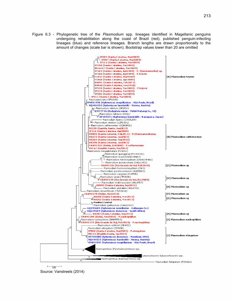
213
Figure 6.3 - Phylogenetic tree of the Plasmodium spp. lineages identified in Magellanic penguins undergoing rehabilitation along the coast of Brazil (red), published penguin-infecting lineages (blue) and reference lineages. Branch lengths are drawn proportionally to the amount of changes (scale bar is shown). Bootstrap values lower than 20 are omitted
Source: Vanstreels (2014)

214
Figu
re 6
.4 -
His
tolo
gica
l fin
ding
s as
soci
ated
with
avi
an m
alar
ia in
Mag
ella
nic
peng
uins
(S
phen
iscu
s m
agel
lani
cus)
. Hem
atox
ilin-
Eos
in.
Sca
le b
ars
= 15
µm
S
ourc
e: V
anst
reel
s (2
014)
Le
gend
: (a)
exo
eryt
hroc
ytic
mer
onts
in e
ndot
helia
l cel
ls (
arro
whe
ads)
with
in a
live
r ar
terio
le (
R00
40, P
. tej
erai
); (b
) pa
rasi
tized
ery
thro
cyte
(ar
row
head
) w
ithin
a c
ereb
ral b
lood
ves
sel (
CR
AM
2127
, P. n
ucle
ophi
lum
); (c
) di
ffuse
gra
nulo
cytic
int
erst
itial
pne
umon
ia,
cong
estio
n an
d ed
ema
(IF58
4, P
. te
jera
i); (
d) d
iffus
e ne
crot
izin
g sp
leni
tis w
ith a
n ex
oery
thro
cytic
mer
ont
with
in a
n en
doth
elia
l ce
ll of
a c
entra
l ar
terio
le
(arr
owhe
ad)
(R02
90,
P.
cath
emer
ium
); (e
) m
ultif
ocal
per
ivas
cula
r m
onon
ucle
ar h
epat
itis,
con
gest
ion
and
hem
osid
eros
is (R
0093
, Pla
smod
ium
sp
linea
ge E
).

215
6.5 DISCUSSION
Avian malaria has been considered one of the most significant infectious
diseases for wild and captive penguins (CLARKE; KERRY, 1993; JONES; SHELLAM,
1999b; PARSONS; UNDERHILL, 2005). In this study we demonstrated that Plasmodium
spp. infect Magellanic penguins at several rehabilitation centers along the Brazilian
coast, leading to substantial mortality and limiting the success of rehabilitation efforts for
this species.
6.5.1 Epidemiology of avian malaria in penguins at rehabilitation centers
While the overall infection rate ranged from 2.1% to 13.8% among rehabilitation
centers, when only samples collected and tested in a systematic manner with highly
sensitive diagnostic methods were considered the apparent prevalence was 7.75%,
which results in an estimated true prevalence between 6.6% and 13.5%. The only
comparable data in the literature is provided by the Southern African Foundation for the
Conservation of Coastal Birds (SANCCOB), an organization that rehabilitates African
penguins (Spheniscus demersus; APs) at South Africa; 17-34% of the APs admitted by
SANCCOB in 2001-2002 were positive to Plasmodium at some point during their
rehabilitation (PARSONS; UNDERHILL, 2005). Furthermore, we found that 4.95% of
MPs that survived through rehabilitation were Plasmodium-positive at some point,
compared to 10.81% of the deceased MPs, with Plasmodium-positive penguins facing a
44% higher mortality rate than those Plasmodium-negative (66.6% vs. 46.2%). This
result contrasts with the epidemiology at SANCCOB, where the release rate of
Plasmodium-positive individuals is not substantially different from that of the overall
population (PARSON; UNDERHILL, 2005).
There is, however, a critical difference to be considered when comparing our
findings with those of SANCCOB: there are no records of Plasmodium infections in wild

216
MPs, whereas APs are infected in the wild (JONES; SHELLAM, 1999a). Therefore whilst
we found no evidence to indicate that the MPs we studied carried the infection from the
wild, 30-35% of the Plasmodium-positive APs are already positive upon admission
(PARSONS; UNDERHILL, 2005). In this sense, the epidemiology of avian malaria in
Magellanic penguins in rehabilitation centers may resemble more that of captive
penguins in the Northern hemisphere than that of their South African counterparts.
In the Northern hemisphere, it is well established that avian malaria in penguins is
strongly modulated by seasonality, with cases concentrating in boreal spring-summer
due to climate-mediated fluctuations in mosquito abundance (SLADEN et al., 1979;
BEIER; STOSKOPF, 1980; FIX et al., 1988). We observed a consistent concentration of
all Plasmodium-positive cases in austral spring-summer (October to April), with the
probability of infection being positively associated with the number of days spent in
rehabilitation during that time of the year. In contrast, most MPs are received for
rehabilitation along the Brazilian coast during winter months (June to September),
particularly those oiled (Figure 6.2). Because rehabilitation protocols attempt to respect
the natural migration ecology of the species, the release of individuals during summer is
avoided (SILVA-FILHO; RUOPPOLO, 2007). As a result, only individuals considered
unfit for release until spring will be retained for release in the subsequent winter, besides
a small number of individuals received during summer months.
Oiled MPs tend to be in relatively better health condition when compared to those
that are not oiled (RODRIGUES et al., 2010), which is probably why they tend to be
rapidly rehabilitated and fit for release before summer (Figure 6.2). It is therefore most
likely that the higher frequency of Plasmodium infection in MPs that were not oiled
indirectly reflects the seasonality of the infection, and not a relation to oiling per se. The
same is probably true to the positive association between Plasmodium infection and
body mass, as individuals admitted during summer months are often admitted due to
moult problems and may have relatively higher body mass (subj. obs.).
The rehabilitation facility at Florianópolis had a substantially higher Plasmodium
incidence than the remaining, and a few characteristics of this facility may explain this.
While all facilities herein studied are to some extent near areas of Atlantic forest and/or
freshwater bodies, the facility at Florianópolis is perhaps more so than others. The

217
facility is located within a State Park (Parque Estadual do Rio Vermelho) and penguin
enclosures are directly under tree cover of Atlantic forest and less than 10 meters away
from a large freshwater lake (Lagoa da Conceição), which provides an optimal
environment for mosquito proliferation and close proximity to an abundant and diverse
avifauna. Additionally, this facility rehabilitates not only marine animals but also
terrestrial wildlife, including birds apprehended from illegal trafficking. The higher
infection rate of MPs at this facility thus probably reflects not a difference in susceptibility
or lineage virulence, but an increased probability of inoculation due to close contact with
mosquitoes and avian reservoirs of infection.
6.5.2 Plasmodium lineages infecting penguins and their pathology
It has been generally accepted that P. relictum and P. elongatum are the two
most significant species of Plasmodium that cause avian malaria in penguins (CLARKE;
KERRY, 1993; JONES; SHELLAM, 1999a; QUILLFELDT et al., 2011), along with an
isolated record of P. juxtanucleare (GRIM et al., 2003). Our results contrast this
perception, demonstrating that a broad variety of Plasmodium species can be found in
penguins undergoing rehabilitation in South America, including species that had not yet
been demonstrated to infect penguins (P. nucleophilum and P. cathemerium).
Regarding P. cathemerium, it is worth noting that even though we observed mature
microgametocytes with well-defined elongated rod-shaped pigment granules with
pointed ends (Appendix A), which are the defining features of P. (Haemamoeba)
cathemerium (VALKIŪNAS, 2005), in all cases we also observed late trophozoites and
young macrogametocytes with relatively large vacuoles surrounded by small round
pigment granules and relatively small meronts with scanty cytoplasm, which are
uncharacteristic to P. cathemerium. These findings may be interpreted as: (a) a host-
specific morphological variation of P. cathemerium, (b) a variant or subspecies of P.
cathemerium, (c) co-infection with a secondary unidentified lineage, or (d) a novel and
yet undescribed Plasmodium species. Considering the molecular evidence indicating

218
high sequence identity and phylogenetic proximity with a reference lineage of P.
cathemerium, however, we believe there is sufficient evidence to identify these lineages
as P. cathemerium.
Furthermore, the phylogenetic tree suggests that one of the lineages corresponds
to P. unalis (cluster D), which has not yet been recorded in penguins either. A number of
lineages could not be identified (C, E, F, G and H), and probably some of these
correspond to Plasmodium species that have not been reported in penguins as well.
Such broad diversity of plasmodia lineages corroborates the interpretation that the
infection of captive penguins results from local mosquitoes inoculating penguins with
Plasmodium spp acquired from the native birds in the surrounding of the penguin exhibit
(BEIER; STOSKOPF, 1980; CRANFIELD et al., 1994; GRACZYK et al., 1994; BUENO
et al., 2010). The predominance of P. relictum in Europe and Asia and P. relictum and P.
elongatum in North America therefore might not necessarily indicate a particular
susceptibility of penguins to those species, but merely reflect their local abundance in
those regions. It is worth noting that P. cathemerium, P. elongatum, P. nucleophilum and
P. relictum are renowned as generalist parasites with low host-specificity, infecting avian
species in several taxonomic orders (BENNETT et al., 1993).
Even though penguins seem to be susceptible to infection by a variety of
Plasmodium lineages occurring regionally, that is not to say that all lineages have similar
epidemiology or pathogenicity. In this study we found that P. tejerai and P. cathemerium
were lethal to respectively 73% and 33% in which they were detected, whilst other
lineages had only a few cases recorded and/or could not be demonstrated as the
leading factor causing death. These findings, combined with previous indications that
albeit less frequent P. relictum tends to produce more severe disease than P. elongatum
in penguins (BEIER; STOSKOPF, 1980; GRACZYK et al., 1994), raises the question on
whether the subgenus Haemamoeba is more pathogenic to these birds than other
plasmodia subgenera. Comparative pathology through experimental inoculation in
laboratory birds may assist in clarifying whether pathogenicity is intrinsically higher for
these lineages or if it reflects a susceptibility bias of penguins.
Overall, however, the histopathological lesions herein observed were generally
consistent among lineages. The most prominent pathological processes were
219
granulocytic pneumonia and splenitis and mononuclear hepatitis that was probably the
effect of vasculitis associated with the proliferation of Plasmodium within endothelial
cells in multiple tissues, particularly in the lungs, spleen and liver; in most cases death
likely culminated as a result of respiratory insufficiency from the marked pneumonia,
congestion and edema. These lesions are not unlike those observed in P. relictum and
P. elongatum infections in penguins in zoos and aquaria in the Northern hemisphere
(FLEISCHMAN et al., 1968a; FIX et al., 1988; KO et al., 2008). The only exception was
a MP deceased with a P. nucleophilum infection, in which no exoerythrocytic meronts
were present but a large number of intraerythrocytic parasites were observed within
blood vessels, suggesting a distinct pathogenesis. Future studies will be welcome to
clarify whether this is a consistent pattern for P. nucleophilum infections in penguins and
which pathophysiological mechanisms are involved.
6.5.3 Concurrent diseases
Two of the Plasmodium-positive MPs identified in this study had also been
identified as Avipoxvirus positive in a previous study (Appendix C) (NIEMEYER et al.,
2013). This probably occurs occasionally as both are mosquito-borne pathogens,
however, it can confuse interpretation of pathological findings due to overlapping
lesions; in particular, the only two individuals with necrotizing splenitis in Niemeyer et al.
(2013) were Plasmodium-positive, indicating this lesion is most likely related to avian
malaria and not poxvirosis. Other concurrent diseases included aspergillosis,
gastrointestinal helminthiasis, spleen amyloidosis, cholestasis, unidentified myocardium
cysts, and helminthes in the lungs, liver, air sacs and skin. Some of these findings, such
as aspergillosis and gastrointestinal helminthiasis have been previously reported in
penguins with avian malaria (e.g. GRINER; SHERIDAN, 1967; FIX et al., 1988).
However, there are no reports of helminthes in the respiratory system, skin or air sacs of
MPs (CLARKE; KERRY, 1993). Furthermore, the myocardium cysts herein observed
clearly were not Plasmodium and could correspond to either protozoan or metazoan

220
parasites. Additional studies will be conducted to clarify the identity and significance of
these parasites.
6.5.4 Implications for rehabilitation and conservation
The prevention of avian malaria in penguins in Northern hemisphere zoos has
largely relied on the oral administration of primaquine during summer (SILVA-FILHO;
RUOPPOLO, 2007). In Brazil, however, primaquine commerce is restricted by the
government due to concerns of resistance development in human malaria, and is thus
impossible to acquire for rehabilitation centers. As a result, these organizations are
forced to favor other prevention strategies, namely the isolation of penguins from
mosquitoes, which is often challenging and costly. Our findings provide a positive aspect
of the epidemiology of avian malaria in penguins at rehabilitation centers, namely that
the periods in which Plasmodium infections occur (summer) is directly opposite to the
period in which there are most penguins in rehabilitation (winter). As a result, these
facilities would benefit from narrowing their malaria-prevention efforts to a relatively
smaller number of individuals (those received and/or maintained between October and
April), becoming more cost-effective in the prevention and early diagnosis. This is a
relevant implication not only for permanent rehabilitation efforts, but also for oil spill
responses, when the physical and human resources required of malaria prevention,
diagnosis and treatment may be substantial (WOLFAARDT et al., 2008).
A number of MPs herein examined were considered released despite having
been Plasmodium-positive at some point. It must be considered that even non-
parasitemic penguins can relapse if treated with corticosteroids – and presumably the
same would occur if they became stressed – due to the persistence of exoerythrocytic
meronts (CRANFIELD et al., 1994). Brossy et al. (1999) expressed concern on the
potential of rehabilitation centers releasing APs with blood parasites, and perhaps this
concern should be even greater for MPs considering that Plasmodium has yet to be
recorded in the species in the wild. In the case of MPs, however, we reason that

221
because climate and environmental conditions are generally adverse and mosquitoes
occur very scarcely in the southeastern coast of Argentina (MITCHELL; DARSIE, 1985;
WRBU, 2014) and are absent at the Malvinas-Falkland Islands (MEDLOCK et al., 2010)
the probability of Plasmodium spill-over from a rehabilitated to a wild penguin is probably
very low. Even so, it is important to emphasize that pathogen spill-over to wild
populations should remain a prime and critical concern for rehabilitation centers, and
that even a low probability is nonetheless a significant risk to be considered and
addressed.

222
REFERENCES BEIER, J. C.; STOSKOPF, M. K. The epidemiology of avian malaria in black-footed penguins (Spheniscus demersus). Journal of Zoo Animal Medicine, v. 11, n. 4, p. 99-105, 1980. BEIER, J. C.; TRPIS, M. Incrimination of natural culicine vectors which transmit Plasmodium elongatum to penguins at the Baltimore Zoo. Canadian Journal of Zoology, v. 59, n. 3, p. 470-475, 1981. BENNETT, G. F.; BISHOP, M. A.; PEIRCE, M. A. Checklist of the avian species of Plasmodium Marchiafava & Celli, 1885 (Apicomplexa) and their distribution by avian family and Wallacean life zones. Systematic Parasitology, v. 26, p. 171-179, 1993. BENSCH, S.; HELLGREN, O.; PÉREZ-TRIS, J. MalAvi: a public database of malaria parasites and related haemosporidians in avian hosts based on mitochondrial cytochrome b lineages. Molecular Ecology Research, v. 9, p. 1353-1358, 2009. BROSSY, J. J.; PLÖS, A. L.; BLACKBEARD, J. M.; KLINE, A. Diseases acquired by captive penguins: what happens when they are released into the wild? Marine Ornithology, v. 27, p. 185-186, 1999. BUENO, M. G.; LOPEZ, R. P. G.; MENEZES, R. M. T.; COSTA-NASCIMENTO, M. J.; LIMA, G. F. M. C.; ARAÚJO, R. A. S.; GUIDA, F. J. V.; KIRCHGATTER, K. Identification of Plasmodium relictum causing mortality in penguins (Spheniscus magellanicus) from São Paulo Zoo, Brazil. Veterinary Parasitology, v. 173, n. 1-2, p. 123-127, 2010. CLARKE, J. R.; KERRY, K. R. Diseases and parasites of penguins. Korean Journal of Polar Research, v. 4, n. 2, p. 79-96, 1993. CRANFIELD, M. R.; GRACZYK, T. K.; BEALL, F. B.; IALEGGIO, D. M.; SHAW, M. L.; SKJOLDAGER, M. L. Subclinical avian malaria infections in African black-footed penguins (Spheniscus demersus) and induction of parasite recrudescence. Journal of Wildlife Diseases, v. 30, n. 3, p. 372-376, 1994.

223
FANTHAM, H. B.; PORTER, A. On a Plasmodium (Plasmodium relictum var. spheniscidae, n. var.), observed in four species of penguins. Proceedings of the Zoological Society of London, v. 114, p. 279-292, 1944. FIX, A. S.; WATERHOUSE, C.; GREINER, E. C.; STOSKOPF, M. K. Plasmodium relictum as a cause of avian malaria in wild-caught Magellanic penguins (Spheniscus magellanicus). Journal of Wildlife Diseases, v. 24, n. 4, p. 610-619, 1988. FLEISCHMAN, R. W.; SQUIRE, R. A.; SLADEN, W. J. L.; MELBY Jr., E. C. Malaria (Plasmodium elongatum) in captive African penguins (Spheniscus demersus). Journal of the American Veterinary Medical Association, v. 153, n. 7, p. 928-935, 1968b. GERING, E.; ATKINSON, C. T. A rapid method for counting nucleated erythrocytes on stained blood smears by digital image analysis. Journal of Parasitology, v. 90, n. 4, p. 879-881, 2004. GRACZYK, T. K.; CRANFIELD, M. R.; McCUTCHAN, T. F.; BICKNESE, E. J. Characteristics of naturally acquired avian malaria infections in naive juvenile African black-footed penguins (Spheniscus demersus). Parasitological Research, v. 80, p. 634-637, 1994. GRIM, K. C.; van der MERWE, E.; SULLIVAN, M.; PARSONS, N.; McCUTCHAN, T. F.; CRANFIELD, M. Plasmodium juxtanucleare associated with mortality in black-footed penguins (Spheniscus demersus) admitted to a rehabilitation center. Journal of Zoo and Wildlife Medicine, v. 34, n. 3, p. 250-255, 2003. GRINER, L. A.; SHERIDAN, B. W. Malaria (Plasmodium relictum) in penguins at the San Diego Zoo. Veterinary Clinical Pathology, v. 1, p. 7-17, 1967. HELLGREN, O.; WALDENSTRÖM, J.; BENSCH, S. A new PCR assay for simultaneous studies of Leucocytozoon, Plasmodium, and Haemoproteus from avian blood. Journal of Parasitology, v. 90, n. 4, p. 797-802, 2004. JARVI, S. I.; SCHULTZ, J. J.; ATKINSON, C. T. PCR diagnostics underestimate the prevalence of avian malaria (Plasmodium relictum) in experimentally-infected passerines. Journal of Parasitology, v. 88, n. 1, p. 153-158, 2002.

224
JONES, H. I.; SHELLAM, G. R. The occurrence of blood-inhabiting protozoa in captive and free-living penguins. Polar Biology, v. 21, p. 5-10, 1999a. JONES, H. I.; SHELLAM, G. R. Blood parasites in penguins, and their potential impact on conservation. Marine Ornithology, v. 27, p. 181-184, 1999b. KO, Kyeong-Nam; KANG, Sang-Chul; JUNG, Ji-Youl; BAE, Jong-Hee; KIM, Jae-Hoon. Avian malaria associated with Plasmodium spp. infection in a penguin in Jeju Island. Korean Journal of Veterinary Research, vol. 48, n. 2, p. 197-201, 2008. LEVIN, I. I.; PARKER, P. G. Hemosporidian parasites: impacts on avian hosts. In: MILLER, E.; FOWLER, M. Fowler’s zoo and wild animals medicine. Missouri: Elsevier Saunders, 2011. p. 356-363. MARTINSEN, E. S.; PERKINS, S. L. The diversity of Plasmodium and other Haemosporidians: The interesection of taxonomy, phylogenetics and genomics. In: CARLTON, J. M.; PERKINS, S. L.; DEITSCH, K. W. Malaria parasites: comparative genomics, evolution and molecular biology. Norfolk: Caister Academic Press, 2013. p. 1-15. MEDLOCK, J. M.; SCHAFFNER, F.; FONTENILLE, D. Invasive mosquitoes in the European associate continental and overseas territories. 2010. Disponível em: <http://www.ecdc.europa.eu/en/activities/sciadvice/_layouts/forms/Review_DispForm.aspx?ID=212&List=a3216f4c-f040-4f51-9f77-a96046dbfd72>. Acesso em: 16 feb. 2014. MITCHELL, C. J.; DARSIE, R. F. Mosquitoes of Argentina: Part II. Geographic distribution and bibliography (Diptera, Culicidae). Mosquito Systematics, v. 17, n. 4, p. 279-360, 1985. NIEMEYER, C.; FAVERO, C. M.; KOLESNIKOVAS, C. K. M.; BHERING, R. C. C.; BRANDÃO, P.; CATÃO-DIAS, J. L. Two different avipoxviruses associated with pox disease in Magellanic penguins (Spheniscus magellanicus) along the Brazilian coast. Avian Pathology, v. 42, n. 6, p. 546-551, 2013. PARSONS, N. J.; UNDERHILL, L. G. Oiled and injured African penguins Spheniscus demersus and other seabirds admitted for rehabilitation. African Journal of Marine Science, v. 27, n. 1, p. 289-296, 2005.

225
POSADA, D.; CRANDALL, K. A. MODELTEST: testing the model of DNA substitution. Bioinformatics, v. 14, p. 817-818, 1998. REICZIGEL, J.; FÖLDI, J.; ÓZSVÁRI, L. Exact confidence limits for prevalence of a disease with an imperfect diagnostic test. Epidemiology and Infection, v. 138, p. 1674-1678, 2010. RODRIGUES, S. C.; ADORNES, A. C.; SANTOS-FILHO, E. A.; SILVA-FILHO, R. P.; COLARES, E. P. Surviving probability indicators of landing juvenile Magellanic penguins arriving along the southern Brazilian coast. Brazilian Archives of Biology and Technology, v. 52, n. 2, p. 419-424, 2010. SILVA-FILHO, R. P.; RUOPPOLO, V. Sphenisciformes. In: CUBAS, Z. S.; SILVA, J. C. R.; CATÃO-DIAS, J. L. Tratado de animais selvagens – medicina veterinária. São Paulo: Roca, 2007. p. 309-323. SILVEIRA, P.; BELO, N. O.; LACORTE, G. A.; KOLESNIKOVAS, C. K. M.; VANSTREELS, R. E. T.; STEINDEL, M.; CATÃO-DIAS, J. L.; VALKIŪNAS, G.; BRAGA, É. M. Parasitological and new molecular-phylogenetic characterization of the malaria parasite Plasmodium tejerai in South American penguins. Parasitology International, v. 62, p. 165-171, 2013. SLADEN, W. J. L.; GAILEY-PHIPPS, J. J.; DIVERS, B. J. Medical problems and treatment of penguins at the Baltimore Zoo. International Zoo Yearbook, v. 19, p. 202-209, 1979. TAMURA, K.; PETERSON, D.; PETERSON, N.; STECHER, G.; NEI, M.; KUMAR, S. MEGA5: molecular evolutionary genetics analysis using maximum likelihood, evolutionary distance, and maximum parsimony methods. Molecular Biology and Evolution, v. 28, p. 2731-2739, 2011. THOMPSON, J .D.; GIBSON, T. J.; PLEWNIAK, F.; JEANMOUGIN, F.; HIGGINS, D. G. The CLUSTAL_X windows interface: flexible strategies for multiple sequence alignment aided by quality analysis tools. Nucleic Acids Research, v. 25, p. 4876-4882, 1997. VALKIŪNAS, G.; ZEHTINDJIEV, P.; DIMITROV, D.; KRIŽANAUSKIENĖ, A.; IEZHOVA, T. A.; BENSCH, S. Polymerase chain reaction-based identification of Plasmodium (Huffia) elongatum, with remarks on species identity of haemosporidian lineages deposited in GenBank. Parasitological Research, v. 102, p. 1185-1193, 2008.

226
VALKIŪNAS, G. Avian malaria parasites and other haemosporidia. Boca Ratón: CRC Press, 2005. 932 p. WILLIAMS, T. D.; BOERSMA, P. D. Magellanic penguin (Spheniscus magellanicus). In: WILLIAMS, T. D. The penguins: Spheniscidae. Oxford: Oxford University Press, 1995. p. 249-258. WOLFAARDT, A. C.; UNDERHILL, L. G.; ALTWEGG, R.; VISAGIE, J.; WILLIAMS, A. J. Impact of the Treasure oil spill on African penguins Spheniscus demersus at Dassen Island: case study of a rescue operation. African Journal of Marine Science, v. 30, n. 2, p. 405-419, 2008. WRBU. WALTER REED BIOSYSTEMATICS UNIT. VectorMap: Know the vector, know the threat. 2014. Disponível em: <http://www.vectormap.org/>. Acesso em: 16 fev. 2014.

227
Appendix A - Plasmodium spp. in Giemsa-stained blood smears of Magellanic penguins (Spheniscus magellanicus). Scale bar = 5 µm
Source: Vanstreels (2014) Legend: P. nucleophilum (CRAM2127): (a,b) trophozoites, (c,d) meronts, (e) coinfection by erythrocytic
meront and microgametocyte, (f) macrogametocyte, (g) microgametocyte, (h) co-infection by macro and microgametocyte; P. cathemerium (CRAM1923): (i) trophozoite, (j,k) meronts, (l-n) macrogametocytes, (o,p) microgametocytes

228
App
endi
x B
- S
umm
ary
of th
e re
habi
litat
ion
and
clin
ical
par
amet
ers
Pla
smod
ium
-pos
itive
Mag
ella
nic
peng
uins
und
ergo
ing
reha
bilit
atio
n al
ong
the
coas
t of B
razi
l

229
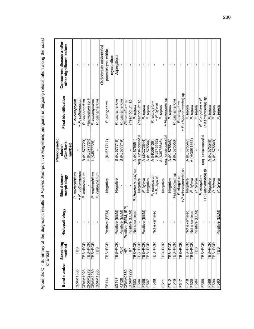
230
App
endi
x C
- S
umm
ary
of th
e di
agno
stic
res
ults
of P
lasm
odiu
m-p
ositi
ve M
agel
lani
c pe
ngui
ns u
nder
goin
g re
habi
litat
ion
alon
g th
e co
ast
of B
razi
l

231
Le
gend
: TB
S –
thin
blo
od s
mea
rs, P
CR
– n
este
d po
lym
eras
e ch
ain
reac
tion,
HP
– h
isto
path
olog
y, E
EM
– e
xoer
ythr
ocyt
ic m
eron
ts, I
EP
–
intra
eryt
hroc
ytic
par
asite
s

232
7 INVESTIGATION OF BLOOD PARASITES OF PYGOSCELID PENGUINS AT THE KING GEORGE AND ELEPHANT ISLANDS, SOUTH SHETLANDS ARCHIPELAGO, ANTARCTICA
7.1 ABSTRACT
Parasites may adversely affect the breeding success and survival of penguins,
potentially hampering the viability of their populations. We examined 161 pygoscelid
penguins (3 Pygoscelis adeliae, 98 Pygoscelis antarcticus, and 60 Pygoscelis papua) at
the South Shetlands Archipelago during the 2010–2011 summer; blood smears were
examined for 64 penguins (2 P. adeliae, 18 P. antarcticus, and 44 P. papua), and a PCR
test targeting Haemoproteus sp. and Plasmodium sp. was applied for 37 penguins (2 P.
adeliae, 17 P. antarcticus, 19 P. papua). No blood parasites were observed, and all PCR
tests were negative, leukocyte profiles were similar to those reported in other studies for
wild pygoscelid penguins, and all penguins were in good body condition and had no
external signs of disease. One specimen of chewing lice (Austrogoniodes sp.) was
recorded in one P. antarcticus at King George Island. Ticks (Ixodes uriae) were not
observed on the penguins, but were found on the ground near P. antarcticus nests at
King George Island. The absence of avian blood parasites in Antarctic penguins is
thought to result from the absence of competent invertebrate hosts in the climatic
conditions. Predicted climate changes may redefine the geographic distribution of
vector-borne pathogens, and therefore, the occurrence of blood parasites and their
invertebrate hosts should be monitored regularly in Antarctic birds, particularly in the
northernmost Antarctic Peninsula.

233
7.2 INTRODUCTION
Diseases and parasites may adversely affect breeding success and lead to the
mortality of penguins, potentially hampering the viability of their populations (BARBOSA;
PALACIOS, 2009). In particular, blood parasites are considered a potential threat to the
conservation of penguins (JONES; SHELLAM, 1999). Blood parasites known to infect
penguins include Babesia peircei (EARLÉ et al 1993), Borrelia sp. (YABSLEY et al.,
2012), Haemoproteus sp. (LEVIN et al., 2009), Leucocytozoon tawaki (ARGILLA et al.,
2013), Plasmodium spp. (RODHAIN; ADRIANNE, 1952), Trypanosoma eudyptulae
(JONES; WOEHLER, 1989), and microfilariae (MERKEL et al., 2007), all of which have
been demonstrated or are thought to be transmitted by arthropods. Avian malaria
(Plasmodium sp.) is considered most relevant due to its history of causing high-mortality
outbreaks in captive penguins and episodic mortalities in wild penguins (JONES;
SHELLAM, 1999). Mortality of wild penguins associated with Leucocytozoon sp.
(ARGILLA et al., 2013) have also been reported, raising concerns on the potential
significance of these parasites to penguin conservation.
While blood parasites have been reported in wild penguins in temperate and
tropical regions, all studies to date have failed to detect blood parasites in penguins or
other birds in the Antarctic (QUILLFELDT et al., 2011). There are very few records of
blood parasites in sub-Antarctic birds, and these were obtained in migratory seabirds
that most likely acquired the infection elsewhere, such as Haemoproteus-infected brown
skuas (Stercorarius antarcticus) at South Africa (PARSONS et al., 2010), or involve tick-
transmitted parasites such as Hepatozoon sp. in albatrosses (Diomedea spp.) at Bird
Island (PEIRCE; PRINCE, 1980). The most southern record of blood parasites in
penguins was provided by Laird (1950), who reported Plasmodium relictum in a yellow-
eyed penguin (Megadyptes antipodes) at Campbell Island (52°32’S 169°9’E).
With the exception of these rare reports, the lack of blood parasites in the sub-
Antarctic and Antarctic avifauna is considered to be due to an inability of their arthropod
hosts to survive under such relatively cold and harsh environmental conditions
(MERINO et al., 1997; JOVANI et al., 2001), although competition among competent

234
and non-competent ectoparasites could also play a role (MARTÍNEZ-ABRAÍN et al.,
2004). Temperature may also be a limiting factor for the development of the protozoan
parasites within the arthropod host (BENNING et al., 2002). Predicted climate changes
are expected to produce poleward changes in the geographic distribution of arthropods
and arthropod-transmitted pathogens (HARVELL et al., 2002), and regions such as sub-
Antarctic islands and the Antarctic Peninsula can be predicted to be at higher risks of
the emergence of conservation-threatening parasites such as avian malaria. In
particular, the Antarctic Peninsula has been one of the regions with the most
pronounced warming trends in the recent past (VAUGHAN et al., 2003) and is inhabited
by large populations of Pygoscelis sp. penguins (WOEHLER, 1993). This is of particular
concern as pygoscelid penguins are known to be highly susceptible to avian malaria
(RODHAIN; ADRIANNE ,1952; GRINER; SHERIDAN, 1967).
Blood smear examination is the standard method for the detection of
hemoparasites, while molecular methods such as polymerase chain reaction (PCR) are
highly valuable to detect cases of chronic infections and low parasitemia
(GARAMSZEGI, 2010). Blood smear examination also allows for differential erythrocyte
and leukocyte counts, conveying indirect information on the general health status and
the physiological and immune system responses to stress, reproduction, and pathogens
(VLECK et al., 2000; CLARK et al., 2009). In this study, we present integrated results
from physical examinations, blood smear examinations, and PCR to obtain information
on the health status and parasites of pygoscelid penguins at the King George and
Elephant Islands, South Shetlands Archipelago.
7.3 MATERIALS AND METHODS
A total 161 randomly selected adult penguins – 3 Adelie penguins (Pygoscelis
adeliae), 98 Chinstrap penguins (Pygoscelis antarcticus), and 60 Gentoo penguins
(Pygoscelis papua) – were examined between December 2010 and March 2011 at
breeding colonies in five sites at the South Shetlands Archipelago: Keller Peninsula

235
(62°4’55”S 58°24’32”W), Chabrier Rock (62°11’13”S 58°17’50”W), Demay Point
(62°12’25”S 58°27’7”W), and Thomas Point (62°10’23”S 58°28’31”W) at Admiralty Bay,
King George Island, and Stinker Point, Elephant Island (61°13’20”S 55°21’35”W).
Penguins were subjected to a rapid clinical examination for lesions and external signs of
disease (general external aspect, respiratory frequency, oral mucosae, eyes and
conjunctivae, limb and abdominal palpation, external lesions, cloaca). Ectoparasites
were carefully visually inspected for and collected in ethanol 70%; lice were mounted
and identified (CLAY, 1967; PALMA, 1978). Blood samples were collected from the
metatarsal or jugular veins of 64 of these penguins from January 31 to February 8, 2011
(late chick-rearing and early molting): 2 Adelie penguins (Thomas Point), 18 Chinstrap
penguins (7 at Keller Peninsula, 10 at Demay Point, and 1 at Thomas Point), and 44
Gentoo penguins (24 at Stinker Point, 18 at Keller Peninsula, and 2 at Thomas Point).
For each penguin, two thin blood smears were freshly prepared, then fixed in
methanol and stained with Diff-Quick and Wright-Rosenfeld (ROSENFELD, 1947).
Minimum 3 × 10⁴ erythrocytes per animal were examined for blood parasites under
1000x magnification light microscopy. Differential leukocyte counts were conducted for
200 leukocytes, and pro-erythroblasts and erythroblasts were quantified as a percentage
of the cells from the erythrocytic lineage (CLARK et al., 2009); the heterophil-to-
lymphocyte ratio was calculated (VLECK et al., 2000). Additionally, for 37 of these
penguins a small aliquot of blood was frozen for molecular testing: 2 Adelie penguins
(Thomas Point), 17 Chinstrap penguins (6 at Keller Peninsula, 10 at Demay Point, and 1
at Thomas Point), and 19 Gentoo penguins (17 at Keller Peninsula, 2 at Thomas Point).
DNA was extracted from the blood samples in the laboratory using a standard
phenol/chloroform and proteinase K extraction followed by ethanol precipitation
(SAMBROOK et al., 2001) and non-phenolic extraction (CARVALHO, 2010); DNA
extraction was verified and quantified through Nanodrop 2000 spectrophotometry
(ThermoScientific, Wilmington, USA). A nested PCR targeting sequences of the
cytochrome b gene of Haemoproteus sp. and Plasmodium sp. was used, as described
by Hellgren et al. (2004) (each reaction had 25 µL, with 75 ng of sample DNA; first
reaction with primers HaemNFI and HaemNR3, and second reaction with primers
HaemF and HaemR2). Blood samples from chicken experimentally infected with

236
Plasmodium gallinaceum or raised in arthropod-free environments were used as positive
and negative controls, respectively. Gel electrophoresis was conducted to visualize
amplification products, using 3 % agarose gel, SYBR Safe (Invitrogen, Carlsbad, USA),
and a high-resolution imaging system (Gel Doc EZ System – Bio-Rad, Hercules, USA).
Mann-Whitney tests were used to compare relative leukocyte counts between
Gentoo penguins sampled at Stinker Point and Keller Peninsula, and between Chinstrap
and Gentoo penguins sampled at Keller Peninsula. Significance level was 0.05 for all
tests. All research procedures were conducted under the Brazilian Antarctic Project
(PROANTAR) and complying with the Scientific Committee on Antarctic Research
(SCAR).
7.4 RESULTS
All penguins had no clinical signs of disease. One male chewing louse was
present in a Gentoo penguin at Keller Peninsula, King George Island. We determined
that louse belonged to the genus Austrogoniodes. No other ectoparasites were
observed on the penguins. Even though not specifically searched for, one adult female
tick Ixodes uriae was found amidst guano and rocks at Chabrier Rock, an islet with a
breeding colony of approximately 800 Chinstrap penguins, 31 of which were examined
and presented no ticks. No blood parasites were observed on the blood smears, and all
PCR tests were negative. Differential leukocyte counts are presented in Table 7.1;
results from Thomas Point are omitted due to the low sample sizes (2 P. adeliae, 1 P.
antarcticus, and 2 P. papua). All animals had a few circulating polychromatic
erythrocytes (<10%), which were abundant (approx. 20%) only in two Gentoo penguins.
Erythroblasts and pro-erythroblasts were seen sporadically regardless of the abundance
of polychromatic erythrocytes, having occurred in one Chinstrap penguin (6%) and in
nine Gentoo penguins (20%). Significant differences between Gentoo penguins from
Keller Peninsula and Stinker Point occurred in monocyte (P = 0.007), heterophil (P =
0.039), and lymphocyte counts (P = 0.024), but not in eosinophil (P = 0.980) and

237
basophil counts (P = 0.919). Significant differences between Chinstrap and Gentoo
penguins from Keller Peninsula occurred in heterophil (P = 0.001), lymphocyte (P =
0.001), eosinophil (P = 0.001), and monocyte counts (P = 0.036), but not in basophil
counts (P = 0.759).
Table 7.1 - Differential leukocyte counts (Mean ± S.D.) for penguins at Demay Point (DP), Keller
Peninsula (KP) and Stinker Point (SP), January-February 2011 Chinstrap penguins Gentoo penguins DP (n=10) KP (n=7) KP (n=18) SP (n=24) Heterophils (%) 47.6 ± 11.0 37.2 ± 3.8 52.3 ± 8.1 46.7 ± 9.0 Lymphocytes (%) 48.7 ± 11.3 59.8 ± 4.2 41.3 ± 7.6 47.0 ± 9.6 Eosinophils (%) 2.9 ± 2.7 1.1 ± 0.7 5.1 ± 2.7 5.7 ± 4.0 Basophils (%) 0.2 ± 0.5 0.1 ± 0.2 0.1 ± 0.2 0.2 ± 0.5 Monocytes (%) 0.7 ± 0.6 1.8 ± 0.7 1.2 ± 1.4 0.3 ± 0.5 Heterophil-to-Lymphocyte Ratio 1.09 ± 0.59 0.63 ± 0.11 1.33 ± 0.40 1.08 ± 0.46
7.5 DISCUSSION
Austrogoniodes spp. chewing lice and I. uriae ticks are common parasites of sub-
Antarctic and Antarctic penguins, including at the South Shetlands Archipelago
(BARBOSA et al., 2011). We were not able to identify the species of the recovered
Austrogoniodes louse because its reproductive tract was damaged; however, it is most
likely to be Austrogoniodes gressitti (see CLAY, 1967). While it is unknown whether
Austrogoniodes spp. are competent hosts or vectors to any of the blood parasites
recorded so far on penguins, I. uriae is thought to play a key role in the transmission of
Babesia peircei and Borrelia sp. to penguins (EARLÉ et al., 1993; YABSLEY et al.,
2012) and between penguins and long-distance migrating seabirds such as skuas and
storm petrels (OLSÉN et al.,1995). Our results suggest an overall low prevalence and
intensity of infection by Austrogoniodes sp. and I. uriae in pygoscelid penguins at the
sampling sites; however, this may be an underestimate associated with visual inspection
(CLAYTON; DROWN, 2001). In some instances, extreme parasitism by I. uriae has

238
been shown to hamper reproduction or lead to mortality (GAUTHIER-CLERC et al.,
1998; MANGIN et al., 2003). We observed no apparent signs of disease or negative
effects from ectoparasitism (which does not exclude the possibility of subclinical
disease); however, it is also possible that sick individuals skipped breeding and/or were
distributed elsewhere within the breeding colonies and could therefore have been less
likely to be sampled in the study.
Low but statistically significant differences were observed in the leukocyte profiles
among localities and penguin species, particularly in the heterophil, lymphocyte, and
monocyte counts. Low-level changes in leukocyte differential counts could potentially
reflect differences in exposure to and resultant physiological changes associated with
microbial infections, parasites, stress, and breeding (VLECK et al., 2000; CLARK et al.,
2009). However, as all relative leukocyte counts were similar to those observed in other
studies in wild pygoscelid penguins (e.g., HAWKEY et al., 1989; VLECK et al., 2000)
and both erythrocyte and leukocyte results were well within the reference values for
healthy individuals of other avian species (see CLARK et al., 2009), it is difficult to
determine the biological significance, if any, of the observed differences. The heterophil-
to-lymphocyte ratios observed in this study for Chinstrap and Gentoo penguins (mean
0.63 – 1.33) were considerably lower than those reported by other authors for captive
penguins (mean > 1.8) (HAWKEY et al., 1985; ISIS, 2002), and this may reflect stress –
as occurs in captivity – is known to significantly increase absolute and relative heterophil
counts (VLECK et al., 2000).
The absence of avian blood parasites in this and in previous studies should not
dissuade future studies in the sub-Antarctic and Antarctic region. The emergence of
pathogens in regions where they had been historically absent can have profound
ecological and conservation consequences, as exemplified by the population decreases
and extinctions of native Hawaiian birds during the twentieth century following the
introduction of avian malaria and poxvirus (ATKINSON; LAPOINTE, 2009). More
recently, the introduction of avian malaria to the Galápagos Islands has raised great
concern on the potential implications for the conservation of the Galápagos penguin
(Spheniscus mendiculus) and other endemic birds (LEVIN et al., 2009). Because
penguins are remarkably susceptible to avian malaria (JONES; SHELLAM, 1999), a

239
climate change-induced emergence of this disease in sub-Antarctic and Antarctic
regions could have dramatic consequences. Furthermore, tick-transmitted pathogens
such as Babesia sp. or Hepatozoon sp. can also be expected to better thrive in these
regions if they became warmer. It is therefore important to investigate and monitor these
pathogens in the sub-Antarctic and Antarctic region to assure their early detection if ever
they are to occur. The South Shetlands Archipelago, because of their relatively mild
climate and geographic positioning close to the northern tip of the Antarctic Peninsula, is
a strategic location for monitoring these parasites. In this context, it will be important for
the Antarctic programs, including that of Brazil (PROANTAR), to develop and maintain
consistent and long-term monitoring of the wildlife health in the region.

240
REFERENCES ARGILLA, L. S.; HOWE, L.; GARTRELL, D.; ALLEY, M. R. High prevalence of Leucocytozoon spp. in the endangered yellow-eyed penguin (Megadyptes antipodes) in the sub-Antarctic regions of New Zealand. Parasitology, v. 140, n. 5, p. 672-682, 2013. ATKINSON, C. T.; LAPOINTE, D. A. Introduced avian diseases, climate change, and the future of Hawaiian honeycreepers. Journal of Avian Medicine and Surgery, v. 23, n. 1, p. 53-63, 2009. BARBOSA, A.; PALACIOS, M. J. Health of Antarctic birds: a review of their parasites, pathogens and diseases. Polar Biology, v. 32, p. 1095-1115, 2009. BARBOSA, A.; BENZAL, J.; VIDAL, V.; D'AMICO, V.; CORIA, N.; DIAZ, J.; MOTAS, M.; PALACIOS, M. J.; CUERVO, J. J.; ORTIZ, J.; CHITIMIA, L. Seabird ticks (Ixodes uriae) distribution along the Antarctic peninsula. Polar Biology, v. 34, p. 1621-1624, 2011. BENNING, T. L.; LAPOINTE, D.; ATKINSON, C. T.; VITOUSEK, P. M. Interactions of climate change with biological invasions and land use in the Hawaiian Islands: modeling the fate of endemic birds using a geographic information system. Proceedings of the National Academy of Science, v. 99, p. 14246-14249, 2002. CARVALHO, A. L. Estudo de um polimorfismo no gene da cadeia pesada b da miosina (CPbM). Coimbra: Universidade de Coimbra, 2010. Disponível em: <https:// woc.uc.pt/zoologia/getFile.do?tipo=2&id=5273>. Acesso em: 11 jun. 2013. CLARK, P.; BOARDMAN, W.; RAIDAL, S. Atlas of clinical avian hematology. Oxford: Blackwell, 2009. 184 p. CLAY, T. Mallophaga (biting lice) and Anoplura (sucking lice) Part I: Austrogoniodes (Mallophaga) parasitic of penguins (Sphenisciformes). In: GRESSITT, J. L. Entomology of Antarctica. Washington: American Geophysical Union, 1967. p. 149-155. CLAYTON, D. H.; DROWN, D. M. Critical evaluation of five methods for quantifying chewing lice (Insecta: Phthiraptera). Journal of Parasitology, v. 87, n. 6, p. 1291-1300, 2001.

241
EARLÉ, R. A.; HUCHZERMEYER, F. W.; BENNETT, G. F.; BROSSY, J. J. Babesia peircei sp. nov. from the jackass penguin. African Zoology, v. 28, n. 2, p. 88-90, 1993. GARAMSZEGI, L. Z. The sensitivity of microscopy and PCR-based detection methods affecting estimates of prevalence of blood parasites in birds. Journal of Parasitology, v. 96, n. 6, p. 1197-1203, 2010. GAUTHIER-CLERC, M.; CLERQUIN, Y.; HANDRICH, Y. Hyperinfestation by ticks Ixodes uriae: a possible cause of death in adult king penguins, a long-lived seabird. Colonial Waterbirds, v. 21, p. 229-233, 1998. GRINER, L. A.; SHERIDAN, B. W. Malaria (Plasmodium relictum) in penguins at the San Diego Zoo. Veterinary Clinical Pathology, v. 1, p. 7-17, 1967. HARVELL, C. D.; MITCHELL, C. E.; WARD, J. R.; ALTIZER, S.; DOBSON, A. P.; OSTFELD, R. S.; SAMUEL, M. D. Climate warming and disease risks for terrestrial and marine biota. Science, v. 296, p. 2158-2162, 2002. HAWKEY, C.; SAMOUR, H. J.; HENDERSON, G. M.; HART, M. G. Haematological findings in captive gentoo penguins (Pygoscelis papua) with bumblefoot. Avian Pathology, v. 14, p. 251-256, 1985. HAWKEY, C. M.; HORSLEY, D. T.; KEYMER, I. F. Haematology of wild penguins (Sphenisciformes) in the Falkland islands. Avian Pathology, v. 18, n. 3, p. 495-502, 1989. HELLGREN, O.; WALDENSTRÖM, J.; BENSCH, S. A new PCR assay for simultaneous studies of Leucocytozoon, Plasmodium, and Haemoproteus from avian blood. Journal of Parasitology, v. 90, n. 4, p. 797-802, 2004. ISIS. INTERNATIONAL SPECIES INFORMATION SYSTEM. Reference ranges for physiological data values. Apple Valley: ISIS, 2002. JONES, H. I.; SHELLAM, G. R. Blood parasites in penguins, and their potential impact on conservation. Marine Ornithology, v. 27, p. 181-184, 1999.

242
JONES, H. I.; WOEHLER, E. J. A new species of blood trypanosome from little penguins (Eudyptula minor) in Tasmania. Journal of Protozoology, v. 36, n. 4, p. 389-390, 1989. JOVANI, R.; TELLA, J. L.; FORERO, M. G.; BERTELLOTTI, M.; BLANCO, G.; CEBALLOS, O.; DONÁZAR, J. Á. Apparent absence of blood parasites in the Patagonian seabird community: is it related to the marine environment? Waterbirds, v. 24, p. 430-433, 2001. LAIRD, M. Some blood parasites of New Zealand. Zoological Publications of Victoria University College, v. 5, p. 1-20, 1950. LEVIN, I. I.; OUTLAW, D. C.; VARGAS, F. H.; PARKER, P. G. Plasmodium blood parasite found in endangered Galapagos penguins (Spheniscus mendiculus). Biological Conservation, v. 142, p. 3191-3195, 2009. MANGIN, S.; GAUTHIER-CLERC, M.; FRENOT, Y.; GENDNER, J. P.; LE MAHO, Y. Ticks Ixodes uriae and the breeding performance of a colonial seabird, king penguin Aptenodytes patagonicus. Journal of Avian Biology, v. 34, p. 30-34, 2003. MARTÍNEZ-ABRAÍN, A.; ESPARZA, B.; ORO, D. Lack of blood parasites in bird species: does absence of blood parasite vectors explain it all? Ardeola, v. 51, p. 225-232, 2004. MERINO, S.; BARBOSA, A.; MORENO, J.; POTTI, J. Absence of haematozoa in a wild chinstrap penguin Pygoscelis antarctica population. Polar Biology, v. 18, p. 227-228, 1997. MERKEL, J.; JONES, H. I.; WHITEMAN, N. K.; GOTTDENKER, N.; VARGAS, H.; TRAVIS, E. K.; MILLER, R. E.; PARKER, P. G. Microfilariae in Galápagos penguins (Spheniscus mendiculus) and flightless cormorants (Phalacrocorax harrisi). Journal of Parasitology, v. 93, n. 3, p. 495-503, 2007. OLSÉN, B.; DUFFY, D. C.; JAENSON, T. G. T.; GYLFE, A.; BONNEDAHL, J.; BERGSTRÖM, S. Transhemispheric exchange of Lyme disease spirochetes by seabirds. Journal of Clinical Microbiology, v. 33, p. 3270-3274, 1995. PALMA, R. L. Slide-mounting of lice: a detailed description of the Canada balsam technique. New Zealand Entomologist, v. 6, p. 432-436, 1978.

243
PARSONS, N. J.; PEIRCE, M. A.; STRAUSS, V. New species of haematozoa in Phalacrocoracidae and Stercorariidae in South Africa. Ostrich, v. 81, p. 103-108, 2010. PEIRCE, M. A.; PRINCE, P. A. Hepatozoon albatrossi sp. nov. (Eucoccida: Hepatozoidae) from Diomedea spp. in the Antarctic. Journal of Natural History, v. 14, n. 3, p. 447-452, 1980. QUILLFELDT, P.; ARRIERO, E.; MARTÍNEZ, J.; MASELLO, J. F.; MERINO, S. Prevalence of blood parasites in seabirds: a review. Frontiers in Zoology, v. 8, n. 26, 2011. RODHAIN, J.; ANDRIANNE, V.-F. Deux nouveaux cas d'infestation par Plasmodium chez des pingouins. Annales de Parasitologie Humaine et Comparée, v. 27, n. 6, p. 573-577, 1952. ROSENFELD, G. Corante pancrômico para hematologia e citologia clínica: Nova combinação dos componentes do May-Grunwald e do Giemsa num só corante de emprego rápido. Memórias do Instituto Butantan, v. 20, p. 329-335, 1947. SAMBROOK, K. J.; RUSSEL, D. W.; SAMBROOK, J. Molecular cloning: a laboratory manual. New York: Cold Spring Harbor Laboratory Press, 2001. 2344 p. VAUGHAN, D. G.; MARSHALL, G. J.; CONNOLLEY, W. M.; PARKINSON, C.; MULVANEY, R.; HODGSON, D. A.; KING, J. C.; PUDSEY, C. J.; TURNER, J. Recent rapid regional climate warming on the Antarctic peninsula. Climate Change, v. 60, p. 243-274, 2003. VLECK, C. M.; VERTALINO, N.; VLECK, D.; BUCHER, T. L. Stress, corticosterone, and heterophil to lymphocyte ratios in free-living Adelie penguins. Condor, v. 102, p. 392-400, 2000. WOEHLER, E. J. The distribution and abundance of Antarctic and Subantarctic penguins. Cambridge: Scientific Committee on Antarctic Research, 1993. 76 p. YABSLEY, M. J.; PARSONS, N. J.; HORNE, E. C.; SHOCK, B. C.; PURDEE, M. Novel relapsing fever Borrelia detected in African penguins (Spheniscus demersus) admitted to two rehabilitation centers in South Africa. Parasitology Research, v. 110, n. 3, p. 1125-1130, 2012.

244
8 CONSIDERAÇÕES FINAIS
Desde o primeiro registro de malária aviária em um pinguim-rei na Sociedade
Zoológica de Londres pelo Dr. H. Harold Scott, em 1927, um número crescente de
registros desta enfermidade em Sphenisciformes tem sido publicados. No Brasil, há
algumas décadas existem relatos anedóticos de malária aviária em pinguins-de-
Magalhães em reabilitação e cativeiro na costa brasileira. Embora estes pinguins sejam
reabilitados no Brasil anualmente às centenas ou milhares, nunca havia sido realizado
um estudo detalhado da malária aviária nestas aves no país. Neste cenário, a medicina
veterinária brasileira baseava-se exclusivamente na literatura acerca da malária aviária
em pinguins cativos na América do Norte e Europa, assumindo que a enfermidade
exibiria um comportamento epidemiológico e patológico semelhante no Brasil.
Nesta tese foi apresentada uma ampla investigação retrospectiva e prospectiva
da ocorrência da malária aviária em pinguins-de-Magalhães em centros de reabilitação
ao largo da costa brasileira. Os resultados revelam um perfil epidemiológico com
algumas semelhanças àquele observado em pinguins cativos no Hemisfério Norte, em
especial na sua ocorrência na forma de surtos. Estes surtos têm potencial para causar
a morbidade e mortalidade de grande quantidade de animais em reabilitação, e seu
desenvolvimento rápido requer que os centros de reabilitação estejam preparados
antecipadamente para que tenham um sucesso significativo na mitigação dos seus
impactos. A estreita sazonalidade desta enfermidade, cuja incidência restringe-se aos
meses de outubro a abril, é um aspecto positivo para os centros de reabilitação uma
vez que permite que estes concentrem seus esforços de prevenção e diagnóstico
durante um período em que normalmente há um número relativamente pequeno de
pinguins em reabilitação. Por outro lado, a constatação de que a enfermidade ocorre ao
largo de toda a costa brasileira aparentemente sem um gradiente latitudinal significativo
indica que a malária aviária deve ser prevenida e monitorada em pinguins em qualquer
região do nosso país, inclusive no extremo sul.

245
Além do estudo epidemiológico geral, uma contribuição significativa deste estudo
foi a ampliação do número de espécies de Plasmodium registradas em pinguins. Até
então, apenas três espécies deste gênero haviam sido reportadas em pinguins, sendo
P. relictum e P. elongatum classicamente consideradas como as mais relevantes.
Através da combinação de técnicas morfológicas e biomoleculares foi possível agregar
três novas espécies a esta lista, P. tejerai, P. nucleophilum e P. cathemerium. Assim, o
estudo apresenta uma mudança de paradigma ao revelar que a diversidade de
plasmódios aviários que podem infectar os pinguins é consideravelmente maior do que
se imaginava, e que é provável que muitas outras espécies de Plasmodium spp.
venham a ser registradas nestas aves no futuro.
Por outro lado, embora não existam colônias reprodutivas de pinguins na costa
brasileira, nosso país tem a possibilidade de conduzir estudos científicos destas aves
em seu ambiente reprodutivo. Embora o resultado deste estudo seja consistente com
outros estudos que não detectaram hemoparasitas em pinguins na região Antártica e
arquipélagos adjacentes, a execução de investigações desta natureza é importante
para detectar não apenas uma potencial expansão da malária aviária às regiões mais
austrais mas também para revelar a ocorrência de hemoparasitas veiculados por outros
artrópodes. Em tempos de mudanças ambientais e climáticas abruptas, há muito se
anteveem mudanças na distribuição de patógenos veiculados por artrópodes, e as
populações de aves marinhas na região da Península Antártica podem ser um foco
estratégico para o monitoramento de mudanças epidemiológicas.
Além destes estudos prospectivos e retrospectivos, a tese apresenta uma
revisão e compilação dos aspectos da literatura científica acerca desta enfermidade e
outras hemosporidioses, com uma abordagem voltada aos médicos veterinários
atuando na reabilitação e manutenção em cativeiro de pinguins e outras aves no Brasil.
À parte desta revisão de foco mais generalista, uma abordagem crítica e meta-análise
de todas as publicações acerca da ocorrência de hemoparasitas intracelulares em
pinguins permitiu agregar uma grande quantidade de informações que muitas vezes
estão dispersas e fragmentadas. Esta revisão, juntamente com o estudos prospectivos
e retrospectivos apresentados no restante da tese, estabelece um panorama mais claro
da ocorrência das hemoparasitoses nestas aves e das suas implicações

246
epidemiológicas e para a conservação, fundamentando análises subsequentes e
apontando as atuais lacunas de conhecimento.
Em suma, a malária aviária é uma enfermidade relevante para as mais diversas
espécies de pinguins em todo o mundo e possui particular importância para a
reabilitação e conservação destas aves no litoral brasileiro, merecendo maiores
investigações no futuro.

247
APÊNDICE
Plasmodium (Haemamoeba) tejerai Anilha “IF-593”; 25/03/2009; Florianópolis, SC, Brasil. Giemsa; Barra de escala = 5 µm
Legenda: (a-c) trofozoítos, (d-g) merontes, (h-j) macrogametócitos, (k,l) microgametócitos.

248
Plasmodium (Huffia) elongatum Anilha “IF-508”; 25/03/2009; Florianópolis, SC, Brasil. Giemsa; Barra de escala = 5 µm
Legenda: (a-d) trofozoítos, (e,f) merontes, (g) co-infecção por trofozoíto e meronte, (h-i) macrogametócitos, (j) co-infecção por dois macrogametócitos, (k,l) microgametócitos.

249
Plasmodium (Novyella) nucleophilum Anilha “CRAM-1886”; 12/10/2009; procedente de Salvador, BA, Brasil; amostrado em
Rio Grande, RS, Brasil. Giemsa; Barra de escala = 5 µm
Legenda: (a-c) trofozoítos, (d-f) merontes, (g,h) macrogametócitos, (i,j) microgametócitos, (k) co-infecção por macrogametócito e microgametócito, (l) co-infecção por mertonte e microgametócito.

250
Plasmodium (Haemamoeba) cathemerium Anilha “CRAM-1923”; 12/10/2009; procedente de Salvador, BA, Brasil; amostrado em
Rio Grande, RS, Brasil. Giemsa; Barra de escala = 5 µm
Legenda: (a-c) trofozoítos, (d-f) merontes, (g-i) macrogametócitos, (j-l) microgametócitos.