Estimation and Diversity of Phylloplane Mycobiota on Selected Plants in a Mediterranean-Type...
10
Estimation and Diversity of Phylloplane Mycobiota on Selected Plants in a Mediterranean–Type Ecosystem in Portugal J. Ina ´cio, 1 P. Pereira, 2 M. de Carvalho, 2 A ´ . Fonseca, 1 M.T. Amaral-Collac ¸o, 2 I. Spencer-Martins 1 1 Centro de Recursos Microbiolo ´gicos (CREM), Biotechnology Unit, Faculty of Sciences and Technology, Universidade Nova de Lisboa, Caparica, Portugal 2 Department of Biotechnology, Instituto Nacional de Engenharia e Tecnologia Industrial (INETI), Lisbon, Portugal Received: 17 January 2002; Accepted: 30 July 2002; Online publication: 15 October A B S T R A C T Mediterranean ecosystems have not been consistently investigated as natural habitats for mi- crobes in general, and fungi in particular. Here we present the results of a survey of epiphytic mycobiota (filamentous fungi and yeasts) on the phylloplane of selected plants in the Arra ´bida Natural Park, an ecosystem of Mediterranean characteristics in Portugal, using conventional culture-dependent isolation methods. Leaves from the species Acer monspessulanum and Quercus faginea (deciduous trees) and Cistus albidus, Pistacia lentiscus, and Osyris quadripartita (evergreen shrubs) were collected twice a year for two consecutive years, at two distinct locations of Serra da Arra ´bida: the more humid northern slope and the drier southern slope. A total of 1029 strains of filamentous fungi and 540 strains of yeasts were isolated, which represented at least 36 and 46 distinct species, respectively. Total counts were higher on the plants from the northern slope and there was a general increase from spring to autumn, notably on the decid- uous trees for the yeasts. Plant species that had higher numbers of leaf colonists (A. monspes- sulanum, C. albidus, and Q. faginea) also yielded a wider range of species. Among the filamentous fungi there was a predominance of species of ascomycetous affinity, whereas ba- sidiomycetous species dominated among yeast isolates. Some of the taxa recovered were com- mon to other phylloplane studies (e.g., ubiquitous molds and yeasts such as Cladosporium spp. and Cryptococcus spp., respectively), but less common species were also found, some of which appeared to represent undescribed taxa. Interestingly, a few species seemed to be associated with a particular plant, notably in the case of the evergreen shrub C. albidus. However, for a con- siderable number of fungi and yeasts the same taxon was recovered throughout the year from more than one plant and at both sites, suggesting that such species might be genuine phylloplane inhabitants (or at least of aerial plant surfaces) even though they appeared not to display host specificity. Correspondence to: A ´ . Fonseca; E-mail: [email protected] Microb Ecol (2002) 44:344–353 DOI: 10.1007/s00248-002-2022-z ȑ 2002 Springer-Verlag New York Inc.
Transcript of Estimation and Diversity of Phylloplane Mycobiota on Selected Plants in a Mediterranean-Type...

Estimation and Diversity of Phylloplane Mycobiota on SelectedPlants in a Mediterranean–Type Ecosystem in Portugal
J. Inacio,1 P. Pereira,2 M. de Carvalho,2 A. Fonseca,1 M.T. Amaral-Collaco,2 I. Spencer-Martins1
1 Centro de Recursos Microbiologicos (CREM), Biotechnology Unit, Faculty of Sciences and Technology,
Universidade Nova de Lisboa, Caparica, Portugal2 Department of Biotechnology, Instituto Nacional de Engenharia e Tecnologia Industrial (INETI), Lisbon,
Portugal
Received: 17 January 2002; Accepted: 30 July 2002; Online publication: 15 October
A B S T R A C T
Mediterranean ecosystems have not been consistently investigated as natural habitats for mi-
crobes in general, and fungi in particular. Here we present the results of a survey of epiphytic
mycobiota (filamentous fungi and yeasts) on the phylloplane of selected plants in the Arrabida
Natural Park, an ecosystem of Mediterranean characteristics in Portugal, using conventional
culture-dependent isolation methods. Leaves from the species Acer monspessulanum and
Quercus faginea (deciduous trees) and Cistus albidus, Pistacia lentiscus, and Osyris quadripartita
(evergreen shrubs) were collected twice a year for two consecutive years, at two distinct locations
of Serra da Arrabida: the more humid northern slope and the drier southern slope. A total of
1029 strains of filamentous fungi and 540 strains of yeasts were isolated, which represented at
least 36 and 46 distinct species, respectively. Total counts were higher on the plants from the
northern slope and there was a general increase from spring to autumn, notably on the decid-
uous trees for the yeasts. Plant species that had higher numbers of leaf colonists (A. monspes-
sulanum, C. albidus, and Q. faginea) also yielded a wider range of species. Among the
filamentous fungi there was a predominance of species of ascomycetous affinity, whereas ba-
sidiomycetous species dominated among yeast isolates. Some of the taxa recovered were com-
mon to other phylloplane studies (e.g., ubiquitous molds and yeasts such as Cladosporium spp.
and Cryptococcus spp., respectively), but less common species were also found, some of which
appeared to represent undescribed taxa. Interestingly, a few species seemed to be associated with
a particular plant, notably in the case of the evergreen shrub C. albidus. However, for a con-
siderable number of fungi and yeasts the same taxon was recovered throughout the year from
more than one plant and at both sites, suggesting that such species might be genuine phylloplane
inhabitants (or at least of aerial plant surfaces) even though they appeared not to display host
specificity.
Correspondence to: A. Fonseca; E-mail: [email protected]
Microb Ecol (2002) 44:344–353
DOI: 10.1007/s00248-002-2022-z
� 2002 Springer-Verlag New York Inc.

Introduction
The surface of plant leaves, usually referred to as the
phylloplane, represents an important terrestrial habitat and
harbors a wide range of microorganisms [3 and references
therein]. Fungi (encompassing both filamentous and yeast
taxa) constitute a major component of the phylloplane
microbiota [2, 6, 11, 12, 16, 27], but most investigations on
their occurrence in this type of habitat have focused mainly
on the inventory and description of new or specific taxa,
generally in a restricted number of ecosystems and in some
cases with emphasis on phytopathogenic taxa [1, 7, 9, 13,
18, 20, 28, 38]. Both the surface and the interior of leaves
can be colonized by fungi with varied consequences for the
host plants [23]. The epiphytic fungi, the surface inhabit-
ants, depend on a thin film of nutrients that are deposited
on the leaf from the atmosphere (e.g., contained in pollen
grains) by insects and other organisms or that are exuded
from the leaf itself [21, 22, 41]. As for other microbes, the
concentration of fungi on the phylloplane is influenced by
factors such as nutrient availability, humidity, leaf age and
type, presence of inhibitors, immigration (arrival of viable
propagules on the phylloplane), and emigration (removal
or physical loss of viable propagules) [3, 22].
In the case of filamentous fungi growth, on leaf surfaces
is generally enhanced when moisture levels are high and
temperatures are moderate [7, 13, 22]. These microor-
ganisms have been found on leaf surfaces of many plant
species in both temperate and tropical ecosystems [6, 7,
11, 12, 18, 23, 32]. In general, there appears to be a marked
dominance of deuteromycetes, mostly of ascomycetous
affinity. The most common genera found on the phyllo-
plane are Cladosporium, Aspergillus, Alternaria, Aureo-
basidium, and Epicoccum [1, 6, 7, 16, 23]. Only occasional
colonies of either basidiomycetous or zygomycetous mi-
crofungi have been reported [6, 14, 20]. Yeasts are also
found on the phylloplane of different plant species in
both temperate and tropical regions [27, 35, 38]. As
observed in the case of other aerial plant surfaces [8, 28],
there appears to be a marked dominance of basidiomy-
cetous yeasts on the phylloplane, namely of species
belonging to the genera Sporobolomyces, Rhodotorula
(collectively referred to as the ‘‘pink yeasts’’), and Cryp-
tococcus (‘‘white yeasts’’) [30, 35]. Moreover, members of
the genera Bullera, Sporobolomyces, and Tilletiopsis are
thought to be especially adapted to this kind of environ-
ment, because of the production of forcibly ejected ball-
istoconidia, and are thus commonly isolated from leaves
[e.g., [33]]. In a few cases, however, ascomycetous yeasts,
which frequently dominate the mycobiota of flowers and
fruits [e.g., 35], have been reported as important leaf
colonists [31, 35].
Serra da Arrabida is a small chain of limestone out-
crops with a maximum elevation of 500 m running along
an East-West direction, parallel to the Southern edge of the
Setubal peninsula and falling as steep cliffs into the sea
[10, 34]. It encompasses different areas with specific for-
mations of typical Mediterranean vegetation of great in-
terest, which has led to the creation of the Arrabida
Natural Park. The predominant plant cover consists of
forests and shrub formations of sclerophyll species such as
Quercus coccifera, Phyllirea latifolia, Arbutus unedo, and
Pistacia lentiscus (additional information on the biocli-
matic characteristics and on the vegetation and flora of
Serra da Arrabida can be found in [10] and [34]). Plants in
Mediterranean ecosystems have not been consistently in-
vestigated as natural habitats for microbes in general, and
fungi in particular, and nothing is known on the possible
association between members of the phylloplane mycobi-
ota and the prevalent plant species. The present work
aimed at a preliminary evaluation of the abundance, di-
versity, and seasonal variation of epiphytic mycobiota
(filamentous fungi and yeasts) on the phylloplane of se-
lected plant species of the Arrabida Natural Park. We
chose five native Mediterranean species representative of
the Arrabida plant cover [10, 34]—two deciduous trees,
Acer monspessulanum and Quercus faginea, and three
evergreen shrubs, Cistus albidus, Pistacia lentiscus, and
Osyris quadripartita—and two sampling sites with distinct
climatic conditions (see Methods). This approach was
intended to provide clues on the repercussion of the dif-
ferent variables—plant species, plant type (deciduous vs
evergreen), location, and humidity levels—on the densi-
ties and diversity of the fungal populations. The two an-
nual collections (spring and autumn) that were carried out
further allowed us to evaluate possible variations in fungal
diversity and population size throughout the year, ex-
pected to be more evident for the deciduous plants. We
used culture-dependent isolation methods, which despite
their well-known limitations, such as the selective nature
of conventional culture media [e.g., 3], are expected to
provide a first insight into the nature and abundance of
phylloplane mycobiota on Mediterranean plants in the
investigated area. The results obtained may in turn give
forth valuable clues for additional studies on the microbial
ecology of this type of ecosystem.
Phylloplane Mycobiota in a Portuguese Mediterranean Ecosystem 345

Methods
Study Area
Serra da Arrabida (38�27¢ N, 9�02¢ W) is generally characterized
by an Atlantic-Mediterranean climate and consists of different
microclimatic areas determined by the varying orientation of
the landscape and orography [34]. Two study locations were
selected: Fonte do Veado (38�28¢50¢¢ N, 9�0¢17¢¢ W; 300 m ele-
vation), a humid site with more pronounced Atlantic influence
located on the northern slope and consisting of deciduous oak
and Acer woodland, and Mata do Solitario (38�27¢55¢¢ N,
8�59¢35¢¢ W; 50 m elevation), subhumid to semiarid site on the
southern slope consisting of mixed sclerophyll and deciduous
woodland and ‘‘maquis’’ formations (for additional details
on the climate and vegetation of Arrabida see [10] and [34]).
Leaf samples were collected from five plant species: Quercus
faginea, Pistacia lentiscus, and Cistus albidus, present in both
locations, and Acer monspessulanum and Osyris quadripartita,
which are specific to Fonte do Veado and Mata do Solitario,
respectively.
Sample Collection
Collections took place in early spring (March) and in mid-au-
tumn (November), during two consecutive years (March 1997 to
November 1998, for filamentous fungi, and November 1997 to
March 1999, for yeasts; see Fig. 1), and at both selected sites. At
each sampling date healthy leaves were collected from the same
individual plants of each species, which were located in an area
with a radius of 100–200 m. To avoid possible distortions in
counts due to local fluctuations in fungal populations, leaves
were chosen at random from distinct areas of the tree or shrub.
The leaves were picked with sterile forceps and placed in sterile
polyethylene bags, which were kept in a cool container (for a
period that, in principle, did not exceed 4–5 h) until they were
processed in the laboratory. In a few cases leaves were stored at
4�C, in the laboratory, for no longer than 24 h.
Isolation
The isolation of both filamentous fungi and yeasts was based on
the plating of leaf washings. In the case of filamentous fungi, a
preliminary washing step was carried out in order to avoid
plating of phyllosphere conidia deposited on the leaf surfaces [1].
For this purpose, 5 g of leaves from each plant was washed by
mechanical shaking in 100 mL of sterile distilled water. The water
was decanted and the washing step repeated six times. The fol-
lowing procedure was then used for the isolation of molds and
yeasts. Leaves were cut to ca 10 · 10 mm sized pieces with a
sterile scalpel and 1 g from each plant was suspended in 10 mL of
sterile Ringer’s solution (NaCl 0.45% w/v), followed by vigorous
shaking for 1 min. This procedure, followed in previous phyllo-
plane studies [11, 20, 30, 32], may yield inocula from endophytic
mycobiota, but the latter are probably outnumbered by epiphytic
cells and should not be significant. The supernatant was diluted
10- to 104-fold and duplicate aliquots of 0.1–0.2 mL from each
suspension were spread onto plates containing the following
media: glucose 1% (w/v); mycological peptone 0.5%; dipotassium
phosphate 0.1%; magnesium sulfate 0.05%; rose Bengal 0.005%;
agar 1.6% (mold medium) or malt extract 0.7% (w/v), Soytone
0.25%, yeast extract 0.05%, rose Bengal 0.004%, agar 1.5% (yeast
medium). Both media were supplemented with chloramphenicol
(0.01–0.05% w/v) to prevent bacterial growth. The plates were
incubated in the dark, at 20–25�C, and colonies were counted
after 3, 5, and 7 days and expressed as colony-forming units
(CFU) cm)2 leaf area.
At least one colony from each macromorphological type was
picked for purification on the following media: malt extract 3%
(w/v); mycological peptone 0.5%; agar 1.5% (MEA medium) for
molds and the ‘‘yeast medium’’ without rose Bengal or chl-
oramphenicol for yeasts (MYP medium). Isolates were main-
tained on slants of the latter media at 4�C. Means and standard
deviations were calculated for each sample (see Fig. 1). Statistical
analysis employed single classification analysis of variance (using
a significance level of 5%) [40]. Densities for the same season
were combined when the observed values were concordant in
consecutive years for the same plant.
Identification of Isolates
Identification of filamentous fungi followed standard methods
[14, 36], which are mostly based on macro and micro-morpho-
logical features such as colony diameter, texture, color, and the
dimensions and morphology of hyphae and of reproductive
structures (when present). In some cases, physiological features
were also determined, such as growth temperatures and assimi-
lation tests of specific carbon and nitrogen compounds. Nons-
porulating isolates (i.e., molds without any kind of spores or
other differentiated structures) that could not be assigned to any
taxonomic group will be referred to as sterile mycelia. Yeast
identification followed the standard morphological and physio-
logical tests, as described by Yarrow [42], and the dichotomic
keys presented in Kurtzman and Fell [25] and Barnett et al. [4].
For selected strains that could not be clearly identified by the
previous methods (ca. 15% of the total number of yeast isolates)
we determined nucleotide sequences from the D1/D2 domain of
the 26S ribosomal RNA gene, a region that is being used suc-
cessfully for the molecular identification of yeasts [e.g., 15]. DNA
isolation and PCR amplification for sequencing were performed
as described by Sampaio et al. [39]. DNA amplification used
universal fungal primers ITS 5 (5¢ GGA AGT AAA AGT CGT AAC
AAG G) and LR6 (5¢CGC CAG TTC TGC TTA CC). The sequence
from the D1/D2 600–650 base pair region at the 5¢ end of the 26S
rDNA was obtained with an ALFexpressII Automated Sequencer
(Amersham-Pharmacia). Cycle sequencing employed forward
primer NL1 (5¢ GCA TAT CAA TAA GCG GAG GAA AAG) and
reverse primer NL4 (5¢ GGT CCG TGT TTC AAG ACG G). The
nucleotide sequences obtained were checked against the se-
quences for all currently recognized yeast species available in
346 J. Inacio et al.

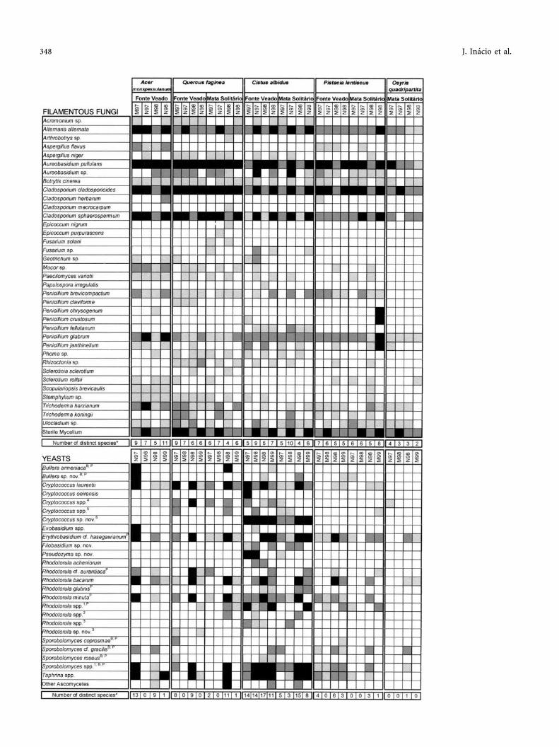
Fig. 1. Total counts of filamentous fungi and yeasts on the phylloplane of the different plant species for each sampling date, at the two
sites (Fonte do Veado or Mata do Solitario); CFU, Colony-forming units; error bars represent the standard deviation of mean densities;
*, not sampled.
Phylloplane Mycobiota in a Portuguese Mediterranean Ecosystem 347
348 J. Inacio et al.

public databases (GenBank). Sequences obtained in this study are
available upon request to the authors.
Results and Discussion
Total Mycobiota
The results presented in Figs. 1 and 2 show that the
phylloplane of the sampled plants is colonized by con-
siderably large and diverse fungal populations. Total
counts and species richness varied according to plant
species, season, and sampling site. Relative frequencies of
molds and yeasts were generally concordant and com-
bined totals ranged from ca 1 · 102 CFU cm)2 on O.
quadripartita to values well above 1 · 104 CFU cm)2 on
A. monspessulanum and Q. faginea (Fig. 1). In a recent
study on the phylloplane of mango trees in South Africa
using a similar isolation method [11], mold and yeast
densities were in the range 1 · 104 to 1 · 105 CFU cm)2
(although Aureobasidium pullulans was counted as a
member of the yeast population). In the present study and
for the plant species that occurred at both sampling sites,
the total number of isolates of either filamentous fungi or
yeasts were generally higher on the samples from Fonte do
Veado, regardless of season (Figs. 1 and 2). The differences
are statistically supported for filamentous fungi on C. al-
bidus (spring, p = 0.002, and autumn, p = 0.020) and Q.
faginea (spring, p = 0.037, and autumn, p = 0.008) and for
yeasts on C. albidus (spring and autumn, p = 0.007). This
result might be explained by the more humid conditions
that prevail on the northern slope throughout the year,
which concurs with similar observations about the effect
of high humidity levels on the density of phylloplane mi-
crobial populations made by other authors [e.g., 5, 22]. On
the other hand, there was a general increase in the number
of isolates on the leaves of the deciduous tree species (A.
monspessulanum and Q. faginea) from spring to autumn
(Fig. 1). The differences are statistically supported for fil-
amentous fungi: on A. monspessulanum (p = 0.040) and
on Q. faginea (Fonte do Veado, p = 0.004; Mata do So-
litario, p < 0.001). This trend was confirmed in the case of
the yeasts by the results of an extra sampling in the
summer of 1998, which yielded intermediate numbers of
leaf colonists between the spring and autumn values (data
not shown). This observation is in agreement with the
prevailing view that phylloplane fungal populations on
deciduous plants increase with leaf development reaching
a peak at mid-season or at leaf-fall [7, 17, 20, 27]. It is
conceivable that on deciduous trees the colonizers of
newly opened leaves originate from cells that overwintered
in the buds or on the bark; they may also result from air-
or insect-borne immigrants present on the evergreens,
namely in the case of the widespread species that were
found on more than one plant at both sites (Fig. 2). This
seasonal trend was not so evident on the evergreens (Fig.
1), except for the filamentous fungi on C. albidus at both
sites (p < 0.001). However, counts were consistently low in
the case of the yeasts on P. lentiscus and O. quadripartita
(Fig. 1). The latter are both xerophytes producing large
amounts of terpenoid compounds that might exert a se-
lective effect on the potential leaf colonizers [e.g., 29]. On
the phylloplane of evergreens, the inoculum for young
leaves is most probably provided by cells that are washed
out or blown out of older leaves on the same plant and
therefore total counts would be expected to remain con-
stant throughout the year. This appears to be the case for
the yeasts on C. albidus where the differences between
spring and autumn are not statistically significant (Fonte
do Veado, p = 0.484; Mata do Solitario, p = 0.421).
It is worth noting that total counts and species richness
for both filamentous fungi and yeasts were not signifi-
cantly different when comparing the results of the autumn
collections for the deciduous trees with those of the ev-
ergreens (excluding O. quadripartita) (Figs. 1 and 2). It
could be anticipated that phylloplane populations on the
evergreens would be more numerous because of the lon-
gevity of their leaves [e.g., 11], but in some cases the op-
posite was observed (Fig. 1), e.g., the combined densities
of filamentous fungi for the autumn samplings of the de-
ciduous trees (A. monspessulanum and Q. faginea) at
Fonte do Veado were statistically higher (p < 0.001) than
the corresponding values for the evergreens (Cistus albi-
dus and Pistacia lentiscus). These results suggest that in
this particular case populations of mycobiota become es-
tablished on the phylloplane of the deciduous trees in a
Fig. 2. Species diversity and relative frequency of occurrence of
filamentous fungi and yeasts on the phylloplane of the different
plant species, according to sampling site and date (M, March; N,
November). Shade code: black, >102 CFU cm)2; dark gray, 101–
102 CFU cm)2; light gray, <101 CFU cm)2; white, not detected.BBallistoconidia-forming yeasts; Ppigmented yeasts. Phylogenetic
affiliation of yeasts: 1Erythrobasidium clade, 2Ustilaginales,3Microbotryum clade, 4Filobasidiales, 5Tremellales. *Only species
with densities >101 CFU/cm)2 were considered and some entries
include more than one distinct species, e.g., Cryptococcus spp.
b
Phylloplane Mycobiota in a Portuguese Mediterranean Ecosystem 349

relatively short period (from spring to autumn) and that
leaf turnover, nutrient availability, or other intrinsic fac-
tors (e.g., presence of phytochemicals with antimicrobial
activity on the leaves of evergreens—see above) effect
some control on the populations levels on the evergreens.
Analysis of Fig. 2 shows that plant species having
higher numbers of leaf mycobiota (A. monspessulanum, C.
albidus, and Q. faginea) also yielded a slightly wider range
of species (Fig. 2). The results further suggest that the
most prevalent mold or yeast species followed the overall
seasonal trend, their numbers rising from spring to
autumn, and therefore they may correspond to the most
representative phylloplane inhabitants (see below). These
taxa are, in most cases, not restricted to a particular plant,
but a few examples emerged of specific associations (see
below). Other less frequent species may represent occa-
sional colonists that originate from cells arising from
the air, deposited by insects or rain-washed from other
aerial plant parts. Testing of these hypotheses would,
however, require additional isolations from all possible
sources.
Filamentous Fungi
The total numbers of filamentous fungi recovered from
each sample is shown in Fig. 1, with values ranging from
ca 102 to 104 CFU cm)2, and were generally higher than for
the yeasts. As pointed out above, molds were apparently
more numerous on plant leaves from Fonte do Veado than
from Mata do Solitario (Fig. 1), except for C. albidus and
P. lentiscus in November 1997, the latter showing a sig-
nificantly larger number (ca 2.5-fold) of isolates from Mata
do Solitario. As already mentioned, there was an increase
in the number of isolates from spring to autumn on the
deciduous trees and, in the case of filamentous fungi, also
on the evergreens C. albidus and P. lentiscus (Fig. 1). Q.
faginea and A. monspessulanum consistently had the
highest number of leaf colonists in both seasons, with
pronounced peaks in the November collections.
The majority of isolates were deuteromycetes, mostly of
ascomycetous affinity (77% of a total of 1029 isolates).
Another considerable fraction of the total number of iso-
lates (19%) were nonsporulating species and were thus
included in the sterile mycelia group. This observation is
in agreement with the results of other phylloplane studies
[1, 6, 20]. Isolates that could be identified represented at
least 36 distinct species (10 of which were only identified
to the genus level) and were distributed as shown in Fig. 2.
Species that were recovered from all samples included
Cladosporium cladosporioides, Aureobasidium pullulans,
Alternaria alternata, and Penicillium glabrum. Fungi be-
longing to the sterile mycelia group were also found in all
samples with frequencies ranging from 10 to 103 CFU
cm)2, but on A. monspessulanum and Q. faginea leaves
from Fonte doVeado this heterogeneous group of fungi
appeared with frequencies higher than 103 CFU cm)2. A
few fungal species that have been reported by others as
common inhabitants of the phylloplane, such as Cla-
dosporium sphaerospermum, Trichoderma harzianum,
Penicillium brevicompactum, and Aureobasidium spp. [6,
7, 12], were also found to occur with high frequencies in
the plants studied in the present work. Overall, Q. faginea
was the plant that showed the highest degree of coloni-
zation and species richness (Fig. 2), followed by A.
monspessulanum. Some fungi appear to be restricted to Q.
faginea, namely Penicillium claviforme, Cladosporium
macrocarpum, Fusarium solani, Epicoccum purpurascens,
Epicocum nigrum, and Arthrobotrys sp. However, these
species were found at low frequencies and only occa-
sionally. Penicillium fellutanum appears to be restricted to
the leaves of C. albidus from both locations and Scopu-
lariopsis brevicaulis was consistently isolated from A.
monspessulanum. On the other hand, O. quadripartita
yielded the narrowest range of species (Fig. 2). Some
species reported as common phylloplane inhabitants, such
as Cladosporium herbarum [1, 14, 20], E. purpurascens, E.
nigrum [1, 7, 9, 14, 16], Papulospora irregularis, and
Cheaetomium sp. [14], were only rarely found in the
present study. Moreover, typical soil fungi, e.g., Muco-
rales, Aspergillus, Trichoderma, Scopulariopsis, and Pae-
cilomyces spp., which appear to be active in leaf litter
decomposition [19, 37] and are often recovered from leaf
surfaces [18, 20, 26], were a relatively minor component of
the total isolates in our study.
Direct comparison of the data of the present study with
those obtained by other authors is hampered by differ-
ences in isolation methods and frequency of sampling.
However, it appears that fungal species richness on
phylloplane samples from Serra da Arrabida (36 distinct
species) is comparatively lower than the values reported
for fungal communities from other ecosystems, especially
tropical and temperate habitats [1, 6, 7, 12, 18]. For ex-
ample, from the phylloplane of four trees in a primary rain
forest of Costa Rica an average of 134 distinct fungal
species were isolated and identified [6]. A survey of the
evergreen shrub qat (Catha edulisis) located in the ‘‘coffee-
350 J. Inacio et al.

zone’’ provinces of Yemen Arab Republic, yielded 64
fungal species [1], whereas ca 53 distinct fungal species
were collected from leaves of Acer platanoides collected in
the grounds of the University of Stirling, Scotland [7].
Yeasts
To our knowledge, the only information on the yeast biota
associated with plant species in a geographical area with
Mediterranean characteristics was recently provided by
Middelhoven [31]. Samples were collected in the Canary
Islands, from plants we have not investigated, and the few
yeasts recovered included representatives of the genera
Debaryomyces, Cryptococcus, and Rhodotorula. In the
present work, the vast majority of isolates were of basid-
iomycetous affinity (ca 93% of a total of 540 strains), in
agreement with the results of other phylloplane studies.
Most basidiomycetous isolates were found to belong to the
following major phylogenetic clades sensu Fell et al. [15]:
Tremellales, Filobasidiales, Ustilaginales, Microstroma-
tales, Microbotryum, and Erythrobasidium (Fig. 2). They
represented at least 45 species (note that some entries in
Fig. 2 include more than one distinct species under a
single designation: e.g., Cryptococcus spp.). Several yeasts
were present in all leaves regardless of plant or location:
apart from species that are known to be ubiquitous on
leaves, such as Cryptococcus laurentii, Rhodotorula min-
uta, and Sporobolomyces spp., we recovered significant
numbers of strains of Rhodotorula bacarum and of species
closely related to Erythrobasidium cf. hasegawianum and
Rhodotorula cf. aurantiaca, which have not been com-
monly isolated from the phylloplane [27, 35]. Some of
these species were present in high densities that followed
the seasonal trend observed for the total yeast population
(e.g., Rhodotorula minuta—Figs. 1 and 2). A surprising
result was the low incidence of ballistoconidia-forming
yeasts, which could be attributed either to their lower
relative concentration or to a stronger attachment of these
yeasts to the leaf surfaces preventing their recovery by the
isolation procedure employed in this study. However,
using an isolation method specific for those types of cells
(the ballistospore-fall method) we were able to isolate
many strains, mainly of Sporobolomyces roseus and Til-
letiopsis spp. (data not shown). The latter method is,
however, not amenable to quantification. Among the few
ascomycetous isolates, we found significant numbers of
presumptive yeast stages of Taphrina spp., which were
present in a large number of samples from all plants
(except O. quadripartita) (Fig. 2). This finding was some-
what surprising, since the yeast stages of these dimorphic
phytopathogenic fungi are rarely isolated from natural
substrates except from infected plant material [e.g., 24].
Previous studies have pointed to an apparent lack of
specificity of the basidiomycetous yeasts that occur on the
phylloplane, namely Cryptococcus spp. and the ballisto-
conidia-producing species Sporobolomyces roseus and
Bullera alba [27], a hypothesis partly confirmed in the
present study. However, some species appear to be re-
stricted to Cistus albidus, namely Rhodotorula acheniorum
and three putative novel species: a Filobasidium sp. (Fi-
lobasidiales), Pseudozyma sp. (Ustilaginales), and, in ex-
ceptionally high densities, a Cryptococcus sp. resembling
Cryptococcus hungaricus (Tremellales). The yeast abun-
dance and species richness on the phylloplane of this plant
is particularly conspicuous and may be a consequence of
the dense trichome cover of its leaves [3].
The quantitative results presented in Fig. 1 are difficult
to compare to those from previous reports because, in
some of these studies, the ubiquitous dimorphic deuter-
omycete Aureobasidium pullulans is considered as part of
the yeast population [7, 11]. However, frequencies ranging
up to 104 CFU cm)2, such as the ones we obtained, are
not uncommon [e.g., 27]. The observed frequencies were
conspicuously low on P. lentiscus and, especially, on O.
quadripartita. On these two plants, we observed a higher
proportion of pigmented yeast species than on the other
plants (Fig. 2). A possible selective advantage of pig-
mented yeasts on these xerophytes warrants further study.
The seasonal variation of total yeast counts observed on
the deciduous trees as compared to the evergreen shrubs
was more pronounced than that observed for the fila-
mentous fungi (Fig. 1).
Conclusion
To our knowledge, no information is so far available on
the number, or diversity, of either filamentous fungi or
yeasts associated with the plant species sampled in the
present study. In spite of the known limitations of the
isolation methods employed and of some apparent overlap
with results of previous phylloplane surveys, both in
quantitative and qualitative terms, our work is thought to
constitute an important contribution to the assessment of
microbial diversity in a Mediterranean-type ecosystem. It
is worth noting that a number of isolates appeared to
Phylloplane Mycobiota in a Portuguese Mediterranean Ecosystem 351

represent undescribed species. This seems to be the case of
ca 25 distinct yeast species (corresponding to 23% of the
total number of isolates); however, 10 of these were rep-
resented by single strains. Molecular identification of
molds that would allow similar calculations for these fungi
is currently underway. Formal descriptions of putative
novel taxa are, however, beyond the scope of this report
and will be presented elsewhere. It is nevertheless clear
that our knowledge about phylloplane mycobiota is still
scarce and that more surveys are needed for the ecological
characterization of microbial populations on this envi-
ronment. Finally, to unveil new species and reveal specific
plant–microbe associations holds a considerable biotech-
nological potential, e.g., the use of fungi as biological
control agents [8, 28] or for the production of novel an-
timicrobial compounds [30].
Acknowledgments
Joao Inacio receives a PhD grant (Praxis XXI/BD/19833/
99) from ‘‘Fundacao para a Ciencia e a Tecnologia,’’ Por-
tugal, and the work was partially supported by project
contract no. Praxis XXI/2/2.1/BIA/413/94.
References
1. Alhubaishi AA, Abdel-Kader MI (1991) Phyllosphere and
phylloplane fungi of qat in Sana’a, Yemen Arab Republic. J
Basic Microbiol 31:83–89
2. Allsopp D, Colwell RR, Hawksworth DL (eds) (1995) Mi-
crobial Diversity and Ecosystem Function. CAB Interna-
tional, Wallingford, UK
3. Andrews JH, Hirano SS (eds) (1991) Microbial Ecology of
Leaves. Springer-Verlag, New York
4. Barnett JA, Payne RW, Yarrow D (2000) Yeasts, Character-
istics and Identification, 3rd ed. Cambridge Univ. Press,
Cambridge, UK
5. Bashi E, Fokkema NJ (1977) Environmental factors limiting
growth of Sporobolomyces roseus, an antagonist of Cochlio-
bolus sativus, on wheat leaves. Trans Brit Mycol Soc 68:17–
25
6. Bills GF, Polishook JD (1994) Abundance and diversity of
microfungi in leaf litter of a lowland rain forest in Costa
Rica. Mycologia 86:187–198
7. Breeze EM, Dix NJ (1981) Seasonal analysis of the fungal
community on Acer platanoides leaves. Trans Brit Mycol Soc
77:321–328
8. Buck JW, Lachance M-A, Traquair JA (1998) Mycoflora of
peach bark: population dynamics and composition. Can J
Bot 76:345–354
9. Cabral D (1985) Phyllosphera of Eucalyptus viminalis: dynam-
ics of fungal population. Trans Brit Mycol Soc 85:501–511
10. Catarino FM, Correia OA, Correia AI (1982) Structure and
dynamics of Serra da Arrabida mediterranean vegetation.
Ecologia Mediterranea. T.VIII. Fasc. 1/2:203–222
11. de Jager ES, Wehner FC, Korsten L (2001) Microbial ecology
of the mango phylloplane. Microb Ecol 42:201–207
12. Dickinson CH (1976) Fungi on the aerial surfaces of higher
plants. In: TF Preece, CH Dickinson (eds) Microbiology
of Aerial Plant Surfaces Academic Press, London pp 293–324
13. Diem HG (1974) Micro-organisms of the leaf surface: esti-
mation of the mycoflora of the barley phyllosphere. J Gen
Microbiol 80:77–83
14. Domsch KH, Gams W, Anderson TH (1980) Compendium of
Soil Fungi. Academic Press, London
15. Fell JW, Boekout T, Fonseca A, Scorzetti G, Statzell-Tallman
A (2000) Biodiversity and systematics of basidiomycetous
yeasts as determined by large-subunit rDNA D1/D2 domain
sequence analysis. Int J Syst Evol Microbiol 50:1351–1371
16. Fokkema NJ (1991) The phyllosphere as an ecologically
neglected milieu: a plant pathologist’s point of view. In: JH
Andrews, SS Hirano (eds) Microbial Ecology of Leaves
Springer-Verlag, New York pp 3–18
17. Fokkema NJ, Schippers B (1986) Phyllosphere versus rhi-
zosphere as environments for saprophytic colonization. In:
NJ Fokkema, J van den Heuvel (eds) Microbiology of the
Phyllosphere. Cambridge University Press, London pp 137–
159
18. Heredia G (1993) Mycoflora associated with green leaves and
litter of Quercus germana, Quercus sartorri and Liquidambar
styraciflua in a Mexican cloud forest. Cryptolog Mycol
14:171–183
19. Hering TF (1965) The succession of fungi in the litter of a
Lake District oakwood. Trans Brit Mycol Soc 48:391–408
20. Hogg BM, Hudson HJ (1966) Micro-fungi on leaves of Fagus
sylvatica. I. The micro-fungal succession. Trans Brit Mycol
Soc 49:185–192
21. Juniper BE (1991) The leaf from the inside and the outside: a
microbe’s perspective. In: JH Andrews, SS Hirano (eds)
Microbial Ecology of Leaves. Springer-Verlag, New York pp
21–42
22. Kinkel LL (1997) Microbial population dynamics on leaves.
Annu Rev Phytopathol 35:327–347
23. Kirk PM (1995) Inventorying microfungi on tropicals plants.
In: D Allsopp, RR Colwell, DL Hawksworth (eds) Microbial
Diversity and Ecosystem Function CAB International, Wal-
lingford, UK pp 355–360
24. Kramer CL (1987) The Taphrinales. In: GS Hoog, ACM
Smith, ACM Weijman (eds) The Expanding Realm of Yeast-
like Fungi. Elsevier, Amsterdam pp 151–166
25. Kurtzman CP, Fell JW (1998) The Yeasts, A Taxonomic
Study, 4th ed. Elsevier, Amsterdam
26. Kuter GA (1986) Microfungal populations associated with
the decomposition of sugar maple leaf litter. Mycologia
78:114–126
352 J. Inacio et al.

27. Last FT, Price D (1969) Yeasts associated with living plants
and their environs. In: AH Rose, JS Harrison (eds) The
Yeasts, 1st ed, vol I. Academic Press, New York, pp 183–218
28. Li H, Veenendaal E, Ab Shukor NA, Cobbinah JR, Leifert C
(1995) Yeast populations on the tropical timber tree species
Milicia excelsa. Lett Appl Microbiol 21:322–326
29. Magiatis P, Melliou E, Skaltsounis AL, Chinou IB, Mitaku S
(1999) Chemical composition and antimicrobial activity of
the essential oils of Pistacia lentiscus var. chia. Planta Med
65:749–752
30. McCormack PJ, Wildman HG, Jeffries P (1994) Production
of antibacterial compounds by phylloplane-inhabiting yeasts
and yeastlike fungi. Appl Environ Microbiol 60:927–931
31. Middelhoven WJ (1997) Identity and biodegradative abilities
of yeast isolated from plants growing in an arid climate.
Antonie van Leeuwenhoek 72:81–89
32. Mishra RR, Dickinson CH (1981) Phylloplane and litter
fungi of Ilex aquifolium. Trans Brit Mycol Soc 77:329–337
33. Nakase T (2000) Expanding world of ballistosporous yeasts:
Distribution in the phyllosphere, systematics and phylogeny.
J Gen Appl Microbiol 46:189–216
34. Pedro JG (1991) Vegetacao e Flora da Arrabida. Servico
Nacional de Parques, Reservas e Conservacao da Natureza,
Lisboa (in Portuguese)
35. Phaff HJ, Starmer WT (1987) Yeasts associated with plants,
insects and soil. In: AH Rose, JS Harrison (eds) The Yeasts,
2nd ed., vol I, Biology of Yeasts. Academic Press, London,
pp 123–180
36. Pitt JI (1979) The Genus Penicillium and its Teleomorphic
States Eupenicillium and Talaromyces. Academic Press,
London
37. Pugh GJ (1974) Terrestrial fungi. In: CH Dickinson, GJ Pugh
(eds) Biology of Plant Litter Decomposition, vol 2. Academic
Press, London, pp 303–336
38. Ruinen J (1963) The phyllosphere. II. Yeasts from the
phyllosphere of tropical foliage. Antonie van Leeuwenhoek
29:425–438
39. Sampaio JP, Gadanho M, Santos S, Duarte FL, Pais C,
Fonseca A, Fell JW (2001) Polyphasic taxonomy of the ba-
sidiomycetous yeast genus Rhodosporidium: Rhodosporidi-
um kratochvilovae and related anamorphic species. Int J Syst
Evol Microbiol 51:687–697
40. Sokal RR, Rohlf FJ (1969) Biometry: The Principles and
Practice of Statistics in Biological Research. W. H. Freeman
and Company, San Francisco
41. Tukey Jr HB (1971) Leaching of substances from plants. In:
TF Preece, CH Dickinson (eds) Ecology of Leaf Surface
Micro-organisms. Academic Press, London, pp 67–80
42. Yarrow D (1998) Methods for the isolation, maintenance,
classification and identification of yeasts. In: CP Kurtzman,
JW Fell (eds) The Yeasts: A Taxonomic Study. Elsevier,
Amsterdam pp 45–104
Phylloplane Mycobiota in a Portuguese Mediterranean Ecosystem 353