At the periphery of the Empire - HAL-SHS - Archives-ouvertes.fr
Upload
independentCategory
view
4download
0
MOLECULAR AND CELLULAR BIOLOGY, Dec. 2002, p. 8292–8301 Vol. 22, No. 230270-7306/02/$04.00�0 DOI: 10.1128/MCB.22.23.8292–8301.2002Copyright © 2002, American Society for Microbiology. All Rights Reserved.
Esc1, a Nuclear Periphery Protein Required for Sir4-Based PlasmidAnchoring and Partitioning
Erik D. Andrulis,1† David C. Zappulla,1 Athar Ansari,2 Severine Perrod,3 Catherine V. Laiosa,2Marc R. Gartenberg,2* and Rolf Sternglanz1*
Department of Biochemistry and Cell Biology, State University of New York, Stony Brook, New York 11794-52151;Department of Pharmacology, University of Medicine and Dentistry of New Jersey-Robert Wood Johnson
Medical School, Piscataway, New Jersey 088542; and Swiss Institute for ExperimentalCancer Research (ISREC), CH-1066 Epalinges/Lausanne, Switzerland3
Received 31 July 2002/Accepted 5 September 2002
A targeted silencing screen was performed to identify yeast proteins that, when tethered to a telomere,suppress a telomeric silencing defect caused by truncation of Rap1. A previously uncharacterized protein, Esc1(establishes silent chromatin), was recovered, in addition to well-characterized proteins Rap1, Sir1, and Rad7.Telomeric silencing was slightly decreased in �esc1 mutants, but silencing of the HM loci was unaffected. Onthe other hand, targeted silencing by various tethered proteins was greatly weakened in �esc1 mutants.Two-hybrid analysis revealed that Esc1 and Sir4 interact via a 34-amino-acid portion of Esc1 (residues 1440to 1473) and a carboxyl-terminal domain of Sir4 known as PAD4 (residues 950 to 1262). When tethered toDNA, this Sir4 domain confers efficient partitioning to otherwise unstable plasmids and blocks the ability ofbound DNA segments to rotate freely in vivo. Here, both phenomena were shown to require ESC1. Sirprotein-mediated partitioning of a telomere-based plasmid also required ESC1. Fluorescence microscopy ofcells expressing green fluorescent protein (GFP)-Esc1 showed that the protein localized to the nuclearperiphery, a region of the nucleus known to be functionally important for silencing. GFP-Esc1 localization,however, was not entirely coincident with telomeres, the nucleolus, or nuclear pore complexes. Our data suggestthat Esc1 is a component of a redundant pathway that functions to localize silencing complexes to the nuclearperiphery.
The silent mating-type loci (HML and HMR) in the yeastSaccharomyces cerevisiae are maintained in a transcriptionallyinactive state due to the formation of a specialized chromatinstructure analogous to heterochromatin of higher eukaryotes.Silencing of the mating-type loci requires flanking sequenceelements, termed silencers, which bind the transcription fac-tors Rap1 and Abf1, as well as the multisubunit origin recog-nition complex (ORC) (reviewed in references 11, 15, and 27).Together, these proteins recruit the silencing proteins, Sir1,Sir2, Sir3, and Sir4, that participate in the formation of het-erochromatin.
Genes placed near telomeres are also silenced. Telomericsilencing depends on Rap1, which binds to telomeric TG1-3
repeats and recruits Sir3 and Sir4 (28, 29). A Sir complexconsisting of Sir2, Sir3, and Sir4 then spreads from the telo-meres to nearby nucleosomes to form silent chromatin (16, 17).Although RAP1 is an essential gene, mutations that delete the3� end of the gene (rap1�C) are viable but lead to a complete
loss of telomeric silencing (22, 26, 28). This is due to theinability of Rap1�C to recruit Sir3 and Sir4 to the telomeres.
Expressing Sir proteins as GAL4 DNA-binding domain(GBD) hybrids and tethering them to defective silencers inwhich binding sites for the ORC, Rap1, and/or Abf1 have beenreplaced by Gal4 binding sites can lead to silencing. This so-called targeted silencing was first demonstrated with GBD-Sir1but has since been shown with the other Sir proteins as well aswith Rap1 and Orc1 (5, 6, 38). Targeted silencing also has beenachieved by forming GBD hybrids with endoplasmic reticulumor Golgi proteins and tethering them to a partially defectiveHMR E silencer (1). Overexpression of such membrane pro-tein hybrids causes them to accumulate in the endoplasmicreticulum (which is contiguous with the nuclear envelope). Asa consequence, the DNA-binding domain of Gal4 is in thenucleus but is anchored to the nuclear membrane. In this caseit is thought that silencing occurs because the HMR locus, withGal4 sites at the E silencer, is drawn to the periphery of thenucleus where there is a high concentration of Sir proteins (1).
Circular autonomously replicating sequence (ARS) plasmidsthat lack a mechanism for mitotic segregation are preferen-tially retained in mother cells, resulting in the generation ofplasmid-free daughters. In the absence of selection, these plas-mids are lost from logarithmically growing cultures (30). Incontrast, ARS plasmids that contain embedded telomeric se-quences or the HMR E silencer are segregated efficiently be-tween dividing cells and are stably propagated (8, 19, 23, 24).Plasmid segregation mediated by the HMR E silencer requiresthe Sir proteins, and segregation mediated by telomeric se-
* Corresponding authors. Mailing address for Marc R. Gartenberg:Department of Pharmacology, UMDNJ-Robert Wood Johnson Med-ical School, 675 Hoes Lane, Piscataway, NJ 08854. Phone: (732) 235-5800. Fax: (732) 235-4073. E-mail: [email protected]. Mailingaddress for Rolf Sternglanz: Department of Biochemistry and CellBiology, State University of New York, Stony Brook, NY 11794-5215.Phone: (631) 632-8565. Fax: (631) 632-8575. E-mail:[email protected].
† Present address: Department of Molecular Biology and Genetics,Cornell University, Ithaca, NY 14853.
8292
quences is improved by the silencing factors. Previously, weshowed that tethering a specific domain of Sir4, the so-calledpartitioning and anchoring domain (PAD4), directly to ARSplasmids also confers efficient mitotic segregation (2). Using aDNA-topology assay that measures axial rotation of intracel-lular DNA segments, it was shown that the tethered PAD4domain of Sir4 also immobilizes the DNA to which it is bound(2). The partitioning and DNA immobilization data suggesteda model in which the Sir4 domain attaches to a nuclear com-ponent, such as a chromosome or the nuclear membrane, thatdivides symmetrically between cells at mitosis.
Here we describe the results of a screen for factors that,when tethered to a telomere, can reestablish silencing at atelomere in a rap1�c mutant defective in silencing. In thisscreen we identified three known Sir-interacting proteins,Rap1, Sir1, and Rad7, as well as a novel protein, Esc1, whichwe show interacts with the PAD4 domain of Sir4. We furthershow that Esc1 is located at the nuclear periphery and isessential for the partitioning and anchoring of plasmids by thePAD4 domain of Sir4. The results suggest that Esc1 helpsrecruit Sir4 to the nuclear periphery.
MATERIALS AND METHODS
Yeast strains. The strains used in this study are listed in Table 1. Deletion ofESC1 with kanMX4 was achieved by generating a targeting construct by PCRwith oligonucleotides directly upstream and downstream of the ESC1 openreading frame (ORF) and with genomic DNA isolated from strain 20805(�esc1::kanMX4) as a template (Research Genetics, Inc.). �esc1::his5� deletionswere generated by PCR-mediated targeted disruption by use of a plasmid con-taining the Schizosaccharomyces pombe his5� gene (N. Dean, Stony Brook,N.Y.); this gene complements the S. cerevisiae his3 mutation. The Esc1-greenfluorescent protein (GFP)-expressing strain YDZ49, strain YDZ239, and variousmutant derivatives of YDZ225 were all generated using previously describedplasmids and methods (25). The GAL1 promoter and the coding sequence forGFP replaced 46 nucleotides upstream of the ESC1 ORF plus the codon for theinitiating methionine. W303-1a/0, YDZ13/0, and THC18/0 were derived fromW303-1a, YDZ13, and THC18, respectively, by curing the strains of the 2�mplasmid using targeted DNA damage (39). The endogenous 2�m plasmid mustbe eliminated to study the partitioning of telomere-derived sequences that aresituated on 2�m-based plasmids.
Telomere one-hybrid screen. The one-hybrid telomere silencing screen wasperformed in strain Lev8. Lev8 harbors a URA3 reporter gene with UASG sitesnear the left arm of chromosome VII-L; it has a RAP1 deletion on the chromo-some and is kept alive by a wild-type RAP1 gene on a plasmid (26). By plasmidshuffling, the wild-type RAP1 was replaced by a C-terminal truncation allele,rap1�670-807, on plasmid sp18. Cells containing this rap1 allele are completelyderepressed for telomeric silencing and grow slowly (26). Lev8/sp18 cells were
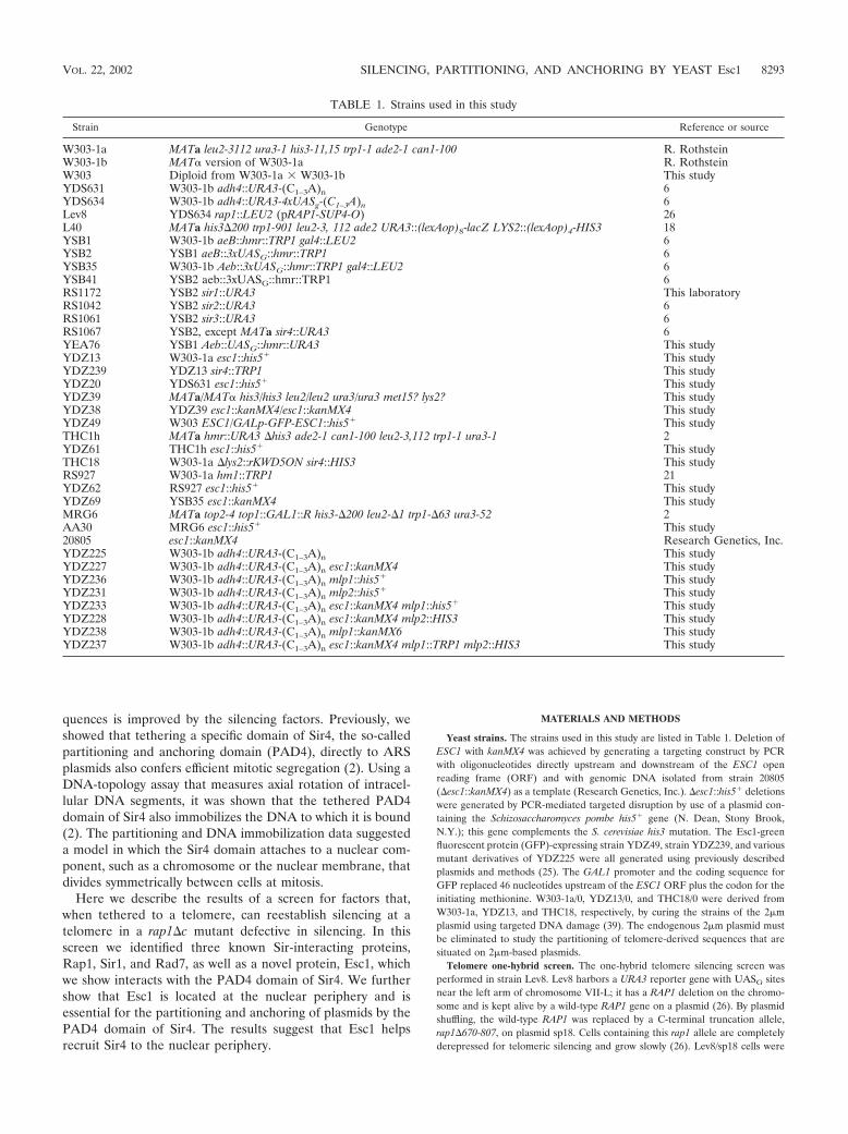
TABLE 1. Strains used in this study
Strain Genotype Reference or source
W303-1a MATa leu2-3112 ura3-1 his3-11,15 trp1-1 ade2-1 can1-100 R. RothsteinW303-1b MAT� version of W303-1a R. RothsteinW303 Diploid from W303-1a � W303-1b This studyYDS631 W303-1b adh4::URA3-(C1–3A)n 6YDS634 W303-1b adh4::URA3-4xUASg-(C1–3A)n 6Lev8 YDS634 rap1::LEU2 (pRAP1-SUP4-O) 26L40 MATa his3�200 trp1-901 leu2-3, 112 ade2 URA3::(lexAop)8-lacZ LYS2::(lexAop)4-HIS3 18YSB1 W303-1b aeB::hmr::TRP1 gal4::LEU2 6YSB2 YSB1 aeB::3xUASG::hmr::TRP1 6YSB35 W303-1b Aeb::3xUASG::hmr::TRP1 gal4::LEU2 6YSB41 YSB2 aeb::3xUASG::hmr::TRP1 6RS1172 YSB2 sir1::URA3 This laboratoryRS1042 YSB2 sir2::URA3 6RS1061 YSB2 sir3::URA3 6RS1067 YSB2, except MATa sir4::URA3 6YEA76 YSB1 Aeb::UASG::hmr::URA3 This studyYDZ13 W303-1a esc1::his5� This studyYDZ239 YDZ13 sir4::TRP1 This studyYDZ20 YDS631 esc1::his5� This studyYDZ39 MATa/MAT� his3/his3 leu2/leu2 ura3/ura3 met15? lys2? This studyYDZ38 YDZ39 esc1::kanMX4/esc1::kanMX4 This studyYDZ49 W303 ESC1/GALp-GFP-ESC1::his5� This studyTHC1h MATa hmr::URA3 �his3 ade2-1 can1-100 leu2-3,112 trp1-1 ura3-1 2YDZ61 THC1h esc1::his5� This studyTHC18 W303-1a �lys2::rKWD5ON sir4::HIS3 This studyRS927 W303-1a hm1::TRP1 21YDZ62 RS927 esc1::his5� This studyYDZ69 YSB35 esc1::kanMX4 This studyMRG6 MATa top2-4 top1::GAL1::R his3-�200 leu2-�1 trp1-�63 ura3-52 2AA30 MRG6 esc1::his5� This study20805 esc1::kanMX4 Research Genetics, Inc.YDZ225 W303-1b adh4::URA3-(C1–3A)n This studyYDZ227 W303-1b adh4::URA3-(C1–3A)n esc1::kanMX4 This studyYDZ236 W303-1b adh4::URA3-(C1–3A)n mlp1::his5� This studyYDZ231 W303-1b adh4::URA3-(C1–3A)n mlp2::his5� This studyYDZ233 W303-1b adh4::URA3-(C1–3A)n esc1::kanMX4 mlp1::his5� This studyYDZ228 W303-1b adh4::URA3-(C1–3A)n esc1::kanMX4 mlp2::HIS3 This studyYDZ238 W303-1b adh4::URA3-(C1–3A)n mlp1::kanMX6 This studyYDZ237 W303-1b adh4::URA3-(C1–3A)n esc1::kanMX4 mlp1::TRP1 mlp2::HIS3 This study
VOL. 22, 2002 SILENCING, PARTITIONING, AND ANCHORING BY YEAST Esc1 8293

transformed with a GBD yeast library in the vector pGBT-CYH (CEN TRP1CYH2 ADHp-GBD), a gift of C. Evangelista and S. Fields, University of Wash-ington. Transformants were selected on medium lacking tryptophan. Colonieswere grown at 30°C for 4 to 5 days before being replica plated on 5-fluorooroticacid (5-FOA) to select cells exhibiting silencing of URA3. Candidates werestreaked to single colonies three times on 5-FOA plates. DNA was then isolatedfrom these 5-FOA-resistant colonies, electroporated into Escherichia coli, andanalyzed by restriction enzyme digestion. Unique GBD plasmids were retrans-formed into Lev8 to identify those that restored silencing. These library plasmidswere subjected to sequencing.
Plasmids. Library plasmids L4.1, L5.2, and L18.6, from the telomere one-hybrid screen described above, all encode identical GBD-Rap1 hybrids. L5.4 andL19.4 encode GBD-Sir1 and GBD-Rad7, respectively (Table 2). Library plasmidL2.4 encodes GBD-Esc1(1124-1658).
The ESC1 insert from plasmid L2.4 was excised with SmaI and PstI, and thefragment was subcloned into the LexA vector pBTM116-Kn-ADE2 (2�m TRP1ADE2 ADHp-LexA) to create pEDA114 and into the GAD vector pGAD424(2�m LEU2 ADHp-GAD) to create pEDA115. To create LexA-Esc1(1395-1551)and GAD-Esc1(1395-1551), a SmaI-PstI fragment was cloned (from a libraryplasmid isolated in a similar screen to be reported elsewhere) into pBTM116-Kn-ADE2 (pEDA157) and pGAD424 (pEDA159). GBD-Esc1(1200-1448)(pEDA167) was created as follows: pEDA115 was digested with BamHI andBglII and the ESC1 fragment was cloned into the BamHI site of pBluescript IIKS(�) to create pEDA124. pEDA124 was digested with NsiI and SalI and thefragment was subcloned into pKSII� (pEDA160). A BamHI-XhoI fragmentfrom pEDA160 was then subcloned into pTT63, a pGBT9 derivative (2�m HIS3ADHp-GBD) (4, 38) to make pEDA167.
Full-length ESC1 was cloned as follows: pEDA115 was digested with BamHIand HindIII to liberate the entire 3� coding region of Esc1 and was subclonedinto pRS306 (35) to make pEDA122. pEDA122 was digested at a unique EcoRIsite within the ESC1 coding sequence, and the linear DNA was transformed intoyeast. Genomic DNA was isolated from Ura� transformants, digested with EagI,incubated with T4 DNA ligase, and electroporated into E. coli strain MH3. Thepresence of an insert containing the entire Esc1 coding region (pEDA128) wasverified by restriction enzyme analysis. A 7.0-kb BstXI-XhoI fragment containingthe full-length ORF was isolated from pEDA128 and subcloned into pKSII� togenerate pEDA129. A yeast shuttle vector containing the ESC1 gene was con-structed by excising a SacII-XhoI fragment from pEDA129 and ligating it intopRS315 (CEN/ARS LEU2) (35) cut with SacII and SalI to make pDZ45.
The construction of the following GAD-Sir4 constructs has been describedpreviously: GAD-Sir4(1-1358), pCTC79; GAD-Sir4(839-1358), pCTC18; GAD-Sir4(1262-1358), pCTC24; and GAD-Sir4(839-1149), pCTC50 (6, 38). LexA-Sir4plasmids used in two-hybrid experiments have been described previously (2).GAD-Sir4(934-1358) is clone C1-5.1, isolated in the two-hybrid screen with LexA-Esc1(1124-1658).
Plasmids LS4 (Sir4 950-1262), pAA6, and pKWD200 were described previ-ously (2). Additional details are provided here. The 950- to 1,262-amino-acidfragment of Sir4 in LS4 terminates with a serine rather than the cysteine nor-mally present in the Sir4 protein at position 1262. pKWD200 and pAA6 containsix copies of an oligonucleotide that has two LexA operators. Thus, the plasmidscontain 12 LexA sites each. In pAA6, the XbaI site within the 2�m episome-derived portion of the plasmid was destroyed, thereby eliminating the Flp rec-ognition target and errors in partitioning measurements due to interplasmidrecombination with the endogenous 2�m episome (40).
Two-hybrid screens. Two-hybrid experiments were performed in strain L40,which contains LexA operators upstream of the HIS3 and lacZ reporter genes(18). For screening with LexA-Esc1(1124-1658), cells were cotransformed withboth the LexA hybrid and a yeast GAD library. For the screen with LexA-Sir4(950-1262), L40 was transformed sequentially, first with plasmid LS4(950-
1262) (2) and then with the yeast GAD library. Library clones that promotedsufficient HIS3 expression for growth on SC-LEU-TRP-HIS plates (containing 5mM 3-aminotriazole for the LexA-Sir4 screen) were subsequently tested for�-galactosidase expression. His� �-galactosidase� transformants were charac-terized as described previously to eliminate false positives (33).
Silencing assays. Strains THC1h and YDZ61 were used to measure silencingof HMR. Strains RS927 and YDZ62 were used to measure silencing of HML.Strains YDS631 and YDZ20 were used to measure telomeric silencing. Cellswere grown to saturation at 30°C in medium selecting for the GBD plasmid or, ifno plasmid was involved, in yeast extract-peptone-dextrose medium. Cells werethen serially diluted 10-fold and 10 �l of these dilutions was spotted onto platescontaining appropriate media. For experiments with URA3-based reporterstrains, cells were also spotted onto medium containing 0.1% 5-FOA (AngusBiochemicals, Niagara Falls, N.Y.).
Localization of GFP-Esc1. For confocal microscopy used to detect GFP-Esc1,strain YDZ49 was grown overnight in galactose medium supplemented withadenine to reduce vacuolar autofluorescence and then centrifuged at low speedand fixed in 95% ethanol at 30°C for several hours. Cells were then washed andresuspended in SHA buffer (1 M sorbitol, 0.1 M Na-HEPES [pH 7.5], 5 mMNaN3). Spheroplasts were prepared by the addition of zymolyase and �-mercap-toethanol and the cell wall digestion was stopped by washing in SHA buffer.Spheroplasted cells were resuspended in SHA buffer and allowed to settle in theeight wells of a polylysine-coated slide. The overlaying buffer was aspirated andthe cells were treated with 1 mg of RNase A per ml for 30 min at 37°C. Cells werethen washed with 2 drops of SHA buffer 5 to 10 times. Propidium iodide (1�g/ml) in SHA buffer was added to the digested cells and incubated at roomtemperature for 10 min. Cells were then washed 10 times with SHA buffer andpreserved in mounting medium under a sealed coverslip.
Immunofluorescence. Strains YDZ38 and YDZ39 were used for immunoflu-orescence, using previously described methods and antibodies (13, 14).
Plasmid stability and anchoring assays. Plasmid stability tests and anchoringexperiments were carried out as previously described (2).
RESULTS
A screen for proteins capable of restoring silencing at atelomere identifies Esc1. A one-hybrid screen was developedto look for new proteins involved in silencing. The screen wasperformed in strain Lev8, which has Gal4 DNA-binding sites(UASG) and a URA3 reporter gene placed near a telomere(Fig. 1). The strain also harbors a deletion in RAP1 (�670-807)that removes the Sir4 interaction domain (26). Without thisdomain, telomeric silencing is abolished. Lev8 was transformedwith a GBD hybrid plasmid library and screened for GBD fusion
FIG. 1. Telomeric targeted silencing screen. A telomere with aURA3 reporter gene and four adjacent Gal4 binding sites is depicted.(A) The RAP1 strain encodes a wild-type Rap1 protein that promotesthe formation and spreading of a Sir2-Sir3-Sir4 complex that silencesthe URA3 gene. (B) The rap1 strain encodes a Rap1 protein with adeletion of amino acids 670 to 807 and hence cannot bind Sir3 andSir4. Thus, the URA3 gene is not silenced. (C) The strain is the sameas in panel B except that a GBD hybrid binds to the Gal4 binding sitesand causes silencing of the URA3 gene.
TABLE 2. Genes identified in one-hybrid telomeric silencing screen
GBD hybrid Amino acidsin hybrida Comments (reference)
GBD-Rap1 354–827 Binds to Sir3 and Sir4 (28, 29)GBD-Sir1 163–678 Binds to Sir4 (38)GBD-Rad7 28–565 Binds to Sir3 (31)GBD-Esc1 1,124–1,658 Binds to Sir4 (this study)
a Each hybrid contained the C-terminal region of the protein, as indicated, upto and including the final amino acid.
8294 ANDRULIS ET AL. MOL. CELL. BIOL.

proteins that restored silencing of the telomeric URA3. Re-pression of the gene was monitored by growth on 5-FOA.
Three of the four different proteins identified in this screenhad been studied previously (Table 2). One of them was Rap1.Three different plasmid isolates encoded a fragment of Rap1from amino acid 354 to the end of the protein. This portion ofRap1 includes the Sir4 interaction domain that was deletedfrom strain Lev8 (26) and thus was not unexpected. Also iden-tified in the screen was Sir1, a protein known to play an es-sential role in the establishment of silencing at the HM loci andalso shown previously to be capable of targeted silencing via aninteraction with Sir4 (6, 38). Although Sir1 is dispensable fornormal telomeric silencing, Sir1 enhances telomeric silencingwhen targeted to telomeric DNA (6). The third protein iden-tified in the screen was Rad7, a protein implicated in the repairof DNA damage. Rad7 has been shown to interact with Sir3(31). Thus, it is likely that the GBD-Rad7 hybrid causes silenc-ing by recruiting Sir3 to the telomere.
The fourth clone identified in this screen was a GBD hybridto residues 1124 to 1658 of a previously uncharacterized yeastORF, YMR219w, which we have named ESC1 (establishessilent chromatin). ESC1 encodes a large protein containing1,658 amino acids. The protein is highly acidic, contains a
P-loop (purine-binding motif), many predicted coiled-coils,and a putative nuclear localization sequence at the C terminus.
Targeted silencing by GBD-Esc1. Targeted silencing assayswith Esc1 are shown in Fig. 2. Three different GBD-Esc1 hy-brids were used: the one found in the screen described above,a second constructed by us, and a third found in an HMRtargeted silencing screen to be described elsewhere. The GBD-Esc1 hybrids gave targeted silencing, not only at telomeres, butalso at various HMR alleles containing HMR E silencer dele-tions. The silencing required Sir2, -3, and -4 and Gal4 bindingsites (UASG) (Fig. 2C), as was expected from earlier studies oftargeted silencing (6). Subcloning of Esc1 demonstrated that a54-amino-acid region from near the carboxy-terminal end ofthe protein (residues 1395 to 1448) was sufficient for transcrip-tional repression (data not shown).
esc1 mutant. A strain with a precise deletion of the ESC1open reading frame was constructed in a diploid. Sporulationand dissection of the heterozygous diploid yielded �esc1 hap-loid strains that grew normally at all temperatures tested.Strains with the �esc1 mutation were tested for silencing de-fects at HML, HMR, telomeres, and the ribosomal DNA(rDNA) locus. No silencing defects were observed at the HMloci or the rDNA (data not shown). A slight decrease in telo-
FIG. 2. Targeted silencing by GBD-Esc1 at a telomere and at HMR. (A) Two different GBD-Esc1 hybrids silence the URA3 gene in the telomerereporter strain YDS634. Tenfold serial dilutions were plated on �Trp medium to indicate the number of cells plated and on 5-FOA medium tomeasure the extent of silencing. (B) GBD-Esc1 hybrids silence URA3 at an HMR locus in which the HMR E silencer has the Rap1 and Abf1 bindingsites replaced by a Gal4 binding site (Aeb::UASG; strain YEA76). (C) Targeted silencing by GBD-Esc1 at an HMR locus with a TRP1 reporter gene(strain YSB2) is Sir dependent. Sir� cells harboring GBD-Esc1(1200-1448) (row 1) grow poorly on �Trp medium due to silencing of the reportergene, while sir mutants (rows 2 to 5) grow better. Targeted silencing by GBD-Esc1 is also dependent upon the presence of UASG sites (strain YSB1;row 6). In the absence of any silencer element at HMR E (strain YSB41), Esc1 is still capable of targeted silencing (row 7).
VOL. 22, 2002 SILENCING, PARTITIONING, AND ANCHORING BY YEAST Esc1 8295

meric silencing was seen, as discussed below (see Fig. 8). Inaddition, the �esc1 mutant exhibited significant defects in tar-geted silencing. For this set of experiments, various silencingproteins were tethered to a mutated HMR E silencer via GBD
and tested for the ability to silence a nearby reporter gene. Theproteins examined included Rap1, Orc1, Sir1, Sir3, and Sir4.Also tested was a membrane protein, Yif1, previously shown togive efficient targeted silencing because it tethers HMR to thenuclear periphery (1). A comparison of targeted silencing bythese various proteins in ESC1 versus �esc1 strains is shown inFig. 3. Deletion of ESC1 nearly eliminates silencing by themembrane protein, Yif1, and greatly weakens silencing by Sir1,Orc1 (full length or the N-terminal fragment known to bindSir1 [38]), and Rap1. Silencing by Sir4 is also somewhat weak-ened by the esc1 mutation. Introduction of a CEN plasmidcontaining the ESC1 gene, pDZ45, into the esc1 mutant re-stored silencing by these proteins (data not shown). Silencingby only one hybrid protein, GBD-Sir3, was unaffected by delet-ing esc1. This result will be considered further (see Discus-sion).
Esc1 interacts with Sir4. Since Esc1 had targeted silencingactivity, it seemed likely that it was recruiting a known silencingprotein. To identify Esc1-interacting proteins, a two-hybridscreen was undertaken using the domain of Esc1 recovered inthe targeted silencing screen (amino acids 1124 to 1658) as abait. A C-terminal fragment of Sir4 (amino acids 934 to 1358)was found as a specific interacting partner. The two-hybridsystem was used to delineate the domains required for theEsc1-Sir4 interaction. This directed approach showed that
GAD hybrids containing amino acids 839 to 1149 of Sir4 weresufficient for interaction with LexA-Esc1 (amino acids 1124 to1658), whereas a GAD hybrid containing the extreme C-termi-nal lamin-like heptad repeats of Sir4 (amino acids 1262 to1358) was not (Fig. 4). An identical Sir4 interaction profile wasobserved using a smaller Esc1 hybrid, LexA-Esc1 (amino acids1395 to 1551), indicating that this 157-amino-acid domain ofEsc1 contained a Sir4-interacting motif. Neither Esc1 hybridinteracted with full-length GAD-Sir4.
The region of Sir4 found to interact with Esc1 coincided witha previously described domain of Sir4, the partitioning andanchoring domain (PAD4). This Sir4 domain, when bound toplasmids via LexA, greatly increases mitotic stability of plas-mids, and furthermore, appears to anchor them as judged by atopological assay (2). Interestingly, a two-hybrid search usingthe PAD4 domain of Sir4 (amino acids 950 to 1262) as baitidentified four different Esc1 fragments. Three of them encom-passed the C terminus of Esc1, from amino acids 944, 1013,and 1227 to the end of the protein. The fourth PAD4-inter-acting clone was a small Esc1 fragment spanning amino acids1440 to 1473, indicating that this 34-amino-acid fragment wassufficient for interaction with Sir4.
Further evidence that the PAD4 domain of Sir4 coincidedwith the Esc1-interacting domain is shown in Fig. 4. Five dif-ferent LexA hybrids containing different regions of Sir4,encompassing part or all of the operationally defined PAD4domain, were tested against two GAD-Esc1 hybrids in the two-hybrid system. A perfect correlation could be seen betweenthose LexA-Sir4 hybrids that interacted with Esc1 and those
FIG. 3. Comparison of targeted silencing in ESC1 and �esc1 strainsYSB35 and YDZ69. A TRP1 reporter gene was used and thus lack ofgrowth on �Trp medium indicates good silencing. Note that the esc1mutation affects silencing by all the GBD hybrids except GBD-Sir3.
FIG. 4. Summary of two-hybrid data showing the interaction be-tween Esc1 and Sir4. �, significant two-hybrid interaction in strainL40; �, no interaction. The various LexA-Sir4 hybrids were also testedfor improved partitioning of plasmid pAA6 as described previously (2).Symbols for partitioning column: �, improved partitioning; �, noimprovement.
8296 ANDRULIS ET AL. MOL. CELL. BIOL.

that were positive in the plasmid partitioning assay (2), whichwe describe below.
Plasmid partitioning by targeted Sir4 requires Esc1. Anassociation between Esc1 and the PAD4 domain of Sir4 sug-gested that Esc1 might be required for Sir4-mediated parti-tioning of plasmids. To test this possibility, strain W303-1a andthe isogenic �esc1 mutant were cotransformed with a LexA-PAD4 expression vector and an unstable circular ARS plas-mid, pAA6, that contains 12 LexA binding sites. Partitioning ofpAA6 was evaluated by measuring the mitotic stability and theloss rate of the plasmid. The former parameter corresponds tothe fraction of cells that contain pAA6 under selective growthconditions whereas the latter corresponds to the rate of pAA6loss during nonselective growth conditions. In agreement withearlier results, partitioning of pAA6 in W303-1a was enhancedby the expression of LexA-PAD4 but not by LexA alone (2).Strikingly, PAD4-mediated partitioning of pAA6 was abol-ished in the �esc1 strain. Mitotic stability dropped nearly five-fold and plasmid loss rate increased more than sixfold (Table3). Introduction of an ESC1 plasmid, pDZ45, into the esc1mutant restored PAD4-mediated partitioning (data notshown). These results indicate that ESC1 is required forPAD4-mediated plasmid stabilization.
One explanation for the partitioning defect in the �esc1strain is that the gene plays a role in DNA replication. Thispossibility is not supported by the finding that a CEN/ARSplasmid exhibited similar mitotic stability and plasmid loss ratein both the ESC1 and �esc1 strains (Table 4). Decreased plas-mid persistence, therefore, does not result from an inability toreplicate plasmids properly. An alternative explanation for thepartitioning defect is that ESC1 is required for the LexA chi-mera to bind DNA. In a �esc1 strain, however, LexA-PAD4can bind the promoter of a lacZ reporter gene and blockactivation by an upstream UASG, indicating that expression,nuclear transport, and DNA binding of LexA-PAD4 can occurin the absence of ESC1 (data not shown).
Sir4-mediated partitioning of telomere-based plasmids re-quires Esc1. The finding that Esc1 promotes plasmid partition-ing by artificially tethered Sir4 suggested that ESC1 might also
contribute to segregation of plasmids that recruit Sir4 natu-rally. We therefore measured the role of ESC1 in segregatingtwo circular plasmids that possess telomeric sequences. Thefirst plasmid, pYET1, contains a telomeric TG1-3 tract and asubtelomeric X repeat (sometimes referred to as an X-TAS,for X-telomere-associated sequence), whereas the second plas-mid, pETACC, contains only the TG1-3 tract (24). Previouswork showed that both plasmids partitioned well. However, theX repeat of pYET1 provided additional stabilization and thisadded effect required Sir2, Sir3, and Sir4 (8, 23). Accordingly,deletion of SIR4 from the strain used here increased thepYET1 loss rate from 0.10 to 0.15 (Table 5). Deletion of ESC1resulted in a comparable increase in the pYET1 loss rate,suggesting that ESC1 is required for the enhanced partitioningof telomeric sequences by Sir4. The esc1 sir4 double mutanthad the same loss rate as each single mutant, showing that bothgenes act in the same pathway. The loss rate of pETACC, onthe other hand, was not changed significantly by deletion ofeither SIR4 or ESC1. The combined evidence supports a modelin which telomeric sequences recruit Sir4 as part of the Sirsilencing complex and then Sir4 in turn associates with Esc1 toconfer partitioning.
Anchoring of DNA by targeted Sir4 requires Esc1. The do-main of Sir4 that confers plasmid partitioning also hinders theaxial rotation (swiveling) of DNA fragments to which it isbound (2). A role for ESC1 in this form of DNA immobiliza-tion, termed DNA anchoring, was tested with a DNA-topolo-gy-based assay (for a detailed description, see reference 2).Briefly, twin domains of positive and negative supercoils aregenerated in a DNA ring formed by site-specific recombina-tion. Proteins that block DNA rotation prevent cancellation ofthe two domains. In top1 top2 yeast mutants expressing thenegative supercoil-specific E. coli topoisomerase I, selectiverelaxation of negative supercoils results in positive supercoilaccumulation. Under these conditions, DNA rings containingLexA sites became highly positively supercoiled when LexA-PAD4 was expressed (2) (Fig. 5A, lane 1). The result indicatesthat the LexA-PAD4–DNA complex is anchored in vivo. Sub-stantial positive supercoiling did not occur, however, if theexperiment was performed in an isogenic �esc1 strain. Thelevel of supercoiling in this mutant was comparable to the levelin an ESC1 strain that expressed LexA alone (Fig. 5A, lanes 2
TABLE 3. Partitioning of plasmid pAA6 by a LexA-Sir4p hybridrequires ESC1
LexA hybrid Strain Relevantgenotype
% Mitoticstabilitya
Plasmid lossratea
LS4 (950–1262) W303-1a ESC1 57.0 5.0 0.08 0.03LS4 (950–1262) YDZ13 �esc1::his5� 12.0 3.0 0.51 0.02LexA only W303-1a ESC1 20.2 2.1 0.39 0.10
a Each entry represents the mean standard deviation of at least threeindependent trials with individual transformants.
TABLE 4. CEN/ARS plasmid partitioning does not require ESC1a
Strain Relevantgenotype
% Mitoticstabilityb
Plasmid lossrateb
W303-1A ESC1 84.8 2.4 0.08 0.01YDZ13 �esc1::his5� 96.3 2.4 0.09 0.02
a Data obtained with pRS416 (35).b Each entry represents the mean standard deviation of at least three
independent trials with individual transformants.
TABLE 5. ESC1 and SIR4 stabilize telomeric plasmids containing asubtelomeric X repeat
Telomere-basedplasmid Straina Relevant
genotypePlasmid loss
rateb
pYET1 W303-1a/0 Wild type 0.10 0.008YDZ13/0 �esc1::his5� 0.17 0.013THC18/0 �sir4::HIS3 0.15 0.013YDZ239/0 �esc1-�sir4 0.15 0.005
pETACC W303-1a/0 Wild type 0.20 0.036YDZ13/0 �esc1::his5� 0.28 0.06THC18/0 �sir4::HIS3 0.25 0.086YDZ239/0 �esc1-�sir4 0.28 0.057
a All strains were cured of the endogenous 2�m plasmid, as indicated by “/0”(see Materials and Methods).
b Each entry represents the mean standard deviation of at least threeindependent trials with individual transformants.
VOL. 22, 2002 SILENCING, PARTITIONING, AND ANCHORING BY YEAST Esc1 8297

and 3). These data demonstrate that ESC1 is required forSir4-mediated DNA anchoring.
To test whether ESC1 plays a general role in DNA anchor-ing, the blot in Fig. 5A was stripped and rehybridized with aprobe to a second plasmid that contains the partitioning locusof the 2�m plasmid, REP3, which was previously shown toform a DNA anchor (12). The plasmid, pKWD1�, accumu-lates positive supercoils efficiently in both the ESC1 and �esc1strains, irrespective of whether LexA or LexA-PAD4 is ex-pressed (Fig. 5B). Thus, the role for ESC1 in the DNA an-choring assay is specific for the anchor formed by LexA-PAD4.
Subcellular localization of Esc1. To identify the subcellularlocation of Esc1, a strain was generated that expresses a GFP-Esc1 fusion protein under control of the GAL1 promoter (25).This construct complemented a �esc1 mutant in the partition-ing assay described above (data not shown). Confocal micros-copy of galactose-induced cells showed that the expressed GFPfusion protein localized to the periphery of the nucleus in apunctate pattern (Fig. 6A). Our finding that Esc1 and Sir4interact prompted us to examine the relative locations of thetwo proteins. Indirect immunofluorescence showed that thetwo proteins colocalized at many positions along the nuclearperiphery (Fig. 6B). GFP-Esc1, however, was also found atother peripheral positions that lacked Sir4. Colocalization
studies with the nucleolar marker Nop1 demonstrated thatEsc1 did not localize to the nucleolus (data not shown).
Previously Sir4 and other telomere binding proteins wereshown to colocalize with telomeres in discrete perinuclear foci(13, 32). Disruption of a single telomere binding factor oftenleads to dispersion of others and sometimes leads to unclus-tering of the telomeric DNA sequences (reviewed in reference7). Indirect immunofluorescence of Sir4, however, showed thattelomeric foci were not disrupted in a �esc1 strain (Fig. 7).
GFP-Esc1 localization somewhat resembled the distributionof nucleoporins. The relative distribution of these two entitieswas examined using an antibody to nuclear pore proteins (3).Esc1 and nuclear pores were only occasionally found to becoincident (Fig. 6C). Thus, Esc1 does not appear to be acomponent of the nuclear pore complex. This is consistent withthe result that Esc1 was not found in a comprehensive study ofyeast nuclear pore proteins (34).
Interestingly, in cells in which GFP-Esc1 was most highly
FIG. 5. DNA anchoring by targeted Sir4 requires ESC1. Assayswere performed in strains MRG6 (ESC1) and AA30 (�esc1) trans-formed with an E. coli topoisomerase I expression plasmid [YEp-topA(PGPD)], a LexA-PAD4 expression plasmid [LS4(950-1262)],and an excision substrate vector (pKWD200) that produces a ring(rKWD200) with 12 LexA sites. (A) Analysis of ring rWKD200 super-coiling by two-dimensional gel electrophoresis in buffer containingchloroquine. DNA topoisomers were resolved into an arc with posi-tively supercoiled rings coalescing in a spot at the extreme clockwiseend (lane 1) and negatively supercoiled rings occupying a broad regionat the counterclockwise end (lane 2). The minor population of positivesupercoils seen in lanes 2 and 3 appears occasionally with rings thatlack DNA anchors. (B) Analysis of plasmid pKWD1� supercoiling.rKWD200 and pKWD1� were visualized sequentially by hybridizationwith randomly primed radiolabeled probes. FIG. 6. Esc1 localizes to the nuclear periphery. (A) Confocal im-
ages of GFP-Esc1 and propidium iodide fluorescence; the latter stainsthe nuclear DNA. (B) Esc1 and Sir4 are both at the nuclear peripherybut they do not colocalize. Both proteins were visualized by immuno-fluorescence. (C) Esc1 does not colocalize with nuclear pores. Theinsets in panels B and C show examples of cells with very strongGFP-Esc1 fluorescence at the bud neck. This was seen for 5 to 10% ofcells.
8298 ANDRULIS ET AL. MOL. CELL. BIOL.

expressed, the Esc1 signal was also detected in a single pro-jection from the nucleus (Fig. 6B and C, inset panels). Thisprojection did not appear to contain Sir4, although nuclearpores could be detected at the base of the projection, suggest-ing that the projection may be an extension of the nuclearmembrane.
Telomeric silencing in esc1 and mlp1 and -2 mutants. Tworelated yeast proteins, called Mlp1 and Mlp2 (myosin-like pro-tein), have been shown to localize to the nuclear periphery andbind to nuclear pore proteins (36). An mlp1 mlp2 double mu-tant has been reported to have telomeric silencing defects (9,10). Since Esc1, Mlp1, and Mlp2 are all large coiled-coil pro-teins that localize to the nuclear periphery, we wonderedwhether they played overlapping roles in silencing. We there-fore constructed all combinations of single, double, and triplemutants and tested them for telomeric silencing. As mentionedabove, esc1 single mutants have a slight but reproducible si-lencing defect (Fig. 8). To our surprise, mlp1 and mlp2 single
and double mutants show no silencing defect and do not ex-acerbate the esc1 silencing defect (Fig. 8). However, we didobserve an unusual colony morphology for mlp1 mlp2 doublemutants, just as had been reported previously (10). This con-firms that we had indeed mutated the MLP1 and MLP2 genes.We have no explanation for why our silencing results differfrom those previously published for the mlp1 mlp2 mutant (9,10). It seems unlikely that strain differences could account forsuch a drastic difference.
DISCUSSION
Esc1 was identified in a screen for proteins that confer si-lencing when targeted to silencing-defective loci. The proteinwas shown to silence both a derepressed telomere and a mu-tated HMR silencer when tethered to these sites (Fig. 2). Par-adoxically, strains lacking Esc1 have no obvious silencing de-fects at the native HM loci or rDNA and only minor defects attelomeres (Fig. 8). Nevertheless, several lines of evidence sug-gest that the protein plays a role in Sir-mediated repression;silencing by targeted Esc1 requires the other Sir proteins (Fig.2), and Esc1 is required for silencing by other targeted proteins(Fig. 3). Esc1 also interacts with Sir4 (Fig. 4). This interactionmay explain why the protein was recovered in the targetedsilencing screen: a tethered fragment of Esc1 complexes withSir4, which in turn associates with Sir2 and Sir3 to nucleatesilent chromatin. While the genetic evidence presented heremakes it likely that Esc1 binds directly to Sir4, we have no invitro evidence to support this. It is conceivable that the inter-action is bridged by another yeast protein.
Esc1 localizes to the nuclear periphery (Fig. 6) and a poten-tial role for the protein in silencing may be to tether silentchromatin complexes to this subnuclear compartment. ThoughSir proteins are limiting in the nucleus, the factors concentrateat telomeres, which cluster at the nuclear periphery (7). Silenc-ing of the mating-type loci is thought to benefit from theirproximity to telomeres and the associated pools of Sir proteins.Indeed, tethering a defective silencer to the telomere-rich nu-clear periphery imparts Sir-dependent transcriptional repres-sion (1). Thus, Esc1 may function in silencing because it colo-
FIG. 7. Telomere clustering, visualized with an antibody to Sir4, isnot altered in �esc1 mutants. For comparison, a wild-type ESC1 strainis shown in the inset.
FIG. 8. Telomeric silencing in esc1, mlp1, and mlp2 mutants. Tenfold serial dilutions are shown for each strain on complete medium (SC) andon 5-FOA medium. Growth on the latter medium indicates silencing of the URA3 reporter gene. WT, wild type.
VOL. 22, 2002 SILENCING, PARTITIONING, AND ANCHORING BY YEAST Esc1 8299

calizes the silent mating-type loci with perinuclear pools of Sirproteins.
A role for Esc1 as a localization factor is supported by thefinding that Esc1 influences the intracellular behavior of Sir4.We show here that partitioning and anchoring of plasmids bya tethered fragment of Sir4 requires Esc1 (Table 3; Fig. 5).Moreover, Esc1 is required for Sir4-enhanced partitioning of atelomere-based plasmid, an extrachromosomal element thatrecruits Sir4 as part of a silent chromatin complex (Table 5). Itseems likely that Esc1 facilitates the segregation process byrecruiting Sir4-bound plasmids to the nuclear membrane orsome other structure at the nuclear periphery that dividesbetween progeny at mitosis.
Why does Esc1 not play a greater role in silencing at nativesilent loci? One explanation is that silencing is a process withbuilt-in redundancies. Native silencers, for example, bind re-dundant combinations of proteins that cooperate to recruit Sirproteins for remarkably efficient silencing. Elimination of onesilencer-bound protein is often not sufficient to disrupt silenc-ing. Targeted silencing, on the other hand, is always measuredin situations in which some component of silencing has beenweakened by mutation. According to this view, some redun-dant feature of native silencing must be eliminated before thedeleterious effects of an esc1 deletion can be observed. Redun-dancy might not be limited to silencers and the proteins thatbind them. There might also be overlapping mechanisms fortargeting silent chromatin to the nuclear periphery. The Kuprotein and Mlp1 and Mlp2 have been implicated in tetheringtelomeres to the nuclear periphery and these proteins maycontribute to or overlap with Esc1-mediated targeting of silentchromatin (10, 20). But, as shown in Fig. 8, we see no evidencethat Mlp1 and Mlp2 play any role in telomeric silencing. Nev-ertheless, additional factors are likely to contribute to telomerelocalization since the left telomere of chromosome VII re-mains at the periphery in Ku and silencing-deficient strains(37).
Why is targeted silencing by GBD-Sir3 ESC1 independentwhile silencing by all the other hybrid proteins tested is weak-ened by deleting ESC1 (Fig. 3)? A simple explanation is thattethering Sir3 to a silencer bypasses the requirement for ESC1in silencing. According to this view, ESC1 acts upstream of Sir3recruitment. Conversely, targeted silencing by GBD-Sir4 is fa-cilitated by ESC1. Thus, the data indicate that ESC1 plays arole subsequent to Sir4 recruitment.
Localization of Esc1 at the nuclear periphery does not co-incide exactly with Sir4 or with nuclear pores, although there issome overlap (Fig. 6). Esc1 is a large protein with predictedcoiled-coil domains throughout its length. It has always beenunclear why yeasts do not have nuclear lamins of the typefound in larger eukaryotes. Perhaps Esc1 plays that role inyeast, along with other coiled-coil proteins. Identification offactors that maintain Esc1 at the periphery may address thisquestion. In summary, we think that Esc1 is a component ofthe nuclear periphery that helps attract silent chromatin com-plexes containing Sir4 to the periphery. Of course, it may haveother roles yet to be uncovered.
ACKNOWLEDGMENTS
E.D.A., D.C.Z., and A.A. contributed equally to this work.
We thank C. Evangelista and S. Fields for the GBD library, J. Ber-man and D. Gottschling for plasmids, S. Gasser for advice and encour-agement, and J. Speh and G. Mandel for help with confocal micros-copy.
This work was supported by National Institutes of Health grantsGM51402 to M.R.G. and GM28220 to R.S.
REFERENCES
1. Andrulis, E. D., A. M. Neiman, D. C. Zappulla, and R. Sternglanz. 1998.Perinuclear localization of chromatin facilitates transcriptional silencing.Nature 394:592–595. (Erratum, 395:525.)
2. Ansari, A., and M. R. Gartenberg. 1997. The yeast silent information regu-lator Sir4p anchors and partitions plasmids. Mol. Cell. Biol. 17:7061–7068.
3. Aris, J. P., and G. Blobel. 1989. Yeast nuclear envelope proteins cross reactwith an antibody against mammalian pore complex proteins. J. Cell Biol.108:2059–2067.
4. Bartel, P. L., J. A. Roecklein, D. SenGupta, and S. Fields. 1996. A proteinlinkage map of Escherichia coli bacteriophage T7. Nat. Genet. 12:72–77.
5. Buck, S. W., and D. Shore. 1995. Action of a RAP1 carboxy-terminal silenc-ing domain reveals an underlying competition between HMR and telomeresin yeast. Genes Dev. 9:370–384.
6. Chien, C. T., S. Buck, R. Sternglanz, and D. Shore. 1993. Targeting of SIR1protein establishes transcriptional silencing at HM loci and telomeres inyeast. Cell 75:531–541.
7. Cockell, M., and S. M. Gasser. 1999. Nuclear compartments and gene reg-ulation. Curr. Opin. Genet. Dev. 9:199–205.
8. Enomoto, S., M. S. Longtine, and J. Berman. 1994. Enhancement of te-lomere-plasmid segregation by the X-telomere associated sequence in Sac-charomyces cerevisiae involves SIR2, SIR3, SIR4 and ABF1. Genetics 136:757–767.
9. Feuerbach, F., V. Galy, E. Trelles-Sticken, M. Fromont-Racine, A. Jacquier,E. Gilson, J. C. Olivo-Marin, H. Scherthan, and U. Nehrbass. 2002. Nucleararchitecture and spatial positioning help establish transcriptional states oftelomeres in yeast. Nat. Cell Biol. 4:214–221.
10. Galy, V., J. C. Olivo-Marin, H. Scherthan, V. Doye, N. Rascalou, and U.Nehrbass. 2000. Nuclear pore complexes in the organization of silent telo-meric chromatin. Nature 403:108–112.
11. Gartenberg, M. R. 2000. The Sir proteins of Saccharomyces cerevisiae:mediators of transcriptional silencing and much more. Curr. Opin. Micro-biol. 3:132–137.
12. Gartenberg, M. R., and J. C. Wang. 1993. Identification of barriers torotation of DNA segments in yeast from the topology of DNA rings excisedby an inducible site-specific recombinase. Proc. Natl. Acad. Sci. USA 90:10514–10518.
13. Gotta, M., T. Laroche, A. Formenton, L. Maillet, H. Scherthan, and S. M.Gasser. 1996. The clustering of telomeres and colocalization with Rap1, Sir3,and Sir4 proteins in wild-type Saccharomyces cerevisiae. J. Cell Biol. 134:1349–1363.
14. Gotta, M., S. Strahl-Bolsinger, H. Renauld, T. Laroche, B. K. Kennedy, M.Grunstein, and S. M. Gasser. 1997. Localization of Sir2p: the nucleolus as acompartment for silent information regulators. EMBO J. 16:3243–3255.
15. Grunstein, M. 1998. Yeast heterochromatin: regulation of its assembly andinheritance by histones. Cell 93:325–328.
16. Hecht, A., T. Laroche, S. Strahl-Bolsinger, S. M. Gasser, and M. Grunstein.1995. Histone H3 and H4 N-termini interact with SIR3 and SIR4 proteins:a molecular model for the formation of heterochromatin in yeast. Cell80:583–592.
17. Hecht, A., S. Strahl-Bolsinger, and M. Grunstein. 1996. Spreading of tran-scriptional repressor SIR3 from telomeric heterochromatin. Nature 383:92–96.
18. Hollenberg, S. M., R. Sternglanz, P. F. Cheng, and H. Weintraub. 1995.Identification of a new family of tissue-specific basic helix-loop-helix proteinswith a two-hybrid system. Mol. Cell. Biol. 15:3813–3822.
19. Kimmerly, W. J., and J. Rine. 1987. Replication and segregation of plasmidscontaining cis-acting regulatory sites of silent mating-type genes in Saccha-romyces cerevisiae are controlled by the SIR genes. Mol. Cell. Biol. 7:4225–4237.
20. Laroche, T., S. G. Martin, M. Gotta, H. C. Gorham, F. E. Pryde, E. J. Louis,and S. M. Gasser. 1998. Mutation of yeast Ku genes disrupts the subnuclearorganization of telomeres. Curr. Biol. 8:653–656.
21. Le, S., C. Davis, J. B. Konopka, and R. Sternglanz. 1997. Two new S-phase-specific genes from Saccharomyces cerevisiae. Yeast 13:1029–1042.
22. Liu, C., X. Mao, and A. J. Lustig. 1994. Mutational analysis defines aC-terminal tail domain of RAP1 essential for telomeric silencing in Saccha-romyces cerevisiae. Genetics 138:1025–1040.
23. Longtine, M. S., S. Enomoto, S. L. Finstad, and J. Berman. 1993. Telomere-mediated plasmid segregation in Saccharomyces cerevisiae involves geneproducts required for transcriptional repression at silencers and telomeres.Genetics 133:171–182.
24. Longtine, M. S., S. Enomoto, S. L. Finstad, and J. Berman. 1992. Yeasttelomere repeat sequence (TRS) improves circular plasmid segregation, and
8300 ANDRULIS ET AL. MOL. CELL. BIOL.

TRS plasmid segregation involves the RAP1 gene product. Mol. Cell. Biol.12:1997–2009.
25. Longtine, M. S., A. McKenzie, 3rd, D. J. Demarini, N. G. Shah, A. Wach, A.Brachat, P. Philippsen, and J. R. Pringle. 1998. Additional modules forversatile and economical PCR-based gene deletion and modification in Sac-charomyces cerevisiae. Yeast 14:953–961.
26. Marcand, S., E. Gilson, and D. Shore. 1997. A protein-counting mechanismfor telomere length regulation in yeast. Science 275:986–990.
27. Moazed, D. 2001. Common themes in mechanisms of gene silencing. Mol.Cell 8:489–498.
28. Moretti, P., K. Freeman, L. Coodly, and D. Shore. 1994. Evidence that acomplex of SIR proteins interacts with the silencer and telomere-bindingprotein RAP1. Genes Dev. 8:2257–2269.
29. Moretti, P., and D. Shore. 2001. Multiple interactions in Sir protein recruit-ment by Rap1p at silencers and telomeres in yeast. Mol. Cell. Biol. 21:8082–8094.
30. Murray, A. W., and J. W. Szostak. 1983. Pedigree analysis of plasmid seg-regation in yeast. Cell 34:961–970.
31. Paetkau, D. W., J. A. Riese, W. S. MacMorran, R. A. Woods, and R. D. Gietz.1994. Interaction of the yeast RAD7 and SIR3 proteins: implications forDNA repair and chromatin structure. Genes Dev. 8:2035–2045.
32. Palladino, F., T. Laroche, E. Gilson, A. Axelrod, L. Pillus, and S. M. Gasser.1993. SIR3 and SIR4 proteins are required for the positioning and integrityof yeast telomeres. Cell 75:543–555.
33. Park, H., and R. Sternglanz. 1998. Two separate conserved domains ofeukaryotic DNA topoisomerase I bind to each other and reconstitute enzy-matic activity. Chromosoma 107:211–215.
34. Rout, M. P., J. D. Aitchison, A. Suprapto, K. Hjertaas, Y. Zhao, and B. T.Chait. 2000. The yeast nuclear pore complex: composition, architecture, andtransport mechanism. J. Cell Biol. 148:635–651.
35. Sikorski, R. S., and P. Hieter. 1989. A system of shuttle vectors and yeasthost strains designed for efficient manipulation of DNA in Saccharomycescerevisiae. Genetics 122:19–27.
36. Strambio-de-Castillia, C., G. Blobel, and M. P. Rout. 1999. Proteins con-necting the nuclear pore complex with the nuclear interior. J. Cell Biol.144:839–855.
37. Tham, W. H., J. S. Wyithe, P. K. Ferrigno, P. A. Silver, and V. A. Zakian.2001. Localization of yeast telomeres to the nuclear periphery is separablefrom transcriptional repression and telomere stability functions. Mol. Cell8:189–199.
38. Triolo, T., and R. Sternglanz. 1996. Role of interactions between the originrecognition complex and SIR1 in transcriptional silencing. Nature 381:251–253.
39. Tsalik, E. L., and M. R. Gartenberg. 1998. Curing Saccharomyces cerevisiaeof the 2 micron plasmid by targeted DNA damage. Yeast 14:847–852.
40. Tschumper, G., and J. Carbon. 1983. Copy number control by a yeastcentromere. Gene 23:221–232.
VOL. 22, 2002 SILENCING, PARTITIONING, AND ANCHORING BY YEAST Esc1 8301
Copyright © 2022 FDOKUMEN