Classroom Behavior Management Guidelines for Success INTRODUCTION
Upload
khangminh22Category
view
0download
0
POSITION STATEMENT Open Access
ERNICA guidelines for the management ofrectosigmoid Hirschsprung’s diseaseKristiina Kyrklund1*, Cornelius E. J. Sloots2, Ivo de Blaauw3, Kristin Bjørnland4, Udo Rolle5, Duccio Cavalieri6,Paola Francalanci7, Fabio Fusaro8, Annette Lemli9, Nicole Schwarzer9, Francesco Fascetti-Leon10, Nikhil Thapar11,Lars Søndergaard Johansen12, Dominique Berrebi13, Jean-Pierre Hugot14, Célia Crétolle15, Alice S. Brooks16,Robert M. Hofstra16, Tomas Wester17 and Mikko P. Pakarinen1
Abstract
Background: Hirschsprung’s disease (HSCR) is a serious congenital bowel disorder with a prevalence of 1/5000.Currently, there is a lack of systematically developed guidelines to assist clinical decision-making regarding diagnosticsand management.
Aims: This guideline aims to cover the diagnostics and management of rectosigmoid HSCR up to adulthood. It aims todescribe the preferred approach of ERNICA, the European Reference Network for rare inherited and congenitaldigestive disorders.
Methods: Recommendations within key topics covering the care pathway for rectosigmoid HSCR were developed byan international workgroup of experts from 8 European countries within ERNICA European Reference Network fromthe disciplines of surgery, medicine, histopathology, microbiology, genetics, and patient organization representatives.Recommendation statements were based on a comprehensive review of the available literature and expert consensus.AGREE II and GRADE approaches were used during development. Evidence levels and levels of agreement are noted.
Results: Thirty-three statements within 9 key areas were generated. Most recommendations were based on expertopinion.
Conclusion: In rare or low-prevalence diseases such as HSCR, there remains limited availability of high-quality clinicalevidence. Consensus-based guidelines for care are presented.
Keywords: Rectosigmoid Hirschsprung’s disease, HSCR, Diagnosis, Management, Follow-up
BackgroundHirschprung’s disease (HSCR) is a congenital intestinal mo-tility disorder with an incidence of 1:5000, with a male tofemale predominance of 4:1. It is characterized by an ab-sence of enteric ganglion cells from the distal intestine,causing chronic functional bowel obstruction [1]. AlthoughHSCR can affect any length of bowel from the anus prox-imally, 80–85% of cases are limited to the rectosigmoid
colon [2]. Operative management involves resection of theabnormally innervated bowel. Despite surgical treatment,postoperative defects in bowel function are common, andhighly specialist long-term aftercare that includes transitionto adult care providers is required. Currently, there is a lackof systematically developed guidelines to assist clinicaldecision-making in HSCR, although standardization ofdiagnostics, treatments and care pathways would clearlybenefit patients.Addressing healthcare inequalities and ensuring deliv-
ery of high-quality care for patients with rare or low-prevalence, complex diseases has been identified as an
© The Author(s). 2020 Open Access This article is licensed under a Creative Commons Attribution 4.0 International License,which permits use, sharing, adaptation, distribution and reproduction in any medium or format, as long as you giveappropriate credit to the original author(s) and the source, provide a link to the Creative Commons licence, and indicate ifchanges were made. The images or other third party material in this article are included in the article's Creative Commonslicence, unless indicated otherwise in a credit line to the material. If material is not included in the article's Creative Commonslicence and your intended use is not permitted by statutory regulation or exceeds the permitted use, you will need to obtainpermission directly from the copyright holder. To view a copy of this licence, visit http://creativecommons.org/licenses/by/4.0/.The Creative Commons Public Domain Dedication waiver (http://creativecommons.org/publicdomain/zero/1.0/) applies to thedata made available in this article, unless otherwise stated in a credit line to the data.
* Correspondence: [email protected] of Pediatric Surgery, Children’s Hospital, University of Helsinkiand Helsinki University Hospital, Helsinki, FinlandFull list of author information is available at the end of the article
Kyrklund et al. Orphanet Journal of Rare Diseases (2020) 15:164 https://doi.org/10.1186/s13023-020-01362-3
important target at the level of the European Union (EU)[3]. To address the geographical scattering of experthealthcare providers and patients, European ReferenceNetworks (ERNs) for were founded. These currently com-prise 24 ERNs from over 900 highly specialized healthcareunits and 313 hospitals [3]. ERNICA is the ERN for rareinherited and congenital digestive disorders, includingHirschsprung’s disease. This document aims to describethe preferred approach of ERNICA for the diagnostics andmanagement of rectosigmoid HSCR up to adulthood. Acore aim of ERNICA is to improve the quality of care thatpatients receive, and to reduce the long-term impact ofHSCR for patients [3].
ResultsRecommendations for the diagnosis of HSCR (Table 1)Around 90% of patients with HSCR present during theneonatal period [4]. In rectosigmoid HSCR, there is amale predominance of 4:1 [5]. The classic clinical symp-toms include abdominal distension (> 90%), vomiting (>85%), which may be bilious, and failure to pass meco-nium during the first 24 h of life (> 60%) [4]. Digital rec-tal examination or passage of a rectal tube typicallyresults in the evacuation of gas and faeces, which may beexplosive and/or foul-smelling. Plain abdominal x-ray
findings include dilated gas-filled loops of bowel suggest-ive of a distal obstruction. Some patients may havesymptoms of enterocolitis at presentation. A family his-tory of HSCR or presence of a syndrome associated withHSCR lowers the threshold for investigation. Accordingto a recent systematic review, only 5% of 4127 infants di-agnosed with HSCR between 1964 and 2013 were pre-mature (< 37 weeks’s gestation), but this proportion washigher (14%) amongst patients born during recent years[6]. Contemporary studies suggest that late presentationbeyond 3 years of age is unusual [4, 7].Representative rectal histology is required for the diag-
nosis of HSCR. Rectal biopsies may be taken as suctionor open surgical biopsies, opting for the least invasive,feasible method. Biopsies should be taken a minimum of2 cm above the dentate line to avoid the physiologicaganglionic/hypoganglionic zone of the distal rectum [8].The International Working Group of the 2009 WorldCongress of Gastroenterology advocates that a biopsyspecimen should be at least 3 mm diameter, and a mini-mum of one-third of the sample should comprise sub-mucosa [9, 10]. Hematoxylin-eosin (H&E) staining andAcetylcholine-Esterase (AChE) histochemistry are widelyused. The presence of any number of ganglion cells onH&E staining excludes HSCR. However, H&E has
Table 1 Recommendations for the diagnosis of HSCR
The diagnosis of HSCR should be based on representative rectal histology,and should be confirmed before pull-through surgery.• Rectal suction biopsy (RSB) and open biopsy are equally accurate if they provideenough submucosal tissue. The least invasive, feasible method should be chosen.
• Biopsies should be taken from the posterior and/or lateral rectal wall at least 2 cmproximal to the dentate line or 3 cm from the anal orifice, and must contain arepresentative amount of submucosa.
• A minimum of 1 histologically representative tissue sample is required. One openbiopsy usually provides sufficient tissue but with RSB it is advisable to take 2–3 biopsies.
Level of evidence IIIStrength of recommendation: Strong, forLevel of agreement: 100%
Rectal biopsy is indicated if the clinical history and physical signs are suggestiveof HSCR.• The classic triad of symptoms is delayed passage of meconium (> 24 h in a term infant),abdominal distension and bilious vomiting.
• The majority of patients present during the neonatal period or early infancy.• The threshold for biopsy is lowered by the presence of a syndrome associated with HSCRor family history of HSCR
Level of evidence IIIStrength of recommendation: Strong, forLevel of agreement: 100%
Rectal biopsy should also be considered for the exclusion of HSCR in:• Early-onset constipation associated with failure to thrive• Older children with persistent constipation or symptoms of more generalized intestinalmotility disorders
• Patients with an absent recto-anal inhibitory reflex (RAIR) on anorectal manometry
Level of evidence IIIStrength of recommendation: Conditional, forLevel of agreement: 100%
Biopsies should be evaluated by an experienced consultant histopathologist,seeking external consultation if necessary• The presence of any number of ganglion cells on hematoxylin and eosin (H&E) stainingexcludes HSCR.
• If ganglion cells are not seen, additional histologic evaluation should be consideredbefore setting a diagnosis of HSCR.
• Calretinin and/or peripherin should be used to look for ganglion cells, particularly inpremature infants where these are small and not well visualised on H&E.
• In HSCR, acetylcholinesterase activity is increased, and calretinin immunohistochemistryis negative.
(Within Europe, external consultation can be requested from an ERNICA centre. A ClinicalPatient Management System e-platform is under development)
Level of evidence IIIStrength of recommendation: Strong, forLevel of agreement: 100%
Kyrklund et al. Orphanet Journal of Rare Diseases (2020) 15:164 Page 2 of 16
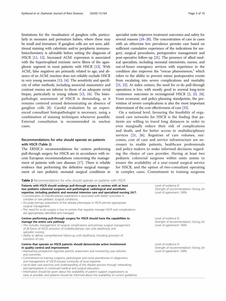
limitations for the visualisation of ganglion cells, particu-larly in neonates and premature babies, where these maybe small and immature. If ganglion cells are not seen, add-itional staining with calretinin and/or peripherin immuno-histochemistry is advisable before setting the diagnosis ofHSCR [11, 12]. Increased AChE expression is associatedwith the hypertrophied extrinsic nerve fibres of the agan-glionic segment in most patients with HSCR [13]. WithAChE, false negatives are primarily related to age, and ab-sence of an AChE reaction does not reliably exclude HSCRin very young neonates [13, 14]. The sensitivity and specifi-city of other methods, including anorectal manometry andcontrast enema are inferior to those of an adequate rectalbiopsy, particularly in young infants [15, 16]. The histo-pathologic assessment of HSCR is demanding, as itremains centered around demonstrating an absence ofganglion cells [8]. Careful evaluation by an experi-enced consultant histopathologist is necessary, using acombination of staining techniques whenever possible.External consultation is recommended in unclearcases.
Recommendations for who should operate on patientswith HSCR (Table 2)The ERNICA recommendations for centres performingpull-through surgery for HSCR are in accordance with re-cent European recommendations concerning the manage-ment of patients with rare diseases [17]. There is reliableevidence that performing the definitive surgical manage-ment of rare pediatric neonatal surgical conditions in
specialist units improves treatment outcomes and safety forseveral reasons [18–20]. The concentration of care in caseswith an otherwise low prevalence permits care based onsufficient cumulative experience of the indications for sur-gery, surgical procedures, perioperative management andpost-operative follow-up [21]. The presence of allied med-ical specialists, including neonatal intensivists, nurses, andout-of-hours emergency services with experience in thecondition also improves the “rescue phenomenon,” whichrefers to the ability to prevent minor postoperative eventsfrom escalating into severe complications and mortality[21, 22]. At index centers, the need for re-do pull-throughoperations is low, with mostly good to normal long-termcontinence outcomes in rectosigmoid HSCR [2, 23, 24].From economic and policy-planning standpoints, the pre-vention of severe complications is also the most importantdeterminant of the cost-effectiveness of care [25].On a national level, favouring the feasibility of orga-
nized care networks for HSCR is the finding that pa-tients are willing to travel long distances in order toeven marginally reduce their risk of complicationsand death, and for better access to multidisciplinaryservices [21, 26]. Registries of care volumes, out-comes, cost of care and service infrastructure are ne-cessary to enable patients, healthcare professionalsand policy-makers to make informed decisions regard-ing the choice of care provider. Having at least twopediatric colorectal surgeons within units assists toensure the availability of a year-round surgical servicefor HSCR, and the option of two-consultant operatingin complex cases. Commitment to training surgeons
Table 2 Recommendations for who should operate on patients with HSCR
Patients with HSCR should undergo pull-through surgery in centres with at leasttwo pediatric colorectal surgeons and pathological, radiological and anestheticexpertise, including pediatric and neonatal intensive care and specialized nursing 24/7.• Concentration of interdisciplinary experience is associated with better outcomes incomplex or rare pediatric surgical conditions.
• Accurate primary assessment of the disease phenotype in HSCR permits appropriatesurgical management
• The need for re-do surgery is low in centres that regularly manage HSCR and complicationsare appropriately identified and managed
Level of evidence IIIStrength of recommendation: Strong, forLevel of agreement: 100%
Centres performing pull-through surgery for HSCR should have the capabilities tomanage the entire care pathway• This includes management of surgical complications and primary surgical managementof all forms of HSCR, provision of multidisciplinary care until adulthood, andspecialist nursing
• Ability to deliver comprehensive follow-up until adulthood, including provision oftransition of care
Level of evidence IIIStrength of recommendation: Strong, forLevel of agreement: 100%
Centres that operate on HSCR patients should demonstrate active involvementin quality control and improvement• Maintaining prospective registries permits assessment and monitoring case volumesand outcomes
• Commitment to training surgeons, pathologists and nurse practitioners in diagnosticsand management of HSCR ensures continuity of local expertise
• Up-to-date care practices and understanding of the disease process through networkingand participation in continued medical and surgical education
• Information should be given about the availability of patient support organizations asearly as possible, and patients should be informed about the availability of current guidelines
Level of evidence IIIStrength of recommendation: Strong, forLevel of agreement: 100%
Kyrklund et al. Orphanet Journal of Rare Diseases (2020) 15:164 Page 3 of 16
and other allied specialists ensures long-term servicecontinuity.Centres performing pull-through surgery for HSCR
should have the capacity to manage all levels of HSCR, asaganglionosis extending proximal to the rectosigmoid af-fects 15–20% of patients [23]. Despite an optimal pull-through, impairment of bowel function and enterocolitisare common during the first few years after surgery, andbetween 4 and 30% of patients have an associated develop-mental disorder or syndrome [23, 27, 28]. Competence inthe management of HSCR is therefore not only defined byan ability to perform complex surgery or hospital volumesalone, but also on capacity to manage the disease processas a whole and to provide individualized long-term follow-up into adulthood [21]. For this, a properly functioningmultidisciplinary approach is essential [21]. Evidence ofquality should be based on outcome registries, alongsideregular participation in internal and external assessmentof benchmarks. Involvement in research is beneficial forgenerating knowledge regarding the effects of current andcompetitive therapies, which are needed to develop prac-tices towards new standards.
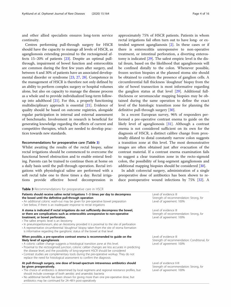
Recommendations for preoperative care (Table 3)Whilst awaiting the results of the rectal biopsy, salinerectal irrigations should be commenced to overcome thefunctional bowel obstruction and to enable enteral feed-ing. Parents can be trained to continue them at home ona daily basis until the pull-through operation. Rectal irri-gations with physiological saline are performed with asoft rectal tube one to three times a day. Rectal irriga-tions provide effective bowel decompression in
approximately 75% of HSCR patients. Patients in whomrectal irrigations fail often turn out to have long- or ex-tended segment aganglianosis [2]. In these cases or ifthere is enterocolitis unresponsive to non-operativetreatment, or intestinal perforation, a diverting enteros-tomy is indicated [29]. The safest empiric level is the dis-tal ileum, based on the likelihood that aganglionosis willbe confined distally to the colon. Whenever possible,frozen section biopsies at the planned stoma site shouldbe obtained to confirm the presence of ganglion cells. Acircumferential full thickness ‘doughnut’ biopsy from thesite of bowel transection is most informative regardingthe ganglion status at that level [29]. Additional full-thickness or seromuscular mapping biopsies may be ob-tained during the same operation to define the exactlevel of the histologic transition zone for planning thedefinitive pull-through surgery [30].In a recent European survey, 96% of responders per-
formed a pre-operative contrast enema to guide on thelikely level of aganglionosis [31]. Although a contrastenema is not considered sufficient on its own for thediagnosis of HSCR, a distinct calibre change from prox-imally dilated to distal constantly narrow colon suggestsa transition zone at this level. The most demonstrativeimages are often obtained just after evacuation of thecontrast material. If a contrast enema examination failsto suggest a clear transition zone in the recto-sigmoidcolon, the possibility of long-segment aganglionosis andadditional mapping biopsies should be considered [30].In adult colorectal surgery, administration of a single
preoperative dose of antibiotics has been shown to re-duce postoperative wound infections by 75% [32]. A
Table 3 Recommendations for preoperative care in HSCR
Patients should receive saline rectal irrigations 1–3 times per day to decompressthe bowel until the definitive pull-through operation• An additional colonic wash-out may be given for pre-operative bowel preparation• See below, if there is an inadequate response to rectal irrigations
Level of evidence IIIStrength of recommendation: Strong, forLevel of agreement: 100%
A stoma is indicated if rectal irrigations do not sufficiently decompress the bowel,or there are complications such as enterocolitis unresponsive to non-operativetreatment, or bowel perforation.• The safest empiric level is an ileostomy• In pneumoperitoneum, also an ileostomy provided it is proximal to the site of perforation• A representative circumferential ‘doughnut’ biopsy taken from the site of stoma formationis informative regarding the ganglionic status of the bowel at that level
Level of evidence IIIStrength of recommendation: Strong, forLevel of agreement: 100%
When possible, a pre-operative contrast enema is recommended to guide on thelikely level of aganglianosis• A colonic caliber change suggests a histological transition zone at this level.• Proximal to the rectosigmoid junction, colonic caliber changes are less accurate in predictingthe disease level, and the possibility of long-segment HSCR should be considered
• Contrast studies are complementary tools during the pre-operative workup. They do notreplace the need for histological assessment to confirm the diagnosis.
Level of evidence IIIStrength of recommendation: Conditional, forLevel of agreement: 100%
At pull-through surgery, one dose of broad-spectrum intravenous antibiotics shouldbe given preoperatively.• The choice of antibiotics is determined by local regimens and regional resistance profiles, butshould include coverage of both aerobic and anaerobic bacteria
• No additional benefit has been shown for giving more than one pre-operative dose, butantibiotics may be continued for 24–48 h post-operatively
Level of evidence II-IIIStrength of recommendation: Strong, forLevel of agreement: 100%
Kyrklund et al. Orphanet Journal of Rare Diseases (2020) 15:164 Page 4 of 16
combination of aerobic and anaerobic bacteria coveragewith a single agent or combination therapy is most ef-fective, the choice depending on local resistance patterns.Administration of prolonged antibiotic prophylaxis hasnot been proven to be more effective in preventing surgi-cal wound infections [33]. It is unknown to what extentthis reduces sequelae from anastomotic leakage, deep sur-gical infections, urinary tract or respiratory infections [32].Studies of antibiotic prophylaxis in the pediatric surgicalpopulation are scarce and do not include the latest tech-niques for HSCR surgery using minimally invasive ortransanal approaches [33].
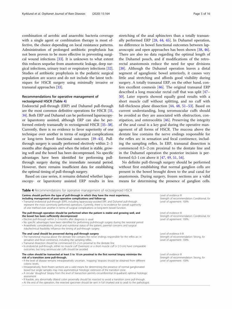
Recommendations for operative management ofrectosigmoid HSCR (Table 4)Endorectal pull-through (ERP) and Duhamel pull-throughare the most common definitive operations for HSCR [31,34]. Both ERP and Duhamel can be performed laparoscopy-or laparotomy assisted, although ERP can also be per-formed entirely transanally in rectosigmoid HSCR [35–38].Currently, there is no evidence to favor superiority of onetechnique over another in terms of surgical complicationsor long-term bowel functional outcomes [39–43]. Pull-through surgery is usually performed electively within 2–3months after diagnosis and when the infant is stable, grow-ing well and the bowel has been decompressed. No specificadvantages have been identified for performing pull-through surgery during the immediate neonatal period.However, there remains insufficient data for establishingthe optimal timing of pull-through surgery.Based on case series, it remains debated whether lapar-
oscopy- or laparotomy assisted ERP results in less
stretching of the anal sphincters than a totally transan-ally performed ERP [28, 44, 45]. In Duhamel operation,no difference in bowel functional outcomes between lap-aroscopic and open approaches has been shown [38, 46].There are also no data regarding the optimal length ofthe Duhamel pouch, and if modifications of the retro-rectal anastomosis reduce the need for spur divisions[38]. Although the Duhamel operation leaves a distalsegment of aganglionic bowel anteriorly, it causes verylittle anal stretching and affords good visibility duringsurgery. A totally transanal ERP, on the other hand, con-fers excellent cosmesis [46]. The original transanal ERPdescribed a long muscular rectal cuff that was split [47–50]. Later reports showed equally good results with ashort muscle cuff without splitting, and no cuff withfull-thickness plane dissection [44, 48, 51–53]. Based oncurrent understanding, long seromuscular cuffs shouldbe avoided as they are associated with obstruction, con-stipation, and enterocolitis [46]. Preserving the integrityof the anal canal is a key goal during the operative man-agement of all forms of HSCR. The mucosa above thedentate line contains the nerve endings responsible forthe reflex arc in sensation and fecal continence, includ-ing the sampling reflex. In ERP, transanal dissection iscommenced 0.5–2 cm proximal to the dentate line andin the Duhamel operation the posterior incision is per-formed 0,5-1 cm above it [47, 49, 51, 54].No definite pull-through surgery should be performed
without first establishing that normal ganglion cells arepresent in the bowel brought down to the anal canal foranastomosis. During surgery, frozen sections are a validmeans for determining the presence of ganglion cells.
Table 4 Recommendations for operative management of rectosigmoid HSCRCentres should perform the type of pull-through in which they have the most experience,including management of post-operative complications and follow-up• Transanal endorectal pull-through (ERP), including laparoscopy-assisted ERP, and Duhamel pull-throughrepresent the most commonly performed operations. Currently, there is no evidence for overall superiorityof one method over another in terms of surgical complications or long-term bowel function.
Level of evidence IIIStrength of recommendation: Conditional, forLevel of agreement: 100%
The pull-through operation should be performed when the patient is stable and growing well, andthe bowel has been sufficiently decompressed• Elective pull-through within 2–3 months after diagnosis is usual• No specific advantages have been identified for performing pull-through surgery during the neonatal period• Anaesthetic considerations, clinical and nutritional status of the patient, parental concerns and surgicalrisks/technical feasibility influence the timing of pull-through surgery
Level of evidence IIIStrength of recommendation: Conditional, forLevel of agreement: 100%
The anal canal should be preserved during pull-through surgery• The transitional mucosa above the dentate line contains the nerve endings responsible for the reflex arc insensation and fecal continence, including the sampling reflex.
• Transanal dissection should be commenced 0.5–2 cm proximal to the dentate line• In endorectal pull-through, either no muscle cuff (Swenson) or a short muscle cuff (< 2-3 cm) have comparableoutcomes, but long seromuscular cuffs should be avoided
Level of evidence II-IIIStrength of recommendation: Strong, forLevel of agreement: 100%
The colon should be transected at least 5 to 10 cm proximal to the first normal biopsy minimize therisk of a transition zone pull-through.• If the level of disease remains intraoperatively uncertain, ‘mapping’ biopsies should be obtained from differentcolonic levels.
• Intraoperatively, fresh frozen sections are a valid means for determining the presence of normal ganglionatedbowel but single samples may miss asymmetrical histologic extension of the transition zone.
• A circular ‘doughnut’ biopsy from the level of transection permits circumferential (4-quadrant) optimal histologicassessment
• If feasible, any abnormally dilated colon proximally should be resected to avoid a transition zone pull-through.• At the end of the operation, the resected specimen should be sent in full (marked oral to anal) to the pathologist.
Level of evidence IIIStrength of recommendation: Strong, forLevel of agreement: 100%
Kyrklund et al. Orphanet Journal of Rare Diseases (2020) 15:164 Page 5 of 16
Full-thickness biopsies permit examination of both themyenteric and submucosal plexuses and thereby minimizethe chance of anastomosing abnormally innervated bowelto the anal canal. Seromuscular biopsies have a lower riskof fecal spillage and postoperative perforations, but maybe more prone to sampling errors if the transition zone isirregular due to a lack of full submucosal assessment [46].As the length of the transition zone may be variable, theproximal transection margin should be a minimum of 5–10 cm orally to where normal ganglion cells are found, un-less a “doughnut” biopsy, showing ganglion cells circum-ferentially in both the submucosal and myenteric plexus isavailable intraoperatively [55].
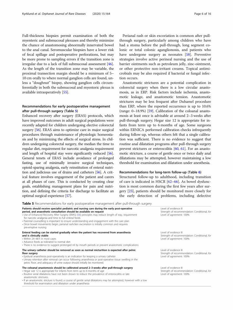
Recommendations for early postoperative managementafter pull-through surgery (Table 5)Enhanced recovery after surgery (ERAS) protocols, whichhave improved outcomes in adult surgical populations wererecently adapted for children undergoing elective colorectalsurgery [56]. ERAS aims to optimize care in major surgicalprocedures through maintenance of physiologic homeosta-sis and by minimizing the effects of surgical stress. In chil-dren undergoing colorectal surgery, the median the time toregular diet, requirement for narcotic analgesia requirementand length of hospital stay were significantly reduced [56].General tenets of ERAS include avoidance of prolongedfasting, use of minimally invasive surgical techniques,opioid-sparing analgesia, early reinstitution of enteral nutri-tion and judicious use of drains and catheters [56]. A crit-ical feature involves engagement of the patient and carersat all phases of care. This is conducted by creating cleargoals, establishing management plans for pain and nutri-tion, and defining the criteria for discharge to facilitate anoptimal surgical experience [57].
Perianal rash or skin excoriation is common after pull-through surgery, particularly among children who havehad a stoma before the pull-through, long segment co-lonic or total colonic aganglionosis, and patients whohave undergone surgery as neonates [58]. Preventivestrategies involve active perineal nursing and the use ofbarrier ointments such as petroleum jelly, zinc-ointment,or other protective non-irritant creams. Topical antimi-crobials may be also required if bacterial or fungal infec-tion occurs.Anastomotic strictures are a potential complication in
colorectal surgery when there is a low circular anasto-mosis, as in ERP. Risk factors include ischemia, anasto-motic leakage, and anastomotic tension. Anastomoticstrictures may be less frequent after Duhamel procedurethan ERP, where the reported occurrence is up to 10.6%(range: 0–18.9%) [59]. Calibration of the coloanal anasto-mosis at least once is advisable at around 2–3 weeks afterpull-through surgery; Hegar size 12 is appropriate for in-fants from term up to 6months of age. Some surgeonswithin ERNICA performed calibration checks infrequentlyduring follow-up, whereas others felt that a single calibra-tion was sufficient. There is no evidence to suggest thatroutine anal dilatation programs after pull-through surgeryprevent strictures or enterocolitis [60, 61]. For an anasto-motic stricture, a course of gentle once or twice daily analdilatations may be attempted, however maintaining a lowthreshold for examination and dilatation under anesthesia.
Recommendations for long-term follow-up (Table 6)Structured follow-up to adulthood, including transitionof care is indicated in HSCR [62–66]. As bowel dysfunc-tion is most common during the first few years after sur-gery [23], patients should be monitored more closely forthe early detection of problems, including defective
Table 5 Recommendations for early postoperative management after pull-through surgeryPatients should receive specialist pediatric and nursing care during the early post-operativeperiod, and anaesthetic consultation should be available on request• Use of Enhanced Recovery After Surgery (ERAS) [56] principles may reduce length of stay, requirementfor narcotic analgesia and time to full enteral feeds
• Parental counselling is important to ensure understanding and engagement with the care plan• Once bowel movements begin, perianal rash/skin excoriation is initially common and requirespre-emptive nursing
Level of evidence IIIStrength of recommendation: Conditional, forLevel of agreement: 100%
Enteral feeding can be started gradually when the patient has recovered from anaesthesiaand is clinically stable• Within 24–48 h in most cases• Advance feeds as tolerated to normal diet• There is no evidence to suggest prolonged nil by mouth periods or prevent anastomotic complications
Level of evidence IIIStrength of recommendation: Conditional, forLevel of agreement: 100%
The urinary catheter should be removed as soon as normal micturition is expected after pelvicfloor surgery• Epidural anaesthesia post-operatively is an indication for keeping a urinary catheter• Urinary retention after removal can occur following anaesthesia or post-operative tissue swelling in thepelvic floor, and adequacy of urine output should initially be monitored.
Level of evidence IIIStrength of recommendation: Conditional, forLevel of agreement: 100%
The coloanal anastomosis should be calibrated around 2–3 weeks after pull-through surgery• Hegar size 12 is appropriate for infants from term up to 6 months of age• Routine serial dilatations have not been shown to reduce the prevalence of enterocolitis or lateanastomotic strictures.
• If an anastomotic stricture is found, a course of gentle serial dilatations may be attempted, however with a lowthreshold for examination and dilatation under anaesthesia
Level of evidence IIIStrength of recommendation: Conditional, forLevel of agreement: 100%
Kyrklund et al. Orphanet Journal of Rare Diseases (2020) 15:164 Page 6 of 16
continence, enterocolitis and late complications. Clinicalfollow-up should be maintained even if patients are doingwell. Among patients with HSCR and a syndrome associ-ated with cognitive impairment, reaching continence isoften significantly delayed. In patients with poor func-tional outcomes, adequate follow-up permits the institu-tion of interventions before school age. This is importantto prevent social discrimination, and to allow participationin normal childhood activities with minimal limitationsfrom the condition [62]. As HSCR is a rare disorder ingeneral practice, patients should also be managed by spe-cialized medical personnel familiar with the disease [67],and receive instructions on whom to contact and where toseek medical attention in both elective and emergency cir-cumstances. The availability of specialist nurses improvescommunication and access to care, and may reduce hospi-talizations/admissions and health care costs [68].In patients with HSCR, life events that necessitate
follow-up include puberty and sexual development, pro-creation and heritability, and transition to adulthood andadult care [64, 69, 70]. Psychological factors to be ad-dressed include self-efficacy, social functioning and cop-ing skills for residual symptoms [71, 72]. As the parentsof chronically ill children also experience considerablestress, strategies to reduce psychological morbidity thatalso involve the parents are likely to be beneficial [62].Achieving optimal outcomes requires collaboration be-tween medical specialists, nurses and auxiliary resources,including psychologists and sexual therapists, physicaland nutritional therapists. In addition to pediatric sur-geons and gastroenterologists, urologic, gynecologic andgenetic consultation should be sought as appropriate.Active involvement of the patient/family at all stages of
follow-up is important to enable understanding the careplan, and to establish mutually agreed priorities for care[73]. For peer support on lived experience of the disease,patient support organizations encourage contact frompatients and their families for advice and networking.During the care pathway, as capacity for understanding
increases, patients should be encouraged to acquirehealth literacy skills and gradually assume responsibilityfor their own care. Adequate information provision isimportant in this regard, so that including issues relatingto sexual health during adulthood can be addressed [62].Planning transition of care should commence sufficientlyearly, around 13–14 years of age to allow sufficient timefor adjustment and should have sufficient flexibility toallow overlap and contact between the existing and fu-ture practitioners until patients are satisfactorily estab-lished in adult care [74].
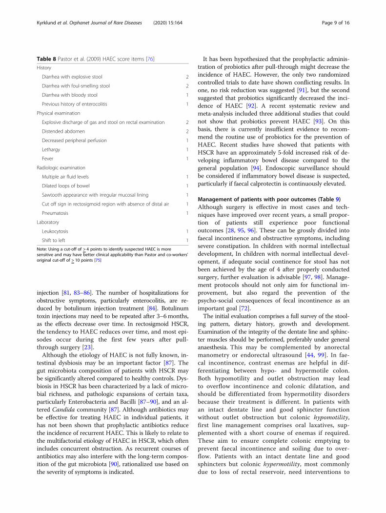
Recommendations for Hirschsprung’s-associatedenterocolitis (HAEC; Table 7)HAEC is the most frequent serious complication afterpull-through surgery for Hirschsprung disease. Predis-posing factors include family history of HSCR, long seg-ment disease, Down syndrome (trisomy 21) and previousepisodes of HAEC [75]. Approximately 30% of HSCRpatients have at least one episode of postoperativeHAEC. Although the definition remains imprecise, usingthe Pastor et al. HAEC score items (Table 8) [76], with acut-off of > 4 points to identify suspected HAEC is moresensitive and may have better clinical applicability [75]than Pastor and co-workers' original cut-off of > 10points. HAEC should be suspected in the presence ofdiarrhea with explosive, foul-smelling or bloody stool,
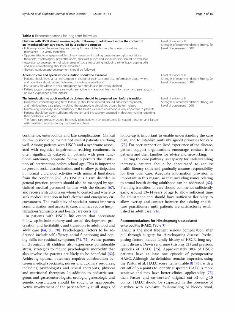
Table 6 Recommendations for long-term follow-up
Children with HSCR should receive regular follow-up to adulthood within the context ofan interdisciplinary care team, led by a pediatric surgeon• Follow-up should be more frequent during 1st year of life, but regular contact should bemaintained 1–2 yearly thereafter
• Opportunities to engage multidisciplinary resources, including gastroenterologists, nutritionaltherapists, psychologists, physiotherapists, specialist nurses and social workers should be available
• Attention to development of wider areas of social functioning, including self-efficacy, coping skillsand sexual functioning should be addressed
• Growth, nutrition and development should be followed
Level of evidence IIIStrength of recommendation: Strong, forLevel of agreement: 100%
Access to care and specialist consultation should be available• Patients should have a named surgeon in charge of their care and clear information about whereand how they should attend follow-up, including in adulthood
• Instructions for where to seek emergency care should also be clearly defined• Patient support organizations networks are active in many countries for information and peer supporton lived experience of the disease
Level of evidence IIIStrength of recommendation: Strong, forLevel of agreement: 100%
The introduction to adult medical disciplines should be prepared well before transition• Discussions concerning long-term follow-up should be initiated around adolescence/puberty,and individualized care plans involving the appropriate disciplines should be formulated
• Maintaining continuity and consistency of the health care into adulthood is very important to patients• Patients should be given sufficient information and increasingly engaged in decision-making regardingtheir healthcare with age
• The future care provider should be clearly identified, with an opportunity for staged transition and liaisonwith paediatric services during the transition phase.
Level of evidence IIIStrength of recommendation: Strong, forLevel of agreement: 100%
Kyrklund et al. Orphanet Journal of Rare Diseases (2020) 15:164 Page 7 of 16
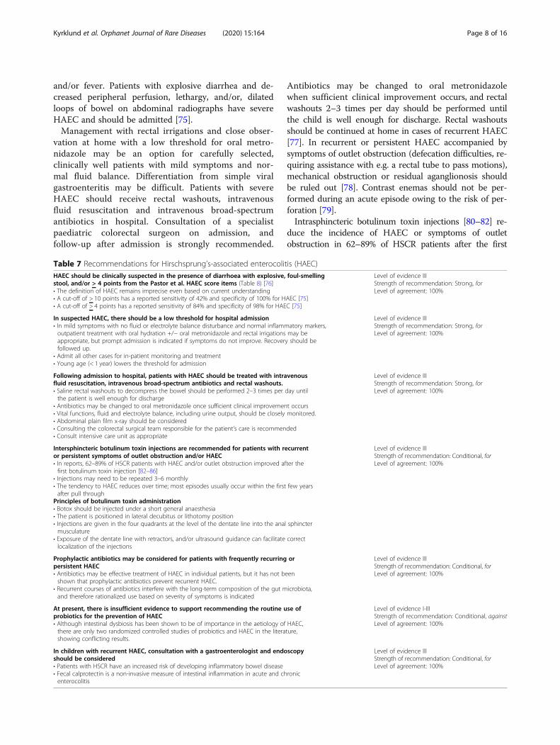
and/or fever. Patients with explosive diarrhea and de-creased peripheral perfusion, lethargy, and/or, dilatedloops of bowel on abdominal radiographs have severeHAEC and should be admitted [75].Management with rectal irrigations and close obser-
vation at home with a low threshold for oral metro-nidazole may be an option for carefully selected,clinically well patients with mild symptoms and nor-mal fluid balance. Differentiation from simple viralgastroenteritis may be difficult. Patients with severeHAEC should receive rectal washouts, intravenousfluid resuscitation and intravenous broad-spectrumantibiotics in hospital. Consultation of a specialistpaediatric colorectal surgeon on admission, andfollow-up after admission is strongly recommended.
Antibiotics may be changed to oral metronidazolewhen sufficient clinical improvement occurs, and rectalwashouts 2–3 times per day should be performed untilthe child is well enough for discharge. Rectal washoutsshould be continued at home in cases of recurrent HAEC[77]. In recurrent or persistent HAEC accompanied bysymptoms of outlet obstruction (defecation difficulties, re-quiring assistance with e.g. a rectal tube to pass motions),mechanical obstruction or residual aganglionosis shouldbe ruled out [78]. Contrast enemas should not be per-formed during an acute episode owing to the risk of per-foration [79].Intrasphincteric botulinum toxin injections [80–82] re-
duce the incidence of HAEC or symptoms of outletobstruction in 62–89% of HSCR patients after the first
Table 7 Recommendations for Hirschsprung’s-associated enterocolitis (HAEC)HAEC should be clinically suspected in the presence of diarrhoea with explosive, foul-smellingstool, and/or > 4 points from the Pastor et al. HAEC score items (Table 8) [76]• The definition of HAEC remains imprecise even based on current understanding• A cut-off of > 10 points has a reported sensitivity of 42% and specificity of 100% for HAEC [75]• A cut-off of > 4 points has a reported sensitivity of 84% and specificity of 98% for HAEC [75]
Level of evidence IIIStrength of recommendation: Strong, forLevel of agreement: 100%
In suspected HAEC, there should be a low threshold for hospital admission• In mild symptoms with no fluid or electrolyte balance disturbance and normal inflammatory markers,outpatient treatment with oral hydration +/− oral metronidazole and rectal irrigations may beappropriate, but prompt admission is indicated if symptoms do not improve. Recovery should befollowed up.
• Admit all other cases for in-patient monitoring and treatment• Young age (< 1 year) lowers the threshold for admission
Level of evidence IIIStrength of recommendation: Strong, forLevel of agreement: 100%
Following admission to hospital, patients with HAEC should be treated with intravenousfluid resuscitation, intravenous broad-spectrum antibiotics and rectal washouts.• Saline rectal washouts to decompress the bowel should be performed 2–3 times per day untilthe patient is well enough for discharge
• Antibiotics may be changed to oral metronidazole once sufficient clinical improvement occurs• Vital functions, fluid and electrolyte balance, including urine output, should be closely monitored.• Abdominal plain film x-ray should be considered• Consulting the colorectal surgical team responsible for the patient’s care is recommended• Consult intensive care unit as appropriate
Level of evidence IIIStrength of recommendation: Strong, forLevel of agreement: 100%
Intersphincteric botulinum toxin injections are recommended for patients with recurrentor persistent symptoms of outlet obstruction and/or HAEC• In reports, 62–89% of HSCR patients with HAEC and/or outlet obstruction improved after thefirst botulinum toxin injection [82–86]
• Injections may need to be repeated 3–6 monthly• The tendency to HAEC reduces over time; most episodes usually occur within the first few yearsafter pull through
Principles of botulinum toxin administration• Botox should be injected under a short general anaesthesia• The patient is positioned in lateral decubitus or lithotomy position• Injections are given in the four quadrants at the level of the dentate line into the anal sphinctermusculature
• Exposure of the dentate line with retractors, and/or ultrasound guidance can facilitate correctlocalization of the injections
Level of evidence IIIStrength of recommendation: Conditional, forLevel of agreement: 100%
Prophylactic antibiotics may be considered for patients with frequently recurring orpersistent HAEC• Antibiotics may be effective treatment of HAEC in individual patients, but it has not beenshown that prophylactic antibiotics prevent recurrent HAEC.
• Recurrent courses of antibiotics interfere with the long-term composition of the gut microbiota,and therefore rationalized use based on severity of symptoms is indicated
Level of evidence IIIStrength of recommendation: Conditional, forLevel of agreement: 100%
At present, there is insufficient evidence to support recommending the routine use ofprobiotics for the prevention of HAEC• Although intestinal dysbiosis has been shown to be of importance in the aetiology of HAEC,there are only two randomized controlled studies of probiotics and HAEC in the literature,showing conflicting results.
Level of evidence I-IIIStrength of recommendation: Conditional, againstLevel of agreement: 100%
In children with recurrent HAEC, consultation with a gastroenterologist and endoscopyshould be considered• Patients with HSCR have an increased risk of developing inflammatory bowel disease• Fecal calprotectin is a non-invasive measure of intestinal inflammation in acute and chronicenterocolitis
Level of evidence IIIStrength of recommendation: Conditional, forLevel of agreement: 100%
Kyrklund et al. Orphanet Journal of Rare Diseases (2020) 15:164 Page 8 of 16
injection [81, 83–86]. The number of hospitalizations forobstructive symptoms, particularly enterocolitis, are re-duced by botulinum injection treatment [84]. Botulinumtoxin injections may need to be repeated after 3–6months,as the effects decrease over time. In rectosigmoid HSCR,the tendency to HAEC reduces over time, and most epi-sodes occur during the first few years after pull-through surgery [23].Although the etiology of HAEC is not fully known, in-
testinal dysbiosis may be an important factor [87]. Thegut microbiota composition of patients with HSCR maybe significantly altered compared to healthy controls. Dys-biosis in HSCR has been characterized by a lack of micro-bial richness, and pathologic expansions of certain taxa,particularly Enterobacteria and Bacilli [87–90], and an al-tered Candida community [87]. Although antibiotics maybe effective for treating HAEC in individual patients, ithas not been shown that prophylactic antibiotics reducethe incidence of recurrent HAEC. This is likely to relate tothe multifactorial etiology of HAEC in HSCR, which oftenincludes concurrent obstruction. As recurrent courses ofantibiotics may also interfere with the long-term compos-ition of the gut microbiota [90], rationalized use based onthe severity of symptoms is indicated.
It has been hypothesized that the prophylactic adminis-tration of probiotics after pull-through might decrease theincidence of HAEC. However, the only two randomizedcontrolled trials to date have shown conflicting results. Inone, no risk reduction was suggested [91], but the secondsuggested that probiotics significantly decreased the inci-dence of HAEC [92]. A recent systematic review andmeta-analysis included three additional studies that couldnot show that probiotics prevent HAEC [93]. On thisbasis, there is currently insufficient evidence to recom-mend the routine use of probiotics for the prevention ofHAEC. Recent studies have showed that patients withHSCR have an approximately 5-fold increased risk of de-veloping inflammatory bowel disease compared to thegeneral population [94]. Endoscopic surveillance shouldbe considered if inflammatory bowel disease is suspected,particularly if faecal calprotectin is continuously elevated.
Management of patients with poor outcomes (Table 9)Although surgery is effective in most cases and tech-niques have improved over recent years, a small propor-tion of patients still experience poor functionaloutcomes [28, 95, 96]. These can be grossly divided intofaecal incontinence and obstructive symptoms, includingsevere constipation. In children with normal intellectualdevelopment, In children with normal intellectual devel-opment, if adequate social continence for stool has notbeen achieved by the age of 4 after properly conductedsurgery, further evaluation is advisable [97, 98]. Manage-ment protocols should not only aim for functional im-provement, but also regard the prevention of thepsycho-social consequences of fecal incontinence as animportant goal [72].The initial evaluation comprises a full survey of the stool-
ing pattern, dietary history, growth and development.Examination of the integrity of the dentate line and sphinc-ter muscles should be performed, preferably under generalanaesthesia. This may be complemented by anorectalmanometry or endorectal ultrasound [44, 99]. In fae-cal incontinence, contrast enemas are helpful in dif-ferentiating between hypo- and hypermotile colon.Both hypomotility and outlet obstruction may leadto overflow incontinence and colonic dilatation, andshould be differentiated from hypermotility disordersbecause their treatment is different. In patients withan intact dentate line and good sphincter functionwithout outlet obstruction but colonic hypomotility,first line management comprises oral laxatives, sup-plemented with a short course of enemas if required.These aim to ensure complete colonic emptying toprevent faecal incontinence and soiling due to over-flow. Patients with an intact dentate line and goodsphincters but colonic hypermotility, most commonlydue to loss of rectal reservoir, need interventions to
Table 8 Pastor et al. (2009) HAEC score items [76]
History
Diarrhea with explosive stool 2
Diarrhea with foul-smelling stool 2
Diarrhea with bloody stool 1
Previous history of enterocolitis 1
Physical examination
Explosive discharge of gas and stool on rectal examination 2
Distended abdomen 2
Decreased peripheral perfusion 1
Lethargy 1
Fever 1
Radiologic examination
Multiple air fluid levels 1
Dilated loops of bowel 1
Sawtooth appearance with irregular mucosal lining 1
Cut off sign in rectosigmoid region with absence of distal air 1
Pneumatosis 1
Laboratory
Leukocytosis 1
Shift to left 1
Note: Using a cut-off of > 4 points to identify suspected HAEC is moresensitive and may have better clinical applicability than Pastor and co-workers'original cut-off of > 10 points [75]
Kyrklund et al. Orphanet Journal of Rare Diseases (2020) 15:164 Page 9 of 16
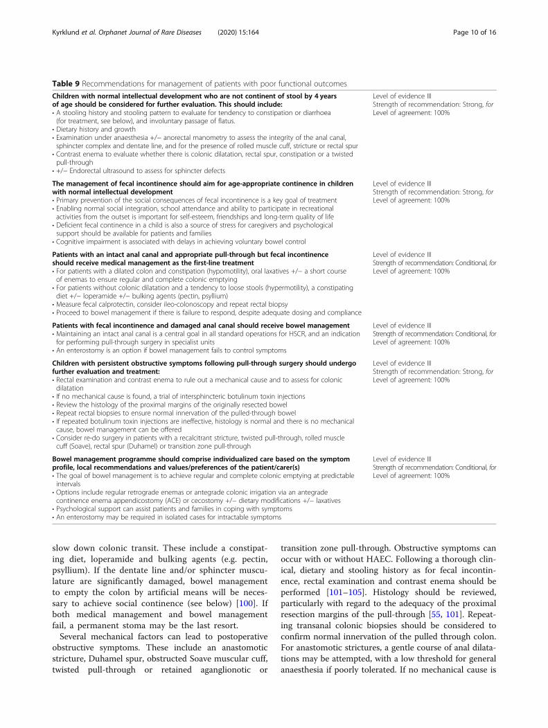
slow down colonic transit. These include a constipat-ing diet, loperamide and bulking agents (e.g. pectin,psyllium). If the dentate line and/or sphincter muscu-lature are significantly damaged, bowel managementto empty the colon by artificial means will be neces-sary to achieve social continence (see below) [100]. Ifboth medical management and bowel managementfail, a permanent stoma may be the last resort.Several mechanical factors can lead to postoperative
obstructive symptoms. These include an anastomoticstricture, Duhamel spur, obstructed Soave muscular cuff,twisted pull-through or retained aganglionotic or
transition zone pull-through. Obstructive symptoms canoccur with or without HAEC. Following a thorough clin-ical, dietary and stooling history as for fecal incontin-ence, rectal examination and contrast enema should beperformed [101–105]. Histology should be reviewed,particularly with regard to the adequacy of the proximalresection margins of the pull-through [55, 101]. Repeat-ing transanal colonic biopsies should be considered toconfirm normal innervation of the pulled through colon.For anastomotic strictures, a gentle course of anal dilata-tions may be attempted, with a low threshold for generalanaesthesia if poorly tolerated. If no mechanical cause is
Table 9 Recommendations for management of patients with poor functional outcomes
Children with normal intellectual development who are not continent of stool by 4 yearsof age should be considered for further evaluation. This should include:• A stooling history and stooling pattern to evaluate for tendency to constipation or diarrhoea(for treatment, see below), and involuntary passage of flatus.
• Dietary history and growth• Examination under anaesthesia +/− anorectal manometry to assess the integrity of the anal canal,sphincter complex and dentate line, and for the presence of rolled muscle cuff, stricture or rectal spur
• Contrast enema to evaluate whether there is colonic dilatation, rectal spur, constipation or a twistedpull-through
• +/− Endorectal ultrasound to assess for sphincter defects
Level of evidence IIIStrength of recommendation: Strong, forLevel of agreement: 100%
The management of fecal incontinence should aim for age-appropriate continence in childrenwith normal intellectual development• Primary prevention of the social consequences of fecal incontinence is a key goal of treatment• Enabling normal social integration, school attendance and ability to participate in recreationalactivities from the outset is important for self-esteem, friendships and long-term quality of life
• Deficient fecal continence in a child is also a source of stress for caregivers and psychologicalsupport should be available for patients and families
• Cognitive impairment is associated with delays in achieving voluntary bowel control
Level of evidence IIIStrength of recommendation: Strong, forLevel of agreement: 100%
Patients with an intact anal canal and appropriate pull-through but fecal incontinenceshould receive medical management as the first-line treatment• For patients with a dilated colon and constipation (hypomotility), oral laxatives +/− a short courseof enemas to ensure regular and complete colonic emptying
• For patients without colonic dilatation and a tendency to loose stools (hypermotility), a constipatingdiet +/− loperamide +/− bulking agents (pectin, psyllium)
• Measure fecal calprotectin, consider ileo-colonoscopy and repeat rectal biopsy• Proceed to bowel management if there is failure to respond, despite adequate dosing and compliance
Level of evidence IIIStrength of recommendation: Conditional, forLevel of agreement: 100%
Patients with fecal incontinence and damaged anal canal should receive bowel management• Maintaining an intact anal canal is a central goal in all standard operations for HSCR, and an indicationfor performing pull-through surgery in specialist units
• An enterostomy is an option if bowel management fails to control symptoms
Level of evidence IIIStrength of recommendation: Conditional, forLevel of agreement: 100%
Children with persistent obstructive symptoms following pull-through surgery should undergofurther evaluation and treatment:• Rectal examination and contrast enema to rule out a mechanical cause and to assess for colonicdilatation
• If no mechanical cause is found, a trial of intersphincteric botulinum toxin injections• Review the histology of the proximal margins of the originally resected bowel• Repeat rectal biopsies to ensure normal innervation of the pulled-through bowel• If repeated botulinum toxin injections are ineffective, histology is normal and there is no mechanicalcause, bowel management can be offered
• Consider re-do surgery in patients with a recalcitrant stricture, twisted pull-through, rolled musclecuff (Soave), rectal spur (Duhamel) or transition zone pull-through
Level of evidence IIIStrength of recommendation: Strong, forLevel of agreement: 100%
Bowel management programme should comprise individualized care based on the symptomprofile, local recommendations and values/preferences of the patient/carer(s)• The goal of bowel management is to achieve regular and complete colonic emptying at predictableintervals
• Options include regular retrograde enemas or antegrade colonic irrigation via an antegradecontinence enema appendicostomy (ACE) or cecostomy +/− dietary modifications +/− laxatives
• Psychological support can assist patients and families in coping with symptoms• An enterostomy may be required in isolated cases for intractable symptoms
Level of evidence IIIStrength of recommendation: Conditional, forLevel of agreement: 100%
Kyrklund et al. Orphanet Journal of Rare Diseases (2020) 15:164 Page 10 of 16
found, first-line treatment is intersphincteric botulinumtoxin injections to relieve internal sphincter achalasia. Ifsymptoms fail to improve after repeated (> 3) botulinumtoxin injections, bowel management is the second-linechoice of treatment.Bowel management comprises individualized man-
agement based on the patient’s preferences. Optionsinclude regular retrograde enemas, or antegrade co-lonic irrigation via an antegrade continence enemaappendicostomy (ACE) or cecostomy (CHAIT, button)in conjunction with oral laxatives and dietary modifi-cations. Re-do surgery should be considered in symp-tomatic patients with a recalcitrant stricture, twistedpull-through, rolled muscle cuff (Soave), rectal spur(Duhamel), or aganglionic/transition zone pull-through. The full evaluation and re-do surgical pro-cedure should be performed in a centre with experi-ence of complex pathology in HSCR and re-doprocedures. Although redo-surgery is appropriate inselected cases resolves obstructing symptoms, it maybe associated with relatively high rates of fecal incon-tinence and thus, careful patient selection is import-ant [103, 105].
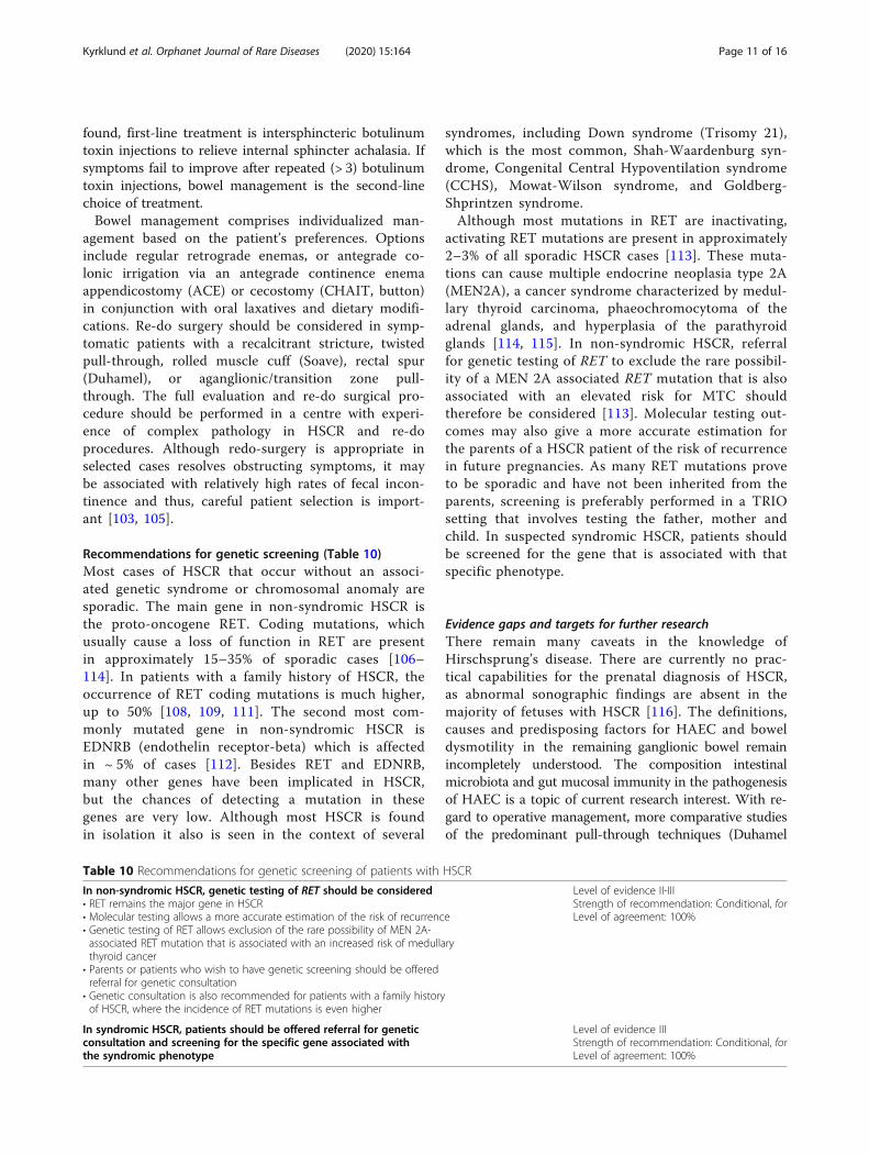
Recommendations for genetic screening (Table 10)Most cases of HSCR that occur without an associ-ated genetic syndrome or chromosomal anomaly aresporadic. The main gene in non-syndromic HSCR isthe proto-oncogene RET. Coding mutations, whichusually cause a loss of function in RET are presentin approximately 15–35% of sporadic cases [106–114]. In patients with a family history of HSCR, theoccurrence of RET coding mutations is much higher,up to 50% [108, 109, 111]. The second most com-monly mutated gene in non-syndromic HSCR isEDNRB (endothelin receptor-beta) which is affectedin ~ 5% of cases [112]. Besides RET and EDNRB,many other genes have been implicated in HSCR,but the chances of detecting a mutation in thesegenes are very low. Although most HSCR is foundin isolation it also is seen in the context of several
syndromes, including Down syndrome (Trisomy 21),which is the most common, Shah-Waardenburg syn-drome, Congenital Central Hypoventilation syndrome(CCHS), Mowat-Wilson syndrome, and Goldberg-Shprintzen syndrome.Although most mutations in RET are inactivating,
activating RET mutations are present in approximately2–3% of all sporadic HSCR cases [113]. These muta-tions can cause multiple endocrine neoplasia type 2A(MEN2A), a cancer syndrome characterized by medul-lary thyroid carcinoma, phaeochromocytoma of theadrenal glands, and hyperplasia of the parathyroidglands [114, 115]. In non-syndromic HSCR, referralfor genetic testing of RET to exclude the rare possibil-ity of a MEN 2A associated RET mutation that is alsoassociated with an elevated risk for MTC shouldtherefore be considered [113]. Molecular testing out-comes may also give a more accurate estimation forthe parents of a HSCR patient of the risk of recurrencein future pregnancies. As many RET mutations proveto be sporadic and have not been inherited from theparents, screening is preferably performed in a TRIOsetting that involves testing the father, mother andchild. In suspected syndromic HSCR, patients shouldbe screened for the gene that is associated with thatspecific phenotype.
Evidence gaps and targets for further researchThere remain many caveats in the knowledge ofHirschsprung’s disease. There are currently no prac-tical capabilities for the prenatal diagnosis of HSCR,as abnormal sonographic findings are absent in themajority of fetuses with HSCR [116]. The definitions,causes and predisposing factors for HAEC and boweldysmotility in the remaining ganglionic bowel remainincompletely understood. The composition intestinalmicrobiota and gut mucosal immunity in the pathogenesisof HAEC is a topic of current research interest. With re-gard to operative management, more comparative studiesof the predominant pull-through techniques (Duhamel
Table 10 Recommendations for genetic screening of patients with HSCR
In non-syndromic HSCR, genetic testing of RET should be considered• RET remains the major gene in HSCR• Molecular testing allows a more accurate estimation of the risk of recurrence• Genetic testing of RET allows exclusion of the rare possibility of MEN 2A-associated RET mutation that is associated with an increased risk of medullarythyroid cancer
• Parents or patients who wish to have genetic screening should be offeredreferral for genetic consultation
• Genetic consultation is also recommended for patients with a family historyof HSCR, where the incidence of RET mutations is even higher
Level of evidence II-IIIStrength of recommendation: Conditional, forLevel of agreement: 100%
In syndromic HSCR, patients should be offered referral for geneticconsultation and screening for the specific gene associated withthe syndromic phenotype
Level of evidence IIIStrength of recommendation: Conditional, forLevel of agreement: 100%
Kyrklund et al. Orphanet Journal of Rare Diseases (2020) 15:164 Page 11 of 16
and ERP) are needed to gather higher quality evidence ofthe long-term outcomes, and whether laparoscopy-assisted techniques confer advantages. Little is also knownabout the sexual function and fertility after pull-throughsurgery, although physical sexual functions appear to bepreserved in the majority [117]. However, it has beenshown that technically comparable low pelvic surgery forother pediatric bowel problems, impacts on later fertility,especially in females [118]. Further prospective researchand multicenter studies are needed to obtain a full under-standing of the long-term implications of HSCR on pa-tients’ health.
ConclusionsIn rare or low-prevalence diseases such as HSCR,there is limited availability of high-quality clinical evi-dence. However, patients born with these conditionscontinue to require highly specialized care from in-fancy up to adulthood. In this document, consensus-based guidelines that describe the preferred approachof ERNICA, an international panel of experts from 10major European centres to the management of recto-sigmoid HSCR are presented. ERNICA remains com-mitted to upholding and improving care standards forpatients with HSCR.
Materials and methodsMembership of the guideline workgroupThe workgroup comprised an international panel ofexperts in HSCR from specialist centres in 8 Europeancountries (Denmark, France, Germany, Finland, Italy,Netherlands, Norway, Sweden and the United Kingdom).These included pediatric surgeons, histopathologists, gas-troenterologists, microbiologists and geneticists from 10ERNICA (ERN) member centres and 3 associated expertsselected by ERNICA. Patient representatives from SoMA,the German patient support organisation for anorectalmalformations and HSCR and A.Mor.Hi, the Italian Soci-ety for HSCR, participated throughout the process.Accreditation as an ERN centre requires demonstration
and approval of specific specialist competencies and compli-ance with a comprehensive set of operational criteria [119].These include demonstration of quality in organizational in-frastructure, patient-centered management, clinical govern-ance, sufficient care volumes, ability to report outcomes andcommitment to upholding expertise through research, train-ing and education. Recognition of competencies and mem-bership is based on regular internal and external assessment.
Background preparationA literature search was conducted using PubMed of arti-cles published on HSCR from 1990 to 2018, and their ref-erences to identify articles that had not been brought up bythe original search. Where studies of HSCR or pediatric
populations were scarce or unavailable, data was derivedfrom relevant studies from adult populations. Withingroups, expert clinicians selected those publications theyconsidered most relevant and highest quality for the refer-ence pool of the guideline. Patient representatives contrib-uted publications that best reflected their values to theworkgroup. All authors had access to the reference poolduring development. Articles were assigned an evidencelevel of I-III based on the Canadian Task Force on thePeriodic Health Examination classification system (1979)summarized in Table 11 [120], with the expectation that inrare or low prevalence diseases most would fall into cat-egory III (descriptive studies, expert opinion).
Determination of the core recommendation statementsThe working group met on three occasions: 1) to discussthe content of the guideline, 2) to decide on the corerecommendations and supporting statements, and 3) tofinalize the content, including voting on levels of agree-ment. After drafting recommendations, the supporting textand summary of the evidence was prepared. The AGREE IIframework was used to assess the scope and rigour of themethodology [121]. The GRADE approach was used to ratethe strength of the recommendations [122, 123]. Updatingof the recommendations at 5-year intervals by ERNICAwas planned.
Assigning strength of the recommendationsRecommendations were classed as strong (for or against),if the workgroup felt highly confident of the balance be-tween desirable and undesirable consequences [122,123]. Recommendations were classed as conditional (foror against), if the appropriate course of action was con-sidered subject to patient values and preferences, avail-ability of resources and/or setting of the intervention[122, 123].
Voting on levels of agreementVoting on levels of agreement was conducted at the finalmeeting. Recommendations receiving < 100% agreementwere further discussed, revised and voted on again at themeeting until consensus was reached; > 75% agreement wasconsidered necessary to finalize recommendations [124].
Table 11 Grading of levels of Evidence [120]
Level of Evidence
• I Evidence from at least one randomized controlled trial
• II1 Evidence from well-designed case-control or cohort studies,preferably from more than one research group or centre
• II2 Comparisons between times and places with or without theintervention, or dramatic results of uncontrolled experiments
• III Opinions of respected authorities, based on clinical experience,descriptive studies or reports of expert committees.
Kyrklund et al. Orphanet Journal of Rare Diseases (2020) 15:164 Page 12 of 16
AbbreviationsACE: Antegrade continence enema; AChE: Acetylcholine esterase;EDNRB: Endotheln receptor beta; ERN: European Reference Network;ERNICA: The European Reference Network for for rare inherited andcongenital digestive disorders; ERAS: Enhanced recovery after surgery;ERP: Endorectal pull-through; EU: European Union; HSCR: Hirschsprung’sdisease; H&E: Haematoxylin and eosin; MEN: Multiple endocrine neoplasia;MTC: Medullary thyroid cancer; RET: Rearranged during transfection proto-oncogene
AcknowledgmentsWe would like to thank Prof. Dr. René Wijnen, Renée de Ruiter and OliviaSpivack from ERNICA for organising the Network’s meetings, that alsoenabled completion of this project.
Authors’ contributionsAll authors of the work have made substantial contributions to theconception and design of the work and to the literature search and itsanalysis. All authors have been involved in drafting the work and revising itcritically for important intellectual content, and all authors read andapproved the final manuscript.
FundingWorkgroup meetings and article-processing charges for open accesspublication in Orphanet Journal of Rare Diseases were supported by ERNICAEuropean Reference Network. The authors have no other sources of fundingto disclose.
Availability of data and materialsNot applicable to article type.
Ethics approval and consent to participateNot applicable to article type.
Consent for publicationNot applicable to article type.
Competing interestsThe authors declare that they have no competing interests
Author details1Department of Pediatric Surgery, Children’s Hospital, University of Helsinkiand Helsinki University Hospital, Helsinki, Finland. 2Department of PediatricSurgery, Erasmus MC – Sophia Children’s Hospital, Rotterdam, TheNetherlands. 3Department of Surgery, Division of Pediatric Surgery,Radboudumc-Amalia Children’s Hospital, Nijmegen, The Netherlands.4Department of Pediatric Surgery, Oslo University Hospital and University ofOslo, Oslo, Norway. 5Department of Pediatric Surgery and Pediatric Urology,University Hospital Frankfurt, Frankfurt/M, Germany. 6Department of Biology,University of Florence, A.Mor.Hi, The Italian Association for Hirschsprung’sdisease, Florence, Italy. 7Pathology Unit, Bambino Gesù Children’s Hospital,IRCSS, Rome, Italy. 8Neonatal Surgery Unit – Department of Medical andSurgical Neonatology, Bambino Gesù Children’s Hospital, IRCSS, Rome, Italy.9SoMA, The German patient support organization for anorectalmalformations and Hirschsprung Disease, Munich, Germany. 10PediatricSurgery, Department of Women’s and Children’s Health, University Hospitalof Padua, Padua, Italy. 11UCL Great Ormond Street Institute of Child Health;Department of Pediatric Gastroenterology, Great Ormond Street Hospital forChildren, London, UK. 12Department of Surgery and Transplantation,Abdominal Centre, Rigshospitalet, Copenhagen, Denmark. 13Department ofPediatric Pathology, Hôpital Universitaire Robert Debré, Paris DiderotUniversity, Paris, France. 14Department of Pediatric Gastroenterology, HôpitalUniversitaire Robert Debré, Assistance Publique Hôpitaux de Paris, Universitéde Paris, Paris, France. 15Department of Pediatric Surgery, University HospitalNecker-Enfants Malades, APHP centre, Paris University, Paris, France.16Department of Clinical Genetics, Erasmus Medical Center, Rotterdam, TheNetherlands. 17Department of Pediatric Surgery, Karolinska UniversityHospital, Stockholm, Sweden.
Received: 7 October 2019 Accepted: 18 March 2020
References1. Langer JC. Hirschsprung disease. Curr Opin Pediatr. 2013;25:368–74.2. Neuvonen MI, Kyrklund K, Lindahl HG, et al. A population-based, complete
follow-up of 146 consecutive patients after transanal mucosectomy forHirschsprung’s disease. J Pediatr Surg. 2015;50:1653–8.
3. European Commission: European Reference Networks https://ec.europa.eu/health/sites/health/files/ern/docs/2017_brochure_en.pdf, last Accessed 3rdOctober 2019.
4. Bradnock TJ, Knight M, Kenny S, et al. on behalf of the British Association ofPediatric Surgeons Congenital Anomalies Surveillance System. Hirschprung’sdisease in the UK and Ireland: incidence and anomalies. Arch Dis Child.2017;102:722–7.
5. Russell MB, Russell CA, Niebuhr E. An epidemiological study ofHirschsprung’s disease. Acta Paediatr. 1994;83:68–71.
6. Duess JW, Hofmann AD, Puri P. Prevalence of Hirschsprung’s disease inpremature infants: a systematic review. Pediatr Surg Int. 2014;30:791–5.
7. Stensrud KJ, Emblem R, Bjornland K. Late diagnosis of Hirschsprung’sdisease – patient characteristics and results. J Pediatr Surg. 2012;47:1874–9.
8. Hernandez DG, Plesec T. Hirschsprung disease and use of calretinin ininadequate rectal suction biopsies. Arch Pathol Lab Med. 2013;137:1099–102.
9. Knowles CH, De Giorgio R, Kapur RP, et al. The London classification ofgastrointestinal neuromuscular pathology: report on behalf of the Gastro2009 International Working Group. Gut. 2010;59:882–7.
10. Schäppi MG, Staiano A, Milla PJ, et al. A practical guide for the diagnosis ofprimary enteric nervous system disorders. J Pediatr Gastroenterol Nutr. 2013;57:677–86.
11. Jeong H, Jung HR, Hwang I, et al. Diagnostic accuracy of combinedacetylcholinesterase histochemistry and calretinin immunohistochemistry ofrectal biopsy specimens in Hirschsprung’s disease. Int J Surg Pathol. 2018;26:507–13.
12. Guinard-Samuel V, Bonnard A, De Lagausie P, et al. Calretininimmunohistochemistry: a simple and efficient tool to diagnoseHirschsprung disease. Mol Pathol. 2009;22:1379–84.
13. Moore SW, Johnson G. Acetylcholinesterase in Hirschsprung’s disease.Pediatr Surg Int. 2005;21:255–63.
14. Janssen Lok M, Rassouil-Kirchmeier R, Köster N, et al. Development of nervefiber diameter in young infants with Hirschsprung disease. J PediatrGastroenterol Nutr. 2018;66:253–6.
15. de Lorijn F, Kremer LCM, Reitsma JB, et al. Diagnostic tests in Hirschsprungdisease: A systematic review. J Pediatr Gastroenterol Nutr. 2006;42:496–505.
16. Allen AR, Putnam AR, Presson AP, et al. The accuracy of suction rectalbiopsy for the diagnosis of Hirschsprung’s disease. Eur J Pediatr Surg. 2018.https://doi.org/10.1055/s-0038-1667040 [Epub ahead of print].
17. Schmiedeke E, de Blaauw I, Lacher M, et al. Towards the perfect ARMcenter: the European Union’s criteria for centers of expertise and theirimplementation in the member states. A report from the ARM-Net. PediatrSurg Int. 2015;31:741–5.
18. Pakarinen MP, Bjornland K, Qvist N, et al. Centralized pediatric surgery in theNordic countries: A role model for Europe? Eur J Pediatr Surg. 2017;27:395–8.
19. Durkin N, Davenport M. Centralization of pediatric surgical procedures inthe United Kingdom. Eur J Pediatr Surg. 2017;27:410–5.
20. Lampela H, Ritvanen A, Kosola S, et al. National centralization of biliaryatresia care to an assigned multidisciplinary team provides high-qualityoutcomes. Scand J Gastroenterol. 2012;47:99–107.
21. Volanthen R, Lodge P, Barkun JS, et al. Toward a consensus oncentralization in surgery. Ann Surg. 2018;268:712–4.
22. Ward ST, Dimick JB, Zhang W, et al. Association between hospital staffingmodels and failure to rescue. Ann Surg. 2018. https://doi.org/10.1097/SLA.0000000000002744 [Epub ahead of print].
23. Neuvonen MI, Kyrklund K, Rintala RJ, et al. Bowel function and quality of lifeafter transanal endorectal pull-through for Hirschsprung disease: controlledoutcomes up to adulthood. Ann Surg. 2017;52:622–9.
24. Ralls MW, Freeman JJ, Rabah R, et al. Redo pullthrough for Hirschsprungdisease: a single surgical group’s experience. J Pediatr Surg. 2014;49:1394–9.
25. Dindo D, Demartines N, Clavien PA. Classification of surgical complications:anew proposal with evaluation in a cohort of 6336 patients and resultsof asurvey. Ann Surg. 2004;240:205–13.
Kyrklund et al. Orphanet Journal of Rare Diseases (2020) 15:164 Page 13 of 16
26. Vallejo-Torres L, Melnychuk M, Vindrola-Padros C, et al. Discrete-choiceexperiment to analyse preferences for centralizing specialist cancer surgeryservices. Br J Surg. 2018;105:587–96.
27. Wester T, Granström AL. Hirschsprung disease- bowel function beyondchildhood. Semin Pediatr Surg. 2017;26:322–7.
28. Bjornland K, Pakarinen MP, Stenstrom P, et al. A Nordic multicenter survey oflong-term bowel function after transanal endorectal pull-through in 200 patientswith rectosigmoid Hirschsprung disease. J Pediatr Surg. 2017;52(9):1458–64.
29. Langer JC, Rollins MD, Levitt M, et al. Guidelines for the management ofpostoperative obstructive symptoms in children with Hirschsprung’s disease.Pediatr Surg Int. 2017;33:523–6.
30. Hukkinen M, Koivusalo A, Rintala RJ, et al. Restorative proctocolectomy withJ-pouch ileoanal anastomosis for total colonic aganglianosis amongneonates and infants. J Pediatr Surg. 2014;49:570–4.
31. Zani A, Eaton S, Morini F, et al. European Pediatric Surgeons’ Associationsurvey on the management of Hirschsprung Disease. Eur J Pediatr Surg.2017;27:96–101.
32. Nelson RL, Gladman E, Barbateskovic M. Antimicrobial prophylaxis for colorectalsurgery. Cochrane Database Syst Rev. 2014; 9: CD001181. doi: https://doi.org/10.1002/14651858.CD001181.pub4, last accessed 29th December 2018.
33. Rangel SJ, Islam S, St Peter SD, et al. Prevention of infectious complicationsafter elective colorectal surgery in children: an American Pediatric SurgicalAssociation outcomes and clinical trials committee comprehensive review. JPediatr Surg. 2015;50:192–200.
34. Keckler SJ, Yang JC, Fraser JD, et al. Contemporary practice patterns in the surgicalmanagement of Hirschsprung's disease. J Pediatr Surg. 2009;44(6):1257–60.
35. Georgeson KE. Laparoscopic-assisted pull-through for Hirschsprung'sdisease. Semin Pediatr Surg. 2002;11(4):205–10.
36. Tomuschat C, Zimmer J, Puri P. Laparoscopic-assisted pull-throughoperation for Hirschsprung's disease: a systematic review and meta-analysis.Pediatr Surg Int. 2016;32(8):751–7.
37. Antao B, Roberts J. Laparoscopic-assisted transanal endorectal coloanalanastomosis for Hirschsprung's disease. J Laparoendosc Adv Surg Tech A.2005;15(1):75–9.
38. Nah SA, de Coppi P, Kiely EM, et al. Duhamel pull-through for Hirschsprungdisease: a comparison of open and laparoscopic techniques. J Pediatr Surg.2012;47(2):308–12.
39. Tannuri AC, Tannuri U, Romao RL. Transanal endorectal pull-through inchildren with Hirshsprung disease: Refinements and comparison of resultswith the Duhamel procedure. J Pediatr Surg. 2009;44:767–72.
40. El-Sawaf MI, Drongowski RA, Chamberlain JN, et al. Are the long-termresults of the transanal pull-through equal to those of the transabdominalpull-through? A comparison of the 2 approaches for Hirschsprung disease. JPediatr Surg. 2007;42(1):41–7.
41. Minford JL, Ram A, Turnock RR, et al. Comparison of functional outcomes ofDuhamel and transanal endorectal coloanal anastomosis for Hirschsprung'sdisease. J Pediatr Surg. 2004;39(2):161–5.
42. Mao YZ, Tang ST, Li S. Duhamel operation vs. transanal endorectal pull-through procedure for Hirschsprung disease: A systematic review and meta-analysis. J Pediatr Surg. 2018;53(9):1710–5.
43. Seo S, Miyake H, Hock A, et al. Duhamel and Transanal Endorectal Pull-throughs for Hirschsprung' Disease: A Systematic Review and Meta-analysis.Eur J Pediatr Surg. 2018;28(1):81–8.
44. Stensrud KJ, Emblem R, Bjørnland K. Anal endosonography and bowelfunction in patients undergoing different types of endorectal pull-throughprocedures for Hirschsprung disease. J Pediatr Surg. 2015;50:1341–6.
45. Van Leeuwen K, Geiger JD, Barnett JL, et al. Stooling and manometricfindings after primary pull-throughs in Hirschsprung's disease: Perinealversus abdominal approaches. J Pediatr Surg. 2002;37(9):1321–5.
46. Arts E, Botden SMB, Lacher M, et al. Duhamel versus transanal endorectalpull through (TERPT) for the surgical treatment of Hirschsprung’s disease.Tech coloproctol. 2016;20:677–92.
47. De la Torre L, Ortega A. Transanal versus open endorectal pull-through forHirschsprung’s disease. J Pediatr Surg. 2000;35:1630–2.
48. Nasr A, Langer JC. Evolution of the technique in the transanal pull-through forHirschsprung's disease: effect on outcome. J Pediatr Surg. 2007;42(1):36–9.
49. Miyano G, Takeda M, Koga H, et al. Hirschsprung's disease in thelaparoscopic transanal pull-through era: implications of age at surgery andtechnical aspects. Pediatr Surg Int. 2018;34(2):183–8.
50. Yang L, Tang ST, Cao GQ, et al. Transanal endorectal pull-through forHirschsprung's disease using long cuff dissection and short V-shaped
partially resected cuff anastomosis: early and late outcomes. Pediatr SurgInt. 2012;28(5):515–21.
51. Rintala RJ. Transanal coloanal pull-through with a short muscular cuff forclassic Hirschsprung's disease. Eur J Pediatr Surg. 2003;13(3):181–6.
52. Levitt MA, Hamrick MC, Eradi B, et al. Transanal, full-thickness, Swenson-likeapproach for Hirschsprung disease. J Pediatr Surg. 2013;48(11):2289–95.
53. Elhalaby EA, Hashish A, Elbarbary MM, et al. Transanal one-stage endorectalpull-through for Hirschsprung's disease: a multicenter study. J Pediatr Surg.2004;39(3):345–51 discussion -51.
54. Yamataka A, Kaneyama K, Fujiwara N, et al. Rectal mucosal dissection duringtransanal pull-through for Hirschsprung disease: the anorectal or thedentate line? J Pediatr Surg. 2009;44(1):266–9.
55. Lawal TA, Chatoorgoon K, Collins MH, et al. Redo pull-through inHirschsprung's disease for obstructive symptoms due to residualaganglionosis and transition zone bowel. J Pediatr Surg. 2011;46(2):342–7.
56. Short HL, Heiss KF, Burch K, et al. Implementation of an enhanced recoveryprotocol in pediatric colorectal surgery. J Pediatr Surg. 2018;53:688–92.
57. Heiss KF, Raval MV. Patient engagement to enhance recovery for childrenundergoing surgery. Semin Pediatr Surg. 2018;27:86–91.
58. Wester T, Rintala RJ. Early outcome of transanal endorectal pull-throughwith a short muscle cuff during the neonatal period. J Pediatr Surg. 2004;39:157–60.
59. Seo S, Miyake H, Hock A, et al. Duhamel and Transanal Endorectal Pull-throughs for Hirschsprung’s Disease: Review and Meta-analysis. Eur J PediatrSurg. 2018;28:81–8.
60. Aworanti O, Hung J, McDowell D, et al. Are routine dilatations necessarypost pull-through surgery for Hirschsprung disease? Eur J Pediatr Surg.2013;23:383–8.
61. Temple SJ, Shawyer A, Langer JC. Is daily dilatation by parents necessaryafter surgery for Hirschsprung disease and anorectal malformations? JPediatr Surg. 2012;47:209–12.
62. Kyrklund K, Neuvonen MI, Pakarinen MP, et al. Social morbidity in relation tobowel functional outcomes in anorectal malformations and Hirschsprung’sdisease. Eur J Pediatr Surg. 2018;28:522–8.
63. Meinds RJ, van der Steeg AFW, Sloots CEJ, et al. Long-term functionaloutcomes and quality of life in patients with Hirschsprung’s disease. Br JSurg. 2019;106:499–507.
64. Witvliet MJ, van Gasteren S, van den Hondel D, et al. Predicting sexualproblems in young adults with an anorectal malformation or Hirschsprungdisease. J Pediatr Surg. 2018 Aug;53(8):1555–9.
65. Veras LV, Chotai PN, Tumen AZ, et al. Impaired growth outcomes inchildren with congenital colorectal diseases. J Surg Res. 2018 Sep;229:102–7.
66. Onishi S, Nakame K, Kaji T, et al. The bowel function and quality of life ofHirschsprung disease patients who have reached 18 years of age or older –the long-term outcomes after undergoing the transabdominal soaveprocedure. J Pediatr Surg. 2017;52:2001–5.
67. Rintala RJ, Pakarinen MP. Long-term outocomes of Hirschsprung’s disease.Semin Pediatr Surg. 2012;21:336–43.
68. Salamanca-Balen N, Seymour J, Caswell G, et al. The costs, resource use andcost-effectiveness of clinical nurse specialist-led interventions for patientswith palliative care needs: A systematic review of international evidence.Palliat Med. 2018;32:447–65.
69. Nah SA, Ong CCP, Lie D, et al. Understanding experiences of youth growingup with anorectal malformation or Hirschsprung’s disease to informtransition of care: A qualitative in-depth interview study. Eur J Pediatr Surg.2018;28:67–74.
70. Cairo SB, Chiu PPL, Dasgupta R, et al. American Academy of PediatricsSection on Surgery's Delivery of Surgical Care Committee. Transitions incare from pediatric to adult general surgery: Evaluating an unmet need forpatients with anorectal malformation and Hirschsprung disease. J PediatrSurg. 2018;53:1566–72.
71. Hartman EE, Oort FJ, Sprangers MA, et al. Factors affecting quality of life ofchildren and adolescents with anorectal malformations or Hirschsprungdisease. J Pediatr Gastroenterol Nutr. 2008;47:463–71.
72. Hartman EE, Oort FJ, Aronson DC, et al. Quality of life and disease-specificfunctioning of patients with anorectal malformations or Hirschsprung'sdisease: a review. Arch Dis Child. 2011 Apr;96(4):398–406.
73. Wittmeier KDM, Hobbs-Murison K, Holland C, et al. Identifying informationneeds for Hirschsprung disease through caregiver involvement via socialmedia: a prioritization study and literature review. J Med Internet Res. 2018;20:e297.
Kyrklund et al. Orphanet Journal of Rare Diseases (2020) 15:164 Page 14 of 16
74. NICE guideline (NG43). (2016) Transition from childrens’ to adults’ servicesfor young people using health or social care services. https://www.nice.org.uk/guidance/ng43, last accessed July 2019.
75. Frykman PK, Short S. Hirschsprung-associated enterocolitis: prevention andtherapy. Semin Pediatr Surg. 2012;21:328–35.
76. Pastor AC, Osman F, Teitelbaum DH, et al. Development of a standardizeddefinition for Hirschsprung-associated enterocolitis: a Delphi analysis. JPediatr Surg. 2009;44:251–6.
77. Gosain A, Frykman PK, Cowles RA, et al. Guidelines for the diagnosis andmanagement of Hirschsprung associated enterocolitis. Pediatr Surg Int. 2017;33:517–21.
78. Langer JC, Rollins MD, Levitt M, et al. American Pediatric SurgicalAssociation Hirschsprung Disease Interest Group. Guidelines for themanagement of postoperative obstructive symptoms in children withHirschsprung disease. Pediatr Surg Int. 2017;33:523–6.
79. Gosain A, Brinkman AS. Hirschsprung’s associated enterocolitis. Curr OpinPediatr. 2015;27:364–49.
80. Halleran DR, Lu PL, Ahmad H, et al. Anal sphincter botulinum toxin injection inchildren with functional anorectal and colonic disorders: A large institutionalstudy and review of the literature focusing on complications. J Pediatr Surg.2019. https://doi.org/10.1016/j.jpedsurg.2019.03.020 [Epub ahead of print].
81. Han-Geurts IJM, Hendrix VC, de Baauw I, et al. Outcome after analintrasphincteric botox injection in children with surgically treatedHirschsprung disease. J Pediatr Gastroenterol Nutr. 2014;59:604–7.
82. Louis-Borrione C, Faure A, Garnier S, et al. Neurostimulation-guided analintrasphincteric botulinum toxin injection in children with HirschsprungDisease. J Pediatr Gastroenterol Nutr. 2019;68:527–32.
83. Wester T, Granström AL. Botulinum toxin is efficient to treat obstructive symptomsin children with 77 Hirschsprung disease. Pediatr Surg Int. 2015;31:255–9.
84. Patrus B, Nasr LJC, et al. Intrasphincteric botulinum toxin decreases the rateof hospitalization for postoperative obstructive symptoms in children withHirschsprung disease. J Pediatr Surg. 2011;46:184–7.
85. Chumpitazi BP, Nurko S. Defecation disorders in children after surgery forHirschsprung disease. J Pediatr Gastroenterol Nutr. 2011;53:75–9.
86. Koivusalo AI, Pakarinen MP, Rintala RJ. Botox injection treatment for analoutlet obstruction in patients with internal anal sphincter achalasia andHirschaprung disease. Pediatr Surg Int. 2009;25:873–6.
87. Frykman PK, Nordenskjöld A, Kawaguchi A, et al. Characterization ofbacterial and fungal microbiome in children with Hirschsprung disease withand without a history of enterocolitis: A multicenter study. PLoS One. 2015;10(4):e0124172.
88. Yan Z, Poroyko V, Gu S, et al. Characterization of the intestinal microbiomeof Hirschsprung's disease with and without enterocolitis. Biochem BiophysRes Commun. 2014;445(2):269–74.
89. Li Y, Poroyoko V, Yan Z, et al. Characterization of intestinal microbiomes ofHirschsprung’s disease patients with or without enterocolitis using illuminaMi-Seq High-Throughput Sequencing. PLoS One. 2016;11:e0162079.
90. Neuvonen MI, Korpela K, Kyrklund K, et al. Intestinal Microbiota inHirschsprung Disease. J Pediatr Gastroenterol Nutr. 2018;67:594–600.
91. El-Sawaf M, Siddiqui S, Mahmoud M, et al. Probiotic prophylaxis afterpullthrough for Hirschsprung disease to reduce incidence of enterocolitis: Aprospective, randomized, double-blind, placebo-controlled, multicenter trial.J Pediatr Surg. 2013;48:111–7.
92. Wang X, Li Z, Xu Z, et al. Probiotics prevent Hirschsprung’s disease-associated enterocolitis: a prospective multicenter randomized controlledtrial. Int J Color Dis. 2015;30:105–10.
93. Nakamura H, Lim T, Puri P. Probiotics for the prevention of Hirschsprung-associatedenterocolitis: a systematic review and meta-analysis. Pediatr Surg Int. 2018;34:189–93.
94. Granström AL, Amin L, Arnell H, et al. Increased risk of inflammatory boweldisease in a population-based cohort-study of patients with Hirschsprungdisease. J Pediatr Gastroenterol Nutr. 2018;66:398–401.
95. Zimmer J, Tomuschat C, Puri P. Long-term results of transanal pull-throughfor Hirschsprung's disease: a meta-analysis. Pediatr Surg Int. 2016;32(8):743–9. https://doi.org/10.1007/s00383-016-3908-z Epub 2016 Jul 6.
96. Granström AL, Danielson J, Husberg B, et al. Adult outcomes after surgeryfor Hirschsprung's disease: Evaluation of bowel function and quality of life. JPediatr Surg. 2015;50(11):1865–9. https://doi.org/10.1016/j.jpedsurg.2015.06.014 Epub 2015 Jun 24.
97. Dasgupta R, Langer JC. Evaluation and management of persistent problemsafter surgery for Hirschsprung disease in a child. J Pediatr GastroenterolNutr. 2008;46:13–9.
98. Koppen IJ, von Gontard A, Chase J, et al. Management of functionalnonretentive fecal incontinence in children: Recommendations fromthe International Children's Continence Society. J Pediatr Urol. 2016;12:56–64.
99. Keshtgar AS, Ward HC, Clayden GS, et al. Investigations for incontinenceand constipation after surgery for Hirschsprung's disease in children. PediatrSurg Int. 2003;19(1-2):4–8 Epub 2002 Nov 14.
100. Bischoff A, Frischer J, Knod JL, et al. Damaged anal canal as a cause of fecalincontinence after surgical repair for Hirschsprungdisease - a preventableand under-reported complication. J Pediatr Surg. 2017;52(4):549–53.
101. Ralls MW, Coran AG, Teitelbaum DH. Reoperative surgery for Hirschsprungdisease. Semin Pediatr Surg. 2012;21:354–63.
102. Dickie BH, Webb KM, Eradi B, et al. The problematic Soave cuff in Hirschsprungdisease: manifestations and treatment. J Pediatr Surg. 2014;49(1):77–80.
103. Obermayr F, Hacker HW, Bornemann W, et al. Redo-endorectal pull throughfollowing various pull through procedures in Hirschsprung's disease.Langenbeck's Arch Surg. 2008;393:493–9.
104. Friedmacher F, Puri P. Residual aganglionosis after pull-throughoperation for Hirschsprung’s disease: A systematic review and meta-analysis. Pediatr Surg Int. 2011;27:1053–7.
105. Dingemans AJM, van der Steeg HJJ, Rassouli-Kirchmeier R, et al. Redopull-through surgery in Hirschsprung disease: Short-term clinicaloutcome. J Pediatr Surg. 2017;52:1446–50.
106. Edery P, Lyonnet S, Mulligan LM, et al. Mutations of the RET proto-oncogene in Hirschsprung's disease. Nature. 1994;367:378–80.
107. Romeo G, et al. 1994 Point mutations affecting the tyrosine kinase domain ofthe RET proto-oncogene in Hirschsprung's disease. Nature. 1994;367:377–8.
108. Hofstra RM, Wu Y, Stulp RP, et al. RET and GDNF gene scanning inHirschsprung patients using two dual denaturing gel systems. HumMutat. 2000;15:418–29.
109. Attie T, Pelet A, Edery P, et al. Diversity of RET proto-oncogenemutations in familial and sporadic Hirschsprung disease. Hum MolGenet. 1995;4:1381–6.
110. Pasini B, Borrello MG, Greco A, et al. Loss of function effect of RETmutations causing Hirschsprung disease. Nat Genet. 1995;10:35–40.
111. Virtanen VB, Pukkala E, Kivisaari R, et al. Thyroid cancer and co-occurring RET mutations in Hirschsprung’s disease. Eur J Med Genet.2018. https://doi.org/10.1016/j.ejmg.2018.07.019 [Epub ahead of print].
112. Amiel J, Sproat-Emison E, Garcia-Barcelo M, et al. Hirschsprung disease,associated syndromes and genetics: a review. J Med Genet. 2008;45:1–14.
113. Sijmons RH, Hofstra RM, Wijburg FA, et al. Oncological implications of RETgene mutations in Hirschsprung's disease. Gut. 1998;43:542–7.
114. Mulligan LM, Kwok JB, Healey CS, et al. Germ-line mutations of theRET proto-oncogene in multiple endocrine neoplasia type 2A. Nature.1993;363:458–60.
115. Donis-Keller H, Dou S, Chi D, et al. Mutations in the RET proto-oncogene areassociated with MEN 2A and FMTC. Hum Mol Genet. 1993;2:851–6.
116. Jakobson-Setton A, Weissmann-Brenner A, Achiron R, et al.Retrospective analysis of prenatal ultrasound of children withHirschsprung disease. Prenat Diagn. 2015;35:699–702.
117. Neuvonen M, Kyrklund K, Taskinen S, et al. Lower urinary tractsymptoms and sexual functions after endorectal pull-through forHirschsprung disease: controlled long-term outcomes. J Pediatr Surg.2017 Aug;52(8):1296–301.
118. Shannon A, Eng K, Kay M, et al. Long-term follow-up of ileal pouch analanastomosis in a large cohort of pediatric and young adult patients withulcerative colitis. J Pediatr Surg. 2016;51:1181–6.
119. European Commission: ERN assessment manual for applicants. 4-Operational Criteria for the assessment of Healthcare Providers http://ec.europa.eu/research/participants/data/ref/other_eu_prog/hp/guide/pse/hp-asses-manual-ern-op-criteria-hc-providers_en.pdf, last accessed 6thDecember 2018.
120. Canadian Task Force on the Periodic Health Exmination: Task Force Report.CMA Journal 1979; 121: 1193-1254.
121. Brouwers M, Kho ME, Browman GP, et al. on behalf of the AGREE NextSteps Consortium. AGREE II: Advancing guideline development, reportingand evaluation in healthcare. Can Med Assoc J. 2010;182:E839–42. https://doi.org/10.1503/cmaj.090449.
122. Andrews J, Guyatt G, Oxman AD, et al. GRADE guidelines: 14. Going fromevidence to recommendations: the significance and presentation ofrecommendations. J Clin Epidemiol. 2013;66:719–25.
Kyrklund et al. Orphanet Journal of Rare Diseases (2020) 15:164 Page 15 of 16
123. Canadian Task Force on Preventitive Health Care. Putting prevention intopractice. GRADE: Grades of Recommendation, Development, Assessmentand Evaluation. https://canadiantaskforce.ca/wp-content/uploads/2016/05/grade-en.pdf, last accessed 8th December 2018.
124. Forbes A, Escher J, Hebuterne X, et al. ESPEN guideline: Clinical nutritionand inflammatory bowel disease. Clin Nutr. 2017;36:321–47.
Publisher’s NoteSpringer Nature remains neutral with regard to jurisdictional claims inpublished maps and institutional affiliations.
Kyrklund et al. Orphanet Journal of Rare Diseases (2020) 15:164 Page 16 of 16
Copyright © 2022 FDOKUMEN