![Godina 1914. u hrvatskom medaljerstvu. [1914 in the Croatian Art of the Medal.]](https://static.fdokumen.com/doc/165x107/631c2f8e93f371de190151b7/godina-1914-u-hrvatskom-medaljerstvu-1914-in-the-croatian-art-of-the-medal.jpg)
Godina 1914. u hrvatskom medaljerstvu. [1914 in the Croatian Art of the Medal.]
Upload
southernmissCategory
view
0download
0
Erection of the haploporid genus Litosaccus n. g. and itsphylogenetic relationship within the Haploporidae Nicoll,1914
Michael J. Andres • Eric E. Pulis •
Thomas H. Cribb • Robin M. Overstreet
Received: 17 July 2014 / Accepted: 14 August 2014
� Springer Science+Business Media Dordrecht 2014
Abstract Litosaccus n. g. is erected for Paraleci-
thobotrys brisbanensis Martin, 1974 n. comb. for
which an amended description is given. The new
genus is morphologically similar to the haploporine
Lecithobotrys Looss, 1902 but with a more elongate
and cylindrical body; an infundibuliform oral sucker; a
thin-walled hermaphroditic sac; a shallow genital
atrium; and unequal, cylindrical and elongated caeca.
It also resembles Pseudolecithobotrys Blasco-Costa,
Gibson, Balbuena, Raga & Kostadinova, 2009, but the
only member of that genus has a hermaphroditic sac
that is twice the length of the ventral sucker, a
hermaphroditic duct with intensely-staining cuboidal
cells, an elongate testis, and single or paired caeca.
A Bayesian inference analysis of partial 28S rDNA
sequences of L. brisbanensis and 24 other haplopor-
oids revealed that L. brisbanensis grouped with other
haploporines and placed Intromugil Overstreet &
Curran, 2005 in a clade with the chalcinotrematine
Saccocoelioides Szidat, 1954 rather than the other
seven tested waretrematine species. This analysis
represents the first phylogenetic study of the Haplo-
poridae Nicoll, 1914 that incorporates a haploporine
from outside of the Mediterranean Sea.
Introduction
Martin (1974) described the haploporid Paralecitho-
botrys brisbanensis Martin, 1974 from the Brisbane
River, Queensland (QLD), Australia, in Mugil cephalus
Linnaeus. In a review of the Haploporidae Nicoll, 1914,
Overstreet & Curran (2005) reported that the holotype of
P. brisbanensis had been temporarily lost, but they
examined specimens of P. brisbanensis collected by
RMO from the type-host, near the type-locality. They
transferred P. brisbanensis to Lecithobotrys Looss,
1902 as Lecithobotrys brisbanensis (Martin, 1974)
Overstreet & Curran, 2005 because members of Para-
lecithobotrys Teixeira de Freitas, 1947 have vitelline
follicles distributed in a patchy manner rather than in
two distinct, grape-like clusters (as in Lecithobotrys)
and are found in non-mugilid, freshwater fishes in South
America and Africa. Additionally, they considered
Paralecithobotrys to belong in the subfamily Chalci-
notrematinae Overstreet & Curran, 2005. Blasco-Costa
et al. (2009b) revised Haploporus Looss, 1902 and
Lecithobotrys and considered L. brisbanensis to be a
species inquirenda. They considered it to possess
morphological features inconsistent with Lecithobotrys,
namely, an elongate cylindrical body, a weakly-
M. J. Andres (&) � R. M. Overstreet
Department of Coastal Sciences, University of Southern
Mississippi, Ocean Springs, MS, USA
e-mail: [email protected]
E. E. Pulis
The Institute for Marine Mammal Studies, Gulfport, MS,
USA
T. H. Cribb
School of Biological Sciences, University of Queensland,
Brisbane, QLD, Australia
123
Syst Parasitol (2014) 89:185–194
DOI 10.1007/s11230-014-9521-4

muscularised genital atrium, a poorly-developed her-
maphroditic sac, and an armed hermaphroditic duct.
Citing the loss of the type-material and morphological
differences between Lecithobotrys and L. brisbanensis
sp. inq., Blasco-Costa et al. (2009b) suggested that
description of new material from the type-host and type-
locality was needed to assess the generic affiliation of L.
brisbanensis.
Blasco-Costa et al. (2009a) provided the first
molecular phylogenetic hypothesis for the Haplopori-
dae based on sequences of partial 28S ribosomal DNA
(rDNA), and it included the type-species of Lecitho-
botrys, Lecithobotrys putrescens Looss, 1902, and
eight other haploporine genera. Since then, four
additional works on haploporids have incorporated
molecular data. Pulis & Overstreet (2013) generated
the second molecular hypothesis for the family and
included four waretrematines. Pulis et al. (2013)
described Intromugil alachuaensis Pulis, Fayton, Cur-
ran & Overstreet, 2013 and provided sequences of the
internal transcribed spacer region (ITS1-5.8S-ITS2)
and partial 28S rDNA for two species of Intromugil
Overstreet & Curran, 2005. Besprozvannykh et al.
(2014) restored Parasaccocoelium Zhukov, 1971 and
resolved three species of that genus close to the
waretrematine genus Capitimitta Pulis & Overstreet,
2013 based on analysis of partial 28S rDNA sequence
data. Bray et al. (2014) used the same gene region to
demonstrate that Cadenatella Dollfus, 1946 belongs
within the superfamily Haploporoidea Nicoll, 1914,
despite the absence of a hermaphroditic sac in its
members, for which they used subfamily name
Cadenatellinae Gibson & Bray, 1982. Here we report
on freshly collected specimens of L. brisbanensis from
the type-host near the type-locality, provide supple-
mental material, and present a Bayesian inference (BI)
analysis of partial 28S rDNA sequences to test its
phylogenetic placement within the Haploporidae.
Materials and methods
During March, 2010 three moribund specimens resem-
bling L. brisbanensis sp. inq. were collected from M.
cephalus cast-netted off Shorncliffe, Queensland
(QLD), Australia, following the method of Cribb &
Bray (2010) for gastrointestinal species, but skipping
the initial examination under a dissecting microscope
because of the large volume of intestinal contents. The
worms were rinsed and cleaned in a container with
saline and examined briefly; then most of the saline was
decanted, and the worms were killed by pouring hot
(not boiling) water over them and then fixed in 70%
ethanol. Additional specimens of L. brisbanensis sp.
inq. were collected from M. cephalus during: April,
1984 off Redland Bay, QLD; January, 1995 from the
Brisbane River, Toowong, QLD; and November, 1997
from off Shorncliffe and Wynnum Creek, QLD.
Worms were stained in Mayer’s haematoxylin or Van
Cleave’s haematoxylin, dehydrated in a graded ethanol
series, cleared in clove oil (Van Cleave’s) or methyl
salicylate (Mayer’s), and mounted permanently in
Canada balsam (Van Cleave’s) or Damar gum
(Mayer’s). Measurements were made using a com-
pound microscope equipped with a differential inter-
ference contrast, a Cannon EOS Rebel T1i camera, and
calibrated digital software (iSolutions Lite �). All
measurements are in micrometres and data for the
illustrated specimen are followed by the range of data
for the other specimens in parentheses. Terminology of
the hermaphroditic sac and its structures follows the
terms used by Pulis & Overstreet (2013).
Genomic DNA was isolated from two entire
specimens using Qiagen DNAeasy Tissue Kit (Qia-
gen, Inc., Valencia, California, USA) following the
instructions provided. DNA fragments c. 2,550 base
pairs (bp) long, comprising the 30 end of the 18S
nuclear rRNA gene, internal transcribed spacer region
(including ITS1 ? 5.8S ? ITS2), and the 50 end of the
28S rRNA gene (including variable domains D1–D3),
were amplified from the extracted DNA by polymer-
ase chain reaction (PCR) on a PTC-200 Peltier
Thermal Cycler using forward primer ITSF (50-CGC
CCG TCG CTA CTA CCG ATT G-30) and reverse
primer 1500R (50-GCT ATC CTG AGG GAA ACT
TCG-30). These PCR primers and multiple internal
primers were used in sequencing reactions. The
internal forward primers were DIGL2 (50-AAG CAT
ATC ACT AAG CGG-30), 300F (50-CAA GTA CCG
TGA GGG AAA GTT G-30), and 900F (50-CCG TCT
TGA AAC ACG GAC CAA G-30) and the internal
reverse primers were 300R (50-CAA CTT TCC CTC
ACG GTA CTT G-30), DIGL2R (50-CCG CTT AGT
GAT ATG CTT-30), and ECD2 (50-CTT GGT CCG
TGT TTC AAG ACG GG-30). The resulting PCR
products were excised from PCR gels using QIAquick
Gel Extraction Kit (Qiagen, Inc., Valencia, California,
USA) following the manufacturer’s instructions,
186 Syst Parasitol (2014) 89:185–194
123

cycle-sequenced using ABI BigDyeTM chemistry
(Applied Biosystems, Inc., Carlsbad, California,
USA), ethanol-precipitated, and run on an ABI 3130
Genetic AnalyzerTM. Contiguous sequences from the
species were assembled using SequencherTM (Gene-
Codes Corp., Ann Arbor, Michigan, USA, Version
4.10.1) and submitted to GenBank. Sequences of
related species were obtained from GenBank
(Table 1). The sequences were aligned using MAFFT
version 6.611b (Katoh et al., 2005) with 1,000 cycles
of iterative refinement and the genafpair algorithm.
The alignment was masked with ZORRO (Wu et al.,
2012) using default settings, positions with confidence
scores \0.4 were excluded and the alignment was
trimmed to the shortest sequence on both 50 and 30 ends
in Bioedit, ver. 7.1.3.0. (Hall, 1999). The resulting
alignment utilised two atractotrematids, two species of
Cadenatella, and 22 haploporids with the paragonimid
Paragonimus westermani (Kerbert, 1878) as the
outgroup based on its phylogenetic position relative
to the Haploporoidea (Olson et al., 2003). Phyloge-
netic analysis of the data was performed using BI with
MrBayes 3.1.2 software (Huelsenbeck & Ronquist,
2001). The best nucleotide substitution model was
estimated with jModeltest-2 (Darriba et al., 2012) as
general time reversible with estimates of invariant sites
and gamma-distributed among site-rate variation
(GTR ? I ? C). The following model parameters were
used in MrBayes: nst = 6, rates = invgamma, ngen =
1,000,000 and samplefreq = 100. Burn-in value was
1,500 estimated by plotting the log-probabilities against
generation and visualising plateau in parameter values
(sump burnin = 1,500), and nodal support was esti-
mated by posterior probabilities (sumt) (Huelsenbeck
et al., 2001) with all other settings left as default.
Litosaccus n. g.
Diagnosis
Body of adult elongate, cylindrical, slightly more than
69 longer than wide. Tegument sparsely spinous. Eye-
spot pigment diffuse in forebody. Oral sucker termi-
nal, infundibuliform, with small papillae surrounding
periphery. Ventral sucker slightly elevated, trans-
versely oval, shorter than oral sucker. Prepharynx
distinct. Pharynx subglobular to globular, smaller than
oral sucker. Oesophagus present. Intestinal bifurcation
approximately at second fifth of body length. Caeca
two, cylindrical, uneven to subequal, end blindly at
approximately last quarter of body. Testis single,
subspherical, median, located approximately at level
of midbody. External seminal vesicle claviform to sac-
like. Hermaphroditic sac not well developed, in first
quarter of body length, arcuate, elongate-oval, slightly
longer than to 1.59 length of pharynx; sac containing
internal seminal vesicle, small prostatic bulb, thin-
walled male duct, female duct, and hermaphroditic
duct. Genital atrium shallow. Ovary subglobular to
globular, medial, pretesticular. Uterus occupies most
of hindbody. Vitellarium in two clusters of subglob-
ular to globular follicles, posterolateral to ovary. Eggs
numerous, containing developed miracidia with two
fused eye-spots. Excretory vesicle I-shaped, bulbous
anteriorly, terminating in hindbody. In Mugilidae; in
Southwest Pacific Region. Type- and only species:
Paralecithobotrys brisbanensis Martin, 1974.
Etymology The Greek litos for ‘simple’ and the
masculine Greek saccus for ‘sac’ refer to the small,
relatively simple hermaphroditic sac.
Remarks
The new genus presently accommodates only Litos-
accus brisbanensis (Martin, 1974) n. comb. that is
morphologically most similar to the haploporine
genera Lecithobotrys and Pseudolecithobotrys Blas-
co-Costa, Gibson, Balbuena, Raga & Kostadinova,
2009 in possessing a vitellarium comprising two
grape-like clusters of follicles lateral to the ovary. The
new genus can be separated from the two by possess-
ing two uneven caeca, an infundibuliform oral sucker,
a small, thin-walled hermaphroditic sac (hermaphro-
ditic sac length/ ventral sucker length 57–104% as
opposed to over 110%), and shallow genital atrium.
Additionally, it can be further differentiated from
Lecithobotrys in having an elongate, cylindrical body
rather than a fusiform to pyriform body and can be
further differentiated from Pseudolecithobotrys in
possessing a subspherical testis rather than an elon-
gate, subcylindrical testis. Martin (1974) originally
described P. brisbanensis as having a hermaphroditic
duct ‘‘lined with tiny spines or tubercles’’, a feature we
cannot confirm. Our specimens do not appear to have
any spines or tubercles lining the hermaphroditic duct,
although he stated that it is best seen in specimens with
an everted duct, not present in the specimens we
examined.
Syst Parasitol (2014) 89:185–194 187
123
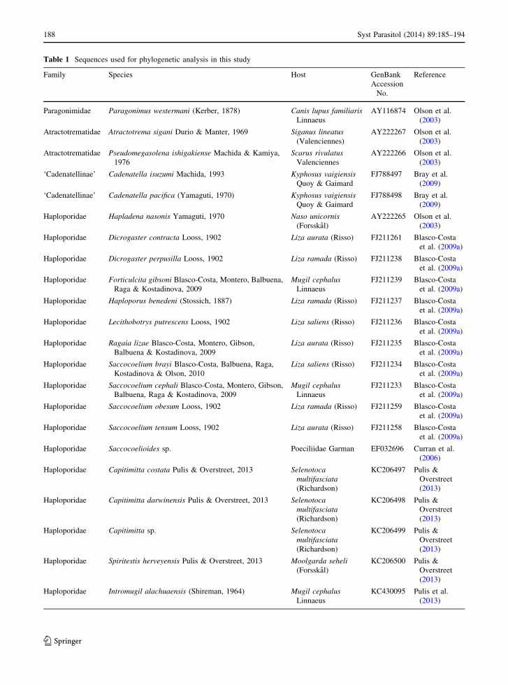
Table 1 Sequences used for phylogenetic analysis in this study
Family Species Host GenBank
Accession
No.
Reference
Paragonimidae Paragonimus westermani (Kerber, 1878) Canis lupus familiaris
Linnaeus
AY116874 Olson et al.
(2003)
Atractotrematidae Atractotrema sigani Durio & Manter, 1969 Siganus lineatus
(Valenciennes)
AY222267 Olson et al.
(2003)
Atractotrematidae Pseudomegasolena ishigakiense Machida & Kamiya,
1976
Scarus rivulatus
Valenciennes
AY222266 Olson et al.
(2003)
‘Cadenatellinae’ Cadenatella isuzumi Machida, 1993 Kyphosus vaigiensis
Quoy & Gaimard
FJ788497 Bray et al.
(2009)
‘Cadenatellinae’ Cadenatella pacifica (Yamaguti, 1970) Kyphosus vaigiensis
Quoy & Gaimard
FJ788498 Bray et al.
(2009)
Haploporidae Hapladena nasonis Yamaguti, 1970 Naso unicornis
(Forsskal)
AY222265 Olson et al.
(2003)
Haploporidae Dicrogaster contracta Looss, 1902 Liza aurata (Risso) FJ211261 Blasco-Costa
et al. (2009a)
Haploporidae Dicrogaster perpusilla Looss, 1902 Liza ramada (Risso) FJ211238 Blasco-Costa
et al. (2009a)
Haploporidae Forticulcita gibsoni Blasco-Costa, Montero, Balbuena,
Raga & Kostadinova, 2009
Mugil cephalus
Linnaeus
FJ211239 Blasco-Costa
et al. (2009a)
Haploporidae Haploporus benedeni (Stossich, 1887) Liza ramada (Risso) FJ211237 Blasco-Costa
et al. (2009a)
Haploporidae Lecithobotrys putrescens Looss, 1902 Liza saliens (Risso) FJ211236 Blasco-Costa
et al. (2009a)
Haploporidae Ragaia lizae Blasco-Costa, Montero, Gibson,
Balbuena & Kostadinova, 2009
Liza aurata (Risso) FJ211235 Blasco-Costa
et al. (2009a)
Haploporidae Saccocoelium brayi Blasco-Costa, Balbuena, Raga,
Kostadinova & Olson, 2010
Liza saliens (Risso) FJ211234 Blasco-Costa
et al. (2009a)
Haploporidae Saccocoelium cephali Blasco-Costa, Montero, Gibson,
Balbuena, Raga & Kostadinova, 2009
Mugil cephalus
Linnaeus
FJ211233 Blasco-Costa
et al. (2009a)
Haploporidae Saccocoelium obesum Looss, 1902 Liza ramada (Risso) FJ211259 Blasco-Costa
et al. (2009a)
Haploporidae Saccocoelium tensum Looss, 1902 Liza aurata (Risso) FJ211258 Blasco-Costa
et al. (2009a)
Haploporidae Saccocoelioides sp. Poeciliidae Garman EF032696 Curran et al.
(2006)
Haploporidae Capitimitta costata Pulis & Overstreet, 2013 Selenotoca
multifasciata
(Richardson)
KC206497 Pulis &
Overstreet
(2013)
Haploporidae Capitimitta darwinensis Pulis & Overstreet, 2013 Selenotoca
multifasciata
(Richardson)
KC206498 Pulis &
Overstreet
(2013)
Haploporidae Capitimitta sp. Selenotoca
multifasciata
(Richardson)
KC206499 Pulis &
Overstreet
(2013)
Haploporidae Spiritestis herveyensis Pulis & Overstreet, 2013 Moolgarda seheli
(Forsskal)
KC206500 Pulis &
Overstreet
(2013)
Haploporidae Intromugil alachuaensis (Shireman, 1964) Mugil cephalus
Linnaeus
KC430095 Pulis et al.
(2013)
188 Syst Parasitol (2014) 89:185–194
123

Litosaccus brisbanensis (Martin, 1964) n. comb.
Syns Paralecithobotrys brisbanensis Martin, 1964;
Lecithobotrys brisbanensis (Martin, 1964) Overstreet
& Curran, 2005
Type- and only known host: Mugil cephalus Linnaeus,
flathead grey mullet (Teleostei: Mugilidae).
Type-locality: Brisbane River, Queensland, Australia.
Other localities: Shorncliffe Beach, Bramble Bay,
QLD, 27�1902600S, 153�501000E (Fig. 1); Shorncliffe
Boat Ramp, Cabbage Tree Creek, QLD, 27�1904700S,
153�501100E (DNA); Brisbane River, Toowong, QLD
(27�290 2900S, 152�5903400E); Wynnum Creek, QLD
(27�260900S, 153�1002800E); Redland Bay, QLD.
Site in host: Intestine.
Type-material: Hancock Parasitology Collection, Uni-
versity of Southern California, No. 7112 (presently
unable to locate).
Voucher material: Queensland Museum, Brisbane,
Australia, G234515–G234522; Harold W. Manter Labo-
ratory Collection, Lincoln, Nebraska, U.S.A. P-2014-021.
Representative DNA sequences: Partial 18S, entire
ITS region, partial (D1–D3) 28S: GenBank accession
no. KM253765, from 2 identical sequences (2 adult
specimens from Cabbage Tree Creek, QLD).
Description (Figs. 1–4)
[Measurements based on 11 gravid wholemounts.]
Body elongate, cylindrical, 2,048 (1,416–2,256) long,
302 (227–285) wide at second fifth of body length
(BL), with width representing 15 (12–19)% of BL.
Tegumental spines exceptionally thin, 5–10 (6–13)
long. Forebody 563 (339–581) long, representing 27
(23-30)% of BL. Hindbody 1,312 (923–1,575) long,
representing 64 (60–70)% of BL. Oral sucker infun-
dibuliform, terminal, 259 (192–267) long, 245
(201–234) wide, with anterior periphery surrounded
by ring of approximately 12 small papillae. Ventral
sucker 173 (154–192) long, 204 (137–190) wide.
Ratio of oral sucker to ventral sucker width 1:0.83
(1:0.67–0.88). Prepharynx 64 (41–88) long. Pharynx
subglobular, approximately twice length of prephar-
ynx, 118 (89–128) long, 126 (99–121) wide. Ratio of
oral sucker width to pharynx width 1:0.51 (1:0.48-
0.60). Oesophagus 96 (117–317) long, extending to
second fifth of BL, swollen posteriorly. Intestinal
bifurcation at or posterior to level of ventral sucker.
Caeca long, relatively narrow, uneven to subequal
(sinistral caecum longer in all but 1 specimen), more
bulbous posteriorly in most specimens, terminating
blindly, with posterior-most caecum terminating 481
(293–577) from posterior end, with postcaecal space
representing 24 (15–34)% of BL.
Testis single, 151 (113–211) long, 129 (113–163)
wide, 270 (210–346) from posterior margin of ventral
sucker. Post-testicular space 893 (443–1,074) long,
representing 44 (28–48)% of BL. External seminal
vesicle claviform to sac-like, 163 (72–158) long, 68
(29–75) wide, dorsal to ventral sucker. Hermaphroditic
sac thin-walled, anterodorsal to dorsal of ventral
sucker, 112 (109–190) long, 67 (55–89) wide, repre-
senting 65 (57–104)% of ventral sucker length and 5
(6–10)% of BL; containing internal seminal vesicle 78
(61–102) long by 38 (24–40) wide, prostatic bulb,
female duct, and hermaphroditic duct; male and female
ducts unite at anterior third of hermaphroditic sac;
hermaphroditic duct muscularised, approximately 1/3
length of hermaphroditic sac. Genital pore medial, 55
(10–56) anterior to anterior margin of ventral sucker.
Table 1 continued
Family Species Host GenBank
Accession No.
Reference
Haploporidae Intromugil mugilicolus Pulis, Fayton,
Curran & Overstreet, 2013
Mugil cephalus Linnaeus KC430096 Pulis et al. (2013)
Haploporidae Parasaccocoelium haematocheilum
Besprozvannykh, Atopkin,
Ermolenko & Nikitenko, 2014
Liza haematocheila
(Temminck & Schlegel)
HF548461 Besprozvannykh
et al. (2014)
Haploporidae Parasaccocoelium mugili Zhukov, 1971 Liza haematocheila
(Temminck & Schlegel)
HF548468 Besprozvannykh
et al. (2014)
Haploporidae Parasaccocoelium polyovum
Besprozvannykh, Atopkin,
Ermolenko & Nikitenko, 2014
Liza haematocheila
(Temminck & Schlegel)
HF548474 Besprozvannykh
et al. (2014)
Syst Parasitol (2014) 89:185–194 189
123
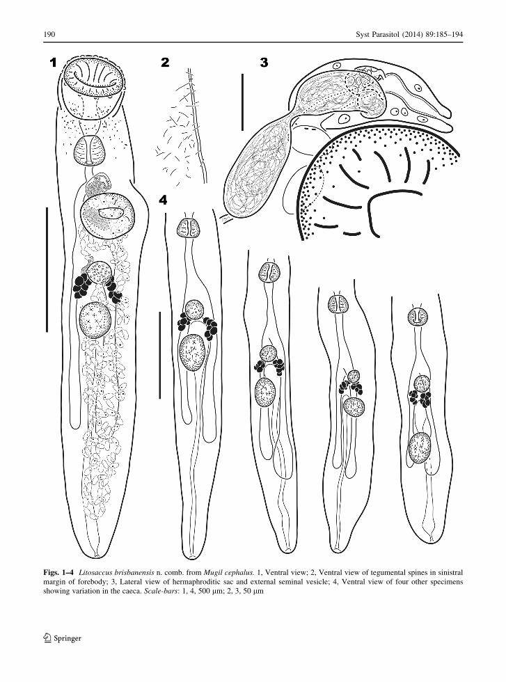
Figs. 1–4 Litosaccus brisbanensis n. comb. from Mugil cephalus. 1, Ventral view; 2, Ventral view of tegumental spines in sinistral
margin of forebody; 3, Lateral view of hermaphroditic sac and external seminal vesicle; 4, Ventral view of four other specimens
showing variation in the caeca. Scale-bars: 1, 4, 500 lm; 2, 3, 50 lm
190 Syst Parasitol (2014) 89:185–194
123

Ovary globular to subglobular, medial, 91 (67–145)
long, 94 (65–109) wide, 101 (17–130) from posterior
margin of ventral sucker, 76 (9–227) from anterior
margin of testis, posteroventral to ventral to intestinal
bifurcation. Uterus emerging from dextral side of
ovary, winding anteriorly to or slightly beyond
posterior margin of ventral sucker and then winding
posteriorly, occupying most of hindbody, with prox-
imal portion filled with sperm. Laurer’s canal not
observed. Vitellarium in 2 lateral clusters of 7–10
Fig. 5 Phylogenetic relationships among members of the Haploporidae resulting from Bayesian inference analysis of partial 28S
rDNA sequences (GTR ? I ? C; 1,000,000 generations and a sample frequency of 100) revealing Litosaccus brisbanensis n. comb as a
haploporine. Support values of \75% not shown. Vertical bars denote family or subfamily groups. Abbreviations: At,
Atractotrematidae; Ca, Cadenatellinae; Ch, Chalcinotrematinae; Fo, Forticulcitinae; Ha, Haploporinae; Me, Megasoleninae; Wa,
Waretrematinae
Syst Parasitol (2014) 89:185–194 191
123
subglobular to spherical follicles 26–30 (24–46) long
by 26–29 (23–39) wide, with sinistral cluster 125
(96–162) long, dextral cluster 103 (79–129) long,
contiguous or nearly so with posterior margin of
ovary, with anterior-most follicle 157 (106–218) from
posterior margin of ventral sucker, ventral to caeca.
Eggs thin-shelled, numerous, in distal portion of uterus
mostly with developed miracidia having eye-spots
fused, 40–45 (40–46) long, 24–26 (22–26) wide.
Excretory vesicle I-shaped, bulbous anteriorly,
terminating just posterior to ovary, with 1 specimen
having well-defined crura extending anteriorly from
level of vitelline clusters; pore terminal.
Remarks
Martin’s (1974) type-material (originally deposited in
the no longer cohesive Hancock Parasitology Collec-
tion, University of Southern California) is still miss-
ing; we have been unsuccessful in our attempt to find
the holotype at the Santa Barbara Museum of Natural
History (Pers. comm. Daniel Geiger & Patricia
Sadeghian), the Los Angeles County Museum of
Natural History (Pers. comm. Joel Martin), and the
U.S. National Helminthological Collection (Pers.
comm. Patricia Pillit). For consistency we chose to
illustrate and measure the same specimen illustrated
by Overstreet & Curran (2005) in their chapter in the
Keys to the Trematoda Vol. 2 (figure 12.9). The
excretory vesicle was described by Martin (1974) as
being Y-shaped, but it is I-shaped in all of our
specimens. However, in one of the specimens, the one
illustrated (Fig. 1), there are well-defined crura
extending from level of the vitelline clusters. These
crura are likely collecting branches because each is
differentiated from the vesicle by a sphincter. Martin
(1974) did not indicate the presence of small papillae
surrounding the oral sucker that usually are apparent
on many well-fixed trematodes, but the shape of the
oral sucker in his illustration and his measurements are
consistent with our specimens. Martin (1974) reported
the tegument as mostly smooth but with a few spines
dorso-anteriorly and immediately posterior to the
ventral sucker. Tegumental spines were observed by
us in only four of our specimens; two had thin spines
sparsely covering the entire tegument and two had
only a few spines posterior to the ventral sucker.
Presumably, the spines of L. brisbanensis are fragile,
shallowly embedded, or easily lost and were therefore
not observed on most of our specimens because of loss
due to fixation, preservation, or handling techniques.
Despite these potential differences and based on the
size and shape of the body, suckers, reproductive
organs, and hermaphroditic sac, we have no doubt that
the specimens we collected are conspecific with those
of Martin (1974).
Molecular analysis
The DNA sequence fragment amplified encompasses
the 30 end of the 18S gene, the ITS region (ITS1-5.8S-
ITS2) and 1,415 bp of the 50 end of the 28S gene. No
intraspecific variation occurred between the two
sequenced specimens of L. brisbanensis. The align-
ment of partial 28S rDNA sequences of L. brisbanen-
sis and related species from GenBank was 1,128
characters long with 655 conserved sites, 473 variable
sites and 337 informative sites. The BI analysis of
those sequences incorporated the paragonimid P.
westermani as an outgroup and an ingroup of two
species each of atractotrematids and Cadenatella, L.
brisbanensis, and 21 other species of Haploporidae
(Fig. 5). The ingroup of the Haploporidae was
revealed as a paraphyletic clade. The megasolenine
Hapladena nasonis Yamaguti, 1970 was well sup-
ported as basal to Cadenatella spp. and the other
haploporids. The position of Cadenatella as sister to
the non-Hapladena haploporids was poorly supported.
The 20 other non-Hapladena haploporids formed a
polytomy consisting of Forticulcita gibsoni Blasco-
Costa, Montero, Balbuena, Raga & Kostadinova,
2009, Spiritestis herveyensis Pulis & Overstreet,
2013, Capitamitta spp. ? Parasaccocoelium spp.,
and a clade that included two subclades: one com-
prised of Intromugil spp. ? Saccocoelioides sp. and
the other of Litosaccus brisbanensis ? the Mediter-
ranean haploporines.
Discussion
Blasco-Costa et al. (2009b) considered Lecithobotrys
brisbanensis as a species inquirenda and stated that it
likely did not belong in Lecithobotrys; our BI analysis
confirms that it does not. We erected Litosaccus for L.
brisbanensis, which has morphological characters in
common with the Haploporinae (i.e. vitellarium that is
192 Syst Parasitol (2014) 89:185–194
123
reduced, a uterus that occupies much of the hindbody
but does not extend into the forebody, and developed
eggs containing miracidia with eye-spots) and is
similar to Lecithobotrys and Pseudolecithobotrys.
In view of the only slight morphological discrep-
ancies between Martin’s (1974) specimens and our
own, we have little doubt that our specimens are
conspecific with those originally described. In the
redescription of I. mugilicolus by Pulis et al. (2013),
they noted that the hermaphroditic duct had a ‘‘series
of sacs containing a glandular substance’’ that was
observable in living specimens and specimens stored
in ethanol, but they were no longer easily discernible
after processing for mounting. Similarly, the ‘‘tiny
spines or tubercles’’ described by Martin (1973) as
lining the hermaphroditic duct of L. brisbanensis may
not be apparent in our fixed specimens. Thus,
additional specimens need to be examined live to
confirm the presence or absence of an armed
hermaphroditic duct. Litosaccus is not an appropriate
repository for either of the other two species of
Lecithobotrys considered species inquirenda by Blas-
co-Costa et al. (2009b), and we agree that both
require further data to clarify their generic affinity.
To the best of our knowledge, L. brisbanensis may be
considered rare or its host has not been collected when
the infection is at its peak intensity. We have examined a
total of 46 specimens of M. cephalus from the QLD
coast (12 in 1984, 18 in 1997 and 16 in 2010) and only
recovered a total of 16 specimens, all from the Brisbane/
Moreton Bay area. Lester et al. (2009) found that
approximately 50% of the individuals of M. cephalus
they examined had evidence of infection by the blood
fluke Plethorchis acanthus Martin, 1975 in the Moreton
Bay area, while M. cephalus from along the New South
Wales coast showed no such infection, suggesting the
parasite was acquired in Moreton Bay, perhaps in the
upper estuary. A similar pattern may occur for infection
with L. brisbanensis, because we recovered the parasite
from Moreton Bay drainages only. Additionally, in 2010
we examined 65 individuals of the greenback mullet,
Chelon subviridis (Valenciennes), flat-tail mullet, Liza
argentea (Quoy & Gaimard), and silver mullet, Pa-
ramugil georgii (Ogilby), from Cabbage Tree Creek and
the Pine River, which, along with the Brisbane River,
empty into Moreton Bay, and we did not find any
specimen of L. brisbanensis.
In a review of the Haploporidae, Overstreet &
Curran (2005) recognised four subfamilies based on
morphology: the Chalcinotrematinae (infecting estu-
arine and freshwater fishes in the New World and
Africa), the Haploporinae (with members primarily in
mugilids worldwide), the Megasoleninae Manter,
1935 (primarily in marine, reef associated percifor-
mes) and the Waretrematinae Srivastava, 1937 (in
marine, estuarine, and freshwater fishes worldwide,
but primarily in the Indo-Pacific). Blasco-Costa et al.
(2009a) established the Forticulcitinae Blasco-Costa,
Balbuena, Kostadinova & Olson, 2009 (with members
in mugilids in the Mediterranean Sea and Red Sea)
based on a single, compact vitellarium and their BI
analysis of partial 28S rDNA sequence data. This is the
first phylogenetic hypothesis of the Haploporidae to
include a haploporine collected outside of the Med-
iterranean Sea. Litosaccus was resolved as distinct
from Lecithobotrys but well supported as sister to the
Mediterranean haploporines (Fig. 5), confirming that
members of the Haploporinae are not restricted to the
Mediterranean Sea.
We agree with Pulis & Overstreet’s (2013) skep-
ticism of the morphologically defined haploporid
subfamilies due to the paucity of molecular data for
most genera. Our BI analysis revealed the Waretre-
matinae to be paraphyletic with Intomugil being closer
to Saccocoelioides Szidat, 1954 and Spiritestis Na-
gaty, 1948 being recovered in the polytomy leading to
the other major haploporid clades, but, at this time, we
refrain from making any nomenclatural changes.
Besprozvannykh et al. (2014) resurrected Parasacco-
coelium and demonstrated that the three species they
treated formed a well-supported clade with Capitimit-
ta, which we recovered as well. However, we are
skeptical of their consideration of Pseudohapladena
lizae Liu & Yang, 2002 as a junior synonym of
Parasaccocoelium mugili Zhukov, 1971. Liu & Yang
(2002) described Ps. lizae as having a longer oesoph-
agus, smaller eggs, a well-separated ovary and testis,
and a more tubular vitellarium.
Bray et al. (2014) used BI analysis of 28S rDNA
sequences to demonstrate that Cadenatella had previ-
ously been misplaced in the Enenteridae Yamaguti,
1958 (Lepocreadioidea Odhner, 1905) and belongs in
the Haploporoidea. They noted that with the inclusion
of the Cadenatella spp. in the Haploporoidea, the
Haploporidae was not well resolved because Hapla-
dena Linton, 1910 did not cluster with the other
members of the family. We also resolved Hapladena
(the sole representative of the Megasoleninae included
Syst Parasitol (2014) 89:185–194 193
123
in both analyses) outside of the clade containing
Cadenatella spp. and the rest of the haploporids. The
position of Cadenatella as the sister group to the rest of
the haploporids was not well supported; thus, an
important component of future considerations will be
whether these taxa belong in the Haploporidae or
whether there is a case for recognition of further
family level taxa within the Haploporoidea.
The systematics of haploporids still requires con-
siderable resolution. Erecting Litosaccus brings the
total number of haploporine genera to ten. Four of
those genera, Pseudodicrogaster Blasco-Costa, Mon-
tero, Gibson, Balbuena & Kostadinova, 2009, Pseudo-
lecithobotrys, Rondotrema Thatcher, 1999, and
Unisaccus Martin, 1973, lack a representative DNA
sequence. Since all four of those genera also lack a
Mediterranean representative, their inclusion in a
molecular framework will help clarify the subfamilial
relationships within the Haploporidae and help detect
the pattern of diversification within the Haploporinae.
Acknowledgements The authors are grateful to the following
people for their contributions to the procurement of hosts,
assistance in trying to locate type-material and providing
accession numbers: Robert Adlard, Mal Bryant, Adam
Fletcher, Daniel Geiger, Jeff Johnson, Jason Lally, Joel
Martin, Patricia Pilitt, Gabor Racz, and Patricia Sadeghian.
From the University of Southern Mississippi, we thank Jean
Jovonovich Alvillar and Janet Wright for their assistance with
DNA sequencing reactions. The material treated here is based
on work supported by the National Science Foundation under
grant no. 0529684, Ocean and Human Health Initiative grant no.
NA08NOS4730322, and US Fish and Wildlife Service/
Mississippi Department of Marine Resources MSCIAP
MS.R.798 Award M10AF20151.
References
Besprozvannykh, V. V., Atopkin, D. M., Ermolenko, A. V., &
Nikitenko, A. (2014). Restoration of the genus Parasac-
cocoelium Zhukov, 1971 (Digenea: Haploporidae) and a
description of two new species from mugilid fish in the Far
East of Russia. Journal of Helminthology (In press).
Blasco-Costa, I., Balbuena, J. A., Kostadinova, A., & Olson, P. D.
(2009a). Interrelationships of the Haploporinae (Digenea: Ha-
ploporidae): a molecular testof the taxonomic framework based
on morphology. Parasitology International, 58, 265–269.
Blasco-Costa, I., Gibson, D. I., Balbuena, J. A., Raga, J. A., &
Kostadinova, A. (2009b). A revision of the Haploporinae
Nicoll, 1914 (Digenea: Haploporidae) from mullets (Mu-
gilidae): Haploporus Looss, 1902 and Lecithobotrys
Looss, 1902. Systematic Parasitology, 73, 107–133.
Bray, R. A., Cribb, T. H., Waeschenbach, A., & Littlewood, D.
T. J. (2014). Molecular evidence that the genus
Cadenatella Dollfus, 1946 (Digenea; Plagiorchiida)
belongs in the superfamily Haploporoidea Nicoll, 1914.
Systematic Parasitology, 89, 15–21.
Bray, R. A., Waeschenbach, A., Cribb, T. H., Weedall, G. D.,
Dyal, P., & Littlewood, D. T. J. (2009). The phylogeny of
the Lepocreadioidea (Platyhelminthes: Digenea) inferred
from nuclear and mitochondrial genes: implications for
their systematics and evolution. Acta Parasitologica, 54,
310–329.
Cribb, T. H., & Bray, R. A. (2010). Gut wash, body soak,
blender and heat-fixation: approaches to effective collec-
tion, fixation and preservation of trematodes of fishes.
Systematic Parasitology, 76, 1–7.
Curran, S. S., Tkach, V. V., & Overstreet, R. M. (2006). A
review of Polylekithum Arnold, 1934 and its familial
affinities using morphological and molecular data, with
description of Polylekithum catahoulensis sp. nov. Acta
Parasitologica, 51, 238–248.
Darriba, D., Taboada, G. L., Doallo, R., & Posada, D. (2012).
jModelTest 2: more models, new heuristics and parallel
computing. Nature Methods, 9, 772.
Hall, T. A. (1999). BioEdit: a user-friendly biological sequence
alignment editor and analysis program for Windows 95/98/
NT. Nucleic Acids Research, 41, 95–98.
Huelsenbeck, J. P., & Ronquist, F. (2001). MRBAYES: Bayesian
inference of phylogeny. Bioinformatics, 17, 754–755.
Huelsenbeck, J. P., Ronquist, F., Nielsen, R., & Bollback, J. P.
(2001). Bayesian inference of phylogeny and its impact on
evolutionary biology. Science, 294, 2310–2314.
Katoh, K., Kuma, K.-I., Toh, H., & Miyata, T. (2005). MAFFT
version 5: improvement in accuracy of multiple sequence
alignment. Nucleic Acids Research, 33, 511–518.
Lester, R. J. G., Rawlinson, S. E., & Weaver, L. C. (2009).
Movement of sea mullet Mugil cephalus as indicated by a
parasite. Fisheries Research, 96, 129–132.
Martin, W. E. (1973). A new genus and species of haploporid
trematode (Haploporidae: Trematoda) from Australian
mullet. Bulletin of the Southern California Academy of
Science, 72, 166–168.
Martin, W. E. (1974). Paralecithobotrys brisbanensis sp. n.
from Australian mullet (Trematoda: Haploporidae). Pro-
ceedings of the Helminthological Society of Washington,
41, 16–18.
Olson, P. D., Cribb, T. H., Tkach, V. V., Bray, R. A., & Lit-
tlewood, D. T. J. (2003). Phylogeny and classification of
the Digenea (Platyhelminthes: Trematoda). International
Journal for Parasitology, 33, 733–755.
Overstreet, R. M., & Curran, S. S. (2005). Family Haploporidae
Nicoll, 1914. In: Jones, A., Bray, R. A. & Gibson, D. I.
(Eds) Keys to the Trematoda. Volume 2. Wallingford, UK:
CAB International, pp. 129–167.
Pulis, E. E., Fayton, T. J., Curran, S. S., & Overstreet, R. M.
(2013). A new species of Intromugil (Digenea: Haplopo-
ridae) and redescription of Intromugil mugilicolus. Journal
of Parasitology, 99, 501–508.
Pulis, E. E., & Overstreet, R. M. (2013). Review of haploporid
(Trematoda) genera with ornate muscularisation in the
region of the oral sucker, including four new species and a
new genus. Systematic Parasitology, 84, 167–191.
Wu, M., Chatterji, S., & Eisen, J. A. (2012). Accounting for align-
ment uncertainty in phylogenomics. PLOS ONE, 7, e30288.
194 Syst Parasitol (2014) 89:185–194
123
Copyright © 2022 FDOKUMEN