
ERCIYES UNIVERSITY FACULTY OF MEDICINE 6th ...
59
6 th ERCIYES UNIVERSITY FACULTY OF MEDICINE INTERNATIONAL CONGRESS OF MEDİCAL GENETICS ERCIYES UNIVERSITY FACULTY OF MEDICINE 6 th INTERNATIONAL CONGRESS OF MEDICAL GENETICS Honorary President of Congress Mustafa Calis Hizir Yakup Akyildiz Executive Committee Taha Bahsi (President) Mehmet Ali Ergun (Vice President) Ebru Marzioglu Ozdemir Beyhan Durak Aras Sule Altiner Esra Arslan Ates Altug Koc Congress President Munis Dundar Organization Board Munis Dundar Yusuf Ozkul Cetin Saatci
-
Upload
khangminh22 -
Category
Documents
-
view
1 -
download
0
Transcript of ERCIYES UNIVERSITY FACULTY OF MEDICINE 6th ...

6th ERCIYES UNIVERSITY FACULTY OF MEDICINE INTERNATIONAL CONGRESS OF MEDİCAL GENETICS
ERCIYES UNIVERSITY FACULTY OF MEDICINE 6th INTERNATIONAL CONGRESS OF MEDICAL GENETICS
Honorary President of Congress Mustafa Calis
Hizir Yakup Akyildiz
Executive Committee Taha Bahsi (President) Mehmet Ali Ergun (Vice President) Ebru Marzioglu Ozdemir Beyhan Durak Aras Sule Altiner Esra Arslan Ates Altug Koc
Congress President
Munis Dundar
Organization Board
Munis Dundar
Yusuf Ozkul
Cetin Saatci

6th ERCIYES UNIVERSITY FACULTY OF MEDICINE INTERNATIONAL CONGRESS OF MEDİCAL GENETICS
Bilimsel Kurul
Abdulgani Tatar (Turkey) Ahmet Arman (Turkey) Ahmet Cevdet Ceylan (Turkey)
Ahmet Dursun (Turkey) Ahmet Okay Çaglayan (Turkey) Andrea Taylor (United Kingdom)
Andreas Janecke (Austria) Asli Subasioglu (Turkey) Aslihan Kiraz (Turkey)
Asude Durmaz (Turkey) Atil Bisgin (Turkey) Ayfer Ulgenalp (Turkey)
Aykut Ozdarendeli (Turkey) Beyhan Durak Aras (Turkey) Burak Durmaz (Turkey)
Denise Aldrian (Austria) Ellen J. Hoffman (USA) Emine Berrin Yuksel (Turkey)
Fatma Silan (Turkey) Ferda Ozkinay (Turkey) Ferda Percin (Turkey)
Feride Sahin (Turkey) Gokay Bozkurt (Turkey) Gunes Özhan (Turkey)
Hakan Akça (Turkey) Hakan Gurkan (Turkey) Haluk Akin (Turkey)
Hilmi Tozkir (Turkey) Huseyin Per (Turkey) Ibrahim Boga (Turkey)
Julia Vodopiutz (Austria) M. Cengiz Yakicier (Turkey) Mahmut Cerkez Ergoren (Cyprus)
Mahmut Selman Yildirim (Turkey) Margit Burmeister (USA) Mehmet Ali Ergun (Turkey)
Mehmet Kosker (Turkey) Munis Dundar (Turkey) Murat Derya Ercal (Turkey)
Mustafa Calis (Turkey) Nadir Kocak (Turkey) Necat Imirzalioglu (Turkey)
Nihal Hatipoglu (Turkey) Nurten Akarsu (Turkey) Oya Uyguner (Turkey)
Ozturk Ozdemir (Turkey) Seher Basaran (Turkey) Selma Demir (Turkey)
Sevcan Tug Bozdogan (Turkey) Sevda Yesim Ozdemir (Turkey) Sevilhan Artan (Turkey)
Shrikant Mane (USA) Sibel Kantarci (USA) Sehime Gulsun Temel (Turkey)
Taha Bahsi (Turkey) Tolga Aslan (Turkey) Tulay Tos (Turkey)
Tulin Cora (Turkey) Ugur Ozbek (Turkey) Umut Fahrioglu (UAE)
Vehap Topcu (Turkey) Xavier David (France) Yunus Kasim Terzi (Turkey)
Yusuf Ozkul (Turkey) Zerrin Yilmaz Celik (Turkey) Zuhal Altıntas (Turkey)

6th ERCIYES UNIVERSITY FACULTY OF MEDICINE INTERNATIONAL CONGRESS OF MEDİCAL GENETICS
Oral Presentation Awards
1st Place
Prevalence of SARS-CoV-2 N501Y mutation in Northern Cyprus Gulten Tuncel, Mahmut Cerkez Ergoren, Buket Baddal, Pinar Tulay, Cenk Serhan Ozverel, Emrah Guler, Huseyin Kaya Suer, Murat Sayan, Tamer Sanlidag
1st Place
Genotip-Fenotip İlişkisinin Doğru Değerlendirilmesinde Harman Fenotipler Arda Çetinkaya
1st Place
A novel variant in the autosomal dominant form of LGMDR1 (LGMDD4) İzem Olcay Şahin, Emine Karataş, Mikail Demir, Nuriye Gökçe, Yusuf Özkul, Munis Dündar
2nd Place
Discordance of NGS-CNV and MLPA Methods in a Hereditary Colon Cancer Case Nihat Buğra Ağaoğlu
3rd Place
A rare disease associated with the CDK13 gene: CHHDFIDD Çağrı Doğan, Mustafa Doğan
3rd Place
The evaluation of rare and low-frequency genetic variants in common variable immune deficiency (CVID) patients together with CV/RVCD (common variant/rare variant, common disease) hypothesis for final interpretation
Atil Bisgin, Ozge Sonmezler, Ibrahim Boga, Mustafa Yilmaz
Poster Presentation Awards
1st Place Molecular and clinical approach to the MEDNIK-like syndrome
Muhammed Burak Bereketoglu, Nigar Shirinova, Sevcan Tug Bozdogan
2nd Place Duplication of 10q24.31 in a family with Congenital Nystagmus and Split-hand/foot Malformation
Ceren Alavanda, Fatma Uguzdogan, Senol Demir, Hamza Polat, Esra Arslan Ates, Mehmet Ali Soylemez, Bilgen Bilge Geckinli, Ahmet Ilter Guney, Pinar Ata, Ahmet Arman
2nd Place
COVID-19 pandemic in patients with familial mediterranean fever; The possible protective role of colchicine in COVID-19 symptoms Mehmet Berkay Akcan, Burcu Albuz, Öztürk Özdemir, Fatma Sılan
2nd Place
NBN gene mutations with clinical spectrum in our patients who applied to our outpatient clinic: Case series Volkan Sonmez, Ahmet Kablan, Mehmet Berkay Akcan, Derya Kaya, Fatma Sılan, Ozturk Ozdemir
3rd Place
t(11;17) Balanced Reciprocal Translocation Detected in an Infertile Couple: A Case Report Nihan Ecmel Akbaş, Derya Kaya, Canan Köse, Yunus Emre Mutluer, Ahmet Kablan, Fatma Silan, Öztürk Özdemir
3rd Place
From tissue to diagnosis; case report with Proteus Syndrome Ahmet Kablan, Derya Kaya, Volkan Sönmez, Mehmet Berkay Akcan, Fatma Silan, Ozturk Ozdemir

P1 | Abstracts | Invited Speakers
Invited Speakers Robinow syndrome from past to present Ferda Emriye Perçin Department of Medical Genetics, Faculty of Medicine, Gazi University, Ankara, Turkey In 1969, Robinow et al. described a new, rare form of dwarfism with mesomelic limb shortening, hemivertebrae, genital hypoplasia and a distinctive facial appearance in a single family which is inherited with autosomal dominant manner. Within a few years of the original discovery of autosomal dominant Robinow syndrome (DRS), the autosomal recessive form (RRS) was described. While there is a wide range of severity in both form, the phenotypes is quite overlapping. Although the authors have mostly suggested that short stature and skeletal abnormalities are more common in RRS, experience has shown that this may not be the case. It is a genetically heterogeneous disorder and studies have identified five genes in which pathogenic variants can cause RS. Biallelic mutations of two genes are associated with recessive forms, ROR2 (MIM 602337), and NXN (MIM 612895). Additionally, biallelic loss-of-function (LoF) variants in WNT5A were reported in one patient with RS. Heterozygous pathogenic variants in WNT5A (MIM 164975), DVL1 (MIM 601365), and DVL3 (MIM: 601368) have been identified as causative for the dominant forms. In addition, it has been suggested that heterozygous pathogenic variant in the FZD2 (MIM 164745) gene, which causes autosomal dominant Omodysplasia type 2, may also be responsible for DRS. The aforementioned “Robinow-associated genes” and their gene products all play a role in the non-canonical Wnt (β-catenin-independent) signaling that establishes cellular orientation via the WNT/planar cell polarity signaling pathway (PCP). Keywords: Robinow syndrome, Autosomal dominant Robinow syndrome, Autosomal recessive Robinow syndrome Genomic Profiling of Cancer Patients: Discrepancy of Results with Clinical Status Feride İffet Şahin Başkent University Faculty of Medicine Department of Medical Genetics, Ankara, Turkey Patients diagnosed with cancer are now considered to have a chronic disease. Due to genetic heterogeneity of cancer and the individual clinical course and survival characteristics of the patients, genomic profiling or analysis of mutation signature is important in planning of follow-up and treatment of disease in diagnosed cases. In this presentation, evaluations that can be made in the analysis and interpretation of results of profiling tests, which are usually applied to each patient sample with ready-made panels will be discussed. In cases in whom mutations with known pathogenicity are not observed, or if the expected clinical benefit is not achieved despite the use of targeted therapy agents suitable for the mutations detected, the possibilities awaiting the clinician and geneticist who will inform the patient will be evaluated. Keywords: genomic profiling, results, cancer SARS-CoV-2 infection, hypercoagulability and hereditary thrombophilia factors Aslıhan Kiraz Medical Genetics Clinic, Kayseri City Training and Research Hospital, Kayseri, Turkey Coronavirus disease (COVID-19) is a potentially fatal infectious disease caused by the SARS-CoV-2 virus, a virus of zoonotic origin (1). It has been observed that clinical manifestations of SARS-CoV-2 infection range from asymptomatic disease to severe viral pneumonia accompanied by severe respiratory failure and may result in death (2). The most common initial symptoms of COVID-19 disease are fever, cough, and fatigue. Transmission primarily occurs through direct contact or droplets spread from an infected person (1). The binding of a receptor expressed by host cells is the first step of viral infection. Lung epithelial cells are thought to be the primary target of the virus. Most of the used drugs are drugs used in the treatment of other diseases, and their effectiveness in the treatment of COVID-19 is still at the research level. Specific anti-infection drugs are under development for potential treatment in humans (1). Veklury (Remdesivir) is the first treatment for COVID-19 to receive FDA approval. It is used in adults and pediatric patients [12 years and older and at least 40 kilograms] for the treatment of COVID-19 requiring hospitalization (3). The treatment of COVID-19 in our country is carried out by the recommendations of the Ministry of Health’s Guide “COVID-19 (SARS-CoV-2 infection)” prepared by the recommendations of the Coronavirus Science Council and updated by the developments (4). All of the drugs recommended in the manual are used under the approval of the Ministry of Health within the framework of non-indication drug use. In this pandemic, where new waves are constantly coming, scientists have succeeded in developing a large number of COVID-19 vaccine types in a short time as a result of intensive studies. The mRNA-based Pfizer-BioNTech COVID-19 vaccine is the first FDA-approved vaccine [23.08.2021] (5). The information about the COVID-19 disease, which is spreading rapidly around the world, is increasing with new researches day by day. Thrombophilia is a hypercoagulable condition that predisposes patients to thrombosis. It is a multifactorial condition that can result from genetic factors, acquired factors, or a combination of both. The prothrombin gene (F2), factor V Leiden (F5), and PAI-1 are important biomarkers of thrombophilia. Patients with multiple gene defects have a high risk of thrombosis (6). It is known that thromboembolic events can develop in patients with COVID-19 and the incidence of death increases accordingly. Studies have shown that VTE can be induced in patients with COVID-19 and severe pneumonia, and the incidence of VTE in COVID-19 patients hospitalized in the intensive care unit due to severe pneumonia has been reported to be high (7). The risk of thrombosis and arterial and venous thromboembolic complications seen in 30% of hospitalized subjects due to Novel Coronavirus pneumonia has been reported in many studies, which can be explained by the prolonged inflammatory response, decreased physical activity during infection, and reduced oxygen levels in the circulation (6). Thrombotic and microangiopathic effects of the SARS-CoV-2 virus have been reported in COVID-19 patients (8). Circulatory disorders in the toes of COVID-19 patients are also reported as “Covid toe (acro ischemia)” (9). Although it is reported that the disease progresses more severely in the elderly, patients with sub-diseases, and smoking history, it is also observed that the clinical course is severe and patient losses are experienced in young patients who do not have any underlying disease. The mechanisms of the development of thromboembolic events are the binding of the virus to the ACE-2 receptor and/or direct endothelial damage, activation of inflammatory and microthrombotic pathways as a result of endothelial damage by complement activation as in sepsis, and stasis due to hospitalization and immobility (10). DIC clinic may develop in patients with severe clinical course. The pathophysiology of DIC is reported to be complex and multifactorial, involving the interaction between the hemostatic system and components of the innate immune response to the infecting pathogen (11). However, there

P2 | Abstracts | Invited Speakers
is no comprehensive research on inherited thrombophilia factors that may affect the hemostatic system. Severe clinical course in young patients, a similar clinical course in individuals from the same family even though they are in different cities, elderly patients recovering from the clinic safely and being discharged, VTE in different locations in COVID-19 patients, microangiopathic thrombus, etc. monitoring of the findings, the use of anticoagulants due to their positive contributions in treatment are included in the observations in this process. In the light of all this information, it is aimed to examine and reveal the possible relationship between the genetic variations evaluated in the thrombophilia panel and COVID-19 and presented with the literature data findings and recent studies. Gralinski et al. suggested that PAI-1 plays a protective role against infection in the viral pathogenesis studies of SARS-coronavirus disease (12). In another study, no statistically significant difference in thrombophilia polymorphic biomarkers between the severely ill COVID-19 group and the healthy population was found (6). Various studies that will provide new information on the subject are also actively continuing. In conclusion, thrombosis is frequently seen in severe COVID-19 patients. Essentially, the determination of the thrombophilia profile can assist in determining bleeding risk, mortality, ARDS incidence, and admission to the ICU. Latent genetic risk factors for thrombotic events may affect the outcome of COVID-19. Therefore, the identification of these factors may be useful for understanding the various COVID-19 outcomes and assessing COVID-19 patients' risk of thrombosis, severe disease, and vaccination policies. Keywords: COVID-19, Genetic, Thrombophilia References
1. Rothan HA, Byrareddy SN. The epidemiology and pathogenesis of coronavirus disease COVID-19) outbreak. J Autoimmun. 2020 Feb 26:102433. doi: 10.1016/j.jaut.2020.102433. https://www.ncbi.nlm.nih.gov/pubmed/32113704
2. Zhou F, Yu T, Du R, et al. Clinical course and risk factors for mortality of adult inpatients with COVID-19 in Wuhan, China: a retrospective cohort study. Lancet 2020; 395(10229):1054-62. https:// www.thelancet.com/pb-assets/Lancet/pdfs/S014067362305663.pdf
3. https://www.fda.gov/drugs/emergency-preparedness-drugs/coronavirus-covid-19-drugs 4. Bilimsel Danışma Kurulu, COVID-19 (SARS-CoV-2 Enfeksiyonu) Erişkin Hasta Tedavisi Rehberi T.C. Sağlık Bakanlığı Halk Sağlığı
Genel Müdürlüğü, 2 Ağustos 2020. (https://covid19bilgi.saglik.gov.tr/tr/covid-19-rehberi.html) 5. https://www.fda.gov/news-events/press-announcements/fda-approves-first-covid-19-vaccine 6. Kiraz A., Guzeldag S., Eren E., Goksu M., Bayram A. Investigation of the relationship between inherited thrombophilia and novel
coronavirus pneumonia. (Short Communication ) Future Virology 2021 Apr; 16(5) https://doi.org/10.2217/fvl-2020-0395 7. Cui S, Chen S, Li X, et al. Prevalence of Venous Thromboembolism in Patients With Severe Novel Coronavirus Pneumonia. [Ahead
of print, 2020 Apr 9]. J Thromb Haemost. 2020;10.1111/ jth.14830. doi:10.1111/jth.14830. https://onlinelibrary.wiley.com/doi/epdf/10.1111/jth.14830
8. Sharon E. Fox, Aibek Akmatbekov, Jack L. et al. Pulmonary and Cardiac Pathology in Covid-19: The First Autopsy Series from New Orleans. medRxiv preprint. https://doi.org/10.1101/2020.04.0 6.20050575.
9. Zhang Y, Cao W, Xiao M, Li YJ, Yang Y, Zhao J, Zhou X, Jiang W, Zhao YQ, Zhang SY, Li TS . Clinical and coagulation characteristics of 7 patients with critical COVID-2019 pneumonia and acro-ischemia. Zhonghua Xue Ye Xue Za Zhi 2020 Mar 28;41(0):E006. doi: 10.3760/cma.j.issn.0253-2727.2020.0006. [Epub ahead of print]
10. Bilimsel Danışma Kurulu. COVID-19 (Sars-Cov-2 Enfeksiyonu) Antisitokin-Antiinflamatuar Tedaviler, Koagülopati Yönetimi Rehberi. T.C. Sağlık Bakanlığı Halk Sağlığı Genel Müdürlüğü, 31 Temmuz 2020. https://Covid19.Sağlik.Gov.Tr/ .
11. Lillicrap D. Disseminated intravascular coagulation in patients with 2019-nCoV pneumonia. J Thromb Haemost. 2020 Apr;18(4):786-787. doi: 10.1111/jth.14781. Epub 2020 Mar 2
12. Gralinski LE, Bankhead A, Jeng S et al. Mechanisms of severe acute respiratory syndrome coronavirus-induced acute lung injury. MBio 4(4), e00271–13 (2013).
Congenital Diarrhea and Cholestatic Liver Disease: Phenotypic Spectrum Associated with MYO5B Mutations Denise Aldrian1, Andreas Janecke2, Ekkehard Sturm3 1Department of Pediatrics I, Medical University of Innsbruck, A-6020 Innsbruck, Austria 2Division of Human Genetics, Medical University of Innsbruck, A-6020 Innsbruck, Austria 3Children's Hospital Tübingen, 72076 Tübingen, Germany Myosin Vb (MYO5B) is a motor protein that facilitates protein trafficking and recycling in polarized cells by RAB11- and RAB8-dependent mechanisms. Biallelic MYO5B mutations are identified in the majority of patients with microvillus inclusion disease (MVID). MVID is an intractable diarrhea of infantile onset with characteristic histopathologic findings that requires life-long parenteral nutrition or intestinal transplantation. A large number of such patients eventually develop cholestatic liver disease. Bi-allelic MYO5B mutations are also identified in a subset of patients with predominant early-onset cholestatic liver disease. We present here the compilation of 114 patients with disease-causing MYO5B genotypes, including 44 novel patients as well as 35 novel MYO5B mutations, and an analysis of MYO5B mutations with regard to functional consequences. Our data support the concept that (1) a complete lack of MYO5B protein or early MYO5B truncation causes predominant intestinal disease (MYO5B-MVID), (2) the expression of full-length mutant MYO5B proteins with residual function causes predominant cholestatic liver disease (MYO5B-PFIC), and (3) the expression of mutant MYO5B proteins without residual function causes both intestinal and hepatic disease (MYO5B-MIXED). Genotype-phenotype data are deposited in the existing open MYO5B database in order to improve disease diagnosis, prognosis, and genetic counseling. Keywords: MYO5B, PFIC, congenital diarrheal diseases, enteropathy, genotype–phenotype correlation Genomic profiling in clinical practice Zerrin Yılmaz Çelik Başkent University Faculty of Medicine Department of Medical Genetics, Ankara, Turkey The genome of a cancer cell includes the cumulative somatic aberrations that have been arisen during its evolutionary past. Recurrent somatic alterations that impact diagnosis, prognosis, and therapy selection have been identified by molecular profiling studies of tumor samples. The current oncological approach requires following the molecular processes for cancer treatment and making patient-specific

P3 | Abstracts | Invited Speakers
treatment and follow-up planning based on them. Genomic profiling (CGP) analysis of somatic and germline target mutations at the DNA and RNA levels is possible with next-generation sequencing methods. This approach provides the ability to simultaneously detect relevant genetic alterations associated with cancer typing and treatment options. The presence of specific tumor mutation signatures may inform the etiology of an individual’s disease and predict the likelihood of response to new therapies. Nowadays, in certain somatic tissue or hematologic cancer types such as lung cancer, breast ovary cancer, Ph-like ALL and myeloid neoplasia, molecular profiling has become standard practice for targetable mutations. In this presentation, the use of comprehensive genetic profiling in hematooncologic cancers will be discussed in the light of current applications. Keywords: Cancer, Genomic profiling, NGS Method selection in genomic profiling in cancer Yunus Kasım Terzi Baskent University Faculty of Medicine Department of Medical Genetics, Ankara, Turkey Analysis in tumor tissue is necessary to diagnose, predict prognosis, and make a therapeutic selection in clinical oncology. In addition, genetic analysis of germline mutations is important in the diagnosis and management of hereditary cancer syndromes. The clinical findings of the patient, as well as the family history, provide guidance on which test should be selected such as single gene or multigene panel testing associated with a particular diagnosis. Multigene panel testing may also cover not only single nucleotide variations but also fusions and also amplification of certain genes which would be the therapeutic target. These NGS-based methodologies allow us to obtain a comprehensive genome profile of the tumor. Different types of biological samples can be used such as tumor tissue, blood, fluid, paraffin-embedded (FFPE) pathology sample, and cell-free DNA (cf-DNA). Despite the great progress that has been made in accuracy and coverage it is not possible to reach the results that will determine the diagnosis, treatment options, or prognosis for every patient. As a result, in certain cases, analysis of a single gene or a limited number of genes is enough. Method of choice could be evaluated according to the personalized clinical status of patients. Keywords: Next-generation sequencing, RT-PCR, diagnosis, cancer, gene Exome and genome sequencing to identify the causes of rare disorders Andreas Janecke Medical University of Innsbruck, Austria Rare diseases affect about one person in 20 (1:20) in Europe and represent a major public health burden. There are between 6,000-8,000 different rare diseases, and the cause is (mono)genetic in an estimated 80% of all rare diseases. At least 50% of rare diseases are symptomatic in childhood. In more than half of rare diseases, the underlying causes are poorly understood or not understood at all. For a number of patients, it takes months, years or even their entire lives to receive a proper diagnosis and, occasionally, appropriate treatment. Next generation sequencing (NGS) platforms usage in routine diagnostic testing has decreased the time to diagnosis and identifies numerous rare “novel” diseases constantly. It thus represents a step in the process of delineating the underlying pathogenesis of rare diseases and the way to developing possible new therapies, i.e., personalized medicine Keywords: monogenic disease, next generation sequencing, exome Clinical and Molecular Evaluation of MEFV Gene Variants in the Turkish Population: A study by the National Genetics Consortium Umut Fahrioğlu1,2, Munis Dündar3 1Agiomix, Dubai, United Arab Emirates 2Medical Biology, Faculty of Medicine, Near East Unıversity, Nicosia, Cyprus 3Medical Genetics, Faculty of Medicine, Erciyes University, Kayseri, Turkey Familial Mediterranean Fever (FMF) is a monogenic autoinflammatory disorder. It’s characterized with recurrent fever, abdominal pain, serositis, articular manifestations, erysipelas-like erythema, and renal complications as its main features., FMF mainly affects people of Mediterranean descent with a higher incidence in the Turkish, Jewish, Arabic, and Armenian populations and is caused by the mutations in the Mediterranean FeVer (MEFV) gene. As our understanding of FMF improves, it becomes clearer that we are facing with a more complex picture of FMF with respect to its pathogenesis, penetrance, mutation type (gain-of-function vs. loss-of-function), and inheritance. In this study, MEFV gene analysis results and clinical findings of 27,504 patients from 35 universities and institutions in Turkey and Northern Cyprus are combined to provide a better insight into the genotype-phenotype correlation and how a specific variant contributes to certain clinical findings in FMF patients. Our results may help better understand this complex disease and how the genotype may sometimes contribute to the phenotype. Our study investigated a broader symptomatic spectrum and the relationship between the genotype and phenotype data. In this sense, we aimed to guide all clinicians and academicians who work in this field to better establish a comprehensive data set for the patients. The take home message of the study is that lack of uniformity in some clinical and demographic data of participants may become an obstacle in approaching FMF patients and understanding this complex disease. Keywords: Familial Mediterranean Fever, Genotype-Phenotype Correlations, MEFV, National Genetics Consortium
P4 | Abstracts | Invited Speakers
Evaluation of incidental variants identified in whole-exome sequencing Vehap Topçu Department of Medical Genetics, Ankara City Hospital, Ankara, Turkey Next-generation sequencing has boosted the discovery of genes linked to Mendelian conditions. Yet, approximately two thousand recessive protein-coding genes related to recessive phenotypes are identified, corresponding to numerous disorders inherited on autosomal or X chromosomes. The bioinformatic approach estimates around 9,000 genes might be related to recognizable recessive conditions; therefore, genes linked to identifiable phenotypes are 20-25% of the theoretical approximation. Whole-exome sequencing is a beneficial tool in the diagnostic odyssey for individuals with rare diseases, with varying success rates of diagnosis depending on the applicant's age and the system affected. Additionally, some incidental variants emerge from individual exome data; of these, the American College of Medical Genetics and Genomics recommends reporting a set of gene variants that have predictive value for short or long-term personal health. Also, heterozygosity for other pathogenic/likely pathogenic variants comes out in several recessive genes. There is no consensus regarding how to handle these findings; however, a parental screen unveils if that variant is shared; and allows making decisions on reproductive health picks for couples in future pregnancies. Authorities recommend carrier screening for spinal muscular atrophy, cystic fibrosis, hemoglobinopathies, and fragile X syndrome in pan-ethnic populations whereas, carrier screening for Tay-Sachs disease is offered in individuals of Jewish descent. We need knowledge regarding the carrier frequency for recessive conditions in diverse populations to determine which other genes should be candidates for screening in couples. Nationwide whole exome or whole genome population screening programs would reveal this information. Keywords: Incidental variant, recessive phenotype, carrier frequency, carrier screening Recent advances in preimplantation genetic testing Burak Durmaz Ege University, Faculty of Medicine, Medical Genetics, Izmir, Turkey After the birth of the first test tube baby in 1978, assisted reproductive technologies gained a great momentum. The aim of preimplantation genetic tests (PGT) is to allow physicians to select embryos predicted to be free of a specific genetic disorder or chromosomal anomaly for transfer. For this purpose, polar body, blastomere and trophectoderm biopsies can be performed. Today, many centers prefer blastocyst biopsy not because more cells can be obtained but also the inner cell mass is preserved and the mosaicism rate is relatively low. In this stage, the transfer of mosaic embryos is controversial; but it is recommended under certain circumstances and scoring criteria. In addition, combined PGT is frequently preferred as it was revealed to increase the live birth rate and reduce the spontaneous abortion rate. For selecting the best embryo, many experimental studies are being performed in regard to mitochodrial DNA, micro RNAs, epigenetic factors, endoplasmic reticulum stress and non-invasive imaging techniques. In parallel to this, non-invasive PGT studies are highly popular and many convincing results have already been presented. In addition, gene editing techniques and artificial intelligence applications have started to take place in preimplantation genetic testing. As a pleasing improvement for our country, PGT procedures for some hereditary diseases have been included in the national health practice statement (SUT). All these topics will be presented with up-to-date literature. Keywords: blastocyst, genetic testing, mosaicism, preimplantation genetic diagnosis Impact of high resolution CNV analysis on clinical diagnosis Ahmet Cevdet Ceylan Ankara Yıldırım Beyazıt University, Faculty of Medicine, Medical Genetics, Ankara, Turkey Changes greater than 50 bases in the human genome are called "Copy Number Variations"(CNV). Microarray method is routinely used to detect genome-wide CNVs. Methods such as FISH, MLPA, qPCR are used to detect targeted CNVs. CNV analysis can be performed from sequence data with the help of software with the development of new generation technologies. Firstly, background noise must be cleaned, while using these softwares for higher resolution analysis.Secondly, depth comparisons are made with reference to internal and external samples. The success of the software that performs this process has been reported very differently. However, recent studies show that success rates can reach up to 90-95%.It is recommended to verify the changes found by a secondary method. Chips with higher probes are used to perform CNV analysis with the microarray method. Thus, the number of probes per unit area increases, exonic changes can be seen.When we re-analyzed 2000 microarrays with Infinium CytoSNP-850Kv1.2 chips in Ankara City Hospital in 2019-2020, we found that 34 pathogenic CNVs below 500kb. The fact that 13 of these CNVs are homozygous deletions reveals the importance of CNV analysis in the analysis of patients born by consanguineous marriage.For this reason, high-resolution microarray analysis is recommended, even if next-generation sequencing is performed on platforms without CNV analysis.High-resolution CNV analysis remains up-to-date both in the diagnosis and treatment of cancer patients and in the diagnosis of genetic diseases.Although the technologies used change, it will continue to be used in both research and routine laboratories as it benefits patients. Keywords: Copy Number Variations, high resolution, next generation sequencing

P5 | Abstracts | Invited Speakers
Approach to the splicing variations from genetic diagnosis to treatment Selma Demir Demir Trakya University, Faculty of Medicine, Medical Genetics, Edirne, Turkey The splicing processes must be perfectly executed during the formation of mature mRNA from pre-mRNA for proper protein translation, The precision of this process depends on the consensus 'cis' sequences that define the exon-intron boundaries and regulatory sequences on the splicing region. As a result of point mutations in these sequences, the splicing mechanism cannot function properly and errors occur in protein translation. Recent studies suggest that an estimated 62% of all disease-causing point mutations affect RNA splicing mechanisms. It is an inevitable fact that this number will increase in the coming years with the development of new generation sequencing technologies. For this reason, the detection of splicing region mutations and the determination of their disease-causing effects gain importance. Although bioinformatic tools are used to evaluate the possible effect of mutations, it should be emphasized that the results of such tools are only predictive and the precise effect of the mutation should be confirmed by functional studies. Growing evidences support that the splicing mutations may also be targettted for the treatment of genetic diseases. We aimed to present the mechanisms of splicing mutations and the importance of supporting them with the use of in silico analysis methods and functional tools on the detection of mutations in splicing mechanisms. Moreover, we aimed to summarize current therapyh approaches targetting the components and the mechanisms of splicing. Keywords: Splicing, ACMG 2015, oligonucleotides Genetics of ALS Ahmet Arman Department of Medical Genetics, Faculty of Medicine, Marmara University, Istanbul, Turkey Amyotrophic lateral sclerosis (ALS) is a progressive nervous system disease that affects upper motor neurons of the motor cortex and lower motor neuron of the brainstem and spinal cord. ALS is characterized by motor neuron degeneration and death, loss of cortical motor cells, spinal cord atrophy, thinning of ventral roots, loss of large myelinated fibers in motor nerves, denervation atrophy of affected muscles, intracellular inclusions in degenerating neurons and glia. The incidence of ALS is about two cases per 100,000 per year and a prevalence of about five cases per 100,000 in the United States. ALS incidence was found 1.4 and 1.9 cases per 100,000 people and its prevalence was determined 7.3 cases per 100,000 people in Turkey. Although majority of ALS cases are sporadic (sALS), 5-10 % of ALS shows familial (fALS). Genetic transmission of ALS is commonly autosomal dominant but X-inked and recessive genetics transmission are seen rarely. More than 100 genes were identified to increase ALS susceptibility or modify its phenotype. Commonly mutated genes are c9orf72, SOD1, FUS and TARDBP but a GGGGCC hexanucleotide repeat expansion in the first intron of c9orf72 gene. C9orf72 gene mutations are the most common genetic cause of fALS (25-30%) of fALS and sALS (5%) as well as frontotemporal dementia. We identified repeat number in patients by long range of PCR in our lab and recruited patients with normal repeat counts to the ALS panel containing 37 genes involved ALS development. Our results showed that some of patients showed homozygote and heterozygote more than 400 repeat expansion in the c9orf72 gene and these huge amount of repeat fragments in first intron of c9orf72 gene are unambiguously pathogenic. Mechanisms of ALS caused by expansion of GGGGCC hexanucleotide repeat in c9orf72 gene of our patients are under investigation. Additionally, we identified mutation and variants in genes involved in ALS development and some of patients showed heterozygote variations in two different genes related ALS. Function of these variations are under investigation. Role of Epigenetic Factors in Human Diseases Asude Durmaz Ege University Medical Faculty Department of Medical Genetics, Izmir, Turkey Epigenetics is a term consisting of a combination of "beyond" and "genetics " to describe inheritable changes in cell phenotype or gene expression independent of the primary DNA sequence. The change in gene expression, which takes place without changing the DNA sequence, occurs through covalent modifications such as DNA methylation, methylation and acetylation of histone tails, and posttranslational mechanisms with non-coding RNAs. DNA methylation, which is one of the most studied epigenetic mechanisms, is generally associated with silencing of the relevant gene, while demethylation is associated with the expression of the relevant gene. As a result of histone methylation, which is one of the modifications that occur in the tails of histones on which the DNA chain is wrapped, the DNA helix is more tightly wound on histone proteins, so that transcription factors cannot be bound and transcription of the relevant gene is suppressed. As a result of histone acetylation, another histone modification, the DNA helix is opened and transcriptional activation occurs. However, this may have different results on gene expression depending on which histone protein is acetylated or methylated. MicroRNAs (miRNAs), which are non-coding (nc-RNA) mRNAs that are effective in post-transcriptional epigenetic regulation, have been mostly studied in the field of cancer today, and there are effective studies in the diagnosis, prognosis and treatment of many diseases such as various neurological, psychiatric and metabolic diseases apart from cancer. MiRNAs, which are estimated to be approximately 2000 in recent years, destroy the mRNAs they target and prevent their expression. However, it is no longer possible to think that epigenetic signs affect only the individual himself. Although it is thought that the deletion of epigenetic marks in the sperm and egg during the embryonic development period is to delete the epigenetic marks from the previous generation and create a new and zero pluripotent state in development, in recent years, epigenetic marks from previous generations have been transferred to other generations, and in some cases, intergenerational epigenetic transition is in question. Publications that begin to show that Today, it has been shown that many diseases known as multifactorial may occur as a result of different expression of genes that epigenetic signs are affected by passing from generation to generation.

P6 | Abstracts | Invited Speakers
Genetic Data Sharing in Large Consortia: Experiences from BRCA Atil Bisgin1,2, Ozge Sonmezler1, Sevcan Tug Bozdogan1,2 1Cukurova University AGENTEM (Adana Genetic Diseases Diagnosis and Treatment Center), Adana, Turkey 2Cukurova University, Faculty of Medicine, Department of Medical Genetics, Adana, Turkey Two opposing ideas have been put forward in these discussions. While William Bateson, Hugo de Vries, and other so-called 'Mendelianists' attribute the fundamental element of phenotypic expression and similarity to gene-driven units of inheritance and Mendelian laws, 'Biometricians' such as Karl Pearson focus on the measurement and statistical analysis of continuous phenotypes of height within a population as well as phenotypic variations. This debate was resolved for the most part RA Fisher. Fisher argued that following Mendel's laws but cumulatively many genes can affect phenotypic expression and thus the rate and diversity at which a phenotype can be seen in a population [1]. The Common Disease-Common Variant (CDCV) hypothesis discuss that genetic variations with higher frequency in the population but relatively low penetrance are the main causation of genetic susceptibility to common diseases. It predicts that disease-causing frequent alleles/variants in all human populations cause the given disease. According to the CD-CV hypothesis, some of the variants common variants (pathogenic or benign) lead to predisposition to polygenic diseases. On the contrary, the Common Disease-Rare Variant CDRV hypothesis, suggest that multiple rare genetic changes that have relatively high penetrance are the major factor to genetic susceptibility to diseases [2]. BRCAs, or breast cancer susceptibility genes, are genes related to breast, ovarian, prostate, cholangiocarcinoma and other cancers. The encoded protein of BRCA1 gene combines with other tumor suppressors, DNA damage sensors, and signal transducers to form a large multi-subunit protein complex known as the BRCA1-associated genome surveillance complex (BASC). Both BRCA1 and BRCA2 are involved in maintenance of genome stability, specifically the homologous recombination pathway for double-strand DNA repair. They function as tumor suppressors genes in the DNA repair process of the genetic changes caused by by natural and medical radiation, other environmental exposure of carcinogens or errors occur during the cell division. Functional deficiencies caused by these mutations impair DNA repair and cause irregularities in DNA synthesis resulting in many types of cancers [3]. There are several BRCA related open source databases such as Breast Cancer Information Core, Breast Exchange, HGMD (The Human Gene Mutation Database) and ClinVar to utilize in variant evaluation in BRCAs. There are 7492 BRCA1 and BRCA2 gene mutations identified in HGMD [4]. The BIC Mutation Detection Database for BRCA1 includes oligonucleotide primers designed for mutation analysis of the BRCA1 gene. Primers are detailed in descriptions with the techniques which have been used to screen the coding regions of BRCA1 for genetic variations. They claim that a free flow of communication regarding the use of primers and protocols and any problems associated with either, will enable individual groups to use the information to select methodology suitable for their own practical needs. The BRCA2 gene sequence has been organised into individual coding exons including flanking intronic sequence (approximately 400bp 5' and 3' of each exon). A set of primers designed and tested for reliability to amplify each exon from genomic DNA have also been listed. In addition, information regarding each primer pair (localisation to the intronic/exonic sequence, suggested annealing conditions for PCR and the technique for which primer pairs were designed) is provided. Further information regarding primers and conditions used by individual groups is also available from the BIC database [6]. The BRCA Exchange dataset is constituted from the information collected from known clinical databases, including the Breast Cancer Information Core, ClinVar, and the Leiden Open Variation Databases. It aims to advance and promote the understanding of the genetic basis of breast cancer, ovarian cancer and other diseases by pooling data on BRCA1/2 genetic variants and corresponding clinical data globally. ClinVar aggregates information about genomic variation and its relationship to human health [7]. Frequency and distribution of BRCA mutations vary in different populations. Some mutations have a low worldwide minor allel frequency but some can reach high prevalence in certain populations. On the other hand, some mutations can reach a higher frequency in certain populations, due to a founder effect in religious, ethnic or geographical isolates. Some mutations may be rare for a certain population, but relatively frequent in other ethnic groups [8,9]. Genetic analysis may result in the identification of a common disease causing mutations in and a rare variations BRCAs but the clinical interpretation of the genetic changes may vary with respect to specific population. Also, segregation analysis should be performed from the patients’ family in many cases if the clinical evaluation based on the global databases cannot enlighten the impact of variant. On these grounds, a significant need of a local database formed from the different genetic diagnosis centers across the country became more prominent in order to perform an accurate disease management in BRCA related diseases. So that, establishing a multi-center BRCA consortium consisting of all the regions of Turkey to identify the country’s dynamic patient and genomic profile and distribution is crucial. References
1. El-Fishawy P. (2013) Common Disease-Common Variant Hypothesis. In: Volkmar F.R. (eds) Encyclopedia of Autism Spectrum Disorders. Springer, New York, NY. https://doi.org/10.1007/978-1-4419-1698-3_1998
2. Gibson G. Rare and common variants: twenty arguments. Nat Rev Genet. 2012 Jan 18;13(2):135-45. doi: 10.1038/nrg3118. PMID: 22251874; PMCID: PMC4408201.
3. Bisgin, A, Boga, I, Yalav, O, Sonmezler, O, Tug Bozdogan, S. BRCA mutation characteristics in a series of index cases of breast cancer selected independent of family history. Breast J. 2019; 25: 1029– 1033. https://doi.org/10.1111/tbj.13400
4. Stenson et al (2003), The Human Gene Mutation Database (HGMD®): 2003 Update. Hum Mutat (2003) 21:577-581. 5. Howlader N, Noone AM, Krapcho M, et al. SEER Cancer Statistics Review, 1975–2017, National Cancer Institute. Bethesda, MD,
https://seer.cancer.gov/csr/1975_2017/, based on November 2019 SEER data submission, posted to the SEER web site, April 2020.
6. Szabo, C., Masiello, A., Ryan, J.F., and Brody, L.C. (2000), The Breast Cancer Information Core: Database design, structure, and scope. Hum. Mutat., 16: 123-131. https://doi.org/10.1002/1098-1004(200008)16:2<123::AID-HUMU4>3.0.CO;2-Y

P7 | Abstracts | Invited Speakers
7. Cline MS, Liao RG, Parsons MT, Paten B, Alquaddoomi F et al. .; BRCA Challenge Authors, Burn J, Chanock S, Rätsch G, Spurdle AB.
BRCA Challenge: BRCA Exchange as a global resource for variants in BRCA1 and BRCA2. PLoS Genet. 2018 Dec 26;14(12):e1007752. doi: 10.1371/journal.pgen.1007752. PMID: 30586411; PMCID: PMC6324924.
8. Roy, R., Chun, J., & Powell, S. N. (2011). BRCA1 and BRCA2: different roles in a common pathway of genome protection. Nature reviews. Cancer, 12(1), 68–78. https://doi.org/10.1038/nrc3181
9. Chen S, Parmigiani G. Meta-analysis of BRCA1 and BRCA2 penetrance. Journal of Clinical Oncology 2007; 25(11):1329–1333. Gene Therapy for Hemoglobinopathies Ferda Ozkinay Ege University, Faculty of Medicine, Department of Child Health and Diseases, Izmir, Turkey Hemoglobinopathies are resulted from defects in the genes encoding globin molecules of hemoglobin. Thalassemias occur as a result of insufficient or no synthesis of globin chains, and structural hemoglobinopathies occur as a result of changes in amino acid type. The condition resulting from increased synthesis of fetal hemoglobin (HBF) caused by dysfunction of the gamma globin gene is called hereditary persistent fetal hemoglobinemia (HPFH). Hemoglobinopathies are the most common monugenic disorder in the world. Seven per cent of the world's population are carriers of h emoglobinopathy, and 1.1% of couples in the world are at risk of having a child with hemoglobinopathy. There are four globin chains in the hemoglobin molecule. In adult life, two of these chains are alpha chains. The other two are beta or delta chains. HbA, which consists of two alpha and two beta chains, is the main hemoglobin found in adults. The delta chain is synthesized much less than the beta chain and is found in the structure of HbA2. Hemoglobinopathies are among the diseases in which the first gene therapy trials were made. Recently, two main methods have been used in gene therapy: The first is the older and classical method of adding the intact gene to the genome using various vectors. the first is older and classical, adding the intact gene to the genome using various vectors. The second is newer and in this method, direct intervention (genome editing) is made on the defective gene or the factors affecting the function of the gene. These treatments can be done after the cells are taken out of the body (ex-vivo) or by giving them directly to the body (in-vivo). Ex-vivo method is mostly preferred in gene therapy in hemoglobinopathies. Gene therapy for hemoglobinopathies in humans was first applied in 2007 to an 18-year-old patient with transfusion-dependent HbE/β-thalassemia. This gene therapy was performed ex-vivo using classical method and 33 months after the gene therapy, the patient’s Hb levels were 9-10 g/dl and did not require transfusion. However, although activation of the HMGA2 gene, which is a proto-oncogene, was observed in the patient, no malignancy developed after 3 years. After a few pilot human studies, a gene therapy study involving more patients was approved in 2013. In this study, the HbAT87Q gene was inserted into the genome ex-vivo by using the lentiGlobin BB305 vector in transfusion-dependent β-thalassemia patients aged 12-35 years. Good clinical results were obtained in these patients. Most have either become transfusion-free or have greatly reduced transfusion requirements. Vector-related complications were also not observed. Gene therapy using the same method was also successful in patients with sickle cell anemia in 2017. Some studies conducted and approved with this method are still in progress. In recent years, successful results have been obtained with genome editing methods in this dsease group. After obtaining in-vivo successful results with the CRISPR-Cas9, prime editing and base editing systems, clinical studies in humans were started. It has been known for years that high HBF is effective in treating the hemolytic anemia symptoms in patients with hemoglobinopathies. 13-kb Sicilian deletion causes HPFH by eliminating enhancer sequences in the Gamma globin gene promoter 13-kb Sicilian deletion causes HPFH by eliminating enhancer sequences in the Gamma globin gene promoter. By creating this mutation with genome editing (CRISPR-Cas9 system), it has been shown that HbF levels increase in hematopoietic stem cells by removing the binding sites of BCL11A and ZBTB7A transcription factors in the promoters of HBG1 and HBG2 genes encoding gamma globin. BCL11A is a transcription factor that suppresses gamma globin gene function. In primats (Macaques) by eliminating the regions where BCL11A binds in gamma genes using the CRISPR-Cas9 system, it was observed that HBF increased in hematopoietic cells. It is possible to remove the binding sites of transcription factors that increase the expression of the BCLA11A gene in this gene with the CRISPR-Cas9 system. This method of increasing HBF (CTX001) has been applied in 7 patients with thalassemia or sickle cell anemia as a clinical study and the results of 3-15 months seem very promising. n this study, it was observed that HbF and Hb levels increased in all patients, successful stem cell engraftment, and no crisis occurred in 5 thalassemia patients after 2 months, and in 2 sickle cell anemia patients. However, there are still some problems in hemoglobinopathies, and these problems are tried to be overcome with studies. These problems are: 1- Lack of optimal, reproducible protocols 2- Inability to provide consistently high globin expression with the vectors used 3- failure to achieve complete engraftment in the bone marrow environment 4-Integration of vectors into different parts of the genome and insufficient efficiency. As a conclusion, although there are some problems, gene therapy applications in hemoglobinopathies are developing rapidly and promising results are obtained in this disease group. Use of zebrafish as a model in neuronal degeneration, regeneration and neurodegenerative disease research Gunes Ozhan Izmir Biomedicine and Genome Center (IBG), Dokuz Eylul University Health Campus, Inciralti-Balcova, Izmir, Turkey Izmir International Biomedicine and Genome Institute (IBG-Izmir), Dokuz Eylul University, Inciralti-Balcova, Izmir, Turkey Mammalian brain has a very restricted capacity to regenerate upon damage to neuronal networks caused by injury. In contrast to the limited regenerative ability of the adult mammalian brain, zebrafish can constitutively produce new neurons throughout their life as they have numerous zones of stem/progenitor cells all over the brain, representing the most widespread vertebrate neurogenesis capacity known to date. Zebrafish represents an exclusive example of a highly regenerative vertebrate model due to its distinct regenerative capacity in various organs and tissues including the heart, pancreas, liver, muscle, skin, blood and pigment cells, fins and the central nervous system (CNS). Zebrafish offers great advantages as a vertebrate brain regeneration model owing to its sequenced genome, established transgenic tools,

P8 | Abstracts | Invited Speakers
live-cell imaging and cell lineage tracing methodologies, availability of forward and reverse genetic approaches and micromanipulation techniques. In our laboratory, by exploiting a wide of these techniques, we use the zebrafish model to understand the molecular mechanisms of neuronal regeneration and degeneration. Furthermore, the zebrafish is a useful vertebrate model for the study of human diseases. Thus, to study the repair mechanisms of the non-traumatic brain damages in the highly regenerative zebrafish brain, we generate different brain damage models including amyloid-beta (Aβ) toxicity model for Alzheimer's disease (AD) and experimental autoimmune encephalomyelitis model for multiple sclerosis (MS). In the AD model, we explore the capacity of specific formulations and drugs to dissolve Aβ plaques. In the MS model, we analyze the effects of injury on signaling activities. By revealing the molecular mechanisms that are activated in the repair of traumatic and non-traumatic brain injuries, we aim to compare underling similarities and differences and shed light on the development of treatment options for brain injuries and translational neuroscience research. MicroRNA-mediated controlling of lung cancer invasion Hakan Akca Pamukkale University, Faculty of Medicine, Department of Medical Genetics, Denizli, Turkey It can be clearly said that changes in regulation of gene expression have a much greater impact on carcinogenesis as well as the effect of genetic mutations. Because of the gene expression change, the cuboidal epithelial cells on the basal lamina with tight connections to each other can be separated from other cells and secreted various enzymes, enter the basal lamina and begin to move there, and have the ability for invasion and metastasis. Therefore, elucidating the mechanisms regulating gene expression will greatly increase our chances of finding new targets for cancer treatment. After mi-RNAs are transcribed from their genes in the genome, they bind to the specific recognition (seed region) sequences in the 3'UTRs of their target mRNAs and cause them to be degrade, thus, the gene expression of the relevant gene is specifically suppressed since the related mRNA cannot undergo protein synthesis in the ribosome. We aim to investigate the molecular mechanisms of invasion and metastasis of NSCLC since 2004. We detected mi-RNAs that NFkB-mediated expression was induced by CHIP and deep sequence methods in The Scientific And Technological Research Project supported by The Scientific And Technological Research Council Of Turkey (TÜBİTAK-1001 project 108S187). Afterwards, we determine the target genes of these mi-RNAs by using cloning, luciferase assay, site-directed mutagenesis, western blotting, invasion chamber techniques and in vivo experiments. In my speech, some miRNAs and their targets, as well as signaling pathways that we have uncovered, will be mentioned. The data presented in this talk were obtained from 1001-The Scientific And Technological Research Projects (108S187, 112S636, 114S007 and the 119S413 (the ongoing project)), supported by The Scientific And Technological Research Council Of Turkey (TÜBİTAK). In this context, we would like to express our gratitude to TÜBİTAK for its support. Examples of Neurogenetic Cases in Pediatric Neurology Patients Huseyin Per Erciyes University, Faculty of Medicine, Department of Child Neurology, Kayseri, Turkey Genetics is a large and growing field in human biology and medicine throughout, also it has made major contributions to the field of medicine with innovations in disease biology as well as knowledge of fundamental processes from birth to death. Nowadays, genetic-based treatments are planned and better use of existing treatments is ensured. Neurogenetic disease is defined as "clinical disease caused by gene or gene defects that affect the differentiation and function of the neuroectoderm and its appendages". It affects 4-6% of the general population, mostly children, and 1/3 of 2.5 million disabled patients in our country have neurogenetic diseases, and 40.000 children are born with neurogenetic diseases every year in our country. When the clinical and genetics come together and the diagnosis of the patients is finalized, the treatment of the patients will become cheaper and more effective. New discoveries in genetics will change, expand and strengthen our perspective on child neurology. With the detection of genetic disorder, the inheritance pattern will be determined and healthier genetic counseling will be provided. Epilepsy, Ataxias, Neurocutaneous diseases, Mitochondrial Diseases, Ion Transport Defects, Muscular Dystrophy, Congenital Myopathies, Hereditary Peripheral Neuropathies, Spinal Muscular Atrophies, Fatty Acid and Ketone Body Metabolism Disorders, Mental Retardation, Autism, Connective Tissue Diseases, Skeletal Dysplasia, Aminoa Disorders Carbohydrate Metabolism Disorders, Vitamin Cofactor and trace element disorders, Purine-Purimidine Metabolism Disorders, Neurodegenerative Diseases, Lipid Storage Diseases, Cranial Malformations, Microcephaly, Peroxismal Diseases, treatment options have started to increase due to genetics. Chronic ataxias: The term ataxia is used when there is a disturbance in the balanced and purposeful performance of movements without underlying paresis, muscle tone disorder or involuntary movement. In other words, Ataxia means "lack of coordination"; It is a clinical definition associated with clumsiness, instability, slurred speech, and loss of coordination. Other comorbidities that may accompany: spasticity, tremor, sensory disturbance, hearing and visual impairment, bladder and bowel dysfunction, cardiac complications, musculoskeletal complications and cognitive disorders. At least 40 dominant and 45 recessive genes that cause ataxias have been identified. Hereditary ataxias are often slowly progressive, accompanied by spinal and/or cerebellar atrophy. These diseases are classified as autosomal dominant, autosomal recessive and X-linked according to their inherited characteristics.
Friedreich Ataxia.
Ataxia telangiectasia (AT)
Charlevoix-Saguenay - Spastic ataxia
Marinesco-Sjogren Syndrome
Abetalipoproteinemia
Isolated vitamin E deficiency
Ataxia type 1 with oculomotor apraxia (AOA1)
Ataxia type 2 (AOA2) with oculomotor apraxia
Ataxia due to Coenzyme Q10 Deficiency
Spinocerebellar Ataxias

P9 | Abstracts | Invited Speakers
References
1- Per H, Gümüş H, Ichida K, Cağlayan O, Kumandaş S. Molybdenum cofactor deficiency: clinical features in a Turkish patient. Brain Dev. 2007;29:365-8.
2- Gümüş H, Ghesquiere S, Per H, Kondolot M, Ichida K, Poyrazoğlu G, Kumandaş S, Engelen J, Dundar M, Cağlayan AO. Maternal uniparental isodisomy is responsible for serious molybdenum cofactor deficiency. Dev Med Child Neurol. 2010;52(9):868-72.
3- Bilgüvar K, Oztürk AK, Louvi A, et al. Whole-exome sequencing identifies recessive WDR62 mutations in severe brain malformations. Nature. 2010;467:207-10.
4- Caglayan AO, Per H, Akgumus G, et al. Whole-exome sequencing identified a patient with TMCO1 defect syndrome and expands the phenotic spectrum. Clin Genet. 2013 Oct;84(4):394-5.
5- Bayrakli F, Canpolat M, Per H, et al. A Family with Mental Retardation, Epilepsy and Cerebellar Hypoplasia Showing Linkage to Chromosome 20p11.21-q11.23. Case Rep Neurol. 2014 Jan 23;6(1):18-22.
6- Poyrazoğlu HG, Karaca E, Per H, Gümüs H, Onay H, Canpolat M, Canöz O, Ozkınay F, Kumandas S. Three Patients With Lafora Disease: Different Clinical Presentations and a Novel Mutation. J Child Neurol. 2014 Jul
7- Mishra-Gorur K, Çağlayan AO, Schaffer AE, et al. Mutations in KATNB1 Cause Complex Cerebral Malformations by Disrupting Asymmetrically Dividing Neural Progenitors. Neuron.2014 Dec 17;84(6):1226-39.
8- Kaçar Bayram A, Per H, Quon J, et al. A rare case of congenital fibrosis of extraocular muscle type 1A due to KIF21A mutation with Marcus Gunn jaw-winking phenomenon. Eur J Paediatr Neurol. 2015 Nov;19(6):743-6.
9- Gumus H, Bayram AK, Kardas F, Canpolat M, Çağlayan AO, Kumandas S, Kendirci M, Per H. The Effects of Ketogenic Diet on Seizures, Cognitive Functions, and Other Neurological Disorders in Classical Phenotype of Glucose Transporter 1 Deficiency Syndrome. Neuropediatrics. 2015 Oct;46(5):313-20.
10- Bayram AK, Yilmaz E, Per H, Ito M, Uchino H, Doganay S, Houkin K, Unal E. Familial moyamoya disease in two Turkish siblings with same polymorphism in RNF213 gene but different clinical features. Childs Nerv Syst. 2015
11- Per H, Canpolat M, Bayram AK, et al. Clinical, Electrodiagnostic, and Genetic Features of Tangier Disease in an Adolescent Girl with Presentation of Peripheral Neuropathy. Neuropediatrics. 2015 Oct
12- Gokay S, Kendirci M, Ustkoyuncu PS, Kardas F, Bayram AK, Per H, Poyrazoğlu HG. Tyrosinemia type II: Novel mutations in TAT in a boy with unusual presentation. Pediatr Int. 2016 Oct;58(10):1069-1072.
13- Wang H, Castiglioni C, Kaçar Bayram A, Fattori F, et al. Insights from genotype-phenotype correlations by novel SPEG mutations causing centronuclear myopathy. Neuromuscul Disord. 2017 Sep;27(9):836-842.
14- Lardelli RM, Schaffer AE, Eggens VR, et al. Biallelic mutations in the 3' exonuclease TOE1 cause pontocerebellar hypoplasia and uncover a role in snRNA processing. Nat Genet. 2017 Mar;49(3):457-464.
15- Guemez-Gamboa A, Çağlayan AO, Stanley V, et al. Loss of Protocadherin-12 Leads to Diencephalic-Mesencephalic Junction Dysplasia Syndrome. Ann Neurol. 2018 Nov;84(5):638-647.
16- Berber U, Özdemir MA, Unal E, Taheri S, Yildiz S, Bayramov KK, Güler Y, Per H. Genetic Polymorphism of VKORC1-1639 in Children With Intracranial Hemorrhage Due to Vitamin K Deficiency. Clin Appl Thromb Hemost. 2018 Dec;24(9_suppl):89S-93S.
17- Bozpolat A, Unal E, Topaloglu T, Taheri S, Bayram AK, Ozcan A, Karakukcu M, Ozdemir MA, Per H. The relationship between the prognosis of children with acute arterial stroke and polymorphisms of CDKN2B, HDAC9, NINJ2, NAA25 genes. J Thromb Thrombolysis. 2019 May;47(4):578-584.
18- Caglayan AO, Gumus H, Sandford E, Kubisiak TL, Ma Q, Ozel AB, Per H, Li JZ, Shakkottai VG, Burmeister M. COQ4 Mutation Leads to Childhood-Onset Ataxia Improved by CoQ10 Administration. Cerebellum. 2019 Jun;18(3):665-669.
19- Wang H, Kaçar Bayram A, Sprute R, et al. Genotype-Phenotype Correlations in Charcot-Marie-Tooth Disease Due to MTMR2 Mutations and Implications in Membrane Trafficking. Front Neurosci. 2019 Oct 14;13:974.
Transition from whole genome sequencing to bioinformatics for the reliable mutational signature analysis in cancer Ibrahim Boga 1, 2, Atil Bisgin 1, 2
1 Cukurova University AGENTEM (Adana Genetic Diseases Diagnosis and Treatment Center), Adana, Turkey
2 Cukurova University Faculty of Medicine, Medical Genetics Department of Balcali Hospital and Clinics, Adana, Turkey NGS (Next Generation Sequencing) created a new era on genetic science, and has immediately taken a great part in cancer genetics especially for target detection in precision therapies. Ability to sequence wide genomic regions even whole genome with multiple samples in single experiment with high specificity and sensitivity enable the different approaches such as pan cancer genomic analysis. Even though it is widely used to sequence specific regions, the importance of wider perspective studies like pan cancer analysis has become more prominent. Mutational signature analysis has reached its significance with pan cancer studies. Previous comprehensive studies showed that mutational signature analysis can direct the mutagenesis of tumors. Whether DNA replication infidelity, genotoxins, DNA repair deficiency, enzymatic DNA editing specific pattern of variations may provide beneficial clinical information(1). Mutational signature in a most naive explanation is a pattern of somatic variation in tumor genome. Theses somatic variations may be caused by different mechanisms in different etiologies and they can be identified by mutational signature analysis. In 1980’s it is firstly defined in TP53 gene for tobacco usage or UV originated mutagenesis. However, to define mutation signature in tumors objectively and accurately, it requires much bigger data than one gene sequencing. To overcome this issue and obtain much wider data sets, cancer consortiums studied mutation signature and published their data sets(2, 3). Nowadays, there are 53 different mutational signatures have been defined. Mutational signatures have been classified in three main groups according to genetic variation type; SBS (single base substitution), DBS (double-base substitution) and ID [insertion or deletion (indel)] signatures. Each main group has its own subgroups. 60 different sub combinations of SBSs are defined while 11 are determined for DBSs and 18 for IDs(4). To study experimental mutational signatures analysis, whole study workflow must be finely tuned for mutational signature fundamentals and it may require additional consideration of factors and modifications to an ordinary study design. Study design starts with the proper cell line selection. In the mutational signature analysis experiments, all final products must be compared to the parental clones. Therefore, the type of the cell line is extremely important. For example, study plan of stem cell lines may be different from cancer cell line and this should be under consideration while study results had been evaluated. Moreover, ploidy is another factor that may affect study results. So scientist should be aware about the genome and genomic profile (replication period, ploidy etc) of cell line that had been used(5).

P10 | Abstracts | Invited Speakers
When the experiment design for the gene editing, type of gene editing (knock out, knock in) and the impact of this editing must be foreseeded by the investigator. Upregulation or down regulation of certain proteins must be closely followed up and timing of mutational signature analysis must be decided accordingly. In genotoxin research, it should be certain that control group has no stress to trigger any mutagenesis mechanism. Genomic timing of cell lines must be considered while gentoxin proceeding is occured. Additionally, optimal dosage of genotoxin may differentiate between different phases of cell proliferation. So that, dosage of the used toxin must be set up accordingly. Tracking of mutagenesis may be performed in different methodologies such as IFS (immunofluorescence staining, flow cytometry (FACS) or NGS (next generation sequencing). Even though the purity of the cell lines is important for the all methods, final product of these methodologies may not be effected by genomic background but it is important for sensitive genetic methods. Many factors such as ploidy of cell lines or the type of genomic editing, should be under consideration when these methodologies have been used such as NGS. Mutational signature analysis proceeds differently in patient-based studies when it compared to the experimental studies. Thus, there are some factors makes significant differences in the results of mutational signatures. In the clinical studies, healthy tissue of patients must be as used as control. Tumor type and the tissue of tumor should be evaluated as the cell line evaluation in experimental study design. The year and the patients’ age at the time of diagnosis are another factors that must be evaluated due to increasing number of mutations in years. Additionally, somatic sample types (tumor biopsy, liquid biopsy) must be chosen carefully for study and if it is possible using multiple sample types comparatively may increase quality and reliability of the study. Mutational signature analysis is possible because of NGS technologies. However, NGS experiments may vary according to the sequencing platform that have been used and results may be evaluated differently by sequencing target. NGS system may have short reads (~150 bp) or long reads (~7 kb - ~10 kb). This features of systems may affect the bioinformatics analysis approach directly and should be evaluated accordingly. Sequence targets may cover whole genome or it may be limited with the few gene of interests. Thus, detection power of mutational signature related directly with the target of sequence due to region size that had been sequenced. Once sequencing is complete, quality control of the run should be performed carefully as previously described(6, 7). Sequencing results of high quality can be analyzed for somatic variations. In this step, due to the vast amount of data will be processed, computational setup must be adequate for this and further steps of the bioinformatics analysis. Somatic variant analysis is the most crucial step for the detection of mutational signatures. Even though, potential variant numbers may vary according to targeted region and it is most likely to evaluate variants one by one will not be possible. In this case healthy tissue of the patient or parental clone must be used as a control and somatic variants must be detected accordingly. Additionally, filtering parameters of somatic variant analysis plays a key role to detect correct mutational signature analysis. Frequency threshold for variant detection is one of the example of these filtering options. This parameter can cause overlooking the somatic variation or to detect false positive variants(8). Once we detect somatic variants we can analyse mutational signature. For this matter, we need to catalogue the variants. For SBS signatures, it is necessary to define upstream and downstream bases of alteration point. In this case there are 16 different combinations should be assessed for single alteration. Additionally, all alterations should be evaluated according to one strand. More specifically, if there is a G>T alteration occurs it is equal to C>A alteration(9). In signature analysis bioinformatics tools uses a mathematical algorithm that called Nonnegative matrix factorization (NMF). There are bunch of bioinformatics tools that are available for mutation signature analysis. But we need to be aware of the aim of bioinformatics tools since some of them matching the signature with the existing mutational signature while others may detect de novo mutational signatures. Therefore, to achieve proper experimental study results more than one bioinformatics tools should be used. There are some main obstacles to be aware of during the interpretation of the results of mutational signature. Signature bleeding and cosine similarities are most likely to cause misinterpretation of the signature analysis. Moreover, it may be very difficult to separate two or more signatures from each other and in this case re-run the bioinformatics tools may be beneficial. Clustered alterations another key feature that may affect the signature analyses results clustered alterations. These variations should be analyzed carefully and should be discard from the signature analysis if necessary. As a result, mutational signature analysis promising studies to enlighten of caner pathogenesis and more than that may provide new targeted therapy options for cancer patients. However, lack of qualified and specialized people on the field, high infrastructure costs and the need of more sophisticated bioinformatics algorithms and tools pull the pace of improvement of this approach back. Acknowledgments: This study was supported by grants from Cukurova University Scientific Research Projects (Project No: FDK-2019-11650). References 1. Koh G, Degasperi A, Zou X, Momen S, Nik-Zainal S. Mutational signatures: emerging concepts, caveats and clinical applications. Nature Reviews Cancer. 2021;21(10):619-37. 2. Alexandrov LB, Kim J, Haradhvala NJ, Huang MN, Tian Ng AW, Wu Y, et al. The repertoire of mutational signatures in human cancer. Nature. 2020;578(7793):94-101. 3. The I, of Whole TP-CA, Consortium G. Pan-cancer analysis of whole genomes. Nature. 2020;578(7793):82. 4. https://cancer.sanger.ac.uk/signatures/. 5. Koh G, Zou X, Nik-Zainal S. Mutational signatures: experimental design and analytical framework. Genome biology. 2020;21(1):1-13. 6. Boga I, Sonmezler O, Bisgin A. Clinical Validation of a Novel GeneReader Next Generation Sequencing System for Tumor Specific Mutations and Bioinformatics Variant Analysis. Clinical laboratory. 2020;66(11). 7. Sonmezler O, Boga I, Bisgin A. Integration of Liquid Biopsies into Clinical Laboratory Applications via NGS in Cancer Diagnostics. Clinical laboratory. 2020;66(5). 8. Nakagawa H, Fujita M. Whole genome sequencing analysis for cancer genomics and precision medicine. Cancer science. 2018;109(3):513-22. 9. Tate JG, Bamford S, Jubb HC, Sondka Z, Beare DM, Bindal N, et al. COSMIC: the Catalogue Of Somatic Mutations In Cancer. Nucleic Acids Research. 2018;47(D1):D941-D7.

P11 | Abstracts | Invited Speakers
The present and future of mRNA vaccines Nadir Kocak Selcuk University, Medical Faculty, Medical Genetics, Konya, Turkey It was reported for the first time that mRNA vaccines are effective for direct gene transfer by Woff et al. To date, two forms of mRNA vaccines have been developed: conventional mRNA vaccines and self-amplifying mRNA vaccines derived from positive-stranded RNA viruses. Although mRNA vaccines were first tested in the early 1990s, these vaccines were not widely used initially, due to concerns about ubiquitous ribonucleases and their fragile stability, which has led to small-scale production. The first indication that mRNA stability can be improved by optimization and formulation was published in 1995 by Ross et al. Since then, studies on mRNA vaccines have increased and mRNA can now be produced synthetically through an in vitro enzymatic transcription reaction. The in vitro transcription reaction includes a linearized plasmid DNA encoding the mRNA vaccine as a template, a recombinant RNA polymerase, and nucleoside triphosphates as key components. At the end of the reaction or as a synthetic cap analog in a one-step procedure, an enzymatically synthesized cap construct is added to the transcriptional product. Over the past two decades, mRNA vaccines have been extensively researched, particularly for the prevention of infectious diseases and the prophylaxis and treatment of cancer. Much progress has been made so far in the field of mRNA vaccines. Cancer mRNA vaccines are designed to express tumor-associated antigens that stimulate cell-mediated immune responses to clear or inhibit cancer cells. Most cancer vaccines have been more researched at the therapeutic level than prophylactic agents. mRNA vaccines against infectious diseases can be developed prophylactically or therapeutically. mRNA vaccines expressing the antigen of the infectious pathogen induce both robust T cell and humoral immune responses. The production procedure for producing mRNA vaccines is simple and rapid, completely cell-free, as compared to the production of whole microorganism, live attenuated and subunit vaccines. This rapid and simple production process makes mRNA a promising bioproduct that could potentially fill the gap for emerging infectious disease and the need for effective vaccines. Besides being used as a vaccine, mRNA can also be used for therapeutic purposes. Interestingly, a recently published study by Pardi et al showed that stimulation of mRNA encoding the light and heavy chains of a broad neutralizing anti-HIV antibody encapsulated in lipid nanoparticles (LNPs) protected humanized mice from intravenous HIV challenge. The data indicate that the use of nucleoside modified mRNA can be extended for passive immunotherapy against HIV, cytomegalovirus (CMV), human papilloma virus, etc. Self-amplifying mRNA vaccines allow rapid expression of antigens in large quantities and robust T cellular immune responses. It is seen that mRNA-based treatment approaches can be applied to many diseases in the future. A large number of mRNA-based clinical studies are currently being conducted for many different diseases. In addition, investments in this treatment area continue to increase every day. Keywords: mRNA, vaccine, self-amplified Central Hypothyroidism with Cases: Clinical and Genetic Approach Nihal Hatipoglu Erciyes University, Faculty of Medicine, Department of Pediatric Endocrinology Kayseri, Turkey While primary congenital hypothyroidism is caused by the structure or function of the thyroid gland, central hypothyroidism is associated with disorders of the hypothalamo-pituitary axis. Central hypothyroidism is a difficult condition to diagnose and treat in childhood and adulthood. In congenital hypothyroidism screening programs, which also provide the opportunity to find central hypothyroidism, its frequency has been reported as one in 16 000 cases. Delay in treatment causes permanent metal problem and growth retardation in cases of central hypothyroidism like primary hypothyroidism. Central hypothyroidism may be due to isolated TSH deficiency, or it may appear as a component of multiple pituitary hormone deficiency. In this case, it is likely to produce life-threatening hypoglycemia with adrenocorticotropic hormone (ACTH) and growth hormone (GH) and early detection will prevent this. The diagnosis of severe congenital central hypothyroidism usually occurs between 2-8 months of age and mental retardation is evident. Mild central hypothyroidism, on the other hand, may present with subtle hypothyroidism findings that can be easily overlooked, or may be discovered incidentally during the evaluation of thyroid function tests. In the laboratory evaluations of patients with clinically suspected central hypothyroidism, free thyroxine (fT4) levels are low, while thyroid stimulating hormone (TSH) levels are low, inappropriately normal or slightly elevated. If this situation persists in repeated measurements, a diagnosis of central hypothyroidism is made. The next approach is to distinguish whether it is a part of multiple pituitary hormone deficiency. Therefore, in all cases with central hypothyroidism, the first step should be to evaluate other pituitary hormone deficiencies and, if necessary, to request a pituitary-hypothalamus image. Central hypothyroidism, which is seen as a component of both isolated and multiple pituitary hormone deficiency, is caused by mutations of some genes. So far found genes responsible for isolated TSH deficiency; it can be listed as TSHB, TRHR, IGSF1, TBL1X, IRS4. Among multiple pituitary hormone deficiencies, mutations associated with early and late transcription factors that play a role in the development of the hypothalamo-pituitary system have been reported. These genetic disorders will be discussed through case examples.

P12 | Abstracts | Invited Speakers
Precision Medicine: Liquid Biopsy and Fusion Detection by Next generation sequencing Sehime G. Temel Bursa Uludag University, Faculty of Medicine, Department of Medical Genetics, Bursa, Turkey The genome-wide analysis of germ line and somatic genetic and epigenetic events facilitates the identification of new diagnostic biomarkers and therapeutic targets for cancer, as well as understanding the pathogenesis and molecular classification of all cancer types. Functional next generation genomic approaches like genome-wide association studies, whole-genome and whole-exome sequencing, global DNA methylome mapping, and gene or noncoding RNA expression profiling have enabled disease prevention, early detection of the disease and a better treatment option for cancer patients. Liquid biopsy is one of the non-invasive/minimally invasive methods, that represents a better view of tumor heterogeneity, and allows real-time monitoring of cancer. It provides the opportunity to detect, analyze and monitor cancer in various body fluids such as blood, urine (circulating tumor cells, cell free nucleic acids, exosomes etc.). Structural gene fusion rearrangements are frequently detected in many cancer types. These chimeric fusions lead to aberrant signaling. Gene fusions have significant prognostic and predictive value and are screened as part of molecular profiling testing for disease management. In the light of this information, we will discuss our molecular profiling approaches to our cancer patients at the time of diagnosis and to monitor the progression of the disease. Building a molecular bridge from the lab bench to the patient bed through Translational Medicine is extremely important to increase the cancer success rate. National Newborn Screening Programs on Genetic Diagnosis: Situation Specific to Cystic Fibrosis Sevcan Tug Bozdogan Cukurova University AGENTEM (Adana Genetic Disease Diagnosis and Treatment Center) and Medical Genetics Department of Medical Faculty, Adana, Turkey Newborn screening programs (NBS) are preventive health services that have a very important place in public health programs in developed and developing countries all over the world. With these programs, it is aimed to test newborns in terms of treatable genetic, endocrinological, metabolic, hematological diseases and infectious processes, to distinguish those at risk from normal newborns, to determine the diseases before the symptoms and signs, or to reduce mortality and morbidity with early treatment. Diseases included in the screening program have common features: these diseases are common in the community, the tests are simple and inexpensive, the test results are obtained in a short time, they can be treated when identified; if left untreated, they cause severe damage and comorbidity. In addition, these diseases have a low false-negative rate, a reliable screening test, and a definitive test in which false positivity is excluded. Within the scope of the NBS, it is aimed to screen newborns for phenylketonuria, congenital hypothyroidism, biotinidase deficiency and cystic fibrosis; to prevent mental retardation, and irreversible damage and also to prevent economic burden to the society, to raise public awareness about reducing consanguineous marriages, to initiate appropriate treatment to prevent any long term consequences of the diseases. Cystic fibrosis and Screening Program: Cystic fibrosis (CF) (MIM #219700) is the most common worldwide, life-shortening multisystem disease with an autosomal recessive inheritance pattern affecting 1 in 3300 to 1 in 4800 neonates and 1 in 2500 white individuals. CF is caused by mutations in CFTR gene which is located at the 7q13 position and spans 190 kilobases with 27 exons. Mutations in the CFTR gene may result in defective protein processing that leads to changes in function and regulation of the chloride channel and affects exocrine glands; the secretions are abnormally viscous. It mainly involves the lungs and pancreas, as well as the upper airways, liver, intestine, and reproductive organs; 99% of the affected male patients are infertile due to obstructive azoospermia, and 87% of patients have exocrine pancreatic insufficiency. The NBS program for CF in Turkey has been in operation since 2015 and immunoreactive trypsinogen (IRT) measurement is used as a first-tier testing method. Newborns whose IRT level is high are directed to the nearest CF center for sweat testing followed by clinical assessment. Then, the infants are referred to genetic diagnosis centers for molecular genetic testing to also identify the mutational status and provide counseling to the family. Peripheral blood samples of 1595 newborns with positive CF NBS program results since April 2017 who were to Cukurova University Adana Genetic Diseases Diagnosis and Treatment Center (AGENTEM) referred for molecular genetic testing were included in this study. The next-generation sequencing workflow was performed to achieve a minimum of 300X coverage on an Illumina MiSeq. All of the samples that were negative or had a heterozygote mutation for CFTR next generation sequencing were then screened to identify the deletions of the CFTR gene via MLPA. According to the results, 560 (35.1%) of the 1595 patients carried at least 1 (one) homozygous or heterozygous CF-related variant, while 1035 patients (64.9%) had no detected clinically significant variants. Fifteen novel variants were detected that have not been previously reported. A total of 161 variants were identified in 560 patients with mutations. The most frequent mutation was p.L997F while the most common CFTR mutation worldwide (p.F508del) was observed as third most frequent mutation in our study. Deletion–duplication of CFTR was investigated with the MLPA method in 391 patients with no mutations, and deletion was detected in 3 patients. Two of three patients had exon 10 deletions, and the others had multiple exon (exon 4–11) deletion—these patients had no mutations detected by sequencing. In populations such as those in the Mediterranean region, consanguineous marriage makes the incidence of autosomal recessive diseases such as CF in this study higher. Most interestingly, we reported double homozygous CFTR mutation among patients whose parents were first-degree cousins, and their clinical status were more severe. Thus, more precautions and family counseling to increase awareness about the risk of such relationships should be conducted to prevent such extremely rare phenomena. NBS for CF as a public health program in Turkey has achieved enormous success in regard to genetic testing to the extent that it may be implemented in other areas. Moreover, the most crucial step for effective CF management is the early and accurate diagnosis, as provided by this program, and our datasets are available to other centers for variant interpretation.

P13 | Abstracts | Invited Speakers
Recent advancements in Mendelian Genomics Shrikant Mane Yale Center for Genome Analysis and Keck Proteomics Laboratory, USA Yale University School of Medicine, USA Our group foresaw the scientific opportunities for the development and use of exome sequencing in Mendelian genetics and was the first to develop the method for exome capture on the NimbleGen/Roche platform. We were also the first to demonstrate the biological utility of exome sequencing for clinical diagnostic applications. Over the last couple of years, exome sequencing approach developed by us has identified disease causing variations from wide variety of phenotypes that has broadened understanding the underlying biology linking mutations to human phenotypes. In addition, it fueled the development of essential tools for diagnosing, preventing, and treating both rare and common diseases in the clinical setting. In the past five years, we have successfully applied exome analysis approach to complete whole-exome sequencing and analysis of over 160,000 samples of a wide spectrum of phenotypes to identify hundreds of novel genes. Although exome has revolutionized the discovery of Mendelian loci, the success in discovery of causal variants using ES in suspected Mendelian traits is estimated at <50%, and the large majority of ES-negative (ES-neg) cases remain unsolved. The presentation will focus on different approaches taken by us to increase the discovery rate, particularly for exome negative cases, significance of variants of unknown function and development of high-throughput assays to characterize functional role of VUS. Microarray Applications in Clinical Decisions Sibel Kantarci Clinical Molecular Geneticist & Genomicist, Clinical Cytogeneticist, Los Angeles, USA Chromosomal microarray analysis (CMA) technology is used for the detection of clinically-significant copy number variants (CNVs) thought to play an important role in the pathogenesis of a broad spectrum of constitutional disorders, primarily neurodevelopmental disorders and congenital anomalies, in both prenatal or postnatal settings. Understanding the clinical relevance of CNVs is a complex, continually evolving process that constitutes the practice of medicine. Though many recurrent CNVs have been clinically well-characterized, most CNVs are unique, requiring further investigation to determine their potential clinical significance. The classification of unique CNVs can be challenging for several reasons, including absent, limited, or conflicting associations with clinical phenotypes described in published literature and genomics databases. Clinical interpretation process of CNVs requires a systematic evaluation of the genomic content of a CNV region and correlating clinical findings with those reported in the medical literature, with the ultimate goal of producing consistent, evidence-based clinical classification across laboratories. The American College of Medical Genetics and Genomics (ACMG) and the National Institutes of Health (NIH)-funded Clinical Genome Resource (ClinGen) published a semi-quantitative point-based scoring metric for CNV classification in 2020. A web based CNV classification calculator based on these scoring metrics is publicly available to facilitate use of this semi-quantitative system (http://cnvcalc.clinicalgenome.org/cnvcalc/). The ClinGen CNV Pathogenicity Calculator allows users to apply points for individual evidence categories for a given CNV and will automatically calculate the final point value and corresponding CNV classification. Peer-reviewed literature is the preferred evidence type for CNV classification, though evaluations may benefit from the consideration of other types of publicly available evidence, such as constraint information from gnomAD, or the presence of overlapping variants in the general population from resources such as gnomAD or the Database of Genomic Variants (DGV). Cases described in publicly available clinical databases such as ClinVar or DECIPHER may be used under certain conditions as supportive evidence towards dosage sensitivity, but peer-reviewed literature cases should be considered as primary evidence sources for scoring. Search engines such as PubMed and/or Google Scholar are good places to begin the literature search. Other places to look for relevant information may include the following: OMIM, GeneReviews, ClinVar, Human Gene Mutation Database (HGMD), ClinGen Gene-Disease Validity Curations, and Disease Specific Resources, such as Geisinger Developmental Brain Disorder Database. In summary, understanding the clinical relevance of CNVs is a complex, continually evolving process. CNV classifications should be based on evidence. Therefore, classifications should be the same regardless of patient-specific factors such as reason for referral, sex, age, etc. The point-based scoring metrics for CNV classification represent an initial effort to move toward more consistent CNV interpretation between laboratories and across technologies. Systematic approaches to variant interpretation will evolve over time. Illumina GSACyto array coupled with BioDiscovery's NxClinical analysis software Xavier David1,2 1Illumina, France 2ATC Genomics Health Technologies, Turkey Current ACMG Guidelines recommend microarray as a 1st line test for constitutional genetic abnormalities. As a result, high-density SNP and aCGH arrays are the most common platforms for cytogenetic analyses. Illumina GSACyto array The design of the Illumina GSACyto array captures the latest insights on gene disease. The array focuses on cytogenetically important genes/regions while providing strong backbone coverage from which to make CNV and AOH calls. The GSACyto array yields high-quality results from diverse sample types including (but not limited to) blood, prenatal tissues, amniotic fluid, bone marrow, buccal cells, and formalin fixed paraffin embedded (FFPE) tissues. Thanks to his backbone of 650K SPNs used for genetic and clinical studies, the GSACyto array also serves as a robust complimentary assay to next generation sequencing (NGS) results. Since 2017, more than 20 millions of samples have been run on the GSA array which becomes a high-powered, economical tool for population-scale genomics regularly updated with newly identified markers of interest and now the right tool for cytogenetic tests.

P14 | Abstracts | Invited Speakers
BioDiscovery NxClinical For more than 20 years, BioDiscovery's software has enabled clinical and research labs around the world to analyze and interpret genomic data all the way through to high-quality diagnostic reports. Today, NxClinical software is the most comprehensive solution for analysis and interpretation of any microarray or NGS generated data by integrating CNV, AOH, and Sequence Variant data into a single comprehensive interface. NxClinical software enables laboratories to review cases faster and remain more economical than current workflows. Functional Analysis of ASD Risk Genes in Zebrafish Hellen Weinschutz Mendes1, Tianying Chen1, Weimiao Wu1, Sarah Fitzpatrick1, David Jin1, Uma Neelakantan1, Ningshan Li1, Yunqing Liu1, Marina Carlson1, April Pruitt1, Ijeoma Nawabudike1, Kristen Enriquez1, Jason Rihel2, Xenophon Papademetris1, Zuoheng Wang1, Ellen J. Hoffman1 1Yale University, New Haven, CT, USA 2University College London, United Kingdom Autism spectrum disorders (ASDs) are a group of neurodevelopmental disorders characterized by marked deficits in social communication and repetitive, restrictive behaviors. Recent large-scale whole-exome sequencing studies have led to the identification of a growing number of genes that are strongly associated with ASD. However, the mechanisms by which loss of ASD risk gene function affects convergent molecular mechanisms remain incompletely understood, which limits our ability to develop targeted pharmacological treatments. The goal of this research is to identify potential points of convergence across ASD risk genes in the developing vertebrate brain as a path towards uncovering potential pharmacological targets. To accomplish this, we use zebrafish as a model system, given their optical transparency, high tractability, and amenability to high-throughput screens. Using CRISPR/Cas9, we generated zebrafish mutants disrupting 10 high confidence ASD risk genes. We performed pharmaco-behavioral profiling to identify pharmacological compounds that might reverse abnormal sensory processing and arousal behaviors in mutants. To identify alterations in brain circuitry, we are performing whole-brain activity mapping. We characterized the behavioral "fingerprints" of 10 zebrafish mutant lines of the following genes: CHD8, CNTNAP2, CUL3, DYRK1A, GRIN2B, KATNAL2, KDM5B, POGZ, SCN2A, and TBR1. This led to our identification of points of convergence and divergence across mutant behavioral profiles. In addition, we screened 775 FDA-approved drugs in wild-type fish and generated a novel pharmaco-behavioral dataset, which we will use to predict and test potential suppressors of mutant behavioral phenotypes. Together, these studies highlight the strength of high-throughput functional screens in zebrafish to identify potential convergent pathways underlying ASD risk genes. Cytogenetic Approach to Leukemia Genetics Beyhan Durak Aras Eskişehir Osmangazi University, Medical Faculty, Department of Medical Genetics The World Health Organization (WHO) classifies most hematological neoplasms according to genetic abnormalities. Determination of clonal anomalies in these diseases is important for revealing a neoplastic and/or premalignant disease, determining the prognosis of the disease and choosing treatment. The malignant cells in many patients with leukemia have clonal chromosomal abnormalities. In other words, many of the leukemias are associated with chromosomal anomalies and these anomalies; are characteristic, recurrent and acquired anomalies. The methods used in the diagnosis of hematological neoplasms can be listed as chromosome analysis, Fluorescent In Situ Hybridization (FISH), Microarray, Molecular Tests (PCR), Mutation Analysis (NGS, Panels). Cytogenetic techniques include chromosome banding analysis as well as FISH analysis as an adjunct technique. Cytogenetic analysis are performed at the single cell level, thus the karyotype or specific abnormalities in each cell are revealed individually. Cytogenetic analyzes determine complex karyotype and monosomal karyotype and cost less than next generation genetic testing. Chromosome analysis is currently the top-level diagnostic method for leukemias. We can identify all microscopically detectable chromosomal abnormalities on the karyotype. However, in vitro proliferating cells and metaphase chromosomes are needed for the application of this method. The cytogenetic method, which is technically difficult and has a low resolution power, is dependent on human labor, experience and power. According to WHO 2008 and revised WHO 2016, karyotype is required for diagnosis and classification of some leukemias. In order to make the treatment decision, it becomes necessary to know the karyotype in an increasing number of diseases every day. However, in terms of diagnosis, a highly detailed characterization of each patient's karyotype/genotype should be targeted. The value of the diagnosis depends on the therapeutic options that can change the prognosis of the patients. Thus, the more targeted therapies become available, the greater the need for comprehensive diagnostics will be. One of the most important questions is how well cytogenetic analysis meet this requirement. It is expected that the molecular karyotype will replace the chromosomal karyotype in the future. Whole genome sequencing; As with chromosome banding analysis, it can provide information about the entire genome. It can detect balanced rearrangements leading to fusion genes and unbalanced rearrangements leading to copy number changes. Also molecular mutations can be detected. The resolution is also quite high when compared to chromosome banding analysis. In addition, whole genome sequencing; It is also capable of providing the most important information needed in a diagnostic. However, since the technique does not provide data at the single cell level, clonal evolution can only be calculated and implicitly assumed. Also, independent clones cannot be separated by this method. Although whole genome sequencing is technically possible, the bioinformatics analysis required to routinely use sequencing data is still in its infancy, especially in our country. Cytogenetics still remains important for diagnosis, classification, prognosis and treatment decisions in hematological neoplasms. Looking to the next 10 years, cytogenetics will remain strong, at least for this period. No other technique that can provide all the information provided by cytogenetic analysis in routine diagnosis seems very ready for us yet.

P15 | Abstracts | Invited Speakers
A Twist of Fate-ATS the nonprofit for arterial tortuosity syndrome Andrea Taylor A Twist of Fate My name is Andrea Taylor and I am the president/founder of A Twist of Fate-ATS, the only 501c3/patient group in the world for arterial tortuosity syndrome (ATS). Our Journey with ATS started in 2010, when my youngest son had two pneumonia type illnesses, just a few weeks apart, which required hospitalization. It was then that the doctors at Arkansas Children’s Hospital, in Little Rock, Arkansas, noticed that he had an odd sounding heart murmur and preformed all sorts of tests. We were very lucky to have had genetic testing right away to confirm the scans. The rest of that year was filled with heart catherizations and a procedure called Plumonary Patch Plasty. He is the only known ATS patient in the world to have had this procedure to reconstruct a pulmonary artery, and not need to be on the bypass machine. In 2014, my youngest son had a life-threatening medical event and we had moved away from Arkansas, so we were not with our diagnosing team. We were in contact with them, but since it was a pneumonia type illness, we all agreed, being treated at the local children’s hospital would be fine. It was then, we became aware that if a provider tries to treat an ATS patient inside a nice neat little box, like other patients, the interventions can be maladapted. Thus, resulting in disastrous outcomes. After collapsed lungs and the need to move to ICU and possibly be placed on a ventilator, we were flown to Arkansas Children’s Hospital, to his medical team who know and understood himself and ATS. Within 3 days, we were headed back home and from that point, I knew that we had to create a community, provide awareness to the world about ATS. Our first few years we worked on gathering patients and providers, and we were lucky to be able to partner with Dr. Bert Callewaert, from Ghent, Belgium. His team were the ones who found the gene that causes ATS, the SLC2A10 gene. Our partnership has been instrumental in moving our initiatives forwards. There have been many advancements in science, a larger patient community, and many journal articles that have come from these early years. A Twist of Fate-ATS (ATOF) is determined for the patient/family to have a voice. We work hard to ensure the patient is in the forefront of research, clinical care, and we teach our families how to advocate for themselves, whether it is in their school, community, or with their healthcare teams. ATS is an ultra-rare condition, and we want our families to feel empowered to be able to speak up about their particular ATS needs. At ATOF, not only do we focus on our patients/families, we also are the experts in ATS. We are the hub for researchers, providers, and patients bringing everyone together full circle, to work as a unified team to better understand arterial tortuosity syndrome, create better clinical guidelines, and accelerate science to find a “Cure.” Our approach and philosophy are patient-centric, constantly developing new preclinical and clinical research opportunities, propelling ATS patients at the forefront, and assisting in these life-changing activities and decisions. New patients and providers can learn about ATS from our resources. We have a vast network of doctors in different specialties that are eager to lend their expertise. We have a vast network of doctors in different specialties that are eager to lend their expertise. We feel that novice ATS providers also need support and a network to provide the best care for their ATS patients, as possible. A Twist of Fate-ATS works to host conferences for all stakeholders to meet, connect, and learn. We have a wonderful community and we always have an amazing time learning and supporting each other. It is important for them to have a knowledgeable team and the support of the ATS community. There is great hope for families that have been diagnosed with ATS. If you have a patient that you suspect a patient with a connective tissue disorder or ATS, please do genetic testing early on, for conformation. It is so important to the family to have access to a diagnosis as soon as possible, in order to empower them selves to work to the brightest future as possible. Introducing the SEQ Platform by Genomize: What it brings to the genetic diagnostics field with extended annotation and genotype/phenotype database features Tolga Aslan Genomize Genomize is a bioinformatics start-up company specialized in analysing NGS data with high reliability and sensitivity. Our NGS data analysis solution, The SEQ Platform, is a cloud based software which can perform tertiary SNP/MNP analysis, multisample comparison and copy number variation analysis for germline samples. The SEQ Platform can also analyse cancer patient samples, including liquid biopsy samples, and provide the clinician with a list of actionable alterations together with plethora of additional information to plot the individualized treatment courses based on the genetic profile of the tumour. In addition to its germline and somatic mutation analyses capabilities, the SEQ Platform offers solutions to address the technical and scientific limitations of the next generation sequencing technology. With its extended annotation feature, the SEQ Platform not only annotates the variant using a single annotation source (RefSEQ or Ensembl) and the reference transcript, it annotates all isoforms in both databases to provide a comprehensive and complete analyses of the variant. With the extended annotation feature, the user doesn't need to consider differences between the databases or annotate the alternative isoforms herself to make sure the alteration is not causing the phenotype. The SEQ Platform performs this analysis in the background and informs you if and only if a variant causes higher pathogenicity for an alternative isoform than the reference transcript. This feature is especially powerful for genes with varying isoform expression in different tissues and for genes that are not studied extensively in the literature. With its VUS+/VUS++ feature, the SEQ Platform addresses the issue with the “unimplementable” evidence codes in ACMG variant interpretation guideline. A few of the 28 evidence codes outlined in the ACMG guideline cannot be check using the data obtained from a single FASTQ file and therefore must be omitted. This omission, however, raises the question whether the variants classified as VUS are actual VUS variants or they are classified as VUS simply because lack of these unimplemented codes. To overcome this issue, the SEQ Platform

P16 | Abstracts | Invited Speakers
interrogates every VUS variant and if the addition of these unimplemented codes causes the variant to be upgraded to LP or P classification, the clinician is informed. With this feature, the clinician doesn’t have to check VUS variants that potentially be the driver mutations herself. The SEQ Platform does the work and informs the clinician. Knowledge about the genetic profile of the population is important in clinical genetics. In most rare diseases, this regional frequency value is used to decide if a variant is disease causing or not. Unfortunately, most countries haven’t completed their genome projects yet and geneticists in these countries must rely on public databases such as GnomAD, ESP6500 and UK10K. These databases, however, have over-representation of European Caucasians which may and does differ from other ethnic or geographical populations. We, at Genomize, are aware of how important the variant frequency values in NGS analysis and have added the real-time phenotype/genotype database feature to the SEQ Platform. With this database feature, you can create your own frequency pool with your own cohort all the while having access to frequency information of all samples uploaded to the SEQ Platform from your own country or geographical area. With this feature, it is very easy and fast to identify regional polymorphisms. Together with variant frequency database, powerful filtering options and quality controls, it is up to 80% more efficient to filter unrelated variants especially in CES and WES samples. With its innovative features, the SEQ Platform offers quick, reliable and sensitive analysis of germline and somatic NGS data. The SEQ Platform is under constant development and with the addition of new functionalities that are in development, it is poised to be one of the most comprehensive NGS analysis software in the market. Approach to skeletal dysplasias Julia Vodopiutz Medical University of Vienna Skeletal disorders are rare inherited disorders with predominant skeletal involvement. Based on the clinical, radiological and genetic phenotype, the group of skeletal disorder comprises more than 450 different disorders. Rapid and precise diagnoses are urgently needed in skeletal disorders for patient care and are based on the combination of clinical, radiological and genetic analysis. Here we describe a multidisciplinary postnatal approach for the diagnosis and management of patients and families with rare skeletal disorders at the Vienna Bone and Growth Center. We discuss the value of a multidisciplinary diagnostic and management approach in the postnatal setting and provide a diagnostic flowchart for rare skeletal disorders. Why genetic testing for ataxia genes should be routine? M. Burmeister, M12, E. Sandford E1, H. Gumum3, H. Per3, T. Kubisiak1, S. Das4 and AO. Caglayan, AO5 1.Molecular & Behavioral Neuroscience Institute, University of Michigan 2. Dept of Computational Medicine & Bioinformatics, Psychiatry and Human Genetics, University of Michigan, Ann Arbor, USA 3. Private practice, Kaysera, Turkey; 4. Dept of Human Genetics, University of Chicago, USA; 5. Dept of Genetics, Bilim University, Istanbul, Turkey, present affiliation: Department of Medical Genetics, Dokuz Eylül University Medical Faculty, İzmir, Turkey Introduction: Genetic testing for ataxia has been primarily used to explain symptoms, test pre-symptomatically, and for family planning purposes. Whole exome sequencing (WES) is used to identify both known and novel ataxia genes. Methods: Next generation whole exome sequencing (WES) was analyzed for research purposes. Results: Mutations in genes causing metabolic disorders were identified that suggest customized treatments. Case 1 was diagnosed age 16 months with ataxia and hypotonia. Metabolic tests, chromosome breakage, microarray analysis and MRIs of both brain and body scans were initially reported as normal. WES identified Pro428Leu and Asp327Val in the Arylsulfatase (ARSA) gene, causing metachromatic leukodystrophy (MLD), which if untreated leads to rapid lethal neurological deterioration. Sanger sequencing and pathologically low ARSA enzyme activity, as well as re-analysis of the MRI and body scan confirmed MLD. A stem cell transplant was initiated. One year and two years later, the child’s neurological and intellectual development is normal. Case 2 are siblings from a consanguineous marriage with adolescent onset of ataxia in Turkey. Linkage analysis under consanguinity modeling identified one 1 Mb linkage peak, on Chr. 9 with LOD score >1, under which a homozygous conservative mutation in COQ4, Gly55Val was identified. Severe mutations in COQ4 cause primary COQ10 deficiency with fatal neonatal epileptic encephalopathy and seizures. Blood of patients showed borderline/low COQ10 levels. High dose (3000 mg) COQ10 treatment was initiated in one of the siblings, who significantly improved (SARA score of 30 before to 10 after treatment). After treatment discontinuation due to cost, the patient’s condition deteriorated with seizures. After treatment was reinitiated in both siblings, significant improvement was observed. Conclusion: Whole exome sequencing can identify treatable causes of ataxia and should be considered an essential aspect in the diagnosis of ataxia, not restricted to research.

P17 | Abstracts | Invited Speakers
Why genetic testing for ataxia genes should be routine? M. Burmeister, M1,2, E. Sandford E1, H. Gumum3, H. Per3, T. Kubisiak1, S. Das4 and AO. Caglayan, AO5 1 Molecular & Behavioral Neuroscience Institute, University of Michigan, USA 2 Dept of Computational Medicine & Bioinformatics, Psychiatry and Human Genetics, University of Michigan, Ann Arbor, USA 3 Private practice, Kaysera, Turkey 4 Dept of Human Genetics, University of Chicago, USA 5 Dept of Genetics, Bilim University, Istanbul, Turkey, present affiliation: Department of Medical Genetics, Dokuz Eylül University Medical Faculty, İzmir, Turkey Introduction: Genetic testing for ataxia has been primarily used to explain symptoms, test pre-symptomatically, and for family planning purposes. Whole exome sequencing (WES) is used to identify both known and novel ataxia genes. Methods: Next generation whole exome sequencing (WES) was analyzed for research purposes. Results: Mutations in genes causing metabolic disorders were identified that suggest customized treatments. Case 1 was diagnosed age 16 months with ataxia and hypotonia. Metabolic tests, chromosome breakage, microarray analysis and MRIs of both brain and body scans were initially reported as normal. WES identified Pro428Leu and Asp327Val in the Arylsulfatase (ARSA) gene, causing metachromatic leukodystrophy (MLD), which if untreated leads to rapid lethal neurological deterioration. Sanger sequencing and pathologically low ARSA enzyme activity, as well as re-analysis of the MRI and body scan confirmed MLD. A stem cell transplant was initiated. One year and two years later, the child’s neurological and intellectual development is normal. Case 2 are siblings from a consanguineous marriage with adolescent onset of ataxia in Turkey. Linkage analysis under consanguinity modeling identified one 1 Mb linkage peak, on Chr. 9 with LOD score >1, under which a homozygous conservative mutation in COQ4, Gly55Val was identified. Severe mutations in COQ4 cause primary COQ10 deficiency with fatal neonatal epileptic encephalopathy and seizures. Blood of patients showed borderline/low COQ10 levels. High dose (3000 mg) COQ10 treatment was initiated in one of the siblings, who significantly improved (SARA score of 30 before to 10 after treatment). After treatment discontinuation due to cost, the patient’s condition deteriorated with seizures. After treatment was reinitiated in both siblings, significant improvement was observed. Conclusion: Whole exome sequencing can identify treatable causes of ataxia and should be considered an essential aspect in the diagnosis of ataxia, not restricted to research.

P18 | Abstracts | Oral Presentation
Oral Presentation
OP-01 Schimke immuno-osseous dysplasia patient with early renal dysfunction harboring a novel homozygous mutation in the SMARCAL1 gene Ceren Alavanda1, Senol Demir2, Fatma Uguzdogan1, Hamza Polat1, Esra Arslan Ates2, Mehmet Ali Soylemez1, Bilgen Bilge Geckinli1, Ahmet Ilter Guney1, Pinar Ata1, Ahmet Arman1 1Marmara University School of Medicine, Department of Medical Genetics, Istanbul, Turkey 2Marmara University Pendik Research and Training Hospital, Department of Medical Genetics, Istanbul, Turkey Introduction: Schimke immuno-osseous dysplasia(SIOD)(OMIM #242900) is an ultra-rare autosomal recessive panethnic pleiotropic disease. Main findings of this syndrome are steroid resistant nephrotic syndrome (SRNS), immunodeficiency and spondyloepiphyseal dysplasia (SED). Biallelic mutations in SMARCAL1 gene cause SIOD. SMARCAL1 encodes a conserved ATP-dependent chromatin remodeling protein which is a member of Sucrose Non-Fermenting 2(SNF2) family. Case: One-year-old female referred to our clinic because of having growth retardation and developmental delay. Her parents were from the same small village. She was delivered prematurely due of preeclampsia. In neonatal intensive care unit cardiac and renal anomalies were detected. Eruption of deciduous teeth were delayed. Fine hair, microcephaly, prominent forehead, malar hypoplasia, depressed nasal bridge, bulbous nasal tip, long philtrum, thin upper lip, everted lower lip, microdontia, anteverted ears, short neck and trunk, hyperpigmented macules on trunk, protruding abdomen, tapering fingers, brachydactyly were detected. She was diagnosed with SRNS. Skeletal survey showed platyspondyly, scoliosis, shallow acetabular fossae. No pathology was observed in the epiphyses. After DNA isolation from the peripheral blood, clinical exome sequencing were performed via next-generation-sequencing. Novel homozygous c.2423_2427+9delCCAGGGGTAAGAGA mutation in the SMARCAL1 gene(NM_001127207) was detected. According to ACMG criterias it was pathogenic(PVS1,PM2, PP3). Her parents were heterozygous. Discussion/ Conclusion: SIOD is characterized with short stature,SED,immune deficiency,SRNS and dysmorphic findings. SIOD had classified into severe and mild types. In severe patients, infections, cerebrovascular disease and renal phenotype present at an earlier age. Our patient had a severe phenotype as she carried a truncating mutation. This study reveals a novel mutation and contributes to the genotype-phenotype correlation for SIOD. Keywords: Schimke, SIOD, SMARCAL1, renal failure, novel OP-02 A Novel GJC2 Mutation Causing Pelizaeus-Merzbacher-like Disease Hamza Polat1, Bilgen Bilge Geçkinli1, Esra Arslan Ateş2, Şenol Demir2, Fatma Uğuzdoğan1, Ceren Alavanda1, Pınar Ata1, Ahmet Arman1 1Department of Medical Genetics, School of Medicine, Marmara University, Istanbul, Turkey. 2Diagnosis Center of Genetic Diseases, Marmara University Pendik Training and Research Hospital, Istanbul, Turkey Introduction: Inherited diseases of white matter are degenerative and progressive clinical entities. These diseases often cause abnormal maturation of myelin Pelizaeus-Merzbacher disease (PMD) and Pelizaeus-Merzbacher-like disease (PMLD1) are the main forms of hypomyelinating leukodystrophy.PMLD1 is associated with mutations in the gene encoding for the gap junction protein gamma 2 (GJC2) that plays a key role in central myelination and is involved in peripheral myelination in humans. Purpose: Our aim is to present a novel mutation detected at our patient and to discuss the clinical and molecular findings. Material/ Method: Clinical Exome Sequencing (CES) was accomplished using via next generation sequencing tecnique (NGS).Data was analyzed through Sophia DDM-V4 platform. Case: The patient without a pathological history in the antenatal period was admitted to the hospital with congenital nystagmus and severe hypotonia when he was 30 days old. He was evaluated with physical examination, laboratory work-up, neurophysiological and neuroradiological investigations.He was unable to achieve head control.The patient demonstrated an unusually severe MRI pattern of hypomyelinating leukodystrophy affecting especially subcortical white matter, including the brainstem with additional spinal cord involvement. A novel homozygous c.706 G>C p.(Gly236Arg) mutation was detected that was located in the extracellular domain effecting a highly conserved residue. According to ACMG criterias this variant was pathogenic. Conclusion: Our clinical and molecular findings contribute to the genotype-phenotype relationships in PMDL1.The novel mutation of GJC2 described herein will help further understanding of the pathogenic mechanism underlying PMLD1 The novel mutations of GJC2 described herein will help to further understand the pathogenic mechanisms underlying PMLD1. Keywords: Pelizaeus-Merzbacher-like Disease, Hypomyelinating Leukodystrophy, GJC2

P19 | Abstracts | Oral Presentation
OP-03 Association of LACC1 with systemic juvenile idiopathic arthritis: A single center study Sibel Balci1, Rabia Miray Kisla Ekinci1, Cem Mujde2, Dilek Ozcan3, Derya Ufuk Altintas3, Mustafa Yilmaz3, Atil Bisgin4 1Cukurova University, Faculty of Medicine, Department of Pediatric Rheumatology, Adana, Turkey 2Cukurova University AGENTEM (Adana Genetic Diseases Diagnosis and Treatment Center), Adana, Turkey 3Cukurova University, Faculty of Medicine, Department of Pediatric Allergy and Immunology, Adana, Turkey 4Cukurova University, AGENTEM & Faculty of Medicine, Department of Medical Genetic, Adana, Turkey Objective: Juvenile idiopathic arthritis (JIA) is the most common chronic inflammatory arthropathy in childhood which has seven subgroups according to the ILAR (International League of Associations for Rheumatology) classification. A subgroup of JIA called systemic juvenile idiopathic arthritis (sJIA) has extra-articular findings along with arthritis. The etiopathogenesis of systemic JIA is not fully understood yet, but most recently, LACC1 gene mutations have been incidentally identified in sJIA and familial JIA cases. Thus, this study was undertaken to identify the LACC1 mutation status in sJIA patients. Material-Methods: We studied 40 patients with sJIA who were classified according to ILAR, then referred to CU AGENTEM. DNA isolation was performed from peripheral blood samples followed by Sanger sequencing of LACC1 gene via Applied Biosystems 3130XL (Thermo Fisher Scientific). Results: As a result of this study, clinically relevant likely pathogenic and variant of unknown clinical significant (VUS) variants of LACC1 gene were identified in 8 (20%) of 40 patients. While six of the detected variants were c.653G>A (p.R218Q) which was evaluated as likely pathogenic by in-silico analysis, other 2 novel variants were c.989_991delTTG (p.I330del) and c.741+3A> C accordingly, that both were classified as VUS. Additionally, in 18 (45%) patients a polymorphism associated with increased disease risk c.760A> G (p.I254V) variant was detected. Discussion: The observation of likely pathogenic and VUS variants adds strong evidence that variants in LACC1 gene are causal for sJIA and the disease perpetuation. Conclusion: Our findings of both novel variants and polymorphisms justify the investigation of LACC1 role in etiopathogenesis. Keywords: JIA, sJIA, LACC1, Sanger Sequencing OP-04 Homozygous Frameshift Mutation of DCAF17 in a case of Woodhouse–Sakati syndrome Büşra Göksel Tulgar1, Fahrettin Duymus1, Fuat Bugrul2, Tülün Cora1 1Selcuk University, Faculty of Medicine, Department of Medical Genetics, Selcuklu, Konya, Turkey 2Selcuk University, Faculty of Medicine, Department of Pediatrics Division of Pediatric Endocrinology, Selcuklu, Konya, Turkey Woodhouse-Sakati Syndrome (MIM #241080) is a very rare genetic disease caused by pathogenic variants in the DCAF17 gene. It was first described in Saudi Arabia in 1983. Approximately 35 patients have been reported so far.16-year-old female patient applied to our clinic to investigate the etiology of hypogonadism. She had a worsening speech difficulty for 1 year. There was a second degree of consanguinity between the parents of the patient. In addition to his hypogonadism history, there was also learning disability. Uterus hypoplasia was detected in abdominal ultrasonography. The ovaries could not be visualized. Whole exome sequencing analysis detected a homozygous 1 base duplication in the DCAF17 gene. This frameshift caused the mutation and the formation of the stop codon 28 codons later. This variant was not found in healthy population databases. this change was evaluated by in silico genetic tools such as Mutation taster and Varsome and evaluated pathologically. Treatment is symptomatic and should be managed by a multidisciplinary team. Our patient received hormone replacement therapy to induce hypogonadism, secondary sex characteristics, and to improve bone health in normal adolescence. Testing for at-risk relatives, prenatal testing for high-risk pregnancy, and pre-implantation genetic testing are important in the genetic management of the syndrome. Keywords: Woodhouse-Sakati Syndrome, DCAF17 gene, Hypogonadism OP-05 Relationship of MPV, coagulation parameters, and genetic results with the inflammatory process in patients with FMF children Kübra Yüksek Acinikli1, Belgin Eroğlu Kesim2, Nurver Akıncı3 1Dokuz Eylul University, Faculty of Medicine, Pediatric Endocrinology, Izmir, Turkey 2Prof. Dr. Cemil Tascioglu State Hospital, Medical Genetics, Istanbul, Turkey 3Bezmialem Vakif University Faculty of Medicine Hospital, Pediatric Nefrology, Istanbul, Turkey Introduction Familial Mediterranean Fever (FMF) is an autosomal recessive autoinflammatory disease characterized by recurrent attacks of fever, abdominal, chest, and joint pain(1). FMF occurs due to disease-causing variations in the Mediterranean Fever (MEFV) gene (2). M694 V, M680I, V726A, E148Q mutations are the most frequent variations (4). Clinical findings disappear during non-attack periods (5,6) C-reactive protein (CRP), erythrocyte sedimentation rate (ESH), fibrinogen, leukocyte, and neutrophil counts are the most commonly used inflammatory acute phase response markers clinically. İncreased these markers in patients during non-attack periods may suggest that inflammation continues subclinically (7-9).Thrombosis risk increases due to the increase in procoagulant factors during the inflammation process and the decrease in natural anticoagulants and fibrinolytic activity (10) . Mean Platelet Volume (MPV) is increased in inflammatory diseases such as rheumatoid arthritis and psoriasis and is an indicator of increased platelet activity. İncreased platelet activity can also be used in the follow-up of subclinical inflammation in inflammatory diseases such as FMF (11,12).This study is to determine the inflammatory process and susceptibility to thrombosis in the non-attack period in pediatric patients with FMF. In addition, correlation analysis of genotype and

P20 | Abstracts | Oral Presentation
laboratory data in the non-attack period. Determination of subclinical inflammation during the non-attack period may predict atherosclerotic changes that develop due to this inflammation. MPV and coagulation parameters may be beneficial for long-term follow-up patients. Materials and methods 133 patients were included in the study. 108 patients with FMF and 25 healthy controls, were followed up in the study at 158 visits. The patients were divided into four groups. Group 1 used colchicine for at least 3 months and within 6-60th hours of symptom onset, Group 2,non-attack group, patients who use colchicine for at least 3 months and did not have any symptoms for at least 7 days. Group 3, newly diagnosed patients who were diagnosed with FMF according to Tel-Hashomer criteria and were planned to be followed up for at least 3 months after starting colchicine treatment were considered as pre-colchicine attack group. Healthy children with no FMF symptoms and no FMF diagnosis in their first-degree relatives were included in the study as the control group (Group 4). There was no anticoagulant or antithrombotic treatment in the patient groups. Leukocyte, platelet and mean platelet volume (MPV), sedimentation, CRP, fibrinogen, prothrombin time (PT), PT activity, and d-Dimer were measured in all cases in all groups. The normal range is the erythrocyte sedimentation rate <20 mm/h, < 5 mg/L for C-reactive protein, 100-360 mg/dl for fibrinogen, 0-500 ng/ml for d-Dimer, 7 -12 sec for PT , values between 70% and 100% were accepted for PT activity. E148Q, P369S, F479L, M680I (G/C), M680I (G/A), M694V, M694I, I692DEL, A744S, R761H, V726A, K695R, and R202 Q mutations, common in all patients, were screened. Strip assay methods based on Polymerase Chain Reaction (PCR) and reverse hybridization were used. Mutations were classified as heterozygous, homozygous, and combined complex compound heterozygous. General mutation frequencies and exon distributions were determined. Data were compared between all three patient groups and control groups. Comparisons between groups were made using the Student t-test for parametric groups and the Mann-Whitney U test for non-parametric ones. At the same time, whether there was a relationship between the variables was statistically evaluated with the Pearson correlation test. Statistical analyzes were performed using the SSPS 11.5 program. Results The patient group’s mean age was 10.9 ± 3.8 years and the control group’s mean age was 10.5 ± 3.5 years. 65 of the patients were girls and 43 were boys (F/M= 65/43). There was no significant difference between the age and gender of the patient group and the control group (p<0.05). Erythrocyte sedimentation rate (ESH), CRP, and fibrinogen values were found to be significantly higher in Groups 1 and 3 compared to Groups 2 and 4. Groups 1 and 3 were analyzed, ESR and fibrinogen were found to be significantly higher in Group 3. ESR, CRP, and fibrinogen values of Group 2 and Group 4 were compared and no significant difference was found. At least one acute phase reactant was positive in 30% (24/80) of group 2 patients. MPV and platelet counts were compared and no significant difference was found between the groups. When MPV results were evaluated, 6 (6/54) patients in the attack group and 6 (6/80) patients in the non-attack period had low MPV values. PT and INR were found to be significantly higher in Groups 1 and 3 compared to Groups 2 and 4. PT activity was found to be significantly lower in Groups 1 and 3 compared to Groups 2 and 4. d-Dimer was found to be significantly higher in Groups 1 and Group 3 compared to Groups 2 and 4. Groups 1 and 3 were compared and d-Dimer was found to be significantly higher in Group 3. (Table 1) In MEFV gene mutation analysis, changes on Exon 2,3,5,10 were examined. The Mutation was detected in 75 (69.4%) patients. According to the mutation, our patients were in four groups as homozygous (n:17), heterozygous(n:35), combined /complex heterozygous(n:23), and no mutation group(n:33). No correlation was found between non-attack period mutation groups and laboratory results. (Table 2) Table 1. laboratory values of the patients and statistically significant groups
Group 1 (n:28)
Group 2 (n:80)
Group 3 (n:25)
Group 4 (n:25)
P(statistically significant groups)
ESH(mm/h) 18(1-48) 8,6(6-13,4) 26(2-51) 5(1-12) 0-1,0-3,1-2,1-3,2-3
CRP (mg/dl) 35(0-237) 4,5(0-35) 55(0-216) 2,9(0-7) 0-1,0-3,1-2,2-3
Fibrinogen 356(215-537) 286(186-533) 429(204-681) 258(165-467) 0-1,0-3,1-2,1-3,2-3
Leukocyte(/mm³) 11700(3800-25540) 7806(4400-13900) 9999(4800-20400) 7604(4000-14000) 0-1,1-2,2-3
Neutrophil (/mm³) 9049(2000-21520) 4312(1400-10800) 5793(1760-17200) 3994(1300-7800) 0-1,1-2,1-3
Platelet(/mm³) 284000(176000-497000)
288352(162000-655000)
288648(220000-528000)
291000(199000-448000)
MPV(fl) 7,9(6,6-9,6) 8,3(6-13,4) 8(6,2-10,1) 8,3(7-10)
d-Dimer(ng/ml) 735(120-2090) 364(59-1139) 1472(425-5989) 284(135-653) 0-1,0-3,1-2,1-3,2-3
PT(sn) 13,9(11-17) 12,8(10-15) 14,1(12-19) 12,4(12-14) 0-1,03,2-1,2-3
%PTT 75(56-94) 86(10-100) 74(56-98) 85(72-100) 0-1,0-3,2-1,2-3
INR 1,19(1-1,8) 1,09(1-1,3) 1,18(0,98-1,4) 1,07(0,97-1,18) 0-1,0-3,2-1,2-3

P21 | Abstracts | Oral Presentation
Table 2. laboratory values of the patients according to mutation groups and statistically significant groups
Discussion In recent studies in FMF patients, findings pointing to continued subclinical inflammation with increased cytokine values have been shown, even if clinically normalized in the non-attack period (7,8). In studies in FMF patients, increased inflammation was also found in the period between attacks (8,10,11,17). In our study, no significant difference was found between the acute phase reactants values between the non-attack group and the control group. However, we found at least one acute phase reactant positive at a rate of 30% (24/80) in the non-attack period. In this case, it supports that inflammation continues in the non-attack period. In studies of thrombocyte count and MPV values in FMF cases, it is emphasized that MPV is not a determinant since inflammation is more prominent than infection. ( 17) Korkmaz et al., Demirel et al., Çoban et al., did not find a significant difference between the FMF attack and non-attack groups in terms of platelet count and MPV values.(7,15,18). In our study, no significant difference was found between the groups in terms of platelet count and MPV values. Courillon-Mallet et al. showed that increased inflammation in FMF activates the procoagulant system (17). In our study, PT and INR were significantly higher and PT activity decreased in the attack group compared to the control and non-attack groups. These statistically significant changes were within the normal range but were within the upper limits. When the PT and INR values of the attack groups before colchicine and those under colchicine treatment were compared, no significant difference was found. These findings show that the coagulation mechanisms change independently of the use of colchicine during the attack period. In our study consistent with Demirel and Elönü et al., PT and INR can be used as laboratory data during an attack in FMF patients. (14-15).During inflammation procoagulant activity increases as well as decreased fibrinolytic activity (20-21). The d-Dimer levels of our patients were found to be significantly higher in the attack. Among the attack groups, the d-Dimer level was found to be higher in the group that did not use colchicine. This indicates that fibrinolytic activity is also activated during the attack, colchicine limits the increased fibrinolytic activity by decreased inflammation. Kosan et al.'s study analyzed the mutation group's leukocyte count and CRP was significantly higher in groups with M694V homozygous and heterozygous mutations (16). İn our study, laboratory data were analyzed in the non-attack period according to the mutation groups, and no correlation was found between the groups. As a result, detection of at least one acute phase reactant (CRP, ESH, fibrinogen elevation, thrombocytosis, leukocytosis) in 30% of the non-attack period supports the persistence of inflammation in FMF patients. Significantly increased PT and INR during the attack period is a result of inflammation-induced endothelial damage in FMF patients. The d-Dimer level was found to be lower in the group using colchicine in the attack suggests that colchicine suppresses the activated coagulation and the fibrinolytic activity by limiting inflammation. Conclusion We consider that the coagulation mechanism may be effective in the etiopathogenesis and prognosis of FMF. Our study will be beneficial in terms of emphasizing the anti-inflammatory effect of colchicine. We think that it is important to follow up patients with high levels of inflammation even under colchicine treatment; and in the long-term follow-up of these patients, our ESR, CRP, fibrinogen, leukocyte count, neutrophil count, platelet count, MPV, PT, PT activity, INR and d-Dimer values will be useful. Keywords: FMF,MPV, d-dimer, colchium, koagulation, inflamation. References: 1. Bernot, A., da Silva, C., Petit, J.L., Cruaud, C., et. al. 1998. Non-founder mutations in the MEFV gene establish this gene as the cause of
familial Mediterranean fever (FMF). Hum Mol Genet., 7, 1371-25 2. Fujikura, K., 2015. Global epidemiology of Familial Mediterranean fever mutations using population exome sequences Mol Genet
Genomic Med.; 3, 272–282. doi: 10.1002/mgg3.140 3. Ben-Chetrit, E., and Touitou, I. 2009. Familial Mediterranean Fever in the world. Arthritis Rheum. 61, 1447-53. doi:
10.1002/art.24458. 4. Dundar, M., Emirogullari, E.F., Kiraz, A., Taheri, S., Baskol, M. 2011. Common Familial Mediterranean Fever gene mutations in a
Turkish cohort. Mol. Biol. Rep. 38, 5065–5069 5. Özcakar, Z.B., et al., Possible effect of subclinical inflammation on daily life in familial Mediterranean fever. Clinical rheumatology,
2006. 25(2): p. 149-152. 6. Tunca, M., et al., Acute phase response and evolution of familial Mediter ranean fever. The Lancet, 1999. 353(9162): p. 1415. 7. Korkmaz, C., et al., Acute phase response in familial Mediterranean fever. Annals of the rheumatic diseases, 2002. 61(1): p. 79-81. 8. Bakkaloglu A. Familial Mediterranean fever. Pediatr Nephrol. 2003;18:853- 9 9. Sarı I, Yuksel A, Kozaci D, Selcuk S, Gokce G, Yildiz Y, Demirel H, Sop
G, AlacaciogluA,GunayN, AkkocN.The effect of regular colchicine treatment on biomarkers related with vascular injury in newly diagnosed patientswith familial Mediterranean fever. Inflammation. 2012 Jun;35(3):1191-7. doi: 10.1007/s10753-012-9428-7
10. Aksu K, Donmez A, Keser G. Inflammation-induced thrombosis: mechanisms, disease associations and management. Curr Pharm Des. 2012;18(11):1478‐1493. doi:10.2174/138161212799504731
Homozygous (n:17)
Combined /complex heterozygous (n:23)
Heterozygous (n:35) No mutation group (n:33)
P (istatistically significant groups)
ESH (mm/h) 5,5±1,57 9,3±2,99 11,2±5,4 8,58±3,8 >0,05
CRP(mg/l) 3,4±1,07 2,9±0,96 6,96±2,9 5,61±3,8 >0,05
Fibrinogen(mg/dl) 279±23 276±20 307±26 290±35 >0,05
Leukocyte(/ (/mm3) 8078±790 7207±558 8368±1265 7878±935 >0,05
Neutrophil (/mm3) 4798±871 3848±460 4881±974 3989±933 >0,05
Platelet(/mm3) 253852±40343 291961±32539 302750±52518 305789±38345 >0,05
MPV(Fl) 8,7±0,61 8,2±0,32 8,5±0,73 8,3±0,49 >0,05
PT(sn) 12,19±1,29 13,04±0,36 13,36±0,35 12,87±0,22 >0,05
%PTT 89±4,1 85±4,2 83±11,8 86±4,3 >0,05
INR 1,07±0,03 1,09±0,02 1,13±0,03 1,09±0,02 >0,05
d-Dimer(ng/ml) 340±68 401±85 378±114 324±60 >0,05

P22 | Abstracts | Oral Presentation
11. Yazici S, Yazici M, Erer B, et al. The platelet indices in patients with rheumatoid arthritis: mean platelet volume reflects disease activity. Platelet 2010;21:122-5.
12. Canpolat F, Akpinar H, Eskioğlu F. Mean platelet volume in psoriasis and psoriatic arthritis. Clin Rheumatol 2010;29:325-8. 13. TchernitchkoD,LegendreM,DelahayeA,etal.Clinicalevaluation of a reverse hybridization assay for the molecular detection of twelve
MEFV gene mutations. Clin Chem 2003;49:1942–1945 14. Ailesel Akdeniz Ateş’li hastalarda koagülasyon parametreleri ve trombosit fonsiyonları Tıpta uzmanlık tezi Dr. M. Kaan ELÖNÜ,2009 15. Demirel, A., et al., Is Familial Mediterranean Fever a thrombotic disease or not? European journal of pediatrics, 2008. 167(3): p. 279-
285. 16. KosanC, CayirA, TuranMIRelationship between genetic mutation variations and acutephase reactants inthe attackfree period of childr
endiagnosed with familial Mediterranean feverBraz J Med Biol Res. 2013 Sep 18;46(10):904-8. doi: 10.1590/1414 431X20133178. Epub 2013 Sep 18
17. Courillon-Mallet A, Bevilacqua M, Wautier JL et al. Increased procoagulant response of monocytes from patients with familial Mediterranean fever. Thromb Haemost. 1986; 56: 211-89- Makay B, Türkyilmaz Z, Unsal E. Mean platelet volume in children with familial Mediterranean fever. Clin Rheumatol. 2009 Aug;28(8):975-8. doi: 10.1007/s10067-009-1148-5. Epub 2009 Mar 13
18. Coban E, Adanir H. Platelet activation in patients with Familial Mediterranean Fever Platelets. 2008 Sep;19(6):405-8. doi: 10.1080/09537100802187121
19. Kasapcopur O, Arısoy N. Ailesel Akdeniz Ateşi ve diğer otoenflamatuar hastalıklar. Turk Pediatri Arşivi 2006;41:9-17. 20. van Gorp EC, Suharti C, ten Cate H et al. Infectious diseases and coagulation disorders. J Infect Dis 1999; 180: 176-86 21. Amaral A, Opal SM, Vincent JL. Coagulation in sepsis. Intensive Care Med 2004; 30: 1032–40.62-Yamauchi T, Umeda F, Inoguchi T et
al. Antithrombin III stimulates prostacyclin production by cultured aortic endothelial cells. Biochem Biophys Res Commun 1989; 163: 1404-11.
OP-06 Bioinfomatics analysis of prognostic miRNA signature and potential key genes in colorectal cancer Elif Onur1, Tuba Denkçeken2 1SANKO University, Faculty of Medicine, Department of Medical Biology, Gaziantep, Turkey 2SANKO University, Faculty of Medicine, Department of Biophysics, Gaziantep, Turkey Background: Colorectal cancer (CRC) is one of the leading causes of mortality and morbidity among cancer patients. Most patients can be diagnosed at an advanced stage, which leads to problems in follow-up and treatment. We aimed to make comprehensive analyses of biomarkers in early diagnosis and prognosis of CRC disease with Principal Component Analysis (PCA)-based Unsupervised Feature Extraction (FE) and other bioinformatical approaches. Methods: mRNA and miRNA expression profiling studies of CRC in Gene Expression Omnibus (GEO) database were downloaded. PCA-based unsupervised FE was used to determine significant miRNA and mRNAs that could discriminate two groups. The target genes of the detected miRNAs were identified. Functional enrichment analysis of common genes was performed with DAVID. Protein-protein interaction (PPI) network was established with STRING, and miRNA-mRNA negative regulatory network was constructed with Cytoscape. Identified hub-miRNAs and hub-genes were verified using the Cancer Genome Atlas (TCGA). Results: We determined matrices of 2768 tumor and 1807 healthy tissues from 47 different mRNA datasets and matrices of 615 tumor and 378 healthy tissues from 25 miRNA datasets. The overlapped 425 genes were obtained from miRNA targets and determined mRNAs from GEO. KEGG pathway analysis showed that these genes were mostly cancer-related. After PPI, Cytoscape and TCGA verification analysis hsa-miR-195-5p, hsa-miR-145-5p, hsa-miR-17-5p, hsa-miR-200c-3p, hsa-miR-21-5p, EZH2, E2F1 and CDC42 were found as hub-miRNAs and hub-genes. Conclusion: Eight novel biomarkers identified using big-data will be experimentally validated in future studies. Funding: This study was funded by TÜSEB (Türkiye Sağlık Enstitüleri Başkanlığı) with the Project Grant No: TA-4614. Keywords: Bioinformatics, Colorectal cancer, mRNA, miRNA, PCA-based unsupervised FE OP-07 A novel mutation of SAR1B gene in two children with chylomicron retention disease Şenol Demir1, Ceren Alavanda2, Hamza Polat2, Fatma Uğuzdoğan2, Bilgen Bilge Geçkinli2, Esra Arslan Ateş1, Pınar Ata2, Ahmet Arman2 1Diagnosis Center of Genetic Diseases, Marmara University Pendik Training and Research Hospital, Istanbul, Turkey 2Department of Medical Genetics, School of Medicine, Marmara University, Istanbul, Turkey Chylomicron retention disease (CRD) is an ultra-rare autosomal recessive syndrome with approximately 50 cases reported worldwide. CRD is characterized by malabsorption of fat, cholesterol, and fat-soluble vitamins. Biallelic mutations in the SAR1B gene encoding the SAR1B-GTPase protein cause CRD. The proband is a sixteen-year-old boy who second child of healthy and consanguineous parents. His sister also had same clinical presentation. He was referred to our clinic because of having elevated creatine kinase-liver enzymes, short stature, sleep apnea, and neurodevelopmental delay. In anthropometric measurements were under the 3. Percentile. Total cholesterol, HDL, vitamin A and E levels were low, triglyceride levels were normal. Physical examination revealed low hairline, synophrisis, hypertelorism, prominent nasal root, thin upper lip, narrow and high palate, clinodactyly. In his endoscopy, distinct white mucous appearance in the second part of the duodenum was detected. Since the triglyceride level was normal and cholesterol levels were low, chylomicron retention disease was considered as a pre-diagnosis. Sanger analysis revealed a novel homozygous c.243dupA (p. Ala82Serfs*35) mutation in the SAR1B gene (NM_016103.4). Segregation analysis revealed that her parents were heterozygous, and his affected sister was homozygous. The clinical appearance of CRD begins in infancy and early childhood but may not be detected due to nonspecific symptoms. CRD should be considered in patients with growth

P23 | Abstracts | Oral Presentation
retardation, low cholesterol value, neurological complaints, and low vitamin levels. Sleep apnea seen in proband may be a new clinical finding that had not been reported in CRD disease before. Our study reveals a novel mutation to the literature and contributes to the genotype-phenotype correlation of CRD. Keywords: Chylomicron retention disease, CRD, SAR1B gene, Novel OP-08 The evaluation of rare and low-frequency genetic variants in common variable immune deficiency (CVID) patients together with CV/RVCD (common variant/rare variant, common disease) hypothesis for final interpretation Atil Bisgin1, Ozge Sonmezler2, Ibrahim Boga1, Mustafa Yilmaz3 1Curova University Faculty of Medicine, Balcali Hospital and Clinics, Deptartment of Medical Genetics and Cukurova University AGENTEM (Adana Genetic Diseases Diagnosis and Treatment Center), Adana, Turkey 2Cukurova University AGENTEM (Adana Genetic Diseases Diagnosis and Treatment Center), Adana, Turkey 3Cukurova University Faculty of Medicine, Balcali Hospital and Clinics, Division of Pediatric Allergy and Immunology, Adana, Turkey Introduction: The use of next-generation-sequencing (NGS) together with the bioinformatics and clinical interpretation becomes the most important strategy for discerning the underpinnings of immunopathologies. Within this study, we emphasize the importance of rare and low-frequency variants’ interpretation as well as common genetic changes to understand disease pathogenicity specified in CVID (common variable immunodeficiency). Material-Methods: Variations in CARD11, CD19, CD81, ICOS, CTLA4, CXCR4, GATA2, ICOS, IRF2BP2, MOGS, MS4A1, NFKB1, NFKB2, PLCG2, TNFRSF13B, TNFRSF13C, TNFSF12, TRNT1 and TTC37 genes with a population frequency of <10% were analyzed for the possible cumulative effects on disease perpetuation. The rare variants also underwent a secondary classification according to their frequency in the study group. Then lastly, variants were categorized based on their pathogenicity according to the ACMG criteria. Results: 112 different (total of 227) variants under 10% population frequency in 103 patients which 22 (19.6%) were benign, 29 (25.9%) were likely benign, 4 (3.9%) were likely pathogenic and 2 (1,8%) were pathogenic. Additionally, 55 (49.1%) variants were classified as VUS. More interestingly, different variant frequencies were observed when compared to international population based frequency databases. Discussion: Based on our findings, we emphasize that the cumulative effects of multiple rare and common variants should also be considered in clinical outcome. Conclusion: Case-control data is not always sufficient enough to unravel the genetic etiology of complex diseases such as immune deficiencies. Thus, it is important to understand the coexistence of rare variants for the possible key role in etiopathogenesis of immune deficiencies. Keywords: CVID, rare variants, immunodeficiencies, rare diseases, rare variant common disease OP-09 Discordance of NGS-CNV and MLPA Methods in a Hereditary Colon Cancer Case Nihat Buğra Ağaoğlu Umraniye Training and Research Hospital, Medical Genetics Department, Istanbul, Turkey Introduction: Next-generation sequencing(NGS) is used as a diagnostic test in the evaluation of hereditary cancers. With the newly developed bioinformatics algorithms, targeted panels can screen single nucleotide variations(SNV), small deletions and insertions(INDEL) and copy number variations(CNV) of multiple genes, in a large number of cases in the same run. Although SNV and INDEL analyzes have become highly standardized and reliable, due to the nature of the NGS method, CNV analysis still needs additional tools to be confirmed. In this study, discordant result of NGS-CNV algorithm and Multiplex Ligation-dependent Probe Amplification (MLPA) in a hereditary cancer case is presented. Case: A 71-year-old female patient who was diagnosed with moderately differentiated multifocal invasive adenocarcinoma was admitted to our clinic. During the pretest genetic counseling, she reported multiple affected individuals in the family and therefore she was enrolled to the hereditary cancer panel consisting of 27 genes. Following the genomic DNA isolation from peripheral blood, the data was analyzed in Sophia DDM software. No pathogenic SNV, INDEL, or CNV was detected. The patient was also evaluated with the MLPA method in terms of possible CNVs, and a heterozygous exon1 deletion was detected in the MSH6 gene. The read depths and confidence levels given by the NGS-CNV algorithm for MSH6 were high but the result was negative. Discussion: Here we conclude that although the reliability of NGS-CNV algorithms is gradually increasing, it is not yet at a sufficient level in terms of clinical use and should be confirmed by an additional method. Keywords: Next Generation Sequencing, Copy Number Variation, Multiplex Ligation-dependent Probe Amplification, MSH6

P24 | Abstracts | Oral Presentation
OP-10 The hint points for QF-PCR technique in karyotyping of aborted materials İpek Yılmaz1, Pınar Ata1, İpek Erbarut2, Nihan Karakaş3, Esra Esim Büyükbayrak3 1Marmara University School of Medicine, Department of Medical Genetics, Istanbul, Turkey 2Marmara University School of Medicine, Department of Pathology, Istanbul, Turkey 3Marmara University School of Medicine, Department of Obstetrics and Gynecology, Istanbul, Turkey Recurrent pregnancy losses (RPL) occurs in 0.5–3% of all pregnancies. An explanatory cause is unknown in about half of the cases. In unexplained abortions, it is necessary to analyze the fetal chromosomal configuration that is a hard issue in abortus material. Here we aimed to report the retrospectively analyses of QF-PCR for STR profiles of 16 RPL couples and 8 patients with spontaneous abortions that are admitted to our clinic between September 2018 - December 2020. From the paraffin blocks of abortus material the fetal tissue is selected and marked. Conventional karyotyping for those abortus materials which tissue culturing was possible, was also analyzed. The Roche DNA isolation kit was used for DNA isolation from the selected fetal paraffin tissues. For maternal contamination control, maternal blood DNA isolation was performed simultaneously. Chromosome 13, 15, 16, 18, 21, 22, X and Y STR regions were analyzed with fragment analysis technique using ABI3500 Sanger Sequencer. It can be concluded that QF PCR analysis when the fetal tissue target is selected carefully, is an effective method to detect etiological chromosomal abnormalities causing abortus. Keywords: Abortus, QF fragment analysis, Chromosomal analysis, Paraffin embedded tissue, Recurrent pregnancy loss OP-11 Infantile-onset ascending hereditary spastic paralysis; A Rare Case Deniz Esin1, Fahrettin Duymuş1, Nadir Koçak1, Tülün Çora1, Ayşe Kartal2 1Selcuk University, Faculty of Medicine, Department of Medical Genetics, Selcuklu, Konya, Turkey 2Selcuk University, Faculty of Medicine, Department of Pediatrics Division of Pediatric Neurology, Selcuklu, Konya, Turkey Infantile-onset increasing hereditary spastic paralysis is an autosomal recessive genetic disease caused by mutations in the ALS2 gene. This disease is characterized by progressive spasticity and paraplegia. Affected babies are typically normal at birth and the first symptoms of the disease appear in the first 2 years of life.As the disease progresses, affected children develop spasticity in the leg muscles. Affected individuals may develop anarthria, but intelligence development is not affected. A 17-year-old male patient diagnosed with quadriplegic cerebral palsy, whose parents are consanguineous; He was consulted to us because he had a sister in a similar situation. In the anamnesis of the patient, it was learned that his neuromotor development was normal until the age of 1 and his complaints started around the age of 2 years. The patient's cognitive and intellectual functions were normal. Physical examination of the patient revealed hyperreflexia and severe spasticity. As a result of the whole-exome sequencing analysis of the patient, we found a homozygous c.4261 C> T nonsense mutation in the ALS2 gene. This change has been classified as pathogenic in current databases and this variant has not been found in the literature to date. As a result of the segregation analysis, we found the heterozygous mutation in her parents and the homozygous mutation in her sister. With this case report, we aim to bring this variant, which has never been encountered before, and two cases of this extremely rare genetic disease, with approximately 30 cases reported worldwide so far, to the literature. Keywords: als2, infantile onset, spasticity OP-12 Two novel KMT2D variants in a series of 7 patients with Kabuki syndrome Fırat Özçelik1, Nilgün Duman2, Aslıhan Kiraz3, Özlem Öz4, Nuriye Gökçe1, Dilek Çiçek5, Hüseyin Per6, Yusuf Özkul1, Munis Dündar1
1Department of Medical Genetics, Faculty of Medicine, Erciyes University, Kayseri, Turkey 2Department of Medical Genetics, Faculty of Medicine, Bezmialem Vakıf University, Istanbul, Turkey 3Department of Medical Genetics, Kayseri City Hospital, Kayseri, Turkey 4Department of Medical Genetics, Faculty of Medicine, Harran University, Sanliurfa, Turkey 5Department of Pediatric Endocrinology, Faculty of Medicine, Erciyes University, Kayseri, Turkey 6Department of Pediatric Neurology, Faculty of Medicine, Erciyes University, Kayseri, Turkey Kabuki syndrome is a rare disorder characterized by postnatal growth retardation, intellectual disability, a distinctive facial gestalt, and multiple congenital anomalies. It was described in 1981 by two independent research groups in Japan. Although its exact prevalence is unknown, its estimated prevalence in Japan is 1/32,000. Pathogenic variants in KMT2D and KDM6A are responsible for 56%-75% and 3%-8% of patients, respectively. The data of 7 patients, referred to 4 different centers in Turkey with suspicion of Kabuki syndrome, were evaluated retrospectively. Here, we discuss the growth parameters, dysmorphic features, and signs and symptoms of the patients. NGS methods (Clinical/Whole Exome sequencing and targeted sequencing) were performed for all the patients. Combined with Sanger sequencing in 4 patients, they revealed de novo disease-causing KMT2D variants in 6 patients. Patient 4 did not harbor a pathogenic variant, though he was diagnosed according to the proposed clinical criteria.

P25 | Abstracts | Oral Presentation
Patients 1, 3, 5, and 7 carried heterozygous known pathogenic variants in the KMT2D gene, namely c.5269C>T, c.15142C>T, c.8743C>T, and c.13040_13041del, respectively. Patient 2, presenting with severe congenital heart anomalies combined with growth retardation, hypotonia, seizures, and dysmorphic features had a novel c.2527del heterozygous frameshift variant. He, his sister, and his mother also carried the missense heterozygous pathogenic SCN1A c.824A>G variant. Patient 6, who also had the characteristic craniofacial features, short stature, microcephaly, and intellectual disability, had a novel nonsense heterozygous c.13249C>T variant. In this study, we report two novel variants in Kabuki syndrome and contribute to the clinical and molecular spectrum of the disorder. Keywords: Kabuki syndrome, KDM6A, KMT2D, Next-generation sequencing OP-13 Retrospective evaluation of amniocentesis analysis results for prenatal diagnosis Hilal Akalın Department of Medical Genetics,Faculty of Medicine, Ahi Evran University, Kırşehir, Turkey Objective: The aim is to contribute to the literature by carrying out retrospective analysis of the cases who underwent amniocentesis in Erciyes University, Faculty of Medicine, Department of Medical Genetics and sharing our relevant experience. Method: 206 cases of amniocentesis applied for prenatal diagnosis from risky pregnancies were included in our study. Demographic characteristics, weeks of gestation, amniocentesis indications, cytogenetic results and STR analysis results with QF-PCR were evaluated.The results of cases with chromosomal anomaly were compared with their indications. Results: The mean age of all cases included in our study was 32, and the average gestational week was 19.2 weeks. The rate of cases with chromosomal anomaly in prenatal diagnosis was 13.59%. While advanced age was the most common indication in cases with chromosomal anomaly, it was followed by high-risk combined test and abnormal ultrasonography (USG) findings. A total of 28 aneuploidies were detected by both karyotype analysis and QF-PCR analysis. In addition, while there was mosaicism in the sex chromosomes in 2 cases, structural anomaly was observed in 1 case. Conclusion: Although USG and biochemical tests are widely used for prenatal diagnosis, karyotype analysis is required for definitive diagnosis in high-risk pregnancies. Short tandem repeats (STRs) analyzes with QF-PCR are important in the diagnosis of aneuploidy in terms of giving results in a short time in routine practice. However, since mosaicism and structural anomalies cannot be detected by STR analysis, the gold standard in prenatal diagnosis is classical chromosome analysis. Keywords: Amniocentesis, chromosomal anomaly, OF-PCR, serum screening tests OP-14 2 cases with MRT7 Syndrome and a novel mutation in the TUSC3 gene Elif Uzay1, Hatice Derin2 1Department of Medical Genetics, Elazığ Fethi Sekin City Hospital, Elazığ, Turkey 2Department of Child Neurology, Elazığ Fethi Sekin City Hospital, Elazığ, Turkey Autosomal recessive non-syndromic intellectual disability type 7 (MRT7) is a very rare syndrome that develops due to homozygous mutation in the TUSC3 gene on chromosome 8. The TUSC3 gene encodes a protein that has been implicated in a variety of biological functions, including cellular magnesium uptake, protein glycosylation, and embryonic development. This protein localizes to the endoplasmic reticulum and acts as a component of the oligosaccharyltransferase complex, which is responsible for N-linked protein glycosylation. A 5 years and 10 months old male patient who presented with epilepsy, neuromotor developmental delay and moderate intellectual disability; his 3-year-old and 7-month-old brother also had a diagnosis of epilepsy and ASD. A homozygous c.191C>G (p.Ser64Ter) mutation was found in the TUSC3 gene as a result of the whole exome sequence analysis performed on the cases whose parents had 1st degree cousin marriage. Here, we describe 2 new cases with novel mutations in the TUSC3 gene, which have been reported very rarely. We emphasize the importance of the contribution of whole-exome sequence analysis to the diagnosis of this type of intellectual disability syndrome. Keywords: ID, MRT7, TUSC3

P26 | Abstracts | Oral Presentation
OP-15 Prevalence of SARS-CoV-2 N501Y mutation in Northern Cyprus Gulten Tuncel1, Mahmut Cerkez Ergoren2, Buket Baddal3, Pinar Tulay2, Cenk Serhan Ozverel4, Emrah Guler5, Huseyin Kaya Suer6, Murat Sayan7, Tamer Sanlidag1 1DESAM Institute, Near East University, Nicosia, Cyprus 2Department of Medical Genetics, Faculty of Medicine, Near East University, Nicosia, Cyprus 3Department of Clinical Microbiology and Medical Microbiology, Faculty of Medicine, Near East University, Nicosia, Cyprus 4Department of Basic Medical Sciences, Faculty of Dentistry, Near East University, Nicosia, Cyprus 5Faculty of Health Sciences, Near East University, Nicosia, Cyprus 6Department of Infectious Diseases and Clinical Microbiology, Faculty of Medicine, Near East University, Nicosia, Cyprus 7PCR Unit, Educational and Research Hospital of Kocaeli University, Kocaeli University, Kocaeli, Turkey Emergence of novel SARS-CoV-2 variants has been an important source of concern since the onset of the COVID-19 pandemic. Variants of Concern (VoC) carry important mutations especially in the SARS-CoV-2 Spike protein that render the virus more transmissible. The N501Y mutation was first defined in the B.1.1.7. lineage that was identified in the UK and is also shared with other VoCs including P.1 and B.1.351 lineages from Brazil and South Africa. Variants of SARS-CoV-2 have been reported to affect transmissibility of the virus, have an impact on vaccine effectiveness and evade viral diagnostic tests. In this context, monitoring of the circulating SARS-CoV-2 variants bearing mutations represents a major requirement for a public health response in a country. We aimed to investigate the prevalence of SARS-CoV-2 positive samples bearing the N501Y mutation in Northern Cyprus between November 2020 and March 2021. All samples that were identified as SARS-CoV-2 positive between these dates were screened for the presence of N501Y mutation by reverse transcription quantitative PCR (RT-qPCR) technique. Our results indicate that while no samples that contain the N501Y mutation was detected in November and December 2020, the proportion of N501Y bearing variants significantly increased from January through March 2021 (45.2%-69.2%) and became the dominant lineage in Northern Cyprus. These results highlight an alarming situation that require strict governmental measures to minimize COVID-19 transmission, morbidity, and mortality in the country. Keywords: SARS-CoV-2, B.1.1.7, COVID-19, N5017Y OP-16 Investigation of Hereditary Breast and Ovarian Cancer Variants by Targeted High-Throughput Sequencing Method: Single Center Experience Hazal Sezginer Guler, Drenushe Zhuri, Damla Eker, Fulya Dusenkalkan, Engin Atli, Sinem Yalcintepe, Selma Demir, Hakan Gurkan Department of Medical Genetics, Trakya University, Edirne, Turkey Introduction : Breast Cancer (BC) is the second most common cancer in women. The incidence of hereditary BC is 5-10%. The ovarian Cancer (OC) is the sixth common cancer in women, with 4% of incidence. Pathogenic variants of BRCA1-BRCA2 and TP53 genes are important in the genetic etiology of hereditary BC and OC. Materials and Methods: In this study, genetic analysis results of 492 patients (15 men, 477 women) who applied to the Trakya University Health Research and Application Center, Genetic Diseases and Evulation Center, outpatient clinic with a preliminary diagnosis of hereditary BC and/or OC between October 2015-May 2021 were included. DNA isolation was performed from peripheral blood samples of the patients. The samples were analyzed with NextSeq-550-Illumina system using Illumina TruSight Cancer kit (Illumina) and Qiaseq Targeted DNA Panel (Qiagen). BRCA1-BRCA2 deletion/duplication analyzes were performed using the Multiplex Ligation-Dependent Probe Amplification (MLPA) method. Variants were classified according to ACMG-2015 guidelines. Results: Pathogenic/likely pathogenic (P/LP) variants located in BRCA1/BRCA2, PALB2, TP53, ATM, CHEK2 genes were detected in 50(10.16%) patients. Seven of these variants were novel. BRCA1-BRCA2 gene deletions were detected in 4 patients. Discussion: In patients with a hereditary BC and/or OC, genetic analysis of risk-associated genes are important. In the presence of a P/LP variant in the index case, family segregation analysis can help for early detection and prevention of the relatives. BRCA1-BRCA2 genes should be evaluated in terms of deletion/duplication with MLPA method in patients whose P/LP variants are not detected by the next generation sequencing method. Keywords: Breast cancer, next generation sequencing, multiplex ligation-dependent probe amplification, ovarian cancer OP-17 Two siblings with a novel homozygous mutation in the UNC80 gene with IHPRF-2 Fatma Uğuzdoğan1, Ceren Alavanda1, Hamza Polat1, Şenol Demir2, Bilgen Bilge Geçkinli1, Esra Arslan Ateş2, Ahmet İlter Güney1, Ahmet Arman1, Pınar Ata1 1Department of Medical Genetics, School of Medicine, Marmara University, Istanbul, Turkey 2Diagnosis Center of Genetic Diseases, Marmara University Pendik Training and Research Hospital, Istanbul, Turkey Introduction: Infantile hypotonia with psychomotor retardation and characteristic facies-2 (IHPRF-2) (OMİM# 616801) is a rare autosomal recessive disease. Biallelic UNC80 mutations cause IHPRF2, which is characterized by hypotonia, developmental delay, intellectual disability, intrauterine and postnatal growth retardation, and characteristic facial features. UNC80 function is to regulate neuronal excitability by controlling the sizes of NALCN-dependent sodium-leak current. Purpose: Our aim is to present an ultra-rare syndrome with its clinical findings and to contribute to the literature with a novel mutation. Material/ Method: We had evaluated two siblings who admitted with developmental delay and dysmorphic facial features. After the parents are informed and given written consent, DNA isolation from peripheral blood was performed. Whole Exome Sequencing (WES) was accomplished via next-generation sequencing (Illumina Nextseq 500). Data was analyzed through Sophia DDM-V4 platform. Segregation of the mutation was assayed via Sanger sequencing. Case: The proband, a 6.5 -years-old girl, who is the second child of healthy and consanguineous Turkish parents. She was born with cesarean section due to oligohydramnios. Her birth weight, body length and occipitofrontal circumference (OFC) were 2.400 gr (-2,19 SDS), 48 cm (-0,66 SDS), 33 cm (-1,09 SDS), respectively. She was referred to our clinic with hypotonia, microcephaly, and developmental delay. There was
P27 | Abstracts | Oral Presentation
no family history. Physical examination revealed hypotonia, mild dysmorphic features (strabismus, upslanting palpebral fissures, thin upper lip), height was 70 cm (-0,46 SDS), weight was 4670 gr (-4,69 SDS) and OFC was 40,5 cm (-3,08 SDS). She could not hold her head. Her hearing test was normal. At 4 years of age, she still could not walk and speak and she had microcephaly, strabismus, nystagmus, thin upper lip, spaced teeth, low-set and posteriorly rotated ears, tapering fingers, hypotonia, scoliosis, overlap in the toes, and percutaneous endoscopic gastrostomy due to feeding difficulties at her physical examination. Her metabolic screening and plasma level of creatine kinase were normal. Her brain MRI, abdominal ultrasonography and echocardiogram results were unremarkable. Karyotype analysis and a genetic test for spinal muscular atrophy, DNA sequence analysis of whole mitochondria were normal. The array CGH identified a 914 kb duplication at the 8p11 chromosome band (chr8:42,908,376-43,822,214). There were four genes in this region and were not compatible with the clinic. Array CGH analyses of mother and father were normal. Our second patient is an eight months old boy who is younger brother of the proband. He was born at 38th weeks of gestation with a weight of 2920 gr ( -1,07 SDS), length of 48 cm (-0,91 SDS), and OFC of 35 cm (0,07 SDS).He is internalized at the neonatal intensive care unit (NICU) for one month due to her respiratory distress. He was hypotonic and he had a triangular face, wide forehead, posteriorly rotated ears, thin upper lip, open mouth, short neck, long thin fingers, pes eqinovarus. Echocardiogram showed a thin PDA and secundum ASD. Urinary USG revealed mild enlargement in the left renal pelvis. Molecular analysis revealed a novel homozygous c.5608+2_5608+4delTACinsAAA likely pathogenic variant in UNC80 gene (NM_032504.1). The same homozygous mutation was detected in her affected sister. Segregation analysis revealed that parents and healthy sister were heterozygous for the mutation. Discussion/ Conclusion: UNC80 is a large component of the NALCN sodium-leak channel complex that regulates the basal excitability of the nervous system. With UNC80 mutations, clinical presentation is characterized by infantile hypotonia, psychomotor retardation, and characteristic facial features. The disease phenotype in the affected individuals is similar. To conclude, this study reveals a novel mutation to the literature and contributes to the genotype-phenotype correlation in IHPRF-2. Keywords: infantile hypotonia, psychomotor retardation, UNC80 OP-18 Toll-like receptor 3 c.1377C/T and -7C/A polymorphisms in COVID-19 infection Nil Özbilüm Şahin1, Burcu Bayyurt2, Serdal Arslan2, Mehmet Bakır3 1Department of Molecular Biology and Genetic, Faculty of Science, Sivas Cumhuriyet University, Sivas, Turkey. 2Department of Medical Biology, Faculty of Medicine, Sivas Cumhuriyet University, Sivas, Turkey. 3Departments of Infectious Diseases and Clinical Microbiology, Faculty of Medicine, Sivas Cumhuriyet University, Sivas, Turkey Aim: The Sars Corona Virus (SARS CoV) belongs to the Nidovirales order, the Coronaviridae family,and the genus Coronavirus. The SARS CoV has enveloped, linear, positive sense and single-stranded RNA. The disease caused by SARS-CoV 2 as named as COVID-19. Toll-like receptors (TLRs) initiate signaling cascades leading to the activation of the innate immune system. TLR3 activates antiviral immune responses through the production of inflammatory cytokines and type I interferons. In this study we aimed to investigate TLR 3 c.1377C/T and -7C/A polymorphisms in COVID-19 infection. Methods: In this study, we investigated the frequencies of TLR3 (c.1377C/T and -7 C/A) polymorphisms in 150 COVID-19 patients and 171 healthy adults as controls in Sivas Cumhuriyet University. Firstly, DNA was isolated using phenol-chloroform methods. Then we performed polymerase chain reaction (PCR) based restriction fragment length polymorphism (RFLP). We also investigated whether these polymorphisms were related to the severity of COVID-19 disease. Results: We found that both TLR3 polymorphisms were associated with COVID-19 disease. TLR3 c.1377C/T TT genotype frequencies were found statistically significant between patients and controls (p=0.019). In TLR3 -7C/A polymorphism we found statistically significant difference in A allele frequencies (p=0.03). There is an also statistically significant difference in distribution of TLR3 -7C/A CT genotype frequency between patients and controls (p=0.04). However, there is no statistically significant association between severe/non-severe and two TLR3 polymorphisms. Conclusion: Our findings suggest that TLR3 c.1377C/T and -7C/A polymorphisms may be important on susceptibility or clinical course of COVID-19. Keywords: TLR3, polymorphism, COVID-19 OP-19 A cleft palate with 49, XXXXY karyotype: A case report Ozlem Gokce Ekinci1, Sercan Kenanoglu1, Tugce Yasar Kuçuk1, Busra Ozguç1, Busra Tan1, Selma Babacan1, Mehmet Adnan Ozturk2, Cetin Saatci1, Yusuf Ozkul1, Munis Dundar1 1Department of Medical Genetics, Erciyes University Faculty of Medicine, Kayseri, Turkey. 2Department of Pediatrics, Erciyes University Faculty of Medicine, Kayseri, Turkey. Cleft lip and/or palate cleft is one of the malformations that can be revealed due to environmental and genetic reasons. Proportionally, 60-80% of affected individuals are males. Some people may have only cleft palate or cleft lip, while some patients may have both malformations. Nearly 6% of congenital malformations are due to apparent cytogenetic disorders. Klinefelter syndrome is a sex chromosome aneuploidy that occurs in males as a result of two or more X chromosomes such as 47, XXY, and 49, XXXXY. There are not many Klinefelter Syndrome cases related to the cleft palate in the literature. In this case report, a case with 49, XXXXY karyotype was found by chromosomal analysis of a patient with cleft palate in the Department of Medical Genetics in Erciyes University. The peripheral blood sample was taken from the patient. Afterward, Giemsa-trypsin banding and karyotyping of prepared lymphocyte cultures were performed according to the International System of Human Cytogenetic Nomenclature (ISCN). As a result of the analysis, the preparations at the level of 500 bands were examined and 20 metaphase areas were evaluated, and sex chromosome aneuploidy was detected in the karyotype of the patient. In a study conducted in parallel with our study, 49, XXXXY was reported in the peripheral blood karyotype analysis of an infant with cleft palate and congenital

P28 | Abstracts | Oral Presentation
heart disease. In conclusion, the explanation of the results after the diagnosis is important in terms of understanding the status of the individuals and the karyotype results. Keywords: 49, XXXXY, Cleft palate, Cytogenetics, Klinefelter Syndrome OP-20 A Case With Atypical Autism and Hereditary Motor Sensory Neuropathy Mustafa Tarık Alay, Aysel Kalaycı Yiğin, Mehmet Seven Istanbul University-Cerrahpasa,Cerrahpasa Medical Faculty A 7-year-old male patient was referred to our center with a pre-diagnosis of atypical autism and epilepsy. The mother of the patient, who was not consanguineous with his spouse, had a history of hypothyroidism throughout the pregnancy. The patient, born in 40 weeks with C/S, developed his first seizures at the age of 30 months, 5 years and, 5 years 9 months. His clinical signs and symptoms were staring eyes into space, fluttering eyes, making slight jerking movements of their body and limbs, falling and sleeping. His EEG was compatible with the bilateral centrotemporal activity. In the neurological examination of the patient, weakness, and loss of sensation in the distal muscles, especially in the lower extremity muscles, difficulty in walking and foot drop were detected. The patient had a neuromotor developmental delay and could only a few words. In addition, the patient had a diagnosis of behavioral disorder, attention deficit and hyperactivity, and atypical autism. Chromosomal analysis revealed that 46, XY. Furthermore, Fragile-X fragment analyses were in a normal range. Clinical exome analysis detected that there are previously unreported two heterozygous mutations which are located in the ASH1L gene(c.3586 G>A) and AARS1 gene(c.2285 delA). Based on in slico databases, the first mutation was evaluated as a variation of unknown significance and the second mutation was pathogenic. Segregation analysis of families is still ongoing. To the best of our knowledge, this is the first case report of AARS1 and ASH1L mutations together seen. Keywords: Hereditary motor sensory neuropathy, Autism, ASH1L, AARS1 OP-21 Investigation of genetic etiology in gastrointestinal cancer patients with next generation sequencing method Drenushe Zhuri, Hazal Sezginer Guler, Damla Eker, Fulya Dusenkalkan, Engin Atli, Sinem Yalcintepe, Selma Demir, Hakan Gurkan Department of Medical Genetics, Trakya University, Edirne, Turkey Introduction: Gastrointestinal cancer is known as a cancer that begins in the connective tissue cells and occurs in digestive system. Gastrointestinal cancer accounts for 20% of all cancer deaths. In our study, we aimed to investigate the contribution of molecular genetic analysis results to diagnosis, treatment and prognostic follow-up of the patients with gastrointestinal cancer. Materials and Methods: Our study included 117 patients who applied to Trakya University Health Research and Application Center, Genetic Diseases Evaluation Center, with the diagnosis of gastrointestinal cancer between years of 2016 January- 2021 June. DNA samples isolated from peripheral venous blood of the patients were analyzed with the NextSeq550-Illumina system using the Illumina Trusight Cancer kit (Illumina) and the Qiaseq Targeted (Qiagen) DNA panel. MLH1, MSH2 and EPCAM deletion/duplications were evaluated with Multiplex Ligation-Dependent Probe Amplification method. Results: As a result of analysis for targeted cancer genes, pathogenic/likely pathogenic variations were detected in 34 (29%) out of 117 patients, and 40 (34%) clinical significance unknown (VUS) variations were determined. Fourteen (7.9%) of the variants were novel. MLH1, MSH2 and EPCAM gene deletions were detected in 5 (4.2%) of the patients. Discussion: Next-generation sequencing of multiple gene panels in gastrointestinal cancers can help diagnostic processes. However, in patients whose pathogenic/likely pathogenic variants are not detected by next-generation sequencing, relevant genes should be evaluated for deletion/duplication with Multiplex Ligation-Dependent Probe Amplification method. Recognition of hereditary gastrointestinal cancer syndromes and variants enables early detection and prevention of cancer in affected individuals and their relatives. Keywords: Gastrointestinal cancer, next generation sequencing technology, targeted gene analysis OP-22 A rare disease associated with the CDK13 gene: CHHDFIDD Çağrı Doğan1, Mustafa Doğan2 1Ordu University Medical Genetic Department, Ordu, Turkey 2Başakşehir Çam ve Sakura City Hospital Medical Genetic Department, Istanbul, Turkey Recently defined congenital heart defect, dysmorphic face and intellectual developmental disorder (CHDFIDD) is an autosomal dominant inherited disease associated with the CDK13 gene. Major findings that give the disease its name; although observed in almost all of the initial cases, it has been reported in later case reports in patients without major findings. In addition, joint hypermobility, clinodactyly, epilepsy, and hypomyelenization area in brain MR have been reported in patients with CHDFIDD in the literature. Our patient; an 11-month-old male was consulted because of developmental delay and a history of seizures. Physical examination revealed general growth retardation, dysmorphic face (hypertelorism, bilateral ptosis, upslanting palpebral fissures, posteriorly located ears, low ear line), hypotonia. Developmental tests are compatible with 3-4 months old, and brain MRI examination showed severe hypomyelination. No heart defect was detected in the cardiological examination. Whole exome sequencing was requested from the patient, who was followed up with a preliminary diagnosis of metabolic disease, and the c.950G>T Ser317Ile mutation in the CDK13 gene was observed as heterozygous. The mutation detected in our patient was likely pathogenic and has not been reported in the literature before. CHHDFIDD is a rare disease and the number of patients reported in the literature to date is limited. Although congenital heart defect is observed in most of the CHDFIDD patients, it has been reported in patients without cardiac findings, as in our patient. We think that the novel mutation carried by our patient may have caused the incomplete clinical picture observed in our patient. Keywords: CDK13, novel mutation, rare disease

P29 | Abstracts | Oral Presentation
OP-23 A case report of pericentric inversion, inv (21) (p12; q22) in repeated pregnancy loss Cansu Aydin, Emine Ikbal Atli, Sinem Yalcintepe, Selma Demir, Hakan Gurkan Department of Medical Genetics, Trakya University, Edirne, Turkey Introduction: Chromosomal rearrangements are common in humans. Inversion is one of these rearrangements. It occurs when the chromosome is broken at two different points and then the broken chromosomal piece rotates 180° and reattaches to the broken area. Pericentric inversions are among the most common chromosomal rearrangements (1-2%). This is a case of pericentric inversion of one of the chromosomes 21: inv (21) (p12; q22) in repeated pregnancy loss. A couple was referred for cytogenetic examination. Materials and Methods: In this case report, our index case is a 34-year-old male patient who applied with a preliminary diagnosis of his wife's repeated pregnancy loss. Chromosome analysis was performed from the patient's peripheral blood sample. Array comparative genomic hybridization (arrayCGH) was also studied to confirm the patient's genomic loss or gain. Results: As a result of chromosome analyzes, pericentric inversion was detected in the 21st chromosome of the patient (46, XY, inv (21) (p12q22)) (about 10Mb conversion). This abnormal karyotype was diagnosed after a history of two miscarriages. The result of arrayCGH was found to be normal. Discussion: When there is a balanced rearrangement, most carriers of pericentric inversion heterozygous have no phenotypic effect. However, carriers of pericentric inversion may experience miscarriages, infertility, and/or chromosomally unstable offspring. In the presence of an inv21 in the index case, family segregation analysis may be helpful for early identification of relatives and genetic counseling should be recommended. Keywords: Array comparative genomic hybridization, chromosome analyzes, inversion, repeated pregnancy loss OP-24 A case of rare CYP26B1-related craniosynostosis in a Turkish female patient Mikail Demir1, Büşra Özgüç1, Mehmet Adnan Öztürk2, Yusuf Özkul1, Munis Dündar1 1Department of Medical Genetics, Faculty of Medicine, Erciyes University, Kayseri, Turkey 2Department of Pediatrics, Division of Neonatology, Faculty of Medicine, Erciyes University, Kayseri, Turkey CYP26B1 is a member of the Cytochrome P450 enzyme family and is one of the enzymes responsible for the inactivation of retinoic acid. Exposure to retinoic acid is teratogenic during embryogenesis, as well as mutations in genes involved in endogenous retinoic acid synthesis and regulation can lead to defects in craniofacial bones and extremities. There have been nine cases of skeletal dysplasia linked to a biallelic pathogenic mutation in the CYP26B1 gene reported in the literature so far. Clinical findings of the patients include craniosynostosis, multiple skeletal anomalies, arachnodactyly, and encephalocele. It causes a prenatal or neonatal lethal effect in some patients. A 3-years-old female patient was referred to us from the pediatrics clinic due to craniosynostosis. He was the third living child of consanguineous parents. The patient's height was 92 cm (25-50 percentile), weight was 11kg (3 percentile), and head circumference was 43 cm (3 percentile). Physical examination included bilateral exophthalmos, upslanting palpebral fissure, maxillary hypoplasia, macroglossia, and beak nose. Karyotype analysis was 46,XX. Clinical exome sequencing revealed a likely pathogenic homozygous missense c.965G>A p.(Arg322Gln) mutation in exon 5 of the CYP26B1 gene. This mutation was confirmed in parents by sanger sequencing in family segregation. Herein, we present a case of skeletal dysplasia with a mutation in the CYP26B1 gene. In this study, we aimed to contribute to the phenotype-genotype correlation by expanding the phenotypic spectrum of this very rare disease. Keywords: craniosynostosis, CYP26B1 gene, retinoic acid OP-25 A novel homozygous mutation in CYP11A1 gene in 46, XX patient with P450scc deficiency Hande Kulak Abay1, Nihal Hatipoğlu2, Ülkü Gül Şiraz2, Yusuf Özkul1, Munis Dündar1 1Department of Medical Geneticts, Erciyes University, Kayseri, Turkey 2Department of Pediatric Endocrinology, Erciyes University, Kayseri, Turkey Cholesterol side chain cleavage enzyme (P450scc) deficiency, which is clinically and hormonal findings similar to congenital lipoid adrenal hyperplasia, is a condition in which adrenal and gonadal steroidogenesis is impaired. P450scc is encoded by CYP11A1 gene on chromosome 15q23-24. Steroid hormone biosynthesis is initiated by the cholesterol side-chain cleavage enzyme, which resides on the inner mitochondrial membrane where it converts cholesterol to pregnenolone, the precursor of all steroid hormones. Here we present a patient with a novel mutation in the CYP11A gene. A seven-years-old female patient was referred to our clinics with ambigouos genitalia. Her parents are not consanguineous but they are from the same village. In clinical examination, her height was 118,2 cm (27p) and her was weight 21,9kg (38p). Endocrinological findings were increased levels of ACTH, aldosterone, dehydroepiandrosterone sulfate (DHEA-S) and renin. Serum levels of 17α-hydroxyprogesterone, testosterone, LH, FSH and estradiol were also normal. In physical examination, there were ambigouos genitalia, cliteromegaly and hypertrophic labium majus. Her karyotype was 46,XX. Molecular analysis exposed a novel homozygous missense variation c.749T>C. In this disease, there is no abnormal genital finding in females. But our patient had genital findings. In conclusion, a novel mutation defined in this study may help to make phenotype genotype correlation in patients with P450scc deficiency. Keywords: P450scc deficiency, CYP11A gene, a novel mutation, ambigouos genitalia

P30 | Abstracts | Oral Presentation
OP-26 A novel homozygous variant in SUOX gene causes classic isolated sulfite oxidase deficiency: a case report Kübra Uslu1, Ayten Güleç2, Sezai Arslan3, Neslihan Başgöz1, Fatih Kardaş3, Hüseyin Per2, Yusuf Özkul1, Munis Dündar1 1Department of Medical Genetics, Faculty of Medicine, Erciyes University 2Pediatric Neurology Division, Department of Pediatrics, Faculty of Medicine, Erciyes University 3Pediatric Nutrition and Metabolism Division, Department of Pediatrics, Faculty of Medicine, Erciyes University Isolated sulfite oxidase deficiency (ISOD) is a rare autosomal recessive disorder caused by the pathogenic alterations in the SUOX gene. SUOX gene mutations that lead to ISOD, impair sulfite oxidase enzyme function. Therefore, sulfur-containing amino acids cannot be completely split. The amount of non-degradable sulfite-containing compounds increases to abnormal levels in the body. According to researchers, these increased amounts of compounds are toxic particularly to the brain and play a role in the pathophysiology of brain damage in ISOD. There are classic (severe) and late-onset (mild) forms of ISOD. Clinical findings of the classic form of ISOD, generally start in the first days of life. The main findings are; intractable seizures, hypotonia (in infancy) or hypertonia, progressive microcephaly, developmental delay, and ectopia lentis. Although the prevalence of ISOD is not known exactly, there are at least fifty patients described in the medical literature. In this report, we presented a case of a sixteen-month-old girl with intractable seizures, hypotonia, neurodevelopmental delay, microcephaly, and chronic subdural hematoma. We performed clinical exome sequencing from patient DNA and detected a novel missense homozygous (c.871G>C p.Ala291Pro) likely pathogenic variant in the SUOX gene in clinical exome sequencing analysis. The variant we detected in our case causes classic ISOD. In conclusion, the novel mutation detected in our case may contribute to determining the genotype phenotype correlation. Keywords: clinical exome sequencing, isolated sulfite oxidase deficiency, next generation sequencing, novel mutation OP-27 Evaluation of chimerism test and genetic translocation results in ALL, AML and CML patients Busra Tan1, Izem Olcay Sahin1, Ozlem Gokce Ekinci1, Ebru Yilmaz2, Alper Ozcan2, Musa Karakukcu2, Muzaffer Keklik3, Yusuf Ozkul1, Ali Unal3, Munis Dundar1 1Department of Medical Genetics, School of Medicine, Erciyes University, Kayseri, Turkey 2Division of Pediatric Hematology, Department of Pediatrics, School of Medicine, Erciyes University, Kayseri, Turkey 3Department Hematology, School of Medicine, Erciyes University, Kayseri, Turkey Chimerism analysis is the determination of the percentage of donor short-tandem-repeat (STR) loci in the recipient's blood or bone marrow after transplantation in patients undergoing hematopoietic stem cell transplantation. This percentage represents the performance of the transplant. The aim of this study is to examine the distribution of 124 leukemia patients and donors who underwent chimerism test between 2020-2021 in Erciyes University, Department of Medical Genetics, according to gender, age, diagnosis, and transplant success. After DNA isolation of the samples, STR loci were amplified using AmpFLSTR Identifiler Plus PCR Amplification Kit, ABI PRISM ® 3500 genetic analyzer was used for analysis. After analysis, of the 124 patients, 4 had CML, 39 had AML and 30 had ALL. CML patients were adults and more than one transplant recipient could not be identified. The percentage of STR loci increased in 3 of the patients and decreased in 1 of them. Of the AML patients, 12 were children and 27 adults, 2 received a second transplant and 1 received a third transplant. The percentage of STR increased in 5, decreased in 3, and remained stable in 4 of the pediatric patients. It increased in 13, decreased in 5, and remained stable in 9 of adults. Of the ALL patients, 14 were children, 16 were adults, and 1 received a second transplant. This percentage increased in 6, decreased in 3, and remained stable in 5 of the pediatric patients. It increased in 5, decreased in 3 and remained stable in 8 of the adults. Keywords: Chimerism, KML, ALL, AML OP-28 A case report of Alstrom syndrome in a Turkish girl with syndromic obesity Mustafa Yakubi1, Mikail Demir1, Dilek Çiçek2, Nihal Hatipoğlu2, Yusuf Özkul1, Munis Dündar1 1Department of Medical Genetics, Faculty of Medicine, Erciyes University, Kasyseri, Turkey 2Department of Pediatric, Division of Pediatric Endocrinology, Faculty of Medicine, Erciyes University, Kayseri, Turkey Objective: Alstrom syndrome (AS) is an autosomal recessive genetic disorder caused by a pathogenic mutation in the ALMS1 gene. It is a progressive multisystemic disease, characterized mostly by cone-rod dystrophy, obesity, progressive bilateral sensorineural hearing impairment, infantile or adolescent-onset cardiomyopathy, insulin resistance/hyperinsulinemia, type 2 diabetes mellitus, hypogonadism, and chronic progressive kidney disease. The clinical diagnosis of Alstrom syndrome is based on some cardinal clinical features that emerge throughout infancy, childhood, and adulthood. However, the clinical diagnosis of AS is still a great challenge for clinicians due to the absence of a complete set of characteristic clinical symptoms in all patients. Thus molecular diagnosis has a crucial role in early diagnosis at any age group. Although the exact role of ALMS1 protein is not discovered yet but according to recent investigations, it plays an important role in maintaining centriole-nucleated sensory organelles called primary cilia, thus its malfunctioning causes ciliopathies. Method: A six years old girl with syndromic obesity, congenital rod-cone dystrophy was tested and diagnosed with AS by clinical-exome sequencing. Results: Clinical exome-sequencing revealed a homozygous variant, c.2311_2312del p.(Ile771Phefs*13) in exome 8 of ALMS1 gene. This

P31 | Abstracts | Oral Presentation
variant causes a premature termination codon and thus a truncation protein. Sanger sequencing of her asymptomatic parents showed to be heterozygous carriers of the same mutation. Conclusion: Herein we report a homozygous variant of the ALMS1 gene discovered in a Turkish girl with Alstrom syndrome. Our findings can contribute to current knowledge for the diagnosis and treatment of Alstrom syndrome. Keywords: ALMS1, Alstrom Syndrome, Syndromic obesity OP-29 Installation of Down syndrome screening test platform and evaluation of results Belgin Eroğlu Kesim1, Mesut Yağcı2, Adnan Yüksel3 1Prof. Dr. Cemil Tascioglu Hospital, Medical Genetics, Istanbul, Turkey 2Centro Laboratories, Biochemistry, Istanbul, Turkey 3T.C. Biruni University, Faculty of Medicine, Istanbul, Turkey Down Syndrome (DS) is one of the most prevalent chromosomal abnormality in live births. Moreover, it is one of the most prominent genetic causes of intellectual disabilities (1). Recently, prenatal screening tests for DS are widely used due to serious complications arising from invasive prenatal diagnosis procedures (2,3). Furthermore, advances in genetics have added fetal DNA screening from maternal blood to screening tests (4,5,6). However, such tests have high costs and limited availability. Thus, second-trimester screening tests that use biochemical parameters are currently preferred. Hence, the most frequent used biochemical parameters are Alpha-fetoprotein (AFP), Human chorionic gonadotropin (β-HCG), Unconjugated estriol (uE3), and inhibin-A. Research has shown decreased levels of maternal alfa-fetoprotein (MSAFP) and estriol (MSuE3), and increased levels of Human chorionic gonadotropin (β-HCG) and inhibin-A (MSIA) during the second trimester of pregnancies with DS (7-10). Medians of biochemical parameters calculated between the 16th and 20th weeks of pregnancy are expressed as multiples of the median (MOM) to equalize the units. Mother’s age, weight, race, hypertension, diabetes mellitus, and such factors were considered before the Likelihood Ratio were to be calculated (11). Wald et al. (2003) have reported that 77% of pregnancies with DS were able to be detected with a 5% false-positive rate by combining maternal age, with MSAFP, MSβ-HCG, MSuE3 levels during second trimester DS screening tests (12). Furthermore, research has reported that this detection rate could be increased when inhibin-A is added to the equation (13). In our study, it was planned to evaluate the results obtained from our cases and the second trimester DS screening test platform setup process. For this purpose, in 16-18 weeks pregnant women who are known to have given birth to a normal healthy child; MoM values were established for each gestational week by determining the levels of MSAFP, MSβ-HCG, MSE3, MSIA and our study results were compared with other studies. In addition, in our study, values in 179 cases were entered into the risk program first as a classical triple test, then as MSAFP, MS-βHCG, and MSIA, and the DS risk results obtained from both triples were statistically compared. Materials And Methods This study's sample size was 319 pregnant women who were in between their 16th to 18th gestational weeks. Pregnant women were first informed about the limits and results of the triple test. After the DS screening test, a briefing that regards invasive prenatal tests’ possibilities, limits, and complications was given to pregnant women with higher risk. Serum samples were obtained from all pregnant women to be used in the newly established prenatal screening program. Moreover, serum samples that were gathered from the pregnant women were stored at -20 0 C for later use. Furthermore, 179 of the sample was used later for IA measurements.Diagnostic Systems Laboratories (DSI), Enzyme-Linked Immunosorbent Assay (ELISA) AFP kits were used for measuring MSAFP levels. DSI, Enzyme-Linked Immunosorbent Assay (ELISA) MSβ-HCG kit was used to measure MS-βHCG levels. Furthermore, DSI, ELISA uE3 kits were used for measuring MSuE3. Finally, SEROTEC, ELISA IA dimer Assay kits were used to measure MSIA levels. The median values of all parameters for each gestational week were determined and expressed as MoM. Later, it was checked whether the measured parameters were correlated with each other and/or with other variables. The parameter values that were measured in each case were entered into the PRISCA version 3. 4. 22, consequently, mathematical risk results were calculated by PRISCA and the risk results were obtained. Prenatal diagnostic testing opportunities were offered to risk-positive families by giving genetic counseling. In addition, in 179 cases where we measured all our parameters together, the parameters adjusted according to the MoM values we created were first entered into the risk program as MSAFP, MsuE3, MS B-HCG, and then MSAFP, MSβHCG, and MSIA. DS risk results for both groups of tests were obtained separately. These two results were compared statistically where SPSS for Windows 9.D statistical package program was used in the analysis of the data. Pearson tests were used for correlation analysis and students' Mann Whitney u tests were used for comparisons. p < 0.01 values were considered significant. Results The sample consisted of 319 pregnant women whose ages ranged from 17 to 49 (M= 27.599, SD= 5.147), their weight ranged from 43 to 98 (M= 63.21, SD= 9.43). All of the samples had a singleton pregnancy. This study had pregnant women who were between their 16th to 18th weeks of gestation (M= 16.97, SD= 0.80). Furthermore, the delivery time was determined as 39.2 weeks. 179 (56.1%) of the offspring were female, and 140 (43.9%) were male. Their mean birth weight was 3.280±0.439 kg. Table 1. 10th, 50th and 90th percentile values of biochemical parameters according to gestational weeks.
Gestational
weekn 10p 50p 90p n 10p 50p 90p n 10p 50p 90p n 10p 50p 90p
16 108 11.49 27 42 108 11,8 28.1 67.17 108 0,68 1,2 1,8 63 80,3 212,7 336
17 113 14.4 30 52 113 9.3 23 42,26 113 0,8 1,5 3,5 60 68,5 130 268
18 98 18,9 35 68.3 98 6.3 17.85 38,47 98 1,3 1,9 3,2 56 64,4 207,7 377,2
MSAFP(ng/ml) MSβ-HCG(IU/ml) MSuE3 (ng/ml) MSIA(pg/ml)

P32 | Abstracts | Oral Presentation
Table 2. MoM values of MSAFP, MSβHCG, MsuE3, MSIA by gestational week
Gestational weeks Persentil MSAFPMOM MSβHCG MOM MSuE3MOM MSIAMOM
n 108 108 108 63
16 10 0,4422 0,3979 0,5667 0,3774
50 1, 0000 1, 0000 1, 0000 1, 0000
90 1, 5556 2, 3906 1, 5000 1, 5795
n 113 113 113 60
17 10 0,4800 0,4043 0,5213 0,5259
50 1, 0000 1, 0000 1, 0000 1, 0000
90 1, 7333 2, 0548 2, 2710 2, 0563
n 98 98 98 56
18 10 ,5400 ,3527 ,6503 ,2956
50 1, 0000 1, 0000 10,000 1, 0000
90 19,514 2, 1552 1, 6266 18,159
n 319 319 319 179
Total 10 0,4815 0,3986 0,5806 0,4133
50 1, 0000 1, 0000 1, 0000 1, 0000
90 1, 7143 2, 1352 1, 7588 1, 8169
Table 3. Down syndrome screening test results .
Total 319 100 (%)
DS Risk negative 290 90.9
DS Risk Positive 29 9.1
DS risk results were obtained from two different 2nd trimester triple screening tests. In the first triple test, DS screening risk was negative in 164 (91.64%) of 179 cases and risk positive in 15 (8.4%) cases. In the second triple test, DS screening risk was negative in 170 (95%) cases and DS screening risk was positive in 9 (5%) cases (Table 4). Table 4. Risk results of two different prenatal Down syndrome screening tests.
Table 5. Correlation of the parameters used in the study with each other and the information about the mothers and the childs.
Pregnancy Week
MSAFP MSβHCG MSuE3 MSIA Time of birth
MSAFP , 284** *
MSβ-HCG -, 264 * * * ,029
MSuE3 ,050 ,057 , 14 7 **
MSIA -, 002 - , 004 , 114 - , 104
Time of birth , 129 * - , 028 - , 043 - , 061 , 046
baby weight , 204 * * * , 025 - , 1 3 4 * - , 043 , 079 , 338 ***
mother weight - , 068 - , 84 *** - , 066 - , 005 ,095 , 129 *
* P<O, 05 , ** P<O, 01 ,***p<O. 001 Discussion The biochemical parameter values used in the second trimester DS screening tests vary according to the gestational weeks. Research has shown that MSAFP levels continue to increase from the 7th gestational week until the 16-18th week (14). Likewise, this study also found a positive correlation between gestational week and MSAFP in our cases (p<o. 001). Consistent with the publications, a negative correlation was found between MSAFP and maternal weight in our study (p<0. 001) (15). Research has stated that MSβHCG levels tend to decrease until delivery after reaching the highest levels at the 8th to 10th gestational week (16). Similarly, in this study, a negative correlation was found due to the decrease in MSβHCG levels as the gestational week increased (p<0.001). Likewise, MSuE3 increases throughout pregnancy after the 10th week of pregnancy (17). Although, in this study, no significant correlations were to be found between gestational week and MSuE3. It was hypothesized that this non-significance was based on the high gestational week intervals and the high number of cases in this study. Moreover, research has shown peaks of MSIA levels between the 8th and 10th week of pregnancy, whereas it was relatively low between 14th and 30th weeks, and MSIA levels were observed to be the lowest at the 17th week (18). This study was in line with the previous study, as the results suggest that median values at 17 weeks of gestation were lower than 16 and 18 weeks of gestation. Furthermore, there were no correlations between parameters and maternal age. Research has also shown a negative correlation between MSβ-HCG MSuE3 (19). However, this study provided results that showed a positive correlation (p<O. 001). Nowadays, measurements of these parameters are conducted across different laboratories and with a variety of kits. Moreover, different techniques and methods and their applications would
I. triple test II. triple test
N % N %
DS risk negative 164 91. 6 170 95
DS risk positive 15 8.4 9 5
Total 179 100 179 100

P33 | Abstracts | Oral Presentation
result in different median values across the study. This phenomenon is caused by different values of parameters across studies. Considering all these factors, this study tried to establish median and MoM values for the 16th to 18th of gestation. In the second trimester of DS screening, MSAFP below 0,77 MoM, MSβ-HCG above 1.97 MoM, MsuE3 below 0,76 MoM, and MSIA above 1.62 MoM were accepted as threshold values (19,20,21,22). Furthermore, research has shown how MoM values can vary according to race, origin, and region (23,24). Thus, it is advised for laboratories to calculate their median values (25). In this study, prenatal testing was used for the first time in the laboratory, and calculations were made based on our median values. Results indicated that 90 subjects were below the threshold value for MSAFP, 45 subjects were above the threshold value for MSβ-HCG, 72 subjects were below the threshold value for MSuE3, and 25 subjects were above the threshold value for MSIA. Considering that all babies born are normal, it can be said that different results can be obtained by using different threshold MOM values in other studies and these numbers can be minimized. Moreover, it can be assumed that threshold values, such as median, differ nationally. Genetic counseling was given to 29 expectant mothers with positive prenatal DS risk, and prenatal diagnosis possibilities were offered, but none of the cases were included. The reason for this was thought to be due to many religious, ethical, socio-cultural factors as stated in the literature (26,27). When the results obtained from the test were compared, no statistically significant difference could be obtained. Thus, it is hypothesized that there may be a need for more studies to use any of the tests. Research has shown the necessity to calculate the relative risk (Likelihood ratio) considering all factors. In most of the studies in which these procedures are performed, calculations are made in the presence of at least 50 or even 100 DS cases in addition to 500-100 normal pregnancies (28,29). However, the normal pregnancy results in our study allowed us to establish a MoM value for normal pregnancies, but it was not suitable for establishing an MoM value for DS pregnancies. Therefore, the originality and sensitivity of the test we tried to set up could not be calculated. We think that this situation may change with more comprehensive studies in which the number of healthy cases and DS pregnancies is high. However, it was determined that the results obtained from our study were partially similar to the source data. Therefore, the median values obtained as a result of follow-up and measurements will reveal more accurate triple test results, and the specificity, sensitivity, and false-negative rates of the test will be determined. Accurate data obtained from these tests are also important in terms of genetic counseling given by geneticists in the prenatal period. As a result, well-designed and optimized screening tests have the potential to protect expectant mothers from unnecessary invasive tests and reduce the complications that occur in the fetus/mother as a result of these procedures. Keywords: Down Syndrome, Genetic counseling, Maternal Serum İnhibin A, Second trimester, Serum screening test References
1. Hayes A, Batshaw ML. Down syndrome. Pediatr Clin North Am. 1993 Jun;40(3):523-35. 2. Marteau TM, Cook R, Kidd J, Michie S, Johnston M, Slack J, Shaw RW. The psychological effects of false-positive results in prenatal
screening for fetal abnormality: a prospective study. Prenat Diagn. 1992 Mar;12(3):205-14. 3. Ananth CV, Wapner RJ, Ananth S et al. First-Trimester and Second-Trimester Maternal Serum Biomarkers as Predictors of
Placental Abruption. Obstet Gynecol. 2017 Mar;129(3):465-472. 4. Summers AM, Farrell SA, Huang T at al. Maternal serum screening in Ontario using the triple marker test. J Med Screen.
2003;10(3):107-11 5. Bishop AJ, Marteau TM, Armstrong D et al. Women and health care professionals' preferences for Down's Syndrome screening
tests: a conjoint analysis study. BJOG. 2004 Aug;111(8):775-9. 6. Dondorp W, de Wert G, Bombard Y et al. European Society of Human Genetics; American Society of Human Genetics. Non-
invasive prenatal testing for aneuploidy and beyond: challenges of responsible innovation in prenatal screening. Eur J Hum Genet. 2015 Nov;23(11):1438-50.
7. Merkatz IR, Nitowsky HM, Macri JN, Johnson WE. An association between low maternal serum alpha-fetoprotein and fetal chromosomal abnormalities. Am J Obstet Gynecol. 1984 Apr 1;148(7):886-94.
8. Bogart MH, Pandian MR, Jones OW. Abnormal maternal serum chorionic gonadotropin levels in pregnancies with fetal chromosome abnormalities. Prenat Diagn. 1987 Nov;7(9):623-30.
9. Canick JA, Knight GJ, Palomaki GE et al. Low second trimester maternal serum unconjugated oestriol in pregnancies with Down's syndrome. Br J Obstet Gynaecol. 1988 Apr;95(4):330-3.
10. Wenstrom KD, Owen J, Chu DC, Boots L. Elevated second-trimester dimeric inhibin A levels identify Down syndrome pregnancies. Am J Obstet Gynecol. 1997 Nov;177(5):992-6.
11. Cuckle HS, Wald NJ. Biochemical screening. Curr Opin Obstet Gynecol. 1990 Apr;2(2):246-52. 12. Wald NJ, Rodeck C, Hackshaw AK et al. First and second trimester antenatal screening for Down’s syndrome: the results of the
Serum, Urine and Ultrasound Screening Study (SURUSS). Health Technol Assess. 2003;7:1-77. 13. Wenstrom KD, Owen J, Chu DC, Boots L. Alpha-fetoprotein, free beta-human chorionic gonadotropin, and dimeric inhibin A
produce the best results in a three-analyte, multiple-marker screening test for fetal Down syndrome. Am J Obstet Gynecol. 1997 Nov;177(5):987-91.
14. Gitlin D, Boesman M. Serum alpha-fetoprotein, albumin, and gamma-G-globulin in the human conceptus. J Clin Invest. 1966 Nov;45(11):1826-38.
15. Wald N, Cuckle H, Boreham J et al. The effect of maternal weight on maternal serum alpha-fetoprotein levels. Br J Obstet Gynaecol. 1981 Nov;88(11):1094-6.
16. Jameson JL, Hollenberg AN. Regulation of chorionic gonadotropin gene expression. Endocr Rev. 1993 Apr;14(2):203-21. 17. De Hertogh R, Thomas K, Bietlot Y, Vanderheyden I, Ferin J. Plasma levels of unconjugated estrone, estradiol and estriol and of
HCS throughout pregnancy in normal women. J Clin Endocrinol Metab. 1975 Jan;40(1):93-101. 18. Qu JP, Vankrieken L, Brulet C, Thomas K. Circulating bioactive inhibin levels during human pregnancy. J Clin Endocrinol Metab.
1991 Apr;72(4):862-6. 19. Wald NJ, Cuckle HS, Densem JW, Nanchahal K, Royston P, Chard T, Haddow JE, Knight GJ, Palomaki GE, Canick JA. Maternal serum
screening for Down's syndrome in early pregnancy. BMJ. 1988 Oct 8;297(6653):883-7. 20. Haddow JE, Palomaki GE, Knight GJ et al. Prenatal screening for Down's syndrome with use of maternal serum markers. N Engl J
Med. 1992 Aug 27;327(9):588-93. 21. Heyl PS, Miller W, Canick JA. Maternal serum screening for aneuploid pregnancy by alpha-fetoprotein, hCG, and unconjugated
estriol. Obstet Gynecol. 1990 Dec;76(6):1025-3.

P34 | Abstracts | Oral Presentation
22. Lam YH, Tang MH. Second-trimester maternal serum inhibin-A screening for fetal Down syndrome in Asian women. Prenat Diagn. 1999 May;19(5):463-7.
23. Vranken G, Reynolds T, Nueten J Van. Medians for second-trimester maternal serum markers: geographical differences and variation caused by median multiples-of-median equations. Journal of Clinical Pathology. 2006; 59:639-644.
24. Şanlı D B, Kartkaya K. Eskişehir Bölgesinde Üçlü Test Tarama Parametrelerinin Medyan Düzeylerinin Belirlenmesi. Türk Biyokimya Dergisi. 2011; 36 (1) ; 50–54.
25. CLSI. Maternal Serum Screening; Approved Standard-Second Edition. CLSI document I/LA25-A2.Wayne, PA: Clinical and Laboratory Standards Institute; 2011.
26. Suriadi C, Jovanovska M, Quinlivan JA. Factors affecting mothers' knowledge of genetic screening. Aust N Z J Obstet Gynaecol. 2004 Feb;44(1):30-4.
27. Julian-Reynier C, Macquart-Moulin G, Moatti JP, Loundou A et al. Attitudes of women of childbearing age towards prenatal diagnosis in southeastern France. Prenat Diagn. 1993 Jul;13(7):613-27.
28. Benn PA, Ying J, Beazoglou T, Egan JF. Estimates for the sensitivity and false-positive rates for second trimester serum screening for Down syndrome and trisomy 18 with adjustment for cross-identification and double-positive results. Prenat Diagn. 2001 Jan;21(1):46-51.
29. Chao AS, Chung CL, Wu CD et al. Second trimester maternal serum screening using alpha fetoprotein, free beta human chorionic gonadotropin and maternal age specific risk: result of chromosomal abnormalities detected in screen positive for Down syndrome in an Asian population. Acta Obstet Gynecol Scand. 1999 May;78(5):393-7.
OP-30 Multiple Findings in Diagnostic Whole Exome Sequencing: A Case Based Story to True Diagnosis Sevcan Tug Bozdogan1, Sinem Ozer2, Ayse Filiz Koc3, Ibrahim Boga1, Atil Bisgin1 1Cukurova University AGENTEM (Adana Genetic Diseases Diagnosis and Treatment Center) and Faculty of Medicine, Balcali Hospital and Clinics, Department of Medical Genetics, Saricam, Adana 2Cukurova University Faculty of Medicine, Balcali Hospital and Clinics, Department of Medical Genetics, Saricam, Adana 3Cukurova University Faculty of Medicine, Balcali Hospital and Clinics, Department of Clinic Neurology, Saricam, Adana Objective: Genetic counseling together with phenomics has been focused on more than one target gene since whole exome sequencing(WES) widely become available in clinical practice. Thus the selection of patients with multiple Mendelian conditions is the hot spot area in medical genetics. Case: We evaluated a 15 years old female with pains in extremities and weakness without any family history of neuropathy/myopathy symptoms. Methods: Peripheral blood sample was obtained from the patient for WES via Illumina NGS platform. Clinical and genetic features were examined as well as bioinformatics analysis. Moreover, family screening was performed to determine the multiple potentially relevant genetic findings. Results: WES, revealed a heterozygous pathogenic p.D313Y(c.937G>T) variant in GLA gene associated with Fabry disease and an additional novel-pathogenic p.I9133dup(c.27398_27400dupTTA) and pathogenic p.D22871F(c.68613T>A) variants in TTN gene as compound heterozygous which were associated with cardiomyopathy and Limb-Girdle-Muscular-Dystrophy(LGMD). As the result of family screening; while the mother has a heterozygous GLA mutation but wild type TTN gene; the father has both variants in TTN gene with a wild type GLA gene. However, two brothers of the index case, has a hemizygous pathogenic GLA variant and only one of them had an additional compound heterozygous variants in TTN gene. Conclusion: Multiple potentially relevant genetic findings may not be distinguished in clinic due to complexity as in our case report. Fabry disease usually does not cause significant symptoms in females, however the symptoms occur with the presence of LGMD. Thus, WES may be beneficial to enlight such clinical cases. Keywords: Multiple genetic diseases, whole exome sequencing, diagnostic algorithm OP-31 The utility of non-invasive longitudinal molecular profiling for lung cancer patients Cagla Rencuzogullari1, Ibrahim Boga2, Ozge Sonmezler1, Cem Mujde1, Abdullah Hanta1, Atil Bisgin2
1Cukurova University AGENTEM (Adana Genetic Diseases Diagnosis and Treatment Center), Adana, Turkey 2Cukurova University AGENTEM (Adana Genetic Diseases Diagnosis and Treatment Center) and Cukurova University Faculty of Medicine, Balcali Hospital and Clinics, Department of Medical Genetics, Adana, Turkey Introduction: Targeted treatments and follow-up care are crucial for precision medicine in cancer patients’ management. However, clinical courses may vary widely during a long-term follow up. Thus, as repeated tissue biopsies pose some difficulties and procedural risk, analysis of serial tissue and liquid biopsies can provide additional predictors of therapy response that could assist further personalized patient management. Objective and Methods: Here, we report a case series (n=11) of lung cancer patients to demonstrate how analysis of FFPE (formalin-fixed paraffin-embedded) and liquid biopsy samples might be used to identify the biomarkers to monitor therapy over time. Results and Conclusion: Results of next generation sequencing and copy number alteration profiling at various time points during the disease course reflected the most recent status. To sum up, our findings illustrate the utility of both FFPE and serial liquid biopsy sampling for longitudinal molecular profiling in the assessment of cancer patient status. Keywords: liquid biopsy, lung cancer, molecular profiling

P35 | Abstracts | Oral Presentation
OP-32 Restriction Fragment Analysis Results in Patients with SMA Yunus Arikan1, Ercan Mihci2 1Yozgat Bozok University, Department of Medical Genetics, Yozgat, Turkey 2Akdeniz University, Department of Medical Genetics, Antalya, Turkey SMA is a progressive neuromuscular disease in which anterior horn motor neuron cells are affected and it is inherited as autosomal recessively. The SMN genes in SMA are known as telomeric SMN1-SMNt and centromeric SMN2-SMNc. While homozygous deletions are observed in the 7th and 8th exon of the SMN1 gene in 95% of SMA patients, it is stated that the SMN2 gene copy number is important. We aimed to share our data obtained with the RFLP in individuals with a pre-diagnosis of SMA where we are diagnosing SMA with the MLPA method now. Following PCR-RFLP methods, the amplicons containing seventh and eighth exons of the SMN1 and SMN2 genes, were evaluated as their amplification-deletion profiles, according to the band patterns in agarose gel electrophoresis. Seven out of 13 patients with deletion in the SMN1, were male, and a mean age was 19.4.Deletion was observed in exons 7 and 8 of the SMN1 in 19.1% of our patients with a pre-diagnosis of SMA. While deletions were detected in both SMN1 and SMN2 genes in only one patient, 7 and 8 exonic deletions of SMN2 without deletion in SMN1 gene were observed in 2 individuals. It is planned to determine the copy numbers of the 7th and 8th exons of the SMN1 and SMN2 using the MLPA prospectively in patients diagnosed with RFLP. Our study is important for the SMA disease that the Ministry of Health will take into the screening program to employ in the Genetic Diseases Diagnosis Centers. Keywords: SMA, SMN1, SMN2, RFLP, genetic diagnosis OP-33 Blended Phenotypes for Accurate Evaluation of Genotype-Phenotype Relation Arda Çetinkaya Hacettepe University, Faculty of Medicine, Medical Genetics, Ankara, Turkey The combined clinical phenotype caused by co-occurrence of >1 Mendelian disorders in an individual is referred to as a “blended phenotype”. Blended phenotypes are rare, however it has been reported to occur up to 4.9% in selected populations with high consanguinity (PMID:27959697). In this study, WES from RiboEurope-Turkey cohort, comprised of individuals with inherited bone marrow failure syndromes, have been evaluated for blended phenotypes. Among 97 individuals with complete work-up in RiboEurope-Turkey cohort, three (3.1%) were explained by blended phenotypes. An individual with homozygous mutations in both CFTR (cystic fibrosis) and SLC7A7 (Lysinuric protein intolerance) is able to explain all clinical findings including respiratory symptoms and cytopenias. The second individual with severe infantile epilepsy and anemia was carefully evaluated by pedigree and WES to discover homozygous mutations in CDIN1 (congenital dyserythropoietic anemia) and C2ORF69. The discovery of the nonsense mutation in C2ORF69 led to definition of a novel disease named glycogen-storage-associated mitochondriopathy (PMID:34038740). The analysis for the third individual is ongoing. Inadequacy of a single gene defect in explaining all individual clinical findings is often interpreted as a phenotypic expansion and possibility of blended phenotypes is usually omitted. However, the shift from gene-based to genome-based diagnosis in recent years is creating an opportunity for geneticists to see most genetic variants at once. As shown here, considering blended phenotypes allows for accurate evaluation of phenotype as well as discovery of new gene-disease relations when working with complicated phenotypes. This study was supported by TUBITAK (319S062) within context of RiboEurope Consortium Project. Keywords: Phenotype, WES, Bone Marrow Failure OP-34 A novel variant in the autosomal dominant form of LGMDR1 (LGMDD4) İzem Olcay Şahin, Emine Karataş, Mikail Demir, Nuriye Gökçe, Yusuf Özkul, Munis Dündar Erciyes University Faculty of Medicine, Department of Medical Genetics, Kayseri, Turkey LGMDR1 is a progressive single gene disease that is autosomal recessive and occurs as a result of mutations in the CAPN3 gene located in the 15q15.1 region. Contractures, hip extensor, thigh adductor, and hamstring involvement, scapular winging and high CK are common symptoms. Usually, the first symptoms begin in adolescence. In recent years, cases of the autosomal dominant form of LGMDR1 (LGMDD4) have been reported. This study aims to report a new variant compatible with the LGMDR1 clinic. Although penetrance is nearly complete in LGMDD4, clinical heterogeneity has been reported. While the first symptoms of a 30-year-old female patient were difficulty in walking and climbing stairs at the age of 14, there were decreased strength in the proximal upper extremity muscles, inability to walk unaided, and winged scapula symptoms over time. It was determined that γ and δ sarcoglycan levels decreased in the IHC staining applied to the muscle sample taken from the patient by biopsy. There were no symptoms and consanguinity in the patient's parents. Genes were analyzed using the NGS method using the SophiaGenetics Inherited Disease kit. As a result of the analysis, we report a heterozygous missense variant that has not been reported in the literature before as c.2437G>A, p.Glu813Lys. Although the clinical picture of the case is compatible with LGMR1, the decrease in γ and δ sarcoglycans may also have contributed to this clinical picture, since the interaction of FLNC with sarcoglycans was regulated in different ways with the cleavage of FLNC by CAPN3. Keywords: LGMDR1, LGMDD4, CAPN3, Novel variant

P36 | Abstracts | Oral Presentation
OP-35 A case with extra derivative choromosome 22 Rumeysa Atasay1, Mikail Demir1, Murat Erdoğan3, Abdulbaki Yıldırım1, Cengiz Gündüz1, Sefer Kumandaş2, Yusuf Özkul1, Munis Dündar1 1Department of Medical Genetics, Faculty of Medicine, Erciyes University, Kayseri, Turkey 2Department of Pediatrics, Division of Pediatric Neurology, Erciyes University, Faculty of Medicine, Kayseri, Turkey 3Department of Medical Genetics, Ministry of Health Kayseri City Hospital, Kayseri, Turkey The Emanuel syndrome is a chromosomal disorder characterized by partial duplication of chromosomes 11 and 22, or supernumerary der(22) t(11;22). It is an unbalanced translocation syndrome, usually resulting from 3:1 meiosis I malsegregation during gametogenesis. Der(22) usually occurs as a result of a balanced reciprocal translocation inherited from the parents. Clinical features of the disease include growth retardation, microcephaly, severe intellectual disability and cardiac anomalies. A four-year and eight-month-old female patient was referred to our clinic from pediatric neurology due to microcephaly, hypotonia and choreoathetotic movements. Her weight was 11kg (< third percentile), length was 100 cm (< tenth percentile), head circumference was 43 cm (< third percentile). Her physical examination revealed mental retardation, arachnodacty, upslanting palpebral fissure, and narrow forehead. Echocardiography displayed the patent ductus arteriosus and minimal mitral regurgitation. As a result of the karyotype analysis performed with the patient's peripheral blood sample, 47,XX,+marker chromosomal anomaly was detected. Chromosomal microarray analysis was performed on the patient to investigate the origin of the marker chromosome. Microarray revealed a duplication of 18.3 MB in the terminal region of the long arm of chromosome 11 and 3.8 Mb in the long arm of chromosome 22. The karyotype analyzes of accomplished her parents were performed and a balanced reciprocal translocation was detected in the phenotypically normal her mother as 46,XX,t(11;22)(q23.3;q11.2). The aim of this study was to show that the use of cytogenetic and molecular cytogenetic analyses together plays an important role in uncovering the etiology of the derivative chromosome. Keywords: Emanuel Syndrome, supernumerary, translocation OP-36 Short stature caused by ACAN gene mutation; a case report Emine Karataş1, Fırat Özçelik1, Mikail Demir1, Leyla Kara2, Esra Akyürek1, Nihal Hatipoğlu2, Yusuf Özkul1, Munis Dündar1 1Department of Medical Genetics, Faculty of Medicine, Erciyes University, Kayseri, Turkey 2Department of Pediatrics, Division of Pediatrics Endocrinology, Erciyes University, Kayseri, Turkey Short stature is grouped as structural, familial, idiopathic short stature. Aggrecanopathies are a rare disorder that causes idiopathic short stature. It is caused by pathogenic changes in the ACAN gene located on chromosome 15q26. This gene encodes aggrecan, which is the main component of the epiphyseal plate and articular cartilage, which plays a role in the longitudinal elongation of the bone. In this study, we present a case in which we found a mutation in the ACAN gene. A 6-year-old 3-month-old male patient was referred to us from pediatric endocrinology due to his short stature. His height was 106 cm (< 3 P), weight 18 kg (38 P), head circumference 54 cm (95 P). His physical examination revealed frontal bossing, macrocephaly, midface hypoplasia, ptosis in the right eye, and wide toes. There was a proportional shortening of the extremities. Bone age was compatible with 7 years of age. The patient's SHOX gene was normal. In clinical exome sequencing, heterozygous nonsense c.1243G>T p.(Glu415*) pathogenic variant was detected in the ACAN gene. The same variant was found in the father of the patient who was phenotypically similar when family segregation was performed. While our patient had no joint findings, her father had lumbar disc herniation. ACAN gene mutation should be considered in the differential diagnosis in patients with autosomal dominant short stature, and the development and widespread use of new generation sequencing technology has increased the diagnostic and treatment possibilities. Keywords: ACAN gene, aggrecan, short stature OP-37 Molecular evaluation of patients with pre-diagnosed Dravet Syndrome Nuriye Gokce1, Damla Badur Mermer1, Nurana Mammadova1, Hilal Akalın1, Neslihan Başgöz1, Esra Akyürek1, Mehmet Canpolat2, Hakan Gümüş2, Hüseyin Per2, Munis Dündar1, Yusuf Özkul1 1Erciyes Üniversitesi Tıp Fakültesi Tıbbi Genetik Anabilim Dalı 2Erciyes Üniversitesi Tıp Fakültesi Dahili Tıp Bilimleri Çocuk Sağlığı ve Hastalıkları Anabilim Dalı Dravet syndrome is defined as severe developmental and epileptic encephalopathies characterized by seizures, usually accompanied by fever, in the first years of life, which occurs in 1 in 15700 live births. Generally, changes in the SCN1A gene are held responsible for the syndrome. The majority of mutations in the SCN1A gene occur denovo, only 10% of cases are inherited. Over 3000 different mutations have been identified in SCN1A, and mutations were detected in 70-80% of the cases. It has been reported in cases with mutations in the SCN1B, SCN2A, GABRG2, GABRA1, STXBP1, PCDH19 genes. The next-generation sequencing method was studied using the Illumina NextSeq 500 System NGS Platform. Various mutations were detected in the SCN1A gene of 7 patients. No mutation was detected in 9 patients. In the other 10 patients, various changes were detected in ABCD1, CACNA1A, HSD17B4, PRRT2, PRX, RELN, RYR1, SCN2A, SCN8A, NAT8L, GRIN2A genes. In the pathological evaluation of the mutations detected in 17 patients, 58.8% of the cases were classified as variants of unknown significance, 29.4% as likely pathogenic variants, and 11.8% as pathogenic variants. Detection of many different variants with Next

P37 | Abstracts | Oral Presentation
Generation Sequencing technology serves as a preliminary study for the treatment strategies that are being developed. It has been observed that different variants may have different clinical effects, which is important in terms of developing different treatment strategies for patients. Keywords: Dravet syndrome, SCN1A, next generation sequencing OP-38 A novel L1CAM variant detected in two siblings with L1 spectrum disorder Seyma Aktas1, Kubra Uslu1, Damla Badur Mermer1, Mehmet Adnan Ozturk2, Yusuf Ozkul1, Munıs Dundar1 1Department of Medical Genetics, Faculty of Medicine, Erciyes University, Kayseri, Turkey 2Department of Pediatrics, Faculty of Medicine, Erciyes University, Kayseri, Turkey The L1CAM gene is located on chromosome Xq28 and is expressed primarily in the nervous system, where it plays important roles in neuronal development, including the guidance of neurite outgrowth, neuronal cell migration, myelination, neuronal cell survival, and long-term potentiation. L1 syndrome is an X-linked recessive rare genetic disorder. L1 disease is a group of overlapping clinical phenotypes including X-linked hydrocephalus, HSAS (hydrocephalus due to stenosis of aqueduct of Sylvius), MASA (mental retardation, aphasia, shuffling gait, and adducted thumbs), and CRASH (corpus callosum hypoplasia, retardation, adducted thumbs, spastic paraplegia, and hydrocephalus) syndromes. A two-day-old male patient with hydrocephalus was referred from the Neonatology Department. Hydrocephalus was also present in prenatal ultrasonography. His parents were healthy and were not consanguineous. He also had a 2-year-old brother with hydrocephalus. In physical examination, he had adducted thumbs. Cranial MRI examination revealed agenesis of the corpus callosum. Afterward, we also evaluated his brother and detected the same clinical and imaging findings. A multigene panel was performed on two siblings with a preliminary diagnosis of the L1 syndrome. Molecular analysis revealed a novel hemizygous frameshift variant (c.539dupA; p.Gln181Alafs*46) in L1CAM in both patients. This variant was confirmed by Sanger sequencing. The mother was found to have the same variant in the heterozygous state. In this study, we contribute to the molecular spectrum of L1 syndrome by reporting a novel variant in the L1CAM gene. We emphasize the importance of revealing the molecular pathology in the etiology of hydrocephalus in providing accurate genetic counseling to families. Keywords: L1CAM, hydrocephaly, novel OP-39 Assessment of patogenic mutations in different cases with hereditary ovarian and endometrial cancer Nefise Kandemir1, Hanife Saat1, Filiz Dişçi2 1Department of Medical Genetics Dışkapı Yıldırım Beyazıt Trainig and Research Hospital Ankara, Turkey 2Department of Obstetrics and Gynecology Aksaray Trainig and Research Hospital Aksaray, Turkey Introduction and aim Endometrial and ovarian carcinomas are among the most common female cancers in the world. In the United States, 60,000 and 20,000 new cases, respectively, are expected to be diagnosed in 2018 [1]. Among gynecologic cancers, endometrial cancer is the most common, whereas ovarian cancer is the leading cause of mortality. Endometrial and ovarian cancer may occur as part of Lynch syndrome (LS), in which inherited defects in DNA mismatch repair (MMR) underlie autosomal dominantly inherited predisposition to cancers of multiple organs [2]. Up to 54% and 24% of female mutation carriers develop endometrial and ovarian cancer at some point of their lives [3,4]. On the population level, 9% of endometrial cancer cases under 50 years of age [5] and 2% of ovarian cancer cases unspecified for age [6] have been estimated to be due to germline mutations in MMR genes. Endometrial cancer in LS is of endometrioid histology in ~90% of cases and associated with earlier age at diagnosis (mean 50 vs. 68 years) and a higher prevalence of lower uterine segment involvement compared to sporadic cases [7,8]. Ovarian cancer in LS is likewise diagnosed at a younger age (mean 45 years,which is 15–20 years earlier than in sporadic cases), and 77% of epithelial ovarian carcinomas in LS are non-serous [9] in a marked contrast with the average population where the high-grade serous type predominates [10]. In 10% of sporadic cases [11] and 20% of LS cases [7,12], carcinomas are diagnosed in both the uterus and the ovary simultaneously, raising the question of tumor origins: do the two cancers arise independently or one as a metastasis of the other? In the sporadic setting, two recent studies addressed this question by targeted sequencing, and shared profiles of somatic mutations suggested that synchronous tumors represented metastatic disease from one site to the other [13,14]. However, synchronous endometrial and ovarian carcinomas from an additional LS case lacked somatic mutations in common, implying that LS might constitute an exception to the general rule [14]. Epidemiological observations suggest that the developmental pathways to endometrial and ovarian carcinoma may cross far prior to malignant transformation. Up to 42% of women in whom endometrial sampling reveals atypical endometrial hyperplasia are found to have simultaneous endometrial cancer in hysterectomy specimens [15] consistent with the idea that endometrioid endometrial carcinoma evolves via endometrial hyperplasia [16]. Interestingly, some 50% of patients with endometrioid ovarian carcinoma, too, display concurrent atypical endometrial hyperplasia [17], the significance ofwhich remains to be clarified: does endometrial hyperplasia represent an early step of synchronous endometrial tumorigenesis or have relevance for ovarian cancer development as well, given that endometrial epithelial cells are considered to be the origins of endometrioid and clear cell carcinomas of the ovary [18]. Aim The aim of this study is to reveal the importance of genetics in hereditary ovarian and endometrial cancers for early diagnosis and treatment. Material and Method The study included patients with ovarian or endometrial cancer who have applied to our clinic in the last six months, we had 50 patients with this criterions.

P38 | Abstracts | Oral Presentation
Three ml of peripheral blood samples with EDTA were obtained from each patient and DNA isolation was performed, the laboratory process is briefly described by chain polymerase reaction (PCR) of disease-associated genes region(s). And sequencing of this region using next-generation sequencing technology, fort his purpose Hereditary Solution (HCS_v1_1) by Sophia Genetics kit is used. The sequencing reaction is the Illumina NextSeq® system and compatible reagents performed using the kits. Studied genes: APC, ATM, BARD1, BRCA1, BRCA2, BRIP1, CDH1, CHEK2, EPCAM, MLH1, MRE11, MSH2, MSH6, MUTYH, NBN, PALB2, PIK3CA, PMS2, PMS2CL, PTEN, RAD50, RAD51C, RAD51D, STK11, TP53, XRCC2, FAM175A. Findings We detected patogenic mutations in 8 out of 50 patients, 3 of these 8 are carrying BRCA1 gene mutation, 4 of them BRCA2 gene mutation and one of them is carrying MSH6 gene mutation. All patient had positive in family history. Characteristics of patients and analysis results: Discussion In this study; we analysed the HCS genes in patients with ovarian or endometrial cancer and different mutation was observed.
Lori S. Friedman et al. performed genetic evidence supporting the identity of the candidate gene for BRCA1 through the characterization of germline mutations in 63 breast cancer patients and 10 ovarian cancer patients [19]. In another study on exome sequencing, Paul J. Goodfellow et al. Described data that suggests that inherited defects in MSH6 in women with endometrial cancer are relatively common [20] Conclusion In previous studies regarding the hcs gene analyses results showed which is also observed in our study. According to our results, endometrial cancer is also related to DNA mismatch repair gene mutation. We can explain family history, genetic testing analysis and genetic counceling are of great importance for the diagnosis and treatment of cencers at an early stage. References [1] Society® AC, Cancer Facts & Figures, 2018 (2018). [2] B.A. Thompson, A.B. Spurdle, J.P. Plazzer,M.S. Greenblatt, K. Akagi, F. Al-Mulla, et al., Application of a 5-tiered scheme for standardized classification of 2,360 unique mismatch repair gene variants in the InSiGHT locus-specific database, Nat. Genet. 46 (2014) 107–115. [3] V. Bonadona, B. Bonaiti, S. Olschwang, S. Grandjouan, L. Huiart, M. Longy, et al., Cancer risks associated with germline mutations in MLH1, MSH2, and MSH6 genes in Lynch syndrome, JAMA 305 (2011) 2304–2310. [4] P. Moller, T. Seppala, I. Bernstein, E. Holinski-Feder, P. Sala, D.G. Evans, et al., Cancer incidence and survival in Lynch syndrome patients receiving colonoscopic and gynaecological surveillance: first report from the prospective Lynch syndrome database, Gut 66 (2017) 464–472. [5] K.H. Lu, J.O. Schorge, K.J. Rodabaugh, M.S. Daniels, C.C. Sun, P.T. Soliman, et al., Prospective determination of prevalence of lynch syndrome in young women with endometrial cancer, J. Clin. Oncol. 25 (2007) 5158–5164. [6] S. Malander, E. Rambech, U. Kristoffersson, B. Halvarsson, M. Ridderheim, A. Borg,et al., The contribution of the hereditary nonpolyposis colorectal cancer syndrome to the development of ovarian cancer, Gynecol. Oncol. 101 (2006) 238–243. [7] L. Rossi, M.A. Le Frere-Belda, P. Laurent-Puig, B. Buecher, A. De Pauw, D. Stoppa-Lyonnet, et al., Clinicopathologic characteristics of endometrial cancer in lynch syndrome: a French multicenter study, Int. J. Gynecol. Cancer 27 (2017) 953–960. [8] E.M. Grindedal, L. Renkonen-Sinisalo, H. Vasen, G. Evans, P. Sala, I. Blanco, et al., Survival in women with MMR mutations and ovarian cancer: a multicentre study in Lynch syndrome kindreds, J. Med. Genet. 47 (2010) 99–102. [9] J.M. Helder-Woolderink, E.A. Blok, H.F. Vasen, H. Hollema, M.J. Mourits, G.H. De Bock, Ovarian cancer in Lynch syndrome; a systematic review, Eur. J. Cancer 55 (2016) 65–73. [10] R.J. Kurman, M. Shih Ie, Pathogenesis of ovarian cancer: lessons from morphology and molecular biology and their clinical implications, Int. J. Gynecol. Pathol. 27 (2008) 151–160. [11] L.E. Kelemen, P.F. Rambau, J.M. Koziak, H. Steed,M. Kobel, Synchronous endometrial and ovarian carcinomas: predictors of risk and associations with survival and tumor expression profiles, Cancer Causes Control 28 (2017) 447–457. [12] N.A. Ryan, D.G. Evans, K. Green, E.J. Crosbie, Pathological features and clinical behavior of Lynch syndrome-associated ovarian cancer, Gynecol. Oncol. 144 (2017) 491–495. [13] M.S. Anglesio, Y.K. Wang, M. Maassen, H.M. Horlings, A. Bashashati, J. Senz, et al., Synchronous endometrial and ovarian carcinomas: evidence of clonality, J. Natl. Cancer Inst. 108 (2016), djv428. [14] A.M. Schultheis, C.K. Ng, M.R. De Filippo, S. Piscuoglio, G.S. Macedo, S. Gatius, et al.,Massively parallel sequencing-based clonality analysis of synchronous endometrioid endometrial and ovarian carcinomas, J. Natl. Cancer Inst. 108 (2016), djv427. [15] S.R. de Rijk,M.E. Steenbergen, T.E. Nieboer, S.F. Coppus, Atypical endometrial polyps and concurrent endometrial cancer: a systematic review, Obstet. Gynecol. 128 (2016) 519–525.
Patient AGE Diag. Age
Cancer Type Family Hist Mutation
1 46 46 OC + BRCA2 (NM_000059.3) c.9117G>A p.(Pro3039=)
2 58 39 OC + BRCA1(NM_007294.3) c.66dupA p.(E23fs*18)
3 60 56 OC + BRCA1 (NM_007294.3) c.1116G>A p.(Trp372Ter)
4 58 57 OC + BRCA1 (NM_007294.3) c.5266dupC p.(Q1756fs*74)
5 57 47 OC + BRCA2 (NM_000059.3) c.1773_1776dalTTAT p.(I591fs*22)
6 60 52 OC + BRCA2 (NM_000059.3) c.3589A>T p.(K1197*)
7 65 60 EC + BRCA2 (NM_000059.3) c.9117G>A p.(Pro3039=)
8 57 52 EC + MSH6 (NM_000179) c.3358G>T p.(Glu1120Ter)

P39 | Abstracts | Oral Presentation
[16] J. Prat, A. Gallardo, M. Cuatrecasas, L. Catasus, Endometrial carcinoma: pathology and genetics, Pathology 39 (2007) 72–87. [17] M.J. Mingels, R. Masadah, Y.P. Geels, I. Otte-Holler, I.M. de Kievit, J.A. van der Laak,et al., High prevalence of atypical hyperplasia in the endometrium of patients with epithelial ovarian cancer, Am. J. Clin. Pathol. 142 (2014) 213–221. [18] I. Gounaris, D.S. Charnock-Jones, J.D. Brenton, Ovarian clear cell carcinoma–bad endometriosis or bad endometrium? J. Pathol. 225 (2011) 157–160. [19]. Lori S. Friedman, Elizabeth A. Ostermeyer, Csilla I. Szabo, Patrick Dowd, Eric D. Lynch, Sarah E. Rowell & Mary-Claire King, Confirmation of BRCA1 by analysis of germline mutations linked to breast and ovarian cancer in ten families, Nature Genetics volume 8, pages399–404 (1994) [20]. Paul J. Goodfellow, Barbara M. Buttin, Thomas J. Herzog, Janet S. Rader, Randall K. Gibb, Elizabeth Swisher, Katherine Look, Ken C. Walls, Ming-Yu Fan, and David G. Mutch, Prevalence of defective DNA mismatch repair and MSH6 mutation in an unselected series of endometrial cancers, proceeding of the national academy of sciencesof the United states of america(PNAS), May 13, 2003 100 (10) 5908-5913. Keywords: Ovarian cancer, Endometrial cancer, Patogenic mutation OP-40 Autosomal recessive phenotype does not always guarantee a symptom free phenotype: Comparison of diagnosed cases of Hereditary sensory and autonomic neuropathy type IIA (HSAN2A) versus type IIB (HSAN2B) Muhammer Ozgur Çevik, Haydar Bağıs Adiyaman University, Faculty of Medicine, Department of Medical Genetics, Adiyaman, Turkey Hereditary sensory and autonomic neuropathy type II (HSAN2) is an autosomal recessive rare disease with unknown prevalence. The signs and symptoms of HSAN2 typically begin in the early childhood. HSAN2 primarily affects the sensory neurons which in turn causes a special form of peripheral neuropathy. HSAN2 has four clinical subtypes: HSAN2A, HSAN2B, HSAN2C and HSAN2D. Particularly two subtypes, HSAN2A and HSAN2B, are observed more common than the others and display similar clinical symptoms. Differential diagnosis is mainly genetics: HSAN2A subtype is caused by mutations in the WNK1 gene and HSAN2B is caused by mutations in the RETREG1 gene. Both HSAN2A and HSAN2B are inherited in an autosomal recessive pattern. It is expected that only homozygous mutations will cause symptoms in the individuals (1). In this report, we discuss our observations on two different families. The Mendelian autosomal recessive rule has stayed unbroken for our HSAN2A patient and her family of whom were detected as bearers of a single base insertion at WNK1 c.2510_2511insT:p.P837fs point. Homozygous members of this family had clinical symptoms of HSAN2A, but heterozygous members had no symptoms, as expected from a typical autosomal recessive Mendelian disease. The interesting point here is, HSANB patient with heterozygous RETREG1 c.433 C>T mutation displayed clinical symptoms of HSANB despite his heterozygous father did not. Regarding this paradox, literature had some similar cases to offer explanation to recessive Mendelian disease in HSAN2B. According to these publications, uniparental disomy at chromosome 5 may cause autosomal dominant while being heterozygous for RETREG1 mutations (2). Despite being defined as an autosomal recessive disease, typical HSAN2B symptoms were observed in our patient with heterozygous RETREG1 c.433C>T (p.Gln145) (rs137852737). This may create a diagnostic pitfall in the clinics (3). Our future work will be checking uniparental disomy in these patients. References: 1) Kurth I. Hereditary Sensory and Autonomic Neuropathy Type II. 2010 Nov 23 [Updated 2021 Apr 1]. In: Adam MP, Ardinger HH, Pagon RA, et al., editors. GeneReviews® [Internet]. Seattle (WA): University of Washington, Seattle; 1993-2021. Available from: https://www.ncbi.nlm.nih.gov/books/NBK49247/ 2) Park GY, Jang DH, Lee DW, Jang JH, Joo J. Hereditary Sensory and Autonomic Neuropathy 2B Caused by a Novel RETREG1Mutation (c.765dupT) and Paternal Uniparental Isodisomy of Chromosome 5. Front Genet. 2019;10:1085. Published 2019 Oct 31. doi:10.3389/fgene.2019.01085 3) Erger, F., Burau, K., Elsässer, M. et al. Uniparental isodisomy as a cause of recessive Mendelian disease: a diagnostic pitfall with a quick and easy solution in medium/large NGS analyses. Eur J Hum Genet 26, 1392–1395 (2018). https://doi.org/10.1038/s41431-018-0195-2 Keywords: Hereditary sensory and autonomic neuropathy type II (HSAN2), HSAN2A, HSAN2B OP-41 Effects of VDR polymorphisms on the COVID-19 Symptoms Feyza Bayram1, Tuğçe Apaydın2, Ebru Arslan1, Hamza Polat1, İpek Yılmaz1, Hülya Gözü2, Goncagül Haklar3, Pınar Ata1 1Marmara University School of Medicine, Medical Genetics Department, Istanbul, Turkey 2Marmara University School of Medicine, Endocrinology Department, Istanbul, Turkey 3Marmara University School of Medicine, Biochemistry Department, Istanbul, Turkey Vitamin D [25 (OH)D] plays a role in many of biological processes, such as bone metabolism, immunomodulation, cell proliferation, differentiation, and regulation. Also, it has anti‐inflammatory, antifibrotic, and antioxidant effects. Due to the immunomodulatory effects of 25 (OH)D, its deficiency is blamed for a higher risk for COVID-19 infection. Serum concentrations of 25 (OH)D were inversely associated with proinflammatory cytokines such as increased IL-6, CRP levels, and increased risk of pneumonia or ARDS. Lower 25 (OH)D concentrations are associated with a higher risk for infections, especially from the respiratory tract [1]. Chronic vitamin D deficiency can induce the renin-angiotensin system activation and leads to fibrotic changes that can cause lung injury by inducing proinflammatory cytokine production in human monocytes/macrophages (2).

P40 | Abstracts | Oral Presentation
Increased frequency of COVID-19 infection at high latitudes and worse prognosis of these cases made clinicians to think that 25 (OH)D levels may affect the risk and prognosis of COVID-19 infection [3]. In previous reports, in the early pandemic, a higher prevalence of vitamin D deficiency has been reported to be related to high rates of COVID-19 infection, higher risk of invasive mechanical ventilation (IMV), and mortality [6]. Whilst, it is reported that 25 (OH)D may not protect against COVID-19 infection in recent studies. Moreover, it was not associated with disease severity or lethality [4-6]. The active form of vitamin D binds to its receptor (VDR) and modulates its responses. VDR is located on chromosome 12q13, consisting of 9 exons. Vitamin D-VDR signaling regulates the expression of a wide range of physiological functions. Herein, VDR polymorphisms cause a dysfunctional receptor that affects VDR activity. Both innate and adaptive immune responses can vary according to different polymorphisms of VDR. Also VDR polymorphisms have been previously found to be associated with bacterial infections such as tuberculosis [7] and severe Respiratory Syncytial Virus (RSV) bronchiolitis in respect to vitamin D deficiency [8]. Moreover, it was demonstrated that different VDR polymorphisms such as FokI, BsmI, ApaI, and TaqI could change the course of RSV infection in several studies, respectively [8-10]. This study aimed to evaluate if there is any association between the VDR gene polymorphism at FokI, TaqI, BsmI, and ApaI alleles and the prognosis of COVID‐19 in respect to vitamin D deficiency. Two-hundred ninety-seven (n=297) patients with reverse-transcription polymerase chain reaction (RT-PCR)-confirmed COVID-19 who were admitted to Marmara University Education and Research Hospital between April and October 2020 were enrolled. The severity of COVID-19 patients was classified into 1-10 according to WHO criteria. The patients’ requirement for noninvasive mechanical ventilation (NIMV) or reservoir mask, their requirement for admission to intensive care unit (ICU), mortality, and WHO clinical progression scales were reviewed. Four variant regions of vitamin D receptor (VDR); FokI, BsmI, ApaI, and TaqI were determined using the Restriction Fragment Length Polymorphism (RFLP) technique. To conclude; The effect of VDR polymorphisms on the receptor function causes intensive care unit treatment, disease severity and mortality differences among patients with covid-19 infection in the clinical set-up. VDR Ff genotype was related with disease severity, TT with disease severity and aa with mortality respectively. As a result we have detected that 25 (OH)D levels were not related to COVID-19 infection severity and mortality. Additionally, it indicated that VDR polymorphisms are independently associated with the severity of COVID-19 and the survival of patients. More extensive studies are needed to determine the impact of polymorphisms on COVID-19 and explain the underlying cause. References: 1.Shi Y, Liu T, Yao L, Xing Y, Zhao X, Fu J, Xue X (2017) Chronic vitamin D deficiency induces lung fibrosis through activation of the renin-angiotensin system. Sci Rep 7 (1):3312. doi:10.1038/s41598-017-03474-6 2. Laird E, Rhodes J, Kenny RA (2020) Vitamin D and Inflammation: Potential Implications for Severity of Covid-19. Ir Med J 113 (5):81 3. Radujkovic A, Hippchen T, Tiwari-Heckler S, Dreher S, Boxberger M, Merle U (2020) Vitamin D Deficiency and Outcome of COVID-19 Patients. Nutrients 12 (9). doi:10.3390/nu12092757 4. Butler-Laporte G, Nakanishi T, Mooser V, Morrison DR, Abdullah T, Adeleye O, Mamlouk N, Kimchi N, Afrasiabi Z, Rezk N, Giliberti A, Renieri A, Chen Y, Zhou S, Forgetta V, Richards JB (2021) Vitamin D and COVID-19 susceptibility and severity in the COVID-19 Host Genetics Initiative: A Mendelian randomization study. PLoS Med 18 (6):e1003605. doi:10.1371/journal.pmed.1003605 5. Murai IH, Fernandes AL, Sales LP, Pinto AJ, Goessler KF, Duran CSC, Silva CBR, Franco AS, Macedo MB, Dalmolin HHH, Baggio J, Balbi GGM, Reis BZ, Antonangelo L, Caparbo VF, Gualano B, Pereira RMR (2021) Effect of a Single High Dose of Vitamin D3 on Hospital Length of Stay in Patients With Moderate to Severe COVID-19: A Randomized Clinical Trial. JAMA 325 (11):1053-1060. doi:10.1001/jama.2020.26848 6. Bakaloudi DR, Chourdakis M (2021) Prevalence of vitamin D is not associated with the COVID-19 epidemic in Europe. A critical update of the existing evidence. medRxiv:2021.2003.2004.21252885. doi:10.1101/2021.03.04.21252885 7. Lewis SJ, Baker I, Davey Smith G (2005) Meta-analysis of vitamin D receptor polymorphisms and pulmonary tuberculosis risk. Int J Tuberc Lung Dis 9 (10):1174-1177 8. McNally JD, Sampson M, Matheson LA, Hutton B, Little J (2014) Vitamin D receptor (VDR) polymorphisms and severe RSV bronchiolitis: a systematic review and meta-analysis. Pediatr Pulmonol 49 (8):790-799. doi:10.1002/ppul.22877 9. Roth DE, Jones AB, Prosser C, Robinson JL, Vohra S (2008) Vitamin D receptor polymorphisms and the risk of acute lower respiratory tract infection in early childhood. J Infect Dis 197 (5):676-680. doi:10.1086/527488 10. Laplana M, Royo JL, Fibla J (2018) Vitamin D Receptor polymorphisms and risk of enveloped virus infection: A meta-analysis. Gene 678:384-394. doi:10.1016/j.gene.2018.08.017 Keywords: COVID-19 symptoms, polymorphism, VDR OP-42 A male individual with t(2;7)(p23;q35) anomaly: A case report Abdulbaki Yildirim1, Duygu Taskin1, Seyma Aktas, Munis Dundar1
1 Department of Medical Genetics, School of Medicine, Erciyes University, Kayseri, Turkey. Abstract: Recurrent pregnancy loss (RPL) is a phenomenon caused by many etiologies. The majority of these causes are chromosomal anomalies. In this case report, cytogenetic analysis was applied to the family who consulted to our department with the complaint of RPL, and a normal karyotype was found in the woman (46, XX). However, t(2;7)(p23;q35) translocation was detected in the male. Reciprocal translocations are a common class of chromosomal abnormalities and we anticipate that this case of translocation will be a new cause for RPL. In the analysis, preparations at the level of 500 bands were examined. At least 20 metaphase areas were evaluated. From the results of cytogenetic and FISH analysis, we determined that the patient had t(2;7)(p23;q35) chromosomal anomaly. The probe binding the patient's 2p23 region signaled at the q-terminal of the chromosome 7. However, the other two chromosomes (2 and 7) were normal. We could not find such a case in the literature for recurrent pregnancy loss complaints. With this case, it will be reported for the first time in the literature that the embryo formed with the gametes carrying unbalanced genetic material of an individual with the karyotype 46,XY,t(2;7)(p23;q35) is incompatible with life. Keywords: chromosomal translocation, RPL, cytogenetic analysis

P41 | Abstracts | Poster Presentation
Poster Presentation
P-01 A very rare case of metabolic disease with hypolacrima and cerebellar tonsillar herniation Şenol Demir1, Ceren Alavanda2, Hamza Polat2, Fatma Uğuzdoğan2, Bilgen Bilge Geçkinli2, Esra Arslan Ateş1, Ahmet İlter Güney2, Pınar Ata2, Ahmet Arman2 1Diagnosis Center of Genetic Diseases, Marmara University Pendik Training and Research Hospital, Istanbul, Turkey 2Department of Medical Genetics, School of Medicine, Marmara University, Istanbul, Turkey NGLY1 deficiency (OMIM:610661) is an ultra-rare autosomal recessive genetic disorder caused by biallelic mutations in NGLY1 gene encoding the enzyme N-glycanase (NGLY1) that catalyzes protein deglycosylation by cleaving the intact glycan from N-linked glycoproteins. The patient was the third child of consanguineous parents and he was born at 36 weeks of gestation via cesarean section. In family history, his two siblings died prematurely because of epilepsy and cerebral palsy. He was referred to our clinic because of global developmental delay, increased liver enzymes, epilepsy, and dysmorphic features. His growth parameters were in normal range. His physical examination revealed thick eyebrows, prominent nasal root, hypertelorism, long eyelashes, long philtrum, thin upper lip, pes planus. Hypolacrima was detected in the ophthalmologic examination. Cranial MRI revealed cerebellar tonsillar herniation. After normal karyotype and array-CGH results WES was performed. Homozygous c.708G>T (p.Trp236Cys) mutation in the NGLY1 gene (NM_018297.4) was detected. Segregation analysis was planned from his parents. Only 43 patients with NGLY1 deficiency have been reported in the literature so far. Global developmental delay, hypotonia, seizure, hypolacrima / alacrima are observed in most of the patients. Cerebellar tonsillar herniation that seen in our patient had not been reported before in the literature. Nonsense mutations and deletions were reported in most of the NGLY1 deficiency patients, and missense variants were less reported. However, there is no clear genotype-phenotype correlation. To conclude, this study reveals an unreported clinical finding to the literature and contributes to the genotype-phenotype correlation of NGLY1 deficiency. Keywords: NGLY1 deficiency, hypolacrima/alacrima, cerebellar tonsillar herniation P-02 A patient with a novel homozygous CD55 gene mutation and its clinical presentation Fatma Uğuzdoğan1, Ceren Alavanda1, Hamza Polat1, Şenol Demir2, Bilgen Bilge Geçkinli1, Esra Arslan Ateş2, Ahmet İlter Güney1, Ahmet Arman1, Pınar Ata1 1Department of Medical Genetics, School of Medicine, Marmara University, Istanbul, Turkey 2Diagnosis Center of Genetic Diseases, Marmara University Pendik Training and Research Hospital, Istanbul, Turkey Introduction: CD55 deficiency with hyperactivation of complement, angiopathic thrombosis and protein-losing enteropathy (CHAPLE) (OMİM # 226300) is a rare autosomal recessive disease. Syndrome is caused by abnormal complement hyperactivation due to biallelic loss-of function in CD55 gene. Protein losing enteropathy due to the inflammatory attack on intestinal lymphatic vessels by complement overactivation. Herein our aim study is to present a rare syndrome’s clinical presentation and to contribute to the literature with a novel mutation. Material/Method: Whole Exome Sequencing was accomplished with next generation sequencing. Data was analyzed through Sophia DDM-V4 platform. Case: Six years old boy referred to our clinic with diarrhea, edema, growth retardation. The first complaints were started at the first postnatal year; were greasy, malodorous bloody diarrhea, edema. After colonoscopy inflammatory bowel disease was considered and the treatment was started but his complaints continued. At six years of age, his physical examination revealed edema and his laboratory examination revealed hypoalbuminemia, hypogammaglobulinemia, anemia, thrombocytosis. He developed a pulmonary embolism. Lacteal dilation was detected by biopsy. Due to the low CD55 expression and gastrointestinal findings, the patient was diagnosed as CHAPLE syndrome. Molecular analysis revealed a homozygous c.347_348del AG p.N117Lfs*11 variant in CD55 gene (NM¬_000574). Conclusion: CHAPLE is a pleiotropic disorder with typical clinical findings. Biallelic loss of function of CD55 cause complement hyperactivation, protein-losing enteropathy due to lymphangiectasia, immunodeficiency due to immunoglobulin wasting from the intestine. In this study, we have reported a boy with typical clinical findings of CHAPLE disease expressed due to a novel CD55 mutation. Keywords: CD55 deficiency, complement activation, protein-losing enteropathy P-03 Duplication of 10q24.31 in a family with Congenital Nystagmus and Split-hand/foot Malformation Ceren Alavanda1, Fatma Uguzdogan1, Senol Demir2, Hamza Polat1, Esra Arslan Ates2, Mehmet Ali Soylemez1, Bilgen Bilge Geckinli1, Ahmet Ilter Guney1, Pinar Ata1, Ahmet Arman1 1Marmara University School of Medicine, Department of Medical Genetics, Istanbul, Turkey 2Marmara University Pendik Research and Training Hospital, Department of Medical Genetics, Istanbul, Turkey Introduction: Split-hand/split-foot malformation (SHFM),also known as ectrodactyly,is a congenital limb malformation caused by absence of central rays. Phenotypic spectrum can range from a median cleft of the hand and/or foot to monodactyly. It occurs in 1/8500-1/25000 newborns. It can be isolated or with various syndromes. For isolated types seven loci were found responsible. However molecular mechanism was not elucidated in split-hand with congenital nystagmus,fundal changes,and cataracts syndrome (OMIM:183800), also known as Karsch-Neugebauer syndrome (KNS). Cases: Five-month-old female patient was referred to our clinic because of having SHFM. SHFM was detected by prenatal USG. Natal and postnatal histories were normal. Opththalmological consultation revealed congenital nystagmus,and strabismus. Her growth parameters were normal. Strabismus,nystagmus,epicanthus,upslanting palpebral fissures, flattened nasal root,anteverted nares,micrognathia,and
P42 | Abstracts | Poster Presentation monodactyly of four limbs were detected. Her mother had also similar phenotype. No additional anomaly was detected in both. A-CGH analysis revealed 419,9 kb heterozygous duplication in 10q24.31 containing LBX1, BTRC, POLL, FBXW4 genes. Segregation studies still continue. Discussion: Although SHFM3 is one of the most common types of SHFM, nystagmus had not been reported in any patient in the literature. On the other hand,SHFM and nystagmus are the key features in the KNS. Herein we report a family with KNS harboring the duplication of 10q24.31. To best of our knowledge this is the first report to illuminate the molecular mechanism of KNS. More cases and functional studies are needed to distinguish whether SHFM3 and KNS are same or different entity. Keywords: 10q24.31, SHFM, Ectrodactyly, Nystagmus, Karsch-Neugebauer P-04 A Case Report Of Beaulieu-Boycott-Innes Syndrome Diagnosed In A Newborn Nigar Shirinova1, Muhammed Burak Bereketoglu1, Ibrahim Boga2, Hacer Yapicioglu Yildizdas3, Atil Bisgin2, Sevcan Tug Bozdogan2 1Cukurova University Faculty of Medicine, Balcalı Hospital and Clinics, Deptartment of Medical Genetics, Adana, Turkey 2Cukurova University Faculty of Medicine, Balcalı Hospital and Clinics, Deptartment of Medical Genetics and Cukurova University AGENTEM (Adana Genetic Diseases Diagnosis and Treatment Center, Adana, Turkey 3Cukurova University, Faculty of Medicine, Department of Paediatrics, Division of Neonatology, Adana, Turkey Objective: Beaulieu-Boycott-Innes syndrome (BBIS) is a rare autosomal recessive disorder caused by with THOC6 gene and is clinically associated with moderate-to-severe developmental delay or intellectual disability, nonspecific dysmorphic features and other anomalies such as microcephaly, multiple vertebral anomalies, cardiac and renal defects, hypergonadotropic hypogonadism. There are only 19 cases reported and none was diagnosed in the neonatal period. Here we report a case diagnosed in the neonatal period and with additional clinical findings that have not been previously described related to the syndrome. Case: A 29 days old male infant was consulted to Medical Genetics Department with dysmorphic features anal stenosis history. He was born by C/S at 38 weeks of pregnancy, with a birth weight of 2.460 gr (<3 percentile). At birth, he was intubated in newborn intensive care unit due to respiratuar distress. Anal dilatation was performed due to anal stenosis at the age of 3 days. During the pedigree analysis it was learned that he was born from the 9th pregnancy of the consanguineous healthy parents. 5 siblings died in utero, 1 sibling was born with facial anomalies and died at the first month, 1 sibling born with cleft palate-lip and died at the age of 1 day; and 1 sibling was alive and healthy. Dysmorphic facial features seen on pysical examination included premature closure of fontanels, high anterior hairline, upslanted palpebra fissures, deep set eyes, low set ears, auricular helix anomalies, bulbouse and long nose, deep arched palate, prominent forehead, micrognathia, thin lips and triangular face, hyperpigmented scrotum, 4cm penil length(>%90) and anal stenosis. No abnormal findings were found by opthalmological examination. He was identified to have patent foremen ovale by echocardiography. The metabolic tests performed were normal. The karyotype was 46,XY. Methods: In order to identify the genetic etiology whole exome sequencing (WES) was performed in this patient. Genomic DNA was isolated from the peripheral blood speciemens using the QIAamp DNA Blood Mini Kit (Qiaqen, Germany), according to the manufacturer’s instructions. The quality of the DNA samples was assesed with the QubitTM Fluorometric Quantitation system (Thermo Fisher Scientific, USA). Whole exome sequencing as a total workflow covers several steps which are fragmentation, ligation, enrichment, hybridization and sequencing. As a final step, the prepared library is sequenced via Illumina Next-Seq NGS system (Illumina, California, USA). The sequenced data analysed by CLC Genomic Workbench (Qiagen, Hildenberg, Germany) bioinformatics analysis tool after all quality control assessments. Variants were than for further evaluation by QCI-I (Qiagen, Hildenberg, Germany) to perform clinical interpretation and multiple databases such as HGMD, ClinVar, NCBI, VarSome, ExAC, 1000 Genome Frequency, ESP, Ancestry, Ingenuity Knowledge Base and OMIM have been used during bioinformatics analysis. Moreover, multiple in-silico tools such as CADD, MutationTaster, PolyPhen and DANN were used during in-silico analysis. As a result of the exome analysis a homozygous nonsense mutation; c.667C>T p.R223* homozigot (NM_001347703.2) was identified in the THOC6 gene, which is classified as a pathogenic variant according to ACMG. The patient was diagnosed with Beaulieu-Boycott-Innes syndrome with these molecular and clinical findings. Conclusion: Beaulieu-Boycott-Innes Syndrome is an extremely rare syndrome in the world. In this case report, a patient was diagnosed for the first time in the literature in the neonatal stage. Typical findings such as neurodevelopmental deficiency, hypergonadotropic hypogonadism and short stature which manifest themselves in later ages have not yet developed in this patient diagnosed at the neonatal stage. In addition, findings that were not observed in previous cases such as anal stenosis and hyperpigmented scrotum. This case report has shown that Beaulieu-Boycott-Innes syndrome can be diagnosed even at very early ages and the disease can occur in a wide spectrum. Keywords: Beaulieu-Boycott-Innes Syndrome, Newborn, THOC6 gene, whole exom sequencing P-05 Identification of Relationship between Multiple Sclerosis and IL-7R Alpha gene T244I (rs6897932 C/T) Polymorphism in Turkish population Eda Cakmak1, Ahmet Arman2, Oznur Kapar1, Sinan Kadir Arslan3 1Institute of Health Sciences, Marmara University, Istanbul, Turkey 2Department of Medical Genetics, Medical School, Marmara University, Istanbul, Turkey 3Institute of Sciences, Yeditepe University, Istanbul, Turkey Introduction: Multiple Sclerosis (MS) is an inflammatory disease which is defined as demyelinization and axonal degeneration in central nervous system. Although etiology of MS is not known so clearly; genetic, ethnicity, geographical changes and environmental factors play a critical role on for development of MS. It is thought that there is a relationship between MS and Interleukin 7 Receptor (IL7R). Objective: In this project, our purpose was to identify the relationship between MS and IL7RA T244I (rs6897932 C/T) polymorphism in Turkish population.

P43 | Abstracts | Poster Presentation Material-Methods: This project included 100 healthy controls and 100 MS patients. Blood of the patients and controls was taken and their DNAs were isolated. T244I (rs6897932 C/T) region of IL7RA gene was amplified by using polymerase chain reaction (PCR) and then the PCR products are cut by Bcc-1 enzyme at 37˚C for 12 hours. Genotype distributions and allele frequency of IL7RA T244I (rs6897932 C/T) polymorphism were calculated. Results: There was no significant relationship between genotype distribution of IL7RA gene T244I (rs6897932 C/T) polymorphism and MS disease (p=0,777). Also, no significant relationship was found for allele frequency of IL7RA gene T244I (rs6897932 C/T) polymorphism with MS disease (p>0,05). There was no significant relationship between the age onset of MS disease and IL7RA gene T244I (rs6897932 C/T) polymorphism (p=0,777). Conclusion: The results showed no relationship between the risk of MS disease and IL7RA gene polymorphism (rs6897932 C/T) in Turkish population. Keywords: Multiple Sclerosis, IL7RA, Exon 6 P-06 Three rare variants in one patient: A case report Momen Kanjee, Pelin Ercoskun, Cigdem Yuce Kahraman Department of Medical Genetics, Faculty of Medicine, Ataturk University, Erzurum, Turkey Introduction Cerebral creatine deficiency syndrome 1 (CCDS1, OMIM #300352), also known as creatine transporter deficiency, is an X-linked recessive inherited disease characterized by mental retardation, speech impairment, behavioral abnormalities, and seizures. This disease is caused by a mutation in the creatine transporter gene (SLC6A8), with a prevalence of around 2% in males with X-linked intellectual disability1. To date, roughly 150 patients have been reported in the literature. Cerebral creatine deficiency is diagnosed by 1H magnetic resonance spectroscopy (MRS) of the brain, specific disturbances in metabolites of creatine metabolism in body fluids, such as the increased urine creatine/creatinine ratio in CCDS1 patients and confirmed by genetic testing for mutations in the SLC6A8 gene2. Case Report A 12-year-old boy, the first child of a healthy consanguineous couple, was consulted to our clinic with epilepsy, mental retardation, and chorea-like movement disorder. On examination, the patient had microcephaly (head circumference 49 cm), poor weight gain (weight 22 kg; <3 percentile), global developmental delay, absence of speech, moderate to severe intellectual disability and autistic behaviors. Brain MRI revealed minimal hypoplasia of corpus callosum and his EEG showed focal epileptiform anomaly. After evaluating the laboratory results, hypouricemia and low serum creatine were observed (Uric acid: 0 mg/dL (3.5-7.5), serum creatine: 0.22 mg/dL (0.67-1.17)). Methods Whole exome sequencing was performed from the blood sample of the patient. Variant calling and analysis were performed by using the Qiagen Clinical Insight interface system. Synonymous variants and polymorphisms (according to the gnomAD database) were excluded. Reportable variants were classified according to the American College of Medical Genetics criteria. Disease-specific information for variants was obtained using the ClinVar database. Results Whole exome sequencing (WES) analysis revealed a 3-base hemizygous deletion in the SLC6A8 gene (NM_005629.4: c.1516_ 1518delGAC, p.D506del). The deletion site was in a critical and well-defined functional region, and the variant was classified as likely pathogenic. Also, a homozygous likely pathogenic variant in the XDH gene (NM_000379.4: c.2614C>T, p.Q872*); OMIM #278300 Xanthinuria, Type I, was identified explaining the patient's hypouricemia. Both variants were consistent with the patient's clinical findings. A previously reported homozygous pathogenic variant in the PROK2 gene was detected incidentally; (NM_021935.4: c.217C>T, p.R73C), which was thought to be associated with an autosomal recessive form of Kallmann syndrome3. The patient was referred to the pediatric endocrine clinic to determine the clinical effect of the detected PROK2 variant.Additionally, the patient was found to be carrier of several diseases, since heterozygous pathogenic variants in MEFV; (OMIM #249100 Familial Mediterranean Fever; FMF, p.V726A), COL9A1; (OMIM #614134 Stickler Syndrome, Type IV, p.R507*), MCCC2; (OMIM #210210 3-Methylcrotonyl-CoA Carboxylase 2 Deficiency, p.V339M), and CYP24A1; (OMIM #143880 Hypercalcemia, Infantile, 1, p.E143del) genes were observed in this analysis. Genetic counseling was provided to the family, and family screening was planned. Conclusion In this study, three rare disease-causing mutations were reported in one patient; empathizing the clinical importance of whole exome sequencing (WES) analysis, which can provide the genetic diagnosis in patients whose phenotype fits multiple diseases. References 1. van de Kamp JM, Betsalel OT, Mercimek-Mahmutoglu S, et al. Phenotype and genotype in 101 males with X-linked creatine transporter deficiency. J Med Genet. 2013;50(7):463-472. doi:10.1136/jmedgenet-2013-101658 2. Stockler S, Schutz PW, Salomons GS. Cerebral creatine deficiency syndromes: clinical aspects, treatment and pathophysiology. Subcell Biochem. 2007;46:149-166. doi:10.1007/978-1-4020-6486-9_8 3. Dodé C, Hardelin JP. Clinical genetics of Kallmann syndrome. Ann Endocrinol (Paris). 2010;71(3):149-157. doi:10.1016/j.ando.2010.02.005 Keywords: CCDS1, Intellectual disability, Neurodevelopmental disorder, SLC6A8

P44 | Abstracts | Poster Presentation P-07 A Patient with Spinocerebellar Ataxia and Hair Abnormality Mutlu Karkucak, Zeynep Ocak, Tahsin Yakut Genetic Diseases Assessment Center, Istinye University, Istanbul, Turkey SCAR17 is an autosomal recessive disease and is a type of spinosebellar ataxia that occurs at an early age with cerebellar findings (ataxia, dysarthria, nystagmus, especially cerebellar vermis hypoplasia on MRI), hypotonia, mild cognitive impairment, and tremor findings. Abnormal hair, joint laxity, and developmental delay (HJDD) is characterized by normal hair at birth and gradually transforms into a thinning, twisting, brittle hair structure with pili torti and trichorrhexis nodosa observed under light microscopy. Other features include sparse eyelashes and eyebrows, unevenly distributed hair, increased joint mobility, and cognitive delay. A 4-year-old female patient has motor retardation, gait disturbance, speech retardation and hair structure abnormalities(weakness in hair strands, absence of patchy hair). In the cranial MRI of the patient, hypoplasia in the cerebellar inferior part is also observed. Peripheral blood chromosome analysis was assessed as 46,XX. Array CGH analysis was also observed as normal. In the whole exome sequencing(WES) analysis performed on the patient's peripheral blood, the c.467delC(p.Pro156HisfsTer33) variant in the CWF19L1 (NM_018294.6) gene and the c.563C>T(p.Ser188Leu) variant in the HEPHL1 (NM_001098672.2) gene were found to be homozygous. In Sanger sequencing analysis, these variations were found to be heterozygous in the mother, father and brother. We found that these two pathogenic variants in CWF19L1 and HEPHL1 genes coexisted in the WES analysis in our case. Thus the patient was diagnosed. It also assisted families with genetic counseling and prenatal diagnosis in subsequent pregnancy. Keywords: CWF19L1 gene, HEPHL1 gene, Spinocerebellar Ataxia, Hair Abnormality P-08 A Novel Germline Mutation in ANKRD11 Gene Causing KBG syndrome: A Case Report Kubra Ates, Murat Ozturk, Zeynep Esener, Ibrahim Tekedereli Department of Medical Genetics, School of Medicine, Inonu University, Malatya, Turkey Object: KBG syndrome is an autosomal dominant disorder characterized by intellectual disability, developmental delay, short stature, distinctive facial features and various skeletal anomalies. Here we report a 2-year-old boy with KBG syndrome. Methods: Cytogenetic analysis; G-banding karyotype, SNP array and whole exome sequencing analysis. Findings: The case had psychomotor retardation, speech delay, short stature, ventricular septal defect, cryptorchidism and hypospadias. Enlarged cysterna magna and bilateral frontotemporal atrophy were indicated on neurological evaluation. Although he had recurrent otitis media, he did not have hearing loss. The anterior fontanelle was approximately 3*3 centimeters even at the age of 2 years and 4 months. The patient had characteristic facial dysmorphism with prominent metopic ridge, wide eyebrows, synophiris, long eyelashes, depressed nasal root, antevert nares, long and smooth philtrum, thin upper lip, high arched palate, macrodontia, pitting on the bilateral ear lobules, short neck and also he had clinodactyly on the fifth fingers of hands. Kabuki syndrome, Cornelia de Lange syndrome and KBG syndrome were considered in differential diagnosis. Chromosomal analysis was 46,XY and molecular karyotyping revealed no microdeletions or duplications. Whole exome sequencing determined a novel germline frameshift c.3717delC, p.E1240fs*78 (NM_001256183.2) mutation in ANKRD11 gene that leads to loss of function on gene. Results: KBG syndrome should be considered in cases with clinical findings and dysmorphism resembling Cornelia de Lange syndrome but result in negatively. Our study suggests that whole exome sequencing is an effective tool for diagnosis. Keywords: ANKRD11, KBG syndrome, macrodontia, whole exome sequencing P-09 Two siblings with Escobar syndrome Murat Ozturk1, Kubra Ates1, Zeynep Esener1, Emine Demiral1, Ramazan Ozdemir2, Ibrahim Tekedereli1 1Department of Medical Genetics, School of Medicine, Inonu Unıversity, Malatya, Turkey 2Department of Pediatrics, School of Medicine, Inonu Unıversity, Malatya, Turkey Introduction Escobar or multiple pterygium syndrome (MIM 609339 and 265000) is a form of arthrogryposis multiplex congenita. The main clinical findings are excessive webbing (pterygium), multiple joint contractures and scoliosis. It is inherited autosomal recessively. Additional clinical findings include short stature, intrauterine death, respiratory distress at birth, facial dysmorphism (such as hypertelorism, droopy ears, downward-sloping palpebral fissures, and high-arched palate), ptosis, arachnodactyly, and cryptorchidism in males [1, 2]. Surviving patients had growth retardation. However, their mental development was seen as normal. Mutations in the cholinergic nicotinic receptor gamma subunit (CHRNG) gene cause autosomal recessive MPS [3]. Although autosomal recessive inheritance appears to be the most common form of this disease, X-linked and autosomal dominant inheritance patterns are also seen in a few cases [4, 5]. However, the lethal form of this disorder (LMPS, OMIM 253290) is considered fatal before or shortly after birth [6]. Cases Two siblings with similar findings were consulted with us at different times. First case was consulted after birth due to syndromic appearance and extremity deformities. The parents was consanguineous (Figure 1). Antenatal follow-ups revealed talipes equinovarus and cystic hygroma. Birth weight, height, head circumference were 3600 grams (57p), 46 cm (-2,79 SD), 37 cm(92p), respectively. He had bilateral cryptorchidism, facial dysmorphism, neck movement limitation, axial and peripheral hypertonicity, bilateral talipes equinovarus and contractures of joints (Figure 2). The siblings of the first case had similar phenotype. In the 6th month of antenatal follow-up, she had short neck, kyphoscoliosis in the lumbar region and bilateral pes equinovarus . Birth weight, height, head circumference were 1595 grams (-1,44 SD), 41 cm (-1,38 SD), 31 cm(47p), respectively. She had facial dysmorphism, flexion contracture in multiple joints, bilateral pes equinovarus and scoliosis (Figure 3).

P45 | Abstracts | Poster Presentation Congenital contracture/ arthrogryposis syndromes, autosomal recessive Klippel Feil syndrome, Escobar syndrome, chromosomal abnormalities were considered in the differential diagnosis for siblings. Results WES analysis was performed on two siblings and mothers, we detected a homozygous, c.715C>T, p.R239C Class I (pathogenic) mutation in the CHRNG gene in siblings. The detected variant was confirmed by Sanger sequencing (Figure 4). The result is compatible with Escobar syndrome. G-banding karyotype analysis revealed 46, XX, inv (9) (p12q13). The CGH-array was normal. Discussion When we reviewed the literature, joint contractures, camptodactyly and pterygium, as well as decreased fetal movements (53%), difficulty sucking (33%), amniotic fluid abnormalities (19%), pulmonary hypoplasia (8%), facial dysmorphism (micrognathia, small mouth, high arched palate, short and webbed neck, mild ptosis, downward sloping palpebral fissures or drooping ears), short stature (from birth), scoliosis, and the presence of cryptorchidism in males. Intellectually affected was not demonstrated in any patient. A delayed ability to walk independently was found in two patients, which was thought to be caused by joint contractures. The median age of unassisted walking was 15 months (mean: 29 months, range: 12-96 months). Pterygium was commonly reported in the neck (79%), axilla (72%), elbows (75%), and knees (94%). Hydrops or cardiac anomalies have been reported rarely in cases with Escobar syndrome, they were not present in our case [7]. Our second child did not pass the newborn hearing screening test, further audiological examinations were planned. Hearing loss in Escobar syndrome has not been reported so far. Hearing evaluation is followed by us, and further studies are needed to understand whether it is a syndrome-specific finding or it occurs randomly. We recommend WES in cases with arthrogryposis and multiple congenital contracture syndromes.

P46 | Abstracts | Poster Presentation
References 1. Escobar, V., et al., Multiple pterygium syndrome. Am J Dis Child, 1978. 132(6): p. 609-11. 2. Rajab, A., et al., Escobar variant with pursed mouth, creased tongue, ophthalmologic features, and scoliosis in 6 children from Oman. Am J Med Genet A, 2005. 134a(2): p. 151-7. 3. Hoffmann, K., et al., Escobar syndrome is a prenatal myasthenia caused by disruption of the acetylcholine receptor fetal gamma subunit. Am J Hum Genet, 2006. 79(2): p. 303-12. 4. Chong, J.X., et al., Autosomal-Dominant Multiple Pterygium Syndrome Is Caused by Mutations in MYH3. Am J Hum Genet, 2015. 96(5): p. 841-9. 5. Joshi, T., et al., Lethal multiple pterygium syndrome. J Family Med Prim Care, 2016. 5(2): p. 477-478. 6. Mohtisham, F.S., A. Sallam, and A. Shawli, Lethal multiple pterygium syndrome. BMJ Case Rep, 2019. 12(5). 7. Carrera-García, L., et al., CHRNG-related nonlethal multiple pterygium syndrome: Muscle imaging pattern and clinical, histopathological, and molecular genetic findings. Am J Med Genet A, 2019. 179(6): p. 915-926. Keywords: CHRNG, Escobar syndrome, whole exom sequencing

P47 | Abstracts | Poster Presentation P-10 Compound Heterozygous SPINK5 Gene Mutations and Heterozygous FLG Gene Mutation in A Infant with Netherton Syndrome Mutlu Karkucak1, Duygu Övünç Hacıhamdioğlu2, Zeynep Ocak1, Tahsin Yakut1 1Genetic Diseases Assessment Center, Istinye University, Istanbul, Turkey 2Department of Pediatrics, Division of Pediatric Nephrology, Bahçeşehir University Medicine Faculties, Medical Park Göztepe Hospital, Istanbul, Turkey Netherton Syndrome, which caused by mutations in the SPINK5 gene, is a rare and severe autosomal recessive skin disease characterized by congenital erythroderma, specific hair-shaft abnormality and atopic symptoms with high IgE levels. Pathogenic variant in the FLG gene is associated with ichthyosis vulgaris and susceptibility to atopic dermatitis. On physical examination after birth, the patient had moderate erythema and exfoliation on the face and trunks. Afterwards, the patient developed restlessness, high CRP and hypernatremia. Compound heterozygous pathogenic variants in the SPINK5 gene (NM_001127698.2, c.410+1G>A and c.2039_2049del) and a variant in FLG gene (NM_002016.2, c.5690delA) were found in the patient's Whole Exome Sequencing (WES) analysis. Rare pathogenic variants of the SPINK5 gene caused to Netherton Syndrome in our case. Although not known for sure, additional variants in FLG gene could also contribute to the phenotype of Netherton Syndrome. Eosinophilia may not be constantly present or it may increase during acute episodes. Monitoring cell destruction products may be helpful as there are both hyperplasia and increased desquamation. In addition, our case may help to provide an appropriate genetic counseling and prenatal/preimplantasyon genetic diagnosis for subsequent pregnancies in the family and individuals at high risk of Netherton Syndrome. Keywords: SPINK5 gene, FLG gene, Netherton Syndrome P-11 A Case of Branchio-Otic Syndrome Mustafa Yılmaz, Büşra Saruhan, Abdulgani Tatar, Çiğdem Yüce Kahraman Atatürk University Faculty of Medicine, Erzurum, Turkey Branchiootic syndrome is a rare autosomal dominant condition, characterised by malformations of the outer, middle and inner ear, associated with branchial abnormalities, such as clefts, fistulae and cysts. The related condition of branchiootorenal syndrome, which additionally features renal abnormalities as severe as renal agenesis, is an important differential. Two siblings had hearing loss at the ages of 7 and 8 respectively were dispatched to us.Both children had hearing and speech problems.When the family history was deepened, the father of his children had similar complaints.Besides, It was also learned that he had been operated on for a wound that caused discharge on his neck.Bilateral brankial cleft fistula was observed in the examination of the children.However, preauricular pits were detected on both sides.The siblings, whose urinary system ultrasonography showed no abnormalities, were initially thought to have Braankiootic syndrome.Finally, heterozygous mutation was detected as a result of EYA1 gene sequence analysis of blood samples taken from both siblings. Branchiootoic syndrome represents part of the spectrum of Branchiootorenal, where outer, middle, or inner ear anomalies and branchial anomalies are not associated with renal malformations. The clinical presentation in individuals affected by Branchiootorenal is very variable because penetrance is high but incomplete, demonstrating variable expressivity between and within affected families. The Branchiootorenal phenotype is typically described to include cup-shaped pinnae, preauricular pits, branchial fistulae, and mild renal anomalies. However, preauricular tags; renal aplasia or agenesis; a deep overbite; and a long, narrow face have also been described in these patients. Keywords: Branchiootic syndrome, Branchial cleft fistulas, EYA1 P-12 Uniparental disomic effects by CNVs and structural point mutations in ELN gene: comparison of two cases Derya Kaya, Yunus Emre Mutluer, Mehmet Berkay Akcan, Fatma Silan, Ozturk Ozdemir Department of Medical Genetics,Çanakkale Onsekiz Mart University,Çanakkale,Turkey Introduction and Aim: The ELN gene located in the 7q11.23 region encodes the structural connective tissue protein elastin. In-frame mutations in this gene are associated with autosomal dominant inherited supravalvular aortic stenosis (SVAS) and cutis laxa; Microdeletions containing this gene are associated with Williams Beauren Syndrome (WBS). Cases and Methods: Case1: A 3-month-old boy was referred to our clinic with a dysmorphic face and a history of operated supravalvular aortic stenosis.He has a long philtrum,flattened nose and micrognathia. FISH anaylsis and karyotype (46, XY) were found to be normal. Then, heterozygous c.1917delT;p.F639fs*3(NM_000501.4) frameshift mutation was detected at ELN gene. Case2: A 19-year-old boy was referred with a history of atypical autism and MODY. MicroArray analysis was performed with ILLumina iScan System,700K and 1,713 Kb(7q11.22-7q11.23) uniparental disomy detected including ELN gene. Discussion and Conclusion: In our first case, a pathogenic mutation was detected in the reading frame in the ELN gene; In our second case,segmental uniparental disomy of the 1.713 Kb region covering the ELN gene was detected. Although isolated vascular phenotype was reported with a nonsense mutation of the ELN gene, facial dysmorphism was also present in our first case. Microdeletion of the region in which UPD was detected in the second case led to WBS, but the typical phenotype of this syndrome was not present in our case. Analysis of uniparental disomy along with Microarray led to elucidation of the etiology in the second case. Keywords: ELN gene, segmental UPD, supravalvular aortic stenosis, 7q11.23

P48 | Abstracts | Poster Presentation P-13 Association between Sporadic Parkinson Disease and IL-12B Gene Polymorphisms in the Turkish Population Oznur Kapar1, Ahmet Arman2, Eda Cakmakci1, Kadir Sinan Arslan3 1Institute of Health Sciences, Marmara University, Istanbul, Turkey 2Department of Medical Genetics, Medical School, Marmara University, Istanbul, Turkey 3Institute of Sciences, Yeditepe University, Istanbul, Turkey Introduction: The pathogenesis of Parkinson Disease (PD) is not understand very well. Clinical features of PD are tremor, akinesia, bradykinasia, hypomimia, decreased eye blinking, and hypophonia. Genetic polymorphisms (one is 4 bp insertion/deletion (rs17860508) and another one is A/C in 3'untranslated region (UTR) of (rs3212227) in IL12B gene may be involved in to susceptibility and/or pathogenesis of PD. Purpose: The purpose of this study was to determine the relationship between IL-12B gene polymorphisms and PD in the Turkish population Material/ Method: This study includes 168 PD patients and 221 control groups. Genomic DNA was isolated from blood samples and IL12B rs17860508 polymorphism region was amplified by allele specific polymerase chain reaction (PCR) using specific primers. IL12B rs3212227 site for each patient and control was amplified with PCR technique. The PCR products were digested with 5 unite Taq I restriction enzyme for 3 hours at 65 C0. Results: Genotype distributions of IL-12B rs17860508 polymorphism did not show any significant association with PD directly (p=0.123). Also there is no association was found between allele frequency of 12B rs17860508 and Parkinson disease (p=0.220). Significant association was seen between genotype distribution of IL-12B rs3212227 polymorphism and PD (p=0.021). This results imply CC genotype of 12B rs3212227 is risk factor for PD. Discussion: First time we are reporting the association between IL12 polymorphism and PD. Distribution of the IL-12B rs3212227 2/2 (C/C) genotype was found to be significantly higher in PD patients than in healthy controls (P= 0.021). Keywords: PD, IL-12B gene, polymorphisms P-14 Investigation of the Effects of Obesity on the Pluripotency Feature of Mouse Adipose Tissue Originated Mesenchymal Stem Cells Sura Hilal Ahmed Al Sammarraie1, Zeynep Günaydın2, Betül Seyhan Sınıksaran3, Munis Dündar4, Servet Özcan5 1Department of Experimental Medicine, Luigi Vanvitelli Campania University, 80138 Naples, Italy 2Department of Stem Cells, Institue of Health Sciences, Erciyes University, Talas, 38039 Kayseri, Turkey 3Department of Medical Genetics, Faculty of Medicine, Erciyes University, Talas, 38039 Kayseri, Turkey 4Department of Medical Genetics, Erciyes University, Talas, 38039 Kayseri, Turkey 5Department of Biology, Faculty of Sciences, Erciyes University, Kayseri, Turkey Obesity is a serious public health problem. Irregularities in eating habits, environmental and genetic toxicity may be the main factors that cause obesity. Additionally, obesity is the main reason for many diseases including diabetes. It is also known that obesity negatively affects mesenchymal stem cell health. This negative ongoing affects the stemness capacity of stem cells. The negative impact on stem cells' stemness capacity leads to a decrease in their potency capacities. In the present study, we analyzed the status and the presence of pluripotency of the cells in high fat diet male mouse model of C57BL6. We used flow cytometry for the analysis of the cells labeled with various surface markers to evaluate biological properties and their pluripotency levels. In order to evaluate cells senescence rates and condition, we performed Beta-galactosidase staining procedure on both groups. We have observed decrease in level of STAT3 and MYC gene expression in high fat diet groups. Also we have detected down regulation leptin secretion in high fat groups.We also analized the expressions of various stemness genes. According to results pluripotency feature of mice cells fed with high-fat food were decreased when compared to those that fed with normal food. We also observed that the expression of stemness genes was being negatively affected in mice that have the obese phenotype. Keywords: obesity, mesenchimal stem cells, puluripotency P-15 Partial monosomy 9q: a novel case with distinct cytogenetic features Sümeyye Kaya1, Ayşe Gül Zamani1, Emine Göktaş1, Ali Acar2, Fatma Demiryılmaz3, Mahmut Selman Yıldırım1 1Department of Medical Genetics, Necmettin Erbakan University, Meram Medical Faculty, Konya, Turkey 2Department of Obstetrics and Gynecology, Necmettin Erbakan University, Meram Medical Faculty, Konya, Turkey 3Department of Medical Genetics, DETAGEN Genetics Diagnostic Center, Kayseri, Turkey Introduction: Chromosomal insertion is a translocation type that is difficult to detect with conventional cytogenetic methods. Since three break points are required for a simple insertion to occur; they are rarely observed. In this report, we described an interstitial deletion of 9q22.33-q33.1 in a fetus whose father had interstitial translocation between chromosomes 9 and 15. Materials and methods: A 36-year-old woman was referred to our medical genetics clinic for prenatal testing, after the detection of ventriculomegaly, cystic hygroma, and pulmonary atresia in fetal ultrasonographic screening . QF-PCR test and karyotyping from amniotic fluid culture were performed, and following FISH and microarray analysis were planned. Results: The karyotype imaging from the amniotic fluid(AF) showed a shorter 9th chromosome. AF-FISH analysis was performed by using a 9qter specific subtelomeric probe(Cytocell, LPT09QR/G), showing two red signals in that region.Parents' karyotypes were studied; and revealed a translocation between 15th and 9th chromosome ins(15;9)(q24;q22.33q33.1). Subsequent, FISH analysis using Prader-Willi/Angelman probe(Cytocell, LPU005) and whole 9 chromosome painting probe(red) confirmed the karyotype result.

P49 | Abstracts | Poster Presentation Microarray analysis of the fetus showed a deletion containing 89 OMIM genes with a size of 21.4 Mb in the 9q22q33. Pregnancy was terminated due to severe anomalies and consent of the parents. Conclusion: To our knowledge, there is no previous report of a 9q22.33q33.1 deletion due to an imbalanced segregation of 15;9 insertion. Reporting these unbalanced inheritances are valuable for future researchers to better understand the nature of this particular copy number variant and to aid in defining genotype-phenotype relotionships. Keywords: insertion, interstitial deletion, translocation P-16 Variations of the NF1 gene in children with juvenile myelomonocytic leukemia without clinical evidence of neurofibromatosis, type 1 Betül Okur Altındaş1, Ayşe Gül Zamani1, Hüseyin Tokgöz2, Mahmut Selman Yıldırım1 1Department of Medical Genetics, Necmettin Erbakan University, Konya, Turkey 2Department of Pediatric Hematology, Necmettin Erbakan University, Konya, Turkey Introduction: Juvenile myelomonocytic leukemia (JMML) is an aggressive and rare myeloid malignancy of early childhood with an incidence of 1 to 2 cases per million. JMML is caused by gain of function mutations in the genes of Ras signal transduction pathway, including NF1. The clinical course of the disease shows variation widely. Materials-Methods: We present a patient with two NF1 gene variants who has only hematological findings. 3-month old female patient referred to pediatric hematology department with leukocytosis and bicytopenia. After her initial assessment, comprehensive variation analysis of A2ML1, ACTB, ACTG1, BRAF, CBL, CDC42, HRAS, KAT6B, KRAS, LZTR1, MAP2K1, MAP2K2, NF1, NRAS, PTPN11, RAF1, RASA1, RASA2, RIT1, RRAS, SHOC2, SOS1, SOS2, SPRED1 genes was performed to the peripheral blood sample, by targeted next-generation sequencing. Result: The result revealed two different pathogenic variations in NF1 gene in the patient; c.2026_2027 insC, single nucleotide insertion (T676TX [p.Thr676ThrX], frameshift) and c.3449C>G single nucleotide exchange (S1150* [p.Ser1150Ter], stop gained). The latter was inherited from the patient’s mother who shows clinical signs of neurofibromatosis type 1; yet, she was undiagnosed. Her father refused to have a genetic test. Conclusions: JMML may be the initial presenting feature of neurofibromatosis type 1 in young children. Determining NF1 gene variations in leukemias that occur in children without clinical evidence of the disease by next-generation sequencing technology, which allows screening of a large number of genes simultaneously, will facilitate an early and accurate diagnosis of patients. This case is presented as a contribution to the literature. Keywords: Neurofibromatosis, NF1, JMML, Ras P-17 COVID-19 pandemic in patients with familial mediterranean fever; The possible protective role of colchicine in COVID-19 symptoms Mehmet Berkay Akcan1, Burcu Albuz2, Öztürk Özdemir1, Fatma Sılan1 1Department of Medical Genetics, Faculty of Medicine, Canakkale 18 Mart University, 17020 Canakkale, Turkey 2Department of Medical Genetics, University of Health Sciences, Trabzon Kanuni Training and Research Hospital, 61250 Trabzon, Turkey Familial Mediterranean Fever (FMF) is the most common autoinflammatory disease characterized by recurrent fever and attacks of polyserositis, arthritis, erysipelas-like erythema. Colchicine; has been the main therapeutic agent in FMF patients since 1972. The Novel Coronavirus Disease (COVID-19) was first identified in a group of patients with respiratory symptoms in Wuhan, China, and affected the whole world.This study aims to evaluate the possible correlation between the frequency and severity of FMF attacks and COVID-19 symptoms after colchicine use in FMF patients with pathogenic/likely pathogenic MEFV mutation(s) during the pandemic. We included FMF patients who applied to Çanakkale Onsekiz Mart University Hospital, Medical Genetics Department between 01.01.2010-15.03.2020 and having variable. Pathogenic/likely pathogenic MEFV mutations detected by variable methods (pyrosequencing, NGS, Sanger sequencing, fragment analysis, real-time PCR, etc.). A 19-question questionnaire was created online via Google Drive and the questionnaire link was sent to patients’ mobile phone via SMS. The responses received until 31.12.2020 were evaluated and analyzed with the IBM SPSS Statistics 25. Permission was obtained from the local ethics committee and the ethics committee of the Ministry of Health to conduct this research. We obtained 110 responses that met the research criteria. The number of patients with COVID-19 symptoms was 13(%11.8). While there was no statistically significant difference in frequency and severity of attacks between the groups of patients with and without symptoms of COVID-19, a significant difference was found in the need to increase the colchicine dose. Furthermore, while the rate of patients who had an FMF attack (n=45)(46.4%) and who didn’t (n=52)(53.6%) was close to each other in those without symptoms, the rate of patients who had an attack was significantly higher with symptoms (n=12)(92.3%). It is reported that the symptoms in FMF and COVID-19 diseases may have arisen as a result of similar inflammatory responses. Although the frequency and severity of attacks did not change among our patients with COVID-19 symptoms, the need to increase the colchicine dose and the higher rate of individuals who had an attack may be due to this clinical similarity. There are also reports on the use of colchicine in individuals diagnosed with COVID-19. Due to the small number of participants and the inability to clearly evaluate other factors that may affect the course of the disease, more comprehensive studies are needed on this subject. Keywords: Colchicine, COVID-19, Familial Mediterranean Fever

P50 | Abstracts | Poster Presentation P-18 The acquisition of mosaic trisomy 8 associated with corpus callosum agenesis in Warkany syndrome 2: A Case Report Menekşe Öztürk, Müge Aygür, Öztürk Özdemir, Fatma Sılan Department of Medical Genetics, Faculty of Medicine, Canakkale Onsekiz Mart University, Canakkale, Turkey Objective and Aim: Trisomy 8 mosaicism (T8M) (Warkany syndrome 2) is a rare disorder that characterized by distinctive facial features; intellectual disability; end joint, kidney, cardiac, skeletal abnormalities and deep palmar, plantar creases. Deep plantar creases are characteristics of trisomy 8 mosaicism. Males are more frequently affected than females. Here we aimed to report a case of mosaic trisomy 8 associated with corpus callosum agenesis in Warkany syndrome 2. Case and Methods: A 3,5 years-old male child born of consanguineous marriage was referred us because of corpus callosum agenesis. Clinical examination showed retromicrognathia, thin upper lip, everted lower lip, slightly dysplastic large ears.He was noted to have aggressive behavior by child and adolescent psychiatry. Cranial imaging revealed corpus callosum agenesis, dilatation in the cisterna magna and the lateral ventricles. Patient was analyzed by chromosamal analyses and Microarray-CGH (Agilent 180 K platform,US) Results: The cytogenetic analyses of patient was [47,XY+8(3)/46,XY(23)] indicating %11,5(3/23) mosaicism for trisomy 8.Array comparative genomic hybridization (aCGH) analysis of the genomic DNAs from peripheral blood-EDTA using oligonucleotide-based aCGH revealed genomic imbalance and %26 gene dosage increase in chromosome 8. Discussion: Trisomy 8 mosaicism is a rare disorder and there is a great phenotypic variability and its severity does not seem to be related to the degree of mosaicism. We aimed to contribute to the literature about this rare disease with the findings of this patient. Keywords: Trisomy 8 mosaicism (T8M), corpus callosum agenesis, Warkany syndrome 2 P-19 De novo GABRB3 c.103G>A mutation detected in a patient with epilepsy and speech retardation: case report Nihan Ecmel Akbaş, Volkan Sönmez, Mehmet Berkay Akcan, Derya Kaya, İrem Müge Çelik, Fatma Silan, Öztürk Özdemir Department of Medical Genetics, Faculty of Medicine, Canakkale 18 Mart University, 17020 Canakkale, Turkey Objective and Aim: Epilepsy is a chronic disease that occurs as a result of many genetic and environmental factors and affects approximately 0.5-1% of the world's population.We aimed to present a case of epilepsy with a rare de novo GABRB3 mutation. Case: Parents of 5-year-old male with epilepsy consulted to find out the possibility of inheritance of the epilepsy to next pregnancy.The patient was born at 36 weeks due to preeclampsia,walked at 18 months and can form only 3-word sentences at age 5.No other epileptic person in the family. Method and Result: DNA isolated from patient’s peripheral blood and sequenced with targeted gene panel for epilepsy then analyzed on IonTorrent S5 NGS platform. Test came back negative for any reportable variant,then we performed Whole Exom Sequencing(WES).We identified the pathogenic heterozygous GABRB3 c.103G>A(p.D35N)(NM_001278631.1) mutation,considered de novo since parents do not carry.GABRB3 gene is associated with autosomal dominant “Developmental and epileptic encephalopathy“ and “Childhood Absence Epilepsy” in OMIM which shows compatibility with the case. Discussion: GABRB3 mutations are also associated with Childhood Absence Epilepsy ,in which there is a lack of penetrance with EEG changes and spontaneous remission after 12 years of age and no family history of epilepsy reported in literature. Mutations in conserved sequences in major structural domains, usually de novo, are associated with “Developmental and epileptic encephalopathy”.Although this mutation is de novo,we evaluated it as compatible with Childhood Absence Epilepsy diagnosis due to the localization and phenotype of the mutation,we provided genetic counseling and we planned regular follow-up for patient. Keywords: epilepsy, Next Generation Sequencing(NGS), Whole Exom Analysis(WES), de novo mutation P-20 From tissue to diagnosis; case report with Proteus Syndrome Ahmet Kablan, Derya Kaya, Volkan Sönmez, Mehmet Berkay Akcan, Fatma Silan, Ozturk Ozdemir Department of Medical Genetics, Onsekiz Mart University, Çanakkale, Turkey Proteus Syndrome is a rare condition which characterized by progressive segmental overgrowth usually affects the central nervous systems, skeleton, skin and adipose tissue. Symptoms generally develop in time, generally after normal or mild symptomatic early infancy.Diagnosis based on clinical criteria or identification of mosaic-somatic pathogenic AKT1 variant. 4 months old male patient referred to our clinic with newly recognized asymmetrical growth in lower extremities.Physical examination revealed freckling like hyperpigmentation of left axilla, multiple café-au-lait spots on left shoulder and right inguinal regions, 4x4 cm capillary hemangioma on midclavicular line in upper left quadrant of abdomen, macrodactyly on left toe.In patient history, after normal prenatal period and uneventful birth at 38 weeks, there was a history of congenital hip dislocation and patient’s growth parameters were in normal percentile.Parents were nonconsanguineous and family pedigree was non-significant.Abdomen ultrasound was normal.As a genetic work-up, after normal 46,XY karyotype, we did NF-1 and NF-2 molecular testing which turned out normal.After clinical suspicion, we isolated DNA from skin biopsy material from hyperpigmented lesions and performed Next Generation Sequencing which revealed c.49G>A (p.Glu17Lys) (NM_001014431.2) variant on AKT1 gene with %28 allele fraction.This variant is the only variant has been linked to clinically diagnosed Proteus Syndrome in literature so far.Patient has been referred to related clinics after genetic counselling. Proteus Syndrome is considered mosaic for both genotype and phenotype. Especially suggestive skin findings should alert the physician about diagnosis.Identifying somatic mutation is particularly beneficial with cases lacking clinical criteria for definitive diagnosis in addition to support for clinically defined diagnosis. Keywords: Proteus Syndrome, somatic mutation, mosaic variant

P51 | Abstracts | Poster Presentation P-21 Submicroscopic evaluation with chromosomal microarray to elucidate the genetic etiology of patients with cerebral palsy: A case series report Fatma Sılan, Onur Recep Gündüz, Volkan Sönmez, Mehmet Berkay Akcan, Derya Kaya, Öztürk Özdemir Department of Medical Genetics, Çanakkale Onsekiz Mart University, Çanakkale, Turkey Objective and Aim: Cerebral palsy, has been reported as 2 per 1000 live births;is a disease caused by permanent but non-progressive damage to the brain by exposure to a pathological factor during the rapid neurological development period.Recent studies have shown that 30% of the etiology of cerebral palsy may be genetic background.Our aim in this study is to investigate the relationship between CNVs in patients with cerebral palsy. Cases and Results: GTG banded karyotype analysis was performed from 4 patients referred with cerebral palsy phenotype and no anomaly was detected. Subsequently,chromosomal microarray(CMA) was studied from peripheral blood samples with EDTA.While no anomaly was detected in two patients; 1Mb duplication in the 10q22.3 locus was detected in third patient; and 1.2Mb deletion in the 13q31.1 and 6Mb in the 15q11.2 in fourth patient.These changes have been reported in the DECIPHER database and literature in relation to central nervous system-related pathologies such developmental delay,seizures,intellectual loss.These CNVs explain the phenotype. Discussion and Conclusion: Well known predisposing factors for cerebral palcy such as intrauterine growth retardation, prematurity, difficult/complicated delivery are also frequent in chromosomal/genetic diseases.In many cases, an acquired etiology(especially difficult birth) will be held responsible and the underlying genetic etiology will be ignored.Therefore genetic diagnostic tests should be required to elucidate the etiology.Although our case series was small, we detected 50% pathogenic CNV.With the developments in genetic diagnostic methods, it is predicted that the accurate genetic diagnosis of the cerebral palsy case will be revealed higher percentage, even an acquired etiology was defined like hypoxic ischemic encephalopathy. Keywords: cerebral palsy, chromosomal microarray, CNVs P-22 NBN gene mutations with clinical spectrum in our patients who applied to our outpatient clinic: Case series Volkan Sonmez, Ahmet Kablan, Mehmet Berkay Akcan, Derya Kaya, Fatma Sılan, Ozturk Ozdemir Department of Medical Genetics, Faculty of Medicine, Canakkale 18 Mart University, Canakkale, Turkey Introduction: NBN(Nibrin) gene is located in the 8q21.3 region and heterozygous mutations detected in NBN, related in various cancer pathogenesis. In this case series, we aimed to present NBN gene mutations with phenotypes of 10 patients in 2020-2021. Method: In a NGS panel of 61 genes targeted, all exonic regions and exon-intron junctions were analyzed by Qiagen Clinical Insight Interpret software after DNA isolation from peripheral blood. Cases: We analysed 167 patients; 2 patogenic and 8 variant of uncertain significance(VUS) variants detected in NBN gene. c.411_415delACAAA(p.K137fs*16)(NM_001024688.2) pathogenic variant was detected in patient with prostate cancer. Patient with c.163_171+3delACCAACCTGGTA(NM_001024688.2)patogenic variant was diagnosed with pancreatic cancer and her mother was diagnosed with ovarian cancer. c.265A>G(p.I89V)(NM_001024688.2) VUS mutation detected in 3(non-related)patients. First patient's family had early-age prostate cancer, medullary thyroid carcinoma, endometrial cancer. Second family has breast cancers, brain tumor; third patient had endometrial cancer with familial breast, endometrial and stomach cancer. c.634A>T(p.M212L)(NM_001024688.2)VUS variant detected in patient with multipl breast cancer in family history(35-70 years old). c.397C>T(p.R133W)(NM_001024688.2)VUS variant detected in 2 siblings with family history of mesothelioma, pancreatic, endometrial and breast cancer. Another patient with this NBN variant has family history with breast (39-years-old), colon and endometrial cancer. c.1824+3A>C(NM_001024688.2) VUS variant detected in a patient with family history of young double primary-breast cancer (40-years-old) with gastric cancer; prostate cancer, breast cancers. Conclusion: Although only leukemia associated with heterozygous NBN gene mutations in OMIM database; breast, pancreas, colon, prostate and over cancer also published. We found wide spectrum of cancer types in our cohort. Keywords: Nibrin, NBN, Next generation sequencing, Cancer families

P52 | Abstracts | Poster Presentation P-23 Pericentric invertion and duplication deletion of chromosome 8p syndrome: Report of two cases Derya Kaya, Ahmet Kablan, Mehmet Berkay Akcan, Volkan Sönmez, Ozturk Ozdemir, Fatma Sılan Çanakkale Onsekiz Mart Üniversitesi Tıp Fakültesi Tıbbi Genetik AD, Çanakkale, Turkey Objective: Rearrangements that occur in the short arm of chromosome 8 by non-allelic homologous recombination(NAHR) during maternal meiosis are associated with a varible clinical spectrum.In the current report we aimed to show the genotype-phenotype correlation of 2 cases with rearrangements at short arm of chromosome 8 by cytogenetic and molecular cytogenetic methods. 1stcase: An 11-years-old male patient was evaluated in our outpatient clinic.Physical examination revealed narrow forehead,bushy eyebrows,hypertelorism,macrotia,hypoplastic ala nasi,short philtrum,thin upper lip,hypotonia,scoliosis and short stature.Polyhydramnios and high AFP levels were detected prenatal follow-ups,Also,he had neuromotor developmental delay and seizures.MRI shows Corpus Callosum agenesis.There were individuals with intellectual disability in his family history. 2ndcase: A 5-month-old female patient with sacral dimple,poor head control,lack of eye contact,prominent forehead,retrognathia and cutis marmoratus was referred.Her prenatal follow-ups was normal.She was hospitalized in neonatal intensive care unit due to infection.Weight and height percentiles were normal.Cranial USG was normal.ASD was detected by echocardiography.Family history was unremarkable. Method and Results: Karyotype analysis of the first case was revealed 46,XY,dup8p (Figure 1 (A)) and arrayCGH planned to define breakpoints. Agilent SurePrint G3 Human CGH 60K analysis shows 7.9Mb deletion and 31Mb duplication at 8p23.1 and 8p11.1 locus respectively (Figure 2 (A)).
Karyotype of the second case revealed 46,XX,dup(8)(p11.1p23.1::p23.1qter) (Figure 1 (B)). Illumina iScan System 700K analysis shows 8Mb deletion at 8p23.1-p23.3 region and 30.7Mb duplication at 8p11.1-p23.1 region (Figure 2 (B)). Conclusion: Inverted Duplication Deletion 8p Syndrome is a rare syndrome characterized by mental-motor retardation, growth retardation and hypotonia. However, our patients are also had sensorineural development delay and poor head control, which have not been previously reported by duplication that accompanied by a microdeletion of terminal part of 8p. Keywords: chromosome 8p, Deletion, duplication, invertion, microarray-CGH P-24 A rare 22q11.2 microduplication in a boy with poor head control: a case report Kübra Müge Çelik, Mehmet Berkay Akcan, Nihan Ecmel Akbaş, Derya Kaya, Fatma Sılan, Öztürk Özdemir Department of Medical Genetics,Çanakkale Onsekiz Mart University,Çanakkale,Turkey Introduction and Aim: 22q11.2 microduplication syndrome may present with variable clinical features such as mild-moderate hypotonia, mental retardationand dysmorphic findingsin addition to normal phenotype. Few cases with 22q11.2 microduplication have been described in the literature and prevalence is not clear. In this presentation, we aimed to present a case that we associated with 22q11.21 microduplication. Case: 50-days-old boy was referred to our outpatient clinic due to poor head control. Prenatal history was normal. Patient born at 42-weeks with cesarean procedure after failure of labor induction and stayed in intensive care unit for 10 days due to asphyxia. There is no additional finding other than poor head control. Cranial computed tomography was normal. Parents were healthy and distant relatives. Family pedigree showedthe grandmother had hearing loss and the grandmother's brother had a speech defect. MethodsandConclusion: GTG banded karyotyping revealed 46,XY. MicroArray-CGH(IlluminaiScanSystem 300K) analysis showed 391kb heterozygous duplications containing the morbid OMIM genes LZTR1, PI4KA, SERPIND1, SNAP29 in the region 22.q11.21(20714371-21105931). This duplication is linked to poor head control in the literature. Segregation study was planned. Discussion: It has been suggested that for the clinical heterogeneity of 22q11.2 duplication, non-duplicated genes may compensate for the pathogenesis and low penetration may play a role. A typical 22q11.2 duplication is 1.5-3Mb long, containing 30-40 genes. Although there was duplication in a smaller region compared to the typical syndrome in our case, this change was reflected in the patient's phenotype. This might suggests that the region in our patient is the critical region contributing to the phenotype in 22q11.2 duplication syndrome. Keywords: 22q11.2, Microduplication, Poor Head Control P-25 Bloom Syndrome with Wilms’ Tumor: A Case Report Ömer Yakar, Ziya Bulduk, Pelin Ercoşkun, Abdulgani Tatar Ataturk University, Medical Faculty, Department of Medical Genetics, Erzurum, Turkey Bloom syndrome is a very rare autosomal recessive condition that originates from chromosomal instability caused by a mutation in the BLM gene. Bloom syndrome is typically characterized by short stature, photosensitivity, telangiectatic erythema, learning difficulties, immunodeficiency, and malignancy. People with Bloom syndrome have an increased risk of developing any type of cancer earlier than the general population. Individuals with Bloom syndrome have high-pitched voice and distinctive facial features including a long, narrow face, a small lower jaw, and prominent nose and ear. Parental consanguinity is a common occurrence feature. A 3.5 years old girl came under investigation because of a palpable abdominal mass and poor feeding. She was the product of a consanguineous marriage with a birth weight of 1200 g at a term pregnancy. In physical examination, she had a developmental delay with 74 cm of height, 5.8 kg of weight, and 43 cm of head circumference. She had microcephaly, frontal bossing, low-set ears, long eyelashes, and a slender face. Her stature was short and her nutritional status was poor as she had a reduced appetite. On MRI, the abdominal mass was revealed to be a metastatic tumor. Exome analysis has shown a likely pathogenic frameshift variant in BLM gene which was considered causal. She received palliative therapy and survived a few months after. In conclusion, abdominal mass and malnutrition can be an indicator of Wilms tumor in patients with Bloom syndrome. Keywords: BLM, Bloom syndrome, Wilms tumor

P53 | Abstracts | Poster Presentation P-26 Identification of a COPA Gene Variant in a Patient with Kidney Involvement and Autoimmunity Mutlu Karkucak1, Duygu Övünç Hacıhamdioğlu2, Tahsin Yakut1, Zeynep Ocak1 1Genetic Diseases Assessment Center, Istinye University, Istanbul, Turkey 2Department of Pediatrics, Division of Pediatric Nephrology, Bahçeşehir University Medicine Faculties, Medical Park Göztepe Hospital, Istanbul, Turkey Autoimmune interstitial lung, joint, and kidney disease (AILJK) with evidence of heterozygous mutations in the COPA (Coatomer Protein Complex, Subunit Alpha) gene is an autosomal dominant systemic autoimmune disorder characterized by interstitial lung disease, inflammatory arthritis, and immune complex-mediated renal disease. A sixteen-year-old female was admitted to hospital six years ago with an eczematoid skin rash. Antinuclear antibody (ANA) tests were positive and urinalysis showed the patient had slight proteinuria. The blood pressure, serum creatinine, albumin, cholesterol, electrolytes, urine analysis, C3 (Complement component 3) and C4 (Complement component 4) values were normal. A heterozygous variant in the NM_004371.4(COPA):c.2626G>A (p.Glu876Lys) was found in the patient's WES (Whole Exome Sequencing) analysis. This missense variant is not found in allele frequency databases. Sanger sequencing of the COPA gene in the patient's parents confirmed a heterozygous variant of c.2626G>A in the father. Detection of the same variant in the father of the patient, who complained of mild morning pains, as well as his incidental kidney findings show a distinct similarity to the inheritance pattern of AILJK disease. Here, we present a c.2626G>A variant in the COPA gene in a patient with moderate renal involvement and autoimmunity. It is recommended to keep the AILJK syndrome in mind in patients with autoimmunological findings and kidney involvement but who do not meet the diagnostic criteria for lupus. Keywords: Autoimmunity, COPA gene, kidney P-27 The role of balanced chromosomal translocation in the etiology of habitual abortion and the importance of traditional karyotype analysis in definitive diagnosis: Case Report Onur Recep Gündüz, Ahmet Kablan, Nihan Ecmel Akbas, Fatma Sılan, Öztürk Özdemir Department of Medical Genetics, Çanakkale Onsekiz Mart University, Çanakkale, Turkey 15% of all pregnancies result in abortion and 2-5% of them classified as habitual abortion. Numerical and structural chromosomal anomalies were detected in fetuses in 50% of first trimester abortions.The incidence of structural chromosomal anomaly for one of the spouses in habitual abortions is 2.5-5%.This presentation aimed to illuminate the role and importance of traditional karyotyping for definitive and effective diagnosis of balanced translocation with habitual abortion cases. Non-consanguineous, healthy couple who had a history of two abortions at 6th and 7th weeks and one unsuccessful in vitro fertilization attempt were referred to our clinic.Family history was nonsignificant.Karyotype analysis with heparinized peripheral blood for couple and thrombophilia gene panel for female with EDTA-peripheral blood was performed.Female was found to be carrier of balanced reciprocal translocation 46,XX,t(8;12)(q13;q11.2) with normal thrombophilia panel in addition to normal 46,XY karyotype for male.Segregation study has determined that the translocation was inherited from female’s mother (57) who had two healthy children and no history of abortion. This specific balanced translocation is being firstly reported for habitual abortion.Results showed us the conventional karyotype analysis has a crucial role in diagnosis cases with balanced translocation when compared to advanced diagnostic techniques in medical genetics.Having a healthy previous obstetric history does not rule out the possibility of chromosomal translocation.As we have given, genetic counseling should include prenatal and preimplantation genetic diagnoses as well as offering karyotype analysis for family members especially siblings of carrier and additional counselling shoud be considered in case of any family member with same translocation status. Keywords: conventional karyotype analysis, habitual abortion, reciprocal translocation P-28 t(11;17) Balanced Reciprocal Translocation Detected in an Infertile Couple: A Case Report Nihan Ecmel Akbaş, Derya Kaya, Canan Köse, Yunus Emre Mutluer, Ahmet Kablan, Fatma Silan, Öztürk Özdemir Çanakkale Onsekiz Mart University, Department of Medical Genetics Introduction: Infertility is an important health problem and chromosomal abnormalities are an important cause of infertility.We report the role of the FISH method, especially the new probe design in the diagnosis of reciprocal translocations. Case and Method: The Steril couple referred to our outpatient clinic.No additional disease in the couple and their family.GTG banded karyotype analysis from venous samples,resulted 46,XY and 46,XX,t(11;17)(q11-q13,3;q21.1-q21.2)reciprocal translocation.Agilent180K ArrayCGH showed no deletion from possible breakpoints and confirmed this translocation was balanced.Custom designed CCND1/RARA Dual Color probes from DIAGEN Biotechnology(Ankara) used for detection of breakpoints.The CCND1 probe of the 11th Chromosome is approximately 608kb long(chr11:69,166,206-69,774,495;February2009 Assembly UCSC Human Genome Browser)located at 11q13.3 and marked in red.The RARA probe of the 17th chromosome is approximately 335kb long(chr17:38,153,483-38,488,688;February2009 Assembly UCSC Human Genome Browser) located at 17q21.1-q21.2 and marked in green.We studied the locus-specific combined FISH probe in the metaphase plaques of the female patient and we found that the patient had a reciprocal translocation carrier of 46,XX,t(11;17)(q13.3;q21.1-q21.2)by analysis in the dual filter.We performed a chromosome analysis on the patient's parents.The result of the mother was normal,the father was found to have the same translocation as his daughter. Discussion and Conclusion: It is noteworthy that in the case in which we found balanced reciprocal translocation,her father was also fertile with the same translocation,and there was no history of pregnancy loss,a child with congenital anomaly,infant death.ArrayCGH method is expected to be uninformative in balanced translocations.It should be remembered that traditional G-band chromosome analysis and FISH methods are superior to DNA-based tests in cases of infertility and abortion. Keywords: reciprocal translocation, infertility, chromosome analysis, FISH

P54 | Abstracts | Poster Presentation P-29 Molecular and clinical approach to the MEDNIK-like syndrome Muhammed Burak Bereketoglu1, Nigar Shirinova1, Sevcan Tug Bozdogan2 1Cukurova University Faculty of Medicine, Balcalı Hospital and Clinics, Department of Medical Genetics 2Cukurova University Faculty of Medicine, Balcalı Hospital and Clinics, Department of Medical Genetics and AGENTEM (Adana Genetic Diseases, Diagnosis and Treatment Center) Objective: MEDNIK-like syndrome is a rare autosomal recessive disorder of copper metabolism caused by mutations of AP1B1 gene, which encoding the large β subunit of the AP-1 complex. The disease is characterized by mental retardation, enteropathy, deafness, peripheral neuropathy, ichthyosis, keratoderma, low copper and ceruloplasmin levels in the plasma and copper accumulation in the liver. Case: The patient was a 20-months old male whom has intellectual disability, sensorineural deafness, hyperkeratotic plague, recurrent respiratory tract infections, episodes of diarrhea and feeding difficulties. His parents were consanguineous. Pedigree analysis revealed that they had lost two children due to a similar clinical findings and have three other healthy children. Erythema, ichthyosis, sparse hair, frontal bossing, microstomia, ectropion were detected on physical examination. He had motor retardation, neuropathy, and inability to walk without support. Methods: As a result of whole exome sequencing analysis study a homozygous mutation; c.1288delG p.A4301fs* (NM_001127.3) in the AP1B1 gene, which is classified as a pathogenic variant according to ACMG, was detected. His parents were heterozygous for the same variant. Based on these molecular and clinical findings, the patient was diagnosed with MEDNIK-like syndrome (KIDAR syndrome; #242150). Conclusion: In this case, severe intellectual disability and muscle weakness was seen in neurologic examination. This case has emphasized that MEDNIK-like syndrome is a multisystem disorder that can occur in a broad spectrum and can be diagnosed even at early ages. Keywords: AP1B1 mutation, MEDNIK-like syndrome, Neuro-ichthyotic syndromes P-30 Detection and clinical implications of exosome-based EGFR mutations from plasma of non-small cell lung cancer patients Ege Rıza Karagür1, Aydın Demiray1, Nedim Karagenç1, Esra Elver3, Onur Tokgün1, Arzu Yaren2, Gamze Gököz Doğu2, Hakan Akça1 1Departmnet of Medical Genetic, School of Medicine Pamukkale University, Pamukkale, 20160 Denizli, Turkey 2Department of Medical Oncology, Pamukkale University, Pamukkale, 20160 Denizli, Turkey 3Graduate School of Health Sciences, Molecular Cancer Biology, Pamukkale University, Pamukkale, 20160 Denizli, Turkey First and second-generation Epidermal Growth Factor Receptor Tyrosine Kinase Inhibitors has a substantial role for individual therapy. Today EGFR mutations was detected by using patient’s biopsy samples. But this method has some disadvantages such as, tumor heterogeneity and non-repeatable. The efficiency of individual therapy depends to examine on mutation statues of patients after each treatment regimen. Nowadays researchers focus on to develop methods which is non-invasive and can be reused after each therapy regime for monitoring EGFR mutation statues and other genes for individual therapy. Therefore, we aim to examine whether it is an efficient method to follow the EGFR mutation status of patients via exosomes. We used pyrosequencing method to detection of EGFR mutation on biopsy and exosome samples. We have evaluated progression free survival accordingly EGFR mutation statues to before and after treatment. And we show that the PFS was median 101,7 weeks (95% CI: 0.09-3.21) in EGFR mutation positive patients before and after treatment, and PFS was median 52 weeks (95% CI: 0.17-2.84) for patients in EGFR mutation positive before treatment and negative after treatment. The PFS was median 42.43 weeks (95% CI:0.31-10.52) in patients with EGFR mutation negative before and after treatment, and PFS was median 27.57 weeks (95% CI:0.35-5.58) in patients who is EGFR mutation negative before treatment and positive after treatment. These results show that exosomes which is a non-invasive and repeatable method are a good tool for monitoring EGFR mutation status. Keywords: Exosome, Lung cancer, EGFR, NSCLC, PFS
Şekil 1. KHDAK'i hastalarında EGFR geninin mutasyon profilleri (n=28).
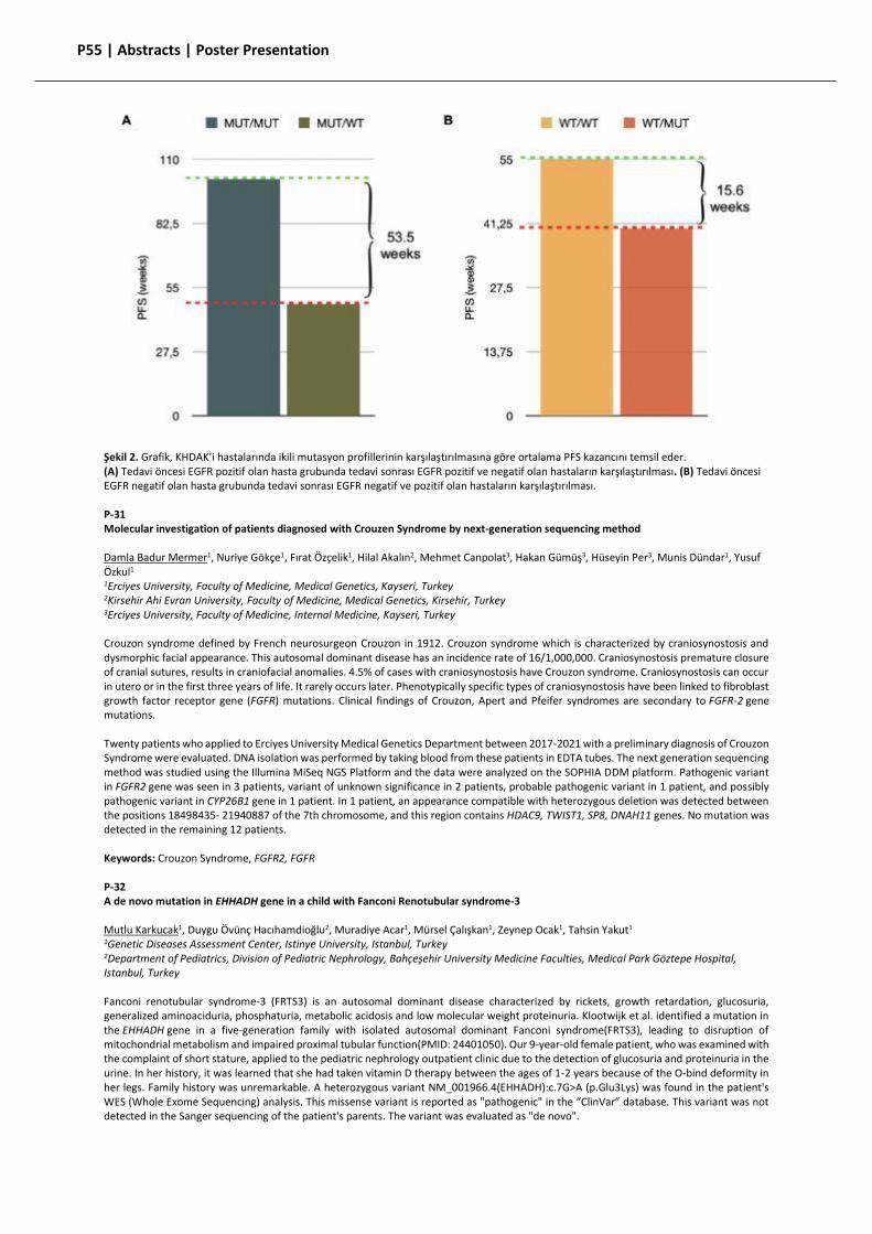
P55 | Abstracts | Poster Presentation
Şekil 2. Grafik, KHDAK’i hastalarında ikili mutasyon profillerinin karşılaştırılmasına göre ortalama PFS kazancını temsil eder. (A) Tedavi öncesi EGFR pozitif olan hasta grubunda tedavi sonrası EGFR pozitif ve negatif olan hastaların karşılaştırılması. (B) Tedavi öncesi EGFR negatif olan hasta grubunda tedavi sonrası EGFR negatif ve pozitif olan hastaların karşılaştırılması. P-31 Molecular investigation of patients diagnosed with Crouzen Syndrome by next-generation sequencing method Damla Badur Mermer1, Nuriye Gökçe1, Fırat Özçelik1, Hilal Akalın2, Mehmet Canpolat3, Hakan Gümüş3, Hüseyin Per3, Munis Dündar1, Yusuf Özkul1 1Erciyes University, Faculty of Medicine, Medical Genetics, Kayseri, Turkey 2Kirsehir Ahi Evran University, Faculty of Medicine, Medical Genetics, Kirsehir, Turkey 3Erciyes University, Faculty of Medicine, Internal Medicine, Kayseri, Turkey Crouzon syndrome defined by French neurosurgeon Crouzon in 1912. Crouzon syndrome which is characterized by craniosynostosis and dysmorphic facial appearance. This autosomal dominant disease has an incidence rate of 16/1,000,000. Craniosynostosis premature closure of cranial sutures, results in craniofacial anomalies. 4.5% of cases with craniosynostosis have Crouzon syndrome. Craniosynostosis can occur in utero or in the first three years of life. It rarely occurs later. Phenotypically specific types of craniosynostosis have been linked to fibroblast growth factor receptor gene (FGFR) mutations. Clinical findings of Crouzon, Apert and Pfeifer syndromes are secondary to FGFR-2 gene mutations. Twenty patients who applied to Erciyes University Medical Genetics Department between 2017-2021 with a preliminary diagnosis of Crouzon Syndrome were evaluated. DNA isolation was performed by taking blood from these patients in EDTA tubes. The next generation sequencing method was studied using the Illumina MiSeq NGS Platform and the data were analyzed on the SOPHIA DDM platform. Pathogenic variant in FGFR2 gene was seen in 3 patients, variant of unknown significance in 2 patients, probable pathogenic variant in 1 patient, and possibly pathogenic variant in CYP26B1 gene in 1 patient. In 1 patient, an appearance compatible with heterozygous deletion was detected between the positions 18498435- 21940887 of the 7th chromosome, and this region contains HDAC9, TWIST1, SP8, DNAH11 genes. No mutation was detected in the remaining 12 patients. Keywords: Crouzon Syndrome, FGFR2, FGFR P-32 A de novo mutation in EHHADH gene in a child with Fanconi Renotubular syndrome-3 Mutlu Karkucak1, Duygu Övünç Hacıhamdioğlu2, Muradiye Acar1, Mürsel Çalışkan1, Zeynep Ocak1, Tahsin Yakut1 1Genetic Diseases Assessment Center, Istinye University, Istanbul, Turkey 2Department of Pediatrics, Division of Pediatric Nephrology, Bahçeşehir University Medicine Faculties, Medical Park Göztepe Hospital, Istanbul, Turkey Fanconi renotubular syndrome-3 (FRTS3) is an autosomal dominant disease characterized by rickets, growth retardation, glucosuria, generalized aminoaciduria, phosphaturia, metabolic acidosis and low molecular weight proteinuria. Klootwijk et al. identified a mutation in the EHHADH gene in a five-generation family with isolated autosomal dominant Fanconi syndrome(FRTS3), leading to disruption of mitochondrial metabolism and impaired proximal tubular function(PMID: 24401050). Our 9-year-old female patient, who was examined with the complaint of short stature, applied to the pediatric nephrology outpatient clinic due to the detection of glucosuria and proteinuria in the urine. In her history, it was learned that she had taken vitamin D therapy between the ages of 1-2 years because of the O-bind deformity in her legs. Family history was unremarkable. A heterozygous variant NM_001966.4(EHHADH):c.7G>A (p.Glu3Lys) was found in the patient's WES (Whole Exome Sequencing) analysis. This missense variant is reported as "pathogenic" in the “ClinVar” database. This variant was not detected in the Sanger sequencing of the patient's parents. The variant was evaluated as "de novo".
P56 | Abstracts | Poster Presentation Our case is the same as the previous mutation, but differs in that it is "de novo". Our case is important in that it supports the role of the mutation in the EHHADH gene in the etiology of FRTS3 disease. Keywords: EHHADH gene, kidney, Fanconi renotubular syndrome P-33 Novel variant detected in the FAS gene of a patient with Autoimmune Lymphoproliferative Syndrome Mucahit Bilgehan Ari1, Tugba Yaman1, Cagri Caliskan1, Firat Ozcelik2, Alper Ozcan3, Yusuf Ozkul2, Ekrem Unal3, Munis Dundar2 1Faculty of Medicine, Erciyes University, Kayseri, Turkey 2Department of Medical Genetics, Erciyes University, Kayseri, Turkey 3Department of Pediatric Oncology and Hematology, Erciyes University, Kayseri, Turkey Autoimmune lymphoproliferative syndrome (ALPS) is a rare disorder caused by immune dysregulation due to disrupted lymphocyte apoptosis. In autoimmune lymphoproliferative syndrome, defective lymphocyte apoptosis manifests as chronic, nonmalignant adenopathy, splenomegaly and the development of autoimmune disease. Most cases of autoimmune lymphoproliferative syndrome have heterozygous variants in the lymphocyte surface protein Fas (CD95, Apo 1) that impair a major apoptotic pathway. Here, we present a Turkish male patient who has a novel de novo variant in the FAS gene. He was referred to our clinic with low WBC level, splenomegaly, decrease in bone marrow cell lines. The patient is the second child of his family, there is no consanguineous marriage in his family tree. Clinical Exome Solution revealed a heterozygous nonsense variant of c.766G>T (p.Glu256*) in the FAS gene (NM_000043). The variant was classified as “pathogenic” according to the ACMG 2015 guideline for interpretation of sequence variants. Sanger sequencing confirmed the variant in the patient, while the parents were detected as normal. According to the literature, there was no previous study reporting this variant. We believe this novel may help for future patients' diagnoses and treatments. Keywords: ALPS, FAS, autoimmune, NGS


![Dictionary of Chemistry [6th Ed.]](https://static.fdokumen.com/doc/165x107/6316ba01f68b807f88035f31/dictionary-of-chemistry-6th-ed.jpg)


