
SANCTION FOR GENOCIDE: ANTISEMITISM AND THE EVOLUTION OF EVIL
Upload
independentCategory
view
0download
0
Behavioral/Cognitive
Epigenetic Modification of the Glucocorticoid Receptor GeneIs Linked to Traumatic Memory and Post-Traumatic StressDisorder Risk in Genocide Survivors
Vanja Vukojevic,1,2 Iris-T. Kolassa,3 Matthias Fastenrath,1,4 Leo Gschwind,1,4 Klara Spalek,1,4 Annette Milnik,1,5
Angela Heck,1,5,6 Christian Vogler,1,5,6 X Sarah Wilker,3 Philippe Demougin,1,2 Fabian Peter,1,2 Erika Atucha,7
Attila Stetak,1,2,5 Benno Roozendaal,7 X Thomas Elbert,8 Andreas Papassotiropoulos,1,2,5,6
and Dominique J.-F. de Quervain4,5,6
1Department of Psychology, Division of Molecular Neuroscience, University of Basel, CH-4055 Basel, Switzerland, 2Department Biozentrum, Life SciencesTraining Facility, University of Basel, CH-4056 Basel, Switzerland, 3Clinical & Biological Psychology, Institute of Psychology and Education, University ofUlm, D-89069 Ulm, Germany, 4Department of Psychology, Division of Cognitive Neuroscience, University of Basel, CH-4055 Basel, Switzerland, 5PsychiatricUniversity Clinics, University of Basel, CH-4012 Basel, Switzerland, 6Transfacultary Research Platform, University of Basel, CH-4055 Basel, Switzerland,7Department of Cognitive Neuroscience and Donders Institute for Brain, Cognition and Behaviour, Radboud University Nijmegen, 6525 EZ Nijmegen,Netherlands, and 8Clinical Psychology and Neuropsychology, University of Konstanz, D-78464 Konstanz, Germany
Recent evidence suggests that altered expression and epigenetic modification of the glucocorticoid receptor gene (NR3C1) are related tothe risk of post-traumatic stress disorder (PTSD). The underlying mechanisms, however, remain unknown. Because glucocorticoidreceptor signaling is known to regulate emotional memory processes, particularly in men, epigenetic modifications of NR3C1 might affectthe strength of traumatic memories. Here, we found that increased DNA methylation at the NGFI-A (nerve growth factor-induced proteinA) binding site of the NR3C1 promoter was associated with less intrusive memory of the traumatic event and reduced PTSD risk in male,but not female survivors of the Rwandan genocide. NR3C1 methylation was not significantly related to hyperarousal or avoidancesymptoms. We further investigated the relationship between NR3C1 methylation and memory functions in a neuroimaging study inhealthy subjects. Increased NR3C1 methylation–which was associated with lower NR3C1 expression–was related to reduced picturerecognition in male, but not female subjects. Furthermore, we found methylation-dependent differences in recognition memory-relatedbrain activity in men. Together, these findings indicate that an epigenetic modification of the glucocorticoid receptor gene promoter islinked to interindividual and gender-specific differences in memory functions and PTSD risk.
Key words: DNA methylation; GR; memory; PTSD
IntroductionPost-traumatic stress disorder (PTSD) is characterized by intru-sive memories of traumatic events, avoidance, and hyperarousalsymptoms [Diagnostic and Statistical Manual of Mental Disorders,Fourth Edition (DSM-IV), American Psychiatric Association,
2000]. In contrast to a state of chronic stress, PTSD is not paral-leled by increased glucocorticoid levels (Meewisse et al., 2007),but rather by enhanced hypothalamus–pituitary–adrenal (HPA)axis feedback (Yehuda, 2002; Rohleder et al., 2004; Pitman et al.,2012). Moreover, there is evidence suggesting that altered HPAaxis regulation and a high number of glucocorticoid receptors(GRs) represent a pretrauma risk factor for the disorder (Yehuda,2009; van Zuiden et al., 2011, 2013). The reason for interindi-vidual differences in GR number and the mechanism by whichGRs influence the risk for PTSD, however, remain largelyunknown.
Timed activation of GRs is of crucial importance in memoryconsolidation of learned information, especially of emotionallyarousing information (Roozendaal et al., 2006). Human studieshave shown that the memory-enhancing effects of stress-inducedelevations of glucocorticoids are usually more pronounced inmen (Andreano and Cahill, 2006; Preuss and Wolf, 2009; Corne-lisse et al., 2011). Furthermore, there is evidence that glucocorti-coids impair the retrieval of memory processes (de Quervain etal., 2009). Because fear learning and memory processes play an
Received April 15, 2014; revised June 14, 2014; accepted June 18, 2014.Author contributions: V.V., I.-T.K., B.R., T.E., A.P., and D.J.-F.D.Q. designed research; V.V., I.-T.K., M.F., A.M., K.S.,
P.D., and F.P. performed research; V.V., P.D., F.P., T.E., A.P., and D.J.-F.D.Q. contributed unpublished reagents/analytic tools; V.V., I.-T.K., M.F., L.G., A.M., K.S., A.H., C.V., S.W., P.D., E.A., A.S., B.R., T.E., A.P., and D.J.-F.D.Q.analyzed data; V.V., I.-T.K., M.F., A.M., K.S., A.H., C.V., S.W., P.D., E.A., A.S., B.R., T.E., A.P., and D.J.-F.D.Q. wrote thepaper.
This work was supported by the Swiss National Science Foundation (Sinergia Grants CRSIK0_122691,CRSI33_130080 to D.J.-F.D.Q. and A.P.) and by the German Research Foundation (grants to I.-T.K. and T.E.). V.V. wassupported by a Werner Siemens Foundation (Zug, Switzerland) PhD grant and Opportunities for Excellence PhDProgram of the Biozentrum.
The authors declare no competing financial interest.Correspondence should be addressed to either of the following: Vanja Vukojevic at the above
address, E-mail: [email protected]; or Dominique J.-F. de Quervain at the above address,E-mail: [email protected].
DOI:10.1523/JNEUROSCI.1526-14.2014Copyright © 2014 the authors 0270-6474/14/3310274-11$15.00/0
10274 • The Journal of Neuroscience, July 30, 2014 • 34(31):10274 –10284

important role in the pathogenesis of PTSD (de Quervain, 2006;Brewin, 2011; Pitman et al., 2012; Wilker et al., 2013), GRs may beimplicated in PTSD risk by influencing memory processes (deQuervain et al., 2009). Accordingly, it has been shown that theBclI polymorphism of NR3C1 (nuclear receptor subfamily 3,group C, member 1), the gene encoding the GR, is associated withsensitivity to glucocorticoids (van Rossum et al., 2003), emo-tional memory in healthy individuals (Ackermann et al., 2013),traumatic memories and PTSD symptoms in critically ill patients(Hauer et al., 2011).
Epigenetic mechanisms may contribute to interindividual dif-ferences in GR signaling. DNA hypermethylation of the exonNR3C1-17 promoter in rodents and of its human ortholog, theexon NR3C1-1F promoter (Turner and Muller, 2005), was asso-ciated with low maternal care, maternal depression, and perinatalstress (Weaver et al., 2007; Oberlander et al., 2008). In line withthe enhanced GR feedback reported in PTSD cases (Yehuda,2009), it has been shown that increased NR3C1 expression inperipheral tissue is related to PTSD risk (van Zuiden et al., 2011).Furthermore, it appears that these changes are partially epigeneti-cally controlled. Two recent studies suggested that DNA methyl-ation of the GR gene promoter are inversely correlated withlifetime PTSD risk (Labonte et al., 2014; Yehuda et al., 2014).Moreover, allele-specific DNA demethylation of FKBP5, a regu-lator of the GR complex, has been found to mediate gene– child-hood trauma interactions (Klengel et al., 2013).
Given the evidence that GR signaling influences both memoryprocesses and PTSD risk, we investigated whether epigenetic dif-ferences in the human NR3C1 gene promoter are related to trau-matic memory and the risk for PTSD in 152 survivors of theRwandan genocide. Furthermore, we investigated the relation-ship of NR3C1 promoter DNA methylation with memory pro-cesses in a functional neuroimaging study in 72 healthy subjects.
Materials and MethodsSubjects: Rwanda sample. We included 152 survivors from the 1994Rwandan genocide (69 females, 83 males; median age, 35 years; range,30 – 41 years) who lived as refugees in the Nakivale settlement in Uganda.The sample consisted of 93 subjects fulfilling the diagnostic criteria of theDSM-IV for lifetime PTSD, and 59 individuals who did not meet theDSM-IV diagnostic criteria for PTSD (61.2% with PTSD lifetime diag-nosis; 31.2% subjects with current PTSD according to DSM-IV (Ameri-can Psychiatric Association, 2000). Additionally, to exclude geneticrelatives in the samples, only one person per household was interviewed.Candidates exhibiting current alcohol abuse and acute psychotic symp-toms were excluded. All subjects had experienced highly aversive trau-matic situations and were examined in 2006/2007 (de Quervain et al.,2007). The Post-Traumatic Diagnostic Scale (PDS; Foa et al., 1997) andevent list (Ertl et al., 2010) were administered as a structured interview byexpert psychologists from the University of Konstanz, Germany, as wellas by trained local interviewers. Interviewers first went through a six-week course on principles of quantitative data collection and interview-ing techniques. The translation of the instruments into Kinyarwanda wasdone using several steps of translations, blind back-translations, and sub-sequent corrections by independent groups of translators (Neuner et al.,2008). Following the translations, the psychometric properties of thetranslated scales were investigated in a validation study that included aretest spanning a two-week period and a cross-validation with expertrating (Neuner et al., 2008). The PDS was used to assess intrusions,avoidance, and hyperarousal. A checklist of 36 war-related and nonwar-related traumatic event types (e.g., injury by weapon, rape, accident) wasused to assess traumatic events (de Quervain et al., 2007; Neuner et al.,2008). Traumatic load was estimated by assessing the number of differenttraumatic event types experienced or witnessed. This measure is consid-ered more reliable than assessing the frequency of traumatic events
(Neuner et al., 2008). Study procedures were approved by the ethicscommittees of the University of Konstanz, Germany, and the MbararaUniversity of Science and Technology, Mbarara, Uganda. Before the in-terview, all participants provided written informed consent.
Subjects were selected to have experienced �19 traumatic event types,to avoid known ceiling effects on PTSD risk (Kolassa et al., 2010). Salivasamples from all subjects were collected using Oragene DNA Kits (DNAGenotek).
Subjects: Swiss sample. A total of 72 healthy young subjects (47 females,25 males; median age, 23 years; range, 18 –34 years) were included in thefunctional magnetic resonance imaging (fMRI) study. fMRI data fromone male and two female subjects were corrupted and therefore were notincluded in the study. Subjects were free of any neurological or psychi-atric illness, and did not take any medication at the time of the experi-ment (except hormonal contraceptives). The ethics committees of theCanton of Basel and Baselland approved the experiments.
Subjects were tested in the late morning and afternoon hours (meantime, 2:30 P.M.; SD � 3 h). All participants received general informationabout the study and gave their written informed consent for participa-tion. After completing the training outside of the scanner, subjects per-formed two different consecutive tasks in the scanner. The first two tasks,i.e., the picture-encoding task and the working-memory task (n-back),were described in detail previously (de Quervain et al., 2007). Briefly,stimuli in the picture-encoding task consisted of 72 pictures selectedfrom the International Affective Picture System (Lang et al., 1999) as wellas from in-house standardized picture sets that allowed us to equate thepictures for visual complexity and content (e.g., human presence). Onthe basis of normative valence scores (from 1 to 9), pictures were assignedto emotionally negative (2.3 � 0.6), emotionally neutral (5.0 � 0.3), andemotionally positive (7.6 � 0.4) conditions, resulting in 24 pictures foreach emotional valence. Four additional pictures showing neutral objectswere used to control for primacy and recency effects in memory. Two ofthese pictures were presented in the beginning and two at the end of thepicture task and the working memory task. In addition, 24 scrambledpictures were used. The background of the scrambled pictures containedthe color information of all pictures used in the experiment (exceptprimacy and recency pictures), overlaid with a crystal and distortionfilter (Adobe Photoshop CS3). In the foreground, a mostly transparentgeometrical object (rectangle or ellipse of different sizes and orienta-tions) was shown. Participants were instructed and then trained on thepicture-encoding task. After training, they were positioned in the scan-ner. The picture-encoding task, which included the arousal and valenceratings of the pictures, lasted for �20 min. Immediately afterward, sub-jects performed the working-memory task for 10 additional minutes.After leaving the scanner, participants gave a free recall of the pictures ina separate room (no time limit was set for this task). Forty to 50 min afterthe presentation of the last picture in the encoding task, participants wererepositioned in the scanner and performed a recognition task for 20 min.The recognition task consisted of two sets of stimuli that were either new(i.e., not presented before) or old (i.e., presented during the picture-encoding task). Each of the two sets contained 72 pictures (24 pictures foreach emotional valence).
Using an Oragene DNA and Oragene RNA Kit (DNA Genotek), salivasamples were collected from all subjects. Participants received 25 Swissfrancs per hour for participation.
DNA isolation and bisulfite conversion. Saliva DNA was initially ex-tracted from the Oragene DNA Kit (DNA Genotek) using the precipita-tion protocol recommended by the producer. To obtain high-purityDNA before bisulfite conversion, samples were additionally repurified.For this purpose, 2 �g of DNA isolated via the Oragene recommendedprocedure, was incubated overnight at 50°C with proteinase K (lysisbuffer: 30 mM Tris-Cl, 10 mM EDTA, 1% SDS, pH�8.0, 150 ng/�lproteinase K), agitated by gentle orbital shaking. Next, the DNA waspurified using a Genomic DNA Clean & Concentrator Kit (Zymo Re-search). The quality and concentration were assessed using gel electro-phoresis and fluorometry (Qubit dsDNA BR Assay Kit, Invitrogen),respectively. Five hundred nanograms of high-purity, intact DNA wasused for bisulfite conversion using an EZ DNA Methylation-Gold kit(Zymo Research) by following standard protocols. Bisulfite DNA quality
Vukojevic et al. • DNA-Methylation of Nr3c1 Is Linked to PTSD and Memory J. Neurosci., July 30, 2014 • 34(31):10274 –10284 • 10275
and concentration was determined using an RNA Pico 6000 Kit on aBioanalyzer 2100 instrument (Agilent Technologies) and Nanodrop2000 (ThermoScientific). An external control sample was always con-verted in parallel to assess possible variations in conversion reactions. Allsamples were bisulfite converted on two separate occasions, under theidentical conditions. Bisulfite-converted (BSC) samples were normalizedto 10 ng/�l.
All samples were analyzed in quadruplicates from two independentbisulfite conversions.
Pyrosequencing analysis. The DNA methylation status of the CpG-richregion of the human NR3C1 gene, including the exon 1F promoter(NR3C1-1F) with the NGFI-A binding site, previously reported as im-portant for epigenetic regulation of GR gene expression (Weaver et al.,2004), was quantified by direct bisulfite pyrosequencing (Tost and Gut,2007). Primers were designed according to recommendations of Woj-dacz et al. (2009): NR3C1_Q-CpG_FW, 5�-GTTATTCGTAGGGGTATTG-3� and NR3C1_Q-CpG_RV, 5�-biot-CAACTCCCCAAAAAAAAAAA-3� (Microsynth). A 206 bp NR3C1 promoter fragment was amplifiedusing an AmplyTaq Gold Kit from Applied Biosystems (Life Technolo-gies). PCR was done in 30 �l reactions containing the following: 1� PCRbuffer II, 300 �M deoxynucleotide triphosphates, final 3.5 mM MgCl2,200 �M of each primer, 20 ng of BSC DNA. We used the following cyclingconditions: 95°C, 15 min–50 � (95°C, 30 s; 55°C, 30 s; 72°C, 30 s)–72°C,10 min. PCR products were purified and sequenced using a PyroMark IDSystem (Biotage) following the manufacturer’s suggested protocol andtwo sequencing primers: NR3C1_S2, 5�-GAGTGGGTTTGGAGT-3� andNR3C1_S3, 5�-AGAAAAGAAATTGGAGAAATT-3� (Microsynth) wereused as in Oberlander et al. (2008). Testing for PCR temperature bias andcalibration was done by introducing a series of calibrator samples withknown methylation levels. Briefly, we prepared unmethylated standardsby using two rounds of linear whole-genome amplification with an Ova-tion WGA System Kit (Nugene), starting from 10 ng of DNA, as recom-mended by the manufacturer. Methylated standards were made usingCpG methyltransferase assay with M.SssI (New England Biolabs) startingfrom 2 �g of purified DNA, following the standard protocol. Bisulfiteconversion of standard samples was done as described above.
RNA isolation and expression analysis. For the purpose of DNA methy-lation–RNA expression correlation experiments, an additional 24 (12females, 12 males; median age, 25 years; range, 21–29 years) subjects wererecruited from the Swiss sample, as described above. RNA was isolatedfrom Oragene RNA Self-Collection Kits from saliva. Promptly after col-lection, samples were mixed for 10 s by vortexing, stabilized at 50°C in awater bath for 2.5 h, and stored at �80°C until further analysis. For theisolation procedure, samples were processed in 250 �l aliquots. Briefly,samples were incubated in a water bath at 90°C for 15 min, and thencooled to room temperature. Following this, 750 �l of TRIReagent LS(Ambion, Life Technologies) was added to each sample and homogeni-zation was done for 1 min at 28 Hz using TissueLyser II (Qiagen). Afterhomogenization, 1000 �l of absolute ethanol was added to the sampleand mixed. Finally, the mixture was loaded on a Zymo-Spin IIC columnand RNA was further purified using a Direct-zol kit (ZymoResearch)following the recommended procedure, including DNase I treatment.The concentration and quality of the RNA was determined using Nano-drop 2000 (ThermoScientific) and an RNA Nano 6000 Kit on a Bioana-lyzer 2100 instrument (Agilent).
For reverse transcription, 350 ng of total RNA was denatured for 8 minat 70°C followed by ice incubation in the presence of 25 ng of anchoredoligo(dT)20 primer (Invitrogen) and 75 ng of random decamers primers(Ambion). In the reverse transcription reaction, cDNA was generated in25 �l reaction using a Super RT kit (HT Biotechnology). Upon comple-tion of the reaction, the volume was adjusted to 200 �l in Lambda DNAsolution (5 ng/�l final concentration; Promega). The primers were de-signed against splice variants that contain exon 1F of the NR3C1 gene:NR3C1_1F_FW, 5�-GTTGATATTCACTGATGGA-3� and NR3C1_1F_RV, 5�-CTTGGAGTCTGATTGAGA-3� (Microsynth). Additionallytotal expression of the GR gene was determined against exon 2:NR3C1_2_FW, 5�-CTTCAGAACAGCAACATT-3� and NR3C1_2_RV,5�-GACTCTCATTCGTCTCTT-3� (Microsynth). The expression levelswere normalized to the RPLPO gene (human large ribosomal protein)
using the following primers: RPLP0-Ex3-4_FW, 5�-CTCTGGAGAAACTGCTGC-3� and RPLP0-Ex3-4_RV, 5�-CTGATCTCAGTGAGGTCC-3� (Sigma-Aldrich). qPCR was performed using the Power SYBRGreen PCR Master Mix (Life Technologies) according to standard rec-ommendations, in 12 �l final volume of reaction, using 2 �l of cDNAtemplate, on a RotorGene 6000A instrument (Corbett Research). Cy-cling conditions were as follows: 95°C, 60 s– 40� (95°C, 3 s; 56°C, 10 s;72°C, 4 s). This was followed by a melting curve analysis (61–95°C, risingby 0.7°C/3 s) to attest amplification specificity. Threshold cycles (cross-ing point) were determined using Rotor-Gene software version 6.1 (Cor-bett Research). RPLPO was selected as reference gene for normalizationafter we tested several candidate reference genes, as had been previouslydescribed (Movassagh et al., 2010). Expression levels were normalizedusing a geometric mean level of expression (Vandesompele et al., 2002).Fold differences were calculated using the delta-delta-Ct method (Pfaffl,2001) with the help of qBasePlus software (Biogazelle).
fMRI study design. During the encoding task, the pictures were pre-sented for 2.5 s in a quasirandomized order so that �4 pictures of thesame category occurred consecutively. A fixation cross appeared on thescreen for 500 ms before each picture presentation. Trials were separatedby a variable intertrial period of 9 –12 s (jitter) that was equally distrib-uted for each stimulus category. During the intertrial period, participantssubjectively rated the picture showing scenes according to valence (neg-ative, neutral, positive) and arousal (high, medium, and low) on a three-point scale (self-assessment manikin) by pressing a button with a fingerof their dominant hand. For scrambled pictures, participants rated form(vertical, symmetric, or horizontal) and size (large, medium, small) ofthe geometrical object in the foreground.
During the recognition task, the pictures were presented for 1 s in aquasirandomized order so that �4 pictures of the same category (i.e.,negative new, negative old, neutral new, neutral old, positive new, posi-tive old) occurred consecutively. A fixation cross appeared on the screenfor 500 ms before each picture presentation. Trials were separated by avariable intertrial period of 6 –12 s (jitter) that was equally distributed foreach stimulus category. During the intertrial period, participants subjec-tively rated the picture as either remembered, familiar, or new on athree-point scale by pressing a button with a finger of their dominanthand. Recognition performance was assessed as a number of correctlyrecognized previously seen pictures, corrected for the number of falsepositives.
fMRI methods. Measurements were performed on a Siemens Magne-tom Verio 3 T whole-body MR unit equipped with a 12-channel head coil(Siemens Healthcare). Functional time series were acquired with asingle-shot echo-planar sequence using parallel imaging [GRAPPA (gen-eralized autocalibrating partially parallel acquisition)]. We used the fol-lowing acquisition parameters: TE, 35 ms; field of view, 22 cm;acquisition matrix, 80 � 80, interpolated to 128 � 128; voxel size, 2.75 �2.75 � 4 mm 3; GRAPPA acceleration factor r, 2.0. Using a midsagittalscout image, 32 contiguous axial slices placed along the anterior–poste-rior commissure plane covering the entire brain with a TR � 3000 ms(� � 82°) were acquired using an ascending interleaved sequence.
Participants received earplugs and headphones to reduce scannernoise. The head of each participant was fixated in the coil using smallcushions, and participants were told not to move their head. Pictureswere presented in the scanner using MR-compatible liquid crystal displaygoggles (VisualSystem; NordicNeuroLab). Eye correction was used whennecessary.
Preprocessing and data analysis was performed using SPM8 (Statisti-cal Parametric Mapping, Wellcome Trust Centre for Neuroimaging;http://www.fil.ion.ucl.ac.uk/spm/) implemented in Matlab R2011b(Mathworks). Volumes were slice-time corrected to the first slice andrealigned using the “register to mean” option. A mean image was gener-ated from the realigned series and coregistered to the structural image.This ensured that functional and structural images were spatially aligned.
The functional images and the structural images were spatially nor-malized by applying DARTEL (diffeomorphic anatomical registrationthrough exponentiated lie algebra), which leads to an improved registra-tion between subjects. Normalization incorporated the following steps:(1) structural images of each subject were segmented using the “New
10276 • J. Neurosci., July 30, 2014 • 34(31):10274 –10284 Vukojevic et al. • DNA-Methylation of Nr3c1 Is Linked to PTSD and Memory

Segment” procedure in SPM8; (2) the resulting gray and white matterimages were used to derive a study-specific group template computedfrom a large population of 612 subjects, including the 69 subjects fromthis study where methylation data were available; (3) an affine transfor-mation was applied to map the group template to MNI space; and (4)subject-to-template and template-to-MNI transformations were com-bined to map the functional images to MNI space. The functional imageswere smoothed with an isotropic 8 mm full width at half maximumGaussian filter.
Intrinsic autocorrelations were accounted for by AR(1) and low-frequency drifts were removed via high-pass filter (time constant, 128 s).For each subject, evoked hemodynamic responses to event types weremodeled with a delta function convolved with a canonical hemodynamicresponse function within the context of a general linear model. Buttonpresses and rating scale presentation during ratings were modeled sepa-rately. In addition, six movement parameters from spatial realigningwere included as regressors of no interest.
Encoding task. The effect of picture encoding was investigated by con-structing separate regressors representing the event types for positive,negative, neutral, and scrambled pictures. We contrasted brain activityduring the encoding of pictures against brain activity during the presen-tation of scrambled pictures (positive, negative, and neutral pictures vsscrambled pictures).
Recognition task. Eighteen different picture event types were modeledby crossing each stimulus category with the subject-specific rating of eachpicture while differentiating between correct and incorrect recognitionratings with respect to the new or old picture set: (negative, positive,neutral) � (remembered, familiar, new) � (correct, incorrect). We con-trasted brain activity between items that were correctly recognized asseen before and new pictures (i.e., pictures seen in the encoding task andrated as remembered vs pictures not seen before and rated as new). Itemsrated as familiar were not included in the analysis.
The contrasts of both the encoding and the recognition task werecalculated individually using a fixed-effects model (first-level analysis).The resulting contrast parameters were then used for methylation-dependent brain activity analyses in a random-effects, linear regressionmodel where we entered the methylation level as a covariate (second-level analysis).
Construction of a population-average anatomical probabilistic atlas. Au-tomatic segmentation of the subjects’ T1-weighted images was used tobuild a population-average probabilistic anatomical atlas. More pre-cisely, each participant’s T1-weighted image was first automatically seg-mented into cortical and subcortical structures using FreeSurfer (Fischlet al., 2002; version 4.5, http://surfer.nmr.mgh.harvard.edu/). Labelingof the cortical gyri was based on the Desikan-Killiany Atlas (Desikan etal., 2006), yielding 35 regions per hemisphere.
The segmented T1 image was then normalized to the study-specificanatomical template space using the subject’s previously computed warpfield. After this, the image was affine-registered to the MNI space.Nearest-neighbor interpolation was applied to preserve labeling of the
different structures. The normalized segmen-tations were finally averaged across subjects tocreate a population-average probabilistic atlas.Each voxel of the template could consequentlybe assigned a probability of belonging to agiven anatomical structure, based on the indi-vidual information from all subjects.
Statistical analyses. Statistical analyses weredone in R (version 12.15.2; R DevelopmentCore Team 2012). Due to non-normal distri-bution of methylation data in the Rwanda sam-ple, we applied nonparametric tests. Weassessed DNA methylation, trauma load, andage differences between two genders byKruskal–Wallis one-way ANOVA and correla-tions of DNA methylation data with PTSDsymptom clusters scores by Spearman’s rankcorrelation (�s). To account for trauma loadand ceiling effects (Kolassa et al., 2010), PTSDsymptom cluster scores were divided by the
sum of lifetime traumatic event types (i.e., symptoms scores per trau-matic event type; de Quervain et al., 2007). The relationship betweenDNA methylation at NR3C1 promoter and lifetime PTSD status wasassessed using binary logistic regression, with NR3C1 CpG3 DNA meth-ylation as quantitative predictor and the sum of life traumatic event typesas a covariate.
Due to normal distribution of the methylation data, statistical analysesof behavioral data for the Swiss sample were performed using linearmodels. The relationship of NR3C1 DNA methylation to memory per-formance and NR3C1 expression was assessed using linear models. Toaccount for methylation-dependent differences in arousal during recog-nition of previously seen pictures, both arousal and NR3C1 CpG3 DNAmethylation were included in the linear model: we modeled valence-specific recognition performance as dependent on NR3C1 CpG3 DNAmethylation and valence-specific arousal.
Correlation between NR3C1 CpG3 DNA methylation and NR3C1 ex-pression was examined by linear regression model, corrected for ageindependently in two genders.
A comparison of methylation levels of NR3C1 CpG3 site between theRwandan and the Swiss population was done by Kruskal–Wallis one-wayANOVA. Furthermore, we additionally assessed the equality of distribu-tions and variability between two populations: Kolmogorov–Smirnovtwo-sample test and Siegel–Tukey test, respectively.
Bonferroni correction was implemented to account for multiple test-ing procedures. The significance threshold was set to p � 0.05.
All laboratory procedures were conducted in a blind, randomized or-der, including DNA and RNA isolations, bisulfite conversion, PCR, py-rosequencing, and expression analysis. Only after performing allprocedures and excluding samples with low quality controls and outliers,further analysis with phenotypic data was performed.
ResultsDNA methylation of the NR3C1 promoter in traumatizedsurvivors of the Rwandan genocideThe promoter region of the NR3C1 gene studied herein is illus-trated in Figure 1. It spans the exon 1F region, a human ortho-logue of the rat exon 1-7 (Turner and Muller, 2005), previouslyreported as differentially methylated (Oberlander et al., 2008;McGowan et al., 2009). The analysis covered eight CpGs, with theNGFI-A consensus binding sequence encompassing CpG posi-tions 3 and 4. Because glucocorticoid-related changes in memoryformation were repeatedly found to be more pronounced in men(Andreano and Cahill, 2006; Preuss and Wolf, 2009), we alsoconsidered possible gender differences in the present study. Wefirst compared DNA methylation between genders and foundsignificant differences between men and women at several CpGpositions (p � 0.05, e.g., methylation levels at NR3C1_CpG3
1B 1F 1G 1C 2 3‘5‘
+1
1 2 3 -3236 ttgccgccaaggggcagagcgagctcccgagtgggtctggagccgcggagctgggcggggg- 4 5 6 7 8 cgggaaggAGGTAGCGAGAAAAGAAACTGGAGAAACTCGGTGGCCCTCTTAACGCC-
-3225 GCCCCAGAGAG
Figure 1. Human NR3C1 gene and the CpG region analyzed by bisulfite pyrosequencing. The 5� region of the NR3C1 genecontains multiple first exons, corresponding to multiple transcriptional start sites and multiple mRNA splice variants. The regionanalyzed by pyrosequencing is illustrated by enlarged box, below the exon 1F. Numbering is relative to the alternative transcriptionstart site in exon 2. The CpG3 and CpG4 sites are encompassed by the potential NGFI-A binding site. Adapted from Oberlander et al.(2008).
Vukojevic et al. • DNA-Methylation of Nr3c1 Is Linked to PTSD and Memory J. Neurosci., July 30, 2014 • 34(31):10274 –10284 • 10277

were significantly higher in women thanin men, p � 0.01). Therefore, we pro-ceeded with gender-specific analyses. Toaccount for trauma load, PTSD symptomcluster scores were corrected by the sumof lifetime traumatic event types (deQuervain et al., 2007). Methylation levelsat the CpG3 site, which is embedded in theNGFI-A consensus binding sequence,were significantly negatively correlatedwith the severity of symptoms related tore-experiencing traumatic events (intru-sive memories) in men (�s � �0.355,pnominal � 0.001, pBonferroni corrected � 0.008for eight CpG comparisons; Fig. 2, Table1). No such association was found inwomen (�s � �0.073, pnominal � 0.553;Table 2). Correlations of DNA methyl-ation at CpG3 with the severity ofsymptoms related to avoidance and hy-perarousal were not significant after Bon-ferroni correction for eight CpGs (p �0.05), indicating that the methylationchanges at this site were preferentially re-lated to the memory aspects of PTSD.None of the other examined NR3C1 pro-moter CpG sites were significantly associ-ated with any of the PTSD symptomscores, after accounting for multiple test-ing (Tables 1, 2), indicating a pronouncedrole of methylation at the CpG3 site.
Furthermore, we investigated whethermethylation levels at CpG3 were signifi-cantly associated with lifetime PTSD risk(taking into account the total number ofexperienced traumatic event types; seeMaterials and Methods). We found thathigher methylation levels were signifi-cantly associated with a lower lifetimePTSD risk in men (binary logistic regres-sion model, � � �0.519, Wald � 2 � 7.0,p � 0.008; Fig. 3), but not in women (� ��0.170, Wald � 2 � 1.4, p � 0.243; Fig. 3).
Interindividual differences in methyl-ation levels at CpG3 were not significantlyassociated with the number of traumaticlife event types ( p � 0.23 and 0.76 formen and women, respectively). Further-more, age was not significantly associ-ated with DNA methylation in eithergender ( p � 0.10 and 0.14 for men andwomen, respectively).
DNA methylation at the NR3C1promoter, memory performance,and fMRI activity during picturerecognitionBecause in genocide survivors NR3C1CpG3 methylation was strongly associ-ated with the intrusive memory aspect ofPTSD and given the role of glucocorticoidsignaling in memory processes (de Quer-vain et al., 2009), we further examined the
-logP
Intru
sion
s
Chr5: 142763759
-logP
Hyp
erar
ousa
l
Chr5: 142763759 Chr5: 142763833
Chr5: 142763833
Chr5: 142763759 Chr5: 142763833
-logP
Avo
idan
ce
* PBonferroni < 0.05
0
1
2
3
* PBonferroni < 0.05
0
1
2
3
* PBonferroni < 0.05
0
1
2
3
CpG1
CpG2
CpG3
CpG4
CpG5
CpG6
CpG7
CpG8
CpG1
CpG2
CpG3
CpG4
CpG5
CpG6
CpG7
CpG8
CpG1
CpG2
CpG3
CpG4
CpG5
CpG6
CpG7
CpG8
Figure 2. DNA methylation of NR3C1 promoter and PTSD symptoms in male genocide survivors. Association (Spearman’s rankcorrelation, �log10p) of DNA methylation marks across eight CpG positions in the human NR3C1 gene promoter with PTSDsymptoms clusters intrusions, avoidance, and hyperarousal. The association between CpG3 and intrusions reached Bonferroni-corrected statistical significance (after correcting for 8 tests; PBonferroni � 0.008). The CpGs are aligned by genomic position. Theposition of the potential NGFI-A binding site is shaded in purple. The horizontal dashed line indicates the p � 0.05 Bonferroni-corrected significance threshold.
10278 • J. Neurosci., July 30, 2014 • 34(31):10274 –10284 Vukojevic et al. • DNA-Methylation of Nr3c1 Is Linked to PTSD and Memory
relationship of NR3C1 DNA methylation variation to memoryprocesses in healthy humans. Specifically, we investigatedwhether NR3C1 CpG3 methylation levels in healthy individualswere associated with memory performance and brain activationduring a picture-recognition task in the fMRI scanner.
NR3C1 CpG3 DNA methylation was negatively correlatedwith the correct recognition of previously seen pictures in menbut not in women (men: � � �0.455, p � 0.012; women: � ��0.065, p � 0.667; Table 3). Furthermore, DNA methylation atthis site in men was significantly associated with arousal duringencoding of negative pictures, but not of neutral or positive pic-tures (Table 3). After correcting for the valence-specific arousalrating, recognition of previously seen pictures was significantlynegatively correlated with NR3C1 CpG3 DNA methylation inde-pendently of the valence category in men but not in women (Ta-ble 3). Free-recall performance was not related to methylationlevels at NR3C1 CpG3 (men: � � �0.291, p � 0.16; women: � ��0.048, p � 0.74).
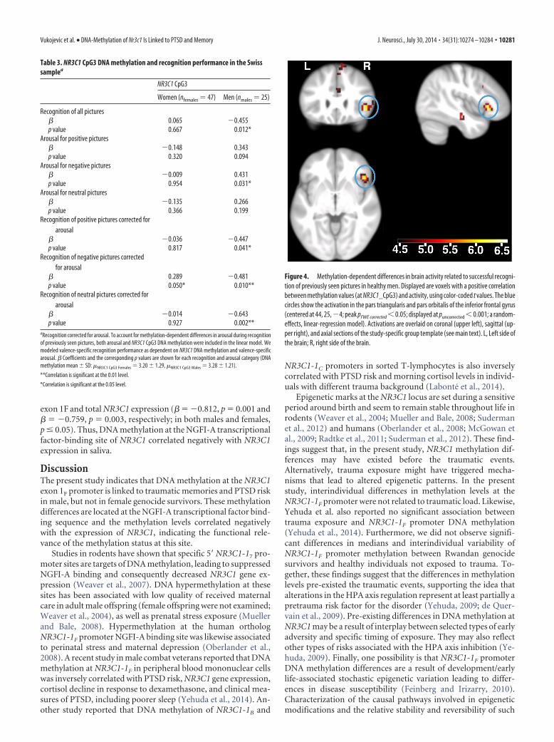
Next, we performed a methylation-dependent fMRI analysisduring successful memory recognition (see Materials and Meth-ods). In men, we found a whole-brain, family-wise error (FWE)multiple-comparison corrected (pFWE corrected � 0.05, puncorrected
� 0.001) positive correlation between methylation levels at theCpG3 site of the NR3C1 promoter and brain activity related to
the successful recognition of previously seen pictures in the parstriangularis and pars orbitalis of the inferior frontal gyrus, in thecuneus, and in the vicinity of the superior temporal and superiorfrontal cortices (Fig. 4, Table 4). No voxels correlated signifi-cantly with methylation values in the opposite direction. We didnot observe significant associations between methylation levels atthe CpG3 site and brain activity in females. Moreover, we did notfind significant correlations of individual methylation levels withencoding-related brain activity.
Finally, we compared methylation levels of NR3C1 promoterCpG3 site between the Rwandan and the Swiss sample. Impor-tantly, we did not find a significant difference in median,distribution, or variability between the samples (Kruskal–Wallis� 2 � 1.392, p � 0.24; Kolmogorov–Smirnov D � 0.150, p � 0.22;Siegel–Tukey W � 4829.5, p � 0.29). These findings suggest thatmethylation at this specific site is a stable trait and likely pre-existed the traumatic event in the Rwandan population.
Expression of the GR gene and DNA methylation at theNR3C1 gene promoterFinally, we tested whether DNA methylation on the CpG3 site ofthe NR3C1 promoter was associated with NR3C1 gene expressionin a subset of 24 healthy (12 females and 12 males) subjects. DNAmethylation at the CpG3 site correlated significantly with both
Table 1. Association of PTSD symptom clusters and DNA methylation of NR3C1 gene promoter in Rwandan mena
PTSD symptom clusters (nmales � 83) NR3C1_CpG1 NR3C1_CpG2 NR3C1_CpG3 NR3C1_CpG4 NR3C1_CpG5 NR3C1_CpG6 NR3C1_CpG7 NR3C1 _CpG8
Intrusionsb
Spearman’s � �0.235 �0.039 �0.355 �0.128 �0.203 �0.059 �0.118 �0.18p value
Nominal 0.037* 0.733 0.001*** 0.261 0.073 0.606 0.3 0.112Bonferroni correction for 8 CpGs 0.296 1 0.008 1 0.584 1 1 0.896Bonferroni correction for 8 CpGs, sex,
and 3 PTSD symptom clusters1 1 0.048 1 1 1 1 1
Avoidanceb
Spearman’s � �0.115 �0.033 �0.260 �0.033 �0.101 �0.049 �0.019 �0.074p value
Nominal 0.315 0.772 0.021* 0.77 0.376 0.666 0.87 0.517Bonferroni correction for 8 CpGs 1 1 0.168 1 1 1 1 1Bonferroni correction for 8 CpGs, sex,
and 3 PTSD symptom clusters1 1 1 1 1 1 1 1
Hyperarousalb
Spearman’s � �0.187 �0.097 �0.346 �0.038 �0.165 �0.074 �0.08 �0.053p value
Nominal 0.099 0.396 0.009** 0.74 0.145 0.514 0.483 0.645Bonferroni correction for 8 CpGs 0.792 1 0.072 1 1 1 1 1Bonferroni correction for 8 CpGs, sex,
and 3 PTSD symptom clusters1 1 0.432 1 1 1 1 1
PDS scoreb,c
Spearman’s � �0.207 �0.056 �0.355 �0.081 �0.172 �0.036 �0.078 �0.112p value
Nominal 0.067 0.627 0.001*** 0.476 0.131 0.756 0.496 0.324Bonferroni correction for 8 CpGs 0.536 1 0.008 1 1 1 1 1Bonferroni correction for 8 CpGs, sex,
and 3 PTSD symptom clusters1 1 0.048 1 1 1 1 1
NR3C1 DNA methylationMean 2.86 1.52 2.84 1.01 1.77 3.52 4.88 1.83SD 1.32 1.55 1.56 1.45 1.58 1.81 1.82 1.56
aCorrelation of NR3C1 gene promoter DNA methylation with sum of PTSD-specific symptom subscores according to PDS and DSM-IV. To account for trauma load, PTSD symptom cluster scores were divided by the sum of lifetime traumaticevent types. Spearman’s correlation �s coefficients and the corresponding nominal and Bonferroni-corrected p values are shown for each symptom cluster.bSum of specific symptom clusters corrected for total number of lifetime traumatic events.cPDS score is not an additional symptom measure, as it represents the sum of the subscores of Intrusions, Avoidance, and Hyperarousal.
***Correlation is nominally significant at the 0.001 level (2-tailed, Spearman’s �).
**Correlation is nominally significant at the 0.01 level (2-tailed, Spearman’s �).
*Correlation is nominally significant at the 0.05 level (2-tailed, Spearman’s �).
Vukojevic et al. • DNA-Methylation of Nr3c1 Is Linked to PTSD and Memory J. Neurosci., July 30, 2014 • 34(31):10274 –10284 • 10279
0 2 4 6 8
0.0
0.2
0.4
0.6
0.8
1.0
NR3C1_CpG3 DNA methylation
PT
SD
life
time
risk
womenmen
Figure 3. Fitted values of probability for lifetime PTSD risk against DNA methylation at the NGFI-A binding site of the NR3C1 promoter for males and females. Lifetime PTSD risk was assessedagainst NR3C1 CpG3 DNA methylation and the number of lifetime traumatic event types via binary logistic regression (nmales � 83, pnominal_males � 0.008, �males � �0.519; nfemales � 69,pnominal_females � 0.243, �females � �0.170). This graph also contains the raw CpG methylation values obtained in participants without (bottom line) and with (top line) PTSD.
Table 2. Association of PTSD symptom clusters and DNA methylation of NR3C1 gene promoter in Rwandan womena
PTSD symptom clusters (nfemales � 69) NR3C1_CpG1 NR3C1_CpG2 NR3C1_CpG3 NR3C1_CpG4 NR3C1_CpG5 NR3C1_CpG6 NR3C1_CpG7 NR3C1_CpG8
Intrusionsb
Spearman’s � �0.166 0.013 �0.073 0.037 0.065 0.033 �0.071 0.019p value
Nominal 0.173 0.916 0.553 0.763 0.598 0.785 0.561 0.876Bonferroni correction for 8 CpGs 1 1 1 1 1 1 1 1Bonferroni correction for 8 CpGs, sex,
and 3 PTSD symptom clusters1 1 1 1 1 1 1 1
Avoidanceb
Spearman’s � �0.116 �0.118 �0.139 0.006 �0.059 �0.058 �0.045 0.092p value
P-value (nominal) 0.344 0.333 0.256 0.963 0.633 0.635 0.714 0.45Bonferroni correction for 8 CpGs 1 1 1 1 1 1 1 1Bonferroni correction for 8 CpGs, sex,
and 3 PTSD symptom clusters1 1 1 1 1 1 1 1
Hyperarousalb
Spearman’s � �0.003 �0.014 �0.052 0.053 0.002 �0.04 �0.057 �0.033p value
Nominal 0.983 0.912 0.672 0.667 0.99 0.746 0.642 0.79Bonferroni correction for 8 CpGs 1 1 1 1 1 1 1 1Bonferroni correction for 8 CpGs, sex,
and 3 PTSD symptom clusters1 1 1 1 1 1 1 1
PDS scoreb,c
Spearman’s � �0.114 �0.046 �0.089 0.011 0.003 �0.006 �0.063 0.054p value
Nominal 0.352 0.706 0.469 0.928 0.982 0.959 0.605 0.657Bonferroni correction for 8 CpGs 1 1 1 1 1 1 1 1Bonferroni correction for 8 CpGs, sex,
and 3 PTSD symptom clusters1 1 1 1 1 1 1 1
NR3C1 DNA methylationMean 3.23 1.90 3.55 1.10 2.42 4.16 5.43 2.29SD 1.62 1.81 1.85 1.59 1.83 2.32 1.96 1.90
aCorrelation of NR3C1 gene promoter DNA methylation with sum of PTSD-specific symptom subscores according to PDS and DSM-IV. To account for trauma load, PTSD symptom cluster scores were divided by the sum of lifetime traumaticevent types. Spearman’s correlation � coefficients and the corresponding nominal and Bonferroni-corrected p values are shown for each symptom cluster.bSum of specific symptom clusters corrected for total number of traumatic events in a lifetime.cPDS score is not an additional symptom measure, as it represents the sum of the subscores of Intrusions, Avoidance and Hyperarousal.
10280 • J. Neurosci., July 30, 2014 • 34(31):10274 –10284 Vukojevic et al. • DNA-Methylation of Nr3c1 Is Linked to PTSD and Memory
exon 1F and total NR3C1 expression (� � �0.812, p � 0.001 and� � �0.759, p � 0.003, respectively; in both males and females,p � 0.05). Thus, DNA methylation at the NGFI-A transcriptionalfactor-binding site of NR3C1 correlated negatively with NR3C1expression in saliva.
DiscussionThe present study indicates that DNA methylation at the NR3C1exon 1F promoter is linked to traumatic memories and PTSD riskin male, but not in female genocide survivors. These methylationdifferences are located at the NGFI-A transcriptional factor bind-ing sequence and the methylation levels correlated negativelywith the expression of NR3C1, indicating the functional rele-vance of the methylation status at this site.
Studies in rodents have shown that specific 5� NR3C1-17 pro-moter sites are targets of DNA methylation, leading to suppressedNGFI-A binding and consequently decreased NR3C1 gene ex-pression (Weaver et al., 2007). DNA hypermethylation at thesesites has been associated with low quality of received maternalcare in adult male offspring (female offspring were not examined;Weaver et al., 2004), as well as prenatal stress exposure (Muellerand Bale, 2008). Hypermethylation at the human orthologNR3C1-1F promoter NGFI-A binding site was likewise associatedto perinatal stress and maternal depression (Oberlander et al.,2008). A recent study in male combat veterans reported that DNAmethylation at NR3C1-1F in peripheral blood mononuclear cellswas inversely correlated with PTSD risk, NR3C1 gene expression,cortisol decline in response to dexamethasone, and clinical mea-sures of PTSD, including poorer sleep (Yehuda et al., 2014). An-other study reported that DNA methylation of NR3C1-1B and
NR3C1-1C promoters in sorted T-lymphocytes is also inverselycorrelated with PTSD risk and morning cortisol levels in individ-uals with different trauma background (Labonte et al., 2014).
Epigenetic marks at the NR3C1 locus are set during a sensitiveperiod around birth and seem to remain stable throughout life inrodents (Weaver et al., 2004; Mueller and Bale, 2008; Sudermanet al., 2012) and humans (Oberlander et al., 2008; McGowan etal., 2009; Radtke et al., 2011; Suderman et al., 2012). These find-ings suggest that, in the present study, NR3C1 methylation dif-ferences may have existed before the traumatic events.Alternatively, trauma exposure might have triggered mecha-nisms that lead to altered epigenetic patterns. In the presentstudy, interindividual differences in methylation levels at theNR3C1-1F promoter were not related to traumatic load. Likewise,Yehuda et al. also reported no significant association betweentrauma exposure and NR3C1-1F promoter DNA methylation(Yehuda et al., 2014). Furthermore, we did not observe signifi-cant differences in medians and interindividual variability ofNR3C1-1F promoter methylation between Rwandan genocidesurvivors and healthy individuals not exposed to trauma. To-gether, these findings suggest that the differences in methylationlevels pre-existed the traumatic events, supporting the idea thatalterations in the HPA axis regulation represent at least partially apretrauma risk factor for the disorder (Yehuda, 2009; de Quer-vain et al., 2009). Pre-existing differences in DNA methylation atNR3C1 may be a result of interplay between selected types of earlyadversity and specific timing of exposure. They may also reflectother types of risks associated with the HPA axis inhibition (Ye-huda, 2009). Finally, one possibility is that NR3C1-1F promoterDNA methylation differences are a result of development/earlylife-associated stochastic epigenetic variation leading to differ-ences in disease susceptibility (Feinberg and Irizarry, 2010).Characterization of the causal pathways involved in epigeneticmodifications and the relative stability and reversibility of such
Figure 4. Methylation-dependent differences in brain activity related to successful recogni-tion of previously seen pictures in healthy men. Displayed are voxels with a positive correlationbetween methylation values (at NR3C1_CpG3) and activity, using color-coded t values. The bluecircles show the activation in the pars triangularis and pars orbitalis of the inferior frontal gyrus(centered at 44, 25, �4; peak pFWE corrected � 0.05; displayed at puncorrected � 0.001; a random-effects, linear-regression model). Activations are overlaid on coronal (upper left), sagittal (up-per right), and axial sections of the study-specific group template (see main text). L, Left side ofthe brain; R, right side of the brain.
Table 3. NR3C1 CpG3 DNA methylation and recognition performance in the Swisssamplea
NR3C1 CpG3
Women (nfemales � 47) Men (nmales � 25)
Recognition of all pictures� 0.065 �0.455p value 0.667 0.012*
Arousal for positive pictures� �0.148 0.343p value 0.320 0.094
Arousal for negative pictures� �0.009 0.431p value 0.954 0.031*
Arousal for neutral pictures� �0.135 0.266p value 0.366 0.199
Recognition of positive pictures corrected forarousal
� �0.036 �0.447p value 0.817 0.041*
Recognition of negative pictures correctedfor arousal
� 0.289 �0.481p value 0.050* 0.010**
Recognition of neutral pictures corrected forarousal
� �0.014 �0.643p value 0.927 0.002**
aRecognition corrected for arousal. To account for methylation-dependent differences in arousal during recognitionof previously seen pictures, both arousal and NR3C1 CpG3 DNA methylation were included in the linear model. Wemodeled valence-specific recognition performance as dependent on NR3C1 DNA methylation and valence-specificarousal. � Coefficients and the corresponding p values are shown for each recognition and arousal category (DNAmethylation mean � SD: �NR3C1 CpG3 Females � 3.20 � 1.29, �NR3C1 CpG3 Males � 3.28 � 1.21).
**Correlation is significant at the 0.01 level.
*Correlation is significant at the 0.05 level.
Vukojevic et al. • DNA-Methylation of Nr3c1 Is Linked to PTSD and Memory J. Neurosci., July 30, 2014 • 34(31):10274 –10284 • 10281
changes require more research (Zhang et al., 2010; Hunter andMcEwen, 2013).
Previous studies investigated epigenetic modifications associ-ated with PTSD in peripheral blood (Chang et al., 2012; Klengel etal., 2013; Mehta et al., 2013; Labonte et al., 2014; Yehuda et al.,2014). In this study we used DNA isolated from saliva. The sourceof DNA in saliva is a mixture of ectodermal-origin epithelial cellsand white blood cells (Zhou et al., 2011). Several studies suggestthat DNA methylation signatures in genomic regions rich incytosine-guanine dinucleotides (as it is the case with NR3C1-1F pro-moter) generally show stable epigenetic signatures across brainand nonbrain tissues (Ladd-Acosta et al., 2007; Mill et al., 2008;Lister et al., 2009; Dempster et al., 2011; Davies et al., 2012).Indeed, it has been shown that childhood adversity is related toNR3C1-1F promoter methylation both in peripheral tissue(Oberlander et al., 2008; Radtke et al., 2011) and in the brain(McGowan et al., 2009). Moreover, we have found that interin-dividual differences in methylation of the NR3C1-1F promoter inDNA isolated from saliva were associated with differences inbrain activity. Together, these findings suggest that with regard toNR3C1-1F methylation, similar patterns may be present acrosstissues. Thus, it may be possible to a certain extent to use non-brain tissue for the investigation of certain CNS traits, such aspsychiatric disorders.
As expected, we found that increased DNA methylation at theNR3C1-1F promoter negatively correlated with expression ofNR3C1. It is known that an acute and timed activation of GRs iscrucially involved in memory formation (Roozendaal, 2000;Roozendaal et al., 2006). Thus, increased NR3C1 promoter meth-ylation might be related to reduced memory formation, includ-ing traumatic memory after a traumatic event. Indeed, we foundthat increased NR3C1-1F CpG3 methylation was associated withless intrusive memory (genocide survivors) and less picture-recognition memory (healthy population), but only in males. It isunlikely that gender differences in methylation or expression mayhave accounted for the lack of relationship between methylationand memory in women, because the correlation between CpG3methylation and NR3C1 expression was independent of gender.We hypothesize that the gender-specific findings reported hereinoccur post-transcriptionally, possibly reflecting gender-specificeffects of glucocorticoids on memory. Indeed, the finding thatNR3C1-1F methylation was related to memory only in men is inline with reports that found glucocorticoid effects on memory tobe more pronounced in men than in women, or even restricted tomen (Andreano and Cahill, 2006; Preuss and Wolf, 2009; Corne-lisse et al., 2011). The reason for this gender-dependent differ-ence in glucocorticoid effects is not well understood. A possibleinteraction between glucocorticoids and sex hormones has beenproposed (Preuss and Wolf, 2009), and further efforts are neces-sary to address sex-related differences in glucocorticoid signalingand its molecular signatures.
The present findings point to a relationship of NR3C1-1F pro-moter methylation with memory processes also in healthy hu-mans. Specifically, we found that methylation levels at theNR3C1-1F correlated negatively with recognition memory inmen, but not in women. This relationship was independent ofvalence, which is in line with previous findings indicating that thememory-modulating effects of elevated glucocorticoid levels canalso affect neutral material when the learning context is emotion-ally arousing (Preuss and Wolf, 2009). The neuroimaging find-ings indicated that male subjects with higher methylation levelshad increased activation in the right ventrolateral prefrontal cor-tex (VLPFC) and the cuneus. Interestingly, the right VLPFC hasbeen discussed as a brain region involved in retrieval attempt andeffort rather than retrieval success (Taylor et al., 2004; Badre andWagner, 2007), which is in line with the present finding ofincreased activation in this region in subjects with higher meth-ylation levels and less successful recognition performance. More-over, a recent meta-analysis has pointed to an involvement of theVLPFC in the neurocircuitry of PTSD (Hayes et al., 2012). Spe-cifically, patients with PTSD show decreased activity in the infe-rior frontal gyrus compared with trauma-exposed controlswithout PTSD when reliving one’s traumatic event.
The relationship between glucocorticoids, memory, andPTSD is complex and not fully understood. Whereas high num-bers of GRs before trauma have been found to be a risk factor fordeveloping PTSD (van Zuiden et al., 2011), a pharmacologicalelevation of glucocorticoid levels during or after a traumaticevent seems to prevent and reduce PTSD symptoms (Schelling etal., 2001, 2004; Aerni et al., 2004). This may have to do withdifferential effects of glucocorticoids depending on timing andthe memory phase affected (de Quervain et al., 2009; Joels et al.,2011). A timed activation of GRs is important for memory con-solidation and may lead to strong aversive memories in case of atraumatic event. On the other hand, elevated glucocorticoid lev-els have been shown to impair memory retrieval of emotionallyarousing information, which may lead to reduced traumaticmemories (de Quervain et al., 2009). In addition to the glucocor-ticoid effects on emotional memory, evidence indicates that glu-cocorticoids can have effects on emotional processing: a recentstudy suggested that the presence of elevated levels of glucocor-ticoids at the time of acute stress confers protection against thedelayed enhancing effect of stress on amygdala synaptic connec-tivity and anxiety-like behavior (Rao et al., 2012). Thus, alteredglucocorticoid signaling may have differential effects on the de-velopment and symptoms of PTSD, depending on the emotionaland cognitive processes affected.
In conclusion, we provide evidence that DNA methylation atthe NR3C1-1F promoter is preferentially related to the memoryaspects of PTSD symptomatology and PTSD risk in men. More-over, we found that the same epigenetic modification was relatedto memory functions in healthy subjects, suggesting that this
Table 4. Methylation-dependent differences in brain activity related to the successful recognition of previously seen pictures in healthy male individualsa
Maximum t value within cluster Regional correspondence of the maximum
MNI coordinates at maximum
X Y Z
6.4174 Cortex-right hemisphere-pars orbitalis (36%),cortex-right hemisphere-pars triangularis(31%) of the inferior frontal gyrus
44 24.75 �4
6.5307 Near superior temporal cortex 44 �16.5 �166.3278 Cortex-left hemisphere-cuneus (48%), cortex-left
hemisphere-pericalcarine (10%)0 �85.25 12
6.4978 Near superior frontal cortex 19.25 38.5 32apFWE whole-brain corrected � 0.05. Swiss sample, nmales � 24. Regions and probabilities in accordance to the in-house atlas (see Materials and Methods).
10282 • J. Neurosci., July 30, 2014 • 34(31):10274 –10284 Vukojevic et al. • DNA-Methylation of Nr3c1 Is Linked to PTSD and Memory

modification possibly affects traumatic memory and PTSD via amodulation of memory processes. The present findings may addto the understanding of individual PTSD risk factors and suggestthat NR3C1 DNA methylation may represent a biological markerfor traumatic memories and PTSD.
ReferencesAckermann S, Heck A, Rasch B, Papassotiropoulos A, de Quervain DJ (2013)
The BclI polymorphism of the glucocorticoid receptor gene is associatedwith emotional memory performance in healthy individuals. Psychoneu-roendocrinology 38:1203–1207. CrossRef Medline
Aerni A, Traber R, Hock C, Roozendaal B, Schelling G, Papassotiropoulos A,Nitsch RM, Schnyder U, de Quervain DJ (2004) Low-dose cortisol forsymptoms of posttraumatic stress disorder. Am J Psychiatry 161:1488 –1490. CrossRef Medline
American Psychiatric Association (2000) Diagnostical and statistical man-ual of mental disorders, fourth revised edition (DSM-IV-TR). Washing-ton, DC: American Psychiatric Association.
Andreano JM, Cahill L (2006) Glucocorticoid release and memory consoli-dation in men and women. Psychol Sci 17:466 – 470. CrossRef Medline
Badre D, Wagner AD (2007) Left ventrolateral prefrontal cortex and the cognitivecontrol of memory. Neuropsychologia 45:2883–2901. CrossRef Medline
Brewin CR (2011) The nature and significance of memory disturbance inposttraumatic stress disorder. Annu Rev Clin Psychol 7:203–227.CrossRef Medline
Chang SC, Koenen KC, Galea S, Aiello AE, Soliven R, Wildman DE, Uddin M(2012) Molecular variation at the SLC6A3 locus predicts lifetime risk ofPTSD in the Detroit Neighborhood Health Study. PLoS One 7:e39184.CrossRef Medline
Cornelisse S, van Stegeren AH, Joels M (2011) Implications of psychosocialstress on memory formation in a typical male versus female student sam-ple. Psychoneuroendocrinology 36:569 –578. CrossRef Medline
Davies MN, Volta M, Pidsley R, Lunnon K, Dixit A, Lovestone S, Coarfa C,Harris RA, Milosavljevic A, Troakes C, Al-Sarraj S, Dobson R, SchalkwykLC, Mill J (2012) Functional annotation of the human brain methylomeidentifies tissue-specific epigenetic variation across brain and blood. Ge-nome Biol 13:R43. CrossRef Medline
Dempster EL, Pidsley R, Schalkwyk LC, Owens S, Georgiades A, Kane F,Kalidindi S, Picchioni M, Kravariti E, Toulopoulou T, Murray RM, Mill J(2011) Disease-associated epigenetic changes in monozygotic twins dis-cordant for schizophrenia and bipolar disorder. Hum Mol Genet 20:4786 – 4796. CrossRef Medline
de Quervain DJ (2006) Glucocorticoid-induced inhibition of memory re-trieval: implications for posttraumatic stress disorder. Ann NY Acad Sci1071:216 –220. CrossRef Medline
de Quervain DJ, Kolassa IT, Ertl V, Onyut PL, Neuner F, Elbert T, Papassoti-ropoulos A (2007) A deletion variant of the alpha2b-adrenoceptor isrelated to emotional memory in Europeans and Africans. Nat Neurosci10:1137–1139. CrossRef Medline
de Quervain DJ, Aerni A, Schelling G, Roozendaal B (2009) Glucocorticoidsand the regulation of memory in health and disease. Front Neuroendo-crinol 30:358 –370. CrossRef Medline
Desikan RS, Segonne F, Fischl B, Quinn BT, Dickerson BC, Blacker D, Buck-ner RL, Dale AM, Maguire RP, Hyman BT, Albert MS, Killiany RJ (2006)An automated labeling system for subdividing the human cerebral cortexon MRI scans into gyral based regions of interest. Neuroimage 31:968 –980. CrossRef Medline
Ertl V, Pfeiffer A, Saile R, Schauer E, Elbert T, Neuner F (2010) Validation ofa mental health assessment in an African conflict population. PsycholAssess 22:318 –324. CrossRef Medline
Feinberg AP, Irizarry RA (2010) Stochastic epigenetic variation as a drivingforce of development, evolutionary adaptation, and disease. Proc NatlAcad Sci U S A 107:1757–1764. CrossRef Medline
Fischl B, Salat DH, Busa E, Albert M, Dieterich M, Haselgrove C, van derKouwe A, Killiany R, Kennedy D, Klaveness S, Montillo A, Makris N,Rosen B, Dale AM (2002) Whole brain segmentation. Neuron 33:341–355. CrossRef Medline
Foa EB, Cashman L, Jaycox L, Perry K (1997) The validation of a self-reportmeasure of posttraumatic stress disorder: The Posttraumatic DiagnosticScale. Psychol Assess 9:445– 451. CrossRef
Hauer D, Weis F, Papassotiropoulos A, Schmoeckel M, Beiras-Fernandez A,Lieke J, Kaufmann I, Kirchhoff F, Vogeser M, Roozendaal B, Briegel J, de
Quervain D, Schelling G (2011) Relationship of a common polymor-phism of the glucocorticoid receptor gene to traumatic memories andposttraumatic stress disorder in patients after intensive care therapy. CritCare Med 39:643– 650. CrossRef Medline
Hayes JP, Hayes SM, Mikedis AM (2012) Quantitative meta-analysis of neu-ral activity in posttraumatic stress disorder. Biol Mood Anxiety Disord2:9. CrossRef Medline
Hunter RG, McEwen BS (2013) Stress and anxiety across the lifespan: struc-tural plasticity and epigenetic regulation. Epigenomics 5:177–194.CrossRef Medline
Joels M, Fernandez G, Roozendaal B (2011) Stress and emotional memory:a matter of timing. Trends Cogn Sci 15:280 –288. CrossRef Medline
Klengel T, Mehta D, Anacker C, Rex-Haffner M, Pruessner JC, Pariante CM,Pace TW, Mercer KB, Mayberg HS, Bradley B, Nemeroff CB, Holsboer F,Heim CM, Ressler KJ, Rein T, Binder EB (2013) Allele-specific FKBP5DNA demethylation mediates gene– childhood trauma interactions. NatNeurosci 16:33– 41. CrossRef Medline
Kolassa I-T, Ertl V, Eckart C, Kolassa S, Onyut LP, Elbert T (2010) Sponta-neous remission from PTSD depends on the number of traumatic eventtypes experienced. Psychol Trauma 2:169 –174. CrossRef
Labonte B, Azoulay N, Yerko V, Turecki G, Brunet A (2014) Epigeneticmodulation of glucocorticoid receptors in posttraumatic stress disorder.Transl Psychiatry 4:e368. CrossRef Medline
Ladd-Acosta C, Pevsner J, Sabunciyan S, Yolken RH, Webster MJ, Dinkins T,Callinan PA, Fan JB, Potash JB, Feinberg AP (2007) DNA methylationsignatures within the human brain. Am J Hum Genet 81:1304 –1315.CrossRef Medline
Lang PJ, Bradley MM, Cuthbert BN (1999) International affective picturesystem (IAPS): technical manual and affective ratings. Technical reportA-8 (Lang PJ, Bradley MM, Cuthbert BN, eds). Gainesville, FL: Universityof Florida.
Lister R, Pelizzola M, Dowen RH, Hawkins RD, Hon G, Tonti-Filippini J,Nery JR, Lee L, Ye Z, Ngo QM, Edsall L, Antosiewicz-Bourget J, Stewart R,Ruotti V, Millar AH, Thomson JA, Ren B, Ecker JR (2009) Human DNAmethylomes at base resolution show widespread epigenomic differences.Nature 462:315–322. CrossRef Medline
McGowan PO, Sasaki A, D’Alessio AC, Dymov S, Labonte B, Szyf M, TureckiG, Meaney MJ (2009) Epigenetic regulation of the glucocorticoid recep-tor in human brain associates with childhood abuse. Nat Neurosci 12:342–348. CrossRef Medline
Meewisse ML, Reitsma JB, de Vries GJ, Gersons BP, Olff M (2007) Cortisoland post-traumatic stress disorder in adults: systematic review and meta-analysis. Br J Psychiatry 191:387–392. CrossRef Medline
Mehta D, Klengel T, Conneely KN, Smith AK, Altmann A, Pace TW, Rex-HaffnerM, Loeschner A, Gonik M, Mercer KB, Bradley B, Muller-Myhsok B, ResslerKJ, Binder EB (2013) Childhood maltreatment is associated with distinctgenomic and epigenetic profiles in posttraumatic stress disorder. Proc NatlAcad Sci U S A 110:8302–8307. CrossRef Medline
Mill J, Tang T, Kaminsky Z, Khare T, Yazdanpanah S, Bouchard L, Jia P,Assadzadeh A, Flanagan J, Schumacher A, Wang SC, Petronis A (2008)Epigenomic profiling reveals DNA-methylation changes associated withmajor psychosis. Am J Hum Genet 82:696 –711. CrossRef Medline
Movassagh M, Choy MK, Goddard M, Bennett MR, Down TA, Foo RS(2010) Differential DNA methylation correlates with differential expres-sion of angiogenic factors in human heart failure. PLoS One 5:e8564.CrossRef Medline
Mueller BR, Bale TL (2008) Sex-specific programming of offspring emo-tionality after stress early in pregnancy. J Neurosci 28:9055–9065.CrossRef Medline
Neuner F, Onyut PL, Ertl V, Odenwald M, Schauer E, Elbert T (2008) Treat-ment of posttraumatic stress disorder by trained lay counselors in anAfrican refugee settlement: a randomized controlled trial. J Consult ClinPsychol 76:686 – 694. CrossRef Medline
Oberlander TF, Weinberg J, Papsdorf M, Grunau R, Misri S, Devlin AM(2008) Prenatal exposure to maternal depression, neonatal methylationof human glucocorticoid receptor gene (NR3C1) and infant cortisol stressresponses. Epigenetics 3:97–106. CrossRef Medline
Pfaffl MW (2001) A new mathematical model for relative quantification inreal-time RT-PCR. Nucleic Acids Res 29:e45. CrossRef Medline
Pitman RK, Rasmusson AM, Koenen KC, Shin LM, Orr SP, Gilbertson MW,Milad MR, Liberzon I (2012) Biological studies of post-traumatic stressdisorder. Nat Rev Neurosci 13:769 –787. CrossRef Medline
Vukojevic et al. • DNA-Methylation of Nr3c1 Is Linked to PTSD and Memory J. Neurosci., July 30, 2014 • 34(31):10274 –10284 • 10283

PreussD,WolfOT (2009) Post-learningpsychosocialstressenhancesconsolidationof neutral stimuli. Neurobiol Learn Mem 92:318–326. CrossRef Medline
Radtke KM, Ruf M, Gunter HM, Dohrmann K, Schauer M, Meyer A, Elbert T(2011) Transgenerational impact of intimate partner violence on meth-ylation in the promoter of the glucocorticoid receptor. Transl Psychiatry1:e21. CrossRef Medline
Rao RP, Anilkumar S, McEwen BS, Chattarji S (2012) Glucocorticoids pro-tect against the delayed behavioral and cellular effects of acute stress onthe amygdala. Biol Psychiatry 72:466 – 475. CrossRef Medline
Rohleder N, Joksimovic L, Wolf JM, Kirschbaum C (2004) Hypocortisolismand increased glucocorticoid sensitivity of pro-inflammatory cytokineproduction in Bosnian war refugees with posttraumatic stress disorder.Biol Psychiatry 55:745–751. Medline
Roozendaal B (2000) 1999 Curt P. Richter award. Glucocorticoids and theregulation of memory consolidation. Psychoneuroendocrinology 25:213–238. CrossRef Medline
Roozendaal B, Okuda S, de Quervain DJ, McGaugh JL (2006) Glucocorticoidsinteract with emotion-induced noradrenergic activation in influencing dif-ferent memory functions. Neuroscience 138:901–910. CrossRef Medline
Schelling G, Briegel J, Roozendaal B, Stoll C, Rothenhausler HB, Kapfham-mer HP (2001) The effect of stress doses of hydrocortisone during septicshock on posttraumatic stress disorder in survivors. Biol Psychiatry 50:978 –985. CrossRef Medline
Schelling G, Roozendaal B, De Quervain DJ (2004) Can posttraumatic stressdisorder be prevented with glucocorticoids? Ann NY Acad Sci 1032:158 –166. CrossRef Medline
Suderman M, McGowan PO, Sasaki A, Huang TC, Hallett MT, Meaney MJ,Turecki G, Szyf M (2012) Conserved epigenetic sensitivity to early lifeexperience in the rat and human hippocampus. Proc Natl Acad Sci U S A109:17266 –17272. CrossRef Medline
Taylor SF, Welsh RC, Wager TD, Phan KL, Fitzgerald KD, Gehring WJ(2004) A functional neuroimaging study of motivation and executivefunction. Neuroimage 21:1045–1054. CrossRef Medline
Tost J, Gut IG (2007) DNA methylation analysis by pyrosequencing. NatProtoc 2:2265–2275. CrossRef Medline
Turner JD, Muller CP (2005) Structure of the glucocorticoid receptor(NR3C1) gene 5� untranslated region: identification, and tissue distribu-tion of multiple new human exon 1. J Mol Endocrinol 35:283–292.CrossRef Medline
van Rossum EF, Koper JW, van den Beld AW, Uitterlinden AG, Arp P, Ester W,Janssen JA, Brinkmann AO, de Jong FH, Grobbee DE, Pols HA, Lamberts SW(2003) Identification of the BclI polymorphism in the glucocorticoid recep-tor gene: association with sensitivity to glucocorticoids in vivo and body massindex. Clin Endocrinol (Oxf) 59:585–592. CrossRef Medline
van Zuiden M, Geuze E, Willemen HL, Vermetten E, Maas M, Heijnen CJ,Kavelaars A (2011) Pre-existing high glucocorticoid receptor numberpredicting development of posttraumatic stress symptoms after militarydeployment. Am J Psychiatry 168:89 –96. CrossRef Medline
van Zuiden M, Kavelaars A, Geuze E, Olff M, Heijnen CJ (2013) PredictingPTSD: pre-existing vulnerabilities in glucocorticoid-signaling and impli-cations for preventive interventions. Brain Behav Immun 30:12–21.CrossRef Medline
Vandesompele J, De Preter K, Pattyn F, Poppe B, Van Roy N, De Paepe A,Speleman F (2002) Accurate normalization of real-time quantitativeRT-PCR data by geometric averaging of multiple internal control genes.Genome Biol 3:RESEARCH0034. Medline
Weaver IC, Cervoni N, Champagne FA, D’Alessio AC, Sharma S, Seckl JR,Dymov S, Szyf M, Meaney MJ (2004) Epigenetic programming by ma-ternal behavior. Nat Neurosci 7:847– 854. Medline
Weaver IC, D’Alessio AC, Brown SE, Hellstrom IC, Dymov S, Sharma S, Szyf M,Meaney MJ (2007) The transcription factor nerve growth factor-inducibleprotein A mediates epigenetic programming: altering epigenetic marks byimmediate-early genes. J Neurosci 27:1756–1768. CrossRef Medline
Wilker S, Kolassa S, Vogler C, Lingenfelder B, Elbert T, Papassotiropoulos A,de Quervain DJ, Kolassa IT (2013) The role of memory-related geneWWC1 (KIBRA) in lifetime posttraumatic stress disorder: evidence fromtwo independent samples from African conflict regions. Biol Psychiatry74:664 – 671. CrossRef Medline
Wojdacz TK, Borgbo T, Hansen LL (2009) Primer design versus PCR bias in meth-ylation independent PCR amplifications. Epigenetics 4:231–234. Medline
Yehuda R (2002) Post-traumatic stress disorder. NEJM 346:108 –114.CrossRef Medline
Yehuda R (2009) Status of glucocorticoid alterations in post-traumaticstress disorder. Ann NY Acad Sci 1179:56 – 69. CrossRef Medline
Yehuda R, Flory JD, Bierer LM, Henn-Haase C, Lehrner A, Desarnaud F,Makotkine I, Daskalakis NP, Marmar CR, Meaney MJ (2014) Lowermethylation of glucocorticoid receptor gene promoter 1F in peripheralblood of veterans suffering from post-traumatic stress disorder. Biol Psy-chiatry pii: S0006-3223(14)00100 – 0. CrossRef Medline
Zhang TY, Hellstrom IC, Bagot RC, Wen X, Diorio J, Meaney MJ (2010) Ma-ternal care and DNA methylation of a glutamic acid decarboxylase 1 pro-moter in rat hippocampus. J Neurosci 30:13130–13137. CrossRef Medline
Zhou Y, Li S, Zhou J, Wang L, Song X, Lu X, Wang J, Ye Y, Ying B, Jia Y(2011) DNA profiling in blood, buccal swabs and hair follicles of patientsafter allogeneic peripheral blood stem cells transplantation. Legal Med13:47–51. Medline
10284 • J. Neurosci., July 30, 2014 • 34(31):10274 –10284 Vukojevic et al. • DNA-Methylation of Nr3c1 Is Linked to PTSD and Memory
Copyright © 2022 FDOKUMEN