Endoscopic Management of Drain Inclusion in the Gastric Pouch after Gastrojejunal Leakage after...
6
Hindawi Publishing Corporation Diagnostic and Therapeutic Endoscopy Volume 2010, Article ID 891345, 5 pages doi:10.1155/2010/891345 Case Report Endoscopic Management of Drain Inclusion in the Gastric Pouch after Gastrojejunal Leakage after Laparoscopic Roux-en-Y Gastric Bypass for the Treatment of Morbid Obesity (LRYGBP) Ramon Vilallonga, 1 Jos´ e Manuel Fort, 1 Oscar Gonzalez, 1 Juan Antonio Baena, 1 Albert Lecube, 2 Jos` e Salord, 2 Manel Armengol Carrasco, 1 and Josep Ramon Armengol-Mir ´ o 2 1 Endocrine and Metabolic Unit, General Surgery Department, Universitary Hospital Vall d’Hebron, Passeig de la Vall d’Hebron, 119-129, 08035 Barcelona, Spain 2 Endocrinology Department, Universitary Hospital Vall d’Hebron, 08035 Barcelona, Spain Correspondence should be addressed to Ramon Vilallonga, [email protected] Received 31 January 2010; Accepted 1 May 2010 Academic Editor: Shai Friedland Copyright © 2010 Ramon Vilallonga et al. This is an open access article distributed under the Creative Commons Attribution License, which permits unrestricted use, distribution, and reproduction in any medium, provided the original work is properly cited. Background. Drain inclusion inside the gastric pouch is rare and can represent an important source of morbidity and mortality associated with laparocopic Roux-en-Y gastric bypass (LRYGBP). These leaks can become chronic and challenging. Surgical options are often unsuccessful. We present the endoscopic management of four patients with drain inclusion. Patients. All four obese morbidly patients underwent LRYGBP and presented a gastro-jejunal fistula after acute anastomotic leakage. During follow- up endoscopy the drain was found inside the gastric pouch. It was moved into the abdominal cavity. Fistula debit reduced significantly and closed. Results. Gastric leak closure in less than 24 hours was achieved in all, with complete resolution of symptoms. These patients benefited exclusively from endoscopic treatment. Conclusions. Endoscopy is useful and technically feasible in chronic fistulas. This procedure is a less invasive alternative to traditional surgical revision. Other therapeutic strategies can be used such as clips and fibrin glue. Drains should not be placed in contact with the anastomosis or stapled lines. Drain inclusion must be suspected when fistula debit suddenly arises. If so, endoscopy is indicated for diagnostic accuracy. Under endoscopy vision, the drain is gently removed from the gastric reservoir leading to sudden and complete resolution of the fistula. 1. Introduction Laparoscopic Roux-en-Y gastric bypass (LRYGBP) is one of the most frequently performed bariatric procedures worldwide and complications such as postoperative gastro- cutaneous fistula (GCF) are infrequent and difficult to treat [1]. Leaks can occur in 0.5% to 4.4% of patients who undergo LRYGBP operations, resulting in significant morbidity with peritonitis, abscess formation, sepsis, multiorgan failure, and eventual death [2–5]. Early detection of leaks is necessary and is proven to reduce morbidity and mortality. Leaks may appear in the gastric remanent either in staple line or in the gastrojejunal anastomosis itself. Some surgeons feel that most leaks can be managed conservatively with total parenteral nutrition and broad-spectrum intravenous antibiotics as long as adequate drainage has been achieved. Operative intervention with large-drain placement and/or surgical repair is, however, necessary when sepsis and/or symptoms of sepsis developed. Though, spontaneous closure of GCF occurs in 90% of cases within 2 weeks, but the mortality rate can reach 85% among patients presenting with sepsis [6]. Because of the surgical difficulty, however, successful primary surgical repair of the leak is difficult. This is the reason why an endoscopic approach can be useful and less aggressive in order to treat these complications. We present herein four patients with gastrojejunal fistula with drain inclusion into the gastric pouch after LRYGBP.
-
Upload
independent -
Category
Documents
-
view
0 -
download
0
Transcript of Endoscopic Management of Drain Inclusion in the Gastric Pouch after Gastrojejunal Leakage after...
Hindawi Publishing CorporationDiagnostic and Therapeutic EndoscopyVolume 2010, Article ID 891345, 5 pagesdoi:10.1155/2010/891345
Case Report
Endoscopic Management of Drain Inclusion in the Gastric Pouchafter Gastrojejunal Leakage after Laparoscopic Roux-en-Y GastricBypass for the Treatment of Morbid Obesity (LRYGBP)
Ramon Vilallonga,1 Jose Manuel Fort,1 Oscar Gonzalez,1 Juan Antonio Baena,1
Albert Lecube,2 Jose Salord,2 Manel Armengol Carrasco,1 and Josep Ramon Armengol-Miro2
1 Endocrine and Metabolic Unit, General Surgery Department, Universitary Hospital Vall d’Hebron, Passeig de la Vall d’Hebron,119-129, 08035 Barcelona, Spain
2 Endocrinology Department, Universitary Hospital Vall d’Hebron, 08035 Barcelona, Spain
Correspondence should be addressed to Ramon Vilallonga, [email protected]
Received 31 January 2010; Accepted 1 May 2010
Academic Editor: Shai Friedland
Copyright © 2010 Ramon Vilallonga et al. This is an open access article distributed under the Creative Commons AttributionLicense, which permits unrestricted use, distribution, and reproduction in any medium, provided the original work is properlycited.
Background. Drain inclusion inside the gastric pouch is rare and can represent an important source of morbidity and mortalityassociated with laparocopic Roux-en-Y gastric bypass (LRYGBP). These leaks can become chronic and challenging. Surgicaloptions are often unsuccessful. We present the endoscopic management of four patients with drain inclusion. Patients. All fourobese morbidly patients underwent LRYGBP and presented a gastro-jejunal fistula after acute anastomotic leakage. During follow-up endoscopy the drain was found inside the gastric pouch. It was moved into the abdominal cavity. Fistula debit reducedsignificantly and closed. Results. Gastric leak closure in less than 24 hours was achieved in all, with complete resolution ofsymptoms. These patients benefited exclusively from endoscopic treatment. Conclusions. Endoscopy is useful and technicallyfeasible in chronic fistulas. This procedure is a less invasive alternative to traditional surgical revision. Other therapeutic strategiescan be used such as clips and fibrin glue. Drains should not be placed in contact with the anastomosis or stapled lines. Draininclusion must be suspected when fistula debit suddenly arises. If so, endoscopy is indicated for diagnostic accuracy. Underendoscopy vision, the drain is gently removed from the gastric reservoir leading to sudden and complete resolution of the fistula.
1. Introduction
Laparoscopic Roux-en-Y gastric bypass (LRYGBP) is oneof the most frequently performed bariatric proceduresworldwide and complications such as postoperative gastro-cutaneous fistula (GCF) are infrequent and difficult to treat[1]. Leaks can occur in 0.5% to 4.4% of patients who undergoLRYGBP operations, resulting in significant morbidity withperitonitis, abscess formation, sepsis, multiorgan failure, andeventual death [2–5]. Early detection of leaks is necessaryand is proven to reduce morbidity and mortality. Leaksmay appear in the gastric remanent either in staple lineor in the gastrojejunal anastomosis itself. Some surgeonsfeel that most leaks can be managed conservatively with
total parenteral nutrition and broad-spectrum intravenousantibiotics as long as adequate drainage has been achieved.Operative intervention with large-drain placement and/orsurgical repair is, however, necessary when sepsis and/orsymptoms of sepsis developed.
Though, spontaneous closure of GCF occurs in 90%of cases within 2 weeks, but the mortality rate can reach85% among patients presenting with sepsis [6]. Because ofthe surgical difficulty, however, successful primary surgicalrepair of the leak is difficult. This is the reason why anendoscopic approach can be useful and less aggressive inorder to treat these complications.
We present herein four patients with gastrojejunal fistulawith drain inclusion into the gastric pouch after LRYGBP.

2 Diagnostic and Therapeutic Endoscopy
1.1. Patient 1. A 51-year-old man with BMI = 49, 5 Kg/m2
hypertension underwent a Roux-en-Y gastric bypass. Exten-sive adhesions were noted during the surgery. The routinegastrografin (Bristol-Myers Squibb, Princeton, NJ) swallowstudies performed at the fourth postoperative day showeda leak at the gastrojejunal anastomosis. Though the patientwas completely asymptomatic, he underwent surgery andno anastomotic leakage was found in the jejunojejunalanastomosis. A Jackson-Pratt drain was placed next to theprevious gastrojejunal anastomosis. During the postopera-tive period, the patient was treated with IV antibiotics andon the 7th day after surgery, drainage showed the presenceof intestinal fluid. A gastrografin swallow demonstrated aleak from the proximal gastric pouch to the peritoneum thatwas not seen in the reoperation. Patient needed ambulatoryparenteral nutrition with the fistula debit always inferiorto 30 cc/24 hours. Patient was discharged after 20 days.After two months the fistula debit suddenly arised up to300 cc/24 hours.
Therefore, the patient underwent ambulatory endoscopyexploration. On the endoscopy, 10 cm of the tip of the draintube was visible inside the gastric pouch, just above thegastrojejunal anastomosis. The drain was pushed gently intothe abdominal cavity, until complete disappearance fromthe pouch. There were no complications associated with theprocedure. No clips were used to close the 6 mm anastomoticdefect. Fistula debit decreased immediately to 0 cc. One weeklater, the drain was removed and the patient was dischargedfrom the hospital with regular intake.
1.2. Patient 2. A 63-year-old female with BMI = 43 Kg/m2
underwent an LRYGBP. Ten days after the operation, shepresented a positive methylene blue swallow test, withmethylene blue through the drain. She was treated with IVantibiotics with no systemic worsening. After three weeks,a gastrostomy was performed and the patient began enteralintake through the gastrostomy. Patient was dischargedwith the drain placed after 25 days. After two months,she presented with an epigastric pain and an endoscopyshowed an acute ulcer due to eroding effect of the drainthat was inside the gastric pouch. Drainage was moved intothe abdominal cavity as in the other patient. Fistula debitdecreased immediately and closed spontaneously.
1.3. Patient 3. A 54-year-old man with BMI = 49 Kg/m2
and hypertension underwent an LRYGBP. He presented withfever and abdominal pain two days after surgery. Patientunderwent urgent surgical exploration. The anastomoticleakage was sutured and a penrose drain was left next tothe anastomosis. On the seventh postoperative day, a 30 ccfistula debit began. Because of a sudden increase (up to250 cc/24 hours) of fistula debit observed after 3 weeks, anendoscopy was performed. The drain was also visible insidethe gastric pouch. It was removed to the abdominal cavityunder endoscopic vision as in the other cases and fistuladebit decreased immediately so that drainage could be finallyremoved. Patient was discharged after 36 days.
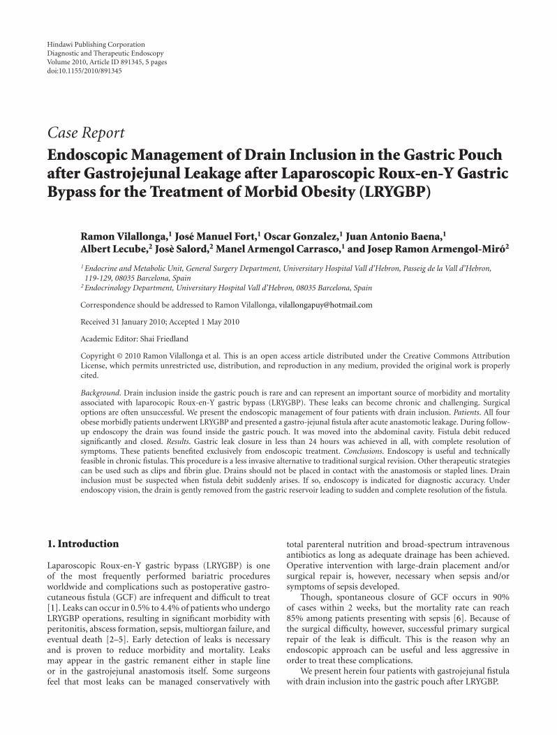
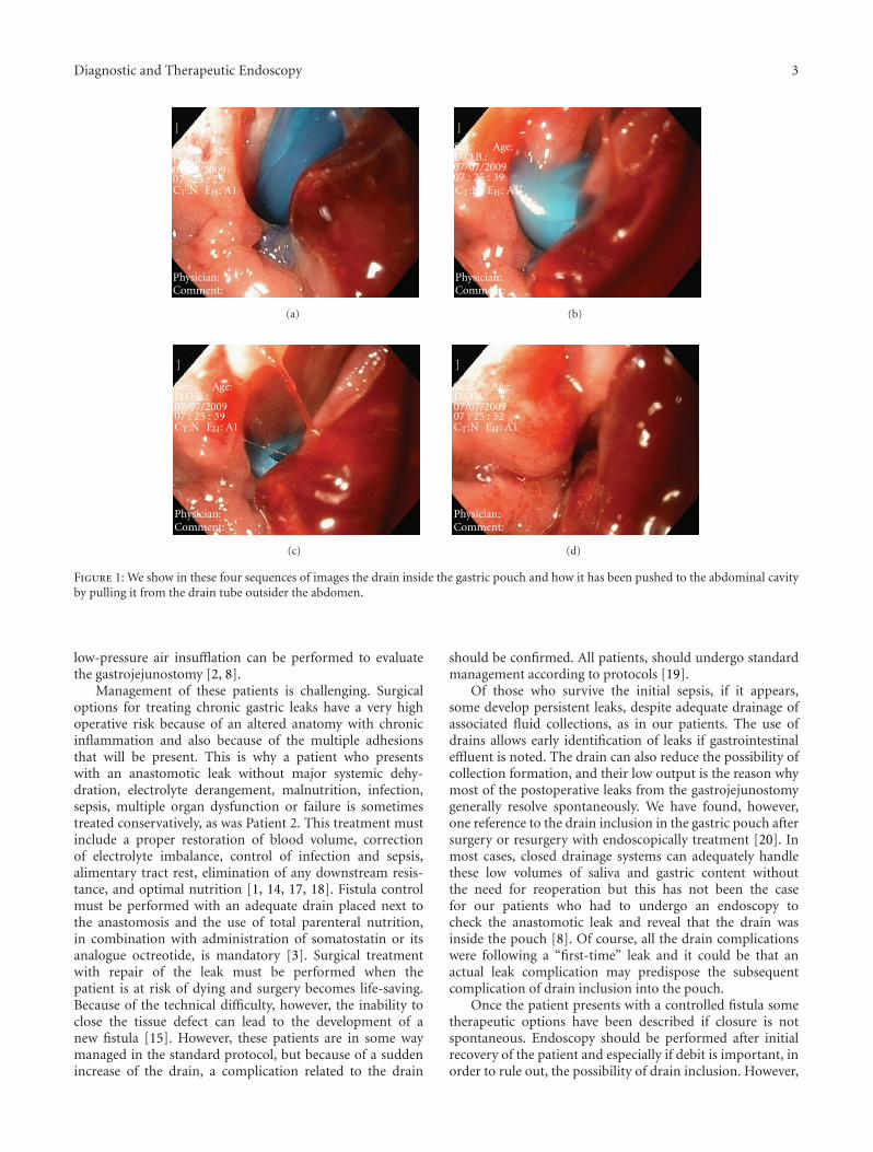
1.4. Patient 4. A 50-year-old woman with BMI = 40 Kg/m2,DM-2 and hypertension underwent an LRYGBP. She pre-sented with an acute digestive haemorrhage and abdominalpain two days after surgery. She required blood and plasmatransfusions and antithrombotic drugs suspension. After 5days of operation, the patient began with suppuration fromthe drain and a methylene blue test confirmed the presenceof a fistula, with a 20 cc/24 hours debit. At the twenty-seventh day after surgery, an endoscopy was performed. Thedrain was also visible inside the gastric pouch (Figure 1).It was removed to the abdominal cavity under endoscopicvision as in the other cases and fistula debit decreasedimmediately. Three days after, a gastrografin (Bristol-MyersSquibb, Princeton, NJ) swallow confirmed the closure of thefistula and the patient began the oral intake. Patient was tendischarged after 32 days.
2. Discussion
Laparoscopic bariatric operations have become standard sur-gical treatment for morbid obesity. LRYGBP is a technicallydemanding operation and technical errors can cause seriouscomplications unless a learning curve is correctly observed[7]. One of the most serious related mortality complicationsis anastomotic dehiscence and leak [2, 3, 8, 9]. Gastricpouch leaks occur in up to 5.6% of LRYGBP operations,resulting in significant morbidity, sepsis, multiorgan failure,and eventual death [2–5, 10, 11].
It is crucial to detect early any kind of leak in orderto reduce morbidity and mortality. Detecting postoperativeleaks can be challenging. Clinical signs such as fever,tachycardia and abdominal pain may alert the clinician.The role of drainage in the early diagnosis of fistula is wellrecognised.
Assessment of peritonitis in these patients can be difficultowing to obesity and few findings from physical examina-tion, and we consider that a CT scan should be performed.The postoperative period is also a problem when somepatients are discharged two or three days after surgery.The postoperative observation period is short and someauthors have showed the convenience of leaving a draineven after patient discharge [3]. Another aspect is the rapidintroduction of the oral intake that may lead to more severeperitonitis, which will increase the morbimortality of thepatient [2]. This last argument is important in the contextof leak detection because, despite aggressive medical and/orsurgical management, some patients will develop ongoingsepsis, multisystem organ failure, and death.
The surgical technique is a factor to take into considera-tion. Some authors have reported the incidence rates of leakin vertical gastroplasty (whether banded [12] or modified[13]), in which disruption of the staple lines (vertical orcircular) occurs in about 2.0% to 4.5% of cases [14, 15].
We routinely perform intraoperative testing, infusingmethylene blue through the orogastric tube under laparo-scopic guidance in order to minimise postoperative leaks.This measure has already been recommended by otherauthors [16]. Additionally, an intraoperative endoscopy with
Diagnostic and Therapeutic Endoscopy 3
Sex: Age:D.O.B.:07/07/200907 : 25 : 25CT:N EH: A1
Physician:Comment:
]
(a)
Sex: Age:D.O.B.:07/07/200907 : 25 : 39CT:N EH: A1
Physician:Comment:
]
(b)
Sex: Age:D.O.B.:07/07/200907 : 25 : 39CT:N EH: A1
Physician:Comment:
]
(c)
Sex: Age:D.O.B.:07/07/200907 : 25 : 52CT:N EH: A1
Physician:Comment:
]
(d)
Figure 1: We show in these four sequences of images the drain inside the gastric pouch and how it has been pushed to the abdominal cavityby pulling it from the drain tube outsider the abdomen.
low-pressure air insufflation can be performed to evaluatethe gastrojejunostomy [2, 8].
Management of these patients is challenging. Surgicaloptions for treating chronic gastric leaks have a very highoperative risk because of an altered anatomy with chronicinflammation and also because of the multiple adhesionsthat will be present. This is why a patient who presentswith an anastomotic leak without major systemic dehy-dration, electrolyte derangement, malnutrition, infection,sepsis, multiple organ dysfunction or failure is sometimestreated conservatively, as was Patient 2. This treatment mustinclude a proper restoration of blood volume, correctionof electrolyte imbalance, control of infection and sepsis,alimentary tract rest, elimination of any downstream resis-tance, and optimal nutrition [1, 14, 17, 18]. Fistula controlmust be performed with an adequate drain placed next tothe anastomosis and the use of total parenteral nutrition,in combination with administration of somatostatin or itsanalogue octreotide, is mandatory [3]. Surgical treatmentwith repair of the leak must be performed when thepatient is at risk of dying and surgery becomes life-saving.Because of the technical difficulty, however, the inability toclose the tissue defect can lead to the development of anew fistula [15]. However, these patients are in some waymanaged in the standard protocol, but because of a suddenincrease of the drain, a complication related to the drain
should be confirmed. All patients, should undergo standardmanagement according to protocols [19].
Of those who survive the initial sepsis, if it appears,some develop persistent leaks, despite adequate drainage ofassociated fluid collections, as in our patients. The use ofdrains allows early identification of leaks if gastrointestinaleffluent is noted. The drain can also reduce the possibility ofcollection formation, and their low output is the reason whymost of the postoperative leaks from the gastrojejunostomygenerally resolve spontaneously. We have found, however,one reference to the drain inclusion in the gastric pouch aftersurgery or resurgery with endoscopically treatment [20]. Inmost cases, closed drainage systems can adequately handlethese low volumes of saliva and gastric content withoutthe need for reoperation but this has not been the casefor our patients who had to undergo an endoscopy tocheck the anastomotic leak and reveal that the drain wasinside the pouch [8]. Of course, all the drain complicationswere following a “first-time” leak and it could be that anactual leak complication may predispose the subsequentcomplication of drain inclusion into the pouch.
Once the patient presents with a controlled fistula sometherapeutic options have been described if closure is notspontaneous. Endoscopy should be performed after initialrecovery of the patient and especially if debit is important, inorder to rule out, the possibility of drain inclusion. However,
4 Diagnostic and Therapeutic Endoscopy
in the fourth patient, daily debit was only of 20 cc. Muchinteresting information concerning the leak can be given.We have not found any report of endoscopic gastric leakrepair after LRYGBP, while there are analogous reports ofoesophageal fistula repair. Both acute oesophageal perfo-rations and chronic oesophageal fistulas have been treatedwith endoclips [20–22]. Another conservative method is theuse of fibrin glue endoscopically placed to close an entericfistula [20, 23–26]. Fibrin tissue glue has been used formore than ten years in different fields of surgery [27–29]and in morbidly obese patients who have undergone verticalgastroplasty with good results [30]. We might consider thepossibility of using low aspirative pressure drains, and theseshould be placed close to the anastomosis. In our fourpatients we did not use any fibrin adhesive because the simplemobilisation of the drain permitted the immediate collapseand closure of the defect. In some cases reported, multipleapplications and relatively large volumes of fibrin adhesiveare required to close the pouch defect [31]. Some authorshave proven the utility of the fibrin glue, which promotesneovascularisation and fibroblast proliferation [31–33].
In a more recent report, Gumbbs treated two patientsof fourteen (14.29%) who underwent vertical gastroplastyand developed a nonhealing gastrocutaneous fistula withendoscopic application of a fibrin sealant under direct vision.This procedure was described as simple, safe, and effectiveand, in some cases, life-saving [34].
In addition to clips and glue, argon plasma coagulationand self-expanding covered metal stents have been used toclose leaks and perforations by acting as a fluid barrier[35]. Some potential complications have been described,such as stent migration and tissue overgrowth. Treatingcomplications like stent removal can also be very challenging.
Even if our patients were treated endoscopically, none ofthe endoscopic techniques described was used. Endoscopyshowed the drain inclusion, however, and allowed us tocontrol the manoeuvre to push the drain into the abdominalcavity. A more common way to manage such drains isto withdraw the drains 3-4 cm on a weekly basis, turningthe leak back to a controlled fistula tract. This may haveavoided the need to push back the drain endoscopically.Clip placement, glueing or luminal stent placement mightbe considered on another occasion. Also some authorshave reviewed the endoscopic management of megaobesepatients with BMI over 70 Kg/m2. In these patients, Suture-line reinforcement, at least selectively in the middle-upperportion of the staple line and in super-super-obese patients,is recommended to decrease the incidence of specific compli-cations such as fistula [36]. Other authors have performed alaparoscopic fistulotomy with debridement [37].
3. Conclusions
Patients who present with a chronic leak must undergo anendoscopy to avoid the possibility of drain inclusion in thegastric pouch. In these cases, endoscopic repair may be asafe and effective option for patients who would otherwiseundergo complex surgical revision.
Acknowledgments
The authors want to acknowledge the Dr. Ramon Vilal-longa Foundation for its financial support. (http://www.fundacioramonvilallonga.org/).
References
[1] S. J. Dudrick, A. R. Maharaj, and A. A. McKelvey, “Artificialnutritional support in patients with gastrointestinal fistulas,”World Journal of Surgery, vol. 23, no. 6, pp. 570–576, 1999.
[2] T. L. Sims, M. A. Mullican, E. C. Hamilton, D. A. Provost,and D. B. Jones, “Routine upper gastrointestinal gastrografin�
swallow after laparoscopic Roux-en-Y gastric bypass,” ObesitySurgery, vol. 13, no. 1, pp. 66–72, 2003.
[3] E. Chousleb, S. Szomstein, D. Podkameni et al., “Routineabdominal drains after laparoscopic Roux-en-Y gastric bypass:a retrospective review of 593 patients,” Obesity Surgery, vol. 14,no. 9, pp. 1203–1207, 2004.
[4] A. C. Wittgrove and G. W. Clark, “Laparoscopic gastric bypass,roux en-Y-500 patients: technique and results, with 3-60month follow-up,” Obesity Surgery, vol. 10, no. 3, pp. 233–239,2000.
[5] T. K. Byrne, “Complications of surgery for obesity,” SurgicalClinics of North America, vol. 81, no. 5, pp. 1181–1193, 2001.
[6] S. T. Papavramidis, E. E. Eleftheriadis, T. S. Papavramidis, K. E.Kotzampassi, and O. G. Gamvros, “Endoscopic managementof gastrocutaneous fistula after bariatric surgery by using afibrin sealant,” Gastrointestinal Endoscopy, vol. 59, no. 2, pp.296–300, 2004.
[7] A. Lecube, J. M. Fort, J. A. Baena, M. Armengol, R. Simo, and J.Mesa, “Inicial experience with 150 consecutive gastric bypass:an approach to the learning curve of bariatric procedures,”Obesity and Metabolism, vol. 3, pp. 175–182, 2007.
[8] F. Serafini, W. Anderson, P. Ghassemi, J. Poklepovic, andM. M. Murr, “The utility of contrast studies and drains inthe management of patients after Roux-en-Y gastric bypass,”Obesity Surgery, vol. 12, no. 1, pp. 34–38, 2002.
[9] P. R. Schauer, S. Ikramuddin, W. Gourash, R. Ramanathan,and J. Luketich, “Outcomes after laparoscopic Roux-en-Ygastric bypass for morbid obesity,” Annals of Surgery, vol. 232,no. 4, pp. 515–529, 2000.
[10] K. D. Higa, K. B. Boone, and T. Ho, “Complications of thelaparoscopic Roux-en-Y gastric bypass: 1,040 patients—whathave we learned?” Obesity Surgery, vol. 10, no. 6, pp. 509–513,2000.
[11] A. Z. Fernandez Jr., E. J. DeMaria, D. S. Tichansky et al.,“Experience with over 3,000 open and laparoscopic bariatricprocedures: multivariate analysis of factors related to leak andresultant mortality,” Surgical Endoscopy, vol. 18, no. 2, pp. 193–197, 2004.
[12] E. E. Mason, “Vertical banded gastroplasty for obesity,”Archives of Surgery, vol. 117, no. 5, pp. 701–706, 1982.
[13] A. P. Aidonopoulos and S. T. Papavramidis, “Vertical gas-troplasty with artificial pseudopylorus: a modification in thetreatment of morbid obesity: early results,” Obesity Surgery,vol. 4, no. 4, pp. 349–352, 1994.
[14] E. R. T. C. Owen, R. Abraham, and A. E. Kark, “Gastroplastyfor morbid obesity: technique, complications and results in 60cases,” British Journal of Surgery, vol. 76, no. 2, pp. 131–135,1989.

Diagnostic and Therapeutic Endoscopy 5
[15] M. M. Meguid and A. C. L. Campos, “Nutritional manage-ment of patients with gastrointestinal fistulas,” Surgical Clinicsof North America, vol. 76, no. 5, pp. 1035–1080, 1996.
[16] D. C. Amarasinghe, “Air test as an alternative to methyleneblue test for leaks,” Obesity Surgery, vol. 12, no. 2, pp. 295–296,2002.
[17] D. Rose, M. F. Yarborough, P. C. Canizaro, and S. F. Lowry,“One hundred and fourteen fistulas of the gastrointestinaltract treated with total parenteral nutrition,” Surgery Gynecol-ogy and Obstetrics, vol. 163, no. 4, pp. 345–350, 1986.
[18] S. T. Papavramidis and C. Milias, “Complications after verticalgastroplasty with artificial pseudopylorus in the treatment ofmorbid obesity: a 7-year experience,” Obesity Surgery, vol. 9,no. 6, pp. 535–538, 1999.
[19] T. S. Papavramidis, K. Kotzampassi, E. Kotidis, E. E. Eleft-heriadis, and S. T. Papavramidis, “Endoscopic fibrin seal-ing of gastrocutaneous fistulas after sleeve gastrectomy andbiliopancreatic diversion with duodenal switch,” Journal ofGastroenterology and Hepatology, vol. 23, no. 12, pp. 1802–1805, 2008.
[20] M. Garcia-Caballero, M. Carbajo, J. M. Martınez-Moreno,M. Sarria, D. Osorio, and J.-A. Carmona, “Drain erosionand gastro-jejunal fistula after one-anastomosis gastric bypass:endoscopic occlusion by fibrin sealant,” Obesity Surgery, vol.15, no. 5, pp. 719–722, 2005.
[21] G. S. Raymer, A. Sadana, D. B. Campbell, and W. A. Rowe,“Endoscopic clip application as an adjunct to closure ofmature esophageal perforation with fistulae,” Clinical Gas-troenterology and Hepatology, vol. 1, no. 1, pp. 44–50, 2003.
[22] L. Cipolletta, M. A. Bianco, G. Rotondano, R. Marmo, R.Piscopo, and C. Meucci, “Endoscopic clipping of perforationfollowing pneumatic dilation of esophagojejunal anastomoticstrictures,” Endoscopy, vol. 32, no. 9, pp. 720–722, 2000.
[23] L. R. Rabago, N. Ventosa, J. L. Castro, J. Marco, N. Herrera,and F. Gea, “Endoscopic treatment of postoperative fistulasresistant to conservative management using biological fibringlue,” Endoscopy, vol. 34, no. 8, pp. 632–638, 2002.
[24] B. Petersen, A. Barkun, S. Carpenter et al., “Tissue adhesivesand fibrin glues: November 2003,” Gastrointestinal Endoscopy,vol. 60, no. 3, pp. 327–333, 2004.
[25] F. Maluf-Filho, E. Moura, P. Sakai, et al., “Endoscopictreatment of esophagogastric fistulae with an acellular matrix,”Gastrointestinal Endoscopy, vol. 59, no. 5, p. 151, 2004.
[26] E. Scappaticci, F. Ardissone, S. Baldi et al., “Closure ofan iatrogenic tracheo-esophageal fistula with bronchoscopicgluing in a mechanically ventilated adult patient,” Annals ofThoracic Surgery, vol. 77, no. 1, pp. 328–329, 2004.
[27] M. Jung, B. C. Manegold, and W. Brands, “Endoscopictherapy of gastrointestinal fistulae with fibrin tissue sealant,”in Progress in Fibrin Sealing, H. W. Waclawiczek, Ed., pp. 43–52, Springer, Berlin, Germany, 1989.
[28] T. Kurokawa, S. Okushiba, M. Kadoya et al., “Selectiveocclusion with fibrin glue under fistuloscopy: seven cases ofpostoperative management for intractable complex fistulas,”Endoscopy, vol. 34, no. 3, pp. 220–222, 2002.
[29] N. N. T. Lomis, F. J. Miller, T. J. Loftus, J. H. Whiting, A. W.Giuliano, and H.-C. Yoon, “Refractory abdominal-cutaneousfistulas or leaks: percutaneous management with a collagenplug,” Journal of the American College of Surgeons, vol. 190, no.5, pp. 588–592, 2000.
[30] S. T. Papavramidis, E. E. Eleftheriadis, D. N. Apostolidis, andK. E. Kotzampassi, “Endoscopic fibrin sealing of high-outputnon-healing gastrocutaneous fistulas after vertical gastroplasty
in morbidly obese patients,” Obesity Surgery, vol. 11, no. 6, pp.766–769, 2001.
[31] E. Eleftheriadis, E. Tzartinoglou, K. Kotzampassi, and H. Ale-tras, “Early endoscopic fibrin sealing of high-output postoper-ative enterocutaneous fistulas,” Acta Chirurgica Scandinavica,vol. 156, no. 9, pp. 625–628, 1990.
[32] M. A. Chung and H. J. Wanebo, “Surgical management andtreatment of gastric and duodenal fistulas,” Surgical Clinics ofNorth America, vol. 76, no. 5, pp. 1137–1146, 1996.
[33] H. Groitl and J. Scheele, “Initial experience with the endo-scopic application of fibrin tissue adhesive in the uppergastrointestinal tract,” Surgical Endoscopy, vol. 1, no. 2, pp. 93–97, 1987.
[34] A. A. Gumbs, A. J. Duffy, and R. L. Bell, “Managementof gastrogastric fistula after laparoscopic Roux-en-Y gastricbypass,” Surgery for Obesity and Related Diseases, vol. 2, no.2, pp. 117–121, 2006.
[35] B. F. Merrifield, D. Lautz, and C. C. Thompson, “Endoscopicrepair of gastric leaks after Roux-en-Y gastric bypass: a lessinvasive approach,” Gastrointestinal Endoscopy, vol. 63, no. 4,pp. 710–714, 2006.
[36] T. Papavramidis, I. Kesisoglou, V. Orailoglou, V. Baltzopoulos,V. Grosomanidis, and S. Papavramidis, “Mega-obese patientsweighing more than 250 kg: a problematic group,” ActaChirurgica Belgica, vol. 109, no. 1, pp. 61–64, 2009.
[37] O. N. Tucker, S. Szomstein, and R. Rosenthal, “Laparoscopicmanagement of chronic pouch fistula after a leak followingstaple line dehiscence after laparoscopic revision of a dilatedpouch following Roux-en-Y gastric bypass,” Obesity Surgery,vol. 18, no. 2, pp. 228–232, 2008.

Submit your manuscripts athttp://www.hindawi.com
Stem CellsInternational
Hindawi Publishing Corporationhttp://www.hindawi.com Volume 2014
Hindawi Publishing Corporationhttp://www.hindawi.com Volume 2014
MEDIATORSINFLAMMATION
of
Hindawi Publishing Corporationhttp://www.hindawi.com Volume 2014
Behavioural Neurology
EndocrinologyInternational Journal of
Hindawi Publishing Corporationhttp://www.hindawi.com Volume 2014
Hindawi Publishing Corporationhttp://www.hindawi.com Volume 2014
Disease Markers
Hindawi Publishing Corporationhttp://www.hindawi.com Volume 2014
BioMed Research International
OncologyJournal of
Hindawi Publishing Corporationhttp://www.hindawi.com Volume 2014
Hindawi Publishing Corporationhttp://www.hindawi.com Volume 2014
Oxidative Medicine and Cellular Longevity
Hindawi Publishing Corporationhttp://www.hindawi.com Volume 2014
PPAR Research
The Scientific World JournalHindawi Publishing Corporation http://www.hindawi.com Volume 2014
Immunology ResearchHindawi Publishing Corporationhttp://www.hindawi.com Volume 2014
Journal of
ObesityJournal of
Hindawi Publishing Corporationhttp://www.hindawi.com Volume 2014
Hindawi Publishing Corporationhttp://www.hindawi.com Volume 2014
Computational and Mathematical Methods in Medicine
OphthalmologyJournal of
Hindawi Publishing Corporationhttp://www.hindawi.com Volume 2014
Diabetes ResearchJournal of
Hindawi Publishing Corporationhttp://www.hindawi.com Volume 2014
Hindawi Publishing Corporationhttp://www.hindawi.com Volume 2014
Research and TreatmentAIDS
Hindawi Publishing Corporationhttp://www.hindawi.com Volume 2014
Gastroenterology Research and Practice
Hindawi Publishing Corporationhttp://www.hindawi.com Volume 2014
Parkinson’s Disease
Evidence-Based Complementary and Alternative Medicine
Volume 2014Hindawi Publishing Corporationhttp://www.hindawi.com