Endoscopic cap resection for treatment of early Barrett's neoplasia is safe: a prospective analysis...
219
Downloaded from UvA-DARE, the Institutional Repository of the University of Amsterdam (UvA) http://dare.uva.nl/document/45314 File ID 45314 Filename Peters__F.P._-def.pdf SOURCE, OR PART OF THE FOLLOWING SOURCE: Type Dissertation Title Endoscopic treatment of early Barrett's neoplasia Author F.P. Peters Faculty Faculty of Medicine Year 2007 Pages 217 FULL BIBLIOGRAPHIC DETAILS: http://dare.uva.nl/record/217693 Copyrights It is not permitted to download or to forward/distribute the text or part of it without the consent of the copyright holder (usually the author), other then for strictly personal, individual use. UvA-DARE is a service provided by the Library of the University of Amsterdam (http://dare.uva.nl)
-
Upload
independent -
Category
Documents
-
view
0 -
download
0
Transcript of Endoscopic cap resection for treatment of early Barrett's neoplasia is safe: a prospective analysis...

Downloaded from UvA-DARE, the Institutional Repository of the University of Amsterdam (UvA)http://dare.uva.nl/document/45314
File ID 45314Filename Peters__F.P._-def.pdf
SOURCE, OR PART OF THE FOLLOWING SOURCE:Type DissertationTitle Endoscopic treatment of early Barrett's neoplasiaAuthor F.P. PetersFaculty Faculty of MedicineYear 2007Pages 217
FULL BIBLIOGRAPHIC DETAILS: http://dare.uva.nl/record/217693
Copyrights It is not permitted to download or to forward/distribute the text or part of it without the consent of the copyright holder(usually the author), other then for strictly personal, individual use. UvA-DARE is a service provided by the Library of the University of Amsterdam (http://dare.uva.nl)

Endoscopic treatment of early Barrett’s neoplasia

The studies described in this thesis have been supported by an unrestricted research grant of AstraZeneca Netherlands.
The printing of this thesis was financially supported by:
Endoscopic treatment of early Barrett’s neoplasia. By Femke P. Peters. Thesis University of Amsterdam, The Netherlands
Paranimfen: Wilda D. Rosmolen, Wouter L. Curvers
© Copyright - F.P. Peters, Amsterdam, 2007. The copyright of articles that have been already published or accepted for publication, have been transferred to the respective journals and /or organizations.
Cover: Lara Laghetto, VisualMedicsLay out: Chris Bor, Medical Photography and Illustration, Academic Medical Center, AmsterdamPrinted by: Buijten & Schipperheijn, Amsterdam
ISBN-10: 90-9021538-7ISBN-13: 978-90-9021538-9

Endoscopic treatment of early Barrett’s neoplasia
ACADEMISCH PROEFSCHRIFT
ter verkrijging van de graad van doctor aan de Universiteit van Amsterdam op gezag van de Rector Magnificus
prof.dr. J.W. Zwemmer ten overstaan van een door het college voor promoties ingestelde
commissie, in het openbaar te verdedigen in de Aula der Universiteit op donderdag 15 maart 2007, te 12.00 uur
door
Femke Petra Peters
geboren te Zoetermeer

Promotiecommissie: Promotores: Prof.dr. P. Fockens Prof.dr. F.J.W. ten KateCo-promotores: Dr. J.J.G.H.M. Bergman Dr. K.K. Krishnadath
Overige leden: Prof.dr. J.J.B. van Lanschot Prof.dr. G.N.J. Tytgat Dr. P.D. Siersema Dr.ir. P. Breedveld Prof.dr. H. Neuhaus Dr. M. Vieth Prof.dr. J. Stoker
Faculteit der Geneeskunde

Voor mijn ouders


ContentsIntroduction and outline of the thesis
Chapter 1: Endoscopic treatment of early Barrett’s neoplasia: a technical review
Educational syllabus for the Dutch EMR training program 2007
Chapter 2: Endoscopic cap-resection for treatment of early Barrett’s neoplasia is safe. A prospective analysis of acute and early complications in 216 procedures
Submitted
Chapter 3: Multi-Band Mucosectomy for endoscopic resection of Barrett’s esophagus. A prospective series with matched historical controls.
Accepted for publication Eur J Gastroenterol Hepatol
Chapter 4: Histological evaluation of resection specimens obtained at 293 endoscopic resections in Barrett’s esophagus
Submitted
Chapter 5: Surveillance history of endoscopically treated patients with early Barrett’s neoplasia: non-adherence to the Seattle biopsy protocol leads to sampling error.
Submitted
Chapter 6: Endoscopic treatment of high-grade dysplasia and early stage cancer in Barrett’s esophagus.
Gastrointestinal endoscopy 2005;61(4):506-14
Chapter 7: Poor results with 5-ALA-PDT for residual high-grade dysplasia and early cancer in Barrett esophagus.
Endoscopy 2005;37(5):418-24
Chapter 8: Stepwise Radical Endoscopic Resection is safe and effective for complete removal of Barrett’s esophagus with early neoplasia.
American Journal of Gastroenterology 2006; 101:1449-1457
Chapter 9: Stepwise radical endoscopic resection of the complete Barrett’s esophagus with early neoplasia successfully eradicates pre-existing genetic abnormalities.
Submitted
SummaryNederlandse samenvatting / Dutch summaryList of contributing authorsDankwoord
9
13
99
111
121
131
139
155
169
186
199205209213


Introduction and outline of the thesis

10

IntroductionOver the last years, the endoscopic treatment of early Barrett’s neoplasia has evolved rapidly. Its efficacy as curative treatment for high-grade dysplasia and mucosal cancer has been shown and in the Netherlands this treatment regimen is now considered to be the treatment of choice in selected patients. New endoscopic treatment techniques are being developed and existing treatment protocols are constantly optimized. The aim of this thesis was to evaluate the technical and clinical efficacy of these different endoscopic treatment techniques and protocols.
The studies included in this thesis represent the different stages of endoscopic treatment of early Barrett’s neoplasia in Amsterdam over the last five years; starting with monotherapy of focal neoplastic lesions with endoscopic resection; followed by endoscopic resection of focal lesions complemented by endoscopic ablation of the residual Barrett’s segment for prevention of recurrences, and subsequently by endoscopic resection of the complete Barrett’s segment with early neoplasia for improved long term remission rates. Each of these different stages has contributed to an increasing knowledge of how to improve the efficacy and safety of the endoscopic approach in these patients and led to subsequently new treatment techniques and protocols. In addition, they have provided insight into which patients with early Barrett’s neoplasia are eligible for endoscopic treatment and which patients are not.Endoscopic treatment of early Barrett’s neoplasia is here to stay, but there is a need to continue the search for even better, safer and easier treatment protocols than already available and to improve patient selection.
Clarification of used nomenclatureThe studies presented in this thesis have been performed over a number of years and have been published in different journals. This has led to the use of a number of different terms for identical entities. For clarity, the most important ones are described here.The terms “Barrett’s esophagus” and “Barrett esophagus” are both used to describe histologically confirmed columnar-lined distal esophagus with intestinal metaplasia. In the earlier studies presented in this thesis the term “dysplasia” is used, where in the later chapters the term “intraepithelial neoplasia” is introduced, in accordance to the revised Vienna Classification. Therefore, the term “high-grade dysplasia” in this thesis is identical to the term “high-grade intraepithelial neoplasia”. “Early Barrett’s neoplasia” are defined as lesions in Barrett’s mucosa that contain HGIN, cancer with only mucosal infiltration (T1m) or cancer with superficial submucosal infiltration (T1sm1; i.e. infiltration into the upper third of the submucosal layer or submucosal infiltration of less than 500 µm). In this thesis, the term “endoscopic resection (ER)” is used, rather than the frequently used term “endoscopic mucosal resection (EMR)”. We prefer to use this term, since not only the mucosa, but also the submucosa, is removed during an endoscopic resection.
Introduction and outline of the thesis
11

Outline of the thesisThe thesis starts with an extensive review of endoscopic treatment of early Barrett’s neoplasia (Chapter 1). The endoscopic work-up and patient selection of patients with early Barrett’s neoplasia for endoscopic treatment is described and recommendations are made. Subsequently, the different endoscopic treatment techniques are described, with special emphasis on endoscopic resection. Technical and practical aspects are discussed, an overview of clinical results is presented, and practical recommendations for endoscopic treatment are made. This review serves as a educational syllabus for the Dutch EMR training program, in which gastroenterologists are trained for endoscopic treatment of early upper GI neoplasia.
In Chapters 2 and 3, technical aspects of the two most frequently used endoscopic resection techniques, endoscopic cap resection and multi-band-mucosectomy, are studied and described.
Chapter 4 describes a study evaluating the histopathological characteristics of endoscopic resection specimens. The histopathological characteristics of endoscopically resected early Barrett’s neoplasia are correlated with the endoscopic characteristics and the histological diagnosis of pre-treatment biopsies in order to make recommendations relevant for patient management.
In Chapter 5 the surveillance history of endoscopically treated patients with early Barrett’s neoplasia is described.
Clinical results achieved with a number of endoscopic treatment protocols are described in Chapters 6,7 and 8. In Chapter 6 the results of endoscopic treatment with either endoscopic resection alone or a combination of endoscopic resection and photodynamic therapy (PDT) of the first 28 patients treated at our institution are evaluated. The contribution of PDT to the success of endoscopic treatment is evaluated in Chapter 7. In Chapter 8 stepwise radical endoscopic resection of the complete Barrett’s segment as an alternative treatment protocol with potentially better long term success, is evaluated for the treatment of Barrett’s esophagus with early neoplasia.
The efficacy of stepwise radical endoscopic resection in the eradication of genetic abnormalities that underlie the malignant potential of Barrett’s mucosa is described in Chapter 9.
12

Educational syllabus for the Dutch EMR training program 2007
C h a p t e r1Endoscopic treatment of early Barrett’s neoplasia: a technical review
F.P. Peters
J.J.G.H.M. Bergman


IntroductionAfter the first reports on columnar lined esophagus in 1950 by Norman Barrett (1), the definition of this condition has been adjusted several times. The definition that is currently in use is that the Barrett’s esophagus is a condition in which the normal squamous epithelium of the distal esophagus is replaced with columnar epithelium containing intestinal metaplasia (2-4). It is thought to be a result of longstanding severe gastro-esophageal reflux disease (GERD), and the typical patient is a middle-aged Caucasian male. The prevalence of Barrett’s esophagus is estimated at 10% of patients with GERD (5) and a recent large sized population based Swedish study showed a population prevalence of 1.6% (6). The importance of Barrett’s esophagus is the premalignant character of the condition (7-9). Patients with a Barrett’s esophagus have an increased risk for developing esophageal adenocarcinoma compared to the general population. The incidence of esophageal adenocarcinoma has increased four to six fold over the last decades (10-16). The incidence of esophageal adenocarcinoma in Barrett’s patients differs between published studies, with lower incidences found in the larger sized studies, suggesting a publication bias. The annual incidence is now estimated at approximately 0.5% (17-19). Most esophageal adenocarcinomas are diagnosed when patients present with symptoms, mostly dysphagia, and the disease then usually is at an advanced stage. The prognosis
Table 1 Mortality and morbidity rates of surgical esophagectomy for HGIN and/or early adenocarcinoma
Author Nr patients Type of patients Mortality*,** Morbidity#
Rice (353) 16 HGIN 0% 44%
Bonavina (354) 253 T1 9.1%
Ferguson (355) 15 HGIN 0% 73%
Hölscher (25) 41 T1 2.4% 43.9%
Nigro (23) 45 HGIN-T1m 3%
van Sandick (83) 32 T1 0% 41%
Stein (82) 94 T1 4.2% 43.7%
Zaninotto (356) 15 HGIN 0% 53%
Fujita (357) 35 T1m 14% 69%
78 T1sm 5% 88%
Rice (81) 122 T1 2.5% 47.5%
van Lanschot (358) 757 T1 4.9%
Fernando (359) 28 HGIN 3.6% 54%
Headrick (360) 54 HGIN 1.8% 57%
Tseng (361) 60 HGIN 1.7% 29%
Reed (362) 49 HGIN 2%
Sujendran (363) 17 HGIN 0%
Westerterp (79) 702 HGIN-T1 4%
Total 2413 Weighted mean## 4.6% 52.9%
95% CI 3.8% - 5.4% 52.8% - 53.0%
* In hospital or 30-day mortality rates: ** Only mortality rates of high volume centers included when specified. # Only overall morbidity rates included in this calculation. ## Weighted for number of included patients. Only data from high volume centers included if available.Yrs.: years, Nr.: number, CI: confidence interval.
Endoscopic treatment of early Barrett’s neoplasia
15
1c h a p t e r

of advanced esophageal adenocarcinoma is dismal; even after curative surgery the 5-year survival rates are approximately 20% (20-22). The prognosis is much better when patients are treated at an early stage, with 5 year survival rates of 83-100% after surgical esophagectomy for mucosal or superficial submucosal neoplasia (23-25).The development of esophageal adenocarcinoma in Barrett’s esophagus is a multi-step process from intestinal metaplasia through different grades of dysplasia (intraepithelial neoplasia) (2;26;27) resulting from accumulation of genetic abnormalities (28). The presence of the premalignant Barrett’s esophagus, together with this multi-step process enables the detection of esophageal adenocarcinoma before it reaches an advanced, incurable stage. This is the reason that regular endoscopic surveillance is advised for patients with a known Barrett’s esophagus (29), http://www.bsg.org.uk). This endoscopic surveillance is aimed at detecting early neoplastic changes in the Barrett’s segment, such as low-grade and high-grade intraepithelial neoplasia (LGIN and HGIN) or early cancer (i.e., mucosal or superficial submucosal lesions) with a more favorable prognosis (5;30;31). With the implementation of endoscopic surveillance, more and more early Barrett’s neoplasia are detected and treated (32), although a significant percentage (appr. 40%) of patients with early Barertt’s neoplasia is not detected by surveillance but as a diagnosis by chance during an endoscopy for a different indication.The standard treatment of early Barrett’s neoplasia, including HGIN and early cancer, used to be surgical esophagectomy. This is an invasive treatment modality with significant morbidity and mortality rates (Table 1) that temporarily reduces quality of life (33;34). In case of mucosal neoplasia (HGIN, T1m) the mean mortality rate of
Table 2a Lymph node involvement in surgical resection specimens of patients with early cancer in Barrett’s esophagus.
Author Nr. patients Type patients Surgical technique Nr resected LNN Pos LNN in HGIN Pos Lnn in T1m Pos LNN in T1sm
Rice (353) 16 Pre-operative HGIN TTECR (n=7), THECR (n=9) 0/10 0/6
Peters (364) 17 Pre-operative HGIN/T1 TTECR (n=2), THECR (n=15) 0/4 0/9 1/2
Ferguson (355) 15 Pre-operative HGIN/T1 TTECR (n=9), THECR (n=6) 0/4 0/3 0/8
Holscher (25) 41 Post-operative pT1 TTECR en-bloc (n=3), THECR (n=38) 23 (mean) 0/10 5/31
Nigro (23) 37 Post-operative pT1/2 TTECR en-bloc, 2 field, colon interposition (n=37) 41 (median) 1/15 6/12
Nigro (365) 28 Post-operative pT1/2 TTECR, 2 FD, less extensive (n=28) 14 (median) 0/13 1/6
van Sandick (83) 32 Post-operative pT1 TTECR (n=8), THECR (n=24) 13 (mean) 0/12 6/20
Stein (82) 94 Post-operative pT1 TTECR (n=11), THECR (n=60), limited resection (n=23) 21 and 19 (median) 0/38 10/56
Hagen (366) 100 all T1 TTECR en-bloc (n=100) 48 (median) 1/16 5/16
Rice (81) 122 Post-operative pT1 TTECR (n=47), THECR (n=75) 0/38 2/53 6/31
Scotiniotis (367) 22 Pre-operative HGIN/T1 TTECR (n=8), THECR (n=14) 10.5 (mean) 0/14 0/2 0/1
Fernando (359) 28 Pre-operative HGIN Minimal invasive esophagectomy 16 (mean) 0/17 0/6 1/5
Stein (368) 157 all T1 TTECR (n=36), THECR (n=121) 24 (median) 0/13 0/57 18/87
Liu (78) 90 Post-operative pT1 TTECR with 3 FD (n=4), TTECR (n=36), THECR (n=50) 13 (mean) 2/53 10/38
Westerterp (79) 120 Post-operative pT1 THECR (n=120) 8.6 (mean) 1/54 18/66
Bollschweiler (80) 36 Post-operative pT1 TTECR (n=29), THECR (n=7) 0/14 9/22
Total 0/100 (0%) 7/361 (1.9%) 96/401 (23.9%)
95% CI 0.5%- 3.3% 19.7%-28.1%
Nr.: number, LNN: lymph nodes, Pos: positive, HGIN: high-grade intra-epithelial neoplasia, TTECR:n trans-thoracic esophagus-cardia resection, THECR: trans-hiatal esophagus-cardia resection, FD: field lymph node dissection, CI: confidence interval.
16

esophagectomy with an average of 4.6% (Table 1) is higher than the mean risk of lymph node involvement of 0 and 1.9% (Table 2a). The low risk of lymph node involvement for mucosal Barrett’s neoplasia compared to that of mucosal squamous cell carcinomas, may be explained by the histological architecture of Barrett’s esophagus that resembles more the architecture of the stomach with a thicker mucosa and blood- and lymph vessels situated relatively deep in the submucosa. In addition, Barrett’s esophagus often has a double muscularis mucosae increasing the thickness of the Barrett’s mucosa (35). Feith et al. have suggested that the risk of lymph node metastasis in Barrett’s neoplasia is reduced due to the ongoing reflux associated inflammation, which may occlude the submucosal lymph-vessels (36), but proof for this hypothesis is lacking. An important implication of this low risk of lymph node involvement in early Barrett’s neoplasia is that endoscopic therapy of the primary lesion may cure these patients without the need for esophagectomy. A variety of endoscopic techniques is now available for treatment of early Barrett’s neoplasia. Endoscopic resection is considered by many to be the cornerstone of endoscopic treatment, since it provides a specimen for histopathological evaluation of the resected lesion.This review will discuss which patients with early Barrett’s neoplasia are eligible for endoscopic treatment, how patient selection can be performed adequately, and what different endoscopic treatment techniques are available with technical guidelines for performing endoscopic resection.
Table 2a Lymph node involvement in surgical resection specimens of patients with early cancer in Barrett’s esophagus.
Author Nr. patients Type patients Surgical technique Nr resected LNN Pos LNN in HGIN Pos Lnn in T1m Pos LNN in T1sm
Rice (353) 16 Pre-operative HGIN TTECR (n=7), THECR (n=9) 0/10 0/6
Peters (364) 17 Pre-operative HGIN/T1 TTECR (n=2), THECR (n=15) 0/4 0/9 1/2
Ferguson (355) 15 Pre-operative HGIN/T1 TTECR (n=9), THECR (n=6) 0/4 0/3 0/8
Holscher (25) 41 Post-operative pT1 TTECR en-bloc (n=3), THECR (n=38) 23 (mean) 0/10 5/31
Nigro (23) 37 Post-operative pT1/2 TTECR en-bloc, 2 field, colon interposition (n=37) 41 (median) 1/15 6/12
Nigro (365) 28 Post-operative pT1/2 TTECR, 2 FD, less extensive (n=28) 14 (median) 0/13 1/6
van Sandick (83) 32 Post-operative pT1 TTECR (n=8), THECR (n=24) 13 (mean) 0/12 6/20
Stein (82) 94 Post-operative pT1 TTECR (n=11), THECR (n=60), limited resection (n=23) 21 and 19 (median) 0/38 10/56
Hagen (366) 100 all T1 TTECR en-bloc (n=100) 48 (median) 1/16 5/16
Rice (81) 122 Post-operative pT1 TTECR (n=47), THECR (n=75) 0/38 2/53 6/31
Scotiniotis (367) 22 Pre-operative HGIN/T1 TTECR (n=8), THECR (n=14) 10.5 (mean) 0/14 0/2 0/1
Fernando (359) 28 Pre-operative HGIN Minimal invasive esophagectomy 16 (mean) 0/17 0/6 1/5
Stein (368) 157 all T1 TTECR (n=36), THECR (n=121) 24 (median) 0/13 0/57 18/87
Liu (78) 90 Post-operative pT1 TTECR with 3 FD (n=4), TTECR (n=36), THECR (n=50) 13 (mean) 2/53 10/38
Westerterp (79) 120 Post-operative pT1 THECR (n=120) 8.6 (mean) 1/54 18/66
Bollschweiler (80) 36 Post-operative pT1 TTECR (n=29), THECR (n=7) 0/14 9/22
Total 0/100 (0%) 7/361 (1.9%) 96/401 (23.9%)
95% CI 0.5%- 3.3% 19.7%-28.1%
Nr.: number, LNN: lymph nodes, Pos: positive, HGIN: high-grade intra-epithelial neoplasia, TTECR:n trans-thoracic esophagus-cardia resection, THECR: trans-hiatal esophagus-cardia resection, FD: field lymph node dissection, CI: confidence interval.
Endoscopic treatment of early Barrett’s neoplasia
17
1c h a p t e r

Patient selectionFor successful endoscopic treatment of early Barrett’s neoplasia adequate patient selection is imperative. Only patients with disease confined to the superficial esophageal wall layers (mucosa and superficial submucosa) can be cured with endoscopic treatment. Patient selection is, therefore, aimed at identifying patients without metastases and with a negligible or low risk of lymph node involvement. In order to identify eligible patients, the following endoscopic work-up and staging procedures are advised.
Endoscopic work-upDuring endoscopic work-up, the esophagus of patients with (possible) early Barrett’s neoplasia is mapped. The work-up is aimed at detecting all neoplastic lesions in the Barrett’s segment and identifying the most advanced lesion which is most important in determining the management strategy. Basic guidelines for optimal endoscopic imaging of Barrett’s esophagus encompass three variables: the quality of the video-endoscope, the experience of the endoscopist and a systematic endoscopic approach. Recently, endoscopes have become available that have charge-coupled devices (CCDs) with 600,000 to 1 million pixels, instead of the standard 100,000 to 300,000. These high resolution endoscopes provide a high-quality white light image in overview which improves the detection of abnormal areas compared to standard video-endoscopes (37). Most experts now agree that high resolution endoscopy is preferred for the endoscopic inspection of Barrett’s esophagus (38). Early Barrett’s neoplasia are usually flat and associated with only subtle mucosal abnormalities. These abnormalities are often visible with high resolution endoscopy, but are frequently overlooked since they are not recognized as such; the endoscopist does not necessarily diagnose what is SEEN, but only diagnoses what is RECOGNIZED as being abnormal.Early Barrett’s neoplasia is relatively rare which causes most endoscopists to have little experience in their recognition. Knowledge of the endoscopic appearance of early Barrett’s neoplasia is, however, essential for its diagnosis. This knowledge may be improved by training videos and DVDs which are now available.
Gross mucosal lesions such as elevations, ulcerations, and nodularity may be detected easily in overview. Careful and thorough inspection following a systematic approach is, however, necessary for detecting the more frequent subtle lesions. After first inspection in overview of the inflated esophagus, gradual deflation should be performed to reveal irregularities that had been stretched out by the inflation (Figure 1). Special attention should be paid to the area between 12 and 6 o’clock in the endoscopic view, since the majority of early neoplastic lesions are localized there. Subsequent retrograde inspection of the most distal part of the Barrett’s esophagus and the hiatal hernia may allow detection of abnormalities that are overlooked in the antegrade view (Figure 2). Following the inspection of the Barrett’s segment and classification of visible lesions, biopsies should be obtained from all visible lesions followed by random four-quadrant biopsies for every two centimeters of the Barrett’s segment, always starting distally working upwards. It has been calculated that a 2 cm Barrett’s segment has a surface area
18
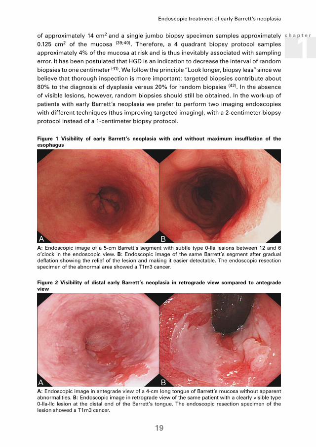
of approximately 14 cm2 and a single jumbo biopsy specimen samples approximately 0.125 cm2 of the mucosa (39;40). Therefore, a 4 quadrant biopsy protocol samples approximately 4% of the mucosa at risk and is thus inevitably associated with sampling error. It has been postulated that HGD is an indication to decrease the interval of random biopsies to one centimeter (41). We follow the principle “Look longer, biopsy less” since we believe that thorough inspection is more important: targeted biopsies contribute about 80% to the diagnosis of dysplasia versus 20% for random biopsies (42). In the absence of visible lesions, however, random biopsies should still be obtained. In the work-up of patients with early Barrett’s neoplasia we prefer to perform two imaging endoscopies with different techniques (thus improving targeted imaging), with a 2-centimeter biopsy protocol instead of a 1-centimeter biopsy protocol.
A: Endoscopic image in antegrade view of a 4-cm long tongue of Barrett’s mucosa without apparent abnormalities. B: Endoscopic image in retrograde view of the same patient with a clearly visible type 0-IIa-IIc lesion at the distal end of the Barrett’s tongue. The endoscopic resection specimen of the lesion showed a T1m3 cancer.
Figure 2 Visibility of distal early Barrett’s neoplasia in retrograde view compared to antegrade view
A: Endoscopic image of a 5-cm Barrett’s segment with subtle type 0-IIa lesions between 12 and 6 o’clock in the endoscopic view. B: Endoscopic image of the same Barrett’s segment after gradual deflation showing the relief of the lesion and making it easier detectable. The endoscopic resection specimen of the abnormal area showed a T1m3 cancer.
Figure 1 Visibility of early Barrett’s neoplasia with and without maximum insufflation of the esophagus
Endoscopic treatment of early Barrett’s neoplasia
19
1c h a p t e r

Macroscopic aspect early Barrett’s neoplasiaMacroscopically visible lesions are classified according to the Paris classification (43;44), adopted from the Japanese Gastric Cancer Association (45). In this classification type 0 is used for superficial lesions and is divided into three categories (Figure 3): 0-I for protruding or polypoid lesions, 0-II for non-protruding and non-excavated lesions (i.e. flat-lesions) and 0-III for excavated or ulcerated lesions. Category I can be subdivided in pedunculated (0-Ip) and sessile (0-Is) lesions. Type 0-II has three subtypes: slightly elevated (0-IIa), completely flat (0-IIb) and slightly depressed (0-IIc). The three subtypes can be present in all combinations, e.g. 0-IIa+IIc or 0-IIa+IIb, with the first mentioned being the most predominant type. Approximately three quarters to 85% of early Barrett’s neoplasia are of the 0-II type, with types 0-IIc and 0-III being relatively rare (43;46;47). The macroscopic classification correlates with the infiltration depth of the lesion and helps to predict the risk of submucosal invasion and thus the risk of lymph node involvement (see below). In the squamous esophagus and the stomach type 0-I and 0-III lesions are most often associated with submucosal infiltration and type 0-IIb has the lowest risk of submucosal invasion (43;44). Only a limited number of studies are available on
Macroscopic classification of superficial focal gastrointestinal lesions according to the Paris classification: 0-I; protruding or polypoid lesions, 0-II; non-protruding and non-excavated lesions (i.e. flat-lesions) and 0-III; excavated or ulcerated lesions. Category I can be subdivided in pedunculated (0-Ip) and sessile (0-Is) lesions. Type 0-II has three subtypes: slightly elevated (0-IIa), completely flat (0-IIb) and slightly depressed (0-IIc). The three subtypes can be present in every combination.
Figure 3 Macroscopic classification of superficial focal lesions in gastrointestinal tract
20

the correlation between the macroscopic aspect of early Barrett’s neoplasia and their infiltration depth (46;47). Analysis of 148 lesions that were endoscopically resected at our unit showed that type 0-I and 0-IIc lesions are associated with an increased risk for submucosal infiltration than type 0-IIa, 0-IIb and combined types (26% versus 8%) (47). In Barrett’s esophagus type 0-III lesions are uncommon but have by definition submucosal invasion and are usually accompanied by development of dense fibrous tissue, making endoscopic resection difficult if not impossible. Type 0-III lesions are, therefore, not suitable for endoscopic treatment (48).
The role of advanced imaging techniques in imaging Barrett’s esophagus (38)
For endoscopic imaging of Barrett’s esophagus there are a number of advanced imaging techniques available, which are mainly used in experimental setting in tertiary centers. In the following section these techniques will be discussed. (Reading of this section is not mandatory for the basic clinical knowledge of the work-up of patients with early Barrett’s neoplasia.)
High resolution and magnifying endoscopyEndoscopes with charge-coupled devices, i.e. electronic endoscopes, have largely replaced fiber-optic endoscopes in the last two decades. A charge-coupled device (CCD) is an integrated electrical circuit made of semi conductive material, usually silicon, which is inherently photosensitive. The CCD surface is divided into numerous photosensitive elements, better known as pixels. Light photons incident on the CCD alter the electrical charge in a directly proportionate relationship: the more photons (higher light intensity), the higher the generated charge. Each pixel generates and stores electrons in proportion to the number of incident photons on that pixel. The stored charge in all pixels is transferred to an amplifier that measures the charge in each pixel. For transfer and read-out of the stored charge various methods are available. The analogous signal from the CCD is converted in a digital signal by using a computer processor that also converts the received information into a color image that is displayed on a television screen.Beside geometrical and electromechanical factors, the number of pixels on a CCD and the number of pixels used to generate the image determines the resolution of a CCD based instrument. Image resolution is the ability to discriminate between two closely adjacent points and with higher resolutions you are, therefore, able to obtain more detailed images. CCDs in standard video endoscopes have 100,000 to 300,000 pixels. Recently endoscopes with 600,000 to 1 million pixels, i.e. high resolution endoscopes, have become available. Because of the higher resolution lesions are better visualized and can be detected with more ease (Figure 4). HRE is usually combined with zooming or magnifying capability to detect even smaller abnormalities. By bringing the tip of the endoscope closer to the esophageal wall and adjusting the focal length of the endoscope, detailed inspection of the mucosa is possible (Figure 4). In standard endoscopes the focal length is fixed (e.g. 1-9 cm) and to be in focus, the target area should be in this range, otherwise the image will get blurred. In zoom or magnifying endoscopes, one of the lenses at the tip of the endoscope can
Endoscopic treatment of early Barrett’s neoplasia
21
1c h a p t e r

be moved along its longitudinal axis, thereby adjusting the focal length. This results in optical magnification in which all pixels are used and optimal image resolution is preserved.HRE and magnifying endoscopy aid in the detection of early subtle neoplasia. Magnification endoscopy allows detailed inspection of mucosal and vascular patterns that can help in detecting these minute lesions. Using magnification endoscopy to investigate large surface areas is laborious and not optimally effective because overview of the whole segment is lost. Magnification may, however, be useful for detailed inspection of an already detected suspect lesion. In the colon there is a well established pit-pattern classification that is used as a diagnostic tool for identification of pre-cancerous polyps by means of magnifying endoscopy with or without dye-staining (49). Similar classifications have been proposed and evaluated for Barrett’s esophagus, but none have been validated and proven to be of use.
ChromoendoscopyHRE and magnifying endoscopy can be combined with staining techniques (i.e. chromoendoscopy) to achieve a better visualization of the surface details. Acetic acid, indigo carmine and methylene blue are the most used staining agents in Barrett’s esophagus.Acetic acid is a fatty acid that, when sprayed on the mucosal surface, interacts with superficial glycoproteins, predominantly cytokeratins. Due to the change in local pH, disulfide bonds of these cytokeratins undergo reversible breakdown, altering the tertiary structure of these proteins. This change leads to a whitish discoloration of the epithelium (i.e. acetowhite reaction) and, combined with the mucolytic action of the acetic acid, enhances the visualization of the mucosal pattern (Figure 5). Acetic acid, however, also increases the opacity of the surface and thus masks the vascular network so that important information may be lost.For use in Barrett’s esophagus, a small amount of approximately 10 ml of 1-2% acetic acid is sprayed on the mucosal surface using a spraying catheter and the acetowhite reaction can be observed immediately. When an excessive amount of acetic acid has
Type 0-IIb lesion with HGIN with A: high resolution endoscopy, showing the lesion at the 11 to 4 o’clock position, and B: with magnification showing an irregular mucosal pattern.
Figure 4 High resolution ndoscopy and magnification in Barrett’s esophagus
22

been applied, localized bleeding may occur that hampers further inspection. So far, only three studies have been published on acetic acid combined with magnifying endoscopy in Barrett’s esophagus (50-52). These studies, however, focused on detection of intestinal metaplasia, and not dysplasia.Indigo carmine is a non-vital staining agent, which means that it is not absorbed by the mucosal cells but accumulates in the pits and grooves along the epithelial surface, thus highlighting the mucosal architecture (Figure 5). Detailed inspection with HRE and magnifying endoscopy can be performed subsequently. For its application again a spraying catheter is used and a 0.4% solution is sprayed on the mucosal surface. Excess dye can be aspirated and repeated application and aspiration may be needed for an even distribution. Because of gravity, the dye tends to accumulate at the 6 o’clock position in the esophagus making that area difficult to evaluate. The feasibility of this technique has been demonstrated by Sharma et al. who showed that indigo carmine chromoendoscopy was able to detect high-grade intraepithelial neoplasia, but not low-grade intraepithelial neoplasia (53).Methylene blue is a vital stain taken up by actively absorbing epithelia such as intestinal epithelium but not by non-absorbing epithelia like squamous and gastric epithelium. In Barrett’s esophagus, areas with intestinal metaplasia will stain homogeneously blue, while dysplasia and cancer will stain irregularly or remain unstained (Figure 5) (38). To obtain optimal staining results with methylene blue, the mucosa should be pre-treated with a mucolytic agent, such as acetyl cysteine. Subsequently, a 0.5% solution is sprayed on the mucosa using a spraying catheter and left to be absorbed for one or two minutes. Then the excess dye needs to be washed away by rigorous water rinses in order to distinguish absorbed dye from adherent dye. In expert hands methylene blue staining has been shown to increase the detection of intestinal metaplasia and dysplasia (54;55). The procedure can, however, be messy and is labor intensive requiring an additional assistant to prepare and hand all different solutions and water syringes. In addition, in vitro studies suggest that the combination of methylene blue and white light induces genetic damage in Barrett’s mucosa (56). One study has evaluated methylene blue staining combined with HRE and magnifying endoscopy in Barrett’s esophagus and identified different mucosal patterns (57).Drawbacks of all chromoendoscopy techniques are that they all require the use of additional materials, spraying catheters and often additional assistants. The techniques
Figure 5 Chromoendoscopy in Barrett’s esophagus
A non-dysplastic Barrett’s island surrounded by squamous epithelium with A: high-resolution endoscopy, B: indigo carmine, and C: acetic acid.
Endoscopic treatment of early Barrett’s neoplasia
23
1c h a p t e r

are time consuming and it can be difficult to obtain an even distribution and thus a homogeneously stained Barrett’s segment.
Narrow-band imagingNarrow-band imaging (NBI) is a novel contrast-enhancing technique that uses optical filters instead of staining agents. With the use of filters, the relative contribution of blue light is increased by narrowing the band widths of the red, green and blue components of the excitation light and reducing the contribution of green and eliminating the contribution of red light. The improved imaging of mucosal patterns results from the relatively high intensity of blue light in NBI which reveals superficial structures due to its shallow penetration depth and limited optical scattering. In addition, absorption of blue light by hemoglobin enables detailed inspection of the microvasculture (38). NBI is user friendly; the endoscopist can switch back-and-forth between NBI and white light by manual switching, therefore, it is easier to use than chromoendoscopy. We have shown that the combination of NBI and magnifying endoscopy may reveal irregular mucosal and vascular patterns as well as the presence of abnormal blood vessels, which are associated with early Barrett’s neoplasia (Figure 6) (58). We have tested the combination of HRE with indigo carmine chromoendoscopy and HRE with NBI in a prospective randomized cross-over trial. The aim of the study was to test and compare these combinations for the detection of HGIN and EC in Barrett’s esophagus. Twenty-eight patients underwent two imaging endoscopies, one with HRE and indigo carmine and one with HRE and NBI, with a six to eight week interval by two different endoscopists, blinded for each others findings. The efficacy of both techniques was found to be the same; therefore, NBI can be preferred over indigo carmine chromoendoscopy because of its ease. The most remarkable finding of this study was that all patients with HGIN or EC were diagnosed with high resolution endoscopy only. The additional value of both indigo carmine chromoendoscopy and NBI was limited and did not alter the sensitivity of HRE for detecting HGIN and EC. Indigo carmine chromoendoscopy and NBI did improve the detailed inspection of lesions identified with high resolution endoscopy in overview, suggesting that they are more suitable for targeted inspection of suspicious lesions and not for their primary detection.
Auto-fluorescence endoscopyDetection of early neoplasia in Barrett’s esophagus is the key-issue of endoscopic work-up and it appears that none of the techniques described above aid in this detection. What is needed is a ‘red flag technique’, a technique that draws the endoscopists’ attention to (possible) abnormal areas. The only advanced imaging technique available that appears to fulfill this purpose is auto-fluorescence endoscopy (AFI). When tissues are illuminated with light with short wavelengths (i.e., ultraviolet or short wavelength visible light), a number of endogenous biological substances, most importantly collagen, emit fluorescence light with longer wavelengths (fluorophores). Both excitation and fluorescence light can be taken up by chromophores, which are molecules that absorb photons without emission of fluorescence, hemoglobin being the most important chromophore in the gastrointestinal tract. The physical properties of fluorescence light
24

therefore depend on the exciting light, biochemical composition and activity of the tissues as well as tissue perfusion (59). Dysplastic tissues have been shown to have a different fluorescence spectrum than normal (non-dysplastic) Barrett’s epithelium; this feature can be used to detect such areas. The first auto-fluorescence endoscopy system with a real time full endoscopic view was the light-induced fluorescence endoscopy (LIFE) system (Xillix Corp., Richmond, BC, Canada), incorporated in a fiber-optic endoscope. The ratio of red to green auto-fluorescence was the only physical property used to construct the auto-fluorescent image. Although feasibility studies suggested that LIFE might increase the detection of early Barrett’s neoplasia, a randomized cross-over study from our group showed that LIFE did not increase the detection rate of HGIN or EC (60). This may have been a result of a sub-optimal algorithm (which was primarily developed in the colon) to construct the auto-fluorescence image and of the fiber-optic endoscopy system, which provides a relatively poor white-light image. A newly developed video auto-fluorescence endoscopy system uses a new algorithm with a separate AFI-CCD incorporated in a high resolution video endoscope (Olympus Optical Co, Ltd, Tokyo, Japan). The mucosa is sequentially illuminated with red, green and blue light. The blue light is used for excitation of auto-fluorescence and the green and red light for green reflectance. The AFI-CCD has a barrier filter that allows the detection of all wavelengths between 490 and 625 nm, cutting of the blue excitation light. The processor integrates the images of the total fluorescence and green and red reflectance into one AFI image. With this algorithm, normal squamous and Barrett’s mucosa appear greenish and suspicious areas are brown-purple (Figure 6). A feasibility study from our group suggested that this new auto-fluorescence endoscopy system may improve the detection of early neoplastic lesions (61). It is, however, also associated with a high number of false-positive lesions; in our study population of high risk patients the positive predictive value of an area with abnormal auto-fluorescence was 49%. A subsequent technique, used for determining whether a lesion identified with auto-fluorescence endoscopy is indeed a neoplastic lesion would, therefore, be useful. Since NBI was shown to be a targeted imaging technique, the combination of AFI for detection and NBI for confirmation appears to be a logical combination. We have tested this approach in 20 Barrett’s patients (62). Each patient was examined first with HRE and AFI and all lesions that were found to be suspicious on AFI were targeted by NBI magnifying endoscopy for evaluation of the mucosal morphology. This means that each patient underwent two back-to-back endoscopies with the two different systems. This approach resulted in a reduction of the false positive rate from 40% to 10%, whereas the sensitivity of 100% of HRE and AFI was not affected by the addition of NBI. The combination of AFI as a detection, or ‘red flag’ technique with subsequent NBI as confirmation technique, therefore, seems promising. The drawback of the need for two endoscopy systems and two different endoscopes has been solved by the development of a system in which HRE, AFI, NBI and magnifying endoscopy have been incorporated.A problem with interpreting the results of all imaging studies and extrapolating them to the general practice is that these studies have all been performed in tertiary referral centers with highly experienced endoscopists and in high risk populations. The additional value of auto-fluorescence endoscopy as “red flag technique” might be less in a regional hospital with a Barrett’s population that has a lower a priori chance of
Endoscopic treatment of early Barrett’s neoplasia
25
1c h a p t e r

neoplasia. Furthermore, the AFI and NBI images may be more difficult to interpret for endoscopists with less experience in this field. On the other hand, the less experienced endoscopists may be helped with rapid identification of areas of interest by AFI during a tightly scheduled endoscopy program. As mentioned, experienced endoscopists identify most early Barrett’s neoplasia with HRE only and in this setting the additional value of AFI may be limited. Prospective multi-centre trials, including regional hospitals, with these systems will have to clarify these questions.
Histopathological diagnosis based on biopsiesThe histological diagnosis of biopsies obtained during surveillance or work-up endo-scopies in Barrett’s patients determines the individual patient management strategy. The current surveillance guidelines of the American College of Gastroenterology (ACG) advise a surveillance interval of 3 years in case no dysplasia is found at two consecutive endoscopies. In case of low-grade intraepithelial neoplasia (LGIN), these guidelines advise a surveillance interval of one year, for a diagnosis of HGIN either tri-monthly surveillance or treatment is advised. The histological evaluation of a biopsy specimen is, however, a subjective process in which multiple morphological characteristics, such as (among others) cellularity, presence and distribution of undifferentiated-atypical cells, presence of normal gradual differentiation towards the surface and size, shape and polarity of nuclei are interpreted. The inter-observer agreement of histological diagnoses in Barrett’s biopsies is far from perfect, especially when discriminating between indefinite for dysplasia and LGIN or between HGIN and well differentiated carcinomas; with for example kappa scores ranging between -0.04 and 0.28, and an absolute agreement of 58-61% (63-65). Fortunately, the discrimination of indefinite/LGIN versus HGIN/EC is better, with 85% - 87% agreement and kappa scores of 0.82 between pathologists (63;64). Studies suggest that the reliability of the histopathological diagnosis is better when samples are interpreted by expert GI pathologists or when the diagnosis is confirmed by a second pathologist (4;29;65;66). Since the histopathological diagnosis is based on the subjective interpretation of morphological characteristics, studies have investigated the use of objective markers of dysplasia. Immunohistochemical stainings (IHC) for p53 protein accumulation and proliferative activity (Ki-67) may aid in the differentiation of reactive inflammatory changes
Figure 6 Auto-fluorescence endoscopy and narrow-band imaging in Barrett’s esophagus
A type 0-IIb lesion at the 12 o’clock position with HGIN with A: high resolution endoscopy, B: auto-fluorescence endoscopy, showing the lesion as a brown-purple discoloration, and C: narrow-band imaging, showing an irregular mucosal and vascular pattern.
26

and dysplasia and may predict the risk of progression to HGIN (67-71). Immunohistochemical markers, however, may be associated with substantial false positive and false negative rates. For example: not all p53 mutations lead to the accumulation of the p53 protein that can be detected by IHC and the reverse is also true: not all p53 over expression is due to mutations. More specific evaluation of genetic abnormalities, for example by fluorescent in-situ hybridization techniques with specific fluorescent DNA-probes or flow cytometry analysis of ploidy status (72;73) may be more promising in this respect. Extensive research aiming at identification of genetic markers that help in diagnosing dysplasia and that can help in the risk stratification of patients with dysplasia is performed. This has, however, not yet led to a clinically useful marker and potential markers have not yet been tested prospectively. A second way to improve the histological evaluation of Barrett’s esophagus samples might be the availability of a well defined histological classification system. Many different classification systems have been developed. The revised Vienna classification (74;75) has been shown to be the most reproducible and clinically useful classification system (76). This system was developed in 1998 to increase the agreement between Western and Eastern pathologists in diagnosing gastrointestinal neoplasia and includes information on nuclear and structural features (conventionally mainly used by Eastern pathologists) and information on invasion depth (conventionally mainly used by Western pathologists). After its revision in 2000 (75), the classification now includes 5 categories: 1) negative for neoplasia, 2) indefinite for neoplasia, 3) low-grade intra-epithelial neoplasia, 4) high-grade intra-epithelial neoplasia, including suspicious for invasive carcinoma and non-invasive carcinoma (carcinoma in situ) and 5) invasive carcinoma (5.1 intramucosal, 5.2 submucosal). In the revised Vienna classification no difference is made between the terms “high-grade dysplasia”, “non-invasive carcinoma (carcinoma in situ)” and “suspicious for invasive carcinoma”, since these three subcategories are difficult to distinguish and are not associated with local lymph node metastasis, which implies that they can be managed in the same manner. Initially the revised Vienna classification placed “invasive mucosal cancer” (category 5.1) and HGIN (category 4) in different categories. These two categories are, however, difficult to differentiate even on full thickness surgical resection specimens (77), probably since most early Barrett’s cancers develop within a field of high grade dysplasia and are well differentiated. Schlemper has therefore suggested to classify “invasive mucosal cancer” and HGIN into a single category but this has not been generally accepted. In Barrett’s neoplasia, the discrimination between HGIN and “invasive mucosal cancer” may not be clinically relevant for most cases, since the risk of lymph node involvement is negligible for both entities as long as the cancers are well differentiated (Table 2). This may not be the case for moderately or poorly differentiated cancers, but data are lacking to confirm this suspicion.
StagingInfiltration depthThe most important determinant of the risk for lymph node metastasis is the infiltration depth of the lesion. Infiltration depth of T1 tumors (i.e. infiltration up to the level of the
Endoscopic treatment of early Barrett’s neoplasia
27
1c h a p t e r

muscularis propria) in Barrett’s esophagus is often classified into five to seven categories. Mucosal lesions are subdivided into three or four categories; depending on the presence of a double muscularis mucosae, which is often the case in Barrett’s esophagus (35). T1m1 indicates that the tumor is limited to the epithelial layer, T1m2 indicates infiltration into the lamina propria and T1m3/4 indicates infiltration into the (first or second) muscularis mucosae layer. The submucosa is usually divided into three equal parts: T1sm1-3. There are strikingly few studies available about risks of lymph node involvement in early Barrett’s neoplasia compared to early esophageal squamous cell carcinomas and early gastric carcinomas. Table 2 shows the risk of local lymph node involvement as assessed in surgical series of patients who underwent esophagectomy and who were found to have had a mucosal or submucosal Barrett’s cancer. With the available data we can conclude that lymph node metastases are never encountered in patients with HGIN (0/100; 0% CI: 0-0) and are only rarely present in patients with mucosal cancers (7/361; 1.9% CI:-3.25 - 6.05). The risk of lymph node involvement in mucosal cancers in these studies may even be an overestimation. The histological evaluation of surgical resection specimens is performed in less detail when compared to the evaluation of endoscopic resection specimens. An esophageal cancer specimen is generally cut into sections of 5 mm and especially in early lesions that are often smaller than two centimeters, this leaves plenty of room for ‘missing’ the deepest extension of the cancer. This sampling error of the area with the deepest penetration in surgical series may lead to an over-estimation of the risk of lymph node involvement of mucosal cancers: some of the mucosal cancers that were found to be associated with local lymph node metastasis may in fact have been submucosally invading cancers. For surgical patients with early neoplasia, the differentiation between a mucosal and submucosal cancer has little clinical implications, thus justifying this 5 mm section interval. In endoscopic resection specimens the sections are usually made every 2 mm, reducing the chance of missing the area with the deepest extension, since the difference between mucosal and submucosal invasion often means a change in management strategy from endoscopic treatment into surgical treatment.Recent studies suggest that the risk of lymph node involvement may also be low in superficial submucosal cancers (T1sm1). Surgical series that supply detailed data on the penetration depth with further division of T1 tumors into mucosal and submucosal sub classifications are scarce (Table 3). Liu et al. (78) used a T1sm1-2 subdivision and found positive lymph nodes in the surgical resection specimens in 1/13 (7.7%) patients with infiltration in the upper half of the submucosa, versus 9/25 (36%) of patients with infiltration into the lower half. Westerterp et al. (79) and Bollschweiler et al. (80) both used a T1sm1-3 subdivision. Westerterp et al. found a risk of lymph node involvement for T1sm1
Table 3 Lymph node involvement in surgical resection specimens of patients with early cancer in Barrett’s esophagus: number of patients with positive lymph nodes for mucosal and submucosal subtypes.
T1m1 T1m2 T1m3 T1sm1 T1sm2 T1sm3
Liu (78) 0/36 2/17
Westerterp (79) 0/13 0/18 1/23 0/25 6/23 12/18
Bollsweiler (80) 2/9 0/4 7/9Total (%) 0/13 (0%) 0/54 (0%) 3/40 (8%) 2/34 (6%) 6/27 (22%) 19/27 (70%)
28

cancers of 0% (0/25), a risk of 26% (6/23) for T1sm2 cancers and a risk of 67% (12/18) for T1sm3 cancers (79). Bollsweiler et al. found a risk of lymph node involvement of 22% (2/9) for T1sm1 cancers, 0% for T1sm2 cancers and 78% (7/9) for T1sm3 cancers (80). Liu and Westerterp also show that the risk of lymph node involvement in T1m3 tumors is comparable to that of T1sm1 tumors with risks of 8% and 6%, respectively. There appear to be two cut off points in these risk numbers (Table 3). First, with infiltration not exceeding the lamina propria there is no (or very little) chance of lymph node metastasis, this chance increases up to 6-8% in T1m3 and T1sm1 tumors. And second, when infiltration exceeds the T1sm1 stage (T1sm1-3 classification) the risk increases significantly from 6% to 22% (p<0.001). Although the data come from only three studies with low numbers of patients, it does suggest that especially for patients with high surgical risks due to age or comorbidity (both frequent findings in Barrett’s patients), the risk for lymph node involvement will probably be lower than the mortality rate of surgical esophagectomy. Furthermore, the 5-year survival rates after esophagectomy in N1 patients is relatively poor at 50% (CI 12.3-85.3) (25;78;79;81-83). This means that if local lymph nodes are found in the surgical resection specimen this does not automatically imply that the patients are cured from their disease. In the Westerterp study 12 out of 19 patients who were found to have positive lymph nodes after surgery for “early” Barrett’s neoplasia died of recurrent disease during follow-up (79). The combination of the low risk of lymph node involvement in T1sm1 tumors, the surgical risk of an esophagectomy, and the relatively poor outcome after surgery for N1 disease suggests that local endoscopic treatment of T1sm1 tumors may be indicated in individual patients. More data, preferably from prospective trials and with more slides cut from the specimen than every 5 mm to prevent sampling error, is needed to clarify the implications of T1sm1 tumors.Assessment of infiltration depth is obviously important for determining the right treatment strategy for the patient. Are there ways to reliably assess this prior to endoscopic or surgical resection? As described earlier, the endoscopic aspect of a lesion may predict its infiltration depth. For Barrett’s neoplasia, however, there is not enough data available to determine the depth of infiltration accurately, with enough certainty based on the endoscopic view alone. Additional techniques are, therefore, necessary to assess of the infiltration depth (T stage) and in addition for the assessment of lymph node and distant metastasis (N and M stages).
Endoscopic ultrasonographyEndoscopic ultrasonography (EUS) is currently the most important and most accurate technique for T and N staging in esophageal cancer and is superior to CT scanning (84;85). The overall accuracy of standard 7.5-12-MHz EUS in the assessment of infiltration depth, including squamous cell carcinomas and advanced carcinomas, is approximately 75% (85-
88). The accuracy is clearly better when performed by an expert endoscopist (89). Within T1 tumors, however, distinguishing mucosal and submucosal tumors with standard EUS is inadequate, since the resolution of standard EUS is not sufficient to reliably visualize the different layers of the mucosa (85;90;91). The newer high frequency (20-30 MHz) mini-probes that can be passed through the accessory channel of the endoscope appear to be better for T staging than standard EUS with an overall accuracy of approximately
Endoscopic treatment of early Barrett’s neoplasia
29
1c h a p t e r

85%. Miniprobes have, however, been tested extensively for staging early squamous cell carcinomas of the esophagus (92-100), whereas only few studies are available for early Barrett’s neoplasia (98;101;102). The results obtained in the squamous esophagus cannot be extrapolated to early Barrett’s neoplasia without reservations. The layered architecture of the squamous epithelium of the esophagus differs significantly from that of Barrett’s epithelium with its crypts and villi. In addition, in Barrett’s epithelium the high amount of inflammation and the presence of a double muscularis mucosae make the endosonographic distinction of the different superficial wall layers more difficult than in squamous mucosa. Furthermore, most Barrett’s lesions are situated close to the location of the cardia where EUS interpretation is notoriously difficult (87;98). Moreover, the additional value of EUS for determining the infiltration depth of early neoplasia after endoscopic assessment by an expert endoscopist using high-resolution endoscopy is limited. May et al (98) performed a prospective blinded trial, comparing high-resolution endoscopy (HRE) and high-frequency EUS using a 20-MHz miniprobe (HF-EUS) in a total of 100 patients with suspected early esophageal adenocarcinoma (n=81) or squamous cell carcinoma (n=19). After endoscopic staging with HRE by two experienced endoscopists, EUS was performed by an experienced endosonographist who was blinded to the endoscopic assessment. Results of the staging examinations were correlated with the histology of the resected tumors. The overall accuracy rates of the endoscopic and endosonographic staging were 83% and 80%, respectively. Sensitivity for mucosal tumors (n=68) was excellent (EUS 91%, endoscopy 94%), while sensitivity for submucosal tumors (n=25) was lower, at 48% for EUS and 56% for endoscopic staging. A combination of the two techniques increased the sensitivity for submucosal tumors to 60%. Submucosal tumors in the tubular esophagus were significantly better staged with HF-EUS than submucosal tumors close to the gastro-esophageal junction (10/11 vs. 2/14; p < 0.001). Tumors infiltrating the second and third submucosal layers were also more correctly diagnosed than tumors with slight infiltration of the first submucosal layer (sm1). This study elegantly shows that the overall diagnostic accuracy of both HRE and HF-EUS in early esophageal cancer is high (approximately 80%) and that expert endoscopists can stage these lesions just as accurately with as without the use of EUS (103).The diagnostic accuracy of EUS pertaining to lymph node involvement (N-stage) has been reported to range from 68% to 86% (85;90;91). Compared to other imaging techniques such as CT-scanning, the endosonographic assessment of malignant lymph nodes is clearly superior (85;90;91) and standard EUS is superior to high frequency miniprobes due to the deeper penetration. Unlike CT-scanning, where lymph node size is used as a single parameter in differentiating benign from malignant nodes, EUS assessment of N-status includes incorporation of multiple parameters. This leads to a more subjective assessment than in evaluating T-status. This is illustrated by studies on interobserver agreement of EUS staging of oesophageal cancer, which have shown a lower agreement in assessing N-status than in the evaluation of tumor penetration (104). Whereas, the accuracy of predicting N-stage is high, predicting malignant involvement of individual nodes is less accurate. Catalano et al (105) studied the relationship between endosonographic characteristics of individual lymph nodes and the presence or absence of malignancy. The echo texture of lymph nodes (homogeneous vs. heterogeneous) was found to be
30

the most sensitive parameter for malignant involvement of nodes, followed by border demarcation (sharp vs. fussy), shape (round vs. elliptical), and size (>10 mm vs. <10 mm). If all four parameters were present, the positive predictive value was reported to be 100%. However, all four of these features are present in only 25% of malignant lymph nodes and no single feature independently can predict malignant involvement (106). In addition, the assessment of the different characteristics is subjective and there is a high inter-observer variability (104). Furthermore, there are multiple other factors that lead the endoscopist in his judgment of the N-stage and many of them have not yet been clearly evaluated. The most important parameter is probably the T-status of the primary tumor. There is a recognized association between the depth of tumor infiltration and lymph node metastasis. The incidence of lymph nodes increases with advancing T-stage: T1, 4-14%; T2, 33-52%; T3, 73-82%; T4, 86-91% (107;108). Since the endoscopist is not blinded to the T-status of the patient this may be a strong confounding factor in the assessment of the N-status. To some extent the accuracy of endosonography in N-staging may thus very well be a reflection of its accuracy in T-staging. The number of lymph nodes and the site of lymph node involvement may also influence the endosonographic judgment of the N-status. For example, EUS images of mediastinal lymph nodes are frequently seen in the subcarinal area, and their normal pattern is readily recognized, regardless of their size and number. If, however, lymph nodes are encountered at sites where they are usually not visualized, the chances of malignant involvement of one of these nodes are likely to be much higher. This may lead the endoscopist to classify the patient as N1, irrespective of other endosonographic characteristics of the lymph nodes.With EUS-guided fine needle aspiration (EUS-FNA), suspicious lymph nodes can be sampled to obtain a cytological diagnosis. EUS-FNA can increase the specificity of EUS N-staging and in advanced cancers the accuracy of EUS N-staging can be increased up to 90% (109). No studies, however, have been done with EUS-FNA in early cancer. The low likelihood of positive lymph nodes will probably give a lower sensitivity and specificity for EUS-FNA in early cancer as compared to advanced cancer. In addition, it is difficult to determine which lymph nodes need to be sampled. As described, the characteristics that are associated with malignancy can be present in normal lymph nodes and in the normal situation, therefore, ‘suspicious’ lymph nodes with one or more of the characteristics can also be detected. These lymph nodes with intermediate findings will be judged ‘suspicious’ by one endoscopist, where another endoscopist would call it normal. For the work-up and staging of patients with early lesions, N-staging is of crucial importance: positive lymph nodes will exclude the patient from endoscopic treatment. Therefore, in case of intermediate or suspicious findings, lymph nodes need to be sampled using EUS-FNA. When performing this EUS-FNA, the endoscopist needs to ensure that the lymph node is not sampled through the neoplastic lesion, giving rise to false positive cytology results. This implicates that for patients eligible for endoscopic treatment, the EUS-FNA sampling should be performed after the lesion is removed by endoscopic resection. The best method for assessment of the risk of lymph node involvement eventually is assessment of the infiltration depth of the lesion. This can be performed adequately with diagnostic endoscopic resection of the lesion (see below).
Endoscopic treatment of early Barrett’s neoplasia
31
1c h a p t e r

Optical coherence tomography (OCT)A new technique under development for endoscopic T-staging is optical coherence tomography (OCT) which uses light waves instead of sound waves for imaging. It is a high resolution imaging technique for subsurface tissue structures that has a maximum penetration depth of 2 mm. The resolution achieved in vitro is much higher than the resolution of EUS. OCT has been shown to allow differentiation of squamous and gastric epithelium from Barrett’s mucosa. This is based on the absence of the typical layered architecture of squamous epithelium and pits in gastric epithelium (110). In Barrett’s mucosa, however, there is a lack of architecture which is even worse in neoplastic lesions. This leads to less penetration and poorer quality images. It is dubious whether OCT will achieve an image resolution in Barrett’s mucosa that will be high enough to aid in the T-staging of early Barrett’s neoplasia.
CTIn the work-up and staging of esophageal cancer patients Computed tomography (CT) scanning is mainly performed to detect distant metastasis (85). The risk of distant metastasis is virtually absent in early Barrett’s neoplasia (HGIN and mucosal cancers) and, therefore, CT has only limited additional value over endoscopy and EUS in these patients. The risk of distant metastasis increases with submucosal infiltration, therefore, a helical CT-scan of thorax and abdomen, combined with an ultrasound of the neck, should be performed for the M-staging in patients in whom submucosal infiltration is suspected based on the endoscopic resection specimen or EUS.
Positron emission tomography (PET)At this moment 18 fluorodeoxyglucose (FDG) positron emission tomography (PET) does not appear to play a role in staging early Barrett’s neoplasia. A systematic review of the staging performance of FDG-PET for esophageal cancer showed only moderate sensitivity and specificity (0.51 (95% CI: 0.34-0.69), 0.84 (95% CI: 0.76-0.91)) for loco-regional metastases and reasonable sensitivity and specificity (0.67 (95% CI: 0.58-0.76), 0.97 (95% CI: 0.9-1.0)) for distant metastases (111;112). The role of FDG PET in esophageal cancer staging will probably be more important for advanced (T3-T4) cancers and for evaluating the response to neoadjuvant treatment (113).
Endoscopic Resection as a staging toolFor completing and optimizing the T-staging of early Barrett’s neoplasia, the lesion can be endoscopically resected. The resection provides a specimen that can be histologically evaluated, leading to an objective T-stage diagnosis (101;114-117). If the histology of the resection specimen shows a radically removed early neoplastic lesion (up to T1sm1), the diagnostic endoscopic resection was also the first step in the endoscopic treatment of the patient. If the specimen shows a poorly or undifferentiated, incompletely removed and/or deep submucosal (T1sm2-3) lesion, the patient can still be referred for surgery with only a short delay.
32

Recommendations for work-up and staging of patients with early Barrett’s neoplasia.Patients with early Barrett’s neoplasia are eligible for endoscopic therapy if they have an endoscopically resectable lesion without (or with a low risk of) lymph node and distant metastases. This is generally the case in patients with a well or moderately differentiated, type 0-I or 0-II lesion, with a maximum infiltration depth of T1m3(-4). In individual cases poorly differentiated and superficial submucosal (T1sm1) lesions can be eligible too (e.g. in case of significant contraindications for surgery).The work-up of patients with (possible) early Barrett’s neoplasia should be performed by an endoscopist with expertise in the endoscopic inspection of Barrett’s esophagus using state-of-the-art endoscopic equipment. The endoscopic work-up should be aimed at identification of the most suspicious area and all surrounding additional abnormal areas, after which the infiltration depth of the most suspicious lesion should be determined. EUS can be used for exclusion of lesions with overt deep (T2-4) invasion, that are, however, usually recognized as such endoscopically. In all other early lesions, infiltration depth can best and objectively be assessed with a diagnostic endoscopic resection.N and M-staging only plays a little role in most patients with early Barrett’s neoplasia, due to the low risk of lymph node involvement and distant metastasis. Until further evidence, however, N-staging with EUS and, in case of suspicious lymph nodes, EUS-FNA should be performed in patients with early neoplasia. M-staging with CT-scanning of thorax and abdomen and an ultrasound of the neck should be performed in patients with (suspicion of) submucosal infiltration.
Endoscopic treatment of early Barrett’s neoplasia
33
1c h a p t e r

Endoscopic TherapyEndoscopic treatment of early Barrett’s neoplasia can be divided into two main categories: endoscopic resection and endoscopic ablation. Endoscopic resection techniques are safe and effective for removal of superficial lesions with the advantage of histopathological correlation. They are, however, less suitable for the resection of larger lesions since piece-meal resection is often necessary, making it impossible to be conclusive about the radicality of the resection at the lateral margins. Endoscopic ablation therapy, such as photodynamic therapy (PDT) or argon plasma coagulation (APC), allows for treatment of larger areas but has only limited depth of eradication and does not provide a specimen for histopathological evaluation. In Europe and Japan, endoscopic resection is considered the cornerstone of endoscopic therapy and ablative therapy is mainly used as an adjunct. In the US endoscopic resection is less frequently used and ablative therapy is used as the primary endoscopic treatment modality in patients with early Barrett’s neoplasia most centers.
Endoscopic ResectionThe concept of therapeutic use of endoscopic resection originates from the use of big particle biopsies for diagnostic purposes in the stomach (118;119). A wide variety of endoscopic resection techniques have been developed since, mainly in Japan for treatment of early gastric cancer or squamous cancer of the esophagus.
General aspectsSedationEndoscopic resection procedures in Barrett’s esophagus usually take 30 to 45 minutes to complete, depending on the technique used and the size of the resected area. Patients undergoing endoscopic resections are, therefore, usually sedated. Most centers use conscious sedation with a combination of midazolam and fentanyl. At our institution we usually use intravenous administration of midazolam (2.5-15 mg) and/or fentanyl (50-100 mg) with pulse oximetry monitoring of the patient. This is sufficient in most patients, but for more time consuming procedures, such as endoscopic submucosal dissection (see below), conscious sedation can be insufficient and propofol or general anesthesia may be required. The drawback of using deeper sedation methods is that in most centers it requires the presence of an anesthetist, which may cause organizational problems and increase costs.
Delineation and marking of the target lesion/areaTo achieve a complete endoscopic resection it is important to have a good pre-procedural plan, especially when performing a piece-meal resection (120). With most endoscopic resection techniques the endoscopist, therefore, starts with delineation of the target lesion (or area) by placing markings at approximately 2-5 mm outside the margins of the lesion (Figure 7). This ensures a tumor free margin in accordance with basic oncological principals. Furthermore, it guides the endoscopist during the procedure: visibility of the margins of the target lesion is reduced by submucosal injection, the use of a distal attachment cap, coagulation effects, and minor bleeding after the resection. Markings
34

can be made with many different techniques, such as the tip of a snare, the APC probe set at APC 40 Watt (APC-Sonde 2200A with Erbotom ICC 200 and Erbe APC 300; Erbe Elektromedizin GmbH, Tübingen, Germany) or with the newer Erbe system set at pulsed APC 20-30 Watt (APC-Sonde 2200A with Erbe VIO 200D and Erbe APC 2) or the tip of a needle knife (e.g. Huibregtse Needle Knife, Cook® Endoscopy, Limerick, Ireland).
Submucosal liftingAfter delineation of the lesion by placement of coagulation markings, the lesion is lifted by submucosal fluid injection, which lifts the mucosa and lesion away from the muscularis propria making the lesion more accessible for resection and protecting the deeper wall layers from thermal injury and perforation (121-124). In addition, submucosal lifting may provide information about the infiltration depth of the lesion (125-127).
Lifting signKato et al. (126) identified four different types of lifting during endoscopic resections of 94 early colorectal cancers (Figure 8). In type I (complete/soft) the lesion is completely lifted and stretched softly like a dome. In the study by Kato et al. all lesions with type I lifting had only superficial infiltration (maximum infiltration depth T1sm1) (126). Type II lifting (complete/hard) indicates that the lesion is completely lifted, but is stretched rigidly, maintaining its original form. Type II lifted lesions were mostly T1m cancers with some T1sm2 cancers, but they could all be resected completely by endoscopic resection. In type III lifting (incomplete lifting), the lesion is slightly lifted, but less than the surrounding mucosa. These lesions were all at least T1sm1, and mostly T1sm2 lesions. Type IV lifting indicates that the lesion does not lift at all; this is called the “non-lifting sign”. In the Kato et al. study (126) these lesions were all T1sm3 cancers and most of them were ulcerated lesions. These ulcerated lesions penetrate deep into the deeper wall layers and are associated with fibrosis and scarring. These lesions, therefore, will not lift and are not amendable for endoscopic resection (48;48). In the study by Kato et al.
Figure 7 Marking of target area for endoscopic resection
A: Endoscopic image of a type 0-I lesion in a 5-cm Barrett’s segment after placement of markings at 2-5 mm from the lesion. B: Endoscopic image of the resection ulcer after endoscopic resection of the target lesion. Most markings have been resected. Histopathological evaluation of the endoscopic resection specimen showed a T1m3 cancer with deeper and lateral margins free of neoplasia.
Endoscopic treatment of early Barrett’s neoplasia
35
1c h a p t e r

(126) the lifting type was not correlated with the size of the lesion. In Barrett’s esophagus and in the stomach ulceration may also be a cause of a non-lifting lesion without the presence of deep malignant infiltration.
Submucosal injection techniqueFor the injection, standard sclerotherapy needles (Interject™ contrast injection therapy needle; Microvasive Endoscopy, Boston Scientific Corp, Natick, Mass) are mostly used with different diameters (23-25 gauge) to fit the viscosity of the fluid. A special submucosal injection needle with multiple side holes proximal to its tip is commercially available (Submucosal Injection Needle, Cook® Endoscopy, Limerick, Ireland). Theoretically this may lead to a more effective submucosal lifting, but no formal studies investigating this are available. Submucosal injection should be started at the distal margin of the lesion. For Barrett’s lesions located at the distal end of the Barrett’s segment injecting fluid in the retroflexed position is preferred since it combines optimal visualization with a tangential position of the sclerotherapy needle. When the endoscope in the tubular esophagus is in the antegrade position, submucosal injection is best performed by positioning the injection site at the 6 o’clock position in the field of view. This ensures a tangential entry of the sclerotherapy needle into the submucosal space. For lesions located at the 12 o’clock position in the field of view, injection without this maneuver requires angulation of the tip of the endoscope with the up-down control. This brings the lesion in an en-face position and causes the sclerotherapy needle to enter the esophageal wall in an almost perpendicular orientation. As a result, the fluid may be injected outside the esophageal wall and creating a submucosal cushion can be difficult.Submucosal injection is best achieved by slowly starting fluid injection just prior to inserting the needle into the mucosa. The submucosal injections are preferably made outside the lesion to avoid the theoretical risk of seeding the tumor cells into the deeper wall layers (128). The soft connective tissue of the submucosal space is easily expanded which causes immediate lifting upon entry with the needle. As soon as this is visualized injection is continued until 3-4 cc have been injected. Usually three or four injections are performed in this manner before the resection can be performed. For larger lesions that
Figure 8 Lifting types of lesions after submucosal lifting of focal lesions
Lifting types of lesions after submucosal lifting of focal lesions according to the Kato classification. A: Type I (complete/soft) lifting. B: Type II (complete/hard) lifting. C: Type III (incomplete) lifting. D: Type IV lifting indicating that the lesion does not lift: “the non-lifting sign”. Source: Kato et al, Endoscopy 2001, 33: 568-573
36

require piece-meal resection it is best to perform repetitive injections after each single resection instead of lifting the whole lesion at the beginning of the procedure. Too much submucosal injection at the proximal side may make it difficult to advance the endoscope with the cap towards the distal side, which should be resected first. Furthermore, the submucosal cushion usually quickly disperses and lifting therefore lasts too short to allow for safe piece-meal resection after performing submucosal injection only once.
Fluids for submucosal injectionMany different fluids and combinations of fluids have been tested and used for submucosal lifting. The most widely used fluid is 0.9% saline, usually in combination with epinephrine in a 1:100,000 solution to prevent bleeding. We use an average of 10 ml per resection injected with a standard sclerotherapy needle. In a study by Izumi et al. (129) the submucosal fluid cushion was inspected with EUS after lifting and it was shown that with correct injection a volume of 10-15 ml was sufficient for submucosal separation. The advantages of saline are that it is safe, cheap and easily available. A drawback is that the fluid diffuses quickly and the cushion disappears after approximately 10 minutes (130;131). For piece-meal resections it is, therefore, necessary to repeat the submucosal lifting after every resection to maintain sufficient lifting. In order to provide submucosal lifting that is maintained for a longer duration, different fluid types have been tested. Hypertonic solutions have been examined under the presumption that the short duration of saline injection was a result of quick diffusion into the surrounding tissues due to its isotonic characteristics. Different hypertonic solutions have been used, such as hypertonic saline-epinephrine solutions (132), glycerol (or Glyceol) (133) and solutions of 50% glucose or dextrose with or without epinephrine (132;134). These substances were mainly tested in animal studies prior to resection of gastric mucosa. In these studies, the duration of submucosal lifting after injection of hypertonic solutions was at best marginally better than with normal saline. Studies have shown, however, that the hypertonic characteristics of these solutions may cause tissue damage which may impair the histological evaluation of the resection specimens as well as wound healing. Because of this, hypertonic solutions should not be used for submucosal lifting prior to endoscopic resection. The most important factor for the duration of submucosal lifting is the resistance of the injected fluid against local tissue pressure (135). Solutions with higher visco-elasticity offer a more durable resistance and thus a longer lasting swelling (135). The most widely used high visco-elastic solution is sodium hyaluronate. Sodium hyaluronate is the sodium salt form of hyaluronic acid (HA), a glycosaminoglycan that is a major component of connective tissue. Its function is to bind high amounts of water, to give structure to tissues, and to lubricate joints and muscles. Sodium hyaluronate also acts as a shock absorber in the weight bearing joints and it is used for example in the treatment of osteoarthritis because of its similarities with synovial fluid, for ophthalmic surgery, and in anti-aging skin therapies. Sodium hyaluronate is available in two different molecular weights (800 kDa and 1900 kDa) with a different viscosity when in solution. The standard solution for use in endoscopic resection is a 0.5% solution of 800 kDa sodium hyaluronate (Bio-Connect BV, Huissen, The Netherlands) obtained from rooster
Endoscopic treatment of early Barrett’s neoplasia
37
1c h a p t e r
combs in 0.9% saline. This viscous solution can be injected through a 21 or 23 gauge needle. Sodium hyaluronate has been shown to provide a more prominent and longer lasting submucosal fluid cushion and gives a higher en-bloc resection rate than other lifting fluids, such as saline and hypertonic solutions (131;136-138). The prominent swelling, however, can make aspiration of the lesion into the cap more difficult. In addition, it is an expensive compound (appr. 110 euros for 1 gram in the Netherlands). It is not readily available and has special storage needs. Although several studies have shown that sodium hyaluronate is not toxic or antigenic (139-142), one in-vitro study found that it might enhance tumor growth and that sodium hyaluronate should be used with care in endoscopic resections of neoplastic lesions (143). The costs of sodium hyaluronate are the most important drawback. To reduce costs others have suggested a solution containing 0.125% 1900 kDa sodium hyaluronate in 20% dextrose or Glyceol. This solution, however, has the same disadvantages as other hypertonic solutions (144) and requires the same complicated storage and preparation as standard sodium hyaluronate solutions. A second alternative for sodium hyaluronate is hydroxypropyl methylcellulose (HPMC). HPMC is a cellulose derivative with the same visco-elastic properties as sodium hyaluronate. It is mainly used in eye droplets and in ophthalmic surgery where it has been found to be safe and in some studies superior to sodium hyaluronate (145-148). When used for submucosal lifting, HPMC provides a similar long lasting submucosal fluid cushion as sodium hyaluronate at significantly lower costs (0.95 eurocents for 1 ml of 2% HPMC based eye droplets, Methocel, Novartis Pharma BV, Arnhem, Netherlands) (135;149). We use a mixture of 3 ml of 2% Methocel with 7 ml of a 1:100,000 adrenalin - 0.9% saline solution. A theoretical disadvantage of HPMC may be that it is a synthetic solution that could give rise to allergic reactions. A third solution with high visco-elasticity that has been used for submucosal lifting is a mixture of fibrinogen in 0.9% saline (with or without epinephrine). This solution provides a long lasting fluid cushion, comparable to that of sodium hyaluronate and HPMC, appears to be safe and may provide microvascular hemostasis (Haemocomplettan P, ZLB Behring, Belgium, costs: 250 euros for 1 gram) (135;150). A possible important drawback, however, is that is a human blood product with the inherent risk of viral contamination.To all the different lifting fluids described above indigo carmine can be added (0.4%, 1 ml in 10 ml solution). The purpose of the dye is to color the submucosal space improving its visibility. The use of a dye in the submucosal lifting fluid is, therefore, strongly advised for endoscopic submucosal dissection (see below). For the endoscopic cap resection technique and ligation based techniques the addition of a dye is not necessary.Recently, the use of autologous blood has been suggested as a safe and cheap alternative for sodium hyaluronate and HPMC (151;152). It was shown to produce long lasting submucosal lifting in the esophagus and the stomach of pigs and is suggested to prevent post-interventional bleeding due to its procoagulatory constituents (151;152). The use of autologous blood for submucosal lifting is likely to decrease the visibility because of its opacity and one would have to use the blood quickly after it has been obtained to prevent it from clotting before submucosal injection. Experimentally, CO2-gas submucosal dissection was tested in pigs for its lifting capacity (153), the blebs in the pigs esophagus, however, lasted for less than one minute: too short for the use in
38

endoscopic resection. Another experimental new technique for submucosal lifting was introduced at the DDW 2006 in Los Angeles. Takimoto et al. (154) presented their first clinical results with endoscopic resection after using balloon catheters for submucosal lifting. They started with lifting the lesion with a 0.9% saline injection, than made a small incision through which they introduced an introductory (ID) balloon. With inflation of the ID balloon a submucosal tunnel was created. After removal of the ID balloon a second balloon was inserted: the EMR balloon. This balloon was inflated to elevate the resection area and the lesion was resected using a diathermic knife. They tested this new method in 15 patients and experienced it as a safe and easy endoscopic resection method. This promising technique is, however, still experimental.In summary, there are many different solutions available for submucosal lifting prior to endoscopic resection each with their own advantages and disadvantages. In general, we believe that for small en-bloc resections or piece-meal resection with the cap technique (repeated) injection of a simple 0.9% saline/epinephrine 1:100,000 solution should be preferred. For procedures with a longer duration, such as endoscopic submucosal dissection procedures (see below) of larger lesions or areas, solutions with a high viscosity that provide lifting with a longer duration are required. In that case HPMC (0.6%) would be the first choice because of its safety, availability and low costs.
Endoscopic resection techniquesMany different endoscopic resection techniques have been developed. The techniques that are used in the esophagus are discussed in the following section.
Strip-biopsyThe oldest endoscopic resection technique is the strip-biopsy technique, which requires the use of a double-channel endoscope (155-157). After marking and submucosal lifting, a polypectomy snare is opened over the lesion. Through the second working channel a grasping forceps is inserted and used to grasp the lesion through the opened snare. While the lesion is pulled into the snare, the snare is pushed down to the base of the lesion and tightened. Subsequently, the lesion is resected using electrocautery. With this technique the view is not impaired by a cap as used in other techniques and the centre of the lesion can be targeted precisely by the use of the grasping forceps. The drawbacks are that it requires a double-channel endoscope, and an additional assistant. Grasping the lesion may injure the mucosa, impairing the histological evaluation of the resected specimen. In addition, the resection specimens are usually small (10-15 mm) and the technique, therefore, results in a low rate of (radical) en-bloc resections (158). The strip-biopsy technique is only rarely used for endoscopic resection of esophageal (Barrett’s) lesions.A number of different modifications of the strip-biopsy technique have been reported. For protruding lesions a double-snare method has been used with a snare for grasping the lesion instead of a grasping forceps (159;160). Inoue et al. have reported a modified strip-biopsy technique with the aid of a transparent overtube to create a better view of the lesion, to decrease the perforation risk and to make the grasping easier by creating negative pressure and manipulation of the lesion into a better position (161;162). To fix the snare at the right position around the lesion a method creating a four-point fixation using
Endoscopic treatment of early Barrett’s neoplasia
39
1c h a p t e r

clips has been reported (163;164). For lesions located at positions which are difficult to reach, a technique using a side-viewing endoscope and a special electrocautery snare (165) and a technique using a partial transparent hood (166) have been reported. None of these modified strip-biopsy techniques are, however, widely used.
Lift-and-snare and simple snare techniqueIn the lift-and-snare technique, the lesion or target area is first marked, than lifted with submucosal fluid injection and subsequently resected by closing a polypectomy snare directly over the elevated lesion (Figure 9). The main advantage of this technique is its simplicity. Drawbacks are that it is less suitable for flat lesions and lesions in difficult locations and that the snare tends to slide of during tightening making it more difficult to achieve a targeted, complete en-bloc resection (167). In general, normal braided polypectomy snares are used for this procedure. Some of these snares are relatively soft which causes the tip to be lifted of the mucosa when pressing the proximal side to the mucosa during closure of the snare. In addition, the braided, round wire results in little friction with the tissue. The combination of these two factors may cause the lesion to slip out of the snare during closure. Correct positioning of the opened snare and applying suction during its closure are important to overcome this problem. Special snares are
Figure 9 The “lift-and-snare” endoscopic resection technique
The target area is first marked, than lifted with submucosal fluid injection (A), subsequently the polypectomy snare is placed over the elevated lesion (B), the snare is closed (C), and the lesion is resected (D).
40

available that make capturing of flat lesions easier. Some snares have a short needle at the tip, allowing it to be anchored into the mucosa and avoiding it to being lifted off when the proximal end is being pushed down. Other snares have small hooks attached to the wires on both sides of the polypectomy loop to increase the friction between the wires and the mucosa during closure of the snare. Finally, stiff monofilament snares are available that have a rectangular shape. The relative stiffness allows the endoscopist to press down onto the lesion without the distal tip being lifted away, whereas the “sharper” edges makes it easier to grasp the lesion during closure of the snare. This technique is routinely used for removal of flat polyps in the colon. In the upper GI-tract, the lift-and-snare technique is used mainly in the stomach. Because lesions in the esophagus are (almost) always located tangentially to the endoscope, the lift-and-snare technique is less suitable for esophageal endoscopic resections. Giovannini et al. have used this technique for widespread mucosal resections in squamous and Barrett’s esophagus (168;169) using a needle-tipped polypectomy snare.With the simple snare technique the lesion or mucosa is resected without prior submucosal lifting. The snare is simply placed over the target area and after the snare tightened while pressing it against the mucosa and applying suction to draw the mucosa into the snare, the lesion is resected with electrocautery. The Hamburg-group have used the simple snare technique for esophageal endoscopic resection (170;171). They used a monofilament steel wire with a diameter of 0.4 mm, which measures 30x50 mm when opened. Despite the lack of submucosal lifting, they did not encounter any perforations. In our experience, the simple snare technique is a useful technique for resection of flat mucosa without focal lesions. For resection of focal lesion, the technique may be less suitable since positioning of the lesion in the centre of the resection area may be more difficult.
Endoscopic cap resection techniqueThe most widely used resection technique in the esophagus is the endoscopic cap resection (ER-cap) technique, developed by Inoue et al. (172;173). Before or after marking the target area, a transparent cap is placed on the tip of the endoscope and fixed with water resistant tape. Subsequently the lesion is lifted by submucosal injection and a crescent shaped snare is placed in the ridge of the cap. The cap is positioned close to the area of interest and the lesion is aspirated into the cap. When complete “red-out” is observed (i.e., the lens of the endoscope is completely covered with mucosa giving a homogeneously red view, indicating that the cap has been completely filled with mucosa), the snare is quickly tightened until resistance is encountered. Suction is the discontinued and the snare with the captured pseudopolyp is pushed outside the cap. Subsequently, the lesion is resected with electrocautery. (Figure 10) At the end of the procedure, the specimens can be retrieved from the stomach by aspirating them into the cap, or in case of multiple specimens, by using a foreign body retrieval basket.ER-cap resections can be performed using a standard gastroscope and only one assistant is needed. Other advantages of this technique are that the technique is relatively easy to perform and that the targeted area needs not to be approached en face. This allows resection of lesions in areas that are difficult to approach with other techniques, such as the lesser curvature of the stomach or the dorsal wall of the antrum. The specimens
Endoscopic treatment of early Barrett’s neoplasia
41
1c h a p t e r

obtained with the ER-cap technique are larger than with the strip-biopsy, lift-and-snare or simple snare techniques and according to an uncontrolled retrospective series, the ER-cap technique may have a lower risk of bleeding (174). Although the specimens are bigger than with other techniques, it is difficult to obtain an en-bloc resection of lesions larger than approximately 20 mm. For these cases multiple resections can be performed within one session (piecemeal resection) but this makes evaluation of the completeness of the resection at the lateral margins difficult or impossible (175;176). In rare occasions, the cap may dislocate from the scope (175). To prevent this from happening, it is important to select a cap that it fits snugly around the tip of the endoscope and to fix the cap with a water resistant tape. In case the cap does dislocate, it can be retrieved using a foreign body basket or a stone-extraction balloon which is passed through the cap and then inflated.A number of different caps are available and have been used for endoscopic cap resections. The most commonly used caps have a ridge on the inside to position the snare (Olympus GmbH) (173). These caps are available in a variety of diameters to fit all standard and therapeutic endoscopes and they have either a straight or an oblique shape. Oblique caps are best suited for lesions that are located tangentially to the endoscope (such as
Figure 10 The endoscopic cap resection technique
The target area is first marked and lifted with submucosal fluid injection, before or after placement of a transparent ER-cap (A). After placement of the ER-snare in the ridge of the ER-cap, the target area is aspirated into the ER-cap (B). Subsequently the snare is tightened (C), and the target area is resected (D).
42
in the esophagus), whereas straight caps can be used for lesions that are approached en face. In the ridge of the cap there is a small opening that should be aligned with the accessory channel of the endoscope, allowing passage of other devices, such as the injection needle. ER-caps come in two size categories, namely hard caps with an outer diameter of 12.8-16.1 mm and flexible caps with a large outer diameter of 18 mm. The large caliber cap provides a wider and deeper resection which increases the en-bloc resection rate (175). This large caliber cap should, therefore, be used for all attempts at an en-bloc resection. Lesions larger than 20 mm generally require a piecemeal resection when using the ER-cap technique. Piecemeal resections with the ER-cap technique are, in our opinion, best performed with the standard size ER-caps, since this may prevent complications such as perforation. Introduction of the endoscope into the esophagus with cap may sometimes be difficult. Introduction of the large caliber flexible cap is often easier than the smaller hard caps because its shape changes upon compression. In case introducing the cap into the esophagus is not possible, a controlled radial expansion (CRE) dilatation balloon can be used. The dilatation balloon is advanced through the accessory channel of the endoscope and the cap into the proximal esophagus. The balloon is then withdrawn such that the proximal end is just inside the cap and subsequently gently inflated. With the balloon wedged in the cap, the endoscope is then carefully advanced into the esophagus where the balloon can be deflated and removed.Single use crescent shaped snares with a small ‘tip’ are used for endoscopic cap resections. To place the snare in the cap, the cap is placed onto normal mucosa, for example more proximal in the esophagus, or in the antrum of the stomach, and by slightly pressing the cap onto the mucosa and applying gentle suction, the cap is sealed. The snare is then slowly opened while making sure that the ‘tip’ of the snare is located at the 6-9 o’clock position and that the snare opens clockwise. When the snare is in place the suction is released. This may be a challenging procedure, since the tip of the snare may move to the 12 o’clock position, the snare may open counter-clockwise, or may pop out of the ridge. This may happen when the snare is opened too quickly, when the cap is not completely sealed off with mucosa or when too much suction is applied reducing the visibility of the procedure. Furthermore, the ER-snares easily loose their shape in this process and sometimes it may be necessary to use a new snare when positioning proves difficult and multiple attempts have been made with one snare. Positioning of the snare in the large flexible cap is more difficult than in the standard hard caps. These large caliber caps have a diameter that is larger than that of the endoscope and since the working channel of the endoscope is not in the center of the cap, this further complicates positioning of the snare. In addition, the flexibility of this cap may cause the snare to be dislocated from the ridge of the cap when its shape changes.A mucosectome device (Top Co., Ltd., Japan) with the snare placed around the cap is commercially available (133;174;177). The snare is placed through a teflon tube that has been fixated to the endoscope. For safety reasons, an overtube is used which is uncomfortable for the patient and for every resection, the endoscope needs to be retracted to place a new snare. Other modifications of the ER-cap technique have been reported. Katsube et al. first make a circular incision around the lesion to guide the cap resection and claim
Endoscopic treatment of early Barrett’s neoplasia
43
1c h a p t e r

that this increases the complete resection rate (178). Others have reported on overtube based techniques with the snare placed around the end of the overtube or within a lateral window in the overtube (179-183). Some studies suggest that these methods provide higher complete en-bloc resection rates, but data are relatively sparce.
Series published on endoscopic cap resections in the esophagus and stomach are shown in Table 4. At our institution we have performed 216 ER-cap resections in Barrett’s esophagus between 2000 and March 2006 (184). The ER-cap technique proved to be safe with severe complications being rare; there were two perforations (0.6%) that were conservatively managed with full recovery of the patients. Mild acute bleedings occurred in 49 procedures (23%); none of these bleedings, however, resulted in a drop in hemoglobin levels, repeat endoscopy, or the need for blood transfusions. Delayed bleedings were seen after three procedures and were treated during repeat endoscopy. None of these delayed bleedings occurred after an acute bleeding had occurred during the procedure. (Table 6) Although the rate of complete en-bloc resection is low with the ER-cap technique (only 13 of 42 en-bloc resections were complete in our hands), this technique still is the technique of choice for resection of focal lesions in Barrett’s esophagus until new, safe en-bloc techniques will become available.
Ligate-and-cut techniqueA simple alternative to the ER-cap technique is the ligate-and-cut technique. This technique uses a transparent cap loaded with a rubber band. With or without prior submucosal lifting,
Table 4 Studies published on endoscopic resection using the endoscopic cap resection technique.
Author Year of publication
Nr. of procedures
Location Technique Piece-meal Complete resection rate
Mean largest diameter specimen
Torii (133) 1995 26 Stomach Snare around cap 20/26 19-21 ± 3-6
Tanabe (174) 1999 49 Stomach Snare around cap 19/49 30/30* 20.3 ± 3.4
Takeshita (369) 1997 56 Squamous esophagus
Normal ER-cap 14/56 20-25
Tanabe (370) 2002 106 Stomach Snare around cap 38/106 84/106
Matsuzaki (175) 2003 44 Stomach Hard cap 19/44 15.8 ± 0.3
39 Stomach Soft cap 26/39 22.1 ± 0.7
May (259) 2003 50 Barrett’s and squamous esophagus
Normal ER-cap 15.1 ± 4.1
Kume (371) 2004 15 Esophagus (4), stomach (11)
Irrigated large cap 2/15 24.5 (IQR 15-35)
Tanabe (177) 2004 39 Squamous esophagus
Snare around oblique cap
21/39 29/39 23.9 ± 5.2
Conio (372) 2005 39 Barrett’s esophagus
Normal ER-cap 20/39 19.7 ± 9.4
Peters (184) Own data, submitted
216 Barrett’s esophagus
Normal ER-cap 162/216 13/42*
Hard cap 20 ± 5.0
Soft cap 23 ± 5.8
* Complete en-bloc resection rate.
44

the lesion is sucked into the cap and the rubber band is released, creating a pseudopolyp that is subsequently resected below or above the rubber band. (Figure 11) The series published on ligate-and-cut endoscopic resections are shown in Table 5.Masuda et al. were the first to publish on this technique in 1993 (185). In the first series published on the ligate-and-cut technique a standard variceal ligator with a single rubber band was used. Since the accessory channel of the endoscope was occupied with the wires required for releasing the rubber band, the endoscope had to be removed to disassemble the ligation device in order to allow passage of a polypectomy snare for resection of the pseudopolyp (134;180;181;186;187). When more than one resection had to be performed, the whole procedure had to be repeated, requiring multiple removals and re-introductions of the endoscope. Because of the repeated scope introductions an overtube was used in these resections. Later a pneumoactivated ligator was developed that resulted in a free accessory channel for snare polypectomy, but still only a single resection could be performed without removing the endoscope (188;189). The Wiesbaden group developed a reusable multiple-band ligator, the Euroligator (190), which can hold up to five rubber bands. Since the system is reusable, costs are saved. The snare, however, cannot be passed through the accessory channel, since this is occupied by the flexible shaft for releasing the bands. Prior to resection, the endoscope has to be removed to disassemble the ligator prior to reintroduction and snare resection of the ligated areas.
Figure 11 The “ligate-and-cut” endoscopic resection technique
With or without (A) prior submucosal lifting, the lesion is sucked into the cap (B) and the rubber band is released, creating a pseudopolyp (C) that is subsequently resected (D).
Endoscopic treatment of early Barrett’s neoplasia
45
1c h a p t e r

The Euroligator allows the endoscopist to ligate multiple areas first and then to resect each of them after reintroduction of the endoscope. This, however, inevitably results in residual bridges of tissue between the resected areas. Recently a multi-band mucosectomy (MBM) kit (Duette™ Multi-Band Mucosectomy kit, Cook Endoscopy) has been introduced that consists of a standard variceal rubber band ligator cap containing six bands and a specially designed handle that allows passage of a 5 or 7 Fr (191) snare alongside the wires required for releasing the bands. The hexagonal snare that is included in the kit can be used for all resections and can, therefore, be kept in the accessory channel during the whole procedure. The snare does not have to be placed in the rim of the cap as with the ER-cap technique and is easily positioned over the pseudopolyps. This makes it possible to perform six consecutive resections without the need to remove the endoscope in a fast ‘suck-band-snare’ way. In addition, submucosal lifting is not required since the rubber band is not strong enough to contain the muscularis propria in case this is captured in the band. These characteristics make ligate-and-cut resections using this multi-band-mucosectomy kit easy and fast. At the AMC the MBM-kit has been used for resections in Barrett’s esophagus since the end of 2004. We have evaluated the first 80 MBM procedures performed up to December 2005 and compared them to a historical control group of 86 ER-cap procedures (192). Our results suggest that endoscopic resection in Barrett’s esophagus using the MBM-technique is easier and faster than the ER-cap technique. MBM appears to be associated with less bleedings during the procedure, possibly because MBM resections extend less deep into the submucosa or due to the use of a thicker snare resulting in more coagulation during the resection.The MBM technique does, however, appear to result in resection specimens with a smaller size than with the ER-cap technique. The mean diameter of the specimens measures approximately 15 mm (Table 5) which makes safe, complete en-bloc resections of lesions with a diameter of only up to 10-12 mm possible. This indicates that MBM is most suited for en-bloc resection of small lesions up to approximately 10 mm in size or for
Table 5 Studies published on endoscopic resection using the ligate-and-cut technique.
Author Year of publication
Nr. of procedures
Location Ligator Submucosal lifting
Resection Piece-meal Complete resection rate
Mean largest diameter specimen
Chaves (186) 1994 8 Esophagus and stomach Variceal ligator no above band
Lee (180) 1996 4 Stomach Variceal ligator yes above band 4/4 10-15 mm
Sakal (181) 1996 6 Esophagus and stomach Variceal ligator no below band 1/6 6/6 15-25 mm
Fleischer (134) 1996 5 Squamous esophagus Variceal ligator yes above band 1/5 5/5
Akiyama (188) 1997 40 Stomach Variceal ligator yes below band 5/40 40/40 14.2 mm
Suzuki (189) 1999 40 Stomach Variceal ligator yes below band 3/40 30/40 12.8 mm
Kim (187) 2000 74 Stomach Variceal ligator yes below band 28/74 61/74 12 mm
Ell (190) 1999 45 Esophagus and stomach Euroligator no
Ell (373) 2000 117 Barrett’s esophagus Variceal ligator, Euroligator
no
May (259) 2003 50 Esophagus Euroligator no 16.1 ± 4.1 mm
Soehendra (191) 2006 ±20 Barrett’s esophagus Duette MBM-kit no above and below band all na 14.3 ± 4.1 mm*
Peters (192) own data, in press 80 Barrett’s esophagus Duette MBM-kit no below band 79/80 na 17 ± 6.3 mm
Studies published on endoscopic resection using the ligate-and-cut technique. * Measured before formalin fixation.
46

widespread piece-meal resection of flat mucosa. A second drawback is that the visibility with MBM is reduced compared to the ER-cap procedure. The releasing wires may shift in the field of view and the black rubber bands absorb light, reducing the visibility of the area of interest. In some cases it may, therefore, be necessary to release non-targeted rubber bands to shift the releasing wires or to decrease the light absorbance. Because of the decreased visibility, a pre-procedural plan is required and prior inspection without the assembled MBM-kit and marking of the target area is advised. For cases where only two or three bands are likely to be required we suggest to release two to three rubber bands prior to introducing the instrument to optimize visualization.
En-bloc techniquesAll the techniques described above are only suitable for en-bloc resections of small lesions with a diameter up to 20 mm. For larger lesions piecemeal resection is required when using these techniques. From an oncological point of view piecemeal procedures with resections going through, instead of around the lesion are suboptimal and some studies have shown a higher recurrence rate after piecemeal resection compared to en-bloc resection (158;193-
195). In addition, the complete resection rate at the lateral margins cannot be assessed after piecemeal resection, since it is usually not possible to reconstruct the lesion from the resected specimens. With expanding indications for endoscopic resections, an en-bloc technique for larger lesions is required. A number of en-bloc techniques have been reported in literature thus far and will be discussed shortly.
Lift-cut-snare techniqueThe lift-cut-snare technique is a modification of the strip-biopsy technique and was developed by Hirao et al. (132). After marking the lesion, a circular incision into the submucosa 1-2 mm outside the coagulation marks. After completing the incision, an opened polypectomy snare is placed in the incision and after tightening, the lesion is resected. Because the incision guides the snare and holds it in place, it is possible
Table 5 Studies published on endoscopic resection using the ligate-and-cut technique.
Author Year of publication
Nr. of procedures
Location Ligator Submucosal lifting
Resection Piece-meal Complete resection rate
Mean largest diameter specimen
Chaves (186) 1994 8 Esophagus and stomach Variceal ligator no above band
Lee (180) 1996 4 Stomach Variceal ligator yes above band 4/4 10-15 mm
Sakal (181) 1996 6 Esophagus and stomach Variceal ligator no below band 1/6 6/6 15-25 mm
Fleischer (134) 1996 5 Squamous esophagus Variceal ligator yes above band 1/5 5/5
Akiyama (188) 1997 40 Stomach Variceal ligator yes below band 5/40 40/40 14.2 mm
Suzuki (189) 1999 40 Stomach Variceal ligator yes below band 3/40 30/40 12.8 mm
Kim (187) 2000 74 Stomach Variceal ligator yes below band 28/74 61/74 12 mm
Ell (190) 1999 45 Esophagus and stomach Euroligator no
Ell (373) 2000 117 Barrett’s esophagus Variceal ligator, Euroligator
no
May (259) 2003 50 Esophagus Euroligator no 16.1 ± 4.1 mm
Soehendra (191) 2006 ±20 Barrett’s esophagus Duette MBM-kit no above and below band all na 14.3 ± 4.1 mm*
Peters (192) own data, in press 80 Barrett’s esophagus Duette MBM-kit no below band 79/80 na 17 ± 6.3 mm
Studies published on endoscopic resection using the ligate-and-cut technique. * Measured before formalin fixation.
Endoscopic treatment of early Barrett’s neoplasia
47
1c h a p t e r

to resect larger areas en-bloc. For the lifting of the lesion usually fluids are used that provide a long-lasting submucosal cushion, since the creation of the incision often takes a substantial amount of time. The incision can be made with different knifes, such as the needle knife (158;194) or the insulated tip (IT) knife (193;195-197). The specimens obtained with this technique are, however, not much bigger than reported with standard techniques. For lesions above 20 mm, piece-meal resections are still required in most cases. Especially larger lesions are difficult to ensnare. In addition, the cutting with the snare is blind and thus less controlled. The technique, initially developed in Japan, has there been largely replaced by a technique that does not suffer from these drawbacks: endoscopic submucosal dissection.
Endoscopic submucosal dissection techniquesThe newest endoscopic resection technique is endoscopic submucosal dissection (ESD) (198). After marking, the outer margins of the lesion are lifted, followed by circumcision of the lesion 1-2 mm outside the markers. Subsequently, the lesion is carefully dissected en-bloc from the deeper layers at the level of the submucosa. (Figure 12) The submucosal dissection is time consuming and requires a higher level of endoscopic expertise that the aforementioned resection techniques. Because of the free-hand technique and the long duration, it is essential to perform adequate submucosal lifting with fluids that provide a long lasting submucosal fluid cushion, such as sodium hyaluronate or HPMC (see above). The initial mucosal incision and the subsequent dissection are performed using electrosurgical knifes. Initially a standard needle knife was used for ESD, but this appears to carry an increased risk for perforation. Therefore, a variety of different knifes have been designed for ESD. The isolated tip (IT) knife has been developed to decrease the perforation risk. It consists of a small ceramic ball attached to the tip of a high-frequency needle knife. The ceramic functions as an insulator of the tip of the needle knife so that incision and dissection of the mucosa and submucosa can be performed safely. The insulator helps to prevent perforation due to accidental cutting of the muscularis propria. A specific feature of
Figure 12 Endoscopic submucosal dissection (ESD)
Schematic representation of the endoscopic mucosal resection technique. A: Placement of markings for the incision line. B: Submucosal injections at the distal margin. C: Mucosal elevation with submucosal injections under and around the lesion. D: Circumferential mucosal incision around the lesion. E: Submucosal incision with a needle knife through the small-caliber-tip transparent hood. F: Complete resection of the lesion in one piece. M: mucosa; SM: submucosa; MP: muscularis propria. Source: Yamamoto et al, Endoscopy 2003; 35 (8): 690–694
48

the IT knife, different from other incision instruments, is that the portion between the insulator tip and the sheath is used for incision, sweeping off the tissue with the blade portion of the knife instead of the tip. This is both an advantage and a disadvantage of this technique, since it makes a pull-cut easier, while the direction of incision is limited, and straight forward incision is difficult while looking directly at the incision line or submucosa (199).The Hook knife is an ESD knife with the outer 1 mm of the tip bent in a right angle (200). With the hook, endoscopists can hook and pull tissue while dissecting. Vessels can be coagulated, as well as bleedings that may occur. The flex knife looks like a spiraled, multi-filament snare and it can also be used for making the initial mucosal incision (201). The length can be adjusted and it has a rounded tip and a flexible, soft sheath. Miyashita et al. used scissors and grasping forceps with a double channel endoscope (202). The hood-knife has a snare incorporated in a semitransparent hood, covering one third of the circumference of the endoscope (203;204). The hood protects the deeper wall layers, and the accessory channel remains available for other accessories such as grasping forceps or hemostatic devices. Many different submucosal dissection knifes and tools are being developed rapidly (205-207) and some have even used a holmium laser system for mucosal incision (208).The dissection should be started proximally and the patients should be in a position in which the mucosal flap is being pulled down by gravity. Submucosal fluid injection should be repeated as often as necessary to obtain a sufficiently high cushion. To obtain a good view on the dissection area, a number of methods have been developed. Yamamoto et al. use short (4 mm) a transparent plastic cap attached to the distal end of the endoscope for ESD to push aside the overlying dissected mucosa and to protect the deeper layers (209;210). Kuwano et al. have described the use of two small endoscopes through an overtube for dissection with one endoscope and grasping the dissected tissue flap with the other endoscope (211). Others have used overtubes with one or more channels to enable insertion of two grasping forceps alongside the endoscope (212;213). In order to keep the mucosal flap out of view, some authors have used magnetic anchors, sinkers and winders on a clip to have the tissue flap been pulled away from the dissection area (214-216).A German group presented a novel, experimental submucosal dissection technique in pigs: submucosal endoscopy (217). With small caliber endoscopes, they entered and endoscopied the submucosal space after a mucosal incision. In the submucosal space they performed submucosal dissection with coagulation of blood vessels under direct view while dissecting. The sight was surprisingly well and they speculate that with this technique submucosal invasion of cancers can be accurately assessed. Human studies will have to be performed to show whether submucosal endoscopy will be able in diseased esophagus in which inflammatory changes may interfere with submucosal endoscopy.A double-lumen, multi-bending endoscope, R-scope (Olympus XGIF−2TQ240R), has become available that may make ESD easier to perform (218-221). The R−scope has two movable instrument channels. For use in ESD one channel allows the use of a grasping forceps which can be moved vertically to lift the target area up and away from the muscle
Endoscopic treatment of early Barrett’s neoplasia
49
1c h a p t e r

layer for countertraction; the second channel allows horizontal movement of an ESD knife for dissection. The instruments are introduced through the movable channels and can be manipulated during the operation with a knob and lever that surround the angulation control knobs of the R−scope. Once the endoscopist has decided on the knob or lever positions, they can be locked into place. In addition, the R−scope has a multibending system with two bending sections: the proximal section can be deflected in one plane (up/down); and the distal section can be deflected in two planes (up/down and right/left), as with a conventional endoscope. The multibending system, thus, allows both adequate positioning of the scope and countertraction on the lesion (220). The value of the multi-bending scope, however, may be less in the esophagus than in the stomach because of the tubular shape of the esophagus.Studies suggest that the risk of bleeding is not increased after ESD since hemostasis is obtained under direct vision during the dissection procedure. The risk of perforation, however, appears to be higher than with other endoscopic resection techniques especially when the procedure it is performed by less experienced endoscopists. The most important advantage of ESD is that large lesions can be resected en-bloc and this appears to be associated with a lower recurrence rate. ESD, however, is a technically difficult procedure with a significant risk of complications. Procedures may take multiple hours and are, therefore, preferably performed under general anesthesia. Experience in Barrett’s esophagus with ESD is limited. ESD in Barrett’s esophagus may be more difficult than in the squamous esophagus or stomach because Barrett’s esophagus is accompanied by inflammation in the submucosa, which may make the submucosal dissection more difficult to perform.
Experimental endoscopic resection techniquesRadu et al. evaluated a new endoscopic resection technique in pigs (222). With a modified rigid esophagoscope with a distal transparent window in lateral orientation, the mucosa and part of the submucosa were sucked in and resected with a wire loop. Using this device they resected areas of 6-12 m2 en-bloc without prior submucosal injection. After resection they sprayed Mitomycin C which inhibits fibroblast proliferation on the resection wound to prevent stenosis. Although this technique appears to allow safe en-bloc resections of large areas, the use of a rigid endoscope may inhibit its implementation.
ComplicationsAcute bleedingsThe most frequent complication of endoscopic resection is the acute bleeding that may occur during the procedure (Figure 13). Acute bleedings requiring endoscopic intervention are reported in approximately 11% of endoscopic resection procedures, with a higher reported rate for the ER-cap, strip biopsy and simple snare techniques than for the ligate-and-cut and lift-and-snare techniques (Table 6).Bleedings after endoscopic resection always originate from (generally small) submucosal vessels that have been transected during the resection. In a certain sense, these bleedings are inevitable when using the aforementioned techniques, since the resection procedure is performed blindly.
50

Most bleeding episodes that are encountered during the procedure are oozing bleedings which stop spontaneously after a few minutes. When using the endoscopic cap resection technique, it is possible to apply pressure on the location of the bleeding using the cap, which may help stop the oozing and to maintain endoscopic overview. If the bleeding does not stop spontaneously, or when a visible vessel is present, endoscopic hemostatic techniques can be applied. For adequate endoscopic management of these bleedings, the most important issue is to locate the exact bleeding spot. The use of a water-jet channel, allowing the endoscopist to clear the endoscopic field of blood while keeping the accessory channel available for hemostatic devices, is extremely useful in this respect. Sometimes, shifting the position of the patient may be helpful if there is pooling of blood in the bleeding area.There are three types of endoscopic hemostatic techniques available: mechanical methods, injection therapy or coagulation therapy. The most widely used mechanical hemostasis method is placement of hemoclips (Figure 13). The clip applicators are passed through the accessory channel of the endoscope and the newer systems allow rotation of the clips for accurate orientation and closing and re-opening for testing their correct position (Resolution clip, Boston Scientific, Natick, Ma, USA). Hemoclips usually provide effective hemostasis, but may hamper subsequent resection during piecemeal procedures. In these one may select one of the other hemostatic methods. Injection therapy is one of the most widely used hemostasis techniques; it is simple, cheap and readily available. The hemostatic effect of injection therapy is probably multi-factorial and depends on the type and volume of the injected fluid. Adrenaline (1:10,000) has a brief vasoconstrictive effect that probably facilitates hemostasis by activating platelet function and the coagulation cascade. The plasma concentration of adrenaline has been shown to increase in patients after injection of 1:10,000 solutions, but subsequent complications have not been reported. For treatment of ulcer bleedings
Figure 13 Acute bleeding that occurred during an endoscopic cap resection in Barrett’s esophagus.
A: Spurting bleeding after endoscopic cap resection. B: After placement of a hemoclip.
Endoscopic treatment of early Barrett’s neoplasia
51
1c h a p t e r

Table 6 Complications from endoscopic resection in the esophagus
Author Year of publication Techniques used Location Bleeding Blood transfusion Perforation Delayed bleeding Stenosis Deaths
Takeshita (369) 1997 ER-cap Esophagus 2/56 1/56 2/56 0
May (259) 2003 ER-cap Squamous and Barrett’s esophagus
1/50 0/50 0/50 0/50 0
Shimizu (228) 2004 ER-cap Squamous esophagus 1/45
Tanabe (177) 2004 ER-cap Squamous esophagus 1/39 0/39 3/39 0
Conio (372) 2005 ER-cap Barrett’s esophagus 4/39 0/39 0/39 0/39 1/39 0
Peters (184) own data, submitted ER-cap Barrett’s esophagus 49/216 0/216 2/216 5/216 0
Overall ER-cap 14% 0.9% 2% 4%
Fleischer (134) 1996 Ligate-and-cut Squamous esophagus 1/5i 0/5 0/5 0/5 0
May (259) 2003 Ligate-and-cut Squamous and Barrett’s esophagus
1/50 0/50 0/50 0/50 0
Soehendra (191) 2006 Ligate-and-cut Barrett’s esophagus 3/10 0/10 7/10
Peters (192) own data, in press Ligate-and-cut Barrett’s esophagus 5/80 0/80 0/80 0/80 0
Ell (373) 2000 Ligate-and-cut (n=117), lift-and-snare (n=3)
Barrett’s esophagus 8/120 0/120 0/120 0/120 0
Overall Ligate-and-cut 7% 0% 0%
Oyama (200) 2005 ESD Squamous esophagus 0/102 7/102 0
Kakushima (374) 2006 ESD EGJ 0/30 1/30 1/30 0
Nishimura (375) DDW 06 ESD Squamous esophagus 0/6 0/6 0/6 0/6 0
Oyama (376) DDW 06 ESD Squamous esophagus 0/130 0
Overall ESD 0.4% 6%
Moreira (377) 1995 Strip-biopsy Squamous esophagus 0/11 0/11 0/11 0
Narahara (378) 2000 Strip-biopsy Squamous esophagus 5/21 0/21 0
Tanabe (177) 2004 Strip-biopsy Squamous esophagus 2/27 0/27 0
Overall Strip-biopsy 12% 0%
Makuuchi (179) 2004 ER-cap tube method Esophagus 5/700
Shimizu (228) 2004 ER-cap tube method Squamous esophagus 2/140
Overall ER-cap tube method 0.8%
Nijhawan (263) 2000 Lift-and-snare Barrett’s esophagus 0/25 0/25 0/25 0/25 0
Giovannini (169) 2004 Lift-and-snare Barrett’s esophagus 4/21 0/21 0/21 0
Overall Lift-and-snare 9%
Soehendra (171) 1997 Simple snare Squamous and Barrett’s esophagus
0/7 0/7 0/7 0/7 0
Seewald (170) 2003 Simple snare Barrett’s esophagus 4/31 0/31 2/12 0
Overall Simple snare 11% 0% 11%
Noguchi (379) 2000 Strip-biopsy, ER-cap, overtube method
Squamous esophagus 1/33 3/33 3/33
Inoue (162) 1991 Modified strip-biopsy (overtube)
Squamous esophagus 0/4 0/4 0/4 0
Total All techniques 11% (91/851) 0% (0/621) 0.7% (13/1998) 0.8% (5/630) 8% (25/332) 0%
52
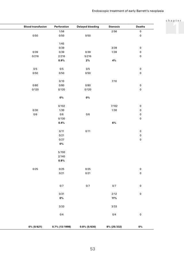
Table 6 Complications from endoscopic resection in the esophagus
Author Year of publication Techniques used Location Bleeding Blood transfusion Perforation Delayed bleeding Stenosis Deaths
Takeshita (369) 1997 ER-cap Esophagus 2/56 1/56 2/56 0
May (259) 2003 ER-cap Squamous and Barrett’s esophagus
1/50 0/50 0/50 0/50 0
Shimizu (228) 2004 ER-cap Squamous esophagus 1/45
Tanabe (177) 2004 ER-cap Squamous esophagus 1/39 0/39 3/39 0
Conio (372) 2005 ER-cap Barrett’s esophagus 4/39 0/39 0/39 0/39 1/39 0
Peters (184) own data, submitted ER-cap Barrett’s esophagus 49/216 0/216 2/216 5/216 0
Overall ER-cap 14% 0.9% 2% 4%
Fleischer (134) 1996 Ligate-and-cut Squamous esophagus 1/5i 0/5 0/5 0/5 0
May (259) 2003 Ligate-and-cut Squamous and Barrett’s esophagus
1/50 0/50 0/50 0/50 0
Soehendra (191) 2006 Ligate-and-cut Barrett’s esophagus 3/10 0/10 7/10
Peters (192) own data, in press Ligate-and-cut Barrett’s esophagus 5/80 0/80 0/80 0/80 0
Ell (373) 2000 Ligate-and-cut (n=117), lift-and-snare (n=3)
Barrett’s esophagus 8/120 0/120 0/120 0/120 0
Overall Ligate-and-cut 7% 0% 0%
Oyama (200) 2005 ESD Squamous esophagus 0/102 7/102 0
Kakushima (374) 2006 ESD EGJ 0/30 1/30 1/30 0
Nishimura (375) DDW 06 ESD Squamous esophagus 0/6 0/6 0/6 0/6 0
Oyama (376) DDW 06 ESD Squamous esophagus 0/130 0
Overall ESD 0.4% 6%
Moreira (377) 1995 Strip-biopsy Squamous esophagus 0/11 0/11 0/11 0
Narahara (378) 2000 Strip-biopsy Squamous esophagus 5/21 0/21 0
Tanabe (177) 2004 Strip-biopsy Squamous esophagus 2/27 0/27 0
Overall Strip-biopsy 12% 0%
Makuuchi (179) 2004 ER-cap tube method Esophagus 5/700
Shimizu (228) 2004 ER-cap tube method Squamous esophagus 2/140
Overall ER-cap tube method 0.8%
Nijhawan (263) 2000 Lift-and-snare Barrett’s esophagus 0/25 0/25 0/25 0/25 0
Giovannini (169) 2004 Lift-and-snare Barrett’s esophagus 4/21 0/21 0/21 0
Overall Lift-and-snare 9%
Soehendra (171) 1997 Simple snare Squamous and Barrett’s esophagus
0/7 0/7 0/7 0/7 0
Seewald (170) 2003 Simple snare Barrett’s esophagus 4/31 0/31 2/12 0
Overall Simple snare 11% 0% 11%
Noguchi (379) 2000 Strip-biopsy, ER-cap, overtube method
Squamous esophagus 1/33 3/33 3/33
Inoue (162) 1991 Modified strip-biopsy (overtube)
Squamous esophagus 0/4 0/4 0/4 0
Total All techniques 11% (91/851) 0% (0/621) 0.7% (13/1998) 0.8% (5/630) 8% (25/332) 0%
Endoscopic treatment of early Barrett’s neoplasia
53
1c h a p t e r

adrenaline injection is associated with significant rates of rebleeding (223). Sclerosing agents (1% polidocanol, 5% ethanolamine, 3% sodium tetradecyl sulphate or alcohol) have been reported for ulcer bleedings with variable results. These agents should, however, never be used to treat bleedings after endoscopic resection, since they will cause venous thrombosis, deep ulceration and tissue necrosis of the resection wound with perforation as a result (223). The suggested temporary effect of adrenaline injection is less relevant for treatment of bleedings after endoscopic resection; the rate of late bleedings is low and rebleedings after treatment of an acute bleeding are rare (184) (see also delayed bleedings below).The third endoscopic hemostasis method is thermal coagulation of the bleeding site. This can be achieved with a number of different techniques: bipolar coagulation, argon plasma coagulation (APC), or the use of a coagulation forceps. The easiest way to coagulate small bleedings is to use the tip of the ER-snare to gently apply a short burst of electric current (settings: 20 W forced coagulation, effect 2 for the Erbe VIO platform, 45 W coagulation for the Erbe ICC200). Care should be taken not to apply too much pressure with the tip of the snare, since the high current density may then easily cause damage to the deeper wall layers. The main indication for the use of the bipolar coagulation probe (e.g., Goldprobe, Boston Scientific) is the treatment of ulcer bleedings. The technique should, preferably, not be used for bleedings after endoscopic resection, since it is difficult to estimate its coagulation effect on the thin underlying tissue of the resection wound. The combination of too much pressure and prolonged application time may, thus, easily lead to complications. In APC, argon gas is sprayed from the probe and conducts the electrical current onto the tissue surface. It does not require physical contact with the target tissue and its damaging effect on the deeper wall layers of the esophageal wall can thus be better controlled. In case of active bleeding, however, this non-contact approach may be less effective. In our hands APC is mainly used for coagulation of small visible vessels in the resection wound or for ablation of residual neoplastic tissue at the edges of the resection area. For application on the resection wound we use an energy setting of 20 W (forced APC, effect 2) for the VIO platform and 40 W for the ICC 200 plus APC 300. For the ablation of residual neoplasia the settings are 40 W and 80 W, respectively. Drawback of APC is the intestinal distension caused by the argon gas flow, which may cause considerable discomfort to the patient and increase the risk for aspiration (224). The use of a coagulation forceps usually allows for management of the more severe bleedings after endoscopic resection. In contrast to ulcer bleedings where the hard, fibrotic ulcer surface prevents tissue from being grasped effectively, the soft fresh resection wound can be easily captured in forceps specially designed for this purpose (e.g., Hotclaw FD-420LR, Hot Biopsy Forceps FD-230U, Olympus). The use of a water-jet channel usually allows the bleeding area to be precisely identified and the bleeding vessel to be grasped by the coagulation forceps, while avoiding pressure onto the deeper wall layers. Coagulation is then applied using 45 W soft coagulation for 3-4 seconds. The forceps is then carefully opened while flushing water through the water-jet channel. This allows the tissue to be grasped immediately in case the bleeding has not stopped.
54
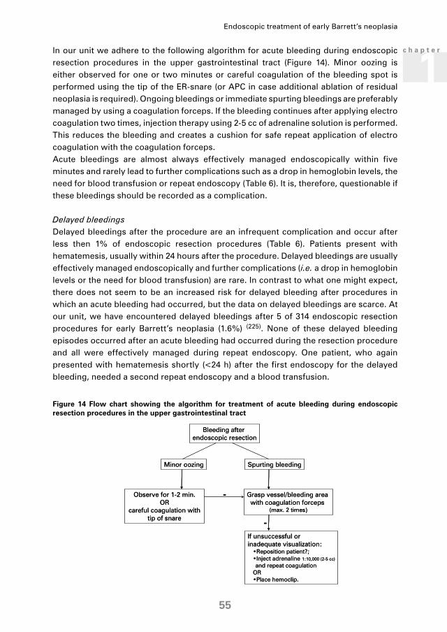
In our unit we adhere to the following algorithm for acute bleeding during endoscopic resection procedures in the upper gastrointestinal tract (Figure 14). Minor oozing is either observed for one or two minutes or careful coagulation of the bleeding spot is performed using the tip of the ER-snare (or APC in case additional ablation of residual neoplasia is required). Ongoing bleedings or immediate spurting bleedings are preferably managed by using a coagulation forceps. If the bleeding continues after applying electro coagulation two times, injection therapy using 2-5 cc of adrenaline solution is performed. This reduces the bleeding and creates a cushion for safe repeat application of electro coagulation with the coagulation forceps.Acute bleedings are almost always effectively managed endoscopically within five minutes and rarely lead to further complications such as a drop in hemoglobin levels, the need for blood transfusion or repeat endoscopy (Table 6). It is, therefore, questionable if these bleedings should be recorded as a complication.
Delayed bleedingsDelayed bleedings after the procedure are an infrequent complication and occur after less then 1% of endoscopic resection procedures (Table 6). Patients present with hematemesis, usually within 24 hours after the procedure. Delayed bleedings are usually effectively managed endoscopically and further complications (i.e. a drop in hemoglobin levels or the need for blood transfusion) are rare. In contrast to what one might expect, there does not seem to be an increased risk for delayed bleeding after procedures in which an acute bleeding had occurred, but the data on delayed bleedings are scarce. At our unit, we have encountered delayed bleedings after 5 of 314 endoscopic resection procedures for early Barrett’s neoplasia (1.6%) (225). None of these delayed bleeding episodes occurred after an acute bleeding had occurred during the resection procedure and all were effectively managed during repeat endoscopy. One patient, who again presented with hematemesis shortly (<24 h) after the first endoscopy for the delayed bleeding, needed a second repeat endoscopy and a blood transfusion.
Figure 14 Flow chart showing the algorithm for treatment of acute bleeding during endoscopic resection procedures in the upper gastrointestinal tract
Endoscopic treatment of early Barrett’s neoplasia
55
1c h a p t e r

PerforationsThe most feared complication of endoscopic resection of early neoplasia is perforation. Fortunately, this is a rare complication occurring in less than 1% of the endoscopic resection procedures (Table 6). A perforation occurs when the deeper layers of the muscularis propria are resected along with the mucosa and submucosa. Usually perforations can be immediately identified as a defect of the deeper muscle layers and the view onto the omentum or peri-esophageal fat (Figure 15). In esophageal perforations, subcutaneous emphysema can be found and mediastinal emphysema and free air can be detected on X-ray. An esophagography with watery contrast medium can be used to detect leakage. Esophageal perforation can cause mediastinitis, a severe and potentially lethal complication.
Figure 15 Esophageal perforation after endoscopic resection
A: Esophageal perforation after endoscopic resection. A defect in the endoscopic resection wound is clearly visible with a view on the mediastinal fat. B: A covered self-expandable stent has been placed over the perforation site to seal the leak. The stent has been fixed to the mucosa with two hemoclips and two additional hemoclips have been placed in the mucosa at the top of the stent to mark the correct proximal position of the stent. With the four hemoclips, the location of the stent can be checked on X-ray. C: Seven weeks after the perforation had occurred and three weeks after stent removal, the esophageal wall has healed completely. The perforation site has regenerated with neo-squamous mucosa with some retraction visible. Subsequently, the residual Barrett’s segment has been completely resected.
Clearly it is best to prevent perforations. Imperative in most endoscopic resection techniques is adequate submucosal lifting as described earlier with repeating the submucosal lifting before every resection in piecemeal procedures and preventing too much overlap of adjacent resections. Some authors have proposed to use EUS before every resection to assess the presence of the muscularis propria in the pseudopolyp (129;133;226;227). Although this may appear effective, it is a time consuming method requiring either the use of a double channel endoscope, or repeated endoscope exchanges with snares placed alongside the endoscope. In our opinion, this technique has no additional value over careful adequate submucosal lifting while assessing the Kato lifting sign. Esophageal perforations can be treated conservatively, endoscopically, or surgically ((partial) esophagectomy). The choice of treatment depends on a number of factors. First of all, how effective have you been in treating the neoplasia with the resection or how effective do you expect to be with (additional) endoscopic treatment? If the chances are high that endoscopic therapy is or will be insufficient, for example because of dubious penetration depth of the lesion, surgical treatment is a logical choice, since this will treat the underlying disease at the same time. Second, the location, size of the
56

perforation, and the chances of successful endoscopic treatment of the perforation should be considered. Finally, the operability of the patient is also relevant for choosing the optimal management strategy. Most Barrett’s esophagus patients are elderly with a frail condition; for a significant proportion of patients their high surgical risk led them to be referred for endoscopic treatment of the early neoplasia in the first place. In these patients, an emergency esophagectomy is a high risk intervention and one may favor conservative or endoscopic treatment in these patients.Conservative management of esophageal perforations consists of placement of a suction tube proximal to the site of the perforation and insertion of a duodenal feeding tube. Patients are put on nil per mouth and negative pressure (40-60 mmHg) is applied through the suction tube in order to prevent passage of fluids through the leak. In addition, patients should be treated intravenously with antibiotics, high-dose proton pump inhibitors, and should be fed through the duodenal tube.In addition to the aforementioned conservative measurements, endoscopic treatment of the perforation can be attempted by endoscopic closure of the defect by clips (228) or sealing of the defect using covered stents. Primary closure of the defect with clips is only possible if the perforation is recognized immediately. It is thought that closure is possible for defects with a size up to 1 or 1.5 cm (228;229). In the stomach the omentum can be used as a patch for the defect and can be fixed with clips (230;231). In the esophagus, however, no omentum is available and the esophageal wall is less compliant, making it more difficult to close the defect with clips. A number of suturing devices have been developed for endoscopic application. These devices have been developed mainly for treatment of arterial bleeding, and although they may seem promising, they have not yet been tested for their use in closing esophageal perforations (232;233). A general problem with applying endoscopic treatment for esophageal perforations is that the air insufflation required for the endoscopy can induce pneumomediastinum which may lead to life-threatening mediastinal compression. An alternative to endoscopic clipping of esophageal perforation after endoscopic resection is the placement of a covered self-expandable stent to seal the perforation. This has been shown successful in the treatment of different kinds of iatrogenic perforations or esophageal leaks (234-239). Complete sealing of the defects is seen in approximately 83% of cases (range 73-92%) (234-239). Main problem encountered with the use of stents for this indication is migration (approximately 24%; 15-33%) (234-238). Published experienced on the use of covered self-expandable stents for sealing of perforations after endoscopic resection, is limited. This makes it difficult to make clear recommendations. In Amsterdam we have used the Choo-stent (Fujinon Medical) and the Polyflex stent (Boston Scientific) for this indication. Based on this limited experience, the following recommendations are made. Stent insertion ideally should be done under endoscopic and fluoroscopic control. If fluoroscopy is not available, the Choo-stent is preferred over the Polyflex stent, since it is not associated with shortening during employment of the stent. The correct position of the stent should be checked endoscopically after stent placement. Usually the perforation site can be seen through the stent after stent insertion, but placing coagulation markers 3-5 cm above the perforation site, marking the aimed position of the proximal end of the stent, can be helpful. After the correct
Endoscopic treatment of early Barrett’s neoplasia
57
1c h a p t e r

position of the stent has been confirmed endoscopically, the proximal edge of the stent is clipped to the esophageal mucosa with one or two Hemoclips, in an attempt to prevent dislocation of the stent. In addition, one or two clips are placed in the mucosa at the proximal end of the stent. These clips can than serve as an orientation point for X-rays taken in the days following stent insertion. After stent insertion a suction tube with multiple side-holes is inserted with its distal tip 2-3 cm above the proximal end of the stent and constant suction (40-60 mmHg) is applied to keep the esophageal lumen as dry as possible. Subsequently, a video-esophagography is performed using water-soluble contrast to confirm adequate sealing of the perforation site. This should be done with the patient in different positions. Under fluoroscopic guidance a feeding tube is then inserted into the duodenum. The patient is than given complete bed rest and the position that provokes leakage of contrast on the esophagography and that places the perforation at a dependant position is avoided. The position of the stent is checked by daily X-rays, using the relative position of the clips on the stent and the clips on the mucosa as reference. (Figure 15) Five to seven days after placement of the stent, the video-esophagograph is repeated. If no leakage of contrast is observed oral intake is started. After clinical observation for one or two days the patient is discharged and scheduled for elective stent removal after 6 weeks. It can be anticipated that mobilizing the patient and starting oral intake will likely cause the stent to dislocate and the patient should be informed that the stent might pass rectally and instructed to contact the hospital in case of abdominal pain. Preferably, the (dislocated) stent should not be removed within six weeks to have the perforation site heal completely before the stent is withdrawn from the stomach or the esophagus.
PainAfter endoscopic resection in the esophagus, most patients experience mild continuus retrosternal pain for approximately one or two days, in rare cases up to two weeks, also depending on the extend of the resected area. Pain experienced during a meal may last somewhat longer. This is usually effectively controlled with acetaminophen. Few patients require stronger analgesics such as NSAIDS that are then always suppositories.
Esophageal stenosisAfter endoscopic resection esophageal stenosis may develop due to contraction and fibrosis during the healing process. Dysphagia after endoscopic resection usually develops after approximately two weeks. The patients often start off with experiencing hiccups during meals, which may than be associated with progressive dysphagia. Dysphagia significantly affects the quality of life and in general treatment is required for these patients. Katada et al. have shown that in the squamous esophagus resection of more than three fourths of the circumference and resection over a length more than 3 cm is associated with development of esophageal stenosis (240). Evaluation of the outcomes of patients treated at our institution showed that symptomatic esophageal stenosis after endoscopic resection in Barrett’s esophagus occurs almost exclusively after resection of more than 2/3 of the circumference (241).
58

For the treatment of esophageal stenosis there are a number of options. The most frequently applied therapy is dilatation of the stenosis, which can be performed with a dilatation balloons or dilatation bougies. We use Savary dilatations following the “rule of three”; i.e., three sequential bougies with increments of 1 mm staring from the first dilator where resistance is met. Dilatation sessions are usually repeated on a weekly basis until a dilator of 18 mm is passed. This requires a median number of 4 sessions (IQR 2-6) (242). As an alternative, a self-expandable stent can be placed to relieve the dysphagia and to dilate the stenosis (236;243-245). Four reports on the use of temporary stent placement for the treatment of esophageal stenosis have been published, including a total number. Treatment was successful in 12 (44%) and in some patients multiple stents were necessary to resolve the stenosis (236;243-245). In our experience temporary stent placement for treatment of post-endoscopic resection esophageal stenosis is accompanied by significant retrosternal pain, sometimes difficult stent removals and the need for additional dilatation sessions once the stent has been removed. With the ongoing evolution of endoscopic treatment of early Barrett’s neoplasia, there is a tendency to expand the indications to larger areas with esophageal stenosis being one of the limiting factors. Prevention of esophageal stenosis would, therefore, be of great value. This may be achieved by topical application of agents that improve healing. Mitomycin C has been used for a number of fibrosis associated conditions in ophthalmology and for recurrent subglottal and bronchial stenosis (246-248). Mitomycin has also been shown to prevent esophageal stenosis after caustic burns in rats (249). Radu et al. (222) used topical mitomycin C after circumferential resection of esophageal mucosa in sheep. The application of mitomycin C appeared to prevent stenosis, but timing of the application of mitomycin C was important since application shortly after ER resulted in perforations and death in four sheep, whereas application after 3 weeks was effective and uncomplicated. A possible explanation for the severe complications may be that Radu et al. used a relatively high dosage of mitomycin C (2 mg/ml for 5 minutes) compared to the aforementioned rat study (0.4 mg/ml) (249). An important concern with the use of mitomycin C is that it is an alkylating agent with mutagenic effects. The cytocidal effects have, however, been shown to occur with dosages of 1.0 mg/ml, higher than the dosage that is probably needed (250). In addition, no malignancies were observed in a 10-year review of 870 pterygium cases treated with topical application of 0.4 mg/ml mitomycin C (251). More studies are, however, required on the possible mutagenic effects of mitomycin C before it can be used to prevent post-endoscopic resection stenosis in humans.Many other agents have been tested in rats for a preventive effect on the development of stenosis after caustic burns to be applied after caustic injury in children (249;252-256) but none of these have reached clinical application.One study has suggested that sucralfate may have a preventive effect on development of caustic stenosis in rats (257). We prescribe all our patients sucralfate suspension (1 gr. four times daily) but this is mainly for its acid binding effect. We consider aggressive acid suppressant therapy the most important medical measure to prevent post-endoscopic resection stenosis. Patients are, therefore, after each endoscopic resection procedure treated with sucralfate and ranitidine (300 mg at bedtime) for two weeks, added to the maintenance treatment dosage of esomeprazol (40 mg bid).
Endoscopic treatment of early Barrett’s neoplasia
59
1c h a p t e r
An experimental method of prevention has recently been reported. Sakurai et al. injected autologous keratinocytes obtained from oral mucosa biopsies in endoscopic resection wounds in swine (258). They found that injected wounds had a smoother surface after healing without the formation of scar tissue. They did not report how the keratinocytes were extracted form the biopsies and how many biopsies and injections were needed, therefore, it is difficult to estimate the practical applicability in humans. Further studies are awaited. Recommendations for when to use which endoscopic resection techniqueFew randomized studies have been performed that have compared different endoscopic resection techniques. May et al. compared 50 endoscopic cap resections with 50 ligate-and-cut resections in Barrett’s esophagus and found both techniques to be equally efficient and safe (259). A number of retrospective studies have compared strip-biopsy or the ligate-and-cut technique with the endoscopic cap technique (174;177;192), but are hampered by methodological flaws and none of them deal with endoscopic resection in Barrett’s esophagus. We, therefore, have to base our advice on available non-randomized data and our own experience. For endoscopic resection of small focal lesions (i.e., <10 mm), en-bloc resection with the ligate-and-cut technique is a safe, easy and effective method. Focal lesions with a diameter between 10 and 20 mm can be best resected using the endoscopic cap resection technique with the flexible large caliber cap. Focal lesions with a diameter larger than 20 mm can be resected with the endoscopic cap resection technique, using a standard oblique cap, or the ligate-and-cut technique in a piece-meal resection. From a theoretical standpoint, an en-bloc resection with endoscopic submucosal dissection techniques might be preferred but this is a technique with a high risk of perforation that requires a high level of expertise.
Histological assessment of ER specimensEndoscopic resection specimens should be retrieved from the stomach at the end of the procedure, for example by using a Foreign Body Retrieval Basket (foreign Body Roth net, US Endoscopy, Mentor OH, USA). Subsequently, they should be stretched and pinned down on paraffin with the mucosal side up and fixed in 4% formalin. At the pathology department the specimens are usually cut in 2-mm sections and subsequently, 4-µm slides are cut from each section at three levels. If necessary, additional slides are cut from the sections. The specimens are evaluated for the presence of a (pre-) malignant lesion, its differentiation grade and infiltration depth, infiltration in lymph- or blood vessels, and the radicality of the resection at the deeper (vertical) resection margins. Assessment of lateral margins is only of value in en-bloc resections of lesions and if the lateral margins are positive for the presence of neoplasia, resection of the surrounding tissue in a subsequent endoscopic resection is possible. The infiltration depth of the lesion and the radicality of the resection at the deeper margins are most important in the evaluation, since these are the main (known) determinants for risk of lymph node involvement and thus for patient management after endoscopic resection (see above, Table 2). In Barrett’s esophagus, assessment of the infiltration depth can be difficult due
60

to the double layered muscularis mucosae that is often present in Barrett’s mucosa. The tissue between the two muscle layers may be misinterpreted as submucosa when the second muscle layer has not been resected. Furthermore, in 25% of all the procedures, the evaluation of the resection margins is impaired by significant coagulation damage to the specimens (47). The infiltration depth is usually classified in six categories as discussed above. Exact sub classification of submucosal cancers into sm1, 2 or 3 is difficult because the submucosa is not completely present in an endoscopic resection specimen and there is a wide variation in the thickness of the submucosa between patients. The discrimination is, however, important for assessment of the risk of lymph node involvement, as described above. Some authors, therefore, have suggested that an exact measurement of the submucosal infiltration depth in µm is more accurate (44). This measurement, however, may be influenced by the amount of stretching that has been applied while pinning down the specimen and by the shrinkage of the specimen in formalin. (260). In addition, the cut off level between sm1 and sm2 in Barrett’s neoplasia is set at 500 µm (44). This level is derived from data on gastric neoplasia and although it appears logical to apply this in Barrett’s neoplasia, it has not properly been studied. Because of the aforementioned difficulties in reproducible assessment of Barrett’s samples combined with its clinical importance, it is imperative that the evaluation of endoscopic resection specimens is performed by pathologist with experience in this field. Luckily, the interobserver agreement between pathologists is significantly better for the histological evaluation of endoscopic resection specimens than that of biopsy specimens of Barrett’s esophagus (261).Most endoscopically removed early Barrett’s neoplasia are lesions with HGIN or well differentiated mucosal carcinomas with a deeper margin free of neoplasia (47;260). Moderately (G2) and poorly (G3) differentiated cancers make up approximately 25% of endoscopically resected Barrett’s cancers. The evaluated G2 and G3 Barrett’s cancers significantly more often invade the submucosa and are significantly more frequent irradically removed at their deeper margins. Lymphatic and blood vessel invasion is rare (1-3.5%) and is almost exclusively seen in G2 and G3 cancers (47;260). G2 and G3 cancers and cancers with submucosal invasion are considered only relative indications for endoscopic resection.
Clinical results with endoscopic resection as mono-therapyClinical studies on the use of endoscopic resection of Barrett’s neoplasia as mono-therapy with a significant follow-up are scarce (Table 7), since in most centers patients with residual neoplasia after endoscopic resection or with diffuse neoplasia undergo
Table 7 Clinical results of patients treated with endoscopic resection as monotherapy for early Barrett’s neoplasia.
Author Year of publication
Nr. treated Patients
Nr. patients treated with ER
Nr. patients with follow-up data
Median follow-up (range)
Nr. recurrences
Nijhawan (263) 2000 25 18 18 10 months (4-42) 0/18 (0%)
May (262) 2002 115 70 65 34 months (24-60) 21/65 (32%)
Peters (380) 2005 28 18 18 19 months (13-24) 0/18 (0%)
Nr.: number, ER: endoscopic resection.
Endoscopic treatment of early Barrett’s neoplasia
61
1c h a p t e r

some sort of additional therapy immediately after endoscopic resection. The reason that patients are additionally treated is because many of them have multifocal or diffuse disease, or develop recurrent or metachronous lesions during follow-up in up to 32% (Table 7). The high rate of recurrences can be explained by the fact that with the endoscopic resection only a small part of the Barrett’s segment has been treated and the residual Barrett’s mucosa still has its malignant potential or already contains a low grade of neoplasia. Although recurrences can almost always be retreated endoscopically (262-
264), prevention of recurrences by additional treatment is desirable and many different methods of additional therapy have been developed of which endoscopic ablation therapy is the most widely used.
Endoscopic ablation therapyPhotodynamic therapyPhotodynamic therapy (PDT) is the most widely used ablation technique for treatment of early neoplasia in Barrett’s esophagus. (Figure 16) There are three essential components in PDT: drug, light and oxygen. For PDT a drug (i.e., photosensitizer) is administered that sensitizes (neoplastic) tissue for light with a specific wavelength. After accumulation of the photosensitizer in the target tissue, the tissue is illuminated with light of the appropriate wavelength that activates the drug. The photosensitizer absorbs the energy delivered by the light that is then transferred to molecular oxygen within the tissue leading to the creation of highly reactive singlet oxygen radicals that leads to cell damage and delayed cell death by necrosis and apoptosis (265;266). The tissue damage is delayed and becomes visible 8-12 hours after the procedure. This makes the targeted treatment of areas more difficult because there is no direct positional feedback. In many cases the patients, therefore, undergo an endoscopy 24 hours after the treatment to assess the treated area. Studies using PDT in Barrett’s esophagus differ significantly on the type of photosensitizer, light delivery systems and dosimetry used. There are three different photosensitizers used for PDT of early Barrett’s neoplasia. In the US the only FDA approved photosensitizer is sodium porfimer or Photofrin II. In Europe 5-aminolevulinic acid (5-ALA) is mainly used for photosensitization. Meso-tetrahydroxyphenylchlorine (mTHPC) has been used for PDT in Barrett’s esophagus in smaller sized series.
Complications and side effectsCutanous PhotosensitivityThe most important acute side effect of PDT is the cutanous photosensitivity due to accumulation of the photosensitizer in the skin and this is most pronounced with sodium porfimer PDT. This leads to complications of erythema, edema, blistering and even necrosis in approximately 20% of the patients treated with sodium porfimer PDT. The cutanous photosensitivity in sodium porfimer PDT starts one hour after administration, is the worst in the first two weeks and generally lasts for 30-90 days, but longer periods of photosensitivity have been seen (267). Patients need to wear sun blocking clothing that covers the body, extremities, hands, head and face for two weeks (268). With 5-ALA PDT the photosensitivity lasts approximately 24-48 hours and rarely leads to the development of the above mentioned complications (269-272).
62

A and G: Pre-treatment image of a Barrett’s segment, after administration of photosensitizer. B: Insertion of the PDT-balloon over a guidewire. C: Insertion of the laser fiber in the center of the PDT-balloon under guidance of green laser light. D: Treatment of the Barrett’s segment with laser light of the appropriate wavelength. E and H: Image of the treated Barrett’s segment 24 hours after the procedure, with necrosis of the mucosal layer. F and I: Image of the distal esophagus weeks after the PDT treatment with regeneration with neo-squamous mucosa and residual Barrett’s isles.
Figure 16 Photodynamic therapy of Barrett’s esophagus
Endoscopic treatment of early Barrett’s neoplasia
63
1c h a p t e r

Other acute side effects and complicationsThe most common side effect of PDT is retrosternal pain that can last up to approximately 2 weeks after the procedure. Most patients can manage the pain with regular analgesic medication; in some patients, however, more powerful analgesics are necessary. Mild, short lived nausea is experienced by 7-19% of the patients treated with 5-ALA PDT and is dose dependant (272;273). Hypotension and cardiac arrhythmia (atrial fibrillation) can be seen. These are in general transient and no permanent cardiac damage is seen (274), in one study comparing 5-ALA-PDT with APC for patients without dysplasia or LGIN, however, a patient died three days after 5-ALA-PDT, presumably from cardiac arrhythmia (275). After 5-ALA administration liver enzyme disorders are frequently seen in more than half of the patients. These disorders can involve all liver enzymes but most significantly are elevated ALT and AST levels. The effects are dose dependent and all resolve within two months without permanent effects (272;273). Rare complications are fever due to tissue necrosis or the “post-polypectomy syndrome” (serosal inflammation by thermal injury) and perforations.
Symptomatic stenosisThe most important long term complication of PDT is the development of symptomatic, often severe esophageal stenosis (Table 8). This complication is almost exclusively seen in sodium porfimer PDT since sodium porfimer accumulates in all esophageal wall layers. After sodium porfimer PDT, stenosis is seen in approximately 33% of patients (Table 8) and is more frequent after the treatment of more than one area in one procedure, especially when there is overlap of the subsequent areas and with the use of longer PDT balloons (276;277). Lower energy densities are associated with a lower rate of stenosis, but unfortunately also with lower efficacy (277). Treatment with oral steroids during the healing period after the treatment does not prevent the development of stenosis (276). Stenoses can be treated endoscopically as described earlier for the treatment of post-endoscopic resection stenosis.
Buried Barrett’sOne of the problems encountered after PDT is the presence of small islands of residual Barrett’s mucosa (glands with intestinal metaplasia) located underneath the neo-squamous mucosa (buried Barrett’s). This buried Barrett’s is found in a considerable number of patients in published series (Table 8) and in papers specifically evaluating the presence of buried Barrett’s, the reported rates are even higher with 33% and 52% (278;279). The clinical implications of buried Barrett’s are not clear. There have been a number of publications on carcinomas arising in buried Barrett’s mucosa (280-283). Ban et al. found foci of buried HGIN or EC in 9 patients and in three of the reported clinical series buried HGIN and EC has been found (Table 8). Some authors, however, suggest that the malignant potential of buried Barrett’s is much lower than normal Barrett’s mucosa, since the buried mucosa is no longer exposed to the toxic esophageal contents (284;285). Hornick et al. found that in 68% of the biopsies with buried Barrett’s an extension of the Barrett’s mucosa to the surface could be detected, questioning the actual percentage of true buried Barrett’s which is, by these authors, supposed to be undetectable due to
64

the lack of any contact with the surface (284). The fact that upon histological evaluation areas of subsquamous Barrett’s mucosa apparently can be found to communicate with the surface does, however, not imply that the endoscopist can actually detect these areas. To this end, changing the term buried Barrett’s into “hidden Barrett’s” may be more appropriate. Since the term ‘buried Barrett’s’ is more widely known, this term is continued to be used in this review.With the significant number of reported buried HGIN and EC, it is clear that the induction of buried Barrett’s should be prevented and that after successful treatment with a treatment modality that can cause buried Barrett’s, long term follow-up with large capacity biopsies of the neo-squamous mucosa is required.
Genetic abnormalities pre- and post-PDTAnother important problem associated with PDT is that the genetic abnormalities that are present before PDT and which are the cause of the malignancy and malignant potential are still present in the residual and recurrent Barrett’s mucosa after PDT (286-288). In addition, new genetic abnormalities may be induced by the oxygen radicals formed by PDT (289-293), as has been shown in one study (294). The presence of genetic abnormalities in the residual Barrett’s mucosa would implicate that this mucosa still has malignant potential and the recurrence rate of approximately 20% after PDT suggests that this is indeed the case.
Clinical results of PDT for early Barrett’s neoplasiaClinical results obtained in Barrett’s esophagus patients with early neoplasia with 5-ALA-PDT, sodium porfimer PDT, and mTHPC are shown in Table 8. The initial success rate of PDT for eradication of the neoplastic lesions with both sensitizers is high and comparable with 84% for 5-ALA-PDT and 81% for sodium porfimer PDT (not significantly different). The recurrence rates of both techniques are also comparable with 16% for 5-ALA-PDT and 18% for sodium porfimer PDT (not significantly different). Differences between the two sensitizers are most pronounced in the stenosis rate, which is 0% for 5-ALA and 33% for sodium porfimer and in the rate of development of buried Barrett’s. Although these rates are not statistically different with these limited numbers of series, there seems to be a trend towards a higher rate in 5-ALA-PDT than in sodium porfimer PDT. This may be explained by the less deep effect that is accomplished with 5-ALA-PDT. The limited clinical data available on mTHPC-PDT shows a varying success rate with a considerable complication rate and up to now no clear advantages over the other two photosensitizers.
Summary PDTPDT is mainly used as additional therapy of the residual Barrett’s segment after endoscopic resection of focal lesions with HGIN or EC. The purpose of the additional therapy is the eradication of any residual neoplastic changes and the prevention of recurrent neoplasia. Treatment of the residual Barrett’s segment means that in many cases a large area needs to be treated. PDT is specifically suitable for the treatment of large areas, especially when a balloon is used. With the balloon, a circumferential segment of 2-7 cm can be ablated at once.
Endoscopic treatment of early Barrett’s neoplasia
65
1c h a p t e r

Table 8 Clinical results of patients with early Barrett’s neoplasia treated with PDT
Author Patients Photo-sensitizer Dosage sensitizer
Interval Wavelength Light dosage Complete eradication neoplasia
% regression BE Buried Barrett’s Stenosis Follow-up Recurrence
Barr (381) 5 (5 HGIN) 5-ALA 60 mg/kg 4 hrs 630 50-150 J/cm2 100% 40% 26-44 mo
Gossner (382) 32 (10 HGIN, 22 EC) 5-ALA 60 mg/kg 4-6 hrs 635 150 J/cm2 84% partial in 68% 7% mean 9.9 (1-30) mo
7%
Ackroyd (273) 10 (3 LGIN, 4 HGIN, 3 EC) 5-ALA 30 mg/kg 4 hrs 514 (n=5), 630 (n=5)
50-100 J/cm2 70% mean 44% (10-100)
28 mo 0%
Pech (383) 51 (30 HGIN, 21 EC)* 5-ALA 60 mg/kg 4-6 hrs 635 150 J/cm2 100% 38 (26-56) mo 24%
Peters (323) 20 (18 HGIN, 2 EC) 5-ALA 40 mg/kg 1.5 -4 hrs 630 100 J/cm2 75% median 50% (25-70)
53% 30 (22-31) mo 27%
Median: 84% (IQR 73-100)
Median: 40% (IQR 7-53)
Median: 16% (IQR 2-26)
Overholt (384) 2 (2 EC) Sodium porfimer 2 mg/kg 48 hrs 630 300 J/cm 50% 50% 0 24 mo 50%
Laukka (385) 5 (4 LGIN, 1 HGIN) Sodium porfimer 1.5 mg/kg 48 hrs 630 175 J/cm 100% (all res. LGIN)
mean 24% yes 0
Overholt (386) 8 (3 HGIN, 5 EC) Sodium porfimer 2 mg/kg 48 hrs 630 300 J/cm fiber, 250 J/cm balloon
88% 38% mean 10.4 mo 13%
Overholt (387) 12 (1 LGIN, 6 HGIN, 5 EC) Sodium porfimer 2 mg/kg 48 hrs 630 300 J/cm fiber, 250 J/cm balloon
92% in 3 pts 100% 8% 33% 6-54 mo 17%
Sibille (388) 19 (19 EC)# Sodium porfimer 2-2.5 mg/kg 48-72 hrs 630 200 J/cm2 89% 35% mean 30.6 mo (1-71)
75%
Overholt (389) 36 (5 LGIN, 17 HGIN, 14 EC) Sodium porfimer 2 mg/kg 48 hrs 630 100-300 J/cm 81% 75-80%, 100% in 10
6% 58% mean 22 mo (7-62)
0%
Overholt (390) 100 (14 LGIN, 73 HGIN, 13 EC) Sodium porfimer 2 mg/kg 48 hrs 630 100-250 J/cm 80% 75-80%, 100% in 43
5%, 2 with HGIN, 1 with ca
34% 23%
Overholt (391) 103 (14 LGIN, 80 HGIN, 9 EC) Sodium porfimer 2 mg/kg 48 hrs 630 100-250 J/cm 77% 100% in 56** 5%, 3 with ca 30% mean 51 mo (±20.57)
Wolfsen (392) 102 (69 HGIN, 33 EC) Sodium porfimer 2 mg/kg 48, 72 hrs 630 150-225 J/cm 96% 100% in 57 4%, all with HGIN/ca
20% median 1.6 yrs (0.5-6.5)
Overholt (393) 133 ( 133 HGIN) Sodium porfimer 2 mg/kg 40-50 hrs 630 130 J/cm 77% 72% 36% mean 43 mo (±24.2)
13%
Foroulis## (394) 25 (15 HGIN, 10 T1a) Sodium porfimer 2 mg/kg 24 hrs 630 200-250 J/cm for HGIN, 500-600 J/
cm for Ca
81% 100% in 5 20% 6% median 14 mo (6-70)
18%
Median: 81% (IQR 77-92)
Median: 6% (IQR 5-11)
Median: 33% (IQR 6-36)
Median: 18% (IQR 13-43)
Javaid (395) 7 (6 HGIN, 1 EC) mTHPC 0.15 mg/kg 96 hrs 652 8-20 J/cm2 100% (two residual LGIN)
100% in 1 29% 0%
Etienne (396) 14 lesions (7 HGIN, 7 EC) mTHPC 0.15 mg/kg 96 hrs 514 75 J/cm2 100% 0% 8% mean 34 mo (±15)
8%
Lovat (397) 19 (7 HGIN, 12 EC) mTHPC 0.15 mg/kg 72 hrs 511-652 7 J/cm2 - 75 J/cm2 42% 21% 11% median 24 mo (1-72)
75%
Median: 100% (IQR 42-100)
Median: 11 (IQR 8-29)
Median: 8% (IQR 0-75)
* Total study population 66 pts, 51 pts were treated with PDT alone, 15 received additional endoscopic resections, APC, KTP laser treatment; excluded in this table. ** after additional PDT sessions and ND:YAG laser destruction small residual isles. # Study population included an additional 104 patients with squamous cell carcinoma, excluded for this table. ## Only data on patients with HGIN/intramucosal carcinoma are presented in the table, study included an additional 6 patients with more advanced tumors.
66

Table 8 Clinical results of patients with early Barrett’s neoplasia treated with PDT
Author Patients Photo-sensitizer Dosage sensitizer
Interval Wavelength Light dosage Complete eradication neoplasia
% regression BE Buried Barrett’s Stenosis Follow-up Recurrence
Barr (381) 5 (5 HGIN) 5-ALA 60 mg/kg 4 hrs 630 50-150 J/cm2 100% 40% 26-44 mo
Gossner (382) 32 (10 HGIN, 22 EC) 5-ALA 60 mg/kg 4-6 hrs 635 150 J/cm2 84% partial in 68% 7% mean 9.9 (1-30) mo
7%
Ackroyd (273) 10 (3 LGIN, 4 HGIN, 3 EC) 5-ALA 30 mg/kg 4 hrs 514 (n=5), 630 (n=5)
50-100 J/cm2 70% mean 44% (10-100)
28 mo 0%
Pech (383) 51 (30 HGIN, 21 EC)* 5-ALA 60 mg/kg 4-6 hrs 635 150 J/cm2 100% 38 (26-56) mo 24%
Peters (323) 20 (18 HGIN, 2 EC) 5-ALA 40 mg/kg 1.5 -4 hrs 630 100 J/cm2 75% median 50% (25-70)
53% 30 (22-31) mo 27%
Median: 84% (IQR 73-100)
Median: 40% (IQR 7-53)
Median: 16% (IQR 2-26)
Overholt (384) 2 (2 EC) Sodium porfimer 2 mg/kg 48 hrs 630 300 J/cm 50% 50% 0 24 mo 50%
Laukka (385) 5 (4 LGIN, 1 HGIN) Sodium porfimer 1.5 mg/kg 48 hrs 630 175 J/cm 100% (all res. LGIN)
mean 24% yes 0
Overholt (386) 8 (3 HGIN, 5 EC) Sodium porfimer 2 mg/kg 48 hrs 630 300 J/cm fiber, 250 J/cm balloon
88% 38% mean 10.4 mo 13%
Overholt (387) 12 (1 LGIN, 6 HGIN, 5 EC) Sodium porfimer 2 mg/kg 48 hrs 630 300 J/cm fiber, 250 J/cm balloon
92% in 3 pts 100% 8% 33% 6-54 mo 17%
Sibille (388) 19 (19 EC)# Sodium porfimer 2-2.5 mg/kg 48-72 hrs 630 200 J/cm2 89% 35% mean 30.6 mo (1-71)
75%
Overholt (389) 36 (5 LGIN, 17 HGIN, 14 EC) Sodium porfimer 2 mg/kg 48 hrs 630 100-300 J/cm 81% 75-80%, 100% in 10
6% 58% mean 22 mo (7-62)
0%
Overholt (390) 100 (14 LGIN, 73 HGIN, 13 EC) Sodium porfimer 2 mg/kg 48 hrs 630 100-250 J/cm 80% 75-80%, 100% in 43
5%, 2 with HGIN, 1 with ca
34% 23%
Overholt (391) 103 (14 LGIN, 80 HGIN, 9 EC) Sodium porfimer 2 mg/kg 48 hrs 630 100-250 J/cm 77% 100% in 56** 5%, 3 with ca 30% mean 51 mo (±20.57)
Wolfsen (392) 102 (69 HGIN, 33 EC) Sodium porfimer 2 mg/kg 48, 72 hrs 630 150-225 J/cm 96% 100% in 57 4%, all with HGIN/ca
20% median 1.6 yrs (0.5-6.5)
Overholt (393) 133 ( 133 HGIN) Sodium porfimer 2 mg/kg 40-50 hrs 630 130 J/cm 77% 72% 36% mean 43 mo (±24.2)
13%
Foroulis## (394) 25 (15 HGIN, 10 T1a) Sodium porfimer 2 mg/kg 24 hrs 630 200-250 J/cm for HGIN, 500-600 J/
cm for Ca
81% 100% in 5 20% 6% median 14 mo (6-70)
18%
Median: 81% (IQR 77-92)
Median: 6% (IQR 5-11)
Median: 33% (IQR 6-36)
Median: 18% (IQR 13-43)
Javaid (395) 7 (6 HGIN, 1 EC) mTHPC 0.15 mg/kg 96 hrs 652 8-20 J/cm2 100% (two residual LGIN)
100% in 1 29% 0%
Etienne (396) 14 lesions (7 HGIN, 7 EC) mTHPC 0.15 mg/kg 96 hrs 514 75 J/cm2 100% 0% 8% mean 34 mo (±15)
8%
Lovat (397) 19 (7 HGIN, 12 EC) mTHPC 0.15 mg/kg 72 hrs 511-652 7 J/cm2 - 75 J/cm2 42% 21% 11% median 24 mo (1-72)
75%
Median: 100% (IQR 42-100)
Median: 11 (IQR 8-29)
Median: 8% (IQR 0-75)
* Total study population 66 pts, 51 pts were treated with PDT alone, 15 received additional endoscopic resections, APC, KTP laser treatment; excluded in this table. ** after additional PDT sessions and ND:YAG laser destruction small residual isles. # Study population included an additional 104 patients with squamous cell carcinoma, excluded for this table. ## Only data on patients with HGIN/intramucosal carcinoma are presented in the table, study included an additional 6 patients with more advanced tumors.
Endoscopic treatment of early Barrett’s neoplasia
67
1c h a p t e r

The initial success rate of PDT in eradicating HGIN and EC is acceptable. The recurrence rates, however, are substantial and are not much better than the recurrence rates after treatment with endoscopic resection of neoplastic lesions as monotherapy. Considering the primary purposes of PDT (or ablation in general), treatment of neoplasia and prevention of recurrences, PDT does not seem to live up to these expectations. This disappointing recurrence rate in combination with the problems of buried Barrett’s and persistent and induced genetic abnormalities limits the indications for PDT significantly and alternatives are desirable.
Argon plasma coagulationArgon plasma coagulation (APC) is a second endoscopic ablation technique that is used for treatment of early Barrett’s neoplasia and larger areas of (non-)dysplastic Barrett’s mucosa. APC is a modality that applies high-frequency electric current to tissue via ionized and therefore electrically conductive argon gas. This generates heat that causes thermal effects that depend on the distance to the location of application. In the zone where the current first reaches the tissue, the tissue is devitalized, the second zone is coagulated, the third zone is desiccated and in the fourth zone the tissue shrinks. When areas of tissue lose their electric conductivity due to desiccation, the beam automatically changes its direction to a location that still is electrically conductive. This results in a relatively uniform depth of the different zones and the maximum depth of the thermal effect is automatically limited decreasing the risk of perforation. The maximum depth of the thermal effect is determined by the power settings and the duration of application (224). APC requires an argon source, high frequency current source and an applicator to deliver the argon and current to the tissue. The APC probe that is used in gastrointestinal endoscopy consists of a flexible tube with a lumen for the argon gas flow and a wire connected to an electrode at the tip of the probe for conducting the current (Figure 17) (APC-Sonde 2200A, Erbe Elektromedizin GmbH, Tübingen, Germany). The probe fits through the accessory channel of the endoscope and is connected to the APC source
A: Image of argon plasma coagulation of Barrett’s mucosa. B: Endoscopic image of the continuous golden-brown coagulum after argon plasma coagulation of Barrett’s mucosa.
Figure 17 Argon plasma coagulation ablation
68

and generator (Erbe VIO 200D and Erbe APC 2, or Erbotom ICC 200 and Erbe APC 300, Erbe Elektromedizin GmbH). By placing a neutral electrode on the patient an electric field is created between the tissue and the electrode in the probe that ionizes the argon gas and creates the high-frequency current to the tissue. The probe should not make contact with the tissue, since the current travels through the gas. In general, low power settings are safer but less effective than higher settings. For adequate treatment with a treatment depth of approximately 2-3 mm a power of 60-80 W is used when using the ICC 200 and APC 300 system and 40 W forced APC with the VIO platform, with an argon gas flow of 1.6-2 L/min. The tissue should be targeted until a homogenous and continuous coagulum has developed there should not be separate patches of coagulum (Figure 17). Care should be taken not to target one location too long and not to make contact between the probe and the tissue, since this may cause a micro-perforation that can be fatal (295).
Complications and side effectsBesides pain and odynophagia, perforation and stenosis after large area ablation are the only reported complications of APC. Late perforations may theoretically occur due to transmural necrosis, although this has not been reported in clinical series. It is important to make sure that all the mucosa of the target area is homogeneously ablated, since any residual Barrett’s mucosa could give rise to residual Barrett’s mucosa after healing or buried Barrett’s underneath the regenerated neosquamous mucosa (see PDT section). Buried Barrett’s is seen in median 15% (IQR 15-31) of the patients treated with APC (296-305) and carcinoma arising in these buried glands has been reported (280;299). In line with PDT, pre-existing genetic abnormalities of the Barrett’s epithelium may not be completely eradicated by APC. Lopes et al. found over expression of p53 protein in the neo-squamous mucosa after APC (306) and Hage et al. found persistent genetic abnormalities in residual Barrett’s mucosa after APC (275).
Clinical resultsClinical results of APC for ablation of Barrett’s mucosa with HGIN or early cancer are shown in Table 9. More studies have been performed with APC ablation of Barrett’s segments without dysplasia or with LGIN (275;296-300;303;304;307-314). Successful eradication of neoplasia is achieved in a median of 86% (IQR 74-98) of cases, which is comparable to PDT but is associated with fewer complications and less esophageal stenosis. Complete ablation of all Barrett’s mucosa is achieved in only a minority of patients and recurrence of Barrett’s mucosa is seen in a median of 8% (IQR 5-30) of patients (296;297;299;300;304;308;310
-312). One prospective randomized trial has been performed comparing APC with sodium porfimer PDT for eradication of HGIN (283). This trial showed that sodium porfimer PDT was more effective for eradicating neoplasia (77% vs. 67%, p=0.03) and equally efficient for eradicating Barrett’s mucosa. PDT was more expensive than APC. Four prospective randomized trials have been performed comparing APC with 5-ALA-PDT or multipolar electrocoagulation (MPEC) ablation of complete Barrett’s segments without dysplasia or LGIN (275;307;314;315). One study found APC to be more effective than 5-ALA-PDT in eradication of Barrett’s mucosa (314), the other studies showed no significant differences in efficacy of the different treatment modalities.
Endoscopic treatment of early Barrett’s neoplasia
69
1c h a p t e r

Summary APCFor eradication of neoplasia, APC has an acceptable success rate that is comparable with PDT and seems to be associated with fewer recurrences during follow-up (Table 9). It is also associated with only partial Barrett’s ablation in most patients with again a significant percentage of buried Barrett’s, persistent genetic abnormalities, and recurrence of Barrett’s mucosa during follow-up. Technically it is a time consuming technique when used for large areas and it is uncomfortable for the patient. Therefore, we believe that APC is not suitable for ablation of large areas of Barrett’s mucosa and its use should be limited to a minimum. It can in some cases be useful for ablation of small residual isles of Barrett’s mucosa for example after piece-meal endoscopic resection for eradication of small residual bridges between the resections.
Radiofrequency ablationA relatively new technique used for ablation of Barrett’s esophagus is radiofrequency energy ablation (RFA). Radiofrequency energy is generated by a special RF generator and applied to the tissue by a balloon-based RF electrode (HALO360) that consists of multiple tightly spaced bipolar electrodes that alternate in polarity (BARRx, Sunnyvale, CA, USA). The electrode is 3 cm in length and a variety of different diameters is available (22-34 mm). The total energy (J) that is delivered to the tissue is controlled by the generator and determines the depth of injury. The energy is adjusted to the balloon diameter, thereby delivering a standardized energy density (J/cm2) to the tissue. The energy amount can be selected and is delivered in less than one second at 300 W (316). In an experimental study, energy densities of 8-20 J/cm2 resulted in complete epithelial ablation without inducing esophageal stenosis (316). In a second experimental study it was shown that the optimal setting to achieve an ablation depth up to (but not into) the submucosa was an energy density of 10 J/cm2 delivered twice, or 12 J/cm2 delivered once or twice (317).
Table 9 Clinical results of patients with early Barrett’s neoplasia treated with APC
Author N Type patients Power Gas flow Complete eradication neoplasia
regression BE Buried Barrett’s Major complications Stenosis Follow-up Recurrence
Pereira-Lima (302) 33 18 ND, 14 LGIN, 1 HGIN
65-70 W 2 l/min 100% 100% 0% 1 possible micro-perforation, 15% fever
9% mean 10.6 (6-18) mo 0
Morris (301) 55 37 ND, 9 LGIN, 9 HGIN
96% (1 death, 1 drop out) mean 5 cm (SD 2.37)
30% 2 perforations with 1 death (pt with HGIN), 1 admission
for pain
mean 38.5 (SD 14.6) mo 0
Van Laethem (305) 10 7 HGIN, 3 EC 90 W 80% 100% in 5 30% 10% median 24 (12-36) mo 0
Attwood (398) 29 29 HGIN 70 W 2 l/min 86% 76% 1 perforation with death post-operatively
0% mean 37 (7-78) mo 0
Ragunath (283) 13 12 LGIN, 1 HGIN 65 W 1.8 l/min 67% median 65% 1 admission for pain and fever
15% 12 mo 0
median 86% (74-98%) median 30% (0-30%) median 10% (2-14%)
N: number of patients, BE: Barrett’s esophagus, ND: no-dysplasia, LGIN: low-grade intraepithelial neoplasia, HGIN: high-grade intraepithelial neoplasia, mo: months, pt: patient, EC: early cancer
70
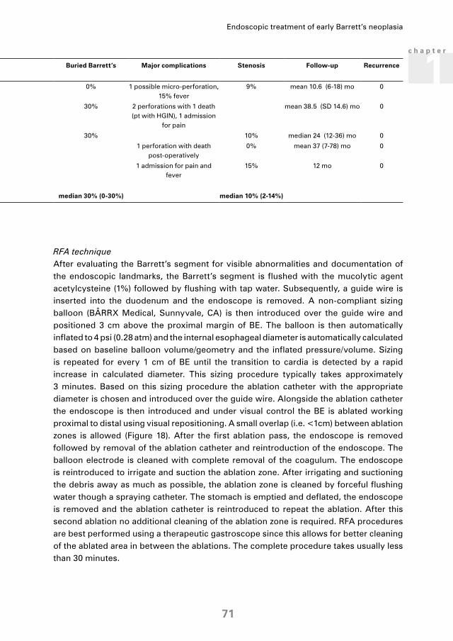
RFA techniqueAfter evaluating the Barrett’s segment for visible abnormalities and documentation of the endoscopic landmarks, the Barrett’s segment is flushed with the mucolytic agent acetylcysteine (1%) followed by flushing with tap water. Subsequently, a guide wire is inserted into the duodenum and the endoscope is removed. A non-compliant sizing balloon (BÂRRX Medical, Sunnyvale, CA) is then introduced over the guide wire and positioned 3 cm above the proximal margin of BE. The balloon is then automatically inflated to 4 psi (0.28 atm) and the internal esophageal diameter is automatically calculated based on baseline balloon volume/geometry and the inflated pressure/volume. Sizing is repeated for every 1 cm of BE until the transition to cardia is detected by a rapid increase in calculated diameter. This sizing procedure typically takes approximately 3 minutes. Based on this sizing procedure the ablation catheter with the appropriate diameter is chosen and introduced over the guide wire. Alongside the ablation catheter the endoscope is then introduced and under visual control the BE is ablated working proximal to distal using visual repositioning. A small overlap (i.e. <1cm) between ablation zones is allowed (Figure 18). After the first ablation pass, the endoscope is removed followed by removal of the ablation catheter and reintroduction of the endoscope. The balloon electrode is cleaned with complete removal of the coagulum. The endoscope is reintroduced to irrigate and suction the ablation zone. After irrigating and suctioning the debris away as much as possible, the ablation zone is cleaned by forceful flushing water though a spraying catheter. The stomach is emptied and deflated, the endoscope is removed and the ablation catheter is reintroduced to repeat the ablation. After this second ablation no additional cleaning of the ablation zone is required. RFA procedures are best performed using a therapeutic gastroscope since this allows for better cleaning of the ablated area in between the ablations. The complete procedure takes usually less than 30 minutes.
Table 9 Clinical results of patients with early Barrett’s neoplasia treated with APC
Author N Type patients Power Gas flow Complete eradication neoplasia
regression BE Buried Barrett’s Major complications Stenosis Follow-up Recurrence
Pereira-Lima (302) 33 18 ND, 14 LGIN, 1 HGIN
65-70 W 2 l/min 100% 100% 0% 1 possible micro-perforation, 15% fever
9% mean 10.6 (6-18) mo 0
Morris (301) 55 37 ND, 9 LGIN, 9 HGIN
96% (1 death, 1 drop out) mean 5 cm (SD 2.37)
30% 2 perforations with 1 death (pt with HGIN), 1 admission
for pain
mean 38.5 (SD 14.6) mo 0
Van Laethem (305) 10 7 HGIN, 3 EC 90 W 80% 100% in 5 30% 10% median 24 (12-36) mo 0
Attwood (398) 29 29 HGIN 70 W 2 l/min 86% 76% 1 perforation with death post-operatively
0% mean 37 (7-78) mo 0
Ragunath (283) 13 12 LGIN, 1 HGIN 65 W 1.8 l/min 67% median 65% 1 admission for pain and fever
15% 12 mo 0
median 86% (74-98%) median 30% (0-30%) median 10% (2-14%)
N: number of patients, BE: Barrett’s esophagus, ND: no-dysplasia, LGIN: low-grade intraepithelial neoplasia, HGIN: high-grade intraepithelial neoplasia, mo: months, pt: patient, EC: early cancer
Endoscopic treatment of early Barrett’s neoplasia
71
1c h a p t e r

A: Pre-treatment image of a Barrett’s segment. B and C: The esophageal diameter is measured at 1-cm intervals with a sizing balloon placed over a guidewire. D: Introduction of the RFA balloon catheter with the appropriate diameter over the guidewire. E: The inflated RFA balloon positioned 1 cm above the top of the Barrett’s segment. F: The RFA balloon repositioned for ablation of the second zone after ablation of the first zone with an overlap of 1 cm with the first ablation zone. The instantaneous necrosis of the superficial mucosa is visible in the first ablation zone. G: Image of the treated Barrett’s segment immediately after the RFA ablation with visible necrosis of the superficial mucosa. H: Image of the healed distal esophagus 3 months after RFA treatment with regeneration with neo-squamous mucosa and three small isles with residual Barrett’s mucosa. I: Introduction of the endoscope with the HALO90 cap for focal ablation placed at the tip. J: Ablation of the third isle of Barrett’s mucosa. The necrosis caused by ablation of the first two isles is visible. K: Image of the distal esophagus immediately after ablation of the three residual isles of Barrett’s mucosa. L: Image of the healed distal esophagus, showing complete regeneration with neo-squamous mucosa.
Figure 18 Radiofrequency ablation
72

Clinical resultsThe RFA system has first been tested in a large US multicentre trial for complete ablation of non-dysplastic Barrett’s esophagus (AIM-I and AIM-II) (318). In the first phase of the study, the AIM-I phase, the appropriate energy setting for Barrett’s esophagus was determined in a dose-escalating study (using 6, 8, 10, 12 J/cm2) in 32 patients with a Barrett’s length of 2-3 cm. The complete response rate for eradication of all Barrett’s mucosa was 0%, 10%, 67% and 55%, respectively, with histological clearance of Barrett’s epithelium in 93%, 65%, 100% and 100%, respectively. Based on these results, 70 patients with a Barrett’s length of 2-6 cm were treated at 10 J/cm2 delivered twice in one session in the effectiveness phase of the trial. Complete remission for Barrett’s esophagus was achieved in 70% of patients after a maximum of two treatment sessions. There were no severe complications, the procedures were well tolerated and none of the total 102 treated patients developed an esophageal stenosis. The residual Barrett’s mucosa was typically in the form of small, visible islands. In the more than 4000 biopsies that have been obtained in these patients during follow-up, no buried Barrett’s has been detected.In a separate study, 10 patients with LGIN in a Barrett’s esophagus of 2-6 cm in length were treated at 12 J/cm2 delivered twice (AIM-LGD trial) (319). Four months after the first treatment patients with residual Barrett’s mucosa or LGIN were treated with a second ablation. One patient had a mild bleeding that resolved spontaneously; there were no other serious adverse events and no esophageal stenoses. After 12 months follow-up all patients had complete eradication of all LGIN and 80% of patients had complete eradication of all Barrett’s mucosa. In the remaining patients, only small islets of Barrett’s mucosa remained. In more than 450 follow-up biopsies, no buried Barrett’s was detected.At our institution we have now treated 23 patients with LGIN or HGIN. Thirteen of these patients had undergone a prior endoscopic resection of a focal lesion containing carcinoma (n=4), HGIN (n=6) or LGIN (n=3). The baseline histological diagnosis after endoscopic resection and before RFA treatment was LGIN in three patients and HGIN in 20 patients. Patients were treated at 12 J/cm2 delivered twice. In case of remaining Barrett’s mucosa or neoplasia, patients were retreated four months after the first treatment. For treatment of small areas of Barrett’s mucosa, a focal RFA device was developed (HALO90 system) that uses the same electrode array technology as the HALO360 system. The HALO90 uses a distal attachment cap that can be placed on the tip of the endoscope. The HALO90 cap allows focal ablation of an area of 1.5 x 2.0 cm in a single energy application. Patients with small residual islands of Barrett’s mucosa after circumferential treatment with the HALO360 system were treated with this cap. In 96% of the patients eradication of all neoplasia was achieved; one patient had persisting HGIN. The median regression percentage of the Barrett’s segment was 99% (IQR 85-100), complete eradication was achieved in 21/23 patients (92%). During a median follow-up of 6 months, no patients showed recurrence of neoplasia of Barrett’s mucosa. No buried Barrett’s has yet been detected in 512 biopsies obtained during follow-up endoscopies and none of the patients has developed thermal related stenosis or dysphagia (320).
Endoscopic treatment of early Barrett’s neoplasia
73
1c h a p t e r

Summary RFASummarizing, radiofrequency ablation appears to be an effective treatment modality for Barrett’s ablation that can be safely performed after endoscopic resection of focal abnormalities. The technique is safe, fast, well tolerated and relatively easy to perform. None of the reported 135 patients have developed esophageal stenosis and had buried Barrett’s in over 4500 biopsies. These first reports on RFA suggest that this ablation technique is the first to find the right trade-off between effectiveness on one hand (causing complete removal of all Barrett’s with no buried Barrett’s) and the avoidance of damage to the deeper layers causing complications such as stenosis. Long term follow-up results are required to confirm these results and to study possible recurrences of neoplasia and/or of Barrett’s mucosa. Large multi-centre trials are underway in Europe and the US.
Clinical results of endoscopic resection combined with ablationAs mentioned, endoscopic ablation is mainly used as an adjunct to endoscopic resection. First, focal lesions are endoscopically resected and subsequently the residual Barrett’s segment is treated with an endoscopic ablation modality for treatment of any residual flat neoplasia and to prevent recurrences by eradicating the Barrett’s epithelium. Only a small number of series reporting on the success of such combined treatment protocols have been published. These series are presented in Table 10 and include only combinations of endoscopic resection with PDT.The short term success rates for complete elimination of all neoplasia are high, ranging from 83-98% (262;264;321;322), but complete eradication of all Barrett’s mucosa is rarely achieved. The development of new or recurrent lesions during follow-up is seen in a considerable number of patients (0-39%) (262;264;321;322). The goal of this combination treatment is, therefore, only partly achieved.
Table 10 Clinical results of patients with early Barrett’s neoplasia treated with endoscopic resection combined with endoscopic ablation therapy.
Author No. pts treated Complete local remission Follow up (mo) recurrences
Pacifico et al (322) 24* 20 (83%) 12±2 0
May et al (262) 110† 108 (98%) 34±10 34 (39%)
Buttar et al (321) 17‡ 16 (94%) 13 (3-48) 1 (6%)
Peters et al (380) 28** 26 (93%) 19 (13-24) 5 (19%)
*All patients treated with ER and Photofrin®-PDT. †Endoscopic Resection (ER) in 66 patients (pts), Photodynamic Therapy (PDT) in 32 pts (26 pts 5-ALA-PDT, 4 pts mTHPC-PDT, 2 pts 5-ALA+mTHPC-PDT), ER and PDT in 10 pts, APC in 3 pts, one patient died before endoscopic treatment was completed from myocardial infarction.‡All pts treated with ER and Photofrin®-PDT.**ER in 5 pts, ER and 5-ALA-PDT in 19 pts, ER and APC in 3 pts.
74

Stepwise radical endoscopic resectionIn our unit, we found that the combination of endoscopic resection and PDT did not result in a reduced rate of recurrent lesions during follow-up (323). We, therefore, started to treat patients with complete endoscopic resection of the entire Barrett’s segment in multiple sessions. With this stepwise radical endoscopic resection protocol (SRER) all premalignant tissue is completely resected with histological correlation. It is expected to induce less buried Barrett’s since endoscopic resection usually extends into the submucosa leaving no residual Barrett’s mucosa behind. SRER may therefore lead to a permanent cure of Barrett’s esophagus and its associated neoplasia.The feasibility of complete endoscopic resection of the distal esophageal mucosa has been shown in animal studies (324;325). Seewald et al. (170) published a series of 12 patients with a median Barrett’s length of 5 cm. They performed endoscopic resections using the ‘simple-snare’ technique and needed a median number of 2.5 sessions with median of 5 resections/session per patient. There were no severe complications and during a median follow-up of nine months no recurrence of Barrett’s mucosa or neoplasia was seen. Two of 12 patients (17%) developed stenosis managed by bougienage. In 2004, Giovannini et al. (169) reported a retrospective series of 21 patients with a mean Barrett’s length of 3.5 cm, using the ‘lift-and-cut’ technique. Patients were treated with approximately 2 sessions with a median of 4.5 resections per session. There were no severe complications and none of the patients developed stenosis. In three patients (piece-meal) resection of the mucosal carcinoma was considered incomplete; one patient underwent surgery (no residual tumor was found in the surgical resection specimen), the other two patients underwent chemoradiotherapy. At follow-up, all three patients were alive without residual disease. Of the 18 patients with successful eradication of neoplasia, two patients (11%) had recurrence of HGIN during follow-up and were successfully retreated with endoscopic resection. Five patients had residual isles of Barrett’s mucosa embedded in the neosquamous mucosa, in three patients with the presence of LGIN. At our institution we use the following SRER protocol (242;326). First focal lesions suspicious of submucosal invasion are endoscopically resected for histological diagnosis before entering the SRER protocol. During the first SRER session removal of 50% of the BE is attempted, including the most suspicious area in case this had not been resected in an earlier diagnostic endoscopic resection. Subsequent SRER sessions are performed with an interval of six to eight weeks until eradication of all Barrett’s mucosa and all neoplasia is considered to be complete (Figure 19). Complete endoscopic eradication is confirmed by lugol (2%) staining and complete histological eradication is confirmed by jumbo-biopsies. For the resections we use the endoscopic cap technique or the ligate-and-cut technique. For ablation of small residual isles of Barrett’s mucosa that can not be resected, APC is used. Between January 2003 and June 2005, 56 consecutive patients have been treated by SRER. Complete eradication of early neoplasia was achieved in all 56 treated patients (100%). Complete eradication of all Barrett’s mucosa was achieved in 44/56 (79%): four patients (7%) had a small rim of less than 5 mm of non-dysplastic residual BE in their distal esophagus, eight patients (14%), whom all had undergone APC, had small foci of BE buried underneath neosquamous mucosa. This buried Barrett’s was found in general only in 1-2 biopsy specimens and was not reproduced during
Endoscopic treatment of early Barrett’s neoplasia
75
1c h a p t e r

further FU. Complications occurred in 4/165 (2%) endoscopic resection procedures: one asymptomatic perforation and three delayed bleedings. Stenosis occurred in 24/58 (41%) patients and was effectively treated by endoscopic bougienage (median number dilatations 4 (IQR 2-5)) and/or temporary self-expandable stent placement. The median FU was 24 months (IQR 19.0-32.8). There were no deaths. One patient had a 2-mm island of Barrett’s mucosa with HGIN detected after 17 months FU that was resected endoscopically. Two pts (5%) had a small rim (<5 mm) of non-dysplastic BE detected in their distal esophagus. From these data we can conclude that SRER is safe and effective for eradication of early Barrett’s neoplasia. Complete eradication of all Barrett’s mucosa is achieved in the majority of cases with only minute, non-dysplastic
A: A 2 cm Barrett’s segment with a 25 mm type 0-IIa-IIc lesion at the 11-3 o’clock position, biopsies from the area showed carcinoma. B: In retrograde position, the lesion is visible at the 3-7 o’clock position. C: Situation after diagnostic piece-meal resection (3 pieces); argon plasma coagulation was used for small residual Barrett’s isles and a hemoclip was necessary for hemostasis. D: As C, view in retrograde position. E: Histopathological image of the ER specimen (H&E staining) showing a mucosal cancer and part of the squamo-columnar junction (left hand side). The lesion infiltrates through the first layer of the muscularis mucosae (*), but not through the second muscularis mucosae layer (**). A submucosal peri-esophageal gland is clearly visible (##). F: Second endoscopic resection performed after 8 weeks for removal of the remainder of the Barrett’s segment. G: Situation after six months follow-up: the distal esophagus is completely lined with neo-squamous epithelium. H: As in G, after lugol staining. I: As in G, retrograde view.
Figure 19 Stepwise radical endoscopic resection
76

remnants of IM in those with persistent or recurrent Barrett’s mucosa. Recurrence of neoplasia is rare (2%) and can be retreated endoscopically. SRER is, however, a technically demanding procedure. Judgment of whether or not the resection has extended deep enough into the hiatal hernia may be difficult. It requires experience to adequately resect the target area in a piece-meal resection, trying to avoid leaving residual Barrett’s mucosa between the resection wounds that may require the use of APC. The use of APC is, as shown above, associated with the development of buried Barrett’s and we believe the buried Barrett’s that was found in eight patients in our series was associated with APC use. All eight patients had undergone APC, of whom four had undergone APC of a relatively large area. This suggests that the use of APC should be limited to a minimum to prevent the development of buried Barrett’s. Furthermore, scarring after endoscopic resections, may make subsequent submucosal lifting and resection more difficult. In a number of patients we have been unable to perform further SRER sessions which made more extensive APC treatment necessary. It is therefore important that the first endoscopic resection is targeted to the area in the Barrett’s segment with the highest grade of neoplasia. Esophageal scarring also causes of the most significant late complication encountered with this treatment protocol: the development of symptomatic stenosis. This occurs in up to 39% of patients, a rate comparable to that of sodium porfimer PDT. In summary, stepwise radical endoscopic resection is safe and effective for the treatment of selected patients with early neoplasia in Barrett’s esophagus. Compared to most endoscopic ablation modalities, SRER appears to achieve a higher rate of eradication of neoplasia and of Barrett’s mucosa. In addition, SRER may give fewer recurrences during follow-up, since all the mucosa at risk is effectively removed with histological correlation. A significant number of patients, however, develop a symptomatic stenosis during follow-up.
Endoscopic treatment of early Barrett’s neoplasia
77
1c h a p t e r

Post-endoscopic treatment acid suppression therapyThere is no convincing evidence that acid suppressant therapy and anti-reflux surgery by itself (without preceding injury to the mucosa) cause a clinically significant regression of the Barrett’s segment or decrease the risk of developing malignancy (327-334). Injury to the Barrett’s epithelium followed by healing in an acid-controlled environment, however, usually reverses the Barrett’s epithelium into squamous epithelium (335-339). Therefore, all endoscopic treatment sessions in Barrett’s esophagus should be accompanied by strong acid suppressant therapy. There is no consensus on the type and duration of the acid suppressant therapy. We treat all our patients with ranitidine 300 mg at bedtime and sucralfate 1 g four times a day for 2 weeks after treatment, added to the maintenance dosage of esomeprazol 40 mg bid.
Follow-up after endoscopic treatmentAfter successful endoscopic treatment of Barrett’s neoplasia follow-up endoscopies should be scheduled every three months during the first year, every six months during the second year, and yearly thereafter. EUS follow-up of the lymph node status is performed every year, although its relevance is debated. During all follow-up endoscopies, 4-quadrant random biopsies should be obtained for every 2 cm of the original Barrett’s segment. Biopsies should be taken from both residual Barrett’s mucosa and neo-squamous mucosa, and just below the neo-Z-line. Biopsies from the neo-squamous mucosa should be taken using a large capacity biopsy forceps to ensure that the mucosa is sampled together with underlying submucosa for the detection of any buried Barrett’s.If there is residual Barrett’s mucosa, this mucosa should be endoscopically followed according to the standard Barrett’s surveillance guidelines, since the premalignant lesion is still present. It is unclear how long the endoscopic follow-up should be continued after complete ablation of the entire Barrett’s segment is achieved. Standard follow-up for epithelial neoplasia is in general 5 years. This is, however, not applicable for Barrett’s neoplasia for a number of reasons. First, there might be undetected buried Barrett’s with carcinogenic potential. Second, Barrett’s mucosa may return as has been seen in a number of series on endoscopic ablation therapy (see above). And third, there is no long term follow-up data available on any of the endoscopic treatment modalities. Therefore, until longer term follow-up data is available, life long endoscopic follow-up of endoscopically treated patients is required.
78

Future prospectsIn patient selectionIt is estimated that only 3-5% of all Barrett’s esophagus patients develop esophageal adenocarcinoma (3;4;7;340-344). To enable early detection of neoplasia, all patients with a known Barrett’s esophagus undergo regular endoscopic surveillance. This surveillance is however not evidence based and expensive and is accompanied by a (small) risk of complications and an emotional burden for the patient (345;345). The biggest challenge is to identify those Barrett’s patients who are at risk of developing an adenocarcinoma. Research in this field encompasses, amongst others, the evaluation of genetic markers with fluorescent in-situ hybridization (FISH) in brush cytology specimens (346-348). If this type of sampling would prove to be effective, it might abolish the need for the laborious random biopsies and predict which patients within the Barrett’s population are at risk for development of neoplasia. In the future, patients who are diagnosed with Barrett’s esophagus will undergo a risk stratification to assess their progression risk. Patients with a significant risk will subsequently undergo surveillance, chemoprevention (345;349;350), or even prophylactic Barrett’s eradication. Patients with a low risk can be discharged from further follow-up. Risk stratification will probably encompass a number of items such as demographic characteristics (ethnical background, sex), results from genetic profiling by FISH or flow cytometry analysis of biopsies or brush cytology specimens, (72;73;346-348;351) or DNA analysis (352). Results from further studies on these subjects are eagerly awaited.
In endoscopic treatmentIt is now believed that the complete Barrett’s segment should be eradicated to achieve long term remission in patients with early Barrett’s neoplasia. Endoscopic resection techniques should be developed that focus on easier and faster resection of large areas and that allow safe en-bloc resection of lesions larger than 2 cm in diameter. The ligate-and-cut technique using the MBM-kit is probably such an easy and fast technique for resection of large areas of flat mucosa. ESD techniques for large en-bloc resections are undergoing a rapid evolution. Endoscopic resection of complete Barrett’s segments is, however, accompanied with a high percentage of symptomatic esophageal stenosis. Methods to prevent the development of esophageal stenosis would be of great value and more research should be performed in this field. A promising new development is radiofrequency ablation of Barrett’s esophagus. The first results with this technique show a high efficacy in eradicating HGIN, LGIN and (non dysplastic) Barrett’s mucosa, without buried Barrett’s mucosa and without esophageal stenosis. The combination of endoscopic resection of focal neoplasia followed by RFA of the residual Barrett’s segment appears promising.
Endoscopic treatment of early Barrett’s neoplasia
79
1c h a p t e r

SummaryEndoscopic therapy has proved to be safe and effective for early Barrett’s neoplasia, with complete remission rates of 83-100%. Achieving such high remission rates depends on adequate patient selection, identifying those patients with only localized disease who have a low or absent risk of lymph node metastasis. For adequate patient selection a systematic endoscopic work-up using high quality endoscopes and adequate sampling of the Barrett’s segment followed by expert histological evaluation is necessary. A definitive diagnosis and assessment of the risk of lymph node involvement can be made by endoscopic resection of neoplastic lesions, since it provides a specimen for histological evaluation. For the same reason endoscopic resection is also the cornerstone of endoscopic treatment. Many different endoscopic resection techniques are available, with the endoscopic cap resection technique as most widely used. The ligate-and-cut technique using the novel multi-band mucosectomy kit has made endoscopic resection of large areas of flat mucosa easier, possibly safer and faster. Recently developed endoscopic submucosal dissection techniques allow for en-bloc resection of lesions larger than 2 cm in diameter. Long term remission rates can be achieved by eradication of the complete Barrett’s segment. Most endoscopic ablation techniques do not achieve complete Barrett’s eradication, are associated with buried Barrett’s and persistent genetic abnormalities, and recurrences of neoplasia are still frequently seen after ablation. Radical endoscopic resection of the complete Barrett’s segment achieves complete eradication of all Barrett’s mucosa in most patients without persistent genetic abnormalities, a lower rate of buried Barrett’s, and possibly a lower recurrence rate. It, however, also leads to symptomatic esophageal stenosis in a significant percentage of patients. The novel endoscopic radiofrequency ablation technique may prove to be an effective technique without the aforementioned drawbacks. With the evolution of endoscopic imaging and treatment of Barrett’s esophagus and early Barrett’s neoplasia, the management of Barrett’s esophagus patients has become highly specialized. Systematic prospective registration of treated patients with reporting of long term follow-up results is imperative to continue good quality research in this field. The management of patients with early Barrett’s neoplasia and research into management of these patients should, therefore, be performed in expert centers with expert endoscopists, pathologists and surgeons.
Reference List 1. Barrett, N.R. Chronic peptic ulcer of the oesophagus and ‘oesophagitis’. Br.J.Surg. 1950;38(150):175-
182.
2. Haggitt, R.C. Barrett’s esophagus, dysplasia, and adenocarcinoma. Hum.Pathol. 1994;25(10):982-993.
3. Reid, B.J. and Weinstein, W.M. Barrett’s esophagus and adenocarcinoma. Annu.Rev.Med. 1987;38477-492.
4. Sampliner, R.E. Practice guidelines on the diagnosis, surveillance, and therapy of Barrett’s esophagus. The Practice Parameters Committee of the American College of Gastroenterology. Am.J.Gastroenterol. 1998;93(7):1028-1032.
80

5. Sharma, P. and Sidorenko, E.I. Are screening and surveillance for Barrett’s oesophagus really worthwhile? Gut 2005;54 Suppl 1i27-i32.
6. Ronkainen, J., Aro, P., Storskrubb, T. et al. Prevalence of Barrett’s esophagus in the general population: an endoscopic study. Gastroenterology 2005;129(6):1825-1831.
7. Cameron, A.J., Ott, B.J., Payne, W.S. The incidence of adenocarcinoma in columnar-lined (Barrett’s) esophagus. N.Engl.J.Med. 1985;313(14):857-859.
8. Lagergren, J., Bergstrom, R., Lindgren, A. et al. Symptomatic gastroesophageal reflux as a risk factor for esophageal adenocarcinoma. N.Engl.J.Med. 1999;340(11):825-831.
9. Sjogren, R.W., Jr. and Johnson, L.F. Barrett’s esophagus: a review. Am.J.Med. 1983;74(2):313-321.
10. Blot, W.J., Devesa, S.S., Kneller, R.W. et al. Rising incidence of adenocarcinoma of the esophagus and gastric cardia. JAMA 1991;265(10):1287-1289.
11. Blot, W.J., Devesa, S.S., Fraumeni, J.F., Jr. Continuing climb in rates of esophageal adenocarcinoma: an update. JAMA 1993;270(11):1320
12. Bytzer, P., Christensen, P.B., Damkier, P. et al. Adenocarcinoma of the esophagus and Barrett’s esophagus: a population-based study. Am.J.Gastroenterol. 1999;94(1):86-91.
13. Devesa, S.S., Blot, W.J., Fraumeni, J.F., Jr. Changing patterns in the incidence of esophageal and gastric carcinoma in the United States. Cancer 1998;83(10):2049-2053.
14. Pera, M., Cameron, A.J., Trastek, V.F. et al. Increasing incidence of adenocarcinoma of the esophagus and esophagogastric junction. Gastroenterology 1993;104(2):510-513.
15. Pohl, H. and Welch, H.G. The role of overdiagnosis and reclassification in the marked increase of esophageal adenocarcinoma incidence. J.Natl.Cancer Inst. 2005;97(2):142-146.
16. van Soest, E.M., Dieleman, J.P., Siersema, P.D. et al. Increasing incidence of Barrett’s oesophagus in the general population. Gut 2005;54(8):1062-1066.
17. Cameron, A.J. Epidemiology of columnar-lined esophagus and adenocarcinoma. Gastroenterol.Clin.North Am. 1997;26(3):487-494.
18. O’Connor, J.B., Falk, G.W., Richter, J.E. The incidence of adenocarcinoma and dysplasia in Barrett’s esophagus: report on the Cleveland Clinic Barrett’s Esophagus Registry. Am.J.Gastroenterol. 1999;94(8):2037-2042.
19. Shaheen, N.J., Crosby, M.A., Bozymski, E.M. et al. Is there publication bias in the reporting of cancer risk in Barrett’s esophagus? Gastroenterology 2000;119(2):333-338.
20. Chan, W.H., Wong, W.K., Chan, H.S. et al. Results of surgical resection of oesophageal carcinoma in Singapore. Ann.Acad.Med.Singapore 2000;29(1):57-61.
21. Gillison, E.W., Powell, J., McConkey, C.C. et al. Surgical workload and outcome after resection for carcinoma of the oesophagus and cardia. Br.J.Surg. 2002;89(3):344-348.
22. Liu, J.F., Wang, Q.Z., Hou, J. Surgical treatment for cancer of the oesophagus and gastric cardia in Hebei, China. Br.J.Surg. 2004;91(1):90-98.
23. Nigro, J.J., Hagen, J.A., DeMeester, T.R. et al. Occult esophageal adenocarcinoma: extent of disease and implications for effective therapy. Ann.Surg. 1999;230(3):433-438.
24. Schnell, T.G., Sontag, S.J., Chejfec, G. et al. Long-term nonsurgical management of Barrett’s esophagus with high-grade dysplasia. Gastroenterology 2001;120(7):1607-1619.
25. Holscher, A.H., Bollschweiler, E., Schneider, P.M. et al. Early adenocarcinoma in Barrett’s oesophagus. Br.J.Surg. 1997;84(10):1470-1473.
26. Haggitt, R.C., Tryzelaar, J., Ellis, F.H. et al. Adenocarcinoma complicating columnar epithelium-lined (Barrett’s) esophagus. Am.J.Clin.Pathol. 1978;70(1):1-5.
27. Haggitt, R.C. Pathology of Barrett’s esophagus. J.Gastrointest.Surg. 2000;4(2):117-118.
28. Koppert, L.B., Wijnhoven, B.P., van Dekken, H. et al. The molecular biology of esophageal adenocarcinoma. J.Surg.Oncol. 2005;92(3):169-190.
29. Sampliner, R.E. Updated guidelines for the diagnosis, surveillance, and therapy of Barrett’s esophagus. Am.J.Gastroenterol. 2002;97(8):1888-1895.
30. Corley, D.A., Levin, T.R., Habel, L.A. et al. Surveillance and survival in Barrett’s adenocarcinomas: a population-based study. Gastroenterology 2002;122(3):633-640.
Endoscopic treatment of early Barrett’s neoplasia
81
1c h a p t e r

31. Aldulaimi, D.M., Cox, M., Nwokolo, C.U. et al. Barrett’s surveillance is worthwhile and detects curable cancers. A prospective cohort study addressing cancer incidence, treatment outcome and survival. Eur.J.Gastroenterol.Hepatol. 2005;17(9):943-950.
32. Rembacken, B.J., Gotoda, T., Fujii, T. et al. Endoscopic mucosal resection. Endoscopy 2001;33(8):709-718.
33. Blazeby, J.M., Alderson, D., Farndon, J.R. Quality of life in patients with oesophageal cancer. Recent Results Cancer Res. 2000;155193-204.
34. de Boer, A.G., Van Lanschot, J.J., van Sandick, J.W. et al. Quality of life after transhiatal compared with extended transthoracic resection for adenocarcinoma of the esophagus. J.Clin.Oncol. 2004;22(20):4202-4208.
35. Takubo, K., Sasajima, K., Yamashita, K. et al. Double muscularis mucosae in Barrett’s esophagus. Hum.Pathol. 1991;22(11):1158-1161.
36. Feith, M., Stein, H.J., Siewert, J.R. Pattern of lymphatic spread of Barrett’s cancer. World J.Surg. 2003;27(9):1052-1057.
37. Bruno, M.J. Magnification endoscopy, high resolution endoscopy, and chromoscopy; towards a better optical diagnosis. Gut 2003;52 Suppl 4iv7-11.
38. Kara, M.A., Sharma, P., Bergman, J.J.G.H.M. Surface imaging in Barrett’s esophagus: the role of high resolution endoscopy, magnifying endoscopy and related techniques [in press]. Sharma and Sampliner, Barrett’s esophagus and esophageal adenocarcinoma 2006;second edition, Chapter 15149-155.
39. Boyce, H.W. Endoscopic definitions of esophagogastric junction regional anatomy. Gastrointest.Endosc. 2000;51(5):586-592.
40. Boyce, H.W. Barrett esophagus: endoscopic findings and what to biopsy. J.Clin.Gastroenterol. 2003;36(5 Suppl):S6-18.
41. Reid, B.J., Blount, P.L., Feng, Z. et al. Optimizing endoscopic biopsy detection of early cancers in Barrett’s high-grade dysplasia. Am.J.Gastroenterol. 2000;95(11):3089-3096.
42. Kara, M.A., Peters, F.P., Rosmolen, W.D. et al. High-resolution endoscopy plus chromoendoscopy or narrow-band imaging in Barrett’s esophagus: a prospective randomized crossover study. Endoscopy 2005;37(10):929-936.
43. The Paris endoscopic classification of superficial neoplastic lesions: esophagus, stomach, and colon: November 30 to December 1, 2002. Gastrointest.Endosc. 2003;58(6 Suppl):S3-43.
44. Update on the paris classification of superficial neoplastic lesions in the digestive tract. Endoscopy 2005;37(6):570-578.
45. Japanese Gastric Cancer Association Japanese Classification of Gastric Carcinoma - 2nd English Edition -. Gastric.Cancer 1998;1(1):10-24.
46. Pech, O., Gossner, L., Huijsmans, J. et al. Macroscopic Classification of Early Barrett’s Neoplasia in 416 Lesions: Prospective Data From the Wiesbaden Barrett’s Cancer Register [abstract]. Gastrointest.Endosc. 2005;61(5):AB140-
47. Peters, F.P., Brakenhoff, K.P.M., Curvers, W.L. et al. Histological evaluation of resection specimens obtained at 293 endoscopic resections in Barrett’s esophagus [submitted]. 2007;
48. Tani, M., Sakai, P., Kondo, H. Endoscopic mucosal resection of superficial cancer in the stomach using the cap technique. Endoscopy 2003;35(4):348-355.
49. Kudo, S., Tamura, S., Nakajima, T. et al. Diagnosis of colorectal tumorous lesions by magnifying endoscopy. Gastrointest.Endosc. 1996;44(1):8-14.
50. Toyoda, H., Rubio, C., Befrits, R. et al. Detection of intestinal metaplasia in distal esophagus and esophagogastric junction by enhanced-magnification endoscopy. Gastrointest.Endosc. 2004;59(1):15-21.
51. Guelrud, M., Herrera, I., Essenfeld, H. et al. Enhanced magnification endoscopy: a new technique to identify specialized intestinal metaplasia in Barrett’s esophagus. Gastrointest.Endosc. 2001;53(6):559-565.
82

52. Hoffman, A., Kiesslich, R., Bender, A. et al. Acetic acid-guided biopsies after magnifying endoscopy compared with random biopsies in the detection of Barrett’s esophagus: a prospective randomized trial with crossover design. Gastrointest.Endosc. 2006;64(1):1-8.
53. Sharma, P., Weston, A.P., Topalovski, M. et al. Magnification chromoendoscopy for the detection of intestinal metaplasia and dysplasia in Barrett’s oesophagus. Gut 2003;52(1):24-27.
54. Canto, M.I., Setrakian, S., Willis, J.E. et al. Methylene blue staining of dysplastic and nondysplastic Barrett’s esophagus: an in vivo and ex vivo study. Endoscopy 2001;33(5):391-400.
55. Gossner, L., Pech, O., May, A. et al. Comparison of methylene blue-directed biopsies and four-quadrant biopsies in the detection of high-grade intraepithelial neoplasia and early cancer in Barrett’s oesophagus. Dig.Liver Dis. 2006;38(10):724-729.
56. Olliver, J.R., Wild, C.P., Sahay, P. et al. Chromoendoscopy with methylene blue and associated DNA damage in Barrett’s oesophagus. Lancet 2003;362(9381):373-374.
57. Endo, T., Awakawa, T., Takahashi, H. et al. Classification of Barrett’s epithelium by magnifying endoscopy. Gastrointest.Endosc. 2002;55(6):641-647.
58. Kara, M.A., Ennahachi, M., Fockens, P. et al. Detection and classification of the mucosal and vascular patterns (mucosal morphology) in Barrett’s esophagus by using narrow band imaging. Gastrointest.Endosc. 2006;64(2):155-166.
59. Kara, M.A. and Bergman, J.J. Autofluorescence imaging and narrow-band imaging for the detection of early neoplasia in patients with Barrett’s esophagus. Endoscopy 2006;38(6):627-631.
60. Kara, M.A., Smits, M.E., Rosmolen, W.D. et al. A randomized crossover study comparing light-induced fluorescence endoscopy with standard videoendoscopy for the detection of early neoplasia in Barrett’s esophagus. Gastrointest.Endosc. 2005;61(6):671-678.
61. Kara, M.A., Peters, F.P., ten Kate, F.J. et al. Endoscopic video autofluorescence imaging may improve the detection of early neoplasia in patients with Barrett’s esophagus. Gastrointest.Endosc. 2005;61(6):679-685.
62. Kara, M.A., Peters, F.P., Fockens, P. et al. Endoscopic video-autofluorescence imaging followed by narrow band imaging for detecting early neoplasia in Barrett’s esophagus. Gastrointest.Endosc. 2006;64(2):176-185.
63. Alikhan, M., Rex, D., Khan, A. et al. Variable pathologic interpretation of columnar lined esophagus by general pathologists in community practice. Gastrointest.Endosc. 1999;50(1):23-26.
64. Reid, B.J., Haggitt, R.C., Rubin, C.E. et al. Observer variation in the diagnosis of dysplasia in Barrett’s esophagus. Hum.Pathol. 1988;19(2):166-178.
65. Skacel, M., Petras, R.E., Gramlich, T.L. et al. The diagnosis of low-grade dysplasia in Barrett’s esophagus and its implications for disease progression. Am.J.Gastroenterol. 2000;95(12):3383-3387.
66. Hulscher, J.B., Haringsma, J., Benraadt, J. et al. Comprehensive Cancer Centre Amsterdam Barrett Advisory Committee: first results. Neth.J.Med. 2001;58(1):3-8.
67. Krishnadath, K.K., Tilanus, H.W., van Blankenstein, M. et al. Accumulation of p53 protein in normal, dysplastic, and neoplastic Barrett’s oesophagus. J.Pathol. 1995;175(2):175-180.
68. Lorinc, E., Jakobsson, B., Landberg, G. et al. Ki67 and p53 immunohistochemistry reduces interobserver variation in assessment of Barrett’s oesophagus. Histopathology 2005;46(6):642-648.
69. Polkowski, W., Van Lanschot, J.J., ten Kate, F.J. et al. The value of p53 and Ki67 as markers for tumour progression in the Barrett’s dysplasia-carcinoma sequence. Surg.Oncol. 1995;4(3):163-171.
70. Skacel, M., Petras, R.E., Rybicki, L.A. et al. p53 expression in low grade dysplasia in Barrett’s esophagus: correlation with interobserver agreement and disease progression. Am.J.Gastroenterol. 2002;97(10):2508-2513.
71. Younes, M., Ertan, A., Lechago, L.V. et al. p53 Protein accumulation is a specific marker of malignant potential in Barrett’s metaplasia. Dig.Dis.Sci. 1997;42(4):697-701.
72. Rabinovitch, P.S., Longton, G., Blount, P.L. et al. Predictors of progression in Barrett’s esophagus III: baseline flow cytometric variables. Am.J.Gastroenterol. 2001;96(11):3071-3083.
Endoscopic treatment of early Barrett’s neoplasia
83
1c h a p t e r

73. Reid, B.J., Levine, D.S., Longton, G. et al. Predictors of progression to cancer in Barrett’s esophagus: baseline histology and flow cytometry identify low- and high-risk patient subsets. Am.J.Gastroenterol. 2000;95(7):1669-1676.
74. Schlemper, R.J., Riddell, R.H., Kato, Y. et al. The Vienna classification of gastrointestinal epithelial neoplasia. Gut 2000;47(2):251-255.
75. Schlemper, R.J., Kato, Y., Stolte, M. Diagnostic criteria for gastrointestinal carcinomas in Japan and Western countries: proposal for a new classification system of gastrointestinal epithelial neoplasia. J.Gastroenterol.Hepatol. 2000;15 SupplG49-G57.
76. Schlemper, R.J., Kato, Y., Stolte, M. Review of histological classifications of gastrointestinal epithelial neoplasia: differences in diagnosis of early carcinomas between Japanese and Western pathologists. J.Gastroenterol. 2001;36(7):445-456.
77. Ormsby, A.H., Petras, R.E., Henricks, W.H. et al. Observer variation in the diagnosis of superficial oesophageal adenocarcinoma. Gut 2002;51(5):671-676.
78. Liu, L., Hofstetter, W.L., Rashid, A. et al. Significance of the depth of tumor invasion and lymph node metastasis in superficially invasive (T1) esophageal adenocarcinoma. Am.J.Surg.Pathol. 2005;29(8):1079-1085.
79. Westerterp, M., Koppert, L.B., Buskens, C.J. et al. Outcome of surgical treatment for early adenocarcinoma of the esophagus or gastro-esophageal junction. Virchows Arch. 2005;446(5):497-504.
80. Bollschweiler, E., Baldus, S.E., Schroder, W. et al. High rate of lymph-node metastasis in submucosal esophageal squamous-cell carcinomas and adenocarcinomas. Endoscopy 2006;38(2):149-156.
81. Rice, T.W., Blackstone, E.H., Goldblum, J.R. et al. Superficial adenocarcinoma of the esophagus. J.Thorac.Cardiovasc.Surg. 2001;122(6):1077-1090.
82. Stein, H.J., Feith, M., Mueller, J. et al. Limited resection for early adenocarcinoma in Barrett’s esophagus. Ann.Surg. 2000;232(6):733-742.
83. van Sandick, J.W., Van Lanschot, J.J., ten Kate, F.J. et al. Pathology of early invasive adenocarcinoma of the esophagus or esophagogastric junction: implications for therapeutic decision making. Cancer 2000;88(11):2429-2437.
84. Pech, O., May, A., Gunter, E. et al. The impact of endoscopic ultrasound and computed tomography on the TNM staging of early cancer in Barrett’s esophagus. Am.J.Gastroenterol. 2006;101(10):2223-2229.
85. Rosch, T. Endosonographic staging of esophageal cancer: a review of literature results. Gastrointest.Endosc.Clin.N.Am. 1995;5(3):537-547.
86. Greenberg, J., Durkin, M., Van Drunen, M. et al. Computed tomography or endoscopic ultrasonography in preoperative staging of gastric and esophageal tumors. Surgery 1994;116(4):696-701.
87. Heeren, P.A., van Westreenen, H.L., Geersing, G.J. et al. Influence of tumor characteristics on the accuracy of endoscopic ultrasonography in staging cancer of the esophagus and esophagogastric junction. Endoscopy 2004;36(11):966-971.
88. Meining, A., Dittler, H.J., Wolf, A. et al. You get what you expect? A critical appraisal of imaging methodology in endosonographic cancer staging. Gut 2002;50(5):599-603.
89. Fockens, P., Van den Brande, J.H., van Dullemen, H.M. et al. Endosonographic T-staging of esophageal carcinoma: a learning curve. Gastrointest.Endosc. 1996;44(1):58-62.
90. Caletti, G., Bocus, P., Fusaroli, P. et al. Cancer of the esophagus--endoscopic ultrasound: selection for cure. Can.J.Gastroenterol. 1998;12(5):341-346.
91. Van Dam, J. Endosonographic evaluation of the patient with esophageal cancer. Chest 1997;112(4 Suppl):184S-190S.
92. Murata, Y., Suzuki, S., Ohta, M. et al. Small ultrasonic probes for determination of the depth of superficial esophageal cancer. Gastrointest.Endosc. 1996;44(1):23-28.
93. Waxman, I. and Saitoh, Y. Clinical outcome of endoscopic mucosal resection for superficial GI lesions and the role of high-frequency US probe sonography in an American population. Gastrointest.Endosc. 2000;52(3):322-327.
84
94. Yanai, H., Yoshida, T., Harada, T. et al. Endoscopic ultrasonography of superficial esophageal cancers using a thin ultrasound probe system equipped with switchable radial and linear scanning modes. Gastrointest.Endosc. 1996;44(5):578-582.
95. Fukuda, M., Hirata, K., Natori, H. Endoscopic ultrasonography of the esophagus. World J.Surg. 2000;24(2):216-226.
96. Hasegawa, N., Niwa, Y., Arisawa, T. et al. Preoperative staging of superficial esophageal carcinoma: comparison of an ultrasound probe and standard endoscopic ultrasonography. Gastrointest.Endosc. 1996;44(4):388-393.
97. Hunerbein, M., Ghadimi, B.M., Haensch, W. et al. Transendoscopic ultrasound of esophageal and gastric cancer using miniaturized ultrasound catheter probes. Gastrointest.Endosc. 1998;48(4):371-375.
98. May, A., Gunter, E., Roth, F. et al. Accuracy of staging in early oesophageal cancer using high resolution endoscopy and high resolution endosonography: a comparative, prospective, and blinded trial. Gut 2004;53(5):634-640.
99. Vazquez-Sequeiros, E. and Wiersema, M.J. High-frequency US catheter-based staging of early esophageal tumors. Gastrointest.Endosc. 2002;55(1):95-99.
100. Wallace, M.B., Hoffman, B.J., Sahai, A.S. et al. Imaging of esophageal tumors with a water-filled condom and a catheter US probe. Gastrointest.Endosc. 2000;51(5):597-600.
101. Larghi, A., Lightdale, C.J., Memeo, L. et al. EUS followed by EMR for staging of high-grade dysplasia and early cancer in Barrett’s esophagus. Gastrointest.Endosc. 2005;62(1):16-23.
102. Waxman, I., Raju, G.S., Critchlow, J. et al. High-frequency probe ultrasonography has limited accuracy for detecting invasive adenocarcinoma in patients with Barrett’s esophagus and high-grade dysplasia or intramucosal carcinoma: a case series. Am.J.Gastroenterol. 2006;101(8):1773-1779.
103. Bergman, J.J. The endoscopic diagnosis and staging of oesophageal adenocarcinoma. Best.Pract.Res.Clin.Gastroenterol. 2006;20(5):843-866.
104. Roubein, L.D., Lynch, P., Glober, G. et al. Interobserver variability in endoscopic ultrasonography: a prospective evaluation. Gastrointest.Endosc. 1996;44(5):573-577.
105. Catalano, M.F., Sivak, M.V., Jr., Rice, T. et al. Endosonographic features predictive of lymph node metastasis. Gastrointest.Endosc. 1994;40(4):442-446.
106. Bhutani, M.S., Hawes, R.H., Hoffman, B.J. A comparison of the accuracy of echo features during endoscopic ultrasound (EUS) and EUS-guided fine-needle aspiration for diagnosis of malignant lymph node invasion. Gastrointest.Endosc. 1997;45(6):474-479.
107. Catalano, M.F., Sivak, M.V., Jr., Rice, T.W. Depth of tumor invasion of oesophageal carcinoma (ECA) is predictive of lymph node metastasis: role of endoscopic ultrasonography. Am.J.Gastroenterol. 1992;871245A
108. Dittler, H.J., Rosch, T., Lorenz, R. Failure of endoscopic ultrasonography to differentiate malignant from benign lymph nodes in esophagogastric cancer. Gastrointest.Endosc. 1992;38240A-
109. Klapman, J., Chang, K.J., Wiersema, M. et al. Endoscopic ultrasound-guided fine-needle aspiration biopsy in esophageal cancer. Endoscopy 2005;37(4):381-385.
110. Chak, A., Wallace, M.B., Poneros, J.M. Optical coherence tomography of Barrett’s esophagus. Endoscopy 2005;37(6):587-590.
111. van Westreenen, H.L., Westerterp, M., Bossuyt, P.M. et al. Systematic review of the staging performance of 18F-fluorodeoxyglucose positron emission tomography in esophageal cancer. J.Clin.Oncol. 2004;22(18):3805-3812.
112. Westerterp, M., van Westreenen, H.L., Sloof, G.W. et al. Role of positron emission tomography in the (re-)staging of oesophageal cancer. Scand.J.Gastroenterol.Suppl 2006;(243):116-122.
113. Westerterp, M., van Westreenen, H.L., Reitsma, J.B. et al. Esophageal cancer: CT, endoscopic US, and FDG PET for assessment of response to neoadjuvant therapy--systematic review. Radiology 2005;236(3):841-851.
114. Conio, M., Repici, A., Cestari, R. et al. Endoscopic Mucosal Resection for High-Grade Dysplasia and Intramucosal Carcinoma Occurring in Barrett’s Esophagus [abstract]. Gastrointest.Endosc. 2004;59(5):AB253.
Endoscopic treatment of early Barrett’s neoplasia
85
1c h a p t e r
115. Lewis, J.D., Lutzke, L.S., Smyrk, T.C. et al. The Limitations of Endoscopic Mucosal Resection in Barrett’s Esophagus [abstract]. Gastrointest.Endosc. 2004;59(5):AB101.
116. Maish, M.S. and DeMeester, S.R. Endoscopic mucosal resection as a staging technique to determine the depth of invasion of esophageal adenocarcinoma. Ann.Thorac.Surg. 2004;78(5):1777-1782.
117. Mino-Kenudson, M., Brugge, W.R., Puricelli, W.P. et al. Management of superficial Barrett’s epithelium-related neoplasms by endoscopic mucosal resection: clinicopathologic analysis of 27 cases. Am.J.Surg.Pathol. 2005;29(5):680-686.
118. Deyhle, P., Sulser, H., Sauberli, H. Endoscopic snare ectomy of an early gastric cancer: a therapeutical metnod? Endoscopy 1974;6195-198.
119. Ottenjan, R., Lux, G., Nenke, M. et al. Big particle biopsy. Endoscopy 1973;5139-143.
120. Tani, M., Takeshita, K., Inoue, H. et al. Adequate endoscopic mucosal resection for early gastric cancer obtained from the dissecting microscopic features of the resected specimens. Gastric.Cancer 2001;4(3):122-131.
121. Makuuchi, H., Kise, Y., Shimada, H. et al. Endoscopic mucosal resection for early gastric cancer. Semin.Surg.Oncol. 1999;17(2):108-116.
122. Norton, I.D., Wang, L., Levine, S.A. et al. Efficacy of colonic submucosal saline solution injection for the reduction of iatrogenic thermal injury. Gastrointest.Endosc. 2002;56(1):95-99.
123. Rosenberg, N. Submucosal saline wheal as safety factor in fulguration or rectal and sigmoidal polypi. AMA.Arch.Surg. 1955;70(1):120-122.
124. Sadahiro, S., Ishida, H., Tokunaga, N. et al. Experimental assessment of endoscopic mucosectomy with a cap-fitted panendoscope. Endoscopy 1998;30(8):713-717.
125. Ishiguro, A., Uno, Y., Ishiguro, Y. et al. Correlation of lifting versus non-lifting and microscopic depth of invasion in early colorectal cancer. Gastrointest.Endosc. 1999;50(3):329-333.
126. Kato, H., Haga, S., Endo, S. et al. Lifting of lesions during endoscopic mucosal resection (EMR) of early colorectal cancer: implications for the assessment of resectability. Endoscopy 2001;33(7):568-573.
127. Uno, Y. and Munakata, A. The non-lifting sign of invasive colon cancer. Gastrointest.Endosc. 1994;40(4):485-489.
128. Zarchy, T. Risk of submucosal saline injection for colonic polypectomy. Gastrointest.Endosc. 1997;46(1):89-90.
129. Izumi, Y., Inoue, H., Kawano, T. et al. Endosonography during endoscopic mucosal resection to enhance its safety: a new technique. Surg.Endosc. 1999;13(4):358-360.
130. Iishi, H., Tatsuta, M., Kitamura, S. et al. Endoscopic resection of large sessile colorectal polyps using a submucosal saline injection technique. Hepatogastroenterology 1997;44(15):698-702.
131. Yamamoto, H., Yube, T., Isoda, N. et al. A novel method of endoscopic mucosal resection using sodium hyaluronate. Gastrointest.Endosc. 1999;50(2):251-256.
132. Hirao, M., Masuda, K., Asanuma, T. et al. Endoscopic resection of early gastric cancer and other tumors with local injection of hypertonic saline-epinephrine. Gastrointest.Endosc. 1988;34(3):264-269.
133. Torii, A., Sakai, M., Kajiyama, T. et al. Endoscopic aspiration mucosectomy as curative endoscopic surgery; analysis of 24 cases of early gastric cancer. Gastrointest.Endosc. 1995;42(5):475-479.
134. Fleischer, D.E., Wang, G.Q., Dawsey, S. et al. Tissue band ligation followed by snare resection (band and snare): a new technique for tissue acquisition in the esophagus. Gastrointest.Endosc. 1996;44(1):68-72.
135. Jin, H.J., Rae, C.H., Jai, C.H. et al. Comparison of the characteristics of submucosal injection solutions used in endoscopic mucosal resection. Scand.J.Gastroenterol. 2006;41(4):488-492.
136. Conio, M., Rajan, E., Sorbi, D. et al. Comparative performance in the porcine esophagus of different solutions used for submucosal injection. Gastrointest.Endosc. 2002;56(4):513-516.
137. Fujishiro, M., Yahagi, N., Kashimura, K. et al. Comparison of various submucosal injection solutions for maintaining mucosal elevation during endoscopic mucosal resection. Endoscopy 2004;36(7):579-583.
86

138. Yamamoto, H., Koiwai, H., Yube, T. et al. A successful single-step endoscopic resection of a 40 millimeter flat-elevated tumor in the rectum: endoscopic mucosal resection using sodium hyaluronate. Gastrointest.Endosc. 1999;50(5):701-704.
139. Nagano, K., Goto, S., Okabe, R. et al. Acute toxicity test of sodium hyaluronate [in Japanese]. Pharmacol.Ther. 1984;125369-5377.
140. Nakagawa, K., Nozaki, F., Sugawara, H. et al. Antigenicity studies on sodium hyaluronate, part 1 [in Japanese]. Pharmacol.Ther. 1984;12141-50.
141. Nakagawa, K., Nozaki, F., Sugawara, H. et al. Antigenicity studies on sodium hyaluronate, part 2 [in Japanese]. Pharmacol.Ther. 1984;12151-161.
142. Ueno, Y., Kuramoto, K., Konno, N. et al. Investigation on result of use after launch of Artz and Artz disp.: evaluation of the efficacy, safety and utility in the medication for osteoarthritis of the knee and periarthritis of the shoulder resection [in Japanese with English abstract]. Pharmacol.Ther. 1995;232125-2170.
143. Matsui, Y., Inomata, M., Izumi, K. et al. Hyaluronic acid stimulates tumor-cell proliferation at wound sites. Gastrointest.Endosc. 2004;60(4):539-543.
144. Fujishiro, M., Yahagi, N., Kashimura, K. et al. Different mixtures of sodium hyaluronate and their ability to create submucosal fluid cushions for endoscopic mucosal resection. Endoscopy 2004;36(7):584-589.
145. Davis, E.A. and Lindstrom, R.L. Corneal thickness and visual acuity after phacoemulsification with 3 viscoelastic materials. J.Cataract Refract.Surg. 2000;26(10):1505-1509.
146. Obara, S., Muto, H., Kokubo, H. et al. Primary dermal and eye irritability tests of hydrophobically modified hydroxypropyl methylcellulose in rabbits. J.Toxicol.Sci. 1992;17(1):21-29.
147. Obara, S., Muto, H., Kokubo, H. et al. Studies on single-dose toxicity of hydrophobically modified hydroxypropyl methylcellulose in rats. J.Toxicol.Sci. 1992;17(1):13-19.
148. Rainer, G., Menapace, R., Findl, O. et al. Intraocular pressure rise after small incision cataract surgery: a randomised intraindividual comparison of two dispersive viscoelastic agents. Br.J.Ophthalmol. 2001;85(2):139-142.
149. Feitoza, A.B., Gostout, C.J., Burgart, L.J. et al. Hydroxypropyl methylcellulose: A better submucosal fluid cushion for endoscopic mucosal resection. Gastrointest.Endosc. 2003;57(1):41-47.
150. Lee, S.H., Cho, W.Y., Kim, H.J. et al. A new method of EMR: submucosal injection of a fibrinogen mixture. Gastrointest.Endosc. 2004;59(2):220-224.
151. Giday, S.A., Magno, P., Buscaglia, J. et al. Is Blood the Ideal Submucosal Cushioning Agent? A Comparative Study in a Porcine Model [abstract]. Gastrointest.Endosc. 63(5):AB225.
152. Schneider, A.R., Kriener, S., Hoepffner, N. et al. Submucosal Injection of Autologous Blood Before Endoscopic Resection - An Option to NaCl and Hyaluronate? Results of an Ex-Vivo Pilot Study [abstract]. Gastrointest.Endosc. 2006;63(5):AB80.
153. de la Mora, J.G., Rajan, E., Gostout, C.J. et al. Intramural Endoscopic Dissection Using Pressurized Gas: A Novel Approach to Large Area Mucosal Resection and Polypectomy? [abstract]. Gastrointest.Endosc. 2004;59(5):P91.
154. Takimoto, Y., Aoki, N., Fujita, M. et al. A Balloon Catheter EMR Method for Early Gastric Cancer [abstract]. Gastrointest.Endosc. 2006;63(5):AB248-
155. Tada, M., Shimada, M., Murakami, F. et al. Development of the strip-off biopsy [in Japanese with English abstract]. Gastroenterol.Endosc. 1984;26833-839.
156. Tada, M., Yanai, H., Arima, K. et al. New technique of gastric biopsy [in Japanese]. Stomach.Intestine. 1984;191107-1116.
157. Takemoto, T., Tada, M., Karita, M. et al. Significance of strip biopsy, with particluar reference to endoscopic mucosectomy [in Japanese]. Dig.Endosc. 1989;14-9.
158. Matsushita, M., Hajiro, K., Okazaki, K. et al. Endoscopic mucosal resection of gastric tumors located in the lesser curvature of the upper third of the stomach. Gastrointest.Endosc. 1997;45(6):512-515.
159. Takekoshi, T., Takagi, K., Fujii, A. et al. [Treatment of early gastric cancer by endoscopic double snare polypectomy (EDSP)]. Gan No Rinsho 1986;32(10):1185-1190.
Endoscopic treatment of early Barrett’s neoplasia
87
1c h a p t e r

160. Takekoshi, T., Baba, Y., Ota, H. et al. Endoscopic resection of early gastric carcinoma: results of a retrospective analysis of 308 cases. Endoscopy 1994;26(4):352-358.
161. Inoue, H. and Endo, M. Endoscopic esophageal mucosal resection using a transparent tube. Surg.Endosc. 1990;4(4):198-201.
162. Inoue, H., Endo, M., Takeshita, K. et al. Endoscopic resection of early-stage esophageal cancer. Surg.Endosc. 1991;5(2):59-62.
163. Inatsuchi, S. and Tanaka, M. Clinical evaluation of an improved technique in strip biopsy for gastric lesions [in Japanese]. Gastroenterol.Endosc. 1994;36939-948.
164. Inatsuchi, S. and Tanaka, M. Safer and more reliable endoscopic mucosal resection by the four point fixation method in the treatment of early gastric cancer [in Japanese]. Endoscopia.Digestiva. 1996;8499-507.
165. Fujimori, T., Nakamura, T., Hirayama, D. et al. Endoscopic mucosectomy for early gastric cancer using modified strip biopsy. Endoscopy 1992;24(3):187-189.
166. Noda, M., Kobayashi, N., Kanemasa, H. et al. Endoscopic mucosal resection using a partial transparent hood for lesions located tangentially to the endoscope. Gastrointest.Endosc. 2000;51(3):338-343.
167. Soetikno, R.M., Gotoda, T., Nakanishi, Y. et al. Endoscopic mucosal resection. Gastrointest.Endosc. 2003;57(4):567-579.
168. Giovannini, M., Bernardini, D., Moutardier, V. et al. Endoscopic mucosal resection (EMR): results and prognostic factors in 21 patients. Endoscopy 1999;31(9):698-701.
169. Giovannini, M., Bories, E., Pesenti, C. et al. Circumferential Endoscopic Mucosal Resection in Barrett’s Esophagus with High-Grade Intraepithelial Neoplasia or Mucosal Cancer. Preliminary Results in 21 Patients. Endoscopy 2004;36(9):782-787.
170. Seewald, S., Akaraviputh, T., Seitz, U. et al. Circumferential EMR and complete removal of Barrett’s epithelium: a new approach to management of Barrett’s esophagus containing high-grade intraepithelial neoplasia and intramucosal carcinoma. Gastrointest.Endosc. 2003;57(7):854-859.
171. Soehendra, N., Binmoeller, K.F., Bohnacker, S. et al. Endoscopic snare mucosectomy in the esophagus without any additional equipment: a simple technique for resection of flat early cancer. Endoscopy 1997;29(5):380-383.
172. Inoue, H., Takeshita, K., Hori, H. et al. Endoscopic mucosal resection with a cap-fitted panendoscope for esophagus, stomach, and colon mucosal lesions. Gastrointest.Endosc. 1993;39(1):58-62.
173. Inoue, H., Noguchi, O., Saito, N. et al. Endoscopic mucosectomy for early cancer using a pre-looped plastic cap. Gastrointest.Endosc. 1994;40(2 Pt 1):263-264.
174. Tanabe, S., Koizumi, W., Kokutou, M. et al. Usefulness of endoscopic aspiration mucosectomy as compared with strip biopsy for the treatment of gastric mucosal cancer. Gastrointest.Endosc. 1999;50(6):819-822.
175. Matsuzaki, K., Nagao, S., Kawaguchi, A. et al. Newly designed soft prelooped cap for endoscopic mucosal resection of gastric lesions. Gastrointest.Endosc. 2003;57(2):242-246.
176. Shim, C.S. Endoscopic mucosal resection: an overview of the value of different techniques. Endoscopy 2001;33(3):271-275.
177. Tanabe, S., Koizumi, W., Mitomi, H. et al. Usefulness of EMR with an oblique aspiration mucosectomy device compared with strip biopsy in patients with superficial esophageal cancer. Gastrointest.Endosc. 2004;59(4):558-563.
178. Katsube, T., Ogawa, K., Hamaguchi, K. et al. Modification of endoscopic aspiration mucosectomy (EAM) for early gastric cancer: EAM with pre-cutting. Hepatogastroenterology 2002;49(48):1510-1513.
179. Makuuchi, H., Yoshida, T., Ell, C. Four-Step endoscopic esophageal mucosal resection (EEMR) tube method of resection for early esophageal cancer. Endoscopy 2004;36(11):1013-1018.
180. Lee, D.K., Lee, S.W., Kwon, S.O. et al. Endoscopic mucosectomy using an esophageal variceal ligation device for minute gastric cancer. Endoscopy 1996;28(4):386-389.
181. Sakal, P., Maluf, F.F., Iryia, K. et al. An endoscopic technique for resection of small gastrointestinal carcinomas. Gastrointest.Endosc. 1996;44(1):65-68.
88

182. Kawano, T., Miyake, S., Yasuno, M. et al. A new technique for endosopic esophageal mucosectomy using a transparent overtube with intraluminal negative pressure (NP-EEM). Dig.Endosc. 1991;3159-167.
183. Makuuchi, H. Endoscopic mucosal resection for early esophageal cancer. Dig.Endosc. 1996;8175-179.
184. Peters, F.P., Brakenhoff, K.P.M., Curvers, W.L. et al. Endoscopic cap-resection for treatment of early Barrett’s neoplasia is safe. A prospective analysis of acute and early complications in 216 procedures [submitted].
185. Masuda, K., Fujisaki, J., Suzuki, H. Endoscopic mucosal resection using a ligating device (EMRL) [in Japanese]. Endoscopia.Digestiva. 1993;51215-1219.
186. Chaves, D.M., Sakai, P., Mester, M. et al. A new endoscopic technique for the resection of flat polypoid lesions. Gastrointest.Endosc. 1994;40(2 Pt 1):224-226.
187. Kim, H.S., Lee, D.K., Baik, S.K. et al. Endoscopic mucosal resection with a ligation device for early gastric cancer and precancerous lesions: comparison of its therapeutic efficacy with surgical resection. Yonsei Med.J. 2000;41(5):577-583.
188. Akiyama, M., Ota, M., Nakajima, H. et al. Endoscopic mucosal resection of gastric neoplasms using a ligating device. Gastrointest.Endosc. 1997;45(2):182-186.
189. Suzuki, Y., Hiraishi, H., Kanke, K. et al. Treatment of gastric tumors by endoscopic mucosal resection with a ligating device. Gastrointest.Endosc. 1999;49(2):192-199.
190. Ell, C., May, A., Wurster, H. The first reusable multiple-band ligator for endoscopic hemostasis of variceal bleeding, nonvariceal bleeding and mucosal resection. Endoscopy 1999;31(9):738-740.
191. Soehendra, N., Seewald, S., Groth, S. et al. Use of modified multiband ligator facilitates circumferential EMR in Barrett’s esophagus (with video). Gastrointest.Endosc. 2006;63(6):847-852.
192. Peters, F.P., Kara, M.A., Curvers, W.L. et al. Multi-Band Mucosectomy for endoscopic resection of Barrett’s esophagus. Feasibility study with matched historical controls. [in press] Eur J Gastroenterol Hepatol.
193. Miyamoto, S., Muto, M., Hamamoto, Y. et al. A new technique for endoscopic mucosal resection with an insulated-tip electrosurgical knife improves the completeness of resection of intramucosal gastric neoplasms. Gastrointest.Endosc. 2002;55(4):576-581.
194. Yamamoto, H., Kawata, H., Sunada, K. et al. Success rate of curative endoscopic mucosal resection with circumferential mucosal incision assisted by submucosal injection of sodium hyaluronate. Gastrointest.Endosc. 2002;56(4):507-512.
195. Muto, M., Miyamoto, S., Hosokawa, A. et al. Endoscopic mucosal resection in the stomach using the insulated-tip needle-knife. Endoscopy 2005;37(2):178-182.
196. Hosokawa, K. and Yoshida, S. [Recent advances in endoscopic mucosal resection for early gastric cancer]. Gan To Kagaku Ryoho 1998;25(4):476-483.
197. Ohkuwa, M., Hosokawa, K., Boku, N. et al. New endoscopic treatment for intramucosal gastric tumors using an insulated-tip diathermic knife. Endoscopy 2001;33(3):221-226.
198. Yamamoto, H., Yahagi, N., Oyama, T. Mucosectomy in the colon with endoscopic submucosal dissection. Endoscopy 2005;37(8):764-768.
199. Yamamoto, H. and Kita, H. Endoscopic therapy of early gastric cancer. Best.Pract.Res.Clin.Gastroenterol. 2005;19(6):909-926.
200. Oyama, T., Tomori, A., Hotta, K. et al. Endoscopic submucosal dissection of early esophageal cancer. Clin.Gastroenterol.Hepatol. 2005;3(7 Suppl 1):S67-S70.
201. Yahagi, N., Fujishiro, M., Imagawa, A. et al. Endoscopic submucosal dissection for the reliable en bloc resection of colorectal mucosal tumors. Dig.Endosc. 2004;16S89-S92.
202. Miyashita, M., Tajiri, T., Maruyama, H. et al. Endoscopic mucosal resection scissors for the treatment of early gastric cancer. Endoscopy 2003;35(7):611-612.
203. Kume, K., Yamasaki, M., Kanda, K. et al. Endoscopic submucosal dissection using a novel irrigation hood-knife. Endoscopy 2005;37(10):1030-1031.
204. Rajan, E., Gostout, C.J., Feitoza, A.B. et al. Widespread EMR: a new technique for removal of large areas of mucosa. Gastrointest.Endosc. 2004;60(4):623-627.
Endoscopic treatment of early Barrett’s neoplasia
89
1c h a p t e r

205. Kawahara, Y., Takenaka, R., Imagawa, A. et al. Novel Procedure of Endoscopic Submucosal Dissection (ESD) Using a New Device (Mucosectome) for Early Esophageal Cancer [abstract]. Gastrointest.Endosc. 2006;63(5):AB222.
206. Miyamoto, S., Nitta, T., Aoi, T. et al. A New Endoscopic Attachment for the Submucosal Dissection [abstract]. Gastrointest.Endosc. 2006;63(5):AB246.
207. Toyonaga, T., Nishino, E., Hirooka, T. Endoscopic Submucosal Dissection Using New Devices (Flushknife) for Gastric Tumors [abstract]. Gastrointest.Endosc. 2006;63(5):AB233.
208. Dye, C., Kinney, T., Chi, K. et al. Holmium Laser Assisted Mucosectomy (HLAM) [abstract]. Gastrointest.Endosc. 2004;59(5):P90.
209. Yamamoto, H., Sekine, Y., Higashizawa, T. et al. Successful en bloc resection of a large superficial gastric cancer by using sodium hyaluronate and electrocautery incision forceps. Gastrointest.Endosc. 2001;54(5):629-632.
210. Yamamoto, H., Kawata, H., Sunada, K. et al. Successful en-bloc resection of large superficial tumors in the stomach and colon using sodium hyaluronate and small-caliber-tip transparent hood. Endoscopy 2003;35(8):690-694.
211. Kuwano, H., Mochiki, E., Asao, T. et al. Double endoscopic intraluminal operation for upper digestive tract diseases: proposal of a novel procedure. Ann.Surg. 2004;239(1):22-27.
212. Mukai, M., Iwase, H., Himeno, S. et al. A novel approach to endoscopic colorectal mucosal resection using a three-channel outer tube and multiple forceps: an experimental assessment. Endoscopy 2001;33(3):253-256.
213. Imaeda, H., Iwao, Y., Ogata, H. et al. Novel Procedure of Grasper-associated Endoscopic Mucosal Resection Against Early Gastric Cancer [abstract]. Gastrointest.Endosc. 2004;59(5):P91.
214. Kobayashi, T., Gotohda, T., Tamakawa, K. et al. Magnetic anchor for more effective endoscopic mucosal resection. Jpn.J.Clin.Oncol. 2004;34(3):118-123.
215. Saito, Y., Emura, F., Matsuda, T. et al. A new sinker-assisted endoscopic submucosal dissection for colorectal cancer. Gastrointest.Endosc. 2005;62(2):297-301.
216. Sakurazawa.N., Yamashita, N., Maezawa, K. et al. A Novel Device, the Mucosal Winder, Is Easier to Use and Safer for the Patients During Endoscopic Submucosal Dissection [abstract]. Gastrointest.Endosc. 2006;63(5):AB246.
217. Frimberger, E., Von Delius, S., Feussner, H. et al. Submucosal Endoscopy - A Novel Approach to En Bloc Endoscopic Mucosal Resection [abstract]. Gastrointest.Endosc. 2006;63(5):AB79.
218. Isshi, K., Tajiri, H., Fujisaki, J. et al. The effectiveness of a new multibending scope for endoscopic mucosal resection. Endoscopy 2004;36(4):294-297.
219. Sumiyama, K., Kaise, M., Nakayoshi, T. et al. Combined use of a magnifying endoscope with a narrow band imaging system and a multibending endoscope for en bloc EMR of early stage gastric cancer. Gastrointest.Endosc. 2004;60(1):79-84.
220. Yonezawa, J., Kaise, M., Sumiyama, K. et al. A novel double-channel therapeutic endoscope (“R-scope”) facilitates endoscopic submucosal dissection of superficial gastric neoplasms. Endoscopy 2006;38(10):1011-1015.
221. Neuhaus, H., Costamagna, G., Deviere, J. et al. Endoscopic submucosal dissection (ESD) of early neoplastic gastric lesions using a new double-channel endoscope (the “R-scope”). Endoscopy 2006;38(10):1016-1023.
222. Radu, A., Grosjean, P., Fontolliet, C. et al. Endoscopic mucosal resection in the esophagus with a new rigid device: an animal study. Endoscopy 2004;36(4):298-305.
223. Church, N.I. and Palmer, K.R. Injection therapy for endoscopic haemostasis. Baillieres Best.Pract.Res.Clin.Gastroenterol. 2000;14(3):427-441.
224. Grund, K.E., Straub, T., Farin, G. New haemostatic techniques: argon plasma coagulation. Baillieres Best.Pract.Res.Clin.Gastroenterol. 1999;13(1):67-84.
225. Peters, F.P., Brakenhoff, K.P.M., Curvers, W.L. et al. Endoscopic resection in esophagus and stomach is safe: a prospective analysis of 363 procedures [abstract]. Endoscopy 2006;38 (Suppl II):A34.
90

226. Akahoshi, K., Chijiiwa, Y., Hamada, S. et al. Endoscopic ultrasonography: a promising method for assessing the prospects of endoscopic mucosal resection in early gastric cancer. Endoscopy 1997;29(7):614-619.
227. Waxman, I., Saitoh, Y., Raju, G.S. et al. High-frequency probe EUS-assisted endoscopic mucosal resection: a therapeutic strategy for submucosal tumors of the GI tract. Gastrointest.Endosc. 2002;55(1):44-49.
228. Shimizu, Y., Kato, M., Yamamoto, J. et al. Endoscopic clip application for closure of esophageal perforations caused by EMR. Gastrointest.Endosc. 2004;60(4):636-639.
229. Seewald, S. and Soehendra, N. Perforation: part and parcel of endoscopic resection? Gastrointest.Endosc. 2006;63(4):602-605.
230. Minami, S., Gotoda, T., Ono, H. et al. Complete endoscopic closure of gastric perforation induced by endoscopic resection of early gastric cancer using endoclips can prevent surgery (with video). Gastrointest.Endosc. 2006;63(4):596-601.
231. Tsunada, S., Ogata, S., Ohyama, T. et al. Endoscopic closure of perforations caused by EMR in the stomach by application of metallic clips. Gastrointest.Endosc. 2003;57(7):948-951.
232. Hu, B., Chung, S.C., Sun, L.C. et al. Eagle Claw II: A novel endosuture device that uses a curved needle for major arterial bleeding: a bench study. Gastrointest.Endosc. 2005;62(2):266-270.
233. Swain, P., Fritscher-Ravens, A., Mosse, S. et al. Solutions to Some Difficulties in Sewing at Flexible Endoscopy [abstract]. Gastrointest.Endosc. 2004;59(5):P148.
234. Gelbmann, C.M., Ratiu, N.L., Rath, H.C. et al. Use of self-expandable plastic stents for the treatment of esophageal perforations and symptomatic anastomotic leaks. Endoscopy 2004;36(8):695-699.
235. Hunerbein, M., Stroszczynski, C., Moesta, K.T. et al. Treatment of thoracic anastomotic leaks after esophagectomy with self-expanding plastic stents. Ann.Surg. 2004;240(5):801-807.
236. Radecke, K., Gerken, G., Treichel, U. Impact of a self-expanding, plastic esophageal stent on various esophageal stenoses, fistulas, and leakages: a single-center experience in 39 patients. Gastrointest.Endosc. 2005;61(7):812-818.
237. Schubert, D., Scheidbach, H., Kuhn, R. et al. Endoscopic treatment of thoracic esophageal anastomotic leaks by using silicone-covered, self-expanding polyester stents. Gastrointest.Endosc. 2005;61(7):891-896.
238. Siersema, P.D., Homs, M.Y., Haringsma, J. et al. Use of large-diameter metallic stents to seal traumatic nonmalignant perforations of the esophagus. Gastrointest.Endosc. 2003;58(3):356-361.
239. Siersema, P.D. Treatment of esophageal perforations and anastomotic leaks: the endoscopist is stepping into the arena. Gastrointest.Endosc. 2005;61(7):897-900.
240. Katada, C., Muto, M., Manabe, T. et al. Esophageal stenosis after endoscopic mucosal resection of superficial esophageal lesions. Gastrointest.Endosc. 2003;57(2):165-169.
241. Peters, F.P., Brakenhoff, K.P.M., Curvers, W.L. et al. Endoscopic Resection for Early Esophageal and Gastric Neoplasia in 140 Patients [abstract]. Gastrointest.Endosc. 2006;63(5):AB141.
242. Peters, F.P., Curvers, W.L., Kara, M.A. et al. Stepwise radical endoscopic resection for complete removal of Barrett’s Esophagus with early neoplasia [abstract]. Gastrointest.Endosc. 2006;63(5):AB133.
243. Ohmura, K., Nagashima, R., Takeda, H. et al. Temporary stenting with metallic endoprosthesis for refractory esophageal stricture secondary to cylindrical resection of carcinoma. Gastrointest.Endosc. 1998;48(2):214-217.
244. Nguyen, V.N. and Stark, B. Endoscopic Treatment with the Use of Silicone Covered Self Expanding Polyester Stents in Esophageal Leaks, Strictures, and Fistulae [abstract]. Gastrointest.Endosc. 2006;63(5):AB140.
245. Schembre, D.B. and Kozarek, R. Use of a Self-Expanding, Removable, Plastic Stent (Polyflex®) for Esophageal Fistulae, Perforations and Benign and Malignant Strictures: Early Experience At a Single Institution [abstract]. Gastrointest.Endosc. 2006;63(5):AB252.
246. Erard, A.C., Monnier, P., Spiliopoulos, A. et al. Mitomycin C for control of recurrent bronchial stenosis: a case report. Chest 2001;120(6):2103-2105.
Endoscopic treatment of early Barrett’s neoplasia
91
1c h a p t e r

247. Majmudar, P.A., Forstot, S.L., Dennis, R.F. et al. Topical mitomycin-C for subepithelial fibrosis after refractive corneal surgery. Ophthalmology 2000;107(1):89-94.
248. Skuta, G.L., Beeson, C.C., Higginbotham, E.J. et al. Intraoperative mitomycin versus postoperative 5-fluorouracil in high-risk glaucoma filtering surgery. Ophthalmology 1992;99(3):438-444.
249. Yagmurlu, A., Aksu, B., Bingol-Kologlu, M. et al. A novel approach for preventing esophageal stricture formation: sphingosylphosphorylcholine-enhanced tissue remodeling. Pediatr.Surg.Int. 2004;20(10):778-782.
250. Ingrams, D.R., Volk, M.S., Biesman, B.S. et al. Sinus surgery: does mitomycin C reduce stenosis? Laryngoscope 1998;108(6):883-886.
251. Anduze, A.L. Pterygium surgery with mitomycin-C: ten-year results. Ophthalmic Surg.Lasers 2001;32(4):341-345.
252. Koltuksuz, U., Mutus, H.M., Kutlu, R. et al. Effects of caffeic acid phenethyl ester and epidermal growth factor on the development of caustic esophageal stricture in rats. J.Pediatr.Surg. 2001;36(10):1504-1509.
253. Ocakci, A., Coskun, O., Tumkaya, L. et al. Beneficial effects of Ebselen on corrosive esophageal burns of rats. Int.J.Pediatr.Otorhinolaryngol. 2006;70(1):45-52.
254. Ocakci, A., Kanter, M., Cabuk, M. et al. Role of caffeic acid phenethyl ester, an active component of propolis, against NAOH-induced esophageal burns in rats. Int.J.Pediatr.Otorhinolaryngol. 2006;
255. Yukselen, V., Karaoglu, A.O., Ozutemiz, O. et al. Ketotifen ameliorates development of fibrosis in alkali burns of the esophagus. Pediatr.Surg.Int. 2004;20(6):429-433.
256. Yukselen, V., Karaoglu, A.O., Yenisey, C. et al. Trimetazidine reduces the degree of fibrosis in alkali burns of the esophagus. J.Pediatr.Surg. 2005;40(3):505-509.
257. Temir, Z.G., Karkiner, A., Karaca, I. et al. The effectiveness of sucralfate against stricture formation in experimental corrosive esophageal burns. Surg.Today 2005;35(8):617-622.
258. Sakurai, T. and Hori, Y. Regenerative Medicine for Widespread Endoscopic Mucosal Resection (EMR) of the Esophageal [abstract]. Gastrointest.Endosc. 2006;63(5):AB79.
259. May, A., Gossner, L., Behrens, A. et al. A prospective randomized trial of two different endoscopic resection techniques for early stage cancer of the esophagus. Gastrointest.Endosc. 2003;58(2):167-175.
260. Vieth, M., Ell, C., Gossner, L. et al. Histological Analysis of Endoscopic Resection Specimens From 326 Patients with Barrett’s Esophagus and Early Neoplasia. Endoscopy 2004;36(9):776-781.
261. Mino-Kenudson, M., Hull, M.J., Brown, I. et al. Endoscopic Mucosal Resection As a Diagnostic Modality: Interrater Diagnostic Correlation for Endoscopic Mucosal Resection Specimens Is Better Than Biopsy Specimens [abstract]. Gastroenterology 2006;130(4 S2):A-410.
262. May, A., Gossner, L., Pech, O. et al. Local endoscopic therapy for intraepithelial high-grade neoplasia and early adenocarcinoma in Barrett’s oesophagus: acute-phase and intermediate results of a new treatment approach. Eur.J.Gastroenterol.Hepatol. 2002;14(10):1085-1091.
263. Nijhawan, P.K. and Wang, K.K. Endoscopic mucosal resection for lesions with endoscopic features suggestive of malignancy and high-grade dysplasia within Barrett’s esophagus. Gastrointest.Endosc. 2000;52(3):328-332.
264. Peters, F.P., Kara, M.A., Rosmolen, W.D. et al. Endoscopic treatment of high-grade dysplasia and early stage cancer in Barrett’s esophagus. Gastrointest.Endosc. 2005;61(4):506-514.
265. Agarwal, M.L., Clay, M.E., Harvey, E.J. et al. Photodynamic therapy induces rapid cell death by apoptosis in L5178Y mouse lymphoma cells. Cancer Res. 1991;51(21):5993-5996.
266. Webber, J., Luo, Y., Crilly, R. et al. An apoptotic response to photodynamic therapy with endogenous protoporphyrin in vivo. J.Photochem.Photobiol.B 1996;35(3):209-211.
267. Razum, N., Balchum, O.J., Profio, A.E. et al. Skin photosensitivity: duration and intensity following intravenous hematoporphyrin derivates, HpD and DHE. Photochem.Photobiol. 1987;46(5):925-928.
268. Wang, K.K. Photodynamic therapy made simple. Clinical.Perspectives.Gastroenterology. 2001;(March/April):90-100.
92

269. Peng, Q., Berg, K., Moan, J. et al. 5-Aminolevulinic acid-based photodynamic therapy: principles and experimental research. Photochem.Photobiol. 1997;65(2):235-251.
270. Kennedy, J.C. and Pottier, R.H. Endogenous protoporphyrin IX, a clinically useful photosensitizer for photodynamic therapy. J.Photochem.Photobiol.B 1992;14(4):275-292.
271. Bedwell, J., MacRobert, A.J., Phillips, D. et al. Fluorescence distribution and photodynamic effect of ALA-induced PP IX in the DMH rat colonic tumour model. Br.J.Cancer 1992;65(6):818-824.
272. Webber, J., Kessel, D., Fromm, D. Side effects and photosensitization of human tissues after aminolevulinic acid. J.Surg.Res. 1997;68(1):31-37.
273. Ackroyd, R., Brown, N., Vernon, D. et al. 5-Aminolevulinic acid photosensitization of dysplastic Barrett’s esophagus: a pharmacokinetic study. Photochem.Photobiol. 1999;70(4):656-662.
274. Overholt, B.F., Panjehpour, M., Ayres, M. Photodynamic therapy for Barrett’s esophagus: cardiac effects. Lasers Surg.Med. 1997;21(4):317-320.
275. Hage, M., Siersema, P.D., van Dekken, H. et al. 5-Aminolevulinic acid photodynamic therapy versus argon plasma coagulation for ablation of Barrett’s oesophagus: a randomised trial. Gut 2004;53(6):785-790.
276. Panjehpour, M., Overholt, B.F., Haydek, J.M. et al. Results of photodynamic therapy for ablation of dysplasia and early cancer in Barrett’s esophagus and effect of oral steroids on stricture formation. Am.J.Gastroenterol. 2000;95(9):2177-2184.
277. Panjehpour, M., Overholt, B.F., Phan, M.N. et al. Optimization of light dosimetry for photodynamic therapy of Barrett’s esophagus: efficacy vs. incidence of stricture after treatment. Gastrointest.Endosc. 2005;61(1):13-18.
278. Ban, S., Mino, M., Nishioka, N.S. et al. Histopathologic aspects of photodynamic therapy for dysplasia and early adenocarcinoma arising in Barrett’s esophagus. Am.J.Surg.Pathol. 2004;28(11):1466-1473.
279. Biddlestone, L.R., Barham, C.P., Wilkinson, S.P. et al. The histopathology of treated Barrett’s esophagus: squamous reepithelialization after acid suppression and laser and photodynamic therapy. Am.J.Surg.Pathol. 1998;22(2):239-245.
280. Shand, A., Dallal, H., Palmer, K. et al. Adenocarcinoma arising in columnar lined oesophagus following treatment with argon plasma coagulation. Gut 2001;48(4):580-581.
281. van Hillegersberg, R., Haringsma, J., ten Kate, F.J. et al. Invasive carcinoma after endoscopic ablative therapy for high-grade dysplasia in Barrett’s oesophagus. Dig.Surg. 2003;20(5):440-444.
282. Van Laethem, J.L., Peny, M.O., Salmon, I. et al. Intramucosal adenocarcinoma arising under squamous re-epithelialisation of Barrett’s oesophagus. Gut 2000;46(4):574-577.
283. Ragunath, K., Krasner, N., Raman, V.S. et al. Endoscopic ablation of dysplastic Barrett’s oesophagus comparing argon plasma coagulation and photodynamic therapy: a randomized prospective trial assessing efficacy and cost-effectiveness. Scand.J.Gastroenterol. 2005;40(7):750-758.
284. Hornick, J.L., Blount, P.L., Sanchez, C.A. et al. Biologic properties of columnar epithelium underneath reepithelialized squamous mucosa in Barrett’s esophagus. Am.J.Surg.Pathol. 2005;29(3):372-380.
285. Kelty, C. and Ackroyd, R. Re-epithelialisation of Barrett’s oesophagus. Gut 2000;47(5):741.
286. Hage, M., Siersema, P.D., Vissers, K.J. et al. Molecular evaluation of ablative therapy of Barrett’s oesophagus. J.Pathol. 2005;205(1):57-64.
287. Hage, M., Siersema, P.D., Vissers, K.J. et al. Genomic analysis of Barrett’s esophagus after ablative therapy: Persistence of genetic alterations at tumor suppressor loci. Int.J.Cancer 2005;
288. Krishnadath, K.K., Wang, K.K., Taniguchi, K. et al. Persistent genetic abnormalities in Barrett’s esophagus after photodynamic therapy. Gastroenterology 2000;119(3):624-630.
289. Batlle, A.M. Porphyrins, porphyrias, cancer and photodynamic therapy--a model for carcinogenesis. J.Photochem.Photobiol.B 1993;20(1):5-22.
290. Chen, X., Ding, Y.W., Yang, G. et al. Oxidative damage in an esophageal adenocarcinoma model with rats. Carcinogenesis 2000;21(2):257-263.
291. Farhadi, A., Fields, J., Banan, A. et al. Reactive oxygen species: are they involved in the pathogenesis of GERD, Barrett’s esophagus, and the latter’s progression toward esophageal cancer? Am.J.Gastroenterol. 2002;97(1):22-26.
Endoscopic treatment of early Barrett’s neoplasia
93
1c h a p t e r

292. Olyaee, M., Sontag, S., Salman, W. et al. Mucosal reactive oxygen species production in oesophagitis and Barrett’s oesophagus. Gut 1995;37(2):168-173.
293. Wetscher, G.J., Hinder, R.A., Klingler, P. et al. Reflux esophagitis in humans is a free radical event. Dis.Esophagus. 1997;10(1):29-32.
294. Krasinskas, A.M., Pal, R., Sasatomi, E. et al. Molecular genotyping of esophageal neoplasms pre and post photodynamic therapy [abstract]. Gastroenterology 2004;126(4, suppl 2):A-650.
295. Byrne, J.P., Armstrong, G.R., Attwood, S.E. Restoration of the normal squamous lining in Barrett’s esophagus by argon beam plasma coagulation. Am.J.Gastroenterol. 1998;93(10):1810-1815.
296. Ackroyd, R., Tam, W., Schoeman, M. et al. Prospective randomized controlled trial of argon plasma coagulation ablation vs. endoscopic surveillance of patients with Barrett’s esophagus after antireflux surgery. Gastrointest.Endosc. 2004;59(1):1-7.
297. Basu, K.K., Pick, B., Bale, R. et al. Efficacy and one year follow up of argon plasma coagulation therapy for ablation of Barrett’s oesophagus: factors determining persistence and recurrence of Barrett’s epithelium. Gut 2002;51(6):776-780.
298. Grade, A.J., Shah, I.A., Medlin, S.M. et al. The efficacy and safety of argon plasma coagulation therapy in Barrett’s esophagus. Gastrointest.Endosc. 1999;50(1):18-22.
299. Kahaleh, M., Van Laethem, J.L., Nagy, N. et al. Long-term follow-up and factors predictive of recurrence in Barrett’s esophagus treated by argon plasma coagulation and acid suppression. Endoscopy 2002;34(12):950-955.
300. Mork, H., Barth, T., Kreipe, H.H. et al. Reconstitution of squamous epithelium in Barrett’s oesophagus with endoscopic argon plasma coagulation: a prospective study. Scand.J.Gastroenterol. 1998;33(11):1130-1134.
301. Morris, C.D., Byrne, J.P., Armstrong, G.R. et al. Prevention of the neoplastic progression of Barrett’s oesophagus by endoscopic argon beam plasma ablation. Br.J.Surg. 2001;88(10):1357-1362.
302. Pereira-Lima, J.C., Busnello, J.V., Saul, C. et al. High power setting argon plasma coagulation for the eradication of Barrett’s esophagus. Am.J.Gastroenterol. 2000;95(7):1661-1668.
303. Schulz, H., Miehlke, S., Antos, D. et al. Ablation of Barrett’s epithelium by endoscopic argon plasma coagulation in combination with high-dose omeprazole. Gastrointest.Endosc. 2000;51(6):659-663.
304. Tigges, H., Fuchs, K.H., Maroske, J. et al. Combination of endoscopic argon plasma coagulation and antireflux surgery for treatment of Barrett’s esophagus. J.Gastrointest.Surg. 2001;5(3):251-259.
305. Van Laethem, J.L., Jagodzinski, R., Peny, M.O. et al. Argon plasma coagulation in the treatment of Barrett’s high-grade dysplasia and in situ adenocarcinoma. Endoscopy 2001;33(3):257-261.
306. Lopes, C.V., Pereira-Lima, J., Hartmann, A.A. p53 immunohistochemical expression in Barrett’s esophagus before and after endoscopic ablation by argon plasma coagulation. Scand.J.Gastroenterol. 2005;40(3):259-263.
307. Dulai, G.S., Shekelle, P.G., Jensen, D.M. et al. Dysplasia and risk of further neoplastic progression in a regional Veterans Administration Barrett’s cohort. Am.J.Gastroenterol. 2005;100(4):775-783.
308. Madisch, A., Miehlke, S., Bayerdorffer, E. et al. Long-term follow-up after complete ablation of Barrett’s esophagus with argon plasma coagulation. World J.Gastroenterol. 2005;11(8):1182-1186.
309. Manner, H., May, A., Miehlke, S. et al. Ablation of nonneoplastic Barrett’s mucosa using argon plasma coagulation with concomitant esomeprazole therapy (APBANEX): a prospective multicenter evaluation. Am.J.Gastroenterol. 2006;101(8):1762-1769.
310. Pedrazzani, C., Catalano, F., Festini, M. et al. Endoscopic ablation of Barrett’s esophagus using high power setting argon plasma coagulation: a prospective study. World J.Gastroenterol. 2005;11(12):1872-1875.
311. Pinotti, A.C., Cecconello, I., Filho, F.M. et al. Endoscopic ablation of Barrett’s esophagus using argon plasma coagulation: a prospective study after fundoplication. Dis.Esophagus. 2004;17(3):243-246.
312. Van Laethem, J.L., Cremer, M., Peny, M.O. et al. Eradication of Barrett’s mucosa with argon plasma coagulation and acid suppression: immediate and mid term results. Gut 1998;43(6):747-751.
313. Dumoulin, F.L., Terjung, B., Neubrand, M. et al. Treatment of Barrett’s esophagus by endoscopic argon plasma coagulation. Endoscopy 1997;29(8):751-753.
94

314. Kelty, C.J., Ackroyd, R., Brown, N.J. et al. Endoscopic ablation of Barrett’s oesophagus: a randomized-controlled trial of photodynamic therapy vs. argon plasma coagulation. Aliment.Pharmacol.Ther. 2004;20(11-12):1289-1296.
315. Sharma, P., Wani, S., Weston, A.P. et al. A randomised controlled trial of ablation of Barrett’s oesophagus with multipolar electrocoagulation versus argon plasma coagulation in combination with acid suppression: long term results. Gut 2006;55(9):1233-1239.
316. Ganz, R.A., Utley, D.S., Stern, R.A. et al. Complete ablation of esophageal epithelium with a balloon-based bipolar electrode: a phased evaluation in the porcine and in the human esophagus. Gastrointest.Endosc. 2004;60(6):1002-1010.
317. Dunkin, B.J., Martinez, J., Bejarano, P.A. et al. Thin-layer ablation of human esophageal epithelium using a bipolar radiofrequency balloon device. Surg.Endosc. 2006;20(1):125-130.
318. Fleischer, D.E., Sharma, V., Reymunde, A. et al. Circumferential RF Ablation for Non-Dysplastic Barrett’s Esophagus (NDBE) Using the HALO360 Ablation System (AIM Trial): One-Year Follow-Up of 100 Patients [abstract]. Gastrointest.Endosc. 2006;63(5):AB127.
319. Sharma, V., Kim, H.J., Mclaughlin, R. et al. Successful Circumferential Ablation of Barrett’s Esophagus (BE) with Low Grade Dysplasia (LGD) Using the HALO360 Ablation System: One-Year Follow-Up of the AIM-LGD Pilot Trial [abstract]. Gastrointest.Endosc. 2006;63(5):AB127.
320. Bergman, J.J.G.H.M., Sondermeijer, C.M.T., Peters, F.P. et al. Circumferential balloon-based radiofrequency ablation of Barrett’s esophagus in patients with low-grade dysplasia or high-grade dysplasia with and without prior endoscopic resection using the HALO360 ablation system [abstract]. Gut 2006;55(Suppl V):A14.
321. Buttar, N.S., Wang, K.K., Lutzke, L.S. et al. Combined endoscopic mucosal resection and photodynamic therapy for esophageal neoplasia within Barrett’s esophagus. Gastrointest.Endosc. 2001;54(6):682-688.
322. Pacifico, R.J., Wang, K.K., Wongkeesong, L.M. et al. Combined endoscopic mucosal resection and photodynamic therapy versus esophagectomy for management of early adenocarcinoma in Barrett’s esophagus. Clin.Gastroenterol.Hepatol. 2003;1(4):252-257.
323. Peters, F., Kara, M., Rosmolen, W. et al. Poor results of 5-aminolevulinic acid-photodynamic therapy for residual high-grade dysplasia and early cancer in barrett esophagus after endoscopic resection. Endoscopy 2005;37(5):418-424.
324. Conio, M., Sorbi, D., Batts, K.P. et al. Endoscopic circumferential esophageal mucosectomy in a porcine model: an assessment of technical feasibility, safety, and outcome. Endoscopy 2001;33(9):791-794.
325. Kamler, J.P., Borsatto, R., Binmoeller, K.F. Circumferential endoscopic mucosal resection in the swine esophagus assisted by a cap attachment. Gastrointest.Endosc. 2002;55(7):923-928.
326. Peters, F.P., Kara, M.A., Rosmolen, W.D. et al. Stepwise radical endoscopic resection is effective for complete removal of Barrett’s esophagus with early neoplasia: a prospective study. Am.J.Gastroenterol. 2006;101(7):1449-1457.
327. Williamson, W.A., Ellis, F.H., Jr., Gibb, S.P. et al. Effect of antireflux operation on Barrett’s mucosa. Ann.Thorac.Surg. 1990;49(4):537-541.
328. Sampliner, R.E., Garewal, H.S., Fennerty, M.B. et al. Lack of impact of therapy on extent of Barrett’s esophagus in 67 patients. Dig.Dis.Sci. 1990;35(1):93-96.
329. Gore, S., Healey, C.J., Sutton, R. et al. Regression of columnar lined (Barrett’s) oesophagus with continuous omeprazole therapy. Aliment.Pharmacol.Ther. 1993;7(6):623-628.
330. Kerlin, P., d’Mellow, G., van Deth, A. Barrett’s esophagus: clinical, endoscopic, and histologic spectrum in fifty patients. Aust.N.Z.J.Med. 1986;16(2):198-205.
331. Wesdorp, I.C., Bartelsman, J., Schipper, M.E. et al. Effect of long-term treatment with cimetidine and antacids in Barrett’s oesophagus. Gut 1981;22(9):724-727.
332. Mann, N.S., Tsai, M.F., Nair, P.K. Barrett’s esophagus in patients with symptomatic reflux esophagitis. Am.J.Gastroenterol. 1989;84(12):1494-1496.
333. Collen, M.J., Lewis, J.H., Benjamin, S.B. Gastric acid hypersecretion in refractory gastroesophageal reflux disease. Gastroenterology 1990;98(3):654-661.
Endoscopic treatment of early Barrett’s neoplasia
95
1c h a p t e r

334. Sampliner, R.E. Effect of up to 3 years of high-dose lansoprazole on Barrett’s esophagus. Am.J.Gastroenterol. 1994;89(10):1844-1848.
335. Barham, C.P., Jones, R.L., Biddlestone, L.R. et al. Photothermal laser ablation of Barrett’s oesophagus: endoscopic and histological evidence of squamous re-epithelialisation. Gut 1997;41(3):281-284.
336. Berenson, M.M., Johnson, T.D., Markowitz, N.R. et al. Restoration of squamous mucosa after ablation of Barrett’s esophageal epithelium. Gastroenterology 1993;104(6):1686-1691.
337. Sampliner, R.E., Hixson, L.J., Fennerty, M.B. et al. Regression of Barrett’s esophagus by laser ablation in an anacid environment. Dig.Dis.Sci. 1993;38(2):365-368.
338. Sampliner, R.E., Fennerty, B., Garewal, H.S. Reversal of Barrett’s esophagus with acid suppression and multipolar electrocoagulation: preliminary results. Gastrointest.Endosc. 1996;44(5):532-535.
339. Brandt, L.J., Blansky, R.L., Kauvar, D.R. Repeat laser therapy of recurrent Barrett’s epithelium: success with anacidity. Gastrointest.Endosc. 1995;41(3):267.
340. Cameron, A.J. Barrett’s esophagus: prevalence and size of hiatal hernia. Am.J.Gastroenterol. 1999;94(8):2054-2059.
341. McKinney, A., Sharp, L., Macfarlane, G.J. et al. Oesophageal and gastric cancer in Scotland 1960-90. Br.J.Cancer 1995;71(2):411-415.
342. Miros, M., Kerlin, P., Walker, N. Only patients with dysplasia progress to adenocarcinoma in Barrett’s oesophagus. Gut 1991;32(12):1441-1446.
343. Robertson, C.S., Mayberry, J.F., Nicholson, D.A. et al. Value of endoscopic surveillance in the detection of neoplastic change in Barrett’s oesophagus. Br.J.Surg. 1988;75(8):760-763.
344. Spechler, S.J., Robbins, A.H., Rubins, H.B. et al. Adenocarcinoma and Barrett’s esophagus. An overrated risk? Gastroenterology 1984;87(4):927-933.
345. Sharma, P., McQuaid, K., Dent, J. et al. A critical review of the diagnosis and management of Barrett’s esophagus: the AGA Chicago Workshop. Gastroenterology 2004;127(1):310-330.
346. Doak, S.H., Jenkins, G.J., Parry, E.M. et al. Chromosome 4 hyperploidy represents an early genetic aberration in premalignant Barrett’s oesophagus. Gut 2003;52(5):623-628.
347. Falk, G.W., Skacel, M., Gramlich, T.L. et al. Fluorescence in situ hybridization of cytologic specimens from Barrett’s esophagus: a pilot feasibility study. Gastrointest.Endosc. 2004;60(2):280-284.
348. Rygiel, A.M., Bergman, J.J.G.H.M., van Baal, J.W. et al. Efficient Detection of Genetic Abnormalities in Barrett’s Esophagus Patients By Automated Analysis of DNA Fish on Brush Cytology Specimens. [abstract]. Gastroenterology 2005;128(4 S2):A16.
349. Heath, E.I., Canto, M.I., Wu, T.T. et al. Chemoprevention for Barrett’s esophagus trial. Design and outcome measures. Dis.Esophagus. 2003;16(3):177-186.
350. Jankowski, J. and Sharma, P. Review article: approaches to Barrett’s oesophagus treatment-the role of proton pump inhibitors and other interventions. Aliment.Pharmacol.Ther. 2004;19 Suppl 154-59.
351. Reid, B.J., Prevo, L.J., Galipeau, P.C. et al. Predictors of progression in Barrett’s esophagus II: baseline 17p (p53) loss of heterozygosity identifies a patient subset at increased risk for neoplastic progression. Am.J.Gastroenterol. 2001;96(10):2839-2848.
352. Rygiel, A.M., Repping, S., Moons, L.M. et al. Association of Y Chromosome Haplotypes with Barrett’s Esophagus and Esophageal Adenocarcinoma in the Dutch Caucasian Population [abstract]. Gastroenterology 2006;130(4 S2):A-152.
353. Rice, T.W., Falk, G.W., Achkar, E. et al. Surgical management of high-grade dysplasia in Barrett’s esophagus. Am.J.Gastroenterol. 1993;88(11):1832-1836.
354. Bonavina, L. Early oesophageal cancer: results of a European multicentre survey. Group Europeen pour l’Etude des Maladies de l’Oesophage. Br.J.Surg. 1995;82(1):98-101.
355. Ferguson, M.K. and Naunheim, K.S. Resection for Barrett’s mucosa with high-grade dysplasia: implications for prophylactic photodynamic therapy. J.Thorac.Cardiovasc.Surg. 1997;114(5):824-829.
356. Zaninotto, G., Parenti, A.R., Ruol, A. et al. Oesophageal resection for high-grade dysplasia in Barrett’s oesophagus. Br.J.Surg. 2000;87(8):1102-1105.
96

357. Fujita, H., Sueyoshi, S., Yamana, H. et al. Optimum treatment strategy for superficial esophageal cancer: endoscopic mucosal resection versus radical esophagectomy. World J.Surg. 2001;25(4):424-431.
358. Van Lanschot, J.J., Hulscher, J.B., Buskens, C.J. et al. Hospital volume and hospital mortality for esophagectomy. Cancer 2001;91(8):1574-1578.
359. Fernando, H.C., Luketich, J.D., Buenaventura, P.O. et al. Outcomes of minimally invasive esophagectomy (MIE) for high-grade dysplasia of the esophagus. Eur.J.Cardiothorac.Surg. 2002;22(1):1-6.
360. Headrick, J.R., Miller, D.L., Nagorney, D.M. et al. Surgical treatment of hepatic and pulmonary metastases from colon cancer. Ann.Thorac.Surg. 2001;71(3):975-979.
361. Tseng, E.E., Wu, T.T., Yeo, C.J. et al. Barrett’s esophagus with high grade dysplasia: surgical results and long-term outcome--an update. J.Gastrointest.Surg. 2003;7(2):164-170.
362. Reed, M.F., Tolis, G., Jr., Edil, B.H. et al. Surgical treatment of esophageal high-grade dysplasia. Ann.Thorac.Surg. 2005;79(4):1110-1115.
363. Sujendran, V., Sica, G., Warren, B. et al. Oesophagectomy remains the gold standard for treatment of high-grade dysplasia in Barrett’s oesophagus. Eur.J.Cardiothorac.Surg. 2005;28(5):763-766.
364. Peters, J.H., Clark, G.W., Ireland, A.P. et al. Outcome of adenocarcinoma arising in Barrett’s esophagus in endoscopically surveyed and nonsurveyed patients. J.Thorac.Cardiovasc.Surg. 1994;108(5):813-821.
365. Nigro, J.J., Hagen, J.A., DeMeester, T.R. et al. Prevalence and location of nodal metastases in distal esophageal adenocarcinoma confined to the wall: implications for therapy. J.Thorac.Cardiovasc.Surg. 1999;117(1):16-23.
366. Hagen, J.A., DeMeester, S.R., Peters, J.H. et al. Curative resection for esophageal adenocarcinoma: analysis of 100 en bloc esophagectomies. Ann.Surg. 2001;234(4):520-530.
367. Scotiniotis, I.A., Kochman, M.L., Lewis, J.D. et al. Accuracy of EUS in the evaluation of Barrett’s esophagus and high-grade dysplasia or intramucosal carcinoma. Gastrointest.Endosc. 2001;54(6):689-696.
368. Stein, H.J., Feith, M., Bruecher, B.L. et al. Early esophageal cancer: pattern of lymphatic spread and prognostic factors for long-term survival after surgical resection. Ann.Surg. 2005;242(4):566-573.
369. Takeshita, K., Tani, M., Inoue, H. et al. Endoscopic treatment of early oesophageal or gastric cancer. Gut 1997;40(1):123-127.
370. Tanabe, S., Koizumi, W., Mitomi, H. et al. Clinical outcome of endoscopic aspiration mucosectomy for early stage gastric cancer. Gastrointest.Endosc. 2002;56(5):708-713.
371. Kume, K., Yamasaki, M., Kubo, K. et al. EMR of upper GI lesions when using a novel soft, irrigation, prelooped hood. Gastrointest.Endosc. 2004;60(1):124-128.
372. Conio, M., Repici, A., Cestari, R. et al. Endoscopic mucosal resection for high-grade dysplasia and intramucosal carcinoma in Barrett’s esophagus: an Italian experience. World J.Gastroenterol. 2005;11(42):6650-6655.
373. Ell, C., May, A., Gossner, L. et al. Endoscopic mucosal resection of early cancer and high-grade dysplasia in Barrett’s esophagus. Gastroenterology 2000;118(4):670-677.
374. Kakushima, N., Yahagi, N., Fujishiro, M. et al. Efficacy and safety of endoscopic submucosal dissection for tumors of the esophagogastric junction. Endoscopy 2006;38(2):170-174.
375. Nishimura, M., Yamamoto, H., Sugano, K. Successful En-Bloc Resection of 6 Cases of Large Superficial Esophageal Cancer Using Sodium Hyaluronate and Needle-Knife [abstract]. Gastrointest.Endosc. 2006;63(5):AB136.
376. Oyama, T. and Hotta, K. Endoscopic Submucosal Dissection for Early Esophageal Cancer [abstract]. Gastrointest.Endosc. 2006;63(5):AB83.
377. Moreira, L.F., Kamikawa, Y., Naomoto, Y. et al. Endoscopic mucosal resection for superficial carcinoma and high-grade dysplasia of the esophagus. Surg.Laparosc.Endosc. 1995;5(3):171-175.
378. Narahara, H., Iishi, H., Tatsuta, M. et al. Effectiveness of endoscopic mucosal resection with submucosal saline injection technique for superficial squamous carcinomas of the esophagus. Gastrointest.Endosc. 2000;52(6):730-734.
Endoscopic treatment of early Barrett’s neoplasia
97
1c h a p t e r

379. Noguchi, H., Naomoto, Y., Kondo, H. et al. Evaluation of endoscopic mucosal resection for superficial esophageal carcinoma. Surg.Laparosc.Endosc.Percutan.Tech. 2000;10(6):343-350.
380. Peters, F.P., Kara, M.A., Rosmolen, W.D. et al. Endoscopic treatment of high-grade dysplasia and early stage cancer in Barrett’s esophagus. Gastrointest.Endosc. 2005;61(4):506-514.
381. Barr, H., Shepherd, N.A., Dix, A. et al. Eradication of high-grade dysplasia in columnar-lined (Barrett’s) oesophagus by photodynamic therapy with endogenously generated protoporphyrin IX. Lancet 1996;348(9027):584-585.
382. Gossner, L., Stolte, M., Sroka, R. et al. Photodynamic ablation of high-grade dysplasia and early cancer in Barrett’s esophagus by means of 5-aminolevulinic acid. Gastroenterology 1998;114(3):448-455.
383. Pech, O., Gossner, L., May, A. et al. Long-term results of photodynamic therapy with 5-aminolevulinic acid for superficial Barrett’s cancer and high-grade intraepithelial neoplasia. Gastrointest.Endosc. 2005;62(1):24-30.
384. Overholt, B., Panjehpour, M., Tefftellar, E. et al. Photodynamic therapy for treatment of early adenocarcinoma in Barrett’s esophagus. Gastrointest.Endosc. 1993;39(1):73-76.
385. Laukka, M.A. and Wang, K.K. Initial results using low-dose photodynamic therapy in the treatment of Barrett’s esophagus. Gastrointest.Endosc. 1995;42(1):59-63.
386. Overholt, B.F. and Panjehpour, M. Barrett’s esophagus: photodynamic therapy for ablation of dysplasia, reduction of specialized mucosa, and treatment of superficial esophageal cancer. Gastrointest.Endosc. 1995;42(1):64-70.
387. Overholt, B.F. and Panjehpour, M. Photodynamic therapy in Barrett’s esophagus: reduction of specialized mucosa, ablation of dysplasia, and treatment of superficial esophageal cancer. Semin.Surg.Oncol. 1995;11(5):372-376.
388. Sibille, A., Lambert, R., Souquet, J.C. et al. Long-term survival after photodynamic therapy for esophageal cancer. Gastroenterology 1995;108(2):337-344.
389. Overholt, B.F. and Panjehpour, M. Photodynamic therapy for Barrett’s esophagus: clinical update. Am.J.Gastroenterol. 1996;91(9):1719-1723.
390. Overholt, B.F., Panjehpour, M., Haydek, J.M. Photodynamic therapy for Barrett’s esophagus: follow-up in 100 patients. Gastrointest.Endosc. 1999;49(1):1-7.
391. Overholt, B.F., Panjehpour, M., Halberg, D.L. Photodynamic therapy for Barrett’s esophagus with dysplasia and/or early stage carcinoma: long-term results. Gastrointest.Endosc. 2003;58(2):183-188.
392. Wolfsen, H.C., Hemminger, L.L., Wallace, M.B. et al. Clinical experience of patients undergoing photodynamic therapy for Barrett’s dysplasia or cancer. Aliment.Pharmacol.Ther. 2004;20(10):1125-1131.
393. Overholt, B.F., Lightdale, C.J., Wang, K.K. et al. Photodynamic therapy with porfimer sodium for ablation of high-grade dysplasia in Barrett’s esophagus: international, partially blinded, randomized phase III trial. Gastrointest.Endosc. 2005;62(4):488-498.
394. Foroulis, C.N. and Thorpe, J.A. Photodynamic therapy (PDT) in Barrett’s esophagus with dysplasia or early cancer. Eur.J.Cardiothorac.Surg. 2006;29(1):30-34.
395. Javaid, B., Watt, P., Krasner, N. Photodynamic therapy (PDT) for oesophageal dysplasia and early carcinoma with mTHPC (m-tetrahydroxyphenyl chlorin): a preliminary study. Lasers Med.Sci. 2002;17(1):51-56.
396. Etienne, J., Dorme, N., Bourg-Heckly, G. et al. Photodynamic therapy with green light and m-tetrahydroxyphenyl chlorin for intramucosal adenocarcinoma and high-grade dysplasia in Barrett’s esophagus. Gastrointest.Endosc. 2004;59(7):880-889.
397. Lovat, L.B., Jamieson, N.F., Novelli, M.R. et al. Photodynamic therapy with m-tetrahydroxyphenyl chlorin for high-grade dysplasia and early cancer in Barrett’s columnar lined esophagus. Gastrointest.Endosc. 2005;62(4):617-623.
398. Attwood, S.E., Lewis, C.J., Caplin, S. et al. Argon beam plasma coagulation as therapy for high-grade dysplasia in Barrett’s esophagus. Clin.Gastroenterol.Hepatol. 2003;1(4):258-263.
98

C h a p t e r2Endoscopic cap-resection for treatment of early Barrett’s neoplasia is safe. A prospective analysis of acute and early complications in 216 procedures
F.P. Peters
K.P.M. Brakenhoff
W.L. Curvers
W.D. Rosmolen
F.J.W. ten Kate
K.K. Krishnadath
P. Fockens
J.J.G.H.M. Bergman
Submitted

Abstract AimTo prospectively evaluate the safety of endoscopic resection for early neoplasia in Barrett’s esophagus using the endoscopic cap resection technique.
Patient & methods All resections performed between September 2000 and March 2006 with the ER-cap technique in patients with Barrett’s esophagus were included. Complications were classified “acute” (during the ER procedure) or “early” (< 48 hours after ER).
Results216 ER-cap procedures were performed in 121 patients. 145 ERs were performed with a standard hard cap and 71 with a large flexible cap. Specimens removed with the standard cap had a mean diameter of 20 mm (SD 5.0) vs 23 mm (SD 5.8) for the large cap (p<0.001). Acute complications occurred in 51 procedures (24%): 49 bleedings and 2 perforations. All bleedings were effectively treated with haemostatic techniques and classified as mild complications: none of the patients had a drop in hemoglobin levels, required blood transfusions or repeat interventions. The two perforations were classified as severe complications and were treated conservatively. Three (1%) early complications occurred, all bleedings, effectively treated with endoscopic hemostatic techniques and classified as moderately severe complications. In multivariate analysis the indication for the resection (HGIN or EC vs. LGIN or no dysplasia) was found to be significantly associated with an increased risk of acute bleeding.
ConclusionsEndoscopic cap resection in Barrett’s esophagus is safe. Most complications become apparent immediately during the procedure and can be managed endoscopically. Bleeding after the ER procedure and severe acute complications (i.e. perforations) are rare (2%).
100

IntroductionBarrett’s esophagus (BE) is a complication of gastro-esophageal reflux disease and is defined as the replacement of squamous mucosa of the distal esophagus by columnar epithelium containing intestinal metaplasia. BE is a premalignant condition in which the malignant transformation is a multi-step process progressing from columnar epithelium without dysplasia to low-grade intra-epithelial neoplasia (LGIN), high-grade intra-epithelial neoplasia (HGIN) and finally invasive adenocarcinoma (1-3). This gradual process provides the unique opportunity to detect developing esophageal adenocarcinomas in an early and curable stage and because the rate of lymph node metastases is negligible for HGIN and low for early cancer (EC, i.e. T1m or T1sm1), local endoscopic treatment is considered to be curative (4-6). Endoscopic therapy for early Barrett’s neoplasia can be performed with endoscopic resection (ER) and/or with endoscopic ablation techniques. ER has the advantage that it provides a specimen for histological analysis and is considered the cornerstone of endoscopic treatment of early Barrett’s neoplasia in Europe and Japan. Ablation techniques such as photodynamic therapy (PDT) and argon plasma coagulation (APC) lack this advantage but allow for treatment of larger areas. Various ER techniques have been described; (7-9) the ER-cap technique, the ligate-and-cut technique and the inject-and-cut technique are most commonly used. These techniques have been primarily developed in Japan where ER has emerged as a safe, effective and minimally invasive method for treatment of mucosal neoplasia of the squamous esophagus and stomach (10-13). Barrett’s neoplasia, however, is relatively rare in Japan, and only few series on ER for the treatment of early Barrett’s neoplasia have been published by Western centres. The aim of this study, therefore, was to prospectively evaluate the safety of ER for early neoplasia in Barrett’s esophagus.
MethodsData of all ER-cap procedures performed in Barrett’s esophagus at our institution between September 2000 and March 2006 were prospectively entered in a database and retrospectively analysed for this study. The resections were performed for a number of indications: e.g. histological diagnosis,, therapy of focal lesions, or stepwise radical endoscopic resection of residual Barrett’s mucosa after earlier ER (14;15).
Endoscopic work-up and stagingWork-up and staging procedures have been described in detail previously (14;15). In short, patients underwent two separate imaging endoscopies using standard video endoscopy (Olympus GIF 100/130/140/160, Olympus Medical Systems Europe GmbH, Hamburg, Germany) and advanced imaging techniques (high resolution endoscopy with or without indigo carmine chromoendoscopy, narrow-band imaging, or video auto-fluorescence endoscopy) , usually performed within trial-setting, with an interval of four to six weeks (16-19). Targeted biopsies were obtained of all visible abnormalities, followed by a two cm, four quadrant random biopsy protocol.
ER-cap for early Barrett’s neoplasia
101
2c h a p t e r

Endoscopic ultrasonography (EUS) was performed for T, N and M (celiac nodes) staging, using standard EUS (GF-UM130, GF-UM160 and GF-UE160R endoscopes, Olympus Medical Systems Europe GmbH, Hamburg, Germany) and in some cases a 20 MHz mini-probe EUS in trial-setting (UM-3-R, Olympus Medical Systems Europe GmbH, Hamburg, Germany). If present, suspicious lymph nodes (14) were sampled by EUS-guided fine needle aspiration. Patients with lesions suspicious for submucosal infiltration underwent computed tomography (CT) of thorax and abdomen.
ER-cap techniqueIn general, ER procedures were performed under conscious sedation (intravenous administration of midazolam 2.5-15 mg and/or fentanyl 50-100 µg) as an outpatient procedure and most patients were discharged after an observation period of 3-4 hours. If necessary, individual patients were hospitalized for observation: e.g. in case of long distance between the patient’s home and the hospital or if the patient was living alone. Patients were contacted 24-48 hours after treatment by telephone to check for complications. Follow-up endoscopy was performed after 6-8 weeks. The ER-cap technique was performed according to standard guidelines (20). The target area was marked and subsequently lifted with submucosal injection of approximately 10 mL/resection of a 1:100.000 adrenaline/0.9% NaCl solution through a standard sclerotherapy needle (Interject™ injection therapy needle; Microvasive Endoscopy, Boston Scientific Corp, Natick, MA, USA). Subsequently, the target area was aspirated in the ER cap, the prepositioned snare was tightened and the lesion was resected using pure coagulation current set at 45 Watt (Erbotom ICC 200, Erbe Elektromedizin GmbH, Tübingen, Germany) (Figure 1). Various oblique ER caps were used, including the standard hard cap (16.1 mm, MAJ-296/297, Olympus Medical Systems Europe GmbH, Hamburg, Germany) and the large flexible cap (18 mm, D206-5, Olympus Medical Systems Europe GmbH, Hamburg, Germany). For all ER-cap procedures a standard crescent shaped
A: Spurting bleeding after endoscopic cap resection. B: After placement of a hemoclip
Figure 1 Acute bleeding that occurred during an endoscopic cap resection in Barrett’s esophagus.
102

ER snare (SD-221L-25, Olympus Medical Systems Europe GmbH, Hamburg, Germany) was used. The procedures were performed by three endoscopists (JB, PF, KK) with extensive experience in endoscopic procedures in Barrett’s esophagus patients. All patients were on esomeprazole maintenance therapy (40 mg bid) and were treated with ranitidine 300 mg at bedtime and sucralfate 1 g four times daily for 14 days after each ER.
Piece-meal resectionsPiece-meal resection (i.e. resection of a lesion or an area in multiple pieces) was used for lesions > 15-20 mm in diameter and removal of larger areas of Barrett’s mucosa for stepwise radical endoscopic resection (SRER) of the entire Barrett’s segment in multiple sessions (15). Piece-meal resections were performed with the standard oblique ER-cap and were started distally. A slight overlap of subsequent resections was accepted.
ComplicationsComplications were classified as acute or early complications. Acute complications were defined as complications occurring immediately during the ER procedure that required endoscopic intervention, such as endoscopic treatment of a bleeding by placing hemoclips. Early complications were defined as complications occurring within 48 hours after the ER procedure.
Statistical analysisFor descriptive statistics, the mean (±SD) was used in case of a normal distribution of variables and the median (IQR) was used for variables with a skewed distribution. Statistical analysis was performed using the Statistical Software Package version 12.0.1 for windows (SPSS, Chicago, Illinois, USA). For univariate analysis data were analysed using the student-t test or chi-square test. The chi-square test for trend was used in case of variables with ordered categories. For multivariate analysis we used a step-forward logistic regression model using the occurrence of acute bleeding as outcome variable. All factors with a p value of 0.2 or lower in the univariate analysis were entered in the
A: Esophageal perforation after endoscopic cap resection with a clear view on the mediastinal fat. B: Same area after three months, the perforation has healed completely with scarring at the 3 o’clock position. C: Two years after the perforation had occurred. The residual Barrett’s esophagus with an original length of 6 cm has been completely resected in multiple sessions. The esophagus has regenerated with neo-squamous epithelium.
Figure 2 Esophageal perforation that occurred after a piece-meal endoscopic cap resection in Barrett’s neoplasia.
ER-cap for early Barrett’s neoplasia
103
2c h a p t e r

multivariate analysis model. The significance level for the multivariate analysis was determined as p<0.05.
ResultsER-cap proceduresTwo hundred and sixteen ER-cap procedures were performed in 121 patients (Table 1). The indications for the resections were LGIN in a lesion or mass (n=1), HGIN in a lesion or mass (n=62), diffuse HGIN (n=15), early carcinoma (EC; n=66), and removal of residual BE after earlier ER for stepwise radical endoscopic resection of the whole Barrett’s segment (n=72) (15). Three types of ER-caps were used: a standard hard oblique cap (n=142), a hard straight cap (n=3, for further analysis included in the standard hard oblique cap group) and a large flexible oblique cap (n=71). In 162 (75%) procedures a piece-meal resection was performed with a median of 3 (IQR 2-5) resections. In 12 piece-meal procedures, the large caliber flexible cap was changed after the first resection of a visible lesion for the smaller sized hard oblique cap to prevent complications with subsequent resections. Specimens removed with the hard caps had a mean diameter of 20 mm (SD 5.0) versus 23 mm (SD 5.8) for specimens removed with the large flexible cap (p<0.001).
Acute complicationsAcute complications occurred during 51 procedures (24%) in 43 patients; there were 49 bleedings (Figure 1) and 2 perforations (Figure 2) (Table 1). All acute bleedings were effectively treated with hemostatic techniques immediately during the endoscopic procedure: hemoclip placement (n=27), injection therapy (n=9), coagulation therapy (n=7), or combinations of these (n=6). None of the patients had a drop in hemoglobin level or required a blood transfusion; therefore, all acute bleedings were classified as “mild” complications. Bleedings occurred in 30/144 (21%) procedures with the standard cap and in 19/72 (26%) procedures with the large flexible cap (p=0.391) and during 34/162 (21%) piece-meal resections and 15/54 (28%) en-bloc procedures (p=0.349). Two patients had two mild bleedings (at different places in the resection area) during a piece-meal procedure, both treated endoscopically. Severe complications (both perforations) were observed during two piecemeal procedures; one with the standard cap and one with the large caliber cap (Figure 2). The perforations were immediately identified after the area involved had been resected. The resection procedure was discontinued and a nasogastric suction tube plus a duodenal feeding tube were placed. No attempts were made to close the defect endoscopically, e.g. with the use of clips. No contrast leakage into the mediastinum was visible on the esophagography with a water soluble contrast medium made shortly after the procedures. The patients were treated with antibiotics and they were fed through the duodenal tube. One patient remained asymptomatic and was discharged from the hospital after five days. The other patient showed a pneumomediastinum and subcutaneous emphysema on CT-scanning after 24 hours and a second esophagography showed leakage of contrast into the mediastinum. Conservative treatment with broad spectrum antibiotics, nil per mouth
104

and naso-esophageal suction was continued. He developed atrial fibrillation for which anti-arrhythmic medication and anticoagulant therapy (coumarines) were started. The anticoagulant therapy was complicated by hematemesis from the ER wound for which a blood transfusion and correction of the coumarine therapy was required. He finally recovered and was discharged from the hospital after 17 days.
Univariate and multivariate analysisIn univariate analysis, a low session number (i.e. the first versus second, second versus third, etc.) and the indication for the resection (i.e. HGIN or EC versus resection of non-dysplastic Barrett’s mucosa/LGIN) were found to be significantly associated with an increased risk of acute bleedings (p=0.028 and p=0.01 respectively). Other factors, such as en-bloc vs. piece-meal resection, the size of the ER-cap and the time period when the ER was performed (Table 2), were not significantly associated with the risk of acute bleeding. After multivariate analysis “HGIN or EC” as indication for the resection was found to be significantly associated with an increased risk of acute bleeding. (Table 2)
Early complicationsThree early bleedings occurred within 48 hours after the resection procedure (1%), all were treated during a repeat endoscopy with hemoclip placement (n=2) or coagulation therapy (n=1). These bleedings were classified as complications of moderate severity. One patient had hematemesis within 24 hours after the first repeat endoscopy and required a second repeat endoscopy with hemoclip placement and injection therapy and a blood transfusion. None of the delayed bleedings occurred in patients in whom an acute bleeding had occurred during the initial resection procedure.There were not enough early bleedings for an analysis of associated factors.
Table 1. Characteristics of 216 endoscopic cap resections in Barrett’s esophagus.
Nr ERs (% of total)
Indication:
LGIN in DALM 1 (1)
HGIN in DALM 62 (29)
Diffuse HGIN 15 (7)
Early cancer 66 (31)
Stepwise radical endoscopic resection 72 (33)
Type of cap:
Standard oblique hard 142 (66)
Standard straight hard 3 (1)
Large flexible oblique 71 (33)
Piece-meal resection 162 (75)
En-bloc resection 54 (25)
Acute complications during endoscopy: 51 (24)
Bleeding 49 (23)
Perforation 2 (1)
Early complications (<48 hours after ER): 3 (1)
Bleeding 3 (1)
Nr: number, LGIN; low-grade dysplasia, DALM; dysplasia associated lesion or mass, HGIN; high-grade dysplasia, ER: endoscopic resection.
ER-cap for early Barrett’s neoplasia
105
2c h a p t e r

DiscussionEarly Barrett’s neoplasia (i.e. HGIN and early cancer up to T1sm1) without lymph node involvement or distant metastasis can be effectively treated endoscopically (14;15;21-
23). The cornerstone of endoscopic treatment is the endoscopic resection (ER), since this treatment technique removes the lesion completely and provides a specimen for histological analysis. A variety of different endoscopic resection techniques is available and the most widely used technique is the ER-cap technique (7-9) We prospectively evaluated the safety of the ER-cap technique for the removal of early Barrett’s neoplasia in a large cohort of consecutive patients.The rates of acute (i.e. during the procedure) and early complications (<48 hours after the procedure) were evaluated in 216 ER-cap procedures. The data show that endoscopic cap resection is safe for the removal of early Barrett’s neoplasia. There were
Table 2. Univariate and multivariate analysis of factors with possible association with an increased risk of acute bleeding.
nNr bleedings
(%) Univariate analysis
p value*Multivariate analysis
p value**
Nr of ERs 216
Nr of bleedings 49
Session nr 1 121 34 (28%)
2 56 10 (18%)
3 27 4 (15%)
4 11 1 (9%)
5 1 0 (0%) 0.028 NS
Indication SRER/LGIN 73 9 (12%)
HGIN/EC 143 40 (28%) 0.01 0.02
Type of cap Standard hard 144 30 (21%)
Large flexible 72 19 (26%) 0.391 NS
Resection En-bloc 54 15 (28%)
Piece-meal 162 34 (21%) 0.349 NS
Time period 2000-2002 48 17 (35%)
2003-2004 133 16 (12%)
2005-2006 35 16 (46%) 0.609 NS
Histology ND, Ind 43 8 (19%)
LGIN 19 1 (5%)
HGIN 69 18 (26%)
EC 75 21 (28%) 0.109 NS
Nr of pieces 1 54 15 (28%)
2 63 9 (14%)
3 36 10 (28%)
4 20 7 (35%)
5 15 2 (13%)
6 11 2 (18%)
>=7 17 4 (24%) 0.897 NS
* For the univariate analysis the chi-square test, for trend in case of variables with ordered categories, was used. ** For multivariate analysis a forward step-wise logistic regression model was used. Nr: number, ER: endoscopic resection, NS: not significant, SRER: stepwise radical endoscopic resection, LGIN: low-grade dysplasia, HGIN: high-grade dysplasia, EC: early carcinoma, ND: no dysplasia, Ind: indefinite for dysplasia.
106

no treatment related deaths and severe complications or delayed bleedings were rare. In two procedures (1%) a perforation occurred, both treated conservatively. In retrospect, the perforations both resulted from insufficient repeated submucosal lifting between resections during a piece-meal resection. In 49 procedures acute bleeding during the procedure occurred (23%), all treated endoscopically without further complications (i.e. drop in hemoglobin levels, need for blood transfusion or repeat endoscopy or admission). It is therefore questionable if these acute bleedings should be classified as complications. We propose that only bleedings which require additional investigations, blood transfusion or (prolonged) admission should be classified and reported as complications.In univariate analysis the session number (i.e. the first, second or third ER session) and the indication for the resection (i.e. HGIN or EC versus resection of non-dysplastic Barrett’s mucosa/LGIN for SRER) were found to be significantly associated with an increased risk of acute bleedings (p=0.028 and p=0.01 respectively). After multivariate analysis only the indication for the resection showed a significant association with an increased risk of acute bleedings. The association of the session number with an increased risk for acute bleedings in univariate analysis was to be expected for several reasons. In the first ER-session the most involved area within the Barrett’s segment is removed and it is known that early neoplastic lesions have better vascularization than non-dysplastic areas that were generally removed at subsequent resection sessions. The significant association of the risk of bleeding and the indication for the resection supports this. In addition, the first session usually encompasses more resections and a more frequent use of the large caliber cap that is known for its deeper resections than the standard cap (24). The use of the large caliber cap, however, was in our series not found to be associated with a higher rate of bleeding than the use of the standard cap. This possibly reflects our policy to use the large caliber cap only for en-bloc resections (i.e. single resection) and to use the standard cap for piece-meal resections (i.e. multiple resections in one session).We did not find an association between the rate of bleedings and increasing experience over time. This may be due to the fact that with increasing experience also more complicated procedures were performed. We have no clear explanation why factors that could expected to be related to an increased risk of bleeding, such as size of the ER-cap and the number of resections per session, were negative in our multivariate analysis. We speculate that the relationship between these factors and other determinants, such as endoscopic expertise or indication for the ER, may be complex and that a larger number of cases are necessary to allow detection of relevant predictors. The clinical relevance of this, however, may be limited, since all acute bleedings were effectively managed endoscopically without an apparent negative effect on the outcome of the procedure.Our study has a number of limitations that need to be addressed. First, it is a series of procedures performed over a relatively large time frame, from 2000-2006. During this period circumstances may have changed, especially the experience of the endoscopists, as discussed earlier. Furthermore, in this study we arbitrarily classified immediate bleedings during the procedure only as a complication if an endoscopic intervention
ER-cap for early Barrett’s neoplasia
107
2c h a p t e r

was performed. The judgement of if and when to intervene in an individual bleeding is, however, subjective. During piece-meal resections, for example, hemoclips used for treating a bleeding may impair subsequent resections and to prevent this, the endoscopist may take more time to see if the bleeding may stop spontaneously. On the contrary, in case of a bleeding after an en-bloc resection it may be faster to just treat the bleeding than to wait for it to stop spontaneously. Finally, this study was performed in a tertiary referral centre with - at least for European standards - a relatively high volume of endoscopic resection procedures. The results may be different in low volume centres with less endoscopic expertise in this field.In summary, this prospective series of 216 endoscopic cap resections in Barrett’s esophagus shows that the procedure is safe in expert hands, and that severe complications are rare.
AcknowledgementsWe thank B.A. Hutten for her help with the statistical analysis.
Reference List 1. Haggitt, R.C., Tryzelaar, J., Ellis, F.H. et al. Adenocarcinoma complicating columnar epithelium-lined
(Barrett’s) esophagus. Am.J.Clin.Pathol. 1978;70(1):1-5.
2. Haggitt, R.C. Barrett’s esophagus, dysplasia, and adenocarcinoma. Hum.Pathol. 1994;25(10):982-993.
3. Haggitt, R.C. Pathology of Barrett’s esophagus. J.Gastrointest.Surg. 2000;4(2):117-118.
4. Bollschweiler, E., Baldus, S.E., Schroder, W. et al. High rate of lymph-node metastasis in submucosal esophageal squamous-cell carcinomas and adenocarcinomas. Endoscopy 2006;38(2):149-156.
5. Liu, L., Hofstetter, W.L., Rashid, A. et al. Significance of the depth of tumor invasion and lymph node metastasis in superficially invasive (T1) esophageal adenocarcinoma. Am.J.Surg.Pathol. 2005;29(8):1079-1085.
6. Westerterp, M., Koppert, L.B., Buskens, C.J. et al. Outcome of surgical treatment for early adenocarcinoma of the esophagus or gastro-esophageal junction. Virchows Arch. 2005;446(5):497-504.
7. Inoue, H., Tani, M., Nagai, K. et al. Treatment of esophageal and gastric tumors. Endoscopy 1999;31(1):47-55.
8. Rembacken, B.J., Gotoda, T., Fujii, T. et al. Endoscopic mucosal resection. Endoscopy 2001;33(8):709-718.
9. Soetikno, R.M., Gotoda, T., Nakanishi, Y. et al. Endoscopic mucosal resection. Gastrointest.Endosc. 2003;57(4):567-579.
10. Akiyama, M., Ota, M., Nakajima, H. et al. Endoscopic mucosal resection of gastric neoplasms using a ligating device. Gastrointest.Endosc. 1997;45(2):182-186.
11. Inoue, H., Takeshita, K., Hori, H. et al. Endoscopic mucosal resection with a cap-fitted panendoscope for esophagus, stomach, and colon mucosal lesions. Gastrointest.Endosc. 1993;39(1):58-62.
12. Inoue, H. Endoscopic mucosal resection for esophageal and gastric mucosal cancers. Can.J.Gastroenterol. 1998;12(5):355-359.
13. Takeshita, K., Tani, M., Inoue, H. et al. A new method of endoscopic mucosal resection of neoplastic lesions in the stomach: its technical features and results. Hepatogastroenterology 1997;44(18):1602-1611.
14. Peters, F.P., Kara, M.A., Rosmolen, W.D. et al. Endoscopic treatment of high-grade dysplasia and early stage cancer in Barrett’s esophagus. Gastrointest.Endosc. 2005;61(4):506-514.
108

15. Peters, F.P., Kara, M.A., Rosmolen, W.D. et al. Stepwise radical endoscopic resection is effective for complete removal of Barrett’s esophagus with early neoplasia: a prospective study. Am.J.Gastroenterol. 2006;101(7):1449-1457.
16. Kara, M.A., Peters, F.P., ten Kate, F.J. et al. Endoscopic video autofluorescence imaging may improve the detection of early neoplasia in patients with Barrett’s esophagus. Gastrointest.Endosc. 2005;61(6):679-685.
17. Kara, M.A., Smits, M.E., Rosmolen, W.D. et al. A randomized crossover study comparing light-induced fluorescence endoscopy with standard videoendoscopy for the detection of early neoplasia in Barrett’s esophagus. Gastrointest.Endosc. 2005;61(6):671-678.
18. Kara, M.A., Peters, F.P., Rosmolen, W.D. et al. High-resolution endoscopy plus chromoendoscopy or narrow-band imaging in Barrett’s esophagus: a prospective randomized crossover study. Endoscopy 2005;37(10):929-936.
19. Kara, M.A., Peters, F.P., Fockens, P. et al. Endoscopic video-autofluorescence imaging followed by narrow band imaging for detecting early neoplasia in Barrett’s esophagus. Gastrointest.Endosc. 2006;64(2):176-185.
20. Bergman, J.J. Endoscopic treatment of high-grade intraepithelial neoplasia and early cancer in Barrett oesophagus. Best.Pract.Res.Clin.Gastroenterol. 2005;19(6):889-907.
21. Buttar, N.S., Wang, K.K., Lutzke, L.S. et al. Combined endoscopic mucosal resection and photodynamic therapy for esophageal neoplasia within Barrett’s esophagus. Gastrointest.Endosc. 2001;54(6):682-688.
22. May, A., Gossner, L., Pech, O. et al. Local endoscopic therapy for intraepithelial high-grade neoplasia and early adenocarcinoma in Barrett’s oesophagus: acute-phase and intermediate results of a new treatment approach. Eur.J.Gastroenterol.Hepatol. 2002;14(10):1085-1091.
23. Pacifico, R.J., Wang, K.K., Wongkeesong, L.M. et al. Combined endoscopic mucosal resection and photodynamic therapy versus esophagectomy for management of early adenocarcinoma in Barrett’s esophagus. Clin.Gastroenterol.Hepatol. 2003;1(4):252-257.
24. Matsuzaki, K., Nagao, S., Kawaguchi, A. et al. Newly designed soft prelooped cap for endoscopic mucosal resection of gastric lesions. Gastrointest.Endosc. 2003;57(2):242-246.
ER-cap for early Barrett’s neoplasia
109
2c h a p t e r

C h a p t e r3Multi-Band Mucosectomy for endoscopic resection of Barrett’s esophagus. Feasibility study with matched historical controls
F.P. Peters
M.A. Kara
W.L. Curvers
W.D. Rosmolen
P. Fockens
K.K. Krishnadath
F.J.W. ten Kate
J.J.G.H.M. Bergman
Accepted for publication in the Eur J Gastroenterol Hepatol

AbstractBackground & aimsPiece-meal resection endoscopic resection (ER) of early neoplastic lesions larger than 15-20 mm is a laborious procedure with the ER-cap technique. Multi-Band Mucosectomy (MBM) is a new technique using a modified variceal band ligator. Submucosal lifting and prelooping of the snare in the cap is not necessary and multiple resections can be performed with a single snare. We prospectively evaluated the feasibility of MBM for widespread ER in patients with a Barrett’s esophagus (BE) with early neoplasia and compared results retrospectively with prospectively registered ER-cap procedures.
Results80 MBM procedures were performed in 40 patients, 86 ER-cap procedures in 53 patients. Median duration of the MBM procedures was 37 min vs. 50 min for ER-cap procedures (p=0.06); median duration per resection was 6 min vs. 12 min respectively (p<0.001). Median diameter of the specimens was 16 mm vs. 21 mm (p<0.001). One perforation in the ER-cap group was successfully treated conservatively. Mild bleeding occurred in 6% of MBM- and 20% of ER-cap procedures (p=0.012). Technical difficulties during MBM-procedures included a decreased visibility due to the black bands and the releasing wires.
ConclusionsMBM allows for safe and easy widespread piece-meal resections in BE. Time and costs appear to be saved compared to the ER-cap technique, and MBM appears to cause less bleeding during the ER procedure.MBM, however, results in smaller specimens and is, therefore, most suited for en-bloc resection of lesions smaller than 10 mm or for widespread resection of flat mucosa.
112

IntroductionEndoscopic therapy of high-grade intraepithelial neoplasia (HGIN) and early cancer (EC) in Barrett’s esophagus (BE) is a safe and effective alternative to surgery in selected patients (1-3). The cornerstone of endoscopic therapy is the endoscopic resection (ER), since this technique allows for removal of the lesion with histological correlation. The most widely used ER technique is the ER-cap technique, introduced by Inoue et al (4). With the evolution of endoscopic therapy for Barrett’s neoplasia, larger areas are now being removed in one session, which usually requires piece-meal resection (5-7). The ER-cap technique has a number of disadvantages when used for resection of multiple pieces. Firstly, repeated submucosal lifting is required before each resection to prevent perforation. Secondly, for every resection a new snare has to be placed in the rim of the cap, which sometimes can be difficult. These features make the ER-cap technique expensive and laborious when used for piece-meal resection of larger areas.An alternative to the ER-cap technique is the ligate-and-cut technique. This technique uses a transparent cap loaded with a rubber band. Without prior submucosal lifting, the lesion is sucked into the cap and the rubber band is released, thus creating a pseudopolyp that is subsequently resected (8). Since the accessory channel of the endoscope is occupied with the wires required for releasing the rubber bands, the endoscope has to be removed to disassemble the ligation device in order to allow passage of a polypectomy snare for resection of the pseudopolyp. The need for removal and re-introduction of the endoscope for every single resection is time consuming and uncomfortable for the patient.Recently a multi-band mucosectomy (MBM) kit has been introduced that consists of a standard variceal rubber band ligator cap containing six bands and a specially designed handle that allows passage of 7 Fr. snare alongside the releasing wires required for releasing the bands. This makes it possible to perform consecutive resections without the need to remove the endoscope. Since no submucosal lifting is required and the same polypectomy snare is used for all resections, this new device may allow for easier and quicker piece-meal resection.The aim of this study was to assess the feasibility and safety of widespread endoscopic resection in Barrett’s esophagus using the MBM-kit. The results of the MBM-procedures were retrospectively compared with a historical cohort of prospectively registered ER-cap procedures performed for the same indication.
Patients and methodsPatientsPatients with a BE containing high-grade intraepithelial neoplasia (HGIN) or early cancer (EC), scheduled for endoscopic resection were eligible for this study. Endoscopic resection, with both techniques, was performed for four different indications: 1) a diagnostic resection, 2) mono-therapy of lesions with early neoplasia, 3) for resection of lesions with early neoplasia as a part of stepwise radical endoscopic resection, or 4) for resection of residual (often non-dysplastic) Barrett’s mucosa for stepwise radical
MBM in Barrett’s esophagus
3c h a p t e r
113
endoscopic resection of the complete Barrett’s segment (6). Before the introduction of MBM in November 2004, all resections for these indications were performed with the ER-cap technique, after this date selection of technique was based on the endoscopic appearance (endoscopic characteristics such as lesion type and EUS findings) of the lesion: in case deep mucosal or submucosal infiltration was suspected, lesions were resected with a large caliber flexible cap (for attempted en-bloc resection) or the standard hard oblique cap (for expected piece-meal resections). All other lesions and flat mucosa were resected using the MBM-technique. Early cancer was defined as cancers infiltrating into the mucosa or superficial submucosa (sm1). Before endoscopic treatment, patients underwent extensive endoscopic work-up and staging as described previously (3). All endoscopic resections were performed by the same experienced endoscopist (JB).
Multi-band mucosectomy techniqueAfter inspection of the esophagus, the endoscope was removed and the MBM-kit (Duette™ Multi-Band Mucosectomy kit, Cook Endoscopy, Limerick, Ireland) was assembled. This single-use kit consists of three parts (Figure 1): I) a transparent cap with 6 rubber bands and releasing wires, II) a modified ligator handle, that allows passage of a polypectomy snare alongside the releasing wires, and III) a 7 Fr. mini-hexagonal, braided polypectomy snare. After marking of the target area, the target area was sucked into the cap until the cap was completely filled with mucosa (complete “red-out”) followed by release of the most distal rubber band. The snare was subsequently placed under the rubber band and the
Figure 1 The multi-band mucosectomy kit for endoscopic resection using the ligate-and-cut technique.
This single-use kit consists of three parts: a transparent cap with 6 rubber bands and attached releasing wires, a 7 Fr. mini-hexagonal, braided polypectomy snare (A) and a modified ligator handle that allows passage of a polypectomy snare alongside the releasing wires (B).
114

pseudopolyp was resected using 45 Watt pure coagulation current (Erbotom ICC 200, Erbe Elektromedizin GmbH, Tübingen, Germany). (Figure 2) The resected specimen was passed into the stomach and subsequent resections were performed until the area of interest had been completely removed. All resections were performed with the same snare without removing it from the endoscope.
A: A 5-7mm type IIa lesion at the one o’clock position in a C2M4 Barrett’s segment. B: The target area for the resection is determined by placing coagulation marks at a safe distance from the outer margins of the lesion. C: View on the lesion after assembly of the kit. For the purpose of obtaining a better view, two rubber bands have been released in the stomach prior to the resection. D: View on the pseudopolyp after the lesion has been sucked in the cap and the rubber band has been placed. E: The snare has been placed underneath the rubber band; the pseudopolyp is resected using 45 Watt pure coagulation current. F: View on the resection wound, no residual markers are visible. The specimen showed an intramucosal cancer with radical vertical and lateral resection margins.
Figure 2 Pictures of the endoscopic resection using the multi-band mucosectomy kit of a small intramucosal cancer in a Barrett’s esophagus.
Cap resectionsPiece-meal ER-cap procedures that were previously performed at our department and prospectively entered into a database, were retrospectively selected based on the following selection criteria: I) piece-meal resections for HGIN or EC in BE, II) performed with the ER-cap technique using a standard oblique 16.1 mm hard ER-cap (MAJ-297/296, Olympus GmbH, Hamburg, Germany), III) performed by the same endoscopist as the MBM-procedures (JB). The ER-cap technique was performed as previously described (3). Each ER-cap resection was performed with a new snare and repeated submucosal lifting before every single resection.
In all piece-meal procedures (cap and MBM), consecutive resections were started at the distal end of the targeted area working upwards. A small overlap of subsequent resections was considered acceptable; any residual bridges of tissue between
MBM in Barrett’s esophagus
3c h a p t e r
115
subsequent resections were removed with additional resection by MBM or by the use of the hexagonal snare only. Small residual areas (<2 mm) were ablated using argon plasma coagulation. All specimens were collected at the end of the procedure using a foreign body retrieval basket (Disposable 1.8mm foreign Body Roth net, US Endoscopy, Mentor OH, USA). No attempts were made to reconstruct the resected area by piecing the resected specimens together.
ParametersThe following data was prospectively recorded for both groups:I) number resections per session;II) time from introduction of endoscope till end of procedure (including treatment of
any complications);III) acute complications, i.e. bleeding and perforation;IV) technical difficulties as experienced by the endoscopist;V) size of largest diameter of each resection specimen as retrieved from pathology
reports;VI) and completeness of the resection at its deeper (i.e. vertical) margins.The duration of the procedure (parameter II) was either prospectively recorded or retrieved from videotapes of procedures that were continuously recorded, if available.
Statistical analysisFor descriptive statistics, mean (±SD) was used in case of a normal distribution of variables and median (IQR) was used for variables with a skewed distribution. Where appropriate, the Fisher Exact test, the student t-test and the Mann-Whitney test were used.
ResultsEighty MBM procedures were performed in 40 BE patients between November 2004 and December 2005 and prospectively registered for this study. Eighty-six ER-cap procedures, performed in 53 patients between November 2000 and December 2005, were retrieved from our database. Fifty patients had undergone more than one endoscopic resection of which 11 patients had resections with both techniques; the latter are, therefore, included in both groups. The kind of lesions that were resected with the MBM technique did not differ significantly from those resected with the ER-cap technique. The numbers of the different types of lesions classified according to the Paris classification (9;10) as well as other patient characteristics are shown in Table 1.
MBM versus CapA mean number of 6 (±3.5) resections per session were performed in the MBM procedures versus a mean of 4 (±2.6) in the ER-cap procedures (p=0.001). The duration of the procedure was available for 20 MBM and 31 ER-cap procedures. The MBM procedure required less time than the ER-cap procedure, with a median duration of the procedure of 37 min (28-58) vs 50 min (38-57) (p=0.06) and a median duration
116

per resection of 6 min (4-8) vs 12 (8-17) (p<0.001). The mean diameter of the resection specimens in the MBM group was significantly smaller than in the ER-cap group: 17 mm (± 6.3) vs. 21 mm (± 5.5) (p<0.001). All resection specimens obtained with the MBM and the ER-cap technique, involved the submucosa. In none of the procedures in the MBM group the deeper (i.e. vertical) resection margins of the specimens were found to be positive of neoplasia versus four in the cap group (p=0.11), these patients were referred for surgery. The specimens with positive deeper resection margins concerned 4 of 7 resections of T1m3-T1sm cancers in the ER-cap group; in the MBM group four T1m3-T1sm cancers were resected of which none had positive deeper resection margins. In the MBM group, significantly less complications were observed during the procedures than in the ER-cap group with complications in 5/80 (6%) procedures and 18/86 (21%) procedures, respectively (p=0.012). All but one of these complications were acute bleedings and were classified as mild complications, effectively managed endoscopically with hemostatic techniques (hemoclips n=18, adrenalin injection n=3, coagulation n=2). None of these bleedings were associated with a drop in hemoglobin level, a need for blood transfusion or repeat endoscopy. One asymptomatic perforation occurred in the ER-cap group. After the second resection, a defect suspicious for a transmural resection was seen. The procedure was discontinued and a suction tube and duodenal feeding tube were placed. Subsequent esophagography with watery contrast medium showed no leakage. The patient was treated conservatively with nil by mouth and antibiotics and remained asymptomatic. He left the hospital in good condition after five days. The ER-specimen indeed showed a transmural resection.
Table 1 Results of 80 multi-band mucosectomy procedures compared with 86 cap endoscopic resection procedures in patients with high-grade intraepithelial neoplasia in Barrett’s esophagus without visible lesions or masses. MBM; multi-band mucosectomy, Nr; number, IQR; inter quartile range, NS; not significant.
MBM Cap
Nr patients 40 53
Mean Barrett’s length cm (SD) 5 (2.5) 5 (2.7) NS
Nr procedures 80 86
Macroscopic type lesions*
Type 0-I 0 2 (2%) NS
Type 0-IIa 4 (5%) 6 (7%) NS
Type 0-IIb 11 (14%) 10 (12%) NS
Type 0-IIa-IIc 9 (11%) 16 (19%) NS
Flat residual Barrett’s mucosa** 56 (70%) 52 (60%) NS
Mean nr resections/session (SD) 6 (3.5) 4 (2.6) p=0.001
Median time procedure min (IQR) 37 (28-58)# 50 (38-57)## p=0.061
Median time/resection min (IQR) 6 (4-8)# 12 (8-17) ## p=0.001
Nr complications 5/80 (6%)† 18/86 (21%)†† p=0.012
Mean diameter specimens mm (IQR) 17 (6.3) 21 (5.5) p<0.001
*According to the Paris classification of the macroscopic appearance of early neoplastic lesions in the gastrointestinal tract [9,10]. ** Resection of flat residual Barrett’s mucosa for stepwise radical endoscopic resection of the complete Barrett’s segment.# Data available for 20 procedures. ## Data available for 31 procedures. † All mild bleedings managed endoscopically. †† One asymptomatic perforation successfully treated conservatively without further complications; 17 mild bleedings managed endoscopically.
MBM in Barrett’s esophagus
3c h a p t e r
117

Experienced technical difficultiesA number of technical difficulties were encountered during MBM-procedures. In a number of procedures, the visibility was decreased. This was mainly due to the release wires shifting in the field of view after releasing one or two bands. In addition, at the start of the MBM procedures when the cap was still fully loaded with all 6 rubber bands, the black bands absorbed a significant part of the light, also decreasing the visibility compared to the ER-cap technique.
DiscussionUsing standard endoscopic resection techniques for removal of early neoplastic lesions larger than 15-20 mm usually requires piece-meal resection. With the most widely used ER-cap technique, this is a laborious procedure which requires repetitive submucosal injections and the use of multiple snares. Multi-Band Mucosectomy (MBM) is a new technique that uses a modified variceal band ligator. Submucosal lifting and prelooping of the snare in the cap is not necessary and multiple resections can be performed with a single snare. The MBM technique is, therefore, potentially easier and faster than the more widely used ER-cap technique.Our results show that the MBM technique is safe and fast. Although the median duration of the total MBM procedure was not statistically significantly different from that of the ER-cap technique (36 min vs. 50 min (p=0.06), the difference showed a trend to statistical significance. The median time required per resection was significantly shorter in the MBM group, with 6 min vs. 12 min (p<0.001). In addition, there were significantly less acute complications with the MBM technique than with the ER-cap technique (5/80 MBM and 18/86 ER-cap procedures; p=0.012). Most complications were bleedings, all easily managed endoscopically. The mean diameter of the resection specimens obtained with the MBM procedure was significantly smaller than that of the specimens obtained with the ER-cap procedure (17 mm vs. 21 mm; p<0.001). The significantly smaller size of the MBM specimens as compared to the specimens obtained by the ER-cap technique implies that for the resection of the same area, more resections are needed with the MBM technique, which may reduce some of its gain in time. The more important implication, however, may be that for lesions between 10-15 mm in size, the ER-cap technique should be preferred since this has a higher chance of obtaining a radical en-bloc resection. This holds even more so if a large calibre cap is used since this would allow for larger resections than the standard cap used in this study (11). However, for small sized lesions (i.e. < 10 mm) or lesions that are too large for en-bloc resection, MBM may be preferred given its ease and safety.Our study design has several limitations that may impair the conclusions of the study. First, the ER-cap procedures were retrospectively selected and although attempts were made to select comparable cases, a systematic bias can not be ruled out. This may be illustrated by the higher number of cases with a positive deeper resection margin in the ER-cap group. Since the cap technique results in larger and therefore also deeper
118

resections (11) than the MBM technique, it is unlikely that this is related to the differences between the techniques. It is more likely that this reflects a selection bias that favours inclusion of flat type lesions in the MBM group. Second, the cap procedures were retrospectively collected over a time period of multiple years during which experience and interpretation of endoscopic findings (such as complications) may have changed. In addition, because of the partly retrospective and non-randomized design, the duration of the procedure was not available for all procedures. Only a randomized trial will, therefore, allow for a scientifically valid comparison of the two techniques.Thirdly, the endoscopist in this study had an extensive experience with the ER-cap technique (>300 procedures). We speculate that the ease of the MBM-technique and the relative unfamiliarity of non-expert endoscopists with the ER-cap technique may increase the benefit of the MBM technique in non-expert centres. In MBM-procedures, there is no need for submucosal lifting. The snare does not have to be placed in the rim of the cap; a laborious and technically demanding step in the ER-cap technique. The hexagonal snare is usually easily placed over the pseudopolyp and retains its shape after multiple resections. In our opinion, the ease and safety of the MBM technique potentially may allow less experienced endoscopists to safely perform endoscopic resections.A drawback of the MBM technique is the decreased visibility as compared with the ER-cap technique, due to the black bands and the releasing wires. Because of the decreased visibility, a pre-procedural plan is required and prior inspection without the assembled MBM-kit and marking of the area planned for resection is advised. For cases where only two or three bands are likely to be required it is advisable to release two to three rubber bands prior to introducing the instrument to optimize visualisation.In conclusion, endoscopic resection with the MBM technique of early neoplastic lesions in Barrett’s esophagus appears a safe and fast as compared to the widely used ER-cap technique. MBM may be associated with less bleeding during the procedure than the ER-cap technique. MBM, however, results in smaller specimens compared to the ER-cap technique. MBM, therefore, appears to be most suited for en-bloc resection of small lesions or for widespread resection of flat mucosa based on technical considerations. With MBM the visibility is reduced compared to ER-cap procedure, therefore a ‘pre-procedural plan’ is required.
Reference List 1. Buttar, N.S., Wang, K.K., Lutzke, L.S. et al. Combined endoscopic mucosal resection and
photodynamic therapy for esophageal neoplasia within Barrett’s esophagus. Gastrointest.Endosc. 2001;54(6):682-688.
2. May, A., Gossner, L., Pech, O. et al. Local endoscopic therapy for intraepithelial high-grade neoplasia and early adenocarcinoma in Barrett’s oesophagus: acute-phase and intermediate results of a new treatment approach. Eur.J.Gastroenterol.Hepatol. 2002;14(10):1085-1091.
3. Peters, F.P., Kara, M.A., Rosmolen, W.D. et al. Endoscopic treatment of high-grade dysplasia and early stage cancer in Barrett’s esophagus. Gastrointest.Endosc. 2005;61(4):506-514.
4. Inoue, H., Takeshita, K., Hori, H. et al. Endoscopic mucosal resection with a cap-fitted panendoscope for esophagus, stomach, and colon mucosal lesions. Gastrointest.Endosc. 1993;39(1):58-62.
MBM in Barrett’s esophagus
3c h a p t e r
119

5. Giovannini, M., Bories, E., Pesenti, C. et al. Circumferential Endoscopic Mucosal Resection in Barrett’s Esophagus with High-Grade Intraepithelial Neoplasia or Mucosal Cancer. Preliminary Results in 21 Patients. Endoscopy 2004;36(9):782-787.
6. Peters, F.P., Kara, M.A., Rosmolen, W.D. et al. Stepwise radical endoscopic resection is effective for complete removal of Barrett’s esophagus with early neoplasia: a prospective study. Am.J.Gastroenterol. 2006;101(7):1449-1457.
7. Seewald, S., Akaraviputh, T., Seitz, U. et al. Circumferential EMR and complete removal of Barrett’s epithelium: a new approach to management of Barrett’s esophagus containing high-grade intraepithelial neoplasia and intramucosal carcinoma. Gastrointest.Endosc. 2003;57(7):854-859.
8. May, A., Gossner, L., Behrens, A. et al. A prospective randomized trial of two different endoscopic resection techniques for early stage cancer of the esophagus. Gastrointest.Endosc. 2003;58(2):167-175.
9. The Paris endoscopic classification of superficial neoplastic lesions: esophagus, stomach, and colon: November 30 to December 1, 2002. Gastrointest.Endosc. 2003;58(6 Suppl):S3-43.
10. Update on the paris classification of superficial neoplastic lesions in the digestive tract. Endoscopy 2005;37(6):570-578.
11. Matsuzaki, K., Nagao, S., Kawaguchi, A. et al. Newly designed soft prelooped cap for endoscopic mucosal resection of gastric lesions. Gastrointest.Endosc. 2003;57(2):242-246.
120
F.P. Peters
K.P.M. Brakenhoff
W.L. Curvers
W.D. Rosmolen
P. Fockens
F.J.W. ten Kate
K.K. Krishnadath
J.J.G.H.M. Bergman
Submitted
C h a p t e r4Histological evaluation of resection specimens obtained at 293 endoscopic resections in Barrett’s esophagus

AbstractAim To study the histopathology of endoscopic resection (ER) specimens of early Barrett’s (BE) neoplasia and correlate this with endoscopic characteristics in order to make recommendations for patient management.
Patients and methods Histology and correlating endoscopy reports were reviewed of specimens obtained at consecutive ERs in BE between ‘00-’06.
ResultsOf 293 ERs, 150 were performed for focal lesions: 16% type 0-I lesions, 23% 0-IIa, 7% 0-IIb, 3% 0-IIc, 9% 0-IIa-IIb and 42% type 0-IIa-IIc lesions; and 143 for flat mucosa.Histology revealed no dysplasia in 57 ERs, LGIN in 52, HGIN in 104, T1m in 61 and T1sm in 17, in two cancers infiltration depth could not be assessed due to artefacts. Type 0-I and 0-IIc lesions significantly more often penetrated the submucosa than other types (p=0.009). Sixty percent were G1 cancers, 23% G2 cancers and 18% G3 cancers. G2/G3 cancers significantly more often invaded the submucosa (p<0.001) or had positive vertical margins (p=0.015) compared to G1 cancers. Histology of the ER specimens led to a change in diagnosis in 49% of the focal lesions and a relevant change in treatment policy in 30%.Of 42 en-bloc resections for focal lesions with HGIN/EC, only 29% had negative lateral margins. Artefacts complicating the evaluation were seen in 25%.
ConclusionsEndoscopic resection is a valuable diagnostic tool leading to a change in treatment policy in a significant number of cases. Most endoscopically resected early Barrett’s neoplasia are of the 0-II type and are G1 mucosal neoplasia. Submucosal infiltration is more often encountered in type 0-I and 0-IIc lesions and in G2/G3 cancers.
122
IntroductionEndoscopic therapy for early neoplasia in Barrett’s esophagus (BE) is a safe and effective alternative to surgical esophagectomy for selected patients (1-5). Endoscopic resection (ER) is the cornerstone of endoscopic therapy, since it provides a specimen that can be histopathologically evaluated for prognostic factors such as differentiation grade, infiltration depth, vascular invasion, and completeness of the resection. Recommendations for selection of patients eligible for endoscopic resection in Barrett’s esophagus are based on data on gastric cancer (6-8) and include: lesions limited to the mucosa, size smaller than 2 cm, with a well (G1) or moderate (G2) differentiation grade and without lymphatic or blood vessel invasion. These selection criteria are based on large scale studies, mainly from Japan and few studies are available on the value of these criteria for treatment of early Barrett’s neoplasia (9). Our aim, therefore, was to study the histology of Barrett’s endoscopic resection specimens and correlate this with endoscopic characteristics in order to make recommendations relevant for patient management.
MethodsWe retrospectively evaluated the histology reports and correlating endoscopy reports of all resection specimens obtained at endoscopic resections of early neoplasia in Barrett’s esophagus between September 2000 and March 2006.Endoscopic resections were performed for removal of (lesions suspicious of) early neoplasia and removal of residual (non-) dysplastic Barrett’s mucosa after earlier endoscopic resection for stepwise radical endoscopic resection (SRER) of the complete Barrett’s segment (5). Resections were performed using the ER-cap technique (4) and the multi-band mucosectomy (MBM) technique (10). Piece-meal resection was used for lesions >15-20 mm in diameter and removal of larger areas of (non-) dysplastic Barrett’s esophagus for SRER. Piece-meal resections were started distally and a slight overlap of subsequent resections was considered acceptable. No attempts were made to reconstruct the resected area by placing the specimens together; therefore, the lateral margins were not assessed in specimens obtained after piece-meal resection.
Specimen handlingAt the end of the ER procedure the specimens were retrieved from the stomach using a foreign body basket (disposable 2.5 mm Foreign Body Roth Net, US Endoscopy, Mentor OH, USA). The specimens were pinned down on a block of paraffin with the mucosal side up and immediately fixed in 4% formalin. At the pathology department, the bases of the specimens were inked and subsequently the specimens were cut in 2 mm sections. Standard H&E staining was performed on 4 µm slides and, if necessary, supplemented with p53 and Ki-67 immunohistochemical staining. All evaluations were performed by two experienced GI pathologists.
Histology of early Barrett’s neoplasia
123
4c h a p t e r

Data collectionData was retrospectively retrieved from the endoscopy reports and routine histology reports. The macroscopic lesion types of the resected lesions were classified according to the Paris classification (11;12) as type 0-I (protruding or polypoid), type 0-IIa (slightly elevated), type 0-IIb (flat), type 0-IIc (slightly depressed), type 0-III (ulcerated) or a combination of types. The histology was recorded according to the revised Vienna classification as no intraepithelial neoplasia, indefinite for intraepithelial neoplasia, low-grade intraepithelial neoplasia (LGIN), high-grade intraepithelial neoplasia (HGIN), and invasive neoplasia (intra-mucosal and submucosal carcinoma) (13-15). For analysis the diagnosis indefinite for intra-epithelial neoplasia was grouped with LGIN. Neoplastic mucosal lesions were classified as T1m1, T1m2 or T1m3 according to earlier studies (16). In lesions with submucosal infiltration, the penetration depth was classified as T1sm1, if limited to the upper 1/3 of the submucosa, T1sm2 if limited to the middle 1/3 and T1sm3 if reaching to the deep 1/3 of the submucosa. In addition, the exact depth of submucosal penetration was measured in µm for comparison with the sm1-3 sub classification.Specimens were revised by an experienced pathologist in case the exact penetration depth was unclear from the routine report, in case of positive deep resection margins, and in case of submucosal invasion for exact measurement of the penetration depth.
Statistical analysisIn case of a normal distribution of variables the mean (±SD) was used for descriptive statistics. The median (IQR) was used for variables with a skewed distribution. The Fisher’s Exact test and Chi-square test were used for comparison of categorical data between groups.
ResultsWe retrospectively evaluated 1103 resection specimens obtained at 293 endoscopic resection procedures performed in 141 patients between September 2000 and March 2006. Results are presented on a per-procedure basis.
Endoscopic resection proceduresThe indications of the 293 procedures were as follows: LGIN in a lesion or mass (n=4), HGIN in a lesion or mass (n=89), diffuse HGIN (n=20), mucosal or superficial submucosal cancer (n=57) and removal of residual (non-) dysplastic Barrett’s mucosa after earlier endoscopic resection for SRER (n=123). The lesion types of the 150 focal lesions that were resected were classified as follows: 24 (16%) type 0-I lesions, 35 (23%) type 0-IIa lesions, 11 (7%) type 0-IIb lesions, 4 (3%) type 0-IIc lesions, 13 (9%) type 0-IIa-IIb lesions and 63 (42%) type 0-IIa-IIc lesions.During 207 (71%) procedures the ER-cap technique was used and during 86 (29%) the MBM technique. In 234/293 (80%) a piece-meal resection was performed, with a median number of 3 resections (IQR 2-5).
124

HistologyHistology of the ER specimens revealed no intraepithelial neoplasia in the specimens of 57 ERs (20%, all SRER), indefinite for intraepithelial neoplasia in 15 (5%), LGIN in 37 (13%), HGIN in 104 (36%) and carcinoma in 80 (27%). There were 61 carcinomas limited to the mucosa and 17 carcinomas with infiltration into the submucosa (Table 1). In the ER specimens of two procedures (one G1, one G2) the penetration depth could not be determined due to tangential orientation of the specimens. Most carcinomas (48/80, 60%) were classified as well differentiated (G1), 18/80 (23%) were classified as moderately differentiated (G2) and 14/80 (18%) as poorly differentiated (G3). Of the G1 carcinomas 45/48 (94%) were limited to the mucosa and 2/48 (4%) penetrated the submucosa (in one G1 cancer the penetration depth could not be assessed). Nine out of 18 G2 carcinomas (50%) and 7/14 (50%) G3 carcinomas were limited to the mucosa and 8/18 (44%) and 7/14 (50%), respectively, infiltrated the submucosa (in one G2 cancer the penetration depth could not be assessed). G2-G3 cancers significantly more often invaded the submucosa than G1 cancers (15/32 (47%) vs. 2/48 (42%), p<0.001).
Table 1. Relationship between infiltration depth and tumor differentiation grade of endoscopically resected Barrett’s carcinomas.
T1m T1sm Total
G1 cancers 45 (96%) 2 (4%)* 47
G2 cancers 9 (53%) 8 (47%)* 17
G3 cancers 7 (50%) 7 (50%)* 14
Total 61 17 78
In one G1 and one G2 cancer the penetration depth of the tumor could not be determined due to tangential orientation of the specimen; these lesions have been excluded from this tableG1: Well differentiated, G2: Moderately differentiated, G3: Poorly differentiated. T1m: Infiltration through basal membrane up to the submucosa, T1sm: Infiltration into the submucosa.* G2-G3 cancers significantly more often invaded the submucosa than G1 cancers (15/32 (47%) vs. 2/48 (42%), p<0.001).
Relationship between endoscopic appearance and histologyThe relationship between the worst histology of 148 resected focal lesions and their endoscopic classification according to the Paris classification is presented in Table 2. The two cancers with unclear penetration depth due to tangential orientation of the specimens were excluded from this table and analysis. Type 0-I and type 0-IIc lesions penetrated into the submucosa in 26% and 25% of cases, respectively; significantly more often than types 0-IIa, IIa-IIb and IIa-IIc lesions (9%, 8% and 10%, respectively) (p=0.009). Submucosal invasion was not observed in any of the 11 type 0-IIb lesions.
Classifying submucosal infiltrationOf the 17 cancers with submucosal invasion, 10 had a negative vertical resection margin that could be evaluated; eight were classified as T1sm1 and two as T1sm2. The resection specimens of these cancers were revised to measure the exact penetration depth into the submucosa in µm and to correlate this with the earlier sm1-3 diagnosis. Of the eight cancers that were initially classified as T1sm1, six had a penetration depth of less than 100 µm. The other two cancers penetrated 200 µm and 300 µm. Of the two cancers
Histology of early Barrett’s neoplasia
125
4c h a p t e r

that were classified as T1sm2, the penetration depths measured 120 µm and 200 µm, suggesting that these were over diagnosed as T1sm2.
Vascular permeationBlood vessel invasion was observed in three G2-3 cancers (3/80, (4%), one T1m3, two T1sm1-2). One poorly differentiated mucosal cancer (G3-T1m3) showed lymphatic vessel invasion (1/80, 1%).
Change in histological diagnosis after endoscopic resectionThe histological diagnosis prior to the endoscopic resection (pre-ER-diagnosis) and the histological diagnosis of the resection specimen (post-ER-diagnosis) of the 150 resected focal lesions are shown in Table 3. The post-ER-diagnosis differed from the pre-ER-diagnosis in 73 cases (49%). In 45 cases (30%) this led to a relevant change in treatment policy. In 25 patients with a pre-ER-diagnosis of HGIN, the resection specimen revealed adenocarcinoma, precluding these patients from a possible ‘conservative’ management with endoscopic surveillance. In 20 patients with a pre-ER-diagnosis of G1-carcinoma, the differentiation grade of the carcinoma in the endoscopic resection specimen was upgraded to G2 or G3, which is only a relative indication for endoscopic treatment.
Table 2. Relationship between the macroscopic classification of focal abnormalities in Barrett’s esophagus and their penetration depth in endoscopic resection specimens.
Total T1m T1sm
Type 0-I 23* 17 (74%) 6 (26%)**
Type 0-IIa 34* 31 (91%) 3 (9%)**
Type 0-IIb 11 11 0**
Type 0-IIc 4 3 (75%) 1 (25%)**
Type 0-IIa-IIb 13 12 (92%) 1 (8%)**
Type 0-IIa-IIc 63 57 (90%) 6 (10%)**
* Two cancers were excluded from this table, since the penetration depth of the carcinoma could not be assessed due to tangential orientation of the specimens.Type 0-I and type 0-IIc lesions penetrated into the submucosa significantly more often than types 0-IIa, IIa-IIb and IIa-IIc lesions (26%, 25% versus 9%, 8% and 10%, respectively) (p=0.009).
Table 3. Pre- and post-endoscopic resection histological diagnosis of 148 resected focal lesions. Histology ER specimens
Histology biopsies Normal LGIN HGIN G1 G2/3
Lesion suspicious for neoplasia 2 1 0 1 0
HGIN in DALM 5 12 47 18 7
G1 cancer 0 0 12 18 20
G2 cancer 0 0 0 2 5
ER: endoscopic resection, LGIN: Low grade intra-epithelial neoplasia, HGIN: High grade intra-epithelial neoplasia, G1: Well differentiated, G2: Moderately differentiated, G3: Poorly differentiated.
Resection margins Of the 184 procedures with either HGIN or cancer as the worst histology of the resection specimens, 7 (4%) had specimens with a deeper (vertical) resection margins not free of neoplasia. In the specimens of three other procedures this could not be evaluated due
126
to tangential orientation of the specimens. These seven positive specimens involved one G3-T1-m3 lesion and 6 T1-sm lesions (one G1, two G2, three G3). A positive vertical resection margin was significantly more often found in G2/G3 cancers than in G1 cancers (6/32 (19%) vs. 1/48 (2%), p=0.015).In resections with more than one adjacent pieces resected (piecemeal resections), we did not attempt to reconstruct the specimens. Therefore, the lateral margins of the specimens obtained during piece-meal resection were not evaluated. In procedures with resection of a lesion in one piece (en-bloc resection) the lateral margins were assessed for the presence of neoplasia. In 30 of the 42 en-bloc resections for focal neoplastic lesions (71%) the lateral margins showed residual neoplasia (19 HGIN and 11 carcinoma).
ArtefactsMechanical and/or cautery artefacts that interfered with the histological evaluation were seen in specimens of 74/293 (25%) of the endoscopic resections. Artefacts occurred in 27% (55/207) of the procedures performed with the ER-cap technique and in 22% (19/86) with the MBM technique (NS).In the specimens of three procedures the exact penetration depth of the carcinoma and the radicality of the resection at the deeper margins could not be determined due to tangential orientation of the specimens.
DiscussionData on appropriate selection criteria of patients eligible for endoscopic treatment of early Barrett’s neoplasia are scarce (9). The selection criteria that are currently used are based on Japanese cohort studies of endoscopic treatment of early gastric neoplasia and include lesions limited to the mucosa, size smaller than 2 cm, a well (G1) or moderate (G2) differentiation grade and absence of lymphatic or blood vessel invasion. We, therefore, retrospectively evaluated the histology of all endoscopic resection specimens that were obtained after endoscopic resections for early Barrett’s neoplasia and correlated this with endoscopic characteristics in order to make recommendations for patient management.We evaluated 1103 specimens obtained in 293 endoscopic resection procedures in 141 patients. The results show that most endoscopically resected early Barrett’s lesions are flat (126/150, 84%), predominantly of the 0-IIa-IIc subtype (63/150, 42%). Most early Barrett’s lesions contain HGIN or G1 mucosal cancer, with a deeper (or vertical) resection margin free of neoplasia. Moderately (G2) to poorly (G3) differentiated cancers and type 0-I and 0-IIc lesions significantly more often invade the submucosa and G2/3 cancers are significantly more often incompletely removed at the deeper resection margins. In this series, none of the type 0-IIb lesions (n=11) showed submucosal invasion. Lymphatic vessel invasion and blood vessel invasion are rare findings in early Barrett’s neoplasia (in this series 1% and 4%, respectively) and are, in this series, only seen in G2 or G3 cancers. The higher risk of irradical endoscopic resection, submucosal invasion, and blood and lymph vessel invasion in G2/G3 cancers imply that G2/G3 cancers are only relative indications for endoscopic resection.
Histology of early Barrett’s neoplasia
127
4c h a p t e r

Classifying submucosal penetration depth in endoscopic resection specimens based on the estimated relative penetration into the submucosa (1/3-1/3-1/3 for sm1, sm2, and sm3, respectively) proved to be inaccurate. In this series two cancers were initially classified as sm2 cancers while exact measurement showed a submucosal penetration depth of 120 µm and 200 µm, respectively. The explanation for this inaccuracy is that only a part of the submucosa is available for evaluation in endoscopic resection specimens and, therefore, an accurate subdivision into three equal parts (sm1-3) is impossible to make. Although measurement of submucosal penetration depth may also has its limitations because of flattening of the submucosa by pinning the specimens down after the resection and the artificial shrinkage due to the fixation process, we believe that measurement of the submucosal infiltration depth in µm is more reliable than a sm1-3 classification for early Barrett’s neoplasia.The histological diagnosis of focal lesions before endoscopic resection was changed after histological evaluation of the resection specimen in 49% of cases and in 30% this led to a relevant change in treatment policy. These results underline the importance of endoscopic resection as a diagnostic tool. In our opinion, all patients with early neoplastic lesions that are considered for endoscopic treatment should be offered an endoscopic resection as the final step in the diagnostic work-up.For most endoscopic resections of early Barrett’s neoplasia a piece-meal resection (i.e., a procedure in which more than one resection is performed) is necessary. Since reconstruction of the resected area is difficult, if not impossible, after a piece-meal resection, histological evaluation of the lateral margins is not possible after most piece-meal resections. It is, therefore, debated whether piece-meal resection is adequate from an oncological point of view. Some studies in early gastric neoplasia have shown a higher recurrence rate after piece-meal resection compared to en-bloc endoscopic resection (resection in one piece) (17-19). The clinical importance of recurrences is not clear, since almost all recurrences can be retreated endoscopically (3;4). In the present study only 29% of all en-bloc resections were radical and other studies in Barrett’s esophagus have shown comparable results (9). Early neoplastic lesions in Barrett’s esophagus occur against a background of a “field-defect” which makes it difficult to delineate lesions properly or to resect them en-bloc. This underlines the importance of either strict endoscopic follow-up or the additional treatment of the residual Barrett’s segment after endoscopic resection.The evaluation of the resection margins was complicated by coagulation artefacts in 25% of the procedures evaluated in the present study. In addition, in the resection specimens of three procedures, it was impossible to determine the exact depth of cancer penetration due to tangential orientation of the specimen on the slides. To limit tangential orientation of specimens, it is important to adequately stretch and pin down the specimens immediately after retrieval, before fixation in formalin. Correct histological evaluation of the resection margins is essential to determine further management and prognosis of patients. Given this high rate of relevant artefacts the histological evaluation of endoscopic resection specimens should, in our opinion, be performed by a pathologist with experience in this field.
128

Our results are in line with the results obtained by Vieth et al. who analysed the histology of 711 endoscopic resection specimens of early Barrett’s neoplasia (9). They found that most early neoplasia are well to moderately differentiated mucosa cancers. G2-3 cancers significantly more often invaded the submucosa (32/79 vs. 15/205, p<0.001) and that en-bloc resections were radical in only 30.3% of cases. There are a number of limitations of our present study that need to be addressed. First of all, this is a retrospective study and not all the resection specimens have been re-assessed for this study. Therefore, the histological evaluation may not have been performed uniformly. The specimens have, however, all been evaluated by at least one of two experienced GI pathologists in our unit. Second, a selection bias cannot be ruled out, since the resections evaluated in this study, were all performed in patients who we thought to be suitable for endoscopic treatment. With most resections (91%) being radical at the deeper resection margins, we can, however, conclude that our endoscopic work-up is adequate. Summarizing we can conclude, that most endoscopically resected early Barrett’s neoplasia are lesions of the 0-II, flat type and are well differentiated, radically removed mucosal neoplasia. Type 0-I and type 0-IIc lesions and moderately and poorly differentiated cancers are only relative indications for endoscopic resection because of an associated higher risk of submucosal infiltration. The histological evaluation of the endoscopic resection specimens leads to a change in treatment policy in a significant number of cases, demonstrating the value of endoscopic resection as a diagnostic tool. Given the importance of correct histological evaluation of endoscopic resection specimens and the problems encountered with the interpretation, this evaluation should be performed by an experienced pathologist.
Reference List 1. Ahmad, N.A., Kochman, M.L., Long, W.B. et al. Efficacy, safety, and clinical outcomes of endoscopic
mucosal resection: a study of 101 cases. Gastrointest.Endosc. 2002;55(3):390-396.
2. Ell, C., May, A., Gossner, L. et al. Endoscopic mucosal resection of early cancer and high-grade dysplasia in Barrett’s esophagus. Gastroenterology 2000;118(4):670-677.
3. May, A., Gossner, L., Pech, O. et al. Local endoscopic therapy for intraepithelial high-grade neoplasia and early adenocarcinoma in Barrett’s oesophagus: acute-phase and intermediate results of a new treatment approach. Eur.J.Gastroenterol.Hepatol. 2002;14(10):1085-1091.
4. Peters, F.P., Kara, M.A., Rosmolen, W.D. et al. Endoscopic treatment of high-grade dysplasia and early stage cancer in Barrett’s esophagus. Gastrointest.Endosc. 2005;61(4):506-514.
5. Peters, F.P., Kara, M.A., Rosmolen, W.D. et al. Stepwise radical endoscopic resection is effective for complete removal of Barrett’s esophagus with early neoplasia: a prospective study. Am.J.Gastroenterol. 2006;101(7):1449-1457.
6. Kunisaki, C., Shimada, H., Nomura, M. et al. Appropriate lymph node dissection for early gastric cancer based on lymph node metastases. Surgery 2001;129(2):153-157.
7. Takeshita, K., Tani, M., Inoue, H. et al. Endoscopic treatment of early oesophageal or gastric cancer. Gut 1997;40(1):123-127.
8. Yasuda, K., Shiraishi, N., Suematsu, T. et al. Rate of detection of lymph node metastasis is correlated with the depth of submucosal invasion in early stage gastric carcinoma. Cancer 1999;85(10):2119-2123.
Histology of early Barrett’s neoplasia
129
4c h a p t e r

9. Vieth, M., Ell, C., Gossner, L. et al. Histological Analysis of Endoscopic Resection Specimens From 326 Patients with Barrett’s Esophagus and Early Neoplasia. Endoscopy 2004;36(9):776-781.
10. Peters, F.P., Kara, M.A., Curvers, W.L. et al. Multi-Band Mucosectomy for endoscopic resection of Barrett’s esophagus. Feasibility study with matched historical controls. [submitted]. Eur.J.Gastroenterol.Hepatol. 2006;
11. The Paris endoscopic classification of superficial neoplastic lesions: esophagus, stomach, and colon: November 30 to December 1, 2002. Gastrointest.Endosc. 2003;58(6 Suppl):S3-43.
12. Update on the paris classification of superficial neoplastic lesions in the digestive tract. Endoscopy 2005;37(6):570-578.
13. Schlemper, R.J., Riddell, R.H., Kato, Y. et al. The Vienna classification of gastrointestinal epithelial neoplasia. Gut 2000;47(2):251-255.
14. Schlemper, R.J., Kato, Y., Stolte, M. Diagnostic criteria for gastrointestinal carcinomas in Japan and Western countries: proposal for a new classification system of gastrointestinal epithelial neoplasia. J.Gastroenterol.Hepatol. 2000;15 SupplG49-G57.
15. Schlemper, R.J., Kato, Y., Stolte, M. Review of histological classifications of gastrointestinal epithelial neoplasia: differences in diagnosis of early carcinomas between Japanese and Western pathologists. J.Gastroenterol. 2001;36(7):445-456.
16. Westerterp, M., Koppert, L.B., Buskens, C.J. et al. Outcome of surgical treatment for early adenocarcinoma of the esophagus or gastro-esophageal junction. Virchows Arch. 2005;446(5):497-504.
17. Iishi, H., Tatsuta, M., Iseki, K. et al. Endoscopic piecemeal resection with submucosal saline injection of large sessile colorectal polyps. Gastrointest.Endosc. 2000;51(6):697-700.
18. Matsushita, M., Hajiro, K., Okazaki, K. et al. Endoscopic mucosal resection of gastric tumors located in the lesser curvature of the upper third of the stomach. Gastrointest.Endosc. 1997;45(6):512-515.
19. Yamamoto, H., Kawata, H., Sunada, K. et al. Success rate of curative endoscopic mucosal resection with circumferential mucosal incision assisted by submucosal injection of sodium hyaluronate. Gastrointest.Endosc. 2002;56(4):507-512.
130

C h a p t e r5Surveillance history of endoscopically treated patients with early Barrett’s neoplasia: non-adherence to the Seattle biopsy protocol leads to sampling error
F.P. Peters
W.L. Curvers
W.D. Rosmolen
C.E. de Vries
F.J.W. ten Kate
K.K. Krishnadath
P. Fockens
J.J.G.H.M. Bergman
Submitted

AbstractBackground & aimsAim was to retrospectively evaluate the surveillance history of Barrett’s esophagus (BE) patients with endoscopically treated early neoplasia.
Patients & methodsAll BE patients endoscopically treated for early cancer (EC) or high-grade intraepithelial neoplasia (HGIN) in a lesion or mass treated between 1998 and 2005 were included. Endoscopy and histology records were reviewed.
ResultsNinety-four patients (78 male, mean 67 yrs, 24 HGIN, 70 EC) were included. In 36 (38%) patients the HGIN/EC was diagnosed at the initial endoscopy. The remaining 58 (62%) patients had a surveillance history (median duration 7 yrs, mean 6.7 endoscopies). Seventy-nine percent of these patients had LGIN diagnosed at least once during their surveillance period with a median of 7 endoscopies and a median number of biopsies of 50% of what should have been taken according to the Seattle protocol. The patients without no dysplasia during earlier surveillance (n=12, 21%) had undergone significantly less endoscopies (median 4, p=0.02) and had a median biopsy percentage of 23% of the Seattle protocol (p<0.001 vs. 50% in LGIN).
ConclusionsIn this selected cohort of patients with early Barrett’s neoplasia, 38% of patients were diagnosed at the initial endoscopy. Of the patients with a surveillance history, 79% had shown LGIN prior to the HGIN/EC diagnosis. Only 21% of patients had a surveillance history without any dysplasia, which in general encompassed endoscopies with an insufficient number of biopsies, suggesting sampling error.
132

IntroductionBarrett’s esophagus (BE) is a metaplastic change of the normal squamous lining of the distal esophagus to columnar epithelium containing specialized intestinal metaplasia, and is thought to develop as a complication of severe gastro-esophageal reflux disease. BE is a premalignant condition that carries an estimated annual risk of developing esophageal adenocarcinoma (EAC) of approximately 0.5% (1-3). The prognosis of advanced EAC is poor, with 5 year survival rates of approximately 20% after surgery with curative intent (4-6). Patients with a known BE are advised to undergo regular endoscopic surveillance to detect neoplasia at an early and curable stage (i.e. high-grade intra-epithelial neoplasia (HGIN) or early cancer (EC)). The American College of Gastroenterology’s guidelines advise surveillance endoscopies with a three year interval for patients without dysplasia. If low-grade intraepithelial neoplasia (LGIN) is found, yearly endoscopy is advised and in case of high-grade intraepithelial neoplasia (HGIN) treatment or endoscopic surveillance every three months is recommended (7). The scientific evidence for these surveillance intervals is limited. The low annual incidence of early neoplasia in most cohort studies on BE surveillance questions the cost-effectiveness of this approach (8). Some studies suggest that a five year interval for BE without dysplasia could be recommended, but this may increase the number of interval cancers (9).In this study we retrospectively analyzed the surveillance history of patients treated for early Barrett’s neoplasia at our institution.
Patients & methodsPatient selectionPatients were included if they had had a confirmed diagnosis of either HGIN in a lesion or mass or early cancer (i.e. T1m or T1sm1) and had received primary endoscopic treatment between 1998 en 2005. Patients with HGIN without visible abnormalities (“flat HGIN”) were not included since the risk of development of EAC in flat HGIN is believed to be lower than in HGIN in a lesion or mass and the need for treatment of these patients is not universally accepted.The treatment consisted of either endoscopic resection, endoscopic ablation or a combination of both (10-12).
Data collectionFor each patient endoscopy and associated histopathological data were collected from the session where the BE was first diagnosed up to the diagnosis of HGIN or EC for which they were treated. Endoscopy and histopathology reports were retrieved from the patient files from our institution and, if appropriate, requested from the referring hospitals. Of all endoscopic procedures, the date, the presence of abnormalities, the number of biopsies and the worst pathological result were registered. The number of biopsies taken during the endoscopies was standardized by presenting the number of actually obtained biopsies as a percentage of the number of biopsies that should have been obtained according to the (slightly modified) Seattle protocol: the length of the BE
Surveillance history of patients with early Barrett’s neoplasia
133
5c h a p t e r

in centimetres divided by two and round up to determine the number of levels at which four random biopsies should have been obtained.Pathology findings were scored according to the revised Vienna classification (13-15) and were based on the pathology reports without a review of the slides for this study. For the analysis the histological diagnosis “indefinite for dysplasia” was grouped with LGIN.
Statistical analysisData was analyzed using SPSS statistical software package (SPSS Inc., Chicago, USA). For descriptive statistics, mean (±SD) was used in case of a normal distribution of variables and median (IQR) was used for variables with a skewed distribution. Where appropriate, the Mann-Whitney test and t-test were used.
ResultsPatientsA total of 94 patients were included; 78 male and 16 female. The mean age at diagnosis of the early neoplasia was 67.1 years (SD 9.6). Twenty-four patients (26%) had a diagnosis of HGIN in a lesion or mass and 70 (74%) patients were diagnosed with EC. In 36 patients (38%) the HGIN or EC was diagnosed during or shortly (within 6 months) after the first endoscopy, since these patients were considered not to have a surveillance history of their Barrett’s esophagus, they were excluded from further analysis (Table 1).
Surveillance historyFifty-eight patients (62%) had a BE surveillance history prior to the diagnosis of HGIN/EC. The median duration of the surveillance history was 7 years (IQR 3.4-10.0) with a mean number of 7 (SD 3.1) endoscopies and a median biopsy percentage of 39% (IQR 25-70). The majority of patients (46, 79%) had LGIN diagnosed at some point during their surveillance history. In 12 patients (21%) the diagnosis HGIN or EC was not preceded by an earlier diagnosis of LGIN. These patients without LGIN in their surveillance history had undergone a median of 4 (IQR 3-7) surveillance endoscopies, significantly less than patients with LGIN (median 7 (IQR 5-9), p=0.024). The median biopsy percentage in the 12 patients without prior dysplasia was 23% (IQR 0-38), significantly less than that of patients with a prior diagnosis of LGIN (median 50% (IQR 25-75), p<0.001), suggesting sampling error.
Table 1 Patient characteristics of Barrett’s esophagus patients endoscopically treated for high-grade intraepithelial neoplasia in a lesion or mass or early cancer between 1998-2005.
Patients with prevalent neoplasia
Patients with surveillance history
Overall
Number 36 (38%) 58 (62%) 94
male:female 33:03:00 44:13:00 77:16:00
mean age (SD) 70 yrs (8.8) 65 yrs (9.6) 67 yrs (9.6)
HGIN (n) 8 16 24
EC (n) 28 41 69
SD: standard deviation, yrs: years, HGIN: high-grade intraepithelial neoplasia, EC: early cancer.
134
DiscussionIn order to detect adenocarcinomas in a Barrett’s esophagus in an early and curable stage and improve survival, patients with a known Barrett’s esophagus are advised to undergo surveillance endoscopies. Guidelines advise endoscopic surveillance with a three year interval for patients with repeated absence of dysplasia. If low-grade intraepithelial neoplasia (LGIN) is found, yearly endoscopy is advised and in case of high-grade intraepithelial neoplasia (HGIN) treatment or endoscopic surveillance every three months is recommended (7). These surveillance intervals are, however, not evidence based and the cost-effectiveness of Barrett’s surveillance is debated. Recent decision analysis suggests that a 5 years interval in case of no dysplasia may be more cost effective (8). To obtain more insight in the effectiveness of surveillance intervals and obtain circumstantial evidence concerning the appropriate intervals, we evaluated the surveillance history of Barrett’s patients who were treated endoscopically for early neoplasia at our institution.We have evaluated the surveillance history of 94 patients. Thirty-eight percent of these patients were diagnosed with their HGIN or EC at or shortly after their initial endoscopy. The majority (79%) of patients with a surveillance history had had a diagnosis of LGIN prior to the HGIN/EC diagnosis. The surveillance intervals in this group of patients varied widely. This may reflect changing insights and ideas over time, discrepancies between different guidelines (7) (http://www.bsg.org.uk) and personal preferences of endoscopists. In general, the number of biopsies obtained during endoscopies was significantly less than the number that should have been obtained according to the Seattle protocol. Previous studies have shown that not obtaining biopsies according to the Seattle protocol results in a significant degree of sampling error. Reid et al. (16) demonstrated that in patients with HGIN without visible abnormalities a four-quadrant, 2-cm biopsy protocol missed 50% of cancers that were detected with a four-quadrant 1-cm biopsy protocol. The significantly lower number of biopsies in the group of patients without a preceding diagnosis of dysplasia in the present study, as compared to patients with a preceding diagnosis of LGIN, also suggests sampling error. The surveillance history data of this study should be interpreted with caution. First, the data were retrospectively collected and endoscopies were performed over a long period of time. The awareness and knowledge of Barrett’s esophagus and its related problems, including the interpretation of the endoscopic characteristics and histopathological evaluation of Barrett’s specimens, has changed substantially over time. The reported endoscopic and histopathological findings of endoscopies performed in different centers, by different endoscopists using different endoscopy equipment all lead to a heterogeneous dataset. It, however, also reflects the heterogeneity as is present in current Barrett’s surveillance as practiced by different centers and endoscopists (17-19). Thus our results are a relevant representation of daily Barrett’s surveillance practice. Second, a number of problems arise when attempting to evaluate the efficacy of surveillance programs with retrospective cohort studies. We studied a highly selected group of Barrett’s patients who received primary endoscopic treatment for early neoplasia and this introduces the problem of length bias. This means that the patients that are successfully detected by screening or surveillance are the patients with slower
Surveillance history of patients with early Barrett’s neoplasia
135
5c h a p t e r

growing, and thus less malignant cancers, whereas the patients with aggressive, fast growing cancers present with prevalent, advanced cancers or interval cancers. These patients are not included in this study. The included patient group does, however, consist of the patients who are the primary target patients of Barrett’s surveillance: patients with early Barrett’s neoplasia who can be treated curatively (11;12;20-22). The (cost) effectiveness of Barrett’s surveillance is debated. Cost effectiveness analyses have suggested that endoscopic surveillance programs for Barrett’s esophagus are only effective with acceptable costs at a 5 years interval in case of no dysplasia (8;9). Shorter intervals are associated with unacceptable high costs. One study even showed that a strategy with one time screening of patients with GERD symptoms at the age of 50 with subsequent surveillance only in the case of dysplasia is most cost effective (8). Ideally, the decision to perform surveillance should be based on a risk profile for development of Barrett’s adenocarcinoma that can be determined at time of the initial diagnosis of the Barrett’s esophagus. Much research is done attempting to identify genetic risk markers that can supply such risk stratification but this research has not yet lead to clinical implementation of useful markers (23-32). Our study shows that a significant proportion of patients with early Barrett’s neoplasia are not identified by Barrett’s surveillance but are diagnosed at endoscopies performed for unrelated symptoms. The majority of patients who had a surveillance history showed LGIN prior to the diagnosis of HGIN or EC and in the patients without LGIN in their surveillance history the limited number of biopsies obtained at their surveillance endoscopies suggests sampling error, underlining the importance of obtaining an adequate number of biopsies during surveillance endoscopies.
Reference List 1. Drewitz, D.J., Sampliner, R.E., Garewal, H.S. The incidence of adenocarcinoma in Barrett’s esophagus:
a prospective study of 170 patients followed 4.8 years. Am.J.Gastroenterol. 1997;92(2):212-215.
2. Hameeteman, W., Tytgat, G.N., Houthoff, H.J. et al. Barrett’s esophagus: development of dysplasia and adenocarcinoma. Gastroenterology 1989;96(5 Pt 1):1249-1256.
3. Shaheen, N.J., Crosby, M.A., Bozymski, E.M. et al. Is there publication bias in the reporting of cancer risk in Barrett’s esophagus? Gastroenterology 2000;119(2):333-338.
4. Chan, W.H., Wong, W.K., Chan, H.S. et al. Results of surgical resection of oesophageal carcinoma in Singapore. Ann.Acad.Med.Singapore 2000;29(1):57-61.
5. Gillison, E.W., Powell, J., McConkey, C.C. et al. Surgical workload and outcome after resection for carcinoma of the oesophagus and cardia. Br.J.Surg. 2002;89(3):344-348.
6. Liu, J.F., Wang, Q.Z., Hou, J. Surgical treatment for cancer of the oesophagus and gastric cardia in Hebei, China. Br.J.Surg. 2004;91(1):90-98.
7. Sampliner, R.E. Updated guidelines for the diagnosis, surveillance, and therapy of Barrett’s esophagus. Am.J.Gastroenterol. 2002;97(8):1888-1895.
8. Inadomi, J.M., Sampliner, R., Lagergren, J. et al. Screening and surveillance for Barrett esophagus in high-risk groups: a cost-utility analysis. Ann.Intern.Med. 2003;138(3):176-186.
9. Provenzale, D., Schmitt, C., Wong, J.B. Barrett’s esophagus: a new look at surveillance based on emerging estimates of cancer risk. Am.J.Gastroenterol. 1999;94(8):2043-2053.
136

10. Peters, F., Kara, M., Rosmolen, W. et al. Poor results of 5-aminolevulinic acid-photodynamic therapy for residual high-grade dysplasia and early cancer in barrett esophagus after endoscopic resection. Endoscopy 2005;37(5):418-424.
11. Peters, F.P., Kara, M.A., Rosmolen, W.D. et al. Endoscopic treatment of high-grade dysplasia and early stage cancer in Barrett’s esophagus. Gastrointest.Endosc. 2005;61(4):506-514.
12. Peters, F.P., Kara, M.A., Rosmolen, W.D. et al. Stepwise radical endoscopic resection is effective for complete removal of Barrett’s esophagus with early neoplasia: a prospective study. Am.J.Gastroenterol. 2006;101(7):1449-1457.
13. Schlemper, R.J., Riddell, R.H., Kato, Y. et al. The Vienna classification of gastrointestinal epithelial neoplasia. Gut 2000;47(2):251-255.
14. Schlemper, R.J., Kato, Y., Stolte, M. Diagnostic criteria for gastrointestinal carcinomas in Japan and Western countries: proposal for a new classification system of gastrointestinal epithelial neoplasia. J.Gastroenterol.Hepatol. 2000;15 SupplG49-G57.
15. Schlemper, R.J., Kato, Y., Stolte, M. Review of histological classifications of gastrointestinal epithelial neoplasia: differences in diagnosis of early carcinomas between Japanese and Western pathologists. J.Gastroenterol. 2001;36(7):445-456.
16. Reid, B.J., Blount, P.L., Feng, Z. et al. Optimizing endoscopic biopsy detection of early cancers in Barrett’s high-grade dysplasia. Am.J.Gastroenterol. 2000;95(11):3089-3096.
17. Falk, G.W., Ours, T.M., Richter, J.E. Practice patterns for surveillance of Barrett’s esophagus in the united states. Gastrointest.Endosc. 2000;52(2):197-203.
18. Mandal, A., Playford, R.J., Wicks, A.C. Current practice in surveillance strategy for patients with Barrett’s oesophagus in the UK. Aliment.Pharmacol.Ther. 2003;17(10):1319-1324.
19. Smith, A.M., Maxwell-Armstrong, C.A., Welch, N.T. et al. Surveillance for Barrett’s oesophagus in the UK. Br.J.Surg. 1999;86(2):276-280.
20. Buttar, N.S., Wang, K.K., Lutzke, L.S. et al. Combined endoscopic mucosal resection and photodynamic therapy for esophageal neoplasia within Barrett’s esophagus. Gastrointest.Endosc. 2001;54(6):682-688.
21. May, A., Gossner, L., Pech, O. et al. Local endoscopic therapy for intraepithelial high-grade neoplasia and early adenocarcinoma in Barrett’s oesophagus: acute-phase and intermediate results of a new treatment approach. Eur.J.Gastroenterol.Hepatol. 2002;14(10):1085-1091.
22. Pacifico, R.J., Wang, K.K., Wongkeesong, L.M. et al. Combined endoscopic mucosal resection and photodynamic therapy versus esophagectomy for management of early adenocarcinoma in Barrett’s esophagus. Clin.Gastroenterol.Hepatol. 2003;1(4):252-257.
23. Arber, N., Lightdale, C., Rotterdam, H. et al. Increased expression of the cyclin D1 gene in Barrett’s esophagus. Cancer Epidemiol.Biomarkers Prev. 1996;5(6):457-459.
24. Bani-Hani, K., Martin, I.G., Hardie, L.J. et al. Prospective study of cyclin D1 overexpression in Barrett’s esophagus: association with increased risk of adenocarcinoma. J.Natl.Cancer Inst. 2000;92(16):1316-1321.
25. Eads, C.A., Lord, R.V., Kurumboor, S.K. et al. Fields of aberrant CpG island hypermethylation in Barrett’s esophagus and associated adenocarcinoma. Cancer Res. 2000;60(18):5021-5026.
26. El Serag, H.B., Nurgalieva, Z., Souza, R.F. et al. Is genomic evaluation feasible in endoscopic studies of Barrett’s esophagus? A pilot study. Gastrointest.Endosc. 2006;64(1):17-26.
27. Murray, L., Sedo, A., Scott, M. et al. TP53 and progression from Barrett’s metaplasia to oesophageal adenocarcinoma in a UK population cohort. Gut 2006;55(10):1390-1397.
28. Preston, S.L. and Jankowski, J.A. Drinking from the fountain of promise: biomarkers in the surveillance of Barrett’s oesophagus--the glass is half full! Gut 2006;55(10):1377-1379.
29. Prevo, L.J., Sanchez, C.A., Galipeau, P.C. et al. p53-mutant clones and field effects in Barrett’s esophagus. Cancer Res. 1999;59(19):4784-4787.
30. Reid, B.J., Blount, P.L., Rubin, C.E. et al. Flow-cytometric and histological progression to malignancy in Barrett’s esophagus: prospective endoscopic surveillance of a cohort. Gastroenterology 1992;102(4 Pt 1):1212-1219.
Surveillance history of patients with early Barrett’s neoplasia
137
5c h a p t e r

31. Reid, B.J., Levine, D.S., Longton, G. et al. Predictors of progression to cancer in Barrett’s esophagus: baseline histology and flow cytometry identify low- and high-risk patient subsets. Am.J.Gastroenterol. 2000;95(7):1669-1676.
32. Wong, D.J., Paulson, T.G., Prevo, L.J. et al. p16(INK4a) lesions are common, early abnormalities that undergo clonal expansion in Barrett’s metaplastic epithelium. Cancer Res. 2001;61(22):8284-8289.
138

F.P. Peters
M.A. Kara
W. D. Rosmolen
M.C.G. Aalders
F.J.W. ten Kate
A.C. Bultje
K.K. Krishnadath
P. Fockens
J.J.B. van Lanschot
S.J.H. van Deventer
J.J.G.H.M. Bergman
Gastrointestinal Endoscopy 2005;61(4):506-14
C h a p t e r6Endoscopic treatment of high grade dysplasia and early cancer in Barrett’s esophagus

AbstractBackground:The aim of this study was to prospectively evaluate endoscopic resection (ER) combined with photodynamic therapy (PDT) for the treatment of selected patients with early neoplasia in Barrett’s esophagus.
Patients and Methods:Patients with Barrett’s esophagus and neoplastic lesions <2cm and no signs of submucosal infiltration, positive lymph nodes, or distant metastasis underwent diagnostic ER (cap-technique). Patients with a T1sm tumor in the resection specimen were referred for surgery; those with a T1m or a less invasive tumor underwent additional endoscopic therapy (ER, PDT and/or argon plasma coagulation (APC)) or they were followed. PDT was performed with 5-aminolevulinic acid and a light dose of 100 J/cm2 at λ=632 nm.
Results:Thirty-three patients underwent diagnostic ER. Endoscopic treatment was discontinued in five patients, who underwent surgery (4 T1sm; 1, patient preference). Five patients were immediately entered into a follow-up protocol, and 23 received additional endoscopic treatment (13 additional ER, 19 PDT and 3 APC). Endoscopic treatment was successful in 26/28 patients; no severe complication was observed. During follow-up (median 19 months, range 13-24 months), 5/26 patients showed a recurrence of high-grade dysplasia: all were successfully re-treated with ER. At the end of follow-up, 26/33 originally enrolled patients (79%) and 26/28 endoscopically treated patients (93%) were in local remission.
Conclusions:Endoscopic therapy is safe and effective for selected patients with early neoplasia in Barrett’s esophagus.
140

IntroductionBarrett’s esophagus is thought to be a complication of long-standing GERD in which the normal squamous mucosa of the distal esophagus is replaced by columnar epithelium containing specialized intestinal metaplasia. Barrett’s esophagus is a premalignant condition with an estimated annual incidence of adenocarcinoma (Barrett’s carcinoma) of approximately 0.5% (1-3). The incidence of Barrett’s carcinoma has increased almost 4-fold in Western countries over the last three decades (4-6). The development of Barrett’s carcinoma is a multi-step process, with progression through increasing grades of dysplasia to invasive cancer (7-9). A Barrett’s carcinoma, detected when a patient is symptomatic, usually is advanced, and the prognosis for the patient is poor, with a 5-year survival of approximately 20% (10-12). Surveillance, therefore, is advocated to detect neoplastic abnormalities at an early, curable stage, i.e., high-grade dysplasia (HGD) or early stage cancer (EC). Standard therapy for these early stage lesions has been esophagectomy. However, this is associated with significant mortality and morbidity rates (3-5% and 40-50% respectively) (13-18), even when surgery is performed in expert centres. Because the risk of lymph node involvement or hematogenous dissemination sites is absent in case of HGD and negligible with EC (16;19-25), local endoscopic therapy may be an attractive, less invasive treatment alternative. Endoscopic therapy can be subdivided into two categories: endoscopic resection (ER) and endoscopic ablation therapy. ER is safe and effective for complete resection of superficial neoplastic lesions and has the advantage of histopathologic evaluation of the nature and the extent of the neoplastic lesion (Figure 1) (26). However, piece-meal resection often is required for larger lesions, which frequently renders assessment of the completeness of the resection inconclusive. Ablative therapy, such as photodynamic therapy (PDT) and argon plasma coagulation (APC), may be used to treat larger areas. These methods, however, do not provide a specimen for histopathologic evaluation and usually the depth of eradication is limited. Conceptually, a combination of methods, with an ER of the most abnormal-appearing area and ablation of more superficial abnormalities (i.e., diffuse HGD) might be successful. However, only a few studies have evaluated such forms of combination-therapy (27-29). The aim of the present study was to prospectively evaluate the results of combined ER and PDT/APC for treatment of HGD/EC in a series of 33 patients with Barrett’s esophagus.
Patients and MethodsPatientsThirty-three patients with Barrett’s esophagus and HGD or EC in surveillance biopsy specimens underwent endoscopic mapping and staging to evaluate eligibility for endoscopic treatment between January 2001 and December 2002.
Endoscopic evaluationPatients underwent two separate endoscopic procedures for accurate mapping of the Barrett’s segment. The procedures were performed 4 to 6 weeks apart by two different
Endoscopic treatment of early Barrett’s neoplasia
141
6c h a p t e r

experienced endoscopists as a part of a randomised cross-over study (30). One endoscopy was performed with a standard videoendoscope (GIF 100/130/140, Olympus Optical Co (Europa), Hamburg, Germany); the other, with a fluorescence endoscopy system (LIFE-II-GI system, Xillix Corp, Richmond, British Columbia, Canada). The length of the Barrett’s segment was measured in centimetres from the squamocolumnar junction distally to the proximal extend of the gastric rugal folds. Visible lesions were classified according to the Japanese Classification of Gastric Carcinoma: (31;32) type I (protruded), type IIa (elevated), type IIb (flat), type IIc (depressed), or type III (excavated). Targeted biopsies were taken from all visible lesions, followed by random 4-quadrant biopsies at 2cm intervals, by using a standard biopsy forceps (FB-24k-1, Olympus). A large capacity forceps was not used, because the diameter of the accessory channel of the endoscopes used does not accommodate this type of device.
Staging proceduresLesions were staged according to the TNM-classification. For T, N and M staging (celiac lymph node), patients underwent EUS with a standard echoendoscope (GIF-UM130 and GIF-UM160 endoscopes, Olympus), together with high-frequency EUS with a 20-MHz catheter probe (UM-3-R, Olympus). Lymph nodes were considered to contain cancer if they exhibited two or more of the following 4 characteristics: homogeneous hypoechoic echotexture, round or oval shape, diameter greater than 10 mm, and/or well-defined border. If a metastatic lymph node was suspected, the patient underwent EUS-guided FNA (EUS-FNA) with a linear-array echoendoscope (GF-
Figure 1: A: Endoscopic view of small early stage Barrett’s carcinoma (type I) at proximal margin of a linear extension of Barrett’s mucosa. B: Endoscopic appearance immediately after ER. C: Photomicrograph of lesion shown in A, showing T1m Barrett’s carcinoma with infiltration into muscularis mucosae and surrounding high-grade dysplasia. (H&E, orig.mag. x20)
A
C
B
142
UCT140 endoscope, Olympus) with cytological examination of the specimens. EUS-FNA was performed with a 22-gauge, 2-mm-long, ball-tip needle (EUSN-3, Wilson-Cook Medical Inc, Winston-Salem, NC); a minimum of 3 needle passes were made. Specimen adequacy was evaluated during the procedure by a cytopathologyl assistant present in the endoscopy room. In most cases, EUS-FNA was performed after diagnostic ER of the lesion to preclude passage of the needle through neoplastic tissue. For M staging of patients with EC, contrast-enhanced helical CT of the thorax and of the abdomen (3-5 mm slices) and an US of the neck were performed.
Inclusion criteriaPatients were included if they met the following criteria: presence of a macroscopically visible lesion(s) with HGD or EC smaller than 20 mm in diameter, no evidence of submucosal infiltration or positive lymph nodes on EUS, no sign of distant metastasis, and written informed consent. The treatment protocol was approved by the medical ethical committee of our institution.Patients with lesions larger than 20 mm in diameter and/or EUS signs of superficial submucosal infiltration were included if there was a major contraindication to surgery or if they refused surgery.
Endoscopic techniquesIn general, the endoscopic procedures were performed with the patient under conscious sedation (intravenous administration of midazolam, 2.5-15 mg, and/or fentanyl, 50-100 µg). Patients remained under observation up to 4 hours after ER and 24 hours after PDT. ER usually was performed as an outpatient procedure, although individual patients were hospitalized for observation if this seemed necessary. During the treatment phase and the first 3 months of follow-up (FU), patients were treated with oral esomeprazol (40 mg orally twice a day) and ranitidine (300 mg orally at bedtime). Thereafter, treatment was continued with a standard dosage of esomeprazol (40 mg twice a day).
ER-techniqueER was performed with the cap-technique (33) and with an endoscope with an outer diameter of 11.3 mm and accessory channel diameter of 3.7 mm (Olympus GIF-1T140, Olympus). Lesions were elevated by submucosal injection of 20 to 40 ml of an epinephrine solution (1:100,000 dilution in saline solution) with a standard sclerotherapy needle (Interject contrast injection therapy needle, Microvasive Endoscopy, Boston Scientific Corp, Natick, Mass). Flexible, large diameter (18 mm) ER caps were used (D206-5, Olympus), together with a single-use, crescent-shaped ER snare (SD-221L-25, Olympus). For lesions larger than 2 cm in diameter, piecemeal resection generally was necessary. After ER, all specimens were retrieved for pathologic evaluation.
PDT techniqueFor PDT, patients ingested 5-aminolevulinic acid (5-ALA) (Dr. Fisher Farma BV, Lelystad, The Netherlands) (40 mg/kg dissolved in 100 mL orange juice). Before 5-ALA
Endoscopic treatment of early Barrett’s neoplasia
143
6c h a p t e r

administration and on the day after the PDT procedure, blood samples were obtained for biochemical tests of liver function and for a complete blood cell count. After 5-ALA ingestion, vital signs were checked every hour. PDT was performed 3 to 4 hours after administration of 5-ALA. Through the accessory channel of the endoscope (GIF1T140, Olympus), a stainless-steel guidewire with a flexible tip was inserted into the distal stomach. After removal of the endoscope, an inflatable PDT balloon (Wizard X-cell PDT Balloon, Wilson-Cook), with an outer diameter of 25 mm, a 5-cm cylindrical window, and proximal and distal shielding caps, was passed over the guidewire. After removal of the guidewire, an optical fibre with a cylindrical diffuser tip 4-cm long (Cylindrical Diffuser, CD 603-40C, CeramOptec GmbH, Bonn, Germany) was inserted into the balloon and placed in the center of the cylindrical window. The balloon was inflated to a pressure of 20 to 25 mm Hg. The endoscope was reintroduced for accurate positioning of the balloon and left in place to check its position at regular intervals during the procedure. Light was delivered from a laser with a dye module (Kalium-Titanyl-Phosphate Yttrium Aluminium Garnet, Surgical Laser System and 600 series DYE module, Laserscope, San Jose, Calif) at a wavelength of 630 nm and a power output of 1.8 to 2.2 W. Segments were illuminated for a period of 2000 seconds to reach a light dosage of 100 J/cm2. If the lenth of the Barrett’s segment to be treated exceeded the length of the balloon window, more than one segment was illuminated, with a 1 to 2 cm overlap, in one or more procedures, depending on how the patient tolerated the procedure. A maximum of two PDT treatment sessions was allowed before treatment was considered unsuccessful.
APC techniqueAPC was performed with an argon plasma generator (Erbotom ICC 200 and Erbe APC 300, Erbe Elektromedizin GmbH, Tübingen, Germany) and a probe (APC-Sonde 2200A, Erbe Elektromedizin GmbH). A power setting of 80 W and an argon flow rate of 2.0 L/min were used.
Histopathologic evaluationBiopsies and ER specimens were assessed by a senior pathologist with extensive experience in the evaluation of Barrett’s neoplasia when using standard techniques. All ER specimens were reviewed by an experienced senior pathologist (FtK) with a standardised scoring form.
Treatment policy and definitions of end pointsAll eligible patients first underwent diagnostic ER with resection of the most suspicious area for accurate histopathologic assessment. If this revealed infiltration into the submucosa, the patient was offered surgical resection. Patients with a complete, en bloc resected neoplastic lesion entered FU without further endoscopic treatment. Patients who underwent a piecemeal resection and those with positive lateral resection margin had additional endoscopic therapy, generally PDT. In case of remaining macroscopically visible abnormalities, PDT was preceded by additional ER until a flat mucosa was obtained. APC was used to ablate small areas (too small for ER) of residual neoplasia after piecemeal resection. Three other patients underwent APC, because the laser for
144
PDT was unavailable because of technical problems. Patients entered FU when local remission was achieved.The latter was defined as the absence of HGD and carcinoma in all biopsy specimens obtained at two consecutive endoscopies after treatment. Failure was defined as the inability to obtain local remission with the treatment program described above. Recurrence was defined as presence of HGD and/or carcinoma in any biopsy specimen after local remission had been achieved.
FUAt 24 hours after PDT and ER, patients who underwent an outpatient procedure were contacted by telephone; hospitalized patients were evaluated in the hospital. The first endoscopic FU was scheduled 4 to 6 weeks after ER or PDT. In case of local remission, endoscopy was performed every 3 months for the first year, every 6 months for the second year, and yearly thereafter. Examinations consisted of standard endoscopy, with biopsy specimens taken from all visible abnormalities. In addition, 4-quadrant biopsy specimens were taken every 2 cm along the length of the initial Barrett’s segment, including both Barrett’s and neosquamous mucosa. In addition, standard EUS was performed at 6- and 12-month FU examinations.
Statistical analysis For descriptive statistics, mean (standard deviation) was used in case of a normal distribution of variables; median (interquartile range) was used for variables with a skewed distribution.
ResultsPatientsThirty-three patients (25 men, 8 women; mean age 67 [9.6] years) were included and underwent a diagnostic ER. Four of the patients (12%) were identified from the Barrett’s surveillance population of our institution, and 29 (88%) were referred from other hospitals in the Netherlands and Belgium with a diagnosis of HGD or Barrett’s carcinoma.
Endoscopic evaluationThe median length of the Barrett’s segments (33 patients) was 5 cm with an interquartile range of 4 to 8 cm. Histopathologic examination of the pretreatment biopsy specimens revealed the following: HGD, 9 patients; HGD/possible carcinoma, 10 patients; and carcinoma, 14 patients. The morphologic types of the lesions were the following: type I, 7 lesions; type IIa, 3; type IIb, 2; type IIc, 1; and type IIa-IIc, 20. The median diameter of the lesions was 15 mm, with an interquartile range of 10 to 20 mm; three lesions were larger than 20 mm (two 25mm, one 30mm).
StagingEUS raised a suspicion of submucosal infiltration in 4 patients. However, these patients had substantial contraindications for surgery and, therefore, underwent endoscopic
Endoscopic treatment of early Barrett’s neoplasia
145
6c h a p t e r

treatment. Two patients were found to have lymph nodes with intermediate characteristics for malignancy and underwent EUS-FNA; in both, the cytopathologic evaluation revealed lymphatic cells without evidence of malignancy. No patient had evidence of distant metastasis.
Diagnostic ERER was performed as an en-bloc resection in 20 patients. In 13 patients, lesions were removed by piecemeal (median number of resections, 2; interquartile range 2-3). Outcomes for 33 patients treated endoscopically for HGD or EC are depicted in Figure 2. Complications of diagnostic ERMild bleeding was encountered during 15 ER-procedures (46%). In all cases, the bleeding was effectively managed by injection of a solution of epinephrine (1:10,000 dilution) (n=9; mean volume, 5 mL), by placement of hemoclips (n=4; mean, 2 clips), or a combination of these two methods (n=2). The Hb level did not decrease in any patient, and no patient
Figure 2: Flow-chart describing outcome in 33 patients treated endoscopically for high-grade dysplasia (HGD) or early cancer (EC) in a Barrett’s esophagus (BE).ER, endoscopic resection; FU, follow-up.
146

required blood transfusions or experienced hematemesis or melena after the procedure. No perforation was encountered, and no complication was reported at 24 hours and 30 days FU.
Histopathologic evaluation of diagnostic ER specimensAll specimens included the submucosa, none contained muscularis propria. The mean diameter of the specimens was 23 (6.0) mm. In 6 patients, the histopathlogic findings (ER specimen) were less severe than those noted for the initial biopsy specimens: 3 biopsy diagnoses of carcinoma were changed to HGD, and 3 biposy diagnoses of HGD were changed to no dysplasia. In 3 patients, the initial histopathologic findings (HGD) were less severe than those in the ER-specimen (EC). The ER-specimen revealed EC in 22 patients, HGD in 8 patients, and no dysplasia in 3 patients. Eighteen EC were well differentiated (G1), two were moderately differentiated (G2) and another two were poorly differentiated (G3). Seven patients had a T1sm tumor in the ER specimen; in 4, the deeper resection margins were involved by tumor. In six of the seven patients with T1sm in the ER specimens, submucosal infiltration was suspected based on EUS findings or the endoscopic appearance of the lesion. Diagnostic ER was performed in these patients, because there were major contraindications to surgery or because of the treatment preference of the patient. One patient in whom submucosal invasion was not suspected after endoscopic inspection and EUS, the ER specimen contained superficial tumor invasion into the submucosa (T1sm1). Of the 20 en bloc resections, 13 ER specimens had lateral margins positive for HGD or EC. Because no attempt was made to reconstruct lesions resected piecemeal, assessment of the lateral margins was considered clinically irrelevant in these cases.
Treatment policy after diagnostic ERFour patients with T1sm tumors and positive deeper resection margins underwent surgery. One 34-year-old man with a T1m tumor preferred surgery. None of these patients was found to have positive lymph nodes in the surgical resection specimen or residual submucosal neoplasia. For 5 patients, the diagnostic ER was considered sufficient treatment; these patients entered FU immediately. The remaining 23 patients underwent additional endoscopic therapy: 13 additional ER, 19 PDT and 3 APC (because of a temporary technical failure of the laser equipment). (Fig. 2)
Complications with additional endoscopic therapyMild bleeding occurred during 5 (39%) of the 13 additional ER procedures. In all cases, the bleeding was easily managed by hemoclips placement (n=4; mean 2 clips per bleeding episode) or by hemoclips placement plus injection therapy (n=1, 6 mL and two clips). There was no evidence of recurrent bleeding in any patient; there was no decrease in the Hb level of any patient, and none required blood transfusion. One patient developed symptoms because of an esophageal stenosis after piecemeal ER, which resolved after two dilatation sessions.
Endoscopic treatment of early Barrett’s neoplasia
147
6c h a p t e r

Complications after PDT were observed in 4 patients. One patient did not comply with the acid suppression therapy and presented with hematemesis 1 week after PDT. Erosive esophagitis was found in the treated area and was managed with acid suppression therapy and blood transfusions. Two patients had mild 5-ALA-induced hypotension after PDT but remained asymptomatic. In both, the hypotension was corrected by intravenous fluid replacement. One patient with known hypertensive cardiomyopathy had a period with atrial fibrillation 6 hours after PDT that was treated pharmacologically. Two patients experienced mild retrosternal pain after PDT; both responded to oral administration of analgesics.
Treatment outcomeThe mean treatment period was 3 (4.5) months. Complete local remission was achieved in 26 of the 33 originally enrolled patients (79%) and in 26 of the 28 patients (93%) treated endoscopically. Endoscopic treatment was discontinued in two patients: one woman with a 20 mm (type I) T1sm tumor and poor healing of the ER defects, and another woman with a 20 mm (type IIa-IIc) lesion and persistent multifocal HGD after ER and PDT. Both were referred for surgery. (Fig. 2)
Follow up and recurrencesThe median FU (as of January 1, 2004) was 19 months (interquartile range 13-24 months) (Fig. 2). No patient died during FU. The mean number of FU endoscopies performed was 4 (2.6), with a median total number of FU biopsies of 49 (interquartile range 27-74). During FU, no lymph nodes that raised suspicion of metastatic involvement were identified at EUS.Five patients (19%) who were treated with ER and PDT had recurrence of HGD at 3, 6, 6, 7, and 15 months FU, respectively. All were successfully re-treated endoscopically with either ER alone (n=4) or ER plus APC (n=1). Histopathologic evaluation of these ER specimens revealed HGD (n=3), indefinite for dysplasia (n=1), and no-dysplasia (n=1). During FU, 7 patients were found to have areas of intestinal metaplasia beneath the (neo)-squamous epithelium. None of these areas, however, were found to contain dysplasia.At the end of FU (January 1, 2004), 13 patients were in local remission, with no Barrett’s mucosa endoscopically or histopathologically; 13 were in local remission, with residual Barrett’s mucosa present (6 with low-grade dysplasia); and 7 have been referred for surgery (5 after the diagnostic ER, two because of failed endoscopic treatment).
DiscussionUntil recently, surgical resection of the esophagus was considered the standard therapy for patients with HGD or EC in Barrett’s esophagus. Studies have shown that patients with Barrett’s cancer detected during endoscopic surveillance have an excellent prognosis after surgical resection, better than that for patients who did not undergo surveillance, with a 5-year survival rate over 90% (14;34;35). Surgical resection of esophageal cancer, however, is a high-risk procedure; complications occur in 30% to 40% of the patients, and the 30-day mortality rate ranges from 3 to 5%, even when surgery is performed
148

in centres with high levels of expertise (13-18). Studies of surgical resection specimens have shown that for Barrett’s cancer limited to the mucosa (T1m), the chance of local lymph-node metastasis is smaller than the associated surgical mortality rate (16;19-25). Studies have also suggested that this may also hold for lesions that penetrate into the superficial submucosa (36;37). The low likelihood of local lymph-node metastasis and the aforementioned drawbacks of surgical resection have led to the introduction of endoscopic treatment modalities for HGD and EC in patients with Barrett’s esophagus. These minimally invasive techniques carry a low risk for complications, leave the esophagus functionally intact, and achieve local remission in over 90% of cases (27-29). However, only a few series of patients treated endoscopically are reported (Table 1); in general, FU in these studies is short, and the numbers of patients treated are small. With 28 patients and a median FU of 19 months, the present series, to our knowledge, is the second largest patient cohort reported to date. Nevertheless, the size of this cohort is relatively small and the length of FU is limited.Endoscopic therapy of selected patients with HGD and/or EC in Barrett’s esophagus is safe. In the current series, there was no serious complication and most of the therapeutic procedures were performed on an outpatient basis. However, the rate of bleeding observed immediately after ER was relatively high. Bleeding was generally mild and was easily treated endoscopically. We suggest that such immediate bleeding be classified as a complication only if it results in a concomitant decrease in Hb level, if a blood transfusion is required, or if recurrent bleeding is detected after the endoscopic procedure. None of the episodes of bleeding in our patients met this definition.Although the initial results of endoscopic treatment of HGD and EC in Barrett’s esophagus are promising, the current treatment protocol has several important limitations. A high rate of recurrent lesions was observed during FU after initial successful treatment. The recurrence rate of 18% in the present study is in accordance with the 39% recurrence rate reported by May et al., (28) when the longer FU in that study is taken in consideration (Table 1). The recurrences in all patients in the present series, as well as those in the series by May et al., were effectively treated endoscopically. Nevertheless, the high recurrence rate is an important drawback and underscores the importance of intensive endoscopic FU in these patients. In the present series, lesions recurred only in patients with residual Barrett’s mucosa after the initial therapy. Based on a retrospective comparison of two
Table 1. Series of endoscopically treated patients with early neoplasia in a Barrett’s esophagus.
Author No. pts treated No. complete local remission (%)
Follow-up (mo) No. recurrences (%)
Pacifico et al (29) 24* 20 (83%) 12±2 0
May et al (28) 110† 108 (98%) 34±10 34 (39%)
Buttar et al (27) 17‡ 16 (94%) 13 (3-48) 1 (6%)
Present series 28** 26 (93%) 19 (13-24) 5 (19%)
ER: endoscopic resection, PDT: photodynamic therapy, ALA: aminolevulinic acid, mTHPC: meta-tetrahydroxyphenyl-chlorin, APC: argon plasma coagulation. *All patients treated with ER and Photofrin-PDT. †Endoscopic Resection (ER) in 66 patients (pts), Photodynamic Therapy (PDT) in 32 pts (26 pts 5-ALA-PDT, 4 pts mTHPC-PDT, 2 pts 5-ALA+mTHPC-PDT), ER and PDT in 10 pts, APC in 3 pts, one patient died before endoscopic treatment was completed from myocardial infarction. ‡All pts treated with ER and Photofrin®-PDT. **ER in 5 pts, ER and 5-ALA-PDT in 19 pts, ER and APC in 3 pts.
Endoscopic treatment of early Barrett’s neoplasia
149
6c h a p t e r

historical cohorts, Behrens et al. (38) suggested that removal of the entire Barrett’s segment may be associated with a lower recurrence rate. Endoscopic ablation therapy with 5-ALA has been shown to be highly effective for selected patients with HGD or EC in small series. Complete eradication of the entire Barrett’s segment, however, is rarely achieved, and, thus, the patients are at risk for recurrent neoplasia (39;40). Moreover, PDT with 5-ALA is time consuming, especially if illumination of more than one segment is required (more than 30 minutes are required per segment). In addition, the damage induced by PDT is only superficial and re-epithelization can be associated with areas of Barrett’s mucosa beneath the neosquamous mucosa (41;42). PDT with hematoporphyrin (Photofrin, Axcan Pharma Inc, Birmingham, Ala) is an alternative to 5-ALA that is mainly used in the United States. Photofrin accumulates not only in the mucosa but also in the deeper layers of the esophageal wall. This may increase the efficacy of treatment (29;43-45) but is also associated with a higher rate of complications, such as fever, pleural effusion, and esophageal stenosis (29;43-45). For PDT either with 5-ALA-PDT and Photofrin, it has been shown that the genetic abnormalities in Barrett’s esophagus are not completely eradicated; this may be associated with a risk of recurrent neoplasia during FU (46). In contrast to ER, endoscopic ablation therapy precludes the histopathologic verification of the results of the therapy. In our opinion, ablation therapy should only be used as an adjuvant treatment after ER, with histopathologic evaluation of the areas in the Barrett’s segment most likely to contain HGD or EC. ER of the entire Barrett’s segment might eliminate most of the drawbacks of the current treatment protocols. Current ER techniques, however, allow only for en bloc removal of relatively small lesions. Even with the use of a large diameter ER cap, the mean diameter of the resection specimen in studies has been 23 mm. For larger lesions, piecemeal resection is required, which makes accurate evaluation of the radicality of the resection difficult. New ER techniques, such as those that use specially designed needle knifes or prototype ER caps, may allow removal of larger specimens, but these are technically demanding (47-52). In conclusion, endoscopic therapy is safe and effective in selected patients with HGD and EC in Barrett’s esophagus. Histopathologic evaluation of the ER specimen is the final diagnostic step before deciding on further management. In patients with positive deep margins and/or extensive submucosal invasion, surgical resection is advised, whereas patients with mucosal lesions can be managed endoscopically. Ablation therapy should be considered adjuvant treatment after ER. Lesions may recur after endoscopic treatment and mandate close FU, but these often can be successfully treated endoscopically.
Reference List 1. Drewitz, D.J., Sampliner, R.E., Garewal, H.S. The incidence of adenocarcinoma in Barrett’s esophagus:
a prospective study of 170 patients followed 4.8 years. Am.J.Gastroenterol. 1997;92(2):212-215.
2. Shaheen, N.J., Crosby, M.A., Bozymski, E.M. et al. Is there publication bias in the reporting of cancer risk in Barrett’s esophagus? Gastroenterology 2000;119(2):333-338.
3. Hameeteman, W., Tytgat, G.N., Houthoff, H.J. et al. Barrett’s esophagus: development of dysplasia and adenocarcinoma. Gastroenterology 1989;96(5 Pt 1):1249-1256.
150

4. Blot, W.J., Devesa, S.S., Kneller, R.W. et al. Rising incidence of adenocarcinoma of the esophagus and gastric cardia. JAMA 1991;265(10):1287-1289.
5. Bytzer, P., Christensen, P.B., Damkier, P. et al. Adenocarcinoma of the esophagus and Barrett’s esophagus: a population-based study. Am.J.Gastroenterol. 1999;94(1):86-91.
6. Prach, A.T., MacDonald, T.A., Hopwood, D.A. et al. Increasing incidence of Barrett’s oesophagus: education, enthusiasm, or epidemiology? [letter]. Lancet 1997;350(9082):933-
7. Haggitt, R.C., Tryzelaar, J., Ellis, F.H. et al. Adenocarcinoma complicating columnar epithelium-lined (Barrett’s) esophagus. Am.J.Clin.Pathol. 1978;70(1):1-5.
8. Haggitt, R.C. Barrett’s esophagus, dysplasia, and adenocarcinoma. Hum.Pathol. 1994;25(10):982-993.
9. Haggitt, R.C. Pathology of Barrett’s esophagus. J.Gastrointest.Surg. 2000;4(2):117-118.
10. Chan, W.H., Wong, W.K., Chan, H.S. et al. Results of surgical resection of oesophageal carcinoma in Singapore. Ann.Acad.Med.Singapore 2000;29(1):57-61.
11. Gillison, E.W., Powell, J., McConkey, C.C. et al. Surgical workload and outcome after resection for carcinoma of the oesophagus and cardia. Br.J.Surg. 2002;89(3):344-348.
12. Liu, J.F., Wang, Q.Z., Hou, J. Surgical treatment for cancer of the oesophagus and gastric cardia in Hebei, China. Br.J.Surg. 2004;91(1):90-98.
13. Bonavina, L. Early oesophageal cancer: results of a European multicentre survey. Group Europeen pour l’Etude des Maladies de l’Oesophage. Br.J.Surg. 1995;82(1):98-101.
14. Heitmiller, R.F., Redmond, M., Hamilton, S.R. Barrett’s esophagus with high-grade dysplasia. An indication for prophylactic esophagectomy. Ann.Surg. 1996;224(1):66-71.
15. Holscher, A.H., Bollschweiler, E., Schneider, P.M. et al. Early adenocarcinoma in Barrett’s oesophagus. Br.J.Surg. 1997;84(10):1470-1473.
16. Nigro, J.J., Hagen, J.A., DeMeester, T.R. et al. Occult esophageal adenocarcinoma: extent of disease and implications for effective therapy. Ann.Surg. 1999;230(3):433-438.
17. Rice, T.W., Falk, G.W., Achkar, E. et al. Surgical management of high-grade dysplasia in Barrett’s esophagus. Am.J.Gastroenterol. 1993;88(11):1832-1836.
18. Thomas, P., Doddoli, C., Neville, P. et al. Esophageal cancer resection in the elderly. Eur.J.Cardiothorac.Surg. 1996;10(11):941-946.
19. Fernando, H.C., Luketich, J.D., Buenaventura, P.O. et al. Outcomes of minimally invasive esophagectomy (MIE) for high-grade dysplasia of the esophagus. Eur.J.Cardiothorac.Surg. 2002;22(1):1-6.
20. Nigro, J.J., Hagen, J.A., DeMeester, T.R. et al. Prevalence and location of nodal metastases in distal esophageal adenocarcinoma confined to the wall: implications for therapy. J.Thorac.Cardiovasc.Surg. 1999;117(1):16-23.
21. Peters, J.H., Clark, G.W., Ireland, A.P. et al. Outcome of adenocarcinoma arising in Barrett’s esophagus in endoscopically surveyed and nonsurveyed patients. J.Thorac.Cardiovasc.Surg. 1994;108(5):813-821.
22. Rice, T.W., Blackstone, E.H., Goldblum, J.R. et al. Superficial adenocarcinoma of the esophagus. J.Thorac.Cardiovasc.Surg. 2001;122(6):1077-1090.
23. Ruol, A., Merigliano, S., Baldan, N. et al. Prevalence, management and outcome of early adenocarcinoma (pT1) of the esophago-gastric junction. Comparison between early cancer in Barrett’s esophagus (type I) and early cancer of the cardia (type II). Dis.Esophagus. 1997;10(3):190-195.
24. Stein, H.J., Feith, M., Mueller, J. et al. Limited resection for early adenocarcinoma in Barrett’s esophagus. Ann.Surg. 2000;232(6):733-742.
25. van Sandick, J.W., Baak, J.P., Van Lanschot, J.J. et al. Computerized quantitative pathology for the grading of dysplasia in surveillance biopsies of Barrett’s oesophagus. J.Pathol. 2000;190(2):177-183.
26. Pech, O., May, A., Gossner, L. et al. Barrett’s esophagus: endoscopic resection. Gastrointest.Endosc.Clin.N.Am. 2003;13(3):505-512.
Endoscopic treatment of early Barrett’s neoplasia
151
6c h a p t e r

27. Buttar, N.S., Wang, K.K., Lutzke, L.S. et al. Combined endoscopic mucosal resection and photodynamic therapy for esophageal neoplasia within Barrett’s esophagus. Gastrointest.Endosc. 2001;54(6):682-688.
28. May, A., Gossner, L., Pech, O. et al. Local endoscopic therapy for intraepithelial high-grade neoplasia and early adenocarcinoma in Barrett’s oesophagus: acute-phase and intermediate results of a new treatment approach. Eur.J.Gastroenterol.Hepatol. 2002;14(10):1085-1091.
29. Pacifico, R.J., Wang, K.K., Wongkeesong, L.M. et al. Combined endoscopic mucosal resection and photodynamic therapy versus esophagectomy for management of early adenocarcinoma in Barrett’s esophagus. Clin.Gastroenterol.Hepatol. 2003;1(4):252-257.
30. Bergman, J.J.G.H.M., Kara, M.A., Smits, M. et al. Randomized cross-over study comparing light-induced fluorescence endoscopy (LIFE) with standard endoscopy for detection of early neoplasia in Barrett’s esophagus (BE) [Abstract]. Gastroenterology 2003;124(4, suppl 1):A-49.
31. Aiko, T. and Sasako, M. The new Japanese Classification of Gastric Carcinoma: Points to be revised. Gastric.Cancer 1998;1(1):25-30.
32. Japanese Gastric Cancer Association Japanese Classification of Gastric Carcinoma - 2nd English Edition -. Gastric.Cancer 1998;1(1):10-24.
33. Inoue, H., Takeshita, K., Hori, H. et al. Endoscopic mucosal resection with a cap-fitted panendoscope for esophagus, stomach, and colon mucosal lesions. Gastrointest.Endosc. 1993;39(1):58-62.
34. Pera, M., Trastek, V.F., Carpenter, H.A. et al. Barrett’s esophagus with high-grade dysplasia: an indication for esophagectomy? Ann.Thorac.Surg. 1992;54(2):199-204.
35. van Sandick, J.W., Van Lanschot, J.J., Kuiken, B.W. et al. Impact of endoscopic biopsy surveillance of Barrett’s oesophagus on pathological stage and clinical outcome of Barrett’s carcinoma. Gut 1998;43(2):216-222.
36. Westerterp, M., Koppert, L.B., Buskens, C.J. et al. Outcome of surgical treatment for early adenocarcinoma of the esophagus or gastro-esophageal junction. Virchows Arch. 2005;446(5):497-504.
37. Shimizu, Y., Tsukagoshi, H., Fujita, M. et al. Long-term outcome after endoscopic mucosal resection in patients with esophageal squamous cell carcinoma invading the muscularis mucosae or deeper. Gastrointest.Endosc. 2002;56(3):387-390.
38. Behrens, A., Pech, O., May, A. et al. Curative endoscopic therapy of early cancer and high-grade dysplasia in Barrett’s esophagus can reduce the risk of recurrent carcinomas [Abstract]. Gastroenterology 2003;124(4 suppl 1):A-637.
39. Wang, K.K. Current status of photodynamic therapy of Barrett’s esophagus. Gastrointest.Endosc. 1999;49(3 Pt 2):S20-S23.
40. Ackroyd, R., Brown, N.J., Davis, M.F. et al. Photodynamic therapy for dysplastic Barrett’s oesophagus: a prospective, double blind, randomised, placebo controlled trial. Gut 2000;47(5):612-617.
41. Barr, H., Shepherd, N.A., Dix, A. et al. Eradication of high-grade dysplasia in columnar-lined (Barrett’s) oesophagus by photodynamic therapy with endogenously generated protoporphyrin IX. Lancet 1996;348(9027):584-585.
42. Gossner, L., Stolte, M., Sroka, R. et al. Photodynamic ablation of high-grade dysplasia and early cancer in Barrett’s esophagus by means of 5-aminolevulinic acid. Gastroenterology 1998;114(3):448-455.
43. Overholt, B.F., Panjehpour, M., Halberg, D.L. Photodynamic therapy for Barrett’s esophagus with dysplasia and/or early stage carcinoma: long-term results. Gastrointest.Endosc. 2003;58(2):183-188.
44. Panjehpour, M., Overholt, B.F., Haydek, J.M. et al. Results of photodynamic therapy for ablation of dysplasia and early cancer in Barrett’s esophagus and effect of oral steroids on stricture formation. Am.J.Gastroenterol. 2000;95(9):2177-2184.
45. Wang, K.K. Photoablation of Barret’s esophagus. J.Gastrointest.Surg. 2000;4(4):344-345.
46. Krishnadath, K.K., Wang, K.K., Taniguchi, K. et al. Persistent genetic abnormalities in Barrett’s esophagus after photodynamic therapy. Gastroenterology 2000;119(3):624-630.
47. Ohkuwa, M., Hosokawa, K., Boku, N. et al. New endoscopic treatment for intramucosal gastric tumors using an insulated-tip diathermic knife. Endoscopy 2001;33(3):221-226.
152

48. Miyamoto, S., Muto, M., Hamamoto, Y. et al. A new technique for endoscopic mucosal resection with an insulated-tip electrosurgical knife improves the completeness of resection of intramucosal gastric neoplasms. Gastrointest.Endosc. 2002;55(4):576-581.
49. Hosokawa, K. and Yoshida, S. [Recent advances in endoscopic mucosal resection for early gastric cancer]. Gan To Kagaku Ryoho 1998;25(4):476-483.
50. Hino, S., Laurence, B., Matsuda, K. et al. A new material to aid endoscopic mucosal resection (EMR) [Abstract]. Gastrointest.Endosc. 2003;57(5):AB86.
51. Inoue, H. and Kudo, S. A novel procedure of en bloc EMR using triangle-tipped knife [Abstract]. Gastrointest.Endosc. 2003;57(5):AB86.
52. Rajan, E., Gostout, C., Feitoza, A. et al. Widespread endoscopic mucosal resection (WEMR) of the esophagus in a porcine model using a prototype endoscopic cap device [Abstract]. Gastrointest.Endosc. 2003;57(5):AB86.
Endoscopic treatment of early Barrett’s neoplasia
153
6c h a p t e r


F.P. Peters
M.A. Kara
W.D. Rosmolen
M.C. Aalders
F.J.W. ten Kate
K.K. Krishnadath
J.J.B. van Lanschot
P. Fockens
J.J.G.H.M. Bergman
Endoscopy 2005;37(5):418-24
C h a p t e r7Poor results of 5-ALA-photodynamic therapy for residual high-grade dysplasia and early cancer in Barrett esophagus after endoscopic resection

AbstractBackground and Study AimsThe aim of the study was to evaluate the efficacy of photodynamic therapy (PDT) in the treatment of residual high-grade dysplasia or early cancer (HGD/EC) after endoscopic resection (ER) in Barrett esophagus.
Patients and MethodsStudy patients were separated into group A, with proven residual HGD/EC, and group B with possible HGD/EC (positive lateral margins in the endoscopic resection specimen, without HGD/EC in the remaining Barrett Esophagus). PDT treatment consisted of 5-aminolevulinic (5-ALA) photosensitization (40 mg/kg) followed by illumination of the Barrett esophagus with a total light dose of 100 J/cm2. Complete remission was defined as the absence of HGD/EC in biopsies taken in two consecutive follow-up endoscopies. The percentage regression of Barrett esophagus, as well as the recurrence rate of HGD/EC, was calculated.
Results20 patients underwent PDT (group A, 11; group B, 9). Mild complications were seen in 4/26 procedures. The overall success rate was 15/20 (75%). There was a significant difference in success rate between group A (55%) and group B (100%); P=0.03. All patients had residual Barrett esophagus after PDT; the median regression percentage 50% (IQR 25-70%). Recurrence of HGD/EC occurred in four patients (two each in groups A and B) after a median follow-up or 30 months.
ConclusionsIn this selected group of patients, the addition of 5-ALA-PDT after endoscopic resection for HGD/EC had a disappointing success rate in patients who had residual HGD/EC after endoscopic resection. Most patients undergoing 5-ALA-PDT have residual Barrett mucosa and 5-ALA-PDT does not seem to prevent recurrences during follow-up.
156

IntroductionBarrett esophagus is a premalignant condition with an incidence of adenocarcinoma of approximately 0.2-2% (1;2). Until recently, esophagectomy was the standard treatment for high-grade dysplasia and early cancer in Barrett esophagus. Surgical resection, however, carries a 40-50% risk for major complications and has a mortality rate of 3-5% at expert centers and up to more than 20% at low-volume centers (3-9). Endoscopic therapy for selected patients with high-grade dysplasia or early cancer (HGD/EC) appears to be a promising alternative for surgery (10;11). Many experts consider endoscopic resection to be the cornerstone of endoscopic treatment, since it provides a specimen for histopathological assessment. Endoscopic resection of early Barrett neoplasia is, however, associated with a 30% recurrence rate during follow-up (10;11). Recurrence of HGD/EC after endoscopic resection may occur for the following reasons. Firstly, the endoscopic resection of the initial lesion may have been irradical (incomplete): approximately 80% of patients are found to have HGD/EC in the lateral resection margins of the endoscopic resection specimen (12). Secondly, HGD/EC may be present elsewhere in the BE; in other words there may be synchronous lesions which become apparent during FU. Finally, new areas of HGD/EC, that is, metachronous lesions, may develop in the residual Barrett esophagus.Photodynamic therapy (PDT) is an endoscopic ablative therapy that may eradicate residual dysplasia after endoscopic resection and reduce or prevent recurrences. It requires the administration of a photosensitizing drug that accumulates in the target tissue, application of light (usually laser light) of an appropriate wavelength, and the presence of oxygen. In the presence of these three factors a photodynamic reaction takes place that generates oxygen radicals causing delayed cell death, which usually becomes apparent after 12-24 hours. We have used PDT with 5-aminolevulinic acid (5-ALA) as photosensitizer for adjuvant treatment after endoscopic resection of focal areas with HGD/EC in patients with Barrett esophagus. The aim of this study was to evaluate the efficacy of 5-ALA-PDT in the eradication of residual neoplasia after prior endoscopic resection, and to assess the recurrence rate of neoplasia during follow-up.
Patients and MethodsBetween January 2001 and December 2002, 34 patients underwent work-up for endoscopic treatment of HGD/EC in Barrett esophagus.
Work-Up and Treatment Protocol First, patients underwent extensive endoscopic work-up and staging, as described elsewhere (13), to assess their eligibility for endoscopic treatment. This work-up consisted of standard video-endoscopy and fluorescence endoscopy, both with random biopsies according to the Seattle protocol for mapping of the Barrett esophagus. In addition, endoscopic ultrasonography (EUS) was performed, with both a dedicated echo
5-ALA-PDT for early Barrett’s neoplasia
157
7c h a p t e r

endoscope as well and a 20 MHz high-frequency miniprobe. Patients with early cancer also underwent a computed tomography (CT) scan of the chest and abdomen. Patients were considered eligible for endoscopic therapy if they met the following criteria: a focal lesion with HGD/EC not larger than 20 mm in diameter, no signs of submucosal infiltration or positive lymph nodes on EUS, and no signs of hematogenous dissemination. Patients with lesions larger than 20 mm in diameter and/or signs of superficial submucosal infiltration on EUS were also included if they had major contra-indications for surgery or refused surgery. Subsequently, all eligible patients underwent a diagnostic endoscopic resection of the most suspicious area for accurate histopathological assessment.After the histopathological evaluation of the endoscopic resection specimen, patients with infiltration of the carcinoma into the submucosa were referred for surgery. Patients who had undergone a radical (complete) endoscopic en bloc resection, and in whom no HGD/EC had been detected elsewhere in the Barrett esophagus during the endoscopic work-up, entered follow-up. Additional PDT was performed in the following patients: those who had undergone a piece-meal resection (i.e., resection in multiple pieces); those in whom the specimen showed positive lateral margins after an endoscopic en bloc resection; and those patients with a radical resection but with HGD/EC in other areas of the Barrett segment. In the case of remaining focal abnormalities, PDT was preceded by additional endoscopic resections until no more focal abnormalities could be identified endoscopically.
Inception Cohort of the Current StudyAfter endoscopic resection of all visible lesions, patients were eligible if the resection(s) had been piecemeal, or if the lateral resection margins of an en bloc resection were positive for HGD/EC, or if HGD/EC had been found in biopsies obtained from other areas of the Barrett segment during the mapping endoscopies prior to the initial endoscopic resection. The initial endoscopic resection had to have been radical at the deeper resection margins, and it was required that patients had given written informed consent. Patients were separated in two groups based on the likelyhood that HGD/EC was present after endoscopic resection, prior to PDT. Patients were classified as having ‘proven residual HGD/EC’ if non-focal, non-resectable HGD/EC was present in biopsies obtained elsewhere from the Barrett segment during the mapping endoscopies or during endoscopies between the endoscopic resection and PDT procedures (group A). Patients were classified as having ‘possible residual HGD/EC’ when the initial endoscopic resection had been a piecemeal resection and endoscopic mapping prior to the endoscopic resection had shown no HGD/EC other than the resected focal lesion (group B). In addition, group B also included patients with an endoscopically radical en bloc resection in whom the lateral margins of the endoscopic resection specimen were dubiously positive for HGD/EC but who did not have HGD/EC elsewhere in the Barrett segment.
PDT ProcedureAfter an overnight fast, and 90 minutes to 4 hours prior to PDT, patients were given orally 40 mg/kg of 5-ALA dissolved in 100 ml orange juice. After 5-ALA ingestion, vital signs
158

were checked every hour. PDT was performed with the patient under conscious sedation with intravenous midazolam (2.5-15 mg) and/or fentanyl (50-100 µg). First, a stainless steel guide wire with a flexible tip was inserted into the distal stomach through the working channel of an Olympus GIF-1T140 endoscope (Olympus, Hamburg, Germany). After removal of the endoscope, an inflatable PDT balloon (Wizard X-cell PDT Balloon; Wilson-Cook Medical Inc., Winston-Salem, North Carolina, USA) with an outer diameter of 25 mm, a 5-cm cylindrical window and proximal and distal shielding caps, was inserted over the guide wire. After removal of the guide wire, an optical fiber bearing a cylindrical diffuser tip with a 4-cm diffuser length (Cylindrical Diffuser, CD 603-40C; CeramOptec GmbH, Bonn, Germany) was inserted into the balloon and placed in the centre of the cylindrical window. The balloon was subsequently inflated up to a pressure of 20-25 mmHg. For accurate positioning of the PDT balloon, the endoscope was reintroduced and left in place to check the position of the balloon at regular intervals during the procedure. A KTP/Nd: YAG laser with a dye-module (KTP/YAG Surgical Laser System and a 600-series DYE module; Laserscope, San Jose, California, USA) was used. The dye-laser delivered red light with a wavelength of 630 nm and an output of 1.8-2.2 W. Segments were illuminated with a fluence rate of 400 mW/cm, for a total light dosage of 100 J/cm2. If necessary because of the length of the Barrett segment to be treated, more than one segment was illuminated with a 1-2-cm overlap, in one or more procedures.After receiving PDT, patients were hospitalized for clinical observation for 24 hours and were advised to stay out of direct sunlight for 48 hours. During the first week after PDT, patients were treated with oral esomeprazol 40 mg twice a day and ranitidine 300 mg at bedtime. Thereafter, patients continued on a standard dosage of esomeprazol 40 mg twice a day.
Follow Up ProceduresEndoscopic follow-up was scheduled 4-6 weeks and 3 months after PDT. In case of successful eradication of HGD/EC after PDT, further endoscopies were performed every 3 months for the first year, every 6 months for the second year and yearly thereafter. Examinations consisted of standard endoscopy with biopsies taken from all visible abnormalities, including endoscopic resection scars. Four-quadrant biopsies were also taken every 2 cm along the length of the initial Barrett segment, including both Barrett mucosa and neosquamous mucosa. In addition, standard EUS was performed at 6 and 12 months of follow-up.
Definitions of End PointsPrimary end pointsThese were: (i) the number of patients achieving complete local remission and (ii) the number of patients with recurrence of HGD/EC after initial complete local remission. Complete local remission (“success”) was defined as absence of high-grade dysplasia and carcinoma in all biopsies obtained at two consecutive endoscopies after treatment. Recurrence was defined as the presence of HGD/EC in the endoscopic resection specimen after initial eradication of HGD and early cancer.
5-ALA-PDT for early Barrett’s neoplasia
159
7c h a p t e r

Secondary end pointThis was the percentage of the surface area of Barrett epithelium that regressed after PDT. It was evaluated by comparing endoscopic photographs of the treated area before and 3 months after PDT.
Statistical Analysis and Ethical ConsiderationsFor descriptive statistics, the mean ± standard deviation (SD) was used for variables with a normal distribution of variables and the median with interquartile range (IQR) was used for variables with a skewed distribution. The Fisher exact test was used to compare the success rates between groups.The treatment protocol was approved by the medical ethics committee of our institution.
ResultsPatientsA total 34 patients underwent endoscopic resection for focal lesions with HGD/EC in Barrett esophagus. After endoscopic resection, six patients were referred for surgery, because submucosal invasion was found in their resection specimen (n=4), the patient
Figure 1: Flow-chart showing the inception cohort of the current study
ER, endoscopic resection; HGD, high-grade dysplasia; EC, early cancer; BE, Barrett esophagus; PDT, photodynamic therapy.
160

preferred surgery (n=1), or there was poor healing of the endoscopic resection wound with residual neoplasia (n=1). Five patients had a radical endoscopic resection with no signs of HGD/EC elsewhere in the Barrett esophagus and therefore entered follow-up. The remaining 23 patients met the inclusion criteria of the present study and were scheduled for additional PDT. Three of these patients were treated with argon plasma coagulation (APC) due to a temporary technical defect in our laser equipment, and 20 patients were treated with PDT and thus formed the inception cohort of this study. (Figure 1)Group A consisted of 11 patients with proven residual HGD/EC in Barrett esophagus after endoscopic resection. Group B consisted of 9 patients with possible residual HGD/EC in Barrett esophagus after endoscopic resection. (Table 1)
Photodynamic therapyThe 20 patients underwent a total of 26 PDT procedures. In 14 procedures, one segment was illuminated; in 11 procedures two segments; and in 1 procedure three segments. After PDT, complications were observed in four patients. One patient, who failed to comply with the prescribed acid-suppressant therapy, presented with hematemesis 1 week after PDT. At endoscopy he was found to have erosive esophagitis of the treated area. He was hospitalized for 3 days and treated conservatively with acid suppressant therapy and blood transfusions. Two patients had a 5-ALA-induced hypotensive episode
Figure 2: Flow-chart showing the outcome of 20 patients treated with 5-ALA-PDT for residual high-grade dysplasia or early cancer in a Barrett esophagus after endoscopic resection
PDT, photodynamic therapy; HGD, high-grade dysplasia; EC, early cancer; FU, follow-up in months (interquartile range)*Fisher exact test
5-ALA-PDT for early Barrett’s neoplasia
161
7c h a p t e r

after the procedure. The hypotension was asymptomatic in both patients and resolved after intravenous fluid replacement. Finally, one patient, who was known to have a hypertensive cardiomyopathy, suffered from an episode of atrial fibrillation six hours after PDT and was successfully treated with antiarrhythmic medication.
OutcomesAfter PDT, complete local remission was achieved in 15 (75%) patients. All the group B patients (100%) reached complete local remission compared with 6 out of 11 (55%) of group A patients (P=0.03; Fisher exact test). (Figure 2)Of the five patients with persisting HGD/EC after PDT, two were referred for surgery, whereas three were further endoscopic treatment, with endoscopic resection plus APC (n=1), endoscopic resection plus PDT (n=1) and PDT plus APC (n=1), and reached complete local remission. Overall, therefore, endoscopic treatment resulted in complete local remission in 18/20 patients (90%).
162
Table 1: Patients treated with photodynamic therapy for high-grade dysplasia and early cancer in a Barrett esophagus after prior endoscopic resection
Group Patient age sex Length BE (cm) Indication for PDTNo. PDT
proceduresNo. segments
illuminated Outcome* % regression FU (mo) Recurrence**
A 1 71 Male 5 EC 2 1 success 93 30 no
2 60 Male 6 HGD 1 2 success 20 22 no
3 71 Male 11 HGD 1 2 success 67 25 no
4 70 Female 5 HGD 1 2 success 40 18 no
5 73 Male 5 HGD 1 1 success 50 31 Yes (15 mo)
6 69 Male 6 HGD 2 1 success 25 12 Yes (10 mo)
7 75 Male 5 EC 2 2 failure 95
8 74 Male 9 HGD 2 2 failure 50
9 71 Male 5 HGD 2 1 failure 50
10 67 Male 6 HGD 1 2 failure 20
11 59 Female 6 HGD 1 2 failure <5
B 12 73 Male 13 HGD lat.mar. 1 3 success 20 31 no
13 69 Male 1 HGD lat.mar. 1 1 success n.a. 26 no
14 68 Male 5 HGD lat.mar. 1 1 success 99 30 no
15 71 Female 4 HGD lat.mar. 1 1 success 25 30 no
16 65 Female 2 HGD lat.mar. 1 1 success 50 26 no
17 77 Male 8 Piece-meal 1 2 success 75 33 no
18 71 Male 6 Piece-meal 2 1 success 70 19 no
19 72 Male 6 HGD lat.mar. 1 2 success 55 32 Yes (6 mo)
20 72 Male 4 HGD lat.mar. 1 2 success 50 30 Yes (10 mo)
BE, Barrett esophagus; PDT, photodynamic therapy; FU, follow-up in months; HGD, high-grade dysplasia elsewhere in Barrett esophagus; EC, early carcinoma elsewhere in Barrett esophagus; HGD lat.mar., high-grade dysplasia in lateral margins endoscopic resection specimen; n.a., not assessed.* Complete local remission (“success”) was defined as absence of high-grade dysplasia and carcinoma in all biopsies obtained at two consecutive endoscopies after treatment. Failure was defined as persisting high-grade dysplasia or carcinoma during and after treatment.** Recurrence was defined as the presence of high-grade dysplasia or carcinoma in the endoscopic resection specimen after initial eradication of high-grade dysplasia and early cancer.

At 3 months after PDT, all patients had residual Barrett mucosa at endoscopy; median regression percentage was 50% (25-70%) being 50% (20-70%) in group A and 55% (30-75%) in group B.
Follow-upAt June, 1 2004, the median follow-up was 30 months (22-31 months). There were no deaths during follow-up. The median total number of biopsies was 76 (54-86), and these were taken during a mean of 6 (±2.5) follow-up endoscopies. Subsquamous islets of Barrett epithelium (‘buried Barrett’) were found in 8/15 (53%) patients. No suspicious lymph nodes were identified at EUS during follow-up. Of the 15 patients in whom complete local remission was achieved after PDT, four (27%) developed recurrence of high-grade dysplasia during follow-up: two of six patients (33%) in group A and two of nine patients (22%) in group B (P=0.57, Fisher exact test). (Figure 2) Three patients were again treated endoscopically with endoscopic resection, and again reached complete local remission; one patient is still receiving treatment for recurrent high-grade dysplasia.
5-ALA-PDT for early Barrett’s neoplasia
163
7c h a p t e r
Table 1: Patients treated with photodynamic therapy for high-grade dysplasia and early cancer in a Barrett esophagus after prior endoscopic resection
Group Patient age sex Length BE (cm) Indication for PDTNo. PDT
proceduresNo. segments
illuminated Outcome* % regression FU (mo) Recurrence**
A 1 71 Male 5 EC 2 1 success 93 30 no
2 60 Male 6 HGD 1 2 success 20 22 no
3 71 Male 11 HGD 1 2 success 67 25 no
4 70 Female 5 HGD 1 2 success 40 18 no
5 73 Male 5 HGD 1 1 success 50 31 Yes (15 mo)
6 69 Male 6 HGD 2 1 success 25 12 Yes (10 mo)
7 75 Male 5 EC 2 2 failure 95
8 74 Male 9 HGD 2 2 failure 50
9 71 Male 5 HGD 2 1 failure 50
10 67 Male 6 HGD 1 2 failure 20
11 59 Female 6 HGD 1 2 failure <5
B 12 73 Male 13 HGD lat.mar. 1 3 success 20 31 no
13 69 Male 1 HGD lat.mar. 1 1 success n.a. 26 no
14 68 Male 5 HGD lat.mar. 1 1 success 99 30 no
15 71 Female 4 HGD lat.mar. 1 1 success 25 30 no
16 65 Female 2 HGD lat.mar. 1 1 success 50 26 no
17 77 Male 8 Piece-meal 1 2 success 75 33 no
18 71 Male 6 Piece-meal 2 1 success 70 19 no
19 72 Male 6 HGD lat.mar. 1 2 success 55 32 Yes (6 mo)
20 72 Male 4 HGD lat.mar. 1 2 success 50 30 Yes (10 mo)
BE, Barrett esophagus; PDT, photodynamic therapy; FU, follow-up in months; HGD, high-grade dysplasia elsewhere in Barrett esophagus; EC, early carcinoma elsewhere in Barrett esophagus; HGD lat.mar., high-grade dysplasia in lateral margins endoscopic resection specimen; n.a., not assessed.* Complete local remission (“success”) was defined as absence of high-grade dysplasia and carcinoma in all biopsies obtained at two consecutive endoscopies after treatment. Failure was defined as persisting high-grade dysplasia or carcinoma during and after treatment.** Recurrence was defined as the presence of high-grade dysplasia or carcinoma in the endoscopic resection specimen after initial eradication of high-grade dysplasia and early cancer.

DiscussionBarrett esophagus is a premalignant condition of the esophagus with an overall incidence of adenocarcinoma (Barrett carcinoma) of 0.2-2% (1;2). Barrett carcinoma is preceded by different grades of dysplasia that may be detected during endoscopic surveillance. If detected at an early stage, these precursor lesions can be treated endoscopically.Several endoscopic treatment modalities are available, and these can be divided in two main categories: endoscopic resection techniques and endoscopic ablative modalities. The advantage of endoscopic resection is that it provides a resection specimen for histopathological assessment; a disadvantage is the limited amount of mucosa that can be resected en bloc with the currently available techniques. Ablative therapy (e.g. APC, laser therapy, or PDT) allows the treatment of larger areas, but lacks the advantage of histopathological correlation. A combination of techniques, with resection of the most suspicious area followed by ablation of the remaining mucosa at risk, appears promising (10;11).We have used 5-ALA-PDT for the ablation of residual neoplasia after endoscopic resection of focal lesions with HGD/EC. To evaluate the outcome of PDT without overestimating its effect, we divided the included patients in two groups. Patients in group A, unequivocally had residual HGD or EC in their Barrett esophagus at the time of PDT, since biopsies taken during mapping endoscopies before endoscopic rescetion or during endoscopies between endoscopic resection and PDT, had shown HGD/EC in areas of the Barrett esophagus other than the resected area. Patients in group B had not had HGD/EC detected elsewhere in their Barrett esophagus but their endoscopic resection specimens had shown, often dubiously, positive lateral resection margins. In all cases the endoscopic resection was endoscopically considered to be radical (complete) and therefore it is questionable whether these patients actually had any residual HGD/EC at the time of PDT.Although we finally achieved complete remission in 90% of our patients and successfully re-treated recurrences during follow-up, the contribution of 5-ALA-PDT was disappointing: in five out of 11 patients with proven HGD/EC, PDT failed, and among the remaining six patients in whom PDT was initially successful, high-grade dysplasia recurred during follow-up in two. These results differ from the findings of other studies; there are, however, only few studies available in literature, most of which are in abstract form (14-18). Barr et al. (14) treated five patients with high-grade dysplasia in Barrett esophagus with 5-ALA-PDT, using a 5-ALA dose of 60 mg/kg given 4 hours prior to illumination with a total light dose of 90-150 J/cm2. All five patients reached complete local remission. Gossner et al. (15) treated 32 patients with HGD/EC using 60 mg/kg 5-ALA given 4-6 hours prior to illumination with a total light dose of 150 J/cm2. All 10 patients with high-grade dysplasia and 17 of 22 patients with early cancer reached complete remission, giving an overall success rate 27/32 (84%). Some differences in PDT technique might account for the contrasting outcome of the current study. Firstly, we used a 5-ALA dose of 40 mg/kg, which is less than the 60 mg/kg used by both Barr et al. (14) and Gossner et al. (15). Several authors, however, have used the same dosage as in our study or even lower (16;19-24). Kelty et al. studied the clinical
164

outcome of low-dose (30 mg/kg) vs. high-dose (60 mg/kg) 5-ALA and found no difference in tissue levels of protoporphyrin IX (PpIX), the active endogenous photosensitizer, after administration of 5-ALA and no difference in clinical outcome was observed (21). They did, however, report more side effects and more complications with the 60 mg/kg 5-ALA dosage. Although we cannot exclude that a higher success rate might have been achieved with a higher 5-ALA dosage, the 40 mg/kg dosage in the current study seems appropriate.Secondly, the interval of 90-240 minutes between 5-ALA administration and illumination was shorter than the interval used in other studies (240-360 min). Animal studies on time-dependent accumulation of PpIX levels in the esophagus after 5-ALA administration have shown that although PpIX levels are highest at 3-6 hours after administration, there is no significant difference in PpIX levels between a 1-hour and 6-hour period (25). In addition, other animal studies suggest that ablation is most extensive when the esophagus is illuminated 2 hours after 5-ALA administration (26). This suggests that illumination 2-4 hours after administration of 5-ALA, as used in the current study, may be considered appropriate for 5-ALA-PDT.Thirdly, the total light dose used in this study (100 J/cm2) was lower than others have used (14;15). The literature, however, shows a wide variation in this respect and 100 J/cm2, or even lower light dosages, have been used by others (21;23;27;28).In summary, PDT is a procedure that has many different parameters that may affect its outcome. In literature there is no consensus about the optimal settings for any of these parameters. Although our PDT protocol leaves room for a more aggressive approach, we feel that our protocol does not differ significantly from the protocols used by other groups and cannot therefore account for the difference in success.In addition to these disappointing results, 5-ALA-PDT has further drawbacks. In most patients residual Barrett mucosa is still present after PDT, and even in areas where Barrett mucosa appears to have been converted to neosquamous mucosa, glands may still be hidden underneath the neosquamous mucosa. Malignancy arising in these buried glands has been reported (29;30). Genetic abnormalities which were present in dysplastic Barrett mucosa before PDT have been shown to persist in residual Barrett mucosa following PDT (31;32), and free oxygen radicals produced by PDT may even induce new genetic abnormalities (33-36). In our opinion, given the disappointing success rate combined with the risk of subsquamous isles of Barrett mucosa and persisting genetic abnormalities, an alternative for 5-ALA PDT is desirable. Such an alternative could be Photofrin-PDT. In the United States, where 5-ALAhas not been approved as a photosensitizer by the FDA (Food and Drugs Administration), porfimer sodium (Photofrin) is the most frequently used photosensitizer. Photofrin accumulates in all wall layers of the esophageal wall. As a result, Photofrin-PDT produces extensive necrosis of the esophageal wall that may increase its efficacy in eradicating high-grade dysplasia and early cancer, but also causes esophageal stenosis in a significant number of patients. In addition, it is associated with cutanous photosensitivity that may last up to six weeks. As has been described for 5-ALA-PDT, Photofrin-PDT is also associated with subsquamous Barrett mucosa and persisting genetic abnormalities (30;31;37).
5-ALA-PDT for early Barrett’s neoplasia
165
7c h a p t e r

Radical endoscopic resection of the entire Barrett esophagus is currently the most promising alternative for treating selected Barrett patients with HGD/EC (38;39). It permits the complete removal of the mucosa at risk, with histopathological correlation, and will most likely not suffer from the drawbacks as persisting Barrett mucosa, subsquamous Barrett mucosa, or persisting genetic abnormalities.
In conclusion, in this cohort, 5-ALA-PDT after endoscopic resection had a disappointing success rate, left residual Barrett mucosa behind in all patients and was associated with a significant recurrence rate comparable with that reported for treatment with endoscopic resection alone (40). More efficient alternatives are needed for the treatment of residual high-grade dysplasia and early cancer in Barrett esophagus.
Reference List 1. Cameron, A.J. Epidemiology of columnar-lined esophagus and adenocarcinoma. Gastroenterol.
Clin.North Am. 1997;26(3):487-494.
2. O’Connor, J.B., Falk, G.W., Richter, J.E. The incidence of adenocarcinoma and dysplasia in Barrett’s esophagus: report on the Cleveland Clinic Barrett’s Esophagus Registry. Am.J.Gastroenterol. 1999;94(8):2037-2042.
3. Birkmeyer, J.D., Siewers, A.E., Finlayson, E.V. et al. Hospital volume and surgical mortality in the United States. N.Engl.J.Med. 2002;346(15):1128-1137.
4. Bonavina, L. Early oesophageal cancer: results of a European multicentre survey. Group Europeen pour l’Etude des Maladies de l’Oesophage. Br.J.Surg. 1995;82(1):98-101.
5. Heitmiller, R.F., Redmond, M., Hamilton, S.R. Barrett’s esophagus with high-grade dysplasia. An indication for prophylactic esophagectomy. Ann.Surg. 1996;224(1):66-71.
6. Holscher, A.H., Bollschweiler, E., Schneider, P.M. et al. Early adenocarcinoma in Barrett’s oesophagus. Br.J.Surg. 1997;84(10):1470-1473.
7. Nigro, J.J., Hagen, J.A., DeMeester, T.R. et al. Occult esophageal adenocarcinoma: extent of disease and implications for effective therapy. Ann.Surg. 1999;230(3):433-438.
8. Rice, T.W., Falk, G.W., Achkar, E. et al. Surgical management of high-grade dysplasia in Barrett’s esophagus. Am.J.Gastroenterol. 1993;88(11):1832-1836.
9. Thomas, P., Doddoli, C., Neville, P. et al. Esophageal cancer resection in the elderly. Eur.J.Cardiothorac.Surg. 1996;10(11):941-946.
10. Buttar, N.S., Wang, K.K., Lutzke, L.S. et al. Combined endoscopic mucosal resection and photodynamic therapy for esophageal neoplasia within Barrett’s esophagus. Gastrointest.Endosc. 2001;54(6):682-688.
11. May, A., Gossner, L., Pech, O. et al. Local endoscopic therapy for intraepithelial high-grade neoplasia and early adenocarcinoma in Barrett’s oesophagus: acute-phase and intermediate results of a new treatment approach. Eur.J.Gastroenterol.Hepatol. 2002;14(10):1085-1091.
12. Bergman, J.J.G.H.M., Fockens, P., Van Lanschot, J.J. et al. Endoscopic mucosal resection (EMR) for intramucosal neoplasia in Barrett’s esophagus [Abstract]. Gastrointest.Endosc. 2002;55(5):AB206.
13. Peters, F.P., Kara, M.A., Rosmolen, W.D. et al. Endoscopic treatment of high-grade dysplasia and early stage cancer in Barrett’s esophagus. Gastrointest.Endosc. 2005;61(4):506-514.
14. Barr, H., Shepherd, N.A., Dix, A. et al. Eradication of high-grade dysplasia in columnar-lined (Barrett’s) oesophagus by photodynamic therapy with endogenously generated protoporphyrin IX. Lancet 1996;348(9027):584-585.
15. Gossner, L., Stolte, M., Sroka, R. et al. Photodynamic ablation of high-grade dysplasia and early cancer in Barrett’s esophagus by means of 5-aminolevulinic acid. Gastroenterology 1998;114(3):448-455.
166
16. Haringsma, J., Siersema, P.D., Kuipers, E.J. Endoscopic ablation of Barrett’s neoplasia. Rotterdam results [abstract]. Gastrointest.Endosc. 2004;59(5):AB252.
17. Macrae, F.A., Rajesekaram, R., Thomas, R. et al. Photodynamic therapy in high grade dysplasia in Barrett’s oesophagus using 5 amino-levulinic acid sensitization [abstract]. Gastrointest.Endosc. 2004;59(5):AB252.
18. Pech, O., Gossner, L., May, A. et al. Long term results of PDT for early neoplasia in Barrett’s esophagus [abstract]. Gastrointest.Endosc. 2004;59(5):AB257.
19. Ackroyd, R., Brown, N.J., Davis, M.F. et al. Photodynamic therapy for dysplastic Barrett’s oesophagus: a prospective, double blind, randomised, placebo controlled trial. Gut 2000;47(5):612-617.
20. Ackroyd, R., Kelty, C.J., Brown, N.J. et al. Eradication of dysplastic Barrett’s oesophagus using photodynamic therapy: long-term follow-up. Endoscopy 2003;35(6):496-501.
21. Kelty, C.J., Ackroyd, R., Brown, N.J. et al. Comparison of high- vs low-dose 5-aminolevulinic acid for photodynamic therapy of Barrett’s esophagus. Surg.Endosc. 2004;18(3):452-458.
22. Forcione, D.G., Hasan, T., Ortel, B.J. et al. Optimization of aminolevulinic acid-based photodynamic therapy of Barrett’s esophagus with high grade dysplasia [abstract]. Gastrointest.Endosc. 2004;59(5):AB251.
23. Kelty, C., Ackroyd, R., Brown, N.J. et al. Endoscopic ablation of Barrett’s esophagus: a randomized trial of photodynamic therapy (PDT) versus argon plasma coagulation (APC) [abstract]. Gastrointest.Endosc. 2004;59(5):AB250.
24. Selvasekar, C.R., Novelli, M.R., Thorpe, S.M. et al. Interim results of a randomized controlled trial (RCT) comparing green and red laser photodynamic therapy using low dose ALA for high grade dysplasia [abstract]. Gastrointest.Endosc. 2004;59(5):AB252.
25. van den Boogert J., van Hillegersberg, R., de Rooij, F.W. et al. 5-Aminolaevulinic acid-induced protoporphyrin IX accumulation in tissues: pharmacokinetics after oral or intravenous administration. J.Photochem.Photobiol.B 1998;44(1):29-38.
26. van den Boogert J., van Hillegersberg, R., van Staveren, H.J. et al. Timing of illumination is essential for effective and safe photodynamic therapy: a study in the normal rat oesophagus. Br.J.Cancer 1999;79(5-6):825-830.
27. Hage, M., Siersema, P.D., van Dekken, H. et al. 5-Aminolevulinic acid photodynamic therapy versus argon plasma coagulation for ablation of Barrett’s oesophagus: a randomised trial. Gut 2004;53(6):785-790.
28. Tan, W.C., Fulljames, C., Stone, N. et al. Photodynamic therapy using 5-aminolaevulinic acid for oesophageal adenocarcinoma associated with Barrett’s metaplasia. J.Photochem.Photobiol.B 1999;53(1-3):75-80.
29. Van Laethem, J.L., Peny, M.O., Salmon, I. et al. Intramucosal adenocarcinoma arising under squamous re-epithelialisation of Barrett’s oesophagus. Gut 2000;46(4):574-577.
30. Overholt, B.F., Panjehpour, M., Halberg, D.L. Photodynamic therapy for Barrett’s esophagus with dysplasia and/or early stage carcinoma: long-term results. Gastrointest.Endosc. 2003;58(2):183-188.
31. Krishnadath, K.K., Wang, K.K., Taniguchi, K. et al. Persistent genetic abnormalities in Barrett’s esophagus after photodynamic therapy. Gastroenterology 2000;119(3):624-630.
32. Hage, M., van Dekken, H., Vissers, K.J. et al. Molecular aberrations are still present after ablative therapy of Barrett’s esophagus [abstract]. Gastrointest.Endosc. 2004;59(5):AB249-
33. Chen, X., Ding, Y.W., Yang, G. et al. Oxidative damage in an esophageal adenocarcinoma model with rats. Carcinogenesis 2000;21(2):257-263.
34. Farhadi, A., Fields, J., Banan, A. et al. Reactive oxygen species: are they involved in the pathogenesis of GERD, Barrett’s esophagus, and the latter’s progression toward esophageal cancer? Am.J.Gastroenterol. 2002;97(1):22-26.
35. Olyaee, M., Sontag, S., Salman, W. et al. Mucosal reactive oxygen species production in oesophagitis and Barrett’s oesophagus. Gut 1995;37(2):168-173.
36. Wetscher, G.J., Hinder, R.A., Klingler, P. et al. Reflux esophagitis in humans is a free radical event. Dis.Esophagus. 1997;10(1):29-32.
5-ALA-PDT for early Barrett’s neoplasia
167
7c h a p t e r

37. Wang, K.K., Buttar, N.S., Wong Kee Song, L.M. et al. Assessment of genetic defects in patients with complete versus partial ablation after photodynamic therapy (PDT) for Barrett’s esophagus [abstract]. Gastroenterology 2004;126(4 suppl 2):A-177.
38. Seewald, S., Akaraviputh, T., Seitz, U. et al. Circumferential EMR and complete removal of Barrett’s epithelium: a new approach to management of Barrett’s esophagus containing high-grade intraepithelial neoplasia and intramucosal carcinoma. Gastrointest.Endosc. 2003;57(7):854-859.
39. Giovannini, M., Bories, E., Pesenti, C. et al. Circumferential Endoscopic Mucosal Resection in Barrett’s Esophagus with High-Grade Intraepithelial Neoplasia or Mucosal Cancer. Preliminary Results in 21 Patients. Endoscopy 2004;36(9):782-787.
40. Nomura, T., Boku, N., Ohtsu, A. et al. Recurrence after endoscopic mucosal resection for superficial esophageal cancer. Endoscopy 2000;32(4):277-280.
168

American Journal of Gastroenterology 2006; 101:1449-1457
C h a p t e r8Stepwise radical endoscopic resection is effective for complete removal of Barrett’s esophagus with early neoplasia. A prospective study
F.P. Peters
M.A. Kara
W.D. Rosmolen
F.J.W. ten Kate
K.K. Krishnadath
J.J.B. van Lanschot
P. Fockens
J.J.G.H.M. Bergman
AbstractObjectivesEndoscopic therapy for early neoplasia in Barrett’s esophagus (BE) is evolving rapidly. Aim of this study was to prospectively evaluate safety and efficacy of stepwise radical endoscopic resection (ER) of BE containing early neoplasia.
MethodsPatients with early neoplasia (i.e., high-grade intraepithelial neoplasia or early cancer) in BE ≤ 5 cm, without signs of submucosal infiltration or lymph node/distant metastases, were included. Patients underwent resection sessions (cap technique after submucosal lifting) with intervals of 6 weeks.
ResultsBetween January 2003 and December 2004, 39 consecutive patients were included. Therapy was discontinued in two patients due to unrelated comorbidity. Complete eradication of early neoplasia was achieved in all 37 treated patients in a median number of three sessions. Complete removal of all Barrett’s mucosa was achieved in 33 (89%) patients: 4 patients (all had undergone APC[argon plasma coagulation]) were found to have small isles of Barrett’s mucosa underneath neosquamous mucosa. Complications occurred in two out of 88 (2%) ER procedures: one asymptomatic perforation, one delayed bleeding. Symptomatic stenosis occurred in 10 of 39 (26%) patients and was effectively treated by endoscopic bougienage. During a median follow-up of 11 months, no patients died and none had recurrence of neoplasia or Barrett’s mucosa.
ConclusionsStepwise radical ER is effective for selected patients with early neoplasia in BE; provides optimal histopathological diagnosis; and may reduce recurrence rate, since all mucosa at risk is effectively removed. Use of APC should be limited to prevent buried Barrett’s mucosa. Methods for prevention of stenosis should be developed.
170

IntroductionBarrett’s esophagus (BE) is a condition in which the normal squamous epithelium of the distal esophagus has been replaced by columnar epithelium, containing intestinal metaplasia. BE is a premalignant condition with an estimated annual incidence of adenocarcinoma of 0.5% (1-3). Endoscopic treatment of high-grade intraepithelial neoplasia (HGIN) and early cancer (EC), defined as cancers limited to the mucosa (i.e., T1m) or superficial submucosa (i.e., T1sm1), has been shown an effective and safe alternative to surgical esophagectomy in small series (4-7).Cornerstone of endoscopic treatment is the endoscopic resection (ER), since this technique provides a tissue specimen for histopathological evaluation. ER of only focal lesions is, however, associated with recurrent neoplasia during follow-up in up to 30% of cases (4;5). These recurrences may develop from residual neoplasia left behind at the ER site but may also develop from other foci in the remaining BE.Some centers treat the residual BE with photodynamic therapy (PDT) with the purpose of treating any synchronous lesions and to prevent development of metachronous lesions (4-8). After treatment with PDT, many patients still have residual BE, sometimes with areas of intestinal metaplasia buried underneath neo-squamous epithelium (‘buried Barrett’s’) (8;9). This hampers the surveillance of these patients and carcinomas arising in these buried glands have been reported (9;10). In addition, it has been shown that some of the oncogenic alterations present in HGIN/EC are still present in residual Barrett’s mucosa after PDT (11-14). An alternative treatment technique for the residual BE after ER of focal lesions would therefore be of great value.A promising alternative seems to be stepwise radical endoscopic resection (SRER) of the complete BE (15-18). This approach has the advantage of complete removal of all BE, with the possibility of histological correlation. Since the resection involves the complete thickness of the mucosa and the upper part of the submucosa, SRER may give rise to less, if any, buried Barrett’s and may eliminate the oncogenic alterations of the epithelium, leading to a more sustained treatment response during FU.The aim of this prospective study was to evaluate the feasibility of SRER for the removal of the complete Barrett’s segment containing HGIN and/or EC.
Patients and methodsPatientsPatients with HGIN or EC in BE, who were either referred to our institution for treatment or diagnosed by our own surveillance program, underwent endoscopic workup for possible endoscopic treatment.
Endoscopic workup and stagingWorkup and staging procedures have previously been described in detail (7). In short, patients underwent two separate imaging endoscopies using standard video endoscopy (Olympus GIF 100/130/140, Olympus Medical Systems GmbH, Hamburg, Germany) and advanced imaging techniques (high resolution endoscopy with or without chromoscopy
SRER for Barrett’s esophagus with early neoplasia
171
8c h a p t e r

with indigo carmine, narrow-band imaging and video auto-fluorescence endoscopy), with an interval of 4-6 weeks, usually performed within trial setting (19-22). Visible lesions were classified according to the Japanese classification for gastric cancer (23;24) and underwent targeted biopsy which was followed by a 2-cm 4-quadrant random biopsy protocol. A standard endoscopic ultrasonography (EUS) (GIF-UM130 and GIF-UM160 endoscopes, Olympus Medical Systems GmbH, Hamburg, Germany) sometimes supplemented by 20 MHz mini-probe EUS in trial setting (UM-3-R, Olympus Medical Systems GmbH, Hamburg, Germany) was performed for T, N and M (celiac nodes) staging. Suspicious lymph nodes (7) were sampled during EUS with fine needle aspiration (EUS-FNA). Patients with EC underwent computed tomography (CT) of thorax and abdomen. Patients with a lesion with suspicion of submucosal invasion, as suggested by EUS or endoscopic appearance, underwent a diagnostic ER of this lesion for evaluation of infiltration depth.
Inclusion criteriaPatients were included if they met the following criteria: 1) biopsy proven HGIN or EC in BE with a maximal estimated length of 5 cm, 2) lesions (if any) of type I, IIa, IIb or IIc, with a diameter of <20 mm, 3) absence of deep local infiltration (>T1sm1) in the ER specimen, 4) absence of lymph node metastasis and hematogenous dissemination, and 5) written informed consent. In case of lesions with a larger diameter, patients were also included if they had serious contraindications for surgery. The treatment-protocol was approved by the medical ethics committee of our institution.
Endoscopic treatment techniquesPatients underwent endoscopies while under conscious sedation and were kept for observation 4 h after treatment. All patients were contacted by telephone in the days following treatment, to check for complications. Patients were treated with ranitidine, 300 mg at bedtime, and 1 g sucralfate four times a day, for 14 days after every treatment session, in addition to maintenance therapy with esomeprazole 40 mg b.i.d.
ER techniqueFor all ERs, the ER cap technique (25) was used, as previously described (7). For resection of focal lesions larger than 20 mm in diameter and SRER sessions, piece-meal resection was performed with repeated submucosal injection. For submucosal lifting an adrenalin/NaCl 0.9% 1:100,000 solution was used, with an initial volume of approximately 20-40 ml and an additional 5-10 ml for every subsequent resection. During SRER sessions resections were started distally in the hiatal hernia, up to the squamous epithelium, while avoiding overt overlap. Residual BE between resections was resected using the simple snare technique or occasionally by using the hot biopsy forceps.
Argon plasma coagulation techniqueFor ablation of small residual isles of BE that could not be resected, argon plasma coagulation (APC) was used using a 2.3-mm forward spraying APC probe (Erbotom ICC 200 and Erbe APC 300, APC-probe 2200 A, Erbe Elektromedizin GmbH, Tübingen, Germany). The power was set at 80-99 W and an argon flow of 1.6-2.0 L/min was used.
172

Histological evaluationAll biopsies and ER specimens were evaluated by two pathologists who are experienced in the evaluation of Barrett’s neoplasia (7;8;19;20;26-32). ER-specimens were pinned out on paraffin and formalin fixed immediately after resection. The maximum diameter of the specimens was measured and the specimens were cut into 2-mm slices. Routine evaluation was performed on HE-stained slides and abnormalities were classified according to the revised Vienna criteria (33).
Figure 1 Endoscopic and histopathological images of a stepwise radical endoscopic resection of the complete Barrett’s segment in a patient with a T1m carcinoma.
A: A 2 cm Barrett’s segment with a 25 mm type 0-IIa-IIc lesion at the 11-3 o’clock position, biopsies from the area showed carcinoma. B: In retrograde position, the lesion is visible at the 3-7 o’clock position. C: Situation after diagnostic piece-meal resection (3 pieces); argon plasma coagulation was used for small residual Barrett’s isles and a hemoclip was necessary for hemostasis. D: As C, view in retrograde position. E: Histopathological image of the ER specimen (H&E staining) showing a mucosal cancer and part of the squamo-columnar junction (left hand side). The lesion infiltrates through the first layer of the muscularis mucosae (*), but not through the second muscularis mucosae layer (**). A submucosal peri-esophageal gland is clearly visible (##). F: Second endoscopic resection performed after 8 weeks for removal of the remainder of the Barrett’s segment. G: Situation after six months follow-up: the distal esophagus is completely lined with neo-squamous epithelium. H: As in G, after lugol staining. I: As in G, retrograde view.
SRER for Barrett’s esophagus with early neoplasia
173
8c h a p t e r
Treatment protocolDuring the first SRER session removal of 50% of the BE was attempted, including the most suspicious area in case this had not been resected in a diagnostic ER. Subsequent SRER sessions were performed with an interval of 6-8 wk until eradication of all BE and all neoplasia was considered to be complete. (Figure 1) Complete endoscopic eradication was confirmed by lugol (2%) staining. Complete histological eradication was confirmed by jumbo biopsies (see end points).
Follow-upDuring follow-up endoscopies, targeted biopsies were taken from all visible focal lesions using a large capacity biopsy forceps (FB-50K-1, Olympus Medical Systems GmbH, Hamburg, Germany). In addition, the neo-squamous epithelium was sampled over the full length of the initial BE using a 2-cm 4-quadrant random biopsy protocol. Follow-up was scheduled every 3 months during the first year and annually thereafter. After 12 months, standard EUS was performed for evaluation of local lymph nodes.
End pointsPrimary end pointsThe following primary endpoints were defined:I) Complete eradication of all HGIN and EC, defined as the absence of HGIN/EC in all
biopsies taken during the first follow-up endoscopy;II) Complete eradication of all HGIN and EC AND eradication of all BE, including
BE underneath neo-squamous epithelium, defined as the absence of intestinal metaplasia in all biopsies taken above the upper end of the gastric folds, including the absence of glands with intestinal metaplasia buried under neo-squamous mucosa (‘buried Barrett’s’).
Secondary EndpointsSecondary endpoints were acute (during the procedure and within 24 hours) and late complications. Bleeding was only recorded as a complication if it resulted in a drop of hemoglobin levels (i.e,. ≥ 0.6 mmol/l) and/or the need for blood transfusions, or if clinical signs of bleeding occurred after the procedure (7).The number of SRER sessions required for achieving complete eradication of neoplasia and Barrett’s mucosa was recorded. SRER sessions were defined as all sessions in which Barrett’s mucosa was resected without a primarily diagnostic purpose.Histological results were reported as follows: Worst histology of:I) All pre-treatment biopsies and diagnostic ER-specimens;II) Diagnostic ER or the ER-specimens obtained at the first SRER-session;III) All ER-specimens obtained at subsequent SRER sessions;IV) All biopsies obtained at follow-up endoscopies.
174
Included pts 39
Male/female 31/8
Mean age (yrs (±SD)) 65 (±7,9)
Referred 33
Own surveillance program 6
Median BE length (cm (IQR)) 4 (2)
Lesions n/pts 41/34
Lesion type
I 1
IIa 10
IIb 10
IIc 1
I-IIa 1
I-IIc 2
IIa-IIc 15
IIa-IIb 1
Mean diameter lesion (mm (±SD)) 19 (±7.8)
Table 1 Patient characteristics of 39 patients that underwent stepwise radical endoscopic resection of Barrett’s esophagus with high-grade intraepithelial neoplasia or early cancer.
Statistical analysisFor descriptive statistics, mean (±SD) was used in case of a normal distribution of variables and median (25%-75%) was used for variables with a skewed distribution. Where appropriate, the Mann-Whitney test was used.
ResultsPatientsBetween January 2003 and December 2004, 39 patients were included. Patient characteristics are shown in Table 1.
Endoscopic workup and staging EUS-FNA was performed in 7 patients; none of the cytology specimens showed malignant cells. All patients with EC underwent a computed tomography of the abdomen or thorax and none was found to have signs of distant metastases. Twenty patients were included after a diagnostic ER. (Table 1)
Endoscopic treatment proceduresER proceduresA total number of 108 ER sessions were performed, of which 20 diagnostic ERs and 88 SRER sessions. Patients underwent a median number of 3 (2-3) ER sessions (including diagnostic ERs), with a median number of 3 (2-4) resections per session.
Argon plasma coagulation (APC)APC was used in 34 patients. In 24 patients this concerned small isles (≤2mm), in 10 patients APC was used for larger areas. In the initial experience of this study, the new squamocolumnar junction was treated with APC in 7 patients since we had doubts whether the resections extended deep enough into the hiatal hernia. Later, this approach
SRER for Barrett’s esophagus with early neoplasia
175
8c h a p t e r

was abandoned and biopsies were taken, followed by additional SRER sessions if necessary. Three patients were treated with APC for larger areas of BE (approximately 1-2 cm2), since local scarring due to previous ER’s made additional resections impossible.
Figure 2 Flowchart describing the complete eradication of HGIN/EC (primary outcome parameter in this study) in 39 patients with Barrett’s esophagus with high-grade intraepithelial neoplasia or early cancer who were treated with stepwise radical endoscopic resection.
* In 34 patients (24 for small isles of residual Barrett’s mucosa; 10 for larger areas) additional APC was used during or after the SRER sessions. Pts, patients; HGIN, high-grade intraepithelial neoplasia; EC, early cancer; FU, follow-up; mo, months.
Treatment outcomeTreatment was discontinued in two patients due to unrelated co-morbidity (lung cancer, leukemia). Therapy was completed in the remaining 37 patients.
Complete eradication of all HGIN and EC Complete eradication of all HGIN and EC was achieved in all 37 patients in whom the radical resection protocol was completed and during a median follow-up of 11 months no recurrences were observed. (Figure 2)
Complete eradication of all BE Complete eradication of all BE was initially achieved in 30 of the 37 patients (81%) who completed the treatment protocol.In three patients in whom the eradication of the BE seemed complete endoscopically, biopsies showed BE below the new squamocolumnar junction, in 1 with LGIN. All were treated with additional SRER sessions deeper into the hiatal hernia and reached complete eradication of all BE, thus making the overall success rate for complete eradication of all BE 89% (33/37). (Figure 3)In the remaining 4 patients, buried Barrett’s was found, in 1 patient with LGIN. In 3 patients the buried Barrett’s was found during the first follow-up endoscopy, in 1 of 9, 1 of 12 and 1 of 6 biopsies. This finding was not reproduced in the respective patients in 41 biopsies
176
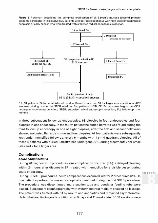
in three subsequent follow-up endoscopies, 68 biopsies in four endoscopies and four biopsies in one endoscopy. In the fourth patient the buried Barrett’s was found during the third follow-up endoscopy in one of eight biopsies, after the first and second follow-up showed no buried Barrett’s in nine and four biopsies. All four patients were subsequently kept under intensified follow-up: every 6 months with 1-cm 4-quadrant biopsies. All of these 4 patients with buried Barrett’s had undergone APC during treatment: 2 for small isles and 2 for a larger area.
ComplicationsAcute complicationsDuring 20 diagnostic ER procedures, one complication occurred (5%): a delayed bleeding within 24 hours after diagnostic ER, treated with hemoclips for a visible vessel during acute endoscopy.During 88 SRER procedures, acute complications occurred in/after 2 procedures (2%). In one patient a perforation was endoscopically identified during the first SRER procedure. The procedure was discontinued and a suction tube and duodenal feeding tube were placed. Subsequent esophagography with watery contrast medium showed no leakage. The patient was treated with nil by mouth and antibiotics and remained asymptomatic. He left the hospital in good condition after 5 days and 11 weeks later SRER sessions were
Figure 3 Flowchart describing the complete eradication of all Barrett’s mucosa (second primary outcome parameter in this study) in 39 patients with Barrett’s esophagus with high-grade intraepithelial neoplasia or early cancer who were treated with stepwise radical endoscopic resection.
* In 34 patients (24 for small isles of residual Barrett’s mucosa; 10 for larger areas) additional APC was used during or after the SRER sessions. Pts, patients; HGIN, BE, Barrett’s esophagus; neo-SCJ, neo-squamo-columnar junction; SRER, stepwise radical endoscopic resection; FU, follow-up; mo, months.
SRER for Barrett’s esophagus with early neoplasia
177
8c h a p t e r

resumed that finally resulted in complete eradication of all neoplasia and BE. The ER-specimen indeed showed a transmural resection and HGIN.One patient suffered from a delayed bleeding within 24 hours after treatment, and was treated by hemoclips for a spurting bleeding during acute endoscopy.
Late complications: post-ER stenosisTen patients (10 of 39, 26%) developed a symptomatic stenosis. All were adequately treated by endoscopic bougienage with a median of 5 (3-5) dilatation sessions. There were no differences between patients with and without stenosis in length of BE (5 [3-5] cm vs. 4 [2-5] cm, p=0.524), number of ER sessions (3 [2-4] vs. 3 [2-3], p=0.462) or the total number of resections/session (3 [2-5] vs. 3 [2-3], p=0.109).
Histology ER specimensPretreatment histological diagnosis In the 20 patients who were included after diagnostic ER, the ER-specimen showed HGIN in nine patients, T1m carcinoma in eight patients and T1sm1 carcinoma in three patients. In all patients the deeper resection margins were free of neoplasia. (Table 2)
Worst histology ER specimens of the first SRER sessionNineteen patients were not expected to have submucosal infiltration and did not undergo a diagnostic ER before inclusion. Histology of the ER specimens of the first SRER session in these patients is shown in Table 2. None of these patients were found to have lesions infiltrating in the submucosa.
Table 2 Histological diagnoses of all consecutive biopsies and resection specimens obtained in 39 patients who underwent stepwise radical endoscopic resection for Barrett’s esophagus with high-grade intraepithelial neoplasia or early cancer.
Histology
n No Barrett’s
No dysplasia
Indefinite for dysplasia
LGIN HGIN T1m T1sm1
Pre-treatment
Pre-treatment biopsies 19 16 3
Diagnostic ER-specimens 20 9 8 3
Overall pre-treatment diagnosis 39 26 10 3
SRER
First SRER session of pts without diagnostic ER
19 4 2 1 8 4
Subsequent SRER sessions 37* 1 5 7 7 14 3
Overall ER diagnosis 37* 1† 2† 1† 18 12 3
LGIN; low-grade intraepithelial neoplasia, HGIN; high-grade intraepithelial neoplasia, ER; endoscopic resection, SRER; stepwise radical endoscopic resection.All deeper ER-resections margins were free of tumor. * Excluding two patients in whom SRER was discontinued due to unrelated comorbidity.† After review of all histological specimens, the pre-treatment diagnosis in one patient was adjusted to LGIN. In three patients the discrepancy between the pre-treatment diagnosis and the worst histology in the ER-specimens persisted after revision.
178

Worst histology of ER specimens obtained at subsequent SRER sessionsIn the 37 patients who completed the treatment protocol, the worst histology of all ER specimens of the additional SRER sessions (i.e., after the diagnostic ER or first SRER session) is shown in Table 2. In one patient the histology of additional SRER sessions was found to be worse than the preceding first SRER session; the histology of the first SRER showed indefinite for dysplasia, probably reactive changes with negative p53 and Ki-67 staining, after HGIN had been diagnosed in the pre-treatment biopsies. In the ER-specimens obtained at the second SRER session, HGIN was found.
Overall histologyThe overall worst histological diagnoses, including all ER specimens obtained at any given time, in the 37 patients who completed the protocol are shown in Table 2. In 4 patients, the HGIN that was diagnosed in the pre-treatment biopsies was not detected in the ER-specimens. After review of all histological specimens (i.e. biopsies and ER-specimens), the pre-treatment diagnosis in one patient was adjusted to LGIN, in 3 patients the discrepancy between the pre-treatment diagnosis and the worst histology in the ER specimens remained after revision.
Follow-upThe median follow-up on May 1st 2005 was 11 (5-18) months. There were no deaths. Patients underwent a median of 2 (1-3) follow-up endoscopies, with a median of 10 (8-14) biopsies per endoscopy. There were no signs of lymph node metastases on EUS. In one patient follow-up was discontinued after 5 months due to a urinary bladder carcinoma. Of the 37 patients in whom the radical resection protocol was completed and in whom complete eradication of HGIN and EC was achieved, no recurrences were observed during follow-up (Figure 2). There was one patient who had buried Barrett’s detected at the third follow-up endoscopy and who had not been identified as such at the first follow-up endoscopy when the primary endpoint was assessed. Since we considered this to be due to sampling error, this patient was still considered to be a failure of complete eradication of all BE and not a recurrence of intestinal metaplasia (Figure 3).
DiscussionER of early neoplasia in BE has been shown to be a safe and effective therapy in small-sized series (4-7;34;35). Leaving the residual Barrett’s segment untreated, however, is associated with 30% metachronous lesions during follow-up (4;5). Therefore, in many centres the residual Barrett’s segment is treated with endoscopic ablation therapy with the purpose of treating synchronous lesions, if present, and preventing development of metachronous lesions. PDT is the most widely used technique for this purpose. PDT, however, does not allow histological evaluation as with an ER specimen, and after treatment with PDT many patients have residual Barrett’s mucosa and/or isles of Barrett’s mucosa buried underneath neo-squamous epithelium (buried Barrett’s) in which oncogenic alterations have been shown to persist (8-10;12;13). Analysis of our own results using 5-aminolevulinic acid (5-ALA) PDT as an adjunct to ER shows a 27% recurrence rate, which questions the
SRER for Barrett’s esophagus with early neoplasia
179
8c h a p t e r

efficacy of this approach (8). Photofrin®-PDT appears to be more effective but is also associated with a higher rate of complications such as the development of esophageal stenosis in approximately 30% of patients and the cutanous photosensitivity (9). A promising alternative appears to be SRER of the complete Barrett’s segment. The complete Barrett’s segment is removed with histological correlation, with the purpose of achieving a better long-term outcome. Conio et al. (15) and Kamler et al. (17) first reported the safety and feasibility of radical resection of the distal 3 cm and 5 cm of the esophageal mucosa in a porcine model, with development of stenosis as most important complication. Satodate et al (36), were the first to report on radical endoscopic resection in humans in a case report. Seewald et al. (18) performed radical endoscopic resection with the ‘simple-snare’ technique in a series of 12 patients with a median Barrett’s length of 5 cm. Two of 12 patients developed a stenosis and were managed by endoscopic bougienage. No recurrence of Barrett’s mucosa or neoplasia was observed during a median follow-up of 9 months. Giovannini et al. (16) reported on SRER in a series of 21 patients with a mean Barrett’s length of 3.5 cm, using the ‘lift-and-cut’ technique. There were no severe complications and none of the patients developed stenosis. In 3 patients (piecemeal) resection of a mucosal carcinoma was considered incomplete, they underwent alternative treatment. Of the 18 patients with successful eradication of neoplasia, 2 patients (11%) showed recurrence of HGIN during follow-up and were successfully retreated with ER. Five patients had residual isles of Barrett’s mucosa embedded in the neosquamous mucosa, in 3 patients with the presence of LGIN. The current study is the first and largest prospective study on SRER for HGIN and EC in Barrett’s esophagus. In the 37 patients who completed the treatment protocol, complete eradication of HGIN and EC was achieved, with no recurrences observed during a median follow-up of 11 months. Complete eradication of all Barrett’s mucosa was achieved in 33 of these 37 (89%) patients. In 108 ER procedures, two acute complications with moderate-severity (delayed bleedings) and one severe acute complication (perforation) were observed. Although the patient in this study who suffered from a perforation remained asymptomatic and recovered without any serious adverse events, this complication was graded as “severe” given its potentially less favourable outcome in other patients. The complication rate of 3% (3 of 108 procedures) is not higher than the complication rate seen in single ER sessions. On a per patient analysis, the acute complication rate (3 of 32) still compares favourably with that of Photofrin®-PDT and surgical resection (9;37;38), the alternative treatment modalities for Barrett’s esophagus patients with early neoplasia. In our opinion, SRER of the complete Barrett’s segment is therefore a relatively safe and effective treatment for selected patients with BE with HGIN and EC.Radical ER is, however, a technically demanding treatment and a number of issues need to be discussed. First, the judgement of whether or not the resection has extended deep enough into the hiatal hernia may be difficult. In 3 patients, we expected the SRER to be complete based on the endoscopic findings. Biopsies taken just below the new Z-line at the first follow-up endoscopy, however, showed residual Barrett’s mucosa, in 1 patient containing LGIN. All 3 patients were treated with additional SRER sessions and reached complete eradication of all Barrett’s mucosa. This underlines the need for an adequate pre-procedural plan (i.e., during the ER procedure, immediately prior
180
to resection), in which the target area is adequately marked, extending below the top of the gastric folds. Furthermore, the completeness of the radical resection (complete eradication of all Barrett’s mucosa) should be confirmed by biopsies taken just beneath the neo-Z-line at follow-up endoscopies. None of the aforementioned series on SRER has reported on this (16;18).Second, it requires experience to adequately resect the target area in a piece-meal resection, trying to avoid leaving residual Barrett’s mucosa between the resection wounds that may require the use of APC. In this series, APC was required for ablating residual Barrett’s mucosa in most patients, in the majority of cases only for small residual isles. The use of endoscopic ablation therapy, including APC, is associated with the development of columnar epithelium with intestinal metaplasia buried underneath neo-squamous epithelium (buried Barrett’s) (39). The clinical relevance of these small foci of buried Barrett’s is uncertain. Some authors believe that the risk of developing malignancy in these buried glands is negligible, since these glands are not in contact with the oncogenic contents of the esophagus (40;41). There are, however, anecdotal reports of submucosal adenocarcinomas diagnosed after APC and PDT that supposedly originated from areas with buried Barrett’s (9;10). In our series, buried Barrett’s was found in 4 patients, with signs of LGIN in 1. All four patients had undergone APC, of whom 2 had undergone APC of a relatively large area. This suggests that the use of APC should be limited to a minimum to prevent the development of buried Barrett’s.Third, with every ER, fibrous tissue is induced making subsequent submucosal lifting and resections more difficult. In 3 patients this led to the inability to perform another SRER session and made more extensive APC treatment necessary. It is therefore important that the first ER is targeted to the area in the Barrett’s segment with the highest grade of neoplasia. In the current study all patients except 1 had their worst histology in their resection specimens obtained at the first ER procedure, which in our opinion reflects the quality of our endoscopic work-up. The development of scar tissue is also the cause of the most significant late complication encountered with this treatment protocol: the development of symptomatic stenosis in 26% (10/39) of patients. This rate is comparable with the 30% rate of post- Photofrin®-PDT stenosis (9). All patients were (successfully) treated with endoscopic bougienage, requiring a median of five sessions. In 4 patients, the HGIN that was diagnosed in pretreatment biopsies was not found on histological evaluation of the ER specimens. All histological specimens (i.e., biopsies and ER specimens) were reviewed and the pretreatment diagnosis was adjusted in 1 patient to LGIN. In the other 3 patients, the discrepancy between the pretreatment diagnosis and the worst histology in the ER specimens persisted. Seewald et al. (18) found similar discrepancies in 3 of 12 patients (25%). There are a number of possible explanations for these discrepancies. In our opinion, HGIN was probably only very focally present in the Barrett’s segment and was either removed completely by the pretreatment biopsies or missed in the ER specimens. One may argue that any treatment of patients with such a focal extent of dysplasia is questionable. Indeed, endoscopic surveillance of selected patients with HGIN and no visible abnormalities may be a valid alternative (42). In addition, the presence of HGIN, if confirmed by an expert pathologist, is still considered
SRER for Barrett’s esophagus with early neoplasia
181
8c h a p t e r

an indication for surgical resection in many centres and discrepancies between pre-treatment diagnosis and surgical resection specimens have been reported as well (43).Future developments within this technique should aim at optimizing ER techniques, so that resection of large areas becomes easier, with better targeting of the subsequent resections in one session in order to minimize the creation of small residual BE between resections with the need for (APC) ablation. New methods should be developed to prevent stenosis after widespread ER. This might be achieved by the topical application of agents that improve healing. Radu et al. (44) used mitomycin C after circumferential resection of esophageal squamous mucosa in 10 sheep, which appeared to prevent stenosis; however, if applied shortly after ER it resulted in perforations in four sheep and has mutagenic effects, which makes human application impossible. Another possible method might be the insertion of a soft, covered stent directly after ER, to protect the wound surface from the damaging esophageal contents and to keep the esophagus in an expanded shape during the healing process, but this remains hypothetical. The median follow-up in this study was 11 months and this raises the question of long-term effects of this treatment policy. It is unknown whether the Barrett’s mucosa will stay eradicated over the years or whether it will recur. Will patients need to be on lifelong endoscopic follow-up? What is the clinical relevance of buried Barrett’s? Until more data are available, patients will have to undergo regular follow-up endoscopies.In summary, SRER is effective for the treatment of selected patients with HGIN or EC in BE. SRER may give rise to a lower recurrence rate, since all the mucosa at risk is effectively removed with histological correlation. However, long-term follow-up results are not yet available. SRER is technically demanding and the use of APC should be limited to prevent the development of buried Barrett’s. Future research should aim at developing easier and faster ER techniques and to prevent the development of stenosis.
Reference List 1. Drewitz, D.J., Sampliner, R.E., Garewal, H.S. The incidence of adenocarcinoma in Barrett’s esophagus:
a prospective study of 170 patients followed 4.8 years. Am.J.Gastroenterol. 1997;92(2):212-215.
2. Hameeteman, W., Tytgat, G.N., Houthoff, H.J. et al. Barrett’s esophagus: development of dysplasia and adenocarcinoma. Gastroenterology 1989;96(5 Pt 1):1249-1256.
3. Shaheen, N.J., Crosby, M.A., Bozymski, E.M. et al. Is there publication bias in the reporting of cancer risk in Barrett’s esophagus? Gastroenterology 2000;119(2):333-338.
4. Buttar, N.S., Wang, K.K., Lutzke, L.S. et al. Combined endoscopic mucosal resection and photodynamic therapy for esophageal neoplasia within Barrett’s esophagus. Gastrointest.Endosc. 2001;54(6):682-688.
5. May, A., Gossner, L., Pech, O. et al. Local endoscopic therapy for intraepithelial high-grade neoplasia and early adenocarcinoma in Barrett’s oesophagus: acute-phase and intermediate results of a new treatment approach. Eur.J.Gastroenterol.Hepatol. 2002;14(10):1085-1091.
6. Pacifico, R.J., Wang, K.K., Wongkeesong, L.M. et al. Combined endoscopic mucosal resection and photodynamic therapy versus esophagectomy for management of early adenocarcinoma in Barrett’s esophagus. Clin.Gastroenterol.Hepatol. 2003;1(4):252-257.
7. Peters, F.P., Kara, M.A., Rosmolen, W.D. et al. Endoscopic treatment of high-grade dysplasia and early stage cancer in Barrett’s esophagus. Gastrointest.Endosc. 2005;61(4):506-514.
182

8. Peters, F., Kara, M., Rosmolen, W. et al. Poor results of 5-aminolevulinic acid-photodynamic therapy for residual high-grade dysplasia and early cancer in barrett esophagus after endoscopic resection. Endoscopy 2005;37(5):418-424.
9. Overholt, B.F., Panjehpour, M., Halberg, D.L. Photodynamic therapy for Barrett’s esophagus with dysplasia and/or early stage carcinoma: long-term results. Gastrointest.Endosc. 2003;58(2):183-188.
10. Van Laethem, J.L., Peny, M.O., Salmon, I. et al. Intramucosal adenocarcinoma arising under squamous re-epithelialisation of Barrett’s oesophagus. Gut 2000;46(4):574-577.
11. Garewal, H., Ramsey, L., Sharma, P. et al. Biomarker studies in reversed Barrett’s esophagus. Am.J.Gastroenterol. 1999;94(10):2829-2833.
12. Krishnadath, K.K., Wang, K.K., Taniguchi, K. et al. Persistent genetic abnormalities in Barrett’s esophagus after photodynamic therapy. Gastroenterology 2000;119(3):624-630.
13. Wang, K.K., Buttar, N.S., Wong Kee Song, L.M. et al. Assessment of genetic defects in patients with complete versus partial ablation after photodynamic therapy (PDT) for Barrett’s esophagus [abstract]. Gastroenterology 2004;126(4 suppl 2):A-177
14. Hage, M., Siersema, P.D., Vissers, K.J. et al. Molecular evaluation of ablative therapy of Barrett’s oesophagus. J.Pathol. 2005;205(1):57-64.
15. Conio, M., Sorbi, D., Batts, K.P. et al. Endoscopic circumferential esophageal mucosectomy in a porcine model: an assessment of technical feasibility, safety, and outcome. Endoscopy 2001;33(9):791-794.
16. Giovannini, M., Bories, E., Pesenti, C. et al. Circumferential Endoscopic Mucosal Resection in Barrett’s Esophagus with High-Grade Intraepithelial Neoplasia or Mucosal Cancer. Preliminary Results in 21 Patients. Endoscopy 2004;36(9):782-787.
17. Kamler, J.P., Borsatto, R., Binmoeller, K.F. Circumferential endoscopic mucosal resection in the swine esophagus assisted by a cap attachment. Gastrointest.Endosc. 2002;55(7):923-928.
18. Seewald, S., Akaraviputh, T., Seitz, U. et al. Circumferential EMR and complete removal of Barrett’s epithelium: a new approach to management of Barrett’s esophagus containing high-grade intraepithelial neoplasia and intramucosal carcinoma. Gastrointest.Endosc. 2003;57(7):854-859.
19. Kara, M.A., Peters, F.P., ten Kate, F.J. et al. Endoscopic video autofluorescence imaging may improve the detection of early neoplasia in patients with Barrett’s esophagus. Gastrointest.Endosc. 2005;61(6):679-685.
20. Kara, M.A., Smits, M.E., Rosmolen, W.D. et al. A randomized crossover study comparing light-induced fluorescence endoscopy with standard videoendoscopy for the detection of early neoplasia in Barrett’s esophagus. Gastrointest.Endosc. 2005;61(6):671-678.
21. Kara, M.A., Peters, F.P., Rosmolen, W.D. et al. High-resolution endoscopy plus chromoendoscopy or narrow-band imaging in Barrett’s esophagus: a prospective randomized crossover study. Endoscopy 2005;37(10):929-936.
22. Kara, M.A., Ennahachi, M., Fockens, P. et al. Detection and classification of the mucosal and vascular patterns (mucosal morphology) in Barrett’s esophagus by using narrow band imaging. Gastrointest.Endosc. 2006;64(2):155-166.
23. Aiko, T. and Sasako, M. The new Japanese Classification of Gastric Carcinoma: Points to be revised. Gastric.Cancer 1998;1(1):25-30.
24. Japanese Gastric Cancer Association Japanese Classification of Gastric Carcinoma - 2nd English Edition -. Gastric.Cancer 1998;1(1):10-24.
25. Inoue, H., Takeshita, K., Hori, H. et al. Endoscopic mucosal resection with a cap-fitted panendoscope for esophagus, stomach, and colon mucosal lesions. Gastrointest.Endosc. 1993;39(1):58-62.
26. Baak, J.P., ten Kate, F.J., Offerhaus, G.J. et al. Routine morphometrical analysis can improve reproducibility of dysplasia grade in Barrett’s oesophagus surveillance biopsies. J.Clin.Pathol. 2002;55(12):910-916.
27. Hulscher, J.B., Haringsma, J., Benraadt, J. et al. Comprehensive Cancer Centre Amsterdam Barrett Advisory Committee: first results. Neth.J.Med. 2001;58(1):3-8.
SRER for Barrett’s esophagus with early neoplasia
183
8c h a p t e r

28. Polkowski, W., Van Lanschot, J.J., ten Kate, F.J. et al. The value of p53 and Ki67 as markers for tumour progression in the Barrett’s dysplasia-carcinoma sequence. Surg.Oncol. 1995;4(3):163-171.
29. Polkowski, W., Meijer, G.A., Baak, J.P. et al. Reproducibility of p53 and Ki-67 immunoquantitation in Barrett’s esophagus. Anal.Quant.Cytol.Histol. 1997;19(3):246-254.
30. Polkowski, W., Baak, J.P., Van Lanschot, J.J. et al. Clinical decision making in Barrett’s oesophagus can be supported by computerized immunoquantitation and morphometry of features associated with proliferation and differentiation. J.Pathol. 1998;184(2):161-168.
31. van Hillegersberg, R., Haringsma, J., ten Kate, F.J. et al. Invasive carcinoma after endoscopic ablative therapy for high-grade dysplasia in Barrett’s oesophagus. Dig.Surg. 2003;20(5):440-444.
32. van Sandick, J.W., Baak, J.P., Van Lanschot, J.J. et al. Computerized quantitative pathology for the grading of dysplasia in surveillance biopsies of Barrett’s oesophagus. J.Pathol. 2000;190(2):177-183.
33. Schlemper, R.J., Riddell, R.H., Kato, Y. et al. The Vienna classification of gastrointestinal epithelial neoplasia. Gut 2000;47(2):251-255.
34. Ell, C., May, A., Gossner, L. et al. Endoscopic mucosal resection of early cancer and high-grade dysplasia in Barrett’s esophagus. Gastroenterology 2000;118(4):670-677.
35. Inoue, H., Fukami, N., Yoshida, T. et al. Endoscopic mucosal resection for esophageal and gastric cancers. J.Gastroenterol.Hepatol. 2002;17(4):382-388.
36. Satodate, H., Inoue, H., Fukami, N. et al. Squamous reepithelialization after circumferential endoscopic mucosal resection of superficial carcinoma arising in Barrett’s esophagus. Endoscopy 2004;36(10):909-912.
37. Rice, T.W., Falk, G.W., Achkar, E. et al. Surgical management of high-grade dysplasia in Barrett’s esophagus. Am.J.Gastroenterol. 1993;88(11):1832-1836.
38. Thomas, P., Doddoli, C., Neville, P. et al. Esophageal cancer resection in the elderly. Eur.J.Cardiothorac.Surg. 1996;10(11):941-946.
39. Hage, M., Siersema, P.D., van Dekken, H. et al. 5-Aminolevulinic acid photodynamic therapy versus argon plasma coagulation for ablation of Barrett’s oesophagus: a randomised trial. Gut 2004;53(6):785-790.
40. Hornick, J.L., Blount, P.L., Sanchez, C.A. et al. Biologic properties of columnar epithelium underneath reepithelialized squamous mucosa in Barrett’s esophagus. Am.J.Surg.Pathol. 2005;29(3):372-380.
41. Kelty, C. and Ackroyd, R. Re-epithelialisation of Barrett’s oesophagus. Gut 2000;47(5):741-
42. Kara, M.A., Bergman, J.J., Tytgat, G.N. Follow-up for high-grade dysplasia in Barrett’s esophagus. Gastrointest.Endosc.Clin.N.Am. 2003;13(3):513-33, viii.
43. Levine, D.S., Haggitt, R.C., Blount, P.L. et al. An endoscopic biopsy protocol can differentiate high-grade dysplasia from early adenocarcinoma in Barrett’s esophagus. Gastroenterology 1993;105(1):40-50.
44. Radu, A., Grosjean, P., Fontolliet, C. et al. Endoscopic mucosal resection in the esophagus with a new rigid device: an animal study. Endoscopy 2004;36(4):298-305.
184

F.P. Peters
K.K. Krishnadath
A.M. Rygiel
W.L. Curvers
W.D. Rosmolen
P. Fockens
F.J. ten Kate
J.W.P.M. van Baal
J.J.G.H.M. Bergman
Submitted
C h a p t e r9Stepwise radical endoscopic resection of the complete Barrett’s esophagus with early neoplasia successfully eradicates pre-existing genetic abnormalities
AbstractBackground & aimsMalignant transformation of Barrett’s mucosa is associated with the accumulation of genetic alterations. Stepwise radical endoscopic resection of the Barrett’s segment with early neoplasia is a promising new treatment resulting in complete re-epithelialization of the esophagus with neo-squamous epithelium. It is unknown whether radical resection also eradicates genetic abnormalities. The aim of this study was to prospectively evaluate whether genetic abnormalities as found in the Barrett’s segment before radical resection are effectively eradicated and absent in the neo-squamous epithelium.
Patients & methodsNine patients with early neoplasia who successfully underwent radical resection were included. Immunohistochemistry was performed to assess proliferation (Ki-67) and p53 protein over-expression. DNA Fluorescent In-Situ Hybridization was performed for evaluation of numerical abnormalities of chromosomes 1 and 9, and losses of p16 and p53. Immunohistochemistry and DNA-FISH were performed on endoscopic resection specimens of the neoplasia and on follow-up biopsies of the neo-squamous epithelium.
ResultsDNA-FISH and IHC showed alterations in the pre-treatment samples of all patients. All showed aneusomy of chromosome 1 and 9. Loss of p16 and p53 were seen in 6 and 8 patients. IHC showed increased proliferation in all patients and in seven patients intense p53 nuclear staining. Post-treatment biopsies showed neo-squamous epithelium with a normal diploid signal count for all DNA-FISH probes and normal IHC stainings in all patients.
ConclusionsRadical resection of Barrett’s esophagus with early neoplasia successfully eradicates pre-existing genetic abnormalities and results in neo-squamous epithelium without these genetic abnormalities.
186

INTRODUCTIONEndoscopic treatment has been proven to be an attractive alternative to surgical esophagectomy for selected Barrett’s esophagus patients with high-grade dysplasia (HGD) or early cancer (EC; defined as cancers restricted to the mucosa (T1m) or superficial submucosa (T1sm1)). Widely used endoscopic treatment strategies, such as endoscopic resection (ER) of neoplastic lesions or ablation by photodynamic therapy (PDT), are faced with a number of drawbacks. ER as mono-therapy only removes the most involved part of the BE and leaves the remaining mucosa in place which is associated with recurrent lesions in up to 30% (1). After PDT most patients still have residual Barrett’s mucosa in which genetic abnormalities have been shown to persist, which may lead to metachronous lesions during follow-up (2-4). As an alternative for ablative therapy of residual HGD/EC, some centres are now treating their patients with stepwise radical endoscopic resection (SRER) of the complete Barrett’s segment (5-7). SRER results in complete re-epithelialization of the resected area with neo-squamous epithelium. It is presumed that SRER completely eradicates the early neoplasia and metaplastic mucosa with all of its genetic abnormalities. There is, however, no evidence for the assumption that the neo-squamous epithelium after SRER is free of genetic abnormalities. Analysis of genetic alterations within mucosal cells can be performed by different techniques, e.g. indirect via protein staining using immuno-histochemistry (IHC) (3;8-10), polymerase chain reaction (PCR) and DNA sequencing (11-13), real time PCR (14;15) or DNA fluorescent in situ hybridization (FISH) (16;17). With DNA-FISH, fluorescent DNA probes are hybridized onto the DNA of the tissue to be investigated. FISH probes are available for separate chromosomes (i.e. peri-centromeric probes), allowing the assessment of numerical chromosomal changes, but also for specific chromosomal regions (i.e. locus specific probes) that enable evaluation of losses or gains of specific genes of interest (such as p53 and p16). The main advantages of the FISH technique are that tissue architecture is preserved and that it can be performed both on fresh-frozen and paraffin-embedded material. The aim of this study was to prospectively evaluate whether genetic abnormalities as found prior to treatment are effectively eradicated by SRER and thus absent in the neo-squamous epithelium.
PATIENTS & METHODSPatientsBE patients with HGD or EC in a visible lesion or mass, who were successfully treated with SRER (7) were eligible for inclusion in this study. All patients signed informed consent for the use of their histological material.
SRER eradicates pre-existing genetic abnormalities
187
9c h a p t e r

MethodsMaterialsParaffin-embedded ER-specimens containing the lesion with HGD or EC were used for the pre-treatment assessment (Figure 1). The specimens were reviewed by an experienced pathologist to identify the area with the most pronounced histological abnormalities. From the corresponding archived paraffin blocks a series of twelve, 4-µm sections were cut. The first and last sections of this series were HE-stained and used to confirm the presence of HGD/EC in the complete series. The two middle slides were used for IHC and the remaining eight for FISH-analysis. For post-treatment assessment paraffin-embedded and fresh frozen (liquid nitrogen) biopsies from the regenerated neo-squamous epithelium were used. The biopsies were obtained during follow-up endoscopies using a large capacity biopsy forceps (FB-50K-1, Olympus Medical Systems GmbH, Hamburg, Germany) and targeted at the former location of the resected lesion with HGD/EC. The paraffin-embedded post-treatment biopsies were used after routine histological assessment by an experienced pathologist. For all fresh frozen samples, a correlating, adjacent biopsy was taken for histological assessment by an experienced pathologist. A series of ten 4-µm sections were cut; again the two middle slides were used for IHC and the remaining eight for FISH analysis.As control measurements for the DNA-FISH scores lymphocyte aggregations within the ER-specimens were used.
Figure 1: Endoscopic images pre- and post-treatment with pictures of the H&E stainings of the corresponding specimens.
A: Endoscopic pre-treatment image showing a 3 cm long Barrett’s segment with a large type 0-IIa lesion.
B: Endoscopic post-treatment image showing the distal esophagus that has completely regenerated with neo-squamous mucosa.
C: H&E staining of the endoscopically resected lesion showing a T1m3 cancer surrounded by high-grade intraepithelial neoplasia.
D: H&E staining of the post-treatment biopsy obtained at the original location of the cancer, showing normal squamous mucosa.
Immuno-histochemical stainingIHC for Ki-67 and p53 (antibodies: Ki-67: mouse monoclonal, Dako, Glostrup, Denmark; p53: p53 ab-8, mouse monoclonal, Neomarkers™, Stratech Scientific Ltd, Cambridgeshire, UK) was performed according to a standard IHC protocol. In summary, paraffin sections were deparaffinized, dehydrated in graded alcohols and immersed in 0.3% hydrogen
188

peroxide in methanol for 20 minutes to suppress endogenous peroxidase activity. After washing (3 times 5 min in phosphate-buffered saline (PBS)), slides were incubated for 10 minutes with 10% goat serum in PBS and incubated with the primary antibody for 60 minutes diluted in Normal Antibody Diluent (Scytek, Logan, Utah, USA). After washing in PBS, post-antibody blocking (Immunologic, Duiven, The Netherlands) was performed for 15 minutes (diluted in PBS). After washing, slides were then incubated with the secondary antibody (Poly-HRP-Goat anti Mouse IgG, Immunologic) at room temperature for 30 minutes in PBS. Slides were washed and peroxidase activity was detected with 3,3’-diaminobenzidine (DAB, Sigma, St Louis, MO), 0.5mg/ml in 0.05M Tris pH 7.6 with hydrogen peroxide (0.05%). Finally sections were counterstained with haematoxylin, dehydrated and mounted with Pertex under coverslips.An experienced pathologist scored the IHC stainings. For Ki-67, the presence of positive staining of the surface epithelium of the neoplastic lesions or the post-treatment biopsies was considered abnormal (scored as positive). For the p53 IHC, the presence of intense nuclear staining in the lesions and post-treatment biopsies was considered abnormal (scored as positive).
Tissue DNA-Fluorescent in situ hybridizationFISH was used for the analysis of aneusomy of chromosomes 1 and 9 and the numerical status of p16 and p53 loci. For aneusomy analysis of chromosome 1, a centromeric probe for chromosome 1 was used (SpectrumOrange™ CEP 1, Vysis Inc., Downers Grove, IL) For chromosome 9 and p16, a probe mix was used containing a locus specific p16 (9p21) probe (SpectrumOrange™ LSI® p16) and a centromeric probe for chromosome 9 (SpectrumGreen™ CEP 9, Vysis Inc.). For p53 a 17p13.1 locus specific probe (SpectrumOrange™ LSI® p53, Vysis Inc.) was used. Probes were pre-treated according to the manufacturer’s protocol.First, paraffin slides were deparaffinized in xylene and dehydrated in graded alcohols. Slides were boiled for 20 min in 10 mM citric acid (pH 6.0) and, after a 10 min cool-down on ice, incubated in pepsin (4 mg pepsin in 1 ml of 0.9% NaCl, pH 1.5, Pepsin A P6887, Sigma-Aldrich Chemie B.V., Zwijndrecht, The Netherlands) for 25-30 minutes at 37°C to enhance tissue permeability. Fresh frozen samples were cut in 4-µm sections and mounted on positively charged, glass slides. After air drying for 24 hours, the slides were aged on a slide warmer set at 55°C for 30 min and then washed for 10 minutes in 2 x Standard Saline Citrate (SSC) at 37°C. Next, slides were incubated in 0.005% pepsin in 0.01 N HCl at 37°C for 30 min and washed in PBS for 5 min at room temperature. Slides were fixed in 5% formaldehyde for 10 min at room temperature and again washed for 5 min in PBS at room temperature.Subsequently, slides (paraffin and fresh frozen) were dehydrated in increasing concentrations of ethanol (70% for 10 min, 85% for 1 min and 100% for 1 min) and dried for 2 min on a slide warmer set at 50°C. Probes were pre-denatured for 5 min at 75°C and immediately put on ice. Then 3 µl probe solution was added on the slides and sealed with a coverslip and rubber cement. The DNA and probes were co-denatured for 3 min at 80°C and then incubated at 37°C in a humidified chamber. After 48 hours the slides were washed for 3 min in 0.4 x SSC at 70°C, then shortly rinsed in 2 x SSC / 0.1% non-
SRER eradicates pre-existing genetic abnormalities
189
9c h a p t e r

ionic detergent Nonindet (NP40) at room temperature and dehydrated in ethanol (1 min in 70%, 1 min in 85% and 1 min in 100% ethanol). Slides were then air-dried and counter-stained and mounted with 3 µl DAPI (Roche, Mannheim, Germany)/Vectashield (Vector laboratories Inc, Burlingame, CA, USA) 1:1000 and stored at 4°C.Analysis was performed using a fluorescent microscope (Olympus BX60, Olympus GmbH, Hamburg, Germany), with probe-specific filters for spectrum red and green, respectively. FISH results were scored by two assessors (FP and KK) blinded for each other results. A minimum of 100 nuclei were scored for the presence of 0, 1, 2, 3, 4 or >4 signals for each probe. The scores of the specimens gave a distribution of the frequencies with which the different numbers of signals were seen. The distribution of these scored frequencies is a result of sectioning of the tissue specimens. Due to sectioning of the tissue into 4 µm sections the nuclei, which on average are 10 µm, in a normal, diploid population will contain two, one or nil signals.
ControlsIn the control samples the distribution of the frequencies of cells with 0, 1, 2, 3, 4 or >4 signals was determined and the mean frequencies of all control samples of the scored signal per probe were calculated. The resulting distributions of the different probes were used as a reference for comparison with the case samples. In the control samples the frequencies were on average 3%, 36%, 60%, 1%, 0% and 0% respectively for chromosome 1, 3%, 37%, 59%, 2%, 0% and 0% respectively for chromosome 9, 5%, 34%, 60%, 1%, 0% and 0% respectively for p16 and 8%, 29%, 61%, 2%, 0% and 0% respectively for p53. These scores are represented in Figure 2.
Statistical data analysisFor descriptive statistics, mean (SD) was used in case of a normal distribution of variables and median (25%-75%) was used for variables with a skewed distribution. The pre- and post-treatment signal score distributions of the case samples were tested by Χ2 test against the calculated distributions of the normal, diploid controls. In case of a
Lymphocyte aggregations in the endoscopic resection specimens were used as controls. In all control samples DNA-FISH showed normal, diploid counts. The frequencies of cells with 0, 1, 2, 3, 4 or >4 signals were scored. The distribution of the frequencies is a result of sectioning of the tissue specimens. Due to sectioning of the tissue into 4 µm sections the nuclei, that on average are 10 µm, in a normal, diploid population will contain two, one or nil signals.
Figure 2: DNA-FISH counts of lymphocyte aggregations in the endoscopic resection specimens, used as normal controls.
190

statistically significant different shift in distribution with to the left, with an increased frequency of 0 and 1 signal compared to the control distribution, the abnormal distribution was classified as a loss. The abnormal distribution was classified as a gain in case of a statistically significant shift in distribution to the right with an increased frequency of 2, 3, 4 and/or >4 signals compared to the control distribution. Aneusomy of chromosomes 1 and 9 was defined as a statistically significant different distribution for the CEP 1 probe or the CEP 9 probe in the case sample compared to the control sample.
RESULTSPatientsNine male patients were included with a median Barrett’s length of 4 cm (3-5). Three patients had HGD in a lesion or mass and six had a mucosal cancer. All patients were successfully treated with the stepwise radical resection protocol (7) and are currently free of neoplasia and Barrett’s esophagus with a median follow-up of 27 months (IQR 25.1-32.9).
Pre-treatment measurementsDNA-FISHAll patients showed an abnormal probe distribution of chromosome 1 with an increase of the number of nuclei with 3 and 4 FISH spots, reflecting a gain of chromosome 1. Two patients had a gain of both chromosome 9 and p16, and 4 patients a loss of both chromosome 9 and p16. Two patients showed a gain of chromosome 9 and a loss of p16 and one patient showed a relative loss of p16, in this case there was a normal p16 count but a gain for chromosome 9. All, but one patient had a loss of p53. (Table 1) Pictures of pre-treatment DNA-FISH samples are shown in Figure 3, the abnormal probe distributions of patient 8 are shown in Figure 4.
Table 1 Pre-treatment results of genetic markers tested in 9 patients treated with stepwise radical endoscopic resection for early neoplasia in Barrett’s esophagus.
Patient Chromosome 1FISH
Chromosome 9FISH
p16 FISH
p53 FISH
p53 IHC
Proliferative activity (Ki-67)
IHC
1 Gain (p<0.001) Loss (p<0.001) Loss (p<0.001) Loss (p=0.001) positive positive surface
2 Gain (p<0.001) Gain (p<0.001) Loss (p=0.003) Loss (p<0.001) positive positive surface
3 Gain (p<0.001) Gain (p<0.001) Gain (p=0.005) Loss (p=0.002) negative positive surface
4 Gain (p<0.001) Loss (p=0.031) Loss (p<0.001) Loss (p=0.029) positive positive surface
5 Gain (p<0.001) Gain (p<0.001) Relative loss* Loss (p=0.011) positive positive surface
6 Gain (p<0.001) Gain (p<0.001) Gain (p<0.001) Normal (p=0.837) positive positive surface
7 Gain (p<0.001) Loss (p<0.001) Loss (p<0.001) Loss (p=0.026) negative positive surface
8 Gain (p<0.001) Gain (p<0.001) Loss (p<0.001) Loss (p<0.001) positive positive surface
9 Gain (p<0.001) Loss (p=0.002) Loss (p<0.001) Loss (p=0.043) positive positive surface
P-values reflect the results of the sample signal distributions tested by chi-square test against the distributions of the normal, diploid controls. * The patient had a normal p16 count (p=0.417) with a gain in chromosome 9. This reflects a relative loss of p16, since there was a gain of chromosome 9 and p16 is located at the short arm of this chromosome. FISH: fluorescent in-situ hybridization, IHC: immuno-histochemical staining, pos.: positive, neg.: negative.
SRER eradicates pre-existing genetic abnormalities
191
9c h a p t e r

A: DNA-FISH sample of endoscopic resection specimen hybridized with a centromeric probe for chromosome 1. This sample had a gain of chromosome 1. The arrows indicate nuclei with more than two signals. B: DNA-FISH sample of a biopsy of neosquamous mucosa hybridized with a centromeric probe for chromosome 1. This sample had a normal diploid distribution of chromosome 1. C: DNA-FISH sample of endoscopic resection specimen hybridized with a centromeric probe for chromosome 9 (green spots) and a locus specific probe for p16 (red spots). This sample had a gain of chromosome 9 and a loss of p16. The arrows indicate nuclei with more than two signals for chromosome 9 with two or less signals for p16. D: DNA-FISH sample of a biopsy of neosquamous mucosa hybridized with a centromeric probe for chromosome 9 (green spots) and a locus specific probe for p16 (red spots). This sample had a normal diploid distribution of chromosome 9 and p16. E: DNA-FISH sample of endoscopic resection specimen hybridized with a locus specific probe for p53. This sample had a loss of p53. The arrows indicate nuclei with one or nil signals for p53. F: DNA-FISH sample of a biopsy of neosquamous mucosa hybridized with a locus specific probe for p53. This sample had a normal diploid distribution of p53.
Figure 3. Different DNA-FISH measurements, pre- and post-treatment.
A: Intense nuclear p53 immuno-histochemical staining of the resected cancer shown in Figure 1.
B: P53 immuno-histochemical staining of the post-treatment biopsy with normal positive staining in the nuclei of the basal cells.
C: Immuno-histochemical ki-67 staining of the cancer with positive staining in the surface epithelium.
D: Immuno-histochemical ki-67 staining of the post-treatment biopsy with normal positive staining of the nuclei of the basal cells.
Figure 5. Different IHC measurements, pre- and post-treatment.
192

Figure 4: Distribution of the number of cells and the number of DNA-FISH counts before and after endoscopic resection of a Barrett’s segment with a mucosal cancer (patient number 8). The following probes were used: centromeric probes for chromosomes 1 and 9 (representing numerical changes of the chromosomes) and locus specific probes for p16 and p53 (representing numerical changes of these tumor suppressor genes)
A: Pre-treatment
B: Control sample
C: Post-treatment
A: Distribution of the frequencies of the counted DNA-FISH signals for chromosomes 1 and 9, p16 and p53 in the endoscopic resection specimen of the mucosal cancer of patient 8. Chromosomes 1 and 9 show a gain with an increase in the number of nuclei with 3, 4 or >4 signals compared to the control sample (B). Both p16 and p53 show a loss with more nuclei with 1 or nil signals compared with the control sample (B).
B: Distribution of the frequencies of the same DNA-FISH signals in lymphocyte aggregations in the endoscopic resection specimens used as controls (see Figure 1).
C: Distribution of same DNA-FISH signals in biopsies of the neo-squamous mucosa obtained during follow-up at the location of the original lesion of patient 8. All DNA-FISH counts show a normal diploid distribution, comparable to the distribution of the control sample.
SRER eradicates pre-existing genetic abnormalities
193
9c h a p t e r

IHC-stainingIn seven out of nine patients nuclei throughout the neoplastic lesion showed intense nuclear staining for the p53 protein, the remaining two had negative staining for p53 protein. Eight patients showed abnormal positive Ki-67 staining at the surface of the mucosa of the neoplastic lesions. In one patient, the surface epithelium was not reliably assessable due to mechanical artefacts of the specimen, the available epithelium, however, showed increased intense Ki-67 staining with an abnormal distribution with positively stained nuclei extending high up towards the surface of the mucosa. (Table 1, Figure 5)
Post-treatment measurementsDNA-FISHAll post-treatment biopsies showed normal neo-squamous epithelium on routine HE staining. The FISH counts of chromosomes 1 and 9 and p16 and p53 showed normal diploid distributions in all nine patients. The median (25%-75%) percentages of 0, 1, 2, 3, 4 and >4 signals in the nine patients were 0% (0-1), 41% (38-48), 58% (50-62), 0% (0-1), 0% (0-0) and 0% (0-0) respectively for chromosome 1, 0% (0-1), 42% (38-47), 58% (53-61), 0% (0-1), 0% (0-0) and 0% (0-0) respectively for chromosome 9, 1% (0-2), 41% (38-48), 58% (51-61), 0% (0-1), 0% (0-0) and 0% (0-0) respectively for p16 and 0% (0-3), 35% (32-41), 65% (57-68), 0% (0-0), 0% (0-0) and 0% (0-0) respectively for p53. (Figure 6)
IHCThe Ki-67 and p53 staining of the post-treatment biopsies showed in all patients a normal distribution of positively stained nuclei at the basal layer of the epithelium and not in higher layers. (Figure 5)
Figure 6: Post-treatment median DNA FISH counts of chromosomes 1 and 9 and p16 and p53 of all nine patients combined.
194

DISCUSSIONDevelopment of adenocarcinoma in Barrett’s esophagus (BE) is a multi-step process (18-
21) paralleled by the accumulation of genetic alterations (22). Fundamental research has shown a large number of genetic alterations which may be involved in this process (22). In esophageal adenocarcinoma several genetic alterations have been shown to occur early in the metaplasia-dysplasia-cancer sequence. Most important are an increased proliferation index, p16 inactivation, p53 loss and aneuploidy that may be present in up to 85%, 88% and 95% of patients respectively (12;22-28). Endoscopic treatment of early Barrett’s neoplasia by endoscopic resection or ablative therapy has shown good short term clinical outcomes (1;29-31). After endoscopic resection of the early Barrett’s neoplasia however, a recurrence rate up to 30% has been reported (1) in the not treated residual Barrett’s segment that may still carry several genetic alterations. In an attempt to remove the entire premalignant lesion patients are now being treated with stepwise radical endoscopic resection (SRER) of the complete Barrett’s segment that results in complete regeneration of the esophagus by neo-squamous epithelium (5-7). Short term clinical results of this treatment protocol are promising (5-7), yet complete eradication of genetic alteration in the neosquamous epithelium has not been demonstrated. Therefore, in this study we hypothesized that resection of the complete Barrett’s segment eradicates all neoplasia, metaplasia and all genetic alterations, thereby reducing the risk of recurrence of neoplasia. Using DNA-FISH we evaluated aneusomy for chromosomes 1 and 9 and for loss of the tumor suppressor genes p16 and p53, in pre- and post treatment biopsies of BE patients with HGD or early neoplastic lesions. In addition IHC was used for evaluation of the p53 and Ki-67 (proliferation) protein expression. The patients were selected from a previously reported cohort of patients treated with stepwise radical endoscopic resection, in which the complete Barrett’s segment is endoscopically resected (7). The selected patients all had complete eradication of all Barrett’s mucosa, as confirmed with lugol staining and jumbo biopsies. All patients had genetic alterations in their neoplastic lesions prior to treatment (Table 1): all had aneusomy for chromosome 1 and 9, all nine patients had abnormal p16 counts, all but one had a loss of p53, seven showed accumulation of p53 protein in the neoplasia and all patients showed increased proliferative activity. In the post-treatment biopsies, none of these genetic alterations were found. The absence of genetic alterations in the regenerated neo-squamous epithelium suggests that the neo-squamous epithelium has no direct malignant potential and is in line with the findings of Paulson et al. (32), who found that pre-existing p16 and p53 abnormalities were absent in under PPI treatment regenerated neo-squamous mucosa in 19 of 20 patients. In one patient an infrequent p16 mutation was present in both the pre-existing Barrett’s mucosa as in the neo-squamous mucosa. Our results strengthen the conclusions of Paulson et al. that the neo-squamous mucosa regenerates from a different progenitor cell than the Barrett’s mucosa, possibly originating from the submucosal peri-esophageal glands (32-34). If indeed the neo-squamous mucosa arises from different progenitor cells, this also strengthens the hypothesis that neo-squamous regeneration will decrease the recurrence rate during follow-up. Although these results are promising, long term clinical follow-up after SRER should confirm a reduced recurrence rate. During a median follow-up of 13 months of 59 patients that
SRER eradicates pre-existing genetic abnormalities
195
9c h a p t e r

have been treated thus far in our hospital with this protocol no recurrences were seen (35). In comparison to alternative ablative strategies such as photodynamic therapy (PDT) for removing the residual Barrett’s segment studies have shown that patients treated with PDT often have residual Barrett’s epithelium and a significant recurrence rate (36;37). The explanation for this recurrence rate can be found in the presence of residual, and possibly even novel genetic alterations after PDT (3;4;8;37;38).A number of limitations of our study should be addressed. First of all, we only performed analysis of pre-treatment samples obtained from the BE with a visible abnormality. As a result, we can not state that the whole Barrett’s segment indeed had genetic alterations prior to treatment. Studies, however, suggest that the majority of biopsies obtained from BE with early neoplasia do have at least one of these genetic alterations (26;39-44). Second, we can not exclude the possibility of sampling error interfering with the post-treatment results. In case the post-treatment biopsies have erroneously been obtained not exactly from the original location of the neoplastic lesion, but from an adjacent area without prior genetic alterations, the absence of post-treatment genetic alterations is not a result of the treatment but a reflection of the pre-treatment state. It is, however, unlikely that this would have occurred in all nine patients and in general, the most malignant lesion in a Barrett’s segment is embedded in an area with (a lower grade of) intraepithelial neoplastic changes, with a high chance of accompanying genetic alterations. We further have evaluated only a limited number of genetic alterations en can’t rule out that other abnormalities may still exist. Therefore, we do not claim that this study shows that SRER eradicates all genetic alterations, it does, however, show that the most frequently found alterations such as anuesomy and p53 en p16 alterations in BE are indeed present in the pre-treatment samples and absent in the post-treatment samples.In conclusion, our results show that genetic alterations, reflecting malignant changes and malignant potential, that were present before treatment, are effectively eradicated with stepwise radical endoscopic resection of the complete Barrett’s segment. This may result in a decreased recurrence rate during follow-up, as compared to endoscopic resection as mono-therapy or combined with PDT. Long-term clinical follow-up, however, should confirm this.
AcknowledgementsK.K. Krishnadath and J.W.P.M. van Baal are supported by the Dutch National Cancer Institute.
Reference List 1. May, A., Gossner, L., Pech, O. et al. Local endoscopic therapy for intraepithelial high-grade neoplasia
and early adenocarcinoma in Barrett’s oesophagus: acute-phase and intermediate results of a new treatment approach. Eur.J.Gastroenterol.Hepatol. 2002;14(10):1085-1091.
2. Hage, M., Siersema, P.D., Vissers, K.J. et al. Genomic analysis of Barrett’s esophagus after ablative therapy: Persistence of genetic alterations at tumor suppressor loci. Int.J.Cancer 2005;
3. Krishnadath, K.K., Wang, K.K., Taniguchi, K. et al. Persistent genetic abnormalities in Barrett’s esophagus after photodynamic therapy. Gastroenterology 2000;119(3):624-630.
196

4. Hage, M., Siersema, P.D., Vissers, K.J. et al. Molecular evaluation of ablative therapy of Barrett’s oesophagus. J.Pathol. 2005;205(1):57-64.
5. Giovannini, M., Bories, E., Pesenti, C. et al. Circumferential Endoscopic Mucosal Resection in Barrett’s Esophagus with High-Grade Intraepithelial Neoplasia or Mucosal Cancer. Preliminary Results in 21 Patients. Endoscopy 2004;36(9):782-787.
6. Seewald, S., Akaraviputh, T., Seitz, U. et al. Circumferential EMR and complete removal of Barrett’s epithelium: a new approach to management of Barrett’s esophagus containing high-grade intraepithelial neoplasia and intramucosal carcinoma. Gastrointest.Endosc. 2003;57(7):854-859.
7. Peters, F.P., Kara, M.A., Rosmolen, W.D. et al. Stepwise radical endoscopic resection is effective for complete removal of Barrett’s esophagus with early neoplasia: a prospective study. Am.J.Gastroenterol. 2006;101(7):1449-1457.
8. Garewal, H., Ramsey, L., Sharma, P. et al. Biomarker studies in reversed Barrett’s esophagus. Am.J.Gastroenterol. 1999;94(10):2829-2833.
9. Krishnadath, K.K., Tilanus, H.W., Alers, J.C. et al. Detection of genetic changes in Barrett’s adenocarcinoma and Barrett’s esophagus by DNA in situ hybridization and immunohistochemistry. Cytometry 1994;15(2):176-184.
10. Polkowski, W., Meijer, G.A., Baak, J.P. et al. Reproducibility of p53 and Ki-67 immunoquantitation in Barrett’s esophagus. Anal.Quant.Cytol.Histol. 1997;19(3):246-254.
11. Kusser, W.C., Levin, D.B., Glickman, B.W. Sensitive two-stage PCR of p53 genomic DNA exons 5-9. PCR Methods Appl. 1993;2(3):250-252.
12. Neshat, K., Sanchez, C.A., Galipeau, P.C. et al. p53 mutations in Barrett’s adenocarcinoma and high-grade dysplasia. Gastroenterology 1994;106(6):1589-1595.
13. Paulson, T.G., Galipeau, P.C., Reid, B.J. Loss of heterozygosity analysis using whole genome amplification, cell sorting, and fluorescence-based PCR. Genome Res. 1999;9(5):482-491.
14. Sarbia, M., Geddert, H., Klump, B. et al. Hypermethylation of tumor suppressor genes (p16INK4A, p14ARF and APC) in adenocarcinomas of the upper gastrointestinal tract. Int.J.Cancer 2004;111(2):224-228.
15. Barrett, M.T., Pritchard, D., Palanca-Wessels, C. et al. Molecular phenotype of spontaneously arising 4N (G2-tetraploid) intermediates of neoplastic progression in Barrett’s esophagus. Cancer Res. 2003;63(14):4211-4217.
16. Doak, S.H., Jenkins, G.J., Parry, E.M. et al. Characterisation of p53 status at the gene, chromosomal and protein levels in oesophageal adenocarcinoma. Br.J.Cancer 2003;89(9):1729-1735.
17. Persons, D.L., Croughan, W.S., Borelli, K.A. et al. Interphase cytogenetics of esophageal adenocarcinoma and precursor lesions. Cancer Genet.Cytogenet. 1998;106(1):11-17.
18. Haggitt, R.C., Tryzelaar, J., Ellis, F.H. et al. Adenocarcinoma complicating columnar epithelium-lined (Barrett’s) esophagus. Am.J.Clin.Pathol. 1978;70(1):1-5.
19. Haggitt, R.C. Barrett’s esophagus, dysplasia, and adenocarcinoma. Hum.Pathol. 1994;25(10):982-993.
20. Haggitt, R.C. Pathology of Barrett’s esophagus. J.Gastrointest.Surg. 2000;4(2):117-118.
21. Tselepis, C., Perry, I., Jankowski, J. Barrett’s esophagus: disregulation of cell cycling and intercellular adhesion in the metaplasia-dysplasia-carcinoma sequence. Digestion 2000;61(1):1-5.
22. Koppert, L.B., Wijnhoven, B.P., van Dekken, H. et al. The molecular biology of esophageal adenocarcinoma. J.Surg.Oncol. 2005;92(3):169-190.
23. Barrett, M.T., Sanchez, C.A., Galipeau, P.C. et al. Allelic loss of 9p21 and mutation of the CDKN2/p16 gene develop as early lesions during neoplastic progression in Barrett’s esophagus. Oncogene 1996;13(9):1867-1873.
24. Gonzalez, M.V., Artimez, M.L., Rodrigo, L. et al. Mutation analysis of the p53, APC, and p16 genes in the Barrett’s oesophagus, dysplasia, and adenocarcinoma. J.Clin.Pathol. 1997;50(3):212-217.
25. Klump, B., Hsieh, C.J., Holzmann, K. et al. Hypermethylation of the CDKN2/p16 promoter during neoplastic progression in Barrett’s esophagus. Gastroenterology 1998;115(6):1381-1386.
26. Wong, D.J., Paulson, T.G., Prevo, L.J. et al. p16(INK4a) lesions are common, early abnormalities that undergo clonal expansion in Barrett’s metaplastic epithelium. Cancer Res. 2001;61(22):8284-8289.
SRER eradicates pre-existing genetic abnormalities
197
9c h a p t e r

27. Gleeson, C.M., Sloan, J.M., McGuigan, J.A. et al. Base transitions at CpG dinucleotides in the p53 gene are common in esophageal adenocarcinoma. Cancer Res. 1995;55(15):3406-3411.
28. Whittles, C.E., Biddlestone, L.R., Burton, A. et al. Apoptotic and proliferative activity in the neoplastic progression of Barrett’s oesophagus: a comparative study. J.Pathol. 1999;187(5):535-540.
29. Buttar, N.S., Wang, K.K., Lutzke, L.S. et al. Combined endoscopic mucosal resection and photodynamic therapy for esophageal neoplasia within Barrett’s esophagus. Gastrointest.Endosc. 2001;54(6):682-688.
30. Pacifico, R.J., Wang, K.K., Wongkeesong, L.M. et al. Combined endoscopic mucosal resection and photodynamic therapy versus esophagectomy for management of early adenocarcinoma in Barrett’s esophagus. Clin.Gastroenterol.Hepatol. 2003;1(4):252-257.
31. Peters, F.P., Kara, M.A., Rosmolen, W.D. et al. Endoscopic treatment of high-grade dysplasia and early stage cancer in Barrett’s esophagus. Gastrointest.Endosc. 2005;61(4):506-514.
32. Paulson, T.G., Xu, L., Sanchez, C. et al. Neosquamous epithelium does not typically arise from Barrett’s epithelium. Clin.Cancer Res. 2006;12(6):1701-1706.
33. Biddlestone, L.R., Barham, C.P., Wilkinson, S.P. et al. The histopathology of treated Barrett’s esophagus: squamous reepithelialization after acid suppression and laser and photodynamic therapy. Am.J.Surg.Pathol. 1998;22(2):239-245.
34. Long, J.D. and Orlando, R.C. Esophageal submucosal glands: structure and function. Am.J.Gastroenterol. 1999;94(10):2818-2824.
35. Peters, F.P., Curvers, W.L., Kara, M.A. et al. Stepwise radical endoscopic resection for complete removal of Barrett’s Esophagus with early neoplasia [abstract]. Gastrointest.Endosc. 2006;63(5):AB133
36. Overholt, B.F., Panjehpour, M., Halberg, D.L. Photodynamic therapy for Barrett’s esophagus with dysplasia and/or early stage carcinoma: long-term results. Gastrointest.Endosc. 2003;58(2):183-188.
37. Peters, F., Kara, M., Rosmolen, W. et al. Poor results of 5-aminolevulinic acid-photodynamic therapy for residual high-grade dysplasia and early cancer in barrett esophagus after endoscopic resection. Endoscopy 2005;37(5):418-424.
38. Wang, K.K., Buttar, N.S., Wong Kee Song, L.M. et al. Assessment of genetic defects in patients with complete versus partial ablation after photodynamic therapy (PDT) for Barrett’s esophagus [abstract]. Gastroenterology 2004;126(4 suppl 2):A-177
39. Barrett, M.T., Sanchez, C.A., Prevo, L.J. et al. Evolution of neoplastic cell lineages in Barrett oesophagus. Nat.Genet. 1999;22(1):106-109.
40. Eads, C.A., Lord, R.V., Kurumboor, S.K. et al. Fields of aberrant CpG island hypermethylation in Barrett’s esophagus and associated adenocarcinoma. Cancer Res. 2000;60(18):5021-5026.
41. Galipeau, P.C., Prevo, L.J., Sanchez, C.A. et al. Clonal expansion and loss of heterozygosity at chromosomes 9p and 17p in premalignant esophageal (Barrett’s) tissue. J.Natl.Cancer Inst. 1999;91(24):2087-2095.
42. Maley, C.C., Galipeau, P.C., Li, X. et al. Selectively advantageous mutations and hitchhikers in neoplasms: p16 lesions are selected in Barrett’s esophagus. Cancer Res. 2004;64(10):3414-3427.
43. Maley, C.C. and Reid, B.J. Natural selection in neoplastic progression of Barrett’s esophagus. Semin.Cancer Biol. 2005;15(6):474-483.
44. Prevo, L.J., Sanchez, C.A., Galipeau, P.C. et al. p53-mutant clones and field effects in Barrett’s esophagus. Cancer Res. 1999;59(19):4784-4787.
198

Summary & Samenvatting

200

SummaryOver the last years, the endoscopic treatment of early Barrett’s neoplasia has rapidly evolved. Its efficacy as curative treatment for high-grade dysplasia and mucosal cancer has been shown and in the Netherlands this treatment regimen is now considered to be the treatment of choice in selected patients. New endoscopic treatment techniques are being developed and existing treatment protocols are constantly optimized.This thesis contains studies on various aspects of different endoscopic treatment techniques and treatment protocols for the treatment of early Barrett’s neoplasia.
Chapter 1 is a review on endoscopic treatment of early Barrett’s neoplasia. The chapter describes the endoscopic work-up and patient selection for endoscopic treatment of patients with early Barrett’s neoplasia, and recommendations are made. Subsequently, the different endoscopic treatment techniques are described, with special emphasis on endoscopic resection. Technical and practical aspects are discussed, an overview of clinical results is presented, and practical recommendations for endoscopic treatment are made.
Chapter 2 describes a prospective study on the safety of endoscopic cap resection in Barrett’s esophagus. This technique is the most frequently used endoscopic resection technique. The acute and early complications (i.e., occurring during the procedure or within 48 hours after the procedure, respectively) of 216 consecutive endoscopic cap resections were evaluated. Acute complications occurred in 51 procedures (24%), 49 bleedings and 2 perforations, and three (1%) early complications occurred, all bleedings. All acute and early bleedings were effectively treated with endoscopic hemostatic techniques. The two perforations were treated conservatively. This study showed that the procedure is safe in expert hands, and that severe complications are rare.
Chapter 3 describes a prospective feasibility study on the multi-band mucosectomy (MBM) technique for widespread endoscopic resection in patients with early Barrett’s neoplasia. The results of 80 MBM procedures were retrospectively compared to 86 prospectively registered endoscopic cap resections. The study showed that MBM allows for safe and easy widespread piece-meal resections in Barrett’s esophagus. Time and costs appear to be saved compared to the endoscopic cap resection technique, and MBM appears to cause less bleeding during the procedure.MBM, however, results in smaller specimens and we, therefore, suggest that MBM is most suited for en-bloc resection of lesions smaller than 10 mm or for widespread resection of flat mucosa.
Chapter 4 describes a study evaluating the histopathological characteristics of endoscopic resection specimens. Specimens obtained during 293 consecutive endoscopic resection procedures in Barrett’s esophagus were evaluated. Most endoscopically resected early Barrett’s neoplasia were lesions of the 0-II, flat type and were well differentiated, radically removed mucosal neoplasia. Type 0-I and type 0-IIc lesions were associated with a higher rate of submucosal invasion than lesions of the 0-IIa, 0-IIb or combined type (26% vs.
Summary & Samenvatting
201
9c h a p t e r

8%), and moderately and poorly differentiated cancers significantly more often invaded the submucosa (15/32 vs. 2/48) or had positive vertical margins (6/32 vs 1/48) than well differentiated cancers. The histology of the endoscopic resection specimens led to a relevant change in treatment policy in 30% of cases. We concluded that endoscopic resection is a valuable diagnostic tool and that moderately and poorly differentiated cancers and type I and IIc lesions are only relative indications for endoscopic resection. Given the importance of correct histological evaluation of endoscopic resection specimens and the problems encountered with the interpretation, we recommend that this evaluation should be performed by an experienced pathologist.
Chapter 5 describes a retrospective study evaluating the surveillance history of Barrett’s esophagus patients with endoscopically treated early neoplasia. The surveillance history of 94 consecutive patients who were endoscopically treated for early cancer or HGIN in a lesion or mass was collected and reviewed. In this selected cohort of patients, 38% was diagnosed with their neoplasia at the initial endoscopy. Of the patients with a surveillance history, the majority (79%) had LGIN diagnosed prior to the diagnosis of HGIN or EC. In the patients without LGIN in their surveillance history the limited number of biopsies obtained at their surveillance endoscopies suggests sampling error, underlining the importance of obtaining an adequate number of biopsies during surveillance endoscopies.
Chapter 6 describes a prospective cohort study evaluating an endoscopic treatment protocol of endoscopic resection combined with photodynamic therapy (PDT) for the treatment of selected patients with early Barrett’s neoplasia. This treatment protocol was used during the first years of the AMC experience with endoscopic treatment for early Barrett’s neoplasia. Twenty-eight patients were treated endoscopically; five patients were immediately entered into a follow-up protocol after the first endoscopic resection, and 23 received additional endoscopic treatment (with either additional endoscopic resection, 5-ALA-PDT and/or APC). Endoscopic treatment was successful in 26/28 patients. During a median follow-up of 19 months, 5/26 patients showed a recurrence of HGIN: all were successfully re-treated with endoscopic resection. This study showed that endoscopic therapy is safe and effective for selected patients with early Barrett’s neoplasia.
Chapter 7 describes a prospective cohort study evaluating the contribution of 5-ALA-PDT to the success of endoscopic treatment. The study patients were separated into group A (n=11), with proven residual HGIN/EC, and group B (n=9) with possible HGIN/EC (positive lateral margins in the endoscopic resection specimen, without HGIN/EC in the remaining Barrett’s segment). The overall success rate for eradication of the HGIN/EC was 15/20 (75%). There was, however, a significant difference in success rate between group A (55%) and group B (100%). Recurrence of HGIN/EC occurred in four patients (two each in groups A and B) after a median follow-up or 30 months. The sustained remission rate in the patients with proven residual neoplasia (group A), therefore, was only 36%.
202

The results showed that, in this selected group of patients, the addition of 5-ALA-PDT after endoscopic resection for HGIN/EC had a disappointing success rate in patients who had proven residual HGD/EC. In addition, after 5-ALA-PDT most patients had residual Barrett mucosa, and 5-ALA-PDT did not seem to prevent recurrences during follow-up. We, therefore, concluded that more efficient alternatives are needed as an adjunct to endoscopic resection for the treatment of residual high-grade dysplasia and early cancer in Barrett’s esophagus.
Chapter 8 describes a prospective cohort study evaluating safety and efficacy of stepwise radical endoscopic resection (SRER) for complete removal of Barrett’s esophagus containing early neoplasia. This treatment protocol was used as an alternative to the protocol combining endoscopic resection with PDT, with the purpose of achieving a more complete and more sustained response compared to the previous protocol. Thirty-nine consecutive patients were included and 37 completed the treatment protocol. Complete removal of all neoplasia was achieved in 100% and complete removal of all Barrett’s mucosa was achieved in 33 (89%) patients. During a median follow-up of 11 months, none had recurrence of neoplasia or Barrett’s mucosa. Symptomatic stenosis, however, occurred in 26% of patients. The results showed that SRER is effective for selected patients with early Barrett’s neoplasia; it provides optimal histopathological diagnosis, and may reduce recurrence rate, since all mucosa at risk is effectively removed. We advised that the use of APC should be limited to prevent buried Barrett’s mucosa and that methods for prevention of stenosis should be developed.
Chapter 9, finally, describes a prospective study evaluating whether genetic abnormalities as found in the Barrett’s segment before SRER are effectively eradicated and absent in the neo-squamous epithelium after SRER. Nine patients with early neoplasia who successfully underwent radical resection were included. Immunohistochemistry and DNA-fluorescent in-situ hybridization (FISH) were performed on endoscopic resection specimens of the neoplasia and on follow-up biopsies of the neo-squamous epithelium. Immunohistochemistry was performed to assess proliferation (Ki-67) and p53 protein over-expression and DNA-FISH for evaluation of numerical abnormalities of chromosomes 1 and 9, and losses of p16 and p53. DNA-FISH and IHC showed alterations in the pre-treatment samples of all patients. All showed aneusomy of chromosome 1 and 9. Loss of p16 and p53 were seen in 6 and 8 patients. IHC showed increased proliferation in all patients and in seven patients intense p53 nuclear staining. Post-treatment biopsies showed neo-squamous epithelium with a normal diploid signal count for all DNA-FISH probes and normal IHC stainings in all patients.The results showed that genetic alterations, reflecting malignant changes and malignant potential, that were present before treatment, are effectively eradicated with SRER of the complete Barrett’s segment. This may result in a decreased recurrence rate during follow-up, as compared to endoscopic resection as mono-therapy or combined with PDT.
Summary & Samenvatting
203
9c h a p t e r


Samenvatting
De technieken beschikbaar voor en de kennis over endoscopische therapie voor vroege vormen van Barrett neoplasie hebben zich de afgelopen jaren in een hoog tempo ontwikkeld. Endoscopische therapie heeft zich bewezen als een effectieve behandeling van deze vroege afwijkingen en wordt in Nederland dan nu ook beschouwd als de behandeling van eerste keuze voor geselecteerde patiënten. Nieuwe behandeltechnieken worden ontwikkeld en nieuwe behandel protocollen continu geoptimaliseerd.Dit proefschrift bevat studies over de verschillende aspecten van een aantal behandeltechnieken en protocollen voor de endoscopische behandeling van vroege vormen van Barrett neoplasie .
Hoofdstuk 1 is een uitgebreid overzichtsartikel over endoscopische therapie voor vroege vormen van Barrett neoplasie . De endoscopische work-up, stagering en selectie van patiënten die in aanmerking komen voor endoscopische behandeling wordt beschreven en er worden aanbevelingen gedaan voor een adequate work-up, stagering en selectie. Vervolgens worden de verschillende beschikbare endoscopische behandel technieken beschreven, met de nadruk op de verschillende endoscopische resectie technieken. Van alle technieken wordt een technische en praktische beschrijving gegeven. Een overzicht van de behaalde klinische resultaten met de verschillende technieken wordt gepresenteerd en we geven praktische aanbevelingen en richtlijnen voor de behandeling.
Hoofdstuk 2 beschrijft een prospectieve studie naar de veiligheid van endoscopische resectie met de cap-techniek in de Barrett slokdarm. Deze cap-techniek is de meest gebruikte endoscopische resectie techniek. De acute (tijdens de procedure) en vroege (binnen 48 uur na de procedure) complicaties van 216 opeenvolgende endoscopische cap-resecties zijn geëvalueerd. Acute complicaties kwamen voor tijdens 51 procedures (24%) en betroffen 49 bloedingen en 2 perforaties. Na drie procedures trad een vroege complicatie op (1%), deze waren allen nabloedingen. Alle acute bloedingen en nabloedingen konden effectief endoscopisch worden behandeld; de perforaties werden conservatief behandeld. Deze studie laat zien dat endoscopische resectie met de cap-techniek veilig is wanneer uitgevoerd door een ervaren endoscopist en dat ernstige complicaties zeldzaam zijn.
Hoofdstuk 3 beschrijft een prospectieve studie naar de technische werkzaamheid van de multi-band mucosectomy (MBM) techniek voor endoscopische resectie van grote gebieden in patiënten met vroege vormen van Barrett neoplasie . De eerste 80 MBM procedures zijn vergeleken met 86, retrospectief verzamelde, endoscopische cap-resecties. De resultaten lieten zien dat de MBM techniek relatief eenvoudig uitvoerbaar en veilig is voor uitgebreide resecties in Barrett slokdarm. De MBM techniek lijkt sneller en goedkoper te zijn en minder bloedingen te geven dan de cap-techniek. De techniek resulteert, echter, wel in kleinere resectie preparaten dan de cap-techniek. Wij zijn daarom van mening dat de MBM techniek in het meest geschikt is voor en-bloc resecties
Summary & Samenvatting
205
9c h a p t e r
van afwijkingen met een diameter kleiner dan 10 mm of voor uitgebreide resecties van vlakke mucosa.
Hoofdstuk 4 beschrijft een studie naar de histologische bevindingen in endoscopische resectie preparaten verkregen bij 293 opeenvolgende endoscopische resecties. Het grootste deel van de endoscopische verwijderde vroege afwijkingen waren lesies van het 0-II, vlakke type die goed gedifferentieerde, radicaal verwijderde neoplastische afwijkingen bevatten. Lesies van het type 0-I en 0-IIc lieten vaker submucosale infiltratie zien dan 0-IIa, 0-IIb of gecombineerde type lesies (26% vs. 8%), net als matig tot slecht gedifferentieerde carcinomen in vergelijking tot goed gedifferentieerde carcinomen (15/32 vs. 2/48). Matig tot slecht gedifferentieerde carcinomen hadden ook vaker positieve diepe resectie vlakken dan goed gedifferentieerde carcinomen (6/32 vs. 1/48). De histologische evaluatie van de resectie preparaten leidde in 30% van de procedures tot een relevante verandering van het behandelplan ten opzichte van het behandelplan zoals opgesteld naar aanleiding van de histologische diagnose op basis van de biopten. Wij concludeerden dat de endoscopische resectie een waardevol diagnostisch hulpmiddel is en dat matig tot slecht gedifferentieerde carcinomen en type 0-I en 0-IIc lesies beschouwd moeten worden als slechts relatieve indicaties zijn voor endoscopische resectie. Gezien het belang van een correcte histologische beoordeling van endoscopische resectie preparaten en de problemen die gepaard gaan met de interpretatie van de bevindingen, zijn wij van mening dat deze beoordelingen moeten worden uitgevoerd door pathologen met ervaring op dit terrein.
Hoofdstuk 5 beschrijft een retrospectieve studie naar de surveillance geschiedenis van 94 opeenvolgende Barrett patiënten die endoscopisch behandeld zijn voor vroegcarcinomen of HGIN in een macroscopisch zichtbare afwijking. De neoplastische afwijking bleek in 38% van deze geselecteerde patiënten groep reeds tijdens de initiële endoscopie te zijn gediagnosticeerd. Van de patiënten met een surveillance voorgeschiedenis had het grootste gedeelte (79%) van de patiënten voorafgaand aan diagnose van de neoplastische afwijking reeds een of meerdere malen een diagnose van LGIN gekregen. Bij de patiënten zonder voorafgaande LGIN was het aantal verkregen biopten tijdens de surveillance scopiën significant lager dan in de patiënten met LGIN. Dit suggereert sampling error en onderstreept het belang van adequate sampling met voldoende biopten tijdens surveillance scopiën.
Hoofdstuk 6 beschrijft een prospectieve studie naar de combinatie van endoscopische resectie met photodynamische therapie (PDT) voor behandeling van vroege vormen van Barrett neoplasie. Dit protocol was het eerste protocol dat in het AMC voor de endoscopische behandeling van vroege vormen van Barrett neoplasie werd toegepast. Achtentwintig patiënten werden met dit protocol behandeld. In vijf patiënten werd de eerste diagnostische endoscopische resectie gevolgd door endoscopische follow-up; 23 patiënten ondergingen na de eerste diagnostische endoscopische resectie aanvullende endoscopische behandeling (met endoscopische resectie, 5-ALA-PDT en/of argon plasma coagulatie (APC)). De endoscopische behandeling was succesvol in 26/28 (93%)
206

patiënten. Tijdens follow-up ontwikkelden vijf van de 26 patiënten opnieuw HGIN; zij konden allen opnieuw endoscopisch worden behandeld. Deze studie laat zien dat endoscopische therapie veilig en effectief is voor geselecteerde patiënten met vroege vormen van Barrett neoplasie .
Hoofdstuk 7 beschrijft een prospectieve cohort studie waarin de aanvullende waarde van 5-ALA-PDT voor het succes van endoscopische therapie werd geëvalueerd. De studiepatiënten werden in twee groepen ingedeeld: groep A (n=11) had bewezen resterende neoplastische afwijking na initiële endoscopische resectie en groep B (n=9) had alleen mogelijk resterende neoplastische afwijkingen op basis van positieve laterale resectie randen zonder neoplastische afwijkingen in het resterende Barrett segment. Het succes percentage over beide groepen was 75% (15/20). Het succes percentage verschilde significant tussen de beide groepen: 55% in groep A en 100% in groep B. Recidief HGIN werd gevonden in vier patiënten tijdens follow-up (mediaan 30 maanden); twee in elke groep. Het percentage patiënten met langdurige remissie was daarom slechts 36% in groep A.De resultaten van deze studie laten zien dat in deze geselecteerde groep patiënten de aanvullende waarde van 5-ALA-PDT teleurstellend was in patiënten met bewezen resterende neoplastische afwijkingen. Daarnaast hebben de meeste patiënten na 5-ALA-PDT resterend Barrett epitheel, PDT is geassocieerd met persisterende, en mogelijk zelfs nieuw geïnduceerde, genetische afwijkingen en het lijkt geen recidieven te voorkomen tijdens follow-up. Wij concludeerden daarom dat er meer efficiënte alternatieven nodig zijn voor de behandeling van resterende neoplastische afwijkingen na endoscopische resectie.
Hoofdstuk 8 beschrijft een prospectieve cohort studie naar de veiligheid en effectiviteit van stapsgewijze radicale endoscopische resectie (SRER) van het gehele Barrett segment met vroege neoplastische afwijkingen. Dit behandelprotocol werd ingevoerd als alternatief voor het protocol dat endoscopische resectie met PDT combineert, met als doel een completere en langduriger respons. Negenendertig patiënten werden in de studie geïncludeerd en in 37 werd het behandelprotocol volbracht. Complete eradicatie van alle neoplastische afwijkingen werd bereikt in alle 37 patiënten. Complete eradicatie van alle Barrett mucosa werd bereikt in 33 patiënten (89%). Tijdens de mediane follow-up van 11 maanden ontwikkelde niemand recidief HGIN of recidief Barrett mucosa. Zesentwintig procent van alle patiënten ontwikkelde een symptomatische oesophagus stenose. De resultaten van deze studie laten zien dat SRER veilig en effectief is voor de behandeling van geselecteerde patiënten met vroege Barrett afwijkingen. SRER resulteert in verwijdering van het gehele Barrett segment met optimale histologische correlatie en mogelijk met een verminderde kans op het ontwikkelen van recidief neoplastische afwijkingen, aangezien al het risicodragende mucosa effectief wordt verwijderd. Wij adviseren naar aanleiding van de resultaten van deze studie dat het gebruik van APC tot een minimum moet worden beperkt om de ontwikkeling van resterend Barrett epitheel onder het neo-squameuze epitheel te voorkomen. Daarnaast moet er worden gezocht naar methoden om de ontwikkeling van symptomatische stenoses te voorkomen.
Summary & Samenvatting
207
9c h a p t e r

Hoofdstuk 9, tenslotte, beschrijft een prospectieve studie waarin de effectiviteit wordt geëvalueerd van SRER in het verwijderen van genetische afwijkingen die aanwezig zijn in het neoplastische Barrett epitheel. Negen patiënten die succesvol werden behandeld met het SRER protocol voor HGIN in een zichtbare afwijking of een vroegcarcinoom werden geïncludeerd. Met behulp van immunohistochemische kleuringen (IHC) en DNA-fluorescent in-situ hybridization (FISH) werden verscheidene afwijkingen geëvalueerd in het endoscopische resectie preparaat van de neoplastische afwijking en in biopten van het geregenereerde neo-squameuze epitheel ter plaatse van de originele afwijking. Met IHC werd de proliferatie index (Ki-67) bepaald, alsook accumulatie van het p53 eiwit. Met DNA-FISH werden numerieke afwijkingen van chromosomen 1 en 9 en van de tumor suppressor genen p16 en p53 onderzocht. In alle patiënten werden in de endoscopische resectie preparaten afwijkingen gevonden met IHC en/of DNA-FISH. Alle patiënten hadden aneusomy van chromosoom 1 en 9. Verlies van p16 en p53 werd gevonden in 6 en 8 patiënten. In alle patiënten werd een toegenomen proliferatie index gevonden en bij 7 patiënten een intense aankleuring van het p53 eiwit in de celkernen. In alle biopten van het neo-squameuze epitheel lieten alle IHC en DNA-FISH onderzoeken geen afwijkingen zien. Deze resultaten laten zien dat genetische afwijkingen die ten grondslag liggen aan maligniteiten en het maligne potentieel van het Barrett epitheel effectief worden verwijderd door SRER van het gehele Barrett segment. Dit zou kunnen resulteren in een vermindering van het aantal recidieven tijdens follow-up, vergeleken met endoscopische resectie als mono-therapie of in combinatie met PDT.
208

List of contributing authors

210

List of contributing authorsM.C. Aalders, PhD: AMC Laser centre, Academic Medical Center,
Amsterdam, The NetherlandsJ.W.P.M. van Baal, PhD: Laboratory of experimental internal medicine,
Academic Medical Center, Amsterdam, The Netherlands
J.J.G.H.M. Bergman, MD, PhD: Department of Gastroenterology and Hepatology, Academic Medical Center, Amsterdam, The Netherlands
K.P.M. Brakenhoff, BSc: Medical student, Academic Medical Center, Amsterdam, The Netherlands
A.C. Bultje: Clinical Epidemiology and Biostatistics, Academic Medical Center, Amsterdam, The Netherlands
W.L. Curvers, MD: Department of Gastroenterology and Hepatology, Academic Medical Center, Amsterdam, The Netherlands
Prof. S.J.H. van Deventer, MD, PhD: Department of Gastroenterology and Hepatology, Academic Medical Center, Amsterdam, The Netherlands
Prof. P. Fockens, MD, PhD: Department of Gastroenterology and Hepatology, Academic Medical Center, Amsterdam, The Netherlands
M.A. Kara, MD, PhD: Department of Gastroenterology and Hepatology, Academic Medical Center, Amsterdam, The Netherlands
Prof. F.J.W. ten Kate, MD, PhD: Department of Pathology, Academic Medical Center, Amsterdam, The Netherlands
K.K. Krishnadath, MD, PhD: Department of Gastroenterology and Hepatology, Academic Medical Center, Amsterdam, The Netherlands
Prof. J.J.B. van Lanschot, MD, PhD: Department of Surgery, Academic Medical Center, Amsterdam, The Netherlands
W.D. Rosmolen, BSc: Department of Gastroenterology and Hepatology, Academic Medical Center, Amsterdam, The Netherlands
A.M. Rygiel-Lukuc, MSc: Laboratory of experimental internal medicine, Academic Medical Center, Amsterdam, The Netherlands
C.E. de Vries, BSc: Medical student, Academic Medical Center, Amsterdam, The Netherlands
List of contributing authors
211
9c h a p t e r


Dankwoord

214

En dan is het nu werkelijk tijd geworden om mijn dankwoord te schrijven. Het dankwoord is een niet geheel onbelangrijk deel van een proefschrift, als het al niet het meest belangrijke deel is. Het is in ieder geval op zijn minst het meest gelezen onderdeel! Eindelijk de kans om ongecensureerd aan iedereen die zo belangrijk voor mij is geweest in de afgelopen jaren persoonlijk te laten weten wat ik zo van ze waardeer. Een meer toepasselijk moment om te beginnen aan dit dankwoord bestaat niet. Mijn zoon en ik hebben net kerstkoekjes gebakken en nu zitten we op de bank Dora’s kerstfeest te kijken. Het is namelijk eerste kerstdag 2006.
En bij wie moet ik anders beginnen dan bij jou Jacques, mijn co-promotor en leermeester. Ik heb zo ongelooflijk veel van je geleerd en niet alleen over de wetenschap. Jij bent in staat alles, altijd op alle mogelijke niveaus te doorzien en te overzien; een gave waar ik hopelijk iets van heb mogen meepikken. Je hebt me geleerd om hoofd- en bijzaken beter van elkaar te scheiden en zo ook betere timemanagement te bereiken; en van wie kan een mens dat beter leren dan van jou. Ik snap namelijk nog steeds niet hoe je in godsnaam al jouw nationale en internationale onderzoeks en klinische werkzaamheden, gezinsleven en hockeypassie in 24 uur weet te passen. En dat zonder cup-a-soup naammokken! Het is onmogelijk alles wat ik van je geleerd heb te beschrijven, mijn oneindige dank daarvoor. Bedankt ook voor al je begrip in de minder makkelijke tijden die het werk wel eens in de weg hebben gestaan de afgelopen jaren. Ondanks dat de timing nu niet helemaal optimaal is, ben ik toch blij dat ik niet al je adviezen heb opgevolgd!
En dan uiteraard Wilda, lieve Willie, lieve paranimf, bedankt voor vier ontzettend leuke jaren!! Bedankt voor alles wat je al die tijd hebt gedaan voor al mijn studies. Ook van jou heb ik ongelooflijk veel geleerd, met name wat betreft omgang met patiënten. Jouw oneindige geduld en inzet voor patiënten is bewonderenswaardig. De persoonlijke aanpak die jij iedereen weet te geven is uniek. Iedere patiënt ken je bij naam en bij iedere naam ken je een gezicht (en slokdarm). Dit gecombineerd met jouw oprechte, openhartige en vooral duidelijke manier van voorlichten en inlichten leidt tot een absolute “win-win” situatie voor patiënt, arts en onderzoeksgroep: er zijn maar weinig onderzoeksgroepen die van zo’n hoge patiëntenparticipatie kunnen genieten. Daarnaast is ook jouw timemanagement capaciteit onovertroffen: een “full-full-full”-time baan, universitaire studie en uitgebreid sociaal leven allemaal tegelijk; hoe krijg je het toch voor elkaar?Maar wat ik denk ik na al die jaren het meest ga missen is ons contact: hoe ga ik ooit nog een collega vinden aan wie ik zo weinig hoef uit te leggen omdat ik toch wel weet dat ze exact hetzelfde denkt?! Toch knap dat we ondanks dat toch nog zoveel te bespreken hadden al die tijd! Onze gelijkgestemdheid leidde tot niet alleen een efficiënte en productieve manier van samenwerken, maar vooral ook tot heel veel lol, gezelligheid, begrip en goede gesprekken. Misschien dat de term “collega’s” ons contact toch een beetje te kort doet? Ik zou je graag op de thee vragen op je ‘roostervrije dag’, maar ja, dan zien we elkaar zeker nooit meer… Kunnen we niet gewoon af en toe nog eens samen naar Amsterdam op en neer rijden om bij te kletsen?
Dankwoord
215

Prof. Fockens, beste Paul, bedankt voor je begeleiding en ondersteuning. Het is leerzaam geweest om de zaken ook vanuit een andere hoek belicht te zien; om me af en toe even bewust te zijn van de relativiteit van het betreffende onderzoek. Maar zie hoe zo’n klein stukje slokdarm toch zo groots kan zijn!
Prof. ten Kate, ik vind het een grote eer dat u mijn promotor wilde zijn. Qua hoeveelheid ‘één-op-één’ contact uren doet uw begeleiding niet ver onder voor die van mijn directe begeleider. Zeker niet als we uren achter de microscoop, voor achten ‘s ochtends (terecht) dubbel tellen! Ik heb in ieder van die uren en van iedere coupe weer meer geleerd; over Barrett’s specifiek, maar zeker ook over de algemene oncologie, wat mij in mijn beoogde carrière plannen hopelijk veel zal opleveren. Het was u nooit teveel om weer een stapel preparaten door te werken, foto’s te maken of om nog even wat blanco’s te snijden: bedankt!
Beste Sheila, ik ben erg blij dat ik naast alle klinische studies ook de kans heb gekregen een meer basale labstudie te doen en dat onder jouw supervisie. Door deze studie heb ik meer inzicht gekregen in de genetica van de Barrett’s slokdarm en de meer algemene oncologie, maar ook meer inzicht in de labwereld en met name ook labfrustraties. Natuurlijk ook dank voor je bijdrage aan en begeleiding bij alle andere studies en het leuke en makkelijke contact van de afgelopen jaren.
Wouter, lieve Wout, mijn andere paranimf, dank je voor je fijne samenwerking en alle gezelligheid: much appreciate it! Ik heb altijd genoten van onze vakinhoudelijke discussies, gesprekken, brainstorm sessies en ik heb er veel van geleerd. We hebben ook een paar mooie reisjes samen mogen maken; toch jammer dat die sauna in Berlijn niet door kon gaan… Bedankt voor je hulp, zeker ook de afgelopen weken en voor je lieve zorgen. Ik wens je alle succes toe in je verdere carrière en natuurlijk alle geluk.
Beste Mo, jammer dat we elkaar nu net mislopen als jij terug komt uit Ierland. Ik heb ongelooflijk veel bewondering voor hoe jij je leven en carrière naar eigen inzicht weet in te vullen. Ik vind het fantastisch dat je alles zo weet te regelen dat je precies kunt doen wat je het liefst wilt met zo veel succes. Bedankt voor je altijd zeer waardevolle, kritische commentaar op mijn stukken, ik heb er veel van geleerd. Beste Joep, ik ben blij dat ik heb mogen meegenieten van jouw tomeloze enthousiasme! Als je ervoor zorgt dat je dat enthousiasme op de juiste manier inzet dan kun je er ver mee komen! Het levert op zijn minst hele verrassende en vernieuwende onderzoeksvraagstellingen op! Ik hoop dat ik ooit in de media de onthullende waarheid achter Coca-cola te horen ga krijgen van ene befaamde dokter Gondrie.Beste Roos, ik vind het ontzettend fijn en leuk dat ik mijn werk kan doorgeven aan iemand die er met zoveel motivatie en enthousiasme, zo voortvarend mee aan de slag gaat. Ik hoop, en verwacht, dat jij met net zoveel plezier aan de therapeutische lijn zult werken als ik al die tijd heb gedaan, het blijft natuurlijk gewoon de mooiste onderzoekslijn!
216

Lieve dames van het Slokdarm team, Annet, Brenda, Carine en Ineke, zonder jullie zou de groep niet kunnen draaien zoals hij nu doet. Dank voor alles wat jullie voor mijn studies hebben gedaan en voor alle gezelligheid. Annet, heel veel succes met het afronden van je studie. Brenda, geniet van je eigen stekkie. Carine, succes met je meiden en zorg voor de eerste succesvolle commerciële proteomics-kanker-bloedtest!! Ineke, geniet van jullie nieuwe huis en jullie geluk samen.
Beste onderzoekers van de labpoot van het Slokdarm team, lieve Jantine, Franceska en Agnieszka. Thank you all for all your help and patience in the lab, I couldn’t have done the FISH-study without you. I’m sorry that a ‘domme-dokter’ like me took up all that time and used all those materials from you. Lieve Tine, dank je voor je hulp en begeleiding en de gezelligheid. De gesprekken met jou hebben me in die tijd erg geholpen, dank je voor je steun. Heel veel succes met je (internationale) carrière, je bruiloft en huwelijk. En gelukkig ben je op tijd terug voor kraamvisite! Franceska, thank you especially for the great time we had in the Ukraine and I wish you both all the happiness in your own home. Agnieszka, thank you for all your FISH-help and all the luck in the world to you!
Kamergenoten van de B1-245, beste René, Huub, Olaf, Sophie, Bart en Frank; ook al waren we dan gedumpt op de benedenverdieping, we hadden wel het mooiste uitzicht van de hele afdeling! Dank jullie allemaal voor alle gezelligheid op de kamer. Bart, ik ben benieuwd wiens kaartje het eerst op de deurmat valt. Heel veel succes en geniet van jullie gezinnetje!Beste Sultans: Mede-onderzoekers van de MDL, het lab en de chirurgie, Olle, Inge, Klaartje, Anne, Tamira, Maartje, Mark, Bram, Niels, Philip, Marinke, en iedereen die ik nog niet heb genoemd. Bedankt voor alle kopjes koffie op het lab. Bedankt voor alle gezelligheid in New Orleans, Los Angeles (wordt lastig die DDW te evenaren of overtreffen) en Berlijn. Veel succes met jullie onderzoeken en maak nog veel mooie reizen met Sultan Solutions!
Alle medewerkers van de MDL, Jitteke, Theo, Agaath, Ann, Shirley, Monim, bedankt voor jullie hulp en gezelligheid! Dames en heer, Marion, van het secretariaat bedankt voor alle ondersteuning.
Mijn beste vrienden, Lien, Steve, Bob en Nora, bedankt voor jullie onmisbare vriendschap. Ik hoop dat we nog jaren lang deze vriendschap mogen delen! Misschien nog eens een ‘reünie’ op Curaçao?
Lieve schoonfamilie, bedankt voor alle gezelligheid en steun. Marijke, bedankt voor alle hulp bij de opvang van Kasper, dat was onmisbaar.
Lieve zussen, lieve Nan en Marij, nog veel belangrijker dan leuke collega’s en gezelligheid op het werk is een hechte familie. Ik ben er trots op en ontzettend blij mee dat wij zo’n goede band met elkaar hebben; jullie zijn onvervangbaar.
Dankwoord
217

Lieve Nan, ontzettend bedankt voor al je hulp bij de opvang van Kasper, zonder die hulp was dit alles zeker nog niet klaar geweest! Lieve Rije, we hadden het natuurlijk liever niet hoeven delen, maar gedeelde smart is toch halve smart. Ons wederzijds begrip en je steun was (en is) onbetaalbaar. Ik wens jullie alle geluk van de wereld en meer; ik hou van jullie.Natuurlijk zijn Sander, Joep, Berend en Jette ook onmisbaar in dit geheel!
Lieve Papa en Mama, bedankt dat jullie me de kans hebben gegeven zover te komen en te bereiken waar ik als heel klein meisje al van droomde. Jullie onvoorwaardelijke en volledige steun is de basis voor alles wat ik gedaan heb en doe. Dit boekje is voor jullie; ik hou van jullie.
Mijn allerliefste mannen, Siem en Kas, wat is het heerlijk om steeds weer bij jullie thuis te komen. Lieve Siem, het scheelt enorm dat we dit samen doen en precies begrijpen van elkaar waar we mee bezig zijn. Het scheelt ook enorm om een privé statisticus en ras-wetenschapper in huis te hebben; heel erg bedankt voor al je (statistische) hulp! Maar meer nog bedankt voor dat je bent wie je bent en bij mij bent, samen met onze fantastische Kas en straks ook zijn broertje of zusje. Ik hou oneindig veel van jullie, en dan nog heel veel meer!
![Binder 216, Terminology [Trematoda Taxon Notebooks]](https://static.fdokumen.com/doc/165x107/63338ab23108fad7760f19c8/binder-216-terminology-trematoda-taxon-notebooks.jpg)