
Endometrial effects of selective estrogen receptor modulators (SERMs) on estradiol-responsive gene...
14
Journal of Steroid Biochemistry & Molecular Biology 84 (2003) 513–526 Endometrial effects of selective estrogen receptor modulators (SERMs) on estradiol-responsive gene expression are gene and cell-specific Yuhua Z. Farnell, Nancy H. Ing ∗ Departments of Animal Science, and Veterinary Anatomy and Public Health, Faculties of Genetics and Reproductive Biology, Texas A&M University, College Station, TX 77843-2471, USA Received 10 June 2002; accepted 23 January 2003 Abstract Three selective estrogen receptor modulator (SERM) drugs which included 4-OH-tamoxifen (Tam), EM-800 (EM) and GW 5638 (GW) were investigated to determine their ability to inhibit estradiol-responsive gene expression in sheep endometrium. The uteri of ovariectomized ewes (10 ewes per SERM group) were infused with 10 −7 M SERMs for 24h prior to hysterectomy. Five ewes from each group received 50 g 17-estradiol (E2) and the remaining five ewes received vehicle 18h prior to hysterectomy. Northern blot analyses and in situ hybridization demonstrated that E2 treatment increased estrogen receptor (ER), progesterone receptor (PR), glyceraldehyde 3-phosphate dehydrogenase (GAPDH), and cyclophilin (CYC) mRNA levels in most endometrial cells examined. Tam and GW exhibited characteristics similar to E2 by increasing ER gene expression, but they antagonized the E2-induced increases in PR and CYC mRNA levels. EM acted as an E2-agonist of GAPDH gene expression, but antagonized the E2 up-regulation of ER, PR and CYC gene expression in most endometrial cells. Immunohistochemistry determined that EM decreased ER protein levels in the glandular epithelium, and the SERMs investigated antagonized increases in PR protein levels in endometrium. In conclusion, GW and EM exhibit fewer agonist effects than Tam on endometrial gene expression. EM demonstrated the greatest antagonism of E2-enhanced levels of ER, PR and CYC, likely due to the inhibition of ER gene expression at both mRNA and protein levels. © 2003 Elsevier Science Ltd. All rights reserved. Keywords: Selective estrogen receptor modulator; Estradiol; Endometrium; Gene expression 1. Introduction Estrogens play important roles in the growth regula- tion, development, and differentiation of the reproductive tract, mammary glands, and central nervous system (re- viewed in [1]). Estrogens bind to estrogen receptor (ER) proteins, which in association with cofactors, and activate transcription of estrogen-responsive genes [2,3]. Selective estrogen receptor modulators (SERMs) are synthetic com- pounds developed to bind ER proteins and act as estrogen (E2)-antagonists in mammary gland (such as tamoxifen; Tam) or E2-agonists in bone (such as raloxifene) [3,4]. Tam, one of SERMs currently used in the treatment and prevention of breast cancer [5], has been shown to express mixed estrogenic/anti-estrogenic activity in uteri of women and different rodent species [6,7]. In women, its use to pre- vent breast cancer growth is correlated to an increased risk This is the first paper in a set, with the second concerning myometrial effects in the same ewes. ∗ Corresponding author. Tel.: +1-979-862-2790; fax: +1-979-862-3399. E-mail address: [email protected] (N.H. Ing). of endometrial cancer [8,9]. In sheep endometrium, Tam acts as an E2-agonist in the up-regulation of ER and glyc- eraldehyde 3-phosphate dehydrogenase (GAPDH) mRNA concentrations [10]. Two new SERMs, GW 5638 (GW) and EM-800 (EM) are currently being tested in clinical trials as “pure anti-estrogens”. As shown in Fig. 1, GW is a triphenylethy- lene derivative, structurally related to tamoxifen, while EM is a derivative of benzopyran structurally related to ralox- ifene [11,12]. In the rat, GW antagonized E2-increased uterine weight, while displaying minimal uterotropic activ- ity when used alone [13]. Also, GW acted as an E2-agonist in both bone and cardiovascular system, thus demonstrating a high degree of tissue specificity [11]. EM is an orally active anti-tumor agent for mammary cancer in the rat [14]. EM displayed pure anti-estrogenic effects in mouse uterus, vagina, mammary gland, and hypothalamic–pituitary axis as well as in Ishikawa cells, a human endometrial carcinoma cell line [15,16]. EM also prevented bone loss and lowered serum cholesterol levels without estro- genic effects on the endometrium in ovariectomized rats [17–19]. 0960-0760/03/$ – see front matter © 2003 Elsevier Science Ltd. All rights reserved. doi:10.1016/S0960-0760(03)00076-1
-
Upload
independent -
Category
Documents
-
view
1 -
download
0
Transcript of Endometrial effects of selective estrogen receptor modulators (SERMs) on estradiol-responsive gene...
Journal of Steroid Biochemistry & Molecular Biology 84 (2003) 513–526
Endometrial effects of selective estrogen receptor modulators (SERMs)on estradiol-responsive gene expression are gene and cell-specific�
Yuhua Z. Farnell, Nancy H. Ing∗Departments of Animal Science, and Veterinary Anatomy and Public Health, Faculties of Genetics and Reproductive Biology,
Texas A&M University, College Station, TX 77843-2471, USA
Received 10 June 2002; accepted 23 January 2003
Abstract
Three selective estrogen receptor modulator (SERM) drugs which included 4-OH-tamoxifen (Tam), EM-800 (EM) and GW 5638(GW) were investigated to determine their ability to inhibit estradiol-responsive gene expression in sheep endometrium. The uteri ofovariectomized ewes (10 ewes per SERM group) were infused with 10−7 M SERMs for 24 h prior to hysterectomy. Five ewes from eachgroup received 50�g 17�-estradiol (E2) and the remaining five ewes received vehicle 18 h prior to hysterectomy. Northern blot analysesand in situ hybridization demonstrated that E2 treatment increased estrogen receptor (ER), progesterone receptor (PR), glyceraldehyde3-phosphate dehydrogenase (GAPDH), and cyclophilin (CYC) mRNA levels in most endometrial cells examined. Tam and GW exhibitedcharacteristics similar to E2 by increasing ER gene expression, but they antagonized the E2-induced increases in PR and CYC mRNAlevels. EM acted as an E2-agonist of GAPDH gene expression, but antagonized the E2 up-regulation of ER, PR and CYC gene expressionin most endometrial cells. Immunohistochemistry determined that EM decreased ER protein levels in the glandular epithelium, and theSERMs investigated antagonized increases in PR protein levels in endometrium. In conclusion, GW and EM exhibit fewer agonist effectsthan Tam on endometrial gene expression. EM demonstrated the greatest antagonism of E2-enhanced levels of ER, PR and CYC, likelydue to the inhibition of ER gene expression at both mRNA and protein levels.© 2003 Elsevier Science Ltd. All rights reserved.
Keywords:Selective estrogen receptor modulator; Estradiol; Endometrium; Gene expression
1. Introduction
Estrogens play important roles in the growth regula-tion, development, and differentiation of the reproductivetract, mammary glands, and central nervous system (re-viewed in [1]). Estrogens bind to estrogen receptor (ER)proteins, which in association with cofactors, and activatetranscription of estrogen-responsive genes[2,3]. Selectiveestrogen receptor modulators (SERMs) are synthetic com-pounds developed to bind ER proteins and act as estrogen(E2)-antagonists in mammary gland (such as tamoxifen;Tam) or E2-agonists in bone (such as raloxifene)[3,4].Tam, one of SERMs currently used in the treatment andprevention of breast cancer[5], has been shown to expressmixed estrogenic/anti-estrogenic activity in uteri of womenand different rodent species[6,7]. In women, its use to pre-vent breast cancer growth is correlated to an increased risk
� This is the first paper in a set, with the second concerning myometrialeffects in the same ewes.
∗ Corresponding author. Tel.:+1-979-862-2790; fax:+1-979-862-3399.E-mail address:[email protected] (N.H. Ing).
of endometrial cancer[8,9]. In sheep endometrium, Tamacts as an E2-agonist in the up-regulation of ER and glyc-eraldehyde 3-phosphate dehydrogenase (GAPDH) mRNAconcentrations[10].
Two new SERMs, GW 5638 (GW) and EM-800 (EM)are currently being tested in clinical trials as “pureanti-estrogens”. As shown inFig. 1, GW is a triphenylethy-lene derivative, structurally related to tamoxifen, while EMis a derivative of benzopyran structurally related to ralox-ifene [11,12]. In the rat, GW antagonized E2-increaseduterine weight, while displaying minimal uterotropic activ-ity when used alone[13]. Also, GW acted as an E2-agonistin both bone and cardiovascular system, thus demonstratinga high degree of tissue specificity[11]. EM is an orallyactive anti-tumor agent for mammary cancer in the rat[14]. EM displayed pure anti-estrogenic effects in mouseuterus, vagina, mammary gland, and hypothalamic–pituitaryaxis as well as in Ishikawa cells, a human endometrialcarcinoma cell line [15,16]. EM also prevented boneloss and lowered serum cholesterol levels without estro-genic effects on the endometrium in ovariectomized rats[17–19].
0960-0760/03/$ – see front matter © 2003 Elsevier Science Ltd. All rights reserved.doi:10.1016/S0960-0760(03)00076-1
514 Y.Z. Farnell, N.H. Ing / Journal of Steroid Biochemistry & Molecular Biology 84 (2003) 513–526
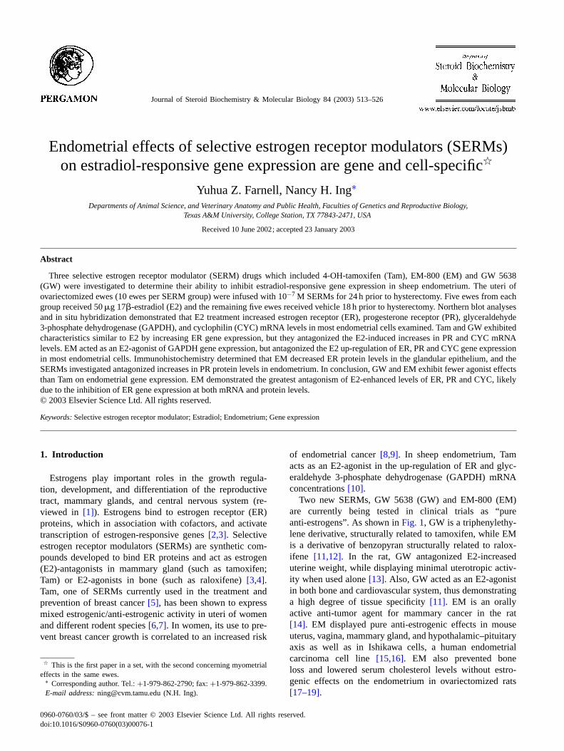
Fig. 1. Chemical structures of 17�-estradiol and SERMs used in this study. 4-OH tamoxifen (Tam), a triphenylethylene SERM, is the active metaboliteof tamoxifen. The compound GW 5638 (GW) is a new SERM that is structurally related to tamoxifen. EM-800 (EM) is the derivative of benzopyran.
During estrous and menstral cycles, the pre-ovulatorysurge of estrogen alters gene expression in endometrium tofacilitate the preparatory development of pre-implantation.Our previous work has demonstrated that a single physiolog-ical dose of E2 mimics the pre-ovulatory surge of estrogenand up-regulates ER, progesterone receptor (PR), GAPDHand cyclophilin (CYC) mRNA levels at 18 h post-injection insheep endometrium[20,21]. In this paper, the E2-responsivegenes[20,21] found in sheep endometrium are evaluated invivo based on the agonist and antagonist effects of the threeSERMs: Tam, GW and EM.
2. Materials and methods
2.1. Chemicals
All chemicals (including Tam) were purchased fromSigma (St. Louis, MO), unless otherwise indicated. EMand GW were obtained from Dr. Fernand Labrie (LavalUniversity; Qué., Canada) and Dr. David C. Morris (GlaxoWellcome Research and Development; Durham, NC), re-spectively. Tam, GW and EM were dissolved in ethanol tomake 10−3 M stock solutions. They were diluted to 10−7 Min 0.1% ovine serum albumin (OSA) in phosphate-buffered
saline (PBS) for infusion. E2 was dissolved in a mini-mal amount of ethanol prior to dilution to 100�g/ml incharcoal-stripped corn oil (Kodak; Rochester, NY).
2.2. Animals, treatments and sample collection
After confirmation of estrous cycles of normal duration(16–18 days), cross-bred Rambouillet ewes were ovariec-tomized and their uterine horns were fitted with the catheters[22]. Fifteen days after ovariectomy, four groups of ewes (10ewes per group) were infused with 10−7 M SERMs or drugvehicle (0.1% ovine serum albumin in phosphate-bufferedsaline, OSA in PBS) at rate of 3 ml/h for 24 h in each uterinehorn via indwelling catheters. Eighteen hours prior to hys-terectomy, five ewes in each group were injected intramus-cularly with E2 (50�g) and remaining five ewes receivedvehicle (0.5 ml charcoal-stripped corn oil). The time for E2treatment was chosen for its rapid ER mRNA accumulation[20]. For subsequent histochemistry, a 1 cm cross-sectionwas removed from each uterine horn distal to the externalbifurcation and fixed in 4% paraformadehyde[23]. The en-dometrium was dissected from the remaining uterus, minced,snap-frozen in liquid nitrogen, and stored at−80◦C. TheTexas A&M University Laboratory Animal Care and UseCommittee approved all animal procedures.
Y.Z. Farnell, N.H. Ing / Journal of Steroid Biochemistry & Molecular Biology 84 (2003) 513–526 515
2.3. Total RNA preparation and Northern blot analysis
Total cellular RNA was extracted from 0.5 g of en-dometrium using Tripure Reagent according to themanufacturer’s instructions (Boehringer Mannheim; Indi-anapolis, IN). Northern blot analysis of total cellular RNA(8�g per lane) was performed as previously described[23]. Antisense cRNA probes were generated for probingestrogen-responsive (ER, PR, GAPDH and CYC) mRNAs,as previously described[20,21]. Blot hybridization andwashing were performed according to Ing et al.[20]. Hy-bridization signals were captured by directly scanning blotson InstantImager (Packard, Meriden, CT).
2.4. Identifying the endometrial cells that change theexpression of E2-responsive genes by in situ hybridization
Adjacent uterine cross-sections (7 mm) from each ewewere placed on Superfrost Plus slides (Curtin Matheson Sci-entific, Houston, TX) less than 1 week before histochem-ical development. In situ hybridization was performed toidentify the response of specific uterine cells to SERMsand E2 as described previously[21]. Antisense and sensecRNA probes were generated with (35S)-UTP in place of(32P)-UTP for in vitro transcription reactions. Slides weredipped in NTB-2 autoradiography emulsion (Eastman, Ko-dak) and exposed for 5 weeks for ER and PR mRNA data or8 weeks for GAPDH and CYC mRNA data. Sections werecounterstained with hematoxylin. Quantitative analyses ofsilver grains (relating to amount of mRNAs) were assessedusing the Reichert MicroStar IV Microscope (Diagnostic In-strument; MI) and NIH image 1.61 software[21]. For eachmRNA examined, pixel densities were assessed on sectionsprobed with antisense and sense cRNAs. Pixel densities ofsilver grain were analyzed for five regions containing 15–25cells of a cell compartment in three view fields for each uter-ine section. The in situ hybridization signals for an mRNAwere analyzed alongside the sense signals (non-specific) onthe same area of an adjacent section, used as a covariate inthe analyses.
2.5. Immunohistochemistry to characterize regulation ofER and PR protein levels by SERMs
Immunohistochemical procedures were performed as de-scribed previously[23]. Briefly, uterine cross-sections weretreated with 0.3% H2O2 for 30 min at room temperature,0.5 mg/ml of Pronase for 8 min at 37◦C and then subjectedto protein blocker (Biostain Super ABC kit; Biomeda, Fos-ter City, CA). Sections were incubated with rat anti-ERantibody H222 (4�g/ml), which is a monoclonal antibodyraised against human ER� (a gift from Dr. G. Greene, Uni-versity of Chicago, Chicago, IL), biotinylated goat anti-ratIgG and peroxidase-labeled avidin (Biostain Super ABCkit). The chromagen used for peroxidase localization was
3,3′-diaminobenzidine tetrahydrochloride. Another mono-clonal antibody, mouse anti-human ER 1D5 (DAKO A/S,Glostrup, Denmark), which, like H222, does not cross-reactwith ER� [24], was also used in immunohistochemistry. Fordetection of PR protein, we used the MA1-411 antibody(Affinity BioReagents, Golden, CO) and microwave antigenretrieval[23]. Non-immune rat and mouse IgG replaced theantibodies, respectively, on adjacent sections for negativecontrols.
2.6. Statistical analysis
All quantitative data were analyzed by least-squaresANOVA using the general linear model procedure of SAS.Data analysis of gene expression used 18S rRNA hybridiza-tion signals to correct for unequal RNA loading. Data arepresented as least-squares means and standard errors of themean for each treatment group. The E2 treatment group datawas compared to control (Con) group data to identify E2effects. Data from SERM treatment alone groups were com-pared to those from the Con group to identify E2-agonisteffects of SERMs. Data from SERM+ E2 groups werecompared to E2 group to identify E2-antagonistic effects.Level of statistical significance was considered to be com-parisons withP-values less than or equal to 0.05, unlessotherwise indicated.
3. Results
3.1. Northern blot analysis identified gene-specific effectsof SERMs
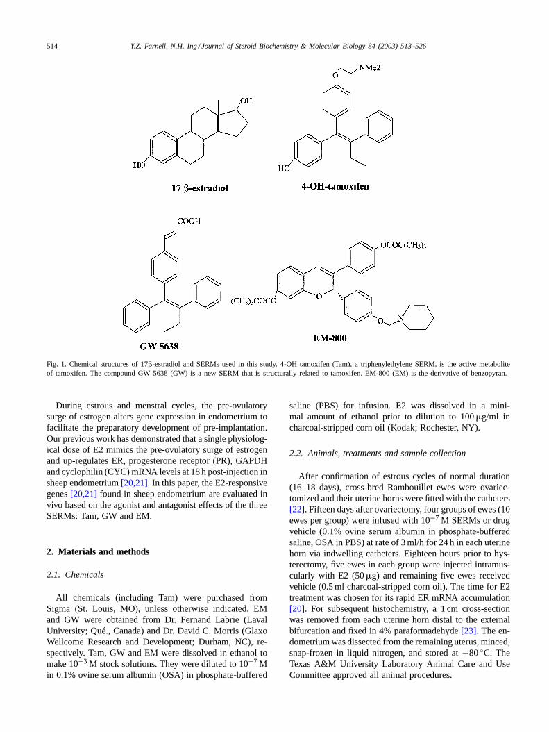
We quantitated the effects of E2 with and without SERMson the regulation of E2-responsive genes in the endometriumof ovariectomized ewes with Northern blot analyses. Hy-bridization signals for ER, PR, GAPDH, and CYC mRNAsand 18S rRNA are shown inFig. 2A for all of the ewes.Quantitation of these results demonstrated that ER gene ex-pression was up-regulated by Tam and GW alone treatmentsby more than 100% compared to those in the Con groupewes, similar to the up-regulation by E2 (indicated by aster-isks in Fig. 2B). Only EM antagonized E2’s up-regulationof ER gene expression (indicated by double asterisks inFig. 2B) and it did so completely. Results from the PR geneindicated that SERMs tested alone showed no agonist actionor replication of E2’s 23% up-regulation of PR gene ex-pression. However, both Tam and GW antagonized this E2response. For the expression of GAPDH and CYC genes,Tam and EM acted as E2-agonists, inducing an average 45%increase in GAPDH and CYC mRNA levels, similar to theincrease in response to E2. Northern blot analysis detectedno antagonistic effect towards the E2 up-regulation of ei-ther GAPDH or CYC mRNA levels in endometrium by anySERM. Thus, all SERMs exhibited partial agonist/partialantagonist effects in endometrium that were gene-specific.
516 Y.Z. Farnell, N.H. Ing / Journal of Steroid Biochemistry & Molecular Biology 84 (2003) 513–526
Fig. 2. Effects of SERMs with and without E2 on estrogen receptor (ER), progesterone receptor (PR), glyceraldehyde 3-phosphate dehydrogenase(GAPDH) and cyclophilin (CYC) mRNA concentrations in endometrium. Total cellular RNA was prepared from endometrium from each ewe in the eighttreatment groups: control (Con), E2, Tam, Tam+ E2, GW, GW+ E2, EM, EM+ E2 (n = 5 ewes/group). Replicate Northern blots were hybridized with32P-labeled antisense cRNA probes for ER, PR, GAPDH, and cyclophilin mRNAs and 18S rRNA. Panel A shows the raw data while panel B presentsthe quantitative analysis of their hybridization signals as least-squares means± standard error of the means. For treatment groups: Con (open, hatched),E2 (filled, hatched), Tam (open, stippled), Tam+ E2 (filled, stippled), GW (open, cross-hatched), GW+ E2 (filled, cross-hatched), EM (open, horizontalstrips), and EM+ E2 (filled, horizontal stripes). Values are normalized to those of Con group, set at 100. Significant E2-agonist effects (differencescompared to the Con group) are indicated by asterisks (*) while antagonist effects (compared to E2 group) are indicated by asterisks (**) over the bars.
3.2. Cell-specific effects of SERMs on endometrial geneexpression
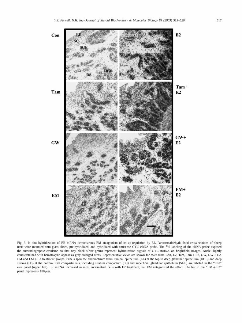
In situ hybridization identified the regulation of gene ex-pression in the specific endometrial cells that responded toSERMs± E2 treatments. Representative images of signalsfor ER, PR, GAPDH and cyclophilin mRNAs on sheependometrial cross-sections are shown inFigs. 3–6, respec-tively, for ewes from each treatment group. Brightfieldimages show the pinpoint black silver grains from in situhybridization over the cell nuclei stained lightly with hema-toxylin. Each panel spans the endometrium from luminalepithelium (LE) at the upper part of the panel to deep glan-dular epithelium (DGE) and surrounding stroma (SS) atlower part of the picture. These cell compartments, along
with stratum compactum (SC) and superficial glandular ep-ithelium (SGE), are labeled in the upper left panel from aCon ewe inFig. 3. This panel shows very little hybridiza-tion signal for ER mRNA over the gray cell nuclei. E2treatment increases the ER mRNA signals strongly in allbut the LE cells. The effect is not antagonized by Tam orGW, but it is antagonized by EM. InFig. 4, E2 enhance-ment of PR mRNA levels is obvious when comparing thetop two panels. The primary response was in the epithelialand superficial stromal cells. All SERMS tested antago-nized the effect of E2 but EM did this most completelyacross the different endometrial cell types. GAPDH mRNA(Fig. 5) was up-regulated by E2, Tam and EM treatments.Although it was strongly expressed in the epithelial cells,the stromal cells also responded. SERM treatment did
Y.Z. Farnell, N.H. Ing / Journal of Steroid Biochemistry & Molecular Biology 84 (2003) 513–526 517
Fig. 3. In situ hybridization of ER mRNA demonstrates EM antagonism of its up-regulation by E2. Paraformaldehyde-fixed cross-sections of sheeputeri were mounted onto glass slides, pre-hybridized, and hybridized with antisense CYC cRNA probe. The35S labeling of the cRNA probe exposedthe autoradiographic emulsion so that tiny black silver grains represent hybridization signals of CYC mRNA on brightfield images. Nuclei lightlycounterstained with hematoxylin appear as gray enlarged areas. Representative views are shown for ewes from Con, E2, Tam, Tam+ E2, GW, GW+ E2,EM and EM+ E2 treatment groups. Panels span the endometrium from luminal epithelium (LE) at the top to deep glandular epithelium (DGE) and deepstroma (DS) at the bottom. Cell compartments, including stratum compactum (SC) and superficial glandular epithelium (SGE) are labeled in the “Con”ewe panel (upper left). ER mRNA increased in most endometrial cells with E2 treatment, but EM antagonized the effect. The bar in the “EM+ E2”panel represents 100�m.

518 Y.Z. Farnell, N.H. Ing / Journal of Steroid Biochemistry & Molecular Biology 84 (2003) 513–526
Fig. 4. In situ hybridization demonstrates that all three SERMs antagonize E2 up-regulation of PR mRNA levels. In situ hybridization was performedand results are presented as described inFig. 3. The bar in the “EM+ E2” panel represents 100�m. E2 increased PR mRNA levels in most endometrialcells, but SERMs antagonized the effect especially in the deep endometrium.
not antagonize or add to the E2 effect on GAPDH geneexpression.
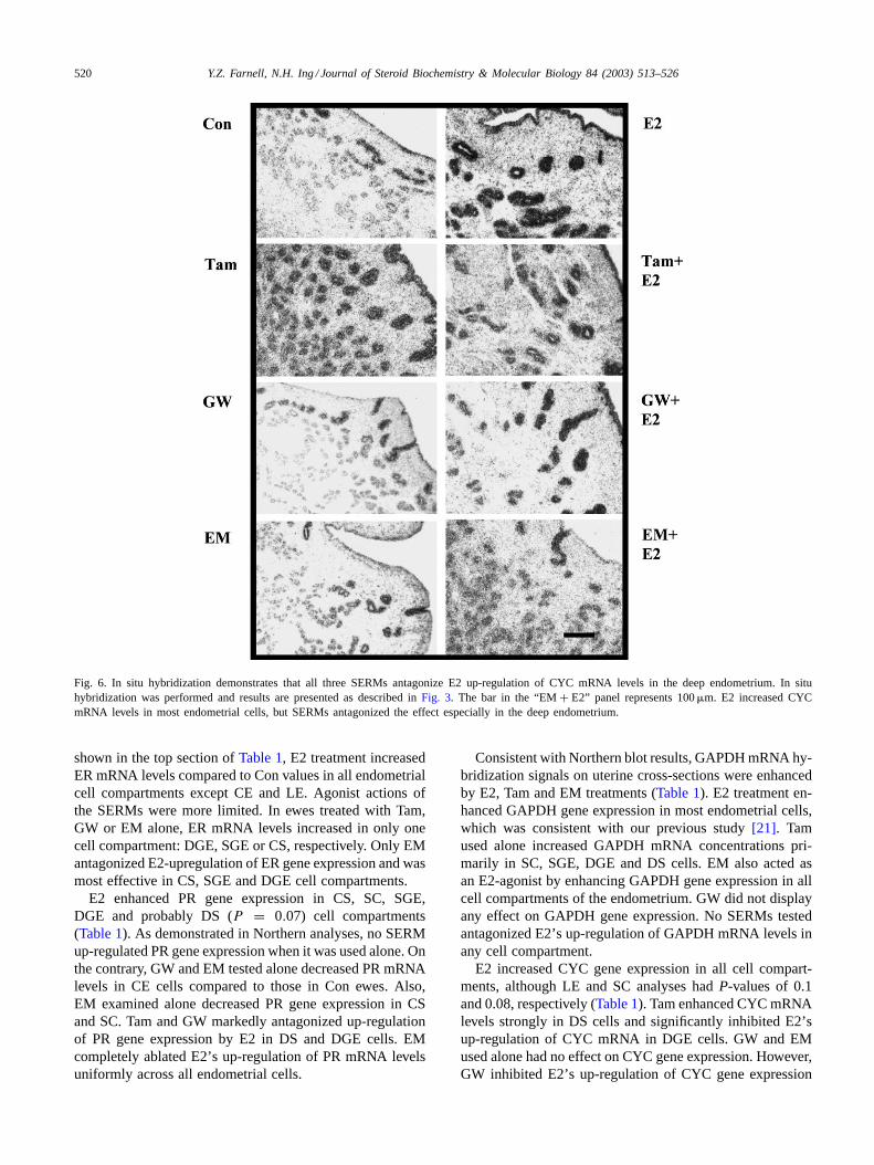
While similar data for ER, PR, and GAPDH gene ex-pression have been shown for Con and E2 ewes in previ-ous works[21,23], this is the first description of CYC geneexpression in uterine cross-sections. Cyclophilin mRNA ap-peared most prevalent in superficial epithelium, LE and SGE
in Con, GW and EM ewes (Fig. 6). Consistent with North-ern blot results, E2 treatment resulted in strong, uniformCYC gene expression in all endometrial epithelium as wellas stromal cells (“E2” panel inFig. 6). Tam acted as a par-tial E2-agonist by enhancing CYC mRNA levels in deep en-dometrium when used alone (“Tam” panel), but preventedsome up-regulation by E2 in deep endometrium (“Tam+E2”

Y.Z. Farnell, N.H. Ing / Journal of Steroid Biochemistry & Molecular Biology 84 (2003) 513–526 519
Fig. 5. In situ hybridization demonstrates that Tam and EM act as E2-agonists by up-regulating GAPDH mRNA levels in endometrium. In situ hybridizationwas performed and results are presented as described inFig. 3. The bar in the “EM+ E2” panel represents 100�m. E2, Tam and EM increased GAPDHmRNA levels in most endometrial cells, and no SERM antagonized the effect of E2.
panel). Both GW and EM showed no agonist activity (“GW”and “EM” panels), but they antagonized E2’s increase inCYC gene expression in deep endometrium (“GW+ E2”and “EM+ E2” panels). In addition, EM also antagonizedthe E2-induced increases in CYC mRNA levels in LE andSGE (“EM+ E2” panel). Semi-quantitative analyses of thein situ hybridization results are presented in the followingsection.
3.3. Semi-quantitative analyses of in situ hybridizationidentified both gene- and cell-specific effects of SERMs
In situ hybridization signals for ER, PR, GAPDH andCYC mRNA were quantitated in the five endometrial cellcompartments labeled in the “Con” panel ofFig. 3 as wellas in epithelium (CE) and stroma (CS) of caruncles, whichare endometrial evaginations unique to ruminant species. As
520 Y.Z. Farnell, N.H. Ing / Journal of Steroid Biochemistry & Molecular Biology 84 (2003) 513–526
Fig. 6. In situ hybridization demonstrates that all three SERMs antagonize E2 up-regulation of CYC mRNA levels in the deep endometrium. In situhybridization was performed and results are presented as described inFig. 3. The bar in the “EM+ E2” panel represents 100�m. E2 increased CYCmRNA levels in most endometrial cells, but SERMs antagonized the effect especially in the deep endometrium.
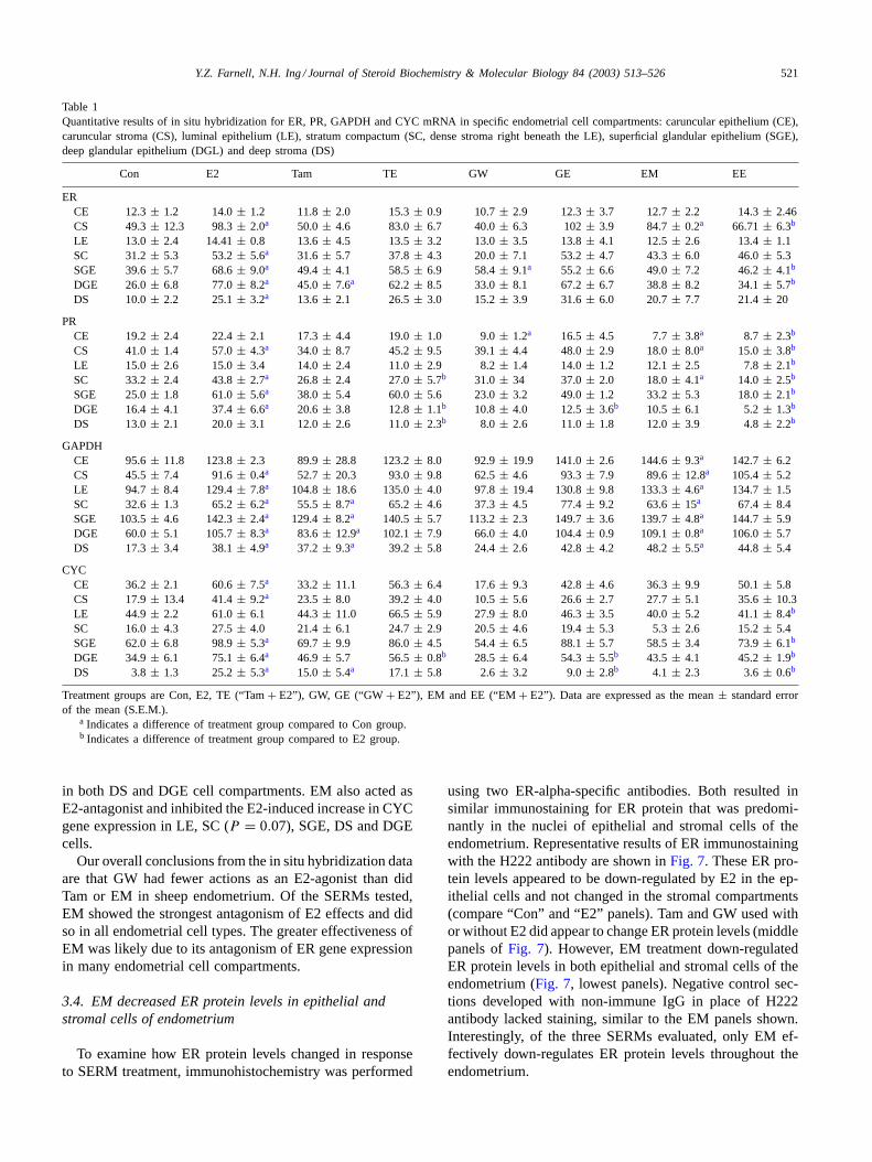
shown in the top section ofTable 1, E2 treatment increasedER mRNA levels compared to Con values in all endometrialcell compartments except CE and LE. Agonist actions ofthe SERMs were more limited. In ewes treated with Tam,GW or EM alone, ER mRNA levels increased in only onecell compartment: DGE, SGE or CS, respectively. Only EMantagonized E2-upregulation of ER gene expression and wasmost effective in CS, SGE and DGE cell compartments.
E2 enhanced PR gene expression in CS, SC, SGE,DGE and probably DS (P = 0.07) cell compartments(Table 1). As demonstrated in Northern analyses, no SERMup-regulated PR gene expression when it was used alone. Onthe contrary, GW and EM tested alone decreased PR mRNAlevels in CE cells compared to those in Con ewes. Also,EM examined alone decreased PR gene expression in CSand SC. Tam and GW markedly antagonized up-regulationof PR gene expression by E2 in DS and DGE cells. EMcompletely ablated E2’s up-regulation of PR mRNA levelsuniformly across all endometrial cells.
Consistent with Northern blot results, GAPDH mRNA hy-bridization signals on uterine cross-sections were enhancedby E2, Tam and EM treatments (Table 1). E2 treatment en-hanced GAPDH gene expression in most endometrial cells,which was consistent with our previous study[21]. Tamused alone increased GAPDH mRNA concentrations pri-marily in SC, SGE, DGE and DS cells. EM also acted asan E2-agonist by enhancing GAPDH gene expression in allcell compartments of the endometrium. GW did not displayany effect on GAPDH gene expression. No SERMs testedantagonized E2’s up-regulation of GAPDH mRNA levels inany cell compartment.
E2 increased CYC gene expression in all cell compart-ments, although LE and SC analyses hadP-values of 0.1and 0.08, respectively (Table 1). Tam enhanced CYC mRNAlevels strongly in DS cells and significantly inhibited E2’sup-regulation of CYC mRNA in DGE cells. GW and EMused alone had no effect on CYC gene expression. However,GW inhibited E2’s up-regulation of CYC gene expression
Y.Z. Farnell, N.H. Ing / Journal of Steroid Biochemistry & Molecular Biology 84 (2003) 513–526 521
Table 1Quantitative results of in situ hybridization for ER, PR, GAPDH and CYC mRNA in specific endometrial cell compartments: caruncular epithelium (CE),caruncular stroma (CS), luminal epithelium (LE), stratum compactum (SC, dense stroma right beneath the LE), superficial glandular epithelium (SGE),deep glandular epithelium (DGL) and deep stroma (DS)
Con E2 Tam TE GW GE EM EE
ERCE 12.3± 1.2 14.0± 1.2 11.8± 2.0 15.3± 0.9 10.7± 2.9 12.3± 3.7 12.7± 2.2 14.3± 2.46CS 49.3± 12.3 98.3± 2.0a 50.0 ± 4.6 83.0± 6.7 40.0± 6.3 102± 3.9 84.7± 0.2a 66.71± 6.3b
LE 13.0 ± 2.4 14.41± 0.8 13.6± 4.5 13.5± 3.2 13.0± 3.5 13.8± 4.1 12.5± 2.6 13.4± 1.1SC 31.2± 5.3 53.2± 5.6a 31.6 ± 5.7 37.8± 4.3 20.0± 7.1 53.2± 4.7 43.3± 6.0 46.0± 5.3SGE 39.6± 5.7 68.6± 9.0a 49.4 ± 4.1 58.5± 6.9 58.4± 9.1a 55.2 ± 6.6 49.0± 7.2 46.2± 4.1b
DGE 26.0± 6.8 77.0± 8.2a 45.0 ± 7.6a 62.2 ± 8.5 33.0± 8.1 67.2± 6.7 38.8± 8.2 34.1± 5.7b
DS 10.0± 2.2 25.1± 3.2a 13.6 ± 2.1 26.5± 3.0 15.2± 3.9 31.6± 6.0 20.7± 7.7 21.4± 20
PRCE 19.2± 2.4 22.4± 2.1 17.3± 4.4 19.0± 1.0 9.0± 1.2a 16.5 ± 4.5 7.7± 3.8a 8.7 ± 2.3b
CS 41.0± 1.4 57.0± 4.3a 34.0 ± 8.7 45.2± 9.5 39.1± 4.4 48.0± 2.9 18.0± 8.0a 15.0 ± 3.8b
LE 15.0 ± 2.6 15.0± 3.4 14.0± 2.4 11.0± 2.9 8.2± 1.4 14.0± 1.2 12.1± 2.5 7.8± 2.1b
SC 33.2± 2.4 43.8± 2.7a 26.8 ± 2.4 27.0± 5.7b 31.0 ± 34 37.0± 2.0 18.0± 4.1a 14.0 ± 2.5b
SGE 25.0± 1.8 61.0± 5.6a 38.0 ± 5.4 60.0± 5.6 23.0± 3.2 49.0± 1.2 33.2± 5.3 18.0± 2.1b
DGE 16.4± 4.1 37.4± 6.6a 20.6 ± 3.8 12.8± 1.1b 10.8 ± 4.0 12.5± 3.6b 10.5 ± 6.1 5.2± 1.3b
DS 13.0± 2.1 20.0± 3.1 12.0± 2.6 11.0± 2.3b 8.0 ± 2.6 11.0± 1.8 12.0± 3.9 4.8± 2.2b
GAPDHCE 95.6± 11.8 123.8± 2.3 89.9± 28.8 123.2± 8.0 92.9± 19.9 141.0± 2.6 144.6± 9.3a 142.7± 6.2CS 45.5± 7.4 91.6± 0.4a 52.7 ± 20.3 93.0± 9.8 62.5± 4.6 93.3± 7.9 89.6± 12.8a 105.4± 5.2LE 94.7 ± 8.4 129.4± 7.8a 104.8± 18.6 135.0± 4.0 97.8± 19.4 130.8± 9.8 133.3± 4.6a 134.7± 1.5SC 32.6± 1.3 65.2± 6.2a 55.5 ± 8.7a 65.2 ± 4.6 37.3± 4.5 77.4± 9.2 63.6± 15a 67.4 ± 8.4SGE 103.5± 4.6 142.3± 2.4a 129.4± 8.2a 140.5± 5.7 113.2± 2.3 149.7± 3.6 139.7± 4.8a 144.7± 5.9DGE 60.0± 5.1 105.7± 8.3a 83.6 ± 12.9a 102.1± 7.9 66.0± 4.0 104.4± 0.9 109.1± 0.8a 106.0± 5.7DS 17.3± 3.4 38.1± 4.9a 37.2 ± 9.3a 39.2 ± 5.8 24.4± 2.6 42.8± 4.2 48.2± 5.5a 44.8 ± 5.4
CYCCE 36.2± 2.1 60.6± 7.5a 33.2 ± 11.1 56.3± 6.4 17.6± 9.3 42.8± 4.6 36.3± 9.9 50.1± 5.8CS 17.9± 13.4 41.4± 9.2a 23.5 ± 8.0 39.2± 4.0 10.5± 5.6 26.6± 2.7 27.7± 5.1 35.6± 10.3LE 44.9 ± 2.2 61.0± 6.1 44.3± 11.0 66.5± 5.9 27.9± 8.0 46.3± 3.5 40.0± 5.2 41.1± 8.4b
SC 16.0± 4.3 27.5± 4.0 21.4± 6.1 24.7± 2.9 20.5± 4.6 19.4± 5.3 5.3± 2.6 15.2± 5.4SGE 62.0± 6.8 98.9± 5.3a 69.7 ± 9.9 86.0± 4.5 54.4± 6.5 88.1± 5.7 58.5± 3.4 73.9± 6.1b
DGE 34.9± 6.1 75.1± 6.4a 46.9 ± 5.7 56.5± 0.8b 28.5 ± 6.4 54.3± 5.5b 43.5 ± 4.1 45.2± 1.9b
DS 3.8± 1.3 25.2± 5.3a 15.0 ± 5.4a 17.1 ± 5.8 2.6± 3.2 9.0± 2.8b 4.1 ± 2.3 3.6± 0.6b
Treatment groups are Con, E2, TE (“Tam+ E2”), GW, GE (“GW+ E2”), EM and EE (“EM+ E2”). Data are expressed as the mean± standard errorof the mean (S.E.M.).
a Indicates a difference of treatment group compared to Con group.b Indicates a difference of treatment group compared to E2 group.
in both DS and DGE cell compartments. EM also acted asE2-antagonist and inhibited the E2-induced increase in CYCgene expression in LE, SC (P = 0.07), SGE, DS and DGEcells.
Our overall conclusions from the in situ hybridization dataare that GW had fewer actions as an E2-agonist than didTam or EM in sheep endometrium. Of the SERMs tested,EM showed the strongest antagonism of E2 effects and didso in all endometrial cell types. The greater effectiveness ofEM was likely due to its antagonism of ER gene expressionin many endometrial cell compartments.
3.4. EM decreased ER protein levels in epithelial andstromal cells of endometrium
To examine how ER protein levels changed in responseto SERM treatment, immunohistochemistry was performed
using two ER-alpha-specific antibodies. Both resulted insimilar immunostaining for ER protein that was predomi-nantly in the nuclei of epithelial and stromal cells of theendometrium. Representative results of ER immunostainingwith the H222 antibody are shown inFig. 7. These ER pro-tein levels appeared to be down-regulated by E2 in the ep-ithelial cells and not changed in the stromal compartments(compare “Con” and “E2” panels). Tam and GW used withor without E2 did appear to change ER protein levels (middlepanels ofFig. 7). However, EM treatment down-regulatedER protein levels in both epithelial and stromal cells of theendometrium (Fig. 7, lowest panels). Negative control sec-tions developed with non-immune IgG in place of H222antibody lacked staining, similar to the EM panels shown.Interestingly, of the three SERMs evaluated, only EM ef-fectively down-regulates ER protein levels throughout theendometrium.
522 Y.Z. Farnell, N.H. Ing / Journal of Steroid Biochemistry & Molecular Biology 84 (2003) 513–526
Fig. 7. SERM effects on ER protein levels in endometrium. Representative data from endometrial cross-sections are shown for ewes from all eighttreatment groups. Immunohistochemistry with H222 antibody demonstrated nuclear staining of ER protein in endometrial cells (see “Con” panel). LE islocated at the top of each panel and the deep endometrium is at the bottom. E2 decreased ER protein in glandular epithelial cells, while EM decreasedER protein in both epithelial and stromal cells. The bar in the “EM+ E2” panel represents 100�m.
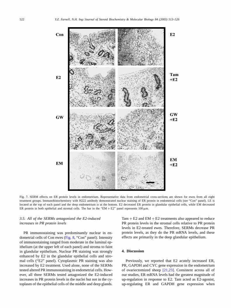
3.5. All of the SERMs antagonized the E2-inducedincreases in PR protein levels
PR immunostaining was predominantly nuclear in en-dometrial cells of Con ewes (Fig. 8, “Con” panel). Intensityof immunostaining ranged from moderate in the luminal ep-ithelium (at the upper left of each panel) and stroma to faintin glandular epithelium. Nuclear PR staining was stronglyenhanced by E2 in the glandular epithelial cells and stro-mal cells (“E2” panel). Cytoplasmic PR staining was alsoincreased by E2 treatment. Used alone, none of the SERMstested altered PR immunostaining in endometrial cells. How-ever, all three SERMs tested antagonized the E2-inducedincreases in PR protein levels in the nuclei but not in the cy-toplasm of the epithelial cells of the middle and deep glands.
Tam+ E2 and EM+ E2 treatments also appeared to reducePR protein levels in the stromal cells relative to PR proteinlevels in E2-treated ewes. Therefore, SERMs decrease PRprotein levels, as they do the PR mRNA levels, and theseeffects are primarily in the deep glandular epithelium.
4. Discussion
Previously, we reported that E2 acutely increased ER,PR, GAPDH and CYC gene expression in the endometriumof ovariectomized sheep[21,23]. Consistent across all ofour studies, ER mRNA levels had the greatest magnitude ofup-regulation in response to E2. Tam acted as E2-agonist,up-regulating ER and GAPDH gene expression when

Y.Z. Farnell, N.H. Ing / Journal of Steroid Biochemistry & Molecular Biology 84 (2003) 513–526 523
Fig. 8. All three SERMs antagonized E2 up-regulation of PR protein levels in DGE. PR immunohistochemistry demonstrated mainly nuclear stainingin endometrial cells. Representative endometrial cross-sections are shown for Con, E2, Tam, Tam+ E2, GW, GW+ E2, EM and EM+ E2 ewes. Theuterine lumen and LE are located at the upper left corner of each panel. E2 intensified PR immunostaining in glandular epithelial cells and all threeSERMs inhibited this effect. The bar in the “EM+ E2” panel represents 100�m.
delivered systemically by injection[10]. Here, we infused10−7 M of Tam directly into the uterine lumen for 24 h andfound it had similar E2-agonist actions that up-regulatedexpression of ER and GAPDH genes. Along with the manySERM effects identified in the deep endometrial compart-ments (this report), as well as in myometrium (accompany-ing paper), this confirms the efficacy of intrauterine deliveryof SERMs to the entire endometrium. This report of Tamantagonism of E2 induction of PR gene expression is inagreement with Tam antagonism of E2-induced PR gene ex-pression in the ovariectomized rat uterus and other tissues,as well as in breast cancer cell lines[25–29,41]. It is note-
worthy that our E2 and SERM treatments were acute, so asto minimize changes in the endometrial cell populations.Most other SERM trials in animals employ chronic admin-istration that simulates women taking the drugs chronically.
In our sheep, GW treatment alone had E2-agonist effectson expression of the ER gene at mRNA levels in very fewendometrial cell compartments. Others have shown no ef-fect of GW on ER mRNA or protein levels in human breastand endometrial cancer cell lines MCF-7 and ECC-1 cells,respectively[28]. Ours is the first report of GW’s antago-nism of the E2 up-regulation of PR and CYC gene expres-sion. However, EM also showed some E2-agonist effects,
524 Y.Z. Farnell, N.H. Ing / Journal of Steroid Biochemistry & Molecular Biology 84 (2003) 513–526
e.g. increases in ER gene expression in CS cells and en-hanced GAPDH gene expression in most of the endometrialcells. In this study, EM appears to exert superior antagonis-tic effects of E2-induced expression of ER, PR, and CYCgenes among the SERMs examined. It is important to notethat E2 up-regulation of ER gene expression occurs by apost-transcriptional mechanism, by stabilizing ER mRNA[30,31], and this is the first report of a SERM blocking it.The superior antagonism is likely to be due to EM antago-nizing the up-regulation of ER mRNA and decreasing ERprotein levels in most endometrial cells. Our data agreeswith a report that EM decreased rat uterine ER and PR lev-els, measured with a ligand binding assay[14,32].
GAPDH and CYC have been thought of as “housekeepinggenes”, expressed constitutively in tissues and cells. How-ever, that is not the case in the sheep and rat uterus andbreast cancer cell lines after an E2 challenge, where expres-sion of GAPDH and CYC genes increases[33–35]. SinceGAPDH and CYC have important roles in metabolism andprotein folding, perhaps it should not be surprising to seethem up-regulated by E2[36,37]. Tam acts as an E2-agoniston GAPDH gene expression, which agrees with a previousstudy in sheep endometrium[10]. All three SERMs testedshowed antagonism of E2 up-regulation of CYC mRNA lev-els, primarily in deep cell compartments of the endometrium.These SERM effects confirm that GAPDH and CYC genesare regulated, not constitutive, in sheep endometrium.
In this study, E2 increased ER mRNA but decreased ERprotein levels in glandular epithelial cells (Figs. 3 and 7).Several reports have stated changes in ER protein levelswere not directly correlated to those of ER mRNA levels inrat, ewe and lamb uterus, which were treated with E2 for8 h, or 12 h[12,23,38,39]. Acute ER protein degradation inresponse to E2 treatment has been reported in rat, mouseand sheep uterus, as well as MCF-7 cells[12,23,40,41].We have found that time of E2 treatment greatly affectsER protein in cells: immunoreactive ER was absent at 12 hpost-injection, but increased to levels greater than thosein untreated control ewes at 24 and 48 h post-injection[23]. Estradiol binding triggers rapid ER degradation viaubiquitin-proteasome pathway[42–44], with the later rise inER protein levels due to increased translation from increasedlevels of ER mRNA. EM may similarly affect ER proteinlevels to result in the profound down-regulation demon-strated here. Sampling time in this study (18 h post-estradioland immediately after the 24 h infusion of EM) is duringthe up-regulation/recovery phase of ER mRNA and proteinlevels. Several SERMs have been tested for regulation ofER mRNA or protein levels in other tissues or cell lines.Alteration of ER gene expression is dependent on the spe-cific SERM, tissue or cell, and length of treatment. AnotherSERM, ICI 182,780, also causes loss of ER protein levels,while ER mRNA levels are maintained in MCF-7 cells[12].The fact that the combined treatment of E2 and EM hasthe lowest level of ER gene expression may relate to bothpotent ligands down-regulating the ER protein[43].
Endometrium is composed of heterogeneous cell popula-tions including luminal and glandular epithelial cells, stro-mal cells, immune cells, and cells that form blood vessels[45]. The majority of epithelial and stromal endometrial cellsrespond to estrogens (endogenous and exogenously applied)by altering gene expression. We and others have seen thegreatest up-regulation of ER and PR genes by estrogen inthe deep endometrial cells and inner layer of myometrium([23], accompanying paper). The SERMs that were mostpotent as E2-agonists and antagonists (Tam and EM, re-spectively) acted in the majority of endometrial cell com-partments. In other cases, however, compartment-specificeffects were observed. For example, the deep endometrialregion and the outer myometrium appeared more sensitiveto E2-antagonism by Tam and GW than the other regions.These data imply paracrine regulation of gene expressionbetween the deep endometrium and the myometrial layerswhich have yet to be elucidated.
In conclusion, of the two new SERMs, GW has fewerE2-agonist effects in endometrium than the older Tam drugdoes. Although EM mimicked E2 in up-regulating GAPDHmRNA levels, it showed the most effective and widespreadantagonism of E2-enhanced levels of ER, PR and CYC geneexpression. This is probably due to EM down-regulating ERmRNA and protein levels in the majority of endometrialcells.
Acknowledgements
We gratefully acknowledge the generous gifts of EM fromDr. Fernand Labrie of Laval University, GW from Dr. DavidC. Morris of Glaxo Wellcome Research and Development,and the H222 anti-ER antibody from Dr. Geoffery Greeneof University of Chicago, IL. The authors also thank Ms.Cindy Balog, Dr. Lora Lindahl as well as members of Dr.Fuller Bazer’s laboratory for assistance with animal work.We also acknowledge the assistance of the image analysiscore facility of Dr. Robert C. Burghardt. Funding for thisresearch was received from the USDA NRI-CGP grant #98-35203-6272.
References
[1] C.M. Klinge, Estrogen receptor interaction with co-activators andco-repressors, Steroids 65 (2000) 227–251.
[2] J.F. Couse, K.S. Korach, Estrogen receptor null mice: what have welearned and where will they lead us, Endocrinol. Rev. 20 (1999)358–417.
[3] V.C. Jordan, M. Morrow, Tamoxifen, raloxifene, and the preventionof breast cancer, Endocrinol. Rev. 20 (1999) 253–278.
[4] C. Ribot, F. Tremollieres, Anti-estrogens and bone tissue, Ann.Endocrinol. 56 (1995) 603–608.
[5] G.H. Eltabbakh, S.L. Mount, Tamoxifen and the female reproductivetract, Expert Opin. Pharmacother. 2 (2001) 1399–1413.
[6] J.I. MacGregor, V.C. Jordan, Basic guide to the mechanism ofanti-estrogen action, Pharmacol. Rev. 50 (1998) 151–196.
Y.Z. Farnell, N.H. Ing / Journal of Steroid Biochemistry & Molecular Biology 84 (2003) 513–526 525
[7] B.S. Katzenellenbogen, I. Choi, R. Delage-Mourroux, T.R. Ediger,P.G. Martini, M. Montano, J. Sun, K. Weis, J.A. Katzenellenbogen,Molecular mechanisms of estrogen action: selective ligands andreceptor pharmacology, J. Steroid Biochem. Mol. Biol. 74 (2000)279–285.
[8] D.C. Robinson, J.D. Bloss, M.A. Schiano, A retrospective study oftamoxifen and endometrial cancer in breast cancer patients, Gynecol.Oncol. 59 (1995) 186–190.
[9] Y. Daniel, M. Inbar, A. Bar-Am, M.R. Peyser, J.B. Lessing, Theeffects of tamoxifen treatment on the endometrium, Fertil. Steril. 65(1996) 1083–1089.
[10] J.A. Robertson, S. Bhattacharyya, N.H. Ing, Tamoxifen up-regulatesoestrogen receptor-alpha, c-fos and glyceraldehyde 3-phosphatedehydrogenase mRNA in ovine endometrium, J. Steroid Biochem.Mol. Biol. 67 (1998) 285–292.
[11] T.M. Willson, B.R. Henke, T.M. Momtahen, P.S. Charifson, K.W.Batchelor, D.B. Lubahn, L.B. Moore, B.B. Oliver, H.R. Sauls,J.A. Triantafillou, 3-[4-(1,2-Diphenylbut-1-enyl)phenyl]acrylic acid:a non-steroidal estrogen with functional selectivity for bone overuterus in rats, J. Med. Chem. 37 (1994) 1550–1552.
[12] J.I. Schafer, H. Liu, D.A. Tonetti, V.C. Jordan, The interaction ofraloxifene and the active metabolite of the anti-estrogen EM-800(SC 5707) with the human estrogen receptor, Cancer Res. 59 (1999)4308–4313.
[13] T.M. Willson, J.D. Norris, B.L. Wagner, I. Asplin, P. Baer, H.R.Brown, S.A. Jones, B. Henke, H. Sauls, S. Wolfe, D.C. Morris,D.P. McDonnell, Dissection of the molecular mechanism of actionof GW5638, a novel estrogen receptor ligand, provides insights intothe role of estrogen receptor in bone, Endocrinology 138 (1997)3901–4101.
[14] S. Luo, M. Stojanovic, C. Labrie, F. Labrie, Inhibitory effect ofthe novel anti-estrogen EM-800 and medroxyprogesterone acetateon estrone-stimulated growth of dimethylabenz[�]anthracene-inducedmammary carcinoma in rats, Int. J. Cancer 73 (1997) 580–586.
[15] S. Luo, A. Sourla, C. Labrie, S. Gauthier, Y. Merand, A.Belanger, F. Labrie, Effect of twenty-four-week treatment with theanti-estrogen EM-800 on estrogen-sensitive parameters in intact andovariectomized mice, Endocrinology 139 (1998) 2645–2656.
[16] J. Simard, R. Sanchez, D. Poirier, S. Gauthier, S.M. Singh, Y. Merand,A. Belanger, C. Labrie, F. Labrie, Blockage of the stimulatoryeffect of estrogens, OH-tamoxifen, OH-toremifene, droloxifen, andraloxifene on alkaline phosphatase activity by the anti-estrogenEM-800 in human endometrium adenocarcinoma Ishikawa cells,Cancer Res. 57 (1997) 3494–3497.
[17] S. Gauthier, B. Caron, J. Cloutier, Y.L. Dory, A. Favre, D.Larouche, J. Mailhot, C. Ouellet, A. Schwerdtfeger, G. Leblanc, C.Martel, J. Simard, Y. Merand, A. Belanger, C. Labrie, F. Labrie,(S)-(+)-4-[7-(2,2-dimethyl-1-oxopropoxy)-4-methyl-2-[4-[2-(1-pipe-ridinyl)-ethoxy]phenyl]-2H-1-benzopyran-3-yl]-phenyl 2,2-dimethyl-propanoate (EM-800): a highly potent, specific, and orally activenonsteroidal anti-estrogen, J. Med. Chem. 40 (1997) 2117–2122.
[18] F. Labrie, C. Labrie, A. Belanger, J. Simard, S. Gauthier, V. Luu-The,Y. Merand, V. Giguere, B. Candas, S. Luo, C. Martel, S.M. Singh,M. Fournier, A. Coquet, V. Richard, R. Charbonneau, G. Charpenet,A. Tremblay, G. Tremblay, L. Cusan, R. Veilleux, EM-652 (SCH57068), a third generation SERM acting as pure anti-estrogen in themammary gland and endometrium, J. Steroid Biochem. Mol. Biol.69 (1999) 51–84.
[19] C. Martel, S. Picard, V. Richard, A. Belanger, C. Labrie, F.Labrie, Prevention of bone loss by EM-800 and raloxifene in theovariectomized rat, J. Steroid Biochem. Mol. Biol. 74 (2000) 45–56.
[20] N.H. Ing, T.E. Spencer, F.W. Bazer, Estrogen enhances endometrialestrogen receptor gene expression by a post-transcriptionalmechanism in the ovariectomized ewe, Biol. Reprod. 54 (1996) 591–599.
[21] J.A. Robertson, Y. Zhang, N.H. Ing, ICI 182,780 acts as partialagonist and antagonist of estradiol effects in specific cells of thesheep uterus, J. Steroid Biochem. Mol. Biol. l77 (2001) 281–287.
[22] J.L. Vallet, F.W. Bazer, Effect of ovine trophoblast protein-1,oestrogen and progesterone on oxytocin-induced phosphatidylinositolturnover in endometrium of sheep, J. Reprod. Fertil. 87 (1989) 755–761.
[23] N.H. Ing, M.B. Tornesi, Estradiol up-regulates estrogen receptor andprogesterone receptor gene expression in specific ovine uterine cells,Biol. Reprod. 56 (1997) 1205–1215.
[24] M. Tomanek, C. Pisselet, P. Monget, T. Madigou, M.L. Thieulant,D. Monniaux, Estrogen receptor protein and mRNA expression inthe ovary of sheep, Mol. Reprod. Dev. 48 (1997) 53–62.
[25] P.J. Shughrue, M.V. Lane, I. Merchenthaler, Regulation ofprogesterone receptor messenger ribonucleic acid in the rat medialpreoptic nucleus by estrogenic and anti-estrogenic compounds: an insitu hybridization study, Endocrinology 138 (1997) 5476–5484.
[26] S.L. Fitzpatrick, T.J. Berrodin, S.F. Jenkins, D.M. Sindoni, D.C.Deecher, D.E. Frail, Effect of estrogen agonists and antagonists oninduction of progesterone receptor in a rat hypothalamic cell line,Endocrinology 140 (1999) 3928–3937.
[27] E. Castellano-Diaz, M.I. Gonzalez-Quijano, J.M. Liminana, B.N.Diaz-Chico, Tamoxifen decreases the estradiol-induced progesteronereceptors by interfering with nuclear estrogen receptor accumulation,J. Steroid Biochem. 33 (1989) 133–139.
[28] D. Bentrem, R. Dardes, H. Liu, J. MacGregor-Schafer, J. Zapf, V.Jordan, Molecular mechanism of action at estrogen receptor alphaof a new clinically relevant anti-estrogen (GW 7604) related totamoxifen, Endocrinology 142 (2001) 838–846.
[29] U. Karck, F. Kommoss, Does tamoxifen change oestrogen andprogesterone receptor expression in the endometrium and breast, Eur.J. Cancer 36 (2000) S45–46.
[30] N.H. Ing, T.L. Ott, Estradiol up-regulates estrogen receptor-�
messenger ribonucleic acid in sheep endometrium by increasing itsstability, Biol. Reprod. 60 (1999) 134–139.
[31] J. A Robertson, Y. Farnell, L.S. Lindahl, N.H. Ing, Estradiolup-regulates estrogen receptor messenger ribonucleic acid inendometrial carcinoma (Ishikawa) cells by stabilizing the message29 (2002) 125–135.
[32] C. Martel, C. Labrie, A. Belanger, S. Gauthier, Y. Merand, X. Li,L. Provencher, B. Candas, F. Labrie, Comparison of the effects ofthe new orally active anti-estrogen EM-800 with ICI 182780 andtoremifene on estrogen-sensitive parameters in the ovariectomizedmouse, Endocrinology 139 (1997) 2486–2492.
[33] K. Zou, N.H. Ing, Oestradiol up-regulates oestrogen receptor,cyclophilin, and glyceraldehyde phosphate dehydrogenase mRNAconcentrations in endometrium, but down-regulates them in liver, J.Steroid Biochem. Mol. Biol. 64 (1998) 231–237.
[34] P. Diel, T. Schulz, K. Smolnikar, E. Strunck, G. Vollmer, H. Michna,Ability of xeno- and phytoestrogens to modulate expression ofestrogen-sensitive genes in rat uterus: estrogenicity profiles anduterotropic activity, J. Steroid Biochem. Mol. Biol. 73 (2000) 1–10.
[35] P. Kumar, P.J. Mark, B.K. Ward, R.F. Minchin, T. Ratajczak,Estradiol-regulated expression of the immunophilin cyclophilin 40and FKBP52 in MCF-7 breast cancer cells, Biochem. Biophys. Res.Commun. 284 (2001) 219–225.
[36] E. Nagy, W.F. Rigby, Glyceraldehyde-3-phosphate dehydrogenaseselectively binds AU-rich RNA in the NAD+-binding region, J. Biol.Chem. 270 (1995) 2755–2763.
[37] L. Andreeva, R. Heads, C.J. Green, Cyclophilins and their possiblerole in the stress response, Int. J. Pathol. 80 (1999) 305–315.
[38] Y. Zhou, L.P. Chorich, V.B. Mahesh, T.F. Ogle, Regulation ofestrogen receptor protein and messenger ribonucleic acid by estradioland progesterone in rat uterus, J. Steroid Biochem. Mol. Biol. 46(1993) 687–698.
[39] A. Meikle, M. Forsberg, L. Sahlin, B. Masironi, C. Tasende, M.Rodriguez-Pinon, E.G. Garofalo, A biphasic action of estradiol onestrogen and progesterone receptor expression in the lamb uterus,Reprod. Nutr. Dev. 40 (2000) 283–293.

526 Y.Z. Farnell, N.H. Ing / Journal of Steroid Biochemistry & Molecular Biology 84 (2003) 513–526
[40] K.L. Medlock, C.R. Lyttle, N. Kelepouris, E.D. Newman, D.M.Sheehan, Estradiol down-regulation of the rat uterine estrogenreceptor, Proc. Soc. Exp. Biol. Med. 196 (1991) 293–300.
[41] T.A. Tibbetts, M. Mendoza-Meneses, B.W. O’Malley, O.M.Conneely, Mutual and intercompartmental regulation of estrogenreceptor and progesterone receptor expression in the mouse uterus,Biol. Reprod. 59 (1998) 1143–1152.
[42] P.B. Nirmala, R.V. Thampan, Ubiquitination of the rat uterineestrogen receptor: dependence on estradiol, Biochem. Biophys. Res.Commun. 213 (1995) 24–31.
[43] Z. Nawaz, D.M. Lonard, A.P. Dennis, C.L. Smith, B.W. O’Malley,Proteasome-dependent degradation of human estrogen receptor, Proc.Natl. Acad. Sci. U.S.A. 96 (1999) 1858–1862.
[44] A.L. Wijayaratne, D.P. McDonnell, The human estrogenreceptor-alpha is a ubiquitinated protein whose stability is affecteddifferentially by agonists, antagonists, and selective estrogen receptormodulators, J. Biol. Chem. 276 (2001) 35684–35692.
[45] C.A. Gray, F.F. Bartol, B.J. Tarleton, A.A. Wiley, G.A. Johnson,F.W. Bazer, T.E. Spencer, Developmental biology of uterine glands,Biol. Reprod. 65 (2001) 1123–1311.