Endogenous Cardiac Stem Cell Activation by Insulin-Like Growth Factor1/Hepatocyte Growth Factor...
10
Endogenous Cardiac Stem Cell Activation by Insulin-Like Growth Factor-1/Hepatocyte Growth Factor Intracoronary Injection Fosters Survival and Regeneration of the Infarcted Pig Heart Georgina M. Ellison, PHD,*† Daniele Torella, MD, PHD,*† Santo Dellegrottaglie, MD, PHD,§ Claudia Perez-Martinez, DVM, PHD, Armando Perez de Prado, MD, Carla Vicinanza, PHD,*† Saranya Purushothaman, BSC,* Valentina Galuppo, MD,† Claudio Iaconetti, PHD,† Cheryl D. Waring, PHD,* Andrew Smith, PHD,* Michele Torella, MD, PHD,# Carlos Cuellas Ramon, MD, Jose Manuel Gonzalo-Orden, DVM, PHD, Valter Agosti, MD, PHD,‡ Ciro Indolfi, MD,† Manuel Galiñanes, MD, PHD,¶ Felipe Fernandez-Vazquez, MD, PHD, Bernardo Nadal-Ginard, MD, PHD* Liverpool, United Kingdom; Catanzaro and Naples, Italy; and León and Barcelona, Spain Objectives The purpose of this study was to test the ability of insulin-like growth factor (IGF)-1/hepatocyte growth factor (HGF) to activate resident endogenous porcine cardiac stem/progenitor cells (epCSCs) and to promote myocar- dial repair through a clinically applicable intracoronary injection protocol in a pig model of myocardial infarction (MI) relevant to human disease. Background In rodents, cardiac stem/progenitor cell (CSC) transplantation as well as in situ activation through intramyocardial injection of specific growth factors has been shown to result in myocardial regeneration after acute myocardial infarction (AMI). Methods Acute MI was induced in pigs by a 60-min percutaneous transluminal coronary angiography left anterior de- scending artery occlusion. The IGF-1 and HGF were co-administered through the infarct-related artery in a single dose (ranging from 0.5 to 2 g HGF and 2 to 8 g IGF-1) 30 min after coronary reperfusion. Pigs were sacrificed 21 days later for dose-response relationship evaluation by immunohistopathology or 2 months later for cardiac function evaluation by cardiac magnetic resonance imaging. Results The IGF-1/HGF activated c-kit positive–CD45 negative epCSCs and increased their myogenic differentiation in vitro. The IGF-1/HGF, in a dose-dependent manner, improved cardiomyocyte survival, and reduced fibrosis and cardiomyo- cyte reactive hypertrophy. It significantly increased c-kit positive–CD45 negative epCSC number and fostered the gen- eration of new myocardium (myocytes and microvasculature) in infarcted and peri-infarct/border regions at 21 and 60 days after AMI. The IGF-1/HGF reduced infarct size and improved left ventricular function at 2 months after AMI. Conclusions In an animal model of AMI relevant to the human disease, intracoronary administration of IGF-1/HGF is a practical and effective strategy to reduce pathological cardiac remodeling, induce myocardial regeneration, and improve ven- tricular function. (J Am Coll Cardiol 2011;58:977–86) © 2011 by the American College of Cardiology Foundation From *The Stem Cell and Regenerative Biology Unit (BioStem), RISES, Liverpool John Moores University, Liverpool, United Kingdom; †Molecular and Cellular Cardiology, Department of Medicine, Magna Graecia University, Catanzaro, Italy; ‡Molecular Oncology, Department of Medicine, Magna Graecia University, Catan- zaro, Italy; §Division of Cardiology, Federico II University, Naples, Italy; IBIOMED y Fundacio ´n Investigacio ´n Sanitaria en Leo ´n, Universidad y Hospital Universitario de Leo ´n, Leo ´n, Spain; ¶Department of Cardiac Surgery, Reparative Therapy of the Heart, Area del Cor and Research Institute, University Hospital Vall d’Hebron, Universitat Autònoma de Barcelona, Barcelona, Spain; and the #Department of Cardio-Thoracic and Respiratory Sciences, Second University of Naples, Naples, Italy. This work was in part supported by grants from the British Heart Foundation (PG/06/045; PG 08/085), Marie Curie FP7 (PIRG02-GA-2007-224853), Cardio Repair European Multidisciplinary Initiative FP7 (FP7-HEALTH-2009-242038), FIRB-Futuro-in-Ricerca (RBFR081CCS), Associazione Italiana per la Ricerca sul Cancro (MFAG-2008), Italian Ministry of Health (GR-2008-1142673), and Span- ish Ministry of Science and Innovation (MoSI, Factostem DEX-580000-2008-0043). The Instituto Carlos III through the “Red de Terapia Celular and the Junta de Castilla y Leon” supported cardiac magnetic resonance imaging studies. Iberhospitex Journal of the American College of Cardiology Vol. 58, No. 9, 2011 © 2011 by the American College of Cardiology Foundation ISSN 0735-1097/$36.00 Published by Elsevier Inc. doi:10.1016/j.jacc.2011.05.013
-
Upload
independent -
Category
Documents
-
view
1 -
download
0
Transcript of Endogenous Cardiac Stem Cell Activation by Insulin-Like Growth Factor1/Hepatocyte Growth Factor...
Endogenous Cardiac Stem Cell Activationby Insulin-Like Growth Factor-1/HepatocyteGrowth Factor Intracoronary Injection FostersSurvival and Regeneration of the Infarcted Pig Heart
Georgina M. Ellison, PHD,*† Daniele Torella, MD, PHD,*† Santo Dellegrottaglie, MD, PHD,§Claudia Perez-Martinez, DVM, PHD,! Armando Perez de Prado, MD,! Carla Vicinanza, PHD,*†Saranya Purushothaman, BSC,* Valentina Galuppo, MD,† Claudio Iaconetti, PHD,†Cheryl D. Waring, PHD,* Andrew Smith, PHD,* Michele Torella, MD, PHD,#Carlos Cuellas Ramon, MD,! Jose Manuel Gonzalo-Orden, DVM, PHD,! Valter Agosti, MD, PHD,‡Ciro Indolfi, MD,† Manuel Galiñanes, MD, PHD,¶ Felipe Fernandez-Vazquez, MD, PHD,!Bernardo Nadal-Ginard, MD, PHD*
Liverpool, United Kingdom; Catanzaro and Naples, Italy; and León and Barcelona, Spain
Objectives The purpose of this study was to test the ability of insulin-like growth factor (IGF)-1/hepatocyte growth factor(HGF) to activate resident endogenous porcine cardiac stem/progenitor cells (epCSCs) and to promote myocar-dial repair through a clinically applicable intracoronary injection protocol in a pig model of myocardial infarction(MI) relevant to human disease.
Background In rodents, cardiac stem/progenitor cell (CSC) transplantation as well as in situ activation through intramyocardialinjection of specific growth factors has been shown to result in myocardial regeneration after acute myocardialinfarction (AMI).
Methods Acute MI was induced in pigs by a 60-min percutaneous transluminal coronary angiography left anterior de-scending artery occlusion. The IGF-1 and HGF were co-administered through the infarct-related artery in a singledose (ranging from 0.5 to 2 !g HGF and 2 to 8 !g IGF-1) 30 min after coronary reperfusion. Pigs were sacrificed21 days later for dose-response relationship evaluation by immunohistopathology or 2 months later for cardiacfunction evaluation by cardiac magnetic resonance imaging.
Results The IGF-1/HGF activated c-kit positive–CD45 negative epCSCs and increased their myogenic differentiation in vitro.The IGF-1/HGF, in a dose-dependent manner, improved cardiomyocyte survival, and reduced fibrosis and cardiomyo-cyte reactive hypertrophy. It significantly increased c-kit positive–CD45 negative epCSC number and fostered the gen-eration of new myocardium (myocytes and microvasculature) in infarcted and peri-infarct/border regions at 21 and60 days after AMI. The IGF-1/HGF reduced infarct size and improved left ventricular function at 2 months after AMI.
Conclusions In an animal model of AMI relevant to the human disease, intracoronary administration of IGF-1/HGF is a practicaland effective strategy to reduce pathological cardiac remodeling, induce myocardial regeneration, and improve ven-tricular function. (J Am Coll Cardiol 2011;58:977–86) © 2011 by the American College of Cardiology Foundation
From *The Stem Cell and Regenerative Biology Unit (BioStem), RISES, LiverpoolJohn Moores University, Liverpool, United Kingdom; †Molecular and CellularCardiology, Department of Medicine, Magna Graecia University, Catanzaro, Italy;‡Molecular Oncology, Department of Medicine, Magna Graecia University, Catan-zaro, Italy; §Division of Cardiology, Federico II University, Naples, Italy; !IBIOMED yFundacion Investigacion Sanitaria en Leon, Universidad y Hospital Universitario deLeon, Leon, Spain; ¶Department of Cardiac Surgery, Reparative Therapy of theHeart, Area del Cor and Research Institute, University Hospital Vall d’Hebron,Universitat Autònoma de Barcelona, Barcelona, Spain; and the #Department of
Cardio-Thoracic and Respiratory Sciences, Second University of Naples, Naples,Italy. This work was in part supported by grants from the British Heart Foundation(PG/06/045; PG 08/085), Marie Curie FP7 (PIRG02-GA-2007-224853), CardioRepair European Multidisciplinary Initiative FP7 (FP7-HEALTH-2009-242038),FIRB-Futuro-in-Ricerca (RBFR081CCS), Associazione Italiana per la Ricerca sulCancro (MFAG-2008), Italian Ministry of Health (GR-2008-1142673), and Span-ish Ministry of Science and Innovation (MoSI, Factostem DEX-580000-2008-0043).The Instituto Carlos III through the “Red de Terapia Celular and the Junta deCastilla y Leon” supported cardiac magnetic resonance imaging studies. Iberhospitex
Journal of the American College of Cardiology Vol. 58, No. 9, 2011© 2011 by the American College of Cardiology Foundation ISSN 0735-1097/$36.00Published by Elsevier Inc. doi:10.1016/j.jacc.2011.05.013
The presence of endogenous res-ident cardiac stem and progeni-tor cells (eCSCs) in the adultmammalian heart of differentspecies, including human (1,2),supports the notion that theadult myocardium has an intrin-sic regenerative capacity, whichcould be exploited to producemeaningful myocardial regenera-tion (3–6).
Data from mice and dogsshow that the regenerative re-sponse of eCSCs to a ischemicinsult can be enhanced in situ byadministration of growth factors(7,8). Nevertheless, before suchprotocols can be clinically tested,they need to be validated in alarge-animal model that closelymimics the human heart using agrowth factor dose and method ofadministration compatible withclinical application. Swine, becauseof similarities to ours in organ size,coronary anatomy, immunology,
and physiology, stands up as the most attractive model forpre-clinical protocols of eCSC activation for myocardialregeneration.
Here, we show that the adult pig myocardium harborsc-kit positive (c-kitpos) CD45 negative (CD45neg) eCSCs,which show rapid activation and robust cardiomyogenicdifferentiation when stimulated by insulin-like growth fac-tor (IGF)-1 and/or hepatocyte growth factor (HGF) invitro. Small amounts of IGF-1 and HGF administered tothe damaged porcine myocardium through the coronarycirculation after acute myocardial infarction (AMI), in a lineardose/effect relationship, salvages a significant portion of themyocardium at risk, produces a robust activation of the residenteCSC pool and regenerates most myocytes and microvascula-ture lost by the ischemic insult. At 2 months after myocardialinfarction (MI), this treatment results in improved left ventric-ular (LV) function and reduced infarct size.
Methods
See the Online Appendix for a complete description ofMethods.
Results
Porcine resident c-kitposCD45neg cardiac cells are multi-potent stem/progenitor cells. Small cells positive for c-kit(c-kitpos) and negative for blood-cell lineage markers (i.e.,CD45), Linneg, are ubiquitous in the adult porcine myocar-dium, with higher density in the atria and the ventricularapex (Online Fig. 1).
The c-kitpos cardiac cells constituted 9 ! 2% and 4 ! 2% ofthe starting myocyte-depleted cardiac small cell populationfrom atria and LV, respectively (Fig. 1A). Moreover, 75 !12% and 50 ! 8% of these c-kitpos cells from the atria and theleft ventricle, respectively, also expressed CD45 (Figs. 1B and1C), which, when co-expressed with c-kit, identifies residentcardiac mast cells (9). Thus, using negative (CD45) andpositive (c-kit) sorting by magnetic activated cell sortingtechnology, we obtained "95% enriched c-kitpos cardiac cells,depleted of tryptasepos and CD45pos mast cells (Figs. 1D and1E), and endothelial/hematopoietic CD34pos progenitors(Online Fig. 2). A fraction of c-kitposCD45neg cardiac cellsexpressed the mesenchymal marker, CD90 (38 ! 6%) andadhesion molecule/cardiac progenitor marker, CD166 (74 !11%) (Fig. 1E). Porcine c-kitposCD45neg cardiac cells werenegative for a panel of CD markers specific for other hema-topoietic, mesenchymal, and endothelial cell lineages (OnlineFig. 2). Freshly isolated c-kitposCD45neg cells expressedOct3/4 (3 ! 1%) (Fig. 1F to 1H), SSEA-4 (11 ! 2%)(Fig. 1F), SSEA-3 (10 ! 3%), Nanog (9 ! 3%), Telomerase(58 ! 8%), Bmi-1 (59 ! 11%), Flk-1 (46 ! 5%), Gata-4(49 ! 7%), Nkx2.5 (29 ! 10%), and Isl-1 (1 ! 1%).
The c-kitposCD45neg cells from atria, ventricles, and apexexhibited #20% clonal efficiency, independently of theirchamber of origin (Online Fig. 3). Three randomly pickedclones each from atria-, ventricle-, and apex-derived cellswere further expanded. These clones had a #22 h doublingtime. They have been propagated for "65 passages andserially subcloned every 10 passages without reachinggrowth arrest, senescence, or showing any detectable chro-mosomal alterations (Online Fig. 4). Cloned cells showedpositivity for c-kit (90 ! 8%), Oct3/4 (62 ! 11%), SSEA-3(65 ! 10%), SSEA-4 (59 ! 14%), Nanog (46 ! 5%),telomerase (81 ! 10%), Bmi-1 (70 ! 14%), Flk-1 (86 ! 9%),Gata-4 (60 ! 11%), Nkx2.5 (52 ! 8%) and Isl-1 (8 ! 6%)(Figs. 1G and 1H, Online Figs. 3 and 5), indicating higherclonal efficiency of cells expressing multipotency genes.
Cloned c-kitposCD45neg cardiac cells grew in suspensionand generated cardiospheres (1,2). Cardiospheres placed indifferentiation medium attached, and cells spread out fromthe sphere (Online Fig. 3), differentiating into myocytes(27 ! 4%), endothelial (10 ! 6%), and smooth muscle cells(34 ! 5%) (Online Fig. 3). These results show that porcineresident c-kitposCD45neg cardiac cells (hereafter identified
S. L., Spain, provided catheters, stents, and other technical supplies. Dr. Nadal-Ginard is a cofounder of and worked as Scientific Director of Coretherapix, SRL,Madrid, Spain, a biotechnology start-up company. Coretherapix was not involved inany aspect of the experiments described in this paper apart from participating in theFactostem, a multicenter cooperative grant awarded to Coretherapix as the coordi-nator and three other Institutions among which is the Fundacion InvestigacionSanitaria of Leon (FIS). The Factostem grant funded a portion of the animalexperiments with cMRI performed at FIS. All other authors have reported that theyhave no relationships relevant to the contents of this paper to disclose. Drs. G. Ellisonand D. Torella contributed equally to this work. Drs. Fernandez-Vazquez andNadal-Ginard shared the senior responsibility for the project.
Manuscript received April 20, 2011; revised manuscript received May 20, 2011,accepted May 24, 2011.
Abbreviationsand Acronyms
AMI ! acute myocardialinfarction
BrdU ! bromodeoxyuridine
CD45neg ! CD45 negative
c-kitpos ! c-kit positive
cMRI ! cardiac magneticresonance imaging
CSC ! cardiac stem/progenitor cell
CTRL ! control
eCSC ! endogenousresident cardiac stem/progenitor cells
epCSC ! endogenousporcine cardiac stem/progenitor cell
HGF ! hepatocyte growthfactor
IGF ! insulin-like growthfactor
LV ! left ventricular
MI ! myocardial infarction
978 Ellison et al. JACC Vol. 58, No. 9, 2011CSC Activation by IGF-1/HGF for Heart Regeneration August 23, 2011:977–86

Figure 1 Phenotype of Porcine c-kitposCD45neg Cardiac Stem/Progenitor Cells
(A) Florescence activated cell sorting (FACS) analysis of c-kitpos CD45pos cells within the myocyte-depleted cardiac small cells from the atria and left ventricle (LV).(B, C) FACS analysis of c-kitpos magnetic activated cell sorting (MACS)-sorted cells from right atria and LV distinguishes CD45 positive and negative fractions. (D)A significant fraction of freshly isolated porcine c-kitpos (green) cardiac cells express tryptase (red; a) but when depleted of CD45pos, cells are negative for tryptase (b).Bar $ 20 !m. (E) Phenotype of c-kitposCD45neg cells obtained using MACS sorting. (F) Oct-4 expression (red; a) and SSEA-4 expression (green; b) in freshly isolatedckitposCD45neg cardiac cells. Nuclei stained by 4’,6-diamidino-2-phenylindole (DAPI) in blue. Bar $ 50 !m. (G, H) Bar graph and representative gel showing stemnessand cardiac gene transcripts in freshly isolated and cloned endogenous porcine cardiac stem/progenitor cell (epCSCs). *p % 0.05 versus freshly isolated epCSCs.DNA $ deoxyribonucleic acid; FITC $ fluorescein isothiocyanate; mRNA $ messenger ribonucleic acid; PE $ phycoerythrin; qRT-PCR $ quantitative reverse transcription-polymerase chain reaction.
979JACC Vol. 58, No. 9, 2011 Ellison et al.August 23, 2011:977–86 CSC Activation by IGF-1/HGF for Heart Regeneration

as endogenous porcine CSCs [epCSCs]) have true stem/progenitor cell characteristics (2).The epCSCs express intact IGF-1/HGF signaling pathwaysthat modulate their activation and differentiation in vitro.The epCSCs express IGF-1 and HGF (known as c-met)receptors (Online Fig. 3). When grown in culture, freshlyisolated and cloned c-kitposCD45neg epCSCs respond tostimulation with human recombinant IGF-1 and humanrecombinant HGF with proliferation, migration, and activa-tion of specific downstream effector pathways (Online Fig. 6).
When IGF-1/HGF were separately added to epCSCs,IGF-1 had no effect on myogenic differentiation, as mea-sured by the number of Nkx2.5 and cardiac troponin Ipositive cells (Figs. 2A to 2D). In contrast, HGF increasedsignificantly the number of these cells (Figs. 2A to 2D). Thecombination of the 2 factors did not increase myogenicdifferentiation over HGF alone (Figs. 2A to 2D). Yet,neither factor alone or in combination was able to induceepCSC differentiation into beating myocytes. However,co-culture with adult rat ventricular myocytes inducedepCSCs (tagged with green fluorescent protein lentiviralconstruct [GFPpos]) to synchronized beating (Online Video 1).The HGF was a stronger inducer of functional cardiomyogenicdifferentiation than IGF-1, while the combination of bothgrowth factors significantly improved the effects of each alone(Figs. 2E and 2F). These data suggest that HGF is able toprompt epCSC cardiomyogenic commitment per se withoutinducing cell contraction. Together, IGF-1 and HGFstimulate a paracrine response in adult myocytes that in-duces functional cardiomyogenic maturation of differenti-ating epCSCs in vitro.Intracoronary IGF-1/HGF injection after AMI preservesthe organization of the infarcted tissue and improvescardiomyocyte survival. The IGF-1/HGF were admin-istered to female juvenile White pigs (22 ! 3 kg) after AMIproduced by 1 h of total balloon occlusion of the leftanterior descending coronary artery below the origin ofthe first diagonal branch, followed by 30 min reperfusion(10). Different doses of IGF-1/HGF, identified as 1&,2&, and 4& dose (0.5/2 !g, 1/4 !g, and 2/8 !g,respectively) (see Online Fig. 7) were administered in 15ml of phosphate-buffered saline over 15 min at a rate of1 ml/min with 1 min reperfusion every 5 min. Thecontrol group pigs (CTRL) were submitted to the iden-tical protocol but received saline alone. To track myocar-dial cell regeneration, animals were administered bro-modeoxyuridine (BrdU) through an implanted osmoticpump for 14 days.
There was no statistical difference in the infarcted area atthe time of sacrifice, between the IGF-1/HGF-treated andthe CTRL group (23 ! 2%, 21 ! 3%, 20 ! 3% inIGF-1/HGF 1&, 2&, and 4&, respectively, vs. 21 ! 3% inCTRL) 21 days after AMI. However, histological analysisrevealed islands of survived myocardial tissue distributedamong the fibrotic infarcted zone that were more abundantin the IGF-1/HGF treated than in the CTRL infarcted
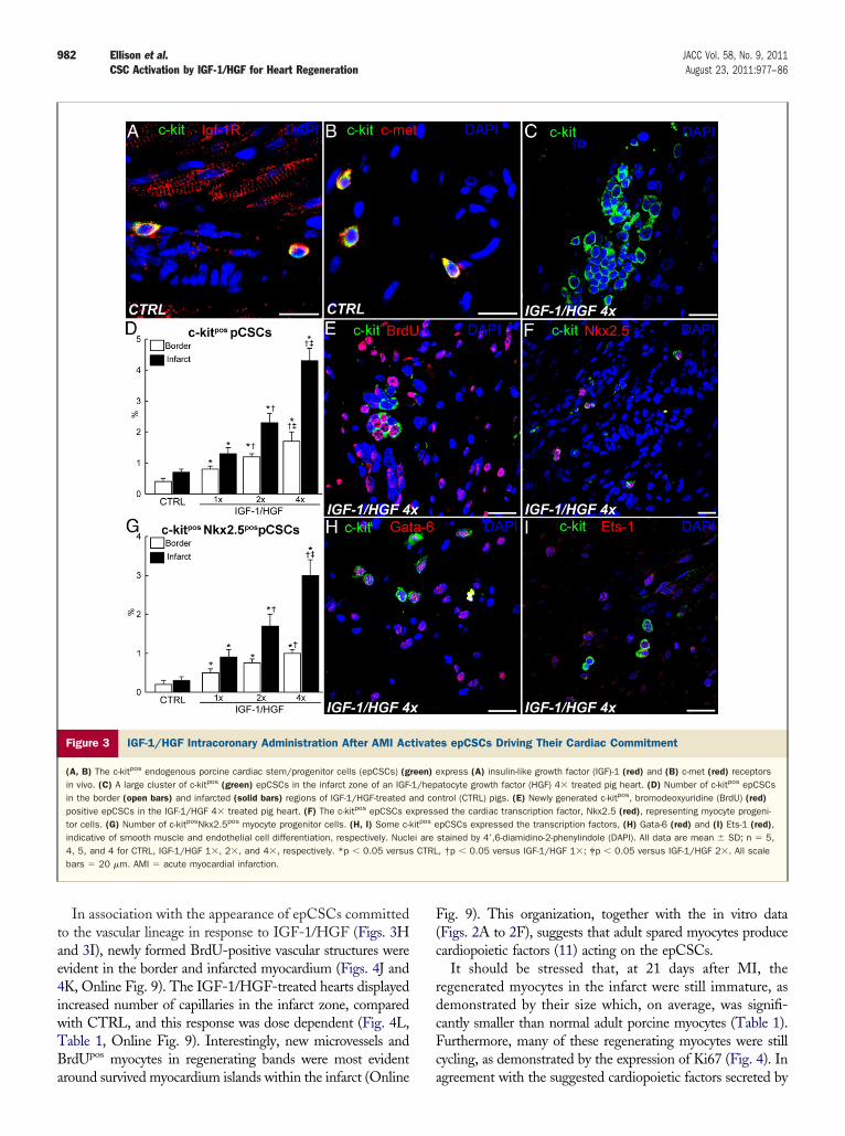
myocardium (Figs. 2G and 2H). These islands consisted oflarge BrdU-negative cardiomyocytes, a phenotype that,together with their mature, even hypertrophic nature con-firmed their survival as pre-infarct myocytes (Fig. 2I).Compared with CTRL, the IGF-1/HGF-treated hearts, ina dose-dependent manner, had significantly less fibrotictissue in the infarct region (Figs. 2J to 2L). Additionally,IGF-1/HGF reduced myocyte apoptosis and hypertrophyin the peri-infarct/border zone in a dose-dependent manner(Figs. 2M to 2O). These findings indicate that earlyIGF-1/HGF administration after AMI rescues myocytes atrisk and improves myocardial remodeling in pigs.Intracoronary IGF-1/HGF administration after AMIactivates epCSCs. In healthy and post-AMI hearts, #90%of c-kitpos epCSCs express IGF-1 and c-met (HGF) recep-tors (Figs. 3A and 3B). Accordingly, 21 days after AMI,IGF-1/HGF-treated infarcted hearts showed a significantincrease of c-kitposCD45neg epCSCs in the border regionand even higher in the infarcted area (Figs. 3C and 3D).These cells are CD45neg and tryptaseneg and were clearlydistinguishable from resident c-kitposCD45pos cardiac mastcells (Fig. 1D).
That the increase in c-kitposCD45neg epCSCs is theresult of IGF-1/HGF administration was confirmed by itsdirect correlation to the IGF-1/HGF-dose administered(Fig. 3D). At the highest dose, the number of epCSCs inthe infarcted area is more than 6-fold greater than in theCTRL hearts (Fig. 3D, Table 1). Most epCSCs were BrdUpositive, a fixture that documents their birth after AMI(Fig. 3E). Many epCSCs expressed either transcriptionfactor Nkx-2.5, Ets-1, or Gata6, indicative of their com-mitment to the myocyte, endothelial, and smooth musclelineage, respectively (Figs. 3F to 3I). The number ofcommitted myogenic progenitors (c-kitposNkx2.5pos cells)significantly increased in the infarct and border regions ofthe treated hearts in a dose-dependent manner (Fig. 3G,Table 1).IGF-1/HGF administration produces robust myocardialcell regeneration after AMI. As expected from the in-crease in epCSCs and committed myocyte progenitors,IGF-1/HGF-treated hearts harbored a large population ofvery small, newly formed BrdUpos and still-proliferating(Ki67pos) myocytes in the infarct and border regions (Figs. 4Ato 4F). Furthermore, some were in mitosis and cytokinesis,confirming their immature nature (Fig. 4G). Newly formedBrdUpos myocytes were also present in the border region of theCTRL pigs. However, their number was less than one-fifth ofthose in IGF-1/HGF 1& treated hearts, and they werepractically absent in the infarct zone (Figs. 4H and 4I, OnlineFig. 8, Table 1).
There was a direct correlation between the number ofsmall BrdUpos/Ki67pos newly formed myocytes and IGF-1/HGF-dose (Fig. 4H and 4I, Table 1). The BrdUpos myo-cytes were organized as clusters of regenerating bands in theinfarct zone, which were more organized and compactedwith increasing IGF-1/HGF dose (Fig. 4A and 4B).
980 Ellison et al. JACC Vol. 58, No. 9, 2011CSC Activation by IGF-1/HGF for Heart Regeneration August 23, 2011:977–86
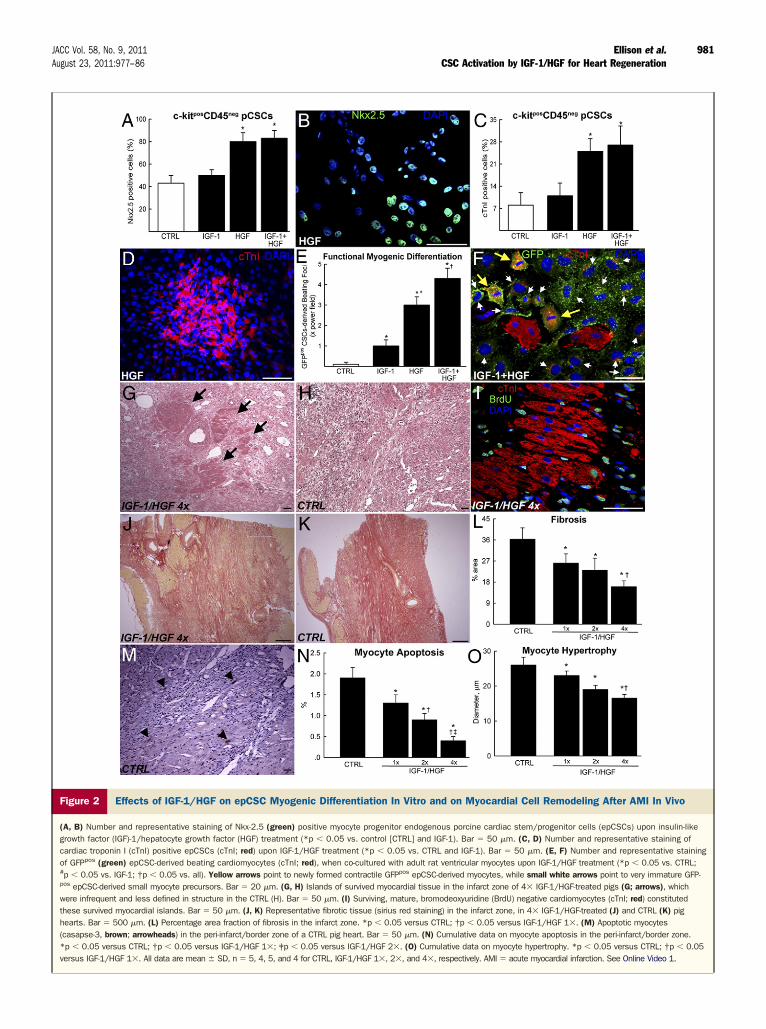
Figure 2 Effects of IGF-1/HGF on epCSC Myogenic Differentiation In Vitro and on Myocardial Cell Remodeling After AMI In Vivo
(A, B) Number and representative staining of Nkx-2.5 (green) positive myocyte progenitor endogenous porcine cardiac stem/progenitor cells (epCSCs) upon insulin-likegrowth factor (IGF)-1/hepatocyte growth factor (HGF) treatment (*p % 0.05 vs. control [CTRL] and IGF-1). Bar $ 50 !m. (C, D) Number and representative staining ofcardiac troponin I (cTnI) positive epCSCs (cTnI; red) upon IGF-1/HGF treatment (*p % 0.05 vs. CTRL and IGF-1). Bar $ 50 !m. (E, F) Number and representative stainingof GFPpos (green) epCSC-derived beating cardiomyocytes (cTnI; red), when co-cultured with adult rat ventricular myocytes upon IGF-1/HGF treatment (*p % 0.05 vs. CTRL;#p % 0.05 vs. IGF-1; †p % 0.05 vs. all). Yellow arrows point to newly formed contractile GFPpos epCSC-derived myocytes, while small white arrows point to very immature GFP-pos epCSC-derived small myocyte precursors. Bar $ 20 !m. (G, H) Islands of survived myocardial tissue in the infarct zone of 4& IGF-1/HGF-treated pigs (G; arrows), whichwere infrequent and less defined in structure in the CTRL (H). Bar $ 50 !m. (I) Surviving, mature, bromodeoxyuridine (BrdU) negative cardiomyocytes (cTnI; red) constitutedthese survived myocardial islands. Bar $ 50 !m. (J, K) Representative fibrotic tissue (sirius red staining) in the infarct zone, in 4& IGF-1/HGF-treated (J) and CTRL (K) pighearts. Bar $ 500 !m. (L) Percentage area fraction of fibrosis in the infarct zone. *p % 0.05 versus CTRL; †p % 0.05 versus IGF-1/HGF 1&. (M) Apoptotic myocytes(casapse-3, brown; arrowheads) in the peri-infarct/border zone of a CTRL pig heart. Bar $ 50 !m. (N) Cumulative data on myocyte apoptosis in the peri-infarct/border zone.*p % 0.05 versus CTRL; †p % 0.05 versus IGF-1/HGF 1&; ‡p % 0.05 versus IGF-1/HGF 2&. (O) Cumulative data on myocyte hypertrophy. *p % 0.05 versus CTRL; †p % 0.05versus IGF-1/HGF 1&. All data are mean ! SD, n $ 5, 4, 5, and 4 for CTRL, IGF-1/HGF 1&, 2&, and 4&, respectively. AMI $ acute myocardial infarction. See Online Video 1.
981JACC Vol. 58, No. 9, 2011 Ellison et al.August 23, 2011:977–86 CSC Activation by IGF-1/HGF for Heart Regeneration
In association with the appearance of epCSCs committedto the vascular lineage in response to IGF-1/HGF (Figs. 3Hand 3I), newly formed BrdU-positive vascular structures wereevident in the border and infarcted myocardium (Figs. 4J and4K, Online Fig. 9). The IGF-1/HGF-treated hearts displayedincreased number of capillaries in the infarct zone, comparedwith CTRL, and this response was dose dependent (Fig. 4L,Table 1, Online Fig. 9). Interestingly, new microvessels andBrdUpos myocytes in regenerating bands were most evidentaround survived myocardium islands within the infarct (Online
Fig. 9). This organization, together with the in vitro data(Figs. 2A to 2F), suggests that adult spared myocytes producecardiopoietic factors (11) acting on the epCSCs.
It should be stressed that, at 21 days after MI, theregenerated myocytes in the infarct were still immature, asdemonstrated by their size which, on average, was signifi-cantly smaller than normal adult porcine myocytes (Table 1).Furthermore, many of these regenerating myocytes were stillcycling, as demonstrated by the expression of Ki67 (Fig. 4). Inagreement with the suggested cardiopoietic factors secreted by
Figure 3 IGF-1/HGF Intracoronary Administration After AMI Activates epCSCs Driving Their Cardiac Commitment
(A, B) The c-kitpos endogenous porcine cardiac stem/progenitor cells (epCSCs) (green) express (A) insulin-like growth factor (IGF)-1 (red) and (B) c-met (red) receptorsin vivo. (C) A large cluster of c-kitpos (green) epCSCs in the infarct zone of an IGF-1/hepatocyte growth factor (HGF) 4& treated pig heart. (D) Number of c-kitpos epCSCsin the border (open bars) and infarcted (solid bars) regions of IGF-1/HGF-treated and control (CTRL) pigs. (E) Newly generated c-kitpos, bromodeoxyuridine (BrdU) (red)positive epCSCs in the IGF-1/HGF 4& treated pig heart. (F) The c-kitpos epCSCs expressed the cardiac transcription factor, Nkx2.5 (red), representing myocyte progeni-tor cells. (G) Number of c-kitposNkx2.5pos myocyte progenitor cells. (H, I) Some c-kitpos epCSCs expressed the transcription factors, (H) Gata-6 (red) and (I) Ets-1 (red),indicative of smooth muscle and endothelial cell differentiation, respectively. Nuclei are stained by 4’,6-diamidino-2-phenylindole (DAPI). All data are mean ! SD; n $ 5,4, 5, and 4 for CTRL, IGF-1/HGF 1&, 2&, and 4&, respectively. *p % 0.05 versus CTRL, †p % 0.05 versus IGF-1/HGF 1&; ‡p % 0.05 versus IGF-1/HGF 2&. All scalebars $ 20 !m. AMI $ acute myocardial infarction.
982 Ellison et al. JACC Vol. 58, No. 9, 2011CSC Activation by IGF-1/HGF for Heart Regeneration August 23, 2011:977–86

mature myocytes, those newly formed myocytes in contact orclose proximity with mature ones (i.e., in the border zone) weresignificantly larger than those in the middle of the scar withno proximity to spared myocytes (Fig. 4, Online Fig. 9). Also,IGF-1/HGF enhanced myocyte maturation, as shown by theincreased average size of BrdUpos myocytes with increasingdose (Table 1). However, despite this robust regeneration ofmyocyte number, the treated hearts still had a severe myocyte-mass deficit due to the significantly smaller size of the regen-erated myocytes compared with those lost by the AMI.IGF-1/HGF treatment improves cardiac function afterAMI. To test the effects on cardiac function after AMI,IGF-1/HGF at a 4& dose or saline (CTRL) was given byintracoronary administration to additional pigs. Cardiacmagnetic resonance imaging (cMRI) was performed beforecoronary occlusion, at 72 h, at 1 month and before sacrifice,and at 2 months after AMI (Online Fig. 7).
Intracoronary administration of a 4& dose of IGF-1/HGF (n $ 5) maintained the favorable histological effectson myocardial repair also at 2 months after MI (OnlineFig. 10). Remarkably, epCSC number, BrdUpos myocytesand capillary density were significantly higher in IGF-1/HGF-treated pigs compared with CTRL (n $ 4) (Fig. 5A,Online Fig. 10). Interestingly, at 2 months, BrdUpos myo-cyte maturation in the infarct region of treated pigs (diam-eter $ 14.3 ! 0.7 !m) was increased compared with 21days (at 4& dose, 12.3 ! 1.2 !m, p % 0.05), but theywere still smaller than the average adult porcine myocyte(17 ! 1 !m, p % 0.05). More importantly, infarct size,as measured by computed planimetric analysis at sacrifice,was significantly smaller in treated animals (16 ! 4% ofthe total left ventricular free wall) than in CTRL animals(24 ! 2%, p % 0.05).
The anatomical and histological improvement of cardiacrepair after MI in IGF-1/HGF-treated pigs was associatedwith better LV function (Figs. 5B to 5F). Indeed, in the
CTRL pigs, LV volumes and function progressively worsenedfrom 72 h to 2 months after AMI, whereas IGF-1/HGFprevented LV cardiac dilation, which resulted in a better LVejection fraction (45 ! 8% vs. 33 ! 6%, p % 0.05) at 2 monthsafter AMI (Figs. 5B to 5F, Online Table 1).
Discussion
Since its birth, regenerative cardiology’s holy grail hasbeen the development of procedures to either replace lostmyocytes with transplanted stem cells or to use stem cellsto mediate functional repair through paracrine effects(2,4,5,12,13). Although the discovery of eCSCs togetherwith definitive proof of adult mammalian myocyte renewalwere initially met with enthusiasm (14), mounting skepti-cism has developed about the physiological significance andregenerative potential of the resident c-kitpos eCSC popu-lation (3,15,16). A recent report claimed that the cardio-genic potential in vitro of the c-kitpos cardiac ells is robustonly during neonatal age, but it is severely limited or lost inthe adult (17).
Undoubtedly, resident adult stem-progenitor cells aremore abundant in early post-natal life and decline thereafter(18). These progenitor cells are highly active in the neonatalperiod because they are main participants in the myocar-dium hyperplastic growth and maturation to adulthood. Incontrast, most eCSCs in the adult myocardium are quies-cent (2). This difference might explain why eCSCs from theadult have a higher threshold than those from the neonateto activate their myogenic pathway in co-culture with fetalcardiomyocytes (17). Moreover, isolated c-kitpos cardiaccells are a mixed population containing not only real eCSCsand progenitor/precursors but also cells committed to otherlineages, including mast cells. Indeed, we show that theadult c-kitposCD45neg fraction is enriched for cells withcardiac stem and progenitor cell properties, while the
Histological and Immunohistochemical Data 21 Days After MITable 1 Histological and Immunohistochemical Data 21 Days After MI
CTRL(n ! 5)
IGF-1/HGF 1"(n ! 4)
IGF-1/HGF 2"(n ! 5)
IGF-1/HGF 4"(n ! 4)
Fibrosis, % 36.2 ! 5.9 26 ! 7.0† 23 ! 7.0† 16 ! 2.5†‡
Myocyte apoptosis, % 1.9 ! 0.25 1.3 ! 0.2† 0.9 ! 0.15†‡ 0.4 ! 0.1†‡§
Myocyte hypertrophy, !m 26 ! 2.2 23 ! 1.2† 19 ! 1.3† 16.5 ! 1.1†‡
c-kitpos CSCs (border zone), % 0.4 ! 0.1 0.8 ! 0.2† 1.2 ! 0.1†‡ 1.7 ! 0.3†‡§
c-kitpos CSCs (infarct area), % 0.7 ! 0.1 1.3 ! 0.2† 2.3 ! 0.3†‡ 4.3 ! 0.4†‡§
c-kitposNkx2.5pos CSCs (border zone), % 0.2 ! 0.1 0.5 ! 0.0† 0.7 ! 0.1†‡ 0.9 ! 0.05†‡§
c-kitposNkx2.5pos CSCs (infarct area), % 0.3 ! 0.1 0.9 ! 0.2† 1.7 ! 0.3†‡ 3.0 ! 0.4†‡§
BrdUpos myocytes (infarct zone), % 0.5 ! 0.1 7.1 ! 0.8† 12.7 ! 1.5†‡ 19.3 ! 2.6†‡§
BrdUpos myocytes (border area), % 1.2 ! 0.3 5.4 ! 0.6† 9.2 ! 1.3†‡ 11.8 ! 1.0†‡§
Ki67pos myocytes (border zone), % 0.7 ! 0.2 3.3 ! 0.3† 4.7 ! 0.4†‡ 6.5 ! 0.9†‡§
Ki67pos myocytes (infarct area), % 0.3 ! 0.0 3.9 ! 0.6† 8.2 ! 0.6†‡ 10.4 ! 1.1†‡§
Number of capillaries, &0.2 !m2 5 ! 1 7 ! 1 9 ! 2† 11 ! 1†‡§
BrdUpos myocyte diameter* (infarct zone), !m 6.4 ! 1.2 9.2 ! 1.3 10.4 ! 1.5† 12.3 ! 1.2†
*Diameter of normal BrdUneg myocyte $ 17 ! 1 !m; †p % 0.05 versus control (CTRL); ‡p % 0.05 versus IGF-1/HGF 1&; §p % 0.05 versusIGF-1/HGF 2&.
BrdU $ bromodeoxyuridine; CSC $ cardiac stem/progenitor cell; HGF $ hepatocyte growth factor; IGF $ insulin-like growth factor;MI $ myocardial infarction.
983JACC Vol. 58, No. 9, 2011 Ellison et al.August 23, 2011:977–86 CSC Activation by IGF-1/HGF for Heart Regeneration

fraction expressing CD45 does not contain cardiomyogeniccells (data not shown). This is not different from bonemarrow stem cells, whereby only a very small fraction ofc-kitpos cells are actually stem/progenitor cells (18). Thus, it
is not surprising that when the identity and number of truestem/progenitor cells is not known, as in Zaruba et al. (17),such undefined cell mixtures produce uninterpretable resultsin vitro and even more so in vivo. Furthermore, the failure
Figure 4 IGF-1/HGF Intracoronary Administration Induces Myocardial Regeneration After AMI
(A, B) Small, newly formed bromodeoxyuridine (BrdUpos) (green) myocytes (red) ("-sarcomeric actin) in the infarct regions of insulin-like growth factor (IGF)-1/hepatocytegrowth factor (HGF) of (A) 1& and (B) 4& treated pig hearts. (C) Within these regenerating bands were small Ki67pos (green) proliferating myocytes. (D, E) Newlyformed small BrdUpos myocytes (red) ("-sarcomeric actin) in the border zone after IGF-1/HGF (D) 1& and (E) 4& doses. (F) Small Ki67pos myocytes were also presentin the border zone after IGF-1/HGF-injection. (G) A small Ki67pos (green) mitotic myocyte precursor. Nuclei were stained by 4’,6-diamidino-2-phenylindole (DAPI) in blue.All scale bars $ 20 !m. (H, I) BrdUpos and Ki67pos myocyte number. Open bars indicate border region; solid bars indicate infarct region. (J, K) Newly formed arterialand capillary structures: BrdU $ green; "-smooth muscle actin (SMA) $ white; myosin heavy chain (MHC) $ red; von Willebrand factor (vWF) $ red; DAPI $ blue in theinfarcted region of IGF-1/HGF 4& treated pig hearts. Bar $ 20 !m. (L) Number of capillaries in the infarct zone. All data are mean ! SD; n $ 5, 4, 5, and 4 for CTRL,IGF-1/HGF 1&, 2&, and 4&, respectively. *p % 0.05 versus CTRL; †p % 0.05 versus IGF-1/HGF 1&; ‡p % 0.05 versus IGF-1/HGF 2&. AMI $ acute myocardialinfarction.
984 Ellison et al. JACC Vol. 58, No. 9, 2011CSC Activation by IGF-1/HGF for Heart Regeneration August 23, 2011:977–86

of adult c-kitpos cells to differentiate in Zaruba et al. (17)unlikely arises from their lack of actual regenerative poten-tial but rather from the lack of effective protocols tostimulate their differentiation. Accordingly, the data pre-sented here on the effects of IGF-1/HGF on adult c-kitpos
epCSCs activation, differentiation and maturation in vivoand in vitro, further ascertains the role of these cells inmyocardial cell homeostasis and regeneration.
There is little controversy that the best replacement forthe lost myocardium after MI is functional autologousmyocardial tissue. However, as presently practiced, theisolation and expansion of eCSCs for autologous cell trans-plantation is slow, expensive, and of uneven quality (2). Forthat reason, it is unlikely that autologous eCSCs transplan-tation will ever become widely available to treat acute andsubacute MI (2,19). Even for late and chronic MI, autolo-gous eCSCs can become available only to a very smallfraction of patients in need of myocardial regeneration andat very high costs. Thus, there is need for strategies tospecifically activate in situ the intrinsic cardiac regenerativepotential represented by the resident eCSCs using combi-
nations of growth factors, cytokines, and drugs, obviatingthe need for cell transplantation (2,19).
Recently, some growth factors/receptors axis—and acti-vating small molecules—modulating CSCs fate have beendescribed (20,21). Indeed, eCSCs possess IGF-1 and HGFsignaling pathways that regulate their growth, survival, andmigration (7,8,22). Also, HGF induces expression ofcardiac-specific markers in embryonic stem cells (23).
Despite their value as proof of concept, the extrapolationof murine and dog data (7,8) to the human pathology isdebatable. The 3 orders of magnitude difference in heart sizebetween mice and humans and the abundance of collateralsin the dog coronary circulation make these experimentalinfarct models not comparable to that of the human. Moreimportantly, in the mouse and canine studies, the growthfactors have been injected transepicardially through open-chest surgery, which is not a suitable option in the treatmentof AMI. In contrast, the protocol used here could easily beapplied at the time of primary revascularization for AMI.
Six major conclusions emanate from this study: 1) the adultporcine myocardium harbors epCSCs—phenotypically distin-
Figure 5 IGF-1/HGF Intracoronary Administration Improves Cardiac Function After AMI
(A) Number of c-kitpos endogenous porcine cardiac stem/progenitor cells (epCSCs) and bromodeoxyuridine-positive (BrdUpos) myocytes 2 months after acute myocardialinfarction (AMI). *p % 0.05 versus control (CTRL). (B to D) Left ventricular end-diastolic volume (LVEDV), left ventricular end-systolic volume (LVESV), and left ventricularejection fraction (LVEF) in the 2 groups of pigs. *p % 0.05 versus 72 h; #p % 0.05 versus CTRL. (E, F) Cardiac magnetic resonance imaging (cMRI) images showing pro-gressive cardiac enlargement in CTRL as opposed to insulin-like growth factor (IGF)-1/hepatocyte growth factor (HGF) treatment. All data are mean ! SD; n $ 4 for CTRLand n $ 5 for IGF-1/HGF 4&.
985JACC Vol. 58, No. 9, 2011 Ellison et al.August 23, 2011:977–86 CSC Activation by IGF-1/HGF for Heart Regeneration

guishable from cardiac mast cells—that differentiate intomyocytes, a process specifically stimulated by HGF;2) injection of small amounts of IGF-1/HGF in thecoronary artery supplying the infarcted region improvescardiomyocyte survival and remodeling; it has significantprotective effect on myocardial tissue organization andstructure; 3) intracoronary IGF-1/HGF administration ac-tivates the resident epCSCs, which multiply and differen-tiate, fostering the regeneration of the myocytes and micro-vasculature lost by the AMI; 4) the intensity of epCSCactivation, myocardial survival, and cell regeneration aredirectly correlated to the dose of IGF-1/HGF administered,strongly pointing to a direct cause-effect relationship; 5) theeffects of a single administration of IGF-1/HGF is stillmeasurable 2 months after its application, suggesting theexistence of a feedback loop triggered by the external stimulithat activates the production of growth and survival factorsby the targeted cells, which explains the persistence and longduration of the regenerative myocardial response; and 6)IGF-1/HGF treatment improves cardiac function by de-creasing cell death, infarct size, and preventing LV cardiacdilation, resulting in better LV ejection fraction at 2 monthsafter AMI, as measured by cMRI.
Taken together, these results provide the proof ofconcept needed to justify further experimental develop-ment and refinement of this approach, which ultimatelycould lead to an effective, simple, clinically applicable,and widely available protocol of myocardial regeneration.Study limitations. By extrapolation from fetal myocytes(24), it is likely that, despite the large number and progressivematuration of the regenerated myocytes from 21 days to 2months, these immature myocytes have limited force-generating capacity when compared with the adult survivingcounterparts. Therefore, the documented beneficial effect ofIGF-1/HGF on ventricular performance is unlikely due onlyto the direct force-generating contribution of the regeneratedmyocytes. Most probably, the effects on ventricular perfor-mance shown in Figure 5 are due to a combination of betterpreservation of myocardial architecture, decreased myocytedeath, reduced remodeling, and regeneration of new myocar-dial cells including neomyogenesis as well neoangiogenesis. Forfurther progress, the identification of the factors that drive fullCSC-derived cardiomyocyte maturation is warranted.
Reprint requests and correspondence: Dr. Bernardo Nadal-Ginard, The Stem Cell and Regenerative Biology Unit (BioStem),RISES, Liverpool John Moores University, Tom Reilly Building,Room 1.41, Byrom Street, Liverpool L3 3AF, United Kingdom.E-mail: [email protected]. OR Dr. Daniele Torella, Mo-lecular and Cellular Cardiology, Department of Experimental andClinical Medicine, Magna Graecia University, Campus S.Venuta,Viale Europa, 88100 Catanzaro, Italy. E-mail: [email protected].
REFERENCES
1. Beltrami AP, Barlucchi L, Torella D, et al. Adult cardiac stem cells aremultipotent and support myocardial regeneration. Cell 2003;114:763–76.
2. Torella D, Ellison GM, Karakikes I, Nadal-Ginard B. Residentcardiac stem cells. Cell Mol Life Sci 2007;64:661–73.
3. Martin-Puig S, Wang Z, Chien KR. Lives of a heart cell: tracing theorigins of cardiac progenitors. Cell Stem Cell 2008;2:320–31.
4. Ellison GM, Torella D, Karakikes I, Nadal-Ginard B. Myocyte deathand renewal: modern concepts of cardiac cellular homeostasis. NatClin Pract Cardiovasc Med 2007;4 Suppl 1:52–9.
5. Janssens S. Stem cells in the treatment of heart disease. Annu Rev Med2010;61:287–300.
6. Terzic A, Nelson TJ. Regenerative medicine advancing health care2020. J Am Coll Cardiol 2010;55:2254–7.
7. Urbanek K, Rota M, Cascapera S, et al. Cardiac stem cells possessgrowth factor-receptor systems that after activation regenerate theinfarcted myocardium, improving ventricular function and long-termsurvival. Circ Res 2005;97:663–73.
8. Linke A, Müller P, Nurzynska D, et al. Stem cells in the dog heart areself-renewing, clonogenic, and multipotent and regenerate infarctedmyocardium, improving cardiac function. Proc Natl Acad Sci U S A2005;102:8966–71.
9. Sperr WR, Bankl HC, Mundigler G, et al. The human cardiac mastcell: localization, isolation, phenotype, and functional characterization.Blood 1994;84:3876–84.
10. de Prado AP, Cuellas-Ramon C, Regueiro-Purriños M, et al. Closed-chest experimental porcine model of acute myocardial infarction-reperfusion. J Pharmacol Toxicol Meth 2009;60:301–6.
11. Behfar A, Perez-Terzic C, Faustino RS, et al. Cardiopoietic program-ming of embryonic stem cells for tumor-free heart repair. J Exp Med2007;204:405–20.
12. Kubal C, Sheth K, Nadal-Ginard B, Galiñanes M. Bone marrow cellshave a potent anti-ischemic effect against myocardial cell death in man.J Thorac Cardiovasc Surg 2006;132:1112–8.
13. Lai VK, Linares-Palomino J, Nadal-Ginard B, Galiñanes M. Bonemarrow cell-induced protection of the human myocardial: character-ization and mechanism of action. J Thorac Cardiovasc Surg 2009;138:1400–8.
14. Leinwand LA. Hope for a broken heart? Cell 2003;114:658–9.15. Passier R, van Laake LW, Mummery CL. Stem-cell-based therapy
and lessons from the heart. Nature 2008;453:322–9.16. Pouly J, Bruneval P, Mandet C, et al. Cardiac stem cells in the real
world. J Thorac Cardiovasc Surg 2008;135:673–8.17. Zaruba MM, Soonpaa M, Reuter S, Field LJ. Cardiomyogenic
potential of C-kit(')-expressing cells derived from neonatal and adultmouse hearts. Circulation 2010;121:1992–2000.
18. Rossi DJ, Jamieson CH, Weissman IL. Stems cells and the pathwaysto aging and cancer. Cell 2008:132;681–96.
19. Nadal-Ginard B, Torella D, Ellison G. Cardiovascular regenerativemedicine at the crossroads. Clinical trials of cellular therapy must nowbe based on reliable experimental data from animals with characteris-tics similar to human’s. Rev Esp Cardiol 2006:59;1175–89.
20. Sadek H, Hannack B, Choe E, et al. Cardiogenic small molecules thatenhance myocardial repair by stem cells. Proc Natl Acad Sci U S A2008;105:6063–8.
21. Engel FB, Hsieh PC, Lee RT, Keating MT. FGF1/p38 MAP kinaseinhibitor therapy induces cardiomyocyte mitosis, reduces scarring, andrescues function after myocardial infarction. Proc Natl Acad Sci U S A2006;103:15546–51.
22. Torella D, Rota M, Nurzynska D, et al. Cardiac stem cell and myocyteaging, heart failure, and insulin-like growth factor-1 overexpression.Circ Res 2004;94:514–24.
23. Roggia C, Ukena C, Böhm M, Kilter H. Hepatocyte growth factor(HGF) enhances cardiac commitment of differentiating embryonicstem cells by activating PI3 kinase. Exp Cell Res 2007;313:921–30.
24. Siedner S, Krüger M, Schroeter M, et al. Developmental changes incontractility and sarcomeric proteins from the early embryonic to theadult stage in the mouse heart. J Physiol 2003;548:493–505.
Key Words: cardiac magnetic resonance imaging y cardiac stem cells ygrowth factors y myocardial infarction y myocardial regeneration.
APPENDIX
For a complete description of Methods, supplementary figures, a supple-mentary table, and a supplementary video, please see the online version ofthis article.
986 Ellison et al. JACC Vol. 58, No. 9, 2011CSC Activation by IGF-1/HGF for Heart Regeneration August 23, 2011:977–86