Electrophysiological Evidence of Semantic Interference in Visual Search
Upload
independentCategory
view
2download
0
Identification of taste receptors and proteomiccharacterization of the antenna and legs of Triboliumbrevicornis, a stored food product pest
T. Alabi*¶, F. Marion-Poll†‡§, M. Danho¶,G. D. Mazzucchelli**, E. De Pauw**, E. Haubruge*and F. Francis*
*Functional and Evolutionary Entomology, GemblouxAgro-Bio Tech, University of Liege, Gembloux, Belgium;†AgroParisTech, Département Sciences de la Vie etSanté, Paris, France; ‡INRA, UMR1272 Physiologie del’Insecte: Signalisation et Communication, Versailles,France; §Université Pierre et Marie Curie, UMR1272Physiologie de l’Insecte: Signalisation etCommunication, Versailles, France; ¶NationalPolytechnic Institute FHB, ESA of Yamoussoukro,Yamoussoukro, Ivory Cost; and **Mass SpectrometryLaboratory, University of Liege, Liege, Belgium
Abstract
Chemoreception plays an important role in mediatinga diverse range of behaviours, including predationand food selection. In the present study, we combinedanatomical observations, electrophysiology and pro-teomics to investigate sensilla that mediate chemore-ception on the antenna and the legs of Tribolium.Scanning electron microscopy was used to differen-tiate the coxal and trochanteral segments of the pro-,meso- and metathoracic legs by the presence ofsensilla trichoidea and chaetica, while the antennaewere covered with five types of sensilla (chaetica,basiconica, trichoidea, squamiformia and coeloco-nica). Antenna morphology and ultrastructure weresimilar in both sexes. Electrophysiological recordingsallowed us to characterize a row of small sensillabasiconica on the terminal segment of the antennaas taste receptors, responding to sucrose and NaCl.Proteomics investigations of antennae and legsyielded several proteins with specific interest for
those involved in chemoreception. Odorant-bindingproteins were antenna-specific, while chemosensoryproteins were detected in both tissues.
Keywords: Tribolium, chemosensory, electrophysio-logy, proteomic, sensilla.
Introduction
Tribolium beetles belong to the Tenebrionidae family, andinclude 36 species, eight of which represent the mostprominent stored food product pests worldwide (Nakakita,1982; Angelini & Jockusch, 2008; Angelini et al., 2009).Classified as secondary colonizers, these beetles havespread widely as a result of international shipments ofinfested grain and flour. Although Tenebrionidae can alsoinfest sound, whole grains and cereal seeds, their growthis optimum in flour and other processed cereal products(Aitken, 1975). The largest species, Tribolium brevicornisranges in size from 10 to 12 mm with elongated shapes,and parallel, evenly coloured brown to blackish stripes onthe elytra. The antennae end in gradual clubs. The beetleis a recurrent pest of stored products in California, USA,and causes significant economic damage in Idaho, USAas a predator of the immature stages of leaf-cutting beesreared to pollinate alfalfa (Polk, 1977).
One of the most intriguing Tribolium beetle behavioursis cannibalism, and a number of studies have investigatedthis attribute (Alabi et al., 2008; 2009, 2011; Alabi, 2010).Cannibalism and predation are widespread amongTribolium species, and have been well described (Parket al., 1964, 1965; Stevens, 1989; Alabi et al., 2008).There is mounting evidence that different Triboliumspecies display varying propensities for cannibalism andpredation, suggesting different selective/evolutionaryforces operate for these behaviours among species andstrains (Sokoloff, 1972; Alabi et al., 2008). Some beetles,which are of primary economic importance because of theadverse impact of the species on food storage, exhibitsuch levels of cannibalism that laboratory stocks wouldbecome extinct without removing adults to permit eggs to
First published online 6 November 2013.
Correspondence: Taofic Alabi, Functional and Evolutionary Entomology,Gembloux Agro-Bio Tech, University of Liege, Passage des Déportés, 2B-5030 Gembloux, Belgium. Tel.: +32 81622287; fax: +32 81622312;e-mail: [email protected]
bs_bs_banner
InsectMolecular
Biology
Insect Molecular Biology (2014) 23(1), 1–12 doi: 10.1111/imb.12056
© 2013 The Royal Entomological Society 1

hatch into larvae and the larvae to become pupae. Pupaeare subsequently isolated to obtain parents to initiatenew laboratory cultures (Sokoloff, 1972). Other species,including T. brevicornis, which has recently been recog-nized as a potential stored products pest, avoid consum-ing conspecific pupae, and instead devour heterospecifics(Alabi et al., 2008, 2011). The evolution of prey discrimi-nation plays an important role in species viability.
In general, predator–prey relationships (e.g. wherebyinsects locate and select prey, and escape predation) inassociation with specific environmental factors have beenone of several processes leading to the origin of insectspecies. The environment may also reinforce aversivecannibalistic behaviours. Mertz & Robertson (1970)hypothesized that high rates of egg cannibalism in thelaboratory suggest Tribolium use a stimulus to locate eggsin flour. Craig (1986) confirmed that chemosensory infor-mation might affect egg cannibalism.
Contact chemoreception, whereby sources of thechemical stimulant are in contact with receptors, sendsinformation regarding foods and toxins (Ozaki &Tominaga, 1999). Insects perceive environmental stimulithrough their olfactory and chemosensory organs, includ-ing antennae, legs and mouthparts. Stimuli detectionprovides insects with information about food, predatorlocation and mating opportunities. Selection has favouredspecific morphological structures in Tribolium speciesbecause of its distribution in confined spaces with noparticular need for rapid locomotion, i.e. the beetle pos-sesses very short legs. The legs are potentially used tomake contact with their prey. Antennae are sensory struc-tures that house hearing, olfactory and tactile perceptionorgans. In Tribolium, the antennae are crucial for thespecies to interact with its environment (Snodgrass, 1935;Chapman, 1998). The scape, pedicel and flagellum com-prise the three segments of Tribolium antennae.
Tribolium species locate and optimize their search forprey disseminated in flour using a combination of sensorycues. In the present work, we concentrated on chemicalcues which are detected by olfactory and taste receptors,mostly located on the beetle’s antennae mouthparts andlegs (Merivee et al., 2004). The aims of the present studywere: (1) to determine the distribution of antennal andleg sensilla using scanning electron microscopy (SEM);(2) to confirm the gustatory role of a conspicuous groupof sensilla on the antenna using electrophysiologicalmethods; and (3) to perform a proteomics analysis of theantennae and legs using two-dimensional (2D) electro-phoresis followed by matrix-assisted laser desorption/ionization (MALDI)/mass spectrometry (MS) and liquidchromatography tandem MS (LC-MS/MS) analysis.
The Tribolium castaneum genome has been entirelysequenced and Tribolium beetles represent importantmodel organisms in insect development, evolution,
comparative genomics and pest science. Further-more, because of its notable economic importance,T. brevicornis is an appropriate model to examine thecapacity of flour beetles to interact with a diverse chemicalenvironment via chemoreceptive proteins, and severalother biochemical functions (metabolic pathways, defenceand contractile apparatus, among others).
Results
Anatomy
The legs of T. brevicornis are composed of the coxa, thetrochanter, the femur, and five tarsi in the fore and midlegs and four tarsi in the hind legs, which are equippedwith sensilla unequally distributed. We found sensillatrichodea and chaetica on the coxal and trochanteral seg-ments of each leg (Fig. 1).
The antenna of T. brevicornis are composed of a scape,a pedicel and a club-shaped elongated funiculus compris-ing 11 segments (Fig. 2). The most abundant type ofsensilla was sensilla chaetica, which were found bothin males and females, and distributed on all antennal
Figure 1. Scanning electron microscopy of ventral and dorsal leg viewsrepresentative of adult Tribolium brevicornis showing distinctive featuresof sensilla trichoidea (T) and chaetica (Ch).
2 T. Alabi et al.
© 2013 The Royal Entomological Society, 23, 1–12

segments, and especially on the club. These sensillawere straight with longitudinal grooves and their cuticleappeared thick and nonporous, suggesting a nonche-mosensory function. Sensilla chaetica varied in lengthfrom ∼18 to 24 μm. The sensilla base was inserted into asocket, which was slightly depressed into the cuticle.
On the club of the antenna of both sexes, we found fiveadditional sensillum types: chaetica (L = 18–24 μm), basi-conica (L = 2.30–3.80 μm), trichodea (L = 28–57 μm),squamiformia (L = 14.5 μm), and coeloconica (L =
3.15 μm) (Fig. 3). A circular row of sensilla basiconica wasfound at the periphery of the club. Given their morphologyand their location, we hypothesized that these sensilla hada taste function, notably because these sensilla exhibit apore at their tip (Fig. 3F). We presume that the other typeof sensilla have an olfactory function.
Electrophysiological responses to NaCl and sucrose
Using fine-tipped electrodes containing 0.1 M NaCl, wefirst probed the antenna and legs under a stereomicro-scope, to find hairs with which we could establish anelectrical contact. This initial survey allowed us to estab-lish that we could obtain a reliable contact with mostsensilla of a crown of sensilla chaetica disposed at the tipof the club of the antenna; however, given the small size ofthese sensilla, it was technically difficult to stimulate eachindividual sensilla as the liquid contained in the stimuluselectrode would often flow over the antenna by capillarity.A similar situation has been noted on antennal taste hairsof the honeybee (de Brito Sanchez et al., 2005).
We monitored the responsiveness of 1–5 sensilla fromthis crown, chosen because of their proper orientation,from about 10 different insects. These sensilla were stimu-lated with NaCl and sucrose at four different concentra-tions, as salts and sugars are tastants generally detectedby phytophagous and other insects (Chapman, 2003).
Figure 2. Scanning electron microscopy showing longitudinal view of anadult Tribolium brevicornis antenna.
A C E
B D F
Figure 3. (A) Scanning electron microscopy (SEM) depicting crown of the first club segment; scale bar = 10 μm. (B) SEM providing a ventral view ofthe first club segment; scale bar = 10 μm. (C) Micrographs of sensilla squamiformium (Sq), trichoideum, basiconicum (B) and chaetica (Ch), scalebar = 10 μm. (D) SEM close-up view of the flagellum crown with sensilla basiconica (B, white arrowheads), sensilla trichoideum (T, white arrows), andsensilla coeloconica (Coe, black arrowheads). (E) Micrograph central view of sensilla squamiformia (Sq), scale bar = 1 μm. (F) SEM representativesensilla basiconicum (B) from which successful tip recordings were obtained from Tribolium brevicornis.
Chemoreception in Tribolium 3
© 2013 The Royal Entomological Society, 23, 1–12

NaCl and sucrose activated at least one nerve cell whichincreased its firing in response to increased concentra-tions of these tastants (Fig. 4). These observationsconfirm the hypothesis that these sensilla have a tastefunction and that they contain several chemosensitiveneurons.
General proteomic profiles of Tribolium brevicornis legsand antennae
The antennal protein profiles of T. brevicornis were wellrepresented in 2D gels (Fig. 5). Proteomics approaches tocharacterizing the T. brevicornis proteome were optimized
Figure 4. (A) Mean concentration-response curve from salt and sugar-sensitive neurons in the basiconicum sensilla to sodium chloride and sucroseconcentrations ranging from 10−5 M to 10−2 M. The abscissa displays the stimulus solution concentration, and the ordinate the number of spikes occurringin consecutive 50-ms bins. Bars represent the mean SE. (B) s. basiconicum response to 10−5 M, 10−3 M, and 10−2 M NaCl, and 10−5 M and 10−2 Msucrose solution samples. Trace duration = 2 s; vertical bar = 1 mV; horizontal bar = 100 ms.
Figure 5. Tribolium brevicornis antenna 2DE gels stained with Coomassie (A) and silver (B). Proteins were subjected to isoelectric focusing on pH 3–11non-linear immobiline dry strips (IPG; GE Healthcare), and subsequently separated with 12% (w/v) polyacrylamide SDS-PAGE. The images areconsistent with results from those obtained in three gels from three independent experiments.
4 T. Alabi et al.
© 2013 The Royal Entomological Society, 23, 1–12
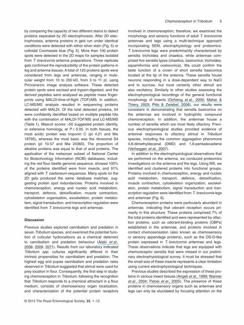
by comparing the capacity of two different stains to detectproteins separated by 2D electrophoresis. After 2D elec-trophoresis, antenna proteins in gels run under identicalconditions were detected with either silver stain (Fig. 5) orcolloidal Coomassie blue (Fig. 5). More than 100 proteinspots were detected in the 2D maps for samples isolatedfrom T. brevicornis antenna preparations. Three replicategels confirmed the reproducibility of the protein patterns inleg and antenna tissues. A total of 125 proteins spots wereconsidered from legs and antennae, ranging in mole-cular weight from 10 to 250 kD, from 3 to 11 pI, usingPROGENESIS image analysis software. These detectedprotein spots were excised and trypsin-digested, and thederived peptides were analysed as peptide mass finger-prints using MALDI-time-of-flight (TOF)/MS. In addition,LC-MS/MS analysis resulted in sequencing proteinsdetected with MALDI. Of the total detected proteins, 54were confidently identified based on multiple peptide hitswith the combination of MALDI-TOF/MS and LC-MS/MS(Table 1). Mascot scores >50 suggested protein identity,or extensive homology, at P < 0.05. In both tissues, themost acidic protein was troponin C (pI 4.21 and Mw18795), whereas the most basic was the ribosomal S4protein (pI 10.57 and Mw 24360). The proportion ofalkaline proteins was equal to that of acid proteins. Theapplication of the metazoan subset of National Centerfor Biotechnology Information (NCBI) databases, includ-ing the red flour beetle genome sequence, showed 100%of the proteins identified were from insects, and 91%aligned with T. castaneum sequences. Many spots on the2D gels produced the same database matches, sug-gesting protein spot redundancies. Proteins involved inchemoreception, energy and nucleic acid metabolism,transport, defence, detoxification, muscle contraction,cytoskeleton organisation, exoskeleton, protein metabo-lism, signal transduction, and transcription regulation wereidentified from T. brevicornis legs and antennae.
Discussion
Previous studies explored cannibalism and predation inseven Tribolium species, and examined the potential func-tion of cuticular hydrocarbons as a chemical deterrentto cannibalism and predation behaviour (Alabi et al.,2008, 2009, 2011). Results from our laboratory indicatedTribolium spp. cultures significantly differed in theirintrinsic propensities for cannibalism and predation. Thehighest egg and pupae cannibalism and predation ratesobserved in Tribolium suggested that stimuli were used forprey location in flour. Consequently, the first step in study-ing chemoreception in Tribolium, following the recognitionthat Tribolium responds to a chemical attractant in a flourmedium, consists of chemosensory organ localization,and characterization of the potential protein receptors
involved in chemoreception; therefore, we examined themorphology and sensory functions of adult T. brevicornisantennae and legs using a multi-technique approachincorporating SEM, electrophysiology and proteomics.T. brevicornis legs were predominantly characterized bysensilla trichoidea and chaetica, while antennae com-prised five sensilla types (chaetica, basiconica, trichoidea,squamiformia and coeloconica). We could confirm thetaste function of a crown of short sensilla basiconicalocated at the tip of the antenna. These sensilla houseneurons responding in a dose-dependent way to NaCland to sucrose, but most certainly other stimuli arealso excitatory. Similarly to other studies assessing theelectrophysiological recordings of the general functionalmorphology of insects (Ochieng et al., 2000; Maher &Thiery, 2003; Pitts & Zwiebel, 2006), our results wereconsistent in demonstrating that sensilla basiconica onthe antennae are involved in hydrophilic compoundchemoreception. In addition, the antennae house anumber of sensilla which are most likely olfactory. Previ-ous electrophysiological studies provided evidence ofantennal responses to olfactory stimuli in Triboliumspecies, including the common aggregation pheromone4,8-dimethyldecanal (DMD) and 1,6-pentadecadiene(Verheggen et al., 2007).
In addition to the electrophysiological observations thatwe performed on the antenna, we conduced proteomicsinvestigations on the antenna and the legs. Using MS, weidentified and clustered proteins into functional groups.Proteins involved in chemoreception, energy and nucleicacid metabolism, transport, defence, detoxification,muscle contraction, cytoskeleton organization, exoskel-eton, protein metabolism, signal transduction and tran-scription regulation were identified from T. brevicornis legsand antennae (Fig. 6).
Chemoreception proteins were particularly abundant inantennae, indicating that odorant reception occurs pri-marily in this structure. These proteins comprised 7% ofthe total proteins identified and were represented by olfac-tion proteins, such as odorant-binding proteins (OBPs)established in the antennae, and proteins involved incontact chemosensation (also known as chemosensoryor sensory appendage proteins), such as the OS-D-likeprotein expressed in T. brevicornis antennae and legs.These observations indicate that legs are equipped withchemoreceptor sensilla that were missed in our prelimi-nary electrophysiological survey. It must be stressed thatthe small size of these insects represents a clear limitationusing current electrophysiological techniques.
Previous studies described the expression of these pro-teins in various insect tissues (Angeli et al., 1999; Wanneret al., 2004; Pelosi et al., 2005). The presence of theseproteins in chemosensory organs such as antennae andlegs can only be elucidated by focusing attention on the
Chemoreception in Tribolium 5
© 2013 The Royal Entomological Society, 23, 1–12

Tab
le1.
List
ofid
entifi
edpr
otei
nsfr
omth
ele
gsan
dan
tenn
aeof
Trib
oliu
mbr
evic
orni
sby
aco
mbi
natio
nof
MA
LDI-
TO
Fan
dliq
uid
chro
mat
ogra
phy
tand
emm
ass
spec
trom
etry
LC-M
S/M
S
Pro
tein
nam
eO
rgan
ism
Acc
essi
onP
eptid
esM
ass
(D)
pIM
axsc
ore
Seq
uenc
esi
mila
rity
E-v
alue
Fun
ctio
n
Cut
icul
arpr
otei
n66
DC
G32
029-
PA
Trib
oliu
mca
stan
eum
XP
_001
8163
49.1
K.A
LSN
CV
1964
29.
4878
.225
0.05
0E
xosk
elet
on
Arg
inin
eki
nase
T.ca
stan
eum
XP
_971
800.
2R
.AK
LEE
IAG
K.F
R.G
TR
GE
HT
EA
EG
GIY
DIS
NK
.R40
325
5.66
607
100
5e-1
72A
min
oac
idm
etab
olis
m,
phos
phot
rans
fera
seA
TP
synt
hase
subu
nit
β,m
itoch
ondr
ial
T.ca
stan
eum
NP
_001
1643
61.1
R.S
HA
AK
AA
K.A
R.A
IAE
LGIY
PA
VD
PLD
ST
SR
.I53
544
5.19
811
100
0.0
AT
Pfo
rmat
ion
Ann
exin
IXC
G57
30-P
CT.
cast
aneu
mX
P_9
6793
1.1
R.T
IAQ
FY
EN
MY
GK
.SK
.WG
TE
ES
QF
NQ
ILIT
R.S
3603
25.
2163
610
01e
-180
Cal
cium
bind
ing/
sign
altr
ansd
uctio
nG
lyce
rald
ehyd
e3-
phos
phat
ede
hydr
ogen
ase
T.ca
stan
eum
XP
_974
181.
1K
.VIH
DN
FE
IVE
GLM
TT
VH
AT
TAT
QK
.TR
.LG
KP
AT
YD
DIK
.A35
705
8.43
592
100
1e-1
67C
arbo
hydr
ate
met
abol
ism
Eno
lase
CG
1765
4-P
BT.
cast
aneu
mX
P_9
7527
4.1
R.S
GE
TE
DT
FIA
DLV
VG
LST
GQ
IK.T
K.N
IILP
VP
AF
NV
ING
GS
HA
GN
K.L
4712
46.
4870
599
0.0
Car
bohy
drat
em
etab
olis
mM
alat
ede
hydr
ogen
ase
T.ca
stan
eum
XP
_969
151.
1R
.YG
QN
VLI
QF
ED
FG
NH
NA
FR
.YK
.GLA
FT
LEE
R.Q
6937
26.
7212
5110
00.
0C
arbo
hydr
ate
met
abol
ism
,py
ruva
teca
tabo
lism
Gly
copr
otei
n93
CG
5520
-PA
T.ca
stan
eum
XP
_971
540.
1K
.SE
GLD
MN
DM
IGQ
FG
VG
FY
SA
FLV
AD
R.V
K.IS
ALT
DLM
DV
IER
.L57
651
5.72
400
493e
-109
Cha
pero
ne
OS
-Dlik
epr
otei
n,O
S-D
2bM
egou
ravi
ciae
CA
G25
436.
1K
.KP
CT
PE
GA
ELR
.KK
.DY
DA
EW
KQ
LLD
K.W
1283
18.
8686
.710
07e
-16
Che
mor
ecep
tion
Cut
icul
arpr
otei
nLd
-CP
3T.
cast
aneu
mX
P_9
7390
9R
.GA
YR
QP
QP
GQ
PIA
ILR
.QR
.QP
QP
GQ
PIA
ILR
.Q7
878
4.94
146
100
6e-3
4C
hitin
scle
rotiz
atio
n
*Put
ativ
ees
tera
seT.
cast
aneu
mC
AH
6016
5.1
K.D
MV
MA
LKW
VQ
TN
IK.Y
K.V
WA
DIY
NS
NP
LFT
Q.-
5810
77.
1499
410
00.
0C
holin
este
rase
activ
ity
Cyt
ochr
ome
bT.
cast
aneu
mN
P_2
0316
6.1
R.N
IIID
LTN
2133
710
.39
63.5
332e
-09
Res
pira
tory
chai
nco
mpl
exco
mpo
nent
Act
in-8
7Eis
ofor
m2
T.ca
stan
eum
XP
_975
870.
1R
.TT
GIV
LDS
GD
GV
TH
TV
PIY
EG
YA
LPH
AIL
R.L
K.A
GFA
GD
DA
PR
.A38
199
5.36
690
100
0.0
Cyt
oske
leto
nor
gani
zatio
n
Act
inT.
cast
aneu
mX
P_0
0181
4869
R.IM
KE
K.L
NN
AS
GA
SN
R.S
1968
15.
2531
510
01e
-84
Cyt
oske
leto
nor
gani
zatio
n
†RN
Agu
anin
e-9-
met
hyltr
ansf
eras
edo
mai
nco
ntai
ning
2T.
cast
aneu
mX
P_9
7269
0.1
MS
AC
DD
TT
RT
PP
PN
PD
VIP
AK
.KK
.AA
FE
MIL
PK
.R34
701
9.24
567
100
7e-1
60E
nerg
ym
etab
olis
m
Sim
.G
prot
ein-
coup
led
rece
ptor
kina
se2
Api
sm
ellif
era
XP
_394
109.
2Q
FH
IYN
MG
GE
PG
LDIA
R.A
LGA
INS
FT
ST
TW
VE
NR
DW
TLR
.G28
977
9.91
249
633e
-64
Epi
thel
ialc
ellm
igra
tion/
phos
phor
ylat
ion
Fru
ctos
e-bi
spho
spha
teal
dola
seis
ofor
mA
Nas
onis
vitr
ipen
nis
XP
_001
6010
54K
.AD
DG
TP
FP
ELL
K.Q
K.K
DG
CH
FAK
.W40
120
7.60
644
100
0.0
Fru
ctos
ean
dm
anno
sem
etab
olis
mP
yrro
line-
5-ca
rbox
ylat
ede
hydr
ogen
ase
T.ca
stan
eum
XP
_969
408.
1K
.TV
IQA
EID
SA
AE
LID
FF
R.L
K.Y
LAG
INF
TG
SV
PT
FT
R.L
6348
48.
6711
1710
00.
0G
luta
mat
ean
dpr
olin
em
etab
olis
mG
luta
mat
ede
hydr
ogen
ase
T.ca
stan
eum
XP
_968
936.
1K
.DIV
HS
GLD
YT
ME
R.S
R.II
ND
ES
VQ
ES
LER
.R62
794
8.26
872
100
0.0
Glu
tam
ate
cata
bolis
m
Suc
ciny
l-CoA
synt
heta
seβ
chai
nA
edes
aegy
pti
XP
_001
6618
66.1
R.IC
NA
VM
VA
ER
.KK
.VH
AIL
VN
IFG
GIM
R.C
4873
36.
9763
999
0.0
Liga
seac
tivity
cellu
lar
FAB
P-li
kepr
otei
nis
ofor
m2
T.ca
stan
eum
NP
_001
1641
31.1
MV
DA
HLG
KK
YK
.LA
SS
EN
FD
E15
029
7.79
156
962e
-37
lipid
and
carb
ohyd
rate
met
abol
ism
sS
im.
Rho
GA
P71
EC
G32
149-
PA
T.ca
stan
eum
XP
_974
494.
2K
.LN
KE
AP
YR
.K28
359
9.47
389
841e
-106
Mag
nesi
umio
nbi
ndin
g/m
etab
olis
mN
AD
Hde
hydr
ogen
ase
(ubi
quin
one)
1αsu
bcom
plex
,13
Bom
byx
mor
iN
P_0
0104
0176
.1R
.NR
DE
EA
K.L
1884
19.
3018
887
2e-4
6M
itoch
ondr
iale
lect
ron
tran
spor
tH
eat
shoc
kco
gnat
e70
,is
ofor
m1
T.ca
stan
eum
EFA
1238
2.1
K.N
QV
AM
NP
NN
TIF
DA
K.R
R.II
NE
PTA
AA
IAY
GLD
K.K
7132
25.
3311
2894
0.0
Mol
ecul
arch
aper
one
(str
ess
resp
onse
)H
eat
shoc
k70
kDpr
otei
nco
gnat
eT.
cast
aneu
mX
P_9
7056
9.1
R.V
EIIA
ND
QG
NR
.IR
.ITP
SY
VA
FTA
DG
ER
.L63
256
5.45
1042
970.
0M
olec
ular
chap
eron
e(s
tres
sre
spon
se)
Hea
tsh
ock
prot
ein
20.6
T.ca
stan
eum
XP
_973
685.
1K
.LG
DF
SV
IDT
EF
SS
IR.E
K.D
GV
LTV
EA
PLP
AIT
AG
ET
LIP
IQH
.21
829
5.36
348
100
2e-9
4M
olec
ular
chap
eron
e(s
tres
sre
spon
se)
6 T. Alabi et al.
© 2013 The Royal Entomological Society, 23, 1–12

Trop
omyo
sin
1is
ofor
m1
T.ca
stan
eum
XP
_967
128.
1K
.TLT
NA
EA
EM
AS
LNR
.KK
.LA
FV
ED
ELE
VA
ED
R.V
3236
04.
7748
610
01e
-135
Mus
cle
cont
ract
ion
Mus
cle
prot
ein
20-li
kepr
otei
nT.
cast
aneu
mX
P_9
7500
0.1
K.F
PP
GE
LYE
DV
IR.D
R.A
GE
SIIG
LQA
GQ
NK
.G20
314
8.91
371
100
2e-1
01M
uscl
eco
ntra
ctio
n
Trop
onin
Cty
peIII
aT.
cast
aneu
mX
P_9
7061
5.1
-.IN
NN
FLK
K.K
1954
64.
4527
185
2e-7
1M
uscl
eco
ntra
ctio
nTr
opon
inC
T.ca
stan
eum
XP
_973
979.
1K
.SG
SIP
CD
MV
SD
ILR
LMG
QP
FD
K.K
R.L
EF
EE
FV
TLA
AK
FIV
EE
DD
EA
MQ
K.E
1879
54.
2130
292
6e-8
1M
uscl
eco
ntra
ctio
n
*Odo
rant
rece
ptor
153
T.ca
stan
eum
EE
Z99
175.
1R
.QA
LIG
GA
GH
TV
MLL
KA
LFLT
TK
.KR
.LK
PE
IDK
.A46
659
8.95
6243
1e-0
7O
lfact
ory
syst
em
*Odo
rant
bind
ing
prot
ein
C13
T.ca
stan
eum
CM
0002
85.2
R.N
HE
DV
HD
PK
LDE
HG
FC
ILK
.K16
157
6.67
87.8
683e
-16
Olfa
ctor
ysy
stem
*Odo
rant
bind
ing
prot
ein
C13
T.ca
stan
eum
CM
0002
85.2
R.N
HE
DV
HD
PK
LDE
HG
FC
LLK
.T15
407
5.88
79.7
739e
-14
Olfa
ctor
ysy
stem
AT
Psy
ntha
seB
.m
ori
NP
_001
0402
33.1
R.T
GA
IVD
VP
VG
DE
LLG
R.V
R.E
AY
PG
DV
FY
LHS
R.L
5961
29.
0993
899
0.0
Oxi
dativ
eph
osph
ory-
latio
nA
TP
form
atio
nS
im.
mC
G12
9107
(ubi
quiti
n)T.
cast
aneu
mX
P_9
7440
6.1
K.G
GP
IST
RR
.L18
730
9.65
219
631e
-55
Pro
tein
met
abol
ism
Spl
icin
gfa
ctor
45T.
cast
aneu
mX
P_9
7514
9.1
INIC
LVK
M29
201
6.93
335
684e
-90
Pro
tein
met
abol
ism
splic
ing
Put
ativ
enu
cleo
side
diph
osph
ate
kina
seT.
cast
aneu
mX
P_9
6750
3.2
R.V
MLG
AT
NP
AD
SA
SG
TIR
.G15
164
8.54
249
866e
-65
Pur
ine
and
pyrim
idin
em
etab
olis
mP
yruv
ate
kina
seis
ofor
mT.
cast
aneu
mX
P_9
6669
8.1
R.L
SG
IICT
IGP
AS
R.D
K.M
ME
TG
MN
IAR
.L58
685
6.84
1013
100
0.0
Pyr
uvat
eca
tabo
lism
Sim
.rib
osom
alpr
otei
nS
4T.
cast
aneu
mX
P_9
6926
2.1
SQ
DG
AR
SK
.KR
.KLG
NS
SA
.24
360
10.5
748
.910
4e-0
4R
NA
bind
ing
activ
ity
Sim
.M
od(m
dg4)
-heS
0053
1T.
cast
aneu
mE
FA08
224.
1K
.DV
AH
DN
MK
DIL
EF
MY
MG
EV
NV
LR.E
1791
96.
7520
964
6e-5
3S
plic
eoso
me
asse
mbl
ypa
thw
ayR
ibos
omal
prot
ein
S8
T.ca
stan
eum
XP
_967
339
R.N
PLR
K.K
2571
010
.29
316
849e
-103
Str
uctu
ralc
onst
ituen
tof
ribos
ome
*Glu
tam
ate
rece
ptor
,io
notr
opic
kain
ate
1,2,
3(g
lur5
,gl
ur6,
glur
7)
T.ca
stan
eum
XP
_974
933
R.IN
EA
LLR
.L14
947
5.91
132
851e
-29
Syn
aptic
tran
smis
sion
regu
latio
n
Coi
led-
coil
dom
ain
cont
aini
ng99
T.ca
stan
eum
XP
_966
464.
1K
.SQ
GN
SLF
AE
VD
DR
R.V
K.D
VP
PS
AS
GM
MK
YF
DN
MLA
MK
.N29
533
6.29
491
100
4e-1
37Tr
ansc
riptio
n;st
ruct
ural
iden
tity
ofce
llsV
asa
RN
Ahe
licas
eT.
cast
aneu
mN
P_0
0103
4520
R.A
PG
TG
DE
R.L
R.E
AG
VH
AT
R.G
2313
99.
9440
.427
0.14
Tran
scrip
tion
ofm
ultip
ledo
wns
trea
mm
RN
As
Sim
.C
G36
55C
G36
55-P
BT.
cast
aneu
mE
FA05
385.
1R
.HE
TLN
NH
GS
SS
LGLS
R.Y
K.Y
SG
NQ
GK
.I69
498
8.89
713
800.
0U
nkno
wn
†Hyp
.pr
otei
nTc
asG
A2_
TC
0072
56T.
cast
aneu
mE
FA01
682
HLA
IINE
CLF
IKH
GLP
GR
PLS
PP
TR
.HK
.IMT
IYC
LSF
SD
CT
ILK
.NA
SLE
ISK
.F
2678
19.
8619
947
3e-4
9U
nkno
wn
Hyp
.pr
otei
nTc
asG
A2_
TC
0020
82T.
cast
aneu
mE
FA12
376.
1R
.KLH
LSD
NT
NV
NV
NE
ICE
NLS
R.S
2131
78.
5437
310
05e
-102
Unk
now
nH
yp.
prot
ein
Tcas
GA
2_T
C00
8519
T.ca
stan
eum
EFA
0278
2K
.LN
HIA
IWS
K.N
2548
29.
8192
.419
3e-1
7U
nkno
wn
*Hyp
.pr
otei
nTc
asG
A2_
TC
0131
19T.
cast
aneu
mE
FA03
199.
1K
.DD
KP
VK
INK
.W23
188
9.69
245
562e
-63
Unk
now
nH
yp.
prot
ein
Tcas
GA
2_T
C00
0638
T.ca
stan
eum
EE
Z98
206.
1R
.IGG
RM
VT
SK
.SR
.SY
PM
VK
AT
NP
LFP
LPK
.A16
939
9.13
112
321e
-23
Unk
now
n
*Hyp
.pr
otei
nTc
asG
A2_
TC
0078
08T.
cast
aneu
mE
FA02
155
R.T
GY
ES
SM
NH
R.I
K.M
TF
VIIG
AC
MG
ALG
LMIL
TV
GC
LAT
GA
TR
H18
736
10.2
070
.558
5e-1
1U
nkno
wn
Hyp
.pr
otei
nTc
asG
A2_
TC
0306
74T.
cast
aneu
mE
FA02
969.
1R
.QS
LTLQ
NT
TS
E21
545
10.0
851
.211
6e-0
5U
nkno
wn
Sim
.C
G50
65-P
AT.
cast
aneu
mX
P_9
7343
1.1
K.T
VF
ITG
GT
GF
MG
KV
LLE
K.L
2198
59.
3016
540
3e-3
9U
nkno
wn
Sim
.C
G11
876
CG
1187
6-P
BT.
cast
aneu
mX
P_9
6666
4.1
K.T
NV
WV
QLM
K.Q
2114
510
.36
78.6
203e
-13
Unk
now
n
*Pro
tein
sfo
und
inan
tenn
aon
ly.
†Pro
tein
sfo
und
inle
gson
ly.
Hyp
.,hy
poth
etic
al;
Sim
.,si
mila
rto
.
Chemoreception in Tribolium 7
© 2013 The Royal Entomological Society, 23, 1–12

specific organ function. Indeed, OBPs and chemosensoryproteins are crucial for odours to deliver a signal to theolfactory receptors, where the signal transduction is initi-ated, and a specific behaviour is induced in the insect(Kaissling, 2001; Picimbon, 2003). OBPs have also beenfound to be necessary for contact chemoreception(Galindo & Smith, 2001; Xu et al., 2005; del Campo et al.,2011).
Chemoreception is important to all insects, particularlyin T. brevicornis; it may initiate behavioural responses,including the search for prey, feeding, escape, matingand oviposition (Gillott, 1980; Chapman, 2003; Jin et al.,2006).
The highest proportion of leg and antennal proteins wererelated to metabolism, representing 26% of the proteome,including proteins involved in energy production (carbohy-drate, lipid, and amino acid metabolism) and sugarmetabolism (glycolysis and tricarboxylic acid) pathways.The metabolic proteins identified consisted of includedenolase, pyruvate kinase, malate dehydrogenase,succinyl-CoA synthetase, beta chain and fructose 1,6-bisphosphate aldolase, fatty acid binding protein, andglyceraldehyde 3-phosphate dehydrogenase. Resultssuggested important lipid metabolism roles in Tribolium bythe presence of fatty acid binding proteins (FABPs) inT. brevicornis. FABPs are members of a highly conservedprotein family responsible for protecting the delicate lipidbalance of cells and are thought to facilitate the transfer offatty acids between extra- and intracellular membranes.
Based on current knowledge of Tribolium beetles, wehypothesize that these three metabolic pathways (carbo-hydrates, lipids and amino acids) are involved in energyproduction in the legs and antennae of T. brevicornis;however, in-depth kinetic investigations of metabolicenzymes to identify which substrate is preferentially uti-lized, in addition to studies applying deprivation of thesedifferent energy sources, are necessary to test thishypothesis.
The third group of proteins identified were involvedin muscle and cytoskeleton activity. Legs representthe primary source of locomotion in Tribolium species. Thecontraction of leg muscles mediates locomotion. Ingeneral, insect muscle is very similar to vertebrate striatedmuscle in function, and is characterized by several pro-teins that contribute to muscle contraction (Southgateet al., 1989). These proteins include muscle protein 20(mp20), actin, tropomyosin and troponine. Our proteomicsstudy provided evidence of these proteins in Tribolium.
In addition to the functional groups already described,the T. brevicornis leg and antennae proteome containedprotective proteins, including 70 kDa heat shock proteincognate 4 (Hsc 70-4), and small heat shock protein (Hsp)20.6. In laboratory culture conditions described in previ-ous reports (Arnaud et al., 2005; Alabi et al., 2008), highTribolium beetle population densities can act as an envi-ronmental stressor. Beetles synthesize Hsps in responseto stress (Mahroof et al., 2005).
The proteomes of both tissues examined in the presentstudy also included proteins involved in transport, particu-larly the electron and proton transport in various typesof metabolism. Proteins associated with adenosinetriphosphate (ATP) synthesis-coupled proton transportrepresented 8% of the total identified proteins (Clark &Baumann, 1997; Kidd et al., 2005).
Proteins associated with protein metabolism repre-sented the sixth most important group of proteins weestablished. This group (11% of total proteins identified)comprised ribosomal protein S4, ribosomal protein S8,mCG129107, splicing factor 45, Mod(mdg4)-heS00531,and Vasa RNA helicase.
In addition to the six functionally dominant groupsdescribed, the T. brevicornis antennae and legs containeda smaller proportion of proteins representative of otherbiological processes (Table 1).
In conclusion, the present study examined whether spe-cific proteins involved in chemoreception were expressed
Figure 6. Distribution of proteins identified inT. brevicornis legs and antennae based onbiochemical functions.
8 T. Alabi et al.
© 2013 The Royal Entomological Society, 23, 1–12

in the antennae and legs of the beetle T. brevicornis, astored food product pest by combining morphological,electrophysiological and proteomics techniques. Resultsprovided details characterizing the ultrastructure of differ-ent sensillum types located in antennae and legs, andelectrophysiological evidence that sensilla basiconicaorganized in a crown at the tip of the antenna respond tosucrose and NaCl solutions. In addition, a database ofantennal and leg proteins was generated by this study.The results of our investigation make a valuable contribu-tion to the understanding of insect chemoreception, andconsequently show great promise in reducing the eco-nomic losses caused by Tribolium. Also, the multidiscipli-nary – morphological, electrophysiological and proteomics– approach provided robust data to enhance our knowl-edge of chemoreception in the T. brevicornis beetle.
Leg and antenna examination in T. brevicornis served toelucidate important morphological structural elements ofthe cuticular sensilla of both tissues. In addition to provid-ing an extensive database of antennal and leg proteins inthis beetle, our electrophysiological recordings contributeto the knowledge of antennal chemoreception character-istics in T. brevicornis, which are relevant to potentialinvolvement in feeding and other behaviours.
Experimental procedures
Animals
Flour beetles, T. brevicornis, were reared in Petri dishes(140 mm) maintained in an incubator under complete darkness ata constant temperature (25 °C) and relative humidity (65 ± 5%).The insects were fed a mixture of wheat flour and powderedBrewer’s yeast according to Alabi et al. (2008).
Protein extraction and two-dimensional gel electrophoresis
Crude protein extracts from 300 legs (100 mg) and 2500 anten-nae (37 mg) of T. brevicornis were separately homogenized usinga polytron apparatus (PT 1200) in 20 mM phosphate buffer,pH 6.8, containing protease inhibitors (Roche, Indianapolis, IN,USA), followed by centrifugation at 15 000 × g for 10 min at 4 °C.The supernatant was collected, and proteins were subsequentlyisolated using the 2D Clean-Up Kit (GE Healthcare, LittleChalfont, UK) according to the manufacturer’s guidelines. Proteinconcentrations were determined using the 2D Quant Kit from GEHealthcare. Immobiline dry strips (IPG strips, GE Healthcare;pH 3–11, 18 cm) were rehydrated before isoelectric focusing with180 and 150 g of leg and antenna protein samples, respectivelyat 25 °C overnight (9 h) at a constant voltage (50 V). Four focus-ing steps were performed as follows: the initial step at 200 V for2 h, step 2 at 1000 V for 4 h, step 3 at 10 000 V for 1 h, and thefinal step at 10 000 V for 3 h 30 min. The maximum currentsetting was limited to 50 µA per IPG strip. Focused IPG stripswere equilibrated in reducing buffer for 15 min (375 mM Tris[pH 8.8], 6 M urea, 20% v/v glycerol, 2% w/v sodium dodecylsulphate (SDS) and 130 mM DTT), and alkylating buffer for anadditional 15 min (135 mM iodoacetamide, 375 mM Tris [pH 8.8],
6 M urea, 20% v/v glycerol, 2% w/v SDS). The equilibrated IPGstrips were fixed with 0.5% (w/v) agarose in SDS running bufferoverlaid on a 15% acrylamide gel sandwiched between glassplates for the second electrophoresis dimension. Electrophoresisusing denaturing conditions was performed using a Bio-RadPROTEAN II xi 2-D Cell apparatus (Hercules, CA, USA) at 1W/gel overnight at room temperature (Rabilloud, 1999; Franciset al., 2006).
Protein staining and image analysis
Electrophoresced proteins were detected using Coomassie col-loidal blue and silver staining. For Coomassie staining, the 2Dgels were fixed in 50% ethanol and 3% phosphoric acid for atleast 3 h at room temperature. The gels were then washed threetimes in distilled water for 20 min before pre-incubation for 1 h in34% (v/v) methanol, 3% (v/v) phosphoric acid, and 17% (w/v)ammonium sulphate. Coomassie G250 powder (0.36 g/l) wasadded in an equilibrated solution, and gels were incubated withthe stain for 4–5 days at room temperature on a rotary shaker.Finally, the gels were washed in water to remove backgroundstain (Westermeier, 2006).
For silver staining, gels were immediately fixed with 40%methanol (v/v) and 10% acetic acid. Thick, high-percentagepolyacrylamide gels were used, therefore gels were fixed over-night at room temperature (Walker, 2005). Following fixation,Milli-Q water (Millipore, Billerica, MA, USA) was used to rehydrategels and remove methanol through washing. Gels were thenincubated in a sensitization solution (0.02% v/v sodiumthiosulphate, 6.8% v/v sodium acetate, and 20% v/v methanol).Gels were washed twice (1 min per wash) in Milli-Q water toremove excess sensitization solution. Gels were then incubatedin a staining solution (0.1% w/v silver nitrate and 0.04% v/vformaldehyde) for 20 min. Gels were washed twice (1 min perwash), incubated in development solution (0.04% v/v formalde-hyde and 2% v/v sodium carbonate) until an acceptable signal-to-noise ratio was achieved, then transferred to stopping solution(5% acetic acid).
After gel scanning and gel image editing, raw images wereprocessed and analysed using PROGENESIS V.3.0 software (GEHealthcare).
In-solution digestion and protein identification
Protein extracts collected from T. brevicornis antenna and legsusing the 2D Clean Up kit were resuspended in 20 μl of 50 mMNH4HCO3, pH 8.0. Cysteines were reduced with 1 μl of a 200 mMDTT solution in 100 mM NH4HCO3 for 10 min at 50 °C followed byalkylation for 45 min with 0.8 μl of a 50 mM iodoacetamidesolution in 100 mM NH4HCO3 at room temperature in thedark. Alkylation was terminated by neutralizing the remainingiodoacetamide via the addition of 4 μl of 200 mM DTT in 100 mMNH4HCO3 at room temperature for 45 min. Digestion was per-formed overnight with 0.1 μg of trypsin in water. The resultingpeptides were dried in a vacuum centrifuge twice (Walker, 2005;Harmel et al., 2008).
Peptide separation by reverse-phase liquid chromatographywas performed on an Ultimate LC system (LC Packings, Amster-dam, The Netherlands) complete with a Famos autosampler anda Switchos II micro-column switching device for sample clean-upand pre-concentration. Each sample (30 μl) was loaded in dupli-
Chemoreception in Tribolium 9
© 2013 The Royal Entomological Society, 23, 1–12

cate at a flow rate of 200 nL/min on a micro-precolumn cartridge(300 μm, inner diameter [i.d.] x 5 mm, packed with C18 PepMapresin, 5 μm, 10 nm). After 5 min, the precolumn was connected tothe separating nano-column (75 μm i.d. x 15 cm, packed with C18PepMap100, 3 μm, 10 nm), and the gradient flow was initiated.The elution gradient varied from 0 to 30% in buffer B (0.1% formicacid in acetonitrile/water, 20:80 v/v) for 30 min. Buffer A consistedof 0.1% formic acid in acetonitrile/water (2:98 v/v). The LC systemoutlet was directly connected to the nano-electrospray source ofan Esquire HCT ion trap mass spectrometer (Bruker Daltonics,Bremen, Germany). Mass data acquisition was performed in themass range of 50 to 1700 m/z using the standard-enhancedmode (8100 m/z per s). For each scan, a data-dependent schemeselected the four most abundant doubly or triply charged ionsto be isolated and fragmented in the trap, and the resultingfragments were mass analysed using the Ultra Scan mode(50–3000 m/z at 26 000 m/z per s).
Data processing and database searching
Data processing and database searching was facilitated by adatabase containing the whole genome of the related speciesT. castaneum, a widespread stored product pest, which wassequenced by the Tribolium genome sequencing consortium.Gene sequences are available from the BCM-HGSC website,https://www.hgsc.bcm.edu/content/genome-data.
Peptide sequences obtained from tryptic-digested excised 2Dgel fragments, and identified by the MALDI bio-tool (Bruker-Daltonics) were matched to peptides from the metazoan subset ofthe NCBI database (a nonredundant database composed of full-length protein sequences, including the translation of nucleotidesequence databases), and the T. castaneum genome usingMascot Software (Matrix Science, London, UK; Perkins et al.,1999; Mascot server http://139.165.204.202/mascot/x-cgi/ms-review.exe). Expected fragment ions were calculated for eachpeptide in the above database, where the mass was similar tothat observed. Finally, a score calculated from the observed frag-ment ions matching the expected number, and the accuracy ofthe calculation, was generated.
Scanning electron microscopy
Adult T. brevicornis isolated from nutritional wheat flour werecleaned with a fine paintbrush, and starved for 24 h before SEMprocessing. After starvation, insects were dehydrated in subse-quent 70% and 90% alcohol baths, for 60 min each. Insects werethen fixed on a stub, and sputter-coated with gold three timesduring 60 s with stub rotation. SEM was performed with a JoelJSM840 electron microscope at 15 kV.
Electrophysiological recordings
Adult T. brevicornis beetles were immobilized on a carvedmicropipette tip fragment using strips of adhesive tape. Beetleswere oriented with the ventral side exposed upwards. For testingantenna, they were oriented so that the crown sensilla of the firstclub article was exposed and directed upward. The preparationwas then mounted on a magnetic ball-joint, and oriented under astereo-microscope (Leica Wild M10, France). Recordings wereperformed from sensilla located on the antenna, using the tip-recording method (Hodgson et al., 1955). Each recording was
obtained by capping a single sensilla for ∼2 s under visual control,with a capillary electrode containing the stimulus (mixed with1 mM KCl to carry the electrical contact). A second electrode(0.8 mm silver wire), connected to the ground, was inserted intothe abdomen and closed the circuit. The tip-recording electrodewas connected to an amplifier (TasteProbe DTP02, Syntech,Kirchzarten, DE; Marion-Poll & van der Pers, 1996). Borosilicateglass electrodes (O.D. 1 mm) were pulled to a tip diameter ∼10 μm(P77 horizontal electrode puller, Sutter Instruments Co., Novato,CA, USA), and filled with a stimulating solution immediately prior torecording. The electrical signals were further amplified (x 500–1000) and filtered (0.1–30 to 2800 Hz band-pass filter) usinga programmable amplifier (CyberAmp320; Axon Instruments,Foster City, CA, USA). Data were recorded and stored on a com-puter with a 16-bitA/D conversion card (DT9803; Data Translation,Malboro, MA, USA) controlled by custom software (dbWave:http://taste.versailles.inra.fr\deterrents\tk\dbwave\; Marion-Poll,1996). Each recording lasted 2 s, and was activated by a pulsedelivered by the amplifier on initial electrode contact with thesensillum.
Each set of recordings was performed by stimulating the gus-tatory sensilla of adults of both sexes with increasing concentra-tions of sucrose and NaCl stimuli (from 10−5 to 10−2 M). NaCl andsucrose purchased from Sigma-Aldrich Corp. (Saint-QuentinFallavier, France) were dissolved in distilled water (for sucrosewith 1 mM KCl), and stored at −20 °C. Stimulation solutions weremaintained at 4 °C for less than 1 week. Each stimulus wasapplied twice, with a time interval of 2 min to avoid fatiguebetween stimuli. The responses were quantified by the number ofspikes elicited during the first second of each recording.
Acknowledgements
We sincerely thank Prof. Philippe Compere, DominiqueTauban and Cyril Gaertner for their assistance with SEM.We are also grateful for comments on sensilla identifica-tion from Prof. Michel Renou (INRA Versailles). This workwas supported though the Ivorian Research Ministry andthe French community of Belgium fellowship program, aswell as research funds from the Gembloux Agro-Bio Techof Liege University.
References
Aitken, A.D. (1975) Insect travellers. 1. Coleoptera. Ministry ofAgriculture, Fisheries and Food. Tech Bull 31: 82–86.
Alabi, T. (2010) Cannibalism and Predation Behaviours amongTribolium Species. Liège University, Gembloux. 240 p.
Alabi, T., Michaud, J.P., Arnaud, L. and Haubruge, E. (2008) Acomparative study of cannibalism and predation in sevenspecies of flour beetles. Ecol Entomol 33: 716–726.
Alabi, T., Patiny, S., Francis, F. and Haubruge, E. (2009) Origineet évolution du cannibalisme dans les populations animales:Pourquoi manger son semblable? Biotechnol Agron SocEnviron 13: 409–425.
Alabi, T., Dean, J., Michaud, J.P., Verheggen, F., Lognay, G. andHaubruge, E. (2011) Does Tribolium brevicornis cuticularchemistry deter cannibalism and predation of pupae?(Coleoptera: Tenebrionidae). J Insect Sci 11: 115; 1–11.
10 T. Alabi et al.
© 2013 The Royal Entomological Society, 23, 1–12

Angeli, S., Ceron, F., Scaloni, A., Monti, M., Monteforti, G.,Minnocci, A. et al. (1999) Purification, structural characteriza-tion, cloning and immunocytochemical localization of chem-oreception proteins from Schistocerca gregaria. Eur JBiochem 262: 745–754.
Angelini, D.R. and Jockusch, E.L. (2008) Relationships amongpest flour beetles of the genus Tribolium (Tenebrionidae)inferred from multiple molecular markers. Mol PhylogenetEvol 46: 127–141.
Angelini, D.R., Kikuchi, M. and Jockusch, E.L. (2009) Geneticpatterning in the adult capitate antenna of the beetle Triboliumcastaneum. Dev Biol 327: 240–251.
Arnaud, L., Brostaux, Y., Lallemand, S. and Haubruge, E. (2005)Reproductive strategies of Tribolium flour beetles. J Insect Sci5: 33; 1–12.
de Brito Sanchez, M.G., Giurfa, M., de Paula Mota, T.R. andGauthier, M. (2005) Electrophysiological and behaviouralcharacterization of gustatory responses to antennal ‘bitter’taste in honeybees. Eur J Neurosci 22: 3161–3170.
Chapman, R.F. (1998) The Insects: Structure and Function.Cambridge University Press, Cambridge, UK; New York.
Chapman, R.F. (2003) Contact chemoreception in feeding byphytophagous insects. Annu Rev Entomol 48: 455–484.
Clark, M.A. and Baumann, P. (1997) The (F1F0) ATP synthase ofBuchnera aphidicola (endosymbiont of aphids): genetic analy-sis of the putative ATP operon. Curr Microbiol 35: 84–89.
Craig, D.M. (1986) Stimuli governing intraspecific egg predationin the flour beetles, Tribolium confusum and Triboliumcastaneum. Res Popul Ecol 28: 173–183.
Del Campo, M.L., Palmer, S. and Caillaud, M. (2011) Characteri-zation of a new odorant binding protein gene in gustatoryorgans of Manduca sexta larvae (Lepidoptera: Sphingidae).Ann Entomol Soc Am 104: 319–325.
Francis, F., Gerkens, P., Harmel, N., Mazzuchelli, G., De Paw, E.and Haubruge, E. (2006) Proteomics in Myzus persicae: effectof aphid host plant switch. Insect Biochem Mol Biol 36: 219–227.
Galindo, K. and Smith, D.P. (2001) A large family of divergentDrosophila odorant-binding proteins expressed in gustatoryand olfactory sensilla. Genetics 159: 1059–1072.
Gillott, C. (1980) Entomology, 2nd edn. Plenum Press, New York.Harmel, N., Letocart, E., Cherqui, A., Giordanengo, P.,
Mazzucchelli, G., Guillonneau, F. et al. (2008) Identification ofaphid salivary proteins: a proteomic investigation of Myzuspersicae. Insect Mol Biol 17: 165–174.
Hodgson, E.S., Lettvin, J.Y. and Roeder, K.D. (1955) Physiologyof primary chemoreceptor unit. Science 122: 417–418.
Jin, X., Zhang, S. and Zhang, L. (2006) Expression of odorant-binding and chemosensory proteins and spatial map ofchemosensilla on labial palps of Locusta migratoria(Orthoptera: Acrididae). Arthropod Struct Dev 35: 47–56.
Kaissling, K.E. (2001) Olfactory perireceptor and receptorevents in moths: a kinetic model. Chem Senses 26: 125–150.
Kidd, T., Abu-Shumays, R., Katzen, A., Sisson, J.C., Jimenez, G.,Pinchin, S. et al. (2005) The epsilon-subunit of mitochondrialATP synthase is required for normal spindle orientation duringthe Drosophila embryonic divisions. Genetics 170: 697–708.
Maher, N. and Thiery, D. (2003) Distribution of chemo- andmechanoreceptors on the tarsi and ovipositor of female Euro-pean grapevine moth, Lobesia botrana. Entomol Exp Appl110: 135–143.
Mahroof, R., Zhu, K.Y. and Subramanyam, B.H. (2005) Changesin expression of heat shock proteins in Tribolium castaneum(Herbst) (Coleoptera: Tenebrionidae) in relation to develop-mental stage, exposure time, and temperature. Ann EntomolSoc Am 98: 100–107.
Marion-Poll, F. (1996) Display and analysis of electrophysio-logical data under Windows. Entomol Exp Appl 80: 116–119.
Marion-Poll, F. and Van der Pers, J.N.C. (1996) Un-filteredrecordings from insect taste sensilla. Entomol Exp Appl 80:113–115.
Merivee, E., Renou, M., Mänd, M., Luik, A., Heidemaa, M. andPloomi, A. (2004) Electrophysiological responses to saltsfrom antennal chaetoid taste sensilla of the ground beetlePterostichus aethiops. J Insect Physiol 50: 1001–1013.
Mertz, D.B. and Robertson, J.R. (1970) Some developmentalconsequences of handling, egg-eating, and population densityfor flour beetle larvae. Ecology 51: 989–998.
Nakakita, H. (1982) Effect of larval density on pupation ofTribolium freemani Hinton (Coleoptera: Tenebrionidae). ApplEntomol Zool 17: 269–276.
Ochieng, S.A., Park, K.C., Zhu, J.W. and Baker, T.C. (2000)Functional morphology of antennal hemoreceptors of theparasitoid Microplitis croceipes (Hymenoptera: Braconidae).Arthropod Struct Dev 29: 231–240.
Ozaki, M. and Tominaga, Y. (1999) Contact chemoreceptors.In Atlas of Arthropod Sensory Receptors (Eguchi, E. andTominaga, Y., eds), pp. 143–154. Springer-Verlag, Tokyo.
Park, T., Leslie, P. and Mertz, D.B. (1964) Genetic strains andcompetition in populations of Tribolium. Physiol Zool 37:97–162.
Park, T., Mertz, D.B., Grodzinski, W. and Prus, T. (1965) Canni-balistic predation in populations of flour beetles. Physiol Zool38: 289–321.
Pelosi, P., Calvello, M. and Ban, L. (2005) Diversity of odorant-binding proteins and chemosensory proteins in insects. ChemSenses 30 (Suppl 1): i291–i292.
Perkins, D.N., Pappin, D.J., Creasy, D.M. and Cottrell, J.S. (1999)Probability based protein identification by searching sequencedatabases using mass spectrometry data. Electrophoresis 20:3551–3567.
Picimbon, J.F. (2003) Biochemistry and evolution of OBP andCSP proteins. In Insect Pheromone Biochemistry and Molecu-lar Biology (Blomquist, G.J. and Vogt, R.G., eds), pp. 539–566. Elsevier Academic Press, London.
Pitts, R.J. and Zwiebel, L.J. (2006) Antennal sensilla of twofemale anopheline sibling species with differing host ranges.Malar J 5: 26; 1–19.
Polk, D. (1977) Overwintering management for control of thegiant flour beetle (Tribolium brevicornis) in alfalfa leaf cuttingbee nests. Proc Entomol Soc Wash 39: 522–527.
Rabilloud, T. (1999) Proteome Research: Two-Dimensional GelElectrophoresis and Identification Methods. Springer-Verlag,Heidelberg, Berlin.
Snodgrass, R.E. (1935) Principles of Insect Morphology.McGraw-Hill, New York.
Sokoloff, A. (1972) The Biology of Tribolium, Vol. I. ClarendonPress, Oxford. p. 610.
Southgate, A., Lasko, P.F., French, C. and Pardue, M.L. (1989)Characterization of the gene for mp20: a Drosophila muscleprotein that is not found in asynchronous oscillatory flightmuscle. J Cell Biol 108: 521–531.
Chemoreception in Tribolium 11
© 2013 The Royal Entomological Society, 23, 1–12

Stevens, L. (1989) The genetics and evolution of cannibalismin flour beetles, genus Tribolium spp. Evolution 43: 169–179.
Verheggen, F., Ryne, C., Olson, P., Arnaud, L., Lognay, G.,Högberg, H. et al. (2007) Electrophysiological and behavioralactivity of secondary metabolites in the confused flour beetleTribolium confusum. J Chem Ecol 33: 525–539.
Walker, J.M. (2005) The Proteomics Handbook. Humana PressInc, Totowa, NJ.
Wanner, K., Willis, L., Theilmann, D., Isman, M., Feng, Q. andPlettner, E. (2004) Analysis of the insect OS-D-like genefamily. J Chem Ecol 30: 889–911.
Westermeier, R. (2006) Sensitive, quantitative, and fast modifi-cations for coomassie blue staining of polyacrylamide gels.Proteomics 6: 61–64.
Xu, P., Atkinson, R., Jones, D.N. and Smith, D.P. (2005)Drosophila OBP LUSH is required for activity of pheromone-sensitive neurons. Neuron 45: 193–200.
12 T. Alabi et al.
© 2013 The Royal Entomological Society, 23, 1–12
Copyright © 2022 FDOKUMEN