El capitán Alatriste
81
Supplement to T HE J OURNAL OF Allergy AND Clinical Immunology VOLUME 114 NUMBER 3 Hereditary and acquired angioedema: Problems and progress: Proceedings of the third C1 esterase inhibitor deficiency workshop and beyond Angelo Agostoni, MD, Emel Aygo ¨ ren-Pu ¨ rsu ¨ n, MD, Karen E. Binkley, MD, FRCPC, Alvaro Blanch,* Konrad Bork, MD,* Laurence Bouillet, MD, CCA,* Christoph Bucher, MD,* Anthony J Castaldo, MPA, Marco Cicardi, MD,* Alvin E Davis III, MD, Caterina De Carolis, MD, Christian Drouet, PhD,* Christiane Duponchel, Henriette Farkas, MD, PhD,* Ka ´ lma ´ n Fa ´ y, MD, Be ´ la Fekete, MD, PhD, DSc, Bettina Fischer, MD, Luigi Fontana, MD, George Fu ¨ st, MD, PhD, DSc,* Roberto Giacomelli, MD, Albrecht Gro ¨ ner, PhD, C. Erik Hack, MD, PhD,* George Harmat, MD, PhD, John Jakenfelds, MD, Mathias Juers, MD, Lajos Kalma ´ r, Pa ´ l N. Kaposi, MD, PhD, Istva ´ n Kara ´di, MD, PhD, DSc, Arianna Kitzinger, Tı´mea Kolla ´ r, MD, Wolfhart Kreuz, MD, PhD, Peter Lakatos, MD, PhD, DSc, Hilary J. Longhurst, MA, MRCP, PhD, MRCPath,* Margarita Lopez-Trascasa, MD, PhD,* Inmaculada Martinez-Saguer, MD, Nicole Monnier, PhD, Istva ´ n Nagy, E ´ va Ne ´ meth, Erik Waage Nielsen, MD, PhD,* Jan H. Nuijens, MD, PhD, Caroline O’Grady, RGN, Emanuela Pappalardo, PhD,* Vincenzo Penna, Carlo Perricone, Roberto Perricone, MD, Ursula Rauch, Olga Roche,* Eva Rusicke, Peter J Spa ¨ th, PhD, FAMH, George Szendei, MD, Edit Taka ´ cs, MD, Attila Tordai, MD, PhD,* Lennart Truedsson, MD, PhD,* Lilian Varga, PhD,* Bea ´ ta Visy, MD, Kayla Williams, BS, MA, MFA, Andrea Zanichelli, and Lorenza Zingale, MD* Milan, Rome, L’Aquila, and Peschiera Borromeo, Italy, Frankfurt, Mainz, Bonn, Marburg, Hattersheim, Frankfut/Main, and Aldenhoven/Siersdorf, Germany, Toronto, Canada, Madrid, Spain, Grenoble and Rouen, France, Zurich and Bern, Switzerland, Washington, DC, Boston and Cambridge, Mass, Budapest, Sopron, and Sze ´kesfe ´he ´rvar, Hungary, Amsterdam and Leiden, The Netherlands, Chalfont St. Giles and London, England, Bodø, Norway, and Lund, Sweden Hereditary angioedema (HAE), a rare but life-threatening condition, manifests as acute attacks of facial, laryngeal, genital, or peripheral swelling or abdominal pain secondary to intra-abdominal edema. Resulting from mutations affecting C1 esterase inhibitor (C1-INH), inhibitor of the first complement system component, attacks are not histamine-mediated and do not respond to antihistamines or corticosteroids. Low awareness and resemblance to other disorders often delay diagnosis; despite availability of C1-INH replacement in some countries, no approved, safe acute attack therapy exists in the United States. The biennial C1 Esterase Inhibitor Deficiency Workshops resulted from a European initiative for better knowledge and treatment of HAE and related diseases. This supplement contains work presented at the third workshop and expanded content toward a definitive picture of angioedema in the absence of allergy. Most notably, it includes cumulative genetic investigations; multinational laboratory diagnosis recommendations; current pathogenesis hypotheses; suggested prophylaxis and acute attack treatment, including home treatment; future treatment options; and analysis of patient subpopulations, including pediatric patients and patients whose angioedema worsened during pregnancy or hormone administration. Causes and management of acquired angioedema and a new type of angioedema with normal C1-INH are also discussed. Collaborative patient and physician efforts, crucial in rare diseases, are emphasized. This supplement seeks to raise awareness and aid diagnosis of HAE, optimize treatment for all patients, and provide a platform for further research in this rare, partially understood disorder. (J Allergy Clin Immunol 2004;114:S51-131.) Key words: AAE, acquired angioedema, angioedema, C1 esterase inhibitor, C1-INH, HAE, HANE, HANO, hereditary angioedema, hereditary angioneurotic edema, angioneurotic edema, chemically induced angioedema, human SERPING1 protein INTRODUCTION This supplement, like the 2003 C1 Esterase Inhibitor Deficiency Workshop and the many patient and physician *Members of the PREHAEAT European Union network. Disclosure of potential conflict of interest: See page S121. Supported by Kallikrein, LLC, a joint venture of Dyax Corp., and Genzyme Corporation, Cambridge, Mass. Received for publication June 23, 2004; revised June 24, 2004; accepted for publication June 24, 2004. Reprint requests: Dr. Tony Williams, Dyax Corp., 300 Technology Square, Cambridge, MA 01239; e-mail: t[email protected]. 0091-6749/$30.00 Ó 2004 American Academy of Allergy, Asthma and Immunology doi:10.1016/j.jaci.2004.06.047 S51
-
Upload
independent -
Category
Documents
-
view
3 -
download
0
Transcript of El capitán Alatriste

Supplement to
THE JOURNAL OF
Allergy AND ClinicalImmunology
VOLUME 114 NUMBER 3
Hereditary and acquired angioedema: Problems andprogress: Proceedings of the third C1 esteraseinhibitor deficiency workshop and beyond
Angelo Agostoni, MD, Emel Aygoren-Pursun, MD, Karen E. Binkley, MD, FRCPC, Alvaro Blanch,*
Konrad Bork, MD,* Laurence Bouillet, MD, CCA,* Christoph Bucher, MD,* Anthony J Castaldo, MPA,
Marco Cicardi, MD,* Alvin E Davis III, MD, Caterina De Carolis, MD, Christian Drouet, PhD,* Christiane
Duponchel, Henriette Farkas, MD, PhD,* Kalman Fay, MD, Bela Fekete, MD, PhD, DSc, Bettina
Fischer, MD, Luigi Fontana, MD, George Fust, MD, PhD, DSc,* Roberto Giacomelli, MD, Albrecht
Groner, PhD, C. Erik Hack, MD, PhD,* George Harmat, MD, PhD, John Jakenfelds, MD, Mathias Juers,
MD, Lajos Kalmar, Pal N. Kaposi, MD, PhD, Istvan Karadi, MD, PhD, DSc, Arianna Kitzinger, Tımea
Kollar, MD, Wolfhart Kreuz, MD, PhD, Peter Lakatos, MD, PhD, DSc, Hilary J. Longhurst, MA, MRCP,
PhD, MRCPath,* Margarita Lopez-Trascasa, MD, PhD,* Inmaculada Martinez-Saguer, MD, Nicole
Monnier, PhD, Istvan Nagy, Eva Nemeth, Erik Waage Nielsen, MD, PhD,* Jan H. Nuijens, MD, PhD,
Caroline O’Grady, RGN, Emanuela Pappalardo, PhD,* Vincenzo Penna, Carlo Perricone, Roberto
Perricone, MD, Ursula Rauch, Olga Roche,* Eva Rusicke, Peter J Spath, PhD, FAMH, George Szendei,
MD, Edit Takacs, MD, Attila Tordai, MD, PhD,* Lennart Truedsson, MD, PhD,* Lilian Varga, PhD,*
Beata Visy, MD, Kayla Williams, BS, MA, MFA, Andrea Zanichelli, and Lorenza Zingale, MD* Milan,
Rome, L’Aquila, and Peschiera Borromeo, Italy, Frankfurt, Mainz, Bonn, Marburg, Hattersheim, Frankfut/Main, and
Aldenhoven/Siersdorf, Germany, Toronto, Canada, Madrid, Spain, Grenoble and Rouen, France, Zurich and Bern,
Switzerland, Washington, DC, Boston and Cambridge, Mass, Budapest, Sopron, and Szekesfehervar, Hungary,
Amsterdam and Leiden, The Netherlands, Chalfont St. Giles and London, England, Bodø, Norway, and Lund, Sweden
Hereditary angioedema (HAE), a rare but life-threatening
condition, manifests as acute attacks of facial, laryngeal,
genital, or peripheral swelling or abdominal pain secondary to
intra-abdominal edema. Resulting from mutations affecting C1
esterase inhibitor (C1-INH), inhibitor of the first complement
system component, attacks are not histamine-mediated and do
not respond to antihistamines or corticosteroids. Low
awareness and resemblance to other disorders often delay
diagnosis; despite availability of C1-INH replacement in some
countries, no approved, safe acute attack therapy exists in the
United States. The biennial C1 Esterase Inhibitor Deficiency
Workshops resulted from a European initiative for better
knowledge and treatment of HAE and related diseases. This
supplement contains work presented at the third workshop and
expanded content toward a definitive picture of angioedema in
*Members of the PREHAEAT European Union network.
Disclosure of potential conflict of interest: See page S121.
Supported by Kallikrein, LLC, a joint venture of Dyax Corp., and Genzyme
Corporation, Cambridge, Mass.
Received for publication June 23, 2004; revised June 24, 2004; accepted for
publication June 24, 2004.
Reprint requests: Dr. Tony Williams, Dyax Corp., 300 Technology Square,
Cambridge, MA 01239; e-mail: [email protected].
0091-6749/$30.00
� 2004 American Academy of Allergy, Asthma and Immunology
doi:10.1016/j.jaci.2004.06.047
the absence of allergy. Most notably, it includes cumulative
genetic investigations; multinational laboratory diagnosis
recommendations; current pathogenesis hypotheses; suggested
prophylaxis and acute attack treatment, including home
treatment; future treatment options; and analysis of patient
subpopulations, including pediatric patients and patients whose
angioedema worsened during pregnancy or hormone
administration. Causes and management of acquired
angioedema and a new type of angioedema with normal
C1-INH are also discussed. Collaborative patient and physician
efforts, crucial in rare diseases, are emphasized. This
supplement seeks to raise awareness and aid diagnosis of HAE,
optimize treatment for all patients, and provide a platform for
further research in this rare, partially understood disorder.
(J Allergy Clin Immunol 2004;114:S51-131.)
Key words: AAE, acquired angioedema, angioedema, C1 esterase
inhibitor, C1-INH, HAE, HANE, HANO, hereditary angioedema,
hereditary angioneurotic edema, angioneurotic edema, chemicallyinduced angioedema, human SERPING1 protein
INTRODUCTION
This supplement, like the 2003 C1 Esterase InhibitorDeficiency Workshop and the many patient and physician
S51

J ALLERGY CLIN IMMUNOL
SEPTEMBER 2004
S52 Agostoni et al
Abbreviations used
AAE: Acquired angioedema
AAEE: (Italian) Voluntary Association for the Study,
Therapy, and Fight Against Hereditary
Angioedema
ACE: Angiotensin-converting enzyme
APP: Aminopeptidase P
AT2: Angiotensin II
B19V: Parvovirus B19
BMD: Bone mineral density
BVDV: Bovine viral diarrhea virus
C1: First component of the complement cascade
C1-INH: C1 esterase inhibitor
C1nh: Murine C1 esterase inhibitor gene
C1NH: Human C1 esterase inhibitor gene
C2: Second component of the complement cascade
C3: Third component of the complement cascade
C4: Fourth component of the complement cascade
C5: Fifth component of the complement cascade
CCM: Chemical cleavage of mismatches
CH50: Total hemolytic complement, 50% cell lysis
Cmax: Maximum concentration
CPMP: Committee for Proprietary Medicinal Products
CPV: Canine parvovirus
DHPLC: Denaturing HPLC
FF: (Ovarian) follicular fluid
FFP: Fresh frozen plasma
HAE: Hereditary angioedema
HAE-I: Hereditary angioedema type I
HAE-II: Hereditary angioedema type II
HAEA: US HAE Association
HAV: Hepatitis A virus
HbsAg: Hepatitis B surface antigen
HBV: Hepatitis B virus
HCV: Hepatitis C virus
HK: High molecular weight kininogen
HRT: Hormone replacement therapy
HUVS: Hypocomplementemic urticaria-vasculitis
syndrome
LH: Luteinizing hormone
MASP: Mannose-binding protein associated serine
protease
MBL: Mannan-binding lectin
MFO: Multifollicular ovary
MGUS: Monoclonal gammopathies of undetermined
significance
Mr: Molecular mass
NAT: Nucleic acid amplification technique
NEP: Neutral endopeptidase
OC: Oral contraceptive
OMIM: Online Mendelian Inheritance in Man (database)
PCO: Polycystic ovary
PCT: Primary care trust
PREHAEAT: Novel Methods for Predicting, Preventing,
and Treating Attacks in Patients
with Hereditary Angioedema
PRV: Pseudorabies virus
rhC1-INH: Recombinant human C1 esterase inhibitor
rtPA: Recombinant tissue-type plasminogen activator
SHBG: Sex hormone binding globulin
SSCA: Single-stranded conformational analysis
tPA: Tissue-type plasminogen activator
UK: United Kingdom
initiatives that inspired it, seeks to further assist cliniciansand researchers in the diagnosis, understanding, and man-agement of nonallergic angioedema. It represents thecombined scientific effort of nearly 80 scientists, physi-cians, and patient advocates from around the world, manyof whom presented at the Budapest workshop, but all ofwhom have helped to advance the knowledge of these raredisorders, and it cites the work of hundreds more. May thespirit of scientific solidarity contained herein sparkcontinued efforts toward international unity in improvingthe knowledge and management of these diseases.
History of angioedema from Quinckeand Osler to today
(Angelo Agostoni, MD, Lorenza Zingale, MD,* KaylaWilliams, BS, MA, MFA, and Marco Cicardi, MD,*Milan, Italy, and Cambridge, Mass)
Angioedema in the absence of allergy continues torepresent a medical paradox. This uncommon disordermay manifest as facial, laryngeal, genital, or intra-abdominal swelling or swelling of the extremities.Despite its often dramatic presentation, its rarity and itstendency to mimic other, dissimilar disease states oftenobscure its diagnosis. Even so, the condition has beendocumented for more than a century. Although a reviewby Dennehy1 posited that the writer Nathaniel Hawthornefirst described familial angioedema in his 1851 novel TheHouse of the Seven Gables,2 the hereditary disorderdescribed therein caused death associated with hemor-rhage (‘‘There was an unnatural distortion in the fixednessof Colonel Pyncheon’s stare. there was blood on his ruff,and. his hoary beard was saturated with it’’) andwas thusquite different from angioedema. Indeed, even Dennehy1
wisely attributed the observation to his father-in-law,hinting at a personal understanding of familial—if notheritable—defects.
Nonetheless, true medical descriptions soon appeared.In 1876, John Laws Milton3 described ‘‘giant urticaria.’’Acute, circumscribed edema of the skin was documentedby Heinrich Quincke4 in 1882. By 1888, Sir WilliamOsler5 distinguished an inherited form of angioedema,then known as angio-neurotic edema, and was the first tofully describe its clinical characteristics. A biochemicaldefect was isolated 75 years later, when Donaldson andEvans6 described similar patients whom they demon-strated were lacking the serum inhibitor directed againstthe first component of the complement system, C1 esteraseinhibitor (C1-INH). At the time of their 1963 publication,the extent of the deficiency was unknown, but immuno-electrophoresis permitted a semiqualitative evaluationindicating that the patients’ blood lacked C1-INH.
Since then, further work has been undertaken to betterunderstand the genetics, pathogenesis, and appropriateclinical management of nonallergic angioedema. Witha fuller knowledge of its biochemical mechanism hascome the gradual dismissal of neurotic from its name.However, today hereditary angioedema (HAE) and itseven rarer acquired form, acquired angioedema (AAE),

J ALLERGY CLIN IMMUNOL
VOLUME 114, NUMBER 3
Agostoni et al S53
remain little known in clinical practice and thus frequentlymisdiagnosed and inappropriately treated, often resultingin unnecessary suffering. Similarities to allergic condi-tions and inappropriate framing as part of the urticaria-angioedema syndrome frequently lead patients withHAE to be considered allergic and treated with anti-histamines and corticosteroids, ineffective in this dis-order. Abdominal edemamay so closely resemble an acuteabdomen that some patients with HAE have undergoneunnecessary surgical explorations, often more than once.Because untreated edema of the larynx may be fatal,inappropriate management may result in death.
For many, HAE and AAE present an ongoing clinicalchallenge. Despite the recurrent nature of angioedemaattacks, their acute treatment is often suboptimal, some-times delayed, and often requires lengthy hospital stays. Insome countries, including the United States, no safe andeffective acute attack therapy is available. Even theprophylactic management of these disorders is inconsis-tent across centers and nations, and, because of the sideeffects of antifibrinolytics and steroids currently in use,requires a lifelong, individualized calculation of benefitsand risks. These drawbacks are well known to the smallcommunity of physicians who deal frequently with thesediseases and are a feature of life for those patients whosuffer frequent or severe attacks.
Nonallergic angioedema as a model for thetreatment of rare diseases
(Kayla Williams, BS, MA, MFA, and Henriette Farkas,MD, PhD,* Cambridge, Mass, and Budapest, Hungary)
In recognition of these challenges, several national andinternational physician and patient initiatives have begunin the past 2 decades. In many ways, the field ofnonallergic angioedema, and especially HAE, is becom-ing an exemplar for the understanding and managementof rare diseases. The estimated frequency of HAE is1:50,000.7 As in many uncommon conditions, HAE’sinfrequent incidence fosters collaboration, forcing clini-cians and researchers to pool their anecdotal experiencesand data to attain statistical significance. Nonetheless,HAE is an attractive field because it offers doctors a chanceto improve the lives of their patients dramatically throughstudy but also via educated case management. As such, ithas brought together a group of motivated and compas-sionate physicians. The pharmaceutical industry has alsobeen welcomed to the C1 Esterase Inhibitor DeficiencyWorkshop and other HAE initiatives, fostering freeexchanges between academia, industry, and patients.
Indeed, perhaps the most distinctive feature of HAEphysician initiatives is their inclusion of patients withHAE, not only in a traditional capacity of raisingawareness and research funding but also as ethicaladvisors and welcomed guests for the presentation ofscientific abstracts and talks. The first C1 EsteraseInhibitor Deficiency Workshop, held in Hungary in1999, was the earliest meeting to follow this model.Since then, the 2 subsequent workshops and other patient-
association gatherings in the United States and Canadahave followed its inclusive precedent. Such a high level ofpatient involvement reflects not only the close relationshipbetween knowledgeable physicians and their patients butalso regional shortcomings in diagnosis and treatment.Because of the incapacitating and life-threatening aspectsof the disease, patients and their families from areaswhere HAE is largely unknown have been forced tobecome educated enough to explain the disorder tostrangers and, often, emergency department personnelto obtain the proper treatment. Even patients whose casesare managed by a competent local practitioner may haveattacks while traveling or when their doctor is unavail-able and thus may need to articulate their condition tosomeone entirely unfamiliar with the disease. By in-controvertible necessity, patients with HAE are one of thebest-educated patient populations, and this is especiallytrue in areas where satisfactory therapy for acute attacks isunavailable.
For patients and physicians alike, the Internet facilitatesincreasingly more communication, both personal andscientific. For patients with HAE, it can help to reducethe isolation of having a rare disease. Many patients firstcontact their national patient association online and use e-mail to stay in touch with fellow patients. The Internet isalso being used by physicians and scientists to supporta private patient registry and a public, constantly updatedhuman C1-INH gene (C1NH) mutation database. Throughthis online contact and regular meetings open to all,information about nonallergic angioedema is sharedrapidly among a small, concerned group. Nonetheless,the need to educate more physicians and the general publicremains. The rarity of nonallergic angioedema increasesthe likelihood that clinicians, especially general practi-tioners or emergency department personnel, may neverhave seen a case. Patient organizations and other groupshave thus worked to create emergency passports forpatients with known HAE to carry and educationalmaterials to distribute to emergency departments.
Scientific opportunities and current areasof controversy
(Kayla Williams, BS, MA, MFA, Cambridge, Mass)Nonallergic angioedema is a puzzle with relatively well-
defined borders:many specificC1NHmutations resulting inHAE have been identified, and the symptomatic results areknown. However, several central pieces are missing.Despite recognition of functional C1-INH deficiency asthe cause of most forms of nonallergic angioedema, thespecific mechanism of attack generation has not beendefinitively described. Likewise, symptoms similar to thoseof nonallergic angioedema have now been reported inpatients with normal amounts of functional C1-INH.8
Multiple pathways have been proposed for the chemicalcause of angioedema attacks. The murine HAE modeldeveloped by Han et al9 shares similarities with the humanform of the disease but diverges from typical HAE in thetriggering of angioedema. Despite homozygous C1-INH

J ALLERGY CLIN IMMUNOL
SEPTEMBER 2004
S54 Agostoni et al
deficiency, the mice, with few exceptions, have not beenobserved to have typical angioedema attacks. Attacks,manifesting solely as local increases in vascular permeabil-ity, could be provoked by the application of mustard oil.Rather than representing a shortcomingof themousemodel,such a high threshold for attacksmight parallel the course ofthose human heterozygotes, identified via a family memberwith activeHAE,who nonetheless never have an attack (fordocumentation of such patients, seeAgostoni andCicardi7).The absence of spontaneous attacks despite profound C1-INH deficiency suggests that multiple biological eventsmust transpire for angioedema to manifest.
Equally fascinating is the range of human disordersassociated with functional C1-INH deficiency. On themild end of the spectrum, the American physiciansLuong and Nguyen10 have reported a group of ap-parently unrelated Vietnamese women presenting to theirCalifornia clinic with lower extremities discomfort ofunknown etiology. All of these womenwere found to havereduced amounts of serumC1-INH, and danazol treatmentresolved both the C1-INH deficiency and the discomfort.At the opposite end of the C1-INH deficiency spectrum,some patients with HAE have periods of weekly or near-continuous angioedema attacks. In the most severe cases,laryngeal attacks may extend far enough into the thoraxthat even tracheostomy cannot maintain airway patency.
It is unclear whether discerning the mechanism of someforms of HAE, AAE, and C1-INH deficiency–associateddisordersmay elucidate others, but the attraction of a unifiedtheory is obvious. However, among other factors, theinhibitory promiscuity of the C1-INH molecule and itspredisposition to mutation may not lend themselves toa simple answer. Nonetheless, given the many proposedpathways for attack generation, information gained towarda full understanding of nonallergic angioedema attacksmaylead to a greater knowledge of 1 ormore chemical cascades,including the classical complement pathway, kinin gener-ation, and the intrinsic coagulation pathway.
The areas of greatest controversy include whichvasoactive peptide is ultimately responsible for the in-creased vascular permeability that results in angioedema.Bradykinin and second component of the complementcascade (C2)-kinin have been proposed,11,12 with recentresearch contributing evidence to the importance ofbradykinin.13-16 Nonetheless, within the current under-standing of coagulation, kinin, and complement pathways,neither peptide seems to perfectly explain all of thesymptoms of angioedema. Although bradykinin is theonly candidate mediator for which there is direct clinicalevidence, it is possible that yet another system, interme-diary, or molecule may be involved in edema-generatingvascular leakage.
Specific triggers for vasoactive peptide release are alsounknown. It is proposed that the activation of factor XII iscrucial to attack generation,17 and that factor XII activa-tion may be a result of phospholipids released fromdamaged or apoptotic cells. Recently, endothelial cellshave been implicated in the generation, via kallikrein, ofbradykinin, both in the presence18 and absence19,20 of
factor XII. These hypotheses explain how illness orlocalized tissue damage may precipitate attacks but donot account for other triggers, which are themselves notwell defined. In large part, triggers seem to vary frompatient to patient and, in several attacks, may not beapparent. Of these, the most scientifically documented andexplored are hormonal triggers, made all the more in-teresting by relatively recent reports of patients withnormal C1-INH concentrations and HAE-like symptomsprovoked or exacerbated by increased levels of estrogen.
The importance of hormones in the regulation ofnonallergic angioedema has long been acknowledgedvia its prophylaxis with androgens. Increasingly, theeffects of estrogen, progesterone, and other sex hormonesare being explored. In some women, estrogen results in anincreased frequency of angioedema attacks,21,22 but othersappear unaffected. Depending on the patient and trimester,pregnancymay reduce or increase the number and severityof attacks.23,24
In this supplement, the role of progesterone is debated,with Visy et al finding a positive correlation betweenserum progesterone values and attack frequency, whereasBork et al note no increase in attack frequency amongpatients whose oral contraceptive (OC) contained pro-gesterone and estrogen compared with those receivingestrogen alone. Indeed, Bork et al refer to severalpublished works in which progestins were used, withvarying success, to ameliorate HAE symptoms.22a-24a Incontrast, danazol, a common prophylaxis, alters multiplebiological mechanisms but is known to block progester-one receptors and increase progesterone’s metabolicclearance.25 Given these conflicting findings, the influenceof progesterone seems a likely area for further study.
About the supplement
After the 2003 Third C1 Esterase Inhibitor DeficiencyWorkshop in Budapest, participants were invited tofurther develop the information they presented for publi-cation. The scientific content herein represents the work ofparticipants who responded, some of which, as noted, hasnow been published elsewhere. In an attempt to survey thefield of nonallergic angioedema fairly and completely, thesupplement also includes reviews of relevant articles aswell as original material covering emergent areas in thefield of HAE, AAE, and related disorders. Individualauthorship is cited in text where possible and fullyattributed in the table of contents.
This supplement would not have been possible withoutthose who organized the Third C1-Esterase InhibitorDeficiency Workshop: the European C1-INH DeficiencyWorking Group, the Hungarian Society for Immunology,and the Foundation for the Prevention and Treatment ofFatal Angio-oedematous Diseases. Most especially, Iwould like to acknowledge Editor Dr. Henriette Farkasfor her unfailing compassion, organization, and support,and Professor Dr. Marco Cicardi and Dr. Tony Williams,who first imagined that such a document could be a reality.Dr. Bruce Zuraw’s cogent explanation of bradykininmetabolites was greatly appreciated. In addition, Dr.
J ALLERGY CLIN IMMUNOL
VOLUME 114, NUMBER 3
Agostoni et al S55
Karen Binkley graciously shared her work on very shortnotice and Dr. Alvin Davis III provided a valuable review;Dr. Shih-Wen Huang’s contribution to the US HAEAssociation newsletter informed me of the full range ofC1-INH deficiencies, Dr. Alvin Schmaier explained themystery of angiotensin II receptor blocker-associatedangioedema, and Dr. Erik Nielsen, both thorough hisonline Hereditary Angioedema Thesis and quick corre-spondence, provided information and inspiration. Manythanks go to Dr. Ineke Bos, whose model of the C1esterase inhibitor molecule graces our cover; ChrystalMcDonald who worked tirelessly to secure reprint per-missions; Dr. Brunello Wuthrich who provided images ofHAE attacks; and Drs. Werner Muller and Georg Dewaldfor their additions to the text. I would also like to recognizeMr. Anthony Castaldo of the US HAE Association for hisreview of text pertaining to the patient experience and hisindomitable, sustaining sense of humor.
Lastly, I would like to dedicate this supplement to itsmany contributing authors and all the HAE, AAE, andnon-allergic angioedema patients they strive to help.Kayla Williams
CLINICAL MANIFESTATIONS ANDDIAGNOSIS
In the first part of this section, Cicardi and Zingaledescribe the varied ways in which HAE can manifest anddiscuss other diseases that published case reports and theirclinical case series have associated with HAE.
Clinical manifestations of HAE
(Marco Cicardi, MD,* and Lorenza Zingale, MD,*Milan, Italy)
The symptoms of HAE are caused by the extravasationof plasma into the deeper cutaneous or mucosal layers asa result of 1 or more locally released vasoactive peptides.The edema in HAE is nonwhealing, nonpruritic, andgenerally unrelieved by antihistamines, suggesting thathistamine is not involved in its induction.26 The biologicalcharacteristics of the vasoactive peptides released inC1-INH–deficient sera indicate that the peptides belongto the kinin family. However, the discussion is not entirelyclosed on whether bradykinin, released because of contactsystem activation, or a peptide originated from C2 onclassical complement pathway activation and the gener-ation of plasmin, is the main mediator of symptoms inpatients with HAE.11,12 Nonetheless, recent lines ofevidence coming from C1-INH knockout mice, studiesin patients’ plasma, and analysis of C1-INH mutantsfrom patients with HAE support the bradykinin hypoth-esis.9,27,28 Kinin peptides participate in inflammatoryprocesses and increase vascular permeability, activatingintracellular pathways that lead to the release of nitricoxide.29,30 Vascular leakage can occur without anatomicaldamage and rapidly revert when the release of mediatormolecules ceases. Hence, edema usually resolves within72 hours. In some cases, it may resolve within 12 hours,
but in others, it may persist as long as 5 days. Urticaria,a condition analogous to angioedema but with plasmaleakage into the upper cutaneous layers, is typicallyabsent or minimal and short-lasting in patients withHAE.Typical symptoms. The recurrence of cutaneous
angioedema, abdominal pain, and asphyxia caused bylaryngeal edema is the full clinical pattern of HAE, presentin about 50% of adult patients.7 Attacks usually evolvewithin a single site, but it is not uncommon for somepatients to have simultaneous or closely spaced cutaneousand abdominal involvement. Most patients recognizeseveral hours in advance that an attack is coming. Theymay have sudden mood changes, anxiety, or completeexhaustion.
Cutaneous symptoms. Skin edema is nonpitting andnonerythematous, with ill-defined margins. It typicallyaffects the face (Fig 1), extremities, and genitals (Fig 2). Itusually spreads to disfigure the affected site, temporarilydepriving it of function.Most often, a single site is affectedby an extended edema that grows and then regresseswithin 2 to 5 days. Alternatively, edema may persist,although reduced in size, and migrate to differentcutaneous locations. In contrast with edema of otheretiologies, edema associated with HAE does not princi-pally manifest in the perioral region. Edema can localizesubcutaneously in any body part, including the trunk.
Abdominal symptoms. Recurrent abdominal pain, aconsequence of gastrointestinal wall edema, is reported by70% to 80% of patients with HAE.7,31,32 This is a dis-tinguishing feature of C1-INH deficiency because abdom-inal involvement is rarely seen in angioedema of otherorigins. It presents with symptoms that may vary frommild discomfort to severe, intractable pain accompaniedby vomiting and/or diarrhea.33 In this setting, hypovole-mia can result from a combination of fluid loss, plasmaextravasation, and vasodilation and can progress tohypovolemic shock.34,35 Ascites resulting from extrava-sation into the peritoneal cavity, edema of the bowel wall,or changes in splenoportal axis caliber have been de-scribed during abdominal attacks as detected by ultra-sounds or computed tomography.36-41 Gastrointestinalendoscopy performed during an abdominal attack re-vealed gastric involvement. Interestingly, during thehealing process after a prominent gastric edema, severalsmall nodules and raised erosions developed over theentire gastric mucosal surface. Within 55 days, the gastricmucosa had returned to normal.42
The similarity between bowel angioedema and surgicalemergencies is confirmed by the fact that approximatelyEof patients with undiagnosed HAE undergo unnecessarysurgery during abdominal attacks.7 However, even aftera diagnosis of HAE has been established, differentiatingangioedema of the bowel from a surgical emergencyremains a critical task for the physician.31 The physicalexamination can show the presence of an abdominaldefense reaction. Moderate or sometimes even markedleukocytosis can be part of an angioedema attack.43
Abdominal ultrasounds and computer-assisted tomogra-

J ALLERGY CLIN IMMUNOL
SEPTEMBER 2004
S56 Agostoni et al
phy scans demonstrate the presence of free peritoneal fluidand edematous intestinal mucosa.36,39,41 However, all ofthese signs are clearly not specific to angioedema. Theauthors note that this symptomatic generality should beborne in mind to avoid the situation that occurred witha patient in their case series. Surgery was inappropriatelydelayed when acute appendicitis was mistaken for in-testinal angioedema. The efficacy of C1-INH plasmaconcentrate in resolving symptoms may help to distin-guish angioedema from a true surgical emergency.
Laryngeal symptoms. Laryngeal edema is the mostdramatic clinical event for patients with HAE. Half ofthem have it at least once in their lives, but a history ofrecurrent episodes of suffocation caused by laryngealedema is not uncommon, and deaths still occur asa result.44 In the past, 25% to 30% of patients with HAEdied from laryngeal edema. This percentage has dramat-ically dropped for patients who are appropriately di-agnosed because of the availability of effectivetreatments in several countries.45 Nevertheless, becauseof previous life-threatening experiences, some patientswith HAE still carry permanent tracheal cannulae, allow-ing them to breathe by bypassing the larynx when edemaoccurs.
As mentioned, angioedema without urticaria is thehallmark of C1-INH deficiency. However, a discrete
FIG 1. Facial edema. Photo: Dermatologische Klinik, Universitats-
spital Zurich, Switzerland. Brunello Wuthrich, MD. Reprinted with
permission from Swiss Medical Weekly.43a
number of patients, 26% in a survey by Frank et al,31
have erythematous mottling, erythema multiforme, orerythema marginatum, always mild and transient, thatinconstantly heralds or attends their angioedema.46 Somepatients recognize this symptom as announcing an attack,and when on prophylactic treatment, can still have a rashnot followed by swelling. Fig 3 depicts several erythem-atous rashes experienced by patients before or duringattacks of angioedema.Unusual symptoms. Reports in the literature suggest
that edema caused by C1-INH deficiency could occur inlocations other than the characteristic sites of manifesta-tion.47-50 Frank et al31 reported transient pleuritic symp-toms with pleural effusion in 2 patients. Local cerebraledema has been considered responsible for transientseizures and hemiparesis seldom described in patientswith HAE.31,51 This assumption, despite its attraction andits occurrence in other forms of angioedema,49 has notbeen confirmed so far. Neurologic disorders and thepotential manifestation of cerebral edema remain a rarityin patients with HAE.
Although atypical, urinary symptoms mimicking aninfection have been described, and in 1 patient, thepresence of bladder edemawas documented by endoscopyand biopsy.24,48
Pulmonary edema as a consequence of C1-INH de-ficiency has occasionally been suggested but never clearlydemonstrated.47 In the authors’ experience, such an event
FIG 2. Penile edema. Photo credit: Dr. Martin Ludovic.

J ALLERGY CLIN IMMUNOL
VOLUME 114, NUMBER 3
Agostoni et al S57
FIG 3. Various erythematous rashes preceding or accompanying angioedema episodes. A1, Facial erythema
marginatum; A2, close view. Photo credit: Brunello Wuthrich, MD. Reprinted with permission from Swiss
Medical Weekly.43a B1, Mottling on chest; B2, close view. Photo credit: George Harmat, MD. Reprinted with
permission from Acta Dermato-Venereologica.46
was never observed to accompany an angioedema attack.They suggest that the high efficiency of the pulmonaryvascular tree in the inactivation of bradykinin accounts forthe lungs’ protection from its effects.52
Age of onset and frequency of symptoms. C1-INHdeficiency is present at birth, and a minority can haveperinatal angioedema symptoms. Most commonly, symp-toms begin at school age. Half of patients with HAE hadsymptoms within the first decade of life, and another thirdhad symptoms by the second decade. Asymptomaticadults carrying a C1NH mutation, detected because ofthe presence of offspring with clinically overt disease,have been described and are estimated to account for 5%of all patients with HAE.7
The frequency at which bouts of angioedema recur isextremely variable among subjects and may vary in thesame individual during different stages of life. A survey ofthe Italian case list showed that slightly less than E ofuntreated patients with HAE have more than 1 angioe-dema attack per month, 40% have 6 to 11 swellings peryear, and the remaining 30% are infrequently symptom-atic or completely symptom-free. This range of pheno-typic expression has no significant correlation with plasmaconcentrations of C1-INH and is usually inconsistentamong family groups. It should therefore be concludedthat factors other than C1-INH deficiency intervene to
determine a subject’s tendency to develop angioedema.These factors might be genetic or environmental. Thehypothesis that symptom frequency correlates with spe-cific functional polymorphisms of some of the proteinsinvolved in pathogenesis is attractive but thus far un-proven. An initial report suggesting that a polymorphismwithin the bradykinin receptor could distinguish oligo-symptomatic from polysymptomatic patients has not beenconfirmed.53 Farkas et al54 found that patients with HAEinfected with Helicobacter pylori are more susceptible tosymptoms than uninfected patients, and that eradicationof the infection reduces the frequency and severity ofswellings, particularly angioedema of the bowel. If con-firmed in a larger group of patients, these findings couldsupport those of several groups suggesting that infectionsincrease susceptibility to angioedema in the generalpopulation as well as in patients with HAE.7,31,55-57
Clinical and laboratory criteria for diagnosis are pro-vided in Table I; a severity scale for the evaluation ofnonallergic angioedema is provided in Table II. Thesetools are based on contributions elaborated from expertsfrom 10 European countries who received a grant from theEuropean Commission for a project called Novel Methodsfor Predicting, Preventing, and Treating Attacks inPatients with Hereditary Angioedema (PREHAEAT),consisting of a concerted action in the framework of the

J ALLERGY CLIN IMMUNOL
SEPTEMBER 2004
S58 Agostoni et al
specific research and a technologic development program,Quality of Life and Management of Living Resources,designed to improve the lives of patients with HAE.Diseases associated with C1-INH deficiency. Most
often, patients with HAE are substantially healthy apartfrom problems associated with swelling. However, thereare several reports of autoimmune diseases in patients withHAE,58-68 and systemic lupus, in particular, has beendescribed rather often.62 In a systematic study, 19 of 157patients with HAE had some kind of autoimmunedisorder.60 Moreover, patients with HAE, because ofdefective control of the classical pathway of complementactivation, have a deficiency of the fourth component ofthe complement cascade (C4) and C2, a condition thatincreases the risk of autoimmune diseases.69 A largeepidemiologic study in 1997 based on 24 major autoim-mune diseases estimated the prevalence of autoimmunediseases inAmericans to be 1 in 31 (3.2%).70 Given that allautoimmune diseases were not evaluated in this generalpopulation study, one cannot definitively conclude thatpatients with HAE have a higher risk of autoimmunedisease, but it appears likely.
The association of HAE with other inherited andnoninherited conditions has occasionally been reported,but these observations remain isolated.71-74
Last, patients with HAE can be exposed to risk throughneeded treatments. Several cases of hepatitis C virus(HCV) in the Italian case series were a result of receivingplasma-derived products. These cases occurred before theintroduction of viral inactivating procedures for plasmaproducts.75 No cases of HIV were reported, but because ofHCV, approximately 5% of their patients now have liver-related problems.
TABLE I. Criteria for diagnosis of angioedema caused
by C1 inhibitor deficiency
Clinical criteria
Major
(1) Self-limiting, noninflammatory subcutaneous
angioedema without major urticarial rash, often
recurrent and often lasting more than 12 hours
(2) Self-remitting abdominal pain without clear
organic etiology, often recurrent and often
lasting more than 6 hours
(3) Recurrent laryngeal edema
Minor
(4) Family history of recurrent angioedema
and/or abdominal pain and/or laryngeal edema
Laboratory criteria
(1) C1 inhibitor antigenic levels <50% of
normal at 2 separate determinations with
patient in basal condition and after the first year of age
(2) C1 inhibitor functional levels <50% of
normal at 2 separate determinations with
patient in basal condition and after the first year of age
(3) Mutation in C1 inhibitor gene altering protein
synthesis and/or function
Diagnosis can be established in presence of 1 major (1-3)
clinical criterion and 1 laboratory criterion
Role of ultrasound investigations in HAE
(George Harmat, MD, PhD, Pal N. Kaposi, MD, PhD,Kalman Fay, MD, Istvan Karadi, MD, PhD, DSc, BelaFekete, MD, PhD, DSc, George Fust, MD, PhD, DSc,*Lilian Varga, PhD,* and Henriette Farkas, MD, PhD,*Budapest, Hungary)
In this section, Harmat et al describe the results ofa study of 70 Hungarian patients with HAE in whomultrasonography was used to evaluate acute abdominalattacks of HAE.Background and rationale. Ascites can result from
diverse causes. The most common etiology, found inapproximately 80% of cases, is the decompensated liver(cirrhosis). The remaining 20% result from other pathol-ogies, such as malignancy in the abdomen (10%); variousinflammatory diseases and other disorders, such asnephrotic syndrome, exudative enteropathy, chylousascites, and mesenteric thrombosis; and others. However,HAE is very seldom mentioned as a cause of ascites. Thisis a real problem, because ascites are a significant di-agnostic sign of this uncommon but serious disease.
Themost common symptoms ofHAE appear in the formof ascites that cause acute abdominal attacks. For diagnos-ing this state, ultrasonography is themost potent tool.39,76,77
Methods. Ultrasonographic assessment is especiallywell suited to investigating the cause of abdominalsymptoms. This study was performed to evaluate theusefulness of ultrasonographic diagnosis and included 70patients (26 pediatric) from the Hungarian HAE centerdatabase. Of these, 60 had HAE type I and 10 had HAEtype II. Themale to female ratio was 32:38, and patient ageranged from 2.5 to 66 years. Patient follow-up continuedfor a decade. In addition to biochemical studies, ultra-sound investigations were performed at 6-month intervals.
Patients with typical symptoms of HAE were hospital-ized if the presence of other pathologies could be ruled outand if the manifestation was associated with hypovolemiaand included recurrent paroxysms of acute colicky pain,nausea and vomiting, or profuse diarrhea, not respondingto symptomatic therapy. All hospitalized patients un-derwent ultrasonography.
During each abdominal attack, ultrasound examina-tions were performed before treatment and repeated at 24and 48 hours post-treatment.78,79 Ultrasonographic inves-tigations were performed by using a Hitachi 451, a HitachiEUB 40 (Hitachi Medical Systems, Zug, Switzerland), oran Aloka SSD-1700 diagnostic system (Aloka Co, LTD,Tokyo, Japan), with a 3.5-MHz or - MHz convex trans-ducer or a linear 7.5-MHz transducer. Subdiaphragmaticand pelvic regions were scanned with the patient in asupine position. Kidneys were explored and the presenceof free peritoneal or retroperitoneal fluid was ascertainedwith the patient in the supine and lateral positions or,when necessary, standing. Free fluid, when detected, wasclassified into 1 of 3 categories, as follows:
(1) Small-volume free peritoneal fluid was visible onlyin the subhepatic or subsplenic space, and in everycase, in the Douglas cul-de-sac.

J ALLERGY CLIN IMMUNOL
VOLUME 114, NUMBER 3
Agostoni et al S59
TABLE II. Criteria for evaluation of disease severity*
Attack severity Score
Mild attacks (discomfort noticed, but no disruption of normal daily activity) 0.5 for each 24 hours
Moderate attacks (discomfort sufficient to reduce or affect normal daily activity) 1 for each 24 hours
Severe attacks (inability to work or perform daily activity) 2 for each 24 hours
Need for treatment
Emergency treatment: conservative, substitutive (C1-INH, FFP) 5 each
Emergency treatment: invasive (intubation, tracheotomy) 25 each
Long-term prophylaxis for more than 6 months 25
Long-term prophylaxis for 3-6 months 12.5
Score Class Degree
>30 1 Severe
21-30 2 Moderate
11-20 3 Mild
1-10 4 Minimal
0 5 Asymptomatic
*These parameters are determined over the period of 1 year. The sum of the scores defines the severity of the disease for that year.
(2) Moderate-volume ascites, in addition to ascites foundin these regions, included those identified in thesublienal space and among the intestinal loops. Theintestinal walls were also swollen (thickness inexcess of 5 mm80).
(3) In large-volume ascites, the intestinal loops floated inperitoneal fluid.
Results. An ultrasound image taken during an acuteabdominal attack (Fig 4) clearly illustrates the abdominalmanifestations of HAE. In this medial sagittal section ofthe pelvic area, a large amount of free peritoneal fluid canbe observed in the Douglas cul-de-sac, distal to and wellseparated from the urinary bladder. A floating intestinalloop can be seen.
During the attack, an edematous thickening of theintestinal wall and a thin, echo-free fluid layer around thebowels also could be observed, as illustrated in Fig 5. Asshown in Fig 6, a small amount of free fluid may beobserved in the triangle among the colon, spleen, and leftkidney; here, the intestinal wall is also thickened.
FIG 4. Sagittal sonogram during an abdominal HAE attack. A
significant amount of fluid can be seen in the pouch of Douglas,
with a swollen intestinal loop visible (arrow) floating in the free
fluid.
The symptoms of HAE are usually treated by theadministration of C1-INH concentrate. Fig 7 comparessonograms taken before and after treatment. At 24 hoursposttreatment, the volume of the ascites had decreased
FIG 5. Transverse sonogram during an abdominal HAE attack,
showing bowel and pancreas. Longitudinal section of a swollen
bowel: the intestinal wall is edematously thickened (arrows); in
addition, the reflectivity of the pancreas is increased.
FIG 6. Sonogram during an abdominal attack of HAE, showing
kidney and spleen. Section of a thickened intestinal wall (large
arrow) and a small amount of fluid (small arrow) between the left
kidney and the spleen.

J ALLERGY CLIN IMMUNOL
SEPTEMBER 2004
S60 Agostoni et al
FIG 7. Sagittal and transverse sonograms during an HAE attack before and after treatment. Sagittal sections
are shown above, transverse below. A, A large amount of free peritoneal fluid has accumulated in the pouch of
Douglas and, in the sagittal section, a floating intestinal loop is visible. The urinary bladder appears below. B,
Soon after treatment with C1-INH concentrate, the amount of peritoneal fluid is somewhat decreased. C, Only
a minimal amount of fluid is present in the pouch of Douglas 24 hours after C1-INH treatment. Several
sonograms have previously been published in slightly different format.78,81 Transverse panels B and C
reprinted with permission from Acta Paediatrica.81 Sagittal panels A-C reprinted with permission from the
European Journal of Gastroenterology and Hepatology.78
significantly; however, clinical symptoms abated within30 to 60 minutes of the infusion. The free peritoneal fluidand intestinal wall swelling fully disappeared within 48hours of treatment.
FIG 8. Transverse sonogram during an abdominal HAE attack: liver
and pancreas. Increased hepatic reflection (starry sky liver) and
thickened, echogenic portal veins (arrow); the pancreatic region is
also hyperechoic (double arrows). Reprinted with permission from
Acta Paediatrica.81
Edema of the portal veins, biliary ducts, and cholecystwall, causing gross structural changes in the liver, was alsoobserved. The liver parenchyma generally appeared lessechogenic, whereas the walls of the portal vein radiclesdisplayed increased echogenicity, resulting in a so-calledstarry sky texture that could be observed during the acutephase. Because of local edema, the pancreatic region alsodisplayed an increased echogenicity (Fig 8). In addition tothe hepatic portal vein, the wall of the cholecyst was alsoechogenic (Fig 9). After treatment with C1-INH concen-trate, the former brightness disappeared, and the echopattern of the liver returned to normal (sonogram notshown).Discussion. Early recognition of acute abdominal
attacks is of utmost importance because incorrect ordelayed diagnosis often leads to unnecessary surgicalintervention. In undiagnosed patients, ultrasound exami-nation can be a differential diagnostic means forrecognizing HAE in the abdominal organs because of itsability to detect nonspecific but sensitive clues such asthickening of the intestinal wall, free peritoneal fluid,intestinal hypermotility or hypomotility, and echo pattern

J ALLERGY CLIN IMMUNOL
VOLUME 114, NUMBER 3
Agostoni et al S61
changes of the liver and pancreas. Ultrasound examinationhas therefore proven very useful as a complementary,quick, and painless tool for recognizing the early phasesymptoms of HAE. Patients presenting with skin symp-toms (erythema marginatum) or acute pains, nausea,vomiting, or profuse diarrhea of unknown origins shouldbe immediately hospitalized and investigated with ultra-sound. Ultrasound follow-up in known cases of HAE isalso capable of proving the efficacy and expeditiousnessof acute attack treatment. In rare cases in which patientswith known HAE present with abdominal symptomsunresponsive to C1-INH concentrate, ultrasonographymay help distinguish between a refractory HAE attackand an unrelated surgical emergency.
Abdominal and pelvic ultrasound examination isa highly reproducible and informative diagnostic tooland thus is indicated during acute abdominal attacks ofHAE unresponsive to C1-INH concentrate. Conversely,a search for HAE is warranted when the typical sono-graphic features are ascertained in a patient with abdom-inal symptoms.
CLASSIFYING HAE AND AAE
(Kayla Williams, BS, MA, MFA, and Christoph Bucher,MD,* Cambridge, Mass, and Zurich, Switzerland)
Angioedema may be caused by reasons as various asallergies, inherited or acquired deficiencies of C1-INH, ordrug reactions.82,83 For the life of a patient presenting withunexplained airway swelling, the most important etiologicdistinction is that between angioedema of allergic, hista-minergic origins and the far rarer C1-INH–associated ornonallergic angioedema. When allergic angioedema hasbeen ruled out, nonallergic angioedema is next determinedto be hereditary or acquired, and subclassification ispursued.
Allergic angioedema, with histamine as its majormediator, may best be defined by its clinical response toantiallergic drugs such as antihistamines and cortico-
FIG 9. Transverse sonogram during an abdominal HAE attack: liver
and cholecyst. Because of edematous swelling, the wall of the
cholecyst is hyperechoic (large arrow) and the small portal veins
are also more echogenic (small arrows) in contrast with the liver’s
overall decrease in echogenicity.
steroids. In this type of angioedema, reaction of specificIgE antibodies with an allergen induces the release ofhistamine and other mediators from mast cells. It is oftenassociated with urticaria. In contrast, angioedema causedby C1-INH deficiency is not known to be triggered by anallergic reaction, is not usually associated with hives, andlikely has bradykinin as its principal mediator.
Current systems for classifying HAE andAAE describethe disorders in terms of C1-INH deficiency type.Although observed convention supports the classificationof major types, some further classifications, such as AAEtypes, are more fluid. In the case of the more recentlydescribed estrogen-sensitive angioedema, a new formaldescription is suggested here. In the interest of bothdefinition and the elucidation of mechanism that thesedifferences imply, the divisions of HAE and AAE type arepresented. For an example of prevalence, Table IIIpresents Agostoni’s 573-patient angioedema case seriesby type.
HAE: Types I and II
(Angelo Agostoni, MD, Konrad Bork, MD,* BettinaFischer, MD, C. Erik Hack, MD, PhD,* Christian Drouet,PhD,* Alvaro Blanch,* Olga Roche,* Nicole Monnier,PhD, Christiane Duponchel, Lajos Kalmar, Attila Tordai,MD, PhD,* Emanuela Pappalardo, PhD,* RobertoPerricone, MD, Margarita Lopez-Trascasa, MD, PhD,*Lorenza Zingale, MD,* and Marco Cicardi, MD,* Milanand Rome, Italy, Mainz, Germany, Amsterdam, TheNetherlands, Grenoble and Rouen, France, Madrid,Spain, and Budapest, Hungary)
Hereditary angioedema related to C1 inhibitor de-ficiency is a well-defined autosomal dominant trait. Itsvariants include types I (HAE-I) and II (HAE-II),associated with mutations of the C1 inhibitor gene(C1NH or SERPING1),84,85 and a newly described typenot associated with C1-INH deficiency8,86,87 further de-fined and discussed in another section.
The disease results from a large variety of mutations ofthe C1NH gene, located in the q12-q13.1 subregion ofchromosome 11. According to the relative concentrationsof antigenic and functional C1-INH, 2 types of HAE havetraditionally been described.88 The defective gene produ-ces either no C1-INH (HAE-I) or a dysfunctional C1-INH(HAE-II).6,84,85,88 In either case, it is associated with lowfunctional activity of C1-INH, low levels of C4, andnormal levels of the third component of the complement
TABLE III. Large nonallergic angioedema case series
classified by type
Classification
Number of
patients N = 573
HAE-I 356 (62.1%)
HAE-II 85 (14.8%)
ACE inhibitor–related angioedema 64 (11.2%)
Idiopathic nonhistiminergic angioedema 43 (7.5%)
AAE (with or without antibodies) 25 (4.4%)

J ALLERGY CLIN IMMUNOL
SEPTEMBER 2004
S62 Agostoni et al
cascade (C3). Concentrations of C1q, other than duringangioedema attacks, are normal.
In HAE-I (;85% of patients with C1-INH–associatedHAE), defective expression of 1 allele results in lowantigenic and functional concentrations of C1-INH.
In HAE-II (;15% of patients with C1-INH–associatedHAE), concentrations of functional C1-INH are low, butC1-INH antigenic levels are normal or increased, with thepresence of a dysfunctional mutant protein.88
For both, C1-INH function is usually 5% to 30% ofnormal, instead of the 50% expected if the single normalallele were fully expressed. This difference is ascribed topermanent C1 and contact phase activation, with sub-sequent C1-INH consumption in the periphery.89,90
Interestingly, the description of low levels of nonfunc-tional C1-INH mutants in patients with HAE-I hasdemonstrated that the distinction between HAE-I andHAE-II is not absolute.91 This finding occurred in patientswith mutations to exon 8 at the carboxy terminus of theC1NH gene, thought to be responsible for the properfolding necessary for transport outside of the cell andexposure of the reactive site loop. Thus, although thesepatients with low antigenic concentrations of C1-INHappear to have HAE-I, they are in fact expressingnonfunctional C1-INH that cannot efficiently exit the cell.
Estrogen-dependent and estrogen-associatedinherited angioedema (previously HAEtype III)
(Karen Binkley, MD, FRCPC, and Alvin E. Davis III,MD, Toronto, Canada, and Boston, Mass)
A type of angioedema, to date manifest only in women,has recently been described.8,86,87 Its symptoms closelyresemble those associated with functional C1-INH de-ficiency but occur in the presence of normal C1-INHconcentrations; the genetic defect responsible is currentlyunknown. Although this type of angioedema has beenreferred to as HAE type III (Online Mendelian Inheritancein Man [OMIM] 300268), others have argued that this de-signation is both redundant andmisleading. The followingpiece by Binkley and Davis explores their work in a kind-red with estrogen-dependent inherited angioedema, morefully describes estrogen-sensitive forms of inherited angio-edema, and proposes a rational system of nomenclature.Overview. The authors investigated a family with
symptoms of angioedema restricted to conditions of highestrogen levels. Although this investigation was under-taken to provide better care for the affected familymembers, it also presented a unique opportunity tobetter understand the effects of estrogen and androgenson C1-INH.8 However, instead of altered hormonalregulation of C1-INH, this family seemed to possessa completely novel abnormality, as suggested by theabsence of identifiable mutations in either the coding orthe 5# regulatory regions of the C1NH gene8 and normalC1-INH function and activity in a pregnant, symptomaticfamily member.92 The exact mechanisms responsible forangioedema in these patients have yet to be identified.
The importance of kinin degradation pathways andaminopeptidase P (APP) in the control of angioedemageneration has been independently recognized in studiesof angiotensin-converting enzyme (ACE) inhibitor–re-lated angioedema. Bradykinin and its active metabolite,des-Arg-bradykinin, are metabolized largely by 2 en-zymes, ACE and APP.93-95 With ACE inhibitor adminis-tration, APP becomes the primary enzyme responsible forinactivating bradykinin and des-Arg-bradykinin. In fact,individuals with low plasma concentrations of APP appearto be predisposed to developing angioedema during ACEinhibitor treatment, when neither ACE nor APP is avail-able to inactivate these kinins.96
Kinin inactivation pathways might also modulateclinical symptoms in classic HAE. For example, de-creasing kinin inactivation in patients with HAE withthe use of ACE inhibitors can result in exacerbation ofangioedema.97-100 Given the important contribution ofkinin inactivation pathways to the control of angioedema,this may be an avenue for further investigation.Case histories and investigation of the index family.
The index family presented with histories of episodic,HAE-like angioedema.8 These episodes occurred onlyduring pregnancy, OC use, or estrogen replacementtherapy. Symptoms began 14 to 21 days after conception,or within 7 to 14 days of starting endogenous hormones.No episodes occurred in the postpartum period. Onepatient’s description was particularly compelling: ‘‘Myperiod was just a day or two late, but when one side of myface swelled up, I knew I must be pregnant, because this isjust like what happened to my mother and sisters everytime they were pregnant.’’ In affected individuals, symp-toms occurred in all pregnancies and with each course ofestrogen therapy. Unaffected individuals had no symp-toms at any time. There were 8 affected women in 3generations and 1 obligate male carrier. Transmission wasconsistent with an autosomal dominant inheritance.Complement values, C1-INH, C1-INH function, prekal-likrein, factor XII, and high molecular weight kininogenwere normal in 3 patients during asymptomatic periods.
Genetic investigations were undertaken for the follow-ing reasons: (1) the patients were asymptomatic at the timeof presentation, (2) baseline biochemical investigationswere unremarkable, and (3) exposing patients to estro-gens for the purpose of detecting resultant biochemicalabnormalities was unethical in light of the risk of laryngealedema.
The striking clinical similarity to classic HAE focusedinitial investigations on the C1NH gene. However, noabnormalities in the coding sequences of the C1NH geneor in the 5# regulatory region were detected.8
When patient III-24 became pregnant and developedrecurrent angioedema, biochemical investigations wereundertaken.92 C1-INH antigen and function were bothnormal. The mechanism by which increased estrogensprecipitate symptoms thus remains under investigation.Related phenotypes: HAE with normal C1 inhibitor
activity in women. Most of the 36 women with angioe-dema in 10 families reported by Bork et al86 appeared to

J ALLERGY CLIN IMMUNOL
VOLUME 114, NUMBER 3
Agostoni et al S63
have a phenotype different than that of estrogen-de-pendent angioedema, because only 1 of 36 patients hadattacks exclusively during pregnancy. In 10 of 36 patients,attacks occurred more frequently during OC use but werenot limited to these periods. By extrapolation, 15 of these36 patients had angioedema apparently unrelated to use ofOCs or pregnancy. Age of onset of symptoms in thepatients of Bork et al86 was variable and was not reportedas directly correlating with onset of exogenous estrogenuse or pregnancy. Symptoms in at least 1 patient started asearly as 1 year of age, before significant hormonal effectswere likely as the authors note. These features are in sharpcontrast with those of patients with estrogen-dependentinherited angioedema, in whom episodes of angioedemaoccurred exclusively during pregnancy or exogenousestrogen therapy, and suggest that a different underlyingdefect might be responsible for the different phenotypes.
In the women described by Bork et al,86 C1-INH andC4 levels were normal in the affected individuals withoutsymptoms. Normal measurements of C4 and C1-INHduring symptomatic periods were also obtained in someindividuals.86 Other pedigrees have also been reported.87
Nomenclature. Until further biochemical and molecu-lar genetic studies elucidate the underlying defects in thesepedigrees, it remains unclear whether the different pedi-grees represent subtle abnormalities in the same underly-ing pathway or distinct biochemical and clinical entities.Therefore, affected patients can currently be classifiedonly on the basis of phenotype, without reference to theunderlying defect.
This has implications for the nomenclature applied tothese conditions. The term HAE type III may be mis-leading because it implies that these patients have a defectsimilar to HAE-I (inadequate C1-INH concentration) andHAE-II (inadequate C1-INH function). This is clearly notthe case, because C1-INH concentration and function arenormal in several pedigrees.92,101 Further confusion arisesbecause the term HAE type III had been previouslysuggested to apply to a form of angioedema resultingfrom inadequate C1-INH function caused by a mutationresulting in inappropriate binding to albumin.102,103
Although HAE type IV was suggested for the patients ofBork et al86 to address this latter concern,103 the term stillerroneously implies a defect in C1-INH function. Theauthors thus suggest that patients should be categorized onthe basis of their phenotype and recommend the termsestrogen-dependent inherited angioedema and estrogen-associated inherited angioedema92,101 until molecularstudies suggest an alternate, rational nomenclature.Clinical implications. Further studies are required to
identify the factors that contribute to angioedema inpatients with estrogen-dependent angioedema.Unaffected family members might then be identifiedthrough biochemical or genetic assays so that they mightuse OCs or plan pregnancies freely. Identification ofaffected family members would allow these individuals toavoid OCs, bypassing a trial of therapy and the attendantrisk of laryngeal edema. Should effective treatmentbecame available, affected individuals wishing to use
OCs or become pregnant could begin treatment pro-phylactically or, at least, ensure its availability before-hand.
If a particular factor is conclusively shown to bereduced in these patients, symptomatic individuals mightbe treated by replacing the missing factor. Other possibletreatments include novel strategies to reduce kinin forma-tion or enhance kinin inactivation.
Prenatal diagnosis of fetal status (affected or unaf-fected) might also be relevant to the management ofpregnancy in these individuals. The reported kindredshowed significant variation in symptom severity duringpregnancy, with some individuals experiencing relativelymild symptoms. In at least 1 affected individual, it islikely that symptoms during a pregnancy with an affectedfetus (identified as such only later in life) were accu-rately recalled as being particularly severe (Binkley,Unpublished data, March 2000). It is interesting tospeculate that an affected fetus would not provide themissing factor to the affected pregnant mother, and thismight explain the severity of the symptoms. Conversely,an unaffected fetus might act as a source of the otherwisemissing factor during pregnancy and might mitigatesymptoms in an affected pregnant mother. If pregnanciescould be identified early as being at high or low risk forsevere angioedema on the basis of fetal status, follow-upand management could be guided accordingly.
At least 1 direction for further study of the mechanismsresponsible for symptoms in patients with estrogen-associated angioedema is suggested by the reduced kinininactivation in ACE inhibitor–associated forms of angioe-dema. Elucidation of the defect responsible for thisphenotype would allow better diagnosis and possiblyspecific treatment. General strategies to reduce kininformation and/or enhance inactivation might also behelpful for the amelioration of symptoms.
Concerning HAE-I and HAE-II, just as variations inserum concentrations of APP appear to determine whichindividuals in a normal population develop angioedemawith a second perturbation of kinin metabolism, such asthe use of ACE inhibitors,96 it could be speculated thatvariations in either kinin activation or inactivation path-ways might contribute to the differences in severity ofangioedema in individuals with a pre-existing perturbationin kinin metabolism, such as a mutation in C1-INH (asoccurs in HAE). Thus, it is possible that some of thevariation in symptom severity seen between differentmembers of the same family, carrying the same C1-INHmutation, comes from variation in other kinin pathways.Identification of the defects in estrogen-dependent andestrogen-associated angioedema might illuminate poten-tial candidate factors.
Knowledge of kinin production and inactivation path-ways and how they are influenced by sex hormones mayalso offer insight into some perplexing issues regardingthe effects of sex hormones on C1-INH values andangioedema symptoms in HAE. Androgens are effectivein reducing episodes of angioedema and are used clini-cally for this purpose in HAE.104,105 Although androgens

J ALLERGY CLIN IMMUNOL
SEPTEMBER 2004
S64 Agostoni et al
increase plasma concentrations of C1-INH,105 the amountof C1-INH increase does not correlate well with symptomdiminution.106 It is tempting to speculate that androgensmay also increase kinin inactivation pathways, and this,perhaps in combination with slightly higher amounts ofC1-INH, contributes to the observed reduction in angioe-dema with androgen therapy. Further studies will benecessary to explore this possibility as well.
Use of estrogen therapies typically results in somelowering of plasma C1-INH concentration in normalindividuals,107 and use of estrogen therapy tends toexacerbate angioedema in patients with HAE.31
However, during pregnancy, estrogen concentrations arehigh, C1-INH concentrations decrease,108-110 and para-doxically, episodes of angioedema may decrease, espe-cially in late pregnancy.31 These puzzling observationshave long suggested that a secondmechanism is importantin controlling angioedema. Kinin inactivation pathwaysmay be one such mechanism. Speculation about possiblemechanisms of symptom reduction in pregnancy suggestspotential fruitful areas for further study. For example, arethere hormonal factors in pregnancy, not operative duringestrogen therapy, that increase kinin inactivation or otherfactors and reduce angioedema, despite an estrogen-induced lowering of C1-INH? Is the fetus or placentaa source of kinin inactivation factors or other factors thatmitigate the effects of estrogen-induced lowering of C1-INH? Does variation in fetal production of kinin-inacti-vators or other factors underlie any variation in angioe-dema symptoms between pregnancies in the sameindividual, or between individuals in the same families,all with the same C1-INH mutation?
Acquired angioedema is typically caused by ACEinhibitor treatment, and less commonly is caused byautoantibodies directed at C1-INH. General strategies toreduce kinin formation or/and increase kinin inactivation,identified through characterization of the elements of thesepathways as well as their regulation, may be applicable tothese patients as well.Moving ahead. The discoveries of estrogen-dependent
and estrogen-associated inherited angioedema are likely tofocus attention on mechanisms other than C1-INH thatcontrol angioedema. Pathways involving kinin productionand inactivation may be fruitful areas of further study inthese conditions, a better understanding of which mightprovide new therapeutic opportunities potentially relevantto all types of angioedema.
AAE: Types I and II and subcategories
Angioedema may be acquired, mainly in associationwith lymphoproliferative disorders or occasionally withautoimmune, neoplastic, or infectious diseases.7 AAE alsoincludes various other types of secondary C1-INH de-ficiency, angioedema caused by certain antihypertensivemedications, urticaria-associated angioedema, and idio-pathic angioedema.82,83 In the laboratory, AAE is char-acterized by low functional C1-INH, low amounts of C4,and normal amounts of C3. Concentrations of C1q areoften very low.
Angioedema caused by acquired C1-INH deficiency:Type I and type II distinguished.
(Marco Cicardi, MD*, Andrea Zanichelli, LaurenceBouillet, MD, CCA,* and Emel Aygoren-Pursun, MD,Milan, Italy, Grenoble, France, and Frankfurt, Germany)
In this section, Cicardi et al review the currentclassifications of AAE and discuss the possible pathogenicmechanisms on which these distinctions are ostensiblybased.
Angioedema caused by acquired deficiency of theinhibitor of the first component of human complement(C1-INH), usually referred to as acquired angioedema, isa rare, life-threatening disease first described by Caldwellet al.111 Characteristic of acquired C1-INH deficiency arethe increased consumption of C1-INH and the hyper-activation of the classical pathway of human comple-ment.112 As a consequence, these patients have almostundetectable serum levels and/or activity of C1-INH, C4,C2, and C1q, r, and s. Usually, these abnormalities areconstantly present, but temporary normalization of 1 ormore of these parameters has been reported.113
The clinical manifestations of the disease mimic thoseof the inherited defect of C1-INH and include subcutane-ous, nonpruritic swelling without accompanying urticaria;involvement of the upper respiratory tract manifested asdysphagia, voice change, or respiratory stridor; and partialobstruction of the gastrointestinal tract presenting ascolicky abdominal pain.7 Angioedema caused by acquiredC1-INH deficiency differs from HAE by the absence ofa family history of angioedema and a late onset ofsymptoms (in the fourth decade of life or later).Response to treatment varies compared with HAE causedby the C1-INH hypercatabolism characteristic of acquiredC1-INH deficiency.114
Acquired C1-INH deficiency is frequently reported inassociation with B lymphoproliferative diseases. Differentforms of B lymphoproliferation can occur, ranging frombenign monoclonal gammopathies of undetermined sig-nificance (MGUS) to true malignancies.115 Neoplasticlymphatic tissues have been shown to consumeC1-INH116
and/or classical pathway complement components,117
suggesting that they were directly involved in the patho-genesis of acquired C1-INH deficiency. Scattered reportsdescribe acquired C1-INH deficiency associated withnonhematologic neoplasm, infections, or autoimmunediseases, whereas 14% of patients with acquired C1-INHdeficiency have no other disease.105,118-126 Bouillet et al inGrenoble recently observed an acquired C1-INH defi-ciency state via liver transplantation (Bouillet et al,Personal Communication, May 2003). The liver donordid not have a history of angioedema but was of unknownC1-INH status. It is speculated that a C1-INH deficiencymight have been present.
In 1986, autoantibodies inactivating C1-INH were firstdetected in patients with acquired C1-INH deficiency.127
Initially, autoantibodies inactivating C1-INH were iden-tified in otherwise healthy patients. On the basis of thisobservation, it was proposed that 2 separate forms ofacquired C1-INH deficiency existed: type I, paraneo-

J ALLERGY CLIN IMMUNOL
VOLUME 114, NUMBER 3
Agostoni et al S65
plastic, mainly associated with lymphatic malignancies;and type II, autoimmune, caused by autoantibodies to C1-INH. The latter form appeared to be characterized furtherby elevated serum levels of cleaved C1-INH.128,129
Because cleaved C1-INH was not invariably found to bepresent in the serum of patients with so-called autoimmuneacquired C1-INH deficiency,115 this division has beenquestioned.130,131 Furthermore, autoantibodies to C1-INHwere later described in patients with associated diseases.These autoantibodies were found to be common inpatients with MGUS and frequently exhibit the sameisotype of the M component.115,132,133 Autoantibodies toC1-INH impair C1-INH function. Although the exactmechanism for such impairment remains controver-sial,7,122 the majority of these autoantibodies appear toenhance C1-INH cleavage by target proteases, preventingtheir inactivation. A recent article on 23 patients withacquired C1-INH deficiency followed for as long as 24years (median, 8 years) demonstrated that half of thepatients with malignancies also had autoantibodies to C1-INH, either at the time of onset of angioedema or later inthe course of disease, indicating that autoimmune acquiredC1-INH deficiency is not distinct from the acquired C1-INH deficiency that occurs in the setting of malignanciesor other diseases. Detection of autoantibodies to C1-INHin a patient with acquired C1-INH deficiency should notdecrease the importance of considering the possibility ofan associated pathologic condition. Compared with thegeneral population, patients with acquired C1-INH de-ficiency presented higher risk for B-cell malignancies. Inpatients with acquired C1-INH deficiency, the risk forprogression of MGUS to malignancy was not higher thanin other patients with MGUS.134
AAE: Further distinctions
(Angelo Agostoni, MD, Milan, Italy)New causes of AAE, especially drug-related AAE, are
still being discovered, posing the question whether alltypes of AAE share a common biomechanism if nota common etiology. In the descriptive sections that follow,Agostoni surveys several classes of AAE by cause.
Idiopathic nonhistaminergic angioedema. Cicardiet al135 describe a subset of angioedema patients havingnormal complement values, no history of provoking drugtreatment, and who are unresponsive to antihistamines.This condition, with a clinical presentation similar to thatof C1-INH deficiency, is deemed idiopathic nonhistami-nergic angioedema. It is possible that this classificationmight overlap, at least in part, with that of estrogen-sensitive angioedema.
ACE inhibitor-related angioedema. Angioedema maybe a consequence of an adverse drug reaction not inducedby an allergic or parallergic mechanism.136 ACE in-hibitor–related angioedema occurs in 0.1% to 0.5% ofpatients taking the drug. Decreased bradykinin degrada-tion is implicated because ACE, also known as kinase II,activates both angiotensin I and bradykinin. ACE in-hibitor–related angioedema may be an underestimated
side effect because it can appear after years of ACEinhibitor use, thus obscuring its relationship with the drug.
Unlike patients with C1-INH deficiency, patients whodevelop ACE inhibitor–related angioedema show noevidence of the cleavage products of high molecularweight kininogen (HK) in their plasma, despite highplasma concentrations of bradykinin. Because the cleav-age of HK generates bradykinin, the pathogenic mecha-nism of ACE inhibitor–related angioedema probablyresides in the catabolic side of bradykinin metabolisminstead.16
When ACE is inhibited, APP plays a major role inplasma bradykinin catabolism. To identify patients at riskof developing angioedema during ACE inhibitor treat-ment, Adam et al96 evaluated blood concentrations ofAPP. Their results indicated lower plasma concentrationsof APP in patients who had previously hadACE inhibitor–associated angioedema, suggesting an inverse relationshipbetween APP concentration and the tendency to developangioedema.
It is evident that ACE inhibitor use should be avoided inpatients with hereditary or acquired C1-INH deficiency.
Angioedema related to other drugs. Rare instances ofangioedema have been reported with angiotensin II (AT2)receptor antagonsists,137 although this adverse effectseems to occur less frequently with AT2 receptor antag-onists than with ACE inhibitors.136 It is unknown whetherthe 2 adverse drug reactions share the same mechanism.
Scattered reports have suggested the possibility ofangioedema associated with the use of estrogens, fibrino-lytic agents, psychotropic agents, and antihypertensivesother than ACE inhibitors.
HAE: GENETIC INVESTIGATIONS
(Christian Drouet, PhD,* Alvaro Blanch,* OlgaRoche,* Nicole Monnier, PhD, Christiane Duponchel,Lajos Kalmar, Attila Tordai, MD, PhD,* EmanuelaPappalardo, PhD,* Roberto Perricone, MD, LorenzaZingale, MD,* and Margarita Lopez-Trascasa, MD,PhD,* Grenoble and Rouen, France, Madrid, Spain,Budapest, Hungary, and Milan and Rome, Italy)
The molecular diagnosis of angioedema is primarilybased on evidence of the decrease or lack of C1-INHfunction, which is routinely stated by its control capacitytoward the target protease C1s in spectrophotometricassays.138 Molecular sizing of circulating C1-INH cansubsequently be assessed by SDS-PAGE and immunoblot.
As discussed in greater detail in the Pathogenesis andPathobiology of HAE and AAE section, C1-INH controlsseveral proteases, including C1r and C1s, the mannose-binding protein associated serine protease (MASP) sys-tem, kallikrein, coagulation factors XIIa and XIa, plasmin,and tissue plasminogen activator.139-144 Therefore, C1-INH plays a key role in regulating the early steps ofcomplement and the contact system of kinin formation.89
This broad inhibitory ability ensues from a propertyunique to the serpin class: highly efficient complex

J ALLERGY CLIN IMMUNOL
SEPTEMBER 2004
S66 Agostoni et al
FIG 10. Structure of C1NH gene.85,153 Arrowheads represent Alu elements.150 The positions of the 4
polymorphic sites are indicated by elongated arrows. Nucleotide numbers refer to the C1NH gene, with
number 1 denoting the first nucleotide of exon 1. Accession number X54486.
formation with target proteases.145 Thus, mutations of theC1NH gene typically affect many pathways. To addfurther complexity, many different C1NH mutationsresulting in HAE have been discovered. Through thestudy of these mutations, it is hoped that more completeknowledge of the many functions of C1-INH can begained, ultimately contributing to a better biochemicalknowledge of HAE.
Mutation analysis of the C1NH gene
(Christian Drouet, PhD,* Alvaro Blanch,* OlgaRoche,* Nicole Monnier, PhD, Christiane Duponchel,Lajos Kalmar, Attila Tordai, MD, PhD,* EmanuelaPappalardo, PhD,* Lorenza Zingale, MD,* RobertoPerricone, MD, and Margarita Lopez-Trascasa, MD,PhD,* Grenoble and Rouen, France, Madrid, Spain,Budapest, Hungary, and Milan and Rome, Italy)
In this section, Drouet et al review the methodscurrently available for detecting C1NH mutations anddescribe the powerful online mutation database that hasgrown out of such efforts.C1NH gene. TheC1NH genemaps to chromosome 11.
Theriault et al,146 by using in situ hybridization in 1990,localized it to 11q11-q13.1; a year later, Janson et al147
mapped it to 11q12-q13.1. It consists of 8 exonsdistributed over a DNA stretch of 17 kb, with intronscontaining 17 repetitive Alu sequences148 (Fig 10). Thestructural abnormalities in the C1NH gene in patients withHAE have been found to be very heterogeneous.Illustrative examples explain the generation of C1NHgene defects: large deletions or, less frequently, partialduplications involving Alu repeats distributed along theC1NH gene149,150; deletions resulting from a peculiarconsensus sequence or an alternative secondary struc-ture151; and mutations based on Cytosine-phosphate-guanine (CpG) methylation and subsequent cytosinedeamination to thymine.91,152 As shown in Table IV,more than 150 mutations have been reported in unrelatedpatients, with pathogenic amino acid substitutions distrib-uted over the entire length of the coding sequence.85,153 Inaddition, the frequent de novo mutations in the C1NHgene underline the presence of multiple hot spots, in-cluding those containing a CpG dinucleotide.154 All of
these lead to an apparent failure to synthesize or secretefunctional C1-INH protein.
In the context of a normal steady-state C1-INH mRNAcontent in almost all cases of HAE-I andHAE-II, defectiveexpression has been related to impaired protein secre-tion,91,155 transinhibition of C1-INH translation,156 orextensive consumption.157 The consequences of severalmissense mutations have been determined at the func-tional and intracellular processing level by transfection ofan in vitro mutagenized construct into COS cells. Pointmutations help map amino acid residues critical for propermolecular folding and processing, with subsequentconversion of the serpin as a substrate,158 determinationof target protease specificity,28,159,160 or spontaneousmultimerization.89,155,161 Hence, many different muta-tions can lead to dysfunctional C1-INH, as recentlyreviewed.121,162
Strategies for mutation analyses of the C1NH gene.
Molecular genetic analysis of C1NH gene anomalies inpatients serves as a supplementary diagnostic tool foraccurate diagnosis at the molecular level. It contributes tothe understanding of DNAmutagenesis processes, proteinfolding and processing, and the structure-function rela-tionships of the C1-INH serpin.85,153,154,163 Such studiesmay also help the collection of population distributiondata, potentially deepening understanding of the relation-ship between de novo mutation formation and distinct,independent founder effects in different geographicallocations.
C1-INH deficiency is heterogeneous at the gene leveland is caused by subtle changes affecting 1 or severalnucleotides, large deletions, or duplications.84,153,164 Thisheterogeneity prompted the authors to develop suitablemethods for the detection not only of mutations previouslyidentified in probands but also of as yet unknown muta-tions. De novoC1NHmutations, including exon deletions,account for nearly 25% of cases of angioedema.154 Thisfinding has implications relevant to the genetic epidemi-ology of and genetic counseling for this disease.
The following sections describe the principles, advan-tages, critical parameters, and drawbacks of some techni-cal strategies, including successful developments in thedetection of C1NH gene anomalies.

J ALLERGY CLIN IMMUNOL
VOLUME 114, NUMBER 3
Agostoni et al S67
TABLE IV. Mutations in the C1NH gene that lead to HAE*
Region
Mutation (genomic
nucleotide change)
Phenotype (predicted
change)
HAE
type Reference
Promoter 2103C>T ? I Verpy et al,172 1996
240C>G ? I Verpy et al,172 1996
Exon 1 None
Intron 1 564G>A Splicing defect (21) I Verpy et al,172 1996
Exon 2 566T>C ? I Verpy et al,172 1996;
Cumming et al,183 2003
589_596dup 8 bp Frameshift>stop eighth
residue of the signal
peptide sequence
I Verpy et al,172 1996
602_603insGA Frameshift>stop ninth
residue of the signal
peptide sequence
I Zuraw and Herschbach,84 2000
Intron 2 638G>A Splicing defect (11) I Kalmar et al,185 2003
642G>A Splicing defect (15) I Verpy et al,172 1996
2194delA Splicing defect (22) I Pappalardo et al,154 2000
(neomutation)
Exon 3 2238C>T nonsense Q10X I Kalmar et al,185 2003
2250_2251delAG Frameshift>stop 34 I Verpy et al,172 1996
2264_2265delAG Frameshift>stop 34 I Freiberger et al,175 2002
2304delC Frameshift>stop 56 I Freiberger et al,175 2002
2353C>G nonsense S48X I Freiberger et al,175 2002
2394_2558del 165 bp D62_T116del (423-residue
protein lacking N-terminal
region{)
I-II Bos et al,206 2003
2458_2461delCAAC Frameshift>stop 124 I Cumming et al,183 2003
2467insA Frameshift>stop 110 I Bowen et al,85 2001
2490C>T nonsense Q94X I Pappalardo et al,154 2000
(neomutation)
2533G>A missense C108Y I Kalmar et al,185 2003
2534_2535delCT Frameshift>Stop 109 I Kalmar et al,185 2003
2579_2620del 42 bp L124_A137del I Kalmar et al,185 2003
2589T>G missense F127V I Bissler et al,177 1997
2608A>G missense H133R I Bissler et al,177 1997
2650T>C missense F147S I Verpy et al,172 1996
2652T>C missense S148P I Pappalardo et al,154 2000
(neomutation)
2656C>T missense P149L I Verpy et al,172 1996
2674T>C missense L155P I Bissler et al,177 1997
2679_2680insTT Frameshift>stop 189 I Verpy et al,172 1996
2694G>A Splicing defect (11) or
G162R
I Verpy et al,172 1996; Pappalardo
et al,154 2000; Zuraw and
Herschbach,84 2000 (including
neomutation)
Intron 3 2695G>A Splicing defect (12) I Kalmar et al,185 2003
2696_2697insT Splicing defect I Kalmar et al,185 2003
Exon 4 4351G>A missense G162E I Zuraw and Herschbach,84 2000
4371delA Frameshift>stop 188 I Zuraw and Herschbach,84 2000
4371A>G missense T169P I Bissler et al,177 1997
4372_4373insA Frameshift>stop 234 I Verpy et al,172 1996
4395delT Frameshift>stop 188 I Bissler et al,177 1997
4400delC Frameshift>stop 188 I Verpy et al,172 1996
4414G>A missense C183Y, destroys disulfide
bridge
I Zuraw and Herschbach,84 2000
4428delC Frameshift>stop 188 I Zuraw and Herschbach,84 2000
4453T>A missense V196D I Zuraw and Herschbach,84 2000
4460_4461insA Frameshift>stop 234 I Zuraw and Herschbach,84 2000
4467C>T nonsense Q201X I Kalmar et al,185 2003
Exon 5 8346T>C missense F214S I Verpy et al,172 1996
8370AC>T Frameshift>stop 229 I Pappalardo et al,154 2000
(neomutation)
Table continued on next page

J ALLERGY CLIN IMMUNOL
SEPTEMBER 2004
S68 Agostoni et al
TABLE IV. (continued)
Region
Mutation (genomic
nucleotide change)
Phenotype (predicted
change)
HAE
type Reference
8377C>A missense S224R I Bissler et al,177 1997
8383delC Frameshift>stop 229 I Verpy et al,172 1996
8390delC Frameshift>stop 229 I Verpy et al,172 1996
8448_8449insAACAC Frameshift>stop 258 I Bissler et al,434 1994
8455_8457delCAA N250del I Bissler et al,434 1994; Verpy et al,172
1996
8456_8458delAAG K251del,� creates an
N-glycosylation site
II Parad et al,435 1990; Zuraw and
Herschbach,84 2000
8457_8459delAGA K251del,� creates an
N-glycosylation site
II Bissler et al,434 1994
8459A>G missense I252V I Zuraw and Herschbach,84 2000
8493_8494delCC Frameshift>stop 281 I Freiberger et al,175 2002
8517T>C missense I271T I Pappalardo et al,154 2000
(neomutation)
Intron 5 8721A>G Splicing defect (12) I Bowen et al,85 2001
Exon 6 8735C>A missense T279K I Bissler et al,177 1997
8735C>T missense T279I I Bissler et al,177 1997
8737_8739delACA T280del I Pappalardo et al,154 2000
(neomutation)
8741T>G missense F281C, introduces a fifth Cys
residue
I Verpy et al,172 1996
8755A>G missense T286A I Cumming et al,183 2003
8770T>C missense F291L I Siddique et al,436 1995
8779_8784del 6 bp K294_N295del I Bissler et al,177 1997
8807T>C missense M303T I Verpy et al,172 1996; Pappalardo
et al,154 2000 (neomutation)
8814_8815delCA Frameshift>stop 314 I Verpy et al,172 1996
8823C>G nonsense Y308X I Verpy et al,172 1996
Intron 6 8863G>T Splicing defect (11) I Siddique et al,437 1991
14019_14047del 29 bp Splicing defect I Verpy et al,172 1996
14029_14032AGGT>GCA Splicing defect (22) I Verpy et al,172 1996
14030G>A Splicing defect (21) I Cumming et al,183 2003
14030G>C Splicing defect (21) I Verpy et al,172 1996; Cumming
et al,183 2003
Exon 7 14037C>T nonsense Q324X I Verpy et al,172 1996
14070delA Frameshift>stop 341 I Pappalardo et al,154 2000
(neomutation)
14082C>T nonsense Q339X I Ono et al,438 1996
14103G>T nonsense E346X I Bissler et al,177 1997
14107delA Frameshift>stop 374 I Kalmar et al,185 2003
14181A>C missense T372P I Verpy et al,172 1996; Kalmar et al,185
2003
14196C>T missense P377S I Bissler et al,177 1997
14196C>G missense P377A I Bissler et al,177 1997
14206A>T missense K380I II Bissler et al,177 1997
14224A>T missense D386V I Kalmar et al,185 2003
14233C>A nonsense S389X I Pappalardo et al,154 2000
(neomutation)
Intron 7 14252T>A Splicing defect (12) I Kawachi et al,439 1998
Exon 8 16655delT Frameshift>stop 408 I Frangi et al,440 1991
16657delC Frameshift>stop 408 I Zhi et al,441 2003
16661_16662insA Frameshift>stop 404 I Frangi et al,440 1991
16676_16677delTG Frameshift>stop 449 I Freiberger et al,175 2002
16698delC Frameshift>stop 427 I Siddique et al,442 1992
16707delT Frameshift>stop 427 I Blanch et al,178 2002
16708delC Frameshift>stop 427 I Pappalardo et al,154 2000
(neomutation)
16720A>G missense H421R I Zuraw and Herschbach,84 2000;
Blanch et al,178 2002 (neomutation)

J ALLERGY CLIN IMMUNOL
VOLUME 114, NUMBER 3
Agostoni et al S69
TABLE IV. (continued)
Region
Mutation (genomic
nucleotide change)
Phenotype (predicted
change)
HAE
type Reference
16735_16737AAC>T Frameshift>stop 449 I Pappalardo et al,154 2000
(neomutation)
16742_16743insA Frameshift>stop 450 I Blanch et al,178 2002
16749_16775dup 27 bp G431_V439dup I Kalmar et al,185 2003
16749_16750insTGT G431V and insW432 II Siddique et al,443 1993
16752_16785del 34 bp Frameshift>stop 513 I Bissler et al,151 1994
16753T>A missense V432Ek II Davis et al,159 1992
16759C>A missense A434Ek II Skriver et al,444 1991; Siddique
et al,445 1991; Blanch et al,178
2002
16764G>A missense A436T{ II Levy et al,446 1990; Davis et al,159
1992; Aulak et al,161 1993
16765C>T missense A436V II Frangi et al,447 1992; Siddique
et al,448 1992
16770T>C missense S438P II Blanch et al,178 2002
16771_16790dup 20 bp Frameshift>stop 531 I Bissler et al,151 1994; Ernst et al,189
1996
16774C>T missense A439V I Bissler et al,177 1997
16775_16776insC Frameshift>stop 450 I Blanch et al,178 2002
16786C>T missense A443V§ II Zahedi et al,160 1995; Zahedi et al,28
1997
16788C>T missense R444C,§ introduces a fifth
Cys residue
II Ariga et al,187 1989; Skriver et al,152
1989; Donaldson and Bissler,449
1992; Bissler et al,177 1997;
Nielsen et al,450 1998; Blanch
et al,178 2002; Pappalardo et al,154
2000; Freiberger et al,175
2002; Kalmar et al,185 2003
(including neomutation)
16788C>A missense R444S§ II Aulak et al,451 1990; McPhaden
et al,181 1991; Eldering et al,191
1992
16789G>A missense R444H§ II Siddique et al,448 1992; Eldering
et al,191 1992; Bissler et al,177�1997; Zuraw and Herschbach,84
2000; Freiberger et al,175 2002;
Blanch et al,178 2002 (including
neomutation); Cumming et al,183
2003
16789G>T missense R444L§ I-II Frangi et al,447 1992; Bissler et al,177
1997; Pappalardo et al,154 2000;
Zuraw and Herschbach,84 2000;
Blanch et al,178 2002 (including
neomutation)
16789G>C missense R444P II Blanch et al,178 2002
16791A>C missense T445P II Ocejo-Vinyals et al,452 1995
16809G>A missense V451M{ I-II Verpy et al,91 1995; Eldering et al,155
1995; Verpy et al,172 1996
16810T>G missense V451G I Blanch et al,178 2002
16810T>A missense V451E I Kalmar et al,185 2003 (neomutation)
16812C>G missense Q452E I-II Bissler et al,177 1997; Verpy et al,172
1996
16822T>C missense F455S{ I Verpy et al,91 1995; Eldering et al,155
1995; Verpy et al,172 1996
16827T>C missense F457L I Bowen et al,85 2001
16834T>C missense L459P intracellular retention I Verpy et al,91 1995; Verpy et al,172
1996
16834T>G missense L459R intracellular retention I Verpy et al,91 1995; Verpy et al,172
1996
16838G>A nonsense W460X I Blanch et al,178 2002
Table continued on next page

J ALLERGY CLIN IMMUNOL
SEPTEMBER 2004
S70 Agostoni et al
TABLE IV. (continued)
Region
Mutation (genomic
nucleotide change)
Phenotype (predicted
change)
HAE
type Reference
16842C>T nonsense Q462X I Ariga et al,453 1993; Siddique et al,454
1993; Blanch et al,178 2002
16858C>G missense P467R intracellular retention I Verpy et al,91 1995; Verpy et al,172
1996
16861T>A missense V468D I Blanch et al,178 2002
16867T>A missense M470K I Bowen et al,85 2001
16869G>A missense G471R I Sugiyama et al,186 2001
16870G>A missense G471E I Blanch et al,178 2002; Kalmar et al,185
2003
16872C>T nonsense R472X intracellular retention I Verpy et al,91 1995; Verpy et al,172
1996; Bissler et al,177 1997;
Zuraw and Herschbach,84 2000;
Blanchet al,178 2002 (neomutation);
Kalmar et al,185 2003
16884C>T missense P476S{ I-II Verpy et al,91 1995; Eldering et al,155
1995; Verpy et al,172 1996
16885C>G missense P476R I Kalmar et al,185 2003
16893T>A missense X479R I Blanch et al,178 2002
Alu Exons 1-3 > 4 kb deleted I Stoppa-Lyonnet et al,150 1991
Exons 1-4 > 9 kb deleted I Stoppa-Lyonnet et al,150 1991
Segment incl exon 4 9 kb deleted I Kalmar et al,185 2003
Segment incl exon 4 4 kb deleted I Kalmar et al,185 2003
Segment incl exon 4 4 kb deleted I Kalmar et al,185 2003
Exon 4 1 kb deleted I McPhaden et al,181 1991
Exon 4 2 kb deleted I Pappalardo et al,154 2000
Exon 4 2.6 kb deleted I Stoppa-Lyonnet et al,455 1990;
Stoppa-Lyonnet et al,150 1991
Exon 4 2.6 kb deleted I Stoppa-Lyonnet et al,150 1991
Exon 4 2.75 kb deleted I Stoppa-Lyonnet et al,150 1991
Exon 4 3.2 kb deleted I Stoppa-Lyonnet et al,455 1990;
Stoppa-Lyonnet et al,150 1991
Exon 4 3.2 kb deleted I Stoppa-Lyonnet et al,455 1990;
Stoppa-Lyonnet et al,150 1991
Exon 4 3.5 kb deleted I Kalmar et al,185 2003
Exons 4-6 8.5 kb deleted I Ariga et al,149 1990
Exons 4-6 Unknown I Cumming et al,183 2003
Exons 5-6 5.5 kb deleted I Pappalardo et al,154 2000
Exons 5-8 17 kb deleted I Ariga et al,453 1993 (neomutation)
Exon 7 1.7 kb deleted I Ariga et al,187 1989
Exon 7 1.7 kb deleted I McPhaden et al,181 1991
Exons 7-8 Unknown I Cumming et al,183 2003
Exon 8 3.5 kb deleted I Stoppa-Lyonnet et al,150 1991
All exons > 15 kb deleted I Duponchel et al,182 2001
*The nucleic acid site and residue change are used to denote mutations. Nucleotide numbering after Carter et al,148 1991, with nucleotide 1 denoting the first
nucleotide of exon 1. All of these naturally occurring mutations of the C1NH gene have been introduced in the HAEdb database (http://hae.biomembrane.hu).
�Determined solely at the protein level.
Type II protein phenotypes have been characterized with functional outcomes as follows:
�Multimerization and conversion to substrate.
§Alteration in specificity.
kConversion to substrate or
{multimerization only.
Scanning methods for point mutations or smalldeletions or insertions. Methods based on heteroduplexDNA and single-strand conformation analyses are ofproven efficiency in the screening of large segments ofgenomic or complementary DNA.165,166 Specific PCRamplification is first performed on exons and flankingintronic sequences or cDNA. Any variation detected is
then sequenced to characterize the molecular change. Inthe following text, whenever possible, methods areaccompanied by citations for reviews describing theiradvantages and limits.
Chemical cleavage of mismatches (CCM) technologyis based on selective reactions of mismatched thymine andcytosine with OsO4 and hydroxylamine, respectively. The

J ALLERGY CLIN IMMUNOL
VOLUME 114, NUMBER 3
Agostoni et al S71
FIG 11. Mutation analysis of the C1NH gene by DHPLC showing data with insertion, point mutation, and
deletion. Red and black profiles correspond to patient and control samples, respectively. A, A 10-nucleotide
insertion in exon 3 causes the presence of heteroduplexes before and after normal homoduplex. B, A point
mutation (A / G) in exon 4 causes the presence of heteroduplexes in close vicinity to a normal homoduplex.
C, A 5-nucleotide deletion in exon 5 causes the presence of heteroduplexes before normal homoduplex.
Drouet et al, Unpublished data, August 2003.
modified mismatched bases are subjected to piperidinecleavage reactions, and the resulting fragments areseparated and identified by gel electrophoresis.167 Thismethod is, in principle, well suited to detect mutationsindependent of the length and sequence composition ofthe examined region and capable of detecting nearlyall single-base mismatches. It has been successfullydeveloped in a large number of studies (reviewed inEllis et al168), including those of the C1NH gene todetect pathogenic mutations and analyze its polymor-phism.169
Even if all mutations could potentially be detected, theefficiency of chemical modification and cleavage of theCCM depends on type of the mispairing and the stabilityof the adjacent sequences. In consequence, some mis-matches are poorly cleaved, with their mutations sub-sequently undetectable via CCM.170
The need for a method suited to identifying mostanomalies in the C1NH gene, including point mutationsand short and medium-sized deletions or insertions,prompted some groups to take advantage of CCM andfluorescent probes with the development of fluorescence-assisted mismatch analysis.154,171,172
Denaturing HPLC (DHPLC) uses an alkylated non-porous poly(styrene-divinylbenzene) matrix and anamphiphilic ion gradient enabling separation of homodu-plexes and heteroduplexes by means of ion-pair reverse-phase liquid chromatography (reviewed in Xiao andOefner173). Illustrative examples of its application inC1NH gene studies are given in Fig 11. The mostimportant advantage of the DHPLC method is its easyautomation by using a mechanical sampler.
Denaturing gradient gel electrophoresis is based on themigration of double-stranded DNA molecules throughpolyacrylamide gels containing linearly increasing con-centrations of a denaturing agent (reviewed in Fodde andLosekoot174). C1NH-specific primers have been designedto amplify genomic DNA segments ranging from 171 to408 bp.175 The amplified fragments identified as formingheteroduplexes were directly sequenced.
Single-stranded conformational analysis (SSCA) is anelectrophoretic method using a nondenaturing gel, inwhich the mobility of heat-denatured single strands isdependent on their folding according to individualsecondary structure formation. Since its introduction in1989, it has gained popularity for its technical simplicity,low cost, and high sensitivity sufficient to detect mostmutations (reviewed in Nataraj et al176). The method hasbeen used in C1NH gene studies84,177,178 (Fig 12).
FIG 12. Analysis of C1NH gene mutations by SSCA. A, SSCA of
fragment 2144-2360 including part of exon 3; lanes 1, 2, 4 HAE
kindreds without mutations in this fragment; lane 3, mutation 2264-
5delAG [R18fsX33]. B, SSCA of fragment 2482-2761 including part
of exon 3; lane 1, healthy control; lanes 3, 4, HAE kindreds without
mutations in this fragment; lane 2, mutation in exon 3: T2650C
[F147S]. C, SSCA of fragment 8690-8895 including exon 6; lanes 1,
3, 4, HAE kindreds without mutations in exon 6; lane 2, exon 6
mutation: C8823G [Y308Stop]. Lopez-Trascasa et al, Unpublished
data, June 2003.

J ALLERGY CLIN IMMUNOL
SEPTEMBER 2004
S72 Agostoni et al
FIG 13. Southern blot analysis of patients with HAE-I carrying rearrangements in the C1NH gene. The outer
lanes (N) contain control DNA of unaffected individuals and flank different heterozygous deletions/
duplications. BclI-digested DNA was hybridized with a full-length cDNA probe. The 20-kb Bcl I fragment
contains the 8 exons of the C1NH gene. Data from Stoppa-Lyonnet et al.150
Analysis of DNA rearrangements. Because largegenomic rearrangements account for nearly 20% of thetotal spectrum of the observed changes (reviewed inTosi153), methods adapted to analyzing DNA rearrange-ments are required every time the aforementioned strate-gies are unsuccessful.
Southern blot analysis. Southern blot analysis was firstused for C1NH gene analysis by 2 groups who comparedDNA from multiple members of families with HAEagainst DNA from unrelated patients, yielding the firstindication that a defective structural gene was responsiblefor the disease.179,180 Digestion of genomic DNA withBclI can detect most deletion/insertion boundaries, be-cause the 17-kb–long C1NH gene lies within a 20-kb BclIfragment (Fig 13). To localize gene alterations moreprecisely, other enzymes have been used in single ormultiple digestions: BamHI, BglII, EcoRI, HindIII, SacI,SalI, PvuI, and PstI.150,181
Quantitative exon multiplex PCR. Southern analysesare difficult to set up and time consuming. To ensurecomplete molecular characterization of C1-INH deficien-
cies, a fluorescent multiplex assay has been constructed toamplify 5 exons of C1NH and 1 exon of the BRCA1 genesimultaneously.182 In brief, after C1NH exon amplifica-tion, the fluorescence intensities of C1NH exons arecompared quantitatively with those of a control exon(eg, BRCA1 exon 5) under conditions in which templateconcentration is rate limiting. Themethod has been furthervalidated in exploring large deletions and insertions.183
Reliable estimates of relative gene dosage can be obtainedby comparing peak levels in the test DNA with those ofappropriate controls in deletion/insertion situations182
(Fig 14).C1NH promoter sequence analysis. The promoter of
the C1NH gene is unusual because it contains no TATAsequence, but instead contains a TdT-like initiator elementat nucleotides23 to15 and a polypurine-polypyrimidinetract between nucleotides 217 and 245.184 Only a fewmutations have been reported to affect promoter sequencetranscriptional activity, and among them, an interestingpromoter variant (a C-to-T transition at position 2103)was found in an exceptional family with recessive trans-
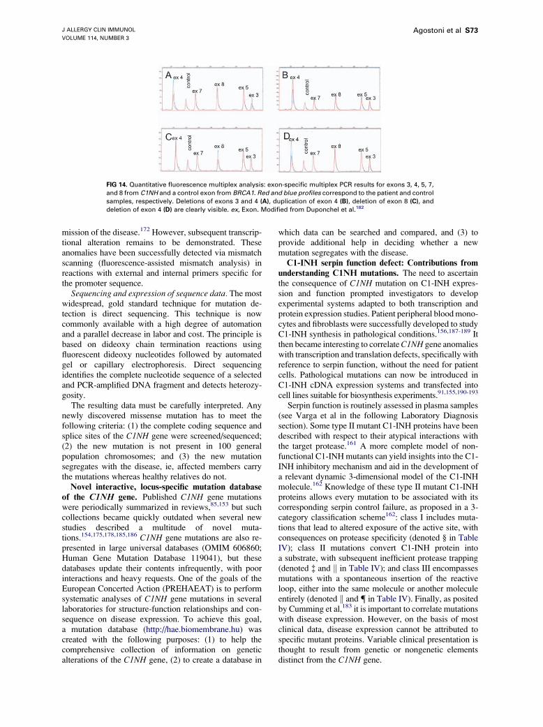
J ALLERGY CLIN IMMUNOL
VOLUME 114, NUMBER 3
Agostoni et al S73
FIG 14. Quantitative fluorescence multiplex analysis: exon-specific multiplex PCR results for exons 3, 4, 5, 7,
and 8 from C1NH and a control exon from BRCA1. Red and blue profiles correspond to the patient and control
samples, respectively. Deletions of exons 3 and 4 (A), duplication of exon 4 (B), deletion of exon 8 (C), and
deletion of exon 4 (D) are clearly visible. ex, Exon. Modified from Duponchel et al.182
mission of the disease.172 However, subsequent transcrip-tional alteration remains to be demonstrated. Theseanomalies have been successfully detected via mismatchscanning (fluorescence-assisted mismatch analysis) inreactions with external and internal primers specific forthe promoter sequence.
Sequencing and expression of sequence data. The mostwidespread, gold standard technique for mutation de-tection is direct sequencing. This technique is nowcommonly available with a high degree of automationand a parallel decrease in labor and cost. The principle isbased on dideoxy chain termination reactions usingfluorescent dideoxy nucleotides followed by automatedgel or capillary electrophoresis. Direct sequencingidentifies the complete nucleotide sequence of a selectedand PCR-amplified DNA fragment and detects heterozy-gosity.
The resulting data must be carefully interpreted. Anynewly discovered missense mutation has to meet thefollowing criteria: (1) the complete coding sequence andsplice sites of the C1NH gene were screened/sequenced;(2) the new mutation is not present in 100 generalpopulation chromosomes; and (3) the new mutationsegregates with the disease, ie, affected members carrythe mutations whereas healthy relatives do not.Novel interactive, locus-specific mutation database
of the C1NH gene. Published C1NH gene mutationswere periodically summarized in reviews,85,153 but suchcollections became quickly outdated when several newstudies described a multitude of novel muta-tions.154,175,178,185,186 C1NH gene mutations are also re-presented in large universal databases (OMIM 606860;Human Gene Mutation Database 119041), but thesedatabases update their contents infrequently, with poorinteractions and heavy requests. One of the goals of theEuropean Concerted Action (PREHAEAT) is to performsystematic analyses of C1NH gene mutations in severallaboratories for structure-function relationships and con-sequence on disease expression. To achieve this goal,a mutation database (http://hae.biomembrane.hu) wascreated with the following purposes: (1) to help thecomprehensive collection of information on geneticalterations of the C1NH gene, (2) to create a database in
which data can be searched and compared, and (3) toprovide additional help in deciding whether a newmutation segregates with the disease.C1-INH serpin function defect: Contributions from
understanding C1NH mutations. The need to ascertainthe consequence of C1NH mutation on C1-INH expres-sion and function prompted investigators to developexperimental systems adapted to both transcription andprotein expression studies. Patient peripheral bloodmono-cytes and fibroblasts were successfully developed to studyC1-INH synthesis in pathological conditions.156,187-189 Itthen became interesting to correlateC1NH gene anomalieswith transcription and translation defects, specifically withreference to serpin function, without the need for patientcells. Pathological mutations can now be introduced inC1-INH cDNA expression systems and transfected intocell lines suitable for biosynthesis experiments.91,155,190-193
Serpin function is routinely assessed in plasma samples(see Varga et al in the following Laboratory Diagnosissection). Some type II mutant C1-INH proteins have beendescribed with respect to their atypical interactions withthe target protease.161 A more complete model of non-functional C1-INH mutants can yield insights into the C1-INH inhibitory mechanism and aid in the development ofa relevant dynamic 3-dimensional model of the C1-INHmolecule.162 Knowledge of these type II mutant C1-INHproteins allows every mutation to be associated with itscorresponding serpin control failure, as proposed in a 3-category classification scheme162: class I includes muta-tions that lead to altered exposure of the active site, withconsequences on protease specificity (denoted § in TableIV); class II mutations convert C1-INH protein intoa substrate, with subsequent inefficient protease trapping(denoted � and k in Table IV); and class III encompassesmutations with a spontaneous insertion of the reactiveloop, either into the same molecule or another moleculeentirely (denoted k and { in Table IV). Finally, as positedby Cumming et al,183 it is important to correlate mutationswith disease expression. However, on the basis of mostclinical data, disease expression cannot be attributed tospecific mutant proteins. Variable clinical presentation isthought to result from genetic or nongenetic elementsdistinct from the C1NH gene.

J ALLERGY CLIN IMMUNOL
SEPTEMBER 2004
S74 Agostoni et al
FIG 15. Decision algorithm for mutation analyses of the C1NH gene. This algorithm takes into account the
importance of mutations in the coding sequence and in the introns, and the abundance of rearrangements.
Depending on the methods used, a preliminary screening of rearrangements may be preferred.
In laboratory practice. Every index case characterizedby impaired C1-INH serpin function and ruled not to be ofan acquired origin can be submitted to a genetic analysis.To ensure an economical and reliable strategy, analgorithm is proposed in Fig 15. Currently, to beexhaustive, the conjunction of scanning methods andSouthern hybridization or exon multiplex technology isrequired to detect most mispaired or unpaired sequencesand rearrangements, respectively. Every nucleotidechange detected from the index case has to be analyzedin the family members, keeping in mind that de novomutations are not uncommon. As stated, and in particularwhen detected in introns, every new mutation withouta known significance has to be distinguished from a poly-morphism in the C1NH gene after establishing its absencein the relevant healthy population (from at least 100chromosomes) and its segregation with the disease.
The interpretation of biochemical data on C1-INHserpin function and of C1NH gene anomalies is of greatimportance to establish a better knowledge of the patho-genic effects of C1NH gene mutations. Moreover, thepossibility of C1-INH polymorphisms affecting diseaseexpression should be considered.183 Generating a C1-INHmolecular model should be of great help in the un-derstanding and treatment of HAE type II and discerningwhich C1-INH residues are essential for efficient controlof all its target proteases.162
The uniform resource locators for data related to thissection are as follows:
d C1 Inhibitor Gene Mutation Database: http://hae.bio-membrane.hu/
d eGenome 11 home page: http://genome.chop.edu/d Ensembl: http://www.ensemble.org/Homo_sapiens(Ensembl Gene ID ENSG00000149131)
d Information about HAE (types I and II) at the OMIMdatabase: http://ncbi.nlm.nih.gov/htbin-post/Omim/(OMIM ID 106100)
d NCBI LocusLink database: http://www.ncbi.nlm.nih.gov/LocusLink/ (Locus ID 710; OrgSymbol SERP-ING1)
d Human GeneMutation Database Cardiff: http://archive.uwcm.ac.uk/uwcm/mg (Human Gene Mutation Data-base 119041)
This section on mutation analysis was supportedby grants from the European Community Quality ofLife Program (2001-01359), Programme Hospitalierde Recherche Clinique PHRC 2002, Fondo deInvestigaciones Sanitarias FIS (00/0216), HungarianNational Science Research (OTKA) (T034830), Ministryof Education of Hungary (NKFP) (1/024/2001), andMinistry of Health of Hungary (ETT 490/2003). Itsauthors are indebted to Prs. Marco Cicardi, Mario Tosi,and Joel Lunardi for valuable discussions, and thank Pr.Jean-Yves Cesbron for continuous encouragement. O.Roche and A. Blanch are supported by fellowships fromFIS (BEFI 00/9158) and Programa de Formacion dePersonal Universitario de la Universidad Autonoma deMadrid, respectively.
PATHOGENESIS AND PATHOBIOLOGYOF HAE AND AAE
From a pathogenetic perspective, the form of angioe-dema most extensively studied is that resulting from C1-INH deficiency. Nonetheless, the pathogenesis of thisclinical condition remains incompletely understood. In thefollowing section, Hack briefly reviews the biochemistryand biology of C1-INH as well as the biochemical changesthat occur in persons with C1-INH deficiency during bothattacks and symptom-free periods. On the basis of thesereviewed data, Hack then discusses the potential patho-genesis of C1-INH deficiency angioedema.

J ALLERGY CLIN IMMUNOL
VOLUME 114, NUMBER 3
Agostoni et al S75
Pathogenesis of angioedema attacks
(C. Erik Hack, MD, PhD,* Amsterdam, TheNetherlands)
Often, HAE results from a deficiency of C1-INH. Aninteresting feature, revisited in the previous Clinical
Manifestation and Diagnosis section, is the great variance
of the disease’s clinical course. Some patients are virtuallyfree of attacks even in the absence of treatment, whereas
others, despite therapy, have attacks nearly every week.
Angioedema attacks result from the extravasation of fluid
caused by increased vasopermeability. Unlike angioe-
dema of allergic origins, angioedema caused by HAE
typically does not itch. Pain is often not a primary feature
of such angioedema, although it may manifest secondary
to the localization of the angioedema, eg, severe pain
caused by obstruction of the bowel lumen. In spite of
decades of research, the pathogenesis of HAE attacks is
still unclear.C1-INH biochemistry and biology. C1-INH belongs
to the family of serine protease inhibitors, or serpins,
which also includes proteins such as a1-antitrypsin and
antithrombin III194-197 (Fig 16). Most serpins have only 1
or a few target proteases, but C1-INH is exceptional in this
respect. It is the major inhibitor of several proteases,
including (1) C1s and C1r—two serine proteases that,
together with C1q, constitute the C1 complex of the
classical pathway of complement, (2) the mannan-binding
lectin (MBL)–associated serine proteases or MASPs, and
(3) the contact system proteases factor XIa, factor XIIa,and kallikrein.198 In addition, C1-INH may interact with
several proteases such as thrombin,199 plasmin,200 and
tissue-type plasminogen activator (tPA).201 Thus, C1-INH
regulates the activity of several inflammatory, clotting,
and fibrinolytic proteases and is therefore an inhibitor of
several pathways of inflammation.198 In comparison with
other serpins, C1-INH is a relatively weak inhibitor,
approximately 100 times less effective than other serpins.
However, the inhibitory activity of C1-INH toward C1r,
C1s, and factor XIa can be remarkably potentiated by
heparin and other glycosaminoglycans.202,203 The inhib-
itory activity of C1-INH to factor XIIa and kallikrein
cannot be potentiated. Dextran sulfate is the most effectiveglycosaminoglycan, enhancing C1-INH function 130-fold
in vitro and 60-fold in plasma. However, the in vivo effect
of dextran sulfate is transient, probably because of
dissociation of the dextran sulfate–C1-INH complex and
the subsequent rapid clearance of dextran sulfate.204
Mature C1-INH consists of 478 amino acids and isheavily glycosylated (approximately 30% by weight).
Although on SDS-PAGE gels it migrates with an apparent
molecular weight of 104 kd, its calculated molecular
weight is 76 kd. The C1-INH molecule has at least 13glycosylation sites205 and possibly 20 glycosylation sites,
part of them linked to threonine residues. Most carbohy-
drate groups are located at the N-terminal region; their
function is largely unknown. The C1-INH molecule is
composed of an N-terminal domain of 113 amino acids
and a serpin domain of 365 amino acids. Although the
function of the N-terminal domain is unknown, recentevidence suggests that it helps to stabilize the central b-sheet by forming 2 disulfide bridges linking cysteine 101to cysteine 406, and cysteine 108 to cysteine 183.206 Thestructure of the serpin domain is homologous to thatof other serpins and is essential for the inhibitory capacityof the molecule. Recently, a novel 3-dimensional model ofC1-INH was proposed on the basis of the crystal structureof 4 other inhibitory serpins.162
Similar to other serpins, C1-INH inhibits proteases bybinding to their active site via its reactive center. Thisreaction follows the equation
P1C1�ð1Þ INH4P:C1�ð2Þ INH/P:C1�ð3Þ INH�
/P1C1� INH�
P ¼ target protease;C1� INH� ¼ modified C1� INH
In the first step, a reversible complex is formed betweenthe target protease and C1-INH; C1-INH then undergoesconformational changes caused by insertion of part of itsreactive center loop into a 5-stranded b-sheet. This resultsin the formation of modified C1-INH (C1-INH* in theequation) that is tightly bound to the target protease (step2). Mutations of the residues at the P14, P12, and P10positions in the reactive center interfere with efficient loopinsertion and cause the reaction to proceed to step 3. In thatcase, C1-INH has become a substrate for its targetproteases. Such mutations have been found in some
FIG 16. a-1-Antitrypsin tertiary structure conserved among the
serpin family a-1-antitrypsin is composed of 3 b-sheets (A in red, B
in green, and C in yellow) and 9 a-helices (A-I in gray). The blue
strand is the reactive center loop (RCL) with the P1 active amino
acid. Reprinted with permission from Silverman et al.197

J ALLERGY CLIN IMMUNOL
SEPTEMBER 2004
S76 Agostoni et al
patients with HAE-II (characterized by the presence ofdysfunctional C1-INH protein). Complexes betweenmodified C1-INH* and proteases are very stable and onlyvery slowly dissociate into an inactive, modified C1-INH(with changed conformation caused by a completed in-sertion of the cleaved active site loop into the 5-strandedb-sheet) and an active target protease (step 3 in theequation). Most of the stable complexes will be removedfrom the circulation before dissociation via receptorsspecific for complexed serpins. This mechanism ensuresefficient removal of biologically active proteases.
C1-INH is an acute phase protein, of which plasmaconcentrations may increase as much as 2-fold duringuncomplicated infections. Woo et al207 reported that thesynthetic rate of C1-INH may increase as much as 2.5times the normal rate in patients with rheumatoid arthritis.Cytokines such as interferon-g stimulate the synthesis ofC1-INH.208 In normal volunteers, the fractional catabolicrate of C1-INH is 2.5% of the plasma pool per hour, whichresults in an apparent plasma half-life time of clearance ofabout 28 hours.90 The half-life time of clearance of humanC1-INH in rabbits is comparable, at 26 hours,209 whereasthat in rats is considerably shorter, at about 4.5 hours.210
The apparent half-life time of clearance is markedly longerin patients with HAE, in whom it may exceed 48 hours, asdetermined by assessing the course of plasma concen-trations after the intravenous administration of exogenousC1-INH.211 However, at low plasma concentrations ofC1-INH, such as occur in untreated patients with HAE, C1is autoactivated212 and consumes functional C1-INH. Athigher C1-INH concentrations such as occur after theadministration of exogenous C1-INH, this autoactivationis inhibited, leading to a decreased consumption of C1-INH. Hence, monitoring of plasma concentrations aftera therapeutic dose of C1-INH may lead to an over-estimation of the half-life caused by inhibition of con-sumption of endogenous C1-INH. Removal of sialic acidsgreatly enhances the clearance of C1-INH from thecirculation, yielding an apparent half-life of 3 to 5minutes,209 presumably by binding to asialoglycoproteinreceptors in the liver. Subsequent removal of penultimategalactosyl residues largely restores the clearance rate toa value similar to that of normal C1-INH.209
Activation of cascade systems in HAE. As men-tioned, C1-INH regulates the activation of at least 3different so-called plasma cascade systems, ie, the classi-cal and MBL pathways of complement via its effect onC1r, C1s, and the MASPs; the contact system via itseffects on kallikrein and activated factor XII (factor XIIa);and the intrinsic pathway of coagulation via its effect onactivated factor XI (factor XIa).213 In addition, it mayregulate coagulation to some extent via its effects onthrombin, and it may regulate fibrinolysis through itseffect on tPA and plasmin. Except for the MBL pathway,there is evidence for activation of all of these systems inpatients with HAE.
The classical pathway of complement consists of C1q,C1r, and C1s, together forming the C1 complex; C4 andC2; as well as the inhibitors C4 binding protein and factor
I. Typically, patients with HAE have low C4 and C2 butnormal C3 values even during attack-free periods. LowC4and C2 result from uncontrolled activation of activated C1because the concentration of activation products of C4,C4b/c, or C4d is elevated.213,214 Activation of C3 istypically low or absent, probably because of efficientcontrol of activation at the level of C4 by C4 bindingprotein and factor I. This continuous activation of theclassical pathway is attributed to enhanced autoactivationof C1, because this autoactivation process is dependent onfunctional C1-INH values.212 However, it is now clear thatthe classic pathway of complement may be activated byapoptotic cells through various mechanisms.215-217
Hence, one could postulate that the excessive activationof the classic pathway in untreated patients with HAEresults from activation of this pathway by cell debris.Indeed, Familian and Hack have observed increasedamounts of complement activation products in patientswith HAE (Unpublished data, November 2002), reflectingactivation via the penetraxin serum amyloid P component,1 of the proteins that can bind to apoptotic cells.218
Ongoing activation of the classical pathway in untreatedpatients continuously produces activated C2.
The contact system of coagulation consists of theproteins factor XII (also known as Hageman factor),prekallikrein, and high molecular weight kininogen.219
The system is activated in vitro on contact withnegatively charged surfaces, but the nature of potentialactivators in vivo is currently unknown. Activation ofthe system yields factor XIIa and kallikrein. C1-INHinhibits both proteases. In addition, kallikrein can also beinhibited to some extent by a2-macroglobulin. Kallikreincan cleave HK, yielding cleaved HK and bradykinin. Thelatter nonapeptide is a potent inducer of vasodilationand increased vasopermeability. Activation of this sys-tem is often assessed by measuring factor XIIa–C1-INHand kallikrein–C1-INH complexes,220 although theseparameters are unsuitable to assess activation in HAE.Alternatively, activation can be measured by detectingincreased amounts of cleaved HK or decreased antigenicquantities of the contact system proteins. Activation ofthe contact system in HAE typically occurs duringattacks and is hardly detectable during attack-freeperiods.221 In particular, increased amounts of cleavedHK are found in most, if not all, patients withattacks.17,222,223 Remarkably, nearly all HK is oftencleaved during attacks, such that native HK is hardlydetectable. It is questionable whether all HK cleavedduring an attack gives rise to bradykinin, given itsextremely potent activity as a vasodilator. Bradykinin isknown to cause a significant drop in blood pressure evenat low concentrations, yet hypotension is not character-istic for HAE. However, there are no data supporting thecleavage of HK via other degradation pathways duringHAE attacks such as would generate minimal or nobradykinin. It should be noted that the measurement ofbradykinin in plasma or other body fluids is hampered byits rapid degradation by carboxypeptidases and is onlypossible using samples collected in special inhibitor

J ALLERGY CLIN IMMUNOL
VOLUME 114, NUMBER 3
Agostoni et al S77
cocktails to prevent enzymatic degradation. Few studieshave attempted to measure bradykinin according to suchprotocols in HAE, but these studies have shown in-creased bradykinin generation during attacks and notduring symptom-free periods, particularly when samplesare drawn from the site of the edematous swelling.13,14,27
C1-INH is amajor inhibitor of factor XIa of the intrinsicpathway of the intrinsic pathway of coagulation143,224 andalso inhibits thrombin,199 although inhibition of this latterenzyme is likely physiologically inconsequential. The roleof factor XI in coagulation is not yet resolved. Originally,coagulation was considered to proceed via at least 2different pathways, the intrinsic and the extrinsic, mergingat the level of factor X into a common pathway ofthrombin generation. The extrinsic pathway is initiatedby exposure of factor VII to tissue factor, whereas theintrinsic pathway is triggered by factor XII activation, eg,upon contact of blood with negatively charged surfacessuch as glass. In this view, activation of factor XII led tothrombin generation via activation of factor XI, whichthen activated factor IX, which together with factor VIIIaactivated factor X, which then together with factor Vaactivated prothrombin. This traditional distinction be-tween intrinsic and extrinsic pathway has been left behind,and the intrinsic pathway is now considered to amplifyextrinsic pathway activation in that factor IX amplifiesfactor VIIa–induced activation of factor X, whereas factorXI, activated by thrombin, activates additional factor IX.According to this revised scheme of coagulation, theactivation of factor XI is no longer mediated solely byfactor XIIa but also may occur with significant thrombingeneration. In the traditional scheme of coagulation, C1-INH is expected to have a major effect on intrinsicpathway activation because it blocks the activity of factorXIIa, kallikrein, and factor XIa, whereas according to thecurrent understanding, C1-INH has only a limited effecton coagulation, with its main effect the inhibition of factorXIa–mediated amplification of factor IX activation.Clinical observations in HAE do not support that C1-INH deficiency is a major risk for thromboembolicdisease, although some generation of thrombin may occurduring attacks.222 This mild effect of C1-INH is easilyunderstood through the revised scheme of coagulation.
Although C1-INH inhibits plasmin and tPA in vitro,and to some extent in vivo,201 this inhibition is weak.Nonetheless, significant plasmin formation, as measuredby the formation of plasmin–a2-antiplasmin complexes,occurs during attacks in patients with HAE but hardlyat all during symptom-free periods. At present there isno evidence for tPA or urokinase-type plasminogen acti-vator involvement in the formation of plasmin duringHAE attacks. As such, this generation most likely resultsfrom factor XII activation. Although debated in theliterature, convincing in vivo evidence exists to supportthe activation of plasminogen in human beings by factorXIIa.225,226
Angioedema attack pathogenesis: Mediators of
angioedema. Angioedema is a frequently experiencedside effect for patients receiving ACE inhibitors. This
angioedema does not itch, is not associated with urticariallesions, and does not respond at all to corticosteroids orantihistamines, features also typical of angioedema asso-ciated with C1-INH deficiency. ACE inhibitors slowdown the processing of C-terminal arginine residues ofvarious vasoactive peptides such as angiotensin andbradykinin, thus prolonging the biological activity ofthese compounds. The association between ACE inhib-itors and angioedema is a strong argument that thiscondition may result from exposure of the endotheliumto increased levels of vasoactive peptides. Considering thesimilarities between ACE inhibitor–related angioedemaand that caused by C1-INH deficiency, the latter form alsolikely results from enhanced exposure of the endotheliumto vasoactive peptides. Studies in a few patients indicatethat bradykinin may be the vasoactive peptide in-volved.13,14,27
Considering that contact activation in HAE mainly, ifnot exclusively, occurs during attacks, and that activationof this system results in the liberation of bradykinin fromHK, most investigators currently believe that C1-INHdeficiency angioedema results from the local generation ofbradykinin caused by uncontrolled proteolytic activity offactor XIIa and kallikrein (Fig 17, bottom).15 Kallikreinhas 2 important effects in contact activation: it (1) amplifiesfactor XII activation via so-called reciprocal activation,and (2) releases bradykinin from HK. This mechanism ofcontact activation and bradykinin generation as the basisfor angioedema is supported by observations in the murinemodel of C1-INH deficiency, because the enhanced vaso-permeability in this model can be blocked with bradykininreceptor antagonists.9 According to this mechanism,inhibiting kallikrein or blockading bradykinin receptors(BKR-2) would be therapeutic options; as discussed in theTreatment section of this supplement, both are currentlybeing investigated in clinical trials.
Activation of the contact system holds several attrac-tions as an explanation for angioedema attacks. First,
FIG 17. Different pathways of kinin generation in C1-inhibitor
deficiency state. Low C1-inhibitor leads to uncontrolled activation
of factor XII, which generates kallikrein and plasmin. Kallikrein
liberates bradykinin from HK, whereas plasmin cleaves off C2-kinin
from activated C2. Activated C2 is continuously produced during
baseline complement activation, which is increased as a result of
insufficient control of autoactivation of C1 caused by C1-INH
deficiency.

J ALLERGY CLIN IMMUNOL
SEPTEMBER 2004
S78 Agostoni et al
factor XII activation can be triggered by chemicalactivators and may well occur in bouts. Second, bradyki-nin is a potent vasoactive peptide that can enhancevasopermeability. Third, ACE inhibitors have been shownto result in the delayed breakdown of bradykinin, and partof their hypotensive effects are actually blocked bybradykinin receptor antagonists.227 However, some strongarguments exist against this mechanism as the soleexplanation for angioedema. First, bradykinin is a strongvasodilating agent and among the most hypotensiveagents known, whereas HAE attacks are not known tobe associated with clear hypotensive reactions.Nonetheless, one could argue that most bradykinin isgenerated locally, at the site of edema formation, ratherthan in the circulation. Second, the local injection ofbradykinin results in a strong pain sensation,228 whereasprimary, localized pain is not typical for HAE attacks.Third, contact activation cannot explain the efficacy ofantifibrinolytic agents such as tranexamic acid. It has beensuggested that these agents are efficacious by reducingplasmin formation during attacks, which would lead to thereduced consumption of functional C1-INH by plasmin.In spite of this, it is difficult to understand how activeplasmin would be capable of mediating C1-INH con-sumption, because it is rapidly inhibited by a potentplasmin inhibitor, ie, a2-antiplasmin. Moreover, in mostpatients with HAE, rather than being elevated, theconcentration of cleaved C1-INH (free C1-INH* in theequation) is fairly low. Finally, although vasopermeabilityseems to be enhanced and dependent on bradykininformation in C1-INH–deficient mice, these permeabilitychanges do not appear to occur in bouts and are otherwiseatypical for angioedema. For all of these reasons, othermechanisms may be involved in HAE attacks.
Some decades ago, another vasoactive peptide wasthought to be involved in angioedema formation. Thispeptide can be generated by cleavage of the C2b fragment,but not from native C2, by plasmin, and is therefore calledC2-kinin.229 This C-terminal fragment of C2b can begenerated by C1s or plasmin. Mediation of attacks by thisfragment requires activation of multiple plasma cascadesystems: (1) complement, to generate activated C2; (2) thecontact system, to generate factor XIIa; and (3) thefibrinolytic system, to generate the C2-kinin generatingenzyme, plasmin. According to this mechanism, attacksare triggered by factor XII activation that then yieldsplasmin, which in turn cleaves the C2-kinin sequence fromC2b (Fig 17, top). The attractive aspects of this contro-versial mechanism include its explanation of the efficacyof antifibrinolytic agents. Furthermore, one may postulatethat C2-kinin does not induce vasodilation, explainingwhy HAE attacks are not associated with hypotension andmay not induce sensations of pain, andwhy angioedema ingeneral does not hurt. Nevertheless, it should be noted thatthe biologic properties of C2-kinin are not well estab-lished, and thus, it is not truly known whether this kinin’sproperties are consistent with the description. Moreover,receptors for this kinin have not been identified, and it istherefore unknown whether their distribution correlates
positively with the localization of attacks. In addition,there are no data regarding concentrations of C2-kinin inpatients with HAE. Finally, if correct, the C2-kininmechanism provides no explanation for the profoundcleavage of HK and the apparent lack of biological activityof bradykinin.
These mechanisms are not necessarily exclusive andmay be simultaneously involved in attacks. Furthermore,the involvement of other, as yet unknown vasoactivepeptides in the pathogenesis of attacks cannot be excluded.Triggers of angioedema attacks. Clinically, HAE
attacks are often triggered by trauma or emotional stress.Attacks of HAE are specifically associated with activationof the fibrinolytic and contact systems, whereas comple-ment activation occurs continuously, independent ofclinical symptoms. Activation of the fibrinolytic systemcan occur via 3 plasminogen activators: tPA, urokinase-type plasminogen activator, and factor XII. There is noevidence that the former 2 are involved in plasminformation during attacks; hence, it likely results fromfactor XII activation. Thus, attacks are triggered byactivation of factor XII. Though factor XII (Hagemanfactor) was discovered in the 1950s, physiological acti-vators of this contact system protein have not yet beenidentified. In vitro, factor XII is easily activated by glassand other negatively charged compounds such as dextransulfate. Obviously, these compounds are not relevant asactivators of the contact system in vivo. The author writesthat recently, his group has studied the generation ofthrombin by phospholipid microparticles released in vivofrom activated platelets and damaged cells.230 By usingvarious specific mAbs against tissue factor, factor VII,factor XI, and factor XII, they noticed that a small butsignificant part of thrombin generation by microparticlespurified from human plasma was dependent on factor XII.Indeed, earlier studies have clearly indicated that factorXII can be activated by phospholipids in vitro.231,232
Apparently, factor XII can be activated by phospholipidmicroparticles in vivo. Damaged or apoptotic cells gener-ate such particles, and one may therefore postulate thatthese particles constitute triggers for contact activation.Under normal conditions, ie, at normal concentrations ofC1-INH, this weak activation will never yield substantialcontact activation. However, in the absence of sufficientamounts of this inhibitor, the system may become pro-foundly activated by even small amounts of microparticlesvia the principle of reciprocal activation (factor XIIactivates prekallikrein into kallikrein, which in turnactivates additional factor XII, and so on).Microparticles and damaged cells are likely generatedduring trauma, which may explain why this is a trigger forangioedema attacks. Emotional stress as a trigger forattacks is more difficult to understand, although one mayspeculate that it predisposes patients to microtrauma.Support for this hypothesis is that occasional patientshave been described who, in spite of acquired C1-INHdeficiency, are free of symptoms. Two such patients haddetectable antibodies against phospholipids.125 Theseantibodies might interfere with the binding of factor XII

J ALLERGY CLIN IMMUNOL
VOLUME 114, NUMBER 3
Agostoni et al S79
to phospholipid microparticles or damaged cells. Furtherstudies are needed to provide evidence toward thishypothesis of HAE attack generation.Attack frequency. The angioedema attack rate in
persons with C1-INH deficiency may vary widely,ranging from no attacks to an attack every week. Themolecular mechanism for the variation in attack rate isunknown. In the previous paragraph, 2 mechanisms toexplain the trigger for attacks are discussed. Bothmechanisms claim that attacks are initiated by activationof factor XII. Therefore, one may speculate that thefrequency of attacks is strongly linked to an individual’stendency toward factor XII activation. If true, attack ratesmight be influenced by the concentration of factor XII aswell as by the amount of factors that compete with factorXII for binding to its activator molecules. As discussed, inthe case of phospholipid microparticles, antiphospholipidbodies may prevent factor XII from binding to theparticles, conferring protection against the activation offactor XII. Older literature has revealed several proteinscapable of competition with factor XII for binding toartificial activators.233,234 The concentrations of thesecompeting proteins might well determine whether factorXII may become activated on exposure to an activator andtherefore influence HAE attack frequency. Future researchwill test this hypothesis.In summary. Angioedema attacks caused by C1-INH
deficiency are likely mediated by the excessive release ofvasoactive peptides such as bradykinin and C2-kinin. Thisrelease is initiated by the uncontrolled activation of factorXII, leading to kallikrein and plasmin formation. Thetrigger for attacks may be phospholipids, released fromdamaged cells, that activate factor XII. Factor XII’s ease ofactivation may be determined by its concentration as well
FIG 18. Analysis of vascular permeability. Extravasation of Evans
blue dye at 15 to 30 minutes was much more extensive in C1-INH–
deficient mice (B) than in wild-type mice (A), particularly after the
application of mustard oil to the left ears of the mice. (C) The
difference in the amount of extravasation was clearly demon-
strated by the rear footpads of mice of each genotype. Adminis-
tration of human C1-INH resulted in reduced vascular permeability,
as did the combination of Bk2R deficiency together with C1-INH
deficiency. Reprinted with permission of the American Society for
Clinical Investigation.9
as the concentration of proteins that compete with it forbinding to activators, and may explain the variablefrequency of attacks in patients.Additional discussions of pathogenesis
Others, assuming bradykinin to be the principal medi-ator of nonallergic angioedema attacks, have begun worktoward bridging the gap between C1-INH deficiency andlocal angioedema generation. Bolstered by the increasingevidence that ACE inhibitors235-237 can cause AAE ina small percentage of individuals, investigators havesought to determine whether—independent of C1NHgene sequence—an increased liberation of bradykinin,a decrease in its catabolism, or a combination of the 2 maycontribute to the angioedema experienced in HAE, AAE,and related disorders.Murine model of angioedema. (Kayla Williams, BS,
MA, MFA, Cambridge, Mass: Review of E. Han, R. C.McFarlane, A. N. Mulligan, J. Scafidi, and A. E. Davis)
In 2002, Han et al9 published their influential mousemodel of angioedema. In this model, mice heterozygousand homozygous for disruption to the gene coding formurine C1-INH (C1nh gene) were created by genetrapping. Although human beings homozygous forC1NH mutations have yet to be documented (with 1exception171,238), no obvious phenotypic abnormalitieswere observed in C1nh heterozygous mice, nor in micelacking both copies of a functional C1nh gene.
Phenotypic characteristics. Mating of heterozygous-deficient mice produced the expected 1:2:1 Mendelianratio of C1-INH1/1:C1-INH1/2:C1-INH2/2 mice. Asobserved in human heterozygotes, C1-INH expressionwas less than 50% of normal in heterozygous mice;homozygous C1-INH knockouts did not produce detect-able plasma concentrations of C1-INH. In male but not infemale mice, carrying the defective C1-INH gene corre-lated with a slight but statistically significant decrease in
FIG 19. Spectrophotometric analysis of vascular permeability in the
small intestine. Quantitation of extravasated Evans blue dye in
C1INH2/2 mice treated with an ACE inhibitor (captopril), a Bk1R
antagonist (des-Arg9,[Leu8]-bradykinin; Bk1RA), and a Bk2R antag-
onist (Hoe140). Reprinted with permission of the American Society
for Clinical Investigation.9
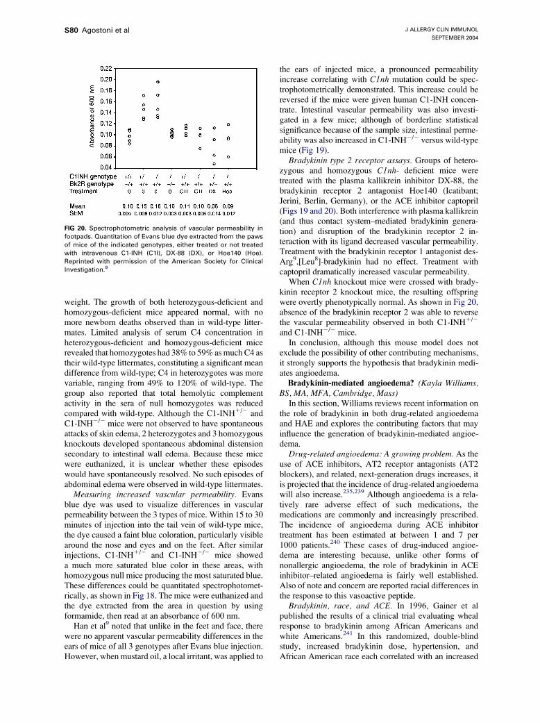
J ALLERGY CLIN IMMUNOL
SEPTEMBER 2004
S80 Agostoni et al
weight. The growth of both heterozygous-deficient andhomozygous-deficient mice appeared normal, with nomore newborn deaths observed than in wild-type litter-mates. Limited analysis of serum C4 concentration inheterozygous-deficient and homozygous-deficient micerevealed that homozygotes had 38% to 59% asmuch C4 astheir wild-type littermates, constituting a significant meandifference from wild-type; C4 in heterozygotes was morevariable, ranging from 49% to 120% of wild-type. Thegroup also reported that total hemolytic complementactivity in the sera of null homozygotes was reducedcompared with wild-type. Although the C1-INH1/2 andC1-INH2/2 mice were not observed to have spontaneousattacks of skin edema, 2 heterozygotes and 3 homozygousknockouts developed spontaneous abdominal distensionsecondary to intestinal wall edema. Because these micewere euthanized, it is unclear whether these episodeswould have spontaneously resolved. No such episodes ofabdominal edema were observed in wild-type littermates.
Measuring increased vascular permeability. Evansblue dye was used to visualize differences in vascularpermeability between the 3 types of mice. Within 15 to 30minutes of injection into the tail vein of wild-type mice,the dye caused a faint blue coloration, particularly visiblearound the nose and eyes and on the feet. After similarinjections, C1-INH1/2 and C1-INH2/2 mice showeda much more saturated blue color in these areas, withhomozygous null mice producing the most saturated blue.These differences could be quantitated spectrophotomet-rically, as shown in Fig 18. The mice were euthanized andthe dye extracted from the area in question by usingformamide, then read at an absorbance of 600 nm.
Han et al9 noted that unlike in the feet and face, therewere no apparent vascular permeability differences in theears of mice of all 3 genotypes after Evans blue injection.However, when mustard oil, a local irritant, was applied to
FIG 20. Spectrophotometric analysis of vascular permeability in
footpads. Quantitation of Evans blue dye extracted from the paws
of mice of the indicated genotypes, either treated or not treated
with intravenous C1-INH (C1I), DX-88 (DX), or Hoe140 (Hoe).
Reprinted with permission of the American Society for Clinical
Investigation.9
the ears of injected mice, a pronounced permeabilityincrease correlating with C1nh mutation could be spec-trophotometrically demonstrated. This increase could bereversed if the mice were given human C1-INH concen-trate. Intestinal vascular permeability was also investi-gated in a few mice; although of borderline statisticalsignificance because of the sample size, intestinal perme-ability was also increased in C1-INH2/2 versus wild-typemice (Fig 19).
Bradykinin type 2 receptor assays. Groups of hetero-zygous and homozygous C1nh- deficient mice weretreated with the plasma kallikrein inhibitor DX-88, thebradykinin receptor 2 antagonist Hoe140 (Icatibant;Jerini, Berlin, Germany), or the ACE inhibitor captopril(Figs 19 and 20). Both interference with plasma kallikrein(and thus contact system–mediated bradykinin genera-tion) and disruption of the bradykinin receptor 2 in-teraction with its ligand decreased vascular permeability.Treatment with the bradykinin receptor 1 antagonist des-Arg9,[Leu8]-bradykinin had no effect. Treatment withcaptopril dramatically increased vascular permeability.
When C1nh knockout mice were crossed with brady-kinin receptor 2 knockout mice, the resulting offspringwere overtly phenotypically normal. As shown in Fig 20,absence of the bradykinin receptor 2 was able to reversethe vascular permeability observed in both C1-INH1/2
and C1-INH2/2 mice.In conclusion, although this mouse model does not
exclude the possibility of other contributing mechanisms,it strongly supports the hypothesis that bradykinin medi-ates angioedema.Bradykinin-mediated angioedema? (Kayla Williams,
BS, MA, MFA, Cambridge, Mass)In this section, Williams reviews recent information on
the role of bradykinin in both drug-related angioedemaand HAE and explores the contributing factors that mayinfluence the generation of bradykinin-mediated angioe-dema.
Drug-related angioedema: A growing problem. As theuse of ACE inhibitors, AT2 receptor antagonists (AT2blockers), and related, next-generation drugs increases, itis projected that the incidence of drug-related angioedemawill also increase.235,239 Although angioedema is a rela-tively rare adverse effect of such medications, themedications are commonly and increasingly prescribed.The incidence of angioedema during ACE inhibitortreatment has been estimated at between 1 and 7 per1000 patients.240 These cases of drug-induced angioe-dema are interesting because, unlike other forms ofnonallergic angioedema, the role of bradykinin in ACEinhibitor–related angioedema is fairly well established.Also of note and concern are reported racial differences inthe response to this vasoactive peptide.
Bradykinin, race, and ACE. In 1996, Gainer et alpublished the results of a clinical trial evaluating whealresponse to bradykinin among African Americans andwhite Americans.241 In this randomized, double-blindstudy, increased bradykinin dose, hypertension, andAfrican American race each correlated with an increased

J ALLERGY CLIN IMMUNOL
VOLUME 114, NUMBER 3
Agostoni et al S81
wheal response to bradykinin. Further investigations bythe same group compared the effects of short-term ACEinhibition in normotensive and hypertensive individualsof both races. In this study, the hypotensive effects of theACE inhibitor captopril were shown to be greatly reducedin all subjects by concomitant administration of thebradykinin receptor 2 antagonist Icatibant.227 Icatibantwas also shown to significantly alter the change in plasmarenin activity in response to ACE inhibition. These resultsnot only confirmed the importance of bradykinin in theshort-term effects of ACE inhibitors but also suggestedthat bradykinin contributes to the effects of ACE in-hibition on the renin-angiotensin system. In addition,when considered together, these results suggest thatclinicians prescribing ACE inhibitors for patients ofAfro-Caribbean descent should be especially aware ofthe possibility for drug-related angioedema, a warningbeginning to be supported by case analysis.242,243
Further antihypertensive-related angioedema.Initially, AT2 blockers were thought to be a saferalternative for patients who had angioedema associatedwith ACE inhibitors. Unlike ACE inhibitors, AT2 block-ers have not been shown to increase bradykinin concen-trations. Nonetheless, further case reports documentedangioedema after therapy with AT2 blockers.137,244 Ina survey of the literature, 32% of these patients also hadsimilar episodes in conjunction with ACE inhibitortreatment.245 Additional documented cases of AT2blocker–associated angioedema threw bradykinin’s rolein the pathogenesis of such episodes into doubt246 becauseincreased concentrations of bradykinin are not associatedwith AT2 blocker use.245 However, very recent work hasshown that increased concentrations of AT2, such aswould occur during a blockade of AT2 receptors, areassociated with an increase in the expression of bradykininreceptors that may be almost 3-fold.247 Thus, increasedopportunities for bradykinin binding may explain angioe-dema in the absence of extra bradykinin. The mediator forthis receptor crosstalk effect has been shown to be the AT2receptor 1.247 (Presumably, this phenomenon could occurin the presence of AT2 receptor 1 blockers only in theabsence of a total blockade.)
Meanwhile, the new drug application for the vaso-peptidase inhibitor omapatrilat, a dual inhibitor of bothACE and neutral endopeptidase (NEP), was voluntarilywithdrawnwhen the Food andDrug Administration raisedquestions regarding the comparative incidence of angioe-dema reported within the new drug application database.In response to these questions, a large (25,000-patient)multinational trial was initiated to compare the efficacyand safety of omapatrilat versus the ACE inhibitorenalapril in patients with heart failure.248 The results ofthis study have recently been reported.249 Angioedemaoccurred in 2.17% of subjects receiving omapatrilatcompared with 0.68% of subjects receiving an ACEinhibitor alone. Two of the dual inhibitor–treated subjectshad angioedema with airway compromise. An editorial250
published after the trial results were released but beforetheir publication noted that the rates of angioedema were
much higher in African Americans receiving the dualinhibitor (5.54%) than in those receiving the ACE in-hibitor alone (1.62%). Of smokers receiving the dualinhibitor, 3.93% had angioedema, versus 0.81% ofsmokers receiving the ACE inhibitor alone.
The biologic activity of NEP must account for thisseveral-fold increase in angioedema. Indeed, among itsmany possible functions, NEP has been shown tometabolize bradykinin to an inactive form.237 A combi-nation of ACE and NEP inhibition could thus be expectedto prevent efficiently the degradation of bradykinin toinactive metabolites, thereby increasing the risk ofangioedema.251 In certain subgroups with altered re-sponses to bradykinin or altered pathways for its gener-ation or degradation, these effects could be especiallypronounced, and such predisposing factors may also beinvolved in the generation of C1-INH deficiency–relatedangioedema.
Implications for C1-INH deficiency angioedema.Early investigations have confirmed that bradykininmay be at work in C1-INH deficiency angioedema.The transience of bradykinin has complicated measure-ments of this nonapeptide; however, several smallstudies of plasma bradykinin concentrations in patientswith angioedema have revealed that high concentrationsof this vasoactive peptide occur during edematousattacks.13,14,27 This increase in plasma bradykinin wasdemonstrated both for patients with HAE with C1-INHdeficiency and for patients with drug-associated angioe-dema who had an angioedematous attack in conjunctionwith ACE inhibitor treatment. Patients receiving ACEinhibitors showed elevated plasma bradykinin concen-trations throughout ACE inhibitor therapy, both duringattacks and during periods of remission.13 As docu-mented in 1 patient, this increase reverted to normal ondiscontinuation of ACE inhibitor therapy.14 In contrast,patients with C1-INH deficiency were shown to havehigh concentrations of bradykinin during their attacks13
but have been documented to have normal or marginallyincreased amounts of bradykinin during periods ofremission.14 At least in C1-INH deficiency, bradykininincreases may be highly localized; in 2 previouslystudied C1-INH–deficient patients, bradykinin concen-trations in blood draining from an angioedematous sitewere 3 and 8 times as high as those measured in systemiccirculation.
Thus, bradykinin appears to be important in variouscurrently identified forms of nonallergic angioedema.Following the example put forth in the large-scale trialof antihypertensive agents that showed (1) African descentand, to a lesser extent, (2) smoking to correlate withincreased angioedema risk,250 clinicians and researchersmay wish to investigate the interaction of these variableswith C1-INH deficiency.
Bradykinin magnified. Even if bradykinin is the majormediator of nonallergic angioedema, many questionsregarding the triggering and localization of angioedemaattacks remain. Why should patients with ACE inhibitor–induced angioedema have this angioedema in bouts if

J ALLERGY CLIN IMMUNOL
SEPTEMBER 2004
S82 Agostoni et al
FIG 21. Pathways for bradykinin generation and metabolism. On the endothelium, bradykinin generation
likely initiates independent of factor XII; however, once kallikrein is generated, it soon catalyzes factor XII
activation, thus amplifying the response.
bradykinin is constantly elevated? What is different aboutthe small percentage of patients taking ACE inhibitorswho have angioedema? What regulatory pathways areinvolved? How, chemically, does one proceed frominsufficient functional C1-INH to localized, full-blownangioedema?
The past decade of research indicates that the answersmay reside in a surprisingly multifactorial system.Bradykinin-mediated effects can result from increasedamounts of bradykinin, the reduced breakdown of brady-kinin to inactive metabolites, the increased conversion ofbradykinin to active metabolites, and/or the increaseduptake of these active metabolites. Each of these mech-anisms may be involved in at least 1 form of nonallergicangioedema. Pathways for the breakdown of bradykininare depicted in Fig 21.
Decreased or alternative catabolism. Molinaro et al252
have demonstrated that in human plasma, bradykinin ismetabolized mainly by 3 metallopeptidases: ACE, APP,and kininase I (carboxypeptidase N). Of these, ACE andAPP, respectively, are the first and secondmajor pathwaysof inactivation. Kininase I represents a minor pathwayunless ACE is inhibited, whereupon kininase I transformsbradykinin into its active metabolite des-Arg9-bradykinin,which, in turn, is inactivated by APP and ACE, with APP
the major inactivating metallopeptidase.253 NEP, as dis-cussed in the context of vasopeptidase inhibition, likelyrepresents another minor pathway.
Once formed, bradykinin and des-Arg9-bradykinin areshort-lived and thus exert their effects locally at the site oftheir formation, stimulating B2 and B1 receptors, re-spectively.254,255 During ACE or vasopeptidase inhibi-tion, patients with low APP activity may be subject toangioedema. In fact, Adam et al96 have found a correlationbetween low APP activity with a history of angioedemaamong patients receiving ACE inhibitors. Likewise, intheir Estrogen-dependent and Estrogen-associated in-herited angioedema piece earlier in this supplement,Binkley and Davis propose reduced APP activity asa contributing element in estrogen-sensitive inheritedangioedema. In addition, they note that estrogen maydecrease gene expression of ACE.256,257
To demonstrate further the delicate balance of enzymesand receptors, it should be noted that Mathews et al258
observed angioedema in a subject with familial serumcarboxypeptidase N deficiency. Because the subject’ssister had an equally depressed serum carboxypeptidasevalue but no reported episodes of angioedema,259 it ispossible that another defective control mechanism, possi-bly APP, contributed to the angioedema.

J ALLERGY CLIN IMMUNOL
VOLUME 114, NUMBER 3
Agostoni et al S83
Increased anabolism. As Hack discussed in thePathogenisis and Pathobiology of HAE and AAE section,C1-INH is known to inhibit not just C1 esterase but alsoother serine proteases, including factor XII (Hagemanfactor). In addition, C1-INH inhibits the serine proteasekallikrein. Without the controlling effects of C1-INH, it ispossible that both of these serine proteases may contributeto an increased generation of bradykinin. This reaction isthought to occur at the endothelial cell surface. The way inwhich this unchecked bradykinin generation starts hasbeen debated, with recent evidence supporting the sec-ondary role of factor XII,260,261 and placing the conversionof prekallikrein to kallikrein, likely by an enzyme such asprolylcarboxypeptidase,262,263 foremost, at least underresting physiologic conditions. It is thought that oncekallikrein is present, it catalyzes the activation of factorXII. Only in the presence of activated platelets, neutro-phils, or lymphocytes can the high local concentration offree zinc ions necessary for factor XII to bind to theendothelial cell surface—and thus initiate bradykininrelease—occur.263 This provides yet another example ofhow infection or tissue damage might aggravate anexisting tendency to angioedema.
In vitro, kallikrein and plasmin together have beenshown to exert a synergistic effect on the release of kininsfrom HK. Of interest, plasmin’s digestion of HK in thismodel could be inhibited by e-aminocaproic acid.264
Because C1-INH is also known to inhibit plasmin, andantifibrinolytics show at least partial efficacy in thetreatment of HAE, this enhancing mechanism might wellbe operative in C1-INH deficiency angioedema. Inaddition, because angioedema has been reported inpatients treated for acute ischemic stroke who receivedrecombinant tissue-type plasminogen activator (rtPA;often concomitantly with an ACE inhibitor), Molinaroet al252 undertook an in vitro comparison of humanplasma incubated with and without rtPA in the presenceof an ACE inhibitor. Their results showed that rtPAgenerates bradykinin that is further metabolized to des-Arg9-bradykinin; this effect could be recreated to a sim-ilar extent in the absence of rtPA by the addition ofnegatively charged glass beads, thus mimicking contactactivation.
Joseph et al19,20 have identified yet another possiblemechanism active at the endothelial cell level, showingthat the cytosol of endothelial cells contains a factorcapable of converting prekallikrein to kallikrein in theabsence of factor XII. They identified this factor as heatshock protein 90. Perhaps this mechanism might accountfor angioedema attacks that occur in the wake of trauma orinfection when ruptured cells spill their contents into localcirculation. Indeed, increased kallikrein may be a featureofmechanical trauma: an experiment conductedmore than20 years ago demonstrated that mechanically generatedblisters in 5 of 5 patients with HAE contained activekallikrein that could be inactivated by C1-INH concentrateor antibodies to plasma kallikrein. In contrast, blister fluidsfrom 6 of 8 healthy subjects did not contain detectableamounts of plasma kallikrein.265
Last, these anabolic effects might be created ormagnified by hormonal interactions. As noted byBinkley and Davis,92 estrogens may increase the expres-sion of factor XII107,266-268 and prekallikrein.269
Increased uptake. Various factors may increase geneexpression of the B1 and B2 receptors. Not normallyexpressed, the B1 receptor has a high affinity for kininmetabolites.270 Expression of B1 receptors may be in-duced during inflammation and tissue damage.271,272
Although B2 receptors are constitutively expressed,273
their expression may be increased by estrogens274 andAT2 receptor antagonists.247
In summary. Despite increasing evidence that bradyki-nin is indeed the major mediator of nonallergic angioe-dema, this knowledge does not represent a simple answergiven the many variables governing bradykinin and itsmetabolites. However, an understanding of this web ofchemical interactions may yield more successful manage-ment algorithms for the various types of nonallergicangioedema. This recent knowledge also suggests a placefor bradykinin receptor antagonists and kallikrein inhib-itors for the control of nonallergic angioedema, especiallywhen it occurs in the presence of functional C1-INH.
ANGIOEDEMA ATTACKS: TRIGGERS ANDHORMONAL INFLUENCES
The current scientific understanding of the triggering ofangioedema attacks ranges from the anecdotal to thatsupported by relatively large and rigorous studies. Patientswith HAE, AAE, or related forms of angioedema,especially patients who have angioedema attacks in theabsence of a formal diagnosis, pay particular attention tothe events that precede their angioedema attacks; however,even in the same patient, these events may vary amongattacks. Various patients have implicated exposure to cold;mechanical trauma sustained during routine activities suchas gardening; prolonged sitting or standing; eating certainfoods, such as eggs; exposure to pesticides or chemicals innew fabrics and clothing; infection or illness; or excite-ment or stress in triggering their attacks (K. Williams,Unpublished data, August 2003). However, as with other,better studied triggering events, these conditions oractivities did not always predictably provoke attacks.
Ethical concerns and the small patient populationsavailable to would-be investigators prevent the systematicinvestigation of most patient-reported angioedema attacktriggers. Those triggers more readily quantifiable vialaboratory assays without necessitating patients’ increasedrisk of developing attacks have received themost scientificattention, largely restricting the study of attack triggers tothe possible role of hormones.
Role of sex hormones in HAE
(Laurence Bouillet, MD, CCA,* Grenoble, France)In the first part of this section, Bouillet reviews his
group’s clinical experience and other reports linking sex

J ALLERGY CLIN IMMUNOL
SEPTEMBER 2004
S84 Agostoni et al
hormones and HAE. On the basis of the literature, heproposes possible pathogenic mechanisms by which theseinfluences might manifest. These relationships are dis-cussed with a final view toward patient care.Current literature. It is currently known that HAE is
influenced by the fluctuation of female hormones, but theeffects sometimes appear to vary greatly among women.Some patients seem to be estrogen-dependent: puberty,pregnancies, or estroprogestative contraception can pre-cipitate attacks, whereas menopause impairs the disease.However, the course of somewomen’s disease seems to beunaffected by estrogen.
Reports have highlighted the close relationship be-tween female hormones and angioedema. Yip andCunliffe22 described cases of HAE occurring in a motherand her daughter whose symptoms appeared to beestrogen-influenced. Their first attack happened aroundpuberty; angioedema worsened premenstrually and whenthey took OCs. Likewise, a woman with HAE-II associ-ated with Turner syndrome was reported as experiencingan increase in both the severity and the frequency of herangioedema attacks on beginning estrogen physiologicalreplacement at age 34 years.275 McGlinchey andMcCluskey276 also described HAE manifesting after theinitiation of estrogen replacement therapy. Bork et al,277 asdiscussed later in this supplement, reported their evalua-tion of the effect of OC use or hormone replacementtherapy on the frequency and the severity of HAE attacks,finding that 63% of women with HAE taking these drugspresented with new or worsened angioedema.
Some kinin-mediated angioedema appears to be re-lated to estrogen therapy. The first cases of angioedemaduring oral contraception were described in a 1986 reportby Warin et al277a in which 2 sisters presented with HAEonly after taking oral contraception and during preg-nancy: C1-INH concentration and complement compo-nents were normal. Bouillet’s group has also reported 5women278 whose angioedema attacks manifested afterstarting oral contraception. A study of C1-INH biologicalfunction revealed a lowered C1-INH activity withmarked protein cleavage on the immunoblot. Clinicaland biological anomalies ceased after oral contraceptionwas discontinued.
Hypoandrogenism has been associated with insufficientproduction of C1-INH in women taking cyproteroneacetate as well in men with hypogonadism. In 1989,Pichler et al279 reported the cases of 4 women taking thecontraceptive drug cyproterone acetate who developedclinical HAE reversed by discontinuation of this contra-ceptive; also described were the cases of 2 men withhypogonadism who had recurrent angioedema success-fully cured by androgen treatment.
As discussed in previous sections, 3 reports describeda new estrogen-influenced HAE.8,86,87 Only women hadangioedema, and their attacks were precipitated whenestrogen concentrations were increasing because of preg-nancies, oral contraception, or hormone replacementtherapy. No C1-INH abnormalities have been reportedin these cases.
Hormone-related pathogenesis. For these clinicalevents to be understood, they must be placed in a bio-chemical context. As discussed, the complement, contact,and fibrinolytic systems are involved in the genesis ofangioedema attacks,17,222,223 and all are regulated, at leastto some extent, by C1-INH. Bradykinin is thought bymany to be the principal candidate mediator of angioe-dema genesis, and a mouse model demonstrated thatangioedema is mediated by bradykinin via B2 receptor.9
In human beings, studies have shown local elevations inbradykinin concentration during HAE attacks.13,14 It isspeculated that factor XIIa, kallikrein, and thrombin maybe increased as well. An elevation of plasmin–a2-antiplasmin complexes is also noted.
In ovariectomized rats, studies showed that 17b-estrad-iol favored the increase of factor XII by stimulating itsgene transcription.268,280-282 This hormone also increasedkininogen and kallikrein concentrations.283 In addition,estrogens regulate B2 receptor gene expression andfunction: when reduced in ovariectomized rats, the vaso-depressor response to bradykinin and B2 receptor mRNAlevels could be restored by estrogen substitution.274
Progesterone did not modify factor XII concentrationbut seemed to increase the amount of kallikrein cDNA.283
In healthy women taking oral contraception, there is anincrease of the fibrinolytic proteins: plasmin and factorsVII, X, and IX are increased, whereas plasminogenactivator inhibitor is decreased.266,284,285 These effectsappear to be estrogen dose–dependent.285 Plasma samplesfrom these women showed enhanced in vitro fibrinoly-sis.107 The contact system is also altered: factor XII,prekallikrein, kallikrein, and HK increase.286-288 Last, C1-INH levels are decreased, correlating with the increase infactor XII.107,286,287
Hormone replacement therapy (HRT), although usinga weaker estrogen dose than oral contraception, appears tohave the same effects. Fibrinolytic proteins are alsoincreased (plasminogen and tPA), whereas plasminogenactivator inhibitor decreases.289-291 HRT increases factorXII, prekallikrein, C3, and C4 concentrations.266,290,291
Moreover, some studies have shown HRT to influencethe bradykinin system: ACE activity decreases, whereasbradykinin concentration increases.256,292,293
Toward treatment. This imbalance in these 3 systemsappears to trigger angioedema attacks in some estrogen-sensitive women. Each physician should identify theseestrogen-sensitive patients to manage specifically theircontraception needs, pregnancies, and choices regardingHRT. Further studies to determine predictive biological,genetic, and clinical parameters would be of use.
Role of sex hormones in HAE, part II
(Beata Visy, MD, George Fust, MD, PhD, DSc,* LilianVarga, PhD,* George Szendei, MD, Edit Takacs, MD,Istvan Karadi, MD, PhD, DSc, Bela Fekete, MD, PhD,DSc, George Harmat, MD, PhD, and Henriette Farkas,MD, PhD,* Budapest, Hungary)
Most reported studies have investigated the influence ofexogenous sex hormones on the frequency and severity of

J ALLERGY CLIN IMMUNOL
VOLUME 114, NUMBER 3
Agostoni et al S85
HAE attacks. Here, Visy et al very briefly summarizea study they undertookmeasuring serum concentrations ofendogenous sex hormones in women with HAE andcorrelating these results with the occurrence of HAEattacks. This work has since been published in ClinicalEndocrinology.294
Objective. The fluctuations of sex hormone concen-trations at the beginning of adolescence, in the perimeno-pausal period, and during pregnancy or OC use canprecipitate HAE attacks. Edematous attacks usually dis-appear after the onset of menopause. The authors un-dertook their study to establish any relationship betweenserumconcentrations of sex hormones and the incidence ofHAE attacks.Patients and measurements. Serum concentrations of
luteinizing hormone (LH), follicle-stimulating hormone,progesterone, estradiol, testosterone, prolactin, and sexhormone binding globulin (SHBG) were measured in 78patients (mean age, 30.3 years; range, 4-70 years) withHAE. A questionnaire was used to explore the medicalhistory of adult patients to characterize the evolution andproperties of attacks.Results. Twenty-one patients had been symptom-free
before menarche. Symptoms in 13 of 21 patients firstoccurred during adolescence, whereas the remaining 8patients had no attacks in the pubertal period. Edemaduring the perimenstrual period was reported by 42.4%.Pregnancy was associated with a higher incidence ofattacks in 36%; edema formation was less common in56%, and 8% had no change in the frequency of symptomsdespite pregnancy.
Of the 11 patients using OCs, 7 reported an increase inthe frequency and severity of edematous symptomscompared with the period before hormonal contraceptionhad been initiated.
Serum concentrations of sex hormones were normal inthe majority of patients; however, more than half of thesubjects had progesterone values above the upper limit ofthe normal range.
During the 1-year follow-up, the attack rate was 5 timeshigher in female patients with high progesterone concen-trations (above the menopausal threshold of 4 nmol/L)than in women with normal or low serum concentrations.The eminent role of this hormone is also confirmed by theobservation that only progesterone was higher in morethan 50% of subjects than the serum level considerednormal for sex, age, and stage of menstrual cycle.Therefore, serum progesterone concentrations above thepostmenopausal threshold are predictive of a higher in-cidence of edematous attacks in female patients with HAE.In addition, multiple logistic regression analysis demon-strated a significantly lower attack frequency during 1-yearfollow-up in patients with higher (40 nmol/L) SHBG level(odds ratio, 0.25 confidence interval; 0.07-0.90;P = .034).This difference existed independently of age and danazoldose (Fig 22).Looking ahead. In view of these results, the monitor-
ing of progesterone and SHBG concentrations can proveuseful in the prediction of HAE attacks. Further inves-
tigations might address whether this increased progester-one is a cause or a result of frequent attacks, or whetherprogesterone itself exerts any direct effects on vascularpermeability. Last, progesterone’s effects on relevant geneexpression might represent another valuable avenue ofexploration.
Influence of OCs or HRT on hereditary formsof recurrent angioedema
(Konrad Bork, MD,* and Bettina Fischer, MD, Mainz,Germany)
In this section, Bork et al write of a systemic studyundertaken after they noticed women in their clinic withnew or worsened angioedema in response to oral contra-ception or HRT. Their study crosses the barriers of C1-INH deficiency and HAE type classifications to investi-gate the effects of exogenous hormones on HAE. The newtype of inherited angioedema recently described by theirgroup has clinical features highly similar to classic HAE-Iand HAE-II; however, it is not associated with a C1-INHdeficiency. Currently, the genetic defect is unknown. Untilnow, the disease has been observed exclusively inwomen8,86,87; however, in 2 families the existence ofclinically unaffected male carriers has been deduced.8,86
In the following report, the authors show that estrogensplay a similar role in HAE-I and this new type of inheritedangioedema, herein described as HAE with normal C1inhibitor (HAE type III) in accordance with the originalreport of this condition by Bork et al.86
Among their case series, the authors observed femalepatients with HAE who had newly developed recurrent
FIG 22. Correlation in female patients with HAE between serum
progesterone (A) and SHBG concentrations (B) and the number of
edematous attacks in the year after blood sampling. Spearman
correlation coefficients and their significance (P values) are in-
dicated.

J ALLERGY CLIN IMMUNOL
SEPTEMBER 2004
S86 Agostoni et al
angioedema after receiving OCs or postmenopausal HRT.In addition, other women had a worsening of their pre-existing angioedema after initiating OCs or HRT, sug-gested that these treatments might trigger HAE. However,this possible relationship has not formally been investi-gated, nor is angioedema listed as an adverse effect of OCsor HRT.295-297 Information regarding the relationshipbetween hormone administration and the first occurrenceor worsening of HAE symptoms is limited to anecdotalreports.8,22-24,31,32,86,87,107,275,276,277a,299
In this study, a systematic approach was used to obtaininformation about the relationship between the use of OCor HRT and the clinical manifestation of HAE-I and HAEwith normal C1-INH (HAE type III).Methods. The case series by Bork et al277 included 228
women with recurrent angioedema who had taken OC,HRT, or both.277 Of these, this study investigated 32womenwith HAE-I and 39womenwith HAEwith normalC1-INH (HAE type III). The OCs used by all patients werecombinations of estrogen (ethinyl estradiol) and progestin.Medications used for HRT contained only estrogens, aswas standard medical practice in Germany until recently.
All patients underwent a thorough clinical examination,and a standardized medical and family history was taken.In particular, patients were questioned about the fre-quency, intensity, and organ involvement of their angioe-dema since the very first manifestation of their disease.The start date and duration of administration of sexhormones, with special emphasis on OC and HRT, wererecorded. Patients were asked whether their angioedemaoccurred shortly after beginning this hormonal medica-tion. If yes, the time between starting OC or HRT and theonset of angioedema was recorded. Patients who hadrecurrent angioedema before the intake of OC or HRTwere asked whether their angioedema episodes were morefrequent or severe after beginning treatment.
Concentrations of C1-INH, C4, and C1q were assayedby radial immunodiffusion, and C1-INH functional activ-ity was determined by using the chromogenic sub-strate C2H5CO-Lys(e-Cbo)-Gly-Arg-pNA (ImmunochromC1-INH; Immuno Diagnostics, Vienna, Austria).300,301
Results. As shown in Table V, OC, HRT, or bothtreatments were associated with angioedema attacks in63% of women with HAE-I and 62% of women with HAEtype III. Among the group of patients whose angioedemaattacks were newly induced by estrogen-containingmedications, there was a high preponderance of HAE
TABLE V. Summary of 71 women with inherited
angioedema receiving OCs or HRT: Outcome of
medication
Type Women N
Angioedema
induced or
worsened n (%)
Angioedema
not influenced
n (%)
HAE-I 32 20 (62.5) 12 (37.5)
HAE type III 39 24 (61.5) 15 (38.5)
Total 71 44 27
type III, whereas in the patient group with an exacerba-tion of symptoms, there were more patients with HAE-I(Fig 23).
In 23 women—6 with HAE-I and 17 with HAE typeIII—occurrence of skin or gastrointestinal angioedemamanifested for the first time after the administration of OC.Because of the assumed intolerance, several women trieddifferent OC formulations, and all 33 trials were followedby recurrences of angioedema episodes so severe that allOC had to be discontinued.
The interval between starting OC and episode occur-rence ranged from 2 to 90 days (mean, 18 days).
In 9 of the 23 women, symptoms were limited to theperiod of OC administration and ended after discontinu-ation. All 9 had HAE type III. In the other 14 patients,angioedema attacks recurred despite OC discontinuation.Episodes occurred less frequently and less severely in 12,whereas symptoms were unchanged in 4 women.
There was 1 occurrence of angioedema for the first timeafter starting postmenopausal HRT. One woman initiallydeveloped recurrent angioedema after starting HRT withestradiol (2 mg/d). The patient continued to take oralestradiol over a period of 4 years. After a consultation inthe outpatient clinic, HRT was discontinued. Symptomspersisted but occurred less frequently and were milder.The patient’s daughter had recurrent skin angioedema andgastrointestinal pain attacks since age 12 years; therefore,with a normal C1-INH concentration and activity in bothmother and daughter, a diagnosis of HAE type III wasassumed.
In 18women, the administration ofOC induced a severeexacerbation of their pre-existing angioedematous dis-ease. Because of this exacerbation, 4 women switched toanother OC, some trying many different preparations. Allof these OC doses were followed by recurrences of severeangioedema such that OC had to be discontinued.
Five women with previous recurrent angioedema of theskin and the gastrointestinal tract developed an exacerba-tion of their disease after starting HRT. Of these, only 2
FIG 23. Underlying angioedema disease in 44 women who had
induction or exacerbation of recurrent angioedema caused by OCs
or HRT.

J ALLERGY CLIN IMMUNOL
VOLUME 114, NUMBER 3
Agostoni et al S87
were eventually able to tolerate such therapy. In 3 of the 5,their angioedema also had exacerbated when on OCseveral decades before.Discussion. The results indicate that in 63% of women
with HAE-I and in 62% of women with HAE type III, theuse of OC or HRT was associated with either the initialappearance of angioedema attacks or with a severeexacerbation of previously existing angioedema.Correspondingly, although also taking OC or receivingHRT, 37%ofwomenwithHAE-I and 38%ofwomenwithtype III tolerated these medications without any negativeinfluence on their disease; their disease was neitherestrogen-dependent nor estrogen-sensitive. The resultsdemonstrate that estrogen sensitivity is not a featurespecific to HAE type III because it occurs in a similarpercentage of women with HAE-I.
Results from several case reports have suggested thatadministration of estrogens may exacerbate clinical man-ifestations of HAE-I. Frank et al31 mentioned 2 youngwomen with an exacerbation of HAE associated with theuse of OC. Similar effects of the administration of OC orHRT on the clinical condition of patients with HAE-I havebeen documented by others.22-24,32,107,276,277a,299 As inthe current study, initiation of hormonal therapy wasassociated with the onset of clinical disease or a markedincrease in the frequency and severity of angioedemaattacks.
With respect to HAE-II, Fletcher and Weetman275
described a patient with HAE-II and coexisting Turnersyndrome who had a severe exacerbation of angioedemaattacks after initiation of a HRT with a combination ofconjugated estrogen and progesterone.
Another type of HAE, not associated with a C1-INHdeficiency, has been recently described.8,86,87 This diseasehas been reported exclusively in women and was termedHAE type III by Bork et al86 and estrogen-dependentinherited angioedema or estrogen-associated inheritedangioedema by others.8,87 Ten of the 36 women describedas having HAE type III in a previous study by Bork et al86
took estrogen-containing OC and reported either the firstappearance of clinical symptoms or a severe exacerbationof the disease in association with this treatment. In 2additional families with this new type of HAE,8,87 of 11affected women, 4 developed angioedema after startingOC, as did 3 after initiating HRT.Warin et al277a described2 sisters with estrogen-associated urticaria/angioedema.With the exception of urticaria, the symptoms in thesewomen closely resemble those of patients Bork et al86
have described as having HAE type III.It is highly probable that the agent responsible for
worsening of pre-existing angioedema or provocation ofinitial symptoms in the women studied was estrogen ratherthan progestin. Six of the women evaluated in their studyhad new angioedema attacks or exacerbation of pre-existing disease after receiving estrogens alone, and theirsymptoms did not differ from those of women whoreceived estrogens plus progestins. Moreover, progestinshave even been used for treatment of HAE caused by C1-INH deficiency, and improvement of disease has been
observed in at least some of the patients treated.302-304
Most important in the current context, progestin treatmenthas not been associated with worsening of angioedema.The causative role of estrogens in disease exacerbationsuspected in this and other reports is also supported by thefact that attenuated androgens are effective in preventingedema attacks in HAE caused by C1-INH deficiency. Allof these observations are consistent with the view thatestrogens and not progestins are responsible for provoca-tion of angioedema attacks or worsening disease inwomen with inherited angioedema who take OC orreceive HRT.
The pathophysiological mechanisms underlying theoccurrence of angioedema in association with estrogen-containing medications is not known. As stated, availableevidence suggests that bradykinin is involved in thedevelopment of symptoms in patients with HAE causedby C1-INH deficiency.9,14,30 In addition to its inhibitoryeffects on C1s and C1r, C1-INH blocks the activity ofother plasma proteases including kallikrein and factorXIIa/XIIf, enzymes that participate in bradykinin forma-tion. Bradykinin is generated from HK by kallikrein; theactivation of prekallikrein to kallikrein is mediated byactivated factor XII. The degradation of bradykinin to des-Arg bradykinin and finally vasoinactive peptides is mainlymediated by carboxypeptidase N (kininase I) and angio-tensin I converting enzyme (kininase II).30 Estrogen-containing medications might favor edema formation byinfluencing both kinin generation and degradation.Gordon et al107 showed that C1-INH levels in the plasmaof women taking OC are lowered. Administration ofestrogens also leads to increased factor XII levels, possiblyvia a functional estrogen response element in the promoterof the gene encoding factor XII.30,107,280 Plasma concen-trations of prekallikrein have also been shown to beincreased in women using estrogen-containing OC.269
Estrogens might also influence the degradation of brady-kinin via their influence on ACE. It has been demonstratedthat HRT in postmenopausal women leads to a decrease inACE activity.257,292,305 Nogawa et al292 also observed anincrease in plasma bradykinin levels in such women,a finding consistent with the action of ACE in thebreakdown of bradykinin.
The mediator responsible for edema formation in HAEtype III is not known. However, many clinical similaritiesexist between HAE-I and type III, and the percentages ofwomen whose disease is negatively affected by estrogen-containing medications is similar in the 2 conditions.These facts could permit speculation that estrogen-asso-ciated effects in women with HAE type III may also berelated to the kinin pathway.
However, whether the influence of estrogen-containingOC or HRT on the kinin pathway actually plays a role infavoring angioedema formation in patients with inheritedangioedema is far from being clear today. The currentresults suggest that among patients with underlying HAE,administration of estrogen does not always result in theappearance or worsening of symptoms. For example,other factors that might predispose women with HAE to

J ALLERGY CLIN IMMUNOL
SEPTEMBER 2004
S88 Agostoni et al
TABLE VI. At-a-glance comparison of bradykinin, complement protein concentrations, and C1-INH function in HAE and
AAE vs in HUVS and ACE inhibitor–induced angioedema
Condition
C1q
concen-
tration
C1s
concen-
tration
C1-INH
concen-
tration
C1-INH
function
C4
concen-
tration
C3
concen-
tration
Bradykinin
concen-
tration
HAE-I N N <30% <30% L* N TR�HAE-II N N N/H <30% L N TR
Estrogen-dependent inherited angioedema N N N N N N ?
Acquired C1-INH deficiency type I <30% <30% <50% L <30% N/L TR
Acquired C1-INH deficiency type II L/(N)� L N/L L <30% N/L TR
Angioedema in association with sex
hormone balance shifts§
N N N N N N ?
HUVS <30% N N/H N/L N/L ?
ACE inhibitor–induced angioedema N N N N N N TR
H, High; L, low; N, normal; TR, transient rise.
*A normal C4 concentration was reported in 1 patient by Karim et al.332
�A transient rise in bradykinin can be measured during attacks.14
�At least 1 patient with normal C1q level was reported.114
§Female patients and 1 male patient with recurrent edema caused by androgen deficit.279
new or exacerbated symptoms when treated with estro-gens could include a functionally or quantitatively rele-vant genetic polymorphism in the kinin system.Information about such polymorphisms might be impor-tant in differentiating women whose angioedema wouldbe aggravated by administration of estrogen-containingmedications.
LABORATORY DIAGNOSIS
Recent guidelines for urticaria and angioedema re-leased by the British Association of Dermatologists306
define HAE as angioedema without urticaria and highlightthe need for a specific treatment for this condition. Towardthis end, the newly created European PREHAEATnetwork proposes a list of diagnostic criteria for angioe-dema caused by functional C1-INH deficiency (Table I).Because of the tendency of nonallergic angioedema tomimic other disorders, these criteria rely on both clinicaland laboratory assessments. In addition, in the case ofasymptomatic or newborn family members of relativeswith known HAE, laboratory measures, and genetic anal-yses represent the sole means of learning whether they,too, have the disease and might have future attacks. Thesepoints underscore the importance of laboratory measuresin the definitive diagnosis of HAE and related disorders.
Laboratory findings not only permit more completediagnosis and classification of angioedema caused byinadequate C1-INH function but also form a key part ofthe evolving understanding of the genetic and biochemicalaspects of these diseases. However, currently, no valuablelaboratory criteria are available for the recently detectedestrogen-dependent inherited angioedema.8,86,87 In thereport that follows, Varga et al, in an internationalcollaboration arising out of the Third C1 EsteraseInhibitor Deficiency Workshop, set forth guidelines forthe proper laboratory investigation of suspected HAE,AAE, and related disorders.
Diagnostic tools in a routine laboratory:Problems and opportunities
(Lilian Varga, PhD,* Erik Waage Nielsen, MD, PhD,*Lennart Truedsson, MD, PhD,* C. Erik Hack, MD, PhD,*George Fust, MD, PhD, DSc,* and Peter Spath, PhD,Budapest, Hungary, Bodø, Norway, Bern, Switzerland,and Amsterdam, The Netherlands)
For diagnostic purposes, the C1-INH protein pheno-types of the various types of angioedema without anallergic or pseudoallergic background are important(Table VI). Mutations in the coding region of the C1-INH gene leading to HAE are all heterozygous conditions.(There is 1 known exception, an Italian family witha homozygous defect in the gene control region thatmight have evolved because of multiple consanguinity.However, even as a homozygous mutation, this defect didnot lead to a complete deficiency of C1-INH.171,238) HAE-I is defined as the presence of 1 mutant C1-INH genewhose gene product is undetectable in the circulation byroutine methods. However, ultrasensitive methods mightdetect abnormal protein in some patients diagnosed withHAE-I by routine analysis. In these patients, a reducedamount of normal C1-INH is found (Table VI). HAE-Imay result from the production of a missing mutantprotein, eg, from a mutation resulting in a stop codon,production of a mutant protein that is subsequentlydegraded intracellularly, production of a mutant proteinthat is not secreted and that might accumulate intracellu-larly, or production of a mutant protein that is released butrapidly catabolized or degraded in the circulation.
Hereditary angioedema type II is defined as 1 C1-INHgene producing a dysfunctional protein that is released andcoexists with the normal protein in patients’ circulation.The current routine analytic tools for detecting C1-INHantigen are unable to distinguish between normal andabnormal forms of circulating C1-INH. As a consequence,normal or elevated concentrations of the protein are found(Table VI). Notably, the distinction between HAE-I and

J ALLERGY CLIN IMMUNOL
VOLUME 114, NUMBER 3
Agostoni et al S89
HAE-II is not absolute because some mutant protein canbe detected in selected patients diagnosed with HAE-I byusing the most sensitive detection systems rather thanroutine methods. Thus, the classification or diagnosis ofHAE-I or HAE-II might depend on the sensitivity of theanalytic system used. Independent of the type of HAE, theaffected heterozygous individuals show concentrations ofnative, functional C1-INH far below 50% of normal.
Estrogen-dependent inherited angioedema patients(formerly HAE type III) show no abnormalities in C1-INH concentration or function (Table VI). Tissue swellingin the presence of normal C1-INH concentration andfunction in this form of inherited angioedema might beunderstood through the assumption that attacks are relatedto transient increases in bradykinin concentration.
For the classification of the acquired forms of angioe-dema, the presence of a pathogenic autoantibody to C1-INH is relevant: type I shows no such pathogenicautoantibody, whereas in patients with type II, suchautoantibodies are detectable. However, the distinctionbetween type I and type II AAEmay become obsolete overa disease course, because patients might present withsymptoms in the early stage of AAE without detectableautoantibodies, whereas at later stages, autoantibodies canbe detected.131 Regarding the distinction between the 2types of AAE, the diagnostic tools used and theirsensitivities or accuracy of assessment might be crucialfor classification.132 The presence of anti–C1-INH auto-antibodies causes hypercatabolism of C1-INH; increasedamounts of a cleaved C1-INH with a reduced molecularweight are found in the circulation in patients with AAE.
Finally, a few patients with recurrent, nonallergicangioedema in association with androgen deficit havebeen reported.279 The report mentions an affected malepatient in addition to affected female patients; diseasemanifestation was reversible by appropriate correction ofandrogen levels. Inheritance was not reported. It remainsopen whether this form fits best to AAE.
According to comprehensive family studies and casereports, the onset of HAE-I can manifest during anaffected child’s first years of life.307-309 As early as1966, a high number of colicky babies were reported infamilies with HAE.310 Several authors have observed theonset of the disease beginning in children 1.5 years ofage.311,312 Farkas et al79 found clinical manifestations ofHAE in children from 2.5 years. In a large HAE-affectedfamily in northern Norway, children with the disease hadtheir first typical symptoms at 3 to 4 years of age.However, in 1 case, a mother noticed different phonation,edema of the mouth and tongue, and enlargement of theuvula in her 6-month-old child, arising during a period ofprimary teeth eruption.24 Even if HAE often makes itdebut later, the statement in a review on the managementof HAE in pregnancy in the journal Anesthesia that ‘‘HAEis always asymptomatic in infancy’’313 is misleading.Early infancy is a likely age for the first manifestations,and physicians dealing with newborns with HAE-affectedparents should suspect an underlying C1-INH deficiencyas a potential cause of symptoms such as unspecific
recurrent abdominal pain, diarrhea, upper respiratorydisease, or skin rashes. Therefore, a reliable diagnosis ofchildren born to HAE-affected parents is required as soonas possible. A C1-INH concentration or functional activity<50 % of normal, in conjunction with low C4 concentra-tion, supports a diagnosis of HAE.314
Tools for laboratory diagnosis. Life-threatening la-ryngeal edema in association with inadequate C1-INHfunction in untreated or poorly treated patients has beenreported to have a lethality of 14% to 30%.7,31,315 BecauseC1-INH functional deficit might remain clinically silentfor decades, the high lethality because of acute airwayobstruction underscores the importance and significanceof awareness of this syndrome and the availability of anexpert laboratory to assess C1-INH functional deficiency.
Laboratory analyses should be performed under at leastminimal quality assurance conditions. Test systems usingcalibration curves with only 3 points must be consideredas not meeting the standard; through 3 points, every curvefitting is possible. The test system must also include 2quality control specimens: 1 normal to high and 1 low. Atest is valid only if the quality control measurements arewithin a range of predefined values. It is advisable toassess the laboratory’s own normal range. The number ofnormal sera/plasma used (in Bern) for such a purpose is>50. A standard specimen is prepared by pooling equalaliquots of each serum/plasma; next, testing individualplasma samples against the pool provides the normalrange.Material for analysis. For complement protein con-
centration measurements, EDTA-treated plasma is bestsuited. In the event that citrated plasma is used, normalranges assessed with citrated plasma must be applied tocorrect for dilution by the citrate. For assessment of C1-INH function, serum or citrated plasma is convenient;heparinized plasma should not be used.301 A diagnosis ofany type of C1-INH deficiency or dysfunction should beconfirmed with at least 2 assessments by using samplesdrawn 1 to 3 months apart with the patient in basalcondition.
TABLE VII. Example C1-INH concentrations in serum
samples from patients with angioedema symptoms
visiting the Angioedema Center in Hungary*
Patients
C1-INH
(%)
25th-75th
Percentile
Number of
samples
HAE-I
New� 22.0 11.5-29.5 25
Untreated� 19.5 7.5-28 74
Treated§ 23.0 12.5-30.5 45
HAE-II 287 267-327 9
Not HAE 118 100-135 330
*C1-INH concentrations are reported as percentage of normal (normal
range is 64% to 166%).
�New: C1-INH concentration was determined at the first visit of those
patients in whom a diagnosis of HAE-I was later proven.
�Untreated: Patients had only mild symptoms and required no treatment.
§Treated: Patients receiving androgens and/or replacement infusions with
C1-INH concentrate.

J ALLERGY CLIN IMMUNOL
SEPTEMBER 2004
S90 Agostoni et al
TABLE VIII. Alteration of C1-INH function measured by 4 different methods and changes in C1-INH concentration vs
therapy regimen in 1 patient with HAE*
Therapy and parameters Normal range Before infusion 30 min after infusion P value
1000 U of C1-INH concentrate per week (for 15 doses)
Concentration (%) 55-135 18.2 6 4.9 35.2 6 8.4 <.001EtCO-R (%) 70-130 10.6 6 10.5 27.1 6 22.5 .055
MeCO-R (%) 80-125 11.9 6 5.4 26.2 6 10.9 .001
C1r/C1-INH (%) 70-135 29.8 6 20.9 87.7 6 40.0 <.001
C1s/C1-INH (%) >68 19.3 6 10.5 54.1 6 16.4 <.001500 U of C1-INH concentrate twice per week (for 16 doses)
Concentration (%) 55-135 20.5 6 5.8 30.6 6 6.6 <.001
EtCO-R (%) 70-130 11.9 6 11.0 23.5 6 14.7 .015
MeCO-R (%) 80-125 19.4 6 9.1 26.2 6 9.7 .012
C1r/C1-INH (%) 70-135 38.4 6 9.8 73.6 6 30.0 <.001
C1s/C1-INH (%) >68 23.8 6 13.6 43.8 6 16.6 <.002
500 U of C1-INH concentrate twice a week (for 31 doses)1 danazol 100 mg/d
Concentration (%) 55-135 23.2 6 3.4 34.8 6 4.7 <.001
EtCO-R (%) 70-130 13.2 6 16.5 25.3 6 20.3 .012
MeCO-R (%) 80-125 9.1 6 6.2 20.1 6 11.9 <.001
C1r/C1-INH (%) 70-135 47.3 6 23.6 85.6 6 25.8 <.001C1s/C1-INH (%) >68 24.0 6 11.0 42.7 6 7.2 .090
*A previously untreated patient received initial treatment with 1000 U of C1-INH concentrate (Aventis-Behring). Because of the patient’s tendency to develop
an attack before the next C1-INH concentrate infusion, the weekly dose of C1-INH was divided into two 500 U infusions. Several times, while receiving
1000 U C1-INH concentrate per week, the patient had an attack the day or a day before the next infusion.316 The possible effect of a baseline therapy of danazol
in HAE patients is best seen in samples before when comparing therapy with 500 U C1-INH concentrate per week with 500 U C1-INH concentrate per
week plus danazol 100 mg/d. R 5 (e-Cb0)-Gly-Arg-pNA.
For the early diagnosis of newborns born to parentswith HAE-I, sufficient blood can easily and painlessly beobtained from the placenta or the umbilical cord at birth.Because newborns have a very high hematocrit of 0.6 anda consequently low plasma portion, the amount of proteinwill be diluted when using citrate as anticoagulant.314 Thisshould be kept in mind when comparing reference valuesfrom newborns; citrate dilution of samples must also beconsidered with samples from adult patients.C1-INH concentration. The concentration of C1-INH
protein can be assessed by several routine methods such asradial immunodiffusion, nephelometry, or ELISA. Themean serum concentration in normal individuals is around0.2 g/L.
In the sera of newly diagnosed patients with HAE-I orpatients with HAE-I patients who do not need medicationbecause of mild symptoms, the mean concentration of C1-INH protein is considerably reduced (Table VII). Inpatients with HAE-II, the serum C1-INH concentrationis approximately 100% or may be elevated to 250% to350%. The latter excess is a consequence of anArg444Cys mutation of the reactive site, resulting in a freesulfhydryl group, enabling the mutated C1-INH protein tobind to other serum proteins (Table VII).
In the sera of patients with AAE, the concentration ofC1-INH can be low or normal. Normal antigenic amountsof C1-INH can be detected if interaction with autoanti-bodies against the 105-kd C1-INH protein results ina cleaved, nonfunctional 96-kd protein.128
C1-INH function. C1-INH function can be assessedby several methods.316 When analyzing samples froma patient with HAE-I before and 30 minutes after C1-INH
concentrate infusion, considerable differences emerge inrise of apparent C1-INH function, depending on whichmethod is applied (Table VIII). C1-INH function, whenassessed by enzyme-based chromogenic assays, results ina linear relationship with the antigenic concentration ofthe C1-INH protein. The chromogenic substrates usedare EtCO-Lys (e-Cbo)-Gly-Arg-pNA (formerly fromImmuno, Vienna) or MeCO-Lys (e-Cbo)-Gly-Arg-pNA(Aventis Behring, Marburg, Germany). Some reportsfavor the assessment of C1-INH function by loss ofimmunologically reactive C1r. The method uses thenatural substrate C1r of C1-INH as the indicator andworks exclusively with endogenous complement. Byusing this inexpensive type of assay system, one canobserve a functional threshold value for C1-INH concen-tration and function.138 Formation of C1-INH–C1r or C1-INH–C1s complexes can be assessed by an ELISA testsystem that also uses the natural but exogenous substrateof C1-INH as an indicator. By commercial ELISA testsbased on the detection of formed C1-INH–C1s com-plexes, the median functional concentration is less than50% of normal at the time of diagnosis (Table IX, newgroup), although normal functional C1-INH concentra-tions were observed in 6 of these 25 cases despitea confirmed HAE diagnosis via DNA analysis. Normalfunctional C1-INH concentrations were detected in 22 of72 sera (30%) of 36 patients who were not treated withattenuated androgens or C1-INH concentrate (Table IX).This observation is somewhat discordant with numerousobservations made in Bern, reported in Table VIII.Analysis of other complement components. To ex-
clude or to confirm an acquired form of C1-INH de-

J ALLERGY CLIN IMMUNOL
VOLUME 114, NUMBER 3
Agostoni et al S91
ficiency, functional C1 can be assessed by a hemolyticassay, or 1 of the subcomponents of C1, C1q, C1r, and C1scan be assessed by radial immunodiffusion or by ELISA.In HAE, C1 or its subcomponents are normal, as indicatedin Table X, whereas decreased concentration of 1 of theseproteins together with deficient C1-INH function or lowC1-INH concentration is indicative of acquired C1-INHdeficiency. However, low concentrations of C1q are notexclusive to AAE. Levels lower than the normal limit caneventually be found in 10% to 20% of blood samples ofnewly diagnosed (15%), nontreated (10%), or previouslydiagnosed patients with HAE. Furthermore, low C1q andC4 occasionally accompanied by consumptional loss ofC1-INH is observed in anti-C1q autoantibody associatedhypocomplementemic urticaria-vasculitis syndrome(HUVS; Table VI). Diagnosis of AAE can be cumber-some, because normal C1q/C1 values have been observedin this condition, particularly in those patients withautoantibody-mediated C1-INH deficiency.114,132
The concentration of C4 may be assessed by nephe-lometry or radial immunodiffusion by using polyclonalantibodies. A substantial part of the already diminished C4actually consists of C4 split products,317 as can bemeasured with a monoclonal antibody specific for a neo-epitope on C4 split-products.318,319 The ratio of C4 split-products over C4 was 0.39 for a normal subject and 4.34for the subject’s cousin with HAE.320 Functionally activeC4 can be determined by hemolytic assay. In AAE withdiminished C1, the replacement of C1must be considered,because the lowC1 valuemay hamper C4 assessment. Theantigenic or functional concentration of C4 is very low inalmost all newly diagnosed or untreated cases of HAE(Table X) and AAE. In Hungary, a borderline C4concentration was found in only 1 of 25 newly diagnosedpatients with HAE. Thus, for an inexpensive follow-up,assessment of C4 concentration might suffice. Indeed,increased C1-INH concentration and function because oftherapy that resulted in a reduction in edematous attackswas associated with a measurable increase of C4 concen-tration to nearly normal when using polyclonal antibodiesto assess C4 (Fig 24). It should be noted that such a testsystem is unable to distinguish between natural andcleaved C4.
A diminished antigenic level of C3 is extremely rare inHAE, but in 10% to 20% of cases, even in 5 of 25 in newlydiagnosed patients with HAE, a slightly depressedconcentration of C3 was measured in Hungary. There isno correlation between the concentration of C1q and C3.Detection of pathogenic anti–C1-INH autoanti-
bodies. The majority of patients with AAE have autoanti-bodies to C1-INH in their serum (AAE type II). Theseantibodies bind epitopes within or close to the reactive siteof C1-INH. Anti–C1-INH autoantibodies that bind to C-INH with different and generally low affinities can belongto each class (IgG, IgA, or IgM). Anti–C1-INH autoanti-bodies are detected by ELISA.Diagnosis of C1-INH deficiency in newborns at risk.
In adults with angioedema caused by C1-INH deficiency,determination of C4 and C1-INH antigenic and functional
concentrations nearly always allows a correct diagnosis.However, these complement components show consider-able age-specific variations in children. The concentrationof C1-INH antigen measured in umbilical cord blood, asa rule, is approximately F the concentration found inadults.321-325 A reference range for the common functionalC1-INH test in newborn cord blood was published in1994.320 The percentiles are presented in Table XI.Functional assays even allow the prompt detection of therare form of HAE, ie, HAE-II, in which the antigenicconcentration of C1-INH is normal or even higher thannormal, but mostly consists of a dysfunctional protein.Whereas in the 1994 study the mean C1-INH functionalactivity in healthy newborns was approximately 70% ofthe mean adult value, Sonntag et al318 found a somewhathigher C1-INH function (;90%) in their study in citratedcord blood. During the child’s first days, C1-INH and C4values rapidly rise andmay reach 1.5 times values found inadults.326 Andrew et al327 found that near-adult values areachieved for C1-INH by 3 months of life. In healthypremature infants, C1-INH concentrations reached thenormal adult range by 1 week of age.328 Norman et al329
found large variation in C4, which in their opinion limitsthe diagnostic usefulness of quantitative C4 concentra-tions in children.Diagnostic problems and strategy. Several conse-
quences result from the aforementioned experiences. Toconfirm or disprove a suspicion of clinical HAE-I,measuring the concentration of C1-INH protein is satis-factory; however, this is not adequate to detect HAE-II orAAE type II. Assessment of C1-INH function is thereforerequired. Normal functional C1-INH concentrations canbe observed in patients with HAE under therapy whenmeasured by assays based on the formation of C1-INH–C1r/C1s complexes. If the analyzing laboratory is un-aware of this fact and receives a sample from a patient notknown in the laboratory, a diagnosis of HAE might bemissed. In contrast, C1-INH measurements using com-mercial enzyme-based chromogenic assays yielded a falsediagnosis of HAE in 26% of samples.330 The positivepredictive value for a diagnosis of C1-INH deficiency wasonly 36% with the chromogenic assay, whereas it was92% with the C1-INH–C1s ELISA test kit. The negative
TABLE IX. C1-INH functional values in samples from
patients with angioedematous symptoms visiting the
Angioedema Center in Hungary
Patients
C1-INH
function*
25th-75th
Percentile
Number of
samples
HAE-I
New 44.8 33.0-64.5 25
Untreated 60.2 28.1-71.1 74
Treated 62.6 40.4-76.1 45
HAE-II 44.0 25.0-59.5 9
Not HAE 92.7 82.6-103.7 330
*Functional C1-INH was assessed by using a commercial ELISA kit based
on detection of formed C1-INH–C1s complexes. Functional level is
reported as percentage of normal, where normal is >69%.

J ALLERGY CLIN IMMUNOL
SEPTEMBER 2004
S92 Agostoni et al
TABLE X. C1q, C4, and C3 concentrations in samples from patients with angioedema symptoms visiting the
Angioedema Center in Hungary in 2003*
Patients C1q C4 C3 n
HAE-I
New 93.9 (64.6-121.0) 0.06 (0.02-0.12) 1.03 (0.74-1.18) 25
Untreated 97.2 (75.6-132.5) 0.04 (0-0.08) 1.04 (0.83-1.19) 74
Treated 113.0 (89.4-151.0) 0.05 (0.01-0.08) 1.05 (0.75-1.31) 45
HAE-II 96.7 (66.2-116.8) 0.05 (0-0.11) 1.16 (0.90-1.30) 9
Not HAE 130.9 (101.0-172.0) 0.39 (0.29-0.47) 1.14 (0.89-1.39) 330
*C1q mean concentration is given in mg/L (normal, 60-180 mg/L); C4 concentration is given in g/L (normal, 0.15-0.55); C3 concentration is given in g/L
(normal, 0.7-1.8). Values in parentheses indicate the 25th to 75th percentiles.
predictive value was very high (99%) with the chromo-genic assay andwas 89%with the C1-INH–C1s ELISA314
(Varga et al, Unpublished data, April 2003). Impropersample handling might account for some of the differ-ences. However, the considerable differences in apparentC1-INH function seen with the different test systems werealso seen when assessed in samples collected, frozen inaliquots, and thawed only once (Table VIII).316 Methodsthat are based on determination of C1-INH–C1r/C1scomplexes appear more sensitive to changes at low C1-INH concentrations. Apparent functional levels measuredby these methods correlate well with the clinical status and
FIG 24. Assessment of C4 antigen concentration by polyclonal anti-
C4 antibodies. Assessment of C4 antigen levels by polyclonal anti-
C4 antibodies might serve as a simple follow up parameter for
therapy. Upper left and right panels depict C1-INH versus C4
antigen concentrations in 111 plasma samples from 21 patients
with HAE-I. In the left panel, the individual C1-INH concentrations
are plotted against C4 concentrations in plasma samples from
patients with various therapeutic managements. Open square, no
therapy; inversed open triangle, therapy with tranexamic acid;
closed circle, therapy with danazol; closed triangle, therapy with
stanozolol. Open squares and triangles represent samples from
patients with current or recent bacterial or viral infections and
therapy with danazol or stanozolol. Open squares with a crossing
line indicate samples from an asymptomatic woman, a member of
a family with concomitant heterozygous C1-INH deficiency and
factor I protein gene mutation, herself heterozygous for C1-INH
only. In the right panel, the means (61 SD) of concentration ranges
are depicted. The lower left panel indicates clinical manifestation of
the C1-INH deficiency in 111 patients with HAE-I: closed circles give
C1-INH concentration in plasma collected after an attack, and open
circles indicate mean C1-INH concentrations of 2 assessments
between which an attack occurred.
with increase in C4 concentration (Fig 24). Normalfunctional activity of C1-INH (based on the detection ofC1-INH–C1r or C1-INH–C1s complexes) is frequentlyfound not only in patients under successful treatment butalso in patients having mild symptoms who therefore donot require treatment. A cautionary note: patients withHAE-II who, because of borderline C1-INH functionalconcentrations, have only mild symptoms, can escapediagnostic confirmation despite the application of bothantigenic and functional determinations of C1-INH.
Measurement of C4 concentration can be used asa screening marker when HAE is suspected. However,low C4 is not sufficient for diagnosis, because this is seenin many conditions with classic pathway activation. If C4and the antigenic concentration of C1-INH are bothnormal, C1-INH functional deficiency can be excluded.Low C4 concentration is also a characteristic feature ofAAE. Diminished amounts of C1 or its subcomponents,preferably C1q, are indicative of an acquired C1-INHdeficiency. However, low C1 (q, r, s) together with lowC4is not a proof for AAE (Table VI). A normal complementprofile (normal antigenic C1-INH, C1q, and C4) wasreported in 2 patients with AAE.113,331 In the latter case,a transient improvement in C1-INH function was associ-ated with a transient loss of anti–C1-INH autoantibodies.
On the basis of the listed diagnostic possibilities, theauthors suggest an algorithm for the diagnosis of any typeof suspected C1-INH deficiency, as shown in Fig 25. Theuse of such an algorithm is strongly advised because, on
TABLE XI. C1-INH in umbilical cord blood from
80 normal newborns*
C1-INH
functional
assay (%)
C1-INH
antigen
concentra-
tion (g/L)
Birth
weight
(g)
Minutes
from birth
to blood
collection
Minimum 45.6 0.07 2580 2
2.5th percentile 47.2 0.11 2825 2
10th percentile 49.0 0.13 3030 8
Median 64.5 0.17 3580 14
90th percentile 78.0 0.21 4230 23
97.5th percentile 85.9 0.22 4650 38
Maximum 104.3 0.25 4850 59
*Functional C1-INH was assessed by using a modified chromogenic assay
(Aventis Behring).300

J ALLERGY CLIN IMMUNOL
VOLUME 114, NUMBER 3
Agostoni et al S93
the basis of their experiences, only 1% of patientspresenting with angioedematous symptoms are provento have hereditary or, more rarely, acquired C1-INHdeficiency.
In conclusion, the diagnostic problems of a routinelaboratory are caused by the following:
(1) Angioedema within the scope of the Third EuropeanC1-INH Deficiency Workshop, ie, not on the basisof allergic or pseudoallergic reactions, can be asso-ciated with any type of functional deficiency ofC1-INH, but hereditary and nonhereditary forms ofangioedema with apparently normal C1-INH func-tion have also been described. An associationbetween hormonal status and the dysregulation ofthe kinin pathway in these patients is suggested.
(2) One of the predominant kinins, if not the main kininresponsible for angioedema attacks in such patients,is bradykinin. Today, routine assessment of brady-kinin is not feasible, nor is the assessment of otherkinins that might be involved in edema formation.
(3) In patients with pure functional deficiency of C1-INH, activation of the classical pathway of comple-ment does not progress beyond C2 activation,although mild activation of C3 and the formationof fifth component of the complement cascade (C5)b-9 complexes can be demonstrated during at-tacks.213,327 Significant activation of C3 and/or C5with generation of anaphylatoxins C3a and C5aindicates either a pathophysiologic background otherthan functional C1-INH deficiency, eg, an immunecomplex disease, or 2 or more pathological mecha-nisms coinciding in the patient. Detection of elevatedC3 split products such as C3a or C3d should initiatediagnostic measures outside the frame of hereditaryor acquired C1-INH deficiency. Detection of C5a invivo is almost impossible because of rapid binding ofthis fragment to high-affinity receptors on neutro-philic granulocytes. Hence, elevated C5a levels maybe found in neutropenic patients or in patients withfulminate, end-stage disease.
(4) Low levels of functional C1-INH can occur becauseof in vitro artifacts. A frequent cause is thephenomenon of so-called cold activation, whichoccurs with some blood samples during storage at0�C. A normal C4 value points to this phenomenonand rules out a diagnosis of HAE-I or HAE-II, or thatof AAE, with 1 reported exception of a patient withHAE-I but normal C4 concentrations.332
Follow-up of patients. According to the literature, C1-INH concentrations in HAE-I patients greater than 35% to40% of normal are sufficient to provide inhibition ofclassical pathway activation, a finding that is confirmed byclinical observations. Thus, in such patients, the dose ofattenuated androgens should not be increased with the aimto reach antigenic C1-INH levels approximately 50% ofnormal. Ignorance of this fact might provoke higher and
higher doses of, eg, attenuated androgens that bring noadded benefit for C1-INH function and might harm thepatient’s liver. Enzyme-based chromogenic substrateassays are excellent for initial diagnosis of deficient C1-INH function, but seem less well suited for monitoringduring therapy (Table VIII). Measurement of C4 concen-tration is an inexpensive and generally useful parameter,even to follow up therapy, and especially in AAE.Concentrations of C4 close to the lower limit of normal,ie, at the lower 95% limit of confidence, have been foundto indicate sufficient function of C1-INH to allow patientsto be attack-free (Fig 24).In summation. For definitive diagnosis of angioedema
associated with C1-INH functional deficiency (HAE-I,HAE-II, and AAE), the assessment of C1-INH concen-tration and/or function and, in some cases, the concentra-tion of at least 1 of the C1 subcomponents are the routinediagnostic tools.
For laboratory diagnosis, measurements should beperformed twice, at an interval of at least 1 month,showing C1-INH concentration and/or function <50%of normal. Laboratory diagnosis in newborns is difficultand needs special consideration.
Assessments of other complement parameters, such asC4 concentration, are to be considered surrogate markers,although excellent ones.
No laboratory analyses to reveal estrogen-dependentinherited angioedema are thus far available.
TREATMENT
In HAE, there are 3 main therapeutic aims: (1) treatingacute angioedema attacks, (2) avoiding new attacks viapermanent prophylaxis, and (3) avoiding attacks at timesof increased risk, eg, after surgery. This section addressesall 3 treatment elements.
FIG 25. Work-up flow sheet for patients with a clinical history of
idiopathic or unknown origin abdominal symptoms or angioedema.

J ALLERGY CLIN IMMUNOL
SEPTEMBER 2004
S94 Agostoni et al
Current treatment of HAE and futuretreatment options
(Konrad Bork, MD,* Mainz, Germany)In this section, Bork describes traditional acute attack
treatments as well as what lies ahead. Both short-term and
long-term prophylaxis regimens are also discussed.Current treatment of acute edema episodes. Treat-
ment for acute edema episodes of HAE must be
evaluated regarding efficacy, safety, and cost. Laryngeal,
pharyngeal, and tongue edema are life-threatening or
may progress to a life-threatening state. In these cases,
a drug therapy is necessary. During laryngeal edema,
according to the state of respiratory distress, furtheremergency measures may become necessary to keep the
upper airways open, such as the use of epinephrine,
intubations, or cricothyrotomia. Abdominal attacks
caused by intestinal wall edema may be extremely
painful and are often associated with vomiting or
diarrhea. Severe acute abdominal attacks should be
treated; mild abdominal attacks may remain untreated.
Skin swelling involving the extremities may also remain
untreated.Drug therapy for acute attacks includes the following:C1-INH concentrate. C1-INH preparations are puri-
fied and pasteurized or vapor-heated concentrates from
pooled human plasma and have a half-life of 64 hours.
They are effective in preventing and treating acute attacks
of HAE caused by C1-INH deficiency and are the
treatment of choice where available. In Europe, C1-INH
concentrate has been widely used since 1973.333 Arandomized, placebo-controlled trial211,334 was conducted
in 11 patients for a total of 55 attacks in the skin, abdomen,
and larynx, in which relief was seen after C1-INH
concentrate treatment. After C1-INH administration,
69% of the attacks responded after 30 minutes, and 95%
responded after 2 hours. Another study focusing on
laryngeal edema in 18 patients with HAE showed that,
in 192 of 193 laryngeal edema episodes treated with C1-
INH concentrate, response occurred within 30 to 60
minutes.45 In the 1 remaining episode, response occurred
within 4 hours. In other reports, 8 patients,335,336 9
patients,336 and 7 patients337 with attacks of skin,
gastrointestinal, or laryngeal edema were successfully
treated with C1-INH concentrate.Because purified C1-INH concentrate is derived from
human plasma, virus safety is critical. As mentioned
earlier, in 1983 to 1984, 13 patients developed a non-A,
non-B hepatitis related to the administration of C1-INH
concentrate, probably because of a contaminated batch.7
Since the introduction of viral inactivation steps, trans-
missions of infectious agents have not been reported.75
Side effects of C1-INH concentrate are rare and includefever and headache. To date, the formation of autoanti-bodies to C1-INH as a result of C1-INH concentrate admi-
nistration has not been observed in any patient with HAE.C1-INH concentrate is expensive because of high
production costs, the low number of patients with HAE,
and the relatively low dose per patient compared with
other blood products, such as those used in hemophilia.C1-INH concentrate is now available in most countries, atleast on a named-patient basis, but is not available in theUnited States.Fresh frozen plasma. Fresh frozen plasma (FFP) is
also effective338 and can be used if C1-INH concentratecannot be obtained. Because FFP is not virally inactivated,the risk of transmitting infectious agents is relatively high.The administered volume is larger and requires a longerinfusion time, which might be critical in emergencysituations. The use of FFP is associated with severaladverse effects, including urticaria, anaphylactic shock,and hemolysis.Other. Corticosteroids and antihistamines are usually
ineffective. Attenuated androgens require at least a fewdays to become effective and therefore are usually notadministered in acute attacks of HAE. The same istypically true for antifibrinolytics; however, some patientswith mild attacks respond to 1g tranexamic acid by mouthevery 3 to 4 hours for 12 to 18 hours.339
For abdominal pain during attacks, spasmolytics suchas butylscopolamine may be used symptomatically andare of benefit in milder attacks.Future treatment options for acute edema episodes.
Kallikrein inhibitors. The candidate mediator of angio-edema caused by C1-INH deficiency is bradykinin, re-leased upon kallikrein activation. More than 25 years ago,aprotinin, a kallikrein inhibitor extracted from bovinelungs, was shown to be effective in treating HAEattacks.340 Because of the risk of fatal anaphylacticreactions,341 its use was discontinued after substitutivetherapy with C1-INH concentrate became available.7
A novel kallikrein inhibitor has recently been de-veloped.342 It is a synthetic Kunitz domain proteindesigned by phage display technology to bind to humankallikrein. It has a high affinity and is highly selective forkallikrein. It is effective in abating acute attacks of HAEbut is not yet commercially available.343
Bradykinin antagonists. Because bradykinin is as-sumed to be the key mediator of HAE, blocking its B2receptors might be therapeutically useful. Icatibant isa synthetic peptide with a structure similar to bradykinin.However, it contains nonproteinogenic amino acids and isnot degraded by the 2 main bradykinin cleaving enzymes,carboxypeptidase and ACE. A potent antagonist, Icatibanthas the same affinity for B2 receptors as bradykinin acrossseveral tested species, including human beings. It isspecific for B2 bradykinin receptors and does not interactwith receptors of other peptides. Preliminary studies wereperformed in human beings with asthma and seasonalallergic rhinitis,344,345 and Icatibant was also showneffective in reversing increased vascular permeability inthe murine HAEmodel.9 Recently, the results of a phase IIsingle-dose trial for the treatment of acute HAE attacks in8 patients were reported. In this European trial conductedby Bork et al.345a Icatibant appeared effective and welltolerated.
Recombinant C1-INH. During recent years, a recombi-nant C1-INH produced in the milk of transgenic rabbits

J ALLERGY CLIN IMMUNOL
VOLUME 114, NUMBER 3
Agostoni et al S95
has been developed. However, because of glycosylationdifferences, it has a shorter half-life than C1-INH derivedfrom human plasma. Initial results in patients with HAEconcerning tolerability and safety are encouraging.346 Theinterim results of a phase II trial conducted by Leviet al346a in acute attacks of HAE appear positive.Long-term prophylaxis. Long-term prophylactic treat-
ment is indicated for patients with many harmful anddisturbing edema episodes, usually patients with morethan 1 attack per month. Long-term prophylaxis may beperformed with attenuated androgens, antifibrinolyticagents, and C1-INH concentrate. All of these medicationsare associated with potential adverse effects, limiting theiruse as standard, life-long prophylactic treatment.
Attenuated androgens. In 1960, Spaulding104 reportedthe striking efficacy of methyltestosterone in HAE, de-scribing both a decrease in the severity and number ofattacks. Later, further androgens, fluoxymestrone in menand oxymetholone in women (with fewer virilizing effectsthan methyltestosterone), were tested. In 1959, stanozololwas synthesized, having 1 of the largest anabolic/andro-genic ratios. In 1963, danazol, a synthetic analogue ofethinyltestosterone and less virilizing than the earlysynthetic androgens, was developed. In 1976, the atten-uated androgen danazol was shown to considerably reducethe number of HAE attacks.347 Since then, attenuatedandrogens (17-a alkylated androgens), mostly danazol orstanozolol, have been widely used for long-term pro-phylaxis in HAE.348,349 Although 17-a alkylated andro-gens are more efficient than other androgens, they aremetabolized by the liver. Thus, hepatotoxicity and theinduction of liver cell adenoma or carcinoma are majorconcerns. Androgens’ mechanisms for exerting beneficialeffects in patients with HAE are unknown.
Major contraindications for treatment with androgensare pregnancy, lactation, childhood, and prostate cancer.However, as discussed later, there are a few reports on thesuccessful use of androgen treatment in children.
Danazol. A widely used regimen is to start with aninduction dose of 400 to 600mg daily for 4weeks and thentaper down to the lowest maintenance dose that providessymptomatic relief. Most patients’ HAE is controlled with50 to 200 mg daily or every other day. Long-termadministration may be associated with several adverseeffects, including weight gain, amenorrhea, decreasedlibido, menstrual irregularities, virilization in women(hirsutism, deepening of the voice, and decreased breastsize), acne, muscle cramps, myalgia, fatigue, headaches,hemorrhagic cystitis, arterial hypertension, and hepaticnecrosis or cholestasis.348-351 Recently, hepatocellularadenoma and liver cell carcinoma have been reported inpatients with HAE taking danazol for many years.352-354
Laboratory changes may include decreased sex-hormonebinding protein and thyroxine-binding protein, increasedaminotransferases and cholesterol, and polyglobulia.
Stanozolol. Stanozolol is used in initial doses of 4 to 6mg per day for 1 month, then tapered to the minimaleffective dose (0.5-2 mg daily). Potential adverse effectsmay be similar to those of danazol but seem to occur less
frequently. However, this needs further confirmation,because fewer patients treated with stanozolol have beenreported in the literature compared with patients receivingdanazol.
Other androgens. Several patients have been treatedwith an oxandrolone. A small number of patients do notrespond to androgens.
Antifibrinolytic agents. Antifibrinolytic agents may alsobe used for the continuous prophylactic treatment ofHAE.355 However, their efficacy is lower than that ofandrogens. In about 70% of patients, attacks occur lessfrequently and are usually milder. A considerable re-duction of the number and severity of attacks is reached inonly approximately 30% of patients. If long-term pro-phylaxis is necessary in children, tranexamic acid may bepreferable to androgen treatment because androgen treat-ment should be avoided in childhood. Presumably,antifibrinolytic agents act through the inhibition ofplasmin. All antifibrinolytics bear the risk of thromboem-bolic events.
e-Aminocaproic acid is effective and reduces thefrequency of attacks. The daily dose is usually 8 to 12 gin 4 equally divided doses. It is associated with variousside effects such as thrombosis, postural hypotension,muscular pain and weakness associated with an increase increatine kinase and aldolase, anal pruritus, and myositis.
Because of the many adverse effects associated withe-aminocaproic acid, tranexamic acid is the antifibrino-lytic of choice. The daily tranexamic acid dose is usually1 to 3 g per day. Side effects are uncommon but includeabdominal discomfort, mild transient diarrhea, nausea,headache, and anal pruritus.
C1-INH concentrate. Not only an acute attack therapy,C1-INH concentrate has been found effective in the long-term prophylaxis of HAE. In 1980, the first patients weretreated with 500 U C1-INH concentrate 1 or 2 timesweekly for 1 year or more.356 A report of 20 patients whoreceived C1-INH concentrate twice weekly was publishedin 2002.357
Short-term prophylaxis before surgery. Because of therisk of developing angioedema and possibly experiencinglife-threatening laryngeal edema after dental surgery,endoscopy, endotracheal intubation, and other surgicalprocedures, preoperative treatment has been attempted. Todate, available information is limited to case reports orsmall patient series. Furthermore, even in the absence ofpreoperative prophylaxis, not all patients with HAEdevelop edema attacks after surgery. Patients who un-dergo multiple dental surgeries may develop edemaepisodes after some tooth extractions but not others. It istherefore difficult to assess the efficacy of a recommendedpreoperative prophylaxis until studies with larger numbersof patients are available.
Such prophylaxis has been performed with FFP,antifibrinolytics, attenuated androgens, and C1-INH con-centrate. In several patients, FFP has been used pro-phylactically before dental surgery; in some patients, mildattacks occurred despite pretreatment with FFP.358-360
Antifibrinolytics such as e-aminocaproic acid361 and

J ALLERGY CLIN IMMUNOL
SEPTEMBER 2004
S96 Agostoni et al
tranexamic acid362,363 have also been used for preopera-tive prophylaxis in some patients. Because antifibrino-lytics are less effective in long-term HAE prophylaxis,preoperative treatment focuses on attenuated androgensand C1-INH concentrate. In patients with known HAEcaused by C1-INH deficiency, preoperative prophylaxiswith attenuated androgens, either danazol or stanozolol,has been shown to be effective in many patients,359,364-366
although some patients nonetheless developed swell-ing.358,367 For short-term prophylaxis, attenuated andro-gens may be administered from 5 days before to 3 daysafter the event, danazol at a dose of 10 mg/kg/d, maximum600 mg per day, or stanozolol at a dose of 6 mg per day.
Currently, C1-INH concentrate has been used as pre-operative prophylaxis in only a few patients; for thispurpose, the dose of C1-INH concentrate was 500 or1000 U.368,369
The risk of developing angioedema attacks secondaryto dental surgery or other oral manipulations cannot becompletely avoided by preoperative prophylaxis. As such,it is important to inform the patient that an angioedemaattack might occur, to describe the clinical symptoms oflaryngeal edema, and to educate the patient about what todo in case a laryngeal edema occurs.
Treatment of AAE
(Marco Cicardi, MD,* Andrea Zanichelli, LaurenceBouillet, MD, CCA,* and Emel Aygoren-Pursun, MD,Milan, Italy, Grenoble, France, and Frankfurt, Germany)
In this section, Cicardi et al describe the typicaltreatment of AAE and discuss the special considerationsunique to this disorder.From pathogenesis to treatment. Like other acquired
forms of protein deficiencies, the course of acquired C1-INH deficiency can be related to the course of theunderlying disease. The possibility of reversing the bio-chemical and/or clinical abnormalities of acquired C1-INH deficiency by curing the associated disease wasreported by Cohen et al105 and subsequently con-firmed.122,132,370-372 However, the response can be tem-porary, even without evidence of relapse of the associateddisease.373
Analogous to HAE, patients with acquired C1-INHdeficiency have received attenuated androgens for pro-phylaxis and C1-INH concentrate to treat acute at-tacks.334,347 Nevertheless, these patients are frequentlyresistant to attenuated androgens,114,371 whereas theytend to benefit from antifibrinolytic agents.114,133,374
Antifibrinolytics are thus more effective for long-termprophylaxis in this population and represent the firstchoice for patients with acquired C1-INH deficiency.134
Replacement therapy with C1-INH plasma concentrateis the treatment of choice for life-threatening laryngealattacks. However, patients with AAE partially resistant tothis treatment have been reported.114,129,356,375 The re-sponse to treatment differs from HAE because of the rapidcatabolism of C1-INH that characterizes AAE. Higherdoses of C1-INH plasma concentrate were required inpatients with a slow response to treatment.112 Slow
responsiveness is a high-risk condition because it seemsto increase with subsequent treatment. A new peptide,DX-88, a synthetic kallikrein inhibitor based on the samefunctional domain (Kunitz domain) of aprotinin, is understudy for treating angioedema in patients with HAE.342,343
By inhibiting kallikrein activation, this peptide can stopgeneration of the vasoactive peptide bradykinin fromHK,18 the candidate mediator of symptoms in C1-INHdeficiency. Bypassing the problem of accelerated C1-INHcatabolism, this peptide represents a promising alternativefor patients with acquired C1-INH deficiency who areresistant to C1-INH concentrate.
Virus safety of current plasma-derivedC1-INH: The Aventis Behring experience
(Mathias Juers, MD, and Albrecht Groner, PhD,Hattersheim and Marburg, Germany)
For HAE and related conditions, plasma-derivedC1-INH concentrate remains the acute attack therapy ofchoice. In this section, Juers and Groner detail the stepsthat are taken to ensure the high margin of virus safety forBerinert P, produced at Aventis Behring GmbH, a ZLBBehring company.
Before the advent of specific therapy, the HAEmortality rate caused by laryngeal edema was as high as30%; even today, rates as high as 40%mortality associatedwith HAE have been published.31,44 The introduction ofC1-INH concentrate (Berinert in 1979 in Germany) wasfollowed by the German registration of a pasteurized C1-INH concentrate (Berinert P) in 1985. These drugssignificantly changed the treatment procedures for acuteattacks in patients with HAE, becoming the treatmentstandard in the countries where Berinert P is available. Incountries where Berinert P or another C1-INH concentratewas not available (for example, the United States),treatment of acute attacks was and sometimes still isusually performed with FFP. However, during the prep-aration of FFP, no reduction of potentially present virusesis achieved, in contrast with the plasma-derived C1-INHconcentrate Berinert P. Similarly, although solvent-de-tergent treated FFP might seem an alternative, thistreatment does not inactivate nonenveloped viruses. Thefollowing sections describe the current status of virussafety of Berinert P.Methods. Berinert P is produced from pooled human
plasma, primarily from source plasma, ie, plasma col-lected by plasmapheresis. In general, plasma-derivedproducts may potentially transmit infectious and/or path-ogenic viruses. This risk is minimized by careful selectionof donor centers with regard to virus marker rates(epidemiology) and careful selection of donors; eachdonor, including qualified donors (ie, repeat donors), isevaluated before each donation for vital signs includingtemperature and blood pressure and subjected to anintensive questionnaire. Details have been describedrecently.376 Furthermore, every donation is tested withserologic methods for hepatitis B surface antigen (HbsAg)and antibodies against HCV, HIV-1, and HIV-2 as well aselevated alanine aminotransferase levels. In addition,

J ALLERGY CLIN IMMUNOL
VOLUME 114, NUMBER 3
Agostoni et al S97
TABLE XII. Mean overall virus reduction factors for HIV, BVDV, PRV, HAV, and CPV for Berinert P
Virus
Chromatography Virus
reduction factor (6 SE) [log10]
Pasteurization Virus reduction
factor (6 SE) [log10]
Overall mean virus
reduction factor (6 SE) [log10]
Run A Run B Run A Run B Overall
HIV �4.5 6 0.3 �4.5 6 0.3 �6.6 6 0.3 �6.4 6 0.3 �11.0 6 0.3
BVDV �5.1 6 0.3 �5.0 6 0.2 �9.2 6 0.2 �9.0 6 0.2 �14.2 6 0.3
PRV �6.7 6 0.2 �6.5 6 0.2 6.7 6 0.3 6.5 6 0.3 �13.2 6 0.3
HAV 3.6 6 0.1 �3.2 6 0.2 �6.4 6 0.3 �6.3 6 0.2 �9.8 6 0.2
CPV �6.8 6 0.2 6.6 6 0.1 1.4 6 0.1 1.4 6 0.1 8.1 6 0.1
sample pools of donations are tested by using nucleic acidamplification technique (NAT)/PCR for genomic materialof hepatitis A virus (HAV), hepatitis B virus (HBV), HCV,HIV-1, and high titers of parvovirus B19 (B19V).Reactive donations are discarded. Furthermore, the plasmapool for fractionation is tested and released for furtherprocessing only if the pool is nonreactive (negative) forHbsAg and nonreactive for antibodies against HCV, andHIV-1, and HIV-2 as well as nonreactive for HAV RNA,HBV DNA, HCV RNA, and HIV-1 RNA, and for B19VDNA not exceeding 105 IU/mL. The third main stepensuring virus safety is the elimination and inactivation ofpossible viral contaminants by the manufacturing pro-cedure, whose capacity for virus reduction is tested in virusvalidation studies. The virus inactivation/removal stepsused in the manufacture of Berinert P use treatment inaqueous stabilized solution at 60�C for 10 hours (pasteur-ization) and specific chromatography.Results. In virus validation studies designed to assess
the capacity of the manufacturing process to inactivateand/or eliminate intentionally added virus, high overallvirus reduction factors were demonstrated (Table XII).These virus validation studies were performed in accor-dance with the applicable Committee for ProprietaryMedicinal Products (CPMP) Notes for Guidance.Viruses covering a wide range of physicochemicalproperties were used in these studies (HIV-1 and HAVare relevant viruses, bovine viral diarrhea virus [BVDV]and canine parvovirus [CPV] are specific model virusesfor HCV and B19V, respectively, and pseudorabies virus(PRV) is a nonspecific model virus for large envelopedDNA viruses). The results demonstrate that the pasteur-ization and the manufacturing process of Berinert Peffectively inactivate and/or eliminate relevant humanpathogenic viruses. To demonstrate that the manufactur-ing steps explored in the virus validation studies were veryrobust and that variations in the manufacturing procedurehad no effect on the virus reduction capacity, specificstudies covering parameters beyond production specifica-tion were performed. These studies showed that evenunder these conditions, a very effective virus inactivationoccurred. Regarding analysis of postmarketing surveil-lance for Berinert P from 1985 until now, no cases ofproven virus transmission, according to the assessment ofthe Global Pharmacovigilance Department at AventisBehring, were reported, although a total of almost 100million units of Berinert P were administered.
Discussion. The outbreak of AIDS in the 1980s and theresulting infections caused by blood transfusion andtreatment with certain plasma derivatives, mainly factorVIII concentrates, showed that virus safety in plasmaproteins is a field of permanent attention, screening, andcontinuous research. Since the former Behringwerke AGintroduced pasteurization in the 1980s (developed byHeimburger primarily for hemophilia products), no HIVtransmission via the company’s pasteurized products hasoccurred.377-379 This pasteurization procedure has beenadapted to Aventis Behring’s plasma products, includingC1-INH concentrate. The pasteurization procedure hasbeen shown to have an excellent track record of virussafety, and the validity of the pasteurization for virusinactivation was confirmed in scientific articles.380-382
Pasteurization is a very effective and robust virus in-activation method, but there are also limits. Animalparvovirus, for example, is inactivated by pasteurizationonly to a certain degree. However, because of recentfindings, it should be kept in mind that the human B19V isheat-sensitive and can be effectively inactivated bypasteurization; preliminary data from the group’s ownexperiments (data not shown) also confirm this observa-tion for stabilized aqueous solutions.383 In addition,antibodies against B19V in the plasma pool for fraction-ation and in intermediates derived from that pool willneutralize B19V to a high degree. Furthermore, regardingCPV, the specific chromatographic step was validated forits capacity to remove CPV, and a very effective androbust removal could be demonstrated. In conclusion, onthe basis of the data from virus validation studies, 2independent effective virus reduction steps in the pro-duction procedure for Berinert P, effective for a widerange of enveloped and nonenveloped viruses, could bedemonstrated.
As outlined in the CPMP Guideline (CPMP/BWP/268/95), the aim of virus validation studies is ‘‘(i) to provideevidence that the production process will effectivelyinactivate/remove viruses which are either known tocontaminate the starting materials, or which could con-ceivably do so, and (ii) provide indirect evidence that theproduction process might inactivate/remove novel orunpredictable virus contamination.’’ So far, all newemerging viruses or members of the same virus family,which may be of severe concern in a blood transfusionsetting, were shown to be reduced through manufacturingprocedures. This was especially demonstrated for West

J ALLERGY CLIN IMMUNOL
SEPTEMBER 2004
S98 Agostoni et al
Nile virus and in preliminary studies for the severe acuterespiratory syndrome model virus (data not shown).384
The high margin of virus safety of Berinert P and otherpasteurized products by Aventis Behring has continuouslybeen confirmed in clinical publications over the years.385-389 In conclusion, on the basis of virus validation studiesand testing the starting material for relevant viruses, thecurrently established safety and quality standards atAventis Behring meet all requirements as documented inpostmarketing surveillance data regarding the virus safetyof Berinert P. Exceeding the requirements established byauthorities, Aventis Behring is testing the starting materialfor Berinert P for HBV,HIV-1, HAV, andB19V via NAT/PCR.390 Therefore, management of donations (eg, as longas 6 months quarantine for donations from first-timedonors [applicant donors], and 60 days inventory holdof donations from repeat donors [qualified donors] todiscard a donation before entering the fractionation pro-cess based on Look Back procedure); NAT/PCR testingfor HAV, HBV, HCV (HCV is mandatory in Europe),HIV-1, and B19V; quality assurance reviewing currentGood Manufacturing Practice; and the internal releaseprocedures overseen by Aventis Behring’s quality controlas well as external ones by the competent authority Paul-Ehrlich-Institut all facilitate the virus reduction capacity ofthe manufacturing procedure, improving the margin ofsafety beyond the numbers reported for the overall virusreduction factors. Furthermore, 2 independent and effec-tive virus reduction steps are inherent in themanufacturingprocedure of Berinert P. In conclusion, since its in-troduction in 1985, no cases of proven virus transmissionhave been reported to Aventis Behring GlobalPharmacovigilance, although more than almost 100million units of Berinert P have been sold.
The authors dedicate this section to Prof. Dr. NorbertHeimburger, inventor of the specialized pasteurizationstep that has protected thousands of patients with hemo-philia from HIV transmission during treatment with theirpasteurized product.
Access to C1 inhibitor for patients with HAEand AAE in London, UK: An audit
(Hilary J. Longhurst, MA, MRCP, PhD, MRCPath,*London, UK)
Although C1-INH concentrate is largely accepted as thecurrent treatment of choice for HAE and AAE amongpractitioners who regularly treat these diseases, somegovernments and private healthcare institutions refuse tostock hospitals with this potentially life-savingmedicationor fund patients to keep a personal dose. In this section,Longhurst reports the results of a local audit investigatingthe effects of C1-INH concentrate availability on patients’lives.
C1-INH concentrate is the treatment of choice forlaryngeal and severe abdominal edema in patients withHAE andAAE and in theUnited Kingdom (UK) is usuallyadministered in the nearest emergency department.45,334
However, C1-INH concentrate is not universally availablein the UK and cannot be obtained quickly in an
emergency. For this reason, the author recommends thatpatients keep 1 treatment dose (usually 500 IU) of C1-INHconcentrate at home to bring to their local hospital forinfusion in case of emergency. In addition, she recom-mends that the local hospital stocks C1-INH concentrate.Patients receive this if their family doctor (generalpractitioner) or local primary care trust (PCT) agrees topay for it. Not all UK doctors and PCTs accept the need forpatients to hold their own supply of C1-INH concentrate.All of the author’s patients carry a letter detailing emer-gency management of HAE and giving contact numbers atwhich 24-hour expert medical advice is available. Despitethis, several incidents and near misses have resulted fromdifficulties with access to C1-INH concentrate. Theauthor’s group’s quality standard was that every patientshould have timely access to C1-INH concentrate fortreatment of severe or life-threatening attacks.Methods. Twenty-four patients attending the author’s
center with confirmed HAE or AAE, under follow-up for5 to 48 months, were audited. The occurrence of HAE-related events and extent of access to C1-INH concentratewere determined by reviewing patient and pharmacyrecords.Results. Of 24 patients, 13 had access to C1-INH
concentrate at home: 4 patients had been prescribed C1-INH concentrate before referral to the center, and a further9 received C1-INH concentrate after a delay of 0 to 6months. Despite the group’s recommendation, 11 patientsdid not receive C1-INH concentrate. Of these, 3 did nothave access to C1-INH at their local hospital.
Patients had medical, psychologic, and economicdifficulties attributable to HAE or AAE. Among patientswith a home supply of C1-INH concentrate, 3 patients had4 avoidable adverse effects. One developed severe anxietyand depression requiring outpatient psychiatric care onbeing diagnosed with AAE. Another was unable to workas a baker on several occasions because of severe handedema. The third had frequent angioedema and long-standing severe temperomandibular pain, which resolvedwhen she was started on regular C1-INH concentrate. Thispatient required frequent absences from work.
Of patients who did not have access to C1-INHconcentrate at home, 7 patients had 16 avoidable adverseeffects. As Fig 26 illustrates, 2 patients had 6 avoidableadmissions to hospital, attributable to C1-INH concentratetreatment not being given or being delayed, resulting insuboptimal response to therapy. One man required in-tubation and a 5-day stay in the intensive care unit when noC1-INH concentrate could be found in the department.The same man was listed for surgery (tracheotomy forlaryngeal edema and laparotomy for acute abdominal painand guarding) on 2 occasions despite carrying a lettergiving advice concerning the emergency management ofhis AAE. On both occasions, after calls to his regularphysician, he responded to treatment with C1-INHconcentrate, and surgery was canceled. In addition, 3patients were treated with FFP, a product that is likely tobe less safe and possibly less efficacious than C1-INHconcentrate.75,382 One woman received an average of 2

J ALLERGY CLIN IMMUNOL
VOLUME 114, NUMBER 3
Agostoni et al S99
units of FFP each month at her local emergency de-partment, where personnel were unwilling to prescribeC1-INH concentrate.
One patient had severe anxiety and depression and wasunable to work. She improved when started on the hometherapy program. However, she had a severe exacerbationof her psychiatric condition when funding difficulties putthe future of her home therapy in doubt. Four additionalpatients had 1 day or more per month of absence fromwork or school because of symptoms, and 1 patientrequested a private prescription of C1-INH concentrateto enable him to travel abroad safely, despite being entitledto C1-INH concentrate funded by the UK National HealthService.Discussion. Many UK patients have difficulty in
accessing appropriate emergency treatment for HAE orAAE, with avoidable medical, psychiatric, and economicconsequences. The events recorded here are the mostserious. Many patients have ongoing anxiety concerningaccess to treatment. Some are reluctant to travel; manyaccept frequent painful attacks and disruption of educationor work without complaint (Price; UK HAE network;Personal communication, June 2004). This audit suggeststhat patients who carried their own supplies of C1-INHconcentrate were less likely to have difficulties in access-ing appropriate care and were less likely to have ill effects.
Reasons for PCTs’ reluctance to fund home C1-INHconcentrate included lack of familiarity with the use of C1-INH, reluctance to use a product not licensed in the UK,lack of national management guidelines for HAE andAAE, lack of agreement among UK specialists of thenecessity for a home C1-INH supply, expense of theproduct, and themistaken belief that the hospital had fundsto prescribe C1-INH concentrate.
On the basis of their experience, the author’s grouprecommends that all patients with HAE or AAE shouldcarry their own supply of C1-INH concentrate and thattheir local hospital should stock additional supplies so thatthis can be replaced immediately after use.
To improve access in the UK, the author recommendsnegotiation with UK primary care trusts to fund costs ofHAE or AAE treatment for all patients, development ofnational and international guidelines concerning HAEmanagement and supply of C1-INH concentrate, andimproved education for medical staff.
Home therapy for HAE: The Barts experience
(Hilary J. Longhurst, MA, MRCP, PhD, MRCPath,*and Caroline O’Grady, RGN, London, UK)
In this companion piece, Longhurst and O’Gradydescribe a program allowing patients with HAE to self-infuse C1-INH concentrate and detail its outcome.
The benefit and safety of home therapy is wellestablished for many patient groups, including thoserequiring blood products such as Igs for antibody de-ficiency and factor VIII for hemophilia A.391,392
In the UK, patients with HAE are advised to attend theirlocal emergency department for treatment of severe orpotentially life-threatening angioedema attacks. Patients
with frequent attacks are also offered prophylaxis withattenuated androgens, such as danazol, or tranexamicacid.83,339 Despite prophylaxis, a minority of patients withC1-INH deficiency require repeated emergency hospitalvisits for administration of C1-INH concentrate to treatlife-threatening laryngeal swelling and severe abdominalpain. Waiting times in UK emergency departments can beseveral hours, and personnel may be unfamiliar withmanaging HAE. As a result, hospital attendance isdisruptive to the patient, who may be tempted to waituntil the attack is severe, with potentially serious con-sequences.Methods. The authors have introduced a home therapy
program to improve access to treatment, thereby improv-ing quality of life for people with severe HAE andreducing the use of emergency services.
Patients entering the home therapy program must fulfillthe following criteria:
(1) Proven C1-INH deficiency(2) Prophylactic therapy optimal(3) Required infusions of C1-INH concentrate at least
every 3 months(4) Infusion partner available(5) Commitment to program
Of 34 patients considered for the program, 4 patientsfrom the same family were already self-administering(under the care of their general practitioner) and receiveda refresher course. Six further patients’ symptoms werenot controlled after optimizing prophylactic therapy. Ofthese, 4 patients met the criteria for inclusion, 1 patientwas refused funding, and 1 patient did not wish to havehome therapy. All 4 patients were trained and successfullyuse C1-INH home therapy.
A minimum of 6 hours training over a period of 3 visitswas followed by a formal assessment of patient andinfusion partner. Patients kept a record sheet of all attacksand contacted the specialist nurse if they infused. Recordswere reviewed and infusion technique (using sterilesaline) and knowledge were assessed at 3-month intervals.Case reports. Patient A, a 30-year-old female patient,
was initially treated with danazol. This was stopped
FIG 26. Adverse events attributable to HAE or AAE in patients with
and without a supply of C1 inhibitor at home.

J ALLERGY CLIN IMMUNOL
SEPTEMBER 2004
S100 Agostoni et al
because of persistently abnormal liver function.Tranexamic acid was only partially effective. She hadattacks of severe angioedema affecting the skin, abdomen,and occasionally the larynx, requiring treatment with 1500IU of C1-INH concentrate at least once a week. Intractabletemperomandibular pain developed. She was started onC1-INH concentrate (Berinert P) 1000 IU twice weekly.Her temperomandibular pain improved, and she had fewerangioedema attacks.
Patient B, a 51-year-old female patient, had discon-tinued danazol because of excessive weight gain andvoice abnormalities. She had subsequently received depotmethyltestosterone 100 mg subcutaneously every 3months without side effects for many years, in combina-tion with tranexamic acid 1g twice daily. However, shehad cutaneous and severe abdominal angioedema every 3to 4 days. Her mother had died of HAE despite knowledgeof the diagnosis, and the patient developed severe anxiety,necessitating specialist psychiatric treatment. After in-clusion on the home therapy program, the patient infused1000 to 1500 IU C1-INH concentrate approximately oncea month, as required for severe symptoms, and reported animprovement in her quality of life. However, the threat ofsuspension of her home therapy because of fundingproblems led to an increase in her anxiety and the numberof angioedema attacks, a well-recognized consequence ofpsychologic distress,24 such that 1000 IU of C1-INHconcentrate was required once or twice per week.Resolution of the funding problems led to an immediaterestoration of well-being and reduction in use of medicalservices, but she continued to require 4 to 5 infusions of1000 IU C1-INH concentrate per month.
Patient C, a 33-year-old female patient, had frequentabdominal HAE attacks, requiring an average of 2 days offwork per month. She attended her local hospital emer-gency service approximately once per month and on eachoccasion received 2 units of FFP. After inclusion in thehome therapy program, she reported a greatly improvedquality of life, no time off work, and no further exposure toFFP. She infused 500 to 1000 IU of C1-INH concentrateapproximately once per month.
Patient D, a 29-year-old female patient who hadinfrequent attacks without prophylaxis, developed severeabdominal attacks, occurring once or twice a week fromthe 13th week of her first pregnancy. She was treated with1000 IU of C1-INH concentrate twice weekly from week16 until 5 weeks after delivery of a healthy boy. She hadonly 1 mild abdominal attack during her pregnancy andcontinued full time work until the 36th week. Afterdelivery, her attacks were infrequent, despite discontinu-ation of C1-INH concentrate. She chose to withdraw fromthe home therapy program but plans to retrain if necessaryduring any future pregnancies.
Family E, a 59-year-old mother, her 2 sons (30 and 20years), and her daughter (31 years), had been trained in theuse of C1-INH concentrate by their family doctor, and hadbeen using this for treatment of acute attacks for 20 yearswithout significant side effects. This family was theinspiration for the establishment of home therapy service.
All 4 members of this family have been able to completetertiary education, pursue demanding professional careers,and travel extensively, including to regions where medicalcare is not readily available, despite, in 1 case, severeHAE.Results. In all cases, home therapy resulted in im-
proved quality of life as a result of improved access totreatment and reduced use of emergency services. Nopatient used C1-INH concentrate inappropriately. Noadverse incidents occurred.Discussion. Despite the existence of successful home
therapy programs for Ig and factor VIII,391,392 C1-INHconcentrate home therapy is not universally accepted inthe UK. No literature exists for its use elsewhere, althoughanecdotal reports suggest that it is being used successfullyin several European countries (Bork, Personal communi-cation, April 2004; Levi, Personal commnuication, May2003]. UK practitioners are concerned about safety ofhome infusion, maintenance of patient skills, possibleinappropriate use leading to increased C1-INH concen-trate costs, and the fact that C1-INH concentrate isunlicensed in the UK. The authors’ patients have reportedno side effects from C1-INH concentrate other thanoccasional difficulties with venous access. Despite con-cerns that patients on home therapy might delay going tohospital if venous access could not be established or C1-INH concentrate was ineffective, this has not occurred.Patients also retain the option of hospital treatment ifconditions for home therapy are not optimal. These resultsshow that use of C1-INH concentrate is appropriate andthat, in general, C1-INH concentrate usage is not increasedby home therapy. Use of emergency and routine medicalservices and time off work is reduced, and all patientsreport great improvements in their quality of life.
Recombinant C1 inhibitor
(Jan H. Nuijens, MD, PhD, and C. Erik Hack, MD,PhD,* Leiden and Amsterdam, The Netherlands)
As discussed, recombinant C1-INH is among thetherapies currently being investigated for the futuretreatment of HAE. In this section, Nuijens and Hackdescribe the potential advantages of such a therapy andthe steps being taken toward realizing this means oftreatment.
Functional deficiency of the plasma protein C1-INH inpatients with HAE and AAE accounts for the recurrentattacks of localized submucosal and/or subcutaneousswelling that can cause serious discomfort and aresometimes life-threatening.7 Intravenous supplementationwith human plasma–derived C1-INH is the preferredtreatment of severe attacks of angioedema, and thistreatment appears very effective and well toler-ated.335,393-395 However, plasma-derived products are oflimited availability for reasons related to their humanblood origin, supply, safety concerns, and/or developmentcosts. Three different plasma-derived C1-INH prepara-tions are licensed by regulatory authorities in 1 to 3European countries each. Even where licensed and/oravailable, treatment with plasma-derived C1-INH often isrestricted to laryngeal attacks with risk for suffocation,

J ALLERGY CLIN IMMUNOL
VOLUME 114, NUMBER 3
Agostoni et al S101
severe abdominal attacks, and preoperative prophylacticuse. These restrictions have grown out of viral safetyconcerns, although the risks of transmission of humanpathogenic viruses may nowadays be considered mini-mal.75 Long-term prophylactic treatment with plasma-derived C1-INH, although apparently safe and efficacious,occurs only on a very small scale.356 Plasma-derived C1-INH has also been tried in several serious pathologicalconditions such as sepsis, vascular leakage syndromes,and acute myocardial infarction, ie, in conditions associ-ated with excessive activation of the complement andcontact systems and/or absolute or relative deficiency offunctional C1-INH.198,396 Although its application inthese conditions appeared safe, its efficacy was not yetdemonstrated unequivocally.Rationale for production of recombinant human C1-
INH. Until very recently, all animal and clinical studieswith C1-INH used human blood plasma-derived C1-INH.Although the advantage of this C1-INH preparation is thatit has normal clearance kinetics, normal specificity, and noantigenicity, the apparent disadvantages are that thesource material is limited and potentially contaminatedwith blood-borne pathogens. In particular, the limitedsource is an obstacle to develop C1-INH for indicationsother than HAE because these indications require moreC1-INH than for the treatment of HAE (eg, approximately15-25 U/kg body weight for an HAE attack vs 200 U/kgfor the treatment of myocardial infarction). Hence,availability of recombinant C1-INH is warranted. Majoradvantages of production of human C1-INH via recombi-nant DNA technology are that the recombinant material isnot derived from a human source, that the production canmore easily be controlled, and that the production can bescaled up to meet the requirements for the investigationaltreatment of HAE as well as other potential indications.198
On cloning of C1-INH by Bock et al,205 recombinantfunctional C1-INH was expressed in COS-1 cells byEldering et al190 in the late 1980s. The C1-INH expressedin this way was fully active, but expression levels werelow (approximately 1 mg/L), thus allowing only structure-function relationship studies.190 Expression inEscherichia coli was also attempted. Though much largerquantities of protein were expressed, only a fraction (a fewpercent) was active, whereas the majority was denatured(Hack et al, Unpublished data, January 1990). Similarobservations have beenmade by Lamark et al,397 althoughthese investigators could obtain more functional C1-INHpreparations by using different vectors and differentstrains of E coli. Hence, other expression systems areneeded to produce recombinant C1-INH for clinicalapplications. Two such systems, expression in transgenicanimals and a yeast system, are discussed here.Generation of recombinant human C1-INH trans-
genic rabbits and pharmaceutical development of trans-
genic recombinant human C1-INH. Expression ofC1INH via the mammary gland of transgenic animalswas first attempted in rabbits, among others, because ofrabbits’ short generation time. In addition, a lactatingrabbit may produce 10 L milk per year; because of this,
transgenic rabbits may yield sufficient quantities ofrecombinant human C1-INH (rhC1-INH) for the HAEindication. A mammary gland–specific expression vectorcontaining a bovine milk-specific promoter sequence(aS1 casein) functionally linked to the gene encodinghuman C1-INH was introduced into the rabbit germ lineby microinjection of fertilized oocytes isolated fromsuperovulated female donor animals. A production linewas selected with a relatively low transgene copy numberand that expressed rhC1-INH in milk at levels exceeding10 g/L. To ensure continuous availability of a herd oftransgenic animals from the selected founder line and torestrict potential rearrangements of the transgene throughsubsequent generations, a Master Transgenic Bank anda Manufacturing Working Transgenic Bank have beenestablished. Production herds are housed in speciallydesigned and validated facilities subject to stringentenvironmental monitoring. Animal health is continuouslymonitored, and the rabbits are regularly tested for thepresence of a wide range of rabbit and human pathogens.Rabbits showing any abnormality are excluded from beingmilked. This health monitoring system ensures that theproduction herd is a specified pathogen-free rabbitpopulation. Milk is collected by using a specially designedrabbit-milking device. The raw milk is analyzed forcompliance with internal criteria before release fordownstream processing. The purification of rhC1-INHfrom raw milk is composed of skimming the milk, cation-exchange chromatography, viral inactivation by solvent-detergent incubation, anion-exchange chromatography,zinc-chelating chromatography, virus removal by nano-filtration, ultrafiltration, final filtration, filling, and lyoph-ilization. Stability studies of vialed rhC1-INH haveindicated a shelf life of at least 2 years. The downstreamprocess of rhC1-INH has been the subject of a virusremoval or inactivation validation study using a panel ofviruses representing different viral classes and sizes(enveloped and nonenveloped, single-stranded and dou-ble-stranded RNA and DNA viruses).Characterization of transgenic rhC1-INH protein.
Nonreduced and reduced SDS-PAGE of rhC1-INHshowed rather broad (single-chain) protein bands withan estimated relative molecular mass (Mr) of approxi-mately 90,000. The observed heterogeneity inMr on SDS-PAGE of rhC1-INH as well as the difference in Mr(approximately 15,000 and 10,000 on nonreduced andreduced SDS-PAGE) of rhC1-INH and plasma-derivedC1-INH most likely results from differential glycosyla-tion. This follows from the observation that both theN-terminus and C-terminus of the protein are intact asdetermined by N-terminal and C-terminal amino acidsequence analyses. Moreover, liquid chromatography–mass spectrometry peptide mapping data revealed approx-imately 70% recovery of the amino acid sequence ofrhC1-INH and conformity with the expected amino acidsequence. The theoretical molecular mass of the poly-peptide backbone of C1-INH is approximately 53 kd. Theuse of matrix-assisted laser desorption ionization–time offlight mass spectroscopy revealed a molecular mass of

J ALLERGY CLIN IMMUNOL
SEPTEMBER 2004
S102 Agostoni et al
approximately 67 kd and approximately 73 kd for rhC1-INH and plasma-derived C1-INH, respectively, andsuggests that approximately 21% and approximately28% of the molecular mass of rhC1-INH and plasma-derived C1-INH can be ascribed to carbohydrate struc-tures. Monosaccharide analysis of the major monosac-charides in glycoproteins (Fuc, GalNac, GlcNac, Gal,Man, and Neu5Ac) confirmed that rhC1-INH consists ofapproximately 21% carbohydrates. N-glycolyl neura-minic acid, a sialic acid that is not found in normal humantissue, as well as a-galactosyl were not detected onmonosaccharide analysis of rhC1-INH. Mass spectromet-ric and nuclear magnetic resonance analyses of the glycansreleased from rhC1-INH indeed showed the presence of atleast 4 different O-linked and at least 7 different N-linkedcarbohydrate structures, including oligomannose andhybrid and complex-type carbohydrates.398 N-linkedglycan profiling indicated that most of the N-linkedcomplex structures of rhC1-INH appeared to be mono-sialylated. The inhibitory potency of rhC1-INH toward thetarget proteases C1s, kallikrein, factor XIa, and factor XIIawas found to be comparable with that of plasma-derivedC1-INH. In addition, no difference in the inhibition ofplasmin and thrombin was observed. Physicochemicalcharacterization to demonstrate identity, purity, potency,and consistency between different batches of rhC1-INHrevealed that it was consistently being produced as anactive, intact molecule, with purity exceeding 99.98 %,containing no detectable amounts of cleaved rhC1-INH,endogenous rabbit C1-INH (less than 1 ppm), and rabbitmilk proteins (less than 20 ppm).Summary of experiments in animals with transgenic
rhC1-INH. The purpose of the preclinical safety programwas to assess the initial safe dose in human beings and toidentify safety parameters for clinical monitoring. Thepackage contained safety pharmacology in dogs (625 U/kg), acute single-dose toxicity in rats and dogs (25-1250U/kg), 2-week subchronic toxicity in rats (25-625 U/kg),repeated dose toxicity in dogs (625 U/kg/day for 5 days),and local tolerance in rabbits (intravenous, perivenous, orintra-arterial microinfusions). Overall, no adverse clinicalsigns were observed and no or negligible effects werereported on clinical laboratory parameters. On the macro-scopic level, some minor findings were reported, buthistopathological analyses revealed no treatment-relatedfindings. rhC1-INH was cleared more rapidly from thecirculation of the rat and the dog in comparison withplasma-derived C1-INH. In rat serum samples fromanimals in the highest dosage group (single-dose, 1250U/kg; subchronic, 625 U/kg), a mild specific immune
TABLE XIII. Dosing scheme for phase I trial of rhC1-INH
Study period 1 Washout Study period 2
Group A 6.25 25
Group B 12.5 50
Group C 25 100
Group D 50 100
response was noted. There was no evidence for thegeneration of neutralizing antibodies. On the basis ofthese studies, it was concluded that rhC1-INH may beconsidered safe.Phase I study of rhC1-INH in asymptomatic sub-
jects with HAE. An open-label phase I study of thesafety, tolerability, pharmacokinetics, and pharmacody-namics of ascending intravenous doses of rhC1-INH(6.25-100 U/kg) in 12 asymptomatic subjects with HAEwas conducted at the Centre for Human Drug Research(Leiden, the Netherlands; Cohen et al). The study subjectswere invited to participate by Levi (Department of InternalMedicine, Academic Medical Centre, Amsterdam, TheNetherlands) in close communication with the DutchAssociation of Patients with HAE and Quincke’sEdema. All subjects were without angioedema symptomsat the time of the trial and received rhC1-INH on 2occasions with an interval of at least 5 weeks (Table XIII).The subjects were assessed clinically, and blood sampleswere taken to evaluate laboratory safety parameters andimmunogenicity as well as the clearance and biologicaleffects of rhC1-INH. Assays of pharmacokinetics andpharmacodynamics parameters were performed atSanquin Research (Hack, Amsterdam, the Netherlands).
Safety results were as follows. RhC1-INH appearedsafe and well tolerated: no probable product-relatedadverse events (eg, allergic reactions) or changes inelectrocardiogram, vital signs, or routine laboratoryparameters were observed. Neither clinically significantincreases in anti–C1-INH or antirabbit milk protein norevidence of C1-INH neutralizing antibodies was noted.
Pharmacokinetics results were as follows. Infusion ofrhC1-INH resulted in dose-dependent increases of func-tional C1-INH response parameters maximum concentra-tion (above baseline), area under effect curve abovebaseline, dose-normalized area under effect curve, andof time above 0.4 U/mL, whereas dose-normalizedmaximum concentration appeared constant (approxi-mately 0.02 U/mL/U/kg). The profiles of functional C1-INH showed a full initial recovery and a dose-dependentclearance of rhC1-INH that indicated a saturable mecha-nism of elimination. After the infusion of rhC1-INH at 100U/kg, a clearance of approximately 13 mL/min, a half-lifeof about 3 hours, a volume of distribution of about 3 L, andan endogenous infusion rate of about 2 U/min wereobserved by using a standard 1-compartment model ofanalysis. Application of a 1-compartment model withMichaelis-Menten elimination provided dose-indepen-dent estimates of Vmax, Km, volume of distribution, andendogenous infusion rate of approximately 45 U/min,approximately 0.6 U/mL, approximately 3 L, and approx-imately 12 U/min, respectively. Thus, the half-life ofrhC1-INH in the subjects with HAE appeared reducedcompared with historical data of plasma-derived C1-INH.211,334,335 This likely relates to the differentialglycosylation and lower degree of sialylation in rhC1-INH. The increased clearance of rhC1-INH from thebloodstream likely occurs mainly through endocyticasialoglycoprotein and mannose receptors on hepato-

J ALLERGY CLIN IMMUNOL
VOLUME 114, NUMBER 3
Agostoni et al S103
cytes.209 After dosing with rhC1-INH at 100 U/kg,functional C1-INH was at least 2-fold the normal levelfor about 2 hours and remained above 0.4 U/mL for about9 hours. Activation of the cascade systems releasinginflammatory mediators responsible for the generation ofedema is thought to be sufficiently controlled withfunctional C1-INH levels exceeding 0.4 U/mL.138
Pharmacodynamics results were as follows. rhC1-INHdisplayed biological activity in the subjects as evidencedby plasma C4 increases and inhibition of C4 cleavage. Thedose-dependent increase in mean normalized C4 washighly variable within dosage groups and was approxi-mately 2-fold increased after the infusion of rhC1-INH at100 U/kg. C4 peak levels occurred at approximately 12hours postinfusion, similar to C4 responses after plasma-derived C1-INH, and thereafter gradually declined tobaseline.211,335 An immediate dose-dependent effect ofrhC1-INH on plasma C4b/c was observed. The magnitudeand the duration of the decrease in C4b/c appeareddependent on the dose of rhC1-INH. Cleavage of C4appeared to occur once functional C1-INH dropped belowa level of approximately 70% of normal.
Taken together, the phase I results suggest that anadequate dosage of rhC1-INH will be able to correct C1-INH activity in blood for a sufficiently long period to haltthe progression of a swelling episode and to allow theresolution of edema. That is, the results warrant clinicalstudies of the safety and efficacy of rhC1-INH insymptomatic patients with HAE.Phase II and phase II/III studies of transgenic rhC1-
INH for the treatment of acute attacks in patients with
HAE. A phase II exploratory open-label study of thesafety, tolerability, and efficacy as well as pharmacoki-netics and pharmacodynamics of rhC1-INH at 100 U/kg insymptomatic patients with HAE with severe attacks ofangioedema in the abdomen, the facial-oro-pharyngealregion, and/or the genito-urinary region is currently beingperformed in the Netherlands (Levi). On the basis of theinitial encouraging results of the phase II study, a multi-center phase II/III study of rhC1-INH at sites in severalEuropean countries is being initiated.Expression of wild-type C1-INH in the yeast Pichia
pastoris. The yeast Pichia pastoris is increasingly used asan expression system to produce recombinant proteins ona large scale.399 Hence, recombinant C1-INH has beenexpressed in this system as well. However, developmentof yeast-expressed rhC1-INH for clinical use is still at anearly stage. Initial expression of C1-INH was achieved byusing the vectors pPicZaA and pPic9.400 Without opti-mization, expression of as much as 2 g of recombinantprotein per liter was observed. Though some of the C1-INH preparations produced in P pastoriswere fully active,compared with plasma C1-INH, other preparations wereinactive because of proteolytic inactivation of the re-combinant molecule by contaminating proteases.Inactivation in particular was observed when the yeastwas cultured for more than 3 days. The recombinantprotein was secreted in the culture supernatant andconstituted at least 20%, in most cases >50%, of the total
amount of protein, which may make this system attractivefor commercial production applications. RecombinantC1-INH was purified to >95% homogeneity on SDS-PAGE on laboratory scale by using HPLC cation-exchange chromatography.
Plasma C1-INH is a heavily glycosylated molecule,owing approximately 28% of its molecular mass tocarbohydrate groups. In the original publication by Bocket al205 describing the nucleotide sequence of cDNAcoding for human C1-INH, 13 glycosylation sites wereidentified. However, the number of potential glycosyla-tion sites (N-linked and O-linked) may be as high as 28.The function of these carbohydrate groups is not com-pletely clear, although there is conclusive evidence thatincomplete glycosylation and/or undersialylation of thecarbohydrate groups is involved in the clearance of themolecule in vivo.209 On the basis of migration patterns ofyeast-expressed C1-INH on SDS-PAGE as well as currentknowledge of glycosylation by yeast (mainly mannose), itis expected that carbohydrate groups on yeast-expressedC1-INH will need to be modified to achieve normalclearance in vivo. Indeed, in a rat model, acceleratedclearance of Pichia-expressed C1-INH was observed(half-life, 20 minutes for Pichia–C1-INH vs 3.5 hoursfor plasma derived C1-INH; Bos et al).400,401
Expression of C1-INH mutants. C1-INH mutantshave been made for different purposes, ie, to study thestructure and function, to study glycosylation, and tomodify function toward applicability in inflammatoryconditions. Regarding application in inflammatory con-ditions, C1-INH, like many other serpins, can be catalyt-ically inactivated by neutrophil elastase, which may limitits use in inflammatory diseases. To improve the potentialperformance of C1-INH at sites of inflammation, it may bebeneficial to enhance the resistance of the molecule tocatalytic inactivation by elastase, because this wouldreduce the amount of inhibitor needed. C1-INH mutantswith amino acid replacements at the so-called P3 and P5positions of the reactive center (this is the domaininteracting with target and nontarget proteases) have beendeveloped.402 These mutants have a nearly normalfunction but are much more resistant to inactivation byelastase than wild-type C1-INH. Until now, these mutantshave never been tested in animal models to prove theprinciple that they indeed perform better in inflammatoryconditions.
As indicated, Pichia-expressed C1-INH is glycosylateddifferently (mainly mannose) than plasma-derived C1-INH (mainly complex-type carbohydrate groups). Mostglycosylation sites are located in the N-terminal part of themolecule. The function of this part is currently unknown,although according to the latest data, the most C-terminalamino acids of this N-terminal part serve to stabilize theso-called centralb-sheet of C1-INH, which is essential forserpin function.206 To evaluate the role of the N-terminalportion in the functional activity of C1-INH, deletionmutants lacking 76 or 98 amino acids of the N-terminushave been expressed in Pichia. The functional activity ofthese mutants has yet to be studied in detail but at first
J ALLERGY CLIN IMMUNOL
SEPTEMBER 2004
S104 Agostoni et al
glance looks normal. In contrast, a mutant lacking thecomplete N-terminal part was inactive. The mutantlacking 98 amino acids is interesting in that it seems tohave normal functional activity despite having only 3carbohydrate groups left. Experiments with endoglycosi-dase H and concanavalin A reveal that these groups areN-linked (Asn 216; Asn 231 and Asn 330).For the future. Recombinant protein technology not
only offers the possibility of producing large amounts ofprotein at acceptable costs but also allows for mutationof the protein to adapt its function. Expression of wild-type human C1-INH has been successfully achieved intransgenic rabbits, and this recombinant inhibitor is nowin phase II clinical development for the treatment ofacute attacks in patients with HAE. Pichia-expressedC1-INH may constitute another alternative to plasma-derived C1-INH but is still at a very early stage ofdevelopment.
DX-88: Efficacy and safety of a kallikreininhibitor in HAE and AAE
(Kayla Williams, BS, MA, MFA, Cambridge, Mass:Review of T. Gonzales-Quevado, T. Caballero, M.Cicardi, K. Bork, and A. Williams; and A. Williams andL. G. Baird)
This section reviews the work of Gonzales-Quevado etal343 and Williams et al342 in which they briefly describethe apparent success of a kallikrein inhibitor in relievingattacks of HAE and AAE.Kallikrein inhibition and nonallergic angioedema.
The kallikrein inhibitor DX-88 was generated via phagedisplay, a technique for rapidly identifying target-specificprotein binders. The phage display process involvesgenerating many possible binders specific for each desiredtarget, in this case human plasma kallikrein, and thenselecting the binder with the highest affinity. As such, thisprocess resulted in a molecule with an extremely high (44-pmol/L) binding affinity for human plasma kallikrein.Kallikrein is known to catalyze the conversion ofkininogen to bradykinin.254 Assuming bradykinin is themajor mediator of nonallergic angioedema,28 plasmakallikrein inhibition should be beneficial during angioe-dema attacks. Kallikrein has also been shown to influencethe renin-angiotensin system by promoting the conversionof prorenin to renin262; however, as discussed in thepathogenesis section of this supplement, the renin-angio-tensin system likely plays a much lesser role in angioe-dema generation.Phase I normal volunteer study. In a study conducted
in Scotland, DX-88 was administered to 12 healthy maleor female volunteers; pharmacokinetic and safety param-eters were assessed. Two patients each received 10 or 20mg DX-88, and 4 patients each received 40 or 80 mg DX-88. Half-life measurements ranged from 0.55 to 1.71hours.Phase II European study. An open-label study of DX-
88 for the treatment of nonallergic angioedema attackswas conducted in 4 centers in Europe. Of the 9 treatedpatients, 7 patients had HAE, and 2 had AAE. Patients
presented with facial/lip, hand, genital, or abdominalattacks; 1 patient was treated for simultaneous hand andabdominal edema. In this dose-ranging study, 3 patientseach received 10mg, 40mg, or 80mg intravenous DX-88.One patient (40 mg DX-88; genital edema) had a drug-related anaphylactoid reaction. Patient-reported times tothe start of attack resolution ranged from 25 to 240minutes (mean, 92 minutes); patient-reported times tocomplete resolution ranged from 2 to 72 hours (mean, 38.6hours). The authors note that DX-88 could potentiallyadvance the treatment of HAE by reducing patients’reliance on anabolic steroids or plasma-derived products.
TREATMENT-ASSOCIATED CONDITIONS
Attenuated anabolic steroids are the most commonprophylaxis for HAE and associated disorders. Althoughthese agents are effective in many patients, they carrya wide range of side effects, many of which are undesir-able. Their mechanism of action in these diseases, andindeed the full extent of their systemic effects, is notentirely known. As illustrated by the following inves-tigations, attenuated androgens such as danazol may haveother, at least transiently positive effects in addition totheir better-documented risks.
Investigation of bone turnover in patientswith HAE
(Tımea Kollar, MD, Bela Fekete, MD, PhD, DSc, PeterLakatos, MD, PhD, DSc, Beata Visy, MD, Eva Nemeth,and Henriette Farkas, MD, PhD,* Budapest, Hungary)
In this section, Kollar et al summarize their study of theeffects of danazol on bone turnover in patients with HAEreceiving danazol prophylaxis.
Attenuated androgens effectively prevent swellingattacks in HAE.351 The attenuated anabolic steroiddanazol is mainly used in the therapy of many estrogen-dependent diseases such as endometriosis, cystic fibroticmastopathy, primary menorrhagia, primary pubertas prae-cox, and gynecomastia.403,404 However, its other, lessknown clinical indication is the prophylaxis of HAE.351
The high androgen and low estrogen levels caused bythis drug25,405 are responsible for some side effects.Among these, seborrhea, acne, hirsutism, weight gain,hair loss, voice changes, clitoral hypertrophy, vaginitis,irregular menstruation or amenorrhea, and reduced breastmass are the most common.349
Methods. This study investigated the effects of danazolon bone metabolism. Low estrogen concentrations havepreviously been shown to play a role in the developmentand progression of osteoporosis. The authors’ currentinvestigation sought to discover how reduced estrogenproduction caused by danazol therapy influences boneturnover. According to available data, danazol’s effects onbone metabolism have been investigated only in patientswith endometriosis.406-409 These investigations gavecontradictory results. In addition, the authors note thatthe danazol doses given to treat endometriosis in these

J ALLERGY CLIN IMMUNOL
VOLUME 114, NUMBER 3
Agostoni et al S105
studies far exceed the doses administered in HAE. Rock etal410 have reported a temporary increase in bone mineraldensity (BMD) lasting only for the 24 weeks of treatment,which then decreased such that BMD values measured atthe 72nd week were lower than the initial values.According to Dodin et al,411 the lumbar bone massslightly increased by the end of the third and sixthmonths.366, Dmowski et al412 found no significant changein BMD during the danazol treatment.
The current investigation measured the degree to whichthe minimum effective dose of danazol for HAE controlinfluenced bone turnover. All patients cared for at theHungarian HAE Center were involved in this study. Thecontrol group contained 50 patients not treated withdanazol. Concentrations of alkaline phosphatase, calcium,phosphate, parathyroid hormone, dehydroepiandrosteronesulfate, b-cross laps, and osteocalcin were measured inblood serum. Dual-energy X-ray absorptiometry was alsoperformed in patients older than 20 years.Results. Significant correlations have been found by
using multiple logistic regression analysis between thevariable of b-cross laps, osteocalcin values, z score valuesdetected by the femoral neck, and danazol treatment. b-Cross laps and osteocalcin concentration (markers of boneresorption) have shown a negative correlation, whereas zscore values (markers of bone formation) have showna positive correlation with danazol treatment. In theauthors’ study, it was evident that b-cross lap concentra-tion was lower in the danazol-treated group than in thecontrol group. Changes in serum osteocalcin concentra-tions, like those in b-cross lap values, showed a negativecorrelation. Osteocalcin concentrations in patients treatedwith danazol were considerably decreased. However, theauthors note that both osteocalcin and b-cross lapconcentrations oscillated in the normal range. z Scoresmeasured by the femoral neck showed that positive resultswere much more frequently detected in the danazol-treated group, whereas values in the untreated controlgroup were always between 0 and 20.5. Thus, bonemineral density was significantly higher in the danazol-treated group.
The authors propose that the increased androgen levelmay have compensated for the deleterious effect ofdecreased estrogens, citing that the anabolic effect ofandrogens on bone metabolism have been well docu-mented in the literature.Discussion. Comparing the results of this study with
those reported in the literature on the effects of high-dosedanazol treatment in endometriosis, the authors suggestthat the minimum effective dose treatment in HAE doesnot cause osteoporosis and, indeed, may protect the bonefrom mineral loss.
Ovarian function and follicular fluid in HAE:Effects of danazol
(Roberto Perricone, MD, Caterina De Carolis, MD,Carlo Perricone, Roberto Giacomelli, MD, and LuigiFontana, MD, Rome and L’Aquila, Italy)
Here, Perricone et al describe the full arc of their pastresearch in HAE and ovulation, culminating in theirinvestigating the influence of HAE management dosesof danazol on ovarian function.
Ovulation is the result of complex, interrelated mech-anisms initiated by the surge of LH and characterized byresumption of meiosis and germinal vesicle breakdown,initiation of luteinization of the granulosa cells, rupture ofthe follicle wall, and release of a fertilizable ovum.413
Hormonal control of ovulation and biochemical andmorphologic changes of the preovulatory follicle, in-cluding collagenolysis and vascular changes, have beenextensively studied.
Ovarian follicular fluid (FF) is an enzyme reservoircontrolling the permeability of the capillaries of theantrum and proteolytically degrading collagen, thusleading to the rupture of the follicle wall. The plasmino-gen-activator-plasminogen hypothesis is the mostlikely explanation of the mechanism initiating thecascade that leads to follicular rupture. The preovulatoryfollicle, stimulated by gonadotropins, secretes urokinase-type plasminogen activator, which can convert plasmin-ogen to plasmin. A latent collagenase activated byplasmin attacks collagen, and the resulting telopeptide-free collagen can be attacked and degraded by non-specific proteases. The decrease of the tensile strengthof the follicle wall then allows the rupture of thefollicle by the action of the existing intrafollicularpressure.413
In addition to several multifactorial systems (eg, kininsand fibrinolysis) that have been demonstrated and char-acterized in human FF, the presence of active complementhas also been demonstrated in amounts similar to thosepresent in normal human serum.414 Plasmin’s activation ofcomplement in FF is very important for the function of theenzymatic multifactorial mechanism of ovulation. Aftercomplement activation in FF, biologically active peptidessuch as C3a, C4a, and C5a are released via the comple-ment cascade. Complement activation is also importantfor the assembly of the membrane attack complex on thefollicle wall.414
Complement activation and ovulation: Conse-
quences in HAE. It has been clearly demonstrated thatwomen of reproductive age with HAEwhowere not underdanazol treatment and off any therapy, including OCs,frequently had cystic ovaries with an ultrasound pattern ofpolycystic ovaries (PCOs; approximately 35%) or ofmultifollicular ovaries (MFOs; approximately 55%).415
Although PCOs in women not affected with HAE areassociated with increased LH and testosterone concen-trations, and MFOs with almost normal values of thesehormones, in patients with HAE, normal concentrations ofLH and testosterone (and of prolactin, cortisol, andACTH) are almost always demonstrable independentlyfrom the ultrasound pattern (PCO, MFO, normal) but inthe presence of significantly reduced follicle stimulatinghormone and slightly increased LH:follicle stimulatinghormone ratio. Menstrual irregularities and oligomenor-rhea are frequent features of the patients studied, and

J ALLERGY CLIN IMMUNOL
SEPTEMBER 2004
S106 Agostoni et al
hirsutism is sometimes present in patients with HAE bothwith PCO and MFO.Neuroendocrine connection? In HAE, the role of C1-
INH deficiency in the development of clinical symptomsand in the management of the disease has been explored.31
Nevertheless, some interesting questions remain. Theproposed relationship between physical and/or psycho-logic stress and the onset of acute episodes has yet to beproven fully. Likewise, the effects of repeated stressfulsituations, such as the attacks themselves, on patients’neuroendocrine systems have yet to be completelyexplained.
However, some possible links between the neuroendo-crine system and HAE have been studied and reported.Perricone et al416 have previously found high plasma b-endorphin concentrations without simultaneous elevationof ACTH during HAE attacks as well as during symptom-free periods. This provides evidence of a possible massiverelease of b-endorphin from the readily disposable poolpresent in the pituitary and/or for an increase in theturnover of the peptide as evaluated by b-endorphin:b-lipotropin ratios and, in turn, of a pituitary abnormalityin the patients.
Also in the group of patients with HAE affected withcystic ovaries (studied off therapy and in symptom-freeperiods), significantly high plasma b-endorphin concen-trations and normal ACTH have been detected.415 Thevery high prevalence of PCO and MFO in HAE againemphasizes the possible presence of hypothalamic-pitui-tary abnormality in these patients. In fact, both PCO andMFO are linked to hypothalamic-pituitary dysfunction,417
and in HAE, 1 of the features of such a dysfunction, ie,high plasma b-endorphin concentrations and normalACTH, is present in many patients, including men,postmenopausal women, and girls who are not of re-productive age.416
Influences on ovulation. The presence of both com-plement and ovarian abnormalities in patients with HAEand the role of FF complement in ovulation induced theauthors to study complement function in the FF of womenof reproductive age affected with HAE. They found thatimpaired complement function is present in FF obtainedfrom female patients of reproductive age affected withHAE. In fact, contrary to what had been observed both inpreviously studied women not affected with HAE415 andin controls, HAE patients’ FF showed a dramaticallydecreased classic pathway activity as well as undetectablefunctional and antigenic C1-INH, and very low C4. Inaddition, the authors observed a slightly reduced or withinnormal serum range values for alternative complementpathway activity, including C3 and factor B. Thiscomplement profile was also detected415 in the patients’sera.418
It is well known that the multifactorial mechanism ofovulation involves 2 relevant steps modulated by C1-INH:(1) the formation of plasmin, and (2) the plasmin-de-pendent complement classical pathway activation.414
Activation of FF complement is important for the correctcontinuation of the ovulatory process, and the lack of C1-
INH, which facilitates the formation of plasmin andplasmin-dependent complement activation, can, in theory,be beneficial. On the contrary, when the activation ofcomplement is massive, as occurs during HAE attacks,a significant release of biologically active complementproducts can induce consistent inflammation and, in turn,pain. Abdominal pain is frequently encountered in femalepatients with HAE during attacks and during ovulation aswell. However, because complement is chronically con-sumed during remissions and dramatically consumedduring HAE attacks, the potential of complement activa-tion in HAE FF is indeed the result of complex interrelatedphenomena.
If significant consumption of complement componentsoccurs as a result of HAE attacks shortly before theperiovulatory period, FF complement will represent a de-fective reservoir of components unable to support furtheractivation during ovulation. Such a situation occurred in 1patient: a few days before ovulation, an attack character-ized by edema of the extremities and abdominal painoccurred; thereafter, the patient manifested neither ab-dominal pain nor detectable FF complement activities andC3 cleavage fragments during the periovulatory period. Inanother patient, the periovulatory period (as well as thetime of the study) was far away from attacks (30 days); atthis time, abdominal pain (without other symptoms of thedisease) in the presence of reduced but detectable FFcomplement activities and of C3 cleavage fragmentsoccurred.
Notwithstanding the presence of some important mech-anisms of proven efficacy that can compensate for thecomplement deficiency (eg, the activation of a latentcollagenase and the action of lysosomal enzymes),413 it ispossible that, in the presence of impaired complementfunction, the rupture of the follicle wall is more difficult.However, once significant time has elapsed since the lastHAE attack, complement function can ameliorate; ifovulation coincides with such a condition, it will meetan only mildly reduced complement that will be capable ofparticipating in the rupture of the follicle wall. Theseconsiderations can explain the apparently undiminishedreproductive capacity of female patients with HAE. On theother hand, HAE patients’ FF complement supportsa certain degree of activation by seminal plasma,419
possibly caused by the relative integrity of the alternativepathway supporting its participation in the phenomenon. Itis nonetheless possible that the more difficult rupture ofsome follicles in HAE and the consequent altered intra-ovaric regulation by atresic follicles can result in the PCOand MFO frequently observed in the disease.415 It isknown that in cystic ovaries, the rupture of the cysts is verypainful; thus, the authors cannot exclude that in patientswith HAE, the rupture of ovarian cysts might contribute tothe genesis of the dramatic abdominal pain during attacks.Danazol and ovulation: Conclusions. One of the
mainstays in themanagement of HAE is long-term therapywith danazol, a mild androgen capable of correcting lowC1-INH concentrations and of greatly reducing the typicalsymptoms of the disease.350 The authors therefore studied

J ALLERGY CLIN IMMUNOL
VOLUME 114, NUMBER 3
Agostoni et al S107
the effects of danazol on ovarian FF C1-INH and onovarian ultrasound pattern. Six patients with HAE affectedwith cystic ovaries (3 with PCO and 3withMFO) who hadbeen off any therapy for at least 1 year were treated withdanazol (200 mg daily). As a result of danazol treatment,HAE clinical symptoms consistently improved, and C1-INH serum concentrations increased. After 6 months oftherapy, these patients were monitored by pelvic ultra-sound scanning, and their ovaries were found normal.Serum C1-INH and FF C1-INH also increased. Further-more, when the ultrasound pattern of the ovaries wasstudied in another group of patients, women with HAE ofreproductive age who were usually under danazol treat-ment, cystic ovaries were found in only 1 of 9patients (11%).
Taken together, these data suggest a further linkbetween the neuroendocrine and immune systems, de-scribing a pathology involving both hypothalamic-pitui-tary dysregulation and an immunologic disorder.
PEDIATRIC HAE
Recent research has contributed to a less anecdotal andmore systematic appreciation of the differences betweenadult and pediatric HAE and the special age-relatedconcerns that attend HAE from the first months of lifethrough puberty. Here, 2 prospective follow-ups ofpediatric case series are reported.
Clinical manifestation and therapy inchildren with HAE: A prospective follow-up
(Inmaculada Martinez-Saguer, MD, Eva Rusicke, EmelAygoren-Pursun, MD, and Wolfhart Kreuz, MD, PhD,Frankfurt/Main and Frankfurt, Germany)
In this section,Martinez-Saguer et al detail the results ofa prospective follow-up of their pediatric case series inGermany and summarize their treatment recommenda-tions on the basis of these data.
Most patients have the first manifestation of HAEduring childhood. Unfortunately, treatment of acuteattacks that manifest with circumscribed swelling of theskin and the subcutaneous tissues can be performedsuccessfully only by administering C1-INH concen-trate.420 Other medications that are usually helpful inedema of other origins (eg, antihistamines or corticoids)are not effective here.421 The authors report that F of theirpatients, when anamnestically well evaluated, had edemaof the mucous membranes of the respiratory tract at leastonce; remarkably high lethality rates (as high as 30%)have been associated with this disease in the past.31,44,421
Even recently, a mortality rate as high as 12.5% waspublished in an article summarizing experiences withAustrian, Swiss, and German patients with HAE.422 Aspreviously discussed, bradykinin seems to be the majormediator of edema, but discussions have not yet beenfinalized.9 Because this disease still bears unknown detailsthus far identified, the authors chose to examine thebaseline parameters of patients with HAE to broaden the
general database available and to enhance the understand-ing of the medical and scientific community.Methods. Data from 245 patients with HAE have been
collected in the database at the Department for PediatricHematology, Oncology and Hemostaseology of theJohann Wolfgang Goethe University, Frankfurt/Main,Germany. As of this March 2003 analysis, this databasecontained a total of 56 pediatric patients (age <18 years)with diagnosedHAE. These patients had been followed upfor a period of as long as 12.9 years. The baselinecharacteristics of a total of 45 patients from 35 differentfamilies with HAE were analyzed.
The authors further monitored C1-INH plasma activityand C1-INH plasma antigen and supplemented thesevalues with analyses of C3 and C4 and measurement ofthe total hemolytic activity (CH50) in the patients’ plasma.Finally, the frequency and localization of attacks, thehistory of each patient, and the efficacy and safety ofreplacement therapy with C1-INH concentrate (BerinertP) were investigated.
Antigenic C1-INH levels were determined by radialimmunodiffusion with NOR-Partigen plates (DadeBehring, Deerfield, Ill). Functional C1-INH concentrationwas determined by chromogenic assay with BerichromC1-Inhibitor (Dade Behring).Results and discussion. All patients showed low C1-
INH activity, with a median activity of 22% of normal(normal value, 64% to 146%; range, 6.4% to 47%).Decreased C4 (45/45 patients), C3 (9/45 patients), andCH50 (24/45 patients) were found at baseline. Median ageat HAE diagnosis was 4.8 years (range, 0.1-12.3 years). Of46 pediatric patients, 26 were symptomatic as of March2003. These 26 patients presented with subcutaneousedema of the extremities (17 patients), face (13 patients),or larynx (3 patients), or gastrointestinal manifestations(20 patients). The initial manifestation of HAE in childrenoccurred at a median age of 4.8 years (range, 0.3-9.7years) and was associated with low C1-INH activity anddecreased plasma concentrations of C4. The authors foundthat C1-INH concentration did not correlate with thenumber and severity of attacks, with each patient seemingto have an individual threshold. Four patients presentedsymptoms in the first year of life (1 patient, face; 3patients, extremities). Decreased amounts of CH50 werepresent in 24 of 45 patients (58%).
All acute HAE attacks were successfully treated with10 to 30 units C1-INH concentrate per kilogram bodyweight. In these pediatric HAE patients, C1-INH concen-trate was effective and well tolerated. No side effects orseroconversions for HAV, HBV, HCV, hepatitis G virus,HIV-1, or HIV-2 were observed. The gold standard oftreatment for acute attacks in pediatric patients with HAEis the administration of a C1-INH concentrate, preferably1 that is specifically virus-inactivated. The authors alwaysused a pasteurized C1-INH concentrate (Berinert P) anddid not observe any problems regarding safety or efficacy.
In 1998, the transmission of hepatitis G virus in patientswith angioedema treated with a steam-heated concentrateof C1-INH was reported.423 The authors have not

J ALLERGY CLIN IMMUNOL
SEPTEMBER 2004
S108 Agostoni et al
observed a seroconversion with regard to hepatitis G virusin their patients treated with pasteurized C1-INH concen-trate.386,423 As treatment of pediatric patients with HAEwith C1-INH concentrate is not registered all over theworld, some pediatric patients have to be treated withdanazol therapy.312 Introduced in 1976 by Gelfand etal,347 this treatment seems to have a rationale in long-termprophylaxis of patients with HAE.347 However, becausethis treatment sometimes loses efficacy over time despiteincreased doses of danazol424 and is often associated withsevere side effects,352 especially in children, the authorsrecommend the treatment of pediatric HAE exclusivelywith C1-INH concentrate if possible on a named-patientbasis through personal importation programs. In theirexperience, this approach should also be feasible forwomen of childbearing age and all other patients withHAE who cannot tolerate the side effects associated withdanazol treatment.
Clinical management of HAE in children
(Henriette Farkas, MD, PhD,* George Harmat, MD,PhD, George Fust, MD, PhD, DSc,* Lilian Varga,PhD,*and Beata Visy, MD, Budapest, Hungary)
Although excellent reviews on HAE have been pub-lished,7,31 searching the literature yields only a fewpublications on the treatment and follow-up care ofpediatric patients. This paucity of relevant informationprompted Farkas et al to review their decade-plusexperience treating children with HAE.Methods. Between 1987 and 2000, ninety patients
were registered in the Hungarian HAE Center database.What follows is a review of the clinical data of 26 pediatricpatients from 19 families. The male to female ratio was11:15, and the age at time of diagnosis ranged from 2.5 to15 years (mean, 7.3 years).
CH50 total complement levels were measured by thestandard hemolytic titration method, using sensitizedsheep erythrocytes.425 The C4 complement fraction wasdetermined by single radial immunodiffusion using anti-C4 antibodies (ATAB, Stillwater, Minn), and results wereexpressed as the percentage of standard, normal serumvalues.426 The C1-INH concentration was also measuredby radial immunodiffusion; C1-INH activity was mea-sured by kinetic assay using a commercially availablediagnostic kit (Behringwerke AG, Marburg, Germany).The activity of the C1-INH in the serum was determinedby photometry and expressed as the percentage ofstandard, normal values.
In the follow-up protocol, the first occurrence, fre-quency, and localization of clinical manifestations, dura-tion of the disease, as well as potential precipitating factorsof edematous attacks were ascertained.
d A pedigree analysis was performed in all cases.d Long-term prophylaxis was elected if edematousattacks recurred frequently (�1 attack per month) ora life-threatening episode could be identified in thehistory.
d The following tests were performed in patients re-ceiving long-term danazol prophylaxis:- Anthropometric assessment of growth (comparisonof body height and weight development to age-specific normal values).
- Bone age determination (hand X-rays).- Development of secondary sexual characteristicsand time of puberty compared with control pop-ulation.
- Mental development (performance at school).d Short-term prophylaxis was administered before sur-gical or diagnostic procedures performed on the headand neck.
d Changes in serum complement fractions and clinicalmanifestations were monitored during therapy. Poten-tially drug-related adverse effects of treatment werealso recorded.
d In patients undergoing long-term prophylaxis, labora-tory tests (complete blood count, liver function tests,creatine kinase activity, and urinalysis) were repeatedat 3-month to 4-month intervals, and abdominalultrasound was performed semiannually during thefirst 2 years of treatment. Subsequently, asymptomaticindividuals or patients with mild symptoms werechecked every 6 months to detect potential liverdamage.
d Patients with an acute attack of submucosal (laryngealor severe abdominal) edema were always hospitalizedand abdominal ultrasound performed if the attackinvolved the gastrointestinal tract. Hospitalization foran acute abdominal attack was considered justifiedwhen the presence of other acute abdominal patholo-gies could not be ruled out with certainty and/ormanifestations were severe—that is, the attack wasassociated with obvious signs of hypovolemia (pale-ness of skin, prostration, dehydration, tachyarrhyth-mia) and included recurrent paroxysms of acutecolicky pain unresponsive to symptomatic therapy,nausea and vomiting, or profuse diarrhea.
d The C1-INH concentrate used in this study wasscreened for hepatitis B and C by the manufacturer.Nevertheless, in observance of the safety precautionspertinent to blood products, HbsAg and HCV specificantibody were tested by using commercially availableimmunoassays in all patients who had received C1-INH concentrate.
Acute edematous attacks were treated by administeringC1-INH concentrate (Berinert P injection; Centeon,Vienna, Austria), and e-aminocaproic acid (Acepramin;Pannon Pharma, Pecsvarad, Hungary), tranexamic acid(Exacyl; Sanofi Synthe Labo-Chinoin, Budapest,Hungary), and danazol (Danoval; KRKA, Novo Mesto,Slovenia) were used for prophylaxis.Results. The initial manifestations of HAE were
observed within an age range of 2.5 to 12 years (median,5.9 years). Six children were asymptomatic at the time ofdiagnosis, which was established only after HAE wasascertained in their symptomatic parents. Pedigree anal-

J ALLERGY CLIN IMMUNOL
VOLUME 114, NUMBER 3
Agostoni et al S109
TABLE XIV. Demographic properties of 26 pediatric patients
Number Sex Age at diagnosis (y) Age at onset of clinical manifestations (y) Length of follow-up (y) Pedigree analysis
1 Female 10 9 1 P
2 Female 10 5 2 Ø
3 Male 5 Ø 1 M
4 Male 7 7 11 M, sibling
5 Male 13 12 11 M, sibling
6 Male 11 11 1 M, sibling
7 Male 5 4 6 M
8 Male 7 5 5 P
9 Male 8 5 6 P
10 Male 7 7 6 P
11 Male 11 3 2 Ø
12 Female 7 3 1 M, sibling
13 Female 3 Ø 1 P
14 Female 5 2.5 1 M
15 Female 3 6 9 M, sibling
16 Female 7 5 9 M, sibling
17 Female 6 6 7 M
18 Female 14 9 6 P
19 Female 3 3 19 M
20 Female 15 Ø 1 P
21 Female 8 6 11 P
22 Male 8 4 2 P
23 Male 12 Ø 1 P, sibling
24 Female 11 Ø 1 M, sibling
25 Female 1 6 1 M, sibling
26 Female 9 Ø 1 M, sibling
P, Father; M, mother.
ysis revealed familial occurrence of the disease in 24 cases(from 19 families) but failed to identify HAE in first-degree relatives of 2 children; therefore, in these patients,C1-INH deficiency must have resulted from a new C1NHmutation (Table XIV). The localization of clinical signswas variable. In the majority of patients (20 children;76%), edema formation involved subcutaneous tissues,whereas gastrointestinal manifestations occurred in 14patients (53%) and laryngeal edema developed in 7patients (26%). Subcutaneous edema of the extremitiesor genitals usually persisted for 2 to 3 days, then resolvedspontaneously. Edematous swelling of the skin was notaccompanied by itching but caused an unpleasant sensa-tion of distensionwithin the involved region. In 8 children,generalized, nonpruritic skin rash (erythema marginatum)was observed before and during edematous attacks ofHAE. The exploration of potential precipitating causesinvariably identified mechanical trauma; however, upperairway infection was another important factor (Table XV).According to the results of serum complement studies, 21children had HAE type I, whereas 5 had HAE type II(Table XVI).
All children with an acute edematous attack manifest-ing as laryngeal edema or severe acute abdominalcomplaints were hospitalized. Emergency treatment wasimplemented by the parenteral administration of 500 UC1-INH concentrate exclusively (Table XVII). The in-jection accomplished substantial symptomatic reliefwithin 30 minutes to 1 hour. This was accompanied by
the regression of edematous swelling; however, completeresolution of clinical signs and symptoms took 24 to 48hours. Administration of an additional 500 U dose (ie,a 1000 U cumulative dose) was necessary in 2 patients (1with laryngeal and diffuse facial edema, 1 with acuteabdominal attack) to achieve a satisfactory rate ofimprovement. All patients hospitalized with an acuteabdominal attack underwent abdominal ultrasound exam-ination. Ascites was invariably ascertained in all cases,whereas edema of the intestinal wall was detected in 80%of patients (Fig 7, A). As evidenced by follow-upultrasound, appropriate treatment (C1-INH concentrate)achieved complete disappearance of free peritoneal fluidand edematous swelling of the intestinal wall within 48hours.
Long-term drug prophylaxis with antifibrinolyticagents (tranexamic acid or e-aminocaproic acid) wasinitiated in 11 cases because of frequent (weekly ormonthly) recurrence of edematous attacks or the presenceof life-threatening attacks in the history. Completeremission was achieved in 3 girls maintained on 1 to 2g/d tranexamic acid; however, antifibrinolytic agents wereineffective in 8 cases. Transferring these patients totreatment with 100 to 200 mg danazol/d completelyeliminated serious edematous attacks (Table XVII). Totaper the cumulative dose, the dosage interval of danazol(100 mg) was increased to 2 to 3 days after 6 months ofclinical remission. Switching to this intermittent regimen(ie, doses repeated every other day or at 3-day intervals)

J ALLERGY CLIN IMMUNOL
SEPTEMBER 2004
S110 Agostoni et al
TABLE XV. Clinical characteristics of 26 pediatric patients
N
Involved site Precipitating factors
Subcutaneous Laryngeal Abdominal Trauma Stress Infection Menstruation
1 1 2 1 1 2 1 2
2 1 2 1 1 2 2 2
3 None
4 1 1 1 1 2 1 2
5 1 2 2 1 2 1 2
6 1 2 2 1 2 2 2
7 1 1 1 1 2 1 2
8 1 1 1 1 1 1 2
9 1 2 2 1 2 2 2
10 1 2 2 1 2 2 2
11 1 2 1 1 2 1 2
12 1 1 1 1 2 1 2
13 None
14 1 2 2 1 2 2 2
15 1 2 1 2 2 2 2
16 1 2 1 1 1 1 1
17 1 2 1 1 1 2 2
18 1 2 1 1 1 1 1
19 1 1 1 1 1 1 1
20 None
21 1 1 1 1 1 1 1
22 1 1 2 1 1 1 2
23 None
24 None
25 1 2 1 1 2 1 2
26 None
from continuous dosage was successful in 4 cases. Inpatients maintained on long-term prophylaxis, doublingthe daily dose for several days aborted prodromalsymptoms or prevented the progression of mild clinicalmanifestations to full-blown attacks. A similar protectiveeffect was observed in cases exposed to a precipitatingfactor (eg, upper airway infection or mechanical trauma).
No adverse effects potentially related to long-termprophylaxis necessitating the withdrawal of treatmentwere observed. The bone age, skeletal growth, and weightdevelopment were not substantially different from the age-specific averages of the Hungarian pediatric population.The age at appearance of secondary sexual characteristicsand the onset of puberty were also similar to the nationalaverage in 7 children, but in 1 girl, menarche was delayed(to the age of 19 years), and her menstruation wasirregular. Hirsutismwas not observed in patients receivinglong-term danazol prophylaxis. Monitoring of laboratoryparameters revealed normal values in all 11 childrentested, and abdominal ultrasound showed no abnormali-ties. The efficacy of danazol treatment was demonstratedby the significant increases of C4 and C1-INH concen-trations in the serum (Fig 27).
Nine children had only mild, subcutaneous symptoms,and even these resolved spontaneously within 2 to 3 days.Accordingly, long-term prophylaxis was withheld, andtreatment was administered only when prodromal symp-toms and signs occurred. In 4 patients, tranexamic acid (2g/d) given for 2 to 3 days reduced the severity and duration
of subcutaneous manifestations. Follow-up visits werescheduled at 6-month intervals for these children.
Short-term prophylaxis was necessary in 2 boys (age 11and 14 years) undergoing dental extraction, because bothhad facial and laryngeal edema precipitated by similarprocedures in their history. Danazol (300 mg/d) initiated 4days before and continued 4 days after dental treatmentsuccessfully prevented edema formation.
The elimination of known precipitating factors alsosubstantially reduced the incidence of edematous attacks.Discussion. In 80% of HAE cases, a known family trait
of the disease facilitates diagnosis. In the remaining 20%,in which no afflicted relatives can be identified, HAEresults from a new gene mutation.393 In the Hungarianpediatric population analyzed by the authors, the pro-portion of such cases was 93% and 7%, respectively.According to the literature, 85% of patients with HAEhave type I and 15% have type II disease.31 Thepercentage distribution of type I and type II cases was81% and 19% in the Hungarian series.
In general, the clinical manifestations of HAE firstdevelop before 6 years of age7; the median age of theHungarian study population was 5.9 years. However, nooccurrences have been observed in newborns, and only 1infant (19-month-old) case has been reported in theliterature.320 According to the experience of the authors,adolescence is often associated with substantial changes inthe activity of the disease, particularly in girls, warrantingcloser follow-up during this period. In particular, cluster-

J ALLERGY CLIN IMMUNOL
VOLUME 114, NUMBER 3
Agostoni et al S111
ing of edematous attacks during the menstrual period wasobserved in 4 girls. Nowadays, contraception is initiated inearly adolescence. However, OCs can precipitate acuteattacks, and therefore, these agents are not recommendedfor girls with HAE.7
The incidence and severity of characteristic manifes-tations show substantial interindividual variation. Lessthan 5% of patients with HAE are asymptomatic, and 25%had symptoms only sporadically. Manifestations of thedisease are common in 70% of patients and severe in 30%of this subpopulation.7 Eleven of the 26 children followedup in this study had frequent and severe attacks. Usually,mechanical trauma is reported as the most commonprecipitating factor. Through their experience, the authorsconfirm this and identify upper airway infection as anotherpotential triggering factor.
Considering the importance of physical activity andsports on growth and development, restrictions should bedetermined carefully and individually. In children, as inadults, edema of the extremities is the dominant manifes-tation; furthermore, abdominal attacks are more commonthan laryngeal edema.7,31 Nevertheless, even rare occur-rences of laryngeal edema should be considered realemergencies. Because of the small diameter of upperairways in children, relatively mild swelling of themucosal lining causes substantial obstruction, and suffo-cation can therefore rapidly ensue. In view of these risks,referral to intensive care is justified to monitor the clinicalcourse and ascertain the efficacy of treatment. In somepatients, repeated administration of C1-INH concentratemay be necessary to achieve satisfactory control ofedematous manifestations. This fact and the ineffective-ness of conventional agents (eg, antihistamines, cortico-steroids, and epinephrine) account for the invariably highmortality of the disease and emphasize the importance ofaccurate diagnosis.
The striking similarity between the clinical manifes-tations of abdominal HAE attacks and surgical emergen-cies is an apparent diagnostic pitfall. Failure to evade thistrap often leads to unnecessary interventions on patientsalready prostrated by the consequences of the acuteedematous attack.36,427 The authors reviewed the medicalhistory of their patients before diagnosis, revealing that 2children had undergone exploration or laparoscopy duringan acute abdominal attack, 1 of them twice. Theseinterventions were clearly unwarranted, because no ab-normality other than the edematous swelling of intestinalwall was ascertained. To avoid this diagnostic pitfall, theauthors recommend close monitoring of the patient,volume replacement, parenteral medication, and assess-ment of therapeutic efficacy, all achievable by observingthe patient for 24 to 48 hours. Abdominal ultrasound isa noninvasive and readily accessible diagnostic test thatyields reproducible results, features particularly advanta-geous in pediatric practice.39 The authors identify freeperitoneal fluid and edema of the intestinal wall asinvaluable sonographic clues for distinguishing abdomi-nal HAE attacks from other pathologies commonlyassociated with ascites formation (such as intra-abdominal
inflammation, autoimmune disease, tumors, and so forth).They recommend that, when all other acute abdominaldisorders had been ruled out, C1-INH concentrate shouldbe administered to afebrile patients without laboratoryabnormalities, particularly to those with known HAE.Prompt symptomatic relief (or lack thereof) after C1-INHconcentrate administration can then confirm or refute thetentative diagnosis.
During life-threatening HAE attacks, appropriate meas-ures can include the administration of FFP. This prepara-tion contains C1-INH; however, its use may be associatedwith alloimmunization or the transmission of infections.7
The most appropriate therapy is the intravenous adminis-tration of C1-INH concentrate. Its dosage is the same forchildren and adults (ie, 500-1000 U).320 In this study,acute HAE attacks were treated with C1-INH concentrateexclusively. This approach was highly effective, resolvingclinical symptoms within an hour, and was not associatedwith adverse effects. C1-INH concentrate has beensuggested as an ideal agent for permanent substitution,particularly for pediatric patients. Unfortunately, the short(72-hour) half-life and high cost ($310 per vial) of theconcentrate preclude its use for continuous administra-tion.428 Furthermore, C1-INH concentrate is a bloodproduct, and the theoretical risk of blood-borne infectionscannot be completely excluded. Nevertheless, viral (HIV,HBV, HCV) safety of the concentrate is promoted by heattreatment as well as the establishment and continuousvalidation of safe plasma pools.75,334 In the Hungarian
TABLE XVI. Complement values at the time of diagnosis
N
CH50
(48-103
CH50/mL)
C4 (36%
to 144%)
C1-INH
conc.
(64% to 166%)
C1-INH
activity
(>70%)
Type
of HAE
1 15.1 30 19 1.4 I
2 31 15 233 22.5 II
3 31 24 43 47.5 I
4 41 9.2 214 0 II
5 23 21 297 0 II
6 5 5 5 1.5 I
7 41 5 24.1 0 I
8 17.1 2 19.4 14.5 I
9 48.6 23 17 36.1 I
10 28.8 17.2 30.8 17.5 I
11 27 6.4 23 9.7 I
12 5 38 5.8 7.2 I
13 50 20.8 36.4 10.2 I
14 51 6.5 21 13 I
15 41 17 250 14.3 II
16 22 15 311 6.8 II
17 58.8 12.4 21.1 12 I
18 39.6 6.2 15.1 15.5 I
19 24 15 18 0 I
20 26 18 48 23.5 I
21 38.4 15 12.6 35.5 I
22 5 12 15 21 I
23 29 8.1 19 21.2 I
24 47 25 30 59.8 I
25 87 14 36 63.7 I
26 69 5 23 49.1 I

J ALLERGY CLIN IMMUNOL
SEPTEMBER 2004
S112 Agostoni et al
TABLE XVII. Frequency of symptoms and type of treatment in 21 pediatric patients
Symptoms Therapy*
Number N R F Tranexamic acid Danazol Duration of treatment C1-INH concentrate cumulative dose/attack
1 d 2 g/d Long-term (1 y) 2 3 500 U
2 d 2 g/d For symptoms only
3 d
4 d 200 mg/d Long-term (11 y) 3 3 500 U
5 d 100 mg/d Long-term (11 y)
6 d
7 d 100 mg/2 d Long-term (6 y) 3 3 500 U1 1 3 1000 U
8 d 100 mg/3 d Long-term (4 y) 4 3 500 U
9 d
10 d
11 d 100 mg/2 d Long-term (2 y) 2 3 500 U
12 d 1 g/d Long-term (1 y) 3 3 500 U
13 d
14 d
15 d
16 d 2 g/d For symptoms only
17 d 2 g/d For symptoms only
18 d 100 mg/2 d Long-term (5 y) 3 3 500 U
19 d 200 mg/d Long-term (11 y) 2 3 500 U1 1 3 1000 U
20 d
21 d 2 g/d Long-term (1 y) 1 3 500 U
22 d 100 mg/d Long-term (8 y)
23 d
24 d
25 d 2 g/d For symptoms only
26 d
N, None; R, rare (<1 attack/mo); F, frequent (>1 attack/mo).
*e-Aminocaproic acid is not shown because it had no effect or caused intolerance, and therefore, it was replaced by tranexamic acid.
case series, transmission of viral infections could not bedemonstrated after the administration of C1-INH concen-trate.
In pediatric patients, antifibrinolytic agents are the firstchoices for long-term prophylaxis because their safetyprofile is more favorable than that of attenuated andro-gens.7,31,36,429 Nevertheless, antifibrinolytic agents cancause adverse effects such as muscle weakness, myalgia,elevation of creatine kinase activity, vascular thrombosis,postural hypotension, and myonecrosis.31 The experienceof the authors showed tranexamic acid to be bettertolerated than e-aminocaproic acid, which often causedgastrointestinal discomfort. However, when these agentsfail to achieve satisfactory improvement, treatment withattenuated androgens (eg, danazol, stanozolol) is neces-sary.312 Numerous studies performed on adult patientshave demonstrated the efficacy of 17a-ethyltestosterone(danazol) manifested by the substantial relief of symptomsas well as the elevation of serum C4 and C1-INHconcentrations.347 Their mechanism of action has not yetbeen fully elucidated, and limited experience with theirpediatric use has been obtained only in patients treated foridiopathic thrombocytopenic purpura.430 Potential ad-verse effects of danazol include weight gain, myalgia,headache, tremor, libido changes, elevation of serumtransaminase levels, microhematuria, menstruation irreg-ularities, and hirsutism.349 In addition, 2 reports have been
published on hepatocellular adenoma formation observedin adults undergoing long-term, high-dose danazol ther-apy.353 Nevertheless, using the lowest effective mainte-nance dose and intermittent dosage regimens can preventadverse effects that would require the withdrawal oftreatment. In the study population, the longest durationof danazol prophylaxis was 11 years. No drug-relatedadverse reactions or impairment of growthwas recorded inthese pediatric patients, with the exception of 1 case ofdelayed menarche with subsequent irregular menstruationprobably caused by long-term treatment with 200 mg/d danazol. Compared with age-specific percentiles of theHungarian population, somatomental development wasnormal in all cases. Treatment prevented edematousattacks and elevated C4 and C1-INH concentrationssignificantly. Effective therapy eliminated the need forfrequent hospitalizations for acute symptoms—along withtheir unfavorable psychologic sequelae—and therebyimproved the patients’ quality of life.
The authors emphasize the essential importance ofclose follow-up, with appropriate tests performed atregular intervals. The diagnosis of HAE can be made asearly as at birth by assessing C1-INH antigenicity andfunction.320 Prenatal screening is not yet feasible.Importantly, baseline complement concentrations are ofno use for predicting the severity and frequency of futureattacks; for example, some patients are asymptomatic

J ALLERGY CLIN IMMUNOL
VOLUME 114, NUMBER 3
Agostoni et al S113
despite extremely lowC4 concentrations and low amountsof antigenic and functional C1-INH. Finally, whenadministered properly, long-term prophylaxis usuallyprovides reliable protection against acute attacks.
During a patient’s first 2 years of long-term prophy-laxis, laboratory tests should be performed every 3 to 4months and abdominal ultrasounds scheduled at 6-monthintervals. The clinical course of HAE and reactions totreatment should be monitored to recognize potentialadverse effects and ascertain the rate of somatomentaldevelopment as well as the need for therapy modification.In view of their favorable experience, the authors suggestthat danazol administered in the lowest effective doseseems to be well tolerated by pediatric patients and isprobably appropriate for long-term therapy. Nevertheless,adverse reactions can occur sporadically. In particular, 1case of delayed menarche and irregular menstruation wasobserved in this series; however, the abnormality wasreadily reversible on discontinuing danazol treatment.
Short-term prophylaxis is indicated for patients un-dergoing surgical or diagnostic interventions of the headand neck region. Such procedures most commonly includedental procedures and tonsillectomy, but the potentialconsequences of endotracheal intubation for generalanesthesia must also be emphasized.365,431 Althoughshort-term prophylaxis is required less frequently forchildren than for adults, antifibrinolytic agents or attenu-ated androgens provide adequate protection if adminis-tered in higher than usual doses. Nevertheless,prophylaxis with C1-INH concentrate is the method ofchoice in patients with a history of severe attacks pre-cipitated by similar procedures.432
In addition, follow-up should also include the educationof patients and their parents, as well as advice on the mostsuitable means of lifestyle modification. The activity ofpatients’ self-help groups can lend vigor to such efforts.Early health education and meticulous follow-up initiatedin childhood can successfully prevent social stigmatiza-tion and guarantee an improved the quality of life foradulthood. It is prudent to provide patients with HAEwitha medical information card summarizing essential knowl-edge andmethods of emergency help in several languages.When justified by their history, patients should besupplied with C1-INH concentrate to be kept at hand, ie,in the refrigerator at home, for emergencies. The datacontained on the medical information card can prove anextremely useful aid for medical professionals relativelyunfamiliar with HAE and help to ensure patients’ safety.
This article first appeared in a slightly different form asFarkas H, Harmat G, Fust G, Varga L, Visy B. Clinicalmanagement of hereditary angio-oedema in children.Pediatr Allergy Immunol 2002;13:153-61. It is reprintedwith permission.
PATIENT INITIATIVES
Although all patient and physician groups share thecommon goals of increased knowledge of HAE andrelated disorders and optimized therapy, patient and
physician experiences differ among countries. The fol-lowing accounts demonstrate the range of perspectives,treatment options, and concerns found in Europe and theUnited States, concluding with a short history of past C1-INHDeficiencyWorkshops in which representatives fromthese and other countries havemet to further their commonaims.
An international approach to patient registries
(Marco Cicardi, MD,* and John Jakenfelds, MD,Milan, Italy, and Chalfont St. Giles, UK)
The concept of a European patient registry grew out ofthe first C1-INH Deficiency Workshop. In this section,Cicardi and Jakenfelds describe the process of creating theEuropean HAE Register and how the register will work.
In 1999, the European group of patients, clinicians, andscientists interested in HAE met for the first C1-INHWorkshop in Visegrad, a Hungarian town near Budapest.Workshop members identified the creation of a registercontaining the clinical and laboratory data of patients withHAE as an obvious first step.
There is a real need for a better insight into the fullpresentation of HAE, the different ways it is managed, andthe various outcomes (including clinical, functional, andeconomical variables) associated with different diseasemanagement strategies. Currently, there are significantdifferences in the way patients with HAE are treated invarious HAE expert centers. There is no consensus on thestate-of-the-art treatment of HAE, and there is a lack of
FIG 27. Effect of long-term danazol treatment on serum concen-
trations of C4 (A) and C1-INH (B) in children with HAE. Baseline
values are compared with those measured in the first serum
sample obtained after treatment had been initiated. C4 levels were
determined in 8 children. Patients with high concentrations of
nonfunctional C1-INH (233, 214, and 297%) were excluded from the
comparison. Danazol treatment significantly increased both C4
and C1-INH levels (P = .002 and P = .0156, respectively; Wilcoxon
signed-rank test).

J ALLERGY CLIN IMMUNOL
SEPTEMBER 2004
S114 Agostoni et al
FIG 28. Flow of data in the European HAE Register. PC, Personal computer.
data to develop uniform, evidence-based treatment guide-lines. As a result, an enormous gap exists between resour-ces already or potentially available and their exploitation.
Furthermore, the low awareness of this disease causesonly a minority of patients to reach correct diagnosis andtreatment. Small and dispersed case lists and the lack ofepidemiologic data lessen the value of clinical studies anddiscourage public and private investment in researchaimed at developing new therapeutic strategies or tailoringexisting ones to HAE. Given this situation, an interna-tional patient register could play a significant role in thedevelopment of a better understanding of HAE and itsmanagement.First steps. Two pharmaceutical companies, Pharming
and Baxter, encouraged the establishment of an HAEregister in 2001. Baxter already had a product for thetreatment of the disease (plasma-derived C1-INH) in someEuropean countries, and Pharming was developing a re-combinant version (rhC1-INH). Pharming became themajor contributor until 2002, when a group of HAEexperts from 10 different European countries receiveda grant from the European Commission for a project calledPREHAEAT, consisting of a concerted action in theframework of the specific research and technologic de-velopment program, Quality of Life and Management ofLiving Resources. Establishing a European Register forHAE was the first work package of the project and wasstarted accordingly by using the Pharming-funded registeras a starting point for further refinement.
In this new register, physicians will enter completelyanonymized patient data into a Web-based data capturetool. Princeton Healthcare will be responsible for thecentral server and software, accessible by all sites andindividuals wishing to enter data (Fig 28). Princeton isregistered under the UK Data Protection Act to storepersonal information as part of its ongoing work withclinical systems, which have been in use for several years.
The register will work by using the Internet. Byconnecting to the Web and entering the site’s name, thepassword-protected entry screen will be viewable (Fig 29).On filling out details, the user will be taken to a menu ofoptions, including the entry of new data or the viewing ofreports covering anonymized patient records (either in-dividual or for the group under treatment at the clinic). Onlythe treating physician, by knowing a code, will be able tolink an individual report to an individual patient. Accessinitially will be restricted to the physicians participating inthe concerted action and eventually will be open to anyHAE-treating physician on request. Efforts will be taken topublicize maximally the existence of the register.Access to data: Ownership and management. Each
patient owns his or her own data. In order for the data to beentered into the register, patients must give consent to sharetheir anonymized data. All clinicians contributing to theregister will have access to their own data as individual orgroup reports. The board, consisting of the participants to theconcerted action andof 2 patient association representatives,will manage the anonymized database and may accessaggregated reports but not individual data sets. No singlemember of the board can use or publish the data without theconsent of the board and the contributors as a whole.
The goal of the register is to develop a better un-derstanding of the current benchmarks of care for HAEand the outcomes that result to improve the ways in whichpatients are treated.
For each patient, there will be an initial entry form and 1or more follow-up visit forms (Fig 30). The initial entryform is structured to capture the natural course of HAE ina patient. It will contain the past history before diagnosisof HAE was recognized and/or specific treatment estab-lished. It will contain anagraphic data, information oncharacteristics and severity of symptoms, presence ofassociated diseases, and laboratory data at the time ofdiagnosis.

J ALLERGY CLIN IMMUNOL
VOLUME 114, NUMBER 3
Agostoni et al S115
FIG 29. Window for accessing the European HAE Register.
The follow-up visit forms can be replicated indefinitely.They will collect the same data, updated according to thechanges occurring in the course of the disease, anddetailed data on the different therapies. Specific care hasbeen taken to record effectiveness and side effects of HAEtreatments.Expected achievements and perspectives. This regis-
ter is expected to become a reference database in HAE.Efforts have already begun to promote the harmonization ofsimilar data collection systems on this topic around theworld such that compatible formats will allow pooled dataanalysis.
Information describing the natural history of HAE andthe effect of treatment should be obtained. This databaseshould also be used to provide policy makers and thepharmaceutical industry with data showing exactly howthe disease affects the lives of patients who have it.
HAE therapy in the United States
(Anthony J. Castaldo, Washington, DC)Castaldo, the author of this section, is a Founding Board
Member of the United States HAE Association.The absence of an approved HAE acute attack therapy
in the United States of America represents a catastrophicunmet medical need. Patients are suffering needlessly andin some cases are dying. Indeed, the United States HAEAssociation (HAEA) is aware of 4 HAE-related deathsover the period of the past 40 months. The most pressingnear-term goal of the HAEA is to work with industry, theresearch community, and regulators to encourage US-based clinical trials that will result in licensure of an acuteattack therapy. Clearly, the lack of a therapy to treat anHAE attack once it has begun poses a tragic andunnecessary risk to every US HAE patient’s life.
Fortunately, a phase II clinical trial for an acute HAEattack therapy sponsored by Dyax Corp is now up andrunning in the United States. The patient community isexcited about the prospects for Dyax’s therapy on the basisof some promising safety and efficacy data developedduring phase I and II trials conducted in Europe. TheHAEA isworking closelywithDyax to recruit patients andcomplete this vital clinical trial. If the US phase II resultsare as promising as results seen in phase I/II Europeantrials, the researchers hope that Dyax will request anexpedited licensing review by the Food and DrugAdministration. The HAEA is committed to ensuring thatpatients, industry, and the Food and Drug Administrationwork together and strike an intelligent balance that permitsan expeditious path to meeting a catastrophic unmetmedical need while simultaneously protecting the publichealth and safety.
Meetings such as the one in Budapest have provided theopportunity for patients with HAE to interact and shareinformation. This networking has spawned a movementthat should soon result in the creation of a legallyrecognized international HAE patient organization.Clearly, an international HAE organization can providethe world’s HAE groups with information, analysis, andguidance on key issues regarding HAE management,diagnosis, and the direction of future research. Indeed,researchers active in establishing an international HAEgroup have already begun the process of examining thestate-of-the-art for HAE treatment in different parts of theworld. This process has uncovered some strongly heldviews among the world’s patients who are not satisfiedwith the current treatment options and standards. Thisdrumbeat of concern is prompting international leaders tore-examine the HAE treatment paradigm that almost

J ALLERGY CLIN IMMUNOL
SEPTEMBER 2004
S116 Agostoni et al
FIG 30. European HAE Register visit form.
always features androgens for prophylaxis, with acuteattack therapy (C1-INH concentrate) administered onlyfor gastrointestinal and/or life-threatening attacks. Thereis some very exciting and innovative work going on inGermany that quantifies patient dissatisfaction with thecurrent treatment norm. Specifically, the German datashow that when given an option, many patients discon-tinue androgens and opt for on-demand (usually home-based) treatment with an acute attack therapy that theyinfuse during the attack prodrome.
In sum, the treatment of HAE in theUnited States is bestdescribed as medieval. Without access to an acute attacktherapy, patients must rely exclusively on prophylaxiswith androgens. These drugs are highly toxic and haveharsh side effects, particularly in women. The HAEA isworking hard to assist companies interested in licensing anacute attack therapy in the United States. There is muchhope that once formally established, an international HAEorganization will help to create and rationalize a partner-ship between the world’s HAE patients, researchers,regulators, and industry to ensure new therapies can beexpeditiously tested, approved, and marketed.
The Hungarian experience: Assisting the fewto live a better life
(Istvan Nagy, Arianna Kitzinger, and Henriette Farkas,MD, PhD,* Szekesfehervar, Sopron, and Budapest,Hungary)
In this section, Hungarian physician Farkas and patient-researchers Nagy andKitzinger describe the importance ofthe Hungarian HAE Center and its infrastructure, as wellas their case series and approach to HAE.
Creating an infrastructure. The HungarianComplement Laboratory in Budapest was founded byProf. George Fust in 1970. The first article detailing 3HAE patients’ cases was published in 1972.433 Theseachievements were followed by several milestones lead-ing to the establishment of the Hungarian HAE Center:
d Accumulating relevant knowledge from the interna-tional literature, adapting foreign experiences todomestic conditions, and developing Hungarian pro-tocols for clinical work-up and treatment
d Undertaking retrospective screening for patients byusingarchived laboratory test results (complement values)
d Communicating pertinent knowledge to medical pro-fessionals, graduate and postgraduate education pro-grams, and others via publications
d Establishing a results-based referral system: if labora-tory complement measurements are suggestive ofHAE, accessibility information for the HungarianHAE Center is printed on the laboratory test slip
The development of research ran in parallel with theincreasing number of patients diagnosed with HAE. Fig31 illustrates the rate of case accrual. Currently, there are96 patients in Hungary (total population: 10 millionpeople), of whom 28 live in Budapest and its suburbs.
Hungarian HAE Center. The headquarters of theHungarian HAE Center are located on the premises ofSemmelweis University in Budapest. Each facility has itsassignments, as shown in Fig 32.
Assignments of the Allergy and AngioedemaOutpatients’ Clinic at Semmelweis University includethe following:

J ALLERGY CLIN IMMUNOL
VOLUME 114, NUMBER 3
Agostoni et al S117
d Differential diagnosis, diagnostic work-up, and treat-ment of patients with angioedematous symptoms fromall over the country
d Development of national guidelines for the recogni-tion, treatment, and follow-up of patients with C1-INHdeficiency
d Maintenance of the Hungarian HAE Registry (whichcurrently contains the data of 92 patients), as well asthe serum, plasma and DNA bank
d Patient education and counselingd Supply of all patients with a multilingual infocard andC1-INH concentrate for emergencies (the HungarianMinistry of Health grants full social security reim-bursement for the price of the injection, relievingpatients of copayment; patients can store the prepara-tion at hand, readily available whenever and whereveremergency occurs)
d Management of patient follow-up (laboratory check-upat 3-month to 6-month intervals, abdominal ultrasoundevery 6 months, particularly if the patient takesdanazol)
d Maintenance of a telephone hotline service for patientsand doctors from other specialties
During an acute, life-threatening attack, adult patientsare hospitalized and treated at the Third Department ofInternal Medicine of Semmelweis University, Budapest.Pediatric patients are admitted to the Madarasz StreetChildren’s Hospital, Budapest. Adults’ and children’shealth care institutions provide emergency services forpatients with HAE around the clock.
The Complement Laboratory functions as a unit of theHAE Center and is properly equipped to perform totalcomplement profiling (CH50, C4, C1q, C1-INH concen-tration and functional activity, anti–C1-INH antibodies,anti-C1q antibodies). The Molecular Biology Laboratoryand the Research Laboratory of the Third Department ofInternal Medicine conduct genetic testing and mutationanalysis on all patients with HAE. Furthermore, the centerparticipates in undergraduate and postgraduate educationand also pursues scientific activities.Case management standards. The Hungarian algo-
rithm for HAE treatment, in the light of its symptoms,therapy, and management is as follows:
d Mild or infrequent subcutaneous edema does notrequire special treatment. Self-management of symp-toms will suffice, and medical monitoring can consistof yearly complement measurements.
d Severe or frequent subcutaneous and submucosal edem-atous symptoms are treated by either long-term pro-phylaxis (danazol, e-aminocaproic-acid, tranexamic-acid) or short-term prophylaxis (danazol, e-aminocap-roic-acid, tranexamic-acid, C1-INH concentrate). Suchpatients require regular medical and self-control. Med-ical control involves liver function monitoring andblood cell and complement measurements twice yearly.
d In case of acute abdominal or laryngeal edematousattacks, inpatient therapy with C1-INH concentrate isneeded. Patients experiencing these symptoms also
require regular medical and self-control. Postattackfollow-up is necessary.
Hungarian HAE patients’ association. Established in1996, the Hungarian HAE Patients’ Association organizesannual meetings at which patients can meet doctors andexchange views with each other. A foundation has beenestablished to support patients with HAE, and a HungarianHAE Web site is now being created.
The Hungarian HAE Patients’ Association recentlycollected 26 patient diaries, drawing attention to topicsimportant among the patient population. The association isnow beginning to make a systematic survey of socialbackground and its effect on the disease, which ideallywill lead to a deeper understanding of HAE and animprovement in patients’ quality of life.
The patients’ association maintains fruitful relation-ships with foreign patients’ self-help groups. As with alluncommon diseases, the elucidation of its epidemiologicfeatures and pathomechanism, the establishment of cur-rent diagnostic algorithms, and the development ofeffective and safe treatment protocols all require interna-tional effort. This recognition led to the establishment ofthe European C1-INH Deficiency Working Group in1998. During its first conference held in Rhodes,Greece, this professional organization identified accom-plishing these goals as its ultimate objective. Since thattime, the Hungarian HAE Work Group has been the mainorganizer of the European C1-INH DeficiencyWorkshops, the basis for this supplement. The nextworkshop is planned for 2005 in Budapest.
The Hungarian HAE Work Group values its interna-tional connections and is
d An original member of the European C1-INH WorkingGroup
d A member of the European HAE Boardd A participating center of the ATHERNET EuropeanUnion (EU) Project
d A subcenter of PREHAEAT EU Project
Much progress has been made since the first steps takenin the 1970s. In addition to the results described, theassociation is glad to welcome several humanitarianchanges. Patients with the diagnosis of HAE are nowunder regular medical control, in contrast with perpetualuncertainty of the past. As a result of patient education and
FIG 31. Hungarian HAE Center patient accrual.

J ALLERGY CLIN IMMUNOL
SEPTEMBER 2004
S118 Agostoni et al
FIG 32. Hungarian HAE Center: flow of responsibilities.
counseling (ie, sufficient familiarity with the disease),patients are understood and helped more effectively bythose in their environment. Instead of isolation, patientsfeel integration, trust, and confidence: a major change andan impressive outcome in the case of this life-threateningdisease. As shown in Table XVIII, all of this progress hashelped patients to find an appropriate lifestyle.
However, without doctors’ professional knowledge andits perpetual updating, these developments in HungarianHAE research could not have been achieved. Manydoctors regard their job as their hobby and also feela commitment to help at both the domestic and in-ternational levels. Their compassionate care for patientsis both noticed and appreciated by those in need.
Portions of this article appeared in Farkas H, Varga L.The Hungarian HAE experience. Transfus Apheresis Sci2003;29:229-33. They are reprinted with permission.
The German experience: HAE and self-medication with Berinert P
(Ursula Rauch, Aldenhoven/Siersdorf, Germany)In this section, Rauch, a member of the German HAE
patient association, HAE Vereinigung e. V., writes of herexperiences as she presented them at the 2003 C1 EsteraseInhibitor Deficiency Workshop in Budapest. The GermanHAE patient association’s motto isNicht Mehr Allein, ‘‘nolonger alone.’’
To introduce myself, I would like to tell you a littlebit about my career as an HAE patient. I think all theHAE-affected here have had the same or similar experi-ences.
So, here is the short version of my story. I am 44 yearsold and have HAE-I. I had my first attack at the age of 1½,
diagnosed as allergy; it was followed by tests and experi-ments with antiallergic remedies and corticoids, whichwere, of course, useless. There was an increase in myattacks during puberty, and I received the correct di-agnosis at age 16. There was no remedy at that time, but itwas a relief to know it was HAE and to stop the antiallergyexperiments.
Sometimes I went for 2 months without an attack;sometimes I would spend 2 weeks with a permanentattack; but to a certain degree I managed to arrange my lifearound my disease.
But after my second pregnancy, the attacks greatlyincreased. I often needed help to care for my children. Ibecame depressed and could not hold down a job becauseI was ill too often. Not 1 week went by without anattack, and I often had nonstop attacks that lasted 8 to 10days.
In 2000, I finally found our support group, got a lot ofinformation, and was told about the HAE Center inFrankfurt. I made an appointment at the university hospitalin Frankfurt, where I received very good counseling, and 4weeks later I went to my first HAE group meeting.
With the help of the physicians of the universityhospital in Frankfurt and my practitioner, I learned toinfuse myself in an emergency. So I was much moreindependent and felt much safer than before.
After training with normal saline, when I tried my firstinjection, I went to my practitioner in order to havea supervisor—and it did work!
And today?No endless pain, vomiting, and breakdowns of my
circulation.No waiting for emergency medical personnel, espe-
cially on Saturday or Sunday, or at night.

J ALLERGY CLIN IMMUNOL
VOLUME 114, NUMBER 3
Agostoni et al S119
No more useless claims that corticoids always help,even though my emergency passport says differently.
No hurrying to a hospital, breathless and in panic,because of a laryngeal edema.
No waiting there for a tired physician who already has30 hours of work behind him.
No enduring questions about whether my husbandcould have beaten me because my face looks just likea boxer’s.
I was able to participate in further professionaleducation. I look after my children myself. I can againgo on vacations, which always seemed too dangerousbefore. And I am active in the patient group as much as Ican be. I learned how helpful and important it is to worktogether and fight for a better and more effective therapy.
To me, it is absolutely incredible that in most countries,families do not have even 1 package of C1 inhibitor athome for emergency cases, and that there are doctors whogive androgens to children, even though they are stillgrowing and the doctors should know how dangerousthese androgens can be.
And it is horrible that a lot of affected people are stillmisdiagnosed and therefore constantly living in danger.
Wemust create the opportunity for every HAE-affectedperson to get the right diagnosis and the same help, so thatchildren can grow up as normally as possible, withoutmissing school very often, and so that we all can go towork and enjoy life—just like the nonaffected, withouthorrible pains and horrible fears about the risk ofsuffocating to death or dying of pain.
We are a minority—in every country—but together weare strong enough not to be stopped.
The Italian experience
(Vincenzo Penna and Marco Cicardi, MD,* Milan andPeschiera Borromeo, Italy)
In this section, Penna and Cicardi of the VoluntaryAssociation for the Study, Therapy, and Fight againstHereditary Angioedema (AAEE)—ONLUS, describe thegoals and accomplishments of their organization.
In recent years, patient associations have had a funda-mental role in the fight against rare diseases. Manyorganizations now exist in various parts of the world,even for HAE, to perform the important task of sharinginformation and maintaining a connection between pa-tients and doctors. Such associations perform indispens-able functions, useful to both doctors and researchers.
Aims. AAEE was founded as a nonprofit voluntaryassociation in Milan on March 29, 1980, and has thefollowing aims:
d To spread knowledge of the disease and allow correctdiagnosis
d To make high-quality medical care and adequatetreatment available to all patients
d To issue an officially recognized health identificationdocument describing the disease and its emergencytherapy and containing the contact information ofdoctors who can be called for further information
d To promote social and scientific meetings and con-gresses at a national and international level
d To encourage meetings and exchanges among patients,between patients and doctors, and among doctors
Services. AAEE represents approximately 400 mem-bers and, in more than 20 years of activity, has createda series of services to benefit patients:
d In collaboration with the doctors of the Diagnosis andCure Center in Milan, a 24-hour emergency telephonenumber was activated.
d All patients have an emergency identification docu-ment indicating their diagnosis and its suggestedtreatment in more than 1 language.
d A direct link has been established with the pharma-ceutical company that produces C1-INH concentrate tolocate this life-saving medicine at any time.
d The Minister of Health and Welfare has added HAE tothe list of rare diseases for which patients are exemptedfrom paying for medicine.
d A multilingual Internet site (http://www.angioede-maereditario.org) offers medical and scientific infor-mation, useful addresses, and contact information forall HAE associations worldwide.
d Regional representatives permit a more direct connec-tion between patients and the association.
d Gynecologic and dental consultations are available.d To spread medical and scientific information as well asthe association’s activities, radio and television broad-casts have been organized.
d Every year, a national meeting is held in addition toregional meetings to exchange information betweendoctors and patients and between patients and theassociation.
d Some patients who have social roles in the communityoffer their experience and assistance to the associationto deal correctly with various bureaucratic problems.
d There are now 3 other centers in addition to the 1 inMilan available to patients who need treatment, inRome, Palermo, and Civitanova Marche.
Recent initiatives. Recently, the association prepareda very simple form to collect data on the frequency of HAEattacks in Italian patients. Information will be collectedover the period of the next 6 months as a brief trial. Thiswill help AAEE evaluate replies received from patients tomake all necessary modifications and adapt the survey as
TABLE XVIII. What has changed through the establish-
ment of the Hungarian HAE Center?
Relationship Then Now
Patient-doctor Uncontrolled situation Under control
Patient Uncertainty Certainty, access
to medicine
Patient-
environment
Sympathy
but lack of
comprehension
Becoming understood
and helped effectively
Patient-patient Isolation Integration

J ALLERGY CLIN IMMUNOL
SEPTEMBER 2004
S120 Agostoni et al
much as possible to each involved country’s needs andrequirements. Data will be collected in a proposed specificdatabase.
To make better use of the information, the AAEEintends to put their Internet site at everyone’s disposal. Inthe last few weeks, the international pages have beenmodified, transforming them into a very simple butfunctional place where HAE information and linksthroughout the world can easily be found.
With the representatives of other associations, themembers of AAEE are working to create an internationalumbrella organization. After last year’s short meeting onHAE in Palermo, where the first ideas were elaborated andafter the Budapest workshop, an international statuteproposal now exists. During the last meeting, which tookplace in Milan on October 4 and 5, 2003, an internationalcommittee and a medical panel of doctors and researcherswere formed.
CONCLUSION
(Kayla Williams, BS, MA, MFA, Cambridge, Mass)
Clinical manifestations
Hereditary angioedema and related disorders are char-acterized by acute episodes of unexplained swelling thatare not responsive to antihistamines or corticosteroids.Attacks may manifest in 1 or more locations, mostfrequently the upper airways, face, hands, feet, genitalia,or intra-abdominal contents. Untreated laryngeal swellingcan result in death; angioedema of the small intestine canresult in obstruction and mimic an acute abdomen.Ultrasound may be useful to distinguish between abdom-inal pain attributable to HAE and a true surgical emer-gency. Occasionally, erythema marginatum or a similar,nonwhealing, nonurticarial rash may precede angioedemaattacks; at other times, there may be no warning. Episodesof HAEmay present as early as late infancy and may vary,even within the same individual, in frequency, severity,and location. Attacks typically resolve within 72 hours butmay persist as long as 5 days. AAE, although much rarerthan HAE, often follows lymphoproliferative disordersor occasionally autoimmune, neoplastic, or infectiousdiseases. Drug-induced angioedema may closely followinitiation of a drug regimen (often an antihypertensiveagent) or appear many months later, whereas estrogen-dependent angioedema seems to follow initiation ofpregnancy, OCs, or HRT more immediately.
Gene mutations and pathogenesis
Hereditary angioedema types I and II are associatedwith various mutations to the C1NH gene, located onchromosome 11. A frequently updated online database ofthese mutations is available to the public at http://hae.biomembrane.hu; at the time of writing, 150 differentC1NH mutations were known. The progression from lowfunctional C1-INH concentrations to attack genesis is stillunder debate; however, bradykinin has been identified as
a mediator of angioedema attacks, and, unlike otherproposed mediators, has been found to correlate withattack localization in early studies. Nonetheless, thisnonapeptide is unstable and cannot be measured viaroutine laboratory analyses. It is hypothesized that thelocal generation of bradykinin results in regional increasesin vascular permeability. The presence of other mecha-nisms, intermediaries, andmoderating factors has not beendiscounted.
Triggers and hormonal influences
Case reports have shown that in some women, theoccurrence or frequency of HAE attacks appears to beinfluenced by estrogen. In these patients, puberty, preg-nancy, or taking estrogen-containing OCs or HRT mayinitiate attacks. Patients have also been described withnormal concentrations of functional C1-INH who none-theless have angioedema attacks in response to increasesin estrogen. Bork et al found that approximately 62% ofwomen with HAE-I or HAE type III have a worsening oftheir attacks because of pregnancy or exogenous estrogen.
Laboratory diagnosis
C1-INH antigenic and functional assays are sufficient todiagnose HAE-I, HAE-II, and AAE. Low C4 concentra-tion is often a reliable confirmation but is not exclusive toC1-INH deficiencies. Currently, no routine laboratorymethods can detect estrogen-dependent angioedema (for-merly HAE type III).
Prophylaxis
Ideal prophylactic regimens are highly individualizedand should consider the severity and frequency of thepatient’s attacks. 17-a Alkylated androgens, antifibrino-lytics, or C1-INH concentrate have been used successfullyfor HAE maintenance prophylaxis and/or short-termprophylaxis surrounding surgical or dental procedures.Bork et al describe C1-INH as an effective, if lessfrequently used, prophylactic agent. In some patients,antifibrinolytics may be sufficient to prevent attacks;however, attenuated androgens more often reduce thefrequency and severity of attacks. Patients receiving long-term androgens, especially pediatric patients, should bemonitored for undesirable hepatic effects and otherconsequences such as virilization or delayed menarche;Farkas et al suggest laboratory work-ups at intervals of 3 to4 months. Androgen overdosing is to be avoided; Vargaand other members of the Workshop laboratory diagnosispanel note that, in HAE-I, antigenic C1-INH concentra-tions approaching 35% to 40% of normal are oftensufficient to prevent attacks, and therefore, aiming steroidprophylaxis to achieve a 50% antigenic concentrationsubjects patients to an unnecessary risk.
For the prophylaxis of AAE, antifibrinolytic agents arerecommended because patients with this condition arefrequently resistant to attenuated androgens.

J ALLERGY CLIN IMMUNOL
VOLUME 114, NUMBER 3
Agostoni et al S121
Associated effects of prophylactic regimens
Danazol is a common prophylaxis for patients withHAE who have frequent, severe angioedema attacks.Despite known associated adverse effects such as viriliza-tion, weight gain, and decreases in hepatic function, theadministration of danazol may result in an improvement ofsome conditions. Contrary to findings in patients receivinghigh-dose danazol treatment for endometriosis, Kollar etal reported that patients with HAE receiving the minimumeffective dose of danazol necessary to control theirangioedema attacks did not have osteoporosis. Indeed,their data suggest that such a danazol regimen may havea protective effect against bone loss. Perricone et al noteda higher prevalence of polycystic ormultifollicular ovariesin women with HAE but found that danazol administeredin HAE-management doses, at least in a small sample ofpatients, improved ovarian condition.
Acute attack treatment
Treatment varies depending on the location andseverity of angioedema. Peripheral edema often requiresno treatment. For attacks involving the airways or severeabdominal manifestations, C1-INH concentrate is thecurrent acute attack therapy of choice for patients withHAE. In countries where this is not available, personalimportation of C1-INH concentrate may be an option;alternatively, the administration of FFP or an antifibrino-lytic, although less effective, may be of some benefit inemergencies. Supportive measures such as rehydration,antiemetics, and pain control may be necessary dependingon the site and severity of the attack.
Future treatment options
New uses for existing therapy, such as prophylactic orhome administration of C1-INH concentrate, have beenstudied on a small scale and proven successful. Newtherapies, including 2 versions of recombinant C1-INH,a kallikrein inhibitor, and a bradykinin receptor-2 antag-onist, are in development.
AFTERWORD
(Marco Cicardi, MD,* Milan, Italy)Since the first comprehensive clinical description of
HAE in 1888, knowledge of the disease has beenexpanding. As have many other genetic defects, HAEhas benefited in the last 20 years from the spread ofmolecular biology, which brought a tremendous advancein the understanding of the molecular basis of the disease.However, what appears remarkably unique to HAE is thatwithin the last 4 years, this bulk of information has beendirectly transferred into therapeutic approaches. Thedream of research from the bench to the bedside isbecoming real in HAE, and the rule that there is no interestin developing drugs for treating a small number of patientsseems to have vanished. Revolutionary new techniques foridentifying and producing drugs, such as peptide selectionfrom phage display libraries and transgenic animals as
source of recombinant proteins, have been implemented toobtain agents active in HAE. Currently, 3 completely newcompounds, a kallikrein inhibitor, a bradykinin receptorantagonist, and a recombinant C1-INH, are under clinicalevaluation for the treatment of HAE.
This disease is facing the new pharmacology.Chemicals antagonizing biologic systems are beingsubstituted with disease-tailored agents, and in HAE, thisdoes not necessarily mean gene therapy. This closerelationship between a pharmacologic agent and a diseaseresults in the need for a close relationship between patientsand industry. In 1999, the first C1 Esterase InhibitorDeficiency Workshop was held in Hungary with the ideaof having patients, scientists, physicians, and industriescontributing with equal authority. Since that time, a realHAE community has been created, and patient supportgroups are actively involved in designing and carryingclinical studies and are supporting initiatives, such as theHAE register, aimed to improve the understanding of thedisease and its treatment.
Aside from the new scientific knowledge or the newtherapeutic compounds, recent years’ frank and opendiscussion between different parties is a major achieve-ment for the advancement of the HAE cause.
Many sections of this supplement have repeatedlyhighlighted how disabling and risky it has been for mostpatients living with HAE, but patients can now have greathopes for the future. The major concern remains thedifficulty that many patients still have in reaching correctdiagnosis and treatment. Deaths caused by HAE andsimilar angioedematous disorders still occur in developedcountries because the majority of physicians, ignoring theexistence of HAE, still label these patients as allergic. Wehope that this supplementwill help bringmoreHAE,AAE,and nonallergic edema patients to the correct diagnosis.
Pr Marco Cicardi, MDUniversita degli Studi di MilanoDipartimento di Medicina InternaOspedale S. GiuseppeMilan, Italy
Disclosure of potential conflict of interest: A. Agostoni is an
employee of the University of Milan. K. Binkley has received
financial support from Dyax Corp. to cover the costs associated with
preparation of this manuscript. C. Bucher’s work in the field of
hereditary angioedema is supported by the Swiss Federal Office for
Education and Science; the European project is supported by
Pharming; he is a consultant with the Swiss branch of Aventis
Behring (now ZLB Behring) and has close contacts/collaboration
with representatives of companies developing new medications for
patients with hereditary angioedema. M. Cicardi has consultant
arrangements with the following companies: Dyax Corp. for de-
velopment of Dx88, Pharming for development of recombinant
C1-INH, Jerini for development of Icatibant, and Aventis for trial on
C1-INH concentrate. A. E. Davis III has consultant arrangements
with Dyax Corp., Jerini, Lev Pharmaceuticals, and Pharming, and he
receives grants/research support from Dyax Corp., Lev
Pharmaceuticals, and Pharming. G. Fust is a collaborator of ZLB
Behring. A. Groner is an employee of Aventis Behring. C. E. Hack is
an employee of Sanquin Research, a not-for-profit Blood Transfusion

J ALLERGY CLIN IMMUNOL
SEPTEMBER 2004
S122 Agostoni et al
Institute in The Netherlands, and has consulting agreements with
Genmab and Lev Pharmaceuticals. M. Juers is an employee of
Aventis Behring GmbH, a ZLB Behring company, and has Aventis
stock options. H. Longhurst has received support/funding from Dyax
Corp., Pharming, Aventis Behring, and Jerini. J. Nuijens is employed
by Pharming Group NV, a biotechnology company developing
recombinant C1NH. C. O’Grady has received support/funding from
Dyax Corp., Pharming, Aventis Behring, Jerini, and Baxter. P. Spath
is a collaborator of ZLB Behring. K. Williams is an independent
writing contractor paid for this and other projects by Dyax Corp.; her
husband is a senior-level employee of Dyax Corp. and owns shares of
Dyax Corp. stock. All other authors—none.
REFERENCES
1. Dennehy JJ. Hereditary angioneurotic edema: report of a large kindred
with defect in C’1 esterase inhibitor and review of the literature. Ann
Intern Med 1970;73:55-9.
2. Hawthorne N. The house of the seven gables. New York: Random
House; 2001.
3. Milton IL. On giant urticaria. Edinburgh Med J 1876;22:513-4.
4. Quincke HI. Uber akutes umschriebenes Hautodem [About an acute
described skin edema]. Monatshe Prakt Dermatol 1882;1:129-31.
5. Osler W. Hereditary angio-neurotic oedema. Am J Med Sci 1888;95:
362-7.
6. Donaldson VH, Evans RR. A biochemical abnormality in hereditary
angioneurotic edema: absence of serum inhibitor of C’ 1-esterase. Am J
Med 1963;35:37-44.
7. Agostoni A, Cicardi M. Hereditary and acquired C1-inhibitor de-
ficiency: biological and clinical characteristics in 235 patients. Medicine
(Baltimore) 1992;71:206-15.
8. Binkley KE, Davis A 3rd. Clinical, biochemical, and genetic charac-
terization of a novel estrogen-dependent inherited form of angioedema.
J Allergy Clin Immunol 2000;106:546-50.
9. Han ED, MacFarlane RC, Mulligan AN, Scafidi J, Davis AE 3rd.
Increased vascular permeability in C1 inhibitor-deficient mice mediated
by the bradykinin type 2 receptor. J Clin Invest 2002;109:1057-63.
10. Luong KV, Nguyen LT. Complement abnormalities with lower
extremities discomfort in Vietnamese immigrants. Allergy Asthma Proc
1997;18:239-44.
11. Donaldson VH, Rosen FS, Bing DH. Role of the second component of
complement (C2) and plasmin in kinin release in hereditary angioneur-
otic edema (H.A.N.E.) plasma. Trans Assoc Am Physicians 1977;90:
174-83.
12. Fields T, Ghebrehiwet B, Kaplan AP. Kinin formation in hereditary
angioedema plasma: evidence against kinin derivation from C2 and in
support of ‘‘spontaneous’’ formation of bradykinin. J Allergy Clin
Immunol 1983;72:54-60.
13. Nussberger J, Cugno M, Cicardi M. Bradykinin-mediated angioedema.
N Engl J Med 2002;347:621-2.
14. Nussberger J, Cugno M, Amstutz C, Cicardi M, Pellacani A, Agostoni
A. Plasma bradykinin in angio-oedema. Lancet 1998;351:1693-7.
15. Davis AE. The pathogenesis of hereditary angioedema. Transfus
Apheresis Sci 2003;29:195-203.
16. Cugno M, Nussberger J, Cicardi M, Agostoni A. Bradykinin and the
pathophysiology of angioedema. Int Immunopharmacol 2003;3:311-7.
17. Cugno M, Cicardi M, Coppola R, Agostoni A. Activation of factor XII
and cleavage of high molecular weight kininogen during acute attacks
in hereditary and acquired C1-inhibitor deficiencies. Immunopharma-
cology 1996;33:361-4.
18. Bossi F, Fischetti F, Durigutto P, Bulla R, Zingale LC, Pellis V, et al.
Endothelial cells contribute to vascular leakage in patients with C1
inhibitor deficiency and angioedema[abstract]. Mol Immunol 2004;41:
212.
19. Joseph K, Tholanikunnel BG, Kaplan AP. Heat shock protein 90
catalyzes activation of the prekallikrein-kininogen complex in the
absence of factor XII. Proc Natl Acad Sci U S A 2002;99:896-900.
20. Joseph K, Tholanikunnel BG, Kaplan AP. Activation of the bradyki-
nin-forming cascade on endothelial cells: a role for heat shock protein
90. Int Immunopharmacol 2002;2:1851-9.
21. Andre F, Veysseyre-Balter C, Rousset H, Descos L, Andre C.
Exogenous oestrogen as an alternative to food allergy in the aetiology
of angioneurotic oedema. Toxicology 2003;185:155-60.
22. Yip J, Cunliffe WJ. Hormonally exacerbated hereditary angioedema.
Australas J Dermatol 1992;33:35-8.
22a. Frank MM. Effect of sex hormones on the complement-related clinical
disorder of hereditary angioedema. Arthritis Rheum 1979;22:1295-9.
23. Boeckers M, Bork K. [Contraception and pregnancy in hereditary
angioedema]. Dtsch Med Wochenschr 1987;112:507-9.
23a. Wautier JL, Caen JP. [Norgestrienone, a possible therapeutic agent in
hereditary angioneurotic edema]. Presse Med 1986;15:2023.
24. Nielsen EW, Gran JT, Straume B, Mellbye OJ, Johansen HT, Mollnes
TE. Hereditary angio-oedema: new clinical observations and autoim-
mune screening, complement and kallikrein-kinin analyses. J Intern
Med 1996;239:119-30.
24a. Laurent J, Jamin C, Lagrue G. [Hereditary angioneurotic edema:
norgestrienone is not effective in every case]. PresseMed 1987;16:2132.
25. Barbieri RL, Ryan KJ. Danazol: endocrine pharmacology and thera-
peutic applications. Am J Obstet Gynecol 1981;141:453-63.
26. Hadjiyannaki K, Lachmann PJ. Hereditary angio-oedema: a review with
particular reference to pathogenesis and treatment. Clin Allergy 1971;1:
221-33.
27. Nussberger J, Cugno M, Cicardi M, Agostoni A. Local bradykinin
generation in hereditary angioedema. J Allergy Clin Immunol 1999;
104:1321-2.
28. Zahedi R, Wisnieski J, Davis AE 3rd. Role of the P2 residue of
complement 1 inhibitor (Ala443) in determination of target protease
specificity: inhibition of complement and contact system proteases.
J Immunol 1997;159:983-8.
29. Venema RC. Post-translational mechanisms of endothelial nitric oxide
synthase regulation by bradykinin. Int Immunopharmacol 2002;2:
1755-62.
30. Kaplan AP, Joseph K, Silverberg M. Pathways for bradykinin formation
and inflammatory disease. J Allergy Clin Immunol 2002;109:195-209.
31. Frank MM, Gelfand JA, Atkinson JP. Hereditary angioedema: the clini-
cal syndrome and its management. Ann Intern Med 1976;84:580-93.
32. Winnewisser J, Rossi M, Spath P, Burgi H. Type I hereditary
angio-oedema: variability of clinical presentation and course within
two large kindreds. J Intern Med 1997;241:39-46.
33. Cicardi M, Bergamaschini L, Cugno M, Beretta A, Zingale LC,
Colombo M, et al. Pathogenetic and clinical aspects of C1 inhibitor
deficiency. Immunobiology 1998;199:366-76.
34. Bork K. [Hypovolemic shock caused by ascites in hereditary angioe-
dema]. Med Klin (Munich) 1998;93:554.
35. Cohen N, Sharon A, Golik A, Zaidenstein R, Modai D. Hereditary
angioneurotic edema with severe hypovolemic shock. J Clin Gastro-
enterol 1993;16:237-9.
36. Talavera A, Larraona JL, Ramos JL, Lopez T, Maraver A, Arias J, et al.
Hereditary angioedema: an infrequent cause of abdominal pain with
ascites. Am J Gastroenterol 1995;90:471-4.
37. Dinkel HP, Maroske J, Schrod L. Sonographic appearances of the
abdominal manifestations of hereditary angioedema. Pediatr Radiol
2001;31:296-8.
38. Campanile E, Scuderi R, Ierna D, Neri S. Changes in splenoportal axis
calibre and flow in a patient affected by hereditary angioedema.
Panminerva Med 2001;43:307-10.
39. Sofia S, Casali A, Bolondi L. Sonographic findings in abdominal
hereditary angioedema. J Clin Ultrasound 1999;27:537-40.
40. De Backer AI, De Schepper AM, Vandevenne JE, Schoeters P,
Michielsen P, Stevens WJ. CT of angioedema of the small bowel.
AJR Am J Roentgenol 2001;176:649-52.
41. Wong RC, Phan TG, Adelstein S, Basten A. Image of the month:
hereditary angioedema (HAE). Gastroenterology 1999;116:514, 777.
42. Hara T, Shiotani A, Matsunaka H, Yamanishi T, Oka H, Ishiguchi T,
et al. Hereditary angioedema with gastrointestinal involvement: endo-
scopic appearance. Endoscopy 1999;31:322-4.
43. Calbo L, Quattrocchi P, Ferlazzo B. Abdominal attack of hereditary
angioedema associated with marked leucocytosis: a case report. Ital J
Gastroenterol 1992;24:464-5.
43a. Wuthrich B, Devay J, Spath P. [Hereditary or acquired angioedema
caused by functional deficiency of C1 inhibitor–a still unfamiliar
disease picture]. Schweiz Med Wochenschr 1999;129:285-91.
J ALLERGY CLIN IMMUNOL
VOLUME 114, NUMBER 3
Agostoni et al S123
44. Bork K, Siedlecki K, Bosch S, Schopf RE, Kreuz W. Asphyxiation by
laryngeal edema in patients with hereditary angioedema. Mayo Clin
Proc 2000;75:349-54.
45. Bork K, Barnstedt SE. Treatment of 193 episodes of laryngeal edema
with C1 inhibitor concentrate in patients with hereditary angioedema.
Arch Intern Med 2001;161:714-8.
46. Farkas H, Harmat G, Fay A, Fekete B, Karadi I, Visy B, et al. Erythema
marginatum preceding an acute oedematous attack of hereditary angio-
neurotic oedema. Acta Derm Venereol 2001;81:376-7.
47. Legendre M, Chiche JF, Molina C, Grouffal C, Betail G. [Respiratory
manifestations in hereditary angioneurotic edema]. Rev Pneumol Clin
1985;41:251-8.
48. Van Dellen RC, Myers RP. Bladder involvement in hereditary
angioedema. Mayo Clin Proc 1980;55:277-8.
49. Sunder TR, Balsam MJ, Vengrow MI. Neurological manifestations of
angioedema: report of two cases and review of the literature. JAMA
1982;247:2005-7.
50. Krause KH, Rentrop U, Mehregan U. [Cerebral manifestations in
angioneurotic edema (author’s translation)]. J Neurol Sci 1979;42:
429-35.
51. Landerman NS. Hereditary angioneurotic edema, I: case reports and
review of the literature. J Allergy 1962;33:316-29.
52. Bonner G, Preis S, Schunk U, Toussaint C, KaufmannW. Hemodynamic
effects of bradykinin on systemic and pulmonary circulation in healthy
and hypertensive humans. J Cardiovasc Pharmacol 1990;15(suppl 6):
S46-56.
53. Lung CC, Chan EK, Zuraw BL. Analysis of an exon 1 polymorphism of
the B2 bradykinin receptor gene and its transcript in normal subjects
and patients with C1 inhibitor deficiency. J Allergy Clin Immunol 1997;
99:134-46.
54. Farkas H, Fust G, Fekete B, Karadi I, Varga L. Eradication of
Helicobacter pylori and improvement of hereditary angioneurotic
oedema. Lancet 2001;358:1695-6.
55. Iamandescu IB. Contributions to the study of the favouring role of
chronic urinary infections in inducing and starting drug-allergic-type
reactions. Med Interne 1990;28:53-60.
56. Watson RD, Gershwin ME. Acquired angioedema associated with
sinusitis. J Investig Allergol Clin Immunol 2000;10:129-34.
57. Wedi B, Kapp A. Helicobacter pylori infection and skin diseases.
J Physiol Pharmacol 1999;50:753-76.
58. Gudat W, Bork K. Hereditary angioedema associated with subacute
cutaneous lupus erythematosus. Dermatologica 1989;179:211-3.
59. Duncan IJ, Tymms KE, Carney G. Rheumatoid arthritis and hereditary
angioedema. J Rheumatol 1988;15:700-2.
60. Brickman CM, Tsokos GC, Balow JE, Lawley TJ, Santaella M,
Hammer CH, et al. Immunoregulatory disorders associated with
hereditary angioedema, I: clinical manifestations of autoimmune
disease. J Allergy Clin Immunol 1986;77:749-57.
61. Brickman CM, Tsokos GC, Chused TM, Balow JE, Lawley TJ,
Santaella M, et al. Immunoregulatory disorders associated with
hereditary angioedema, II: serologic and cellular abnormalities.
J Allergy Clin Immunol 1986;77:758-67.
62. Koide M, Shirahama S, Tokura Y, Takigawa M, Hayakawa M,
Furukawa F. Lupus erythematosus associated with C1 inhibitor de-
ficiency. J Dermatol 2002;29:503-7.
63. Sturfelt G, Rydgren L, Truedsson L, Alm P, Sjoholm AG. C1 inhibitor
deficiency in a patient with rheumatoid arthritis—increased risk of
adverse effects of penicillamine? J Rheumatol 1996;23:378-81.
64. Hory B, Haultier JJ. Glomerulonephritis and hereditary angioedema:
report of 2 cases. Clin Nephrol 1989;31:259-63.
65. Ley SJ, Williams RC Jr. A family with hereditary angioedema and
multiple immunologic disorders. Am J Med 1987;82:1046-51.
66. Agnello V. Association of systemic lupus erythematosus and SLE-like
syndromes with hereditary and acquired complement deficiency states.
Arthritis Rheum 1978;21:S146-52.
67. Pan CG, Strife CF, Ward MK, Spitzer RE, McAdams AJ. Long-term
follow-up of non-systemic lupus erythematosus glomerulonephritis in
patients with hereditary angioedema: report of four cases. Am J Kidney
Dis 1992;19:526-31.
68. Suzuki Y, Nihei H, Mimura N, Hara M. A case of hereditary
angioneurotic edema associated with systemic lupus erythematosus.
Jpn J Med 1986;25:281-7.
69. Alper CA. Inherited deficiencies of complement components in man.
Immunol Lett 1987;14:175-81.
70. Jacobson DL, Gange SJ, Rose NR, Graham NM. Epidemiology and
estimated population burden of selected autoimmune diseases in the
United States. Clin Immunol Immunopathol 1997;84:223-43.
71. Fernandez PG, Day JH, Simpson NE, Zachariah PK. Hereditary
angioneurotic edema and Charcot-Marie-Tooth disease in the same
family. Can Med Assoc J 1978;119:455-8.
72. Christie DR, Kirk JA, Clarke CL, Boyages J. Association of hereditary
angioedema and hereditary breast cancer. Cancer Genet Cytogenet
1997;95:159-62.
73. Farkas H, Visy B, Fekete B, Karadi I, Kovacs JB, Kovacs IB, et al.
Association of celiac disease and hereditary angioneurotic edema. Am J
Gastroenterol 2002;97:2682-3.
74. Farkas H, Gyeney L, Nemesanszky E, Kaldi G, Kukan F, Masszi I, et al.
Coincidence of hereditary angioedema (HAE) with Crohn’s disease.
Immunol Invest 1999;28:43-53.
75. Cicardi M, Mannucci PM, Castelli R, Rumi MG, Agostoni A.
Reduction in transmission of hepatitis C after the introduction of
a heat-treatment step in the production of C1-inhibitor concentrate.
Transfusion 1995;35:209-12.
76. Laurent J, Toulet R, Lagrue G. Ultrasonography in the diagnosis of
hereditary angioneurotic oedema. Lancet 1988;1:761.
77. Burghardt W, Wernze H. Ultrasonography for early diagnosis of
hereditary angioneurotic oedema. Lancet 1987;2:165.
78. Farkas H, Harmat G, Kaposi PN, Karadi I, Fekete B, Fust G, et al.
Ultrasonography in the diagnosis and monitoring of ascites in acute
abdominal attacks of hereditary angioneurotic oedema. Eur J Gastro-
enterol Hepatol 2001;13:1225-30.
79. Farkas H, Harmat G, Fust G, Varga L, Visy B. Clinical management of
hereditary angio-oedema in children. Pediatr Allergy Immunol 2002;13:
153-61.
80. Mittelstadt CA, editor. General ultrasound. Edinburgh: Churchill-
Livingstone; 1992.
81. Farkas H, Harmat G, Fekete B, Karadi I, Visy B, Varga L. Acute
abdominal attack of hereditary angioneurotic oedema associated with
ultrasound abnormalities suggestive of acute hepatitis. Acta Paediatr
2002;91:971-4.
82. Greaves M, Lawlor F. Angioedema: manifestations and management. J
Am Acad Dermatol 1991;25:155-61, discussion 61-5.
83. Nzeako UC, Frigas E, Tremaine WJ. Hereditary angioedema: a broad
review for clinicians. Arch Intern Med 2001;161:2417-29.
84. Zuraw BL, Herschbach J. Detection of C1 inhibitor mutations in
patients with hereditary angioedema. J Allergy Clin Immunol 2000;105:
541-6.
85. Bowen B, Hawk JJ, Sibunka S, Hovick S, Weiler JM. A review of the
reported defects in the human C1 esterase inhibitor gene producing
hereditary angioedema including four new mutations. Clin Immunol
2001;98:157-63.
86. Bork K, Barnstedt SE, Koch P, Traupe H. Hereditary angioedema with
normal C1-inhibitor activity in women. Lancet 2000;356:213-7.
87. Martin L, Degenne D, Toutain A, Ponard D, Watier H. Hereditary
angioedema type III: an additional French pedigree with autosomal
dominant transmission. J Allergy Clin Immunol 2001;107:747-8.
88. Rosen FS, Pensky J, Donaldson V, Charache P. Hereditary angioneur-
otic edema: two genetic variants. Science 1965;148:957-8.
89. Davis AE 3rd. C1 inhibitor and hereditary angioneurotic edema. Annu
Rev Immunol 1988;6:595-628.
90. Quastel M, Harrison R, Cicardi M, Alper CA, Rosen FS. Behavior in
vivo of normal and dysfunctional C1 inhibitor in normal subjects and
patients with hereditary angioneurotic edema. J Clin Invest 1983;71:
1041-6.
91. Verpy E, Couture-Tosi E, Eldering E, Lopez-Trascasa M, Spath P, Meo
T, et al. Crucial residues in the carboxy-terminal end of C1 inhibitor
revealed by pathogenic mutants impaired in secretion or function. J Clin
Invest 1995;95:350-9.
92. Binkley KE, Davis AE 3rd. Estrogen-dependent inherited angioedema.
Transfus Apheresis Sci 2003;29:215-9.
93. Decarie A, Raymond P, Gervais N, Couture R, Adam A. Serum
interspecies differences in metabolic pathways of bradykinin and
[des-Arg9]BK: influence of enalaprilat. Am J Physiol 1996;271:
H1340-7.

J ALLERGY CLIN IMMUNOL
SEPTEMBER 2004
S124 Agostoni et al
94. Erdos EG, Skigel RA. Metabolism of bradykinin in health and disease.
In: Farmer SG, editor. Handbook of immunopharmacology: the kinin
system. London: Academic Press; 1997. p. 111-41.
95. Blais C Jr, Marc-Aurele J, Simmons WH, Loute G, Thibault P, Skidgel
RA, et al. Des-Arg9-bradykinin metabolism in patients who presented
hypersensitivity reactions during hemodialysis: role of serum ACE and
aminopeptidase P. Peptides 1999;20:421-30.
96. Adam A, Cugno M, Molinaro G, Perez M, Lepage Y, Agostoni A.
Aminopeptidase P in individuals with a history of angio-oedema on ACE
inhibitors. Lancet 2002;359:2088-9.
97. Shepherd GM. Possible contraindication of angiotensin converting
enzyme inhibitors in patients with hereditary angioedema. Am J Med
1990;88:446.
98. Binkley KE, Leznoff A. Exacerbation of angioedema by an angiotensin
converting enzyme inhibitor in a patient with variant form hereditary
angioedema. Can J Allergy Clin Immunol 1996;1:134-5.
99. Agostoni A, Cicardi M, Cugno M, Zingale LC, Gioffre D, Nussberger J.
Angioedema due to angiotensin-converting enzyme inhibitors. Immuno-
pharmacology 1999;44:21-5.
100. Berkun Y, Shalit M. Hereditary angioedema first apparent in the ninth
decade during treatment with ACE inhibitor. Ann Allergy Asthma
Immunol 2001;87:138-9.
101. Binkley KE, Davis AE. Reply (Hereditary angioedema type III: an
additional French pedigree with autosomal dominant transmission).
J Allergy Clin Immunol 2001;107:747-8.
102. Day NK, Good RA. Inherited and acquired deficiencies of C1 esterase
inhibitor in man. In: Rother K, Till G, editors. The complement system.
Heidelberg: Springer-Verlag; 1988.
103. Kranke B, Salmhofer W, Aberer W. Hereditary angioedema and normal
C1-inhibitor activity in women. Lancet 2000;356:1440-1.
104. SpauldingWB.Methyltestosterone therapy for hereditary episodic edema
(hereditary angioneurotic edema). Ann Intern Med 1960;53:739-45.
105. Gelfand JA, Boss GR, Conley CL, Reinhart R, Frank MM. Acquired C1
esterase inhibitor deficiency and angioedema: a review. Medicine
(Baltimore) 1979;58:321-8.
106. Sheffer AL, Fearon DT, Austen KF. Hereditary angioedema: a decade of
management with stanozolol. J Allergy Clin Immunol 1987;80:855-60.
107. Gordon EM, Ratnoff OD, Saito H, Donaldson VH, Pensky J, Jones PK.
Rapid fibrinolysis, augmented Hageman factor (factor XII) titers, and
decreased C1 esterase inhibitor titers in women taking oral contra-
ceptives. J Lab Clin Med 1980;96:762-9.
108. Ogston D, Walker J, Campbell DM. C1 inactivator level in pregnancy.
Thromb Res 1981;23:453-5.
109. Halbmayer WM, Hopmeier P, Mannhalter C, Heuss F, Leodolter S,
Rubi K, et al. C1-esterase inhibitor in uncomplicated pregnancy and
mild and moderate preeclampsia. Thromb Haemost 1991;65:134-8.
110. Cohen AJ, Laskin C, Tarlo S. C1 esterase inhibitor in pregnancy.
J Allergy Clin Immunol 1992;90:412-3.
111. Caldwell JR, Ruddy S, Schur PH, Austen KF. Acquired C1 inhibitor de-
ficiency in lymphosarcoma. Clin Immunol Immunopathol 1972;1:39-52.
112. Melamed J, Alper CA, Cicardi M, Rosen FS. The metabolism of C1
inhibitor and C1q in patients with acquired C1-inhibitor deficiency.
J Allergy Clin Immunol 1986;77:322-6.
113. Spath PJ, Wuthrich B, Matter L, Loos M, Alsenz J. Acquired
angioedema and anti-C1-inhibitor autoantibody. Arch Intern Med
1989;149:1213, 1216.
114. Cicardi M, Bisiani G, Cugno M, Spath P, Agostoni A. Autoimmune C1
inhibitor deficiency: report of eight patients. Am J Med 1993;95:169-75.
115. Cicardi M, Beretta A, Colombo M, Gioffre D, Cugno M, Agostoni A.
Relevance of lymphoproliferative disorders and of anti-C1 inhibitor
autoantibodies in acquired angio-oedema. Clin Exp Immunol 1996;106:
475-80.
116. Schreiber AD, Zweiman B, Atkins P, Goldwein F, Pietra G, Atkinson
B, et al. Acquired angioedema with lymphoproliferative disorder:
association of C1 inhibitor deficiency with cellular abnormality. Blood
1976;48:567-80.
117. Hauptmann G, Petitjean F, Lang JM, Oberling F. Acquired C1 inhibitor
deficiency in a case of lymphosarcoma of the spleen: reversal of
complement abnormalities after splenectomy. Clin Exp Immunol 1979;
37:523-31.
118. Cicardi M, Frangi D, Bergamaschini L, Gardinali M, Sacchi G,
Agostoni A. Acquired C1 inhibitor deficiency with angioedema
symptoms in a patient infected with Echinococcus granulosus. Com-
plement 1985;2:133-9.
119. Farkas H, Csepregi A, Nemesanszky E, Par A, Gyeney L, Varga L,
et al. Acquired angioedema associated with chronic hepatitis C.
J Allergy Clin Immunol 1999;103:711-2.
120. Farkas H, Gyeney L, Majthenyi P, Fust G, Varga L. Angioedema due to
acquired C1-esterase inhibitor deficiency in a patient with Helicobacter
pylori infection. Z Gastroenterol 1999;37:513-8.
121. Farkas H, Szongoth M, Bely M, Varga L, Fekete B, Karadi I, et al.
Angiooedema due to acquired deficiency of C1-esterase inhibitor
associated with leucocytoclastic vasculitis. Acta Derm Venereol 2001;
81:298-300.
122. Sheffer AL, Austen KF, Rosen FS, Fearon DT. Acquired deficiency of
the inhibitor of the first component of complement: report of five
additional cases with commentary on the syndrome. J Allergy Clin
Immunol 1985;75:640-6.
123. Pascual M, Widmann JJ, Schifferli JA. Recurrent febrile panniculitis
and hepatitis in two patients with acquired complement deficiency and
paraproteinemia. Am J Med 1987;83:959-62.
124. Nakamura S, Yoshinari M, Saku Y, Hirakawa K, Miishima C, Murai K,
et al. Acquired C1 inhibitor deficiency associated with systemic lupus
erythematosus affecting the central nervous system. Ann Rheum Dis
1991;50:713-6.
125. Barilla-LaBarca ML, Gioffre D, Zanichelli A, Cicardi M, Atkinson JP.
Acquired C1 esterase inhibitor deficiency in two patients presenting
with a lupus-like syndrome and anticardiolipin antibodies. Arthritis
Rheum 2002;47:223-6.
126. Frigas E. Angioedema with acquired deficiency of the C1 inhibitor:
a constellation of syndromes. Mayo Clin Proc 1989;64:1269-75.
127. Jackson J, Sim RB, Whelan A, Feighery C. An IgG autoantibody which
inactivates C1-inhibitor. Nature 1986;323:722-4.
128. Jackson J, Sim RB, Whaley K, Feighery C. Autoantibody facilitated
cleavage of C1-inhibitor in autoimmune angioedema. J Clin Invest
1989;83:698-707.
129. Alsenz J, Lambris JD, Bork K, Loos M. Acquired C1 inhibitor
(C1-INH) deficiency type II: replacement therapy with C1-INH and
analysis of patients’ C1-INH and anti-C1-INH autoantibodies. J Clin
Invest 1989;83:1794-9.
130. Whaley K, Sim RB, He S. Autoimmune C1-inhibitor deficiency. Clin
Exp Immunol 1996;106:423-6.
131. D’Incan M, Tridon A, Ponard D, Dumestre-Perard C, Ferrier-Le
Bouedec M, Betail G, et al. Acquired angioedema with C1 inhibitor
deficiency: is the distinction between type I and type II still relevant?
Dermatology 1999;199:227-30.
132. Fremeaux-Bacchi V, Guinnepain MT, Cacoub P, Dragon-Durey MA,
Mouthon L, Blouin J, et al. Prevalence of monoclonal gammopathy in
patients presenting with acquired angioedema type 2. Am J Med 2002;
113:194-9.
133. Bouillet-Claveyrolas L, Ponard D, Drouet C, Massot C. Clinical and
biological distinctions between type I and type II acquired angioedema.
Am J Med 2003;115:420-1.
134. Cicardi M, Zingale LC, Pappalardo E, Folcioni A, Agostoni A.
Autoantibodies and lymphoproliferative diseases in acquired C1-in-
hibitor deficiencies. Medicine (Baltimore) 2003;82:274-81.
135. Cicardi M, Bergamaschini L, Zingale LC, Gioffre D, Agostoni A.
Idiopathic nonhistaminergic angioedema. Am J Med 1999;106:650-4.
136. Agostoni A, Cicardi M. Drug-induced angioedema without urticaria.
Drug Saf 2001;24:599-606.
137. Irons BK, Kumar A. Valsartan-induced angioedema. Ann Pharmacother
2003;37:1024-7.
138. Spath PJ, Wuthrich B, Butler R. Quantification of C1-inhibitor
functional activities by immunodiffusion assay in plasma of patients
with hereditary angioedema—evidence of a functionally critical level of
C1-inhibitor concentration. Complement 1984;1:147-59.
139. Sim RB, Reboul A, Arlaud GJ, Villiers CL, Colomb MG. Interaction of
125I-labelled complement subcomponents C-1r and C-1s with protease
inhibitors in plasma. FEBS Lett 1979;97:111-5.
140. de Agostini A, Lijnen HR, Pixley RA, Colman RW, Schapira M.
Inactivation of factor XII active fragment in normal plasma: pre-
dominant role of C-1-inhibitor. J Clin Invest 1984;73:1542-9.
141. Harpel PC, Lewin MF, Kaplan AP. Distribution of plasma kallikrein
between C-1 inactivator and alpha 2-macroglobulin in plasma utilizing

J ALLERGY CLIN IMMUNOL
VOLUME 114, NUMBER 3
Agostoni et al S125
a new assay for alpha 2-macroglobulin-kallikrein complexes. J Biol
Chem 1985;260:4257-63.
142. Huisman LG, van Griensven JM, Kluft C. On the role of C1-inhibitor as
inhibitor of tissue-type plasminogen activator in human plasma.
Thromb Haemost 1995;73:466-71.
143. Wuillemin WA, Minnema M, Meijers JC, Roem D, Eerenberg AJ,
Nuijens JH, et al. Inactivation of factor XIa in human plasma assessed
by measuring factor XIa-protease inhibitor complexes: major role for
C1-inhibitor. Blood 1995;85:1517-26.
144. Matsushita M, Thiel S, Jensenius JC, Terai I, Fujita T. Proteolytic
activities of two types of mannose-binding lectin-associated serine
protease. J Immunol 2000;165:2637-42.
145. Potempa J, Korzus E, Travis J. The serpin superfamily of proteinase
inhibitors: structure, function, and regulation. J Biol Chem 1994;269:
15957-60.
146. Theriault A, Whaley K, McPhaden AR, Boyd E, Connor JM. Regional
assignment of the human C1-inhibitor gene to 11q11-q13.1. Hum Genet
1990;84:477-9.
147. Janson M, Larsson C, Werelius B, Jones C, Glaser T, Nakamura Y,
et al. Detailed physical map of human chromosomal region 11q12-13
shows high meiotic recombination rate around the MEN1 locus. Proc
Natl Acad Sci U S A 1991;88:10609-13.
148. Carter PE, Duponchel C, Tosi M, Fothergill JE. Complete nucleotide
sequence of the gene for human C1 inhibitor with an unusually high
density of Alu elements. Eur J Biochem 1991;197:301-8.
149. Ariga T, Carter PE, Davis AE 3rd. Recombinations between Alu repeat
sequences that result in partial deletions within the C1 inhibitor gene.
Genomics 1990;8:607-13.
150. Stoppa-Lyonnet D, Duponchel C, Meo T, Laurent J, Carter PE,
Arala-Chaves M, et al. Recombinational biases in the rearranged
C1-inhibitor genes of hereditary angioedema patients. Am J Hum
Genet 1991;49:1055-62.
151. Bissler JJ, Donaldson VH, Davis AE 3rd. Contiguous deletion and
duplication mutations resulting in type 1 hereditary angioneurotic
edema. Hum Genet 1994;93:265-9.
152. Skriver K, Radziejewska E, Silbermann JA, Donaldson VH, Bock SC.
CpG mutations in the reactive site of human C1 inhibitor. J Biol Chem
1989;264:3066-71.
153. Tosi M. Molecular genetics of C1 inhibitor. Immunobiology 1998;199:
358-65.
154. Pappalardo E, Cicardi M, Duponchel C, Carugati A, Choquet S,
Agostoni A, et al. Frequent de novo mutations and exon deletions in the
C1inhibitor gene of patients with angioedema. J Allergy Clin Immunol
2000;106:1147-54.
155. Eldering E, Verpy E, Roem D, Meo T, Tosi M. COOH-terminal
substitutions in the serpin C1 inhibitor that cause loop overinser-
tion and subsequent multimerization. J Biol Chem 1995;270:
2579-87.
156. Kramer J, Rosen FS, Colten HR, Rajczy K, Strunk RC. Transinhibition
of C1 inhibitor synthesis in type I hereditary angioneurotic edema.
J Clin Invest 1993;91:1258-62.
157. Lachmann PJ, Rosen FS. The catabolism of C1(-)-inhibitor and the
pathogenesis of hereditary angio-edema. Acta Pathol Microbiol Immu-
nol Scand Suppl 1984;284:35-9.
158. Davis AE 3rd. C1 inhibitor. Functional analysis of naturally-occurring
mutant proteins. Adv Exp Med Biol 1997;425:185-94.
159. Davis AE 3rd, Aulak K, Parad RB, Stecklein HP, Eldering E, Hack CE,
et al. C1 inhibitor hinge region mutations produce dysfunction by
different mechanisms. Nat Genet 1992;1:354-8.
160. Zahedi R, Bissler JJ, Davis AE 3rd, Andreadis C, Wisnieski JJ.
Unique C1 inhibitor dysfunction in a kindred without angioedema,
II: identification of an Ala443–Val substitution and functional anal-
ysis of the recombinant mutant protein. J Clin Invest 1995;95:
1299-305.
161. Aulak KS, Eldering E, Hack CE, Lubbers YP, Harrison RA, Mast A,
et al. A hinge region mutation in C1-inhibitor (Ala436–Thr) results in
nonsubstrate-like behavior and in polymerization of the molecule. J Biol
Chem 1993;268:18088-94.
162. Bos IG, Hack CE, Abrahams JP. Structural and functional aspects of
C1-inhibitor. Immunobiology 2002;205:518-33.
163. Bissler JJ, Meng QS, Emery T. C1 inhibitor gene sequence facilitates
frameshift mutations. Mol Med 1998;4:795-806.
164. Davis AE 3rd. C1 inhibitor gene and hereditary angioedema. In:
Volanakis J, Frank M, editors. The human complement system in health
and disease. New York: Marcel Dekker; 1998. p. 455-80.
165. Grompe M. The rapid detection of unknown mutations in nucleic acids.
Nat Genet 1993;5:111-7.
166. Taylor GR, Deeble J. Enzymatic methods for mutation scanning. Genet
Anal 1999;14:181-6.
167. Cotton RG, Rodrigues NR, Campbell RD. Reactivity of cytosine and
thymine in single-base-pair mismatches with hydroxylamine and
osmium tetroxide and its application to the study of mutations. Proc
Natl Acad Sci U S A 1988;85:4397-401.
168. Ellis TP, Humphrey KE, Smith MJ, Cotton RG. Chemical cleavage of
mismatch: a new look at an established method. Hum Mutat 1998;11:
345-53.
169. Verpy E, Couture-Tosi E, Tosi M. C1 inhibitor mutations which affect
intracellular transport and secretion in type I hereditary angioedema.
Behring Inst Mitt 1993;120-4.
170. Cotton RG. Current methods of mutation detection. Mutat Res 1993;
285:125-44.
171. Verpy E, Biasotto M, Meo T, Tosi M. Efficient detection of point
mutations on color-coded strands of target DNA. Proc Natl Acad Sci U
S A 1994;91:1873-7.
172. Verpy E, Biasotto M, Brai M, Misiano G, Meo T, Tosi M. Exhaustive
mutation scanning by fluorescence-assisted mismatch analysis discloses
new genotype-phenotype correlations in angiodema. Am J Hum Genet
1996;59:308-19.
173. Xiao W, Oefner PJ. Denaturing high-performance liquid chromatogra-
phy: a review. Hum Mutat 2001;17:439-74.
174. Fodde R, Losekoot M. Mutation detection by denaturing gradient gel
electrophoresis (DGGE). Hum Mutat 1994;3:83-94.
175. Freiberger T, Kolarova L, Mejstrik P, Vyskocilova M, Kuklinek P,
Litzman J. Five novel mutations in the C1 inhibitor gene (C1NH)
leading to a premature stop codon in patients with type I hereditary
angioedema. Hum Mutat 2002;19:461.
176. Nataraj AJ, Olivos-Glander I, Kusukawa N, Highsmith WE Jr.
Single-strand conformation polymorphism and heteroduplex analysis
for gel-based mutation detection. Electrophoresis 1999;20:1177-85.
177. Bissler JJ, Aulak KS, Donaldson VH, Rosen FS, Cicardi M, Harrison
RA, et al. Molecular defects in hereditary angioneurotic edema. Proc
Assoc Am Physicians 1997;109:164-73.
178. Blanch A, Roche O, Lopez-Granados E, Fontan G, Lopez-Trascasa M.
Detection of C1 inhibitor (SERPING1/C1NH) mutations in exon 8 in
patients with hereditary angioedema: evidence for 10 novel mutations.
Hum Mutat 2002;20:405-6.
179. Cicardi M, Igarashi T, Rosen FS, Davis AE 3rd. Molecular basis for the
deficiency of complement 1 inhibitor in type I hereditary angioneurotic
edema. J Clin Invest 1987;79:698-702.
180. Stoppa-Lyonnet D, Tosi M, Laurent J, Sobel A, Lagrue G, Meo T.
Altered C1 inhibitor genes in type I hereditary angioedema. N Engl J
Med 1987;317:1-6.
181. McPhaden AR, Birnie GD, Whaley K. Restriction fragment length
polymorphism analysis of the C1-inhibitor gene in hereditary C1-
inhibitor deficiency. Clin Genet 1991;39:161-71.
182. Duponchel C, Di Rocco C, Cicardi M, Tosi M. Rapid detection by
fluorescent multiplex PCR of exon deletions and duplications in the C1
inhibitor gene of hereditary angioedema patients. Hum Mutat 2001;17:
61-70.
183. Cumming SA, Halsall DJ, Ewan PW, Lomas DA. The effect of
sequence variations within the coding region of the C1 inhibitor gene on
disease expression and protein function in families with hereditary
angio-oedema. J Med Genet 2003;40:e114.
184. Zahedi K, Bissler JJ, Prada AE, Prada JA, Davis AE 3rd. The promoter
of the C1 inhibitor gene contains a polypurine.polypyrimidine segment
that enhances transcriptional activity. J Immunol 1999;162:7249-55.
185. Kalmar L, Bors A, Farkas H, Vas S, Fandl B, Varga L, et al. Mutation
screening of the C1 inhibitor gene among Hungarian patients with
hereditary angioedema. Hum Mutat 2003;22:498.
186. Sugiyama E, Ozawa T, Taki H, Maruyama M, Yamashita N, Ohta M,
et al. Hereditary angioedema with a de novo mutation of exon 8 in the
C1 inhibitor gene showing recurrent edema of the hands around the
peripheral joints: importance for the differential diagnosis of joint
swelling. Arthritis Rheum 2001;44:974-7.

J ALLERGY CLIN IMMUNOL
SEPTEMBER 2004
S126 Agostoni et al
187. Ariga T, Igarashi T, Ramesh N, Parad R, Cicardi M, Davis AE 3rd.
Type I C1 inhibitor deficiency with a small messenger RNA resulting
from deletion of one exon. J Clin Invest 1989;83:1888-93.
188. Kramer J, Katz Y, Rosen FS, Davis AE 3rd, Strunk RC. Synthesis of C1
inhibitor in fibroblasts from patients with type I and type II hereditary
angioneurotic edema. J Clin Invest 1991;87:1614-20.
189. Ernst SC, Circolo A, Davis AE 3rd, Gheesling-Mullis K, Fliesler M,
Strunk RC. Impaired production of both normal and mutant C1 inhibitor
proteins in type I hereditary angioedema with a duplication in exon 8.
J Immunol 1996;157:405-10.
190. Eldering E, Nuijens JH, Hack CE. Expression of functional human C1
inhibitor in COS cells. J Biol Chem 1988;263:11776-9.
191. Eldering E, Huijbregts CC, Lubbers YT, Longstaff C, Hack CE.
Characterization of recombinant C1 inhibitor P1 variants. J Biol Chem
1992;267:7013-20.
192. Zahedi R, Aulak KS, Eldering E, Davis AE 3rd. Characterization of C1
inhibitor-Ta: a dysfunctional C1INH with deletion of lysine 251. J Biol
Chem 1996;271:24307-12.
193. Zahedi R, MacFarlane RC, Wisnieski JJ, Davis AE 3rd. C1 inhibitor:
analysis of the role of amino acid residues within the reactive center
loop in target protease recognition. J Immunol 2001;167:1500-6.
194. Hunt LT, Dayhoff MO. A surprising new protein superfamily contain-
ing ovalbumin, antithrombin-III, and alpha 1-proteinase inhibitor.
Biochem Biophys Res Commun 1980;95:864-71.
195. Salvesen GS, Catanese JJ, Kress LF, Travis J. Primary structure of the
reactive site of human C1-inhibitor. J Biol Chem 1985;260:2432-6.
196. Tosi M, Duponchel C, Bourgarel P, Colomb M, Meo T. Molecular
cloning of human C1 inhibitor: sequence homologies with alpha
1-antitrypsin and other members of the serpins superfamily. Gene
1986;42:265-72.
197. Silverman GA, Bird PI, Carrell RW, Church FC, Coughlin PB, Gettins
PG, et al. The serpins are an expanding superfamily of structurally
similar but functionally diverse proteins: evolution, mechanism of
inhibition, novel functions, and a revised nomenclature. J Biol Chem
2001;276:33293-6.
198. Caliezi C, Wuillemin WA, Zeerleder S, Redondo M, Eisele B, Hack
CE. C1-Esterase inhibitor: an anti-inflammatory agent and its potential
use in the treatment of diseases other than hereditary angioedema.
Pharmacol Rev 2000;52:91-112.
199. Cugno M, Bos I, Lubbers Y, Hack CE, Agostoni A. In vitro interaction
of C1-inhibitor with thrombin. Blood Coagul Fibrinolysis 2001;12:
253-60.
200. Harpel PC. C1 inactivator inhibition by plasmin. J Clin Invest 1970;49:
568-75.
201. Booth NA, Walker E, Maughan R, Bennett B. Plasminogen activator in
normal subjects after exercise and venous occlusion: t-PA circulates as
complexes with C1-inhibitor and PAI-1. Blood 1987;69:1600-4.
202. Wuillemin WA, te Velthuis H, Lubbers YT, de Ruig CP, Eldering E,
Hack CE. Potentiation of C1 inhibitor by glycosaminoglycans: dextran
sulfate species are effective inhibitors of in vitro complement activation
in plasma. J Immunol 1997;159:1953-60.
203. Wuillemin WA, Eldering E, Citarella F, de Ruig CP, ten Cate H, Hack
CE. Modulation of contact system proteases by glycosaminoglycans:
selective enhancement of the inhibition of factor XIa. J Biol Chem
1996;271:12913-8.
204. Bos IG, van Mierlo GJ, Bleeker WK, Rigter GM, te Velthuis H,
Dickneite G, et al. The potentiation of human C1-inhibitor by dextran
sulphate is transient in vivo: studies in a rat model. Int Immunophar-
macol 2001;1:1583-95.
205. Bock SC, Skriver K, Nielsen E, Thogersen HC, Wiman B, Donaldson
VH, et al. Human C1 inhibitor: primary structure, cDNA cloning, and
chromosomal localization. Biochemistry 1986;25:4292-301.
206. Bos IG, Lubbers YT, Roem D, Abrahams JP, Hack CE, Eldering E. The
functional integrity of the serpin domain of C1-inhibitor depends on the
unique N-terminal domain, as revealed by a pathological mutant. J Biol
Chem 2003;278:29463-70.
207. Woo P, Lachmann PJ, Harrison RA, Amos N, Cooper C, Rosen FS.
Simultaneous turnover of normal and dysfunctional C1 inhibitor as
a probe of in vivo activation of C1 and contact activatable proteases.
Clin Exp Immunol 1985;61:1-8.
208. Prada AE, Zahedi K, Davis AE 3rd. Regulation of C1 inhibitor
synthesis. Immunobiology 1998;199:377-88.
209. Minta JO. The role of sialic acid in the functional activity and the
hepatic clearance of C1-INH. J Immunol 1981;126:245-9.
210. de Smet BJ, de Boer JP, Agterberg J, Rigter G, Bleeker WK, Hack CE.
Clearance of human native, proteinase-complexed, and proteolytically
inactivated C1-inhibitor in rats. Blood 1993;81:56-61.
211. Kunschak M, Engl W, Maritsch F, Rosen FS, Eder G, Zerlauth G, et al.
A randomized, controlled trial to study the efficacy and safety of C1
inhibitor concentrate in treating hereditary angioedema. Transfusion
1998;38:540-9.
212. Ziccardi RJ. A new role for C-1-inhibitor in homeostasis: control of
activation of the first component of human complement. J Immunol
1982;128:2505-8.
213. Nielsen EW, Johansen HT, Hogasen K, Wuillemin W, Hack CE,
Mollnes TE. Activation of the complement, coagulation, fibrinolytic
and kallikrein-kinin systems during attacks of hereditary angioedema.
Scand J Immunol 1996;44:185-92.
214. Milgrom H, Curd JG, Kaplan RA, Muller-Eberhard HJ, Vaughan JH.
Activation of the fourth component of complement (C4): assessment by
rocket immunoelectrophoresis and correlation with the metabolism of
C4. J Immunol 1980;124:2780-5.
215. Kim SJ, Gershov D, Ma X, Brot N, Elkon KB. I-PLA(2) activation
during apoptosis promotes the exposure of membrane lysophosphati-
dylcholine leading to binding by natural immunoglobulin M antibodies
and complement activation. J Exp Med 2002;196:655-65.
216. Gershov D, Kim S, Brot N, Elkon KB. C-reactive protein binds to
apoptotic cells, protects the cells from assembly of the terminal
complement components, and sustains an antiinflammatory innate
immune response: implications for systemic autoimmunity. J Exp
Med 2000;192:1353-64.
217. Korb LC, Ahearn JM. C1q binds directly and specifically to surface
blebs of apoptotic human keratinocytes: complement deficiency and
systemic lupus erythematosus revisited. J Immunol 1997;158:4525-8.
218. Familian A, Zwart B, Huisman HG, Rensink I, Roem D, Hordijk PL,
et al. Chromatin-independent binding of serum amyloid P component to
apoptotic cells. J Immunol 2001;167:647-54.
219. Colman RW. Surface-mediated defense reactions: the plasma contact
activation system. J Clin Invest 1984;73:1249-53.
220. Nuijens JH, Huijbregts CC, Eerenberg-Belmer AJ, Abbink JJ, Strack
van Schijndel RJ, Felt-Bersma RJ, et al. Quantification of plasma factor
XIIa-C1-inhibitor and kallikrein-C1-inhibitor complexes in sepsis.
Blood 1988;72:1841-8.
221. Schapira M, Silver LD, Scott CF, Schmaier AH, Prograis LJ Jr, Curd
JG, et al. Prekallikrein activation and high-molecular-weight kininogen
consumption in hereditary angioedema. N Engl J Med 1983;308:
1050-3.
222. Cugno M, Cicardi M, Bottasso B, Coppola R, Paonessa R, Mannucci
PM, et al. Activation of the coagulation cascade in C1-inhibitor
deficiencies. Blood 1997;89:3213-8.
223. Cugno M, Hack CE, de Boer JP, Eerenberg AJ, Agostoni A, Cicardi M.
Generation of plasmin during acute attacks of hereditary angioedema.
J Lab Clin Med 1993;121:38-43.
224. Wuillemin WA, Hack CE, Bleeker WK, Biemond BJ, Levi M, ten Cate
H. Inactivation of factor XIa in vivo: studies in chimpanzees and in
humans. Thromb Haemost 1996;76:549-55.
225. Levi M, Hack CE, de Boer JP, Brandjes DP, Buller HR, ten Cate JW.
Reduction of contact activation related fibrinolytic activity in factor XII
deficient patients: further evidence for the role of the contact system in
fibrinolysis in vivo. J Clin Invest 1991;88:1155-60.
226. Hack CE. The role of factor XII in contact system activation. Blood
1998;92:703-4.
227. Gainer JV, Morrow JD, Loveland A, King DJ, Brown NJ. Effect of
bradykinin-receptor blockade on the response to angiotensin-conver-
ting-enzyme inhibitor in normotensive and hypertensive subjects.
N Engl J Med 1998;339:1285-92.
228. Ueno A, Oh-ishi S. Roles for the kallikrein-kinin system in inflamma-
tory exudation and pain: lessons from studies on kininogen-deficient
rats. J Pharmacol Sci 2003;93:1-20.
229. Strang CJ, Cholin S, Spragg J, Davis AE 3rd, Schneeberger EE,
Donaldson VH, et al. Angioedema induced by a peptide derived from
complement component C2. J Exp Med 1988;168:1685-98.
230. Joop K, Berckmans RJ, Nieuwland R, Berkhout J, Romijn FP, Hack
CE, et al. Microparticles from patients with multiple organ dysfunction

J ALLERGY CLIN IMMUNOL
VOLUME 114, NUMBER 3
Agostoni et al S127
syndrome and sepsis support coagulation through multiple mechanisms.
Thromb Haemost 2001;85:810-20.
231. Griep MA, Fujikawa K, Nelsestuen GL. Binding and activation
properties of human factor XII, prekallikrein, and derived peptides
with acidic lipid vesicles. Biochemistry 1985;24:4124-30.
232. Schousboe I, Rasmussen MS, Lintner R. Dextran sulphate inhibits
phospholipid and sulphatide mediated autoactivation of factor XII.
Blood Coagul Fibrinolysis 1994;5:503-9.
233. Dumenco LL, Everson B, Culp LA, Ratnoff OD. Inhibition of the
activation of Hageman factor (factor XII) by platelet factor 4. J Lab Clin
Med 1988;112:394-400.
234. Henry ML, Everson B, Ratnoff OD. Inhibition of the activation of
Hageman factor (factor XII) by beta 2-glycoprotein I. J Lab Clin Med
1988;111:519-23.
235. Zanoletti E, Bertino G, Malvezzi L, Benazzo M, Mira E. Angioneurotic
edema of the upper airways and antihypertensive therapy. Acta
Otolaryngol 2003;123:960-4.
236. Agostoni A. [Vasopeptidases and their inhibitors]. Recenti Prog Med
2002;93:519-22.
237. Campbell DJ. Vasopeptidase inhibition: a double-edged sword?
Hypertension 2003;41:383-9.
238. La Rocca E, Amoroso S, Brai M, Di Leonardo S, Arico M. [Hereditary
angioedema: genealogic studies and clinical considerations on 2 genetic
forms in a case study of 10 patients]. G Ital Dermatol Venereol 1986;
121:203-8.
239. Pavletic A. Late angioedema caused by ACE inhibitors underestimated.
Am Fam Physician 2002;66:956, 958.
240. Angioedema due to ACE inhibitors: common and inadequately di-
agnosed. Prescrire Int 1998;7:92-3.
241. Gainer JV, Nadeau JH, Ryder D, Brown NJ. Increased sensitivity to
bradykinin among African Americans. J Allergy Clin Immunol 1996;
98:283-7.
242. Ajayi AA, Adigun AQ. Angioedema and cough in Nigerian patients
receiving ACE inhibitors. Br J Clin Pharmacol 2000;50:81-2.
243. Gibbs CR, Lip GY, Beevers DG. Angioedema due to ACE inhibitors:
increased risk in patients of African origin. Br J Clin Pharmacol 1999;
48:861-5.
244. Abdi R, Dong VM, Lee CJ, Ntoso KA. Angiotensin II receptor
blocker-associated angioedema: on the heels of ACE inhibitor angioe-
dema. Pharmacotherapy 2002;22:1173-5.
245. Warner KK, Visconti JA, Tschampel MM. Angiotensin II receptor
blockers in patients with ACE inhibitor-induced angioedema. Ann
Pharmacother 2000;34:526-8.
246. Chiu AG, Krowiak EJ, Deeb ZE. Angioedema associated with
angiotensin II receptor antagonists: challenging our knowledge of
angioedema and its etiology. Laryngoscope 2001;111:1729-31.
247. Tan Y, Hutchison FN, Jaffa AA. Mechanisms of angiotensin II-induced
expression of B2 kinin receptors. Am J Physiol Heart Circ Physiol
2004;286:H926-32.
248. Tabrizchi R. Omapatrilat: Bristol-Myers Squibb. Curr Opin Investig
Drugs 2001;2:1414-22.
249. Kostis JB, Packer M, Black HR, Schmieder R, Henry D, Levy E.
Omapatrilat and enalapril in patients with hypertension: the Omapatrilat
Cardiovascular Treatment vs. Enalapril (OCTAVE) trial. Am J Hyper-
tens 2004;17:103-11.
250. Coats AJ. Omapatrilat—the story of Overture and Octave. Int J Cardiol
2002;86:1-4.
251. Ruschitzka F, Corti R, Quaschning T, Hermann M, Luscher TF.
Vasopeptidase inhibitors—concepts and evidence. Nephrol Dial Trans-
plant 2001;16:1532-5.
252. Molinaro G, Gervais N, Adam A. Biochemical basis of angioedema
associated with recombinant tissue plasminogen activator treatment: an
in vitro experimental approach. Stroke 2002;33:1712-6.
253. Cyr M, Lepage Y, Blais C Jr, Gervais N, Cugno M, Rouleau JL, et al.
Bradykinin and des-Arg(9)-bradykinin metabolic pathways and kinetics
of activation of human plasma. Am J Physiol Heart Circ Physiol 2001;
281:H275-83.
254. Bhoola KD, Figueroa CD, Worthy K. Bioregulation of kinins:
kallikreins, kininogens, and kininases. Pharmacol Rev 1992;44:1-80.
255. Hall JM. Bradykinin receptors. Gen Pharmacol 1997;28:1-6.
256. Sumino H, Ichikawa S, Ohyama Y, Nakamura T, Kanda T, Sakamoto
H, et al. Effects of hormone replacement therapy on serum angioten-
sin-converting enzyme activity and plasma bradykinin in postmeno-
pausal women according to angiotensin-converting enzyme-genotype.
Hypertens Res 2003;26:53-8.
257. Proudler AJ, Ahmed AI, Crook D, Fogelman I, Rymer JM, Stevenson
JC. Hormone replacement therapy and serum angiotensin-converting-
enzyme activity in postmenopausal women. Lancet 1995;346:89-90.
258. Mathews KP, Curd JG, Hugli TE. Decreased synthesis of serum
carboxypeptidase N (SCPN) in familial SCPN deficiency. J Clin
Immunol 1986;6:87-91.
259. Mathews KP, Pan PM, Gardner NJ, Hugli TE. Familial carboxypep-
tidase N deficiency. Ann Intern Med 1980;93:443-5.
260. Rojkjaer R, Hasan AA, Motta G, Schousboe I, Schmaier AH. Factor XII
does not initiate prekallikrein activation on endothelial cells. Thromb
Haemost 1998;80:74-81.
261. Rojkjaer R, Schmaier AH. Activation of the plasma kallikrein/kinin
system on endothelial cell membranes. Immunopharmacology 1999;43:
109-14.
262. Schmaier AH. The plasma kallikrein-kinin system counterbalances the
renin-angiotensin system. J Clin Invest 2002;109:1007-9.
263. Schmaier AH. The physiologic basis of assembly and activation of the
plasma kallikrein/kinin system. Thromb Haemost 2004;91:1-3.
264. Kleniewski J, Blankenship DT, Cardin AD, Donaldson V. Mechanism
of enhanced kinin release from high molecular weight kininogen by
plasma kallikrein after its exposure to plasmin. J Lab Clin Med 1992;
120:129-39.
265. Curd JG, Prograis LJ Jr, Cochrane CG. Detection of active kallikrein in
induced blister fluids of hereditary angioedema patients. J Exp Med
1980;152:742-7.
266. Gordon EM, Williams SR, Frenchek B, Mazur CA, Speroff L.
Dose-dependent effects of postmenopausal estrogen and progestin on
antithrombin III and factor XII. J Lab Clin Med 1988;111:52-6.
267. Jespersen J, Kluft C. Increased euglobulin fibrinolytic potential in
women on oral contraceptives low in oestrogen—levels of extrinsic and
intrinsic plasminogen activators, prekallikrein, factor XII, and C1-in-
activator. Thromb Haemost 1985;54:454-9.
268. Farsetti A, Misiti S, Citarella F, Felici A, Andreoli M, Fantoni A, et al.
Molecular basis of estrogen regulation of Hageman factor XII gene
expression. Endocrinology 1995;136:5076-83.
269. Fossum S, Hoem NO, Johannesen S, Korpberget M, Nylund E, Sandem
S, et al. Contact factors in plasma from women on oral contra-
ception—significance of factor XI for the measured activity of factor
XII. Thromb Res 1994;74:477-85.
270. Farmer SG, Burch RM. Biochemical and molecular pharmacology of
kinin receptors. Annu Rev Pharmacol Toxicol 1992;32:511-36.
271. Ni A, Chao L, Chao J. Transcription factor nuclear factor kappaB
regulates the inducible expression of the human B1 receptor gene in
inflammation. J Biol Chem 1998;273:2784-91.
272. Regoli D, Pheng LH, Allogho SN, Nguyen-Le XK, Gobeil F. Receptors
for kinins: from classical pharmacology to molecular biology. Immu-
nopharmacology 1996;33:24-31.
273. Prado GN, Taylor L, Zhou X, Ricupero D, Mierke DF, Polgar P.
Mechanisms regulating the expression, self-maintenance, and signal-
ing-function of the bradykinin B2 and B1 receptors. J Cell Physiol
2002;193:275-86.
274. Madeddu P, Emanueli C, Song Q, Varoni MV, Demontis MP, Anania
V, et al. Regulation of bradykinin B2-receptor expression by oestrogen.
Br J Pharmacol 1997;121:1763-9.
275. Fletcher A, Weetman AP. Coexistence of hereditary angioedema and
Turner’s syndrome. Postgrad Med J 1998;74:41-2.
276. McGlinchey PG, McCluskey DR. Hereditary angioedema precipitated
by estrogen replacement therapy in a menopausal woman. Am J Med
Sci 2000;320:212-3.
277. Bork K, Fischer B, Dewald G. Recurrent episodes of skin angioedema
and severe attacks of abdominal pain induced by oral contraceptives or
hormone replacement therapy. Am J Med 2003;114:294-8.
277a. Warin RP, Cunliffe WJ, Greaves MW, Wallington TB. Recurrent
angioedema: familial and oestrogen-induced. Br J Dermatol 1986;115:
731-4.
278. Bouillet L, Ponard D, Drouet C, Jullien D, Massot C. Angioedema and
oral contraception. Dermatology 2003;206:106-9.
279. Pichler WJ, Lehner R, Spath PJ. Recurrent angioedema associated with
hypogonadism or anti-androgen therapy. Ann Allergy 1989;63:301-5.

J ALLERGY CLIN IMMUNOL
SEPTEMBER 2004
S128 Agostoni et al
280. Citarella F, Misiti S, Felici A, Farsetti A, Pontecorvi A, Fantoni A.
Estrogen induction and contact phase activation of human factor XII.
Steroids 1996;61:270-6.
281. Gordon EM, Johnson TR, Ramos LP, Schmeidler-Sapiro KT. Enhanced
expression of factor XII (Hageman factor) in isolated livers of estrogen-
and prolactin-treated rats. J Lab Clin Med 1991;117:353-8.
282. Gordon EM, Douglas JG, Ratnoff OD, Arafah BM. The influence of
estrogen and prolactin on Hageman factor (factor XII) titer in
ovariectomized and hypophysectomized rats. Blood 1985;66:602-5.
283. Chen LM, Chung P, Chao S, Chao L, Chao J. Differential regulation of
kininogen gene expression by estrogen and progesterone in vivo.
Biochim Biophys Acta 1992;1131:145-51.
284. Norris LA, Bonnar J. The effect of oestrogen dose and progestogen type
on haemostatic changes in women taking low dose oral contraceptives.
Br J Obstet Gynaecol 1996;103:261-7.
285. Thiery M, Vermeulen A, Baele G, Deslypere JP. Effects of a very
low-estrogen oral contraceptive on clotting factors, carbohydrate
metabolism and plasma lipids and lipoproteins. Med Sci Res 1987;15:
1231-2.
286. Hoem NO, Johannesen S, Hauge G, Rud AC, Sandem S, Briseid K.
Contact activation factors in plasma from women using oral contra-
ceptives—increased levels of factor XII, kinin-free high molecular
weight kininogen and acetone-activated kallikrein. Thromb Res 1991;
64:427-34.
287. Wessler S. Estrogen-associated thromboembolism. Ann Epidemiol
1992;2:439-43.
288. Campbell SJ, Mackie IJ, Robinson GE, Machin SJ. Contact factor
mediated fibrinolysis is increased by the combined oral contraceptive
pill. Br J Obstet Gynaecol 1993;100:79-84.
289. Luyer MD, Khosla S, Owen WG, Miller VM. Prospective randomized
study of effects of unopposed estrogen replacement therapy on markers
of coagulation and inflammation in postmenopausal women. J Clin
Endocrinol Metab 2001;86:3629-34.
290. Madsen JS, Kristensen SR, Gram J, Bladbjerg EM, Henriksen FL,
Christensen K, et al. Positive impact of hormone replacement therapy
on the fibrinolytic system: a long-term randomized controlled study
in healthy postmenopausal women. J Thromb Haemost 2003;1:
1984-91.
291. Teede HJ, McGrath BP, Smolich JJ, Malan E, Kotsopoulos D, Liang
YL, et al. Postmenopausal hormone replacement therapy increases
coagulation activity and fibrinolysis. Arterioscler Thromb Vasc Biol
2000;20:1404-9.
292. Nogawa N, Sumino H, Ichikawa S, Kumakura H, Takayama Y,
Nakamura T, et al. Effect of long-term hormone replacement therapy
on angiotensin-converting enzyme activity and bradykinin in post-
menopausal women with essential hypertension and normotensive
postmenopausal women. Menopause 2001;8:210-5.
293. Gallagher PE, Li P, Lenhart JR, Chappell MC, Brosnihan KB. Estrogen
regulation of angiotensin-converting enzyme mRNA. Hypertension
1999;33:323-8.
294. Visy B, Fust G, Varga L, Szendei G, Takacs E, Karadi I, et al. Sex
hormones in hereditary angioneurotic oedema. Clin Endocrinol (Oxf)
2004;60:508-15.
295. Parfitt K, editor. The complete drug reference. 32nd ed. London:
Pharmaceutical Press; 1999.
296. McEvoy GK, editor. AHFS drug information 2000. Bethesda: Amer-
ican Society of Health-System Pharmacists; 2000.
297. Klasco RK, editor. DRUGDEX� system. Greenwood Village Colo:
Thomson MICROMEDEX; 2001.
298. Reference deleted in proof.
299. Borradori L, Marie O, Rybojad M, Vexiau P, Morel P, Spath P.
Hereditary angioedema and oral contraception. Dermatologica 1990;
181:78-9.
300. Munkvad S, Jespersen J, Gram J, Overgaard K, Ranby M. Effects of
methylamine and heparin on a rapid chromogenic assay of C1-esterase
inhibitor in plasma. Clin Chem 1990;36:737-41.
301. Nielsen EW, Johansen HT, Straume B, Mollnes TE. Effect of time,
temperature and additives on a functional assay of C1 inhibitor.
J Immunol Methods 1994;173:245-51.
302. Reference deleted in proof.
303. Reference deleted in proof.
304. Reference deleted in proof.
305. Schunkert H, Danser AH, Hense HW, Derkx FH, Kurzinger S, Riegger
GA. Effects of estrogen replacement therapy on the renin-angiotensin
system in postmenopausal women. Circulation 1997;95:39-45.
306. Grattan C, Powell S, Humphreys F. Management and diagnostic
guidelines for urticaria and angio-oedema. Br J Dermatol 2001;144:
708-14.
307. Blohme G, Ysander L, Korsan-Bengtsen K, Laurell AB. Hereditary
angioneurotic oedema in three families: symptomatic heterogeneity,
complement analysis and therapeutic trials. Acta Med Scand 1972;191:
209-19.
308. Ohela K. Hereditary angioneurotic oedema in Finland: clinical, immuno-
logical and genealogical studies. Acta Med Scand 1977;201:415-27.
309. Zeana C. Hereditary angioedema due to deficit of C1 esterase inhibitor.
Med Interne 1989;27:143-7.
310. Donaldson VH, Rosen FS. Hereditary angioneurotic edema: a clinical
survey. Pediatrics 1966;37:1017-27.
311. Madalinski K, Sabbouh K, Chorazykiewicz M, Gregorek H. C1-INH
defect as an example of deficiency disease. Immunol Invest 1991;20:
133-41.
312. Barakat A, Castaldo AJ. Hereditary angioedema: danazol therapy in
a 5-year-old child. Am J Dis Child 1993;147:931-2.
313. Cox M, Holdcroft A. Hereditary angioneurotic oedema: current
management in pregnancy. Anaesthesia 1995;50:547-9.
314. Gompels MM, Lock RJ, Morgan JE, Osborne J, Brown A, Virgo PF. A
multicentre evaluation of the diagnostic efficiency of serological
investigations for C1 inhibitor deficiency. J Clin Pathol 2002;55:145-7.
315. Matter L, Schopfer K, Voegelin HP, Fueter R. [Hereditary angioedema:
description of a sibship and review]. Schweiz Med Wochenschr 1981;
111:1238-45.
316. Spath P, Wuthrich B. Inherited and acquired deficiencies of C1 esterase
inhibitor in humans. In: Rother K, Till G, Hansch G, editors. The
complement system. Berlin Heidelberg: Springer-Velag; 1998. p.
353-410.
317. Nielsen EW, Johansen HT, Gaudesen O, Osterud B, Olsen JO, Hogasen
K, et al. C3 is activated in hereditary angioedema, and C1/C1-inhibitor
complexes rise during physical stress in untreated patients. Scand J
Immunol 1995;42:679-85.
318. Sonntag J, Bethge K, Strauss E, Dudenhausen JW, Obladen M. Contact
system in healthy term newborns: reference values in cord blood.
Thromb Res 1997;87:339-42.
319. Wolbink GJ, Bollen J, Baars JW, ten Berge RJ, Swaak AJ,
Paardekooper J, et al. Application of a monoclonal antibody against
a neoepitope on activated C4 in an ELISA for the quantification of
complement activation via the classical pathway. J Immunol Methods
1993;163:67-76.
320. Nielsen EW, Johansen HT, Holt J, Mollnes TE. C1 inhibitor and
diagnosis of hereditary angioedema in newborns. Pediatr Res 1994;35:
184-7.
321. Ferlazzo B, Barrile A, Crisafi A, Quattrocchi P, Bonanno D, Giacobbe
G, et al. [Determination of complement proteins in the umbilical cord in
the early diagnosis of hereditary angioneurotic edema]. Recenti Prog
Med 1991;82:29-30.
322. Johnson U, Truedsson L, Gustavii B. Complement components in 100
newborns and their mothers determined by electroimmunoassay. Acta
Pathol Microbiol Immunol Scand [C] 1983;91:147-50.
323. Spath PJ, Wuthrich B. Angioedema: a review on the acquired, allergic
or non-allergic, and the hereditary forms. Recenti Prog Med 1990;81:
513-31.
324. Davis CA, Vallota EH, Forristal J. Serum complement levels in infancy:
age related changes. Pediatr Res 1979;13:1043-6.
325. Intrator L, Laurent J, Branellec A, Drogoul F, Lucas M, Proust A, et al.
[Complement fractions in cord blood: value for the neonatal detection
of hereditary angioneurotic edema]. Presse Med 1985;14:1668-9.
326. Roach B, Kim Y, Jerome E, Michael AF. Influence of age and sex on
serum complement components in children. Am J Dis Child 1981;135:
918-20.
327. Andrew M, Paes B, Milner R, Johnston M, Mitchell L, Tollefsen DM,
et al. Development of the human coagulation system in the full-term
infant. Blood 1987;70:165-72.
328. Andrew M, Massicotte-Nolan PM, Karpatkin M. Plasma protease
inhibitors in premature infants: influence of gestational age, postnatal
age, and health status. Proc Soc Exp Biol Med 1983;173:495-500.

J ALLERGY CLIN IMMUNOL
VOLUME 114, NUMBER 3
Agostoni et al S129
329. Norman ME, Gall EP, Taylor A, Laster L, Nilsson UR. Serum
complement profiles in infants and children. J Pediatr 1975;87:
912-6.
330. Gompels MM, Lock RJ, Unsworth DJ, Johnston SL, Archer CB, Davies
SV. Misdiagnosis of hereditary angio-oedema type 1 and type 2. Br J
Dermatol 2003;148:719-23.
331. Zuraw BL, Altman LC. Acute consumption of C1 inhibitor in a patient
with acquired C1-inhibitor deficiency syndrome. J Allergy Clin
Immunol 1991;88:908-18.
332. Karim Y, Griffiths H, Deacock S. Normal complement C4 values do not
exclude hereditary angioedema. J Clin Pathol 2004;57:213-4.
333. Brackertz D, Kueppers F. Hereditary angioneurotic oedema. Lancet
1973;2:680.
334. Waytes AT, Rosen FS, Frank MM. Treatment of hereditary angioedema
with a vapor-heated C1 inhibitor concentrate. N Engl J Med 1996;334:
1630-4.
335. Gadek JE, Hosea SW, Gelfand JA, Santaella M, Wickerhauser M,
Triantaphyllopoulos DC, et al. Replacement therapy in hereditary
angioedema: successful treatment of acute episodes of angioedema
with partly purified C1 inhibitor. N Engl J Med 1980;302:542-6.
336. Bergamaschini L, Cicardi M, Tucci A, Gardinali M, Frangi D, Valle C,
et al. C1 INH concentrate in the therapy of hereditary angioedema.
Allergy 1983;38:81-4.
337. Visentin DE, Yang WH, Karsh J. C1-esterase inhibitor transfusions in
patients with hereditary angioedema. Ann Allergy Asthma Immunol
1998;80:457-61.
338. Pickering RJ, Good RA, Kelly JR, Gewurz H. Replacement therapy in
hereditary angioedema: successful treatment of two patients with fresh
frozen plasma. Lancet 1969;1:326-30.
339. Carugati A, Pappalardo E, Zingale LC, Cicardi M. C1-inhibitor
deficiency and angioedema. Mol Immunol 2001;38:161-73.
340. Marasini B, Cicardi M, Martignoni GC, Agostoni A. Treatment of
hereditary angioedema. Klin Wochenschr 1978;56:819-23.
341. Proud G, Chamberlain J. Letter: anaphylactic reaction to aprotinin.
Lancet 1976;2:48-9.
342. Williams A, Baird LG. DX-88 and HAE: a developmental perspective.
Transfus Apheresis Sci 2003;29:255-8.
343. Gonzales-Quevado T, Caballero T, Cicardi M, Bork K, Williams A.
The synthetic Kunitz domain protein DX-88 to treat angioedema in
patients with hereditary angioedema [abstract]. Int Immunopharmacol
2002;2:1318.
344. Turner P, Dear J, Scadding G, Foreman JC. Role of kinins in seasonal
allergic rhinitis: icatibant, a bradykinin B2 receptor antagonist,
abolishes the hyperresponsiveness and nasal eosinophilia induced by
antigen. J Allergy Clin Immunol 2001;107:105-13.
345. Akbary AM, Wirth KJ, Scholkens BA. Efficacy and tolerability of
Icatibant (Hoe 140) in patients with moderately severe chronic
bronchial asthma. Immunopharmacology 1996;33:238-42.
345a. Jerini announces positive phase II study results with Icatibant for the
treatment of Hereditary Angioedema [press release]. Berlin: Jerini
Pharmaceuticals; January 13, 2004.
346. Nuijens J, Van Doorn M, Van Dam T, Burggraaf K, Levi M, Hack E,
et al. A phase I study of recombinant human C1-INH in asymptomatic
patients with hereditary angioedema-HAE. Int Immunopharmacol 2002;
2:1315-6, [abstract].
346a. Pharming features clinical results of C1 inhibitor at investigator
meeting [press release]. Leiden: Pharming; January 19, 2004.
347. Gelfand JA, Sherins RJ, Alling DW, Frank MM. Treatment of
hereditary angioedema with danazol: reversal of clinical and bio-
chemical abnormalities. N Engl J Med 1976;295:1444-8.
348. Cicardi M, Bergamaschini L, Tucci A, Agostoni A, Tornaghi G, Coggi
G, et al. Morphologic evaluation of the liver in hereditary angioedema
patients on long-term treatment with androgen derivatives. J Allergy
Clin Immunol 1983;72:294-8.
349. Cicardi M, Castelli R, Zingale LC, Agostoni A. Side effects of
long-term prophylaxis with attenuated androgens in hereditary angioe-
dema: comparison of treated and untreated patients. J Allergy Clin
Immunol 1997;99:194-6.
350. Zurlo JJ, Frank MM. The long-term safety of danazol in women with
hereditary angioedema. Fertil Steril 1990;54:64-72.
351. Cicardi M, Bergamaschini L, Cugno M, Hack E, Agostoni G, Agostoni
A. Long-term treatment of hereditary angioedema with attenuated
androgens: a survey of a 13-year experience. J Allergy Clin Immunol
1991;87:768-73.
352. Crampon D, Barnoud R, Durand M, Ponard D, Jacquot C, Sotto JJ,
et al. Danazol therapy: an unusual aetiology of hepatocellular carci-
noma. J Hepatol 1998;29:1035-6.
353. Bork K, Pitton M, Harten P, Koch P. Hepatocellular adenomas in
patients taking danazol for hereditary angio-oedema. Lancet 1999;353:
1066-7.
354. Bork K, Schneiders V. Danazol-induced hepatocellular adenoma in
patients with hereditary angio-oedema. J Hepatol 2002;36:707-9.
355. Lundh B, Laurell AB, Wetterqvist H, White T, Granerus G. A case of
hereditary angioneurotic oedema, successfully treated with epsilon-
aminocaproic acid: studies on C’1 esterase inhibitor, C’1 activation,
plasminogen level and histamine metabolism. Clin Exp Immunol 1968;
3:733-45.
356. Bork K, Witzke G. Long-term prophylaxis with C1-inhibitor (C1 INH)
concentrate in patients with recurrent angioedema caused by hereditary
and acquired C1-inhibitor deficiency. J Allergy Clin Immunol 1989;83:
677-82.
357. Martinez-Saguer I, Muller W, Aygoren Pursun E, Rusicke E, Stoll H,
Klarmann D, et al. Pharmacokinetic parameters of C1-inhibitor
concentrate in 40 patients with hereditary angio-oedema (HAE)—a
prospective study. Haemophilia 2002;8:574.
358. Degroote DF, Smith GL, Huttula GS. Acute airway obstruction
following tooth extraction in hereditary angioedema. J Oral Maxillofac
Surg 1985;43:52-4.
359. Atkinson JC, Frank MM. Oral manifestations and dental management
of patients with hereditary angioedema. J Oral Pathol Med 1991;20:
139-42.
360. Phillips KM, Glick M, Cohen SG. Hereditary angioedema: report of
case. Spec Care Dentist 1989;9:23-6.
361. Pence HL, Evans R, Guernsey LH, Gerhard RC. Prophylactic use
of epsilon aminocaproic acid for oral surgery in a patient with
hereditary angioneurotic edema. J Allergy Clin Immunol 1974;53:
298-302.
362. Sheffer AL, Fearon DT, Austen KF, Rosen FS. Tranexamic acid:
preoperative prophylactic therapy for patients with hereditary angio-
neurotic edema. J Allergy Clin Immunol 1977;60:38-40.
363. Crosher R. Intravenous tranexamic acid in the management of
hereditary angio-oedema. Br J Oral Maxillofac Surg 1987;25:
500-6.
364. Hardie J, Ringland T, Yang WH, Wagner V. Potentially fatal hereditary
angioedema: a review and case report. J Can Dent Assoc 1990;56:
1096-9.
365. Farkas H, Gyeney L, Gidofalvy E, Fust G, Varga L. The efficacy of
short-term danazol prophylaxis in hereditary angioedema patients
undergoing maxillofacial and dental procedures. J Oral Maxillofac
Surg 1999;57:404-8.
366. Lehmann A, Lang J, Boldt J, Saggau W. Successful off-pump coronary
artery bypass graft surgery in a patient with hereditary angioedema.
J Cardiothorac Vasc Anesth 2002;16:473-6.
367. Peled M, Ardekian L, Schnarch A, Laufer D. Preoperative prophylaxis
for C1 esterase-inhibitor deficiency in patients undergoing oral surgery:
a report of three cases. Quintessence Int 1997;28:169-71.
368. Leimgruber A, Jaques WA, Spaeth PJ. Hereditary angioedema: un-
complicated maxillofacial surgery using short-term C1 inhibitor re-
placement therapy. Int Arch Allergy Immunol 1993;101:107-12.
369. Langton D, Weiner J, Fary W. C1-esterase inhibitor concentrate
prevents upper airway obstruction in hereditary angio-oedema. Med J
Aust 1994;160:383-4.
370. Bain BJ, Catovsky D, Ewan PW. Acquired angioedema as the
presenting feature of lymphoproliferative disorders of mature B-lym-
phocytes. Cancer 1993;72:3318-22.
371. Bouillet L, Ponard D, Drouet C, Dumestre C, Pernollet M, Bonerandi
JJ, et al. [Acquired angioneurotic edema: clinical and biological
characteristics in 9 patients]. Presse Med 2000;29:640-4.
372. Phanish MK, Owen A, Parry DH. Spontaneous regression of acquired
C1 esterase inhibitor deficiency associated with splenic marginal zone
lymphoma presenting with recurrent angio-oedema. J Clin Pathol 2002;
55:789-90.
373. Cohen SH, Koethe SM, Kozin F, Rodey G, Arkins JA, Fink JN.
Acquired angioedema associated with rectal carcinoma and its response

J ALLERGY CLIN IMMUNOL
SEPTEMBER 2004
S130 Agostoni et al
to danazol therapy: acquired angioedema treated with danazol. J Allergy
Clin Immunol 1978;62:217-21.
374. Cugno M, Cicardi M, Agostoni A. Activation of the contact system and
fibrinolysis in autoimmune acquired angioedema: a rationale for
prophylactic use of tranexamic acid. J Allergy Clin Immunol 1994;
93:870-6.
375. Donaldson VH, Bernstein DI, Wagner CJ, Mitchell BH, Scinto J,
Bernstein IL. Angioneurotic edema with acquired C1-inhibitor de-
ficiency and autoantibody to C1-inhibitor: response to plasmapheresis
and cytotoxic therapy. J Lab Clin Med 1992;119:397-406.
376. Groner A, Nowak T, Romisch J. Purity, activity, and virus safety of
a pasteurized antithrombin concentrate. Semin Thromb Hemost 2002;
28(suppl 1):79-86.
377. Heimburger N, Schwinn H, Gratz P, Kumpe G, Herchenhan B. A factor
VIII concentrate, highly purified and heated in solution. Haemostasis
1981;10(suppl 1):204.
378. Hilfenhaus J. Virus-safe plasma proteins: elimination of viruses of risk
by the manufacturing procedure. Transf Sci 1990;11:35S-41S.
379. Hilfenhaus J, Nowak T. Inactivation of hepatitis A virus by pasteur-
ization and elimination of picornaviruses during manufacture of factor
VIII concentrate. Vox Sang 1994;67(suppl 1):62-6.
380. Schimpf K, Brackmann HH, Kreuz W, Kraus B, Haschke F, Schramm
W, et al. Absence of anti-human immunodeficiency virus types 1 and 2
seroconversion after the treatment of hemophilia A or von Willebrand’s
disease with pasteurized factor VIII concentrate. N Engl J Med 1989;
321:1148-52.
381. Schimpf K, Mannucci PM, Kreuz W, Brackmann HH, Auerswald G,
Ciavarella N, et al. Absence of hepatitis after treatment with a pasteur-
ized factor VIII concentrate in patients with hemophilia and no previous
transfusions. N Engl J Med 1987;316:918-22.
382. Chandra S, Groener A, Feldman F. Effectiveness of alternative treat-
ments for reducing potential viral contaminants from plasma-derived
products. Thromb Res 2002;105:391-400.
383. Blumel J, Schmidt I, Willkommen H, Lower J. Inactivation of
parvovirus B19 during pasteurization of human serum albumin. Trans-
fusion 2002;42:1011-8.
384. Ksiazek TG, Erdman D, Goldsmith CS, Zaki SR, Peret T, Emery S,
et al. A novel coronavirus associated with severe acute respiratory
syndrome. N Engl J Med 2003;348:1953-66.
385. Kreuz W, Auerswald G, Brueckmann C, Linde R, Sutor A, Schramm
W, et al. Virus safety of pasteurized clotting factor concentrates: an
eleven year follow up. Thromb Hemost 1991;65:824.
386. Klarmann D, Kreuz W, Joseph-Steiner J, Ehrenforth S. Hepatitis C and
pasteurized C1-inhibitor concentrate. Transfusion 1996;36:84-5.
387. Bork K. Prospective virus safety follow-up after therapy with a pasteur-
ized C1-inhibitor concentrate. Biomed Prog 1992;5:59-61.
388. Martinez-Saguer I, Heller C, Fischer D, Ettinghausen CE, Kreuz W.
Prophylactic treatment with pasteurised C1 inhibitor in herditary
angioedema (HAE)—a prospective 32 months follow up. Blood
1999;94:2339.
389. Rusicke E, Martinez-Saguer I, Aygoren-Pursuen E, Becker S, Funk M,
Heller C, et al, Prospective follow up of clinical manifestation and
therapy in children with hereditary angioedema (HAE). Ann Hematol
2003;82(suppl 1):S73.
390. Weimer T, Streichert S, Watson C, Groner A. High-titer screening PCR:
a successful strategy for reducing the parvovirus B19 load in plasma
pools for fractionation. Transfusion 2001;41:1500-4.
391. Brennan VM, Salome-Bentley NJ, Chapel HM. Prospective audit of
adverse reactions occurring in 459 primary antibody-deficient patients
receiving intravenous immunoglobulin. Clin Exp Immunol 2003;133:
247-51.
392. Santagostino E, Gringeri A, Mannucci PM. State of care for hemophilia
in pediatric patients. Paediatr Drugs 2002;4:149-57.
393. Cicardi M, Agostoni A. Hereditary angioedema. N Engl J Med 1996;
334:1666-7.
394. Agostoni A, Bergamaschini L, Martignoni G, Cicardi M, Marasini B.
Treatment of acute attacks of hereditary angioedema with C1-inhibitor
concentrate. Ann Allergy 1980;44:299-301.
395. Bork K, Hardt J, Schicketanz KH, Ressel N. Clinical studies of
sudden upper airway obstruction in patients with hereditary angioe-
dema due to C1 esterase inhibitor deficiency. Arch Intern Med 2003;
163:1229-35.
396. Nuijens JH, Eerenberg-Belmer AJ, Huijbregts CC, Schreuder WO,
Felt-Bersma RJ, Abbink JJ, et al. Proteolytic inactivation of plasma C1-
inhibitor in sepsis. J Clin Invest 1989;84:443-50.
397. Lamark T, Ingebrigtsen M, Bjornstad C, Melkko T, Mollnes TE,
Nielsen EW. Expression of active human C1 inhibitor serpin domain in
Escherichia coli. Protein Expr Purif 2001;22:349-58.
398. Koles K, Van Berkel PH, Pieper FR, Nuijens JH, Mannesse ML,
Vliegenthart JF, et al. N- and O-glycans of recombinant human C1
Inhibitor expressed in the milk of transgenic rabbits. Glycobiology
2004;14:51-64.
399. Hamilton SR, Bobrowicz P, Bobrowicz B, Davidson RC, Li H, Mitchell
T, et al. Production of complex human glycoproteins in yeast. Science
2003;301:1244-6.
400. Bos IG, de Bruin EC, Karuntu YA, Modderman PW, Eldering E, Hack
CE. Recombinant human C1-inhibitor produced in Pichia pastoris has
the same inhibitory capacity as plasma C1-inhibitor. Biochim Biophys
Acta 2003;1648:75-83.
401. Hack CE, Bos IGA, de Bruin EC, Karuntu YA, Modderman PW,
Eldering E. Recombinant human C1-inhibitor produced in Pichia
pastoris has the same inhibitory capacity as plasma C1-inhibitor.
Abstracts of the International C1-Esterase Deficiency Workshop
2003;3:34-5.
402. Eldering E, Huijbregts CC, Nuijens JH, Verhoeven AJ, Hack CE.
Recombinant C1 inhibitor P5/P3 variants display resistance to catalytic
inactivation by stimulated neutrophils. J Clin Invest 1993;91:
1035-43.
403. Lauersen NH, Wilson KH. The effect of danazol in the treatment of
chronic cystic mastitis. Obstet Gynecol 1976;48:93-8.
404. Dmowski WP. Danazol-induced pseudomenopause in the management
of endometriosis. Clin Obstet Gynecol 1988;31:829-39.
405. Dmowski WP, Headley S, Radwanska E. Effects of danazol on pulsatile
gonadotropin patterns and on serum estradiol levels in normally cycling
women. Fertil Steril 1983;39:49-55.
406. Dawood MY, Lewis V, Ramos J. Cortical and trabecular bone mineral
content in women with endometriosis: effect of gonadotropin-releasing
hormone agonist and danazol. Fertil Steril 1989;52:21-6.
407. Stevenson JC, Lees B, Gardner R, Shaw RW. A comparison of
the skeletal effects of goserelin and danazol in premenopausal
women with endometriosis. Horm Res 1989;32(suppl 1):161-3, discus-
sion 164.
408. Ylikorkala O, Nilsson CG, Hirvonen E, Viinikka L. Evidence of similar
increases in bone turnover during nafarelin and danazol use in women
with endometriosis. Gynecol Endocrinol 1990;4:251-60.
409. Whitehouse RW, Adams JE, Bancroft K, Vaughan-Williams CA,
Elstein M. The effects of nafarelin and danazol on vertebral trabecular
bone mass in patients with endometriosis. Clin Endocrinol (Oxf) 1990;
33:365-73.
410. Rock JA, Truglia JA, Caplan RJ. Zoladex (goserelin acetate implant) in
the treatment of endometriosis: a randomized comparison with danazol.
The Zoladex Endometriosis Study Group. Obstet Gynecol 1993;82:
198-205.
411. Dodin S, Lemay A, Maheux R, Dumont M, Turcot-Lemay L. Bone
mass in endometriosis patients treated with GnRH agonist implant or
danazol. Obstet Gynecol 1991;77:410-5.
412. Dmowski WP, Rana N, Pepping P, Cain DF, Clay TH. Excretion of
urinary N-telopeptides reflects changes in bone turnover during ovarian
suppression and indicates individually variable estradiol threshold for
bone loss. Fertil Steril 1996;66:929-36.
413. Lipner H. Mechanism of mammalian ovulation. In: Knobil E, Neill J,
editors. The physiology of reproduction. New York: Raven Press; 1988.
p. 447-86.
414. Perricone R, de Carolis C, Moretti C, Santuari E, de Sanctis G, Fontana
L. Complement, complement activation and anaphylatoxins in human
ovarian follicular fluid. Clin Exp Immunol 1990;82:359-62.
415. Perricone R, Pasetto N, De Carolis C, Vaquero E, Noccioli G, Panerai
AE, et al. Cystic ovaries in women affected with hereditary angioede-
ma. Clin Exp Immunol 1992;90:401-4.
416. Perricone R, Moretti C, De Carolis C, De Sanctis G, Gnessi L, Fabbri
A, et al. Increased plasma beta-endorphin levels in hereditary angioe-
dema. Immunopharmacology 1989;18:89-96.
417. Wortsman J, Wehrenberg WB, Gavin JR 3rd, Allen JP. Elevated levels
of plasma beta-endorphin and gamma 3-melanocyte stimulating hor-

J ALLERGY CLIN IMMUNOL
VOLUME 114, NUMBER 3
Agostoni et al S131
mone in the polycystic ovary syndrome. Obstet Gynecol 1990;63:
630-4.
418. Perricone R, De Carolis C, Giacomello F, Giacomelli R, De Sanctis G,
Fontana L. Impaired human ovarian follicular fluid complement
function in hereditary angioedema. Scand J Immunol 2000;51:104-8.
419. Perricone R, Pasetto N, De Carolis C, Vaquero E, Piccione E, Baschieri
L, et al. Functionally active complement is present in human ovarian
follicular fluid and can be activated by seminal plasma. Clin Exp
Immunol 1992;89:154-7.
420. Bork K, Kreuz W, Witzke G. [Hereditary angioneurotic edema. Clinical
picture, diagnosis, patient management and drug therapy]. Dtsch Med
Wochenschr 1984;109:1331-5.
421. Spaeth PJ. Inherited and acquired deficiency of C1 esterase inhibitor in
humans. In: Rother K, Till G, Haensch G, editors. The complement
system. 2nd rev ed. Berlin, Heidelberg: Springer Verlag; 1998. p. 366.
422. Goring HD, Bork K, Spath PJ, Bauer R, Ziemer A, Hintner H, et al.
[Hereditary angioedema in the German-speaking region]. Hautarzt
1998;49:114-22.
423. De Filippi F, Castelli R, Cicardi M, Soffredini R, Rumi MG, Silini E,
et al. Transmission of hepatitis G virus in patients with angioedema
treated with steam-heated plasma concentrates of C1 inhibitor. Trans-
fusion 1998;38:307-11.
424. Purtell A, Curd JG. Loss of danazol responsiveness in angioedema with
C1 inhibitor deficiency. J Allergy Clin Immunol 1983;71:127.
425. Mayer MM. Complement and complement fixation. In: Kabat EA,
Mayer MM, editors. Experimental immunochemistry. 2nd ed. Spring-
field, Ill: Thomas; 1962. p. 213.
426. Kohler PF, Muller-Eberhard HJ. Immunochemical quantitation of the
third, fourth and fifth components of human complement: concen-
trations in the serum of healthy adults. J Immunol 1967;99:1211-6.
427. Weinstock LB, Kothari T, Sharma RN, Rosenfeld SI. Recurrent
abdominal pain as the sole manifestation of hereditary angioedema in
multiple family members. Gastroenterology 1987;93:1116-8.
428. Farkas H, Harmat G, Gyeney L, Fust G, Varga L. Danazol therapy for
hereditary angio-oedema in children. Lancet 1999;354:1031-2.
429. Frank MM, Sergent JS, Kane MA, Alling DW. Epsilon aminocaproic
acid therapy of hereditary angioneurotic edema: a double-blind study. N
Engl J Med 1972;286:808-12.
430. Weinblatt ME, Kochen J, Ortega J. Danazol for children with immune
thrombocytopenic purpura. Am J Dis Child 1988;142:1317-9.
431. Lakshminrusimha S, Kumar SR. Hereditary angio-oedema in children.
Pediatr Asthma Allergy Immunol 1996;10:155-9.
432. Maves KK, Weiler JM. Tonsillectomy in a patient with hereditary
angioedema after prophylaxis with C1 inhibitor concentrate. Ann
Allergy 1994;73:435-8.
433. Raffai I, Fust G, Koo E, Hajos M. [Hereditary angioneurotic edema,
analysis of 3 cases]. Orv Hetil 1972;113:2471-4.
434. Bissler JJ, Cicardi M, Donaldson VH, Gatenby PA, Rosen FS, Sheffer
AL, et al. A cluster of mutations within a short triplet repeat in the C1
inhibitor gene. Proc Natl Acad Sci U S A 1994;91:9622-5.
435. Parad RB, Kramer J, Strunk RC, Rosen FS, Davis AE 3rd. Dysfunc-
tional C1 inhibitor Ta: deletion of Lys-251 results in acquisition of an
N-glycosylation site. Proc Natl Acad Sci U S A 1990;87:6786-90.
436. Siddique Z, McPhaden AR, Whaley K. Characterisation of nucleotide
sequence variants and disease-specific mutations involving the 3# end ofthe C1-inhibitor gene in hereditary angio-oedema. Hum Hered 1995;45:
98-102.
437. Siddique Z, McPhaden AR, Lappin DF, Whaley K. An RNA splice site
mutation in the C1-inhibitor gene causes type I hereditary angio-oede-
ma. Hum Genet 1991;88:231-2.
COVER. Carbon backbone of a 3-dimensional model of C1-in-
hibitor, with amino acids involved in HAE. This model is made by
homology modeling and is based on the crystal structure of 4 other
serpins. The a-helices are depicted in red, the b-strands in light
blue. Amino acids mutated in HAE patients are depicted in yellow
ball-and-stick. The reactive site, P1-Arg, is depicted in magenta
ball-and-stick. Ineke Bos. Adapted with permission from Bos
et al.456
438. Ono H, Kawaguchi H, Ishii N, Nakajima H. A point mutation in exon 7
of the C1-inhibitor gene causing type I hereditary angioedema. Hum
Genet 1996;98:452-3.
439. Kawachi Y, Hibi T, Yamazaki S, Otsuka F. A novel donor splice site
mutation in the C1 inhibitor gene of a patient with type I hereditary
angioneurotic edema. J Invest Dermatol 1998;110:837-9.
440. Frangi D, Cicardi M, Sica A, Colotta F, Agostoni A, Davis AE 3rd.
Nonsense mutations affect C1 inhibitor messenger RNA levels in
patients with type I hereditary angioneurotic edema. J Clin Invest 1991;
88:755-9.
441. Zhi YX, Zhang HY, Huang SZ. [Identification of a novel mutation of
C1 inhibitor gene in a Chinese family with hereditary angioedema].
Zhongguo Yi Xue Ke Xue Yuan Xue Bao 2003;25:664-6.
442. Siddique Z, McPhaden AR, McCluskey D, Whaley K. A single base
deletion from the C1-inhibitor gene causes type I hereditary angio-oe-
dema. Hum Hered 1992;42:231-4.
443. Siddique Z, McPhaden AR, Whaley K. C1-inhibitor gene nucleotide
insertion causes type II hereditary angio-oedema. Hum Genet 1993;92:
189-90.
444. Skriver K, Wikoff WR, Patston PA, Tausk F, Schapira M, Kaplan AP,
et al. Substrate properties of C1 inhibitor Ma (alanine 434-glutamic
acid): genetic and structural evidence suggesting that the P12-region
contains critical determinants of serine protease inhibitor/substrate
status. J Biol Chem 1991;266:9216-21.
445. Siddique Z, McPhaden A, Whaley K. Identification of type I hereditary
angio-oedema (HAE) mutations [abstract]. Clin Exp Immunol 1991;
85S:11.
446. Levy NJ, Ramesh N, Cicardi M, Harrison RA, Davis AE 3rd. Type II
hereditary angioneurotic edema that may result from a single nucleotide
change in the codon for alanine-436 in the C1 inhibitor gene. Proc Natl
Acad Sci U S A 1990;87:265-8.
447. Frangi D, Aulak KS, Cicardi M, Harrison RA, Davis AE 3rd. A
dysfunctional C1 inhibitor protein with a new reactive center mutation
(Arg-444-Leu). FEBS Lett 1992;301:34-6.
448. Siddique Z, McPhaden AR, Whaley K. Type II hereditary angio-oede-
ma associated with two mutations in one allele of the C1-inhibitor gene
around the reactive-site coding region. Hum Hered 1992;42:298-301.
449. Donaldson VH, Bissler JJ. C1-inhibitors and their genes: an update.
J Lab Clin Med 1992;119:330-3.
450. Nielsen EW, Fure H, Winge P, Mollnes TE. Identification of a C/T
mutation in the reactive-site coding region of the C1-inhibitor gene and
its detection by an improved mutation-specific polymerase chain
reaction method. Scand J Immunol 1998;47:273-6.
451. Aulak KS, Cicardi M, Harrison RA. Identification of a new P1 residue
mutation (444Arg-Ser) in a dysfunctional C1 inhibitor protein contained
in a type II hereditary angioedema plasma. FEBS Lett 1990;266:13-6.
452. Ocejo-Vinyals JG, Leyva-Cobian F, Fernandez-Luna JL. A mutation
unique in serine protease inhibitors (serpins) identified in a family with
type II hereditary angioneurotic edema. Mol Med 1995;1:700-5.
453. Ariga T, Hoshioka A, Kohno Y, Sakamaki T, Matsumoto S. A de novo
deletion in the C1 inhibitor gene in a case of sporadic hereditary
angioneurotic edema. Clin Immunol Immunopathol 1993;69:103-5.
454. Siddique Z, McPhaden AR, Fothergill JE, Whaley K. A point mutation
in the C1-inhibitor gene causes type I hereditary angiooedema. Hum
Hered 1993;43:155-8.
455. Stoppa-Lyonnet D, Carter PE, Meo T, Tosi M. Clusters of intragenic
Alu repeats predispose the human C1 inhibitor locus to deleterious
rearrangements. Proc Natl Acad Sci U S A 1990;87:1551-5.
456. Bos IG, Hack CE, Abrahams JP. Structural and functional aspects of
C1-inhibitor. Immunobiology 2002;205:518-33.