Mitochondrial SKN-1/Nrf Mediates a Conserved Starvation Response
Upload
independentCategory
view
2download
0
Ann. Ital. Chir., 79, 2, 2008 143
Effects of nimodipine administrationon small bowel mucosa under conditionsof laparotomy and consequent 48-hourstarvation in a rat model
Ann. Ital. Chir., 2008; 79: 143-150
Harilaos Kantsos*, Stefanos Papadopoulos**, Despina Perrea*, Theodoros Xanthos*,Ioannis Vlachos*, Alkistis Pantopoulou*, George Agrogiannis***, Nicolas Condilis****,Andreas Lazaris ***, Efstathios Patsouris***, John Bramis**
* Laboratory For Experimental Surgery And Surgical Research, Medical School, University of Athens, Athens, Greece** First Department of Propedeutic Surgery, Hippokration Hospital, Medical School, University of Athens, Athens, Greece*** Department of Pathology, Medical School, University of Athens, Athens, Greece**** General Practitioner, National Center of Emergency care (E.K.A.B.), Athens, Greece.
Introduction
Operative procedures anywhere in the body could lead tosurgical stress response and furthermore changes in humanhomeostasis. The intestine’s role in the development ofpostoperative complications, such as sepsis, the systemicimmune response syndrome (SIRS) and multiple organfailure syndrome (MOFS), has been well studied 1.The gut is thought to be highly susceptible to surgicalstress and trauma. Laparotomy, mild intestinal handling2,3 and starvation 4,5 could lead to derangements to the
intestinal mucosal structure and function. The possibleunderlying mechanism for these drastic changes, follow-ing surgery and trauma, might be related to several fac-tors, such as ischemia-reperfusion (I/R) and inflamma-tory mediators that could induce intestinal mucosal dam-ages 1,3,6.Splanchnic hypoperfusion is known to be a commonfinding in trauma. In systemic pathological stresses 1,3,7,some adaptive response exists, mediated by neuro-endocrine 8,9, which results in selective splanchnic vas-cular spasm in order to maintain the normal supply ofblood to vital organs. Consequent reduction in oxy-genation, which affects the small bowel and particularlyits mucosa, is greatly influenced by those alterations.After a period of intestinal ischemia, restoration of bloodflow is necessary, in order to sustain cell function and
Effects of nimodipine administration on small bowel mucosa under conditions of laparotomy and conse-quent 48-hour starvation in a rat model
BACKGROUND/AIMS: The combination of starvation and surgical trauma induces disturbances to the intestinal mucosalstructure and function, as well as changes in mucosal barrier function in the rat small bowel. The aim of the presentstudy was to evaluate the effects of nimodipine administration, on intestinal mucosal structural changes and enterocyteapoptosis, following laparotomy and subsequent postsurgical starvation (PSS) in the rat.METHODS: Thirty Wistar rats were divided into two experimental groups: A: Control group (n=15), where the animalmodels underwent laparotomy and consequent 48-hours PSS and B: Nimodipine group (n=15), where the rats under-went laparotomy, followed by intraperitoneal nimodipine administration and consequent 48-hour (h) PSS. Small bow-el mucosal structural changes and enterocyte epithelial apoptosis were determined 48 h following laparotomy.RESULTS: Nimodipine rats (group B) demonstrated a significant decrease in small bowel villous height in jejunum(p=0.016) and ileum (p=0.002). Similarly, crypt depth decreased in jejunum (p<0.001) and ileum (p<0.001).Nimodipine group exhibited significantly higher apoptotic index in ileum compared to control rats (p=0.006).CONCLUSION: Nimodipine did not protect the intestinal mucosa from damage caused by surgery and consequent PSSand had obvious damaging effects on intestinal mucosa with derangements to its structure and subsequent mucosal atro-phy.
KEY WORDS: Apoptosis, Intestine, Ischemia-reperfusion, Nimodipine, Surgery.
Pervenuto in Redazione Aprile 2007. Accettato per la pubblicazioneLuglio 2007.For correspondence: Harilaos Kantsos, 33 Theodoritou Vresthenis Street,11743, Athens Greece, (e-mail: [email protected]).

viability. However, reperfusion might initiate a series ofevents that could potentially exacerbate tissue injury, viathe formation of reactive oxygen species (ROS) and oth-er toxic and inflammatory mediators 10,11.Necrosis had been thought to be equivalent to epithe-lial cell death after an ischemic insult in various tissues.Nevertheless, more recent reports have underlined thesignificance of apoptosis in cell death after I/R. Ischemiaand/or I/R could provoke a cascade of apoptotic eventsin several tissues, including the brain 12, liver 13, kidney14, myocardium 15, pancreas 16 and stomach 17. Morerecently, the effects of I/R on enterocytic apoptosis inthe intestine have been demonstrated 14-21.Although a pleiad of studies and investigators have not-
ed that the disturbances of the intestinal blood flow playan important role in the development of intestinalmucosal damage during and following surgery and trau-ma, the potential role of nimodipine treatment in thisprocess has not yet been studied and still remains unclear.Nimodipine is a Ca2+ antagonist that affects the L-typecalcium channels in the cell membrane, mainly in thesmooth muscle, causing some vasodilation effects. The aim of the present study was to evaluate the effectof intraperitoneal nimodipine administration on struc-tural mucosal changes in the small bowel induced bysurgical trauma and subsequent PSS in a rat model andto elucidate the possible pathophysiological pathways, bywhich restoration of local circulation and surgicalstress/trauma/laparotomy-induced intestinal ischemiainfluences intestinal structure.
Methods
SURGICAL PREPARATION
After approval by the Directorate of Veterinary Servicesof the Prefecture of Athens, Attica, Greece, 30 maleWistar rats, weighting 250–450 g, were acclimatized at21°C on 12-h day and night cycles for a minimum of1 week before experimentation. The rats had free accessto water and were fed with standard chow. Rats werefasted for 48 h after the experiment, but were allowedfree access to water.Animals were randomly assigned to one of the two exper-imental groups, consisted of 15 rats each, with the useof a sealed envelope. Laparotomy plus starvation rats(Control group) and laparotomy plus starvation, withintraperitoneal nimodipine administration rats(Nimodipine group).
SURGICAL PROCEDURE
The rats were anesthetized with ketamine (90mg/kg) &xylazine (4mg/kg) intramuscularly. Under aseptic condi-tions, the abdomen was opened through a midline inci-sion and laparotomy was performed. Before closure ofthe abdomen, the rats were treated with intraperitonealadministration either with the solvent (Control group) -
Dimethylsulfoxide (DMSO) (1ml/kg) (Sigma-Aldrich,USA) or nimodipine (Nimodipine group), 10 mg/kg,(Batch No.10920320001, Union Quimico FarmaceutikaS.A, Spain), dissolved in DMSO. This dose of nimodip-ine has been shown to induce vasodilation 1h (after treat-ment) in the ileum & jejunum 22. The abdominal cav-ity was then closed in two layers with a running sutureof Dexon S polyglycolic acid 3/0 & Silk 3.0 respective-ly. The rats were then fasted for 48h, but access to waterwas ad libidum. The rats were euthanatized and samplesfrom the proximal jejunum and distal ileum were har-vested.The small bowel was excised quickly, washed with coldisotonic saline and divided into three segments: duode-num, jejunum and terminal ileum.Histological sections were prepared from the proximaljejunum and distal ileum. The samples of intestinal tis-sues were fixed in a 10% formaldehyde solution (2-3%methanol), clearly labelled, coded, embedded in paraffinwax, using standard techniques and finally sectioned.
INTESTINAL MUCOSAL PARAMETERS
Sections (4 Ìm each) were cut and stained with hema-toxylin and eosin. As a measure of mucosal atrophy, vil-lous height and crypt depth were determined by ran-domly selecting at least 30 complete well-oriented crypt-villous units from each section. Crypt depth was deter-mined by measuring the distance from the base of thecrypt to the crypt-villous junction. Villous height wasdetermined by measuring the distance from the crypt-villous junction to the villous tip. Slides were pho-tographed using a Nikon Eclipse 80i microscope (NikonCorp. Tokyo, Japan) attached to a 1600x1200 pixel res-olution digital camera under a 20x magnification. Thevillous height and crypt depth for each specimen weredigitally measured using Image ProPlus software (ImageProPlus v. 5.1.2.59 for WinXP, Media Cybernetics Inc,USA). Villous height and crypt depth data were derivedfrom all rats in each group and each measurement con-sisted of the mean of 30 villi and crypts.
IMMUNOHISTOCHEMISTRY & VILLOUS CELL APOPTOSIS
The apoptosis of ileal cells was detected by immunohi-stochemical (IHC) staining with anti ssDNA monoclo-nal antibody F-7-26 (CHEMICON International, Inc,USA). The procedure, according to manufacturer’sinstructions, included the following steps: 4 Ìm sectionswere obtained from the blocks and attached toSuperfrostPlus slides (Menzel-Glaser GmbH, Germany).After standard deparaffinization and rehydration, slideswere immersed into phosphate buffered saline solution(PBS), incubated in saponine (0.1 mg/mL in PBS) for20 min and in proteinase K (another 20 min in roomtemp), rinsed in distilled water, treated with in waterbath with formamide in 56oC and transferred into icecold PBS for 5 min. Non specific antibody binding wasblocked by treating the slides in 3% non fat dry milk
H. Kantsos et al
144 Ann. Ital. Chir., 79, 2, 2008

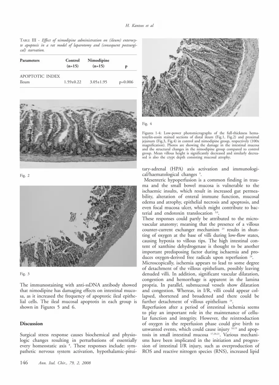
Villous height significantly decreased in jejunum(p=0.016) and ileum (p=0.002) in the nimodipine ratscompared to control animals. Crypt depth showed a pat-tern similar to that of the villous height. The nimodi-pine group demonstrated a lower crypt depth in jeju-num (p<0.001) and ileum (p<0.001) compared to con-trol animals (Table II). Ileum and jejunum sections from the control rats showedbetter architecture of the intestinal epithelium. Bothileum and jejunum from nimodipine rats demonstratedsubepithelial space at villous tip, inflammatory cell infil-tration extending through the wall, shortening and lossof villi, as well as reduction of crypt depth. Whereas,the effect of Nimodipine on microscopic bowel appea-rance, following laparotomy plus consequent PSS in rat,is shown in Figures 1-4.
CELLULAR APOPTOSIS
The AI increased following nimodipine administrationin ileum (p=0.006) compared to control animals, as sum-marized in Table III.
Effect of nimodipine on murine gut mucosa
Ann. Ital. Chir., 79, 2, 2008 145
for 30 min. The F-7-26 mAb was applied in a dilution1:10 and rinsed again in PBS. Chromagen solution(DAB) as substrate and light counterstain with haema-toxylin were applied at the end. Known positive con-trols as well as negative were also stained in each run.The apoptotic index (AI) was determined in ileum anddefined as the number of apoptotic cells/100 villousepithelial cells per villi, selecting, at least, 20 completewell-oriented villi from each section.All measurements were performed by a qualified patho-logist blinded to the source of intestinal tissue.
STATISTICAL ANALYSIS
Data are expressed as mean ±1 standard deviation (S.D.).The Kolmogorov–Smirnov test was used to assess nor-mality of the distributions. Comparisons of continuousvariables were analyzed using the unpaired t-test andMann-Whitney non-parametric test, as appropriate.Linear relationships between quantitative normally distri-buted parameters were assessed with Pearson’s two waytest, otherwise Spearmans’ rho was used. All performedtests were two-sided. Differences were considered as sta-tistically significant at the level of 5% (p<0.05).
Results
MICROSCOPIC BOWEL APPEARANCE
The homogenic intestinal response to nimodipine wassuggested by the following strong statistically significantcorrelations (p<0.001):(i) Mean crypt depth in the ileum positively correlated
with mean crypt depth in the jejunum.(ii) Mean villous height in the ileum positively correla-
ted with mean villous height in the jejunum.(iii) Strong positive correlations were detected between
mean crypt depth and mean villous height in theileum as well as in the jejunum.
These statistical observations are summarized in table 1.
TABLE I - Statistically significant correlations between the measuredparameters.
Correlations c.cp
Crypt depth (Ileum) - 0.835Crypt depth (Jejunum) p<0.001
Villous height (Ileum) - 0.702Villous height (Jejunum) p<0.001
Villous height (Jejunum) - 0.698Crypt depth (Jejunum) p<0.001
Villous height (Ileum) - 0.648Crypt depth (Ileum) p<0.001
TABLE II - Effect of Nimodipine on microscopic bowel appearance fol-lowing laparotomy plus consequent PSS in rat.
Parameters Control Nimodipine(n=15) (n=15) p
VILLOUS HEIGHT (µm)
Jejunum 130.77±26.41 104.93±24.44 p=0.016
Ileum 129.99±34.03 91.11±20.11 p=0.002
CRYPT DEPTH (µm)
Jejunum 55.66±17.67 27.72±6.49 p<0.001
Ileum 54.77±10.84 26.83±9.12 p<0.001
Fig. 1
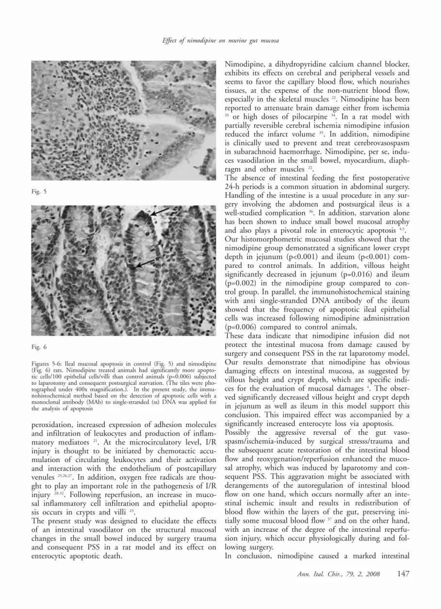
The immunostaining with anti-ssDNA antibody showedthat nimodipine has damaging effects on intestinal muco-sa, as it increased the frequency of apoptotic ileal epithe-lial cells. The ileal mucosal apoptotis in each group isshown in Figures 5 and 6.
Discussion
Surgical stress response causes biochemical and physio-logic changes resulting in perturbations of essentiallyevery homeostatic axis 8. These responses include: sym-pathetic nervous system activation, hypothalamic-pitui-
tary-adrenal (HPA) axis activation and immunologi-cal/haematological changes 9.Mesenteric hypoperfusion is a common finding in trau-
ma and the small bowel mucosa is vulnerable to theischaemic insults, which result in increased gut permea-bility, alteration of enteral immune function, mucosaledema and atrophy, epithelial necrosis and apoptosis, andeven focal mucosa ulcer, which might contribute to bac-terial and endotoxin translocation 3,6.These responses could partly be attributed to the micro-vascular anatomy; meaning that the presence of a villouscounter-current exchanger mechanism 23 results in shun-ting of oxygen at the base of villi during low-flow states,causing hypoxia to villous tips. The high intestinal con-tent of xanthine dehydrogenase is thought to be anotherimportant predisposing factor during ischaemia and pro-duces oxygen-derived free radicals upon reperfusion 24.Microscopically, ischemia appears to lead to some degreeof detachment of the villous epithelium, possibly leavingdenuded villi. In addition, significant vascular dilatation,congestion and hemorrhage is apparent in the laminapropria. In parallel, submucosal vessels show dilatationand congestion. Whereas, in I/R, villi could appear col-lapsed, shortened and broadened and there could befurther detachment of villous epithelium 24.Reperfusion after a period of intestinal ischemia seemsto play an important role in the maintenance of cellu-lar function and integrity. However, the reintroductionof oxygen in the reperfusion phase could give birth tounwanted events, which could cause injury 10,19 and apop-tosis in small intestinal mucosa 17,20,21. Various mechani-sms have been implicated in the initiation and progres-sion of intestinal I/R injury, such as overproduction ofROS and reactive nitrogen species (RNS), increased lipid
H. Kantsos et al
146 Ann. Ital. Chir., 79, 2, 2008
TABLE III - Effect of nimodipine administration on (ileum) enterocy-te apoptosis in a rat model of laparotomy and (consequent postsurgi-cal) starvation.
Parameters Control Nimodipine(n=15) (n=15) p
APOPTOTIC INDEXIleum 1.59±0.22 3.05±1.95 p=0.006
Fig. 2
Fig. 3
Fig. 4
Figures 1-4: Low-power photomicrographs of the full-thickness hema-toxylin-eosin stained sections of distal ileum (Fig.1, Fig.2) and proximaljejunum (Fig.3, Fig.4) in control and nimodipine group, respectively (100xmagnification). Photos are showing the damage in the intestinal mucosaand the structural changes in the nimodipine group compared to controlgroup. Mean villous height is significantly decreased and similarly decrea-sed is also the crypt depth consisting mucosal atrophy.
peroxidation, increased expression of adhesion moleculesand infiltration of leukocytes and production of inflam-matory mediators 21. At the microcirculatory level, I/Rinjury is thought to be initiated by chemotactic accu-mulation of circulating leukocytes and their activationand interaction with the endothelium of postcapillaryvenules 25,26,27. In addition, oxygen free radicals are thou-ght to play an important role in the pathogenesis of I/Rinjury 28-32. Following reperfusion, an increase in muco-sal inflammatory cell infiltration and epithelial apopto-sis occurs in crypts and villi 23.The present study was designed to elucidate the effectsof an intestinal vasodilator on the structural mucosalchanges in the small bowel induced by surgery traumaand consequent PSS in a rat model and its effect onenterocytic apoptotic death.
Nimodipine, a dihydropyridine calcium channel blocker,exhibits its effects on cerebral and peripheral vessels andseems to favor the capillary blood flow, which nourishestissues, at the expense of the non-nutrient blood flow,especially in the skeletal muscles 22. Nimodipine has beenreported to attenuate brain damage either from ischemia33 or high doses of pilocarpine 34. In a rat model withpartially reversible cerebral ischemia nimodipine infusionreduced the infarct volume 35. In addition, nimodipineis clinically used to prevent and treat cerebrovasospasmin subarachnoid haemorrhage. Nimodipine, per se, indu-ces vasodilation in the small bowel, myocardium, diaph-ragm and other muscles 22.The absence of intestinal feeding the first postoperative24-h periods is a common situation in abdominal surgery.Handling of the intestine is a usual procedure in any sur-gery involving the abdomen and postsurgical ileus is awell-studied complication 36. In addition, starvation alonehas been shown to induce small bowel mucosal atrophyand also plays a pivotal role in enterocytic apoptosis 4,5.Our histomorphometric mucosal studies showed that thenimodipine group demonstrated a significant lower cryptdepth in jejunum (p<0.001) and ileum (p<0.001) com-pared to control animals. In addition, villous heightsignificantly decreased in jejunum (p=0.016) and ileum(p=0.002) in the nimodipine group compared to con-trol group. In parallel, the immunohistochemical stainingwith anti single-stranded DNA antibody of the ileumshowed that the frequency of apoptotic ileal epithelialcells was increased following nimodipine administration(p=0.006) compared to control animals.These data indicate that nimodipine infusion did notprotect the intestinal mucosa from damage caused bysurgery and consequent PSS in the rat laparotomy model.Our results demonstrate that nimodipine has obviousdamaging effects on intestinal mucosa, as suggested byvillous height and crypt depth, which are specific indi-ces for the evaluation of mucosal damages 4. The obser-ved significantly decreased villous height and crypt depthin jejunum as well as ileum in this model support thisconclusion. This impaired effect was accompanied by asignificantly increased enterocyte loss via apoptosis. Possibly the aggressive reversal of the gut vaso-spasm/ischemia-induced by surgical stresss/trauma andthe subsequent acute restoration of the intestinal bloodflow and reoxygenation/reperfusion enhanced the muco-sal atrophy, which was induced by laparotomy and con-sequent PSS. This aggravation might be associated withderangements of the autoregulation of intestinal bloodflow on one hand, which occurs normally after an inte-stinal ischemic insult and results in redistribution ofblood flow within the layers of the gut, preserving ini-tially some mucosal blood flow 37 and on the other hand,with an increase of the degree of the intestinal reperfu-sion injury, which occur physiologically during and fol-lowing surgery.In conclusion, nimodipine caused a marked intestinal
Effect of nimodipine on murine gut mucosa
Ann. Ital. Chir., 79, 2, 2008 147
Fig. 5
Fig. 6
Figures 5-6: Ileal mucosal apoptosis in control (Fig. 5) and nimodipine(Fig. 6) rats. Nimodipine treated animals had significantly more apopto-tic cells/100 epithelial cells/villi than control animals (p=0.006) subjectedto laparotomy and consequent postsurgical starvation. (The tiles were pho-tographed under 400x magnification.). In the present study, the immu-nohistochemical method based on the detection of apoptotic cells with amonoclonal antibody (MAb) to single-stranded (ss) DNA was applied forthe analysis of apoptosis

mucosal injury in the rat laparotomy and consequent PSSmodel. The aforementioned disturbance resulted in increa-sed epithelial cell apoptosis, which may be responsible forthis negative effect. Exposure to nimodipine did not pre-vent surgery-induced ischemic damage, possibly via deran-gement of the autoregulation of intestinal blood flow.
Riassunto
La combinazione di trauma chirurgico e di non som-ministrazione di cibo nel postoperatorio, provoca dellealterazioni alla struttura ed alla funzionalita’ della mucosaintestinale, come pure alla funzionalita’ della barrieraintestinale, all’intestino tenue di rati da sperimentazione.Lo scopo del presente paper originale e sperimentale, fuquello di valutare, come la somministrazione di nimodip-ina, su modeli sperimentali di intestino tenue di rati,potesse provocare delle modifiche strutturalli della mucosada una parte ed una pronunciata apoptosi cellulareepiteliale dall’altra, se somministrata per via intraperi-toneale, in seguito ad un intervento di laparotomia e pri-ma che iniziasse il periodo postoperatorio di non som-ministrazione di cibo, della complessiva durata di 48 ore. A detto scopo, 30 rati da sperimentazione di razzaWistar, sono stati divisi in 2 gruppi, di cui uno di con-trollo ( Gruppo A ), composto da 15 di essi, i quallifurono sottoposti ad un intervneto di laparotomia ed allanon somministrazione di cibo per le prime 48 ore nelimmediato postoperatorio ed un altro ( Gruppo B ) sem-pre composto da 15 ratti, i qualli sono stati sottoposti,alla pari dei rati del gruppo di controllo ad un interve-to di laparotomia ed alla non somministrazione di ciboper le prime 48 ore nel postoperatorio, ma tra la finedell’intervento di laparotomia ed il periodo postoperato-rio di astinenza dal cibo, gli si e’ somministrata, per viaintraperitoneale, la Nimodipina. In entrambi i gruppi, 48 ore dopo l’intervento dellalaparotomia, sono state valutate sia le modifiche strut-turali della mucosa intestinale che il grado, da essa giun-to, di apoptosi cellulare.In conclusione di detta sperimentazione, si e’ messo inevidenza che la nimodipina, non solo non protegeva lamucosa intestinale, dal danno provocatele dall’interven-to chirurgico laparotomico e dal succesivo, all’interven-to, periodo di non somministrazione di cibo per 48 ore,ma provocava anche dei ben evidenti danni alla mucosaintestinale, in quanto, in seguito alla sua somminis-trazione per via intraperitoneale, si verificarono intensealterazioni dell’arcitettura della mucosa intestinale ed unasua consistente atrofia.
References
1) Thomas S, Balasubramanian KA: Role of intestine in postsurgicalcomplications: involvement of free radicals. Free Radic Biology Med,2004; 36:745-756.
2) Anup R, Aparna V, Pulimood A, Balasubramanian KA: Surgicalstress and the small intestine: Role of oxygen free radicals. Surgery,1999; 125:560-69.
3) Thomas S, Kang G, Balasubramanian KA: Surgical Manipulationof the Intestine Results in Quantitative and Qualitative Alterations inLuminal Escerichia coli. Ann Surg, 2004; 240:248-54.
4) Chappell VL, Thompson MD, Jeschke MG, Chung DH,Thompson JC, Wolf SE: Effects of incremental starvation on gutmucoca. Dig Dis Sci, 2003; 48:765-69.
5) Wiren M, Soderholm JD, Lindgren J, Olaison G, Permert J,YangH, Larsson J: Effects of starvation and bowel resection on paracellu-lar permeability in rat small-bowel mucoca in vitro. Scand J Gastr,1999; 34:156-62.
6) Hang CH, Shi JX, Li JS, Wu W, Yin HX: Alterations of inte-stinal mucosa structure and barrier function following traumatic braininjury in rats. World J Gastr, 2003; 12:2776-781.
7) Ramzy PI, Wolf SE, Irtun O, Hart DW, Thompson JC,Herndon DN: Gut epithelial apoptosis after severe burn: effects of guthypoperfusion. J Am Coll Surg, 2000; 190:281-87.
8) Udelsman R, Holbrook NJ: Endocrine and molecular responces tosurgical stress. Curr Probl Surg, 1994; 31:653-720.
9) Desborough JP: The stress response to trauma and surgery. Br JAnaesth, 2000; 85:109-117.
10) Madesh M, Ramachandran A, Pulimood A, Vadranam M,Balasubramanian KA: Attenuation of intestinal ischemia/reperfusioninjury with sodium nitroprusside: studies on mitochondrial functionand lipid changes. Biochim Biophys Acta, 2000; 1500:204-16.
11) Zimmerman BJ, Granger DN: Mechanisms of reperfusion injury.Am J Med Sci, 1994; 307:284-92.
12) Tagami M, Ikeda K, Nara Y, Fujino H, Kubota A, NumanoF, Yamori Y: Insulin-like growth factor-1 attenuates apoptosis in hip-pocampal neurons caused by cerebral ischemia and reperfusion in stroke-prone spontaneously hypertensive rats. Lab Invest, 1997; 76:613-17.
13) Yadav S, Sindram D, Perry DK, Clavien PA: Ischemic precon-ditioning protects the mouse liver by inhibition of apoptosis through acaspase-dependant pathway. Hepatology, 1999; 30: 1223-231.
14) Yin T, Sandhu G, Wolfgang CD, Burrier A, Webb RL, RigelDF, Hai T, Whelan J: Tissue-specific pattern of stress kinase activa-tion in ischemia/reperfused heart and kidney. J Biol Chem, 1997;272:1943-950.
15) Maclellan WR, Schneider MD: Death by design. Programmedcell death in cardiovascular biology and disease. Circ Res, 1997;81:137-44.
16) Fujimoto K, Hosotani R, Wada M, Lee JU, Koshiba T,Miyamoto Y, Doi R, Imamura M: Ischemia-reperfusion injury onthe pancreas in rats: identification of acinar cell apoptosis. J Surg Res,1997; 71:127-36.
17) Fukuyama K, Iwakiri R, Noda T, Kojima M, Utsumi H,Tsunada S, Sakata H, Ootani A, Fujimoto K: Apoptosis induced byischemia-reperfusion and fasting in gastric mucosa compared to smallintestinal mucosa in rats. Dig Dis Sci, 2001; 46:545-49.
18) Luo CC, Shih HH, Chiu CH, Ma WC, Chung HY: Reducedapoptosis in newborn compared to adult rat intestine after ischemia-reperfusion. Biol Neonate, 2004; 85:90-93.
H. Kantsos et al
148 Ann. Ital. Chir., 79, 2, 2008

19) Sukhotnik I, Helou H, Mogilner J, Lurie M, Bernsteyn A,Coran AG, Shiloni E: Oral arginine improves intestinal recovery fol-lowing ischemia-reperfusion injury in rat. Pediatr Surg Int, 2005;21:191-96.
20) Noda T, Iwakiri R, Fujimoto K, Matsuo S, Aw TY:Programmed cell death induced by ischemia-reperfusion in the rat inte-stinal mucosa. Am J Physiol, 1998; 274:270-76.
21) Ikeda H, Suzuki Y, Suzuki M, Koike M, Tamura J, Tong J,Nomura M, Itoh G: Apoptosis is a major mode of cell death causedby ischemia and ischemia/reperfusion injury to the rat intestinal epithe-lium. GUT, 1998; 42:530-37.
22) Karlsson BM, Koch M, Koskinen LO: Nimodipine affects themicrocirculation and modulates the vascular effects of acetylcholineste-rase inhibition. Upsala J Med Sci, 2003; 108:141-49.
23) Shah KA, Shurey SS, Green CJ: Characterization of apoptosisin intestinal ischaemia-reperfusion injury - a light and electron micro-scopic study. Int J Exp Path, 1997; 78:355-63.
24) Parks DA, Granger DN: Ischemia-induced vascular changes: roleof xanthine oxidase and hydroxyl radicals. Am J Physiol, 1983;245:285-89.
25) Hernandez L, Grisham B, Twohig B, Arfors KE, Harlan JM,Granger DN: Role of neutrophils in ischemia-reperfusion-inducedmicrovascular injury. Am J Physiol, 1987; 253:699-703.
26) Walden DL, McCutchan HJ, Enquist EG, Schwappach JR,Shanley PF, Reiss OK, Terada LS, Leff JA, Repine JE: Neutrophilsaccumulate and contribute to skeletal muscle dysfunction after ische-mia-reperfusion. Am J Physiol, 1990; 259:1809-812.
27) Granger DN, Benoit JN, Suzuki M, Grisham MB: Leukocytesadherence to venular endothelium during ischemia-reperfusion. Am JPhysiol, 1989; 257:683-88.
28) Klausner JM, Paterson IS, Kobzik L, Valeri CR, Shepro D,
Hechtman HB: Oxygen free radicals mediate ischemia-induced lunginjury. Surgery, 1989; 105:192-99.
29) Menger MD, Barker JH, Messmer K: Capillary blood perfusionduring postischemic reperfusion in striated muscle. Plast Reconstr Surg,1992; 89:1104-114.
30) McCord JM: Oxygen-derived radicals: a link between reperfusioninjury and inflammation. Federation Proc, 1987; 46:2402-406.
31) Suzuki M, Inauen W, Kvietys PR, Grisham MB, Meininger C,Schelling ME, Granger HJ, Granger DN: Superoxide mediates reper-fusion-induced leukocyte-endothelial cell interactions. Am J Physiol,1989; 257:1740-745.
32) Zweier JL, Kuppusamy P, Lutty GA: Measurement of endothe-lial cell free radical generation: Evidence for a central mechanism offree radical injury in post-ischemic tissues. Proc Natl Acad Sci USA,1988; 85:4046-050.
33) Uematsu D, Araki N, Greenberg JH, Sladky J, Reivich M:Combined therapy with MK-801 and nimodipine for protection ofischemic brain damage. Neurology, 1991; 41:88-94.
34) Marinho MM, de Bruin VM, de Sousa FC, Aguiar LMV, dePinho RSN, Viana GSB: Inhibitory action of a calcium channelblocker (nimodipine) on seizures and brain damage induced by pilo-carpine and lithium-pilocarpine in rats. Neurosci Lett, 1997; 235:13-16.
35) Roda JM, Carceller F, Diez-Tejedor E, Avendano C: Reductionof infarct size by intra-arterial nimodipine administered at reperfusionin a rat model of partially reversible brain focal ischemia. Stroke,1995; 26:1888-892.
36) Quigley EM, Thompson JS: The effects of surgery on gastroin-testinal motor activity. Braz J Med Biol Res, 1998; 31:889-900.
37) Reilly PM, Bulkley GB: Vasoactive mediators and splanchnic per-fusion. Crit Care Med, 1993; 21:55-68.
Effect of nimodipine on murine gut mucosa
Ann. Ital. Chir., 79, 2, 2008 149

VIDEO-CHIRURGIA
Ann. Ital. Chir., 2008; 79
Il valore didattico del film in chirurgia
di Giuseppe Romagnolo, Biagio Trojanello
DVD- 21’ 41”- ItalianoPubblicato da : Salvatore Cafiero Audiovisi- 2001
Autore Corrispondente: Biagio Trojanello, Salita Piedigrotta3, 80122 Napoli
Recensione:Rassegna storica, riccamente corredata di iconografia clas-sica, che ripercorre la storia della tecnica chirurgicamoderna non semplicemente nelle tappe dei suoi pro-gressi tecnici quanto nell’ottica della dimostrazione deimezzi strumentali storici, della didattica finalizzataall’apprendimento.Nella prima parte vengono esibiti disegni e figure stati-che da affreschi e miniature classiche, per passare quin-di ad una breve rassegna fotografica esemplificativa dipatologie e situazioni cliniche. Vengono rievocati ritratti
150 Ann. Ital. Chir., 79, 2, 2008
celebri di altrettanti celebri chirurghi nell’atto docentedella dimostrazione anatomo-patologica e chirurgica.L’arrivo dell’invenzione dei fratelli Lumière ha modifica-to radicalmente l’aspetto didattico della chirurgia nondirettamente vissuta ed osservata, poiché essa venne adot-tata già due anni dopo della sua prima dimostrazioneper dimostrare atti chirurgici su pellicola bianco e nero,con intervento del Prof. Roberto Alessandri.Dopo un primo periodo euforico, in cui singoli chirur-ghi si avvalevano della tecnica chirurgica su pellicola –costosa - per dimostrare le personali capacità tecniche,la cinematografia chirurgica ha dimostrato sempre dipiù il suo valore didattico intrinseco. Ma la vera esplo-sione si è avuta con l’introduzione della tecnica di ripre-sa e postproduzione digitale, che ha consentito di con-fezionare prodotti didattici di grande valore. Prevedendoaddirittura non solo la multimedialità ma anche l’inte-rattività tra dimostrazione e spettatore discente.
L’interesse del DVD è di tipo storico , ma il suo valo-re intrinseco risiede anche nell’entusiasmo delle finalitàdimostrate con convinzione e capacità dagli Autori
RECENSIONI DI VIDEO-TAPE DI ANNALI ITALIANI DI CHIRURGIA E DELLA VIDEO REVITA DE CIRURGIA
(a cura del Direttore)
Copyright © 2022 FDOKUMEN


![Mycobacterium Tuberculosis WhiB3 Responds to O2 and Nitric Oxide via Its [4Fe-4S] Cluster and is Essential for Nutrient Starvation Survival](https://static.fdokumen.com/doc/165x107/633297bc576b626f850d7e1e/mycobacterium-tuberculosis-whib3-responds-to-o2-and-nitric-oxide-via-its-4fe-4s.jpg)