1 Death as a Result of Starvation
21
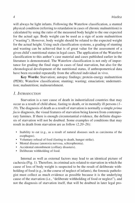
1 Death as a Result of Starvation Diagnostic Criteria Burkhard Madea, MD CONTENTS INTRODUCTION CLINICAL AND AUTOPSY FINDINGS IN STARVATION EXEMPLARY CASES CLASSIFICATION OF PEM DURATION OF STARVATION, PHYSICAL CONDITION PRIOR TO DEATH, AND IMMEDIATE CAUSE OF DEATH REFERENCES SUMMARY Fatal starvation is a rare cause of death in industrialized countries but this entity may become of major medicolegal importance if death results from deliberate withholding of food, especially from infants. In such cases, the task of the forensic pathologist and the medical examiner, respectively, is not only to clarify the cause of death but also to give an expert opinion on the degree and duration of starvation. Several classification systems have been devel- oped to estimate protein-energy malnutrition (PEM) in third world countries (e.g., Waterlow classification, Gomez classification). More simple classifica- tions (e.g., the Gomez classification of PEM) use the weight expected for the respective age group as standard. When applying this standard, small infants From: Forensic Pathology Reviews, Vol. 2 Edited by: M. Tsokos © Humana Press Inc., Totowa, NJ 3
-
Upload
khangminh22 -
Category
Documents
-
view
5 -
download
0
Transcript of 1 Death as a Result of Starvation
Starvation 3
1
Death as a Result of StarvationDiagnostic Criteria
Burkhard Madea, MD
CONTENTS
INTRODUCTION
CLINICAL AND AUTOPSY FINDINGS IN STARVATION
EXEMPLARY CASES
CLASSIFICATION OF PEMDURATION OF STARVATION, PHYSICAL CONDITION PRIOR TO DEATH,
AND IMMEDIATE CAUSE OF DEATH
REFERENCES
SUMMARY
Fatal starvation is a rare cause of death in industrialized countries butthis entity may become of major medicolegal importance if death results fromdeliberate withholding of food, especially from infants. In such cases, the taskof the forensic pathologist and the medical examiner, respectively, is not onlyto clarify the cause of death but also to give an expert opinion on the degreeand duration of starvation. Several classification systems have been devel-oped to estimate protein-energy malnutrition (PEM) in third world countries(e.g., Waterlow classification, Gomez classification). More simple classifica-tions (e.g., the Gomez classification of PEM) use the weight expected for therespective age group as standard. When applying this standard, small infants
From: Forensic Pathology Reviews, Vol. 2Edited by: M. Tsokos © Humana Press Inc., Totowa, NJ
3
Madea4
will always be light infants. Following the Waterlow classification, a stuntedphysical condition (referring to retardation in cases of chronic malnutrition) iscalculated by using the ratio of the measured body height to the one expectedfor the actual age. Body weight can be used as a sign of acute malnutrition(“wasting”). However, body weight should be related to the expected weightfor the actual height. Using such classification systems, a grading of stuntingand wasting can be achieved that is of great value for the assessment of agiven child’s nutritional status in legal cases. The application of the Waterlowclassification to this author’s case material and cases published earlier in theliterature is demonstrated. The Waterlow classification is not only of impor-tance for grading the final stage in cases of fatal starvation, but also for thechronological development of the nutritional status, if anthropometrical datahave been recorded repeatedly from the affected individual in vivo.
Key Words: Starvation; autopsy findings; protein-energy malnutrition(PEM); Waterlow classification; stunting; wasting; emaciation; undernutri-tion; malnutrition; malnourishment.
1. INTRODUCTION
Starvation is a rare cause of death in industrialized countries that mayoccur as a result of child abuse, fasting to death, or in mentally ill persons (1–19). The diagnosis of death as a result of starvation is normally a simple primafacie diagnosis, the visual features of starvation being known from contempo-rary famines. If there is enough circumstantial evidence, the definite diagno-sis of starvation will not be doubted. Some examples of conditions that mayresult in death from starvation are as follow (2,20–26):
• Inability to eat (e.g., as a result of natural diseases such as carcinoma of theesophagus).
• Voluntary refusal of food (fasting to death, hunger strike).• Mental disease (anorexia nervosa, schizophrenia).• Accidental entombment (colliary disasters).• Deliberate withholding of food.
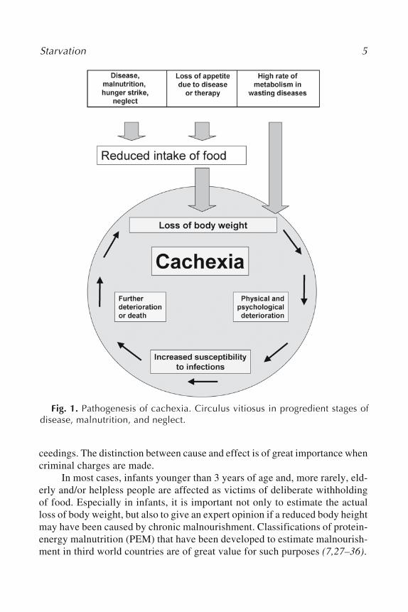
Internal as well as external factors may lead to an identical picture ofcachexia (Fig. 1). Therefore, in criminal acts related to starvation in which thecause of loss of body weight is suspected to be the result of deliberate with-holding of food (e.g., in the course of neglect of infants), the forensic patholo-gist must collect as much evidence as possible because it is the underlyingcause of the starvation (i.e., “deliberate withholding of food or neglect”), andnot the diagnosis of starvation itself, that will be doubted in later legal pro-
Starvation 5
ceedings. The distinction between cause and effect is of great importance whencriminal charges are made.
In most cases, infants younger than 3 years of age and, more rarely, eld-erly and/or helpless people are affected as victims of deliberate withholdingof food. Especially in infants, it is important not only to estimate the actualloss of body weight, but also to give an expert opinion if a reduced body heightmay have been caused by chronic malnourishment. Classifications of protein-energy malnutrition (PEM) that have been developed to estimate malnourish-ment in third world countries are of great value for such purposes (7,27–36).
Fig. 1. Pathogenesis of cachexia. Circulus vitiosus in progredient stages ofdisease, malnutrition, and neglect.
Madea6
2. CLINICAL AND AUTOPSY FINDINGS IN STARVATION
The daily calorie requirement of humans above the basic metabolic ratemainly depends on the physical activity (37–39; Table 1). Insufficient calorieintake compared to the requirement results in a negative energy balance withresultant loss of body weight. Malnutrition has to be differentiated from under-nutrition. Undernutrition is the intake of an insufficient quantity of food,whereas malnutrition is defined as feeding of inadequate quality. Symptomsand pathology of starvation have been comprehensively reported since WorldWar II, based on experiences and observations derived from the Nazi concen-tration camps (22,40–52). The more or less constant symptoms of starvationdevelop in a characteristic chronological order (50):
1. Loss of well-being and hunger, hunger pains, and craving for food.2. Apathy and fatigue.3. Weight loss, more rapid in the first 6 months of starvation than afterward.4. Pigmentation, cachexia, and hypothermia.5. Extreme lethargy, mental retardation, and loss of self-respect.6. Hunger edema.7. Reduced resistance to infections in general, and development of diarrhea and
tuberculosis, or other intercurrent infections.
Even in advanced stages of starvation, death may be sudden and unex-pected (see for instance witness reports from concentration camps [45,52] aswell as more recent publications [53–57]).
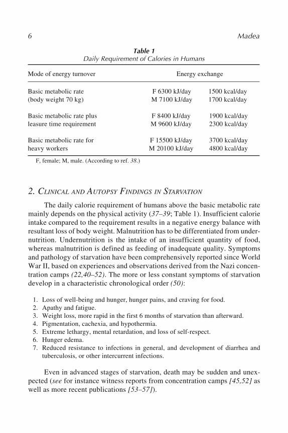
Table 1Daily Requirement of Calories in Humans
Mode of energy turnover Energy exchange
Basic metabolic rate F 6300 kJ/day 1500 kcal/day(body weight 70 kg) M 7100 kJ/day 1700 kcal/day
Basic metabolic rate plus F 8400 kJ/day 1900 kcal/dayleasure time requirement M 9600 kJ/day 2300 kcal/day
Basic metabolic rate for F 15500 kJ/day 3700 kcal/dayheavy workers M 20100 kJ/day 4800 kcal/day
F, female; M, male. (According to ref. 38.)
Starvation 7
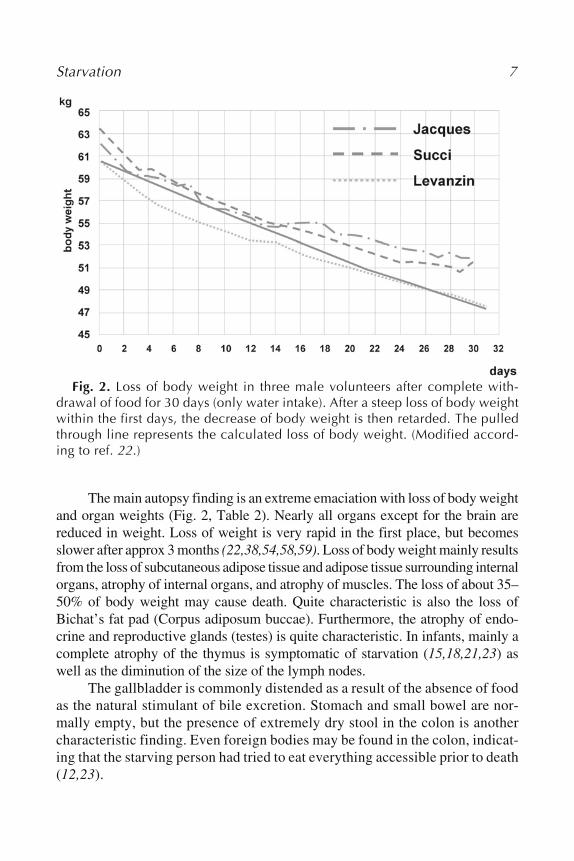
The main autopsy finding is an extreme emaciation with loss of body weightand organ weights (Fig. 2, Table 2). Nearly all organs except for the brain arereduced in weight. Loss of weight is very rapid in the first place, but becomesslower after approx 3 months (22,38,54,58,59). Loss of body weight mainly resultsfrom the loss of subcutaneous adipose tissue and adipose tissue surrounding internalorgans, atrophy of internal organs, and atrophy of muscles. The loss of about 35–50% of body weight may cause death. Quite characteristic is also the loss ofBichat’s fat pad (Corpus adiposum buccae). Furthermore, the atrophy of endo-crine and reproductive glands (testes) is quite characteristic. In infants, mainly acomplete atrophy of the thymus is symptomatic of starvation (15,18,21,23) aswell as the diminution of the size of the lymph nodes.
The gallbladder is commonly distended as a result of the absence of foodas the natural stimulant of bile excretion. Stomach and small bowel are nor-mally empty, but the presence of extremely dry stool in the colon is anothercharacteristic finding. Even foreign bodies may be found in the colon, indicat-ing that the starving person had tried to eat everything accessible prior to death(12,23).
Fig. 2. Loss of body weight in three male volunteers after complete with-drawal of food for 30 days (only water intake). After a steep loss of body weightwithin the first days, the decrease of body weight is then retarded. The pulledthrough line represents the calculated loss of body weight. (Modified accord-ing to ref. 22.)
Madea8
Typical picture of emaciation and exclusion of any other concurrent causeof death are prerequisite for a definitive postmortem diagnosis of death as aresult of starvation (3,7,9–11,27,28,60–62).
If the diagnosis of death as a result of starvation is established, the actualcause of death has to be determined: preexisting diseases (e.g., malabsorption,malassimilation, cancer) or withholding of food.
Histopathology serves mainly to exclude concurrent causes of death andto get clues regarding underlying natural causes of emaciation or to excludethem (22,63,64).
The anthropometrical data of starved persons and organ weights are rou-tinely compared to those of a reference population (28,65,67). These data arethe essential basis for the diagnosis of starvation. However, a plain orientationon the reference values or percentile charts alone does not permit a grading ofthe degree or stage of malnutrition.
Several classification systems have been developed to estimate PEM inthird world countries over the last decades. Especially for forensic patholo-gists, these classifications are superior compared to percentile charts because
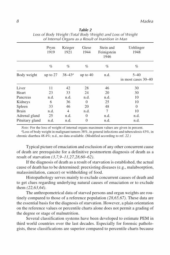
Table 2Loss of Body Weight (Total Body Weight) and Loss of Weight
of Internal Organs as a Result of Inanition in Man
Prym Krieger Giese Stein and Uehlinger1919 1921 1944 Feinigstein 1948
1946
% % % % %
Body weight up to 27 38–43a up to 40 n.d. 5–40in most cases 30–40
Liver 11 42 28 46 30Heart 23 33 24 20 30Pancreas n.d. n.d. n.d. n.d. 10Kidneys 6 36 0 25 10Spleen 33 46 20 48 0Brain n.d. 4 n.d. 7 10Adrenal gland 25 n.d. 0 n.d. n.d.Pituitary gland n.d. n.d. 0 n.d. n.d.
Note: For the loss of weight of internal organs maximum values are given in percent.aLoss of body weight in malignant tumors 38%, in general infections and tuberculosis 43%, in
chronic diarrhea 48.4%; n.d., no data available. (Modified according to ref. 22.)

Starvation 9
anthropometrical data allow also an estimate of the degree of malnutrition(30–36). Those gradings of PEM have been successfully applied to affectedindividuals in third world countries, but they may be used also for the classifi-cation of infantile malnutrition in cases of starvation as a result of deliberatewithholding of food. First experiences with the use of these classification sys-tems were published by this author’s study group in 1994 (12). The applica-tion of such classification systems to this author’s case material and casespublished earlier in the literature (1,6,12,19,23) will be demonstrated in thefollowing.
3. EXEMPLARY CASES
3.1. Own Case Material
This author’s own case material comprises six cases of fatal starvation inchildhood (12). At the time of death, the infants were between 3 months and2.5 years old. Among them were monocygotic twins who were found dead athome on occasion of the visit of a nurse (Fig. 3). All six infants were found inapartments that were in a state of total neglect. Some of the bodies were alreadyin an early state of putrefaction. All infants appeared severely emaciated atgross examination with nearly total absence of fat depots in cheek, abdominal
Fig. 3. Scene of death. These monocygotic twins were found dead in anapartment that was in a state of total neglect.

Madea10
wall, and mesentery (Fig. 4A–C). All infants had the classical signs of starva-tion as discussed previously. In all cases, a “dry” emaciation with the typicalsigns of dehydration was present. The loss of body weight and organ weight(in percent) in relation to the normal weight for the respective age group is
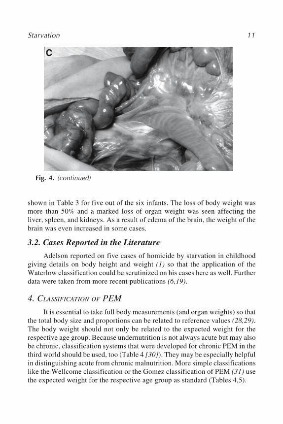
Fig. 4. Typical autopsy findings in starvation. (A) Emaciation, loss of Bichat’sfat pad, and sunken eyes, (B) Complete loss of subcutaneous tissue with pro-nounced rib cage, (C) Loss of adipose tissue of the mesenterium.
Starvation 11
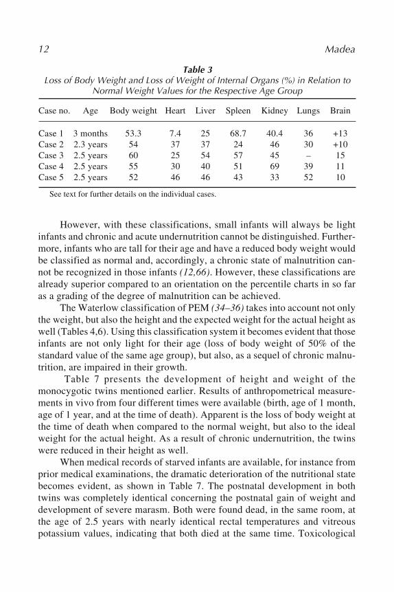
shown in Table 3 for five out of the six infants. The loss of body weight wasmore than 50% and a marked loss of organ weight was seen affecting theliver, spleen, and kidneys. As a result of edema of the brain, the weight of thebrain was even increased in some cases.
3.2. Cases Reported in the Literature
Adelson reported on five cases of homicide by starvation in childhoodgiving details on body height and weight (1) so that the application of theWaterlow classification could be scrutinized on his cases here as well. Furtherdata were taken from more recent publications (6,19).
4. CLASSIFICATION OF PEM
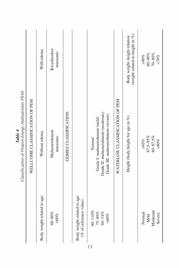
It is essential to take full body measurements (and organ weights) so thatthe total body size and proportions can be related to reference values (28,29).The body weight should not only be related to the expected weight for therespective age group. Because undernutrition is not always acute but may alsobe chronic, classification systems that were developed for chronic PEM in thethird world should be used, too (Table 4 [30]). They may be especially helpfulin distinguishing acute from chronic malnutrition. More simple classificationslike the Wellcome classification or the Gomez classification of PEM (31) usethe expected weight for the respective age group as standard (Tables 4,5).
Fig. 4. (continued)
Madea12
However, with these classifications, small infants will always be lightinfants and chronic and acute undernutrition cannot be distinguished. Further-more, infants who are tall for their age and have a reduced body weight wouldbe classified as normal and, accordingly, a chronic state of malnutrition can-not be recognized in those infants (12,66). However, these classifications arealready superior compared to an orientation on the percentile charts in so faras a grading of the degree of malnutrition can be achieved.
The Waterlow classification of PEM (34–36) takes into account not onlythe weight, but also the height and the expected weight for the actual height aswell (Tables 4,6). Using this classification system it becomes evident that thoseinfants are not only light for their age (loss of body weight of 50% of thestandard value of the same age group), but also, as a sequel of chronic malnu-trition, are impaired in their growth.
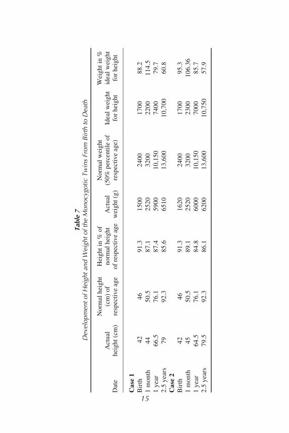
Table 7 presents the development of height and weight of themonocygotic twins mentioned earlier. Results of anthropometrical measure-ments in vivo from four different times were available (birth, age of 1 month,age of 1 year, and at the time of death). Apparent is the loss of body weight atthe time of death when compared to the normal weight, but also to the idealweight for the actual height. As a result of chronic undernutrition, the twinswere reduced in their height as well.
When medical records of starved infants are available, for instance fromprior medical examinations, the dramatic deterioration of the nutritional statebecomes evident, as shown in Table 7. The postnatal development in bothtwins was completely identical concerning the postnatal gain of weight anddevelopment of severe marasm. Both were found dead, in the same room, atthe age of 2.5 years with nearly identical rectal temperatures and vitreouspotassium values, indicating that both died at the same time. Toxicological
Table 3Loss of Body Weight and Loss of Weight of Internal Organs (%) in Relation to
Normal Weight Values for the Respective Age Group
Case no. Age Body weight Heart Liver Spleen Kidney Lungs Brain
Case 1 3 months 53.3 7.4 25 68.7 40.4 36 +13Case 2 2.3 years 54 37 37 24 46 30 +10Case 3 2.5 years 60 25 54 57 45 – 15Case 4 2.5 years 55 30 40 51 69 39 11Case 5 2.5 years 52 46 46 43 33 52 10
See text for further details on the individual cases.
Starvation 13Ta
ble
4C
lass
ifica
tion
of P
rote
in-E
nerg
y M
alnu
triti
on (P
EM)
WE
LL
CO
ME
CL
AS
SIF
ICA
TIO
N O
F P
EM
Bod
y w
eigh
t rel
ated
to a
geW
itho
ut e
dem
aW
ith
edem
a
60–8
0%M
alno
uris
hmen
tK
was
hior
kor
<60
%m
aras
mic
mar
asm
ic
GO
ME
Z C
LA
SS
IFIC
AT
ION
Bod
y w
eigh
t rel
ated
to a
ge(%
of
refe
renc
e va
lue)
90–1
10%
Nor
mal
75–8
9%G
rade
I: m
alno
uris
hmen
t (m
ild)
60–7
4%G
rade
II:
mal
nour
ishm
ent (
mod
erat
e)<
60%
Gra
de I
II: m
alno
uris
hmen
t (se
vere
)
WA
TE
RL
OW
CL
AS
SIF
ICA
TIO
N O
F P
EM
Hei
ght (
body
hei
ght f
or a
ge in
%)
Bod
y w
eigh
t–he
ight
rel
atio
n(w
eigh
t rel
ated
to h
eigh
t in
%)
Nor
mal
>95
%>
90%
Mil
d87
.5–9
5%80
–90%
Mod
erat
e80
–87.
5%70
–80%
Sev
ere
<80
%<
70%
13
Madea14
analysis was negative and the cause of death was stated as a combination ofstarvation and dehydration. The mother, who was a chronic alcoholic, suf-fered from severe hyperammonemia as a result of alcoholic liver damage, andhad obviously left the infants without food and fluid for the last days of theirlives, but witnesses confirmed that the infants had always been underweight.The nearly simultaneous death of twins as a result of starvation has, to the bestof this author’s knowledge, not been described before (for more details seeref. 12).
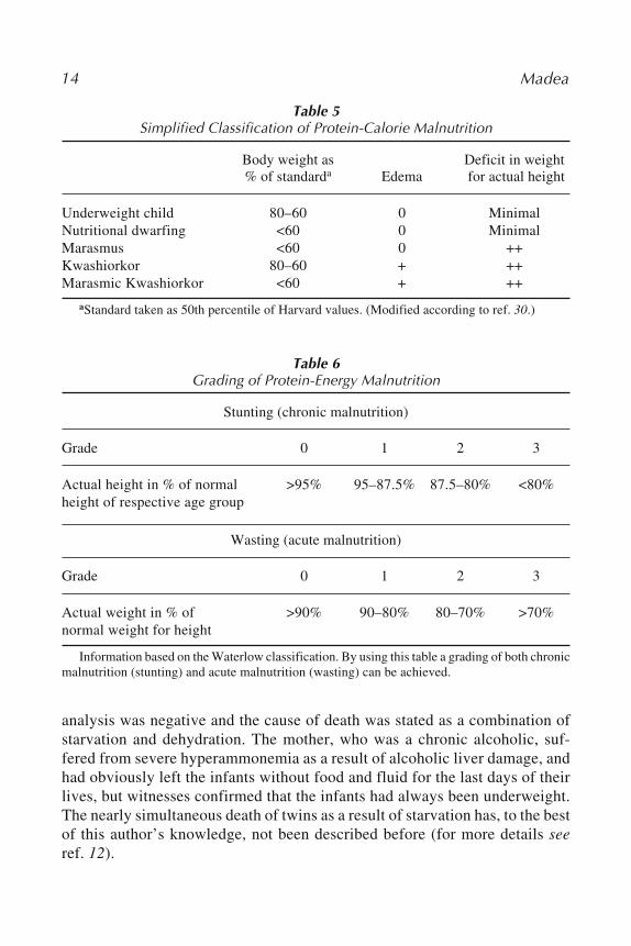
Table 5Simplified Classification of Protein-Calorie Malnutrition
Body weight as Deficit in weight% of standarda Edema for actual height
Underweight child 80–60 0 MinimalNutritional dwarfing <60 0 MinimalMarasmus <60 0 ++Kwashiorkor 80–60 + ++Marasmic Kwashiorkor <60 + ++
aStandard taken as 50th percentile of Harvard values. (Modified according to ref. 30.)
Table 6Grading of Protein-Energy Malnutrition
Stunting (chronic malnutrition)
Grade 0 1 2 3
Actual height in % of normal >95% 95–87.5% 87.5–80% <80%height of respective age group
Wasting (acute malnutrition)
Grade 0 1 2 3
Actual weight in % of >90% 90–80% 80–70% >70%normal weight for height
Information based on the Waterlow classification. By using this table a grading of both chronicmalnutrition (stunting) and acute malnutrition (wasting) can be achieved.
Starvation 15
Tabl
e 7
Dev
elop
men
t of H
eigh
t and
Wei
ght o
f the
Mon
ocyg
otic
Tw
ins
From
Bir
th to
Dea
th
Nor
mal
hei
ght
Hei
ght i
n %
of
Nor
mal
wei
ght
Wei
ght i
n %
Act
ual
(cm
) of
norm
al h
eigh
tA
ctua
l(5
0% p
erce
ntil
e of
Idea
l wei
ght
idea
l wei
ght
Dat
ehe
ight
(cm
)re
spec
tive
age
of r
espe
ctiv
e ag
ew
eigh
t (g)
resp
ecti
ve a
ge)
for
heig
htfo
r he
ight
Cas
e 1
Bir
th 4
246
91.3
1500
2400
1700
88.2
1 m
onth
4450
.587
.125
2032
0022
0011
4.5
1 ye
ar66
.576
.187
.459
0010
,150
7400
79.7
2.5
year
s79
92.3
85.6
6510
13,6
0010
,700
60.8
Cas
e 2
Bir
th42
4691
.316
2024
0017
0095
.31
mon
th45
50.5
89.1
2520
3200
2300
106.
361
year
64.5
76.1
84.8
6000
10,1
5070
0085
.72.
5 ye
ars
79.5
92.3
86.1
6200
13,6
0010
,750
57.9
15
Madea16
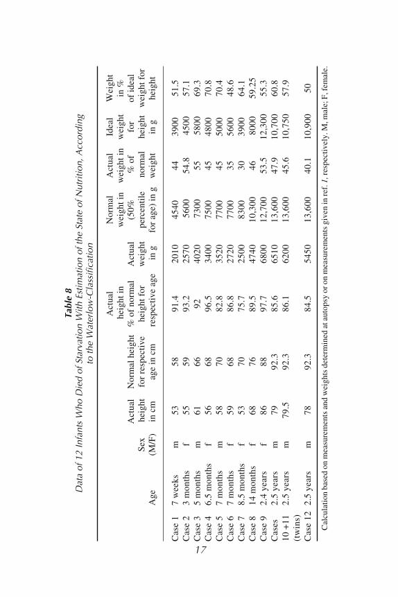
The data of 12 starved infants with an estimation of the state of nutritionaccording to the Waterlow classification are presented in Table 8. These cal-culations are based on measurements and weights determined at autopsy. Fivecases were taken from the publication by Adelson (1). In these cases, the ac-tual body weight related to the expected body weight for the actual heightreveals, according to the Waterlow classification, a severe life-threateningmalnutrition. Nearly all infants were much below the critical values for severemalnutrition that are given in the literature. In all cases shown in Table 8, theactual weight in percent of the ideal weight for height is near or below 70%.Of course, limiting values for fatal starvation are missing in the literature sofar; on the one hand there are only some published case reports on starvationusing the Waterlow classification and on the other hand the actual weight atthe time of death depends on various other factors such as dehydration, infec-tions, and so on. But because this author’s case material, as well as the casespublished by Adelson (1), show a weight quotient below 70%, this may be ahint toward the validity of the grading of the Waterlow classification concern-ing the severity of starvation. Four of the infants over 1 year of age presentedstunted (height below 90% of normal height for the respective age group),whereas infants younger than 1 year had mainly a quotient above 90%. Onemay speculate that in the older infants, undernutrition lasted longer (weightremained constant as a result of stunting), whereas in younger infants under-nutrition caused death more rapidly without the possibility of compensationof body weight as a result of stunting. This hypothesis is strengthened by threecases reported by Wehner et al. (19).
Three boys lived in a foster family. One of the boys (case 1) died fromstarvation at the age of 5 years. After his death, the other two boys (case 2,3;6.5 and 8.5 years of age, respectively) underwent physical examinations. Bothboys presented with a retardation of height and severe acute malnutrition (heightretardation: 80% [case 1], 87% [case 2], and 81% [case 3]; acute malnutrition[weight related to height in percent]: 60% [case 1], 62.5% [case 2] and 65%[case 3]). An acute reduction of weight of 35–50% is usually considered toresult in death and these infants showed a weight reduction of about 35–40%compared to the expected weight for the reduced height. It is remarkable thatthe 5-year-old boy who died (case 1) suffered from the highest loss of bodyweight compared to the actual height and the expected weight for the respec-tive age. On the other hand, Fieguth et al. (6) reported on two cases of fatalstarvation of very young infants (age: 5 weeks and 14 weeks, respectively)who received an inappropriate and inadequate nutrition. These authors reportedalso on an older child (3.5 years of age) who, left alone at home in the lockedapartment, died of acute starvation and showed—corresponding to the afore-
Starvation 17
Tabl
e 8
Dat
a of
12
Infa
nts
Who
Die
d of
Sta
rvat
ion
With
Est
imat
ion
of th
e St
ate
of N
utri
tion,
Acc
ordi
ngto
the
Wat
erlo
w-C
lass
ifica
tion
Act
ual
Nor
mal
Act
ual
Idea
lW
eigh
t h
eigh
t in
wei
ght i
nw
eigh
t in
wei
ght
in %
Act
ual
Nor
mal
hei
ght
% o
f no
rmal
Act
ual
(50%
% o
ffo
rof
idea
lS
ex h
eigh
tfo
r re
spec
tive
heig
ht f
orw
eigh
tpe
rcen
tile
nor
mal
heig
htw
eigh
t for
Age
(M/F
)in
cm
age
in c
m
resp
ecti
ve a
ge in
gfo
r ag
e) in
gw
eigh
tin
ghe
ight
Cas
e 1
7 w
eeks
m 5
358
91.4
2010
4540
4439
0051
.5C
ase
23
mon
ths
f 5
559
93.2
2570
560
054
.8 4
500
57.1
Cas
e 3
5 m
onth
sm
61
6692
4020
730
055
580
069
.3C
ase
46.
5 m
onth
sf
56
6896
.534
0075
0045
4800
70.8
Cas
e 5
7 m
onth
sm
58
7082
.835
2077
0045
5000
70.4
Cas
e 6
7 m
onth
sf
59
6886
.827
2077
0035
5600
48.6
Cas
e 7
8.5
mon
ths
f 5
370
75.7
2500
8300
3039
0064
.1C
ase
814
mon
ths
f 6
876
89.5
4740
10,3
0046
800
059
.25
Cas
e 9
2.4
year
sf
86
8897
.768
0012
,700
53.5
12,3
0055
.3C
ases
2.5
year
sm
79
92.3
85.6
6510
13,6
0047
.910
,700
60.8
10 +
112.
5 ye
ars
m 7
9.5
92.3
86.1
6200
13,6
0045
.610
,750
57.9
(tw
ins)
Cas
e 12
2.5
year
sm
78
92.3
84.5
5450
13,6
0040
.110
,900
50
Cal
cula
tion
bas
ed o
n m
easu
rem
ents
and
wei
ghts
det
erm
ined
at a
utop
sy o
r on
mea
sure
men
ts g
iven
in re
f. 1
, res
pect
ivel
y. M
, mal
e; F
, fem
ale.
17
Madea18
mentioned hypothesis—only a light reduction in body height (95%, 88%, 98%),although the body weight in percent of height related value was reduced toabout 70%.
5. DURATION OF STARVATION, PHYSICAL CONDITION PRIOR
TO DEATH, AND IMMEDIATE CAUSE OF DEATH
5.1. Duration of Starvation
Frequently the medical expert witness is asked for an assessment of theduration of total food and liquid deprivation. An example for the calculationof the caloric deficit in order to determine the degree and duration of depriva-tion was published by Meade and Brissie in 1985 (13). However, these calcu-lations seem to be of value only in cases of acute starvation in which there wasapparently a complete withholding of food, but not in cases of chronic malnu-trition. Nevertheless, these calculations may give hints toward the minimaltime interval of absolute deprivation of food.
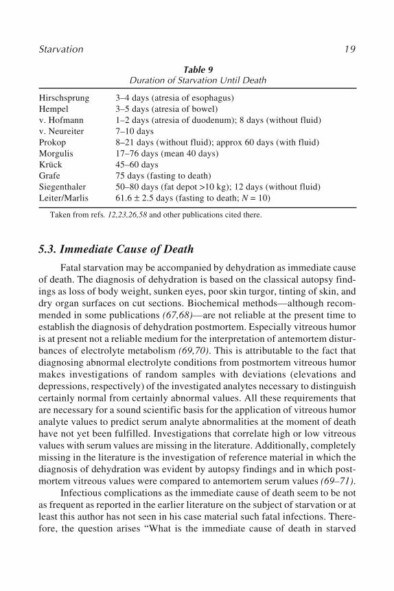
Published data concerning the duration of starvation until death refermainly to adults, acute withdrawal of food and liquid until death, or infantswith inborn abnormalities of the upper gastrointestinal tract (Table 9).According to observations in 10 young, previously healthy hunger strikers(mean age 25.6 ± 0.7 years) the survival period until death varied between 53to 73 days (mean 61 ± 2.5 days [59]). However, these data are of no use incases of chronic starvation especially in infants in which birth was prematureand failure to thrive is reported in the medical records.
5.2. Physical Condition Prior to Death
Medical records of the affected infants must be reviewed to determineevidence of the duration of malnutrition (body weight, height; Table 7). Themedical expert witness may be asked whether some time (days to weeks) priorto death, the undernutrition of the respective child was recognizable (e.g., bycaretakers, such as the parents or health care workers) and if so, the caretakerswould have been obliged to request medical advice and care. In such cases, itis recommended to extrapolate on the time interval prior to death by taking asa basis that complete withholding of food results in a loss of body weight ofabout 0.7 to 1% of total body weight per day. Using the Waterlow classifica-tion, the extrapolated body weight related to the ideal weight for height givesan impression of the severity of malnutrition. When withholding of food wasnot absolute, it can be assumed that the real body weight for the extrapolatedtime was lower in a respective case of chronic starvation.
Starvation 19
Table 9Duration of Starvation Until Death
Hirschsprung 3–4 days (atresia of esophagus)Hempel 3–5 days (atresia of bowel)v. Hofmann 1–2 days (atresia of duodenum); 8 days (without fluid)v. Neureiter 7–10 daysProkop 8–21 days (without fluid); approx 60 days (with fluid)Morgulis 17–76 days (mean 40 days)Krück 45–60 daysGrafe 75 days (fasting to death)Siegenthaler 50–80 days (fat depot >10 kg); 12 days (without fluid)Leiter/Marlis 61.6 ± 2.5 days (fasting to death; N = 10)
Taken from refs. 12,23,26,58 and other publications cited there.
5.3. Immediate Cause of Death
Fatal starvation may be accompanied by dehydration as immediate causeof death. The diagnosis of dehydration is based on the classical autopsy find-ings as loss of body weight, sunken eyes, poor skin turgor, tinting of skin, anddry organ surfaces on cut sections. Biochemical methods—although recom-mended in some publications (67,68)—are not reliable at the present time toestablish the diagnosis of dehydration postmortem. Especially vitreous humoris at present not a reliable medium for the interpretation of antemortem distur-bances of electrolyte metabolism (69,70). This is attributable to the fact thatdiagnosing abnormal electrolyte conditions from postmortem vitreous humormakes investigations of random samples with deviations (elevations anddepressions, respectively) of the investigated analytes necessary to distinguishcertainly normal from certainly abnormal values. All these requirements thatare necessary for a sound scientific basis for the application of vitreous humoranalyte values to predict serum analyte abnormalities at the moment of deathhave not yet been fulfilled. Investigations that correlate high or low vitreousvalues with serum values are missing in the literature. Additionally, completelymissing in the literature is the investigation of reference material in which thediagnosis of dehydration was evident by autopsy findings and in which post-mortem vitreous values were compared to antemortem serum values (69–71).
Infectious complications as the immediate cause of death seem to be notas frequent as reported in the earlier literature on the subject of starvation or atleast this author has not seen in his case material such fatal infections. There-fore, the question arises “What is the immediate cause of death in starved
Madea20
individuals?” Recent reports on sudden death and anorexia nervosa suggestventricular tachyarrhythmia or hypoglycemic coma as the immediate cause ofsudden death in starvation and anorexia nervosa (53–57). However, both con-ditions cannot be diagnosed postmortem.
Cases of sudden deaths of severely underweight and malnourished per-sons are already available in the earlier literature. In his book on malnutritionthat was published in 1947, Wolf-Eisner reported that prisoners in concentrationcamps suffering from severe malnutrition died very suddenly, e.g., while talk-ing to others (52). Reports on PEM indicate that marasmic infants—comparedto infants suffering from Kwashiorkor—are alert and conscious over a longperiod of time. The fact that death may be sudden and unexpected in severemarasmus and PEM has some medicolegal importance. Even in extremelymarasmic infants, the life-threatening condition may be misjudged or over-seen. However, the extreme emaciation is evident for everyone.
REFERENCES
1. Adelson L. Homicide by starvation. The nutritional variant of the battered child.JAMA 1963;186:458–460.
2. Adelson L. Pathology of Homicide. Springfield, IL: Charles C. Thomas, 1974.3. Campbell JAH. The morbid anatomy of infantile malnutrition in Cape Town. Arch
Dis Child 1956;31:310–314.4. Davis JH, Rao VJ, Valdes-Dapena M. A forensic approach to a starved child. J
Forensic Sci 1984;29:663–669.5. Ellerstein NS, Ostrov BE. Growth pattern in children hospitalized because of caloric-
deprivation failure to thrive. Am J Dis Child 1985;139:164–166.6. Fieguth A, Günther D, Kleemann WJ, Tröger HD. Lethal child neglect. Forensic Sci
Int 2002;130:8–12.7. von Harnack GA, Heimann G. Kinderheilkunde, 8th ed. Springer, Berlin, Heidel-
berg, New York, London, Paris, Tokyo, Hongkong, 1990.8. Helfer RE, Kempe CH. The battered child. The University of Chicago Press, Chi-
cago, London, 1968.9. Hughes EA, Stevens LH, Wilkinson AW. Some aspects of starvation in the newborn
baby. Arch Dis Child 1964;39:598–604.10. Listernick R, Christoffel K, Pace J, Chiaramonte J. Severe primary malnutrition in
US children. Am J Dis Child 1985;139:1157–1160.11. Madea B, Henßge C, Berghaus G. Fahrlässige Tötung eines Säuglings durch
Fehlernährung. Arch Kriminol 1992;189:33–38.12. Madea B, Michalk DV, Lignitz E. Verhungern infolge Kindesvernachlässigung.
Arch Kriminol 1994;194:29–38.13. Meade JL, Brissie RM. Infanticide by starvation: calculation of caloric deficit to
determine degree of deprivation. J Forensic Sci 1985;30:1263–1268.
Starvation 21
14. Mimasaka S, Funayama M, Adachi N, Nata M, Morita M. A fatal case of infantilescurvy. Int J Legal Med 2000;114:122–124.
15. Nishio H, Matusi K, Tsuji H, Tamura A, Suzuki K. Immunohistochemical study oftyrosine phosphorylation signalling in the involuted thymus. Forensic Sci Int2000;110:189–198.
16. Sarvesvaran E. Homicide by starvation. Am J Forens Med Pathol 1992;13:264–267.17. Schmidt P, Graß H, Madea B. Child homicide in Cologne (1985–94). Forensic Sci
Int 1996;79:131–144.18. Tanegashima A, Yamamoto H, Yada I, Fukunaga T. Estimation of stress in child
neglect from thymic involution. Forensic Sci Int 1999;102:173–180.19. Wehner F, Schieffer MC, Wehner HD. Percentile charts to determine the duration of
child abuse by chronic malnutrition. Forensic Sci Int 1999;102:173–180.20. Di Maio VJ, Di Maio DJ. Forensic Pathology. 2nd ed. CRC Press, Boca Raton,
London, New York, 2001.21. Gee D. Starvation and neglect. In Mant AK, ed. Taylor’s Principles and Practice of
Medical Jurisprudence. Churchill Livingstone, Edinburgh, London, Melbourne, NewYork, 1984, pp. 276–279.
22. Giese W, Hörstebrock R. Allgemeine Pathologie des exogenen quantitativenNahrungsmangels. In: Büchner F, Letterer E, Roulet F, eds. Handbuch derallgemeinen Pathologie, Vol. 11, Umwelt II, part I. Springer Verlag, Berlin,Göttingen, Heidelberg, 1962, pp. 446–591.
23. Madea B, Banaschak S. Verhungern. In: Brinkmann B, Madea B, eds. HandbuchGerichtliche Medizin, Vol. 1. Springer Verlag, Berlin, Heidelberg, New York, 2004,pp. 905–919.
24. Mueller B. Schädigungen und Tod infolge Nahrungsmangel. In: Mueller B, ed.Gerichtliche Medizin, Vol. 1, 2nd ed. Springer Verlag, Berlin, Heidelberg, NewYork, 1975, pp. 497–500.
25. von Neureiter F, Pietrusky F, Schütt E. Tod und Gesundheitsbeschädigung durchEntzug der Nahrung. In: von Neureiter F, Pietrusky F, Schütt E, eds. Handwörterbuchder Gerichtlichen Medizin und Naturwissenschaftlichen Kriminalistik. SpringerVerlag, Berlin, 1940, pp. 811,812.
26. Prokop O. Das Verhungern. In: Prokop O, ed. Forensische Medizin, VEB Verlag.Berlin, 1966, pp. 141–143.
27. Becker M. Chronische Gedeihstörungen im Säuglingsalter. In: Bachmann KD,Ewerbeck H, Kleihauer E, Rossi E, Stalder E, eds. Pädiatrie in Praxis und Klinik.,Vol. 1, 2nd ed. Gustav Fischer Verlag, Stuttgart, New York, 1989, pp. 545–552.
28. Behrmann RE, Vaughan VC, Nelson WE. Nelson’s Textbook of Pediatrics, 13th ed.W.B. Saunders Company, Philadelphia, London, Toronto, Montreal, Sidney, Tokyo,1987.
29. Bremer HJ. Protein-Energie-Malnutrition der Entwicklungsländer. In: Betke K,Künzer W, Schaub J, eds. Lehrbuch der Kinderheilkunde, 6th ed. Thieme Verlag,Stuttgart, New York, 1991, pp. 247–251.
30. World Health Organisation: Joint FAO/WHO Expert Committee on Nutrition, 8threport. Food fortification and protein-calorie-malnutrition. Tech Rep Ser Wld HlthOrg, No. 477 (WHO, Geneva, 1971).
Madea22
31. Gomez F, Galvan RR, Cravioto J, Frenk S. Malnutrition in infancy and childhoodwith special reference to Kwashiokor. Adv Pediatr 1955;7:131–169.
32. Suskind RM. Primary protein-energy malnutrition: clinical, biochemical and meta-bolic changes. In: Suskind RM, ed., Textbook of Pediatric Nutrition. Raven Press,New York, 1981, pp. 189–307.
33. Suskind RM, Varma RN. Assessment of nutritional status of children. Pediatrics inReview 1984;5:195–202.
34. Waterlow JC. Classification and definition of protein-caloric malnutrition. Br MedJ 1972;2:566–569.
35. Waterlow JC. Note on the assessment and classification of protein-energy malnutri-tion in children. Lancet 1973;2:87–89.
36. Waterlow JC, Buzina R, Keller W, Lane M, Nichaman MZ, Tanner JM. The presen-tation and use of height and weight data for comparing the nutritional status of groupsof children under the age of 10 years. Bull World Health Organ 1977;55:489–498.
37. Beaton GH. Nutritional needs during the first year of life. Pediatr Clin North Am1985;32:275–288.
38. Bürger M, Grosse-Brockhoff F. Energiestoffwechsel. In: Grosse-Brockhoff F, ed.Pathologische Physiologie. Springer Verlag, Berlin, Heidelberg, New York, 1969,pp. 688–697.
39. Ulmer HV. Ernährung. In: Schmidt RF, Thews G, ed. Physiologie des Menschen,22nd ed. Springer Verlag, Berlin, Heidelberg, New York, Tokyo, 1985, pp. 628–641.
40. Adelsberger L. Medical observations in Auschwitz concentration camp. Lancet1946;2:317–319.
41. Cahill GF. Starvation in man. New Engl J Med 1970;282:668–675.42. Chmelnizkij OK. Zur Rolle der Pathologen im belagerten Leningrad. Z allg Pathol
pathol Anat 1987;133:307–310.43. Giese W. Die Pathologie des Hungers. Allg Pathologie 1953;71(Pt II):98–100.44. Girgensohn H. Pathologische Anatomie der Gefangenschaftskrankheit mit
Bemerkungen zu ihrer Klinik und zur Frage der Spät- und Dauerschäden. DieMedizinische 1959;16:761–769.
45. Holle G. Über plötzliche Todesfälle bei schwerer Inanition. Z Ges Inn Med 1948;15/16:491–500.
46. Hottinger A, Gsell O, Uehlinger E, Salzmann C, Labhart A. Hungerkrankheit,Hungerödem, Hungertuberkulose. Benno Schwabe u. Co Verlag, Basel, 1948.
47. Leyton GB. Effects of slow starvation. Lancet 1946;2:73–79.48. Mollison PL. Observation of cases of starvation at Belsen. Br Med J 1946;1:4–8.49. Selberg W. Pathologische Anatomie der Unterernährung. Synopsis. 1948;1:23–50.50. Simpson K. Exposure to cold-starvation and neglect. In: Simpson K, ed. Modern
Trends in Forensic Medicine. Butterworth, London, 1953, pp. 116–132.51. Uehlinger E. Die pathologische Anatomie der Hungerkrankheit und des Hunger-
ödems. Helv Med Acta 1947;415:584–601.52. Wolff-Eisner A. Über Mangelerkrankungen auf Grund von Beobachtungen im
Konzentrationslager Theresienstadt. Lothar Sauer-Morhard Verlag, Würzburg, 1947.53. Isner JM, Roberts WC, Heymsfield SB, Yager J. Anorexia nervosa and sudden death.
Ann Intern Med 1985;102:49–52.
Starvation 23
54. Missliwetz J, Mortinger H. Tod durch Hypoglykämie nach Hungerzustand—Pathophysiologie versus Morphologie. Beitr Gerichtl Med 1992;50:319–323.
55. Ratcliffe PJ, Bevan JS. Severe hypoglycaemia and sudden death in anorexia nervosa.Psychol Med 1985;15:679–681.
56. Rich LM, Caine MR, Findling JW, Shaker JL. Hypoglycaemic coma in anorexianervosa. Arch Intern Med 1990;150:894,895.
57. Smith J. Hypoglycaemic coma associated with anorexia nervosa. Aust NZ J Psychia-try 1988;22:448–453.
58. Kanzow U. Beobachtung während einer 53-tägigen Hungerperiode an einemHungerkünstler. Dt Arch Klin Med 1951;198:698–705.
59. Leiter LA, Marliss EB. Survival during fasting may depend on fat as well as proteinstores. JAMA 1982;248:2306,2307.
60. Berwick DM. Nonorganic failure-to-thrive. Pediatrics in Review 1980;1:265–270.61. Holzel A. Sugar malabsorption due to deficiencies of disaccaridase activities and
monosaccharide transport. Arch Dis Child 1967;42:341–352.62. Pipes PL. Nutrition in infancy and childhood, 2nd ed. The C.V. Mosby Company, St.
Louis, Toronto, London, 1981.63. Janssen W. Forensische Histologie. Verlag Max Schmidt Römhild, Lübeck, 1977.64. Schocken DD, Holloway JD, Powers PS. Weight loss and the heart. Arch Intern Med
1989;149:877–881.65. Roessle R, Roulet F. Mass und Zahl in der Pathologie. Verlag von Julius Springer,
Berlin, 1932.66. Madea B, Banaschak S. Remarks on: “Percentile charts to determine the duration of
child abuse by chronic malnutrition.” Forensic Sci Int 1999;105:191,192.67. Coe JI. Postmortem chemistries on human vitreous humor. Am J Clin Pathol
1969;51:741–750.68. Coe JI. Some further thoughts and observations on postmortem chemistries. Foren-
sic Sci Gazette 1973;4:2–5.69. Madea B. Zur Postmortalen Diagnostik von Störungen des Wasser-Elektrolyt-
Haushaltes. Rechtsmedizin 1996;6:141–146.70. Madea B. Zur Postmortalen Diagnostik der Hypertonen Dehydratation. Paper pre-
sented at the 16th Spring Meeting of the German Society of Forensic Medicine(Northern Group), Kiel, May 2003.
71. Madea B, Herrmann N. “Normal values” in vitreous humor and on dysregulationswhich can be diagnosed postmortem. In: Jacob B, Bonte W, ed. Advances in ForensicSciences, Vol. 4., Forensic Criminalistics II. Verlag Dr. Köster, Düsseldorf, 1995,pp. 49–61.