Discovery of N-Hydroxyindole-Based Inhibitors of Human Lactate Dehydrogenase Isoform A (LDH-A) as...
14
rXXXX American Chemical Society A dx.doi.org/10.1021/jm101007q | J. Med. Chem. XXXX, XXX, 000–000 ARTICLE pubs.acs.org/jmc Discovery of N-Hydroxyindole-Based Inhibitors of Human Lactate Dehydrogenase Isoform A (LDH-A) as Starvation Agents against Cancer Cells Carlotta Granchi, † Sarabindu Roy, † Chiara Giacomelli, † Marco Macchia, † Tiziano Tuccinardi, † Adriano Martinelli, † Mario Lanza, ‡ Laura Betti, ‡ Gino Giannaccini, ‡ Antonio Lucacchini, ‡ Nicola Funel, § Leticia G. Le on, ||,^ Elisa Giovannetti, ^ Godefridus J. Peters, ^ Rahul Palchaudhuri, # Emilia C. Calvaresi, # Paul J. Hergenrother, # and Filippo Minutolo* ,† † Dipartimento di Scienze Farmaceutiche and ‡ Dipartimento di Psichiatria, Neurobiologia, Farmacologia, e Biotecnologie, Universit a di Pisa, Via Bonanno 6, 56126 Pisa, Italy § Dipartimento di Chirurgia (Anatomia Patologica), Universit a di Pisa, Via Roma 57, 56126 Pisa, Italy ) Institudo Universitario de Bio-Organica “Antonio Gonzales”, Universidad de La Laguna, Avda. Astrosico F. S anchez 2, La Laguna, 38206-Tenerife, Spain ^ Department of Medical Oncology, VU University Medical Center, De Boelelaan 1117, 1081 HV Amsterdam, The Netherlands # Department of Chemistry, University of Illinois, 600 S. Mathews Avenue, Urbana, Illinois 61801, United States b S Supporting Information ABSTRACT: Highly invasive tumor cells are characterized by a metabolic switch, known as the Warburg eect, from “normal” oxidative phosphorylation to increased glycolysis even under suciently oxygenated conditions. This dependence on glyco- lysis also confers a growth advantage to cells present in hypoxic regions of the tumor. One of the key enzymes involved in glycolysis, the muscle isoform of lactate dehydrogenase (LDH- A), is overexpressed by metastatic cancer cells and is linked to the vitality of tumors in hypoxia. This enzyme may be con- sidered as a potential target for new anticancer agents, since its inhibition cuts cancer energetic and anabolic supply, thus reducing the metastatic and invasive potential of cancer cells. We have discovered new and ecient N-hydroxyindole-based inhibitors of LDH-A, which are isoform-selective (over LDH-B) and competitive with both the substrate (pyruvate) and the cofactor (NADH). The antiproliferative activity of these compounds was conrmed on a series of cancer cell lines, and they proved to be particularly eective under hypoxic conditions. Moreover, NMR experiments showed that these compounds are able to reduce the glucose-to- lactate conversion inside the cell. ’ INTRODUCTION Unlike normal cells, most invasive tumor phenotypes show a metabolic switch, named the “Warburg eect”, 1 from oxidative phosphorylation to an increased rate of glycolysis. This switch ensures a sucient energy supply from glucose and thus high vitality, even in hypoxic environments. 2,3 This unusual metabolic trait is particularly advantageous in solid tumors, which are gen- erally signicantly less oxygenated than normal tissues. Once their dimension reaches certain values (usually >1 cm 3 ), solid tumors present hypoxic regions surrounding necrotic areas. Hypoxic cancers pose a great challenge to oncologists all over the world because they are especially aggressive, metastatic, and resistant to (1) radiotherapy, mainly because of scarce formation of oxygen- dependent cytotoxic radicals upon irradiation, and (2) che- motherapy because of limited blood supply and also because they have a slow rate of proliferation, whereas most of the currently available therapies target rapidly dividing cells. More- over, hypoxia increases the cancer stem cell fraction and pro- motes acquisition of a stemlike state, thus increasing chances of cancer relapse. 4 This metabolic situation is accompanied by (1) higher consumption of glucose, due to the lower eciency in energy production by glycolysis (Figure 1), and (2) increased extracellular acidosis because of the high production of lactic acid and other acidic species. It may be possible, however, to take advantage of this peculiar metabolic feature of cancer cells for selective anticancer therapy. 5 There are only a few molecules in preclinincal studies or clinical trials that are known to exploit, or interfere with, the increased glycolytic process of invasive tumors. 6,7 Lonidamine, Received: August 4, 2010
-
Upload
independent -
Category
Documents
-
view
1 -
download
0
Transcript of Discovery of N-Hydroxyindole-Based Inhibitors of Human Lactate Dehydrogenase Isoform A (LDH-A) as...
rXXXX American Chemical Society A dx.doi.org/10.1021/jm101007q | J. Med. Chem. XXXX, XXX, 000–000
ARTICLE
pubs.acs.org/jmc
Discovery of N-Hydroxyindole-Based Inhibitors of Human LactateDehydrogenase Isoform A (LDH-A) as Starvation Agents againstCancer CellsCarlotta Granchi,† Sarabindu Roy,† Chiara Giacomelli,† Marco Macchia,† Tiziano Tuccinardi,†
Adriano Martinelli,† Mario Lanza,‡ Laura Betti,‡ Gino Giannaccini,‡ Antonio Lucacchini,‡ Nicola Funel,§
Leticia G. Le!on,||,^ Elisa Giovannetti,^ Godefridus J. Peters,^ Rahul Palchaudhuri,# Emilia C. Calvaresi,#
Paul J. Hergenrother,# and Filippo Minutolo*,†
†Dipartimento di Scienze Farmaceutiche and ‡Dipartimento di Psichiatria, Neurobiologia, Farmacologia, e Biotecnologie,Universit"a di Pisa, Via Bonanno 6, 56126 Pisa, Italy§Dipartimento di Chirurgia (Anatomia Patologica), Universit"a di Pisa, Via Roma 57, 56126 Pisa, Italy
)Institudo Universitario de Bio-Organica “Antonio Gonzales”, Universidad de La Laguna, Avda. Astro!sico F. S!anchez 2, La Laguna,38206-Tenerife, Spain^Department of Medical Oncology, VU University Medical Center, De Boelelaan 1117, 1081 HV Amsterdam, The Netherlands#Department of Chemistry, University of Illinois, 600 S. Mathews Avenue, Urbana, Illinois 61801, United States
bS Supporting Information
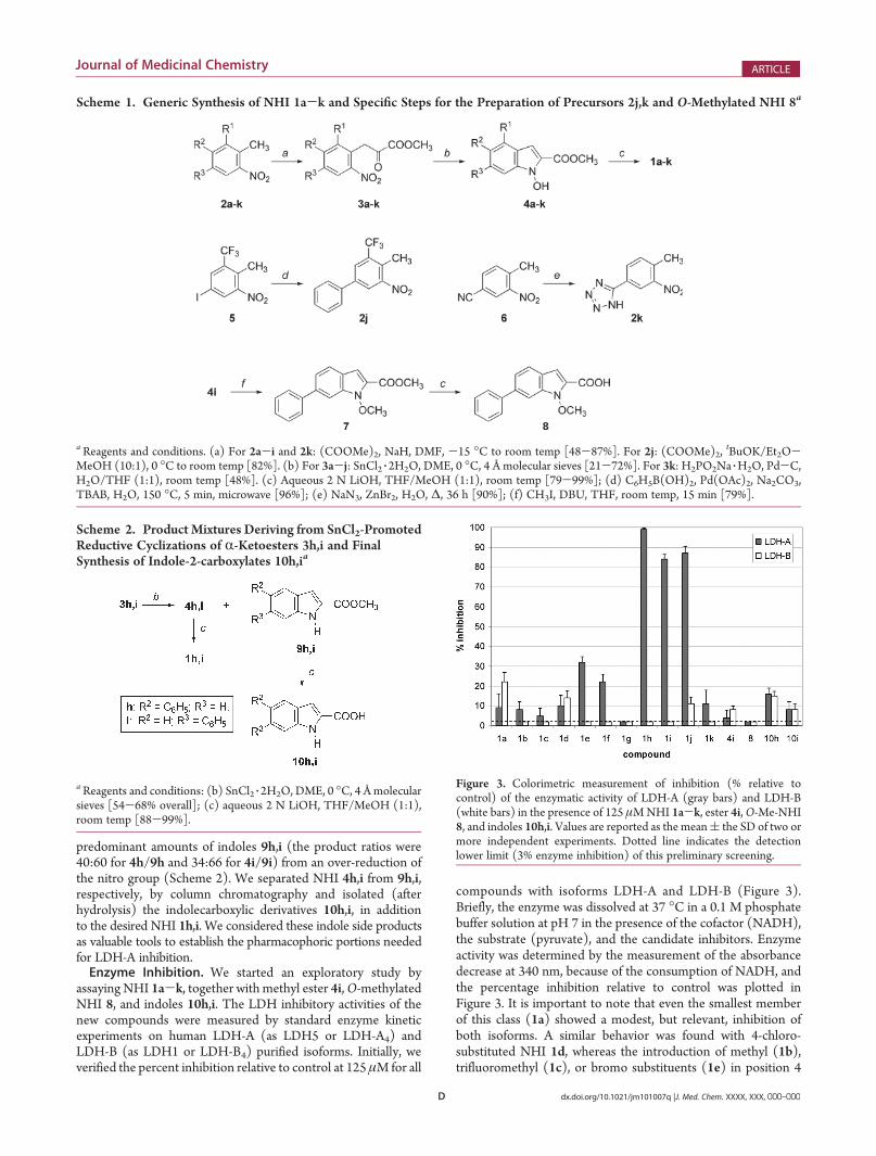
ABSTRACT: Highly invasive tumor cells are characterized byametabolic switch, known as theWarburg e"ect, from “normal”oxidative phosphorylation to increased glycolysis even undersu#ciently oxygenated conditions. This dependence on glyco-lysis also confers a growth advantage to cells present in hypoxicregions of the tumor. One of the key enzymes involved inglycolysis, the muscle isoform of lactate dehydrogenase (LDH-A), is overexpressed by metastatic cancer cells and is linked tothe vitality of tumors in hypoxia. This enzyme may be con-sidered as a potential target for new anticancer agents, since its inhibition cuts cancer energetic and anabolic supply, thus reducingthe metastatic and invasive potential of cancer cells. We have discovered new and e#cient N-hydroxyindole-based inhibitors ofLDH-A, which are isoform-selective (over LDH-B) and competitive with both the substrate (pyruvate) and the cofactor (NADH).The antiproliferative activity of these compounds was con!rmed on a series of cancer cell lines, and they proved to be particularlye"ective under hypoxic conditions. Moreover, NMR experiments showed that these compounds are able to reduce the glucose-to-lactate conversion inside the cell.
’ INTRODUCTION
Unlike normal cells, most invasive tumor phenotypes show ametabolic switch, named the “Warburg e"ect”,1 from oxidativephosphorylation to an increased rate of glycolysis. This switchensures a su#cient energy supply from glucose and thus highvitality, even in hypoxic environments.2,3 This unusual metabolictrait is particularly advantageous in solid tumors, which are gen-erally signi!cantly less oxygenated than normal tissues. Once theirdimension reaches certain values (usually >1 cm3), solid tumorspresent hypoxic regions surrounding necrotic areas. Hypoxiccancers pose a great challenge to oncologists all over the worldbecause they are especially aggressive, metastatic, and resistant to(1) radiotherapy, mainly because of scarce formation of oxygen-dependent cytotoxic radicals upon irradiation, and (2) che-motherapy because of limited blood supply and also becausethey have a slow rate of proliferation, whereas most of the
currently available therapies target rapidly dividing cells. More-over, hypoxia increases the cancer stem cell fraction and pro-motes acquisition of a stemlike state, thus increasing chances ofcancer relapse.4 This metabolic situation is accompanied by (1)higher consumption of glucose, due to the lower e#ciency inenergy production by glycolysis (Figure 1), and (2) increasedextracellular acidosis because of the high production of lactic acidand other acidic species. It may be possible, however, to takeadvantage of this peculiar metabolic feature of cancer cells forselective anticancer therapy.5
There are only a few molecules in preclinincal studies orclinical trials that are known to exploit, or interfere with, theincreased glycolytic process of invasive tumors.6,7 Lonidamine,
Received: August 4, 2010

B dx.doi.org/10.1021/jm101007q |J. Med. Chem. XXXX, XXX, 000–000
Journal of Medicinal Chemistry ARTICLE
an inhibitor of hexokinase (HK), the enzyme that catalyzes thephosphorylation of the 6-position of glucose, thus starting theglycolytic process, has completed a phase 3 trial, but its e#cacywas undermined by pancreatic and hepatic toxicity.8 Among HKinhibitors, 2-deoxyglucose (2-DG) showed promising results in aphase 1 trial, but its action on hypoxic tumors was not satis-factory,9 and 3-bromopyruvate induces apoptosis in some carci-nomas, but no clinical tests are yet available.10 Dichloroacetate(DCA) is a pyruvate dehydrogenase kinase (PDK) inhibitorunder phase 1 clinical study whose action restores the normaloxidative demolition of pyruvate by reactivating pyruvate dehy-drogenase (PDH) and thus indirectly diverting glycolysis,11
although some concerns stem from recent evidence that PDK-knockout has no e"ect on the proliferation and survival ofhypoxic tumor cells.12 Increased glucose uptake occurring ininvasive tumors was clinically exploited by a prodrug, glufosfa-mide, which has completed clinical trials on several tumors andis composed of an alkylating moiety linked to a !-D-glucoseunit that takes advantage of the transmembrane glucosetransport system, which is greatly increased in malignantphenotypes.13 Finally, the suitability of the fetal isoform ofpyruvate kinase (PK-M2), overexpressed by invasive tumors,14
as a potential target for the development of isoform-speci!cinhibitors15 is currently under debate because its inhibition maylead to paradoxical e"ects on cancer growth.16 Indeed, a recentreport describes the development of PK-M2 activators based on a
diarylsulfonamide sca"old, which were intended as prospectiveantiproliferative agents.17
Lactate dehydrogenase (LDH) constitutes amajor checkpointfor the switch from aerobic to anaerobic glycolysis by catalyzingthe reduction of pyruvate into lactate (Figure 1). In humans,LDH is a tetrameric enzyme that may exist in !ve isoforms(hLDH1-5), mostly located in the cytosol. There are only twotypes of subunits: LDH-A (or LDH-M, muscle) and LDH-B(or LDH-H, heart). The !ve isoforms are made up of the variouspossible combinations of these two subunits in the followingway: LDH1= LDH-B4, LDH2 = LDH-AB3, LDH3 = LDH-A2B2,LDH4 = LDH-A3B, LDH5 = LDH-A4. hLDH1 is mostlyrepresented in the heart, whereas hLDH5 is prevalent in liverand skeletal muscle. hLDH5 has been found to be overexpressedin highly invasive and hypoxic carcinomas, and it was clearlyassociated with hypoxia inducible factor HIF-1R.18 Serum andplasma hLDH5 levels are used as tumor biomarkers, and theselevels are not necessarily correlated to nonspeci!c cellular damage.Rather they are caused by overexpression induced by malignanttumor phenotypes.19 An ampli!ed expression of this gene,measured as LDH-A subunit increased production, was foundin several tumor lines.20 This, together with the overexpression ofglucose transporter GLUT1 due to oxygen deprivation,20 furtherpromotes glycolysis. Furthermore, lactate production contri-butes to extracellular acidosis, thus supporting tumor invasivityand exerting an immunosuppressive e"ect.21
LDH-A (for the sake of clarity, from now on we will refer tothis monomeric subunit also for the fully functional tetramerichLDH5) was recently acknowledged as one of the most promis-ing tumor targets.22 In fact, repression of its expression byshRNA cuts the main energy production sequence in hypoxictumors, as shown by a reduced invasiveness in metastatic celllines. More recently, subclones of the breast cancer cell lineMDA-MB-435 that were resistant to paclitaxel were shown to beresensitized upon either down-regulation of LDH-A by siRNA orafter chemical inhibition of LDH-A activity by oxamate.23
Furthermore, inhibition of LDH-A is unlikely to give rise to majorside e"ects in humans, since hereditary LDH-A de!ciency causesmyoglobinuria only after intense anaerobic exercise, whereas itdoes not provoke any symptoms under ordinary circumstan-ces.24 Therefore, compounds able to inhibit LDH-A enzymaticactivity may constitute safe agents able to interfere with tumorgrowth and invasiveness on many fronts. For these reasons,we have decided to pursue “druglike” small molecules able toinhibit hLDH5.
Few LDH inhibitors are reported in the literature, and most ofthem were developed against the isoform present in P. falciparum(pfLDH), as this enzyme was conceived as a valid target for thetreatment of malaria. The inhibitory activity of these compoundsagainst human LDH isoforms was reported as a possible sidee"ect, and in many cases the inhibition levels were extremelylow.25 Oxamate (OXM, Figure 2), still considered as the LDH-Areference inhibitor, is competitive with the pyruvate substrate,but it is nonselective and not very potent, with aKi of 138 "M(vspyruvate).26 Another known LDH-A inhibitor is gossypol, anatural polyphenol dialdehyde extracted from cotton seeds,which is also highly cytotoxic and promiscuous.27 Some verysmall azoles possessing vicinal OH and COOH groups, such as3-hydroxyisoxazole-4-carboxylic acid (HICA, Figure 2) and4-hydroxy-1,2,5-thiadiazole-3-carboxylic acid (HTCA, Figure 2)showed IC50 values of 54 and 10 "M, respectively, on LDH-A,and the hydroxyl-carboxylic substitution pattern proved to be
Figure 1. Fate of pyruvate during glycolysis under aerobic and anaero-bic conditions: role of LDH.

C dx.doi.org/10.1021/jm101007q |J. Med. Chem. XXXX, XXX, 000–000
Journal of Medicinal Chemistry ARTICLE
essential for their inhibitory activity. Unfortunately, these com-pounds have such simple structures that their selective action isnot guaranteed. Thiadiazole derivative HTCA cannot be func-tionalized further, and any substituent placed in the only freeposition of isoxazole HICA has a"orded inactive compounds.28
Thus, these azoles are not ideal candidates for further optimiza-tion. Finally, derivatives of 8-deoxyhemigossylic (2,3-dihydrox-ynaphtalen-1-carboxylic) acid, originally designed as antimalarialagents,29 have furnished some of the most e"ective and selectiveLDH-A inhibitors. One of these compounds, FX11 (Figure 2), isa LDH-A inhibitor competitive with the reduced nicotinamideadenine dinucleotide (NADH) cofactor, displaying a Ki of 8 "Mon LDH-A and a >10-fold selectivity over the other isoform LDH-B, which was very recently shown to inhibit tumor progression byinducing an oxidative stress to the cancer cells.30 Although FX11contains a potentially redox-active catecholmoiety thatmaymake itunsuitable as a drug, it is an important proof-of-concept for LDH-Ainhibition as a tractable anticancer strategy.
The X-ray crystal structure of the LDH-A subunit of hLDH531
shows that the active site is located in a rather deep positionwithin the protein and accessibility to it is narrow. This cavitynormally hosts both the substrate (pyruvate) and the cofactor(NADH). Overall, it is quite polar and rich in cationic residues(arginines). This would explain why the inhibitors so far discovered(Figure 2) have carboxylates, closely associated with a hydroxyl ora carbonyl oxygen atom. This information is consistent with thenatural LDH substrates, anR-hydroxy acid (lactate) or anR-ketoacid (pyruvate), that are thus mimicked by the inhibitors. Wehave considered these pharmacophore requirements and the!rst of a series of hydroxycarboxylates positioned on variousaromatic sca"olds never explored before in this !eld, such as anunusual class of heterocyclic derivatives, the N-hydroxyindoles(NHI),32 bearing a carboxylic acid group in the 2-position(Chart 1). The NHI sca"old has been largely neglected in thedesign of biorelevant molecules, possibly because of the relativelack of synthetic methods for its assembly. On the contrary,numerous isolated natural products have been discovered tocontain NHI portions, such as nocathiacin I, a stable antibiotic,33
and the most signi!cant NHI preparations are essentially repre-sented by a reduction of the indole system to 2,3-dihydroindole,followed by an oxidation step (Na2WO4/H2O2) or by a SnCl2-promoted reduction of carbonyl-substituted nitroarenes.34,35
Compounds 1a-k were thus designed to retain the hydro-xyl-carboxylate motifs of other LDH-A inhibitors (Figure 2),with the di"erence that their N-OH group is slightly lessacidic than typical phenol groups.36 A series of exploratorysimple substitution patterns (halogens, phenyl, tri$uorometh-yl, and tetrazole) were selected to determine if it was possible
to achieve satisfactory levels of LDH-A inhibition within thischemical class.
’RESULTS AND DISCUSSION
Synthetic Chemistry. The synthetic pathway generally fol-lowed for the preparation of NHI 1a-k is shown in Scheme 1.The first step involved a reaction of nitrotoluenes 2a-k withsodium hydride and an excess of dimethyl oxalate in DMF at -15 !C to room temperature. These conditions worked nicely forall the nitrotoluene derivatives with the exception of 2j, whichwas instead treated with potassium tert-butoxide in diethyl etherand the minimum amount of methanol needed to dissolve thesubstrate. In this manner the condensation of the resulting anionwith dimethyl oxalate efficiently afforded ketoester 3j. TheresultingR-ketoesters 3a-jwere then treated with SnCl2 3 2H2Oin DME at 0 !C in the presence of 4 Åmolecular sieves.35 Inmostcases, these conditions promoted the desired reductive cycliza-tion to build the NHI scaffold, whereas in two cases (3h,i)mixtures with the over-reduced indole (9h,i, Scheme 2) deriva-tives were obtained, and in one case (tetrazole 3k) only a com-plex mixture of products was produced. In this latter case, wechanged the condition to a combination of sodium hypopho-sphite and catalytic palladium over charcoal37 and successfullyobtained the tetrazole-substituted NHI 4k. Subsequent hydro-lysis with aqueous 2 N LiOH in a THF/MeOHmixture affordedthe final products 1a-k. Noncommercially available precursors2j and 2k were respectively prepared by a Pd-catalyzed cross-coupling of 5-iodo-2-methyl-3-nitrobenzotrifluoride 538 withphenylboronic acid under ligand-free conditions in a microwavereactor39 and by a zinc-promoted addition of sodium azide40 tobenzonitrile 6. An example of O-methylated NHI (8) was alsosynthesized to verify the importance of the free OH group in theenzyme inhibition assays. For this purpose, NHI 4i was treatedwith iodomethane and DBU in THF at room temperature to giveester 7, which was eventually hydrolyzed to compound 8.As mentioned, during the reductive cyclization step of phenyl-
substituted ketoesters 3h,i to NHI 4h,i, we actually obtained
Figure 2. Structures of some of the most signi!cant LDH-A inhibitors.
Chart 1. N-Hydroxyindoles (NHI) Designed as LDH-AInhibitors (1a-k)
D dx.doi.org/10.1021/jm101007q |J. Med. Chem. XXXX, XXX, 000–000
Journal of Medicinal Chemistry ARTICLE
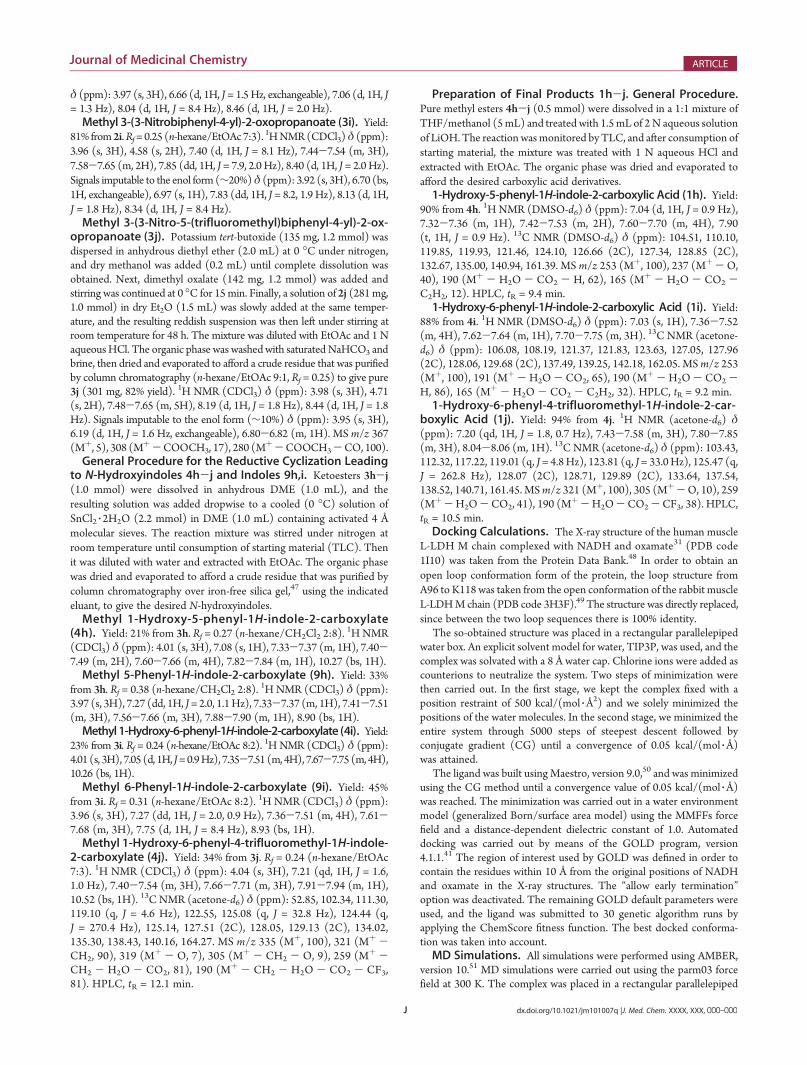
predominant amounts of indoles 9h,i (the product ratios were40:60 for 4h/9h and 34:66 for 4i/9i) from an over-reduction ofthe nitro group (Scheme 2). We separated NHI 4h,i from 9h,i,respectively, by column chromatography and isolated (afterhydrolysis) the indolecarboxylic derivatives 10h,i, in additionto the desired NHI 1h,i. We considered these indole side productsas valuable tools to establish the pharmacophoric portions neededfor LDH-A inhibition.Enzyme Inhibition. We started an exploratory study by
assaying NHI 1a-k, together with methyl ester 4i,O-methylatedNHI 8, and indoles 10h,i. The LDH inhibitory activities of thenew compounds were measured by standard enzyme kineticexperiments on human LDH-A (as LDH5 or LDH-A4) andLDH-B (as LDH1 or LDH-B4) purified isoforms. Initially, weverified the percent inhibition relative to control at 125 "M for all
compounds with isoforms LDH-A and LDH-B (Figure 3).Briefly, the enzyme was dissolved at 37 !C in a 0.1 M phosphatebuffer solution at pH 7 in the presence of the cofactor (NADH),the substrate (pyruvate), and the candidate inhibitors. Enzymeactivity was determined by the measurement of the absorbancedecrease at 340 nm, because of the consumption of NADH, andthe percentage inhibition relative to control was plotted inFigure 3. It is important to note that even the smallest memberof this class (1a) showed a modest, but relevant, inhibition ofboth isoforms. A similar behavior was found with 4-chloro-substituted NHI 1d, whereas the introduction of methyl (1b),trifluoromethyl (1c), or bromo substituents (1e) in position 4
Scheme 1. Generic Synthesis of NHI 1a-k and Speci!c Steps for the Preparation of Precursors 2j,k and O-Methylated NHI 8a
aReagents and conditions. (a) For 2a-i and 2k: (COOMe)2, NaH, DMF, -15 !C to room temp [48-87%]. For 2j: (COOMe)2,tBuOK/Et2O-
MeOH (10:1), 0 !C to room temp [82%]. (b) For 3a-j: SnCl2 3 2H2O, DME, 0 !C, 4 Å molecular sieves [21-72%]. For 3k: H2PO2Na 3H2O, Pd-C,H2O/THF (1:1), room temp [48%]. (c) Aqueous 2 N LiOH, THF/MeOH (1:1), room temp [79-99%]; (d) C6H5B(OH)2, Pd(OAc)2, Na2CO3,TBAB, H2O, 150 !C, 5 min, microwave [96%]; (e) NaN3, ZnBr2, H2O, !, 36 h [90%]; (f) CH3I, DBU, THF, room temp, 15 min [79%].
Scheme 2. Product Mixtures Deriving from SnCl2-PromotedReductive Cyclizations of R-Ketoesters 3h,i and FinalSynthesis of Indole-2-carboxylates 10h,ia
aReagents and conditions: (b) SnCl2 3 2H2O, DME, 0 !C, 4 Åmolecularsieves [54-68% overall]; (c) aqueous 2 N LiOH, THF/MeOH (1:1),room temp [88-99%].
Figure 3. Colorimetric measurement of inhibition (% relative tocontrol) of the enzymatic activity of LDH-A (gray bars) and LDH-B(white bars) in the presence of 125 "MNHI 1a-k, ester 4i,O-Me-NHI8, and indoles 10h,i. Values are reported as the mean( the SD of two ormore independent experiments. Dotted line indicates the detectionlower limit (3% enzyme inhibition) of this preliminary screening.

E dx.doi.org/10.1021/jm101007q |J. Med. Chem. XXXX, XXX, 000–000
Journal of Medicinal Chemistry ARTICLE
caused a drop in the inhibition of LDH-B, whereas the LDH-Ainhibition was in some cases increased, as shown by the 32%inhibition of 4-bromo-substituted NHI 1e. When the bromineatom was shifted to the 6 position, the resulting compound (1f)proved to be less active than its 4-substituted counterpart 1e.Encouraged by these preliminary results obtained with extremelysimplemodifications of the NHI scaffold, we introduced a phenylgroup in the 4, 5, and 6 positions of the central ring. In spite of thepoor results obtained with 4-phenyl-substituted derivative 1g, wewere pleased to find a dramatic increase of the LDH-A inhibitionlevel when the phenyl group was present in position 5 (1h) or inposition 6 (1i), with a 99% and a 84% inhibition, respectively.Both compounds showed practically no detectable inhibition ofthe other isoform (<3%), thus revealing a good level of selec-tivity. The presence of a 4-CF3 and a 6-phenyl in compound 1jcaused a notable 87% LDH-A inhibition at 125 "M, although aminimal residual activity on LDH-B (11% at the same con-centration) could be detected. The replacement of the 6-phenylgroup with a COOH-mimicking heteroaryl portion, such as thetetrazole, caused a dramatic decrease in the inhibitory potency ofthe resulting compound (1k) with a poor 11% inhibition on LDH-A. Methyl ester 4i gave very modest inhibition of both LDH-A(4%) and LDH-B (8%). O-Methylated NHI 8 was completelyinactive on both isoforms, whereas indoles 10h,i proved to bemodest nonselective inhibitors of both LDH-A and LDH-B.These results con!rm that when the “N-OH/COOH” phar-
macophoric motif is modi!ed, the enzyme inhibition potency isnegatively a"ected and that compounds possessing phenyl rings,either at the 5 (1h) or at the 6 position (1i,j), are the most potentand selective inhibitors of LDH-A. On the basis of this informa-tion, these compounds (1h-j) were selected for a completeenzyme kinetic analysis to verify their type of inhibition versusboth the cofactor and the substrate.We !rst evaluated the apparent Michaelis-Menten constants
(Km) of NADH and pyruvate for LDH-A, measured from Line-weaver-Burk plots. Then we evaluated the apparent Km
0 in thepresence of inhibitors 1h-j (concentration range of 25-100 "M).From the values of Km
0 so obtained, Ki values for each singleinhibitor were determined using double-reciprocal Lineweaver-Burk plots (Figure 4), and their values are reported in Table 1.Compounds 1h-j were all found to be competitive inhibitors
of LDH-A with respect to both NADH and pyruvate (Figure 4)in the conversion of pyruvate to lactate catalyzed by this enzyme,whereby NADH is converted to NAD!. In fact, the Vmax valueproved to be independent from the concentration of the inhi-bitor, a fact di"erent from the Km
0 values, which were insteaddependent on concentrations. These compounds were shown tobe potent inhibitors of LDH-A with Ki values reaching the lowmicromolar range (Table 1) with inhibitor 1j (Ki = 8.9 "M vsNADH and 4.7 "M vs pyruvate). Isoform selectivity was con-!rmed by the observation that inhibition of the other isoformLDH-Bwas either negligible (entries 1 and2) or very low (entry 3).Molecular Modeling. In order to further understand and
characterize the interaction of these compounds with the LDH-Aactive site, the most promising ligand (1j) was analyzed bydocking modeling studies into an open loop conformation of theenzyme. The compound was docked into LDH-A using theGOLD program,41 and the best pose was used as starting geo-metry for molecular dynamics (MD) calculations. MD simula-tion for 10 ns with explicit water was carried out. In the first 1.2 nsall the R carbons of the protein were blocked with a harmonicforce constant, which decreased during these 1.2 ns from 10 to
1 kcal/(mol 3Å2), while in the last 7.8 ns, no constraints were
applied. The system reached an apparent equilibrium after about
Figure 4. Lineweaver-Burk plots determined from triplicate experi-ments with inhibitors 1h-j at 25 "M using average activities: competi-tion experiments with NADH (top) and pyruvate (bottom).
Table 1. LDH Inhibition Data (Ki for LDH-A, % Inhibition at125 !M for LDH-B) Obtained with Compounds 1h-ja
LDH-A (Ki, "M)
entry compd [NADH]b [pyruvate]cLDH-B (% at 125 "M)
[NADH]b
1 1h 10.4 ( 1.5 15.7 ( 1.5 <3%
2 1i 19.8 ( 2.2 35.4 ( 3.4 <3%
3 1j 8.9 ( 1.3 4.7 ( 0.5 11% ( 3a Ki determination and percent of inhibition were performed in presenceof compounds 1h-j as described in the Experimental Section. Valuesare reported as the mean ( SD of three or more independent experi-ments. b Saturating concentration (2 mM) of sodium pyruvate andcompetitive increasing concentrations (12.5-150 "M) of NADH.c Saturating concentration (200 "M) of NADH and competitive in-creasing concentrations (25 "M to 1.0 mM) of sodium pyruvate.

F dx.doi.org/10.1021/jm101007q |J. Med. Chem. XXXX, XXX, 000–000
Journal of Medicinal Chemistry ARTICLE
0.5 ns of MD, since the total energy for the residual nanosec-onds remained constant (Figure S1 in Supporting Information).Analyzing the root-mean-square deviation (rmsd) from theinitial model of the R carbons of the proteins, we observed thatafter an initial increase, the rmsd remained approximately con-stant around 1.5 Å during the last 5.5 ns (Figure S1 in SupportingInformation).Figure 5 shows the minimized structures of the average of the
last 5.5 ns of the MD simulation. In the proposed model thecarboxylic group of compound 1j shows a strong interaction withR169 and T248 (see Figure 5A), whereas the N-hydroxy groupshows an H-bond interaction with the nitrogen backbone ofT248 and a water molecule that mediates the interaction of 1jwith the catalytic H193 (see Figure 5A and Figure 5B). An impor-tant role of residues R169, T248, and H193 for the ligand inter-actions has already been highlighted by the analysis of the X-raystructure of the LDHcomplexedwith the natural substrate pyruvateand NADH (Figure 6). In fact, this structure con!rms the inter-actions between the pyruvate carboxylate group with the arginineand threonine residues of the enzyme, as well as that occurringbetween the carbonyl oxygen atom of the substrate and the above-mentioned histidine amino acid (compare Figures 5 and 6).The H-bond analysis of the last 5.5 ns of the MD simulation
seems to con!rm the interactions described above. In particular,as shown in Table S1 (Supporting Information), it is interestingto note that the interaction between the water molecule (boundto H193) and the N-hydroxy group is highly conserved during
MD, con!rming the important role of this N-hydroxy substitu-ent. Furthermore, the analysis of the X-ray structure of Plasmo-dium falciparum isoform of lactate dehydrogenase complexedwith 3,7-dihydroxy-2-naphthoic acid42 revealed the presence of awater molecule able to mediate a H-bond between an OH groupof this legend and the catalytic H193 of the enzyme, thussupporting the possible presence of this water molecule also inthe human LDH-A form, as we have found with our calculations.As for the 4-(tri$uoromethyl)indole central sca"old, it is placedin a cleft mainly delimited by H193, G194, A238, V241, I242,T248. Moreover, N138 is placed in proximity to the tri$uoro-methyl substituent in our model. According to our calculations,the 6-phenyl group is directed toward the entrance of the bindingsite cavity and shows lipophilic interactions with I242 and Y247.Overall, this molecular portion seems to partially overlap with theregion occupied by the cofactor NADH in the human LDH-AX-ray structure.31
Finally, the results obtained by this docking analysis, showingthat compound 1jmay occupy the whole substrate pocket and, inpart, the cofactor pocket of LDH-A, are in good agreement withthe experimental enzyme inhibition data, which indicate thatinhibitor 1j and its analogues 1i,h are competitive with bothpyruvate and NADH.Cell-Based Reduction of Lactate Production. The ability of
inhibitors 1h-j to reduce the cellular production of lactate wasdetermined by 13CNMR spectroscopy studies on perfused culturesof HeLa cervical cancer cells, incubated with labeled D-[1,6-13C2]glucose. This approach was recently reported as a noninvasivemethod to detect modifications of the cellular glucose metabolismin real time.43 D-[1,6-13C2]Glucose undergoes a glycolytic trans-formation by producing [3-13C]pyruvate, which is then trans-formed by LDH into [3-13C]lactate (Figure 7A).In these experiments, cells were treated with 500 "M solutions
of inhibitors 1h-j and the relative production of lactate wasmeasured by integrating the area corresponding to lactate-C3peak at 20.28 ppm relative to the integration of the glucose-C6peak at 60.95 ppm (Lac/Glu peak ratio). After addition ofD-[1,6-13C2]glucose to the cell culture the Lac/Glu peak arearatio in untreated cells increased over time with a value of 0.16after 6 h and a higher value of 0.45 after 12 h, thus showing aprogressive conversion of glucose to lactate over that periodof time (Figure 7B,C). Treatment of cells with a 500 "Msolution of compound 1i (panel D) caused a reduction of that
Figure 5. MD simulation results for the complex of LDH with 1j.Overall disposition of the ligand into LDH (A; residues 240-245 arehidden for a better visualization) and LDH-ligand interactions (B).
Figure 6. Representative example of a 1.80 Å resolution X-ray structureof the complex of LDH with NADH (green) and pyruvate (purple):detailed view of the catalytically active site (PDB code: 3D4P).

G dx.doi.org/10.1021/jm101007q |J. Med. Chem. XXXX, XXX, 000–000
Journal of Medicinal Chemistry ARTICLE
ratio (Lac/Glu peak ratio of 0.12 vs 0.45 for treated and control,respectively) after 12 h, whereas a remarkable drop of lactateproduction was caused by the same concentration of 1j (panelE), which showed no detectable amounts of lactate in the 13CNMR spectrum (Lac/Glu peak ratio of <0.01).The quantitative results obtained with these measurements
were plotted in Figure 7F, where the Lac/Glu peak ratios after 6 h(gray bars) and 12 h (white bars) are reported. This graphindicates that compound 1h displayed only a slight activityunder these conditions, in spite of its good activity on the iso-lated enzyme. This discrepancy may be ascribed to many con-current factors (permeability through cell membrane, intracel-lular tra#cking, etc.), although no experimental evidence ispresently available to support any hypothesis. On the otherhand, a signi!cant reduction of lactate production was asso-ciated with treatment with 1i, after 6 and 12 h. Furthermore,compound 1j proved to be by far the most e#cient inhibitor ofcellular production of lactate, since in this case no detectableamounts of lactate could be reported at any time interval.We carried out numerous additional experiments to verify if
the Lac/Glu ratio reduction operated by the most active com-pound 1j could be detected at concentrations lower than 500 "M,where this ratio was <5% of that obtained in DMSO controlexperiments (Figure 7). Unfortunately, no reduction of lac-tate production was observed at concentrations of 1j less than400 "M, whereas only a very weak reduction (of 20% comparedto control) was detected at 400 "M. These results suggest thatthere is a “threshold” e"ect in this experiment that we haveexecuted so that a signi!cant change of the 13C NMR peak ratioof lactate C-3 vs glucose C-6 in the perfused cell medium canonly be detected after administration of concentrations as high as500 "M compound 1j to the cultured HeLa cells. As a support tothe involvement of a speci!c intervention on LDH-A by 1j, ratherthan a nonspeci!c action on many other possible cellular targetsdue to the high concentration used, we repeated the same
Figure 7. (A) Formation of labeled [3-13C]lactate from D-[1,6-13C2]glucose and relevant 13C NMR peak frequencies. (B-E) 13C NMRspectra of perfused cultures of HeLa cells fed with D-[1,6-13C2]glucose:(B) untreated (DMSO control) after 6 h; (C) untreated (DMSOcontrol) after 12 h; (D) 12 h after addition of 1i (500 "M); (E) 12 hafter addition of 1j (500 "M). (F) E"ect of LDH inhibitors 1h-j(500 "M) on the production of labeled [3-13C]lactate from D-[1,6-13C2]glucose in HeLa cells.
Table 2. E"ects on Cell Growth (IC50, !M) of Di"erentHuman Cells by Compounds 1h-ja
IC50 ("M)
cell type 1h 1i 1j
Cancer Cell (ATCC)
ADDP(ovarian cancer) 14.6 ( 2.8 16.3 ( 3.8 17.0 ( 4.2
A2780/cOHP (ovarian cancer) 13.7 ( 3.6 10.1 ( 2.3 10.8 ( 2.4
H630 (colorectal cancer) 17.7 ( 5.1 15.1 ( 4.9 31.5 ( 7.4
NIH-H2452 (mesothelioma) 45.3 ( 9.7 21.3 ( 4.2 90.2 ( 18.5
NIH-H2052 (mesothelioma) 62.4 ( 11.8 50.5 ( 12.0 73.2 ( 12.4
MSTO-211H (mesothelioma) 2.5 ( 0.5 2.4 ( 0.3 10.6 ( 3.1
NIH-H28 (mesothelioma) 5.8 ( 1.4 7.8 ( 1.9 11.4 ( 3.1
Primary Cancer Culture
LPC006 (pancreatic cancer) 14.3 ( 2.9 15.7 ( 3.7 16.3 ( 2.5
LPC028 (pancreatic cancer) 48.8 ( 6.5 32.3 ( 7.1 41.2 ( 7.8
LPC067 (pancreatic cancer) 20.9 ( 5.4 18.2 ( 4.7 13.5 ( 3.0
Immortalized Cells
hTERT-HPNE (ductal pancreatic) >100 >100 >100
Hs27 (skin !broblasts) >100 >100 >100a IC50: inhibitory concentration causing a 50% reduction in cell growth,in "M; values are the mean( SD from at least two triplicate cytotoxicitysulforhodamine-B (SRB) experiments.

H dx.doi.org/10.1021/jm101007q |J. Med. Chem. XXXX, XXX, 000–000
Journal of Medicinal Chemistry ARTICLE
experiments with 500 "M solutions of other NHI carboxylates1b, 1c, and 1g, which are structurally similar to 1j but do not
inhibit LDH-A. Under these conditions, none of the above-men-tioned LDH-inactive compounds caused a signi!cant reductionof lactate production. Collectively, these results suggest that inspite of the high concentrations needed, the e"ect of 1j on the
Figure 8. LPC006 primary pancreatic cancer culture in hypoxic condi-tions: control (A) and treated with 1j after 48 h of exposure (B). Originalmagni!cation is "10.
Figure 9. IC50 values of compounds 1h-j in LPC006 primary pan-creatic cancer culture growing in normoxic and hypoxic conditions:(column) mean values obtained from three independent experiments;(bar), SE; (/) signi!cantly di"erent from normoxic condition, P = 0.018,0.021, and 0.026 for 1h, 1i, and 1j, respectively.
Figure 10. Cell cycle perturbations and apoptosis induction deter-mined by compounds 1h-j in ADDP cells. Representative histogramsof cell cycle distribution in untreated (A) and treated (B) cells. (C)Analysis of apoptosis as detected by the appearance of the sub-G1 cellsubpopulation: (column) mean values obtained from three indepen-dent experiments; (bar) SE; (/) signi!cantly di"erent (P < 0.05) fromcontrols (untreated cells).

I dx.doi.org/10.1021/jm101007q |J. Med. Chem. XXXX, XXX, 000–000
Journal of Medicinal Chemistry ARTICLE
cellular transformation of glucose into lactate is due to its inhi-bitory activity on LDH-A.InVitroEffectsonCellProliferationandCellCycleDistribution.
The growth inhibitory effect of compounds 1h-jwas assayed ona wide range of cells, including cancer cells from different solidtumors, cancer cells resistant to common chemotherapeutic drugs,primary cultures, and normal immortalized cell lines. Exposureof cells to compounds for 48 h resulted in a dose-dependentdecrease in cell viability. The compounds were active against allthe tumor cells, with IC50 values ranging from 2.4 ( 0.3 "M(1i in the MSTO-211H cells) to 90.2 ( 18.5 "M (1j in theH2452 cells). Fortunately, the compounds were not indiscrimi-nately cytotoxic, as indicated by the observation that the IC50 wasnot reached at 100 "M, the highest concentration tested, in thehuman pancreatic duct epithelial-like cell line hTERT-HPNE aswell as in the skin fibroblasts Hs27 (Table 2). As a negativecontrol, we used compound 1c which did not give any appreci-able inhibition of the cancer cell lines (IC50 > 100 "M).Of note, compounds 1i,j proved to be superior to cisplatin and
oxaliplatin in the ovarian cancer cells with respect to cytotoxicity.Indeed, the IC50 values of cisplatin and oxaliplatin in the ADDPand A2780/C-OHP cells were >20 "M.44 Furthermore, all of thetested compounds were more active than 5-$uorouracil (5-FU)on the inhibition of cell growth in the primary pancreatic cellcultures. In particular, the IC50 of 5-FU was 176.3 ( 64.1 "Min the LPC006, 107.1( 20.0 "M in the LPC028, and 95.5( 24.9"M in the LPC067 cells, while the IC50 values of compounds 1h-jwere always below 50 "M in the same cells (Table 2).To ascertain whether the biological activity of compounds
1h-j could be a"ected by the hypoxic conditions, these com-pounds were also tested on LPC006 cells under hypoxic exposureat di"erent concentrations (1-100 "M). These experimentsshowed a strong increase in the inhibitory e"ect on cell proliferation(Figure 8B) with respect to untreated cells (Figure 8A). In parti-cular IC50 values of compounds 1h, 1i, and 1j were 0.10 ( 0.04,0.36 ( 0.12, and 0.90 ( 0.48 "M, respectively, thus showing astatistically signi!cant (P < 0.05) reduction in IC50 values for all thecompounds under hypoxic conditions when compared to theiractivities in normoxygenated cells (Figure 9).The ability of compounds 1h-j to a"ect cell cycle distribu-
tion and induce cell death by apoptosis was investigated oncells treated for 24 h, using a $ow cytometry analysis of DNAcontent. The exposure of ADDP and H630 cells to compounds1h-j was indeed able to dose-dependently induce cell cycledisturbance. In particular, at IC50 concentration, all the com-pounds displayed a similar ability to slightly increase the G1phase and induce apoptosis, with generation of DNA fragments,as detected by the appearance of the sub-G1 cell subpopulation(Figure 10).
’CONCLUSIONS
A new class of inhibitors of human LDH-A enzyme wasidenti!ed in a series ofN-hydroxy-2-carboxy-substituted indoles.These compounds constitute, to the best of our knowledge, the!rst example in the scienti!c literature so far of LDH-A inhibitorsthat were deliberately designed and synthesized as prospectiveanticancer agents. These inhibitors showed competitive behaviorwith NADH and pyruvate and a generally high preference for theA-isoform versus the B-isoform. Their good activities are ex-pressed by the calculated Ki values, reaching the low micromolarrange (as low as 4.7 "M for 1j vs Pyr). Cellular assays con!rmed
that they can block cancer cell proliferation, especially underhypoxic conditions, and their mechanism of action was sup-ported by the reduction the lactate production in HeLa cellsupon exposure to these inhibitors. Overall, these compoundsprovide a further opportunity to evalutate LDH-A inhibition asan anticancer strategy and add to the growing e"orts devoted totargeting glucose metabolism for cancer therapy.
’EXPERIMENTAL SECTION
Chemistry. Commercially available chemicals were purchasedfrom Sigma-Aldrich or Alfa Aesar and used without further purification.NMR spectra were obtained with a Varian Gemini 200 MHz spectro-meter. Chemical shifts (#) are reported in parts per million downfieldfrom tetramethylsilane and referenced from solvent references. Electronimpact (EI, 70 eV) mass spectra were obtained on a Thermo QuestFinningan (TRACEGCQplusMARCA)mass spectrometer. Purity wasroutinely measured by HPLC on a Waters SunFire RP 18 (3.0 mm "150 mm, 5 "m) column (Waters, Milford, MA, www.waters.com) usinga Beckmann SystemGold instrument consisting of a chromatography125 solvent module and a 166 UV detector. Mobile phases were 10 mMammonium acetate in Millipore purified water (A) and HPLC gradeacetonitrile (B). A gradient was formed from 5% to 80% of B in 10 minand held at 80% for 10 min. Flow rate was 0.7 mL/min, and injectionvolume was 30 "L. Retention times (HPLC, tR) are given in minutes.Compound HPLC purity was determined by monitoring at 254 and300 nm and was found to be in the range 96-99% unless otherwisenoted. Chromatographic separations were performed on silica gelcolumns by flash (Kieselgel 40, 0.040-0.063 mm; Merck) or gravitycolumn (Kieselgel 60, 0.063-0.200 mm; Merck) chromatography.Reactions were followed by thin-layer chromatography (TLC) onMerck aluminum silica gel (60 F254) sheets that were visualized under aUV lamp. Evaporation was performed in vacuo (rotating evaporator).Sodium sulfate was always used as the drying agent. Microwave assistedreactions were run in a CEM or Biotage microwave synthesizer. Yieldsrefer to isolated and purified products. Precursors 2h45 and 2i46 werepreviously reported.4-Methyl-3-nitro-5-(trifluoromethyl)biphenyl (2j). Iodo-aryl
derivative 538 (500 mg, 1.51 mmol) was placed in a vial together withphenylboronic acid (203mg, 1.66mmol), sodiumcarbonate (480mg, 4.53mmol), Pd(OAc)2 (1.4 mg, 0.006 mmol), tetrabutylammonium bromide(486 mg, 1.51 mmol) and water (3 mL). The vial was sealed and heatedunder stirring at 150 !C in a microwave reactor for 5 min. The reactionmixture was then diluted with water and repeatedly extracted with EtOAc.The organic phase was dried and evaporated to afford a crude residue thatwas purified by column chromatography (n-hexane/EtOAc 9:1, Rf = 0.48)to give pure 2j (407 mg, 96% yield); 1H NMR (CDCl3) # (ppm): 2.60(q, 2H, J = 1.3 Hz), 7.44-7.62 (m, 5H), 8.07 (d, 1H, J = 1.5 Hz), 8.11(d, 1H, J = 1.5 Hz). MS m/z 281 (M!, 58), 264 (M! -OH, 100).General Procedure for the Preparation of Ketoesters
3h,i.35 Nitrotoluene derivatives 2h,i (8.5 mmol) and dimethyl oxalate(5.00 g, 42.3 mmol) were dissolved in anhydrous DMF (15 mL), andthe resulting solution was added dropwise under nitrogen to a stirredsuspension of sodium hydride (60% dispersion in mineral oil, 4.0 equiv)in DMF (20 mL) at 0 !C. The mixture was stirred at room temperatureuntil consumption of starting material (TLC). Then it was diluted with1 N HCl or saturated aqueous NH4Cl and extracted with EtOAc. Theorganic phase was dried and evaporated to afford a crude residue that waspurified by column chromatography, using the indicated eluant, to give thedesired ketoesters.Methyl 3-(4-Nitrobiphenyl-3-yl)-2-oxopropanoate (3h). Yield:
77% from2h.Rf=0.34 (n-hexane/EtOAc7:3).1HNMR(CDCl3) # (ppm):
3.96 (s, 3H), 4.62 (s, 2H), 7.44-7.67 (m, 6H), 7.70 (dd, 1H, J=8.6, 2.0Hz),8.28 (d, 1H, J = 8.6 Hz). Signals imputable to the enol form (!30%)
J dx.doi.org/10.1021/jm101007q |J. Med. Chem. XXXX, XXX, 000–000
Journal of Medicinal Chemistry ARTICLE
# (ppm): 3.97 (s, 3H), 6.66 (d, 1H, J = 1.5Hz, exchangeable), 7.06 (d, 1H, J= 1.3 Hz), 8.04 (d, 1H, J = 8.4 Hz), 8.46 (d, 1H, J = 2.0 Hz).Methyl 3-(3-Nitrobiphenyl-4-yl)-2-oxopropanoate (3i). Yield:
81% from2i.Rf=0.25 (n-hexane/EtOAc7:3).1HNMR(CDCl3) # (ppm):
3.96 (s, 3H), 4.58 (s, 2H), 7.40 (d, 1H, J = 8.1 Hz), 7.44-7.54 (m, 3H),7.58-7.65 (m, 2H), 7.85 (dd, 1H, J = 7.9, 2.0 Hz), 8.40 (d, 1H, J = 2.0 Hz).Signals imputable to the enol form (!20%) # (ppm): 3.92 (s, 3H), 6.70 (bs,1H, exchangeable), 6.97 (s, 1H), 7.83 (dd, 1H, J = 8.2, 1.9 Hz), 8.13 (d, 1H,J = 1.8 Hz), 8.34 (d, 1H, J = 8.4 Hz).Methyl 3-(3-Nitro-5-(trifluoromethyl)biphenyl-4-yl)-2-ox-
opropanoate (3j). Potassium tert-butoxide (135 mg, 1.2 mmol) wasdispersed in anhydrous diethyl ether (2.0 mL) at 0 !C under nitrogen,and dry methanol was added (0.2 mL) until complete dissolution wasobtained. Next, dimethyl oxalate (142 mg, 1.2 mmol) was added andstirring was continued at 0 !C for 15 min. Finally, a solution of 2j (281mg,1.0 mmol) in dry Et2O (1.5 mL) was slowly added at the same temper-ature, and the resulting reddish suspension was then left under stirring atroom temperature for 48 h. The mixture was diluted with EtOAc and 1 NaqueousHCl. The organic phase was washedwith saturated NaHCO3 andbrine, then dried and evaporated to afford a crude residue that was purifiedby column chromatography (n-hexane/EtOAc 9:1, Rf = 0.25) to give pure3j (301 mg, 82% yield). 1H NMR (CDCl3) # (ppm): 3.98 (s, 3H), 4.71(s, 2H), 7.48-7.65 (m, 5H), 8.19 (d, 1H, J = 1.8 Hz), 8.44 (d, 1H, J = 1.8Hz). Signals imputable to the enol form (!10%) # (ppm): 3.95 (s, 3H),6.19 (d, 1H, J = 1.6 Hz, exchangeable), 6.80-6.82 (m, 1H). MSm/z 367(M!, 5), 308 (M!-COOCH3, 17), 280 (M
!-COOCH3-CO, 100).General Procedure for the Reductive Cyclization Leading
to N-Hydroxyindoles 4h-j and Indoles 9h,i. Ketoesters 3h-j(1.0 mmol) were dissolved in anhydrous DME (1.0 mL), and theresulting solution was added dropwise to a cooled (0 !C) solution ofSnCl2 3 2H2O (2.2 mmol) in DME (1.0 mL) containing activated 4 Åmolecular sieves. The reaction mixture was stirred under nitrogen atroom temperature until consumption of starting material (TLC). Thenit was diluted with water and extracted with EtOAc. The organic phasewas dried and evaporated to afford a crude residue that was purified bycolumn chromatography over iron-free silica gel,47 using the indicatedeluant, to give the desired N-hydroxyindoles.Methyl 1-Hydroxy-5-phenyl-1H-indole-2-carboxylate
(4h). Yield: 21% from 3h. Rf = 0.27 (n-hexane/CH2Cl2 2:8).1H NMR
(CDCl3) # (ppm): 4.01 (s, 3H), 7.08 (s, 1H), 7.33-7.37 (m, 1H), 7.40-7.49 (m, 2H), 7.60-7.66 (m, 4H), 7.82-7.84 (m, 1H), 10.27 (bs, 1H).Methyl 5-Phenyl-1H-indole-2-carboxylate (9h). Yield: 33%
from 3h. Rf = 0.38 (n-hexane/CH2Cl2 2:8).1H NMR (CDCl3) # (ppm):
3.97 (s, 3H), 7.27 (dd, 1H, J= 2.0, 1.1Hz), 7.33-7.37 (m, 1H), 7.41-7.51(m, 3H), 7.56-7.66 (m, 3H), 7.88-7.90 (m, 1H), 8.90 (bs, 1H).Methyl1-Hydroxy-6-phenyl-1H-indole-2-carboxylate (4i). Yield:
23% from 3i. Rf = 0.24 (n-hexane/EtOAc 8:2).1H NMR (CDCl3) # (ppm):
4.01 (s, 3H), 7.05 (d, 1H, J=0.9Hz), 7.35-7.51 (m, 4H), 7.67-7.75 (m, 4H),10.26 (bs, 1H).Methyl 6-Phenyl-1H-indole-2-carboxylate (9i). Yield: 45%
from 3i. Rf = 0.31 (n-hexane/EtOAc 8:2). 1H NMR (CDCl3) # (ppm):3.96 (s, 3H), 7.27 (dd, 1H, J = 2.0, 0.9 Hz), 7.36-7.51 (m, 4H), 7.61-7.68 (m, 3H), 7.75 (d, 1H, J = 8.4 Hz), 8.93 (bs, 1H).Methyl 1-Hydroxy-6-phenyl-4-trifluoromethyl-1H-indole-
2-carboxylate (4j). Yield: 34% from 3j. Rf = 0.24 (n-hexane/EtOAc7:3). 1H NMR (CDCl3) # (ppm): 4.04 (s, 3H), 7.21 (qd, 1H, J = 1.6,1.0 Hz), 7.40-7.54 (m, 3H), 7.66-7.71 (m, 3H), 7.91-7.94 (m, 1H),10.52 (bs, 1H). 13C NMR (acetone-d6) # (ppm): 52.85, 102.34, 111.30,119.10 (q, J = 4.6 Hz), 122.55, 125.08 (q, J = 32.8 Hz), 124.44 (q,J = 270.4 Hz), 125.14, 127.51 (2C), 128.05, 129.13 (2C), 134.02,135.30, 138.43, 140.16, 164.27. MS m/z 335 (M!, 100), 321 (M! -CH2, 90), 319 (M
! - O, 7), 305 (M! - CH2 - O, 9), 259 (M! -CH2 - H2O - CO2, 81), 190 (M! - CH2 - H2O - CO2 - CF3,81). HPLC, tR = 12.1 min.
Preparation of Final Products 1h-j. General Procedure.Pure methyl esters 4h-j (0.5 mmol) were dissolved in a 1:1 mixture ofTHF/methanol (5mL) and treated with 1.5mL of 2N aqueous solutionof LiOH. The reaction wasmonitored by TLC, and after consumption ofstarting material, the mixture was treated with 1 N aqueous HCl andextracted with EtOAc. The organic phase was dried and evaporated toafford the desired carboxylic acid derivatives.1-Hydroxy-5-phenyl-1H-indole-2-carboxylic Acid (1h). Yield:
90% from 4h. 1H NMR (DMSO-d6) # (ppm): 7.04 (d, 1H, J = 0.9 Hz),7.32-7.36 (m, 1H), 7.42-7.53 (m, 2H), 7.60-7.70 (m, 4H), 7.90(t, 1H, J = 0.9 Hz). 13C NMR (DMSO-d6) # (ppm): 104.51, 110.10,119.85, 119.93, 121.46, 124.10, 126.66 (2C), 127.34, 128.85 (2C),132.67, 135.00, 140.94, 161.39. MSm/z 253 (M!, 100), 237 (M!-O,40), 190 (M! - H2O - CO2 - H, 62), 165 (M! - H2O - CO2 -C2H2, 12). HPLC, tR = 9.4 min.1-Hydroxy-6-phenyl-1H-indole-2-carboxylic Acid (1i). Yield:
88% from 4i. 1H NMR (DMSO-d6) # (ppm): 7.03 (s, 1H), 7.36-7.52(m, 4H), 7.62-7.64 (m, 1H), 7.70-7.75 (m, 3H). 13C NMR (acetone-d6) # (ppm): 106.08, 108.19, 121.37, 121.83, 123.63, 127.05, 127.96(2C), 128.06, 129.68 (2C), 137.49, 139.25, 142.18, 162.05. MSm/z 253(M!, 100), 191 (M! - H2O- CO2, 65), 190 (M
! - H2O- CO2 -H, 86), 165 (M! - H2O - CO2 - C2H2, 32). HPLC, tR = 9.2 min.1-Hydroxy-6-phenyl-4-trifluoromethyl-1H-indole-2-car-
boxylic Acid (1j). Yield: 94% from 4j. 1H NMR (acetone-d6) #(ppm): 7.20 (qd, 1H, J = 1.8, 0.7 Hz), 7.43-7.58 (m, 3H), 7.80-7.85(m, 3H), 8.04-8.06 (m, 1H). 13C NMR (acetone-d6) # (ppm): 103.43,112.32, 117.22, 119.01 (q, J= 4.8Hz), 123.81 (q, J= 33.0Hz), 125.47 (q,J = 262.8 Hz), 128.07 (2C), 128.71, 129.89 (2C), 133.64, 137.54,138.52, 140.71, 161.45. MSm/z 321 (M!, 100), 305 (M!-O, 10), 259(M!-H2O-CO2, 41), 190 (M
!-H2O-CO2-CF3, 38). HPLC,tR = 10.5 min.Docking Calculations. The X-ray structure of the human muscle
L-LDH M chain complexed with NADH and oxamate31 (PDB code1I10) was taken from the Protein Data Bank.48 In order to obtain anopen loop conformation form of the protein, the loop structure fromA96 to K118 was taken from the open conformation of the rabbit muscleL-LDHM chain (PDB code 3H3F).49 The structure was directly replaced,since between the two loop sequences there is 100% identity.
The so-obtained structure was placed in a rectangular parallelepipedwater box. An explicit solvent model for water, TIP3P, was used, and thecomplex was solvated with a 8 Å water cap. Chlorine ions were added ascounterions to neutralize the system. Two steps of minimization werethen carried out. In the !rst stage, we kept the complex !xed with aposition restraint of 500 kcal/(mol 3Å
2) and we solely minimized thepositions of the water molecules. In the second stage, we minimized theentire system through 5000 steps of steepest descent followed byconjugate gradient (CG) until a convergence of 0.05 kcal/(mol 3Å)was attained.
The ligand was built using Maestro, version 9.0,50 and was minimizedusing the CG method until a convergence value of 0.05 kcal/(mol 3Å)was reached. The minimization was carried out in a water environmentmodel (generalized Born/surface area model) using the MMFFs force!eld and a distance-dependent dielectric constant of 1.0. Automateddocking was carried out by means of the GOLD program, version4.1.1.41 The region of interest used by GOLD was de!ned in order tocontain the residues within 10 Å from the original positions of NADHand oxamate in the X-ray structures. The “allow early termination”option was deactivated. The remaining GOLD default parameters wereused, and the ligand was submitted to 30 genetic algorithm runs byapplying the ChemScore !tness function. The best docked conforma-tion was taken into account.MD Simulations. All simulations were performed using AMBER,
version 10.51 MD simulations were carried out using the parm03 forcefield at 300 K. The complex was placed in a rectangular parallelepiped
K dx.doi.org/10.1021/jm101007q |J. Med. Chem. XXXX, XXX, 000–000
Journal of Medicinal Chemistry ARTICLE
water box. An explicit solvent model for water, TIP3P, was used, and thecomplex was solvated with a 10 Åwater cap. Chlorine ions were added ascounterions to neutralize the system. Prior toMD simulations, two stepsof minimization were carried out using the same procedure describedabove. Particle mesh Ewald (PME) electrostatics and periodic boundaryconditions were used in the simulation.52 The MD trajectory was runusing the minimized structure as the starting conformation. The timestep of the simulations was 2.0 fs with a cutoff of 10 Å for the nonbondedinteraction, and SHAKE was employed to keep all bonds involvinghydrogen atoms rigid. Constant-volume periodic boundary MD wascarried out for 400 ps, during which the temperature was raised from 0to 300 K. Then 9.6 ns of constant pressure periodic boundary MD wascarried out at 300 K using the Langevin thermostat to maintain constantthe temperature of our system. In the first 1.2 ns, all the R carbons of theprotein were blocked with a harmonic force constant, which decreasedduring these 1.2 ns from 10 to 1 kcal/(mol 3Å
2), while in the last 7.8 ns,no constraints were applied. General Amber force field (GAFF) para-meters were assigned to the ligand, while partial charges were calculatedusing the AM1-BCC method as implemented in the Antechamber suiteof AMBER 10. The final structure of the complex was obtained as theaverage of the last 5.5 ns of MD minimized by the CG method until aconvergence of 0.05 kcal/(mol 3Å). The average structure was obtainedusing the ptraj program implemented in AMBER 10.Enzyme Kinetics Experiments. The oxidation of NADH is
observed at 340 nm and in the absence of interfering reactions is a directmeasure of the reduction of pyruvate to lactate. The apparent Michaelis-Menten constants (Km) of LDH-A and LDH-B were measured fromLineweaver-Burk plots. The reaction velocity of purified human LDH-Aand LDH-B was determined by a decrease in absorbance at 340 nm ofNADH, and the value of optical density was determined at 30 s intervalsfor 5 min.LDH-A Inhibition Assay. The effect of compounds 1a-k, 4i, 8,
10h-i on LDH-A enzyme activity was measured using purified humanlactate dehydrogenase isoform 5 (hLDH5, LDH-A4, Lee Biosolution,Inc.). LDH activity was determined at 37 !C by measuring the oxidationof NADH spectrophotometrically at 340 nm as a function of time. TheKm values of LDH-A for NADH and pyruvate were determined insaturating conditions as described below. In 100 mM sodium phosphatebuffer (pH 7.4), 0.005 units of LDH-A were combined with 2 mMsodium pyruvate (pyruvate-saturated) and 12.5-150 "M NADH, or200"MNADH(NADH-saturated) and 25"Mto 1mMsodiumpyruvate.These conditions were used for all assays. The reaction velocity of LDH-Awas determined spectrophotometrically (Lambda 25, PerkinElmer) by adecrease in absorbance at 340 nm ($ = 6.2 mM-1 cm-1) of NADH.Michaelis-Menten constants for substrates were determined from initialrate measurements at 37 !C by nonlinear regression analysis with theGraphPad Prism 3.0. Under these conditions NADH showed a Km of20.0 "M and a Vmax of 62.5 ("mol/min)/mg, whereas pyruvate showed aKm of 120 "Mand aVmax of 62.5 ("mol/min)/mg. Compounds 1a-k, 4i,8, 10h-i were dissolved in stock solutions of DMSO (concentrations ofDMSO during the initial rate measurements did not exceed 0.5%). Using25 "M NADH and 2 mM sodium pyruvate, we initially evaluated thepercent inhibition of the compounds. We then carried out kinetic studiesonly on compounds that showed g50% inhibition (1h-j). Then weevaluated the apparentKm
0 values in the presence of inhibitors 1h-j (25-100 "M). From the values of Km
0 so obtained, Ki values for each singleinhibitor were determined using double-reciprocal plots.LDH-B Inhibition Assay. The effect of compounds 1a-k, 4i, 8,
10h-i on LDH-B enzyme activity was measured using purified humanlactate dehydrogenase isoform 1 (hLDH1, LDH-B4, ERM AD453/IFCC from Sigma) as described before for LDH-A, only in pyruvate-saturating conditions. Under these conditions NADH showed a Km of27 "M and a Vmax of 65 ("mol/min)/mg. The inhibition percentage ofcompounds 1a-k, 4i, 8, 10h-i was determined as described above.
NMR Experiments on Perfused Cell Cultures. These experi-ments were conducted as previously reported.43 Briefly, HeLa cells weregrown in 24-well plates in RPMI-1640 medium supplemented with 10%FBS and 1% penicillin/streptomycin at 37 !C in a 5% CO2, 95% humi-dity incubator to a confluency of 80%. Themedium was aspirated, and thecells were washed by incubating with glucose-free DMEM (10% dialyzedFBS) without phenol red for 15 min. The medium was replaced with 990"L ofDMEMsupplementedwith 10mMD-[1,6-13C2]glucose (purchasedfromOmicron Biochemicals, Inc., South Bend, IN). DMSO stocks of eachcompound for each concentrationwere prepared at 100" the final desiredconcentration and added to the cells (1% DMSO final concentration inwells). Vehicle controls were included in each plate. Plates were incubatedunder normoxia in a 37 !C, 5% CO2, 95% humidity incubator. After anincubation time of 6 and 12 h, an amount of 500 "L from each well wascollected and the samples were stored at-20 !C. For the preparation ofNMR tubes, the samples were defrosted and 100 "L of deuterium oxidewas added. The resulting solutions were transferred to 5 mmNMR tubes.13C spectra were acquired (200 scans) with a 500 MHz Varian spectro-meter under fully relaxed conditions with no NOE using a 60! pulse angleand spectral width of 15 000 Hz. Line broadening of 3 Hzwas used for thespectra. NMR spectra were analyzed by using MestReC software.Cell Culture and Cytotoxicity in Normoxic and Hypoxic
Exposure. Cell culture medium was fromGibco (Gaithersburg, MD).All other chemicals were from Sigma (St. Louis, MO). Compounds 1h-jwere dissolved in sterile dimethylsulfoxide (DMSO) at 10 mM andstored at -20 !C. They were diluted in sterile culture medium imme-diately before their use.
Human tumor cells H630 (colon cancer), NIH-H2452, NIH-H2052,MSTO-211H, and NIH-H28 (mesothelioma), ADDP and A2780/C-OHP (ovarian cancer, resistant to cisplatin and oxalilplatin, respectively,44
and human pancreatic duct epithelial-like cell line hTERT-HPNE andskin !broblasts Hs27 were obtained from the American Type CultureCollection (ATCC, Manassas, VA). Three primary pancreatic cancercultures (LPC006, LPC028, LPC067) were isolated from patients at theUniversity Hospital of Pisa (Pisa, Italy), as described previously.53
Cells were maintained in RPMI 1640 medium supplemented withfetal calf serum (10%), glutamine (2 mM), penicillin (50 IU/mL),and streptomicin (50 "g/mL) in an atmosphere of 5% CO2 and 95%air at 37 !C.
Chemosensitivity tests were performed using the SRB assay as des-cribed in the NCI protocol, with slight modi!cations.54 Each compoundwas tested in triplicate at di"erent concentrations (1-100 "M). Controlcells were exposed to an equivalent concentration of DMSO (0.25% v/v,negative control). The drug treatment was started on day 1 after plating.Drug incubation time was 48 h. Then cells were precipitated with 25 "Lof ice-cold 50% (w/v) trichloroacetic acid and !xed for 60 min at 4 !C.After the SRB, the optical density (OD) of each well was measured at492 nm, using a plate reader for absorbance. Values were corrected forbackground OD using wells only containing medium. The percentage ofgrowth (PG) was calculated with respect to untreated control cells (C) ateach of the drug concentration levels based on the di"erence in OD at thestart (T0) and end of drug exposure (T). Therefore, if T g T0, thecalculation is 100" [(T- T0)/(C- T0)]. If T < T0, denoting cell killing,the calculation is 100 " [(T - T0)/(T0)]. The e"ect is de!ned as thepercentage of cell growth, where 50% growth inhibition concentration(IC50), total growth inhibition (TGI), and 50% cell killing (LC50) representthe concentration at which PG is!50, 0, and-50, respectively. With thesecalculations aPGvalue of 0 corresponds to the amount of cells present at thestart of drug exposure, while negative PG values denote net cell kill.
All these experiments were also performed in cells under hypoxiaconditions, using the incubator IncuSafe (Labo Equipment Sanyo,U.K.). Hypoxic cells were maintained in a controlled atmospherechamber with a gas mixture containing 1% O2, 5% (v/v) CO2, and94% (v/v) N2 at 37 !C for 48 h.

L dx.doi.org/10.1021/jm101007q |J. Med. Chem. XXXX, XXX, 000–000
Journal of Medicinal Chemistry ARTICLE
Cell Cycle Analysis. Cells were seeded in six-well plates at adensity of 2 " 105 cells/well. The drug treatment was started on day 1after plating, at IC50 concentration levels in normoxic conditions, andcells were exposed for 24 h. Then cells were trypsinized, harvested,transferred to test tubes, and centrifuged at 1200 rpm for 10 min. Thesupernatant was discarded, and the cell pellets were suspended in 500 "Lof cold propidium iodide buffer and incubated on ice for 30 min. Flowcytometry determination of DNA content (10 000 cells/sample) wasperformed using a FACSCalibur flow cytometer (BectonDickinson, SanJos!e, CA, U.S.). The fractions of the cells in G0/G1, S, andG2/M phaseswere analyzed using the cell cycle analysis software FACS-DIVA, version6.0 (Becton Dickinson).Statistical Analysis. All experiments were performed in triplicate
and repeated at least twice. Data were expressed as mean values (SE and analyzed by Student’s t-test or ANOVA followed by Tukey’smultiple comparison test. The level of significance was P < 0.05.
’ASSOCIATED CONTENT
bS Supporting Information. Detailed synthetic proceduresand characterization data of all nonkey compounds, Figure S1showing an analysis of the MD simulation of the LDH-1jcomplex, and Table S1 reporting a H-bond analysis of theLDH-1j interactions during the last 5.5 ns of MD simulation.This material is available free of charge via the Internet athttp://pubs.acs.org.
’AUTHOR INFORMATION
Corresponding Author*Telephone:!39 050 2219557. Fax:!39 050 2219605. E-mail:[email protected].
’ACKNOWLEDGMENT
Financial support from the European Community (7th Fra-mework Programme, “Marie Curie Actions”) through a researchgrant to F.M. (“Project Coordinator”) and to S.R. (“PostdoctoralFellow”) is gratefully acknowledged (Grant “NOXYCANCER-STARV” FP7-PEOPLE-IIF-2008-235016). We thank the sup-port of the European Organization for Research and Treatmentof Cancer (EORTC) (Pharmacology and Molecular Mecha-nisms (PAMM) group) for a minigrant to support initialchemistry and pharmacology studies (to F.M. and G.J.P.). C.G.is grateful to the Italian Ministry for University and Research(MIUR), Rome, Italy, for a Ph.D. fellowship (“Grandi ProgettiStrategici”). Dr. Giorgio Placanica and Dr. Caterina Orlando aregratefully acknowledged for technical assistance in the analysis ofthe chemical products. We thank the School of ChemicalSciences NMR Laboratory of the University of Illinois atUrbana—Champaign for guidance with the 13C NMR experi-ments monitoring glucose to lactate conversion.
’ABBREVIATIONS USED
LDH, lactate dehydrogenase; NADH, reduced nicotinamideadenine dinucleotide; HK, hexokinase; 2-DG, 2-deoxyglu-cose; DCA, dichloroacetate; PDK, pyruvate dehydrogenasekinase; PDH, pyruvate dehydrogenase; PK, pyruvate kinase;HIF, hypoxia inducible factor; GLUT1, glucose transporter1; OXM, oxamate; HICA, 3-hydroxyisoxazole-4-carboxylic acid;HTCA, 4-hydroxy-1,2,5-thiadiazole-3-carboxylic acid; NHI, N-hydroxyindole;MD, molecular dynamics; SRB, sulforhodamineB; 5-FU, 5-$uorouracil; TLC, thin-layer chromatography
’REFERENCES(1) Warburg, O. On the origin of cancer cells. Science 1956, 123,
309–314.(2) Gatenby, R. A.; Gillies, R. J. Why do cancers have high aerobic
glycolysis? Nat Rev. Cancer 2004, 4, 891–899.(3) Vander Heiden, M. G.; Cantley, L. C.; Thompson, C. B. Under-
standing the Warburg e"ect: the metabolic requirements of cell pro-liferation. Science 2009, 324, 1029–1033.
(4) Heddleston, J. M.; Li, Z.; Lathia, J. D.; Bao, S.; Hjelmeland, A. B.;Rich, J. N. Hypoxia inducible factors in cancer stem cells. Br. J. Cancer2010, 102, 789–795.
(5) Kroemer, G.; Pouyssegur, J. Tumor cell metabolism: cancer’sAchille’s heel. Cancer Cell 2008, 13, 472–482.
(6) (a) Scatena, R.; Bottoni, P.; Pontoglio, A.; Mastrototaro, L.;Giardina, B. Glycolytic enzyme inhibitors in cancer treatment. ExpertOpin. Invest. Drugs 2008, 17, 1533–1545. (b) Sheng, H.; Niu, B.; Sun, H.Metabolic targeting of cancers: from molecular mechanisms to ther-apeutic strategies. Curr. Med. Chem. 2009, 16, 1561–1587. (c) Sattler,U. G. A.; Hirschhaeuser, F.; Mueller-Klieser, W. F. Manipulation ofglycolysis in malignant tumors: fantasy or therapy? Curr Med. Chem.2010, 17, 96–108.
(7) Tennant, D. A.; Dur!an, R. V.; Gottlieb, E. Targeting metabolictransformation for cancer therapy. Nat. Rev. Cancer 2010, 10, 267–277.
(8) Price, G. S.; Page, R. L.; Riviere, J. E.; Cline, J. M.; Thrall, D. E.Pharmacokinetics and toxicity of oral and intravenous lonidamine indogs. Cancer Chemother. Pharmacol. 1996, 38, 129–135.
(9) Maher, J. C.; Wangpaichitr, M.; Savaraj, N.; Kurtoglu, M.;Lampidis, T. J. Hypoxia-inducible factor-1 confers resistance to theglycolytic inhibitor 2-deoxy-D-glucose. Mol. Cancer Ther. 2007, 6,732–741.
(10) Ko, Y. H.; Smith, B. L.; Wang, Y.; Pomper, M. G.; Rini, D. A.;Torbenson, M. S.; Hullihen, J.; Pedersen, P. L. Advanced cancers:eradication in all cases using 3-bromopyruvate therapy to depleteATP. Biochem. Biophys. Res. Commun. 2004, 324, 269–275.
(11) Bonnet, S.; Archer, S. L.; Allalunis-Turner, J.; Haromy, A.;Beaulieu, C.; Thompson, R.; Lee, C. T.; Lopaschuk, G. D.; Puttagunta,L.; Bonnet, S.; Harry, G.; Hashimoto, K.; Porter, C. J.; Andrade, M. A.;Thebaud, B.; Michelakis, E. D. A mitochondria-K! channel axis issuppressed in cancer and its normalization promotes apoptosis andinhibits cancer growth. Cancer Cell 2007, 11, 37–51.
(12) Wig!eld, S. M.; Winter, S. C.; Giatromanolaki, A.; Taylor, J.;Koukourakis, M. L.; Harris, A. L. PDK-1 regulates lactate production inhypoxia and is associated with poor prognosis in head and necksquamous cancer. Br. J. Cancer 2008, 98, 1975–1984.
(13) Briasoulis, E.; Pavlidis, N.; Terret, C.; Bauer, J.; Fiedler, W.;Sch#o"ski, P.; Raoul, J.-L.; Hess, D.; Selvais, R.; Lacombe, D.; Bachmann,P.; Fumoleau, P. Glufosfamide administered using a 1-hour infusiongiven as !rst-line treatment for advanced pancreatic cancer. A phase IItrial of the EORTC-new drug development group. Eur. J. Cancer 2003,39, 2334–2340.
(14) Christofk, H. R.; Vander Heiden, M. G.; Harris, M. H.;Ramanathan, A.; Gerszten, R. E.; Wei, R.; Fleming, M. D.; Schreiber,S. L.; Cantley, L. C. The M2 splice isoform of pyruvate kinase isimportant for cancer metabolism and tumour growth. Nature 2008,452, 230–233.
(15) (a) Spoden, G. A.; Mazurek, S.; Morandell, D.; Bacher, N.;Ausserlechner, M. J.; Jansen-D#urr, P.; Eigenbrodt, E.; Zwerschke, W.Isotype-speci!c inhibitors of the glycolytic key regulator pyruvate kinasesubtype M2moderately decelerate tumor cell proliferation. Int. J. Cancer2008, 123, 312–321. (b) Cantley, L. C. Vander Heiden, M. G. Christofk,H. R. Inhibitors of Pyruvate Kinase and Methods of Treating Disease.WO2008019139, 2008.
(16) Christofk, H. R.; Vander Heiden, M. G.; Wu, N.; Asara, J. M.;Cantley, L. C. Pyruvate kinase M2 is a phosphotyrosinebinding protein.Nature 2008, 452, 181–186.
(17) Boxer, M. B.; Jiang, J. K.; Vander Heiden, M. G.; Shen, M.;Skoumbourdis, A. P.; Southall, N.; Veith, H.; Leister, W.; Austin, C. P.;

M dx.doi.org/10.1021/jm101007q |J. Med. Chem. XXXX, XXX, 000–000
Journal of Medicinal Chemistry ARTICLE
Park, H. W.; Inglese, J.; Cantley, L. C.; Auld, D. S.; Thomas, C. J.Evaluation of substituted N,N0-diarylsulfonamides as activators of thetumor cell speci!c M2 isoform of pyruvate kinase. J. Med. Chem. 2010,53, 1048–1055.(18) (a) Koukorakis, M. I.; Giatromanolaki, A.; Simopoulos, C.;
Polychronidis, A.; Sivridis, E. Lactate dehydrogenase 5 (LDH5)relates to up-regulated hypoxia inducible factor pathway and meta-stasis in colorectal cancer. Clin. Exp. Metastasis 2005, 22, 25–30. (b)Koukorakis, M. I.; Pitiakoudis, M.; Giatromanolaki, A.; Tsarouha, A.;Polychronidis, A.; Sivridis, E.; Simopoulos, C. Oxygen and glucoseconsumption in gastrointestinal adenocarcinomas: correlation withmarkers of hypoxia, acidity and anaerobic glycolysis.Cancer Sci. 2006,97, 1056–1060. (c) Kolev, Y.; Uetake, H.; Takagi, Y.; Sugihara, K.Lactate dehydrogenase-5 (LDH-5) expression in human gastriccancer: association with hypoxia-inducible factor (HIF-1R) pathway,angiogenic factors production and poor prognosis. Ann. Surg. Oncol.2008, 15, 2336–2344.(19) Koukourakis, M. I.; Giatromanolaki, A.; Sivridis, E.; Bougiou-
kas, G.; Didilis, V.; Gatter, K. C.; Harris, A. L. Lactate dehydrogenase-5(LDH-5) overexpression in non-small-cell lung cancer tissues is linkedto tumour hypoxia, angiogenic factor production and poor prognosis. Br.J. Cancer 2003, 89, 877–885.(20) (a) Sørensen, B. S.; Hao, J.; Overgaard, J.; Vorum, H.; Honor!e,
B.; Alsner, J.; Horsman, M. R. In$uence of oxygen concentration and pHon expression of hypoxia induced genes. Radiother. Oncol. 2005, 76,187–193. (b) Sørensen, B. S.; Alsner, J.; Overgaard, J.; Horsman, M. R.Hypoxia induced expression of endogenous markers in vitro is highlyin$uenced by pH. Radiother. Oncol. 2007, 83, 362–366.(21) (a) Lardner, A. The e"ects of extracellular pH on immune
function. J. Leukocyte Biol. 2001, 69, 522–530. (b) Fischer, K.;Ho"mann, P.; Voelkl, S.; Meidenbauer, N.; Ammer, J.; Edinger, M.;Gottfried, E.; Schwarz, S.; Rothe, G.; Hoves, S.; Renner, K.; Timischl, B.;Mackensen, A.; Kunz-Schughart, L.; Andreesen, R.; Krause, S. W.;Kreutz, M. Inhibitory e"ect of tumor cell-derived lactic acid on humanT cells. Blood 2007, 109, 3812–3819.(22) Fantin, V. R.; St-Pierre, J.; Leder, P. Attenuation of LDH-A
expression uncovers a link between glycolysis, mitochondrial physiol-ogy, and tumor maintenance. Cancer Cell 2006, 9, 425–434.(23) Zhou, M.; Zhao, Y.; Ding, Y.; Liu, H.; Liu, Z.; Fodstad, O.;
Riker, A. I.; Kamarajugadda, S.; Lu, J.; Owen, L. B.; Ledoux, S. P.; Tan,M. Warburg e"ect in chemosensitivity: targeting lactate dehydrogen-ase-A re-sensitizes Taxol-resistant cancer cells to Taxol. Mol. Cancer2010, 9, 33.(24) Kanno, T.; Sudo, K.; Maekawa, M.; Nishimura, Y.; Ukita, M.;
Fukutake, K. Lactate dehydrogenaseM-subunit de!ciency: a new type ofhereditary exertional myopathy. Clin. Chim. Acta 1988, 173, 89–98.(25) Granchi, C.; Bertini, S.; Macchia, M.; Minutolo, F. Inhibitors of
lactate dehydrogenase isoforms and their therapeutic potentials. Curr.Med. Chem. 2010, 17, 672–697.(26) Thornburg, J. M.; Nelson, K. K.; Clem, B. F.; Lane, A. N.;
Arumugam, S.; Simmons, A.; Eaton, J. W.; Telang, S.; Chesney, J.Targeting aspartate aminotransferase in breast cancer. Breast Cancer Res.2008, 10, R84.(27) Vander Jagt, D. L.; Deck, L. M.; Royer, R. E. Gossypol:
prototype of inhibitors targeted to dinucleotide folds. Curr. Med. Chem2000, 7, 479–498.(28) Cameron, A.; Read, J.; Tranter, R.; Winter, V. J.; Sessions, R. B.;
Brady, R. L.; Vivas, L.; Easton, A.; Kendrick, H.; Croft, S. L.; Barros, D.;Lavandera, J. L.; Martin, J. J.; Risco, F.; García-Ochoa, S.; Gamo, F. J.;Sanz, L.; Leon, L.; Ruiz, J. R.; Gabarr!o, R.;Mallo, A.; G!omez de las Heras,F. Identi!cation and activity of a series of azole-based compounds withlactate dehydrogenase-directed anti-malarial activity. J. Biol. Chem. 2004,279, 31429–31439.(29) Deck, L. M.; Royer, R. E.; Chamblee, B. B.; Hernandez, V. M.;
Malone, R. R.; Torres, J. E.; Hunsaker, L. A.; Piper, R. C.; Makler, M. T.;Vander Jagt, D. L. Selective inhibitors of human lactate dehydrogenasesand lactate dehydrogenase from the malarial parasite Plasmodiumfalciparum. J. Med. Chem. 1998, 41, 3879–3887.
(30) Le, A.; Cooper, C. R.; Gouw, A. M.; Dinavahi, R.; Maitra, A.;Deck, L.M.; Royer, R. E.; Vander Jagt, D. L.; Semenza, G. L.; Dang, C. V.Inhibition of lactate dehydrogenase A induces oxidative stress andinhibits tumor progression. Proc. Natl. Acad. Sci. U.S.A. 2010, 107,2037–2042.
(31) Read, J. A.; Winter, V. J.; Eszes, C. M.; Sessions, R. B.; Brady,R. L. Structural basis for altered activity of M- and H-isozyme forms ofhuman lactate dehydrogenase. Proteins 2001, 43, 175–185.
(32) Somei, M. 1-Hydroxyindoles. Heterocycles 1999, 50, 1157–1211.
(33) Bagley, M. C.; Dale, J. W.; Merritt, E. A.; Xiong, X. Thiopeptideantibiotics. Chem. Rev. 2005, 105, 685–714.
(34) Somei, M. Recent advances in the chemistry of 1-hydroxyin-doles, 1-hydroxytryptophans, and 1-hydroxytryptamines. Adv. Hetero-cycl. Chem. 2002, 82, 101–155.
(35) Nicolaou, K. C.; Estrada, A. A.; Freestone, G. C.; Lee, S. H.;Alvarez-Mico, X. New synthetic technology for the construction of N-hydroxyindoles and synthesis of nocathiacin I model systems. Tetrahe-dron 2007, 63, 6088–6114.
(36) Bykov, E. E.; Lavrenov, S. N.; Preobrazhenskaya, M. N. Quan-tum-chemical investigation of the dependence of pKa on the calculatedenergy of proton removal for certain derivatives of indole and phenol.Chem. Heterocycl. Compd. 2006, 42, 42–44.
(37) Entwistle, I. D.; Gilkerson, T.; Johnstone, R. A. W.; Telford,R. P. Rapid catalytic transfer reduction of aromatic nitro compounds tohydroxylamines. Tetrahedron 1978, 34, 213–215.
(38) Hume, W. E.; Tokunaga, T.; Nagata, R. A concise andregioselective synthesis of 6-iodo-4-tri$uoromethylisatin, an intermedi-ate in the synthesis of the novel, non-peptidyl growth hormonesecretagogue SM-130686. Tetrahedron 2002, 58, 3605–3611.
(39) Leadbeater, N. E.; Marco, M. Ligand-free palladium catalysis ofthe Suzuki reaction in water using microwave heating.Org. Lett. 2002, 4,2973–2976.
(40) Demko, Z. P.; Sharpless, K. B. Preparation of 5-substituted 1H-tetrazoles from nitriles in water. J. Org. Chem. 2001, 66, 7945–7950.
(41) Verdonk, M. L.; Cole, J. C.; Hartshorn, M. J.; Murray, C. W.;Taylor, R. D. Improved protein-ligand docking using GOLD. Proteins2003, 52, 609–623.
(42) Conners, R.; Schambach, F.; Read, J.; Cameron, A.; Sessions,R. B.; Vivas, L.; Easton, A.; Croft, S. L.; Brady, R. L. Mapping the bindingsite for gossypol-like inhibitors of Plasmodium falciparum lactate dehy-drogenase. Mol. Biochem. Parasitol. 2005, 142, 137–148.
(43) DeBerardinis, R. J.; Mancuso, A.; Daikhin, E.; Nissim, I.;Yudko", M.; Wehrli, S.; Thompson, C. B. Beyond aerobic glycolysis:transformed cells can engage in glutamine metabolism that exceeds therequirement for protein and nucleotide synthesis. Proc. Natl. Acad. Sci. U.S.A. 2007, 104, 19345–19350.
(44) Noordhuis, P.; Laan, A. C.; van de Born, K.; Losekoot, N.;Kathmann, I.; Peters, G. J. Oxaliplatin activity in selected and unselectedhuman ovarian and colorectal cancer cell lines. Biochem. Pharmacol.2008, 76, 53–61.
(45) Grieve,W. S. M.; Hey, D. H. Substitution in compounds contain-ing two or more phenyl groups. II. The nitration of 3-methylbiphenyl. J.Chem. Soc. 1932, 2245–2247.
(46) Suryadevara, P. K.; Olepu, S.; Lockman, J. W.; Ohkanda, J.;Karimi, M.; Verlinde, C. L. M. J.; Kraus, J. M.; Schoepe, J.; Van Voorhis,W. C.; Hamilton, A. D.; Buckner, F. S.; Gelb, M. H. Structurally simpleinhibitors of lanosterol 14R-demethylase are e#cacious in a rodentmodel of acute chagas disease. J. Med. Chem. 2009, 52, 3703–3715.
(47) Cesario, C.; Miller, M. J. Pd(0)/InI-mediated allylic additionsto 4-acetoxy-2-azetidinone: new route to highly functionalized carbo-cyclic sca"olds. Org. Lett. 2009, 11, 1293–1295.
(48) Berman, H. M.; Westbrook, J.; Feng, Z.; Gilliland, G.; Bhat,T. N.; Weissig, H.; Shindyalov, I. N.; Bourne, P. E. The Protein DataBank. Nucleic Acids Res. 2000, 28, 235–242.
(49) Swiderek, K.; Panczakiewicz, A.; Bujacz, A.; Bujacz, G.; Paneth,P. Modeling of isotope e"ects on binding oxamate to lactic dehydro-genase. J. Phys. Chem. B 2009, 113, 12782–12789.

N dx.doi.org/10.1021/jm101007q |J. Med. Chem. XXXX, XXX, 000–000
Journal of Medicinal Chemistry ARTICLE
(50) Maestro, version 9.0; Schr#odinger Inc: Portland, OR, 2009.(51) Case, D. A.; Darden, T. A.; Cheatham, T. E., III; Simmerling,
C. L.; Wang, J.; Duke, R. E.; Luo, R.; Crowley, M.; Walker, R. C.; Zhang,W.; Merz, K. M.; Wang, B.; Hayik, S.; Roitberg, A.; Seabra, G.;Kolossv!ary, I.; Wong, K. F.; Paesani, F.; Vanicek, J.; Wu, X.; Brozell,S. R.; Steinbrecher, T.; Gohlke, H.; Yang, L.; Tan, C.; Mongan, J.;Hornak, V.; Cui, G.; Mathews, D. H.; Seetin, M. G.; Sagui, C.; Babin, V.;Kollman, P. A. AMBER, version 10; University of California: SanFrancisco, CA, 2008.(52) York, D. M.; Darden, T. A.; Pedersen, L. G. The e"ect of long-
range electrostatic interactions in simulations of macromolecular crys-tals: a comparison of the Ewald and truncated list methods. J. Chem. Phys.1993, 99, 8345–8348.(53) Funel, N.; Giovannetti, E.; Del Chiaro, M.; Mey, V.; Pollina,
L. E.; Nannizzi, S.; Boggi, U.; Ricciardi, S.; Del Tacca, M.; Bevilacqua, G.;Mosca, F.; Danesi, R.; Campani, D. Laser microdissection and primarycell cultures improve pharmacogenetic analysis in pancreatic adenocar-cinoma. Lab. Invest. 2008, 88, 773–784.(54) Le!on, L. G.; Carballo, R. M.; Vega-Hern!andez, M.-C.; Miranda,
P. O.; Martín, V. S.; Padr!on, J. I.; Padr!on, J. M. !0-Hydroxy-R,!-unsaturated ketones: a new pharmacophore for the design of anticancerdrugs. Part 2. ChemMedChem 2008, 3, 1740–1747.