Effects of nanoencapsulation and PEGylation on biodistribution of indocyanine green in healthy mice:...
12
© 2013 Bahmani et al, publisher and licensee Dove Medical Press Ltd. This is an Open Access article which permits unrestricted noncommercial use, provided the original work is properly cited. International Journal of Nanomedicine 2013:8 1609–1620 International Journal of Nanomedicine Effects of nanoencapsulation and PEGylation on biodistribution of indocyanine green in healthy mice: quantitative fluorescence imaging and analysis of organs Baharak Bahmani 1 Christian Y Lytle 2 Ameae M Walker 2 Sharad Gupta 1 Valentine I Vullev 1 Bahman Anvari 1 1 Department of Bioengineering, 2 Division of Biomedical Sciences, University of California, Riverside, CA, USA Correspondence: Bahman Anvari University of California, Riverside, Department of Bioengineering, Riverside, California 92521, USA Tel +1 951 827 5726 Fax +1 951 827 6416 Email [email protected] Abstract: Near-infrared nanoconstructs present a potentially effective platform for site-specific and deep tissue optical imaging and phototherapy. We have engineered a polymeric nanocapsule composed of polyallylamine hydrochloride (PAH) chains cross-linked with sodium phosphate and doped with indocyanine green (ICG) toward such endeavors. The ICG-doped nanocapsules were coated covalently with polyethylene glycol (5000 daltons) through reductive amination. We administrated the constructs by tail vein injection to healthy mice. To characterize the biodistribution of the constructs, we performed in vivo quantitative fluorescence imaging and subsequently analyzed the various extracted organs. Our results suggest that encapsulation of ICG in these PEGylated constructs is an effective approach to prolong the circulation time of ICG and delay its hepatic accumulation. Increased bioavailability of ICG, due to encapsulation, offers the potential of extending the clinical applications of ICG, which are currently limited due to rapid elimination of ICG from the vasculature. Our results also indicate that PAH and ICG-doped nanocapsules (ICG-NCs) are not cytotoxic at the levels used in this study. Keywords: cancer, fluorescent imaging, nanoprobes, near infrared, pharmacokinetics, phototherapy, vascular imaging Introduction Nanoconstructs containing near-infrared (NIR) exogenous chromophores are cur- rently being investigated as enabling platforms for site-specific optical imaging. 1–3 Use of NIR wavelengths is particularly advantageous, in that it allows for relatively deep optical penetration, in the order of a few centimeters in biological tissues, due to reduced light absorption by water and proteins. 4 Additionally, tissue autofluorescence is negligible over the NIR spectral band, allowing for increased contrast when using exogenous NIR fluorophores. 5,6 We have engineered polymeric nanocapsules composed of polyallylamine hydro- chloride (PAH) chains cross-linked with sodium phosphate, and doped with indocyanine green (ICG), the only US Food and Drug Administration-approved NIR chromophore for specific imaging applications. 7–11 For example, in ophthalmology, ICG is used to image the nature of choroidal circulation and its abnormalities including suspected polypoidal choroidal neovascularization, choroidal hemangioma, chronic central serous chorioretinopathy, and certain forms of neovascularization in age-related macular degeneration. 12–14 Other clinical applications of ICG are in assessment of cardiovascular and liver functions. 15,16 ICG has also been investigated in patients with breast, gastric, Dovepress submit your manuscript | www.dovepress.com Dovepress 1609 ORIGINAL RESEARCH open access to scientific and medical research Open Access Full Text Article http://dx.doi.org/10.2147/IJN.S42511
-
Upload
independent -
Category
Documents
-
view
3 -
download
0
Transcript of Effects of nanoencapsulation and PEGylation on biodistribution of indocyanine green in healthy mice:...
© 2013 Bahmani et al, publisher and licensee Dove Medical Press Ltd. This is an Open Access article which permits unrestricted noncommercial use, provided the original work is properly cited.
International Journal of Nanomedicine 2013:8 1609–1620
International Journal of Nanomedicine
Effects of nanoencapsulation and PEGylation on biodistribution of indocyanine green in healthy mice: quantitative fluorescence imaging and analysis of organs
Baharak Bahmani1
Christian Y Lytle2
Ameae M Walker2
Sharad Gupta1
Valentine I Vullev1
Bahman Anvari1
1Department of Bioengineering, 2Division of Biomedical Sciences, University of California, Riverside, CA, USA
Correspondence: Bahman Anvari University of California, Riverside, Department of Bioengineering, Riverside, California 92521, USA Tel +1 951 827 5726 Fax +1 951 827 6416 Email [email protected]
Abstract: Near-infrared nanoconstructs present a potentially effective platform for site-specific
and deep tissue optical imaging and phototherapy. We have engineered a polymeric nanocapsule
composed of polyallylamine hydrochloride (PAH) chains cross-linked with sodium phosphate
and doped with indocyanine green (ICG) toward such endeavors. The ICG-doped nanocapsules
were coated covalently with polyethylene glycol (5000 daltons) through reductive amination.
We administrated the constructs by tail vein injection to healthy mice. To characterize the
biodistribution of the constructs, we performed in vivo quantitative fluorescence imaging and
subsequently analyzed the various extracted organs. Our results suggest that encapsulation of
ICG in these PEGylated constructs is an effective approach to prolong the circulation time of
ICG and delay its hepatic accumulation. Increased bioavailability of ICG, due to encapsulation,
offers the potential of extending the clinical applications of ICG, which are currently limited
due to rapid elimination of ICG from the vasculature. Our results also indicate that PAH and
ICG-doped nanocapsules (ICG-NCs) are not cytotoxic at the levels used in this study.
Keywords: cancer, fluorescent imaging, nanoprobes, near infrared, pharmacokinetics,
phototherapy, vascular imaging
IntroductionNanoconstructs containing near-infrared (NIR) exogenous chromophores are cur-
rently being investigated as enabling platforms for site-specific optical imaging.1–3
Use of NIR wavelengths is particularly advantageous, in that it allows for relatively
deep optical penetration, in the order of a few centimeters in biological tissues, due to
reduced light absorption by water and proteins.4 Additionally, tissue autofluorescence
is negligible over the NIR spectral band, allowing for increased contrast when using
exogenous NIR fluorophores.5,6
We have engineered polymeric nanocapsules composed of polyallylamine hydro-
chloride (PAH) chains cross-linked with sodium phosphate, and doped with indocyanine
green (ICG), the only US Food and Drug Administration-approved NIR chromophore
for specific imaging applications.7–11 For example, in ophthalmology, ICG is used to
image the nature of choroidal circulation and its abnormalities including suspected
polypoidal choroidal neovascularization, choroidal hemangioma, chronic central serous
chorioretinopathy, and certain forms of neovascularization in age-related macular
degeneration.12–14 Other clinical applications of ICG are in assessment of cardiovascular
and liver functions.15,16 ICG has also been investigated in patients with breast, gastric,
Dovepress
submit your manuscript | www.dovepress.com
Dovepress 1609
O R I G I N A L R E S E A R C h
open access to scientific and medical research
Open Access Full Text Article
http://dx.doi.org/10.2147/IJN.S42511

International Journal of Nanomedicine 2013:8
and skin cancer undergoing sentinel lymph node mapping,17–23
and evaluation of lymphedema.24,25
Despite its proven efficacy in these applications, the
utility of ICG for broader clinical usage has been limited
due to its nonspecific binding to albumin and high-density
lipoproteins in plasma. Such binding results in rapid clear-
ance of ICG from blood (with plasma half-life in the order of
2–4 minutes).26 We7–11 and others27–30 are pursuing encapsula-
tion of ICG as a potential approach to increase its circulation
time with the intention of broadening the medical applications
of this clinically proven optical material.
A common phenomenon associated with the use of
exogenous nanoconstructs is that, once administered into the
vasculature, blood plasma proteins can bind to the surface
of these materials. Macrophages of the reticuloendothelial
system recognize the nanoparticles by the presence of
these surface-adsorbed proteins, and clear them from the
vasculature through phagocytosis.31 The circulation time
of nanoparticles within the vasculature can be improved
by minimizing nonspecific protein binding and phagocytic
recognition. Development of bio-inert (non-fouling) surfaces
that resist protein adsorption is a common strategy to prolong
the vascular circulation time of nanoparticles.32
In the study reported here, we modified the surface of
ICG-doped nanocapsules (ICG-NCs) with polyethylene
glycol (PEG) to increase the circulation time of the con-
structs. In a previous study, we demonstrated that PEGylating
ICG-NCs reduced the uptake of the constructs by primary
human hepatocytes in vitro.33 We have also investigated
the effect of low (5 kDa) and high (30 kDa) molecular
weights (MW) of PEG coating, and determined that coat-
ing the surface of ICG-NCs with 5 kDa PEG reduces the
interaction between the particles and spleen macrophages,
as determined by flow cytometry.10 Motivated by these previ-
ous in vitro studies, in the study reported here, we extended
our investigations to in vivo studies using healthy mice.
Specifically, the objectives of this study were to evaluate
the effectiveness of encapsulation and coating the surface
of ICG-NCs with 5 kDa PEG in extending the circulation
time of ICG and to investigate the utility of these ICG-NCs
for in vivo quantitative fluorescence imaging.
MethodsSynthesis and PEGylation of ICG-NCsICG-NCs were synthesized by ionic cross-linking PAH
chains to sodium phosphate and then loading the nano-
capsules with ICG using diffusion mediation, as reported
previously.7–11,34,35 The amounts of PAH and sodium phosphate
and the aging time controlled the size of the ICG-NCs.9
Briefly, we fabricated the ICG-NCs by mixing 20 µL of
PAH stock solution (2 mg/mL, 4°C) and 10 µL of disodium
hydrogen phosphate heptahydrate solution (0.01 M, 4°C). The
nanoparticle suspension was then diluted by the addition of
1.2 mL pre-cooled deionized water (4°C), and this was imme-
diately followed by the addition of 240 µL of ICG aqueous
solution (0.65 mM, 4°C). The ICG-NC suspension was aged
for 15 minutes at 4°C, and then washed through differential
centrifugation. The suspension was centrifuged at 2300 g
(5000 rpm) for 5 minutes followed by another centrifugation
at 845 g (3000 rpm) for 30 minutes to separate out the large
ICG-NCs. Then ICG-NCx were washed twice and collected
using centrifugation after each wash (845 g for 2 hours).
The ICG-NCs were subsequently coated with aldehyde-
terminated polyethylene glycol (PEG-ALD, MW = 5000 Da)
using reductive amination, as previously described.10 The
PEG-ALD was added to the ICG-NC suspension to conjugate
one PEG chain/nm2 of the nanoparticle surface. We used
approximately two equivalents of sodium dithionite per mole
PEG-ALD as a reducing agent. This suspension was then
aged for 2 hours at 4°C. This process resulted in the formation
of a covalent bond between the amine groups on the surface
of the ICG-NCs and the aldehyde group of the PEG-ALD.
The PEG-coated ICG-NCs were washed twice and collected
by centrifugation after each wash (845 g for 2 hours). The
uncoated and PEG-coated ICG-NCs were resuspended in
phosphate-buffered saline and stored at 4°C in the dark.
Characterization of ICG-NCsThe morphology of the ICG-NCs was determined using
scanning electron microscopy (XL-30 FEG, Philips, Amster-
dam, Netherlands). Hydrodynamic diameters of the uncoated
and PEGylated ICG-NCs were measured by dynamic light
scattering (Zetasizer Nanoseries, NanoZS90, Malvern
Instruments, Malvern, UK). Absorption spectra of ICG-NCs
were obtained using a Cary 50 UV-Vis spectrophotometer
(Agilent Technologies, Santa Clara, CA, USA) with a 1 cm
path length. Fluorescence spectra of the ICG-NCs were
obtained with a Fluorolog-3 spectrofluorometer (Edison,
NJ, USA) in response to a 680 nm excitation wavelength, a
wavelength at which both non-PEGylated and PEGylated
constructs have nearly the same absorbance value. For
comparison, we obtained the absorption spectrum of 9 µM
ICG dissolved in water, and its corresponding fluorescence
spectrum in response to 680 nm excitation.
submit your manuscript | www.dovepress.com
Dovepress
Dovepress
1610
Bahmani et al
International Journal of Nanomedicine 2013:8
Cytotoxicity assessmentWe incubated human dermal microvascular endothelial cells
(PCS-110 cell line) purchased from the American Type
Culture Collection (Manassas, VA, USA) with media con-
taining uncoated ICG-NCs (4.6 µg PAH/mL), PEG-coated
ICG-NCs (4.6 µg PAH/mL), or PAH (at 6 and 12 µg/mL)
for 24 hours at 37°C and 5% CO2. Cells without exposure to
any agent, and those incubated with phenol (100 µL) were
used as negative and positive controls, respectively. After
24 hours, cells were washed twice with phosphate-buffered
saline and stained with propidium iodide to identify the dead
cells. We used flow cytometry to determine the percentage
of dead cells present.
Animal preparation and administration of imaging agentsFemale Swiss Webster mice, 25–30 g, 10–12 weeks old, were
procured from Charles River Laboratories (Wilmington, MA,
USA) and utilized in this study under a protocol (A-20080039)
approved by the University of California Riverside Insti-
tutional Animal Care and Use Committee. Animals were
anesthetized by inhalation of 2% isoflurane in oxygen. The
ventral side of each mouse was shaved 1 hour prior to admin-
istration of ICG-NCs. Following this, PEG-coated ICG-NCs,
uncoated ICG-NCs, or freely dissolved ICG were adminis-
tered intravenously via tail vein injection while the animal
was anesthetized. The absorbance values of all samples at
800 nm were the same and equivalent to the value associated
with 12 µM of free ICG. The administered level of ICG in our
experiments, 75 µg ICG/kg weight of mouse, was well below
the lethal dosage in 50% of animals of 62 mg/kg in mice.36
The injection volume for all samples was 150 µL.
Whole-body fluorescence imagingWhole-body images of the mice were captured using a
luminescence dark box. Each animal was placed in a supine
position on a heating pad to maintain body temperature.
A constant flow of 2% isoflurane in oxygen was delivered
using a plastic gas manifold during the imaging to immo-
bilize the mice. Images were captured at various times
and up to 60 minutes post-injection of samples. Two light
emitting diodes (LEDs) equipped with an excitation filter
(700 ± 30 nm) were used for illumination. Fluorescent emis-
sion was captured using a charge-coupled device camera
(Pixis 1024B, Roper Scientific, Trenton, NJ, USA) equipped
with a long-pass filter transmitting wavelengths greater than
810 nm. To prevent pixel saturation, the camera exposure
time was set to 0.5 second for animals injected with free ICG
and 1.0 second for animals injected with other agents.
Quantitative image analysisThe acquired fluorescent images were analyzed using ImageJ
software.37 The abdominal area of the mouse, corresponding
to the liver and small intestine, was selected as the region of
interest (ROI). The mean intensity (I) values of each fluores-
cent image acquired from the ROI at different post-injection
times were calculated as:
II
mjm
j= =Σ 1 ,
where m is the total number of pixels in the ROI, and Ij is the
pixel intensity at the jth pixel. Subsequently, we computed
the image contrast (C) associated with the ROI as:
CI I
IT B
B
=− ,
where IT and IB represent the mean intensity of the ROI
(abdominal area) and background, respectively.8,38
Biodistribution characterizationMice were euthanized by inhalation of compressed CO
2 gas
at various times (15, 30, and 60 minutes) following injection
with PEG-coated ICG-NCs, uncoated ICG-NCs, or freely
dissolved ICG. Five mice were studied for each of the imag-
ing agents and each time point, giving a total of 45 animals.
After sacrificing each mouse, 500 µL of blood was collected
from the heart by cardiac puncture. The blood sample was
mixed with 5 mL of sodium dodecyl sulfate (SDS, 5% w/v
in water) solution to lyse the blood cells, and release the ICG
molecules or nanocapsules taken up by the cells. Various
organs including the heart, lungs, liver, kidneys, spleen,
stomach, and intestine were harvested at each time point and
for each imaging agent. Organs were ground using disposable
polystyrene tissue grinders (Fisherbrand, Fisher Scientific
International, Hampton, NH, USA). Smaller organs, such
as the heart, spleen, lungs, and kidneys, were incubated in
5 mL of SDS solution for 1 hour to lyse the cells. The liver,
intestine, and stomach were incubated in 10 mL of SDS
solution (5% w/v in water) for 1 hour.
Lysed organs and blood samples were centrifuged in
SDS solution at 12,000 g (9000 rpm) for 30 minutes at 4°C.
Subsequently, the supernatants of the blood sample and
homogenized organs were collected, and the fluorescence
submit your manuscript | www.dovepress.com
Dovepress
Dovepress
1611
Effects of nanoencapsulation and PEGylation on ICG biodistribution
International Journal of Nanomedicine 2013:8
emission spectrum of each sample was recorded using
the fluorometer in response to an excitation wavelength of
680 nm. The ICG content within each organ was calculated
by comparing the integrated fluorescent signal (over the
bandwidth of 700–900 nm) with a calibration curve that
related the fluorescence emission over the same bandwidth
to various concentrations of ICG in SDS solution.
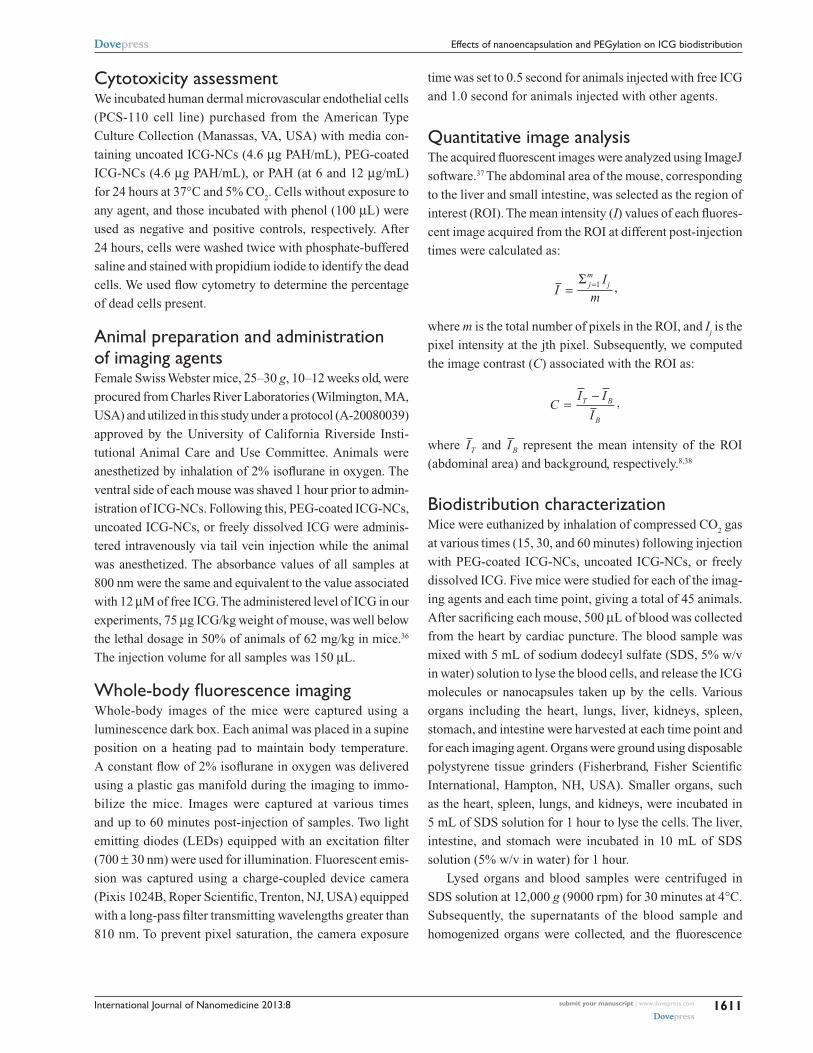
ResultsCharacterization of ICG-NCsScanning electron microscopy revealed the spherical
morphology of the ICG-NCs (Figure 1A). Using dynamic
light scattering, the peak diameter of the uncoated
ICG-NCs – based on the amounts of the reagents utilized,
time, and other experimental protocols, as indicated in the
Methods section – was 77 nm (Figure 1B). PEGylation
increased the peak diameter by nearly 10 nm and resulted in
a right shift in the population distribution. Both peak values
were associated with 23% of the nanoparticles in the popula-
tion distribution.
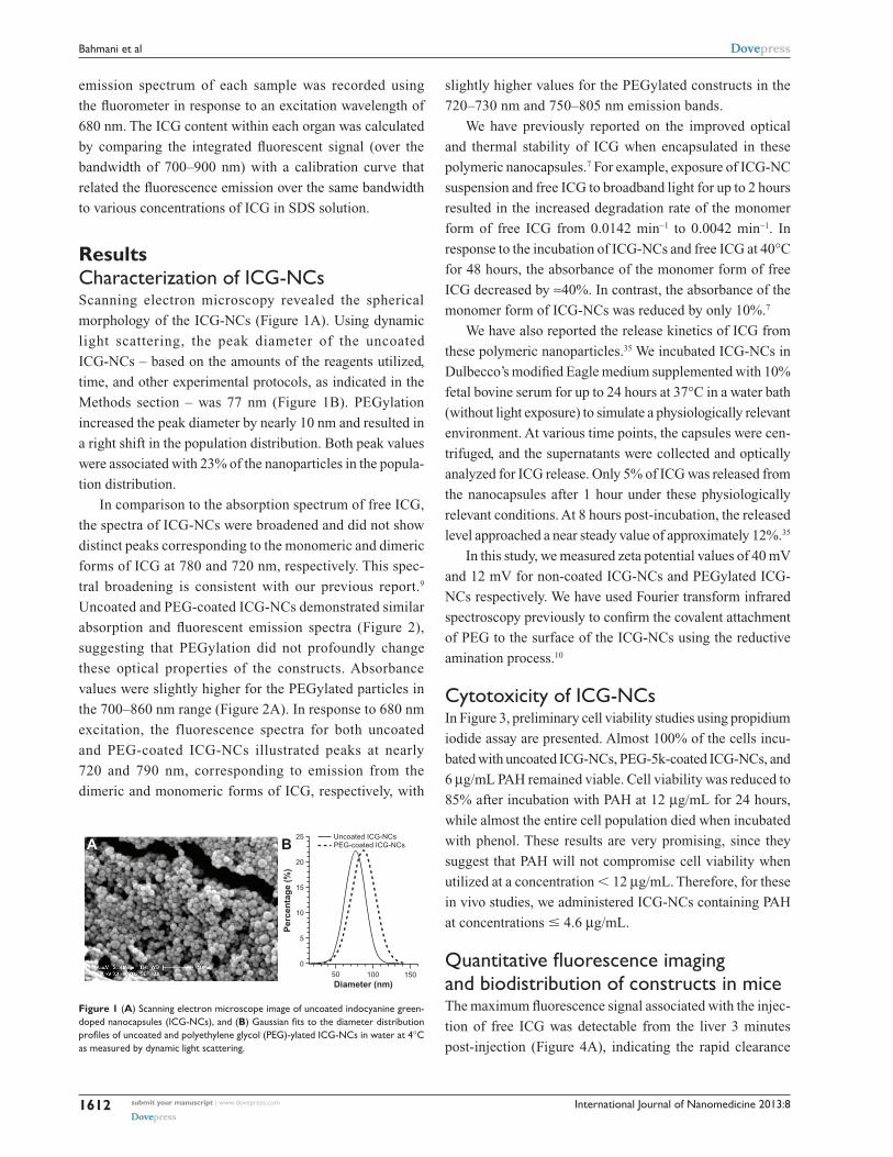
In comparison to the absorption spectrum of free ICG,
the spectra of ICG-NCs were broadened and did not show
distinct peaks corresponding to the monomeric and dimeric
forms of ICG at 780 and 720 nm, respectively. This spec-
tral broadening is consistent with our previous report.9
Uncoated and PEG-coated ICG-NCs demonstrated similar
absorption and fluorescent emission spectra (Figure 2),
suggesting that PEGylation did not profoundly change
these optical properties of the constructs. Absorbance
values were slightly higher for the PEGylated particles in
the 700–860 nm range (Figure 2A). In response to 680 nm
excitation, the fluorescence spectra for both uncoated
and PEG-coated ICG-NCs illustrated peaks at nearly
720 and 790 nm, corresponding to emission from the
dimeric and monomeric forms of ICG, respectively, with
slightly higher values for the PEGylated constructs in the
720–730 nm and 750–805 nm emission bands.
We have previously reported on the improved optical
and thermal stability of ICG when encapsulated in these
polymeric nanocapsules.7 For example, exposure of ICG-NC
suspension and free ICG to broadband light for up to 2 hours
resulted in the increased degradation rate of the monomer
form of free ICG from 0.0142 min−1 to 0.0042 min−1. In
response to the incubation of ICG-NCs and free ICG at 40°C
for 48 hours, the absorbance of the monomer form of free
ICG decreased by ≈40%. In contrast, the absorbance of the
monomer form of ICG-NCs was reduced by only 10%.7
We have also reported the release kinetics of ICG from
these polymeric nanoparticles.35 We incubated ICG-NCs in
Dulbecco’s modified Eagle medium supplemented with 10%
fetal bovine serum for up to 24 hours at 37°C in a water bath
(without light exposure) to simulate a physiologically relevant
environment. At various time points, the capsules were cen-
trifuged, and the supernatants were collected and optically
analyzed for ICG release. Only 5% of ICG was released from
the nanocapsules after 1 hour under these physiologically
relevant conditions. At 8 hours post-incubation, the released
level approached a near steady value of approximately 12%.35
In this study, we measured zeta potential values of 40 mV
and 12 mV for non-coated ICG-NCs and PEGylated ICG-
NCs respectively. We have used Fourier transform infrared
spectroscopy previously to confirm the covalent attachment
of PEG to the surface of the ICG-NCs using the reductive
amination process.10
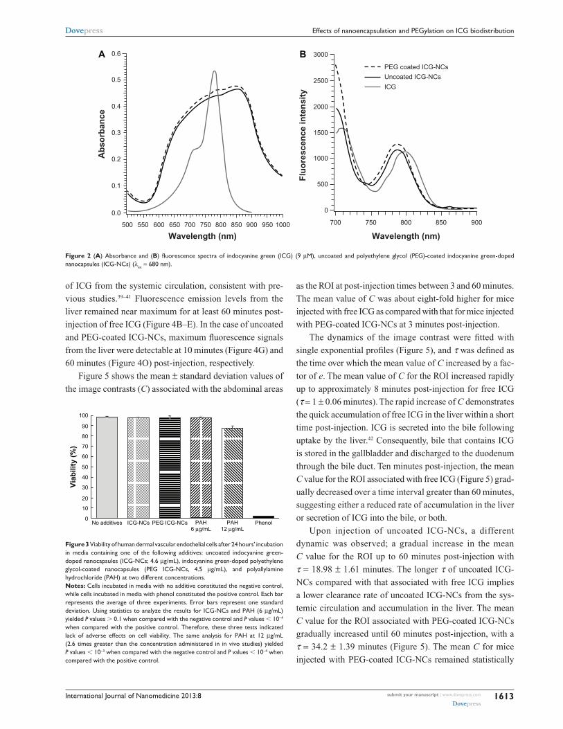
Cytotoxicity of ICG-NCsIn Figure 3, preliminary cell viability studies using propidium
iodide assay are presented. Almost 100% of the cells incu-
bated with uncoated ICG-NCs, PEG-5k-coated ICG-NCs, and
6 µg/mL PAH remained viable. Cell viability was reduced to
85% after incubation with PAH at 12 µg/mL for 24 hours,
while almost the entire cell population died when incubated
with phenol. These results are very promising, since they
suggest that PAH will not compromise cell viability when
utilized at a concentration , 12 µg/mL. Therefore, for these
in vivo studies, we administered ICG-NCs containing PAH
at concentrations # 4.6 µg/mL.
Quantitative fluorescence imaging and biodistribution of constructs in miceThe maximum fluorescence signal associated with the injec-
tion of free ICG was detectable from the liver 3 minutes
post-injection (Figure 4A), indicating the rapid clearance
500
5
10
15
20
25 Uncoated ICG-NCsPEG-coated ICG-NCsBA
100Diameter (nm)
Per
cen
tag
e (%
)
150
Figure 1 (A) Scanning electron microscope image of uncoated indocyanine green-doped nanocapsules (ICG-NCs), and (B) Gaussian fits to the diameter distribution profiles of uncoated and polyethylene glycol (PEG)-ylated ICG-NCs in water at 4°C as measured by dynamic light scattering.
submit your manuscript | www.dovepress.com
Dovepress
Dovepress
1612
Bahmani et al
International Journal of Nanomedicine 2013:8
500
0.0
0.1
0.2Ab
sorb
ance
Flu
ore
scen
ce in
ten
sity
0.3
0.4
0.5
0.6
600 700
Wavelength (nm)800 900550 650 750 850 950 9001000 850800
Wavelength (nm)
750700
3000
2500
PEG coated ICG-NCs
Uncoated ICG-NCs
ICG
2000
1500
1000
500
0
BA
Figure 2 (A) Absorbance and (B) fluorescence spectra of indocyanine green (ICG) (9 µM), uncoated and polyethylene glycol (PEG)-coated indocyanine green-doped nanocapsules (ICG-NCs) (λex = 680 nm).
100
90
80
70
60
50
40
30
20
10
0No additives
Via
bili
ty (
%)
ICG-NCs PEG ICG-NCs PAH6 µg/mL
PAH12 µg/mL
Phenol
Figure 3 Viability of human dermal vascular endothelial cells after 24 hours’ incubation in media containing one of the following additives: uncoated indocyanine green- doped nanocapsules (ICG-NCs; 4.6 µg/mL), indocyanine green-doped polyethylene glycol-coated nanocapsules (PEG ICG-NCs, 4.5 µg/mL), and polyallylamine hydrochloride (PAh) at two different concentrations. Notes: Cells incubated in media with no additive constituted the negative control, while cells incubated in media with phenol constituted the positive control. Each bar represents the average of three experiments. Error bars represent one standard deviation. Using statistics to analyze the results for ICG-NCs and PAh (6 µg/mL) yielded P values . 0.1 when compared with the negative control and P values , 10−4 when compared with the positive control. Therefore, these three tests indicated lack of adverse effects on cell viability. The same analysis for PAh at 12 µg/mL (2.6 times greater than the concentration administered in in vivo studies) yielded P values , 10−3 when compared with the negative control and P values , 10−4 when compared with the positive control.
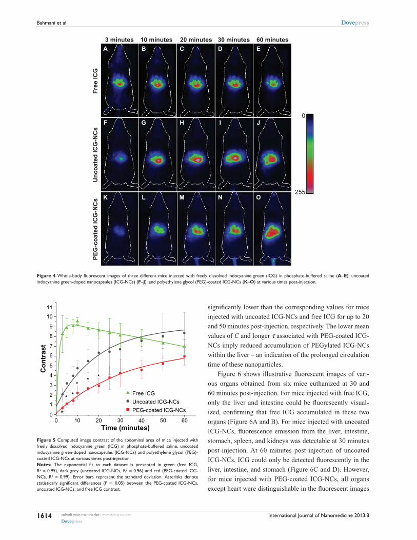
of ICG from the systemic circulation, consistent with pre-
vious studies.39–41 Fluorescence emission levels from the
liver remained near maximum for at least 60 minutes post-
injection of free ICG (Figure 4B–E). In the case of uncoated
and PEG-coated ICG-NCs, maximum fluorescence signals
from the liver were detectable at 10 minutes (Figure 4G) and
60 minutes (Figure 4O) post-injection, respectively.
Figure 5 shows the mean ± standard deviation values of
the image contrasts (C) associated with the abdominal areas
as the ROI at post-injection times between 3 and 60 minutes.
The mean value of C was about eight-fold higher for mice
injected with free ICG as compared with that for mice injected
with PEG-coated ICG-NCs at 3 minutes post-injection.
The dynamics of the image contrast were fitted with
single exponential profiles (Figure 5), and τ was defined as
the time over which the mean value of C increased by a fac-
tor of e. The mean value of C for the ROI increased rapidly
up to approximately 8 minutes post-injection for free ICG
(τ = 1 ± 0.06 minutes). The rapid increase of C demonstrates
the quick accumulation of free ICG in the liver within a short
time post-injection. ICG is secreted into the bile following
uptake by the liver.42 Consequently, bile that contains ICG
is stored in the gallbladder and discharged to the duodenum
through the bile duct. Ten minutes post-injection, the mean
C value for the ROI associated with free ICG (Figure 5) grad-
ually decreased over a time interval greater than 60 minutes,
suggesting either a reduced rate of accumulation in the liver
or secretion of ICG into the bile, or both.
Upon injection of uncoated ICG-NCs, a different
dynamic was observed; a gradual increase in the mean
C value for the ROI up to 60 minutes post-injection with
τ = 18.98 ± 1.61 minutes. The longer τ of uncoated ICG-
NCs compared with that associated with free ICG implies
a lower clearance rate of uncoated ICG-NCs from the sys-
temic circulation and accumulation in the liver. The mean
C value for the ROI associated with PEG-coated ICG-NCs
gradually increased until 60 minutes post-injection, with a
τ = 34.2 ± 1.39 minutes (Figure 5). The mean C for mice
injected with PEG-coated ICG-NCs remained statistically
submit your manuscript | www.dovepress.com
Dovepress
Dovepress
1613
Effects of nanoencapsulation and PEGylation on ICG biodistribution
International Journal of Nanomedicine 2013:8
significantly lower than the corresponding values for mice
injected with uncoated ICG-NCs and free ICG for up to 20
and 50 minutes post-injection, respectively. The lower mean
values of C and longer τ associated with PEG-coated ICG-
NCs imply reduced accumulation of PEGylated ICG-NCs
within the liver – an indication of the prolonged circulation
time of these nanoparticles.
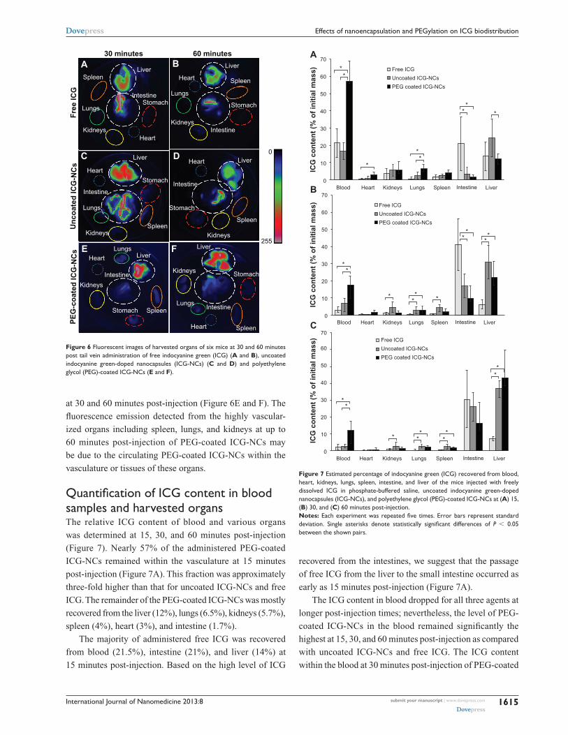
Figure 6 shows illustrative fluorescent images of vari-
ous organs obtained from six mice euthanized at 30 and
60 minutes post-injection. For mice injected with free ICG,
only the liver and intestine could be fluorescently visual-
ized, confirming that free ICG accumulated in these two
organs (Figure 6A and B). For mice injected with uncoated
ICG-NCs, fluorescence emission from the liver, intestine,
stomach, spleen, and kidneys was detectable at 30 minutes
post-injection. At 60 minutes post-injection of uncoated
ICG-NCs, ICG could only be detected fluorescently in the
liver, intestine, and stomach (Figure 6C and D). However,
for mice injected with PEG-coated ICG-NCs, all organs
except heart were distinguishable in the fluorescent images
A B C D E
F G H I J
K
PE
G-c
oat
ed IC
G-N
Cs
255
0
3 minutes 10 minutes 20 minutes 30 minutes 60 minutes
Un
coat
ed IC
G-N
Cs
Fre
e IC
G
L M N O
Figure 4 Whole-body fluorescent images of three different mice injected with freely dissolved indocyanine green (ICG) in phosphate-buffered saline (A–E), uncoated indocyanine green-doped nanocapsules (ICG-NCs) (F–J), and polyethylene glycol (PEG)-coated ICG-NCs (K–O) at various times post-injection.
0
1
2
3
4
5
7
8
9
10
11
6
0 10 20 30 40 50Time (minutes)
Free ICG
***
**
*
Uncoated ICG-NCs
PEG-coated ICG-NCs
Co
ntr
ast
60
Figure 5 Computed image contrast of the abdominal area of mice injected with freely dissolved indocyanine green (ICG) in phosphate-buffered saline, uncoated indocyanine green-doped nanocapsules (ICG-NCs) and polyethylene glycol (PEG)-coated ICG-NCs at various times post-injection.Notes: The exponential fit to each dataset is presented in green (free ICG, R2 = 0.95), dark grey (uncoated ICG-NCs, R2 = 0.96) and red (PEG-coated ICG-NCs, R2 = 0.99). Error bars represent the standard deviation. Asterisks denote statistically significant differences (P , 0.05) between the PEG-coated ICG-NCs, uncoated ICG-NCs, and free ICG contrast.
submit your manuscript | www.dovepress.com
Dovepress
Dovepress
1614
Bahmani et al
International Journal of Nanomedicine 2013:8
at 30 and 60 minutes post-injection (Figure 6E and F). The
fluorescence emission detected from the highly vascular-
ized organs including spleen, lungs, and kidneys at up to
60 minutes post-injection of PEG-coated ICG-NCs may
be due to the circulating PEG-coated ICG-NCs within the
vasculature or tissues of these organs.
Quantification of ICG content in blood samples and harvested organsThe relative ICG content of blood and various organs
was determined at 15, 30, and 60 minutes post-injection
(Figure 7). Nearly 57% of the administered PEG-coated
ICG-NCs remained within the vasculature at 15 minutes
post-injection (Figure 7A). This fraction was approximately
three-fold higher than that for uncoated ICG-NCs and free
ICG. The remainder of the PEG-coated ICG-NCs was mostly
recovered from the liver (12%), lungs (6.5%), kidneys (5.7%),
spleen (4%), heart (3%), and intestine (1.7%).
The majority of administered free ICG was recovered
from blood (21.5%), intestine (21%), and liver (14%) at
15 minutes post-injection. Based on the high level of ICG
A B
DC
E F
Spleen Spleen
Spleen
Spleen
Spleen
Spleen
Lungs
Lungs
Lungs
Lungs
Lungs
KidneysKidneys
Kidneys
Kidneys
Kidneys
Kidneys
Heart
Heart
Heart
Heart
Heart
Heart
Stomach Stomach
Stomach
Stomach
Stomach
Stomach
Intestine
Intestine
Intestine
Intestine
Intestine
Intestine
Liver
30 minutes
Fre
e IC
GU
nco
ated
ICG
-NC
sP
EG
-co
ated
ICG
-NC
s
60 minutes
0
255
Liver
Liver
LiverLiver
Liver
Figure 6 Fluorescent images of harvested organs of six mice at 30 and 60 minutes post tail vein administration of free indocyanine green (ICG) (A and B), uncoated indocyanine green-doped nanocapsules (ICG-NCs) (C and D) and polyethylene glycol (PEG)-coated ICG-NCs (E and F).
Blood0
10
20
ICG
co
nte
nt
(% o
f in
itia
l mas
s)
30
40
50
60
70
**
*
**
**
*
Heart
Free ICG
A
Uncoated ICG-NCs
PEG coated ICG-NCs
Kidneys Lungs Spleen Intestine Liver
Blood0
10
20
ICG
co
nte
nt
(% o
f in
itia
l mas
s)
30
40
50
60
70
**
**
*
Heart
Free ICG
B
Uncoated ICG-NCs
PEG coated ICG-NCs
Kidneys Lungs Spleen Intestine Liver
*
*
*
*
*
Blood0
10
20
ICG
co
nte
nt
(% o
f in
itia
l mas
s)
30
40
50
60
70
***
*
Heart
Free ICG
C
Uncoated ICG-NCs
PEG coated ICG-NCs
Kidneys Lungs Spleen Intestine Liver
*
*
*
*
*
Figure 7 Estimated percentage of indocyanine green (ICG) recovered from blood, heart, kidneys, lungs, spleen, intestine, and liver of the mice injected with freely dissolved ICG in phosphate-buffered saline, uncoated indocyanine green-doped nanocapsules (ICG-NCs), and polyethylene glycol (PEG)-coated ICG-NCs at (A) 15, (B) 30, and (C) 60 minutes post-injection.Notes: Each experiment was repeated five times. Error bars represent standard deviation. Single asterisks denote statistically significant differences of P , 0.05 between the shown pairs.
recovered from the intestines, we suggest that the passage
of free ICG from the liver to the small intestine occurred as
early as 15 minutes post-injection (Figure 7A).
The ICG content in blood dropped for all three agents at
longer post-injection times; nevertheless, the level of PEG-
coated ICG-NCs in the blood remained significantly the
highest at 15, 30, and 60 minutes post-injection as compared
with uncoated ICG-NCs and free ICG. The ICG content
within the blood at 30 minutes post-injection of PEG-coated
submit your manuscript | www.dovepress.com
Dovepress
Dovepress
1615
Effects of nanoencapsulation and PEGylation on ICG biodistribution
International Journal of Nanomedicine 2013:8
ICG-NCs (Figure 7B) was nearly equal to the ICG content in
blood at 15 minutes post-injection of free ICG or uncoated
ICG-NCs (Figure 7A).
At 30 minutes post-injection, the amount of ICG in
the blood and liver dropped dramatically to 7% and 6%,
respectively (Figure 7B); conversely, an increase in ICG
content in the intestine to 41% was observed. This rise is
indicative of ICG transport to the duodenum. In the case
of uncoated ICG-NCs, the amount of ICG recovered from
the blood, heart, and kidneys dropped to 6.8%, 0.3%, and
4.4%, respectively. However, the ICG content remained
the same in the lungs and spleen. A significant increase
in ICG concentration was observed in the intestine and
liver at longer times post-injection of uncoated ICG-NCs
(Figure 7B and C).
At 30 and 60 minutes post-injection of PEG-coated ICG-
NCs, the amount of ICG dropped significantly in blood and
all organs except the liver and intestine (Figure 7B and C).
The dramatic increase in liver ICG content from nearly 12%
at 15 minutes to 22% at 30 minutes, and 42% at 60 minutes
post-injection is consistent with the whole-body fluorescent
images (Figure 4) and the associated ROI image contrast
(Figure 5), illustrating the delayed uptake of PEG-coated
ICG-NCs by the liver. The amount of ICG recovered from
the lungs, kidneys, spleen, and heart decreased at 30 and
60 minutes post-injection of PEG-coated ICG-NCs.
The spleen is one of the organs of the immune system
responsible for filtering foreign body materials and dead
or damaged erythrocytes from the blood.43 We noted that
about 4% of the PEG-coated ICG-NCs was recovered from
spleen at 15 minutes post-injection. However, the level of
recovered ICG was statistically significantly reduced to 2%
at 60 minutes post-injection. Therefore, we associate the high
level of PEGylated ICG-NCs recovered from the spleen at
15 minutes post-injection with the nanoparticles circulating
within the vasculature of spleen. In the open blood circula-
tion system of the spleen, the capillaries discharge openly
into the pulp cord before reaching the sinusoids. The pulp
cords contain reticular cells, macrophages, plasma cells and
lymphocytes.44 The PEGylated ICG-NCs in the blood have
to pass through the pulp cord before entering the venous
sinusoids. We attribute the initial rise in fluorescent signal
detected from the spleen and the concentration of recovered
nanocapsules from this organ to the portion of the PEG-
coated ICG-NCs passing through the pulp cord and not to
the uptake by splenic macrophages.
Lungs, as the organs receiving the entire cardiac output,
are prone to the accumulation of nanocapsules.45 Our results
demonstrate that a statistically significant level of ICG was
recovered from the lungs (6.5%) at 15 minutes post-injection
of PEG-coated ICG-NCs compared with uncoated ICG-
NCs and free ICG (Figure 7A). The drop in ICG content in
the lungs to 2.5% after 1 hour following administration of
PEG-coated ICG-NCs suggests that the particles were in the
vasculature of the lungs.
DiscussionOur ICG-NCs spontaneously self-assemble via green chem-
istry, are fabricated with tunable diameters in the range
of ≈60 nm to 1 µm, and the presence of amine groups on the
surface of the constructs enables the covalent attachment of
various moieties for molecular targeting.
To suppress the propensity of the ICG-NCs for non-
specific adsorption of proteins, we coated them with PEG,
which is widely used to produce bio-inert surfaces by sup-
pressing nonspecific interactions.46 To attain stability of the
passivation coating, we covalently grafted the PEG chains
to the amines on the surface of the nanoparticles. Employing
chemical reactions that proceed through small intermediates
with quantitative yields, such as reductive amination, ensures
sufficiently well-packed PEG layers.47
The bio-inertness of the PEG coatings strongly depends
on the length of the polymer chains. Our goal encom-
passed not only suppressing molecular-level nonspecific
interactions – which can be achieved even with ethylene
glycol oligomers (ie, with three to six repeating ethylene
glycol units) – but also preventing nonspecific adhesion
at mesoscopic (micron and sub-micron) levels. Coatings
of PEG polymers that are too short would not provide suf-
ficient separation between the nanoparticles and surfaces, to
which they have tendency to be nonspecifically adsorbed by.
Concurrently, PEG chains that are too long would result
in layers in which the polymer termini have non-helical
“mushroom” conformation that compromises the ability of
such coatings to suppress nonspecific interactions.48 Using
force measurements, as implemented with magnetic pullers,
we previously determined that PEG with a MW of 5 kDa –
among the PEG molecules investigated with MW ranging
between 1 and 20 kDa – provided the optimal covalently
grafted coatings for suppressing nonspecific adhesion.49
The higher flexibility and mobility of low MW PEG chains
result in higher entropic repulsion between proteins and the
surface.50,51 Our in vitro studies confirmed the choice of PEG
5 kDa for passivation coatings of ICG-NCs and for imped-
ing their uptake by macrophages.10 Conversely, reduced
flexibility of high MW PEG chains, due to the entrapment
submit your manuscript | www.dovepress.com
Dovepress
Dovepress
1616
Bahmani et al
International Journal of Nanomedicine 2013:8
of long chains, induces low protein resistivity. In this study,
we examined the biodistribution of ICG-NCs coated with a
passivation coating of PEG 5 kDa.
In previous studies by other groups, ICG-containing
constructs composed of poly(DL-lactic-co-glycolic-acid)
(PLGA) whose surfaces were functionalized by both PEG
and folic acid were administered into tumor-bearing mice.52,53
While these studies demonstrated the efficacy of targeted
fluorescence imaging of tumors using functionalized ICG-
containing constructs, they did not isolate the effects of
ICG encapsulation – and the additional PEGylation of the
constructs in the absence of targeting moieties such as folic
acid – in modulating the biodistribution of encapsulated ICG
in a non-diseased mammalian system.
In another study by Zheng et al, ICG was entrapped within
PLGA, and this construct was in turn coated with a lipid
2 kDa PEG-folic acid assembly.53 Although the investigators
reported the utility of these structures in targeted fluorescent
imaging of tumor-bearing mice, the lipid 2 kDa PEG-folic
acid assembly was not covalently attached to the inner PLGA
construct. Further, since PEG was simply a linker between
the lipid and the folic acid, its role in suppressing nonspecific
interactions is not clear.
Saxena et al investigated the biodistribution of PLGA
nanoparticles entrapping ICG in healthy mice.40 Although
these constructs were not PEGylated, the investigators
reported that ICG encapsulation into these constructs
resulted in ICG levels that were nearly six times higher
within plasma at 1 hour post-tail vein injection in mice, as
compared with those resulting from non-encapsulated ICG.
In a previous study, our group reported on the biodistribu-
tion of ICG-NCs coated non-covalently with dextran or
10 nm ferromagnetic iron oxide nanoparticles, themselves
individually coated with PEG, in healthy mice.35 The half-
life of these particles in blood and their circulation kinetics
appeared unaffected compared with free ICG, presumably
due to the non-covalent attachment of the coating materials.
Therefore, to the best of our knowledge, the study pre-
sented here is the first to have aimed at characterizing the
biodistribution of encapsulated ICG in a construct system
composed of an inner ICG core entrapped by a polymeric
shell with covalent outer surface PEGylation by both in vivo
quantitative fluorescence imaging and subsequent analysis
of various extracted organs.
Consistent with Saxena et al’s results,40 we observed a
similar increase in ICG levels within plasma at 1 hour post-
injection in the case of the PEGylated ICG-NCs (Figure 7C).
We did not observe any significant differences in plasma ICG
levels at 1 hour post-injection when ICG was administered in
non-encapsulated form or as non-PEGylated ICG-NCs.
Higher ICG levels within various organs, including
the spleen, heart, lungs, and kidneys, were reported by
Saxena et al when ICG was delivered in the PLGA-entrapped
form. These investigators also reported that, as early as
5 minutes post-injection, the amount of ICG within the liver
was nearly 2.5 times greater when it was administered in the
PLGA-entrapped form compared with when it was admin-
istered as free ICG. This result suggests that entrapment of
ICG within the PLGA construct was not quite effective in
delaying the accumulation of ICG within the liver. In com-
parison, in our study, we detected higher ICG levels within
the liver at 30 minutes post-injection of both non-PEGylated
and PEGylated ICG-NCs (Figure 7B).
In addition to the lack of PEGylation in the PLGA con-
structs, another factor that may be responsible for the differ-
ences between our results and those of Saxena et al is the size
of the constructs. The mean diameter of the PLGA constructs
was reported to be 300 nm, nearly three times larger than
the constructs used in our study. It has been indicated that
PEGylated nanoparticles smaller than 100 nm have reduced
plasma protein adsorption on their surfaces, and the blood
clearance of such nanoparticles is slower than for larger
(.100 nm) nanoparticles.31,54
Our results show that the covalent coating of ICG-NCs
with 5 kDa PEG can increase the blood circulation time of
ICG. Our findings are consistent with reports by Ballou et al55
and Ohno et al,56 who have indicated prolonged systemic
circulation of PEGylated silica nanoparticles in vivo.
Ballou et al have reported a dramatic increase in the circu-
lating half-life of core-shell zinc sulfide-cadmium selenide
quantum dots coated with PEG (5000 Da). Their results
have demonstrated that the extended circulation half-life
of these particles is nearly 70 minutes compared with that
of quantum dots coated with shorter length (750 Da and
3,400 Da) PEG chains, which had a half-life of less than
12 minutes.55 Ohno et al have reported prolonged blood half-
life (∼20 hours) of PEGylated silica nanoparticles in healthy
mice, with preferentially high accumulation in tumor tissue
when injected into tumor-bearing mice.56 Shah et al studied
the effect of PEG coating on the blood retention of gold
nanoparticles. They indicated that the longer the particles
stayed in the systemic circulation, the greater was the chance
of accumulation in the tumor.57
The prolonged vascular circulation time of ICG as medi-
ated by its encapsulation in these constructs may open up new
possibilities for further clinical applications of ICG, which
submit your manuscript | www.dovepress.com
Dovepress
Dovepress
1617
Effects of nanoencapsulation and PEGylation on ICG biodistribution
International Journal of Nanomedicine 2013:8
currently remain limited due to its rapid clearance from the
vasculature and accumulation in the liver. The presence of
ICG combined with the ability to functionalize the capsules
provides the potential for ICG-NCs to serve as theranostic
materials for the targeted optical imaging of specific molecu-
lar biomarkers of a disease and phototherapy. Previously, our
group reported the targeted fluorescence imaging of head and
neck squamous cells, cervical squamous cells, and breast
cancer cells, with various expression levels of epidermal
growth factor receptor (EGFR) using anti-EGFR-conjugated
ICG-NCs.9 We have also reported the targeted fluorescent
imaging of ovarian cancer cells using anti-human EGFR 2
functionalized ICG-NCs.58
ICG-NCs can potentially be used as a phototherapeutic
agent by generating heat in response to laser irradiation. In a
previous study,59 our group investigated the heat-generating
capability of ICG-NCs in response to 808 nm laser irradia-
tion and demonstrated the ability of ICG-NC suspensions to
produce temperature rises to ≈80°C. In another study,11 using a
tissue phantom consisting of chicken breast to simulate normal
tissue and an embedded gelatin cylinder (to simulate abnor-
mal vasculature mass) loaded with ICG-NCs, we observed a
nearly 20°C temperature rise within the gelatin cylinder at a
depth of 3 mm below the surface in response to laser irradia-
tion at 808 nm with an incident power of 4.2 W. These results
demonstrate the ability of ICG-NCs to induce a temperature
rise in response to laser irradiation when embedded within
an optically turbid tissue-like structure. In another study,9 we
demonstrated the capability of ICG-NCs coated with anti-
EGFR to destroy cancer cell lines photothermally.
ConclusionIn this study, the biodistribution of ICG-NCs coated with
5 kDa PEG in mice, as assessed by whole-body fluorescent
imaging and individual organ uptake analysis, indicated that
these particles have a prolonged vascular circulation time
and delayed hepatic accumulation in comparison to uncoated
constructs and non-encapsulated ICG. Thus, the encapsula-
tion of ICG in such PEGylated constructs may potentially
extend the current clinical applications of ICG because of
its increased bioavailability.
AcknowledgmentThis work was supported in part by grants from the National
Science Foundation (CBET-1144237, CBET-0923408),
Bourns College of Engineering, Bioengineering Center at
the University of California, Riverside (UCR), and a student
summer research grant from American Society for Laser
Medicine and Surgery. The animal imaging was performed
at the Institute for Integrative Genome Biology at UCR. We
are grateful for the support provided by the Central Facility
for Advanced Microscopy and Microanalysis at UCR. We
thank Mr Andrew Reimer, a member of Bahman Anvari’s
group, for proofreading the manuscript.
DisclosureThe authors report no conflicts of interest in this work.
References 1. Frangioni J. In vivo near-infrared fluorescence imaging. Curr Opin
Chem Biol. 2003;7(5):626–634. 2. Peer D, Karp JM, Hong S, Farokhzad OC, Margalit R, Langer R.
Nanocarriers as an emerging platform for cancer therapy. Nat Nanotechnol. 2007;2(12):751–760.
3. Sevick-Muraca EM, Houston JP, Gurfinkel M. Fluorescence-enhanced, near infrared diagnostic imaging with contrast agents. Curr Opin Chem Biol. 2002;6(5):642–650.
4. Ntziachristos V, Bremer C, Weissleder R. Fluorescence imaging with near-infrared light: new technological advances that enable in vivo molecular imaging. Eur Radiol. 2003;13(1):195–208.
5. He X, Gao J, Gambhir SS, Cheng Z. Near-infrared fluorescent nano-probes for cancer molecular imaging: status and challenges. Trends Mol Med. 2010;16(12):574–583.
6. Kim S, Lim YT, Soltesz EG, et al. Near-infrared fluorescent type II quantum dots for sentinel lymph node mapping. Nat Biotechnol. 2004;22(1):93–97.
7. Yaseen MA, Yu J, Wong MS, Anvari B. Stability assessment of indo-cyanine green within dextran-coated mesocapsules by absorbance spectroscopy. J Biomed Opt. 2007;12(6):064031.
8. Yaseen MA, Yu J, Wong MS, Anvari B. In-vivo fluorescence imaging of mammalian organs using charge-assembled mesocapsule constructs con-taining indocyanine green. Opt Express. 2008;16(25):20577–20587.
9. Yu J, Javier D, Yaseen MA, et al. Self-assembly synthesis, tumor cell targeting, and photothermal capabilities of antibody-coated indocyanine green nanocapsules. J Am Chem Soc. 2010;132(6):1929–1938.
10. Bahmani B, Gupta S, Upadhyayula S, Vullev VI, Anvari B. Effect of polyethylene glycol coatings on uptake of indocyanine green loaded nanocapsules by human spleen macrophages in vitro. J Biomed Opt. 2011;16(5):051303.
11. Yaseen MA, Yu J, Wong MS, Anvari B. Laser-induced heating of dextran-coated mesocapsules containing indocyanine green. Biotechnol Prog. 2007;23(6):1431–1440.
12. Yannuzzi LA. Indocyanine green angiography: a perspective on use in the clinical setting. Am J Ophthalmol. 2011;151(5):745–751. e1.
13. Ross A, Ross AH, Mohamed Q. Review and update of central serous chorioretinopathy. Curr Opin Ophthalmol. 2011;22(3):166–173.
14. Mantel I, Uffer S, Zografos L. Peripheral exudative hemorrhagic chorioretinopathy: a clinical, angiographic, and histologic study. Am J Ophthalmol. 2009;148(6):932–938. e1.
15. Tanaka E, Chen FY, Flaumenhaft R, Graham GJ, Laurence RG, Frangioni JV. Real-time assessment of cardiac perfusion, coronary angiography, and acute intravascular thrombi using dual-channel near-infrared fluorescence imaging. J Thorac Cardiovasc Surg. 2009;138(1): 133–140.
16. El-Desoky A, Seifalian AM, Cope M, Delpy DT, Davidson BR. Experimental study of liver dysfunction evaluated by direct indocya-nine green clearance using near infrared spectroscopy. British J Surg. 1999;86(8):1005–1011.
submit your manuscript | www.dovepress.com
Dovepress
Dovepress
1618
Bahmani et al
International Journal of Nanomedicine 2013:8
17. Hirano A, Kamimura M, Ogura K, et al. A comparison of indocyanine green fluorescence imaging plus blue dye and blue dye alone for senti-nel node navigation surgery in breast cancer patients. Ann Surg Oncol. 2012;19(13):4112–4116.
18. Kitai T, Inomoto T, Miwa M, Shikayama T. Fluorescence navigation with indocyanine green for detecting sentinel lymph nodes in breast cancer. Breast Cancer. 2005;12(3):211–215.
19. Miyashiro I, Miyoshi N, Hiratsuka M, et al. Detection of sentinel node in gastric cancer surgery by indocyanine green fluorescence imaging: comparison with infrared imaging. Ann Surg Oncol. 2008;15(6): 1640–1643.
20. Nimura H, Narimiya N, Mitsumori N, Yamazaki Y, Yanaga K, Urashima M. Infrared ray electronic endoscopy combined with indo-cyanine green injection for detection of sentinel nodes of patients with gastric cancer. Br J Surg. 2004;91(5):575–579.
21. Uhara H, Yamazaki N, Takata M, et al. Applicability of radiocolloids, blue dyes and fluorescent indocyanine green to sentinel node biopsy in melanoma. J Dermatol. 2012;39(4):336–338.
22. Van der Vorst JR, Schaafsma BE, Verbeek FPR, et al. Randomized comparison of near-infrared fluorescence imaging using indocyanine green and 99(m) technetium with or without patent blue for the sentinel lymph node procedure in breast cancer patients. Ann Surg Oncol. 2012; 19(13):4104–4111.
23. Moretó M. Diagnosis of esophagogastric tumors. Endoscopy. 2003; 35(1):36–42.
24. Yamamoto T, Narushima M, Doi K, et al. Characteristic indocyanine green lymphography findings in lower extremity lymphedema: the generation of a novel lymphedema severity staging system using dermal backflow patterns. Plast Reconstr Surg. 2011;127(5):1979–1986.
25. Yamamoto T, Yamamoto N, Doi K, et al. Indocyanine green-enhanced lymphography for upper extremity lymphedema: a novel severity staging system using dermal backflow patterns. Plast Reconstr Surg. 2011;128(4):941–947.
26. Yoneya S, Saito T, Komatsu Y, Koyama I, Takahashi K, Duvoll-Young J. Binding properties of indocyanine green in human blood. Invest Ophthalmol Vis Sci. 1998;39(7):1286–1290.
27. Altinoğlu EI, Adair JH. Near infrared imaging with nanoparticles. Wiley Interdiscip Rev Nanomed Nanobiotechnol. 2010;2(5):461–477.
28. Desmettre TJ, Soulie-Begu S, Devoisselle JM, Mordon SR. Diode laser-induced thermal damage evaluation on the retina with a liposome dye system. Lasers Surg Med. 1999;24(1):61–68.
29. Rodriguez VB, Henry SM, Hoffman AS, Stayton PS, Li X, Pun SH. Encapsulation and stabilization of indocyanine green within poly(styrene-alt-maleic anhydride) block-poly(styrene) micelles for near-infrared imaging. J Biomed Opt. 2013;13(1):014025.
30. Sharma P, Bengtsson NE, Walter GA, et al. Gadolinium-doped silica nanoparticles encapsulating indocyanine green for near infrared and magnetic resonance imaging. Small. 2012;8(18):2856–2868.
31. Alexis F, Pridgen E, Molnar LK, Farokhzad OC. Factors affecting the clearance and biodistribution of polymeric nanoparticles. Mol Pharm. 2008;5(4):505–515.
32. Cao Z, Jiang S. Super-hydrophobic switterionic poly(carboxybetaine) and amphiphilic non-ionic poly(ethylene glycol) for stealth nanopar-ticles. Nano Today. 2012;7(5):404–413.
33. Bahmani B, Gupta S, Vullev V, Anvari B. Uptake of PEGylated indocya-nine green loaded nanocapsules by cells of reticuloendothelial system. In: Achilefu S, Raghavachari R, editors. Reporters, Markers, Dyes, Nanoparticles, and Molecular Probes for Biomedical Applications III. Proceedings of SPIE Volume 7910. Bellingham, WA: SPIE; 2011:79101C.
34. Bahmani B, Jung B, Gupta S, Anvari B. Cellular uptake of polymeric nanocapsules loaded with ICG by human blood monocytes and human spleen macrophages. In: Achilefu S, Raghavachari R, editors. Reporters, Markers, Dyes, Nanoparticles, and Molecular Probes for Biomedical Applications III. Proceedings of SPIE Volume 7576. Bellingham, WA: SPIE; 2010:75761Q.
35. Yaseen MA, Yu J, Jung B, Wong MS, Anvari B. Biodistribution of encapsulated indocyanine green in healthy mice. Mol Pharm. 2009;6(5): 1321–1332.
36. Ebert B, Licha K. Cyanine dyes as contrast agents for near-infrared imaging in vivo: acute tolerance, pharmacokinetics, and fluorescence imaging. J Biomed Opt. 2011;16(6):066003.
37. Rasband WS. ImageJ [software]. Bethesda, MD: US National Institutes of Health; 1997–2012.
38. Houston JP, Ke S, Wang W, Li C, Sevick-Muraca EM. Quality analysis of in vivo near-infrared fluorescence and conventional gamma images acquired using a dual-labeled tumor-targeting probe. J Biomed Opt. 2005;10(5):054010.
39. Sakka SG, Koeck H, Meier-Hellmann A. Measurement of indocyanine green plasma disappearance rate by two different dosages. Intensive Care Med. 2004;30(3):506–509.
40. Saxena V, Sadoqi M, Shao J. Polymeric nanoparticulate delivery system for Indocyanine green: biodistribution in healthy mice. Int J Pharm. 2006;308(1–2):200–204.
41. Desmettre T, Devoisselle JM, Mordon S. Fluorescence properties and metabolic features of indocyanine green (ICG) as related to angiography. Surv Ophthalmol. 2000;45(1):15–27.
42. Cui Y, König J, Leier I, Buchholz U, Keppler D. Hepatic uptake of bilirubin and its conjugates by the human organic anion transporter SLC21A6. J Biol Chem. 2001;276(13):9626–9630.
43. Cesta MF. Normal structure, function, and histology of the spleen. Toxicol Pathol. 2006;34(5):455–465.
44. Mebius RE, Kraal G. Structure and function of the spleen. Nat Rev Immunol. 2005;5(8):606–616.
45. Card JW, Zeldin DC, Bonner JC, Nestmann ER. Pulmonary applications and toxicity of engineered nanoparticles. Am J Physiol Lung Cell Mol Physiol. 2008;295(3):L400–L411.
46. Leckband D, Sheth S, Halperin A. Grafted poly(ethylene oxide) brushes as nonfouling surface coatings. J Biomater Sci Polymer Edn. 1999;10(10):1125–1147.
47. Wan J, Thomas MS, Guthrie S, Vullev VI. Surface-bound pro-teins with preserved functionality. Ann Biomed Eng. 2009;37(6): 1190–1205.
48. Gref R, Lück M, Quellec P, et al. “Stealth” corona-core nanoparticles surface modified by polyethylene glycol (PEG): influences of the corona (PEG chain length and surface density) and of the core composition on phagocytic uptake and plasma protein adsorption. Colloids Surf B Biointerfaces. 2000;18(3–4):301–313.
49. Upadhyayula S, Quinata T, Bishop S, et al. Coatings of polyethylene glycol for suppressing adhesion between solid microspheres and flat surfaces. Langmuir. 2012;28(11):5059–5069.
50. Harder P, Grunze M, Dahint R, Whitesides GM, Laibinis PE. Molecular conformation in oligo (ethylene glycol) – terminated self-assembled monolayers on gold and silver surfaces determines their ability to resist protein adsorption. J Phys Chem B. 1998;102(2):426–436.
51. Xu Z, Holland NB, Marchant RE. Conformations of short-chain poly(ethylene oxide) lipopolymers at the air−water interface: a com-bined film balance and surface tension study. Langmuir. 2001;17(2): 377–383.
52. Ma Y, Sadoqi M, Shao J. Biodistribution of indocyanine green-loaded nanoparticles with surface modifications of PEG and folic acid. Int J Pharm. 2012;436(1–2):25–31.
53. Zheng C, Zheng M, Gong P, et al. Indocyanine green-loaded biode-gradable tumor targeting nanoprobes for in vitro and in vivo imaging. Biomaterials. 2012;33(22):5603–5609.
54. Fang C, Shi B, Pei YY, Hong MH, Wu J, Chen HZ. In vivo tumor targeting of tumor necrosis factor-alpha-loaded stealth nanoparticles: effect of MePEG molecular weight and particle size. Eur J Pharm Sci. 2006;27(1):27–36.
55. Ballou B, Lagerholm BC, Ernst LA, Bruchez MP, Waggoner AS. Noninvasive imaging of quantum dots in mice. Bioconjugate Chem. 2004;15(1):79–86.
submit your manuscript | www.dovepress.com
Dovepress
Dovepress
1619
Effects of nanoencapsulation and PEGylation on ICG biodistribution
International Journal of Nanomedicine
Publish your work in this journal
Submit your manuscript here: http://www.dovepress.com/international-journal-of-nanomedicine-journal
The International Journal of Nanomedicine is an international, peer-reviewed journal focusing on the application of nanotechnology in diagnostics, therapeutics, and drug delivery systems throughout the biomedical field. This journal is indexed on PubMed Central, MedLine, CAS, SciSearch®, Current Contents®/Clinical Medicine,
Journal Citation Reports/Science Edition, EMBase, Scopus and the Elsevier Bibliographic databases. The manuscript management system is completely online and includes a very quick and fair peer-review system, which is all easy to use. Visit http://www.dovepress.com/ testimonials.php to read real quotes from published authors.
International Journal of Nanomedicine 2013:8
56. Ohno K, Akashi T, Tsujii Y, Yamamoto M, Tabata Y. Blood clearance and biodistribution of polymer brush-afforded silica particles prepared by surface-initiated living radical polymerization. Biomacromolecules. 2012;13(3):927–936.
57. Shah NB, Vercellotti GM, White JG, Fegan A, Wagner CR, Bischof JC. Blood−nanoparticle interactions and in vivo biodistribution: impact of surface PEG and ligand properties. Mol Pharm. 2012. Epub July 23.
58. Bahmani B, Vullev V, Anvari B. Development of anti-HER2 conjugated ICG-loaded polymeric nanoparticles for targeted optical imaging of ovarian cancer. In: Achilefu S, Raghavachari R, editors. Reporters, Markers, Dyes, Nanoparticles, and Molecular Probes for Biomedical Applications III. Proceedings of SPIE Volume 82. Bellingham, WA: SPIE; 2012:82330L.
59. Yu J, Yaseen MA, Anvari B, Wong MS. Synthesis of near-infrared-absorbing nanoparticle-assembled capsules. Chem Mater. 2007;(8):1476–1483.
submit your manuscript | www.dovepress.com
Dovepress
Dovepress
Dovepress
1620
Bahmani et al