Effects of manganese on element distribution and structure in thalli of the epiphytic lichens...
12
Effects of manganese on element distribution and structure in thalli of the epiphytic lichens Hypogymnia physodes and Lecanora conizaeoides Alexander Paul a , Markus Hauck a, *, Eberhard Fritz b a Albrecht von Haller Institute of Plant Sciences, University of Go ¨ttingen, Untere Karspu ¨le 2, D-37073 Go ¨ttingen, Germany b Institute of Forest Botany, University of Go ¨ttingen, Bu ¨sgenweg 2, D-37077 Go ¨ttingen, Germany Received 22 November 2002; received in revised form 12 February 2003; accepted 12 February 2003 Abstract Thallus pieces of the Mn-sensitive epiphytic lichen Hypogymnia physodes and of the Mn-resistant Lecanora conizaeoides were incubated in 5 mM MnCl 2 for 1 h. Element concentrations and thallus structure were subsequently studied with scanning electron microscopy (SEM), transmission electron microscopy (TEM) and X-ray microanalysis. Mn concentrations both in fungal and algal cell walls and cell lumina were much lower in L. conizaeoides than in H. physodes , because the former immobilised Mn in the thallus (e.g. in polyphosphate granules) and in apothecia. Within the apothecia, Mn was deposited with phosphate in the hypothecium and in an unknown form in the asci. Effective immobilisation could cause the high Mn tolerance of L. conizaeoides . H. physodes also immobilised some Mn in extracellular particles in the upper cortex and in intracellular polyphosphate granules in the lower cortex. However, extra- and intra-cellular Mn concentrations in H. physodes increased much more during incubation with Mn compared with L. conizaeoides . The highest Mn concentrations were found in the upper and the lower cortex (i.e. in the cell walls and in the interhyphal polysaccharide matrix). The photobiont of H. physodes took up considerably less Mn than the mycobiont; this suggests that the latter is capable of protecting the photobiont to a certain extent from Mn invasion. Mn uptake released much Ca and Mg from H. physodes , especially from cortical cell walls and polysaccharide matrices. In the medulla, Mn was incorporated in Ca oxalate crystals especially on the surface of young growing hyphae. On a long-term basis, this is suspected to affect the integrity of the crystals, which fulfil important structural and physiological functions. Mn exposure decreased the Fe/Mn ratio more in H. physodes than in L. conizaeoides . As Fe is known to alleviate Mn toxicity in H. physodes , this could be a mechanism causing the higher Mn sensitivity of this species. Si/Mn ratios decreased in all thallus layers of H. physodes , but not of L. conizaeoides . Previous studies with soredia of H. physodes suggested possible alleviating effects of Si on Mn toxicity in lichens. Structural changes were observed in neither the mycobiont nor the photobiont of either lichen species. # 2003 Elsevier Science B.V. All rights reserved. * Corresponding author. Tel.: /49-551-39-5703; fax: /49-551-39-2329. E-mail address: [email protected] (M. Hauck). Environmental and Experimental Botany 50 (2003) 113 /124 www.elsevier.com/locate/envexpbot S0098-8472/03/$ - see front matter # 2003 Elsevier Science B.V. All rights reserved. doi:10.1016/S0098-8472(03)00017-0
-
Upload
independent -
Category
Documents
-
view
3 -
download
0
Transcript of Effects of manganese on element distribution and structure in thalli of the epiphytic lichens...

Effects of manganese on element distribution and structure inthalli of the epiphytic lichens Hypogymnia physodes and
Lecanora conizaeoides
Alexander Paul a, Markus Hauck a,*, Eberhard Fritz b
a Albrecht von Haller Institute of Plant Sciences, University of Gottingen, Untere Karspule 2, D-37073 Gottingen, Germanyb Institute of Forest Botany, University of Gottingen, Busgenweg 2, D-37077 Gottingen, Germany
Received 22 November 2002; received in revised form 12 February 2003; accepted 12 February 2003
Abstract
Thallus pieces of the Mn-sensitive epiphytic lichen Hypogymnia physodes and of the Mn-resistant Lecanora
conizaeoides were incubated in 5 mM MnCl2 for 1 h. Element concentrations and thallus structure were subsequently
studied with scanning electron microscopy (SEM), transmission electron microscopy (TEM) and X-ray microanalysis.
Mn concentrations both in fungal and algal cell walls and cell lumina were much lower in L. conizaeoides than in H.
physodes , because the former immobilised Mn in the thallus (e.g. in polyphosphate granules) and in apothecia. Within
the apothecia, Mn was deposited with phosphate in the hypothecium and in an unknown form in the asci. Effective
immobilisation could cause the high Mn tolerance of L. conizaeoides . H. physodes also immobilised some Mn in
extracellular particles in the upper cortex and in intracellular polyphosphate granules in the lower cortex. However,
extra- and intra-cellular Mn concentrations in H. physodes increased much more during incubation with Mn compared
with L. conizaeoides . The highest Mn concentrations were found in the upper and the lower cortex (i.e. in the cell walls
and in the interhyphal polysaccharide matrix). The photobiont of H. physodes took up considerably less Mn than the
mycobiont; this suggests that the latter is capable of protecting the photobiont to a certain extent from Mn invasion.
Mn uptake released much Ca and Mg from H. physodes , especially from cortical cell walls and polysaccharide matrices.
In the medulla, Mn was incorporated in Ca oxalate crystals especially on the surface of young growing hyphae. On a
long-term basis, this is suspected to affect the integrity of the crystals, which fulfil important structural and
physiological functions. Mn exposure decreased the Fe/Mn ratio more in H. physodes than in L. conizaeoides . As Fe is
known to alleviate Mn toxicity in H. physodes , this could be a mechanism causing the higher Mn sensitivity of this
species. Si/Mn ratios decreased in all thallus layers of H. physodes , but not of L. conizaeoides . Previous studies with
soredia of H. physodes suggested possible alleviating effects of Si on Mn toxicity in lichens. Structural changes were
observed in neither the mycobiont nor the photobiont of either lichen species.
# 2003 Elsevier Science B.V. All rights reserved.
* Corresponding author. Tel.: �/49-551-39-5703; fax: �/49-551-39-2329.
E-mail address: [email protected] (M. Hauck).
Environmental and Experimental Botany 50 (2003) 113�/124
www.elsevier.com/locate/envexpbot
S0098-8472/03/$ - see front matter # 2003 Elsevier Science B.V. All rights reserved.
doi:10.1016/S0098-8472(03)00017-0
Keywords: Ca oxalate crystals; Fe/Mn ratio; Heavy metal tolerance; Mn immobilisation; Polyphosphate granules; Trebouxia jamesii
1. Introduction
The foliose epiphytic lichen Hypogymnia phy-
sodes is sensitive to excess concentrations of Mn
(Hauck, 2003). Negative correlations were found
between the cover of H. physodes and the Mn
concentrations in bark or stemflow in Picea abies
forests (Hauck et al., 2001b, 2002a). Mn concen-
trations in bark depend on the Mn content of the
soil (Hauck et al., 2002a), as Mn reaches the bark
after root uptake, xylem transport and radial
translocation (Lovestam et al., 1990; Sloof and
Wolterbeek, 1993). Leaching of Mn from foliage
and bark primarily determines Mn concentrations
in stemflow (Levia and Herwitz, 2000). Excess Mn
caused significant chlorophyll degradation in sor-
edia of H. physodes cultivated on agar plates as
well as in thalli incubated with Mn salt solutions
on a short-term basis (Hauck et al., 2002c, 2003).
Increasing Mn concentrations of the substrate
increasingly inhibited the growth of soredia
(Hauck et al., 2002c). Ca, Mg, and Fe alleviated
for Mn-induced growth inhibition of soredia and
chlorophyll degradation (Hauck et al., 2002c,
2003). Furthermore, Ca and Mg reduced the
extracellular absorption and the intracellular up-
take of Mn in H. physodes (Hauck et al., 2002b).
Negative correlations of the abundance of H.
physodes and other lichen species with the ratios
of Mn to Ca, Mg or Fe in bark or in stemflow
found in coniferous forests of Europe and North
America support the significance of interactions
between these elements in Mn toxicity (Hauck et
al., 2002a; Hauck and Spribille, 2002; Schmull and
Hauck, 2003a).The abundance of another frequent epiphyte on
P. abies in Europe, Lecanora conizaeoides , is not
correlated with Mn concentrations in bark or
stemflow (Hauck et al., 2001b, 2002a). This species
is known for its generally high toxitolerance
(Wirth, 1985; Hauck et al., 2001a). Short-term
incubation with Mn concentrations as high as 10
mM did not affect chlorophyll concentrations in
L. conizaeoides (Hauck et al., 2003). Moreover, L.
conizaeoides was found to take up less Mn from
solution than H. physodes (Hauck et al., 2002b).
In soredia of H. physodes , Hauck et al. (2002c)
studied structural changes subsequent to Mn
exposure by means of scanning electron micro-
scopy (SEM) and transmission electron micro-
scopy (TEM). Soredia developed shortened and
swollen fungal hyphae and smaller and partly
collapsed algal cells. The physical contact between
the mycobiont and the photobiont was consider-
ably reduced. This is supposed to impair the
metabolite transfer between mycobiont and photo-
biont as well as the water supply of the photobiont
(Honegger, 1984; Honegger et al., 1996). Large
amounts of Mn were immobilised in intracellular
polyphosphate granules and in extracellular phos-
phate crusts. The latter completely covered some
algal cells, resulting in their death. X-ray micro-
analysis showed that Mn uptake caused various
changes in intra- and extra-cellular element con-
centrations, e.g. the loss of Ca and Mg from the
mycobiont (Hauck et al., 2002c). Higher Si/Mn
ratios in the cell walls of living versus dead algal
cells suggested a possible alleviating effect of Si in
Mn toxicity in H. physodes ; such an effect is
known from vascular plants (El-Jaoual and Cox,
1998).
Since the chlorophyll measurements in thalli of
H. physodes by Hauck et al. (2003) showed that
Mn exerts a detrimental effect not only on early
developmental stages of H. physodes , but also on
adult thalli, SEM and TEM studies of thalli of H.
physodes were conducted in order to test the
hypothesis that excess Mn causes structural da-
mage both in the mycobiont and in the photo-
biont. By means of X-ray microanalysis, the
hypothesis was tested that Mn is taken up in all
parts of the thallus of H. physodes . Based on the
proven interactions of Mn with Ca, Mg, Fe and P
and on the assumed interaction with Si, we tested
the hypothesis that Mn alters the concentrations of
these elements in parts of the thallus. The Mn-
tolerant L. conizaeoides was included in the study
in order to test the hypothesis that Mn causes
A. Paul et al. / Environmental and Experimental Botany 50 (2003) 113�/124114
neither structural damage nor significant altera-tions in the Ca, Mg, Fe, P, and Si concentrations
within the thallus. The absence of such changes in
element concentrations would explain the high
resistance of the species.
2. Materials and methods
2.1. Incubation procedure and electron microscopy
Thalli of H. physodes (L.) Nyl. and L. coni-
zaeoides Nyl. ex Crombie were sampled in the
Gottingen area, Germany (51833?N, 9857?E). H.
physodes has a stratified, heteromerous thallus,
whereas the thallus of L. conizaeoides is homo-
iomerous. According to ITS rDNA sequencing
(Helms, Hauck and Friedl, unpublished), thephotobiont of both species is Trebouxia jamesii
(Hildreth and Ahmadjian) Gartner. Thalli of H.
physodes were briefly moistened with water, put
on paper towels, cleaned from bark and cut into
pieces of about 1 cm2 (i.e. of about 20 mg d. wt.).
Thalli of the crustose L. conizaeoides were scraped
of tree boles with a razor blade; thallus fragments
obtained by this method weighed about 10 mg d.wt. Thallus pieces of each species were mixed in
order to avoid effects of thallus-dependent differ-
ences in element concentrations. Thalli were stored
in plastic bags at dark at 5�/10 8C for 1 day prior
to use. The thallus pieces of both lichens were
preincubated on moist filter paper in Petri dishes
for 1 day at 80% relative humidity, a day
temperature (for 13 h daily) of 13 8C during aphoton flux of 30 mmol m�2 s�1, and a night
temperature of 10 8C in the growth chamber.
Thallus pieces of each species were gently shaken
for 1 h with 5 mM MnCl2. This concentration is in
the range of total concentrations found in P. abies
bark of the Harz Mountains, Germany (Hauck,
2000) Schmull and Hauck (2003a,b) showed that
Mn is readily available from spruce bark. Samplesincubated with deionised water served as controls.
Subsequent to incubation, the samples were fil-
tered and the lichen material was stored on moist
filter paper in Petri dishes for 1 day in the growth
chamber under climatic conditions as described
above. With this day in the growth chamber
following the exposure to the MnCl2 solution,medium-term reactions of the lichen thalli could be
studied, as this is ecologically more relevant
compared with short-term reactions that may be
reversible within a few hours.
Twenty thallus pieces per species with a length
of 2�/3 mm at each side were rapidly frozen in a 2:1
mixture of propane and isopentane cooled with
liquid nitrogen to �/196 8C, freeze-dried at �/458C for 3 days and stored at 20 8C in a desiccator
over silica gel. For SEM, the freeze-dried material
was mounted on specimen holders (Cu grids),
gold-coated and examined with a SEM 515
(Philips, Eindhoven, The Netherlands) operating
at 20 kV. For TEM, freeze-dried thallus pieces
were infiltrated with ether in a vacuum-pressure
chamber and embedded in styrene�/methacrylate(Fritz, 1989). Blocks were cut with dry glass knives
with a Reichert ultramicrotome in approximately
1.0 mM thick sections. The sections were mounted
on adhesive-coated 100-mesh hexagonal grids
(Fritz, 1991), coated with carbon and stored over
silica gel. The sections were analysed in a Philips
EM 420 with the energy dispersive system EDAX
DX-4 (EDAX Inc., Mahwah, NJ, USA). Theaccelerating voltage was 120 kV, the take-off angle
258 and the counting time 60 live seconds.
Quantitative data (mmol dm�3 element content)
were obtained as described by Fritz and Jentschke
(1994), taking into account the calibration coeffi-
cients (Cliff�/Lorimer factors) of the elements
relative to K. In the heteromerous H. physodes ,
EDAX measurements were carried out in theupper and lower cortex as well as in the algal
layer and the medulla. The homoiomerous thallus
of L. conizaeoides was (notionally) divided into
three evenly thick horizontal layers (i.e. upper and
lower thallus, thallus centre) using the apothecia as
a guide for orientation within the thallus.
2.2. Statistics
Arithmetic mean9/standard error (S.E.) are
given throughout the paper. Data were tested for
normal distribution with the Shapiro�/Wilk test
and tested for significant differences with Dun-
can’s multiple range test (for comparisons of more
than two means). Statistical analyses were com-
A. Paul et al. / Environmental and Experimental Botany 50 (2003) 113�/124 115
puted with SAS 6.04 software (SAS Institute Inc.,Cary, NC, USA).
3. Results
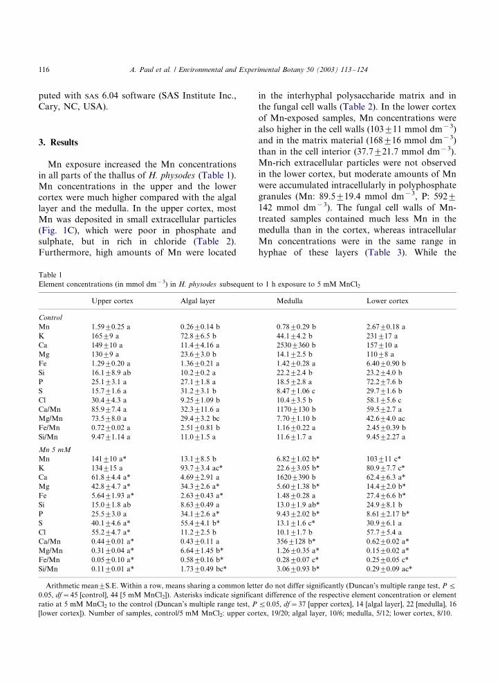
Mn exposure increased the Mn concentrationsin all parts of the thallus of H. physodes (Table 1).
Mn concentrations in the upper and the lower
cortex were much higher compared with the algal
layer and the medulla. In the upper cortex, most
Mn was deposited in small extracellular particles
(Fig. 1C), which were poor in phosphate and
sulphate, but in rich in chloride (Table 2).
Furthermore, high amounts of Mn were located
in the interhyphal polysaccharide matrix and in
the fungal cell walls (Table 2). In the lower cortex
of Mn-exposed samples, Mn concentrations were
also higher in the cell walls (1039/11 mmol dm�3)
and in the matrix material (1689/16 mmol dm�3)
than in the cell interior (37.79/21.7 mmol dm�3).
Mn-rich extracellular particles were not observed
in the lower cortex, but moderate amounts of Mn
were accumulated intracellularly in polyphosphate
granules (Mn: 89.59/19.4 mmol dm�3, P: 5929/
142 mmol dm�3). The fungal cell walls of Mn-
treated samples contained much less Mn in the
medulla than in the cortex, whereas intracellular
Mn concentrations were in the same range in
hyphae of these layers (Table 3). While the
Table 1
Element concentrations (in mmol dm�3) in H. physodes subsequent to 1 h exposure to 5 mM MnCl2
Upper cortex Algal layer Medulla Lower cortex
Control
Mn 1.599/0.25 a 0.269/0.14 b 0.789/0.29 b 2.679/0.18 a
K 1659/9 a 72.89/6.5 b 44.19/4.2 b 2319/17 a
Ca 1499/10 a 11.49/4.16 a 25309/360 b 1579/10 a
Mg 1309/9 a 23.69/3.0 b 14.19/2.5 b 1109/8 a
Fe 1.299/0.20 a 1.369/0.21 a 1.429/0.28 a 6.409/0.90 b
Si 16.19/8.9 ab 10.29/0.2 a 22.29/2.4 b 23.29/4.0 b
P 25.19/3.1 a 27.19/1.8 a 18.59/2.8 a 72.29/7.6 b
S 15.79/1.6 a 31.29/3.1 b 8.479/1.06 c 29.79/1.6 b
Cl 30.49/4.3 a 9.259/1.09 b 10.49/3.5 b 58.19/5.6 c
Ca/Mn 85.99/7.4 a 32.39/11.6 a 11709/130 b 59.59/2.7 a
Mg/Mn 73.59/8.0 a 29.49/3.2 bc 7.709/1.10 b 42.69/4.0 ac
Fe/Mn 0.729/0.02 a 2.519/0.81 b 1.169/0.22 a 2.459/0.39 b
Si/Mn 9.479/1.14 a 11.09/1.5 a 11.69/1.7 a 9.459/2.27 a
Mn 5 mM
Mn 1419/10 a* 13.19/8.5 b 6.829/1.02 b* 1039/11 c*
K 1349/15 a 93.79/3.4 ac* 22.69/3.05 b* 80.99/7.7 c*
Ca 61.89/4.4 a* 4.699/2.91 a 16209/390 b 62.49/6.3 a*
Mg 42.89/4.7 a* 34.39/2.6 a* 5.609/1.38 b* 14.49/2.0 b*
Fe 5.649/1.93 a* 2.639/0.43 a* 1.489/0.28 a 27.49/6.6 b*
Si 15.09/1.8 ab 8.639/0.49 a 13.09/1.9 ab* 24.99/8.1 b
P 25.59/3.0 a 34.19/2.6 a* 9.439/2.02 b* 8.619/2.17 b*
S 40.19/4.6 a* 55.49/4.1 b* 13.19/1.6 c* 30.99/6.1 a
Cl 55.29/4.7 a* 11.29/2.5 b 10.19/1.7 b 57.79/5.4 a
Ca/Mn 0.449/0.01 a* 0.439/0.11 a 3569/128 b* 0.629/0.02 a*
Mg/Mn 0.319/0.04 a* 6.649/1.45 b* 1.269/0.35 a* 0.159/0.02 a*
Fe/Mn 0.059/0.10 a* 0.589/0.16 b* 0.289/0.07 c* 0.259/0.05 c*
Si/Mn 0.119/0.01 a* 1.739/0.49 bc* 3.069/0.93 b* 0.299/0.09 ac*
Arithmetic mean9/S.E. Within a row, means sharing a common letter do not differ significantly (Duncan’s multiple range test, P 5/
0.05, df�/45 [control], 44 [5 mM MnCl2]). Asterisks indicate significant difference of the respective element concentration or element
ratio at 5 mM MnCl2 to the control (Duncan’s multiple range test, P 5/0.05, df�/37 [upper cortex], 14 [algal layer], 22 [medulla], 16
[lower cortex]). Number of samples, control/5 mM MnCl2: upper cortex, 19/20; algal layer, 10/6; medulla, 5/12; lower cortex, 8/10.
A. Paul et al. / Environmental and Experimental Botany 50 (2003) 113�/124116
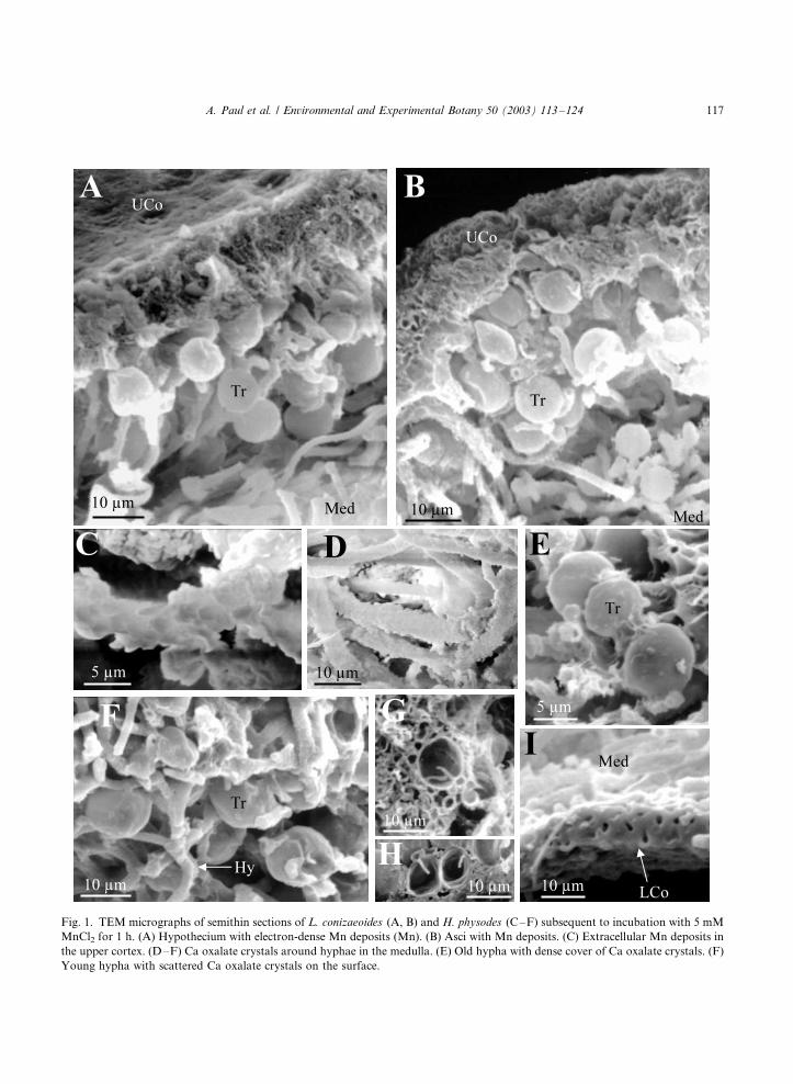
Fig. 1. TEM micrographs of semithin sections of L. conizaeoides (A, B) and H. physodes (C�/F) subsequent to incubation with 5 mM
MnCl2 for 1 h. (A) Hypothecium with electron-dense Mn deposits (Mn). (B) Asci with Mn deposits. (C) Extracellular Mn deposits in
the upper cortex. (D�/F) Ca oxalate crystals around hyphae in the medulla. (E) Old hypha with dense cover of Ca oxalate crystals. (F)
Young hypha with scattered Ca oxalate crystals on the surface.
A. Paul et al. / Environmental and Experimental Botany 50 (2003) 113�/124 117
polysaccharide matrix is restricted to the cortex,
the medulla of H. physodes is characterised by
numerous Ca oxalate crystals covering the surface
of the hyphae (Fig. 1D). While old hyphae were
completely surrounded by crystals (Fig. 1E),
young parts of the hyphae were only patchily
covered (Fig. 1F). Because of these crystals, very
high (molar) Ca concentrations occurred in the
medulla (Table 1). Mn was deposited in the
oxalate crystals decreasing their Ca/Mn ratios
(Table 4). Significantly more Mn was recovered
in the crystals on young versus old hyphae. The
number of crystals in Mn-treated samples was not
different from control samples as visually assessed
with SEM (Fig. 2C, D) and TEM. Algal cells took
up less Mn intracellularly and absorbed less Mn at
the cell walls compared with the fungal cells of any
layer (Table 3). Mn deposits in polyphosphate
granules or other particles were not observed in
the photobiont.
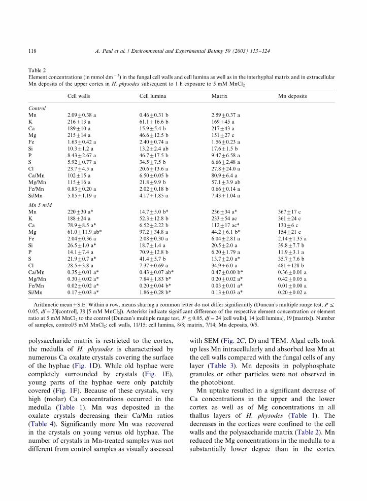
Mn uptake resulted in a significant decrease of
Ca concentrations in the upper and the lower
cortex as well as of Mg concentrations in all
thallus layers of H. physodes (Table 1). The
decreases in the cortices were confined to the cell
walls and the polysaccharide matrix (Table 2). Mn
reduced the Mg concentrations in the medulla to a
substantially lower degree than in the cortex
Table 2
Element concentrations (in mmol dm�3) in the fungal cell walls and cell lumina as well as in the interhyphal matrix and in extracellular
Mn deposits of the upper cortex in H. physodes subsequent to 1 h exposure to 5 mM MnCl2
Cell walls Cell lumina Matrix Mn deposits
Control
Mn 2.099/0.38 a 0.469/0.31 b 2.599/0.37 a
K 2169/13 a 61.19/16.6 b 1699/45 a
Ca 1899/10 a 15.99/5.4 b 2179/43 a
Mg 2159/14 a 46.69/12.5 b 1519/27 c
Fe 1.639/0.42 a 2.409/0.74 a 1.569/0.23 a
Si 10.39/1.2 a 13.29/2.4 ab 17.69/1.5 b
P 8.439/2.67 a 46.79/17.5 b 9.479/6.58 a
S 5.929/0.77 a 34.59/7.5 b 6.669/2.48 a
Cl 23.79/4.5 a 20.69/13.6 a 27.89/24.0 a
Ca/Mn 1029/15 a 6.509/0.05 b 80.99/6.4 a
Mg/Mn 1159/16 a 21.89/9.9 b 57.19/3.9 ab
Fe/Mn 0.839/0.20 a 2.029/0.18 b 0.669/0.14 a
Si/Mn 5.859/1.19 a 4.179/1.85 a 7.439/1.04 a
Mn 5 mM
Mn 2209/30 a* 14.79/5.0 b* 2369/34 a* 3679/17 c
K 1889/24 a 52.39/12.8 b 2339/54 ac 3619/24 c
Ca 78.99/8.5 a* 6.529/2.22 b 1129/17 ac* 1309/6 c
Mg 61.09/11.9 ab* 97.29/34.8 a 44.29/6.1 b* 1549/21 c
Fe 2.049/0.36 a 2.089/0.30 a 6.049/2.81 a 2.149/1.35 a
Si 26.59/1.0 a* 18.79/1.4 a 20.59/2.0 a 39.89/7.7 b
P 14.19/7.4 a 70.99/12.8 b 6.209/1.79 a 11.99/3.1 a
S 21.99/0.7 a* 41.49/5.7 b 13.79/2.0 a* 35.79/7.6 b
Cl 28.59/3.8 a 7.379/0.69 a 34.99/6.0 a 4819/128 b
Ca/Mn 0.359/0.01 a* 0.439/0.07 ab* 0.479/0.00 b* 0.369/0.01 a
Mg/Mn 0.309/0.02 a* 7.849/1.83 b* 0.209/0.02 a* 0.429/0.05 a
Fe/Mn 0.029/0.02 a* 0.209/0.04 b* 0.039/0.01 a* 0.019/0.00 a
Si/Mn 0.179/0.03 a* 1.869/0.28 b* 0.139/0.03 a* 0.209/0.02 a
Arithmetic mean9/S.E. Within a row, means sharing a common letter do not differ significantly (Duncan’s multiple range test, P 5/
0.05, df�/23[control], 38 [5 mM MnCl2]). Asterisks indicate significant difference of the respective element concentration or element
ratio at 5 mM MnCl2 to the control (Duncan’s multiple range test, P 5/0.05, df�/24 [cell walls], 14 [cell lumina], 19 [matrix]). Number
of samples, control/5 mM MnCl2: cell walls, 11/15; cell lumina, 8/8; matrix, 7/14; Mn deposits, 0/5.
A. Paul et al. / Environmental and Experimental Botany 50 (2003) 113�/124118
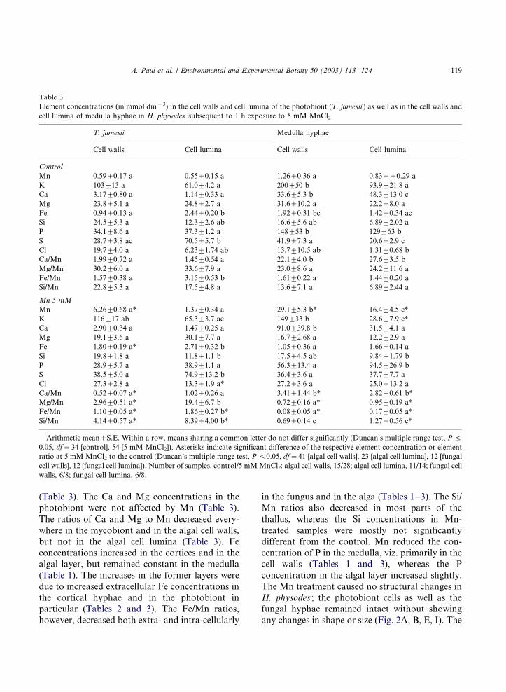
(Table 3). The Ca and Mg concentrations in the
photobiont were not affected by Mn (Table 3).
The ratios of Ca and Mg to Mn decreased every-
where in the mycobiont and in the algal cell walls,
but not in the algal cell lumina (Table 3). Fe
concentrations increased in the cortices and in the
algal layer, but remained constant in the medulla
(Table 1). The increases in the former layers were
due to increased extracellular Fe concentrations in
the cortical hyphae and in the photobiont in
particular (Tables 2 and 3). The Fe/Mn ratios,
however, decreased both extra- and intra-cellularly
in the fungus and in the alga (Tables 1�/3). The Si/
Mn ratios also decreased in most parts of the
thallus, whereas the Si concentrations in Mn-
treated samples were mostly not significantly
different from the control. Mn reduced the con-
centration of P in the medulla, viz. primarily in the
cell walls (Tables 1 and 3), whereas the P
concentration in the algal layer increased slightly.
The Mn treatment caused no structural changes in
H. physodes ; the photobiont cells as well as the
fungal hyphae remained intact without showing
any changes in shape or size (Fig. 2A, B, E, I). The
Table 3
Element concentrations (in mmol dm�3) in the cell walls and cell lumina of the photobiont (T. jamesii ) as well as in the cell walls and
cell lumina of medulla hyphae in H. physodes subsequent to 1 h exposure to 5 mM MnCl2
T. jamesii Medulla hyphae
Cell walls Cell lumina Cell walls Cell lumina
Control
Mn 0.599/0.17 a 0.559/0.15 a 1.269/0.36 a 0.839/9/0.29 a
K 1039/13 a 61.09/4.2 a 2009/50 b 93.99/21.8 a
Ca 3.179/0.80 a 1.149/0.33 a 33.69/5.3 b 48.39/13.0 c
Mg 23.89/5.1 a 24.89/2.7 a 31.69/10.2 a 22.29/8.0 a
Fe 0.949/0.13 a 2.449/0.20 b 1.929/0.31 bc 1.429/0.34 ac
Si 24.59/5.3 a 12.39/2.6 ab 16.69/5.6 ab 6.899/2.02 a
P 34.19/8.6 a 37.39/1.2 a 1489/53 b 1299/63 b
S 28.79/3.8 ac 70.59/5.7 b 41.99/7.3 a 20.69/2.9 c
Cl 19.79/4.0 a 6.239/1.74 ab 13.79/10.5 ab 1.319/0.68 b
Ca/Mn 1.999/0.72 a 1.459/0.54 a 22.19/4.0 b 27.69/3.5 b
Mg/Mn 30.29/6.0 a 33.69/7.9 a 23.09/8.6 a 24.29/11.6 a
Fe/Mn 1.579/0.38 a 3.159/0.53 b 1.619/0.22 a 1.449/0.20 a
Si/Mn 22.89/5.3 a 17.59/4.8 a 13.69/7.1 a 6.899/2.44 a
Mn 5 mM
Mn 6.269/0.68 a* 1.379/0.34 a 29.19/5.3 b* 16.49/4.5 c*
K 1169/17 ab 65.39/3.7 ac 1499/33 b 28.69/7.9 c*
Ca 2.909/0.34 a 1.479/0.25 a 91.09/39.8 b 31.59/4.1 a
Mg 19.19/3.6 a 30.19/7.7 a 16.79/2.68 a 12.29/2.9 a
Fe 1.809/0.19 a* 2.719/0.32 b 1.059/0.36 a 1.669/0.14 a
Si 19.89/1.8 a 11.89/1.1 b 17.59/4.5 ab 9.849/1.79 b
P 28.99/5.7 a 38.99/1.1 a 56.39/13.4 a 94.59/26.9 b
S 38.59/5.0 a 74.99/13.2 b 36.49/3.6 a 37.79/7.7 a
Cl 27.39/2.8 a 13.39/1.9 a* 27.29/3.6 a 25.09/13.2 a
Ca/Mn 0.529/0.07 a* 1.029/0.26 a 3.419/1.44 b* 2.829/0.61 b*
Mg/Mn 2.969/0.51 a* 19.49/6.7 b 0.729/0.16 a* 0.959/0.19 a*
Fe/Mn 1.109/0.05 a* 1.869/0.27 b* 0.089/0.05 a* 0.179/0.05 a*
Si/Mn 4.149/0.57 a* 8.399/4.00 b* 0.699/0.14 c 1.279/0.56 c*
Arithmetic mean9/S.E. Within a row, means sharing a common letter do not differ significantly (Duncan’s multiple range test, P 5/
0.05, df�/34 [control], 54 [5 mM MnCl2]). Asterisks indicate significant difference of the respective element concentration or element
ratio at 5 mM MnCl2 to the control (Duncan’s multiple range test, P 5/0.05, df�/41 [algal cell walls], 23 [algal cell lumina], 12 [fungal
cell walls], 12 [fungal cell lumina]). Number of samples, control/5 mM MnCl2: algal cell walls, 15/28; algal cell lumina, 11/14; fungal cell
walls, 6/8; fungal cell lumina, 6/8.
A. Paul et al. / Environmental and Experimental Botany 50 (2003) 113�/124 119
physical contact between mycobiont and photo-
biont was not affected.
L. conizaeoides contained much less Mn sub-
sequent to the incubation with Mn than H.
physodes (Table 5). Mn concentrations in L.
conizaeoides were in the same range as in the
medulla of H. physodes , but were half as much as
in the algal layer and 15�/25 times lower than in
the cortices of H. physodes . Mn was evenly
distributed within the vegetative thallus parts of
Mn-exposed L. conizaeoides samples. Fungal cell
walls (7.309/1.05 mmol dm�3) and cell lumina
(1.549/0.52 mmol dm�3) contained much less Mn
compared with hyphae of H. physodes . In the algal
cells of Mn-exposed L. conizaeoides , Mn concen-
trations amounted to less than 50% of the corre-
sponding concentrations in H. physodes in the cell
walls (2.739/0.78 mmol dm�3) and to less than
30% in the cell lumina (0.379/0.16 mmol dm�3).
Mn uptake decreased the ratios of Ca, Mg, and Fe
to Mn in all thallus parts of L. conizaeoides ,
whereas the Si/Mn ratio remained constant (Table
5). The concentrations of Fe and Si significantly
increased in the entire thallus, whereas the con-
centrations of Mg and P increased only in the
upper thallus; the concentration of Ca decreased in
the lower thallus.
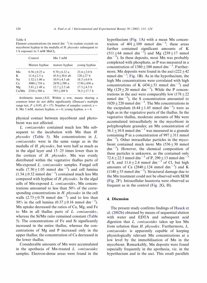
Considerable amounts of Mn were accumulated
in the apothecia of Mn-treated L. conizaeoides
samples. Electron-dense areas were found in the
hypothecium (Fig. 1A) with a mean Mn concen-tration of 4019/109 mmol dm�3; these areas
further contained significant amounts of K
(5119/64 mmol dm�3) and Mg (2399/17 mmol
dm�3). In these deposits, most Mn was probably
complexed with phosphate, as P was measured in a
concentration of 13809/100 mmol dm�3. Further-
more, Mn deposits were found in the asci (2229/42
mmol dm�3; Fig. 1B). As in the hypothecium, thehigh Mn concentrations were correlated with high
concentrations of K (4349/51 mmol dm�3) and
Mg (1299/20 mmol dm�3). While the P concen-
trations in the asci were comparably low (1789/22
mmol dm�3), the S concentration amounted to
10209/220 mmol dm�3. The Mn concentrations in
the excipulum (6.449/1.45 mmol dm�3) were as
high as in the vegetative parts of the thallus. In thevegetative thallus, moderate amounts of Mn were
accumulated intracellularly in the mycobiont in
polyphosphate granules; an Mn concentration of
36.19/10.8 mmol dm�3 was measured in a granule
containing P in a concentration of 9979/311 mmol
dm�3). Other intracellular particles in the myco-
biont contained much more Mn (5369/30 mmol
dm�3). However, the chemical composition ofthese particles is unknown, as the contained only
72.69/22.5 mmol dm�3 of P, 2909/13 mmol dm�3
of S, and 11.69/2.4 mmol dm�3 of Cl, but high
amounts of Ca (28409/124 mmol dm�3) and K
(11409/53 mmol dm�3). Structural damage due to
the Mn treatment could not be observed with SEM
(Fig. 2F). Intracellular haustoria were observed as
frequent as in the control (Fig. 2G, H).
4. Discussion
The present study confirms findings of Hauck et
al. (2002b) obtained by means of sequential elution
with water and EDTA and subsequent acid
digestion that L. conizaeoides takes up less Mn
from solution than H. physodes . Furthermore, L.
conizaeoides is apparently capable of keeping
physiologically relevant Mn concentrations at a
low level by the immobilisation of Mn in the
mycobiont. Remarkably, Mn deposits were found
especially frequently in the apothecia, viz. in the
hypothecium and in the asci. This result parallels
Table 4
Element concentrations (in mmol dm�3) in oxalate crystals on
mycobiont hyphae in the medulla of H. physodes subsequent to
1 h exposure to 5 mM MnCl2
Control Mn 5 mM
Mature hyphae mature hyphae young hyphae
Mn 0.569/0.25 a 9.709/6.12 a 23.49/2.0 b
K 11.49/2.5 a 43.89/30.6 ab 1289/27 b
Na 1.529/1.00 a 10.99/4.5 ab 18.39/6.0 b
Ca 30809/710 a 26709/390 a 17309/430 a
Mg 5.819/1.49 a 12.79/2.2 ab 17.39/4.5 b
Ca/Mn 23109/580 a 7939/269 b 76.59/17.7 b
Arithmetic mean9/S.E. Within a row, means sharing a
common letter do not differ significantly (Duncan’s multiple
range test, P 5/0.05, df�/17). Number of samples: control, n�/
8; Mn 5 mM, mature hyphae, n�/6; young hyphae, n�/6.
A. Paul et al. / Environmental and Experimental Botany 50 (2003) 113�/124120
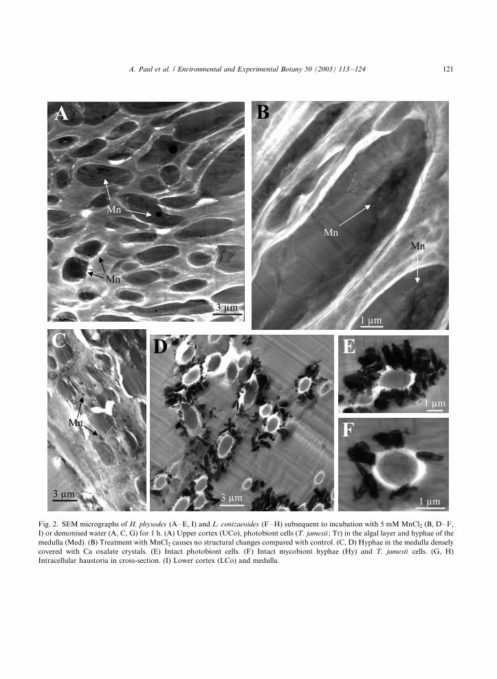
Fig. 2. SEM micrographs of H. physodes (A�/E, I) and L. conizaeoides (F�/H) subsequent to incubation with 5 mM MnCl2 (B, D�/F,
I) or demonised water (A, C, G) for 1 h. (A) Upper cortex (UCo), photobiont cells (T. jamesii ; Tr) in the algal layer and hyphae of the
medulla (Med). (B) Treatment with MnCl2 causes no structural changes compared with control. (C, D) Hyphae in the medulla densely
covered with Ca oxalate crystals. (E) Intact photobiont cells. (F) Intact mycobiont hyphae (Hy) and T. jamesii cells. (G, H)
Intracellular haustoria in cross-section. (I) Lower cortex (LCo) and medulla.
A. Paul et al. / Environmental and Experimental Botany 50 (2003) 113�/124 121
findings in Lecidella bullata (Fam. Lecanoraceae),
where Cu was accumulated in the hypothecium
and in a pruina on the disc surface (Purvis et al.,
1990). Cu accumulation also occurred in apothecia
of Lecanora polytropa (Alstrup and Hansen,
1977). X-ray microanalysis suggests that Mn
deposits of different chemical constitution occur
in L. conizaeoides . In vegetative parts of the
thallus, Mn was found in polyphosphate granules
and in an unknown type of particles. Mn immo-
bilisation in polyphosphate granules has alreadybeen observed in soredia of H. physodes (Hauck et
al., 2002c). In the hypothecium, Mn was also
immobilised with phosphate. Nonetheless, it
should be tested whether complexation with the
depsidone fumarprotocetraric acid is involved in
the immobilisation of Mn in L. conizaeoides .
Fumarprotocetraric acid is the major lichen sub-
stance in L. conizaeoides (Kummerling, 1991). Thedepsidone psoromic acid was found to be the
anionic compound complexing Cu in the apothecia
of L. bullata (Purvis et al., 1990). Complexes of Cu
with psoromic acid and a further depsidone,
norstictic acid, were found in several lichen species
(Purvis et al., 1987, 1990). The three depsidones in
question have an ortho -phenolic hydroxyl group
and an adjacent aldehyde group in common (Hu-neck and Yoshimura, 1996). These functional
groups, which are the assumed binding sites in
the complexes of Cu with psoromic and norstictic
acid (Purvis et al., 1990), perhaps also bind to Mn.
Since complexes of lichen substances with Mn, or
of norstictic acid with metal ions, have not been
found so far, further study is needed.
While L. conizaeoides maintained low Mnconcentrations in the cell lumina as well as in the
cell walls both of the mycobiont and the photo-
biont, Mn concentrations in H. physodes were
strongly dependent on the location within the
thallus. The lowest Mn concentrations occurred
in the algal cell lumina, followed by the algal cell
walls. This suggests that the mycobiont in H.
physodes may have the ability to protect thephotobiont to a certain extent from the invasion
of Mn. The photobiont is usually thought to be the
most sensitive part of the lichen symbiosis, because
the mycobiont enhances the permeability of the
algal or cyanobacterial membranes for assimilate
transfer (Ahmadjian, 1993). In contrast to the
mycobiont, the intracellular Ca/Mn and Mg/Mn
ratios in the photobiont were not affected by Mn.In the fungal cells of H. physodes , the Mn
concentrations increased significantly. Most Mn
was bound to the cortical surfaces of H. physodes .
Fungal cell walls provide many anionic exchange
sites such as carboxyl groups (Richardson, 1995).
Extracellular polysaccharides formed by lichens
also harbour carboxyl groups and other anionic
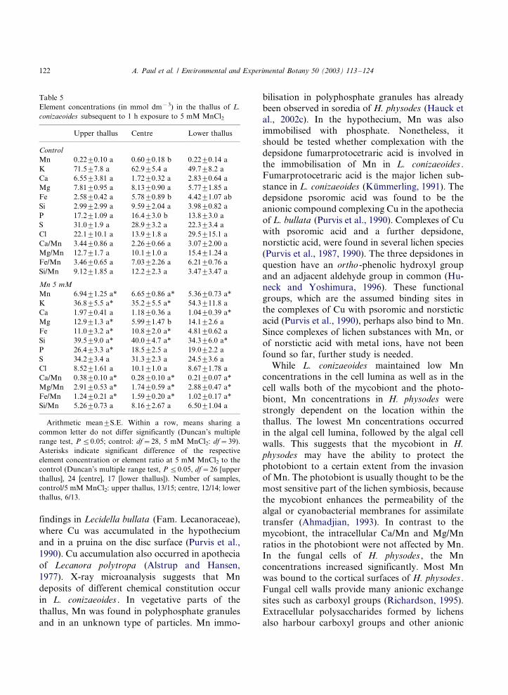
Table 5
Element concentrations (in mmol dm�3) in the thallus of L.
conizaeoides subsequent to 1 h exposure to 5 mM MnCl2
Upper thallus Centre Lower thallus
Control
Mn 0.229/0.10 a 0.609/0.18 b 0.229/0.14 a
K 71.59/7.8 a 62.99/5.4 a 49.79/8.2 a
Ca 6.559/3.81 a 1.729/0.32 a 2.839/0.64 a
Mg 7.819/0.95 a 8.139/0.90 a 5.779/1.85 a
Fe 2.589/0.42 a 5.789/0.89 b 4.429/1.07 ab
Si 2.999/2.99 a 9.599/2.04 a 3.989/0.82 a
P 17.29/1.09 a 16.49/3.0 b 13.89/3.0 a
S 31.09/1.9 a 28.99/3.2 a 22.39/3.4 a
Cl 22.19/10.1 a 13.99/1.8 a 29.59/15.1 a
Ca/Mn 3.449/0.86 a 2.269/0.66 a 3.079/2.00 a
Mg/Mn 12.79/1.7 a 10.19/1.0 a 15.49/1.24 a
Fe/Mn 3.469/0.65 a 7.039/2.26 a 6.219/0.76 a
Si/Mn 9.129/1.85 a 12.29/2.3 a 3.479/3.47 a
Mn 5 mM
Mn 6.949/1.25 a* 6.659/0.86 a* 5.369/0.73 a*
K 36.89/5.5 a* 35.29/5.5 a* 54.39/11.8 a
Ca 1.979/0.41 a 1.189/0.36 a 1.049/0.39 a*
Mg 12.99/1.3 a* 5.999/1.47 b 14.19/2.6 a
Fe 11.09/3.2 a* 10.89/2.0 a* 4.819/0.62 a
Si 39.59/9.0 a* 40.09/4.7 a* 34.39/6.0 a*
P 26.49/3.3 a* 18.59/2.5 a 19.09/2.2 a
S 34.29/3.4 a 31.39/2.3 a 24.59/3.6 a
Cl 8.529/1.61 a 10.19/1.0 a 8.679/1.78 a
Ca/Mn 0.389/0.10 a* 0.289/0.10 a* 0.219/0.07 a*
Mg/Mn 2.919/0.53 a* 1.749/0.59 a* 2.889/0.47 a*
Fe/Mn 1.249/0.21 a* 1.599/0.20 a* 1.029/0.17 a*
Si/Mn 5.269/0.73 a 8.169/2.67 a 6.509/1.04 a
Arithmetic mean9/S.E. Within a row, means sharing a
common letter do not differ significantly (Duncan’s multiple
range test, P 5/0.05; control: df�/28, 5 mM MnCl2: df�/39).
Asterisks indicate significant difference of the respective
element concentration or element ratio at 5 mM MnCl2 to the
control (Duncan’s multiple range test, P 5/0.05, df�/26 [upper
thallus], 24 [centre], 17 [lower thallus]). Number of samples,
control/5 mM MnCl2: upper thallus, 13/15; centre, 12/14; lower
thallus, 6/13.
A. Paul et al. / Environmental and Experimental Botany 50 (2003) 113�/124122
binding sites (Modenesi and Vanzo, 1986; Mod-enesi et al., 1986). Under field conditions, most
extracellular exchange sites in lichens are usually
charged with Ca and Mg (Brown and Brown,
1991). This explains why primarily Ca and Mg
were released both from the cell walls and from the
polysaccharide matrix during Mn absorption
(Hauck et al., 2002b). In contrast to L. coni-
zaeoides , the Si/Mn ratios were significantly re-duced in the thallus of H. physodes , which may
result in higher Mn sensitivity of the latter (Hauck
et al., 2002c). The enrichment of Si in L. coni-
zaeoides during the incubation may result from
very small residual substrate pieces, which could
not be removed completely from the thallus before
the experiment. However, another possible expla-
nation is that the samples were somewhat hetero-genous, though they were all collected from the
same site and mixed before the incubation proce-
dure. Higher Si and S concentrations in some
thallus parts of the Mn-treated H. physodes
samples than in the control support the latter
explanation.
The accumulation of Mn in the Ca oxalate
crystals in the medulla of H. physodes may bedetrimental, because impurities could alter the
crystal structure and integrity (Modenesi et al.,
1997). Since the Mn concentration was highest in
oxalate crystals on the surface of young growing
hyphae, Mn can presumably influence the forma-
tion of new crystals (Cody and Horner, 1984). The
Ca oxalate crystals in the medulla fulfil various
structural and physiological functions. The crys-tals in H. physodes , consisting primarily of Ca
oxalate monohydrate (whewellite), provide me-
chanical stability to the medulla with its large
cavities, which are thought to be important for the
diffusion of gases (Modenesi et al., 1997). Further-
more, the crystals in the medulla are supposed to
reflect light that already passed the algal layer and,
thus, to increase the photosynthetic quantum yield(Modenesi et al., 2000; Clark et al., 2001).
The short-term exposure to Mn did not result in
structural damage in neither H. physodes nor L.
conizaeoides . Since Hauck et al. (2002c) found
numerous damage symptoms in soredia of H.
physodes cultivated on agar plates with 7 mM
MnCl2 for 8 days, an experiment with thalli
repeatedly sprayed with low-concentrated Mnsolutions on a long-term basis should be carried
out in order to test whether adult thalli of
epiphytic lichens suffer structural damage when
they are exposed to Mn for extended periods.
In conclusion, the study suggests that L. coni-
zaeoides is capable of maintaining much lower Mn
levels in the cell lumina and in the cell walls
compared with H. physodes . The high amounts ofreleased Ca and Mg in H. physodes could affect,
e.g. the stability of cell walls and membranes, but
could also disturb various intracellular processes
(Hauck et al., 2002b). Such considerations are
consistent with the observed decreasing abundance
of H. physodes found with increasing Mn/Ca ratio
of stemflow in the field (Hauck et al., 2002a). The
Fe/Mn ratios were much higher in L. conizaeoides
versus H. physodes . This can be a major cause for
the different Mn sensitivity of the two species,
because Hauck et al. (2003) showed that Fe has a
strongly alleviating on Mn toxicity in H. physodes .
Though Mn was shown to be immobilised with
phosphate also in the present study, P does not
appear to be a limiting factor for the abundance of
either lichen species; Hauck et al. (2003) showedthat incubation of H. physodes with 10 mM MnCl2for 1 h did not result in ATP deficiency. Translo-
cation of P from the medulla to the algal layer in
H. physodes (Table 1) may indicate that P was
used there for the immobilisation of Mn in order
to protect the photobiont from toxic Mn concen-
trations. Higher Si/Mn ratios in L. conizaeoides
than in H. physodes suggest that Si is involved inMn tolerance in lichens, as already suggested by
Hauck et al. (2002c) because of higher Si/Mn
ratios in living versus collapsed photobiont cells in
Mn-treated H. physodes soredia.
References
Ahmadjian, V., 1993. The Lichen Symbiosis. Wiley, New York.
Alstrup, V., Hansen, E.S., 1977. Three species of lichens
tolerant of high concentrations of copper. Oikos 29, 290�/
293.
Brown, D.H., Brown, R.M., 1991. Mineral cycling and lichens:
the physiological basis. Lichenologist 23, 293�/307.
Clark, B.M., St Clair, L.L., Mangelson, N.F., Rees, L.B.,
Grant, P.G., Bench, G.S., 2001. Characterization of myco-
A. Paul et al. / Environmental and Experimental Botany 50 (2003) 113�/124 123
biont adaptions in the foliose lichen Xanthoparmelia chlor-
ochroa (Parmeliaceae). Am. J. Bot. 88, 1742�/1749.
Cody, A.M., Horner, H.T., 1984. Crystallographic analysis of
crystal images in scanning electron micrographs and their
application to phytocrystalline studies. Scanning Electr.
Microsc. 6, 1551�/1560.
El-Jaoual, T., Cox, D.A., 1998. Manganese toxicity in plants. J.
Plant Nutr. 21, 353�/386.
Fritz, E., 1989. X-ray microanalysis of diffusible elements in
plant cells after freeze-drying, pressure-infiltration with
ether and embedding in plastic. Scanning Microsc. 3, 517�/
526.
Fritz, E., 1991. The use of adhesive-coated grids for the X-ray
microanalysis of dry-cut sections in the TEM. J. Microsc.
161, 501�/504.
Fritz, E., Jentschke, G., 1994. Agar standards for quantitative
X-ray microanalysis of resin-embedded plant tissues. J.
Microsc. 164, 47�/50.
Hauck, M., 2000. Ecology of epiphytic lichens in a montane
spruce forest: influence of forest dieback and forest manage-
ment on chemical habitat conditions. Diss. Bot. 327, 1�/232.
Hauck, M., 2003. Epiphytic lichen diversity and forest dieback:
the role of chemical site factors. Bryologist 106 (in press).
Hauck, M., Spribille, T., 2002. The Mn/Ca and Mn/Mg ratios
in bark as possible causes for the occurrence of Lobarion
lichens on conifers in the dripzone of Populus in western
North America. Lichenologist 34, 527�/532.
Hauck, M., Hesse, V., Jung, R., Zoller, T., Runge, M., 2001a.
Long-distance transported sulphur as a limiting factor for
the abundance of Lecanora conizaeoides in montane spruce
forests. Lichenologist 33, 267�/269.
Hauck, M., Jung, R., Runge, M., 2001b. Relevance of element
content of bark for the distribution of epiphytic lichens in a
montane spruce forest affected by forest dieback. Environ.
Pollut. 112, 221�/227.
Hauck, M., Hesse, V., Runge, M., 2002a. Correlations between
the Mn/Ca ratio in stemflow and epiphytic lichen abundance
in a dieback-affected spruce forest of the Harz Mountains.
Flora 197, 361�/369.
Hauck, M., Mulack, C., Paul, A., 2002b. Manganese uptake in
the epiphytic lichens Hypogymnia physodes and Lecanora
conizaeoides . Environ. Exp. Bot. 48, 107�/117.
Hauck, M., Paul, A., Mulack, C., Fritz, E., Runge, M., 2002c.
Effects of manganese on the viability of vegetative diaspores
of the epiphytic lichen Hypogymnia physodes . Environ. Exp.
Bot. 47, 127�/142.
Hauck, M., Paul, A., Gross, S., Raubuch, M., 2003. Manganese
toxicity in epiphytic lichens: chlorophyll degradation and
interaction with iron and phosphorus. Environ. Exp. Bot.
49, 181�/191.
Honegger, R., 1984. Cytological aspects of the mycobiont�/
phycobiont relationship in lichens. Haustorial types, phy-
cobiont cell wall types, and the ultrastructure of the cell
surface layers in some cultured and symbiotic myco- and
phycobionts. Lichenologist 16, 111�/127.
Honegger, R., Peter, M., Scherrer, S., 1996. Drought-induced
structural alterations at the mycobiont�/photobiont inter-
face in a range of foliose macrolichens. Protoplasma 190,
221�/232.
Huneck, S., Yoshimura, I., 1996. Identification of Lichen
Substances. Springer, Berlin.
Kummerling, H., 1991. Zur Kenntnis der Flechtenflora am
Hohen Meißner und in seinem Vorland (Hessen) unter
besonderer Berucksichtigung chemischer Merkmale. Bibl.
Lichenol. 41, 1�/315.
Levia, D.F., Herwitz, S.R., 2000. Physical properties of water in
relation to stemflow leachate dynamics: implications for
nutrient cycling. Can. J. For. Res. 30, 662�/666.
Lovestam, G., Johansson, E.-M., Johansson, S., Pallon, J.,
1990. Elemental micro patterns in tree rings*/a feasibility
study using scanning proton microprobe analysis. Ambio
19, 87�/93.
Modenesi, P., Vanzo, C., 1986. The cortical surfaces in
Parmelia saxatilis and P. caperata : a histochemical ap-
proach. Lichenologist 18, 329�/338.
Modenesi, P., Lajolo, L., Dondero, G., 1986. Acid carbohy-
drates in the hypothallus of Catillaria bouteillei (Desm.)
Zahlbr. A histochemical localization. Cryptog. Bryol.
Lichenol. 7, 1�/10.
Modenesi, P., Canepa, R., Tafanelli, A., 1997. The structural
role of calcium oxalate and medullary architecture in
Menegazzia terebrata and Hypogymnia physodes . Bibl.
Lichenol. 68, 101�/110.
Modenesi, P., Piana, M., Giordani, P., Tafanelli, A., Bartoli,
A., 2000. Calcium oxalate and medullary architecture in
Xanthomaculina convoluta . Lichenologist 32, 505�/512.
Purvis, O.W., Elix, J.A., Broomhead, J.A., Jones, G.C., 1987.
The occurrence of copper�/norstictic acid in lichens from
cupriferous substrata. Lichenologist 19, 193�/203.
Purvis, O.W., Elix, J.A., Gaul, K.L., 1990. The occurrence of
copper�/psoromic acid in lichens from cupriferous sub-
strata. Lichenologist 22, 345�/354.
Richardson, D.H.S., 1995. Metal uptake in lichens. Symbiosis
18, 119�/127.
Schmull, M., Hauck, M., 2003a. Element microdistribution in
the bark of Abies balsamea and Picea rubens and its impact
on epiphytic lichen abundance on Whiteface Mountain,
New York. Flora 198, (in press).
Schmull, M., Hauck, M., 2003bb. Extraction methods for
assessing the availability of cations for epiphytic lichens
from bark. Environ. Exp. Bot. 49, 273�/283.
Sloof, J.E., Wolterbeek, B.T., 1993. Substrate influence on
epiphytic lichens. Environ. Monit. Assess. 25, 225�/234.
Wirth, V., 1985. Zur Ausbreitung, Herkunft und Okologie
anthropogen geforderter Rinden- und Holzflechten. Tue-
xenia 5, 523�/535.
A. Paul et al. / Environmental and Experimental Botany 50 (2003) 113�/124124