Effects of an extra X chromosome on language lateralization: An fMRI study with Klinefelter men...
273
Xploring social cognitive pathways to psychopathology Studies with Klinefelter (XXY) men Sophie van Rijn
-
Upload
umcutrecht -
Category
Documents
-
view
0 -
download
0
Transcript of Effects of an extra X chromosome on language lateralization: An fMRI study with Klinefelter men...
Xploring social
cognitive pathways to
psychopathology
Studies with Klinefelter (XXY) men
Sophie van Rijn
Xploring social cognitive pathways
to psychopathology
Studies with Klinefelter (XXY) men
Een exploratie van sociaal-cognitieve routes naar psychopathologie:studies met Klinefelter (XXY) mannen
(met een samenvatting in het Nederlands)
Proefschrift
ter verkrijging van de graad van doctor aan de Universiteit Utrecht op gezagvan de rector magnificus, prof. dr. W.H. Gispen, ingevolge het besluit van het
college van promoties in het openbaar te verdedigen op donderdag 24 mei 2007 des ochtends te 10.30 uur
door
Sophie van Rijn
Geboren op 17 maart 1978 te Amsterdam
Promotores: Prof. dr. R.S. Kahn
Prof. dr. A. Aleman
Prof. dr. E.H.F. de Haan
Prof. dr. H. Swaab
The research reported in this thesis was supported by a ‘Vernieuwingsimpuls’grant (no 016.026.027) from the Dutch Organization for Scientific Research(NWO).
Publication of this thesis was financially supported by GOODLIFEHEALTHCARE B.V., Ad van Rijn and the Department of Psychiatry at theUMCU.

‘All that we see or seem is but a dream within a dream’
Edgar Allan Poe (Dream within in a dream)
Voor mijn ouders
ISBN 978-90-393-4479-8
Copyright © 2007 Sophie van Rijn
Printed in the Netherlands by Gildeprint B.V.
Cover design and layout: Tonny de Kleuver, DKGS Grafisch Servicebureau
Alle rechten voorbehouden. Niets uit deze opgave mag wordenverveelvoudigd, opgeslagen in een automatisch gegevensbestand, of openbaargemaakt, in enige vorm of op enige wijze, hetzij electronisch, mechanisch,door fotokopieën, opname op enigerlei andere manier, zonder voorafgaandschriftelijke toestemming van de auteur.
All rights reserved. No part of this publication may be reproduced in any form by anyelectronic or mechanical means (including photocopying, recording, or information storageand retrieval) without the prior written permission of the author.
Table of contents
1. General introduction . . . . . . . . . . . . . . . . . . . . . . . . . . . . . . . . . . . . 7
Social behavior and psychopathology
2. Social behavior and autism traits in a sex chromosomal disorder:Klinefelter (47XXY) syndrome . . . . . . . . . . . . . . . . . . . . . . . . . . . . 37
3. Klinefelter's syndrome (karyotype 47,XXY) and schizophrenia-spectrum pathology . . . . . . . . . . . . . . . . . . . . . . . . . 51
Social cognition
4. X Chromosomal effects on social cognitive processing and emotion regulation: a study with Klinefelter men (47,XXY) . . . . . 63
5. Social information processing in the schizophrenia spectrum:implicit attention to social signals . . . . . . . . . . . . . . . . . . . . . . . . . . 83
6. What is said or how it is said makes a difference:role of the right fronto-parietal operculum in emotional prosody as revealed by repetitive TMS . . . . . . . . . . . . . . . . . . . . . . 103
7. What it is said versus how it is said: Comprehension of affectiveprosody in men with Klinefelter (47,XXY) syndrome . . . . . . . . . . 121
Neural basis of social cognition
8. Effects of an extra X chromosome on language lateralization:an fMRI study with Klinefelter (XXY) men . . . . . . . . . . . . . . . . . . 137
9. Neurobiology of emotion and high risk for schizophrenia:Role of the amygdala and the X chromosome . . . . . . . . . . . . . . . . 161
10. Functional neuroimaging evidence for abnormal brain activation during social perception in a sex chromosomal disorder (Klinefelter syndrome, 47XXY) . . . . . . . . . . . . . . . . . . . . . . . . . . . 193
11. General discussion . . . . . . . . . . . . . . . . . . . . . . . . . . . . . . . . . . . . . . 213
Summary . . . . . . . . . . . . . . . . . . . . . . . . . . . . . . . . . . . . . . . . . . . . . . . . . . . . . 243
Nederlandse samenvatting . . . . . . . . . . . . . . . . . . . . . . . . . . . . . . . . . . . . . . . 255
Curriculum Vitae . . . . . . . . . . . . . . . . . . . . . . . . . . . . . . . . . . . . . . . . . . . . . . 267
List of publications . . . . . . . . . . . . . . . . . . . . . . . . . . . . . . . . . . . . . . . . . . . . 268
Dankwoord . . . . . . . . . . . . . . . . . . . . . . . . . . . . . . . . . . . . . . . . . . . . . . . . . . . 270

Chapter 1
General introduction
Social cognitionHumans are equipped with affective and cognitive capacities that allow them todeal with a complex and dynamic social world. Although social signals can beprocessed automatically and quickly, in many situations the social significance ofexpressions, actions, and experiences is not fixed, and depends upon cognitivecomputations to extract meaning. As a consequence, social skills largely rely oncognitive capacities that are needed to make sense of the incoming sociallyrelevant information (Pinkham et al., 2006).
The cognitive skills required to adapt to our social environment areencompassed in the term social cognition, which has been defined as ‘allcognitive processes underlying interaction with conspecifics’ or ‘the ability toperceive, process en appropriately respond to social signals’. Examples of socio-emotional processes that are included in this ‘umbrella’ term are perception offaces, decoding affective expressions in faces, decoding social signals fromvoices and body postures, attribution of mental states (believes, desires andintentions) to others, empathy and regulation of emotions.
Social cognitive capacities appear to be relatively independent from otheraspects of intelligence, such as memory or planning. Support for this ideacomes from observations of selective impairments in social behavior togetherwith normal general intelligence as is seen after damage to the frontal cortex inthe brain (Anderson et al., 1999; Fine et al., 2001). In turn, individuals withWilliam’s syndrome are characterized by below-normal intelligence, but aredescribed as ‘hypersocial’ (Jones et al., 2000). Dissociations between socialcognitive abilities and general cognitive capacities have led to the idea that thesefunctions can be dissociated at the neural level and that specific regions devotedto social cognition exist (Adolphs, 2001).
The neural basis of social cognitionSocial cognitive capacities rely on neural networks in the brain that includeregions specifically dedicated to processing social information as well as regionsthat are generally involved in complex perceptual or cognitive computations. Abody of research has pointed to a set of strongly interconnected key areas in thebrain, which are tuned to processing socio-emotional information. Theamygdala seems to play a central role as indicated by the high density ofincoming and outgoing projections to other brain regions. The amygdala isespecially known for its automatic engagement in screening information foremotional and social significance, especially threat-related information (Amaral,2003; Phelps, 2006). As emotional expressions on faces provide a crucial source
8 | Chapter 1
of information needed for decoding social and emotional signals, the amygdalagenerally activates in response to facial expressions (Adolphs, 2001; Haxby et al.,2002; Phan et al., 2002). A region within the fusiform gyrus, the ‘fusiform facearea’, seems to be specifically tuned to faces. This area appears important forprocessing the structural, static properties of faces, which are used to determinepersonal identity (Adolphs, 2001; Haxby et al., 2000). Another region is theinsula, which is important for monitoring and organizing physiological(autonomic) changes in the internal milieu, as is seen in reponse to emotioninducing stimuli (Damasio et al., 2000). It is involved in mediating affectiveresponses to emotional incoming information (Adolphs, 2002; Phillips et al.,2003). It is shown that the superior temporal sulcus (STS) is implicated inprocessing socially salient ‘motion’ information, such as gaze direction, goal-directed movements and biological motion. It contributes to the detection ofother people’s goals and intentions and is involved in mentalizing (Frith et al.,1999; Pelphrey et al., 2006; Zilbovicius et al., 2006). The medial prefrontalcortex also plays a role in the detection of intentions as it seems active duringmentalizing; that is, attributing mental states, goals and believes to others(Adolphs, 2001; Ochsner, 2004). This region, especially the ventromedial part,also appears to be important for regulation of affective states and behavior(Phillips et al., 2003). Another region that is important for regulation of socialbehavior is the orbital frontal cortex, which is involved in representation ofreward value and ways in which this representation guides social behavior(Phillips et al., 2003). The ventral part of the anterior cingulate gyrus plays a rolein emotional and social behavior by integrating sensory, motivational andcognitive information (Bush et al., 2000). This region has been associated withresponse selection, decision making and volitional behavior.
In sum, a network of brain regions prominently involved in social cognitionincludes the amygdala, fusiform face area, insula, superior temporal sulcus,medial prefrontal cortex, orbital frontal cortex and anterior cingulate.
Genetic factors in social cognitionIndividual variance in social cognitive competence is for a substantial partattributable to genetic factors, as indicated by twin studies. For social cognitiveskills, a heritability of 68% has been reported, with shared environmentaccounting for only a minor part of the variance (5%) (Scourfield et al., 1999).In line with this study, an estimated 60% of the individual variation inunderstanding the minds, i.e. believes, intentions and goals, of other individuals,seems to be due to genetic factors (Hughes et al., 1999). Shared environment
General introduction | 9
accounted for only 7% in that study. There is also evidence that social reciprocalbehavior is highly heritable in the general population (Constantino et al., 2003a;Constantino et al., 2000). For monozygotic twin boys (who share 100 % of theirgenes) an ‘inter-twin’ correlation of 0.73 has been observed for impaired socialreciprocal behavior. For dizygotic twin boys (who share on average 50 % oftheir genes), the intertwin correlation was 0.37.
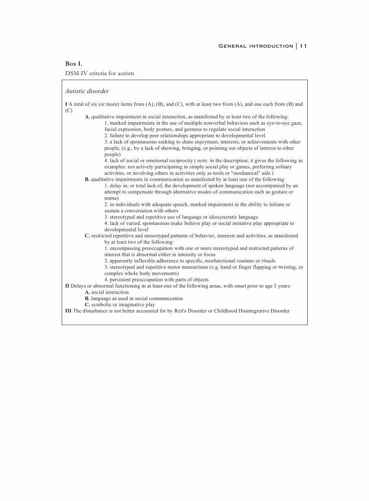
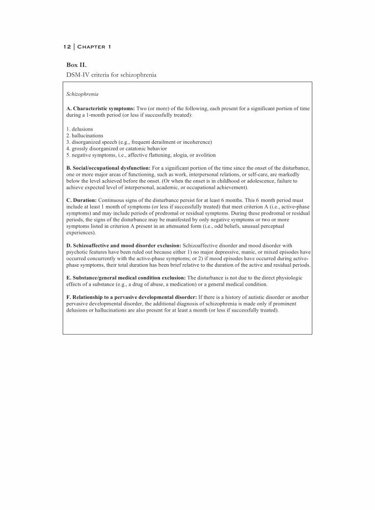
Socially deviant behaviorSocial cognitive competence appears to be a good predictor of social behaviorand adaptation. Severe difficulties in social adaptation, accompanied by adetachment from reality and preoccupation with inner thoughts and feelings,have been described for individuals with an autism spectrum disorder orschizophrenia (Bleuler, 1911; Kanner, 1943). For the diagnostic criteria ofschizophrenia and autism according to the DSM-IV, the Diagnostic andStatistical Manual of Mental Disorders -fourth edition (1994), see box I and II.Autism spectrum disorders and schizophrenia are neurodevelopmentaldisorders with a considerable genetic component, as estimated heritability isaround 80% for schizophrenia (Gottesman, 1991) and 90% for autism(Santangelo et al., 2005). Both autism and schizophrenia are more prevalent inmen. A meta-analysis has reported a risk ratio of 1.42 for men to developschizophrenia relative to women (Aleman et al., 2003), while autism spectrumdisorders are diagnosed approximately four times more often in boys than ingirls (Volkmar et al., 1993).
Autism spectrum disorders and schizophrenia share some characteristics,such as clinical phenomena pertaining to affect, communication and socialinsight (Abdi et al., 2004; Frith, 1992; Goldstein et al., 2002; Konstantareas etal., 2001; Rumsey et al., 1986). Note that clinical criteria (box II.F.) state that ‘ifthere is a history of autistic disorder or another pervasive developmentaldisorder, the additional diagnosis of schizophrenia is made only if prominentdelusions or hallucinations are also present for at least a month’. Although theyare distinct disorders, with the onset of autistic disorders in childhood whileschizophrenia is typically diagnosed in late adolescence/early adulthood(DeLisi, 1992; Minshew, 1996), autism spectrum disorders and schizophreniashare social cognitive dysfunctions, including aspects of language and emotion(Abdi et al., 2004; Frith, 1992; Pilowsky et al., 2000; Rumsey et al., 1986).
10 | Chapter 1
General introduction | 11
12 | Chapter 1
SchizophreniaAmong the clinical manifestations of schizophrenia, significant impairments insocial functioning have been consistently reported. Social isolation, impairmentsin social competence (Penn et al., 1996), deterioration in interpersonal closerelationships (Poole et al., 2000), communication-deficits and interpersonaloddity (Hooker et al., 2002) are recognized as characteristics frequentlydisplayed by patients suffering from schizophrenia. Social cognitiveimpairments have been widely described for these patients, such as impairmentsin gaze-interpretation, reading of affective facial expressions, picking upemotional signals in tone of voice and the ability to infer and interpretintentions, knowledge and believes of others, as measured in theory-of-mind(mentalizing) tasks (Corcoran et al., 1995; Corrigan et al., 2001; Doody et al.,1998; Edwards et al., 2002; Mazza et al., 2001; Penn et al., 1997; Sarfati et al.,1997).
Deficits in social perception appear to play an important role in socialdysfunctioning of schizophrenia patients. General cognitive skills seem toexplain between 20% to 60% of the variance in social outcome (Green et al.,2000). It has been suggested that social cognitive performance may help to,partly, explain the remaining 40% to 80%. Indeed, it has been shown that socialcognitive capacities can explain significantly more variance (26 %) in socialfunctioning in these patients as compared to general cognitive abilities (15 %)(Pinkham et al., 2006), which is comparable to a report of mentalizing abilitiesexplaining 27 % of the variance in social behavioral problems in schizophreniapatients (Brune, 2003). In line with this, another study revealed that the abilityto mentalize (realize that others may have different thoughts, feelings or goals)is among the best cognitive predictors of global social functioning inschizophrenia patients (Roncone et al., 2002).
Autism spectrumDisabilities in the social domain are considered as the primary symptoms in theautism spectrum (Fein et al., 1986). A triad of impairments is characteristic ofthis spectrum: atypical development in reciprocal social interactions, atypicalcommunication, and restricted, stereotyped and repetitive behaviors.
The impairments in social interactions can for example take form ofdifficulties in forming friendships, a lack of social motivation, misinterpretationin communicative intent of others and difficulties in understanding socio-emotional signs and social nuances (Wing et al., 1979). Research devoted toidentifying the social cognitive deficits that may contribute to social dysfunction
General introduction | 13
in the autism spectrum, has revealed abnormalities in eye-gaze processing,interpreting language in social contexts, mentalizing (‘theory of mind’) abilitiesand identifying social signals from faces, voices and body postures (Buitelaar etal., 1999; Klin et al., 2002; Ozonoff et al., 1996; Rutherford et al., 2002; Sasson,2006; Tager-Flusberg, 1999).
The importance of social cognitive capacities in coping with the social worldand related mental well-being calls for a search into the origins of socialcognition on the level of cognition, neurobiology and genes. Although it iswidely acknowledged that there is a genetic basis to the neural and cognitiveabnormalities seen in schizophrenia and autism, defining the underlying geneticfactors directing these aberrations appears to be a difficult task (Norton et al.,2006).
Factors that complicate the search for genetic origins of socially deviantbehavior
1. Genetic mechanisms are complexThe relations between genes and final phenotypes in behavior are complex.Many forces determine the unique phenotypic expression of an individual’sgenetic make-up. Genes can have dynamic interactions with environmentalfactors. These environmental influences not only encompass things as lifeevents, nutrition, aging, and chance, but also endogenous factors such as genesthat regulate expression of other genes (epigenetics). Moreover, genes maypredispose an individual to be exposed to certain environmental influences, i.e.genotype-environment correlations are present (for a review on gene-environment interactions see (Gottesman et al., 2005). Another reason why therelation between genes and behavior is complex, is that gene expression isdynamic over time. Genes can be turned on and off or can take on differentroles during various stages of development (Gottesman et al., 2005).
It is a long road from genotype to final phenotype and many intermediarymechanisms are involved, which are referred to as endophenotypes (see figure1) (Gottesman et al., 2003). For a behavioral phenotype these intermediaryendophenotypes can for example be neurobiological (brain structure) orcognitive (brain function) in nature. The closer a (endo-) phenotype to thegenotype is, the less ‘noise’ from interaction with environmental factors. In caseof the brain, these also include biochemical interactions and dynamic
14 | Chapter 1
interactions between circuits of cells. Because of this complexity, it has been adifficult challenge to identify gene-brain-behavior pathways leading to socialadaptation. In the field of abnormal and non-adaptive behavior in the socialdomain, such as is seen in psychiatric disorders like autism and schizophrenia,identification of etiological pathways to psychopathology has been hamperedby the distant relation between clinical features (the ‘molar’ level) and geneticabnormalities (the ‘molecular’ level) together with a lack of knowledge ofintermediary mechanisms in the genotype-phenotype pathway (Bearden et al.,2004; Gottesman et al., 2003).
General introduction | 15
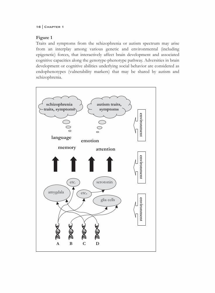
Figure 1Traits and symptoms from the schizophrenia or autism spectrum may arisefrom an interplay among various genetic and environmental (includingepigenetic) forces, that interactively affect brain development and associatedcognitive capacities along the genotype-phenotype pathway. Adversities in braindevelopment or cognitive abilities underlying social behavior are considered asendophenotypes (vulnerability markers) that may be shared by autism andschizophrenia.
16 | Chapter 1
2. Behavioral phenotypes are heterogeneousGenotype-environment interactions allow maximum adaptability to ourenvironment and lead to a heterogeneity in behavioral phenotypes. As a result,behavioral phenotypes may be limited in their use for identifying etiologicalpathways, including biological and genetic underpinnings, to psychopathology(Gottesman et al., 2003) Despite the presence of standardized criteria (DSM-IV) allowing reliable diagnostic classification of mental disorders based onobserved behavior, self-reported symptoms and course, disorders such asautism and schizophrenia are heterogeneous clinical phenomena. (Bearden etal., 2004; Caspi et al., 2006; Jablensky, 2006). Variation in phenotypic expressionacross development, i.e. temporal inconsistency, as well as individual differencesin exposure to medication or other treatments, may result in additionalheterogeneity in clinical populations (Jablensky, 2006).
Not only variance at the behavioral or clinical level, but also variance on agenetic level is implicated in the phenotypic variation of brain disorders such asautism or schizophrenia. It is thought that these disorders have a polygeneticorigin, i.e. many genes are involved rather than one single gene (Jablensky, 2006;Risch et al., 1999).
The continuum approachThe select set of genes that we carry can direct the development of verycomplex phenotypes such as is seen in brain structure, brain function andbehavior, because of a ‘pleiotropic’ design; many genes, in differentcombinations, affect a phenotype. Together with a variety of environmentalforces, a spectrum of variation in brain structure, -function and behavior isproduced in the general population. These distributions tend to take form of abell curve, with the majority of individuals represented in the small rangearound the mean and only a small part of the population in the extremes.Indeed, plots of social competence in the general population appear to be bellcurved distributions (Constantino et al., 2003a; Constantino et al., 2000).
Individuals with severe impairments in the social domain, as is seen inautism and schizophrenia, are found at the far end of the spectrum of socialabilities. Because of the many factors (genetic or environmental) involved indevelopment of the disorders, variation in phenotypic expression is likely to bedistributed along a spectrum. Only in the case of a single factor, such as a singlegene, one would expect the distribution to be truly dichotomous giving rise toall-or-none phenomena (Johns et al., 2001).
General introduction | 17
Phenomena that are part of the clinical phenotype of autism or schizophreniamay be quantitatively distributed along a continuum, rather than being all-or-none, dichotomous disease entities (Johns et al., 2001; Krabbendam et al., 2004).In this view, a clinical definition of autism or schizophrenia may represent onlya small part of the total phenotypic continuum, which is not necessarily clinicalin nature.
It has been proposed that autism is a ‘social brain disorder’ that reflects theextreme of a bell-shaped distribution of variation in autism traits, includingsocial competence, in the population (Baron-Cohen et al., 2001; Constantino etal., 2003b; Spiker et al., 2002). Considering the male disadvantage in socialcognitive abilities in the general population, the ‘extreme male brain theory ofautism’ postulates that autism represents the male end of a sexually-dimorphicsocial cognitive continuum (Baron-Cohen, 2002). Similarly, it has been proposedthat psychotic or schizophrenia-like traits are distributed along a continuum ofseverity that ranges from complete absence of schizophrenia-like traits to aseverity that is seen in individuals with schizophrenia (Lenzenweger, 1994). Forexample, some individuals from the general population have magical ideas,whereas individuals with schizophrenia may have severe delusions impactingtheir behavior and functioning in society (Mullen, 2003). Also, subtle, subclinicalsigns that parallel the symptoms of the illness may be present in healthyindividuals. These signs may be manifested as various schizotypal personalitytraits, such as unusual perceptual experiences, excentric and desorganizedspeech and behavior, suspiciousness and social isolation. Subclinical signs mayin some individuals progress to symptoms of schizophrenia. In fact, a numberof schizotypal traits partially predict schizophrenia at long term follow-up insubjects diagnosed with Schizotypal Personality Disorder (DSM III) (Fenton etal., 1989). Also, the following schizotypal traits in young relatives of patientspredict progression to schizophrenia in the following dimensions: socialwithdrawal, psychotic symptoms and socio-emotional dysfunction (Miller et al.,2002).
The observation that several different personality traits and disorders tendto cluster among biological relatives of individuals suffering from schizophreniahas lead to the hypothesis that a there is spectrum of related phenotypes thatincludes schizophrenia, as well as less severe phenotypes such as schizotypalpersonality disorder (DSM IV) and schizotaxia (Cadenhead et al., 2002;Jablensky, 2006; Meehl, 1989; Vollema et al., 1995). Several studies havereported cognitive and neuroanatomical abnormalities in individuals withschizotypal personality disorder that resemble those found in schizophrenia
18 | Chapter 1
patients, although the deviations are to a lesser degree (Cadenhead et al., 2002;Siever et al., 2004). The term schizotaxia is proposed to reflect a geneticallydetermined defect in integration in the brain, predisposing to schizophrenia. Inthis view, only a minority of individuals with this defect decompensate to thepoint of being diagnosed with schizophrenia based on DSM-IV criteria(Faraone et al., 2001; Meehl, 1989). Also for autism, the concept of a ‘broaderphenotype’ has been introduced to describe the mild features of the clinicalautism phenotype that are seen in biological (i.e. genetically related) relatives ofindividuals with this disorder (Bailey et al., 1998; Bishop et al., 2004). Based ontwin studies, it has been suggested that the typical clinical phenotype of autismor schizophrenia as seen in subjects with these disorders and the broadersubclinical phenotypes (i.e. autism or schizotypal traits) that are seen inbiological relatives, may share a genetic origin (Rutter, 2000; Torgersen et al.,2002).
Relevance of studying genetic disordersGenetic disorders associated with specific deficits in brain development andcognition may help us to unravel genotype-phenotype relations. Starting at thelevel of the genotype instead of the phenotype, reversing the typical line ofresearch, may be a complementary approach. Specific genetic conditions may beused as models of cognitive or behavioral disorders and provide insights intoneurodevelopmental pathways that may be more difficult to uncover bystudying heteregeneous, behaviorally defined populations (Reiss, 2000; Reiss etal., 2000). As such, studying individuals with a genetic abnormality who displaysocial cognitive abnormalities and hence difficulties in coping with socialsituations may help us understand the mechanisms involved in social behavior.It may especially be useful for understanding etiological pathways to autism- orschizophrenia psychopathology.
Klinefelter (47,XXY) syndrome One genetic disorder that is associated with abnormal brain development andbehavior is Klinefelter syndrome, defined by the presence of an extra Xchromosome in males (47,XXY). Klinefelter syndrome is the most common sexchromosomal disorder (Wesner et al., 1973), affecting approximately 1 in 700males (Bojesen et al., 2003). This sex chromosomal aneuploidy results in avariety of phenotypes including hypogonadism, androgen deficiency andinfertility (Lanfranco et al., 2004). Cognitive and behavioral dysfunctions inKlinefelter syndrome have generally been under-appreciated relative to
General introduction | 19
endocrinological and physical features. Although the primary focus in researchhas been on reproductive dysfunction of these patients, there is an awareness ofbehavioral and cognitive abnormalities (Boone et al., 2001; Geschwind et al.,2000b). The most prominent behavioral problems in men with Klinefeltersyndrome are found in the social domain, such as social withdrawal, socialanxiety, shyness, impulsivity and inappropriate or anti-social behavior (Bender etal., 1999; Geschwind et al., 2000a; Ratcliffe, 1999). In early adulthood theyreport having few or no friends, little energy and initiative, few or no sparetimeinterests and poor relations with siblings and parents (Nielsen et al., 1980).
The literature on cognitive mechanisms that may underlie impaired socialadaptation in XXY men is scarce. It has been proposed that difficulties in socialinteractions, and specifically those related to communication, are largelyattributable to disabilities in the language domain in Klinefelter syndrome(Rovet et al., 1996). The reported verbal disabilities include impairments in bothlanguage production and perception and indicate compromised languagefunctions that are typically associated with the left hemisphere (Samango-Sprouse, 2001). For example, Klinefelter boys or men display disabilities inreading, articulation, phonemic processing, spelling, language expression, verbalmemory, language comprehension, understanding words, finding words andverbally expressing their thoughts, all resulting in a verbal IQ that is somewhatlower than their performance IQ (Boone et al., 2001; Geschwind et al., 2000a;Money, 1993).
Compared to what is known about the cognitive mechanisms that contributeto social incompetence in Klinefelter syndrome, even much less is known about theneural mechanisms that are involved. Resting state cerebral blood flow patterns, asmeasured with SPECT, seem more symmetrical in XXY men as compared to menfrom the general population (Itti et al., 2003). Higher resting state blood flow in theright hemisphere in men with the XXY karyotype has been related to languageimpairments. Specific language dysfunctions have also been associated withmorphological abnormalities of the temporal lobe (Itti et al., 2006). Furthermore,structural Magnetic Resonance Imaging (sMRI) studies with XXY men haveindicated volume reductions in regions that are part of a neural networksupporting social cognition, such as the amygdala, insula, anterior cingulate andsuperior temporal gyrus (DeLisi et al., 2005; Patwardhan et al., 2002; Patwardhanet al., 2000; Shen et al., 2004). The difficulties in social adaptation together with thestructural brain abnormalities associated with the XXY karyotype suggest that agenetic mechanism involving genes on the X chromosome might lead todisturbances in development of social cognition in XXY men.
20 | Chapter 1
Difficulties in coping with the social world may be reflective of an increasedvulnerability to traits and symptoms from the autism or schizophreniaspectrum. Indeed, there is some suggestive evidence for a link between theXXY karyotype and increased psychopathology from the schizophreniaspectrum. The importance of investigation into the cognitive and behavioralphenotypical manifestations of Klinefelter syndrome as a means ofunderstanding a predisposition to schizophrenia, is shown by epidemiologicalstudies reporting an increased incidence of XXY karyotypes in schizophrenia.The prevalence of the XXY karyotype in the general population is 0.1-0.2%(Bojesen et al., 2003). There is suggestive evidence that prevalence of the XXYkaryoype in the male schizophrenia population may be 1.6 %, which is severaltimes higher (DeLisi et al., 1994; Kunugi et al., 1999). However, these studiesinvolved relatively small sample sizes (N=60 and N=120) in epidemiologicalterms and some studies have been unable to replicate these findings (Mors et al.,2001; Toyota et al., 2001). In turn, early studies have indicated an increased riskfor schizophrenia and psychotic illnesses among Klinefelter men (Lishman,1998). A review of mental hospital surveys pointed to a threefold increase inKlinefelter patients compared to the general population, which was mainly dueto ‘psychotic illnesses of a schizophrenic nature’ (Forssman, 1970). Anotherstudy showed that 7% of the Klinefelter patients in the psychiatric literature hadpsychoses with paranoid delusions and 6% suffered from schizophrenia(Nielsen et al., 1969). Recently, a survey of hospital admissions and dischargediagnoses has indicated a significantly increased relative risk of beinghospitalized with psychoses (hazard ratio of 4.97) for men with Klinefeltersyndrome (Bojesen et al., 2006). In addition, several case reports of Klinefeltermen suffering from schizophrenia or psychosis have been described in theliterature (Dervaux et al., 2002; Michielsen et al., 2001; Ong et al., 1995; Roy,1981; Warwick et al., 2003).
Present thesisStudying socio-emotional information processing in XXY men at aneuropsychological level as well as neurobiological level might reveal a cognitiveand neural basis for the difficulties in interpersonal relations and social‘awkwardness’ that have been described. Because the XXY chromosomalpattern appears to be associated with difficulties in the social domain,Klinefelter syndrome might also prove to be a useful model for studying gene-brain-behavior pathways to socially deviant behavior and associated traits fromthe autism- or schizophrenia spectrum. Importantly, not only is the XXY
General introduction | 21
population narrowly defined by the presence of an extra X chromosome,individuals with the XXY pattern are generally not mentally retarded (incontrast to many other X chromosomal disorders) which allows the study ofspecific cognitive disabilities, and underlying neural mechanisms, without theconfound of general intellectual decline.
In this thesis, I will focus on socially deviant behavior in adult XXY men on abehavioral, cognitive and neuroanatomical level. In chapter two, we aim torefine the social behavioral phenotype in XXY men. We will examine frequencyof social behavior and distress during social interactions in men with the XXYkaryotype. In addition, we will assess the degree to which features of the autismphenotype, as expressed in autism traits, are present in XXY men. In chapterthree, we will explore evidence for increased schizophrenia spectrum pathologyin XXY men. We will report on clinical measures of schizophrenia symptomsas well as measures of schizotypal personality traits.
I will continue with four chapters that deal with social cognitive abilities inXXY men. In chapter four we will examine socio-emotional processing inKlinefelter syndrome. Several domains of social cognition will be discussed,reflecting aspects of social-emotional information processing on levels ofperception, experience and expression: labeling of facial expressions ofemotion, emotion-cognition interactions in decision making and emotionregulation, that refers to subjective experience and identification of emotionalarousal as well as verbal expression of emotions. Chapter five focuses on‘social intuition’ in XXY men. The ability to quickly and automatically processbasic social cues, such as gaze direction and implied biological motion, isthought to be a prerequisite for establishing successful social interactions andespecially for construing a sense of ‘social intuition’. We report on the extent towhich social cues are processed effortlessly and implicitly in three differentgroups characterized by the presence of traits or symptoms from theschizophrenia spectrum, i.e. patients with schizophrenia, first-degree relatives ofpatients with schizophrenia and individuals with Klinefelter syndrome(47,XXY). Performance in those groups will be compared to matched controlsfrom the general population. In chapter six, we test the hypothesis of theimportance of the right hemisphere for specific pragmatic aspects of languagein individuals from the general population. We will examine the effects oftranscranial magnetic stimulation (TMS) over the right hemisphere on detectionof emotions in tone of voice, a pragmatic aspect of language, in contrast todiscrimination of emotions in verbal content, a semantic aspect of language
22 | Chapter 1
which has been associated with the left hemisphere. Chapter seven describes afirst exploration of evidence for such pragmatic language impairments in inKlinefelter syndrome. By assessing the ability to discriminate emotions inspeech we are able to examine the capacity to perceive and understand socialsignals in the auditory modality. We will contrast perception of emotionalprosody (tone of voice), which is a pragmatic aspect of language thought to belateralized to the right hemisphere, with perception of emotions in verbalcontent, which is lateralized to the left hemisphere.
The following three chapters deal with neuroanatomical mechanisms thatmay underlie social cognitive capacities in Klinefelter syndrome. In chaptereight, a possible neural mechanism underlying language disabilites in XXY menis explored. By using fMRI we are able to reveal the effects of an extra Xchromosome on lateralization of neural activation during language processing.This technique allows us to identify functional asymmetries in specific brainregions as well as to determine whether reduced lateralization, if found, issecondary to decreased function of the left- or increased activity in the righthemisphere. We will explore the relation between loss of language lateralizationand mental functioning in these men, with special interest in clinical phenomenaof disorganization of thought and language.
As the amygdala is considered as a key brain area in socio-emotionalprocessing, in chapter nine I will review evidence for structural abnormalitiesof the amygdala in Klinefelter syndrome based on findings in the literature.Findings will be compared to what is known of abnormalities of the amygdalain populations with increased vulnerability to schizophrenia: individuals fromthe general population displaying subclinical signs of schizophrenia andbiological relatives of schizophrenia patients who may carry a geneticpredisposition for the disorder. Not only volume of the amygdala, alsofunctioning of the amygdala will be considered. Chapter ten deals with findingsfrom a funtional MRI study (fMRI) focused on the neural mechanismsunderlying social cognition in XXY men. In this chapter the functionalcontributions of a neural circuit comprising the amygdala, insula, fusiformgyrus and superior temporal gyrus, to social judgements of faces will bediscussed.
Finally, in chapter eleven, I will evaluate all the presented evidenceregarding social behavior, autism and schizophrenia spectrum traits, socialcognitive disabilities and underlying neural mechanisms in XXY men. Besidesdescribing the social cognitive phenotype at the level of behavior, cognition andbrain structure and -function of this X chromosomal disorder, I will discuss the
General introduction | 23

potential implications of a link between the X chromosome and disturbances indevelopment of social cognition and underlying neural networks forunderstanding gene-brain-behavior pathways to neuropsychiatric disorders suchas autism or schizophrenia.
24 | Chapter 1
ReferencesA.P.A. (1994). Diagnostic and statistical manual of mental disorders (4th ed.).
Washington, DC: American Psychiatric Association Press.Abdi, Z., & Sharma, T. (2004). Social cognition and its neural correlates in
schizophrenia and autism. CNS Spectrums, 9(5), 335-343.Adolphs, R. (2001). The neurobiology of social cognition. Current Opinion in
Neurobiology, 11(2), 231-239.Adolphs, R. (2002). Neural systems for recognizing emotion. Current Opinion
in Neurobiology, 12(2), 169-177.Aleman, A., Kahn, R. S., & Selten, J. P. (2003). Sex differences in the risk of
schizophrenia: Evidence from meta-analysis. Archives of General Psychiatry, 60(6), 565-571.
Amaral, D. G. (2003). The amygdala, social behavior, and danger detection.Annals of the New York Academy of Sciences, 1000(1), 337-347.
Anderson, S. W., Bechara, A., Damasio, H., Tranel, D., & Damasio, A. R.(1999). Impairment of social and moral behavior related to early damage in human prefrontal cortex. Nature Neuroscience, 2(11), 1032-1037.
Bailey, A., Palferman, S., Heavey, L., & Le Couteur, A. (1998). Autism: The phenotype in relatives. Journal of Autism and Developmental Disorders,28(5), 369.
Baron-Cohen, S. (2002). The extreme male brain theory of autism. Trends in Cognitive Sciences, 6(6), 248.
Baron-Cohen, S., Wheelwright, S., Skinner, R., Martin, J., & Clubley, E. (2001).The autism-spectrum quotient (aq): Evidence from asperger syndrome/high-functioning autism, males and females, scientists and mathematicians. Journal of Autism and Developmental Disorders, 31(1),5-17.
Bearden, C. E., Reus, V. I., & Freimer, N. B. (2004). Why genetic investigation of psychiatric disorders is so difficult. Current Opinion in Genetics and Development, 14(3), 280.
Bender, B. G., Harmon, R. J., Linden, M. G., Bucher-Bartelson, B., & Robinson, A. (1999). Psychosocial competence of unselected young adults with sex chromosome abnormalities. American Journal ofMedical Genetics - Neuropsychiatric Genetics, 88(2), 200.
General introduction | 25
Bishop, D. V. M., Maybery, M., Maley, A., Wong, D., Hill, W., & Hallmayer, J.(2004). Using self-report to identify the broad phenotype in parents ofchildren with autistic spectrum disorders: A study using the autism-spectrum quotient. Journal of Child Psychology and Psychiatry and Allied Disciplines, 45(8), 1431.
Bleuler. (1911). Dementia praecox oder die gruppe der schizophrenien.Leipzig: Deutike.
Bojesen, A., Juul, S., Birkebaek, N. H., & Gravholt, C. H. (2006). Morbidity in klinefelter syndrome; a danish register study based on hospital discharge diagnoses. The Journal of Clinical Endocrinology and Metabolism,91(4), 1254-1260.
Bojesen, A., Juul, S., & Gravholt, C. H. (2003). Prenatal and postnatal prevalence of klinefelter syndrome: A national registry study. The Journal of Clinical Endocrinology and Metabolism, 88(2), 622-626.
Boone, K. B., Swerdloff, R. S., Miller, B. L., Geschwind, D. H., Razani, J., Lee,A., et al. (2001). Neuropsychological profiles of adults with klinefelter syndrome. Journal of the International Neuropsychological Society: Jins,7(4), 446-456.
Brune, M. (2003). Theory of mind and the role of iq in chronic disorganized schizophrenia. Schizophrenia Research, 60(1), 57-64.
Buitelaar, J. K., van der Wees, M., Swaab-Barneveld, H., & van der Gaag, R. J.(1999). Theory of mind and emotion-recognition functioning in autistic spectrum disorders and in psychiatric control and normal children.Development and Psychopathology, 11(1), 39-58.
Bush, G., Luu, P., & Posner, M. I. (2000). Cognitive and emotional influences in anterior cingulate cortex. Trends in Cognitive Sciences, 4(6), 215.
Cadenhead, K. S., & Braff, D. L. (2002). Endophenotyping schizotypy: A prelude to genetic studies within the schizophrenia spectrum.Schizophrenia Research, 54(1-2), 47-57.
Caspi, A., & Moffitt, T. E. (2006). Gene-environment interactions in psychiatry: Joining forces with neuroscience. Nature Reviews Neuroscience, 7(7), 583.
Constantino, J. N., Hudziak, J. J., & Todd, R. D. (2003a). Deficits in reciprocal social behavior in male twins: Evidence for a genetically independent domain of psychopathology. Journal of the American Academy ofChild and Adolescent Psychiatry, 42(4), 458.
Constantino, J. N., & Todd, R. D. (2000). Genetic structure of reciprocal social behavior. American Journal of Psychiatry, 157(12), 2043-2045.
26 | Chapter 1
Constantino, J. N., & Todd, R. D. (2003b). Autistic traits in the general population: A twin study. Archives of General Psychiatry, 60(5), 524.
Corcoran, R., Mercer, G., & Frith, C. D. (1995). Schizophrenia,symptomatology and social inference: Investigating "theory of mind" in people with schizophrenia. Schizophrenia Research, 17(1), 5-13.
Corrigan, P. W., & Penn, D. L. (Eds.). (2001). Social cognition and schizophrenia. Washington, D.C.: American Psychological Association.
Damasio, A. R., Grabowski, T. J., Bechara, A., Damasio, H., Ponto, L. L. B.,Parvizi, J., et al. (2000). Subcortical and cortical brain activity during the feeling of self-generated emotions. Nature Neuroscience, 3(10), 1049.
DeLisi, L. E. (1992). The significance of age of onset for schizophrenia.Schizophrenia Bulletin, 18(2), 209.
DeLisi, L. E., Friedrich, U., Wahlstrom, J., Boccio-Smith, A., Forsman, A.,Eklund, K., et al. (1994). Schizophrenia and sex chromosome anomalies.Schizophrenia Bulletin, 20(3), 495-505.
DeLisi, L. E., Maurizio, A. M., Svetina, C., Ardekani, B., Szulc, K., Nierenberg,J., et al. (2005). Klinefelter's syndrome (xxy) as a genetic model for psychotic disorders. American Journal of Medical Genetics B Neuropsychiatric Genetics, 135(1), 15-23.
Dervaux, A., & Artiges, E. (2002). Olanzapine for violent schizophrenia and klinefelter syndrome. American Journal of Psychiatry, 159(3), 493-494.
Doody, G. A., Gotz, M., Johnstone, E. C., Frith, C. D., & Owens, D. G.(1998). Theory of mind and psychoses. Psychological Medicine, 28(2),397-405.
Edwards, J., Jackson, H. J., & Pattison, P. E. (2002). Emotion recognition via facial expression and affective prosody in schizophrenia: A methodological review. Clinical Psychology Review, 22(6), 789-832.
Faraone, S. V., Green, A. I., Seidman, L. J., & Tsuang, M. T. (2001)."schizotaxia": Clinical implications and new directions for research.Schizophrenia Bulletin, 27(1), 1-18.
Fein, D., Pennington, B., & Markowitz, P. (1986). Toward a neuropsychological model of infantile autism: Are the social deficits primary? Journal of the American Academy of Child Psychiatry, 25(2), 198.
Fenton, W. S., & McGlashan, T. H. (1989). Risk of schizophrenia in character disordered patients. The American Journal of Psychiatry, 146(10), 1280-1284.
General introduction | 27
Fine, C., Lumsden, J., & Blair, R. J. R. (2001). Dissociation between `theory ofmind' and executive functions in a patient with early left amygdala damage. Brain, 124(2), 287-298.
Forssman, H. (1970). The mental implications of sex chromosome aberrations. British Journal of Psychiatry, 117(539), 353-363.
Frith, C. D. (1992). The cognitive neuropsychology of schizophrenia.Hillsdale, NJ: Laurence Erlbaum Associates.
Frith, C. D., & Frith, U. (1999). Interacting minds--a biological basis. Science,286(5445), 1692-1695.
Geschwind, D. H., Boone, K. B., Miller, B. L., & Swerdloff, R. S. (2000a).Neurobehavioral phenotype of klinefelter syndrome. Mental Retardation and Developmental Disabilities Research Reviews, 6(2), 107-116.
Geschwind, D. H., Boone, K. B., Miller, B. L., & Swerdloff, R. S. (2000b).Neurobehavioral phenotype of klinefelter syndrome., 6(2), 107-116.
Goldstein, G., Minshew, N. J., Allen, D. N., & Seaton, B. E. (2002). High-functioning autism and schizophrenia: A comparison of an early and late onset neurodevelopmental disorder. Archives of Clinical Neuropsychology, 17(5), 461-475.
Gottesman, I. I. (1991). Schizophrenia genesis: The origin of madness. New York: Freeman.
Gottesman, I. I., & Gould, T. D. (2003). The endophenotype concept in psychiatry: Etymology and strategic intentions. The American Journal ofPsychiatry, 160(4), 636-645.
Gottesman, I. I., & Hanson, D. R. (2005). Human development: Biological and genetic processes. Annual Review of Psychology, 56, 263.
Green, M. F., Kern, R. S., Braff, D. L., & Mintz, J. (2000). Neurocognitive deficits and functional outcome in schizophrenia: Are we measuring the 'right stuff'? Schizophrenia Bulletin, 26(1), 119.
Haxby, J. V., Hoffman, E. A., & Gobbini, M. I. (2000). The distributed human neural system for face perception. Trends in Cognitive Sciences, 4(6),223.
Haxby, J. V., Hoffman, E. A., & Gobbini, M. I. (2002). Human neural systems for face recognition and social communication. Biological Psychiatry,51(1), 59-67.
Hooker, C., & Park, S. (2002). Emotion processing and its relationship to social functioning in schizophrenia patients. Psychiatry Research, 112(1),41-50.
28 | Chapter 1
Hughes, C., & Cutting, A. L. (1999). Nature, nurture, and individual differences in early understanding of mind. Psychological Science, 10(5),429.
Itti, E., Gaw Gonzalo, I. T., Boone, K. B., Geschwind, D. H., Berman, N.,Pawlikowska-Haddal, A., et al. (2003). Functional neuroimaging provides evidence of anomalous cerebral laterality in adults with klinefelter's syndrome. Annals of Neurology, 54(5), 669-673.
Itti, E., Gaw Gonzalo, I. T., Pawlikowska-Haddal, A., Boone, K. B., Mlikotic,A., Itti, L., et al. (2006). The structural brain correlates of cognitive deficits in adults with klinefelter's syndrome. Journal of Clinical Endocrinology and Metabolism, 91(4), 1423-1427.
Jablensky, A. (2006). Subtyping schizophrenia: Implications for genetic research. Molecular Psychiatry, 11(9), 815.
Johns, L. C., & van Os, J. (2001). The continuity of psychotic experiences in the general population. Clinical Psychology Review, 21(8), 1125-1141.
Jones, W., Bellugi, U., Lai, Z., Chiles, M., Reilly, J., Lincoln, A., et al. (2000). Ii.Hypersociability in williams syndrome. Journal of Cognitive Neuroscience, 12 Suppl 1, 30-46.
Kanner, L. (1943). Autistic disturbances of affective contact. Nervous Child,2, 217-250.
Klin, A., Jones, W., Schultz, R., Volkmar, F., & Cohen, D. (2002). Defining and quantifying the social phenotype in autism. The American Journal ofPsychiatry, 159(6), 895-908.
Konstantareas, M. M., & Hewitt, T. (2001). Autistic disorder and schizophrenia: Diagnostic overlaps. Journal of Autism and Developmental Disorders, 31(1), 19-28.
Krabbendam, L., Myin-Germeys, I., De Graaf, R., Vollebergh, W., Nolen, W.A., Eidema, J., et al. (2004). Dimensions of depression, mania and psychosis in the general population. Psychological Medicine, 34(7), 1177.
Kunugi, H., Lee, K. B., & Nanko, S. (1999). Cytogenetic findings in 250 schizophrenics: Evidence confirming an excess of the x chromosome aneuploidies and pericentric inversion of chromosome 9. Schizophrenia Research, 40(1), 43-47.
Lanfranco, F., Kamischke, A., Zitzmann, M., & Nieschlag, P. E. (2004).Klinefelter's syndrome. The Lancet, 364(9430), 273-283.
Lenzenweger, M. F. (1994). Psychometric high-risk paradigm, perceptual aberrations, and schizotypy: An update. Schizophrenia Bulletin, 20(1),121-135.
General introduction | 29
Lishman, W. A. (1998). Endocrine diseases and metabolic disorders. In W. A.Lishman (Ed.), Organic psychiatry: The psychological consequences ofcerebral disorder (pp. 526-527). Oxford: Blackwell Science.
Mazza, M., De_Risio, A., Surian, L., Roncone, R., & Casacchia, M. (2001).Selective impairments of theory of mind in people with schizophrenia.Schizophrenia Research, 47(2-3), 299-308.
Meehl, P. E. (1989). Schizotaxia revisited. Archives of General Psychiatry,46(10), 935.
Michielsen, P. J. S., Verhoeven, W. M. A., & de Blecourt, C. V. (2001).Klinefelter syndrome and psychiatric disturbances - 2 case studies and a survey of the literature. Acta Neuropsychiatrica, 13(1), 15-20.
Miller, P., Byrne, M., Hodges, A., Lawrie, S. M., Owens, D. G. C., & Johnstone,E. C. (2002). Schizotypal components in people at high risk ofdeveloping schizophrenia: Early findings from the edinburgh high-risk study. British Journal of Psychiatry, 180(2), 179-184.
Minshew, N. J. (1996). Autism. In R. D. Adams & M. Victor (Eds.), Principles of child neurology (pp. 1713-1729). New York: McGraw-Hill.
Money, J. (1993). Specific neuro-cognitive impairments associated with turner (45,x) and klinefelter (47,xxy) syndromes: A review. Social Biology, 40(1-2), 147-151.
Mors, O., Mortensen, P. B., & Ewald, H. (2001). No evidence of increased risk for schizophrenia or bipolar affective disorder in persons with aneuploidies of the sex chromosomes. Psychological Medicine, 31(3),425-430.
Mullen, R. (2003). Delusions: The continuum versus category debate.Australian and New Zealand Journal of Psychiatry, 37(5), 505.
Nielsen, J., Johnsen, S. G., & Sorensen, K. (1980). Follow-up 10 years later of34 klinefelter males with karyotype 47,xxy and 16 hypogonadal males with karyotype 46,xy. Psychological Medicine, 10(2), 345.
Nielsen, J., Sørensen, A., Theilgaard, A., Frøland, A., & Johnson, S. G. (1969).A psychiatric-psychological study of 50 severely hypogonadal male patients, including 34 with klinefelter's syndrome, 47,xxy. Copenhagen:Munksgaard.
Norton, N., Williams, H. J., & Owen, M. J. (2006). An update on the genetics of schizophrenia. Current opinion in psychiatry., 19(2), 158-164.
Ochsner, K. N. (2004). Current directions in social cognitive neuroscience.Current Opinion in Neurobiology, 14(2), 254.
30 | Chapter 1
Ong, S. H., & Robertson, J. R. (1995). Schizophrenia with karyotype mosaic 47,xxy/48,xxy+8. Psychiatric Genetics, 5(2), 67-69.
Ozonoff, S., & Miller, J. N. (1996). An exploration of right-hemisphere contributions to the pragmatic impairments of autism. Brain and Language, 52(3), 411-434.
Patwardhan, A. J., Brown, W. E., Bender, B. G., Linden, M. G., Eliez, S., & Reiss, A. L. (2002). Reduced size of the amygdala in individuals with 47,xxy and 47,xxx karyotypes. American Journal of Medical Genetics,114(1), 93-98.
Patwardhan, A. J., Eliez, S., Bender, B., Linden, M. G., & Reiss, A. L. (2000).Brain morphology in klinefelter syndrome: Extra x chromosome and testosterone supplementation. Neurology, 54(12), 2218-2223.
Pelphrey, K. A., & Morris, J. P. (2006). Brain mechanisms for interpreting the actions of others from biological-motion cues. Current Directions in Psychological Science, 15(3), 136.
Penn, D. L., Corrigan, P. W., Bentall, R. P., Racenstein, J. M., & Newman, L.(1997). Social cognition in schizophrenia. Psychological Bulletin, 121(1),114-132.
Penn, D. L., Spaulding, W., Reed, D., & Sullivan, M. (1996). The relationship of social cognition to ward behavior in chronic schizophrenia.Schizophrenia Research, 20(3), 327-335.
Phan, K. L., Wager, T., Taylor, S. F., & Liberzon, I. (2002). Functional neuroanatomy of emotion: A meta-analysis of emotion activation studies in pet and fmri. Neuroimage, 16(2), 331-348.
Phelps, E. A. (2006). Emotion and cognition: Insights from studies of the human amygdala. Annual Review of Psychology, 57, 27.
Phillips, M. L., Drevets, W. C., Rauch, S. L., & Lane, R. (2003). Neurobiology of emotion perception i: The neural basis of normal emotion perception. Biological Psychiatry, 54(5), 504-514.
Pilowsky, T., Yirmiya, N., Arbelle, S., & Mozes, T. (2000). Theory of mind abilities of children with schizophrenia, children with autism, and normally developing children. Schizophrenia Research, 42(2), 145-155.
Pinkham, A. E., & Penn, D. L. (2006). Neurocognitive and social cognitive predictors of interpersonal skill in schizophrenia. Psychiatry Research,143(2-3), 167-178.
Poole, J. H., Tobias, F. C., & Vinogradov, S. (2000). The functional relevance of affect recognition errors in schizophrenia. Journal of the International Neuropsychological Society: Jins, 6(6), 649-658.
General introduction | 31
Ratcliffe, S. (1999). Long-term outcome in children of sex chromosome abnormalities. Archives of Disease in Childhood, 80(2), 192-195.
Reiss, A. L. (2000). Realizing the potential of behavioral neurogenetics research in childhood onset neuropsychiatric disorders. American Journal of Medical Genetics - Neuropsychiatric Genetics, 96(4), 472.
Reiss, A. L., Eliez, S., Schmitt, J. E., Patwardhan, A., & Haberecht, M. (2000).Brain imaging in neurogenetic conditions: Realizing the potential ofbehavioral neurogenetics research. Mental Retardation and Developmental Disabilities Research Reviews, 6(3), 186-197.
Risch, N., Spiker, D., Lotspeich, L., Nouri, N., Hinds, D., Hallmayer, J., et al.(1999). A genomic screen of autism: Evidence for a multilocus etiology.American Journal of Human Genetics, 65(2), 493.
Roncone, R., Falloon, I. R. H., Mazza, M., De Risio, A., Pollice, R.,Necozione, S., et al. (2002). Is theory of mind in schizophrenia more strongly associated with clinical and social functioning than with neurocognitive deficits? Psychopathology, 35(5), 280.
Rovet, J., Netley, C., Keenan, M., Bailey, J., & Stewart, D. (1996). The psychoeducational profile of boys with klinefelter syndrome. Journal ofLearning Disabilities, 29(2), 193-196.
Roy, A. (1981). Schizophrenia and klinefelter's syndrome. Canadian Journal ofPsychiatry, 26(4), 262.
Rumsey, J. M., Andreasen, N. C., & Rapoport, J. L. (1986). Thought, language,communication, and affective flattening in autistic adults. Archives ofGeneral Psychiatry, 43(8), 771.
Rutherford, M. D., Baron-Cohen, S., & Wheelwright, S. (2002). Reading the mind in the voice: A study with normal adults and adults with asperger syndrome and high functioning autism. Journal of Autism and Developmental Disorders, 32(3), 189-194.
Rutter, M. (2000). Genetic studies of autism: From the 1970s into the millennium. Journal of Abnormal Child Psychology, 28(1), 3.
Samango-Sprouse, C. (2001). Mental development in polysomy x klinefelter syndrome (47,xxy; 48,xxxy): Effects of incomplete x inactivation.Seminars in Reproductive Medicine, 19(2), 193-202.
Santangelo, S. L., & Tsatsanis, K. (2005). What is known about autism: Genes,brain, and behavior. American Journal of PharmacoGenomics, 5(2), 71.
32 | Chapter 1
Sarfati, Y., Hardy_Bayle, M. C., Besche, C., & Widlocher, D. (1997).Attribution of intentions to others in people with schizophrenia: A non-verbal exploration with comic strips. Schizophrenia Research, 25(3), 199-209.
Sasson, N. J. (2006). The development of face processing in autism. Journal ofAutism and Developmental Disorders, 36(3), 381.
Scourfield, J., Martin, N., Lewis, G., & McGuffin, P. (1999). Heritability ofsocial cognitive skills in children and adolescents. British Journal ofPsychiatry, 175, 559-564.
Shen, D., Liu, D., Liu, H., Clasen, L., Giedd, J., & Davatzikos, C. (2004).Automated morphometric study of brain variation in xxy males.Neuroimage, 23(2), 648-653.
Siever, L. J., & Davis, K. L. (2004). The pathophysiology of schizophrenia disorders: Perspectives from the spectrum. The American Journal ofPsychiatry, 161(3), 398-413.
Spiker, D., Lotspeich, L. J., Dimiceli, S., Myers, R. M., & Risch, N. (2002).Behavioral phenotypic variation in autism multiplex families: Evidence for a continuous severity gradient. American Journal of Medical Genetics - Neuropsychiatric Genetics, 114(2), 129.
Tager-Flusberg, H. (1999). A psychological approach to understanding the social and language impairments in autism. International Review ofPsychiatry, 11(4), 325.
Torgersen, S., Edvardsen, J., Øien, P. A., Onstad, S., Skre, I., Lygren, S., et al.(2002). Schizotypal personality disorder inside and outside the schizophrenic spectrum. Schizophrenia Research, 54(1-2), 33.
Toyota, T., Shimizu, H., Yamada, K., Yoshitsugu, K., Meerabux, J., Hattori, E.,et al. (2001). Karyotype analysis of 161 unrelated schizophrenics: No increased rates of x chromosome mosaicism or inv(9), using ethnically matched and age-stratified controls. Schizophrenia Research, 52(3), 171-179.
Volkmar, F. R., Szatmari, P., & Sparrow, S. S. (1993). Sex differences in pervasive developmental disorders. Journal of Autism and Developmental Disorders, V23(4), 579.
Vollema, M. G., & Van den Bosch, R. J. (1995). The multidimensionality ofschizotypy. Schizophrenia Bulletin, 21(1), 19.
Warwick, M. M., Lawrie, S. M., Beveridge, A., & Johnstone, E. C. (2003).Abnormal cerebral asymmetry and schizophrenia in a subject with klinefelter's syndrome (xxy). Biological Psychiatry, 53(7), 627-629.
General introduction | 33
Wesner, C. E., Spangler, P., Petrides, A., Baker, D., & Telfer, M. A. (1973).Prepubertal klinefelter syndrome: A report of six cases. Journal ofMental Deficiency Research, 17(3), 237-246.
Wing, L., & Gould, J. (1979). Severe impairments of social interaction and associated abnormalities in children: Epidemiology and classification.Journal of Autism and Developmental Disorders, 9(1), 11.
Zilbovicius, M., Meresse, I., Chabane, N., Brunelle, F., Samson, Y., & Boddaert, N. (2006). Autism, the superior temporal sulcus and social perception. Trends in Neurosciences, 29(7), 359.
34 | Chapter 1
Part I
Social behavior and
psychopathology

Chapter 2
Social behavior and autism traits in a sex
chromosomal disorder:
Klinefelter (47XXY) syndrome
Sophie van Rijn, Hanna Swaab, André Aleman en René S. Kahn
Submitted for publication

AbstractThe XXY chromosomal pattern has been associated with difficulties inpsychosocial functioning. Our aim was to examine frequency of participation insocial interactions, distress during social interactions and autism traits inKlinefelter syndrome.
Scores of 31 XXY men on the Scale for Interpersonal Behavior and theAutism Spectrum Questionnaire were compared to 24 and 20 control menrespectively.
XXY men reported increased distress during social interactions and lessengagement of specific social behaviors. Overall rates of autism traits weresignificantly higher in XXY men.
These findings call for a clinical investigation of vulnerability to autism inKlinefelter syndrome. Klinefelter syndrome might serve as a model for studyinga role of the X chromosome in social behavioral dysfunction and autism-likebehavior.
38 | Chapter 2
IntroductionKlinefelter syndrome affects approximately 1 in 700 men and is the mostcommom sex chromosomal disorder. Men with this syndrome have an extra Xchromosome, giving rise to the XXY chromosomal pattern. This sexchromosomal aneuploidy results in a variety of phenotypes includinghypogonadism, androgen deficiency and infertility (Lanfranco et al., 2004).Cognitive and behavioral dysfunctions in Klinefelter syndrome have generallybeen under-appreciated relative to endocrinological and physical features.However, there is an awareness of behavioral and cognitive abnormalities(Boone et al., 2001; Geschwind et al., 2000b). Prominent behavioral problemsin men with Klinefelter syndrome are found in the social domain, such as socialwithdrawal, social anxiety, shyness, impulsivity and inappropriate social behavior(Bender et al., 1999; Geschwind et al., 2000a; Geschwind et al., 2004; Ratcliffe,1999). In early adulthood XXY men report having few or no friends, poorrelations with siblings and parents, little energy and initiative, and few or nosparetime interests (Nielsen et al., 1980). Difficulties in social functioning havebeen attributed to language based learning difficulties (Geschwind et al., 2000a),social cognitive impairments (van Rijn et al., 2006) and verbal disabilities (Rovetet al., 1996) that have been observed in Klinefelter syndrome.
In previous studies, social adjustment in Klinefelter syndrome has primarilybeen described from a psychosocial perspective. Psychosocial competence hasbeen measured using psychiatric interviews or parental questionnaires focusedat, for example, the quality of relationships with familymembers, self-esteemand coping with stressors (Bender et al., 1995; Ratcliffe, 1999). Impairments insocial adjustment, communication and social cognition might reflect anincreased liability for neurodevelopmental disorders such as autism. Therefore,a refinement of the social behavioral phenotype in individuals with the XXYkaryotype is warranted.
In this study, we measured frequency of participation in social interactionsand distress during social behavior in adult XXY men. To explore the extend towhich social disabilities reflect increased levels of features that belong to theautism phenotype, we included quantative measures of autism traits. We usedthis dimensional, rather than categorical, approach as it has been proposed thatautism is a disorder of social behavior that reflects the extreme of a bell-shapeddistribution of variation in autism traits, including social competence, in thepopulation (Baron-Cohen et al., 2001; Constantino et al., 2003; Spiker et al.,2002).
Social behavior and autism traits in XXY men | 39
Methods
SubjectsWe included 31 men with Klinefelter syndrome (mean age 41.3, SD 10.0) withhelp from the Dutch Klinefelter Association. Diagnosis of Klinefeltersyndrome was confirmed by karyotyping, using standard procedures. Twenty-four men were treated with testosterone supplements, with a mean age oftreatment onset of 26.9 years (SD 7.6).
We compared autism traits in XXY men with 20 men from the generalpopulation (control group I), who were recruited using advertisments in localnewspapers. Mean age in this group was 39.2 years (SD 13.1). Social behavior inXXY men was compared with 24 men from the general population (controlgroup II). Mean age in this group was 35.7 (SD 8.5). There were no significantdifferences in age between the three groups, as indicated by a multivariateANOVA as well as post-hoc tests (F(2,72)=1.8, p=0.16). None of the controlsubjects had a history of psychiatric illness as confirmed with the MiniInternational Neuropsychiatric Interview plus (MINI) (Sheehan et al., 1998).The study was approved by the local ethics committee and written informedconsent was obtained according to the declaration of Helsinki.
Intellectual abilityRaven’s Advanced Progressive Matrices (short form)This test is commonly accepted as a measure of general intelligence and hasbeen shown to correlate with a number of other standardized intelligence tests(Lezak, 1995; Raven et al., 1993). Subjects are shown 12 pictures of matrices (i.e.,related patterns), each of which is a figural design with a part removed. Thesubject must choose the correct missing part from eight options.
National Adult Reading Test (NART)The Dutch translation of the NART (Nelson, 1982; Schmand et al., 1991)provides an estimate of verbal IQ and is based on the high correlation betweenreading ability, specifically of irregular words, with intelligence in the normalpopulation. Subjects are required to read 50 irregular words aloud, and, on thebasis of the number of errors made in pronunciation a reliable estimate ofWAIS-R IQ can be calculated (Willshire et al., 1991).
40 | Chapter 2
Social behaviorSocial behavior was evaluated using the Scale for Interpersonal Behavior (SIB)(Arrindell, 1985). The SIB is a reliable and valid self-report measure of thefrequency of engagement in specific social behaviors as well as the experienceddistress it is accompanied by (Arrindell et al., 2001). Besides an overall measureof frequency of social behavior and distress during social behavior, there are fourfactorially-derived subscales: (I) Display of negative feelings (negativeassertion), such as refusing a request or standing up for one’s rights in a publicsituation, (II) Expression of and dealing with personal limitations, such asability to deal with criticism or requesting attention/help, (III) Initiatingassertiveness, such as starting a conversation with strangers or expressing one’sown opinion and (IV) Praising others and the ability to deal withcompliments/praise of others (positive assertion), such as giving and receivingcompliments. Scores that are obtained with the SIB represent mean item-scoresfor each dimension of social behavior, on a scale from one (high frequency orlow distress) to five (low frequency or high distress).
Autism traitsThe Autism-spectrum Quotient (ASQ) (Baron-Cohen et al., 2001) is a self-administered questionnaire for adults that assesses the degree to which anyindividual adult of normal intelligence might have features of the core autisticphenotype. It has good test-retest reliability and good discriminative validity forAsperger syndrome at a cut-off score of 26 (Woodbury-Smith et al., 2005).Scores on the ASQ have shown to be normally distributed in the generalpopulation. Five subscales cover personality traits associated with the autisticspectrum; social skills, communication, imagination, attention to detail, andattention switching.
Results
Intellectual abilityMean score on the Raven’s Advanced Progressive Matrices was not significantlydifferent between the groups (F(2.72)=1.3, p=0.27) as indicated by amultivariate ANOVA. Post-hoc tests also showed no significant differencesbetween XXY men and the control groups (p=0.18 and p=0.16). Mean scoreswere 105.2 (SD 9.1), 109.4 (SD 8.8) and 109.6 (SD 14.6) for the Klinefeltergroup and control group I and II respectively.
Social behavior and autism traits in XXY men | 41
On the NART, mean score of the Klinefelter men did not significantly differfrom the control groups (F(2,72)=0.7, p=0.48). Post-hoc tests also showed nosignificant differences between XXY men and the control groups (p=0.46 andp=0.58). Mean score in de Klinefelter group was 108.6 (SD 13.5), for thecontrol group I and II it was 110.0 (SD 5.3) and 107.1 (SD 7.8) respectively.
Social behaviorOverall distress during social interactions was significantly higher in the XXYgroup as compared to men from the general population. Mean score in the XXYgroup was 2.2 (SD 0.67) and in the control group 1.6 (SD 0.49), which wassignificantly different (F(1,52)=13.2, p=0.001). Significantly higher scores, i.e.more distress, in XXY men were observed in the subscales ‘negative assertion’(F(1,52)=13.9, p<0.001), ‘personal limitations’ (F(1,52)= 12.1, p=0.001) and‘initiation assertiveness’ (F(1,52)=20.5, p<0.001). Mean item scores for distressin each dimension of social behavior are presented in figure 1.
Although overall frequency score of social behavior was not significantlydifferent between XXY men and control men, the XXY group reported to lessfrequently display negative assertion, such as refusing a request or standing upfor one’s rights in a public situation. Mean frequency score in this domain ofsocial behavior was 2.9 (SD 0.66) in the XXY group and 3.5 (SD 1.1) in thecontrol group, which was significantly different (F(1,52)=6.2, p=0.01).
42 | Chapter 2
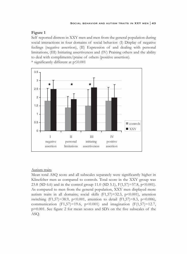
Figure 1Self reported distress in XXY men and men from the general population duringsocial interactions in four domains of social behavior: (I) Display of negativefeelings (negative assertion), (II) Expression of and dealing with personallimitations, (III) Initiating assertiveness and (IV) Praising others and the abilityto deal with compliments/praise of others (positive assertion).* significantly different at p≤0.001
Autism traitsMean total ASQ score and all subscales separately were significantly higher inKlinefelter men as compared to controls. Total score in the XXY group was23.8 (SD 6.6) and in the control group 11.0 (SD 5.1), F(1,57)=57.8, p<0.001).As compared to men from the general population, XXY men displayed moreautism traits in all domains; social skills (F1,57)=32.3, p<0.001), attentionswitching (F1,57)=38.9, p<0.001, attention to detail (F1,57)=8.3, p=0.006),communication (F1,57)=19.6, p<0.001) and imagination (F(1,57)=12.7,p=0.001. See figure 2 for mean scores and SD’s on the five subscales of theASQ.
Social behavior and autism traits in XXY men | 43
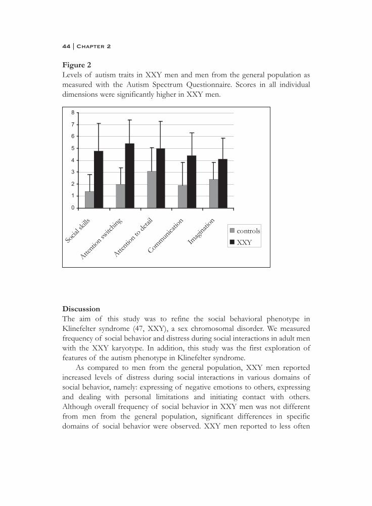
Figure 2Levels of autism traits in XXY men and men from the general population asmeasured with the Autism Spectrum Questionnaire. Scores in all individualdimensions were significantly higher in XXY men.
DiscussionThe aim of this study was to refine the social behavioral phenotype inKlinefelter syndrome (47, XXY), a sex chromosomal disorder. We measuredfrequency of social behavior and distress during social interactions in adult menwith the XXY karyotype. In addition, this study was the first exploration offeatures of the autism phenotype in Klinefelter syndrome.
As compared to men from the general population, XXY men reportedincreased levels of distress during social interactions in various domains ofsocial behavior, namely: expressing of negative emotions to others, expressingand dealing with personal limitations and initiating contact with others.Although overall frequency of social behavior in XXY men was not differentfrom men from the general population, significant differences in specificdomains of social behavior were observed. XXY men reported to less often
44 | Chapter 2
engage in social behavior dealing with expression of negative emotions, such asrefusing a request or standing up for one’s rights in a public situation. Inaddition, high rates of autistic-like traits were observed in XXY men, across alldimensions of the autism phenotype, namely: difficulties in social skills,attention switching, imagination, communication and increased attention todetails.
Our findings of difficulties in coping with social situations in XXY men,especially high levels of distress during social interactions, are consistent withreports of social anxiety, social withdrawal and shyness in individuals with theXXY karyotype (Bender et al., 1999; Ratcliffe, 1999). Difficulties in socialadjustment have primarily been reported for children or adolescents withKlinefelter syndrome. Our data suggest that social difficulties may persist intoadulthood, with social distress more prominent than a general reduction inengagement of social behavior. The use of self-report measures might havebiased the degree of social disabilities in Klinefelter syndrome. However,studies showing that XXY men tend to overrate, rather than underrate, theirown social adjustment (Bender et al., 1999) suggest that our findings mightrather be an underestimation than overestimation of social disabilities inKlinefelter syndrome.
The high levels of autism traits in XXY men that were observed across alldimensions of the autism phenotype, suggest that the impairments in socialinteractions and communication parallel autism-like features that characterizesindividuals at increased risk for autism. Increased levels of autism traits havealso been found in biological relatives of subject with autism (Bishop et al.,2004). Our findings fit with the concept of a ‘broad phenotype’ of autism,which refers to the mild autistic-like features that are seen in individuals that aregenetically related to an individual with autism (Bailey et al., 1998; Bishop et al.,2004). Similar to biological relatives of individuals with autism, autism-likefeatures in XXY men were observed in the face of spared verbal- and generalintellectual abilities (Bishop et al., 2004).
Based on twin studies, it has been suggested that the typical clinicalphenotype of autism as seen in subjects with the disorder and the broadersubclinical phenotype of autism as seen in biological relatives may share agenetic origin (Rutter, 2000). Although speculative, this might suggest that theX chromosome might be one of many genetic factors that play a role in theetiology of autism-like behaviors. Our findings are in line with studies in Turnersyndrome, another X chromosomal disorder characterized by a partial orcomplete absence of one of the X chromosomes in females (45,XO). Turner
Social behavior and autism traits in XXY men | 45
females also display difficulties in the social domain and the estimated risk ofautism spectrum disorders may be several times higher as compared to womenfrom the general population (Creswell et al., 1999; Mazzocco et al., 1998;McCauley et al., 2006; Skuse, 2000).
A hypothesized role of genetic mechanisms involving the X chromosomein social behavior fits with the notion that several genetic factors in autismmight operate on components of the disorder, rather than the syndrome as awhole (Rutter, 2000). Influence of genes on the X chromosome on thedevelopment of autism-like features would fit with the male preponderance inautism spectrum disorders (Volkmar et al., 1993).
Taken together, as compared to men from the general population, XXYmen reported increased levels of distress during social interactions and lessengagement in those aspects of social behavior that deal with display ofnegative emotions. The increased levels of autism traits that we observed inXXY men call for a more thorough clinical investigation of vulnerability toautism in Klinefelter syndrome in a larger and more representative sample inepidemiological terms. Although our findings require replication, Klinefeltersyndrome might prove to serve as a useful model for studying a role of the Xchromosome in social behavioral dysfunction and autism-like behavior.
46 | Chapter 2
ReferencesArrindell, W. A., Bridges, K. R., van der Ende, J., St. Lawrence, J. S., Gray-
Shellberg, L., Harnish, R., et al. (2001). Normative studies with the scale for interpersonal behaviour (sib): Ii. Us students: A cross-cultural comparison with dutch data. Behaviour Research and Therapy, 39(12),1461.
Arrindell, W. A., Van der Ende, J. (1985). Cross-sample invariance of the structure of self-reported distress and difficulty in assertiveness:Experiences with the scale for interpersonal behaviour. Advances in Behaviour Research and Therapy, 7, 205-243.
Bailey, A., Palferman, S., Heavey, L., & Le Couteur, A. (1998). Autism: The phenotype in relatives. Journal of Autism and Developmental Disorders,28(5), 369.
Baron-Cohen, S., Wheelwright, S., Skinner, R., Martin, J., & Clubley, E. (2001).The autism-spectrum quotient (aq): Evidence from asperger syndrome/high-functioning autism, males and females, scientists and mathematicians. Journal of Autism and Developmental Disorders, 31(1),5-17.
Bender, B. G., Harmon, R. J., Linden, M. G., Bucher-Bartelson, B., & Robinson, A. (1999). Psychosocial competence of unselected young adults with sex chromosome abnormalities. American Journal ofMedical Genetics - Neuropsychiatric Genetics, 88(2), 200.
Bender, B. G., Harmon, R. J., Linden, M. G., & Robinson, A. (1995).Psychosocial adaptation of 39 adolescents with sex chromosome abnormalities. Pediatrics, 96(2 Pt 1), 302-308.
Bishop, D. V. M., Maybery, M., Maley, A., Wong, D., Hill, W., & Hallmayer, J.(2004). Using self-report to identify the broad phenotype in parents ofchildren with autistic spectrum disorders: A study using the autism-spectrum quotient. Journal of Child Psychology and Psychiatry and Allied Disciplines, 45(8), 1431.
Boone, K. B., Swerdloff, R. S., Miller, B. L., Geschwind, D. H., Razani, J., Lee,A., et al. (2001). Neuropsychological profiles of adults with klinefelter syndrome. Journal of the International Neuropsychological Society: Jins,7(4), 446-456.
Constantino, J. N., & Todd, R. D. (2003). Autistic traits in the general population: A twin study. Archives of General Psychiatry, 60(5), 524.
Social behavior and autism traits in XXY men | 47
Creswell, C. S., & Skuse, D. H. (1999). Autism in association with turner syndrome: Genetic implications for male vulnerability to pervasive developmental disorders. Neurocase, 5(6), 511-518.
Geschwind, D. H., Boone, K. B., Miller, B. L., & Swerdloff, R. S. (2000a).Neurobehavioral phenotype of klinefelter syndrome. Mental Retardation and Developmental Disabilities Research Reviews, 6(2), 107-116.
Geschwind, D. H., Boone, K. B., Miller, B. L., & Swerdloff, R. S. (2000b).Neurobehavioral phenotype of klinefelter syndrome., 6(2), 107-116.
Geschwind, D. H., & Dykens, E. (2004). Neurobehavioral and psychosocial issues in klinefelter syndrome. Learning Disabilities Research & Practice,19(3), 166-173.
Lanfranco, F., Kamischke, A., Zitzmann, M., & Nieschlag, P. E. (2004).Klinefelter's syndrome. The Lancet, 364(9430), 273-283.
Lezak, M. D. (1995). Neuropsychological assessment (third ed.). New York:Oxford University Press.
Mazzocco, M. M. M., Baumgardner, T., Freund, L. S., & Reiss, A. L. (1998).Social functioning among girls with fragile x or turner syndrome and their sisters. Journal of Autism and Developmental Disorders, 28(6),509-517.
McCauley, E., & Sybert, V. (2006). Social and behavioral development of girls and women with turner syndrome. International Congress Series, 1298,93.
Nelson, H. E. (1982). National adult reading test (nart), manual. Windsor,Bershire, UK: NFER-Nelson.
Nielsen, J., Johnsen, S. G., & Sorensen, K. (1980). Follow-up 10 years later of34 klinefelter males with karyotype 47,xxy and 16 hypogonadal males with karyotype 46,xy. Psychological Medicine, 10(2), 345.
Ratcliffe, S. (1999). Long-term outcome in children of sex chromosome abnormalities. Archives of Disease in Childhood, 80(2), 192-195.
Raven, J. C., Raven, J., & Court, J. H. (1993). Manual for raven's progressive matrices and vocabulary scales. Oxford: Oxford Psychologist Press.
Rovet, J., Netley, C., Keenan, M., Bailey, J., & Stewart, D. (1996). The psychoeducational profile of boys with klinefelter syndrome. Journal ofLearning Disabilities, 29(2), 193-196.
Rutter, M. (2000). Genetic studies of autism: From the 1970s into the millennium. Journal of Abnormal Child Psychology, 28(1), 3.
48 | Chapter 2
Schmand, B., Bakker, D., Saan, R., & Louman, J. (1991). [the dutch reading test for adults: A measure of premorbid intelligence level]. Tijdschrift voor Gerontologie en Geriatrie, 22(1), 15-19.
Sheehan, D. V., Lecrubier, Y., Sheehan, K. H., Amorim, P., Janavs, J., Weiller,E., et al. (1998). The mini-international neuropsychiatric interview (m.I.N.I.): The development and validation of a structured diagnostic psychiatric interview for dsm-iv and icd-10. Journal of Clinical Psychiatry, 59 Suppl 20, 22-33;quiz 34-57.
Skuse, D. H. (2000). Imprinting, the x-chromosome, and the male brain:Explaining sex differences in the liability to autism. Pediatric Research,47(1), 9-16.
Spiker, D., Lotspeich, L. J., Dimiceli, S., Myers, R. M., & Risch, N. (2002).Behavioral phenotypic variation in autism multiplex families: Evidence for a continuous severity gradient. American Journal of Medical Genetics - Neuropsychiatric Genetics, 114(2), 129.
van Rijn, S., Swaab, H., Aleman, A., & Kahn, R. S. (2006). X chromosomal effects on social cognitive processing and emotion regulation: A study with klinefelter men (47,xxy). Schizophrenia Research, 84(2-3), 194-203.
Volkmar, F. R., Szatmari, P., & Sparrow, S. S. (1993). Sex differences in pervasive developmental disorders. Journal of Autism and Developmental Disorders, V23(4), 579.
Willshire, D., Kinsella, G., & Prior, M. (1991). Estimating wais-r iq from the national adult reading test - a cross-validation. Journal of Clinical and Experimental Neuropsychology, 13(2), 204-216.
Woodbury-Smith, M. R., Robinson, J., Wheelwright, S., & Baron-Cohen, S.(2005). Screening adults for asperger syndrome using the aq: A preliminary study of its diagnostic validity in clinical practice. Journal ofAutism and Developmental Disorders, V35(3), 331.
Social behavior and autism traits in XXY men | 49

Chapter 3
Klinefelter's syndrome (karyotype 47,XXY)
and schizophrenia-spectrum pathology
Sophie van Rijn, André Aleman, Hanna Swaab, René S Kahn
British Journal of Psychiatry, 2006, 189 (5), 459-461
AbstractKlinefelter syndrome, characterized by a 47,XXY chromosomal pattern, haslargely been associated with physical abnormalities. Here, we report high levelsof schizophrenia spectrum pathology in 32 Klinefelter men in comparison to26 healthy controls. Schizophrenia spectrum pathology was measured with theSchizotypal Personality Questionnaire (SPQ) and Positive and NegativeSyndrome Scale (PANSS).
Our findings may have important implications for treatment of Klinefeltersyndrome. In addition, these results suggest that the X chromosome may becritically involved in the aetiology of schizophrenia.
52 | Chapter 3
IntroductionKlinefelter syndrome is the most common sex chromosome disorder, affectingapproximately 1 in 400 to 800 males. Individuals with this syndrome arecharacterized by an additional X chromosome, leading to the 47,XXYkaryotype. This sex chromosomal aneuploidy results in a variety of phenotypesincluding hypogonadism, androgen deficiency and infertility (Lanfranco et al.,2004). Although the primary focus in clinical research has been on physicalphenotypes of these men, there is a an awareness of neuro-anatomical,cognitive and behavioral abnormalities (Lanfranco et al., 2004; Shen et al.,2004). Specific impairments on measures of verbal skills, high incidence ofdyslexia and social dysfunctioning are among the most consistentlty reportedbehavioral phenotypes (Geschwind et al., 2000). On the other hand, in a recentreview on Klinefelter syndrome the authors conclude that it still remains unclearwhether Klinefelter syndrome can be associated with psychiatric disturbances(Lanfranco et al., 2004). Interestingly however, many of the abnormalities inKlinefelter syndrome resemble those found in schizophrenia.
For example, structural MRI studies have reported smaller whole brainvolumes, enlarged lateral ventricles and volume reductions of the superiortemporal gyrus (STG), amygdala, hippocampus, insula and cingulate inKlinefelter men (Shen et al., 2004). A comparison of these findings to structuralMRI studies in schizophrenia shows that all these regions are also affected inschizophrenia. Support for the hypothesis that sex chromosomes may play arole in the development of schizophrenia is derived from studies showing thatmales are more often affected by the disease than females and have an earlierage of onset (Aleman et al., 2003).
Case studies have been published describing Klinefelter subjects sufferingfrom schizophrenia and higher rates of Klinefelter syndrome among samples ofschizophrenia patients (DeLisi et al., 1994). Studies investigating psychiatricpathology in Klinefelter syndrome have been limited in that they describedKlinefelter men in psychiatric care and mental hospitals, or recorded mentalhospital admissions. However, there have been no systematic reports of levelsof schizophrenia psychopathology in a large sample of Klinefelter subjectsunselected for psychiatric disorders. In addition, a biological-geneticvulnerability to schizophrenia may not only be investigated using dichotomous,diagnostic outcomes, but also using dimensional measures of schizophreniaspectrum pathology, which are more sensitive measures of vulnerability toschizophrenia. Schizophrenia spectrum phenotypes share common cognitive,neuro-anatomical and genetic characteristics with the severe schizophrenia
Schizophrenia traits in XXY men | 53
phenotype. The present study was designed to test the hypothesis of increasedlevels of of schizophrenia spectrum pathology in subjects with Klinefeltersyndrome.
Methods32 Klinefelter men (mean age 38.8, SD 8.1) and 26 healthy controls (mean age35.0, SD 9.0), matched for age, sex, years of education and intellectual ability,were included in the study. Klinefelter subjects were recruited from the DutchKlinefelter Association and not selected for psychological or behavioralabnormalities. Also, the psychiatry department was not mentioned during therecruitment process. The diagnosis of Klinefelter syndrome (47,XXYkaryotype) was confirmed by karyotyping using standard techniques. In allKlinefelter males, all 16 cells that were screened showed a 47,XXY karyotype,indicating non-mosaicism in this group. 80% Of the Klinefelter men receivedtestosterone supplementation. Mean age of onset of treatment was 27.8 years(SD 7.6). Control subjects were recruited by advertisements. None of thecontrols met criteria for an Axis-I psychiatric disorder, as shown by screeningwith the MINI-Plus. After complete description of the study to the subjects,written informed consent was obtained.
Schizophrenia spectrum pathology was measured with the SchizotypalPersonality Questionnaire (SPQ). The SPQ is regarded as an indicator of thegenetic vulnerability to schizophrenia, since there is a gradient increase inschizotypal traits in relatives of schizophrenia patients that is in proportion tothe risk for schizophrenia associated with the degree of kinship with theschizophrenic family member (Vollema et al., 2002). Factor analytical studieshave revealed three dimensions of schizotypy; Positive schizotypy (for examplereferential thinking and delusional atmosphere), Negative schizotypy (for exampleconstricted affect and social anxiety) and Disorganization (odd speech andeccentric behavior).
In addition, the Positive and Negative Syndrome Scale (PANSS) wasincluded. This is a widely used structured interview to assess symptom profilesin schizophrenia that are present in the week prior to the interview. The PANSSallows categorization of negative-, positive-, and general symptoms.Intellectual ability was measured with the National Adult Reading Test andRaven’s Advanced Progressive Matrices , an estimator of verbal I.Q. andperformance I.Q. respectively.
Group differences were tested using analysis of variance (ANOVA). Effectsizes were presented as Cohen’s d.
54 | Chapter 3
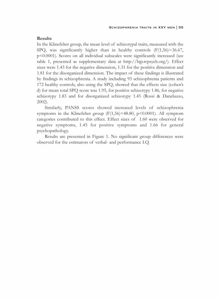
ResultsIn the Klinefelter group, the mean level of schizotypal traits, measured with theSPQ, was significantly higher than in healthy controls (F(1,56)=36.67,p<0.0001). Scores on all individual subscales were significantly increased (seetable 1, presented as supplementary data at http://bjp.rcpsych.org/). Effectsizes were 1.43 for the negative dimension, 1.31 for the positive dimension and1.81 for the disorganized dimension. The impact of these findings is illustratedby findings in schizophrenia. A study including 93 schizophrenia patients and172 healthy controls, also using the SPQ, showed that the effects size (cohen’sd) for mean total SPQ score was 1.95, for positive schizotypy 1.86, for negativeschizotypy 1.83 and for disorganized schizotypy 1.45 (Rossi & Daneluzzo,2002).
Similarly, PANSS scores showed increased levels of schizophreniasymptoms in the Klinefelter group (F(1,56)=48.80, p<0.0001). All symptomcategories contributed to this effect. Effect sizes of 1.60 were observed fornegative symptoms, 1.45 for positive symptoms and 1.66 for generalpsychopathology.
Results are presented in Figure 1. No significant group differences wereobserved for the estimators of verbal- and performance I.Q.
Schizophrenia traits in XXY men | 55
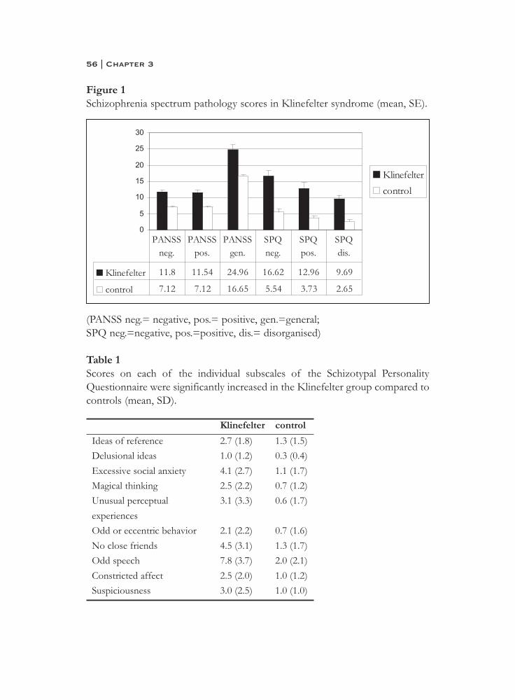
Figure 1Schizophrenia spectrum pathology scores in Klinefelter syndrome (mean, SE).
(PANSS neg.= negative, pos.= positive, gen.=general;SPQ neg.=negative, pos.=positive, dis.= disorganised)
Table 1 Scores on each of the individual subscales of the Schizotypal PersonalityQuestionnaire were significantly increased in the Klinefelter group compared tocontrols (mean, SD).
56 | Chapter 3
0
5
10
15
20
25
30
Klinefelter
control
Klinefelter 11.8 11.54 24.96 16.62 12.96 9.69
control 7.12 7.12 16.65 5.54 3.73 2.65
PANSSneg.
PANSSpos.
PANSSgen.
SPQneg.
SPQpos.
SPQdis.
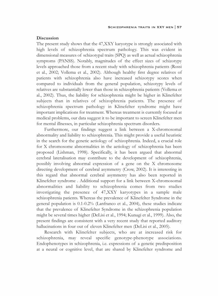
DiscussionThe present study shows that the 47,XXY karyotype is strongly associated withhigh levels of schizophrenia spectrum pathology. This was evident indimensional measures of schizotypal traits (SPQ) as well as actual schizophreniasymptoms (PANSS). Notably, magnitudes of the effect sizes of schizotypylevels approached those from a recent study with schizophrenia patients (Rossiet al., 2002; Vollema et al., 2002). Although healthy first degree relatives ofpatients with schizophrenia also have increased schizotypy scores whencompared to individuals from the general population, schizotypy levels ofrelatives are substantially lower than those in schizophrenia patients (Vollema etal., 2002). Thus, the liability for schizophrenia might be higher in Klinefeltersubjects than in relatives of schizophrenia patients. The presence ofschizophrenia spectrum pathology in Klinefelter syndrome might haveimportant implications for treatment. Whereas treatment is currently focused atmedical problems, our data suggest it to be important to screen Klinefelter menfor mental illnesses, in particular schizophrenia spectrum disorders.
Furthermore, our findings suggest a link between a X-chromosomalabnormality and liability to schizophrenia. This might provide a useful heuristicin the search for the genetic aetiology of schizophrenia. Indeed, a crucial rolefor X chromosome abnormalities in the aetiology of schizophrenia has beenproposed (Lishman, 1998). Specifically, it has been argued that abnormalcerebral lateralisation may contribute to the development of schizophrenia,possibly involving abnormal expression of a gene on the X chromosomedirecting development of cerebral asymmetry (Crow, 2002). It is interesting inthis regard that abnormal cerebral asymmetry has also been reported inKlinefelter syndrome . Additional support for a link between X-chromosomalabnormalities and liability to schizophrenia comes from two studiesinvestigating the presence of 47,XXY karyotypes in a sample maleschizophrenia patients. Whereas the prevalence of Klinefelter Syndrome in thegeneral population is 0.1-0.2% (Lanfranco et al., 2004), these studies indicatethat the prevalence of Klinefelter Syndrome in the schizophrenia populationmight be several times higher (DeLisi et al., 1994; Kunugi et al., 1999). Also, thepresent findings are consistent with a very recent study that reported auditoryhallucinations in four out of eleven Klinefelter men (DeLisi et al., 2005).
Research with Klinefelter subjects, who are at increased risk forschizophrenia, may reveal specific genotype-phenotype associations.Endophenotypes in schizophrenia, i.e. expressions of a genetic predispositionat a neural or cognitive level, that are shared by Klinefelter syndrome and
Schizophrenia traits in XXY men | 57
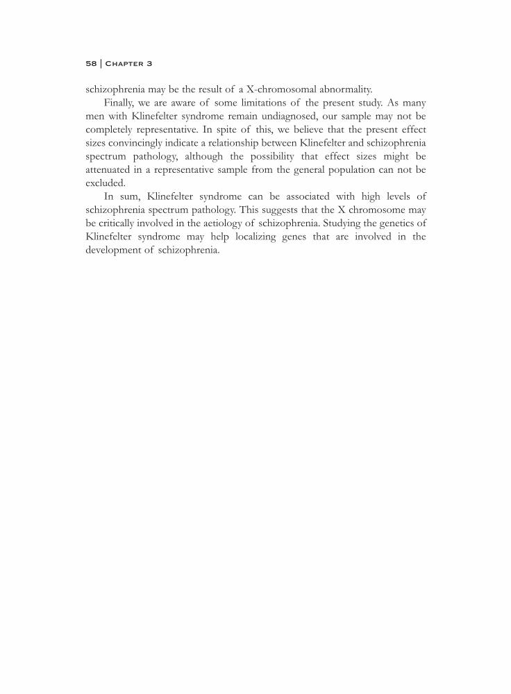
schizophrenia may be the result of a X-chromosomal abnormality.Finally, we are aware of some limitations of the present study. As many
men with Klinefelter syndrome remain undiagnosed, our sample may not becompletely representative. In spite of this, we believe that the present effectsizes convincingly indicate a relationship between Klinefelter and schizophreniaspectrum pathology, although the possibility that effect sizes might beattenuated in a representative sample from the general population can not beexcluded.
In sum, Klinefelter syndrome can be associated with high levels ofschizophrenia spectrum pathology. This suggests that the X chromosome maybe critically involved in the aetiology of schizophrenia. Studying the genetics ofKlinefelter syndrome may help localizing genes that are involved in thedevelopment of schizophrenia.
58 | Chapter 3
ReferencesAleman, A., Kahn, R. S., & Selten, J. P. (2003). Sex differences in the risk of
schizophrenia: Evidence from meta-analysis. Archives of General Psychiatry, 60(6), 565-571.
Crow, T. J. (2002). Handedness, language lateralisation and anatomical asymmetry: Relevance of protocadherin xy to hominid speciation and the aetiology of psychosis - point of view. British Journal of Psychiatry, 181,295-297.
DeLisi, L. E., Friedrich, U., Wahlstrom, J., Boccio-Smith, A., Forsman, A.,Eklund, K., et al. (1994). Schizophrenia and sex chromosome anomalies.Schizophrenia Bulletin, 20(3), 495-505.
DeLisi, L. E., Maurizio, A. M., Svetina, C., Ardekani, B., Szulc, K., Nierenberg,J., et al. (2005). Klinefelter's syndrome (xxy) as a genetic model for psychotic disorders. American Journal of Medical Genetics B Neuropsychiatric Genetics, 135(1), 15-23.
Geschwind, D. H., Boone, K. B., Miller, B. L., & Swerdloff, R. S. (2000).Neurobehavioral phenotype of klinefelter syndrome., 6(2), 107-116.
Kunugi, H., Lee, K. B., & Nanko, S. (1999). Cytogenetic findings in 250 schizophrenics: Evidence confirming an excess of the x chromosome aneuploidies and pericentric inversion of chromosome 9. Schizophrenia Research, 40(1), 43-47.
Lanfranco, F., Kamischke, A., Zitzmann, M., & Nieschlag, P. E. (2004).Klinefelter's syndrome. The Lancet, 364(9430), 273-283.
Lishman, W. A. (1998). Endocrine diseases and metabolic disorders. In W. A.Lishman (Ed.), Organic psychiatry: The psychological consequences ofcerebral disorder (pp. 526-527). Oxford: Blackwell Science.
Rossi, A., & Daneluzzo, E. (2002). Schizotypal dimensions in normals and schizophrenic patients: A comparison with other clinical samples.Schizophrenia Research, 54(1-2), 67-75.
Shen, D., Liu, D., Liu, H., Clasen, L., Giedd, J., & Davatzikos, C. (2004).Automated morphometric study of brain variation in xxy males.Neuroimage, 23(2), 648-653.
Vollema, M. G., Sitskoorn, M. M., Appels, M. C. M., & Kahn, R. S. (2002).Does the schizotypal personality questionnaire reflect the biological-genetic vulnerability to schizophrenia? Schizophrenia Research, 54(1-2),39-45.
Schizophrenia traits in XXY men | 59

Part II
Social cognition

Chapter 4
X Chromosomal effects on social cognitive
processing and emotion regulation: a study
with Klinefelter men (47,XXY)
Sophie van Rijn, Hanna Swaab, André Aleman and René S. Kahn
Schizophrenia Research, 2006, 84 (2-3), 194-203
AbstractStudying Klinefelter syndrome (47,XXY), a genetically defined disordercharacterized by the presence of an additional X chromosome, can revealinsights into genotype-phenotype associations. Increased vulnerability topsychiatric disorders characterised by difficulties in social interactions, such asschizophrenia and autism, has been reported for this population. The reportedsocial difficulties in 47,XXY men may arise as a consequence of impairments inthe processing of social and emotional information. The present study is thefirst investigation of social-emotional information processing in this X-chromosomal disorder.
32 Klinefelter men and 26 men from the general population, with thegroups matched for age, educational level and I.Q., participated in the study.Several tasks were included, reflecting aspects of social-emotional informationprocessing on levels of perception, experience and expression: labeling of facialexpressions of emotion, emotion-cognition interactions in decision making andemotion regulation, that refers to subjective experience and identification ofemotional arousal as well as verbal expression of emotions.
A discrepancy between cognitive appraisal of emotions and emotionalarousal was observed in Klinefelter syndrome. Taken together, Klinefelter menseem less accurate in perception of socio-emotional cues such as angry facialexpressions, they are less able to identify and verbalize their emotions, butexperience increased levels of emotional arousal, in comparison to the generalpopulation. Besides describing the social-emotional phenotype of this X-chromosomal disorder, the present data may prove to be an importantcontribution to the development of more general models describing pathwaysto neuropsychiatric disorders characterized by social cognitive disturbances.
64 | Chapter 4
IntroductionStudying genetically defined syndromes can reveal insights into gene-brain-behavior mechanisms. This may lead to a better understanding of complexpsychiatric diseases, such as schizophrenia. In this regard, an interesting geneticcondition that has been associated with an increased vulnerability toschizophrenia, is Klinefelter syndrome (Bojesen et al., 2006; L.E. DeLisi et al.,1994; L. E. DeLisi et al., 2005; Kunugi et al., 1999; van Rijn et al., in press).Klinefelter syndrome is characterised by the presence of an additional Xchromosome in men. The 47,XXY chromosomal pattern has been associatedwith a variety of physical and neuropsychological abnormalities (Lanfranco etal., 2004). For example, infertility, lowered testosterone levels and languagedisabilities have been reported. At a behavioral level, Klinefelter men have beenreported to have difficulties in social functioning and have been described asbeing introvert, anxious, impulsive, quiet, subassertive and socially withdrawn(D. H. Geschwind et al., 2000a). Socially inappropriate behavior as well as anti-social behavior have also been documented (Gotz et al., 1999; Ratcliffe, 1999).
Social difficulties in Klinefelter syndrome may be dismissed as aconsequence of daily life struggles associated with the disorder, but they mayalso arise from deficits in social cognitive processing. Social cognitiveprocessing, i.e. the ability to perceive, understand and express social signals,covers aspects of intelligence other than general cognitive abilities such aslanguage and attention, that have an independent contribution to socialfunctioning (Amy E. Pinkham et al., 2003). In the general population, socialcompetence seems to be under substantial genetic control (Scourfield et al.,1999).
Findings from MRI studies showing structural abnormalities of brainregions that form part of a neural network subserving social cognition andemotion call for a more thorough investigation of social cognitive processing inKlinefelter syndrome. For example, volume reductions of the amygdala, insula,anterior cingulate and superior temporal gyrus, all of which have been shown toplay an important role in social cognition and emotion, have been observed inthis syndrome (Shen et al., 2004).
Social cognitive processing is thought to mediate between the molecular,genetic level and the behavioral, or even clinical, level and thereby may provideinsight into gene-brain-behavior linkages underlying psychopathology (Van Rijnet al., 2005). Interestingly, as Klinefelter syndrome has been associated withincreased levels of schizophrenia spectrum pathology and autistic features (L.E.DeLisi et al., 1994; L. E. DeLisi et al., 2005; Gillberg, 1995; Swaab et al., in prep.;
Social cognition in XXY men | 65
van Rijn et al., in press), studying social cognitive processing in Klinefeltersyndrome may prove to be an important contribution to the development ofmore general models describing pathways to neuropsychiatric disorderscharacterized by social cognitive disturbances (Van Rijn et al., 2005).
The present study is, to the best of our knowledge, the first investigation ofsocial cognitive processing in a large sample of Klinefelter men. Therefore, abroad range of aspects of social cognitive processing was investigated. Severaltasks were included, reflecting aspects of social cognitive processing on levelsof perception, experience and expression: labeling of facial expressions ofemotion, emotion-cognition interactions in decision making and emotionregulation, that refers to subjective experience and identification of emotionalarousal as well as verbal expression of emotions. The tasks in this study havebeen used in previous studies of social cognitive processing in schizophreniaand/or autism, and for all tasks neural correlates have been defined (A. Aleman,in press; R. Gur et al., 2002a; Sanfey et al., 2003).
Methods
Subjects32 Men with Klinefelter syndrome (mean age 38.8, SD 8.3) were studied. Theparticipants were recruited from the Dutch Klinefelter Association, and werenot selected for psychological, behavioral or cognitive abnormalities. Diagnosisof Klinefelter syndrome was confirmed by karyotyping, using standardprocedures. In this group, 28 men were treated with testosterone supplements,with a mean age of treatment onset of 26.2 years (SD 7.9).26 Male controls from the general population (mean age 35.2, SD 9.1) wereincluded in the study. They were recruited using advertisements in localnewspapers or were drawn from an already existing database in our department.There were no significant differences in age (F(1,50)=2.4, p=0.12) and years ofeducation (F(1,56)=0.4, p=0.52) between the groups. None of the controlsubjects had a history of psychiatric illness as confirmed with the MiniInternational Neuropsychiatric Interview plus (MINI) (Sheehan et al., 1998).Exclusion criteria for both Klinefelter men and controls were neurologicalconditions or history of head injury with loss of consciousness, recent historyof substance abuse and mental retardation. After complete description of thestudy to the subjects, written informed consent was obtained.
66 | Chapter 4
Intellectual abilityRaven’s Advanced Progressive Matrices (short form)This test is commonly accepted as a measure of general intelligence and hasbeen shown to correlate with a number of standardized intelligence tests(Lezak, 1995; Raven et al., 1993). Subjects are shown 12 pictures of matrices(i.e., related patterns), each of which is a figural design with a part removed. Thesubject must choose the correct missing part from eight options.
National Adult Reading Test (NART)Since Klinefelter syndrome has been associated with verbal deficits, we includeda measure of reading skills in addition to general I.Q. The Dutch translation ofthe NART (Nelson, 1982; Schmand et al., 1991) provides an estimate of verbalIQ and is based on the high correlation between reading ability, specifically ofirregular words, with intelligence in the normal population. The test iscomposed of a list of 50 irregular words (i.e., pronunciation does not follow thenormal phonetic rules) printed in order of increasing difficulty. Subjects arerequired to read these words aloud, and, on the basis of the number of errorsmade in pronunciation a reliable estimate of WAIS-R IQ can be calculated(Willshire et al., 1991).
General cognitionGeneral face recognitionThe Benton and Van Allen Test of Facial Recognition, Short Form (Benton &Van Allen, 1973), comprises a series of sheets containing a single photographedtarget face to be matched to a set of six face photographs. In the first six trials,the identical face has to be selected out of six options. In the remaining six trials,three different views (changed in orientation or lighting conditions compared tothe target photograph) have to be discriminated from three incorrectalternatives. All faces are physically similar, not showing glasses or hair.
Social cognitive processingFacial Affect Recognition This task is a measure of explicit facial affect recognition of degraded faces, thathas been used in earlier studies on emotion processing (van 't Wout et al., 2004).Photographs of four different actors, two male and two female, were used(Frigerio et al., 2002). Sixty-four trials were presented, consisting of 16 facepresentations in each of four conditions: angry, happy, fearful and neutral. Ineach condition, 8 trials displayed 100% emotional intensity and the other 8 trials
Social cognition in XXY men | 67
displayed 75% emotional intensity. All photographs of the faces were passedthrough a filter that reduced visual contrast by 30%, in order to avoid local- andstimulate global processing. Subjects were asked to indicate the expression ofeach emotion by clicking with the mouse on one of the four emotion labelsdepicted on the computer screen. They were asked to work as accurately andrapidly as possible.
Emotion in decision makingThe Ultimatum Game is an extensively studied paradigm originating from thefield of strategic decision-making. It provides a measure of cognition-emotioninteractions in strategic decision making, by inducing conflict betweenacceptance (cognitive motive) and rejection (emotional motive) of unfairfinancial offers (Sanfey et al., 2003).
In the Ultimatum Game two players were given the opportunity to splitfictitious a sum of money, ?5. A fictitious proposer (20 times another personand 20 times the computer) made an offer as to how this money should be splitbetween the two. The responder (participant) could either accept or reject thisoffer. If it was accepted, the money was split as proposed, but if the responderrejected the offer, then neither player received anything. In either event, the nextoffer appeared. 20 times the money was fairly split (5,00 versus 5,00) and inanother 20 rounds the offer was unfair. There were three different unfair offers,i.e. twice 9,00 versus 1,00; twice 8,00 versus 2,- and once 7,00 versus 3,00. Thisresulted in a total of 40 rounds. The different offers (fair, unfair, human,computer) were assigned in a random order.
Emotion regulationAlexithymia refers to a personality trait implying an inability or reduction toidentify, experience, describe and reflect on one’s own emotions (Lane et al.,1997; Sifneos, 1973).
We used the Bermond-Vorst Alexithymia Questionnaire to measurealexithymia, i.e. emotion-regulation (Morera et al., 2005; Vorst & Bermond,2001). The BVAQ has good psychometric properties, and consists of fivesubscales: Emotionalizing: the degree to which someone is emotionally arousedby emotion inducing events. Fantasizing: the degree to which someone ininclined to fantasize, imagine, daydream, etc. Identifying: the degree to which oneis able to define one’s arousal states. Analyzing: the degree to which one seeksout explanations of one’s own emotional reactions. Verbalizing: the degree towhich one is able or inclined to describe or communicate about one’s own
68 | Chapter 4
emotional reactions. Each of the subscales consists of eight items. Answers arescored on a 5-point scale (1=certainly does not apply to me, up to 5=certainlyapplies to me). The calculated scores were substracted from 50, resulting inlower scores reflecting lower capacity or performance.The latter three BVAQ-subscales have substantial overlap (correlation of .80) with the more widely usedToronto Alexithymia Scale 20 (TAS-20) (Bagby et al., 1994). However, theBVAQ also includes two measures of emotional experience, fantasizing andemotionalizing, which form part of the original definition of Alexithymiaintroduced by Sifneos (Sifneos, 1973).
Statistical analysesSeparate t-tests were conducted on the demographic data, Raven’s AdvancedProgressive Matrices, NART and the Benton and Van Allen test of FacialRecognition.
Data of the social cognitive tasks were analyzed using GLM repeatedmeasures analysis, with a fixed factor Group (Klinefelter and control) and avariable factor reflecting the conditions within the tasks. When the multivariatetests indicated significant differences, the differences were analyzed withsubsequent post-hoc ANOVA’s. When potential confounding variables (i.e.general face recognition and intellectual ability) differed between the groups,then these were included as a covariate in the analysis. Alpha was set at 0.05.
Results
Intellectual abilityMean score on the Raven’s Advanced Progressive Matrices was not significantlydifferent between the groups (t(1,55)=1.0, p=0.32). Mean scores were 107.2(SD 14.2) and 110.4 (SD 8.6) for the Klinefelter group and control grouprespectively.
On the NART, mean score of the Klinefelter men did not significantlydiffer from controls (t(1,54)=1.78, p=0.08). Mean score in de Klinefelter groupwas 103.5 (SD 9.8), for the control group it was 108.0 (SD 9.1).
General cognitionGeneral face recognitionIn general, no ceiling effects were observed in the Benton and van Allen test.Mean percentage of correct responses was 76.25 % and 80.0 % in theKlinefelter and control group respectively. No group differences were present
Social cognition in XXY men | 69
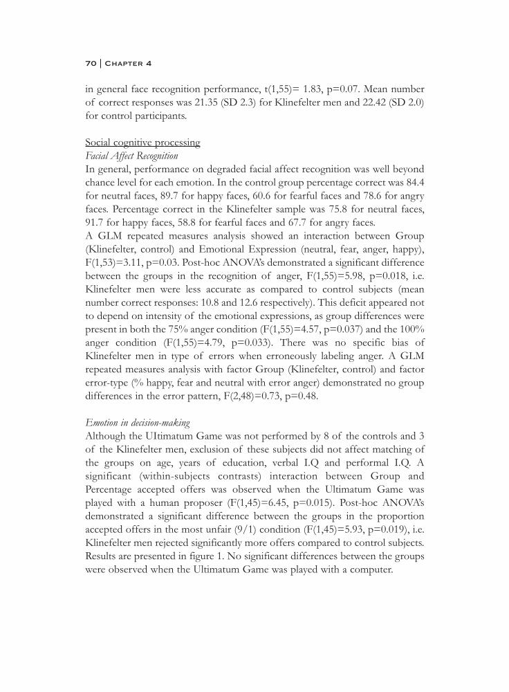
in general face recognition performance, t(1,55)= 1.83, p=0.07. Mean numberof correct responses was 21.35 (SD 2.3) for Klinefelter men and 22.42 (SD 2.0)for control participants.
Social cognitive processingFacial Affect Recognition In general, performance on degraded facial affect recognition was well beyondchance level for each emotion. In the control group percentage correct was 84.4for neutral faces, 89.7 for happy faces, 60.6 for fearful faces and 78.6 for angryfaces. Percentage correct in the Klinefelter sample was 75.8 for neutral faces,91.7 for happy faces, 58.8 for fearful faces and 67.7 for angry faces.A GLM repeated measures analysis showed an interaction between Group(Klinefelter, control) and Emotional Expression (neutral, fear, anger, happy),F(1,53)=3.11, p=0.03. Post-hoc ANOVA’s demonstrated a significant differencebetween the groups in the recognition of anger, F(1,55)=5.98, p=0.018, i.e.Klinefelter men were less accurate as compared to control subjects (meannumber correct responses: 10.8 and 12.6 respectively). This deficit appeared notto depend on intensity of the emotional expressions, as group differences werepresent in both the 75% anger condition (F(1,55)=4.57, p=0.037) and the 100%anger condition (F(1,55)=4.79, p=0.033). There was no specific bias ofKlinefelter men in type of errors when erroneously labeling anger. A GLMrepeated measures analysis with factor Group (Klinefelter, control) and factorerror-type (% happy, fear and neutral with error anger) demonstrated no groupdifferences in the error pattern, F(2,48)=0.73, p=0.48.
Emotion in decision-makingAlthough the UItimatum Game was not performed by 8 of the controls and 3of the Klinefelter men, exclusion of these subjects did not affect matching ofthe groups on age, years of education, verbal I.Q and performal I.Q. Asignificant (within-subjects contrasts) interaction between Group andPercentage accepted offers was observed when the Ultimatum Game wasplayed with a human proposer (F(1,45)=6.45, p=0.015). Post-hoc ANOVA’sdemonstrated a significant difference between the groups in the proportionaccepted offers in the most unfair (9/1) condition (F(1,45)=5.93, p=0.019), i.e.Klinefelter men rejected significantly more offers compared to control subjects.Results are presented in figure 1. No significant differences between the groupswere observed when the Ultimatum Game was played with a computer.
70 | Chapter 4
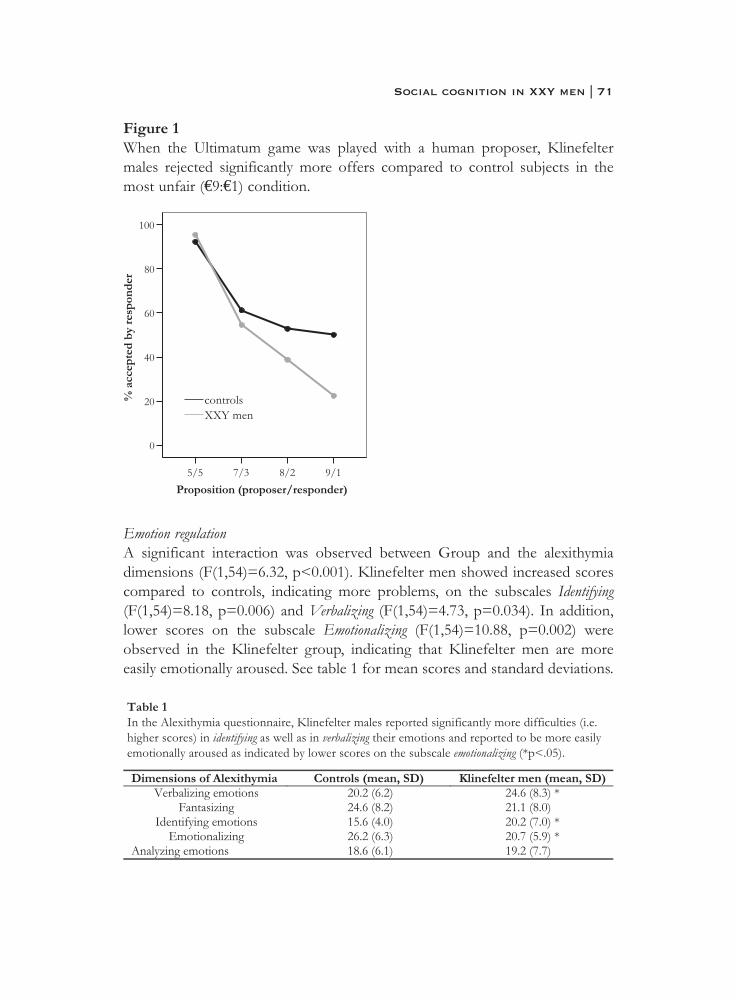
Figure 1When the Ultimatum game was played with a human proposer, Klinefeltermales rejected significantly more offers compared to control subjects in themost unfair (€9:€1) condition.
Emotion regulationA significant interaction was observed between Group and the alexithymiadimensions (F(1,54)=6.32, p<0.001). Klinefelter men showed increased scorescompared to controls, indicating more problems, on the subscales Identifying(F(1,54)=8.18, p=0.006) and Verbalizing (F(1,54)=4.73, p=0.034). In addition,lower scores on the subscale Emotionalizing (F(1,54)=10.88, p=0.002) wereobserved in the Klinefelter group, indicating that Klinefelter men are moreeasily emotionally aroused. See table 1 for mean scores and standard deviations.
Social cognition in XXY men | 71
ConclusionThe current study investigated various aspects of social cognitive processingand emotion regulation in Klinefelter syndrome, a genetic disordercharacterized by an XXY chromosomal pattern. Our findings suggestdisturbances in perception, experience and expression of social cognitiveinformation in this population. Specific deficits in perception of facialexpressions of anger were observed. Abnormal experience of emotions inKlinefelter men was suggested by both explicit and implicit measures.Klinefelter men explicitly reported increased emotional arousal in response toemotion-inducing events and were more influenced by their emotions in astrategic decision making game. Difficulties in identification and expression ofone’s own emotions were also found in this group, as indicated by thealexithymia measures. Taken together, Klinefelter men seem less accurate inperception of social-emotional cues, experience increased levels of emotionalarousal, but are less able to identify and verbalize the emotions they experience,in comparison to the general population.
These deficits in social cognition may explain, in part, the social difficultiesthat have been described in Klinefelter syndrome. First, we found Klinefeltermen to be impaired in recognizing facial expressions of anger, independent ofthe intensity of the emotional expression. Misperception of angry facialexpressions may contribute to problems in social interactions, as non-verbalsignals may convey crucial information about the emotional state of the sender.Various studies have revealed significant relationships between facial affectrecognition performance and social functioning (Christine Hooker & Park,2002).
Regarding the experience of emotions, Klinefelter men rejected more oftenunfair financial offers from human proposers as compared to the control groupin the Ultimatum game. This pattern of performance is shown to reflect theinfluence of emotion, in response to the offer being unfair, on strategic decisionmaking (Sanfey et al., 2003). A recent fMRI study has shown that rejection ofunfair offers could be significantly predicted on the basis of insular activity(Sanfey et al., 2003), a brain region involved in processing emotions such asdisgust (Wicker et al., 2003), and monitoring of the internal milieu, for examplearousal (Augustine, 1996). In line with this, a recent study revealed thatautonomic, emotional arousal, as measured by skin conductance activity, ishigher for unfair offers and associated with the rejection of unfair offers in thisparticular paradigm (van 't Wout et al., in press). Interestingly, in Klinefeltersyndrome, structural abnormalities of the insula and amygdala have been
72 | Chapter 4
reported which may, in part, contribute to this pattern of performanceindicating increased emotional arousal (Shen et al., 2004).
Autistic adults, like Klinefelter men, also accept very little in this paradigmand it has been proposed that impairments in reading intentions and beliefsfrom others play a role in rejecting unfair offers (Camerer, 2003). Althoughspeculative, our finding that Klinefelter men only rejected more offers whenproposed by a human individual, and not when proposed by a computer,tentatively suggests that Klinefelter men have problems in reading intentions ofother people. The present results support our hypothesis that social cognition inKlinefelter syndrome is disturbed, which is expressed predominantly in socialcontexts such as interpersonal relations. The hypothesis regarding theory ofmind capabilites in Klinefelter syndrome should be tested in future research.
Besides this implicit emotional task, increased emotional experience wasalso observed in an explicit, self-report measure of emotional experience, i.e.the alexithymia questionnaire. Klinefelter men reported increased emotionalarousal in response to emotion-inducing events, as indicated by the subscale‘emotionalizing’ in the Alexithymia questionnaire. In contrast to apparenthyperfunctional emotional -experience and -reactivity, identifying and verbalizingthese emotions appears to be hypofunctional in Klinefelter syndrome. The latterfinding can be considered as a possible risk factor for both medical andpsychiatric disorders (Bagby & Taylor, 1997). Interestingly, the specificalexithymia profile observed in Klinefelter syndrome has recently also beendescribed for schizophrenia patients and their relatives, indicating that it may bean expression of the genetic vulnerability for the disease (van 't Wout et al., inprep.). The finding that alexithymia seems to be under considerable geneticcontrol fits this picture (Valera & Berenbaum, 2001).
Recently, a model has been proposed that can explain the two seeminglycontradictory features in schizophrenia that we also find in Klinefeltersyndrome; deficits in perception and experience on the one hand and increasedemotional arousal on the other hand (A. Aleman & Kahn, 2005a). Thisneuroanatomical model describes amygdala dysfunctions that arise fromstructural abnormalities of the amygdala in combination with an imbalance indopamine systems, with specific effects on intra-amygdaloid processing. Indeed,Klinefelter syndrome, as well as other populations at increased risk forschizophrenia, has been associated with structural abnormalities of theamygdala (Van Rijn et al., 2005). Interestingly, such a dissociation betweencognitive appraisal of emotions on the one hand and autonomic, emotionalarousal on the other hand has also been reported for females with Turner
Social cognition in XXY men | 73
syndrome, characterised by X monosomy (45,X) and a 200-fold increased riskfor autism. Turner syndrome subjects display (left amygdala mediated)impairments in labeling of facial expressions of both anger and fear, while their(right amygdala mediated) somatic responses are enhanced in reponse toemotional faces (D. Skuse et al., 2005). Thus, X-linked genes seem important forfunctional integration of autonomic, emotional arousal with cognitive appraisalof emotions.
Altogether, the present study reveals the behavioral/functionalconsequences of a compromised neural network for social cognitive processing.A central issue concerns the degree to which these disturbances in socialcognitive processing are related to the X chromosomal abnormality. Evidently,neural and behavioral phenotypes in Klinefelter syndrome can all be traced backto the additional X chromosome. In this regard, studies with individuals withTurner syndrome (45,X) may provide some insight. In one of these studies, adosage-sensitive locus on the X chromosome has been identified that directsdevelopment of the amygdala (Good et al., 2003). As in Klinefelter syndrome,individuals with Turner syndrome have social adjustment problems and poorsocial skills (Lesniak-Karpiak et al., 1999; Mazzocco et al., 1998). These parallelsin brain architecture and socio-behavioral phenotype support the idea that(mal)development of this area in Klinefelter syndrome may indeed be undercontrol of genes on the X chromosome.
Unfortunately, the exact pathway from X chromosome abnormalities tosocial cognitive impairments in Klinefelter syndrome remains unclear. One ofthe issues deserving attention is the role of abnormal testosterone levels inKlinefelter syndrome, which become apparent in puberty. It is thought thatduring development circulating gonadal hormones can modify brain structureand -function of target areas. As shown by animal studies, one of the majortarget areas of testosterone is the amygdala (Simerly et al., 1990). However, therelation between testosterone levels and behavior is complex; timing ofexposure, sensitivity to testosterone reflected in androgen receptor density andmodulation by environmental factors are important determinants in thebehavioral effects of testosterone (Craig et al., 2004). Furthermore, gonadalhormones may be one of many mechanisms by which sex chromosomes exerttheir influence on brain development. Recent animal studies have pointed todirect, non-hormonal, effects of sex-chromosomes on brain maturation(Dewing et al., 2003).
Reports of increased levels of schizophrenia spectrum pathology andautistic features in Klinefelter men point to a role of the X chromosome in
74 | Chapter 4
psychiatric disorders with predominant social cognitive impairments (L.E.DeLisi et al., 1994; L. E. DeLisi et al., 2005; Gillberg, 1995; Swaab et al., in prep.;van Rijn et al., in press; Van Rijn et al., 2005). Although speculative, X-relateddisturbances in social cognitive processing may be one of many pathways topsychiatric disorders such as schizophrenia and autism. As these disorders aremore common in males compared to females (A. Aleman et al., 2003; D. H.Skuse, 2000), it will be interesting in future studies to investigate if and how thewidely described sex differences in social cognition can be related to genes onthe X chromosome and whether such genes are implicated in these psychiatricdisorders (S. Baron-Cohen et al., 2005; Scourfield et al., 1997). Also, morestudies are needed to explore the role of testosterone in social cognitiveimpairments in Klinefelter syndrome as well as autism and schizophrenia,especially since there are findings pointing to abnormal testosterone levels inthese latter disorders (S. Baron-Cohen et al., 2005; Goyal et al., 2004).
In a recent review it has been argued that the amygdala abnormalities seenin schizophrenia may be an X-linked endophenotype that can account for thesocial cognitive processing deficits observed in patients and their relatives (VanRijn et al., 2005). Also, it has been proposed that dysfunctional neural circuitsunderlying social cognitive impairments in autism may be phenotypes of X-linked genes, that may be sexually dimorphic in expression (S. Baron-Cohen etal., 2005; D. Skuse, 2003). Support for this hypothesis also comes from a reviewof all chromosomal studies in autism suggesting that the X chromosome,chromosome 15 and 17 are the most consistently reported chromosomes toshow abnormalities in the field of autism spectrum disorders (S Baron-Cohen,2004).
A crucial question is how Klinefelter syndrome may serve as a model forinvestigating the potential role of X-linked genes for susceptibility to mentaldisorders, most notably schizophrenia. In contrast to schizophrenia, the geneticpathology underlying deficits in Klinefelter syndrome is known. Commonendophenotypes that are shared by both disorders, such as deficits in socialcognition, point to a role of the X chromosome in social cognitive dysfunctionin the schizophrenia spectrum. Subsequently, one can narrow the search forgenetic pathology involved in schizophrenia, with the hypothesis that genes onthe X chromosome may code for abnormal social cognition in schizophrenia.Other possibilities may be to compare and study genetic pathology ofsubgroups of Klinefelter men: with low versus high levels of schizophreniaspectrum pathology, or poor versus relatively spared social cognition. Thismight lead us to more specific underlying genetic mechanisms such as genomic
Social cognition in XXY men | 75
imprinting or degree of X inactivation. Of course, these strategies apply to allendophenotypes that are shared by schizophrenia, which is clinically defined,and Klinefelter syndrome, which is genetically defined.
Finally, we are aware of some limitations of the present study. As manymen with Klinefelter syndrome remain undiagnosed (Bojesen et al., 2003), oursample may not be completely representative. In spite of this, the presentfindings describe social cognitive deficits, that may have important implicationsfor daily life functioning, in at least a specific population of Klinefelter men.In summary, the disturbances in perception, experience and expression of socialcognitive information that were observed in Klinefelter men may explain thesocial difficulties that have been reported in this syndrome. Besides describingthe social cognitive phenotype of this specific genetic disorder, the present dataencourage the development of a general model describing gene-brain-behaviorpathways underlying neuropsychiatric disorders characterized by social cognitivedisturbances.
76 | Chapter 4
ReferencesAleman, A. (in press). Feelings you can't imagine: Towards a cognitive
neuroscience of alexithymia. Trends in Cognitive Sciences.Aleman, A., & Kahn, R. S. (2005). Strange feelings: Do amygdala
abnormalities dysregulate the emotional brain in schizophrenia? Progress in Neurobiology, in press.
Aleman, A., Kahn, R. S., & Selten, J. P. (2003). Sex differences in the risk ofschizophrenia: Evidence from meta-analysis. Archives of General Psychiatry, 60(6), 565-571.
Augustine, J. R. (1996). Circuitry and functional aspects of the insular lobe in primates including humans. Brain Research Reviews, 22(3), 229-244.
Bagby, R. M., & Taylor, G. J. (1997). Affect dysregulation and alexithymia. In G. J. Taylor, R. M. Bagby & J. D. A. Parker (Eds.), Disorders of affect regulation: Alexithymia in medical and psychiatric illness. UK:Cambridge University Press.
Bagby, R. M., Taylor, G. J., & Parker, J. D. A. (1994). The 20-item toronto-alexithymia-scale.2. Convergent, discriminant, and concurrent validity.Journal of Psychosomatic Research, 38(1), 33-40.
Baron-Cohen, S. (2004). The cognitive neuroscience of autism. J Neurol Neurosurg Psychiatry, 75(7), 945-948.
Baron-Cohen, S., Knickmeyer, R. C., & Belmonte, M. K. (2005). Sex differences in the brain: Implications for explaining autism. Science,310(5749), 819-823.
Benton, A. L., & Van Allen, M. W. (1973). Test of face recognition manual (number 287 ed.). Department of Neurology, Iowa, USA:Neurosensory Center Publication.
Bojesen, A., Juul, S., Birkebaek, N. H., & Gravholt, C. H. (2006). Morbidity in klinefelter syndrome; a danish register study based on hospital discharge diagnoses. J Clin Endocrinol Metab.
Bojesen, A., Juul, S., & Gravholt, C. H. (2003). Prenatal and postnatal prevalence of klinefelter syndrome: A national registry study. The Journal of Clinical Endocrinology and Metabolism, 88(2), 622-626.
Camerer, C. F. (2003). Psychology and economics: Enhanced: Strategizing in the brain. Science, 300(5626), 1673-1675.
Craig, I. W., Harper, E., & Loat, C. S. (2004). The genetic basis for sex differences in human behaviour: Role of the sex chromosomes.Annals of Human Genetics, 68, 269-284.
Social cognition in XXY men | 77
DeLisi, L. E., Friedrich, U., Wahlstrom, J., Boccio_Smith, A., Forsman, A.,Eklund, K., et al. (1994). Schizophrenia and sex chromosome anomalies. Schizophrenia Bulletin, 20(3), 495-505.
DeLisi, L. E., Maurizio, A. M., Svetina, C., Ardekani, B., Szulc, K., Nierenberg,J., et al. (2005). Klinefelter's syndrome (xxy) as a genetic model for psychotic disorders. Am J Med Genet B Neuropsychiatr Genet, 135(1),15-23.
Dewing, P., Shi, T., Horvath, S., & Vilain, E. (2003). Sexually dimorphic gene expression in mouse brain precedes gonadal differentiation. Molecular Brain Research, 118(1-2), 82-90.
Frigerio, E., Burt, D. M., Montagne, B., Murray, L. K., & Perrett, D. I. (2002).Facial affect perception in alcoholics. Psychiatry Research, 113(1-2),161-171.
Geschwind, D. H., Boone, K. B., Miller, B. L., & Swerdloff, R. S. (2000).Neurobehavioral phenotype of klinefelter syndrome. Mental Retardation and Developmental Disabilities Research Reviews, 6(2),107-116.
Gillberg, C. (1995). Specific syndromes not otherwise referred to. In G.Christopher (Ed.), Clinical child neuropsychiatry (pp. 185-267).Cambridge, U.K.: Cambridge University Press.
Good, C. D., Lawrence, K., Thomas, N. S., Price, C. J., Ashburner, J., Friston,K. J., et al. (2003). Dosage-sensitive x-linked locus influences thedevelopment of amygdala and orbitofrontal cortex, and fear recognitionin humans. Brain, 126(11), 2431-2446.
Gotz, M. J., Johnstone, E. C., & Ratcliffe, S. G. (1999). Criminality and antisocial behaviour in unselected men with sex chromosomeabnormalities. Psychological Medicine, 29(4), 953-962.
Goyal, R. O., Sagar, R., Ammini, A. C., Khurana, M. L., & Alias, A. G. (2004).Negative correlation between negative symptoms of schizophrenia andtestosterone levels. Ann N Y Acad Sci, 1032, 291-294.
Gur, R., Schroeder, L., Turner, T., McGrath, C., Chan, R., Turetsky, B., et al.(2002). Brain activation during facial emotion processing. Neuroimage,16(3 Pt 1), 651.
Hooker, C., & Park, S. (2002). Emotion processing and its relationship to social functioning in schizophrenia patients. Psychiatry Research, 112(1),41-50.
78 | Chapter 4
Kunugi, H., Lee, K. B., & Nanko, S. (1999). Cytogenetic findings in 250 schizophrenics: Evidence confirming an excess of the x chromosomeaneuploidies and pericentric inversion of chromosome 9. Schizophr Res,40(1), 43-47.
Lane, R. D., Ahern, G. L., Schwartz, G. E., & Kaszniak, A. W. (1997). Is alexithymia the emotional equivalent of blindsight? Biol Psychiatry,42(9), 834-844.
Lanfranco, F., Kamischke, A., Zitzmann, M., & Nieschlag, P. E. (2004).Klinefelter's syndrome. The Lancet, 364(9430), 273-283.
Lesniak-Karpiak, K., Mazzocco, M. M., Lanham, D. C., & Denckla, M. B.(1999). Behavioral assessment of social skills in children with turnersyndrome or fragile x. Archives of Clinical Neuropsychology, 14(8),767-767.
Lezak, M. D. (1995). Neuropsychological assessment (third ed.). New York:Oxford University Press.
Mazzocco, M. M. M., Baumgardner, T., Freund, L. S., & Reiss, A. L. (1998).Social functioning among girls with fragile x or turner syndrome andtheir sisters. Journal of Autism and Developmental Disorders, 28(6),509-517.
Morera, O. F., Culhane, S. E., Watson, P. J., & Skewes, M. C. (2005). Assessing the reliability and validity of the bermond-vorst alexithymiaquestionnaire among u.S. Anglo and u.S. Hispanic samples. Journal ofPsychosomatic Research, 58(3), 289-298.
Nelson, H. E. (1982). National adult reading test (nart), manual. Windsor,Bershire, UK: NFER-Nelson.
Pinkham, A. E., Penn, D. L., Perkins, D. O., & Lieberman, J. (2003).Implications for the neural basis of social cognition for the study ofschizophrenia. American Journal of Psychiatry, 160(5), 815-824.
Ratcliffe, S. (1999). Long-term outcome in children of sex chromosome abnormalities. Archives of Disease in Childhood, 80(2), 192-195.
Raven, J. C., Raven, J., & Court, J. H. (1993). Manual for raven's progressive matrices and vocabulary scales. Oxford: Oxford Psychologist Press.
Sanfey, A. G., Rilling, J. K., Aronson, J. A., Nystrom, L. E., & Cohen, J. D.(2003). The neural basis of economic decision-making in the ultimatumgame. Science, 300(5626), 1755-1758.
Schmand, B., Bakker, D., Saan, R., & Louman, J. (1991). [the dutch reading test for adults: A measure of premorbid intelligence level]. Tijdschrift voorGerontologie en Geriatrie, 22(1), 15-19.
Social cognition in XXY men | 79
Scourfield, J., Martin, N., Lewis, G., & McGuffin, P. (1999). Heritability ofsocial cognitive skills in children and adolescents. British Journal ofPsychiatry, 175, 559-564.
Scourfield, J., McGuffin, P., & Thapar, A. (1997). Genes and social skills.Bioessays: News and Reviews in Molecular, Cellular and DevelopmentalBiology, 19(12), 1125-1127.
Sheehan, D. V., Lecrubier, Y., Sheehan, K. H., Amorim, P., Janavs, J., Weiller,E., et al. (1998). The mini-international neuropsychiatric interview(m.I.N.I.): The development and validation of a structured diagnosticpsychiatric interview for dsm-iv and icd-10. Journal of ClinicalPsychiatry, 59 Suppl 20, 22-33;quiz 34-57.
Shen, D., Liu, D., Liu, H., Clasen, L., Giedd, J., & Davatzikos, C. (2004).Automated morphometric study of brain variation in xxy males.NeuroImage, 23(2), 648-653.
Sifneos, P. E. (1973). The prevalence of 'alexithymic' characteristics in psychosomatic patients. Psychotherapy and Psychosomatics, 22(2), 255-262.
Simerly, R. B., Chang, C., Muramatsu, M., & Swanson, L. W. (1990).Distribution of androgen and estrogen-receptor messenger rna-containing cells in the rat-brain - an insitu hybridization study. Journal ofComparative Neurology, 294(1), 76-95.
Skuse, D. (2003). X-linked genes and the neural basis of social cognition.Autism: Neural Basis and Treatment Possibilities, Novartis FoundationSymposium, 251, 84-98.
Skuse, D., Morris, J. S., & Dolan, R. J. (2005). Functional dissociation ofamygdala-modulated arousal and cognitive appraisal, in turner syndrome.Brain, 128(Pt 9), 2084-2096.
Skuse, D. H. (2000). Imprinting, the x-chromosome, and the male brain:Explaining sex differences in the liability to autism. Pediatric Research,47(1), 9-16.
Swaab, H., Cohen-Kettenis, P. T., & Van Engeland, H. (in prep.). Social dysfunction in boys with klinefelter syndrome.
Valera, E. M., & Berenbaum, H. (2001). A twin study of alexithymia.Psychotherapy and Psychosomatics, 70(5), 239-246.
van 't Wout, M., Aleman, A., Bermond, B., & Kahn, R. (in prep.). No words for feelings: Alexithymia in schizophrenia patients and first degreerelatives.
80 | Chapter 4
van 't Wout, M., Aleman, A., Kessels, R. P., Laroi, F., & Kahn, R. S. (2004).Emotional processing in a non-clinical psychosis-prone sample.Schizophrenia Research, 68(2-3), 271-281.
van 't Wout, M., Kahn, R., Sanfey, A. G., & Aleman, A. (in press). Affective state and decision-making in the ultimatum game. Experimental BrainResearch.
van Rijn, S., Aleman, A., Swaab, H., & Kahn, R. (in press). 47,xxy karyotype and schizophrenia spectrum pathology. British Journal of Psychiatry.
Van Rijn, S., Aleman, A., Swaab, H., & Kahn, R. S. (2005). Neurobiology ofemotion and high risk for schizophrenia: Role of the amygdala and thex-chromosome. Neuroscience and Biobehavioral Reviews, 29(3), 385-397.
Vorst, H. C. M., & Bermond, B. (2001). Validity and reliability of the bermond-vorst alexithymia questionnaire. Personality and IndividualDifferences, 30(3), 413-434.
Wicker, B., Keysers, C., Plailly, J., Royet, J. P., Gallese, V., & Rizzolatti, G.(2003). Both of us disgusted in my insula: The common neural basis ofseeing and feeling disgust. Neuron, 40(3), 655-664.
Willshire, D., Kinsella, G., & Prior, M. (1991). Estimating wais-r iq from the national adult reading test - a cross-validation. Journal of Clinical andExperimental Neuropsychology, 13(2), 204-216.
Social cognition in XXY men | 81

Chapter 5
Social information processing in the
schizophrenia spectrum: implicit attention to
social signals
Mascha van ’t Wout, Sophie van Rijn, Tjeerd Jellema, René S. Kahn,André Aleman
AbstractSchizophrenia is characterized by disturbances in social functioning. In thesearch for determinants of social dysfunction in schizophrenia patients, socialcognitive capacities appear to be of crucial importance. The ability to processbasic social cues, such as gaze direction and biological motion direction, quicklyand automatically is thought to be a prerequisite for establishing successfulsocial interactions and especially for construing a sense of ‘social intuition’.However, studies that address the ability to automatically process such basicsocial cues in schizophrenia are lacking. We used a new visual illusion measuringthe extent in which social cues are processed effortlessly and implicitly in threedifferent groups characterized by the presence of traits or symptoms from theschizophrenia spectrum, i.e. 33 patients with schizophrenia, 32 siblings ofpatients with schizophrenia and 32 individuals with Klinefelter syndrome(47,XXY). These groups were compared to 50 age-, sex- and education-matched healthy control subjects. Results indicated that, in contrast to controlsubjects, patients with schizophrenia showed insensitivity to social cues. Thiswas particularly pronounced in patients with negative symptoms. The reducedinfluence of social cues was also observed in first-degree relatives of patientswith schizophrenia as well as in Klinefelter subjects. We suggest that theinsensitivity for social cues is a cognitive aspect of schizophrenia that may beseen as an endophenotype as it appears to be present both in relatives who areat increased genetic risk and a genetic disorder associated with schizophreniaspectrum psychopathology. These social cue-processing deficits couldcontribute, in part, to the difficulties in higher order social cognitive tasks andhence decreased social competence that have been observed in these groups.
84 | Chapter 5
IntroductionOne of the cardinal dysfunctions associated with the schizophrenia phenotypeconcerns disturbances in social functioning (DSM-IV American PsychiatricAssociation) (1994). Although some researchers have argued that this might bea consequence of severe psychopathology, others have demonstrated that socialdysfunction is relatively independent of symptomatology (Lenzenweger et al.,1996). This view is further supported by findings that disturbances in socialfunctioning are already present in early adolescence and often precede the onsetof psychosis (Baum et al., 1995; Hans et al., 1992; Walker, 1994). In the searchfor determinants of social dysfunction in schizophrenia patients, cognitivecapacities appear to be of crucial importance. In the last decade, there is agrowing body of research demonstrating deficits in the cognitive processing ofsocial information in schizophrenia (Pinkham et al., 2003), such as difficulties inemotion recognition (Edwards et al., 2002; Kohler et al., 2004) and the inabilityto understand and manipulate other people's behavior in terms of their mentalstates, also called Theory of Mind (Frith, 1992). Furthermore, schizophreniapatients have difficulties in the recognition of abstract social signals, such asinferences regarding actors’ affect and goals (Corrigan et al., 1993). Thesedeficits seem to be independent of intelligence, i.e. not attributable to ageneralized performance deficit (Corrigan, 1994), but are related to negativesymptoms such as withdrawal (Corrigan et al., 1994) and skills to receive,process, and send social signs (Corrigan et al., 1995).
An important underlying characteristic of successful social interaction isthe ability to quickly process social information (Frith et al., 1999). Basicelements of a social signal that are processed fast and automatic are for examplegaze direction, head orientation and body postures (Jellema et al., 2005). Thesecues can give clues about someone’s intentions, goals and believes (Perrett,1999). Usually, these basic social cues are largely automatically processed, whichis necessary to continuously infer the meaning of the rapidly changing socialsignals. Moreover, it is suggested that the ability to process these basic socialcues quickly and automatically is a prerequisite for establishing successful socialinteractions and communication (Frith et al., 1999). It may be especially relevantfor construing a sense of ‘social intuition’, as such implicit learning processesform the basis of social intuition (Lieberman, 2000). Intuitions have beendescribed as following: “intuitions are fast and take into account nonconsciouslygenerated information, gathered from experience, about the probabilisticstructure of the cues and variables relevant to one's judgments, decisions, andbehavior” (Bruner, 1960). Although schizophrenia patients seem to fail in areas
Automatic processing of social cues in XXY men | 85
of social intuition and automaticity in social interactions as observed in theirsocial behavior (Bellack et al., 1990), studies that address the ability toautomatically process such basic social cues in schizophrenia are lacking.
Therefore, we used a new paradigm involving a bias in the judgment of thedistance between two agents induced by the automatic processing of social cuesconveyed by these agents (Jellema et al., 2004). The social cues consisted of thedirection of attention and implied goal-directed actions. Typically, these socialcues induce the sensation of people (dis-)engaging in social interaction whentheir gaze or body postures are attended towards (or away from) each other.Therefore, the social cues of gaze direction and implied biological motion usedin the present paradigm results in people judging the persons as closer togethercompared to reference objects whilst this is not the case objectively.
In addition to patients with schizophrenia and healthy controls, we alsoincluded 2 other groups in the study: a) individuals at increased genetic risk forschizophrenia, i.e. sibling of schizophrenia patients and b) individuals with an Xchromosomal disorder and high levels of schizotypal traits, i.e. men withKlinefelter syndrome. Biological siblings of patients have been shown to be atsignificantly higher risk for the development of schizophrenia (Gottesman,1991) and display cognitive deficits that are also seen in schizophrenia patients,although to a lesser degree (Sitskoorn et al., 2003). Inclusion of the siblinggroup enables the study of social cognitive deficits related to a geneticvulnerability to schizophrenia without confounding environmental influences ashospitalization, medication and psychopathology. Support favoring the role ofgenetic mechanisms in social cognitive deficits is derived from studiesdemonstrating abnormalities in the processing of social-emotional cues inbiological relatives of patients with schizophrenia (Loughland et al., 2004;Toomey et al., 1999). This fits with the finding that social skills are underconsiderable genetic control in the general population. Men with Klinefeltersyndrome have an extra X chromosome (47,XXY chromosomal pattern), anddisplay abnormal brain development (Shen et al., 2004), social cognitiveimpairments (van Rijn et al., 2006b) and psychopathology that is also seen inpatients with schizophrenia (Van Rijn et al., 2006a). An additional advantagearising from studying Klinefelter men is knowledge of the precise geneticetiology of this syndrome, in contrast to what is known of the geneticunderpinnings of social cognitive impairments in schizophrenia. IncludingXXY men might point to a role of the X chromosome in development ofcognitive systems that are important for processing basic social signals (cf.Skuse, 2005). Involvement of sex chromosomes might explain, in part, the sex
86 | Chapter 5
differences that have been observed in social cognitive skills in the generalpopulation as well as in schizophrenia populations (Hampson et al., 2006;McClure, 2000; Scholten et al., 2005).
The aim of the present study was to investigate sensitivity to basic socialcues in individuals with schizophrenia, individuals with an increased genetic riskfor schizophrenia and individuals with a genetic disorder and high levels ofschizotypal traits. To this end we used a social distance judgment task thatincluded two social cues, social attention (gaze direction) and implied biologicalmotion (Jellema et al., 2004). It was hypothesized that patients withschizophrenia would demonstrate difficulties in the automatic processing ofsocial cues compared to control participants, i.e. patients may show no responsebias congruent with the direction of the social cues. Furthermore, therelationship between symptomatology and social cue processing is investigated.We predicted that the problems in social cue processing would be especiallyprevalent in patients with negative symptoms, since patients with negativesymptoms are characterized by social-emotional disturbances. We hypothesizedthat siblings of patients with schizophrenia as well as XXY men would show adeviant pattern comparable to patients, although to a lesser extent. This wouldindicate that deficient processing of basic social cues forms part of the geneticvulnerability for the disease, possibly involving genes on the X chromosome,rather than environmental factors such as medication and potential toxic effectsof psychosis.
Methods
Participants33 Patients (23 men, 10 women) with a diagnosis of schizophrenia wererecruited at the University Medical Center Utrecht. All patients met the DSM-IV criteria for schizophrenia, as confirmed by the Comprehensive Assessmentof Symptoms and History interview (CASH) (Andreasen et al., 1992)administered by a psychiatrist. Patients were also screened for affectivedisorders, i.e. depression and mania, substance-related disorders by the CASH.Most patients were diagnoses with paranoid schizophrenia (n=22), one withdisorganized type, one with residual type, six with undifferentiated type andthree with schizophreniform disorder. Most patients were clinically stable and inresidual state and four patients were inpatients and 29 were outpatients. Patientswere all clinically stable, 31 patients received medication (30 patients onlyantipsychotics, such as leponex (n=13), quetiapine (n=4), olanzapine (n=6),
Automatic processing of social cues in XXY men | 87

risperidone (n=8) and one patient also received oxazepam). Symptoms andseverity were independently rated by two raters with the Positive and NegativeSyndrome Scale (PANSS) (Kay et al., 1987). Raters were trained by a qualifiedtrainer and followed interrater reliability training every six months. Meanpositive symptoms was 14.22 (SD 5.22, range 7-27), negative symptoms 14.84(SD 5.78, range 7-29) and general psychopathology 26.66 (SD 6.84, range 17-47). Most patients were in remission, residual state and were outpatients (29outpatients and 4 inpatients). Mean duration of illness was 9.44 years (SD 8.01)and mean age of onset was 23.83 years (SD 5.45).
32 Siblings of patients with schizophrenia (12 men, 20 women) wererecruited through advertisements at the Ypsilon website, which is a websitededicated to relatives of patients with schizophrenia. The diagnosis ofschizophrenia for the affected sibling was confirmed with a CASH interview(Andreasen et al., 1992). However, due to ethical reasons we were unable toverify the diagnosis of schizophrenia for 12 affected siblings with the CASHinterview.
32 Men with Klinefelter syndrome (47,XXY) were studied. The participantswere recruited from the Dutch Klinefelter Association, and were not selectedfor psychological, behavioral or cognitive abnormalities. Diagnosis ofKlinefelter syndrome was confirmed by karyotyping, using standard procedures.50 Non-psychiatric control participants (31 men, 19 women) were drawn fromthe general population via advertisements in local newspapers.
Inclusion criteria for all participants were age between 18 and 65 years andgood physical health. Exclusion criteria were neurological conditions, history ofhead injury with loss of consciousness, recent history of alcohol and substanceabuse, or mental retardation. None of the control participants and siblings hada history of psychiatric illness or use of psychiatric medication confirmed withthe Mini International Neuropsychiatric Interview plus (MINI) (Sheehan et al.,1998). The local ethics committee approved the study and all subjects providedwritten informed consent after the procedure had been fully explained,according to Declaration of Helsinki. The Dutch translation of the NationalAdult Reading Test (NART) (Schmand et al., 1991) and Raven’s AdvancedProgressive Matrices (Raven et al., 1993) were used to match the groups onestimates of verbal and performance intelligence level, respectively (Lezak,1995).
88 | Chapter 5
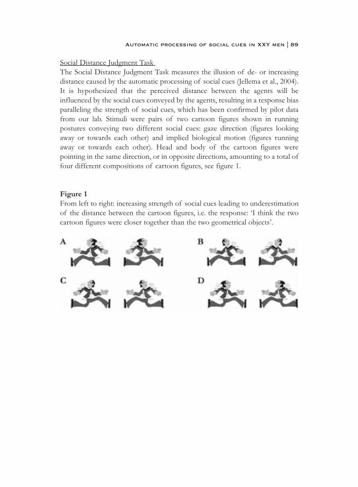
Social Distance Judgment Task The Social Distance Judgment Task measures the illusion of de- or increasingdistance caused by the automatic processing of social cues (Jellema et al., 2004).It is hypothesized that the perceived distance between the agents will beinfluenced by the social cues conveyed by the agents, resulting in a response biasparalleling the strength of social cues, which has been confirmed by pilot datafrom our lab. Stimuli were pairs of two cartoon figures shown in runningpostures conveying two different social cues: gaze direction (figures lookingaway or towards each other) and implied biological motion (figures runningaway or towards each other). Head and body of the cartoon figures werepointing in the same direction, or in opposite directions, amounting to a total offour different compositions of cartoon figures, see figure 1.
Figure 1From left to right: increasing strength of social cues leading to underestimationof the distance between the cartoon figures, i.e. the response: ‘I think the twocartoon figures were closer together than the two geometrical objects’.
Automatic processing of social cues in XXY men | 89
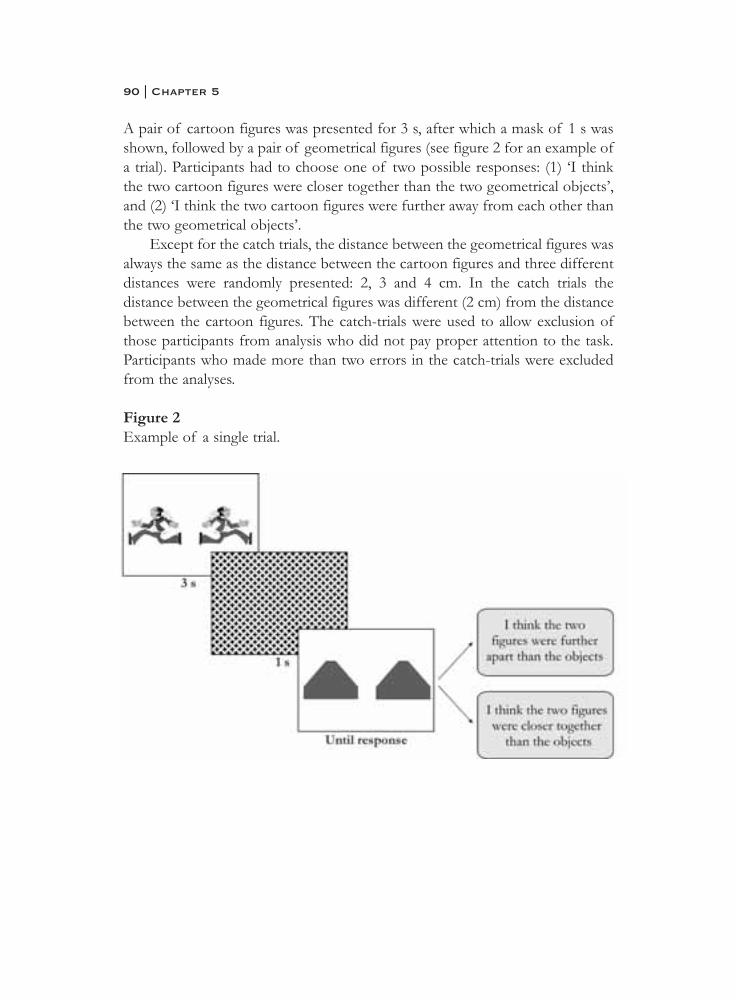
A pair of cartoon figures was presented for 3 s, after which a mask of 1 s wasshown, followed by a pair of geometrical figures (see figure 2 for an example ofa trial). Participants had to choose one of two possible responses: (1) ‘I thinkthe two cartoon figures were closer together than the two geometrical objects’,and (2) ‘I think the two cartoon figures were further away from each other thanthe two geometrical objects’.
Except for the catch trials, the distance between the geometrical figures wasalways the same as the distance between the cartoon figures and three differentdistances were randomly presented: 2, 3 and 4 cm. In the catch trials thedistance between the geometrical figures was different (2 cm) from the distancebetween the cartoon figures. The catch-trials were used to allow exclusion ofthose participants from analysis who did not pay proper attention to the task.Participants who made more than two errors in the catch-trials were excludedfrom the analyses.
Figure 2Example of a single trial.
90 | Chapter 5
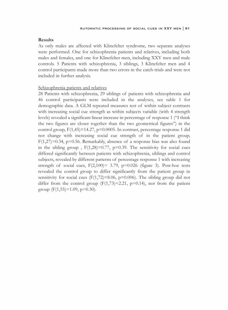
Results As only males are affected with Klinefelter syndrome, two separate analyseswere performed. One for schizophrenia patients and relatives, including bothmales and females, and one for Klinefelter men, including XXY men and malecontrols. 5 Patients with schizophrenia, 3 siblings, 3 Klinefelter men and 4control participants made more than two errors in the catch-trials and were notincluded in further analysis.
Schizophrenia patients and relatives28 Patients with schizophrenia, 29 siblings of patients with schizophrenia and46 control participants were included in the analyses, see table 1 fordemographic data. A GLM repeated measures test of within subject contrastswith increasing social cue strength as within subjects variable (with 4 strengthlevels) revealed a significant linear increase in percentage of response 1 (“I thinkthe two figures are closer together than the two geometrical figures”) in thecontrol group, F(1,45)=14.27, p=0.0005. In contrast, percentage response 1 didnot change with increasing social cue strength of in the patient group,F(1,27)=0.34, p=0.56. Remarkably, absence of a response bias was also foundin the sibling group , F(1,28)=0.77, p=0.39. The sensitivity for social cuesdiffered significantly between patients with schizophrenia, siblings and controlsubjects, revealed by different patterns of percentage response 1 with increasingstrength of social cues, F(2,100)= 3.79, p=0.026 (figure 3). Post-hoc testsrevealed the control group to differ significantly from the patient group insensitivity for social cues (F(1,72)=8.06, p=0.006). The sibling group did notdiffer from the control group (F(1,73)=2.21, p=0.14), nor from the patientgroup (F(1,55)=1.09, p=0.30).
Automatic processing of social cues in XXY men | 91
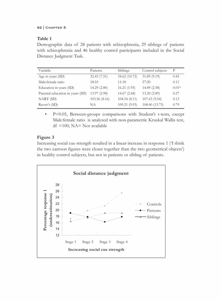
Table 1Demographic data of 28 patients with schizophrenia, 29 siblings of patientswith schizophrenia and 46 healthy control participants included in the SocialDistance Judgment Task.
• P<0.05, Between-groups comparisons with Student’s t-tests, exceptMale:female ratio is analyzed with non-parametric Kruskal Wallis test,df =100; NA= Not available
Figure 3Increasing social cue strength resulted in a linear increase in response 1 (‘I thinkthe two cartoon figures were closer together than the two geometrical objects’)in healthy control subjects, but not in patients or sibling of patients.
92 | Chapter 5
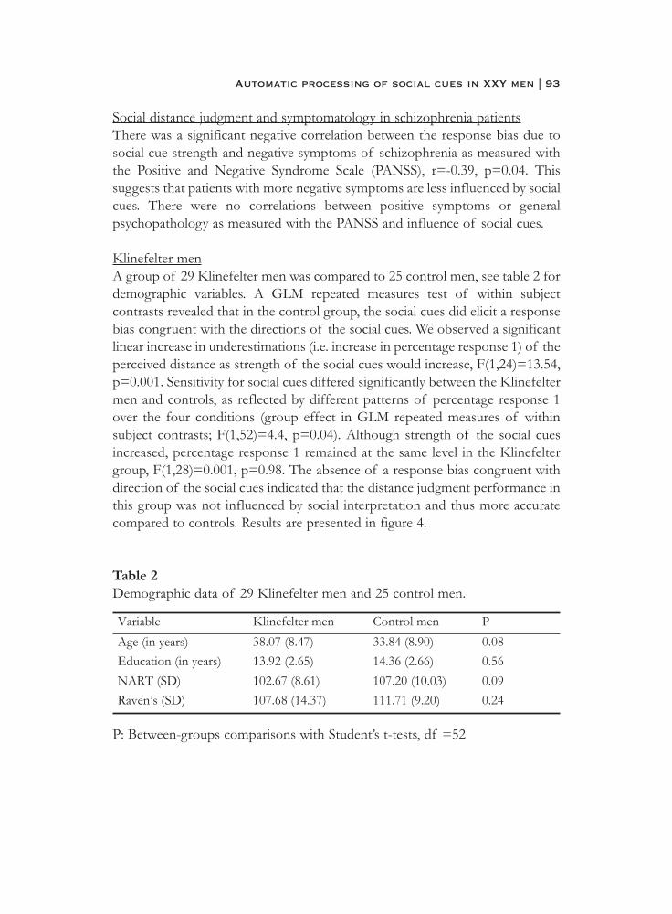
Social distance judgment and symptomatology in schizophrenia patientsThere was a significant negative correlation between the response bias due tosocial cue strength and negative symptoms of schizophrenia as measured withthe Positive and Negative Syndrome Scale (PANSS), r=-0.39, p=0.04. Thissuggests that patients with more negative symptoms are less influenced by socialcues. There were no correlations between positive symptoms or generalpsychopathology as measured with the PANSS and influence of social cues.
Klinefelter menA group of 29 Klinefelter men was compared to 25 control men, see table 2 fordemographic variables. A GLM repeated measures test of within subjectcontrasts revealed that in the control group, the social cues did elicit a responsebias congruent with the directions of the social cues. We observed a significantlinear increase in underestimations (i.e. increase in percentage response 1) of theperceived distance as strength of the social cues would increase, F(1,24)=13.54,p=0.001. Sensitivity for social cues differed significantly between the Klinefeltermen and controls, as reflected by different patterns of percentage response 1over the four conditions (group effect in GLM repeated measures of withinsubject contrasts; F(1,52)=4.4, p=0.04). Although strength of the social cuesincreased, percentage response 1 remained at the same level in the Klinefeltergroup, F(1,28)=0.001, p=0.98. The absence of a response bias congruent withdirection of the social cues indicated that the distance judgment performance inthis group was not influenced by social interpretation and thus more accuratecompared to controls. Results are presented in figure 4.
Table 2Demographic data of 29 Klinefelter men and 25 control men.
P: Between-groups comparisons with Student’s t-tests, df =52
Automatic processing of social cues in XXY men | 93
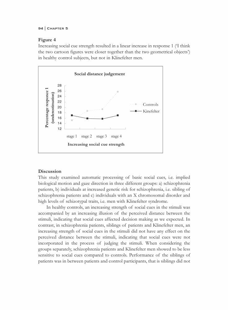
Figure 4Increasing social cue strength resulted in a linear increase in response 1 (‘I thinkthe two cartoon figures were closer together than the two geometrical objects’)in healthy control subjects, but not in Klinefelter men.
DiscussionThis study examined automatic processing of basic social cues, i.e. impliedbiological motion and gaze direction in three different groups: a) schizophreniapatients, b) individuals at increased genetic risk for schizophrenia, i.e. sibling ofschizophrenia patients and c) individuals with an X chromosomal disorder andhigh levels of schizotypal traits, i.e. men with Klinefelter syndrome.
In healthy controls, an increasing strength of social cues in the stimuli wasaccompanied by an increasing illusion of the perceived distance between thestimuli, indicating that social cues affected decision making as we expected. Incontrast, in schizophrenia patients, siblings of patients and Klinefelter men, anincreasing strength of social cues in the stimuli did not have any effect on theperceived distance between the stimuli, indicating that social cues were notincorporated in the process of judging the stimuli. When considering thegroups separately, schizophrenia patients and Klinefelter men showed to be lesssensitive to social cues compared to controls. Performance of the siblings ofpatients was in between patients and control participants, that is siblings did not
94 | Chapter 5
differ significantly from either controls or patients. The differences can not beascribed to differences in general cognitive function, as the groups werematched on education and intelligence estimates. Furthermore, the subjectsincluded in the analysis understood the task and were able to perform the taskcorrectly, as is evidenced from their lack of errors on the catch trials.
These results suggest that patients with schizophrenia demonstrated a lackof sensitivity to even basic, simple social cues, instead of deficits only in moreabstract, higher-order social cue recognition (Corrigan et al., 1993). A failure toautomatically and quickly process these basic social cues may contribute todifficulties in social intuition and hence coping with social situations in thesepatients. Also, because less basic social information is available, morewidespread effects on (‘upstream’-) higher-order social cognitive processing canbe expected. The observed insensitivity to social cues may underly socialcognitive deficits and social dysfunction in schizophrenia. The ability to processsocial cues automatically is thought to be especially important for theory ofmind, i.e. deducing someone else’s intentions, goals and believes (Perrett, 1999)and deficits in the automatic processing of these social cues might lead todisturbances in the attribution of mental states to others (Frith et al., 1999).Indeed, a recently published study demonstrates that patients withschizophrenia were impaired in using appropriate language to describe Theoryof Mind animations (Russell et al., 2006).
Our results showed that especially patients with negative symptoms, whichcomprise social and emotional withdrawal, were insensitive to the influence ofthe social cues in their judgments. Patients with negative symptoms typicallyshow problematic social functioning (Dickerson et al., 1996, 1999; Van DerDoes et al., 1996), but also deficits in other social emotional tasks (Kohler et al.,2000; Mandal et al., 1998; Martin et al., 2005; Schneider et al., 1995). Thus, theseresults corroborate previous research demonstrating that patients withschizophrenia show deficits in the processing of social information (Pinkham etal., 2003), with more severe impairments in patients with negative symptoms(Corcoran et al., 1995; Kohler et al., 2000; Leitman et al., 2005; Mandal et al.,1999). However, this study extends previous research in demonstrating deficitsin the effortless processing of simple social cues.
Interestingly, the absence of influence of the social cues on distancejudgments was also observed in individuals at increased genetic risk forschizophrenia (relatives of patients) and individuals with a genetic disorderassociated with increased schizophrenia spectrum pathology (Klinefeltersyndrome). Based on these findings three important conclusions can be drawn.
Automatic processing of social cues in XXY men | 95
First, siblings as well as the Klinefelter men were not clinically psychotic and didnot use antipsychotic medication. The lack of sensitivity for social cues couldthus not be due to the effects of illness or the medication use. In that way, theseresults validate the observed results in patients. Second, we propose that theobserved lack of sensitivity for social cues is related to a genetic vulnerability toschizophrenia. The results showed that there were no differences betweenpatients and siblings in distance judgment, suggesting that siblings resemblepatients in an absence of automatic processing of social cues. However, onecould also argue that siblings perform normally, as they also did not differ fromcontrols. When taking the within group analysis into account we demonstratedthat siblings, in contrast to normal controls, did not show a linear increase inunderestimations, i.e. their distance judgments were not influenced by the socialcues of human figures running towards each other or looking towards eachother. Thus, our findings imply that performance of siblings resembles the lackof sensitivity to social cues observed in schizophrenia patients, albeit to a lesserextent. Moreover, our results mirror and extend previous studies demonstratingimpairments in other types of social emotional cue processing in relatives ofpatients with schizophrenia such as recognizing emotional facial expressions(Loughland et al., 2004; Toomey et al., 1999), suggesting that insensitivity forsocial cues might be regarded as a genetic vulnerability to schizophrenia. Third,additional evidence for a genetic loading on social cue processing comes fromthe findings in individuals with a genetic disorder. Performance in the social cuetask was indistinguishable between schizophrenia patients and Klinefelter men.As this disorder is defined by an X chromosomal abnormality, impairedcognitive processing of social cues in this group can be regarded as theexpression of X-linked genetic pathology. Klinefelter men also displayimpairments in higher order social cognitive processing, such as recognition offacial expressions (van Rijn et al., 2006b). As insensitivity to social cues seemsan endophenotype that is shared by schizophrenia patients and Klinefelter men,this deficit may have a common genetic (X-linked) origin in both syndromes.
Notably, individuals with autism, a disorder of the ‘social brain’ as indicatedby deficits in theory of mind, facial affect recognition and reciprocal socialbehavior, also demonstrate an insensitivity to social cues using this task (Jellemaet al., 2004). Hence, this study gives converging evidence that attention to basic,typically effortlessly processed social cues may belong to the fundamentalcognitive operations needed for successful social behavior. With regard to theneural correlates involved in the processing of biological motion and socialattention, the superior temporal gyrus, medial prefrontal cortex and anterior
96 | Chapter 5
cingulate have been implied (Jellema et al., 2005). Both in schizophrenia patientsas well as relatives, abnormalities in these regions have been reported (Ashtonet al., 2000; Dolan et al., 1995; Fletcher et al., 1999; Mitelman et al., 2005;Rajarethinam et al., 2000; Shenton et al., 2001; Takahashi et al., 2004).Interestingly, structural abnormalities in the anterior cingulate and the superiortemporal gyrus have been found in Klinefelter syndrome as well (Shen et al.,2004). Additional evidence for a role of the X chromosome in development ofthe superior temporal gyrus comes from studies with individuals with Xmonosomy, showing that volume of this region is dependent on parental originof the X chromosome (Kesler et al., 2003). It is suggested that these neuralcorrelates also underlie Theory of Mind capabilities (Frith et al., 1999; Siegal etal., 2002).
Future studies should relate neural substrates of social cue processing inschizophrenia and relatives together with measures of social functioning. Thiswould elucidate the relationship between the ability to process social cues andsocial behavior and its underlying brain pathology in schizophrenia and providemore insight into the biological vulnerability to schizophrenia. Such researchshould also take into account the role of the amygdala, which has been impliedin social information processing (Adolphs et al., 1998). Abnormalities of theamygdala have been documented in patients with schizophrenia (Aleman et al.,2005) as well as their relatives and Klinefelter patients (Van Rijn et al., 2005).
In summary, this study allowed the investigation of sensitivity to simple,basic social cues that are usually effortlessly processed, i.e. implied biologicalmotion and gaze direction, in individuals with a) a diagnosis of schizophreniab) an increased risk for schizophrenia (relatives of patients) and c) with a geneticdisorder associated with increased schizophrenia spectrum pathology(Klinefelter syndrome). Results showed that patients with schizophrenia,siblings of patients with schizophrenia and Klinefelter men (47, XXY) did notprocess these social cues automatically compared to healthy controls. Within theschizophrenia group, this was especially the case in patients with more severenegative symptoms, i.e. patients that show additional social emotionaldisturbances. Hence, social cue processing deficits seem related to thevulnerability to schizophrenia, instead of illness in general and with a potentialinvolvement of genes on the X chromosome. These basic social cue processingdeficits might underlie impairments in other aspects of social cognition andsocial functioning. Future research should investigate further the relationshipbetween insensitivity to social cues, social functioning and neurobiologicalsubstrates in schizophrenia.
Automatic processing of social cues in XXY men | 97
ReferencesA.P.A. (1994). Diagnostic and statistical manual of mental disorders (4th ed.).
Washington, DC: American Psychiatric Association Press.Adolphs, R., Tranel, D., & Damasio, A. R. (1998). The human amygdala in
social judgment. Nature, 393(6684), 470-474.Aleman, A., & Kahn, R. S. (2005). Strange feelings: Do amygdala
abnormalities dysregulate the emotional brain in schizophrenia?Progress in Neurobiology, 77(5), 283-298.
Andreasen, N. C., Flaum, M., & Arndt, S. (1992). The comprehensive assessment of symptoms and history (cash): An instrument forassessing diagnosis and psychopathology. Archives of GeneralPsychiatry, 49(8), 615.
Ashton, L., Barnes, A., Livingston, M., & Wyper, D. (2000). Cingulate abnormalities associated with panss negative scores in first episodeschizophrenia. Behavioural Neurology, 12(1-2), 93.
Baum, K. M., & Walker, E. F. (1995). Childhood behavioral precursors ofadult symptom dimensions in schizophrenia. Schizophrenia Research,16(2), 111.
Bellack, A. S., Morrison, R. L., Wixted, J. T., & Mueser, K. T. (1990). An analysis of social competence in schizophrenia. The British Journal OfPsychiatry: The Journal Of Mental Science, 156, 809-818.
Bruner, J. (1960). The process of education. Cambridge, MA: Harvard University Press.
Corcoran, R., Mercer, G., & Frith, C. D. (1995). Schizophrenia,symptomatology and social inference: Investigating "theory of mind" inpeople with schizophrenia. Schizophrenia Research, 17(1), 5-13.
Corrigan, P. W. (1994). Social cue perception and intelligence in schizophrenia.Schizophrenia Research, 13(1), 73.
Corrigan, P. W., & Green, M. F. (1993). Schizophrenic patients' sensitivity to social cues: The role of abstraction. American Journal of Psychiatry,150(4), 589.
Corrigan, P. W., Green, M. F., & Toomey, R. (1994). Cognitive correlates to social cue perception in schizophrenia. Psychiatry Research, 53(2), 141.
Corrigan, P. W., & Toomey, R. (1995). Interpersonal problem solving and information processing in schizophrenia. Schizophrenia Bulletin, 21(3),395-403.
98 | Chapter 5
Dickerson, F., Boronow, J. J., Ringel, N., & Parente, F. (1996). Neurocognitive deficits and social functioning in outpatients with schizophrenia.Schizophrenia Research, 21(2), 75.
Dickerson, F., Boronow, J. J., Ringel, N., & Parente, F. (1999). Social functioning and neurocognitive deficits in outpatients withschizophrenia: A 2-year follow-up. Schizophrenia Research, 37(1), 13.
Dolan, R. J., Fletcher, P., Frith, D., Friston, K. J., Frackowiak, R. S. J., & Grasby, P. W. (1995). Dopaminergic modulation of impaired cognitiveactivation in the anterior cingulate cortex in schizophrenia. Nature,378(6553), 180.
Edwards, J., Jackson, H. J., & Pattison, P. E. (2002). Emotion recognition via facial expression and affective prosody in schizophrenia: Amethodological review. Clinical Psychology Review, 22(6), 789-832.
Fletcher, P., McKenna, P. J., Friston, K. J., Frith, C. D., & Dolan, R. J. (1999).Abnormal cingulate modulation of fronto-temporal connectivity inschizophrenia. Neuroimage, 9(3), 337.
Frith, C. D. (1992). The cognitive neuropsychology of schizophrenia.Hillsdale, NJ: Laurence Erlbaum Associates.
Frith, C. D., & Frith, U. (1999). Interacting minds--a biological basis. Science,286(5445), 1692-1695.
Gottesman, I. I. (1991). Schizophrenia genesis: The origin of madness. New York: Freeman.
Hampson, E., van Anders, S. M., & Mullin, L. I. (2006). A female advantage in the recognition of emotional facial expressions: Test of an evolutionaryhypothesis. Evolution and Human Behavior, 27(6), 401.
Hans, S. L., Marcus, J., Henson, L., Auerbach, J. G., & Mirsky, A. F. (1992).Interpersonal behavior of children at risk for schizophrenia. Psychiatry,55(4), 314-335.
Jellema, T., Lorteije, J. A. M., van Rijn, S., van t'Wout, M., de Heer, F., & de Haan, E. H. F. (2004). Failure to automate the semantic processing ofsocial cues in autism. Perception, 33, 101-101.
Jellema, T., & Perret, D. I. (2005). Neural basis for the perception of goal-directed actions. In A. Easton & N. J. Emery (Eds.), The cognitiveneuroscience of social behavior. New York: Psychology Press.
Kay, S. R., Fiszbein, A., & Opler, L. A. (1987). The positive and negative syndrome rating scale (panss) for schizophrenia. Schizophrenia Bulletin,13, 261-276.
Automatic processing of social cues in XXY men | 99
Kesler, S. R., Blasey, C. M., Brown, W. E., Yankowitz, J., Zeng, S. M., Bender,B. G., et al. (2003). Effects of x-monosomy and x-linked imprinting onsuperior temporal gyrus morphology in turner syndrome. BiologicalPsychiatry, 54(6), 636-646.
Kohler, C. G., Bilker, W., Hagendoorn, M., Gur, R. E., & Gur, R. C. (2000).Emotion recognition deficit in schizophrenia: Association withsymptomatology and cognition. Biological Psychiatry, 48(2), 127-136.
Kohler, C. G., & Brennan, A. R. (2004). Recognition of facial emotions in schizophrenia. Current Opinion in Psychiatry, 17(2), 81.
Leitman, D. I., Foxe, J. J., Butler, P. D., Saperstein, A., Revheim, N., & Javitt,D.C. (2005). Sensory contributions to impaired prosodic processing inschizophrenia. Biological Psychiatry, 58(1), 56.
Lenzenweger, M. F., & Dworkin, R. H. (1996). The dimensions ofschizophrenia phenomenology. Not one or two, at least three, perhapsfour. British Journal of Psychiatry, 168(APR.), 432.
Lezak, M. D. (1995). Neuropsychological assessment (third ed.). New York:Oxford University Press.
Lieberman, M. D. (2000). Intuition: A social cognitive neuroscience approach.Psychological Bulletin, 126(1), 109-136.
Loughland, C. M., Williams, L. M., & Harris, A. W. (2004). Visual scanpath dysfunction in first-degree relatives of schizophrenia probands:Evidence for a vulnerability marker? Schizophrenia Research, 67(1), 11-21.
Mandal, M. K., Jain, A., Haque-Nizamie, S., Weiss, U., & Schneider, F. (1999).Generality and specificity of emotion-recognition deficit inschizophrenic patients with positive and negative symptoms. PsychiatryResearch, 87(1), 39.
Mandal, M. K., Pandey, R., & Prasad, A. B. (1998). Facial expressions ofemotions and schizophrenia: A review. Schizophrenia Bulletin, 24(3),399-412.
Martin, F., Baudouin, J. Y., Tiberghien, G., & Franck, N. (2005). Processing emotional expression and facial identity in schizophrenia. PsychiatryResearch, 134(1), 43.
McClure, E. B. (2000). A meta-analytic review of sex differences in facial expression processing and their development in infants, children, andadolescents. Psychological Bulletin, 126(3), 424-453.
100 | Chapter 5
Mitelman, S. A., Shihabuddin, L., Brickman, A. M., Hazlett, E. A., & Buchsbaum, M. S. (2005). Volume of the cingulate and outcome inschizophrenia. Schizophrenia Research, 72(2-3), 91.
Perrett, D. I. (1999). A cellular basis for reading minds from faces and actions.In M. Hauser & M. Konishi (Eds.), The design of animalcommunication. Cambridge, MA: The MIT Press.
Pinkham, A. E., Penn, D. L., Perkins, D. O., & Lieberman, J. (2003).Implications for the neural basis of social cognition for the study ofschizophrenia. American Journal of Psychiatry, 160(5), 815-824.
Rajarethinam, R. P., DeQuardo, J. R., Nalepa, R., & Tandon, R. (2000).Superior temporal gyrus in schizophrenia: A volumetric magneticresonance imaging study. Schizophrenia Research, 41(2), 303-312.
Raven, J. C., Raven, J., & Court, J. H. (1993). Manual for raven's progressive matrices and vocabulary scales. Oxford: Oxford Psychologist Press.
Russell, T. A., Reynaud, E., Herba, C., Morris, R., & Corcoran, R. (2006). Do you see what i see? Interpretations of intentional movement inschizophrenia. Schizophrenia Research, 81(1), 101.
Schmand, B., Bakker, D., Saan, R., & Louman, J. (1991). [the dutch reading test for adults: A measure of premorbid intelligence level]. Tijdschrift voorGerontologie en Geriatrie, 22(1), 15-19.
Schneider, F., Gur, R. C., Gur, R. E., & Shtasel, D. L. (1995). Emotional processing in schizophrenia: Neurobehavioral probes in relation topsychopathology. Schizophrenia Research, 17(1), 67-75.
Scholten, M. R. M., Aleman, A., Montagne, B., & Kahn, R. S. (2005).Schizophrenia and processing of facial emotions: Sex matters.Schizophrenia Research, 78(1), 61.
Sheehan, D. V., Lecrubier, Y., Sheehan, K. H., Amorim, P., Janavs, J., Weiller,E., et al. (1998). The mini-international neuropsychiatric interview(m.I.N.I.): The development and validation of a structured diagnosticpsychiatric interview for dsm-iv and icd-10. Journal of ClinicalPsychiatry, 59 Suppl 20, 22-33;quiz 34-57.
Shen, D., Liu, D., Liu, H., Clasen, L., Giedd, J., & Davatzikos, C. (2004).Automated morphometric study of brain variation in xxy males.Neuroimage, 23(2), 648-653.
Shenton, M. E., Dickey, C. C., Frumin, M., & McCarley, R. W. (2001). A review of mri findings in schizophrenia. Schizophrenia Research, 49(1-2), 1-52.
Automatic processing of social cues in XXY men | 101
Siegal, M., & Varley, R. (2002). Neural systems involved in 'theory of mind'.Nature Reviews Neuroscience, 3(6), 463.
Sitskoorn, M., Aleman, A., Ebisch, S., Appels, M., & Kahn, R. S. (2003). Meta-analysis of neurocognitive deficits in relatives of patients withschizophrenia: Evidence for a strong cognitive endophenotypic marker.Schizophrenia Research, 60(1), 90-90.
Skuse, D. H. (2005). X-linked genes and mental functioning. Hum Mol Genet,14 Spec No 1, R27-32.
Takahashi, H., Koeda, M., Oda, K., Matsuda, T., Matsushima, E., Matsuura,M., et al. (2004). An fmri study of differential neural response toaffective pictures in schizophrenia. Neuroimage, 22(3), 1247-1254.
Toomey, R., Seidman, L. J., Lyons, M. J., Faraone, S. V., & Tsuang, M. T.(1999). Poor perception of nonverbal social-emotional cues in relativesof schizophrenic patients. Schizophrenia Research, 40(2), 121-130.
Van Der Does, A. J. W., Dingemans, P. M. A. J., Linszen, D. H., Nugter, M. A.,& Scholte, W. F. (1996). Symptoms, cognitive and social functioning inrecent-onset schizophrenia: A longitudinal study. SchizophreniaResearch, 19(1), 61.
Van Rijn, S., Aleman, A., Swaab, H., & Kahn, R. (2006a). Klinefelter's syndrome (karyotype 47,xxy) and schizophrenia-spectrum pathology.British Journal of Psychiatry, 189(5), 459-461.
Van Rijn, S., Aleman, A., Swaab, H., & Kahn, R. S. (2005). Neurobiology ofemotion and high risk for schizophrenia: Role of the amygdala and thex-chromosome. Neuroscience and Biobehavioral Reviews, 29(3), 385-397.
van Rijn, S., Swaab, H., Aleman, A., & Kahn, R. S. (2006b). X chromosomal effects on social cognitive processing and emotion regulation: A studywith klinefelter men (47,xxy). Schizophrenia Research, 84(2-3), 194-203.
Walker, E. F. (1994). Developmentally moderated expressions of the neuropathology underlying schizophrenia. Schizophrenia Bulletin, 20(3),453-480.
102 | Chapter 5
Chapter 6
What is said or how it is said makes a
difference: role of the right fronto-parietal
operculum in emotional prosody as revealed
by repetitive TMS
Sophie van Rijn, André Aleman, Eric van Diessen, Celine Berckmoes,Guy Vingerhoets, René S. Kahn
European Journal of Neuroscience, 2005, 21 (11), 3195-3200
AbstractEmotional signals in spoken language can be conveyed by semantic as well asprosodic cues. We investigated the role of the fronto-parietal operculum, asomatosensory area where the lips, tongue and jaw are represented, in the righthemisphere to detection of emotion in prosody versus semantics. 14 Healthyvolunteers participated in the present experiment, which involved TranscranialMagnetic Stimulation (TMS) in combination with frameless stereotaxy. Aspredicted, compared to sham stimulation, TMS over the right fronto-parietaloperculum differentially affected reaction times for detection of emotionalprosody versus emotional semantics, showing that there is a dissociation at aneuroanatomical level. Detection of withdrawal emotions (fear and sadness) inprosody was significantly delayed by TMS. No effects of TMS were observedfor approach emotions (happiness and anger). We propose that the right fronto-parietal operculum is not globally involved in emotion evaluation, but sensitiveto specific forms of emotional discrimination and emotion types.
104 | Chapter 6
IntroductionIn order to understand messages in verbal communication, it is important to payattention not only to what is said but also to how it is said. Besides the semantic(linguistic) meaning of words, features such as intonation and loudness inspeech also may convey crucial information. The term prosody has beenintroduced to describe these non-semantic cues in spoken language. Prosodiccues can have linguistic- as well as affective functions. Linguistic functions are,for example, emphasizing important parts of the message or presentinginformation as a statement or a question. Affective functions of prosody arealso important for understanding intentions of others, because variations intone of voice, such as intonation and loudness, provide information about theemotional state of the speaker. Thus, the emotional relevance of a spokenmessage may be not only be conveyed by meaning of words (i.e. emotionalsemantics), but also expressed in emotional prosody. The present study isfocused on processing of affective information conveyed by these differentaspects of spoken language, i.e. emotional semantics versus emotional prosody.
These two different communicative channels of information about theemotional relevance of a spoken message, i.e. semantic and prosodic, may bedissociable at a neuroanatomical level. A range of studies have suggested thatthe left hemisphere seems to be specialized for most aspects of languageprocessing (Binder, 1999), including linguistic functions of prosody (e.g, stressevaluation,(A. Aleman et al., 2004)). However, the right hemisphere (RH)appears to be involved in non-linguistic components, such as emotional prosody(Baum & Pell, 1999).
The best evidence regarding RH involvement in affective prosody comesfrom studies directly contrasting emotional semantics with emotional prosody.In contrast to processing of emotional semantics, emotional prosody involvesthe right hemisphere as is shown in imaging studies (Baum & Pell, 1999; R. L.Mitchell et al., 2003; Vingerhoets et al., 2003). These findings suggest that therole of the right hemisphere in emotional prosody is not a specialization inemotion detection in general, but that RH involvement depends on thecommunication channel through which emotional relevance is presented.
In addition to type of communication channel, involvement of the righthemisphere may also depend on type of emotion. A widely used categorisationof emotions is based on broader constructs of behavioral inhibition andactivation (Sutton & Davidson, 1997). Whereas withdrawal emotions such asfear and sadness are accompanied by inhibitory motivational tendencies,appraoch emotions such as anger and happiness are accompanied by
Affective prosody and TMS | 105
activational motivational tendencies. Right hemisphere damaged patients showspecific deficits in processing withdrawal emotions, i.e. fear and sadness, incontrast to normal processing of approach emotions such as happiness andanger (A. K. Anderson et al., 2000; M. K. Mandal et al., 1999). Also, it has beenproposed that whereas the left frontal cortical region is important forexpression and experience of approach emotions, the right frontal corticalregion is involved in expression and experience of withdrawal emotions(Harmon-Jones, 2003; Sutton & Davidson, 1997).
Recently, Vingerhoets et al. (Vingerhoets et al., 2003) have measured bloodflow velocity (BFV) with functional transcranial doppler ultrasonography(fTCD) to study the contribution of the right and left hemisphere to thedetection of emotion in prosody versus detection of emotion in semantics ofspoken language. fTDC is a non-invasive tool with high temporal resolution thatallows a continuous monitoring of blood flow velocity in the basal cerebralarteries, thereby reflecting changes in cerebral metabolism that accompaniesmental activity. During detection of emotion in semantics a significant left-hemispheric lateralization of BFV was observed. This lateralization effectdisappeared when attention was shifted to discriminating emotion in prosody,due to a rise in right hemispheric BFV. Although metabolism in the righthemisphere is correlated with detection of emotional prosody in this study, thequestion remains whether the RH is causally involved in emotional prosody. Inaddition, is not clear which brain region within the right hemisphere is criticallyinvolved in emotional prosody discrimination and whether such a region issensitive to specific types of emotions.
As an extension of the study of Vingerhoets et al. (Vingerhoets et al., 2003),we used Transcranial Magnetic Stimulation (TMS) in order to answer thesequestions. TMS delivers short magnetic pulses that penetrate the skull and disruptneural processing in a non-invasive, reversible way (Walsh & Pascual-Leone,2003). This virtual lesion technique allows investigation of the causal relationbetween neural activity and performance at a behavioural level. In addition,specific brain regions can be targeted, depending on the type of coil used forstimulation. Ideally, such regions should be determined on the basis of lesion datain human patients. Recently, a comprehensive 3-D lesion study investigating theneural correlates of emotional prosody has been published (Adolphs et al., 2002).In a three-dimensional reconstruction of all the lesions in focal brain-damagedpatients, density of lesion-overlap was analysed as a function of task performance.The most robust finding was a relation between damage to the right fronto-parietal operculum and compromised detection of emotion from prosody.
106 | Chapter 6
Our prediction was that, when compared to sham stimulation, TMS over theright fronto-parietal operculum differentially affects reaction times for detectionof emotional prosody when compared to emotional semantics, showing that a)distinct neuroanatomical networks underlie attention to emotional prosodyversus emotional semantics and b) the right fronto-parietal operculum issensitive to specific forms of emotional discrimination (semantic/prosodic) andemotion types (withdrawal/approach emotions), rather then globally involved inemotion evaluation.
Methods
SubjectsFourteen healthy adult subjects participated in the study after giving writteninformed consent (19-27 years, mean age 23 years, 8 m/6 f). Handedness wasmeasured with the Edinburgh Handedness Inventory (-24= exclusively lefthanded, 0=no preference, 24= exclusively right handed) (Oldfield, 1971). Allparticipants were right-handed (mean 21.3, S.D.= 2.6). Subjects were screenedfor contraindications to TMS, neurological and medical problems. Theexperiment was conducted in accordance to the Declaration of Helsinki andlocal ethics board approval (University Medical Center Utrecht).
ApparatusFor TMS, we used a MagStim Rapid magnetic stimulator (MagStim Co,Whitland, Wales) with a figure-of-eight magnetic coil with a diameter of 70 mmfor each loop.
Experimental protocolThe off-line TMS experiment comprised two conditions, TMS and sham, eachfollowed by two tasks, emotional semantics and emotional prosody. Order ofboth the conditions (stimulation over the right and left fronto-parietaloperculum for real TMS and sham, respectively) and the tasks wascounterbalanced over subjects. Minimum time interval between TMS and shamwas 30 minutes to prevent carry-over effects (c.f. (Kosslyn et al., 1999; Oliveriet al., 2004)).
Affective prosody and TMS | 107
TMS over the fronto-parietal operculumFirst, the Motor Threshold (MT) was determined for each subject, defined asthe lowest stimulation intensity that induced visible finger movements in at least5 out of 10 trials when TMS was applied to the motor cortex (Pridmore et al.,1998).
Second, localization of the right fronto-parietal operculum wasaccomplished individually using structural MRI and neuronavigation byframeless stereotaxy (NeNa) (Neggers et al., 2004). Anatomical T1-weightedMRI scans were used to delineate the fronto-parietal operculum bilaterally ineach participant. The fronto-parietal operculum was defined as the inferior peri-central sulcus area (see figure 2) and delineated independently by two separateraters as a Region of Interest (ROI) of approximately 1 to 2 centimeters indiameter, conform the size of the cortical region TMS typically affects (Walsh& Cowey, 2000). Size and location of the ROI was based on the regions withhighest lesion-overlap values of the fronto-parietal operculum from the 3-Dlesion study by Adolphs et al. (Adolphs et al., 2002).
The structural MRI scan as well as the Region of Interest (ROI) maps,representing the fronto-parietal operculum bilaterally, were loaded into NeNabefore the experiment. Six anatomical landmarks were set on the skin rendering.The participant would then be seated, the head fixed in a head support, and arubber head cap placed over his head. The positions of the same anatomicallandmarks were measured with the MiniBIRD position-tracker probe, directly atthe head of the participant, and the mapping between 3D space and MRI spacewas calculated. After co-registration, the fronto-parietal operculum was targetedby ‘looking’ through the NeNa probe to the renderings of the skin and ROI onthe screen and the position directly overlying the ROI was marked on the head-cap. For more details on localization using NeNA system, see (Neggers et al.,2004).
In the stimulation condition, subjects were then stimulated with 1 Hz TMSduring 12 min. over the right fronto-parietal operculum, marked at the head-cap,at 90% of the Motor Threshold. These parameters have been applied in earlierstudies of cognitive TMS (for an overview see (Robertson et al., 2003)) and havebeen shown to affect brain metabolism (Mottaghy et al., 2003). The coilwas held manually with the handle pointing backwards and kept tangential tothe subject’s scalp. In the stimulation condition, subjects were then stimulatedwith 1 Hz TMS during 12 min. over the right fronto-parietal operculum, markedat the head-cap, at 90% of the Motor Threshold. These parameters have beenapplied in earlier studies of cognitive TMS (for an overview see (Robertson et
108 | Chapter 6
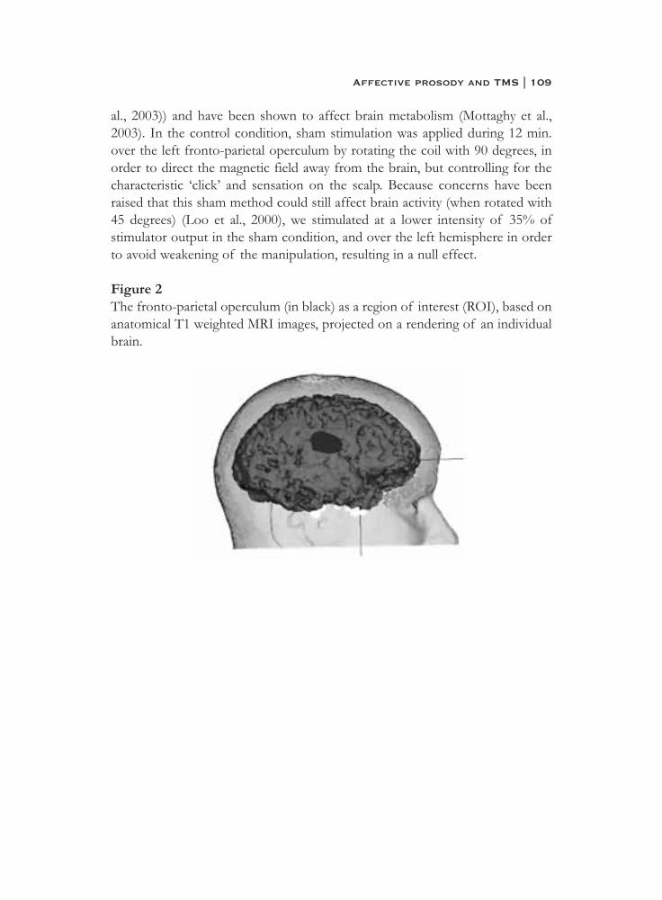
al., 2003)) and have been shown to affect brain metabolism (Mottaghy et al.,2003). In the control condition, sham stimulation was applied during 12 min.over the left fronto-parietal operculum by rotating the coil with 90 degrees, inorder to direct the magnetic field away from the brain, but controlling for thecharacteristic ‘click’ and sensation on the scalp. Because concerns have beenraised that this sham method could still affect brain activity (when rotated with45 degrees) (Loo et al., 2000), we stimulated at a lower intensity of 35% ofstimulator output in the sham condition, and over the left hemisphere in orderto avoid weakening of the manipulation, resulting in a null effect.
Figure 2The fronto-parietal operculum (in black) as a region of interest (ROI), based onanatomical T1 weighted MRI images, projected on a rendering of an individualbrain.
Affective prosody and TMS | 109
Emotion discrimination tasksImmediately after (sham) stimulation, subjects were required to identify theemotion conveyed by prosody or semantics of a number of sentences. We usedthe tasks designed by Vingerhoets et al. (Vingerhoets et al., 2003). Each task(prosody and semantics) was assessed twice (after TMS and after shamstimulation), with a different list of stimuli. Each task presentation waspreceded by 4 practice sentences. Order of the tasks was counterbalanced oversubjects.
Of the 24 sentences in each task, 6 were happy, 6 were sad, 6 were angryand 6 were fearful. Sentences were of approximately equal length and werearticulated by two professional actors, one male and one female. The digitizedstimuli were presented binaurally through earphones. During listening, theemotions to be discriminated were presented on the computer screen. In theprosody task, affective discrimination was based on the affective tone of voice.In this task, the content of the sentences was not affective (i.e. always neutral).In the semantic task, affective discrimination was based on the semantic contentof the sentences. In this task, tone of voice was not affective (i.e always neutral).As soon as they identified the emotion expressed in the sentence, either basedon content or tone of voice, subjects were required to pronounce that particularemotion in a microphone that was connected to the computer. Their responses,both selected emotion and reaction time for detection, were collected. Totalduration for completing one of either tasks was 7 minutes.
Statistical analysesStatistical analyses were performed using Statistical Package for the SocialSciences 11.5.0 (2002). Accuracy and reaction times (RT) for detection ofemotion in semantics and in prosody were chosen as dependent variables.Reaction times for incorrect responses were excluded from analyses. 2x2 GLMrepeated measures analyses with TMS (stimulation, sham) and Task (prosody,semantics) as independent factors were used to test the effects of TMS on thetwo tasks. Post-hoc analyses were performed using paired T-tests. P values wereset at 0.05, two tailed.
110 | Chapter 6
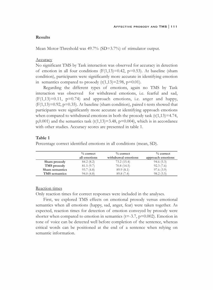
Results
Mean Motor-Threshold was 49.7% (SD=3.7%) of stimulator output.
AccuracyNo significant TMS by Task interaction was observed for accuracy in detectionof emotion in all four conditions (F(1,13)=0.42, p=0.53). At baseline (shamcondition), participants were significantly more accurate in identifying emotionin semantics compared to prosody (t(1,13)=2.98, p=0.01).
Regarding the different types of emotions, again no TMS by Taskinteraction was observed for withdrawal emotions, i.e. fearful and sad,(F(1,13)=0.11, p=0.74) and approach emotions, i.e. anger and happy,(F(1,13)=0.92, p=0.35). At baseline (sham condition), paired t-tests showed thatparticipants were significantly more accurate at identifying approach emotionswhen compared to withdrawal emotions in both the prosody task (t(1,13)=4.74,p,0.001) and the semantics task (t(1,13)=3.48, p=0.004), which is in accordancewith other studies. Accuracy scores are presented in table 1.
Table 1Percentage correct identified emotions in all conditions (mean, SD).
Reaction timesOnly reaction times for correct responses were included in the analyses.
First, we explored TMS effects on emotional prosody versus emotionalsemantics when all emotions (happy, sad, anger, fear) were taken together. Asexpected, reaction times for detection of emotion conveyed by prosody wereshorter when compared to emotion in semantics (t=-3.7, p=0.002). Emotion intone of voice can be detected well before completion of the sentence, whereascritical words can be positioned at the end of a sentence when relying onsemantic information.
Affective prosody and TMS | 111
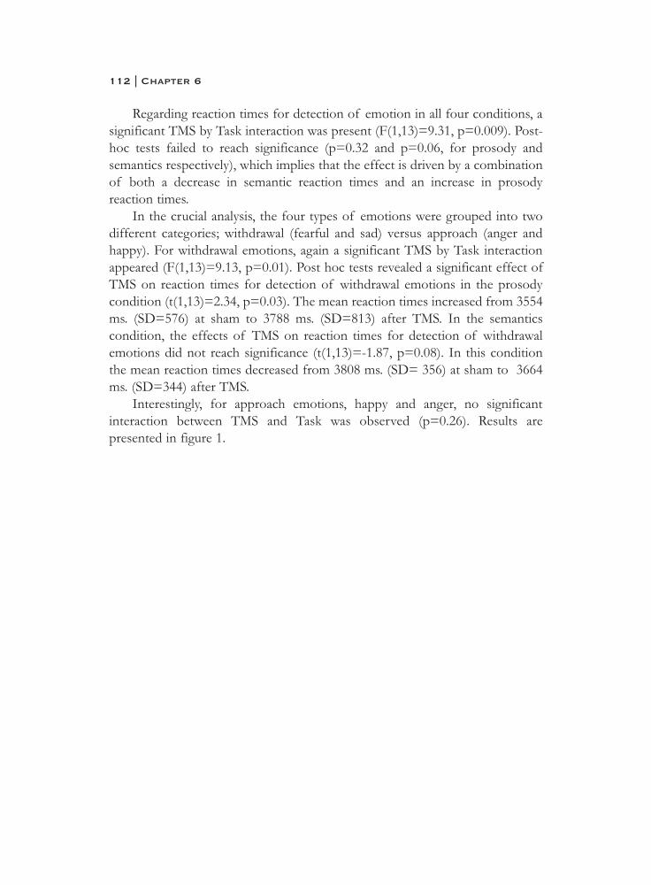
Regarding reaction times for detection of emotion in all four conditions, asignificant TMS by Task interaction was present (F(1,13)=9.31, p=0.009). Post-hoc tests failed to reach significance (p=0.32 and p=0.06, for prosody andsemantics respectively), which implies that the effect is driven by a combinationof both a decrease in semantic reaction times and an increase in prosodyreaction times.
In the crucial analysis, the four types of emotions were grouped into twodifferent categories; withdrawal (fearful and sad) versus approach (anger andhappy). For withdrawal emotions, again a significant TMS by Task interactionappeared (F(1,13)=9.13, p=0.01). Post hoc tests revealed a significant effect ofTMS on reaction times for detection of withdrawal emotions in the prosodycondition (t(1,13)=2.34, p=0.03). The mean reaction times increased from 3554ms. (SD=576) at sham to 3788 ms. (SD=813) after TMS. In the semanticscondition, the effects of TMS on reaction times for detection of withdrawalemotions did not reach significance (t(1,13)=-1.87, p=0.08). In this conditionthe mean reaction times decreased from 3808 ms. (SD= 356) at sham to 3664ms. (SD=344) after TMS.
Interestingly, for approach emotions, happy and anger, no significantinteraction between TMS and Task was observed (p=0.26). Results arepresented in figure 1.
112 | Chapter 6
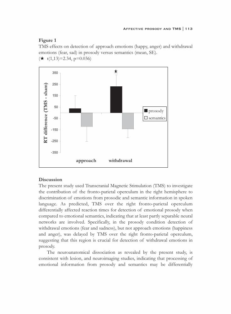
Figure 1TMS effects on detection of approach emotions (happy, anger) and withdrawalemotions (fear, sad) in prosody versus semantics (mean, SE).(� t(1,13)=2.34, p=0.036)
DiscussionThe present study used Transcranial Magnetic Stimulation (TMS) to investigatethe contribution of the fronto-parietal operculum in the right hemisphere todiscrimination of emotions from prosodic and semantic information in spokenlanguage. As predicted, TMS over the right fronto-parietal operculumdifferentially affected reaction times for detection of emotional prosody whencompared to emotional semantics, indicating that at least partly separable neuralnetworks are involved. Specifically, in the prosody condition detection ofwithdrawal emotions (fear and sadness), but not approach emotions (happinessand anger), was delayed by TMS over the right fronto-parietal operculum,suggesting that this region is crucial for detection of withdrawal emotions inprosody.
The neuroanatomical dissociation as revealed by the present study, isconsistent with lesion, and neuroimaging studies, indicating that processing ofemotional information from prosody and semantics may be differentially
Affective prosody and TMS | 113
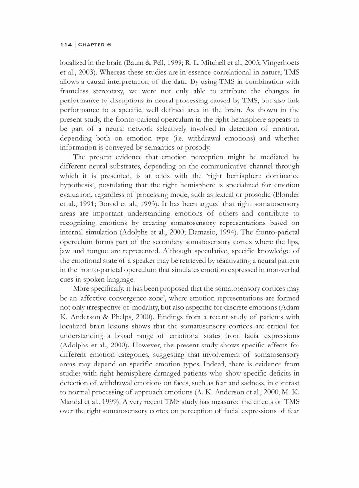
localized in the brain (Baum & Pell, 1999; R. L. Mitchell et al., 2003; Vingerhoetset al., 2003). Whereas these studies are in essence correlational in nature, TMSallows a causal interpretation of the data. By using TMS in combination withframeless stereotaxy, we were not only able to attribute the changes inperformance to disruptions in neural processing caused by TMS, but also linkperformance to a specific, well defined area in the brain. As shown in thepresent study, the fronto-parietal operculum in the right hemisphere appears tobe part of a neural network selectively involved in detection of emotion,depending both on emotion type (i.e. withdrawal emotions) and whetherinformation is conveyed by semantics or prosody.
The present evidence that emotion perception might be mediated bydifferent neural substrates, depending on the communicative channel throughwhich it is presented, is at odds with the ‘right hemisphere dominancehypothesis’, postulating that the right hemisphere is specialized for emotionevaluation, regardless of processing mode, such as lexical or prosodic (Blonderet al., 1991; Borod et al., 1993). It has been argued that right somatosensoryareas are important understanding emotions of others and contribute torecognizing emotions by creating somatosensory representations based oninternal simulation (Adolphs et al., 2000; Damasio, 1994). The fronto-parietaloperculum forms part of the secondary somatosensory cortex where the lips,jaw and tongue are represented. Although speculative, specific knowledge ofthe emotional state of a speaker may be retrieved by reactivating a neural patternin the fronto-parietal operculum that simulates emotion expressed in non-verbalcues in spoken language.
More specifically, it has been proposed that the somatosensory cortices maybe an ‘affective convergence zone’, where emotion representations are formednot only irrespective of modality, but also aspecific for discrete emotions (AdamK. Anderson & Phelps, 2000). Findings from a recent study of patients withlocalized brain lesions shows that the somatosensory cortices are critical forunderstanding a broad range of emotional states from facial expressions(Adolphs et al., 2000). However, the present study shows specific effects fordifferent emotion categories, suggesting that involvement of somatosensoryareas may depend on specific emotion types. Indeed, there is evidence fromstudies with right hemisphere damaged patients who show specific deficits indetection of withdrawal emotions on faces, such as fear and sadness, in contrastto normal processing of approach emotions (A. K. Anderson et al., 2000; M. K.Mandal et al., 1999). A very recent TMS study has measured the effects of TMSover the right somatosensory cortex on perception of facial expressions of fear
114 | Chapter 6
(withdrawal emotion) and happiness (approach emotion) (Pourtois et al., 2004).Interestingly, only perception of fear, and not happiness, was disrupted by TMSover the right somatosensory cortex. Our findings are in line with this study,supporting the hypothesis that right somatosensory areas are important forperception of withdrawal emotions both in facial expressions and spokenlanguage.
Not only visual perception, also experience and expression of withdrawalemotions, but not approach emotions, seem to be mediated by the righthemisphere. PET (Positron Emission Tomography) and EEG(Electroencephalograph) studies have revealed that induced negative affectivestates that are withdrawal-related, as well as production of facial poses ofwithdrawal emotions, are associated with increased activation in cortical regionsin the right hemisphere (Coan et al., 2001; Davidson et al., 2000). Thus, inaddition to a right hemispheric specialization in expression and perception of facialwithdrawal emotions as well as experience of withdrawal emotions as suggestedby these studies, the present data suggest the right hemisphere is cruciallyinvolved in prosodic perception of withdrawal emotions.
Interestingly, a trend in decreasing reaction times for semantic perception of(withdrawal) emotions was observed after TMS to the right fronto-parietaloperculum. Although this trend effect should be interpreted with caution, it maypoint to facilitation of semantic processing after RH TMS. Facilitation effectsin previous TMS studies have been explained by disinhibition of areas that areconnected to the region exposed to TMS or loss of competition between twobrain areas after TMS (Walsh et al., 1999). In the present study, the decrease inreaction times for detecting emotion in semantics after TMS to the right fronto-parietal operculum, may for example result from loss of competition betweenthis area and the right ventrolateral prefrontal cortex (PFC) that is involved inlinguistic aspects of emotion (Hariri et al., 2000). Hence, the ventro-lateral PFCmay process semantic information more efficiently. An alternative explanationmay be that subjects are automatically screening both semantic and prosodicaspects of spoken language. When processing of prosodic information isdisrupted by TMS, subjects may have more processing capacity for analysingsemantic information.
An important issue regarding our TMS effects concerns the potentialcontribution of differences in difficulty between approach and withdrawalemotions. Consistent with the literature, withdrawal emotions were moredifficult to detect than approach emotions (REF), which might yield them moresensitive to the effects of TMS. However, whereas this higher difficulty of
Affective prosody and TMS | 115
withdrawal emotions applies to both semantic and prosodic tasks, TMS effectswere only observed on reaction times in the prosodic task. Moreover, in thesham condition, faster response times were observed in the prosody conditioncompared to the semantics condition, suggesting that by this index prosody waseasier and thus should be less influenced by TMS disruption. Both findingssuggest a pattern of TMS effects that is the opposite of what was observed ifdifficulty is confounded with TMS disruption. With this in mind, it may beimportant to note that reaction times analyses were limited to correct responses,so any effects of speed-accuracy trade-off were eliminated from the analyses.Although we cannot completely exclude that difficulty might play a role, as thisis inherent to the approach-withdrawal dichotomy, our results are consistentwith evidence from lesion-studies (A. K. Anderson et al., 2000; M. K. Mandalet al., 1999), which lends credence to our interpretation. The present studyshows that right hemisphere TMS selectively interferes with processing ofwithdrawal emotions, which might imply that approach emotions are lessmediated by the right hemisphere. This interpretation is consistent with a recentmeta-analysis of 106 imaging studies of emotion that revealed a significantdifference in the spatial distributions associated with withdrawal and approachemotions (Murphy et al., 2003). Processing of approach emotions was strongerassociated with left hemisphere activation compared to the right, supporting theidea that differences in functional neuro-anatomy, rather than differences indifficulty level, can explain our results regarding approach and withdrawalemotions.
A related issue is that TMS was applied to the right hemisphere only, whichis a limitation of the present study. Future studies should incorporate a morecomplex experimental design including TMS over the right as well as lefthemisphere. This design would allow conclusions regarding lateralization of a)emotional prosody and emotional semantics and b) withdrawal and approachemotions.
Another concern could be that our effects of TMS on prosodic perceptionof emotions could be due to stimulation of the nearby auditory cortex.Although we can not exclude that unintended stimulation of auditory cortexcan have affected our findings, the fact that we found specific effects forwithdrawal emotions only is not consistent with this explanation. If TMS wouldhave disrupted auditory analysis as a result of stimulation of the auditorycortex, we would have expected uniform TMS effects on prosodic perceptionacross all types of emotions. Besides this argument regarding functionalspecificity, we would also like to point out that localisation of stimulation site
116 | Chapter 6
using an MRI-guided neuronavigator has been shown to have a high anatomicalspecificity,( i.e. of 5 mm, (Neggers et al., 2004)). The targeted area was alwaysabove the STG (superior temporal gyrus).
In sum, the present findings indicate that at the level of the somatosensorycortex, evaluation of emotional meaning appears to rely on distinct neuralnetworks that are sensitive to a) the communicatieve channel through whichemotional information is presented and b) types of emotions. Our data suggestthat at least part of the somatosensory cortex, the right fronto-parietaloperculum, may not serve a global function in comprehension of emotionalstates of others, but appears to be part of a modality-specific neural network.These findings may contribute to our understanding of how socially relevantinformation is processed at different levels of organization in the brain.
Affective prosody and TMS | 117
ReferencesAdolphs, R., Damasio, H., & Tranel, D. (2002). Neural systems for recognition
of emotional prosody: A 3-d lesion study. Emotion, 2(1), 23-51.Adolphs, R., Damasio, H., Tranel, D., Cooper, G., & Damasio, A. R. (2000). A
role for somatosensory cortices in the visual recognition of emotion asrevealed by three-dimensional lesion mapping. J. Neurosci., 20(7), 2683-2690.
Aleman, A., Formisano, E., Koppenhagen, H., Hagoort, P., De Haan, E. H., & Kahn, R. S. (2004). The functional neuroanatomy of metrical stressevaluation of perceived and imagined spoken words. Cereb Cortex.
Anderson, A. K., & Phelps, E. A. (2000). Perceiving emotion: There's more than meets the eye. Current Biology, 10(15), R551-R554.
Anderson, A. K., Spencer, D. D., Fulbright, R. K., & Phelps, E. A. (2000).Contribution of the anteromedial temporal lobes to the evaluation offacial emotion. Neuropsychology, 14(4), 526-536.
Baum, S. R., & Pell, M. D. (1999). The neural bases of prosody: Insights from lesion studies and neuroimaging. Aphasiology, 13(8), 581-608.
Blonder, L. X., Bowers, D., & Heilman, K. M. (1991). The role of the right hemisphere in emotional communication. Brain; a Journal of Neurology,114 (Pt 3), 1115-1127.
Borod, J. C., Martin, C. C., Alpert, M., Brozgold, A., & Welkowitz, J. (1993).Perception of facial emotion in schizophrenic and right brain-damagedpatients. J Nerv Ment Dis, 181(8), 494-502.
Coan, J. A., Allen, J. J., & Harmon-Jones, E. (2001). Voluntary facial expression and hemispheric asymmetry over the frontal cortex.Psychophysiology, 38(6), 912-925.
Damasio, A. (1994). Descartes' error: Emotion, reason, and the human brain.New York: Grosset/Putnam.
Davidson, R. J., Jackson, D. C., & Kalin, N. H. (2000). Emotion, plasticity,context, and regulation: Perspectives from affective neuroscience.Psychological Bulletin, 126(6), 890-909.
Hariri, A. R., Bookheimer, S. Y., & Mazziotta, J. C. (2000). Modulating emotional responses: Effects of a neocortical network on the limbicsystem. Neuroreport, 11(1), 43-48.
Harmon-Jones, E. (2003). Clarifying the emotive functions of asymmetrical frontal cortical activity. Psychophysiology, 40(6), 838-848.
118 | Chapter 6
Kosslyn, S. M., Pascual-Leone, A., Felician, O., Camposano, S., Keenan, J. P.,Thompson, W. L., et al. (1999). The role of area 17 in visual imagery:Convergent evidence from pet and rtms. Science, 284(5411), 167-170.
Loo, C. K., Taylor, J. L., Gandevia, S. C., McDarmont, B. N., Mitchell, P. B., & Sachdev, P. S. (2000). Transcranial magnetic stimulation (tms) incontrolled treatment studies: Are some "sham" forms active? BiolPsychiatry, 47(4), 325-331.
Mandal, M. K., Borod, J. C., Asthana, H. S., Mohanty, A., Mohanty, S., & Koff,E. (1999). Effects of lesion variables and emotion type on theperception of facial emotion. J Nerv Ment Dis, 187(10), 603-609.
Mitchell, R. L., Elliott, R., Barry, M., Cruttenden, A., & Woodruff, P. W.(2003). The neural response to emotional prosody, as revealed byfunctional magnetic resonance imaging. Neuropsychologia, 41(10), 1410-1421.
Mottaghy, F. M., Gangitano, M., Horkan, C., Chen, Y., Pascual-Leone, A., & Schlaug, G. (2003). Repetitive tms temporarily alters brain diffusion.Neurology, 60(9), 1539-1541.
Murphy, F. C., Nimmo-Smith, I., & Lawrence, A. D. (2003). Functional neuroanatomy of emotions: A meta-analysis. Cogn Affect BehavNeurosci, 3(3), 207-233.
Neggers, S. F., Langerak, T. R., Schutter, D. J., Mandl, R. C., Ramsey, N. F.,Lemmens, P. J., et al. (2004). A stereotactic method for image-guidedtranscranial magnetic stimulation validated with fmri and motor-evokedpotentials. Neuroimage, 21(4), 1805-1817.
Oliveri, M., Romero, L., & Papagno, C. (2004). Left but not right temporal involvement in opaque idiom comprehension: A repetitive transcranialmagnetic stimulation study. J Cogn Neurosci, 16(5), 848-855.
Pourtois, G., Sander, D., Andres, M., Grandjean, D., Reveret, L., Olivier, E., et al. (2004). Dissociable roles of the human somatosensory and superiortemporal cortices for processing social face signals. Eur J Neurosci,20(12), 3507-3515.
Pridmore, S., Fernandes Filho, J. A., Nahas, Z., Liberatos, C., & George, M. S.(1998). Motor threshold in transcranial magnetic stimulation: Acomparison of a neurophysiological method and a visualization ofmovement method. J Ect, 14(1), 25-27.
Robertson, E. M., Theoret, H., & Pascual-Leone, A. (2003). Studies in cognition: The problems solved and created by transcranial magneticstimulation. J Cogn Neurosci, 15(7), 948-960.
Affective prosody and TMS | 119
Sutton, S. K., & Davidson, R. J. (1997). Prefrontal brain asymmetry: A biological substrate of the behavioral approach and inhibition systems.Psychological Science, 8(3), 204-210.
Vingerhoets, G., Berckmoes, C., & Stroobant, N. (2003). Cerebral hemodynamics during discrimination of prosodic and semantic emotionin speech studied by transcranial doppler ultrasonography.Neuropsychology, 17(1), 93-99.
Walsh, V., & Cowey, A. (2000). Transcranial magnetic stimulation and cognitive neuroscience., 1(1), 73-79.
Walsh, V., Ellison, A., Ashbridge, E., & Cowey, A. (1999). The role of the parietal cortex in visual attention--hemispheric asymmetries and theeffects of learning: A magnetic stimulation study. Neuropsychologia,37(2), 245-251.
Walsh, V., & Pascual-Leone, A. (2003). Transcranial magnetic stimulation: A neurochronometrics of mind. Cambridge: MIT press.
120 | Chapter 6
Chapter 7
What is said versus how it is said:
Comprehension of affective prosody in men
with Klinefelter (47,XXY) syndrome
Sophie van Rijn, André Aleman, Hanna Swaab, Tessel Krijn, GuyVingerhoets and René S. Kahn
Journal of the International Neuropsychological Society (JINS), in press
Abstract Difficulties in social communication in individuals with Klinefelter syndrome(XXY chromosomal pattern) have largely been attributed to deficits in lefthemisphere mediated, language functions. This study examined the ability ofXXY men to decode emotions from tone of voice, a pragmatic aspect of socialcommunication that may be associated with right hemisphere functioning.
26 XXY men and 20 men from the general population completed tasksinvolving emotion discrimination in speech, based on verbal content or tone ofvoice.
The XXY group displayed relative difficulties in discriminating emotions intone of voice, and, to a lesser extend, in verbal content.This suggests that the XXY chromosomal pattern may not only be associatedwith difficulties in semantic aspects of language, but with prosodic aspects, aswell. Our findings may contribute to the development of more comprehensivemodels addressing the role of the X chromosome in normal and abnormaldevelopment of social communication.
122 | Chapter 7
IntroductionKlinefelter syndrome is a genetic disorder that affects approximately 1 in 700men. Men with this disorder have an extra X chromosome, creating the 47,XXYchromosomal pattern. Although general intelligence is within the normal range,XXY men display specific deficits in brain development and cognition.Behavioral problems in men with Klinefelter syndrome are found in the socialdomain, such as social withdrawal, social anxiety, shyness, impulsivity andinappropriate social behavior (Bender et al., 1999; Geschwind et al., 2000;Ratcliffe, 1999).
It has been proposed that difficulties in social interactions, and specificallythose related to communication, are largely attributable to disabilities in thelanguage domain that have been observed in XXY boys and men (Rovet et al.,1996). The reported verbal disabilities in Klinefelter syndrome includeimpairments in both language production and perception, and indicatecompromised semantic language functions that are typically associated with theleft hemisphere (Boone et al., 2001; Rovet et al., 1996). For example, Klinefelterboys and men may display disabilities in reading, articulation, phonemicprocessing, spelling, language expression, verbal memory, languagecomprehension, understanding words, finding words and verbally expressingtheir thoughts, all resulting in a verbal IQ that is somewhat lower than theirperformance IQ (Boone et al., 2001; Geschwind et al., 2000). Some of theselanguage dysfunctions have been related to left hemisphere abnormalities inKlinefelter syndrome (Itti et al., 2006).
Although deficits in semantic language functions have been widely describedfor XXY men and boys, it is not yet clear whether deficits in understandingaffective prosodic aspects of language are present. The term prosody has beenintroduced to describe non-semantic cues in spoken language. Prosodic cuescan have linguistic- as well as affective functions. Linguistic functions are, forexample, emphasizing important parts of the message or presentinginformation as a statement or a question. Affective functions of prosody arealso important for understanding intentions of others, because variations intone of voice, such as intonation and loudness, provide information about theemotional state of the speaker. Thus, the emotional relevance of a spokenmessage may not only be conveyed by the meaning of words (i.e. emotionalsemantics), but also expressed in emotional prosody. Although semanticprocessing may be more lateralized to the left hemisphere, affective prosodyrepresents a pragmatic aspect of language that may be lateralized to the righthemisphere (Mitchell et al., 2003; Ross et al., 1997). Indeed, deficits in
Affective prosody in XXY men | 123
perceiving and understanding emotional prosodic aspects of language are oftenseen after damage to the right hemisphere and may lead to difficulties in socialcommunication (for an overview see Joanette et al., 1990). In a functionalmagnetic resonance imaging (fMRI) study, Mitchell et al. (2003) found thatattention to semantics increased activation in widespread left hemisphereregions, whereas attention to emotional prosody increased activation ingenerally right-sided brain regions. Importantly, subtracting the activationassociated with semantic processing from the activation during affectiveprosody processing resulted in a pattern of almost exclusively right sidedactivation. Vingerhoets et al. (2003) have measured blood flow velocity (BFV)with functional transcranial doppler ultrasonography (fTCD) to study thecontribution of the right and left hemisphere to the detection of emotion inprosody versus detection of emotion in semantics of spoken language. Duringdetection of emotion in semantics a significant left-hemispheric lateralization ofBFV was observed. This lateralization effect disappeared when attention wasshifted to discriminating emotion in prosody, due to a rise in right hemisphericBFV. Using identical tasks, (Van Rijn et al., 2005) we have been able to provideevidence that the right hemisphere plays a crucial role in processing affectiveprosody. In that study we examined the effects of transcranial magneticstimulation (TMS) over the fronto-parietal operculum in the right hemisphereon affective prosody detection. Temporarily disrupting neural processing in theright hemisphere delayed reaction times in detecting specific emotions inprosody but not in verbal content, which suggests that this region within theright hemisphere is critically involved in processing emotional prosody.
The current study is the first exploration of affective prosody performancein men with the XXY karyotype. We contrasted the ability to discriminateemotions in prosody with the capacity to discriminate emotions based on verbalcontent (semantics) in both XXY men and men from the general population.We used affective prosodic and affective semantic stimuli designed byVingerhoets et al. (2003). Findings from the abovementioned TMS, fTDC,fMRI and lesion studies, which used tasks identical or very similar to ours,suggest that the task we used for assessing emotion detection in prosodic cuesversus semantic cues, may be considered as task constrasting language functionsthat are either more lateralized to the right or the left hemisphere, respectively.In sum, we investigated the ability of XXY men and control men from thegeneral population to label emotions based on prosodic versus semantic cues inspoken language. In addition, we assessed performance in a spatial, 3D mentalrotation, task that allowed us to compare performance of both groups in a
124 | Chapter 7
cognitive task outside the language domain. We hypothesized that in Klinefeltersyndrome, deficits are present in discriminating emotions not only based onverbal content, but also on tone of voice.
Methods
SubjectsTwenty-six men with Klinefelter syndrome (mean age 42.7, SD 8.7) wererecruited from the Dutch Klinefelter Association. Diagnosis of Klinefeltersyndrome was confirmed by karyotyping, using standard procedures. Twenty-two men were treated with testosterone supplements, with a mean age oftreatment onset of 26.9 years (SD 6.8).
Twenty male controls from the general population (mean age 38.6, SD 13.3)were recruited using advertisements in local newspapers or were drawn from analready existing database in our department. None of the control subjects hada history of psychiatric illness as confirmed with the Mini InternationalNeuropsychiatric Interview plus (MINI) (Sheehan et al., 1998).
There were no significant differences in age between the groups (t(1,43)=-1.1, p=0.26). All subjects in the study were right handed. Mean handednessscore, as indicated by the Edinburgh Handedness Inventory (Oldfield, 1971), inthe Klinefelter group (17.6, SD 12.8) did not significantly differ from that incontrols (17.8, SD 13.3) (t(1,43)=0.23, p=0.98). Scores can range from -24(completely left handed) to 24 (completely right handed).
The study was approved by the local ethics committee and writteninformed consent was obtained according to the declaration of Helsinki.
General IntelligenceRaven’s Advanced Progressive Matrices (short form)This test is commonly accepted as a measure of general intelligence and hasbeen shown to correlate with a number of standardized intelligence tests(Lezak, 1995). Subjects are shown 12 pictures of matrices (i.e., related patterns),each of which is a figural design with a part removed. The subject must choosethe correct missing part from eight options.
National Adult Reading Test (NART)The Dutch translation of the NART (Nelson, 1982; Schmand et al., 1991)provides an estimate of verbal IQ and is based on the high correlation betweenreading ability, specifically of irregular words, with intelligence in the normal
Affective prosody in XXY men | 125
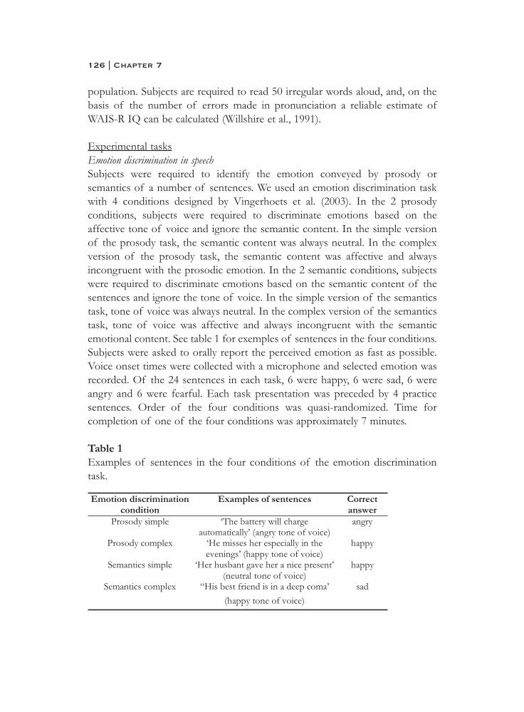
population. Subjects are required to read 50 irregular words aloud, and, on thebasis of the number of errors made in pronunciation a reliable estimate ofWAIS-R IQ can be calculated (Willshire et al., 1991).
Experimental tasksEmotion discrimination in speechSubjects were required to identify the emotion conveyed by prosody orsemantics of a number of sentences. We used an emotion discrimination taskwith 4 conditions designed by Vingerhoets et al. (2003). In the 2 prosodyconditions, subjects were required to discriminate emotions based on theaffective tone of voice and ignore the semantic content. In the simple versionof the prosody task, the semantic content was always neutral. In the complexversion of the prosody task, the semantic content was affective and alwaysincongruent with the prosodic emotion. In the 2 semantic conditions, subjectswere required to discriminate emotions based on the semantic content of thesentences and ignore the tone of voice. In the simple version of the semanticstask, tone of voice was always neutral. In the complex version of the semanticstask, tone of voice was affective and always incongruent with the semanticemotional content. See table 1 for exemples of sentences in the four conditions.Subjects were asked to orally report the perceived emotion as fast as possible.Voice onset times were collected with a microphone and selected emotion wasrecorded. Of the 24 sentences in each task, 6 were happy, 6 were sad, 6 wereangry and 6 were fearful. Each task presentation was preceded by 4 practicesentences. Order of the four conditions was quasi-randomized. Time forcompletion of one of the four conditions was approximately 7 minutes.
Table 1Examples of sentences in the four conditions of the emotion discriminationtask.
126 | Chapter 7
Mental rotation taskThe mental rotation test was an adaptation of original test by Shepard andMetzler (1971). In this computer task, participants were asked to compare twosimultaneously presented objects composed of 10 cubes, and determinewhether they were identical or not. All objects were two-dimensionalrepresentations of three-dimensional objects, rotated at different angles. In50 % of the trials the objects were identical. Participants were given 6 min. tocomplete 20 of these items. Number of correct responses was collected.
ResultsGeneral IntelligenceMean estimated IQ, as measured with the Raven’s Advanced ProgressiveMatrices (short form), in the Klinefelter group (102.9, SD 11.3) did notsignificantly differ from that in the control group (109.6, SD 14.3).Mean estimated verbal IQ, as measured with the National Adult Reading Test(NART), also did not significantly differ between the Klinefelter group (107.4,SD 15.6) and the control group (110.8, SD 5.1).
Experimental tasksEmotion discrimination in speech Significant group differences were present as shown by a General Linear Model(GLM) multivariate test including all four conditions of the emotiondiscrimination task (F(2,41)=7.7, p=.001).
Significant group differences were present in the number of errors made inthe simple prosody condition (F(1,42)=13.2, p=.001). Klinefelter men mademore errors than controls. Also in the complex prosody condition, Klinefeltermen made more errors in discriminating emotions than controls (F(1,42)=15.4,p<.001). When corrected for performance in the semantics task (usingMANOVA), the group difference in performance in the prosody conditionsremained significant (F(1,43)=9.8, p=0.003 and F(1,43)=9.6, p=0.003 for thesimple and complex prosody conditions respectively).
Furthermore, in the simple semantics condition as well as in the complexsemantics condition, Klinefelter men made more errors as compared to controls(F(1,42)=4.5, p=.04 and F(1,42)=7.4, p=.009 respectively). All means and SD’sare presented in Table 2.
Reaction times in the Klinefelter group were significantly higher in all fourconditions (F(4,41)=4.4, p=.005) as indicated by a GLM multivariate test.
Affective prosody in XXY men | 127
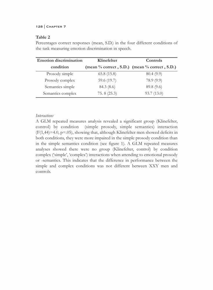
Table 2Percentages correct responses (mean, S.D.) in the four different conditions ofthe task measuring emotion discrimination in speech.
InteractionsA GLM repeated measures analysis revealed a significant group (Klinefelter,control) by condition (simple prosody, simple semantics) interaction(F(1,44)=4.0, p=.05), showing that, although Klinefelter men showed deficits inboth conditions, they were more impaired in the simple prosody condition thanin the simple semantics condition (see figure 1). A GLM repeated measuresanalyses showed there were no group (Klinefelter, control) by conditioncomplex (‘simple’, ‘complex’) interactions when attending to emotional prosodyor -semantics. This indicates that the difference in performance between thesimple and complex conditions was not different between XXY men andcontrols.
128 | Chapter 7
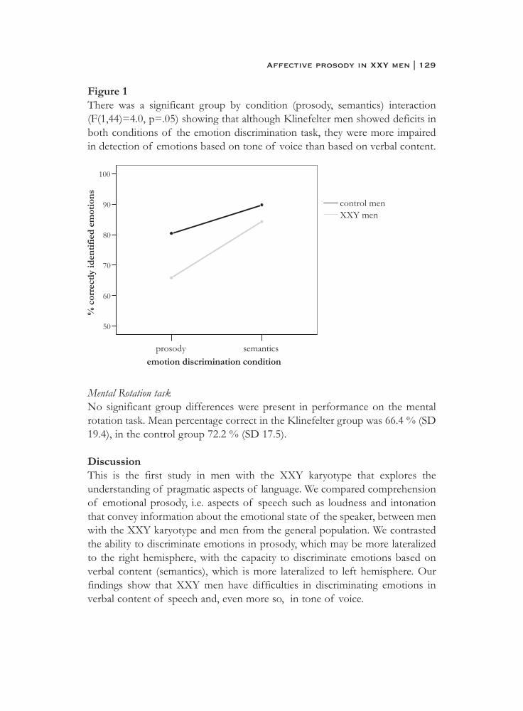
Figure 1There was a significant group by condition (prosody, semantics) interaction(F(1,44)=4.0, p=.05) showing that although Klinefelter men showed deficits inboth conditions of the emotion discrimination task, they were more impairedin detection of emotions based on tone of voice than based on verbal content.
Mental Rotation taskNo significant group differences were present in performance on the mentalrotation task. Mean percentage correct in the Klinefelter group was 66.4 % (SD19.4), in the control group 72.2 % (SD 17.5).
DiscussionThis is the first study in men with the XXY karyotype that explores theunderstanding of pragmatic aspects of language. We compared comprehensionof emotional prosody, i.e. aspects of speech such as loudness and intonationthat convey information about the emotional state of the speaker, between menwith the XXY karyotype and men from the general population. We contrastedthe ability to discriminate emotions in prosody, which may be more lateralizedto the right hemisphere, with the capacity to discriminate emotions based onverbal content (semantics), which is more lateralized to left hemisphere. Ourfindings show that XXY men have difficulties in discriminating emotions inverbal content of speech and, even more so, in tone of voice.
Affective prosody in XXY men | 129
The XXY group showed deficits in identifying emotions from prosodiccues as well as from semantic cues. These impairments were observed in thesimple versions of the task, where either the semantic content or the tone ofvoice was neutral, as well as in the complex versions of the task, where semanticcontent and tone of voice were emotional as well as incongruent. The findingthat XXY men display semantic deficits in our study was expected consideringthe literature on language deficits in Klinefelter syndrome. However, both thecondition by group interaction and the finding that XXY men madesignificantly more errors in the affective prosody condition even when correctedfor their performance in the semantics condition, suggested that on top of thissemantic deficit, even more profound deficits are present in the prosody task.In the study of Vingerhoets et al. (2003), the observed significant bilateralincrease in BFV during the complex versions of this tasks as compared to thesimple versions were prosposed to reflect a general increase in attentionaldemand as the level of BFV change is thought to be associated with taskdemand and reflects the attentional capacity necessary to perform a task(Vingerhoets et al., 2001). As no effects of group on the difference inperformance between the simple and complex tasks were present, this mightsuggest that the specific deficits observed in XXY men were not due to ageneral difficulty in attending to the stimuli. Importantly, as performance on aspatial, 3D mental rotation task was not impaired in the XXY group, thelanguage deficits may not be attributable to a generalized reduction in cognitiveperformance.
Difficulties in picking up pragmatic communicative cues in conversationmight have an impact on how well XXY men cope with social situations, asthese aspects of language are important for understanding interpersonalintentions and responding to those in an appropriate way. The present findingsshow that Klinefelter syndrome may not only be associated with difficulties inidentifying emotions based on facial expressions (Van Rijn et al., 2006), but alsowhen expressed in tone of voice. Besides providing insight in social cognitiveimpairments that might underlie difficulties in social communication and copingwith a social environment, the present findings may have several potentialimplications.
First, our findings suggest that not only language functions typicallymediated by the left hemisphere, but also prosodic aspects of language whichmay be associated with the right hemisphere, are affected in Klinefeltersyndrome. Although speculative, this would be in line with the reportedabnormalities in language-related functioning of the right hemisphere. A
130 | Chapter 7
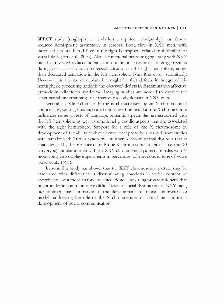
SPECT study (single-photon emission computed tomography) has shownreduced hemispheric asymmetry in cerebral flood flow in XXY men, withincreased cerebral blood flow in the right hemisphere related to difficulties inverbal skills (Itti et al., 2003). Also, a functional neuroimaging study with XXYmen has revealed reduced lateralization of brain activation in language regionsduring verbal tasks, due to increased activation in the right hemisphere, ratherthan decreased activation in the left hemisphere (Van Rijn et al., submitted).However, an alternative explanation might be that defects in integrated bi-hemispheric processing underlie the observed deficts in discrimination affectiveprosody in Klinefelter syndrome. Imaging studies are needed to explore theexact neural underpinnings of affective prosody deficits in XXY men.
Second, as Klinefelter syndrome is characterised by an X chromosomalabnormality, we might extrapolate from these findings that the X chromosomeinfluences some aspects of language, semantic aspects that are associated withthe left hemisphere as well as emotional prosodic aspects that are associatedwith the right hemisphere. Support for a role of the X chromosome indevelopment of the ability to decode emotional prosody is derived from studieswith females with Turner syndrome, another X chromosomal disorder that ischaracterised by the presence of only one X chromosome in females (i.e. the X0karyotype). Similar to men with the XXY chromosomal pattern, females with Xmonosomy also display impairments in perception of emotions in tone of voice(Ross et al., 1995).
In sum, this study has shown that the XXY chromosomal pattern may beassociated with difficulties in discriminating emotions in verbal content ofspeech and, even more, in tone of voice. Besides revealing prosodic deficits thatmight underlie communicative difficulties and social dysfunction in XXY men,our findings may contribute to the development of more comprehensivemodels addressing the role of the X chromosome in normal and abnormaldevelopment of social communication.
Affective prosody in XXY men | 131
ReferencesBender, B. G., Harmon, R. J., Linden, M. G., Bucher-Bartelson, B., &
Robinson, A. (1999). Psychosocial competence of unselected youngadults with sex chromosome abnormalities. American Journal ofMedical Genetics - Neuropsychiatric Genetics, 88(2), 200.
Boone, K. B., Swerdloff, R. S., Miller, B. L., Geschwind, D. H., Razani, J., Lee,A., et al. (2001). Neuropsychological profiles of adults with klinefeltersyndrome. Journal of the International Neuropsychological Society, 7(4),446-456.
Geschwind, D. H., Boone, K. B., Miller, B. L., & Swerdloff, R. S. (2000).Neurobehavioral phenotype of klinefelter syndrome. Mental Retardationand Developmental Disabilities Research Reviews, 6(2), 107-116.
Itti, E., Gaw Gonzalo, I. T., Boone, K. B., Geschwind, D. H., Berman, N.,Pawlikowska-Haddal, A., et al. (2003). Functional neuroimaging providesevidence of anomalous cerebral laterality in adults with klinefelter'ssyndrome. Annals of Neurology, 54(5), 669-673.
Itti, E., Gaw Gonzalo, I. T., Pawlikowska-Haddal, A., Boone, K. B., Mlikotic,A., Itti, L., et al. (2006). The structural brain correlates of cognitivedeficits in adults with klinefelter's syndrome. Journal of ClinicalEndocrinology and Metabolism, 91(4), 1423-1427.
Joanette, Y., Goulet, P., & Hannequin, D. (1990). Right hemisphere and verbal communication. New York: Springer-Verlag.
Lezak, M. D. (1995). Neuropsychological assessment (third ed.). New York:Oxford University Press.
Mitchell, R. L., Elliott, R., Barry, M., Cruttenden, A., & Woodruff, P. W.(2003). The neural response to emotional prosody, as revealed byfunctional magnetic resonance imaging. Neuropsychologia, 41(10), 1410-1421.
Nelson, H. E. (1982). National adult reading test (nart), manual. Windsor,Bershire, UK: NFER-Nelson.
Oldfield, R. C. (1971). The assessment and analysis of handedness: The edinburgh inventory. Neuropsychologia, 9(1), 97-113.
Ratcliffe, S. (1999). Long-term outcome in children of sex chromosome abnormalities. Archives of Disease in Childhood, 80(2), 192-195.
Ross, E. D., Thompson, R. D., & Yenkosky, J. (1997). Lateralization ofaffective prosody in brain and the callosal integration of hemisphericlanguage functions. Brain and Language, 56(1), 27-54.
132 | Chapter 7
Ross, J. L., Stefanatos, G., Roeltgen, D., Kushner, H., & Cutler Jr, G. B. (1995).Ullrich-turner syndrome: Neurodevelopmental changes from childhoodthrough adolescence. American Journal of Medical Genetics, 58(1), 74.
Rovet, J., Netley, C., Keenan, M., Bailey, J., & Stewart, D. (1996). The psychoeducational profile of boys with klinefelter syndrome. Journal ofLearning Disabilities, 29(2), 193-196.
Schmand, B., Bakker, D., Saan, R., & Louman, J. (1991). [the dutch reading test for adults: A measure of premorbid intelligence level]. Tijdschrift voorGerontologie en Geriatrie, 22(1), 15-19.
Sheehan, D. V., Lecrubier, Y., Sheehan, K. H., Amorim, P., Janavs, J., Weiller,E., et al. (1998). The mini-international neuropsychiatric interview(m.I.N.I.): The development and validation of a structured diagnosticpsychiatric interview for dsm-iv and icd-10. Journal of ClinicalPsychiatry, 59 Suppl 20, 22-33;quiz 34-57.
Shepard, R. N., & Metzler, J. (1971). Mental rotation of three-dimensional objects. Science, 171(972), 701-703.
Van Rijn, S., Aleman, A., Swaab, H., Vink, M., Sommer, I., & Kahn, R. S.(submitted). Effects of an extra x chromosome on languagelateralization: An fmri study with klinefelter (xxy) men.
Van Rijn, S., Aleman, A., van Diessen, E., Berckmoes, C., Vingerhoets, G., & Kahn, R. S. (2005). What is said or how it is said makes a difference:Role of the right fronto-parietal operculum in emotional prosody asrevealed by repetitive tms. Eur J Neurosci, 21(11), 3195-3200.
Van Rijn, S., Swaab, H., Aleman, A., & Kahn, R. S. (2006). X chromosomal effects on social cognitive processing and emotion regulation: A studywith klinefelter men (47,xxy). Schizophrenia Research, 84(2-3), 194-203.
Vingerhoets, G., Berckmoes, C., & Stroobant, N. (2003). Cerebral hemodynamics during discrimination of prosodic and semantic emotionin speech studied by transcranial doppler ultrasonography.Neuropsychology, 17(1), 93-99.
Vingerhoets, G., & Luppens, E. (2001). Cerebral blood flow velocity changes during dichotic listening with directed or divided attention: Atranscranial doppler ultrasonography study. Neuropsychologia, 39(10),1105.
Willshire, D., Kinsella, G., & Prior, M. (1991). Estimating wais-r iq from the national adult reading test - a cross-validation. Journal of Clinical andExperimental Neuropsychology, 13(2), 204-216.
Affective prosody in XXY men | 133

Part III
Neural basis of
social cognition

Chapter 8
Effects of an extra X chromosome on
language lateralization:
an fMRI study with Klinefelter (XXY) men
Sophie van Rijn, André Aleman, Hanna Swaab, Matthijs Vink, Iris Sommerand René S. Kahn
Submitted for publication
AbstractDe novo occurring genetic variations provide an opportunity to study theeffects of genes on structure and function of the brain. In this regard,Klinefelter syndrome, characterized by a XXY chromosomal pattern, is ofsignificant interest. Although general intelligence is average in XXY men,prominent effects of the extra X chromosome on cognition are observed in thelanguage domain. One possible neural mechanism underlying these languagedeficits is reduced hemispheric specialization for language. However, there hasbeen no study of brain activity patterns underlying language processing in XXYmen. Also, the consequences for mental functioning in XXY men are as yetunresolved. The clinical relevance of exploring mental consequences inKlinefelter syndrome is illustrated by the finding that Klinefelter men manifestsymptoms that are seen in schizophrenia. A possible commonality betweenthese disorders is that both are characterized by language deficits.
We used functional Magnetic Resonance Imaging (fMRI) to reveal theeffects of an extra X chromosome on language lateralization. This techniqueallows us to identify functional asymmetries in specific brain regions as well asto determine whether reduced lateralization, if found, is secondary to decreasedfunction of the left- or increased activity in the right hemisphere. We exploredthe relation between loss of language lateralization and mental functioning inthese men, with special interest in clinical phenomena of disorganization ofthought and language.
Hemispheric dominance for language was assessed in 15 XXY men and 14control men using fMRI. In each hemisphere, activity in five different languageregions was analyzed: Broca’s area, superior temporal gyrus, middle temporalgyrus, angular gyrus and supramarginal gyrus. Psychopathology was measuredusing the Positive and Negative Syndromes Scale for measuring schizophreniasymptoms and a schizotypal personality questionnaire.
138 | Chapter 8
Compared to controls, the XXY group showed reduced hemisphericspecialization for language. This was due to increased activity in the languageareas of the right hemisphere rather than reduced activity in the left hemisphere.Decreased functional asymmetry was most prominent in the superior temporalgyrus and correlated with symptoms of disorganization.
These findings may suggest that a genetic mechanism involving the Xchromosome contributes to hemispheric specialization for language, since lossof language lateralization, most prominent in the superior temporal gyrus, wasobserved in this X chromosomal disorder. Moreover, loss in hemisphericspecialization for language processing may have important consequences formental functioning, as it was associated with dysfunctions in organization ofthought and language.
Hemispheric lateralization for language in XXY men | 139
IntroductionThe study of de novo occurring genetic variations that are associated withneural, cognitive and behavioral abnormalities may increase our understandingof complex gene-brain-behavior relations. In this regard, Klinefelter syndrome,which is defined by the presence of an additional X chromosome in men, is ofsignificant interest. Although general intelligence is average in XXY men,prominent effects of the extra X chromosome on cognition have beenobserved, particularly in the language domain (D. H. Geschwind et al., 2000a).The reported verbal disabilities include impairments in both languageproduction and perception and indicate compromised language functions thatare typically associated with the left hemisphere (Samango-Sprouse, 2001). Forexample, Klinefelter boys or men have difficulty understanding words, findingwords and to verbally express their thoughts, all resulting in a verbal IQ that islower than their performance IQ. As language is a crucial part of socialcommunication, it has been proposed that these language impairments maycontribute to the observed difficulties in social functioning in Klinefelter men(Samango-Sprouse, 2001; van Rijn et al., 2006b).
Although language impairments in Klinefelter syndrome are well-documented, underlying neurocognitive mechanisms are not well understood(D. H. Geschwind et al., 2000a). One possible neural mechanism involved in theverbal disabilities in Klinefelter syndrome may be abnormal hemisphericinvolvement in language processing. Although the left hemisphere is dominantfor processing verbal information in right-handed individuals from the generalpopulation, there are indications that this dominance is diminished in XXYmen. First, decreased language lateralization has been shown in a study of 32XXY boys using a dichotic listening paradigm (Netley & Rovet, 1984). Second,in a more recent SPECT study, resting state blood flow patterns were moresymmetrical in nine XXY men as compared to nine healthy controls (Itti et al.,2003). In that study, increased right temporal lobe blood flow was related toverbal impairments as measured with neuropsychological tasks assessed in thesame week as the SPECT-session. Although these studies provide someindication that loss of hemispheric asymmetry in the processing of languagemay be a characteristic of the XXY phenotype, it is unclear whether thedecreased language lateralization is secondary to decreased function of the leftor increased activity in the right hemisphere. Moreover, it remains unresolvedwhich specific language regions within the hemispheres are more bilaterallyinvolved in processing language in XXY men.
140 | Chapter 8
Another reason for studying language lateralization in Klinefelter syndrome isthat the consequences for behavioral and psychological or mental functioning inXXY men are as yet unresolved. The importance of considering this issue isillustrated by reports of reduced hemispheric specialization for language inpatients with schizophrenia. In these patients, loss of language lateralization isthought to be related to language impairments as well as clinical symptomsassociated with language, such as auditory hallucinations (‘voices’) ordisorganization of language and thought (Kircher et al., 2002; I. E. Sommer etal., 2001b). Indeed, Klinefelter men manifest symptoms that are also observedin schizophrenia patients (van Rijn et al., in press), including disorganization andthought disorder. Moreover, the frequency of the XXY chromosomal patternhas been reported to be increased in patients with schizophrenia as comparedto the general population (L.E. DeLisi et al., 1994).
To assess hemispheric specialization for language in XXY men, we usedfunctional Magnetic Resonance Imaging (fMRI). By using fMRI we are able toinvestigate lateralization of neural activity during language processing in XXYmen. In addition, this technique allows us to identify specific language regionsin which lateralization is diminished. Our second aim was to investigate the linkbetween loss of language lateralization and mental functioning in these men,with special interest in those symptoms and personality traits pertaining toorganization of thought and language, that are also seen in the schizophreniaspectrum.
Methods
Subjects15 XXY men (mean age 36.9, SD 11.8) and 14 healthy control men (mean age35.5, SD 9.5) participated in the fMRI study. XXY men were recruited from theDutch Klinefelter Association, and were not selected for psychological,behavioral or cognitive abnormalities. Diagnosis of Klinefelter syndrome wasconfirmed by genetic analysis (i.e. karyotyping) using standard procedures. Ofthe XXY men, 14 were treated with testosterone supplements (mean age oftreatment onset of 23.9, SD 7.1 years).
Controls were recruited using advertisements in local newspapers or weredrawn from a database in our department. None of the control subjects had ahistory of psychiatric illness as confirmed with the Mini InternationalNeuropsychiatric Interview plus (MINI) (Sheehan et al., 1998).The primary language of all participants was Dutch. There were no significant
Hemispheric lateralization for language in XXY men | 141
differences in age (t(1,27)=-1.1, p=0.26) or years of education between thegroups (Klinefelter group 15.8 (SD 2.0), control group 15.4 (SD 1.8),t(1,27)=0.41, p=0.68). All participants were right-handed. Mean handednessscore, as indicated by the Edinburgh Handedness Inventory (Oldfield, 1971), inthe Klinefelter group (21.21, SD 3.1) did not significantly differ from that incontrols (22.1, SD 2.1) (t(1,27)=0.85, p=0.40).
Exclusion criteria for both Klinefelter men and controls were neurologicalconditions or history of head injury with loss of consciousness, recent historyof substance abuse and mental retardation. After complete description of thestudy to the subjects, written informed consent was obtained according to thedeclaration of Helsinki.
Schizophrenia psychopathologySchizophrenia spectrum pathology was only measured in the Klinefelter group.Schizophrenia spectrum pathology in a larger population of Klinefelter men atour department as compared to healthy controls has been described elsewhere(van Rijn et al., in press). Schizotypal traits were measured using the SchizotypalPersonality Questionnaire (SPQ) (Raine, 1991). The SPQ is a self-reportmeasure of schizotypal personality traits, which have shown to be normallydistributed in the general population. It is regarded as an indicator of thegenetic vulnerability to schizophrenia, since there is a gradient increase inschizotypal traits in relatives of schizophrenia patients that is in proportion tothe risk for schizophrenia associated with the degree of kinship with theschizophrenic family member (Vollema et al., 2002). Factor analytical studies(Vollema & Hoijtink, 2000) have revealed three dimensions of schizotypy, being(a) Positive schizotypy (for example referential thinking and delusionalatmosphere), (b) Negative schizotypy (for example constricted affect and socialanxiety), and (c) Disorganization (odd speech and eccentric behavior).A clinical measure of schizophrenia symptoms was also included. The Positiveand Negative Syndromes Scale (PANSS) (Kay et al., 1987) is a widely usedstructured interview to assess symptom profiles in schizophrenia patients thatare present in the week prior to the interview. The PANSS allows categorizationof negative, positive, and general symptoms.
Language tasksLanguage tasks were adopted from Ramsey et al. (2001). In the MRI scannersubjects completed three different tasks that have been shown to activatelanguage areas in the brain, being a paced verb generation task, an antonym
142 | Chapter 8
generation task, and a semantic decision task. Difficulty of the tasks was set ata level that would allow Klinefelter men to perform comparable to healthycontrols. All subjects practiced the tasks with a different set of stimuli beforethe scan session. In all tasks, a word was presented visually every 3 seconds andconsisted of four to eight characters. Silent vocalization (i.e. without overtarticulation) was used to avoid head motion. Each task was performed during 5blocks of 29 s, alternated with rest periods of 29 s. A total of 360 functionalscans were collected for each individual.
In the verb generation task, subjects were instructed to repeat a visuallypresented noun silently and subsequently silently vocalize an appropriate verbfor the presented noun. In the antonym generation task, subjects had to thinkof a word that was of opposite meaning in response to the visually presentedword. In the control condition for these tasks a number of dots, equal to thenumber of characters in the presented words, appeared on the screen. Finally,in the semantic decision task, subjects had to indicate, by pushing a button,whether the visually presented word was an animal. The control task alsoincluded button presses, which were cued by the presence of five dots (eitherthree or five dots appeared). Performance was registered with a computer.Performance in the verb- and antonym generation tasks was assessed outsidethe scanner. Each task included 45 words, which were different from thosepresented during scanning, but presented with an identical interstimulusinterval.
ScansScanning technique was adopted from Ramsey et al. (2001). Functional scanswere acquired with a Phillips ACS-NT 1.5-T clinical scanner, using the blood-oxygen-level dependent sensitive, navigated 3D PRESTO pulse sequence (N.F.Ramsey et al., 1998), with the following parameter settings: TE/TR 35/24 ms,flip angle 9º, FOV 225x180x77 mm3 , matrix 64x52x26, voxel size 4 mmisotropic, scan time per volume 2.4 s. Following the fMRI procedure, ananatomical scan was acquired.
fMRI analysisFunctional MRI data preprocessing and analysis was done using SPM2(Wellcome Department of Imaging Neuroscience, London, England;www.fil.ion.ucl.ac.uk).
Hemispheric lateralization for language in XXY men | 143
Brain activity maps were obtained by analyzing the fMRI scans during all threetasks conjointly. It has previously been shown that such an analysis improvesreliability of the subsequently computed laterality index, as compared to thatobtained with individual task analysis (N. F. Ramsey et al., 2001). The rationalefor conjoint analysis is that it improves sensitivity for brain activity that ispresent in all tasks, while reducing contribution of activity that is specific forindividual language tasks.
All functional scans were registered to the last volume of the last block andcoregistered to the anatomical scan. Next, all functional images were registeredto an MNI standard brain, to enable group-wise comparisons. For each subject,a statistical map (i.e. T-map) was obtained from a general linear modelregression analysis using a factor matrix that contained one factor modeling taskactivation for all three tasks combined. Eight discrete cosine functions wereincluded to correct for low-frequency drifts.
Significant activation was then determined in each voxel by applying athreshold. The threshold corresponded to a p value of 0.001, uncorrected formultiple comparisons, and amounted to a T-value of 3.12. This relatively lowthreshold was applied to reduce the chance of a type II error (i.e. the failure tofind a difference while there is a true difference between the conditions). Also,by setting the threshold at this T-value, one increases the likelihood that activevoxels are present in both the left and the right hemisphere in each individual,allowing calculation of a more reliable laterality index.
Regions of interestA map including five regions of interest (ROI), based on nine Brodmann areas(BA), was created using the WFU Pickatlas tool for SPM (Maldjian et al., 2003).This map was dilated with one voxel isotropic and coregistered with the T-maps.The advantage of using Brodmann areas is that volumes of the ROI’s are ofequal size across the hemispheres and across the groups. The following ROI’swere created for the hemispheres separately: Broca’s area and the homotopicalregion in the right hemisphere (BA 44, 45), superior temporal gyrus (BA 22, 38,41, 42), middle temporal gyrus (BA 21), angular gyrus (BA 39) andsupramarginal gyrus (BA 40).
144 | Chapter 8
Lateralization indexFor each individual, the number of voxels above a threshold of T=3.12 (p < 0.001, uncorrected for multiple comparisons) was counted for all ROI’sseparately. A lateralization index for each ROI and all ROI’s combined wascalculated using the following algorithm:
The index ranges from –1 (complete right hemispheric dominance) to +1(complete left hemispheric dominance). The advantage of using this index overabsolute values is that this measure reflects hemispheric dominance within anindividual, and is relative to overall brain activity in each individual. Individuallateralization indices for each language region, and all regions combined, wereentered in an ANOVA.
Hemispheric lateralization for language in XXY men | 145
Active voxels left - active voxels right
Total active voxels
Language lateralizationThe mean lateralization index (for all language regions included) in the XXYgroup was 0.45 (SD 0.30), which was significantly lower than the index of 0.74(SD 0.23) in the control group, F(1,27)= 7.6, p=.01. See figure 1 for thedistribution of lateralization indices in the groups. The lower lateralization indexin the XXY group was due to increased language related activity in the righthemisphere as compared to controls (F(1,27)=6.0, p=.02), rather than group-differences in left hemispheric language-related activity (F(1,27)=1.7, p=.20).More specifically, the mean lateralization index in the Superior Temporal Gyrus(STG, Brodmann area 22, 38, 41, 42) was significantly lower in XXY men (0.27,SD 0.39) as compared to controls (0.69, SD 0.36), F(1,27)=8.2, p=.008). Seetable 1 for the lateralization indices for each language region and figure 2 foractivation patterns in a typical XXY subject and a typical control subject.
Analysis of the button presses during scanning indicated that performancein the semantic decision task was not significantly different between Klinefeltermen and controls (t(1,27)=-0.38, p=0.71). Mean percentages correct were 90.6(SD 8.8) and 89.3 (SD 8.4) respectively. Also, we observed no significant groupdifferences in mean correct responses in the antonym-generation task (controlgroup 42.1 (SD 3.4), Klinefelter group 41.2 (SD 3.8), t(1,24)=0.52, p=0.61) andverb-generation task (control group 40.4 (SD 3.2), Klinefelter group 34.6 (SD9.0), t(1,24)=1.8, p=0.08) outside the scanner. Language lateralization in theXXY group did not correlate with handedness or age at which testosteronesupplementation was started.
146 | Chapter 8
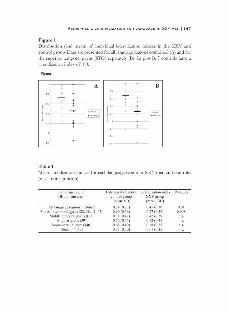
Figure 1Distribution (and mean) of individual lateralization indices in the XXY andcontrol group. Data are presented for all language regions combined (A) and forthe superior temporal gyrus (STG) separately (B). In plot B, 7 controls have alateralization index of 1.0.
Table 1Mean lateralization indices for each language region in XXY men and controls.(n.s.= not signficant)
Hemispheric lateralization for language in XXY men | 147
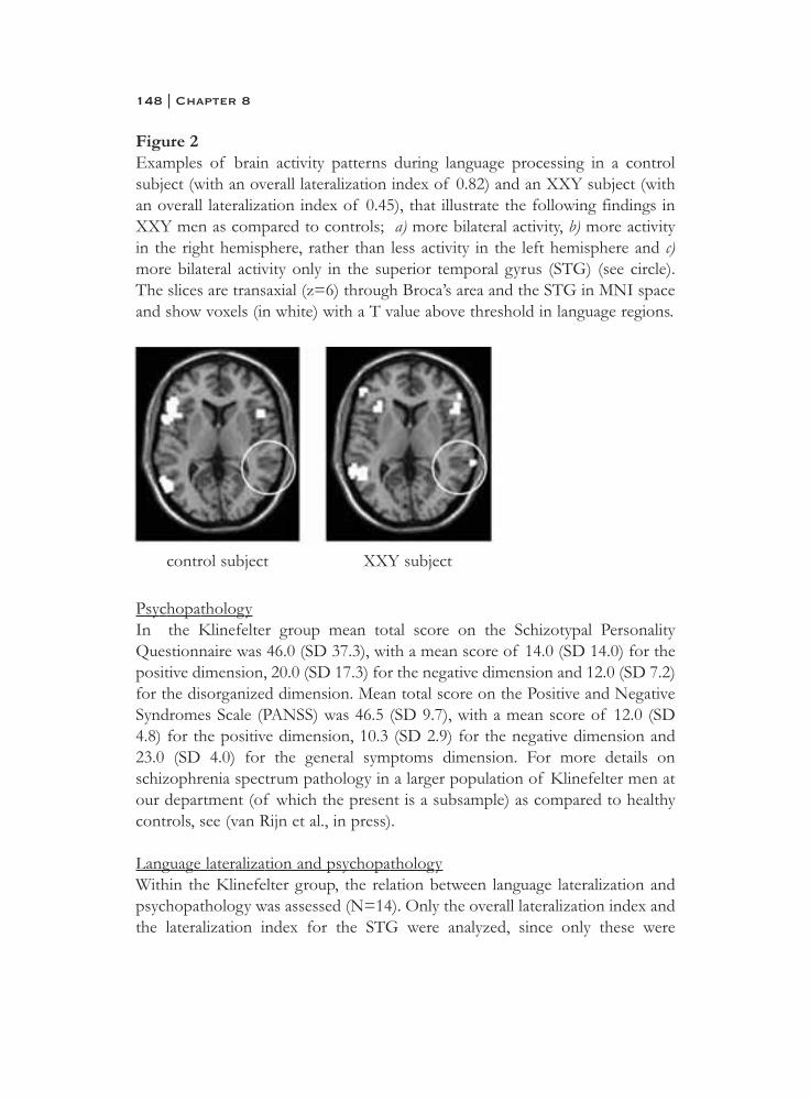
Figure 2Examples of brain activity patterns during language processing in a controlsubject (with an overall lateralization index of 0.82) and an XXY subject (withan overall lateralization index of 0.45), that illustrate the following findings inXXY men as compared to controls; a) more bilateral activity, b) more activityin the right hemisphere, rather than less activity in the left hemisphere and c)more bilateral activity only in the superior temporal gyrus (STG) (see circle).The slices are transaxial (z=6) through Broca’s area and the STG in MNI spaceand show voxels (in white) with a T value above threshold in language regions.
PsychopathologyIn the Klinefelter group mean total score on the Schizotypal PersonalityQuestionnaire was 46.0 (SD 37.3), with a mean score of 14.0 (SD 14.0) for thepositive dimension, 20.0 (SD 17.3) for the negative dimension and 12.0 (SD 7.2)for the disorganized dimension. Mean total score on the Positive and NegativeSyndromes Scale (PANSS) was 46.5 (SD 9.7), with a mean score of 12.0 (SD4.8) for the positive dimension, 10.3 (SD 2.9) for the negative dimension and23.0 (SD 4.0) for the general symptoms dimension. For more details onschizophrenia spectrum pathology in a larger population of Klinefelter men atour department (of which the present is a subsample) as compared to healthycontrols, see (van Rijn et al., in press).
Language lateralization and psychopathologyWithin the Klinefelter group, the relation between language lateralization andpsychopathology was assessed (N=14). Only the overall lateralization index andthe lateralization index for the STG were analyzed, since only these were
148 | Chapter 8
control subject XXY subject
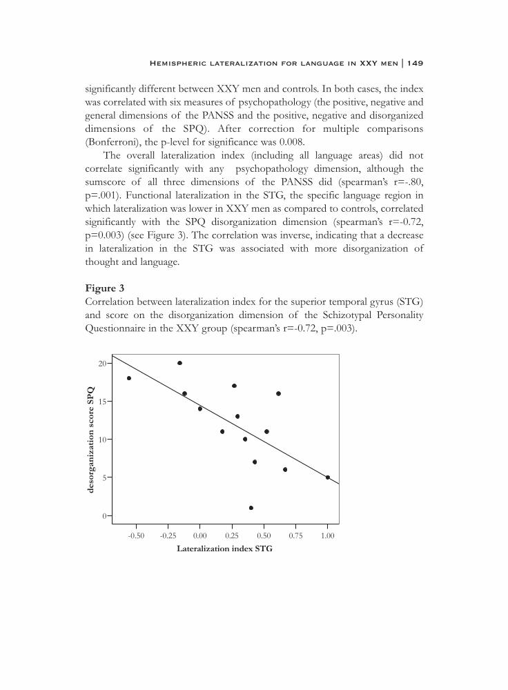
significantly different between XXY men and controls. In both cases, the indexwas correlated with six measures of psychopathology (the positive, negative andgeneral dimensions of the PANSS and the positive, negative and disorganizeddimensions of the SPQ). After correction for multiple comparisons(Bonferroni), the p-level for significance was 0.008.
The overall lateralization index (including all language areas) did notcorrelate significantly with any psychopathology dimension, although thesumscore of all three dimensions of the PANSS did (spearman’s r=-.80,p=.001). Functional lateralization in the STG, the specific language region inwhich lateralization was lower in XXY men as compared to controls, correlatedsignificantly with the SPQ disorganization dimension (spearman’s r=-0.72,p=0.003) (see Figure 3). The correlation was inverse, indicating that a decreasein lateralization in the STG was associated with more disorganization ofthought and language.
Figure 3Correlation between lateralization index for the superior temporal gyrus (STG)and score on the disorganization dimension of the Schizotypal PersonalityQuestionnaire in the XXY group (spearman’s r=-0.72, p=.003).
Hemispheric lateralization for language in XXY men | 149
DiscussionUsing functional Magnetic Resonance Imaging (fMRI) we were able to measure,for the first time, patterns of brain activity during language processing in47,XXY men. By measuring the relative contribution of the right and lefthemisphere in each individual it was shown that language activity in the brainwas less lateralized in the XXY group as compared to healthy males. Loss ofasymmetric processing of language was due to increased activity in the righthemisphere rather than reduced activity in the left hemisphere. Analysis offunctional asymmetry within different language regions indicated that thesuperior temporal gyrus (STG) was the only region in which language was lesslateralized in the XXY group. This regional loss of language laterality was highlycorrelated with the degree of disorganization in these subjects.
Reduced hemispheric specialization for language may contribute to thewidely reported language deficits in XXY adults and children (Boone et al.,2001; D. H. Geschwind et al., 2000a; Money, 1993; Samango-Sprouse, 2001).These include disabilities in reading, articulation, phonemic processing, wordfinding, spelling, language expression, verbal memory, language comprehensionand verbal fluency. The reported patterns of language impairments seem similarto those observed in developmental language disorders, for example dyslexia (D.H. Geschwind et al., 2000a). A proposed neurocognitive basis for the languageprofile in developmental language disorders is reduced hemisphericspecialization for language in the brain. Findings of less cerebral asymmetry,both functional (Ors et al., 2005) and structural (Herbert et al., 2005), indevelopmental language disorders illustrate the contribution of cerebrallateralization to the development of language capabilities. Our fMRI studyshows that specifically an increase in right hemispheric involvement in languagemay contribute to language impairments, which fits with findings from aSPECT-study (Itti et al., 2003) in which increased resting state blood flow in theSTG in the right hemisphere was observed in nine XXY men. They observedthat abnormal resting state activity in XXY men was correlated with verbal skillsas assessed in a neuropsychological session.
The use of fMRI allowed us to identify specific areas in the brain underlyingthe loss of hemispheric specialization for language in Klinefelter syndrome.One of our key findings was that loss of functional asymmetry in the STG wasthe most prominent contributor to reduced language laterality. Our finding offunctional abnormalities in the STG is consistent with studies reportingstructural abnormalities in the temporal lobes in Klinefelter syndrome. An MRIstudy including 10 XXY men showed asymmetric reduction in volume of the
150 | Chapter 8
temporal lobes, with a smaller volume of the left temporal lobe in these men(Patwardhan et al., 2000). In a larger sample of 34 XXY men, Shen et al. (2004)found that more regions in the left, as compared to the right, temporal lobewere smaller in the XXY group in comparison to 62 healthy men. Two otherstudies have reported bilateral volume reductions of (regions in) the temporallobe. DeLisi et al. (2005) observed smaller volumes of the STG bilaterally in 11XXY men. Itti et al. (2006) measured smaller volumes of the temporal lobesbilaterally in 9 XXY men, but only volume reductions in the left hemispherecorrelated with verbal skills.
Decreased functional and structural asymmetry of language area’s in thebrain in XXY men may be secondary to abnormal X chromosomal (in-)activation, since a pseudo-autosomal region on the X chromosome is thoughtto direct abnormal development of asymmetry in XXY men (D.H. Geschwindet al., 1998). Genes in the pseudo-autosomal region escape inactivation resultingin expression of both copies of the genes in XX women and XY men and allthree copies in XXY men (i.e. overexpression). This dosage mechanism makesgenes in this region prime candidates for abnormal brain development,including cerebral asymmetry, and cognition in XXY (D.H. Geschwind et al.,1998). For example, one candidate gene in the pseudo-autosomal (X-Yhomologous) region that has been shown to escape inactivation resulting inexpression of all three copies in Klinefelter syndrome is Protocadherin11XY(PCDH11XY), which is involved in axonal guidance in the brain (T. J. Crow,2002; N. L. J. Ross et al., 2006).
Support for X chromosomal effects on structural asymmetry of thesuperior temporal area in the brain is also provided by studies on brain volumesin individuals with Turner syndrome (46 XO, i.e. X monosomy). Decreasedvolume of the superior temporal sulcus in the left hemisphere as well asincreased volume of the superior temporal gyrus in the right hemisphere(Kesler et al., 2003) have been observed in these subjects. In this study, increasedright STG volume was related to lower verbal intelligence scores in Turnersubjects (but not in controls). Involvement of the X chromosome in abnormaldevelopment of the STG, at least in Turner syndrome, was indicated by theobserved imprinting effects. (Imprinting refers to differential expression of agene depending on whether the gene is passed through from the mother or thefather.) Only STG volumes in Turner subjects with the single X chromosomederived form the mother, and not those with an X chromosome from paternalorigin, were different from controls. The findings that reduced asymmetry,either secondary to increased right hemisphere volume as in Turner syndrome
Hemispheric lateralization for language in XXY men | 151
or decreased left hemisphere volume as in Klinefelter syndrome, can beaccompanied by language dysfunctions, implies that reduced asymmetry may bea key abnormality in language processing deficits.
Decreased functional asymmetry in the STG in Klinefelter men correlatedwith the disorganization dimension of the schizotypy measure. Both items ofthis dimension, ‘odd speech’ (vague or over-inclusive) and ‘odd/eccentricbehavior’, also separately correlated inversely with language lateralization in theSTG. This is consistent with studies finding structural (Matsumoto et al., 2001;Menon et al., 1995; Rajarethinam et al., 2000; Rossi et al., 1994a; Shenton et al.,1992) and functional (Kircher et al., 2002) abnormalities of the STG to berelated to disorganization symptoms such as thought disorder, in a anotherdisorder, i.e. schizophrenia. Specifically, in a functional MRI study withschizophrenia patients with formal thought disorder, higher levels of thoughtdisorder were related to increased STG activity in the right hemisphere (Kircheret al., 2002). Interestingly, in schizophrenia, sex differences have been reportedin the volume of the STG (Flaum et al., 1995; R.E. Gur et al., 2000). A geneticmechanism involving the sex chromosomes might explain this finding. An X-Yhomologous, pseudo-autosomal genetic region directing development ofasymmetry in the brain has been proposed (T.J. Crow, 2004) to underlie theobserved sex differences in verbal abilities, handedness, relative rates ofhemispheric growth and both ages of onset and prevalence of schizophrenia.The idea that reduced cerebral asymmetry and, more specifically, loss oflanguage lateralization is one of the core developmental brain abnormalitiesunderlying schizophrenia symptoms (Bhati, 2005; T.J. Crow, 2004) is supportedby a range of language laterality studies in schizophrenia (Artiges et al., 2000;Dollfus et al., 2005; Kircher et al., 2002; I. Sommer et al., 2001a; Weiss et al.,2006). One of these studies not only assessed the degree of lateralization, butalso examined whether this was secondary to increased right hemisphereactivation or decreased left hemisphere activation (I. E. Sommer et al., 2001b).Similar to what we observed in the Klinefelter men, decreased lateralization oflanguage in schizophrenia patients was due to increased activity in the righthemisphere, rather than decreased activity in the left hemisphere.
A proposed mechanism by which abnormal functional lateralization of theSTG may contribute to disorganization and thought disorder, incorporates thedifferences in semantic processing between the left and right auditoryassociation areas (including the STG). Semantic fields are groups of words andconcepts that are closely related in meaning, often subsumed under a generalterm, forming a category (such as ‘animals’). In the left hemisphere, words are
152 | Chapter 8
associated with other words and concepts within small, focal semantic fields,whereas in the right hemisphere they are associated within large, diffusesemantic fields (Beeman & Chiarello, 1998). Therefore, the right hemisphereappears to be biased to aspects of language that require ‘global’ semanticprocessing, such as forming semantic relations that are only loosely coupled inmeaning, generation of multiple meanings of words and the interpretation ofwhole sentences, stories or metaphors. In contrast, the left hemisphere seems tobe biased to aspects of language that require ‘local’ semantic processing, suchas single interpretations for each word. In the left hemisphere contextuallyinappropriate meanings of words are inhibited and one single meaning isselected. It is an imbalance in these complementary processes that is proposedto underlie disorganization symptoms and thought disorder (Kircher et al.,2002). An imbalance, created by increased right hemispheric activity leading togeneration of distantly related meanings together with reduced left hemisphericactivity resulting in loss of inhibition and selection of meaning, may explain theincreased levels of disorganization in XXY men.
The degree to which functional asymmetries in the XXY brain representthe effects of testosterone- deficits or supplementation remains unclear. Therelationship between testosterone levels and behavior is complex; timing ofexposure, sensitivity to testosterone reflected in androgen receptor density andmodulation by environmental factors are important determinants in the effectsof testosterone (Craig et al., 2004). Furthermore, gonadal hormones may be oneof many mechanisms by which sex chromosomes exert their influence on braindevelopment. Recent animal studies have pointed to direct, non-hormonal,effects of sex-chromosomes on brain maturation (Dewing et al., 2003). In linewith this, a recent longitudinal study dealing with the effects of testosteronetreatment on language lateralization in transsexuals showed that languagelaterality is highly stable and not affected by hormonal interventions (I. E. C.Sommer et al., in prep).
In sum, the present study provides evidence for a loss of languagelateralization, most prominent in the superior temporal gyrus, in XXY men.Moreover, we found that the loss in hemispheric specialization for languageprocessing may have important consequences for mental functioning, as it wasassociated with dysfunctions in organization of thought and language. Thissuggests that a genetic mechanism involving the X chromosome is involved inthe development of hemispheric specialization for language. Althoughspeculative, such a mechanism may involve genes that are overexpressed as aresult of escaping inactivation. Alternatively, it might be an epi-genetic
Hemispheric lateralization for language in XXY men | 153

mechanism, involving an environmentally (i.e. non-genetic) induced change ingene function, or epi-static, involving suppression of expression of other genes.Future genetic studies with XXY men may provide insight into the mechanismsby which genes on the X chromosome direct development of functionalasymmetry in the brain.
154 | Chapter 8
ReferencesArtiges E, Martinot J-L, Verdys M, Attar-Levy D, Mazoyer B, Tzourio N, et al.
Altered hemispheric functional dominance during word generation innegative schizophrenia. Schizophrenia Bulletin 2000; 26: 709-721.
Beeman MJ, Chiarello C. Complementary right- and left-hemisphere language comprehension. Current Directions in Psychological Science 1998; 7: 2-8.
Bhati MT. The brain, language, and schizophrenia. Current Psychiatry Reports 2005; 7: 297-303.
Boone KB, Swerdloff RS, Miller BL, Geschwind DH, Razani J, Lee A, et al.Neuropsychological profiles of adults with Klinefelter syndrome.Journal of the International Neuropsychological Society: Jins 2001; 7:446-56.
Craig IW, Harper E, Loat CS. The genetic basis for sex differences in human behaviour: Role of the sex chromosomes. Annals of Human Genetics2004; 68: 269-284.
Crow TJ. Handedness, language lateralisation and anatomical asymmetry:relevance of protocadherin XY to hominid speciation and the aetiologyof psychosis - Point of view. British Journal of Psychiatry 2002; 181:295-297.
Crow TJ. Cerebral asymmetry and the lateralization of language: Core deficits in schizophrenia as pointers to the gene. Current Opinion in Psychiatry2004; 17: 97-106.
DeLisi LE, Friedrich U, Wahlstrom J, Boccio_Smith A, Forsman A, Eklund K,et al. Schizophrenia and sex chromosome anomalies. SchizophreniaBulletin 1994; 20: 495-505.
DeLisi LE, Maurizio AM, Svetina C, Ardekani B, Szulc K, Nierenberg J, et al.Klinefelter's syndrome (XXY) as a genetic model for psychoticdisorders. Am J Med Genet B Neuropsychiatr Genet 2005; 135: 15-23.
Dewing P, Shi T, Horvath S, Vilain E. Sexually dimorphic gene expression in mouse brain precedes gonadal differentiation. Molecular Brain Research2003; 118: 82-90.
Dollfus S, Razafimandimby A, Delamillieure P, Brazo P, Joliot M, Mazoyer B,et al. Atypical hemispheric specialization for language in right-handedschizophrenia patients. Biological Psychiatry 2005; 57: 1020-1028.
Flaum M, Swayze II VW, O'Leary DS, Yuh WTC, Ehrhardt JC, Arndt SV, et al. Effects of diagnosis, laterality, and gender on brain morphology inschizophrenia. American Journal of Psychiatry 1995; 152: 704-714.
Hemispheric lateralization for language in XXY men | 155
Geschwind DH, Boone KB, Miller BL, Swerdloff RS. Neurobehavioral phenotype of Klinefelter syndrome. Mental Retardation andDevelopmental Disabilities Research Reviews 2000; 6: 107-16.
Geschwind DH, Gregg J, Boone K, Karrim J, Pawlikowska_Haddal A, Rao E,et al. Klinefelter's syndrome as a model of anomalous cerebral laterality:testing gene dosage in the X chromosome pseudoautosomal regionusing a DNA microarray. Developmental Genetics 1998; 23: 215-29.
Gur RE, Turetsky BI, Cowell PE, Finkelman C, Maany V, Grossman RI, et al.Temporolimbic volume reductions in schizophrenia. Archives ofGeneral Psychiatry 2000; 57: 769-75.
Herbert MR, Ziegler DA, Deutsch CK, O'Brien LM, Kennedy DN, Filipek PA, et al. Brain asymmetries in autism and developmental languagedisorder: A nested whole-brain analysis. Brain 2005; 128: 213-226.
Itti E, Gaw Gonzalo IT, Boone KB, Geschwind DH, Berman N,Pawlikowska-Haddal A, et al. Functional neuroimaging providesevidence of anomalous cerebral laterality in adults with Klinefelter'ssyndrome. Ann Neurol 2003; 54: 669-73.
Itti E, Gaw Gonzalo IT, Pawlikowska-Haddal A, Boone KB, Mlikotic A, Itti L,et al. The structural brain correlates of cognitive deficits in adults withKlinefelter's syndrome. Journal of Clinical Endocrinology andMetabolism 2006; 91: 1423-1427.
Kay SR, Fiszbein A, Opler LA. The Positive and Negative Syndrome rating Scale (PANSS) for Schizophrenia. Schizophrenia Bulletin 1987; 13: 261-276.
Kesler SR, Blasey CM, Brown WE, Yankowitz J, Zeng SM, Bender BG, et al.Effects of X-monosomy and X-linked imprinting on superior temporalgyrus morphology in Turner syndrome. Biological Psychiatry 2003; 54:636-646.
Kircher TTJ, Liddle PF, Brammer MJ, Williams SCR, Murray RM, McGuire PK. Reversed lateralization of temporal activation during speechproduction in thought disordered patients with schizophrenia.Psychological Medicine 2002; 32: 439-449.
Maldjian JA, Laurienti PJ, Kraft RA, Burdette JH. An automated method for neuroanatomic and cytoarchitectonic atlas-based interrogation of fMRIdata sets. Neuroimage 2003; 19: 1233-9.
156 | Chapter 8
Matsumoto H, Simmons A, Williams S, Hadjulis M, Pipe R, Murray R, et al.Superior temporal gyrus abnormalities in early-onset schizophrenia:Similarities and differences with adult-onset schizophrenia. AmericanJournal of Psychiatry 2001; 158: 1299-1304.
Menon RR, Barta PE, Aylward EH, Richards SS, Vaughn DD, Tien AY, et al.Posterior superior temporal gyrus in schizophrenia: grey matter changesand clinical correlates. Schizophrenia Research 1995; 16: 127-135.
Money J. Specific neuro-cognitive impairments associated with Turner (45,X) and Klinefelter (47,XXY) syndromes: a review. Social biology 1993; 40:147-151.
Netley C, Rovet J. Hemispheric lateralization in 47,XXY Klinefelter's syndrome boys. Brain Cogn 1984; 3: 10-8.
Oldfield RC. The assessment and analysis of handedness: the Edinburgh inventory. Neuropsychologia 1971; 9: 97-113.
Ors M, Ryding E, Lindgren M, Gustafsson P, Blennow G, Rose?n I. Spect findings in children with specific language impairment. Cortex 2005; 41:316-326.
Patwardhan AJ, Eliez S, Bender B, Linden MG, Reiss AL. Brain morphology in Klinefelter syndrome: extra X chromosome and testosteronesupplementation. Neurology 2000; 54: 2218-23.
Raine A. The SPQ: a scale for the assessment of schizotypal personality based on DSM-III-R criteria. Schizophrenia Bulletin 1991; 17: 555-64.
Rajarethinam RP, DeQuardo JR, Nalepa R, Tandon R. Superior temporal gyrus in schizophrenia: A volumetric magnetic resonance imaging study.Schizophrenia Research 2000; 41: 303-312.
Ramsey NF, Sommer IE, Rutten GJ, Kahn RS. Combined analysis of language tasks in fMRI improves assessment of hemispheric dominance forlanguage functions in individual subjects. Neuroimage 2001; 13: 719-33.
Ramsey NF, Van Den Brink JS, Van Muiswinkel AMC, Folkers PJM, Moonen CTW, Jansma JM, et al. Phase navigator correction in 3D fMRIimproves detection of brain activation: Quantitative assessment with agraded motor activation procedure. NeuroImage 1998; 8: 240-248.
Ross NLJ, Wadekar R, Lopes A, Dagnall A, Close J, DeLisi LE, et al.Methylation of two Homo sapiens-specific X-Y homologous genes inKlinefelter's syndrome (XXY). American Journal of Medical Genetics,Part B: Neuropsychiatric Genetics 2006; 141: 544-548.
Hemispheric lateralization for language in XXY men | 157
Rossi A, Serio A, Stratta P, Petruzzi C, Schiazza G, Mancini F, et al. Planum temporale asymmetry and thought disorder in schizophrenia.Schizophrenia Research 1994; 12: 1-7.
Samango-Sprouse C. Mental development in polysomy X Klinefelter syndrome (47,XXY; 48,XXXY): Effects of incomplete X inactivation.Seminars in Reproductive Medicine 2001; 19: 193-202.
Sheehan DV, Lecrubier Y, Sheehan KH, Amorim P, Janavs J, Weiller E, et al.The Mini-International Neuropsychiatric Interview (M.I.N.I.): thedevelopment and validation of a structured diagnostic psychiatricinterview for DSM-IV and ICD-10. Journal of Clinical Psychiatry 1998;59 Suppl 20: 22-33;quiz 34-57.
Shen D, Liu D, Liu H, Clasen L, Giedd J, Davatzikos C. Automated morphometric study of brain variation in XXY males. NeuroImage2004; 23: 648-653.
Shenton ME, Kikinis R, Jolesz FA, Pollak SD, LeMay M, Wible CG, et al.Abnormalities of the left temporal lobe and thought disorder inschizophrenia. A quantitative magnetic resonance imaging study. TheNew England Journal of Medicine 1992; 327: 604-12.
Sommer I, Aleman A, Ramsey N, Bouma A, Kahn R. Handedness, language lateralisation and anatomical asymmetry in schizophrenia: Meta-analysis.British Journal of Psychiatry 2001a; 178: 344-351.
Sommer IE, Ramsey NF, Kahn RS. Language lateralization in schizophrenia,an fMRI study. Schizophr Res 2001b; 52: 57-67.
Sommer IEC, Cohen-Kettenis PT, Van Raalten T, van der Veer AJ, Gooren LJG, Kahn RS, et al. No effects of cross-sex hormones on cerebrallateralization: an fMRI study in transsexuals. in prep.
van Rijn S, Aleman A, Swaab H, Kahn R. 47,XXY Karyotype and Schizophrenia Spectrum Pathology. British Journal of Psychiatry inpress.
van Rijn S, Swaab H, Aleman A, Kahn RS. X Chromosomal effects on social cognitive processing and emotion regulation: A study with Klinefeltermen (47,XXY). Schizophr Res 2006; 84: 194-203.
Vollema MG, Hoijtink H. The multidimensionality of self-report schizotypy in a psychiatric population: an analysis using multidimensional Raschmodels. Schizophr Bull 2000; 26: 565-75.
Vollema MG, Sitskoorn MM, Appels MCM, Kahn RS. Does the Schizotypal Personality Questionnaire reflect the biological-genetic vulnerability toschizophrenia? Schizophrenia Research 2002; 54: 39-45.
158 | Chapter 8

Weiss EM, Hofer A, Golaszewski S, Siedentopf C, Felber S, Fleischhacker WW. Language lateralization in unmedicated patients during an acuteepisode of schizophrenia: A functional MRI study. Psychiatry Research -Neuroimaging 2006; 146: 185-190.
Hemispheric lateralization for language in XXY men | 159

Chapter 9
Neurobiology of emotion and
high risk for schizophrenia:
Role of the amygdala and the X chromosome
Sophie van Rijn, André Aleman, Hanna Swaab, René S. Kahn
Neuroscience and Biobehavioral Reviews, 2005, 29, 385-397
Abstract Abnormalities in emotion processing and in structure of the amygdala haveconsistently been documented in schizophrenia. A major question is whetheramygdala abnormalities reflect a genetic vulnerability for the disease.
In the present paper, we reviewed Magnetic Resonance Imaging (MRI)studies that reported amygdala measures in several high risk populations:subjects from the general population with subclinical schizophrenia symptomsand relatives of schizophrenia patients. In addition, we reviewed the evidenceregarding Klinefelter syndrome (characterised by an additional X chromosome),which has also been related to an increased risk for schizophrenia.
Overall, the evidence points to structural abnormalities of the amygdala inindividuals at increased risk for schizophrenia. Although the genetic basis ofamygdala deficits remains unclear, abnormalities (of genes) on the Xchromosome might play a role as suggested by the evidence from individualswith sex chromosome aneuploidies. We propose that amygdala abnormalitiesare an endophenotype in schizophrenia and may account for subtle emotionalprocessing deficits that have been described in these high risk groups.
162 | Chapter 9
IntroductionThere is ample evidence that the amygdala plays a central role in emotionalinformation processing (Aggleton, 2000; Aggleton et al., 2000; Baxter et al.,2002). The amygdala appears to be important, among other functions, forevaluation of the emotional valence of stimuli in the very early phases ofsensory processing, linkage of perceptual representation to emotionalmemories, and regulation of autonomic responses (Aggleton, 2000; Aggleton etal., 2000). In humans, the amygdala appears to be especially important forprocessing of emotional information in a social context. Facial affectrecognition, for example, is shown to depend on the integrity of the amygdala(Adolphs et al., 1994; Adolphs et al., 1999; Calder et al., 1996; Gur et al., 2002a;Morris et al., 1998; Young et al., 1995). Not only is the amygdala involved inprocessing information about basic facial emotions, but also about complexsocial judgements, such as trustworthiness judgements of faces (Adolphs,2002).
Schizophrenia is a serious psychiatric disorder characterised by positivesymptoms, reflecting the presence of abnormal behavior such as delusions andhallucinations, and negative symptoms, representing the absence of normalbehavior. Besides impairments in the cognitive domain (Aleman et al., 1999;Heinrichs et al., 1998), individuals with schizophrenia show deficits in emotionprocessing, as indicated by a markedly reduced ability to perceive, process andexpress facial emotions (Mandal et al., 1998; Morrison et al., 1988; Mueser et al.,1996; Streit et al., 2001). These difficulties have a major impact on socialdysfunction in these patients (Edwards et al., 2002). Deficits in facial affectrecognition, for example, have been associated with dysregulation of socialbehaviours and deterioration of interpersonal relations in schizophrenia(Mueser et al., 1996; Poole et al., 2000).
Although a broad range of brain regions seems to be affected in patientswith schizophrenia, structural and functional MRI (Magnetic ResonanceImaging) studies have suggested that abnormalities in the amygdala may accountfor deficits in emotional processing. Structural MRI studies have shown reducedvolume and reduced gray matter density of the amygdala in these patients(Breier et al., 1992; Bryant et al., 1999; Gur et al., 2000; Hulshoff-Pol et al., 2001;Lawrie et al., 2003; Wright et al., 2000). Recent fMRI-studies with schizophreniapatients have shown less functional activation of this structure in response toemotional salient stimuli, such as faces (Gur et al., 2002b; Schneider et al., 1998;Taylor et al., 2002). As revealed by a review of studies focused at facial affectrecognition in schizophrenia (Mandal et al., 1998), recognition of facial
Amygdala abnormalities in the schizoprenia spectrum | 163
expressions of fear and anger seems to be specifically impaired in this illness.These patterns of emotion processing deficits parallel those found in bothhumans and primates with damage to the amygdala; the processing of fear andanger is an almost invariable consequence of amygdala lesions (Skuse et al.,2003). Not only the recognition of basic facial emotions, also creation ofcomplex social judgements such as trustworthiness of faces, which involves theamygdala as evidenced by functional neuroimaging- as well as focal brain lesionstudies, appears to be impaired in schizophrenia (Hall et al., 2004). Moresupport for dysfunction of the amygdala in schizophrenia comes fromimpairments of patients in aversive conditional avoidance learning (Hofer et al.,2001; Kosmidis et al., 1999), in which the amygdala is critically involved (Maren,2003).
Evidence for a specific contribution of morphological abnormalities of theamygdala to deficient emotional processing in schizophrenia is given by a studyshowing that volume of the amygdala can significantly predict performance inan emotional learning task in schizophrenia (Exner et al., In Press). Besidesspecific contributions to behavioral impairments, effects of amygdalaabnormalities may also extend to a clinical level in schizophrenia. Processing ofemotional faces is impaired in schizophrenia and associated with problems insocial behavior (Hooker et al., 2002). Social dysfunction is one of the hallmarksof schizophrenia and social impairments are often already present before actualonset of the disease (Pinkham et al., 2003), which underlines the importance ofstudying neural networks, with a key role for the amygdala, that underlie socialcognition in schizophrenia.
Interestingly, it has been argued that in schizophrenia early amygdaladamage can lead to dysfunction in other brain areas (Grossberg, 2000), forexample prefrontal regions. Indeed, using an animal model forneurodevelopmental psychopathological disorders it has been shown thatamygdala lesions early in life can result in a restricted pattern of innervation ofthe prefrontal cortex later in adulthood (Bouwmeester et al., 2002).
Although emotion deficits in schizophrenia have been widely reported, thedegree to which these deficits are present in individuals with a geneticvulnerability for the disease has received less attention. Therefore, we willaddress the issue whether abnormalities of the amygdala form part of theliability to schizophrenia. We will review evidence of amygdala dysfunction withregard to several distinct populations who are at risk for schizophrenia; subjectsfrom the general population with subclinical schizophrenia symptoms andrelatives of schizophrenia patients. Furthermore, we review the evidence
164 | Chapter 9
regarding individuals with Klinefelter syndrome, characterised by an additionalX chromosome and cognitive and emotional deficits that parallel thoseassociated with schizophrenia. Klinefelter Syndrome has also been related to anincreased risk of schizophrenia (Lishman, 1998).
The study of these individuals with an increased risk for developing theillness is important for several reasons. First, these studies may help distinguishthe latent abnormalities that are related to genetic vulnerability of the illnessapart from the illness itself, because in these studies no confounding effects ofhospitalisation, antipsychotic drug treatment and potential neurotoxicconsequences of psychosis are present. Second, these studies may lead to theidentification of biobehavioural markers of the illness, which are important forgenetic research because these markers may be more reliable and validdescriptors of a vulnerability to schizophrenia compared to the clinical, DSM-IV-based, phenotypes.
High risk paradigmThere is a general consensus that both environmental- and genetic factorsunderlie the structural and functional brain abnormalities found inschizophrenia (Hirsch et al., 2003). The genetic predisposition to schizophreniacan be expressed in a variety of manifestations, ranging from mild schizotypalpersonality traits to severe schizophrenia, as reflected in a schizophreniaphenotype continuum (Faraone et al., 1995; Johns et al., 2001; Keefe et al., 1991;Lenzenweger, 1994). Accordingly, neurobiological markers of a predispositionto schizophrenia are not only present in schizophrenia patients, but to somedegree also in healthy individuals carrying a vulnerability for the disease. Onereason why it may be interesting to focus on structural brain abnormalities inthese individuals, is that brain volumes are highly heritable. To illustrate, severaltwin-studies have reported heritabilities of 0.94 (Bartley et al., 1997), 0.92(Carmelli et al., 1998) and 0.90 (Baare et al., 2001) for total brain volume.
Table 1. lists characteristics and findings of studies into structural amygdalaabnormalities in high-risk groups, which are discussed in the present review.
Amygdala abnormalities in the schizoprenia spectrum | 165
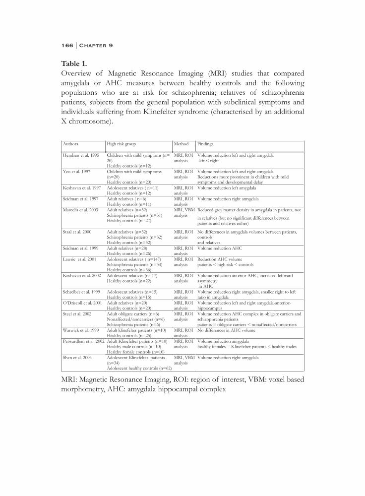
Table 1.Overview of Magnetic Resonance Imaging (MRI) studies that comparedamygdala or AHC measures between healthy controls and the followingpopulations who are at risk for schizophrenia; relatives of schizophreniapatients, subjects from the general population with subclinical symptoms andindividuals suffering from Klinefelter syndrome (characterised by an additionalX chromosome).
MRI: Magnetic Resonance Imaging, ROI: region of interest, VBM: voxel basedmorphometry, AHC: amygdala hippocampal complex
166 | Chapter 9
Subclinical signs in the general populationOne approach to investigate vulnerability to schizophrenia is to identify subtle,subclinical signs in healthy individuals that parallel the symptoms of the illness.These signs, often manifested as various schizotypal personality traits, have beensuggested to be continuously distributed in the general population (Aleman etal., 2001; Claridge et al., 1996; Jessimer et al., 1997; Johns et al., 2001). Examplesof schizotypal traits are referential and magical thinking, suspiciousness andsocial isolation.
Subclinical signs may in some individuals progress to symptoms ofschizophrenia. In fact, the abovementioned schizotypal traits partially predictschizophrenia at long term follow-up in subjects diagnosed with SchizotypalPersonality Disorder (DSM III) (Fenton et al., 1989). Also, schizotypal traits inyoung relatives of patients predict progression to schizophrenia in the followingdimensions: social withdrawal, psychotic symptoms and socio-emotionaldysfunction (Miller et al., 2002).
Subclinical signs in the general population: abnormalities of theamygdalaSome of the structural neuroimaging studies have relied on clinical criteria whendefining vulnerability for the disease. Hendren and colleagues (Hendren et al.,1995) have examined amygdala volumes of twelve children between 8 and 12years, who displayed mild symptoms of (early-onset) schizophrenia and/orschizotypal personality disorder. When compared to twelve age-, sex- and socio-economic status- paired matched controls, a significant reduction in amygdalavolume was present in the symptomatic group. Measurements were adjusted forthe effects of age and total brain volume. Group differences were moreprominent for the left than the right amygdala, a finding that has also beenreported in studies with chronic schizophrenia patients (Rossi et al., 1994;Shenton et al., 2001).
Yeo and al. (Yeo et al., 1997) have been able to partially replicate this resultin a group of twenty children, aged 9 to 12, characterised by schizophrenia-likesymptoms, but who did not meet all the diagnostic criteria for schizophrenia.Again, compared to paired matched controls, a reduction in the amygdalavolume was found using MRI, but no differences between the right and leftamygdala were present. When the symptomatic group was split up in childrenwith and without a history of developmental delay in motor or languagefunction, a comparison of volumetric measurements showed a significantlysmaller amygdala volume in the group who did have a history of developmental
Amygdala abnormalities in the schizoprenia spectrum | 167
delay. All measurements were controlled for age and total brain volume. As theauthors noted, these results point to a possible role of the amygdala in theneural development of schizophrenia.
Thus, already in pre-adolescence schizophrenia-like symptoms may beassociated with abnormalities in the development of the amygdala. Functions ofthe amygdala may become more relevant with age, since it is important forrecognizing and learning the emotional, and possibly survival-related, meaningof stimuli in the environment and producing appropriate behavioural responses.Indeed, findings from structural MRI studies suggest that there is an age- andgender related increase in amygdala volume during childhood and adolescence(Giedd et al., 1996; Sowell et al., 1998). We speculate that the behavioural effectsof dysmaturations of the amygdala may become apparent as high risk childrenapproach adolescence, a period in which maturational processes are dynamicand strengthening of neural connections between the amygdala and for examplefrontal areas take place ((Benes, 1998; Walker et al., 2002). Although the degreeto which these dysmaturations are a direct expression of a geneticpredisposition to schizophrenia remains unclear, the parallels in symptomsbetween these children and schizophrenia patients indicate that they also mayshare genetic pathology.
RelativesSharing a substantial amount of genes, relatives of patients suffering fromschizophrenia are at increased risk for the disease, with the risk increasing withthe degree of kinship (Gottesman, 1991). Schizophrenia as well as schizotypaltraits are more often identified in relatives of patients suffering fromschizophrenia when compared to the general population (Kendler et al., 1995;Kety et al., 1994; Kremen et al., 1998; Vollema et al., 2002; Yaralian et al., 2000).When rates of schizotypal personality traits are substantially increased, criteriafor Schizotypal Personality Disorder (SPD) may be met. Whereas the prevalenceof this disorder is 3% in the general population, 10 to 15% of the relativessuffer from SPD.
But even in the absence of schizophrenia-like symptoms or schizotypalpersonality traits, biological relatives of schizophrenia patients are at substantialrisk for a range of biobehavioral and neurobiological abnormalities that arerelated to a genetic vulnerability (McDonald et al., 2002; Seidman et al., 1997;Seidman et al., 1999; Staal et al., 2000). In addition, healthy relatives of patientsshow abnormalities at a neuropsychological level that parallel the deficits seenin schizophrenia patients, although they are more subtle (Sitskoorn et al., 2003).
168 | Chapter 9
All in all, relatives of schizophrenia patients provide an opportunity for studyingamygdala abnormalities as a neurobiological marker of the disease.
Relatives: abnormalities of the amygdala The most widely used approach to identify liability to schizophrenia is toestablish the genetic risk of subjects based on a family tree describing affectedand unaffected family members. In some family trees unaffected familymembers have been further specified in obligate carriers, subjects havingtransmitted schizophrenia while remaining healthy themselves, and non-carrierswho have not passed any pathological genes on to their offspring. Besidesadolescents with a familial risk for schizophrenia, adult relatives of patients havealso been subject of investigation. Several studies have been able to showamygdala- or amygdala-hippocampal-complex (AHC) abnormalities inindividuals at familial risk.
Amygdala measurementsAn MRI study conducted by Keshavan et al. (Keshavan et al., 1997) includedeleven high-risk adolescents, aged 12 to 18, who had a parent suffering fromschizophrenia. Neither the high-risk adolescents, nor the healthy controls had ahistory of psychotic disorders. After correction for whole brain volumes,offspring of patients were characterised by reduced left amygdala volume.
One of the first studies that systematically examined brain volumes in adultrelatives of patients compared 6 female relatives with 11 controls (Seidman etal., 1997). None of the subjects had suffered from psychosis or schizophreniaspectrum disorders. Among a range of brain volume abnormalities, a meanreduction of the right amygdala was present in the relatives group. Althoughthis reduction was present in spite of the small sample size, no correction formultiple comparisons had been included. When the sample size was extendedto 28 relatives, both male and female, and 26 controls, the volume reduction ofthe amygdala was not statistically significant. However, the amygdala-hippocampal complex as a whole appeared to be significantly reduced inrelatives (Seidman et al., 1999).
The apparent inconsistencies in their results might be related toheterogeneity in their sample. As proposed by the authors, schizophrenia mayarise from a single pool of environmental and genetic factors in which smalleffects of the individual factors can add up to result in a vulnerability for thedisease. As a consequence, relatives may be characterised by different, probablypartially overlapping, sets of vulnerability markers. One way to deal with this
Amygdala abnormalities in the schizoprenia spectrum | 169
heterogeneity is to select large sample sizes. Their sample of 28 relatives wasfurther extended to 45 and factor analysis, a psychometric approach, wasemployed. Although no main effects were present for the left and rightamygdala combined, the left amygdala, among other brain regions, contributedto the only factor that discriminated relatives from controls (Faraone et al.,2003). This reduction in left amygdala volume is consistent with findings fromKeshavan as described above (Keshavan et al., 1997).
There are imaging studies, however, in which no reduction of the amygdalain relatives has been shown. A recent MRI study used computationalmorphometry to compare grey matter densities on a clustered-voxel basis in 31schizophrenia patients, 32 relatives and 27 healthy controls (Marcelis et al.,2003). Grey matter density deficits in the amygdala were present in patients, butnot in relatives, when compared to controls. However, grey matter density in theamygdala did not significantly differ between relatives and patients, making itdifficult to interpret the findings in this study regarding amygdala abnormalitiesin relatives. In study by Staal et al. (Staal et al., 2000), 16 schizophrenia patientswere compared to their healthy siblings and 16 healthy control subjects.Amygdala volume did not differ between the healthy siblings and controls.However, compared to healthy controls, schizophrenia patients did not show areduction of the amygdala, which makes it difficult to draw conclusions fromthis study regarding amygdala abnormalities as a vulnerability marker in healthysiblings.
Amygdala-hippocampal complex (AHC) measurementsBecause the amygdala is notoriously difficult to measure reliably (Brierley et al.,2002), several studies try to solve the problem by measuring the amygdala andhippocampus as a whole; the amygdala-hippocampal complex (AHC). Somestudies divide the AHC in a posterior and anterior portion, with the latterportion representing the amygdala.
As part of the Edinburgh High Risk Study (EHRS), a series of MRI studiesby Lawrie and colleagues have explored abnormalities of the amygdala-hippocampal complex as an expression of a genetic liability to schizophrenia.First, they have compared AHC volumes of 100 adolescents characterised bytwo or more affected relatives, with 20 first episode schizophrenia patients and30 healthy controls (Lawrie et al., 1999). AHC volume in the high-risk groupappeared to be midway between that of healthy controls and first episodepatients, with the latter group showing the smallest mean AHC volume. Whenthe high risk sample was extended to 147 subjects, and compared to 34 first
170 | Chapter 9
episode patients and 36 healthy controls, the same pattern of results appeared(Lawrie et al., 2001). Although AHC volumes in the high risk group were notrelated to the number of affected relatives which may reflect a measure ofgenetic liability, this gradient in AHC volume provides evidence for the idea thatAHC abnormalities in schizophrenia are largely genetically mediated. Thefindings were only partially replicated when using automated Voxel BasedMorphometry (VBM), allowing comparisons of grey matter densities, instead ofvolumetric Region of Interest (ROI) measurements. Although patients didshow a reduction in grey matter density in the amygdala when compared tocontrols, differences in amygdala volume between healthy controls and high risksubjects disappeared when this VBM analysis was applied to the dataset of theEdinburgh High Risk Study (Job et al., 2003).
In the Edinburgh High Risk Study, abnormalities in AHC complex werenot related to the presence of psychotic symptoms in the high risk group. Asubgroup of 28% of the high risk adolescents characterized by psychoticsymptoms did not differ in mean AHC volume from the high risk adolescentswithout psychotic symptoms. This finding underlines that AHC abnormalitiesmay reflect an underlying genetic vulnerability to schizophrenia, and may notpredict the presence or development of psychotic symptoms which in turn maybe triggered by environmental factors and/or other neurodevelopmentalprocesses.
This hypothesis is supported by findings in a two-year follow-up study(Lawrie et al., 2003), which was included allowing investigation of the relationbetween symptom development and brain volume changes. 20 Healthy subjectsand 66 high-risk adolescents were re-scanned and symptoms were re-evaluated.No significant time by group interaction was present for the AHC, meaning thatchange in AHC volume over time did not differ between high risk adolescentsand controls. When comparing high-risk subjects displaying symptoms atfollow-up with high-risk adolescents without symptoms, no significant time bygroup interaction was found for the AHC. This finding has been replicated in alongitudinal imaging study focused at the amygdala as a separate region.Adolescents at high-risk for development of psychosis showed no differencesin gray-matter volume of the amygdala between those who subsequentlydeveloped a psychotic illness (n=23) and those who did not (n=52) (Pantelis etal., 2003).
Other investigators have also performed measurements of AHC volumesin adolescent relatives of schizophrenia patients. Schreiber et al., for example,focused at brain abnormalities in offspring of schizophrenia patients. 15 High-
Amygdala abnormalities in the schizoprenia spectrum | 171
risk adolescents and 15 controls were between 11 and 19 years of age, andindividually matched for age, sex, education and social background. A meanvolume reduction of the right AHC was present in high-risk adolescents. Inaddition, they were characterized by a smaller right-to-left ratio of the AHCwhen compared to controls (Schreiber et al., 1999).
Keshavan et al. included seventeen high risk adolescents (offspring) in theirMRI study and were able to demonstrate volume reductions of about 25% inthe left anterior amygdala-hippocampal complex (AHC) (Keshavan et al., 2002).This anterior part of the AHC largely comprises the amygdala. Significantreductions in the right anterior AHC were also present although not asprominent as in the left hemisphere, indicating an increased leftward asymmetryof the anterior AHC.
Another study in which the AHC was split up in an anterior and posteriorpart focused at adult individuals at familial risk for schizophrenia. O’Driscoll etal. (O'Driscoll et al., 2001) have compared AHC volumes of 20 first-degreerelatives with 20 healthy controls using MRI. All subjects were between 18 and50 years and were similar on demographic variables. None of the subjects hada history of any DSM axis I disorder or met the criteria for SchizotypalPersonality Disorder. The area of the amygdala-hippocampal complex wasdivided in two regions of interest; the amygdala-anterior hippocampus and theposterior hippocampus. Relatives showed a volume reduction of the left- as wellas right amygdala-anterior hippocampus, which comprises the amygdala, thepres hippocampus and the anterior portion of the subiculum.
Although the risk of relatives for developing schizophrenia is increased,only a subgroup will actually carry pathological genes. More evidence for agenetic loading of amygdala abnormalities in schizophrenia comes fromexperimental designs with multiple affected families in which affected subjects,non-carriers (no affected offspring) and obligate carriers (affected offspring) areidentified. In addition to an increase in power, such a design allows a clearseparation of the contribution of pathological genes from the effects of theillness itself.
In the Edinburgh High Risk Study, sib-ships in high risk families wereidentified including one affected, one non-affected/non-carrier and one non-affected/carrier (obligate carrier) familymember (Steel et al., 2002). AHCvolumes (right and left combined) in subjects with schizophrenia and obligatecarriers were significantly smaller when compared to non-affected/non-carriers,which implies a genetic contribution in AHC pathology in schizophrenia. Thus,although AHC abnormalities were not related to a genetic liability index as
172 | Chapter 9
measured by the number of affected relatives and whether these are first-degreeor second degree relatives, carriers of pathological genes do show reductions ofthe AHC. Because cortical changes in relatives were not found, the authorsconcluded that in a framework of complex genetic mechanisms it could bepossible that some vulnerability genes, in this case causing AHC abnormalities,are inherited by obligate carriers, whereas other vulnerability genes, responsiblefor cortical changes for example, are not.
In summary, both adolescents and adult relatives of schizophrenia patientsshow abnormalities of the amygdala, either measured as the amygdala regionexclusively or as part of the amygdala-hippocampal-complex. Since theserelatives share a substantial amount of genes with an affected family member, ashared genetic pathology may account for these amygdala abnormalities. Thefinding that AHC volumes in obligate carriers of pathological genes are similarto AHC volumes in patients, but different from non-affected/non-carrierssupports this hypothesis. AHC volumes in relatives seem not to be related to thepresence of symptoms. Because AHC abnormalities are thought to reflect agenetic liability for schizophrenia and not the actual expression of the illness(Steel et al., 2002), this is not unexpected.
Klinefelter syndromeOne interesting neurodevelopmental disorder that has been associated with anincreased risk for schizophrenia is Klinefelter Syndrome, a genetic disordercharacterised by sex chromosome abnormalities. Klinefelter patients have anadditional X chromosome, leading to the XXY karyotype. Although the primaryfocus in clinical studies on Klinefelter Syndrome has been on reproductivedysfunction of these patients, there is an awareness of behavioral and cognitiveabnormalities, including socio-emotional disturbances (Boone et al., 2001;Geschwind et al., 2000; Simpson et al., 2003; Swaab et al., submitted). Theimportance of investigation into the cognitive and behavioral phenotypicalmanifestations of Klinefelter syndrome as a means of understanding apredisposition to schizophrenia, is shown by epidemiological studies reportingan increased incidence of XXY karyotypes in schizophrenia. The prevalence ofKlinefelter Syndrome in the general population is 0.1-0.2% (Bojesen et al.,2003). The prevalence of Klinefelter Syndrome in the schizophrenia populationis 1.6 %, which is several times higher (DeLisi et al., 1994; Kunugi et al., 1999).In turn, early studies have indicated an increased risk for schizophrenia andpsychotic illnesses among Klinefelter patients (Lishman, 1998). A review ofmental hospital surveys pointed to a threefold increase in Klinefelter patients
Amygdala abnormalities in the schizoprenia spectrum | 173
compared to the general population, which was mainly due to ‘psychoticillnesses of a schizophrenic nature’ (Forssman, 1970). Another study showedthat 7% of the Klinefelter patients in the psychiatric literature had psychoseswith paranoid delusions and 6% suffered from schizophrenia (Nielsen et al.,1969). In addition, Klinefelter syndrome has recently been associated with highlevels of schizophrenia spectrum pathology (van Rijn et al., submitted).Compared to 26 healthy males, a group of 26 Klinefelter patients werecharacterised by elevated levels of schizophrenia symptoms as well asschizotypal traits. Effect sizes, the difference between the groups expressed instandard deviations, in this study paralleled those reported for patients sufferingfrom schizophrenia. From the 26 Klinefelter males, 7 individuals (27%) metdiagnostic criteria for a psychotic disorder.
Studying Klinefelter patients present an opportunity to investigate whetheramygdala abnormalities can be associated with a predisposition toschizophrenia. An additional advantage over studying relatives is the knowledgeof the precise genetic aetiology of Klinefelter syndrome in contrast to what isknown of the genetic pathology in schizophrenia.
Klinefelter syndrome: abnormalities of the amygdalaAlthough AHC abnormalities were not found in an explorative MRI study withKlinefelter patients (Warwick et al., 1999), specific amygdala reductions werepresent in an MRI study focusing at temporal lobe structures in Klinefelterpatients (Patwardhan et al., 2002). 10 XXY males were recruited from a cohortnewborns screened for sex chromosome abnormalities and compared to 10individually matched healthy males and healthy 10 females. Approximately halfof the patients had been exposed to testosterone supplements. Amygdalavolumes of Klinefelter patients were comparable to those of healthy females.Interestingly, Klinefelter patients suffering from schizophrenia show severalparallels with females suffering from schizophrenia; similar ages of onset thatdiffer from male schizophrenia patients and a predominance of positivesymptoms over negative symptoms (Pinabel et al., 1997). In an important study,using voxel-based morphometry, Shan et al. observed pronounced volumereductions of the right amygdala in 34 subjects with Klinefelter syndrome(mean age 12.6 years) compared to 62 healthy matched controls (Shen et al.,2004). Compared to previous findings documented in the literature, findingsfrom this study provide a better spatial localization of the affected regions(other affected regions are the insula, cingulate, hippocampus, temporal gyri andoccipital gyri).
174 | Chapter 9
X-linked genes appear to be involved in the functional as well as structuraldevelopment of the amygdala (Good et al., 2003). Several studies have shownamygdala volume to be inversely correlated with the number of Xchromosomes (Goldstein et al., 2001; Good et al., 2003). For example, males(46,XY) have larger amygdalae than females (46,XX). Patients suffering fromTurner syndrome (45,X), who are characterised by a partial or complete deletionof the X chromosome, have even larger amygdala volumes when compared tohealthy males. On a behavioural level, these patients are impaired in facial affectrecognition while showing normal configural processing (Lawrence et al., 2003).This deficit in emotion recognition appears to be most pronounced for fear, afinding that parallels data from studies with schizophrenia patients (Kohler etal., 2003).
Although speculative, the idea that amygdala abnormalities in schizophreniamay somehow be related to X chromosomes deserves further investigation.Especially in the light of sex-differences in the onset and course of the disease.
Conclusion and discussionThe present review explored whether an increased risk for schizophrenia isassociated with structural abnormalities of the amygdala. Structural neuro-imaging studies with three distinct populations at risk for schizophrenia haveprovided indications that reductions in amygdala volume indeed may beassociated with an increased risk for schizophrenia. First, individuals from thegeneral population with subclinical signs of schizophrenia show reductions inamygdala volume. Second, volume reductions of the amygdala and amygdala-hippocampal-complex (AHC) have been found in relatives of schizophreniapatients. Third, Klinefelter patients, characterised by an XXY chromosomalpattern, also show reduced amygdala volumes.
The fact that amygdala abnormalities, which have consistently beenreported in patients with schizophrenia (Breier et al., 1992; Bryant et al., 1999;Gur et al., 2000; Hulshoff-Pol et al., 2001; Lawrie et al., 2003; Wright et al.,2000), are also observed in high risk populations, is of considerable importance.Since volume reductions of the amygdala seem to be present already in pre-adolescents with subclinical signs of the illness, abnormalities of the amygdalain schizophrenia might have a developmental origin. Indications that theseamygdala abnormalities are an expression of a genetic predisposition toschizophrenia are primarily derived from studies with healthy relatives ofpatients, who besides sharing genes also share amygdala or AHC abnormalities.Consequently, abnormalities of the amygdala or AHC might belong to the
Amygdala abnormalities in the schizoprenia spectrum | 175
endophenotype of schizophrenia, a concept which is introduced to refer tobrain structures or cognitive functions that mediate between the molecular,genetic level and the molar, clinical level of the disease (Gottesman et al., 2003).This would imply that antipsychotic medication, hospitalisation and neurotoxiceffects of psychosis may not fully account for volume reductions of theamygdala in schizophrenia, since reductions have been found in subjects with anincreased risk for the disease, but who did not have a history ofpharmacological treatment, psychosis or hospitalisation. This is supported by animaging study with medication-naive schizophrenia patients, who show volumereductions of the amygdala (Joyal et al., 2003). In the same study, neitherduration of illness, nor symptom severity correlated with amygdala volume.
Amygdala abnormalities may be one of many endophenotypes inschizophrenia, each one being an expression of a particular genetic pathology(Gottesman et al., 2003). Although most attempts to link the generalschizophrenia phenotype to aberrations of the X chromosome have producedcontradictory results (DeLisi, 1997; DeLisi et al., 2000), recent genome screenshave pointed to a possible locus on the X chromosome (Paterson, 1999).However, in these studies the magnitude of the effects regarding the Xchromosome is smaller than the most positive loci. We argue that amygdalaabnormalities, a specific phenotype, in schizophrenia might be related to X-linked genetic pathology. Support for this idea comes from studies withindividuals suffering from X chromosome abnormalities. For example,individuals with Klinefelter syndrome (47,XXY), who have almost anapproximately ten-fold increased risk for schizophrenia when compared to thehealthy population, show reductions in amygdala volume. Furthermore, supportfor the hypothesis that amygdala abnormalities may be directed by genes on theX–chromosome, comes from a study with subjects with X chromosomeabnormalities, showing that a dosage sensitive genetic locus on the Xchromosome appears to play a key role in the functional and structuraldevelopment of the amygdala (Good et al., 2003). In addition, several lines ofresearch have indicated that the number of X chromosomes is inverselycorrelated with amygdala volume (Goldstein et al., 2001; Good et al., 2003). Forexample, males (46,XY) have larger amygdalae than females (46,XX). Males andfemales do not only differ at the level of X chromosomes and amygdalavolumes, at a clinical level gender differences are also present. A recent meta-analysis of 38 studies that reported sex-specific incidences of schizophrenia hasprovided evidence for a sex difference in the risk for developing schizophrenia(Aleman et al., 2003; Phillips et al., 2004). The reported risk ratio for males to
176 | Chapter 9
develop schizophrenia relative to females was 1.42 (95% CI 1.30–1.56). Inaddition, a meta-analysis on sex differences in deficit schizophrenia (whichinvolves larger emotional deficits) even reported a ratio of 1.7 (Roy et al., 2001).Males not only suffer more often from schizophrenia when compared tofemales, men also appear to have an earlier age of onset and a more severecourse of the illness (Gur et al., 1996). Interestingly, Klinefelter patientssuffering from schizophrenia show several parallels with women suffering fromschizophrenia; similar ages of onset, later than male schizophrenia patients, anda predominance of positive symptoms over negative symptoms (Pinabel et al.,1997).
X chromosome abnormalities may co-direct amygdala volumes partlythrough abnormal testosterone levels, because sex of the gonads that producegonadal hormones is determined by genes on the sex chromosomes (Arnold etal., 2004). However, testosterone levels by itself seem not enough to explain theamygdala abnormalities in individuals with sex chromosome aneuploidies. Forexample, although females with Turner syndrome, who are missing one Xchromosome, are also characterised by a reduction in testosterone levels(Hojbjerg Gravholt et al., 1999), these patients show an increase in amygdalavolume rather than a reduction (Kesler et al., 2004). Differences in amygdalavolume between Turner and Klinefelter patients appear to be stronger relatedto the number of X chromosomes, which differentiates the two groups, than totestosterone levels, which are reduced in both syndromes. Alternatively,abnormal testosterone levels may result from X chromosome linked amygdalaabnormalities, since it has been shown that the amygdala is involved in theproduction of testosterone (Banczerowski et al., 2003). In conclusion, althoughthe amygdala, testosterone levels and X chromosomes appear to strongly inter-related, the exact nature of the relationship still remains unclear.
We propose that in schizophrenia, X-linked genetic pathology, eitherdirectly or indirectly via testosterone, might result in a endophenotypecharacterised by amygdala abnormalities. Amygdala abnormalities as anendophenotype of putatively X-linked pathology may not be specific forschizophrenia. This endophenotype may also be present in other psychiatricdisorders, for example autism, which has also been associated with structuralabnormalities of the amygdala (Brambilla et al., 2003; Palmen et al., 2004).Interestingly, in autism males (46, XY) are affected four-fold more often thanfemales (46, XX). Moreover, females suffering from Turner syndrome (45,X),who are missing one X chromosome, have a 200-fold increased risk of autism(Creswell et al., 1999). These findings support the idea that amygdala
Amygdala abnormalities in the schizoprenia spectrum | 177
abnormalities in autism may be linked to the X chromosome. If we in turnfocus at psychiatric disorders characterised by X-linked genetic pathology,abnormalities of the amygdala have been consistently reported. Not only in thepopulation of Klinefelter patients who are at increased risk for schizophrenia,also in Turner syndrome (complete or partial deletion of X chromosome) andFragile X syndrome (mutations on X chromosome) abnormalities of theamygdala have been observed (Good et al., 2003; Hessl et al., 2004; Kesler et al.,2004). In contrast to Klinefelter Syndrome, these disorders have been associatedwith an increase in amygdala volume (Good et al., 2003; Hessl et al., 2004).Interestingly, it has been reported that Turner syndrome occurs approximatelythree-fold more frequently in schizophrenic females when compared to thegeneral female population (Prior et al., 2000). Thus, amygdala abnormalities asan endophenotype may be present in several psychiatric disorders. This reviewhas provided support that schizophrenia is one of these disorders. In addition,we propose that individuals with Klinefelter Syndrome, associated with Xchromosome abnormalities, share this endophenotype with schizophrenia.
A clinically relevant question that arises from these speculations would be:How may amygdala abnormalities at an endophenotypical level extend to aphenotypical, behavioural level? Since the amygdala is strongly involved inemotion processing and appears to be part of a neural network underlyingsocial cognition, one would expect emotional-and social difficulties to bepresent in the populations that have been described here. A review of theliterature tentatively supports this idea. As observed in schizophrenia patients,relatives of patients show deficits in facial affect recognition, although they aremore subtle (Toomey et al., 1999). Amygdala abnormalities as anendophenotype may result in behavioural deficits which, again, need notnecessarily be specific for schizophrenia. In Autism and Turner Syndrome,deficits in facial affect recognition and impairments in social cognition havebeen reported (Baron-Cohen et al., 2000; Good et al., 2003; Klin et al., 2002;Lawrence et al., 2003). Interestingly, parallel to schizophrenia patients andamygdala damaged patients, individuals with autism and Turner syndrome showselective impairments in the recognition of facial expressions of fear (Howardet al., 2000; Lawrence et al., 2003).
Regarding high risk populations, there is a shortage of behavioural studiesfocused at emotion processing. Recently, our department has shown a range ofemotional processing abnormalities in a non-clinical psychosis-prone sample(van 't Wout et al., 2004). In this population, unusual perceptual experiences, apositive schizotypal trait, correlated with more problems in identifying
178 | Chapter 9
emotions. Verbalizing and analysing emotions, as well as emotional arousability,correlated with constricted affect, a negative schizotypal trait.Future research should investigate the functional consequences of amygdalaabnormalities in genetically predisposed subjects using functional MRI andbehavioural measurements. Specific effects of normal brain maturation, illnessand medication on the amygdala should be examined in these subjects inlongitudinal designs. Twin studies or designs in which multiplex families areincluded and obligate carriers are identified will provide unique opportunitiesfor disentangling genetic from environmental effects on the development ofschizophrenia.
It should be noted that several limitations have become apparent in thepresent review of studies. A number of limitations associated with structuralmeasurement of the amygdala may contribute to the heterogeneity in findingsin MRI-studies focused at high-risk populations. The amygdala is located inproximity of the nasal cavity, which may lead to an increase of MRIsusceptibility artefacts. In addition, low scan resolution and limited number ofslices through the amygdala make it more difficult to reliably estimate its volumeand separate it from the hippocampus. Future studies should follow theguidelines given by Brierley et al. (Brierley et al., 2002; David et al., 2002) inorder to enhance methodological rigor. In addition, in several MRI studiesreported in this paper, the amygdala is measured as part of a complex (AHC)and not as a separate region of interest, which is a major limitation. However,although the amygdala and the hippocampus are anatomically dissociable, theAHC may be perceived as a unitary complex at some level, since they sustainfunctions that pertain to emotion processing (Phillips et al., 2003). Thehippocampus has direct connections with the amygdala and plays a key role incontext modulation of emotional behaviour (Davidson et al., 2000). Finally, therange of MRI studies focused at populations at high risk for developingschizophrenia is somewhat limited and not all studies find amygdalaabnormalities. However, the majority of studies are indicative of a role of theamygdala or AHC abnormalities.
In summary, this review focused at structural abnormalities of the amygdalain various populations a high risk for developing schizophrenia. Studies withindividuals from the general population showing subclinical signs of the illnessand studies with relatives of schizophrenia patients indicate that amygdalaabnormalities may form part of the genetic predisposition to schizophrenia.Amygdala abnormalities may be one of the endophenotypes in schizophreniaand might be X chromosome linked, as suggested by converging evidence from
Amygdala abnormalities in the schizoprenia spectrum | 179

individuals with X chromosome abnormalities. We therefore suggest thatamygdala abnormalities might be an X-linked endophenotype in psychiatricdisorders, especially in schizophrenia and populations at high risk forschizophrenia, and may account for emotional en social processing deficits thathave been described in these populations.
180 | Chapter 9
ReferencesAdolphs, R. (2002). Trust in the brain. Nature Neuroscience, 5(3), 192-193.Adolphs, R., Tranel, D., Damasio, H., & Damasio, A. (1994). Impaired
recognition of emotion in facial expressions following bilateral damageto the human amygdala. Nature, 372(6507), 669-672.
Adolphs, R., Tranel, D., Hamann, S., Young, A. W., Calder, A. J., Phelps, E. A.,et al. (1999). Recognition of facial emotion in nine individuals withbilateral amygdala damage. Neuropsychologia, 37(10), 1111-1117.
Aggleton, J. P. (2000). The amygdala. New York: Oxford Press.Aggleton, J. P., & Young, A. W. (2000). The enigma of the amygdala: On its
contibution to human emotion. Cognitive neuroscience of emotion, R.D. Lane, L. Nadel, G. L. Ahernet al, 106-128.
Aleman, A., Hijman, R., de_Haan, E. H., & Kahn, R. S. (1999). Memory impairment in schizophrenia: A meta-analysis. The American Journal ofPsychiatry, 156(9), 1358-1366.
Aleman, A., Kahn, R. S., & Selten, J. P. (2003). Sex differences in the risk ofschizophrenia: Evidence from meta-analysis. Archives of GeneralPsychiatry, 60(6), 565-571.
Aleman, A., Nieuwenstein, M. R., Bocker, K. B. E., & De Haan, E. H. F.(2001). Multi-dimensionality of hallucinatory predisposition: Factorstructure of the launay-slade hallucination scale in a normal sample.Personality and Individual Differences, 30(2), 287-292.
Arnold, A. P., & Burgoyne, P. S. (2004). Are xx and xy brain cells intrinsically different? Trends in Endocrinology and Metabolism, 15(1), 6-11.
Baare, W. F., Hulshoff_Pol, H. E., Boomsma, D. I., Posthuma, D., de_Geus, E.J., Schnack, H. G., et al. (2001). Quantitative genetic modeling ofvariation in human brain morphology. Cerebral Cortex (New York, N.Y.:1991), 11(9), 816-824.
Banczerowski, P., Csaba, Z., Csernus, V., & Gerendai, I. (2003). Lesion of the amygdala on the right and left side suppresses testosterone secretion butonly left-sided intervention decreases serum luteinizing hormone level.Journal of Endocrinological Investigation, 26(5), 429-434.
Baron-Cohen, S., Ring, H. A., Bullmore, E. T., Wheelwright, S., Ashwin, C., & Williams, S. C. (2000). The amygdala theory of autism. Neuroscienceand Biobehavioral Reviews, 24(3), 355-364.
Bartley, A. J., Jones, D. W., & Weinberger, D. R. (1997). Genetic variability ofhuman brain size and cortical gyral patterns. Brain; a Journal ofNeurology, 120 (Pt 2), 257-269.
Amygdala abnormalities in the schizoprenia spectrum | 181
Baxter, M. G., & Murray, E. A. (2002). The amygdala and reward., 3(7), 563-573.
Benes, F. M. (1998). Brain development, vii. Human brain growth spans decades. The American Journal of Psychiatry, 155(11), 1489.
Bojesen, A., Juul, S., & Gravholt, C. H. (2003). Prenatal and postnatal prevalence of klinefelter syndrome: A national registry study. TheJournal of Clinical Endocrinology and Metabolism, 88(2), 622-626.
Boone, K. B., Swerdloff, R. S., Miller, B. L., Geschwind, D. H., Razani, J., Lee,A., et al. (2001). Neuropsychological profiles of adults with klinefeltersyndrome. Journal of the International Neuropsychological Society: Jins,7(4), 446-456.
Bouwmeester, H., Wolterink, G., & van Ree, J. M. (2002). Neonatal development of projections from the basolateral amygdala to prefrontal,striatal, and thalamic structures in the rat. The Journal of ComparativeNeurology, 442(3), 239-249.
Brambilla, P., Hardan, A., di Nemi, S. U., Perez, J., Soares, J. C., & Barale, F.(2003). Brain anatomy and development in autism: Review of structuralmri studies. Brain Research Bulletin, 61(6), 557-569.
Breier, A., Buchanan, R. W., Elkashef, A., Munson, R. C., Kirkpatrick, B., & Gellad, F. (1992). Brain morphology and schizophrenia. A magneticresonance imaging study of limbic, prefrontal cortex, and caudatestructures. Archives of General Psychiatry, 49(12), 921-926.
Brierley, B., Shaw, P., & David, A. S. (2002). The human amygdala: A systematic review and meta-analysis of volumetric magnetic resonanceimaging. Brain Research. Brain Research Reviews, 39(1), 84-105.
Bryant, N. L., Buchanan, R. W., Vladar, K., Breier, A., & Rothman, M. (1999).Gender differences in temporal lobe structures of patients withschizophrenia: A volumetric mri study. The American Journal ofPsychiatry, 156(4), 603-609.
Calder, A. J., Young, A. W., Rowland, D., Perrett, D. I., Hodges, J. R., & Etcoff, N. L. (1996). Facial emotion recognition after bilateral amygdaladamage: Differentially severe impairment of fear. CognitiveNeuropsychology, 13(5), 699-745.
Carmelli, D., DeCarli, C., Swan, G. E., Jack, L. M., Reed, T., Wolf, P. A., et al.(1998). Evidence for genetic variance in white matter hyperintensityvolume in normal elderly male twins. Stroke; a Journal of CerebralCirculation, 29(6), 1177-1181.
182 | Chapter 9
Claridge, G., McCreery, C., Mason, O., Bentall, R., Boyle, G., & Slade, P.(1996). The factor structure of 'schizotypal' traits: A large replicationstudy. British Journal of Clinical Psychology, 35, 103-115.
Creswell, C. S., & Skuse, D. H. (1999). Autism in association with turner syndrome: Genetic implications for male vulnerability to pervasivedevelopmental disorders. Neurocase, 5(6), 511-518.
David, A. S., Brierley, B., Shaw, P., Chance, S. A., Esiri, M. M., & Crow, T. J.(2002). Measuring amygdala volume * authors' reply. British Journal ofPsychiatry, 181(3), 255-256.
Davidson, R. J., Jackson, D. C., & Kalin, N. H. (2000). Emotion, plasticity,context, and regulation: Perspectives from affective neuroscience.Psychological Bulletin, 126(6), 890-909.
DeLisi, L. E. (1997). The genetics of schizophrenia: Past, present, and future concepts. Schizophrenia Research, 28(2-3), 163-175.
DeLisi, L. E., Friedrich, U., Wahlstrom, J., Boccio-Smith, A., Forsman, A.,Eklund, K., et al. (1994). Schizophrenia and sex chromosome anomalies.Schizophrenia Bulletin, 20(3), 495-505.
DeLisi, L. E., Shaw, S., Sherrington, R., Nanthakumar, B., Shields, G., Smith,A. B., et al. (2000). Failure to establish linkage on the x chromosome in301 families with schizophrenia or schizoaffective disorder. AmericanJournal of Medical Genetics, 96(3), 335-341.
Edwards, J., Jackson, H. J., & Pattison, P. E. (2002). Emotion recognition via facial expression and affective prosody in schizophrenia: Amethodological review. Clinical Psychology Review, 22(6), 789-832.
Exner, C., Boucsein, K., Degner, D., Irle, E., & Weniger, G. (In Press).Impaired emotional learning and reduced amygdala size inschizophrenia: A 3-month follow-up. Schizophrenia Research, In Press,Corrected Proof.
Faraone, S. V., Kremen, W. S., Lyons, M. J., Pepple, J. R., Seidman, L. J., & Tsuang, M. T. (1995). Diagnostic accuracy and linkage analysis: Howuseful are schizophrenia spectrum phenotypes? The American Journalof Psychiatry, 152(9), 1286-1290.
Faraone, S. V., Seidman, L. J., Kremen, W. S., Kennedy, D., Makris, N.,Caviness, V. S., et al. (2003). Structural brain abnormalities amongrelatives of patients with schizophrenia: Implications for linkage studies.Schizophrenia Research, 60(2-3), 125-140.
Amygdala abnormalities in the schizoprenia spectrum | 183
Fenton, W. S., & McGlashan, T. H. (1989). Risk of schizophrenia in character disordered patients. The American Journal of Psychiatry, 146(10), 1280-1284.
Forssman, H. (1970). The mental implications of sex chromosome aberrations. British Journal of Psychiatry, 117(539), 353-363.
Geschwind, D. H., Boone, K. B., Miller, B. L., & Swerdloff, R. S. (2000).Neurobehavioral phenotype of klinefelter syndrome., 6(2), 107-116.
Giedd, J. N., Vaituzis, A. C., Hamburger, S. D., Lange, N., Rajapakse, J. C.,Kaysen, D., et al. (1996). Quantitative mri of the temporal lobe,amygdala, and hippocampus in normal human development: Ages 4-18years. The Journal of Comparative Neurology, 366(2), 223-230.
Goldstein, J. M., Seidman, L. J., Horton, N. J., Makris, N., Kennedy, D. N.,Caviness, V. S., et al. (2001). Normal sexual dimorphism of the adulthuman brain assessed by in vivo magnetic resonance imaging. CerebralCortex (New York, N.Y.: 1991), 11(6), 490-497.
Good, C. D., Lawrence, K., Thomas, N. S., Price, C. J., Ashburner, J., Friston,K. J., et al. (2003). Dosage-sensitive x-linked locus influences thedevelopment of amygdala and orbitofrontal cortex, and fear recognitionin humans. Brain, 126(11), 2431-2446.
Gottesman, I. I. (1991). Schizophrenia genesis: The origin of madness. New York: Freeman.
Gottesman, I. I., & Gould, T. D. (2003). The endophenotype concept in psychiatry: Etymology and strategic intentions. The American Journal ofPsychiatry, 160(4), 636-645.
Grossberg, S. (2000). The imbalanced brain: From normal behavior to schizophrenia. Biological Psychiatry, 48(2), 81-98.
Gur, R., Schroeder, L., Turner, T., McGrath, C., Chan, R., Turetsky, B., et al.(2002a). Brain activation during facial emotion processing. Neuroimage,16(3 Pt 1), 651.
Gur, R. E., McGrath, C., Chan, R. M., Schroeder, L., Turner, T., Turetsky, B.I., et al. (2002b). An fmri study of facial emotion processing in patientswith schizophrenia. The American Journal of Psychiatry, 159(12), 1992-1999.
Gur, R. E., Petty, R. G., Turetsky, B. I., & Gur, R. C. (1996). Schizophrenia throughout life: Sex differences in severity and profile of symptoms.Schizophrenia Research, 21(1), 1-12.
184 | Chapter 9
Gur, R. E., Turetsky, B. I., Cowell, P. E., Finkelman, C., Maany, V., Grossman,R. I., et al. (2000). Temporolimbic volume reductions in schizophrenia.Archives of General Psychiatry, 57(8), 769-775.
Hall, J., Harris, J. M., Sprengelmeyer, R., Sprengelmeyer, A., Yuong, A. W.,Santos, I. M., et al. (2004). Social cognition and face processing inschizophrenia. British Journal of Psychiatry, 185(2), 169-170.
Heinrichs, R. W., & Zakzanis, K. K. (1998). Neurocognitive deficit in schizophrenia: A quantitative review of the evidence. Neuropsychology,12(3), 426-445.
Hendren, R. L., Hodde-Vargas, J., Yeo, R. A., Vargas, L. A., Brooks, W. M., & Ford, C. (1995). Neuropsychophysiological study of children at risk forschizophrenia: A preliminary report. Journal of the American Academyof Child and Adolescent Psychiatry, 34(10), 1284-1291.
Hessl, D., Rivera, S. M., & Reiss, A. L. (2004). The neuroanatomy and neuroendocrinology of fragile x syndrome., 10(1), 17-24.
Hirsch, S. R., & Weinberger, D. R. (2003). Schizophrenia. Oxford: Blackwell Science.
Hofer, E., Doby, D., Anderer, P., & Dantendorfer, K. (2001). Impaired conditional discrimination learning in schizophrenia. SchizophreniaResearch, 51(2-3), 127-136.
Hojbjerg Gravholt, C., Svenstrup, B., Bennett, P., & Sandahl Christiansen, J.(1999). Reduced androgen levels in adult turner syndrome: Influence offemale sex steroids and growth hormone status. Clinical Endocrinology,50(6), 791-800.
Hooker, C., & Park, S. (2002). Emotion processing and its relationship to social functioning in schizophrenia patients. Psychiatry Research, 112(1),41-50.
Howard, M. A., Cowell, P. E., Boucher, J., Broks, P., Mayes, A., Farrant, A., et al. (2000). Convergent neuroanatomical and behavioural evidence of anamygdala hypothesis of autism. Neuroreport, 11(13), 2931-2935.
Hulshoff-Pol, H. E., Schnack, H. G., Mandl, R. C., van_Haren, N. E., Koning,H., Collins, D. L., et al. (2001). Focal gray matter density changes inschizophrenia. Archives of General Psychiatry, 58(12), 1118-1125.
Jessimer, M., & Markham, R. (1997). Alexithymia: A right hemisphere dysfunction specific to recognition of certain facial expressions? Brainand Cognition, 34(2), 246-258.
Amygdala abnormalities in the schizoprenia spectrum | 185
Job, D. E., Whalley, H. C., McConnell, S., Glabus, M., Johnstone, E. C., & Lawrie, S. M. (2003). Voxel-based morphometry of grey matter densitiesin subjects at high risk of schizophrenia. Schizophrenia Research, 64(1),1-13.
Johns, L. C., & van Os, J. (2001). The continuity of psychotic experiences in the general population. Clinical Psychology Review, 21(8), 1125-1141.
Joyal, C. C., Laakso, M. P., Tiihonen, J., Syvalahti, E., Vilkman, H., Laakso, A.,et al. (2003). The amygdala and schizophrenia: A volumetric magneticresonance imaging study in first-episode, neuroleptic-naive patients.Biological Psychiatry, 54(11), 1302-1304.
Keefe, R. S., Silverman, J. M., Siever, L. J., & Cornblatt, B. A. (1991). Refining phenotype characterization in genetic linkage studies of schizophrenia.Social Biology, 38(3-4), 197-218.
Kendler, K. S., & Walsh, D. (1995). Schizotypal personality disorder in parents and the risk for schizophrenia in siblings. Schizophrenia Bulletin, 21(1),47-52.
Keshavan, M. S., Dick, E., Mankowski, I., Harenski, K., Montrose, D. M.,Diwadkar, V., et al. (2002). Decreased left amygdala and hippocampalvolumes in young offspring at risk for schizophrenia. SchizophreniaResearch, 58(2-3), 173-183.
Keshavan, M. S., Montrose, D. M., Pierri, J. N., Dick, E. L., Rosenberg, D.,Talagala, L., et al. (1997). Magnetic resonance imaging and spectroscopyin offspring at risk for schizophrenia: Preliminary studies. Progress inNeuro-Psychopharmacology & Biological Psychiatry, 21(8), 1285-1295.
Kesler, S. R., Garrett, A., Bender, B., Yankowitz, J., Zeng, S. M., & Reiss, A. L.(2004). Amygdala and hippocampal volumes in turner syndrome: Ahigh-resolution mri study of x-monosomy. Neuropsychologia, 42(14),1971-1978.
Kety, S. S., Wender, P. H., Jacobsen, B., Ingraham, L. J., Jansson, L., Faber, B.,et al. (1994). Mental illness in the biological and adoptive relatives ofschizophrenic adoptees. Replication of the copenhagen study in the restof denmark. Archives of General Psychiatry, 51(6), 442-455.
Klin, A., Jones, W., Schultz, R., Volkmar, F., & Cohen, D. (2002). Defining and quantifying the social phenotype in autism. The American Journal ofPsychiatry, 159(6), 895-908.
186 | Chapter 9
Kohler, C. G., Turner, T. H., Bilker, W. B., Brensinger, C. M., Siegel, S. J.,Kanes, S. J., et al. (2003). Facial emotion recognition in schizophrenia:Intensity effects and error pattern. The American Journal of Psychiatry,160(10), 1768-1774.
Kosmidis, M. H., Breier, A., & Fantie, B. D. (1999). Avoidance learning in schizophrenia: A dissociation between the effects of aversive and non-aversive stimuli. Schizophrenia Research, 38(1), 51-59.
Kremen, W. S., Faraone, S. V., Toomey, R., Seidman, L. J., & Tsuang, M. T.(1998). Sex differences in self-reported schizotypal traits in relatives ofschizophrenic probands. Schizophrenia Research, 34(1-2), 27-37.
Kunugi, H., Lee, K. B., & Nanko, S. (1999). Cytogenetic findings in 250 schizophrenics: Evidence confirming an excess of the x chromosomeaneuploidies and pericentric inversion of chromosome 9. SchizophreniaResearch, 40(1), 43-47.
Lawrence, K., Kuntsi, J., Coleman, M., Campbell, R., & Skuse, D. (2003). Face and emotion recognition deficits in turner syndrome: A possible role forx-linked genes in amygdala development. Neuropsychology, 17(1), 39-49.
Lawrie, S. M., Whalley, H., Kestelman, J. N., Abukmeil, S. S., Byrne, M.,Hodges, A., et al. (1999). Magnetic resonance imaging of brain in peopleat high risk of developing schizophrenia. Lancet, 353(9146), 30-33.
Lawrie, S. M., Whalley, H. C., Abukmeil, S. S., Kestelman, J. N., Donnelly, L.,Miller, P., et al. (2001). Brain structure, genetic liability, and psychoticsymptoms in subjects at high risk of developing schizophrenia.Biological Psychiatry, 49(10), 811-823.
Lawrie, S. M., Whalley, H. C., Job, D. E., & Johnstone, E. C. (2003). Structural and functional abnormalities of the amygdala in schizophrenia. Annalsof the New York Academy of Sciences, 985, 445-460.
Lenzenweger, M. F. (1994). Psychometric high-risk paradigm, perceptual aberrations, and schizotypy: An update. Schizophrenia Bulletin, 20(1),121-135.
Lishman, W. A. (1998). Endocrine diseases and metabolic disorders. In W. A.Lishman (Ed.), Organic psychiatry: The psychological consequences ofcerebral disorder (pp. 526-527). Oxford: Blackwell Science.
Mandal, M. K., Pandey, R., & Prasad, A. B. (1998). Facial expressions ofemotions and schizophrenia: A review. Schizophrenia Bulletin, 24(3),399-412.
Amygdala abnormalities in the schizoprenia spectrum | 187
Marcelis, M., Suckling, J., Woodruff, P., Hofman, P., Bullmore, E., & van Os, J.(2003). Searching for a structural endophenotype in psychosis usingcomputational morphometry. Psychiatry Research-Neuroimaging,122(3), 153-167.
Maren, S. (2003). What the amygdala does and doesn't do in aversive learning.Learning and Memory, 10(5), 306-308.
McDonald, C., Grech, A., Toulopoulou, T., Schulze, K., Chapple, B., Sham, P.,et al. (2002). Brain volumes in familial and non-familial schizophrenicprobands and their unaffected relatives. American Journal of MedicalGenetics, 114(6), 616-625.
Miller, P., Byrne, M., Hodges, A., Lawrie, S. M., Owens, D. G. C., & Johnstone,E. C. (2002). Schizotypal components in people at high risk ofdeveloping schizophrenia: Early findings from the edinburgh high-riskstudy. British Journal of Psychiatry, 180(2), 179-184.
Morris, J. S., Friston, K. J., Buchel, C., Frith, C. D., Young, A. W., Calder, A. J.,et al. (1998). A neuromodulatory role for the human amygdala inprocessing emotional facial expressions. Brain; a Journal of Neurology,121 (Pt 1), 47-57.
Morrison, R. L., Bellack, A. S., & Mueser, K. T. (1988). Deficits in facial-affect recognition and schizophrenia. Schizophrenia Bulletin, 14(1), 67-83.
Mueser, K. T., Doonan, R., Penn, D. L., Blanchard, J. J., Bellack, A. S., Nishith,P., et al. (1996). Emotion recognition and social competence in chronicschizophrenia. Journal of Abnormal Psychology, 105(2), 271-275.
Nielsen, J., Sørensen, A., Theilgaard, A., Frøland, A., & Johnson, S. G. (1969).A psychiatric-psychological study of 50 severely hypogonadal malepatients, including 34 with klinefelter's syndrome, 47,xxy. Copenhagen:Munksgaard.
O'Driscoll, G. A., Florencio, P. S., Gagnon, D., Wolff, A.-L. V., Benkelfat, C.,Mikula, L., et al. (2001). Amygdala-hippocampal volume and verbalmemory in first-degree relatives of schizophrenic patients. PsychiatryResearch: Neuroimaging, 107(2), 75-85.
Palmen, S. J., & Van Engeland, H. (2004). Review on structural neuroimaging findings in autism. J Neural Transm, 111(7), 903-929.
Pantelis, C., Velakoulis, D., McGorry, P. D., Wood, S. J., Suckling, J., Phillips, L.J., et al. (2003). Neuroanatomical abnormalities before and after onset ofpsychosis: A cross-sectional and longitudinal mri comparison. TheLancet, 361(9354), 281-288.
188 | Chapter 9
Paterson, A. D. (1999). Sixth world congress of psychiatric genetics x chromosome workshop. American Journal of Medical Genetics, 88(3),279-286.
Patwardhan, A. J., Brown, W. E., Bender, B. G., Linden, M. G., Eliez, S., & Reiss, A. L. (2002). Reduced size of the amygdala in individuals with47,xxy and 47,xxx karyotypes. American Journal of Medical Genetics,114(1), 93-98.
Phillips, M. L., Drevets, W. C., Rauch, S. L., & Lane, R. (2003). Neurobiology of emotion perception ii: Implications for major psychiatric disorders.Biological Psychiatry, 54(5), 515-528.
Phillips, M. R., Yang, G., Li, S., & Li, Y. (2004). Suicide and the unique prevalence pattern of schizophrenia in mainland china: A retrospectiveobservational study. Lancet, 364(9439), 1062-1068.
Pinabel, F., Gorwood, P., & Ades, J. (1997). The impact of oestradiol on ages at onset in schizophrenia reevaluated through klinefelter syndrome.Schizophrenia Research, 23(3), 269-270.
Pinkham, A. E., Penn, D. L., Perkins, D. O., & Lieberman, J. (2003).Implications for the neural basis of social cognition for the study ofschizophrenia. American Journal of Psychiatry, 160(5), 815-824.
Poole, J. H., Tobias, F. C., & Vinogradov, S. (2000). The functional relevance of affect recognition errors in schizophrenia. Journal of theInternational Neuropsychological Society, 6(6), 649-658.
Prior, T. I., Chue, P. S., & Tibbo, P. (2000). Investigation of turner syndrome in schizophrenia. American Journal of Medical Genetics, 96(3), 373-378.
Rossi, A., Stratta, P., Mancini, F., Gallucci, M., Mattei, P., Core, L., et al. (1994).Magnetic resonance imaging findings of amygdala-anterior hippocampusshrinkage in male patients with schizophrenia. Psychiatry Research,52(1), 43-53.
Roy, M. A., Maziade, M., Labbe, A., & Merette, C. (2001). Male gender is associated with deficit schizophrenia: A meta-analysis. SchizophreniaResearch, 47(2-3), 141-147.
Schneider, F., Weiss, U., Kessler, C., Salloum, J. B., Posse, S., Grodd, W., et al.(1998). Differential amygdala activation in schizophrenia during sadness.Schizophrenia Research, 34(3), 133-142.
Schreiber, H., Baur_Seack, K., Kornhuber, H. H., Wallner, B., Friedrich, J. M.,De Winter, I. M., et al. (1999). Brain morphology in adolescents atgenetic risk for schizophrenia assessed by qualitative and quantitativemagnetic resonance imaging. Schizophrenia Research, 40(1), 81-84.
Amygdala abnormalities in the schizoprenia spectrum | 189
Seidman, L. J., Faraone, S. V., Goldstein, J. M., Goodman, J. M., Kremen, W.S., Matsuda, G., et al. (1997). Reduced subcortical brain volumes innonpsychotic siblings of schizophrenic patients: A pilot magneticresonance imaging study. American Journal of Medical Genetics, 74(5),507-514.
Seidman, L. J., Faraone, S. V., Goldstein, J. M., Goodman, J. M., Kremen, W.S., Toomey, R., et al. (1999). Thalamic and amygdala-hippocampalvolume reductions in first-degree relatives of patients withschizophrenia: An mri-based morphometric analysis. BiologicalPsychiatry, 46(7), 941-954.
Shen, D., Liu, D., Liu, H., Clasen, L., Giedd, J., & Davatzikos, C. (2004).Automated morphometric study of brain variation in xxy males.Neuroimage, 23(2), 648-653.
Shenton, M. E., Dickey, C. C., Frumin, M., & McCarley, R. W. (2001). A review of mri findings in schizophrenia. Schizophrenia Research, 49(1-2), 1-52.
Simpson, J. L., de la Cruz, F., Swerdloff, R. S., Samango Sprouse, C.,Skakkebaek, N. E., Graham, J. M., et al. (2003). Klinefelter syndrome:Expanding the phenotype and identifying new research directions., 5(6),460-468.
Sitskoorn, M., Aleman, A., Ebisch, S., Appels, M., & Kahn, R. S. (2003). Meta-analysis of neurocognitive deficits in relatives of patients withschizophrenia: Evidence for a strong cognitive endophenotypic marker.Schizophrenia Research, 60(1), 90-90.
Skuse, D., Morris, J., & Lawrence, K. (2003). The amygdala and development of the social brain. Annals of the New York Academy of Sciences,1008, 91-101.
Sowell, E. R., & Jernigan, T. L. (1998). Further mri evidence of late brain maturation: Limbic volume increases and changing asymmetries duringchildhood and adolescence. Developmental Neuropsychology, 14(4),599-617.
Staal, W. G., Hulshoff-Pol, H. E., Schnack, H. G., Hoogendoorn, M. L.,Jellema, K., & Kahn, R. S. (2000). Structural brain abnormalities inpatients with schizophrenia and their healthy siblings. The AmericanJournal of Psychiatry, 157(3), 416-421.
190 | Chapter 9
Steel, R. M., Whalley, H. C., Miller, P., Best, J. J., Johnstone, E. C., & Lawrie, S.M. (2002). Structural mri of the brain in presumed carriers of genes forschizophrenia, their affected and unaffected siblings. J NeurolNeurosurg Psychiatry, 72(4), 455-458.
Streit, M., Ioannides, A., Sinnemann, T., Wolwer, W., Dammers, J., Zilles, K., et al. (2001). Disturbed facial affect recognition in patients withschizophrenia associated with hypoactivity in distributed brain regions:A magnetoencephalographic study. The American Journal of Psychiatry,158(9), 1429-1436.
Swaab, H., Cohen-Kettenis, P. T., & Van Engeland, H. (submitted). Social dysfunction in boys with klinefelter syndrome.
Taylor, S. F., Liberzon, I., Decker, L. R., & Koeppe, R. A. (2002). A functional anatomic study of emotion in schizophrenia. Schizophrenia Research,58, 159-172.
Toomey, R., Seidman, L. J., Lyons, M. J., Faraone, S. V., & Tsuang, M. T.(1999). Poor perception of nonverbal social-emotional cues in relativesof schizophrenic patients. Schizophrenia Research, 40(2), 121-130.
van 't Wout, M., Aleman, A., Kessels, R. P., Laroi, F., & Kahn, R. S. (2004).Emotional processing in a non-clinical psychosis-prone sample.Schizophrenia Research, 68(2-3), 271-281.
van Rijn, S., Aleman, A., Swaab, H., & Kahn, R. (submitted). 47,xxy karyotype and schizophrenia spectrum pathology.
Vollema, M. G., Sitskoorn, M. M., Appels, M. C. M., & Kahn, R. S. (2002).Does the schizotypal personality questionnaire reflect the biological-genetic vulnerability to schizophrenia? Schizophrenia Research, 54(1-2),39-45.
Walker, E., & Bollini, A. M. (2002). Pubertal neurodevelopment and the emergence of psychotic symptoms. Schizophrenia Research, 54(1-2), 17-23.
Warwick, M. M., Doody, G. A., Lawrie, S. M., Kestelman, J. N., Best, J. J., & Johnstone, E. C. (1999). Volumetric magnetic resonance imaging studyof the brain in subjects with sex chromosome aneuploidies. Journal ofNeurology, Neurosurgery, and Psychiatry, 66(5), 628-632.
Wright, I. C., Rabe Hesketh, S., Woodruff, P. W., David, A. S., Murray, R. M.,& Bullmore, E. T. (2000). Meta-analysis of regional brain volumes inschizophrenia. The American Journal of Psychiatry, 157(1), 16-25.
Amygdala abnormalities in the schizoprenia spectrum | 191
Yaralian, P. S., Raine, A., Lencz, T., Hooley, J. M., Bihrle, S. E., Mills, S., et al.(2000). Elevated levels of cognitive-perceptual deficits in individualswith a family history of schizophrenia spectrum disorders.Schizophrenia Research, 46(1), 57-63.
Yeo, R. A., Hodde-Vargas, J., Hendren, R. L., Vargas, L. A., Brooks, W. M.,Ford, C. C., et al. (1997). Brain abnormalities in schizophrenia-spectrumchildren: Implications for a neurodevelopmental perspective. PsychiatryResearch-Neuroimaging, 76(1), 1-13.
Young, A. W., Aggleton, J. P., Hellawell, D. J., Johnson, M., Broks, P., & Hanley, J. R. (1995). Face processing impairments after amygdalotomy.Brain; a Journal of Neurology, 118 (Pt 1), 15-24.
192 | Chapter 9
Chapter 10
Functional neuroimaging evidence for
abnormal brain activation during social
perception in a sex chromosomal disorder
(Klinefelter syndrome, 47XXY)
Sophie van Rijn, Daan Baas, Hanna Swaab, Edward de Haan,André Aleman and René S. Kahn
Submitted for publication
AbstractKlinefelter syndome is a sex chromosomal disorder (47, XXY) that may help usto unravel genotype-phenotype relations. The XXY karyotype has beenassociated with behavioral problems in the social domain and deficits in socialcognition have been observed. Here we used functional MRI to reveal theneural correlates of social perception in XXY men.
Eigtheen healthy men and thirteen XXY men were scanned during socialevaluation of faces. In an event-related design, we measured activation in aneural network comprising the amygdala, insula, superior temporal sulcus (STS)and fusiform gyrus during judgments of faces with regard to trustworthiness.Judging faces as untrustworthy was associated with less activation in each of thefour brain areas in XXY men as compared to men from the general population.To our knowledge, this is the first study of brain mechanisms underlying socialperception in Klinefelter syndrome. Reduced engagement of brain areasimportant for social perception may explain some of the deficits in socialcognition and in social behavior that have been observed in XXY men. Ourfindings suggest a link between the X chromosome and abnormal developmentof the neural networks involved in social perception in Klinefelter syndrome.Besides revealing a neural basis for social dysfunctions in this specific geneticdisorder, the data show that Klinefelter syndrome may serve as a model forstudying the genetic basis of disturbances in the development of socialcognition, which are considered core abnormalities in severe psychiatricdisorders such as autism and schizophrenia.
194 | Chapter 10
IntroductionGenetic disorders associated with specific deficits in brain development andcognition may help us to unravel genotype-phenotype relations. One suchdisorder is Klinefelter syndrome, defined by the presence of an extra Xchromosome in men (47,XXY). Unlike many other X chromosomal disorders,XXY men do not display general intellectual deficits, which allows the study ofspecific cognitive abilities and their neural mechanisms without the confound ofa general intellectual deficit.
Many boys and men with Klinefelter syndrome suffer from behavioralproblems in the social domain, such as social withdrawal, social anxiety, shyness,impulsivity and inappropriate social behavior (Boone et al., 2001; Geschwind etal., 2000; Ratcliffe, 1999). Data on cognitive mechanisms that may underlie theimpaired social adaptation in XXY men is scarce. It is generally thought that thesocial difficulties, particularly those regarding communication are attributable tothe verbal disabilites that have been found consistently in Klinefelter syndrome(see Samango-Sprouse, 2001). However, other deficits, such as abnormal facialaffect recognition, that were also observed in XXY men (Van Rijn et al., 2006b)may play an equally important role in explaining some of the social problems inKlinefelter syndrome.
Compared to what is known about the cognitive deficits that maycontribute to social difficulties in Klinefelter syndrome, much less is knownabout the neural mechanisms that are involved. Interestingly, abnormalities instructural development of brain regions important for processing socialinformation in XXY men are suggested by structural Magnetic ResonanceImaging (MRI) finding volume reductions the amygdala, insula, anteriorcingulate and superior temporal gyrus in XXY men (DeLisi et al., 2005;Patwardhan et al., 2002; Shen et al., 2004). As Klinefelter syndrome is definedby the presence of an additional X chromosome, the structural abnormalitiesmay result from genetic mechanisms involving the X chromosome. Support forthe importance of the X chromosome for both structural and functionaldevelopment of neural networks subserving social cognition is provided byneuroimaging studies with females with Turner syndrome, another Xchromosomal disorder that is characterised by the presence of only one Xchromosome in females (the X0 karyotype). X monosomy has been associatedwith abnormalities of the amygdala, superior temporal gyrus and -sulcus,anterior cingulate, orbitofrontal cortex and insula, which are brain regionsinvolved in processing social information (Kesler et al., 2003; Kesler et al., 2004;Molko et al., 2004; Murphy et al., 1997; Skuse et al., 2005).
Neural basis of social perception in XXY men | 195
Although reductions in volume of brain areas important for social cognitiveprocessing have been found in XXY men, it remains unclear whether and towhat degree these neural networks are dysfunctional. In this study, weinvestigated the effects of an additional X chromosome on functioning of keybrain regions involved in social perception (Adolphs, 2001). To our knowledge,this is the first study of functional brain mechanisms underlying socialperception in Klinefelter syndrome. Using functional Magnetic ResonanceImaging (fMRI) we measured activity in a neural network including theamygdala, fusiform face area, insula and superior temporal sulcus duringtrustworthiness evaluations of faces. These regions were selected based on theirinvolvement in trustworthiness evaluations in the general population (Winstonet al., 2002).
The presence of dysfunctions in neural networks underlying socialcognition in men with the XXY karyotype may have several potentialimplications. First, it would provide a neural basis for the social cognitivedysfunctions that have been observed in men with Klinefelter syndrome.Second, it would suggest that the X chromosome may play an important role inthe development of some of the brain areas subserving social cognition inKlinefelter syndrome and possibly also in the general population. Third, wemight speculate that the X chromosome plays a role in some of the brainabnormalities associated with social cognitive dysfunction in other disorders,such as autism and schizophrenia, which are more prevalent in men.
Methods
SubjectsThirteen XXY men (mean age 39.0, SD 10.3) and 18 control men from thegeneral population (mean age 32.2, SD 9.4) participated in the fMRI study. XXYmen were recruited via the Dutch Klinefelter Association, and were not selectedfor psychological, behavioral or cognitive abnormalities. Diagnosis ofKlinefelter syndrome was confirmed by genetic analysis (i.e. karyotyping) usingstandard procedures. Of the 13 XXY men, 11 were treated with testosteronesupplements (mean age of treatment onset of 23.4, SD 6.8 years). Data onintellectual abilities as measured with the WAIS-III were available in ourdatabase for 11 XXY men (data on intelligence were collected within one yearof the fMRI session). Mean general intelligence in these XXY men was 94.4(SD 14.35), with a score of 96.3 (SD 14.9) for verbal intelligence and 93.7 (SD12.0) for performance intelligence. There were no significant differences in age
196 | Chapter 10
(t(1,29)=3.5, p=0.07) or years of education between the groups (Klinefeltergroup 14.9 (SD 2.1), control group 15.9 (SD1.8), t(1,29)=1.29, p=0.18). Noneof the control subjects had a history of psychiatric illness as confirmed with theMini International Neuropsychiatric Interview plus (MINI) (Sheehan et al.,1998). Exclusion criteria for both Klinefelter men and controls wereneurological conditions or history of head injury with loss of consciousness,recent history of substance abuse and mental retardation. After completedescription of the study (which was approved by the local ethical board) to thesubjects, written informed consent was obtained according to the declaration ofHelsinki.
Social judgment taskIn the MRI scanner subjects completed a task (adapted from Winston et. al.)that involved judgments based on facial appearance. Subjects were presentedwith 16 task blocks of 45 s., in which every 3 s. a face was presented, alternatedwith rest periods of 45 s., during which a fixation cross was presented. Neuralcorrelates of both explicit social evaluations as well implicit, indirect attentionto social signals from faces was measured. In the explicit condition (eight taskblocks) a trustworthiness judgment was required, i.e. untrustworthy ortrustworthy. In the implicit condition (eight task blocks) an age judgment wasrequired, i.e. older or younger than 30 years. Explicit and implicit task blockswere alternated. Decisions were indicated by button presses. Each face waspresented once.
ScansFunctional scans were aquired with a Phillips ACS-NT 1.5-T clinical scanner,using a blood-oxygen-level dependent sensitive (BOLD), gradient-echoechoplanar T2*-weighted sequence, with the following parameter settings: echotime 40 ms; repetition time 76 ms; flip angle 90 degrees; field of view 192 x 192x 99 mm. Each volume comprised 33 x 2.2 mm axial scans with 3-mm in-planeresolution, and volumes were continuously acquired every 2.5 s in an interleavedfashion (bottom slice first). Each run was preceded by 6 ‘dummy’ scans (whichwere not used in further analyses) to allow for T1 equilibration effects. Finally,a T1 weighted structural image was acquired.
Neural basis of social perception in XXY men | 197
Regions of interestRegions of interest (ROI’s) were selected based on the findings in the fMRIstudy of Winston et al. (2002), showing that the amygdala, insula, fusiformgyrus and superior temporal sulcus (STS) are key brain regions involved injudging trustworthiness from faces. Using the WFU Pickatlas tool for SPM(Maldjian et al., 2003), ROI’s for the fusiform area, insula and STS were formedbased on the talairach atlas (in MNI space) and a ROI for the amygdala wasformed based on the AAL atlas (in MNI space). Size of the ROI’s was 162voxels for the amygdala, 1029 voxels for the fusiform gyrus, 1436 voxels for theinsula and 3872 voxels for the STS.
fMRI analysisFunctional MRI data preprocessing and analysis was done using SPM2(Wellcome Department of Imaging Neuroscience, London, England;www.fil.ion.ucl.ac.uk). After slice-timing correction, all functional scans wereregistered to the last volume of the last block and coregistered to the anatomicalscan. Next, all functional images were registered to an MNI standard brain, toenable group-wise comparisons. All volumes were then smoothed with a 6 mmfull-width half-maximum isotropic Gaussian kernel. Time series were high-passfiltered with a cut-off of 128 sec. to remove low frequency signal changes.
In this event-related design, brain activity maps were obtained by analysingthe fMRI scans categorised according to individual judgments oftrustworthiness and age. For each subject, a statistical map (i.e. t-map) wasobtained from a general linear model regression analysis using a factor matrixthat contained a regressor modeling the onsets of faces that were judged to beuntrustworthy, a regressor modeling the onsets of faces that were judged to betrustworthy and a regressor which modeled the onsets of faces that were judgedduring the age condition.
First, a whole brain analysis was performed (for the groups seperately) onactivation related to trustworthiness decisions (all faces) versus baseline. P valuewas set at 0.001, uncorrected, with an extend threshold of 10 voxels. Second,significant activation in each region of interest was determined in each voxel byapplying a statistical treshold. The threshold corresponded to a p value of 0.05,FWE-corrected for multiple comparisons and resulted in the control group toa t-value of 3.70 for the amygdala, 4.80 for the fusiform gyrus, 5.22 for theinsula and 5.73 for the STS. To enhance the interpretability of the within-groupactivation patterns, the thresholds that we applied in the XXY group wereidentical to those in the control group. Note however, that group differences in
198 | Chapter 10
brain activation were explicitly tested in two-sample T-tests. Several averagedcontrasts maps were creasted for both groups, namely: trustworthy minus base-line, untrustworthy minus base-line and untrustworthy minus trustworthy. Theaveraged contrast maps for each group (XXY men and controls) weresubsequently entered in a two-sample t-test and differential task activation wasdefined as differential activation above a significance threshold of P<0.001,uncorrected.
Results
Behavioral A repeated measures analysis indicated no significant interactions betweengroup and trustworthiness judgments (F1,29)=0.03, p=0.87). Within eachgroup, no significant differences between the mean number of decisions of‘trustworthy’ and ‘untrustworthy’ were observed, as indicated by paired samplet-tests (t(1,12)=1.21, p=0.25 for XXY men and t(1,17)=1.70, p=0.11 forcontrols).
Neural activations associated with judging trustworthiness of facesResults of the whole-brain analysis are presented in table 1 for controls andtable 2 for XXY men. With regard to our regions of interest, within-groupanalyses indicated significant activation in the fusiform gyrus in both groupswhen faces were judged trustworthy compared to baseline. In the control groupthe t value was above threshold in 68 voxels (t max.=7.9; x,y,z= -39, -69, -21),whereas in the XXY group this was 17 voxels (t max.=8.2, x,y,z= 45, -57, -24).Indeed, a subsequent two-sample t-test indicated slightly, but significantly, moreactivation in the fusiform gyrus in controls as compared to XXY men (1 voxel,t max.=3.6, p<0.001, x,y,z= 30, -60, -15). No significant activation in theamygdala, STS or insula was observed in both groups. The crucial comparisonwas the difference in brain activation between the groups when faces werejudged untrustworthy (versus baseline). Again, within group analyses of activationin our ROI’s indicated significant activation in the fusiform gyrus in the controlgroup (62 voxels, t max.=7.8; x,y,z= 39, -69, -18) and the XXY group (1 voxel,t=4.9; x,y,z= 45, -57, -24). In addition, significant activation in the amygdala(bilaterally) was observed in the control group (16 voxels, t max.=4.2; x,y,z= 21,-3,-18), but not in the XXY group. A two sample t-test not only showedsignificantly more activation in the fusiform gyrus (17 voxels, t max.=4.7,
Neural basis of social perception in XXY men | 199
p<0.001, x,y,z= -30, -33, -24), including the fusiform face area (FFA, see Grill-Spector et al., 2004 for coordinates), but also in the left amygdala (3 voxels, tmax.=4.77, p<0.001, x,y,z=-21, 3, -18) in the control group as compared to theXXY group. In addition, small but significant group differences were observedin the insula (1 voxels, t max.=3.6, p=0.001, x,y,z= 27, 12, -21), with reducedactivation in the XXY group.
Neural activations associated with judging age of facesWhen judging the age of the presented faces, significant activation (as comparedto baseline) in ROI’s important for social perception was observed in bothgroups. In controls, significant activation in the fusiform gyrus (143 voxels, tmax.=10.2, x,y,z=-39, -48, -24), insula (12 voxels, t max.=6.6, x,y,z= -33, 18, 9)and amygdala (2 voxels, t max.=4.9, x,y,z= 21, -3, -12) was observed. In XXYmen, significant activation was found in the fusiform gyrus (30 voxels, tmax.=7.8, x,y,z= 42, -54, -21) and slight activation in the insula (1 voxel, t=6.1,x,y,z= -39, 12, 15) and amygdala (1 voxel, t=3.9, x,y,z= -24, -3, -27), but not inthe STS. A two-sample t-test showed that activation in the fusiform gyrus (11voxels, t max.= 4.3, p<0.001, x,y,z= -30, -66, -15) and STS (12 voxels, t max.=4.05, p<0.001, x,y,z= -45, -54, 12), but not in the insula or amygdala, wassignificantly stronger in in the control group than in the XXY group.
200 | Chapter 10
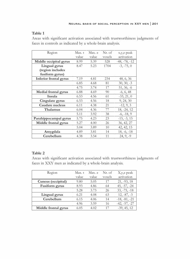
Table 1Areas with significant activation associated with trustworthiness judgments offaces in controls as indicated by a whole-brain analysis.
Table 2Areas with significant activation associated with trustworthiness judgments offaces in XXY men as indicated by a whole-brain analysis.
Neural basis of social perception in XXY men | 201
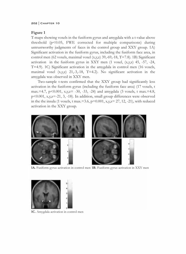
Figure 1T-maps showing voxels in the fusiform gyrus and amygdala with a t-value abovethreshold (p<0.05, FWE corrected for multiple comparisons) duringuntrustworthy judgments of faces in the control group and XXY group. 1A)Significant activation in the fusiform gyrus, including the fusiform face area, incontrol men (62 voxels, maximal voxel (x,y,z) 39,-69,-18, T=7.8). 1B) Significantactivation in the fusiform gyrus in XXY men (1 voxel, (x,y,z) 45, -57, -24,T=4.9). 1C) Significant activation in the amygdala in control men (16 voxels,maximal voxel (x,y,z) 21,-3,-18, T=4.2). No significant activation in theamygdala was observed in XXY men.
Two-sample t-tests confirmed that the XXY group had significantly lessactivation in the fusiform gyrus (including the fusiform face area) (17 voxels, tmax.=4.7, p<0.001, x,y,z= -30, -33, -24) and amygdala (3 voxels, t max.=4.8,p<0.001, x,y,z=-21, 3, -18). In addition, small group differences were observedin the the insula (1 voxels, t max.=3.6, p=0.001, x,y,z= 27, 12, -21), with reducedactivation in the XXY group.
202 | Chapter 10
1A. Fusiform gyrus activation in control men
1C. Amygdala activation in control men
1B. Fusiform gyrus activation in XXY men
DiscussionThis fMRI study examined brain activation patterns underlying social evaluationof faces in men with an extra X chromosome (XXY chromosomal pattern).Compared to men from the general population, evaluation of faces asuntrustworthy was associated with decreased activation in the amygdala, insulaand fusiform face area, which are key neural structures subserving socialperception. In addition, we observed small, but significant, group differences inbrain activation in the insula, with lower activation in XXY men. When faceswere evaluated with regard to age, during which faces may be implicitly screenedfor social significance as shown by Winston et al. (2002), we again observed lessactivation in the fusiform gyrus and insula, but not in the STS and amygdala, inXXY men as compared to men from the general population.
The finding of decreased involvement of brain areas that process socio-emotional signals in XXY men provides, for the first time, a putative neuralexplanation for the reported social cognitive deficits (Van Rijn et al., 2006b) inKlinefelter syndrome. Using an event-related design, brain activation wasanalysed according to subjective evaluation of untrustworthiness. Because thedistribution of trustworthiness decisions was not different between XXY menand controls, we may assume that differences in brain activation patterns did notreflect differences in behavioral performance. Therefore, our findings of lessactivation in the amygdala and fusiform face area during untrustworthy faces inXXY men as compared to men from the general population, can be interpretedas abnormal functioning of these brain areas in Klinefelter syndrome.
The amygdala plays a central role in the neural circuit subserving socialcognition, as indicated by the high density of incoming and outgoingprojections to other brain regions (Aggleton, 2000). The amygdala is especiallyknown for its engagement in screening information for emotional and socialsignificance, especially threat-related information (Amaral, 2003; Phelps, 2006).As emotional expressions on faces provide a crucial source of informationneeded for decoding social and emotional signals, the amygdala generallyactivates in response to facial expressions, especially to angry or fearful faces(Adolphs, 2001; Haxby et al., 2002; Phan et al., 2002). Indeed, in men from thegeneral population, we observed in this study significant activation in theamygdala when judging faces as untrustworthy as well as an increase in amygdalaactivation during untrustworthy- as compared to trustworthy decisions. Incontrast, no significant activation in the amygdala was seen in de XXY groupwhen judging trustworthiness of faces. Interestingly, the functionalabnormalities of the amygdala in XXY men as found in this study are in line
Neural basis of social perception in XXY men | 203

with volume reductions of this area that have been observed in structural MRIstudies in Klinefelter syndrome (Patwardhan et al., 2002; Shen et al., 2004).
One of the areas that receives input from the amygdala is the fusiformgyrus, which includes the ‘fusiform face area’ (FFA) (Puce et al., 1996). This areaappears to be important for visual processing of the structural, static propertiesof faces, which are used to determine personal identity (Adolphs, 2001; Haxbyet al., 2000). It has been shown that activation in the fusiform gyrus can bemodulated by activation in the amygdala (Morris et al., 1998). Increasing threat-related social significance as processed in the amygdala may elicit re-allocationof attentional resources to allow detailed visual analysis of a socially significantstimulus. We speculate that lower levels of activation in the fusiform face areaduring judgment of faces as untrustworthy might, in part, result from reducedactivation in the amygdala.
The insula is another brain area that is closely connected to the amygdalaand is thought to be subject to attentional modulation by the amygdala (Adolphset al., 2006). This somatosensory area is important for monitoring andorganizing physiological (autonomic) changes in the internal milieu, as is seen inresponse to emotion-inducing stimuli (Damasio et al., 2000). It appears to beespecially involved in mediating affective responses to aversive, includingdisgust-related, incoming information (Adolphs, 2002; Phillips et al., 2003a).The latter is consistent with a role of this region in judging faces asuntrustworthy. XXY men displayed less activation associated with(un)trustworthiness in the insula as compared to controls. Significantly, thefunctional abnormalities of this region as found in this study are in line withstructural abnormalities, i.e. smaller volumes, of the insula that have beenreported for XXY men (Shen et al., 2004).
It has been shown by Winston et al. (2002) that not only duringtrustworthiness judgments, but also during evaluation of faces with regard toage, the fusiform face area, amygdala and insula are active. This suggests thatfaces may be automatically screened for social significance. Indeed, in controlswe observed activation in these areas during age-judgments of faces. Similar tothe trustworthiness condition, XXY men displayed less activation in the insulaand fusiform gyrus during age-evaluations as compared to control men.However, no significant group-differences were present in activation in theamygdala, suggesting that even though explicit screening for social relevance bythe amygdala seems to be diminished in Klinefelter syndrome, implicit screeningfor social relevance may be intact.
204 | Chapter 10
Our observations of disturbed development of some regions in a neuralnetwork supporting social cognition in XXY men may have several implications.First, abnormal engagement of a neural network subserving social perceptionmay underlie some of the social cognitive dysfunctions and impaired socialadaptation that have been described in XXY men (Van Rijn et al., 2006b). Inother words, it provides a neuro-anatomical basis for the social behavioralphenotype in Klinefelter syndrome. Second, we can extrapolate from thesefindings that the X chromosome may play an important role in the developmentof some of the brain areas subserving social cognition. This is also supportedby findings of abnormal development of brain areas involved in social cognitiveprocessing in Turner syndrome, another X chromosomal disorder that ischaracterised by the presence of only one X chromosome in females (i.e. the X0karyotype) (Mazzocco et al., 1998; McCauley et al., 2006; Ross et al., 2000).Similar to XXY men, individuals with X monosomy display impairments insocial cognition, such as facial affect recognition (Lawrence et al., 2003;McCauley et al., 1987). Interestingly, imaging studies have revealed structuraland functional abnormalities in the amygdala and insula in females with Turnersyndrome, regions that we also found to be abnormal in XXY men (Molko etal., 2004; Murphy et al., 1997; Skuse et al., 2005). Although speculative,involvement of one of the sex chromosomes in the development of socialcognitive abilities would also be in line with the observed sex differences insocial cognitive skills in the general population, with performance of mengenerally somewhat lower than that of women (Hall, 1984; Hampson et al.,2006; McClure, 2000). Third, we might consider a role the X chromosome insome aspects of social cognitive dysfunction and underlying neuroanatomicalabnormalities in other disorders, such as autism or schizophrenia. Deviant socialbehavior and impairments in social cognitive functions are considered coreabnormalities in these neurodevelopmental disorders (Abdi et al., 2004;Corrigan et al., 2001; Fein et al., 1986). Neuroimaging studies have consistentlyshown abnormalities of the amygdala, insula and fusiform face area in bothautism and schizophrenia (Grelotti et al., 2002; Phillips et al., 2003b; Quintanaet al., 2003). Interestingly, these disorders are more prevalent in men. Thereported risk ratio for men to develop schizophrenia relative to women is 1.42(Aleman et al., 2003), while autism is diagnosed approximately four times moreoften in boys than in girls (Volkmar et al., 1993). This fact has led others topropose that dysfunctional neural circuits underlying social cognitiveimpairments in autism may be related to genes on the X chromosome, that aredifferentially expressed in men and women (Baron-Cohen et al., 2005; Skuse,
Neural basis of social perception in XXY men | 205
2000). Interestingly, a study on sex differences in social perception inschizophrenia has shown that the female superiority in reading social signalsfrom faces seems not to be affected by schizophrenia, in contrast to males withthe disorder who perform significantly worse as compared to healthy men(Scholten et al., 2005).
Both Turner syndrome and Klinefelter syndrome have been associated withincreased symptoms and personality traits that resemble features from theautism- or schizophrenia spectrum. The estimated risk of autism spectrumdisorders may be several times higher in women with X-monosomy (3 %) ascompared to women from the general population (0.01 %) (Creswell et al.,1999). In Klinefelter syndrome, we observed increased levels of autism traits,schizotypal personality traits and schizophrenia-like symptoms (Van Rijn et al.,2006a; Van Rijn et al., submitted). In addition, a survey of hospital admissionsand discharge diagnoses has indicated a significantly increased relative risk ofbeing hospitalized with psychoses (hazard ratio of 4.97) for men withKlinefelter syndrome (Bojesen et al., 2006). In turn, there is suggestive evidencethat prevalence of the XXY karyoype in the male schizophrenia population maybe several times higher as compared to the prevalence of Klinefelter syndromein the general population (DeLisi et al., 1994; Kunugi et al., 1999). Althoughspeculative, observations of increased autism- or schizophrenia spectrumpathology in Turner syndrome and Klinefelter syndrome fit with the hypothesisthat the X chromosome may play a role in some of the developmentalabnormalities of the neural structures for social information processing, such asthe amygdala, insula or fusiform face area, in autism or schizophrenia.
In sum, this study has revealed reduced engagement of the amygdala, insulaand fusiform face area during social perception in XXY men as compared tomen from the general population. Compromised function of these areas mayunderlie some of the deficits in social cognition and social behavior that havebeen observed in XXY men. In addition, as Klinefelter syndrome is defined byan X chromosomal abnormality, our findings suggest a link between one of thesex chromosomes and development of some of the neural regions supportingsocial cognition. In addition to revealing a neural basis for social dysfunctionsin this specific disorder, our data suggest that Klinefelter syndrome may serveas a more general model for studying the genetic basis of developmentalabnormalities in social cognition. This may be particularly relevant since thesedeficits are considered core abnormalities in severe psychiatric disorders such asautism and schizophrenia.
206 | Chapter 10
ReferencesAbdi, Z., & Sharma, T. (2004). Social cognition and its neural correlates in
schizophrenia and autism. CNS Spectrums, 9(5), 335-343.Adolphs, R. (2001). The neurobiology of social cognition. Current Opinion in
Neurobiology, 11(2), 231-239.Adolphs, R. (2002). Neural systems for recognizing emotion. Current Opinion
in Neurobiology, 12(2), 169-177.Adolphs, R., & Spezio, M. (2006). Chapter 20 role of the amygdala in
processing visual social stimuli. Progress in Brain Research, 156, 363-378.
Aggleton, J. P. (2000). The amygdala. New York: Oxford Press.Aleman, A., Kahn, R. S., & Selten, J. P. (2003). Sex differences in the risk of
schizophrenia: Evidence from meta-analysis. Archives of GeneralPsychiatry, 60(6), 565-571.
Amaral, D. G. (2003). The amygdala, social behavior, and danger detection.Annals of the New York Academy of Sciences, 1000(1), 337-347.
Baron-Cohen, S., Knickmeyer, R. C., & Belmonte, M. K. (2005). Sex differences in the brain: Implications for explaining autism. Science,310(5749), 819-823.
Bojesen, A., Juul, S., Birkebaek, N. H., & Gravholt, C. H. (2006). Morbidity in klinefelter syndrome; a danish register study based on hospital dischargediagnoses. The Journal of Clinical Endocrinology and Metabolism,91(4), 1254-1260.
Boone, K. B., Swerdloff, R. S., Miller, B. L., Geschwind, D. H., Razani, J., Lee,A., et al. (2001). Neuropsychological profiles of adults with klinefeltersyndrome. Journal of the International Neuropsychological Society, 7(4),446-456.
Corrigan, P. W., & Penn, D. L. (Eds.). (2001). Social cognition and schizophrenia. Washington, D.C.: American Psychological Association.
Creswell, C. S., & Skuse, D. H. (1999). Autism in association with turner syndrome: Genetic implications for male vulnerability to pervasivedevelopmental disorders. Neurocase, 5(6), 511-518.
Damasio, A. R., Grabowski, T. J., Bechara, A., Damasio, H., Ponto, L. L. B.,Parvizi, J., et al. (2000). Subcortical and cortical brain activity during thefeeling of self-generated emotions. Nature Neuroscience, 3(10), 1049.
DeLisi, L. E., Friedrich, U., Wahlstrom, J., Boccio-Smith, A., Forsman, A.,Eklund, K., et al. (1994). Schizophrenia and sex chromosome anomalies.Schizophrenia Bulletin, 20(3), 495-505.
Neural basis of social perception in XXY men | 207
DeLisi, L. E., Maurizio, A. M., Svetina, C., Ardekani, B., Szulc, K., Nierenberg,J., et al. (2005). Klinefelter's syndrome (xxy) as a genetic model forpsychotic disorders. American Journal of Medical Genetics BNeuropsychiatric Genetics, 135(1), 15-23.
Fein, D., Pennington, B., & Markowitz, P. (1986). Toward a neuropsychological model of infantile autism: Are the social deficits primary? Journal of theAmerican Academy of Child Psychiatry, 25(2), 198.
Geschwind, D. H., Boone, K. B., Miller, B. L., & Swerdloff, R. S. (2000).Neurobehavioral phenotype of klinefelter syndrome. Mental Retardationand Developmental Disabilities Research Reviews, 6(2), 107-116.
Grelotti, D. J., Gauthier, I., & Schultz, R. T. (2002). Social interest and the development of cortical face specialization: What autism teaches usabout face processing. Developmental Psychobiology, 40(3), 213.
Grill-Spector, K., Knouf, N., & Kanwisher, N. (2004). The fusiform face area subserves face perception, not generic within-category identification.Nature Neuroscience, 7(5), 555.
Hall, J. A. (1984). Non-verbal sex differences: Communication accuracy and expressive style. Baltimore: John Hopkins University Press.
Hampson, E., van Anders, S. M., & Mullin, L. I. (2006). A female advantage in the recognition of emotional facial expressions: Test of an evolutionaryhypothesis. Evolution and Human Behavior, 27(6), 401.
Haxby, J. V., Hoffman, E. A., & Gobbini, M. I. (2000). The distributed human neural system for face perception. Trends in Cognitive Sciences, 4(6),223.
Haxby, J. V., Hoffman, E. A., & Gobbini, M. I. (2002). Human neural systems for face recognition and social communication. Biological Psychiatry,51(1), 59-67.
Kesler, S. R., Blasey, C. M., Brown, W. E., Yankowitz, J., Zeng, S. M., Bender,B. G., et al. (2003). Effects of x-monosomy and x-linked imprinting onsuperior temporal gyrus morphology in turner syndrome. BiologicalPsychiatry, 54(6), 636-646.
Kesler, S. R., Garrett, A., Bender, B., Yankowitz, J., Zeng, S. M., & Reiss, A. L.(2004). Amygdala and hippocampal volumes in turner syndrome: Ahigh-resolution mri study of x-monosomy. Neuropsychologia, 42(14),1971-1978.
208 | Chapter 10
Kunugi, H., Lee, K. B., & Nanko, S. (1999). Cytogenetic findings in 250 schizophrenics: Evidence confirming an excess of the x chromosomeaneuploidies and pericentric inversion of chromosome 9. SchizophreniaResearch, 40(1), 43-47.
Lawrence, K., Kuntsi, J., Coleman, M., Campbell, R., & Skuse, D. (2003). Face and emotion recognition deficits in turner syndrome: A possible role forx-linked genes in amygdala development. Neuropsychology, 17(1), 39-49.
Maldjian, J. A., Laurienti, P. J., Kraft, R. A., & Burdette, J. H. (2003). An automated method for neuroanatomic and cytoarchitectonic atlas-basedinterrogation of fmri data sets. Neuroimage, 19(3), 1233-1239.
Mazzocco, M. M. M., Baumgardner, T., Freund, L. S., & Reiss, A. L. (1998).Social functioning among girls with fragile x or turner syndrome andtheir sisters. Journal of Autism and Developmental Disorders, 28(6),509-517.
McCauley, E., Kay, T., Ito, J., & Treder, R. (1987). The turner syndrome:Cognitive deficits, affective discrimination, and behavior problems. ChildDevelopment, 58(2), 464.
McCauley, E., & Sybert, V. (2006). Social and behavioral development of girls and women with turner syndrome. International Congress Series, 1298,93.
McClure, E. B. (2000). A meta-analytic review of sex differences in facial expression processing and their development in infants, children, andadolescents. Psychological Bulletin, 126(3), 424-453.
Molko, N., Cachia, A., Riviere, D., Mangin, J. F., Bruandet, M., LeBihan, D., et al. (2004). Brain anatomy in turner syndrome: Evidence for impairedsocial and spatial-numerical networks. Cerebral Cortex, 14(8), 840-850.
Morris, J. S., Friston, K. J., Büchel, C., Frith, C. D., Young, A. W., Calder, A. J.,et al. (1998). A neuromodulatory role for the human amygdala inprocessing emotional facial expressions. Brain, 121(1), 47.
Murphy, D. G. M., Mentis, M. J., Pietrini, P., Grady, C., Daly, E., Haxby, J. V., et al. (1997). A pet study of turner's syndrome: Effects of sex steroids andthe x chromosome on brain. Biological Psychiatry, 41(3), 285-298.
Patwardhan, A. J., Brown, W. E., Bender, B. G., Linden, M. G., Eliez, S., & Reiss, A. L. (2002). Reduced size of the amygdala in individuals with47,xxy and 47,xxx karyotypes. American Journal of Medical Genetics,114(1), 93-98.
Neural basis of social perception in XXY men | 209
Phan, K. L., Wager, T., Taylor, S. F., & Liberzon, I. (2002). Functional neuroanatomy of emotion: A meta-analysis of emotion activationstudies in pet and fmri. Neuroimage, 16(2), 331-348.
Phelps, E. A. (2006). Emotion and cognition: Insights from studies of the human amygdala. Annual Review of Psychology, 57, 27.
Phillips, M. L., Drevets, W. C., Rauch, S. L., & Lane, R. (2003a). Neurobiology of emotion perception i: The neural basis of normal emotionperception. Biological Psychiatry, 54(5), 504-514.
Phillips, M. L., Drevets, W. C., Rauch, S. L., & Lane, R. (2003b). Neurobiology of emotion perception ii: Implications for major psychiatric disorders.Biological Psychiatry, 54(5), 515-528.
Puce, A., Allison, T., Asgari, M., Gore, J. C., & McCarthy, G. (1996).Differential sensitivity of human visual cortex to faces, letterstrings, andtextures: A functional magnetic resonance imaging study. Journal ofNeuroscience, 16(16), 5205.
Quintana, J., Wong, T., Ortiz-Portillo, E., Marder, S. R., & Mazziotta, J. C.(2003). Right lateral fusiform gyrus dysfunction during facialinformation processing in schizophrenia. Biological Psychiatry, 53(12),1099.
Ratcliffe, S. (1999). Long-term outcome in children of sex chromosome abnormalities. Archives of Disease in Childhood, 80(2), 192-195.
Ross, J., Zinn, A., & McCauley, E. (2000). Neurodevelopmental and psychosocial aspects of turner syndrome. Mental Retardation andDevelopmental Disabilities Research Reviews, 6(2), 135.
Samango-Sprouse, C. (2001). Mental development in polysomy x klinefelter syndrome (47,xxy; 48,xxxy): Effects of incomplete x inactivation.Seminars in Reproductive Medicine, 19(2), 193-202.
Scholten, M. R. M., Aleman, A., Montagne, B., & Kahn, R. S. (2005).Schizophrenia and processing of facial emotions: Sex matters.Schizophrenia Research, 78(1), 61.
Sheehan, D. V., Lecrubier, Y., Sheehan, K. H., Amorim, P., Janavs, J., Weiller,E., et al. (1998). The mini-international neuropsychiatric interview(m.I.N.I.): The development and validation of a structured diagnosticpsychiatric interview for dsm-iv and icd-10. Journal of ClinicalPsychiatry, 59 Suppl 20, 22-33;quiz 34-57.
Shen, D., Liu, D., Liu, H., Clasen, L., Giedd, J., & Davatzikos, C. (2004).Automated morphometric study of brain variation in xxy males.Neuroimage, 23(2), 648-653.
210 | Chapter 10
Skuse, D., Morris, J. S., & Dolan, R. J. (2005). Functional dissociation ofamygdala-modulated arousal and cognitive appraisal, in turner syndrome.Brain, 128(Pt 9), 2084-2096.
Skuse, D. H. (2000). Imprinting, the x-chromosome, and the male brain:Explaining sex differences in the liability to autism. Pediatric Research,47(1), 9-16.
Van Rijn, S., Aleman, A., Swaab, H., & Kahn, R. (2006a). Klinefelter's syndrome (karyotype 47,xxy) and schizophrenia-spectrum pathology.British Journal of Psychiatry, 189(5), 459-461.
Van Rijn, S., Swaab, H., Aleman, A., & Kahn, R. S. (2006b). X chromosomal effects on social cognitive processing and emotion regulation: A studywith klinefelter men (47,xxy). Schizophrenia Research, 84(2-3), 194-203.
Van Rijn, S., Swaab, H., Aleman, A., & Kahn, R. S. (submitted). Social behavior and autism traits in a sex chromosomal disorder: Klinefelter(47xxy) syndrome.
Volkmar, F. R., Szatmari, P., & Sparrow, S. S. (1993). Sex differences in pervasive developmental disorders. Journal of Autism andDevelopmental Disorders, V23(4), 579.
Winston, J. S., Strange, B. A., O_Doherty, J., & Dolan, R. J. (2002). Automatic and intentional brain responses during evaluation of trustworthiness offaces. Nature Neuroscience, 5(3), 277-283.
Neural basis of social perception in XXY men | 211


Chapter 11
General discussion
Social cognitive capacities are crucial for flexible navigation through a socialworld. The severe impact of impaired social cognition is illustrated by disorderssuch as autism or schizophrenia. In these neurodevelopmental disorders, socialcognition is among the core domains of deficits (Corrigan et al., 2001; Fein etal., 1986). The importance of social cognitive capacities in coping with the socialworld and related mental well-being has called for a search into the origins ofsocial cognition on the level of cognition, neurobiology and genes.
Klinefelter syndrome is an X chromosomal disorder known for socialdysfunction and specific deficits in brain development and cognition, which canhelp us to unravel genotype-phenotype relations that are relevant for socialbehavior. Klinefelter syndrome might be used as a model for disorders of socialbehavior to reveal insight into involvement of the X chromosome in socialcognitive pathways to psychopathology which may be more difficult to uncoverby studying heterogeneous, behaviorally defined populations (Reiss, 2000; Reisset al., 2000).
In the present thesis, we investigated the effects of an extra X chromosomeon social cognition and the underlying neural basis. Besides providing insightinto the cognitive and neural basis of social dysfunctions in this specific geneticdisorder, another aim of the present thesis was to explore whether Klinefeltersyndrome may serve as a model for social cognitive disturbances in autism orschizophrenia. We have focused on socially deviant behavior in adult XXY menon a behavioral, cognitive and neuroanatomical level and reviewed evidence forautism and schizophrenia spectrum traits, social dysfunction, social cognitivedisabilites and underlying dysfunctional neural mechanisms in XXY men.
Social behavior and psychopathologyAs described in chapter two and three, we assessed social difficulties, reflectedin frequency of participation in social interactions and distress during theseinteractions, as well as vulnerability to disorders of social behavior, such asautism and schizophrenia, in XXY men. Vulnerability to neurodevelopmentaldisorders associated with difficulties in social adaptation was investigated froma dimensional, symptom oriented perspective, rather than a dichotomal, all-or-none, approach. Dimensional measures allow quantification of a number oftraits associated with the autism or schizophrenia spectrum. In addition, weused a measure of schizophrenia symptoms to quantify symptoms at a clinicallevel. Measures of social behavior and psychopathology in XXY men werecompared to those of men from the general population.
214 | Chapter 11
Men with Klinefelter syndrome reported to less often engage in social behaviorand to experience more distress during social interactions as compared to menfrom the general population. In addition, XXY men displayed significantlyincreased levels of schizophrenia spectrum traits and symptoms as compared tocontrols. Scores were higher on all traits, with effect sizes (cohen’s d) of 1.43 forthe negative dimension (for example constricted affect and social anxiety), 1.31for the positive dimension (for example referential thinking and delusionalatmosphere) and 1.81 for the disorganized dimension (vague/overinclusivespeech and eccentric behavior). Also on a clinical level, higher levels ofschizophrenia symptoms were observed in XXY men, with effect sizes of 1.60for negative symptoms, 1.45 for positive symptoms and 1.66 for generalpsychopathology. These findings provide evidence for increased vulnerability toschizophrenia psychopathology in Klinefelter syndrome and are in agreementwith reports of increased prevalence of the XXY karyotype amongschizophrenia populations (DeLisi et al., 1994; Kunugi et al., 1999) and a surveyof hospital admissions and discharge diagnoses that has indicated a significantlyincreased relative risk of being hospitalized with psychoses (hazard ratio of4.97) for men with Klinefelter syndrome (Bojesen et al., 2006). Similarly, levelsof autism traits, such as impaired social skills, communication, imagination,attention switching and increased attention to details, were all higher in XXYmen as compared to controls. This is the first report of increased levels ofautism traits in Klinefelter syndrome.
These findings suggest that men with Klinefelter syndrome less oftenengage in social behavior and experience more distress during social interactionsas compared to men from the general population. Our findings of difficultiesin coping with social situations in XXY men, especially the high levels ofdistress during social interactions, are consistent with reports of social anxiety,social withdrawal and shyness in individuals with the XXY karyotype (Bender etal., 1999; Ratcliffe, 1999). Difficulties in social adjustment have mostly beenreported for children or adolescents with Klinefelter syndrome. Our datasuggest that social difficulties may persist into adulthood, with social distressmore prominent than a general reduction in engagement in social behavior.The high levels of autism traits and schizotypal traits in XXY men that wereobserved across all dimensions of the autism and schizophrenia phenotype,suggest that some aspects of the XXY phenotype parallel the phenotype thatcharacterizes individuals at increased risk for autism or schizophrenia. Forexample, biological relatives of subjects with these disorders also displayincreased levels of schizotypal- or autism traits (Bishop et al., 2004; Vollema et
General discussion | 215
al., 2002). This hypothesis is in agreement with the concept of ‘broadphenotypes’ of autism or schizophrenia, which refers to mild features of theclinical autism- or schizophrenia phenotype that are seen in biological (i.e.genetically related) relatives of individuals with these disorders (Bailey et al.,1998; Bishop et al., 2004; Gottesman et al., 2003; Jablensky, 2006).
Thus, mild features of the autism or schizophrenia phenotype may not onlybe found in genetically related relatives of individuals with these disorders, butalso in men with an extra X chromosome.
Social cognitionNext, we investigated the effects of an extra X chromosome on social cognitionand explored whether social cognitive dysfunction in Klinefelter syndrome maybe an underlying cognitive mechanism contributing to difficulties in coping withsocial environments as reflected in social distress and increased autism orschizophrenia traits. Several aspects of social cognitive processing were assesedat the level of perception, experience and expression, namely: labeling of facialexpressions of emotion, emotion-cognition interactions in decision making,automatic processing of social cues such as gaze direction and impliedbiological motion, discriminating emotions in tone-of-voice, and emotionregulation, i.e. subjective experience and identification of emotional arousal aswell as verbal expression of emotions.
Perception As described in chapter four, we found XXY men to be impaired inrecognizing facial expressions of anger, independent of the intensity of theemotional expression. This deficit was not attributable to a generalizedimpairment in analyzing faces, as performance in a face identity matching taskwas intact. Misperception of angry facial expressions may contribute todifficulties in social interactions, as non-verbal signals can convey crucialinformation about the emotional state of the sender. Various studies haverevealed significant relationships between facial affect recognition performanceand social functioning (Hooker et al., 2002).
The XXY group not only displayed deficits in processing social signals inthe visual domain, but also in the auditory domain. Besides facial expressions ofemotions, affective prosody in speech is also important for understandingintentions of others, because variations in tone of voice, such as intonation andloudness, provide information about the emotional state of the speaker. Insocial exchange, it is not only important to understand the content of words and
216 | Chapter 11
sentences, i.e. what is said, but it may even be more crucial to consider the(social) context of words and sentences, i.e. how it is said. Lesion- and fMRIstudies indicate that whereas the left hemisphere is specialized for processinglinguistic (semantic) information, the right hemipshere may be specialized foranalyzing pragmatic aspects of language, such as understanding metaphors,irony, discourse, indirect requests and analyzing emotional prosody. In chaptersix, we provided evidence for a causal involvement of the right hemisphere,more specifically the fronto-parietal operculum, in processing affective prosodicinformation in individuals from the general population. Applying inhibitoryTranscranial Magnetic Stimulation (TMS) to the right hemisphere temporarilydisrupted recognition of emotions in prosodic cues in speech, but not semantic(linguistic) cues. These findings extend an earlier study of Vingerhoets et al.(Vingerhoets et al., 2003) who have measured blood flow velocity (BFV) withfunctional transcranial doppler ultrasonography (fTCD) to study thecontribution of the right and left hemisphere to the detection of emotion inprosody versus semantics of spoken language using this particular task. Duringdetection of emotion in semantics they observed a significant left-hemisphericlateralization of BFV. This lateralization effect disappeared when attention wasshifted to discriminating emotion in prosody, due to a rise in right hemisphericBFV. Our findings provide evidence that the right hemisphere is not onlyassociated with, but also causally involved in emotional prosody.
As described in chapter seven, discrimination of emotions in prosodiccues was impaired in XXY men. Although recognizing emotions in semanticcues was also impaired in the XXY group, which is in line with the typical lefthemisphere mediated language dysfunctions in Klinefelter syndrome,recognizing emotions in prosody, a pragmatic, right hemisphere mediated aspectof language, was more affected. Difficulties in picking up pragmaticcommunicative cues in conversation might have an impact on how well XXYmen cope with social situations, as these aspects of language are important forunderstanding interpersonal intentions and responding to those in anappropriate way.
Another important underlying characteristic of successful social interactionis the ability to quickly and automatically process basic elements of a socialsignal such as direction of gaze, head orientation and body postures (Frith et al.,1999; Jellema et al., 2005). These cues can give clues about someone’s intentions,goals and believes (Perrett, 1999). It is suggested that the ability to process thesebasic social cues automatically is a prerequisite for establishing successful socialinteractions and communication (Frith and Frith, 1999). We have shown
General discussion | 217
(chapter five) that in contrast to men from the general population, XXY menhave difficulties in automatically attending to, and process, basic social cues suchas gaze direction and implied biological motion. Reduced availability of basicsocial information may contribute to more widespread effects on (‘upstream’-)higher-order social cognitive processing. Although speculative, this reducedsensitivity to basic elements of a social signal might, in part, explain socialawkwardness and reduced social intuition and which have been described inXXY men. We not only compared performance of XXY men with matchedcontrols, but we were also able to include patients with schizophrenia as well asbiological relatives of schizophrenia patients. Interestingly, performance inprocessing these social cues was indistinguishable between Klinefelter men andschizophrenia patients. Moreover, performance of biological relatives ofschizophrenia patients resembled the lack of sensitivity to social cues observedin schizophrenia patients and XXY men, albeit to a lesser extent, indicating thatit may be an expression of genetic vulnerability for the disease.
Experience and expressionNot only perception, but also disturbances in experience and expression ofemotions were observed in XXY men (chapter four). Regarding the experienceof emotions, Klinefelter men rejected more often unfair financial offers fromhuman proposers as compared to the control group in the Ultimatum game.This pattern of performance is shown to reflect the influence of emotion, inresponse to the offer being unfair, on strategic decision making (Sanfey et al.,2003; van 't Wout et al., 2006). Besides this implicit emotional task, increasedemotional experience was also observed in an explicit, self-report measure ofemotional experience, i.e. the alexithymia questionnaire. Alexithymia is apersonality trait implying an inability or reduction to identify, experience,describe and reflect on one’s own emotions (Lane et al., 1997; Sifneos, 1973). Incontrast to apparent hyperfunctional emotional -experience and -reactivity,identifying and verbalizing one’s own emotions appeared to be hypofunctional inKlinefelter syndrome. This dissociation has been termed type II alexithymia,which contrasts with type I alexithymia (a general reduction in processingemotions). This emotion regulation profile of increased experience butdecreased expression of emotions can be considered as a possible risk factor forboth medical and psychiatric disorders (Bagby et al., 1997).
Taken together, Klinefelter men seem less accurate in perception of socialsignals, both from facial expressions and tone-of-voice, and have reducedautomatic attention to basic social cues, such as gaze direction and implied
218 | Chapter 11
biological motion. Moreover, they are less able to identify and verbally describetheir emotions, but experience increased levels of emotional arousal, incomparison to men from the general population. Although it has beensuggested in the literature that the reported deficits in typical left hemispheremediated language functions largely explain social dysfunction in XXY men, thepresent thesis identified dysfunctions in processing social signals, emotions andright hemisphere mediated pragmatic language as mechanisms that may alsocontribute to social dysfunction and related mental well-being.
Social cognition: neural basis
Language As it has been proposed that language impairments, with language being acrucial part of social communication, may contribute to the observeddifficulties in social functioning in Klinefelter men (Samango-Sprouse, 2001),we studied the neural basis of language processing in XXY men. Althoughlanguage impairments in Klinefelter syndrome are well-documented, and arenot restricted to the typical left hemisphere language deficits but also includeright hemisphere language dysfunctions as pointed out in the present thesis,underlying neural mechanisms are not well understood (Geschwind et al., 2000).One possible neural mechanism involved in the verbal disabilities in Klinefeltersyndrome that we investigated was abnormal hemispheric involvement inlanguage processing.
By using functional Magnetic Resonance Imaging (fMRI) we were able toassess lateralization of activation in specific brain areas during languageprocessing in XXY men (chapter eight). By measuring the relative contributionof the right and left hemisphere in each individual it was shown that languageactivity in the brain was indeed less lateralized in the XXY group as comparedto control men. Loss of asymmetric processing of language was due toincreased activity in the right hemisphere rather than reduced activity in the lefthemisphere. Analysis of functional asymmetry within different language regionsindicated that loss of functional asymmetry in the superior temporal gyrus(STG) was the most prominent contributor to reduced language laterality inXXY men. This regional loss of language laterality was highly correlated withthe degree of disorganization (vague or overinclusive speech and odd/eccentricbehavior) in the XXY group. This is consistent with studies finding structural(Matsumoto et al., 2001; Menon et al., 1995; Rajarethinam et al., 2000; Rossi etal., 1994; Shenton et al., 1992) and functional (Kircher et al., 2002) abnormalities
General discussion | 219
of the STG to be related to disorganization symptoms such as thought disorderin schizophrenia.
Social perceptionThe present thesis revealed that not only some aspects of language but alsoperception of social signals, another core function within the domain of socialcognition, may be disturbed in XXY men. We explored the effects of an extraX chromosome on the neural basis of social cognition and reviewed structuralneuroimaging findings in Klinefelter syndrome, as well as investigated neuralnetworks supporting social perception in XXY men using functionalneuroimaging.
The amygdala seems to play a central role in social perception and isespecially known for its engagement in screening information for emotional andsocial significance (Amaral, 2003; Phelps, 2006). In chapter nine we havereviewed evidence for structural abnormalities of the amygdala in Klinefeltersyndrome based on findings in the literature. Findings were compared to whatis known of abnormalities of the amygdala in populations with increasedvulnerability to schizophrenia: individuals from the general populationdisplaying subclinical signs of schizophrenia and biological relatives ofschizophrenia patients who may carry a genetic predisposition for the disorder.Reductions in volume of the amygdala were found to be present both inKlinefelter syndrome and the populations at high risk for schizophrenia, i.e.across the spectrum of schizophrenia spectrum vulnerability. Morphologicalabnormalities of the amygdala provide a putative neuro-anatomical basis for thedeficits in social perception, such as impaired facial affect recognition, that wehave observed in XXY men. In addition, the fact that amygdala abnormalitiesare not only consistently reported in patients with schizophrenia (Breier et al.,1992; Bryant et al., 1999; Gur et al., 2000; Hulshoff-Pol et al., 2001; Lawrie etal., 2003; Wright et al., 2000) but are also observed in populations with anincreased liability for schizophrenia is of considerable importance.Abnormalities of the amygdala in the schizophrenia spectrum may beneurodevelopmental in origin and might reflect a genetic vulnerability for thedisease rather than being the consequence of antipsychotic medication,hospitalisation and neurotoxic effects of psychosis.
Structural abnormalities of the amygdala in Klinefelter syndrome suggestabnormal functioning of this brain area, possibly extending to a neural networkfor social perception this region is part of. By using fMRI we were able to studyfunctioning of such a neural network during social evaluation of faces
220 | Chapter 11
(chapter ten). We measured brain activity during trustworthiness evaluations offaces, with special interest in the amygdala, fusiform face area, superior temporalgyrus and insula. These regions are highly interconnected brain regions involvedin social perception. We selected those regions based on their involvement intrustworthiness evaluations of faces in the general population (Winston et al.,2002). Activity in the amygdala during trustworthiness evaluation of faces mayrepresent screening of social relevance and especially threat. Involvement of thefusiform face area may indicate processing of the structural, static properties offaces that are used for identification. Activation in the superior temporal sulcusmay reflect efforts to detect goals and intentions based on eye-gaze and othersignals in faces. Finally, insula activity may represent monitoring and organizingphysiological (autonomic) changes in the internal milieu in response to thedegree of trustworthiness of faces.
As predicted, we observed less involvement of region in this key neuralnetwork subserving social cognition during untrustworthy faces as compared tocontrols. Significantly lower levels of activation were observed in the amygdala,insula and fusiform face area in XXY men as compared to controls. Thefindings of less involvement of key brain areas in processing socio-emotionalsignals in XXY men offer, for the first time, insight into the neural basis ofsocial cognition in Klinefelter syndrome. Abnormal engagement of a neuralnetwork subserving social perception may contribute to social cognitivedysfunctions and impaired social adaptation that have been described for menwith Klinefelter syndrome.
Taken together, we observed reduced hemispheric specialization for languageand less engagement of a neural network processing social information in XXYmen. These findings, for the first time, may provide a neural explanation forsome of the social cognitive impairments in Klinefelter syndrome.
The X chromosome and social cognitionAs Klinefelter syndrome is defined by an X chromosomal abnormality, ourfindings suggest that the X chromosome plays a role in the development ofsome aspects of social cognition and neural mechanisms involved in socialinformation processing in Klinefelter syndrome. Additional support for aputative role of the X chromosome in the development of social informationprocessing is derived from observations of social dysfunction and socialcognitive disabilites in Turner syndrome (45,X0), another X chromosomaldisorder characterised by a partial or complete absense of one of the X
General discussion | 221
chromosomes in females (Mazzocco et al., 1998; McCauley et al., 2006; Ross etal., 2000). Similar to XXY men, individuals with X monosomy displayimpairments in facial affect recognition (Lawrence et al., 2003b; McCauley et al.,1987). Also, deficits in gaze direction processing and impairments in judgingmental states from eyes have been reported (Elgar et al., 2002; Lawrence et al.,2003a). Difficulties in picking up social signals are not only observed in thevisual domain, but also in the auditory domain. Comparable to men with theXXY pattern, women with one X chromsome display impairments in decodingemotions from tone of voice, i.e. emotional prosody (Ross et al., 1995).
Similar to XXY men, abnormal development of brain regions that are partof a neural network supporting social cognition have been observed in 45,X0females. These include the amygdala, superior temporal sulcus and -gyrus,anterior cingulate, orbitofrontal cortex and insula (Kesler et al., 2003; Kesler etal., 2004; Molko et al., 2004; Murphy et al., 1997; Skuse et al., 2005). Specificallyinteresting is a functional neuroimaging study with Turner females revealing aloss of correlation between activation in the left amygdala and left fusiformgyrus underlying impairments in labeling of facial expressions of both angerand fear, while right amygdala mediated somatic responses were enhanced inreponse to emotional faces (Skuse et al., 2005). This finding is in line with ourobservation of a dissociation between reduced cognitive labeling of emotionsand increased emotional arousability in XXY men.
The extra X chromosome may also affect neural systems involved inlanguage processing. Genetic mechanisms involving the X chromosome mayinfluence the development of hemispheric specialization for language, asreduced language lateralization was observed in XXY men using fMRI. Aputative link between the X chromosome and reduced language lateralization isin line with a proposed role of the X chromosome in the development ofasymmetry in the brain. It is thought that a pseudo-autosomal region on the Xchromosome is involved in abnormal development of brain asymmetry in XXYmen, as indicated by increased left-handedness and neuropsychological evidenceof anomalous hemispheric dominance (Geschwind et al., 1998). Interestingly,also in Turner syndrome (45,X0) reduced lateralization of verbal informationprocessing has been reported, which supports the hypothesis that the Xchromosome may be important for the development of hemisphericspecialization for verbal information (Netley et al., 1982).
However, the exact genetic mechanism that leads to the XXY phenotyperemains unclear. In healthy females, who have two X chromosomes, certaingenes on the X chromosome are inactivated, while others escape inactivation. In
222 | Chapter 11
Klinefelter syndrome, genes on the X chromosome that that escape inactivationand have a homologue on the Y chromosome and, may be overexpressed. Is hasbeen proposed that phenotype in Klinefelter syndrome, at a neural, cognitiveand behavioral level, might originate from overexpression of such genes (Crow,1988; DeLisi et al., 2005; Geschwind et al., 2000). Alternatively, genomicimprinting may play a role. Genomic imprinting refers to differential expressionof a gene, dependent of the parental origin of that gene. It has been shown thatTurner females with a maternally derived X chromosome display moreimpairments in social skills as compared to those with the X chromosome fromthe father (Skuse et al., 1997). The authors proposed an X-chromosomal locusfor social cognition, which is imprinted and not expressed by the maternallyderived X chromosome. In Klinefelter syndrome, the extra X chromosome isderived from the mother in over half of the cases (Thomas et al., 2003). Arecent study has pointed to putative imprinting effects in 54 XXY men.Impairments in speech and motor development were more often found insubjects with a paternal (extra) X chromosome as compared to those with amaternal (extra) X chromosome (Stemkens et al., 2006). Taken together,abnormal expression of genes on the X chromosome in Klinefelter syndromemay affect development of the brain resulting in social, emotional and languagedysfunctions. Although speculative, an involvement of sex chromosomes insocio-emotional brain functions would be in line with the observed sexdifferences in social cognitive skills in the general population, with performanceof men generally somewhat below that of women (Hall, 1984; Hampson et al.,2006; McClure, 2000; Montagne et al., 2005). However, the exact geneticmechanisms that lead to the XXY phenotype, which may for example includeoverexpression, abnormal inactivation, genomic imprinting or gene-geneinteractions, are as yet unclear. Future genetic studies are needed to identifycandidate genetic mechanisms involving the X chromosome that can explain thesocial cognitive deficits in XXY men, sex differences in social cognition in thegeneral population and the male preponderance in disorders of social cognition.
The role of testosterone deficits The degree to which the observed cognitive and neurobiological deficits inXXY men represent the effects of testosterone- deficits that become apparentin puberty and subsequent testosterone supplementation, is unclear. Therelationship between testosterone levels and brain development is complex;timing of exposure, sensitivity to testosterone reflected in androgen receptordensity and modulation by environmental factors are important determinants in
General discussion | 223
the effects of testosterone (Craig et al., 2004). Although speculative, abnormaltestosterone levels might be part of the mechanism by which X chromosomalabnormalities lead to disturbances in development of neural systems supportingsocial cognition. However, gonadal hormones may be one of many mechanismsby which sex chromosomes exert their influence on brain development. Recentanimal studies have pointed to direct, non-hormonal effects of sex-chromosomes on brain maturation (Dewing et al., 2003).
Although speculative, support for the hypothesis that brain function andcognition in XXY men may represent the effects of X chromosomal geneticpathology rather than low testosterone levels by itself, comes from individualswith X chromosomal aneuploidies that are not associated with low testosteronelevels. For example, although females with the XXX karyotype show typicalhormone levels, they do display impairments in the language domain and havedecreased social adjustment (Bender et al., 1999; Harmon et al., 1998).Interestingly, there have been reports of increased psychopathology, such asconduct disorders and affective disorders, in XXX females (Harmon et al.,1998).
Furthermore, there is no evidence for an effect of testosteronesupplementation on language lateralization. A recent longitudinal study dealingwith the effects of testosterone treatment on language lateralization intranssexuals showed that language laterality is highly stable and not affected byhormonal interventions (Sommer et al., in prep). Also, testosterone levels byitself seem not enough to, for example, explain the amygdala abnormalities inindividuals with sex chromosome aneuploidies. Although females with Turnersyndrome, who are missing one X-chromosome, are also characterised by areduction in testosterone levels (Hojbjerg Gravholt et al., 1999), these patientsshow an increase in amygdala volume rather than a reduction (Kesler et al.,2004). Although speculative, differences in amygdala volume between Turnerand Klinefelter patients might be stronger related to the number of X-chromosomes, which differentiates the two groups, than to testosterone levels,which are reduced in both syndromes. Alternatively, abnormal testosteronelevels may result from X-chromosome linked amygdala abnormalities, since ithas been shown that the amygdala is involved in the production of testosterone(Banczerowski et al., 2003).
In conclusion, the exact nature of the effects of testosterone deficits andtestosterone supplementation on development of social cognition and neuralsubstrates in Klinefelter syndrome remains unclear.
224 | Chapter 11
Potential implications for the study of autism and schizophreniaOur findings may have potential implications for the study ofneurodevelopmental disorders associated with impaired social adaptation. Wemight consider a role of genetic mechanisms involving the X chromosome insome aspects of social cognitive dysfunction and underlying neuroanatomicalabnormalities in the autism or schizophrenia spectrum. Tentative support forthis hypothesis comes from observations of increased autism spectrumpsychopathology in Turner syndrome and both autism and schizophreniaspectrum traits in Klinefelter syndrome. The estimated risk for autism spectrumdisorders may be several times higher in women with X-monosomy (3 %) ascompared to women from the general population (0.01 %) (Creswell et al.,1999). In Klinefelter syndrome, we observed increased levels of autism traits,schizotypal personality traits and schizophrenia-like symptoms (Van Rijn et al.,2006; Van Rijn et al., in preparation). In addition, a survey of hospitaladmissions and discharge diagnoses has indicated a significantly increasedrelative risk of being hospitalized with psychoses (hazard ratio of 4.97) for menwith Klinefelter syndrome (Bojesen et al., 2006). In turn, there is suggestiveevidence that prevalence of the XXY karyoype in the male schizophreniapopulation may be several times higher as compared to the prevalence ofKlinefelter syndrome in the general population (DeLisi et al., 1994; Kunugi etal., 1999).
The increased levels of autism- and schizotypal traits that we observed inXXY men parallel the schizotypal or autism-like features that characterizeindividuals at increased risk for autism or schizophrenia. Biological relatives ofindividuals with autism or schizophrenia also display increased levels of traits ormild features of the disorder (Bishop et al., 2004; Vollema et al., 2002). Basedon twin studies, it has been suggested that the typical clinical phenotype ofautism or schizophrenia as seen in subjects with these disorders and the broadersubclinical phenotypes (i.e. autism or schizotypal traits) as seen in biologicalrelatives, may share a genetic origin (Rutter, 2000; Torgersen et al., 2002).Although speculative, this might suggest that the X chromosome may play a rolein the etiology of some of the traits that are part of the broad autism orschizophrenia spectrum.
Difficulties in the domains of language, social adaptation and emotion arethought be among the core abnormalities in autism and schizophrenia (Alemanet al., 2005; Crow, 2004; Fein et al., 1986). Some of the social cognitiveimpairments in individuals with autism or schizophrenia resemble our findingsin men with Klinefelter syndrome. I will now discuss parallels between XXY
General discussion | 225
men and individuals with autism or schizophrenia with regard to socio-emotional processing and language.
Socio-emotional processingOur studies in Klinefelter syndrome indicated that XXY men have difficultiesin analyzing emotional expressions on faces. A body of research hasdemonstrated that individuals with autism or schizophrenia also have difficultiesin analyzing social cues from faces and hence are less able to use this crucialsource of information in social interactions (Pinkham et al., 2006; Sigman et al.,2004). These deficits are not only present in decoding of basic emotions (Gross,2004; Kohler et al., 2004), but also observed when complex social evaluationsof faces are required (Adolphs et al., 2001; Hall et al., 2004). In one of ourstudies we were able to compare social information processing abilities in XXYmen with schizophrenia patients and relatives of schizophrenia patients.Interestingly, performance in a task which measured the degree to which socialcues (such as direction of gaze and implied biological motion) are processedautomatically, was indistinguishable between XXY men and patients withschizophrenia. Biological relatives of schizophrenia patients were also impaired,although to a lesser extent, suggesting that this deficit may be expressed todifferent degrees across a schizophrenia vulnerability continuum. However, thissocial cognitive deficit seems not confined to the schizophrenia spectrum.Individuals from the autism spectrum also display reduced automatization inattending to social cues, such as gaze direction and implied biological motion,as measured with the same task as we used (Jellema et al., 2004).
Not only in perception of socio-emotional signals, also at the level ofexperience and expression of emotions some parallels between XXY men andindividuals with autism or schizophrenia are present. Cognitive processing ofone’s own emotions, which is neccessary for regulation of emotions, also seemsto be impaired in these neurodevelopmental disorders. Specifically, deficits inidentifying and describing one’s own emotions have been reported forindividuals with autism or schizophrenia, and also in male biological relatives ofschizophrenia patients. (Hill et al., 2004; van 't Wout et al., 2007). Increasedemotional experience of emotions is also seen in patients with schizophreniaand their male biological relatives (van 't Wout et al., 2007). The presence ofsuch disturbances in emotional processing in biological relatives of patients withschizophrenia, who share part of their genes, suggests that this may be part ofthe genetic vulnerability for the disease and possibly be expressed to differentdegrees across the phenotypic continuum of schizophrenia. The finding that
226 | Chapter 11
alexithymia seems to be under considerable genetic control fits this picture(Valera et al., 2001).
Not only our observations in Klinefelter syndrome at the level of cognition,also our findings at the level of neural mechanisms involved in social behaviormight be extrapolated to other disorders. If we focus on structural andfunctional abnormalities of brain regions that are part of a neural networksupporting social cognitive functions, some similarities among individuals withautism or schizophrenia and XXY men can be observed. Structuralneuroimaging findings that we reviewed suggest that morphologicalabnormalities of the amygdala might not only be present in individuals withschizophrenia, but may also be an endophenotype both in Klinefelter syndromeand in individuals at increased risk for schizophrenia (because they have a familymember suffering from the disease or display subclinical signs of the disease).We might extrapolate from these findings that in patients with schizophreniaand individuals at increased risk for schizophrenia, the observed structuralabnormalities of the amygdala might in part originate from genetic mechanismsinvolving the X chromosome. This endophenotype may also be present in otherpsychiatric disorders, for example autism, which has also been associated withstructural abnormalities of the amygdala (Brambilla et al., 2003; Palmen et al.,2004).
The observed functional abnormalities of the amygdala, fusiform face areaand insula in Klinefelter syndrome, also show some resemblence withneuroimaging findings in autism and schizophrenia. Functional MRI studieshave situated social cognitive deficits in schizophrenia in a neural network thatincludes, besides other brain regions, the amygdala, fusiform face area and insula(Phillips et al., 2003; Quintana et al., 2003). Functional abnormalities in theseregions have also been reported for individuals with an autism spectrumdisorder. Reduced involvement of the amygdala in processing socialinformation has been widely reported and is thought to be among the coreneurodevelopmental abnormalities in autism (Baron-Cohen et al., 2000). It hasbeen proposed that amygdala deficits may be related to reduced activation ofthe fusiform face area in response to faces, which has consistently beenobserved in autism spectrum disorders (Grelotti et al., 2002).
LanguageAs language is a crucial part of social communication, it has been proposed thatthe well documented language impairments in Klinefelter syndrome maycontribute to the observed difficulties in social functioning. The reported
General discussion | 227
language disabilities include impairments in both language production andperception and indicate compromised language functions that are typicallyassociated with the left hemisphere (Samango-Sprouse, 2001). However, data inthis thesis suggest that XXY may also have impairments in decoding emotionalprosody, which is a pragmatic aspects of language lateralized to the righthemisphere. Deficits in decoding emotional prosody may also play an importantrole in social behavioral dysfunction in Klinefelter syndrome. In the search fordeterminants of impaired social communication in autism, impairments in theperception of pragmatic aspects of language, such as emotional prosody, havealso been revealed (Lindner et al., 2006). Similarly, in schizophrenia,understanding pragmatic language appears to be affected (Mitchell et al., 2005)and difficulties in analyzing emotional cues in tone of voice have been observed(Edwards et al., 2002). It has even been argued that, at least for language andcommunication, right hemispheric functions are more affected than thosemediated by the left hemisphere in schizophrenia (Ross et al., 2001).
One possible neural mechanism that may underlie language disorders inKlinefelter syndrome is reduced lateralization of language in the brain. Reducedhemispheric specialization for language may contribute to disorders of languageand communication that we not only see in men with the XXY karyotype, butalso in patients with schizophrenia or autism. Recent studies have suggested thatautism spectrum disorders may be associated with abnormal development ofhemispheric specialization for language. A magnetoencephalogram (MEG)study has revealed increasing rightward language dominance with age in autisticchildren in contrast to normally developing children, who display an increasingleftward lateralization (Flagg et al., 2005). The importance of gaining insightinto the genetic and neural basis of language lateralization is also illustrated byfindings in schizophrenia patients. Reduced cerebral asymmetry and, morespecifically, loss of language lateralization is considered to be among the coredevelopmental brain abnormalities underlying schizophrenia symptoms (Bhati,2005; Crow, 2004). A range of language laterality studies in schizophrenia havesupported this (Artiges et al., 2000; Dollfus et al., 2005; Kircher et al., 2002;Sommer et al., 2001a; Weiss et al., 2006). One of these studies not only assessedthe degree of lateralization, but also examined whether this was secondary toincreased right hemisphere activation or decreased left hemisphere activation(Sommer et al., 2001b). Similar to what we observed in Klinefelter men,decreased lateralization of language in schizophrenia patients was due toincreased activity in the right hemisphere, rather than decreased activity in theleft hemisphere. For schizophrenia, it has been suggested that deficits in typical
228 | Chapter 11
right hemispheric language functions, such as in processing emotional prosody,might arise as a consequence of a shift from left hemisphere dominance tomore bilateral control of language processing (Mitchell et al., 2005). Thehypothesis that reduced language lateralization may also be an endophenotypein the ‘broad’ schizophrenia spectrum is tentitavely supported by our findingsin XXY men suggesting that reduced language lateralization in the superiortemporal gyrus is associated with increased schizotypal traits related todisorganization of thought and language, such as vague or overinclusive speechand odd or eccentric behavior.
Although speculative, similarities between XXY men and individuals fromthe autism- or schizophrenia spectrum suggest that we might consider a role ofthe X chomosome in some of the neurobiological and social cognitiveabnormalities that are present in these neurodevelopmental disorders. Thishypothesis fits with the notion that genetic factors in autism or schizophreniamight operate on components of the disorders, rather than the syndrome as awhole (Gottesman et al., 2003; Jablensky, 2006; Rutter, 2000). It has recentlybeen suggested that some of the relations between genotypes andendophenotypes in schizophrenia may not be specific nor confined to onedisorder (Weiser et al., 2005). Weiser et al. (2005) proposed that studyingendophenotypes at the level of cognitive, emotional and social functioning inother disorders may help elucidating the etiology of schizophrenia andpsychiatric morbidity as a whole.
Sex chromosomal effects on brain development and cognition may beespecially relevant for the study of autism and schizophrenia because of themale preponderance in these psychiatric disorders. Schizophrenia is diagnosedapproximately 1.5 times and autism spectrum disorders approximately 4 timesmore often in males than in females (Aleman et al., 2003; Volkmar et al., 1993).Observations that males are more often affected have led others to propose thatdysfunctional neural circuits underlying social cognitive impairments in autismmay be related to genes on the X chromosome, that are differentially expressedin men and women (Baron-Cohen et al., 2005; Skuse, 2000). Studies focused atthe genetic underpinnings of abnormal language lateralization in schizophreniahave also suggested abnormal expression of a X-Y homologous genetic locusdirecting development of brain asymmetry (Crow, 2004). Crow proposes thatsuch a gene would underlie the observed sex differences in language abilities,handedness, relative rates of hemispheric growth and both ages of onset andprevalence of schizophrenia.
General discussion | 229
Taken together, the extra X chromosome in Klinefelter syndrome may play arole in abnormal development of some of the brain mechanisms involved inlanguage, emotion and social behavior, which are considered core domains ofdisabilities in autism and schizophrenia. Although speculative, studying X-linkedgenetic mechanisms in Klinefelter syndrome might help us understandpathways from genes to psychopathology in the autism- and schizophreniaspectrum.
Concluding remarks and future perspectivesThis thesis has presented Klinefelter syndrome as a genetic disorder that mayadvance our understanding of the biological basis of social cognition, i.e. humancapacities that are crucial for adapting to complex social environments. As severedifficulties in social adaptation have been described for neurodevelopmentaldisorders such as autism or schizophrenia, our findings may also have potentialimplications for understanding some of the gene-brain-behavior pathways tosuch forms of psychopathology. Relevance for the study of neurodevelopmentalpsychiatric disorders is also illustrated by reports suggesting an increased risk forpsychopathology in men with Klinefelter syndrome and our observations ofincreased autism- and schizotypal traits, as well as schizophrenia symptoms, inthese men. However, clinical research is needed to more precisely define thevulnerability to autism and schizophrenia in Klinefelter syndrome in a larger andmore representative sample in epidemiological terms. The effects of testosteronetreatment on the cognitive and neural development in Klinefelter syndrome maybe investigated in longitudinal studies during adolescence. Also, developmentalstudies in Klinefelter syndrome may reveal whether impaired social cognitivefunctioning is also present in early infancy, before testosterone deficits becomeapparent. Future genetic studies are warranted to help us understand themechanisms by which an extra X chromosome affects brain development andcognition in Klinefelter syndrome. In addition, an interesting issue for geneticresearch concerns the interactions between X chromosomal genetic pathologyand environmental factors, such as stress, that may interactively direct braindevelopment, social cognitive abilities and mental well-being. Also, there is a needfor studies in which neural and cognitive endophenotypes related to socialbehavior are directly compared between individuals with autism or schizophreniaand individuals with Klinefelter syndrome. Although speculative, putativeoverlap in endophenotypes between Klinefelter syndrome and theseneurodevelopmental disorders might point to a common genetic origin on the Xchromosome.
230 | Chapter 11

Besides revealing a putative neural and cognitive basis for social dysfunctions inthis specific genetic disorder, the present thesis shows that Klinefelter syndromemay serve as a more general model for studying the genetic basis ofdisturbances in the development of social cognition, which are considered coreabnormalities in severe psychiatric disorders such as autism and schizophrenia.
General discussion | 231
ReferencesAdolphs, R., Sears, L., & Piven, J. (2001). Abnormal processing of social
information from faces in autism. Journal of Cognitive Neuroscience,13(2), 232-240.
Aleman, A., & Kahn, R. S. (2005). Strange feelings: Do amygdala abnormalities dysregulate the emotional brain in schizophrenia?Progress in Neurobiology, in press.
Aleman, A., Kahn, R. S., & Selten, J. P. (2003). Sex differences in the risk ofschizophrenia: Evidence from meta-analysis. Archives of GeneralPsychiatry, 60(6), 565-571.
Amaral, D. G. (2003). The amygdala, social behavior, and danger detection.Annals of the New York Academy of Sciences, 1000(1), 337-347.
Artiges, E., Martinot, J.-L., Verdys, M., Attar-Levy, D., Mazoyer, B., Tzourio,N., et al. (2000). Altered hemispheric functional dominance during wordgeneration in negative schizophrenia. Schizophrenia Bulletin, 26(3), 709-721.
Bagby, R. M., & Taylor, G. J. (1997). Affect dysregulation and alexithymia. In G. J. Taylor, R. M. Bagby & J. D. A. Parker (Eds.), Disorders of affectregulation: Alexithymia in medical and psychiatric illness. UK:Cambridge University Press.
Bailey, A., Palferman, S., Heavey, L., & Le Couteur, A. (1998). Autism: The phenotype in relatives. Journal of Autism and Developmental Disorders,28(5), 369.
Banczerowski, P., Csaba, Z., Csernus, V., & Gerendai, I. (2003). Lesion of the amygdala on the right and left side suppresses testosterone secretion butonly left-sided intervention decreases serum luteinizing hormone level.Journal of Endocrinological Investigation, 26(5), 429-434.
Baron-Cohen, S., Knickmeyer, R. C., & Belmonte, M. K. (2005). Sex differences in the brain: Implications for explaining autism. Science,310(5749), 819-823.
Baron-Cohen, S., Ring, H. A., Bullmore, E. T., Wheelwright, S., Ashwin, C., & Williams, S. C. (2000). The amygdala theory of autism. Neuroscienceand Biobehavioral Reviews, 24(3), 355-364.
Bender, B. G., Harmon, R. J., Linden, M. G., Bucher-Bartelson, B., & Robinson, A. (1999). Psychosocial competence of unselected youngadults with sex chromosome abnormalities. American Journal ofMedical Genetics - Neuropsychiatric Genetics, 88(2), 200.
232 | Chapter 11
Bhati, M. T. (2005). The brain, language, and schizophrenia. Current Psychiatry Reports, 7(4), 297-303.
Bishop, D. V. M., Maybery, M., Maley, A., Wong, D., Hill, W., & Hallmayer, J.(2004). Using self-report to identify the broad phenotype in parents ofchildren with autistic spectrum disorders: A study using the autism-spectrum quotient. Journal of Child Psychology and Psychiatry andAllied Disciplines, 45(8), 1431.
Bojesen, A., Juul, S., Birkebaek, N. H., & Gravholt, C. H. (2006). Morbidity in klinefelter syndrome; a danish register study based on hospital dischargediagnoses. The Journal of Clinical Endocrinology and Metabolism,91(4), 1254-1260.
Brambilla, P., Hardan, A., di Nemi, S. U., Perez, J., Soares, J. C., & Barale, F.(2003). Brain anatomy and development in autism: Review of structuralmri studies. Brain Research Bulletin, 61(6), 557-569.
Breier, A., Buchanan, R. W., Elkashef, A., Munson, R. C., Kirkpatrick, B., & Gellad, F. (1992). Brain morphology and schizophrenia. A magneticresonance imaging study of limbic, prefrontal cortex, and caudatestructures. Archives of General Psychiatry, 49(12), 921-926.
Bryant, N. L., Buchanan, R. W., Vladar, K., Breier, A., & Rothman, M. (1999).Gender differences in temporal lobe structures of patients withschizophrenia: A volumetric mri study. The American Journal ofPsychiatry, 156(4), 603-609.
Corrigan, P. W., & Penn, D. L. (Eds.). (2001). Social cognition and schizophrenia. Washington, D.C.: American Psychological Association.
Craig, I. W., Harper, E., & Loat, C. S. (2004). The genetic basis for sex differences in human behaviour: Role of the sex chromosomes. Annalsof Human Genetics, 68, 269-284.
Creswell, C. S., & Skuse, D. H. (1999). Autism in association with turner syndrome: Genetic implications for male vulnerability to pervasivedevelopmental disorders. Neurocase, 5(6), 511-518.
Crow, T. J. (1988). Sex chromosomes and psychosis. The case for a pseudoautosomal locus. The British Journal of Psychiatry; the Journalof Mental Science, 153, 675-683.
Crow, T. J. (2004). Cerebral asymmetry and the lateralization of language: Core deficits in schizophrenia as pointers to the gene. Current Opinion inPsychiatry, 17(2), 97-106.
General discussion | 233
DeLisi, L. E., Friedrich, U., Wahlstrom, J., Boccio-Smith, A., Forsman, A.,Eklund, K., et al. (1994). Schizophrenia and sex chromosome anomalies.Schizophrenia Bulletin, 20(3), 495-505.
DeLisi, L. E., Maurizio, A. M., Svetina, C., Ardekani, B., Szulc, K., Nierenberg,J., et al. (2005). Klinefelter's syndrome (xxy) as a genetic model forpsychotic disorders. American Journal of Medical Genetics BNeuropsychiatric Genetics, 135(1), 15-23.
Dewing, P., Shi, T., Horvath, S., & Vilain, E. (2003). Sexually dimorphic gene expression in mouse brain precedes gonadal differentiation. MolecularBrain Research, 118(1-2), 82-90.
Dollfus, S., Razafimandimby, A., Delamillieure, P., Brazo, P., Joliot, M.,Mazoyer, B., et al. (2005). Atypical hemispheric specialization forlanguage in right-handed schizophrenia patients. Biological Psychiatry,57(9), 1020-1028.
Edwards, J., Jackson, H. J., & Pattison, P. E. (2002). Emotion recognition via facial expression and affective prosody in schizophrenia: Amethodological review. Clinical Psychology Review, 22(6), 789-832.
Elgar, K., Campbell, R., & Skuse, D. (2002). Are you looking at me? Accuracy in processing line-of-sight in turner syndrome. Proceedings of the RoyalSociety of London - Biological Sciences, 269(1508), 2415.
Fein, D., Pennington, B., & Markowitz, P. (1986). Toward a neuropsychological model of infantile autism: Are the social deficits primary? Journal of theAmerican Academy of Child Psychiatry, 25(2), 198.
Flagg, E. J., Cardy, J. E. O., Roberts, W., & Roberts, T. P. L. (2005). Language lateralization development in children with autism: Insights from the latefield magnetoencephalogram. Neuroscience Letters, 386(2), 82-87.
Frith, C. D., & Frith, U. (1999). Interacting minds--a biological basis. Science,286(5445), 1692-1695.
Geschwind, D. H., Boone, K. B., Miller, B. L., & Swerdloff, R. S. (2000).Neurobehavioral phenotype of klinefelter syndrome. Mental Retardationand Developmental Disabilities Research Reviews, 6(2), 107-116.
Geschwind, D. H., Gregg, J., Boone, K., Karrim, J., Pawlikowska_Haddal, A.,Rao, E., et al. (1998). Klinefelter's syndrome as a model of anomalouscerebral laterality: Testing gene dosage in the x chromosomepseudoautosomal region using a DNA microarray. DevelopmentalGenetics, 23(3), 215-229.
234 | Chapter 11
Gottesman, I. I., & Gould, T. D. (2003). The endophenotype concept in psychiatry: Etymology and strategic intentions. The American Journal ofPsychiatry, 160(4), 636-645.
Grelotti, D. J., Gauthier, I., & Schultz, R. T. (2002). Social interest and the development of cortical face specialization: What autism teaches usabout face processing. Developmental Psychobiology, 40(3), 213.
Gross, T. F. (2004). The perception of four basic emotions in human and nonhuman faces by children with autism and other developmentaldisabilities. Journal of Abnormal Child Psychology, 32(5), 469-480.
Gur, R. E., Turetsky, B. I., Cowell, P. E., Finkelman, C., Maany, V., Grossman,R. I., et al. (2000). Temporolimbic volume reductions in schizophrenia.Archives of General Psychiatry, 57(8), 769-775.
Hall, J., Harris, J. M., Sprengelmeyer, R., Sprengelmeyer, A., Yuong, A. W.,Santos, I. M., et al. (2004). Social cognition and face processing inschizophrenia. British Journal of Psychiatry, 185(2), 169-170.
Hall, J. A. (1984). Non-verbal sex differences: Communication accuracy and expressive style. Baltimore: John Hopkins University Press.
Hampson, E., van Anders, S. M., & Mullin, L. I. (2006). A female advantage in the recognition of emotional facial expressions: Test of an evolutionaryhypothesis. Evolution and Human Behavior, 27(6), 401.
Harmon, R. J., Bender, B. G., Linden, M. G., & Robinson, A. (1998).Transition from adolescence to early adulthood: Adaptation andpsychiatric status of women with 47,xxx. Journal of the AmericanAcademy of Child and Adolescent Psychiatry, 37(3), 286.
Hill, E., Berthoz, S., & Frith, U. (2004). Brief report: Cognitive processing ofown emotions in individuals with autistic spectrum disorder and in theirrelatives. Journal of Autism and Developmental Disorders, 34(2), 229.
Hojbjerg Gravholt, C., Svenstrup, B., Bennett, P., & Sandahl Christiansen, J.(1999). Reduced androgen levels in adult turner syndrome: Influence offemale sex steroids and growth hormone status. Clinical Endocrinology,50(6), 791-800.
Hooker, C., & Park, S. (2002). Emotion processing and its relationship to social functioning in schizophrenia patients. Psychiatry Research, 112(1),41-50.
Hulshoff-Pol, H. E., Schnack, H. G., Mandl, R. C., van_Haren, N. E., Koning,H., Collins, D. L., et al. (2001). Focal gray matter density changes inschizophrenia. Archives of General Psychiatry, 58(12), 1118-1125.
General discussion | 235
Jablensky, A. (2006). Subtyping schizophrenia: Implications for genetic research. Molecular Psychiatry, 11(9), 815.
Jellema, T., Lorteije, J. A. M., van Rijn, S., van t'Wout, M., de Heer, F., & de Haan, E. H. F. (2004). Failure to automate the semantic processing ofsocial cues in autism. Perception, 33, 101-101.
Jellema, T., & Perret, D. I. (2005). Neural basis for the perception of goal-directed actions. In A. Easton & N. J. Emery (Eds.), The cognitiveneuroscience of social behavior. New York: Psychology Press.
Kesler, S. R., Blasey, C. M., Brown, W. E., Yankowitz, J., Zeng, S. M., Bender,B. G., et al. (2003). Effects of x-monosomy and x-linked imprinting onsuperior temporal gyrus morphology in turner syndrome. BiologicalPsychiatry, 54(6), 636-646.
Kesler, S. R., Garrett, A., Bender, B., Yankowitz, J., Zeng, S. M., & Reiss, A. L.(2004). Amygdala and hippocampal volumes in turner syndrome: Ahigh-resolution mri study of x-monosomy. Neuropsychologia, 42(14),1971-1978.
Kircher, T. T. J., Liddle, P. F., Brammer, M. J., Williams, S. C. R., Murray, R. M.,& McGuire, P. K. (2002). Reversed lateralization of temporal activationduring speech production in thought disordered patients withschizophrenia. Psychological Medicine, 32(3), 439-449.
Kohler, C. G., & Brennan, A. R. (2004). Recognition of facial emotions in schizophrenia. Current Opinion in Psychiatry, 17(2), 81.
Kunugi, H., Lee, K. B., & Nanko, S. (1999). Cytogenetic findings in 250 schizophrenics: Evidence confirming an excess of the x chromosomeaneuploidies and pericentric inversion of chromosome 9. SchizophreniaResearch, 40(1), 43-47.
Lane, R. D., Ahern, G. L., Schwartz, G. E., & Kaszniak, A. W. (1997). Is alexithymia the emotional equivalent of blindsight? Biological Psychiatry,42(9), 834-844.
Lawrence, K., Campbell, R., Swettenham, J., Terstegge, J., Akers, R., Coleman,M., et al. (2003a). Interpreting gaze in turner syndrome: Impairedsensitivity to intention and emotion, but preservation of social cueing.Neuropsychologia, 41(8), 894-905.
Lawrence, K., Kuntsi, J., Coleman, M., Campbell, R., & Skuse, D. (2003b).Face and emotion recognition deficits in turner syndrome: A possiblerole for x-linked genes in amygdala development. Neuropsychology,17(1), 39-49.
236 | Chapter 11
Lawrie, S. M., Whalley, H. C., Job, D. E., & Johnstone, E. C. (2003). Structural and functional abnormalities of the amygdala in schizophrenia. Annalsof the New York Academy of Sciences, 985, 445-460.
Lindner, J. L., & Rosén, L. A. (2006). Decoding of emotion through facial expression, prosody and verbal content in children and adolescents withasperger's syndrome. Journal of Autism and Developmental Disorders,V36(6), 769-777.
Matsumoto, H., Simmons, A., Williams, S., Hadjulis, M., Pipe, R., Murray, R.,et al. (2001). Superior temporal gyrus abnormalities in early-onsetschizophrenia: Similarities and differences with adult-onsetschizophrenia. American Journal of Psychiatry, 158(8), 1299-1304.
Mazzocco, M. M. M., Baumgardner, T., Freund, L. S., & Reiss, A. L. (1998).Social functioning among girls with fragile x or turner syndrome andtheir sisters. Journal of Autism and Developmental Disorders, 28(6),509-517.
McCauley, E., Kay, T., Ito, J., & Treder, R. (1987). The turner syndrome:Cognitive deficits, affective discrimination, and behavior problems. ChildDevelopment, 58(2), 464.
McCauley, E., & Sybert, V. (2006). Social and behavioral development of girls and women with turner syndrome. International Congress Series, 1298,93.
McClure, E. B. (2000). A meta-analytic review of sex differences in facial expression processing and their development in infants, children, andadolescents. Psychological Bulletin, 126(3), 424-453.
Menon, R. R., Barta, P. E., Aylward, E. H., Richards, S. S., Vaughn, D. D.,Tien, A. Y., et al. (1995). Posterior superior temporal gyrus inschizophrenia: Grey matter changes and clinical correlates.Schizophrenia Research, 16(2), 127-135.
Mitchell, R. L. C., & Crow, T. J. (2005). Right hemisphere language functions and schizophrenia: The forgotten hemisphere? Brain, 128(5), 963-978.
Molko, N., Cachia, A., Riviere, D., Mangin, J. F., Bruandet, M., LeBihan, D., et al. (2004). Brain anatomy in turner syndrome: Evidence for impairedsocial and spatial-numerical networks. Cerebral Cortex, 14(8), 840-850.
Montagne, B., Kessels, R. P. C., Frigerio, E., De Haan, E. H. F., & Perrett, D.I. (2005). Sex differences in the perception of affective facialexpressions: Do men really lack emotional sensitivity? CognitiveProcessing, 6(2), 136-141.
General discussion | 237
Murphy, D. G. M., Mentis, M. J., Pietrini, P., Grady, C., Daly, E., Haxby, J. V., et al. (1997). A pet study of turner's syndrome: Effects of sex steroids andthe x chromosome on brain. Biological Psychiatry, 41(3), 285-298.
Netley, C., & Rovet, J. (1982). Atypical hemispheric lateralization in turner syndrome subjects. Cortex, 18(3), 377.
Palmen, S. J., & Van Engeland, H. (2004). Review on structural neuroimaging findings in autism. J Neural Transm, 111(7), 903-929.
Phelps, E. A. (2006). Emotion and cognition: Insights from studies of the human amygdala. Annual Review of Psychology, 57, 27.
Phillips, M. L., Drevets, W. C., Rauch, S. L., & Lane, R. (2003). Neurobiology of emotion perception ii: Implications for major psychiatric disorders.Biological Psychiatry, 54(5), 515-528.
Pinkham, A. E., & Penn, D. L. (2006). Neurocognitive and social cognitive predictors of interpersonal skill in schizophrenia. Psychiatry Research,143(2-3), 167-178.
Quintana, J., Wong, T., Ortiz-Portillo, E., Marder, S. R., & Mazziotta, J. C.(2003). Right lateral fusiform gyrus dysfunction during facialinformation processing in schizophrenia. Biological Psychiatry, 53(12),1099.
Rajarethinam, R. P., DeQuardo, J. R., Nalepa, R., & Tandon, R. (2000).Superior temporal gyrus in schizophrenia: A volumetric magneticresonance imaging study. Schizophrenia Research, 41(2), 303-312.
Ratcliffe, S. (1999). Long-term outcome in children of sex chromosome abnormalities. Archives of Disease in Childhood, 80(2), 192-195.
Reiss, A. L. (2000). Realizing the potential of behavioral neurogenetics research in childhood onset neuropsychiatric disorders. AmericanJournal of Medical Genetics - Neuropsychiatric Genetics, 96(4), 472.
Reiss, A. L., Eliez, S., Schmitt, J. E., Patwardhan, A., & Haberecht, M. (2000).Brain imaging in neurogenetic conditions: Realizing the potential ofbehavioral neurogenetics research. Mental Retardation andDevelopmental Disabilities Research Reviews, 6(3), 186-197.
Ross, E. D., Orbelo, D. M., Cartwright, J., Hansel, S., Burgard, M., Testa, J. A.,et al. (2001). Affective-prosodic deficits in schizophrenia: Profiles ofpatients with brain damage and comparison with relation toschizophrenic symptoms. Journal of Neurology Neurosurgery andPsychiatry, 70(5), 597-604.
238 | Chapter 11

Ross, J., Zinn, A., & McCauley, E. (2000). Neurodevelopmental and psychosocial aspects of turner syndrome. Mental Retardation andDevelopmental Disabilities Research Reviews, 6(2), 135.
Ross, J. L., Stefanatos, G., Roeltgen, D., Kushner, H., & Cutler Jr, G. B. (1995).Ullrich-turner syndrome: Neurodevelopmental changes from childhoodthrough adolescence. American Journal of Medical Genetics, 58(1), 74.
Rossi, A., Serio, A., Stratta, P., Petruzzi, C., Schiazza, G., Mancini, F., et al.(1994). Planum temporale asymmetry and thought disorder inschizophrenia. Schizophrenia Research, 12(1), 1-7.
Rutter, M. (2000). Genetic studies of autism: From the 1970s into the millennium. Journal of Abnormal Child Psychology, 28(1), 3.
Samango-Sprouse, C. (2001). Mental development in polysomy x klinefelter syndrome (47,xxy; 48,xxxy): Effects of incomplete x inactivation.Seminars in Reproductive Medicine, 19(2), 193-202.
Sanfey, A. G., Rilling, J. K., Aronson, J. A., Nystrom, L. E., & Cohen, J. D.(2003). The neural basis of economic decision-making in the ultimatumgame. Science, 300(5626), 1755-1758.
Shenton, M. E., Kikinis, R., Jolesz, F. A., Pollak, S. D., LeMay, M., Wible, C.G., et al. (1992). Abnormalities of the left temporal lobe and thoughtdisorder in schizophrenia. A quantitative magnetic resonance imagingstudy. The New England Journal of Medicine, 327(9), 604-612.
Sifneos, P. E. (1973). The prevalence of 'alexithymic' characteristics in psychosomatic patients. Psychotherapy and Psychosomatics, 22(2), 255-262.
Sigman, M., Dijamco, A., Gratier, M., & Rozga, A. (2004). Early detection ofcore deficits in autism. Mental Retardation and DevelopmentalDisabilities Research Reviews, 10(4), 221.
Skuse, D., Morris, J. S., & Dolan, R. J. (2005). Functional dissociation ofamygdala-modulated arousal and cognitive appraisal, in turner syndrome.Brain, 128(Pt 9), 2084-2096.
Skuse, D. H. (2000). Imprinting, the x-chromosome, and the male brain:Explaining sex differences in the liability to autism. Pediatric Research,47(1), 9-16.
Skuse, D. H., James, R. S., Bishop, D. V., Coppin, B., Dalton, P.,Aamodt_Leeper, G., et al. (1997). Evidence from turner's syndrome ofan imprinted x-linked locus affecting cognitive function. Nature,387(6634), 705-708.
General discussion | 239
Sommer, I., Aleman, A., Ramsey, N., Bouma, A., & Kahn, R. (2001a).Handedness, language lateralisation and anatomical asymmetry inschizophrenia: Meta-analysis. British Journal of Psychiatry, 178(APR.),344-351.
Sommer, I. E., Ramsey, N. F., & Kahn, R. S. (2001b). Language lateralization in schizophrenia, an fmri study. Schizophrenia Research, 52(1-2), 57-67.
Sommer, I. E. C., Cohen-Kettenis, P. T., Van Raalten, T., van der Veer, A. J.,Gooren, L. J. G., Kahn, R. S., et al. (in prep). No effects of cross-sexhormones on cerebral lateralization: An fmri study in transsexuals.
Stemkens, D., Roza, T., Verrij, L., Swaab, H., van Werkhoven, M. K., Alizadeh,B. Z., et al. (2006). Is there an influence of x-chromosomal imprintingon the phenotype in klinefelter syndrome? A clinical and moleculargenetic study of 61 cases. Clinical Genetics, 70(1), 43.
Thomas, N. S., & Hassold, T. J. (2003). Aberrant recombination and the origin of klinefelter syndrome. Hum Reprod Update, 9(4), 309-317.
Torgersen, S., Edvardsen, J., Øien, P. A., Onstad, S., Skre, I., Lygren, S., et al.(2002). Schizotypal personality disorder inside and outside theschizophrenic spectrum. Schizophrenia Research, 54(1-2), 33.
Valera, E. M., & Berenbaum, H. (2001). A twin study of alexithymia.Psychotherapy and Psychosomatics, 70(5), 239-246.
van 't Wout, M., Aleman, A., Bermond, B., & Kahn, R. S. (2007). No words for feelings: Alexithymia in schizophrenia patients and first-degreerelatives. Comprehensive Psychiatry, 48(1), 27.
van 't Wout, M., Kahn, R., Sanfey, A., & Aleman, A. (2006). Affective state and decision-making in the ultimatum game. Experimental BrainResearch, 169(4), 564-568.
Van Rijn, S., Aleman, A., Swaab, H., & Kahn, R. (2006). Klinefelter's syndrome (karyotype 47,xxy) and schizophrenia-spectrum pathology.British Journal of Psychiatry, 189(5), 459-461.
Van Rijn, s., Aleman, A., Swaab, H., Krijn, T., Vingerhoets, G., & Kahn, R. (in preparation). Not only left, but also right hemisphere deficits inklinefelter (47,xxy) syndrome: Lateralized language impairments inrelation to psychopathology.
Vingerhoets, G., Berckmoes, C., & Stroobant, N. (2003). Cerebral hemodynamics during discrimination of prosodic and semantic emotionin speech studied by transcranial doppler ultrasonography.Neuropsychology, 17(1), 93-99.
240 | Chapter 11
Volkmar, F. R., Szatmari, P., & Sparrow, S. S. (1993). Sex differences in pervasive developmental disorders. Journal of Autism andDevelopmental Disorders, V23(4), 579.
Vollema, M. G., Sitskoorn, M. M., Appels, M. C. M., & Kahn, R. S. (2002).Does the schizotypal personality questionnaire reflect the biological-genetic vulnerability to schizophrenia? Schizophrenia Research, 54(1-2),39-45.
Weiser, M., Van Os, J., & Davidson, M. (2005). Time for a shift in focus in schizophrenia: From narrow phenotypes to broad endophenotypes.British Journal of Psychiatry, 187(SEPT.), 203.
Weiss, E. M., Hofer, A., Golaszewski, S., Siedentopf, C., Felber, S., & Fleischhacker, W. W. (2006). Language lateralization in unmedicatedpatients during an acute episode of schizophrenia: A functional mristudy. Psychiatry Research - Neuroimaging, 146(2), 185-190.
Winston, J. S., Strange, B. A., O_Doherty, J., & Dolan, R. J. (2002). Automatic and intentional brain responses during evaluation of trustworthiness offaces. Nature Neuroscience, 5(3), 277-283.
Wright, I. C., Rabe Hesketh, S., Woodruff, P. W., David, A. S., Murray, R. M.,& Bullmore, E. T. (2000). Meta-analysis of regional brain volumes inschizophrenia. The American Journal of Psychiatry, 157(1), 16-25.
General discussion | 241


Summary
Humans are equipped with affective and cognitive capacities that allow them todeal with a complex and dynamic social world. Although social signals can beprocessed automatically and quickly, in many situations the social significance ofexpressions, actions, and experiences is not fixed, and depends upon cognitivecomputations to extract meaning. As a consequence, social skills largely rely onsocial cognitive capacities that are needed to make sense of the incomingsocially relevant information.
The severe impact of impaired social cognition is illustrated by disorderssuch as autism or schizophrenia. Autism spectrum disorders and schizophreniashare some characteristics, both clinical phenomena pertaining to affect,communication and social insight (Abdi et al., 2004; Frith, 1992; Goldstein etal., 2002; Konstantareas et al., 2001; Rumsey et al., 1986) as well as cognitivedysfunctions in the domain of social information processing, language andemotion (Abdi et al., 2004; Frith, 1992; Pilowsky et al., 2000; Rumsey et al.,1986).
The importance of social cognitive capacities in coping with the socialworld and related mental well-being has called for a search into the origins ofsocial cognition on the level of cognition, neurobiology and genes. Klinefeltersyndrome is an X chromosomal disorder known for social dysfunction andspecific deficits in brain development and cognition, which can help us tounravel genotype-phenotype relations that are relevant for social behavior.Klinefelter syndrome might be used as a model for disorders of social behaviorto reveal insight into involvement of the X chromosome in social cognitivepathways to psychopathology. These pathways may be more difficult to uncoverby studying heterogeneous, behaviorally defined populations (Reiss, 2000; Reisset al., 2000).
In the present thesis, we investigated the effects of an extra X chromosomeon social cognition and the underlying neural basis. Besides providing insightinto the cognitive and neural basis of social dysfunctions associated with theXXY karyotype, the aim of the present thesis was to explore whetherKlinefelter syndrome may serve as a model for social cognitive disturbances inautism or schizophrenia. We have focused on socially deviant behavior in adultXXY men on a behavioral, cognitive and neuroanatomical level and reviewedevidence for autism and schizophrenia spectrum traits, social dysfunction, socialcognitive disabilites and underlying dysfunctional neural mechanisms in XXYmen.
244 | Summary
Social behavior and psychopathologyAs described in chapter two and three, we assessed social difficulties, reflectedin frequency of participation in social interactions and distress during theseinteractions, as well as vulnerability to disorders of social behavior, such asautism and schizophrenia, in XXY men. Vulnerability to autism orschizophrenia was investigated from a dimensional, symptom orientedperspective, rather than a dichotomal, all-or-none, approach.Men with Klinefelter syndrome reported to less often engage in social behaviorand to experience more distress during social interactions as compared to menfrom the general population. In addition, scores were higher on traits accross alldimensions of the autism- and schizophrenia phenotype. Also on a clinical level,higher levels of schizophrenia symptoms were observed in XXY men.
Our findings of difficulties in coping with social situations in XXY men,especially the high levels of distress during social interactions, are consistentwith reports of social anxiety, social withdrawal and shyness in individuals withthe XXY karyotype (Bender et al., 1999; Ratcliffe, 1999). Difficulties in socialadjustment have mostly been reported for children or adolescents withKlinefelter syndrome. Our data suggest that social difficulties may persist intoadulthood, with social distress more prominent than a general reduction inparticipation in social interactions. The high levels of autism traits andschizotypal traits in XXY men suggest that some aspects of the XXYphenotype parallel the phenotype that characterizes individuals at increased riskfor autism or schizophrenia. This hypothesis fits with the concept of ‘broadphenotypes’ of autism or schizophrenia, which refers to mild features of theclinical autism- or schizophrenia phenotype that are seen in biological (i.e.genetically related) relatives of individuals with these disorders. Findings in thisthesis suggest that mild features of the autism or schizophrenia phenotype maynot only be found in genetically related relatives of individuals with thesedisorders, but also in men with an extra X chromosome.
Social cognitionNext, we explored various aspects of social cognitive functioning in Klinefeltersyndrome. Deficits in social cognition might contribute to the difficulties incoping with social environments as reflected in increased social distress andincreased autism or schizophrenia traits.
Klinefelter men appeared less accurate in perception of socio-emotionalsignals. As described in chapter four, we found XXY men to be impaired inrecognizing facial expressions of anger, independent of the intensity of the
Summary | 245
emotional expression. This deficit was not attributable to a generalizedimpairment in analyzing faces, as performance in a face identity matching taskwas intact. The XXY group not only displayed deficits in processing social cuesin the visual domain, but also in the auditory domain. Besides facial expressionsof emotions, af fective prosody (tone of voice) in speech is also important forunderstanding intentions of others, because variations in tone of voice provideinformation about the emotional state of the speaker. Lesion- and MRI studieshave suggested that in contrast to linguistic information, which is largelylateralized to the left hemisphere, pragmatic aspects of language, such asemotional prosody, are lateralized to the right hemisphere. Indeed, by usingTranscranial Magnetic Stimulation (chapter six), we provided evidence for acausal involvement of the right hemisphere in processing affective prosodicinformation in individuals from the general population. As described in chapterseven, recognition of emotions in prosodic cues was impaired in XXY men.Although recognizing emotions in semantic cues was also impaired in the XXYgroup, which is in line with the typical left hemisphere mediated languagedysfunctions in Klinefelter syndrome, recognizing emotions in prosody wasmore affected.
Another important underlying characteristic of successful social interactionis the ability to quickly and automatically process basic elements of a socialsignal such as direction of gaze, head orientation and body postures (Frith et al.,1999; Jellema et al., 2005). We have shown (chapter five) that in contrast to menfrom the general population, XXY men have difficulties in automaticallyattending to, and processing, basic social cues such as gaze direction and impliedbiological motion. Interestingly, performance in processing these social cueswas indistinguishable between Klinefelter men and schizophrenia patients.Moreover, performance of biological relatives of schizophrenia patientsresembled the lack of sensitivity to social cues observed in schizophreniapatients and XXY men, albeit to a lesser extent, indicating that it may be anexpression of genetic vulnerability for the disease.
Not only perception, but also disturbances in experience and expression ofemotions were observed in XXY men (chapter four). XXY men were less ableto identify and verbally describe their emotions, but reported to experienceincreased levels of emotional arousal, in comparison to men from the generalpopulation. Increased emotional experience as reported by XXY men fitted withour observation that XXY men more often rejected financial offers in a strategicdecision game (chapter four). Their pattern of performance in this gamesuggested an increased influence of emotions on strategic decision making.
246 | Summary
It has been proposed that the reported deficits in typical left hemispheremediated language functions, such as understanding and finding words, largelyexplain social dysfunction in XXY men. However, the present thesis haspointed to dysfunctions in processing affective facial expressions, decodingaffective tone of voice, emotion regulation and automatic processing of basicsocial cues (such as gaze direction and implied biological motion), which mayalso contribute to social dysfunction and related mental well-being.
Social cognition: neural basisAs it is thought that language impairments, with language being a crucial part ofsocial communication, may contribute to the observed difficulties in socialfunctioning in Klinefelter men (Samango-Sprouse, 2001), we studied the neuralbasis of language processing in XXY men (chapter eight). By using functionalMagnetic Resonance Imaging (fMRI) we were able to assess lateralization ofactivation in specific brain areas during language processing in XXY men. Bymeasuring the relative contribution of the right and left hemisphere in eachindividual it was shown that language activity in the brain was indeed lesslateralized in the XXY group as compared to control men. Loss of asymmetricprocessing of language was due to increased activity in the right hemisphererather than reduced activity in the left hemisphere. Loss of functionalasymmetry in the superior temporal gyrus (STG) was the most prominentcontributor to reduced language laterality in XXY men. This regional reductionof language laterality was highly correlated with the degree of disorganization(vague or overinclusive speech and odd/eccentric behavior) in the XXY group.These findings fit with a proposed role of the X chromosome in developmentof asymmetry in the brain. In addition, these data suggest that reduced languagelateralization might underlie disorders of thought and language in Klinefeltersyndrome.
We also explored the effects of an extra X chromosome on the neural basisof social perception and reviewed structural neuroimaging findings inKlinefelter syndrome, as well as investigated neural networks supporting socialperception in XXY men using functional neuroimaging. Because the amygdalaplays a crucial role in social perception and social behavior, we reviewedevidence for structural abnormalities of the amygdala in Klinefelter syndromebased on findings in the literature (chapter nine). Findings were compared towhat is known of abnormalities of the amygdala in populations with increasedvulnerability to schizophrenia, namely: individuals from the general populationdisplaying subclinical signs of schizophrenia and biological relatives of
Summary | 247
schizophrenia patients who may carry a genetic predisposition for the disorder.Reductions in volume of the amygdala were found to be present both inKlinefelter syndrome and the populations at high risk for schizophrenia, i.e.across the broad spectrum of vulnerability for schizophrenia psychopathology.These findings suggest that abnormal development of the amygdala may be anendophenotype that is not only present in patients with the clinicalschizophrenia phenotype, but also in individuals displaying traits from the broad(milder) schizophrenia phenotype.
Structural abnormalities of the amygdala in Klinefelter syndrome suggestabnormal functioning of this brain area, possibly extending to a neural networkfor social perception. By using fMRI we were able to study functioning of sucha neural network during social evaluation of faces (chapter ten). We measuredbrain activity during trustworthiness evaluations of faces, with special interest inthe amygdala, fusiform face area, superior temporal gyrus and insula. In theXXY group, we observed signifianctly lower levels of activation in theamygdala, insula and fusiform face area during untrustworthy faces as comparedto controls. The amygdala is escpecially involved in screening the environmentfor threat signals en has strong connections with the insula, which is involved inemotional arousal and experience, and fusiform face area, which is involved inanalyzing the structural properties of faces in detail. These findings, for the firsttime, may provide a neural explanation for some of the impairments in socialcognition in Klinefelter syndrome.
The X chromosome and social cognitionAs Klinefelter syndrome is defined by an X chromosomal abnormality, ourfindings suggest that the X chromosome may play a role in the development ofsome aspects of social cognition and neural mechanisms involved in socialinformation processing in Klinefelter syndrome. Additional support for aputative role of the X chromosome in the development of social informationprocessing is derived from observations of social dysfunction and socialcognitive disabilites in Turner syndrome (45,X0), another X chromosomaldisorder characterised by a partial or complete absense of one of the Xchromosomes in females (Mazzocco et al., 1998; McCauley et al., 2006; Ross etal., 2000). Also in Turner syndrome, abnormalities in brain regions importantfor social cognition have been found. These include the amygdala, superiortemporal sulcus and -gyrus, anterior cingulate, orbitofrontal cortex and insula(Kesler et al., 2003; Kesler et al., 2004; Molko et al., 2004; Murphy et al., 1997;Skuse et al., 2005).
248 | Summary
The extra X chromosome may also affect neural systems involved in languageprocessing. Genetic mechanisms involving the X chromosome may influencethe development of hemispheric specialization for language, as reducedlanguage lateralization was observed in XXY men using fMRI. A putative linkbetween the X chromosome and reduced language lateralization is in line witha proposed role of the X chromosome in the development of asymmetry in thebrain, as indicated by increased left-handedness and neuropsychologicalevidence of anomalous hemispheric dominance in Klinefelter syndrome(Geschwind et al., 1998). Interestingly, also in Turner syndrome (45,X0) reducedlateralization of verbal information processing has been reported, whichsupports the hypothesis that the X chromosome may be important for thedevelopment of hemispheric specialization for verbal information.
Taken together, the X chromosome might play an important role in thedevelopment of brain regions that support social social perception and languageprocessing. However, the exact genetic mechanisms that lead to the XXYphenotype are as yet unclear. Such mechanisms might include overexpression orabnormal inactivation of genes on the X chromosome, genomic imprinting orcomplex gene-gene interactions.
The role of testosterone deficits The degree to which the observed cognitive and neurobiological deficits inXXY men represent the effects of testosterone- deficits that become apparentin puberty and subsequent testosterone supplementation, is unclear. Therelationship between testosterone levels and brain development is complex;timing of exposure, sensitivity to testosterone reflected in androgen receptordensity and modulation by environmental factors are important determinants inthe effects of testosterone (Craig et al., 2004). Abnormal testosterone levelsmight be part of the mechanism by which X chromosomal abnormalities leadto disturbances in development of neural systems supporting social cognition.However, gonadal hormones may be one of many mechanisms by which sexchromosomes exert their influence on brain development. Recent animalstudies have pointed to direct, non-hormonal effects of sex-chromosomes onbrain maturation (Dewing et al., 2003). Indeed, females with the XXX karyotypedisplay impairments in the language domain and have decreased socialadjustment, in the face of typical hormone levels (Bender et al., 1999; Harmonet al., 1998).
Summary | 249
Potential implications for the study of autism and schizophreniaOur findings may have potential implications for the study ofneurodevelopmental disorders associated with impaired social adaptation. Wemight consider a role of genetic mechanisms involving the X chromosome insome aspects of social cognitive dysfunction and underlying neuroanatomicalabnormalities in the autism or schizophrenia spectrum. Tentative support forthis hypothesis comes from observations of increased autism spectrumpsychopathology in Turner syndrome and both autism and schizophreniaspectrum traits in Klinefelter syndrome. The increased levels of autism- andschizotypal traits that we observed in XXY men parallel the schizotypal orautism-like features that characterize individuals at increased risk for autism orschizophrenia. Based on twin studies, it has been suggested that the typicalclinical phenotype of autism or schizophrenia (as seen in subjects with thesedisorders) and the broader subclinical phenotype, i.e. autism or schizotypaltraits, may share a genetic origin (Rutter, 2000; Torgersen et al., 2002). Althoughspeculative, this might suggest that the X chromosome may play a role in theetiology of some of the traits that are part of the broad autism or schizophreniaspectrum.
Difficulties in the domains of language, social adaptation and emotion arethought be among the core abnormalities in autism and schizophrenia (Alemanet al., 2005; Crow, 2004; Fein et al., 1986). Some of the impairments socio-emotional processing and language that are found in individuals with autism orschizophrenia resemble our findings in men with Klinefelter syndrome. Paralellsare also observed at the level of neural networks that support social perceptionand language. Although speculative, similarities between XXY men andindividuals from the autism- or schizophrenia spectrum suggest that we mightconsider a role of the X chomosome in some of the neurobiological and socialcognitive abnormalities that are present in these neurodevelopmental disorders.This hypothesis fits with the notion that genetic factors in autism orschizophrenia might operate on components of the disorders, rather than thesyndrome as a whole (Gottesman et al., 2003; Jablensky, 2006; Rutter, 2000). Sexchromosomal effects on brain development and cognition may be especiallyrelevant for the study of autism and schizophrenia because of the malepreponderance in these psychiatric disorders.
Taken together, the extra X chromosome in Klinefelter syndrome may playa role in abnormal development of some of the brain mechanisms involved inlanguage, emotion and social behavior, which are considered core domains ofdisabilities in autism and schizophrenia. Although speculative, studying X-linked
250 | Summary

genetic mechanisms in Klinefelter syndrome might help us understandpathways from genes to psychopathology in the autism- and schizophreniaspectrum.
Summary | 251
References Abdi, Z., & Sharma, T. (2004). Social cognition and its neural correlates in
schizophrenia and autism. CNS Spectrums, 9(5), 335-343.Aleman, A., & Kahn, R. S. (2005). Strange feelings: Do amygdala
abnormalities dysregulate the emotional brain in schizophrenia?Progress in Neurobiology, 77(5), 283-298.
Bender, B. G., Harmon, R. J., Linden, M. G., Bucher-Bartelson, B., & Robinson, A. (1999). Psychosocial competence of unselected youngadults with sex chromosome abnormalities. American Journal ofMedical Genetics - Neuropsychiatric Genetics, 88(2), 200.
Crow, T. J. (2004). Cerebral asymmetry and the lateralization of language: Core deficits in schizophrenia as pointers to the gene. Current Opinion inPsychiatry, 17(2), 97-106.
Fein, D., Pennington, B., & Markowitz, P. (1986). Toward a neuropsychological model of infantile autism: Are the social deficits primary? Journal of theAmerican Academy of Child Psychiatry, 25(2), 198.
Frith, C. D. (1992). The cognitive neuropsychology of schizophrenia.Hillsdale, NJ: Laurence Erlbaum Associates.
Frith, C. D., & Frith, U. (1999). Interacting minds--a biological basis. Science,286(5445), 1692-1695.
Geschwind, D. H., Gregg, J., Boone, K., Karrim, J., Pawlikowska_Haddal, A.,Rao, E., et al. (1998). Klinefelter's syndrome as a model of anomalouscerebral laterality: Testing gene dosage in the x chromosomepseudoautosomal region using a DNA microarray. DevelopmentalGenetics, 23(3), 215-229.
Goldstein, G., Minshew, N. J., Allen, D. N., & Seaton, B. E. (2002). High-functioning autism and schizophrenia: A comparison of an early andlate onset neurodevelopmental disorder. Archives of ClinicalNeuropsychology, 17(5), 461-475.
Gottesman, I. I., & Gould, T. D. (2003). The endophenotype concept in psychiatry: Etymology and strategic intentions. The American Journal ofPsychiatry, 160(4), 636-645.
Jablensky, A. (2006). Subtyping schizophrenia: Implications for genetic research. Molecular Psychiatry, 11(9), 815.
Jellema, T., & Perret, D. I. (2005). Neural basis for the perception of goal-directed actions. In A. Easton & N. J. Emery (Eds.), The cognitiveneuroscience of social behavior. New York: Psychology Press.
252 | Summary
Kesler, S. R., Blasey, C. M., Brown, W. E., Yankowitz, J., Zeng, S. M., Bender,B. G., et al. (2003). Effects of x-monosomy and x-linked imprinting onsuperior temporal gyrus morphology in turner syndrome. BiologicalPsychiatry, 54(6), 636-646.
Kesler, S. R., Garrett, A., Bender, B., Yankowitz, J., Zeng, S. M., & Reiss, A. L.(2004). Amygdala and hippocampal volumes in turner syndrome: Ahigh-resolution mri study of x-monosomy. Neuropsychologia, 42(14),1971-1978.
Konstantareas, M. M., & Hewitt, T. (2001). Autistic disorder and schizophrenia: Diagnostic overlaps. Journal of Autism andDevelopmental Disorders, 31(1), 19-28.
Mazzocco, M. M. M., Baumgardner, T., Freund, L. S., & Reiss, A. L. (1998).Social functioning among girls with fragile x or turner syndrome andtheir sisters. Journal of Autism and Developmental Disorders, 28(6),509-517.
McCauley, E., & Sybert, V. (2006). Social and behavioral development of girls and women with turner syndrome. International Congress Series, 1298,93.
Molko, N., Cachia, A., Riviere, D., Mangin, J. F., Bruandet, M., LeBihan, D., et al. (2004). Brain anatomy in turner syndrome: Evidence for impairedsocial and spatial-numerical networks. Cerebral Cortex, 14(8), 840-850.
Murphy, D. G. M., Mentis, M. J., Pietrini, P., Grady, C., Daly, E., Haxby, J. V., et al. (1997). A pet study of turner's syndrome: Effects of sex steroids andthe x chromosome on brain. Biological Psychiatry, 41(3), 285-298.
Pilowsky, T., Yirmiya, N., Arbelle, S., & Mozes, T. (2000). Theory of mind abilities of children with schizophrenia, children with autism, andnormally developing children. Schizophrenia Research, 42(2), 145-155.
Ratcliffe, S. (1999). Long-term outcome in children of sex chromosome abnormalities. Archives of Disease in Childhood, 80(2), 192-195.
Reiss, A. L. (2000). Realizing the potential of behavioral neurogenetics research in childhood onset neuropsychiatric disorders. AmericanJournal of Medical Genetics - Neuropsychiatric Genetics, 96(4), 472.
Reiss, A. L., Eliez, S., Schmitt, J. E., Patwardhan, A., & Haberecht, M. (2000).Brain imaging in neurogenetic conditions: Realizing the potential ofbehavioral neurogenetics research. Mental Retardation andDevelopmental Disabilities Research Reviews, 6(3), 186-197.
Summary | 253
Ross, J., Zinn, A., & McCauley, E. (2000). Neurodevelopmental and psychosocial aspects of turner syndrome. Mental Retardation andDevelopmental Disabilities Research Reviews, 6(2), 135.
Rumsey, J. M., Andreasen, N. C., & Rapoport, J. L. (1986). Thought, language,communication, and affective flattening in autistic adults. Archives ofGeneral Psychiatry, 43(8), 771.
Rutter, M. (2000). Genetic studies of autism: From the 1970s into the millennium. Journal of Abnormal Child Psychology, 28(1), 3.
Samango-Sprouse, C. (2001). Mental development in polysomy x klinefelter syndrome (47,xxy; 48,xxxy): Effects of incomplete x inactivation.Seminars in Reproductive Medicine, 19(2), 193-202.
Skuse, D., Morris, J. S., & Dolan, R. J. (2005). Functional dissociation ofamygdala-modulated arousal and cognitive appraisal, in turner syndrome.Brain, 128(Pt 9), 2084-2096.
Torgersen, S., Edvardsen, J., Øien, P. A., Onstad, S., Skre, I., Lygren, S., et al.(2002). Schizotypal personality disorder inside and outside theschizophrenic spectrum. Schizophrenia Research, 54(1-2), 33.
254 | Summary

Nederlandse samenvatting
Mensen zijn uitgerust met cognitieve capaciteiten die hen helpen te navigeren ineen complexe en dynamische sociale wereld. Ook al kunnen sociale signalenautomatisch en onbewust verwerkt worden, in veel situaties staat de socialebetekenis niet vast en is afhankelijk van cognitieve computaties om betekenis teonttrekken. Als gevolg hiervan zijn sociale vaardigheden sterk afhankelijk vansociaal cognitieve capaciteiten die nodig zijn om sociaal relevante informatie tebegrijpen.
De impact van verstoorde sociale cognitie wordt geïllustreerd doorpsychiatrische stoornissen zoals autisme of schizofrenie. Dezeontwikkelingsstoornissen laten een overlap zien in zowel klinische fenomenendie te maken hebben met affect, communicatie en sociaal inzicht (Abdi et al.,2004; Frith, 1992; Goldstein et al., 2002; Konstantareas et al., 2001; Rumsey etal., 1986), alswel onderliggende cognitieve disfuncties in het domein van desociale informatieverwerking zoals beperkingen in het begrijpen van emoties,taal en sociale signalen (Abdi et al., 2004; Frith, 1992; Pilowsky et al., 2000;Rumsey et al., 1986).
Het belang van sociaal cognitieve vaardigheden voor het flexibel kunnenafstemmen op de sociale omgeving en de ernstige effecten die verstoordeontwikkeling van deze vaardigheden kan hebben op mentale gezondheid,hebben aangezet tot een zoektocht naar genetische, neurobiologische encognitieve factoren die een rol spelen in verstoorde sociale adaptatie. In dezecontext kan het bestuderen van een genetisch gedefinieerde populatie mogelijkunieke inzichten bieden ten opzichte van het bestuderen van psychiatrischepopulaties die gedefinieerd zijn op basis van stoornissen in sociaal gedrag (Reiss,2000; Reiss et al., 2000). Deze psychiatrische populaties, zoals autisme ofschizofrenie, zijn namelijk lastiger te definiëren en heterogener. Bovendienkunnen gedragskenmerken op basis waarvan deze populaties gedefinieerdworden afhankelijk zijn van leeftijd en blootstelling aan farmacologische- ofgedragstherapieën.
Het Klinefelter syndroom is een X chromosomale aandoening (47,XXY)die ongeveer bij 1 op de 700 mannen voorkomt. Het XXY chromosomaalpatroon wordt onder andere geassocieerd met moeilijkheden in socialecontacten en specifieke stoornissen in de ontwikkeling van de hersenen encognitieve functies. Het bestuderen van genetische, neurobiologische encognitieve mechanismen in het Klinefelter syndroom kan ons mogelijk iets lerenover hoe sociaal aangepast gedrag ontstaat en wat de oorzaak kan zijn vanbeperkingen hierin. Dit proefschrift beschrijft een serie studies gericht opsociaal cognitieve vaardigheden en onderliggende neurale mechanismen in
256 | Nederlandse samenvatting
mannen met het Klinefelter syndroom. Naast het bieden van inzichten inmogelijke cognitieve en neurobiologische verklaringen voor de moeilijkheden insociale contacten bij mannen met dit syndroom, was een doel van ditproefschrift om te onderzoeken of het Klinefelter syndroom gebruikt kanworden als model voor mechanismen die betrokken zijn bij verstoorde socialecognitie in autisme en schizofrenie. Wij hebben ons gericht op sociaal gedrag involwassen XXY mannen en onderzoek gedaan op het niveau vanneuroanatomie, cognitie, gedrag en psychopathologie. We hebben onderzochtof er bij dit syndroom sprake is van verhoogde kwetsbaarheid voor autisme enschizofrenie, stoornissen in sociaal gedrag, sociaal cognitieve disfuncties enafwijkingen in hersengebieden die een belangrijke rol spelen in het verwerkenvan sociale informatie.
Zoals beschreven in hoofdstuk twee en drie, hebben we stoornissen in sociaalgedrag geïnventariseerd in XXY mannen. Hiertoe hebben we de frequentie vanparticipatie in sociaal gedrag alswel de spanning die ervaren wordt in socialesituaties vergeleken tussen XXY mannen en mannen uit de algemene populatie.Ook beschrijven deze hoofdstukken de onderzoeken naar kwetsbaarheid voorautisme en schizofrenie in deze populatie. Dit is gedaan vanuit eendimensioneel, symptoom georiënteerd perspectief, in plaats van gebruik temaken van een dichotome, diagnostische benadering.
XXY mannen rapporteerden minder vaak deel te nemen aan socialeinteracties en meer spanning te ervaren tijdens deze sociale situaties. Bovendienlieten de XXY mannen hoge niveaus van autistische en schizotypischepersoonlijkheidskenmerken zien en vertoonden zij klinische symptomen enklachten uit het schizofrenie spectrum.
Deze resultaten zijn consistent met eerdere bevindingen van sociale angst,sociaal terugtrekkingsgedrag en verlegenheid bij mensen met het Klinefeltersyndroom (Bender et al., 1999; Ratcliffe, 1999). Moeilijkheden in socialeinteracties zijn in de literatuur vrijwel uitsluitend beschreven voor jongens enjong volwassenen met het Klinefelter syndroom. Onze bevindingen geven aandat beperkingen in het sociale verkeer, vooral spanning tijdens sociale contacten,mogelijk kunnen aanhouden tot in de volwassenheid. De hoge mate van mildeautisme- en schizofrenie kenmerken in de XXY groep suggereert dat sommigeaspecten van het XXY fenotype lijken op het fenotype van mensen met eenverhoogd risico op autisme of schizofrenie. Deze hypothese is in overeenkomstmet het concept van ‘brede fenotypen’ van autisme en schizofrenie, wat verwijstnaar de milde kenmerken van het klinische fenotype in biologische familieleden
Nederlandse samenvatting | 257
van mensen met autisme of schizofrenie. Deze familieleden delen genen metiemand die de stoornis heeft en hebben daardoor een verhoogd risico op destoornis. Onze bevindingen geven aan dat milde vormen van symptomen uit hetautisme en schizofrenie spectrum niet alleen kunnen voorkomen bij genetischeverwanten van mensen met deze stoornissen, maar ook bij mannen met eenextra X chromosoom.
Sociale cognitieVervolgens hebben we verschillende aspecten van sociaal cognitieffunctioneren bij mannen met het Klinefelter syndroom vergeleken met mannenuit de algemene populatie. Sociaal cognitieve disfuncties zouden kunnenbijdragen aan moeilijkheden in het sociale verkeer zoals gereflecteerd inverhoogde spanning in sociale situaties en gedragskenmerken uit het autisme enschizofrenie spectrum.
Onze onderzoeken hebben uitgewezen dat XXY mannen minder goed zijnin het verwerken van visuele sociale en emotionele signalen. Zoals beschrevenin hoofdstuk vier, maakten XXY mannen meer fouten in het interpreteren vanboze gezichtsuitdrukkingen, ongeacht de intensiteit van de emotionele expressieop het gezicht. Deze slechtere cognitieve prestatie was niet toe te schrijven aaneen algemene cognitieve stoornis in het analyseren van gezichten, aangezien zijgeen slechtere prestatie lieten zien in een taak waarbij gezichtsidentiteitgeanalyseerd moest worden.
XXY mannen lieten niet alleen in het visuele domein, maar ook in hetauditieve domein meer moeite zien met het analyseren van sociale signalen.Naast emotionele gezichtsexpressies is emotionele prosodie (stemgebruik) ookbelangrijk voor het begrijpen van intenties van anderen, omdat variaties instemgebruik informatie verschaft over de emotionele toestand van de spreker.Studies naar de effecten van hersenschade en studies gebruik makend vanfunctionele Magnetische Resonantie Imaging (fMRI) hebben laten zien dat incontrast tot taal, dat voornamelijk verwerkt wordt in de linker hersenhelft, deemotionele lading in prosodie voornamelijk wordt geanalyseerd in de rechterhersenhelft. Door gebruik te maken van Transcraniële Magnetische Stimulatie(TMS) (hoofdstuk zes) hebben we sterke aanwijzingen gevonden dat gebiedenin de rechter hersenhelft van cruciaal belang zijn voor het interpreteren vanemotionele prosodie bij mensen uit de algemene populatie. In hoofdstuk zevenwordt beschreven dat XXY mannen meer moeite hadden met het interpreterenvan emotionele prosodie. Niet alleen maakten XXY mannen meer fouten in hetherkennen van emoties op basis van de betekenis van woorden (semantiek),
258 | Nederlandse samenvatting
zoals je zou voorspellen op basis van de gerapporteerde taalstoornissen, maarspecifiek met het herkennen van emoties op basis van stemgebruik maaktenXXY mannen de meeste fouten.
Een andere cognitieve vaardigheid die belangrijk is voor succesvolle socialeinteracties is de mogelijkheid om snel en automatisch basiselementen van eensociaal signaal op te pikken (Frith et al., 1999). Deze basiselementen kunnenbijvoorbeeld kijkrichting, hoofdoriëntatie of lichaamshouding zijn (Jellema etal., 2005). Onze onderzoeken hebben aangetoond (hoofdstuk vijf) dat XXYmannen minder geneigd zijn dan mannen uit de algemene populatie omautomatisch te letten op basale sociale signalen zoals kijkrichting engeïmpliceerde biologische bewegingen. De prestatie van de XXY mannen wasniet te onderscheiden van mannen met schizofrenie en mannelijke familieledenvan mensen met schizofrenie. Deze gelijkenis in sociaal cognitieve prestatiesuggereert dat verminderde sensitiviteit voor basale sociale signalen mogelijkeen genetische kwetsbaarheid voor schizofrenie kan reflecteren.
Niet alleen stoornissen in de perceptie, maar ook in de ervaring en expressie vanemoties werden gevonden bij XXY mannen (hoofdstuk vier). In vergelijkingmet mannen uit de algemene populatie waren XXY mannen minder goed instaat om hun emoties te identificeren en uit te drukken in woorden, maarrapporteerden sneller emotioneel geraakt te zijn. Een verhoogde emotioneleervaring zoals gerapporteerd door de XXY mannen was in overeenkomst metonze observaties dat zij vaker in een strategisch beslissingsspel een financieelaanbod verwierpen als dat bod oneerlijk was, ook al kon er geld mee verdientworden door het te accepteren. Andere onderzoekers hebben eerder in eenfMRI studie laten zien dat het verwerpen van oneerlijke financiële aanbiedingenin deze taak gepaard gaat met meer activiteit in hersengebieden die een rolspelen in emotionele arousal en -ervaring. Bij de XXY mannen leek er dussprake te zijn van een verhoogde invloed van emoties op strategischbeslissingsgedrag.
Sociale cognitie: neurale basisOmdat gedacht wordt dat taalstoornissen in het Klinefelter syndroom eenbelangrijke bijdrage kunnen leveren aan verslechterd sociaal adaptief gedrag(Samango-Sprouse, 2001), hebben we neurale basis van taalverwerking in dehersenen onderzocht in XXY mannen (hoofdstuk acht). Door gebruik temaken van fMRI waren we in staat om de mate van taal-specialisatie van beidehersenhelften te bestuderen in verschillende regio’s in het brein. Door derelatieve bijdrage van beide hersenhelften te meten tijdens taalopdrachten werd
Nederlandse samenvatting | 259
aangetoond dat bij mannen uit de algemene populatie de linker hersenhelft meergespecialiseerd was voor het verwerken van taal. Echter, bij XXY mannen wasde mate van specialisatie van de hersenhelften (lateralisatie) significant minder,doordat er meer betrokkenheid van de rechter hersenhelft was tijdens hetverwerken van taal. Deze verminderde asymmetrie in hersenactiviteit tijdens taalwas het meest prominent in de superieure temporale gyrus (STG), een gebieddat ligt in de auditieve associatiecortex. Deze regionale verminderde specialisatiein de XXY groep was sterk gerelateerd aan de mate van desorganisatie (vage ofincoherente spraak en vreemd/excentriek gedrag). Deze bevindingen zijn inovereenkomst met het idee dat het X chromosoom mogelijk een belangrijke rolspeelt in de ontwikkeling van specialisatie en asymmetrie in het brein. Daarnaastsuggereren deze resultaten dat verminderde specialisatie voor taal in het breinkan bijdragen aan taal- en denkstoornissen in het Klinefelter syndroom.
We hebben ook onderzocht of er effecten zijn van een extra Xchromosoom op de hersensystemen die betrokken zijn bij sociale perceptie.Naast een overzicht van neuroanatomische bevindingen in de literatuur op ditgebied hebben we ook functionele netwerken in het brein die een rol spelen insociale perceptie onderzocht met gebruik van fMRI. Omdat de amygdala eencruciale rol speelt in sociale perceptie en sociaal gedrag, zijn we nagegaan in deliteratuur hoe sterk de evidentie is voor anatomische afwijkingen in dit gebied inhet Klinefelter syndroom (hoofdstuk negen). De neuroanatomischebevindingen werden vergeleken met wat er bekend is over neuroanatomischeafwijkingen van de amygdala in populaties met een verhoogde kwetsbaarheidvoor schizofrenie, namelijk: mensen uit de algemene populatie die mildesymptomen of kenmerken van de stoornis laten zien en genetische verwantenvan patiënten met schizofrenie die een genetische predispositie bij zich kunnendragen. Een kleiner volume van de amygdala bleek zowel voor te komen bij hetKlinefelter syndroom als bij populaties met een verhoogd risico op schizofrenie,dus over een breed spectrum van kwetsbaarheid voor schizofrenie. Debevinding dat abnormale ontwikkeling van de amygdala niet alleen een kenmerkis van patiënten met schizofrenie, maar ook gevonden wordt mensen met eenverhoogde gevoeligheid voor schizofrenie suggereert dat dit een uiting kan zijnin het brein van kwetsbaarheid voor deze stoornis.
Observaties van anatomische afwijkingen van de amygdala in Klinefeltersyndroom roepen de vraag op of er ook functionele afwijkingen in dit gebied zijnen mogelijk in het neurale netwerk waar dit gebied deel vanuit maakt. Met behulpvan fMRI hebben we het functioneren van verschillende gebieden in dit neuralenetwerk onderzocht tijdens sociale evaluatie van gezichten (hoofdstuk tien).
260 | Nederlandse samenvatting
We hebben hersenactiviteit tijdens het beoordelen van betrouwbaarheid vangezichten gemeten in de amygdala, fusiforme gyrus, superieure temporale sulcusen insula. Wanneer gezichten als onbetrouwbaar werden gezien, bleek er in deamygdala en fusiforme gyrus (fusiforme ‘face area’) minder activiteit te zijn inde XXY groep ten opzichte van de groep mannen uit de algemene populatie.De amygdala is vooral betrokken bij het signaleren van dreiging en heeft sterkeconnecties met de fusiforme ‘face area’ waar visuele kenmerken van gezichtenin detail wordt geanalyseerd. Deze bevindingen geven voor het eerst inzicht inde neurobiologische basis en neurale mechanismen die mogelijk ten grondslagliggen aan sociaal cognitieve stoornissen in het Klinefelter syndroom.
Het X chromosoom en sociale cognitieOmdat het Klinefelter syndroom gekenmerkt wordt door een X chromosomaleafwijking, suggereren onze bevindingen dat het X chromosoom mogelijk eenrol kan spelen in de ontwikkeling van bepaalde sociaal cognitieve vaardighedenen onderliggende neurale mechanismen. Aanvullende ondersteuning voor eenmogelijke rol van het X chromosoom in de ontwikkeling van socialeinformatieverwerking komt van observaties van sociaal cognitieve stoornissenen sociale disfunctioneren bij mensen met het Turner syndroom (Mazzocco etal., 1998; McCauley et al., 2006; Ross et al., 2000). Het Turner syndroom is eenandere X chromosomale aandoening waarbij het X chromosoom gedeeltelijk ofin geheel afwezig is bij vrouwen (45,X0). Ook bij het Turner syndroom lijkt ereen neurobiologische basis ten grondslag te liggen aan beperkingen in sociaalgedrag en het verwerken van sociale informatie. Gebieden waar afwijkingen zijngevonden zijn onder andere de amygdala, superieure temporale sulcus,anterieure cingulus, orbitofrontale cortex en insula (Cutter et al., 2006; Kesler etal., 2003; Kesler et al., 2004; Molko et al., 2004; Skuse et al., 2005).(Murphy etal., 1997).
Er lijken ook aanwijzingen te zijn dat het extra X chromosoom een effectheeft op neurale systemen in het brein die betrokken zijn bij taal. Genetischemechanismen waar het X chromosoom bij betrokken is hebben mogelijkinvloed op de ontwikkeling van specialisatie van de hersenhelften voor taal,aangezien verminderde specialisatie werd gezien bij XXY mannen met behulpvan fMRI. Een verband tussen het X chromosoom en verminderde specialisatievan de hersenhelften voor taal zou in overeenkomst zijn een hypothese vanverminderde asymmetrie bij mensen met het Klinefelter syndroom, zoalsgesuggereerd door een verhoogde prevalentie van linkshandigheid enneuropsychologische aanwijzingen van verminderde dominantie van de linker
Nederlandse samenvatting | 261
hersenhelft in het verwerken van verbale informatie bij mensen met hetKlinefelter syndroom (Geschwind et al., 1998; Netley et al., 1984). Ook bijmensen met het Turner syndroom heeft neuropsychologisch onderzoekuitgewezen dat er mogelijk verminderde dominantie van de linker hersenhelftkan zijn in het verwerken van auditief-verbale informatie (Netley et al., 1982).Samengevat wijzen onze bevindingen op een mogelijke rol van het Xchromosoom in de ontwikkeling van sociale cognitie en maturatie vanhersengebieden die betrokken zijn bij sociale perceptie en taal. Echter, wat deexacte genetische mechanismen zijn die leiden tot afwijkingen in brein bijKlinefelter syndroom is nog onduidelijk. Zulke genetische mechanismenkunnen onder andere te maken hebben met overexpressie of abnormaleinactivatie van genen op het X chromosoom, genomische imprinting ofcomplexe interacties tussen genen.
De rol van testosteron deficiënties De mate waarin cognitieve en neurobiologische afwijkingen die wegeobserveerd hebben bij XXY mannen het resultaat zijn van een testosterondeficiëntie die bij XXY jongens in de puberteit lijkt te ontstaan is onduidelijk.Het verband tussen testosteron niveaus en ontwikkeling van de hersenen iscomplex: het moment van blootstelling aan testosteron, gevoeligheid voortestosteron gereflecteerd in androgeen receptor dichtheid en modulatie dooromgevingsfactoren zijn belangrijke determinanten van de effecten vantestosteron (Craig et al., 2004). Abnormale testosteron niveaus kunnen mogelijkonderdeel zijn van het mechanisme waarop afwijkingen aan het X chromosoomkunnen leiden tot verstoorde ontwikkeling van hersensystemen die eenbelangrijke rol spelen in sociale cognitie. Echter, dit kan één van vele mogelijkemechanismen zijn waarop sekse chromosomen invloed uitoefenen op deontwikkeling van het brein. Recente dierstudies hebben laten zien dat seksechromosomen ook directe, niet-hormonale effecten hebben op maturatie vande hersenen (Dewing et al., 2003). Hiermee in overeenkomst zijn bevindingenbij vrouwen met het XXX chromosomale patroon die geen hormonaleafwijkingen lijken te hebben, maar bij wie wel sprake lijkt te zijn van socialedisfuncties en taalstoornissen (Bender et al., 1999; Harmon et al., 1998).
Implicaties voor studies naar autisme en schizofrenieOnze bevindingen hebben potentiële implicaties voor studies naarontwikkelingsstoornissen die geassocieerd worden met ernstige beperkingen insociale adaptatie. Het X chromosoom kan mogelijk ook een rol spelen in
262 | Nederlandse samenvatting
bepaalde sociaal cognitieve disfuncties en onderliggende hersenmechanismen inhet autisme en schizofrenie spectrum. Voorzichtige ondersteuning voor dezehypothese komt van observaties van milde symptomen en kenmerken vanautisme in vrouwen met het Turner syndroom en een verhoogde kwetsbaarheidvoor symptomen uit het autisme- en schizofrenie spectrum in mannen met hetKlinefelter syndroom. De milde kenmerken van autisme en schizofrenie inXXY mannen zoals beschreven in dit proefschrift lijken op het milde fenotypezoals gezien wordt bij mensen met een verhoogd risico op autisme ofschizofrenie. Op basis van tweeling studies is gesuggereerd dat het typischeklinische fenotype van autisme of schizofrenie (zoals gezien wordt bij patiënten)en het mildere sub-klinische fenotype, voort kunnen komen uit eenzelfdegenetische basis (Rutter, 2000; Torgersen et al., 2002).
Moeilijkheden op het gebied van taal, sociale adaptatie en emoties wordengezien als onderdeel van de kernstoornissen in autisme en schizofrenie (Alemanet al., 2005; Crow, 2004; Fein et al., 1986). Sommige van deze disfunctiesvertonen veel gelijkenis met de disfuncties die we geobserveerd hebben bij XXYmannen. Ook zijn er overeenkomsten op het niveau van onderliggendehersenmechanismen die een belangrijke rol spelen in sociale perceptie en taal.We zouden kunnen speculeren dat deze overeenkomsten duiden op een rol vanhet X chromosoom in sommige van de sociaal cognitieve- en neurobiologischestoornissen in deze ontwikkelingsstoornissen. Deze hypothese is inovereenkomst met het idee dat genetische factoren in de stoornis een effecthebben op specifieke onderdelen van de stoornis in plaats van de stoornis alsgeheel (Gottesman et al., 2003; Jablensky, 2006; Rutter, 2000). Het bestuderenvan een mogelijke rol van sekse chromosomen in de ontwikkeling van het breinen cognitie is vooral relevant voor autisme en schizofrenie omdat dezestoornissen vaker bij jongens en mannen voorkomen.
Samengevat, het extra X chromosoom in Klinefelter syndroom kanmogelijk een rol spelen in afwijkende ontwikkeling van bepaaldehersenmechanismen betrokken bij taal, emotie en sociaal gedrag. Dit zijndomeinen waarin ernstige stoornissen worden gevonden bij mensen metautisme of schizofrenie. Mogelijk kan het bestuderen van X gebondengenetische mechanismen in Klinefelter syndroom inzicht bieden in routes vangenen naar psychopathologie in het autisme en schizofrenie spectrum.
Nederlandse samenvatting | 263
ReferentiesAbdi, Z., & Sharma, T. (2004). Social cognition and its neural correlates in
schizophrenia and autism. CNS Spectrums, 9(5), 335.Aleman, A., & Kahn, R. S. (2005). Strange feelings: Do amygdala
abnormalities dysregulate the emotional brain in schizophrenia?Progress in Neurobiology, 77(5), 283-298.
Bender, B. G., Harmon, R. J., Linden, M. G., Bucher-Bartelson, B., & Robinson, A. (1999). Psychosocial competence of unselected youngadults with sex chromosome abnormalities. American Journal ofMedical Genetics - Neuropsychiatric Genetics, 88(2), 200.
Craig, I. W., Harper, E., & Loat, C. S. (2004). The genetic basis for sex differences in human behaviour: Role of the sex chromosomes. Annalsof Human Genetics, 68, 269-284.
Crow, T. J. (2004). Cerebral asymmetry and the lateralization of language: Core deficits in schizophrenia as pointers to the gene. Current Opinion inPsychiatry, 17(2), 97-106.
Cutter, W. J., Daly, E. M., Robertson, D. M. W., Chitnis, X. A., Van Amelsvoort, T. A. M. J., Simmons, A., et al. (2006). Influence of xchromosome and hormones on human brain development: A magneticresonance imaging and proton magnetic resonance spectroscopy studyof turner syndrome. Biological Psychiatry, 59(3), 273-283.
Dewing, P., Shi, T., Horvath, S., & Vilain, E. (2003). Sexually dimorphic gene expression in mouse brain precedes gonadal differentiation. MolecularBrain Research, 118(1-2), 82-90.
Fein, D., Pennington, B., & Markowitz, P. (1986). Toward a neuropsychological model of infantile autism: Are the social deficits primary? Journal of theAmerican Academy of Child Psychiatry, 25(2), 198.
Frith, C. D. (1992). The cognitive neuropsychology of schizophrenia.Hillsdale, NJ: Laurence Erlbaum Associates.
Frith, C. D., & Frith, U. (1999). Interacting minds--a biological basis. Science,286(5445), 1692-1695.
Geschwind, D. H., Gregg, J., Boone, K., Karrim, J., Pawlikowska_Haddal, A.,Rao, E., et al. (1998). Klinefelter's syndrome as a model of anomalouscerebral laterality: Testing gene dosage in the x chromosomepseudoautosomal region using a DNA microarray. DevelopmentalGenetics, 23(3), 215-229.
264 | Nederlandse samenvatting
Goldstein, G., Minshew, N. J., Allen, D. N., & Seaton, B. E. (2002). High-functioning autism and schizophrenia: A comparison of an early andlate onset neurodevelopmental disorder. Archives of ClinicalNeuropsychology, 17(5), 461-475.
Gottesman, I. I., & Gould, T. D. (2003). The endophenotype concept in psychiatry: Etymology and strategic intentions. The American Journal ofPsychiatry, 160(4), 636-645.
Harmon, R. J., Bender, B. G., Linden, M. G., & Robinson, A. (1998).Transition from adolescence to early adulthood: Adaptation andpsychiatric status of women with 47,xxx. Journal of the AmericanAcademy of Child and Adolescent Psychiatry, 37(3), 286.
Jablensky, A. (2006). Subtyping schizophrenia: Implications for genetic research. Molecular Psychiatry, 11(9), 815.
Jellema, T., & Perret, D. I. (2005). Neural basis for the perception of goal-directed actions. In A. Easton & N. J. Emery (Eds.), The cognitiveneuroscience of social behavior. New York: Psychology Press.
Kesler, S. R., Blasey, C. M., Brown, W. E., Yankowitz, J., Zeng, S. M., Bender,B. G., et al. (2003). Effects of x-monosomy and x-linked imprinting onsuperior temporal gyrus morphology in turner syndrome. BiologicalPsychiatry, 54(6), 636-646.
Kesler, S. R., Garrett, A., Bender, B., Yankowitz, J., Zeng, S. M., & Reiss, A. L.(2004). Amygdala and hippocampal volumes in turner syndrome: Ahigh-resolution mri study of x-monosomy. Neuropsychologia, 42(14),1971-1978.
Konstantareas, M. M., & Hewitt, T. (2001). Autistic disorder and schizophrenia: Diagnostic overlaps. Journal of Autism andDevelopmental Disorders, 31(1), 19-28.
Mazzocco, M. M. M., Baumgardner, T., Freund, L. S., & Reiss, A. L. (1998).Social functioning among girls with fragile x or turner syndrome andtheir sisters. Journal of Autism and Developmental Disorders, 28(6),509-517.
McCauley, E., & Sybert, V. (2006). Social and behavioral development of girls and women with turner syndrome. International Congress Series, 1298,93.
Molko, N., Cachia, A., Riviere, D., Mangin, J. F., Bruandet, M., LeBihan, D., et al. (2004). Brain anatomy in turner syndrome: Evidence for impairedsocial and spatial-numerical networks. Cerebral Cortex, 14(8), 840-850.
Nederlandse samenvatting | 265
Murphy, D. G. M., Mentis, M. J., Pietrini, P., Grady, C., Daly, E., Haxby, J. V., et al. (1997). A pet study of turner's syndrome: Effects of sex steroids andthe x chromosome on brain. Biological Psychiatry, 41(3), 285-298.
Netley, C., & Rovet, J. (1982). Atypical hemispheric lateralization in turner syndrome subjects. Cortex, 18(3), 377.
Netley, C., & Rovet, J. (1984). Hemispheric lateralization in 47,xxy klinefelter's syndrome boys. Brain and Cognition, 3(1), 10-18.
Pilowsky, T., Yirmiya, N., Arbelle, S., & Mozes, T. (2000). Theory of mind abilities of children with schizophrenia, children with autism, andnormally developing children. Schizophrenia Research, 42(2), 145-155.
Ratcliffe, S. (1999). Long-term outcome in children of sex chromosome abnormalities. Archives of Disease in Childhood, 80(2), 192-195.
Reiss, A. L. (2000). Realizing the potential of behavioral neurogenetics research in childhood onset neuropsychiatric disorders. AmericanJournal of Medical Genetics - Neuropsychiatric Genetics, 96(4), 472.
Reiss, A. L., Eliez, S., Schmitt, J. E., Patwardhan, A., & Haberecht, M. (2000).Brain imaging in neurogenetic conditions: Realizing the potential ofbehavioral neurogenetics research. Mental Retardation andDevelopmental Disabilities Research Reviews, 6(3), 186-197.
Ross, J., Zinn, A., & McCauley, E. (2000). Neurodevelopmental and psychosocial aspects of turner syndrome. Mental Retardation andDevelopmental Disabilities Research Reviews, 6(2), 135.
Rumsey, J. M., Andreasen, N. C., & Rapoport, J. L. (1986). Thought, language,communication, and affective flattening in autistic adults. Archives ofGeneral Psychiatry, 43(8), 771.
Rutter, M. (2000). Genetic studies of autism: From the 1970s into the millennium. Journal of Abnormal Child Psychology, 28(1), 3.
Samango-Sprouse, C. (2001). Mental development in polysomy x klinefelter syndrome (47,xxy; 48,xxxy): Effects of incomplete x inactivation.Seminars in Reproductive Medicine, 19(2), 193-202.
Skuse, D., Morris, J. S., & Dolan, R. J. (2005). Functional dissociation ofamygdala-modulated arousal and cognitive appraisal, in turner syndrome.Brain, 128(Pt 9), 2084-2096.
Torgersen, S., Edvardsen, J., Øien, P. A., Onstad, S., Skre, I., Lygren, S., et al.(2002). Schizotypal personality disorder inside and outside theschizophrenic spectrum. Schizophrenia Research, 54(1-2), 33.
266 | Nederlandse samenvatting
Curriculum Vitae | 267
Curriculum Vitae
Sophie van Rijn werd geboren op 17 maart 1978 in Amsterdam. Na het behalenvan haar VWO diploma op Scholengemeenschap Damstede in Amsterdambegon zij in 1996 aan een studie Psychologie aan de Universiteit vanAmsterdam. Hier specialiseerde zij zich in de psychologische functieleer envoerde haar afstudeeronderzoek uit op het Nederlands Instituut voorHersenonderzoek in Amsterdam. Onder begeleiding van Dr. Bob Bermond(UVA) en Dr. Jan de Bruin (NIH) deed zij hier onderzoek naar de rol vandopamine D4 receptoren bij cognitieve flexibiliteit. In 2001 studeerde zij cumlaude af, waarna zij in 2002 begon aan haar promotieonderzoek aan deUniversiteit Utrecht. Hier was zij werkzaam op de afdeling Psychiatrie van hetUniversitair Medisch Centrum Utrecht en de afdeling PsychologischeFunctieleer van het Helmholtz Instituut van de Universiteit Utrecht. Hetonderzoek dat zij deed onder begeleiding van Prof. René Kahn, Prof. AndréAleman, Prof. Edward de Haan en prof. Hanna Swaab heeft geresulteerd in hetvoorliggende proefschrift. Momenteel is zij als onderzoeker werkzaam op deafdeling Neuropedagogiek van de Universiteit Leiden.
Sophie van Rijn was born on March 17 of 1978 in Amsterdam, the Netherlands.After completing her secondary education in 1996 at the “ScholengemeenschapDamstede” in Amsterdam, she studied psychology at the University ofAmsterdam with a major in Experimental Psychology. She obtained her degreein psychology in 2001 after a research internship at the Netherlands Institute forBrain Research (NIBR) under supervision of Dr. Bob Bermond (UVA) and Dr.Jan de Bruin (NIBR). In 2002 she started her PhD project at the department ofPsychiatry at the University Medical Center Utrecht and the department ofExperimental Psychology at the Helmholtz Institute, Utrecht University. Thisproject, supervised by Prof. René Kahn, Prof. André Aleman, Prof. Edward deHaan and prof. Hanna Swaab, resulted in the present thesis. Currently she holdsa research position at the department of Clinical Child and Adolescent Studiesat Leiden University, the Netherlands.
List of publications
Peer reviewed articles published in international scientific journals:
Van Rijn, S., Aleman, A., Swaab, H., & Kahn, R. S. (2005). Neurobiology ofemotion and high risk for schizophrenia: Role of the amygdala and theX-chromosome. Neuroscience and Biobehavioral Reviews, 29(3), 385-397.
Van Rijn, S., Aleman, A., van Diessen, E., Berckmoes, C., Vingerhoets, G., & Kahn, R. S. (2005). What is said or how it is said makes a difference:Role of the right fronto-parietal operculum in emotional prosody asrevealed by repetitive tms. European Journal of Neuroscience, 21(11),3195-3200.
Van Rijn, S., Swaab, H., Aleman, A., & Kahn, R. S. (2006). X chromosomal effects on social cognitive processing and emotion regulation: A studywith Klinefelter men (47,XXY). Schizophrenia Research, 84(2-3), 194-203.
Van Rijn, S., Aleman, A., Swaab, H., & Kahn, R. (2006). Klinefelter's syndrome (karyotype 47,XXY) and schizophrenia-spectrum pathology.British Journal of Psychiatry, 189(5), 459-461.
Van Rijn, S., Aleman, A., Swaab, H., Krijn, T., Vingerhoets, G., & Kahn, R.(in press). What it is said versus how it is said: Comprehension ofaffective prosody in men with Klinefelter (47,XXY) syndrome. Journalof the International Neuropsychological Society (JINS).
Van Rijn, S., Aleman, A., Swaab, H., & Kahn, R. (in press). X-chromosome abnormality and schizophrenia: Authors’ Reply, British Journal ofPsychiatry.
Boks, M. P. M., De Vette, M., Sommer, I., Van Rijn, S., Giltay, J. C., Swaab,H., & Kahn, R. S. (in press). Psychiatric morbidity and x-chromosomalorigin in a Klinefelter sample. Schizophrenia Research.
268 | List of publications
Conference abstracts published in international scientific journals:
Van Rijn, S., van Honk, J., Aleman, A., van 't Wout, M., & Kahn, R. S. (2003).Orbitofrontal cortex functioning in schizotypy: Relationship betweenSPQ ratings and punishment learning. Schizophrenia Research, 60(1,Supplement 1), 161. (poster)
Van Rijn, S., Aleman, A., van 't Wout, M., Swaab, H., & Kahn, R. (2004).Schizotypy and Social Skills: Peformance versus Distress. SchizophreniaResearch, 67 (1; Supplement 1), 224. (poster)
Jellema, T., Lorteije, J.A.M., Van Rijn, S., van ’t Wout, M. et al. (2004). Failure to automate the semantic processing of social cues in autism.Perception 33:101-101 Suppl. (poster)
Van Rijn, S., Aleman, A., Swaab, H., & Kahn, R. (2005) Klinefelter syndrome (47,XXY): Biological-genetic vulnerability to schizophrenia andimpairments in social-emotional processing. Schizophrenia Bulletin, 31(2: Supplement). (poster)
Van Rijn, S., Aleman, A., Swaab, H., Sommer, I., & Kahn, R. (2006).Abnormal language lateralization and high risk for psychosis: An fMRIstudy with Klinefelter males (47,XXY). Schizophrenia Research, 81Supplement, 33. (lecture)
Van Rijn, S., Aleman, A., Swaab, H., Sommer, I., & Kahn, R. (2006).Hemispheric dominance for language and disorganization traits inKlinefelter syndrome (47,XXY): evidence from fMRI, SchizophreniaBulletin, 33 (3: Supplement). (poster)
List of publications | 269
Dankwoord
Na pagina’s achtereen te hebben geschreven over hoe belangrijk non-verbalesociale signalen kunnen zijn voor het overbrengen en begrijpen van gevoelens,intenties en gedachten, moet ik constateren dat ik op dit moment onmogelijkeen beroep kan doen op mijn eigen affectieve gezichtsexpressies en -stemgebruik om uitdrukking te geven aan mijn dankbaarheid voor alles watmensen om mij heen de afgelopen jaren aan mij hebben gegeven: tijd, energie,expertise, enthousiasme, plezier, steun en geduld.
Nu het mij hopelijk gelukt is om met bovenstaande zin van 73 woorden deabsolute recordhouder te worden binnen de langlopende competitie in CircusAleman (André, Daan, Mascha en ik) om de langste, officieel gepubliceerde,geschreven zin te produceren, zal ik een poging doen om in woorden uit tedrukken hoezeer ik de volgende mensen waardeer om hun bijdrage aan ditproefschrift:
Allereerst wil ik mijn promotoren André Aleman, René Kahn, Hanna Swaab enEdward de Haan bedanken voor de cruciale bijdrage die zij geleverd hebben aande totstandkoming van dit proefschrift.
André, bedankt voor het vertrouwen dat in je me gesteld hebt. Ik heb veelgeleerd van je onderzoeksmentaliteit en passie voor de wetenschap, je bent eenhele goede begeleider geweest. Ik waardeer je geduld, betrokkenheid enenthousiasme en denk echt met veel plezier terug aan onze samenwerking.René, je bent een goede leermeester voor mij geweest en je hebt het beste inmij naar boven gehaald. Bedankt voor alle expertise, tijd en energie die je in onzeonderzoeken en artikelen gestoken hebt en het feit dat je altijd en overal voorme klaarstond met advies. Hanna, ik ben blij dat onze wegen zich gekruisthebben en dankbaar voor je bijdrage aan het onderzoek. Zonder jou geen XXYmannen en geen bergen met data. En bovenal; zonder jou geen vervolg van onsonderzoek in Leiden; ik ben er trots op nu een deel van jouw team daar te zijn.Bedankt dat je me deze mogelijkheid hebt gegeven. Edward, bedankt voor alledeuren die je voor me opgezet hebt en mij betrokken hebt bij deneuropsychologie. Je hebt me alle vrijheid gegeven om een ‘dubbelenationaliteit’ aan te nemen; mijn aanstelling bij Psychiatrie in het UMC en mijnplek bij Helmholtz.
Mascha en Gudrun, ook al is niets meer zoals het was sinds jullie vertrek naarArizona en Gent, ik weet zeker dat we elkaar nog vaak gaan zien. Mascha, het
270 | Dankwoord
was fijn om met jou alles te kunnen delen; de eerste keer dat we eenposterpresentatie hadden, een praatje moesten houden, flaters begingen,artikelen afgewezen en geaccepteerd kregen en alle andere dingen die we in vierjaar gedaan hebben. Je gevoel voor humor heeft me vaak geholpen. Gudrun,bedankt voor je vriendschap; ik heb je leren kennen als een gedreven, loyaal enbetrokken persoon. Ik heb veel geleerd van je wetenschappelijke en socialevaardigheden en bewonder je sterke persoonlijkheid. Ik kijk uit naar alleweekendjes in Gent die er nog gaan komen!
Daan, in de afgelopen vier jaar heb ik kunnen zien hoe je Evelien ontmoethebt, samen met haar Dex kreeg en een huis kocht; daar valt nog eens van teleren! Met je humor, verhalen en theorieën heb je voor mij een waar circusgemaakt van Circus Aleman. Om onze verschillende aanpak van dingen hebbenwe vaak samen moeten lachen op de kamer. Na het vertrek van André enMascha zijn we een hecht team geworden en dat zal ik missen.
Dennis, Peter, Erno en Nisan, jullie zijn belangrijke mannen voor megeweest in onze gezamenlijke AIO/UD tijd bij psychologische functieleer.Bedankt dat jullie me geleerd hebben wat de beste methode is om totsluitingstijd vol te houden in café België. Ik waardeer jullie openheid enoprechtheid in de gesprekken die we om vijf uur ’s nachts daar nog hadden.
Helen, Tanja, Marjolein, Marieke, Björn en Maarten, het was gezelligmet jullie op de zestiende verdieping. Ik heb altijd met plezier geluisterd naar delachbuien en fanatieke discussies die soms over de gang galmden.
Roy en Martine, ik heb veel bewondering voor jullie fanatisme en gevoelvoor humor. Ik hoop jullie nog vaak te zien op bijeenkomsten/congressen ophet gebied van de neuropsychologie, dat zou gezellig zijn. Roy, sorry nog voordie keer dat ik je bijna naar Breda had gereden in plaats van station Driebergen,ik zal voortaan beter op de borden letten…
Ans en Veronica, bedankt dat jullie altijd voor me klaar stonden op hetsecretariaat. Ik zal het missen dat ik straks niet meer langs kan lopen voor eengezellig praatje.
Frans Verstraten, Chris Paffen, Joke en alle anderen bij psychologischefunctieleer; bedankt voor de leuke tijd!
In het UMC (volwassenenpsychiatrie) wil ik Matthijs Vink, Bas Neggers,Erika van Hell en Iris Sommer bedanken voor hun bijdrage en enthousiasmebij het uitvoeren van de fMRI studies. Peter Lemmens van psychologischefunctieleer wil ik bedanken voor zijn hulp bij het werken met de ANS serverwaar alle fMRI data op staan.
Dankwoord | 271
Bij kinder- en jeugdpsychiatrie van het UMC wil ik Chantal Kemner, HilgoBruining, Tim Ziermans en Mirjam Sprong bedanken voor de fijnesamenwerking en mogelijkheden die ze me gegeven hebben.Guy Vingerhoets in Gent en Tjeerd Jellema in Hull wil ik bedanken voor hetbeschikbaar stellen van hun expertise en materiaal. Guy, bedankt voor alle tijden energie die je besteed hebt aan onze artikelen.
Tessel, Erik, Mirjam en Ingrid, de afstudeerstudenten die ik begeleid heb,wil ik bedanken voor hun bijdrage aan het verzamelen van de data.
Jolijn, Selene, Stephan, Leo, Rita, Monique, Marit, Cyleke en Kamalavan het Neuropedagogiek lab in Leiden onder leiding van Hanna Swaab, wil ikbedanken voor het warme onthaal en de support die ze me gegeven hebben bijhet afronden van mijn proefschrift. Ik voel me helemaal thuis bij jullie en kijkuit naar alle sprankelijke brainstorms die we nog gaan hebben, zowel in de gangop het werk als bij Hanna thuis.
De laatste en lastigste woorden zijn voor dierbare mensen om mij heen die mijhebben geleerd wat werkelijk belangrijk is in het leven. Mijn ouders, Ad enCocky; jullie hebben mij de belangrijkste lessen geleerd. Jullie steun, liefde envertrouwen in mij heeft gemaakt wie ik ben en wat ik bereikt heb; daar ben ikheel dankbaar voor. Mijn zussen Suus en Laura; we zijn allemaal zo anders,maar zo dol op elkaar! Jullie laten me zien dat er zoooooveel meer is in het levendan wetenschap. Oma Jojo; u heeft mij deels ook opgevoed en me geleerd datik mij niet moet laten imponeren door mensen met een pak aan. Josine; je hoortbij de familie en hebt altijd zoveel betrokkenheid laten zien bij wat me bezighoud op school of in m’n werk, dankjewel hiervoor.
Arnick,Gelukkig heeft onze liefde geen woorden nodig.
Ook geen experimenten, testen en ingewikkelde analyses:ik hoef alleen maar in je blauwe ogen te kijken en alles is duidelijk.
272 | Dankwoord