Effects of aminoguanidine and melatonin on intestinal ischemia/reperfusion injury in rats: An...
11
Current Therapeutic Research Volume 70, Number 6, December 2009 449 Accepted for publication June 9, 2009. doi:10.1016/j.curtheres.2009.12.002 © 2009 Excerpta Medica Inc. All rights reserved. 0011-393X/$ - see front matter Effects of Aminoguanidine and Melatonin on Intestinal Ischemia/Reperfusion Injury in Rats: An Assessor- Blinded, Controlled Experimental Study Turan Tunc, MD 1 ; Vural Kesik, MD 1 ; Hilmi Demirin, MD 2 ; Nail Ersoz, MD 3 ; Sebahattin Vurucu, MD 1 ; Mustafa Kul, MD 4 ; Bülent Uysal, MD 5 ; Serdar Sadir, MD 5 ; Ahmet Guven, MD 6 ; and Emin Oztas, MD 7 1 Department of Pediatrics, Gülhane Military Medical Academy, Ankara, Turkey; 2 Department of Biochemistry, Gülhane Military Medical Academy, Ankara, Turkey; 3 Department of General Surgery, Gülhane Military Medical Academy, Ankara, Turkey; 4 Department of Pediatrics, Gülhane Military Medical Academy, Haydarpasa, Turkey; 5 Department of Physiology, Gülhane Military Medical Academy, Ankara, Turkey; 6 Department of Pediatric Surgery, Gülhane Military Medical Academy, Ankara, Turkey; and 7 Department of Histology and Embryology, Gülhane Military Medical Academy, Ankara, Turkey ABSTRACT Background: The reactive oxygen and nitrogen species generated during re- perfusion of tissue are characteristic of intestinal ischemia and reperfusion (IIR) injury. Objective: This study was designed to assess whether the administration of aminoguanidine (AG), a selective nitric oxide synthase inhibitor, and/or melatonin has protective potential in IIR injury. Methods: Male Wistar albino rats (age, 3–4 weeks; weight, 100–150 g) were divided in a nonrandom fashion into 5 groups of equal size: group 1, IIR injury + AG 100 mg/kg; group 2, IIR injury + melatonin 10 mg/kg; group 3, IIR injury + AG 100 mg/kg + melatonin 10 mg/kg; group 4, sham operation; and group 5, IIR injury alone. Sixty minutes of intestinal ischemia and 4 hours of reperfusion were carried out in all but the sham-operation group. Ileal specimens were obtained from all rats to determine the extent of histologic changes, measure tissue concentrations of malondi- aldehyde (MDA) and protein carbonyl (PC), and assess the activity of superoxide dismutase (SOD) and glutathione peroxidase (GPx). Specimens were also assessed and scored by a pathologist blinded to the experiment and the data. Results: Forty rats were divided into 5 groups of 8 each; all 40 survived until study end. In the IIR injury-alone group, mean (SD) MDA concentration and PC content were significantly higher than that of the sham-operation group, and SOD and GPx activity were significantly lower: MDA concentration, 0.86 (0.03) versus 0.54 (0.01) mmol/g protein, respectively; PC content, 0.60 (0.02) versus 0.34 (0.01) mmol/g protein; SOD activity, 104.33 (43.14) versus 2954.72 (109.55) U/g protein; and GPx activity, 10.44 (0.63) versus 24.34 (1.77) U/g protein (all, P < 0.001). Administration of AG,
-
Upload
independent -
Category
Documents
-
view
3 -
download
0
Transcript of Effects of aminoguanidine and melatonin on intestinal ischemia/reperfusion injury in rats: An...
Current Therapeutic Research
Volume 70, Number 6, December 2009
449
Accepted for publication June 9, 2009. doi:10.1016/j.curtheres.2009.12.002© 2009 Excerpta Medica Inc. All rights reserved. 0011-393X/$ - see front matter
Effects of Aminoguanidine and Melatonin on Intestinal Ischemia/Reperfusion Injury in Rats: An Assessor-Blinded, Controlled Experimental StudyTuran Tunc, MD1; Vural Kesik, MD1; Hilmi Demirin, MD2; Nail Ersoz, MD3; Sebahattin Vurucu, MD1; Mustafa Kul, MD4; Bülent Uysal, MD5; Serdar Sadir, MD5; Ahmet Guven, MD6; and Emin Oztas, MD7
1Department of Pediatrics, Gülhane Military Medical Academy, Ankara, Turkey; 2Department of Biochemistry, Gülhane Military Medical Academy, Ankara, Turkey; 3Department of General Surgery, Gülhane Military Medical Academy, Ankara, Turkey; 4Department of Pediatrics, Gülhane Military Medical Academy, Haydarpasa, Turkey; 5Department of Physiology, Gülhane Military Medical Academy, Ankara, Turkey; 6Department of Pediatric Surgery, Gülhane Military Medical Academy, Ankara, Turkey; and 7Department of Histology and Embryology, Gülhane Military Medical Academy, Ankara, Turkey
ABSTRACTBackground: The reactive oxygen and nitrogen species generated during re-
perfusion of tissue are characteristic of intestinal ischemia and reperfusion (IIR) injury.
Objective: This study was designed to assess whether the administration of aminoguanidine (AG), a selective nitric oxide synthase inhibitor, and/or melatonin has protective potential in IIR injury.
Methods: Male Wistar albino rats (age, 3–4 weeks; weight, 100–150 g) were divided in a nonrandom fashion into 5 groups of equal size: group 1, IIR injury + AG 100 mg/kg; group 2, IIR injury + melatonin 10 mg/kg; group 3, IIR injury + AG 100 mg/kg + melatonin 10 mg/kg; group 4, sham operation; and group 5, IIR injury alone. Sixty minutes of intestinal ischemia and 4 hours of reperfusion were carried out in all but the sham-operation group. Ileal specimens were obtained from all rats to determine the extent of histologic changes, measure tissue concentrations of malondi-aldehyde (MDA) and protein carbonyl (PC), and assess the activity of superoxide dismutase (SOD) and glutathione peroxidase (GPx). Specimens were also assessed and scored by a pathologist blinded to the experiment and the data.
Results: Forty rats were divided into 5 groups of 8 each; all 40 survived until study end. In the IIR injury-alone group, mean (SD) MDA concentration and PC content were significantly higher than that of the sham-operation group, and SOD and GPx activity were significantly lower: MDA concentration, 0.86 (0.03) versus 0.54 (0.01) mmol/g protein, respectively; PC content, 0.60 (0.02) versus 0.34 (0.01) mmol/g protein; SOD activity, 104.33 (43.14) versus 2954.72 (109.55) U/g protein; and GPx activity, 10.44 (0.63) versus 24.34 (1.77) U/g protein (all, P < 0.001). Administration of AG,
Current Therapeutic Research
450
melatonin, and the AG/melatonin combination was associated with significantly higher SOD (1802.31 [102.35], 1776.50 [58.41], and 1924.28 [98.10] U/g protein, respec-tively) and GPx (17.36 [1.23], 15.96 [1.08], and 18.06 [1.72] U/g protein) activity and significantly lower MDA concentration (0.62 [0.02], 0.64 [0.02], and 0.56 [0.01] mmol/g protein) and PC content (0.53 [0.03], 0.51 [0.01], and 0.49 [0.02] mmol/g protein) com-pared with the IIR injury-alone group (P < 0.001). Mean intestinal mucosal injury scores were significantly lower in the 3 treatment groups (2.12 [0.35], 1.75 [0.46], and 1.12 [0.35]) compared with the IIR injury-alone group (3.87 [0.35]; all, P < 0.001).
Conclusion: In this study, AG, melatonin, or both administered in combina-tion were associated with improvements in oxidative markers in this rat model of IIR injury. (Curr Ther Res Clin Exp. 2009;70:449–459) © 2009 Excerpta Medica Inc.
Key words: reperfusion injury, melatonin, intestines, aminoguanidine.
INTRODUCTIONIntestinal ischemia and reperfusion (IIR) injury is a serious clinical condition that can affect a wide range of patients of all ages, and may result in severe morbidity or mortality.1,2 Ischemia produces a reversible or irreversible injury that develops due to circulatory insufficiency in the tissue.1 Conditions that can precede IIR include necro-tizing enterocolitis in newborns, malrotation and volvulus in older infants, bleeding, hypovolemia, cardiac insufficiency, trauma, incarcerated inguinal hernia, invagina-tion, hypoxia, hypotension, and coagulopathy.1–3
IIR can compromise the integrity of the mucosal endothelial and epithelial barrier and can derange intestinal motility, provoking local and systemic damage.4,5 Some of the func-tion lost after ischemia may be regained by reperfusion of the tissue, but reperfusion also accelerates the formation of oxygen-derived reactive oxygen species (ROS). Oxygen free radical generation is a critical mechanism that causes injury, such as oxidative stress, in postischemic cells and tissues.6–8 ROS can damage lipids, proteins, and DNA structures because they have an unpaired high-energy electron.7 Production of ROS during ischemia or the reperfusion period includes the creation of superoxide, hydrogen peroxide, and hy-droxyl radicals, which release activated neutrophils.9 Malondialdehyde (MDA) is a second-ary product of lipid peroxidation and is released as a result of the toxic effect of ROS.10,11 Protein oxidation can be evaluated by measurement of the protein carbonyl (PC) content.12
The role of oxidative mechanisms in ischemia suggests that antioxidants may help address this clinical problem, and related experimental studies have been conducted.13–16
Aminoguanidine (AG) is a selective inducible nitric oxide synthase (iNOS) inhibi-tor that has been reported, in previous studies, to have direct antioxidant activity and a preventive effect against tissue damage caused by ROS.17–19 Melatonin, an antioxi-dant and free radical scavenger, has also been reported to have a preventive effect against ischemia/reperfusion injury in the heart, kidney, and intestines.20,21
This study investigated the effects of AG and melatonin on levels of the oxidative damage markers MDA and PC, on the activity of the antioxidant enzymes superoxide dismutase (SOD) and glutathione peroxidase (GPx), and on intestinal histopathologic changes in a rat model of IIR injury.
451
T. Tunc et al.
MATERIALS AND METHODSThis study complied with the current laws and regulations of the Institutional Animal Care and Use Committee with the permission of the Ethical Committee of Laboratory Animals of Gülhane Military Medical Academy.
Male Wistar albino rats (age, 3–4 weeks; weight, 100–150 g) were included in the study. The animals were kept in separate cages in a controlled room (temperature, 20°C–25°C; humidity, 70%–80%; 12-hour light/dark cycle). Standardized rat chow and tap water was provided ad libitum until 12 hours before the procedure. The rats were divided equally in a nonrandom fashion into 5 groups: group 1, IIR injury + AG; group 2, IIR injury + melatonin; group 3, IIR injury + AG + melatonin; group 4, sham operation; and group 5, IIR injury alone. AG was dissolved in saline and ad-ministered intraperitoneally at a dose of 100 mg/kg 1 hour before intraperitoneal operation; 10 mg/kg melatonin was administered intragastrically at 24 hours and 1 hour before intraperitoneal operation.22,23 The operations were carried out under anesthesia (ketamine 20 mg/kg and xylazine 2 mg/kg) and sterile conditions. The superior mesenteric artery was isolated at its origin through a midline laparotomy and occluded with an atraumatic microvascular artery clamp for 60 minutes. Ische-mia was confirmed by the loss of mesenteric pulsations and blanching of the intes-tine. During the ischemic episode, the abdominal incision was temporarily closed to prevent hypothermia. After ischemia, the microvascular clamp was removed and reperfusion was confirmed by the restoration of pulsation and color before closing the incision. The sham-operation group was subjected to laparotomy without clamp application. After 4 hours of reperfusion, tissue samples were obtained from the terminal ileum, 10 cm proximal to the ileocecal region. Rats were euthanized by cervical dislocation, and intestinal tissues (liver and kidney tissues were sampled but the data are not presented) were harvested for biochemical and histopathologic analyses. The resected tissues were flushed with 10 mL of cold physiologic saline to remove blood and to interrupt the metabolism, and a specimen was fixed in 10% buffered formalin for histopathologic analysis. The remaining specimens were rinsed with cold saline solution and were stored at –80°C for further biochemical examination.
Biochemical AnalysesFrozen tissues were homogenized in phosphate buffer (pH 7.4) on ice. The super-
natant was stored at –70°C. The protein content of tissue homogenates was measured using a previously established method24 with bovine serum albumin as the standard.
Tissue PC content concentration was measured by a spectrophotometric method de-scribed by Levine et al,12 in which carbonyl groups reacted with 2,4-dinitrophenylhydrazine to form 2,4-dinitrophenylhydrazone. The PC content concentration is expressed as nmol/g protein.
Lipid peroxidation level was measured by the thiobarbituric acid reaction described by Ohkawa et al.25 This method was used to obtain a spectrophotometric measure-ment of the color produced during the reaction to thiobarbituric acid with MDA at 535 nm. The MDA concentration is expressed as nmol/g protein.
Current Therapeutic Research
452
SOD activity was assayed using the nitroblue tetrazolium method described by Sun et al.26 Nitroblue tetrazolium was reduced to blue formazan by superoxide anion (O2
–), which has a strong absorbance at 560 nm. One unit (U) of SOD is defined as the amount of protein that inhibits the rate of nitroblue tetrazolium reduction by 50%. The calculated SOD activity is expressed as U/g protein.
GPx activity was measured using the method described by Paglia and Valentine,27 in which GPx activity was coupled with the oxidation of nicotinamide adenine di-nucleotide phosphate (NADP) by glutathione reductase. The oxidation of NADP was spectrophotometrically followed up at 340 nm at 37°C. The absorbance at 340 nm was recorded kinetically for 5 minutes in 15-second intervals. The activity was mea-sured as the slope of the lines; mmol of NADP oxidized per minute. GPx activity is expressed as U/g protein.
Histopathologic EvaluationFormalin-fixed tissue specimens were embedded in paraffin and cut into 5-mm sec-
tions. Slides were stained with hematoxylin and eosin and examined under a light microscope. Intestinal injury was classified using a 5-tiered scale defined by Chiu et al28 as follows: grade 0 = no diagnostic change; grade 1 = epithelial layer lifting from the lamina propria, usually at the apex of the villus; grade 2 = moderate epithelial cell layer lifting from the lamina propria; grade 3 = loss of a few villi with massive epi-thelial lifting from the lamina propria with a few denuded villi; and grade 4 = disin-tegration of the lamina propria with ulceration and hemorrhage. Each slide was evaluated by an experienced pathologist who was blinded to the experiment and the data. The slides were randomly numbered and then given to the pathologist for scoring.
Statistical AnalysisStatistical analyses were carried out using SPSS for Windows version 11.0 statistical
software (SPSS Inc., Chicago, Illinois). Results were expressed as mean (SD). Numeric data were analyzed using normality plots with tests to determine whether the data were in normal distribution, after which a Bonferroni-adjusted ANOVA test was used. P values of <0.05 were regarded as statistically significant.
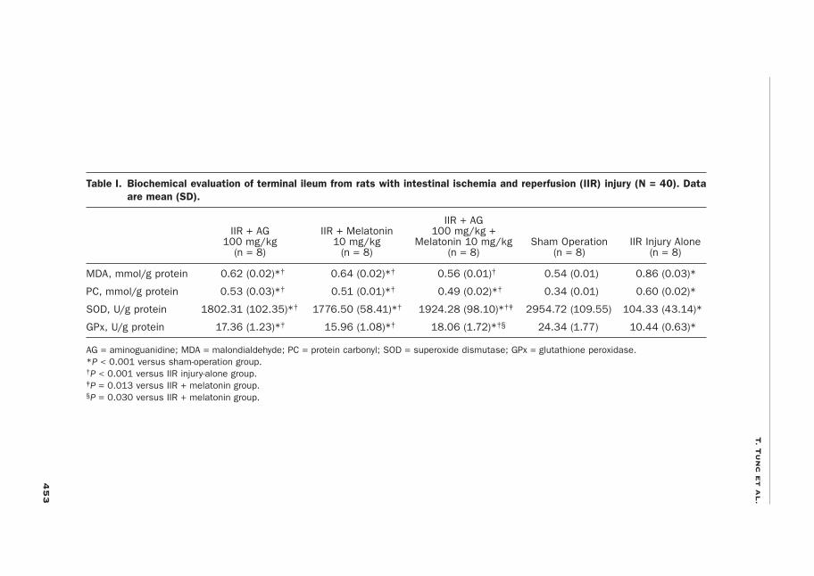
RESULTSBiochemical data are presented in Table I. The IIR injury-alone group had signifi-cantly lower SOD and GPx activity compared with the sham-operation group (mean [SD], SOD: 104.33 [43.14] vs 2954.72 [109.55], respectively; GPx: 10.44 [0.63] vs 24.34 [1.77]; both, P < 0.001). The combination of AG + melatonin was associ-ated with a significant increase in the intestinal SOD and GPx content compared with the melatonin-alone group (mean, SOD: 1924.28 [98.10] vs 1776.50 [58.41], respectively [P = 0.013]; GPx: 18.06 [1.72] vs 15.96 [1.08] [P = 0.030]). Mean MDA concentration in intestinal tissues was significantly higher in the IIR injury-alone group than in the sham-operation group (0.86 [0.03] vs 0.54 [0.01], respec-tively; P < 0.001). The administration of AG or melatonin was associated with lower
4
53
T. T
un
c e
t a
l.
Table I. Biochemical evaluation of terminal ileum from rats with intestinal ischemia and reperfusion (IIR) injury (N = 40). Data
are mean (SD).
IIR + AG IIR + AG IIR + Melatonin 100 mg/kg + 100 mg/kg 10 mg/kg Melatonin 10 mg/kg Sham Operation IIR Injury Alone (n = 8) (n = 8) (n = 8) (n = 8) (n = 8)
MDA, mmol/g protein 0.62 (0.02)*† 0.64 (0.02)*† 0.56 (0.01)† 0.54 (0.01) 0.86 (0.03)*
PC, mmol/g protein 0.53 (0.03)*† 0.51 (0.01)*† 0.49 (0.02)*† 0.34 (0.01) 0.60 (0.02)*
SOD, U/g protein 1802.31 (102.35)*† 1776.50 (58.41)*† 1924.28 (98.10)*†‡ 2954.72 (109.55) 104.33 (43.14)*
GPx, U/g protein 17.36 (1.23)*† 15.96 (1.08)*† 18.06 (1.72)*†§ 24.34 (1.77) 10.44 (0.63)*
AG = aminoguanidine; MDA = malondialdehyde; PC = protein carbonyl; SOD = superoxide dismutase; GPx = glutathione peroxidase.*P < 0.001 versus sham-operation group.†P < 0.001 versus IIR injury-alone group.‡P = 0.013 versus IIR + melatonin group.§P = 0.030 versus IIR + melatonin group.
Current Therapeutic Research
454
MDA concentration compared with the IIR injury-alone group (AG, 0.62 [0.02] vs 0.86 [0.03], respectively; melatonin, 0.64 [0.02] vs 0.86 [0.03]; both, P < 0.001), with the largest decrease observed in the group receiving the combination of the 2 agents (0.56 [0.01] vs 0.86 [0.03]; P < 0.001). The combination of AG + melatonin was associated with a significant decrease in the intestinal MDA content compared with the AG- and melatonin-alone groups (both, P < 0.001). Intestinal PC content concentration in the IIR injury-alone group was significantly higher compared with the sham-operation group (0.60 [0.02] vs 0.34 [0.01]; P < 0.001), and a similar paral-lel increase was seen across the treatment groups (P < 0.001).
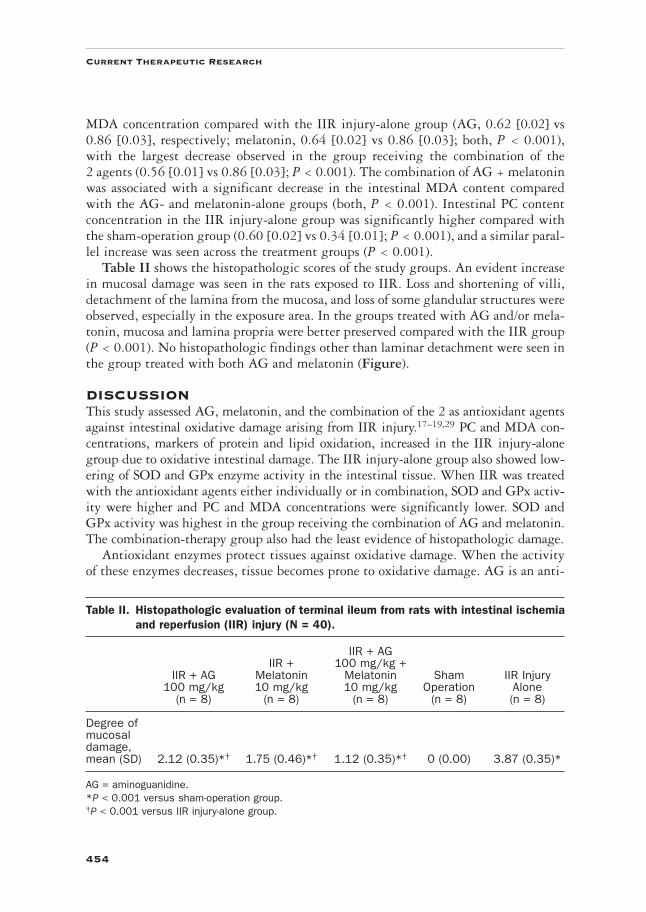
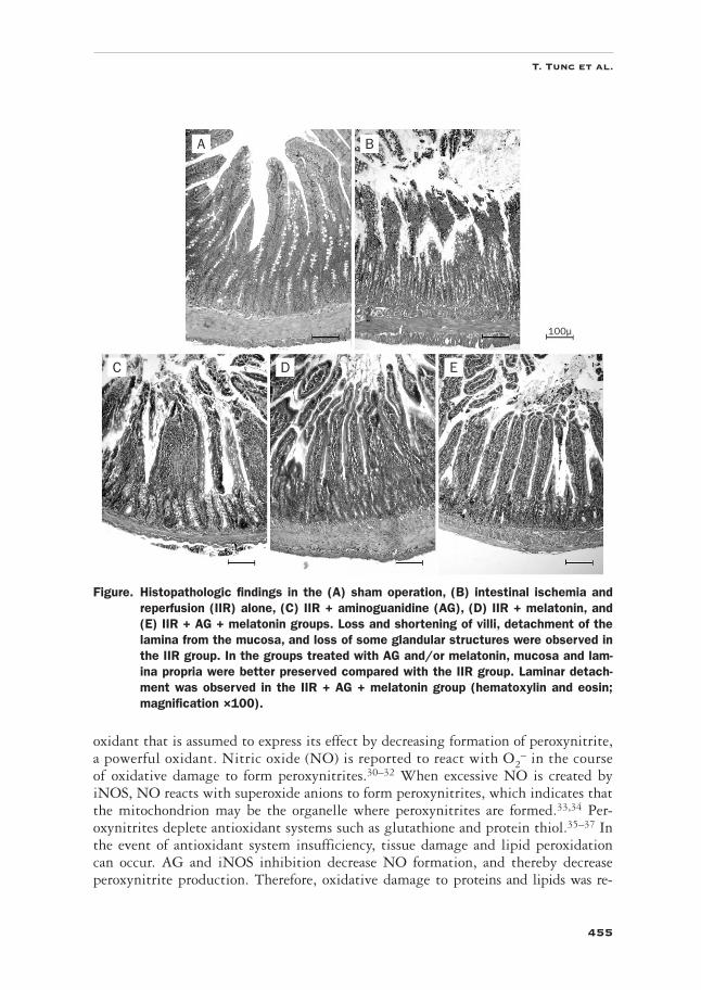
Table II shows the histopathologic scores of the study groups. An evident increase in mucosal damage was seen in the rats exposed to IIR. Loss and shortening of villi, detachment of the lamina from the mucosa, and loss of some glandular structures were observed, especially in the exposure area. In the groups treated with AG and/or mela-tonin, mucosa and lamina propria were better preserved compared with the IIR group (P < 0.001). No histopathologic findings other than laminar detachment were seen in the group treated with both AG and melatonin (Figure).
DISCUSSIONThis study assessed AG, melatonin, and the combination of the 2 as antioxidant agents against intestinal oxidative damage arising from IIR injury.17–19,29 PC and MDA con-centrations, markers of protein and lipid oxidation, increased in the IIR injury-alone group due to oxidative intestinal damage. The IIR injury-alone group also showed low-ering of SOD and GPx enzyme activity in the intestinal tissue. When IIR was treated with the antioxidant agents either individually or in combination, SOD and GPx activ-ity were higher and PC and MDA concentrations were significantly lower. SOD and GPx activity was highest in the group receiving the combination of AG and melatonin. The combination-therapy group also had the least evidence of histopathologic damage.
Antioxidant enzymes protect tissues against oxidative damage. When the activity of these enzymes decreases, tissue becomes prone to oxidative damage. AG is an anti-
Table II. Histopathologic evaluation of terminal ileum from rats with intestinal ischemia
and reperfusion (IIR) injury (N = 40).
IIR + AG IIR + 100 mg/kg + IIR + AG Melatonin Melatonin Sham IIR Injury 100 mg/kg 10 mg/kg 10 mg/kg Operation Alone (n = 8) (n = 8) (n = 8) (n = 8) (n = 8)
Degree of mucosal damage, mean (SD) 2.12 (0.35)*† 1.75 (0.46)*† 1.12 (0.35)*† 0 (0.00) 3.87 (0.35)*
AG = aminoguanidine.*P < 0.001 versus sham-operation group.†P < 0.001 versus IIR injury-alone group.
455
T. Tunc et al.
oxidant that is assumed to express its effect by decreasing formation of peroxynitrite, a powerful oxidant. Nitric oxide (NO) is reported to react with O2
– in the course of oxidative damage to form peroxynitrites.30–32 When excessive NO is created by iNOS, NO reacts with superoxide anions to form peroxynitrites, which indicates that the mitochondrion may be the organelle where peroxynitrites are formed.33,34 Per-oxynitrites deplete antioxidant systems such as glutathione and protein thiol.35–37 In the event of antioxidant system insufficiency, tissue damage and lipid peroxidation can occur. AG and iNOS inhibition decrease NO formation, and thereby decrease peroxynitrite production. Therefore, oxidative damage to proteins and lipids was re-
Figure. Histopathologic findings in the (A) sham operation, (B) intestinal ischemia and
reperfusion (IIR) alone, (C) IIR + aminoguanidine (AG), (D) IIR + melatonin, and
(E) IIR + AG + melatonin groups. Loss and shortening of villi, detachment of the
lamina from the mucosa, and loss of some glandular structures were observed in
the IIR group. In the groups treated with AG and/or melatonin, mucosa and lam-
ina propria were better preserved compared with the IIR group. Laminar detach-
ment was observed in the IIR + AG + melatonin group (hematoxylin and eosin;
magnification ×100).
EDC
100μ
A B
Current Therapeutic Research
456
duced, and the cellular antioxidant systems were preserved. We also found that AG was associated with reduced intestinal injury and decreased MDA levels in the IIR injury-alone group. The efficacy of AG against oxidative damage has been the subject of several studies, and those results are consistent with this investigation.9,38,39
SOD is the first defense against superoxides, and it reduces peroxynitrite forma-tion.8 Sufficient SOD activity must be present in the tissue for this damage reduction to occur. The preventive action of AG results from its direct antioxidant effect, and it is reported to be an effective hydroxyl radical scavenger.40 The molecular oxygen pro-vided by reperfusion becomes a superoxide anion by means of xanthine oxidase. Mean-while, hydrogen peroxide and hydroxyl radicals form, which are responsible for tissue damage.41 The action of AG against oxidative damage and lipid peroxidation and its antioxidant status in several tissue types have been reported in numerous studies.42–44 All seem to be consistent with the present investigation.
Melatonin is reported to be a powerful free radical scavenger in both in vivo and in vitro studies.45,46 Melatonin is also reported to inhibit poly-ADP-ribose synthase activation, and prevent organ damage due to shock, inflammation, or ischemia/reperfusion injury.47 It exercises a tissue-protective effect by increasing the antioxi-dant enzyme levels via its specific receptors and protects the cell from death.48 In addition to its biological effects, melatonin is a powerful free radical scavenger, which suggests a preventive role in ischemia/reperfusion injury.41
In this study, melatonin appears to have had a significant preventive effect on the increase of MDA and PC concentration in rats exposed to IIR. Melatonin also signifi-cantly preserved the activity of SOD and GPx enzymes compared with the IIR group. The preserved intestinal histology in the melatonin group might be attributed to both its radical scavenger and antioxidant features.
The present study suggests that AG and melatonin may have protective effects on IIR that may be associated with their antioxidant and free radical scavenging proper-ties. Limitations of this study include the nonrandomized assignment of the rats to the various study groups and the differences in drug administration routes used. In addition, markers of nitrosative stress and tissue NO levels were not evaluated in this study. To better understand the effects of AG and melatonin in IIR injury, tissue NO level and iNOS activity should be evaluated in future studies.
CONCLUSIONSIn this study, AG, melatonin, or both administered in combination were associated with improvements in oxidative markers in this rat model of IIR injury. Additionally, intestinal mucosa was best preserved in the group receiving the combination. This suggests that better results were obtained by combining the different effects of both agents.
ACKNOWLEDGMENTSThe authors thank Associate Professor Selim Kilic, MD, of the Department of Public Health, Gülhane Military Medical Academy, who helped with statistical analyses. All authors participated in the study’s concept and design.
457
T. Tunc et al.
The authors have indicated that they have no conflicts of interest regarding the content of this article.
REFERENCES 1. Mallick IH, Yang W, Winslet MC, Seifalian AM. Ischemia-reperfusion injury of the intestine
and protective strategies against injury. Dig Dis Sci. 2004;49:1359–1377. 2. Ceylan H, Yüncü M, Gürel A, et al. Effects of whole-body hypoxic preconditioning on hypoxia/
reoxygenation-induced intestinal injury in newborn rats. Eur J Pediatr Surg. 2005;15:325–332. 3. Cabeza J, Motilva V, Martin MJ, de la Lastra CA. Mechanisms involved in gastric protection
of melatonin against oxidant stress by ischemia-reperfusion in rats. Life Sci. 2001;68:1405–1415. 4. Ballabeni V, Barocelli E, Bertoni S, Impicciatore M. Alterations of intestinal motor respon-
siveness in a model of mild mesenteric ischemia/reperfusion in rats. Life Sci. 2002;71:2025– 2035.
5. Erdogan H, Fadillioglu E, Yagmurca M, et al. Protein oxidation and lipid peroxidation after renal ischemia-reperfusion injury: Protective effects of erdosteine and N-acetylcysteine. Urol Res. 2006;34:41–46.
6. Islekel H, Islekel S, Güner G, Ozdamar N. Alterations in superoxide dismutase, glutathione peroxidase and catalase activities in experimental cerebral ischemia-reperfusion. Res Exp Med (Berl). 1999;199:167–176.
7. Schoenberg MH, Beger HG. Reperfusion injury after intestinal ischemia. Crit Care Med. 1993;21:1376–1386.
8. McCord JM. Oxygen-derived free radicals in postischemic tissue injury. N Engl J Med. 1985; 312:159–163.
9. Lipton P. Ischemic cell death in brain neurons. Physiol Rev. 1999;79:1431–1568.10. Draper HH, Hadley M. Malondialdehyde determination as index of lipid peroxidation. Methods
Enzymol. 1990;186:421–431.11. Guven A, Demirbag S, Uysal B, et al. Effect of 3-amino benzamide, a poly(adenosine diphosphate-
ribose) polymerase inhibitor, in experimental caustic esophageal burn. J Pediatr Surg. 2008; 43:1474–1479.
12. Levine RL, Garland D, Oliver CN, et al. Determination of carbonyl content in oxidatively modified proteins. Methods Enzymol. 1990;186:464–478.
13. Schmeling DJ, Caty MG, Oldham KT, Guice KS. Cytoprotection by diclofenac sodium after intestinal ischemia/reperfusion injury. J Pediatr Surg. 1994;29:1044–1048.
14. Koltuksuz U, Ozen S, Uz E, et al. Caffeic acid phenethyl ester prevents intestinal ischemia reperfusion injury in rats. J Pediatr Surg. 1999;34:1458–1462.
15. Hammerman C, Goldschmidt D, Caplan MS, et al. Amelioration of ischemia-reperfusion injury in rat intestine by pentoxifylline-mediated inhibition of xanthine oxidase. J Pediatr Gastroenterol Nutr. 1999;29:69–74.
16. Kazez A, Demirbag M, Ustündag B, et al. The role of melatonin in prevention of intestinal ischemia-reperfusion injury in rats. J Pediatr Surg. 2000;35:1444–1448.
17. Abraham P, Rabi S, Selvakumar D. Protective effect of aminoguanidine against oxidative stress and bladder injury in cyclophosphamide-induced hemorrhagic cystitis in rat. Cell Biochem Funct. 2009;27:56–62.
18. Onem Y, Ipcioglu OM, Haholu A, et al. Posttreatment with aminoguanidine attenuates renal ischemia/reperfusion injury in rats. Ren Fail. 2009;31:50–53.
19. Atasayar S, Gürer-Orhan H, Orhan H, et al. Preventive effect of aminoguanidine compared to vitamin E and C on cisplatin-induced nephrotoxicity in rats. Exp Toxicol Pathol. 2009;61:23–32.
˘ ˘
¸ ¸
Current Therapeutic Research
458
20. Sahna E, Parlakpinar H, Turkoz Y, Acet A. Protective effects of melatonin on myocardial ischemia/reperfusion induced infarct size and oxidative changes. Physiol Res. 2005;54:491–495.
21. Reiter RJ, Tan DX. Melatonin: A novel protective agent against oxidative injury of the ischemic/ reperfused heart. Cardiovasc Res. 2003;58:10–19.
22. Sahna E, Parlakpinar H, Cihan OF, et al. Effects of aminoguanidine against renal ischaemia-reperfusion injury in rats. Cell Biochem Funct. 2006;24:137–141.
23. Colak C, Parlakpinar H, Ozer MK, et al. Investigating the protective effect of melatonin on liver injury related to myocardial ischemia-reperfusion. Med Sci Monit. 2007;13:BR251– BR254.
24. Lowry OH, Rosebrough NJ, Farr AL, Randall RJ. Protein measurement with the Folin phenol reagent. J Biol Chem. 1951;193:265–275.
25. Ohkawa H, Ohishi N, Yagi K. Assay for lipid peroxides in animal tissues by thiobarbituric acid reaction. Anal Biochem. 1979;95:351–358.
26. Sun Y, Oberley LW, Li Y. A simple method for clinical assay of superoxide dismutase. Clin Chem. 1988;34:497–500.
27. Paglia DE, Valentine WN. Studies on the quantitative and qualitative characterization of erythrocyte glutathione peroxidase. J Lab Clin Med. 1967;70:158–169.
28. Chiu CJ, McArdle AH, Brown R, et al. Intestinal mucosal lesion in low-flow states. I. A mor-phological, hemodynamic, and metabolic reappraisal. Arch Surg. 1970;101:478–483.
29. Barocelli E, Ballabeni V, Ghizzardi P, et al. The selective inhibition of inducible nitric oxide synthase prevents intestinal ischemia-reperfusion injury in mice. Nitric Oxide. 2006;14: 212–218.
30. Radi R, Beckman JS, Bush KM, Freeman BA. Peroxynitrite-induced membrane lipid peroxi-dation: The cytotoxic potential of superoxide and nitric oxide. Arch Biochem Biophys. 1991;288:481–487.
31. Reiter RJ, Tan DX, Manchester LC, Qi W. Biochemical reactivity of melatonin with reactive oxy-gen and nitrogen species: A review of the evidence. Cell Biochem Biophys. 2001;34:237–256.
32. Radi R. Nitric oxide, oxidants, and protein tyrosine nitration. Proc Natl Acad Sci U S A. 2004;101:4003–4008.
33. Radi R, Cassina A, Hodara R, et al. Peroxynitrite reactions and formation in mitochondria. Free Radic Biol Med. 2002;33:1451–1464.
34. Trujillo M, Ferrer-Sueta G, Radi R. Peroxynitrite detoxification and its biologic implications. Antioxid Redox Signal. 2008;10:1607–1620.
35. Allen BW, Demchenko IT, Piantadosi CA. Two faces of nitric oxide: Implications for cellular mechanisms of oxygen toxicity. J Appl Physiol. 2009;106:662–667.
36. Varma SD, Hegde KR. Lens thiol depletion by peroxynitrite. Protective effect of pyruvate. Mol Cell Biochem. 2007;298:199–204.
37. Reinartz M, Ding Z, Flögel U, et al. Nitrosative stress leads to protein glutathiolation, in-creased s-nitrosation, and up-regulation of peroxiredoxins in the heart. J Biol Chem. 2008;283: 17440–17449.
38. Hung CR. Role of gastric oxidative stress and nitric oxide in formation of hemorrhagic erosion in rats with ischemic brain. World J Gastroenterol. 2006;12:574–581.
39. Eroglu C, Yildiz OG, Saraymen R, et al. Aminoguanidine ameliorates radiation-induced oxida-tive lung damage in rats. Clin Invest Med. 2008;31:E182–E188.
40. Courderot-Masuyer C, Dalloz F, Maupoil V, Rochette L. Antioxidant properties of aminoguani-dine. Fundam Clin Pharmacol. 1999;13:535–540.
41. Li C, Jackson RM. Reactive species mechanisms of cellular hypoxia-reoxygenation injury. Am J Physiol Cell Physiol. 2002;282:C227–C241.
459
T. Tunc et al.
42. Polat A, Parlakpinar H, Tasdemir S, et al. Protective role of aminoguanidine on gentamicin-induced acute renal failure in rats. Acta Histochem. 2006;108:365–371.
43. Mansour MA, Mostafa AM, Nagi MN, et al. Protective effect of aminoguanidine against neph-rotoxicity induced by cisplatin in normal rats. Comp Biochem Physiol C Toxicol Pharmacol. 2002;132:123–128.
44. Akpinar D, Yargicoglu P, Derin N, et al. Effect of aminoguanidine on visual evoked potentials (VEPs), antioxidant status and lipid peroxidation in rats exposed to chronic restraint stress. Brain Res. 2007;1186:87–94.
45. Reiter RJ, Tan DX, Kim SJ, Qi W. Melatonin as a pharmacological agent against oxidative damage to lipids and DNA. Proc West Pharmacol Soc. 1998;41:229–236.
46. Reiter RJ. Cytoprotective properties of melatonin: Presumed association with oxidative dam-age and aging. Nutrition. 1998;14:691–696.
47. Cuzzocrea S, Reiter RJ. Pharmacological action of melatonin in shock, inflammation and ischemia/ reperfusion injury. Eur J Pharmacol. 2001;426:1–10.
48. Théroux P. Protection of the myocardial cell during ischemia. Am J Cardiol. 1999;83:3G–9G.
Address correspondence to: Turan Tunc, MD, Neonatology Unit, Gülhane Military Medical Academy, 06018, Etlik, Ankara, Turkey. E-mail: [email protected]