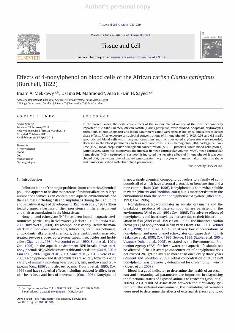
Effects of 4-nonylphenol on blood cells of the African catfish Clarias gariepinus (Burchell, 1822
8
This article appeared in a journal published by Elsevier. The attached copy is furnished to the author for internal non-commercial research and education use, including for instruction at the authors institution and sharing with colleagues. Other uses, including reproduction and distribution, or selling or licensing copies, or posting to personal, institutional or third party websites are prohibited. In most cases authors are permitted to post their version of the article (e.g. in Word or Tex form) to their personal website or institutional repository. Authors requiring further information regarding Elsevier’s archiving and manuscript policies are encouraged to visit: http://www.elsevier.com/copyright
Transcript of Effects of 4-nonylphenol on blood cells of the African catfish Clarias gariepinus (Burchell, 1822

This article appeared in a journal published by Elsevier. The attachedcopy is furnished to the author for internal non-commercial researchand education use, including for instruction at the authors institution
and sharing with colleagues.
Other uses, including reproduction and distribution, or selling orlicensing copies, or posting to personal, institutional or third party
websites are prohibited.
In most cases authors are permitted to post their version of thearticle (e.g. in Word or Tex form) to their personal website orinstitutional repository. Authors requiring further information
regarding Elsevier’s archiving and manuscript policies areencouraged to visit:
http://www.elsevier.com/copyright
Author's personal copy
Tissue and Cell 43 (2011) 223– 229
Contents lists available at ScienceDirect
Tissue and Cell
jou rn al h om epage: www.elsev ier .com/ locate / t i ce
Effects of 4-nonylphenol on blood cells of the African catfish Clarias gariepinus(Burchell, 1822)
Imam A. Mekkawya,b, Usama M. Mahmouda, Alaa El-Din H. Sayeda,∗
a Zoology Department, Faculty of Science, Assiut University, 71516 Assiut, Egyptb Biology Department, Faculty of Science, Taif University, Taif, Saudi Arabia
a r t i c l e i n f o
Article history:Received 21 February 2011Received in revised form 21 March 2011Accepted 21 March 2011Available online 17 April 2011
Keywords:4-NonylphenolBloodCellMicronucleusClarias gariepinus
a b s t r a c t
In the present work, the destructive effects of the 4-nonylphenol on one of the most economicallyimportant Nile fishes, namely African catfish (Clarias gariepinus) were studied. Apoptosis, erythrocytesalterations, micronucleus test and blood parameters count were used as biological indicators to detectthose effects. After exposure to sublethal concentrations of 4-nonylphenol (0, 0.05, 0.08 and 0.1 mg/l),apoptotic red blood cells with many malformations and micronucleated erythrocytes were recorded.Decrease in the blood parameters such as red blood cells (RBCs), hemoglobin (Hb), package cell vol-ume (PCV), mean corpuscular hemoglobin concentration (MCHC), platelets, white blood cells (WBCs),lymphocytes, basophils, monocytes and increase in mean corpuscular volume (MCV), mean corpuscularhemoglobin (MCH), neutrophils, eosinophils indicated the negative effects of 4-nonylphenol. It was con-cluded that, the 4-nonylphenol caused genotoxicity in erythrocytes with many malformations in shapeand number indicated with other blood parameters.
Published by Elsevier Ltd.
1. Introduction
Pollution is one of the major problems in our countries. Chemicalpollution appears to be due to increase of industrialization. A largenumber of chemicals can contaminate aquatic environments andtheir animals including fish and amphibians during their adult lifeand sensitive stages of development (Radhaiah et al., 1987). Theirtoxicity appears because of their persistence in the environmentand their accumulation in the biota tissue.
Nonylphenol ethoxylate (NPE) has been found in aquatic envi-ronments, particularly in river water (Clark et al., 1992; Tsuda et al.,2000; Rivero et al., 2008). This compound is widely used in the man-ufacture of non-ionic surfactants, lubricants, stabilizer polymers,antioxidants, alkylphenol chemicals, detergents, paints, anaerobictreated sewage sludge, polystyrene tubes, insecticides and herbi-cides (Giger et al., 1984; Marcomini et al., 1990; Soto et al., 1991;Cox, 1996). In the aquatic environment NPE breaks down to 4-nonylphenol (NP), which is more stable and persistent (Sakai, 2001;Kim et al., 2002; Uguz et al., 2003; Sone et al., 2004; Rivero et al.,2008). Nonylphenol and its ethoxylates are acutely toxic to a widevariety of animals including bees, spiders, fish, molluscs and crus-taceans (Cox, 1996), and also estrogenic (Flouriot et al., 1995; Cox,1996) and have sublethal effects including reduced fertility, irreg-ular heart beat and loss of movement (Cox, 1996). Nonylphenol
∗ Corresponding author. Tel.: +20 882412381; fax: +20 882342708.E-mail address: alaa [email protected] (A.H. Sayed).
is not a single chemical compound but refers to a family of com-pounds all of which have a central aromatic or benzene ring and anine carbon chain (Cox, 1996). Nonylphenol is somewhat solublein water (Vincent and Sneddon, 2009) but is more persistent in theenvironment than the parent nonylphenol exthyolate (Ahel et al.,1993; Cox, 1996).
Nonylphenols bioaccumulates in aquatic organisms and thebreakdown products of these compounds are persistent in theenvironment (Ahel et al., 1993; Cox, 1996). The adverse effects ofnonylphenols and its ethoxylates increase due to their bioaccumu-lation in fish (Ahel et al., 1993; Cox, 1996). The bioconcentrationfactor (BCF) of nonylphenol in fish varies from 3 to 1300 (Ekelundet al., 1990; Ahel et al., 1993). Relatively low concentrations ofnonylphenol and nonylphenol ethoxylates can cause death to fish(Salanitro et al., 1988; Cox, 1996; Servos, 1999; Staples et al., 2004;Vazquez-Duhalt et al., 2005). As stated by the Environmental Pro-tection Agency (EPA), for fresh water, the aquatic life should notbe affected if the 1 h average concentration of nonylphenol doesnot exceed 28 �g/L on average more than once every three years(Vincent and Sneddon, 2009). Lethal concentration of 0.032 ml/lnonylphenol was previously determined for Oreochromis niloticus(Rivero et al., 2008).
Blood is a good indicator to determine the health of an organ-ism and hematological parameters are important in diagnosingthe functional status of exposed animals to toxicants (Joshi et al.,2002a). As a result of association between the circulatory sys-tem and the external environment, the hematological variableswere used to determine the effects of external stressors and toxic
0040-8166/$ – see front matter. Published by Elsevier Ltd.doi:10.1016/j.tice.2011.03.006

Author's personal copy
224 I.A. Mekkawy et al. / Tissue and Cell 43 (2011) 223– 229
substances (Wendelaar Bonga, 1997). It has been suggested thathaematology, biochemical changes, growth rate and oxygen con-sumption of fish be used in pollutants toxicity detection (Wepener,1997). Damage to blood and heamapoietic organs in fish maybe associated due to either change in environmental conditions(DeWilde and Houston, 1967; Gardner and Yevich, 1969a) or waterborn pollutants (McMay, 1929; Dawson, 1935; Reichenbach-Klink,1966; Gardner and Yevich, 1969b). Blood cell indices (RBCs, WBC,and DLC counts) are good indicators of systemic response to exter-nal stress and any changes therefore reflected in their morphologyand distribution in the blood (Srivastava and Choudhary, 2010).Davis et al. (2008) reported that natural stressors and exogenousstressors in the environment elicit a stress response in fish leuko-cytes profile.
The detection of micronucleus (MN) in fish helps us to knowthe status of water quality, the health of species and potentialrisk (Al-Sabti and Metcalfe, 1995). The study of micronucleus testand abnormal erythrocytes morphology in fishes by various chem-icals have been reported (Grisolia and Starling, 2001; Ferraro et al.,2004; Talapatra and Banerjee, 2007). Fish erythrocytes are espe-cially favoured for micronucleus test (Bushra et al., 2002) and itsfeasibility has already been established in Clarias gariepinus (Bahariet al., 1994). It was stated by many authors that abnormal nuclearmorphology is an indicator of genotoxic damage in fish (Bombailet al., 2001; Talapatra and Banerjee, 2007). Alternatively, variousabnormal morphological forms of erythrocytes are effective indica-tors of cytotoxicity (Bushra et al., 2002). Apoptotic cells are formedby actively dying cells that include cell shrinkage, membrane bleb-bing and chromatin condensation (Murakawa et al., 2001; Talapatraand Banerjee, 2007) and that indicator of abnormal cell divisions(Cavas et al., 2005; Talapatra and Banerjee, 2007).
African catfish (C. gariepinus) is one of the most important trop-ical cultured fish due to high growth rate, high stocking-densitycapacities, high consumer acceptability and high resistance topoor water quality and oxygen depletion (Appelbaum and Kamler,2000; Akinwole and Faturoti, 2007; Adewolu et al., 2008; Karamiet al., 2010). African catfish (C. gariepinus) is distributed through-out Africa (Nguyen and Janssen, 2002). Moreover, it has been usedin fundamental research and considered as an excellent model fortoxicological studies (Degroot, 1987; Volckaert et al., 1994; Nguyenand Janssen, 2002; Osman et al., 2007a; Mekkawy et al., 2010), sinceit has a well documented biology (Volckaert et al., 1994; Osmanet al., 2007b).
The present work was therefore, undertaken to examinethe ability of 4-nonylphenol to induce apoptosis, micronucleus,morphological alterations in blood erythrocytes and other hema-tological parameters of the catfish, C. gariepinus.
2. Materials and methods
2.1. Specimen collection
Specimens of adult Catfish C. gariepinus were collected fromthe River Nile at Assiut and then were transported to Fish BiologyLaboratory at Zoology Department, Faculty of Science, Assiut Uni-versity. The fish (500–1200 g) were fed on a commercial pellet diet(3% of body weight per day) and kept together in 100 l rectangulartanks containing tap water (conductivity 2000 l/cm; pH 7.5; oxy-gen 88–95% saturation; temperature 27–28 ◦C; photoperiod 12:12light:dark). After 2 weeks acclimatization, fishes were used for theexperimental setup.
2.2. 4-Nonylphenol
Nonylphenol was obtained from Sigma–Aldrich (Schnelldrof,Germany).
2.3. Experimental setup
The adapted adult fish were subdivided into four groups (10 fishper each): control, 4-nonylphenol-treated group (0.05 mg/l dailyfor 15 days), 4-nonylphenol-treated group (0.08 mg/l daily for 15days), and 4-nonylphenol-treated group (0.1 mg/l daily for 15 days).In the present study, the range of NP exposures was 0.05–0.1 mg/land these concentrations were chosen in accordance with envi-ronmentally observed values. The conditions of the experimentwere as that of acclimatization with changing all the tap water andconcentrations of 4-nonylphenol every day.
2.4. Hematological parameters
Blood samples were taken from the caudal vein into heparinizedtubes. The concentration of Hb and blood cells count were imme-diately estimated. Other samples of blood were centrifuged at5000 rpm for 10 min and serum samples were stored in polyethy-lene Eppendorf test tubes at −20 ◦C until serum analysis. TheRBCs, WBCs, blood platelets, haematocrite (HCT), hemoglobin (Hb)were determined by using automated technical analyser (Min-dray Bc-2800). Mean corpuscular volume (MCV), mean corpuscularhemoglobin (MCH), and mean corpuscular hemoglobin concentra-tion (MCHC) were calculated using the formulae mentioned byDacie and Lewis (1991).
MCHC (g/dl) = HbHct
× 100
MCH (pg) = HbRBCs
× 10
MCV (mm3) = HctRBCs
× 10
Differential WBCs were counted using blood smears stainedwith Giemsa stain according to Tavares-Dias and Moraes (2003).
2.5. Apoptosis detection
Apoptotic erythrocytes were detected using Annexin V-EGFPApoptosis Detection Kit (Cat. No. K104-25, 100, 400), Biovisionresearch products, 980 Linda Vista Avenue, Mountain view, CA94043, USA). The procedure used to detect the apoptosis in RBCswere as, after collection of blood in heperinized tubes centrifuga-tion occurred at 50,000 rpm obtaining 1–5 × 10⁵ cells, resuspensioncells in 100 �l of 1× binding buffer, then addition of 5 �l Annexin-EGFP and 5 �l of propidium iodide. Incubation at room temperaturefor 5 min in the dark and then placing the cell suspension on glassslide and covered with a glass coverslip. Finally observation cellsunder a Zeiss Axioplan2 fluorescence microscope (200×) providedwith a digital 3 CCD color video camera (Sony, AVT-Horn).
2.6. Micronucleus test and erythrocytes alterations
Blood smears were obtained by the caudal incision on cleangrease free microscopic slides after completion of a desired expo-sure. The smears were fixed in absolute methanol for 10 min afterdrying at room temperature. Slides were stained with haema-toxylin and eosin. It was followed by dehydration in ascendinggrades of alcohol (30, 50, 70, and 90%, absolute), Finally the slideswere cleared in xylene and permanently mounted by DPX (Pascoeand Gatehouse, 1986). Many slides were selected on the basis ofstaining quality, then coded, randomized and scored blindly. In eachgroup 10,000 cells (a minimum of 1000 per slide) were examined(Al-Sabti and Metcalfe, 1995) at 40× objective and 10× eyepiece for

Author's personal copy
I.A. Mekkawy et al. / Tissue and Cell 43 (2011) 223– 229 225
Table 1Apoptosis, micronucleus test and altered erythrocytes (mean ± SD) % after exposure to different doses of 4-nonylphenol of adults African catfish Clarias gariepinus (averagebetween parentheses).
Test Treatments
Control 0.05 mg/l 4-nonylphenol 0.08 mg/l 4-nonylphenol 0.1 mg/l 4-nonylphenol
Apoptosis (%) 0.8 ± 1.03 (0.0–3) a 3 ± 1.41 (1–5) b 6.1 ± 1.72 (3–8) c 23.9 ± 4.22 (18–32) dMicronucleus test (%) 0.2 ± 0.42 (0.0–1) a 1 ± 0.816 (0.0–2) b 1.9 ± 0.99 (1–4) c 4.2 ± 2.44 (1–9) dAltered erythrocytes (%) 0.7 ± 0.48 (0.0–1) a 2.1 ± 1.19 (0.0–4) b 12 ± 3.26 (5–18) c 42 ± 15.98 (12–64) d
Different letters indicates there is a significant difference at (p ≤ 0.05).
micronucleated and morphologically altered erythrocytes in sepa-rate studies. The established criteria for identifying MN (Schmidt,1975) were strictly followed to ensure authentic scoring. In mor-phologically altered erythrocytes, the important variations suchas acanthocytes, tear drop like cells, sickle cells, and swelled cellsechinocytes, alteration of nuclear morphology, enucleated erythro-cytes and vacuoles were observed.
2.7. Statistical analysis
The basic statistics, means, standard divisions and ranges wereestimated. The pattern of variation was one-way analysis of vari-ance using the SPSS package (SPSS 1998) at the 0.05 significancelevel. The Tukey-HSD test was considered for multiple comparisonsand designed to verify the frequency of apoptotic cells, MN inci-dence and erythrocyte alterations. Dunnett t-tests treat one groupas a control, and compare all other groups against it.
2.8. Ethical statement
All experiments were carried out in accordance with the Egyp-tian laws and University guidelines for the care of experimentalanimals. All procedures of the current experiment have beenapproved by the Committee of the Faculty of Science of AssiutUniversity, Egypt.
3. Results
3.1. Apoptosis detection and erythrocytes alterations
As shown in Table 1, the apoptotic cells percentage appears inthe groups treated with 4-nonylphenol more than the control one.Also, this increased with 4-nonylphenol doses increase.
Fig. 1 shows the blood smear of normal fish and represents thenormal structure of blood of the catfish, C. gariepinus. The bloodis composed of nucleated erythrocytes (Er); rounded with a cen-trally located rounded nucleus, thrombocytes (T); were found ovalwith the nucleus nearly filling the cell with dark purple stain.There are two types of lymphocytes according to their size, theircytoplasm is weakly basophilic, non granular with large roundedand darkly stained nucleus, large lymphocytes (LL) are circularin shape, non granular leucocytes with large rounded and darklystained nucleus, small lymphocytes (SL); as that of large lympho-cytes but smaller in size, monocytes (M); non granular leucocyteswith kidney-shaped nucleus and weakly basophilic cytoplasm,neutrophils (N); granulated leucocytes, with a faint purple color,rounded in shape with eccentrically nucleus which exhibits a vari-ety of shapes, eosinophils (E); granulated and rounded leucocytes,they stain bright purple, their nucleus have no definite shape, beingsome times rounded, kidney-shaped or lobulated, basophils (B);basophilic granulated leucocytes, few in number, rounded or ovalwith eccentrically and variably shaped nucleus.
Exposure of catfish to sublethal concentrations of 4-nonylphenol (0.05 mg/l, 0.08 mg/l and 0.1 mg/l) resulted inmorphological changes in the red blood cells and appearance
Fig. 1. Blood film of control catfish Clarias gariepinus showing the rounded shapeof the nucleated erythrocytes (Er), thrymbocytes (T), large lymphocytes (LL), smalllymphocytes (SL), monocyte (M), neutrophils (N), eosinophil (E), and basophil (B).(400×).
of some pathologic types of cells. The major alterations of RBCsin the fish treated with 0.05 mg/l 4-nonylphenol (Fig. 2) areshown by echinocytes or crenated cells (Cr); where the red bloodcells develop irregular cell surface with numerous projections,acanthocytes (Ac); crenated cells with less projections from thesurface, tear drop like cells (Tr), their shape looks like tear withpointed apices. Fig. 3 shows other morphological changes in RBCsin fish treated with 0.08 mg/l 4-nonylphenol, that are sickle cells(Sk) which vary in shape between elliposoidal, boat-shaped andgenuine sickles, swelled cells (Sc); their cytoplasm filled withwater and micronucleus (Mn); one or more Mn per cell in the mostobservations. Also in this treated group, crenated cells, acantho-
Fig. 2. Blood film of treated catfish Clarias gariepinus with 0.05 mg/l 4-nonylphenolshowing crenated cells; echinocytes (Cr), tear drop like cells (Tr) and acanthocyte(Ac). (400×).

Author's personal copy
226 I.A. Mekkawy et al. / Tissue and Cell 43 (2011) 223– 229
Fig. 3. Blood film of treated catfish Clarias gariepinus with 0.08 mg/l 4-nonylphenolshowing sickle cells (Sk), swelled cells (Sc) and distinct micronucleus (Mn). (400×).
cytes and tear drop like cells were frequent (Fig. 3). Besides, lysisof some red cells, where some cells appears to be stacked together.
The fish exposed to 0.1 mg/l 4-nonylphenol shows many alter-ations in their blood (Figs. 4–8) such as some of red blood cells losetheir normal shape, acanthocytes and crenated cells were apparentbeside lysis of some cells, sticking of the red blood cells and frag-mentations of some cells were observed, tear drop like cells andsickle cells become common and obvious disintegration of cell wallof some cells with variation in their nuclei shape. Fig. 4 shows sicklecells, swelled cells, distinct micronucleus and sticking of the redblood cells. Blood film of treated fish with 0.1 mg/l 4-nonylphenolas in Fig. 5a and b shows sticking altered cells with disintegration ofthe red blood cells (arrows). Fig. 6 recorded common malformationsin RBCs which become sticking, altered, acquired different shapesand pale cytoplasm (Pc). The most common observed change inthe red blood cell morphology after treatment with 0.1 mg/l 4-nonylphenol was the cells sticking together in a chain like shape
Fig. 4. Blood film of treated catfish Clarias gariepinus with 0.1 mg/l 4-nonylphenolshowing crenated cells (Cr), swelled cells (Sc), cells have prominent vacuoles (Va)and distinct micronucleus (Mn). (400×).
Fig. 5. Blood film of treated catfish Clarias gariepinus with 0.1 mg/l 4-nonylphenolshowing sticking altered cells; (a and b) notice disintegration of the cell wall of RBCs(arrow). (400×).
Fig. 6. Blood film of treated catfish Clarias gariepinus with 0.1 mg/l 4-nonylphenolshowing sticking altered cells. Notice RBCs acquired different shapes with pale cyto-plasm (Pc). (400×).
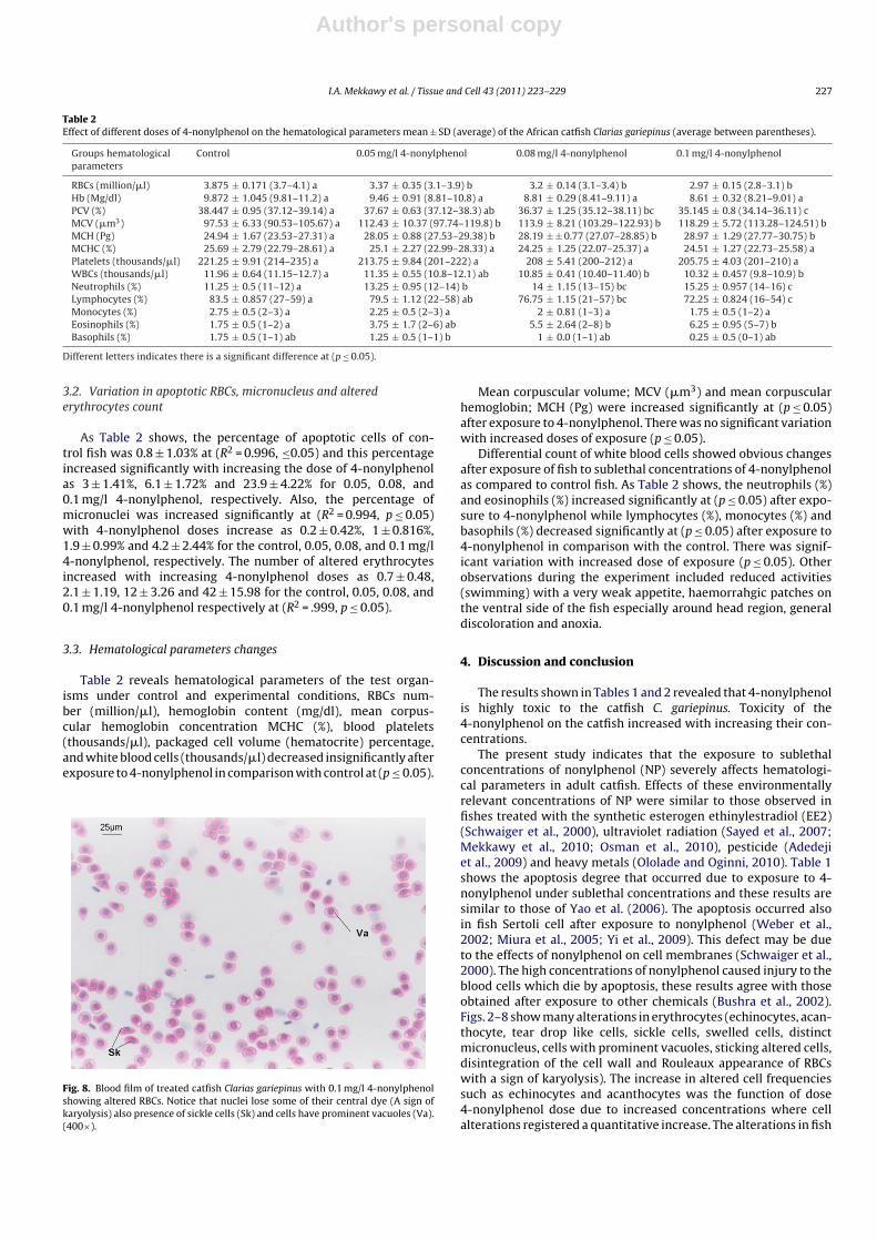
which called Rouleaux appearance of RBCs as shown in Fig. 7a andb. A sign of karyolysis; where cells nuclei lose their central dye,red blood cells with prominent vacuoles and presence of sicklecells were observed in fish treated with 0.1 mg/l 4-nonylphenol asshown in Fig. 8.
Fig. 7. Blood film of treated catfish Clarias gariepinus with 0.1 mg/l 4-nonylphenolshowing Rouleaux appearance of RBCs; (a and b) (400×).
Author's personal copy
I.A. Mekkawy et al. / Tissue and Cell 43 (2011) 223– 229 227
Table 2Effect of different doses of 4-nonylphenol on the hematological parameters mean ± SD (average) of the African catfish Clarias gariepinus (average between parentheses).
Groups hematologicalparameters
Control 0.05 mg/l 4-nonylphenol 0.08 mg/l 4-nonylphenol 0.1 mg/l 4-nonylphenol
RBCs (million/�l) 3.875 ± 0.171 (3.7–4.1) a 3.37 ± 0.35 (3.1–3.9) b 3.2 ± 0.14 (3.1–3.4) b 2.97 ± 0.15 (2.8–3.1) bHb (Mg/dl) 9.872 ± 1.045 (9.81–11.2) a 9.46 ± 0.91 (8.81–10.8) a 8.81 ± 0.29 (8.41–9.11) a 8.61 ± 0.32 (8.21–9.01) aPCV (%) 38.447 ± 0.95 (37.12–39.14) a 37.67 ± 0.63 (37.12–38.3) ab 36.37 ± 1.25 (35.12–38.11) bc 35.145 ± 0.8 (34.14–36.11) cMCV (�m3) 97.53 ± 6.33 (90.53–105.67) a 112.43 ± 10.37 (97.74–119.8) b 113.9 ± 8.21 (103.29–122.93) b 118.29 ± 5.72 (113.28–124.51) bMCH (Pg) 24.94 ± 1.67 (23.53–27.31) a 28.05 ± 0.88 (27.53–29.38) b 28.19 ± ± 0.77 (27.07–28.85) b 28.97 ± 1.29 (27.77–30.75) bMCHC (%) 25.69 ± 2.79 (22.79–28.61) a 25.1 ± 2.27 (22.99–28.33) a 24.25 ± 1.25 (22.07–25.37) a 24.51 ± 1.27 (22.73–25.58) aPlatelets (thousands/�l) 221.25 ± 9.91 (214–235) a 213.75 ± 9.84 (201–222) a 208 ± 5.41 (200–212) a 205.75 ± 4.03 (201–210) aWBCs (thousands/�l) 11.96 ± 0.64 (11.15–12.7) a 11.35 ± 0.55 (10.8–12.1) ab 10.85 ± 0.41 (10.40–11.40) b 10.32 ± 0.457 (9.8–10.9) bNeutrophils (%) 11.25 ± 0.5 (11–12) a 13.25 ± 0.95 (12–14) b 14 ± 1.15 (13–15) bc 15.25 ± 0.957 (14–16) cLymphocytes (%) 83.5 ± 0.857 (27–59) a 79.5 ± 1.12 (22–58) ab 76.75 ± 1.15 (21–57) bc 72.25 ± 0.824 (16–54) cMonocytes (%) 2.75 ± 0.5 (2–3) a 2.25 ± 0.5 (2–3) a 2 ± 0.81 (1–3) a 1.75 ± 0.5 (1–2) aEosinophils (%) 1.75 ± 0.5 (1–2) a 3.75 ± 1.7 (2–6) ab 5.5 ± 2.64 (2–8) b 6.25 ± 0.95 (5–7) bBasophils (%) 1.75 ± 0.5 (1–1) ab 1.25 ± 0.5 (1–1) b 1 ± 0.0 (1–1) ab 0.25 ± 0.5 (0–1) ab
Different letters indicates there is a significant difference at (p ≤ 0.05).
3.2. Variation in apoptotic RBCs, micronucleus and alterederythrocytes count
As Table 2 shows, the percentage of apoptotic cells of con-trol fish was 0.8 ± 1.03% at (R2 = 0.996, ≤0.05) and this percentageincreased significantly with increasing the dose of 4-nonylphenolas 3 ± 1.41%, 6.1 ± 1.72% and 23.9 ± 4.22% for 0.05, 0.08, and0.1 mg/l 4-nonylphenol, respectively. Also, the percentage ofmicronuclei was increased significantly at (R2 = 0.994, p ≤ 0.05)with 4-nonylphenol doses increase as 0.2 ± 0.42%, 1 ± 0.816%,1.9 ± 0.99% and 4.2 ± 2.44% for the control, 0.05, 0.08, and 0.1 mg/l4-nonylphenol, respectively. The number of altered erythrocytesincreased with increasing 4-nonylphenol doses as 0.7 ± 0.48,2.1 ± 1.19, 12 ± 3.26 and 42 ± 15.98 for the control, 0.05, 0.08, and0.1 mg/l 4-nonylphenol respectively at (R2 = .999, p ≤ 0.05).
3.3. Hematological parameters changes
Table 2 reveals hematological parameters of the test organ-isms under control and experimental conditions, RBCs num-ber (million/�l), hemoglobin content (mg/dl), mean corpus-cular hemoglobin concentration MCHC (%), blood platelets(thousands/�l), packaged cell volume (hematocrite) percentage,and white blood cells (thousands/�l) decreased insignificantly afterexposure to 4-nonylphenol in comparison with control at (p ≤ 0.05).
Fig. 8. Blood film of treated catfish Clarias gariepinus with 0.1 mg/l 4-nonylphenolshowing altered RBCs. Notice that nuclei lose some of their central dye (A sign ofkaryolysis) also presence of sickle cells (Sk) and cells have prominent vacuoles (Va).(400×).
Mean corpuscular volume; MCV (�m3) and mean corpuscularhemoglobin; MCH (Pg) were increased significantly at (p ≤ 0.05)after exposure to 4-nonylphenol. There was no significant variationwith increased doses of exposure (p ≤ 0.05).
Differential count of white blood cells showed obvious changesafter exposure of fish to sublethal concentrations of 4-nonylphenolas compared to control fish. As Table 2 shows, the neutrophils (%)and eosinophils (%) increased significantly at (p ≤ 0.05) after expo-sure to 4-nonylphenol while lymphocytes (%), monocytes (%) andbasophils (%) decreased significantly at (p ≤ 0.05) after exposure to4-nonylphenol in comparison with the control. There was signif-icant variation with increased dose of exposure (p ≤ 0.05). Otherobservations during the experiment included reduced activities(swimming) with a very weak appetite, haemorrahgic patches onthe ventral side of the fish especially around head region, generaldiscoloration and anoxia.
4. Discussion and conclusion
The results shown in Tables 1 and 2 revealed that 4-nonylphenolis highly toxic to the catfish C. gariepinus. Toxicity of the4-nonylphenol on the catfish increased with increasing their con-centrations.
The present study indicates that the exposure to sublethalconcentrations of nonylphenol (NP) severely affects hematologi-cal parameters in adult catfish. Effects of these environmentallyrelevant concentrations of NP were similar to those observed infishes treated with the synthetic esterogen ethinylestradiol (EE2)(Schwaiger et al., 2000), ultraviolet radiation (Sayed et al., 2007;Mekkawy et al., 2010; Osman et al., 2010), pesticide (Adedejiet al., 2009) and heavy metals (Ololade and Oginni, 2010). Table 1shows the apoptosis degree that occurred due to exposure to 4-nonylphenol under sublethal concentrations and these results aresimilar to those of Yao et al. (2006). The apoptosis occurred alsoin fish Sertoli cell after exposure to nonylphenol (Weber et al.,2002; Miura et al., 2005; Yi et al., 2009). This defect may be dueto the effects of nonylphenol on cell membranes (Schwaiger et al.,2000). The high concentrations of nonylphenol caused injury to theblood cells which die by apoptosis, these results agree with thoseobtained after exposure to other chemicals (Bushra et al., 2002).Figs. 2–8 show many alterations in erythrocytes (echinocytes, acan-thocyte, tear drop like cells, sickle cells, swelled cells, distinctmicronucleus, cells with prominent vacuoles, sticking altered cells,disintegration of the cell wall and Rouleaux appearance of RBCswith a sign of karyolysis). The increase in altered cell frequenciessuch as echinocytes and acanthocytes was the function of dose4-nonylphenol dose due to increased concentrations where cellalterations registered a quantitative increase. The alterations in fish

Author's personal copy
228 I.A. Mekkawy et al. / Tissue and Cell 43 (2011) 223– 229
erythrocytes were observed in hypoxic condition (Sawhney andJohal, 2000) and by factors that induce apoptosis of blood cells likeradiations (Chukhlovin, 1996). The vacuoles observed in erythro-cytes could be due to unequal distribution of hemoglobin similar toresults of Bushra et al. (2002). The swelled blood cells were recordedas common alteration which may be due to their cellular outlinesthat undergo necrosis (Bushra et al., 2002). Erythrocytes are veryimportant for oxygen transport (Kisch, 1951). Oxygen is impor-tant to fish during respiration occurring via the gills as erythrocytedeformations causes low oxygen level, affecting the circulating sys-tem and causes respiration disfunction. This respiratory stress mayalter the fish erythrocyte number and morphology (Brown, 1993).
Degeneration of erythrocytes could be ascribed to pathologicalconditions in fishes exposed to toxicants (Buckley et al., 1976). It hasbeen reported that high concentration or sublethal concentrationsexposure of fish to pesticides (Adedeji et al., 2009) and to heavymetals such as nickel (Ololade and Oginni, 2010), lead (Adeyemo,2007) usually decrease erythrocyte indices. Some of these alter-ations in erythrocytes were recorded in fish after exposure topesticide (Adedeji et al., 2009). The reduction in RBCs, Hb and PCVsuggests an anaemic condition in treated fish and may be attributedto cytotoxic effect and suppression of erythopoiesis caused by ben-zene derivatives (Keller and Synder, 1988). The same results oferythrocyte, haematcrite and hemoglobin decrease were reportedafter exposure to diazinon (Adedeji et al., 2009). It has beenreported that anaemia found in NP-exposed fish is a consequenceof an interaction between NP and the erythrocyte membrane(Schwaiger et al., 2000). Toxic components induce anaemia andinterfered with platelet production in the animals which reducetheir values (Sudakov, 1992). Lower hemoglobin level may decreasethe ability of fish to enhance its activity in order to meet occasionaldemands according to Joshi et al. (2002b). The significant decreasein the MCHC for the exposed groups is an indication for variations inerythrocyte shape, size and hemoglobin content (Adeyemo, 2005)which is an indicator of anaemia (Eastham and Slade, 1993). Themajor functions of WBCs are to fight infection, defend the bodyagainst foreign organisms and in immune response. A significantreduction in WBCs count with increase in 4-nonylphenol concen-tration suggests that the catfish is exposed to high risk of infection.Significant decrease of WBCs after acute exposure to diazinon wasrecorded (Adedeji et al., 2009). The changes in differential WBCcount can be used as indicator for immunity decrease after expo-sure to toxic substances (Adedeji et al., 2009). Our results showedan increase in neutrophils with decrease in lymphocytes num-bers and these results in agreement with results of Davis et al.(2008). In conclusion, our results confirm the sublethal effect of4-nonylphenol on adult catfish C. gariepinus, using a set of hemato-logical parameters. The decrease in the level of RBCs, HB, and PCVrevealed the hematotoxic effects of this chemical pollutant. Theapoptosis detection, altered erythrocytes and micronucleus testcould be effectively used as potential biomarkers of 4-nonylphenoltoxicity to the fresh water fish and they could be ranked as possiblebiomarkers of pollution.
References
Adedeji, O.B., Adeyemo, O.K., Agbede, S.A., 2009. Effects of diazinon on bloodparameters in the African catfish (Clarias gariepinus). Afr. J. Biotechnol. 8 (16),3940–3946.
Adewolu, M.A., Adeniji, C.A., Adejobi, A.B., 2008. Feed utilization, growth and sur-vival of Clarias gariepinus (Burchell, 1822) fingerlings cultured under differentphotoperiods. Aquaculture 283, 64–67.
Adeyemo, O.K., 2005. Haematological and histopathological effects of Cassava MillEffluent in Clarias gariepinus. Afr. J. Biomed. Res. 8 (3), 179–183.
Adeyemo, O.K., 2007. Haematological profile of Clarias gariepinus (Burchell, 1822)exposed to lead. Turk. J. Fish Aquat. Sci. 7, 163–169.
Ahel, M., McEvoy, J., Giger, W., 1993. Bioaccumulation of the lipophilic metabo-lites of nonionic surfactants in freshwater organisms. Environ. Pollut. 79,243–248.
Akinwole, A.O., Faturoti, E.O., 2007. Biological performance of African Catfish (Clariasgariepinus) cultured in recirculating system in Ibadan. Aquacult. Eng. 36, 18–23.
Al-Sabti, K., Metcalfe, C.D., 1995. Fish micronuclei for assessing genotoxicity in water.Mutat. Res. 343, 121–135.
Appelbaum, S., Kamler, E., 2000. Survival, growth, metabolism and behaviour ofClarias gariepinus (Burchell, 1822) early stages under different light conditions.Aquacult. Eng. 22, 269–287.
Bahari, I.B., Noor, F.M., Daud, N.M., 1994. Micronucleated erythrocytes as an assay toassess actions by physical and chemical genotoxic agents in Clarias gariepinus.Mutat. Res. 313, 1–5.
Bombail, V., Aw, D., Gordon, E., Batty, J., 2001. Application of the comet and micronu-cleus assays to butterfish (Pholis gunnelus) erythrocytes from the Firth of Forth,Scotland. Chemosphere 44, 283–392.
Brown, L., 1993. Aquaculture for veterinarians: fish husbandry and medicine. Perg-amon press, Oxford, North Chicago, USA, 440 pp.
Buckley, J.A., Whitmore, C.M., Matsuda, R.I., 1976. Changes in blood chemistry andblood cell morphology in coho salmon, Oncorhynchus kisutch following exposureto sublethal levels of total residual chlorine in municipal wastewater. J. Fish Res.Board Canada 33, 776–782.
Bushra, A., Abul farah, M., Niamat, M.A., Waseem, A., 2002. Induction ofmicronuclei and erythrocyte alterations in the catfish Clarias batrachus by 2,4-dichlorophenoxyacetic acid and butachlor. Mutat. Res. 518, 135–144.
Cox, C., 1996. Nonyl phenol and related chemicals. J. Pesticide, Reform vol. 16 (Spring(1)) (corrected 4/2003).
Cavas, T., Garanko, N.N., Arkhipchuk, V.V., 2005. Induction of micronuclei and bin-uclei in blood, gill and liver cells of fishes subchronically exposed to cadmiumchloride and copper sulphate. Food Chem. Toxicol. 43, 569–574.
Chukhlovin, A.B., 1996. Apoptosis and red blood cell echinocytosis: common fea-tures. Scanning Microsc. 10, 795–803.
Clark, L.B., Rosen, R.T., Hartman, T.G., Louis, J.B., Suffet, I.H., Lippincott, R.L., 1992.Determination of alkylphenol ethoxylates and their acetic acids derivatives indrinking water by particle beam liquid chromatograph/electroscopy. Int. J. Env-iron. Anal. Chem. 47, 167–180.
Dacie, S., Lewis, S. (Eds.), 1991. Practical Haematology. , 7th ed. Churchill Livingstone,London, p. 633.
Davis, A.K., Maney, D.L., Maerz, J.C., 2008. The use of leukocyte profiles to measurestress in vertebrates: a review for ecologists. Funct. Ecol. 22 (5), 760–772.
Dawson, A.B., 1935. The hemopoietic response in the catfish Ameittrus nebulosus tochronic lead poisoning. Biol. Bull. 68, 335–346.
Degroot, S.J., 1987. Culture of Clarias species. Aquaculture 63, 1–36.DeWilde, M.A., Houston, A.H., 1967. Haematological aspects of the thermoacclima-
tory process in rainbow trout Salmo gairdnerii. J. Fish Res. Board Canada 24,2267–2281.
Eastham, R.D., Slade, R.R., 1993. Clinical Haematology, 7th ed. Butterworth-Heinemann Ltd.
Ekelund, R., et al., 1990. Bioaccumulation of 4-nonylphenol in marine animals-Areevaluation. Environ. Pollut. 64, 107–120.
Ferraro, M.V.M., Fenocchio, A.S., Mantovani, M.S., Ribeiro, C.O., Cestari, M.M.,2004. Mutagenic effects of tributyltin and inorganic lead (Pb II) on thefish H. malabaricus as evaluated using the comet assay and the piscinemicronucleus and chromosome aberration tests. Genet. Mol. Biol. 27, 103–107.
Flouriot, G., Pakdel, F., Ducouret, B., Valotaire, Y., 1995. Influence of xenobiotics onrainbow trout liver estrogen receptor and vitellogenin gene expression. J. Mol.Endocrinol. 15 (2), 143–151.
Gardner, G.R., Yevich, P.P., 1969a. Studies on the blood morphology of three estuarinecyprinodontiform fishes. J. Fish Res. Board Canada 26, 433–447.
Gardner, G.R., Yevich, P.P., 1969b. Toxicological effects of cadmium on Fundulus het-eroclitus under various oxygen, Ph, salinity and temperature regims. Ame. Zool.9, 1096.
Giger, W., Brunner, P.H., Schaffner, C., 1984. 4-Nonylphenol in sewage sludge: accu-mulation of toxic metabolites from non-ionic surfactants. Science 225, 623–625.
Grisolia, C.K., Starling, F.L.R.M., 2001. Micronuclei monitoring of fishes from lakeParanoa under influence of sewage treat plant discharges. Mutat. Res. 491,39–44.
Joshi, P.K., Bose, M., Harish, D., 2002a. Changes in haematological parameters in asiluroid catfish Clarias batrachus (Linn) exposed to mercuric choloride. Pollut.Rec. 21 (2), 129–131.
Joshi, P.K., Harish, D., Bose, M., 2002b. Effect of lindane and malathione exposure tocertain blood parameters in a fresh water teleost fish Clarias batrachus. Pollut.Rec. 21 (1), 55–57.
Karami, A., Christianus, A., Ishak, Z., Courtenay, S.C., Syed, M.A., Noor, Azlina, M.,Noorshinah, H., 2010. Effect of triploidization on juvenile African catfish (Clariasgariepinus). Aquacult. Int. 18, 851–858.
Keller, A., Synder, C.A., 1988. Mice exposed in utero to 20 ppm benzene exhibitaltered numbers of recognizable haematoppietic cells up seven weeks afterexposure. Fundam. Appl. Toxicol. 10, 224–232.
Kim, H.S., Shin, J.H., Kang, I.H., Kim, T.S., Kim, I.Y., Seok, J.H., Pyo, M.Y., Han, S.Y., 2002.Comparative estrogenic effects of nonylphenol by 3-day uterotrophic assay andfemale pubertal onset assay. Reprod. Toxicol. 16, 259–268.
Kisch, B., 1951. Observation on the haematology of fishes and bird. Exp. Med. Surg.7, 326.
Marcomini, A., Pavoni, B., Sfriso, A., Orio, A.A., 1990. Persistent metabolites ofalkylphenol polyethoxylates in the marine environment. Marine Chem. 29,307–323.
Author's personal copy
I.A. Mekkawy et al. / Tissue and Cell 43 (2011) 223– 229 229
McMay, C.M., 1929. Studies Upon Fish Blood and its Relation to Water Pollution. ABiological Survey of the Erie-Niagra System. New York Conservation Dep. N.Y,pp. 140–149.
Mekkawy, I.A.A., Mahmoud, U.M., Osman, A.G., Sayed, A.H., 2010. Effectsof ultraviolet A on the activity of two metabolic enzymes, DNA dam-age and lipid peroxidation during early developmental stages of theAfrican catfish, Clarias gariepinus (Burchell, 1822). Fish Physiol. Biochem. 36,605–626.
Miura, C., Takahashi, N., Michino, F., Miura, T., 2005. The effect of para-nonylphenolon Japanese eel (Anguilla japonica) spermatogenesis in vitro. Aquat. Toxicol. 71,133–141.
Murakawa, M., Jung, S.K., Iijima, K., Yonehara, S., 2001. Apoptosis induc-ing protein, AIP, from parasite-infected fish induces apoptosis in mam-malian cells by two different molecular mechanisms. Cell Death Differ. 8,298–307.
Nguyen, L.T.H., Janssen, C.R., 2002. Embryo-larval toxicity tests with the Africancatfish (Clarias gariepinus): comparative sensitivity of endpoints. Arch. Environ.Contam. Toxicol. 42, 256–262.
Ololade, I.A., Oginni, O., 2010. Toxic stress and hematological effects of nickel onAfrican catfish, Clarias gariepinus, fingerlings. J. Environ. Chem. Ecotoxicol. 2 (2),014–019.
Osman, A.G., Koutb, M.M., Sayed, A.H., 2010. Use of hematological parameters toassess the efficiency of quince (Cydonia oblonga Miller) leaf extract in allevia-tion of the effect of ultraviolet—a radiation on African catfish Clarias gariepinus(Burchell, 1822). J. Photochem. Photobiol. B: Biol. 99, 1–8.
Osman, A.G., Mekkawy, I.A.A., Verreth, J., Kirschbaum, F., 2007a. Effects of lead nitrateon the activity of metabolic enzymes during early developmental stages of theAfrican catfish, Clarias gariepinus (Burchell, 1822). Fish Physiol. Biochem. 33,1–13.
Osman, A.G., Wuertz, S., Mekkawy, I.A.A., Exner, H., Kirschbaum, F., 2007b. Leadinduced malformations in embryos of the African Catfish Clarias gariepinus(Burchell, 1822). Environ. Toxicol. 22 (4), 375–389.
Pascoe, S., Gatehouse, D., 1986. The use of a simple haematoxylin and eosin stainingprocedure to demonstrate micronuclei within rodent bone marrow. Mutat. Res.164, 237–243.
Radhaiah, V., Girija, M., Rao, K.J., 1987. Changes in selected biochemical parame-ters in the kidney and blood of the fish, Tilapia mossambica (Peters), exposed toheptachlor. Bull. Environ. Contam. Toxicol. 39, 1006–1011.
Reichenbach-Klink, H.H., 1966. The blood components of fish with relation toparasiyies infection and water pollution. Bull. Int. Epizool. 65, 1039–1054.
Rivero, C.L.G., Barbosa, A.C., Ferreira, M.N., Dorea, J.G., Grisolia, C.K., 2008. Evalua-tion of genotoxicity and effects on reproduction of nonylphenol in Oreochromisniloticus (Pisces: Cichlidae). Ecotoxicology 17, 732–737.
Sakai, A., 2001. p-Nonilfenol acts as a promoter in the BALB/3T3 cell transformation.Mutat. Res. 493, 161–166.
Salanitro, J.P., et al., 1988. Activated sludge treatment of ethoxylate surfactants athigh industrial use concentrations. Water Sci. Technol. 20, 125–130.
Sawhney, A.K., Johal, M.S., 2000. Erythrocyte alterations induced by malathion inChanna punctatus (Bloch). Bull. Environ. Contam. Toxicol. 64, 398–405.
Sayed, A.H., Ibrahim, A.Th., Mekkawy, I.A.A., Mahmoud, U.M., 2007. Acute effects ofultraviolet-A radiation on African Catfish Clarias gariepinus (Burchell, 1822). J.Photochem. Photobiol. B: Biol. 89, 170–174.
Schmidt, W., 1975. The micronucleus test. Mutat. Res. 31, 9–15.Schwaiger, J., Spieser, O.H., Bauer, C., Ferling, H., Mallow, U., Kalbfus, W., Negele,
R.D., 2000. Chronic toxicity of nonylphenol and ethinylestradiol: haematological
and histopathological effects in juvenile Common carp (Cyprinus carpio). Aquat.Toxicol. 51, 69–78.
Servos, M.R., 1999. Review of the aquatic toxicity, estrogenic responses and bioac-cumulation of alkylphenols and alkylphenol polyethoxylates. Water Qual. Res.J. Canada 34, 123–177.
Sone, K., Hinago, M., Kitayama, A., Morokuma, J., Ueno, N., Watanabe, H., Iguchi,T., 2004. Effects of 17 B-estradiol, nonylphenol, and bisphenol-A on developingXenopus laevis embryos. Gen. Comp. Endocrinol. 138, 228–236.
Soto, A.M., Justicia, H., Wray, J.W., Sonnenschein, C., 1991. p-Nonyl-phenol: an estro-genic xenobiotic released from “modified” polystyrene. Environ. Health Pers. 92,167–173.
Srivastava, S., Choudhary, S.K., 2010. Effect of artificial photoperiod on the blood cellindices of the catfish, Clarias batrachus. J. Stress Physiol. Biochem. 6 (1), 22–32.
Staples, C., Mihaich, E., Carbone, J., Woodbrun, K., Klecka, G., 2004. A weightof evidence analysis of the chronic ecotoxicity of nonylphenol ethoxylates,nonylphenol ether carboxylates, and nonylphenol. Human Ecol. Risk Assess. 10,999–1017.
Sudakov, K.V., 1992. Stress postulate: analysis from the position of general theoryof functional systems. Pathophysiol. Exp. Ther. 4, 86–98.
Talapatra, S.N., Banerjee, S.K., 2007. Detection of micronucleus and abnormalnucleus in erythrocytes from the gill and kidney of Labeo bata cultivated insewage-fed fish farms. Food Chem. Toxicol. 45, 210–215.
Tavares-Dias, M., Moraes, F.R., 2003. Hematological evaluation of Tilapia rendalliBoulenger, 1896 (Osteichthyes: Cichlidae) captured in a fee fishing farm fromFranca, Säo Paulo, Brasil (in Portuguese). Biosci. J. 19, 103–110.
Tsuda, T., Takino, A., Kojima, M., Harada, K., Muraki, T., Tsuji, M., 2000. 4-Nonylphenols and 4-terc-octylphenol in water and fish from rivers flowing intolake Biwa. Chemosphere 41, 757–762.
Uguz, C., Iscan, M., Erguven, A., Isgor, B., Togan, I., 2003. The bioaccumulation ofnonilfenol and its adverse effect on the liver of rainbow trout (Onchorynchusmykiss). Environ. Res. l92, 262–270.
Vazquez-Duhalt, R., Marquez-Rocha, F., Ponce, E., Licea, A.F., Viana, M.T., 2005.Nonylphenol, and integrated vision of a pollutant. Appl. Ecol. Environ. Res. 4,1–25.
Vincent, M.D.V., Sneddon, J., 2009. Nonylphenol: an overview and its determinationin oysters and wastewaters and preliminary degradation results from laboratoryexperiments. Microchem. J., doi:10.1016/j.microc.2009.02.005.
Volckaert, F.A.M., Hellemans, B.A., Galbusera, P., Ollevier, F., Sekkali, B., Belayew,A., 1994. Replication, expression and fate of foreign DNA during embryonicand larval development of the African catfish Clarias gariepinus. Mol. Mar. Biol.Biotechnol. 3, 57–69.
Weber, L.P., Kiparissis, Y., Hwang, G.S., Niimi, A.J., Janz, D.M., Metcalfe, C.D., 2002.Increased cellular apoptosis after chronic aqueous exposure to nonylphenoland quercetin in adult medaka (Oryzias latipes). CBP: C-Toxicol. Pharmacol. 131,51–59.
Wendelaar Bonga, S.E., 1997. The stress response in fish. Physiol. Rev. 77, 591–625.Wepener, W., 1997. Metal ecotoxicology of the Ohfant River in the Kruger National
Park and the effect thereof on fish haematology. Ph.D Thesis, Rand AfrikaansUniversity, South Africa.
Yao, G.H., Yang, L.S., Hu, Y.L., Liang, J., Liang, J.F., Hou, Y.Y., 2006. Nonylphenol-induced thymocyte apoptosis involved caspase-3 activation and mitochondrialdepolarization. Mol. Immunol. 43, 915–926.
Yi, G., Jiang, W., Yufeng, H., Sunan, S., Xiaodong, H., 2009. Nonylphenol induces apo-ptosis in rat testicular Sertoli cells via endoplasmic reticulum stress. Toxicol.Lett. 186, 84–95.