Effects from filtration, capping agents, and presence/absence of food on the toxicity of silver...
10
United States Environmental Protection Agency U.S. Environmental Protection Agency Papers University of Nebraska - Lincoln Year EFFECTS FROM FILTRATION, CAPPING AGENTS, AND PRESENCE/ABSENCE OF FOOD ON THE TOXICITY OF SILVER NANOPARTICLES TO DAPHNIA MAGNA H. Joel Allen * Christopher A. Impellitteri † Dana A. Macke ‡ J. Lee Heckman ** Helen C. Poynton †† James M. Lazorchak ‡‡ Shekar Govindaswamy § Deborah L. Roose ¶ Mallikarjuna N. Nadagouda k * U.S. Environmental Protection Agency † U.S. Environmental Protection Agency, [email protected] ‡ U.S. Environmental Protection Agency ** Shaw Environmental and Infrastructure †† U.S. Environmental Protection Agency ‡‡ U.S. Environmental Protection Agency § Lakeshore Engineering Services ¶ U.S. Environmental Protection Agency k Pegasus Technical Services This paper is posted at DigitalCommons@University of Nebraska - Lincoln. http://digitalcommons.unl.edu/usepapapers/78
-
Upload
independent -
Category
Documents
-
view
0 -
download
0
Transcript of Effects from filtration, capping agents, and presence/absence of food on the toxicity of silver...
United States Environmental Protection Agency
U.S. Environmental Protection Agency Papers
University of Nebraska - Lincoln Year
EFFECTS FROM FILTRATION,
CAPPING AGENTS, AND
PRESENCE/ABSENCE OF FOOD ON
THE TOXICITY OF SILVER
NANOPARTICLES TO DAPHNIA
MAGNA
H. Joel Allen∗ Christopher A. Impellitteri† Dana A. Macke‡
J. Lee Heckman∗∗ Helen C. Poynton†† James M. Lazorchak‡‡
Shekar Govindaswamy§ Deborah L. Roose¶ Mallikarjuna N. Nadagouda‖
∗U.S. Environmental Protection Agency†U.S. Environmental Protection Agency, [email protected]‡U.S. Environmental Protection Agency∗∗Shaw Environmental and Infrastructure††U.S. Environmental Protection Agency‡‡U.S. Environmental Protection Agency§Lakeshore Engineering Services¶U.S. Environmental Protection Agency‖Pegasus Technical Services
This paper is posted at DigitalCommons@University of Nebraska - Lincoln.
http://digitalcommons.unl.edu/usepapapers/78

EFFECTS FROM FILTRATION, CAPPING AGENTS, AND PRESENCE/ABSENCE OF FOOD ONTHE TOXICITY OF SILVER NANOPARTICLES TO DAPHNIA MAGNA
H. JOEL ALLEN,y CHRISTOPHER A. IMPELLITTERI,*y DANA A. MACKE,y J. LEE HECKMAN,z HELEN C. POYNTON,yJAMES M. LAZORCHAK,y SHEKAR GOVINDASWAMY,§ DEBORAH L. ROOSE,y and MALLIKARJUNA N. NADAGOUDAk
yU.S. Environmental Protection Agency, Office of Research and Development, Cincinnati, Ohio
zShaw Environmental and Infrastructure,
§Lakeshore Engineering Services, U.S. EPA Test & Evaluation Facility, Cincinnati, Ohio
kPegasus Technical Services, 46 East Hollister Street, Cincinnati, Ohio, USA
(Submitted 13 October 2009; Returned for Revision 15 December 2009; Accepted 20 June 2010)
Abstract—Relatively little is known about the behavior and toxicity of nanoparticles in the environment. Objectives of work presentedhere include establishing the toxicity of a variety of silver nanoparticles (AgNPs) to Daphnia magna neonates, assessing the applicabilityof a commonly used bioassay for testing AgNPs, and determining the advantages and disadvantages of multiple characterizationtechniques for AgNPs in simple aquatic systems. Daphnia magna were exposed to a silver nitrate solution and AgNPs suspensionsincluding commercially available AgNPs (uncoated and coated), and laboratory-synthesized AgNPs (coated with coffee or citrate). Thenanoparticle suspensions were analyzed for silver concentration (microwave acid digestions), size (dynamic light scattering and electronmicroscopy), shape (electron microscopy), surface charge (zeta potentiometer), and chemical speciation (X-ray absorption spectro-scopy, X-ray diffraction). Toxicities of filtered (100 nm) versus unfiltered suspensions were compared. Additionally, effects fromaddition of food were examined. Stock suspensions were prepared by adding AgNPs to moderately hard reconstituted water, which werethen diluted and used straight or after filtration with 100-nm filters. All nanoparticle exposure suspensions, at every time interval, weredigested via microwave digester and analyzed by inductively coupled argon plasma–optical emission spectroscopy or graphite furnace–atomic absorption spectroscopy. Dose–response curves were generated and median lethal concentration (LC50) values calculated. TheLC50 values for the unfiltered particles were (in mg/L): 1.1� 0.1-AgNO3; 1.0� 0.1-coffee coated; 1.1� 0.2-citrate coated; 16.7� 2.4Sigma Aldrich Ag-nanoparticles (SA) uncoated; 31.5� 8.1 SA coated. LC50 values for the filtered particles were (in mg/L): 0.7� 0.1-AgNO3; 1.4� 0.1-SA uncoated; 4.4� 1.4-SA coated. The LC50 resulting from the addition of food was 176.4� 25.5-SA coated.Recommendations presented in this study include AgNP handling methods, effects from sample preparation, and advantages/disadvantages of different nanoparticle characterization techniques. Environ. Toxicol. Chem. 2010;29:2742–2750. # 2010 SETAC
Keywords—Silver Nanoparticles Daphnia magna Aquatic toxicity
Toxicity of nanomaterials (<100 nm) is a relatively new areaof ecotoxicological research. Production and application ofthese materials has increased rapidly; however; their environ-mental fate and effects are poorly understood. Apparent size-dependent properties such as malleability, optical and electricalproperties, and toxicity are desired characteristics in manyengineered applications but also may play an important rolein their ultimate environmental effects. Estimating current andfuture relevant environmental concentrations is difficult. How-ever, basic toxicological information can and should be gen-erated. Previous research has investigated the toxicity ofnanomaterials in the aquatic environment, including carbonfullerenes [1–3], carbon nanotubes [4–6], and inorganic nano-materials such as TiO2, SiO2, and ZnO [7–9]. Establishingtesting and characterization methods specific to nanoparticlesand determining the utility of classic aquatic toxicity tests inassessing and managing risks from releases of nanoparticles inthe environment are crucial.
The number of manufacturer-identified consumer productscontaining nano-sized materials roughly doubled between 2006and 2007 [10] and continues to increase. The main use of silver
nanoparticles (AgNPs) is in preventing microbial growth[11,12]. This anti-microbial characteristic also means thatAg, especially in ionic form, can be quite toxic to aquaticorganisms [13]. Other cited potential uses for AgNPs are inpolymers/composites [14,15], textiles [16], and solar energymaterials [17]. Silver nanoparticles used as antimicrobial agents(e.g., fabrics containing AgNPs) and impregnated polymerscould potentially enter wastewater treatment and aquatic systems.
Past studies show that Ag ions disrupt sodium influx byblocking the functions of Naþ,Kþ-ATPase, resulting in ionor-egulatory disturbance [18]. This agrees with mechanismsobserved in fish [19]. Mechanisms of toxicity of AgNPs areless clear. Size dependent activity has been observed in viralinactivation [20]. It has also been suggested that toxicity stemsfrom the production of Ag ions from the oxidation of AgNPs[21], which could cause effects similar to those observed in Agion toxicity. The generation of Ag ion from AgNPs was alsoattributed with causing toxicity to freshwater algae [22].
In very simple systems, generation of Ag ions may be thehighest risk factor from AgNPs to aquatic organisms. However,it has been well documented that Ag ions react very quicklywith common environmental ligands such as sulfide and chlor-ide [23]. These reactions can lead to a decrease in toxicity of theAgNPs [24].
The objectives of this study are to establish the toxicity ofdifferent forms of AgNPs to Daphnia magna neonates relativeto AgNO3; examine the effects of filtration in the AgNP
Environmental Toxicology and Chemistry, Vol. 29, No. 12, pp. 2742–2750, 2010# 2010 SETAC
Printed in the USADOI: 10.1002/etc.329
All Supplemental Data may be found in the online version of this article.* To whom correspondence may be addressed
([email protected]).Published online 23 August 2010 in Wiley Online Library
(wileyonlinelibrary.com).
2742
suspension preparation steps on the toxicity assay results;compare and contrast toxicity results in an assay in which foodis present versus the assay without food; assess the effects ofcoating agents on the toxicity of AgNPs; and characterize theAgNPs using a wide variety of methods. This work contributesto initial efforts to establish baseline toxicity of AgNPsto aquatic invertebrates and to the development of standardoperating procedures for creating reproducible exposuresuspensions and speciation/identification of AgNPs withinthe timeframe of typical aquatic toxicity tests.
MATERIALS AND METHODS
Dilution water
The test medium was synthetic moderately hard reconsti-tuted water (MHRW), prepared using granular activated carbonand ion exchange filtered Cincinnati (OH, USA) tap waterpolished to ASTM type II specifications by a Barnstead Nano-pure Ultrapure (Barnstead Thermolyne) system following themethod described by the U.S. Environmental ProtectionAgency (U.S. EPA) [25]. Table S1 in the Supplemental Datacontains typical values for hardness, alkalinity, pH, specificconductivity, DO, total organic carbon (TOC), and dissolvedorganic carbon (DOC).
Toxicity assay methods
Toxicity assays were performed using D. magna. Forty-eight-hour static-renewal tests were performed at least in dupli-cate following methods described by the U.S. EPA [25] at 258C,with mortality as the endpoint. Assays and cultures wereprovided with a 16:8h light:dark photoperiod. Organicallycoated (<100 nm, 99.5% purity, cat. no. 576832) and uncoatedAgNPs (<150 nm, 99% purity, cat. no. 484059) were purchasedfrom Sigma Aldrich. The manufacturer was contacted forinformation on the nature of the organic coating, but noinformation could be obtained because the coating was con-sidered proprietary. The AgNPs were also synthesized usingAgNO3 [26] in combination with citrate or coffee solutions. Aseries of bioassays using AgNO3 (Sigma Aldrich) were per-formed to compare results with established response values inthe literature. Fifty percent serial dilutions were used in theassays with high concentrations ranging from 5 to 400 mg/L,depending on initial toxicity results. Observed (i.e., analyzed)concentrations were used for data analysis. For the fed 48-h test,0.5 ml of an algae/grass extract suspension (Pseudokirchner-iella subcapitata, STAR collection) were added at test initiationand renewal. Algae were grown in a large scale mass culturemodified from U.S. EPA methods [25]. Less than 24-h-oldneonates were held with food before exposure. All tests werecovered and slowly aerated using granular activated carbonfiltered compressed air to keep the nanoparticles suspended.Four replicates were performed for each concentration for eachof the Ag species tested.
One set of toxicity assays were performed for each of theSigma Aldrich Ag-nanoparticles (SA) AgNP types after theAgNP suspensions were filtered (100-nm nylon filter, GE/Osmonics). Results from these assays were compared withresults from exposures to suspensions that were not filtered.For stock suspensions that were filtered, a mass of AgNPs weretransferred to acid-washed 1-L volumetrics, suspended inMHRW, and sonicated for 15 min. The suspensions were thenagitated by inverting (three times) and filtered through 100-nmnylon syringe filters. This process is commonly used to removeaggregates of nanoparticles. For stock suspensions without
sonication or filtration, the AgNPs were added to MHRWand agitated. After stock suspension preparation, samples ofAgNPs of all types were transferred to microwave digestionvessels (MARS, CEM Corporation) for digestion in triplicate,by U.S. EPA method 3015A (without the addition of HCl) [27]and analyzed by inductively coupled argon plasma–opticalemission spectrometry (ICP-OES, Model Optima 2100, PerkinElmer). If the relative standard deviation for a set of stocksuspension replicates was less than 20%, the replicate valueswere averaged and recorded as the stock suspension concen-tration. Fresh stock solutions were prepared for each bioassayand stored in the dark for no longer than one week. Dissolutionof silver nanoparticles was not directly evaluated, but zetapotential measurements of the suspensions showed good stabil-ity of citrate- and coffee-capped particles over a three-monthperiod (data not shown). Different stirring rates (50, 100, and200 rpm on magnetic stir plate) and agitation methods (e.g.,gentle inversion vs vigorous shaking) were used to determinethe most effective method for generating reproducible transfersof the stock suspension either to microwave digestion vessels orvessels for the working suspensions. To monitor the experi-ments as thoroughly as possible, every suspension used in aparticular assay was digested and analyzed at T¼ 0, 24, and48 h; at every time period when the suspension was replaced.The entire volume of the exposure cup (25 ml) was directlytransferred to a Teflon1 microwave digestion vessel. Eachsuspension was quantitatively transferred with two separatealiquots of trace metal grade HNO3. The total volume ofHNO3 added was 10 ml. In preliminary experiments, recoveriesand reproducibility (four exposure replicates per exposureconcentration) were greatly improved with the HNO3 rinses.After digestion, the suspensions were analyzed either by induc-tively coupled argon plasma–optical emission spectrometer(instrument detection limit¼ 10mg/L) or by graphite furnaceatomic absorption (GFAA-Model AA240Z, Varian, instrumentdetection limit¼ 0.5mg/L). Critical factors influencing suspen-sion concentration reproducibility were the method of agitationand type and size of the stock transfer pipette.
Bioassay statistical analysis
Data generated from the assays were pooled by toxicant,filtration, and feeding. Dose–response curves (DRC) weregenerated using the DRC package [28] in the R statisticalpackage [29]. Curves were generated using a two-parameterlogistic model. Curve fit was visually assessed. The chi-squaredtest for heterogeneity was not used to assess lack of fit, becauselarge values of the goodness-of-fit test can result from data thatinclude contributions from concentrations with small expectedvalues [30,31]. Variability was expressed using a 95% con-fidence interval. The EC50 values were compared using thelikelihood-ratio test at the a¼ 0.05 level.
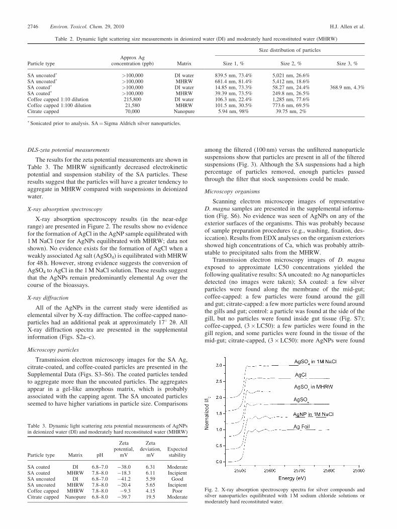
X-ray absorption spectroscopy
One-gram samples of coated and uncoated AgNPs wereequilibrated for 48 h in MHRW in the presence and absenceof chloride (1.0 M sodium salt). A silver salt, AgSO4, was alsoequilibrated for 48 h with MHRW and 1.0 M NaCl. Silverreference materials included AgCl, AgSO4, Ag-foil (elementalAg), and coated and uncoated AgNPs. The samples and refer-ence materials were mounted between two sections of Kaptontape. Silver (K-edge¼ 25,514 eV) X-ray absorption near edgestructure data were collected at beamline 10-ID (MaterialsResearch Collaborative Access Team) at the Advanced PhotonSource at Argonne National Laboratory, Argonne, IL, USA.
Toxicity of silver nanoparticles to Daphnia magna Environ. Toxicol. Chem. 29, 2010 2743
The electron storage ring operated at 7 GeV. A liquid nitrogen–cooled double crystal Si(111) monochromator was used toselect incident photon energies, and a platinum-coated glassmirror was used for harmonic rejection.
At least three scans were collected for each sample. TheX-ray absorption near edge structure spectra were collected influorescence mode using ionization chambers for the incident,transmitted, fluorescence, and reference channels. The collectedscans for a particular sample were aligned using a reference Agfoil and averaged. The averaged data were then normalized, andthe background was removed by spline fitting using the X-rayabsorption spectroscopy data processing program Athena [32].
X-ray diffraction
X-ray diffraction (Xpert Pro, PANalytical B.V.) was used toidentify crystalline phases of AgNPs using Cu Ka radiation at45 kV and 40 mA. Scans ranged from 5 to 90 degrees 2 theta,with 0.02-degree step sizes that were held for 2 s each. Patternanalysis was performed using the Jadeþ software v.7 (MDI),which follows ASTM D934-80 procedures. Sample spectrawere compared with reference patterns from the 2002 PowderDiffraction File (2nd release, International Center for Diffrac-tion Data).
Scanning and transmission electron microscopy
Scanning electron microscopy (SEM, JEOL-6490-LV,JEOL) was performed at the U.S. EPA’s National Risk Manage-ment Research Laboratory in Cincinnati, OH. The AgNPsamples were placed on double-sided carbon tape and mountedon an aluminum sample stub. Images were created for particlesin MHRW and D. magna neonates exposed to LC50 concen-trations of AgNPs. The organisms were imaged in their entiretyafter desiccation and placement on the aluminum sample stub.The exteriors of the organisms were imaged to ascertain thepresence/absence of sorbed AgNPs to surfaces (i.e., antennae,setae, carapace). Energy dispersive X-rays (EDX, X-act EDS,Oxford Instruments) were used to identify elements present atparticular spots in the samples.
Transmission electron microscopy (TEM) was performed atthe U.S. EPA’s National Risk Management Research Labora-tory (particles), the University of Cincinnati (Ohio, USA)(particles), and Purdue University (West Lafayette, Indiana,USA) (D. magna and particles). Particles passing through a100-nm filter were analyzed by TEM (JEOL-1200EXII, JEOL)at the U.S. EPA’s National Risk Management Research Labo-ratory laboratory. Images were captured using a Gatan digitalcamera. Samples were placed on a carbon-coated grid, air dried,and collected at an accelerating voltage of 120 kV.
Particle suspensions were also analyzed in stock solutions atthe beginning of the experiments at the Material Character-ization Laboratory at the University of Cincinnati by TEM(CM-20, Phillips). The nanoparticle suspensions were placed ona Cu grid and air dried, and images were collected at anaccelerating voltage of 200 kV. The images were collectedusing a Gatan ORIUSTM 8-megapixel imaging system. Inaddition, a confirmatory analysis (for Ag presence) was per-formed on some of the imaged samples, using an EDX Energydispersive system (Genesis XMS 2000, EDAX).
Transmission electron microscopy images of exposed D.magnawere generated at Purdue University.D. magna, exposedat approximate LC50 concentrations, were preserved in 2%glutaraldehyde at U.S. EPA’s National Risk ManagementResearch Laboratory and transported to Purdue University.The organisms were then washed in a 0.1-M cacodylate buffer
(pH 7.4 [2� ]), washed in deionized water, dehydratedwith ethanol, and embedded in Spurr’s resin. Thin sections(1- to 2-mm cross sections) stained with uranyl acetate and leadcitrate were made and analyzed by TEM, using an FEI/PhillipsCM-100 transmission electron microscope. Identifying Agnanoparticles from other darkly staining particles was difficult.Therefore, most images were taken without staining with uranylacetate and lead citrate. Images of the samples were created atsimilar distances from the tail (80–100mm). At this point bothgill and midgut were visible in the thin sections.
Images of particles were also taken on the Purdue UniversityTEM, using an accelerating voltage of 80 kV, 50-m objectiveaperture, and spot 3. Magnifications varied between 3,900� and52,000� . Images were captured on Kodak Electron ImageType S0-163 film.
Dynamic light scattering
Particle size/hydrodynamic diameter (HDD) was determinedby dynamic light scattering (DLS) using a Zetasizer Nanoseries(Malvern Instruments) with a 633-nm laser source and adetection angle of 1738 (detection range, 1 nm to 10mm).Measurements were conducted using a 2 ml sample depositedin a disposable polystyrene cuvette. Reported measurementsrepresent the average, by cumulants analysis as described in theInternational Standard on DLS ISO13321 Part 8 [33], of at least12 independent measurements. The size distribution wasobtained from a nonnegative least squares analysis and basedon particle concentration introduced into the instrument. Theinstrument measures the hydrodynamic diameter, the diameterof a sphere that has the same diffusion coefficient as theparticles in solution.
Zeta potential
Zeta potentials were calculated from micro electrophoresismeasurements collected from the Malvern DLS instrument,using a disposable electrophoretic flow through cell with aninternal volume of approximately 1 ml. Ten milliliters samplewas flushed through the flow cell before measurement to avoidcontamination from a previous measurement. Based on pre-liminary data, the voltage applied to the capillary cell was set at100 mV, and a Henry function ( f [Ka]) of 1.5 to 1.23 was used tocalculate zeta potential. Zeta potential was measured at ambientpH. The reported zeta potential values are the average of 10micro electrophoresis measurements. The standard deviationfor all zeta potential measurements was less than 1.5 mV. TheSA particles were analyzed in deionized water and MHRW. Thecoffee-capped particles were measured in MHRW. The citrate-capped particles were not suspended in MHRW, because thenanoparticles were at low silver concentrations, and the instru-ment requires perceptible color/turbidity in colloidal solutionsfor the measurements.
RESULTS
Toxicity
Responses of D. magna to the AgNPs were dependent ondose. The LC50 values of the eight combinations of treatmentsare found in Table 1. Dose–response curves are presented inFigure 1. Toxicity of the AgNPs (without the addition of food)spanned an order of magnitude. The laboratory-synthesizedAgNPs coated with coffee or citrate resulted in dose–responsessimilar to, but slightly higher than, the AgNO3 solution. For theSA AgNPs, the coated form was less toxic than the uncoatedform, and the filtered suspensions (for coated and uncoated)
2744 Environ. Toxicol. Chem. 29, 2010 H.J. Allen et al.
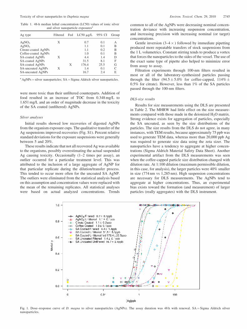
were more toxic than their unfiltered counterparts. Addition offood resulted in an increase of TOC from 0.348 mg/L to1.651 mg/L and an order of magnitude decrease in the toxicityof the SA coated (unfiltered) AgNPs.
Silver analyses
Initial results showed low recoveries of digested AgNPsfrom the organism exposure cups. The qualitative transfer of theAg suspensions improved recoveries (Fig. S1). Percent relativestandard deviations for the exposure suspensions were generallybetween 5 and 20%.
These results indicate that not all recovered Ag was availableto the organisms, possibly overestimating the actual suspendedAg causing toxicity. Occasionally (1–2 times per assay), anoutlier occurred for a particular treatment level. This wasattributed to the inclusion of a large aggregate of AgNP forthat particular replicate during the dilution/transfer process.This tended to occur more often for the uncoated SA AgNP.The outliers were eliminated from the statistical analysis basedon this assumption and concentration values were replaced withthe mean of the remaining replicates. All statistical analyseswere based on actual analyzed concentrations. Trends
common to all of the AgNPs were decreasing nominal concen-tration deviance with increasing suspension concentration,and increasing precision with increasing nominal (or target)concentrations.
Gentle inversion (3–4� ) followed by immediate pipettingproduced more repeatable transfers of stock suspensions fromthe 1 L volumetrics. Constant stirring tends to produce a vortexthat forces the nanoparticles to the sides of the vessel. The use ofthe exact same type of pipette also helped to minimize errorfrom assay to assay.
Filtration experiments through 100-nm filters resulted inmost or all of the laboratory-synthesized particles passingthrough the filter (94.5� 5.0% for coffee-capped, 114%�0.5% for citrate). However, less than 1% of the SA particlespassed through the 100-nm filters.
DLS-size results
Results for size measurements using the DLS are presentedin Table 2. The MHRW had little effect on the size measure-ments compared with those made in the deionized H2O matrix.Strong evidence exists for aggregation of particles, especiallythe SA uncoated, as seen by the size distributions of theparticles. The size results from the DLS do not agree, in manyinstances, with TEM results, because approximately 75 ppb wasused to generate TEM data, whereas more than 20,000 ppb Agwas required to generate size data using the zeta sizer. Thenanoparticles have a tendency to aggregate at higher concen-trations (Sigma Aldrich Material Safety Data Sheet). Anotherexperimental artifact from the DLS measurements was seenwhen the coffee-capped particle size distribution changed withdilution rate. At 1:100 dilution (maximum permissible dilution,in this case, for analysis), the larger particles were 40% smallerin size (774 nm vs 1,285 nm). High suspension concentrationsare necessary for DLS measurements. The AgNPs tend toaggregate at higher concentrations. Thus, an experimentalbias exists toward the formation (and measurement) of largerparticles (really aggregates) with the DLS instrument.
Table 1. 48-h median lethal concentration (LC50) values of ionic silverand silver nanoparticle exposuresa
Ag type Filtered Fed LC50 mg/L 95% CI Group
AgNO3 X 0.7 0.1 AAgNO3 1.1 0.1 BCitrate-coated AgNPs 1.1 0.2 BCoffee-coated AgNPs 1.0 0.1 BSA-coated AgNPs X 4.4 1.4 DSA-coated AgNPs 31.5 8.1 FSA-coated AgNPs X 176.4 25.5 GSA-uncoated AgNPs X 1.4 0.1 CSA-uncoated AgNPs 16.7 2.4 E
a AgNPs¼ silver nanoparticles; SA¼Sigma Aldrich silver nanoparticles.
Fig. 1. Dose–response curve of D. magna to silver nanoparticles (AgNPs). The assay duration was 48 h with renewal. SA¼ Sigma Aldrich silvernanoparticles.
Toxicity of silver nanoparticles to Daphnia magna Environ. Toxicol. Chem. 29, 2010 2745
DLS-zeta potential measurements
The results for the zeta potential measurements are shown inTable 3. The MHRW significantly decreased electrokineticpotential and suspension stability of the SA particles. Theseresults suggest that the particles will have a greater tendency toaggregate in MHRW compared with suspensions in deionizedwater.
X-ray absorption spectroscopy
X-ray absorption spectroscopy results (in the near-edgerange) are presented in Figure 2. The results show no evidencefor the formation of AgCl in the AgNP sample equilibrated with1 M NaCl (nor for AgNPs equilibrated with MHRW; data notshown). No evidence exists for the formation of AgCl when aweakly associated Ag salt (AgSO4) is equilibrated with MHRWfor 48 h. However, strong evidence suggests the conversion ofAgSO4 to AgCl in the 1 M NaCl solution. These results suggestthat the AgNPs remain predominantly elemental Ag over thecourse of the bioassays.
X-ray diffraction
All of the AgNPs in the current study were identified aselemental silver by X-ray diffraction. The coffee-capped nano-particles had an additional peak at approximately 178 2u. AllX-ray diffraction spectra are presented in the supplementalinformation (Figs. S2a–c).
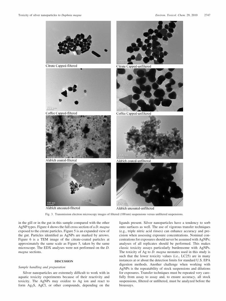
Microscopy particles
Transmission electron microscopy images for the SA Ag,citrate-coated, and coffee-coated particles are presented in theSupplemental Data (Figs. S3–S6). The coated particles tendedto aggregate more than the uncoated particles. The aggregatesappear in a gel-like amorphous matrix, which is probablyassociated with the capping agent. The SA uncoated particlesseemed to have higher variations in particle size. Comparisons
among the filtered (100 nm) versus the unfiltered nanoparticlesuspensions show that particles are present in all of the filteredsuspensions (Fig. 3). Although the SA suspensions had a highpercentage of particles removed, enough particles passedthrough the filter that stock suspensions could be made.
Microscopy organisms
Scanning electron microscope images of representativeD. magna samples are presented in the supplemental informa-tion (Fig. S6). No evidence was seen of AgNPs on any of theexterior surfaces of the organisms. This was probably becauseof sample preparation procedures (e.g., washing, fixation, des-iccation). Results from EDX analyses on the organism exteriorsshowed high concentrations of Ca, which was probably attrib-utable to precipitated salts from the MHRW.
Transmission electron microscopy images of D. magnaexposed to approximate LC50 concentrations yielded thefollowing qualitative results: SA uncoated: no Ag nanoparticlesdetected (no images were taken); SA coated: a few silverparticles were found along the membrane of the mid-gut;coffee-capped: a few particles were found around the gilland gut; citrate-capped: a few more particles were found aroundthe gills and gut; control: a particle was found at the side of thegill, but no particles were found inside gut tissue (Fig. S7);coffee-capped, (3�LC50): a few particles were found in thegill region, and some particles were found in the tissue of themid-gut; citrate-capped, (3�LC50): more AgNPs were found
Table 2. Dynamic light scattering size measurements in deionized water (DI) and moderately hard reconstituted water (MHRW)
Particle typeApprox Ag
concentration (ppb) Matrix
Size distribution of particles
Size 1, % Size 2, % Size 3, %
SA uncoated� >100,000 DI water 839.5 nm, 73.4% 5,021 nm, 26.6%SA uncoated� >100,000 MHRW 681.4 nm, 81.4% 5,412 nm, 18.6%SA coated� >100,000 DI water 14.85 nm, 73.3% 58.27 nm, 24.4% 368.9 nm, 4.3%SA coated� >100,000 MHRW 39.39 nm, 73.5% 249.8 nm, 26.5%Coffee capped 1:10 dilution 215,800 DI water 106.3 nm, 22.4% 1,285 nm, 77.6%Coffee capped 1:100 dilution 21,580 MHRW 101.5 nm, 30.5% 773.6 nm, 69.5%Citrate capped 70,000 Nanopure 5.94 nm, 98% 39.75 nm, 2%
� Sonicated prior to analysis. SA¼ Sigma Aldrich silver nanoparticles.
Fig. 2. X-ray absorption spectroscopy spectra for silver compounds andsilver nanoparticles equilibrated with 1 M sodium chloride solutions ormoderately hard reconstituted water.
Table 3. Dynamic light scattering zeta potential measurements of AgNPsin deionized water (DI) and moderately hard reconstituted water (MHRW)
Particle type Matrix pH
Zetapotential,
mV
Zetadeviation,
mVExpectedstability
SA coated DI 6.8–7.0 �38.0 6.31 ModerateSA coated MHRW 7.8–8.0 �18.3 6.11 IncipientSA uncoated DI 6.8–7.0 �41.2 5.59 GoodSA uncoated MHRW 7.8–8.0 �20.4 5.65 IncipientCoffee capped MHRW 7.8–8.0 �9.3 4.15 PoorCitrate capped Nanopure 6.8–8.0 �39.7 19.5 Moderate
2746 Environ. Toxicol. Chem. 29, 2010 H.J. Allen et al.
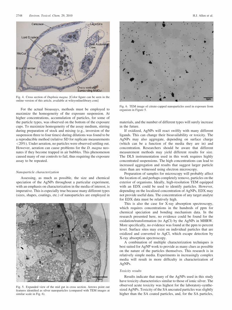
in the gill or in the gut in this sample compared with the otherAgNP types. Figure 4 shows the full cross section of a D. magnaexposed to the citrate particles. Figure 5 is an expanded view ofthe gut. Particles identified as AgNPs are marked by arrows.Figure 6 is a TEM image of the citrate-coated particles atapproximately the same scale as Figure 5, taken by the samemicroscope. The EDX analyses were not performed on the D.magna sections.
DISCUSSION
Sample handling and preparation
Silver nanoparticles are extremely difficult to work with inaquatic toxicity experiments because of their reactivity andtoxicity. The AgNPs may oxidize to Ag ion and react toform Ag2S, AgCl, or other compounds, depending on the
ligands present. Silver nanoparticles have a tendency to sorbonto surfaces as well. The use of vigorous transfer techniques(e.g., triple nitric acid rinses) can enhance accuracy and pre-cision when assessing exposure concentrations. Nominal con-centrations for exposures should never be assumed with AgNPs;analyses of all replicates should be performed. This makesclassic toxicity assays particularly burdensome with AgNPs.The toxicity of Ag to D. magna neonates used in this study issuch that the lower toxicity values (i.e., LC25) are in manyinstances at or about the detection limits for standard U.S. EPAdigestion methods. Another challenge when working withAgNPs is the repeatability of stock suspensions and dilutionsfor exposures. Transfer techniques must be repeated very care-fully from assay to assay and, to ensure accuracy, all stocksuspensions, filtered or unfiltered, must be analyzed before thebioassays.
Fig. 3. Transmission electron microscopy images of filtered (100 nm) suspensions versus unfiltered suspensions.
Toxicity of silver nanoparticles to Daphnia magna Environ. Toxicol. Chem. 29, 2010 2747
For the actual bioassays, methods must be employed tomaximize the homogeneity of the exposure suspension. Athigher concentrations, accumulation of particles, for some ofthe particle types, was observed on the bottom of the exposurecups. To maximize homogeneity of the assay medium, stirringduring preparation of stock and mixing (e.g., inversion of thesuspension three to four times) during dilutions was found to bea reproducible method (relative SD for replicate measurements<20%). Under aeration, no particles were observed settling out.However, aeration can cause problems for the D. magna neo-nates if they become trapped in air bubbles. This phenomenoncaused many of our controls to fail, thus requiring the exposureassay to be repeated.
Nanoparticle characterization
Assessing, as much as possible, the size and chemicalspeciation of the AgNPs throughout a particular experiment,with an emphasis on characterization in the media of interest, isimperative. This is especially true because many different types(sizes, shapes, coatings, etc.) of nanoparticles are employed in
materials, and the number of different types will surely increasein the future.
If oxidized, AgNPs will react swiftly with many differentligands. This can change their bioavailability or toxicity. TheAgNPs may also aggregate, depending on surface charge(which can be a function of the media they are in) andconcentration. Researchers should be aware that differentmeasurement methods may yield different results for size.The DLS instrumentation used in this work requires highlyconcentrated suspensions. The high concentrations can lead toincreased aggregation and results that suggest larger particlesizes than are witnessed using electron microscopy.
Preparation of samples for microscopy will probably affectthe location of, and perhaps completely remove, particles on theexterior of organisms. Ideally, high-resolution TEM equippedwith an EDX could be used to identify particles. However,depending on the localized concentration of AgNPs, EDX maynot provide useful data. The concentration of any target analytefor EDX data must be relatively high.
This is also the case for X-ray absorption spectroscopy,which requires concentrations in the hundreds of ppm forchemical speciation and bonding mechanism data. In theresearch presented here, no evidence could be found for theoxidation/transformation (to AgCl) by the AgNPs in MHRW.More specifically, no evidence was found at the ppm to percentlevel. Surface sites may exist on individual particles that areoxidized and converted to AgCl, which escape detection byX-ray absorption spectroscopy.
A combination of multiple characterization techniques isbest suited for AgNP work to provide as many clues as possibleon the nature of the particles themselves. This research is inrelatively simple media. Experiments in increasingly complexmedia will result in more difficulty in characterization ofAgNPs.
Toxicity results
Results indicate that many of the AgNPs used in this studyhave toxicity characteristics similar to those of ionic silver. Theobserved acute toxicity was highest for the laboratory-synthe-sized AgNPs. Toxicity of the SA uncoated particles was slightlyhigher than the SA coated particles, and, for the SA particles,
Fig. 4. Cross section of Daphnia magna. [Color figure can be seen in theonline version of this article, available at wileyonlinelibrary.com]
Fig. 5. Expanded view of the mid gut in cross section. Arrows point outfeatures identified as silver nanoparticles (compared with TEM images atsimilar scale in Fig. 6).
Fig. 6. TEM image of citrate-capped nanoparticles used in exposure fromorganism in Figure 5.
2748 Environ. Toxicol. Chem. 29, 2010 H.J. Allen et al.
the unfiltered suspensions yielded higher LC50 values.The addition of food decreased toxicity of the SA uncoatedparticles by an order of magnitude. Toxicity of the laboratory-synthesized particles agrees well with the AgNO3 assay valuesand also with published data for D. magna silver salt assays.Elnabarawy et al. [34] reported a 48-h LC50 of 1.5mg/L(alkalinity¼ 230 mg/L as CaCO3, hardness¼ 240 mg/L asCaCO3), Erickson et al. [35] reported 0.58mg/L in soft (alka-linity and hardness¼ 49 mg/L as CaCO3) laboratory water and35mg/L in river water (alkalinity¼ 68 mg/L as CaCO3, hard-ness¼ 81 mg/L as CaCO3), and Karen et al. [36] reported a 48-hLC50 of 0.84mg/L (hardness¼ 100 mg/L as CaCO3). Thesedata suggest that oxidation of the AgNP to ionic Ag at surfacesites may play a role in observed toxicity. However, where theAg ion forms (e.g., inside organism vs bulk medium) remains tobe determined. Decreased toxicity observed in the tests withfood may be explained by observations made by Glover andWood [37] and Bianchini et al. [18] in D. magna, in whichorganic material may either alter availability of ionic Ag orstimulate active sodium uptake by Naþ,Kþ-ATPase.
The differences in toxicity observed among the variousnanoparticle suspensions are probably attributable to particlesize. The particle size to which the organisms are exposed ispartially a function of the degree of aggregation. The TEMimages (Fig. 3) suggest that, even after filtration, the SAparticles tend to aggregate more. Larger particles/aggregatesdecrease the surface area to volume ratio (relative to identicalvolumes of smaller particles) and thus decrease the surface areaavailable to release Ag ion via oxidation reactions. Smallerparticles will also be less prone to spatial hindrances whenentering an organism. With the laboratory-synthesized par-ticles, incomplete reactions may result in the presence of someresidual Ag ion in the suspension. The images in Figure 3 andthe filtration recoveries suggest, however, that most of the Agpresent is particulate. Methods to differentiate between Ag ionand bulk Ag such as 1-kDa filters, dialysis, and microcapillaryaction were not used in the current study. Use of such methodswould provide a more accurate characterization of Ag state (ionvs bulk) and thus greater insight on the exposure mechanism.
The results presented here suggest that, at minimum, AgNPsshould be treated as Ag ions with respect to toxicity to D. magnaneonates. Alternative methods (e.g., gene expression/ecotoxi-cogenomics) should be explored and may provide additionalinformation regarding the toxicity of nanoparticles. Assessmentof the ecotoxicity of nanoparticles presents challenges becauseof the burdensome nature of accounting for concentrations andtransformations of nanoparticles in even simple media. Futureresearch will focus on characterization and toxicity of AgNPs inincreasingly complex matrixes.
SUPPLEMENTAL DATA
Table S1.Figures S1-S7. (11.5 MB DOC)
Acknowledgement—Any opinions expressed in the current study are those ofthe authors and do not necessarily reflect the official positions and policies ofthe U.S. EPA. Any mention of products or trade names does not constituterecommendation for use by the U.S. EPA. Materials Research CollaborativeAccess Team operations are supported by the Department of Energy and theMaterials Research Collaborative Access Team member institutions. Theauthors greatly appreciate the analytical support from the University ofCincinnati and Purdue University. The authors also thank Todd P. Luxton,Kirk G. Scheckel, Mark E. Smith, Sri Panguluri, and Christina Bennet-Stamper for their insights and expertise. Christian Ritz, University of
Copenhagen, Denmark, provided valuable advice on dose–responsestatistical analyses.
REFERENCES
1. Lovern SB, Strickler JR, Klaper R. 2007. Behavioral and physiologicalchanges in Daphnia magna when exposed to nanoparticle suspensions(titanium dioxide, nano-C-60, and C(60)HxC(70)Hx). Environ SciTechnol 41:4465–4470.
2. Oberdorster E. 2004. Manufactured nanomaterials (fullerenes, C60)induce oxidative stress in the brain of juvinile largemouth bass. EnvironHealth Perspect 112:1058–1062 .
3. Zhu S, Oberdorster E, Haasch ML. 2006. Toxicity of an engineerednanoparticle (fullerene,C60) in two aquatic species,Daphnia and fatheadminnow. Mar Environ Res 62 Supplement 1:S5–S9.
4. Cheng J, Flahaut E, Cheng SH. 2007. Effect of carbon nanotubes ondeveloping zebrafish (Danio rerio) embryos. Environ Toxicol Chem26:708–716.
5. Smith CJ, Shaw BJ, Handy RD. 2007. Toxicity of single walled carbonnanotubes to rainbow trout, (Oncorhynchus mykiss): Respiratorytoxicity, organ pathologies, and other physiological effects. AquatToxicol 82:94–109.
6. Zhu Y, Zhao Q, Li Y, Cai X, Li W. 2006. The interaction and toxicity ofmulti-walled carbon nanotubes with Stylonychia mytilus. J NanosciNanotechnol 6:1357–1364.
7. Adams LK, Lyon DY, Alvarez PJ. 2006. Comparative eco-toxicity ofnanoscale TiO2, SiO2, and ZnO water suspensions. Water Res 40:3527–3532.
8. Adams LK, Lyon DY, McIntosh A, Alvarez PJ. 2006. Comparativetoxicity of nano-scale TiO2, SiO2 and ZnO water suspensions.Water SciTechnol 54:327–334.
9. Hund-Rinke K, Simon M. 2006. Ecotoxic effect of photocatalytic activenanoparticles (TiO2) on algae and daphnids. Environ Sci Pollut Res Int13:225–232.
10. Davies JC. 2008. Nanotechnology oversight: An agenda for the newadministration. The project on emerging nanotechnologies. WoodrowWilson International Center for Scholars, Washington, DC.
11. Morones JR, Elechiguerra JL, Camacho A, Holt K, Kouri JB, RamirezJT, Yacaman MJ. 2005. The bactericidal effect of silver nanoparticles.Nanotechnology 16:2346–2353.
12. Soni I, Salopek-Bondi B. 2004. Silver nanoparticles as antimicrobialagen: A case study on E. coli as a model for gram-negative bacteria. JColloid Interface Sci 275:1770–1782.
13. Luoma SN,HogstrandC, Bell RA, Bielmyer GK, Galvez F, LeBlanc GA,Lee BG, Purcell TW, Santore RC, Santschi PH, Shaw JR. 2002.Biological Processes. In Andren AW, Bober TW, eds, Silver in theEnvironment: Transport, Fate, and Effects. SETAC, Pensacola, FL,USA, pp 65-91.
14. Jain P, Pradeep T. 2005. Potential of silver nanoparticle-coatedpolyurethane foam as an antibacterial water filter. Biotechnol Bioeng90:59–63.
15. Sambhy V, MacBride MM, Peterson BR, Sen A. 2006. Silver bromidenanoparticle/polymer composites: dual action tunable antimicrobialmaterials. J Am Chem Soc 128:9798–9808.
16. Lee HY, Park HK, Lee YM, Kim K, Park SB. 2007. A practical procedurefor producing silver nanocoated fabric and its antibacterial evaluation forbiomedical applications. Chem Commun 28:2959–2961.
17. Wen C, Ishikawa K, Kishima M, Yamada K. 2000. Effects of silverparticles on the photovoltaic properties of dye-sensitized TiO2 thin films.Sol Energy Mater Sol Cells 61:339–351.
18. Bianchini A, Wood CM. 2003. Mechanism of acute silver toxicity inDaphnia magna. Environ Toxicol Chem 22:1361–1367.
19. Wood CM, Playle RC, Hogstrand C. 1999. Physiology and modeling ofmechanisms of silver uptake and toxicity in fish. Environ Toxicol Chem18:71–83.
20. Elechiguerra JL, Burt JL, Morones JR, Camacho-Bragado A, Gao X,Lara HH, Yacaman MJ. 2005. Interaction of silver nanoparticles withHIV-1. J Nanobiotechnol 3:6.
21. Lee D, Cohen RE, Rubner MF. 2005. Antibacterial properties of Agnanoparticle loaded multilayers and formation of magnetically directedantibacterial microparticles. Langmuir 21:9651–9659.
22. Navarro E, Piccapietra F, Wagner B, Marconi F, Kaegi R, Odzak N, SiggL, Behra R. 2008. Toxicity of silver nanoparticles to Chlamydomonasreinhardtii. Environ Sci Technol 42:8959–8964.
23. Kramer JR, Benoit G, Bowles KC, DiToro DM, Herrin RT, LutherIIIGW, Manolopoulos H, Robillard KA, Shafer MM, Shaw JR. 2002.Environmental Chemistry of Silver. In Andren AW, Bober TW, eds,Silver in the Environment: Transport, Fate, and Effects. SETAC,Pensacola, FL, USA, pp 1-25.
Toxicity of silver nanoparticles to Daphnia magna Environ. Toxicol. Chem. 29, 2010 2749
24. Choi O, Clevenger TE, Deng B, Surampalli RYL, Ross J, Hu Z. 2009.Role of sulfide and ligand strength in controlling nanosilver toxicity.Water Res 43:1879–1886.
25. Weber CI, ed. 1993. Methods for measuring the acute toxicityof effluentsand receiving waters to freshwater and marine organisms, 4th ed. EPA/600/4-90/027F. U. S. Environmental Protection Agency, Washington,DC.
26. Pillai ZS, Kamat PV. 2004. What factors control the size and shape ofsilver nanoparticles in the citrate ion reduction method. J Phys Chem B108:945–951.
27. United States Environmental Protection Agency. 1997. Method 3051:Microwave assisted acid dissolution of sediments, sludges, soils, andoils. In Test Methods for Evaluating Solid Waste, Physical/ChemicalMethods, 2nd ed. Publication 955-001- 00000-1. U.S. GovernmentPrinting Office, Washington, DC.
28. Ritz C, Streibig JC. 2005. Bioassay analysis using R. J Stat Softw 12:1–22.
29. R Development Core Team. 2009. R: A language and environment forstatistical computing. R Foundation for Statistical Computing, Vienna,Austria.
30. Finney DJ. 1971. Probit Analysis, 3rd ed. Cambridge University,Cambridge, UK.
31. Collett D. 2003.ModelingBinaryData 2nd ed. Chapman and Hall/CRC,Boca Raton, FL, USA.
32. Ravel B, Newville M. 2005. Athena, Artemis, Hephaestus: Data analysisfor X-ray absorption spectroscopy using IFEFFIT. J Synchotron Radiat12:537–541.
33. International Organization for Standardization. 1996. Particle sizeanalysis-photon correlation spectroscopy. ISO 13321: Geneva, Switzer-land.
34. Elnabarawy MT, Welter AN, Robideau RR. 1986. Relative sensitivitiesof three daphnid species to selected organic and inorganic chemicals.Environ Toxicol Chem 5:393–398.
35. Erickson RJ, Brooke LT, Kahl MD, VandeVenter F, Harting S, MarkeeTP, Spehar RL. 1998. Effects of laboratory test conditions on the toxicityof silver to aquatic organisms. Environ Toxicol Chem 17:572–578.
36. Karen DJ, Ownby DR, Forsythe BL, Bills TP, LaPoint TW, Cobb GB,Klaine SJ. 1999. Influence of water quality on silver toxicity to rainbowtrout (Oncorhynchus mykiss), fathead minnows (Pimephales promelas),and water fleas (Daphnia magna). Environ Toxicol Chem 18:63–70.
37. Glover CN, Wood CM. 2004. Physiological interactions of silver andhumic substances in Daphnia magna: effects on reproduction and silveraccumulation following an acute silver challenge. Comp BiochemPhysiol C Toxicol Pharmacol 139:273–280.
2750 Environ. Toxicol. Chem. 29, 2010 H.J. Allen et al.