Effectiveness of interventions to enhance ... - IWGDF Guidelines
15
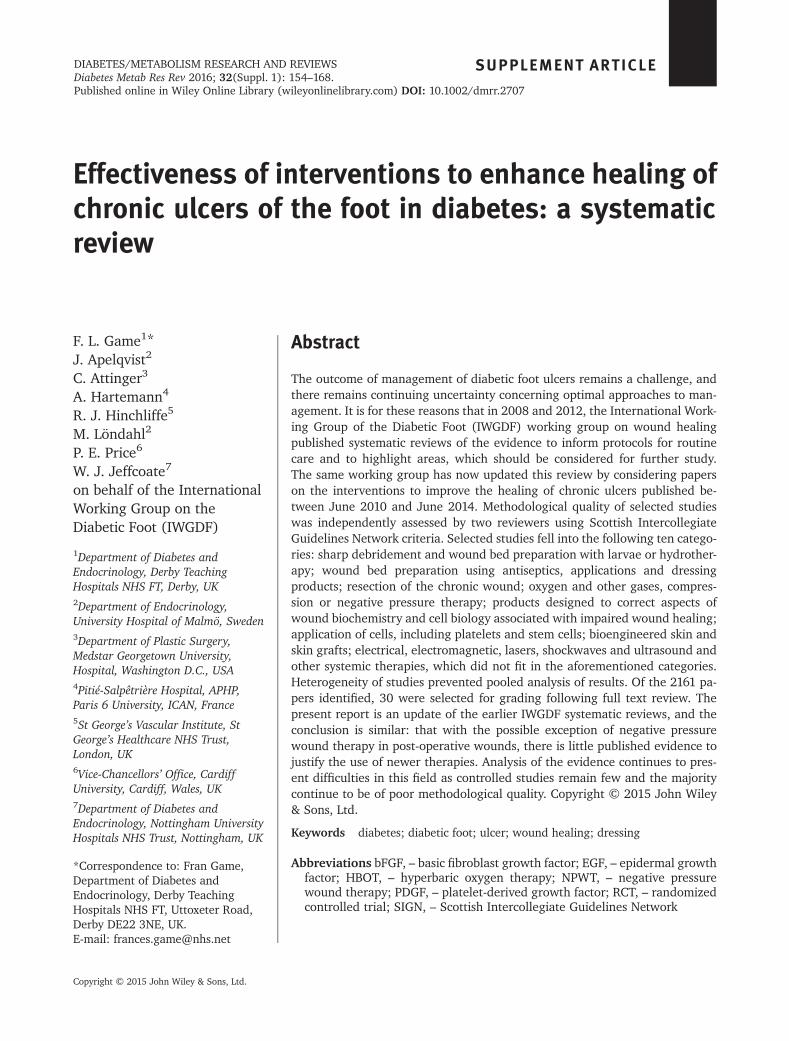
Effectiveness of interventions to enhance healing of chronic ulcers of the foot in diabetes: a systematic review F. L. Game 1 * J. Apelqvist 2 C. Attinger 3 A. Hartemann 4 R. J. Hinchliffe 5 M. Löndahl 2 P. E. Price 6 W. J. Jeffcoate 7 on behalf of the International Working Group on the Diabetic Foot (IWGDF) 1 Department of Diabetes and Endocrinology, Derby Teaching Hospitals NHS FT, Derby, UK 2 Department of Endocrinology, University Hospital of Malmö, Sweden 3 Department of Plastic Surgery, Medstar Georgetown University, Hospital, Washington D.C., USA 4 Pitié-Salpêtrière Hospital, APHP, Paris 6 University, ICAN, France 5 St George’s Vascular Institute, St George’s Healthcare NHS Trust, London, UK 6 Vice-Chancellors’ Office, Cardiff University, Cardiff, Wales, UK 7 Department of Diabetes and Endocrinology, Nottingham University Hospitals NHS Trust, Nottingham, UK *Correspondence to: Fran Game, Department of Diabetes and Endocrinology, Derby Teaching Hospitals NHS FT, Uttoxeter Road, Derby DE22 3NE, UK. E-mail: [email protected] Abstract The outcome of management of diabetic foot ulcers remains a challenge, and there remains continuing uncertainty concerning optimal approaches to man- agement. It is for these reasons that in 2008 and 2012, the International Work- ing Group of the Diabetic Foot (IWGDF) working group on wound healing published systematic reviews of the evidence to inform protocols for routine care and to highlight areas, which should be considered for further study. The same working group has now updated this review by considering papers on the interventions to improve the healing of chronic ulcers published be- tween June 2010 and June 2014. Methodological quality of selected studies was independently assessed by two reviewers using Scottish Intercollegiate Guidelines Network criteria. Selected studies fell into the following ten catego- ries: sharp debridement and wound bed preparation with larvae or hydrother- apy; wound bed preparation using antiseptics, applications and dressing products; resection of the chronic wound; oxygen and other gases, compres- sion or negative pressure therapy; products designed to correct aspects of wound biochemistry and cell biology associated with impaired wound healing; application of cells, including platelets and stem cells; bioengineered skin and skin grafts; electrical, electromagnetic, lasers, shockwaves and ultrasound and other systemic therapies, which did not fit in the aforementioned categories. Heterogeneity of studies prevented pooled analysis of results. Of the 2161 pa- pers identified, 30 were selected for grading following full text review. The present report is an update of the earlier IWGDF systematic reviews, and the conclusion is similar: that with the possible exception of negative pressure wound therapy in post-operative wounds, there is little published evidence to justify the use of newer therapies. Analysis of the evidence continues to pres- ent difficulties in this field as controlled studies remain few and the majority continue to be of poor methodological quality. Copyright © 2015 John Wiley & Sons, Ltd. Keywords diabetes; diabetic foot; ulcer; wound healing; dressing Abbreviations bFGF, – basic fibroblast growth factor; EGF, – epidermal growth factor; HBOT, – hyperbaric oxygen therapy; NPWT, – negative pressure wound therapy; PDGF, – platelet-derived growth factor; RCT, – randomized controlled trial; SIGN, – Scottish Intercollegiate Guidelines Network SUPPLEMENT ARTICLE Copyright © 2015 John Wiley & Sons, Ltd. DIABETES/METABOLISM RESEARCH AND REVIEWS Diabetes Metab Res Rev 2016; 32(Suppl. 1): 154–168. Published online in Wiley Online Library (wileyonlinelibrary.com) DOI: 10.1002/dmrr.2707
-
Upload
khangminh22 -
Category
Documents
-
view
0 -
download
0
Transcript of Effectiveness of interventions to enhance ... - IWGDF Guidelines
Effectiveness of interventions to enhance healing ofchronic ulcers of the foot in diabetes: a systematicreview
F. L. Game1*J. Apelqvist2
C. Attinger3
A. Hartemann4
R. J. Hinchliffe5
M. Löndahl2
P. E. Price6
W. J. Jeffcoate7
on behalf of the InternationalWorking Group on theDiabetic Foot (IWGDF)
1Department of Diabetes andEndocrinology, Derby TeachingHospitals NHS FT, Derby, UK2Department of Endocrinology,University Hospital of Malmö, Sweden3Department of Plastic Surgery,Medstar Georgetown University,Hospital, Washington D.C., USA4Pitié-Salpêtrière Hospital, APHP,Paris 6 University, ICAN, France5St George’s Vascular Institute, StGeorge’s Healthcare NHS Trust,London, UK6Vice-Chancellors’ Office, CardiffUniversity, Cardiff, Wales, UK7Department of Diabetes andEndocrinology, Nottingham UniversityHospitals NHS Trust, Nottingham, UK
*Correspondence to: Fran Game,Department of Diabetes andEndocrinology, Derby TeachingHospitals NHS FT, Uttoxeter Road,Derby DE22 3NE, UK.E-mail: [email protected]
Abstract
The outcome of management of diabetic foot ulcers remains a challenge, andthere remains continuing uncertainty concerning optimal approaches to man-agement. It is for these reasons that in 2008 and 2012, the International Work-ing Group of the Diabetic Foot (IWGDF) working group on wound healingpublished systematic reviews of the evidence to inform protocols for routinecare and to highlight areas, which should be considered for further study.The same working group has now updated this review by considering paperson the interventions to improve the healing of chronic ulcers published be-tween June 2010 and June 2014. Methodological quality of selected studieswas independently assessed by two reviewers using Scottish IntercollegiateGuidelines Network criteria. Selected studies fell into the following ten catego-ries: sharp debridement and wound bed preparation with larvae or hydrother-apy; wound bed preparation using antiseptics, applications and dressingproducts; resection of the chronic wound; oxygen and other gases, compres-sion or negative pressure therapy; products designed to correct aspects ofwound biochemistry and cell biology associated with impaired wound healing;application of cells, including platelets and stem cells; bioengineered skin andskin grafts; electrical, electromagnetic, lasers, shockwaves and ultrasound andother systemic therapies, which did not fit in the aforementioned categories.Heterogeneity of studies prevented pooled analysis of results. Of the 2161 pa-pers identified, 30 were selected for grading following full text review. Thepresent report is an update of the earlier IWGDF systematic reviews, and theconclusion is similar: that with the possible exception of negative pressurewound therapy in post-operative wounds, there is little published evidence tojustify the use of newer therapies. Analysis of the evidence continues to pres-ent difficulties in this field as controlled studies remain few and the majoritycontinue to be of poor methodological quality. Copyright © 2015 John Wiley& Sons, Ltd.
Keywords diabetes; diabetic foot; ulcer; wound healing; dressing
Abbreviations bFGF, – basic fibroblast growth factor; EGF, – epidermal growthfactor; HBOT, – hyperbaric oxygen therapy; NPWT, – negative pressurewound therapy; PDGF, – platelet-derived growth factor; RCT, – randomizedcontrolled trial; SIGN, – Scottish Intercollegiate Guidelines Network
SUPPLEMENT ARTICLE
Copyright © 2015 John Wiley & Sons, Ltd.
DIABETES/METABOLISM RESEARCH AND REVIEWSDiabetes Metab Res Rev 2016; 32(Suppl. 1): 154–168.Published online in Wiley Online Library (wileyonlinelibrary.com) DOI: 10.1002/dmrr.2707
Introduction
The management of foot disease in diabetes remains a ma-jor financial and therapeutic challenge throughout theworld. The International Working Group of the DiabeticFoot (IWGDF) has issued guidelines on management since1999 and systematic reviews to underpin those from 2005.In 2006, the IWGDF Editorial Board invited the IWGDFworking group on wound healing to undertake a systematicreview of the evidence supporting interventions to enhancethe healing of chronic ulcers of the foot in diabetes in orderboth to inform protocols for routine care and to highlightareas, which should be considered for further study. Thefirst review included all papers published up to December2006 [1], and this was later updated to include all subse-quent papers up until June 2010 [2]. The working grouphas now undertaken a further update by consideringpapers on the interventions to improve the healing ofchronic ulcers of the foot in diabetes published betweenJune 2010 and June 2014.
Materials and methods
Controlled studies, which were either prospective or ret-rospective, published in any language, and which evalu-ated interventions for the treatment of chronic footulcers in people aged 18 years or older with either type 1or type 2 diabetes mellitus were considered. Studies wereincluded if they concerned agents or interventions thatmay accelerate the healing process, and the primary out-comes used were clinical: healing, time to healing, and/orreduction in ulcer area. Search strategies (Appendix A)included selected search terms on study design, patientgroup, clinical problem and interventions of interest byusing Medline (June 2010 to June 2014) and Embase(June 2010 to June 2014). Randomized controlled trials(RCT), case–control studies, prospective and retrospectivecohort studies, control before-and-after and interruptedtime series designs were included. Bibliography trackingof identified articles was not performed. Previouslyperformed high-quality systematic reviews and Cochranereviews on the topics of interest were searched to determinethe need for an extension to the literature search. A latersearch was made of the following clinical trials registries;the search terms used were Foot Ulcer; Diabetes Mellitus;Diabetic Foot Ulcer; and Diabetic Foot: http://www.con-trolled-trials.com/, www.clinicaltrials.gov, www.who.int/trialsearch, clinicalstudies.info.nih.gov/, cordis.europa.eu/en/home.html, www.clinicaltrialsregister.eu/, www.pactr.org/, www.anzctr.org.au/, www.canadiancancertrials.ca/,www.fmhs.auckland.ac.nz/sms/oncology/ctnz/default.aspx, www.chictr.org/Default.aspx, cris.nih.go.kr/cris/en/
search/basic_search.jsp, registroclinico.sld.cu/, drks-neu.uniklinik-freiburg.de/drks_web/, www.hkclinicaltrials.com/,www.irct.ir/, www.umin.ac.jp/ctr/ www.kctr.se/, clinicaltrials.health.nz/, www.sanctr.gov.za/SAClinicalTrials/tabid/169/Default.aspx, www.slctr.lk/ www.clinicaltrials.in.th/, public.ukcrn.org.uk/search/ and www.controlled-trials.com/ukctr/, and attempts were made to contactinvestigators if there was no evidence of publication ofrelevant studies.
Two reviewers (FLG and WJJ) independently assessedall identified references by title and abstract to determinepossible eligibility. Full-paper copies of identified articleswere retrieved, and eligibility was confirmed or rejectedby one of four pairs of independent reviewers. Each studywas scored for methodological quality using scoring listsspecific for each study design and based on checklists de-veloped by the Scottish Intercollegiate Guidelines Net-work (SIGN) [3]. Equal weighting was applied to eachvalidity criterion. Findings on data extraction and meth-odological quality were discussed between co-reviewersand a final decision endorsed by the entire group. Qualityitems were rated as ‘done’, ‘not done’ or ‘not reported’, andonly those rated as ‘done’ contributed to methodologicalquality score. This quality score was translated into a levelof evidence according to the SIGN instrument [3]: (1) RCTsand (2) studies with case–control, cohort, control beforeand after or interrupted time series design. Studies werealso rated as ++ (well conducted with very low risk ofbias), + (well conducted with low risk of bias) and �(low quality with higher risk of bias). Meta-analyses, otherreviews and studies reporting non-analytic case reportsand case series were not included. Reviewers did not assesstheir own work because of potential conflicts of interest.
Extracted data were summarized in evidence tables ona study-by-study narrative basis. Because of the heteroge-neity of study designs, including interventions, follow-upand outcomes, no attempt was made to pool the results.The evidence tables were compiled following collectivediscussion by the working party, and conclusions weredrawn. The papers selected for scoring were divided intothe same ten categories as the 2012 review, except thatthe articles on the use of platelet-derived growth factorshave now been included in the section on cell therapy(in contrast to the previous allocation to the section onwound biochemistry); the section on oxygen has beenexpanded to include other gases.
Results
In 2008, a total of 2155 articles were identified fromEMBASE and Medline. Of these, 372 were selected for fulltext review, and 61 were included in the review. In 2012,
Effectiveness of Interventions to Enhance Healing – A Systematic Review 155
Copyright © 2015 John Wiley & Sons, Ltd. Diabetes Metab Res Rev 2016; 32(Suppl. 1): 154–168.DOI: 10.1002/dmrr
a total of 802 articles were identified from EMBASE and507 from Medline. Seventy-two of these were selectedfor full text review. An additional 13 articles were identi-fied from other sources, including other systematicreviews. Of the total 85 articles, 43 were included.
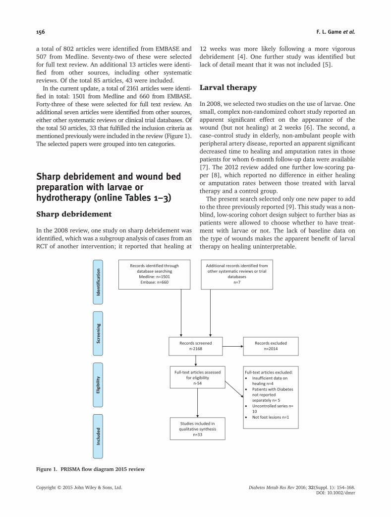
In the current update, a total of 2161 articles were identi-fied in total: 1501 from Medline and 660 from EMBASE.Forty-three of these were selected for full text review. Anadditional seven articles were identified from other sources,either other systematic reviews or clinical trial databases. Ofthe total 50 articles, 33 that fulfilled the inclusion criteria asmentioned previouslywere included in the review (Figure 1).The selected papers were grouped into ten categories.
Sharp debridement and wound bedpreparation with larvae orhydrotherapy (online Tables 1–3)
Sharp debridement
In the 2008 review, one study on sharp debridement wasidentified, which was a subgroup analysis of cases from anRCT of another intervention; it reported that healing at
12 weeks was more likely following a more vigorousdebridement [4]. One further study was identified butlack of detail meant that it was not included [5].
Larval therapy
In 2008, we selected two studies on the use of larvae. Onesmall, complex non-randomized cohort study reported anapparent significant effect on the appearance of thewound (but not healing) at 2 weeks [6]. The second, acase–control study in elderly, non-ambulant people withperipheral artery disease, reported an apparent significantdecreased time to healing and amputation rates in thosepatients for whom 6-month follow-up data were available[7]. The 2012 review added one further low-scoring pa-per [8], which reported no difference in either healingor amputation rates between those treated with larvaltherapy and a control group.
The present search selected only one new paper to addto the three previously reported [9]. This study was a non-blind, low-scoring cohort design subject to further bias aspatients were allowed to choose whether to have treat-ment with larvae or not. The lack of baseline data onthe type of wounds makes the apparent benefit of larvaltherapy on healing uninterpretable.
Figure 1. PRISMA flow diagram 2015 review
156 F. L. Game et al.
Copyright © 2015 John Wiley & Sons, Ltd. Diabetes Metab Res Rev 2016; 32(Suppl. 1): 154–168.DOI: 10.1002/dmrr
Hydrotherapy
No further studies were identified to add to the one paperin the previous on hydrotherapy (Versajet®) [10], whichshowed no benefit to healing at 12 weeks in a small study.
Clostridial collagenase
The use of Clostridial collagenase ointment used daily as adebriding agent was examined in one small study [11].This small, moderate scoring but unblinded study ofnon-ischaemic wounds showed an apparent improvementin area reduction from baseline in the treated group after4 weeks, whereas there was no improvement seen in thecontrol group. There were no between group comparisonsmade, however, and the finding that there was an averageincrease in the area of the wounds in the control groupcompared with baseline at 4 and 12 weeks suggests thatthe control group may not have received usual bestpractice.
Wound bed preparation usingantiseptics, applications and dressingproducts (online Tables 4–6)
Antiseptics and antimicrobials
In 2008, one study was identified, which demonstratedthat cadexomer-iodine showed no benefit in cavitywounds when compared with usual care [12]. A subse-quent large, observer-blinded, RCT of good quality identi-fied in the 2012 review reported no difference betweenthree products: carboxymethylcellulose hydrofibre, a sur-face antiseptic (Inadine®) and a non-adherent productgauze in terms of healing by 24 weeks [13]. The 2008review also found evidence from a single small study ofpossible benefit from the use of zinc oxide tape, but nosubsequent reports have been found [14].
Only one study of the use of honey was identified inthe 2012 review, and this was a small, non-blinded andpoorly designed controlled study, which reported no dif-ference in outcome between the use of honey andpovidone/iodine [15]. In the current review, we identi-fied two further studies. The first [16] was a very small,poorly scoring, non-blinded RCT of honey-soaked dress-ings compared with povidone/iodine dressings. Althoughthere was an apparent difference in area reduction at15 days between the two groups, this result is uninter-pretable given the lack of data on the baseline character-istics of the ulcers in the two groups and the probableinappropriate use of parametric statistics. In a second
small cohort, study [17] comparing honey dressings withiodine dressings, no differences were found in either theincidence of healing or of amputation at 10 weeks al-though there was an apparent reduction in time to out-come (healing or amputation) in the honey group. Thisresult is difficult to interpret, and the study was of poormethodological quality with few data on the baselinecharacteristics of the patients. Despite the widespreaduse of honey dressings in clinical practice, there are norobust data to support their use to enhance the healingof diabetic foot wounds, and this reinforces the conclu-sions of a recent Cochrane review [18].
A single non-blinded RCT on the use of superoxidizedsolution (DermacynW) was identified in the 2012 review[19], which compared the incidence of healing at6 months after infected surgical wounds of the foot hadbeen irrigated with either the superoxidized solution orwith povidone/iodine. The results of this trial were ofdoubtful quality given the methodological flaws in thestudy, and no further studies have been identified in thisreview.
The use of topical antimicrobials (tobramycin beads) onthe wound at the time of forefoot amputation was shownin a non-randomized cohort study reported in the 2012review to have a significant beneficial effect on the needfor later surgical revision [20], but no difference inhealing times or later transtibial amputation. No furtherstudies on antibiotic-impregnated beads or cement havebeen identified, and so the place of these agents in woundhealing is yet to be determined.
Alginate and collagen-alginate products
Two small studies of alginate-containing products wereidentified in the 2008 review. Neither showed evidenceof improved wound healing either in comparison withsaline-moistened gauze [21] or Vaseline gauze [22].
Carboxymethylcellulose dressings
We previously identified an RCT, which reported improve-ment with the use of a carboxymethylcellulose hydrofibredressing in the 2008 review [23]. In the 2012 review,however, a further larger RCT with a silver-impregnateddressing [24] showed no difference in healing at 8 weekswhen compared with an alginate dressing. Another large,observer-blinded, RCT of good quality reported no differ-ence between three products: carboxymethylcellulosehydrofibre, a surface antiseptic (Inadine®) and a non-adherent product gauze in terms of healing by 24 weeks[13]. No relevant new studies were identified in the pres-ent search.
Effectiveness of Interventions to Enhance Healing – A Systematic Review 157
Copyright © 2015 John Wiley & Sons, Ltd. Diabetes Metab Res Rev 2016; 32(Suppl. 1): 154–168.DOI: 10.1002/dmrr
Topical phenytoin
The 2008 review found one cohort [25] and one smallpoorly scoring RCT on the use of topical phenytoin[26], both of which reported a positive benefit in termsof ulcer area reduction, but with a high risk of bias. Thecurrent search identified two further studies. The firstwas a small, poorly scoring, open-label RCT, which re-ported a significant apparent improvement in ulcer areaat 8 weeks when compared with a control group whohad just Vaseline gauze applied to their ulcers [27].The lack of baseline data on the patients or ulcers andthe lack of blinding make this finding difficult to inter-pret. The second study was a slightly larger, high scor-ing, double-blind study comparing topical phenytoinwith an alginate dressing [28]. There was no differencebetween the two groups in terms of healing at 16 weeks.However, recruitment was incomplete, and so the studywas ultimately not powered to show any differences be-tween the two groups.
Hydrogels
We found evidence in the previous reviews from threecontrolled trials suggesting that hydrogels may hastenhealing. One non-blind RCT reported a significant benefitin terms of healing of non-ischaemic foot ulcers when ahydrogel was compared with saline-moistened gauze[29]. Two cohort studies were identified, but neither re-ported any hard data on wound healing, and one usedno statistical analysis [30,31]. No further studies onhydrogels were identified, and the place of these productsin routine care is still not substantiated.
Herb/bark extracts
In the 2012 review, a small study of the use of QRB7 (oakbark extract) in Bensal HP compared with silversulphadiazine for 6 weeks showed a significant benefit interms of healing, but the quality of the study was difficultto assess because of missing details [32].
In the present search, a small, non-blinded and poorlyscoring study of a polyherbal cream compared with appli-cation of a silver sulphadiazine cream was identified [33].There was no difference in the time to healing betweenthe two groups. A small, poorly scoring multicentre RCTof a Chinese polyherbal preparation [34] was also identi-fied. Even though the only analysis was per protocol, nosignificant differences were observed between theintervention and control groups in terms of healing orulcer area reduction up to 24 weeks.
Other
A further small, poorly scoring, non-blinded RCT of bis-muth subgallate/borneol with patients randomized in a2 :1 ratio to either topical application of this or of intrasitegel, found no difference in healing at 12 weeks [35].There was, however, a surprisingly high rate of healingin both groups (100%).
There was a single, small but well-designed double-blind RCTof NorLeu3-A [1–7] (an analogue of angiotensin[1–7]), 0.01% or 0.03% versus placebo [36]. There wasno difference in the proportion of patients healed in eitherof the two treatment groups, or in reduction in woundarea at 12 weeks compared with placebo. At 24 weeks,there was a reported significant increase in the proportionof patients healed in the NorLeu3-A 0.03% group com-pared with controls, but there were a high number ofdropouts and only a per protocol analysis was reported.Hence, the efficacy of this treatment remains unproven.
One small open-label cohort study of a microbial cellu-lose membrane compared with xeroform gauze was iden-tified [37]. The two groups were not well matched atbaseline in terms of the presence of peripheral artery dis-ease (PAD), gender, age ulcer size and duration, and sothe positive results (an apparent significant improvementin time to healing and area reduction per week) reportedshould be interpreted with caution.
A small, double-blind, placebo-controlled RCT of thedaily application of topical insulin cream was found inthe current search [38]. Although mainly an animal/biochemical study, there appeared to be a significant im-provement in the length, width and depth of the ulcersin the intervention group when compared with the controlgroup. The analysis was per protocol, however, and boththis and the lack of clinical baseline characteristics of thepatients make the result difficult to interpret.
In summary, there is still little evidence to support thechoice of any one dressing or wound application in prefer-ence to any other in attempts to promote healing of ulcersof the foot in diabetes.
Resection of the chronic wound (onlineTable 7)
The 2008 review included three studies relating toexcision of plantar ulcers with or without removal of un-derlying bone. Wide excision of chronic plantar ulcers –
combined when indicated with removal of underlyingbone – reduced time to healing but had no effect on even-tual healing rate [39]. Two retrospective cohort studieslooking at either the effect of excising the fifth metatar-sal head underlying a chronic ulcer [40] or excising
158 F. L. Game et al.
Copyright © 2015 John Wiley & Sons, Ltd. Diabetes Metab Res Rev 2016; 32(Suppl. 1): 154–168.DOI: 10.1002/dmrr
wounds under the interphalangeal joint of the halluxor first metatarsophalangeal joint [41], combined witharthroplasty reported benefit in terms of healing. Nofurther publications on this have been found in eitherthe 2012 or this review.
In summary, surgical resection of the chronic woundparticularly when combined with underlying bone mayhave a place in reducing time to healing, although thishas not been tested in rigorous randomized and blindedtrials of appropriate statistical power.
Oxygen and other gases (onlineTables 8–10)
Topical Oxygen
Two studies were identified in the 2008 review, whichevaluated the use of topical hyperbaric oxygen therapy(HBOT). One was randomized and reported no apparentreduction in the cross-sectional area of ulcers at either 7or 14 days [42]. The other was only partially randomizedbut reported an apparent benefit at 4 weeks [43].
The present search identified one further study oftopical HBOT. This was a small cohort study [44] and re-ported an apparent improvement in healing at 90 days inthe intervention group, but it was marred by the fact thatpatients chose the intervention, and there were differ-ences between groups in the number of contacts withhealthcare professionals. At present, therefore, the evi-dence from these three studies does not support the useof topical oxygen therapy to enhance the healing ofdiabetic foot ulcers.
Systemic Oxygen
The 2008 review included four RCTs [45–48], which pro-vided some evidence to suggest that systemic HBOT mayreduce the rate of major amputation. The strongest datacame from a high scoring but rather small, RCTof patientswith unreconstructable PAD [48].
Two further RCTs were included in the 2012 review[49,50], only one of which was methodically sound[50]. This high-quality double-blind RCT demonstratedsignificantly improved outcomes in the interventiongroup, who were more likely to heal within 12 months.Of note, the intervention group included patients whoeither had no evidence of PAD or who were deemedunsuitable for vascular reconstruction, unlike theprevious RCT identified in 2008 [48] where onlypatients with unreconstructable critical limb ischemiawere included.
This review identified four more studies in this group:three RCTs and a large cohort study. The first was a small,non-blinded, randomized study of poor quality [51].Although apparently showing an improvement in theintervention group at 10 weeks, the lack of blinding andincomplete data on important baseline variables makesthis difficult to interpret. The second RCT [52] was anequally small, non-blinded study, which appeared to bedesigned mainly as a biochemical study. The apparent im-provement in the group of patients allocated to systemicHBOT compared with either silver-impregnated or gauzedressings is surprising given the extremely short follow-up period of 2 weeks. The third was another small andnon-blinded RCT that apparently showed inferiority ofHBOTover shockwave treatment [53]. The results are dif-ficult to interpret as the analysis was per protocol through-out, and the patients were able to choose a second courseof either therapy at the end of 6 weeks. In addition, thisstudy is very similar to the one included in the 2012 reviewby the same authors [54], albeit with slightly higher num-bers in the two study arms. It is unclear whether the laterpaper is an update of the previously reported study or iscompletely new.
A single, very large, retrospective cohort study of theuse of HBOT in a population of patients treated in 83 cen-tres located in 31 states of the USA was reported [55]. Pa-tient data were included if patients had poorly healingulcers and had been treated according to reimbursementguidelines from Centers for Medicare and Medicaid Ser-vices, which included the need for adequate peripheralperfusion, as defined by the clinician. Using propensityscore-adjusted models to adjust for differences in baselinevariables compared with a cohort of patients who werenot exposed to HBOT, the authors concluded that HBOTdid not appear to be useful for the prevention of amputa-tion and did not improve the likelihood that a woundwould heal in a cohort of patients selected by the eligibil-ity criteria for reimbursement. This paper has proved con-troversial with a number of authors criticizing themethodology [56,57]. Nevertheless, this report echoesthe concerns of other authors that it is not yet possibleto define the particular patient group in which this ther-apy would be effective and cost effective.
The authors of the present review are aware of anotherlarge blinded RCT of HBOT, which has been completed,but is yet to report its findings [58].
Ozone
One small but high-scoring study of topical ozone onhealing by 24 weeks was identified in the current search.No difference was reported between the intervention andcontrol groups [59].
Effectiveness of Interventions to Enhance Healing – A Systematic Review 159
Copyright © 2015 John Wiley & Sons, Ltd. Diabetes Metab Res Rev 2016; 32(Suppl. 1): 154–168.DOI: 10.1002/dmrr
Compression or negative pressurewound therapy (online Tables 11–13)
Compression
The 2008 review reported a single RCT, which suggested abenefit from compression therapy on post-operativewounds [60]. In 2012, however, three further studies (twoRCTs and a cohort study) were identified. The first RCT,which excluded patients with neuropathy, reported an ap-parent reduction in wound area following the use of vac-uum compression, but was of poor methodological quality[61]. The second investigated large post-operative woundsand, although the results showed a reduction in time tohealing in the intervention group, the study was un-blinded[62]. The cohort study, which showed an apparent signifi-cant increase in the number of patients who healed withlimbs intact, was potentially biased as patients were allowedto choose whether to have the intervention or not [63].
There were no new studies identified in the currentsearch.
Topical negative pressure woundtherapy
The 2008 review also identified three RCTs of topical neg-ative pressure wound therapy (NPWT). Two of the threeRCTs were very small but reported significant benefits inboth healing rate and healing time [64,65]. A third, muchlarger study reported a significant benefit of NPWT inboth time to, and proportion of persons, healing in thosewho had recently undergone foot surgery [66] eventhough the definition of ‘healing’ used included thosewho healed after repeat surgery, and this weakens theconclusions to be drawn from the results.
The 2012 review included three studies of NPWT, twoRCTs and a cohort study. One of the RCTs was too smallto draw any firm conclusions [67]. The second howevermethodologically sound study involving the randomiza-tion of 342 patients [68] showed a reduced time to woundclosure, an increased incidence of healing by 16 weeks, agreater reduction in cross-sectional area by 8 weeks andreduced incidence of minor amputation. The ulcers hadbeen present for much longer than in other studies (mean200 days), but it was not stated how many of them hadoriginally been post-operative wounds. A cohort study(also identified in the 2012 review) attempted to confirmthe effectiveness of NPWT through analysis of reimburse-ment claims, but the results could potentially be ex-plained (in part) by confounding factors [69].
The present search identified only three more small stud-ies, but none of these was of good methodological quality.
The first, a small non-blind RCT, showed no difference be-tween the two groups in terms of healing at 8 weeks, and al-though there was an apparent reduction in wound area, thelack of information on the baseline areas of the two groupsmakes this finding uninterpretable [70]. The second also in-cluded few patients, was non-blinded and compared NPWTwith standard wound care. The size of the wounds wasquite large at baseline (NPWT group mean 35.7 cm2 andcontrol group 29.7 cm2), and it is therefore surprising thatthe apparent time to healing was less than 4.5 weeks ineach group. Although the text of the paper states that thehealing rate was faster in the intervention group, this resultwas not supported by the data given in the table, which sug-gests that the intervention group took on average 0.6 weekslonger to heal [71]. The third paper [72] contained twostudies; the first was a small, low-scoring, non-blindedRCT comparing the use of NPWT after split skin graft witha non-adherent dressing over the graft, which suggestedthat the proportion of the split skin grafts, which took suc-cessfully, was significantly higher in those who had theNPWT. The lack of blinding and information on baselinewound characteristics makes this result difficult to inter-pret. This novel use of NPWT is, however of interest, eventhough the study needs confirmation. The second part ofthis paper describes a small non-blind RCT of infected orsurface-contaminated chronic wounds and compared theuse of NPWT with other advanced wound care products.The definition of healing included those wounds that weresurgically closed as well as those which were allowed toheal by secondary intention. Although there was an appar-ent reduction in the time to healing in the interventiongroup, the lack of data on the baseline area of the ulcers,the uncertain drop-out rate and the lack of blinding (whichcould have influenced the decision to surgically close thewound) makes this result difficult to interpret.
In 2012, it was concluded that further high-quality evi-dence was needed to substantiate the place of NPWT inroutine clinical practice, but no such evidence has beenidentified in this latest search.
This section included growth factors in the earlier re-views, but these have been included in the following sec-tion in this update.
Products designed to correct aspectsof wound biochemistry and cellbiology associated with impairedwound healing (online Tables 14–16)
Collagen/oxidized regenerated cellulose
In 2008, the search found one large RCT of a collagen/oxidized regenerated cellulose (ORC) dressing product, but
160 F. L. Game et al.
Copyright © 2015 John Wiley & Sons, Ltd. Diabetes Metab Res Rev 2016; 32(Suppl. 1): 154–168.DOI: 10.1002/dmrr
this failed to confirm an effect on healing [73]. In 2012, asmall non-blind RCT reported a significant benefit when acollagen/ORC dressing was compared with usual care [74]but was compromised by using per protocol analysis. This re-port included details of a second study, which suggested thattheremay be an additional benefit of combining this dressingwith autologous platelet supernatant when compared witheither treatment alone, but the datawere not fully presented,and the conclusions are therefore difficult to interpret [75].
The current search identified two further RCTs compar-ing collagen/ORC dressings with usual care. The first,which also contained silver in the dressing, was of poorquality but found no difference compared with the controlgroup [76]. The second was also very small and of poorquality and reported an apparent improvement in woundhealing at 8 weeks even though there was a difference inthe baseline area of the two groups, which would havefavoured the intervention [77].
Acellular bioproducts
A single study of an acellular bioproduct derived from thesmall intestinal submucosa of pigs was identified in the2008 review [78]. When compared with platelet-derivedgrowth factor (PDGF), no benefit was observed.
In 2012, a further two RCTs of an acellular dermal regen-erative tissue matrix were identified. The first, a small non-blinded RCTof poor quality combined an acellular dermal re-generative tissue matrix with a mineral oil-soaked dressing[79]. A significant difference in healing and the final woundarea was shown when compared with the control group, butno data were provided on area at baseline. The second wasalso of poor methodological quality and compared a singleapplication of an acellular dermal regenerative tissue matrixcombined with a silver-impregnated dressing, with usualwound care [80]. A significant difference in healing at12 weeks was found, but the study was not blinded.
Others
In the 2012 review, a small partial dose-ranging study oftalactoferrin was identified in [81]. The study design waspoor, however, and no difference was observed betweengroups. Topical Chrysalin, a ligand for thrombin-bindingsites, was studied in a small double-blind placebo-controlled,partial dose-ranging trial [82], and although no statisticalanalysis was presented, the outcomes appeared similar inthe three groups. A small RCT of an extract of the plantTinospora cordifolia, applied as an immunomodulator re-ported a non-significant change in the rate of healing [83]was also identified in the same review. No studies of any ofthese interventions were identified in the current review.
The current search did however identify a high-scoring,double-blind RCT of daily intramuscular injections ofpolydeoxyribonucleotide (a DNA product that is thoughtto stimulate cellular proliferation) for 5 days a week withadditional perilesional injections 2 days a week for8 weeks, compared with placebo injections. The studyreported a significant improvement in the proportion ofulcers healed at 8 weeks as well as the time to healingin those that healed, although the healing rate in the con-trol arm appeared quite low for this type of ulcer, andthere was little information about offloading [84]. Thisinteresting finding therefore needs to be confirmed.
Application of cells, includingplatelets and stem cells and growthfactors (online Tables 17–19)
Growth factors
One small RCTof basic fibroblast growth factor (bFGF) wasidentified in the 2008 review, showing no benefit inhealing by 12 weeks compared with controls [85]. A sec-ond high-quality, partial dose-ranging RCTof bFGF admin-istered in spray form for 8 weeks was identified in the 2012review. Although a significant difference between thehigher dose and placebo in the proportion of ulcers havinga reduction in area by>75%was reported, this was only onper protocol analysis [86]. The authors are aware of an-other trial of bFGF, the results of which are yet to be pub-lished. Preliminary results published in the clinical trialregistry suggest there is no difference between interventionand control arms of the study in terms of healing after12 weeks of treatment [87]. No further published studieson bFGF were identified in the current search.
In the 2008 review, two studies of epidermal growth factor(EGF) were included. The first was a small but high-scoringpartial dose-ranging, double-blind RCTof topical EGF cream[88], which showed a significant improvement in healing ofthe group randomized to the higher dose EGF whencompared with placebo at 12 weeks. Another study was lessrobust and included patients with leg ulcers [89], but therewas no difference in the numbers healed by 16 weeks.
In the 2012 review, it was concluded that the prelimi-nary findings of two more studies of EGF were interesting.One double-blind RCT showed no benefit overall [90], al-though a second [91], high-scoring RCT of intralesionalinjection of EGF reported a highly significant differencebetween groups in the prevalence of granulation tissue af-ter just 2 weeks. Unfortunately, this latter study wasmarred by switching those in the control group to an in-tervention arm after the first two weeks. One furthersmall, poor scoring cohort study was identified in the
Effectiveness of Interventions to Enhance Healing – A Systematic Review 161
Copyright © 2015 John Wiley & Sons, Ltd. Diabetes Metab Res Rev 2016; 32(Suppl. 1): 154–168.DOI: 10.1002/dmrr
current search. No difference in healing was identified inhealing at 8 weeks following weekly application of topicalEGF compared with saline-moistened gauze [92].
In the 2012 review, a small but well-designed double-blind RCT [93] assessed the effect of intramuscular injec-tions of a plasmid containing the gene for vascular endo-thelial growth factor, phVEGF165, and showed that asignificantly greater percentage of the intervention groupachieved the primary outcome measure of >60% reduc-tion in ulcer area than controls. No further studies on thistype of intervention have been identified.
In the 2008 review, five studies of granulocyte-colonystimulating factor granulocyte-colony stimulating factorwere included. Whilst designed to determine its effect oninfection, the five RCTs also assessed wound healingand reduction of amputation as secondary endpoints[94–98]. Only one of the five [96] was associated withany apparent benefit. No further studies were identifiedin either the 2012 or this review.
In 2008, three studies on PDGF were identified. The ini-tial RCT [99] in non-infected neuropathic ulcers indicateda significant effect on healing, and this was confirmed inthe later definitive phase III study [100]. A further study[101] failed to recruit sufficient numbers, and no differ-ences were observed. It is also known to the authors thatan equally large but allegedly negative study was neverpublished; despite extensive efforts, no reference to thisstudy, which started in the pre-registration era, could beidentified. No studies were identified on PDGF in the 2012search, but two studies were identified in this review. Bothwere small and of poor methodological quality. The firstwas a small three-way comparison between a group ofpatients treated with topical antiseptics, a group treatedwith topical HBOT and a group treated with PDGF.Although supposedly showing superiority of PDGF treat-ment in terms of healing at 10 weeks, the lack of baselinedata and the open-label design means that the significanceof any such effect is difficult to determine [44]. The secondwas a poorly scoring, open-label multicentre study, whichshowed no difference in outcome between the twotreatment arms (PDGF versus TheraGauze®) [102].
The 2008 review identified five papers reporting theuse of platelet-derived products, but all were limited bymethodological problems, and no firm conclusion couldbe drawn, although there were data to suggest possiblebenefit [103–107].
It was noted in the 2012 review that products of plateletand platelet-derived products are expensive because of thecost of harvesting autologous platelets. A single study wasidentified that assessed the use of platelets from ABO andrhesus-matched blood bank samples in a single-blind RCT,reporting a significant improvement in the healing of theintervention group at 12 weeks [108]. No further studiesof this type were found in the present search.
In the 2012 review, we found a single observer-blind,good quality, placebo-controlled RCT of autologouslipoaspirate cells, which reported a significantly higherincidence of healing at 8 weeks as well as a significantlyreduced time to healing [109]. No further studies of thistype of intervention have been found.
In summary, the evidence from studies of cell therapyincluding platelets and stem cells and growth factors tosupport their use in wound healing is not robust, and fur-ther rigorously designed blinded trials are needed.
Bioengineered skin and skin grafts(online Tables 20–22)
Dermal fibroblast culture
The 2008 review identified three studies of dermal fibro-blast culture. One dose-ranging study [110] reported thatweekly applications of dermal fibroblast culture improvedhealing of plantar neuropathic ulcers by 12 weeks, com-pared with saline-moistened gauze, but the results shouldbe viewed with caution given the very low healing rate inthe control group (8% at 12 weeks). Another study [111]found no difference between intervention and placebo.Although the third RCT [112] reported that healing by12 weeks was significantly greater in the interventionarm than in controls, again the healing rate of the controlarm was unexpectedly low at 18%.
No further studies of dermal fibroblast culture havebeen identified.
Fibroblast/keratinocyte co-culture
A single multicentre RCT of fibroblast/keratinocyte co-culture was identified in the 2008 review, which showeda significant improvement in both the proportion of ulcershealed at 12 weeks and time to healing in those treatedfor 4 weeks in the intervention arm compared with a con-trol group treated with saline-moistened gauze [113].
One further study was included in the 2012 review. Al-though well designed, the trial was stopped prematurelywhen only 72 of 120 planned participants had beenenrolled. Although there was an apparent significantimprovement in healing at 12 weeks in the interventiongroup (51.5% vs 26.3%; p=0.049), the failure to completerecruitment casts doubt on the strength of the conclusionthat can be drawn and the efficacy of the product [114].
The current review found a single open-label study of atwo-stage procedure, cultured autologous fibroblasts andkeratinocytes on a hyaluronic acid scaffold (HYAFF auto-graft) followed by epidermal tissue engineered autografts
162 F. L. Game et al.
Copyright © 2015 John Wiley & Sons, Ltd. Diabetes Metab Res Rev 2016; 32(Suppl. 1): 154–168.DOI: 10.1002/dmrr
compared with paraffin gauze. The study was stoppedbefore the planned target of 200 patients was reachedbecause of the long duration of recruitment (>6 years).Although there appeared to be a reduction in the time to50% area reduction, there was no difference in the num-bers of patients healed at 12 weeks [115].
Cultured Keratinocytes
In 2008, a single low-scoring RCT reported the use ofkeratinocytes alone, but few data were presented [116]. Inthe 2012 review, a small RCT reported the use of a novelkeratinocyte delivery system but was of very poor methodo-logical quality, and the result was inconclusive [117]. Onesmall single-blind multicentre RCTwas found in this search,which compared cultured allogenic keratinocytes on paraffingauze with paraffin gauze alone. A significant improvementin the intervention group was noted at 12 weeks althoughmany participants were lost to follow-up [118].
Split skin grafts
In the 2012 review, a small case–control study of the use ofsplit skin grafting reported a positive outcome, but the studywas of poor methodological quality and susceptible to biasbecause the patients had the option to select their treatmentgroup [119]. In the present search, a small cohort study ofthe use of artificial dermis replacement applied under a splitthickness skin graft was identified [120]. Although there ap-peared to be an improvement in the rates of healing at12 weeks compared with split skin grafting alone, the studywas non-randomized. Therewere also differences in the datapresented in the text as opposed to the tables, which makethe significance of the observations difficult to determine.
Amniotic membrane
There has also been a recent small and poor scoring,open-label RCT of the use of an amniotic membranewound graft [121], which reported a significant improve-ment in healing at 6 weeks. However, the very lowhealing rate of the ulcers in the control group casts doubton the significance of this finding.
Electrical, electromagnetic, lasers,shockwaves and ultrasound (onlineTables 23–25)
Electrical stimulation
Two RCTs identified in the 2008 review examined electricalstimulation of the feet. The first was methodologically
weak, and no benefit was observed [122]. In contrast, thesecond reported a non-significant trend towards a greaterproportion healing at 12weeks [123]. The 2012 review alsoidentified two studies on electrical therapy. The first, amethodologically weak, cohort study showed no differencein ulcer area reduction at 60 days [124]. The second, asmall low-scoring study [125] compared the use of electri-cal stimulation with a placebo comprising local warmingof the skin. The lack of blinding and other methodologicalweaknesses cast doubt on the positive finding of a signifi-cant reduction in wound area at 4 weeks.
Shockwave therapy
Two trials of shockwave therapy were identified in the2012 review. The first randomized 30 patients to receiveeither shockwave therapy to the perimeter of the ulcereach 72 h or a sham intervention [126]. There was nodifference in ulcer healing by 20 weeks. The second com-pared extracorporeal shockwave treatment with hyper-baric oxygen [54]. Again methodologically weak, thereporting of a significant difference between the superior-ity of shockwave therapy over HBOTwas based on a curi-ous composite end point of the proportion of ulcershealed, or ‘greater than 50% improved’.
The present search found only one new study on physicalmethods. This was a randomized trial comparing shock-wave therapy with hyperbaric oxygen [53]. As noted previ-ously, this study was very similar to the one included in the2012 review by the same authors [54] albeit with slightlyhigher numbers in the two study arms and again showsan apparent superiority of shock wave therapy in terms ofhealing. It is unclear whether the later paper is an updateof the previously reported study or is completely new.
Normothermic therapy/magnets/lasertherapy
Small studies of the normothermic [127], magnetic [128]and laser therapy [129] were also identified in the 2008 re-view, but none reported any convincing evidence of benefit.
Other systemic therapies (onlineTables 26, 27)
Five trials were identified in the 2012 review; one of lowmolecular weight heparin [130], one of iloprost infusion[131], and three of herbal preparations – administeredorally in two [132,133] and intravenously [134] in one.None of the five were of good quality, and none showedany major improvement in outcome.
The current search found only two more papers in thiscategory. One, a poor scoring non-blinded study of oral
Effectiveness of Interventions to Enhance Healing – A Systematic Review 163
Copyright © 2015 John Wiley & Sons, Ltd. Diabetes Metab Res Rev 2016; 32(Suppl. 1): 154–168.DOI: 10.1002/dmrr
vildagliptin [135], showed an apparent improvement inhealing at 12 weeks (31% vs 15%) but the very lowincidence of healing in the control group is surprisingfor the type of ulcer selected for study, and this castsdoubt on the likely clinical benefit of this product inroutine clinical practice. The paper was also notable forthe remarkably good matching of all the baseline clinicalmeasures, especially for a relatively small population.
The second paper reported the use of oral pentoxyfillinein a small cohort study [136]. The only results includedwere the number of patients with a >10×10 mm reduc-tion in ulcer area at 30 days, with no data on the incidenceof healing. In addition, no information was provided onadverse events in this paper.
Discussion
The outcome of treatment of ulcers of the foot in patientswith diabetes remains a challenge. It is, however, impor-tant that the effectiveness and cost effectiveness of newtreatments is rigorously assessed, and that the introduc-tion of treatments that lack evidence of effectivenessshould be avoided. The present report is an update ofthe earlier IWGDF systematic reviews in 2007 (publishedin 2008) and 2011 (published in 2012) [1,2], and the con-clusion is similar in that the evidence to support many ofthe therapies that are in routine use is poor. A systematicreview in 2012 [137] as well as that undertaken by theNational Institute for Health and Clinical ExcellenceGuidelines Committee in the UK [138] came to similarconclusions, and these have not yet been updated.
There has been little change in the quality of the evi-dence since the last review. Once again, many of the papersselected as abstracts were not included as they were notcontrolled, and even those included were generally of poormethodological quality (online see Tables) with, in particu-lar, a general lack of blinded assessments and hence weak-ened by potential bias. The lack of detail on baselinecharacteristics made a number of papers difficult to assessand makes it difficult to extrapolate the conclusions drawnfrom any positive findings difficult to a general clinicalpopulation.
New evidence of effectiveness of testedinterventions
When the results of this updated review are takentogether with those of the earlier report, they provide lim-ited evidence to justify change in routine clinical practice.There are still no good studies to support the use of topi-cal applications or dressing products, a finding supportedby Cochrane reviews [18,139–142].
The previous earlier positive reports from randomizedstudies of hyperbaric oxygen have now been counteredby a large cohort study [55], which showed little evidenceof improvement when used in the patient cohort thatqualifies for reimbursement in the USA, which is differentfrom those patients recruited into the RCTs. Consequently,the question of which patient group would most benefitfrom this type of intervention remains unanswered.
Despite widespread use, there have been no furthergood studies on the use of NPWT, and at present, the ev-idence to support its effectiveness or cost effectiveness inthe healing of chronic ulcers of the foot in diabetes – asopposed to post-operative wounds – is not strong, a con-clusion echoed in the recent Cochrane review [143].
In the 2012 review, we reported on some interestingearly studies on EGF. It is disappointing that no furtherrandomized controlled studies were found in the currentsearch, and although a number of uncontrolled cohortstudies have been published, there has been no advance-ment of knowledge on the effectiveness or cost effective-ness of this therapy.
There have been no good quality studies, which ad-vance our knowledge of the efficacy of any other growthfactors, skin or skin substitutes or any other physicaltherapies.
Conflict of interest
F.G., J. A., A.H., R.H., M. L., P. P., W. J. declared none con-flicting interest relating to the interventions reviewed.
C.A. is the consultant of Acelity, Integra and Smith &Nephew.
References
1. Hinchliffe RJ, Valk GD, Apelqvist J,et al. A systematic review of the effec-tiveness of interventions to enhancethe healing of chronic ulcers of the footin diabetes. Diabetes Metab Res Rev2008; 24(Suppl 1): S119–44.
2. Game FL, Hinchliffe RJ, Apelqvist J,et al. A systematic review of
interventions to enhance the healingof chronic ulcers of the foot in diabetes.Diabetes Metab Res Rev 2012; 28(Suppl1): 119–41.
3. SIGN: critical appraisal: notesand checklists http://www.sign.ac.uk/methodology/checklists.html accessed6th November 2014
4. Saap LJ, Falanga V. Debridement per-formance index and its correlation withcomplete closure of diabetic foot ulcers.Wound Repair Regen 2002; 10:354–359.
5. Steed DL, Donohoe D, Webster MW,Lindsley L. Diabetic Ulcer Study Group.Effect of extensive debridement and
164 F. L. Game et al.
Copyright © 2015 John Wiley & Sons, Ltd. Diabetes Metab Res Rev 2016; 32(Suppl. 1): 154–168.DOI: 10.1002/dmrr
treatment on the healing of diabetic footulcers. J Am Coll Surg 1996; 183: 61–64.
6. Sherman RA. Maggot therapy fortreating diabetic foot ulcers unrespon-sive to conventional therapy. DiabetesCare 2003; 26: 446–451.
7. Armstrong DG, Sala P, Short B, et al.Maggot therapy in “lower extremityhospice” wound care. J Am PodiatrMed Assoc 2005; 95: 254–257.
8. Paul AG, Ahmad NW, Ariff AM, et al.Maggot debridement therapy withLucillia cuprina: a comparison withconventional debridement in diabeticfoot ulcers. Int Wound J 2009; 6:39–46.
9. Wang SY, Wang JN, Lv DC, Diao YP,Zhang Z. Clinical research on the bio-debridement effect of maggot therapyfor treatment of chronically infected le-sions. Orthop Surg 2010; 2(3): 201–6.
10. Caputo WJ, Beggs DJ, DeFede JL,Simm L, Dharma H. A prospective ran-domized controlled trial comparinghydrosurgery debridement with con-ventional surgical debridement inlower extremity ulcers. Int Wound J2008; 5: 288–294.
11. Tallis A, Motley TA, Wunderlich RP,et al. Collagenase Diabetic Foot UlcerStudy Group Clinical and economic as-sessment of diabetic foot ulcer debride-ment with collagenase: results of arandomized controlled study. Clin Ther2013; 35(11): 1805–20.
12. Apelqvist J, Ragnarson TG. Cavity footulcers in diabetic patients: a compara-tive study of cadexomer iodine oint-ment and standard treatment. Aneconomic analysis alongside a clinicaltrial. Acta Derm Venereol 1996; 76:231–235.
13. Jeffcoate WJ, Price PE, Phillips CJ,et al. Randomised controlled trial ofthe use of three dressing preparationsin the management of chronic ulcera-tion of the foot in diabetes. HealthTechnol Assess 2009; 13: 1–86.
14. Apelqvist J, Larsson J, Stenstrom A.Topical treatment of necrotic foot ul-cers in diabetic patients: a comparativetrial of DuoDerm and MeZinc. Br JDermatol 1990; 123: 787–792.
15. Shukrimi A, Sulaiman AR, Halim AY,Azril A. A comparative study betweenhoney and povidone iodine as dressingsolution for Wagner type II diabeticfoot ulcers. Med J Malaysia 2008; 63:44–46.
16. Rehman E-U, Afzal MO, Ali A, QureshiA-RZ-U-R, Rashid M. Comparison be-tween honey and povidone-iodine/normal saline Dressing for manage-ment of Wagner’ grade s I & II diabeticfoot ulcers. Pak J Med Health Sci 2013;7(4): 1082–108.
17. Jan WA, Shah H, Khan M, Fayaz M,Ullah N. Comparison of conventionalpyodine dressing with honey dressingfor the treatment of diabetic foot ul-cers. J Postgrad Med Inst 2012; 26(4):402–7.
18. Jull AB, Walker N, Deshpande S. Honeyas a topical treatment for wounds.Database Syst Rev 2013; 2. Art. No.:CD005083. doi:10.1002/14651858.CD005083.pub3
19. Piaggesi A, Goretti C, Mazzurco S, et al.A randomized controlled trial to exam-ine the efficacy and safety of a newsuper-oxidized solution for the man-agement of wide postsurgical lesionsof the diabetic foot. Int J Low ExtremWounds 2010; 9: 10–15.
20. Krause FG, de Vries G, Meakin C, KaliaTP, Younger AS. Outcome of trans-metatarsal amputations in diabeticsusing antibiotic beads. Foot Ankle Int2009; 30: 486–493.
21. Donaghue VM, Chrzan JS, RosenblumBI, et al. Evaluation of a collagen-alginate wound dressing in the man-agement of diabetic foot ulcers. AdvWound Care 1998; 11: 114–119.
22. Lalau JD, Bresson R, Charpentier P,et al. Efficacy and tolerance of calciumalginate versus Vaseline gauze dressingsin the treatment of diabetic foot lesions.Diabetes Metab 2002; 28: 223–229.
23. Piaggesi A, Baccetti F, Rizzo L, et al.Sodium carboxyl-methyl-cellulosedressings in the management of deepulcerations of diabetic foot. DiabetMed 2001; 18: 320–324.
24. Jude EB, Apelqvist J, Spraul M, MartinJ. Prospective randomized controlledstudy of Hydrofiber dressing containingionic silver or calcium alginate dressingsin non-ischaemic diabetic foot ulcers.Diabet Med 2007; 24: 280–288.
25. Muthukumarasamy MG, Sivakumar G,Manoharan G. Topical phenytoin in di-abetic foot ulcers. Diabetes Care 1991;14: 909–911.
26. Pai MR, Sitaraman N, Kotian MS. Topi-cal phenytoin in diabetic ulcers: a dou-ble blind controlled trial. Indian J MedSci 2001; 55: 593–599.
27. Ahmed A, Ahmed MI. A comparison ofefficacy of topical use of phenytoinand Vaseline gauze dressing with Vase-line gauze dressing alone in healing ofdiabetic foot ulcers. J Postgrad Med Inst2014; 28(3): 297–302.
28. Shaw J, Hughes CM, Lagan KM, et al.The effect of topical phenytoin onhealing in diabetic foot ulcers: a ran-domized controlled trial. Diabet Med2011; 28(10): 1154–7.
29. Jensen JL, Seeley J, Gillin B. Diabeticfoot ulcerations. A controlled, random-ized comparison of two moist woundhealing protocols: carrasyn hydrogelwound dressing and wet-to-moist salinegauze. AdvWound Care 1998; 11: S1–S4.
30. Cangialosi CP. Synthetic skin. A newadjunct in the treatment of diabeticulcers. J Am Podiatry Assoc 1982; 72:48–52.
31. Capasso VA, Munro BH. The cost andefficacy of two wound treatments.AORN J 2003; 77: 984–992.
32. Jacobs AM, Tomczak R. Evaluation ofBensal HP for the treatment of diabetic
foot ulcers. Adv Skin Wound Care 2008;21: 461–465.
33. Viswanathan V, Kesavan R, Kavitha KV,Kumpatla S. A pilot study on the effectsof a polyherbal formulation cream ondiabetic foot ulcers. Indian J Med Res2011; 134: 168–73.
34. Li S, Zhao J, Liu J, et al. Prospective ran-domized controlled study of a Chineseherbal medicine compound TangzuYuyang ointment for chronic diabeticfoot ulcers: a preliminary report. JEthnopharmacol 2011; 133: 543–550.
35. Wang F1, Yuan N, Wang Y, et al.Clinical study on topical bismuthsubgallate/borneol (Suile) dressingfor treatment of diabetic foot ulcers.Zhongguo Xiu Fu Chong Jian Wai KeZa Zhi 2012; 26(8): 955–60.
36. Balingit PP, Armstrong DG, ReyzelmanAM, et al. NorLeu3-A(1–7) stimulationof diabetic foot ulcer healing: results ofa randomized, parallel-group, double-blind, placebo-controlled phase 2 clinicaltrial.Wound Rep Reg 2012; 20: 482–490.
37. Solway DR1, Clark WA, Levinson DJ. Aparallel open-label trial to evaluatemicrobial cellulose wound dressing inthe treatment of diabetic foot ulcers.Int Wound J 2011; 8(1): 69–73.
38. Lima MH, Caricilli AM, de Abreu LL,et al. Topical insulin accelerates woundhealing in diabetes by enhancing theAKT and ERK pathways: a double-blind placebo-controlled clinical trial.PLoS One 2012; 7(5): e36974.
39. Piaggesi A, Schipani E, Campi F, et al.Conservative surgical approach versusnon-surgical management for diabeticneuropathic foot ulcers: a randomizedtrial. Diabet Med 1998; 15: 412–417.
40. Armstrong DG, Rosales MA, Gashi A.Efficacy of fifth metatarsal head resec-tion for treatment of chronic diabeticfoot ulceration. J Am Podiatr Med Assoc2005; 95: 353–356.
41. Armstrong DG, Lavery LA, Vazquez JR,et al. Clinical efficacy of the firstmetatarsophalangeal joint arthroplastyas a curative procedure for hallux inter-phalangeal joint wounds in patientswith diabetes. Diabetes Care 2003; 26:3284–3287.
42. Leslie CA, Sapico FL, Ginunas VJ,Adkins RH. Randomized controlledtrial of topical hyperbaric oxygen fortreatment of diabetic foot ulcers. Diabe-tes Care 1988; 11: 111–115.
43. Heng MC, Harker J, Bardakjian VB,Ayvazian H. Enhanced healing andcost-effectiveness of low-pressure oxy-gen therapy in healing necroticwounds: a feasibility study of technol-ogy transfer. Ostomy Wound Manage2000; 46: 52–60.
44. Blackman E, Moore C, Hyatt J, Railton R,Frye C. Topical wound oxygen therapy inthe treatment of severe diabetic footulcers: a prospective controlled study.Os-tomyWoundManage2010;56(6): 24–31.
45. Faglia E, Favales F, Aldeghi A, et al. Ad-junctive systemic hyperbaric oxygen
Effectiveness of Interventions to Enhance Healing – A Systematic Review 165
Copyright © 2015 John Wiley & Sons, Ltd. Diabetes Metab Res Rev 2016; 32(Suppl. 1): 154–168.DOI: 10.1002/dmrr
therapy in treatment of severe preva-lently ischemic diabetic foot ulcer. Arandomized study. Diabetes Care 1996;19: 1338–1343.
46. Kessler L, Bilbault P, Ortega F, et al. Hy-perbaric oxygenation accelerates thehealing rate of nonischemic chronicdiabetic foot ulcers: a prospectiverandomized study. Diabetes Care 2003;26: 2378–2382.
47. Doctor N, Pandya S, Supe A. Hyper-baric oxygen therapy in diabetic foot. JPostgradMed 1992; 38: 112–114.
48. Abidia A, Laden G, Kuhan G, et al. Therole of hyperbaric oxygen therapy inischaemic diabetic lower extremityulcers: a double blind randomised-controlled trial. Eur J Vasc EndovascSurg 2003; 25: 513–518.
49. Duzgun AP, Satir HZ, Ozozan O, et al.Effect of oxygen therapy on healing ofdiabetic foot ulcers. J Foot Ankl Surg2008; 47: 515–519.
50. Löndahl M, Katzman P, Nilsson A,Hammarlund C. Hyperbaric oxygentherapy facilitates healing of chronicfoot ulcers in patients with diabetes.Diabetes Care 2010; 33: 998–1003.
51. Khandelwal S, Chaudhary P, PoddarDD, et al. Comparative study of differ-ent treatment options of grade III andIV diabetic foot ulcers to reduce the in-cidence of amputations. Clinics andPractice 2013; 3: e9 20–24.
52. Ma L, Li P, Shi Z, et al. A prospective,randomized, controlled study ofhyperbaric oxygen therapy: effects onhealing and oxidative stress of ulcertissue in patients with a diabetic footulcer. Ostomy Wound Manage 2013;59(3): 18–24.
53. Wang CJ, Wu RW. Yang YJ treatment ofdiabetic foot ulcers: a comparativestudy of extracorporeal shockwavetherapy and hyperbaric oxygen ther-apy. Diabetes Res Clin Pract 2011;92(2): 187–93.
54. Wang CJ1, Kuo YR, Wu RW, et al. Ex-tracorporeal shockwave treatment forchronic diabetic foot ulcers. J Surg Res2009; 152(1): 96–103.
55. Margolis DJ, Gupta J, Hoffstad O, et al.Lack of effectiveness of hyperbaric oxy-gen therapy for the treatment of dia-betic foot ulcer and the prevention ofamputation. A cohort study. DiabetesCare 2013; 36(7): 1961–6.
56. Carter MJ, Fife CE, Bennett M. Com-ment on: Margolis et al. lack of effec-tiveness of hyperbaric oxygen therapyfor the treatment of diabetic foot ulcerand the prevention of amputation: acohort study. Diabetes Care 2013; 36:1961–1966.
57. Löndahl M, Katzman P. Comments onMargolis et al. lack of effectiveness ofhyperbaric oxygen therapy for thetreatment of diabetic foot ulcer andthe prevention of amputation. IntWound J 2013. doi:10.1111/iwj.12093.
58. https://clinicaltrials.gov/ct2/show/NCT00621608?term=hyperbaric+
oxygen+diabetes&rank=3 (accessed 5th
November 2014)59. Wainstein J, Feldbrin Z, Boaz M,
Harman-Boehm I. Efficacy of ozone-oxygen therapy for the treatment of di-abetic foot ulcers. Diabetes Technol Ther2011; 13(12): 1255–60.
60. Armstrong DG, Nguyen HC. Improve-ment in healing with aggressive edemareduction after debridement of foot in-fection in persons with diabetes. ArchSurg 2000; 135: 1405–1409.
61. Akbari A, Moodi H, Ghiasi F, SaghebHM, Rashidi H. Effects of vacuumcompression therapy on healing of dia-betic foot ulcers: randomized con-trolled trial. J Rehabil Res Dev 2007;44: 631–636.
62. Mars M, Desai Y, Gregory MA. Com-pressed air massage hastens healing ofthe diabetic foot. Diabetes Technol Ther2008; 10: 39–45.
63. Kavros SJ, Konstantinos TD, Turner NS,et al. Improving limb salvage in criticalschaemia with intermittent pneumaticcompression: a controlled study with18month follow-up. J Vasc Surg 2008; 47:543–549.
64. McCallon SK, Knight CA, Valiulus JP,et al. Vacuum-assisted closure versussaline-moistened gauze in the healing ofpostoperative diabetic foot wounds. Os-tomy Wound Manage 2000; 46: 28–32.
65. Eginton MT, Brown KR, Seabrook GR,Towne JB, Cambria RA. A prospectiverandomized evaluation of negative-pressure wound dressings for diabeticfoot wounds. Ann Vasc Surg 2003; 17:645–649.
66. Armstrong DG, Lavery LA, ConsortiumDFS. Negative pressure wound therapyafter partial diabetic foot amputation:a multicentre, randomised controlledtrial. Lancet 2005; 366: 1704–1710.
67. Sepulveda G, Espindola M, Maureira A,et al. Negative-pressure wound therapyversus standard wound dressing in thetreatment of diabetic foot amputation.A randomised controlled trial. CirurgEspanola 2009; 86: 171–177.
68. Blume PA, Walters J, Payne W, Ayala J,Lantis J. Comparison of negative pres-sure wound therapy using vacuumassisted closure with advanced moistwound therapy in the treatment of dia-betic foot ulcers. Diabetes Care 2008;31: 631–636.
69. Frykberg RG, Williams DV. Negativepressure wound therapy and diabeticfoot amputations. J Am Podiatr Assoc2007; 97: 351–359.
70. Nain PS, Uppal SK, Garg R, Bajaj K,Garg S. Role of negative pressurewound therapy in healing of diabeticfoot ulcers. J Surg Tech Case Rep 2011;3(1): 17–22.
71. Karatepe O, Eken I, Acet E, et al. Vacuumassisted closure improves the quality oflife in patients with diabetic foot. ActaChir Belg 2011; 111: 298–303.
72. Dalla Paola L, Carone A, Ricci S, et al.Use of vacuum assisted closure therapy
in the treatment of diabetic footwounds. J Diabetic Foot Complications2010; 2(2): 33–44.
73. Veves A, Sheehan P, Pham HT. Arandomized, controlled trial ofPromogran (a collagen/oxidized re-generated cellulose dressing) vs stan-dard treatment in the management ofdiabetic foot ulcers. Arch Surg 2002;137: 822–827.
74. Lázaro-Martínez JL, García-Morales E,Beneit-Montesinos JV, Martínez-de-JesisFR Aragón-Sánchez FJ. Ran-domized comparative trial of acollagen/oxidized regenerated cellu-lose dressing in the treatment of neuro-pathic diabetic foot ulcers. Cirurg Esp2007; 82: 27–31.
75. Kakagia DD, Kazakos KJ, Xarchas KC,et al. Synergistic action of protease-modulating matrix and autologousgrowth factors in healing of diabeticfoot ulcers. A prospective randomizedtrial. J Diabetes Complications 2007;21: 387–391.
76. Gottrup F, Cullen BM, Karlsmark T,et al. Randomized controlled trial oncollagen/oxidized regenerated cellulose/silver treatment. Wound Rep Reg 2013;21: 216–225.
77. Motzkau M, Tautenhahn J, Lehnert H,Lobmann R. Expression of matrix-metalloproteases in the fluid of chronicdiabetic foot wounds treated with aprotease absorbent dressing. Exp ClinEndocrinol Diabetes 2011; 119(5):286–90.
78. Niezgoda JA, Van Gils CC, FrykbergRG, Hodde JP. Randomized clinicaltrial comparing OASIS Wound Matrixto Regranex Gel for diabetic ulcers.Adv Skin Wound Care 2005; 18:258–266.
79. Brigido SA. The use of an acellular der-mal regenerative matrix in the treat-ment of lower extremity wounds: aprospective 16-week pilot study. IntWound J 2006; 3: 161–167.
80. Reyzelman A, Crews RT, Moore L, et al.Clinical effectiveness of an acellulardermal regenerative tissue matrix com-pared to standard wound managementin healing diabetic foot ulcers: a pro-spective, randomised, multicentrestudy. Int Wound J 2009; 6: 196–208.
81. Lyons TE, Miller MS, Serena T, et al.Talactoferrin alfa, a recombinant hu-man lactoferrin promotes healing of di-abetic neuropathic ulcers: a phase 1/2clinical study. Am J Surg 2007; 193:49–54.
82. Fife C, Mader JT, Stone J, et al. Throm-bin peptide Chrysalin stimulateshealing of diabetic foot ulcers in aplacebocontrolled phase I/II study.Wound Repair Regen 2007; 15: 23–34.
83. Purandare H, Supe A. Immunomodula-tory role of Tinospora cordifolia as anadjuvant in surgical treatment of dia-betic foot ulcers: a prospective random-ized controlled study. Indian J Med Sci2007; 61: 347–355.
166 F. L. Game et al.
Copyright © 2015 John Wiley & Sons, Ltd. Diabetes Metab Res Rev 2016; 32(Suppl. 1): 154–168.DOI: 10.1002/dmrr
84. Squadrito F, Bitto A, Altavilla D, et al.The effect of PDRN, an adenosine re-ceptor A2A agonist, on the healing ofchronic diabetic foot ulcers: results ofa clinical trial. J Clin Endocrinol Metab2014; 99(5): E746–53.
85. Richard JL, Parer-Richard C, Daures JP,et al. Effect of topical basic fibroblastgrowth factor on the healing of chronicdiabetic neuropathic ulcer of the foot.A pilot, randomized, double-blind,placebo-controlled study. Diabetes Care1995; 18: 64–69.
86. Uchi H, Igarashi A, Urabe K, et al. Clin-ical efficacy of basic fibroblast growthfactor (bFGF) for diabetic ulcer. Eur JDermatol 2009; 19: 461–468.
87. https://clinicaltrials.gov/ct2/show/re-sults/NCT01217476?term=bFGF+diabetes&rank=5§=X01256#all(accessed 2nd April 2015)
88. Tsang MW, Wong WK, Hung CS,et al. Human epidermal growth factorenhances healing of diabetic footulcers. Diabetes Care 2003; 26:1856–1861.
89. Afshari M, Larijani B, Fadayee M, et al.Efficacy of topical epidermal growthfactor in healing diabetic foot ulcers.Therapy 2005; 2: 759–765.
90. Viswanathan V, Pendsey S, Sekar N,Murthy GSR. A phase II study toevaluate the safety and efficacy of re-combinant human epidermal growthfactor (REGEN-D ™ 150) in healing di-abetic foot ulcers. Wounds 2006; 18:186–196.
91. Fernandez-Montequin JI, Valenzuela-Silva CM, Diaz OG, et al. Intra-lesionalinjections of recombinant human epi-dermal growth factor promote granula-tion and healing in advanced diabeticfoot ulcers: multicentre, randomised,placebo controlled, double-blind study.Int Wound J 2009; 6: 432–443.
92. Singla S, Singla S, Kumar A, Singla M.Role of epidermal growth factor inhealing of diabetic foot ulcers. IndianJ Surg 2012; 74(6): 451–455.
93. Kusumanto YH, Van Weel V, MulderNH, et al. Treatment with intramuscu-lar vascular endothelial growth factorgene compared with placebo for pa-tients with diabetes mellitus and criti-cal limb ischaemia: a double-blindrandomized trial. Human Gene Her2006; 17: 683–691.
94. Gough A, Clapperton M, Rolando N,et al. Randomised placebo-controlledtrial of granulocyte-colony stimulatingfactor in diabetic foot infection. Lancet1997; 350: 855–859.
95. de Lalla F, Pellizzer G, Strazzabosco M,et al. Randomized prospective controlledtrial of recombinant granulocyte colonystimulating factor as adjunctive therapyfor limb-threatening diabetic foot infec-tion. Antimicrob Agents Chemother2001; 45: 1094–1098.
96. Yonem A, Cakir B, Guler S, Azal OO,Corakci A. Effects of granulocyte-colony stimulating factor in the
treatment of diabetic foot infection. Di-abetes Obes Metab 2001; 3: 332–337.
97. Kastenbauer T, Hornlein B, Sokol G,Irsigler K. Evaluation of granulocyte-colony stimulating factor (Filgrastim)in infected diabetic foot ulcers.Diabetologia 2003; 46: 27–30.
98. Huang P, Li S, Han M, et al. Autologoustransplantation of granulocyte colony-stimulating factor mobilized peripheralblood mononuclear cells improves crit-ical limb ischemia in diabetes. DiabetesCare 2005; 28: 2155–2160.
99. Steed DL, Group DUS. Clinical evalua-tion of recombinant human platelet-derived growth factor for the treat-ment of lower extremity diabetic ul-cers. J Vasc Surg 1995; 21: 71–78.
100. Wieman TJ, Smiell JM, Su Y. Efficacyand safety of a topical gel formulationof recombinant human platelet-derivedgrowth factor-BB (becaplermin) in pa-tients with chronic neuropathic diabeticulcers. A phase III randomized placebo-controlled double-blind study. DiabetesCare 1998; 21: 822–827.
101. Robson MC, Payne WG, Garner WL,et al. Integrating the results of phaseIV (post-marketing) clinical trial withfour previous trials reinforces the posi-tion that Regranex (becaplermin) gel0.01% is an effective adjunct to thetreatment of diabetic foot ulcers. J ApplRes 2005; 5: 35–45.
102. LandsmanA, AgnewP, Parish L, JosephR,Galiano RD. Diabetic foot ulcers treatedwith becaplermin and TheraGauze, amoisture-controlling smart dressing: arandomized, multicentre, prospectiveanalysis. J Am Podiatr Med Assoc 2010;100(3): 155–160.
103. Krupski WC, Reilly LM, Perez S, et al.A prospective randomized trial ofautologous platelet-derived woundhealing factors for treatment ofchronic nonhealing wounds: a prelim-inary report. J Vasc Surg 1991; 14:526–532.
104. Steed DL, Goslen JB, Holloway GA,et al. Randomized prospective double-blind trial in healing chronic diabeticfoot ulcers. CT-102 activated plateletsupernatant, topical versus placebo. Di-abetes Care 1992; 15: 1598–1604.
105. Margolis DJ, Kantor J, Santanna J,Strom BL, Berlin JA. Effectiveness ofplatelet releasate for the treatment ofdiabetic neuropathic foot ulcers. Diabe-tes Care 2001; 24: 483–488.
106. Driver VR, Hanft J, Fylling CP, BeriouJM, Group ADFUS. A prospective, ran-domized, controlled trial of autolo-gous platelet-rich plasma gel for thetreatment of diabetic foot ulcers.Ostomy Wound Manage 2006; 52:68–70.
107. Feng J, Du WH, Wang J. Clinical studyof various growth factors on the im-provement of impaired healing ulcersin patients with diabetic disease.Zhongguo Xiu Fu Chong Jian Wai Ke ZaZhi 1999; 13: 273–277.
108. Jeong S-H, Han S-K, Kim W-K. Treat-ment of diabetic foot ulcers using ablood bank concentrate. Plast ReconstrSurg 2010; 125: 944–952.
109. Seung-Kyu H, Hong-Ryul K, Woo-Kyung K. The treatment of diabetic footulcers with uncultured, processedlipoaspirate cells: a pilot study. WoundRep Reg 2010; 18: 342–346.
110. Gentzkow GD, Iwasaki SD, HershonKS, et al. Use of Dermagraft, a culturedhuman dermis, to treat diabetic foot ul-cers. Diabetes Care 1996; 19: 350–354.
111. Naughton G, Mansbridge J, GentzkowG. A metabolically active human der-mal replacement for the treatment ofdiabetic foot ulcers. Artif Organs 1997;21: 1203–1210.
112. Marston WA, Hanft J, Norwood P,Pollak R, Group DDFUS. The efficacyand safety of Dermagraft in improvingthe healing of chronic diabetic foot ul-cers: results of a prospective random-ized trial. Diabetes Care 2003; 26:1701–1705.
113. Veves A, Falanga V, Armstrong DG,Sabolinski ML, Study ADFU. Graftskin,a human skin equivalent, is effectivein the management of noninfected neu-ropathic diabetic foot ulcers: a prospec-tive randomized multicenter clinicaltrial. Diabetes Care 2001; 24: 290–295.
114. Edmonds M. Apligraf in the treatmentof neuropathic diabetic foot ulcers. IntJ Low Extrem Wounds 2009; 8: 11–18.
115. Uccioli L, Giurato L, Ruotolo V, et al.Two-step autologous grafting usingHYAFF scaffolds in treating difficultdiabetic foot ulcers: results of amulticentre, randomized controlled clin-ical trial with long-term follow-up. Int JLow Extrem Wounds 2011; 10(2): 80–5.
116. Bayram Y, Deveci M, Imirzalioglu N,Soysal Y, Sengezer M. The cell baseddressing with living allogenickeratinocytes in the treatment of footulcers: a case study. Br J Plast Surg2005; 58: 988–996.
117. MoustafaM, BullockAJ, Creagh FM, et al.Randomized, controlled, single blindstudy on use of autologous keratinocyteson a transfer dressing to treat nonhealingulcers. Regen Med 2007; 2: 887–902.
118. You HJ1, Han SK, Lee JW, Chang H.Treatment of diabetic foot ulcers usingcultured allogeneic keratinocytes – apilot study. Wound Repair Regen 2012;20(4): 491–9.
119. Mahmoud SM, Mohamed AA, MahdiSE, Ahmed ME. Split-skin graft in themanagement of diabetic foot ulcers. JWound Care 2008; 17: 303–306.
120. Jeon H, Kim J, Yeo H, et al. Treatment ofdiabetic foot ulcer using matriderm incomparison with a skin graft. Arch PlastSurg 2013; 40: 403–408.
121. Zelen CM. An evaluation of dehydratedhuman amniotic membrane allograftsin patients with DFUs. J Wound Care2013; 22(7): 347–8.
122. Baker LL, Chambers R, DeMuth SK,Villar F. Effects of electrical stimulation
Effectiveness of Interventions to Enhance Healing – A Systematic Review 167
Copyright © 2015 John Wiley & Sons, Ltd. Diabetes Metab Res Rev 2016; 32(Suppl. 1): 154–168.DOI: 10.1002/dmrr
on wound healing in patients with dia-betic ulcers. Diabetes Care 1997; 20:405–412.
123. Peters EJ, Lavery LA, Armstrong DG,Fleischli JG. Electric stimulation as anadjunct to heal diabetic foot ulcers: arandomized clinical trial. Arch PhysMed Rehabil 2001; 82: 721–725.
124. Margara A, Boriani F, Obbialero FD,Bocciotti MA. Frequency rhythmic elec-trical modulation system in the treat-ment of diabetic ulcers. Chirurgia2008; 21: 311–314.
125. Petrofsky JS, Lawson D, Berk L, Suh H.Enhanced healing of diabetic foot ul-cers using local heat and electricalstimulation for 30 min three times aweek. J Diabetes 2010; 2: 41–46.
126. Moretti B, Notamicola A, Maggio G,et al. The management of neuropathiculcers of the foot in diabetes shockwave therapy. BMC MusculoskeletDisord 2009; 10: 54–61.
127. Alvarez OM, Rogers RS, Booker JG,Patel M. Effect of noncontact normo-thermic wound therapy on the healingof neuropathic (diabetic) foot ulcers:an interim analysis of 20 patients. JFoot Ankle Surg 2003; 42: 30–35.
128. Szor J, Holewinski P. Lessons learnedin research: an attempt to study the ef-fects of magnetic therapy. OstomyWound Manage 2002; 48: 24–29.
129. Chiglashvili DS, Istomin DA. Complextreatment of patients with the diabeticfoot. Klin Med (Mosk) 2004; 82: 66–69.
130. Rullan M, Cerda L, Frontera G,Masmiquel L, Llobera J. Treatment ofchronic diabetic foot ulcers withbemiparin: a randomized, triple blind,placebo-controlled, clinical trial. DiabetMed 2008; 25: 1090–1095.
131. Sert M, Soydas B, Aikimbaev T, TetikerT. Effects of iloprost (a prostacyclin an-alogue) on the endothelial functionand foot ulcers in diabetic patients withperipheral arterial disease. Int J Diabe-tes Metab 2008; 16: 7–11.
132. Leung PC,WongMV,WongWC. Limb sal-vage in extensive diabetic foot ulceration:an extended study using a herbal supple-ment. Hnk Kng Med J 2008; 14: 29–33.
133. Bahrami A, Kamali K, Ali-AsgharzadehA, et al. Clinicall applications of oralform of ANGIPARSTM and in combina-tion with topical form as a new treat-ment for diabetic foot ulcers: arandomized controlled trial. DARU2008; 16(Suppl 1): S41–48.
134. Larijani B, Heshmat R, Bahrami A, et al.Effects of intravenous Semelil(ANGIPARS™) on diabetic foot ulcershealing: a multicentre clinical trial.DARU 2008; 16(Suppl 1): S35–40.
135. Marfella R, Sasso FC, Rizzo MR, et al.Dipeptidyl peptidase 4 inhibition mayfacilitate healing of chronic foot ulcersin patients with type 2 diabetes. Exp Di-abetes Res 2012, Article ID 892706.doi:10.1155/2012/892706.
136. Rewale V, Prabhakar KR, Chitale AM.Pentoxifylline: a new armamentariumin diabetic foot ulcers. J Clin DiagnRes 2014; 8(1): 84–6.
137. Dumville JC, Soares MO, O’Meara S,Cullum N. Systematic review andmixed treatment comparison: dressingsto heal diabetic foot ulcers. Diabetologia2012; 55(7): 1902–10.
138. NICE clinical guidelines. Diabetic footproblems: inpatient management ofdiabetic foot problems. https://www.nice.org.uk/guidance/cg119 (accessed11th October 2015)
139. Dumville JC, Deshpande S, O’Meara S,Speak K. Hydrocolloid dressings forhealing diabetic foot ulcers. Cochrane Da-tabase Syst Rev 2013; 8: CD009099.doi:10.1002/14651858.CD009099.pub3.
140. Dumville JC, O’Meara S, Deshpande S,Speak K. Hydrogel dressings for healingdiabetic foot ulcers. Cochrane DatabaseSyst Rev 2013; 7: CD009101.doi:10.1002/14651858.CD009101.pub3.
141. Dumville JC, O’Meara S, Deshpande S,Speak K. Alginate dressings for healingdiabetic foot ulcers. Cochrane DatabaseSyst Rev 2013; 6: CD009110.doi:10.1002/14651858.CD009110.pub3.
142. Dumville JC, Deshpande S, O’Meara S,Speak K. Foam dressings for healing dia-betic foot ulcers. Cochrane Database Syst
Rev 2013; 6: CD009111. doi:10.1002/14651858.CD009111.pub3.
143. Dumville JC, Hinchliffe RJ, Cullum N,et al. Negative pressure wound therapyfor treating foot wounds in people withdiabetes mellitus. Cochrane DatabaseSyst Rev 2013; 10: CD010318.doi:10.1002/14651858.CD010318.pub2.
144. Blackman JD, Senseng D, Quinn L,Mazzone T. Clinical evaluation of asemipermeable polymeric membranedressing for the treatment of chronicdiabetic foot ulcers. Diabetes Care1994; 17: 322–325.
145. Tan JS, Friedman NM, Hazelton-MillerC, Flanagan JP, File TM Jr. Can aggres-sive treatment of diabetic foot infec-tions reduce the need for above-ankleamputation? Clin Infect Dis 1996; 23:286–291.
146. Chen CE, Ko JY, Fong CY, Juhn RJ.Treatment of diabetic foot infectionwith hyperbaric oxygen. Int J LowWounds 2010; 9: 10–15.
147. Di Mauro C, Ossino AM, Trefiletti M,Polosa P, Beghe F. Lyophilized colla-gen in the treatment of diabetic ul-cers. Drugs Exp Clin Res 1991; 17:371–373.
148. Tom WL, Peng DH, Allaei A, Hsu D,Hata TR. The effect of short con-tact topical tretinoin therapy forfoot ulcers in patients with diabe-tes. Arch Dermatol 2005; 141:1373–1377.
149. Seung-Kyu H, Hyon-Surk K, Woo-Kyung K. Efficacy and safety of fresh fi-broblast allografts in the treatment ofdiabetic foot ulcers. Dermatol Surg2009; 35: 1342–1348.
150. Puttirutvong P. Meshed skin graft ver-sus split thickness skin graft in diabeticulcer coverage. J Med Assoc Thai 2004;87: 66–72.
151. Ennis WJ, Foremann P, Mozen N, et al.Ultrasound therapy for recalcitrantdiabetic foot ulcers: results of arandomized, double-blind, controlled,multicenter study. Ostomy Wound Man-age 2005; 51: 24–39.
Supporting information
All evidence tables and the search strategy can be downloaded as supplements from the publisher’s website.
168 F. L. Game et al.
Copyright © 2015 John Wiley & Sons, Ltd. Diabetes Metab Res Rev 2016; 32(Suppl. 1): 154–168.DOI: 10.1002/dmrr