Effect of Variation in Diacylglycerol Kinase Eta (DGKH) Gene on Brain Function in a Cohort at...
10
Effect of Variation in Diacylglycerol Kinase Eta (DGKH) Gene on Brain Function in a Cohort at Familial Risk of Bipolar Disorder Heather C Whalley* ,1 , Martina Papmeyer* ,1 , Liana Romaniuk 1 , Eve C Johnstone 1 , Jeremy Hall 1 , Stephen M Lawrie 1 , Jessika E Sussmann 1 and Andrew M McIntosh 1 1 Division of Psychiatry, University of Edinburgh, Royal Edinburgh Hospital, Edinburgh, UK Several lines of evidence indicate that the diacylglycerol kinase eta (DGKH) gene is implicated in the etiology of bipolar disorder (BD). However, the functional neural mechanisms of DGKH’s risk association remain unknown. Therefore, we examined the effects of three haplotype-tagging risk variants in DGKH (single nucleotide polymorphisms rs9315885, rs1012053, and rs1170191) on brain activation using a verbal fluency functional magnetic resonance imaging task. The subject groups consisted of young individuals at high familial risk of BD (n ¼ 81) and a comparison group of healthy controls (n ¼ 75). Individuals were grouped based on risk haplotypes described in previous studies. There was a significant risk haplotype * group interaction in the left medial frontal gyrus (BA10, involving anterior cingulate BA32), left precuneus, and right parahippocampal gyrus. All regions demonstrated greater activation during the baseline condition than sentence completion. Individuals at high familial risk for BD homozygous for the DGKH risk haplotype demonstrated relatively greater activation (poor suppression) of these regions during the task vs the low-risk haplotype subjects. The reverse pattern was seen for the control subjects. These findings suggest that there are differential effects of the DGKH gene in healthy controls vs the bipolar high-risk group, which manifests as a failure to disengage default-mode regions in those at familial risk carrying the risk haplotype. Neuropsychopharmacology (2012) 37, 919–928; doi:10.1038/npp.2011.272; published online 2 November 2011 Keywords: bipolar disorder; fMRI; DGKH; default-mode; high risk; lithium INTRODUCTION Bipolar disorder (BD) is a highly debilitating condition marked by recurrent episodes of depression and mania. Epidemiological research has shown that BD has a strong genetic component, with heritability estimates ranging from 59 to 93% (Bertelsen et al, 1977; McGuffin et al, 2003). Genetic linkage and candidate gene association studies indicate that BD is likely to be a complex polygenetic disorder with individual genes conferring small influences to overall risk of disorder (Barnett and Smoller, 2009). One of the first susceptibility genes for BD to be identified by the genome-wide association study (GWAS) approach in two independent samples of European origin was diacyl- glycerol kinase eta (DGKH) (Baum et al, 2008). Association was found for three single nucleotide polymorphisms (SNPs) located in the first intron of the DGKH gene (rs9315885, rs1012053, and rs1170191). Although some studies have not confirmed this association (Sklar et al, 2008; Tesli et al, 2009; Yosifova et al, 2009), numerous lines of evidence indicate that DGKH might be involved in the etiology of the disorder. First of all, DGKH is located in the region 13q14 that has been linked to BD in previous studies (Badner and Gershon, 2002; Detera-Wadleigh and McMahon, 2006). Further, an association of DGKH with BD has been replicated in a Sardinian as well as a Chinese sample at the haplotype level (Zeng et al, 2011; Squassina et al, 2009). A recent study also reported an association of DGKH with BD, as well as unipolar depression and adult attention deficits/hyperactivity disorder (Weber et al, 2011). In addition, SNP rs9315885 has been associated with BD in a Finnish family cohort (Ollila et al, 2009), and the GWAS carried out by the Wellcome Trust Case Control Con- sortium (The Wellcome Trust Case Control Consortium, 2007) indicated association for several SNPs near and within DGKH (Manchia et al, 2009; Robbins and Arnsten, 2009). Finally, gene expression level of DGKH in the prefrontal cortex has been reported to be significantly increased in BD patients (Moya et al, 2010). The DGKH gene is also of particular interest as it encodes the Z isoform of diacylglycerol kinase that is involved in the phosphoinositol pathway through which lithium, one of the most effective pharmacological treatments for BD, is thought to exert its therapeutic effects (Manji and Lenox, 1999). Together, these findings suggest the involvement of the DGKH gene in the pathophysiology of BD. To the best of our knowledge, however, no study has examined association Received 3 June 2011; revised 27 September 2011; accepted 28 September 2011 *Correspondence: Dr HC Whalley or M Papmeyer, Division of Psychiatry, University of Edinburgh, Royal Edinburgh Hospital, Morning- side Park, Edinburgh EH10 5HF, UK, Tel: + 44 (0) 131 537 6502, Fax: + 44 (0) 131 537 6531, E-mail: [email protected] or [email protected] Neuropsychopharmacology (2012) 37, 919–928 & 2012 American College of Neuropsychopharmacology. All rights reserved 0893-133X/12 www.neuropsychopharmacology.org
Transcript of Effect of Variation in Diacylglycerol Kinase Eta (DGKH) Gene on Brain Function in a Cohort at...

Effect of Variation in Diacylglycerol Kinase Eta (DGKH) Gene onBrain Function in a Cohort at Familial Risk of Bipolar Disorder
Heather C Whalley*,1, Martina Papmeyer*,1, Liana Romaniuk1, Eve C Johnstone1, Jeremy Hall1,Stephen M Lawrie1, Jessika E Sussmann1 and Andrew M McIntosh1
1Division of Psychiatry, University of Edinburgh, Royal Edinburgh Hospital, Edinburgh, UK
Several lines of evidence indicate that the diacylglycerol kinase eta (DGKH) gene is implicated in the etiology of bipolar disorder (BD).
However, the functional neural mechanisms of DGKH’s risk association remain unknown. Therefore, we examined the effects of three
haplotype-tagging risk variants in DGKH (single nucleotide polymorphisms rs9315885, rs1012053, and rs1170191) on brain activation
using a verbal fluency functional magnetic resonance imaging task. The subject groups consisted of young individuals at high familial risk of
BD (n¼ 81) and a comparison group of healthy controls (n¼ 75). Individuals were grouped based on risk haplotypes described in
previous studies. There was a significant risk haplotype*group interaction in the left medial frontal gyrus (BA10, involving anterior
cingulate BA32), left precuneus, and right parahippocampal gyrus. All regions demonstrated greater activation during the baseline
condition than sentence completion. Individuals at high familial risk for BD homozygous for the DGKH risk haplotype demonstrated
relatively greater activation (poor suppression) of these regions during the task vs the low-risk haplotype subjects. The reverse pattern
was seen for the control subjects. These findings suggest that there are differential effects of the DGKH gene in healthy controls vs the
bipolar high-risk group, which manifests as a failure to disengage default-mode regions in those at familial risk carrying the risk haplotype.
Neuropsychopharmacology (2012) 37, 919–928; doi:10.1038/npp.2011.272; published online 2 November 2011
Keywords: bipolar disorder; fMRI; DGKH; default-mode; high risk; lithium
������������������������������������������
INTRODUCTION
Bipolar disorder (BD) is a highly debilitating conditionmarked by recurrent episodes of depression and mania.Epidemiological research has shown that BD has a stronggenetic component, with heritability estimates ranging from59 to 93% (Bertelsen et al, 1977; McGuffin et al, 2003).Genetic linkage and candidate gene association studiesindicate that BD is likely to be a complex polygeneticdisorder with individual genes conferring small influencesto overall risk of disorder (Barnett and Smoller, 2009).
One of the first susceptibility genes for BD to be identifiedby the genome-wide association study (GWAS) approach intwo independent samples of European origin was diacyl-glycerol kinase eta (DGKH) (Baum et al, 2008). Associationwas found for three single nucleotide polymorphisms(SNPs) located in the first intron of the DGKH gene(rs9315885, rs1012053, and rs1170191). Although somestudies have not confirmed this association (Sklar et al,2008; Tesli et al, 2009; Yosifova et al, 2009), numerous lines
of evidence indicate that DGKH might be involved in theetiology of the disorder. First of all, DGKH is located in theregion 13q14 that has been linked to BD in previous studies(Badner and Gershon, 2002; Detera-Wadleigh andMcMahon, 2006). Further, an association of DGKH withBD has been replicated in a Sardinian as well as a Chinesesample at the haplotype level (Zeng et al, 2011; Squassinaet al, 2009). A recent study also reported an association ofDGKH with BD, as well as unipolar depression and adultattention deficits/hyperactivity disorder (Weber et al, 2011).In addition, SNP rs9315885 has been associated with BD in aFinnish family cohort (Ollila et al, 2009), and the GWAScarried out by the Wellcome Trust Case Control Con-sortium (The Wellcome Trust Case Control Consortium,2007) indicated association for several SNPs near andwithin DGKH (Manchia et al, 2009; Robbins and Arnsten,2009). Finally, gene expression level of DGKH in theprefrontal cortex has been reported to be significantlyincreased in BD patients (Moya et al, 2010). The DGKH geneis also of particular interest as it encodes the Z isoform ofdiacylglycerol kinase that is involved in the phosphoinositolpathway through which lithium, one of the most effectivepharmacological treatments for BD, is thought to exert itstherapeutic effects (Manji and Lenox, 1999).
Together, these findings suggest the involvement of theDGKH gene in the pathophysiology of BD. To the best ofour knowledge, however, no study has examined association
Received 3 June 2011; revised 27 September 2011; accepted 28September 2011
*Correspondence: Dr HC Whalley or M Papmeyer, Division ofPsychiatry, University of Edinburgh, Royal Edinburgh Hospital, Morning-side Park, Edinburgh EH10 5HF, UK, Tel: + 44 (0) 131 537 6502,Fax: + 44 (0) 131 537 6531, E-mail: [email protected] [email protected]
Neuropsychopharmacology (2012) 37, 919–928
& 2012 American College of Neuropsychopharmacology. All rights reserved 0893-133X/12
www.neuropsychopharmacology.org

between genetic variation in the DGKH gene on brainactivation. Therefore, we investigated the effects of threeSNPs of the DGKH gene that have been associated with BD(Baum et al, 2008) on brain activation during a functionalmagnetic resonance imaging (fMRI) paradigm in healthycontrols and a group of individuals at high familial risk ofthe disorder. The advantage of studying brain activationpatterns in individuals who are unaffected by BD them-selves but have a close relative suffering from the disorder isthat findings are likely to reflect the influence of shared riskgenes for the disorder, excluding confounding effects ofchronic illness or medication.
We examined the effects of the DGKH haplotype in thoseat familial risk for the disorder in relation to healthycontrols. We hypothesized that as DGKH has beenassociated with risk for BD, the effects of the low-risk vshigh-risk haplotype may differ in those who have an alreadypresent genetic loading for the disorder vs those without.As there is limited literature regarding this particularsusceptibility gene in terms of regional brain activation, wehypothesized that these differences would occur in regionspreviously implicated in the disorder, specifically theprefrontal cortex. We employed the verbal initiation sectionof the Hayling Sentence Completion Test (Burgess andShallice, 1996) involving verbal fluency and word produc-tion to test this hypothesis. The task was chosen as it hasbeen shown to differentiate BD patients and those atincreased familial risk for the disorder from healthycontrols with respect to differing brain activation patterns(McIntosh et al, 2008b; Whalley et al, 2011). It has also beenfound to activate left superior and middle prefrontal regions(Allen et al, 2010), anterior cingulate areas (Nathaniel-James et al, 1997), as well as the striatum (McIntosh et al,2008b), which are considered to be involved in thepathophysiology of BD (Strakowski et al, 2005; Cerulloet al, 2009). Moreover, it probes neuropsychological deficitsin verbal initiation and verbal fluency commonly observedin BD (Arts et al, 2008).
MATERIALS AND METHODS
Subjects
Individuals at high genetic risk of BD I (BDI) because of aclose family history of the disorder and control subjectswere recruited as part of the Scottish Bipolar Family Studythat has been described in detail elsewhere (Sprooten et al,2011; Whalley et al, 2011). To identify high-risk partici-pants, caseloads of psychiatrists across Scotland weresearched for individuals diagnosed with BDI. Diagnoseswere confirmed with the Structural Clinical Interview forDSM-IV-TR Axis I Disorders (SCID-I) (First et al, 2002) orthe symptom checklist of the Operational Criteria (McGuf-fin et al, 1991). Subsequently, subjects affected by BDI wereasked to identify a first- or second-degree relative aged 16–25 years not suffering from the disorder. These unaffectedindividuals were invited to participate in this studyproviding that they had at least one first-degree, or twosecond-degree relatives suffering from BDI. It should benoted, owing to the age of the subjects, the high-risk cohortwill still be within the risk period for development of thedisorder, hence this group will likely contain those at high
risk who will remain well and those who will subsequentlybecome unwell. The results should be considered in thislight. Control subjects with no personal history of BD orfamily history of a mood disorder in first-degree relativeswere identified from the social networks of the bipolar high-risk subjects. Only unrelated individuals were includedin the current analysis. Exclusion criteria for all groupsincluded a personal history of major depression, maniaor hypomania, psychosis, or any major neurological orpsychiatric disorder, a history of substance dependence, ahistory of learning disability or any history of head injurythat included loss of consciousness, and any contraindica-tions to fMRI. A total of 81 bipolar high-risk subjects and 75controls provided suitable fMRI data along with geneticinformation. All participants provided written informedconsent, and the study was approved by the multicentreresearch ethics committee for Scotland.
Genotyping
Genomic DNA was extracted from venous blood samples.The genotyping was conducted by the Wellcome TrustClinical Research Facility, Edinburgh, UK (www.wtcrf.ed.ac.uk), and used standard TaqMan assays, by the TaqManPCR-based method (TaqMan, AssayByDesign, AppliedBiosystems, Foster City, California). Subjects were typedat SNPs rs9315885, rs1012053, and rs1170191 that have beenpreviously shown to be associated with BDI (Baum et al,2008). As there was evidence of linkage disequilibrium (LD)between the SNPs (rs9315885 and rs1012053 D’¼ 0.999,po0.001; rs9315885 and rs1170191 D’¼ 0.642, po0.001),we studied the effects of genetic variation of the SNPscombined as a three-marker haplotype in line with aprevious study (Squassina et al, 2009). In this previousstudy, the most common haplotype was T-A-C (forrs9315885, rs1012053, and rs1170191, respectively). Thishaplotype was the only one that showed significantassociation with BD, remaining significant after permuta-tion testing (Squassina et al, 2009). Subjects in the currentstudy were therefore grouped according to the presence ofthis presumed risk haplotype (T-A-C). If they werehomozygous for this risk haplotype they are referred to as‘RISK + ’, if they were not carriers of the risk haplotype orheterozygous they are referred to as ‘RISK�’. Differencesfrom the Hardy–Weinberg equilibrium (HWE) and assess-ments of linkage disequilibrium were performed using thegenetics package in ‘R’ (version 2.12.1, the R Foundation forStatistical Computing).
Clinical Assessments
All participants were interviewed by one of the twoexperienced psychiatrists (AMM and JES) using the SCID(First et al, 2002) to confirm the absence of any lifetime axisI disorders. Current manic and depressive symptoms wererated using the Young Mania Rating Scale (Young et al,1978) and Hamilton Rating Scale for Depression (HAM-D)(Hamilton 1960). Estimates of temperamental variations inminor affective symptoms were assessed using the Tem-perament Evaluation of Memphis, Pisa, Paris and San DiegoAutoquestionnaire (TEMPS-A) (Akiskal et al, 2005), avalidated self-rated questionnaire that provides measures
Variation in diacylglycerol kinase eta (DGKH) geneHC Whalley et al
920
Neuropsychopharmacology

of cyclothymic, depressive, hyperthymic, irritable, andanxious temperament. Statistical analysis of demographiccharacteristics was conducted using independent t-tests orw2 tests. For the clinical assessments and measures oftemperament, comparison of groups was conducted usingMann–Whitney U-tests.
Experimental Paradigm
Subjects performed the verbal initiation section of theHayling Sentence Completion Test (Burgess and Shallice,1997) in the scanner (Whalley et al, 2004). This task isconsidered an extension of the verbal fluency task, whereconstraint is based on sentence context rather than letter orsemantic category. Briefly, subjects were shown sentenceswith the last word missing and asked to silently think of anappropriate word to complete the sentence and press abutton when they had done so. The task has four levels ofdifficulty, according to the range of suitable completionwords suggested by the sentence context. This design allowsboth a standard subtraction analysis (sentence completionvs baseline) and the more constrained parametric analysis(examining areas of increasing activation with increasingtask difficulty). Sentences were presented in blocks of fixeddifficulty. Each block lasted 40 s and included eightsentences. Sentences were presented for a period of 3 sfollowed by a fixation cross for 2 s. The baseline conditionconsisted of viewing a screen of white circles on a blackbackground for 40 s. The order of the blocks was pseudo-random, and each block was repeated four times usingdifferent sentences. Standardized verbal instructions weregiven before scanning.
Immediately after scanning, subjects were given the samesequence of sentences on paper and requested to completeeach sentence with the word they first thought of in thescanner. ‘Word appropriateness’ scores were determinedfrom the word frequency list of sentence completion norms(Bloom and Fischler, 1980). A score of one was given tothe most frequently produced word in the word frequencylist, a score of two for the next most frequently producedword, etc.
Scanning Procedure
Imaging was carried out at the Brain Imaging ResearchCentre (BIRC) for Scotland on a GE 1.5. T Signa scanner(GE Medical, Milwaukee, WI, USA). The functional imagingprotocol consisted of axial gradient-echo planar images(EPI) (TR/TE¼ 2000/40 ms; matrix¼ 64� 64; field of view(fov)¼ 24 cm) acquired continually during the experimentalparadigm. Twenty-seven contiguous 5 mm slices wereacquired within each TR. Each EPI acquisition was runfor 404 volumes, the first four of which were discarded. TheT1 sequence yielded 180 contiguous 1.2 mm coronal slices(matrix¼ 192� 192; fov¼ 24 cm; flip angle¼ 81). Visualstimuli were presented using a screen (IFIS, MRI Devices,Waukesha, WI, USA) placed in the bore of the magnet.
Image Processing and Analysis
The EPI and T1 images were reconstructed into nifti format(Mayo Foundation, Rochester, MN, USA) using DICOM
convert functions available in SPM5 (Statistical ParametricMapping: The Wellcome Department of Cognitive Neuro-logy and collaborators, Institute of Neurology, London)running in Matlab (The MathWorks, Natick, MA, USA).Images were preprocessed using standard protocolsavailable in SPM5. All EPI images were realigned tothe mean volume in the series. The functional imageswere then normalized according to the standard co-registration procedures using the individual’s structuralscan. Finally, all realigned and normalized images weresmoothed with an 8� 8� 8 mm full-width half-maximumGaussian filter.
First-level statistical analysis was performed using thegeneral linear model approach. At the individual subjectlevel, the data were modeled with four conditions corre-sponding to the four difficulty levels each modeled by aboxcar convolved with a synthetic hemodynamic responsefunction. Estimates of the subject’s movement during thescan were entered as ‘covariates of no interest’. Beforefitting the model, the participants data were filtered in thetime domain using high pass filter (128 s cutoff), and serialcorrelations were accounted for by using the autoregressive(AR(1)) model. Contrasts were constructed to examine allfour sentence completion conditions vs baseline, and areasof increasing activation with increasing task difficulty (theparametric contrast).
Second-Level Analysis
All second-level statistical analyses were conductedin SPM5. In order to directly test our hypothesis ofdifferential effects of the risk haplotype between thecontrols and bipolar high-risk groups, our approach wasto examine regions where there was a significant geno-type*group interaction. This was followed by analyzingpair-wise comparisons of the genotype groups withinthe controls and high risk separately to explore theorigin of these interaction effects. For each contrast ofinterest (sentence completion vs baseline and parametriceffects), one contrast image per subject was entered into asecond-level random effects analysis. Haplotype*groupinteractions were examined using a full factorial ANOVAmodel comparing the bipolar high-risk group againstthe healthy controls. Haplotype and group were enteredas two factors in the design matrix with two levels ofgenotype (risk haplotype homozygotes vs the remainder)and two diagnostic groups (healthy controls vs individualsat high risk of BD). Where significant interactions werefound, pair-wise group comparisons were explored and theeffect of haplotype within each diagnostic group wasexamined.
Statistical maps were thresholded at a level of po0.005(uncorrected), and regions were considered significant at acluster level of po0.05, corrected for multiple comparisons.All coordinates are quoted in Montreal NeurologicalInstitute (MNI) convention (http://www.mni.mcgill.ca),and images are overlaid onto standard brain in MNI spaceusing Mango software package (http://ric.uthscsa.edu/mango). Based on our previous hypothesis, small volumecorrections were applied for the prefrontal cortex, createdusing the WFU PickAtlas (Tzourio-Mazoyer et al, 2002;Maldjian et al, 2003).
Variation in diacylglycerol kinase eta (DGKH) geneHC Whalley et al
921
Neuropsychopharmacology

RESULTS
Demographic, Clinical, and Behavioral Measures
Controls. Thirty control subjects were homozygous for therisk haplotype (T-A-C), referred to as RISK + . Of theremainder (n¼ 45), referred to as RISK�, 38 individualscarried one copy of the (T-A-C) haplotype.
Bipolar high risk. For the bipolar high-risk group, 35subjects were homozygous for the risk haplotype. Of theremainder (n¼ 46), 39 individuals carried one copy of the(T-A-C) haplotype.
For each of the SNPs individually, none of the allelefrequencies differed from HWE, and the haplotype fre-quencies did not differ between the groups (w2¼ 0.16,p¼ 0.69). Measures of linkage disequilibrium are providedin the Materials and Methods. Further details describing theindividual haplotype frequencies are presented in Supple-mentary Table. These frequencies were consistent withthose reported previously (Squassina et al, 2009).
Demographic details are presented in Table 1. Therewere no significant differences between the haplotypegroups or between high-risk subjects and controls interms of age, gender, handedness, or verbal intelligence asestimated with the National Adult Reading Test (Nelson1982).
For the clinical measures, there were significant differ-ences between controls and high-risk subjects on measuresof depression and cyclothymia as measured with the HAM-D (Hamilton 1960) and a short version of the TEMPS-A(Akiskal et al, 2005), respectively with the high-risk subjectshaving higher scores on both measures. These findings havebeen described previously in this sample (Sprooten et al,2011; Whalley et al, 2011). There were, however, nosignificant differences within the groups based on haplo-type.
Statistical analyses revealed no significant differencesin Hayling Sentence Completion Test reaction timeor word appropriateness scores between the two haplo-type groups or between the controls and high-risksubjects.
Table 1 Demographic, Clinical, and Behavioral Characteristics
Controls (n¼75) Bipolar high-risk subjects (n¼ 81) Between-groupcomparison
RISK�(n¼ 45)
RISK+(n¼30)
Within-groupcomparison
RISK�(n¼46)
RISK+(n¼ 35)
Within-groupcomparison
Demographics
Mean/n (SD) Mean/n (SD) T/v2 p Mean/n (SD) Mean/n (SD) T/v2 p T/v2 p
Age (years) 20.99 (2.35) 20.53 (2.50) 0.81 0.42 21.00 (2.52) 21.75 (3.08) 1.21 0.23 1.23 0.22
Gender (male:female) 20 : 25 13 : 17 0.01 0.92 21 : 25 19 : 16 0.59 0.44 0.45 0.50
Handedness (right:other) 41 : 1 27 : 3 2.16 0.14 42 : 4 31 : 4 1.33 0.51 1.62 0.45
Verbal IQ (NART) 110.49 (5.91) 109.67 (9.41) 0.46 0.64 109.24 (7.25) 111.60 (6.17) 1.55 0.13 0.09 0.93
Clinical characteristics
Median (IQR) Median (IQR) U p Median (IQR) Median (IQR) U p U P
YMRS sum score 0 (0) 0 (0) 0.12 0.90 0 (0) 0 (0) 0.19 0.19 1.67 0.10
HAM-D sum score 0 (1) 0 (1) 1.33 0.18 0 (2) 1 (2) 1.54 0.12 3.72 o0.01
Temperament characteristics (TEMPS-A)
Median (IQR) Median (IQR) U p Median (IQR) Median (IQR) U p U p
Cyclothymia 1.00 (2.75) 1.00 (2.75) 0.02 0.99 2.00 (4.00) 2.00 (3.00) 0.01 0.99 2.91 o0.01
Depressive 0.00 (2.00) 0.00 (1.00) 0.16 0.87 0.00 (2.00) 0.00 (1.00) 0.20 0.85 0.95 0.34
Irritability 1.00(2.00) 1.00 (2.00) 0.39 0.69 1.00 (1.00) 1.00 (2.00) 1.54 0.12 1.70 0.09
Hyperthymia 2.00 (2.00) 1.50 (4.00) 0.48 0.64 2.00 (2.00) 1.00 (3.50) 0.72 0.47 0.55 0.58
Anxious 0.50 (1.00) 0.50 (2.00) 0.09 0.93 0.00 (1.00) 1.00 (2.00) 0.99 0.32 0.17 0.86
Behavioral characteristics (Hayling sentence completion test)
Mean (SD) Mean (SD) T p Mean (SD) Mean (SD) T p T p
Reaction time (s) 2466 (584) 2440 (543) 0.71 0.81 2505 (679) 2471 (543) 0.25 0.81 0.33 0.74
Word Appro-priateness 3.01 (0.51) 3.02 (0.64) 0.17 0.96 2.86 (0.561) 2.91 (0.56) 0.40 0.69 1.25 0.22
Abbreviations: HAM-D, Hamilton Rating Scale for Depression; IQR, interquartile range; NART, National Adult Reading Test; RISK�, non-risk haplotype carriers; RISK+,risk haplotype homozygotes; TEMPS-A, Temperament Evaluation of Memphis, Pisa, Paris and San Diego Autoquestionnaire; YMRS, Young Mania Rating Scale.
Variation in diacylglycerol kinase eta (DGKH) geneHC Whalley et al
922
Neuropsychopharmacology

Task-Related Brain Activation Patterns
All subjects demonstrated the expected patterns of brainactivation and behavioral responses indicating subjectswere performing the tasks appropriately in the scanner (seeSupplementary Figure) (Whalley et al, 2004; McIntosh et al,2008a; Whalley et al, 2011). Regions activated across thegroups for the sentence completion vs baseline contrastincluded the left medial and lateral prefrontal regions, leftlateral temporal cortex, subcortical structures, left lateralparietal cortex, occipital lobes bilaterally, and rightcerebellum. For the parametric contrast, all groups demon-strated activation in similar areas including left lateral andmedial prefrontal cortex, left lateral temporal cortex, andright cerebellum. Regions of reduced activation during thetask (greater activation during baseline) involved anteriormedial prefrontal regions, precuneus, posterior cingulatecortex, and parahippocampal regions.
Haplotype-Dependent Differences in Brain Activation
Sentence completion vs baseline. For sentence completionvs baseline, there were significant haplotype*group inter-actions in several clusters in the left medial frontal gyrusextending to the anterior cingulate (p¼ 0.04, KE¼ 470,Z¼ 3.76, x¼�10, y¼ 54, and z¼ 4, with a small volumecorrection for the prefrontal cortex), in the left precuneusextending to posterior cingulate (po0.01, KE¼ 1149,Z¼ 3.80, x¼�6, y¼�32, and z¼ 50, at the whole brainlevel), and in the right parahippocampal region (p¼ 0.04,KE¼ 7 07, Z¼ 3.86, x¼ 16, y¼�42, and z¼ 2, at the wholebrain level), see Figure 1, Table 2. For all regions, there wasrelatively greater activation during task conditions in thehigh-risk RISK + group vs the high-risk RISK� group, withthe reverse seen in the control groups, see Figure 2.
Post-hoc pair-wise significance tests between RISK� andRISK + within the control and high-risk groups separatelyindicated these clusters were also significantly differentbetween the haplotype groups (controls RISK� vs RISK + :po0.05, po0.01, po0.01; high-risk RISK� group vsRISK + group: po0.01, po0.05, po0.05 for prefrontal,precuneus, and parahippocampal clusters, respectively).
Parametric contrast. For the parametric contrast, there wasa significant haplotype*group interaction in the posteriorcingulate cortex (po0.01, KE¼ 1317, Z¼ 4.07, x¼ 4,y¼�66, and z¼ 20, at the whole brain level, see Figure 3,Table 2). This interaction was in the direction of a decreasedgradient of activation with increasing sentence difficulty inRISK + vs RISK� high-risk subjects. The reverse patternwas seen in the control group.
Post-hoc pair-wise significance tests between RISK� andRISK + within the control and high-risk groups separatelyindicated this cluster was also significantly differentbetween haplotype groups (controls RISK� vs RISK + :po0.01; high-risk RISK� vs RISK + subjects: po0.05, forposterior cingulate cluster).
Correlation Analyses
We examined correlations across all subjects between dataextracted from the clusters of difference reported above
(medial prefrontal cortex, precuneus, parahippocampalgyrus, and posterior cingulate) and the clinical data(HAM-D score, Young Mania Rating Scale score, andindividual components of the TEMPS-A). None remainedsignificant after controlling for multiple comparisons.
DISCUSSION
In the current study, we have demonstrated differentialeffects of DGKH haplotypes on brain activation in themedial prefrontal cortex, precuneus, parahippocampus, andposterior cingulate between healthy controls and a cohort atfamilial risk of BD during a verbal initiation task. The taskwas found to engage typical language-related regions, aswell as demonstrating reduced activation in regionstypically disengaged during cognitive paradigms as pre-viously described (Raichle et al, 2001; Whalley et al, 2004;Whalley et al, 2011), and see Supplementary Figure 1. Theadvantage of studying such effects in a high genetic riskgroup allows the examination of the impact of the gene onbrain function in the absence of confounding disease andmedication effects associated with studies on patientgroups.
From Figure 2, the origin of the haplotype*groupinteractions for the clusters originating from the sentence
Figure 1 Haplotype*group interactions for DGKH for sentencecompletion vs baseline. Images are overlaid onto standard brain in MNIspace using Mango software package (http://ric.uthscsa.edu/mango). Maprepresents F-statistic images thresholded at FX8, equivalent to puncorrected¼ 0.005, see Materials and Methods for further details.
Variation in diacylglycerol kinase eta (DGKH) geneHC Whalley et al
923
Neuropsychopharmacology

Table 2 Significant Haplotype*Group Interactions
Contrast Region MNI coordinates x, y, z (mm) Cluster size (k) Z p
Sentence completion vs baseline Left medial frontal gyrus �10, 54, 4 470 3.76 o0.05
Left precuneus �6, �32, 50 1149 3.80 o0.01
Right parahippocampus 16, �42, 2 707 3.86 o0.05
Parametric Right posterior cingulate 4, �66, 20 1317 4.07 o0.01
Abbreviations: MNI, Montreal Neurological Institute; k, number of voxels.
Figure 2 Graph of effect size for clusters of significant interaction. For sentence completion vs baseline, RISK� represents non-risk haplotype carriers andRISK + represents risk haplotype homozygotes. The Y axis denotes BOLD effect size.
Variation in diacylglycerol kinase eta (DGKH) geneHC Whalley et al
924
Neuropsychopharmacology
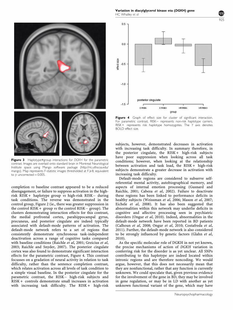
completion vs baseline contrast appeared to be a reduceddisengagement, or failure to suppress activation in the high-risk RISK + haplotype group vs high-risk RISK� duringtask conditions. The reverse was demonstrated in thecontrol group, Figure 2 (ie., there was greater suppression inthe control RISK + group vs the control RISK� group). Theclusters demonstrating interaction effects for this contrast,the medial prefrontal cortex, parahippocampal gyrus,precuneus, and posterior cingulate are indeed typicallyassociated with default-mode patterns of activation. Thedefault-mode network refers to a set of regions thatconsistently demonstrate synchronous task-independentdeactivation across a range of cognitive tasks comparedwith baseline conditions (Raichle et al, 2001; Greicius et al,2003; Raichle and Snyder, 2007). The posterior cingulatecortex was also found to demonstrate significant interactioneffects for the parametric contrast, Figure 4. This contrastfocusses on a gradation of neural activity in relation to taskdifficulty, rather than the sentence completion contrast,which relates activation across all levels of task condition toa simple visual baseline. In the posterior cingulate for theparametric contrast, the RISK� high-risk subjects andRISK + controls demonstrate small increases in activationwith increasing task difficulty. The RISK + high-risk
subjects, however, demonstrated decreases in activationwith increasing task difficulty. In summary therefore, inthe posterior cingulate, the RISK + high-risk subjectshave poor suppression when looking across all taskconditions; however, when looking at the relationshipbetween activation and task load, the RISK + high-risksubjects demonstrate a greater decrease in activation withincreasing task difficulty.
Default-mode regions are considered to subserve self-referential mental activity, autobiographical memory, andaspects of internal emotion processing (Gusnard andRaichle, 2001; Cabeza et al, 2002). Failure to deactivatethese regions has been linked to performance deficits inhealthy subjects (Weissman et al, 2006; Mason et al, 2007;Eichele et al, 2008). It has also been suggested thatabnormalities within this network may underlie deficits incognitive and affective processing seen in psychiatricdisorders (Ongur et al, 2010). Indeed, abnormalities in thedefault-mode network have been reported in BD patients(Calhoun et al, 2008; Ongur et al, 2010; Costafreda et al,2011). Further, the default-mode network is also consideredto be strongly influenced by genetic factors (Glahn et al,2010).
As the specific molecular role of DGKH in not yet known,the precise mechanisms of action of DGKH variation inconferring risk for the disorder is as yet unclear. All SNPscontributing to this haplotype are indeed located withinintronic regions and are therefore noncoding. We wouldargue, however, that this does not necessarily mean thatthey are nonfunctional, rather that any function is currentlyunknown. We could speculate that, given previous evidencefor the involvement of the gene in BD, they may be involvedin gene regulation, or may be in LD with another as yetunknown functional variant of the gene, which may have
Figure 3 Haplotype*group interactions for DGKH for the parametriccontrast. Images are overlaid onto standard brain in Montreal NeurologicalInstitute space using Mango software package (http://ric.uthscsa.edu/mango). Map represents F-statistic images thresholded at FX8, equivalentto p uncorrected¼ 0.005.
Figure 4 Graph of effect size for cluster of significant interaction.For parametric contrast, RISK� represents non-risk haplotype carriers,RISK + represents risk haplotype homozygotes. The Y axis denotesBOLD effect size.
Variation in diacylglycerol kinase eta (DGKH) geneHC Whalley et al
925
Neuropsychopharmacology
downstream influence on BOLD signal. In the context of thecurrent findings, they suggest that there is an influence ofthe DGKH gene on function in default-mode brain regions,and this differs between those at risk for the disorder andhealthy controls. In other words the risk haplotype appearsto alter the ability to disengage regions involved in self-referential and internal emotional processing during cogni-tive tasks in the bipolar high-risk group, as compared tohealthy controls. One possible explanation for this differ-ence is that additional genetic or environmental risk factorsinteracting with the effects of DGKH in the high-risk group.Presumably, controls carrying the risk haplotype are able tocompensate and allocate cognitive resources more effi-ciently during task conditions than the bipolar high-risksubjects carrying the risk haplotype, who also importantlycarry additional background risk factors. We interpret theseresults to suggest therefore that the relationship betweenDGKH genotype and functional architecture in the high-riskgroup may differ from controls based on an already presentgenetic loading for the disorder.
In terms of its biological relevance, DGKH is an upstreamregulator of protein kinase C (PKC). It catalyzes theconversion of diacylglycerol to phosphatidic acid, therebyleading to reductions in diacylglycerol. The latter isessential for activating several isoforms of PKC; a familyof enzymes that is heterogeneously distributed in thebrain and has a pivotal role in regulating pre- and post-synaptic neurotransmission (Casabona 1997; Catapanoand Manji, 2008). Accordingly, it has been proposed thatvariations in the DGKH gene might result in reductions ofdiacylglycerol kinase activity, thereby leading to increasedPKC signaling (Robbins and Arnsten, 2009). Numerouslines of evidence suggest the importance of PKC in thepathophysiology and treatment of BD. Investigations ofperipheral blood and postmortem studies indicate alteredPKC levels in BD, and lithium interacts with the PKCpathway resulting in decreasing downstream PKC levels andactivity (Catapano and Manji, 2008). Similarly, the moodstabilizer valproate inhibits PKC, suggesting a commonalityin the pathway of therapeutic effect (Catapano and Manji,2008). Moreover, the PKC antagonist tamoxifen has beenshown to reduce manic symptoms in BD patients (Yildizet al, 2008). Hence, DGKH is predicted to have a role inlithium response and in mood control and therefore in theetiology of the disorder.
Although there is limited literature relating specifically tothe effects of DGKH on brain functioning, there is evidenceof increased gene expression of DGKH in the prefrontalcortex of patients with BD vs controls (Moya et al, 2010).There are also several lines of evidence suggesting that PKCimpacts on cognitive functioning. Research has focussed onits role in mediating the effects of chronic stress on theprefrontal cortex (Hains et al, 2009), in regulating optimalprefrontal activity during working memory performance(Birnbaum et al, 2004), and in association with age-relatedcognitive decline (Brennan et al, 2009). Although spec-ulative, these may fit with neurobiological models of BDwhere there is altered prefrontal modulation of othercortical and subcortical structures involved in emotionalprocessing leading to impaired emotional regulation andincreased mood fluctuation in the disorder (Phillips et al,2003).
In summary, these findings suggest that variation in theDGKH gene is associated with differential modulation ofdefault network suppression during a sentence completionparadigm in bipolar high-risk individuals when comparedwith healthy controls. Differential effects of DGKH haplo-types in those at high familial risk of BD vs controlssupports previous evidence that the DGKH gene may beinvolved in the pathogenesis of BD. Further replication inpatient groups and/or during resting state scanning mayhelp confirm the nature of these effects.
ACKNOWLEDGEMENTS
We would like to thank all of the participants who took partin the study and the radiographers who acquired the MRIscans. This study was conducted at the Scottish BrainImaging Research Centre, which is supported by SINAPSE(Scottish Imaging Network, a Platform for ScientificExcellence, www.sinapse.ac.uk). The investigators alsoacknowledge the financial support of National HealthService (NHS) Research Scotland, through the ScottishMental Health Research Network (www.smhrn.org.uk) whoprovided assistance with subject recruitment and cognitiveassessments.
DISCLOSURE
The author HCW is supported by a Dorothy HodgkinFellowship from the Royal Society of Edinburgh(DH080018). MP is supported by a studentship from theMedical Research Council. JH is supported by a ScottishSenior Clinical Fellowship from the Chief Scientists Office inScotland. JES is supported by a Clinical Research TrainingFellowship from the Wellcome Trust. AMM is currentlysupported by the Health Foundation through a ClinicianScientist Fellowship (Ref: 2268/4295) and by the NationalAlliance for Research on Schizophrenia and Depressionthrough an Independent Investigator Award. The investi-gators also acknowledge the financial support of NationalHealth Service (NHS) Research Scotland, through theScottish Mental Health Research Network (www.smhrn.org.uk) who provided assistance with subject recruitment andcognitive assessments. HCW, JH, SML, and AMM havereceived financial support from Pfizer (formerly Wyeth) inrelation to imaging studies of people with schizophreniaand BD. The authors MP, LR, ECJ, and JES have nocompeting interests to declare.
REFERENCES
Akiskal HS, Mendlowicz MV, Jean-Louis G, Rapaport MH, KelsoeJR, Gillin JC et al. (2005). TEMPS-A: validation of a short versionof a self-rated instrument designed to measure variations intemperament. J Affect Disord 85: 45–52.
Allen P, Stephan KE, Mechelli A, Day F, Ward N, Dalton J et al.(2010). Cingulate activity and fronto-temporal connectivity inpeople with prodromal signs of psychosis. Neuroimage 49:947–955.
Arts B, Jabben N, Krabbendam L, van Os J (2008). Meta-analyses ofcognitive functioning in euthymic bipolar patients and theirfirst-degree relatives. Psychol Med 38: 771–785.
Variation in diacylglycerol kinase eta (DGKH) geneHC Whalley et al
926
Neuropsychopharmacology
Badner JA, Gershon ES (2002). Meta-analysis of whole-genomelinkage scans of bipolar disorder and schizophrenia. MolPsychiatry 7: 405–411.
Barnett JH, Smoller JW (2009). The genetics of bipolar disorder.Neuroscience 164: 331–343.
Baum AE, Akula N, Cabanero M, Cardona I, Corona W, Klemens Bet al. (2008). A genome-wide association study implicatesdiacylglycerol kinase eta (DGKH) and several other genes inthe etiology of bipolar disorder. Mol Psychiatry 13: 197–207.
Bertelsen A, Harvald B, Hauge M (1977). A Danish twin study ofmanic-depressive disorders. Br J Psychiatry 130: 330–351.
Birnbaum SG, Yuan PX, Wang M, Vijayraghavan S, Bloom AK,Davis DJ et al. (2004). Protein kinase C overactivity impairsprefrontal cortical regulation of working memory. Science 306:882–884.
Bloom PA, Fischler I (1980). Completion norms for 329 sentencecontexts. Mem Cogn 8: 631–642.
Brennan AR, Yuan P, Dickstein DL, Rocher AB, Hof PR,Manji H et al. (2009). Protein kinase C activity is associatedwith prefrontal cortical decline in aging. Neurobiol Aging 30:782–792.
Burgess P, Shallice T (1997). The Hayling and Brixton Tests.Thames Valley Test Company Limited: Bury St. Edmunds, UK.
Burgess PW, Shallice T (1996). Response suppression, initiationand strategy use following frontal lobe lesions. Neuropsychologia34: 263–272.
Cabeza R, Dolcos F, Graham R, Nyberg L (2002). Similarities anddifferences in the neural correlates of episodic memory retrievaland working memory. Neuroimage 16: 317–330.
Calhoun VD, Maciejewski PK, Pearlson GD, Kiehl KA (2008).Temporal lobe and ‘default’ hemodynamic brain modesdiscriminate between schizophrenia and bipolar disorder. HumBrain Mapp 29: 1265–1275.
Casabona G (1997). Intracellular signal modulation: a pivotal rolefor protein kinase C. Prog Neuropsychopharmacol Biol Psychiatry21: 407–425.
Catapano LA, Manji HK (2008). Kinases as drug targets in thetreatment of bipolar disorder. Drug Discov Today 13: 295–302.
Cerullo MA, Adler CM, Delbello MP, Strakowski SM (2009). Thefunctional neuroanatomy of bipolar disorder. Int Rev Psychiatry21: 314–322.
Costafreda SG, Fu CH, Picchioni M, Toulopoulou T, McDonald C,Kravariti E et al. (2011). Pattern of neural responses to verbalfluency shows diagnostic specificity for schizophrenia andbipolar disorder. BMC Psychiatry 11: 18.
Detera-Wadleigh SD, McMahon FJ (2006). G72/G30 in schizo-phrenia and bipolar disorder: review and meta-analysis. BiolPsychiatry 60: 106–114.
Eichele T, Debener S, Calhoun VD, Specht K, Engel AK, HugdahlK, von Cramon DY et al. (2008). Prediction of human errors bymaladaptive changes in event-related brain networks. Proc NatlAcad Sci USA 105: 6173–6178.
First MB, Spitzer RL, Gibbon M, Williams JBW (2002). StructuredClinical Interview for DSM-IV-TR Axis I Disorders, ResearchVersion, Patient Edition with Psychotic Screen. BiometricsResearch, New York State Psychiatric Institute: New York.
Glahn DC, Winkler AM, Kochunov P, Almasy L, Duggirala R,Carless MA et al. (2010). Genetic control over the resting brain.Proc Natl Acad Sci USA 107: 1223–1228.
Greicius MD, Krasnow B, Reiss AL, Menon V (2003). Functionalconnectivity in the resting brain: a network analysis of thedefault mode hypothesis. Proc Natl Acad Sci USA 100: 253–258.
Gusnard DA, Raichle ME (2001). Searching for a baseline:functional imaging and the resting human brain. Nat RevNeurosci 2: 685–694.
Hains AB, Vu MA, Maciejewski PK, van Dyck CH, Gottron M,Arnsten AF (2009). Inhibition of protein kinase C signalingprotects prefrontal cortex dendritic spines and cognition from
the effects of chronic stress. Proc Natl Acad Sci USA 106:17957–17962.
Hamilton M (1960). A rating scale for depression. J NeurolNeurosurg Psychiatry 23: 56–62.
Maldjian JA, Laurienti PJ, Kraft RA, Burdette JH (2003). Anautomated method for neuroanatomic and cytoarchitectonicatlas-based interrogation of fMRI data sets. Neuroimage 19:1233–1239.
Manchia M, Squassina A, Congiu D, Chillotti C, Ardau R,Severino G et al. (2009). Interacting genes in lithium prophy-laxis: preliminary results of an exploratory analysis on the role ofDGKH and NR1D1 gene polymorphisms in 199 Sardinianbipolar patients. Neurosci Lett 467: 67–71.
Manji HK, Lenox RH (1999). Ziskind-Somerfeld Research Award.Protein kinase C signaling in the brain: molecular transductionof mood stabilization in the treatment of manic-depressiveillness. Biol Psychiatry 46: 1328–1351.
Mason MF, Norton MI, Van Horn JD, Wegner DM, Grafton ST,Macrae CN (2007). Wandering minds: the default network andstimulus-independent thought. Science 315: 393–395.
McGuffin P, Farmer A, Harvey I (1991). A polydiagnosticapplication of operational criteria in studies of psychotic illness.Development and reliability of the OPCRIT system. Arch GenPsychiatry 48: 764–770.
McGuffin P, Rijsdijk F, Andrew M, Sham P, Katz R, Cardno A(2003). The heritability of bipolar affective disorder and thegenetic relationship to unipolar depression. Arch Gen Psychiatry60: 497–502.
McIntosh A, Whalley HC, McKirdy J, Hall J, Sussmann J, ShankarP et al. (2008a). Differences in dorsal and ventral prefrontalfunction separate bipolar disorder from schizophrenia. SchizoprRes 98: 40.
McIntosh AM, Whalley HC, McKirdy J, Hall J, Sussmann JE,Shankar P et al. (2008b). Prefrontal function and activationin bipolar disorder and schizophrenia. Am J Psychiatry 165:378–384.
Moya PR, Murphy DL, McMahon FJ, Wendland JR (2010).Increased gene expression of diacylglycerol kinase eta in bipolardisorder. Int J Neuropsychopharmacology 13: 1127–1128.
Nathaniel-James DA, Fletcher P, Frith CD (1997). The functionalanatomy of verbal initiation and suppression using the Haylingtest. Neuropsychologia 35: 559–566.
Nelson H (1982). The National Adult Reading Test Manual. NFER-Nelson: Windsor.
Ollila HM, Soronen P, Silander K, Palo OM, Kieseppa T, KaunistoMA et al. (2009). Findings from bipolar disorder genome-wideassociation studies replicate in a Finnish bipolar family-cohort.Mol Psychiatry 14: 351–353.
Ongur D, Lundy M, Greenhouse I, Shinn AK, Menon V, Cohen BMet al. (2010). Default mode network abnormalities in bipolardisorder and schizophrenia. Psychiatry Res 183: 59–68.
Phillips ML, Drevets WC, Rauch SL, Lane R (2003). Neurobiologyof emotion perception II: implications for major psychiatricdisorders. Biol Psychiatry 54: 515–528.
Raichle ME, Snyder AZ (2007). A default mode of brain function: abrief history of an evolving idea. Neuroimage 37: 1083–1090;discussion 1097–9.
Raichle ME, MacLeod AM, Snyder AZ, Powers WJ, Gusnard DA,Shulman GL (2001). A default mode of brain function. Proc NatlAcad Sci USA 98: 676–682.
Robbins TW, Arnsten AF (2009). The neuropsychopharmacologyof fronto-executive function: monoaminergic modulation. AnnuRev Neurosci 32: 267–287.
Sklar P, Smoller JW, Fan J, Ferreira MA, Perlis RH, Chambert Ket al. (2008). Whole-genome association study of bipolardisorder. Mol Psychiatry 13: 558–569.
Sprooten E, Sussmann JE, Clugston A, Peel A, McKirdy J,Moorhead TWJ et al. (2011). White matter integrity in
Variation in diacylglycerol kinase eta (DGKH) geneHC Whalley et al
927
Neuropsychopharmacology

individuals at high genetic risk of bipolar disorder. BiolPsychiatry 70: 350–356.
Squassina A, Manchia M, Congiu D, Severino G, Chillotti C, ArdauR et al. (2009). The diacylglycerol kinase eta gene and bipolardisorder: a replication study in a Sardinian sample. MolPsychiatry 14: 350–351.
Strakowski SM, Delbello MP, Adler CM (2005). The functionalneuroanatomy of bipolar disorder: a review of neuroimagingfindings. Mol Psychiatry 10: 105–116.
Tesli M, Kahler AK, Andreassen BK, Werge T, Mors O, Mellerup Eet al. (2009). No association between DGKH and bipolar disorderin a Scandinavian case-control sample. Psychiatr Genet 19:269–272.
The Wellcome Trust Case Control Consortium (2007).Genome-wide association study of 14 000 cases of sevencommon diseases and 3000 shared controls. Nature 447:661–678.
Tzourio-Mazoyer N, Landeau B, Papathanassiou D, Crivello F,Etard O, Delcroix N et al. (2002). Automated anatomicallabeling of activations in SPM using a macroscopic anatomicalparcellation of the MNI MRI single-subject brain. Neuroimage15: 273–289.
Weber H, Kittel-Schneider S, Gessner A, Domschke K, Neuner M,Jacob CP et al. (2011). Cross-disorder analysis of bipolar riskgenes: further evidence of DGKH as a risk gene for bipolardisorder, but also unipolar depression and adult ADHD.Neuropsychopharmacology 36: 2076–2085.
Weissman DG, Roberts KC, Visscher KM, Woldorff MG (2006).The neural basis of momentary lapses of attention. Nat Neurosci9: 971–978.
Whalley HC, Simonotto E, Flett S, Marshall I, Ebmeier KP, OwensDG et al. (2004). fMRI correlates of state and trait effects insubjects at genetically enhanced risk of schizophrenia. [seecomment]. Brain 127: 478–490.
Whalley HC, Sussmann JE, Chakirova G, Mukerjee P, Peel A,McKirdy J et al. (2011). The neural basis of familial risk andtemperamental variation in individuals at high risk of bipolardisorder. Biol Psychiatry 70: 343–349.
Yildiz A, Guleryuz S, Ankerst DP, Ongur D, Renshaw PF (2008).Protein kinase C inhibition in the treatment of mania: a double-blind, placebo-controlled trial of tamoxifen. Arch Gen Psychiatry65: 255–263.
Yosifova A, Mushiroda T, Stoianov D, Vazharova R, Dimova I,Karachanak S et al. (2009). Case-control association study of 65candidate genes revealed a possible association of a SNP ofHTR5A to be a factor susceptible to bipolar disease in Bulgarianpopulation. J Affect Disord 117: 87–97.
Young RC, Biggs JT, Ziegler VE, Meyer DA (1978). A rating scalefor mania: reliability, validity and sensitivity. Br J Psychiatry 133:429–435.
Zeng Z, Wang T, Li T, Li Y, Chen P, Zhao Q et al. (2011). CommonSNPs and haplotypes in DGKH are associated with bipolardisorder and schizophrenia in the Chinese Han population. MolPsychiatry 16: 473–475.
Supplementary Information accompanies the paper on the Neuropsychopharmacology website (http://www.nature.com/npp)
Variation in diacylglycerol kinase eta (DGKH) geneHC Whalley et al
928
Neuropsychopharmacology