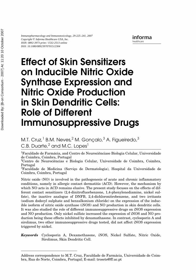
Effect of Skin Sensitizers on Inducible Nitric Oxide Synthase Expression and Nitric Oxide Production...
19
This article was downloaded by:[B-on Consortium - 2007] On: 10 October 2007 Access Details: [subscription number 778384750] Publisher: Informa Healthcare Informa Ltd Registered in England and Wales Registered Number: 1072954 Registered office: Mortimer House, 37-41 Mortimer Street, London W1T 3JH, UK Immunopharmacology and Immunotoxicology Publication details, including instructions for authors and subscription information: http://www.informaworld.com/smpp/title~content=t713597257 Effect of Skin Sensitizers on Inducible Nitric Oxide Synthase Expression and Nitric Oxide Production in Skin Dendritic Cells: Role of Different Immunosuppressive Drugs M.T. Cruz a ; B.M. Neves b ; M. Gonçalo c ; A. Figueiredo c ; C.B. Duarte b ; M.C. Lopes a a Faculdade de Farmácia, and Centro de Neurociênciase Biologia Celular, Universidade de Coimbra, Coimbra, Portugal b Centro de Neurociências e Biologia Celular, Universidade de Coimbra, Coimbra, Portugal c Faculdade de Medicina (Serviço de Dermatologia), Hospital da Universidade de Coimbra, Coimbra, Portugal Online Publication Date: 01 April 2007 To cite this Article: Cruz, M.T., Neves, B.M., Gonçalo, M., Figueiredo, A., Duarte, C.B. and Lopes, M.C. (2007) 'Effect of Skin Sensitizers on Inducible Nitric Oxide Synthase Expression and Nitric Oxide Production in Skin Dendritic Cells: Role of Different Immunosuppressive Drugs', Immunopharmacology and Immunotoxicology, 29:2, 225 - 241 To link to this article: DOI: 10.1080/08923970701512304 URL: http://dx.doi.org/10.1080/08923970701512304 PLEASE SCROLL DOWN FOR ARTICLE Full terms and conditions of use: http://www.informaworld.com/terms-and-conditions-of-access.pdf This article maybe used for research, teaching and private study purposes. Any substantial or systematic reproduction, re-distribution, re-selling, loan or sub-licensing, systematic supply or distribution in any form to anyone is expressly forbidden. The publisher does not give any warranty express or implied or make any representation that the contents will be complete or accurate or up to date. The accuracy of any instructions, formulae and drug doses should be independently verified with primary sources. The publisher shall not be liable for any loss, actions, claims, proceedings, demand or costs or damages whatsoever or howsoever caused arising directly or indirectly in connection with or arising out of the use of this material.
-
Upload
independent -
Category
Documents
-
view
0 -
download
0
Transcript of Effect of Skin Sensitizers on Inducible Nitric Oxide Synthase Expression and Nitric Oxide Production...

This article was downloaded by:[B-on Consortium - 2007]On: 10 October 2007Access Details: [subscription number 778384750]Publisher: Informa HealthcareInforma Ltd Registered in England and Wales Registered Number: 1072954Registered office: Mortimer House, 37-41 Mortimer Street, London W1T 3JH, UK
Immunopharmacology andImmunotoxicologyPublication details, including instructions for authors and subscription information:http://www.informaworld.com/smpp/title~content=t713597257
Effect of Skin Sensitizers on Inducible Nitric OxideSynthase Expression and Nitric Oxide Production inSkin Dendritic Cells: Role of DifferentImmunosuppressive DrugsM.T. Cruz a; B.M. Neves b; M. Gonçalo c; A. Figueiredo c; C.B. Duarte b; M.C.Lopes aa Faculdade de Farmácia, and Centro de Neurociênciase Biologia Celular,Universidade de Coimbra, Coimbra, Portugalb Centro de Neurociências e Biologia Celular, Universidade de Coimbra, Coimbra,Portugalc Faculdade de Medicina (Serviço de Dermatologia), Hospital da Universidade de
Coimbra, Coimbra, Portugal
Online Publication Date: 01 April 2007To cite this Article: Cruz, M.T., Neves, B.M., Gonçalo, M., Figueiredo, A., Duarte, C.B. and Lopes, M.C. (2007) 'Effect ofSkin Sensitizers on Inducible Nitric Oxide Synthase Expression and Nitric Oxide Production in Skin Dendritic Cells: Roleof Different Immunosuppressive Drugs', Immunopharmacology and Immunotoxicology, 29:2, 225 - 241To link to this article: DOI: 10.1080/08923970701512304URL: http://dx.doi.org/10.1080/08923970701512304
PLEASE SCROLL DOWN FOR ARTICLE
Full terms and conditions of use: http://www.informaworld.com/terms-and-conditions-of-access.pdf
This article maybe used for research, teaching and private study purposes. Any substantial or systematic reproduction,re-distribution, re-selling, loan or sub-licensing, systematic supply or distribution in any form to anyone is expresslyforbidden.
The publisher does not give any warranty express or implied or make any representation that the contents will becomplete or accurate or up to date. The accuracy of any instructions, formulae and drug doses should beindependently verified with primary sources. The publisher shall not be liable for any loss, actions, claims, proceedings,demand or costs or damages whatsoever or howsoever caused arising directly or indirectly in connection with orarising out of the use of this material.
Dow
nloa
ded
By:
[B-o
n C
onso
rtium
- 20
07] A
t: 11
:20
10 O
ctob
er 2
007
Immunopharmacology and Immunotoxicology, 29:225–241, 2007Copyright © Informa Healthcare USA, Inc.ISSN: 0892-3973 print / 1532-2513 onlineDOI: 10.1080/08923970701512304
LIPI0892-39731532-2513Immunopharmacology and Immunotoxicology, Vol. 29, No. 2, July 2007: pp. 1–35Immunopharmacology and ImmunotoxicologyEffect of Skin Sensitizerson Inducible Nitric Oxide Synthase Expression andNitric Oxide Productionin Skin Dendritic Cells:Role of Different Immunosuppressive DrugsiNOS Expression by Skin SensitizersM.T. Cruz et al.
M.T. Cruz,1 B.M. Neves,2 M. Gonçalo,3 A. Figueiredo,3 C.B. Duarte,2 and M.C. Lopes1
1Faculdade de Farmácia, and Centro de Neurociênciase Biologia Celular, Universidadede Coimbra, Coimbra, Portugal2Centro de Neurociências e Biologia Celular, Universidade de Coimbra, Coimbra,Portugal3Faculdade de Medicina (Serviço de Dermatologia), Hospital da Universidade deCoimbra, Coimbra, Portugal
Nitric oxide (NO) is involved in the pathogenesis of acute and chronic inflammatoryconditions, namely in allergic contact dermatitis (ACD). However, the mechanism bywhich NO acts in ACD remains elusive. The present study focuses on the effects of dif-ferent contact sensitizers (2,4-dinitrofluorbenzene, 1,4-phenylenediamine, nickel sul-fate), the inactive analogue of DNFB, 2,4-dichloronitrobenzene, and two irritants(sodium dodecyl sulphate and benzalkonium chloride) on the expression of the induc-ible isoform of nitric oxide synthase (iNOS) and NO production in skin dendritic cells.It was also studied the role of different immunosuppressive drugs on iNOS expressionand NO production. Only nickel sulfate increased the expression of iNOS and NO pro-duction being these effects inhibited by dexamathasone. In contrast, cyclosporin A andsirolimus, two other immunosuppressive drugs tested, did not affect iNOS expressiontriggered by nickel.
Keywords Cyclosporin A, Dexamethasone, iNOS, Nickel Sulfate, Nitric Oxide,Sirolimus, Skin Dendritic Cell.
Address correspondence to M.T. Cruz, Faculdade de Farmácia, Universidade de Coim-bra, Rua do Norte, Coimbra, Portugal; E-mail: [email protected]

Dow
nloa
ded
By:
[B-o
n C
onso
rtium
- 20
07] A
t: 11
:20
10 O
ctob
er 2
007
226 M.T. Cruz et al.
INTRODUCTION
Dendritic cells (DCs) are ubiquitously distributed migratory antigen present-ing cells, derived from CD34+ bone marrow stem cells that have the uniquecapacity to prime naïve T cells. DCs also regulate various effectors cell-functionsand, therefore, play central roles in modulating the immune response, namelyin allergic contact dermatitis (ACD).(1–3)
Among all the contact sensitizers, nickel is the most frequent cause ofACD. This hapten increases the expression of DC surface markers essentialfor antigen presentation, such as the molecules of the major histocompatibilitycomplex class II and costimulatory molecules(4–10) and increases chemokineproduction and chemokine receptor expression(11–14), thereby favoring antigenpresentation. In DC, contact sensitizers activate mitogen-activated proteinkinases(15–21). We previously have reported that nickel sulfate activates thetranscription nuclear factor kappa B (NF-kB) and the transcription factoractivator protein-1 (AP-1) and induces the expression of inducible isoform ofnitric oxide synthase (iNOS) in mouse skin DC(22). However, the effect of otherskin contact sensitizers and irritants on iNOS expression and nitric oxide(NO) production was never addressed before.
NO, a highly reactive radical produced from the amino acid L-arginine bythe enzyme iNOS, mediates various physiological functions in the skin, rang-ing from the regulation of cutaneous blood flow to melanogenesis(23, 24), andmodulates antigen presentation.(25) If produced in excess, NO combines withsuperoxide anion to form peroxynitrite that has been considered responsiblefor tissue injury in skin inflammatory processes, such as ACD(26–30). There-fore, in this work we studied the effect of different skin sensitizers that induceACD on NO production by skin dendritic cells.
Sirolimus, cyclosporine A, and dexamethasone are clinically relevantimmunosuppressive drugs with distinct molecular mechanisms of action,exhibiting different immunoregulatory profiles. Although the effect of thesedrugs on dendritic cell differentiation, migration, and maturation has beenreported previously(31), their direct effects on iNOS expression and NO produc-tion triggered by contact sensitizers remain unknown.
The present study focuse on the effects of different contact sensitizers(2,4-dinitrofluorbenzene, 1,4-phenylenediamine, nickel sulfate), the inac-tive analogue of DNFB, 2,4-dichloronitrobenzene (DCNB), and of twoirritants (sodium dodecyl sulphate [SDS] and benzalkonium chloride [BC]),on the expression and activity of iNOS in skin DC. Moreover, it we alsostudied the role of different immunosuppressive drugs on iNOS expressionand NO production. As an experimental model of a DC we used a fetalskin-derived dendritic cell line (FSDC), that has morphological, phenotypi-cal, and functional characteristics of immature skin DC, the Langerhanscells(32).

Dow
nloa
ded
By:
[B-o
n C
onso
rtium
- 20
07] A
t: 11
:20
10 O
ctob
er 2
007
iNOS Expression by Skin Sensitizers 227
MATERIAL AND METHODS
Nickel sulfate, dexamethasone, benzalkonium chloride, and cyclosporin A werefrom Sigma Chemical Co. (Madrid, Spain). Sodium dodecyl sulphate was fromGE Healthcare (Carnaxide, Portugal) and sirolimus was from Calbiochem(San Diego, CA, USA). The rabbit polyclonal anti-iNOS was from Abcam (Cam-bridge, UK). The alkaline phosphatase linked antirabbit IgG (H + L) antibodyand the ECF substrate were purchased from GE Healthcare. Alexa Fluor 488-conjugated goat antirabbit antibody was from Molecular Probes (Leiden, TheNetherlands) and the mounting medium for fluorescence, Vectashield, wasobtained from Vector Laboratories, (Burlingame, CA, USA). The fetal calfserum was from Biochrom KG (Berlin, Germany) and trypsin from Invitrogen(Paisley, UK). The proteases inhibitor cocktail was from Roche (Carnaxide,Portugal). DCNB, DNFB, and phenylenediamine (PPD) were from Aldrich Chem-ical Company (Madrid, Spain). All other reagents were from Sigma Chemical Co.
Cell Culture and ChemicalsThe fetal mouse skin dendritic cell line FSDC was kindly supplied by
Dr. G. Girolomoni(32) and cultured in endotoxin free Iscove´s medium supple-mented with 10 % (v/v) fetal calf serum, 1 % (w/v) glutamine, 3.02 g/l sodiumbicarbonate, 100 μg/ml streptomycin, and 100 U/ml penicillin. For Westernblot analysis FSDC were plated at 2 × 10⁶ cells/well in 6-well culture plates,for 24 hr, prior to treatment with the contact sensitizers or with the contactsensitizer nickel sulfate in the presence of the different immunosuppressors.For nitrite and MTT measurements the cells were plated at 0.2 × 10⁶ cells/wellin 48-well culture plates in the presence of NiSO4 and the immunosuppressorsfor 48 hrs. SDS (50 μg/ml), BC (1 μg/ml), PPD (50 μg/ml), and NiSO4 (50 μg/ml)were dissolved in saline, while DNFB (1 μg/ml) and DCNB (1 μg/ml) were firstsolubilized in dimethyl sulphoxide. Dimethyl sulphoxide concentration neverexceeded 0.025% in the culture medium.
MTT AssayAssessment of MTT reduction by metabolically active cells was made by a
colorimetric assay, using 3-(4,5-dimethylthiazol-2-yl)-2,5-diphenyl tetrazo-lium bromide (MTT) as previously reported(33). The cells (0.2 × 10⁶ cells/well),cultured in 48-well microplates, were incubated for 48 hr with culture mediumalone (control), with NiSO4 (50 μg/ml), or with the contact sensitizer in thepresence of the different immunosuppressors. After removal of cell-free super-natants for the nitrite assay, 400 μl of culture medium and 40 μl of MTT solu-tion (5 mg/ml in PBS) were added to each well. The microplates were furtherincubated at 37°C for 1 hr. Supernatants were then discarded and 300 μl ofacidified isopropanol (0.04 N HCl in isopropanol) were added to the cultures
Dow
nloa
ded
By:
[B-o
n C
onso
rtium
- 20
07] A
t: 11
:20
10 O
ctob
er 2
007
228 M.T. Cruz et al.
and mixed thoroughly to dissolve the dark blue crystals of formazan. Formazanquantification was performed using an automatic plate reader (SLT, Austria)at 570 nm, with a reference wavelength of 620 nm.
Nitrite MeasurementThe production of NO was measured by the accumulation of nitrite in the
culture supernatants, using a colorimetric reaction with the Griess reagent(34).Briefly, after FSDC stimulation with culture medium alone (control), or withNiSO4 (50 μg/ml), or with the contact sensitizer in the presence of the differentimmunosuppressors, for 48 hr, the culture supernatants were collected anddiluted with equal volumes of the Griess reagent (0.1% [w/v] N-(1-naphthyl)ethylenediamine dihydrochloride, 1 % [w/v] sulphanilamide:5 % [w/v] H3PO4),during 10 min. The absorbance at 550 nm was measured after 20-min incuba-tion in an automated plate reader (SLT). Nitrite concentration was deter-mined from a sodium nitrite standard curve.
Western Blot AnalysisFor immunodetection of iNOS, cells were treated with culture medium
(control), with the contact sensitizers NiSO4 (50 μg/ml), DNFB (1 μg/ml), andPPD (50 μg/ml), with the irritants SDS (50 μg/ml) and BC (1 μg/ml), and withthe inactive chemical DCNB (1μg /ml). In another set of experiments, cellswere treated with the contact sensitizer NiSO4 in the presence of the differentimmunosuppressors, and with the immunosuppressors alone (data notshown), for 24 hr. After treatment, cells were washed with PBS and total celllysates were obtained after harvesting the cells in a sonication buffer contain-ing 0.25 M sucrose, 10 mM Tris-HCl, pH 7.5, 1 mM EDTA, 2 mM sodium orto-vanadate, 1mM dithiothreitol, and the protease inhibitor cocktail. The lysateswere then incubated on ice for 30 min and sonicated on ice at low amplitude(four times for 4 sec at 20 μm peak to peak) to disrupt the cells. Protein con-centration was determined using the bicinchoninic acid method. Protein sam-ples were separated on a 12% (v/v) SDS-PAGE and transferred to a PVDFmembrane. The membrane was blocked with 5% (w/v) dry milk in Tris-bufferedsaline with 0.1% (v/v) Tween 20, for 1 hr.
The level of iNOS protein was detected using a rabbit polyclonal anti-mouse iNOS antibody (1:200) for 1 hr, followed by incubation 1 hr at roomtemperature, with antirabbit IgG antibodies coupled to alkaline phosphatase,at 1:20,000 dilution. The immune complexes were detected using the ECF sys-tem (GE Healthcare), and the membranes were then scanned with blue-excited fluorescence on the Storm 860 (Amersham Biosciences), according tothe manufacturer’s instructions. The signals were analyzed using an imageanalyzer. To demonstrate equivalent protein loading the membranes werestripped and reprobed with an antiactin antibody (1:7500).

Dow
nloa
ded
By:
[B-o
n C
onso
rtium
- 20
07] A
t: 11
:20
10 O
ctob
er 2
007
iNOS Expression by Skin Sensitizers 229
Immunofluorescence MicroscopyFor immunofluorescence analysis, FSDC cells grown on Lab-Tek chamber
slide with cover (0.2 × 10⁶ cells/slide) were treated, for 24 hrs, with the con-tact sensitizers NiSO4 (50 μg/ml), DNFB (1 μg/ml), and PPD (50 μg/ml), withthe irritants SDS (50 μg/ml) and BC (1 μg/ml), and with the inactive chemicalDCNB (1μg /ml). In another set of experiments, cells were treated with thecontact sensitizer NiSO4 in the presence of the different immunosuppressors,namely sirolimus (1 nM), dexamethasone (1 μM), and cyclosporin A (0.83μM). The cells were then washed with PBS and fixed and permeabilized withmethanol/acetone (1:1), for 10 min. Nonspecific binding was blocked by incu-bating the cells with 0.2 % gelatine in PBS supplemented with 0.1 % Tween20, for 45 min at room temperature. Cells were then incubated for 120 min atroom temperature, with a rabbit polyclonal antibody directed against iNOS(20 μg/ml).
After rinsing with PBS the cells were incubated with Alexa Fluor 488 con-jugated goat antirabbit antibody (1:500) in 0.5% BSA-PBS, for 45 min. Thechamber slide was rinsed and mounted with the mounting medium for fluo-rescence, Vectashield. Images from cells labelled with anti-iNOS wereacquired on a Zeiss Axiovert 200 fluorescence microscope. Control experi-ments consisted of processing the same preparations as described, except foromission of the primary antibody, and resulted in no specific staining.
Data AnalysisResults are presented as mean ± SEM of the indicated number of experi-
ments. Mean values were compared using one-way ANOVA followed by theBonferroni´s multiple comparison test. The significance level was 0.05.
RESULTS
Nickel Sulfate Increases iNOS Expression and NO ProductionWe evaluated the effect of the sensitizers nickel, DNFB, PPD, the irritants
SDS, BC, and the nonsensitizer DCNB on the expression of iNOS protein indendritic cells. Our results demonstrated that only nickel sulfate increasediNOS expression by FSDC (Figure 1, lane 4), detected by Western blot, andslightly increased nitrite production (Figure 2), determined by the Griessreagent. As shown in Figure 2, stimulation of the cells with NiSO4 for 48 hrincreased nitrite production, from 100% when FSDC were incubated with cul-ture medium alone, to 109.5 ± 1.2%, when FSDC were incubated with 50 μg/ml NiSO4 (p < 0.05), reflecting an increase in NO production. The assay of cel-lular MTT reduction did not show any significant toxic effect induced byNiSO4 for the concentration used in these experiments (data not shown).

Dow
nloa
ded
By:
[B-o
n C
onso
rtium
- 20
07] A
t: 11
:20
10 O
ctob
er 2
007
230 M.T. Cruz et al.
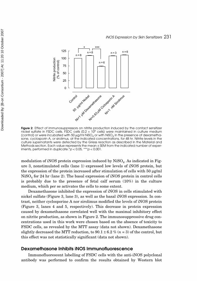
Dexamethasone Inhibits iNOS expression and NO ProductionWe also investigated the effect of three immunosuppressive drugs, dexam-
ethasone, cyclosporin A, and sirolimus, on iNOS expression and NO produc-tion stimulated by the contact sensitizer nickel sulfate on skin DC. AfterFSDC stimulation with the sensitizer nickel in the presence of dexamethasone(1 μM), nitrite production induced by nickel was reduced to 72.2 ± 5.3% of thecontrol (p < 0.001) (Figure 2). Cyclosporin A (0.83 μM) and sirolimus (1 nM)did not modify the increase of nitrite production triggered by the contact sensi-tizer (Figure 2) (108.4 ± 0.9 and 111.4 ± 1.4 % of control, respectively). Westernblot was used to examine the effect of immunosuppressive drugs on the
Figure 1: Effect of contact sensitizers and irritants on iNOS expression in FSDC cells. FSDC cells(2 × 10⁶ cells) were incubated in culture medium alone (control, lane 1), or with 1 μg/ml DNFB(lane 2), 50 μg/ml PPD (lane 3), 50 μg/ml NiSO4 (lane 4), 1 μg/ml DCNB (lane 5), 50 μg/ml SDS(lane 6), or 1 μg/ml BC (lane 7) for 24 hr. Total cell extracts were electrophoresed through SDS-PAGE, transferred to PVDF membranes and subjected to Western blot analysis using an anti-iNOS antibody. (A) The membranes were stripped and reprobed with an antiactin antibodyto confirm equal protein loading (B). The results were quantified by scanning the membranewith a fluorescence scanner and analyzed using an image analyzer. Each value representsthe mean ± SEM from 4 experiments.**p < 0.01.
B
A
contr
ol
DNFBPPD Ni
DCNBSDS BC
0
50
100
150**
iNO
S e
xpre
ssio
n(%
of c
ontro
l)
Dow
nloa
ded
By:
[B-o
n C
onso
rtium
- 20
07] A
t: 11
:20
10 O
ctob
er 2
007
iNOS Expression by Skin Sensitizers 231
modulation of iNOS protein expression induced by NiSO4. As indicated in Fig-ure 3, nonstimulated cells (lane 1) expressed low levels of iNOS protein, butthe expression of the protein increased after stimulation of cells with 50 μg/mlNiSO4 for 24 hr (lane 2). The basal expression of iNOS protein in control cellsis probably due to the presence of fetal calf serum (10%) in the culturemedium, which per se activates the cells to some extent.
Dexamethasone inhibited the expression of iNOS in cells stimulated withnickel sulfate (Figure 3, lane 3), as well as the basal iNOS expression. In con-trast, neither cyclosporine A nor sirolimus modified the levels of iNOS protein(Figure 3, lanes 4 and 5, respectively). This decrease in protein expressioncaused by dexamethasone correlated well with the maximal inhibitory effecton nitrite production, as shown in Figure 2. The immunosuppressive drug con-centrations used in this work were chosen based on the absence of toxicity toFSDC cells, as revealed by the MTT assay (data not shown). Dexamethasoneslightly decreased the MTT reduction, to 90.1 ± 6.2 % (n = 3) of the control, butthis effect was not statistically significant (data not shown).
Dexamethasone Inhibits iNOS ImmunofluorescenceImmunofluorescent labelling of FSDC cells with the anti-iNOS polyclonal
antibody was performed to confirm the results obtained by Western blot
Figure 2: Effect of immunosuppressors on nitrite production induced by the contact sensitizernickel sulfate in FSDC cells. FSDC cells (0.2 × 10⁶ cells) were maintained in culture medium(control) or were incubated with 50 μg/ml NiSO4 or with NiSO4 in the presence of dexametha-sone, cyclosporin A, or sirolimus, at the indicated concentrations, for 48 hr. Nitrite levels in theculture supernatants were detected by the Griess reaction as described in the Material andMethods section. Each value represents the mean ± SEM from the indicated number of exper-iments, performed in duplicate.*p < 0.05, ***p < 0.001.
Contro
l
50 μ
g/ml N
ickel
Ni+1 μ
M Dex
ameth
ason
e
Ni+0.83
μM C
iclos
porin
A
Ni+1 n
M Siro
limus
0
25
50
75
100
125n = 8
n = 3
n = 6
n = 8 n = 6* ***
Nitr
ite p
rodu
ctio
n(%
of c
ontro
l)

Dow
nloa
ded
By:
[B-o
n C
onso
rtium
- 20
07] A
t: 11
:20
10 O
ctob
er 2
007
232 M.T. Cruz et al.
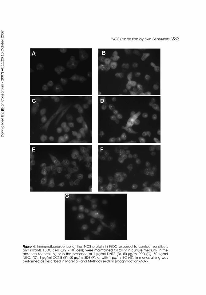
(Figure 4). Nickel sulfate slightly increased iNOS as compared with the cellsmaintained in culture medium. In addition, the other contact sensitizers andirritants studied, as well as DCNB, did not modify iNOS fluorescence. Theseresults are in agreement with those obtained by Western blot (Figure 1) andindicate that the expression of iNOS is upregulated by the sensitizer NiSO4 in
Figure 3: Effect of immunosuppressors on iNOS expression induced by the contact sensitizernickel sulfate in FSDC cells. FSDC cells (2 × 10⁶ cells) were incubated in culture medium alone(control, lane 1), with 50 μg/ml NiSO4 (lane 2), or with NiSO4 in the presence of 1 μM dexam-ethasone (lane 3), 0.83 μM cyclosporin A (lane 4), or 1 nM sirolimus (lane 5), for 24 hr. Total cellextracts were electrophoresed through SDS-PAGE, transferred to PVDF membranes, and sub-jected to Western blot analysis using an anti-iNOS antibody. (A) The membranes werestripped and reprobed with an antiactin antibody to confirm equal protein loading (B). Theresults were quantified by scanning the membrane with a fluorescence scanner and ana-lyzed using an image analyzer. Each value represents the mean ± SEM from 3 experiments.**p < 0.01; ***p < 0.001.
B
A
Contro
l Ni
Ni + Dex
ameth
ason
e
Ni + Cicl
ospo
rin A
Ni + Siro
limus
0
100
200
** ***
iNO
S e
xpre
ssio
n(%
of c
ontro
l)
Dow
nloa
ded
By:
[B-o
n C
onso
rtium
- 20
07] A
t: 11
:20
10 O
ctob
er 2
007
iNOS Expression by Skin Sensitizers 233
Figure 4: Immunofluorescence of the iNOS protein in FSDC exposed to contact sensitizersand irritants. FSDC cells (0.2 × 10⁶ cells) were maintained for 24 hr in culture medium, in theabsence (control, A) or in the presence of 1 μg/ml DNFB (B), 50 μg/ml PPD (C), 50 μg/mlNiSO4 (D), 1 μg/ml DCNB (E), 50 μg/ml SDS (F), or with 1 μg/ml BC (G). Immunostaining wasperformed as described in Materials and Methods section (magnification 650×).

Dow
nloa
ded
By:
[B-o
n C
onso
rtium
- 20
07] A
t: 11
:20
10 O
ctob
er 2
007
234 M.T. Cruz et al.
FSDC. The results obtained by immunofluorescence also demonstrate thatdexamethasone is the unique immunosuppressor able to decrease the iNOSexpression triggered by the contact sensitizer nickel (Figure 5).
DISCUSSION
The aim of our study was to evaluate the effect of the contact sensitizersDNFB, PPD, and nickel sulfate, and the irritants SDS and BC, on the iNOSprotein expression to known if there is a different DC response that could dis-criminate between contact sensitizers and skin irritants. We have previouslydemonstrated, in a mouse skin dendritic cell line, that the contact sensitizernickel sulfate increases iNOS expression and NO production in these skindendritic cells(22). However, the effect of other contact sensitizers and irritantson iNOS expression and NO production in DC was not addressed before. Ourresults demonstrated that only nickel sulfate increased iNOS expression byFSDC (Figure 1, lane 4, and Figure 4) and slightly increased nitrite produc-tion (Figure 2), reflecting an increase in NO production. Accordingly, other
Figure 5: Immunofluorescence of the iNOS protein in FSDC exposed to the contact sensitizernickel and to immunosuppressors. FSDC cells (0.2 × 10⁶ cells) were incubated, for 24 hr, with 50μg/ml NiSO4 (A), with NiSO4 in the presence of 0.83 μM cyclosporin A (B), 1 μM dexametha-sone (C), or 1 nM sirolimus (D). Immunostaining was performed as described in Materials andMethods section (magnification 650×).

Dow
nloa
ded
By:
[B-o
n C
onso
rtium
- 20
07] A
t: 11
:20
10 O
ctob
er 2
007
iNOS Expression by Skin Sensitizers 235
researchers demonstrated that NiCl2 and 2,4-dinitrochlorobenzene stimulatedifferent signal transduction pathways in monocyte-derived dendritic cellsand subsequently induce different phenotypic and functional changes inthem(4).
We also investigated the effect of three immunosuppressive drugs, dexam-ethasone, cyclosporin A and sirolimus, on iNOS expression and NO productioninduced by the contact sensitizer nickel sulphate, on skin DC. Dexamethasonewas the only immunosuppressant that inhibited NO production (Figure 2) andiNOS expression (Figures 3 and 5D) triggered by nickel. Accordingly, dexam-ethasone, but not cyclosporin A, was previously shown to inhibit DC activa-tion stimulated by different haptens, namely nickel(35).
We have preliminary results (n = 2) showing that nickel sulfate increasesthe expression of CD40 in this cell line and this increase was not inhibited bydexamethasone (1 μM), cyclosporin A (0.83 μM), or sirolimus (1 nM), whichdemonstrates that the inhibitory effect of dexamethasone on iNOS expressionis specific to this protein.
The role of NO in numerous skin diseases, including wound healing, burninjury, ultraviolet light-induced sunburn erythema, psoriasis, irritant contactdermatitis and ACD has been extensively documented. However, the mechanismby which NO acts in ACD remains elusive(26, 36). Hyun et al.(37) demonstratedthat in a murine model of contact dermatitis, NO-releasing hydrocortisoneNCX 1022 provided faster and greater protective effects than hydrocortisonealone. However, the researchers suggested that NO exerts a dual role in ACD,exerting both beneficial and aggravating effects(27). Clinically, corticosteroidsare among the first line drugs in the therapy of autoimmune and allergic skindiseases.
The present study highlights another mechanism, namely the inhibitionof the NO production, which can be responsible for the anti-inflammatoryproperties of dexamethasone in skin dendritic cells. Accordingly, recentreports showed that pimecrolimus, a nonsteroid calcineurin inhibitor, has lit-tle effect on DC differentiation, maturation and immune function, in contrastto corticosteroids that showed pronounced effects on DC(38–41). When comparedwith calcineurin inhibitors, namely cyclosporin A and tacrolimus, corticoster-oids have a unique and profound inhibitory effect on the generation and func-tion of DC(42).
Manome et al.(43) demonstrated that dexamethasone and cyclosporin Aaffect differently the maturation of monocyte-derived DC but both reducedallostimulatory properties of DC despite induction of divergent molecular phe-notypes(44). Matsue et al.(45) also demonstrated contrasting effects ofcyclosporin A, sirolimus, and dexamethasone on DC antigen presentation to Tcells. These studies are in agreement with our results demonstrating that dex-amethasone, sirolimus, and cyclosporin A affect differently NO production andiNOS expression induced by nickel sulfate in mouse skin dendritic cells

Dow
nloa
ded
By:
[B-o
n C
onso
rtium
- 20
07] A
t: 11
:20
10 O
ctob
er 2
007
236 M.T. Cruz et al.
(Figures 2, 3 and 5). Dexamethasone was the only immunosuppressant able toinhibit NO production and iNOS expression.
The differential effect of immunosuppressant drugs on nickel-evoked up-regulation of NOS and nitrite production in FSDC is most likely related totheir distinct mechanisms of action. Cyclosporin A binds to its intracellularimmunophilin receptor (cyclophilin A) and this complex decreases calcineurinphosphatase-dependent nuclear translocation of the cytosolic nuclear factorof activated T cells(46). Thus, the immunosuppressive effect of cyclosporin A isprimarily due to its direct suppression of T-cell activation, although this drugmay also have a direct effect on DC by mechanisms that are at least partlyindependent of calcineurin inactivation(31). Our results showed thatcyclosporin A did not modify the levels of iNOS protein (Figure 3, lane 4 andFigure 5C) and NO production (figure 2) elicited by the contact sensitizernickel sulfate in FSDC. Accordingly, in rat hepatocytes, cyclosporin A alsowas without effect on iNOS expression and NO production triggered by inter-leukin 1β(47).
Sirolimus (rapamycin) structurally resembles FK506 (tacrolimus).Although both bind to the same intracellular immunophilin, termed 12-kDaFK506-binding protein (FKBP12), the sirolimus-FKBP12 complex interactswith a distinct molecular target, known as mammalian target of rapamycin(mTOR), which results in the inhibition of multiple biochemical pathways crit-ical for cytokine/growth factor-induced cellular proliferation, ribosome synthe-sis, translation initiation, and cell cycle progression into S phase(46). Althoughsirolimus was shown recently to inhibit DC migration and DC-mediated T-cellactivation(48), our results indicated that sirolimus did not modify the levels ofiNOS protein (Figure 3, lane 5, and Figure 5E) and NO production (Figure 2)elicited by nickel sulfate.
Glucocorticoids, on the other hand, exert their potent immunosuppressiveand anti-inflammatory effects by inhibiting gene transcription, directly bycompeting for DNA-binding sites in the promoter regions, or indirectly bycross-coupling with many transcription factors, including the pro-inflamma-tory transcription factors AP-1 and NF-kB(49). Since the promoter region of theiNOS gene contains binding sites for NF-kB and AP-1(50), dexamethasoneinhibits iNOS expression and NO production by interfering with the NF-kBand/or AP-1 signalling pathways(51,52). Accordingly, our previous work demon-strated that, in FSDC cells, dexamethasone prevents granulocyte macrophagecolony-stimulating factor (GM-CSF)-induced NF-kB activation, iNOS expres-sion, and NO production(53). The pathways regulating iNOS expression seemto vary in different cells or different species(50).
We have previously demonstrated in this dendritic cell line (FSDC) andpublished elsewhere that iNOS expression induced by several stimuli, namelylipopolysaccharide, GM-CSF, and nickel sulphate, is clearly dependent on NF-kB activation(22,33,54,55). Therefore, in this cell line this transcription factor is

Dow
nloa
ded
By:
[B-o
n C
onso
rtium
- 20
07] A
t: 11
:20
10 O
ctob
er 2
007
iNOS Expression by Skin Sensitizers 237
critical for iNOS expression, whereas in other cells, namely macrophages,other transcription factors are also involved in iNOS expression, such as AP-1,Stat 1 α, IRF-1, CREB, and C/EBP.
Therefore, in this dendritic cell line iNOS expression is dependent on NF-kB activation and this justifies why only dexamethasone inhibited iNOSexpression in contrast to cyclosporine and sirolimus whose principal molecu-lar mechanism of action is to inhibit calcineurin phosphatase activity. Accord-ingly, nickel sulfate activates the NF-kB and AP-1 signalling pathways inFSDC(22). NF-kB activation by nickel also was reported in human monocyte-derived DC(15) and in human endothelial cells(56, 57). Moreover, in human air-way epithelial cells, nickel-induced interleukin-8 expression was shown to bedependent on AP-1(58) and in epithelial cells nickel activates AP-1 through anoxidant-independent pathway(59).
CONCLUSION
We showed that nickel sulfate, in contrast with the effect of DNFB, PPD,and the irritants SDS and BC, increased iNOS expression in skin DC. There-fore, this endpoint cannot be used as an in vitro method to study the sensiti-zation potential of chemicals or to discriminate between skin sensitizers andirritants.
Moreover, as the AP-1 and NF-kB transcription factors are involved inthe iNOS expression induced by nickel sulfate, the inhibitory effect wasobserved only for dexamethasone and not for the other immunosuppressorsstudied, which do not significantly affect this pathway. This inhibitory effectof dexamethasone on the iNOS/NO pathway in DC, apart from a knowneffect on T cells and keratinocytes, may account for some of the anti-inflammatory properties of topical corticosteroids observed in inflammatoryskin conditions(38–42). A more pronounced inhibition of dexamethasone in theprocess of activation of DC by allergens, both during sensitization and theafferent phase of ACD elicitation, can sustain why medium/potent topicaland systemic steroids are more effective in the treatment of ACD than therecently commercialized topical immunosuppressors, tacrolimus and pime-crolimus, which act through molecular mechanisms similar to cyclosporin Aand sirolimus.
ACKNOWLEDGMENTS
We thank Dr. G. Girolomoni (Laboratory of Immunology, Instituto Dermopat-ico dell'Immacolata, IRCCS, Rome, Italy) for the kind gift of the fetal skin-derived dendritic cell line (FSDC). This work was supported by Fundação paraa Ciência e Tecnologia, Portugal.

Dow
nloa
ded
By:
[B-o
n C
onso
rtium
- 20
07] A
t: 11
:20
10 O
ctob
er 2
007
238 M.T. Cruz et al.
REFERENCES
1. Griffiths, C.E.; Dearman, R.J.; Cumberbatch, M.; Kimber, I. Cytokines andLangerhans cell mobilisation in mouse and man. Cytokine 2005, 21, 67–70.
2. Randolph, G.J.; Angeli, V.; Swartz, M.A. Dendritic-cell trafficking to lymph nodesthrough lymphatic vessels. Nat. Rev. Immunol. 2005, 5, 617–628.
3. Saint-Mezard, P.; Rosieres, A.; Krasteva, M.; Berard, F.; Dubois, B.; et al. Allergiccontact dermatitis. Eur. J. Dermatol. 2004, 14, 284–295.
4. Aiba, S.; Manome, H.; Nakagawa, S.; Mollah, Z.U.; Mizuashi, M.; et al. p38Mitogen-activated protein kinase and extracellular signal-regulated kinases playdistinct roles in the activation of dendritic cells by two representative haptens,NiCl2 and 2,4-dinitrochlorobenzene. J. Invest. Dermatol. 2003, 120, 390–399.
5. Arrighi, J.F.; Rebsamen, M.; Rousset, F.; Kindler, V.; Hauser, C. A critical role forp38 mitogen-activated protein kinase in the maturation of human blood-deriveddendritic cells induced by lipopolysaccharide, TNF-alpha, and contact sensitizers.J. Immunol. 2001, 166, 3837–3845.
6. De Smedt, A.C.; Van Den Heuvel, R.L.; Van Tendeloo, V.F.; Berneman, Z.N.;Schoeters, G.E. Capacity of CD34+ progenitor-derived dendritic cells to distin-guish between sensitizers and irritants. Toxicol. Lett. 2005, 28, 377–389.
7. Hulette, B.C.; Ryan, C.A.; Gildea, L.A.; Gerberick, G.F. Relationship of CD86 sur-face marker expression and cytotoxicity on dendritic cells exposed to chemicalallergen. Toxicol. Appl. Pharmacol. 2005, 1, 159–166.
8. Manome, H.; Aiba, S.; Singh, S.; Yoshino, Y.; Tagami, H. Dexamethasone andcyclosporin A affect the maturation of monocyte-derived dendritic cells differently.Int. Arch. Allergy Immunol. 2000, 122, 76–84.
9. Staquet, M.; Sportouch, M.; Jacquet, C.; Schmitt, D.; Guesnet, J.; Peguet-Navarro,J. Moderate skin sensitizers can induce phenotypic changes on in vitro generateddendritic cells. Toxicol. In Vitro 2004, 18, 493–500.
10. Tuschl, H.; Kovac, R. Langerhans cells and immature dendritic cells as model sys-tems for screening of skin sensitizers. Toxicol. In Vitro 2001, 15, 327–331.
11. Boisleve, F.; Kerdine-Romer, S.; Rougier-Larzat, N.; Pallardy, M. Nickel andDNCB induce CCR7 expression on human dendritic cells through different sig-nalling pathways: role of TNF-alpha and MAPK. J. Invest. Dermatol. 2004, 123,494–502.
12. Toebak, M.J.; Pohlmann, P.R.; Sampat-Sardjoepersad, S.C.; von Blomberg, B.M.;Bruynzeel, D.P.; et al. Gibbs, S. CXCL8 secretion by dendritic cells predicts con-tact allergens from irritants. Toxicol. In Vitro 2006, 20, 117–124.
13. Verheyen, G.R.; Schoeters, E.; Nuijten, J..M.; Van Den Heuvel, R.L.; Nelissen, I.;et al. Cytokine transcript profiling in CD34+ -progenitor derived dendritic cellsexposed to contact allergens and irritants. Toxicol. Lett. 2005, 15, 187–194.
14. Toebak, M.J.; Moed, H.; von Blomberg, M.B.; Bruynzeel, D.P.; Gibbs, S.; et al.Intrinsic characteristics of contact and respiratory allergens influenceproduction of polarizing cytokines by dendritic cells. Contact Dermat 2006, 55,238–245.
15. Aiba, S.; Manome, H.; Tagami, H. In vitro treatment of human transforminggrowth factor-beta1-treated monocyte-derived dendritic cells with haptens caninduce the phenotypic and functional changes similar to epidermal Langerhanscells in the initiation phase of allergic contact sensitivity reaction. Immunol.2000, 101, 68–75.

Dow
nloa
ded
By:
[B-o
n C
onso
rtium
- 20
07] A
t: 11
:20
10 O
ctob
er 2
007
iNOS Expression by Skin Sensitizers 239
16. Boisleve, F.; Kerdine-Römer, S.; Pallardy, M. Implication of the MAPK pathwaysin the maturation of human dendritic cells induced by nickel and TNF-alpha. Tox-icol. 2005, 206, 233–244.
17. Brand, P.; Plochmann, S.; Valk, E.; Zahn, S.; Saloga, J.; et al. Activation andtranslocation of p38 mitogen-activated protein kinase after stimulation of mono-cytes with contact sensitizers. J. Invest. Dermatol. 2002, 119, 99–106.
18. Bruchhausen, S.; Zahn, S.; Valk, E.; Knop, J.; Becker, D. Thiol antioxidants blockthe activation of antigen-presenting cells by contact sensitizers. J. Invest. Derma-tol. 2003, 121, 1039–1044.
19. Matos, T.J.; Duarte, C.B.; Gonçalo, M.; Lopes, M.C. DNFB activates MAPKs and upreg-ulates CD40 in skin-derived dendritic cells. J. Dermatol. Science 2005, 39, 113–123.
20. Matos, T.J.; Duarte, C.B.; Gonçalo, M.; Lopes, M.C. Role of oxidative stress inERK and p38 MAPK activation induced by the chemical sensitizer DNFB in afetal skin dendritic cell line. Immunol. Cell Biol. 2005, 83, 607–614.
21. Mizuashi, M.; Ohtani, T.; Nakagawa, S.; Aiba, S. Redox imbalance induced bycontact sensitizers triggers the maturation of dendritic cells. J. Invest. Dermatol.2005, 124, 579–586.
22. Cruz, M.T.; Gonçalo, M.; Figueiredo, A.; Carvalho, A.P., Duarte, C.B.; Lopes, MC.Contact sensitizer nickel sulphate activates the transcription factors NF-kB andAP-1 and increases the expression of nitric oxide synthase in a skin dendritic cellline. Exp. Dermatol. 2004, 13, 18–26.
23. Bruch-Gerharz, D.; Ruzicka, T.; Kolb-Bachofen, V. Nitric oxide in human skin:current status and future prospects. J. Invest. Dematol. 1998, 110, 1–7.
24. Cals-Grierson, M.M.; Ormerod, A.D. Nitric oxide function in the skin. Nitric Oxide2004, 10, 179–193.
25. Nishioka, Y.; Wen, H.; Mitani, K.; Robbins, P.D.; Lotze, M.T.; et al. Differential effectsof IL-12 on the generation of alloreactive CTL mediated by murine and human den-dritic cells: a critical role for nitric oxide. J. Leukoc. Biol. 2003, 73, 621–629.
26. Ormerod, A.D.; Dwyer, C.M.; Reid, A.; Copeland, P.; Thompson, W.D. Induciblenitric oxide synthase demonstrated in allergic and irritant contact dermatitis.Acta Derm. Venereol. 1997, 77, 436–440.
27. Ross, R.; Gillitzer, C.; Kleinz, R.; Schwing, J.; Kleinert, H.; et al. Involvement ofNO in contact hypersensitivity. Int. Immunol. 1998, 10, 61–69.
28. Rowe, A.; Farrel, A.M.; Bunker, C.B. Constitutive endothelial and inducible nitricoxide synthase in inflammatory dermatoses. Brit. J. Dermatol. 1997, 136, 18–23.
29. Sahin, S.; Onder, M.; Sancak, B.; Bukan, N.; Gurer, M.A. The role of nitric oxidein allergic contact dermatitis. Arch. Dermatol. Res. 2001, 293, 214–217.
30. Wallengren, J.; Larsson, B. Nitric oxide participates in prick test and irritantpatch test reactions in human skin. Arch. Dermatol. Res. 2001, 293, 121–125.
31. Abe, M.; Thomson, A.W. Influence of immunosuppressive drugs on dendriticcells.Transpl. Immunol. 2003, 11, 357–365.
32. Girolomoni, G.; Lutz, M.B.; Pastore, S.; Abmann, C.U.; Cavani, A.; Ricciardi-Castagnoli, P. Establishment of a cell line with features of early dendritic cell pre-cursors from fetal mouse skin. Eur. J. Immunol. 1995, 25, 2163–2169.
33. Cruz, M.T.; Duarte, C.B.; Gonçalo, M.; Carvalho, A.P.; Lopes, M.C. Involvement ofJAK2 and MAPK on type II nitric oxide synthase expression in skin-derived den-dritic cells. Am. J. Physiol. 1999, 277, C1050–1057.

Dow
nloa
ded
By:
[B-o
n C
onso
rtium
- 20
07] A
t: 11
:20
10 O
ctob
er 2
007
240 M.T. Cruz et al.
34. Green, L.C.; Wagner, D.A.; Glogowski, J.; Skipper, P.L.; Wishnok, J.S.; Tannenbaum,S.R. Analysis of nitrate, nitrite, and [15N]nitrate in biological fluids. Anal.Biochem. 1982, 126, 131–138.
35. Singh, S.; Aiba, S.; Manome, H.; Tagami, H. The effects of dexamethasone,cyclosporine, and vitamin D(3) on the activation of dendritic cells stimulated byhaptens. Arch. Dermatol. Res. 1999, 291, 548–554.
36. Ross, R.; Reske-Kunz, A.B. The role of NO in contact hypersensitivity. Int. Immu-nopharmacol. 2001, 1, 1469–1478.
37. Hyun, E., Bolla, M.; Steinhoff, M.; Wallace, J.L.; Soldato, P.D.; Vergnolle, N. Anti-inflammatory effects of nitric oxide-releasing hydrocortisone NCX 1022, in amurine model of contact dermatitis. Br. J. Pharmacol. 2004, 143, 618–625.
38. Hoetzenecker, W.; Meingassner, J.G.; Ecker, R.; Stingl, G.; Stuetz, A.; Elbe-Burger, A. Corticosteroids but not pimecrolimus affect viability, maturation andimmune function of murine epidermal Langerhans cells. J. Invest. Dermatol.2004, 122, 673–684.
39. Kalthoff, F.S.; Chung, J.; Musser, P.; Stuetz, A. Pimecrolimus does not affect thedifferentiation, maturation and function of human monocyte-derived dendriticcells, in contrast to corticosteroids. Clin. Exp. Immunol. 2003, 133, 350–359.
40. Krummen, M.; Varga, G.; Steinert, M.; Stuetz, A.; Luger, T.A.; Grabbe, S. Effect ofpimecrolimus vs. corticosteroids on murine bone marrow-derived dendritic celldifferentiation, maturation and function. Exp. Dermatol. 2006, 15, 43–50.
41. Meingassner, J.; Kowalsky, E.; Schwendinger, H.; Elbe-Burger, A.; Stutz, A.Pimecrolimus does not affect Langerhans cells in murine epidermis. Br. J. Derma-tol. 2003, 149, 853–857.
42. Woltman, A.M.; de Fijter, J.W.; Kamerling, S.W.; Paul, L.C.; Daha, M.R.; vanKooten, C. The effect of calcineurin inhibitors and corticosteroids on the differen-tiation of human dendritic cells. Eur. J. Immunol. 2000, 30, 1807–1812.
43. Manome, H.; Aiba, S.; Tagami, H. Simple chemicals can induce maturation andapoptosis of dendritic cells. Immunol. 1999, 98, 481–490.
44. Duperrier, K.; Velten, F.W.; Bohlender, J.; Demory, A.; Metharom, P.; Goerdt, S.Immunosuppressive agents mediate reduced allostimulatory properties of mye-loid-derived dendritic cells despite induction of divergent molecular phenotypes.Mol. Immunol. 2005, 42, 1531–1540.
45. Matsue, H.; Yang, C.; Matsue, K.; Edelbaum, D.; Mummert, M.; Takashima, A.Contrasting impacts of immunosuppressive agents (rapamycin, FK506,cyclosporin A, and dexamethasone) on bidirectional dendritic cell-T cell interac-tion during antigen presentation. J. Immunol. 2002, 169, 3555–3564.
46. Stepkowski, S.M. Molecular targets for existing and novel immunosuppressivedrugs. Expert Rev. Mol. Med. 2000, 21, 1–23.
47. Kaibori, M.; Okumura, T.; Ito, S.; Oda, M.; Inoue, T.; Kamiyama, Y. Inhibition ofiNOS induction by FK506, but not by cyclosporine, in rat hepatocytes. Transplant.Proc. 1999, 31, 804–805.
48. Baumer, W.; Sulzle, B.; Weigt, H.; De Vries, V.C.; Hecht, M.; et al. Cilomilast, tac-rolimus and rapamycin modulate dendritic cell function in the elicitation phase ofallergic contact dermatitis. Br. J. Dermatol. 2005, 153, 136–144.
49. Yamamoto, Y.; Gaynor, R.B. Therapeutic potential of inhibition of the NF-kappaBpathway in the treatment of inflammation and cancer. J. Clin. Invest. 2001, 107,135–42.
Dow
nloa
ded
By:
[B-o
n C
onso
rtium
- 20
07] A
t: 11
:20
10 O
ctob
er 2
007
iNOS Expression by Skin Sensitizers 241
50. Kleinert, H.; Pautz, A.; Linker, K.; Schwarz, P.M. Regulation of the expression ofinducible nitric oxide synthase. Eur. J. Pharmacol. 2004, 500, 255–266.
51. Li, Y.H.; Yan, Z.Q.; Brauner, A.; Tullus, K. Activation of macrophage nuclear fac-tor-kappa B and induction of inducible nitric oxide synthase by LPS. Respir. Res.2002, 23.
52. Matsumura, M.; Kakishita, H.; Suzuki, M.; Banba, N.; Hattori, Y. Dexamethasonesuppresses iNOS gene expression by inhibiting NF-kappaB in vascular smoothmuscle cells. Life Sci. 2001, 69, 1067–1077.
53. Vital, A.L., Gonçalo, M.; Cruz, M.T.; Figueiredo, A.; Duarte, C.B.; Lopes, M.C.Dexamethasone prevents GM-CSF-induced NF-kB activation, iNOS expressionand NO production in a skin dendritic cell line. Mediators Inflam. 2003, 12, 71–78.
54. Cruz, M.T.; Duarte, C.B.; Goncalo, M.; Figueiredo, A.; Carvalho, A.P.; Lopes, M.C.Granulocyte-macrophage colony-stimulating factor activates the transcription ofnuclear factor kappa B and induces the expression of nitric oxide synthase in askin dendritic cell line. Immunol. Cell Biol. 2001, 79, 590–596.
55. Cruz, M.T.; Duarte, C.B.; Gonçalo, M.; Figueiredo, A.; Carvalho, A.P.; Lopes, M.C.LPS induction of IkB alpha degradation and iNOS expression in a skin dendriticcell line is prevented by the JAK 2 inhibitor, Tyrphostin B42. Nitric Oxide 2001,5, 53–61.
56. Goebeler, M.; Gillitzer, R.; Kilian, K.; Utzel, K.; Brocker, E.B.; et al. Multiple signal-ling pathways regulate NF-kB-dependent transcription of the monocyte chemoat-tractant protein-1 gene in primary endothelial cells. Blood 2001, 97, 46–55.
57. Goebeler, M.; Roth, J.; Brocker, E.B., Sorg, C.; Schulze-Osthoff, K. Activation ofnuclear factor-kappa B and gene expression in human endothelila cells by thecommon haptens nickel and cobalt. J. Immunol. 1995, 155, 2459–2467.
58. Barchowsky, A.; Soucy, N.V.; O'Hara, K.A.; Hwa, J., Noreault, T.L.; Andrew, A.S.A novel pathway for nickel-induced interleukin-8 expression. J. Biol. Chem. 2002,277, 24225–24231.
59. Andrew, A.S.; Klei, L.R.; Barchowsky, A. AP-1 dependent induction of plasminogenactivator inhibitor-1 by nickel does not require reactive oxygen. Am. J. Physiol.Lung Cell. Mol. Physiol. 2001, 28, L616–623.

Dow
nloa
ded
By:
[B-o
n C
onso
rtium
- 20
07] A
t: 11
:20
10 O
ctob
er 2
007