Effect of Rumex Acetosa Extract, a Herbal Drug, on the ... - MDPI
14
pharmaceutics Article Effect of Rumex Acetosa Extract, a Herbal Drug, on the Absorption of Fexofenadine Jung Hwan Ahn † , Junhyeong Kim † , Naveed Ur Rehman , Hye-Jin Kim , Mi-Jeong Ahn and Hye Jin Chung * College of Pharmacy and Research Institute of Pharmaceutical Sciences, Gyeongsang National University, Jinju 52828, Korea; [email protected] (J.H.A.); [email protected] (J.K.); [email protected] (N.U.R.); [email protected] (H.-J.K.); [email protected] (M.-J.A.) * Correspondence: [email protected]; Tel.: +82-55-772-2430 † These authors contributed equally to this work. Received: 29 April 2020; Accepted: 10 June 2020; Published: 12 June 2020 Abstract: Herbal drugs are widely used for the auxiliary treatment of diseases. The pharmacokinetics of a drug may be altered when it is coadministered with herbal drugs that can affect drug absorption. The effects of herbal drugs on absorption must be evaluated. In this study, we investigated the effects of Rumex acetosa (R. acetosa) extract on fexofenadine absorption. Fexofenadine was selected as a model drug that is a substrate of P-glycoprotein (P-gp) and organic anion transporting polypeptide 1A2 (OATP1A2). Emodine—the major component of R. acetosa extract—showed P-gp inhibition in vitro and in vivo. Uptake of fexofenadine via OATP1A2 was inhibited by R. acetosa extract in OATP1A2 transfected cells. A pharmacokinetic study showed that the area under the plasma concentration–time curve (AUC) of fexofenadine was smaller in the R. acetosa extract coadministered group than in the control group. R. acetosa extract also decreased aqueous solubility of fexofenadine HCl. The results of this study suggest that R. acetosa extract could inhibit the absorption of certain drugs via intervention in the aqueous solubility and the drug transporters. Therefore, R. acetosa extract may cause drug interactions when coadministered with substrates of drug transporters and poorly water-soluble drugs, although further clinical studies are needed. Keywords: P-glycoprotein (P-gp); organic anion transporting polypeptide 1A2 (OATP1A2); Rumex acetosa; pharmacokinetics; fexofenadine; drug interaction 1. Introduction Oral drug administration is a preferred route, offering the advantages of convenience and safety. Many drug interactions with foods and other drugs occur via alteration of drug absorption. There are absorptive transporters, such as organic anion transporting polypeptide (OATP) and secretory transporters, including P-glycoprotein (P-gp), associated with drug absorption. To improve drug therapy, it is necessary to investigate possible interactions mediated by transporters that could alter systemic exposure of drugs. P-gp, belonging to the ATP binding cassette superfamily, is an ATP-dependent efflux protein that excretes drugs out of cells [1–3]. P-gp is an important factor limiting the absorption of drugs and plays a key role in drug distribution and resistance [3,4]. For example, P-gp overexpression induced by a hypoxic environment in many cancers decreases the effects of chemotherapy [5,6]. Furthermore, drug–drug interactions may occur when substrates of P-gp (e.g., cimetidine, digoxin, doxorubicin, fexofenadine, and vinblastine) are coadministered with inhibitors of P-gp (e.g., atorvastatin, ketoconazole and quinidine) or inducers of P-gp (e.g., rifampin and clotrimazole) [7,8]. The OATP family is also an important transporter for drug disposition. The OATP members of the solute Pharmaceutics 2020, 12, 547; doi:10.3390/pharmaceutics12060547 www.mdpi.com/journal/pharmaceutics
-
Upload
khangminh22 -
Category
Documents
-
view
4 -
download
0
Transcript of Effect of Rumex Acetosa Extract, a Herbal Drug, on the ... - MDPI

pharmaceutics
Article
Effect of Rumex Acetosa Extract, a Herbal Drug, on theAbsorption of Fexofenadine
Jung Hwan Ahn †, Junhyeong Kim † , Naveed Ur Rehman , Hye-Jin Kim , Mi-Jeong Ahnand Hye Jin Chung *
College of Pharmacy and Research Institute of Pharmaceutical Sciences, Gyeongsang National University,Jinju 52828, Korea; [email protected] (J.H.A.); [email protected] (J.K.);[email protected] (N.U.R.); [email protected] (H.-J.K.); [email protected] (M.-J.A.)* Correspondence: [email protected]; Tel.: +82-55-772-2430† These authors contributed equally to this work.
Received: 29 April 2020; Accepted: 10 June 2020; Published: 12 June 2020�����������������
Abstract: Herbal drugs are widely used for the auxiliary treatment of diseases. The pharmacokineticsof a drug may be altered when it is coadministered with herbal drugs that can affect drug absorption.The effects of herbal drugs on absorption must be evaluated. In this study, we investigated the effectsof Rumex acetosa (R. acetosa) extract on fexofenadine absorption. Fexofenadine was selected as a modeldrug that is a substrate of P-glycoprotein (P-gp) and organic anion transporting polypeptide 1A2(OATP1A2). Emodine—the major component of R. acetosa extract—showed P-gp inhibition in vitroand in vivo. Uptake of fexofenadine via OATP1A2 was inhibited by R. acetosa extract in OATP1A2transfected cells. A pharmacokinetic study showed that the area under the plasma concentration–timecurve (AUC) of fexofenadine was smaller in the R. acetosa extract coadministered group than in thecontrol group. R. acetosa extract also decreased aqueous solubility of fexofenadine HCl. The results ofthis study suggest that R. acetosa extract could inhibit the absorption of certain drugs via interventionin the aqueous solubility and the drug transporters. Therefore, R. acetosa extract may cause druginteractions when coadministered with substrates of drug transporters and poorly water-solubledrugs, although further clinical studies are needed.
Keywords: P-glycoprotein (P-gp); organic anion transporting polypeptide 1A2 (OATP1A2);Rumex acetosa; pharmacokinetics; fexofenadine; drug interaction
1. Introduction
Oral drug administration is a preferred route, offering the advantages of convenience andsafety. Many drug interactions with foods and other drugs occur via alteration of drug absorption.There are absorptive transporters, such as organic anion transporting polypeptide (OATP) and secretorytransporters, including P-glycoprotein (P-gp), associated with drug absorption. To improve drugtherapy, it is necessary to investigate possible interactions mediated by transporters that could altersystemic exposure of drugs.
P-gp, belonging to the ATP binding cassette superfamily, is an ATP-dependent efflux proteinthat excretes drugs out of cells [1–3]. P-gp is an important factor limiting the absorption of drugsand plays a key role in drug distribution and resistance [3,4]. For example, P-gp overexpressioninduced by a hypoxic environment in many cancers decreases the effects of chemotherapy [5,6].Furthermore, drug–drug interactions may occur when substrates of P-gp (e.g., cimetidine, digoxin,doxorubicin, fexofenadine, and vinblastine) are coadministered with inhibitors of P-gp (e.g., atorvastatin,ketoconazole and quinidine) or inducers of P-gp (e.g., rifampin and clotrimazole) [7,8]. The OATPfamily is also an important transporter for drug disposition. The OATP members of the solute
Pharmaceutics 2020, 12, 547; doi:10.3390/pharmaceutics12060547 www.mdpi.com/journal/pharmaceutics

Pharmaceutics 2020, 12, 547 2 of 14
carrier (SLC) family, contributes to the uptake of substrates, including endogenous compoundsand drugs [9,10]. Drug–drug interactions and food–drug interactions mediated by these two activetransporters—P-gp and OATP—have been reported. In addition, a study on medication use patternsrevealed that 50% of 2590 study participants had taken at least one prescription drug during the weekprior to the study, and 16% of them had taken one or more herbals/supplements [11,12]. Given thatSt. John’s wort was found to increase P-gp expression [13], it is necessary to evaluate the effects ofherbal supplements on these transporters. Despite the widespread use of herbal drugs in combinationwith drugs, there has been little research on the interactions between drugs and herbal medicines.
This study investigated the effects of Rumex acetosa (R. acetosa) extract on P-gp and OATP1A2 in vitroand on fexofenadine absorption in vivo. R. acetosa, used in folk remedies for skin diseases, has beensingled out as a natural herbal medicine for its potential to be used in combination with fexofenadine [14].R. acetosa is widely distributed in eastern Asia and decoction of this plant has been used for the treatmentof several health disorders such as fever, gastro-intestinal problems, inflammatory diseases. It isbelonging in the Polygonaceae family, known to produce many biologic metabolites [15]. Particularly,R. acetosa is rich in anthraquinones and flavonoids that have anti-inflammatory and antiproliferativeeffects [16,17]. Emodin, a major anthraquinone component of R. acetosa extract, is reported that has thepotential for P-gp mediated drug interaction [18] and has various pharmacological effects, such asantidiabetic [19] and anticancer activities [20].
Fexofenadine, a selective histamine H1 receptor antagonist, is widely used for seasonal allergicrhinitis and chronic idiopathic urticarial treatment [21]. There is no evidence for cardiotoxicityassociated with fexofenadine, the active metabolite of terfenadine, even though terfenadine is not usedanymore due to the risk of cardiac arrhythmia. Fexofenadine was selected as a model drug that isa marker substrate of P-gp [22] and OATP1A2 [23]. Fexofenadine is considered a good model drug,because only around 5% of its dose is metabolized and most of the dose is excreted into urine (11%)and feces (80%) as the unchanged form [24,25], which means that metabolism can be excluded ininterpreting the pharmacokinetics of fexofenadine.
To date, there have been many drug interaction studies involving P-gp or OATP. However, therehave been few studies concerning drug interactions with herbal medicines involving both P-gp andOATP1A2. Furthermore, it has been reported that the emodin acts on P-gp as an inducer [26] or aninhibitor [18]. Our results clarify the inhibitory effect of emodin on the P-gp through in vitro and in vivostudy. In addition, our findings include the fact that R. acetosa extract could affect drug absorption viaintervention in the OATP-mediated influx and the aqueous solubility. These results indicate that theeffects of herbal medicines such as plant extracts, on drug absorption must be considered in terms ofnot only efflux through P-gp, but also OATP-mediated influx and the aqueous solubility.
2. Materials and Methods
2.1. Chemicals and Reagents
Fexofenadine hydrochloride and emodin were purchased from Tokyo Chemical Industry (Tokyo,Japan). Dimethyl sulfoxide (DMSO), terfenadine, verapamil, Dulbecco’s modified Eagle’s medium(DMEM) with high glucose, MEM non-essential amino acid solution (NEAA) and glutaminewere purchased from Sigma-Aldrich (St. Louis, MO, USA). HPLC grade acetonitrile and waterwere purchased from Fisher Scientific Korea (Seoul, Korea). Emodin, emodin-8-O-β-d-glucoside,chrysophanol, chrysophanol-8-O-β-d-glucoside, physcion and physcion-8-O-β-d-glucoside isolatedfrom R. acetosa were obtained from the pharmacognosy laboratory of the College ofPharmacy at Gyeongsang National University (Jinju, Korea) [27]. Fetal bovine serum (FBS),N-2-hydroxyethylpiperazine–N′-2-ethanesulfonic acid (HEPES) and Hanks’ balanced salt solution(HBSS) were purchased from Corning (Manassas, VA, USA). Penicillin–streptomycin, Opti-MEM and0.25% (w/v) trypsin–EDTA were purchased from Gibco (Carlsbad, CA, USA). Phosphate buffered

Pharmaceutics 2020, 12, 547 3 of 14
saline (PBS) was purchased from Welgene (Gyeongsan, Korea). An MDR assay kit (fluorometric) waspurchased from Abcam (Cambridge, UK).
2.2. R. acetosa Extract
The R. acetosa extract was prepared by previously reported procedure [27]. Briefly, the driedwhole part of R. acetosa was extracted with 70% ethanol. The extraction was performed by the Soxhletextractor for 3 h at 80 ◦C. The extract was filtered and lyophilized.
The total phenol content and total flavonoid content of R. acetosa extract were 74.5 mg GAE (gallicacid equivalent)/g of dry weight and 180.3 µg QAE (quercetin equivalent)/g of dry weight, respectively.The contents of anthraquinones in R. acetosa extract were determined by HPLC. The contents ofemodin, emodin-8-O-β-d-glucoside, chrysophanol, chrysophanol-8-O-β-d-glucoside, physcion andphyscion-8-O-β-d-glucoside in R. acetosa extract were 0.94 ± 0.15%, 1.29 ± 0.06%, 0.68 ± 0.09%,0.77 ± 0.12%, 0.17 ± 0.02% and 0.41% ± 0.05% (w/w), respectively. The values were expressed as mean± standard deviation.
2.3. Cell Culture
The Caco-2 (HTB-37™) cells were purchased from the American Type Culture Collection (ATCC,Manassas, VA, USA). OATP1A2/SLCO1A2 transfected HEK293 cells were purchased from Corning(New York, NY, USA). The Caco-2 cells were cultured in high glucose added DMEM with 10% FBS,1% NEAA, 10-mM HEPES, 4-mM glutamine, 100 U/mL of penicillin and 100 µg/mL of streptomycin,and maintained in humidified 5% CO2 at 37 ◦C. The medium of the Caco-2 cells was replaced 2–3 timesper week.
The transfected HEK293 cells were cultured in high glucose added DMEM with 10% FBS and1% NEAA, and maintained in 8% CO2 with low humidity at 37 ◦C for 4 h. After incubation for 4 h,the medium of the transfected HEK293 cells was replaced with high glucose added DMEM with 10%FBS, 1% NEAA and 2-mM sodium butyrate, and incubated for 24 h.
2.4. Cytotoxicity Assay
The cytotoxicity of R. acetosa extract on Caco-2 cells and HEK293 cells was measured using anEZ-Cytox cell viability assay kit (Daeil Lab Service, Seoul, Korea). The cells were cultured in DMEMcontaining 10% FBS, 1% NEAA, 10-mM HEPES, 100 U/mL of penicillin and 100-µg/mL streptomycinwithout phenol red. The seeding density was 3 × 10⁴ cells/well for Caco-2 cells and 2.5 × 10⁴ cell/wellfor HEK293 cells, respectively. The Caco-2 cells were incubated for 7 days and the HEK 293 cells wereincubated for 24 h after seeding. The medium was replaced with 50 µL of new medium containingR. acetosa extract at the concentrations of 1, 2, 5, 10, 20, 50 and 100 µg/mL achieved the 0.5% of DMSOcontent. After 15 min of incubation, 5 µL of EZ-Cytox reagent (water-soluble tetrazolium) was addedto the cells, and the cells were incubated for 3 h. Cell viability was calculated as a percentage of theabsorbance at 450 nm compared to untreated cells.
2.5. P-gp Inhibition Test of Anthraquinones and R. acetosa Extract
The P-gp inhibition effect of anthraquinones from R. acetosa was evaluated via MDR assay kitusing Caco-2 cells. It was reported that verapamil has concentration-dependent inhibition effectson absorptive and secretory transporters. Accordingly, 100-µM verapamil was used as a positivecontrol [28]. Caco-2 cells were cultured in 96-well plates at a density of 5 × 10⁵ cells/mL and incubatedin humidified 5% CO2 at 37 ◦C for 24 h. They were treated with 6 test compounds (10 µM) [18] orR. acetosa extract in HBSS and incubated for 15 min. The concentration levels of R. acetosa extract were 5,10, 25 and 50 µg/mL. The MDR dye-loading solution was added at a volume of 100 µL and incubated.Fluorescence intensity was detected with a microplate reader Synerge H1 (Biotek, Winooski, VT, USA)at a wavelength of 490 nm for the excitation and 525 nm for the emission.

Pharmaceutics 2020, 12, 547 4 of 14
2.6. Fexofenadine Uptake Test Using OATP1A2/SLCO1A2 Transfected HEK293 Cells
The seeding density of the OATP1A2 overexpressed HEK293 cells was 105 cells/well. Verapamilwas used as a positive control with a concentration of 100 µM [28]. The cultured cells were washedtwice with warmed HBSS with 5-mM MES after removing the medium, then 15-µM fexofenadine wastreated with R. acetosa extract of 10, 20 and 50 µg/mL. After 15 min of incubation in 8% CO2 with lowhumidity at 37 ◦C, they were washed twice with cold HBSS. They were gently shaken after adding120 µL of 50-ng/mL terfenadine in 80% acetonitrile. Terfenadine was used as an internal standard.After centrifugation at 10,000× g for 5 min, 50 µL of supernatant was mixed with 50 µL of 5-mMammonium formate (pH 4). The liquid chromatography-tandem mass spectrometry (LC-MS/MS) wasused to quantify the fexofenadine uptake amount [28,29].
2.7. LC-MS/MS Analysis
The chromatographic analysis was performed using an Agilent 1260 series (Agilent, Germany)HPLC system. Chromatographic separation was achieved from the Phoroshell® column (C18,3.0 × 50 mm, 2.7 µm). The mobile phase consisted of 5-mM ammonium formate (pH 4) in water (A)and acetonitrile (B). A gradient method was applied at a flow rate of 0.3 mL/min and, kept on thecolumn temperature at 25 ◦C. The injection volume was 2 µL. An Agilent 6460 triple-quadruple massspectrometer (Agilent Technologies, Singapore) with an electrospray ionization (ESI) source was usedto detect the signal. It was operated in positive ion mode on multiple reaction monitoring (MRM).The monitored ions of fexofenadine and internal standard (terfenadine) were m/z 502→466 and m/z472→436 [30,31], respectively. The collision energy and fragmentor of the ions were 25 V and 175 V forfexofenadine, and 25 V and 130 V for terfenadine, respectively. The data were acquired and processedusing Mass Hunter Workstation B.06.00 software (Agilent Technologies, Singapore).
2.8. Animal Study
2.8.1. Animals
Male Sprague-Dawley rats (9 weeks, weighing 300 ± 50 g) were purchased from Koatech(Pyeongtaek, Korea). The rats were acclimated in the Animal Laboratory (Gyeongsang NationalUniversity) under controlled condition of temperature (between 20 and 23 ◦C) and humidity (50% ± 5%)and allowed free access to food and water for 7 days. All rats were allowed to recover for 1 day aftercannulation into the carotid artery. The rats were fasted for 12 h with free access to water, before thepharmacokinetic experiments.
2.8.2. Pharmacokinetic Study
The pharmacokinetic study was performed on a rat model. The dose of R. acetosa extract evaluatedwas 2 g/kg, the maximum dose without the toxicity in rats (unpublished data). The selected oral doseof emodin was 11 mg/kg that inhibited P-gp mediated efflux in rats from the reported study [32].All test compounds—including 11 mg/kg of emodin and 2 g/kg of R. acetosa extract suspended in 0.5%carboxy methyl cellulose (CMC)—were administered orally to rats. Same volume of 0.5% CMC wasadministered to the vehicle control group rats. After 30 min, a single dose of 10 mg/kg of fexofenadinein 10% ethanol was orally administered to each group of rats [33]. Blood samples of 120 µL werecollected from the carotid artery at each time point (0, 0.25, 0.5, 0.75, 1, 1.5, 2, 4, 6, 8, 12 and 24 h) afteroral administration of fexofenadine. The samples were then immediately centrifuged at 10,000× g and4 ◦C for 10 min. All plasma samples were stored at −20 ◦C. The plasma concentrations of fexofenadinewere determined by LC-MS/MS. All experimental procedures of the animal study were approved(GNU-170705-R0030, 5 July 2017) by the Animal Care and Use Committee of Gyeongsang NationalUniversity, Korea.

Pharmaceutics 2020, 12, 547 5 of 14
2.8.3. Sample Preparation
The method of sample preparation was a modified method of Isleyen et al. [34] for determinationof fexofenadine plasma concentration. In summary, 50 µL of 50-ng/mL terfenadine in acetonitrilesolution was added to a 50-µL aliquot of plasma, then 20 µL of aqueous 13-µM formic acid solution wasadded. After vortexing, 50 µL of extraction solvent (a mixture of dichloromethane, ethyl acetate, diethylether at the ratio of 30:40:30, v/v/v) was added. The sample was then vortexed for 40 s. The proteinprecipitation was performed via centrifugation at 10,000× g and 4 ◦C for 5 min. The supernatant wascooled at −80 ◦C for 10 min. The upper fraction of the supernatant was transferred to a polypropylenetube and evaporated with N2 gas. After being reconstituted with 200 µL of the mobile phase initialcomposition [5-mM ammonium formate (at a pH of 4): acetonitrile = 60:40], an aliquot of 2 µL wasinjected into LC-MS/MS.
2.9. Physicochemical Interaction Study
To investigate the possible physicochemical interactions between drug and R. acetosa extract,Fourier transform infrared (FT-IR) spectrum measurement and solubility test were carried out.
FT-IR spectra of fexofenadine HCl, R. acetosa extract and mixture of fexofenadine-extract (1:1)were measured by Nicolet iS 50 FT-IR spectrometer (Thermo Scientific, Waltham, MA, USA) withattenuated total reflectance (ATR) mode.
The change on the solubility of fexofenadine after mixing with R. acetosa extract was tested.The method was modified previously reported method [35,36]. Briefly, 200 µg of fexofenadine andR. acetosa extract were placed in the tube after centrifugal vacuum evaporation of solvent. The controlgroup has fexofenadine only, and the mixed group has both fexofenadine and the extract. A 200-µLaliquot of the simulated intestinal fluid (SIF, pH 6.8) without enzyme [37] was added to each tube.The tubes were then incubated in a shaking water bath at 37 ◦C for 12 h. The concentration offexofenadine was 1 mg/mL, corresponding to the orally administered concentration to the rats(10 mg/5 mL/kg-fexofenadine with 5-mL/kg extract, total 10 mL). After the incubation, the tubes werecentrifuged at 10,000× g for 10 min. The supernatant was filtered, diluted with mobile phase, andanalyzed by LC-MS/MS.
2.10. Statistical Analysis
The statistical analysis was performed using one-way analysis of variance (ANOVA) followed by aDunnett’s multiple comparison test. A p-value of less than 0.05 was considered statistically significant.
3. Results
3.1. Cytotoxicity Assay
Cell viability was expressed as a percentage of the absorbance value obtained from the media onlytreated control group (Figure 1). Even though there were statistically significant differences in Caco-2cell viability between control and R. acetosa treated groups at concentrations of 20, 10, 2 and 1 µg/mL,the cell viability values were high enough to study (96.4% ± 1.3%, 95.0% ± 2.1%, 95.0% ± 1.2% and95.5% ± 2.1% at concentrations of 20, 10, 2 and 1 µg/mL, respectively). It is suggested that there is anegligible cytotoxic effect of R. acetosa on the Caco-2 cells at the concentration range of 1 to 100 µg/mL.
There was no significant difference on the cell viability on HEK293 cells at the concentrationranges of 1 to 50 µg/mL. The cytotoxic effect of R. acetosa was only detected on HEK293 cells at aconcentration of 100 µg/mL with the value of 80.9% ± 11.7%. This result suggests a dose window of R.acetosa extract for the experiment using HEK293 cells. It also indicates that the inhibitory effect of R.acetosa on fexofenadine uptake discussed in Section 3.2 was not due to the cytotoxic effects of R. acetosaon HEK293 cells at the concentration range tested.

Pharmaceutics 2020, 12, 547 6 of 14Pharmaceutics 2020, 12, x 5 of 15
Con Veh 100 50 20 10 5 2 10
50
100
150
Rumex acetosaextract (μg/mL)
* * * *
% o
f con
trol
(A) (B)
Con Veh 100 50 20 10 5 2 10
50
100
150
Rumex acetosaextract (μg/mL)
*
% o
f con
trol
Figure 1. Cytotoxicity of R. acetosa extract in (A) Caco-2 cells and (B) HEK293 cells (n = 6). Con—media only treated control; Veh—vehicle treated group; *—p < 0.05 compared to media only treated control group.
3.2. P-gp Inhibition Test of Anthraquinones and R. acetosa Extract
To determine inhibitory effect of anthraquinones on P-gp, an MDR kit was used. The accumulated amount of fluorescent dye in the cells represented the P-gp inhibition activity. The measured fluorescence intensity is expressed as a percentage of the fluorescence intensity in the control group and is shown in Figure 2. The verapamil, chrysophanol-8-O-β-D-glucoside and emodin treated groups displayed significantly different fluorescence intensities in comparison to those of the control group. However, the chrysophanol-8-O-β-D-glucoside and emodin treated groups showed significantly higher fluorescence intensities than the control group, with average values of 121.4% ± 2.3% and 147.2% ± 12.4%, respectively (mean ± standard deviation). This result suggests that chrysophanol-8-O-β-D-glucoside and emodin affect the efflux of fluorescent dye from Caco-2 cells through P-gp inhibition. This is consistent with previous findings that emodin inhibits P-gp [18]. It is thus reasonable to suggest that herbal drug containing chrysophanol-8-O-β-D-glucoside and emodin may also inhibit P-gp.
Con Ver 1 2 3 4 5 60
100
200
300*
**
Compound
Fluo
resc
ence
inte
nsity
(% o
f con
trol
)
Figure 2. P-gp inhibitory effect of anthraquinones in Caco-2 cells. Cells were treated with 10-μM anthraquinones or 100-μM verapamil (n = 3). Con—vehicle treated control; Ver—verapamil; 1—chrysophanol; 2—chrysophanol-8-O-β-D-glucoside; 3—emodin; 4—emodin-8-O-β-D-glucoside; 5—physcion; 6—physcion-8-O-β-D-glucoside; *—p < 0.05 compared to control group.
The effects of R. acetosa extract on the P-gp were also assessed using an MDR kit. The measured fluorescence intensity is expressed as a percentage of the fluorescence intensity in the
Figure 1. Cytotoxicity of R. acetosa extract in (A) Caco-2 cells and (B) HEK293 cells (n = 6). Con—mediaonly treated control; Veh—vehicle treated group; *—p < 0.05 compared to media only treatedcontrol group.
3.2. P-gp Inhibition Test of Anthraquinones and R. acetosa Extract
To determine inhibitory effect of anthraquinones on P-gp, an MDR kit was used. The accumulatedamount of fluorescent dye in the cells represented the P-gp inhibition activity. The measuredfluorescence intensity is expressed as a percentage of the fluorescence intensity in the control groupand is shown in Figure 2. The verapamil, chrysophanol-8-O-β-d-glucoside and emodin treated groupsdisplayed significantly different fluorescence intensities in comparison to those of the control group.However, the chrysophanol-8-O-β-d-glucoside and emodin treated groups showed significantly higherfluorescence intensities than the control group, with average values of 121.4%± 2.3% and 147.2%± 12.4%,respectively (mean ± standard deviation). This result suggests that chrysophanol-8-O-β-d-glucosideand emodin affect the efflux of fluorescent dye from Caco-2 cells through P-gp inhibition. This isconsistent with previous findings that emodin inhibits P-gp [18]. It is thus reasonable to suggest thatherbal drug containing chrysophanol-8-O-β-d-glucoside and emodin may also inhibit P-gp.
Pharmaceutics 2020, 12, x 5 of 15
Con Veh 100 50 20 10 5 2 10
50
100
150
Rumex acetosaextract (μg/mL)
* * * *
% o
f con
trol
(A) (B)
Con Veh 100 50 20 10 5 2 10
50
100
150
Rumex acetosaextract (μg/mL)
*
% o
f con
trol
Figure 1. Cytotoxicity of R. acetosa extract in (A) Caco-2 cells and (B) HEK293 cells (n = 6). Con—media only treated control; Veh—vehicle treated group; *—p < 0.05 compared to media only treated control group.
3.2. P-gp Inhibition Test of Anthraquinones and R. acetosa Extract
To determine inhibitory effect of anthraquinones on P-gp, an MDR kit was used. The accumulated amount of fluorescent dye in the cells represented the P-gp inhibition activity. The measured fluorescence intensity is expressed as a percentage of the fluorescence intensity in the control group and is shown in Figure 2. The verapamil, chrysophanol-8-O-β-D-glucoside and emodin treated groups displayed significantly different fluorescence intensities in comparison to those of the control group. However, the chrysophanol-8-O-β-D-glucoside and emodin treated groups showed significantly higher fluorescence intensities than the control group, with average values of 121.4% ± 2.3% and 147.2% ± 12.4%, respectively (mean ± standard deviation). This result suggests that chrysophanol-8-O-β-D-glucoside and emodin affect the efflux of fluorescent dye from Caco-2 cells through P-gp inhibition. This is consistent with previous findings that emodin inhibits P-gp [18]. It is thus reasonable to suggest that herbal drug containing chrysophanol-8-O-β-D-glucoside and emodin may also inhibit P-gp.
Con Ver 1 2 3 4 5 60
100
200
300*
**
Compound
Fluo
resc
ence
inte
nsity
(% o
f con
trol
)
Figure 2. P-gp inhibitory effect of anthraquinones in Caco-2 cells. Cells were treated with 10-μM anthraquinones or 100-μM verapamil (n = 3). Con—vehicle treated control; Ver—verapamil; 1—chrysophanol; 2—chrysophanol-8-O-β-D-glucoside; 3—emodin; 4—emodin-8-O-β-D-glucoside; 5—physcion; 6—physcion-8-O-β-D-glucoside; *—p < 0.05 compared to control group.
The effects of R. acetosa extract on the P-gp were also assessed using an MDR kit. The measured fluorescence intensity is expressed as a percentage of the fluorescence intensity in the
Figure 2. P-gp inhibitory effect of anthraquinones in Caco-2 cells. Cells were treated with 10-µManthraquinones or 100-µM verapamil (n = 3). Con—vehicle treated control; Ver—verapamil;1—chrysophanol; 2—chrysophanol-8-O-β-d-glucoside; 3—emodin; 4—emodin-8-O-β-d-glucoside;5—physcion; 6—physcion-8-O-β-d-glucoside; *—p < 0.05 compared to control group.
The effects of R. acetosa extract on the P-gp were also assessed using an MDR kit. The measuredfluorescence intensity is expressed as a percentage of the fluorescence intensity in the control group andis shown in Figure 3. There was no significant difference in fluorescence intensity between the controland the R. acetosa extract treated group. The significant inhibitory effect at the 95% confidence intervalwas only detected in the verapamil group used as a positive control. Although R. acetosa extract contains

Pharmaceutics 2020, 12, 547 7 of 14
chrysophanol-8-O-β-d-glucoside and emodin at concentrations of 0.77% ± 0.12% and 0.94% ± 0.15%(w/w), respectively [27], inhibitory effects on P-gp could not be detected from R. acetosa extract at theconcentrations tested in this assay. The concentrations of emodin and chrysophanol-8-O-β-d-glucosidein R. acetosa extract may not be high enough to inhibit P-gp in Caco-2 cells.
Pharmaceutics 2020, 12, x 6 of 15
control group and is shown in Figure 3. There was no significant difference in fluorescence intensity between the control and the R. acetosa extract treated group. The significant inhibitory effect at the 95% confidence interval was only detected in the verapamil group used as a positive control. Although R. acetosa extract contains chrysophanol-8-O-β-D-glucoside and emodin at concentrations of 0.77% ± 0.12% and 0.94% ± 0.15% (w/w), respectively [27], inhibitory effects on P-gp could not be detected from R. acetosa extract at the concentrations tested in this assay. The concentrations of emodin and chrysophanol-8-O-β-D-glucoside in R. acetosa extract may not be high enough to inhibit P-gp in Caco-2 cells.
Con Ver 5 10 25 500
50
100
150
200
Rumex acetosa extractconcentration (μg/mL)
*Fl
uore
scen
ce in
tens
ity(%
of c
ontr
ol)
Figure 3. P-gp inhibition test of R. acetosa extract using an MDR kit in Caco-2 cells (n = 6). Con—vehicle treated control; Ver—100-μM verapamil; *—p < 0.05 compared to control group.
3.3. Fexofenadine Uptake Test with OATP1A2/SLCO1A2 Transfected HEK293 Cells
The decreased fexofenadine uptake in the OATP1A2/SLCO1A2 transfected HEK293 cells represented the inhibitory effects on OATP1A2. The accumulated amount of fexofenadine in the OATP/SLCO1A2 transfected cells (the control group) was higher than that of untransfected cells (untransfected control group), which means that the OATP1A2 gene was transfected and expressed sufficiently in the control group. In addition, the fexofenadine uptake was lower in the verapamil cotreated group than in the transfected control group. In the R. acetosa extract co-treated group, the uptake amounts of fexofenadine in the OATP1A2 transfected HEK293 cells were significantly lower than that in the control group (Figure 4). This result suggests that R. acetosa extract could affect the absorption of fexofenadine through the inhibition of OATP1A2.
Figure 3. P-gp inhibition test of R. acetosa extract using an MDR kit in Caco-2 cells (n = 6). Con—vehicletreated control; Ver—100-µM verapamil; *—p < 0.05 compared to control group.
3.3. Fexofenadine Uptake Test with OATP1A2/SLCO1A2 Transfected HEK293 Cells
The decreased fexofenadine uptake in the OATP1A2/SLCO1A2 transfected HEK293 cellsrepresented the inhibitory effects on OATP1A2. The accumulated amount of fexofenadine in theOATP/SLCO1A2 transfected cells (the control group) was higher than that of untransfected cells(untransfected control group), which means that the OATP1A2 gene was transfected and expressedsufficiently in the control group. In addition, the fexofenadine uptake was lower in the verapamilcotreated group than in the transfected control group. In the R. acetosa extract co-treated group,the uptake amounts of fexofenadine in the OATP1A2 transfected HEK293 cells were significantly lowerthan that in the control group (Figure 4). This result suggests that R. acetosa extract could affect theabsorption of fexofenadine through the inhibition of OATP1A2.
Pharmaceutics 2020, 12, x 7 of 15
Con Nov Ver 10 20 500
1
2
3
4
Rumex acetosa extractconcentration (μg/mL)
**
* **
Upt
ake
amou
nt o
ffe
xofe
nadi
ne (n
g/10
5 cel
l)
Figure 4. Inhibitory effect of R. acetosa extract on fexofenadine uptake in OATP1A2/SLCO1A2 transfected HEK293 cells (n = 6). Con—OATP1A2/SLCO1A2 transfected control; Nov—untransfected control; Ver—100-μM verapamil; *—p < 0.05 compared to control group.
3.4. Pharmacokinetic Study
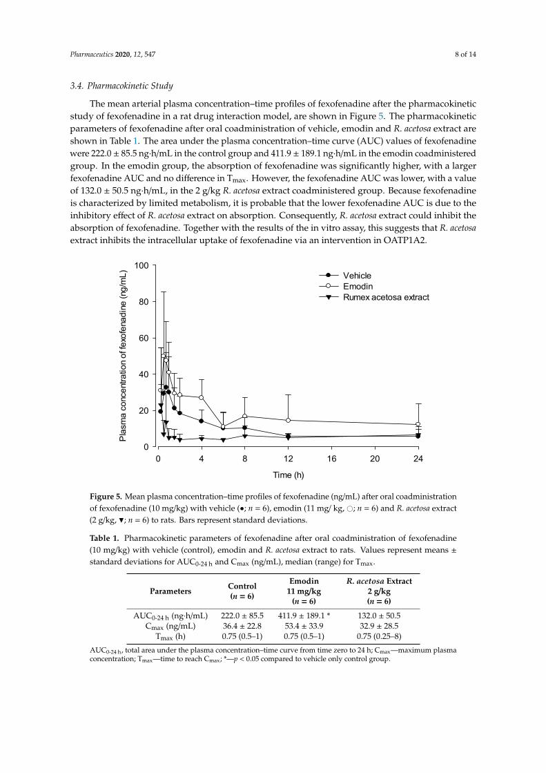
The mean arterial plasma concentration–time profiles of fexofenadine after the pharmacokinetic study of fexofenadine in a rat drug interaction model, are shown in Figure 5. The pharmacokinetic parameters of fexofenadine after oral coadministration of vehicle, emodin and R. acetosa extract are shown in Table 1. The area under the plasma concentration–time curve (AUC) values of fexofenadine were 222.0 ± 85.5 ng∙h/mL in the control group and 411.9 ± 189.1 ng∙h/mL in the emodin coadministered group. In the emodin group, the absorption of fexofenadine was significantly higher, with a larger fexofenadine AUC and no difference in Tmax. However, the fexofenadine AUC was lower, with a value of 132.0 ± 50.5 ng∙h/mL, in the 2 g/kg R. acetosa extract coadministered group. Because fexofenadine is characterized by limited metabolism, it is probable that the lower fexofenadine AUC is due to the inhibitory effect of R. acetosa extract on absorption. Consequently, R. acetosa extract could inhibit the absorption of fexofenadine. Together with the results of the in vitro assay, this suggests that R. acetosa extract inhibits the intracellular uptake of fexofenadine via an intervention in OATP1A2.
Figure 4. Inhibitory effect of R. acetosa extract on fexofenadine uptake in OATP1A2/SLCO1A2 transfectedHEK293 cells (n = 6). Con—OATP1A2/SLCO1A2 transfected control; Nov—untransfected control;Ver—100-µM verapamil; *—p < 0.05 compared to control group.
Pharmaceutics 2020, 12, 547 8 of 14
3.4. Pharmacokinetic Study
The mean arterial plasma concentration–time profiles of fexofenadine after the pharmacokineticstudy of fexofenadine in a rat drug interaction model, are shown in Figure 5. The pharmacokineticparameters of fexofenadine after oral coadministration of vehicle, emodin and R. acetosa extract areshown in Table 1. The area under the plasma concentration–time curve (AUC) values of fexofenadinewere 222.0 ± 85.5 ng·h/mL in the control group and 411.9 ± 189.1 ng·h/mL in the emodin coadministeredgroup. In the emodin group, the absorption of fexofenadine was significantly higher, with a largerfexofenadine AUC and no difference in Tmax. However, the fexofenadine AUC was lower, with a valueof 132.0 ± 50.5 ng·h/mL, in the 2 g/kg R. acetosa extract coadministered group. Because fexofenadineis characterized by limited metabolism, it is probable that the lower fexofenadine AUC is due to theinhibitory effect of R. acetosa extract on absorption. Consequently, R. acetosa extract could inhibit theabsorption of fexofenadine. Together with the results of the in vitro assay, this suggests that R. acetosaextract inhibits the intracellular uptake of fexofenadine via an intervention in OATP1A2.
Pharmaceutics 2020, 12, x 8 of 15
Time (h)
0 4 8 12 16 20 24
Pla
sma
conc
entra
tion
of fe
xofe
nadi
ne (n
g/m
L)
0
20
40
60
80
100VehicleEmodinRumex acetosa extract
Figure 5. Mean plasma concentration–time profiles of fexofenadine (ng/mL) after oral coadministration of fexofenadine (10 mg/kg) with vehicle (●; n = 6), emodin (11 mg/ kg, ○; n = 6) and R. acetosa extract (2 g/kg, ▼; n = 6) to rats. Bars represent standard deviations.
Table 1. Pharmacokinetic parameters of fexofenadine after oral coadministration of fexofenadine (10 mg/kg) with vehicle (control), emodin and R. acetosa extract to rats. Values represent means ± standard deviations for AUC0-24 h and Cmax (ng/mL), median (range) for Tmax.
Parameters Control (n = 6)
Emodin 11 mg/kg
(n = 6)
R. acetosa Extract 2 g/kg (n = 6)
AUC0-24 h (ng∙h/mL) 222.0 ± 85.5 411.9 ± 189.1 * 132.0 ± 50.5 Cmax (ng/mL) 36.4 ± 22.8 53.4 ± 33.9 32.9 ± 28.5
Tmax (h) 0.75 (0.5–1) 0.75 (0.5–1) 0.75 (0.25–8) AUC0-24 h, total area under the plasma concentration–time curve from time zero to 24 h; Cmax—maximum plasma concentration; Tmax—time to reach Cmax; *—p < 0.05 compared to vehicle only control group.
3.5. Physicochemical Interaction Study
To evaluate the possible physicochemical interactions between R. acetosa extract and fexofenadine, FT-IR spectra of extract, fexofenadine and mixture were measured and are shown in Figure 6. The FT-IR spectrum of fexofenadine HCl showed the characteristic absorption bands at 3291.03 (OH stretching), 2936.14 (CH stretching), 2639.82 (OH of carboxylate), 1698.68 (CO stretching), 1448.00, 1403.11 (C=C stretching of aromatic ring), 1167.57 (CO stretching of tertiary alcohol) and 1067.94 (CO stretching of secondary alcohol) [38,39]. According to Figure 6, the mixture of R. acetosa extract and fexofenadine HCl showed the same bands compared to the pure fexofenadine HCl. It suggests that there is no significant physical interaction between fexofenadine molecule and R. acetosa extract component on fexofenadine functional groups.
However, there was significant difference on the solubility of fexofenadine after incubation with the extract (Table 2). The average solubilities of fexofenadine in SIF were 1.03 ± 0.04 mg/mL and 0.83 ± 0.10 mg/mL without and with R. acetosa extract, respectively. This result indicates that R.
Figure 5. Mean plasma concentration–time profiles of fexofenadine (ng/mL) after oral coadministrationof fexofenadine (10 mg/kg) with vehicle (•; n = 6), emodin (11 mg/ kg, #; n = 6) and R. acetosa extract(2 g/kg, H; n = 6) to rats. Bars represent standard deviations.
Table 1. Pharmacokinetic parameters of fexofenadine after oral coadministration of fexofenadine(10 mg/kg) with vehicle (control), emodin and R. acetosa extract to rats. Values represent means ±standard deviations for AUC0-24 h and Cmax (ng/mL), median (range) for Tmax.
Parameters Control(n = 6)
Emodin11 mg/kg
(n = 6)
R. acetosa Extract2 g/kg(n = 6)
AUC0-24 h (ng·h/mL) 222.0 ± 85.5 411.9 ± 189.1 * 132.0 ± 50.5Cmax (ng/mL) 36.4 ± 22.8 53.4 ± 33.9 32.9 ± 28.5
Tmax (h) 0.75 (0.5–1) 0.75 (0.5–1) 0.75 (0.25–8)
AUC0-24 h, total area under the plasma concentration–time curve from time zero to 24 h; Cmax—maximum plasmaconcentration; Tmax—time to reach Cmax; *—p < 0.05 compared to vehicle only control group.

Pharmaceutics 2020, 12, 547 9 of 14
3.5. Physicochemical Interaction Study
To evaluate the possible physicochemical interactions between R. acetosa extract and fexofenadine, FT-IRspectra of extract, fexofenadine and mixture were measured and are shown in Figure 6. The FT-IR spectrumof fexofenadine HCl showed the characteristic absorption bands at 3291.03 (OH stretching), 2936.14 (CHstretching), 2639.82 (OH of carboxylate), 1698.68 (CO stretching), 1448.00, 1403.11 (C=C stretching of aromaticring), 1167.57 (CO stretching of tertiary alcohol) and 1067.94 (CO stretching of secondary alcohol) [38,39].According to Figure 6, the mixture of R. acetosa extract and fexofenadine HCl showed the same bandscompared to the pure fexofenadine HCl. It suggests that there is no significant physical interaction betweenfexofenadine molecule and R. acetosa extract component on fexofenadine functional groups.
Pharmaceutics 2020, 12, x 9 of 15
acetosa extract could alter the solubility of fexofenadine and lead to precipitation in gastro-intestinal tract.
Table 2. The solubility of fexofenadine HCl in simulated intestinal fluid (SIF) with and without R. acetosa extract.
Solubility Without R. acetosa Extract
(n = 3) With R. acetosa Extract
(n = 3) Fexofenadine HCl concentration (mg/mL) 1.03 ± 0.04 0.83 ± 0.10 *
*—p < 0.05 compared to without R. acetosa extract.
Figure 6. FT-IR spectra of (A) fexofenadine, (B) a mixture of fexofenadine and R. acetosa extract and (C) R. acetosa extract.
Figure 6. FT-IR spectra of (A) fexofenadine, (B) a mixture of fexofenadine and R. acetosa extract and(C) R. acetosa extract.
Pharmaceutics 2020, 12, 547 10 of 14
However, there was significant difference on the solubility of fexofenadine after incubation withthe extract (Table 2). The average solubilities of fexofenadine in SIF were 1.03 ± 0.04 mg/mL and0.83 ± 0.10 mg/mL without and with R. acetosa extract, respectively. This result indicates that R. acetosaextract could alter the solubility of fexofenadine and lead to precipitation in gastro-intestinal tract.
Table 2. The solubility of fexofenadine HCl in simulated intestinal fluid (SIF) with and without R.acetosa extract.
Solubility Without R. acetosa Extract(n = 3) With R. acetosa Extract(n = 3)
Fexofenadine HCl concentration (mg/mL) 1.03 ± 0.04 0.83 ± 0.10 *
*—p < 0.05 compared to without R. acetosa extract.
4. Discussion
Pharmacokinetic drug interactions involving drug absorption should be considered for optimumdrug therapy, apart from the drug interactions attributed to the oxidative metabolism via the CYP-450system of different isozymes [40]. Ostensibly harmless natural products—such as juices, fruits,vegetables and herbal products in the form of ayurvedic medicine—have been reported in several studiesto potentially cause many drug interactions affecting drug absorption mediated by transporters [41,42].For example, emodin—a potential antineoplastic drug and a major component of the Rhamnus, Rumex,Aloe, Rheum and Cassia species—has been reported to be a possible P-gp inducer [26] or an inhibitor [18].
This study evaluated the effects of R. acetosa extract on the drug transporters discussed above, aswell as its potential for drug interactions, while presenting a clear view of the interactions of emodinwith the transporter P-gp. The major six anthraquinones present in R. acetosa were shown in ourprevious study [27]. A prior cytotoxicity assay was performed to establish the working range forthe extract suitable for optimal viability of the cells during the experiment. Afterwards, the effectsof these six anthraquinones on P-gp were demonstrated individually with an MDR assay kit usingCaco-2 cells. Verapamil, being an inhibitor of P-gp, served as a positive control. Only groups treatedwith chrysophanol-8-O-β-d-glucoside and emodin showed higher fluorescence intensity than thecontrol group, with average values of 121.4% ± 2.3% and 147.2% ± 12.4%, respectively, suggestingP-gp inhibition. This result is consistent with those obtained in a study by Min et al. [18], in whichemodin was shown to inhibit P-gp. On the other hand, the results from the P-gp inhibition test ofR. acetosa extract suggest no significant inhibition of the efflux transporter, as opposed to the emodinand chrysophanol-8-O-β-d-glucoside, which in contrast showed significant inhibition of the P-gptransporter when treated individually. A possible explanation is that the emodin content may not behigh enough to exert its inhibitory effect in the extract. Chemical contents of herbal plant extractscan vary depending on various factors such as climate, harvesting seasons and extraction solvent.The probability of inhibition of P-gp by R. acetosa extract cannot be ruled out.
OATP1A2—the uptake transporter used in our in vitro test—is widely expressed in the intestinesand serves as a major uptake mechanism for fexofenadine [43,44]. Sometimes, a substrate of P-gp—suchas this study’s selected model drug, fexofenadine—can also be a substrate for the OATP uptaketransporter [43,44], making it necessary to differentiate between the contributions of P-gp and OATP topotential drug interactions and those of other simultaneously administered drugs that could affectthese transporters. Therefore, our in vitro studies were also performed with HEK293 cells transfectedwith the polypeptide transporter OATP1A2. R. acetosa extract was found to inhibit the uptake offexofenadine through in vitro studies. In other words, the uptake of fexofenadine by OATP1A2 intocells declined when R. acetosa extract was used as a co-treatment. This result suggests that R. acetosaextract can affect the absorption of fexofenadine through the inhibition of OATP1A2.
A pharmacokinetic study was designed to verify the results of our in vitro study in view ofthe observed herbal extract’s drug interactions at the uptake transporter for fexofenadine in rats.Rat model is considered unsuitable to predict metabolic drug interaction in human [45]. However,there is a correlation in drug intestinal permeability with both carrier-mediated absorption and passive

Pharmaceutics 2020, 12, 547 11 of 14
diffusion mechanisms between rat and human [46]. Because the property of our selected modeldrug, fexofenadine, has little metabolism, it is reasonable to use the rat model for predicting theintervention of extract on absorption. All rats were divided into 3 groups: an emodin administrationgroup, an R. acetosa administration group and a control group. Eleven milligrams per kilogram ofemodin, 2 g/kg of R. acetosa extract and 0.5% CMC as a control was administered orally to each group.Fexofenadine at the dose of 10 mg/kg was given orally to each group after 30 min. The results showeda smaller AUC of fexofenadine (132.1 ± 50.3 ng·h/mL) in the R. acetosa group in comparison to thatof the control group, in which the AUC was 222.0 ± 92.1 ng·h/mL. These results suggest decreasedabsorption of fexofenadine in the rats treated with R. acetosa extract. In other words, the gut uptaketransporter OATP1A2, which is responsible for fexofenadine absorption, was inhibited, as predictedby the in vitro results. Moreover, the alteration on the solubility of fexofenadine was also observed byR. acetosa extract through the physicochemical interaction study. The FT-IR spectra results suggest thatthere is no functional group interaction between fexofenadine and the component of R. acetosa extract.The fexofenadine solubility in SIF changed from 1.03 ± 0.04 mg/mL to 0.83 ± 0.10 mg/mL after mixingwith the extract. It means that the solubility alteration could also be the reason for the decreasedfexofenadine AUC by R. acetosa extract because fexofenadine HCl is Biopharmaceutics ClassificationSystem (BCS) class 3 drug with high solubility and low permeability. Drug interactions due to changesin solubility can be avoided by adjusting the administration time. R. acetosa extract contains manykinds of compounds, not only anthraquinones, but also flavonoids and polysaccharides [15]. Theyhave also the possibility of interference with the drug absorption through the intervention to thetransporters [47,48]. Particularly, one of the flavonoids of R. acetosa extract, epicatechin-3-O-gallate [49],also has an inhibitory effect on the OATP1A2 [50]. Moreover, there was the possibility that R. acetosaextract may change the gastric emptying time [51,52] and the pH in the gastro-intestinal tract whencoadministered with the fexofenadine. The effects of anthraquinones on OATP have been rarelyreported. Further studies are needed to elucidate the components in R. acetosa extract responsiblefor inhibition of fexofenadine absorption. Meanwhile, emodin increased the AUC for fexofenadine,possibly via the inhibitory effect on an efflux transporter of fexofenadine, P-gp [32], the effect of whichon the uptake transporter of fexofenadine has yet to be fully understood.
Given the evidence from both in vitro and in vivo studies, R. acetosa extract should be used withcaution when substrates of the drug transporters or poorly water-soluble drugs are prescribed.
5. Conclusions
The present study evaluated the effects of R. acetosa extract on 2 active transporters, P-gp andOATP1A2 and the resulting effects on fexofenadine absorption through in vitro and in vivo studies.The findings suggest that emodin can enhance fexofenadine absorption via an inhibitory effect on P-gp.In addition, R. acetosa extract could decrease the absorption of fexofenadine via intervention in theaqueous solubility and the drug transporters.
Author Contributions: Conceptualization, H.J.C.; methodology, H.J.C.; validation, J.K. and N.U.R.; formalanalysis, J.K.; investigation, J.H.A., J.K. and H.-J.K.; resources, M.-J.A. and H.J.C.; data curation, J.H.A., J.K. andH.J.C.; writing—original draft preparation, J.K. and N.U.R.; writing—review and editing, H.J.C.; visualization,J.K.; supervision, H.J.C. and M.-J.A.; project administration, H.J.C.; funding acquisition, H.J.C. All authors haveread and agreed to the published version of the manuscript.
Funding: This research was funded by the National Research Foundation of Korea (NRF) grant funded by theKorean government (MSIP; Ministry of Science & ICT), Grant Number 2017R1C1B5017343.
Conflicts of Interest: The authors declare no conflict of interest.

Pharmaceutics 2020, 12, 547 12 of 14
References
1. Dean, M.; Hamon, Y.; Chimini, G. The human ATP-binding cassette (ABC) transporter superfamily. J. LipidRes. 2001, 42, 1007–1017. [CrossRef] [PubMed]
2. Anderle, P.; Niederer, E.; Rubas, W.; Hilgendorf, C.; Spahn-Langguth, H.; Wunderli-Allenspach, H.;Merkle, H.P.; Langguth, P. P-Glycoprotein (P-gp) mediated efflux in Caco-2 cell monolayers: The influenceof culturing conditions and drug exposure on P-gp expression levels. J. Pharm. Sci. 1998, 87, 757–762.[CrossRef] [PubMed]
3. Fardel, O.; Lecureur, V.; Guillouzo, A. The P-glycoprotein multidrug transporter. Gen. Pharm. 1996, 27,1283–1291. [CrossRef]
4. Chan, L.M.; Lowes, S.; Hirst, B.H. The ABCs of drug transport in intestine and liver: Efflux proteins limitingdrug absorption and bioavailability. Eur. J. Pharm. Sci. 2004, 21, 25–51. [CrossRef]
5. Talks, K.L.; Turley, H.; Gatter, K.C.; Maxwell, P.H.; Pugh, C.W.; Ratcliffe, P.J.; Harris, A.L. The expression anddistribution of the hypoxia-inducible factors HIF-1alpha and HIF-2alpha in normal human tissues, cancers,and tumor-associated macrophages. Am. J. Pathol 2000, 157, 411–421. [CrossRef]
6. Comerford, K.M.; Wallace, T.J.; Karhausen, J.; Louis, N.A.; Montalto, M.C.; Colgan, S.P. Hypoxia-induciblefactor-1-dependent regulation of the multidrug resistance (MDR1) gene. Cancer Res. 2002, 62, 3387–3394.
7. Kim, R.B. Drugs as P-glycoprotein substrates, inhibitors, and inducers. Drug Metab. Rev. 2002, 34, 47–54.[CrossRef]
8. Palmeira, A.; Sousa, E.; Vasconcelos, M.H.; Pinto, M.M. Three decades of P-gp: Skimming through severalgenerations and scaffolds. Curr. Med. Che 2012, 19, 1946–2025. [CrossRef]
9. Shitara, Y.; Maeda, K.; Ikejiri, K.; Yoshida, K.; Horie, T.; Sugiyama, Y. Clinical significance of organic aniontransporting polypeptides (OATPs) in drug disposition: Their roles in hepatic clearance and intestinalabsorption. Biopharm. Drug Dispos. 2013, 34, 45–78. [CrossRef]
10. Yu, J.; Zhou, Z.; Tay-Sontheimer, J.; Levy, R.H.; Ragueneau-Majlessi, I. Intestinal drug interactions mediatedby OATPs: A systematic review of preclinical and clinical findings. J. Pharm. Sci. 2017, 106, 2312–2325.[CrossRef]
11. Agbabiaka, T.B.; Spencer, N.H.; Khanom, S.; Goodman, C. Prevalence of drug-herb and drug-supplementinteractions in older adults: A cross-sectional survey. Br. J. Gen. Pr. 2018, 68, e711–e717. [CrossRef] [PubMed]
12. Kaufman, D.W.; Kelly, J.P.; Rosenberg, L.; Anderson, T.E.; Mitchell, A.A. Recent patterns of medicationuse in the ambulatory adult population of the United States: The Slone survey. JAMA 2002, 287, 337–344.[CrossRef] [PubMed]
13. Durr, D.; Stieger, B.; Kullak-Ublick, G.A.; Rentsch, K.M.; Steinert, H.C.; Meier, P.J.; Fattinger, K. St John’sWort induces intestinal P-glycoprotein/MDR1 and intestinal and hepatic CYP3A4. Clin. Pharm. 2000, 68,598–604. [CrossRef] [PubMed]
14. Gescher, K.; Hensel, A.; Hafezi, W.; Derksen, A.; Kuhn, J. Oligomeric proanthocyanidins from Rumex acetosaL. inhibit the attachment of herpes simplex virus type-1. Antivir. Res. 2011, 89, 9–18. [CrossRef] [PubMed]
15. Vasas, A.; Orban-Gyapai, O.; Hohmann, J. The genus Rumex: Review of traditional uses, phytochemistryand pharmacology. J. Ethnopharmacol. 2015, 175, 198–228. [CrossRef] [PubMed]
16. Kucekova, Z.; Mlcek, J.; Humpolicek, P.; Rop, O.; Valasek, P.; Saha, P. Phenolic compounds from Alliumschoenoprasum, Tragopogon pratensis and Rumex acetosa and their antiproliferative effects. Molecules 2011,16, 9207–9217. [CrossRef]
17. Bae, J.Y.; Lee, Y.S.; Han, S.Y.; Jeong, E.J.; Lee, M.K.; Kong, J.Y.; Lee, D.H.; Cho, K.J.; Lee, H.S.; Ahn, M.J.A comparison between water and ethanol extracts of Rumex acetosa for protective effects on gastric ulcers inmice. Biomol. Ther. (Seoul) 2012, 20, 425–430. [CrossRef] [PubMed]
18. Min, H.; Niu, M.; Zhang, W.; Yan, J.; Li, J.; Tan, X.; Li, B.; Su, M.; Di, B.; Yan, F. Emodin reverses leukemiamultidrug resistance by competitive inhibition and downregulation of P-glycoprotein. PLoS ONE 2017, 12,e0187971. [CrossRef]
19. Feng, Y.; Huang, S.L.; Dou, W.; Zhang, S.; Chen, J.H.; Shen, Y.; Shen, J.H.; Leng, Y. Emodin, a natural product,selectively inhibits 11beta-hydroxysteroid dehydrogenase type 1 and ameliorates metabolic disorder indiet-induced obese mice. Br. J. Pharm. 2010, 161, 113–126. [CrossRef]
20. Hsu, S.C.; Chung, J.G. Anticancer potential of emodin. BioMedicine (Taipei) 2012, 2, 108–116. [CrossRef]

Pharmaceutics 2020, 12, 547 13 of 14
21. Simpson, K.; Jarvis, B. Fexofenadine: A review of its use in the management of seasonal allergic rhinitis andchronic idiopathic urticaria. Drugs 2000, 59, 301–321. [CrossRef] [PubMed]
22. Tahara, H.; Kusuhara, H.; Fuse, E.; Sugiyama, Y. P-glycoprotein plays a major role in the efflux of fexofenadinein the small intestine and blood-brain barrier, but only a limited role in its biliary excretion. Drug Metab.Dispos. 2005, 33, 963–968. [CrossRef] [PubMed]
23. Shimizu, M.; Fuse, K.; Okudaira, K.; Nishigaki, R.; Maeda, K.; Kusuhara, H.; Sugiyama, Y. Contribution ofOATP (organic anion-transporting polypeptide) family transporters to the hepatic uptake of fexofenadine inhumans. Drug Metab. Dispos. 2005, 33, 1477–1481. [CrossRef] [PubMed]
24. Molimard, M.; Diquet, B.; Benedetti, M.S. Comparison of pharmacokinetics and metabolism of desloratadine,fexofenadine, levocetirizine and mizolastine in humans. Fundam. Clin. Pharm. 2004, 18, 399–411. [CrossRef][PubMed]
25. Prescribing Information for Allegra®(fexofenadine hydrochloride). Available online: https://www.accessdata.fda.gov/drugsatfda_docs/label/2003/20872se8-003,20625se8-010_allegra_lbl.pdf (accessed on 16 March 2020).
26. Huang, J.; Guo, L.; Tan, R.; Wei, M.; Zhang, J.; Zhao, Y.; Gong, L.; Huang, Z.; Qiu, X. Interactions betweenemodin and efflux transporters on rat enterocyte by a validated ussing chamber technique. Front. Pharm.2018, 9, 646. [CrossRef]
27. Ullah, H.M.A.; Kim, J.; Rehman, N.U.; Kim, H.J.; Ahn, M.J.; Chung, H.J. A simple and sensitiveliquid chromatography with tandem mass spectrometric method for the simultaneous determinationof anthraquinone glycosides and their aglycones in rat plasma: Application to a pharmacokinetic study ofRumex acetosa extract. Pharmaceutics 2018, 10, 100. [CrossRef]
28. Petri, N.; Tannergren, C.; Rungstad, D.; Lennernas, H. Transport characteristics of fexofenadine in the Caco-2cell model. Pharm. Res. 2004, 21, 1398–1404. [CrossRef]
29. Rebello, S.; Zhao, S.; Hariry, S.; Dahlke, M.; Alexander, N.; Vapurcuyan, A.; Hanna, I.; Jarugula, V. IntestinalOATP1A2 inhibition as a potential mechanism for the effect of grapefruit juice on aliskiren pharmacokineticsin healthy subjects. Eur. J. Clin. Pharm. 2012, 68, 697–708. [CrossRef]
30. Yamane, N.; Tozuka, Z.; Sugiyama, Y.; Tanimoto, T.; Yamazaki, A.; Kumagai, Y. Microdose clinical trial:Quantitative determination of fexofenadine in human plasma using liquid chromatography/electrosprayionization tandem mass spectrometry. J. Chromatogr B Anal. Technol Biomed. Life Sci 2007, 858, 118–128.[CrossRef]
31. Bharathi, V.D.; Radharani, K.; Jagadeesh, B.; Ramulu, G.; Bhushan, I.; Naidu, A.; Mullangi, R. LC–MS–MS assayfor simultaneous quantification of fexofenadine and pseudoephedrine in human plasma. Chromatographia2008, 67, 461–466. [CrossRef]
32. Li, X.; Hu, J.; Wang, B.; Sheng, L.; Liu, Z.; Yang, S.; Li, Y. Inhibitory effects of herbal constituents onP-glycoprotein in vitro and in vivo: Herb-drug interactions mediated via P-gp. Toxicol. Appl. Pharm. 2014,275, 163–175. [CrossRef] [PubMed]
33. Kamath, A.V.; Yao, M.; Zhang, Y.; Chong, S. Effect of fruit juices on the oral bioavailability of fexofenadine inrats. J. Pharm. Sci. 2005, 94, 233–239. [CrossRef] [PubMed]
34. Isleyen, E.A.Ö.; Özden, T.; Özilhan, S.; Toptan, S. Quantitative determination of fexofenadine in humanplasma by HPLC-MS. Chromatographia 2007, 66, 109–113. [CrossRef]
35. Dai, W.G. In vitro methods to assess drug precipitation. Int. J. Pharm. 2010, 393, 1–16. [CrossRef] [PubMed]36. Saito, Y.; Usami, T.; Katoh, M.; Nadai, M. Effects of thylakoid-rich spinach extract on the pharmacokinetics of
drugs in rats. Biol. Pharm. Bull. 2019, 42, 103–109. [CrossRef] [PubMed]37. Stippler, E.; Kopp, S.; Dressman, J.B. Comparison of US pharmacopeia simulated intestinal fluid TS (without
pancreatin) and phosphate standard buffer pH 6.8, TS of the international pharmacopoeia with respect totheir use in in vitro dissolution testing. Dissolution Technol. 2004, 11, 6–10. [CrossRef]
38. Singh, B.; Saini, G.; Vyas, M.; Verma, S.; Thakur, S. Optimized chronomodulated dual release bilayer tabletsof fexofenadine and montelukast: Quality by design, development, and in vitro evaluation. Future J. Pharm.Sci. 2019, 5, 5. [CrossRef]
39. Arefin, P.; Hasan, I.; Reza, M.S. Design, characterization and in vitro evaluation of HPMC K100 M CR loadedfexofenadine HCl microspheres. Springerplus 2016, 5, 691. [CrossRef]
40. Hansten, P.D.; Levy, R.H. Role of P-glycoprotein and organic anion transporting polypeptides in drugabsorption and distribution. Clin. Drug Investig. 2001, 21, 587–596. [CrossRef]

Pharmaceutics 2020, 12, 547 14 of 14
41. Mallhi, T.H.; Sarriff, A.; Adnan, A.S.; Khan, Y.H.; Qadir, M.I.; Hamzah, A.A.; Khan, A.H. Effect offruit/vegetable-drug interactions on CYP450, OATP and p-glycoprotein: A systematic review. Trop. J. Pharm.Res. 2015, 14, 1927–1935. [CrossRef]
42. Bailey, D.G. Fruit juice inhibition of uptake transport: A new type of food-drug interaction. Br. J. Clin. Pharm.2010, 70, 645–655. [CrossRef] [PubMed]
43. Cvetkovic, M.; Leake, B.; Fromm, M.F.; Wilkinson, G.R.; Kim, R.B. OATP and P-glycoprotein transportersmediate the cellular uptake and excretion of fexofenadine. Drug Metab. Dispos. 1999, 27, 866–871. [PubMed]
44. Niemi, M.; Kivisto, K.T.; Hofmann, U.; Schwab, M.; Eichelbaum, M.; Fromm, M.F. Fexofenadinepharmacokinetics are associated with a polymorphism of the SLCO1B1 gene (encoding OATP1B1). Br. J.Clin. Pharm. 2005, 59, 602–604. [CrossRef] [PubMed]
45. Jaiswal, S.; Sharma, A.; Shukla, M.; Vaghasiya, K.; Rangaraj, N.; Lal, J. Novel pre-clinical methodologies forpharmacokinetic drug-drug interaction studies: Spotlight on "humanized" animal models. Drug Metab. Rev.2014, 46, 475–493. [CrossRef] [PubMed]
46. Cao, X.; Gibbs, S.T.; Fang, L.; Miller, H.A.; Landowski, C.P.; Shin, H.C.; Lennernas, H.; Zhong, Y.; Amidon, G.L.;Yu, L.X.; et al. Why is it challenging to predict intestinal drug absorption and oral bioavailability in humanusing rat model. Pharm. Res. 2006, 23, 1675–1686. [CrossRef]
47. Mandery, K.; Bujok, K.; Schmidt, I.; Keiser, M.; Siegmund, W.; Balk, B.; Konig, J.; Fromm, M.F.; Glaeser, H.Influence of the flavonoids apigenin, kaempferol, and quercetin on the function of organic anion transportingpolypeptides 1A2 and 2B1. Biochem. Pharm. 2010, 80, 1746–1753. [CrossRef]
48. Masumoto, K.; Quan, Z.; Ishiuchi, K.i.; Matsumoto, T.; Watanabe, J.; Makino, T. Drug interaction betweenshoseiryuto extract or catechins and fexofenadine through organic-anion-transporting polypeptide 1A2in vitro. Pharmacogn. Mag. 2019, 15, 304–308. [CrossRef]
49. Bicker, J.; Petereit, F.; Hensel, A. Proanthocyanidins and a phloroglucinol derivative from Rumex acetosa L.Fitoterapia 2009, 80, 483–495. [CrossRef]
50. Roth, M.; Timmermann, B.N.; Hagenbuch, B. Interactions of green tea catechins with organicanion-transporting polypeptides. Drug Metab. Dispos. 2011, 39, 920–926. [CrossRef]
51. Bajad, S.; Bedi, K.L.; Singla, A.K.; Johri, R.K. Piperine inhibits gastric emptying and gastrointestinal transit inrats and mice. Planta Med. 2001, 67, 176–179. [CrossRef]
52. Hu, M.L.; Rayner, C.K.; Wu, K.L.; Chuah, S.K.; Tai, W.C.; Chou, Y.P.; Chiu, Y.C.; Chiu, K.W.; Hu, T.H. Effect ofginger on gastric motility and symptoms of functional dyspepsia. World J. Gastroenter. 2011, 17, 105–110.[CrossRef] [PubMed]
© 2020 by the authors. Licensee MDPI, Basel, Switzerland. This article is an open accessarticle distributed under the terms and conditions of the Creative Commons Attribution(CC BY) license (http://creativecommons.org/licenses/by/4.0/).