Effect of Restorative, Endodontic, and Fatigue Treatments on the Cuspal Deflection of Maxillary...
10
The International Journal of Periodontics & Restorative Dentistry
Transcript of Effect of Restorative, Endodontic, and Fatigue Treatments on the Cuspal Deflection of Maxillary...
The International Journal of Periodontics & Restorative Dentistry
Volume 35, Number 2, 2015
221
Lorenzo Madini, DMD1/Pier Antonio Acquaviva, DMD1 Andreas Krokidis, DDS, MSc2/Nicola Barabanti, DMD1 Mutlu Özcan, Dr Med Dent, PhD3/Cumhur Sipahi, DDS, PhD4 Antonio Cerutti, MD, DDS5
Effect of Restorative, Endodontic, and Fatigue Treatments on the Cuspal Deflection of Maxillary Premolars Subjected to Different Cyclic Occlusal Forces: An In Vitro Study
This study aimed to determine the effect of adhesive direct composite restora-tions, endodontic treatments, and fatigue treatments on the cuspal deflection of maxillary premolars subjected to different cyclic occlusal forces. Thirty intact maxillary second premolars were selected. Ten teeth were left untreated (group IN), 10 teeth were subjected to endodontic and restorative treatment (group FL), and the remaining 10 teeth were subjected to endodontic, restorative, and fatigue treatments (group FT). All teeth were subjected to 5 occlusal compressive loading forces (98, 147, 196, 245, and 294 N) with a universal testing device. A total of 15 experimental groups were obtained with 3 tooth conditions (IN, FL, FT) and 5 different occlusal loading values. Deflection amounts (μm) were measured with laser sensors and recorded, and obtained data were statistically analyzed with one-way analysis of variance at a significance level of .05. Mean cuspal deflection values (μm) and SDs of experimental groups ranged as follows: IN-98 (24.4 ± 19.8), IN-147 (34.8 ± 28.9), IN-196 (43.8 ± 34.7), IN-245 (54.5 ± 46.4), IN-294 (60.3 ± 50.6), FL-98 (56 ± 49.1), FL-147 (62.6 ± 49.6), FL-196 (72.4 ± 52.1), FL-245 (81.3 ± 56), FL-294 (92.2 ± 60.9), FT-98 (77.2 ± 80.9), FT-147 (83.4 ± 81.3), FT-196 (92.6 ± 83.7), FT-245 (102.7 ± 85.4), and FT-294 (124.2 ± 89.5). Mean values of three main experimental groups were as follows: IN (43.5 μm), FL (72.9 μm) and FT (96.0 μm). Significant differences were found between the three main groups and relevant subgroups (P < .001). Highest cuspal deflection values (CDV) were obtained in FT groups. Lowest CDV were obtained in IN groups. FL groups showed higher deflection values than IN groups. CDV increased progressively as the teeth were restored and subjected to fatigue treatment. (Int J Periodontics Restorative Dent 2015;35:221–229. doi: 10.11607/prd.1802)
Endodontic treatment causes irre-versible changes in the architectural anatomy of teeth. The chemical and mechanical properties of enamel and dentin also are modified.1–3 Several in vivo,4,5 ex vivo,6 and in vitro7–9 studies have demonstrated that endodontic treatment makes teeth more brittle due to the de-hydration of dentin.10 Loss of soft (pulp chamber) and hard (marginal ridges) tissues due to caries causes an increase in cuspal height and a reduction in the thickness of cavity walls.11–13 These structural deficien-cies may lead to a greater risk of tooth fracture. As reported by Morin et al14 and Panitvisai and Messer,15 mesio-occlusodistal (MOD) cavities represent the worst case in terms of fracture risk when nonadhesive (ie, conventional) restorations are em-ployed.
Aspects of endodontically treated and adhesively restored teeth have been investigated in both noninvasive14,15 and invasive16 studies with use of different materi-als and techniques. In a noninvasive study, Panitvisai and Messer15 found that the cuspal deflection increases as the cavity turns from occlusal to
1 Lecturer, School of Dentistry, University of Brescia, Brescia, Italy.2 PhD Student, Kapodistrian University of Athens, Department of Endodontics, Athens, Greece.3 Professor and Head of Dental Materials Unit, Center for Dental and Oral Medicine, Clinic for Fixed and Removable Prosthodontics, Zurich, Switzerland.
4 Professor, Head of Department of Prosthodontics, Center for Dental Sciences, Gulhane Military Medical Academy, Ankara, Turkey.
5 Associate Professor, Head of Restorative Department, School of Dentistry, University of Brescia, Brescia, Italy.
Correspondence to: Dr Lorenzo Madini, c/o Clinica Odontoiatrica, p.le Spedali Civili 1, 25123, Brescia, Italy; fax: +39037236055; email: [email protected]. ©2015 by Quintessence Publishing Co Inc.
The International Journal of Periodontics & Restorative Dentistry
222
mesio-occlusal (MO)/occlusodistal (OD) or MOD, and recommended a full crown coverage.
In their in vitro study, Coban-kara et al17 applied cyclic loading on intact mandibular molars until fracture ocurred and reported that restorative materials such as amal-gam, direct composite, ceramic inlay, polyethylene ribbon fiber, or composite resin could not ad-equately compensate for the loss of fracture resistance; however, ce-ramic inlays gave the best results. A similar result was found in a study of Hitz et al,18 in which mandibular first molars were subjected to ther-mal cycling and loaded until fail-ure. They found that the resistance against fracture was lower than that of intact teeth, even in teeth re-stored with ceramic inlays.
Fatigue is a process involv-ing nucleation, propagation, and coalescence of cracks19; however, limited information is available about the behavior of endodonti-cally treated teeth restored with adhesive direct resin composite restorations and subjected to cyclic fatigue treatment.
Thus, the aim of this study was to evaluate the effect of adhesive direct composite restorations, end-odontic treatments, and fatigue treatments on the cuspal deflection
of maxillary premolars subjected to different cyclic occlusal loading forces. The null hypothesis was that no difference exists in the elastic behavior of endodontically treated teeth restored with MOD restora-tions after cyclic fatigue treatment.
Method and materials
Assignment of experimental groups
Occlusal surfaces of 30 maxillary second premolar teeth were to be subjected to five different occlusal loading force values (98, 147, 196, 245, 294 N) in three different tooth conditions (n = 10): intact (IN), filled with composite resin after end-odontic treatment (FL), and filled with composite resin after end-odontic treatment and subjected to fatigue treatment (FT). Thus, a total of 15 experimental groups were obtained (Table 1).
Tooth selection and preparation
A total of 30 human maxillary sec-ond premolars were extracted for orthodontic reasons from patients ages 15 to 29 years old who gave
informed and signed consent for the study. All selected teeth had similar dimensions, no caries, no restora-tions, and no cracks that might affect the deflection amount under com-pressive loading. The teeth were examined visually, radiographically, and by means of transillumination in order to eliminate any defective ones. After being extracted, select-ed tooth specimens were stored in 0.9% sodium chloride (NaCl) saline solution at 20°C and were kept in that solution during all preparations and testing procedures to prevent dehydration.
Thirty cylindrical resin blocks containing 30 tooth specimens were prepared as follows: (1) A cylindrical, open-ended, hollow brass matrix, 3 cm in diameter and 4 cm long, was obtained; (2) one of its open-ended sides was placed onto a glass surface and fixed with sticky wax (Dentsply). (3) Freshly poured au-topolymerizing acrylic resin (Melio-dent, Heraeus Kulzer) was injected into the matrix cavity. (4) Roots of each tooth specimen were embed-ded into the resin-filled cylindrical brass matrices 2 mm below the ce-mentoenamel junction (CEJ), with their long axes positioned perpen-dicular to the horizontal plane.
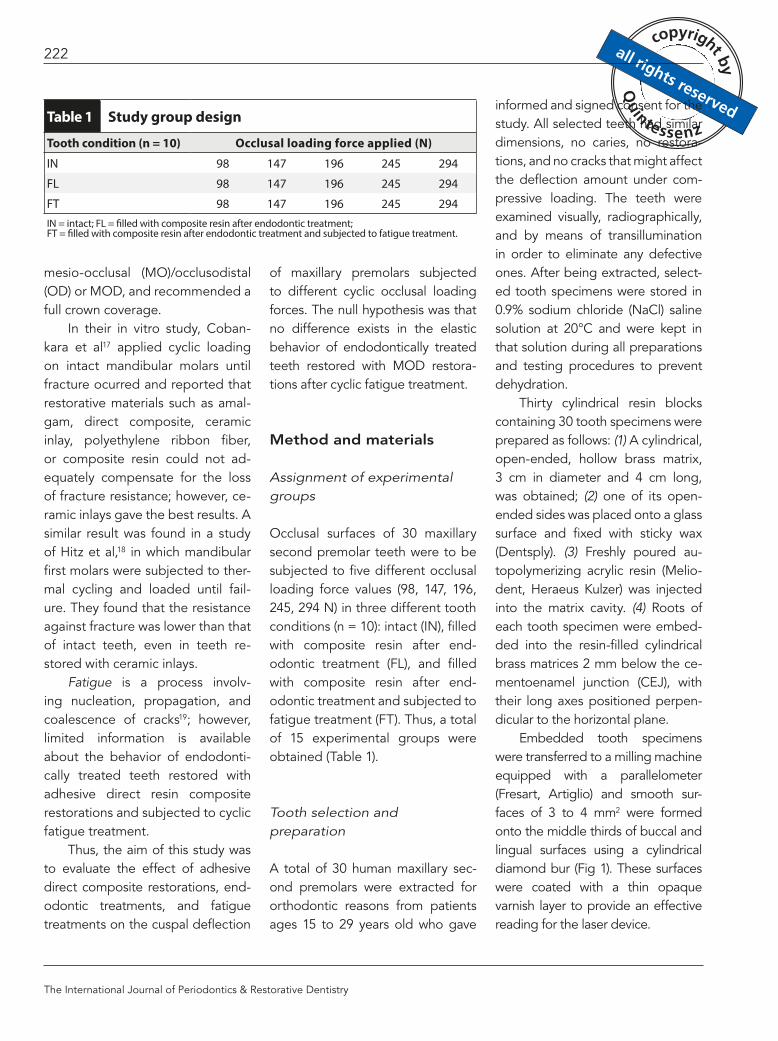
Embedded tooth specimens were transferred to a milling machine equipped with a parallelometer (Fresart, Artiglio) and smooth sur-faces of 3 to 4 mm2 were formed onto the middle thirds of buccal and lingual surfaces using a cylindrical diamond bur (Fig 1). These surfaces were coated with a thin opaque varnish layer to provide an effective reading for the laser device.
Table 1 Study group design
Tooth condition (n = 10) Occlusal loading force applied (N)IN 98 147 196 245 294
FL 98 147 196 245 294
FT 98 147 196 245 294IN = intact; FL = filled with composite resin after endodontic treatment; FT = filled with composite resin after endodontic treatment and subjected to fatigue treatment.
Volume 35, Number 2, 2015
223
Endodontic instrumentation and cavity preparation
Endodontic access cavities were prepared for tooth specimens in the FL and FT groups in a standard configuration. Instrumentation of root canals was performed with nickel-titanium (NiTi) rotary files (ProFile, Dentsply Maillefer), and sodium hypochlorite (NaClO) was used for irrigation. Canal obtura-tion was performed with a vertically condensed gutta-percha technique and with a canal filling paste (Pulp Canal Sealer, Kerr).
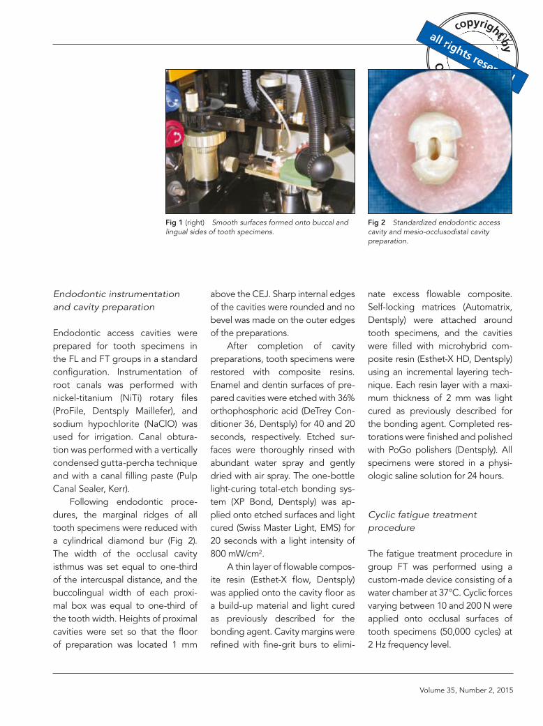
Following endodontic proce-dures, the marginal ridges of all tooth specimens were reduced with a cylindrical diamond bur (Fig 2). The width of the occlusal cavity isthmus was set equal to one-third of the intercuspal distance, and the buccolingual width of each proxi-mal box was equal to one-third of the tooth width. Heights of proximal cavities were set so that the floor of preparation was located 1 mm
above the CEJ. Sharp internal edges of the cavities were rounded and no bevel was made on the outer edges of the preparations.
After completion of cavity preparations, tooth specimens were restored with composite resins. Enamel and dentin surfaces of pre-pared cavities were etched with 36% orthophosphoric acid (DeTrey Con-ditioner 36, Dentsply) for 40 and 20 seconds, respectively. Etched sur-faces were thoroughly rinsed with abundant water spray and gently dried with air spray. The one-bottle light-curing total-etch bonding sys-tem (XP Bond, Dentsply) was ap-plied onto etched surfaces and light cured (Swiss Master Light, EMS) for 20 seconds with a light intensity of 800 mW/cm2.
A thin layer of flowable compos-ite resin (Esthet-X flow, Dentsply) was applied onto the cavity floor as a build-up material and light cured as previously described for the bonding agent. Cavity margins were refined with fine-grit burs to elimi-
nate excess flowable composite. Self-locking matrices (Auto matrix, Dentsply) were attached around tooth specimens, and the cavities were filled with microhybrid com-posite resin (Esthet-X HD, Dentsply) using an incremental layering tech-nique. Each resin layer with a maxi-mum thickness of 2 mm was light cured as previously described for the bonding agent. Completed res-torations were finished and polished with PoGo polishers (Dentsply). All specimens were stored in a physi-ologic saline solution for 24 hours.
Cyclic fatigue treatment procedure
The fatigue treatment procedure in group FT was performed using a custom-made device consisting of a water chamber at 37°C. Cyclic forces varying between 10 and 200 N were applied onto occlusal surfaces of tooth specimens (50,000 cycles) at 2 Hz frequency level.
Fig 1 (right) Smooth surfaces formed onto buccal and lingual sides of tooth specimens.
Fig 2 Standardized endodontic access cavity and mesio-occlusodistal cavity preparation.
The International Journal of Periodontics & Restorative Dentistry
224
Loading procedure
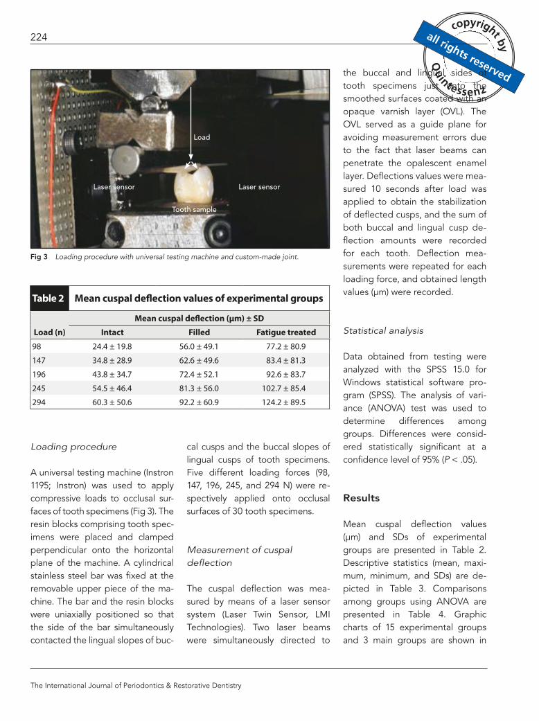
A universal testing machine (Instron 1195; Instron) was used to apply compressive loads to occlusal sur-faces of tooth specimens (Fig 3). The resin blocks comprising tooth spec-imens were placed and clamped perpendicular onto the horizontal plane of the machine. A cylindrical stainless steel bar was fixed at the removable upper piece of the ma-chine. The bar and the resin blocks were uniaxially positioned so that the side of the bar simultaneously contacted the lingual slopes of buc-
cal cusps and the buccal slopes of lingual cusps of tooth specimens. Five different loading forces (98, 147, 196, 245, and 294 N) were re-spectively applied onto occlusal surfaces of 30 tooth specimens.
Measurement of cuspal deflection
The cuspal deflection was mea-sured by means of a laser sensor system (Laser Twin Sensor, LMI Technologies). Two laser beams were simultaneously directed to
the buccal and lingual sides of tooth specimens just onto the smoothed surfaces coated with an opaque varnish layer (OVL). The OVL served as a guide plane for avoiding measurement errors due to the fact that laser beams can penetrate the opalescent enamel layer. Deflections values were mea-sured 10 seconds after load was applied to obtain the stabilization of deflected cusps, and the sum of both buccal and lingual cusp de-flection amounts were recorded for each tooth. Deflection mea-surements were repeated for each loading force, and obtained length values (µm) were recorded.
Statistical analysis
Data obtained from testing were analyzed with the SPSS 15.0 for Windows statistical software pro-gram (SPSS). The analysis of vari-ance (ANOVA) test was used to determine differences among groups. Differences were consid-ered statistically significant at a confidence level of 95% (P < .05).
Results
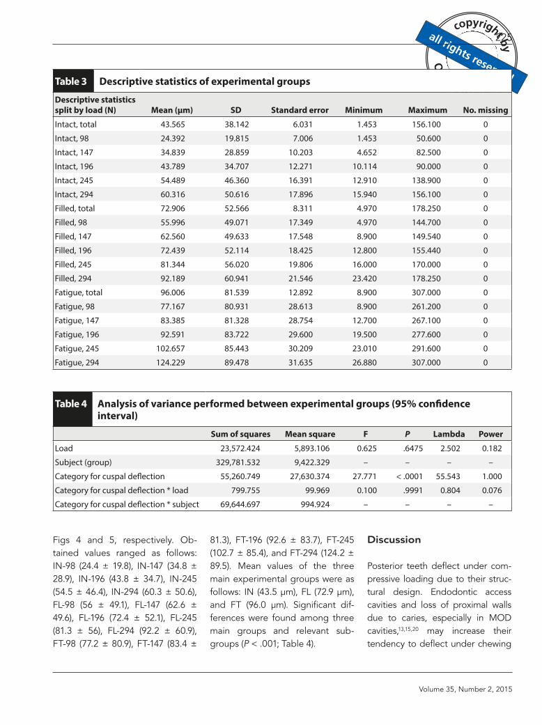
Mean cuspal deflection values (μm) and SDs of experimental groups are presented in Table 2. Descriptive statistics (mean, maxi-mum, minimum, and SDs) are de-picted in Table 3. Comparisons among groups using ANOVA are presented in Table 4. Graphic charts of 15 experimental groups and 3 main groups are shown in
Fig 3 Loading procedure with universal testing machine and custom-made joint.
Table 2 Mean cuspal deflection values of experimental groups
Load (n)Mean cuspal deflection (μm) ± SD
Intact Filled Fatigue treated98 24.4 ± 19.8 56.0 ± 49.1 77.2 ± 80.9
147 34.8 ± 28.9 62.6 ± 49.6 83.4 ± 81.3
196 43.8 ± 34.7 72.4 ± 52.1 92.6 ± 83.7
245 54.5 ± 46.4 81.3 ± 56.0 102.7 ± 85.4
294 60.3 ± 50.6 92.2 ± 60.9 124.2 ± 89.5
Load
Laser sensor
Tooth sample
Laser sensor
Volume 35, Number 2, 2015
225
Figs 4 and 5, respectively. Ob-tained values ranged as follows: IN-98 (24.4 ± 19.8), IN-147 (34.8 ± 28.9), IN-196 (43.8 ± 34.7), IN-245 (54.5 ± 46.4), IN-294 (60.3 ± 50.6), FL-98 (56 ± 49.1), FL-147 (62.6 ± 49.6), FL-196 (72.4 ± 52.1), FL-245 (81.3 ± 56), FL-294 (92.2 ± 60.9), FT-98 (77.2 ± 80.9), FT-147 (83.4 ±
81.3), FT-196 (92.6 ± 83.7), FT-245 (102.7 ± 85.4), and FT-294 (124.2 ± 89.5). Mean values of the three main experimental groups were as follows: IN (43.5 μm), FL (72.9 μm), and FT (96.0 μm). Significant dif-ferences were found among three main groups and relevant sub-groups (P < .001; Table 4).
Discussion
Posterior teeth deflect under com-pressive loading due to their struc-tural design. Endodontic access cavities and loss of proximal walls due to caries, especially in MOD cavities,13,15,20 may increase their tendency to deflect under chewing
Table 3 Descriptive statistics of experimental groups
Descriptive statistics split by load (N) Mean (μm) SD Standard error Minimum Maximum No. missingIntact, total 43.565 38.142 6.031 1.453 156.100 0
Intact, 98 24.392 19.815 7.006 1.453 50.600 0
Intact, 147 34.839 28.859 10.203 4.652 82.500 0
Intact, 196 43.789 34.707 12.271 10.114 90.000 0
Intact, 245 54.489 46.360 16.391 12.910 138.900 0
Intact, 294 60.316 50.616 17.896 15.940 156.100 0
Filled, total 72.906 52.566 8.311 4.970 178.250 0
Filled, 98 55.996 49.071 17.349 4.970 144.700 0
Filled, 147 62.560 49.633 17.548 8.900 149.540 0
Filled, 196 72.439 52.114 18.425 12.800 155.440 0
Filled, 245 81.344 56.020 19.806 16.000 170.000 0
Filled, 294 92.189 60.941 21.546 23.420 178.250 0
Fatigue, total 96.006 81.539 12.892 8.900 307.000 0
Fatigue, 98 77.167 80.931 28.613 8.900 261.200 0
Fatigue, 147 83.385 81.328 28.754 12.700 267.100 0
Fatigue, 196 92.591 83.722 29.600 19.500 277.600 0
Fatigue, 245 102.657 85.443 30.209 23.010 291.600 0
Fatigue, 294 124.229 89.478 31.635 26.880 307.000 0
Table 4 Analysis of variance performed between experimental groups (95% confidence interval)
Sum of squares Mean square F P Lambda PowerLoad 23,572.424 5,893.106 0.625 .6475 2.502 0.182
Subject (group) 329,781.532 9,422.329 – – – –
Category for cuspal deflection 55,260.749 27,630.374 27.771 < .0001 55.543 1.000
Category for cuspal deflection * load 799.755 99.969 0.100 .9991 0.804 0.076
Category for cuspal deflection * subject 69,644.697 994.924 – – – –

The International Journal of Periodontics & Restorative Dentistry
226
forces. Coronal restorations pre-venting residual tooth structures from such stresses are highly recom-mended.
In the past, full-crown cover-age was indicated as the gold stan-dard for postendodontic restoration treatment.21,22 Contrary to nonadhe-sive restorative materials, which re-quire full cuspal coverage, minimally invasive and conservative adhesive materials or restorations have be-come increasingly popular in actual restorative dentistry.23
Little is known about the long-term behavior of restorations, since acrylic resin-tooth interfaces are vul-nerable to progressive damage by occlusal loads, as was substantiated by scanning electron microscopy analyses revealing typical fatigue fracture patterns.24 Fatigue can be defined as the failure of mechanical properties after repeated applica-tions of stress, at a level well below the ultimate fracture strength of the material or interface.19,24,25
In most in vitro studies, de-structive methods are used9,11,25,26 to evaluate the resistance of the tooth-restoration complex. Never-theless, the anatomical variability may strongly influence the results of such investigations due to the size and morphology differences of natural teeth. In vitro fractures induced by compressive loading in adhesively restored teeth may oc-cur with values ranging from 302 to 502 N.16 There is little accordance in the literature about biting forces because a wide range of clenching force values are reported,27 rang-ing from 338 N28 to 720 N29 up to 1,221 N,30 whereas older stud-ies report lower values between 13 and 66 N31 up to a maximum of between 147 and 261 N.32,33 However, some authors25 suggest that tooth fracture seems to occur mostly due to a fatigue phenom-enon: Over time, repeated stress can greatly reduce the resistance to fracture, even under forces far be-
low the loading force normally re-quired to break a healthy tooth.24,34
Nowadays, several in vitro stud-ies about postendodontic restora-tions are available in the literature with both destructive17,18,35 and nondestructive15,36,37,40 techniques. Some interesting clinical trials12,40
also were carried out to assess the reliability of adhesive techniques and materials comparing them with the traditional prosthetic pro-cedures. Measurements of cuspal deflection under load have been used in order to investigate both polymerization shrinkage41 and the mechanical properties of the tooth-restoration complex.1,15,36,37,40
In particular, the evaluation of nondestructive evaluation of teeth deflection in terms of cuspal de-flection under axial load seems to be a valuable method for pre-dicting the capacity of the tooth-restoration complex to withstand intraoral stresses. The rationale for this approach is that, since there
Fig 4 Cell bar chart presenting mean deflection values of 15 experimental groups, split by loads.
Fig 5 Cell bar chart presenting mean deflection values of three main experimental groups under maximum load.
Intact
Loading force (N)
Filled Fatigue
Defl
ectio
n (μ
m)
098147196245294
160
140
120
100
80
60
40
20
0Intact Filled Fatigue
Defl
ectio
n (μ
m)
160
140
120
100
80
60
40
20
0
Volume 35, Number 2, 2015
227
is a linear relationship between fatigue and static loading,41,42 the lower the amount of deflection, the lower the fatigue of the tooth-restoration complex and the better the prognosis.43
The present study focused on the last aspect to assess differ-ences of postendodontic adhe-sive restorations immediately after placement and after cyclic fatigue treatment, to provide information about the changes in mechanical behavior of the tooth-restoration complex over time. The original nondestructive protocol employed in the present study adopted a load range between 98 and 294 N, which is similar to the range normally registered under physi-ological condition for maxillary premolars.36 Maxillary premolars were selected for the present study because they hold the highest risk of fracture, as reported in the litera-ture.40 Since cuspal deflection was tested on the same tooth samples in condition of integrity and follow-ing endodontic treatment/coronal restoration, the variability bias from tooth to tooth was eliminated or strongly reduced.15,36,371
Several papers1,5,7,14,15,36,38,40 re-ported a remarkable difference in static load resistance between sound teeth and restored ones, so a re-stricted number of samples was con-sidered sufficient to assess the null hypothesis.
According to the selected method of measurement, small flat surfaces were created on the buc-cal and lingual sides of each sample to enable the laser beams to mea-sure more accurately each cuspal
displacement without being mis-led by tooth anatomy. Otherwise, the convex shape of tooth surfaces could have caused unreliable data following a possible vertical micro-movement of the tooth during load. Total deflection was recorded as the sum of the deflection of both cusps, without considering the concept of “cuspal independence” reported by Sakaguchi et al.45
The preparation of endodontic access cavities by removing margin-al ridges was meant to simulate the worst condition for the prognosis of the teeth. However, no deflection test was performed on open MOD cavities, as the weakening of cusps was reported several times in the literature.5,9,14,15,40,43 Such a test could have led to a great loss of samples without being really significant.
The flowable composite layer was used to reduce polymerization shrinkage on residual cusps.41 The 24-hour delay in deflection testing after endodontic and restorative treatments was meant to let the res-torations complete their polymeriza-tion reactions and settle the stress of composite contraction.
Load application followed the long axis of the tooth in order to simulate the normal occlusal rela-tionship of maxillary second pre-molars with their antagonist teeth and to standardize as much as pos-sible the test conditions. In addi-tion, since deflection was studied to investigate fatigue, angulated load was avoided, as it was less likely to occur in the oral cavity than axial load.
The results obtained in the present study indicate an increase
in deflection under compressive load in all endodontically treated and adhesively restored teeth com-pared to the intact ones. Greater deformation values were found after mechanical fatigue treat-ment. Standard deviation values were quite high compared to the mean deflection values, as a con-sequence of anatomical variability: Each tooth actually reacts differ-ently under load, depending on its size, morphology, and age.
All restored teeth deflect more than the intact ones; bond-ed coronal restorations then con-tribute to partly recover the initial properties. Such an increase in cuspal deflection was smaller im-mediately after restoration place-ment, and the differences between the groups are greater at higher load values. Significant differ-ences were found between the three experimental conditions. This fact can be related to the different mechanical properties (ie, e-modulus) of dental tissues versus composite material and to the formation of microcracks as load was applied repeatedly.19
Comparing the results of pre-vious similar studies,5,15,38,39,43 slight differences in deflection values can be found. A variety of factors in-volved may contribute to this fact, such as different types of deflec-tion sensors (strain gauges or dif-ferential transformers versus laser sensors), load range, load appli-cation mode, restorative material, and MOD preparation design. The results are comparable to those ob-tained in similar studies36,37 with dif-ferent composite materials.

The International Journal of Periodontics & Restorative Dentistry
228
Although data comparison with similar studies is difficult, the study by Cobankara et al17 confirmed a lower fracture resistance after cyclic loading treatment in teeth restored with ceramic or composite materi-als compared to intact ones, con-cluding that the most interesting option was represented by ceramic inlays. Analogous results were ob-served by Hitz et al18 in a similar study. Different conclusions were drawn in a work by Kuijs et al,46 in which ceramic and direct and in-direct composite employed in the replacement of maxillary premolar cusps were compared by measur-ing the fracture load after fatigue treatment. The three materials per-formed in a similar way, suggesting that the treatment choice should be based on criteria other than fa-tigue resistance.
Fennis et al47 compared the re-sponse to fatigue of composite res-torations in premolars with a MOD cavity and a simulated buccal frac-ture, with or without lingual cuspal coverage. Although the group with cuspal coverage showed better sur-vival rates against fatigue, the frac-tured specimens of this group were mostly unrepairable due to cracked ends below the CEJ. The authors suggested caution in reducing re-sidual cusps.
The cuspal deflection values obtained in restored teeth might provide ideas in the selection of actual adhesive systems and composite materials as alterna-tives to prosthetic solutions, even in borderline situations such as postendodontic MOD cavities. Even if deformation values become
greater after fatigue treatment, res-torations appear to be able to with-stand the stress during function.
Further investigations are need-ed to assess the behavior of com-posite or ceramic inlay restorations under compressive forces. The re-sults of the present in vitro study should be confirmed in clinics by monitoring the behavior of tested restorative materials in randomized clinical trials.
Conclusions
The highest cuspal deflection val-ues (CDVs) were obtained in the FT groups. The lowest CDVs were ob-tained in the IN groups. FL groups showed higher deflection values than IN groups. CDVs increased progressively as the teeth were restored and subjected to fatigue treatment.
Acknowledgments
The authors reported no conflicts of interest related to this study.
References
1. González-López S, De Haro-Gasquet F, Vílchez-Díaz MA, Ceballos L, Bravo M. Effect of restorative procedures and occlusal loading on cuspal deflection. Oper Dent 2006;31:33–38.
2. Huang TJ, Schilder H, Nathanson D. Ef-fect of moisture content and endodontic treatment on some mechanical proper-ties of human dentin. J Endod 1992;18: 209–215.
3. Sedgley CM, Messer HH. Are endodon-tically treated teeth more brittle? J En-dod 1992;7:332–335.
4. Cavel WT, Kelsey WP, Blankenau RJ. An in vivo study of cuspal fracture. J Pros-thet Dent 1985;53:38–42.
5. Hansen EK. In vivo cusp fracture of end-odontically treated premolars restored with MOD amalgam or MOD resin fill-ing. Dent Mater 1988;4:169–173.
6. Vire DE. Failure of endodontically treat-ed teeth: Classification and evaluation. J Endod 1991;17:338–342.
7 . Howe CA, McKendry DJ. Effect of end-odontic access preparation on resis-tance to crown-root fracture. J Am Dent Assoc 1990;121:712–715.
8. Lewinstein I, Grajower R. Root dentin hardness of endodontically treated teeth. J Endod 1981;7:421–422.
9. Wendt SL Jr, Harris BM, Hunt TE. Re-sistance to cusp fracture in endodonti-cally treated teeth. Dent Mater 1987;3: 232–235.
10. Helfer AL, Melnik S, Schilder H. Deter-mination of the moisture content of vital and pulpless teeth. Oral Surg Oral Med Oral Pathol 1972;34:661–670.
11. Burke FJ. Tooth fracture in vivo and in vitro. J Dent 1992;20:131–139.
12. Ferrari M, Cagidiaco MC, Grandini S, De Sanctis M, Goracci C. Post placement af-fects survival of endodontically treated premolars. J Dent Res 2007;86:729–734.
13. Patel DK, Burke FJ. Fractures of posterior teeth: A review and analysis of associat-ed factors. Prim Dent Care 1995;2:6–10.
14. Morin D, DeLong R, Douglas WH. Cusp reinforcement by the acid-etch tech-nique. J Dent Res 1984;63:1075–1078.
15. Panitvisai P, Messer HH. Cuspal deflec-tion in molars in relation to endodontic and restorative procedures. J Endod 1995;21:57–61.
16. Sorrentino R, Monticelli F, Goracci C, et al. Effect of post-retained compos-ite restorations and amount of coronal residual structure on the fracture resis-tance of endodontically-treated teeth. Am J Dent 2007;20:269–274.
17. Cobankara FK, Unlu N, Cetin AR, Ozkan HB. The effect of different restoration techniques on the fracture resistance of endodontically-treated molars. Oper Dent 2008;33:526–533.
18. Hitz T, Ozcan M, Göhring TN. Marginal adaptation and fracture resistance of root-canal treated mandibular molars with intracoronal restorations: Effect of thermocycling and mechanical loading. J Adhes Dent 2010;12:279–286.
19. Baran G, Boberick K, McCool J. Fatigue of restorative materials. Crit Rev Oral Biol Med 2001;12:350–360.
Volume 35, Number 2, 2015
229
20. González-López S, Vilchez Díaz MA, de Haro-Gasquet F, Ceballos L, de Haro-Muñoz C. Cuspal flexure of teeth with composite restorations subjected to occlusal loading. J Adhes Dent 2007; 9:11–15.
21. Abou-Rass M. Post and core restoration of endodontically treated teeth. Curr Opin Dent 1992;2:99–107.
22. Dietschi D, Duc O, Krejci I, Sadan A. Bio-mechanical considerations for the resto-ration of endodontically treated teeth: A systematic review of the literature, part II (evaluation of fatigue behavior, inter-faces, and in vivo studies). Quintessence Int 2008;39:117–129.
23. Bitter K, Kielbassa AM. Post-endodontic restorations with adhesively luted fiber-reinforced composite post systems: A review. Am J Dent 2007;20:353–360.
24. De Munck J, Van Landuyt K, Peumans M, et al. A critical review of the durability of adhesion to tooth tissue: Methods and results. J Dent Res 2005;84:118–132.
25. Frankenberger R, Krämer N, Petschelt A. Fatigue behaviour of different dentin adhesives. Clin Oral Investig 1999;3:11–17.
26. Belli S, Cobankara FK, Eraslan O, Eski-tascioglu G, Karbhari V. The effect of fiber insertion on fracture resistance of endodontically treated molars with MOD cavity and reattached fractured lingual cusps. J Biomed Mater Res B Appl Biomater 2006;79:35–41.
27. Koc D, Dogan A, Bek B. Bite force and influential factors on bite force measure-ments: A literature review. Eur J Dent 2010;4:223–232.
28. Kogawa EM, Calderon PS, Lauris JR, Araujo CR, Conti PC. Evaluation of maxi-mal bite force in temporomandibular disorders patients. J Oral Rehabil 2006; 33:559–565.
29. Gibbs CH, Anusavice KJ, Young HM, Jones JS, Esquivel-Upshaw JF. Maxi-mum clenching force of patients with moderate loss of posterior tooth sup-port: A pilot study. J Prosthet Dent 2002; 88:498–502.
30. Ferrario VF, Sforza C, Zanotti G, Tarta-gilia GM. Maximal bite force in healthy young adults as predicted by surface electromyography. J Dent 2004;32: 451–457.
31. De Boever JA, McCall WD Jr, Holden S, Ash M Jr. Functional occlusal forces: An investigation by telemetry. J Prosthet Dent 1978;40:326–333.
32. Anderson DJ. Measurement of stress in mastication. J Dent Res 1956;35: 664–670.
33. Gibbs CH, Mahan PE, Lundeen HC, et al. Occlusal forces during chewing—Influences of biting strength and food consistency. J Prosthet Dent 1981;46: 561–567.
34. Jantarat J, Palamara JE, Messer HH. An investigation of cuspal deformation and delayed recovery after occlusal loading. J Dent 2001;29:363–370.
35. Oskoee SS, Oskoee PA, Navimipour EJ, Shahi S. In vitro fracture resistance of endodontically-treated maxillary pre-molars. Oper Dent 2007;32:510–514.
36. Cerutti A, Flocchini P, Madini L, Man-gani F, Putignano A, Docchio F. Effects of bonded composites vs. amalgam on resistance to cuspal deflection for end-odontically-treated premolar teeth. Am J Dent 2004;17:295–300.
37. Acquaviva PA, Madini L, Krokidis A, Ga-gliani M, Mangani F, Cerutti A. Adhesive restoration of endodontically treated premolars: Influence of posts on cus-pal deflection. J Adhes Dent 2011;13: 279–286
38. Reeh ES, Douglas WH, Messer HH. Stiff-ness of endodontically-treated teeth related to restoration technique. J Dent Res 1989;68:1540–1544.
39. Mannocci F, Bertelli E, Sherriff M, Watson TF, Ford TR. Three-year clinical compari-son of survival of endodontically treated teeth restored with either full cast cover-age or with direct composite restoration. J Prosthet Dent 2002;88:297–301.
40. Cara RR, Fleming GJ, Palin WM, Walms-ley AD, Burke FJ. Cuspal deflection and microleakage in premolar teeth restored with resin-based composites with and without an intermediary flowable layer. J Dent 2007;35:482–489.
41. Reeh ES, Messer HH, Douglas WH. Re-duction in tooth stiffness as a result of endodontic and restorative procedures. J Endod 1989;15:512–516.
42. Naumann M, Sterzenbach G, Pröschel P. Evaluation of load testing of postendodontic restorations in vitro: Linear compressive loading, gradual cycling loading and chewing simulation. J Biomed Mater Res B Appl Biomater 2005;74:829–834.
43. Garoushi S, Lassila LV, Tezvergil A, Val-littu PK. Static and fatigue compression test for particulate filler composite resin with fiber-reinforced composite sub-structure. Dent Mater 2007;23:17–23.
44. Zidan O, Abdel-Keriem U. The effect of amalgam bonding on the stiffness of teeth weakened by cavity preparation. Dent Mater 2003;19:680–685.
45. Sakaguchi RL, Brust EW, Cross M, De-Long R, Douglas WH. Independent movement of cusps during occlusal loading. Dent Mater 1991;3:186–190.
46. Kuijs RH, Fennis WM, Kreulen CM, Roeters FJ, Verdonschot N, Creugers NH. A comparison of fatigue resistance of three materials for cusp-replacing adhesive restorations. J Dent 2006;34:1 9–25.
47. Fennis WM, Kuijs RH, Kreulen CM, Verdonschot N, Creugers NH. Fatigue resistance of teeth restored with cuspal-coverage composite restorations. Int J Prosthodont 2004;17:313–317.