Importance of mesophyll diffusion conductance in estimation ...
Upload
khangminh22Category
view
3download
0
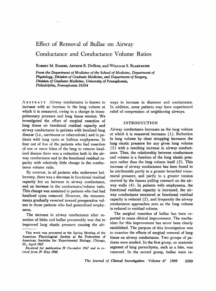
Effect of removal of bullae on airway conductance andconductance volume ratios
Robert M. Rogers, … , Arthur B. DuBois, William S. Blakemore
J Clin Invest. 1968;47(12):2569-2579. https://doi.org/10.1172/JCI105939.
Airway conductance is known to increase with an increase in the lung volume at which it is measured, owing to a changein transpulmonary pressure and lung tissue tension. We investigated the effect of surgical resection of lung tissue onfunctional residual capacity and airway conductance in patients with localized lung disease (i.e., carcinoma ortuberculosis) and in patients with lung cysts or bullous emphysema. In four out of five of the patients who had resection ofone or more lobes of the lung to remove localized disease there was a reduction both in the airway conductance and inthe functional residual capacity with relatively little change in the conductance volume ratio.
By contrast, in all patients who underwent bullectomy, there was a decrease in functional residual capacity but anincrease in airway conductance, and an increase in the conductance/volume ratio. This change was sustained in patientswho had had localized cysts removed. However, the measurements gradually reverted toward preoperative values inthose patients who had generalized emphysema.
The increase in airway conductance after resection of blebs and bullae presumably was due to improved lung elasticpressure causing the airways to increase in diameter and conductance. In addition, some patients may have experiencedrelief of compression of neighboring airways.
Research Article
Find the latest version:
https://jci.me/105939/pdf
Effect of Removal of Bullae on Airway
Conductance and Conductance Volume Ratios
ROBERTM. RoGERs, ARTHmURB. DuBois, and WILLIAM S. BLAKFMORE
From the Department of Medicine of the School of Medicine, Department ofPhysiology, Division of Graduate Medicine, and Department of Surgery,Division of Graduate Medicine, University of Pennsylvania,Philadelphia, Pennsylvania 19104
A B S T R A C T Airway conductance is known toincrease with an increase in the lung volume atwhich it is measured, owing to a change in trans-pulmonary pressure and lung tissue tension. Weinvestigated the effect of surgical resection oflung tissue on functional residual capacity andairway conductance in patients with localized lungdisease (i.e., carcinoma or tuberculosis) and in pa-tients with lung cysts or bullous emphysema. Infour out of five of the patients who had resectionof one or more lobes of the lung to remove local-ized disease there was a reduction both in the air-way conductance and in the functional residual ca-pacity with relatively little change in the conduc-tance volume ratio.
By contrast, in all patients who underwent bul-lectomy, there was a decrease in functional residualcapacity but an increase in airway conductance,and an increase in the conductance/volume ratio.This change was sustained in patients who had hadlocalized cysts removed. However, the measure-ments gradually reverted toward preoperative val-ues in those patients who had generalized emphy-sema.
The increase in airway conductance after re-section of blebs and bullae presumably was due toimproved lung elastic pressure causing the air-
This work was presented at the Spring Meeting of theAmerican Physiological Society at the Federation ofAmerican Societies for Experimental Biology, Chicago,Ill., April 1967.
Received for publication 29 December 1967 and in re-vised form 29 May 1968.
ways to increase in diameter and conductance.In addition, some patients may have experiencedrelief of compression of neighboring airways.
INTRODUCTION
Airway conductance increases as the lung volumeat which it is measured increases (1): Reductionin lung volume by chest strapping increases thelung elastic pressure for any given lung volume(2) with a resulting increase in airway conduct-ance. Thus, the relationship between conductanceand volume is a function of the lung elastic pres-sure rather than the lung volume itself (3). Thisincrease of airway conductance has been found tobe attributable partly to a greater bronchial trans-mural pressure, and partly to a greater tensionexerted by the tissues pulling outward on the air-way walls (4). In patients with emphysema, thefunctional residual capacity is increased, the air-way conductance measured at functional residualcapacity is reduced (2), and frequently the airwayconductance approaches zero as the lung volumeis reduced to residual volume.
The surgical resection of bullae has been re-ported to cause clinical improvement. The mecha-nism for this improvement has never been clearlyestablished. The purpose of this investigation wasto examine the effects of surgical removal of lungtissue on airway conductance. Two groups of pa-tients were studied. In the first group, an anatomicsegment of lung parenchyma, such as a lobe, wasremoved. In the second group, bullae were re-
The Journal of Clinical Investigation Volume 47 1968 2569
moved. The surrounding lung tissue was left un-disturbed insofar as this was possible.
METHODSA brief description of the patients who had large bullae(J. K., W. S., W. K., and F. J.) and diffuse pulmonaryemphysema with small bullae (M. W., R. M., and J. G.)is given in the appendix.
Airway resistance (RA), thoracic gas volume (TGV),and functional residual capacity (FRC) were measuredwith a body plethysmograph (5, 6). Airway conduct-ance, the reciprocal of airway resistance, was calculatedfrom the airway resistance values. The airway conduct-ance was measured, first close to functional residual ca-pacity, and next with the patient holding his chest in aninspiratory position, then in an expiratory position. Thepatients panted at a rate of approximately 2 cycle/secwith a small volume displacement. All subjects had mea-surements made just before surgery and 10-30 days aftersurgery. All emphysematous patients had intensive medi-cal therapy before being admitted to this study. All sub-jects were judged to be free of bronchospasm or bronchialinfection at the time of the studies. In none of these pa-tients was there much change in airway conductance mea-sured before and after inhalation of isoproterenol aerosol.All patients had preoperative measurements of airwayresistance and thoracic gas volume on several occasions,and had additional pulmonary function studies, includ-ing vital capacity, maximum mid-expiratory flow rate,FEV1, FEV8, maximum breathing capacity, functionalresidual capacity, and arterial blood gas studies as partof their preoperative evaluation. The information ob-tained by the spirometric tests contributed to the presentstudy mainly as an aid in establishing the diagnosis. Thepreoperative values are listed in Tables I and II. TableII also contains the postoperative values for all of thepatients who underwent bullectomy (group II), except
for patient M. W. on whom the spirometry tests andblood gases were not performed postoperatively.
RESULTS
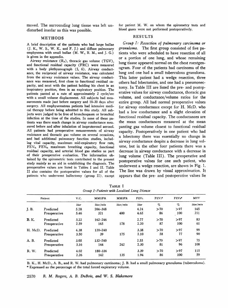
Group I: Resection of pulmonary carcinoma orgranuloma. The first group consisted of five pa-tients who were scheduled to have resection of allor a portion of one lung, and whose remaininglung tissue appeared normal on the chest roentgen-ogram. Four of the patients had carcinoma of thelung and one had a small tuberculous granuloma.This latter patient had a wedge resection, threeothers had lobectomies, and one had a pneumonec-tomy. In Table III are listed the pre- and postop-erative values for airway conductance, thoracic gasvolume, and conductance/volume ratios for theentire group. All had normal preoperative valuesfor airway conductance except for H. McD. whohad a low conductance and a slight elevation offunctional residual capacity. The conductances arethe mean conductances measured at the meanpanting gas volume closest to functional residualcapacity. Postoperatively in one patient who hada lobectomy there was essentially no change inairway conductance despite a decrease in lung vol-ume, but in the other four patients there was adecrease in airway conductance with a decrease inlung volume (Table III). The preoperative andpostoperative values for one such patient, whounderwent a wedge resection, are shown in Fig. 1.The line was drawn by visual approximation. Itappears that the pre- and postoperative values lie
TABLE IGroup I-Patients with Localized Lung Disease
Patient V.C. MMEFR MMIFR FEVi FEVi* FEVs* MW
liter liter/min liter/min liter % % liter/min
J. B. Predicted 5.28 204-348 4.24 >70 >97 145Preoperative 5.46 321 400 4.65 86 100 211
B. K. Predicted 3.22 162-246 2.77 >70 >97 83Preoperative 2.59 165 178 2.20 87 100 61
H. McD. Predicted 4.38 120-240 3.38 >70 >97 99Preoperative 3.50 39 175 2.10 58 77 90
A. B. Predicted 3.05 132-240 2.55 >70 >97 75Preoperative 3.16 144 242 2.30 81 96 108
R. W. Predicted 4.02 180-336 3.39 >70 >97 115Preoperative 2.26 142 135 1.94 86 100 59
B. K., H. McD., A. B., and R. W. had pulmonary carcinoma; J. B. had a small pulmonary granuloma (tuberculoma).* Expressed as the percentage of the total forced expiratory volume.
2570 R. M. Rogers, A. B. DuBois, and W. S. Blakemore
TABLE I IGroup II-Patients with Bullae
Blood gases (arterial)Patients V.C. MMEFR MMIFR FEVi FEVi* FEVa* MW 02Sat Pot Pco2 pH
liter liter/min literlmin liter % % liter/min % mm mmHg Hg
Large bullaeW. S. Predicted 5.28 204-348 4.24 >70 >97 145
Preoperative 3.05 90 135 2.10 68 90 87 94 73 46 7.46Postoperative 2.95 175 250 2.45 83 98 75 94 73 47 7.46
J. K. Predicted 4.82 180-336 3.84 >70 >97 127Preoperative 5.08 181 434 3.56 72 95 167 - - - -
Postoperative 3.36 179 339 2.82 84 100 126 - - - -
W. K. Predicted 4.96 204-348 4.05 >70 >97 140Preoperative 1.56 19 77 0.64 50 83 23 90.0 57 20 7.43Postoperative 2.29 59 144 1.42 72 100 48 94.0 76 33 7.38
F. J. Predicted 4.96 204-348 4.05 >70 <97 140Preoperative 3.10 36 174 1.53 63 75 61 88.8 - 33 7.44Postoperative 1 1.99 74 215 1.60 73 100 46 95.4 - - -Postoperative 2 2.69 156 253 2.35 84 99 131 94.3 - 43 7.42:
Diffuse emphysema with "small" bullaeM. W. Predicted 3.18 132-240 2.62 >70 >97 77
Preoperative 2.36 13 110 0.54 31 56 18 94 61 37 7.49,Postoperative - - - - - - - - - -
R. M. Predicted 4.38 120-240 3.38 >70 >97 99Preoperative 2.59 19 102 0.58 28 53 35 96 69 20 7.50Postoperative 3.15 26 126 0.82 30 54 24 94 62 43 7.52
J. G. Predicted 4.68 150-282 3.66 >70 >97 114Preoperative 2.80 19 130 0.65 27 54 24 94.8 76 38 7.39Postoperative 3.00 26 98 0.71 28 67 31 91.0 64 39 7.40
* Expressed as the percentage of the total forced expiratory volume.
approximately on the same line. The degree of re-duction of airway conductance was consistent withthe amount of decrease in thoracic gas volume.
Group II: Excision of localized lung cysts oremphysematous bullae. The second group con-sisted of seven patients who underwent bullectomy.Two of the patients had normal airway conduct-ance before surgery, whereas five had low airwayconductance. The results are summarized in Ta-ble IV. In all subjects there was a postoperativeincrease in airway conductance and, except forJ. K., a decrease in lung volume. The increase inconductance and decrease in thoracic gas volumeof the group were statistically significant. The lastsubject (F. J.) had no change in airway conduct-ance or thoracic gas volume after the first opera-tion, but following the second operation had a de-crease in his thoracic gas volume and an increasein airway conductance. The results obtained on
one patient (J. G.) are plotted in Fig. 2. Preop-eratively, the values lay to the right of the pre-dicted line with the line of visual approximationintercepting the abscissa at a very high volume.Following operation there was a change in theslope of the line and also a shift of the volumeintercept to the left.
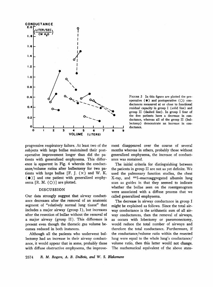
Comparison between groups I and II. Datafrom the individual patients of groups I and IIare plotted in Fig. 3. The individual preoperative(0) and postoperative (0) values for conduct-ance, measured at a panting lung volume closestto functional residual capacity, are joined by lines.The decrease in conductance with the decrease inlung volume (solid line) can be clearly seen infour of the five patients of group I. The data fromgroup II (broken lines) are distinctly different.All subjects had an increase in conductance andall but one had a decrease in lung volume.
Effect of Removal of Bullae on Airway Conductance 2571
0
- CONDUCTANCELITER/SEC/CM H20
x
0
FRC PRE-OP
POST-OP
0.81-
O.6L
0.41 /
- /
I/
0 v 2 3 4
VOLUME LITERS
PREOP= 0 FIGURE 1 The conductance (GA) (ordinate)POST-OP- Y is plotted against the thoracic gas volume
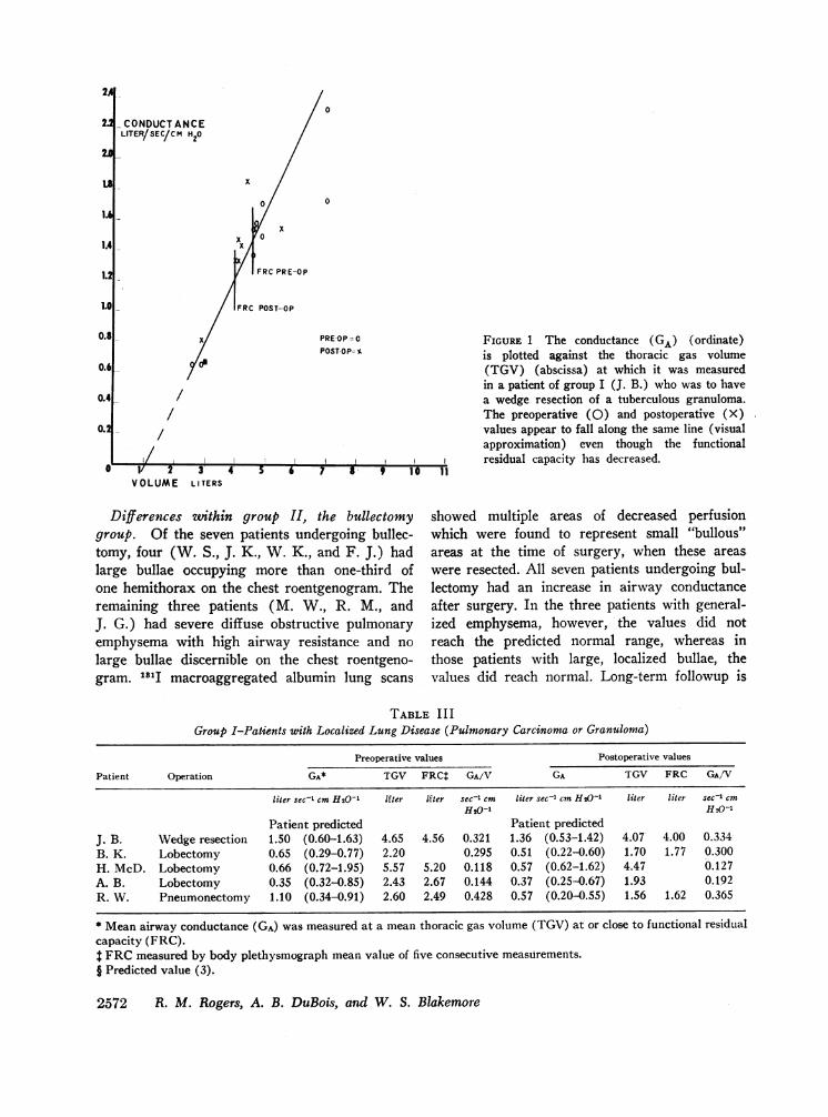
(TGV) (abscissa) at which it was measuredin a patient of group I (J. B.) who was to havea wedge resection of a tuberculous granuloma.The preoperative (0) and postoperative (X)values appear to fall along the same line (visualapproximation) even though the functionalresidual capacity has decreased.
5 6 7 8 9 10 11
Differences within group II, the bullectomygroup. Of the seven patients undergoing bullec-tomy, four (W. S., J. K., W. K., and F. J.) hadlarge bullae occupying more than one-third ofone hemithorax on the chest roentgenogram. Theremaining three patients (M. W., R. M., andJ. G.) had severe diffuse obstructive pulmonaryemphysema with high airway resistance and no
large bullae discernible on the chest roentgeno-gram. 1"1I macroaggregated albumin lung scans
showed multiple areas of decreased perfusionwhich were found to represent small "bullous"areas at the time of surgery, when these areas
were resected. All seven patients undergoing bul-lectomy had an increase in airway conductanceafter surgery. In the three patients with general-ized emphysema, however, the values did notreach the predicted normal range, whereas inthose patients with large, localized bullae, thevalues did reach normal. Long-term followup is
TABLE I I IGroup I-Patients with Localized Lung Disease (Pulmonary Carcinoma or Granuloma)
Preoperative values Postoperative values
Patient Operation GA* TGV FRC GA/V GA TGV FRC GA/V
liter sec'1 cm H20-1 If er If ter seca cm liter sec-' cm H20-1 liter liter sec- cm
H0-1 H2o-Patient predicted Patient predicted
J. B. Wedge resection 1.50 (0.60-1.63) 4.65 4.56 0.321 1.36 (0.53-1.42) 4.07 4.00 0.334B. K. Lobectomy 0.65 (0.29-0.77) 2.20 0.295 0.51 (0.22-0.60) 1.70 1.77 0.300H. McD. Lobectomy 0.66 (0.72-1.95) 5.57 5.20 0.118 0.57 (0.62-1.62) 4.47 0.127A. B. Lobectomy 0.35 (0.32-0.85) 2.43 2.67 0.144 0.37 (0.25-0.67) 1.93 0.192R. W. Pneumonectomy 1.10 (0.34-0.91) 2.60 2.49 0.428 0.57 (0.20-0.55) 1.56 1.62 0.365
* Mean airway conductance (GA) was measured at a mean thoracic gas volume (TGV) at or close to functional residualcapacity (FRC).
FRCmeasured by body plethysmograph mean value of five consecutive measurements.§ Predicted value (3).
2572 R. M. Rogers, A. B. DuBois, and W. S. Blakemore
2
24
U
1.
1.4
1.0
0.2
AI .0 2
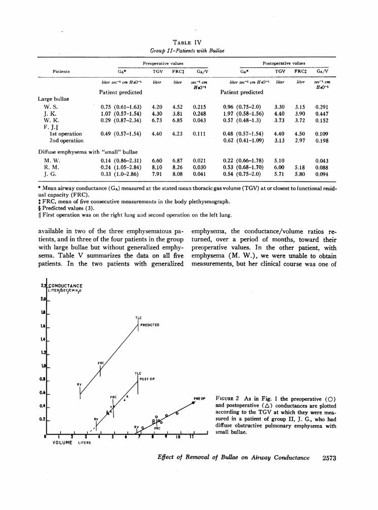
TABLE IVGroup II-Patients with Bullae
Preoperative values Postoperative values
Patients GA* TGV FRCt GA/V GA* TGV FRCJ GA/V
liter sec-1 cm H20-1 liter liter sec-1 cm liter sec'1 cm H20-1 liter liter sec-' cmHs20- HsO-
Patient predicted Patient predictedLarge bullae
W. S. 0.75 (0.61-1.63) 4.20 4.52 0.215 0.96 (0.75-2.0) 3.30 3.15 0.291J. K. 1.07 (0.57-1.54) 4.30 3.81 0.248 1.97 (0.58-1.56) 4.40 3.90 0.447WV. K. 0.29 (0.87-2.34) 6.73 6.85 0.043 0.57 (0.48-1.3) 3.73 3.72 0.152F. J.II
1st operation 0.49 (0.57-1.54) 4.40 4.23 0.111 0.48 (0.57-1.54) 4.40 4.50 0.1092nd operation 0.62 (0.41-1.09) 3.13 2.97 0.198
Diffuse emphysema with "small" bullaeM. W. 0.14 (0.86-2.31) 6.60 6.87 0.021 0.22 (0.66-1.78) 5.10 0.043R. M. 0.24 (1.05-2.84) 8.10 8.26 0.030 0.53 (0.68-1.70) 6.00 5.18 0.088J. G. 0.33 (1.0-2.86) 7.91 8.08 0.041 0.54 (0.75-2.0) 5.71 5.80 0.094
* Mean airway conductance (GA) measured at the stated mean thoracic gas volume (TGV) at or closest to functional resid-ual capacity (FRC).$ FRC, mean of five consecutive measurements in the body plethysmograph.§ Predicted values (3).
First operation was on the right lung and second operation on the left lung.
available in two of the three emphysematous pa-tients, and in three of the four patients in the groupwith large bullae but without generalized emphy-sema. Table V summarizes the data on all fivepatients. In the two patients with generalized
emphysema, the conductance/volume ratios re-turned, over a period of months, toward theirpreoperative values. In the other patient, withemphysema (M. W.), we were unable to obtainmeasurements, but her clinical course was one of
NONDUCTANCELITERAEC/CM H20
TLC
PREDICTED
FoRITLC
POSTOPRV
FRC A PREOP
0
RV FRCI I 1, 1 1 1 a>
5 7 9 I 1 11 z 3 4VOLUME LITERS
FIGURE 2 As in Fig. 1 the preoperative (0)and postoperative (A) conductances are plottedaccording to the TGVat which they were mea-sured in a patient of group II, J. G., who haddiffuse obstructive pulmonary emphysema with
j small bullae.
Effect of Removal of Bullae on Airway Conductance 2573
2.0
U
1.4
1.2
LO
0.8I
0.4 L
0.2
l.o_
'I
I
1/ X °/FIGURE 3 In this figure are plotted the pre-1Jj '-_ O..Q9 operative (0) and postoperative (0) con-
ductances measured at or close to functionalo_ ~{ens residual capacity in group I (solid line) and
group II (dashed line). In group I four ofok > the five patients have a decrease in con-
ductance, whereas all of the group II (bul-lectomy) demonstrate an increase in con-
| I I I I I2 I 1 I ductance.1 2 3 4 5 6
VOLUME (LITERS)7 8 9
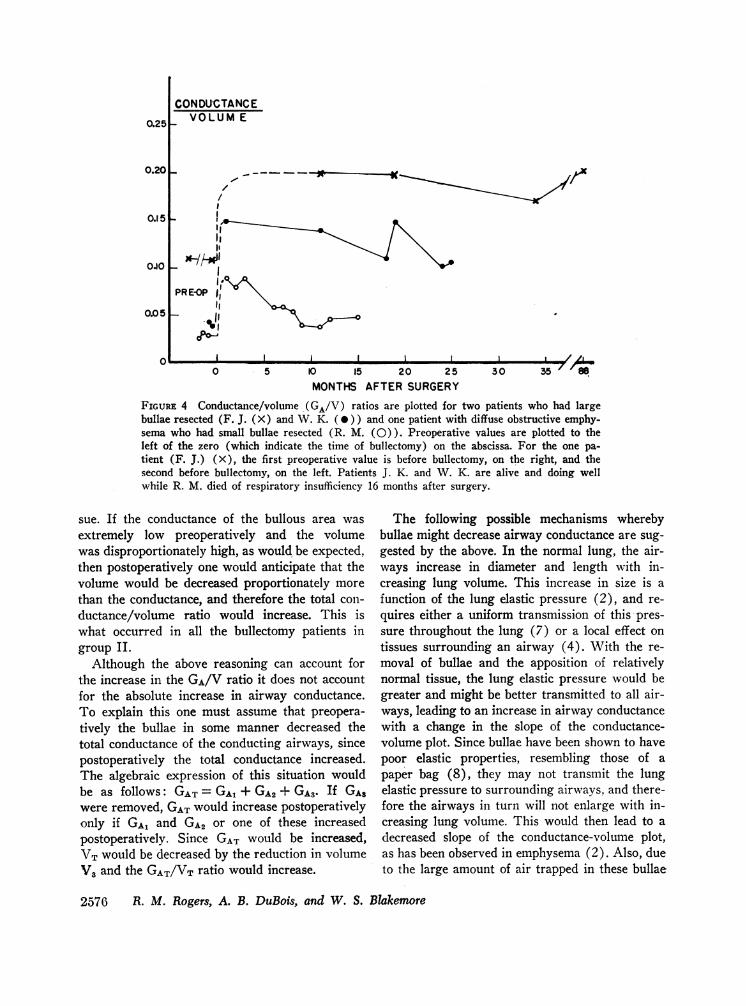
progressive respiratory failure. At least two of thesubjects with large bullae maintained their post-operative improvement longer than did the pa.tients with generalized emphysema. This differ-ence is apparent in Fig. 4 wherein the conduct-ance/volume ratios after bullectomy for two pa-
tients with large bullae [F. J. (X) and W. K.(0)] and one patient with generalized emphy-sema [R. M. ( 0 ) ] are plotted.
DISCUSSION
Our data strongly suggest that airway conduct-ance decreases after the removal of an anatomicsegment of "relatively normal lung tissue" thatincludes a major airway (group I), but increasesafter the resection of bullae without the removal ofa major airway (group II). This difference ispresent even though the thoracic gas volume be-comes reduced in both instances.
Although all the patients who underwent bul-lectomy had an increase in their airway conduct-ance, it would appear that in some, probably thosewith diffuse obstructive emphysema, the improve-
ment disappeared over the course of severalmonths whereas in others, probably those withoutgeneralized emphysema, the increase of conduct-ance was sustained.
The initial criteria for distinguishing betweenthe patients in group II are not as yet definite. Weused the pulmonary function studies, the chestX-ray, and 131I-macroaggregated albumin lungscan as guides in that they seemed to indicatewhether the bullae seen on the roentgenogramwere associated with a diffuse process that we
called generalized emphysema.The decrease in airway conductance in group I
might be explained as follows. Since the total air-way conductance is the arithmetic sum of all air-way conductances, then the removal of airways,as occurs with lobectomy or pneumonectomy,would reduce the total number of airways andtherefore the total conductance. Furthermore, ifthe conductance/volume ratio within the resectedlung were equal to the whole lung's conductance/volume ratio, then this latter would not change.The mathematical equivalent of the above state-
2574 R. M. Rogers, A. B. DuBois, and W. S. Blakemore
CONDUCTANCE2.0 j
LITER/SEC)X1. 8
CCM H20
1.6 _
1.2 _
0.8 _
0.61-
0.41-
0.21-
o0
1.4 _
0
1.0 _-01,
-10
TABLE V
Group II-Conductance- Volume Ratios* after Bullectomy
Large bullae without Diffuse emphysemajdiffuse emphysema with small bullae
Patient ... W.S. W. K. F. J. R. M. J. G.
Preoperative values 0.026 0.0490.043 0.110 0.032 0.057
0.215 0.038 0.109 0.029 0.031
Bullectomy - - - - -
Months after bullectomy 1 0.291 0.150 0.088 0.094-2 0.079 0.08t3 0.088 0.063;4 0.0505 0.60 0.0586 0.059 0.0537 0.059 0.0848 0.053 diedjj9 0.039
1011 0.140 0.198 0.03912 0.046131415 0.04816 0.22317 died18 0.11019 0.149 0.199
24 0.10125 0.106
34 0.171
88 0.202
KR-units, sec-' cm H20-1.$The first value 0.110 is before bullectomy on the right, and the second (0.109) is after bullectomy on the right andbefore bullectomy on the left.J Bulla not discernible on the routine chest roentgenogram.11 Patient died in respiratory insufficiency following bullectomy in the previous unoperated lung.
ment is as follows:
GAT= GA1+ GA2+ GAs (1)VT= V1 + V2 + V3 (2)GA GA1+GA2+GAS (3)V V1 + V2 + V3
Where GAT is the total airway conductance, andGA1, GA, GA3 are the conductances of the air-ways of an individual segment of lung. VT is thetotal volume of all lung segments with V1, V2, andV3 equal to the volume of individual segments.With the removal of a segment or lobe then the
total conductance would decrease, since the con-ductances of the airways removed, for example,.GA3 would be subtracted from the total. How-ever, the volume V3 also would be removed and ifthe GA,/V3 ratio were the same as the GAT/VATthen the latter (GA/V) would be the same post-operatively. The increase in the conductance/volume ratio after bullectomy can be explained withanalogous reasoning. With bullectomy the majorairways to a lobe are not disturbed, i.e., the bullais merely clamped and unroofed with suturingtogether of the relatively normal surrounding tis-
Effect of Removal of Bullae on Airway Conductance 2575
0.25
0.201
0.1 5
0oa0
0.051
CONDUCTANCEVOLUME
,w//4we/-
I f
PRE-0P ofoiAeel:
I I I~~~~~~10 5 10 15 20 25 30 35
MONTHSAFTER SURGERY
FIGuRE 4 Conductance/volume (GA/V) ratios are plotted for two patients who had largebullae resected (F. J. (X) and W. K. (0)) and one patient with diffuse obstructive emphy-sema who had small bullae resected (R. M. (0)). Preoperative values are plotted to theleft of the zero (which indicate the time of bullectomy) on the abscissa. For the one pa-tient (F. J.) (X), the first preoperative value is before bullectomy, on the right, and thesecond before bullectomy, on the left. Patients J. K. and W. K. are alive and doing wellwhile R. M. died of respiratory insufficiency 16 months after surgery.
sue. If the conductance of the bullous area wasextremely low preoperatively and the volumewas disproportionately high, as would be expected,then postoperatively one would anticipate that thevolume would be decreased proportionately morethan the conductance, and therefore the total con-ductance/volume ratio would increase. This iswhat occurred in all the bullectomy patients ingroup II.
Although the above reasoning can account forthe increase in the GA/V ratio it does not accountfor the absolute increase in airway conductance.To explain this one must assume that preopera-tively the bullae in some manner decreased thetotal conductance of the conducting airways, sincepostoperatively the total conductance increased.The algebraic expression of this situation wouldbe as follows: GAT=GA1 + GA2 + GA3. If GAswere removed, GATwould increase postoperativelyonly if GA1 and GA2 or one of these increasedpostoperatively. Since GAT would be increased,VT would be decreased by the reduction in volumeV3 and the GAT/VT ratio would increase.
The following possible mechanisms wherebybullae might decrease airway conductance are sug-gested by the above. In the normal lung, the air-ways increase in diameter and length with in-creasing lung volume. This increase in size is afunction of the lung elastic pressure (2), and re-quires either a uniform transmission of this pres-sure throughout the lung (7) or a local effect ontissues surrounding an airway (4). With the re-moval of bullae and the apposition of relativelynormal tissue, the lung elastic pressure would begreater and might be better transmitted to all air-ways, leading to an increase in airway conductancewith a change in the slope of the conductance-volume plot. Since bullae have been shown to havepoor elastic properties, resembling those of apaper bag (8), they may not transmit the lungelastic pressure to surrounding airways, and there-fore the airways in turn will not enlarge with in-creasing lung volume. This would then lead to adecreased slope of the conductance-volume plot,as has been observed in emphysema (2). Also, dueto the large amount of air trapped in these bullae
2576 R. M. Rogers, A. B. DuBois, and W. S. Blakemore
Il .L /A0
(i.e., it cannot be expelled during expiration), theextrapolated intercept of the conductance-volumeline on the volume axis would be at a very highvolume. These mechanisms are consistent with thefindings in the present study, although we do nothave measurements of lung elastic pressure beforeand after surgery. Wedid not make this measure-ment because of the difficulty of doing repeatedesophageal balloon studies on a patient.
If surgical resection of a part of the lung in-*creases the lung elastic pressure at any given vol-tume in a way comparable to the change after cheststrapping (2, 3) then it might be expected thatif there had been a generalized abnormality inlung elastic recoil preoperatively, postoperatively,airway conductance would be improved. But ifthe tissues exhibited stress relaxation, there wouldbe a gradual return to the previous airway con-ductance and lung volume over a period of time.For example, bullae that were small before opera-tion and that were able to withstand the lung elas-tic pressure before surgery might enlarge as a re-sult of the new pressure-volume relationships. On-the other hand, if the pulmonary parenchyma re-maining after bullectomy were normal, then onemight expect that the new pressure-volume rela-tionships would lead to an increased conductancethat would be maintained over a period of time,as occurred in the patients who had the resectionof large bullae which did not appear to be part ofa generalized process.
Other factors that have not as yet been evaluatedmay play a significant role in the increased con-ductance and the length of time it is maintained.The effect of a pleural reaction on bullae has notbeen adequately studied. It is conceivable that post-operative pleural reaction might fuse the visceraland parietal pleura which might limit the expan-sion of remaining bullae, since they would not befree to expand in all directions. In addition, if thepleural space became obliterated, this might alterthe postural effects on vertical gradients of pleuralpressure (9) and conceivably change the distribu-tion of lung elastic pressure.
Bullae have been known to be associated withairway obstruction (10-14) but it has never beenclearly demonstrated whether they cause or con-tribute to the obstruction, whether the obstructionproduces the bullae, or whether there is a general-ized disease that produces both the bullae and the
airway obstruction. Although the spirometric stud-ies in most of our patients (Table II) demon-strated some improvement it is difficult to relatethese results to the airways, since these studiesare effort dependent., The body plethysmographmeasures airway conductance and is not dependenton muscular effort. It is clear that bullectomy in-creases the airway conductance. Hence, one mustconclude that bullae play a role in the decreasedairway conductance seen before bullectomy. Thepredominant abnormality displayed by these pa-tients was a mechanical one that was corrected bybullectomy, albeit in some cases only temporarily.Whether the bullae developed because of localizedairway obstruction and then participated in themaintenance of generalized, as well as localized,obstruction, or whether they arose de novo andcaused a generalized decrease in airway conduct-ance is not clear. From our data, however, bothpossibilities must be considered and the questionis at present unanswered.
On the basis of our findings, we see no rea-son to change the criteria for surgery outlined byBaldwin et al. (12) and further elaborated byLaurenzi, Turino, and Fishman (10). Bullectomyin patients with severe diffuse chronic obstructiveemphysema cannot be supported by us on the basisof the present data. All such patients in our studyreturned toward their preoperative condition overa period of time. To establish whether a patientwith large bullae also has diffuse obstructive em-physema is still a most difficult problem to whichwe have not found an easy answer.
APPENDIX
Brief clinical summaries of the patients ingroup II
J. K. was a 46 yr old white male with a 40-pack-yr 1history of cigarette smoking, who had a large bulla(11 X 16 X 16 cm) noted on a tuberculosis control sur-vey chest roentgenogram. His only previous chest roent-genogram had been made while he was in the ArmedServices some 20 yr ago. Physical findings were re-stricted to a pectus excavatum of a moderate degree. Hedenied any pulmonary symptomatology. A right upperlobectomy was performed.
W. S. was a 35 yr old Negro male who had a 3month history of dyspnea on exertion and a nonproduc-tive cough. A chest roentgenogram taken 2 months be-
1 1-pack-yr, the consumption by a person of 20 cigar-ettes/day for 1 yr.
Effect of Removal of Bullae on Airway Conductance 2577

fore surgery revealed a large bulla in the right upperlobe and "fibrosis" in the right lower lobe. Microscopicexamination of the resected bulla and surrounding tissuerevealed "sarcoid-like" lesions, in addition to the bullae.The patient also had a reduced single breath DLco (14.7ml/min per mmHg. Predicted: 26.7 ±8).
W. K. was a 36 yr old Negro male with a 10-pack-yr history of smoking cigarettes who noted graduallyprogressing dyspnea over the 5 yr before admission. Thedyspnea progressed more rapidly in the yr before admis-sion so that at the time of surgery the patient wasdyspneic at rest and severely dyspneic on mild exercise(such as walking to the bathroom). A chest roentgeno-gram revealed bilateral large apical bullae that occupiedover 50% of each hemithorax; in addition, there was asmall mass noted in the right mid-lung field. The patientunderwent a wedge resection of the mass, which provedto be an adenocarcinoma, and excision and plication ofbullae in the right upper and lower lung.
F. J. was a 33 yr old Negro male with a history ofover 20-pack-yr of smoking cigarettes who complained ofdyspnea and chest pain, both of which had become pro-gressively more severe over a 2 yr period. He had achronic cough with occasional paroxyms of coughingthat caused an exacerbation of his chest pain. His coughwas productive of thick, viscid, mucoid material. 4 yr be-fore admission the patient had had bronchopneumonia. Achest roentgenogram revealed advanced bullous emphy-sema most marked in both lower lung fields. The patienthad plication of bullae in the left lower lobe, and lingulaand apical posterior segment of the upper lobe. Afterthis procedure, the patient experienced little relief ofhis symptoms, so that 8 months later, plication and re-section of bullae in the right lung were carried out.The majority of bullae were found in the lower lobe,but several small bullae were noted in the middle andupper lobe and also were resected.
M. W. was a 50 yr old white housewife with a 21-pack-yr history of cigarette smoking. The patient statedthat for "many years she had had frequent colds whichtended to persist" and they usually were associated withsputum production. 4 yr before surgery, she noticed theonset of exertional dyspnea that progressed to dyspneaat rest by the time of surgery. She received little relieffrom any medication. Her chest roentgenogram showedbilateral pulmonary emphysema with areas of increasedradiolucency at both bases. Plication and resection ofbullae in the right lung, predominantly in the lower lobe,were performed.
R. M. was a 64 yr old white male with a history of40-pack-yr of cigarette smoking. He was told 5 yr be-fore surgery that he had emphysema, and at that timehe stopped smoking. He had a morning cough that wasproductive of minimal amounts of sputum and occasion-ally the cough occurred in paroxysms causing him tofeel extremely weak. Over the 5 yr since the onset of hissymptoms, the patient had progressive dyspnea so that atthe time of the present admission he was able to walkonly 5 to 10 steps without dyspnea. He noticed the most
severe dyspnea when he arose from a sitting position andthis gradually lessened as he remained standing erect.A chest roentgenogram showed hyperaerated lung fieldsbilaterally with flattened diaphragms and widened inter-costal spaces. The increased radiolucency was moremarked in the upper lobes with some distortion of thevascular markings suggesting the presence of bullae,although no definite bullae could be clearly outlined inthe roentgenogram. Plication and resection of bullaewere carried out on the right upper and lower lobes.
J. G. was a 59 yr old white male with a 45-pack-yrhistory of cigarette smoking. The patient had first no-ticed dyspnea on exertion 9 yr before surgery. The pa-tient's dyspnea increased gradually and insidiously overthe subsequent 8-9-yr period so that at the time of admis-sion he could walk only 15 yd without stopping and hadto rest 3-4 min after ascending one flight of stairs.During this time, the patient also had a chronic coughproductive of a teaspoon of white, clear sputum in themorning. The chest roentgenogram showed flatteneddiaphragms, with a marked degree of hyperaeration andincreased radiolucency in both apices. Plication and re-section of bullae in the right lung were performed. Af-ter an initial improvement in his symptoms, the patientreturned toward his preoperative status. 6 months afterthe initial surgery on the right lung, the patient had aplication of bullae in the left lung. He died on the 15thpostoperative day. His death was attributed to pneumoniaand respiratory insufficiency.
ACKNOWLEDGMENTS
Dr. Rogers received a Public Health Service traininggrant, NIH 5-TO1 GM-95706, which is gratefully ac-knowledged. Dr. DuBois was the recipient of a ResearchCareer award of the National Institutes of Health. Thiswork was supported in part by a contract between theUniversity of Pennsylvania and the Army ChemicalCenter, No. DAAA-15-67-C-0154; and in part by theCardiovascular Clinical Research Center, National HeartInstitute grant HE-06352.
REFERENCES
1. Briscoe, W. A., and A. B. DuBois. 1958. The rela-tionship between airway resistance, airway conduct-ance and lung volume in subjects of different age andbody size. J. Clin. Invest. 37: 1279.
2. Butler, J., C. G. Caro, R. Alcala, and A. B. DuBois.1960. Physiological factors affecting airway resistancein normal subjects and in patients with obstructiverespiratory disease. J. Clin. Invest. 39: 584.
3. Caro, C. G., J. Butler, and A. B. DuBois. 1960. Someeffects of restriction of chest cage expansion on pul-monary function in man: an experimental study. J.Clin. Invest. 39: 573.
4. Marshall, R. 1962. Effect of lung inflation on bron-chial dimensions in the dog. J. Appl. Physiol. 17: 596.
5. DuBois, A. B., S. Y. Botelho, and J. H. Comroe, Jr.1956. A new method for measuring airway resistancein man using a body plethysmograph: values in nor-
2578 R. M. Rogers, A. B. DuBois, and W. S. Blakemore
mal subjects and in patients with respiratory disease.J. Clin. Invest. 35: 327.
6. DuBois, A. B., S. Y. Botelho, G. N. Bedell, R. Mar-shall, and- J. H. Comroe, Jr. 1956. A rapid plethys-mographic method for measuring thoracic gas vol-ume: a comparison with a nitrogen washout methodfor measuring functional residual capacity in normalsubjects. J. Clin. Invest. 35: 322.
7. Mead, J., T. Takishima, and D. Leith. 1967. Mechani-cal interdependence of distensible units in the lungs.Federation Proc. 26: 551.
8. Ting, E. Y., R. Klopstock, and H. A. Lyons. 1963.Mechanical properties of pulmonary cysts and bullae.Am. Rev. Respirat. Diseases. 87: 538.
9. West, J. B. 1966. Distribution of blood and gas inlungs. Phys. Med. Biol. 11: 357.
10. Laurenzi, G. A., G. M. Turino, and A. P. Fishman.1962. Bullous disease of the lung. Am. J. Med. 32:378.
11. Kaltreider, N. L, and W. W. Fray. 1939. Patho-logical physiology of pulmonary cysts and emphyse-matous bullae. Am. J. Med. Sci. 197: 62.
12. Baldwin, E. de F., K. A. Harden, D. G. Green, A.Cournand, and D. W. Richards, Jr. 1950. Pulmo-nary insufficiency. IV. A study of 16 cases of largepulmonary air cysts or bullae. Medicine. 29: 169.
13. Harden, K. A. 1953. Pulmonary air cysts: some diag-nostic, physiologic and therapeutic considerations. J.Natl. Med. Assoc. 45: 167.
14. Knudson, R. J., and E. A. Gaensler. 1965. Surgery foremphysema. Ann. Thor. Surg. 1: 332.
Effect of Removal of Bullae on Airway Conductance 2579
Copyright © 2022 FDOKUMEN