Effect of Glycine-Conjugated Bile Acids with and without Lecithin on Water and Glucose Absorption in...
7
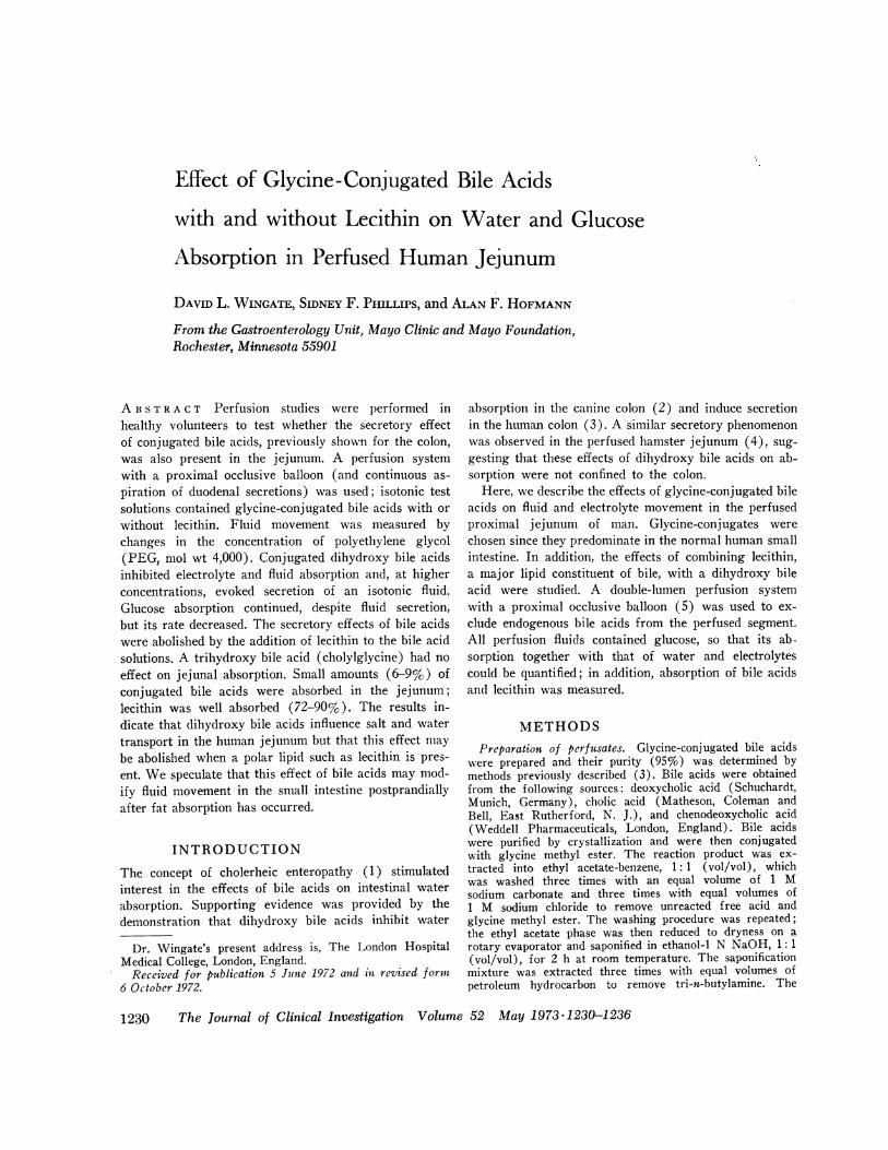
Effect of Glycine-Conjugated Bile Acids with and without Lecithin on Water and Glucose Absorption in Perfused Human Jejunum DAVID L. WINGATE, SIDNEY F. PILLIPS, and ALAN F. HOFMANN From the Gastroenterology Unit, Mayo Clinic and Mayo Foundation, Rochester, Minnesota 55901 A B S T R A C T Perfusion studies were performed in healthy volunteers to test whether the secretory effect of conjugated bile acids, previously shown for the colon, was also present in the jejunum. A perfusion system with a proximal occlusive balloon (and continuous as- piration of duodenal secretions) was used; isotonic test solutions contained glycine-conjugated bile acids with or without lecithin. Fluid movement was measured by changes in the concentration of polyethylene glycol (PEG, mol wt 4,000). Conjugated dihydroxy bile acids inhibited electrolyte and fluid absorption and, at higher concentrations, evoked secretion of an isotonic fluid. Glucose absorption continued, despite fluid secretion, but its rate decreased. The secretory effects of bile acids were abolished by the addition of lecithin to the bile acid solutions. A trihydroxy bile acid (cholylglycine) had no effect on jejunal absorption. Small amounts (6-9%) of conjugated bile acids were absorbed in the jejunum; lecithin was well absorbed (72-90%). The results in- dicate that dihydroxy bile acids influence salt and water transport in the human jejunum but that this effect may be abolished when a polar lipid such as lecithin is pres- ent. We speculate that this effect of bile acids may mod- ify fluid movement in the small intestine postprandially after fat absorption has occurred. INTRODUCTION The concept of cholerheic enteropathy (1) stimulated interest in the effects of bile acids on intestinal water absorption. Supporting evidence was provided by the demonstration that dihydroxy bile acids inhibit water Dr. Wingate's present address is, The London Hospital Medical College, London, England. Received for publication 5 June 1972 and in revised formn 6 October 1972. absorption in the canine colon (2) and induce secretion in the human colon (3). A similar secretory phenomenon was observed in the perfused hamster jejunum (4), sug- gesting that these effects of dihydroxy bile acids on ab- sorption were not confined to the colon. Here, we describe the effects of glycine-conjugated bile acids on fluid and electrolyte movement in the perfused proximal jejunum of man. Glycine-conjugates were chosen since they predominate in the normal human small intestine. In addition, the effects of combining lecithin, a major lipid constituent of bile, with a dihydroxy bile acid were studied. A double-lumen perfusion system with a proximal occlusive balloon (5) was used to ex- clude endogenous bile acids from the perfused segment. All perfusion fluids contained glucose, so that its ab- sorption together with that of water and electrolytes could be quantified; in addition, absorption of bile acids and lecithin was measured. METHODS Preparation of perfusates. Glycine-conjugated bile acids were prepared and their purity (95%) was determined by methods previously described (3). Bile acids were obtained from the following sources: deoxycholic acid (Schuchardt, Munich, Germany), cholic acid (Matheson, Coleman and Bell, East Rutherford, N. J.), and chenodeoxycholic acid (Weddell Pharmaceuticals, London, England). Bile acids were purified by crystallization and were then conjugated with glycine methyl ester. The reaction product was ex- tracted into ethyl acetate-benzene, 1: 1 (vol/vol), which was washed three times with an equal volume of 1 M sodium carbonate and three times with equal volumes of 1 M sodium chloride to remove unreacted free acid and glycine methyl ester. The washing procedure was repeated; the ethyl acetate phase was then reduced to dryness on a rotary evaporator and saponified in ethanol-i N NaOH, 1: 1 (vol/vol), for 2 h at room temperature. The saponification mixture was extracted three times with equal volumes of petroleum hydrocarbon to remove tri-n-butylamine. The 1230 The Journal of Clinical Investigation Volume 52 May 1973 -1230-1236
-
Upload
independent -
Category
Documents
-
view
0 -
download
0
Transcript of Effect of Glycine-Conjugated Bile Acids with and without Lecithin on Water and Glucose Absorption in...
Effect of Glycine-Conjugated Bile Acids
with and without Lecithin on Water and Glucose
Absorption in Perfused Human Jejunum
DAVID L. WINGATE, SIDNEY F. PILLIPS, and ALAN F. HOFMANN
From the Gastroenterology Unit, Mayo Clinic and Mayo Foundation,Rochester, Minnesota 55901
A B S T R A C T Perfusion studies were performed inhealthy volunteers to test whether the secretory effectof conjugated bile acids, previously shown for the colon,was also present in the jejunum. A perfusion systemwith a proximal occlusive balloon (and continuous as-piration of duodenal secretions) was used; isotonic testsolutions contained glycine-conjugated bile acids with orwithout lecithin. Fluid movement was measured bychanges in the concentration of polyethylene glycol(PEG, mol wt 4,000). Conjugated dihydroxy bile acidsinhibited electrolyte and fluid absorption and, at higherconcentrations, evoked secretion of an isotonic fluid.Glucose absorption continued, despite fluid secretion,but its rate decreased. The secretory effects of bile acidswere abolished by the addition of lecithin to the bile acidsolutions. A trihydroxy bile acid (cholylglycine) had noeffect on jejunal absorption. Small amounts (6-9%) ofconjugated bile acids were absorbed in the jejunum;lecithin was well absorbed (72-90%). The results in-dicate that dihydroxy bile acids influence salt and watertransport in the human jejunum but that this effect maybe abolished when a polar lipid such as lecithin is pres-ent. We speculate that this effect of bile acids may mod-ify fluid movement in the small intestine postprandiallyafter fat absorption has occurred.
INTRODUCTION
The concept of cholerheic enteropathy (1) stimulatedinterest in the effects of bile acids on intestinal water
absorption. Supporting evidence was provided by thedemonstration that dihydroxy bile acids inhibit water
Dr. Wingate's present address is, The London HospitalMedical College, London, England.Received for publication 5 June 1972 and in revised formn
6 October 1972.
absorption in the canine colon (2) and induce secretionin the human colon (3). A similar secretory phenomenonwas observed in the perfused hamster jejunum (4), sug-gesting that these effects of dihydroxy bile acids on ab-sorption were not confined to the colon.
Here, we describe the effects of glycine-conjugated bileacids on fluid and electrolyte movement in the perfusedproximal jejunum of man. Glycine-conjugates werechosen since they predominate in the normal human smallintestine. In addition, the effects of combining lecithin,a major lipid constituent of bile, with a dihydroxy bileacid were studied. A double-lumen perfusion systemwith a proximal occlusive balloon (5) was used to ex-clude endogenous bile acids from the perfused segment.All perfusion fluids contained glucose, so that its ab-sorption together with that of water and electrolytescould be quantified; in addition, absorption of bile acidsand lecithin was measured.
METHODSPreparation of perfusates. Glycine-conjugated bile acids
were prepared and their purity (95%) was determined bymethods previously described (3). Bile acids were obtainedfrom the following sources: deoxycholic acid (Schuchardt,Munich, Germany), cholic acid (Matheson, Coleman andBell, East Rutherford, N. J.), and chenodeoxycholic acid(Weddell Pharmaceuticals, London, England). Bile acidswere purified by crystallization and were then conjugatedwith glycine methyl ester. The reaction product was ex-tracted into ethyl acetate-benzene, 1: 1 (vol/vol), whichwas washed three times with an equal volume of 1 Msodium carbonate and three times with equal volumes of1 M sodium chloride to remove unreacted free acid andglycine methyl ester. The washing procedure was repeated;the ethyl acetate phase was then reduced to dryness on arotary evaporator and saponified in ethanol-i N NaOH, 1: 1(vol/vol), for 2 h at room temperature. The saponificationmixture was extracted three times with equal volumes ofpetroleum hydrocarbon to remove tri-n-butylamine. The
1230 The Journal of Clinical Investigation Volume 52 May 1973 -1230-1236

conjugated bile acids were then isolated as described previ-ously. The sodium salt of chenodeoxycholylglycine (CDC-G)1 swas obtainied by freeze-drying-; the sodium salts ofcholylglycine (C-G) and deoxycholylglycine (DC-G) w\ereprecipitated with diethyl ether from a methanlolic solution.All bile acids w\-ere dried to constant weight in a vacuumdesiccator. Lecithin (chromatographically pure, Schwarz/Mann Div., Becton, Dickinson and Co., Orangeburg, N. Y.)was used as purchased.
Control solutions were pH 8.0 and contained the follow-ing concentrations of ions (in meq/liter) Na+, 140; K+,5; C1-, 100; HCO3-, 45. In addition, D-glucose was addedto a concentration of 200 mg/100 ml (11.2 mM-). Poly-ethylene glycol (PEG, mol wt 4000) was the nonabsorb-able marker; each liter of perfusate contained 5 g of stablePEG and 5-10 1ACi of "4C-labeled PEG (New England Nu-clear, Boston, Mass.). In initial studies, stable and 14C-labeled PEG were compared as nonabsorbable markers andno differences between them were found (6). Test solutionsalso contained bile acids, with or without lecithin (see be-low). The sodium concentration in test solutions was up to10 meq/liter greater than that in control solutions. Osmo-lality of control solutions was 284 mosmol/kg and that oftest solutions was 289-299 mosmol/kg.
Perfutsion techniqutc. Subjects were healthy human vol-unteers (postmenopausal women, or men more than 21 yrold) who gave written informed consent.The perfusion tube has been described (5). The tube
was passed at 8 a.m., after the subject had fasted over-night. The location of the balloon was assessed fluoroscopi-cally; when it reached the ligament of Treitz it was in-flated with 35 ml of air and its inflation port was sealed.Occlusion, confirmed by the absence of bile staining in theperfusate, was signaled by a sensation of slight epigastricdistention in the subject. Inflation of the balloon was ad-justed so that the sensation was present but minimal. Theperfusate at 37°C was then delivered at a constant speedof 10 ml/min and sampled from the test segment (25 cm)by siphonage. Intermittent suction was applied to the proxi-mal aspiration lumen to remove duodenal secretions. Duringthe study, the subjects remained fasting and recumbent, butwere allowed to read, converse, watch television, or sleep.Each perfusion fluid was delivered for 90 min; a single
study consisted of four consecutive 90-min experiments.Perfusate samples were collected in 10-min portions. Ineach perfusion, the first 50 min were allowed for equilibra-tion, and the last four 10-min periods were taken to repre-sent sampling of a steady state; these assumptions appearedto be justified in practice.
Ainalytical methods. Sodium and potassium were esti-mated by flame photometry; chloride was measured by elec-trometric titration with a silver nitrate solution. Glucosewas measured by an enzymatic hexokinase method (Boeh-ringer Mannheim Corp., New York). Bile acids were esti-mated by a modification of the method of Stwata and Yama-saki (7). The PEG['4C] in 0.2 ml of perfusate, mixed with15 ml of a scintillation "cocktail," was counted by liquidscintillation spectrometry; quench correction was made byexternal standardization. Phospholipid concentrations weredetermined on the chloroform phase after Folch extraction.
Calculations. Absorption and secretion of water andsolutes, relative to PEG, were calculated by standard
'Abbreviationis used in this paper: CDC-G, chenodeoxy-cholylglycine; C-G, chloylglycine; CMC, critical micellarconcentration; DC, deoxycholate; DC-G, deoxycholylgly-cine; PEG, polyethylene glycol.
methods (6). Comparisons betw een means were made bycalculation of Student s t test for iupaired values; linearregression analysis was by the method of least squares.
Experimental designA control and three test solutions were perfused in ran-
dom order in each subject. Three groups of studies wereperformed.Group 1: Effect of conijutgatcd bile acid. Trihydroxy Bile
Acid. Three solutions, 2.5, 5.0, or 10.0 mM in C-G, andcontrol solutions without bile acids were used in each ofthree subjects. The order of perfusion in each subject wasdrawn from a table of random numbers.Dihydroxy Bile Acids. A control solution and test solutions
containing 2.5, 5.0, or 10 mM dihydroxy bile acid (eitherDC-G or CDC-G) were used in each subject. A 4 X 4Latin square was used for perfusion order so that eachconcentration of each of the two bile acids was adminis-tered at a different point in the perfusion sequence in eachof the eight subjects. This design allowed analysis of thedata for comparison between control and test solutions, forthe effects of sequential perfusion, and for the reversibilityof the effects of test perfusions.Group 2: Effect of unconjugated bile acid. To test the
possibility that the effects found with conjugated dihydroxycompounds might be due to contamination with unconju-3gated bile acid, which is known to be more potent in thehamster (4), two subjects were perfused alternately withglucose-electrolyte solution containing 2.5 m.M C-G (whichhad no effect on fluid movement) and the same solutioncontaining 0.25 mM sodium deoxycholate (DC). Each sub-ject received each solution twice, and the perfusion se-quence was reversed for the second set of perfusions.
Grozup 3.: Effect of dihydroxy bile acid in prcsec~ce of leci-t1hin. This group of studies also utilized a 4 X 4 Latinsquare design in four subjects and was performed to deter-mine whether the effects of a dihydroxy conjugated bileacid would be modified by the presence of phospholipid. Eachsubject received: (a) glucose-electrolyte solution; (b) glu-cose-electrolyte solution with 5 mM DC-G; (c) glucose-electrolyte solution with 5 mM DC-G and 1.25 mM lecithin;and (d) glucose-electrolyte solution with 5 mM DC-G and2.5 mM lecithin. This choice of solutions permitted com-parison, by solutions 1 and 2, with the results of group 1studies. Evaluation of dose-response to lecithin at threedifferent concentrations (0, 1.25, and 2.5 mM) was pos-sible; the effect of perfusion sequence was eliminated.
TABLE IEffect of Perfusion Sequence on l11ater and Glucose Mozvements*
Perfusion period
Substance 1 2 3 4
Water movement
All perfusions (N = 44) 0.7+0.4 0.740.3 1.1 ±0.4 0.740.4Controls only (N = 11) 1.440.2 1.7±0.4 1.740.3 2.2±0.1
Glucose movement
All perfusions 13.1±40.9 14.4±0.9 15.3+1.1 15.8±0.8Controls only 14.341.3 16.6±t1.9 19.1±40.3 17.9 ±41.2
* Water fluxes are expressed as ml/min per 25 cm jejunum; glucose fluxesare mg/min per 25 cm jejunum. Data are shown as means ±SE.
Effect of Glycine-Conjugated Bile Acids on Absorption in Human Jejunum 1231

TABLE I IEffect of Unconjugated Deoxycholate on Net Absorption*
Perfusate
2.5 mM C -GSubstance 2.5 mM C -G with 0.25 mM DC
Water (ml/min per 25 cm) 1.8±0.2 1.440.3Glucose (mg/min per 25 cm) 13.441.8 12.1±1.5Sodium (meq/min per 25 cm) 241 ±36 178 ±39
* Means ±SE; N = 4. Differences not statistically significant.
Bile acid concentration,mM
FIGURE 1 Fluid absorption from 25 cm segment of humanjejunum (mean +SE; N =3 or 4) perfused with isotonicelectrolyte solution and with conjugated trihydroxy (C-G)or dihydroxy (DC-G, CDC-G) bile acid solutions. Nega-tive values represent fluid secretion.
RESULTS
Effect of perfusion sequence. Absorption rates of wa-
ter and glucose were influenced little by the sequence ofperfusion (Table I). When only control perfusions are
considered, a small but statistically significant (P < 0.05)mean increase of water absorption, 0.2 ml/min for eachhour of perfusion, was noted. Thus, during the 5 h ofperfusion there was a trend for water absorption to in-crease slightly. This trend was small relative to thechanges in water movement induced by. bile acid per-
fusions.
Effect of bile acids on water absorption. Dihydroxybile acids (DC-G, CDC-G) inhibited water absorption(Fig. 1). This effect was concentration related: athigher concentrations, fluid was secreted. Infused con-
centrations of DC-G (and CDC-G) and net fluid move-
ment were closely related (P < 0.001). DC-G appearedmore potent but the dose-response relationships were notsignificantly different. Changes in water movement oc-
curred rapidly and were fully reversible; one such studyis illustrated in Fig. 2.
There was no significant depression of water absorp-tion by the trihydroxy bile acid (C-G), even at 10 mM.The relationship between water movement and C-G con-
centration was not significant.Effect of unconjugated deoxycholate. If the DC-G
used in earlier studies was 95% conjugated and 5% un-
conjugated, the 5 mM DC-G solution would have been0.25 mM in unconjugated deoxycholic acid. Since therewas no effect on water absorption when C-G containing0.25 mM unconjugated deoxycholate was perfused (Ta-ble II), the effect of DC-G on absorption was unlikely tobe caused by free deoxycholate present as a contaminant.
Effect of lecithin on water secretion induced by deoxy-cholylglycine. The addition of lecithin reversed thesecretory effect of DC-G: water absorption with 2.5 mMlecithin was close to that from the control solution (Fig.3). The rapidity of the effect and the reversibility of theeffect of lecithin were similar to those found with per-fusions of bile acids without lecithin (Fig. 4).
Relationships between water and electrolyte move-
ments. Net movements of sodium and chloride were
closely related to water movement. However, chlorideabsorption was always less than sodium absorption, sug-
gesting concomitant absorption of another anion (bi-carbonate). The relationship between ion and watermovements indicated that the net fluid that was ab-sorbed or secreted was isotonic, and was predominantly
Bile acid, 10 I._-_I_ -_.......I
0
12-
Water
recovery, 10r..ml/min
HoursFIGURE 2 Water recovery in sequential 10-min samples from jejunum perfused at 10 ml/min,showing rapid onset of inhibition of water absorption induced by dihydroxy bile acid (DC-G),with rapid reversibility of this effect. Rectangles mark samples taken as steady-state observa-tions. Water recovery values below broken line are net absorption; those above the line arenet secretion.
1232 D. L. Wingate, S. F. Phillips, and A. F. Hofmann
. z
S.0~CZ(J
0n0
-1
-2

TABLE IIIEffect of Bile Acid Concentration on Glucose Absorption
Glucose absorption (mg/min per 25 cm jejunum)*Bile acidconc. C-G CDC-G DC-G
M.S1
0 18.040.7 18.740.8 17.8±41.12.5 14.640.9 14.5±2.6 16.14±0.75.0 16.7±0.5 12.7±1.0 13.1±0.6
10.0 15.2 ± 1.7 11.2±A0.9 9.2 ±0.5
* Mean 4SE; N = 3 or 4 for each perfusate. Glucose in-fusion rate was 20 mg/min.
a sodium chloride solution. Detailed analysis of thesechanges will be described elsewhere.2
Glucose absorption. More than 85% of the perfusedglucose was absorbed from the control glucose-electro-lyte solution. There was a progressive decrease in glucoseabsorption (to approximately 50%) with increasingconcentration of DC-G and CDC-G but not with C-G(Table III). Decreased glucose absorption was not dueto a lowering of luminal glucose concentration by se-creted fluid, since effluent glucose concentrations werealways higher when glucose absorption was decreased.With dihydroxy acids at 10 mM, glucose was absorbedwhen fluid was secreted. Glucose absorption and watermovement were related (P < 0.001).
Absorption of bile acid and lecithin. Small but sig-nificant absorption of DC-G occurred (Table IV).There was a significant correlation between bile acidabsorption and water movement, the uptake of bile acidincreasing with water absorption (r = 0.627; P <0.005). Lecithin was well absorbed from DC-G per-fusates.
Side-effects of perfusion. Colicky pain in the upperabdomen, nausea, or vomiting was observed during the
2Wingate, D. L., H. S. Mekhjian, and S. F. Phillips. Un-published observation.
21-
1
-1
-2
Control (no bile acid)
Aspii,' Absorption
I /I I
A1.25 2.5/
-z Secretion
Lecithin,mM
FIGURE 3 Influence of added lecithin on fluid secretion(mean +SE; N=4) induced by 5 mM DC-G. Addition of2.5 mM lecithin blocked the secretory effect of bile acids,and net water absorption was similar to control values (nobile acid, no lecithin). Negative values represent fluidsecretion.
perfusion of 5 mM or 10 mM dihydroxy bile acids in 8of 16 subjects. Pain was sufficiently severe to necessitateinterruption of the study in two instances. PEG recovery
was less in those studies with side-effects (34.8±3.9%)than in those without side-effects (55.8±5.6%; P <0.02), suggesting that the symptoms resulted from thepassage of dihydroxy bile acids into the intestine distalto the test segment. However, there was no correlationbetween the degree of fluid secretion and the occurrence
of symptoms. Replacement of the test solution by a bileacid-free solution containing glucose and electrolyteswas associated with prompt disappearance of symptoms.No symptoms occurred during cholylglycine perfusions.
DISCUSSION
Secretory effects of dihydroxy bile acids. The pres-
ent study is the fourth to show a rapid, reversible, se-
cretory effect of conjugated dihydroxy bile acids in vivo.The dose-response curves, although incomplete, are simi-lar to those reported for hamster jejunum and humanand canine colon (2-4). Forth, Rummel, and Glasner
Bile acid, mM
Lecithin, mM
0 1 2 3 4 5 6
HoursFIGURE 4 Water recovery in sequential 10-min samples from jejunum perfused at 10 ml/min,showing modifying influence of lecithin on secretion induced by bile acid (DC-G). Rectanglesmark samples taken as steady-state observations. Water recovery values below broken line arenet absorption; those above the line are net secretion.
Effect of Glycine-Conjugated Bile Acids on Absorption in Human Jejunum 11233

TABLE IVA bsorption of Deoxycholylglycine and Lecithin
Absorption
Starting conc. Bile acid Lecithin
JAmol/min jmol/minDC -G Lecithin per 25 cm* % per 25 cm* %0
mM
5 0 2.8±0.5 5.6 - -5 1.25 3.3±0.5 6.6 11.342.0 905 2.5 4.6±0.2 9.2 18.0±3.1 72
* Mean -SE; N = 4 for each perfusate.
(8) had shown, in the rat jejunum in vitro, a similareffect of unconjugated bile acids but conjugated bileacids had no effect.A fundamental assumption of these studies is that PEG
serves as a valid marker for fluid movement. Wilkinson(9) has shown that PEG administered orally to healthyman is recovered totally in the feces. In our studies, thejejunum was exposed to physiologic concentrations ofbile acids. Furthermore, PEG was not absorbed whenbile acids were perfused into the human colon (3), andPEG recovery was complete when fluid secretion wasinduced by bile acids in the hamster jejunum (4). Ourperfusion system has also been shown to isolate thejejunum from endogenous biliary secretion (5). Thus,it seems reasonable to propose that PEG dilution wascaused by fluid secretion induced by the perfused bileacids.
In vivo perfusion systems demonstrate phenomena;their value in the elucidation of mechanisms is limited.The observed effects were not osmotic because solutionswere essentially isotonic and small osmotic gradients donot inhibit jejunal absorption in the presence of glucose(10). Moreover, identical concentrations of the trihy-droxy compounds had no effect.
Bile acids can disrupt cell membranes (11). Althoughthe exact time sequence of events cannot be ascertainedby a perfusion system, the rapid reversibility of the se-
cretory effects suggests that structural damage is un-likely. Moreover, in the hamster jejunum (4), secre-tion was induced by conjugated bile acids without histo-logic changes. Nonetheless, we cannot exclude a subtlechange of membrane structure sufficient to alter mucosalpermeability. However, it is not clear how alteration perse in pore size could reverse net flow across the mu-cosa. Glucose absorption persisted and the secreted fluidwas mainly a sodium chloride solution. Rohde and Chen(12) were unable to demonstrate a change in apparentpore size in the jejunum when secretion was induced bycholera toxin.
Bile acids might influence certain chemical mecha-nisms proposed for ion transport, by activation of theadenyl cyclase system or by inhibition of Na, K+-acti-vated ATP-ase. Glycine-conjugated dihydroxy and tri-hydroxy bile acids have been shown to influence intesti-nal ATP-ase in vitro, but the effects of individual bileacids on ATP-ase correlated imperfectly with their ef-fects on water absorption in vivo (13).
Bile acids are surface active, and dihydroxy compoundspossess greater surface activity than do trihydroxy com-pounds; however, correlations between physical andpharmacologic properties are unjustified until a greatervariety of bile acids are tested for a secretory effect.
Abolition of secretion by lecithin addition. Additionof lecithin, which was used in physiologic concentrations(14), would be anticipated to decrease markedly thecritical micellar concentration (CMC) of the dihydroxybile acids (15). By definition, a decrease in CMC woulddecrease the concentration of bile acid in molecular form.The CMC of glycine-conjugated dihydroxy bile acids inthe absence of lecithin is 3-4 mM (16); in the presenceof lecithin, it is lower (15). Lecithin, which decreases"activity" of dihydroxy bile acids chemically, might havealso decreased the pharmacologic activity. However, wecannot exclude a more direct effect of lecithin on mem-brane transport processes.
Glucose movements. Glucose absorption occurred inall experiments, but its magnitude diminished duringperfusion with the bile acids which induced secretion.Levitt, Hakim, and Lifson (17) partitioned glucose ab-sorption in the dog into three components: active trans-port, diffusive transport, and convective transport. Thesimplest explanation of our observations is that the de-crease in glucose absorption associated with water se-cretion represented decreased convective transport. Dif-fusive transport is unlikely to have decreased, since in-traluminal concentrations were greater when secretionoccurred. Whether or not active transport was alteredcannot be ascertained. However, glucose absorption wasnot decreased (17, 18) during fluid secretion induced bymannitol, but in these experiments the amount of inducedsecretion was much less. When secretion was induced bycholera toxin, inhibition of glucose absorption was notobserved (19); but in these experiments the glucose con-centration was unphysiologic (60 mM).
Glucose absorption occurred simultaneously with move-ment of sodium into the lumen when fluid secretion wasinduced. Two recent reports (20, 21) have describedglucose absorption concomitant with movement of so-dium into the lumen when intestinal loops were perfusedwith isotonic, sodium-free solutions containing glucose.These observations and our own are in marked contrastto in vitro studies in which a stoichiometric relationshipbetween glucose and sodium absorption has been ob-
1234 D. L. Wingate, S. F. Phillips, and A. F. Hofmann

served. However, induced secretion may occur from adifferent mucosal site (for example, crypts) than thesite of glucose absorption (villi) (22). Thus, our dataneither sul)l)ort nor refute the hyl)othesis that sodiumnland water absorption is induced by active glucose ab-sorption. Our results do indicate that stoichiometry,claimed for glucose and sodium absorption in vitro (23),is not applicable to our findings which showed glucoseabsorption during secretion of a sodium-containingfluid.
Lecithin absorption. Absorption of lecithin has notbeen studied in this manner before. The hydrolysis ofdlietarv lecithin to 1-lysolecithin and fatty acid has beenclearly shown in man (24), although whether or notbiliary lecithin is hydrolyzed similarly is unclear. Wedid not ascertain if lecithin was hydrolyzed during ab-sorption in our experiments; but the only lipid presentin the recovered fluids was lecithin, indicating that, ifhydrolysis had occurred, the products were completelyabsorbed.
Bile acid absorption. Absorption of DC-G was muchless than that of lecithin. and the presence of lecithin(lid not decrease the rate of bile acid absorption. Accord-ing to current concepts, bile acids are absorbed in thejejunum by passive nonionic diffusion of the protonatedform (25). This mechanism is strongly pH-dependent,since the ionized species should diffuse very slowly dueto a reflection coefficient of close to 1 (25). The rate ofabsorption, extrapolated from our studies at pH 8.0, from150 cm of jejunum would be about 1 to 1.5 mmol/h.This finding is consistent with earlier observations, inman (26, 27) and in primates with ileal resection (28),that passive jejunal absorption of glvcine-conjugateddihydroxy bile acids occurs in health and disease. How-ever, studies of bile acid absorption during digestion ofa meal are needed.
P/ysiologic significance. These results imply thatdihydroxy bile acids inhibit water absorption at low con-centrations and cause water secretion at higher concen-trations in both the small and the large intestine, pro-vided that lecithin is not present. In health, phospho-lipid and lipolytic products are absorbed in the jejunum(29) whereas bile acids are absorbed preferentially inthe distal ileum. Deoxycholic acid evokes water secretionfrom the canine ileum in vivo,3 so the secretory effect ofbile acids is present in the entire gastrointestinal tract.Since the concentration of bile acids in the distal smallbowel is high (30) and unconjugated bile acids arefound there in health (31), bile acids may influence wa-ter movement in this portion of the intestine. Whetherthis secretory effect of bile acids has any physiologicsignificance is as vet uncertain. In disease, bile acid-in-
3Ammon, H. V., and S. F. Phillips. Unpublished observa-tioll.
duced secretion in the colon appears to be important inthe diarrhea that may complicate ileal resection (32).
ACKNOWL E I)GM EN T S
This investigation was supported in part by ResearchGrant AM-6908 from the National Institutes of Health,Public Health Service, and by grants from the ShareFoundation and the Mead Johnson and Company.Note added in proof: Further studies (H. V. Ammon and
S. F. Phillips, unpublished) have indicated that the rate ofabsorption of lecithin from a micellar solution may be muchslower than that reported here. A possible reason for thisdiscrepancy could be continuing hydrolysis of lecithin tolysolecithin during sample handling; in our hands, lyso-lecithin is not completely extracted by the chloroform-metha-nol extraction procedure.
REFERENCES
1. Hofmann, A. F. 1967. The syndrome of ileal diseaseand the broken enterohepatic circulation: cholerheicenteropathy. Gastroetterology. 52: 752.
2. Mekhjian, H. S., and S. F. Phillips. 1970. Perfusionof the canine colon with unconjugated bile acids: effecton water and electrolyte transport, morphology, and bileacid absorption. Gastroenterology. 59: 120.
3. Mekhjian, H. S., S. F. Phillips, and A. F. Hofmann.1971. Colonic secretion of water and electrolytes inducedby bile acids: perfusion studies in man. J. Clin. Invest.50: 1569.
4. Teem, M. V., and S. F. Phillips. 1972. Perfusion of thehamster jejunum with conjugated and unconjugated bileacids: inhibition of water absorption and effects onmorphology. Gastrociterology. 62: 261.
5. Phillips, S. F., and \V. H. J. Summerskill. 1966. Occlu-sion of the jejunum for intestinal perfusion in man..M1ayo Clin. Proc. 41: 224.
6. Wingate, D. L., R. J. Sandberg, and S. F. Phillips.1972. A comparison of stable and '4C-labeled polyethyleneglycol as volume indicators in the human jejunum.Gut. 13: 812.
7. Iwata, T., and K. Yamasaki. 1964. Enzymatic deter-mination and thin-'layer chromatography of bile acids inblood. J. Biochecm. (Tokyo). 56: 424.
8. Forth, W., W. Rummel, and H. Glasner. 1966. Zurresorptionshemmenden Wirkung von Gallensduren. Nati-tyn-Schiuniedebergs Arch. Exrp. Pathol. Pharmiakol. 254:
364.9. Wilkinson, R. 1971. Polyethylene glycol 4000 as a con-
tinuously administered non-absorbable faecal marker formetabolic balance studies in human subjects. Gut. 12:654.
10. Fordtran, J. S., F. C. Rector, Jr., and N. WV. Carter.1968. The mechanisms of sodium absorption in thehuman small intestine. J. Clin. Invest. 47: 884.
11. Dietschy, J. M. 1967. Effects of bile salts on intermedi-ate metabolism of the intestinal mucosa. Fed. Proc. 26:1589.
12. Rohde, J. E., and L. C. Chen. 1972. Permeability andselectivity of canine and human jejunum during cholera.Gut. 13: 191.
13. Hepner, G. W., and A. F. Hofmann. 1973. Differenteffects of free and conjugated bile acids and their ketoderivatives on (Na+,K+)-stimulated and Mg,++-ATPaseof rat intestinal mucosa. Biochinz. Biophys. Acta. 291:237.
Effect of Glycine-Conjugated Bile Acids on Absorption in Human Jejitnumi 112,35

14. Admirand, W. H., and D. M. Small. 1968. The physico-chemical basis of cholesterol gallstone formation inman. J. Chin. Invest. 47: 1043.
15. Small, D. M. 1971. The physical chemistry of cholanicacid. In Bile Acids: Chemistry. P. P. Nair and D.Kritchevsky, editors. Plenum Publishing Corp., NewYork. 1: 249.
16. Hofmann, A. F. 1963. The function of bile salts infat absorption: the solvent properties of dilute mi-cellar solutions of conjugated bile salts. Biochem. J. 89:57.
17. Levitt, D. G., A. A. Hakim, and V. Lifson. 1969.Evaluation of components of transport of sugars bydog jejunum in vivo. Am. J. Physiol. 217: 777.
18. Olsen, W. A., and F. J. Ingelfinger. 1968. The role ofsodium in intestinal glucose absorption in man. J. Clin.Invest. 47: 1133.
19. Carpenter, C. C. J., R. B. Sack, J. C. Feeley, andR. W. Steenberg. 1968. Site and characteristics ofelectrolyte loss and effect of intraluminal glucose inexperimental canine cholera. J. Cling. Invest. 47: 1210.
20. Saltzmann, D. A., F. C. Rector, Jr., and J. S. Ford-tran. 1972. The role of intraluminal sodium in glucoseabsorption in vivo. J. Clin. Invest. 51: 876.
21. Fdrster, H., and I. Hoos. 1972. The excretion of sodiumduring the active absorption of glucose from the per-fused small intestine of rats. Hoppe-Seyler's Z. Physiol.Chem. 353: 88.
22. Hendrix, T. R., and T. M. Bayless. 1970. Digestion:intestinal secretion. Annu. Rev. Physiol. 32: 139.
23. Crane, R. K. 1968. Absorption of sugars. Handb. Phys-iol. 3(Sec. 6): 1323.
24. Arnesj6, B., A. Nilsson, J. Barrowman, and B. Borg-strom. 1969. Intestinal digestion and absorption of cho-
lesterol and lecithin in the human: intubation studieswith a fat-soluble reference substrate. Scand. J. Gastro-enterol. 4: 653.
25. Dietschy, J. M., H. S. Salomon, and M. D. Siperstein.1966. Bile acid metabolism. I. Studies on the mecha-nisms of intestinal transport. J. Clin. Invest. 45: 832.
26. Hislop, I. G., A. F. Hofmann, and L. J. Schoenfield.1967. Determinants of the rate and site of bile acidabsorption in man. J. Clin. Invest. 46: 1070. (Abstr.)
27. Switz, D. M., I. G. Hislop, and A. F. Hofmann. 1970.Factors influencing the absorption of bile acids by thehuman jejunum. Gastroenterology. 58: 999. (Abstr.)
28. Dowling, R. H., E. Mack, and D. M. Small. 1970.Effects of controlled interruption of the enterohepaticcirculation of bile salts by biliary diversion and by ilealresection on bile salt secretion, synthesis, and pool sizein the Rhesus monkey. J. Clin. Invest. 49: 232.
29. Borgstrom, B., A. Dahlqvist, G. Lundh, and J. Sjbvall.1957. Studies of intestinal digestion and absorption inthe human. J. Clin. Invest. 36: 1521.
30. Fordtran, J. S., and T. W. Locklear. 1966. Ionic con-stituents and osmolality of gastric and small-intestinalfluids after eating. Am. J. Dig. Dis. 11: 503.
31. Northfield, T. C., B. S. Drasar, and J. T. Wright. 1972.The value of small intestinal bile acid analysis in thediagnosis of the stagnant loop syndrome. Gastroenter-ology. 62: 790. (Abstr.)
32. Hofmann, A. F., and J. R. Poley. 1972. Role of bileacid malabsorption in pathogenesis of diarrhea and ste-atorrhea in patients with ileal resection. I. Response tocholestyramine or replacement of dietary long chaintriglyceride by medium chain triglyceride. Gastroenter-ology. 62: 918.
1236 D. L. Wingate, S. F. Phillips, and A. F. Hofmann