I Per ::shir ng-2j B is ao cepi ted i I - Bl ' ami( d pr< otest W S ? '
International Journal of Applied Research in Natural Products Vol. 8 (2), pp. 1-13. Directory of Open Access Journals ©2008-2015. IJARNP-HS Publication
___________________ *Corresponding Author. [email protected] +31 (0)71 527 4745/5065 Available online http.//www.ijarnp.org
Original Research Effect of Cinnamomum burmannii Nees ex Bl. and Massoia aromatica Becc. Essential Oils on Planktonic Growth and Biofilm formation of Pseudomonas aeruginosa and Staphylococcus aureus In Vitro Sylvia Utami Tunjung Pratiwi 1,2, Ellen Louise Lagendijk1, Sandra de Weert1,3, Rinaldi Idroes4, Triana Hertiani2, Cees Van den Hondel1* 1Department of Molecular Microbiology and Biotechnology, Institute Biology Leiden, Leiden University, Sylviusweg 72, 2333 BE Leiden, The Netherlands 2Department of Pharmaceutical Biology, Faculty of Pharmacy, Gadjah Mada University, Sekip Utara, Yogyakarta 55281, Indonesia 3Koppert Biological Systems, Veilingweg 14 2650 AD Berkel en Rodenrijs, The Netherlands. 4Department of Pharmacy, Faculty of Mathematics and Natural Science, Syiah Kuala University, Jalan Syech Abdurrauf No. 3, 23111 Darussalam-Banda Aceh, Indonesia Summary. Biofilms are communities of microorganisms that can be found in almost every habitat. They can be attached to a surface and protected by an extracellular matrix of biomolecules that substantially protect microorganisms from environmental effects. The aim of this research is to explore the potency of essential oils from Cinnamomum burmannii Nees ex Bl. and Massoia aromatica Becc. against planktonic growth and biofilm formation of, two opportunistic pathogens, Pseudomonas aeruginosa PAO1 and Staphylococcus aureus Cowan I. Essential oil from C. burmannii and M. aromatica showed a 50% inhibition of P. aeruginosa and S. aureus planktonic growth (PMIC50) at concentration of 0.12 % v/v. Essential oil from C. burmannii and M. aromatica showed capability to inhibit 50% (MBIC50) of P. aeruginosa and S. aureus biofilm formation at concentration of 0.03 % v/v, whereas higher concentration (0.12 % v/v) was needed by C. burmannii and M. aromatica oil to disrupt 50% of P. aeruginosa and S. aureus established biofilm. The analysis by GC-MS showed cinnamic aldehyde (92.02 %) to be the major component of C. burmannii essential oil, whereas Massoialactone (92.05 %) was the main constituent of M. aromatica essential oil. The results obtained in this study have made the oil of C. burmannii and M. aromatica oil as an interesting source for antibiofilm agents in the development of new strategies to treat infections caused by P. aeruginosa and S. aureus biofilm. Industrial Relevance. Instead of freely swimming in solution (planktonic), in nature microbial tends to adhere to surfaces, and develop microbial biofilms. Microbial biofilms are exhibits resistance to both antimicrobial drugs and the host defence systems, which often results in persistent and difficult-to-treat infections. This makes the discovery of anti-infective agents which are active against planktonic and biofilm microbial represents an important goal. Plant is an interesting source for finding novel antibiofilm compounds. They are rich source of new molecules with pharmacological properties for the development of new drugs. The present research reports the potency of Cinnamomum burmannii. and Massoia aromatica oils against planktonic growth and biofilm of, two opportunistic pathogens, Pseudomonas aeruginosa PAO1 and Staphylococcus aureus Cowan I. The results scientifically establish the efficacy of C. burmannii and M. aromatica oils as interesting sources for antibiofilm agents in the development of new strategies to treat and prevent biofilm associated infections. Keywords. Biofilms; Cinnamomum burmannii Nees ex Bl.; Massoia aromatica Becc.; Pseudomonas aeruginosa PAO1; Staphylococcus aureus Cowan I; Lauraceae.
INTRODUCTION
Biofilms are communities of microorganisms that attached to a surface and are protected by an extracellular matrix of biomolecules (O’Toole et al., 2000). Because of the matrix-enclosed mode of growth among others, a biofilm has a high resistance against antibiotics which makes them hard to treat. Treatment of biofilm infections with antibiotics is often not sufficient. The demand for new and effective anti-microbial agents is increasing considerably (Harrison et al., 2005).
In recent years there has been an growing interest in discovery of GRAS (generally regarded as safe) compounds from natural substances to combat new and existing diseases. Higher plants are attractive species for this discovery because they contain an abundance of potentially active secondary metabolites (Clardy and Walsh, 2004). Extracts of these plants, like e.g. essential oils, are generally assumed to be more acceptable and less hazardous than synthetic compounds. Moreover, essential oils that are registered as food grade materials, could even form a better alternative source for antifungal and antibacterial compounds. Therefore essential oils have been extensively studied for its antimicrobial and antifungal activities (Reichling et al., 2009; Bagamboula et al., 2004; Hadizadeh et al., 2009).
Essential oils consist of many chemical constituents and the makeup of the oil quite often varies between species. It is difficult to correlate the activity to single or classes of compounds, and it seems that the antifungal and antibacterial effects are often the result of many compounds acting synergistically. This means that the individual components by themselves are

Pratiwi et al
2
not always sufficiently effective (Hadizadeh et al., 2009). Because of a negligible chance for fungi and bacteria to of develop a resistance towards these oils, makes them one of the most promising candidate groups of natural compounds for the development of safer and active antimicrobial agents (Nadoushani et al., 2010).
In this study we examined the antimicrobial and antibiofilm activities of Cinnamomum burmannii Nees ex Bl. and Massoia aromatica Becc essential oils towards Pseudomonas aeruginosa PAO1 and Staphylococcus aureus Cowan I, as well as their chemical compositions. Cinnamomum burmannii and Massoia aromatica are a rare plant species that grow in Indonesia. Massoia plants grown in Indonesia, especially in Maluku and Papua. Wood and bark of Massoia contains oil which has a distinctive sweet coconut fragrance that has been traded for centuries. Massoia aromatica has a long history to be used as a traditional medicine (Rali et al., 2007). Canoes made from this trees resist insect and fungal predation that might be due to its antifungal properties (Suresh et al., 1992). Massoia lactone, the major compound in Massoia oil, are rare essential oil components in nature that were first characterized by Abe in 1937 (Pacheco et al., 1993). Widely used as a natural coconut flavouring, natural massoia lactone has been largely superseded by a synthetic alternative because the extraction process is expensive and the tree is killed during the process of removing the bark (Ooi et al., 2006).
Cinnamomum burmannii also known as Indonesian cinnamon, is distributed in Southeast Asia and is cultivated in parts of Indonesia and Philippines. The dried bark of the plant is found in the market in the form of rolls and quills, which is used for cooking and flavoring. The dried inner bark of the plant is used as flavoring agent in foods, beverages, chewing gums, etc. The distilled bark oil and the oleoresin of the bark of the plant are used in soap and perfume manufacturing. The powdered bark is also used in traditional medicine (Nadoushani et al., 2010; Suresh et al., 1992; Chami et al., 2004).
The purpose of this research is to generate data to identify those oils which potentially can be used for the development of antibiofilm drugs.
MATERIALS AND METHODS Plant Material. Cinnamomum burmannii Nees ex Bl. bark was collected from Yogyakarta, Central Java, Indonesia, and
the bark of and Massoia aromatica Becc. was collected from West Papua on the basis of ethnopharmacological information. The species were identified and authenticated by Djoko Santosa, M.Sc, Department of Pharmaceutical Biology, Faculty of Pharmacy, Gadjah Mada University, Yogyakarta, Indonesia. The voucher specimens were preserved in Department of Pharmaceutical Biology, Faculty of Pharmacy, Gadjah Mada University, Yogyakarta, Indonesia for further reference.
Isolation of the essential oils. Cinnamomum burmannii and M. aromatica bark were triturated (pulverized into fine powder) and steam hydrodistilled to obtain volatile oils. The essential oils yielded were sealed and kept in a dark glass vial for further analysis. Stock solutions of 50% plant essential oil in 90% methanol was made to enhance the solubility of essential oil in suspension and used for the following dilution to obtain an essential oil concentration ranging from 1 to 0.01 % v/v.
Bacterial strains and culture conditions. Pseudomonas aeruginosa PAO1 and Staphylococcus aureus Cowan I were inoculated in Luria Bertani (LB) media and incubated in a shaking incubator at 28 ⁰C for P. aeruginosa PAO1 and 37 ⁰C for S. aureus Cowan I overnight. Cultures were then diluted 100 fold into LB media and allowed to grow to an OD600 of 0.1 (approximately 1 · 108 CFU mL-1) for the different assays described below.
Antimicrobial susceptibility testing. Antimicrobial susceptibitily test for essential oils was performed according to Clinical and Laboratory Standards Institute (CSLI) guidelines (2007). A culture of S. aureus and P. aeruginosa (5 mL) in LB broth was grown overnight at 37 °C for S. aureus and at 28 °C for P. aeruginosa, and diluted to an OD600 of 0.01 (107 CFU mL-1). The cultures were incubated for an additional 2 h and finally the OD600 was diluted to 105 CFU mL-1. Inhibitory concentrations of essential oils were determined by microdilution method in sterile flat-blottom 96-well polystyrene using Mueller Hinton (MH) broth medium. Concentrations of essential oils used for the test are ranging from 1 to 0.06 % v/v. Negative controls (cells + media), positive controls (cells + media + antibiotic : streptomycin), vehicle controls (cells + media + MeOH), and media controls were included. Streptomycin sulphate was obtained from Duchefa Biochemie, Netherlands. All tests were performed in triplicate. Culture plates were incubated overnight at 37 °C for S. aureus and 28 °C for P. aeruginosa, and optical density readings were taken using plate reader at 595 nm. The % of inhibition of replicate tests was used to determine the final MIC50 values. The formula from Pirbalouti et al. (2010) was used to determine the planktonic minimum inhibitory concentration (PMIC) of the EOs :
Inhibition % = [(ODc –OD t) / ODc] ×100
Where ODc is the OD595 for the negative control (containing no essential oil) at 24 h post-inoculation and Odt is the OD595
for the antimicrobial compounds tested at 24 h post-inoculation. The concentration at which the essential oils depleted the growth of bacteria by at least 50% was labeled as the PMIC50.
Effect on biofilm formation and established biofilm. Biofilm inhibition was studied using a sterile flat-bottom 96-well polystyrene plate. In order to determine the activity of biofilm inhibition and biofilm breakdown, essential oils at subinhibitory concentration (1/2 of PMIC50) were used to ensure a non-toxic concentration (Quave et al., 2008). A 5 µL culture of 107 CFU mL-1 of S. aureus I or P. aeruginosa was dispensed into each well of 96-well polystyrene flat-bottomed microtitre plates containing medium in the presence of 100 µL subinhibitory concentrations (0.01 – 0.5 % v/v) of essential oils. For negative controls, as much as 100 µL TSB medium for S. aureus and M63 medium supplemented with 20% casamino acid, 20% glucose and 1mM MgSO4 for P. aeruginosa were used. A medium with streptomycin concentration of 1024-512 µg mL-1 used as positive control. All tests were performed in triplicate. As a measure of efficacy, OD595 of the negative control was subtracted from corresponding absorbance reading and compared to that of positive control and multiplied by 100. The concentration at which the essential oil depleted the biofilm biomass by at least 50% was labeled as the minimum biofilm inhibition concentration 50 (MBIC50).

Essential oil antibiofilm activity
3
Plates were incubated overnight at 28⁰C for P. aeruginosa and 48 hours at 37 ⁰C for S. aureus. Following the incubation, the contents of the wells were removed, rinsed 3 times with distilled water, and air dried at room temperature for 10 min. Then, 125 µL of 1% crystal violet was added to the wells to stain for 15 min. The excess stain was rinsed off with tap water and 200 µL ethanol (EtOH) was added to the wells, and transfer to flat-bottom 96-well plates. Optical density was determined at 595 nm. The % inhibition of replicate tests was used to determine the final MBIC50 values. Comparing the average of OD of the growth control wells with that of the sample wells, the inhibition percentages for each concentration of the oil was defined by the following formula :
[(ODgrowth control – Odsample) / ODgrowth control] x 100
The efficacy of plant essential oils on established biofilm was also studied, as described by Nostro et al. (2007) with some
modifications. Biofilms were grown on 96-well plates for 24 for P. aeruginosa and 48 h for S. aureus. At post-inoculation, planktonic cells and media were removed and fresh media was added together with the test essential oils. Plates were placed back into the incubator for 24 h. The staining methods are described above. Percentage of minimum biofilm eradication (breakdown) concentration (MBEC) was calculated, as described before.
Qualitative analyses : Microscopic and image acquisition. Confocal laser-scanning microscopy (CLSM) (Jin et al.,
2005) was used to observe the ultrastructure of the P. aeruginosa and S. aureus biofilms. Bacterial biofilms were grown under static condition on glass slides. Twenty millilitres media in a sterile tube with or without essential oil was inoculated to an OD600 of 0·1 from overnight cultures grown in LB. Glass slides were submerged and the cultures were incubated for 24 h at 28 °C or for 48 h at 37 °C. Prior to CLSM analysis, glass slides were rinsed with 0.15 M phosphate-buffered saline (PBS, pH 7.0) to remove unadsorbed cells. After washing with PBS, the bacterial biofilm on the cover-glass was stained for 15 min with 1.5 µL of 3.34 mM SYTO9 in dimethylsulfoxide (DMSO) for the live organisms, and with 1.5 µL of 20mM Propidium Iodide (PI) in DMSO for the dead organisms. SYTO9 penetrates all bacterial membranes and stains the cells green, while PI penetrates only cells with damaged membranes and stains the cells red. The live organisms, freshly cultured and subsequently harvested, were used for control staining. The dead organisms, killed by heating in 100 ⁰C were also used for control staining. Stained biofilms were observed with with a Carl Zeiss LSM 5 Exciter Laser Scanning Confocal Microscope (Leica Microsystems, Germany), using high magnification : 40x - oil immersion microscopy. The images were processed for display using freely available image processing software imageJ version 1.46 (Rasband, National Institutes of Health (NIH), Bethesda, Maryland, USA : http://rsb.info.nih.gov/ij/) including the LSM reader plugin to open LSM5 formatted image stack created by the microscope software. The images' scale bar used to calibrate the ImageJ area measurement algorithm (Dusane et al., 2012).
The image obtained has 2 channel (red and green) and was convert into a composite image with: Image > Color > Make composite. By default, it will assign red to channel #1, green to #2. Brightness and contrast levels were then adjusted to give the best differentiation between the live (green) and dead (red) areas. The scale bar was determined with : Analyze > Tools > Scale bar. Estimated 3D surface plot was obtained using : Plugins > 3D > Interactive 3D Surface Plot, whereas estimated 3D view was obtained using : Plugins > 3D > 3D viewer. Data containing arrays of the type (x, y, z) where x and y are the coordinates of the pixel positioning and the luminance of an image is interpreted as height for the plot (z) : http://rsbweb.nih.gov/ij/plugins/surface-plot-3d.html.
GC-MS analysis. Gas chromatography mass spectrometry (GC-MS) is a useful tool for quantitative and qualitative analysis of a wide range of relatively volatile compounds, and the technique has been widely applied in medical, biological, and food research. Essential oil extracts were analysed by GC-MS according to the method of Wu et al. (2008) on a GC-2010 gas chromatography (Shimadzu, Japan) equipped with a GC-MS-QP2010 Plus mass spectrometer (Shimadzu, Japan). An Rxi-5MS capillary column (30 m length, 0.25 mm diameter, 0.25 µm film thickness, Shimadzu, Japan) was used for separation. A split injector was used and diluted samples (1/100 in ethyl acetate, v/v) of 1.0 mL were injected by an autosampler in the split mode (1/153). The oven temperature was programmed from 60⁰C to 290⁰C at a rate of 10⁰C ml-1. Helium was used as the carrier gas. Identification of compounds was based on comparisons of their mass spectra with those recorded in the National Institute of Standards and Technology (NIST) database. Quantitative analysis of each essential oil component (expressed as area percentage) was carried out by peak area normalization measurement. For dead-time and Kovats Retention Index determination were conducted under isothermal condition (70⁰C).
Kovats Retention Index Determination. The accurate determination method of dead-time is very crucial role in calculating other retention parameters such as, the adjusted retention time (tr’), retention factor (k’), Retention Index (RI) and Separation Factor (α). Kovats retention index (shorter Kovats index, retention index; plural retention indices) is a concept firstly used in gas chromatography system by Ervin Kovats in year 1968 (Kovats, 1968) to quantify the retention of an analyte by comparing it with a pair of n-alkane adjacent chromatograms. This index is claimed independent of column packing, temperature, or any other chromatographic conditions. In an extensive study for determining chromatographic dead time, some researchers (Smith et al., 1985; Idroes & Oesman, 2011) concluded that iteration method established by Guardino et al., (1976) not only the fastest but also the most accurate compared to the other methods. The iterative method in this research developed based on the flow chart that have been given by Guardino et al., and the written source code by Furr (1989).
Statistical analysis. The data was processed with the analysis of variance using the ANOVA, followed by Dunnett’s test. The error bars in all of the graphs indicate the standard error of the mean. A P value of 0.05 or less was considered to be statistically significant.

Pratiwi et al
4
RESULTS Cinnamomum and Massoia oils were collected and screened based on their inhibitory activities on the formation and
breakdown of biofilm. The scientific, family, and local names of the samples as well as their common uses are presented in Table 1.
Table 1. Medicinal plants tested for the antibiofilm activity*
Family Binomial name Local name Voucher number
Indication
Lauraceae Cinnamomum burmannii Nees ex Bl.
Manis jangan STP051 stomach ulcers, nausea, vomiting, flatulence
Massoia aromatica Becc.
Masoyi STP053 To treat fever, stomach pains, headache, vaginal discharge, abdominal cramps, stimulants, cathartic, fever, and as an herbal medicine postpartum
*Indication is the usage of the plant for medical application(s) according to Indonesian National Health Department19
Figure 1. (a). The bark of C. burmanii, and (b) the bark of M. aromatica.
The yield of Cinnamomum oil obtained from steam hydrodistillation process was 0.10 % v/w and the yield of Massoia oil
was 0.05 % v/w. The values of growth inhibition of P. aeruginosa and S. aureus by Cinnamomum and Massoia oils are shown in Figure 2 and Table 2. Both essential oils used in this study showed antibacterial activity to against the bacterials tested at MIC50 concentrations of 0.12 % v/v.
Figure 2. The percentage inhibition of planktonic growth and biofilm formation activity of plant essential oils against (a-b) P. aeruginosa PAO1, and (c-d) S. aureus Cowan I at different concentrations. Error bars represent the standard deviation of each group.

Essential oil antibiofilm activity
5
Table 2. Cinnamomum and Massoia oils efficacy on planktonic growth, biofilm formation and biofilm breakdown of P. aeruginosa and S. aureus. The PMIC50 for growth was tested in the range of 0.06–1 % v/v, whereas the MBIC50 for biofilm formation and MBEC50 for biofilm breakdown were tested from 0.01–0.5 % v/v.
Plant (Binomial name)
Plant part used
Volume oil obtained (mL)
Sample fresh weight (g)
Oil yield (% v/w)a
Minimum planktonic growth inhibition concentration (PMIC50) in % v/v
Minimum biofilm formation inhibition concentration (MBIC50) in % v/v
Minimum biofilm eradication concentration (MBEC50) in % v/v
PAb SAb PAb SAb PAb SAb Cinnamomum burmannii Nees ex Bl.
Bark 3 3000 0.10 0.12 0.12 0.03 0.03 0.12 0.12
Massoia aromatica Becc
Bark 2.5 5000 0.05 0.12 0.12 0.03 0.06 0.12 0.12
a % oil yield (v/w) = (volume of the oil obtained)/(weight of ground plant part) x 100% bPA = P. aeruginosa PAO1; SA = S. aureus Cowan I
Against bacterial biofilms, both essential oils showed 50% inhibition on biofilm formation at concentration of 0.03% v/v
against P. aeruginosa PAO1 , whereas higher concentration (0.06 % v/v) needed by Massoia oil to give 50% inhibition of S. aureus Cowan I biofilm formation (Figure 2). The biofilm formation of P. aeruginosa was partially reduced (50.18± 0.52 %) (**P<0.01) in the presence of C. burmannii oil at concentration of 0.03 % v/v, whereas as much as 51.27±0.80 % (**P<0.01)reduction in biofilm was shown by the M. aromatica oil at concentration of 0.03 % v/v. Similar result was shown against S. aureus biofilm formation. At concentration of 0.03 % v/v, C. burmannii oil reduced the formation of S. aureus biofilm as much as 51.70±0.87 % (**P<0.01), whereas at the same concentration, Massoia oil capable to give 45.26±0.44 % (**P<0.01) reduction of S. aureus biofilm development.
Both C. burmannii and M. aromatica oil needs higher concentrations (0.12 % v/v) in order to be able to break the biofilm
of the bacteria tested (Figure 3). MBEC50 of both oils against P. aeruginosa, as well as against S. aureus was at concentration of 0.12 % v/v. As much as 49.54±0.58 % (**P<0.01) of P. aeruginosa biofilm was degraded in the presence of C. burmannii oil (concentration of 0.12 % v/v), whereas 49.70±0.52 % (**P<0.01) breakdown of P. aeruginosa biofilm was given by concentration of 0.12 % v/v Massoia oil. A slightly more activity in breaking down established biofilm (nsP>0.05) were shown by both oils against S. aureus established biofilm. At concentration of 0.12 % v/v, C. burmannii oil gave 52.50±0.80 % (**P<0.01) breakdown, while at the same concentration, Massoia oil capable to disrupt S. aureus settled biofilm as much as 50.09±0.49 % (**P<0.01), accordingly.
Figure 3. The percentage activity in biofilm breakdown of plant essential oils against P. aeruginosa PAO1 (a) and S. aureus Cowan I (b) at different concentrations. Error bars represent the standard deviation of each group.
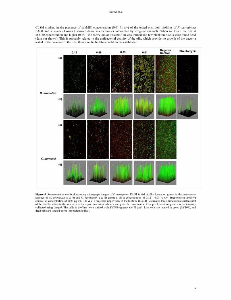
The inhibition on biofilm formation and breakdown of Cinnamomum and Massoia essential oils was confirmed by
confocal laser scanning microscope (CLSM) analysis, along with LIVE/DEAD staining for monitoring live/dead cells. From CLSM observations, we observed an inhibition of biofilm formation and a breakdown of an established biofilm for both bacteria when essential oils were applied.
At the highest tested concentration (0.12 % v/v) of the essential oils, a clear inhibitory effect was observed for both
biofilms of P. aeruginosa and S. aureus (Figure 4 and Figure 5). The biofilm formed in the presence of 0.12 % v/v oil was less dense and consisted of scattered cell clusters compared to the normal biofilm without the presence of oil. At the lowest concentration tested (0.01 % v/v), both oils of Massoia and Cinnamomum failed to inhibit the biofilm formation. From
Pratiwi et al
6
CLSM studies, in the presence of subMIC concentration (0.01 % v/v) of the tested oils, both biofilms of P. aeruginosa PAO1 and S. aureus Cowan I showed dense microcolonies intersected by irregular channels. When we tested the oils at MIC50 concentration and higher (0.25 – 0.5 % v/v) no or little biofilm was formed and few planktonic cells were found dead (data not shown). This is probably related to the antibacterial activity of the oils, which provide no growth of the bacteria tested in the presence of the oils, therefore the biofilms could not be established.
Figure 4. Representative confocal scanning micrograph images of P. aeruginosa PAO1 initial biofilm formation grown in the presence or absence of M. aromatica (a & b) and C. burmannii (c & d) essential oil at concentration of 0.12 – 0.01 % v/v; Streptomycin (positive control) at concentration of 1024 µg mL-1; (a & c) : projected upper view of the biofilm, (b & d) : estimated three-dimensional surface plot of the biofilm refers to the total area in the x-y-z dimension, where x and y are the coordinates of the pixel positioning and z is the intensity collected using ImageJ. The cells in biofilms were stained with SYTO9 (green) and PI (red). Live cells are labeled in green (SYT09), and dead cells are labeled in red (propidium iodide).

Essential oil antibiofilm activity
7
Figure 5. Representative confocal scanning micrograph images of S. aureus Cowan I initial biofilm formation grown in the presence or absence of M. aromatica (a & b) and C. burmannii (c & d) essential oil at concentration of 0.12 – 0.01 % v/v ; Streptomycin (positive control) at concentration of 1024 µg mL-1; (a & c) : projected upper view of the biofilm, (b & d) : estimated three-dimensional surface plot of the biofilm refers to the total area in the x-y-z dimension, where x and y are the coordinates of the pixel positioning and z is the intensity collected using ImageJ. Live cells are labeled in green (SYT09), and dead cells are labeled in red (propidium iodide).
The similar characteristic result was also observed when the oil activity was tested for disruption of existing biofilms (Figure 6 & 7). At higher concentration of 0.25 – 0.5 % v/v, the biofilms largely disappeared and a few dead cell clusters were evident (data not shown). At lower concentrations (0.12 % v/v), the oils have less capability in disrupting the preformed biofilm as shown by surviving cell clusters and dispersion on the glass slide surface. The oils were least effective at concentration of 0.01 % v/v against the adherent cells. In the presence of oil at concentration of 0.01 % v/v the microcolonies within the biofilm are more compact and formed layers which increase the thickness of the biofilm developed on the glass side surface.

Pratiwi et al
8
Figure 6. Representative confocal scanning micrograph images of biofilm breakdown activity of M. aromatica (a & b) and C. burmannii (c & d) essential oil at concentration of 0.12 – 0.01 % v/v against P. aeruginosa PAO1; Streptomycin (positive control) at concentration of 128 µg mL-1 ; (a & c) : projected upper view of the biofilm, (b & d) : estimated three-dimensional view of the biofilm refers to the total area in the x-y-z dimension, where x and y are the coordinates of the pixel positioning and z is the intensity collected using ImageJ. Live cells are labeled in green (SYT09), and dead cells are labeled in red (propidium iodide).

Essential oil antibiofilm activity
9
Figure 7. Representative confocal scanning micrograph images of biofilm breakdown activity of M. aromatica (a & b) and C. burmannii (c & d) essential oil at concentration of 0.12 – 0.01 % v/v against S. aureus Cowan I; Streptomycin (positive control) at concentration of 128 µg mL-1 ; (a & c) : projected upper view of the biofilm, (b & d) : estimated three-dimensional view of the biofilm refers to the total area in the x-y-z dimension, where x and y are the coordinates of the pixel positioning and z is the intensity collected using ImageJ. Live cells are labeled in green (SYT09), and dead cells are labeled in red (propidium iodide).
The principal components of the oils of M. aromatica and C. burmannii were determined by using gas chromatography
and were identified by comparing the mass spectra of chemical compounds in essential oils with library mass spectra from NIST02 (www.nist.gov/index.html) The identification of the essential oils chemical constituents was assigned on the basis of comparison of their retention indices mass spectra with data published in the literature. Kovats retention Index of C. burmannii and M. aromatica oil by isothermal GC were shown in Table 4. The estimated dead-time was at 1.82 minutes with a very good coefficient of correlation 0.9992. These calculations particularly involved approximately 25 iterations. N-alkanes homologous series was used not only as standards described by Kovats but also for estimating dead-time simultaneously. The obtained Kovats Retention Index for C. burmannii oil and M. aromatica oil are 1123.46, 1168.63 respectively. In this study, the analysis by GC-MS showed cinnamaldehyde (92.02 %) to be the major component of C. burmannii essential oil, whereas Massoialactone (92.05 %) was the main constituent of M. aromatica essential oil. The chemical composition of the volatile oils from C. burmannii and M. aromatica obtained from GC-MS were described in Table 3; Figure 8 & 9.

Pratiwi et al
10
Table 3. Major chemical constituents of C. burmannii and M. aromatica essential oil as identified by GC-MS
Plant sample
Peak Retention time (RT)
Kovats Retention Index (RI)
Area Area (%)
Similarity index (SI)
Chemical Component
C. burmannii 3 19.296 1123.459 164665734 92.02 95 Cinnamaldehyde
5 21.630 1142.506 7342238 4.10 93 Alpha-Copaene
7 23.472 1156.016 3708184 2.07 91 3-Phenyl-2-prophenyl acetate
M. aromatica 3 25.346 1168.629 156590945 92.05 94 5-Hydroxy-2-Decenoic acid lactone
(Massoialactone) 4 29.961 1195.847 10522375 6.19 86 5-Hydroxy-2-Decenoic
acid lactone (Massoialactone) *
*The compound obtained is predicted to be a derivate of 5-Hydroxy-2-Decenoic acid lactone (Massoialactone) with higher boiling point and have the similar fragmentation pattern (86%) to Massoialactone.
Table 4. Kovats retention Index of C. burmannii and M. aromatica oil by isothermal GC on a AGILENT J&W DB-1 GC capillary column.
Estimation of Coloumn Dead-time (to)
Estimated dead time : 1.81510 Estimated Coef Corr Semi-log plot : 0.99915 Calculated Slope Semi-log plot : 0.00658 Calculated Intercept Semi-log plot : -4.53096
n-Alkane Carbon Number Retention Time (min) Adjusted Retention Time
(calculated/min) k'
Hexane 6 2.381 0.558 0.311
Heptane 7 2.850 1.077 0.570
Oktane 8 3.899 2.080 1.148
Nonane 9 6.098 4.018 2.359
Decane 10 9.281 7.758 4.113
Estimation of Kovats Index
Analyte Retention Time
(min) Adjusted Retention Time
(min) k' Retention Index
C. burmannii 19.296 17.480 09.630 1123.459
M. aromatica 25.346 23.530 12.963 1168.629

Essential oil antibiofilm activity
11
Figure 8. C. burmannii oil GC-MS profile
Figure 9. M. aromatica oil GC-MS profile
DISCUSSION
The result obtained from this study showed that the antibacterial and antibiofilm activity of plant essential oils varied for
the studied microorganisms. It is very likely that different compounds within the essential oils are responsible for the inhibitory effect on biofilm formation and biofilm breakdown activity. Many plant essential oils, which are mixtures of numerous organic chemicals, contain compounds that inhibit microbial growth. Some antimicrobial agents at sub-inhibitory concentrations could influence bacterial virulence factors, such as adherence, cell surface hydrophobicity, biofilm formation, sensitivity to oxidative stress and motility (Cowan, 1999; Nadoushani et al., 2010).
Essential oils, which are mixtures of numerous organic chemicals, are biosynthesized by plants and their anti-microbial properties, their constituents and their mechanism of action have been extensively studied (Bagamboula et al., 2004). Phenolic compounds such as eugenol and cinnamaldehyde are present in essential oils of many plants and are proved to be active against many pathogenic bacteria and fungi (Suresh et al., 1992; Pacheco et al., 1993; Ooi et al., 2006; Chami et al., 2004; Bennis et al. 2004).
From this study we found out that Cinnamon and Massoia oils possess the antibacterial and antibiofilm activity against the studied microorganisms. It is very likely that different compounds within the essential oils are responsible for the inhibitory effect on biofilm formation and biofilm breakdown activity. Many plant essential oils, which are mixtures of numerous organic chemicals, contain compounds that inhibit microbial growth. Some antimicrobial agents at sub-inhibitory concentrations could influence bacterial virulence factors, such as adherence, cell surface hydrophobicity, biofilm formation, sensitivity to oxidative stress and motility (Cowan et al., 1999; Bennis et al., 2004).
Up to now, there is no information in the literature regarding the influence of sub-PMIC essential oils of M. aromatica Becc. on inhibition and breakdown of P. aeruginosa PAO1 and S. aureus Cowan I biofilm. However, a study has been conducted on activity of cinnamaldehyde, the main compound of C. burmannii Nees ex Bl. essential oil, against S. aureus biofilm (Jia et al., 2011).
Jia et al. (2011) showed that cinnamaldehyde was able to kill S. aureus and detach existing biofilms. Also it was found that the expression of sarA, encoding a staphylococcal accessory regulator which is a central regulatory element that controls the

Pratiwi et al
12
production of virulence factors and is essential for biofilm development of S. aureus, was decreased upon exposure to sub-PMICs of cinnamaldehyde.
In the quantification measurement of (static) biofilms, crystal violet (CV) staining has been widely used as a indirect methods with 96-well microtiter plates (Burt, 2004; Mastelic et al., 2005). The major disadvantage is that CV, a basic dye which binds to negatively charged surface molecules and polysaccharides in the extracellular matrix, stains both living and dead cells. Therefore it is difficult to evaluate killing of biofilm cells (Burton et al., 2007).
In order to gain a clear view about the effect of essential oils on P. aeruginosa and S. aureus biofilm, the Live/Dead staining was performed and viewed using CLSM. Examining the permeability of a compound through biofilms using CLSM and Live/Dead staining capable to show the dead organisms which are differentiated from the live ones with the respective labeling of SYTO9 and propidium iodide (PI) (Djordjevic et al., 2012).
We observed inhibiting activity on biofilm formation when we inoculate the biofilm together with the essential oils using concentration lower than concentration for 50% inhibition of planktonic growth (0.01 - 0.12 % v/v). We found that the essential oil activity against the biofilm formation was concentration dependent. In the presence of the oil, biofilms of both bacterial tested was poorly developed and most of the cells were eventually killed (Table 4).
The concentration of plant essential oils needed to degrade preformed biofilms was higher than the one needed to inhibit the initial attachment. The reduced susceptibility of bacteria in biofilms is thought to be due to a combination of several factors such as the presence of extracellular polymer substances (EPS) surrounding the biofilm cells. The antimicrobial agents is absorb onto the EPS and effectively diluted in its concentration before it reaches the individual cells in the biofilm (Peeters et al., 2008; Takenaka et al., 2001).
The cinnamaldehyde and eugenol in the C. burnamii essential oil has been proven to have antibacterial and antifungal activity (OOi et al., 2006). These two compounds and massoia lactone as the main compound of M. aromatica essential oil might have an influence in inhibiting biofilm formation of P. aeruginosa PAO1 and S. aureus Cowan I.
CONCLUSION
According to the findings of this study, the essential oils of M. aromatica and C. burmannii have antibiofilm formation and
biofilm breakdown activity against P. aeruginosa PAO1 and S. aureus Cowan I. The present study suggests that the essential oils of these plants are a potential source of natural antibiofilm agents. After this screening process, further work on phytochemical characterization will be performed to identify and isolate active constituents responsible for the antibiofilm activity.
ACKNOWLEDGMENTS
We gratefully acknowledge the funds support of this research by the Indonesian Directorate General for Higher Education. We thank Gerda Lammers for technical assistance in CLSM, and Dr. Endang Lukitaningsih for sharing her expertise in GC-MS data interpretation.
REFERENCES
Bagamboula CF, Uyttendaele M, Debevere J 2004. Inhibitory effect of thyme and basil essential oils, carvacrol, thymol, estragol, linalool and
p-cymene towards Shigella sonnei and S. flexneri. Food Microbiol 21. 33–42. Bennis S, Chami F, Chami N, Bouchikhi T, Remmal A 2004. Surface alteration of Saccharomyces cerevisiae induced by thymol and
eugenol. Lett App Microbiol 38. 454-458. Burt S 2004. Essential oils: their antibacterial properties and potential applications in foods—a review. Int J Food Microbiol 94. 223-253. Burton E, Yakandawala N, LoVetri K 2007. A microplate spectrofluorometric assay for bacterial biofilm. J Ind Microbiol Biotechnol 34. 1-4. Chami N, Chami F, Bennis S, Trouillas J, Remmal A 2004. Antifungal treatment with carvacrol and eugenol of oral candidiasis in
immunosuppressed rats. Braz J Infect Dis 8. 217–226. Clardy J, Walsh C 2004. Lessons from natural molecules. Nature 432. 829-837. Clinical and Laboratory Standard Institute (CLSI) 2007. Performance Standards for Antimicrobial Susceptibility Testing: Seventeenth
Informational Supplement. CLSI document M100-S17. Clinical and Laboratory Standard Institute, 940 West Valley Road, Suite 1400, Wayne, PA 19087-1898 USA. 2007.
Cowan M 1999. Plant products as antimicrobial agents. Clin Microbiol 12. 564-582. Djordjevic D, Wiedmann M, McLandsborough LA 2002. Microtiter plate assay for assessment of listeria monocytogenes biofilm formation.
Appl Environ Microbiol 68. 2950–2958. Donlan RM, Costerton JW 2002. Biofilms: survival mechanisms of clinically relevant microorganism. Clin Microbiol Rev 15. 167–193. Dusane DH, Dam S, Nancharaiah YV, Kumar AR, Venugopala VP, Zonjarde SS 2012. Disruption of Yarrowia lipolytica biofilms by
rhamnolipid biosurfactant. Aquatic Biosystems 8 (1). 17. Furr HC 1989. Calculation of chromatographic dead times and retention indices with the aid of a computer program in BASIC. J
Chromatograph Sci 27 (5). 216-220. Guardino X, Albaiges J, Firpo G, Rodriguez-Vinals R, Gassiot M 1976. Accuracy in the determination of the kovats retention index :
mathematical dead time. J Chromatogr 18. 13-22. Hadizadeh I, Peivastega B, ,Hamzehzarghani H 2009. Antifungal activity of essential oils from some medicinal plants of iran against
Alternaria alternate. Am J App Sci 6. 857-861. Harrison JJ, Turner RJ, Ceri H 2005. Persister cells, the biofilm matrix and tolerance to metal cations in biofilm and planktonic Pseudomonas
aeruginosa. Environ Microbiol 7. 981–994. Idroes R, Oesman F 2011. Dead time determination and its influence to retention index in gc system using polar capillary column. Indonesia J
Pharm 22 (3). 257-264. Indonesian National Health Department 1985. Tanaman obat Indonesia Vol 1. Jakarta : Directorate General of Drug and Food Control,
Ministry of Health, Indonesia.

Essential oil antibiofilm activity
13
Jia P, Xue YJ, Duan XJ, Shao SH 2011. Effect of cinnamaldehyde on biofilm formation and sara expression by methicillin-resistant Staphylococcus aureus. Lett App Microbiol 53. 409-416.
Jin Y, Zhang T, Samaranayake YH, Fang HH, Yip HK, Samaranayake LP 2005. The use of new probes and stains for improved assessment of cell viability and extracellular polymeric substances in Candida albicans biofilms. Mycopathologia 159. 353-360.
Kovats E 1968. Zu fragen de polaritat. die method der linearkombination der wechselwirkungskrafte. Chimia 22. 459-66. Mastelic J, Politeo O, Jerkovic I, Radosevic N 2005. Composition and antimicrobial activity of Helichrysum italicum essential oil and its
terpene and terpenoid fractions. Chem Nat Comp 41. 35-40. Nadoushani MS, Oulia P, Najafabadi ML, Rasouli I, Saderi H, Salari MH 2010. Effects of sub-inhibitory concentrations of essential oils of
Mentha spicata and Cumminum cyminum on virulence factors of Pseudomonas aeruginosa. J Med Plants 9. 124-130. Nostro A, Roccaro AS, Bisignano G, Marino A, Cannatelli MA, Pizzimenti FC, Cioni PL, Procopio F, Blanco AR 2007. Effects of oregano,
carvacrol and thymol on Staphylococcus aureus and Staphylococcus epidermidis biofilms. J Med Microbiol 56. 519-523. Ooi LS, Li Y, Kam SL, Wang H, Wong EY, Ooi VE 2006. Antimicrobial activities of cinnamon oil and cinnamaldehyde from the Chinese
medicinal herb Cinnamomum cassia Blume. Am J Chin Med 34. 511-22. O'Toole G, Kaplan HB, Kolter R 2000. Biofilm formation as microbial development. Annu Rev Microbiol 54. 49-79. Pacheco P, Sierra J, Schmeda-Hirschmann G. Potter CW, Jones BM, Moshref M 1993. Antiviral activities of Chilean medicinal plant extracts.
Phytother Res 7. 415-418. Peeters E, Nelis HJ, Coenye T 2008. Comparison of Multiple Methods For Quantification of Microbial Biofilms Grown In Microtiter Plates. J
Microbiol Methods 72. 157–165. Pirbalouti AG, Moosavi SH, Momtaz H, Rahimi E 2010. Antibacterial activities of the essential oils of some iranian herbs against
Campylobacter jejuni and Campylobacter coli. Adv Food Sci 32. 30-34. Quave CL, Plano LRW, Pantuso T, Bennett BC 2008. Effects of extracts from Italian medicinal plants on planktonic growth, biofilm
formation and adherence of methicillin-resistant Staphylococcus aureus. J Ethnopharmacol 118. 418–428. Rali T, Wossa SW, Leach DN 2007. Comparative chemical analysis of the essential oil constituents in the bark, heartwood and fruits of
Cryptocarya massoy (Oken) Kosterm. (Lauraceae) From Papua New Guinea. Molecules 12. 149-154. Reichling J, Schnitzler P, Suschke U, Saller R 2009. Essential oils of aromatic plants with antibacterial, antifungal, antiviral and cytotoxic
properties - an overview. Forsch Komplementmed 16. 79-90. Shan B, Cai YZ, Brooks JD, Corke H 2007. Antibacterial properties and major bioactive components of cinnamon stick (Cinnamomum
burmannii): activity against foodborne pathogenic bacteria. J Agric Food Chem 55. 5484–90. Smith RJ, Haken JK, Wainwright MS 1985. Estimation of dead time and calculation of kovats indices. J Chromatog 334. 95-127. Suresh P, Ingle VK, Lakshmi VV 1992. Antibacterial activity of eugenol in comparison with other antibiotics. J Food Sci Technol 29. 254-
256. Takenaka S, Iwaku M, Hoshino E 2001. Artificial Pseudomonas aeruginosa biofilms and confocal laser scanning microscopic analysis. J
Infect Chemother 7. 87–93. Wu X, Zhao MM, Wang JS, Cui C, Wu JW, Yang B 2008. Effects of cooking conditions on sensory characteristics of red-cooked beef flavor
and identification of the flavor compounds. J Food Process Eng 31. 51–65.
Copyright © 2022 FDOKUMEN


![Bl. Odorik z Pordenone - 680 let od smrti († 14. ledna 1331) - prezentace [Bl. Odoric of Pordenone - 680 years since his death († 14 January 1331) - A presentation]](https://static.fdokumen.com/doc/165x107/6323300061d7e169b00cf6f6/bl-odorik-z-pordenone-680-let-od-smrti-14-ledna-1331-prezentace-bl.jpg)