Effect of aerobic training on ventilatory muscle endurance of spinal cord injured men
6
Eect of aerobic training on ventilatory muscle endurance of spinal cord injured men Antoˆnio Carlos Silva 1 , Jose´ Alberto Neder 1 , Mı´riam Vera Chiurciu 1 , Denise da Cunha Pasqualin 1 , Regina Cintra Querino da Silva 1 , Ana Cla´udia Fernandez 1 , Fla´vio Antoˆ nio Ascaˆnio Lauro 1 , Marco Tu´ lio de Mello 3 and Se´rgio Tufik 2 1 Department of Physiology, and 2 Department of Psychobiology, Universidade Federal de Sa ˜o Paulo - Escola Paulista de Medicina, UNIFESP-EPM, Sao Paulo SP, Brazil; 3 Universidade Federal de Goia ´s, Catala ˜o GO, Brazil The functional consequences of ventilatory muscle impairment of spinal cord injured (SCI) subjects has been evaluated through spirometric and maximal respiratory pressure tests. Nevertheless, underlying functional abnormalities may be evident only under dynamic conditions, such as with a ventilatory muscle endurance test (VME). In order to evaluate the VME of thoracic SCI men and the eect of physical training on it we evaluated 12 SCI subjects (Group I) and 12 able-bodied controls (Group II). The subjects were submitted to clinical evaluation, spirometry, maximum voluntary ventilation in 12 s (MVV-12sec) and a test of VME – the highest time of sustained ventilation at 70% of the maximum voluntary ventilation in isocapnic conditions (MVV-70% time). Gr. II was evaluated before and after an arm cranking aerobic training program (30 min/session, three times/week, 6 weeks) with training target heart rate corresponding to ventilatory anaerobic threshold. On the initial evaluation, Gr. I subjects presented a significantly reduced forced vital capacity (FVC), forced expiratory volume in 1 s (FEV 1 ) and MVV-12 sec when compared to controls (P50.05). Also, the VME was severely reduced in Group I (median, ranges; 1.15, 0.61 – 12.22) when compared to Group II (14.60, 1.20 – 15.00) – P50.001. When Gr. I subjects were separated by the level of lesion, the VME was lower in high injured (T1 – T7) than intermediate (T8 – T10) and low injured patients (T11 – T12) – P50.05. After aerobic training, Group I subjects incremented significantly the FVC (P50.05) and the VME (P50.001), so that MVV-70% time values post- training were not dierent from the initial values of the Gr. II. In conclusion, (i) the VME of thoracic SCI men was severely reduced when compared to able-bodied controls; (ii) a 6-weeks arm cranking aerobic training program was ecient to normalize the VME of SCI subjects. Keywords: spinal cord injury; pulmonary function testing; paraplegia; rehabilitation; aerobic training Introduction Spinal cord injured (SCI) subjects frequently show a reduction in pulmonary volumes (a restrictive pattern on spirometry) and in maximal static respiratory pressures, which are correlated with the functional loss of ventilatory muscles. 1–3 Therefore, the assess- ment of ventilatory impairment in SCI subjects has been made with easily obtainable functional indices, such as the forced vital capacity (FVC) and the maximal inspiratory pressure (MIP). 4 Few studies have approached the dynamic ventila- tory performance of SCI individuals. 5,6 Vital capacity and its subdivisions are relatively insensitive indicators of respiratory muscle weakness. Indeed, respiratory muscle strength has to fall below half normal before the vital capacity is significantly impacted. 6 Addition- ally, SCI subjects may show mechanical alterations during prolonged endurance activities that predispose to muscular fatigue, and such changes could not be detectable by functional resting evaluation. 6 Ventilatory muscle endurance (VME) can be assessed by dierent tests, which vary in complexity and the necessity for technical expertise. 6,7 A simple and reliable test of VME is the longest time that a subject is able to voluntarily sustain 70% of the maximum breathing capacity or maximum voluntary ventilation (70%-MVV time). 8 This ventilatory in- tensity is close to the level of ventilation available for prolonged ventilation at maximal exercise in young healthy subjects. 9,10 To the best of our knowledge, no previous study has analyzed 70%-MVV time in SCI subjects with dierent levels of injury. Thus, the objectives of the present study were (a) to compare the VME (as indicated by the 70%-MVV Correspondence: A C Silva, Depto. de Fisiologia/Laborato´rio de Fisiologia Respirato´ria e do Exercı´cio (LAFIREX)- Rua Botucatu, 862, 58 andar. CEP: 04023-900. Sao Paulo SP, Brazil *Dr. Neder is supported by a Post-doctoral research fellowship grant from FAPESP, Brazil Spinal Cord (1998) 36, 240 – 245 1998 International Medical Society of Paraplegia All rights reserved 1362 – 4393/98 $12.00 http://www.stockton-press.co.uk/sc
Transcript of Effect of aerobic training on ventilatory muscle endurance of spinal cord injured men
E�ect of aerobic training on ventilatory muscle endurance of spinal cordinjured men
Antoà nio Carlos Silva1, Jose Alberto Neder1, Mõ riam Vera Chiurciu1, Denise da Cunha Pasqualin1,Regina Cintra Querino da Silva1, Ana Cla udia Fernandez1, Fla vio Antoà nio Ascaà nio Lauro1, Marco Tu lio de Mello3
and Se rgio Tu®k2
1Department of Physiology, and 2Department of Psychobiology, Universidade Federal de SaÄo Paulo - Escola Paulistade Medicina, UNIFESP-EPM, Sao Paulo SP, Brazil; 3Universidade Federal de GoiaÂs, CatalaÄo GO, Brazil
The functional consequences of ventilatory muscle impairment of spinal cord injured (SCI)subjects has been evaluated through spirometric and maximal respiratory pressure tests.Nevertheless, underlying functional abnormalities may be evident only under dynamicconditions, such as with a ventilatory muscle endurance test (VME). In order to evaluate theVME of thoracic SCI men and the e�ect of physical training on it we evaluated 12 SCIsubjects (Group I) and 12 able-bodied controls (Group II). The subjects were submitted toclinical evaluation, spirometry, maximum voluntary ventilation in 12 s (MVV-12sec) and a testof VME± the highest time of sustained ventilation at 70% of the maximum voluntaryventilation in isocapnic conditions (MVV-70% time). Gr. II was evaluated before and after anarm cranking aerobic training program (30 min/session, three times/week, 6 weeks) withtraining target heart rate corresponding to ventilatory anaerobic threshold. On the initialevaluation, Gr. I subjects presented a signi®cantly reduced forced vital capacity (FVC), forcedexpiratory volume in 1 s (FEV1) and MVV-12 sec when compared to controls (P50.05). Also,the VME was severely reduced in Group I (median, ranges; 1.15, 0.61 ± 12.22) when comparedto Group II (14.60, 1.20 ± 15.00) ±P50.001. When Gr. I subjects were separated by the levelof lesion, the VME was lower in high injured (T1 ±T7) than intermediate (T8 ±T10) and lowinjured patients (T11 ±T12) ±P50.05. After aerobic training, Group I subjects incrementedsigni®cantly the FVC (P50.05) and the VME (P50.001), so that MVV-70% time values post-training were not di�erent from the initial values of the Gr. II. In conclusion, (i) the VME ofthoracic SCI men was severely reduced when compared to able-bodied controls; (ii) a 6-weeksarm cranking aerobic training program was e�cient to normalize the VME of SCI subjects.
Keywords: spinal cord injury; pulmonary function testing; paraplegia; rehabilitation; aerobictraining
Introduction
Spinal cord injured (SCI) subjects frequently show areduction in pulmonary volumes (a restrictive patternon spirometry) and in maximal static respiratorypressures, which are correlated with the functionalloss of ventilatory muscles.1 ± 3 Therefore, the assess-ment of ventilatory impairment in SCI subjects hasbeen made with easily obtainable functional indices,such as the forced vital capacity (FVC) and themaximal inspiratory pressure (MIP).4
Few studies have approached the dynamic ventila-tory performance of SCI individuals.5,6 Vital capacityand its subdivisions are relatively insensitive indicatorsof respiratory muscle weakness. Indeed, respiratorymuscle strength has to fall below half normal before
the vital capacity is signi®cantly impacted.6 Addition-ally, SCI subjects may show mechanical alterationsduring prolonged endurance activities that predisposeto muscular fatigue, and such changes could not bedetectable by functional resting evaluation.6
Ventilatory muscle endurance (VME) can beassessed by di�erent tests, which vary in complexityand the necessity for technical expertise.6,7 A simpleand reliable test of VME is the longest time that asubject is able to voluntarily sustain 70% of themaximum breathing capacity or maximum voluntaryventilation (70%-MVV time).8 This ventilatory in-tensity is close to the level of ventilation available forprolonged ventilation at maximal exercise in younghealthy subjects.9,10 To the best of our knowledge, noprevious study has analyzed 70%-MVV time in SCIsubjects with di�erent levels of injury.Thus, the objectives of the present study were (a) to
compare the VME (as indicated by the 70%-MVV
Correspondence: A C Silva, Depto. de Fisiologia/Laborato rio deFisiologia Respirato ria e do Exercõ cio (LAFIREX)- Rua Botucatu,862, 58 andar. CEP: 04023-900. Sao Paulo SP, Brazil*Dr. Neder is supported by a Post-doctoral research fellowship grantfrom FAPESP, Brazil
Spinal Cord (1998) 36, 240 ± 245 1998 International Medical Society of Paraplegia All rights reserved 1362 ± 4393/98 $12.00
http://www.stockton-press.co.uk/sc

time test) of complete spinal cord injured and able-bodied physically active men and (b) evaluate thee�ect of an arm cranking aerobic training program onthe VME of SCI subjects.
Methods
SubjectsTwelve wheelchair-dependent SCI men (ASIA ClassA,11 with complete sensory and motor block below thelevel of the injury - Group I) and twelve able-bodiedcontrols (Group II) matched for age, body mass andtraining status participated in this study. All partici-pants were non-smokers. Healthy medical studentspersonally volunteered for the control group.All subjects in Group I were paraplegic, with the
level of injury between T1 and T12. Etiology of injurywas gunshot wound in eight subjects, road and tra�caccident in three and one patient had a fall. All lesionshad existed for at least 3 years. They were separatedinto 3 groups according to an anatomical andfunctional criteria of ventilatory muscle impairment:high thoracic injury, with paralysis of intercostalmuscles in di�erent degrees and total paralysis ofabdominal and pelvic muscles (T1 ±T7, n=4); inter-mediate thoracic injury, with paralysis of abdominalmuscles in di�erent degrees and total paralysis ofpelvic muscles (T8 ±T10, n=5); low thoracic injury,with paralysis of low abdominal and pelvic muscles(T11 ±T12, n=3).12
ProceduresEach test session began with the completion of anidenti®cation and subjective information sheet. Thefollowing questions were asked and recorded by theinvestigator: (1) level of habitual physical activity(Saltin and Grimsby questionnaire, modi®ed);13 (2)etiology and time since injury; (3) actual or pastillnesses or interventions; (4) smoking history.Subjects of Group I were weighed in the sitting
position on a hospital scale. All subjects underwent acomprehensive medical examination and none of thesubjects had any cardiovascular disease or usedmedicines likely to a�ect the results. All participantsgave informed consent for participation in the study.The following protocol was carried out in all
subjects, after approbation of Ethical Committee ofUNIFESP-EPM.
Spirometry Spirometric tests were performed using acalibrated 10-liters Collins Survey Spirometer (Collins,Braintree MA, USA). The subjects completed at leastthree acceptable maximal expiratory maneuvers. ForGroup I the tests were conducted with subjects sittingin their wheelchairs. According to standard protocolsof the American Thoracic Society, 199114 fortechnique, acceptability and reproducibility the follow-ing spirometric variables were recorded and expressed
in BTPS conditions: forced vital capacity (FVC, L);forced expiratory volume in 1 s (FEV1, L), FEV1/FVCratio (%), forced expiratory ¯ow between 25 and 75%of the FVC (FEF25 ± 75%, L.sec
71) and peak expiratory¯ow rate (PEFR, L.sec71). Predicted normal values forall spirometric variables were those of Knudson et al15.
Maximal voluntary ventilation (MVV-12sec) Themaximum voluntary ventilation (MVV) is the largestvolume that can be breathed into and out of the lungsduring 10 ± 15 s interval with voluntary e�ort. In thisstudy, the MVV was measured by having the subjectsitting in his wheelchair and breathing deeply (with avolume greater than the tidal volume but lower thanthe vital capacity) and rapidly for a 12 s interval in the10-liters Collins Survey Spirometer (Collins, BraintreeMA, USA). The values were recorded in L.min71
(BTPS), by extrapolating the 12 s accumulated volumeto 1 min. Predicted normal values were those ofCherniack and Raber.16
Ventilatory muscle endurance (70%-MVV time) Tenminutes after the MVV-12sec test, the ventilatorymuscle endurance was measured as the highest timethat the subject was able to voluntarily sustain a targetventilation corresponding to 70% of his MVV-12secvalue (70%-MVV time).8
Ventilation was measured breath-by-breath byelectronic integration and summation of the signalsby a ¯ow-volume module of a computerized metaboliccart (Vista TurboFit, Vacumed, Ventura CA, USA).The actual ventilation was continuously displayed on-line in a video monitor of a host computer, while thetarget ventilation for each individual (70% of hisMVV-12sec) was being identi®ed as a row to befollowed.Hypocapnia was avoided during forced ventilatory
maneuvers by the use of partial rebreathing of expiredair, using a hose connected to a mixing chamber(volume of 5 L). Expired air was continuouslymonitored (Gas Analyzer Set, Vacumed, VenturaCA, USA) from a sample line connected to themouthpiece. The expired fraction of carbon dioxide(FECO2) was maintained in a narrow range (5+1%).The subjects wore noseclips and a standard posture
was adopted: sitting in their wheelchair (Group I) withthe trunk supported by the backrest, avoiding anytrunk stabilization or anchorage of accessory ventila-tory muscles. The participants were instructed tobreathe with any pattern or rhythm that theypreferred and were given an active standard encour-agement to maintain the target ventilation as long aspossible.SVV-70% time was measured from the moment that
target ventilation was reached; whenever the subjectwas unable to reach it within the ®rst minute of thetest, it was interrupted and another attempt wasperformed after 10 min. The test was ended whenthe subject was not able to sustain the targetventilation, with a reduction higher than 10%.
Ventilatory muscle endurance in SCI menAC Silva et al
241

Subsequently they were asked about the nature of thelimiting symptoms.
Exercise testing and training program Prior to sessionsof training, all SCI subjects underwent a cardiopul-monary exercise testing on a computerized metabolicgas exchange system (Vista Turbo Fit, Vacumed,Ventura CA, USA) using a calibrated electromagneti-cally-braked arm crank ergometer (Cybex MET-300,Cybex, USA).The data were calculated automatically using
standard formulae and displayed in descriptivenumerical and graphical forms (average of 20 s). Thefollowing data were obtained: oxygen uptake,mL.min71 STPD); carbon dioxide production(VCO2, mL.min
71 STPD); respiratory exchange ratio(RER); minute ventilation (VE, L.min71 BTPS),respiratory rate (f, bpm); ventilatory equivalent forO2 and CO2 (VE/VO2 and VE/VCO2); expired fractionof O2 and CO2 (FEO2 and FECO2, mmHg); heart rate(HR, bpm) and oxygen pulse (VO2/HR, mL/beat).VO2 at the anaerobic threshold (VO2AT) wasestimated by the ventilatory method, when VE/VO2
and FEO2 increased while VE/VCO2 and FECO2
remained stable. In this study the VO2AT wasidenti®ed in all of the patients being studied.The supervised arm cranking aerobic training
program consisted of 30 min sessions, three times aweek for 6 weeks with the training target heart ratecorresponding to ventilatory anaerobic threshold.
Statistical analysisThe values are reported as median and ranges. Thefollowing statistical tests were used: (a) Mann ±Whitney two sample test to compare the unpaireddata of the two groups in the initial evaluation; (b)Wilcoxon's test to evaluate the di�erences between pre-and post-training paired values of the Group I; (c) OneWay Analysis of Variance (ANOVA) with Bonferroni'sP correction in the comparison of the 70%-MVV timevalues among groups. Level of statistical signi®cancewas always set at P50.05.
Results
Group I patients had median (range) values of age of31 years (22 ± 54 years) and body mass of 75.3 kg(65.8 ± 82.5); Group II able-bodied had median value ofage of 30 years (22 ± 52 years) and body mass of73.8 kg (67.2 ± 80.0) - P40.05. All participants wereclassi®ed as moderately active to active, with physicalactivity score higher than three (i.e. habitual exercise atleast 3 h/week).13
Spirometric, MVV-12sec and 70%-MVV timevalues in the initial evaluation for both groups arereported on Table 1. We found a signi®cant reductionin Group I for all studied variables, with exception ofFEV1/FVC ratio. The spirometric abnormalitiesfollowed a restrictive pattern in ®ve individuals (four
with high thoracic injury) being 3 graded as mild and 2as moderate.14 Although median values were reducedwhen compared to Group II, the spirometric andMVV-12sec values of Group I were higher than 80%of predicted15,16 for the FVC in 7/12, for the FEV1 in9/12 and for the MVV-12sec in all patients (Figure 1).The decrease in the median 70%-MVV time was
severe in Group I (1.15 min; 0.61 ± 12.22) compared toGroup II (14.60 min; 1.20 ± 15.00) - Table 1. When thepatients of Group I were separated by the level ofinjury (see Methods) the VME was lower in highinjured patients (T1 ±T7: 0.65 min; 0.61 ± 0.71) com-pared to intermediate (T8 ±T10: 1.96; 0.72 ± 3.90) and
Table 1 Results of the functional evaluations of the Group Ithoracic SCI men (n=12) and of the Group II able-bodiedcontrols (n=12)*
Group I Group IIVariables Initial Final Initial
FVC(L)(% pred)
4.05{81{
4.40{90{
5.63103
FEV(L)(% pred)
3.62{84{
3.7088
4.46107
FEV1/FVC(%) 85 80 81
MVV-12sec(L.min71)(% pred)
175{124{
178126
212141
70%-MVV time(min) 1.15{ 9.10{ 14.60
*Values are displayed as median. See text for abbreviations.{Signi®cantly lower than Group II in the initial evaluation(P50.05 ±Mann-Whitney test). {Signi®cant di�erences be-tween initial and ®nal evaluations (P50.01 ±Wilcoxon test).Obs: post-training 70%-MVV time values of Group I werenot signi®cantly di�erent from the values of Group II in theinitial evaluation (P40.05; Mann-Whitney test)
Figure 1 Individual and median spirometric* and MVV-12sec** values in % of predicted (% pred) of the Group Ithoracic SCI men (n=12). *Predicted from Knudson et al.15
**Predicted from Cherniack and Raber.16 LLN: Lower limitof normality
Ventilatory muscle endurance in SCI menAC Silva et al
242
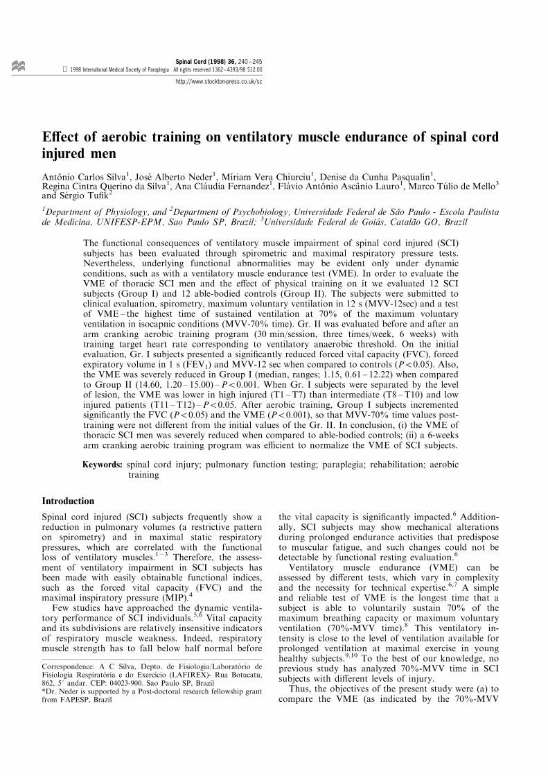
low (T11 ±T12: 4.50 min; 1.60 12.22) ones (Figure 2).However, when compared to Group II, the reductionon VME was signi®cant in both high and intermediateinjured patients (Figure 2). Individual analysis showedthat only one individual of Group I sustained 70% ofhis MVV for more than 10 min and none sustained for15 min; these ®ndings contrast with those from GroupII, where seven subjects sustained the target ventilationfor more than 10 min and ®ve individuals for 15 min(Figure 2).Analyzing the limiting symptoms at the end of 70%-
MVV test, nine subjects of Group I said that dyspneawas the main symptom, 2 said muscle pain ordiscomfort and one said general fatigue; this con-trasted with Group II where only two subjectscomplained of dyspnea at the end of the test, andnine subjects muscle pain or discomfort. No partici-pant from both groups related any symptomcompatible with hypocapnia.After arm cranking aerobic training, Group I
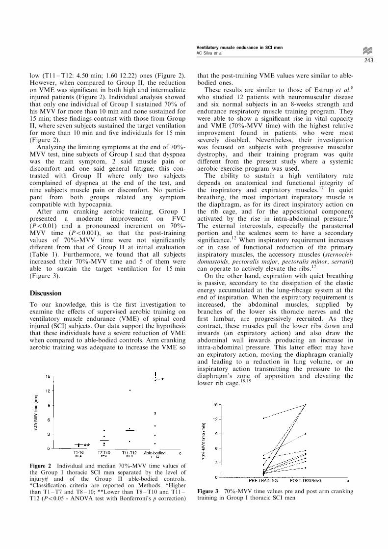
presented a moderate improvement on FVC(P50.01) and a pronounced increment on 70%-MVV time (P50.001), so that the post-trainingvalues of 70%-MVV time were not signi®cantlydi�erent from that of Group II at initial evaluation(Table 1). Furthermore, we found that all subjectsincreased their 70%-MVV time and 5 of them wereable to sustain the target ventilation for 15 min(Figure 3).
Discussion
To our knowledge, this is the ®rst investigation toexamine the e�ects of supervised aerobic training onventilatory muscle endurance (VME) of spinal cordinjured (SCI) subjects. Our data support the hypothesisthat these individuals have a severe reduction of VMEwhen compared to able-bodied controls. Arm crankingaerobic training was adequate to increase the VME so
that the post-training VME values were similar to able-bodied ones.These results are similar to those of Estrup et al.8
who studied 12 patients with neuromuscular diseaseand six normal subjects in an 8-weeks strength andendurance respiratory muscle training program. Theywere able to show a signi®cant rise in vital capacityand VME (70%-MVV time) with the highest relativeimprovement found in patients who were mostseverely disabled. Nevertheless, their investigationwas focused on subjects with progressive musculardystrophy, and their training program was quitedi�erent from the present study where a systemicaerobic exercise program was used.The ability to sustain a high ventilatory rate
depends on anatomical and functional integrity ofthe inspiratory and expiratory muscles.17 In quietbreathing, the most important inspiratory muscle isthe diaphragm, as for its direct inspiratory action onthe rib cage, and for the appositional componentactivated by the rise in intra-abdominal pressure.18
The external intercostals, especially the parasternalportion and the scalenes seem to have a secondarysigni®cance.12 When inspiratory requirement increasesor in case of functional reduction of the primaryinspiratory muscles, the accessory muscles (sternoclei-domastoids, pectoralis major, pectoralis minor, serratii)can operate to actively elevate the ribs.17
On the other hand, expiration with quiet breathingis passive, secondary to the dissipation of the elasticenergy accumulated at the lung-ribcage system at theend of inspiration. When the expiratory requirement isincreased, the abdominal muscles, supplied bybranches of the lower six thoracic nerves and the®rst lumbar, are progressively recruited. As theycontract, these muscles pull the lower ribs down andinwards (an expiratory action) and also draw theabdominal wall inwards producing an increase inintra-abdominal pressure. This latter e�ect may havean expiratory action, moving the diaphragm craniallyand leading to a reduction in lung volume, or aninspiratory action transmitting the pressure to thediaphragm's zone of apposition and elevating thelower rib cage.18,19
Figure 2 Individual and median 70%-MVV time values ofthe Group I thoracic SCI men separated by the level ofinjury# and of the Group II able-bodied controls.*Classi®cation criteria are reported on Methods. *Higherthan T1 ±T7 and T8 ± 10; **Lower than T8 ±T10 and T11 ±T12 (P50.05 - ANOVA test with Bonferroni's p correction)
Figure 3 70%-MVV time values pre and post arm crankingtraining in Group I thoracic SCI men
Ventilatory muscle endurance in SCI menAC Silva et al
243
As expected, the functional reduction of the ventilatorymuscles in individuals with neuromuscular disease dueto weakness or paralysis has a major importance onrespiratory mechanical performance, notably duringmoderate to intense activities for a long period of time.6
Our results suggested that reduction in VME wasrelated to functional muscle loss, with a more severeimpairment in those who are paraplegic above T7. Theloss of function of the abdominal muscles in theseindividuals probably leads to an increase in complianceof abdominal wall and therefore lowers the normalaugmentation of internal pressure impairing theadequate thorax-abdomen coupling.18 ± 20 Addition-ally, during intense voluntary ventilation a vigorousabdominal muscle contraction is necessary to overcomethe high airway resistance of rapid expiratory ¯ow ratesand to decrease the expiratory time in order to achievehigh breathing frequencies.20
Other factors could be involved in the reduction ofVME in these patients, such as the loss of tonicabdominal expiratory activity resulting in higherresidual volume, which induces a raise in the elasticwork.19 On the other hand, the loss of the motion ofthe intercostal muscles after SCI probably reducesinspiratory performance, and also elevates the grade ofdistortion of chest wall, with consequent increment ofelastic inspiratory load.17
From a physiological standpoint measurement ofVME has a more practical signi®cance than dospirometric tests, since it tries to simulate the actualaction of ventilatory muscles during exercise. Never-theless the simulation is not perfect, as exercisehyperpnea occurs with higher volumes, has a differentpattern of muscle recruitment and is in¯uenced by thebronchodilatation e�ect of circulating catechola-mines.21 Furthermore, the humidity and temperatureare clearly distinct from experimental conditions.An interesting ®nding of this study was the highest
frequency of dyspnea as the main symptom in GroupI. This ®nding is in agreement with the mechanicalinappropriateness theory, which suggests that percep-tion of dyspnea happens as the consciousness of theimbalance between intensity of neural drive and actualthorax displacement.22 Thus, SCI subjects might haveinterpreted the mechanical impairment to sustaintarget ventilation as dyspnea sensation.The marked increase of VME time of SCI subjects
after training is consistent with the ®ndings of Powerset al23 who showed that high intensity aerobic trainingin rats resulted in signi®cant improvements in theoxidative and anti-oxidant capacities of the costaldiaphragm, rectus abdominalis and external obliquemuscles. Other authors pointed out that the mamma-lian respiratory muscle (ins and expiratories) respondto whole-body endurance exercise training with small(10 ± 30%), but signi®cant, increases in mitochondrialoxidative capacity and modi®cations in muscle ®bersize.24,25 Moreover, in our study the arm crankingexercise training might have induced direct bene®ts oncervical and upper extremity accessory inspiratory
muscles that contributed to the observed improve-ment. Thus, in our opinion the dramatic improvementon VME of SCI men after training was probably linkedto an e�ective training-induced alteration on primaryand accessory functionally active respiratory muscles.The present study has some technical limitations.
The use of a more sensitive indicator of ventilatoryfatigue than 70%-MVV time test could have detectedother alterations.6 Some individuals could have a hightime of endurance but with a pronounced reduction inpost-test maximal static respiratory pressures. Anotherfactor to be considered is the substantial ¯owresistance on the breathing circuit and the non-conditioning of the inspired air that could a�ect thetime of endurance. However, these factors shouldin¯uence both groups equally, since no previous studyshowed that paraplegic individuals would have ahigher sensitivity to these conditions. Finally, caremust be taken in the analysis of the relationshipbetween reductions in VME and the level of lesionconsidering the small number of individuals studied.This work raised some interesting practical questions
about the ventilatory performance of SCI subjects.Firstly, the results of the traditional pulmonaryfunction tests for prediction of VME must beconsidered with caution. The usual procedure forindication ventilatory limitation in exercise (VEmax/MVV-12sec ratio40.7)26 can be misleading in SCIsubjects, since it di�ers from normal, they cannotsustain 70% of the MVV-12sec for a long time.Secondly, the impact of the improvement on VME inthe quality of life and in the athletic performance ofthese patients is unknown.27 Thirdly, we did notevaluate whether or not this improvement can beachieved or enhanced with isolated respiratory muscletraining. Fourthly, it was very surprisingly the highdegree of VME impairment of these particularly activeSCI subjects; it suggests that their regular physicalactivity pattern was not su�cient to improve theirventilatory performance. Considering the progressiveincrease in the SCI subjects' daily exertion, both inactive and sedentary individuals, the dynamic ventila-tory impairment study deserves more speci®c attention.
Conclusion
In this study we found that (i) the VME (as evaluatedby the 70%-MVV time test) of thoracic SCI men wasseverely reduced when compared to able-bodiedcontrols; (ii) a 6-weeks arm cranking aerobic trainingprogram was e�ective to normalize the VME of SCIsubjects.
Acknowledgements
The authors wish to thank Fernando Carmelo Toà rres,Marcelo A rias Dias Danucalov and Ricardo Ivo de Limafor their help during the training sessions and INDESP andAFIP for the research grants.
Ventilatory muscle endurance in SCI menAC Silva et al
244
References
1 Haas A, Lowman EW, Bergofsky EH. Impairment of respirationafter spinal cord injury. Arch Phys Med Rehabil 1965; 6: 399 ± 405.
2 Fuge-Meyer AR. E�ect of respiratory muscle paralysis intetraplegic and paraplegic patients. Scand J Rehabil Med 1971;3: 141 ± 150.
3 Ohry A, Molhom M, Rozin R. Alterations of pulmonary functionin spinal cord injured patients. Paraplegia 1975; 13: 101 ± 108.
4 Roth EJ et al. Pulmonary function testing in spinal cord injury:correlation with vital capacity. Paraplegia 1995; 33: 454 ± 457.
5 Gross D et al. The e�ect of training on strength endurance of thediaphragm in quadriplegia. Am J Med 1980; 68: 27 ± 35.
6 Rochester DF, Esau SA. Assessment of ventilatory function inpatients with neuromuscular disease. Clin Chest Med 1994; 15(4):751 ± 763.
7 Baydur A. Respiratory muscle strength and control of ventilationin patients with neuromuscular disease. Chest 1991; 99: 330 ± 336.
8 Estrup C, Lyager S, Noeraa N, Olsen C. E�ect of respiratorymuscle training in patients with neuromuscular diseases and innormals. Respiration 1986; 50: 36 ± 43.
9 Shepard RJ. The maximum sustained voluntary ventilation inexercise. Clin Sci 1967; 32: 167 ± 176.
10 Freedman S. Sustained maximum voluntary ventilation. RespirPhysiol 1970; 8: 230 ± 244.
11 Ditunno JF Jr, Young W, Donovan WH, Creasey G. Theinternational standards booklet for neurological and functionalclassi®cation of spinal cord injury. American Spinal InjuryAssociation. Paraplegia 1994; 32: 70 ± 80.
12 De Troyer A, Estenne M. Functional anatomy of the respiratorymuscles. Clin Chest Med 1988; 9: 175 ± 193.
13 Saltin B, Grimby G. Physiological analysis of middle-aged andold former athletes. Circulation, 1968; 38: 1104 ± 1115.
14 American Thoracic Society. Lung function testing: Selection ofreference values and interpretative strategies. Am Rev Respir Dis1991; 144: 1202 ± 1218.
15 Knudson RJ, Lebowitz MD, Holberg CJ, Burrows B. Changes inthe normal maximal expiratory ¯ow-volume curve with growthand aging. Am Rev Respir Dis 1983; 127: 725 ± 734.
16 Cherniack RM, Raber MD. Normal standards for ventilatoryfunction using an automated wedge spirometer. Am Rev RespirDis 1972; 106: 38 ± 46.
17 Grassino A, Goldman MD, Mead J, Sears TA. Mechanics of thehuman diaphragm during voluntary contractions. J Appl Physiol1978; 44: 829 ± 839.
18 De Troyer A, Sampson M, Sigrist S, Kelly S. How the abdominalmuscles act on the rib cage. J Appl Physiol 1983; 54: 465 ± 469.
19 De Troyer A. Mechanical action of the abdominal muscles. BullEur Physiopathol Respir 1983; 19: 575 ± 584.
20 Kyroussis D et al. E�ect of maximal ventilation on abdominalmuscle relaxation rate. Thorax 1996; 51: 510 ± 515.
21 Stubbing DG, Pengelly LD, Morse JLC, Jones NL. Pulmonarymechanics during exercise in normal males. J Appl Physiol 1980;49: 506 ± 510.
22 Gandevia SC, Killian KJ, Campbell EJM. The e�ect ofrespiratory muscle fatigue on respiratory sensation. Clin Sci1981; 60: 463 ± 466.
23 Powers S, Criswell D, Lawler J. Regional training-inducedalterations in diaphragmatic oxidative and antioxidant enzymes.Resp Physiol 1994; 95: 227 ± 237.
24 Grinton S et al. Endurance training-induced increases inexpiratory muscle oxidative capacity. Med Sci Sports Exerc1992; 24: 551 ± 555.
25 Powers SK, Criswell D. Adaptive strategies of respiratorymuscles in response to endurance exercise. Med Sci Sports Exerc1996; 28(9): 1115 ± 1122.
26 Wasserman K et al. (eds.). Principles of Exercise Testing andInterpretation. 2nd edn. Lea & Febiger, Philadelphia, 1994. p 479.
27 Davis GM. Exercise capacity of individuals with paraplegia. MedSci Sports Exerc 1993; 25: 423 ± 432.
Ventilatory muscle endurance in SCI menAC Silva et al
245