Eccentric training effects for patients with post-stroke ...
24
HAL Id: hal-03563114 https://hal.archives-ouvertes.fr/hal-03563114 Submitted on 9 May 2022 HAL is a multi-disciplinary open access archive for the deposit and dissemination of sci- entific research documents, whether they are pub- lished or not. The documents may come from teaching and research institutions in France or abroad, or from public or private research centers. L’archive ouverte pluridisciplinaire HAL, est destinée au dépôt et à la diffusion de documents scientifiques de niveau recherche, publiés ou non, émanant des établissements d’enseignement et de recherche français ou étrangers, des laboratoires publics ou privés. Eccentric training effects for patients with post-stroke hemiparesis on strength and speed gait: A randomized controlled trial Nisrine Abdelnour Lattouf, Roland Tomb, Ayman Assi, Luc Maynard, Serge Mesure To cite this version: Nisrine Abdelnour Lattouf, Roland Tomb, Ayman Assi, Luc Maynard, Serge Mesure. Eccentric train- ing effects for patients with post-stroke hemiparesis on strength and speed gait: A randomized con- trolled trial. NeuroRehabilitation, IOS Press, 2021, 48 (4), pp.513-522. 10.3233/NRE-201601. hal- 03563114
-
Upload
khangminh22 -
Category
Documents
-
view
0 -
download
0
Transcript of Eccentric training effects for patients with post-stroke ...
HAL Id: hal-03563114https://hal.archives-ouvertes.fr/hal-03563114
Submitted on 9 May 2022
HAL is a multi-disciplinary open accessarchive for the deposit and dissemination of sci-entific research documents, whether they are pub-lished or not. The documents may come fromteaching and research institutions in France orabroad, or from public or private research centers.
L’archive ouverte pluridisciplinaire HAL, estdestinée au dépôt et à la diffusion de documentsscientifiques de niveau recherche, publiés ou non,émanant des établissements d’enseignement et derecherche français ou étrangers, des laboratoirespublics ou privés.
Eccentric training effects for patients with post-strokehemiparesis on strength and speed gait: A randomized
controlled trialNisrine Abdelnour Lattouf, Roland Tomb, Ayman Assi, Luc Maynard, Serge
Mesure
To cite this version:Nisrine Abdelnour Lattouf, Roland Tomb, Ayman Assi, Luc Maynard, Serge Mesure. Eccentric train-ing effects for patients with post-stroke hemiparesis on strength and speed gait: A randomized con-trolled trial. NeuroRehabilitation, IOS Press, 2021, 48 (4), pp.513-522. �10.3233/NRE-201601�. �hal-03563114�
1
Dear editor / editor-in-chief,
After receiving your email informing us of the acceptance of our article, we made the necessary changes in order to best fit your journal’s standards. Please find attached the final version.
We thank you for the time and effort put into reviewing the article in question and hope we will have more opportunities to collaborate together in the future.
Kind regards,
(Ref.: Ms. No. NRE-201601 Eccentric training effects for patients with post-stroke hemiparesis on strength and speed gait (randomized controlled trial) NeuroRehabilitation Dear Dr MESURE, We are pleased to inform you that your manuscript has been accepted for publication in NeuroRehabilitation. Before we can send your manuscript to the publisher, please include your personal information (i.e. authors' name, contact information, affiliations, conflict of interest, acknowledgements) in the manuscript. Please send us a complete version of your manuscript in one document (Title page, Body of text (divided by subheadings), Acknowledgements, References, Tables, Figures). Please use the APA style for the references. For more information regarding the make-up of your manuscript, we refer you to our website: https://www.iospress.nl/journal/neurorehabilitation/ Your final manuscript should be submitted to our online submission system (http://nre.editorialmanager.com/). When resubmitting please mention in the cover letter that it concerns the final make-up of your article. Thank you for your attention to this. With kind regards, Melissa Oliver Managing Editor NeuroRehabilitation)
Eccentric training effects for patients with post-stroke hemiparesis on strength
and speed gait (randomized controlled trial)
Running title: Eccentric training in post-stroke hemiparesis (randomized controlled trial)
Nisrine N. AbdelNour Lattouf, PT1*
, Roland Tomb, MD, PhD2, Ayman Assi, PhD
3, Luc
Maynard, PT, CdS4, Serge Mesure, PhD - PT
5
1 Physiotherapy Institute, Faculty of Medicine, Saint Joseph University, Beirut, Lebanon
2 Faculty of Medicine, Saint Joseph University, Beirut, Lebanon
3 Laboratory of Biomechanics and Medical Imaging, Faculty of Medicine, Saint Joseph
University, Beirut, Lebanon
2
4 UGECAM-PACAC, Centre de Rééducation Fonctionnelle de Valmante, Marseille, France
5 Aix Marseille Université, CNRS, Institut des Sciences du Mouvement UMR 7287, Marseille,
France
Corresponding author*
Nisrine N. AbdelNour Lattouf, PT
Physiotherapy Institute, Director
Faculty of Medicine, Saint Joseph University
P.O.Box 17-5208 Mar Mikhael
Beirut, Lebanon
Phone: 961-1-421261 ext 6621
E-mail: [email protected]
Funding: No funding was received
Abstract :
BACKGROUND: In hemiparetic patients, the skeletal muscle is mainly affected with a
combination of abnormalities (denervation, remodeling, spasticity, and eventually muscular
atrophy).
OBJECTIVE: This study examined the role of eccentric exercise in strengthening muscles of the
lower extremity and ultimately improving autonomy in patients with post-stroke hemiparesis
during gait.
METHODS: Thirty-seven patients hemiparetic adults were recruited, randomized into a control
group (n=19) and an intervention group receiving eccentric muscle strengthening (n=18). The
protocol consisted of three sets of five repetitions of eccentric contraction of the paretic limb after
determining the maximum repetition (1 MRI). Evaluation of the 1RM, 10 meters and 6WMT was
performed before and after the exercise for each group. Manova test was used to compare the
differences between the control and intervention groups.
RESULTS: The paretic limb showed significant increase in one-repetition maximum (1RM)
between before and after rehabilitation (p≤0.00003). The two groups of Patients increased their
walking speed (p≤0.0005), but we observed a significant difference between groups only for the
6MWT and not on the 10 meters Test.
3
CONCLUSIONS: Eccentric training can be useful in strengthening the muscles of the lower
limbs, and promoting gait performance. Eccentric training could complement other methods of
managing patients with post-stroke hemiparesis.
Keys words: post-stroke hemiparesis; eccentric training; muscle rehabilitation; gait
Conflicts of Interest
No commercial party has a direct financial interest in the results of the research, supported this
paper, or will confer a benefit upon the authors or upon any organization with which the authors
are associated.
Article type:
Regular article, Randomized controlled trial
4
Introduction
Stroke is the most common cause of acquired brain injury and remains one of the leading causes
of death and disability worldwide (Global Burden of Disease Study, 2020). Stroke causes central
nervous system damage that can result in pyramidal syndrome (lesion of the pyramidal tract)
leading to reduction of nerve impulses responsible for paresis (Thieme et al., 2018). It has been
estimated that only one third of patients with stroke recover; the remaining two thirds either
experience permanent sequelae and live at the expense of their family or the community, or
become deceased (Mendis, 2009; Johnston et al., 2009). Among the two-thirds who remain alive,
only 50% of patients resume an autonomous walk (Belda-Lois et al., 2011).
Hemiparesis is one of the common sequelae of stroke; unlike hemiplegia, the hemiparetic patient
does not experience total paralysis. Mechanisms that impair motor skills in subjects with
hemiparesis include loss of strength, muscle hyperactivity, and soft tissue retraction. The loss of
strength is correlated with the decreased number and frequency of the motor unit discharge
during contraction of the agonist muscle (McComas, 1994; Sheffler et Chae 2013; Vibhor et al.,
2014). Hemiparetic patients experience muscular atrophy, in particular type II atrophy (fast
twitch) muscle fibers in favor of type I (slow twitch) fibers, which decreases the ability to
generate force particularly at high movement velocities (Hafer-Macko et al., 2008; Saunders et
al., 2016). Muscle weakness is greater in distal than proximal muscles (Mercier et Bourbonnais D
2004; O'Sullivan SB, Schmitz 2006).
Many studies have reported on the correlation between gait performance and motor control in
stroke patients (Nadeau et al., 1999; Takeda et al., 2018). Stroke patients frequently develop
upper motor neuron syndrome, including weakness, spasticity, and abnormal movement, which
further impairs lower limb, function, thus influencing walking. Patients may rely on the
unaffected limb for balance and movement to compensate for this impaired function (Boudarham
et al., 2014). Thus, gait rehabilitation is one of the important goals post stroke.
Hemiparetic patients have muscular atrophy, which makes it difficult to initiate and produce
rapid movements with high force. One can also observe a loss of strength due to the bedrest of
5
the patients during the acute phase that results in the degeneration of the paretic limb as well as
the healthy limb (Hafer-Macko et al., 2008). The majority of stroke patients have a 50%
reduction in exercise capacity compared to healthy subjects of the same age, typically observed in
the sub-acute and chronic phase (Loane et Faden 2010). In order to perform the daily living
activities, a certain minimal threshold of muscle strength should be maintained. Training and
repetition of the same rehabilitation moves reduces the consequences of post-injury stroke
disorganization (Loane et Faden 2010). Various approaches of physical rehabilitation have been
used after stroke with varied effectiveness (Guilhem et al., 2010). Muscle strengthening in
cerebral palsy and stroke patients has long been considered a heresy because it can increase
spasticity (Boudarham et al., 2014); moreover, since the lesion is cerebral and involving
voluntary motor control, the muscle being healthy is likely to regain its strength as soon as motor
control is restored. Studies of isokinetic muscle strengthening of the lower limb have reported a
beneficial effect on walking performance (Kiisa et al., 2018). A recent meta-analysis concludes
that it is possible to include muscle strengthening rehabilitation programs for motor function
since there is no correlation between increased muscle strength and increased spasticity
(Abdollahi et al., 2015). On the energy level, the eccentric muscular reinforcement, which is a
part of many daily living activities, is better tolerated, and expends less energy (Eng et al., 2009);
still, this type of contraction is difficult to produce by the patients because it is susceptible to
generate musculotendinous microlesions (Guilhem et al., 2010). In the already existing
reinforcement protocols for traumatic brain injury patients, researchers have favored strength and
endurance training with a low repetition rate (between 8 and 12 repetitions) in order to facilitate
the gestures of daily activities such as walking or stand to sit position (Loane et Faden 2010).
Eccentric training is one of the training methods for muscle strengthening that uses muscle
contractions that involve lengthening the muscle while undergoing tension. It stimulates the
growth of muscle cells by stimulating collagen synthesis (Pollock et al., 2014). Eccentric exercise
induces greater strength gains than concentric or isometric training programs by stimulating
muscle hypertrophy, increasing fascicles length, and promoting neural activation (Eng et al.,
2009; Pollock et al., 2014). The medical literature is lacking on the effect of eccentric training in
post-stroke hemiparesis patients (Kiisa et al., 2018). We therefore conducted a study to examine
whether the effectiveness of a muscle strengthening eccentric training program could directly
influence the progress of hemiparetic stroke patients. Specifically, our objective was to test the
6
effect of a muscle building program through eccentric training on gait speed in patients with post-
stroke hemiparesis.
Method
Participants
Thirty-seven patients with chronic hemiparesis following stroke were recruited for the study. All
the patients have been selected consecutively among the outpatients with standard criteria of
stroke by specialized neurologists. These patients were randomized into two groups: Muscle
Strengthening group (MSG, n=19) and control group (CG, n=18). MSG and CG groups were
frequency matched in gender (8 Women / 11 Men for MSG vs 7 Women / 11 Men for CG).
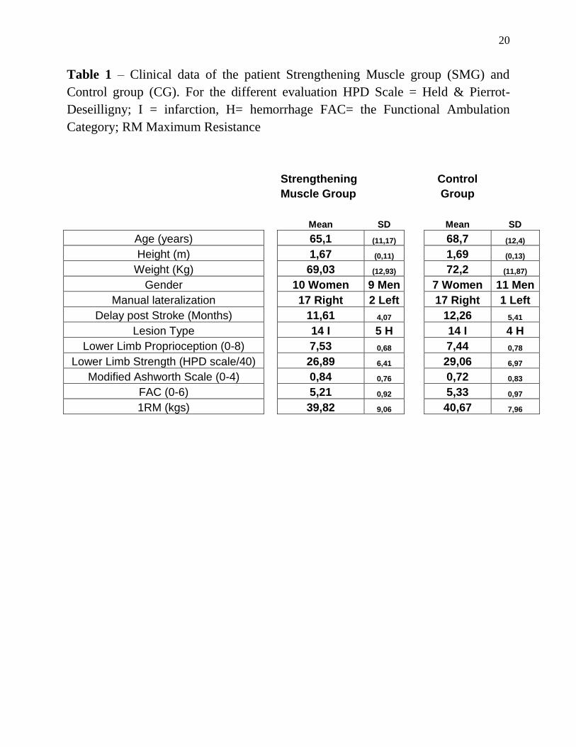
Patients were also similar in age (65.1 ±11.17 years for MSG vs. 68.7 ± 12.4 years for CG),
weight (69.03 ± 12.93 kg for MSG vs. 72.2 ± 11.87 kg for CG), height (1.67 ± 0.11 m for MSG
vs. 1.69 ±0.13 m for CG) and on set duration (12.47 ±4.97 months for MSG vs. 12.26 ±5.41
months for CG). All the characteristics of the study population are described in Table 1. The
patients were recruited with the following inclusion criteria: (i) a single episode of stroke supra-
tentorial that was responsible for the current clinical condition (ischemic or haemorrhagic supra-
tentorial stroke), (ii) ability to communicate and understand study-related information, (iii) ability
to perform the six minutes walking test (6MWT). In addition, all participants had to have
spasticity in the lower limb max of 2 on the modified Ashworth scale, and to be able to perform
the exercise requested on the horizontal press. In terms of exclusion criteria, patients were
excluded if they met one of the following: (i) previous history of neurological disorders, (ii)
cerebellar lesions [patients who could not be available for the entire duration of the study] and
(iii) had syncinesia. Physiotherapists were also asked to indicate the presence of aphasia or higher
functional disorders (non-inclusion criteria). The study was approved by the Locaethics
committee Sud-Méditerranée II (n°ID-RCB 2012-AOO518-35). All participants signed an
informed consent form prior to the study.
Insert Table 1
Clinical measures
7
Therapeutic follow-up assessments were conducted for all patients. Clinical assessments
included: (i) proprioceptive function of the lower limb for each joint (hallux, ankle, knee and hip
for range of motion using Fugl-Meyer scale where 0=absent, 1=impaired, 2=intact);, (ii)
voluntary motor capacity of the lower limb using the Held and Pierrot-Deseilligny scale (0 – 5:
absent – similar to the other side; 8 muscles were assessed: hip, knee and ankle flexors and
extensors and hip adductors and abductors; total/40 points), (iii) spasticity of the Triceps Surae
(postural and gait muscle) using the modified Ashworth scale and (iv) walking ability using the
Functional Ambulation Category (FAC) (0 = non-functional walker to 6 = Independent walker;
Teasdall scale). Lesion type, location and time since stroke were also recorded.
Patients in both groups received rehabilitative care over a period of four consecutive weeks. Each
standard treatment session was 2x30 minutes per day 5 times per week for all patients. .
According to the classic protocol, the main objective of the rehabilitative care was:
-The prevention of complications: pressure sores, phlebitis, glenohumeral subluxation,
installation of vicious attitudes (varus-equine, flexum or knee recurvatum, primitive patterns of
the upper limb).
-The stimulation of selective motor skills.
-The management of spasticity.
-The acquisition of transfers and functional independence.
-The acquisition of the standing position with postural balance.
-The acquisition of an efficient functional displacement.
-The therapeutic education of the patient.
Evaluation of gait/walk
(i) 10-meter Walk Test (Maggio et al., 2020) was used to evaluate gait. Patients were asked to
walk the distance at a comfortable speed, and the time to cover the set distance was documented.
Patients were given an additional distance of 2 meters to achieve a preferred speed before the
assigned 10-meters distance, and another 2 meters beyond the assigned 10-meter distance to
avoid speed deceleration. The test is discontinued in the event the patient complained of pain,
dyspnea, or balance problems.
8
(ii) 6-Minute Walk Test (6MWT on neurological patients Patterson et al., 2018): This sub-
maximum effort test consists of measuring the maximum distance travelled by a patient in 6
minutes, according to a self-determined walking speed. The test takes place indoors on a flat floor
(corridor, technical platform). The patient receives counts every minute of the time spent, as well
as encouragement. If the patient stops walking, s/he is instructed to resume walking as soon as
possible. A technical aid (single cane, tripod, star) is allowed depending on the patient's motor
level (noted on the evaluation sheet). The evaluation is stopped in case of pain, dyspnea and
balance problems. A distance between 400 and 700 meters is performed by non-pathological
persons. For patient safety, the pulse heart rate is checked before and after the 6-minute walk test.
Experimental Protocol
Muscle strengthening during the MSG rehabilitation sessions was based on the following
principles:- The patient activity was voluntary and controlled;
- Increasing the number of muscle fibers recruited was the desired element;
- Muscle contraction was progressive;
- During an intense muscular effort, the energy produced radiates to the other muscles, so special
attention is paid to the implementation of muscular coordination during the movement.
Equipment used
The equipment used for the muscle strengthening exercises was a horizontal Leg Press
(Technogym, Italy, figure 1). The press consisted of adjustable weights, a pulley, a fixed and a
vertical footplate, a sliding seat, an adjustable backrest, handles placed on the seat and handles
arranged on the folder.
Three phases can be noted:
- A first concentric phase where one pushes on the vertical fixed tray;
- A second static phase where the position is maintained;
- A third eccentric phase where one slows down the passive return of the sliding seat.
Insert Figure 1
Calculation of one-repetition maximum (1RM)
9
In order to identify the maximal strength, it was necessary to calculate the one-repetition
maximum (1RM) before each exercise for each patient. 1MR is the maximum weight the patient
is able to move once in the full range for a given exercise, which usually correlates with strength
(Thompson et al., 2019). There are two methods to calculate 1RM: the direct and indirect
method. The direct method requires performing movement against a weak load that is increased
gradually, while the indirect method is based on calculating theoretical 1RM using established
formulae and a theoretical load. In this study, we used the indirect method to find 1RM as it
carries less risk than the direct method for stroke patients. We used the calculation method
proposed by Brzycki (Do Nascimento et al., 2007), which is a mathematical estimate to predict
the theoretical maximum force (1RM) from the mobilized load and the number of successful
repetitions until the onset of fatigue, if this number is less than or equal to 10: Weight / (1.0278 -
(0.0278 * Number of repetitions)).
A session consisted of three sets of 5 repetitions each. The patient was asked to push with both
lower limbs (concentric phase) until the lower limbs were taut (but with a slight flexion of the
knees). This was followed by the static phase during which the patient placed his healthy lower
limb on the horizontal fixed plate, and finally the eccentric phase where the patient slowed down
the return of the seat with his only paretic lower limb. The first two sets were at 40% resistance to
the 1MR. The last set (more specific-muscle strengthening) was at 60% of the 1MR. It was
during this series that the muscle provided the most effort and was therefore more sensitive to
strengthening and voluntary neurological control. The program was performed over a period four
weeks, with three sessions per week. The protocol was carried out during the rehabilitation
session so the two groups had the same amount of time of the rehabilitation session.
Statistical analyses
Descriptive statistics consisted of characterizing the results by mean and standard deviation (SD)/
calculations. Normal distribution of data was tested by Shapiro-Wilk and Lilliefors tests. After
verifying the normal distribution of the data, all parameters were analysed using a general linear
model repeated measures of variance analysis (ANOVA). The clinical characteristics of the two
groups were compared using a one-way ANOVA: Group (Muscle strengthening Group/Control
Group). For the reference task, a two-level analysis was performed: Before and after treatment ×

10
Group. A Newman-Keuls post hoc test was used to determine the locus of differences. Statistica
(version 12) software was used for statistical analysis. The alpha critical level of 0.05 was
considered to denote statistically significant results (p≤0.05).
Results
The anthropometric characteristics of the two groups were similar and there were no
significant differences before treatment between patient groups for different parameters
analyzed (Table 1).
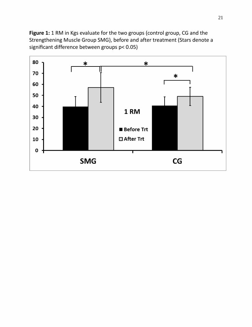
When we analyze the results obtained by the 1RM calculation for these two populations we
observe a significant main effect (Group * treatment F(1,39) = 409.32 p≤0.00001). We found a
significant difference between before and after treatment for the two groups (40.67 +/-7.96 Kgs
before treatment vs 49.06 +/- 8.29 Kgs after treatment for the CG (p≤0.0001); 39.83 +/-9.07 Kgs
before treatment vs 57.22 +/- 13.47 Kgs after treatment for the MSG p≤0.0001). When we
compare the differences between these two groups before treatment, no significant differences
appear. However, the MSG group, which had specific training, showed a significantly greater
increase after treatment than the CG group (57.22 +/- 13.47 Kgs vs 49.06 +/- 8.29 Kgs
respectively, p≤0.014). This means that both groups benefited from rehabilitation with an
increase in the power of their quadriceps more specifically for the MSG (figure 2).
Insert figure 2
Walk analysis
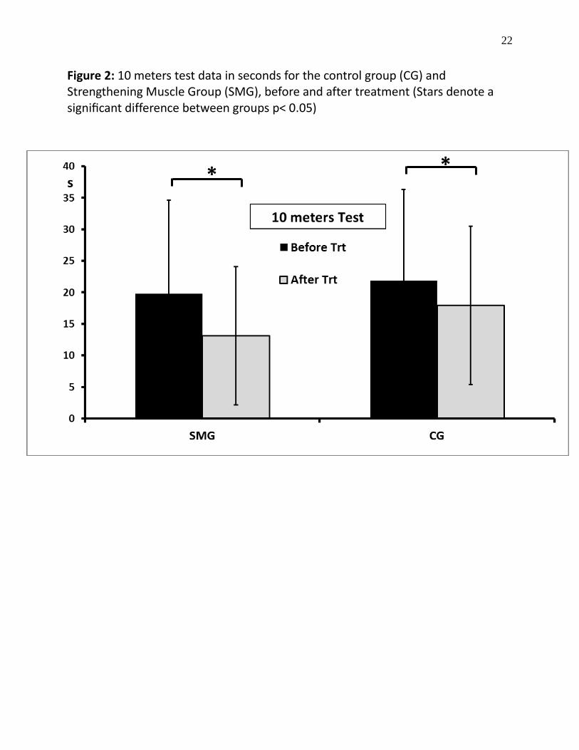
We did two types of locomotion analysis. The 10 meters test evaluates a rapid gait component.
For the two groups, we found a primary significant treatment effect on the time for this test
(F(1-39)= 56.77 p≤0.00001) with a greater speed of walking after (vs. before) reeducation.
However, for this parameter, there was no significant interaction between the group effect and the
treatment effect before and after rehabilitation (Group*treatment). However, when we compare
these results in post hoc, we observe a significant difference between before and after treatment
for the walking speed of these two groups with an increase of the values (21.87 +/- 14.41 vs.
17.94 +/-12.52 for the control group and 19.80 +/- 14.76 vs. 13.12 +/-10.93 for the muscle
strengthening group). We observe an improvement in walking speed for both groups between
11
before and after rehabilitation treatment. For these two groups the percentage improvement
shows a significant difference p≤0.0002 with respectively 32.75% (+/-12.46%) for the MSG
group and 18.57% (+/-8.76%) for the Control Group (figure 3).
Insert figure 3
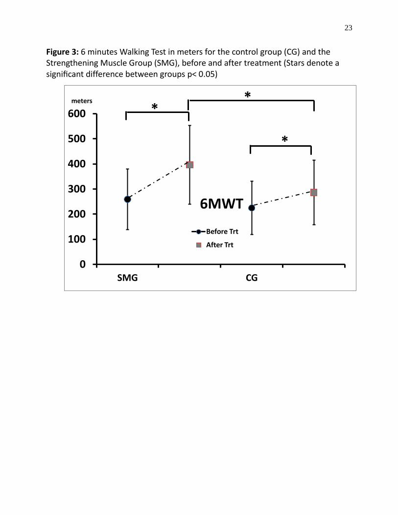
For the second walking test, we analyzed the distance performed on the 6MWT. Our
results show a significant main effect of treatment (before and after) and group
(Group*treatment F(1,39) = 16.82 p≤0.0002). We observe a significant effect for CG
between before and after treatment (p≤0.0003) and we observe the same for the MSG
group (p≤0.0001). Before treatment between these two groups no significant differences
were observed. Whatever treatment proposed, however, our results show a statistically
significant difference between the two groups (p≤0.01). The distance increases
respectively to 258.92 +/-120.05 at 396.94 +/-157.03 for the MSG vs. 224.72 +/-105.94
to 286.37 +/-127.96 for the CG; figure 3). For these two groups the percentage improvement
shows a significant difference p≤0.001 with respectively 62.31% (+/-35.28%) for the MSG group
and 30.04% (+/-17.11%) for the CG.
Finally, we found a significant correlation for both groups between increase of 1RM and
distance in the 6MWT after treatment (r=0.53 for MSG and r=0.92 for CG).
In all of our patients, we observed no change in spasticity after four weeks of training using the
Ashworth scale, which means that our eccentric exercises have not in any way modified a
physiological component that can in turn negatively affect the standard therapeutic management
proposed to these patients.
Discussion
Stroke patients experience considerable impairment in their daily life activities. Gait and walking
speed are affected partly due to hemiparesis because of muscle weakness and decreased motor
activity (Wist et al., 2016). Regaining the ability to maintain gait balance and speed is a major
functional interest for hemiparetic patients. One of the objectives of the current study was to
improve on patients’ gait, not only to increase walking speed, but also to help patients regain
12
normal walk by a better muscle control. We investigated a training protocol that consisted
eccentric control on a press system by using the paretic limb. Our results show significant
improvements with respect to the muscular power, which was reflected in the clinically and
statistically significant differences in 1RM on the press. We observed also a significant
improvement in walking, mainly gait speed using the 6MWT but not the rapid test of 10 meters
test).
Muscular hyperactivity results in excessive contraction of the muscle when it should be at
rest or in a certain state of relaxation (increased muscle tone). On the motor level, brain injury
generates a pyramidal syndrome resulting in hyperexcitability reflex causing inappropriate
muscle contractions, exaggerated and involuntary (muscle spasticity). There are also two types of
co-contraction in the hemiparetic subject. Synergistic co-contraction between two flexor muscles
such asdorsal flexion of the ankle during hip flexion (synchinesis), and exacerbated agonist /
antagonist co-contraction that can reverse the desired direction of movement (Jonkers et al.,
2009; Hyngstrom et al., 2012). Retraction of the soft tissues notably by a muscular atrophy is also
observed due to either an immobilization of the muscles in short position [leading to a decrease
of the proteic synthesis, a degradation of the contractile proteins and a decrease in the number of
sarcomere (Mercier et Bourbonnais, 2004)], or hyperactivity of the muscle [creating excessive
and non-voluntary contractions creating a permanent shortening of the muscle] (Nadeau et al.,
1999. In their study during a six-minute walk test, Straudi et al. (2009) show that it is possible to
determine groups of patients on gait velocity and also on differences in kinematic patterns
associated to muscles coordination or coactivation. This confirms previous studies showing that,
in stroke patients, increased gait velocity is associated with an increase in hip extension during
stance phase and in hip flexion at the end of swing phase (Jonkers et al., 2009). Moreover, the
authors demonstrated that, in addition to hip impairment, inadequate knee function was also a
predictor of walking performance. Another well-known kinematic disorder in hemiplegic patients
is a lack of knee flexion during swing phase (stiff knee gait, Goldberget al., 2003). This is a
common abnormality in hemiplegia and is often related to over activity of the rectus femoris
muscle due to spasticity (Sung & Bang 2000). In our study, we observed an increase of the 1RM
for the patients with eccentric muscle training of this muscle on a press. This result is confirmed
by previous studies in stroke patients, which showed that increased gait velocity is associated
with an increase in knee flexion during swing phase (Jonkers et al., 2009; Straudi et al., 2009). It
13
seems that the gait pattern in hemiplegic patients involves a reduced range of knee motion during
swing phase. The use of muscle strengthening in eccentric contraction therefore seems to modify
the gait patterns of this population by a better use of knee amplitudes and knee control as our
results tend to confirm.
This strategy of movement organization combining muscular synergies therefore appears
to be effective for gait therapy in stroke patients. Our daily physical activities and exercises
consisted of a combination of static (isometric), shortening (concentric), and lengthening
(eccentric) muscle contractions, but eccentric contractions of the knee extensors are emphasized
in some of the activities such as descending stairs or slopes and slowly sitting on a chair. The
study of Chung-Ching et al., (2017) tested the hypothesis that eccentric training of the knee
extensors would improve physical function and health parameters (e.g., blood lipid profiles) of
older adults better than concentric training. Our results suggest that it is better to focus on
eccentric contractions in physiotherapy for stroke patients, and that preconditioning knee
extensor muscles with low-intensity eccentric contractions are effective for attenuating muscle
concentric contraction of the knee extensors habitually used for elderly individuals. In hemiplegic
patients, Eng et al., (2002) showed that peak and average isokinetic torque could be used to
assess reliably lower extremity strength in persons with chronic stroke. There is a significant
difference in peak torque measurements between affected and normal lower limbs of post stroke
patients, as well as a significant correlation between the knee strength and lower limb functions.
Eng et al. (2002) similarly concluded that about the hip, knee and ankle concentric flexions and
extensions at 60 degrees/s and their consequences during gait. They argued that these results were
essentially guaranteed if three elements were respected: the exclusion of extended positions, the
adaptation of standard strength testing to patients with stroke, and the use of low velocities. In
addition, the authors noticed a learning effect for these patients and thus suggested providing two
training sessions before the actual evaluations to reduce this learning effect (Eng et al., 2002).
Our results corroborate their findings, and including those of the 1MR despite using the indirect
evaluation method. Although central mechanisms underlying force generation such as muscle
recruitment and rate coding may be altered in individuals with stroke, the reliability of the peak
and average torque-strength measures was high. In addition, the presence of increased muscle
tone, as quantified by the Modified Ashworth Scale, did not interfere with the reliability of force
production for the participants in a different study (Rabelo et al., 2016).
14
Furthermore, the differences in knee proprioception between the affected and intact limbs were
shown to be significant (Khan et al., 2019). In their study in stroke patients, Bohannon et al.,
(2013) have shown that the correlation between knee extensor torque and gait speed was
significant, whereas spasticity on the paretic side did not correlate with gait speed. The muscle
strength of the non-paretic dorsiflexors and the left lateral trunk flexors might have a role to play
in determining comfortable and maximum gait speeds of individuals with sub-acute stroke
(Aguiar et al., 2018). In our study, walking distance, as reflected in the 6MWT, was significantly
changed. Some authors have inferred a causal relationship between muscle strength and walking
capacity to improve comfortable gait speed and total distance walked for patients with stroke
(Metha et al., 2012). It is worth recalling that whereas use of the hip flexors is critical for walking
in stroke patients, involvement of this muscle group was not emphasized with the current
paradigm, which rather called for closed-chain, simultaneous knee- and hip controlled flexion
(Fernandez-Gonzalo et al., 2014). Our paradigm of strengthening eccentric muscle exercise
appears to present a safe, viable and highly effective method to improve skeletal muscle function,
power, and performance in daily living activities, in individuals suffering from chronic stroke.
Gait speed is generally selected as the outcome measurement in clinical practice and a predictor
of fall after a patient has a stroke, but gait speed is often confounded with balance, motor
function, and endurance (Ta-Sen et al., 2017). The current study and the increase of muscle
activity that patients performed can also prevent the risk of fall and permit an organization of
functional gait more precisely. The previous idea is confirmed by experiment where walking
velocity and cadence were lower in the faller group than the non-faller group (Ta-Sen et al.,
2017).
A simultaneous electromyography (EMG) study of isokinetic movements of the knee muscles in
stroke and control subjects found that there was no electrical activity on the antagonist muscles
and that there was indeed passive resistance to increased stretching on the hemiparetic side that
was pronounced at higher velocities (Mehta et al., 2012). However, these results are correlated
with a functional gain since the increase of the walking speed, as well as the decrease in the
number of steps, is significant. Even a minimal increase in force would be favorable to improve
the walking performance, which subsequently would increase the activity level necessary for the
adaptation of the nervous drive to reach a certain spot (Carr & Shepherd., 2011). The present
15
study showed an increase in walking speed post training, which means that the increase of
patients’ strength and muscular power significantly improved their functional component.
One of the consequences of a motor deficit on walking is an asymmetric support of the
two lower limbs on the ground with the paretic limb spending less time in support and more time
in oscillation (Brière et al., 2010). This decreases the length of the paretic limb and may explain
the fact that the number of steps decreases. We did not find worsening of spasticity in our sample
of patients (0 on the modified Ashworth scale); this means that our exercises did not modify the
physiological component in any manner that could negatively affect the standard therapeutic
management proposed to these patients. Our findings are consistent with the results obtained by
Jung et al., (2016) in which participants underwent 30 sessions of sit-to-stand training, five-times
per week for 6 weeks. This in line with recommendations that have stated that it was possible to
strengthen a hemiparetic limb muscle without increasing its spasticity (Wist et al., 2016). The
quadriceps and hamstrings were chosen for their importance in walking; however, the
strengthening of the knee muscles may not be sufficient to have an impact on walking speed
(Tavares Aguiar et al., 2018). It is impossible to determine accurately in what proportion this
increase in strength and power has affected walking speed. The performance in walking depends
on other parameters such as balance during standing, strength, and inter- and intra-muscular
coordination of other muscle groups such as plantar and dorsal flexors of the ankle. It would be
interesting to compare the balance of patients before and after training in a future study. Since the
press is an unguided movement, improvements in equilibrium may be observed. The muscles
involved in this study were the muscles of the lower limb, and they were anatomically and
physiologically very functional and had a high recovery capacity. In view of our results, it is
difficult to determine whether the progress observed was due to an adaptation of motor control by
cerebral plasticity or due to a gain that was only peripheral and therefore muscular. It might be
that the adaptation was mixed and therefore of neuromuscular origin. During muscle training,
there are two types of adaptations: 1) a very important neurologic adaptation during the first
months that leads to a greater recruitment in the number and size of motor units and allows a
better inter- and intra-muscular coordination, followed by 2) a structural adaptation that causes
hypertrophy of the muscle (Rabelo et al., 2016). Since our protocol lasted only one month, it is
possible to think of an adaptation essentially more nervous than muscular.
16
The findings of the present study should be interpreted in light of a few limitations. First, the
present study results cannot be extrapolated to all people with stroke, particularly patients at
lower functional levels with walking disability or severe cognitive impairments. Second, there is
an insufficient cognition of the influence of neural disorders after stroke on the knee
biomechanics. The reality of knee function during gait after stroke cannot be describing the real
knee biomechanics completely and we need more precise kinematics and kinetics results from
different research. Third, the studies about the influences of knee disorders are mainly
concentrated in walking but strengthening eccentric effect of knee muscles can be tested on other
daily life activities, such as running, stair climbing, and sit-to-stand. Finally, some results suggest
dissociation between quantitative measures of gait, such as velocity versus symmetry, and that
these parameters may measure independent features. It seems important in the future to make
evaluations and correlations between these different aspects of walking and other functional
activities.
Conclusions
This study adds weight to the current trend in the use of muscle strengthening versus resistance
by using muscle eccentric contraction. Moreover, it shows that apart from the positive effects on
muscular power, there may be also a tendency to increase the functional capacities of the non-
spastic hemiparetic patient in the subacute phase. The persistence of motor deficits and the
decrease in patients' ability to move justify the interest of finding new methods of rehabilitation.
All patients appreciated the technique and the smooth process of training. None of the patient
discontinued the study and there were no reports adverse events during the study. Studies
involving a larger number of patients are necessary in order to conclude in the intermediate and
long term the effectiveness of horizontal press rehabilitation.
References
Abdollahi, I., Taghizadeh, A., Shakeri, H., Eivazi, M., Jaberzadeh, S. (2015). The relationship
between isokinetic muscle strength and spasticity in the lower limbs of stroke patients. Journal of
bodywork and movement therapies, 19(2), 284-290.
17
Aguiar, L.T., Camargo, L.B., Estarlino, L.D., Teixeira-Salmela, L.F., Coelho de Morais Faria,
C.D. (2018). Strength of the lower limb and trunk muscles is associated with gait speed in individuals with
sub-acute stroke: a cross-sectional study. Brazilian Journal of Physical Therapy, 22(6), 459–466.
Belda-Lois, J.M., Mena-del Horno, S., Bermejo-Bosch, I., Moreno, J., Pons, J., Farina, D., Iosa,
M., Molinari, M., Tamburella, F., Ramos, A., Caria, A., Solis-Escalante, T., Brunner, C., Rea M. (2011).
Rehabilitation of gait after stroke: a review towards a top-down approach. Journal of Neuroengineering
Rehabilitation, 8(66), 1-19.
Bohannon, R.W., Andrews, W., Glenney, S.S. (2013). Minimal Clinically Important Difference
for Comfortable Speed as a Measure of Gait Performance in Patients Undergoing Inpatient Rehabilitation
after Stroke. Journal of Physical Therapy Science, 25(10), 1223–1225.
Boudarham, J., Roche, N., Pradon, D., Delouf, E., Bensmail, D., Zory R. (2014). Effects of
Quadriceps Muscle Fatigue on Stiff-Knee Gait in Patients with Hemiparesis. PLoS One, 9(4), e94138.
Brière, A., Lauzière, S., Gravel, D., Nadeau, S. (2010). Perception of weight-bearing distribution
during sit-to-stand tasks in hemiparetic and healthy individuals. Stroke, 41, 1704-1708.
Carr, J.H, Shepherd, R.B. (2011). Enhancing physical activity and brain reorganization after
stroke. Neurology research international, 3, 515938. 7p.
Chen, T.C., Tseng, W.C., Huang, G.L., Chen, S.L., Tseng, K.W., Nosaka, K. (2017). Superior
Effects of Eccentric to Concentric Knee Extensor Resistance Training on Physical Fitness, Insulin
Sensitivity and Lipid Profiles of Elderly Men. Front of Physiology, 8, 209-218.
Eng, J.J., Kim, C.M., MacIntyre, D.L. (2002). Reliability of lower extremity strength measures in
persons with chronic stroke. Archives of Physical Medicine Rehabilitation, 83(3), 322–328.
Eng, J.J., Lomaglio, M.J., Macintyre, D.L. (2009). Muscle torque preservation and physical
activity in individuals with stroke. Medical Science of Sports Exercise. 41, 1353-1360.
Global Burden of Disease Study (2015). Mortality and Causes of Death Collaborators Global,
regional, and national life expectancy, all-cause mortality, and cause-specific mortality for 249 causes of
death, 1980–2015: a systematic analysis for the Lancet, 388(10053),1459-1544.
Fernandez-Gonzalo, R., Nissemark, C., Åslund, B., Tesch, P., Sojka, P. (2014). Chronic stroke
patients show early and robust improvements in muscle and functional performance in response to
eccentric-overload flywheel resistance training: a pilot study. Journal of Neuroengineering Rehabilitation,
11, 150-159.
Goldberg, S.R., Ounpuu, S., Delp, S.L. (2003). The importance of swing-phase initial conditions
in stiff-knee gait. Journal of Biomechanics, 36(8), 1111-1116.
Guilhem, G., Cornu, C., Guével, A. (2010). Neuromuscular and muscle-tendon system
adaptations to isotonic and isokinetic eccentric exercise. Annals of Physical Rehabilitation Medicine, 53,
319-341.
Hafer-Macko, C. E., Ryan, A.S., Ivey, F.M., Macko, R.F. (2008). Skeletal muscle changes after
hemiparetic stroke and potential beneficial effects of exercise intervention strategies. Journal of
Rehabilitation Research & Development, 45(2), 261–272.
Hyngstrom, A.S., Onushko, T., Heitz, R.P., Rutkowski, A., Hunter, S.K., Schmit, B.D. (2012).
Stroke-related changes in neuromuscular fatigue of the hip flexors and functional implications. American
Journal of Physical Medicine Rehabilitation, 91, 33–42.
Johnston, S.C., Mendis, S., Mathers, C.D. (2009). Global variation in stroke burden and mortality:
estimates from monitoring, surveillance, and modelling. Lancet Neurology, 8, 345-354.
Jonkers, I., Delp, S., Patten, C. (2009). Capacity to increase walking speed is limited by impaired
hip and ankle power generation in lower functioning persons post-stroke. Gait & Posture, 29(1), 129-137.
Jung, K.S., Jung, J.H., In T.S., Cho, H.Y., (2016). Effects of Weight-shifting Exercise Combined
with Transcutaneous Electrical Nerve Stimulation on Muscle Activity and Trunk Control in Patients with
Stroke. Occupational Therapy International, 23, 436-443.
Khan, F., Anjamparuthikal, H., Chevidikunnan, M. (2019). The Comparison between Isokinetic
Knee Muscles Strength in the Ipsilateral and Contralateral Limbs and Correlating with Function of
Patients with Stroke. Journal of Neurosciences in Rural Practice, 10(4), 683–689.
18
Kiisa, C., Nishikawa, S.L., Lindstedt, P.C., (2018). La Stayo Basic science and clinical use of
eccentric contractions: History and uncertainties. Journal of Sport Health Science, 7(3), 265–274.
Loane, D.J, Faden, A.I. (2010). Neuroprotection for traumatic brain injury: translational
challenges and emerging therapeutic strategies. Trends in Pharmacology Science, 31, 596-604.
Maggio, J.B., Ospina, J.P., Callahan, J., Hunt, A.L., Stephen, C.D., Perez, D.L. (2020). Outpatient
Physical Therapy for Functional Neurological Disorder: A Preliminary Feasibility and Naturalistic
Outcome Study in a U.S. Cohort. Journal of Neuropsychiatry & Clinical Neuroscience, 32(1), 85-89.
McComas, A.J. (1994). Human neuromuscular adaptations that accompany changes in activity.
Medical Science of Sports Exercise, 26, 1498-1509.
Mehta, S., Pereira, S., Viana, R., Mays, R., McIntyre, A., Janzen, S., Teasell, R.W. (2012).
Resistance training for gait speed and total distance walked during the chronic stage of stroke: a meta-
analysis. Topics of Stroke Rehabilitation, 19(6), 471-8.
Mendis, S. (2009). World Stroke Day. International Journal of Stroke, 4, 315-316.
Mercier, C., Bourbonnais, D. (2004). Relative shoulder flexor and handgrip strength is related to
upper limb function after stroke. Clinical Rehabilitation, 18, 215-221.
Nadeau, S., Arsenault, A.B., Gravel, D., (1999). Analysis of the clinical factors determining
natural and maximal gait speeds in adults with a stroke. American Journal of Physical Medicine
Rehabilitation, 78, 123-130.
Nascimento, M.A., Cyrino, E.S., Nakamura, F.Y., Romanzini, M., Cardoso Pianca, H.J.,
Queiróga, M.R. (2007). Validation of the Brzycki equation for the estimation of 1-RM in the bench press.
Review of Brasilian Medicine Esporte, 13(1), 47-50
O'Sullivan, S.B., Schmitz, T.J. (2006). Physical rehabilitation. Publisher: F.A. Davis Company
(5th Ed), Pennsylvania, United States.
Patterson, K.K., Wong, J.S., Prout, E.C., Brooks, D., (2018). Dance for the rehabilitation of
balance and gait in adults with neurological conditions other than Parkinson’s disease: A systematic
review. Heliyon 4, e00584.
Pollock, A., Baer, G., Campbell, P., Ling Choo. P., Forster, A., Morris, J., Pomeroy, V.,
Langhorne, P. (2014). Physical rehabilitation approaches for the recovery of function and mobility
following stroke. Cochrane Database Systematic Review, 4, CD001920.
Rabelo, M., Nunes, G.S., Menezes da Costa, N., Amante, J., de Noronha, M., Fachin-Martins, E.
(2016). Reliability of Muscle Strength Assessment in Chronic Post-Stroke Hemiparesis: A Systematic
Review and Meta-Analysis Topics in Stroke Rehabilitation, 23(1), 26-36.
Saunders, D.H., Sanderson, M., Hayes, S., Kilrane, M., Greig, C.A., Brazzelli, M., Mead, G.E.
(2016). Physical fitness training for stroke patients. Cochrane Database Systematic Review. (3),
CD003316.
Sheffler, L.R., Chae, J. (2013). Technological Advances in Interventions to Enhance Post-Stroke
Gait. Physical Medicine Rehabilitation Clinics North America, 24(2), 305–323.
Straudi, S., Manca, M., Aiello, E., Ferraresi, G., Cavazza, S. (2009). Basaglia N Sagittal plane
kinematic analysis of the six-minute walk test: a classification of hemiplegic gait. European Journal of
Physical Rehabilitation Medicine, 45(3), 341-347.
Sung, D.H., Bang, H.J. (2000). Motor branch block of the rectus femoris: its effectiveness in stiff-
legged gait in spastic paresis. Archives of Physical Medicine and Rehabilitation, 81(7), 910-915.
Takeda, K., Tanabe, S., Koyama, S. (2018). Relationship between the rate of force development in
knee extensor muscles and gait speed in patients with chronic stroke: A cross-sectional study.
NeuroRehabilitation, 43, 425-430.
Ta-Sen, W., Peng-Ta, L., Liang-Wey, C., Sen-Yung, L. (2017). Gait asymmetry, ankle spasticity,
and depression as independent predictors of falls in ambulatory stroke patients. PLoS One, 12(5),
e0177136.
Thieme, H., Morkisch, N., Mehrholz, J., Pohl, M., Behrens, J., Borgetto, B., Dohle, C. (2018).
Mirror therapy for improving motor function after stroke. Cochrane Database Systematic Review, Jul;(7),
CD008449.
19
Thompson, S.W., Rogerson, D., Ruddock, A., Barnes, A. (2020). The Effectiveness of Two
Methods of Prescribing Load on Maximal Strength Development: A Systematic Review. Sports Medicine,
50(5), 919–938.
Van de Port, I.G., Kwakkel, G., Lindeman, E. (2008). Community ambulation in patients with
chronic stroke: how is it related to gait speed? Journal of Rehabilitation Medicine, 40(1), 23-27.
Vibhor, K., Hampton, A., Abhay, V., Mintzer, J., Kindy, M. (2014). Guest Spinal Cord Injury:
How Can We Improve the Classification and Quantification of Its Severity and Prognosis? Journal of
Neurotraumatology, 31(3), 215–227.
Wist, S., Clivaz, J., Sattelmayer, M. (2016). Muscle strengthening for hemiparesis after stroke: A
meta-analysis. Annals of Physical Rehabilitation Medicine, 59, 114-124.
20
Table 1 – Clinical data of the patient Strengthening Muscle group (SMG) and
Control group (CG). For the different evaluation HPD Scale = Held & Pierrot-
Deseilligny; I = infarction, H= hemorrhage FAC= the Functional Ambulation
Category; RM Maximum Resistance
Strengthening Control
Muscle Group Group
Mean SD Mean SD
Age (years) 65,1 (11,17) 68,7 (12,4)
Height (m) 1,67 (0,11) 1,69 (0,13)
Weight (Kg) 69,03 (12,93) 72,2 (11,87)
Gender 10 Women 9 Men 7 Women 11 Men
Manual lateralization 17 Right 2 Left 17 Right 1 Left
Delay post Stroke (Months) 11,61 4,07 12,26 5,41
Lesion Type 14 I 5 H 14 I 4 H
Lower Limb Proprioception (0-8) 7,53 0,68 7,44 0,78
Lower Limb Strength (HPD scale/40) 26,89 6,41 29,06 6,97
Modified Ashworth Scale (0-4) 0,84 0,76 0,72 0,83
FAC (0-6) 5,21 0,92 5,33 0,97
1RM (kgs) 39,82 9,06 40,67 7,96
21
Figure 1: 1 RM in Kgs evaluate for the two groups (control group, CG and the Strengthening Muscle Group SMG), before and after treatment (Stars denote a significant difference between groups p< 0.05)
*
* *
1 RM
22
Figure 2: 10 meters test data in seconds for the control group (CG) and Strengthening Muscle Group (SMG), before and after treatment (Stars denote a significant difference between groups p< 0.05) * *
23
Figure 3: 6 minutes Walking Test in meters for the control group (CG) and the Strengthening Muscle Group (SMG), before and after treatment (Stars denote a significant difference between groups p< 0.05)
0
100
200
300
400
500
600
SMG CG
Before Trt
After Trt
meters
6MWT
*
*
*