E- Souvenir
355
Organized by: Department of Anatomy, King George’s Medical University UP, Lucknow, India (Under the aegis of Anatomical Society of India) E- Souvenir
-
Upload
khangminh22 -
Category
Documents
-
view
0 -
download
0
Transcript of E- Souvenir

Organized by:
Department of Anatomy, King George’s Medical University UP, Lucknow, India (Under the aegis of Anatomical Society of India)
E- Souvenir

Contents
Messages
From the Desk of Chief Organizing Secretary
About the Campus
About Department of Anatomy
Anatomical Society of India: Office Bearers
Programme at a glance
Presentations at a glance
Oral
Poster
Abstracts
Pre-Conference Workshop
CME & Guest Lectures
Oral
Poster
MESSAGE
Greetings from Rishikesh - Devbhoomi!
Dear Friends and colleagues
On behalf Anatomical Society of India, it is my pleasure and privilege to invite you all for 68th NATCON ASI - National
Virtual Conference of Anatomical Society of India to be held in 2022, January 28, 29 & 30.
68th NATCON ASI - National Virtual Conference of Anatomical Society of India - "A virtual Academic fiesta" will be
hosted by Department of Anatomy, King George’s Medical University, Lucknow Uttar Pradesh - India. 68th NATCON is
being held under aegis of Anatomical Society of India on virtual platform and will be a great event to collect galaxy of
anatomists together to share knowledge, ideas and learning.
68th Virtual NATCON –ASI aims to bring together anatomists and other scientists from around the globe to present and
debate the latest and best research on anatomy. It will provide an opportunity for an interaction for all delegates and
participants with senior faculties. Students and young investigators are encouraged to attend and t o present their research
work.
I wish Prof Punita Manik Chief Organizing Secretary 68th Virtual NATCON - 2022 and his team from King George’s
Medical University a great success ahead.
Welcome and best wishes to all participants, panelists and guest speakers to this Virtual NATCON.
Best wishes and Warm Regards
Jai Hind
(Prof. Brijendra Singh)
President
Anatomical Society of India Professor & Head,
Department of Anatomy,
All India Institute of Medical Sciences,
Rishikesh, Uttarakhand, India
MESSAGE
Conferences open the doors between known & unknown. It gives us opportunities to meet with each other and share the
knowledge. With the declaration of Covid-19 Pandemic every one of us was afraid of meeting with each other. All the
academic activities came to stand still, effect of this pandemic is still on.
Looking over the long gap in organizing conference department of Anatomy KGMU was requested to conduct a virtual
conference on behalf of Anatomical society of India, and I am delight to share that they accepted the request immediately
without any question.
Organization of scientific event gives an insight to overall development of teachers & students both intellectually & socially.
I am sure participants would be immensely benefited by different deliberations during conference. One rightly said
Knowledge is of little value when it confined to few individuals & only be utilized when others understand the concept
involved.
I extend my whole hearted appreciations & best wishes to Dr. Punita Manik & her dynamic and vibrant team members for
organizing this 68th NATCON of ASI organized virtually. I applaud them for leaving no stone unturned to make the grand
event a success.
“Success is not final, failure is not fatal it is courage to continue the counts” said by Churchill.
(Prof. S . L. Jethani)
General Secretary Anatomical Society of India
Chief Medical Superintendent, Himalayan Hospital
Professor in Anatomy
Himalayan Institute of Medical Sciences
SRN, Jolly Grant , Dehradun.(Uttarakhand) 248016
India.
Organized by : Department of Anatomy, K. G. Medical University U.P., Lucknow – 226003 In collaberation with Department of Obstetrics & Gynecology K. G. Medical University U.P., Lucknow
MESSAGE
It gives me unfeigned pleasure and great pride to know that the department of Anatomy, King George Medical University U P, is
going to organize the 68th national conference of Anatomical Society of India (68th NATCON of ASI) on a virtual platform. Most
recent advances, innovative researches and development in the trends of anatomical sciences will be explored by eminent scholars.
It will be proved an integrative forum for anatomists to exchange and disseminate their knowledge. The CME on ‘Expanding
Horizons of Anatomy: Knowing the Unknown’ is going to add new chapters in medical curriculum. It turns out to be a great event
and landmark for future.
My heartiest good wishes to the organizers and team for huge success of this conference.
(Prof. Navneet Kumar)
Chief Co-ordinator
Principal
Autonomous State Medical College,
Basti, Uttar Pradesh
Website: www.natconasi.in
68TH NATCON OF ASI 2022 National Virtual Conference of
Anatomical Society of India 28th -30th , January, 2022
Pre Conference Workshop on: Overview of In-Vitro Fertilization: Focus on Male Infertility
Dated-27th Jan 2022
Organized by : Department of Anatomy, K. G. Medical University U.P., Lucknow – 226003 In collaberation with Department of Obstetrics & Gynecology K. G. Medical University U.P., Lucknow
MESSAGE
I am delighted to welcome you all to the 68th National Conference of the Anatomical Society of India being organized by the
Department of Anatomy King George’s Medical University, Lucknow, UP India. The aim of this upcoming virtual academic
carnival is to invite all the Anatomy experts, researchers, academicians and postgraduates to share their knowledge with our large
family of anatomists all over the globe. You will be happy to attend the CME entitled “Expanding Horizons of Anatomy:
Knowing the unknowing” The other major highlights of the conference are Guest lectures by stalwarts of Anatomy and Scientific
paper and poster presentations.
Post Conference Hands on workshop on “Basic laboratory and clinical essentials in IVF” is also being planned. Hope it will be
a great learning experience for all the delegates.
On this virtual platform discussions will be beneficial in development of a deep insight with respect to various anatomical facts and
expansion of the thought process towards a wider approach.
(Prof. Punita Manik)
Chief Organizing Secretary
68th NATCON of ASI
King George’s Medical University UP
Lucknow
Website: www.natconasi.in
68TH NATCON OF ASI 2022 National Virtual Conference of
Anatomical Society of India 28th -30th , January, 2022
Pre Conference Workshop on: Overview of In-Vitro Fertilization: Focus on Male Infertility
Dated-27th Jan 2022
From The Desk of Organizing Secretary

The Campus of KGMU
King George's Medical University is situated in the heart of the historic city of Lucknow which is the capital of India's most populous state Uttar Pradesh. Lucknow lies about 500 km east of Delhi. The city was the seat of the Nawabs of Awadh in the 19 th century and a centre for the arts - music, painting, dance, 'shatranj' or chess, kite flying and 'tehzeeb' or culture. The university campus adjoins the crowded 'Chowk' area and is off Shahmina Road , very close to the bank of River Gomti
The university campus is spread over an area of roughly 100,000 sq metres. It is situated about 5 km from Charbagh Railway Station and is well connected to all parts of the city by public transport systems such as bus, tempo and taxi. Originally, the main administrative building, called the Administrative Block and the Anatomy and Physiolgy buildings were built in the early 20 th century. As the institution grew, more and more departments were created in new buildings. Therefore the campus really consists of a conglomeration of buildings housing the various departments, which came up over a period of 100 years as and when the need was felt. There are separate buildings for Medicine, Surgery, Pediatrics, Obstetrics & Gynecology, Cardiology, Neurology, Psychiatry, Neurosurgery, Plastic Surgery, Rheumatology, Geriatric Mental Health, Surgical Oncology, Chest Medicine, Pathology, Pharmacology, Anaesthesiology, Opthalmology, Urology and Community Medicine. Cardiothoracic Surgery is presently in the Chest Medicine building, Microbiology along with

Pathology and Biochemistry along with Physiology.
The original Administrative Block now houses the Convocation Hall, Seminar Hall (White Hall), Examination Hall, Vice Chancellor's Office and KGMCICE or Clinical Epidemiology Unit besides other related offices. Beside the main building a new Administrative Building has come up recently and houses the Registrar's Office, Finance Office, Examination Cell, Undergraduate and Postgraduate Cells, computer centre etc.
Just down the road from the Administrative Block, the old State Health Institute Building has been taken over as the Pariksha Bhawan. On the right side of the main Administrative Block lies the Central Library and adjoining Ranvaxy Centre The original hospital attached to the college was known as King George Hospital and was commissioned in 1914. This building presently houses the Public Relations Office, the Superintendent's Office, Head Cashier's Office, Matron's Office, Stores, Emergency Pathology Labs and the Departments of Otorhinolarygology, Orthopedic Surgery, Radiodiagnosis and Radiotherapy, besides general and private wards of surgical specialities. The Gandhi Ward is a 3 storied building housing the medical wards. Next to the Ophthalmic Block, a New Ophthalmic Block has come up.
On the other side, a new CT Scan Centre and MRI Centre have been built and started functioning. Just across the road, next to main hospital building a new Cobalt Therapy Unit has been built, as part of Radiotherapy Department. The old Casualty building on Shahmina Road now houses the new OPD Building with modern facilities. A new Surgical Emergency & Trauma Centre was built on Shahmina Road and started functioning in January 2004. The centre serves as the primary reception for all surgical and medical emergencies and provides all diagnostic and treatment facilities under one roof. Other departments across the Shahmina Road are Queen Mary's Hospital (Department of Obstetrics & Gynecology), Psychiatry Department, Department of Geriatric Mental Health, Cardiology Department (Lari Cardiology Centre) and the Dental Hospital . The Physiotherapy & Rehabilitation Department on main MG Road opposite Daliganj Bridge is now the composite complex for Orthopaedics, Rheumatology and Physical rehabilitation. There are several hostels interspersed with the department buildings. Trans Gomti hostel for boys with mulitstoreyed faculty residence Warden's residence in Khadra, across the Gomti. University Guest House and Vice Chancellors Residence is located in the main campus. The Centenary Hospital Complex is located on Shahmina road and shall be housing multi super speciality center.
The huge playground of Sardar Patel Hostel at the intersection of Shahmina Road and MG Road has been divided to accommodate the ambitious Scientific Convention Centre. The center was inaugurated on 19 th December 2004 has been taken over by the university. Phase II of the center now houses a 1000 capacity state of art hall.
Other general facilities of the campus:
Indian Bank – KGMU (in basement of Central Library) and KGDU branches with ATM
service Post Office
Cafeteria (next to Central Library)
Public Call Office (in main hospital building) Three night stay dormitories (rain baseras) for poor patients and attendants..
Sulabh Shauchalaya complexes – one opposite new Pharmacology Building and the
other opposite Gandhi Ward. 24 hour running drug shops
State Government Employees Welfare Canteen
Public Distribution Shop which provides rations, kerosene etc.
Police Outpost and Security personnel Underground Car Parks
Juice shops, Parag Dairy, Nescafe Booth etc
PABX telephone system linking all points on the campus.
Free shuttle service to move around patients and attendants from one department to
other.
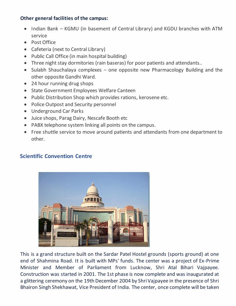
Scientific Convention Centre
This is a grand structure built on the Sardar Patel Hostel grounds (sports ground) at one end of Shahmina Road. It is built with MPs' funds. The center was a project of Ex-Prime Minister and Member of Parliament from Lucknow, Shri Atal Bihari Vajpayee. Construction was started in 2001. The 1st phase is now complete and was inaugurated at a glittering ceremony on the 19th December 2004 by Shri Vajpayee in the presence of Shri Bhairon Singh Shekhawat, Vice President of India. The center, once complete will be taken
over by the university and will be used for national and international level scientific and cultural functions of the university and other bodies as well. Construction is being done by UP Jal Nigam.
The Convention Centre has a floor area of 200,000 sq feet. The dome measures 52.5 metres. It has 3 halls of 200, 400 and 1050 seating capacity, 2 banquet halls, 2 exhibition galleries, an open-air theatre, rehearsal rooms, a foyer area below the dome and connecting corridors. All are fully air conditioned. There are 2 lifts available in the building. It also has 2 lawns in the premises which are used for various college functions.
The center has state of the art audio-visual facilities The auditoria can be used for plays, music as well as films. Besides, there are 2 VIP rooms and there is also adequate space available for kitchen and pantry.
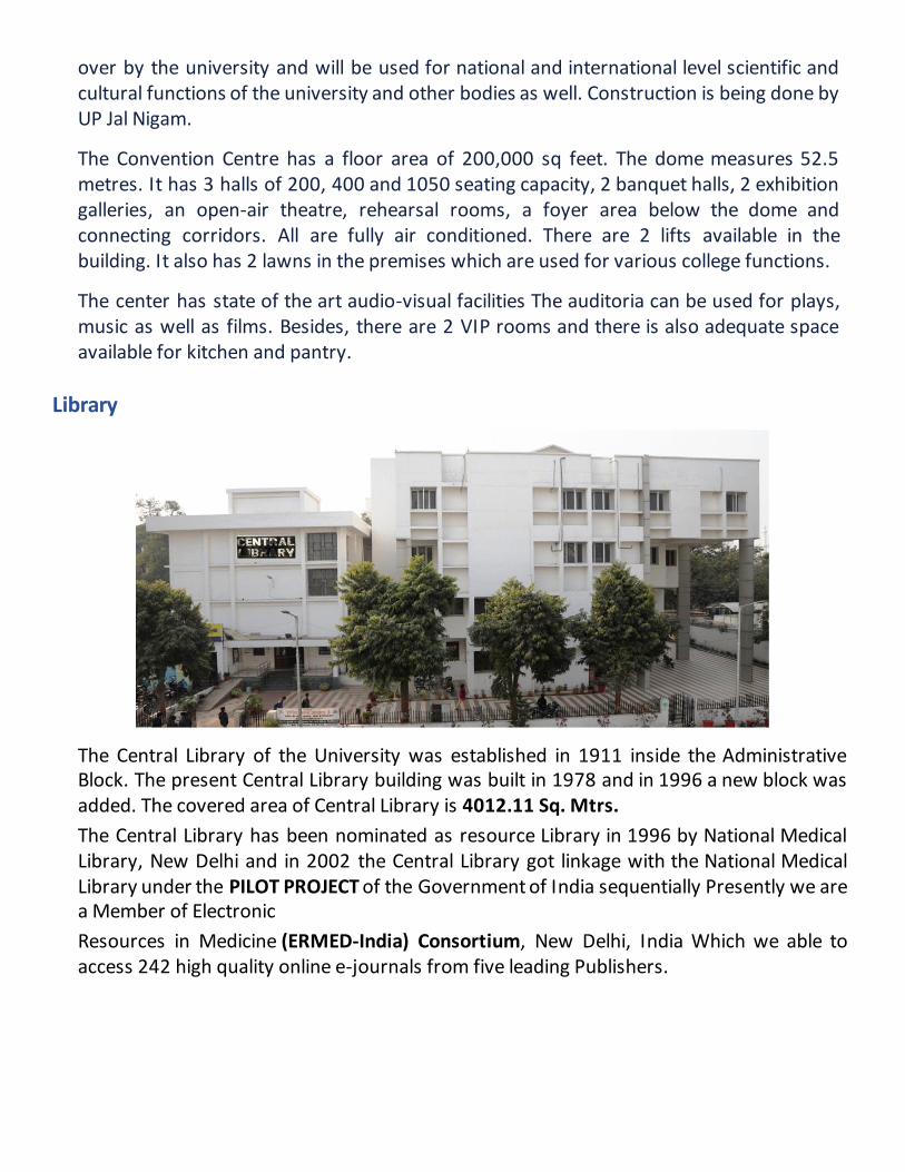
Library
The Central Library of the University was established in 1911 inside the Administrative Block. The present Central Library building was built in 1978 and in 1996 a new block was added. The covered area of Central Library is 4012.11 Sq. Mtrs.
The Central Library has been nominated as resource Library in 1996 by National Medical Library, New Delhi and in 2002 the Central Library got linkage with the National Medical Library under the PILOT PROJECT of the Government of India sequentially Presently we are a Member of Electronic
Resources in Medicine (ERMED-India) Consortium, New Delhi, India Which we able to access 242 high quality online e-journals from five leading Publishers.

Department of Anatomy
Department of Anatomy
The Department of Anatomy has a glorious past. It was one of the four blocks which was
completed for the King George's Medical University, Lucknow at the time of ceremonial
opening of the college building by Sir John Hewett in January 1912. Initially it started with 11
MBBS students and Dr. SS Khan was the first head of the department (1911-1926).Till date
the department has been guided by 14 heads and each one has contributed a new dimension
to the department. We also have some of the internationally acclaimed anatomist like Dr. AC
Das (former head) who was a MLC; Dr. DR Singh (former head) and Dr. Mahdi Hasan were BC
Roy Awardee.
The department was one of the first in the country to accord an MS degree to post-graduate
student in 1940. The Museum is one of the best in South East Asia. The initial lot of
specimens, models, charts in the museum were brought from England. This historic museum
meticulously and systematically displays number of specimens of embryology, gross
anatomy, anthropology, radiology, surface anatomy, osteology, cross sectional anatomy,
genetics etc. Complete skeletal system of human beings, foetuses and various animals are of
special interest. The architecture of the department speaks about its rich and ancient lineage
but with modern amenities. It has research laboratories like Histology lab, Body preservation
lab, Dissection Hall, Cytogenetic & tissue culture lab, Anthropology lab and Cadaveric Skill
lab. Dissection hall and Cadaveric Skill laboratory beautifully displays the CT and MRI films.

MUSEUM
LIBRARY LECTURE THEATRE
THEATREMUSEUM
CYTOGENETICS LAB
HEATREMUSEUM
UG HISTOLOGY LAB
THEATREMUSEUM PG HISTOLOGY LAB
THEATREMUSEUM

BODY PRESERVATION LAB
THEATREMUSEUM
ANTHROPOLOGY LAB
ULTRASOUND LAB DISSECTION HALL
THEATREMUSEUM
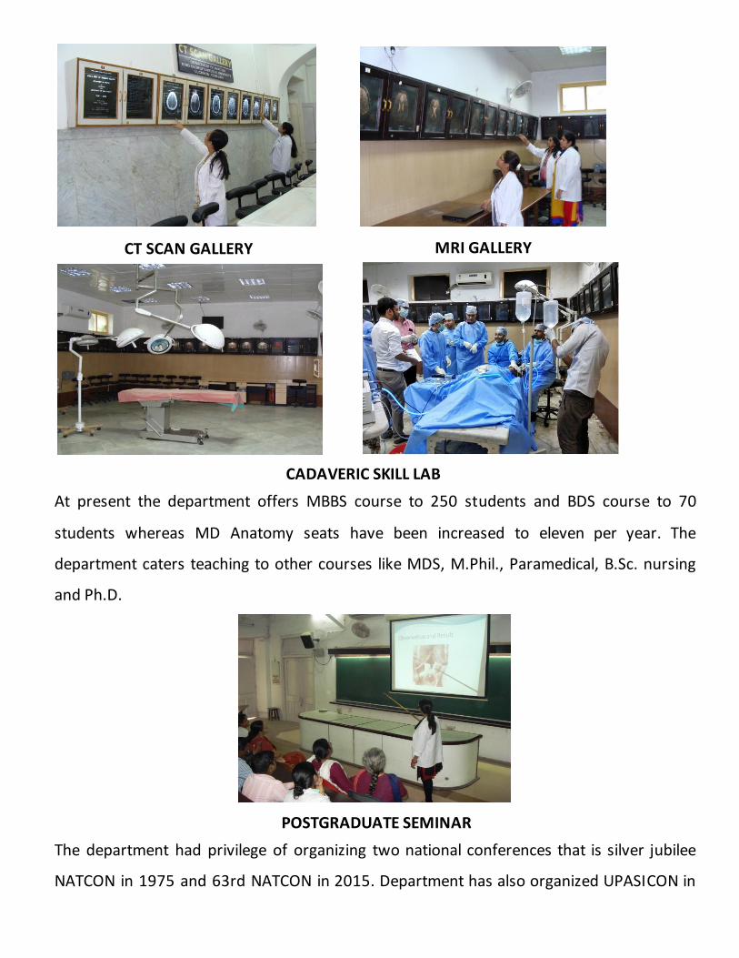
At present the department offers MBBS course to 250 students and BDS course to 70
students whereas MD Anatomy seats have been increased to eleven per year. The
department caters teaching to other courses like MDS, M.Phil., Paramedical, B.Sc. nursing
and Ph.D.
The department had privilege of organizing two national conferences that is silver jubilee
NATCON in 1975 and 63rd NATCON in 2015. Department has also organized UPASICON in
CT SCAN GALLERY MRI GALLERY
CADAVERIC SKILL LAB
POSTGRADUATE SEMINAR

2018. In Covid - 19 pandemic period (2020), the department successfully conducted a three-
webinar series on the theme "A Glimpse into Anatomical Entities for Budding Researchers: A
Futuristic Vision". An “International Virtual Anatomy Conference (IVACON 2021)” was
conducted in February 2021. Department has recently organized 68th NATCON of
Anatomical Society of India virtually in January 2022.
Over these years about 400 research papers have been published. Several distinguished
guests from country and abroad have been visiting the department off and on. Department
has a privileged to publish “Journal of Anatomical Sciences” (two issues in a year) for the year
2017-2019 under the editorship of Dr. Navneet Kumar.
The department has Anatomical society which was established in 1956. The society organizes
various activities annually. Presently the torch of success is being carried under the able
guidance and headship of Prof Punita Manik.
Vision:
The department of anatomy promote use of information technology and advanced software
in teaching learning and research and to equip the department with Virtual Dissection table.
The department is also involved in international collaboration for learning, teaching and
research activities.
Mission:
Our mission is to build up and upgrade knowledge in the field of anatomical sciences by
enriching education and research in Gross, Developmental, Microscopic, Imaging Anatomy
and Medical Education.
We will strive for excellence in anatomy education by:
Improvement of skills by practice and training on cadavers and manikins.
Developing and using the most effective and modern multi-media teaching techniques
available, including web-based instruments.
Acting as a resource for our clinical colleagues.
Contributing to the improvement of medical education by conducting meaningful
research in education and assessment.

Activities of Anatomical Society
1. Paper Writing Competition for undergraduate students
2. Dr. Dharam Narayan Memorial Book Prize Competition
3. Journal of the Anatomical Society of KGMU (once in a year)
4. S. S. Khan Dissection Competition
5. Anatomy Quiz contest
6. Das and Halim Oration
7. Annual Debate
8. Annual Picnic
9. Provide Gross Anatomy, Histology Practical Books
10. Human Bones to students for educational use
11. Embalming
12. Annual Function
DEBATE COMPETITION DR. DHARAM NARAYAN MEMORIAL BOOK
PRIZE COMPETITION
S.S. KHAN DISSECTION COMPETITION
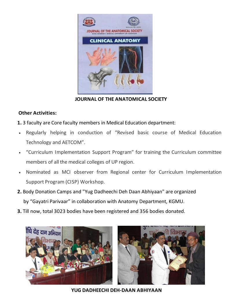
Other Activities:
1. 3 faculty are Core faculty members in Medical Education department:
Regularly helping in conduction of “Revised basic course of Medical Education
Technology and AETCOM”.
“Curriculum Implementation Support Program” for training the Curriculum committee
members of all the medical colleges of UP region.
Nominated as MCI observer from Regional center for Curriculum Implementation
Support Program (CISP) Workshop.
2. Body Donation Camps and "Yug Dadheechi Deh Daan Abhiyaan" are organized
by “Gayatri Parivaar” in collaboration with Anatomy Department, KGMU.
3. Till now, total 3023 bodies have been registered and 356 bodies donated.
JOURNAL OF THE ANATOMICAL SOCIETY
YUG DADHEECHI DEH-DAAN ABHIYAAN
Future Plans:
1. Establishment of Neonatal Anatomy Unit
2. Computerization of Dissection Hall
3. International collaboration for teaching and research
Services: 1. Human skeleton; articulated and disarticulated for educational use.
2. Embalming of dead human bodies in the department and at the residence.
3. Cadaveric Skill Laboratory for conduction of various workshops.
4. Karyotyping for research of common genetic disorders in the Cytogenetics lab of the
department.
ANATOMICAL SOCIETY OF INDIA OFFICE BEARERS
President
Dr. Brijendra Singh (Rishikesh)
Vice President
Dr. G.P. Pal (Indore)
General Secretary
Dr. S. L. Jethani (Dehradun)
Treasurer
Dr. Punita Manik (Lucknow)
Editor
Dr. Vishram Singh (Ghaziabad)
Joint Secretary
Dr. Jitendra Patel (Ahmedabad)
Joint Treasurer
Dr. Rakesh Kumar Verma (Lucknow)

EXECUTIVE COMMITTEE MEMBERS
Dr. Avinash Abhaya (Chandigarh)
Dr. Sumit Tuklsidas Patil (Port Blair)
Dr. Ruchira Sethi (Varanasi)
Dr. Pradeep Bokariya (Sevagram)
Dr. Sharmistha Biswas (Kolkata)
Dr. A. Amar Jayanthi (Kerala)
Dr. Ranjan Kumar Das (Odisha)
Dr. Rajani Singh (Saifai)
Dr. Rekha Lalwani (Bhopal)
Dr. Anshu Sharma (Chandigarh)
Dr. B. Prakash Babu (Manipal)
Dr. Anu Sharma (Ludhiana)
Dr. Subash K Despande (Dharwad)
Dr. Ashok Nirvan (Ahmedabad)
Dr. Rakesh Kumar Diwan (Lucknow)
Dr. Manisha Rajanand Gaikwad (Bhubaneswar)
Dr. Pawar Sudhir Eknath (Ahmednagar)
Dr Mrinmoy Pal (Agartala)
Dr. Siva Kumar (Puducherry)
Dr. Sunita A. Athavale (Bhopal)

Organizing Committee
Prof. Punita Manik Chief Organizing Secretary
Prof. Anita Rani Organizing Secretary
Prof. Jyoti Chopra Joint Organizing Secretary
Prof. Archana Rani Scientific & Souvenir Committee
Dr. R. K. Diwan Finance & Cultural Committee

Dr. A. K. Pankaj Registration Committee
Dr. R. K. Verma Registration, Finance & Workshop Committee
Dr. Garima Sehgal Scientific Committee
Dr. Sushma Tomar Scientific Committee
Mr. Ritesh K. Sharma Web Development, Designing & Technical Support Committee
Mr. Ashutosh Maurya Web Development, Designing
& Technical Support
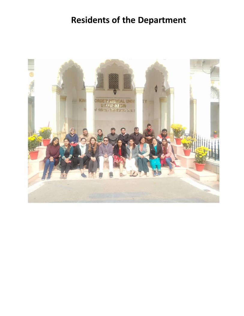
Residents of the Department

68th NATCON of ASI 2022
27th-30th Jan 2022 Organized by
Department of Anatomy, King George’s Medical University UP, India
27th January 2022 Pre-Conference Workshop on ‘Overview of In Vitro Fertilization: Focus on Male Infertility’
28th January 2022
Time Activity Speaker
9:00 am-10:15 am Inauguration
CME on ‘Expanding Horizons of Anatomy: Knowing the Unknown’
10:15 am-10:30 am Introduction to theme of CME “Expanding Horizons of Anatomy: Knowing the Unknown”
Dr. Anita Rani Professor Department of Anatomy King George’s Medical University Lucknow
10:30 am-11:30 am "DNA fingerprinting and its medico-legal applications"
Dr. K Thangaraj Director Centre for DNA Fingerprinting and Diagnostics Hyderabad
11:30 am-12:30 pm “Forensic Podiatry: Foot and Footprint Science”
Dr. Kewal Krishan Professor and Former Chair Department of Anthropology (UGC Centre of Advanced Study) Panjab University Chandigarh
12:30 pm-1:30 pm “An Introduction to Forensic Facial Reconstruction and the Drive to Advance the Field”
Dr. Tobias Houlton Facial Anthropologist, Lecturer Forensic Art & Facial Imaging Centre for Anatomy & Human Identification, University of Dundee (UK)
2:00 pm-3:00 pm “Identification from Prints – A forensic approach”
Prof. Anil Aggrawal Former Head Department of Forensic Medicine Maulana Azad Medical College, New Delhi
Programme at a Glance

3:00 pm-4:00 pm “Role of bone histomorphometry in identifying the age of human beings”
Dr. Julieta Gómez García-Donas Lecturer Forensic/Physical Anthropology Centre for Anatomy & Human Identification, University of Dundee (UK)
4:00 pm-5:00 pm “Role of Imaging in Forensic” Dr. Tanuj Kanchan Additional Professor Department of Forensic Medicine All India Institute of Medical Sciences Jodhpur
29th January 2022
9:00 am-10:00 am Guest Lecture: “Technology and the Art of Anatomy Illustrations”
Dr. Yogesh Ashok Sontakke Additional Professor Department of Anatomy JIPMER, Puducherry
10:15 am-11:15 am Scientific Session I (Oral Presentations)
11:30 am-12:30 pm Scientific Session II (Oral Presentations)
12:45 pm-1:45 pm Scientific Session III (Oral Presentations)
1:45 pm-2:30 pm LUNCH BREAK
2:30 pm-3:30 pm Scientific Session IV (Poster Presentations)
3:45 pm-4:45 pm Scientific Session V (Poster Presentations)
6:00 pm-8:00 pm Cultural Program
30th January 2022
9:00 am-10:00 am Guest Lecture: “The Anatomist at the Crossroads”
Dr. Sudha Seshayyan Vice Chancellor, The Tamilnadu Dr MGR Medical University
10:15 am-11:15 am Scientific Session I (Oral Presentations)
11:30 am-12:30 pm Scientific Session II (Oral Presentations)
12:45pm- 1:45 pm Scientific Session III (Oral Presentations)
1:45 pm-2:30 pm LUNCH BREAK
2:30 pm-3:30 pm Scientific Session IV (Poster Presentations)
3:45 pm-4:45 pm Scientific Session V (Poster Presentations)
5:00 pm-6:00 pm Valedictory Function

68th NATCON of ASI 2022
28th-30th Jan 2022 Organized by
Department of Anatomy, King George’s Medical University, India
28th January 2022
9:00 am-10:15 am Inauguration
CME on ‘Expanding Horizons of Anatomy: Knowing the Unknown’
Time Activity Speaker Chairpersons Moderator
10:15 am-10:30 am Introduction to the theme of CME “Expanding Horizons of Anatomy: Knowing the Unknown”
Dr Anita Rani Professor Department of Anatomy King George’s Medical University Lucknow
10:30 am-11:30 am "DNA fingerprinting and its medico-legal applications"
Dr K Thangaraj Director Centre for DNA Fingerprinting and Diagnostics Hyderabad
Dr MS Siddiqui Dr Gayatri Rath
Dr Farah Ghaus
11:30 am-12:30 pm “Forensic Podiatry: Foot and Footprint Science”
Dr Kewal Krishan Professor and Former Chair Department of Anthropology (UGC Centre of Advanced Study) Panjab University Chandigarh
Dr SD Joshi Dr Shashi Raheja
Dr Nidhi Sharma
12:30 pm-1:30 pm “An Introduction to Forensic Facial Reconstruction and the Drive to Advance the Field”
Dr Tobias Houlton Facial Anthropologist Lecturer in Forensic Art & Facial Imaging Centre for Anatomy & Human Identification University of Dundee (UK)
Dr A Shariff Dr Balasubramanyam V
Dr Anand Mishra
2:00 pm-3:00 pm “Identification from Prints – A forensic approach”
Prof Anil Aggrawal Former Head Department of
Dr Ashok Sahai Dr Brijendra Singh
Dr Lakshmi Rajgopal
CME & Guest Lectures

Forensic Medicine Maulana Azad Medical College Delhi
3:00 pm-4:00 pm “Role of bone histomorphometry in identifying the age of human beings”
Dr Julieta Gómez García-Donas Lecturer in Forensic/Physical Anthropology Centre for Anatomy & Human Identification University of Dundee (UK)
Dr DR Singh Dr GP Pal
Dr Prashant Natekar
4:00 pm-5:00 pm “Role of Imaging in Forensic”
Dr Tanuj Kanchan Professor and Head Department of Forensic Medicine AIIMS Jodhpur
Dr PK Sharma Dr CS Ramesh Babu
Dr Ruchira Sethi
29th January 2022
9:00 am-10:00 am Guest Lecture:
“Technology and the
Art of Anatomy Illustrations”
Dr Yogesh Ashok Sontakke Additional Professor Department of Anatomy JIPMER Puducherry
Dr Kalpana
Ramchandran
Dr Sneh Agarwal
Dr Sanjeev Kumar
Jain
Dr Alka
Singh
30th January 2022
9:00 am-10:00 am Guest Lecture: “The
Anatomist at the Crossroads”
Dr Sudha Seshayyan
Vice Chancellor
The Tamilnadu Dr MGR
Medical University
Dr Deepti Shastri
Dr Surajeet
Ghatak
Dr Anu Sharma
Dr Navbir Pasricha

Oral Presentations at a glance
Scientific Sessions – I (Physical Anthropology)
Date: - 29/01/2022
Time: - 10:15 am to 11:15 am Room – 1
Moderator- Dr Ankit
Chairpersons: DR L. PETER ERICSON
KANA BAL
1. OPCATE1210056
Comparative study of facial dimensions among male and female medical students
Mohini Binda
2. OPCATE1210013
Concavity Index and Disc Wedge Angle of Presacral Vertebra for Age and Gender in a
Northeast Indian Population.
Kalpana Chhetri
3. OPCATE1210048
Study of Correlation between Arm-Span and Height among Population of West Bengal
Madhumita Mahato
4. OPCATE1210023
Morphometric Study of Head of Radius in Vidarbha Region and its Clinical Implications in
Radial Head Prosthesis
Purvi Mishra
5. OPCATE1210025
Sexual Dimorphism of Human Mandible in
Marathwada Region
Swati Prashant Bhusari
6. OPCATE1210036 How Beautiful are Noses of Haryanvis? Khushi Gupta
Scientific Sessions - I
(Gross Anatomy) Date: - 29/01/2022
Time: - 10:15 am to 11:15 am Room – 2
Moderator- Dr Akriti
Chairpersons:
DR RAJAT SUBHRA DAS DR ASHISH KUMAR NAYYAR DR SHILPA BATHLA 7 OPCATE1210003 Morphometric study of the sternocleidomastoid
muscle
Jigyasa Passey
8 OPCATE1210009 The prevalence and distribution of the variants of gantzer's muscle: a meta-analysis of cadaveric
studies
Adil Asghar
9 OPCATE1210005 A morphological study on various formations of
superficial palmar arches
Ankita Saha
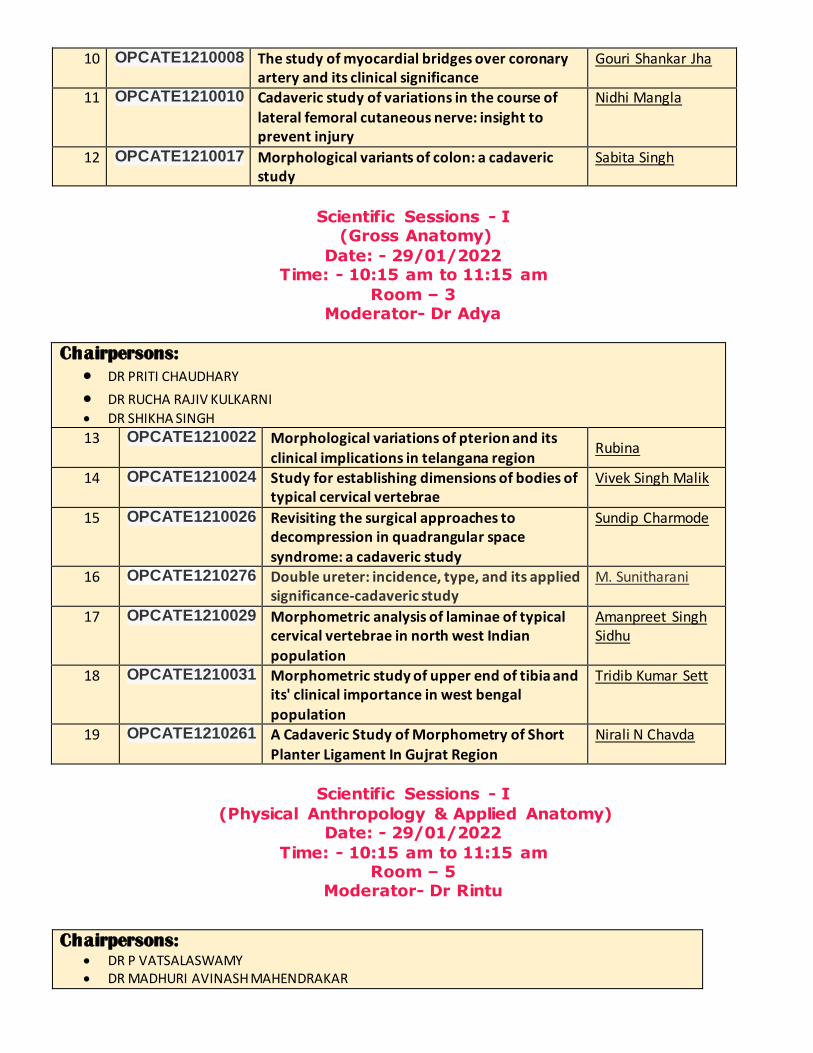
10 OPCATE1210008 The study of myocardial bridges over coronary artery and its clinical significance
Gouri Shankar Jha
11 OPCATE1210010 Cadaveric study of variations in the course of
lateral femoral cutaneous nerve: insight to prevent injury
Nidhi Mangla
12 OPCATE1210017 Morphological variants of colon: a cadaveric study
Sabita Singh
Scientific Sessions - I (Gross Anatomy)
Date: - 29/01/2022 Time: - 10:15 am to 11:15 am
Room – 3 Moderator- Dr Adya
Chairpersons:
DR PRITI CHAUDHARY DR RUCHA RAJIV KULKARNI DR SHIKHA SINGH
13 OPCATE1210022 Morphological variations of pterion and its
clinical implications in telangana region Rubina
14 OPCATE1210024 Study for establishing dimensions of bodies of typical cervical vertebrae
Vivek Singh Malik
15 OPCATE1210026 Revisiting the surgical approaches to decompression in quadrangular space syndrome: a cadaveric study
Sundip Charmode
16 OPCATE1210276
Double ureter: incidence, type, and its applied significance-cadaveric study
M. Sunitharani
17 OPCATE1210029 Morphometric analysis of laminae of typical cervical vertebrae in north west Indian
population
Amanpreet Singh Sidhu
18 OPCATE1210031 Morphometric study of upper end of tibia and its' clinical importance in west bengal
population
Tridib Kumar Sett
19 OPCATE1210261 A Cadaveric Study of Morphometry of Short
Planter Ligament In Gujrat Region
Nirali N Chavda
Scientific Sessions - I
(Physical Anthropology & Applied Anatomy) Date: - 29/01/2022
Time: - 10:15 am to 11:15 am Room – 5
Moderator- Dr Rintu
Chairpersons: DR P VATSALASWAMY DR MADHURI AVINASH MAHENDRAKAR

DR NAINA WAKODE
20 OPCATE1210073
A Morphological and Morphometric Study of Proximal End of Dry Radii and its Clinical
Significance
Rahul Gaur
21 OPCATE1210115
Morphological Study of Anatomical Basis of Coracoacromial Arch in Shoulder Impingement
Rashmi C Goshi
22 OPCATE1210133
Estimation of Height from the Length of Hand :A Study Among Medical Students of North India
Shomalla Jan
23 OPCATE1210135
Morphometric study of distal end of humerus and its applied aspects
Vinay G
24 OPCATE1210105
Attenuation of Neuropathic Pain in Rats by Neuropeptide Y (NPY), an Extensively Distributed and Evolutionarily Conserved Neuropeptide in the Central Nervous System
Mohammed Ahmed Ansari
25 OPCATE1210112
Lumbar pedicles “a dry bone and computed tomographic study”
Uma Shivanal
Scientific Sessions - I
(Medical Education) Date: - 29/01/2022
Time: - 10:15 am to 11:15 am
Room – 6 Moderator- Dr Honey
Chairpersons:
SATHEESHA NAYAK B DR RASHMI JAISWAL DR ARVIND KUMAR PANDEY
26 OPCATE3210002
Introduction of Case Based Learning in Undergraduate Anatomy Teaching
Santanu Bhattacharya
27 OPCATE3210011
Effect of Online Classes on Medical Students in Covid Era
Ishi Jain
28 OPCATE3210018
Is Inadequate Anatomical Knowledge on The Part of Physicians Hazardous for Successful
Clinical Practice?
Rajani Singh
29 OPCATE3210312
Perception of Indian MBBS & BDS students
regarding E-learning amidst COVID-19 pandemic
Nikhil Aggarwal
30 OPCATE3210291 Learning Anatomy by Colourful Specimens Kamal Singh
31 OPCATE3210049
Students Perception on Methods of Teaching
Anatomy a Questionnaire Study
Om Prakesh Mali
Scientific Sessions - I
(Gross Anatomy)
Date: - 29/01/2022 Time: - 10:15 am to 11:15 am
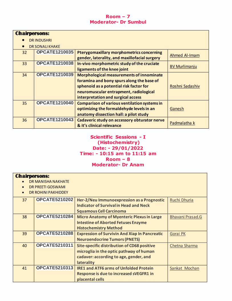
Room – 7 Moderator- Dr Sumbul
Chairpersons:
DR INDUSHRI DR SONALI KHAKE 32 OPCATE1210035 Pterygomaxillary morphometrics concerning
gender, laterality, and maxillofacial surgery Ahmed Al-Imam
33 OPCATE1210038 In-vivo morphometric study of the cruciate ligaments of the knee joint
BV Murlimanju
34 OPCATE1210039 Morphological measurements of innominate foramina and bony spurs along the base of sphenoid as a potential risk factor for neuromuscular entrapment, radiological interpretation and surgical access
Roshni Sadashiv
35 OPCATE1210040 Comparison of various ventilation systems in optimizing the formaldehyde levels in an
anatomy dissection hall: a pilot study
Ganesh
36 OPCATE1210043 Cadaveric study on accessory obturator nerve & it’s clinical relevance
Padmalatha k
Scientific Sessions - I
(Histochemistry)
Date: - 29/01/2022 Time: - 10:15 am to 11:15 am
Room – 8 Moderator- Dr Anam
Chairpersons: DR MANISHA NAKHATE
DR PREETI GOSWAMI
DR ROHINI PAKHIDDEY
37 OPCATE5210202
Her-2/Neu Immunoexpression as a Prognostic Indicator of Survival in Head and Neck
Squamous Cell Carcinoma
Ruchi Dhuria
38 OPCATE5210284
Micro Anatomy of Myenteric Plexus in Large
Intestine of Aborted Fetuses Enzyme Histochemistry Method
Bhavani Prasad.G
39 OPCATE5210288
Expression of Survivin And Xiap In Pancreatic Neuroendocrine Tumors (PNETS)
Gorai PK
40 OPCATE5210311 Site-specific distribution of CD68 positive
microglia in the optic pathway of human cadaver: according to age, gender, and
laterality
Chetna Sharma
41 OPCATE5210313 IRE1 and ATF6 arms of Unfolded Protein Response is due to increased sVEGFR1 in placental cells
Sankat Mochan

42 OPCATE5210021
A Study to Evaluate the Therapeutic Effect of some Nutraceutical combinations among
Indian Subjects of Oral Leukoplakia
Tanveer Ahmad
Scientific Sessions - II
(Physical Anthropology & Applied Anatomy) Date: - 29/01/2022
Time: - 11:30 am to 12:30 pm Room – 1
Moderator- Dr Ankit
Chairpersons:
DR BINDU SINGH
DR PRADEEP SINGH
DR KISHORE CHANDRA THAKUR
43 OPCATE1210163
A Cross Sectional Study of Foramen Magnum
in Dry Human Skulls for Determination of Sex
Hemant Ashish
Harode 44 OPCATE1210172
Predictor Index for Leg Length Discrepancy as a
Screening Tool For Referral To Tertiary Care Centre: A Baseline Study
Shehzeen
45 OPCATE1210173
Anthropometric Study of Hand Parameters for
Addressing Ergonomic Challenges Faced by Female Surgeons in Operation Theatre
Shikha Singh
46 OPCATE1210312 Prevalence Of Retromolar Foramen In Human Mandibles
B. John Rozar Raj
47 OPCATE1210196
Morphometric Analysis of Proximal End of Tibia in Western Rajasthan Population
Manu Shekhawat
48 OPCATE1210314
Estimation of Torgs Ratio in Adult Population
of Uttar Pradesh
Sanjay Prasad Sah
Scientific Sessions - II
(Imaging Anatomy) Date: - 29/01/2022
Time: - 11:30 am to 12:30 pm Room – 2
Moderator – Dr Kaweri
Chairpersons:
DR ADIL ASGHAR KANA BAL
DR MUMAL NAGWANI MISHRA
49 OPCATE4210004
Morphometric Analysis of Cervical Canal by
Computerized Tomography (CT) Scan in North Indian
Kanhaiya Jee
50 OPCATE4210020
Volumetric Study of Hippocampus by Magnetic Resonance Imaging
Kumari Pooja
51 OPCATE4210042
Radioanatomic Diagnostic Accuracy Index of
Cholecystitis And Cholelithiasis
Durgesh Singh
52 OPCATE4210061
Determination of Shape of the Thorax at
Multiple Thoracic Vertebral Levels using
Hari Prasad
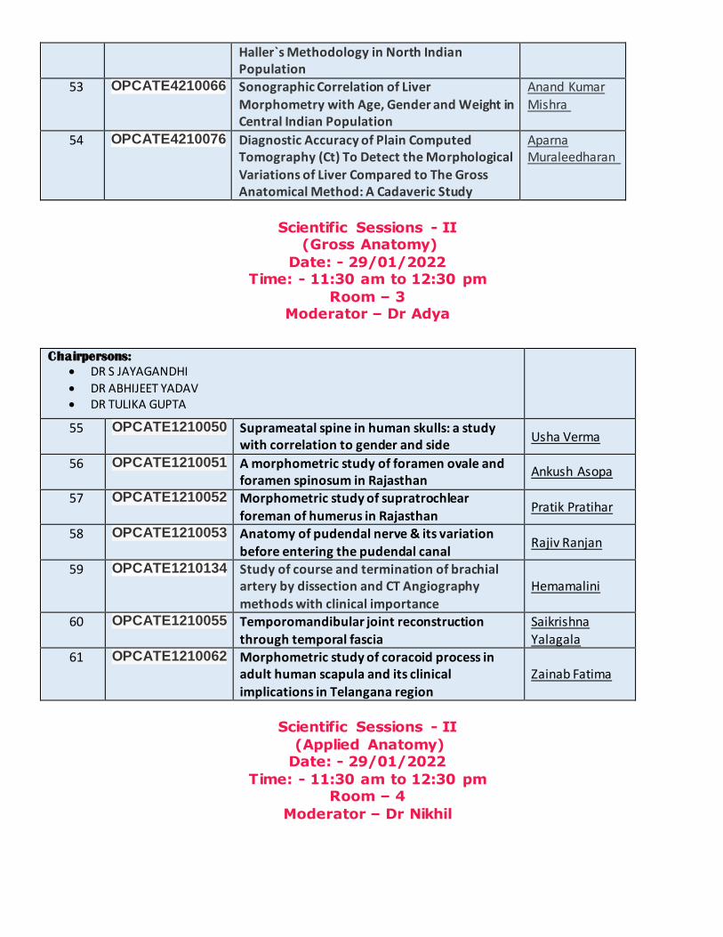
Haller`s Methodology in North Indian Population
53 OPCATE4210066
Sonographic Correlation of Liver
Morphometry with Age, Gender and Weight in Central Indian Population
Anand Kumar
Mishra
54 OPCATE4210076
Diagnostic Accuracy of Plain Computed Tomography (Ct) To Detect the Morphological
Variations of Liver Compared to The Gross Anatomical Method: A Cadaveric Study
Aparna Muraleedharan
Scientific Sessions - II (Gross Anatomy)
Date: - 29/01/2022 Time: - 11:30 am to 12:30 pm
Room – 3 Moderator – Dr Adya
Chairpersons:
DR S JAYAGANDHI
DR ABHIJEET YADAV DR TULIKA GUPTA
55 OPCATE1210050 Suprameatal spine in human skulls: a study with correlation to gender and side
Usha Verma
56 OPCATE1210051 A morphometric study of foramen ovale and foramen spinosum in Rajasthan
Ankush Asopa
57 OPCATE1210052 Morphometric study of supratrochlear
foreman of humerus in Rajasthan Pratik Pratihar
58 OPCATE1210053 Anatomy of pudendal nerve & its variation
before entering the pudendal canal Rajiv Ranjan
59 OPCATE1210134
Study of course and termination of brachial artery by dissection and CT Angiography
methods with clinical importance
Hemamalini
60 OPCATE1210055 Temporomandibular joint reconstruction
through temporal fascia
Saikrishna
Yalagala
61 OPCATE1210062 Morphometric study of coracoid process in adult human scapula and its clinical
implications in Telangana region
Zainab Fatima
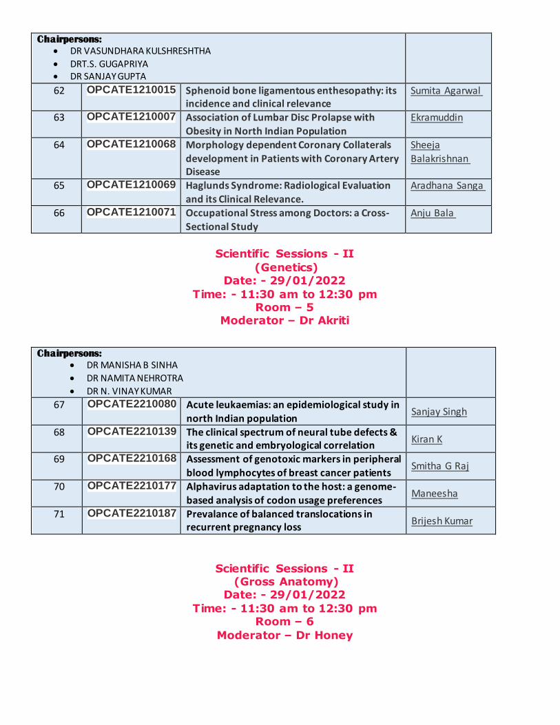
Scientific Sessions - II
(Applied Anatomy) Date: - 29/01/2022
Time: - 11:30 am to 12:30 pm Room – 4
Moderator – Dr Nikhil
Chairpersons:
DR VASUNDHARA KULSHRESHTHA
DRT.S. GUGAPRIYA DR SANJAY GUPTA
62 OPCATE1210015
Sphenoid bone ligamentous enthesopathy: its incidence and clinical relevance
Sumita Agarwal
63 OPCATE1210007
Association of Lumbar Disc Prolapse with
Obesity in North Indian Population
Ekramuddin
64 OPCATE1210068
Morphology dependent Coronary Collaterals
development in Patients with Coronary Artery Disease
Sheeja
Balakrishnan
65 OPCATE1210069
Haglunds Syndrome: Radiological Evaluation and its Clinical Relevance.
Aradhana Sanga
66 OPCATE1210071
Occupational Stress among Doctors: a Cross-
Sectional Study
Anju Bala
Scientific Sessions - II
(Genetics) Date: - 29/01/2022
Time: - 11:30 am to 12:30 pm Room – 5
Moderator – Dr Akriti
Chairpersons:
DR MANISHA B SINHA
DR NAMITA NEHROTRA
DR N. VINAY KUMAR
67 OPCATE2210080
Acute leukaemias: an epidemiological study in
north Indian population Sanjay Singh
68 OPCATE2210139 The clinical spectrum of neural tube defects & its genetic and embryological correlation
Kiran K
69 OPCATE2210168 Assessment of genotoxic markers in peripheral
blood lymphocytes of breast cancer patients Smitha G Raj
70 OPCATE2210177 Alphavirus adaptation to the host: a genome-
based analysis of codon usage preferences Maneesha
71 OPCATE2210187
Prevalance of balanced translocations in recurrent pregnancy loss
Brijesh Kumar
Scientific Sessions - II (Gross Anatomy)
Date: - 29/01/2022
Time: - 11:30 am to 12:30 pm Room – 6
Moderator – Dr Honey
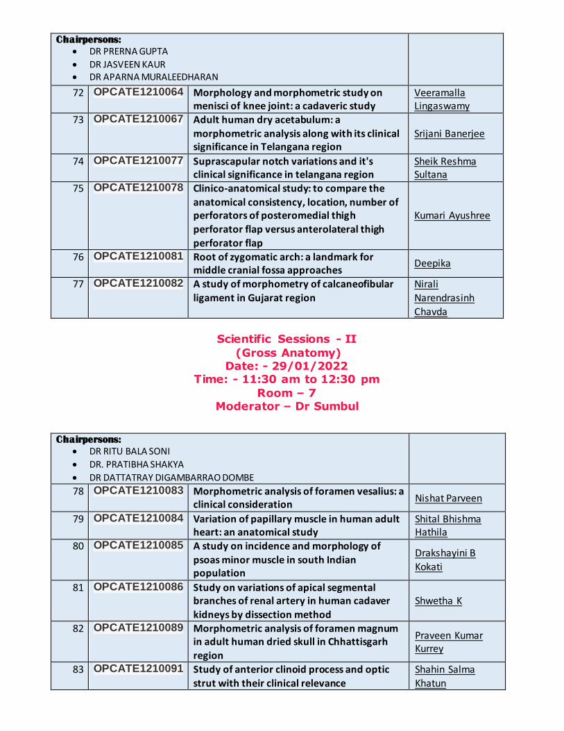
Chairpersons:
DR PRERNA GUPTA
DR JASVEEN KAUR DR APARNA MURALEEDHARAN
72 OPCATE1210064 Morphology and morphometric study on menisci of knee joint: a cadaveric study
Veeramalla Lingaswamy
73 OPCATE1210067 Adult human dry acetabulum: a
morphometric analysis along with its clinical significance in Telangana region
Srijani Banerjee
74 OPCATE1210077 Suprascapular notch variations and it's clinical significance in telangana region
Sheik Reshma Sultana
75 OPCATE1210078 Clinico-anatomical study: to compare the
anatomical consistency, location, number of perforators of posteromedial thigh
perforator flap versus anterolateral thigh perforator flap
Kumari Ayushree
76 OPCATE1210081 Root of zygomatic arch: a landmark for middle cranial fossa approaches
Deepika
77 OPCATE1210082 A study of morphometry of calcaneofibular
ligament in Gujarat region
Nirali
Narendrasinh
Chavda
Scientific Sessions - II
(Gross Anatomy) Date: - 29/01/2022
Time: - 11:30 am to 12:30 pm
Room – 7 Moderator – Dr Sumbul
Chairpersons:
DR RITU BALA SONI
DR. PRATIBHA SHAKYA
DR DATTATRAY DIGAMBARRAO DOMBE
78 OPCATE1210083 Morphometric analysis of foramen vesalius: a clinical consideration
Nishat Parveen
79 OPCATE1210084 Variation of papillary muscle in human adult heart: an anatomical study
Shital Bhishma Hathila
80 OPCATE1210085 A study on incidence and morphology of psoas minor muscle in south Indian population
Drakshayini B
Kokati
81 OPCATE1210086 Study on variations of apical segmental branches of renal artery in human cadaver
kidneys by dissection method
Shwetha K
82 OPCATE1210089 Morphometric analysis of foramen magnum in adult human dried skull in Chhattisgarh region
Praveen Kumar Kurrey
83 OPCATE1210091 Study of anterior clinoid process and optic
strut with their clinical relevance
Shahin Salma
Khatun
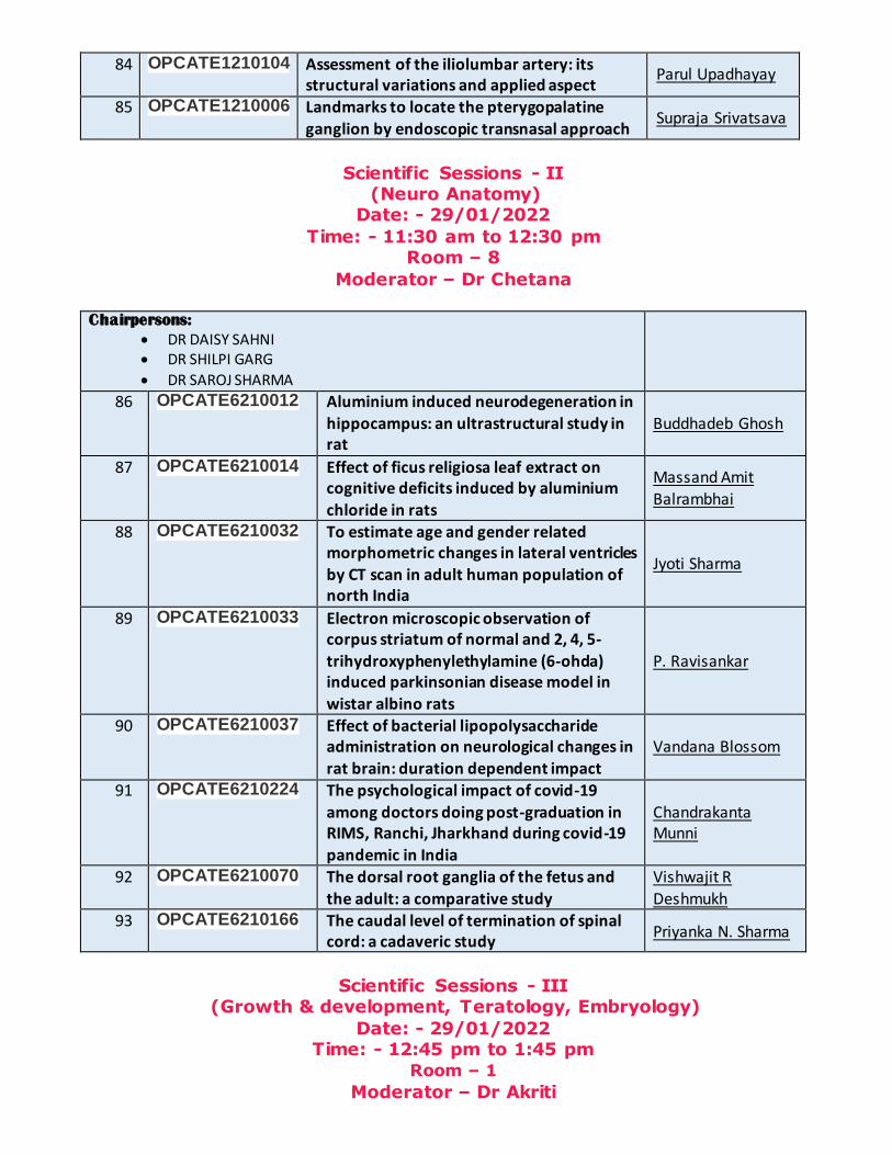
84 OPCATE1210104 Assessment of the iliolumbar artery: its structural variations and applied aspect
Parul Upadhayay
85 OPCATE1210006 Landmarks to locate the pterygopalatine
ganglion by endoscopic transnasal approach Supraja Srivatsava
Scientific Sessions - II (Neuro Anatomy)
Date: - 29/01/2022
Time: - 11:30 am to 12:30 pm Room – 8
Moderator – Dr Chetana
Chairpersons:
DR DAISY SAHNI DR SHILPI GARG
DR SAROJ SHARMA
86 OPCATE6210012 Aluminium induced neurodegeneration in hippocampus: an ultrastructural study in rat
Buddhadeb Ghosh
87 OPCATE6210014 Effect of ficus religiosa leaf extract on cognitive deficits induced by aluminium
chloride in rats
Massand Amit
Balrambhai
88 OPCATE6210032 To estimate age and gender related morphometric changes in lateral ventricles by CT scan in adult human population of north India
Jyoti Sharma
89 OPCATE6210033 Electron microscopic observation of corpus striatum of normal and 2, 4, 5-
trihydroxyphenylethylamine (6-ohda) induced parkinsonian disease model in
wistar albino rats
P. Ravisankar
90 OPCATE6210037 Effect of bacterial lipopolysaccharide administration on neurological changes in rat brain: duration dependent impact
Vandana Blossom
91 OPCATE6210224 The psychological impact of covid-19 among doctors doing post-graduation in RIMS, Ranchi, Jharkhand during covid-19
pandemic in India
Chandrakanta Munni
92 OPCATE6210070 The dorsal root ganglia of the fetus and
the adult: a comparative study
Vishwajit R
Deshmukh 93 OPCATE6210166 The caudal level of termination of spinal
cord: a cadaveric study Priyanka N. Sharma
Scientific Sessions - III (Growth & development, Teratology, Embryology)
Date: - 29/01/2022 Time: - 12:45 pm to 1:45 pm
Room – 1
Moderator – Dr Akriti
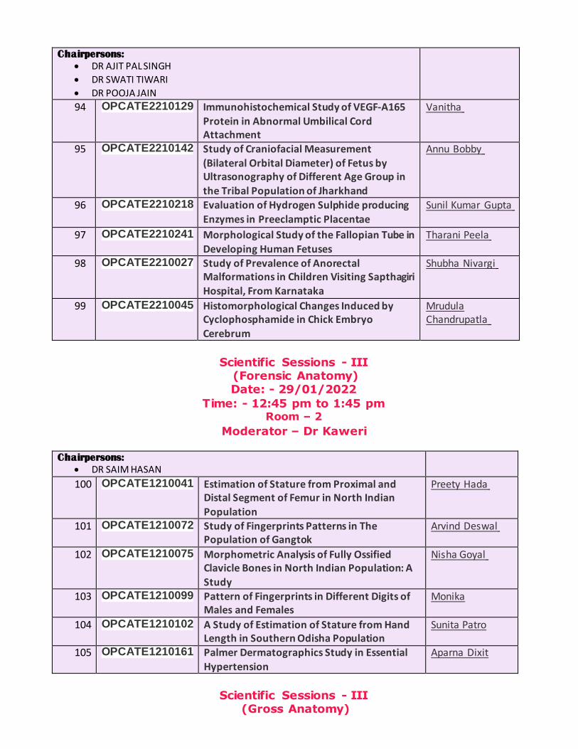
Chairpersons:
DR AJIT PAL SINGH
DR SWATI TIWARI
DR POOJA JAIN
94 OPCATE2210129
Immunohistochemical Study of VEGF-A165
Protein in Abnormal Umbilical Cord Attachment
Vanitha
95 OPCATE2210142
Study of Craniofacial Measurement
(Bilateral Orbital Diameter) of Fetus by Ultrasonography of Different Age Group in
the Tribal Population of Jharkhand
Annu Bobby
96 OPCATE2210218
Evaluation of Hydrogen Sulphide producing
Enzymes in Preeclamptic Placentae
Sunil Kumar Gupta
97 OPCATE2210241
Morphological Study of the Fallopian Tube in Developing Human Fetuses
Tharani Peela
98 OPCATE2210027
Study of Prevalence of Anorectal Malformations in Children Visiting Sapthagiri
Hospital, From Karnataka
Shubha Nivargi
99 OPCATE2210045
Histomorphological Changes Induced by Cyclophosphamide in Chick Embryo
Cerebrum
Mrudula Chandrupatla
Scientific Sessions - III (Forensic Anatomy) Date: - 29/01/2022
Time: - 12:45 pm to 1:45 pm Room – 2
Moderator – Dr Kaweri
Chairpersons:
DR SAIM HASAN
100 OPCATE1210041
Estimation of Stature from Proximal and Distal Segment of Femur in North Indian
Population
Preety Hada
101 OPCATE1210072
Study of Fingerprints Patterns in The Population of Gangtok
Arvind Deswal
102 OPCATE1210075
Morphometric Analysis of Fully Ossified Clavicle Bones in North Indian Population: A
Study
Nisha Goyal
103 OPCATE1210099
Pattern of Fingerprints in Different Digits of Males and Females
Monika
104 OPCATE1210102
A Study of Estimation of Stature from Hand Length in Southern Odisha Population
Sunita Patro
105 OPCATE1210161
Palmer Dermatographics Study in Essential Hypertension
Aparna Dixit
Scientific Sessions - III (Gross Anatomy)

Date: - 29/01/2022 Time: - 12:45 pm to 1:45 pm
Room – 3
Moderator – Dr Adya
Chairpersons:
DR ARCHANA SHEKOKAR
DR HARISH R GAIKWAD
106 OPCATE1210095 Morphometric study of grater palatine foramen in eastern region of India: a cross sectional study
Satabdi Sarkar
107 OPCATE1210100 Morphometric study of mandibular foramen
and its clinical implications in inferior alveolar nerve block in western Rajasthan population
Meenu Yadav
108 OPCATE1210101 A study on morphometry and articular facets types on human dry tali and calcanei of north Indian origin
Shavi Garg
109 OPCATE1210103 Morphometric characteristics of the lower end of fibula: variations and clinical relevance
Rajesh Kumar
110 OPCATE1210275 Morphometric analysis of Pterion, Asterion Asha Joshi 111 OPCATE1210108 Morphology & morphometry of calcaneal
spur in dry human calcanea Isha Marvania
Scientific Sessions - III
(Medical Education) Date: - 29/01/2022
Time: - 12:45 pm to 1:45 pm Room – 4
Moderator – Dr Nikhil
Chairpersons:
DR PUSHPALATHA K
DR NAVBIR PASRICHA
DR ROLI JOSHI
112 OPCATE3210114
Introducing Emergency Cricothyroidotomy in First Semester Medical Students in Relation to Anatomy Course
Anasuya Ghosh
113 OPCATE3210063
Enhancing the Embryology Teaching-Learning
Experience in the Medical Curriculum: A Faculty & Student Lookout
Sushma Prabhath
114 OPCATE3210247
Blended Learning Activities in Embryology post Covid to Increasing Impact of Learning
Roopashree Ramakrishna
115 OPCATE3210094
Learning Outcome and Perception of Didactic
Vs Interactive Quiz based Lecture in Anatomy for Medical Students
Akanksha Verma
116 OPCATE3210146
Teaching Cochlea by using The Moti Conch (Shankh) (Shell)
Kishor D Khushale

Scientific Sessions - III
(Imaging, Museum & Embalming techniques) Date: - 29/01/2022
Time: - 12:45 pm to 1:45 pm Room – 5
Moderator – Dr Rintu
Chairpersons:
DR SONIA SINGH
DR ANAMIKA GAHARWAR
DR POONAM PATNAIK
117 OPCATE4210087
The Current Practices of Storage and
Maintenance of Cadavers and Dissected Parts in Medical Colleges Of Udaipur
Hina Sharma
118 OPCATE4210157
A Digital Makeover of The Human Anatomy
Museum and its Impact on Teaching -Learning
Arvind Kumar
Pandey
119 OPCATE4210088
Age and Sex Related Changes in Pineal Gland Calcification
Soumya Mannarackal
120 OPCATE4210098
Use of Wet Specimens to Create Museum and to Enhance Teaching & Learning Abilities
Amarjyoti Chaturvedi
121 OPCATE4210113
Preparation of Corrosion Casts of The Lung B Subhash
122 OPCATE4210195
Embalming of donated human body during Covid-19 pandemic
Md Arboddin
Scientific Sessions - III (Gross Anatomy)
Date: - 29/01/2022 Time: - 12:45 pm to 1:45 pm
Room – 6
Moderator – Dr Honey
Chairpersons:
DR ANJU BALAJI MORE
DR VANDANA MEHTA
DR VRINDA HARI ANKOLEKAR
123 OPCATE1210110 Morphological and histological study of placenta in anaemic mother of Gwalior region
Reeta Kushwaha
124 OPCATE1210111 Surgical approach on anatomical proximity between mitral valve annulus and circumflex artery
Krishna G
125 OPCATE1210120 Morphological variations of the caudate
lobe of the liver Soram Malasana Devi
126 OPCATE1210123 Morphometric study of the mitral valve Jana Siva Koti
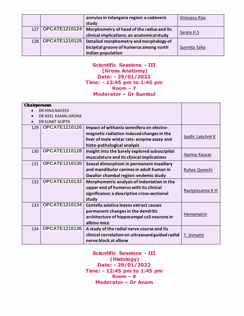
annulus in telangana region: a cadaveric study
Srinivasa Rao
127 OPCATE1210124 Morphometry of head of the radius and its
clinical implications: an anatomical study Sarala H.S
128 OPCATE1210125 Detailed morphometry and morphology of
bicipital groove of humerus among north Indian population
Susmita Saha
Scientific Sessions - III (Gross Anatomy)
Date: - 29/01/2022 Time: - 12:45 pm to 1:45 pm
Room – 7
Moderator – Dr Sumbul
Chairpersons
DR HINA NAFEES DR NEEL KAMAL ARORA
DR SUMIT GUPTA
129 OPCATE1210126
Impact of withania somnifera on electro-
magnetic radiation induced changes in the liver of male wistar rats- enzyme assay and histo-pathological analysis
Jyothi Lakshmi K
130 OPCATE1210128
Insight into the barely explored suboccipital musculature and its clinical implications
Aamna Kausar
131 OPCATE1210130
Sexual dimorphism in permanent maxillary and mandibular canines in adult human in
Gwalior chambal region: endemic study Ruhee Qureshi
132 OPCATE1210132
Morphometric analysis of indentation in the upper end of humerus with its clinical
significance: a descriptive cross-sectional study
Raviprasanna K H
133 OPCATE1210134
Centella asiatica leaves extract causes permanent changes in the dendritic architecture of hippocampal ca3 neurons in
albino mice
Hemamalini
134 OPCATE1210136
A study of the radial nerve course and its
clinical correlation on ultrasound guided radial nerve block at elbow
T. Srimathi
Scientific Sessions - III
(Histology) Date: - 29/01/2022
Time: - 12:45 pm to 1:45 pm Room – 8
Moderator – Dr Anam

Chairpersons:
DR ASEEM TANDON
DR MANISHA UPADHYAY DR POOJA RANI
135 OPCATE5210030
Exposural and Withdrawal Effects of 1900-2200
Mhz Mobile Phone Radiation on the Testis of the Wistar Rats - A Histological Study
Arjilli Vamsy
136 OPCATE5210054
A Study of Histogenesis and Distribution of Islets in Human Fetal Pancreas
Bharti Jakhar
137 OPCATE5210092
Morphological and Histological Study on Gall Bladder Of Mice
Mudassar Imam
138 OPCATE5210118
A Comparative Study on The Short-Term Effects
of Zoledronate and Pamidronate on The Proximal Metaphysis of Tibia in Young Albino
Rats- A Histological Study
Sarah Ralte
139 OPCATE5210151
Polarized Microscopic Study of Collagen in Oral
Submucous Fibrosis, Oral Squamous Cell Carcinoma and Oral Squamous Cell Carcinoma
with Oral Submucous Fibrosis
Shiny Vinila B
H
140 OPCATE5210152
A Histomorphometric study of Cypermethrin on Testis of Adult Albino Rats
Sonu
141 OPCATE5210317 Defective Trophoblast Invasion: Implications For The Pathogenesis of Preeclampsia
Arora Pallavi
Scientific Sessions – I (Physical Anthropology & Applied Anatomy)
Date: - 30/01/2022 Time: - 10:15 am to 11:15 am
Room – 1
Moderator – Dr Ankit
Chairpersons:
DR D. ASHA LATHA DR VINAY G
DR MAYANK KUMAR JAVIA
142 OPCATE1210205
Morphometric study on foramen transversaria of dried atlas vertebrae
Shital Sopanrao Maske
143 OPCATE1210206
A New Method for Symmetric Facial Reconstruction Using Face Symmetry Reference Plane
Pushpa.N.B
144 OPCATE1210221
Morphometric and Morphological Study of
Distal End Of Ulnae
K.Ramesh
145 OPCATE1210231
Estimation of Body Stature from Various Parameters of Hand an Anthropometric Study in Central Adult Indian Population
Kandregula Jyothirmayi
146 OPCATE1210234
Correlation between Hand and Foot Length
and Its Role in Stature Estimation for Personal Identification in Kashmiri Population
Farah Syed
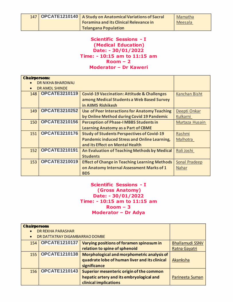
147 OPCATE1210140
A Study on Anatomical Variations of Sacral Foramina and its Clinical Relevance in
Telangana Population
Mamatha Meesala
Scientific Sessions - I
(Medical Education) Date: - 30/01/2022
Time: - 10:15 am to 11:15 am Room – 2
Moderator – Dr Kaweri
Chairpersons:
DR NIKHA BHARDWAJ
DR AMOL SHINDE
148 OPCATE3210119
Covid-19 Vaccination: Attitude & Challenges among Medical Students a Web Based Survey
in AIIMS Rishikesh
Kanchan Bisht
149 OPCATE3210252
Use of Peer Interactions for Anatomy Teaching by Online Method during Covid 19 Pandemic
Deepti Onkar Kulkarni
150 OPCATE3210156
Perception of Phase-I MBBS Students in
Learning Anatomy as a Part of CBME
Murtaza Husain
151 OPCATE3210176
Study of Students Perspectives of Covid-19 Pandemic induced Stress and Online Learning, and its Effect on Mental Health
Rashmi Malhotra
152 OPCATE3210191
An Evaluation of Teaching Methods by Medical
Students
Roli Joshi
153 OPCATE3210019
Effect of Change in Teaching Learning Methods
on Anatomy Internal Assessment Marks of 1 BDS
Sonal Pradeep
Nahar
Scientific Sessions - I (Gross Anatomy)
Date: - 30/01/2022 Time: - 10:15 am to 11:15 am
Room – 3 Moderator – Dr Adya
Chairpersons
DR REKHA PARASHAR
DR DATTATRAY DIGAMBARRAO DOMBE
154 OPCATE1210137
Varying positions of foramen spinosum in relation to spine of sphenoid
Bhallamudi SSNV Ratna Gayatri
155 OPCATE1210138
Morphological and morphometric analysis of quadrate lobe of human liver and its clinical
significance
Akanksha
156 OPCATE1210143
Superior mesenteric origin of the common
hepatic artery and its embryological and clinical implications
Parineeta Suman
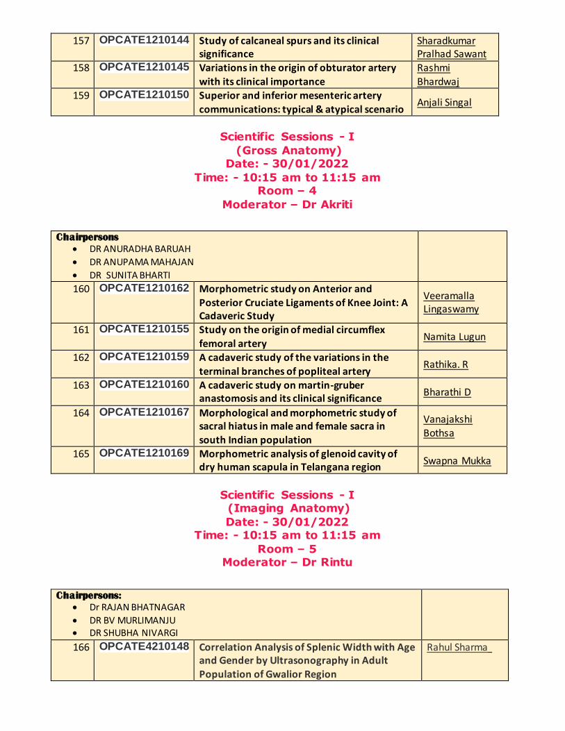
157 OPCATE1210144
Study of calcaneal spurs and its clinical significance
Sharadkumar Pralhad Sawant
158 OPCATE1210145
Variations in the origin of obturator artery
with its clinical importance
Rashmi
Bhardwaj 159 OPCATE1210150
Superior and inferior mesenteric artery
communications: typical & atypical scenario Anjali Singal
Scientific Sessions - I
(Gross Anatomy) Date: - 30/01/2022
Time: - 10:15 am to 11:15 am Room – 4
Moderator – Dr Akriti
Chairpersons
DR ANURADHA BARUAH
DR ANUPAMA MAHAJAN
DR SUNITA BHARTI
160 OPCATE1210162
Morphometric study on Anterior and
Posterior Cruciate Ligaments of Knee Joint: A Cadaveric Study
Veeramalla Lingaswamy
161 OPCATE1210155
Study on the origin of medial circumflex femoral artery
Namita Lugun
162 OPCATE1210159
A cadaveric study of the variations in the
terminal branches of popliteal artery Rathika. R
163 OPCATE1210160
A cadaveric study on martin-gruber anastomosis and its clinical significance
Bharathi D
164 OPCATE1210167
Morphological and morphometric study of sacral hiatus in male and female sacra in south Indian population
Vanajakshi Bothsa
165 OPCATE1210169
Morphometric analysis of glenoid cavity of dry human scapula in Telangana region
Swapna Mukka
Scientific Sessions - I (Imaging Anatomy)
Date: - 30/01/2022 Time: - 10:15 am to 11:15 am
Room – 5 Moderator – Dr Rintu
Chairpersons:
Dr RAJAN BHATNAGAR
DR BV MURLIMANJU DR SHUBHA NIVARGI
166 OPCATE4210148
Correlation Analysis of Splenic Width with Age and Gender by Ultrasonography in Adult Population of Gwalior Region
Rahul Sharma
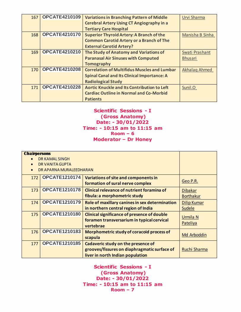
167 OPCATE4210109
Variations in Branching Pattern of Middle Cerebral Artery Using CT Angiography in a
Tertiary Care Hospital
Urvi Sharma
168 OPCATE4210170
Superior Thyroid Artery: A Branch of the Common Carotid Artery or a Branch of The
External Carotid Artery?
Manisha B Sinha
169 OPCATE4210210 The Study of Anatomy and Variations of
Paranasal Air Sinuses with Computed Tomography
Swati Prashant
Bhusari
170 OPCATE4210208
Correlation of Multifidus Muscles and Lumbar
Spinal Canal and Its Clinical Importance: A Radiological Study
Akhalaq Ahmed
171 OPCATE4210228
Aortic Knuckle and Its Contribution to Left Cardiac Outline in Normal and Co-Morbid
Patients
Sunil.O
Scientific Sessions - I
(Gross Anatomy) Date: - 30/01/2022
Time: - 10:15 am to 11:15 am Room – 6
Moderator – Dr Honey
Chairpersons
DR KAMAL SINGH DR VANITA GUPTA
DR APARNA MURALEEDHARAN
172 OPCATE1210174
Variations of site and components in formation of sural nerve complex Geo P.R.
173 OPCATE1210178
Clinical relevance of nutrient foramina of fibula: a morphometric study
Dibakar Borthakur
174 OPCATE1210179
Role of maxillary canines in sex determination in northern central region of India
Dilip Kumar Sudele
175 OPCATE1210180
Clinical significance of presence of double foramen transversarium in typical cervical vertebrae
Urmila N
Pateliya
176 OPCATE1210183
Morphometric study of coracoid process of scapula
Md Arboddin
177 OPCATE1210185
Cadaveric study on the presence of grooves/fissures on diaphragmatic surface of
liver in north Indian population
Ruchi Sharma
Scientific Sessions - I
(Gross Anatomy) Date: - 30/01/2022
Time: - 10:15 am to 11:15 am Room – 7
Moderator – Dr Sumbul
Chairpersons
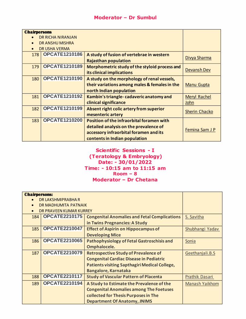
DR RICHA NIRANJAN
DR ANSHU MISHRA
DR USHA VERMA
178 OPCATE1210186
A study of fusion of vertebrae in western
Rajasthan population Divya Sharma
179 OPCATE1210189
Morphometric study of the styloid process and its clinical implications
Devansh Dev
180 OPCATE1210190
A study on the morphology of renal vessels, their variations among males & females in the
north Indian population
Manu Gupta
181 OPCATE1210192
Kambin's triangle- cadaveric anatomy and clinical significance
Meryl Rachel John
182 OPCATE1210199
Absent right colic artery from superior mesenteric artery
Sherin Chacko
183 OPCATE1210200
Position of the infraorbital foramen with detailed analysis on the prevalence of accessory infraorbital foramen and its contents in Indian population
Femina Sam J P
Scientific Sessions - I
(Teratology & Embryology) Date: - 30/01/2022
Time: - 10:15 am to 11:15 am Room – 8
Moderator – Dr Chetana
Chairpersons:
DR LAKSHMIPRABHA R DR MADHUMITA PATNAIK
DR PRAVEEN KUMAR KURREY
184 OPCATE2210175
Congenital Anomalies and Fetal Complications
in Twins Pregnancies: A Study
S. Savitha
185 OPCATE2210047
Effect of Aspirin on Hippocampus of
Developing Mice
Shubhangi Yadav
186 OPCATE2210065
Pathophysiology of Fetal Gastroschisis and Omphalocele.
Sonia
187 OPCATE2210079
Retrospective Study of Prevalence of Congenital Cardiac Disease in Pediatric Patients visiting Sapthagiri Medical College, Bangalore, Karnataka
Geethanjali.B.S
188 OPCATE2210117 Study of Vascular Pattern of Placenta Prathik Dasari
189 OPCATE2210194
A Study to Estimate the Prevalence of the Congenital Anomalies among The Foetuses collected for Thesis Purposes in The Department Of Anatomy, JNIMS
Manash Yaikhom
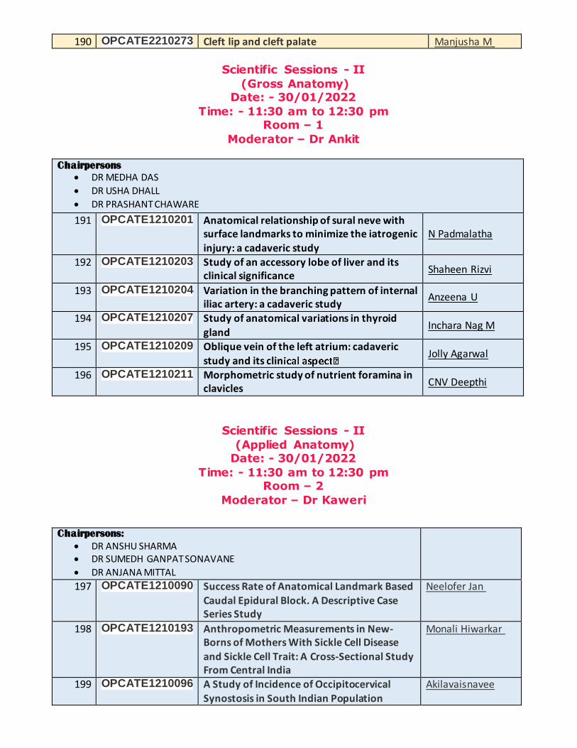
190 OPCATE2210273 Cleft lip and cleft palate Manjusha M
Scientific Sessions - II
(Gross Anatomy) Date: - 30/01/2022
Time: - 11:30 am to 12:30 pm Room – 1
Moderator – Dr Ankit
Chairpersons
DR MEDHA DAS
DR USHA DHALL
DR PRASHANT CHAWARE
191 OPCATE1210201
Anatomical relationship of sural neve with surface landmarks to minimize the iatrogenic injury: a cadaveric study
N Padmalatha
192 OPCATE1210203
Study of an accessory lobe of liver and its clinical significance
Shaheen Rizvi
193 OPCATE1210204
Variation in the branching pattern of internal iliac artery: a cadaveric study
Anzeena U
194 OPCATE1210207
Study of anatomical variations in thyroid
gland Inchara Nag M
195 OPCATE1210209
Oblique vein of the left atrium: cadaveric
study and its clin Jolly Agarwal
196 OPCATE1210211
Morphometric study of nutrient foramina in clavicles
CNV Deepthi
Scientific Sessions - II
(Applied Anatomy) Date: - 30/01/2022
Time: - 11:30 am to 12:30 pm Room – 2
Moderator – Dr Kaweri
Chairpersons:
DR ANSHU SHARMA DR SUMEDH GANPAT SONAVANE
DR ANJANA MITTAL
197 OPCATE1210090
Success Rate of Anatomical Landmark Based
Caudal Epidural Block. A Descriptive Case Series Study
Neelofer Jan
198 OPCATE1210193
Anthropometric Measurements in New-Borns of Mothers With Sickle Cell Disease
and Sickle Cell Trait: A Cross-Sectional Study From Central India
Monali Hiwarkar
199 OPCATE1210096
A Study of Incidence of Occipitocervical Synostosis in South Indian Population
Akilavaisnavee

200 OPCATE1210171
Anatomical Predictors of Difficult Intubation in Critical Care Patients: A Recent Approach
Alisha Rai
201 OPCATE1210154
Anatomical Distribution of Covid-19 Disease
Lesions on HRCT Chest in Kashmiri Population
Sayma Samoon
202 OPCATE1210253
Epimuscular Myofascial Force Transmission from the Gluteus Maximus Muscle to
Latissimus Dorsi Muscle: A Surface Electromyographic Study in Healthy Human
Adults
Sapna Marpalli
Scientific Sessions - II
(Gross Anatomy) Date: - 30/01/2022
Time: - 11:30 am to 12:30 pm Room – 3
Moderator – Dr Adya
Chairpersons
DR NITYA NAND SRIVASTAVA
DR SATYAM KHARE
DR URVASHI SINGH
203 OPCATE1210212
A morphometric study of acromion process of
dry scapulae and its clinical significance Akriti Dhan
204 OPCATE1210214
The relation between radial nerve lateral height and trans-epicondylar distance - a gross
anatomical study with a clinical perspective
Shinto G.B.
205 OPCATE1210215
Prevalence of hepatocystoduodenal and
hepatocystocolic ligaments in western Odisha population-a cadaveric study
Sarita Behera
206 OPCATE1210220
Topographical landmarks for the identification
of lateral calcaneal artery and sural nerve over the ankle and foot- a descriptive cadaveric
study
Arun Prasad S
207 OPCATE1210222
Bilateral asymmetry in the gonial index of
mandible: a radiomorphometric study in the north- Indian population
Shilpa Bathla
208 OPCATE1210223
Morphology and morphometry of flexor hallucis longus
P. Vijayan
Scientific Sessions - II (Medical Education)
Date: - 30/01/2022 Time: - 11:30 am to 12:30 pm
Room – 4
Moderator – Dr Nikhil

Chairpersons:
DR ALKA SINGH
DR ROHINI PUNJA DR GAYATRI GIRISH MUTHIYAN
209 OPCATE3210219
Effectiveness of Anatomy Review Sessions Held During Surgery Rotations
K. Smitha Elizabeth
210 OPCATE3210239
Stained Brain Sections: An Effective 3D
Tool to elucidate Sectional Neuroanatomy for Medical Students
Swati Tiwari
211 OPCATE1210093
Impact of Covid-19 Pandemic on Education, Physical & Social Life of Medical
Students: A Web Based Survey in AIIMS Rishikesh
Yashu Bhardwaj
212 OPCATE3210249
Impact of Covid 19 Pandemic on 1st MBBS
Students in Learning Anatomy; A Students Perspective
Kirti Sudhakarrao
Solanke
213 OPCATE3210044 Peer Group Teaching online during Pandemic and Students Perceptions
Uma SV
214 OPCATE3210296
The effects of Multimedia Instruction on
Cognitive Load in Medical Education
R.Vijaya
Scientific Sessions - II (Imaging Anatomy)
Date: - 30/01/2022 Time: - 11:30 am to 12:30 pm
Room – 5
Moderator – Dr Rintu
Chairpersons:
DR C S RAMESH BABU DR RUCHIRA SETHI
DR T. SRIMATHI
215 OPCATE4210235
Sonographic Assessment of Portal Vein
Diameter in Portal Hypertension Associated with Cirrhosis in South Indian Population
Syeda Nasreen
Fatima
216 OPCATE4210236
Optic canal: A CT Based Morphometric Study in North Indian Population
Eti Sthapak
217 OPCATE4210245
Evaluation of Umbilical Cord Cross Sectional Area as Predictor of Perinatal Outcome By Using Ultrasonography
Khizer Hussain
218 OPCATE4210279
Sonographic Assessment of Spleen Size in Corelation with Portal Hypertension Due to
Cirrhosis
Zuhayr Uddin
219 OPCATE4210281
Ultrasonographic study of liver size associated with portal hypertension
Mohammed Abdur Rehman
220 OPCATE4210311
Ultrasonographic Morphometric Analysis of Uterus in Nulliparous and Multiparous
Females attending Tertiary Care Hospital
Gaharwar Anamika

Scientific Sessions - II
(Gross Anatomy) Date: - 30/01/2022
Time: - 11:30 am to 12:30 pm Room – 6
Moderator – Dr Honey
Chairpersons
DR VIVEK SINGH MALIK
DR MAHENDRA KUMAR PANT
DR ROHINI MOTWANI
221 OPCATE1210225
A study of sex determination of human hip bone by total pelvic height and coxal index
Mridul Tripathi
222 OPCATE1210226
Study of shape and position of mental foramen in central Indian dried mandibles
N. Nagabhushanam
223 OPCATE1210227
Morphometry of suprarenal glands in adult Indian population
Yamini Markam
224 OPCATE1210230
A cadaveric study on accessory hepatic ducts
Fazila A
225 OPCATE1210237
Accessory foramen transversarium an
anatomical variation in the cervical spine: morphology and its clinical importance
Kalpana
226 OPCATE1210238
Morphometric study of coronary artery ostia
in cadaveric human hearts Runjhun Vijayvergia
Scientific Sessions - II
(Gross Anatomy) Date: - 30/01/2022
Time: - 11:30 am to 12:30 pm Room – 7
Moderator – Dr Sumbul
Chairpersons:
DR SHILPA GOSAVI
DR ASHOK KUMAR SRIVASTAVA
DR P K SANKARAN
227 OPCATE1210240 Morphometric study of nutrient foramen of
human tibia bone in western Rajasthan population
Jaya Purohit
228 OPCATE1210243 A morphometric study and variations of foramina transversaria of subaxial cervical vertebra in telangana region
Arshiya Yasmeen
229 OPCATE1210246 High division of brachial artery Mounica Katukuri
230 OPCATE1210250 Morphometric analysis of scapula in central India population
Roshni Chaturvedi
231 OPCATE1210251 A cadaveric study of the origin, relations and branching pattern of buccal branch of facial
Glory Davis

nerve
232 OPCATE1210258 Study of branching pattern of the left coronary artery in cadaveric hearts
Babli Gogoi
Scientific Sessions - II (Forensic Anatomy)
Date: - 30/01/2022 Time: - 11:30 am to 12:30 pm
Room – 8 Moderator – Dr Chetana
Chairpersons:
DR RAJESH ARORA
DR SHRIKANT VERMA DR BALAKRISHNAN RAMAMOORTHY
233 OPCATE1210181
A Study on Morphometry of The Human Mandible in Relation to Sex: A Preliminary Analysis
Tanushree Gurawa
234 OPCATE1210217
The Correlative Study of Human Body Stature With Foot Dimensions in North Indian
Population
Nupur Shukla
235 OPCATE1210254
Morphometric Analysis of Calcaneum Bones
in North Indian Population
Swati Saxena
236 OPCATE1210257
Morphological and Histological Findings in Body Organs in Aluminium Phosphide
Poisoning
Mrinal Patnaik
237 OPCATE1210264
A Comparative analysis of Dermatoglyphic
Pattern among South Indian Women with Hypothyroidism in Relation To Poly Cystic
Ovarian Syndrome
Panuganti
Amarnath
238 OPCATE1210271
Morphometric study of proximal and distal end of radius
Bhavanasri K
239 OPCATE1210272 A Morphological Study of Patterns of Lip Prints
Ashima Nag
Scientific Sessions - III (Physical Anthropology & Applied Anatomy)
Date: - 30/01/2022 Time: - 12:45 pm to 1:45 pm
Room – 1
Moderator – Dr Ankit
Chairpersons: DR GURDEEP SINGH KALYAN DR RUTA BAPAT
DR RAJANI SINGH

240 OPCATE1210255
Study of length of index and ring finger of hand in north Indian population
Hem Singh
241 OPCATE1210263
A Morphological Study of Lumbar Vertebral
Canal Transverse Diameter
Uma Maheswari
M 242 OPCATE1210280
Morphometric Analysis of Lumbar
Intervertebral Foramina at L3/4 and L4/5 Levels in Adult North Indian Population Using
Computed Tomography
Balamurali M
243 OPCATE1210282
Anthropometric Measurements of Type-2 Diabetic Patients With Special Emphasis on
Facial Features
Mrinmayee Deb Barma
244 OPCATE1210164
Morphometric analysis of occipital condyles on
dry human skulls
Hemant Ashish
Harode
245 OPCATE1210315
Distribution Of Nutrient Foramen In Scaphoid: A Dry Bone Study
Sumbul
Scientific Sessions - III
(Histology, Cytology & Immunology) Date: - 30/01/2022
Time: - 12:45 pm to 1:45 pm Room – 2
Moderator – Dr Kaweri
Chairpersons: DR SHARMISTHA BISWAS DR STUTI SRIVASTAVA
246 OPCATE5210182
A Study of Variations in Human Semen
Parameters according To Increase in Age
Shivani Chawla
247 OPCATE5210213
Tumor Angiogenesis and Immunomorphological Pattern of Lymph Nodes in Oral Squamous Cell Carcinoma (OSCC)
T Kalyani
248 OPCATE5210232
Study of Histological Variations of Placenta in Preeclampsia
P. Tanvi Vinod
249 OPCATE5210233
Histopathological effects of Formaldehyde Exposure on Lung of Wistar Albino Rat-An Experimental Study
Masooma Syed
250 OPCATE1210283
Variation of Myotendinous Junction with Muscles Tensile Strength
Pooja Bhadoria
251 OPCATE5210298
Osteoporotic Effect of Cissus Quadrangularis on the Apoptotic Changes of Lumbar Vertebrae in the Bilaterally Ovariectomized Wistar Albino Rats
Vishali. N
Scientific Sessions - III
(Gross Anatomy) Date: - 30/01/2022

Time: - 12:45 pm to 1:45 pm Room – 3
Moderator – Dr Chetana Sharma
Chairpersons: DR RAJANI ANIL JOSHI
DR PRATIMA JAISWAL
DR TEJASWI H L
252 OPCATE1210259 Gross architecture and architectural properties of muscles of anterior and lateral
compartment of leg
Gurpreet Kaur
253 OPCATE1210262
Morphology and morphometric study of talus in south Indian population
Asha Joshi
254 OPCATE1210268
A cadaveric study of variation in the pattern of origin of ascending pharyngeal artery in south Indian population
Shyamala B. Y.
255 OPCATE1210269
Morphometric study of foramen ovale in the
norma basalis with clinical correlation
Shushrutha K
256 OPCATE1210270 Effect of centrality of umbilical cord and vascular pattern of chorionic blood vessels on
foetal outcome
Ranjana Verma
257 OPCATE1210028 A morphological study of left ventricular false
tendons in human cadaveric hearts
Shalom Philip
Scientific Sessions - III
(Medical Education) Date: - 30/01/2022
Time: - 12:45 pm to 1:45 pm Room – 4
Moderator – Dr Nikhil
Chairpersons: DR SHEETAL B. JOSHI
DR GAURAV AGNIHOTRI DR RAKHI MILIND MORE
258 OPCATE3210300
Restore the glory of anatomy where the dead delights to act as simulator'
S Jayagandhi
259 OPCATE3210316
Effectiveness of Mind maps In Improving The Learning Outcome Of Undergraduate Students
Jyothi Lakshmi G.L
260 OPCATE3210197
Short duration videos: Its Effectiveness as an Educational Tool in revising Anatomy Topics
Upasana Sanyal
261 OPCATE3210188
The Effect of Incorporating Images in MCQ based Online Anatomy Assessment among First
Year Medical Students
Magi Murugan
262 OPCATE3210285
Contribution of Indian Authors in Top 20 Journals of Anatomy as Per Scimago Journal Ranking: An Analytical Study
Diwakar Dhurandhar
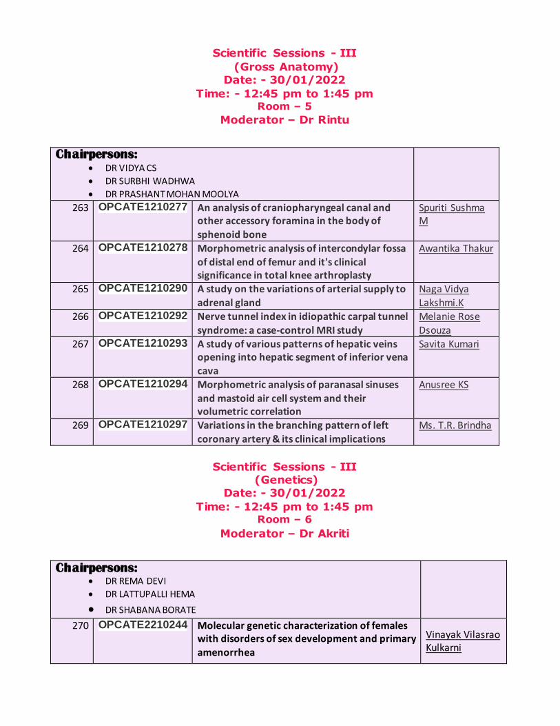
Scientific Sessions - III
(Gross Anatomy) Date: - 30/01/2022
Time: - 12:45 pm to 1:45 pm Room – 5
Moderator – Dr Rintu
Chairpersons: DR VIDYA CS
DR SURBHI WADHWA
DR PRASHANT MOHAN MOOLYA
263 OPCATE1210277
An analysis of craniopharyngeal canal and other accessory foramina in the body of
sphenoid bone
Spuriti Sushma M
264 OPCATE1210278 Morphometric analysis of intercondylar fossa of distal end of femur and it's clinical significance in total knee arthroplasty
Awantika Thakur
265 OPCATE1210290
A study on the variations of arterial supply to
adrenal gland
Naga Vidya
Lakshmi.K
266 OPCATE1210292 Nerve tunnel index in idiopathic carpal tunnel
syndrome: a case-control MRI study
Melanie Rose
Dsouza
267 OPCATE1210293
A study of various patterns of hepatic veins opening into hepatic segment of inferior vena
cava
Savita Kumari
268 OPCATE1210294 Morphometric analysis of paranasal sinuses
and mastoid air cell system and their volumetric correlation
Anusree KS
269 OPCATE1210297 Variations in the branching pattern of left
coronary artery & its clinical implications
Ms. T.R. Brindha
Scientific Sessions - III (Genetics)
Date: - 30/01/2022
Time: - 12:45 pm to 1:45 pm Room – 6
Moderator – Dr Akriti
Chairpersons: DR REMA DEVI
DR LATTUPALLI HEMA
DR SHABANA BORATE
270 OPCATE2210244 Molecular genetic characterization of females with disorders of sex development and primary amenorrhea
Vinayak Vilasrao Kulkarni

271 OPCATE2210256 To delineate the role of hla-g 14 bp insertion polymorphism in recurrent spontaneous
abortion
Khusru Nomani
272 OPCATE2210267 Phenotype-karyotype correlation of patients with primary amenorrhoea in north Kerala
Vidya K
273 OPCATE2210289
Study of the association of matrix metalloproteinase-9 gene polymorphism in preeclampsia
Shivani
274 OPCATE2210295 Mutations associated with immune-evasion in
sars-cov2 infection among cases of breakthrough walk-in patients•
Chetan Sahni
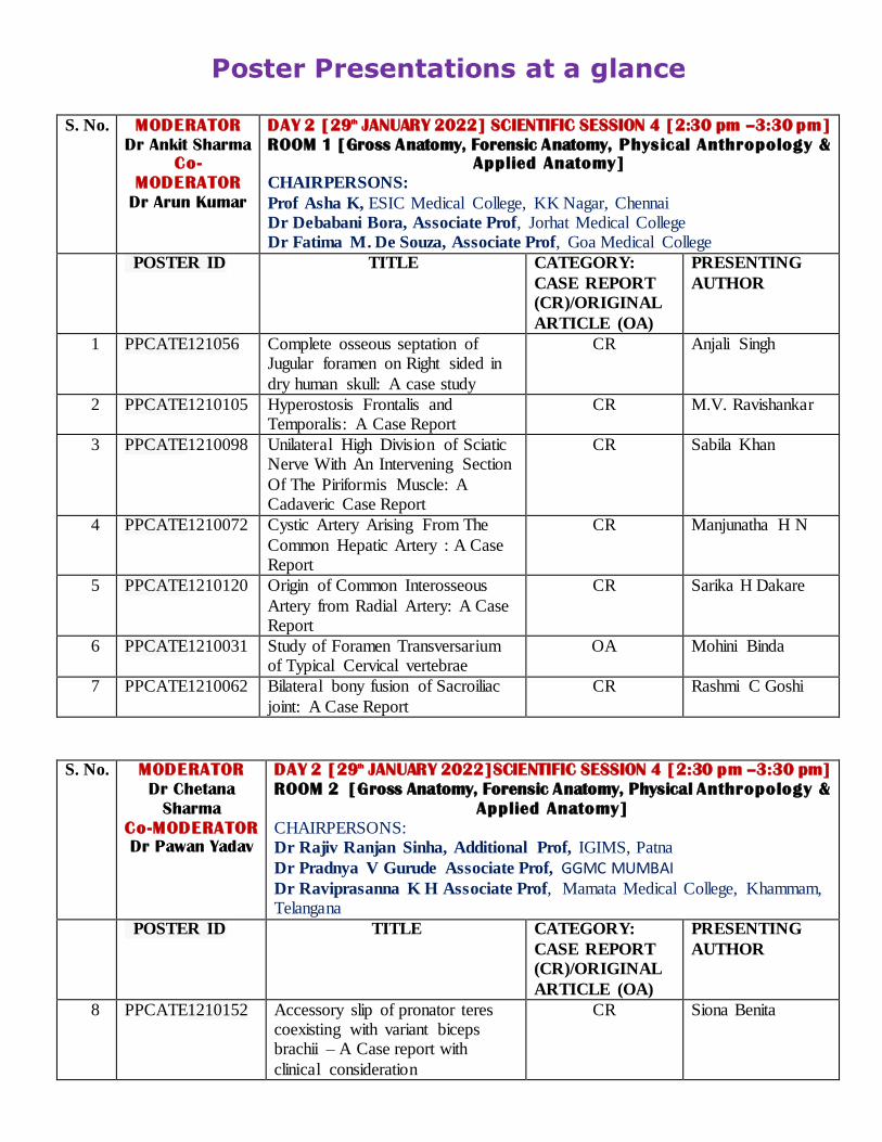
Poster Presentations at a glance
S. No. MODERATOR
Dr Ankit Sharma
Co-
MODERATOR
Dr Arun Kumar
DAY 2 [29th JANUARY 2022] SCIENTIFIC SESSION 4 [2:30 pm –3:30 pm]
ROOM 1 [Gross Anatomy, Forensic Anatomy, Physical Anthropology &
Applied Anatomy]
CHAIRPERSONS:
Prof Asha K, ESIC Medical College, KK Nagar, Chennai Dr Debabani Bora, Associate Prof, Jorhat Medical College Dr Fatima M. De Souza, Associate Prof, Goa Medical College
POSTER ID TITLE
CATEGORY:
CASE REPORT
(CR)/ORIGINAL
ARTICLE (OA)
PRESENTING
AUTHOR
1 PPCATE121056 Complete osseous septation of Jugular foramen on Right sided in
dry human skull: A case study
CR Anjali Singh
2 PPCATE1210105
Hyperostosis Frontalis and Temporalis: A Case Report
CR M.V. Ravishankar
3 PPCATE1210098
Unilateral High Division of Sciatic Nerve With An Intervening Section
Of The Piriformis Muscle: A Cadaveric Case Report
CR Sabila Khan
4 PPCATE1210072 Cystic Artery Arising From The
Common Hepatic Artery : A Case Report
CR Manjunatha H N
5 PPCATE1210120
Origin of Common Interosseous
Artery from Radial Artery: A Case Report
CR Sarika H Dakare
6 PPCATE1210031 Study of Foramen Transversarium of Typical Cervical vertebrae
OA Mohini Binda
7 PPCATE1210062
Bilateral bony fusion of Sacroiliac
joint: A Case Report
CR Rashmi C Goshi
S. No. MODERATOR
Dr Chetana
Sharma
Co-MODERATOR
Dr Pawan Yadav
DAY 2 [29th JANUARY 2022]SCIENTIFIC SESSION 4 [2:30 pm –3:30 pm]
ROOM 2 [Gross Anatomy, Forensic Anatomy, Physical Anthropology &
Applied Anatomy]
CHAIRPERSONS: Dr Rajiv Ranjan Sinha, Additional Prof, IGIMS, Patna
Dr Pradnya V Gurude Associate Prof, GGMC MUMBAI Dr Raviprasanna K H Associate Prof, Mamata Medical College, Khammam, Telangana
POSTER ID TITLE
CATEGORY:
CASE REPORT
(CR)/ORIGINAL
ARTICLE (OA)
PRESENTING
AUTHOR
8 PPCATE1210152
Accessory slip of pronator teres coexisting with variant biceps brachii – A Case report with
clinical consideration
CR Siona Benita
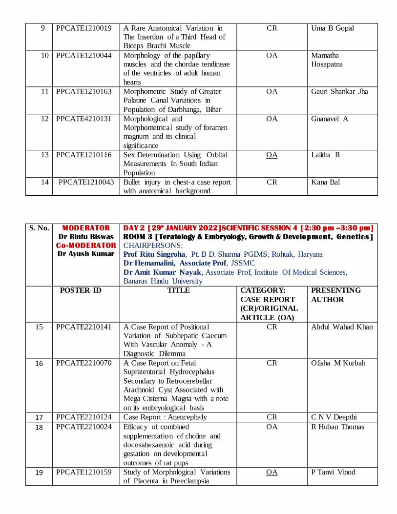
9 PPCATE1210019 A Rare Anatomical Variation in The Insertion of a Third Head of Biceps Brachi Muscle
CR Uma B Gopal
10 PPCATE1210044
Morphology of the papillary muscles and the chordae tendineae of the ventricles of adult human
hearts
OA Mamatha Hosapatna
11 PPCATE1210163 Morphometric Study of Greater Palatine Canal Variations in
Population of Darbhanga, Bihar
OA Gauri Shankar Jha
12 PPCATE4210131 Morphological and Morphometrical study of foramen magnum and its clinical
significance
OA Gnanavel A
13 PPCATE1210116 Sex Determination Using Orbital Measurements In South Indian
Population
OA Lalitha R
14 PPCATE1210043 Bullet injury in chest-a case report with anatomical background
CR Kana Bal
S. No. MODERATOR
Dr Rintu Biswas
Co-MODERATOR
Dr Ayush Kumar
DAY 2 [29th JANUARY 2022]SCIENTIFIC SESSION 4 [2:30 pm –3:30 pm]
ROOM 3 [Teratology & Embryology, Growth & Development, Genetics]
CHAIRPERSONS: Prof Ritu Singroha, Pt. B D. Sharma PGIMS, Rohtak, Haryana Dr Hemamalini, Associate Prof, JSSMC
Dr Amit Kumar Nayak, Associate Prof, Institute Of Medical Sciences, Banaras Hindu University
POSTER ID TITLE
CATEGORY:
CASE REPORT
(CR)/ORIGINAL
ARTICLE (OA)
PRESENTING
AUTHOR
15 PPCATE2210141
A Case Report of Positional Variation of Subhepatic Caecum With Vascular Anomaly - A
Diagnostic Dilemma
CR Abdul Wahad Khan
16 PPCATE2210070 A Case Report on Fetal Supratentorial Hydrocephalus
Secondary to Retrocerebellar Arachnoid Cyst Associated with Mega Cisterna Magna with a note
on its embryological basis
CR Ofisha M Kurbah
17 PPCATE2210124 Case Report : Anencephaly CR C N V Deepthi
18 PPCATE2210024
Efficacy of combined
supplementation of choline and docosahexaenoic acid during gestation on developmental
outcomes of rat pups
OA R Huban Thomas
19 PPCATE1210159
Study of Morphological Variations of Placenta in Preeclampsia
OA P Tanvi Vinod
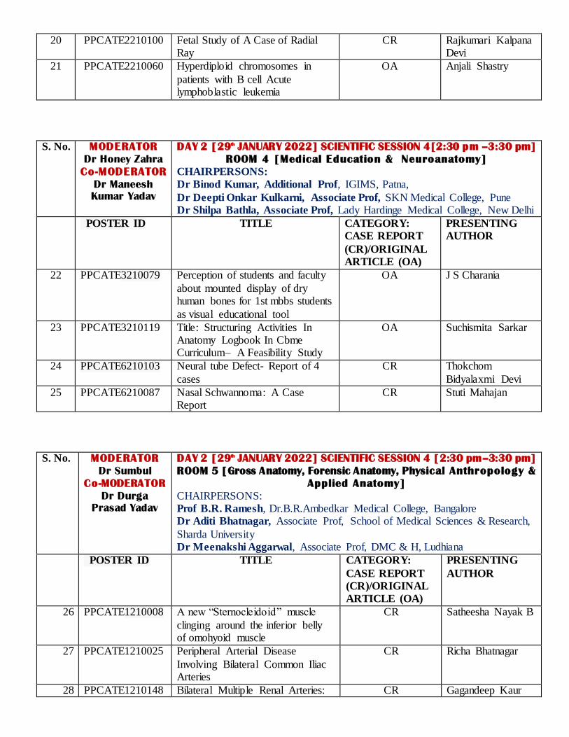
20 PPCATE2210100 Fetal Study of A Case of Radial Ray
CR Rajkumari Kalpana Devi
21 PPCATE2210060
Hyperdiploid chromosomes in
patients with B cell Acute lymphoblastic leukemia
OA Anjali Shastry
S. No. MODERATOR
Dr Honey Zahra
Co-MODERATOR
Dr Maneesh
Kumar Yadav
DAY 2 [29th JANUARY 2022] SCIENTIFIC SESSION 4[2:30 pm –3:30 pm]
ROOM 4 [Medical Education & Neuroanatomy]
CHAIRPERSONS: Dr Binod Kumar, Additional Prof, IGIMS, Patna,
Dr Deepti Onkar Kulkarni, Associate Prof, SKN Medical College, Pune
Dr Shilpa Bathla, Associate Prof, Lady Hardinge Medical College, New Delhi
POSTER ID TITLE
CATEGORY:
CASE REPORT
(CR)/ORIGINAL
ARTICLE (OA)
PRESENTING
AUTHOR
22 PPCATE3210079
Perception of students and faculty
about mounted display of dry human bones for 1st mbbs students
as visual educational tool
OA J S Charania
23 PPCATE3210119
Title: Structuring Activities In Anatomy Logbook In Cbme Curriculum– A Feasibility Study
OA Suchismita Sarkar
24 PPCATE6210103 Neural tube Defect- Report of 4
cases
CR Thokchom
Bidyalaxmi Devi
25 PPCATE6210087
Nasal Schwannoma: A Case Report
CR Stuti Mahajan
S. No. MODERATOR
Dr Sumbul
Co-MODERATOR
Dr Durga
Prasad Yadav
DAY 2 [29th JANUARY 2022] SCIENTIFIC SESSION 4 [2:30 pm–3:30 pm]
ROOM 5 [Gross Anatomy, Forensic Anatomy, Physical Anthropology &
Applied Anatomy]
CHAIRPERSONS: Prof B.R. Ramesh, Dr.B.R.Ambedkar Medical College, Bangalore Dr Aditi Bhatnagar, Associate Prof, School of Medical Sciences & Research,
Sharda University Dr Meenakshi Aggarwal, Associate Prof, DMC & H, Ludhiana
POSTER ID TITLE
CATEGORY:
CASE REPORT
(CR)/ORIGINAL
ARTICLE (OA)
PRESENTING
AUTHOR
26 PPCATE1210008
A new “Sternocleidoid” muscle
clinging around the inferior belly of omohyoid muscle
CR Satheesha Nayak B
27 PPCATE1210025
Peripheral Arterial Disease
Involving Bilateral Common Iliac Arteries
CR Richa Bhatnagar
28 PPCATE1210148 Bilateral Multiple Renal Arteries: CR Gagandeep Kaur

A Case Report
29 PPCATE1210007
Trifurcation Of Common Carotid Artery and Variation In Branching
Pattern Of External Carotid Artery
OA Rubina
30 PACATE1210128
Anatomical Variations Of Azygos Venous System: A Case Report
CR Rita Kumari
31 PPCATE1210092
An Anomalous origin of Superior Vesical Artery and its clinical
significance
CR Rashmi Bhardwaj
32 PPCATE1210169 Bifurcated Main Trunk of Spinal Accessory Nerve-A Case Report
CR Surabhi Sachan
S. No. MODERATOR
Dr Akriti Anand
Co-MODERATOR
Dr Charitra
Singh
DAY 2 [29th JANUARY 2022]SCIENTIFIC SESSION 5 [3:45 pm –4:45 pm]
ROOM 1 [Gross Anatomy, Forensic Anatomy, Physical Anthropology &
Applied Anatomy]
CHAIRPERSONS: Prof Shveta Swami, Kalpana Chawla Govt. Medical College Karnal Dr Deepak Khedekar, Associate Prof, Lokmanya Tilak Municipal Medical
College & General Hospital Dr Chiman Kumari, Associate Prof, PGIMER, Chandigarh
POSTER ID TITLE
CATEGORY:
CASE REPORT
(CR)/ORIGINAL
ARTICLE (OA)
PRESENTING
AUTHOR
33 PPCATE1210115 Lateral Supracondylar Spur of
Humerus – A Rare Case Report with Clinical and Developmental
Correlation
OA Kishore Sesham
34 PPCATE1210016
Bilateral variation in the branching pattern of subclavian artery: A
case report
CR Ashwini Aithal P
35 PPCATE1210112
Retrocaval Right Testicular Artery- A Case Report
CR Savitha S
36 PPCATE1210138
Accessory Belly of Abductor Digiti Minimi- A case report with
clinical consideration
CR A Arshia Fathima
37 PPCATE1210081
Study of the ulnar nerve in the hand from relevant bony
landmarks and its clinical implications
OA Rohini Punja
38 PPCATE1210093 Complete Absence of
Suprascapular Notch: A Case Report
CR Namita Lugun

S. No. MODERATOR
Dr Adya
Priyadarshini
Co-MODERATOR
Dr Ramesh
Chandra Rajak
DAY 2 [29th JANUARY 2022] SCIENTIFIC SESSION 5[3:45 pm –4:45 pm]
ROOM 2 [Gross Anatomy, Forensic Anatomy, Physical Anthropology &
Applied Anatomy]
CHAIRPERSONS: Prof Sherry Sharma, Punjab Institute of Medical Sciences, Jalandhar
Dr Hema N, Associate Prof, ESIC Medical College and PGIMSR, Bangalore Dr Maitreyee Mutalik, Associate Prof, Bharati Vidyapeeth Medical College, Pune
POSTER ID TITLE
CATEGORY:
CASE REPORT
(CR)/ORIGINAL
ARTICLE (OA)
PRESENTING
AUTHOR
39 PPCATE1210090 The Anatomical Case Study to Individuate the Bicuspid Aortic
Valve
CR Jalpa Dabhi
40 PPCATE1210080 Case Report: Agenesis of dorsal wall of Sacrum
CR Prajkta Ashok Thete
41 PPCATE1210068
A Variant of Communicating Branch between the
Musculocutaneous and Median Nerve: A Case Report
CR Chau Pingsaymang Manpoong
42 PPCATE1210130 Unilateral Double Renal Artery: A
Case Report
CR Md. Jawed Akhtar
43 PPCATE1210096 Nasal Parameters In Healthcare Workers– A Study In Medical
College And Associated Hospitals In Sub Himalayan Region
OA Ashwani Sharma
44 PPCATE1210038 A Morphological And Morphometric Study Of Calcaneus
Bone
OA Ashima Nag
45 PPCATE1210170 Variation In The Terminal Branching Pattern Of The Popliteal
Artery: A Case Report
CR Saba Anjum
S. No. MODERATOR
Dr Nikhil
Aggarwal
Co-MODERATOR
Dr Chandan
Kumar
DAY 2 [29th JANUARY 2022] SCIENTIFIC SESSION 5 [3:45 pm–4:45 pm]
ROOM 3 [Teratology & Embryology, Genetics]
CHAIRPERSONS:
Prof V. Subhadra Devi, Apollo Institute of Medical Sciences & Research, Chittoor, A P Dr Manjula Patil, Associate Prof, S. Nijalingappa Medical College, Bagalkot
Dr Sachin Patil, Associate Prof, GAIMS
POSTER ID TITLE
CATEGORY:
CASE REPORT
(CR)/ORIGINAL
ARTICLE (OA)
PRESENTING
AUTHOR
46 PPCATE2210077
Hydrops Fetalis: A fetal autopsy case report with rare associated
subhepatic caecum
CR Sapna Singh
47 PPCATE2210050 Outcome of Genetic counselling: OA Praisy Joy R

Experience from KK Nagar, Chennai
48 PPCATE2210056 Cor Triloculare Biventriculare: A
Case Report
CR Rashmi Avinash
Patil
49 PPCATE2210088 Craniorachiasis Observed In Aborted Foetus: Embryological Prospective And Clinical
Significance - A Case Report
CR Motiram Ramji Khandode
50 PPCATE2210150 Club Foot CR Manjusha M
51 PPCATE2210046
Mermaid Syndrome- A Case Report
CR Vinutha SP
52 PPCATE2210012
Sirenomelia In Connection With
Anencephaly And Spina Bifida: A Rare Case
CR Vishwajit R
Deshmukh
S. No. MODERATOR
Dr Kaweri
Dande
Co-MODERATOR
Dr Preeti
Kumari
DAY 2 [29th JANUARY 2022] SCIENTIFIC SESSION 5 [3:45 pm–4:45 pm]
ROOM 4 [IMAGING, MUSEUM & EMBALMING TECHNIQUES, BODY
DONATION]
CHAIRPERSONS: Prof Manoj P Ambali, Krishna Institute Of Medical Sciences Karad (MS) Dr Shweta Asthana, Associate Prof, RNT Medical College, Udaipur
Dr Lopamudra Mandal, Associate Prof, NRS Medical College
POSTER ID TITLE
CATEGORY:
CASE REPORT
(CR)/ORIGINAL
ARTICLE (OA)
PRESENTING
AUTHOR
53 PPCATE4210083
Study of variations of the middle meatus and their clinical relevance- An anatomical and CT study
OA Suhani Sumalatha
54 PPCATE4210094
Cervical Rib: Its prevalence in
Telangana Region
OA Murtaza Husain
55 PPCATE 321001
Kipple Feil Syndrome-A Case Report
CR P Ravishankar
56 PPCATE4210033 Voluntary Body Donation-Attitude
And Awareness Among Medical Students
OA Rahul Gaur
57 PPCATE4210010 Radiologic exploration of the
morphometric and morphological features of the carotid canal
OA Chandni gupta
58 PPCATE4210145
Incidental Finding On Absence Of Costal Element Of The Foramen
Transversarium Of Atlas Vertebrae
CR Aakash Shanmugam
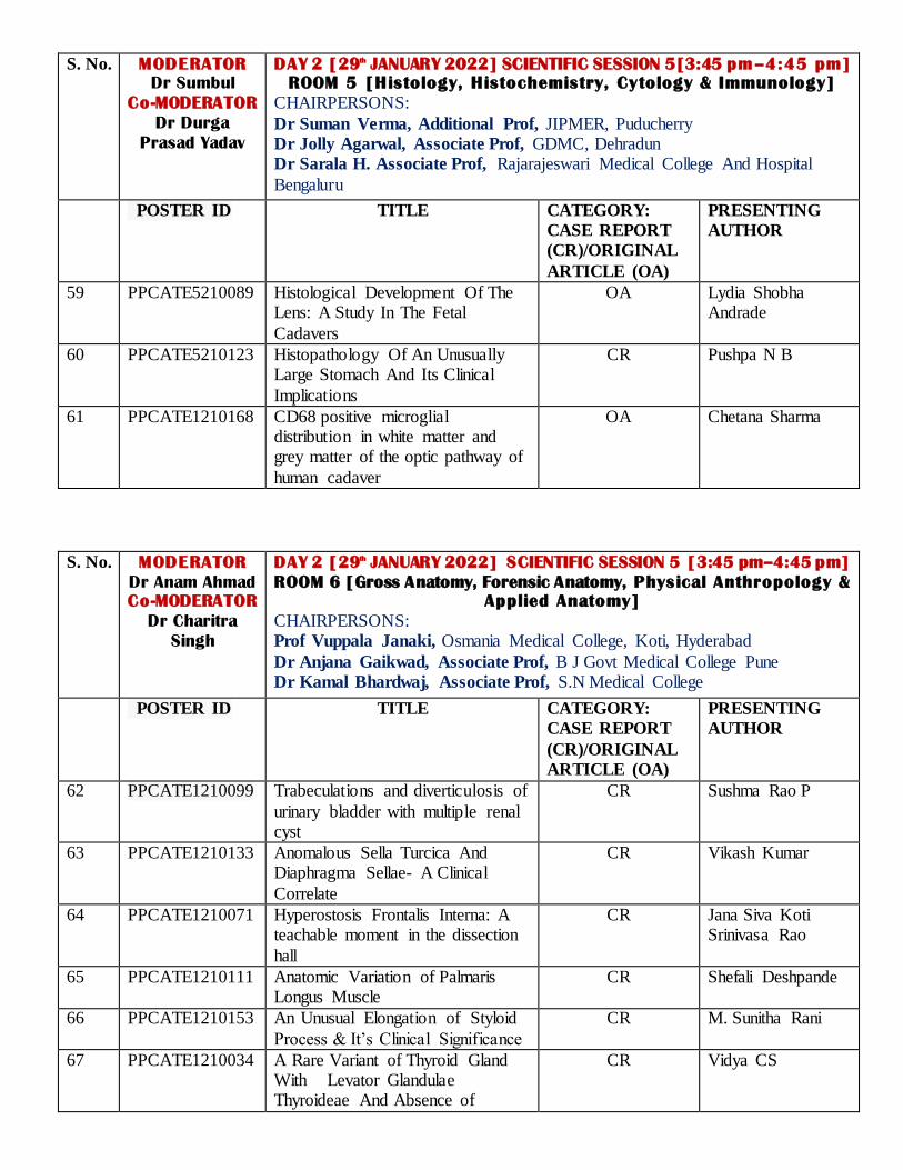
S. No. MODERATOR
Dr Sumbul
Co-MODERATOR
Dr Durga
Prasad Yadav
DAY 2 [29th JANUARY 2022] SCIENTIFIC SESSION 5[3:45 pm–4:45 pm]
ROOM 5 [Histology, Histochemistry, Cytology & Immunology]
CHAIRPERSONS:
Dr Suman Verma, Additional Prof, JIPMER, Puducherry Dr Jolly Agarwal, Associate Prof, GDMC, Dehradun Dr Sarala H. Associate Prof, Rajarajeswari Medical College And Hospital
Bengaluru
POSTER ID TITLE
CATEGORY:
CASE REPORT
(CR)/ORIGINAL
ARTICLE (OA)
PRESENTING
AUTHOR
59 PPCATE5210089
Histological Development Of The Lens: A Study In The Fetal
Cadavers
OA Lydia Shobha Andrade
60 PPCATE5210123
Histopathology Of An Unusually Large Stomach And Its Clinical
Implications
CR Pushpa N B
61 PPCATE1210168 CD68 positive microglial distribution in white matter and grey matter of the optic pathway of
human cadaver
OA Chetana Sharma
S. No. MODERATOR
Dr Anam Ahmad
Co-MODERATOR
Dr Charitra
Singh
DAY 2 [29th JANUARY 2022] SCIENTIFIC SESSION 5 [3:45 pm–4:45 pm]
ROOM 6 [Gross Anatomy, Forensic Anatomy, Physical Anthropology &
Applied Anatomy]
CHAIRPERSONS: Prof Vuppala Janaki, Osmania Medical College, Koti, Hyderabad
Dr Anjana Gaikwad, Associate Prof, B J Govt Medical College Pune Dr Kamal Bhardwaj, Associate Prof, S.N Medical College
POSTER ID TITLE
CATEGORY:
CASE REPORT
(CR)/ORIGINAL
ARTICLE (OA)
PRESENTING
AUTHOR
62 PPCATE1210099
Trabeculations and diverticulosis of
urinary bladder with multiple renal cyst
CR Sushma Rao P
63 PPCATE1210133
Anomalous Sella Turcica And Diaphragma Sellae- A Clinical
Correlate
CR Vikash Kumar
64 PPCATE1210071
Hyperostosis Frontalis Interna: A teachable moment in the dissection
hall
CR Jana Siva Koti Srinivasa Rao
65 PPCATE1210111 Anatomic Variation of Palmaris Longus Muscle
CR Shefali Deshpande
66 PPCATE1210153
An Unusual Elongation of Styloid
Process & It’s Clinical Significance
CR M. Sunitha Rani
67 PPCATE1210034
A Rare Variant of Thyroid Gland With Levator Glandulae Thyroideae And Absence of
CR Vidya CS
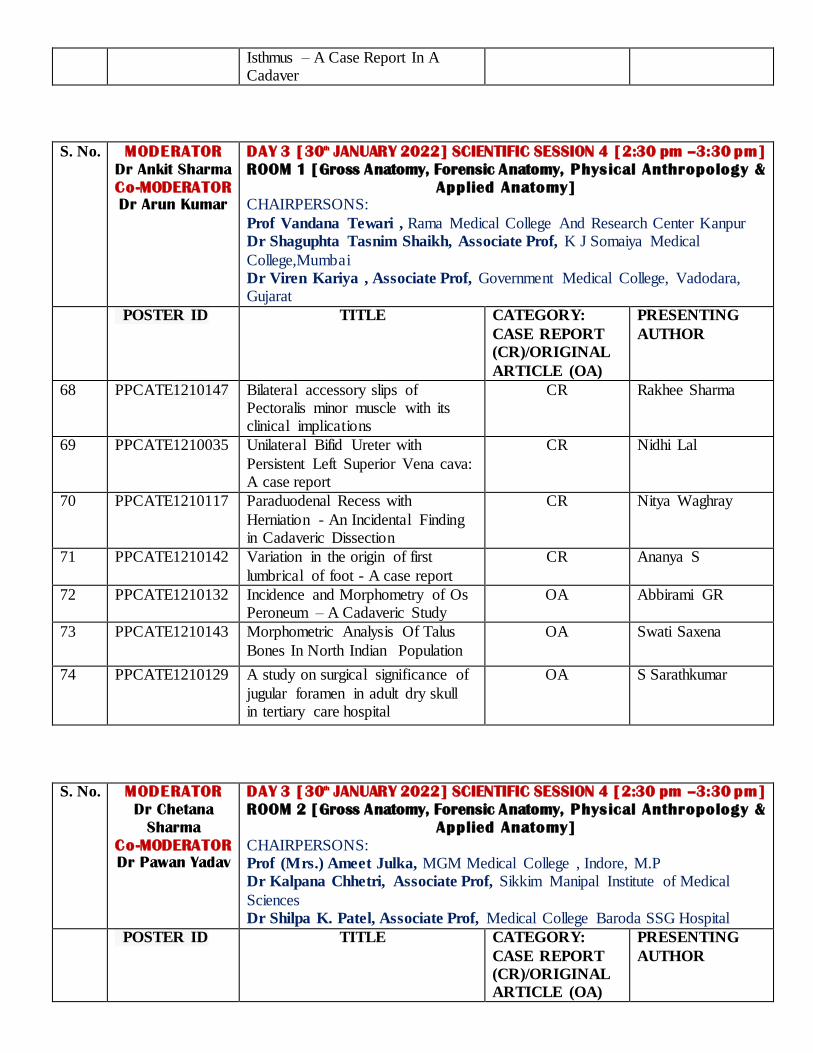
Isthmus – A Case Report In A Cadaver
S. No. MODERATOR
Dr Ankit Sharma
Co-MODERATOR
Dr Arun Kumar
DAY 3 [30th JANUARY 2022] SCIENTIFIC SESSION 4 [2:30 pm –3:30 pm]
ROOM 1 [Gross Anatomy, Forensic Anatomy, Physical Anthropology &
Applied Anatomy]
CHAIRPERSONS:
Prof Vandana Tewari , Rama Medical College And Research Center Kanpur Dr Shaguphta Tasnim Shaikh, Associate Prof, K J Somaiya Medical
College,Mumbai Dr Viren Kariya , Associate Prof, Government Medical College, Vadodara, Gujarat
POSTER ID TITLE
CATEGORY:
CASE REPORT
(CR)/ORIGINAL
ARTICLE (OA)
PRESENTING
AUTHOR
68 PPCATE1210147
Bilateral accessory slips of Pectoralis minor muscle with its clinical implications
CR Rakhee Sharma
69 PPCATE1210035 Unilateral Bifid Ureter with
Persistent Left Superior Vena cava: A case report
CR Nidhi Lal
70 PPCATE1210117 Paraduodenal Recess with
Herniation - An Incidental Finding in Cadaveric Dissection
CR Nitya Waghray
71 PPCATE1210142
Variation in the origin of first
lumbrical of foot - A case report
CR Ananya S
72 PPCATE1210132
Incidence and Morphometry of Os Peroneum – A Cadaveric Study
OA Abbirami GR
73 PPCATE1210143
Morphometric Analysis Of Talus
Bones In North Indian Population
OA Swati Saxena
74 PPCATE1210129 A study on surgical significance of
jugular foramen in adult dry skull in tertiary care hospital
OA S Sarathkumar
S. No. MODERATOR
Dr Chetana
Sharma
Co-MODERATOR
Dr Pawan Yadav
DAY 3 [30th JANUARY 2022] SCIENTIFIC SESSION 4 [2:30 pm –3:30 pm]
ROOM 2 [Gross Anatomy, Forensic Anatomy, Physical Anthropology &
Applied Anatomy]
CHAIRPERSONS: Prof (Mrs.) Ameet Julka, MGM Medical College , Indore, M.P Dr Kalpana Chhetri, Associate Prof, Sikkim Manipal Institute of Medical
Sciences Dr Shilpa K. Patel, Associate Prof, Medical College Baroda SSG Hospital
POSTER ID TITLE
CATEGORY:
CASE REPORT
(CR)/ORIGINAL
ARTICLE (OA)
PRESENTING
AUTHOR

75 PPCATE1210067 Anomalous Disposition Of Femoral Artery -A Case Report
CR Shabnam Arora
76 PPCATE1210064 Anomalous Origin of Obturator
Artery- A Case Report
CR Abhilasha Priya
77 PPCATE1210047
Variations in the cutaneous innervation of the dorsum of the foot
OA Vrinda Hari Ankolekar
78 PPCATE1210054 A Histomorphological Study of the
Posterior Interosseous Nerve (PIN) at Potential Compression Sites in
the Radial Tunnel and its Clinical Implications
OA Sharanya Rao
79 PPCATE1210166
Unilateral Absence Of External
Jugular Vein In a Human Cadaver
CR Bharan R
80 PPCATE1210023 Morphological and Morphometric Features of Cuboid Bone
OA Om Prakash Mali
S. No. MODERATOR
Dr Rintu Biswas
Co-MODERATOR
Dr Ayush Kumar
DAY 3 [30th JANUARY 2022] SCIENTIFIC SESSION 4 [2:30 pm–3:30 pm]
ROOM 3 [Teratology & Embryology, Genetics]
CHAIRPERSONS:
Prof Deepali D Deshatty, Rajarajeswari Medical College, Bangalore
Dr Kirti Sudhakarrao Solanke, Associate Prof, Smt Kashibai Navale Medical College Hospital Narhe Pune
Dr Asmita Sachin Jadhav, Associate Prof, Department of Anatomy, Government Medical College, Mira
POSTER ID TITLE
CATEGORY:
CASE REPORT
(CR)/ORIGINAL
ARTICLE (OA)
PRESENTING
AUTHOR
81 PPCATE2210069
Branchial cleft cyst: A case report and its embryological basis
CR Prajakta Kishve
82 PPCATE2210058 Robertsonian translocation associated with recurrent pregnancy
loss- A case report
CR Shivaleela C
83 PPCATE2210140
Congenital Diaphragmatic Hernia (CDH)
CR Mounica Katukuri
84 PPCATE2210109
Rare Discoid Type Variant of
Crossed Fused Renal Ectopia
CR Pankaj Rathi
85 PPCATE2210104 Study of Karyotype in Pediatric patients visiting Sapthagiri Hospital
from Karnataka region
OA N. Pramod
86 PPCATE2210045
Coloboma: Clinical Presentation, Embryological And Genetic Basis – A Case Report
CR Rakhi Milind More
87 PPCATE2210157 Crouzon’s Syndrome With A Dominant Sinus Pericranii Draining Transverse Sinus- Report Of A Rare
Association
CR Shweta Kumari
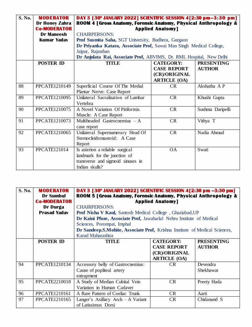
S. No. MODERATOR
Dr Honey Zahra
Co-MODERATOR
Dr Maneesh
Kumar Yadav
DAY 3 [30th JANUARY 2022] SCIENTIFIC SESSION 4[2:30 pm–3:30 pm]
ROOM 4 [Gross Anatomy, Forensic Anatomy, Physical Anthropology &
Applied Anatomy]
CHAIRPERSONS: Prof Susmita Saha, SGT University, Budhera, Gurgaon
Dr Priyanka Katara, Associate Prof, Sawai Man Singh Medical College, Jaipur, Rajasthan Dr Anjulata Rai, Associate Prof, ABVIMS, Dr. RML Hospital, New Delhi
POSTER ID TITLE
CATEGORY:
CASE REPORT
(CR)/ORIGINAL
ARTICLE (OA)
PRESENTING
AUTHOR
88 PPCATE1210149
Superficial Course Of The Medial Plantar Nerve: Case Report
CR Akshatha A P
89 PPCATE1210095
Unilateral Sacralization of Lumbar
Vertebra
CR Khushi Gupta
90 PPCATE1210075
A Novel Variation Of Piriformis Muscle: A Case Report
CR Sushma Daripelli
91 PPCATE1210073
Multiheaded Gastrocnemius – A
case report
CR Vithya T
92 PPCATE1210065
Unilateral Supernumerary Head Of Sternocleidomastoid: A Case Report
CR Nadia Ahmad
93 PPCATE121014 Is asterion a reliable surgical
landmark for the junction of transverse and sigmoid sinuses in
Indian skulls?
OA Swati
S. No. MODERATOR
Dr Sumbul
Co-MODERATOR
Dr Durga
Prasad Yadav
DAY 3 [30th JANUARY 2022] SCIENTIFIC SESSION 4[2:30 pm –3:30 pm]
ROOM 5 [Gross Anatomy, Forensic Anatomy, Physical Anthropology &
Applied Anatomy]
CHAIRPERSONS: Prof Nisha V Kaul, Santosh Medical College , Ghaziabad,UP
Dr Kaini Pfoze, Associate Prof, Jawaharlal Nehru Institute of Medical Sciences, Porompat, Imphal
Dr Sandeep.S.Mohite, Associate Prof, Krishna Institute of Medical Sciences, Karad Maharashtra
POSTER ID TITLE
CATEGORY:
CASE REPORT
(CR)/ORIGINAL
ARTICLE (OA)
PRESENTING
AUTHOR
94 PPCATE1210134
Accessory belly of Gastrocnemius:
Cause of popliteal artery entrapment
CR Devendra
Shekhawat
95 PPCATE2210018 A Study of Median Cubital Vein
Variation in Human Cadaver
CR Preety Hada
96 PPCATE1210161 A Rare Pattern of Coeliac Trunk CR Aarti
97 PPCATE1210165
Langer’s Axillary Arch – A Variant of Latissimus Dorsi
CR Chidanand S
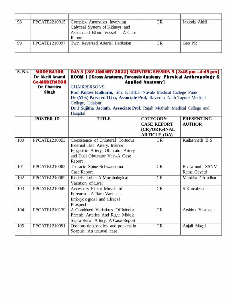
98 PPCATE2210015
Complex Anomalies Involving Calyceal System of Kidneys and
Associated Blood Vessels – A Case Report
CR Jakkula Akhil
99 PPCATE1210097 Twin Reversed Arterial Perfusion CR Geo PR
S. No. MODERATOR
Dr Akriti Anand
Co-MODERATOR
Dr Charitra
Singh
DAY 3 [30th JANUARY 2022] SCIENTIFIC SESSION 5 [3:45 pm –4:45 pm]
ROOM 1 [Gross Anatomy, Forensic Anatomy, Physical Anthropology &
Applied Anatomy]
CHAIRPERSONS:
Prof Pallavi Kulkarni, Smt. Kashibai Navale Medical College Pune Dr (Mrs) Parveen Ojha, Associate Prof, Ravindra Nath Tagore Medical
College, Udaipur Dr J Sujitha Jacinth, Associate Prof, Rajah Muthiah Medical College and Hospital
POSTER ID TITLE
CATEGORY:
CASE REPORT
(CR)/ORIGINAL
ARTICLE (OA)
PRESENTING
AUTHOR
100 PPCATE1210053 Coexistence of Unilateral Tortuous External Iliac Artery, Inferior Epigastric Artery, Obturator Artery
and Dual Obturator Vein-A Case Report
CR Kailashnath B S
101 PPCATE1210085 Thoracic Spine Schwannoma –
Case Report
CR Bhallamudi SSNV
Ratna Gayatri
102 PPCATE1210009
Riedel's Lobe: A Morphological
Variation of Liver
CR Manisha Chaudhari
103 PPCATE1210049
Accessory Flexor Muscle of Forearm – A Rare Variant -
Embryological and Clinical Prospect
CR S Kamalesh
104 PPCATE1210139
A Combined Variations Of Inferior Phrenic Arteries And Right Middle
Supra Renal Artery: A Case Report
CR Arshiya Yasmeen
105 PPCATE1210091
Osseous deficiencies and pockets in Scapula: An unusual case
CR Anjali Singal
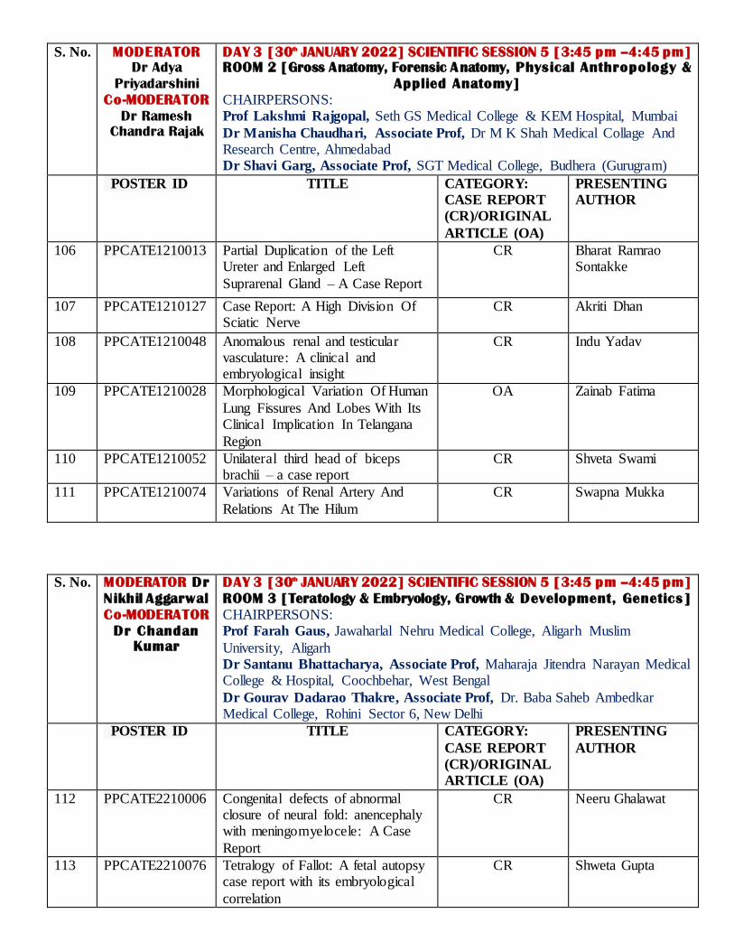
S. No. MODERATOR
Dr Adya
Priyadarshini
Co-MODERATOR
Dr Ramesh
Chandra Rajak
DAY 3 [30th JANUARY 2022] SCIENTIFIC SESSION 5 [3:45 pm –4:45 pm]
ROOM 2 [Gross Anatomy, Forensic Anatomy, Physical Anthropology &
Applied Anatomy]
CHAIRPERSONS: Prof Lakshmi Rajgopal, Seth GS Medical College & KEM Hospital, Mumbai
Dr Manisha Chaudhari, Associate Prof, Dr M K Shah Medical Collage And Research Centre, Ahmedabad Dr Shavi Garg, Associate Prof, SGT Medical College, Budhera (Gurugram)
POSTER ID TITLE
CATEGORY:
CASE REPORT
(CR)/ORIGINAL
ARTICLE (OA)
PRESENTING
AUTHOR
106 PPCATE1210013
Partial Duplication of the Left Ureter and Enlarged Left
Suprarenal Gland – A Case Report
CR Bharat Ramrao Sontakke
107 PPCATE1210127
Case Report: A High Division Of Sciatic Nerve
CR Akriti Dhan
108 PPCATE1210048 Anomalous renal and testicular vasculature: A clinical and embryological insight
CR Indu Yadav
109 PPCATE1210028
Morphological Variation Of Human
Lung Fissures And Lobes With Its Clinical Implication In Telangana
Region
OA Zainab Fatima
110 PPCATE1210052
Unilateral third head of biceps brachii – a case report
CR Shveta Swami
111 PPCATE1210074
Variations of Renal Artery And
Relations At The Hilum
CR Swapna Mukka
S. No. MODERATOR Dr
Nikhil Aggarwal
Co-MODERATOR
Dr Chandan
Kumar
DAY 3 [30th JANUARY 2022] SCIENTIFIC SESSION 5 [3:45 pm –4:45 pm]
ROOM 3 [Teratology & Embryology, Growth & Development, Genetics]
CHAIRPERSONS: Prof Farah Gaus, Jawaharlal Nehru Medical College, Aligarh Muslim
University, Aligarh Dr Santanu Bhattacharya, Associate Prof, Maharaja Jitendra Narayan Medical College & Hospital, Coochbehar, West Bengal
Dr Gourav Dadarao Thakre, Associate Prof, Dr. Baba Saheb Ambedkar Medical College, Rohini Sector 6, New Delhi
POSTER ID TITLE
CATEGORY:
CASE REPORT
(CR)/ORIGINAL
ARTICLE (OA)
PRESENTING
AUTHOR
112 PPCATE2210006
Congenital defects of abnormal closure of neural fold: anencephaly with meningomyelocele: A Case
Report
CR Neeru Ghalawat
113 PPCATE2210076
Tetralogy of Fallot: A fetal autopsy case report with its embryological
correlation
CR Shweta Gupta

114 PPCATE2210135 Developmental anomaly of left lobe of liver
CR Shubhangi Yadav
115 PPCATE2210039 Anencephaly: A case report CR Stuti Srivastava
116 PPCATE2210051
Genetic correlation of maternal
MTHFR gene and Neural Tube Defects
OA Kiran K
117 PPCATE2210020
OEIS Complex: A rare fetal autopsy case with its embryological
correlation
CR Shubhi Saini
118 PPCATE2210162 Trisomy ‘X’: An Incidental Finding during Cytogenetic Analysis
CR Honey Zahra
S. No. MODERATOR
Dr Kaweri
Dande
Co-MODERATOR
Dr Preeti
Kumari
DAY 3 [30th JANUARY 2022] SCIENTIFIC SESSION 5 [3:45 pm–4:45 pm]
ROOM 4 [Gross Anatomy, Forensic Anatomy, Physical Anthropology &
Applied Anatomy]
CHAIRPERSONS:
Prof Shaifaly Madan Rustagi, Army College Of Medical Sciences Delhi Dr Rajesh N. Dehankar, Associate Prof, NKP Salve Institute Of Medical Sciences & Rc Nagpur
POSTER ID TITLE
CATEGORY:
CASE REPORT
(CR)/ORIGINAL
ARTICLE (OA)
PRESENTING
AUTHOR
119 PPCATE1210032 Baker’ S Cyst: A Cadaveric Case
Report
CR Rashmi Jaiswal
120 PPCATE1210017
Persistent sciatic vein and
coexistent neuromuscular variations – case report
CR S. Sandy
121 PCATE1210106 Occipitalization of Atlas: A Case Report
CR Ashutosh Gupta
122 PPCATE1210061
Analysis of the Arch Height Index of the medial longitudinal arch of the foot using digital photography
in an adult Indian population: A cross-sectional study
OA Jyotsna B
123 PPCATE2210036
Abnormal Shapes Of Placenta And
Attachment Of Umbilical Cord And Its Clinical Implications
OA Sheik Reshma
Sultana
124 PPCATE1210030 Low Incidence of Complete Ossification of Superior Transverse
Scapular Ligament in Indian Population: Embryological
Perspective and Clinical Implications
OA Rajesh Kumar

S. No. MODERATOR
Dr Sumbul
Co-MODERATOR
Dr Durga
Prasad Yadav
DAY 3 [30th JANUARY 2022] SCIENTIFIC SESSION 5 [3:45 pm–4:45 pm]
ROOM 5 [Gross Anatomy, Forensic Anatomy, Physical Anthropology &
Applied Anatomy]
CHAIRPERSONS: Dr Shivaleela C, Associate Prof, Sri Siddhartha Medical College, Tumkur,
Karnataka Dr Ashima, Associate Prof, Shaheed Hasan Khan Mewati Govt Medical College, Nalhar Nuh
POSTER ID TITLE
CATEGORY:
CASE REPORT
(CR)/ORIGINAL
ARTICLE (OA)
PRESENTING
AUTHOR
125 PPCATE1210059
Metopic Suture in Dry Adult Skull: A Case Report
CR Isha Marvania
126 PPCATE1210029 Pentafurcation Of Left Main Coronary Artery: A Cadaveric
Study
OA Srijani Banerjee
127 PPCATE1210057
A Case Of Right-Sided Aortic Arch And Aberrant Left Subclavian Artery: Potential Cause For
Dysphagia Lusoria
CR Jashua Shaji Varghese
128 PPCATE1210144 An accessory parotid gland and its clinical significance
CR Babita Kujur
129 PPCATE1210027
Digastric Muscle: An Anatomical
Study with a Brief Clinical Perspective
OA Nandini Prashanth
Bhat
130 PPCATE1210113 Morphology of Sub-thebesian
Recess in the Cavo-tricuspid Isthmus
OA T Jahira Banu
S. No. MODERATOR
Dr Chetana
Sharma
Co-MODERATOR
Dr Pawan Yadav
DAY 3 [30th JANUARY 2022] SCIENTIFIC SESSION 5 [3:45 pm–4:45 pm]
ROOM 6 [Gross Anatomy, Forensic Anatomy, Physical Anthropology &
Applied Anatomy]
CHAIRPERSONS:
Prof Pratoshtha Potdar, Noida International Institute of Medical Science Dr Namrata Valimbe Reddy, Associate Prof, SAMC And PGI Indore
POSTER ID TITLE
CATEGORY:
CASE REPORT
(CR)/ORIGINAL
ARTICLE (OA)
PRESENTING
AUTHOR
131 PPCATE1210086
Variability In The Branching
Pattern Of The Internal Iliac Artery And Its Importance
CR Mamatha Meesala
132 PPCATE1210002
Bilateral Aberrant Origin Of Thyroid Arteries In A Single
Case– A Rare Variation
CR Ankita Saha
133 PPCATE1210066
Anomalous Head of Biceps Brachii-A Case Report
CR Swati Yadav
134 PPCATE1210026 The Correlation between Foot OA Natwar Lal Gaur

Length and Birth Weight Among Newborns
135 PPCATE1210155 Evaluation of angulation between
ipsilateral pedicle and contralateral lamina of seventh cervical vertebra- A morphometric study
OA Shradha Tamang
136 PPCATE121056 Complete osseous septation of
Jugular foramen on Right sided in dry human skull: A case study
CR Anjali Singh

Abstracts of Pre-Conference Workshop

Overview of Assisted Reproductive Techniques
Dr. Amita Pandey Professor, Department of Obstetrics & Gynecology,
King George’s Medical University, Lucknow, UP.
Infertility, defined as failure to achieve a clinical pregnancy after regular intercourse for 12 months is one of the main reproductive health disorders affecting a significant population worldwide Although global infertility prevalence rates are difficult to estimate, according to the Demographic and Health Survey, one in
every four to six couples in low-middle income countries are likely to be affected by infertility. The prevalence of infertility is estimated to grow in the coming years, considering that lifestyle factors such as
alcohol consumption, smoking, obesity, lack of physical activity, sexually transmitted diseases, which interfere negatively in female and male fertility, are increasing in the general population.
Since the first baby with assisted reproductive technology (ART) was conceived in 1978, the use of advanced technologies to overcome infertility has increased steadily and a large number of couples are turning to ART
for having a baby of their own. However, the availability of ART services varies around the world depending on the nature of healthcare system, economics, relative cost of treatment, availability of high-technology
services and government regulations.
What is ART? Assisted reproductive technology (ART) includes procedures that involve the in vitro handling of both human oocytes and sperm, or of embryos, with the objective of establishing a pregnancy. ART is used to treat a
variety of causes of infertility by collecting gametes, creating embryos from these in the laboratory and transferring the most viable embryo into the uterus. They do NOT include treatments in which only sperm
are handled (Intrauterine insemination) or procedures in which a woman takes ovulation inducing drugs to stimulate egg production without the intention of having eggs retrieved.
When do we need ART?
1. Moderate to severe Male factor infertility 2. Ovulatory disorders/ Donor oocyte needed 3. Severe Endometriosis
4. Tubal factor infertility 5. Need for ICSI
6. Need for PGD (Preimplantation Genetic Diagnosis) 7. Fertility preservation in cancer patients
Types of ART?
1. In vitro fertilization (IVF) IVF is one of the most successful options for infertility treatment, boasting a success rate of about 15-
25% per cycle depending upon the indication for which it is done. During an IVF cycle, the woman is given gonadotropins to stimulate follicular development. Once the follicles reach a designated size, the ova are retrieved from the ovaries and fertilized with sperms in the laboratory under controlled environment. The fertilized egg grows in a petri dish for a few days until it i s 6-8 cells or a blastocyst. It is then transferred to the woman’s uterus where it implants.

2. Intra cytoplasmic sperm injection (ICSI) ICSI is the preferred ART procedure in cases with severe male infertility due to very low sperm count, motility or few quality sperms. In this procedure following ovum pick-up, the mature egg is injected
with a single healthy sperm to fertilize it. Once the embryo develops it is transferred back to the woman’s uterus for implantation. Sometimes a man may have normal sperm count and morphology
but significant DNA damage which may decrease fertility or increases risk of miscarriage. ICSI allows the doctor to select the healthiest sperm and inject in the ova for fertilization. As ICSI is an add-on
procedure to IVF, it is more expensive than IVF alone. It may also be used in cases of obstructive azoospermia where sperms are aspirated from the testes using TESA, PESA or MESA.
3. Intrafallopian transfer
Some methods of ART are similar to IVF but use laparoscopic surgery to deliver the gametes directly into the fallopian tube. People may choose this method for religious reasons. This procedure carries the same risks as IVF along with the complications related to laparoscopy. Due to higher costs and risks of this type of ART, specialists rarely use these procedures. The three common types include: (a) Gamete intrafallopian transfer (GIFT): GIFT involves collecting eggs and sperm in a tube before
they are placed directly into the fallopian tubes using laparoscopic surgery. As there is no IVF procedure, a person does not have to choose which embryo to transfer.
(b) Zygote Intrafallopian Transfer (ZIFT): ZIFT is a combination of IVF and GIFT. Following retrieval, the ova is fertilized with sperms in the lab as in IVF and subsequently the zygote is returned to
the fallopian tubes. (c) Pronuclear stage tubal transfer (PROST): PROST is similar to ZIFT but involves the transfer of a
fertilized egg to the fallopian tube before cell division occurs.
4. Frozen embryo transfer (FET) FET involves thawing a frozen embryo previously developed following IVF and implanting it into the uterus. This is the same as IVF except that the embryos so prepared are frozen and then used later on for implantation after thawing. This procedure has become increasingly common and is as safe as
using fresh embryos.
5. Third-party ART This is a procedure where eggs or sperms are donated by different individuals to a couple and the
embryo is implanted in the female partner. It also includes surrogate and gestational carriers who may be inseminated with sperm from the couple or implanted with the embryo of the couple developed using ART. It is used by those who have difficulty producing eggs or sperms or i f they have difficulty in carrying pregnancy to term leading to repeated IVF failures.
Steps in ART cycle? 1. Ovulation Induction: Once couples have been prepared for treatment, Gonadotropins are initiated to
stimulate growth of multiple ovarian follicles, while at the same time other medications (GnRH agonists/ Antagonists) are given to suppress the natural menstrual cycle and down-regulate the
pituitary gland. 2. Follicular Monitoring: Ultrasonographic monitoring is undertaken at intervals to assess the growth of
follicles. 3. Ovulation trigger: Once the follicles have reached an appropriate size, hormones (HCG or
recombinant LH) are administered to bring about the final maturation of the eggs. 4. Ovum pick-up &/ or Sperm retrieval: The next step involves collection of ova from the ovaries
transvaginally (usually under anaesthesia and ultrasound guidance) within 36-40 hrs of ovulation
trigger. Simultaneously, sperm retrieval may be needed in cases of severe male infertility.

5. Insemination: Retrieved ova are inseminated with sperms for fertilization in a petri dish and incubated under controlled conditions. In cases with severe male factor infertility Intracytoplasmic sperm injection (ICSI) may be done.
6. Steps to aid fertilization may be needed in a few cases like special methods of embryo culture, co-culture, assisted hatching, etc.
7. Embryo/ Blastocyst transfer: Embryos are then transferred to the uterine cavity at 6-8 cell stage or blastocyst stage. Important issues include endometrial preparation, the best timing for embryo
transfer, how many embryos to transfer, what type of catheter to use and the use of ultrasound guidance.
8. Luteal phase support: Luteal phase support is given using progesterone, oestrogen (E2) and human chorionic gonadotrophin (hCG) for endometrial preparation and to improve endometrial receptivity
and implantation rates. Conclusion ART is a certainly a viable option for treatment of infertility. The success rates of different types of ART procedures vary according to the type of ART, the indication for the same and age & other health related
factors of the couple. Although there are known complications of ART like ovarian hyperstimulation syndrome, multiple pregnancy and risk of prematurity along with certain ethical issues, with patience, a
positive attitude and appropriate treatment, most infertile couples can eventually experience the joys of parenthood.
References
1. Kushnir V A, Barad D H, Albertini D F, Darmon S K,Gleicher N. Systematic review of worldwide trends in assisted reproductive technology 2004–2013. Reprod Biol Endocrinol 2017; 15:6. doi: 10.1186/s12958-016-0225-2.
2. Farquhar C, Marjoribanks J. Assisted reproductive technology: An overview of Cochrane Reviews. Cochrane Database Syst Rev. 2018 Aug; 2018(8): CD010537. doi: 10.1002/14651858.CD010537.pub5.

Structure and Physiology of Mature Human Sperm
Dr. Jyoti Chopra Professor, Department of Anatomy, King George’s Medical University, UP, Lucknow The exclusive function of the spermatozoa is to transmit the genetic information to the oocyte, and to
initiate and support the development of a new generation. A mature spermatozoon is a highly differentiated single cell that comprises of Head and Tail. The head is formed of nucleus and acrosome. The nucleus consists of metabolically inactive haploid chromatin. The anterior 40-70% nucleus is covered by acrosome cap made up of Golgi body. At the posterior end of the acrosomal region is Equatorial segment that does not take part in acrosomal reaction and is a point where fusion is initiated between the spermatozoon and oocyte during fertilization. Tail is the longest part of sperm which consist of neck, middle piece, principal piece and end piece. Proximal and distal centriole are present in the neck region orthogonally and help in
organizing the cleavage spindle during first cleavage division of the fertilized egg, induce the formation of implantation fossa and give origin to tail axoneme. The tail axoneme has 9+2 arrangement of microtubules.
Tail axoneme is surrounded by mitochondrial helix and outer dense fibers in mid-piece; fibrous sheath in principal piece and only plasma membrane in end piece. Sperms spin and do not swim. 3D microscope
technology has revealed that sperms rotate like a corkscrew.
Morphology of sperm is far from being the only factor considered for male fertility. It is important to know that all men produce abnormal sperm. For a sperm sample to be considered to have fertility potential, it only needs 4 percent or more of the sperm population to be considered normal, using the “strict” morphology criteria. If normal morpholology is observed in <4% sperms, the condition is termed as Teratozoospermia. If <42% normal sperm motility is observed, the condition is termed as Asthenozoosperma.
Semen Analysis & Sperm Function Tests
Dr. Mona Sharma
Additional Professor, AIIMS, Reproductive Biology, New Delhi
Infertility affects 15% couples worldwide and 1/3rd of cases are due to male infertility. Male infertility is commonly due to deficiencies in the semen, and semen quality is used as a surrogate measure of male fecundity. Semen analysis investigates male infertility (Sperm production, Patency of the male ducts, Function of the accessory glands etc.); Checks effectiveness of vasectomy, Selects donors for ART and Fertility Preservation. Conventional semen analysis is not adequate to predict pregnancy outcome. As even in cases of optimal semen parameters, there can be poor pregnancy outcome. The clinical assessment of male with semen analysis can guide clinician to determine how to proceed with further investigation and
management of the infertile couple. Clinically there is growing evidence that chromosomal anomalies and gene mutations underlie a diverse spectrum of semen analysis results associated with male infertility cases.
With absolute normal semen analysis parameters it may not be necessary to shift to specialized tests early but in cases with borderline parameters or with history of fertilization failure in past it becomes necessary to
do a battery of tests to evaluate different stages of fertilization/ detect function of a certain part of spermatozoon. An ideal Sperm Function Test should diagnose a specific sperm dysfunction, should predict fertilization and pregnancy rates and should indicate an appropriate therapy to alleviate the identified
sperm dysfunction. The World Health Organization (WHO) qualified sperm function tests as research tests. These tests were originally designed to evaluate the fertility potential of spermatozoa in vitro. But, over
time, these tests have become more predictive assays for pregnancy.

Genetics and epigenetics of male infertility
Rajender Singh Senior Principal Scientist, Central Drug Research Institute, Lucknow
Professor, Academy of Scientific and Industrial Research (AcSIR), Ghaziabad, India
Genetic mutations contribute to a significant number of male infertility cases. The identification of Y -chromosome deletions was the first milestone in this direction that led to the identification and classification
of azoospermia factor region on the Y-chromosome. Y-deletions explain about 13% of male infertility cases, the frequency of which varies across populations. This was followed by the identification of a number of
mutations in the autosomal genes, which explain a small percentage of male infertility cases, perhaps with an additive effect. Some of these genes include androgen receptor (AR), DAZL, estrogen receptor (ER), POLG, FSHR, GSTT1, GSTM1, GSTP1. Genetic mutations in a number of genes participating in spermiogenesis may also result in structural defects, classified as teratozoospermia. For example, mutations in DNAH1, Akap4, Meig1, Odf1, Sept2, Sept4, Tekt4, Spag16 have been reported to result in structural defects in sperm ta il formation. Similarly, mutations in Sun5 and SPATA6 have been reported in acephalic spermatozoa, and mutations in ZPBP1, SPATA16, DPY19L2 result in globozoospermia. Mutations in these gene have been
reported in sporadic and familial cases of male inferti lity. Research in the genetics of male infertility has recently escalated to the level of investigating epigenetic changes in the genes affecting spermatogenesis. In a recent study, we undertook a genome-wide 450K bead chip analysis on spermatozoal DNA from infertile and fertile males. A total of 1680 CpGs were differentially methylated, out of which 1436 were hypermethylated and 244 were hypomethylated. Out of 1052 genes with differential CpGs, we selected 15 for deep sequencing using MiSeq in another set of 32 infertile and 20 fertile individuals. Classification of
DMCs according to the genes was performed, which identified BCAN, CTNNA3, DLGAP2, GATA3, MAGI2 and TP73 among imprinted genes, SPATA5, SPATA7, SPATA16 and SPATA22 as spermatogenesis associated genes, KDM4C and JMJD1C, EZH2, HDAC4 among genes which regulate methylation and gene expression, HLA-C, HLA-DRB6 and HLA-DQA1 among complementation and immune response genes, and CRISPLD1,
LPHN3, CPEB2 among other genes. ANK2, PRDM1, TP73, GATA3 and VAX2, and MLPH, SMC1B, KCNJ5 are differentially methylated genes consistent with previous studies, which are strong candidates for inclusion in
infertility screening panels. We further analyzed a number of these CpG spots in the peripheral blood samples of infertile patients, finding that a number of these spots were differentially methylated in the
peripheral blood as well, making genetic screening further easier. We also asked if there was a general alteration in the methylation machinery in infertile cases. We found a higher expression of DNA methyltransferases (DNMT1 and DNMT3B) and ten-eleven translocation methylcytosine dioxygenases (TET1
and TET3) in the cases in comparison to controls, suggesting a general alteration of the methylation machinery in infertility.

Work Up of an Infertile Male
Dr. Praveen Kumar Pandey Ms Surgery, Mch(Uro), DNB
Consultant Urologist & Renal Transplantation Surgeon, Sahara Super Specialty Hospital, Lucknow
The problem of infertility affects approximately 15 to 20 % of couples, eventually subjecting one of six of them to this ever increasing disease. On a practical note, we may expect both men and women to equally provide faulty gametes. This fact highlights the importance of evaluation and treatment of males in order to solve this significant health care issue.
Recently, a considerable progress in assisted reproductive techniques (ART) has also substantially improved the ability of couples with infertility to have biological children. Still, evaluation and treatment of suc h males
might help these couples to conceive naturally. In addition, it might also improve the results of ART wherever it is required.
In this presentation, we aim to provide a comprehensive overview of the various assessment methods of
infertile males. Here, we have discussed contemporary methods for evaluating the infertile man, beginning with the basic workup that all suspected infertile men should undergo, followed by subsequent evaluation
steps. As our understanding of male fertility expands, the technologies available to diagnose and ultimately
treat it continue to evolve. Although the fundamental components of the evaluation have remained consistent, we will also discuss newer tools which are now available to assist in identifying the underlying
etiology.
We will conclude by providing a practical guide to entire process of evaluation of infertile males with pointers for referral to concerned specialities if required.
WHAT GOES INSIDE AN IVF LAB
Dr. Sarabpreet Singh
Director at The Fertilis Academy
IFS and ESHRE-certified Senior Clinical Embryologist
Senior Consultant & Chief Embryologist at the Artemis Health Institute, Gurgaon
It is said that your IVF program is as good as the lab which supports it. IVF lab is considered the sanctum
sanctorum of IVF facility. It has to be meticulously brought up, sincerely run & religiously maintained to ensure consistent results in terms of high pregnancy rates & low abortion rates which results in high live
birth rates.
In this lecture, we will introduce you to the design, set-up and workflow of an IVF lab. We will discuss the assisted reproduction techniques (ART) such as IVF & ICSI, their indications and procedural details. The
concept of gamete handling & gamete preparation for IVF/ICSI will be detai led.
Basics of embryonic development & embryo culture will be explained. It will be explained how cryobiology and cryopreservation of gametes and embryos has become the lifeline of IVF practice. Relevant videos of various procedures will be shown.
The technical challenges faced with IVF practice such as a tight control over the macro and microenvironment & how it can influence IVF outcome will be dealt with. One should be prepared to expect
the unexpected and be able to deal with it.
The recent developments and their value addition to IVF practice will be discussed.
In the end, we will emphasize on the importance of a robust quality control and quality assurance program in the IVF facility which is critical to achieve success in IVF.

Abstracts of CME & Guest Lectures

DNA fingerprinting and its medico-legal applications
K. Thangaraj
Director
Centre for DNA Fingerprinting and Diagnostics, Hyderabad
DNA is a genetic material that we inherit from our parents (50% from the father and 50% from the mother).
Similar to human fingerprints, DNA sequence also varies significantly from person to person, hence DNA
fingerprinting technology is being widely employed for individual identification. DNA fingerprinting
technique has gained much importance due to its efficient role in medico-legal applications. DNA
fingerprinting technology is a powerful tool, and understanding the basic facts about DNA and relevant
techniques is important. Hence, this talk would cover; historical aspects of DNA fingerprinting, evolution of
this technique, selection of appropriate genetic markers, and applications with appropriate example.
Forensic podiatry – Foot and footprint science
Kewal Krishan
PhD, FRAI, Professor and Former Chair, Department of Anthropology, Panjab University, Chandigarh
Forensic podiatry is comparatively a new scientific sub-discipline of forensic science, which deals with the
examination of foot and footprint evidence generally encountered at crime scenes. According to DiMaggio
and Vernon (2017), it is defined as the application of sound and researched podiatric knowledge and
experience in forensic investigations, to show the association of an individual with a scene of crime, or to
answer any other legal question concerned with the foot or footwear that requires knowledge of the
functioning foot. Forensic podiatrists contribute to the personal identification in crime scene investigations
whenever foot related evidence is recovered from the crime scene. The need to establish the identity of
dismembered remains may arise in cases of mass fatality incidents like terrorist attacks, mass murders,
transport accidents, tsunamis, floods, and earthquakes. Dismembered and mutilated remains are usually
encountered in these mass fatality incidents. There is an increased likelihood of the recovery of feet (often
enclosed in shoes), separated from the body in mass disasters such as high power explosions and bomb
blasts, airplane crashes and other high impact transportation accidents. In this regard, forensic podiatrists
can collect the evidence related to foot and help in the identification of the individuals from the foot and its
parts. Furthermore, forensic podiatrists conduct the examination of footprints generally recovered at the
crime scene. Footprints are commonly recovered at every crime scene in the form of bare footprints, socked
footprints or shoeprints.
There are many ways in which footprints can be used in establishing personal identification in
forensic podiatry. The analysis of the bare footprints involves the identification based upon class,
intermediate class, and individual characteristics of the footprints. For example, features like corns, pits,
ridges, humps, creases, hammertoe deformity, an extra toe, missing toe in the foot impression, flat
footedness are characteristics of the footprints which can be utilized as forensic evidence in establishing
personal identification. This kind of physical evidence can positively link a suspect to a crime, or it can prove
one’s innocence. By using different anthropometric methods, the stature, sex and body weight can also be
estimated from the footprints recovered at the crime scene. As stature and body weight can provide an idea
about the size of the individual, so they can give useful clues to a forensic scientist in criminal investigation.
Apart from these evidences in forensic podiatry, gait analysis and step/stride length analysis can also furnish
some indication about the criminals involved in a particular case.
The lecture will discuss various methods of personal identification related to the foot and footprint
evidence which is usually recovered from the crime scene or scene of occurrence in the form of
mutilated/dismembered remains, footprints, or questioned footwear.
An Introduction to Forensic Facial Reconstruction and the Drive to Advance the Field
Houlton TMR
BA (hons.) MSc PhD FRPS Facial Anthropologist Lecturer in Forensic Art & Facial Imaging
Centre for Anatomy & Human Identification University of Dundee (UK) Upon the recovery of skeletonised or heavily decomposed remains, it is not always immediately possible to establish identity through standard forms of forensic identification (i.e. DNA, fingerprint, dental record
comparison). Referencing skull morphology, a prediction of facial appearance may thus be rendered as an investigatory tool. Forensic facial reconstruction (also known as approximation) refers to this practice. The
methods applied exercise a high dependency on anatomy and practitioner dexterity, thus anatomists, anthropologists, and artists have commonly collaborated in this field. This presentation will deliver an insight
into the applications of forensic facial reconstruction, discussing the existing modes and methods of reconstruction used. It will highlight key conflicts with early face prediction protocols, and how this is stimulating major revisions within the field today.

Identification from Prints – A forensic approach
Prof. Anil Aggrawal
Retd. Professor, Department of Forensic Medicine, Maulana Azad Medical College, New Delhi
Identification is one of the major problems in the field of modern forensics. Be it the living or the dead, the
problem of identification keeps coming up almost on a daily basis. Besides DNA profiling, the most common – and most useful - method of identification remains fingerprints or dactylography. However, little is known about 6 other prints - Ear prints, Lip prints, Nose prints, Palate prints, tongue prints and tooth prints [Ameloglyphics]. This presentation aims to introduce these new and exotic forensic print technologies to the audience.

Cortical bone micro-anatomy for forensic identification purposes: Estimating age-at-death
in unknown individuals
Dr Julieta Gomez Garcia-Donas
MSc, PhD (Forensic Anthropology)
Lecturer in Forensic/Physical Anthropology
Centre for Anatomy & Human Identification University of Dundee (UK)
The purpose of forensic anthropology is the identification of unknown individuals through the analysis of
skeletal remains. In the identification process, the forensic anthropologist aims to build the biological profile
of the individual gathering key pieces of information such sex, age, ancestry and stature. When bones a re
highly fragmented or damaged, one of the only options for identification is through bone histomorphometry.
Cortical bone histomorphometry is the assessment of bone microstructure and microanatomy, mostly used for differentiating animal versus human remains and for estimating age-at-death.
This talk will introduce the audience to cortical bone histomorphometry in relation to age estimation.
The physiological and biological principles of human cortical bone histology with a focus on age -related
changes observed by the expert will be discussed. The parameters used for estimating age and their
reliability will be presented. The most important methods developed in the field of forensic anthropology will
be reviewed, covering key aspects related to skeletal elements and population-specific standards. Factors
affecting the accuracy of forensic microscopy for age estimation will be introduced covering current
limitations such as age, pathology and physical activity, among others.
Overall, this talk will offer an overview on bone microstructure and its applications, advantages, and
challenges for human identification, highlighting the state-of-art of histomorphometry in forensic
anthropology.
Key words: cortical bone, human identification, forensic anthropology, histomorphometry

Radiological imaging and identification
Tanuj Kanchan
Professor & Head, Department of Forensic Medicine &Toxicology, All India Institute of Medical Sciences,
Jodhpur
Human identification is one of the important tasks in the medicolegal practice. It includes establishing the
identity of unknown individuals, putrefied bodies, charred, skeletonised and mutilated human remains, etc.
In the dead, human identification is required in civil as well as criminal cases for insurance claims,
inheritance, and establishing a criminal act, etc.
Human identification involves generation of the biological profile of the deceased, which primarily consists
of estimation of sex, age, and stature. Traditionally, research pertaining to identification has been carried
out on skeletal remains. However, due to lack of contemporary skeletal repositories and the need for
utilizing resource intensive maceration procedures to obtain bones fit for studying, scientists have started to
use radiological imaging to conduct human identification research. Conventional radiography,
ultrasonography, computed tomography, and magnetic resonance imaging are some of the radiological techniques that are being used presently to conduct anthropological and anatomical research.
These radiological techniques have allowed scientists to estimate sex of an unknown individual using their
skull, dentition, mandible, long bones, sternum, pelvis, and patella, etc. Presently, researchers have
developed robust models to estimate age based on radiological visualization of the skull, dentition, clavicle,
sternum, long bones, ribs, vertebrae, and the pelvis. Likewise, the anatomists and anthropologists have generated models to estimate stature of an unknown individual using the radiology of long bones and skull.
Advent of newer radiological techniques has thus, allowed researchers to formulate newer ways of
generating the biological profile of an unknown individual and aid in human identification. Owing to
population variations, it is high time that standards are laid to develop population specific models using
radiological techniques globally.

Changing Art of Anatomy Illustrations and Potential of Illustration Software Dr Yogesh Sontakke
Additional Professor, Department of Anatomy, Jawaharlal Institute of Postgraduate Medical Education and
Research (JIPMER) (An Institution of National importance under the Ministry of Health and Family Welfare,
Government of India), Puducherry, India
One picture is worth more than a thousand words. The representation of the anatomy topics has a useful
illustration as an important domain. Drawing an anatomy illustration is an art. With the advent of science
and technology, the methods of anatomy illustrations are evolving at a high speed. The print version began
with black and white illustrations with difficult block preparation for printing. Computerization and
modernization of print versions and availability of drawing hardware have changed many concepts about the
illustrations. Black and white figures replaced with coloured line diagrams with flat 2D illustrations. Further,
they have changed to coloured illustrations with depth acquired on shading. Vector illustrations are more
clear and suitable for enlargement than the raster images. The vector s oftware are from coral draw to adobe
illustrator for print versions and Autodesk 3DS Max, and Autodesk Maya software for 3D-modelling. In
creation of any anatomy illustration, it follows the path as selection of suitable illustration – drawing a line
illustration with paper and pencil – transferring the line illustration into electronic format – selection of
colour combination – selection of direction of light – shading the illustration to enhance the depth. This
lecture will be supported with a live demonstration of illustration drawing using Adobe Illustrator software
and Wacom hardware.

Abstracts of Oral Presentations 1. Comparative study of facial dimensions among male and female medical students
Mohini Binda, Mahima Shrivastava
Department of Anatomy, JLN Medical College, Ajmer
Background: Facial anthropometry has well known implications for forensic scientists, criminologists and
physical anthropologists. The aim of this study was to compare facial dimensions of males and females and
draw inferences about facial index.
Material and Methods: This study was done on 100 male medical students and 100 female medical students
of age group 18-25 years in Department of Anatomy JLN Medical College, Ajmer. Measurements taken were
facial length and bizygomatic breadth using vernier callipers and facial index calculated. These parameters
were then compared.
Results: This study showed that in males mean facial length was 11.89cm mean bizygomatic breadth was
13.1 cm mean facial index was 90.89 2. In females mean facial length was 11.009cm mean bizygomatic
breadth was 12.21 cm mean facial index was 90.105
Conclusions: It was concluded that facial parameters were found higher in males than females exhibiting
sexual dimorphism.
2. Concavity Index and Disc Wedge Angle of Presacral Vertebra for Age and Gender in a Northeast
Indian Population
Kalpana Chhetri
Department of Anatomy, Sikkim Manipal Institute of Medical Sciences
Background: The lordotic wedging of the presacral disc protects the orthograde spine from the detrimental
axial loads and shearing forces. Age and mechanical pathophysiology disarrays typically affect fourth and
fifth lumbar segments causing low back ache. Reinstating optimal disc wedging is crucial in spinal
rehabilitation. The vertebral concavity index is a precise indicator of lumbar vertebral morphometry and disc
space essential for analysis of spinal symptoms and lumbar spinal surgical access.
Material and Methods: The fourth and fifth lumbar segments of twenty males and twenty females aged
between 18 to 69 years of age were studied in midsagittal cadaveric sections. The concavity index and disc
wedge angle were determined, disc and vertebral height were measured using digital vernier calliper. The
concavity index and disc wedge angle were determined, disc and vertebral height were measured using digital
vernier calliper.
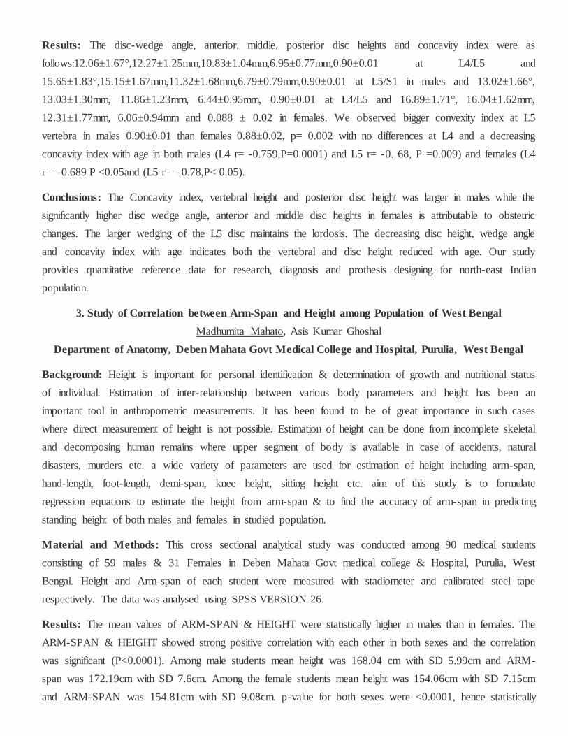
Results: The disc-wedge angle, anterior, middle, posterior disc heights and concavity index were as
follows:12.06±1.67°,12.27±1.25mm,10.83±1.04mm,6.95±0.77mm,0.90±0.01 at L4/L5 and
15.65±1.83°,15.15±1.67mm,11.32±1.68mm,6.79±0.79mm,0.90±0.01 at L5/S1 in males and 13.02±1.66°,
13.03±1.30mm, 11.86±1.23mm, 6.44±0.95mm, 0.90±0.01 at L4/L5 and 16.89±1.71°, 16.04±1.62mm,
12.31±1.77mm, 6.06±0.94mm and 0.088 ± 0.02 in females. We observed bigger convexity index at L5
vertebra in males 0.90±0.01 than females 0.88±0.02, p= 0.002 with no differences at L4 and a decreasing
concavity index with age in both males (L4 r= -0.759,P=0.0001) and L5 r= -0. 68, P =0.009) and females (L4
r = -0.689 P <0.05and (L5 r = -0.78,P< 0.05).
Conclusions: The Concavity index, vertebral height and posterior disc height was larger in males while the
significantly higher disc wedge angle, anterior and middle disc heights in females is attributable to obstetric
changes. The larger wedging of the L5 disc maintains the lordosis. The decreasing disc height, wedge angle
and concavity index with age indicates both the vertebral and disc height reduced with age. Our study
provides quantitative reference data for research, diagnosis and prothesis designing for north-east Indian
population.
3. Study of Correlation between Arm-Span and Height among Population of West Bengal
Madhumita Mahato, Asis Kumar Ghoshal
Department of Anatomy, Deben Mahata Govt Medical College and Hospital, Purulia, West Bengal
Background: Height is important for personal identification & determination of growth and nutritional status
of individual. Estimation of inter-relationship between various body parameters and height has been an
important tool in anthropometric measurements. It has been found to be of great importance in such cases
where direct measurement of height is not possible. Estimation of height can be done from incomplete skeletal
and decomposing human remains where upper segment of body is available in case of accidents, natural
disasters, murders etc. a wide variety of parameters are used for estimation of height including arm-span,
hand-length, foot-length, demi-span, knee height, sitting height etc. aim of this study is to formulate
regression equations to estimate the height from arm-span & to find the accuracy of arm-span in predicting
standing height of both males and females in studied population.
Material and Methods: This cross sectional analytical study was conducted among 90 medical students
consisting of 59 males & 31 Females in Deben Mahata Govt medical college & Hospital, Purulia, West
Bengal. Height and Arm-span of each student were measured with stadiometer and calibrated steel tape
respectively. The data was analysed using SPSS VERSION 26.
Results: The mean values of ARM-SPAN & HEIGHT were statistically higher in males than in females. The
ARM-SPAN & HEIGHT showed strong positive correlation with each other in both sexes and the correlation
was significant (P<0.0001). Among male students mean height was 168.04 cm with SD 5.99cm and ARM-
span was 172.19cm with SD 7.6cm. Among the female students mean height was 154.06cm with SD 7.15cm
and ARM-SPAN was 154.81cm with SD 9.08cm. p-value for both sexes were <0.0001, hence statistically
significant. Regression equation has been derived for estimation of height from the value of arm-span for male
& female individuals.
Conclusions: ARM-SPAN is a reliable tool for predicting the height of an individual and it can be used for
both males and females. It can also be used for the medico-legal Cases. This study will be useful for
anatomist, anthropologists, forensic experts, nutritionists & Physicians.
4. Morphometric Study of Head of Radius in Vidarbha Region and its Clinical Implications in Radial
Head Prosthesis
Purvi Mishra, M K Tabhane
Department of Anatomy, NKPSIMS & RC, Nagpur, Maharashtra
Background: Radius the lateral bone of forearm, has proximal end, distal end and a shaft. Proximal end
includes head, neck and tuberosity. Humero-radial joint permits flexion and extension movements at elbow
joint and superior radioulnar joint permits supination and pronation movements of forearm. The proximal end
of radius is very important clinically. Fractures of proximal end of radius are approximately 1.7%-5.4% of all
the fractures. Radial head fractures represent the most common elbow fracture, they occur when axial load is
applied on forearm, making radial head hit the humeral capitulum. Treatment includes sling and splints for
nondisplaced or minimally displaced fractures and aligned immobilization for displaced fractures. Surgical
options include open reduction, internal fixation fragment excision or radial head replacement. So, the present
study is centred around the morphometric analysis of head of radius in Vidarbha region as it important for the
radial head prosthesis.
Material and Methods: A cross-sectional study was conducted in the Department of Anatomy for a period of
2 years. Sample Size: 150 bones of both sides. Anatomical measurements were performed on radius using
digital Vernier calliper of accuracy 0.01 mm. Inclusion criteria- all dry radii available in department of
anatomy. Exclusion criteria damaged upper end, incomplete ossification, previous fracture or deformity were
excluded Measurement was taken by both authors to reduce bias. Following Parameters were recorded Shape
of radius, Anteroposterior Diameter of radial head (APD) Transverse Diameter of radial head (TD) Medial
Height of radial head (MH) Lateral Height of radial head (LH) Thickness of Ventral Curve (TVC), Thickness
of Lateral Curve (TLC) Thickness of Dorsal Curve (TDC), Depth of Superior Articular Facet (DH) detailed
Results and conclusion will be discussed in presentation.
Results: In the present study the mean values ± standard deviation were 20.18 ± 1.76 mm (APD), 19.52 ±
1.70 mm (TD), 9.49 ± 1.30 mm (MH), 6.74 ± 1.16 mm (LH), 5.35 ± 0.86 mm (TVC), 4.87 ± 0.82 mm (TDC),
4.74 ± 0.85 mm (TLC), 2.32 ± 0.55 mm (DH), 319.63 ± 55.50 mm (SA), 365.58 ± 125.51 (VOL). The mean±
and standard deviation, minimum and maximum values of morphometric parameters of right side (n=76) and
left side (n=74) radius were compared. When the difference between parameters of right side and left side
radius was tested, the difference was not statistically significant (p < 0.05) The correlation between depth vs.

surface area, depth vs. volume and surface area vs. depth was statistically highly significant (p<0.00001). The
correlation between depth vs. surface area was found to be weakly positively correlated, whereas depth vs.
volume and surface area vs. depth was found out to have strong positive correlation.
Conclusions: The correlation between depth vs. surface area, depth vs. volume and surface area vs. depth was
statistically highly significant (p<0.00001). The correlation between depth vs. surface area was found to be
weakly positively correlated, whereas depth vs. volume and surface area vs. depth was found out to have
strong positive correlation. The head of radius is a fundamental element for the physiological or prosthetic.
Stability of the elbow and superior radioulnar joint. The total articular circumference does not articulate with
the radial notch in the pronation-supination motion.
5. Sexual Dimorphism of Human Mandible in Marathwada Region
Swati Prashant Bhusari , Sonal Nahar, Prashant A. Bhusari
Department of Anatomy, MGV KBH Dental College and Hospital, Nashik
Background: The only movable bone in skull is the mandible or lower jaw. It articulates with paired
temporal bones at the synovial temporo-mandibular joints. It is the largest and strongest bone of face.
Identification of mandible is important for medico-legal purpose and in anthropology. Sexual dimorphism
refers to differences in size, stature and appearances between male and female. The aim was 1) to study the
human mandible as one of the indicators of sexual dimorphism. 2) to study the importance of various
parameters of human mandible in the sexual dimorphism 3) to compare our study with available literatures
Material and Methods: A total number of 120 (78 male and 42 female) dry mandibles were taken from 3
medical colleges. The measurements were taken with the help of various instruments.
Results: Eight mandibular parameters were taken and data on all these parameters on 120 mandibles obtained
and statistically evaluated. For each parameter data on mean, standard deviation, range, calculated range,
demarcating point and percentage of mandible sexed correctly. Skewness and Kurtosis are evaluated
separately for male and female. All the parameters are subjected to routine statistical methods by applying ‘t’
test and getting the p values. The t test for bigonial diameter is highly significant. Demarcating point for male
mandibles is > 99.06 & for female mandibles is <81.64.The t test is highly significant for length of mandible,
demarcating point for male mandibles is >198.05. Similarly t test values for mandibular body height,
mandibular body thickness, bicondylar breadth, condylar width and angle of mandible are highly significant
with demarcating values. The t value for distance between two coronoid tips is significant, demarcating point
for female mandibles is <73.50.
Conclusions: Highly significant parameters: Bigonial diameter, Length of mandible, Mandibular body height,
Mandibular body thickness, Bicondylar breadth, Condylar width. Significant parameter was - Distance
between two coronoid tips.

6. How Beautiful are Noses of Haryanvis?
Khushi Gupta , Shalini Choudhary, Arvinder Pal Singh Batra
Department of Anatomy, Bhagat Phool Singh Government Medical College, Khanpur Kalan, Sonipat
Background: Beauty lies in the eyes of beholder. however, there are certain faces and facial features that
attract our attention immediately and are very appealing to us. These faces follow the golden ratio, also called
the divine proportion or fibonacci ratio or phi. The greeks discovered that phi (1.618) was the ideal proportion
of two parts of any object. In the present study, the ratio of nasal length: nasal width taken is 1.618:1.
Material and Methods: The study was done in the Department of Anatomy, BPS GMC for Women,
Khanpur Kalan Sonipat, Haryana. In the present study 200 healthy volunteers (100 males and 100 females) of
18 to 50 years of age group, having descent of Haryana were taken. Sliding calliper was used to take nasal
length and nasal width. The head was held in Frankfurt plane and in a neutral expression.
Results: One male, out of the 200 individuals, fitted the perfect nose criteria. Therefore, the prevalence of the
Golden Ratio nose amongst 200 individuals from Haryana was only 0.5%.
Conclusions: A beautiful nose may depend on a lot of other nasal features than just the nasal length and nasal
width ratio. The width of the nose tends to expand with age. The skin loses elasticity and becomes saggy with
time. This means, even faces that were earlier perfect will tend to deviate from the golden ratio with age.
Thus, we can safely say that we may or may not have the Golden Ratio nose at some point of time in our
lives, but it will not remain so forever.
7. Morphometric study of the sternocleidomastoid muscle
Jigyasa Passey, Pareesa Rabbani, Shayama Kumari Razdan
Department of Anatomy, Hamdard Institute of Medical Science and Research
Background: The sternocleidomastoid muscle is a flexor of the neck and an accessory muscle of
inspiration.the knowledge of its morphology and variation is important during non-invasive and invasive
reconstructive or rehabilitative procedures of the head and neck.
Material and Methods: We studied the morphometry of the Sternocleidomastoid muscle of 20 cadavers at
Department of Anatomy, Hamdard Institute of Medical Sciences & Research, New Delhi. We noted
Sternocleidomastoid under the following headings- 1. Variations in number of heads & its morphometric
analysis 2. Maximum Length & Breadth of sternal head
Results: Variations in the attachment of the muscle were found in 6 (15%) specimens. All had unilateral
variations. In each specimen, the modes of presentation of the muscle and its fusion with the major bulk were
also different. Mean Maximum Length & Breadth of sternal head was 10cm+-2.5cm and 1.5cm+-0.5cm.
Mean Maximum Length & Breadth of clavicular head 8.2cm+-2.2cm and 2.5+-1cm respectively.

Conclusions: In this study, we found variations in the attachment of the muscle in 10% of the specimens
dissected. Surgeons should bear in mind such variations when performing neck surgeries.
8. The prevalence and distribution of the variants of gantzer's muscle: a meta-analysis of cadaveric
studies
Adil Asghar, Rakesh Kumar Jha, Apurba Patra, Binita Chaudhary, Brijendra Singh
Department of Anatomy, All India Institute of Medical Sciences, Patna
Background: The gantzer's muscle is often present in the flexor compartment of the forearm. It lies
underneath of flexor digitorum superficialis and compresses the anterior interosseous nerve. Furthermore, this
muscle frequently bestows an accessory muscle of flexor pollicis longus or flexor digitorum profundus, or
sometimes together. The current meta-analysis aims to compute the prevalence of subtypes of gantzer's
muscle.
Material and Methods: Major electronic databases (PubMed, Scopus, Google Scholar, etc.) were searched
for title and abstract. After removing the duplicate citations, the titles/abstracts were shortlisted with the help
of inclusion and exclusion criteria. The shortlisted titles/abstracts were downloaded or collected from the
library. The data of all subtypes of Gantzer's muscle were pooled from shortlisted published manuscripts for
meta-analysis. The pooled estimate of other anatomical characteristics was also observed.
Results: A total of 59 cadaveric studies of sample size 5903 were evaluated for pooled prevalence of flexor
pollicis longus (accessory head). Similarly, the authors evaluated 14 studies of 1627 upper limbs for the of
flexor digitorum profundus (accessory head). The unit of analysis was per 100 upper limbs. The Pooled
prevalence of accessory muscle of flexor pollicis longus and flexor digitorum profundus were 48% [95% CI,
44-52%] and 17% [95% CI, 13-21%], respectively.
Conclusions: The Gantzer's muscle is present in 2/3rd of the upper limbs. Accessory head of flexor pollicis
longus is almost three times more common than the accessory head of flexor digitorum profundus. A
classification of Gantzer's muscle is needed to reduce the ignorance of these variants.
9. A morphological study on various formations of superficial palmar arches
Ankita Saha, Nidhi Lal
Department of Anatomy, Medical College and Hospital, Kolkata
Background: Superficial palmar arch (spa) is an arterial arcade which lies beneath the palmar aponeurosis. it
is formed by the superficial terminal branch of ulnar artery and completed on the lateral side by superficial
palmar branch of radial artery or arteria princeps pollicis or arteria radialis indicis or arteria nervi mediana.
knowledge of variations in the arterial supply of hand is important while performing microsurgical procedures
like arterial repair, vascular graft and flap application.
Material and Methods: In the present study, a total of 40 cadaveric hands were dissected in the Department
of Anatomy, Medical College and Hospital Kolkata.
Results: Variations in the formation of SPA were seen. Out of 40 specimens, 36 were complete SPA and 4
were incomplete SPA. Variations were observed in 17 cases, as follows- in 12 specimens SPA was complete,
formed alone by Ulnar artery; in 1 specimen SPA was complete formed by Radial artery directly (instead of
its superficial branch) along with Ulnar artery; in 3 specimens SPA was incomplete formed by superficial
branches of Ulnar and Radial arteries; and in 1 specimen SPA was incomplete formed by Arteria nervi
mediana and Ulnar artery.
Conclusions: The functional importance of hand is revealed by its rich vascularity contributed by Superficial
and Deep palmar arches (SPA and DPA). The efficiency of collateral circulation in hand by SPA and DPA is
essential in certain peripheral vascular diseases like Raynaud’s disease and in harvesting radial artery for
coronary artery bypass graft (CABG).
10. The study of myocardial bridges over coronary artery and its clinical significance
Gouri Shankar Jha , Shaleen Kumar
Department of Anatomy, Darbhanga Medical College
Background: Human heart is one of the vital organs of our body. The blood supply to the heart is always of
interest to every medical person, as in the present clinical scenario, most of the disease process affects the
arteries directly or indirectly. Myocardial bridges has been considered a benign condition, but the following
complications have been reported: ischemia and acute coronary syndrome, coronary spasm, ventricular septal
rupture, arrythmias, exercise induced artioventricular conduction blockage. The present study aims to show
the percentage of heart showing myocardial bridges, to show the distribution of myocardial bridges over the
different branches of the coronary arteries.
Material and Methods: The present study was performed on 50 heart in which 40 heart specimens and 10
CAG data of both sexes (male- 30 and female - 20), collected from cadavers from the department of anatomy,
Darbhanga medical college And private care hospital were studied over a peroid of 25 months (september
2019 to september 2021).
Results: Out of 50 number of data study ( 45 Hearts were right dominance & 5 were Left dominance ) 20(
40%) hearts were found to have myocardial bridging and 30(60%) hearts were normal coronary vessels , out
of 20 myocardial bridges heart 15(75%) were male and 5( 25%) were female. Now in out of 20 myocardial
bridging cases, 16 were single artery myocardial bridges, 3 were double myocardial bridges and 1 was triple
artery myocardial bridges.
Conclusions: Muscle bridges are still an open issues. The discussion whether it is pathology or a variation of
physiology is still ongoing. It is known that their existence is congenital. In the present study, we have tried to

find out the prevalence, gender wise distribution and length of myocardial bridges over coronary arteries and
the relationship of myocardial bridges to coronary artery dominance in the adult human heart.
11. Cadaveric study of variations in the course of lateral femoral cutaneous nerve: insight to prevent
injury
Nidhi Mangla, Surbhi Wadhwa, Sabita Mishra, Neelam Vasudeva, Sumit Sural
Department of Anatomy, Base Hospital, Almora
Background: A recent reported spurt in the incidence of meralgia paresthetica to 0.1-81% due to minimally
invasive .anterior approach to the hip joint has resulted in reinterest in the anatomy of lateral femoral
cutaneous nerve (lfcn). Familiarity with the variations in the course of lfcn will contribute in reducing
morbidity associated with various orthopaedic procedures around asis and il.
Material and Methods: 25 adult human formalin embalmed cadavers were dissected. The course and
relations of the nerve to anterior superior iliac spine (ASIS), inguinal ligament (IL) and sartorius was noted
and the relevant distance of the nerve from ASIS at IL was measured and statistically analyzed.
Results: The mean distance of LFCN from ASIS at IL was 1.73±1.15cm with a range of 6.31 cm medial to
3.71 cm lateral to ASIS. 94% of the LFCN crossed IL infero-medial to ASIS with 92% of them being present
within 4cm medial to ASIS. Majority of the LFCNs (90%) exited the pelvis and entered the thigh posterior to
the IL. 48% of these nerves were a single trunk on entry into the thigh which then bifurcated into anterior and
posterior branches. Remaining LFCNs either bifurcated proximal to IL or at the level of IL. Trifurcations
were seen in 6% while a rare case of pentafication was observed. In 66% main trunk/ branches were present in
the intermuscular cleft between sartorius and tensor fascia lata.
Conclusions: Care needs to be exercised by the surgeon while dissecting in the groin or anterolateral aspect
of the thigh as at both sites more than half of the nerves are liable to be injured during operative procedures
resulting in varying degrees of meralgia paraesthetica. This should serve as a guide to the surgeon in
preventing injuries to the nerve at various anatomical sites especially the groin and thigh and as a preoperative
counselling point to the patient. This would help in better anticipation of the problem, acceptance and
reducing litigation.
12. Morphological variants of colon: a cadaveric study
Sabita Singh, Mamata Sar, Dibya Prabha Bara, Sarita Behera, Jeneeta Baa, Arpita Soy, Srikanta
Kumar Mishra6
Department of Anatomy, VIMSAR, Burla
Background: Outcome of colorectal surgery is greatly influenced by regional variation of colonic
morphology and its abnormal peritoneal attachments to the adjacent structures. The present study has been
conducted to describe the morphology of colon, anatomical variations and to discuss their embryogenesis and
surgical implications,

Material and Methods: Out of 32 cadavers dissected, two were excluded for extensive pathological
adhesions of intestine. The detailed anatomy i.e, length, position and attachment of mesentery along with
abnormal peritoneal attachments were observed in each segment of colon.
Results: Out of 30 cadavers, 4 cadavers showed abnormal morphology. The ascending colon, transverse
colon, descending colon and sigmoid colon were abnormally long in 3.3%, 6.6%, 3.3% and 6.6% respectively.
In another two cases (6.6%) very short and fixed sigmoid colon were found. Elongated ascending and
descending colon were mobile colons with persistence of their mesentery and shortened sigmoid colons got
fixed to the pelvic wall by mesenteric absorption. Abnormally long colonic segments had shifted to abnormal
positions and were retained there by abnormal peritoneal attachments to their adjacent structures.
Conclusions: Sigmoid colon is more variable in length in comparison to other segments of colon.
Redundancy can affect any segment of colon. Transverse colon and sigmoid colons are more often elongated
than ascending and descending colon. Anomalous internal fixation of the fetal mesentery disturbs the colon
normal disposition and its relations to adjacent viscera. A fundamental awareness of these variations and
preoperative diagnosis through imaging study is particularly important to avert surgical risk. Key words:
Fixation of colon; mobile colon; Supra pelvic sigmoid colon; peritoneal bands; peritoneal recesses.
13. Morphological variations of pterion and its clinical implications in Telangana region
Rubina, V. Janaki
Department of Anatomy, Osmania Medical College, Koti, Hyderabad
Background: The pterion represents the junction of the frontal, sphenoidal, parietal and temporal bones
within the temporal fossa. it is situated within 1 cms diameter circle entered approximate 2.6cms posterior and
1.3 cms superior to postero lateral margin of the zygomatico frontal suture (Ma et al 2012) alternatively, it is
located approximately 4 cms above the midpoint of the zygomatic arch (Ukona et al 2013).the floor of
temporal fossa is bounded superiorly by the frontal and parietal bones and inferiorly by the greater wing of
sphenoid and squamous part of temporal bone.it usually marks the position of the anterior (frontal) branch of
middle meningeal artery. Pterion has been commonly used bony landmark for surgeons in various surgical
procedures to approach anterior branch of middle meningeal artery, Broca’s motor speech area, insula, lateral
cerebral fissure it is important land mark for age and gender determination for anthropologist and forensic
expert.
Material and Methods: This study was done on 50 dry skulls in department of anatomy of Osmania medical
college, Koti, Hyderabad. The skulls with visible trauma, pathology and fractures were excluded from study.
During this study parameters which were studied are types of pterion and distance of the centre of the
horizontal limb of pterion to upper border of the middle of the zygomatic arch and to frontozygomatic suture
were measured using sliding vernier calliper and photographed. Mean of each IS measured .the results were
analysed and compared with previous study
Results: Sphenoparietal was the most common type followed by frontotemporal and stellate and epipteric.
The mean distances from pterion to the midjoint of zygomatic arch on right side 4.02 cms and leftside is
3.99cms and from frontozygomatic suture on right side is 3.42 cms and on left side is 3.3 cms.
Conclusions: Knowledge of suture morphometry is useful for better surgical safety and efficiency for
neurosurgeons during burr hole evacuation of extradural haematomas and it is also helpful for radiologist for
better prediction of different types of sutural morphology.
14. Study for establishing dimensions of bodies of typical cervical vertebrae
Vivek Singh Malik, Amanpreet Singh Sidhu, Vipin Garsa, Sk Rathee
Department of Anatomy, Pt. B. D. Sharma PGIMS, Rohtak
Background: The knowledge of dimensions of cervical vertebrae is of vital importance for orthopedic and
spine surgeons. These dimensions differ in different populations. Study for establishing dimensions of bodies
of typical cervical vertebrae was undertaken with a view to help surgeons operating in the Haryanvi
population.
Material and Methods: The present study was conducted on typical cervical vertebrae obtained from the
skeleton collection of PGIMS, Rohtak. Morphometric analysis of various parameters of bodies of 164 cervical
vertebrae in the North West Indian population (predominantly Haryanavi). The anteroposterior diameter of
superior and inferior surfaces of vertebral bodies, transverse diameter of superior and inferior surfaces of
vertebral bodies, height of anterior surface of vertebral body were determined using digital vernier callipers.
Results: Anteroposterior diameter of the superior surface of the vertebral body increases from C3 vertebrae
(Mean value 14.67mm) to C6 vertebrae (Mean value 15.73 mm). Anteroposterior diameter of inferior surfaces
of the vertebral body increases from C3 vertebrae (Mean value 15.71 mm) to C6 vertebrae (Mean value 16.26
mm). Similarly, the transverse diameter of superior and inferior surfaces of the vertebral body also increased
from C3 vertebrae (Mean value 20.24 mm) to C6 vertebrae (Mean value 23.82 mm). The transverse diameter
of inferior surfaces of the vertebral body increases from C3 vertebrae (Mean value 19.35 mm) to C6 vertebrae
(Mean value 22.99 mm). However, the height of vertebral bodies is found minimum in C5 vertebrae (Mean
value 10.69 mm) and maximum in C3 vertebrae (Mean value 12.04 mm).
Conclusions: The dimensions found in the present study are similar to dimensions reported for study in the
population of Maharashtra but different from those reported by studies done in South India.
15. Revisiting the surgical approaches to decompression in quadrangular space syndrome: a
cadaveric study
Sundip Charmode, Simmi Mehra, Sudhir Kushwaha, Shalom Philip, Pratik Amrutiya, Ranjna Jangal
Department of Anatomy, All India Institute of Medical Sciences, Rajkot
Background: Quadrangular space syndrome involves compression of the axillary nerve and posterior
circumflex humeral artery and its management in few cases, requires surgical decompression. Aim: the
current study aims to review the surgical approaches used in the decompression of neurovascular structures
and presents our reflections and recommendations.
Material and Methods: A cross-sectional study of retrospective type was conducted in the Department of
Anatomy in which four human cadavers were dissected in the Axillae and the Scapular region by the senior
residents of the Department of Anatomy and Department of Orthopedics, who dissected quadrangular space in
the eight upper limbs, using anterior and posterior surgical approaches.
Results: Posterior approach to identify the quadrangular space and secure its contents was recognized as the
easier and much quicker method by both the Anatomy and Orthopedic residents, but it shall result in increased
postoperative morbidity. Whereas the anterior (Delto-pectoral) approach involves more skill but reduces
postoperative morbidity.
Conclusions: Anterior (Delto-pectoral) approach with suggested modifications can prove as an effective
method in surgical decompression of quadrangular space syndrome. The authors suggest more cadaveric
studies to facilitate anatomists and surgeons with the opportunities to practice and evaluate older and newer
surgical approaches.
16. Double ureter: incidence, type, and its applied significance-cadaveric study
M. Sunitharani, Prashanti. T, S. K. Vanisree, K. Chittynarasamma
Department of Anatomy, Kurnool Medical College, Kurnool
Background: Ureter collects urine through - a funnel shaped upper end and connects to the bladder. Each
ureter is about 25 - 30 cm long, 3 mm in diameter, thick walled, and is continuous above with the funnel
shaped renal pelvis. Each kidney has only one ureter but there are cases where ureter’s duplication can be seen
that can be grouped under congenital anomalies of the kidney. Ureter duplication may be incomplete or
complete. Incomplete duplication of ureter is known as bifid ureter
Material and Methods: During routine dissection for first year MBBS students 50 specimens of kidneys are
noticed in the Department of Anatomy, Kurnool Medical College, Kurnool, for the period of 1 year
Results: Out of 50 kidneys, two (4%) specimens showed an incomplete double ureter, two on the right kidney
and one on the left. In all two specimens, the double ureter fused at different levels to form a single ureter
opening into the bladder.
Conclusions: Double ureter is a developmental anomaly affecting the urinary system. This cadaveric study
was performed in 50 kidney and ureter specimens to determine the incidence of double ureters. The
incomplete double ureter was observed unilaterally in one kidneys (4%) of 50 specimens, one on the right
kidney and one on the left. The angled point of union of incomplete double ureter predisposes to
complications such as uretero-ureteric reflux. The presence of an incomplete double ureter increases the
possibility of ureteral injury during surgery and misinterpretation of radiological images. Hence, radiologists
and surgeons must be familiar with the complete/incomplete double ureter and its subtypes.
17. Morphometric analysis of laminae of typical cervical vertebrae in north west Indian population
Amanpreet Singh Sidhu, Malik Vs, Garsa V, Rathee Sk, Ghalawat N
Department of Anatomy, Pt. B. D. Sharma PGIMS, Rohtak
Background: The human vertebral column protects the spinal cord and helps in posture and locomotion. The
7 cervical, 12 thoracic and 5 lumbar vertebrae are termed free while the 5 sacral and 4 coccygeal vertebrae are
fixed. The cervical vertebrae contain foramina transversaria in their transverse processes. The dimensions of
laminae of cervical vertebrae are required to develop instrumentation for cervical spine. There are ethnic
variations reported in these dimensions.
Material and Methods: The present study was conducted on morphometric analysis of laminae of 164
cervical vertebrae (C3-C6) in North West Indian population. Length of superior border of lamina and inferior
border of lamina was measured on right and left sides using vernier caliper.
Results: The length of superior border of lamina on both sides increases from C3 to C5. Length of inferior
border of lamina on both sides increases from C4 to C6. There was no statistically significant difference
between the lengths of superior border of laminae on right and left sides at any level of vertebra. Difference
between the lengths of inferior border of laminae on right and left sides was statistically significant at C5
vertebra (p value = 0.017) and it was not found statistically significant at other levels of cervical vertebra.
Conclusions: The present study has determined important morphometric parameters of laminae of typical
cervical vertebrae in North West Indian Population. Variability in dimensions of cervical vertebrae amongst
different populations prevents the standardization of measurements. This study provides a reference database
for designing customized spinal implants and screws. Such data allows for comparison with other study
populations.
18. Morphometric study of upper end of tibia and its' clinical importance in west Bengal population
Tridib Kumar Sett, Madhushree Pal, Abhijit Bhakta
Department of Anatomy, NRS Medical College, Kolkata
Background: The knee joint is a compound synovial joint. Upper end of tibia is an important component of
knee joint. Hence the knowledge regarding the morphometry of the articular surface of tibial condyle as well
as inter condylar area is utmost necessary to formulate a baseline data for future studies and to compare the
current data with previous literature. Our study is aimed to measure the various dimensions of upper end of
tibia to contribute and guide for designing the knee prosthesis in west bengal population

Material and Methods: The study has been carried out on 50 dry adult tibia of unknown sex in the
Department of Anatomy of NRS Medical college, Kolkata, West Bengal. Measurements were taken by using
the digital Vernier caliper and statistical analysis has been done.
Results: Analysis revealed that anterior-posterior diameters (APDs) of medial and lateral condyle were
41.91±3.91 and 36.57±4.86 (mean±sd) with ranges of 13.5 and 14.5 mm, respectively. Respective
transverse diameters (TD) were estimated to be 28.47± 2.23 and 29.28±3.85 (mean±sd) along with their
ranges of 8.5 and 14.2 mm, respectively. APD and TD of total condylar area (TCA) were estimated as
42.73±3.99 and 66.09±5.36 (mean±sd) with ranges of 12.8 and 18.7 mm, respectively. APDs were found
significantly larger than TDs in both condyles (Independent =21.131 & 8.307 with P values of 0.000 in both
cases at df=98). However, in TCA, the TD was found to be significantly larger than APD (Independent
=24.722 with P values of 0.000 at df=98).
Conclusions: The present study is to deliver a baseline data consisting of morphomertic details of upper end
of tibia which will help for knee arthroplasty procedure.
19. A cadaveric study of morphometry of short planter ligament in Gujrat region
Nirali. Chavda, Hetal Vaishnani, K.M.Parmar
Department of Anatomy, Smt. B.K Shah Medical Institute and Research Center, Pipariya, Vadodara,
Gujrat
Background: The short plantar ligament or plantar calcaneocuboid ligament is a stabilizer of the
calcaneocuboid joint, which forms the lateral part of the mid tarsal joint. Lies deep to long plantar ligament.
The short plantar ligament is cone-shaped, consisting of a deeper and superficial part. It connects the inferior
calcaneus to the plantar aspect of the cuboid bone and runs slightly medial and deep to the long plantar
ligament. It is broad and extends from the anterior tubercle of calcaneum to the plantar surface of the cuboid
behind its ridge. Short plantar ligament also known as plantar calcaneocuboid ligament.
Material & Methods: The present study was conducted on Twenty formalin fixed adult ankles specimen
human cadavers right ten and left ten were dissected from department of anatomy, B.K. Shah Medical
Institute & Research Centre, Sumandeep Vidyapeeth Institution Deemed To Be University, Vadodara, Gujrat
region.
Results: The plantar aspects of twenty cadaver feet were dissected to expose the associated ligaments.
Short plantar ligaments showed several structural variations of shape and attachments.
Conclusion: Pain on the sole of the foot can indicate a tear or overload of the plantar ligaments or plantar
fasciitis. These conditions are often caused by long periods of standing, overstressing of the foot due to
overweight, foot malpositioning or tense / short muscles of the calf.
20. A Morphological and Morphometric Study of Proximal End of Dry Radii and its Clinical
Significance
Rahul Gaur, Mahima Shrivastava
Department of Anatomy, Jawaharlal Nehru Medical College, Ajmer
Background: Radius is the bone which is present on the lateral aspect of forearm. Fracture of the radial head
constitutes 1/3rd of all the elbow fractures. It occurs as a result of a fall on an outstretched hand or a direct
blow to the lateral aspect of elbow. Accurate knowledge of size and shape of the head of radius is essential in
construction of radial head prosthesis which is anatomically and biomechanically suitable for Indian
population. Hence, the present study was planned to study the morphology and morphometry of proximal end
of dry radii.
Material and Methods: Assessment of 50 intact adult dry radii was done in JLN Medical College Ajmer for
evaluating the morphology and morphometry of proximal end. Vernier calliper was used to measure the
various parameters. The data was then recorded and analysed in Microsoft excel sheet.
Results: The mean length of radius was found to be 23.69 cm. Antero-posterior diameter and transverse
diameter of head were 1.91 cm and 1.89 cm respectively. The height of radial head at medial and lateral ends
were 0.93 cm and 0.68 cm respectively. The length of neck of the radius and diameter of neck of radius were
1.07 cm , 1.28 cm respectively. The length and width of bicipital tuberosity were 2.04 cm and 1.06 cm
respectively. Single ridge was the most common morphologic variant of bicipital tuberosity.
Conclusions: The most common shape of the head of the radius was found to be round. The bicipital
tuberosity showed single ridge in majority bones. Knowledge of size and shape of radial head is essential for
manufacturing of radial head prosthesis that are anatomically and biomechanically suitable for Indian
population. The dimensions of bicipital tuberosity is important during surgical procedures like reconstruction
of biceps tendon and reconstruction in proximal head fractures.
21. Morphological Study of Anatomical Basis of Coracoacromial Arch in Shoulder Impingement
Rashmi C Goshi, Mruthyunjay, Rudresh Hiremath
Department of Anatomy, JSS Medical College, Mysore
Background: Chronic disability in shoulder is recognized to have been caused by impingement of the rotator
cuff beneath the coracoacromial arch. Varying acromial morphology revealed alterations attributable to
mechanical impingement. The anterior lip and under surface of the anterior third of acromion was invariably
involved. Impingement was due to extrinsic factors with the antero-lateral acromion impinging on the superior
surface of the rotator cuff as per Codman and Neer. The treatment options for proven impingement vary from
analgesia, physiotherapy, injection therapy and to surgical intervention, both open and arthroscopic.
Outcomes of arthroscopic subacromial decompression are favourable in most studies and evidence does
suggest that surgery reduces the incidence of rotator cuff tears when compared to an unoperated cohort. There
are disappointing results of complete acromionectomy and lateral acromionectomy which stimulated
investigators to probe the under surface of the acromion in the genesis of the impingement syndrome.
Material and Methods: Patient above 20 years with shoulder impingement syndrome, JSS Hospital, Mysuru,
India were considered. Patients were subjected to shoulder true AP view and MRI of shoulder. Based on this
the type of acromion, acromion index were determined.
Results: When interpreting opaque shadows on radiographs, association of subacromial enthesophytes with
acromial morphology and rotator cuff tears should be borne in mind. Type III found to be predominant in
Impingement syndrome. Remaining details will be discussed during presentation.
Conclusions: The location and size of enthesophytes, acromial shape and rotator cuff status will aid the
clinician to decide the type of surgery. It might benefit the orthopaedician during surgical repair around joint.
It is also helpful to Anthropologists on evaluation of acromion and useful to Forensic Experts in determination
of gender from acromial morphology.
22. Estimation of Height from the Length of Hand :A Study Among Medical Students of North
India
Shomalla Jan, Sujata, Zeenat, Mubeen, Jaspreet, Tarseem
Department of Anatomy, Govt Medical College, Kathua
Background: Identification of sex, age, race and height is the most important aspect of any forensic
investigation. Height is considered as one of the important parameters for personal identification. so, stature
reconstruction is important as it provides forensic anthropological estimation of the height of a person in the
living state which plays a vital role in the identification of individual remains. There is a strong correlation of
stature with hand dimensions and if either of the measurements is known, the other can be calculated.
Material and Methods: This cross sectional study was done among 97 students of phase 1 of Government
Medical College Kathua from 1st September to 1st December 2021. Students with no obvious deformities or
previous history of trauma to the spine or hand were included in the study. The anthropometric characteristics
of their height and hand length were measured, analysed statistically for any significant difference, and
correlation between the parameters studied.
Results: Out of 97 , 49 (50.5%) were males and 48 (49.5%) females. Mean age was 19.72+0.92 years. The
mean height among females was 162.37+7.75 cm, left hand length was 17.84 +1.32 cm and right hand length
was 17.87+1.26 cm. The mean height among males was 164.14+9.48 cm, left hand length was 18.09 +1.24
cm and right hand length was 18.05+1.23 cm. The results show statistically significant differences between
the anthropometric parameters(p<0.001). Pearsons correlation analysis was used and showed a positive
correlation between height and right hand length (0.818) and height and left hand length (0.834) indicating
that height could be predicted using hand length.
Conclusions: Statistical significant relation was observed between hand length and height in the present
study. We can predict height from hand length when it is difficult or not possible to measure height directly.
23. Morphometric study of distal end of humerus and its applied aspects
Vinay G
Department of Anatomy, AIIMS, Guwahati
Background: The humerus is the largest and sturdiest bone of the superior extremity. Humeral morphology is
vital in prosthetic replacement of the distal end of the humerus. The use of implants in comminuted fractures
of the distal end of the humerus of an aged person may be needed to retain the mobility at the elbow joint. The
aim of this study was to provide the morphometric analysis of the distal end of humerus and to compare our
study with other populations.
Material and Methods: Four morphometric parameters were evaluated from 200 dry adult humeral bones
(right - 93 & left - 107) using the osteometric board and digital vernier callipers.
Results: The parameters like total length of humerus (P1), transverse distance between medial epicondyle to
lateral epicondyle (P2), distance between medial margin of trochlea and capitulum (P3) and the maximum
transverse diameter of trochlea (P4) were measured. The values were noted separately for right and left sided
humerus. The Mean & standard deviations were calculated. The p value <0.05 was taken as statistically
significant.
Conclusions: The present data will be useful for specialists in preparing implants and reconstruction of
fractures of the distal end of the humerus. The morphometric knowledge of the distal end of humerus will be
extremely useful for orthopaedic surgeons, anthropologists and forensic experts.
24. Attenuation of Neuropathic Pain in Rats by Neuropeptide Y (NPY), an Extensively Distributed
and Evolutionarily Conserved Neuropeptide in the Central Nervous System
Mohammed Ahmed Ansari , Saroj Kaler Jhajhria, Subrata Basu Ray
Department of Anatomy, All India Institute of Medical Sciences, New Delhi
Background: Neuropeptide Y (NPY) is one of the most conserved peptides during evolution and is present in
all vertebrates. It is present in hypothalamus, thalamus, medulla oblongata and the dorsal horn of spinal cord.
NPY is involved in the regulation of diverse functions like circadian rhythm, food and water intake and
memory. however, its role in neuropathic pain has not been properly investigated.
Material and Methods: Male Sprague Dawley rats (N=30, wt= 200-250g) were equally divided into five
groups. The first two groups were i) absolute control and ii) sham. The remaining rats underwent the partial
sciatic nerve transection (PSNT) in the right thigh under isoflurane anesthesia. On post-operative day 14,
when maximum nociception was observed, they were treated with i) normal saline I.P., ii) NPY (60µg/kg) and

iii) NPY + Y1 receptor antagonist BMS 193885 (10mg/kg) via intraperitoneal route. All the rats were
assessed for mechanical allodynia using von-Frey filaments at various time points (Days 0,7,14,21 & 28).
Results: Immunohistochemistry revealed presence of NPY in the dorsal horn of spinal cord. Neuropathic
pain, which was maximum on day 14 after nerve injury, was significantly attenuated by NPY administration.
This effect persisted up to 24 hours. Also, it was reversed by its specific Y1 receptor antagonist.
Conclusions: Unlike in humans, Sciatic nerve in rats arises from the ventral roots of only L4-L6 spinal cord
segments. Partial transection predominantly spares the tibial component supplying cutaneous innervation to
the sole of hind paw while damaging the common peroneal part. NPY produced a Y1 receptor mediated anti-
nociceptive effect in study, which can form the basis of further work on this endogenous bioactive molecule.
25. Lumbar pedicles “a dry bone and computed tomographic study”
Uma Shivanal, Uma M
Department of Anatomy, JSS Medical College, Aher, Mysuru, Karnataka
Background: The lumbar vertebrae are the five vertebrae between the rib cage and the pelvis. They are the
largest segments of the vertebral column. Pedicles of the lumbar vertebrae are thick, short and strong. Pedicle
screw fixation is a popular method of spinal internal fixation. Transpedicular approaches are used in
procedures like bone biopsy, bone grafting, pedicle screw fixation, vertebroplasty and kyphoplasty. Hence,
the morphometric knowledge of the lumbar pedicle is important in instrumentation procedures and other
surgical procedures to avoid iatrogenic complications
Material and Methods: The morphometric study on the lumbar pedicle was conducted on 75 dry human
adult lumbar vertebra (37 typical, 38 atypical) and 75 CT scan films (31 typical, 44 atypical). Right and left
side of the pedicle width, height was measured using vernier calipers in mm and ortho-view orthopedic digital
imaging software were used for CT scan films.
Results: The mean of pedicle length and width increased from typical to atypical lumbar vertebrae in both dry
bone specimens and in CT scan. The mean of height of lumbar pedicle decreased from typical to atypical
lumbar vertebrae. There is a positive correlation between right and left side in both dry bones and CT scans
with p-value 0.00001. Parameters - typical Bone right and left correlation coefficient CT scan - right and left
correlation coefficient Length 0.889 0.660 Height 0.604 0.945 Width 0.682 0.633
A) In dry bones Mean height of typical lumbar vertebra is 14.9 mm, width 12.22 mm. Mean height of atypical
lumbar vertebra is 12.57 mm, width 15.63 mm. B) In CT scans Mean height of typical lumbar vertebra is
14.98, width 10.20 mm. Mean height of atypical lumbar vertebra is 10.9 mm, width 10.15mm. There is a
positive correlation between right and left side in both dry bones and CT scans with p-value 0.00001.
Parameters - typical Bone right and left Correlation coefficient CT scan - right and left Correlation coefficient
Height 0.604 0.945 Width 0.682 0.633.
Conclusions: Transpedicular screw fixation of spine is a very successful method for spinal fixation. The
success of this technique depends on the ability of the screw to obtain strength within the vertebral body and
the choice of the screw used is determined by the diameter of pedicle. Hence the morphometric studies help us
in deciding the size of screw used for fixation at different populations.
26. Introduction of Case Based Learning in Undergraduate Anatomy Teaching
Santanu Bhattacharya, Akash Kusum Banerjee
Department of Anatomy, Maharaja Jitendra Narayan Medical College & Hospital, Coochbehar,
West Bengal
Background: According to blooms cognitive taxonomy, higher orders of learning are crucial for clinical
training in health science but conventional method of teaching is based on fact transfer and information recall.
Cased based learning (CBL) is focused on application, problem solving and communication. It is an active
learning strategy in small groups in which the groups try to solve the presented problem and thus this
structured method may prepare younger learners in becoming a better self-learner. So, the aim of the present
study was to estimate the impact of CBL in the form of self-directed learning & critical thinking among the
phase-1 students and to estimate the effects of CBL to improve the overall learning of anatomy in
undergraduate medical curriculum.
Material and Methods: A descriptive, observational study was conducted in the Department of Anatomy,
Maharaja Jitendra Narayan Medical College, Coochbehar, during a study period of six months. After
sensitization of departmental faculties & students 100 Phase-I students were divided into five groups and the
selected topics were delivered in CBL method. Then the students & the faculties were invited to provide their
feedback.
Results: More than 90% students felt that it helped in better understanding of the subject, assisted in
memorizing facts easily, gave confidence in subject learning, could be used as a Teaching-Learning Method
for future batches, increased group interaction, motivated to read more and improved the clinical thinking and
increased sensitivity towards the case. Hundred percent faculty felt that it was feasible to conduct CBL
session and the students were confident to apply the theoretical knowledge of anatomy to solve clinical cases
but preparation of CBL classes required a lot of efforts.
Conclusions: CBL is not only an innovative teaching-learning method but also it promotes problem solving
abilities, analytical, communication skills and self-learning among the students.
27. Effect of Online Classes on Medical Students in Covid Era
Ishi Jain
Department of Anatomy, Subharti Medical College, Meerut
Background: When students returned, after attending online classes during lockdown drastic changes were
observed in their physical, mental and social health, which was also reflected in their academic performance.

All these negative impacts were due to prolonged screen time and sedentary lifestyle due to online classes, no
interaction with classmates and poor teacher-student interaction.
Material and Methods: It was an observational study and the data was collected through a self-designed
questionnaire sent via a mode of electronic message (Google forms) to Indian medical and dental students
across India. 800 students responded to our questionnaire. All the responses were then compiled in form of
adequate tables and charts to study the responses.
Results: Among 800 responders 83.4% of students prefer the offline mode of teaching in comparison with the
online mode of teaching. The impact of online classes on academic performance was negative on 73.6% of
students and 67% of students were frustrated with their academics. Weight gain due to lockdown (online
classes) was seen in 62.6% of students. The low energy level was seen in 58.8% of students. Lost interest in a
career was observed in 51.6% of students. 50.6% of students experienced frequent headaches and 54.3% of
students had visual problems. Interaction with batchmates during online classes was missed by 76.1% of
students.
Conclusions: From the present study, we conclude that there is a negative impact of online classes not only
on the physical, mental and social health of Indian medical and dental students but also on their academic
performance and interest in a career.
28. Is Inadequate Anatomical Knowledge on The Part of Physicians Hazardous for Successful
Clinical Practice?
Rajani Singh, DG Jones, Nisha Yadav, Manisha Pandey
Department of Anatomy, UP University of Medical Sciences, Saifai
Background: The physicians say that least anatomy is required for clinical practice. But the disease causes
anatomical distortions or variations in structures impairing functions of organs and systems. So, the diagnosis
and analysis of treatment of disease depends on interwoven interrelationship among anatomy, physiology,
pathology, radiology and clinical sciences, consequently, the upcoming doctors are to be cultivated sufficient
anatomy. Therefore, the objective of this study was to analyze viewpoints of medical students, faculties and
practitioners regarding degree of need of anatomy in clinical practice.
Material and Methods: A feedback survey was carried out among students and faculties to seek their views
on the need of Anatomy in clinical practice using two hypotheses. Hypothesis 1 defines the degree of need of
anatomy (most essential, essential and least essential) in clinical practice based on answers of questionnaire
by medical students and faculties whereas hypothesis 2 assigns weights depending on knowledge and
experience of the feedback providers. The means/weighted means of views have been statistically analyzed.
Additionally, the literature survey was carried out on the demand, necessity, importance, usefulness and
applicability of anatomy in clinical practice.
Results: Statistical analysis revealed that anatomy is most essential for clinical practice. In literature survey
too, the inadequate knowledge of anatomy among medical students leads to poor comprehension of clinical
practice. So, anatomy is most essential for clinical practice.
Conclusions: Sound knowledge of human anatomy is most essential during medical practice for physicians.
29. Perception of Indian MBBS & BDS students regarding E-learning amidst COVID-19 pandemic
Nikhil Aggarwal, Archana Rani, Pragya Chhaya, Punita Manik
Department of Anatomy, King George’s Medical University, Lucknow
Background: COVID-19 compelled majority of Higher Education Institutions (HIEs) to migrate from
conventional teaching methods like chalk-blackboard, to the online mode of teaching using platforms like
Google Classroom/Google Meet. The traditional way of teaching anatomy involved didactic lectures,
followed by hands-on cadaveric dissection. Also, it included demonstration of bones, prosected parts, museum
models, X-ray films and surface anatomy. But comparatively, it appears to be a tedious task to give exact 3D
orientation of human body structures using online- learning system. Hence, we conducted this study to
evaluate the advantages and disadvantages of e-learning.
Material and Methods: This cross-sectional study was conducted amongst 250 MBBS & 50 BDS 1st year
students enrolled at King George’s Medical University UP, Lucknow during the year 2019. Students were
asked to give their views regarding online and offline teaching. Their dissatisfaction or satisfaction for both
modes of teaching was graded from 1-5 using Likert scale. The data was analyzed via IBM SPSS Statistics
software (v26.0).
Results: Amongst the 300 students, 10% were very dissatisfied, 32.7% somewhat dissatisfied, 40.7% were
neither satisfied nor dissatisfied, 13.7% somewhat satisfied and only 3% students were very satisfied with
online teaching. The opinion regarding offline teaching showed that 0.7% were very dissatisfied, 1%
somewhat dissatisfied, 1% were neither satisfied nor dissatisfied, 34.3% somewhat satisfied and 63% were
very satisfied. Majority of the students perceived lacuna in development of practical skills, insufficient
interaction with teachers, hardship in understanding the topics, strain on eyes due to constant focus on
computer screen and to top it all, network connectivity problem being the most common.
Conclusions: There is no substitute to the conventional offline/classroom teaching. But as the future is digital,
any concrete step towards the evolution of 3D virtual teaching platforms, like the use of Augmented Reality
(AR) would make teaching anatomy more interesting.
30. Learning Anatomy by Colorful Specimens
Kamal Singh , Aarti, Suresh Kanta Rathee
Department of Anatomy, Pt. B. D. Sharma PGIMS, Rohtak
Background: Anatomy is the most significant component of medical education. use of coloured prosections
can be turned out as one of the chief methods of learning human anatomy in addition to the traditional
dissection technique.
Material and Methods: The present study was carried out with medical students in the Department of
Anatomy, Pt. B. D. Sharma PGIMS, Rohtak. A total of 200 M.B.B.S. students were divided into two groups
(each comprising of 100 students) Group A and B. Group A students were taught upper and lower limb
anatomical regions with the help of coloured prosections in addition to routine cadaveric dissection whereas
Group B students were taught with routine dissection technique only. Thereafter, MCQ's Based Test related to
the anatomical region taught and a questionnaire related to the subjective feel of the students were taken and
results were compared.
Results: Results revealed that majority of students (>95%) from both groups had not seen coloured
prosections before. They found coloured prosections more interesting, easy to remember and with excellent
visual impact thereby enhancing the skill of thinking. 95% students of both groups felt that coloured
prosections should be included in UG medical teaching curriculum.
Conclusions: The coloured prosections provide the real feel of the body and thereby providing knowledge of
relationship of structures with each other. Integration of newer teaching modalities and modern technology
will develop interest and can help in retention of anatomical knowledge and its clinical application among
medical professionals.
31. Students Perception on Methods of Teaching Anatomy: A Questionnaire Study
Om Prakesh Mali
Department of Anatomy, J.L.N. Medical College, Ajmer
Background: An opinion regarding curriculum, teaching methodology & assessment techniques in anatomy
was taken from the first year MBBS students at J.L.N Medical College Ajmer with specially designed
questionnaire. These strategies shift the focus of teaching from knowledge transmission to knowledge
construction by students and encourage the use of tasks. this study discusses the use of an active and engaging
learning strategy to teach. Aims and objectives develop a standard questionnaire- to acquire the feedback of
students on their opinion for teaching in medical anatomy. this study was undertaken to gauge the perception
of first year medical students towards newer methods of teaching .
Material and Methods: Input from the students was collected (250 MBBS students of 2020 batch) who have
completed 9 months in this Department. It was done by using a specially designed questionnaire comprising
of points relating to the curriculum & teaching methodology used in the Department of Anatomy J.L.N.
Medical College Ajmer for its effectiveness and capacity to meet specific objectives need.
Results: Majority of the students feel the best method of learning is the chalkboard and dissection hall
teaching. Students agreed that multimedia teaching methods are for better understanding and they favoured

descriptive & short essay questions with MCQ as examination pattern. Use of visual aids is the best solution
for problems in histology and embryology. The present study is important because majority were satisfied
with various aspects of lectures being delivered, explanatory lectures, relevance of displayed material and
languages used. Students feedback about the curriculum is a useful basis for modifying and improving
medical education.
Conclusions: The study concluded that chalkboard teaching and dissection hall teaching would be the best
approach for Anatomy teaching and learning and the best method of assessment is part ending tests. Through
feedback we can identify areas of strength and/or weakness of teaching methodology used so that steps can be
taken to rectify deficiencies and to evolve the curriculum and achieve intended goal.
32. Pterygomaxillary morphometrics concerning gender, laterality, and maxillofacial surgery
Ahmed Al-Imam, Ashok Sahai, Vijay K. Konuri
Department of Anatomy, College of Medicine, University of Baghdad, Iraq
Background: The base of the skull, particularly the pterygomaxillary region, has a sophisticated topography,
the morphometry of which interests pathologists, maxillofacial and plastic surgeons. The study aimed to
conduct pterygomaxillary morphometrics and test relevant hypotheses on sexual and laterality-based
dimorphism and causality relationships.
Material and Methods: We handled 60 dry skulls of adult Asian males (36.7%) and females (63.3%). We
calculated the prime distance D [prime] for the imaginary line from the maxillary tuberosity to the midpoint of
the pterygoid process between the upper and the lower part of the pterygomaxillary fissure, as well as the
parasagittal D [x-y inclin.] and coronal inclination of D [x-z inclin.] of the same line. We also took other
morphometrics concerning the reference point, the maxillary tuberosity.
Results: Significant sexual as well as laterality-based dimorphism and bivariate correlations existed. The
univariate models could not detect any significant effect of the predictors. On the contrary, summative
multivariate tests in congruence with neural networks detected a significant effect of laterality on D [x-y
inclin.] (p-value = 0.066, partial eta squared = 0.030), and the interaction of laterality and sex on D [x-z
inclin.] (p-value = 0.050, partial eta squared = 0.034). K-means clustering generated three clusters
highlighting the significant classifier effect of D [prime] and its three-dimensional inclination.
Conclusions: Although the predictors in our analytics had weak-to-moderate effect size underlining the
existence of unknown explanatory factors, it provided novel results on the spatial inclination of the pterygoid
process and reconciled machine learning with non-Bayesian models, the application of which belongs to the
realm of oral-maxillofacial surgery.
33. In-vivo morphometric study of the cruciate ligaments of the knee joint
BV Murlimanju, PV. Santosh Rai, Mangala M. Pai

Department of Anatomy, Kasturba Medical College, Mangalore, Manipal Academy of Higher
Education
Background: Since the in vivo morphometric data about the acl and pcl are scarce, we aimed to study their
length, thickness and angle by using the mri.
Material and Methods: The present study included 34 MRI images of the knee joints. The length and
thickness were measured in sagittal section T1 weighted and T2 weighted images of the 1.5 Tesla MRI. The
angles were measured by using the coronal section of the MRI images. The online software, Radiant DICOM
viewer was used to perform the measurements.
Results: The mean length of the ACL and PCL was 36.8 ± 4 mm and 39 ± 4 mm respectively. The
thickness of the ACL at the femoral end, midpoint and tibial end were 12.4 ± 1.7 mm, 6.8 ± 0.9 mm and
13.2 ± 1.8 mm respectively. The thickness of the PCL at the same topographical location was 8.2 ± 1.2
mm, 4.8 ± 0.9 mm and 8.1 ± 0.8 mm respectively. The ACL coronal tibial angle, Blumensaat angle and
PCL angle were 60.2 ± 4.8º, 11.5 ± 2.7º and 128.8 ± 9.2 º respectively.
Conclusions: The present study has provided novel information about the length, thickness and angle of the
cruciate ligaments in Indians, which can be used to establish standard dimensions of the normal ACL and
PCL as an aid to arthroscopic surgeries of the knee joint.
34. Morphological measurements of innominate foramina and bony spurs along the base of
sphenoid as a potential risk factor for neuromuscular entrapment, radiological interpretation
and surgical access
Roshni Sadashiv , Suresh Managutti, Veena Kulkarni, A. V. Kulkarni, Umesh Dixit
Department of Anatomy, Shri Dharmasthala Manjunatheshwara College of Medical Sciences
and Hospital, Sattur, Dharwad, Karnataka
Background: Restricted access and compression of neurovascular structures at various anatomic variations at
skull base poses a challenge to surgeons, neurologists and anaesthetists. The present study was performed
with an objective to provide morphometric analysis of innominate foramina, and anomalous bony bars and
spurs along the infratemporal surface of greater wing of sphenoid and review the practical significance in
dealing this region.
Material and Methods: Hundred dry aged human adult skulls were collected from the archives of osteology
library of department of Anatomy. A detailed morphometric analysis of such innominate foramina and
anomalous osseous structures along the base of sphenoid was performed using a sliding digital vernier caliper.
Results: Anomalous bony bar was found in 22 skulls (25.28%). A complete bar was observed in eight (9.1%).
An innominate foramen was located inferomedial to foramen ovale (5 unilateral and 3 bilateral) with mean
anteroposterior diameter 3.44 mm and mean transverse diameter of 3.16 mm
Conclusions: Neurovascular structures may be compressed by abnormal bony outgrowths or while traversing
through such unnamed bony foramina. The latter may also be overlooked and mistaken during radiological
interpretation leading to delayed diagnosis. Such unnamed foramina and, bony outgrowths needs to be
documented in literature due to their surgical, radiological implications and limited citations.
35. Comparison of various ventilation systems in optimizing the formaldehyde levels in an anatomy
dissection hall: a pilot study
Ganesh, Anne D Souza, Vanishri S Nayak, Prasanna LC
Department of Anatomy, Kasturba Medical College, Manipal
Background: Formaldehyde (FA) is commonly used for cadaveric specimen preservation due to its
preserving properties and cost-effectiveness. However, it is proven to have health hazards and could even
pose danger of genetic mutation when exposed for the long term. Students and the health care professionals
working in an anatomy dissection hall (DH) are at constant risk of fa exposure. Periodic monitoring and
robust ventilation systems have a significant role in optimizing the fa levels (<0.75ppm). However, these
aspects are seldom explored and often remain unnoticed
Material and Methods: The present study attempts to estimate the FA levels in the DH at a breathing zone at
different time intervals and compare the levels with the natural, fan-based, and air-conditioned (AC) setup. A
formaldehyde monitor was used to monitoring the FA levels in ppm before and after placing the cadavers at
different points such as head and toe ends, between tables, and the central pathway of the DH over one week
Results: In the absence of cadavers, the FA levels ranged from 0.009- 0.017ppm irrespective of the type of
ventilation system. When the cadavers were kept out, the fan-based setup showed significantly higher FA
levels in the pathway ranging from 0.11- 0.24ppm and between the tables ranging from 0.11- 0.26ppm
compared to the natural ventilation (0.13-0.16ppm) and the AC setup (0.08-0.15ppm). At the head-end of the
cadavers, we observed lower FA levels ranging from 0.11- 0.22ppm with AC setup followed by fan-based
(0.35- 0.49ppm) and 0.37- 0.6ppm in a naturally ventilated setup. At the toe-end with AC setup, FA levels
ranged from 0.11- 0.24ppm followed by 0.22- 0.3ppm in fan-based and 0.17- 0.78ppm in a naturally
ventilated setup
Conclusions: FA levels in the DH are influenced by the type of ventilation and airflow direction. Both AC
and fan-based setups effectively optimize the formaldehyde levels compared to a naturally ventilated setup.
The findings of this study would be helpful for medical schools to establish a cost-effective ventilation system
for the DH.
36. Cadaveric study on accessory obturator nerve & its clinical relevance
Padmalatha K, B R Ramesh
Department of Anatomy, ESIC Medical College and PGIMSR, Rajajinagar, Bengaluru
Background: Lumbar plexus is formed in the posterior part of psoas major muscle by the ventral rami of first
three & most of the fourth lumbar. Accessory obturator nerve is a small nerve descends along the medial
border of psoas major, crosses the superior pubic ramus behind the pectineus. Variations of lumbar plexus has
implications in regional block anaesthesia & retroperitoneal endoscopic surgery. The present study is carried
out to find out the incidence of accessory obturator nerve and its clinical relevance.
Material and Methods: The study is conducted on a total of 52 specimens, out of which 48 specimens were
obtained from 24 cadavers allotted to the undergraduate students for dissection at Dr B R Ambedkar Medical
College. The remaining 4 specimens were obtained from 2 still born foetuses collected from OBG department.
The branches of lumbar plexus were identified and painted as per specific colour code.
Results: We came across the presence of Accessory Obturator nerve in 8 out of 52 specimens (15.38%). Out
of 8 cases of accessory obturator nerve, 7 was found in males and one in female.
Conclusions: The anatomical knowledge of lumbar plexus is essential in order to perform retroperitoneal
endoscopic surgery, lumbar plexus blockade and especially in localizing lesions in cases of radiculopathy.
37. Her-2/Neu Immunoexpression as a Prognostic Indicator of Survival in Head and Neck
Squamous Cell Carcinoma
Ruchi Dhuria , Tribhuvan Pal Yadav, Kavita Sahai, Gayatri Vishwakarma
Department of Anatomy, Army College of Medical College, New Delhi
Background: Head and neck squamous cell carcinoma (HNSCC) is a sixth most common malignancy in the
world and rising as a leading health problem in India. Despite advanced surgical techniques and intensive
therapeutics, HNSCC survival rates have not improved significantly over the years. Human epidermal growth
factor receptor 2 (her-2/neu) oncoprotein have been associated with advanced disease, metastasis, poor
clinical outcome and survival in various human cancers. Therefore, it is required to affirm the prognostic
significance of her-2/neu in HNSCC which might open new avenues in treatment approaches and disease
management for better patient outcome. The prospective study was aimed to determine her-2/neu immune-
expression in HNSCC and to evaluate its prognostic relevance with certain clinicopathological variables,
disease recurrence, overall survival and disease-free survival in Indian population.
Material and Methods:Her-2/neu expression were assessed in hundred histopathologically diagnosed and
surgically treated HNSCC cases by immunohistochemical technique. All patients were followed up for one
year/death from the date of surgery. Pearson chi-square test/ Fisher exact test were used to explore the
statistical association of Her-2/neu staining with included clinicopathological parameters and disease
recurrence. Survival analysis was performed by using Kaplan-Meier log rank test.
Results: The positive membranous expression of Her-2/neu was observed in 58% of HNSCC cases. 41%
patients died and 16% had disease recurrence during one year of follow up period. There was no statistical
association found in Her-2/neu level with selected clinicopathological variables except early and advanced

tumor stage (p-value = 0.017). Survival analysis also indicated that Her-2/neu expression was not significantly
associated with overall survival (p-value = 0.475) and disease-free survival (p-value = 0.676) in HNSCC.
Conclusions: Our findings suggests that Her-2/neu is not a valid prognostic marker in HNSCC. Prognostic
significance and clinical implications of Her-2/neu expression warrants further investigations to evaluate its
role in HNSCC.
38. Micro Anatomy of Myenteric Plexus in Large Intestine of Aborted Fetuses Enzyme
Histochemistry Method
Bhavani Prasad G, Chandra Philip, Anita Ramdas, Valsa Diana.
Department of Anatomy, Pondicherry Institute of Medical Sciences
Background: The enteric nervous system plays a vital role in the gut motility. The development and
maturation of intermuscular plexus (Myenteric plexus) and sub mucous plexus (Auerbach plexus) plays a
dynamic role in gut motility. The absence or immature plexus leads to development disorders like
Hirschsprung disease. Many have reported absence, maturation and migration pattern of myenteric plexus in
the sigmoid colon, rectum and anal canal but the literature related to other parts of large intestine is very
minimal. This study is intended to the detailed quantitative morphometric analysis of entire large intestine of
aborted foetuses with a gestation period of 13 weeks and above.
Material and Methods: The entire large intestine of 50 aborted foetuses were examined. The large intestine
is divided in to seven segments, A1- Caecum, A2 Ascending colon, A3 Transverse colon, A4 Descending
colon, A5 Sigmoid colon, A6 Rectum and A7 Anal Canal. The dissected segments were fixed in 4%
paraformaldehyde for 24 hours at the earliest to reduce the post mortem changes. After, the sample were kept
in 15% and 30% sucrose solution for 3 hours and 12 hours respectively at 4°C. 20 µm thick ribbons were
prepared in cryostat and placed on gelatine coated slides. These slides were processed by NADPH diaphorase
enzyme histochemistry method for identification of myenteric plexus.
Results: The myenteric plexus was observed at the age 13 weeks of gestation period. Numerical density was
calculated from caecum to anal canal but it was not uniform. Neuronal cell profile was significant in the
sigmoid colon. The distance between ganglion were longer than normal in some segments. Large abnormal
ganglion cells and small immature ganglion cells were also observed in some segments of samples.
Conclusions: The myenteric plexus is not uniform in all segments of large intestine. The entire numerical
density of large intestine segments will be helpful for identification of future site-specific biopsies. It also
provided over all neural innervation and its level of maturation in different segments of different gestation
periods.
39. Expression of Survivin And Xiap In Pancreatic Neuroendocrine Tumors (PNETS)
Gorai PK, Gupta SK, Arora P, Shivani, Raja JS, Jain H, George F, Priyanka, Sharma MC, Dhingra R,
Nag TC, Pal S, Dash NR, Kumar S, Singh S, Agarwal S, Rani N
Department of Anatomy, All India Institute of Medical Sciences, Delhi
Background: Pancreatic neuroendocrine tumors (PNETS) are rare and account for approximately 12% of all
pancreatic neoplasms. Many studies have shown that overexpression of Survivin and Xiap is a poor
prognostic marker in a variety of solid tumors and hematological malignancies. Increasing incidences
suggests that better diagnostic biomarkers are needed either for treatment or for prognostic significance. Also
Survivin and XIAP induce the nuclear translocation of Nf-k supporting tumor invasion and metastasis. As a
result this study aims to analyse the expression of XIAP and Survivin in PNETS.
Material and Methods: A total of 8 pancreatic tissues were collected, out of which, 4 were surgically
resected PNETs of Grade II and 4 were normal pancreatic tissue obtained from GI Surgery Department and
Department of Pathology, AIIMS, New Delhi. The ethical clearance was taken and informed consent was
obtained from each patient. Tissues were fixed in 4% paraformaldehyde and paraffin blocks were made.
Sections were processed for Haematoxylin & Eosin staining and immunohistochemistry (IHC). Rabbit
polyclonal antibody was used to see the expression of Survivin and XIAP using streptavidin biotin complex
method.
Results: Haematoxylin & Eosin staining shows prominent nesting and a homogeneous growth pattern, cells
were arranged in sheets of monomorphic cells and cells nuclei showed salt and pepper chromatin nucleus in
cases of PNETs. Cytoplasmic expression of Survivin and XIAP was observed in cases of PNET Tumors
(Grade II) as well as control. The intensity of Survivin and XIAP expression was weak in control as compared
to grade II tumors. The intensity of Survivin and XIAP was found to be higher in patients as compared to
control.
Conclusions: Survivin and XIAP expression was found to be relatively higher in PNET tumor tissues(Grade
II) as compared to normal pancreatic tissue. A further study on quantification of Survivin and XIAP is
required for better correlation.
40. Site-specific distribution of CD68 positive microglia in the optic pathway of human cadaver:
according to age, gender, and laterality
Chetana Sharma, Archana Rani, Anita Rani, Shalini Bhalla, R. K. Diwan, A. K. Pankaj
Department of Anatomy, King George’s Medical University, U.P., Lucknow
Background- Microglia are the primary phagocytes of the CNS and form 10% of the adult brain cell
population. The study of microglia is very important for therapy of various neurodegenerative diseases. Most
of the studies of microglia are done on rodents like mice, and only a few studies were found on the human
brain. There is a scarcity of studies on the microglia of optic pathway in humans. Activating microglia for the
long term can cause neurodegenerative disease; Minocycline can block the activation of microglial cells by
suppressing the TLR2.
Methods- After removing 15 brains from the formalin embalmed cadaver, 120 blocks were made (eight from
each optic pathway). Immunohistochemistry was done, and CD68 positive microglia were counted under 40X
magnification.
Results- Mean microglial distribution was 39.5±26.5 on the right and 32.7±26.6 on the left side. In males, it
was 32.7±26.6, and in females, 36.6±25.9. A significantly increasing microglial number with a p-value of
0.013 was found below 60 years old (18.2±8.9) and above 60 years old (39.8±27.4).
Conclusions- No pattern of microglial distribution could be acclaimed due to sidedness between genders. The
number of microglia increases with age in the optic pathway of humans. This knowledge may help correlate
the role of microglia in the etiology of various neurodegenerative diseases.
41. IRE1 and ATF6 arms of Unfolded Protein Response is due to increased sVEGFR1 in placental
cells
Mochan Sankat, Gupta K Sunil, Arora Pallavi, Bhatla Neerja, Dhingra Renu
Departments of Anatomy, Obstetrics and Gynaecology, Biostatistics, All India Institute of Medical
Sciences, New Delhi
Background: Amongst vivid and diversified stresses which placenta in preeclampsia undergoes, ER stress
has been a hall of the fame which is an insinuation of ill-fated UPR. In our previous study, we have already
reported activation of PERK arm due to sFlt-1 present in preeclamptic mothers. The present study is an
attempt to interpret rest of the two arms (IRE1 and ATF6) of UPR and ER stress subsequently by in vitro
study using BeWo cells after upregulation of master regulator of UPR (GRP78) in placental tissue.
Material and Methods: Part I: Serum analysis of circulating levels of GRP78 and sFlt-1 [in 50 pairs of
preeclamptic and normotensive, non-proteinuric (control) pregnant women)] using ELISA. Part II: Correlation
analysis of levels of GRP78 and sFlt-1 in preeclamptic and control groups. Part III: Status of expression of
GRP78 in placentae (n=10 each, preeclamptic and control groups) was reported using immunofluorescence.
Part IV: In vitro experiments using BeWo cells were carried out to analyse the effect of GRP78 and sFlt-1 on
IRE1 and ATF6 arms of UPR at protein (immunofluorescence and western blot) and transcript (qRT-PCR)
levels.
Results: Protein expressions of GRP78 and sFlt-1 were found significantly elevated in preeclamptic patients
as compared to controls. Enhanced expression of master regulator of UPR (GRP78) in placental tissue of

preeclamptic women was reported. Up-regulated arms (IRE1 and ATF6) of UPR in preeclamptic sera treated
BeWo cells.
Conclusions: The upregulated expressions GRP78 in preeclamptic placentae and enhanced expression of
XBP1 and ATF6 markers in BeWo cells at both protein and transcript levels connote role played by raised
circulating levels of GRP78 and sFlt-1 in preeclampsia.
42. A Study to Evaluate the Therapeutic Effect of some Nutraceutical combinations among Indian
Subjects of Oral Leukoplakia
Tanveer Ahmad, M. Moshahid Alam Rizvi
Department of Anatomy, Faculty of Dentistry, Jamia Millia Islamia
Background: Oral leukoplakia, defined as a predominantly white lesion of the oral mucosa that cannot be
characterized as any other definable lesion; some oral leukoplakia will transform into cancer, is the most
common potentially malignant oral disorder. Oral leukoplakia is characterized by non-scrapable, grayish-
white, plaque-like patches on the oral mucosa. the present study conducted is much of a keen interest because
there are a number of new informations, both in the press and the medical literature, about the benefits of
neutraceuticals (such as lycopene and curcumin) for both prevention and treatment of diseases. As clinicians,
we often prescribe medications with significant adverse effects, and certainly, if armed with evidence to
support using such antioxidants as safer therapeutic alternatives for treatment of oral leukoplakia
Material and Methods: The study was conducted on 282 clinically diagnosed oral leukoplakia patients. All
the 282 patients were randomly categorized in three groups 94 each (irrespective of the gender, size and
severity of the lesions). Group-A: Patients were given commercially available curcumin 800 mg. daily in two
equally divided doses orally. Group-B: Patients were given 4 mg of oral lycopene daily in two divided dosage.
Group-C: Patients were treated with 4 mg of lycopene + 800 mg curcumin daily in two equally divided doses
by oral route. After recording the pre-treatment clinical and histological findings, all the patients were
evaluated regularly after 30 days, 60 days, 90 days and 180 days of active treatment and once in a month for
another 3 months of post-treatment follow-up and to evaluate concomitant medication, lesion(s), compliance,
and adverse events.
Results: Maximum patients in all groups showed that white lesion disappear in 90 days of treatment or
significant reduction in the size of leukoplakic patch.
Conclusions: Effect of lycopene along with curcumin is better when given together as compare to curcumin
or lycopene alone.
43. A Cross Sectional Study of Foramen Magnum in Dry Human Skulls for Determination of Sex
Hemant Ashish Harode, S. D. Gupta
Department of Anatomy, Zydus Medical College, Hospital, Dahod

Background: The foramen magnum is a large opening in the posterior aspect of the base of the skull. This
study aimed to evaluate morphology of foramen magnum in male & female dry human skull in central Indian
populations. Morphometric values with different shapes of foramen magnum is useful for sex determination.
Material and Methods: In this cross-sectional study 76 male and 59 female dry human skulls were studied in
the LN Medical College, Bhopal. Morphology of foramen magnum like anteroposterior & transverse
diameter, index and different shapes of foramen magnum were studied.
Results: In females skulls foramen magnum antero-posterior diameter was 33.44 mm, transverse diameter
was 28.71mm, Index was 1.16 ; whereas in male skulls antero-posterior diameter was 34.40mm, transverse
diameter was 29.33mm, Index was 1.17. Different shapes of foramen magnum were noted.
Conclusions: Based on the different diameters of the foramen magnum along with its shapes the
morphometric values of this study will be useful for sex determination.
44. Predictor Index for Leg Length Discrepancy as a Screening Tool For Referral To Tertiary Care
Centre: A Baseline Study
Shehzeen, S. K. Jain, Hina Nafees, Supriti Bhatnagar
Department of Anatomy, Teerthanker Mahaveer Medical College, Moradabad
Background: Musculoskeletal disorder is a common problem nowadays and it constitutes a substantial
economic burden to the community. The discrepancy in lower extremity is associated with musculoskeletal
injuries. Leg length discrepancy may give rise to several complications such as osteoarthritis, low back pain,
scoliosis, foot pain with the advancement of age. Predictor index (PI) was used as a screening tool for
anatomical discrepancy, which will help to identify patients where early intervention and treatment can be
justified. So, the aim of this study was to determine pi in apparently healthy individual.
Material and Methods: Study was done on 374 healthy subjects (187 male and 187 female) between the ages
of 18 to 35 years. Height, weight and BMI were measured. Leg length was measured by different
anthropometric methods. PI was calculated by LLD/Height formula. Pearson correlation test was used to
correlate the discrepancy with Height, Weight and BMI in both genders using SPSS v 25.0 software. P-
value<0.05 was considered statistically significant.
Results: We found 45% subjects had mild discrepancy and 2.67% had moderate discrepancy in both genders.
In moderate discrepancy cases PI was 1.75. There was no statistically significant correlation between LLD
with the Height, weight and BMI.
Conclusions: Data from this study established the relationship among height, PI and LLD. At same
discrepancy, PI had significant values in short height subject as compare to greater height. Significant values
of PI (1.75) will lead to serious complications in future. Thus, screening of Predictor Index for LLD in young

individuals is need of hour which could allow more effective & preventive strategies to minimize the
economic burden of musculoskeletal disorder on the health care system.
45. Anthropometric Study of Hand Parameters for Addressing Ergonomic Challenges Faced by
Female Surgeons in Operation Theatre
Shikha Singh , SK Jain, Hina Nafees, Dilshad A. Ushmani
Department of Anatomy, Teerthanker Mahaveer University, Moradabad
Background: The frequency of female surgeons in the medical field is slowly increasing around the globe,
eventually achieving parity with male surgeons. This demographic shift has significant implications for
surgical instrument designers, as female surgeons have to struggle with hand instruments, designed almost
exclusively for men. For addressing the ergonomic challenges faced by female surgeons in operation theatre,
the present study was done to provide baseline anthropometric values of hands of adult male & female for
designing surgical equipment appropriate for either gender of Indian health care professionals.
Material and Methods: A total of 335 individuals were included in the study in which 168 were males and
167 were females. Hand dimensions including hand length, hand breadth, hand span, wrist circumference, and
hand grip strength was measured in both the genders. Hand parameters were measured with the help of
measuring tape and hand grip strength was measured by Jamar dynamometer. Paired t-test was applied to
compare the parameters between male and female p-value <0.05 was considered significant. Data was
analyzed by SPSS software.
Results: In males the hand length in (cm) was 19.30±1.041, hand breadth in (cm) 20.95±1.271, hand span in
(cm) 21.24±1.482, wrist circumference in (cm) 17.21±1.117, and handgrip strength (1kg/m2) was
46.68±15.945 while in females the hand length in (cm) was 17.20±1.798, hand breadth in (cm) 17.87±1.190,
hand span in (cm) 18.40±1.468, wrist circumference in (cm) 15.41±1.115, and handgrip strength (1kg/m2)
17.69±8.49. A statistically significant difference (<0.05) was found in all the hand parameters in both
genders.
Conclusions: The present study showed a statistically significant difference in the hand dimensions and hand
grip strength of both genders. A surgical instrument designed especially for the average men hand probably
would not meet the need of large or small handed individuals male or female. Designing a product to be
operated by human hand represent a significant challenge not only because of its complex geometry, variation
in size and intricate range of motion, but also because it’s the part of the body that expresses our unique
abilities. So the best approach is actually to design an instrument in a gender-neutral manner.

46. Prevalence of Retromolar Foramen in Human Mandibles
B. John Rozar Raj, A.Trishala Rajan, Vijeta Sinha, Abhinav, Ravisankar Periysamy
SRM Dental College, Ramapuram Campus, SRMIST, Chennai
Background: The retromolar foramen (RMF) is a rare anatomical structure situated in the retromolar fossa
behind the third molar tooth and it allows the passage of neurovascular bundles that contribute to the nutrition
and innervations of the pulp and periodontium of the lower teeth. The presence of retromolar foramen and
canal and its contents have great clinical significance and can be injured in various surgical procedures like
flap lifting, bone tissue for autologous bone grafts, osteotomy for the surgical extraction of third molar,
placement of osseo-integrated implants for orthodontic or in sagittal split osteotomy surgeries.
Materials and methods: A total of 50 Indian dry adult mandibles were observed for the prevalence of
retromolar foramen and canal from the Department of Anatomy. In each mandible, the area behind the third
molar tooth was carefully observed for the presence of retromolar foramen. It is noticed whether the
retromolar foramen is present in one side or in both the sides. If the retromolar foramen was present then the
distance between retromolar foramen and posterior third molar[B] and distance between retromolar foramen
and lingula[C] was measured by using vernier calipers .Mandibles with any defects or any kind of fractures
were not included in the study.
Results: Retromolar foramen was found in 14 of 100 sides of mandibles (20%) of which 4 on the right side
(4%) and 2 on the left side (2%) and 4 bilaterally (8%).The distance between posterior third molar and
retromolar foramen was found to be an average of 14.2 mm and the distance between lingula and retromolar
foramen was found to be an average of 18.6mm.
Conclusion: The present study establishes the incidence of retromolar foramen and its clinical significance.
Care should be given to prevent any damage to neurovascular bundles passing through the retromolar foramen
during any anesthetic, surgical and implantation procedures of the mandible by diagnosing with the help of
panoramic radiographs.
47. Morphometric Analysis of Proximal End of Tibia in Western Rajasthan Population
Manu Shekhawat, Sushma Kushal Kataria, Kalpana Gehot, Samata Goyal
Department of Anatomy, Dr. S. N. Medical College, Jodhpur
Background: Arthroplasty has become a common surgery nowadays. Knee prosthesis, which is used for
these procedures requires adequate measurements specific to the population. The study is important for
anatomists, anthropologists and orthopedics.
Material and Methods: The present study was conducted in the Department of Anatomy at Dr. S.N. Medical
College, Jodhpur. 50 adult dry tibiae of age group 50-70 years, were taken and morphometric parameters of
medial condyle, lateral condyle and intercondylar area were measured with Digital Vernier Calipers with least
count of 0.01mm. Statistical analysis was done for the measurement of various diameters.
Results: In our study, we found anteroposterior measurement (Rt.Medial AP 40.33mm, Lt. Medial AP
40.62mm, Rt. lateral AP 34.94mm, Lt. lateral AP 34.53mm ) to be greater than transverse measurement
(Rt.Medial Tr. 29.85mm, Lt Medial Tr. 30.40mm, Rt. Lateral Tr. 28.86mm, Lt. lateral Tr. 30.46mm) for both
medial and lateral condyles. Medial condyle is larger than lateral condyle on both the sides
Conclusions: The present study provides baseline data that will be helpful in cases of complete knee
arthroplasty procedures and meniscal transplantation in western Rajasthan population.
48. Estimation of Torgs Ratio in Adult Population of Uttar Pradesh
Sanjay Prasad Sah, Nisha V Kaul, Rekha Khare
Department of Anatomy, Santosh Medical College and Hospital, Ghaziabad, NCR Delhi,
& Department of Radiology, Hind Institute of Medical Sciences, Barabanki, Uttar Pradesh, India
Background: The reference values for the sagittal diameter of the cervical spinal canal, vertebral body and
Torg’s ratio derived from MRI are still not available in adult population of Uttar Pradesh. The exact
dimensions of cervical vertebral body and spinal canal are an important consideration in the diagnosis,
prognosis and treatment of diseases related to cervical spine and spinal cord, such as spinal stenosis. The
study is done in MRI as the accuracy of MRI is more than the normal plain radiographs.
Material and Methods: Objectives of the study were 1. To measure the sagittal diameter of the cervical
spinal canal and vertebral body by MRI in both sexes from C3 to C7 2. To provide standard MRI values for
the sagittal diameter of the cervical spinal canal, vertebral body and Torg’s ratio with respect to spinal level in
male and female. Study was conducted on MRI based cross-sectional study of 314 asymptomatic individuals
from 18 to 70 years of age comprising 157 Male and 157 Females. Individuals with any clinical history of
trauma, Pathological lesions, cervical myelopathy, surgery or any congenital anomalies related to cervical
spinal canal were excluded from the study.
Results: In the present study, no statistical significant difference was found when comparing the sagittal
diameter of cervical spinal canal and Torg’s ratio at each vertebral level from C3 to C7 between Male and
Female but found statistical significant difference when comparing the sagittal diameter of the vertebral body
at each vertebral level from C3 to C7 between Male and Female. The mean value of sagittal diameter of the
cervical spinal canal is 14.10±1.51 in male and 14.14±1.31 in female. The mean value of sagittal diameter of
the vertebral body is 14.01±1.62 in male and 12.57±1.54 in female. The Torg’s ratio is 1.03±0.19 in male and
1.15±0.21 in female.

Conclusions: The exact dimensions of cervical vertebral body and spinal canal are an important consideration
in the diagnosis, prognosis and treatment of diseases related to cervical spine and spinal cord, such as spinal
stenosis.
49. Morphometric Analysis of Cervical Canal by Computerized Tomography (CT) Scan in North
Indian
Kanhaiya Jee, Yogesh Yadav
Department of Anatomy, Santosh Medical College, Ghaziabad
Background: Cervical spinal stenosis is a common disease that results in considerable morbidity and
disability. it requires prompt diagnosis and treatment to prevent long-term disabilities secondary to
irreversible spinal cord injury. The bodies of the cervical vertebrae are smaller in size compared with those of
other vertebrae and function to protect the spinal cord, support the head, and allow movement of the head (in
flexion, extension, and rotation).
Material and Methods: This is a Case control study conducted at Santosh Medical College, Ghaziabad, and
Uttar Pradesh. 83 subjects with neck pain (41 male and 42 female) aged between 40-80 years (average age
57.98±7.63 years). The selection of case and control was by inclusion and exclusion criteria being. CT images
used for measurement of sagittal vertebral body diameter and sagittal spinal cord diameter from C3 - C7 of
cervical vertebrae.
Results: In our study, results suggest gradual increase of spinal canal sagittal vertebral body diameter (AP-V),
from C3 (14.06±0.76) to C7 (14.44±0.75). Furthermore, in our study at the level of C7 (9.23±0.40) the
Sagittal spinal cord diameter (AP-SC) increases and the smallest sagittal diameter of the canal was detected at
the level of C3 (9.05±0.47). Additionally, in our study at the level of C7 (11.00±0.78) the Sagittal vertebral
canal diameter (AP-VC) decreases after C6 (11.16±0.78) and the smallest Sagittal vertebral canal diameter of
the canal was detected at the level of C3 (10.97±0.78).
Conclusions: CT scan can provide accurate cervical canal measurements that could serve as a useful guide in
the determination of the cervical canal stenosis, replacing the old lateral plain x-ray technique. Further studies
are required to apply the Torg-Pavlov ratio on the more accurate cervical CT scans.
50. Volumetric Study of Hippocampus by Magnetic Resonance Imaging
Kumari Pooja, Tomar S, Rani A, Singh S
Department of Anatomy, RMLIMS, Lucknow
Background: The hippocampus is one of the major component of the brain. It is mainly involved in memory
and it lies on ventral aspect of brain in temporal lobe of each cerebral hemisphere.

Material and Methods: The study was done in the Department of Anatomy in collaboration with Department
of Radiology, KGMU, Lucknow. The study was done in 50 patients (26 females and 24 males) of age 41-60
years with complaint of epilepsy or headache.
Results: Average total volume of right hippocampus was 6.54 cm3 and left side was 5.87 cm3. In females,
average volume of right hippocampus was 5.72 cm3 and of left side was 5.53 cm3. In males, average volume
of right hippocampus was 7.43 cm3 while that of left was 6.23 cm3. Total average volume in females was
5.62 cm3 and in males was 6.83 cm3.
Conclusions: The total volume of right hippocampus is more than that of left side. The total average
hippocampal volume of males is more than of females.
51. Radioanatomic Diagnostic Accuracy Index of Cholecystitis and Cholelithiasis
Durgesh Singh, Vishram Singh, Ashutosh Tandon
Department of Anatomy, SMMH Government Medical College, Saharanpur
Background: The gallbladder is located in a shallow fossa of the liver on the visceral surface and connected
with the bile duct and supported by the hepatoduodenal ligament. Radio anatomical delineation of gall
gallbladder wall thickness offers to the landmark of physiochemical status and diagnostic accuracy index.
Comparative study of US and CT-assisted quantitative approach of diagnostic accuracy measure the disease
burden and predictive therapeutic intervention such as ERCP.
Material and Methods: The present study was a hospital-based prospective study conducted in the north
Indian population, after institutional ethical clearance. In this study total of 200 samples of USG and CT were
collected after taking consent from the patients and then divided into two groups. Group A includes 100
samples of USG & group B includes 100 samples of Computed tomography. This study was conducted in the
Department of Anatomy in collaboration with the Department of Radiodiagnosis, SMMH Govt. medical
college Saharanpur and also associated with Balaji Diagnostic Center and Tarawati hospital Saharanpur (U.P.)
Results: Acute & chronic cholecystitis was the most common in USG cases (43%) and Chronic cholecystitis
was the most common in CT cases (52%). There was significant (p=0.0001) association of cholecystitis
between USG cases and CT cases. Chronic cholelithiasis was the most common in both USG cases (51%) and
CT cases (62%).The predictive value of gallbladder wall thickness in predicting cholecystitis and
cholelithiasis by USG. Gallbladder wall thickness>2.90 correctly predicted cholecystitis and cholelithiasis by
USG in 33.5% cases with sensitivity and specificity of 67% (95%CI=57.8-76.2) and 72% (95%CI=63.2-
80.8%) respectively
Conclusions: The present study explores the potential of high-tech, real-time molecular-based
cytopathological bases by ultrasound and CT imaging evaluation as a new binodal modality to understand the
burden of disease in practice for treatment and therapy.
52. Determination of Shape of the Thorax at Multiple Thoracic Vertebral Levels using Haller̀ s
Methodology in North Indian Population
Hari Prasad, Rehka Parashar, Ashok Kumar Jain
Department of Anatomy, NIMS University, Rajasthan, Jaipur, India & Department of Radio-diagnosis,
T. S. Misra Medical College & Hospital, Amausi, Lucknow
Background: The Haller index is a ratio of thoracic width and height. Previously this was only at one
undefined vertebral level in the thorax. It was not clear how the Haller index describes the thoracic cage at
every vertebral level in absence of sternal deformity. This study is an attempt to determine the shape of the
thorax by using the Haller index calculated at every vertebral level in 108 normal patients.
Material and Methods: A total of 108 patients were included after taking written informed consent.
Measurements were done in the axial plane image at each particular vertebral level starting from vertebral
level T3 to T12. The internal radiographic transverse and minimum anteroposterior diameter were measured
with the help of GE PACS inbuilt with a CT machine. Both diameters are measured in such a way that they
transect each other at a right angle. Haller index was calculated by Haller index (HI) = Internal transverse
diameter in CM (X) / Antero-posterior diameter in CM (Y)
Results: A total of 108 HRCT images were studied. The study sample comprised of 73 males and 35 females.
The age of patients ranges between 20 to 45 years. The mean age of the male group was 38.89 years with SD
6.11years. The mean age of the female group was 38.83 years with an SD of 8.47 years. Overall mean Haller
index T3 to T12 in male 2.25 with SD 0.501. Overall mean Haller index T3 to T12 in females 2.27 with SD
0.504. But it shows variations at different levels.
Conclusions: This study documents the mean Haller index per vertebral level in both males and females
between the ages of 20 to 45 years in the non-deformed thorax and will provide normative data of thoracic
cavity dimensions for future research.
53. Sonographic Correlation of Liver Morphometry with Age, Gender and Weight in Central Indian
Population
Anand Kumar Mishra, Shema Nair
Department of Anatomy, LN City University
Background: The liver is the largest organ in human body occupying right hypochondrium, epigastrium and
partially left hyopchondrium. As the body grows from infancy to adult-hood, the liver rapidly increases in
size. The maximum growth of liver reaches at around 18 years of age. The size of liver varies according to
age, sex, and body weight. Aim of this study is to correlate cranio-caudal length of right and left lobe and
transverse liver span with age, gender and weight of subjects.
Material and Methods: 214 healthy individual including 106 males and 108 females ranging from 18 years
to 47 years of age were taken for the study. The sonographic measurements of the liver were collected from
real ultrasound images. In this study, subjects demographic data such as age and gender had been collected by
using designed questionnaire and weight was recorded by using weighing machine. T test and ANOVA has
been carried out to see significant difference in gender and age group respectively.
Results: Age and gender wise distribution of all the subjects has been shown for CCL of right lobe, CCL of
left lobe and transverse liver span along with their mean and SD. The mean of CCL right lobe was observed
maximum in 43-47yrs (12.44cm) while CCL for left lobe in 38-42 years (5.53cm). In male the mean ±SD of
CCL right lobe (12.57±1.19cm), left lobe(5.50±0.40cm) and transverse liver span(13.75±0.50cm) was
maximum. T test showing a significant difference in gender for morphometric measurements under
consideration (P <0.05). While there is no significant difference coming for different age groups.
Conclusions: Clinical measurement of liver by percussion and palpation can be inaccurate and unreliable. So,
clinical evaluation by percussion and palpation is flawed for its inability to provide precise dimensions.
Ultrasound is usually the method of choice for screening, diagnostic, prognostic purpose and follow-up after
treatment.
54. Diagnostic Accuracy of Plain Computed Tomography (CT) to Detect the Morphological Variations
of Liver Compared to the Gross Anatomical Method: A Cadaveric Study
Aparna Muraleedharan, Saranya Ragavan, Gopinath Nayakar, Nutan Nalini Bage, Rema Devi, Dilip Shankar
Phansalkar
Department of Anatomy, Pondicherry Institute of Medical Sciences
Background: The prevalence of accessory fissures in liver ranges from 6% - 56% as reported by cadaveric
studies which is much higher as compared to the prevalence of 25% on CT scans. Despite the reporting of
many morphological variations in liver by various cadaveric studies across the world, the imaging studies are
very few. Radiological imaging of patients undergoing liver surgery is a routine preoperative investigation.
Yet there are several occasions where liver variations occur as a surprise during surgery which reflects on the
diagnostic capacity of CT.
Material and Methods: 60 cadaveric livers removed and stored in 10% Formalin were studied for all the
morphological abnormalities by anatomists. The same livers were analysed separately by radiologists using
plain CT imaging. The radiologist reporting the CT findings was blinded from the gross examination findings.
The data obtained by both methods were compared by using appropriate statistical methods and the diagnostic
accuracy of CT was estimated.
Results: The common surface morphological variations detected were accessory fissures, accessory lobes,
pons hepatis and multilobed caudate and quadrate lobes. Out of the total 89 accessory fissures identified by
gross examination, only 73 could be detected by CT scan, thus the sensitivity of CT scan in detecting
accessory fissures is 82%. Similarly, sensitivity of CT scan in detecting accessory lobes, pons hepatis, and
multilobed caudate and quadrate lobe was calculated as 64% (low), 81% and 19% (very low) respectively.
Thus, plain CT has variable sensitivity depending on the morphological variation.
Conclusions: This study has highlighted some of the diagnostic inaccuracies that may arise during plain CT
examination of liver in a person posted for liver surgery or a person with abdominal trauma. Contrast-
enhanced CT scans can circumvent many of these problems. Lack of awareness of these issues may affect the
normal course of treatment and prognosis in such patients.
55. Suprameatal spine in human skulls: a study with correlation to gender and side
Usha Verma, Ritu Singroha, Suresh Kanta Rathee
Department of Anatomy, Pt. BD Sharma PGIMS, Rohtak
Background: The suprameatal triangle is a depression situated immediately behind and above the external
acoustic meatus. Suprameatal depression may show a bony spine or crest in its anterior margin, the
suprameatal spine which not uncommonly varies in shape, size and position
Material and Methods: Total of 120 dry adult human skulls consisting of 80 male & 40 female were
examined for the presence of suprameatal spine and its type. The temporal bones of both sides were examined
for the presence of suprameatal spine and its type.
Results: The suprameatal spine was present in 218 (90.80%) skulls. As regard the types of suprameatal spine
(SMS), the crest type was present in 44 male skulls (55%) and 23 female skulls (57.5%) on the right side,
while it was present in 46 male skulls (57.5%) and 19 female skulls (47.5%) on the left side. The triangular
type was present in 28 male skulls (35%) and in 14 female skulls (35%) on the right side. while it was present
in 28 male skulls (35%) and 15 female skulls (37.5%) on the left side. The crest type suprameatal spine was
present in most of skulls in both male and female followed by triangular type.
Conclusions: The suprameatal triangle is an important surgical landmark as the mastoid antrum lies medially
to it at a depth of approximately 1.25 cm, so it is of a great important to ENT surgeons in surgical approaches.
56. A morphometric study of foramen ovale and foramen spinosum in Rajasthan
Ankush Asopa, Pratima Jaiswal, Aarushi Jain, Pratik Pratihar
Department of Anatomy, Government Medical College, Kota
Background: Foramen ovale is located in the posterior part of the greater wing of sphenoid bone and foramen
spinosum lies posterolateral to it. Foramen ovale gives passage to mandibular nerve, the accessory meningeal
artery, the lesser petrosal nerve and the emissary vein while foramen spinosum gives passage to middle
meningeal artery, middle meningeal vein. Both the foramina show marked variations, knowledge of which is
of great significance to neurosurgeons operating upon middle cranial fossa to avoid undue injuries.

Material and Methods: Study was done on 35 dry skulls (69 sides, 35 left and 34 right) in the departments of
anatomy of Government Medical College, Kota and Jhalawar Medical College, Jhalawar. Measurements were
done using digital vernier caliper and screw adjusted compass.
Results: Shape of the foramen ovale was oval in 66.67% while 30.43% almond, 1.45% slit like 1.45%
irregular shaped. Foramen spinosum were round, oval, almond, irregular and pinhole shaped in 37.68%,
8.69%, 4.34% and 1.45% respectively. 6 (8.96%) foramen ovale had accessory growth as bony plate, spine
and tubercle, while foramen spinosum showed spine (10.14%), bony plate (5.79%), projecting tubercle
(2.89%) and bony spur (1.45%) as an outgrowth. The mean thickness of the bony bar between the two
foramina was observed to be 2.4±0.94 mm on right side and 2.27 ±0.85 mm on left side.
Conclusions: Most observed shape of foramen ovale was oval and that of foramen spinosum was round in
present study. Most common accessary growth in foramen ovale was bony plate followed by spine and
tubercle. While spine was most observed accessary outgrowth in foramen spinosum followed by bony plate,
tubercle and bony spur.
57. Morphometric study of supratrochlear foreman of humerus in Rajasthan
Pratik Pratihar, Pratima Jaiswal, Aarusi Jain, Ankush Asopa
Department of Anatomy, Government Medical College, Kota, Rajasthan
Background: Supratrochlear foramen (stf) is an important variation in the lower end of humerus which has
been neglected and the knowledge of the presence of stf in a humerus may be important for preoperative
planning for treatment of supracondylar fractures.
Material and Methods: The present study was carried out on 88 dried human humeri of unknown sex and
age from anatomy department of government medical college [Kota and Jhalawar]. Shape and size of the STF
was recorded. Vertical and transverse diameters were measured by vernier caliper. Translucent and opaque
septum was seen with the help of flash light.
Results: STF was present in (35.95%) of total humeri, more frequently on the left side (38.29%) than on the
right side (34.14).Most commonly observed shape was oval(43.75%). In present study the mean transverse
diameter of the STF was (5.22 mm) and (6.27 mm) on left and right side and mean vertical diameter of STF
was (3.85mm) and (3.37mm) on left and right side.
Conclusions: Our study showed that STF is more common on the left side, with the oval shape being most
common. The knowledge of STF is very important for preoperative planning of intramedullary Nailing for
treatment of supracondylar fractures, especially following traumatic injuries and pathologic fracture Presence
of STF may be important for anthropological, clinical, and academic purpose.

58. Anatomy of pudendal nerve & its variation before entering the pudendal canal
Rajiv Ranjan, Camellia Chanda, Alka Rashmi Nag
Department of Anatomy, RIMS, Ranchi
Background: In order to protect the integrity of the pudendal nerve & its branches, clear picture of the
anatomy of pudendal nerve & its variations is essential while performing perineal surgeries.
Material and Methods: A cross sectional descriptive study was carried on 14 adult human cadavers (9 male
& 5 female). Anatomy & variations of pudendal nerve before entering the pudendal canal was observed &
photographed.
Results: 14 cadavers (28 hemipelvis) were examined. Single pudendal nerve trunk (Type I) was identified in
32.14% specimens. Two trunked pudendal nerve with inferior gluteal nerve piercing the sacrospinous
ligament (Type II a) was observed in 17.8% specimens. Type II b was identified in 28.5% specimens where
two trunked pudendal nerve with inferior gluteal nerve did not pierce the sacrospinous ligament. Three
trunked pudendal nerve (Type III) was observed in 21.4% specimens. 15/28 specimens (53.57%) showed
communication with sciatic nerve whereas in 13/28 specimens (46.42%) there was no communication with
sciatic nerve.
Conclusions: A sound knowledge of the anatomical variations of the pudendal nerve and its branches is
essential for all surgeons operating in the perineal region.
59. Study of course and termination of brachial artery by dissection and CT
Angiography methods with clinical importance
Hemamalini, Sushravya AS, Vikram Patil
Department of Anatomy, JSS Medical College, Mysuru
Background: Brachial artery, a continuation of axillary artery, from the inferior border of tendon of teres
major to neck of the radius, terminates into radial and ulnar arteries just a cm distal to the elbow joint. Unlike
veins, variations in the arteries are less frequent. According to Poynter, major variations involving the brachial
artery are seen in 25% of cases based on the development of arteries of upper limb. The knowledge of these
variations is important for catheterization, graft harvesting, vascular shunt application. Aim was to observe the
variations in the course and termination of brachial artery by dissection and CT angiography methods.
Materials and Methods: The present study was conducted on 40 upper limbs each in the department of
Anatomy & Radiology of JSS Medical College, Mysuru. The brachial artery was traced from origin to
termination, variations noted and photographed. Patients who are undergoing CT Angiography of upper limb
in JSS Hospital were included in the study. Variations noted and compared with dissection method.
Results: In the present study, normal pattern of brachial artery course and termination was observed in 31
specimens. Remaining 9 specimens showed variant course and termination in the brachial artery like highly

tortuous brachial artery, superficial brachial artery with tortuous course, superficial brachio-ulnar artery and
brachio-radial artery. CT Angiography method showed 6 variations out of 40 limbs studied.
Conclusions: Knowledge of vascular pattern of upper limbs especially their variations in their origin, course
and termination is of utmost importance clinically to physicians, interventional cardiologist, radiologist and
orthopedic surgeons due to the advanced surgical procedures practiced in vascular surgeries, reconstructive
surgeries and for diagnostic and therapeutic approaches.
60. Temporomandibular joint reconstruction through temporal fascia
Saikrishna Yalagala, Radhika Menon1
Department of Anatomy, SRM Dental Collage
Background: Temporomandibular joint (TMJ) reconstruction represents a unique challenge, as TMJ plays a
major role in the functioning of the jaw including mastication, deglutition and phonation. Various methods
have been used to reconstruct including autogenous and alloplastic. The choice of reconstruction is based on
multiple factors, which include extent and laterality of the deformity, patient age, jaw growth pattern and
potential for progressive destruction.
Material and Methods: To outline the typical presentation of TMJ reconstruction and methodologies.
Surgical management involves, a pedicled deep temporalis fascia flap based on middle temporal branch of
superficial temporal artery was used as interpositioning material, incision was carried through subcutaneous
plane down to deep temporal fascia & inferiorly to reach zygomatic arch and reconstruction of glenoid fossa
was done as required.
Results: TMJ joint reconstruction is a promising procedure for treating end stage disease of TMJ, for the
functional and aesthetic reconstruction of the oral and maxillofacial head and neck region. Modern day
approaches include a handful of surgical modalities: costo-chondral grafting, revascularized tissue transfer,
distraction osteogenesis and alloplastic TMJ replacement, and Alloplastic Temporomandibular Joint
Reconstruction (APTMJR) is emerging as a highly successful and versatile surgical modality when comes to
TMJ defects.
Conclusions: Post-operative defects of TMJ can lead to poorer esthetic and functional outcome. With
improved knowledge on distraction osteogenesis and tissue engineering there are good chances to provide
better quality life with optimum function and esthetics to a patient operated for TMJ ankylosis.
61. Morphometric study of coracoid process in adult human scapula and its clinical implications in
Telangana region
Zainab Fatima, M Padmavathi
Department of Anatomy, Osmania Medical College, Koti, Hyderabad

Background: The coracoid process is hook-shaped osseous structure that projects forward and curves
laterally, and arises from the superior border of the head of the scapula. Surgeons often refer the coracoid
process as the "lighthouse of the shoulder" because of its proximity to major neurovascular structures such as
the brachial plexus and the axillary artery and vein, its role in guiding surgical approaches, and its utility as a
landmark for other important structures in the shoulder. The coracoid process also serves as a critical anchor
for many tendinous and ligamentous attachments. These include the tendons of the pectoralis minor,
coracobrachialis, short head of the biceps brachii muscles, and the coracoclavicular, coracohumeral,
coracoacromial and transverse scapular ligaments.
Material and Methods: The study was done on 100 dried adult human scapula bones (50 right and 50 left) of
unknown sex available in the Department of Anatomy, Osmania Medical College, Koti, Hyderabad. Damaged
bones were excluded from the study. Coracoid process was studied for the following dimensions length,
breath, thickness, base height and base width of coracoid process and types of coraco-glenoid space and
coraco-glenoid distance by using digital vernier calipers. These parameters were compared on both the sides
and the results were analysed statistically.
Results: The most predominant type of coraco-glenoid space was found to be the Type I - round bracket
(63%) then Type II Square bracket (25%) and type III Fish hook (12%). All the six parameters such as length,
breath, thickness, base height, base width of coracoid process and coraco-glenoid distance have higher values
on right side when compared to left side but the difference was statistically insignificant.
Conclusions: The study of variation of dimensions of coracoid process provides valuable information
regarding the role of these parameters in etiology of subcoracoid impingement syndrome which will help the
radiologist and orthopedic surgeons for diagnosing various pathologies and plan for surgical procedure on
coracoid process. Also useful in biomechanical engineering for designing implants for total shoulder
replacement.
62. Sphenoid bone ligamentous enthesopathy: its incidence and clinical relevance
Sumita Agarwal
Department of Anatomy, People's College of Medical Sciences and Research Centre, Bhopal
Background: The enthesis is the site of attachment of a tendon, muscle, capsule or ligament to a bone, though
the occuring inflammation at these locations leads to enthesitis and any predisposing systemic or localised
pathology at these soft tissues due to undergoing stress and strains (biological movements of bones) with
abnormal ossification is called enthesopathy. The present study is aimed to determine the incidence of the
ossification of pterygospinous ligament, pterygoalar ligament, caraticoclinoid, interclinoid ligaments and
petrosphenoid ligament of sphenoid bone and their clinical relevance.
Material and Methods: The study was carried out on 94 (188 sides) dried human skulls of unspecified age
and sex, in the Dept. of Anatomy, at People’s College of Medical Sciences and Research Centre, Bhopal & at
Government Doon Medical College, Dehradun, India. The base of skull with both its superior and inferior
surfaces were observed to find out ossified ligaments of sphenoid bone. The measurements of the ossified
ligament/ foramen formed were recorded using Vernier Callipers and their incidence was calculated in
percentage (%). Data was subjected to MS EXCEL 2007 for statistical analysis.
Results: The present study showed the complete ossification of Pterygospinous ligament in 20.21% skulls
with Pterygospinous foramen formation. Completely ossified Pterygoalar ligament with foramen seen in
12.76% cases. Caraticoclinoid and Interclinoid ligaments complete ossification seen in 19.14% and 10.63%
respectively. The skulls presented with ossified Petrosphenoid (Petroclinoid or Gruber,s ligament) in 15.95%.
Conclusions: The anatomical and clinical knowledge of ossified ligaments of sphenoid bone may be of
immense help to surgeons, radiologists, anaesthetists and maxillofacial surgeons to have better and more
appropriate approach while planning surgeries.
63. Association of Lumbar Disc Prolapse with Obesity in North Indian Population
Ekramuddin, Vandana Tiwari, Anil Kumar
Department of Anatomy, Rama Medical College Hospital and Research Centre, Rama University,
Kanpur
Background: Lumbar disc prolapse is one of the causes of low back pain. The inner segment of the
intervertebral disc bulges into and ruptures through the outer segment of the disc into the spinal canal. the disc
prolapse starts as a result of lumbar disc degeneration which is an age related process influenced by
mechanical and genetic factors. The present study aimed to find the association of disc prolapse with obesity,
one of the risk factor, in the North Indian population.
Material and Methods: A total of 200 subjects (100 cases and 100 controls) were finally selected for this
study. All the subjects were from north India. The cases were patients who came to Rama Medical College
Hospital and Research Centre with complaint of low back pain and diagnosis of lumbar disc prolapse was
confirmed by MRI and clinical examination. The controls were volunteers from the general population.
Results: Odds ratios with 95% confidence intervals were calculated using unconditional logistic regression
analysis. The present study found direct association between obesity and the lumbar disc prolapse in the north
Indian population. The most common level of disc prolapse was L4-L5 followed by L5-S1 irrespective of age.
Out of 100 cases, 25% were having disc prolapse at 2 or more sites. Majority of the patients of disc prolapse
were in the age group of 30-50 years.
Conclusions: The risk of disc prolapse increases with the increase in weight in both sexes. BMI is one of the
modifiable risk factors for lumbar disc prolapse. By keeping weight under check, one can prevent or slow
down the lumbar disc disease.

64. Morphology dependent Coronary Collaterals development in Patients with Coronary Artery
Disease
Sheeja Balakrishnan, Senthil Kumar B
Department of Anatomy, Government Medical College, Palakkad
Background: Right and left coronary artery supply blood to the heart. Coronary artery disease is one of the
major causes of death worldwide. Coronary artery diseases are those caused by narrowing of coronary arteries
that prevents adequate blood supply to the heart muscles resulting in acute coronary syndrome, which
includes unstable angina and myocardial infarction. The only remedy for it is to restore the perfusion through
percutaneous intervention and grafting which may sometimes cause reperfusion injury and other
complications. Coronary collaterals are small inter-arterial connections that act as natural bypass which
provide blood flow to the vascular territory when the artery supplying it gets obstructed. Collaterals provide
an alternate source of blood supply when the original artery is occluded. Acute collateral recruitment can be
done as a remedy for these adverse cardiac events. A better understanding of the effects of morphology of
coronary arteries on collateral recruitment is necessary for prognosis in coronary artery diseases and a new
insight for further therapeutic promotion of coronary collaterals.
Material and Methods: Coronary Angiogram of 250 patients with mean age 60 ±15 with occlusion more
than 75% in any one of the main coronary arteries were selected for the analysis. Significant coronary artery
disease were diagnosed for those with 75 % of stenosis in at least one of the major coronary artery.
Angiogram and other clinical data were collected after getting consent from the patient. The angiograms were
examined in different planes and the data was documented. The coronary collateral pathway in these
angiograms were also observed and documented. The morphological features like artery affected, location of
the lesion, effect of totally occlusive lesion, coronary dominance, level of disease, presence of ramus for left
coronary artery were correlated with the collateral grades. The collateral grading was done according to
Rentrop Scoring system.
Results: There was no significant association between the artery affected, coronary dominance and presence
of ramus in left coronary artery on collateral formation. However, the location of atherosclerotic lesion in the
proximal part of main coronary artery, totally occlusive lesion, level of disease and level of lesion have
significant correlation with the collateral formation.
Conclusions: There is not much difference in the development of collaterals based on the artery affected. The
level of occlusion and number of arteries affected have significant effect on collateral development. It may be
concluded that the level of occlusion and the number of arteries affected influences the collateral development
65. Haglunds Syndrome: Radiological Evaluation and its Clinical Relevance
Aradhana Sanga , Maitreyee Kar , Sandipan Paul
Department of Anatomy, RIMS, Ranchi, Jharkhand
Background: Haglunds syndrome an important cause of posterior heel pain is a collection of bony as well as
soft tissue abnormality of posterior calcaneus which presents with a prominent bony contour, a retrocalcaneal
bursitis or achillis tendinitis. These can also occur in isolation hence for the diagnosis of etiology of haglunds
disease certain radiological parameters (angles or lines) and soft tissue abnormality depicting parameters (spur
or steps) have been developed to evaluate the anatomical variations which will assist in its exact diagnosis.
This study is aimed to distinguish between the bony and soft tissue causes of haglunds disease.
Material and Methods: After obtaining permission from Institutional ethical committee subjects between 18-
60 years of age coming to radiology OPD of NBMC Siliguri, for X-ray foot, and having complains of
posterior heel pain due to causes other than arthritis, injury, neurological cause or pregnancy were
approached. A brief history of those giving consent was taken and their x-ray foot lateral view was examined.
Parameters like calcaneal inclination angle (CIA), Fowler Philip angle (FPA), parallel pitch line (PPL),
Stevenson & Evenson angle (SEA), planter/calcaneal spur and posterior calcaneal step were measured.
Results: Sensitivity of all the parameters were analyzed to find CIA, SEA &PPL to be 63.3%, 55% and 55%
sensitive respectively. CIA could be assigned most sensitive but with regard to soft tissue parameters none
were sensitive.
Conclusions: Although three bony parameters appear to be reliable in distinguishing Haglunds disease due to
bony causes compared with soft tissue parameters but a need for its correlation with clinical symptom is found
to play a role if soft tissue investigation like MRI is not done routinely for diagnosis of posterior heel pain.
66. Occupational Stress among Doctors: a Cross-Sectional Study
Anju Bala, Arvind Deswal
Department of Anatomy, Pt. B. D. Sharma PGIMS, Rohtak, Haryana
Background: Occupational stress in modern life is a significant factor of concern in medical institutions.
Medical and physical efforts are required for treatment of patients. In Indian scenario of medical
organizations, occupational stress has worst effect on the personal and family health of doctors. The present
study was conducted to find out the prevalence of occupational stress among doctors working in Pt. B. D.
Sharma PGIMS, Rohtak in Haryana.
Material and Methods: A cross-sectional, descriptive study using a self-administered, standardised
questionnaire was conducted among medical doctors working in PGIMS Rohtak. The research questionnaire
was distributed and returned anonymously to ensure confidentiality.
Results: The overall prevalence of occupational stress in doctors in present study was 81.3%. The prevalence
of occupational stress in females was observed more than males. Female doctors were having worrying and
nervousness stress symptoms in higher level while short temper and irritability was the commonest stress
symptoms in male doctors.

Conclusions: This research confirmed that the prevalence of work-related stress among doctors is high and
that doctors need to take active measures to reduce their stress. Medical Institutes should be organizing
programs to improve the social relations between the doctors and patients, doctors and their colleagues and
system of rewards to reduce the workload.
67. Acute leukaemias: an epidemiological study in north Indian population
Sanjay Singh, Medha Das, Anil Kumar
Department of Anatomy, Hind Institute of Medical Science, Ataria
Background: Leukaemia prevalence varies considerably across India. Currently, the observed geographic
difference in occurrence is unexplained. Because there is no national leukaemia screening programme, the
vast majority of people are unaware of the condition. Epidemiological studies can be critical in determining
the cause and effect of disease.
Material and Methods: Clinically diagnosed leukaemia patients attending the Rama medical college's
medicine OPD and IPD were included in the current study. The patient was analysed according to
morphological, gender, age at diagnosis, prevalence and associated clinical characteristics of leukaemia.
Results: We found every year, acute leukaemia affects approximately 100 people per 1,23,267 (0.000811)
population, with a male predominance . The ALL is a much more prevalent type of leukaemia than AML.
Children under the age of ten are more likely to have ALL-L1, but older children are more likely to have
ALL-L2 or ALL-L3. The AML is more frequently diagnosed in adults and older children. AML-M2 is the
most common subtype, followed by AML-M1, and affects adults aged 20 to 30 years
Conclusions: Population screening is crucial for recognising leukaemia's early signs and symptoms and
permitting timely referral from primary to secondary and tertiary health care centres for in-depth
haematological testing and management.
68. The clinical spectrum of neural tube defects & its genetic and embryological correlation
Kiran K, V Gladwin
Department of Anatomy, Government Medical College (IIMS), Palakkad
Background: The present study was done in anatomy department of JIPMER, Puducherry in collaboration
with departments of neonatology, obstetrics and gynaecology and biochemistry. Two groups were studied
(cases and controls), with a sample size of 36 and 72 respectively. After obtaining written and informed
consent from the participants of study and control groups, a brief antenatal history was taken as per the data
proforma. 5ml of peripheral venous blood was collected from the participants of both study and control
groups. NTDS occur due to either failure of initiation or closure of neural tube during neurulation. Neural tube
defects (NTDS) constitute one of the most prevalent and most severe forms of congenital malformations
worldwide.

Material and Methods: DNA was isolated from the collected blood samples with Qiagen DNA Extraction
Kits. Quantification of DNA was done using Nanodrop technique. The target DNA sequence was amplified
and polymorphism related to two SNPsC677T (rs18011133) and A1298C (rs1801131) of maternal MTHFR
gene were determined by Real Time PCR technique, by using gene specific primers and probes. The
frequency of mutant and wild type SNPs of MTHFR gene, its association with NTDs were documented in
both study and control groups. Data Analysis: The Data were analysed with Graph Pad Instat software. Chi-
square tests were used for the assessment of genotype frequency association between cases and controls.
Fischer exact test with the approximation of woolf, was done to find out the association and the odds ratio of
homozygous variant in comparison with the other mutant genotypes.
Results: It was observed that the frequency of mutant maternal MTHFR gene in particular C677T
(rs18011133) was higher in cases compared to controls. Out of 36 cases, 8 cases were CC genotype (22.2%),
22 cases were CT genotype (61.1%) and 6 cases were TT genotype (16.7%). Out of 72 controls, 18 were CC
genotype (25%), 52 were CT genotype (17.2%) and 2 controls were TT genotype (2.8%). Association of
genotype frequencies among cases and controls was assessed with the help of 3×3 contingency table chi-
square test and p value obtained was 0.0341. Fisher exact test was done with 2×2 table (TT Vs CC+CT)
genotypes and p value obtained was 0.0159 with OR 7.000 (95%CI:1.335-36.701). i.e TT genotype turned out
as a clinically and statistically significant potential risk factor for Neural tube defects.
Conclusions: The association between the mutant C677T (rs18011133) polymorphism and the development
of NTDs was statistically significant. But though the frequency of A1298C (rs1801131) maternal MTHFR
gene was slightly higher in cases, it was statistically not significant and it was also observed that there was no
association between A1298C (rs1801131) and NTDs in South Indian population.
69. Assessment of genotoxic markers in peripheral blood lymphocytes of breast cancer patients
Smitha G Raj, V. Rajitha
Department of Anatomy, Vinayaka Mission Kirupananda Variyar Medical College, Salem
Background: Chromosomal instability is one of the immediate roots for malignant cell transformation. The
acquisition of genomic instability predisposes cells to accumulate stable genome mutations causing
carcinogenesis. The cytokinesis-block micronucleus cytome assay (cbmn), a well-established marker assay for
chromosomal mutagen sensitivity, was applied in this study enrolling breast cancer patients. The aim of this
work was to assess the predictive value of frequency of genotoxic markers in peripheral blood lymphocyte for
the risk/susceptibility of breast cancer.
Material and Methods: Samples from twenty untreated breast cancer patients and twenty control samples
were enrolled in the study from Government Medical College, Alappuzha. The genetic instability was
assessed by scoring micronuclei, nucleoplasmic bridges and nucleoplasmic buds. Also, cytotoxic events and
nuclear division index were marked
Results: The results showed a significant increase in the frequency of micronucleus, nucleoplasmic bridge
and buds in the binucleated cells of breast cancer patients compared to the control samples. The variability
was assessed by CBMN Cyt assay. The frequency of MNi and NBUDs per cell was significantly higher in the
patient groups than that of the controls (p<0.0001). In Breast cancer cells, the mean frequency of MNi per cell
was 42.6±22.3, nucleoplasmic bridges (NPBs) were 13.1±11.5 and nuclear buds (NBUDS) were
1.05±1.35 and in the controls, they were 8.45±3.70, 1.05±1.09, 0.7±0.7 respectively
Conclusions: A larger difference in the frequency of genetic markers in cancer patients over control cases
support a significant role of these markers in the population screening of individuals at high risk of breast
cancer.
70. Alphavirus adaptation to the host: a genome-based analysis of codon usage preferences
Maneesha, Neeta Raj Sharma
Department of Anatomy, Dr B R Ambedkar State Institute of Medical Sciences, Mohali
Background: Alphaviruses belonging to the family togaviridae include a number of arthritogenic and
encephalitic positive sense single stranded RNA viruses affecting humans and are transmitted through
hematophagous vectors mostly mosquitoes. Presently, there are no specific antivirals available against these
viruses. There is a need to understand the adaptation of these viruses to their hosts and vectors for further
development of therapeutic strategies against these viruses. The aim of this study was to understand the
adaptation of viral genomic sequences to that of the human host and mosquito vector.
Material and Methods: The genomic sequences of alphaviruses like Sindbis (SINV), Chikungunya (CHIKV)
and Venezuelen equine encephalitis viruses (VEEV) were retrieved from virus pathogen resource (ViPR) and
were segregated into different sequences for various proteins. Using various bioinformatic approaches, their
adaptation to Homo sapiens and mosquito vectors was analysed. Different indices like relative dinucleotide
abundance, codon adaptation index (CAI) and relative codon deoptimization index (RCDI) were calculated.
Results: The average CAI value of SINV virus with respect to H. sapiens was 0.74 ± 0.027, CHIKV had
CAI value of 0.73±0.029 and 0.76±0.016 with H.sapiens and Aedes aegypti; VEEV showed 0.75±0.016
and 0.77±0.0153 CAI value with H.sapiens and Aedes aegypti. The average RCDI value of SINV was
1.334±0.204 with H.sapiens, CHIKV was 1.352±0.227 and 1.305±0.229 with H. sapiens and Aedes
aegypti, VEEV was 1.203±0.146 and 1.195±0.145 with H.sapiens and Aedes aegypti respectively.
Conclusions: The results especially RCDI values, were indicative of better adaptation of these alphaviruses to
mosquito vector as compared to humans.
71. Prevalence of balanced translocations in recurrent pregnancy loss
Brijesh Kumar, Tara Nath, Meenu Singh, Rakesh Kumar Verma, Navneet Kumar
Advanced Genomics Institute & Laboratory Medicine (AGILE), New Delhi

Background: Balanced translocations involve chromosomal structural rearrangements without loss or gain of
genetic material. Although such balanced translocations are not known to cause any abnormalities or
phenotypic manifestations, the carriers of such rearrangements are at an increased risk of passing an
unbalanced translocation during gametogenesis leading to spontaneous pregnancy losses and/or fetuses with
congenital anomalies. The instance of balanced translocation is estimated at 1 in 500. Such a phenomenon
occurs at increased frequency in IVF patients as they are known cases of infertility and conception failure.
The aim was to identify chromosome abnormality as an etiological factor in couples with recurrent pregnancy
loss (RPL), infertility or IVF failure.
Material and Methods: Peripheral lymphocyte culture and G-Banding as per routine protocol.
Results: In a study of 100 couples (200 cases), with clinical indications of recurrent pregnancy loss 5 cases
were found to be abnormal for balanced translocations. Balanced translocations were found in 3 females and 2
males.
Conclusions: The cases highlight the importance of cytogenetic analysis in all couples with recurrent
pregnancy loss (RPL) and infertility. The identification of a balanced chromosomal rearrangement in a parent
is useful because it provides not only an explanation for the miscarriages, but also information about the
future pregnancies. Since the occurrence of such chromosomal rearrangements is a known cause of IVF
failure, miscarriage and/or fetus with congenital anomalies, it is advisable to screen patients with infertility or
recurrent miscarriages for balanced translocations. This should help in better management of infertility and
counselling of the patients. Such screening will aid in informed choice and application of donor egg/sperm
and PGS/PGD decisions.
72. Morphology and morphometric study on menisci of knee joint: a cadaveric study
Veeramalla Lingaswamy, P. R. Manvikar, S D Jadhav, S. E Pawar, Sandeep Pakhae
Department of Anatomy, DVVPFS Medical College and Hospital, Ahmednagar
Background: The menisci of knee joint are crescentic, intracapsular, fibrocartilaginous laminae depending
the articulation of the tibial surfaces that receive the femoral condyles forming the knee joint. these are
important functional units able to improve joint congruence and load distribution and reducing the stress on
the knee joint, this is considered primordial to protect the articular cartilage and prevent osteoarthritis .this
study of morphology and morphometry of knee joint will supportive to meniscal anatomy for various
meniscal surgical procedures and also for arthroscopy of knee joint. The knowledge of exact size and shape of
menisci for sizing and meniscal transplantation in meniscal injuries
Material and Methods: The present study was carried out on 150 embalmed knee joints which were selected
randomly from the Department of Anatomy at Dr. DY Patil Medical College, Pune and DVVPFs Medical
College, Ahmednagar, over a period of four years. Collected knee joint specimens were dissected for
exposing the intra articular structure of the knee joints. The measurements of menisci length, width thickness,
outer circumference and inner circumference were taken .Among this different types of shapes of Medial and
lateral menisci were observed, and the data was collected with the help of digital vernier calipers.
Results: In the present study morphological observations of medial menisci were 76% crescentic shape, 16%
showed V shape, 12% showed U shape and 8% showed sickle shape. Among the lateral menisci were 46.66%
C-shaped, 26.66% crescent shape, 10.66% U-shape, 8% sided V-shape, 5.33% of discoid shape and 2.66%
incomplete discoid shape. Morphometric measurements osupraf menisci was reported in this study.
Conclusions: The knowledge of morphology and morphometry of menisci can help in sizing the grafts
accurately; hence it is relevant to correlate the allograft sizing with dimensions of cadaveric menisci. The data
of exact size and shape is essential for meniscal transplantation in meniscal injuries.
73. Adult human dry acetabulum: a morphometric analysis along with its clinical significance in
Telangana region
Srijani Banerjee, T. Sumalatha
Department of Anatomy, Osmania Medical College, Koti, Hyderabad
Background: Acetabulum is a large cup-shaped depression present on the lateral aspect of the hip bone. All
three innominate elements - the ilium, ischium and pubis contribute to its formation, bounded by a ridge
known as rim of acetabulum. It is divided into an articular part, the lunate surface which articulates with the
head of femur to form the poly-axial hip joint and a non-articular part, the acetabular fossa. Major defect
involving the joint space requires reconstruction. Therefore, adequate knowledge about dimensions of the
acetabulum is important in the diagnosis and treatment of hip joint injuries and diseases.
Material and Methods: The present study was conducted on 102 adult human dry hip bones (54 right and 48
left) of unknown age and sex with undamaged acetabulum, collected from the Department of Anatomy,
Osmania Medical College. The following parameters were evaluated on both sides: 1) Transverse acetabular
diameter 2) Vertical acetabular diameter 3) Depth of acetabulum 4) Width of acetabular notch 5) Shape of the
anterior acetabular ridge. All measurements were manually performed by placing the Vernier Calipers on the
acetabulum. Statistical analysis was done using Microsoft Excel 2016 and p-values were derived by applying
Unpaired Student’s T-test.
Results: The study showed the following results: Mean Transverse acetabular diameter on right and left sides
are 47.85±4.1 mm and 45.84±5.08 mm respectively. Mean Vertical acetabular diameter on right and left
sides are 49.03±3.14 mm and 48.2±4.5 mm respectively. Mean Depth of the acetabulum on right and left
sides are 21.96±2.48 mm and 21.2±3.86 mm respectively. Mean Width of acetabular notch on right and
left sides are 18.77±4.18 mm and 18.4±2.75 mm respectively. The shape of the anterior acetabular ridge
was curved in 36.3% (37), straight in 28.4% (29), angular in 24.5% (25) and irregular in 10.8% (11) of bones.

Conclusions: Knowledge of the dimensions of acetabulum are vital in planning acetabular surgeries and
treatment of hip joint fractures. Further, the anterior ridge morphology may be helpful in diagnosing
congenital hip dysplasia.
74. Suprascapular notch variations and it’s clinical significance in Telangana region
Sheik Reshma Sultana, Vuppala Janaki
Department of Anatomy, Osmania Medical College, Koti, Hyderabad
Background: The suprascapular notch (SSN) is a small variable depression on the superior border of scapula
slightly to the medial side to the base of coracoid process .the variation in shape of suprascapular notch ease
of important criteria in suprascapular nerve entrapment syndrome .the suprascapular nerve lies in the
suprascapular notch is of special importance as it supplies muscles that initiates abduction of the shoulder
joint .superior transverse scapular ligament bridges the notch and sometimes get ossified. SSN is a large
branch of upper trunk of brachial plexus .it passes via the spino-glenoid notch to supply the infraspinatus .in
its course there is a chance of entrapment of the nerve in the suprascapular notch be various shapes and
dimensions, that leads to suprascapular nerve entrapment syndrome.
Material and Methods: The study was done on 200 dried human scapulae. The suprascapular notch and
variations in its shapes were observed visually in each scapula.
Results: This study showed seven types of suprascapular notches. The scapula showed slightly depressed 23
(11.5%), U shaped 96 (48%), V shaped with shallow notch 32 (16%) and deep notches 25(12.5%), foramen
formed by ossification of transverse ligament partly 10 (5%) or completely 13 (6.5%). and 1 scapula was with
both foramen and notch. Out of all more common was U shaped 96. Further details will be explained in
presentation.
Conclusions: The knowledge of variation in the notch is essential to suspect nerve entrapment as one of the
causes of weakness and pain in shoulder movement. It also helps to prevent injury to suprascapular nerve
during surgeries around the shoulder joint. It helps clinicians for early diagnosis of suprascapular nerve
entrapment syndrome.
75. Clinico-anatomical study: to compare the anatomical consistency, location, number of perforators
of posteromedial thigh perforator flap versus anterolateral thigh perforator flap
Kumari Ayushree, Seema Khambatta
Department of Anatomy, Topiwala National Medical College, Mumbai
Background: Since its inception in 1984, anterolateral thigh flap has become the workhorse flap for soft
tissue reconstructions. The posteromedial thigh perforator flap, described in 2001, has picked up momentum
over the last decade. However, there have been no cadaveric comparative studies to evaluate the two flaps.
We conducted this study in order to assess the two flaps on various parameters to evaluate if PMT can be an
alternative to alt for a soft tissue flap.

Material and Methods: This study was carried out from May 2017- May 2019 at Topiwala National Medical
College, Mumbai, on 30 cadavers. Both thighs were used in each cadaver and a total of sixty ALT and sixty
PMT flaps were dissected and comparisons were drawn on the number of perforators, length of perforators,
pedicle length and type of perforators
Results: The PMT flap was found to be equivalent to ALT in terms of number of perforators, length of
perforators and pedicle length. The percentage of septo-cutaneous perforators was found to be higher in the
PMT flap. It was also noted that the musculocutaneous perforators had a more straight forward course. The
mean thigh length was about 47.93 cm. On each thigh both ALT and PAP flaps each measuring 15x8 cm were
dissected. ALT flap had a mean of 1.78 perforators with a mean perforator length of 4.35cms and PAP flap
had a mean of 1.72 perforators with a mean length of 4.3cms. Mean pedicle length of ALT was 11.41cms and
9.98cms in PAP flap. ALT had predominance of musculocutaneous perforators (81.3%) and PAP flap had
58.3% musculocutaneous perforators with 41.7% septo-cutaneous perforators
Conclusions: The PMT flap is comparable to the ALT in all the assessed parameters. We propose that PMT
flap is a suitable alternative to ALT flap with added merits of greater number of septo-cutaneous perforators,
concealed donor site scar and provision of more soft tissue bulk.
76. Root of zygomatic arch: a landmark for middle cranial fossa approaches
Deepika, Surbhi Wadhwa
Department of Anatomy, University College of Medical Sciences
Background: All neurosurgical approaches of the middle cranial fossa (MCF) involve navigating through a
maze of vital structures present in it. many authors have used a variety of landmarks like the semi-circular
canal, facial hiatus, arcuate eminence, head of malleus, incudo-malleolar joints etc. to localize the position of
the structures of MCF. However, these internal landmarks are often inconsistent and moreover inadvertent
injury to them can sometimes lead to serious complications like sensori-neural deafness and vertigo, facial
nerve paralysis and meningitis. The aim of the study is to establish an association between an external
landmark - the root of the zygomatic arch to the key internal foramina and bony prominences - hiatus of
greater petrosal nerve, arcuate eminence, foramen ovale, foramen spinosum and trigeminal fossa in the MCF.
Material and Methods: The study was performed on 94 sides of human skulls. The linear distances of the
foramen ovale, foramen spinosum, trigeminal fossa, arcuate eminence, internal acoustic meatus and hiatus for
greater petrosal nerve from root of zygomatic arch were measured the in coronal plane using Image J
software. Results were tabulated and analysed using IBM SPSS version 25 and Microsoft excel.
Results: Our results showed conclusively that while the foramen ovale and spinosum coincided with the
middle third of the zygomatic root, trigeminal fossa with the posterior third of root of zygomatic arch. In
majority of the skulls, hiatus for greater petrosal nerve lies beyond the root of zygoma. The detailed analysis
will be presented in the presentation.

Conclusions: These the linear distance will help the surgeon to anticipate the location of the vital structures
while navigating through middle cranial fossa in trans-zygomatic approach.
77. A study of morphometry of calcaneofibular ligament in Gujrat region
Nirali Narendrasinh Chavda, Hetal Vaishnani, K. M. Parmar
Department of Anatomy, Smt. B. K. Shah Medical Institute and Research Center, Vadodara, Gujrat
Background: The lateral ligaments origin at the fibula and draw also to talus anterior and posterior talofibular
or calcaneus calcaneofibular ligament. Specific interest because their rupture is the most common
posttraumatic identification is situated extraarticularly in its entire course and is separated muscles peroneus
longus and brevis tendons by their synovial sheaths. It is a long rounded cord, which runs downward and
backward from the notch on the lower border of tip of the lateral malleolus to the tubercle on the lateral
surface of the calcaneum.
Material and Methods: The present study was conducted on Twenty formalin fixed adult ankles specimen
human cadavers right ten and left ten were dissected from department of anatomy, B.K. Shah Medical
Institute & Research Centre, Sumandeep Vidyapeeth Institution Deemed to be University, Vadodara, Gujrat
region.
Results: Calcaneofibular ligament is subject to a great variety of shapes and courses. The Calcaneal center of
insertion of the calcaneofibular ligament was situated 20.69 ± 3.52 mm anterior and 5.79 ± 1.72 mm
plantar to the superior edge of the calcaneal. Its lateral malleolus attachment of were Calcaneofibular ligament
directly at the tip of the lateral malleolus, dorsal to the fibular attachment of the anterior talofibular ligament.
Variation of band of calcaneofibular ligament was noted.
Conclusions: Appropriate knowledge of anatomy the ankle ligament provides a foundation for understanding
the basic mechanism of injury, diagnosis and treatment of ankle sprain.
78. Morphometric analysis of foramen vesalius: a clinical consideration
Nishat Parveen, Shikha Singh, Sabita Mishra
Department of Anatomy, Maulana Azad Medical College, Delhi
Background: Foramen of vesalius (FV) is minute inconstant foramina present in the skull. It transmits the
sphenoidal emissary vein connecting venous system of the face through pterygoid venous plexus to cavernous
sinus. It may thus offer a path to spread of infection from extracranial veins to intracranial venous sinuses.
Because of its close proximity to foramen ovale, FV content may accidentally get damaged or infected during
surgical intervention for trigeminal neuralgia performed through foramen ovale. Authors around the world
report variable incidence of this foramen. The present study was thus aimed to evaluate the frequency of
occurrence and morphometry of the FV in Indian population.
Material and Methods: 62 dry adult human skulls were studied for occurrence and morphometry of foramen
vesalius both at middle cranial fossa and extracranial base of the skull. Dimensions were taken using Java-
based image processing program, IMAGE J. After collection of data appropriate statistical analysis was done.
Results: Foramen vesalius were observed in 49.1% skulls. 54.2 % of FV were found at the extracranial view
of the skull base where as 45.7% were observed in the middle cranial fossa. Most of the FV were found on the
left side. Comparatively FV at the extracranial view of the skull base had a larger mean maximum diameter
(1.630.91) then in middle cranial fossa (1.390.68). The distance between FV and the foramen ovale was
2.281.36mm measured at the extracranial view of the skull base. The variations in shape of foramen vesalius
was also observed.
Conclusions: Present study is not only of great importance to anatomist but also to neurosurgeons for better
planning and execution of surgical approach to middle cranial fossa through foramen ovale to prevent
iatrogenic injuries.
79. Variation of papillary muscle in human adult heart: an anatomical study
Shital Bhishma Hathila, Ila Suttarwala, Isha Marvania, V. H. Vaniya
Department of Anatomy, Medical College, Baroda, Gujarat
Background: Papillary muscle is attached to ventricular wall and on other end to the cusps of atrioventricular
valve through chordae tendinae. As a part of atrioventricular valve apparatus, papillary muscle is responsible
for transmission of contraction of muscle to the valve. Any rupture and nonfunction of papillary muscle and
chordae lead to valvular prolapse or regurgitation.
Material and Methods: 100 human cadaveric adult hearts of unknown sex were used for study from
Anatomy Department, Medical College, Baroda, Gujarat. Heart were fixed and preserved with formalin.
Damaged and diseased hearts were excluded from the study. Dissection was done to expose papillary muscle
in both ventricles. Papillary muscles were observed for number, additional or missing head, shape of tip
whether conical, bifid or flat. Pattern of papillary muscles were also observed as classical-single base & single
apex, multiapical-single base & divided apex and groups-separate base. Data was recorded, analysed &
presented as percentage and incidence with the help of microsoft excel.
Results: Out of 100 hearts papillary muscles (PM) are observed. Normally there are 3 and 2 papillary muscles
present respectively in right and left ventricle. In present study anterior papillary muscle (APM) single and
double among right and left side are 89%, 92%, 11% and 8% respectively. In two specimens, Septal Papillary
Muscle (SPM) on right side are missing while in 3 specimen SPM is of multiple small elevation. Conical tip
of muscle is common type in both ventricles. Flat tip of APM is seen in 3 cases in right side. Classical pattern
in left ventricle is seen in 42% while in right ventricle it is 36%.In present study two groups and three groups
are observed more in left ventricle than in right ventricle.
Conclusions: This study is useful to cardiologist, radiologist, cardiac surgeons for therapeutic and diagnosis
of adult atrioventricular valvular disease.
80. A study on incidence and morphology of psoas minor muscle in south Indian population
Drakshayini B Kokati
Department of Anatomy, Karnataka Institute of Medical Sciences, Hubli
Background: The muscles of the posterior abdominal wall include psoas group, quadrates lumborum and
iliacus. The psoas muscle group comprises of major, minor and tertius. The psoas major is present in all
individuals1. Psoas minor is often absent in 40% of the cases. When present, it lies anterior to psoas major,
entirely within the abdomen. It arises from the sides of the bodies of the twelfth thoracic and first lumbar
vertebrae, and from the intervertebral disc between them. It has a small fleshy belly and ends in a long, flat
tendon that is attached to the pecten pubis, iliopubic ramus and, laterally, to the iliac fascia. Psoas minor
muscle has racial and morphometric variations. It tends to disappear in primates which assume smaller in size
in humans adapting erect posture and bipedal gait. The present studies aims at incidence and morphology of
psoas minor and discuss its clinical implications important in investigations, diagnosis and treatment in
abdominal emergencies by radiologists, surgeons and physiotherapists.
Material and Methods: The present study was done on 30 embalmed cadavers for teaching purpose from
2017-2020 at Karnataka Institute of Medical Sciences, Hubli. Cadavers in the study were of the age group
between 50-60years. Out of 30 cadavers, five were females and twenty five were males. Anterior abdominal
wall incised and exposed the peritoneum following which all abdominal organs were studied and preserved.
Posterior abdominal wall was cleaned and visualized for presence or absence of psoas minor muscle. In
cadavers with presence of psoas minor muscle following parameters were studied: unilateral or bilateral,
origin, insertion, nerve supply, total length of musculotendinous unit, length of muscle belly, length of tendon,
width and circumference at its widest point.
Results: Out of 30 cadavers, psoas minor was present in ten cadavers (two female & eight male), bilateral in
five; unilateral in five. Incidence and morphomertic data will be discussed in detail at time of presentation
Conclusions: The psoas minor muscle is highly variable in its morphology. The present study enlightens the
incidence and morphology of psoas minor muscle in south Indian population. Incidence of psoas minor
muscle is significant, hence gaining momentum in evolutionary aspects as well as clinically to radiologists,
surgeons and physiotherapists.
81. Study on variations of apical segmental branches of renal artery in human cadaver kidneys by
dissection method
Shwetha K, Dakshayani KR
Department of Anatomy, JSS Medical College, Mysore, Karnataka

Background: The arterial supply of the kidney shows frequent variations. The kidney gets its arterial supply
by renal artery which is a direct branch from abdominal aorta. At the hilum, the renal artery divides into
segmental branches. The present study was conducted on variations in the apical segmental branches of renal
artery by dissection method.
Material and Methods: 60 Kidneys (30 right and 30 left side) with intact abdominal aorta were collected
from the department of Anatomy, Mysore Medical College and Research Institute, Mysore. Careful dissection
was done, the apical segmental branch of the renal artery was observed and noted.
Results: In the present study the type 1 apical segmental artery was noted in 53.3% of specimens, type 2 in
26.6 % of specimens, type 3 in 6.6% of specimens, type 4 in 10% of specimens and type 5 in 3.3% of
specimens.
Conclusions: The variations of arterial supply of the kidney is very frequent. The knowledge is important in
various surgeries especially in nephrectomy and renal transplantation, the various radiological procedures
involving the kidney and also in routine dissections. The present study shows the anatomical variations of the
apical segmental branch of the renal artery by dissection method.
82. Morphometric analysis of foramen magnum in adult human dried skull in Chhattisgarh region
Praveen Kumar Kurrey, Praveen Banjare, Karuna Sonwani
Department of Anatomy, Pt. JNM Medical College, Raipur, Chhattisgarh
Background: background -the foramen magnum is the large opening located in the occipital bone of skull.
The morphometric evaluation of foramen magnum is clinically important because of its relation with its vital
contents. The knowledge of foramen magnum diameters is needed to determine some malformations such as
arnold chiari syndome, which shows expansion of transverse diameter.
Material and Methods: In present study 50 human dry skulls of unknown age and sex were used from
department of Anatomy and Forensic, Pt. JNM Medical College, Raipur, Chhattisgarh. The anteroposterior
and transverse diameters, Anterior and Posterior intercondylar distance were measured using a vernier caliper
graded upto 0.01mm.
Results: In our present study we found that the mean vertical and transverse diameter 33.4mm and 29.8mm
respectively. Anterior intercondylar distance and posterior intercondylar distance is 19.20 mm and 40.20 mm
respectively. The mean antero-posterior diameter was 34.84±2.32mm. The mean transverse diameter was
29.43±1.73mm. The mean area of foramen magnum was 804.8±83.42mm2. The foramen magnum shapes
were determined as an Oval 56%, Round 11%, Egg 12%, Tetragonal 8%, Pentagonal 1%, Hexagonal 3%,
irregular 9%.
Conclusions: The dimensions of the foramen magnum are clinically important for neuro-surgeons to do
cranio-vertebral surgeries.
83. Study of anterior clinoid process and optic strut with their clinical relevance
Shahin Salma Khatun, Yogesh Yadav, Nisha V Kaul, Neeta Chhabra
Department of Anatomy, Santosh Medical College, Ghaziabad
Background: The anterior clinoid process (acp) and optic strut (os) are very important structures that are
closely related to optic nerve, ophthalmic artery, internal carotid artery, cavernous sinus and pituitary gland.
The anterior clinoid process and optic strut are often need to be removed during operation on the anterior part
of cavernous sinus. So, it is important for the neurosurgeons to assess their dimension and variations. The aim
was to study the anatomy of anterior clinoid process and observe the variations in the position of optic strut,
the presence of carotico-clinoid foramen (ccf) and its type.
Material and Methods: A cross-sectional study have been done on 26 adult human dry skulls. The length
and width of anterior clinoid process were measured using digital vernier caliper. The position of optic strut
was observed in relation to the pre-chiasmatic sulcus as pre-sulcal, sulcal and post-sulcal. The presence of
carotico-clinoid foramen was also noted. The data was tabulated and statistically analyzed.
Results: The mean length and width of ACP on the right side were 10.23±1.80 and 9.57±1.76 whereas on
the left side were 11.01±2.02 and 9.93±2.05 respectively. The common position of optic strut was sulcal
(51.92%). The carotico-clinoid foramen was present in 38.46% of cases on the right side and 42.31% of cases
on the left side and most common type was Incomplete in both sides.
Conclusions: Our study mainly depicts the morphometry of anterior clinoid process, variation in the position
of optic strut and also focuses on the presence of carotico-clinoid foramen. The knowledge of these structures
and its variation will be helpful for the surgeons to plan surgery around the parasellar region.
84. Assessment of the iliolumbar artery: its structural variations and applied aspect
Parul Upadhayay, Ranjeeta Hansdak, Sneh Agarwal
Department of Anatomy, Lady Hardinge Medical College, New Delhi
Background: The iliolumbar artery normally arises from the posterior division of internal iliac artery. The
main artery and its two branches supply the iliacus and lumbar region and other vital structures in that area.
However, various studies conducted depict the differences in the pattern of its origin and course. Thus, the
goal of this study was to discover the various origins of the iliolumbar artery, as well as its relationships with
other surgically significant anatomical structures; the importance of which can prevent any intraoperative
hemorrhages during surgery.
Material and Methods: The study was conducted in Department of Anatomy Lady Hardinge Medical
College between 2019-2021. Pelvis of 12 formalin fixed adult cadavers (8 females, 4 males) were dissected to
observe the iliolumbar artery. Its origin, caliber and course were measured using a digital vernier caliper. The
relationship of iliolumbar artery was established with obturator nerve, lumbosacral trunk and sympathetic
chain.
Results: Iliolumbar artery was originating from trunk of internal iliac artery in 70.83% cases in which the
mean distance of origin and mean caliber was more on right side. In the remaining 29.17% cases where the
Iliolumbar artery was arising from posterior division of internal iliac artery, the mean distance of origin and
mean caliber was higher on left side
Conclusions: The variant origin of the iliolumbar artery and its clinic-anatomical relationships have been
highlighted to reduce iatrogenic artery trauma during surgery.
85. Landmarks to locate the pterygopalatine ganglion by endoscopic transnasal approach
Supraja Srivatsava, John Kamadam Kulathu Mathew, Blessi Katti, Rajan Sunderesan, Suganthy Rabi
Department of Anatomy, Christian Medical College, Vellore
Background: Access to the pterygopalatine ganglion (ppg) is a surgical challenge, and procedures like ppg
blocks, are associated with a high rate of failure. The aim of this study was to elucidate landmarks on the
lateral wall of the nose with respect to transnasal endoscopic approach to the ppg.
Material and Methods: The sphenopalatine foramen (SPF) was located on 20 sagitally sectioned cadaveric
heads (n= 20; 10R, 10L). The distance of the SPF from the posterior end of hard palate, posterior end of the
middle turbinate, and the ethmoidal crest was measured. Further, 10 specimens were sectioned coronally 1 cm
anterior to the SPF. The posterior wall of maxillary sinus was dissected and the PPG exposed. The distance of
the PPG from the SPF and lateral wall of the nose was measured
Results: The distance of the SPF from the posterior end of hard palate, posterior end of middle turbinate and
ethmoidal crest and was 23.67 ± 2.19 mm, 9.44 ± 2.06 mm and 1.86 ± 0.41mm respectively. The
distance of the PPG from SPF and lateral wall of nose was 8.81 ± 2.07 mm and 8.23 ± 2.62 mm
respectively
Conclusions: Consistent landmarks to locate the SPF were the posterior end of hard palate and the ethmoidal
crest. The distance of the PPG from the lateral wall of the nose was deeper than previously reported.
86. Aluminium induced neurodegeneration in hippocampus: an ultrastructural study in rat
Buddhadeb Ghosh, Ravikant Sharma, Suman Yadav
Department of Anatomy, Katihar Medical College, Katihar
Background: Exposure to high levels of aluminium (Al) leads to neurotoxicity. Hippocampus is one of the
preferred sites of aluminium accumulation. Nevertheless, the role of Al in Alzheimer’s disease (AD) remains
controversial and there is little proof directly interlinking al to AD. The present study was undertaken to find
out the occurrence of AD pathogenesis in hippocampus under moderate aluminium exposure in rats.
Material and Methods: Adult rats were divided into control (C) and aluminium treated (E) groups eight
animal each. The rats in group E were exposed to aluminium 4.2 mg/kg body weight for three months after

due approval from the institute animal ethics committee. The hippocampus was processed for
histopathological and electron microscopy observation.
Results: Moderate Al intake produces significant reduction in the count of pyramidal cells in hippocampus
identified by shrunken cells as well as pyknosed cell bodies. Cornu Ammonis (CA) exhibited significantly
reduced Nissl bodies with a marked reduction in neuronal cell loss. Neurofibrillary tangles and plaque were
seen in Al treated animals. Electron microscopy from experimental group showed that the majority of neurons
were disintegrating, the nuclear membrane has ruptured, and nucleoli appeared significantly distorted. The
chromatin condensed and the mitochondria had disintegrated. Many vacuoles and lipofuscin sediment in
cytoplasm, as compared to the control group noted.
Conclusions: The present data demonstrated that Al exposure with moderate dose reduce the number of
neuronal cells in the hippocampus and showed neurofibrillary tangle and plaque. This set of data strongly
indicates that Al plays a significant role in neurodegeneration and may induced Alzheimer's disease
pathogenesis.
87. Effect of ficus religiosa leaf extract on cognitive deficits induced by aluminium chloride in rats
Massand Amit Balrambhai, Rai Rajalakshmi, Rai Ashwin, Joy Teresa
Department of Anatomy, Kasturba Medical College, Mangalore
Background: In recent years, rate of neurodegenerative disorders are increasing worldwide, which are
responsible for disability-adjusted life-years. Aluminium (Al) is considered as one of major factor for
neurotoxicity in human brain which is present in the environment and various foods. When accumulation of al
rises in brain it generates free radicals leading to neurodegeneration. Cognitive dysfunction and alteration in
behaviour have been reported in many cases due to al toxicity. With this context, the present study is carried
out to investigate aluminium induced neurotoxicity in wistar rats and the modulating effect of ficus religiosa
(FR) leaf extract on the same.
Material and Methods: In this study 42 male wistar rats were grouped into 7 different groups. The groups
were divided into control group, Al induced group, treated group, prophylactic group. Aluminium was given
orally (100mg/kg/bw) and treated with FR leaf extract (200mg/kg/bw and 300mg/kg/bw). Cognitive changes
were recorded using active avoidance, passive avoidance and open field tests, to check memory retention and
locomotor activities in rats.
Results: We observed an improvement in FR leaf extract treated rats in all three tests that was diminished in
Al induced rats.
Conclusions: The results of the present study suggest that FR leaf extract has the ability to mitigate the
cognitive deficits in rats that was induced by aluminium chloride.

88. To estimate age and gender related morphometric changes in lateral ventricles by CT scan in adult
human population of north India
Jyoti Sharma, Pradeep Singh, Anshu Gupta, Anjali Gupta, Hari Singh
Department of Anatomy, S.N Medical College, Agra
Background: As the age advances, human brain undergoes many histopathological and gross changes leading
to the enlargement of the lateral ventricles. Knowledge of morphometrical measurements and size of normal
ventricular system of brain is of paramount importance to understand these changes and correlate with clinical
significance. The present study is designed to provide a normative data of cella media measurements (cmi) of
lateral ventricle of brain, comparison between gender and correlation with age
Material and Methods: The present study was conducted in the department of Anatomy & department of
Radiodiagnosis in S.N Medical College, Agra, U.P. For the present prospective study, Computed Tomography
(CT) for 101 patients (56 Males and 45 Females) were done and studied for the measurements of lateral
ventricle and it was statistically analyzed.
Results: In our study, Cella Media Index mean in males is 4.36 +/- 0.77 and Cella Media Index mean in
females is 4.38 +/- 0.69, difference being statistically non-significant as p = 0.148 (p ≤ 0.05). In our
finding r = 0.145 showing mild positive correlation between age and transverse diameter of Cella Media
Index.
Conclusions: The present study concluded that with increase in age there is increase of Cella media ratio /
Schiersmann’s Index which is helpful in diagnosis of obstructive hydrocephalus, ventriculomegaly,
neurological conditions (e.g. schizophrenia and bipolar disorder).
89. Electron microscopic observation of corpus striatum of normal and 2, 4, 5-
trihydroxyphenylethylamine (6-ohda) induced Parkinson’s disease model in wistar albino rats
P. Ravisankar, Mohammed Mustafa Shariff
Department of Anatomy, SRM Dental College & Hospital, SRMIST, Ramapuram Campus, Chennai
Background: Parkinson’s disease (PD) is a progressive neurological disorder, marked by the loss of
dopaminergic neurons in the nigrostriatal pathway that leads to abnormal gait, rigidity, slowness of
movement, and tremor.
Material and Methods: 22 Male adult rats were received unilateral stereotaxical injection of the 6-OHDA in
the right side of the neostriatum and were sacrificed at different periods after the lesion. The following
stereotaxic co-ordinates were used to target the dorso-lateral part of the striatum. AP = 0.2mm, ML=3.2mm,
DV = 4.5mm from the bregma. Dorso-medial part of striatum, reached by the following co-ordinates of AP =
1.1mm, ML = 2.4mm and DV =3.5mm. These targets of lesions were reached using the stereotaxic atlas of
Paxinos and Watson (1986). Non-selective dopamine agonist apomorphine hydrochloride (0.5mg/kg-1) was
injected subcutaneously (s.c.) in the neck region between 10-12 days after the 6-OHDA lesion. The rotational
behaviour was monitored in cylinder and the numbers of rotation of the animals in cylinder were counted for a
period of 60 min (The rotational counts - 360°turns) were recorded.
Results: The nucleus, which is usually round and comparatively large in proportion to the cell body, lies in
the centre of the cell. Sometimes, one or two dense nucleoli are located eccentrically in the lighter
nucleoplasm. In the scarce cytoplasm around the nucleus, the conspicuous organelles are the numerous
ribosomes which are mostly free and appear as rosettes or clusters, some of which are attached to the
endoplasmic reticulum. In 6-OHDA lesioned striatum, the intracellular structure has been changed from the
normal texture with fragmented nucleus
Conclusions: The striatal injection of 6-OHDA leads to retrograde degeneration as well as dopamine
pathway. This observation authenticates the cell damage in ultra-structural level over the routine light
microscopic observation.
90. Effect of bacterial lipopolysaccharide administration on neurological changes in rat brain: duration
dependent impact
Vandana Blossom, Sheetal Ullal, Rajalaxmi Rai1, Nayantara AK, Mangala M Pai1, Massand Amit
Department of Anatomy, Kasturba Medical Collage (MAHE), Mangalore
Background: Neuro-inflammation is the basis of various neurodegenerative disorders. The triggering
mechanisms involved in the disease's onset and development are still poorly understood. Several past research
papers have demonstrated that neuronal cell death induces inflammation progressively leading to cell death.
Bacterial lps have been commonly used in models of neurodegeneration. The present study explored the
efficacy of bacterial lipopolysaccharide administered for different durations, in inducing neurodegeneration in
wistar rats focusing on neuronal and behavioral analysis.
Material and Methods: All the procedures were done as per the guidelines of the Institutional Animal Ethics
Committee. Twenty-four Wistar Albino rats, weighing 180-200g were used in this study. The rats were
divided into 6 groups based on the duration of weekly 170μg/kg LPS injection administrated
intraperitoneally to the animals. Group 1 received weekly LPS for 3 weeks, Group 2 received it for 4 weeks
and similarly Group 5 received it for 8 weeks, Group 6 was normal control which received an equivalent
volume of normal saline intraperitonially. After the experimentation procedures the behavioral changes were
noted, and discrete brain tissues were visualized for the neuronal analysis
Results: The step through latency was significantly reduced after 5 weeks. A steady decline in the number of
neurons of frontal cortex, dentate gyrus and different regions of hippocampus was observed in the LPS group
treated for 6 and 8 weeks.
Conclusions: A minimum of six weekly administrations of LPS are needed to effectively induce neuronal
damage in wistar rats. Administration of LPS for more than six weeks does not provide any further advantage
in terms of neuronal damage.
91. The psychological impact of covid-19 among doctors doing post-graduation in RIMS, Ranchi,
Jharkhand during covid-19 pandemic in India
Chandrakanta Munni
Department of Anatomy, Rajendra Institute of Medical Sciences, Ranchi, Jharkhand
Background: The pandemic of covid-19 caused by the SARS-COV-2 virus is on a completely different scale.
It has shaken the entire world and created global panic. The pandemic of covid-19 poses a significant
challenge to the healthcare workers and infrastructure. The impact of covid-19 on mental health are anxiety,
depression and sleep disturbances. I studied the prevalence of anxiety and depressive symptoms among
doctors doing post-graduation in RIMS, Ranchi, Jharkhand during the covid-19 pandemic and the factors that
contribute to these symptoms.
Material and Methods: The study was conducted from 29-3-2020 to 26-6-2021 using a self-administered
questionnaire using the Hospital Anxiety and Depression Scale which was done through verbal conversations.
I studied on 84 Doctors doing Post-Graduation and their responses were analysed. Out of 84 Doctors 34 were
Female and 50 were Male from Clinical and Non-clinical Departments.
Results: Anxiety and Depressive symptoms were seen in 36% and 30% among those 84 doctors, respectively.
Anxiety and depressive symptoms significantly associated with gender, clinical vs non clinical departments,
marital status and duration of study. Depressive symptoms were more in Female Doctors and anxiety
symptoms were observed more in Male Doctors.
Conclusions: The study reveals a high prevalence of anxiety and depressive symptoms among Doctors doing
Post-Graduation in RIMS, Ranchi, Jharkhand. The main contributing factors are gender, duration of study and
clinical or non-clinical departments.
92. The dorsal root ganglia of the fetus and the adult: a comparative study
Vishwajit R Deshmukh, Bharat Sontakke, Aaditya Tarnekar
Department of Anatomy, AIIMS, Nagpur
Background: The dorsal root ganglion (DRG) consists of neurons that were classified according to their cell
body size. While adult DRG has been extensively studied, fetal DRG remains an unexplored area. The current
study compares the fetal and adult DRGs. The DRG neurons can be used to understand fetal pain. The fetus
doesn't feel pain until the 12th week of pregnancy because the pain signal transmission pathways haven't
developed but after the 12th-week fetus may sense nociceptive signals. Today's intrauterine surgery
management must identify fetal pain. Also, if fetal pain is possible, the likelihood of alleviating it through
fetal anesthesia can be calculated.

Material and Methods: After ethical approval, adult DRG will be obtained from cadavers and the fetus from
the obstetrics and gynecology departments. Image J software will be used to analyse sections. All statistical
analyses will be done in Stata 13.0. The data are expressed as the mean and standard error of the mean
Results: The fetal DRG had 3-4 times as many DRG neurons as the adult DRG. Also, fetus neuronal profiles
are not arranged like adult DRG neuronal cluster. Unlike in adults, fetal axons are mixed with neuronal
profiles. The adult DRG had 4-7 satellite cells per neuron compared to 3-5 in the fetal DRG. The fetal DRG is
well vascularized as compared to the adult counterpart.
Conclusions: The size of neuronal cell bodies in fetuses and adults will be measured. The study will also look
at the glial cells that surround the neurons. The number of satellite glial cells varies with neuron size. These
satellite glial cells maintain the internal milieu of neuronal cell bodies. The current study will help identify
different neuron dimensions in fetal life and compare them to adult counterparts.
93. The caudal level of termination of spinal cord - a cadaveric study
Priyanka N. Sharma, A. R. Gandotra
Department of Anatomy, Smt. B. K. Shah Medical Institute & Research Centre
Background: The purpose of this study was to analyze the termination of spinal cord. This has clinical
significance in various spinal surgery, lumbar punctures, epidural and spinal anesthesia procedures.
Material and Methods: The subjects of this study were 30 formalin preserved cadavers (15 make & 15
Female) with a normal vertebral column which were dissected on dorsal aspect to expose entire spinal cord.
The termination of spinal cord noted at its corresponded vertebral level. The data was analyzed by using SPSS
software version 23.
Results: The site of caudal termination of spinal cord varies from twelfth thoracic vertebra to second lumbar
vertebra vertebral level. A total of 50% of spinal cord terminated between twelfth thoracic vertebra and first
lumbar vertebra while 26.7% of spinal cords ended between first lumbar vertebra and second lumbar vertebra.
Only 10% of spinal cord terminate at twelfth thoracic vertebra. There was no significant gender difference in
the level of termination of the spinal cord (p>0.05).
Conclusions: In majority of cases, the spinal cord terminates between twelfth thoracic vertebra and first
lumbar vertebra but above the second lumbar vertebra. It is therefore safe to perform lumbar punctures after
second lumbar vertebra.
94. Immunohistochemical Study of VEGF-A165 Protein in Abnormal Umbilical Cord Attachment
Vanitha, Daksha Dixit, Shilpa M Bhimalli
Department of Anatomy, Srinivas Institute of Medical Sciences and Research Centre, Mukka
Background: Introduction: vascular endothelial growth factor-a (VEGF-A) is an angiogenic growth factor
involved in vasculogenesis and angiogenesis in the placenta. It also helps in the proliferation of trophoblast

for implantation and spiral artery remodelling. Studies have shown its expression in the cells of the placenta.
Imbalance in the level of VEGF has been linked with many pregnancy complications. The umbilical cord
(UC) is a connecting link between the placenta and the fetus. The normal development of the UC is crucial for
the viable fetal outcome. Abnormalities of the UC have been linked with many pregnancy complications.
There is no explanation for the UC anomalies. We hypothesize that VEGF is related to placental vascular
disorders, so VEGF may be involved in abnormal umbilical cord development.
Material and Methods: Placentae (n=173) of 32-41 weeks of gestation were collected after taking informed
consent from mothers. Placentae of complicated pregnancies were excluded from the study. Placentae were
observed for umbilical cord attachment, then a section was taken and fixed in formalin and processed for
block preparation. Tissues were sectioned at 5um and stained with antibody VEGF-A165 by
immunohistochemistry. VEGF staining was detected in syntiocytotrophoblast (STB), endothelium of blood
vessels (BV) and Hofbauer cells (HC) of villi of placentae. The intensity of staining was quantified in Image J
software.
Results: Central cord attachment was observed in 15 cases, eccentric cord in 127, marginal cord in 26, and
velamentous cord in 5 cases. VEGF expression was decreased as the placentae became non-central.
Conclusions: VEGF was reduced in abnormal cord attachments, suggesting that decreased placental VEGF
can cause abnormal cord attachments. Thus, placental VEGF may play a role in umbilical cord development.
95. Study of Craniofacial Measurement (Bilateral Orbital Diameter) of Fetus by Ultrasonography of
Different Age Group in the Tribal Population of Jharkhand
Annu Bobby, Rahul Prasad
Department of Anatomy, Rajendra Institute of Medical Sciences (RIMS), Ranchi
Background: Bilateral orbital diameter (BOD) is one of the three measurements that can be obtained in the
transverse plane of the fetus at the level of the eye. The ocular diameter is a measurement that is performed
from the lateral wall of the same orbit to the medial wall of the same orbit.
Material and Methods: The cases were selected from antenatal clinic of out-patients department of Obs and
Gynae of RIMS Ranchi. The ultrasonography was done by Dept of Radiology RIMS Ranchi. Fetal inner
orbital diameter and outer orbital diameter were measured in the occipital transverse and posterior position in
180 normal pregnancies. The outer orbital diameter was found to be closely related to the biparietal diameter.
Results: The present study shows the measurement of fetus orbit and lens early can provide data on the
normal growth of the orbit and lens starting in early gestation. The nomogram might be help in detection of
various anomalies in eye
Conclusions: The normal fetal eye growth as see on USG imaging can be used as a craniofacial measurement
to measure the growth of the fetus and may be helpful in identifying abnormal eyes in the fetus.

96. Evaluation of Hydrogen Sulphide producing Enzymes in Preeclamptic Placentae
Sunil Kumar Gupta, Mochan Sankat, Arora Pallavi, Kshetrapal Pallavi, Rani Neerja, Bhatla Neerja, Dhingra
Renu
Department of Anatomy, Atal Bihari Vajpayee Institute of Medical Sciences & Dr. Ram Manohar
Lohia Hospital, Department of Obstetrics And Gynecology, AIIMS, New Delhi & Paediatric Biology
Centre, Translational Health Science and Technology Institute, Faridabad, Haryana
Background: Preeclampsia (PE), a hypertensive disorder of pregnancy, is characterized by a poorly perfused,
ischemic placenta which may release anti-angiogenic and pro-oxidant factors in maternal circulation causing
endothelial dysfunction. Thus it becomes imperative to study the expression of compounds like hydrogen
sulphide which recently have been shown to possess pro-angiogenic and anti-oxidant properties. In the present
study, we aimed to compare the status of the hydrogen sulphide producing enzymes cystathionine-beta-
synthase (CBS) and (cystathionine-gamma-lyase (CSE) in placentae from preeclamptic as well as
normotensive non-proteinuric (control) pregnant women.
Material and Methods: The placentae from gestational and maternal age matched preeclamptic (n=30), and
normotensive, non-proteinuric controls (n=30) were obtained from Department of Gynaecology after taking
ethical clearance. The Placental chunks were briefly rinsed in cold 1X PBS and placed in 4%
paraformaldehyde solution for immunohistochemistry. Part of placental tissue was transferred immediately
into liquid nitrogen and stored at -80°C for western blot analysis.
Results: Immunohistochemistry showed that the CBS protein was expressed in the syncytiotrophoblasts,
endothelial cells and Hofbauer cells of placental villi whereas CSE protein was immunolocalized not only in
the syncytiotrophoblast, endothelial cells and Hofbauer cells of placental villi, but also in smooth muscle
cells. Expression of CBS and CSE was found to be significantly reduced in preeclamptic placentae (p<0.0001)
as compared to normotensive placentae by both IHC and western blot.
Conclusions: Decrease in CBS and CSE in preeclamptic placentae as compared to normotensive placentae
observed in the present study indicates down regulation of anti-oxidant substances which could result in
increased oxidative stress in trophoblast cells leading to the development of preeclampsia.
97. Morphological Study of the Fallopian Tube in Developing Human Fetuses
Tharani Peela, C. Rajendra Singh, N. Saratchandra Singh
Department of Anatomy, Vardhaman Mahavir Medical College and Safdarjung Hospital, Delhi
Background: The fallopian tube is the part of the female reproductive tract that receives the ovum released
from the ovary, provides the appropriate environment for its fertilisation, and transports it to the uterus. In a
normal female there are 2 fallopian tubes, right and left.
Material and Methods: Fifty medically terminated and stillborn normal human female fetuses of different
gestational ages (GA) were collected and used for the present study. The fallopian tubes of both sides were
dissected out. The fetuses were categorized into 4 groups: Group A (14-20 weeks ), Group B( 21-26 weeks
),Group C( 27-32 weeks),Group D (33-39 weeks) and their morphometrical and morphological features were
noted.
Results: The fallopian tubes are seen as slender tubular structures extending from the two ends of the uterus
in group A (14-20 weeks). Finger like projections could be observed in the infundibular region in group B
(21-26 weeks). An expanded region of the tube, ampulla close to the infundibulum starts to appear in
27weeks. The infundibulum and fimbriae can be demonstrated at around 33 weeks. In group A (14-20 weeks),
the mean length of the fallopian tube is 1.22cm on left side and 1.15cm on right side. In group B (21-26
weeks), it is 1.81cm on left side and 1.94cm on right side. In group C (27-32weeks) it is 2.63cm on left side
and 2.55cm on right side. In group D (33-39 weeks),it is 3.32cm on left side and 3.35cm on right side.
Conclusions: A humble approach has been attempted through this study to establish the morphological
features in developing human fetuses ranging from 14-39 weeks.
98. Study of Prevalence of Anorectal Malformations in Children Visiting Sapthagiri Hospital, From
Karnataka
Shubha Nivargi, Jayanthi V, Venkatachalapathy, Mr Pramod
Department of Anatomy, Sapthagiri Institute of Medical Sciences and Research Center, Bangalore
Background: Congenital anomalies affect a large number of population and also responsible for significant
mortality and morbidity in newborns of which anorectal malformations (arms) are one of the major congenital
anomalies in neonates with incidence ranging from 1:2000 to 1:5,000 live births.
Material and Methods: The aim of this study was to understand the prevalence of anorectal malformations
in children visiting department of Paediatric surgery at Sapthagiri hospital during the year 2021.The study
population included all children visiting Department of Paediatric surgery at Sapthagiri hospital, Bangalore
during the year 2021.The retrospective data of the children visiting the department of Paediatric surgery was
collected from the case sheets from the medical records department and analysed.
Results: Anorectal malformations (ARMs) were found in 25 children of the 250 children who visited
Sapthagiri hospital during the year 2021 to the department of Paediatric surgery. Most of the types of ARM
found in our study were either Low or Intermediate types. These patients were treated with surgical
intervention with colostomy.
Conclusions: Anorectal malformations are frequently found anomalies in children with a wide array of
clinical presentations. Most of these are diagnosed in the neonatal period. Diagnosis is confirmed by imaging
techniques like abdominal USG, pelvic & perineal USG complimented with voiding cystourethrography and
Echo to rule out associated anomalies. The early management of these children with anorectal malformations
is crucial so as to aid in the complete faecal and urinary continence and also for a better quality of life.
99. Histomorphological Changes Induced by Cyclophosphamide in Chick Embryo Cerebrum
Mrudula Chandrupatla , Shabana Sultana
Department of Anatomy, All India Institute of Medical Sciences
Background: Cyclophosphamide is an alkylating agent under the category of nitrogen mustards, which is
commonly used in the treatment of carcinomas or neoplasms (Goodman and Gilman 9th edition 1996), like,
acute lymphoblastic leukemia, chronic lymphocytic leukemias, Hodgkins lymphoma, non- hodgkins
lymphoma, multiple myeloma, neuroblastoma, breast cancer and more. It was synthesized and introduced in
the year 1854 and its properties were elaborately observed and described by 1887. It is also a potent immuno
suppressive drug and been used for the control of organ rejection after transplantation, wegners
granulomatosis, rheumatoid arthritis, nephrotic syndrome in children. Present study is been conducted to
know the effect of this drug on the cerebrum.
Material and Methods: In the present study 20 fertilized eggs were administered with cyclophosphamide
and incubated for a period of 20 days. After the duration the eggs are sacrificed to observe the morphological
and histological changes in the cerebral tissue.
Results: Cyclophosphamide cytotoxicity resulted in reduction in the number of cells in the cerebrum. The cell
activity is modified and associated with malformations in the cerebrum and embryonic death.
Conclusions: Judicious use of cyclophosphamide during pregnancy is needed to prevent morphological and
structural changes in cerebrum.
100. Estimation of Stature from Proximal and Distal Segment of Femur in North Indian Population
Preety Hada, Gopal Sharma
Department of Anatomy, Jhalawar Medical College
Background: Stature is an important data for identification in forensic medicine. Many factors like racial,
ethnic and nutritional play an important role in human growth and development. Stature of an individual can
be estimated from measurement of long bone fragments with help of established formula. In north Indian
population, femur gives more accurate result compared to other bones, but intact femur is not always available
for analysis in forensic cases. So, it is necessary to derive a regression equations for estimation of stature from
fragments of femur.
Material and Methods: Present study was conducted on 110 dry femur of both sex and both side obtained
from Jhalawar medical college and Kota medical college Rajasthan. All the measurements were taken using
osteometric board, vernier calliper and measuring tape. Statistical analysis was done using SPSS software trial
version 23.0.
Results: Vertical diameter of head, bicondylar width and femur length were measured and regression
equations were derived. The mean value of femur length was 435.55 mm, the mean value of vertical diameter
of head was 40.53 mm and mean value of bicondylar width was 64.74 mm. The Pearson correlation
coefficient for vertical head diameter is 0.995 and bicondylar width is 0.991.
Conclusions: When the sex is unknown, the maximum length of femur can be calculated from ----- femur
length = 5.52 (vertical head diameter) + 211.74 femur length = 2.45 (bicondylar width) + 277.20 both
fragmentary measurement in our study show positive correlation with femur length.
101. Study of Fingerprints Patterns in The Population of Gangtok
Arvind Deswal, Anju Bala
Department of Anatomy, Rama Medical College, Hapur
Background: Study of fingerprints is the study of epidermal ridge pattern on fingers, palm, and soles. It is
used for personal identification, diseases condition, intelligence of individual and to solve disputed paternity.
Human fingerprints are detailed, nearly unique, difficult to alter, and durable over the life of an individual,
making them suitable as long-term markers of human identity.
Material and Methods: The present study was carried out in the Department of Anatomy, Sikkim Manipal
Institute of Medical Sciences, Gangtok, Sikkim. Demographic details and history was recorded on a proforma.
Fingerprints were taken by the Ink Method as described by Cummins and Midlo (1961). Fingerprints were
collected from 343 subjects, out of which 187 were males and 156 were females.
Results: In total subjects, the most predominant pattern was ulnar loop (50.08%), followed by whorl
(44.46%), arch (3.17%) and radial loop (2.27%). In both hands of male, ulnar loop was most predominant
pattern followed by whorl, arch and radial loop patterns. In right hand of female, ulnar loop was most
predominant pattern followed by whorl, arch and radial loop patterns while in left hand of female, ulnar loop
was most predominant pattern followed by whorl, radial loop and arch patterns.
Conclusions: Because of their uniqueness and permanence, fingerprints emerged as the most widely used
biometric identifier in the 2000s. Automated fingerprint verification systems were developed to meet the
needs of law enforcement and their use became more widespread in civilian applications.
102. Morphometric Analysis of Fully Ossified Clavicle Bones in North Indian Population: A Study
Nisha Goyal, Arvind, Anju, Anil Garg
Department of Anatomy, Rama Medical College, Hapur
Background: The clavicle is considered to be an important bone while dealing with sex difference in skeletal
material. Forensic anthropologists or anatomists also consider multiple osteological variations due to
occupation and activity. Clavicle has an additional forensic advantage of being having extended growth period
sufficient to indent additional identifying features in it.

Material and Methods: The present study included 84 fully ossified clavicle bones (42 right sided and 42 left
sided) of unknown sex and age. Parameters like Maximum length, Acromial breadth and height, Sternal
breadth and height & articular facet on Conoid tubercle of clavicles were studied. All measurements were
taken with the help of vernier calliper and measured in mm.
Results: The maximum length, height and width of sternal end of clavicle were found higher in left sided
clavicle while height and width of acromial end of clavicles were found higher in right sided clavicles. Out of
84 clavicles, only in 4 (4.76%) clavicles had articular facet for CCJ on conoid tubercle. Out of 4 facets, 2 were
present on right sided clavicles and 2 were present in left sided clavicles.
Conclusions: The present study will be helpful for orthopaedic surgeons for correct surgical procedures of
clavicle fractures. These parameters are also useful for the forensic experts in medico-legal issues and for
anthropologists to study evolution of human beings and migration of races.
103. Pattern of Fingerprints in Different Digits of Males and Females
Monika, Suresh Kanta Rathee
Department of Anatomy, Pt. B. D. Sharma PGIMS, Rohtak
Background: Fingerprint is an impression of the curved lines of skin at the end of a finger that is left on a
surface or made by pressing an inked finger onto paper. It has a unique characteristic, mark or pattern that can
be used in medico-legal cases and also to identify somebody or something related to that particular
individual.
Material and Methods: The present study was conducted on 150 students (75 Males & 75 Females) in the
Department of Anatomy in NIMS University, Jaipur. Fingerprints were recorded to analyze pattern of
fingerprints in different digits. The fingerprint patterns were recorded by the ink method.
Results: The study shows the highest frequency of loops followed by whorls and arches. The index, ring and
little fingers of males show high frequency of loops while in females; loops are more in thumb and middle
finger. Whorls are found to be high in ring and little fingers of females while in males, whorls are more in
thumb, index and middle fingers. All fingers show comparatively high frequency of arches in females than
males except the ring finger which had more arches in males.
Conclusions: The present study showed the distribution of fingerprint patterns in individual digits of both
hands of males and females. In males, loops and arches are predominant in left hand while right hand showed
more whorls. But in females, right hand predominately contains more loops and whorls while left hand
contains more arches.
104. A Study of Estimation of Stature from Hand Length in Southern Odisha Population
Sunita Patro, Nilesh S Rakate, Manoj K Naik
Department of Anatomy, M.K.C.G Medical College and Hospital, Berhampur
Background: Now a days we are facing mass disasters frequently in form of cyclone, road traffic accident,
earthquake, epidemics etc. In such situations personal identification becomes essential for investigation but
due to availability of macerated face, amputated and decomposed body parts, other than visual identification
method should be established. Various factors such as geographical area, race, gender, age, etc. affect the
growth and development of individual. So, it becomes essential to assess the stature from different parts of the
body. We have taken hand length as a parameter to predict the stature in the population of southern Odisha.
Material and Methods: This study was conducted on 196 young and healthy students (118 males and 78
females) in age group of 18 to 21 years in Department of Anatomy, M.K.C.G Medical College, Berhampur,
Odisha. The hand length was measured using a spreading calipers. The height of the individual was measured
by cross bar and vertical scale.
Results: All the results were tabulated. Results were analysed using SPSS software version 17.0. It is
observed that mean height of males was 163.53 cm with a standard deviation of 8.13 cm and mean height of
females was 155.24 cm with a standard deviation of 6.11 cm. It showed that mean value of hand length in
males were larger than that of females.
Conclusions: There exists a significant and positive correlation between stature and hand length between both
the sexes indicating strong and reliable relationship between the two parameters. Regression equations for
stature estimation from hand length were formulated and checked for their accuracy by comparing the
estimated stature and the actual stature. The results indicate that hand length provides an accurate and reliable
means in reconstructing the stature.
105. Palmer Dermatographics Study in Essential Hypertension
Aparna Dixit
Department of Anatomy, Saraswati Medical College, Unnao
Background: The study was carried out to determine the dermatoglyphic pattern associated with essential
hypertension (EHP). Since essential hypertension is one of the most common disease in the world affecting an
estimated 20 percent of adult population and it is associated with high risk of morbidity and mortality. it is
therefore important to develop method for early diagnosis of individuals who are at risk. The use of
dermatoglyphics as presented in this work is a unique approach for early identification of such individuals,
since dermatoglyphic patterns are established in utero, Schaumann, (1983).
Material and Methods: The study was conducted in 90 Patients from the Department of Medicine in
Saraswati Medical College, out of which 50 patients were Hypertensive. The Indian ink method (Cummins
and Midlo; 1961) was used to take hand prints with the help of Printer ink on the white paper.
Results: In this study by applying unpaired t test the mean value of right and left hand is 45.66667± and 42.
86667± standard deviation (SD) is 7.067816 and 4.305036 of d angle between normal and study group. P-
value is 0.01.

Conclusions: A d angle difference helps in preliminary diagnosis of hypertension. Significant difference was
observed in d angle of both the hands, between patient and control groups.
106. Morphometric study of grater palatine foramen in eastern region of India: a cross sectional study
Satabdi Sarkar, Samanwita Basak, PK Saha
Department of Anatomy, AIIMS, Kalyani
Background: Anatomy of greater palatine foramen (GPF) has immense important in the fields of dentistry,
maxillofacial surgery and otorhinolaryngology. It is situated in posterior part of hard palate and represents
lower end of palatine canal which transmits greater palatine nerve and vessels. Racial and geographical
variations were reported regarding the location of greater palatine foramen. Keeping this information in mind
we want to study the anatomy of greater palatine foramen in eastern region of India
Material and Methods: 49 dry skulls were examined and vertical distance from the centre of GPF to the post
margin of hard palate, distance between the incisive fossa and greater palatine foramen and transverse
distance between mid -sagittal plane and greater palatine foramen was measured. Location of greater palatine
foramen in respect to maxillary molar tooth was noted. All the data was tabulated and statistically analysed
Results: The Mean distance of greater palatine foramen from incisive fossa was 37.59±2.69 mm on right
side and 37.81±2.72 mm on left side. The mean distance between mid -sagittal plane and greater palatine
foramen was 15.41±1.73 mm on right side and 14.99±1.63 mm on left side. Centre of GPF was
3.53±1.09 mm (right side) and 3.73±1.25 mm (left side) anterior to posterior margin of hard palate. Most
common type of location of greater palatine foramen was medial to 3rd molar tooth
Conclusions: The data of our study focused on the racial variation in anatomical position of greater palatine
foramen. The knowledge of position of greater palatine foramen helps the surgeon for better evaluation of
greater palatine nerve and vessels in oro-maxillofacial surgeries.
107. Morphometric study of mandibular foramen and its clinical implications in inferior alveolar nerve
block in western Rajasthan population
Meenu Yadav, Sushma K Kataria, Leena Raichandani
Department of Anatomy, Dr. S. N. Medical College, Jodhpur, Rajasthan
Background: The mandibular foramen is an opening on the internal surface of the ramus of the mandible.
The mandibular foramen contains a branch of the trigeminal nerve (cn 5), a branch of the maxillary artery and
the inferior alveolar vessels. The dental professionals understanding the position of mandibular foramen is
essential in inferior alveolar nerve block technique in various procedure like tooth extraction, implant
placements and surgical corrections.
Material and Methods: To observe the position of mandibular foramen from various landmarks in adult dry
human mandible of unknown sex and age. 50 dry human mandible were observed in Department of Anatomy,
Dr. S.N. Medical College Jodhpur, for its position using Digital Vernier Caliper.
Results: The average distance of mandibular notch to mandibular foramen was 17.22 ± 0.732 mm(right
side) and 17.19 ± 0.867 mm(left side); from the anterior border of ramus to mandibular foramen was 15.95
± 0.506 mm(right side) and 16.29 ± 0.550 mm(left side); from the posterior border of ramus to mandibular
foramen was 15.68 ± 0.400 mm (right side) and 15.93 ± 0.402 mm (left side); from the base of mandible
to mandibular foramen was 31.02 ± 1.821 mm(right side) and 31.52 ± 1.773 mm(left side); respectively.
Lingula was used as a key landmark to measure all the dimensions.
Conclusions: Knowledge of the average distance of mandibular foramen from various anatomical landmarks
is useful for surgeons to deliver successful anaesthesia and prevent neurovascular complications.
108. A study on morphometry and articular facets types on human dry tali and calcanei of north Indian
origin
Shavi Garg, Usha Verma
Department of Anatomy, SGT Medical College, Budhera, Gurugram
Background: The purpose of our study is to do a morphometric analysis of talus and calcaneum along with
the pattern of articular facets on their surface to facilitate orthopedicians in planning surgeries for subtalar
implants and foot prostheses
Material and Methods: For the study, 50 dry adult human tali (24 left and 26 right) and 60 dry calcanei (30
left and 30 right) were obtained from the Department of Anatomy, SGT Medical College, Budhera. The tali
and calcanei were looked for number and type of articular facets on their surface. The morphometry of talus
and calcaneus was done using a digital caliper accurate to 0.01 mm and data was recorded.
Results: The most common type of articular facet on tali and calcanei in the North Indian population studied
was type B. The anteroposterior length and transverse width of the left tali were: 50.89 mm and 37.84 mm
respectively and of right tali were 51.14 mm and 37.9 mm respectively. For left and right calcanei,
anteroposterior length and transverse width were: 70.61 mm and 40.1mm, and 71.02 mm and 41.48 MM
respectively.
Conclusions: Our study will be helpful as an important tool for reconstruction surgeries of hindfoot
deformities and foot reconstruction procedures
109. Morphometric characteristics of the lower end of fibula: variations and clinical relevance
Rajesh Kumar, Dibakar Borthakur, Rima Dada
Department of Anatomy, All India Institute of Medical Sciences, New Delhi

Background: Ankle joint is one of the most frequently injured joint and very limited number of studies are
available on morphometry of the articular surfaces of bones participating in the talo-cural joint particularly the
fibula.
Material and Methods: 51 fibulae (24 right sided and 27 left sided) were included in the study. The various
measurements taken at the fibula lateral malleolar articular surface, dimensions of the retro malleolar groove,
distance of medial and lateral tubercle from the lowest point of the fibula, total length of the fibula, presence
or absence of nutrient foramina, and depth of the malleolar fossa were noted. Variant anatomy wherever
found was noted carefully.
Results: Average dimensions of anteromedial, posteromedial border, width, height and angle of articular
surface of lateral malleolus are 1.88±0.18, 1.77±0.22; 1.96±0.27, 1.89±0.24; 1.79±0.24, 1.72±0.16;
2.02±0.22, 1.99±0.21centimetres and 63.08̊±7.5̊, 60.81̊±9.56̊ for right and left sides
respectively. Length, width and depth of retro malleolar groove were 1.88± 0.45, 2.01±0.34; 0.65±0.09,
0.71±0.15 and 1.29±0.23, 1.31±0.54 cm for right and left sides respectively. Depth of malleolar fossa
was 0.34±0.08 and 0.36±0.08 cm for right and left sides respectively. Medial and lateral tubercles, nutrient
foramina, total length and dimensions of posterior surface of lower end are also evaluated.
Conclusions: Morphometric study is useful in forensic, anthropological and also in clinics for patient
management of various orthopaedic procedures. Differences may be found due to geographical, population
groups and genetic differences. It will help in the reconstruction surgeries and in the construction of implants.
110. Morphometric analysis of pterion, asterion in north Indian population
Asha Joshi, Yogesh Yadav, Nisha Kaul, Ranjana Verma
Department of Anatomy, GIMS, Greater Noida
Introduction: The pterion is the weakest part of the skull and the most interesting neurosurgical and
anthropometric landmark in craniofacial osteology. It is H-shaped suture between frontal, parietal, greater
wing of sphenoid and squamous temporal bone. It is situated 4.0cms above the zygomatic arch and 3.5cms
behind the frontozygomatic suture. The asterion is a sutural convergence of the parietal, temporal and
occipital bones, seen in norma occipitalis. It is also an important surgical landmark for the posterior cranial
fossa, corresponding to the location of the transverse sinus. During surgical approaches, pterion and asterion
should be given consideration to avoid injury to important neurovascular structures.
Material & Methods: The present study was done on hundred dry human skulls of unknown age and sex,
available in the Department of Anatomy of Santosh Medical College & Hospital, Government Institute of
Medical Sciences, Greater Noida and other colleges of Northern Uttar Pradesh. The measurements were taken
from the centre point of the pterion to superior edge of midpoint of zygomatic arch, postero-lateral aspect of
fronto-zygomatic suture, antero-superior margin of auditory meatus and inferior margin of mastoid process
and four type of pterion (Sphenoparietal, epipteric, fronto-temporal and stellate) has been recorded.

Measurement for asterion were taken from the centre of asterion to apex of mastoid, posterior end of
zygomatic arch, external occipital protuberance and lambda and classified into two types (Type I and Type II)
according to presence or absence of sutural bones.
Results: The pterion of Sphenoparietal type was found in 62% of skulls, epipteric was found in 22% of skull,
fronto-temporal was found in 4% and stellate type was found in 12% respectively. Asterion Type I was found
in 21% of skulls while Type II was found in 79% of skull.
Conclusions: In our study we found that pterion was predominantly sphenoparietal type and asterion with
Type II. Anatomical variations of the pterion and asterion which are of interest to anthropologists, forensic
experts and surgeons, deserve further investigation in other populations from different geographical area.
111. Morphology & morphometry of calcaneal spur in dry human calcanea
Isha Marvania, Shital Hathila, V. H. Vaniya
Department of Anatomy, Medical College, Baroda
Background: The calcaneum is the strongest and largest bone of the foot which plays a major role in weight
transmission. The calcaneal spur is an osseous outgrowth at the site of ligament and tendon insertions.
Calcaneal spurs are posterior and inferior types. Radiological, histological, and surgical studies are mainly
available for calcaneal spurs. This study was observed by naked eye examination. Calcaneal spur is
responsible for severe heel pain which hampers daily lifestyle.
Material and Methods: Out of 186 calcanea, 99 calcaneal spurs were found from the Department of
Anatomy, Medical College, Baroda, Gujarat. The calcanea without spur were excluded from the study. Data
were measured in length, width & variation in shapes like triangular, oblong, circular and hook like by using a
vernier caliper in mm and photographed using a digital camera. Data were recorded, analyzed & presented as
mean + SD with the help of Microsoft excel.
Results: Calcaneal spurs were found in 99 calcanea. In inferior spurs, shape varies from circular to triangular
to oblong among which common shape is of circular type. In the present study, 24(n=37) circular spurs were
found on the right side & 26(n=40) on the left side. Most of the spurs originate from medial tubercle while 3
spurs of right side and 3 spurs of left side originate from both the tubercles. Among 99 specimens in 11% spur
are of double hook type and double calcified. Posterior and inferior spurs were statistically analyzed for length
and width of spur. In present study mean length in posterior and inferior spur among right and left side are
8.06 + 4.08, 10.83+ 4.1, 7.76+ 1.77 & 10.85+ 3.69 respectively. In present study mean width in posterior and
inferior spur among right and left sides are 19.98+ 5.64, 17.75+4.77, 18.82+ 4.65, and 18.08+ 3.57
respectively. This study shows that width is more than length irrespective of side.
Conclusions: This study is useful to radiologists, surgeons, orthopedics, and interventionists for differential
diagnosis, accurate diagnosis & treatment of heel pain.
112. Introducing Emergency Cricothyroidotomy in First Semester Medical Students in Relation to
Anatomy Course
Anasuya Ghosh, Subhramoy Chaudhury
Department of Anatomy, AIIMS, Kalyani
Background: Emergency cricothyroidotomy (ECT) is a procedure where the cricothyroid membrane is
incised to access the larynx in otherwise inaccessible airway in emergency situations to save life. The first-
semester medical students can be oriented about this procedure during their head neck anatomy course and
their understanding can be assessed by cadaveric dissection assignment.
Material and Methods: At first, a two hour long interactive lecture was delivered, focussing on the surgical
anatomy of midline neck structures and the brief procedure of ECT to the students. Then some students were
randomly selected and each team was assigned one undissected cadaver to locate and pierce the cricothyroid
membrane by a wide bore pointed probe. The time was recorded and the location of the probe was assessed by
dissection on a later date. Pre and post-test questionnaire was distributed to the students regarding the session
and the responses were collected and analysed.
Results: Ninety percent participants were able to successfully pierce the cricothyroid membrane as a team and
the average time needed was 2 minutes and 35 seconds. Analysis revealed that knowledge and confidence
regarding the procedure increased (p< 0.005) statistically significantly among the study participants. Most
students gave good to very good rating of the session.
Conclusions: The first-semester medical students should be exposed to emergency life-saving procedures
during anatomy course which could be beneficial during their clinical training.
113. Enhancing the Embryology Teaching-Learning Experience in the Medical Curriculum: A Faculty
& Student Lookout
Sushma Prabhath, Suhani Sumalatha, Ashwija Shetty
Department of Anatomy, Kasturba Medical College, Manipal
Background: It has been identified that most medical students are dissatisfied with current embryology
teaching, and students often find embryology learning difficult. The present study was conducted to evaluate
the perception of faculty and medical undergraduate students regarding the existing teaching-learning
practices in embryology. The study also intends to utilize the feedback gained in improving the embryology
teaching- learning experience.
Material and Methods: The present cross-sectional study involved faculty and undergraduate students
involved in teaching-learning embryology. The participants were provided with a semi-structured
questionnaire. The components of the questionnaire addressed the practices of embryology teaching-learning
in the current medical curriculum. The responses were recorded using a Likert scale. It also included an open-

ended question wherein the participants were encouraged to opine on improving the teaching-learning
practices of embryology.
Results: The study involved 250 medical undergraduates (200 first years and 50 students of senior batches)
and 50 faculty. The majority of the students (68.8%) and faculty opined that the current syllabus of
embryology is relevant for acquiring the required anatomical knowledge and understanding. Students (76.1%)
also stated that human embryology is essential in understanding gross anatomical variations, congenital
anomalies, and clinical ailments. Didactic lectures incorporated with videos and demonstrations of models
(73.1%) were voted as the best method to learn embryology. Students (74.8%) opined that small group
teaching is also beneficial. The use of clay models (3D) and videos were stated as facilitating factors to
enhance embryology learning. Case-based and team-based learning were also identified as major promoting
factors.
Conclusions: The present study indicates that integration of the following practices, i.e., Why teach
embryology? Followed by what and how should it be taught? And then when and where should it be taught?
will make embryology teaching- learning an enriching and rewarding experience for both students and faculty.
114. Blended Learning Activities in Embryology post Covid to Increasing Impact of Learning
Roopashree Ramakrishna, Azra Mubeen
Department of Anatomy, East Point College of Medical Sciences & Research Center, Bidarahalli,
Virgonagar
Background: In the 21st century researchers, trainers & teachers have begun to design & implement variety
of teaching methodologies using online technology to improve traditional face-to-face teaching. Blended
learning helps improve the student engagement. It can be applied to more than one methodology media in
education. During pandemic online teaching reached insurmountable mass and level. Blended learning
environment in education system offers the convenience of the online environment with losing face-to-face
teaching, meetings & interaction with the learners. Study aimed a) to compare/ estimate the the effective
learning between the traditional pedagogy blended learning activities through elearning. b) to know the
perception of the blended learning experience through elearning in using google classroom site.
Material and Methods: Regular classroom were used for traditional lecture & digital library for blended
learning for the 1st MBBS students. Regular classroom were used for traditional lecture & digital library for
blended learning for the 1st MBBS students. Statistical analysis or thematic analysis a) G1 & G2 Post-tests
Independent sample t test b) Comparison of Post-test of the blended learning group c) Thematic analysis of
the open-ended questionnaire on the perception of blended learning experience using google classroom site.
Outcome measured & Statistical analysis used 1. Performance in class test Unpaired t test & chi square test 2.
Thematic analysis of the students perception & feedback
Results: Students found Blended Learning helpful for learning the topics chosen in embryology. Comparisons
of the Post test Between the Teaching-learning Methods; was found statistically significant p <0.001, two
tailed test; Comparisons of the Post-test within the Intervention also found statistically significant p<0.001*
and P<0.018**. About 95% of the students strongly agreed to the fact that blended learning in digital library
along with the facilitator is conducive for learning the developmental anatomy.
Conclusions: This study strived to identify and compare the effect of traditional classroom lecturing versus
blended teaching activities for learning embryology on students in terms of impact on learning and their
perceived satisfaction. It was observed that the Blended learning methodology was more impactful on
students in comparison to traditional classroom methodology. Further details will be discussed in the oral
presentation
115. Learning Outcome and Perception of Didactic Vs Interactive Quiz based Lecture in Anatomy for
Medical Students
Akanksha Verma, Navbir Pasricha, Anamika Gaharwar, Eti Sthapak
Department of Anatomy, T. S. Misra Medical College, Lucknow
Background: In recent years, broad adoption of curriculum based medical education (CBME), as well as the
introduction of fresh approaches and research on a variety of creative teaching and learning methods, has
begun to alter medical education around the world. In most professional universities, didactic lectures are the
most popular techniques of instruction. They usually last an hour, and studies have shown that after twenty
minutes, students attention begins to wane. Interactive lectures have been proposed as a way of circumventing
the drawbacks of traditional lectures. Anatomy is one of the most volatile subjects in the medical field,
requiring the learner to comprehend and remember a great deal of material and terminology. To make the
subject exciting and to instil the habit of active learning in students, it is critical to shift from traditional to
interactive teaching methods.
Material and Methods: The whole batch of first-year MBBS students were separated into two groups, each
with 100 students. Students in group I were taught through an interactive quiz-based lecture, whereas students
in group II were taught through a typical didactic lecture. Both groups were given a pre- and post-test, and
feedback on the quiz-based lecture was also taken.
Results: A statistical analysis revealed that group I (quiz-based lecture) performed better than group II
(didactic lecture), but the difference was not statistically significant. According to students feedback, 90% of
them feel that quiz-based lectures are better than regular lectures.
Conclusions: The introduction of a quiz-based anatomy lecture for undergraduate medical students has been
well accepted, and appears to be improving their learning outcomes.

116. Teaching Cochlea by using The Moti Conch (Shankh) (Shell)
Kishor D Khushale
Department of Anatomy, LTMMC & G H, Sion, Mumbai
Background: Easier way to teach anatomy of the cochlea is by using the moti conch (shankh) to the medical
students. Cochlea is the part of the internal ear which is a difficult topic to understand. Moti conch is the
mollusca group, it is very bright, shiny, white in colour. its external and internal structure is as similar as the
cochlea of the internal ear.
Material and Methods: Study participants are MBBS Ug students of LTMMC and GH Sion mumbai
The moti shankh was purchased from conches shopkeeper near sea face. It shows 1) Apex -pointed 2) Base
broad with opening 3) Body which is smooth and shiny like pearl. The body external structure shows whorl
like sutures which is the joint of the internal plate (like Spiral lamina of internal ear). The internal structures
were studied by cutting the moti shankh by using the bone cutter machine in the department of Anatomy with
all safety precautions.
Results: The Cut Moti shankh (from apex to the base) shows similar structures as like as the cochlea with
Apex, Base, Central pillar represents Modiolus of cochlea, Inner plates arising from central pillar to the
internal surface of the body, Duct represents cochlear duct which is wider at the base and narrower at the
apex, Helicotrema at the apex and the inferior surface of the internal plate shows the permanent red colour
dots. The students appreciated the moti shankh and its section which is similar to the cochlea and easy to
understand throughout life and for the exam purpose
Conclusions: We can teach cochlea by model of POP but it is delicate and costly. The students may not
handle it properly. I recommend all colleges should teach the cochlea by using the moti shankh (conch).
117. The Current Practices of Storage and Maintenance of Cadavers and Dissected Parts in Medical
Colleges of Udaipur
Hina Sharma, Meghna Bhaumik
Department of Anatomy, Geetanjali Medical College and Hospital, Udaipur
Background: Embalming is the process of preserving a human dead body with the purpose of postponing
decomposition and to reduce the presence and growth of the micro-organisms, to retard organic
decomposition and to restore acceptable physical appearance. The three major goals of embalming are
preservation, sanitization and restoration of a dead body. It has been observed that every anatomist has his/
her own indigenous method apart from the usual methods employed for preserving and maintaining the
cadavers and their dissected parts as per their regional requirement and availability of resources. This study
aims at getting into the depth of the various methods employed by the anatomists of Udaipur to keep the
cadavers and dissected parts safe from decomposition and to find out methods of tackling untoward
incidences of decomposition, or fungal/ parasitic infestation.

Material and Methods: It is a questionnaire based study. Number of participants- 5 medical colleges in the
region of Udaipur A detailed questionnaire was prepared to enquire about how they handled the cadavers to
utilize the resources to their maximum. Enquiry was made as to what difficulties they faced and what steps
they took to overcome those challenges in storing and maintaining the cadavers. These questions were shared
with the heads of Department of Anatomy of each medical college via Google Forms. All the heads of
departments were briefed about the study before asking them to fill the google forms. The responses received
via the Google forms were then analyzed both quantitatively as well as qualitatively to arrive at the results.
Statistical analysis was done in Microsoft excel.
Results: Results awaited
Conclusions: To be done
118. A Digital Makeover of the Human Anatomy Museum and its Impact on Teaching -Learning
Arvind Kumar Pandey, Sushma Prabhath, Shubha HS, Prathamesh Pai
Department of Anatomy, Kasturba Medical College Manipal, MAHE, Karnataka & Department of
Media Studies, Manipal Institute of Communication, Manipal Academy of Higher Education Manipal,
Karnataka
Background: The museum of human anatomy plays a prominent role in exploring the structural organization
of the human body. Over the years, the anatomy museum has continued to exhibit the dissected human
specimens with the traditional model. However, the millennial students rarely visit the museum and prefer to
browse the internet for learning videos & explanations of the dissected human specimens. The present project
was therefore undertaken to develop the anatomy museum as an innovative online educational tool through
the digitalization of human body specimens.
Material and Methods: An interprofessional team from disciplines of human Anatomy, School of
communication, and Information technology paired together to take up this project. High-resolution pictures
of 100 core specimens were captured, categorized according to the organ systems, and stored in a database
along with the relevant explanation and voiceovers. An online management platform was then built as a
website. The website was made accessible to the student community and the public.
Results: The efforts of digitalizing the human specimens of the Anatomy Museum were well appreciated and
encouraged by the students and the public alike. The reflections recorded from the students were subjected to
thematic analysis and theme were identified.
Conclusions: The study concluded by stating that the digitalized human Anatomy Museum is an excellent
educational tool for teaching and learning Anatomy. The students from various streams of Health and Allied
Sciences will be immensely benefitted from the digitalized Anatomy Museum. It will also provide better
visibility and accessibility to the general population.

119. Age and Sex Related Changes in Pineal Gland Calcification
Soumya Mannarackal, Naufal Perumbalath, Ashalatha P R
Department of Anatomy, Government Medical College, Kozhikode, Kerala
Background: The goal of this study was to compare the incidence of pineal gland calcification (PGC) by age
group and gender
Material and Methods: This study was done on MRI of brain in 100 patients ranging from newborns to 86
years of age.
Results: Incidence rate of pgc among the study population was 12% and males having the higher incidence.
PGC incidence increased after the first decade and remained steady until the age of 70, after that decreased.
Conclusions: This study demonstrates a reltionship between pgc development, age and gender.
120. Use of Wet Specimens to Create Museum and to Enhance Teaching & Learning Abilities
Amarjyoti Chaturvedi, Pooja Bhadoria, Mathew Joseph, Brijendra Singh
Department of Anatomy, All India Institute of Medical Sciences, Rishikesh
Background: From the ancient times, preserved tissues are used in anatomy to simplify the orientation of
structures. Wet specimens can be one of the practical approaches that can be used to create and frame 3d
image for better understanding and learning of anatomy.
Material and Methods: Study was conducted on 60 wet specimens made from formalin fixed cadavers and
20 legally aborted fetuses collected from Department of Obstetrics and Gynecology. Complete dissection kit,
paint, varnish, distilled water, silicone gel, thread, transparent fiber plate and Kaiserling solution were used.
Specimens were mounted in perspex jar in Kaiserling solution. 80 Mounted specimens were observed under
four categories (20 each) every 3 months for one year. Category one with paint and varnish, Category two
with varnish only, Category three without paint and varnish and Category four were fetus specimens.
Results: Category one specimens appeared like artificial specimens. Visibility through jar was also not clear.
Category two specimens appeared natural with shiny surface. In some Viscera like stomach/uterus, varnish a
white layer was observed, due to which jar solution appeared hazy. Category three specimens appeared
natural in a crystal-clear jar with no signs of haziness. Students also found that category three specimens were
best to understand. Important finding of the study was that the solution used for wet specimens was not
compatible with fetus specimens.
Conclusions: Preservation of specimens in natural state for making museum or for teaching, direct
preservation in Kaiserling solution may be recommended. Modified Kaiserling solution should be used to
preserve fetus specimen.

121. Preparation of Corrosion Casts of the Lung
B Subhash, Subhash Bhukya, Akshat Dubey
Department of Anatomy, Armed Forces Medical College, Pune
Background: Each lung consists of ten bronchopulmonary segments. There are studies on the
tracheobronchial tree up to primary bronchi and its normal branching pattern, but there is paucity in literature
regarding the branching pattern of segmental bronchi and vascular patterns, especially using the three-
dimensional (3-d) corrosion endocasts. The anatomical accuracy and durability of lung casts make them
effective tools for medical students to accelerate knowledge gaining and strengthen diagnostic abilities.
Hence, the aim of the study was to study the vascular and branching pattern of bronchus using the 3-d
corrosion endocasts in lung specimens.
Material and Methods: After ethical clearance, the 10 lung samples of various age groups and sex were
obtained from autopsies done at mortuary. Lungs were collected within 24 hours of death. Cellulose acetate
butyrate (CAB) granules (casting media) were dissolved in acetone (1: 2) for 1 hr to obtain a homogenous
solution. At the hilum of lungs, we identified bronchus, pulmonary artery and pulmonary veins of both the
sides, and different coloured solutions were injected in each of these structures. The lung specimens were
immersed in 35% hydrochloric acid (HCl) for maceration of the parenchymatous tissue. This was followed by
washing in copious amounts of water and pruning with fine forceps to remove all the debris to get an intact
well-formed cast. Ramification of bronchial tree was observed and photographed. The segmental bronchi
according to lobes were labelled from B1 to B10.
Results: The branching pattern from the bronchi to fine bronchioles were visible. The spatial relationship of
pulmonary artery and pulmonary vein with respect to the segmental bronchus was observed. We noted that the
fissures and lobes are well defined. The schematic representation of branching pattern of bronchus was drawn
and labelled. The incidence of variation in the branching pattern of segmental bronchi was also observed in 10
lungs.
Conclusions: These models will be quite helpful for undergraduates and other medical personnels to get a
clear three-dimensional idea of the bronchopulmonary segments and a better understanding of the anatomy of
respiratory system.
122. Embalming of donated human body during Covid-19 pandemic
Md Arboddin, Rucha R. Kulkarni, Kishor D. Khushale
Department of Anatomy, Lokmanya Tilak Municipal Medical College
Background: Covid-19 Pandemic has posed a new challenge to the medical colleges regarding the the
sourcing of human cadavers for medical education as in many colleges body donation after death is a major
source of cadavers.Uncertainty of the covid-19 status among the donated bodies posses a health risk for
embalming staff and medical students who handle the embalmed cadavers. In our college, we suspended
activity of accepting donated bodies in this covid-19 pandemic since March-April 2020, as Mumbai in
Maharashtra was the worst impacted city and emerged as a hot spot of covid-19.
Current situation in Mumbai is under control, due to widespread vaccination drive. With this view in mind the
Department of Anatomy at Lokmanya Tilak Municipal Medical College decided to accept donated body and
embalm it.
Materials and Methods: For the safety purpose the body was accepted as the place of death of the deceased
was in our institute, and cause of death was not due to covid-19.
Patient was tested when admitted and found negative for covid-19.With all the recommended protocol for
covid-19 prevention, body was embalmed. All faculty and supporting staff involved in body handling are fully
vaccinated.
Conclusions: With due precautions and safety measurements body donation can be accepted and donated
body can be embalmed during Covid-19 times.
123. Morphological and histological study of placenta in anaemic mother of Gwalior region
Reeta Kushwaha, Naveen Kushwaha
Department of Anatomy, Gajra Raja Medical College, Gwalior
Background: Placenta is provide nutrition and exchange of gases in foetus. So it is a vital organ. It also help
in synthesis of oestrogen and progesterone. Placental abnormalities can be early warning system for foetal
problems. Anaemia is commonest haematological disorder that occurs in pregnancy. In present study we find
out morphological and histological changes in placenta of anaemic mothers and its effects on foetal outcome.
Aim of this study is to find out effect of anaemic mother over foetal outcome, gestational age and baby birth.
Material and Methods: The present study was carried out on 60 placentas from the Department of Obstetrics
and Gynaecology, Gajra Raja Medical College, Gwalior. The placentae were preserved in 10% saline for
microscopic examination. This study was divided into 2 groups-group 1 control group. It comprised 30
placentas from mothers having no sign and symptoms of anaemia and haemoglobin level more than 11gm%.
Group 2 study group- placenta divided into 3 groups depending on the severity of anaemia. Group2a-mild
anaemia, haemoglobin level 11 gm%- 10gm%. Group 2b- moderate anaemia, haemoglobin level 10gm%-
7gm%. Group 2c - severe anaemia, haemoglobin level less than 7gm%.
Results: It was found that the period of gestation decreased with severity of anaemia. Number of cotyledons
decreases of severity of anaemia. Umbilical cord insertion was more towards margin. Incidence of fibrosis
increase with increase in severity of anaemia. In present study microscopic feature observed that capillaries
per villous increase in number and are dilated with increasing grade of anaemia.
Conclusions: It was found that all the cases of study groups that with high parity (67% of cases multigravida)
severity of anaemia increases. The present study also correlates findings of high incidence of premature (40%)

low birth weight (40%) and fetal loss (52%) with the increase in severity of anaemia. We found that weight of
placenta increase with mild anaemia but decrease in weight of the placenta with the severity of anaemia
(average weight 443.33 gms of study group and 480 gms of control group).
124. Surgical approach on anatomical proximity between mitral valve annulus and circumflex artery
Krishna G, Narayanaswamy A G, Gaurav Shetty
Department of Anatomy, Rajarajeshwari Medical College and Hospital
Background: Mitral valve surgery is associated with risk of acute myocardial infarction and iatrogenic injury
of circumflex artery (cfa). Previous studies shown there is relationship between distance of the mitral valve
annulus (mva) and cfa and dominance pattern of coronary arteries. But number of patients studied is small.
Material and Methods: 100 adult human hearts were procured from Forensic department of Rajarajeshwari
medical college. Coronary artery dominance pattern was evaluated. Left atriotomy was performed. Following
anatomical measurements, three different surgical repairs will be performed on each heart. After each surgical
repair, damage to the CFA will be assessed at each hour it was present on the MV clock face using a four-
point index, increasing in severity from 1 to 4.
Results: Mitral valve replacement with everting sutures had potential damage occurring between the 6:00-
9:00 positions Mitral valve replacement with inverting sutures had potential damage occurring between the
7:00-9:00 positions
Conclusions: This study provides better understanding of CFA-MVA relationship and prevents iatrogenic
damage to CFA, thereby helps cardiac surgeons to prevent operative complications. This study supports
previous anatomical findings which states that left dominant hearts shows closer anatomical relationship
between CFA and MVA. This study also afforded the opportunity to examine the CFA-annulus relationship in
a surgical context relevant for cardiothoracic surgeons.
125. Morphological variations of the caudate lobe of the liver
Soram Malasana Devi, Pfoze AK
Department of Anatomy, Jawaharlal National Institute of Medical Sciences, Imphal, Manipur
Background: Knowledge of variations in the external morphology of liver is essential during radiological
investigations and surgery. The objective of the study is to estimate the morphological variations of caudate
lobe of liver.
Material and Methods: For the present study, 50 formalin fixed adult cadaveric livers of unknown age and
sex were obtained from Department of Anatomy, JNIMS, Imphal, Manipur. Liver specimens which were cut
and distorted during dissection were excluded. The caudate lobe of liver specimens were studied and their
shapes whether it was rectangular, pear, triangular and irregular shaped were recorded and photographed.
Further inspection and observation was done to ascertain whether there were presence of prominent caudate
processes, papillary processes and vertical fissure in the specimens. When prominent processes and vertical
fissure were present, they were recorded and photographed. Percentage presence of the different above
mentioned SHAPES of THE caudate lobe, prominent caudate processes, papillary processes and vertical
fissure were calculated.
Results: Rectangular shaped caudate processes were seen in 44%, pear shaped in 24%, triangular in 8% and
irregular in 24 % of the total specimens. Vertical fissure extending upwards from the lower border was seen in
12% of the total specimens. Prominent papillary process was seen in 14% of the total specimens. Prominent
caudate process was seen in 10% of the total specimens. In one specimen (2%), the papillary process was
separated by a deep oblique parasagittal fissure.
Conclusions: Incidence of morphological variations of caudate lobe is high in this study. The papillary
process of caudate lobe is a potential source of pitfalls in interpretation of CT images. Knowledge of these
variations is important for radiologists to achieve correct diagnosis and for surgeons to plan for surgery and to
achieve good surgical outcome.
126. Morphometric study of the mitral valve annulus in Telangana region: a cadaveric study
Jana Siva Koti Srinivasa Rao, T. Sumalatha
Department of Anatomy, Osmania Medical College, Hyderabad
Background: The mitral valve lies between the left atrium and the left ventricle of the heart. In a developing
country like India, the most important cause of valvular morbidity is due to rheumatic heart disease (RHD).
RHD is usually caused by streptococcal infections, with the mitral valve being the most commonly affected
among the four cardiac valves needing replacement with a prosthesis. In the present study, an attempt was
made to study the exact dimension of the annular circumference of the mitral valve in formalin-fixed human
hearts. This information would be useful in the development and manufacture of a better fitting prosthesis
during valvuloplasty procedures.
Material and Methods: The present study included 50 human hearts from the cadavers in the department of
Anatomy at Osmania Medical College, Hyderabad. The hearts were thoroughly washed in running water to
remove any blood clots, the left ventricle and the Mitral valve were dissected and the circumference of the
Mitral valve annulus was measured using a cotton thread, measuring scale, and a magnifying lens. The radius
and area were then derived from the circumference using mathematical equations.
Results: The average circumference of the Mitral valve annulus is found to be 8.63±0.9 cm, the average
diameter was 2.75±0.31 cm and the average area of the Mitral valve was found to be 6.01±1.35 square
centimeters.
Conclusions: The present study provides the necessary data on the dimensions of the circumference of the
mitral valve, which will be useful for surgeons to carry out valve repair as well as to manufacture prosthetic
valves of appropriate dimensions.

127. Morphometry of head of the radius and its clinical implications: an anatomical study
Sarala.HS, Jyothilakshmi. GL, Vineesh. PV
Department of Anatomy, Rajarajeswari Medical College and Hospital, Bengaluru, Karnataka
Background: The head of the radius is articular and plays an important role in stability of elbow joint during
flexion and extension and also in supination and pronation of the forearm. Fractures of the radial head and
neck are one of the most common fractures of the elbow, comprising 33% of all elbow fractures and 1.5% to
4% of all fractures in adults. Operative treatment of displaced and comminuted radial head fractures involves
open reduction and internal fixation with plates and screws in cases where reconstruction is possible and
replacement with a radial head prosthesis when comminution renders the radial head unreconstructable. The
purpose of this study were to evaluate the morphometry of the radial head. The knowledge of these
dimensions may be an add on for the clinical and surgical skills during reconstructive surgeries of the elbow
joint.
Material and Methods: This study was conducted on 128 dry adult undamaged human Radius bones of
unknown age and gender using digital vernier caliper. The following measurements were taken on head of the
Radius Anteroposterior diameter (APD), Transverse diameter (TD), Medial height (MH), Lateral height (LH).
Thickness of ventral curve (TVC), Lateral curve (TLC), Dorsal curve (TDC). Depth of the superior articular
facet (DH), Surface area (SA) and volume (V) were calculated using mathematical formulas and analysed
statistically. Formulas: SA= π(h2+r2) V=1/6π(3r2+h2).
Results: In our study the mean values ±standard deviation were as follows,
19.63±1.75mm(APD),19.18±1.73mm(TD),10.33±1.05mm(MH),7.34±1.30mm(LH),2.61±0.63mm(D
H),3.45±0.77mm(TVC),2.07±0.40mm(TDC),2.41±0.64mm(TMC),2.48±0.63mm(TLC),2.61±0.63m
m(DH),314.21±57.10mm2(SA),396±142.18mm3(V).
Conclusions: The knowledge of the measurements of head of the radius is necessary for the creation of radial
head prosthesis and also for the safe application of osteo-synthesis during reconstructive surgeries of upper
end of radius.
128. Detailed morphometry and morphology of bicipital groove of humerus among north Indian
population
Susmita Saha, Paras Kumar, Prachi Saffar Aneja
Department of Anatomy, SGT University, Budhera, Gurgaon
Background: Bicipital groove (bg) represents an indentation on the proximal humerus which lodges tendon
of long head of biceps brachii with synovial sheath. Its medial & lateral walls are formed by lesser and greater
tubercles respectively and the transverse humeral ligament converts it into a tunnel. The aim of our study is to
find a detailed morphometry of bicipital groove among north Indian population

Material and Methods: The present observational study was conducted on 100 adult intact dry humerii of
unknown sexes obtained from osteology museum, Department of Anatomy, SGT University for several
morphometric parameters like length, width, depth, length of medial & lateral walls as per descriptive
statistics. BG was classified according to the depth, opening angle & medial wall angles. Morphological
features like Supratubercular ridge of Meyer (STR), bony spur on the floor of the groove or thickening of the
walls were also observed and the frequency distribution was presented.
Results: The mean of each parameters were: length 72.98± 7.54 mm, width 9.12± 2.18.mm, depth 5.49±
1.56.mm, medial wall & lateral wall length 54.83±8.62 mm & 60.82± 8.01 mm, opening angle 72.27±
18.12º & medial angle 65.27± 10.71º. Mostly BG was having a depth of 4 to 6 mm with an incidence of
48%. Opening angle of BG was small (<95º) in 84% of humerus & maximum belonged to Type 1 (90º to
75º) variety. Around 34% bones were showing morphological changes; out of which Supratubercular ridge
(STR) was 47%, bony spur on the floor of BG was 20% with wall thickening in 32% of total humerus.
Conclusions: The detailed morphometry & classification of BG will serve as a reference base for North
Indian population. It will be also useful for the clinicians as they should be well verged with the various
morphological appearances of BG among dry bones & radiographs for proper diagnosis & treatment.
129. Impact of withania somnifera on electro-magnetic radiation induced changes in the liver of male
wistar rats- enzyme assay and histo-pathological analysis
Jyothi Lakshmi K, Sathialekshmi V, N. Vinay Kumar
Department of Anatomy, Government Medical College, Palakkad
Background: The present study was to put forth the significance of non-ionizing electromagnetic radiation
induced damages in the liver of wistar rats and the role of withania somnifera, a proven potent anti-oxidant,
on radiation induced changes in liver of wistar rats.
Material and Methods: The study consist of 144 male wistar rats weighing 120-140 gms. The rats were
equally divided into four groups each with 36 rats. Each group had four polypropylene cages of 40*30*20cm
with nine rats in each cage. Group I rats received a standard diet, Group II rats were exposed to active mobile
phone radiation of 2700 MHz fields for 3 hours/day, Group III rats exposed to 2700 MHz fields for 3
hours/day along with administration of 250 mg/kg aqueous extract of Withania somnifera root (Aq-Wsr) by
gastric gavage and Group IV received standard diet and 250 mg/kg Aq-Wsr daily during the experimental
period of 180 days. At the end of every 30 days, 6 rats from each group were sacrificed and the liver was
carefully removed for enzyme and histo-pathological evaluation.
Results: Group II rat liver tissue showed significant changes such as hepatocyte with hydrophic degeneration,
biliary proliferation, marked congestion, inflammatory cell infiltration and kupffer cell proliferation. Group III
showed less necrotic changes compared with Group II whereas Group IV retained the normal liver
architecture as that of the control group. Long term chronic exposure to EMR also proved to cause a

significant increase in the LPO (69.26±1.05 to 265.41±11.88 nmol MDA•mg-1 protein) (p≤0.05)
and decrease in the SOD (5.80±0.08 to 3.58±0.38 U mg-1 protein), CAT (36.35±0.16 to 17.22±0.21 U
mg-1 protein), GSH (963.26±18.38 to 451.68±15.48 U.mg-1 protein) and GPx (1347.69±9.66 to
1623.32±6.87 mg.g-1 wet tissue) enzyme activity.
Conclusions: This research proved that long term exposure to ultra high frequency electromagnetic radiation
induced necrotic changes in the liver tissue which was seen to be attenuated by the administration of aqueous
extract of Withania somnifera. The EMR exposure that also lead to an increase in the LPO and decrease in the
SOD, CAT, GPX and GSH enzyme activity was effectively regulated by the aqueous extract of Withania
Somnifera which was evident from the Group III results.
130. Insight into the barely explored suboccipital musculature and its clinical implications
Aamna Kausar, Mamatha Hospatna, Rohini Punja
Department of Anatomy, Kasturba Medical College, MAHE, Manipal
Background: Muscles of suboccipital region play an important role in the movements of atlanto occipital and
atlanto axial joints. Tension type of headache could be an indication of referred pain from the muscles of the
suboccipital region. Surgical approach to the suboccipital region is challenging as the 3rd part of vertebral
artery is an important content and it demonstrates multiple loops in this region. Thus knowledge of the
anatomy of this region is necessary to preserve the vertebral artery. There are no cadaveric studies which have
been done to determine the morphometry of the muscles of this region. This study aims to highlight these
values along- with its clinical implications.
Material and Methods: The study was conducted on 10 cadavers. The dissection was carried out in layers
from the skin up to the suboccipital region using the microsurgical instruments. The morphological variations
and morphometry of the suboccipital muscles was determined using digital vernier calliper, thread, protractor
and ruler. Total length, thickness and width of the muscles at its attachments were measured.
Results: The thickness of rectus capitis posterior major and minor was more on the left side. Obliquus capitis
superior was longer on right side and obliquus capitis inferior width was more on the left side. The area of the
suboccipital triangle was 1580.92 mm2 and 1504.05 mm2 on right and left side respectively.
Conclusions: Unlike the previous studies which were done mostly on the morphological variations of the
muscles of SOR this study puts emphasis on the morphometric parameters. It provides reference
measurements for surgeons during craniovertebral surgeries and researchers for future studies.
131. Sexual dimorphism in permanent maxillary and mandibular canines in adult human in Gwalior
Chambal region: endemic study
Ruhee Qureshi, Sudhir Saxena, Reeta Kushwaha
Department of Anatomy, Gajra Raja Medical College, Gwalior
Background: Teeth are excellent material in living and nonliving populations for anthropology, genetic,
odontologic, and forensic investigations. The purpose of this study was to investigate whether dimorphism of
permanent mandibular and maxillary canine teeth plays a role in establishing sex identity
Material and Methods: 100 subjects, 50 males, 50 females in the age group of 18-30 yrs were selected. The
study was conducted basically on permanent resident of Gwalior Chambal region UG and PG students of GR
medical college Gwalior, Chambal region. The measurements of the mesiodistal width of teeth were taken by
using a sliding vernier caliper. The intercanine distance was measured between the tips of the mandibular
canines and then between the tips of the maxillary canines intraorally.
Results: A highly significant sexual dimorphism can found in the human permanent canines in their
mesiodistal crown diameters in the present study. The right mandibular canines was found to be most sexual
dimorphic as all the canines followed by right maxillary canine left mandibular canine .and left maxillary
canine
Conclusions: It was concluded that the mesiodistal width of all four canines are significantly different in male
and female as are the mandibular maxillary canines’ indices. The mandibular intercanine distance was not
significantly different. The mean value for mesiodistal crown diameters were established.
132. Morphometric analysis of indentation in the upper end of humerus with its clinical significance: a
descriptive cross-sectional study
Raviprasanna K H, Raviprasanna K. H, Vinay G
Department of Anatomy, Mamata Medical College, Khammam, Telangana
Background: Longitudinal trench between lesser & greater tubercles in upper humerus is called bicipital
groove which helps in providing space for passage of tendon of long head of biceps brachii, its synovial
sheath and ascending branch of anterior circumflex humeral artery. Bicipital groove acts as a guide for
designing and fixing a prosthesis in shoulder arthroplasty to restore humeral head in proximal humerus
fractures. The present study was aimed to measure the dimensions of the indention in the upper end of
humerus and analyze values with different authors which can put more light on territorial variations.
Material and Methods: 200 humerus were used for the study. Convenient sampling method was used. The
bones were segregated depending on the side determination. The bones included in the study were free from
any kind of malformations. Partly damaged bones were excluded from the study. Three parameters planned
for morphometric analysis were measured using digital vernier caliper in millimeters with an accuracy of 0.01
Mm.
Results: The mean length, width and depth of bicipital groove are 82.41 ± 9.57 mm, 10.20 ± 1.47 mm &
4.76 ± 0.87 mm respectively.

Conclusions: The present study likes to add little more information to the already available morphometry of
bicipital groove. Narrow width and shallow depth of bicipital groove can cause subluxation of biceps tendon.
Bicipital groove can be used as a marker for fixing humeral head fractures. This study data can be useful to
forensic anthropologists, orthopaedic surgeons, and clinical anatomists. Correlation of the parameters of the
humerus with different studies acknowledged the appreciable diversity in the available evidence among
different ethnicities.
133. Centella asiatica leaves extract causes permanent changes in the dendritic architecture of
hippocampal ca3 neurons in albino mice
Hemamalini, Muddanna S Rao
Department of Anatomy, JSSMC
Background: Centella asiatica (CeA) is used as brain tonic in ayurvedic medicine to enhance the neural
function, learning and memory. This was correlated with improved dendritic arborization in normal animals.
But CeA leaves extract protecting the neurons from neurodegeneration induced by stress was not reported. so
in the present study the neuroprotective effect of CeA leaves extract on hippocampal ca3 neurons in restraint
stressed mice was investigated first and then the effect of rehabilitation in stress and stress + CeA extract
treated mice.
Material and Methods: Experiment I: Three months old albino mice were divided into four groups. Group
(i) normal control, Group (ii) saline control, Group (iii) stress group, mice were stressed in a wire mesh
restrainer for 6 hours/day for 6 weeks, Group (iv) stress + CeA treated group, mice were stressed like group
(iii) but received orally CeA leaves extract throughout the stress period. After 6 weeks, brain was removed,
hippocampus was dissected and processed for Golgi staining. Hippocampal neurons were traced using camera
lucida and was used to quantify the dendrites. Experiment II: done in the same manner as mentioned above
followed by 30 days of rehabilitation in the normal laboratory conditions after the last dose of extract.
Results: Showed decrease in the number of dendritic branching points, intersections and spine density in
group (iii) both in experiment I and II. However, there was increase in group (iv) which was subjected to
restraint stress and treated with CeA leaves extract both in experiment I and II.
Conclusions: CeA protected the hippocampus CA3 neurons against the stress induced neurodegeneration.
CeA leaves extract caused permanent changes in the dendritic architecture of hippocampal CA3 neurons.
134. A study of the radial nerve course and its clinical correlation on ultrasound guided radial nerve
block at elbow
T. Srimathi, T. Muthukumar
Department of Anatomy, SRMC &RI
Background: Introduction: upper limb nerve blocks are done commonly by brachial plexus (c5-t1) blocks via
supraclavicular, infraclavicular approaches. Sometimes a single peripheral nerve needs additional block with

local anaesthetic to achieve adequate block. Peripheral nerve blocks are useful for minor surgical procedures
in a single nerve distribution. Present study was done to study the course and clinical significance of the radial
nerve in 50 cadaveric upper limbs
Material and Methods: A cross-sectional study was conducted on 50 intact dissected upper limbs. The upper
limbs were obtained from the Department of Anatomy, Sri Ramachandra Medical College from August 2020
to December 2020. Radial nerve was exposed by routine dissection in all the upper limbs and its entire course
was studied and observed for any variation. The distance from the biceps tendon to the radial nerve at the
elbow, distance of the radial nerve in the Lateral Intermuscular Septum (LIS) from the epicondyles at the
elbow were measured. The results obtained were statistically analysed using SPSS version 16.0.
Results: In present study, the mean distance of the radial nerve in the LIS to the medial epicondyle was
12.4±0.31 cm and to the lateral epicondyle was 12.1±0.28 cm. The mean distance from the biceps tendon
to the radial nerve at the elbow was 1.75±0.22 cm.
Conclusions: From the present study, it can be inferred that effective peripheral radial nerve block can be
achieved by blocking the nerve 1.75 cm lateral to the biceps tendon at the elbow 3 cm above the elbow crease.
This can be made comfortable to the patient and more precise by ultrasound localisation of the radial nerve.
135. Exposural and Withdrawal Effects of 1900-2200 Mhz Mobile Phone Radiation on the Testis of the
Wistar Rats - A Histological Study
Arjilli Vamsy, V. Sathia Laksmi, T. Satyaprakash
Department of Anatomy, GSL Medical College, Rajahmundry
Background: Mobile phones in the present era are the most used electronic instruments, as the dependency
on the mobile phones was increasing the constant usage of them is also reaching to the alarming level which is
the major concern of the present study. Many studies were showing the adverse effects of the usage of the
mobile phones at biochemical, tissue level and also reporting the damage at the DNA level. The concentration
in the present study is regarding their exposure effects and after exposure effects.
Material and Methods: 144 animals were taken after getting the approval from animal ethical committee
GSL Medical College, Rajahmundry. The animals were housed in the central animal house GSLMC and were
provided with adequate amount of regular diet and 12 hours of day and 12 hours of dark and maintained at
temp of 24±2°c. 72 animals were given 96 min of radiation/ day was given for 6 months and 36 animals were
observed for withdrawal effects. 72 animals were kept for control and sham exposure group.
Results: There were month wise changes in the epithelium of the testis, congestion in the vessels and
fragmentation in the developing stages of the spermatozoa, there was decrease in the sperm count in the
semen analysis, in the exposure group and its recovery in the withdrawal group
Conclusions: Study showed a recovery of changes in the withdrawal group than the exposure group.
136. A Study of Histogenesis and Distribution of Islets in Human Fetal Pancreas
Bharti Jakhar, Rashmi Malhotra, Kanchan Bisht, Ashok Singh, Kavita Khoiwal, Brijendra Singh
Department of Anatomy, All India Institute of Medical Sciences, Rishikesh
Background: The pancreas is a mixed exocrine and endocrine gland. The endocrine component of the
pancreas is called Islets of Langerhans. An exocrine component consists of pyramidal-shaped called serous
acini. Diabetes currently afflicts about 200 million people all over the world, and it is well known that the
endocrine component of the pancreas has a direct correlation with the severity, morbidity, and treatment of the
disease. Extensive research in the past has been carried out in animal models, which has helped us to know the
different events occurring in the process of pancreatic development, but studies conducted in the human fetal
pancreas are very limited owing to ethical and technical issues. In cases of Type I diabetes mellitus, the
knowledge of stepwise histogenesis of the endocrine component would be largely helpful to the surgeons for
pancreatic transplant and planning treatment protocols for pancreatic cancer.
Results: The parenchyma, exocrine and endocrine components of the pancreases were observed. There was a
correlation between histogenesis of the pancreas about gestational age, gender, diabetic history of the mother,
congenital anomalies in fetus and distribution of islets throughout the pancreas.
Conclusions: The study would be helpful to know about the changes in histological development of fetal
pancreas in different gestation ages and for planning treatment modalities for diabetes and pancreatic diseases.
137. Morphological and Histological Study on Gall Bladder of Mice
Mudassar Imam, Satyam Khare, Shilpi Jain
Department of Anatomy, Subharti Medical College, Meerut
Background: The gallbladder of a mouse is situated between the left and right medial lobe of the liver and
the extra hepatic biliary tract runs on the caudal surface of the liver and opens into the duodenum. its main
function is bile storage.
Material and Methods: Permission from the institutional ethical community was obtained for the study in
mice. 50 mice were taken and anaesthetized with ketamine injection intramuscularly. The gallbladder was
taken out with an instrument and after proper tissue processing, histological observation was done then
examined under the light microscope.
Results: It was observed that the gall bladder was thin slate blue colour the mucosa was lined by simple
columnar cells with striated borders, lamina propria was seen. The middle layer was made up of smooth
muscle mixed with fibrous connective tissue.
Conclusions: In the present study morphologically gall bladder is thin-walled. Histologically no honeycomb
appearance of mucosa was seen which is seen in other mammals, another feature of the mucosa is the same as
a human being so mice may be taken as a good alternative for research purposes.
138. A Comparative Study on The Short-Term Effects of Zoledronate and Pamidronate on The
Proximal Metaphysis of Tibia in Young Albino Rats- A Histological Study
Sarah Ralte, Asima Bhattacharyya, Julie Birdie Wahlang, Pranjal Pratim Saikia, Star Pala
Department of Anatomy, North Eastern Indira Gandhi Regional Institute of Health and Medical
Sciences (NEIGRIHMS)
Background: Bisphosphonates are highly effective in the treatment of hypercalcemia of malignancy, lytic
skeletal metastases from prostate cancer, breast cancer and multiple myeloma and in postmenopausal
osteoporosis in adults. In this study, we compare the short term effects of bisphosphonates zoledronate (a
latest 3rd generation bisphosphonate) and pamidronate (2nd generation bisphosphonate) treated groups on
growing bone in albino rats.
Material and Methods: Twenty days old male and female albino rats were divided into three groups: Control
group I (n=10; 5M+5F), Experimental group II-A (n=10; 5M+5F) and Experimental group II-B (n=10;
5M+5F). Experimental group II-B was administered 0.1 mg/kg body weight of Zoledronate while
experimental group II-A animals were given 3 mg/kg body weight of Pamidronate by intraperitoneal route
daily for eleven days. Control group I pups were treated with normal saline. Animals were sacrificed, tibiae
were dissected out, decalcified in EDTA and processed for paraffin sectioning. Seven microns thick sections
were stained with hematoxylin and eosin and Massons trichrome stains and examined under Zeiss light
microscope. The length of metaphysis and the number of trabeculae and area of trabeculae were studied in the
proximal end of tibia. The findings of the control and experimental group were statistically analyzed using the
Independent Samples t-test.
Results: There was a statistical significant increase (p<0.001) in the length of metaphysis, number and area of
bony trabeculae in the proximal metaphysis of tibia in both bisphosphonate treated groups as compared to
control.
Conclusions: A significant increase in trabecular bone mass in the metaphyseal region was observed in
zoledronate treated rats than with pamidronate treated group indicating that zoledronate is more potent
bisphosphonate than pamidronate following short-term administration of both bisphosphonates. Short-term
administration of zoledronate could be highly beneficial in treating various metabolic bone disorders in
growing children.
139. Polarized Microscopic Study of Collagen in Oral Submucous Fibrosis, Oral Squamous Cell
Carcinoma and Oral Squamous Cell Carcinoma with Oral Submucous Fibrosis
Shiny Vinila B H, G Ashalata, N Vishali
Department of Anatomy, Meenakshi Academy of Higher Education and Research
Background: Collagen in the extra cellular matrix having important role in maintaining structural integrity
and tissue functions and is considered as barrier for the spread of the tumor. The proteolytic remodelling of
extra cellular matrix leads to abundant changes in the collagenous stroma, helps in tumor invasion and
progression. The aim of the present study was to evaluate the collagen in oral submucous fibrosis (OSF), oral
squamous cell carcinoma (OSCC) and oral squamous cell carcinoma with oral submucous fibrosis.
Material and Methods: The study consists of a total of 70 paraffin embedded tissue blocks. Each 20 cases of
OSF, OSCC and OSCC with OSF and 10 cases of normal mucosa as control group were included. All the
tissues were stained with picrosirius red staining and visualized under polarized microscope.
Results: The collagen fibers predominantly showed red birefringence and arranged parallel to the epithelium
as thick bundles in oral submucous fibrosis. In oral squamous cell carcinoma the collagen fibers showed
yellowish green birefringence and the fibers were arranged haphazardly. In oral squamous cell carcinoma with
oral submucous fibrosis the collagen fibers showed varied birefringence from greenish yellow to yellowish
red birefringence it deferred at invasive front.
Conclusions: Picrosirius red is an easily available and economical marker to study the stromal changes in the
potentially malignant conditions like oral submucous fibrosis and its transition to oral squamous cell
carcinoma.
140. A Histomorphometric study of Cypermethrin on Testis of Adult Albino Rats
Sonu, Renu Chauhan
Department of Anatomy, Government Institute of Medical Sciences, Greater Noida
Background: In India, cypermethrin, a synthetic pyrethroid, is being used widely for pest control at home and
in agriculture. Due to its anti-androgenic effects, estrogenic activity of cypermethrin has been the focus of
research over the past few years.
Material and Methods: Adult male Wistar albino rats, weighing 150-200 grams were procured and were
divided into two groups of six animals each. Group I animals were given cypermethrin dissolved in groundnut
oil in a dose of 60 mg/kg/day for fifteen days by oral gavage. Group II rats were given groundnut oil in the
same amount by the same route. Paraffin sections (8 µm) were cut, and stained with haematoxylin and eosin.
Observations were done on every fifth section of on a Zeiss light microscope and Image Pro-Express Analysis
System in both the groups.
Results: There was insignificant body as well as testicular weight loss, gross dimensions of the testis were
unaffected. The basement membrane was disrupted at many places. The germinal epithelium height was
statistically significantly reduced in experimental animals, the seminiferous tubules widely placed suggesting
marked disruption of intertubular stroma. The Leydig cells appeared to be decreased in number. The testicular
tissue was less vascular in experimental animals.
Conclusions: There has been significant increase in infertility in humans in the past decade probably due to
environmental contamination with pyrethroids like cypermethrin which act as endocrine disruptors. Various
research suggests that pyrethroids to which men are exposed constantly in developing countries like India may
be one of the causes that can affect male fertility.
141. Defective Trophoblast Invasion: Implications for the Pathogenesis of Preeclampsia
Arora Pallavi, Mochan Sankat, Gupta K Sunil, Saxena Shobhit, Rani Neerja, SK Pallavi, Dwivedi Sadanand,
Bhatla Neerja, Dhingra Renu
Departments of Anatomy, Obstetrics and Gynaecology, Biostatistics, All India Institute of Medical
Sciences (AIIMS), New Delhi, India Paediatric Biology Centre, THSTI, Faridabad, Haryana
Background: The trophoblastic invasion of maternal vessels results in extracellular matrix remodelling,
which ultimately leads to high uteroplacental vessel distensibility to accommodate the increased blood flow.
Failed trophoblast invasion and spiral artery transformation leads to hypoxia occurs in preeclampsia (PE).
Dysregulation of several plasma factors, such as matrix metalloproteinases (MMPs), has been thought to
contribute to poor invasion of trophoblast cells and failure of spiral artery remodelling. Specifically, MMP-9
is involved in remodelling of placental and uterine arteries and it is regulated by tissue inhibitor of
metalloproteinase-1 (TIMP-1). In the present study, we aimed to observe the expression of MMP-9 and its
inhibitor TIMP-1 in the placentae of PE patients and their maternal age matched normotensive, non-
proteinuric controls at both mRNA and protein levels.
Methods: A total of 30 caesarean delivered placentae (15 PE patients and 15 controls) were collected from
Department of Obstetrics and Gynaecology, AIIMS, New Delhi after taking permission from Institute Ethics
Committee (IEC Ref.No.-IECPG-101/21.03.2018). mRNA expression of MMP-9 and TIMP-1 was
determined by qRT-PCR. Immunohistochemistry (IHC) and Immunofluorescence (IF) staining were done to
observe the expression of MMP-9 and TIMP-1 proteins.
Results: mRNA expression of MMP-9 was found up-regulated (6.66 folds) whereas for that of TIMP-1 (8.32
folds) was down-regulated in normotensive placentae as compared to PE placentae. IHC and IF staining
discerned stronger expression of MMP-9 in syncytiotrophoblasts, blood vessels and stromal component in
normotensive placentae as compared to PE placentae whereas stronger expression of TIMP-1 was seen in
placental villi in PE placentae as compared to that of normotensive placentae.
Conclusion: The present study implies that aberrant functioning of MMP-9 and TIMP-1 in PE patients
contribute to impaired placentation which might be relevant for possible future screening programs in order to
predict and to design therapies for preeclamptic patients.
142. Morphometric study on foramen transversaria of dried atlas vertebrae
Shital Sopanrao Maske, Purvi Mishra
Department of Anatomy, NKPSIMS, Nagpur
Background: Cervical transverse process is formed by fusion of vestigial costal elements of the body and
true transverse process which leads to formation of foramen transversarium (FT) that transmits the vertebral
vascular bundle. The aim of this study was to study the morphology and variations, if any, in the FT of the
atlas which can affect the course of the vertebral artery leading to its insufficiency.
Material and Methods: Sixty foramina transversaria of 30 dry adult human atlas vertebrae obtained from the
Anatomy Department NKPSIMS and RC Nagpur, Maharashtra, India, were employed to carry out this study.
Internal linear measurements of FT, Antero-posterior and transverse diameter were carried out with the help
of a digital Vernier calliper with 0.01mm resolution. On the basis of shape, 5 different types of FT, i.e.
Circular (type1), Elliptical-Anteroposterior (type2), Elliptical Transverse (type3), Elliptical Right-Left (type4)
and Elliptical Left Right (type5), were classified. The presence of unilateral and bilateral accessory foramina
transversaria was also noted
Results: Results showed that the mean of right and left sides of FT AP diameter was 7.42 mm, Transverse
diameter was 5.88 mm. The most common shape of the FT was type 4 with highest frequency of 50% (15) on
the right side and type 1 with highest frequency of 40% (12) on the left side. Out of 30 vertebrae, only
2(6.6%) presented accessory FT on left side.
Conclusions: To conclude, morphological and morphometric knowledge of the FT is clinically important as
the Vertebral Artery passing through it contributes blood supply not only to the brain, but also to the inner ear,
and its compression may lead to neurological and labyrinthine disturbances. FT variations are also helpful in
the interpretation of radiographic pictures or CT scans for diagnostics.
143. A New Method for Symmetric Facial Reconstruction Using Face Symmetry Reference Plane
Pushpa N.B, Prajwala NB
Department of Anatomy, JSS Medical College, Mysore
Background: Symmetry is an important factor which not only influences attractiveness of the human face but
is also important during personal identification and facial reconstruction. Traditionally facial symmetry refers
to a state of equilibrium, where size, shape and position of various facial landmarks corresponds on either side
of the midsagittal plane. It is a matter of concern to plastic surgeons, oro-facio-maxillary surgeons, forensic
experts and orthodontics. Landmark-dependent methods have traditionally been the mainstream approach for
the assessment of facial asymmetry. These methods, however, have been questioned because of unreliable
identification of landmarks. Landmark-dependent methods are also incapable of depicting asymmetries in
regions where landmarks are few and far between.
Material and Methods: Here we propose a new method using image processing, using set of symmetrical
and asymmetrical faces. Steps include-Facial recognition, dividing face region into two equal parts, consider
one half of the image, copy the image and flip the image, join flip image with considered half image, compare
new image with original image for symmetry
Results: Symmetry of the facial images could be identified. By considering only half of the face along the
midsagittal plane, rest of the facial construction could be done. Further this can be enhanced to identify or
predict complete identity of the person.
Conclusions: Facial reconstruction either after an accident/ injury or for cosmetic purpose is widely done in
current days. Hence simple and optimal method for reconstruction serves the purpose better than traditional
manual and recent complicated algorithms.
144. Morphometric and Morphological Study of Distal End of Ulnae
K.Ramesh, Anitha T, Jalaja Y
Department of Anatomy, Government Kilpauk Medical College
Background: The human hand is a prehensile appendage, with a highly complex structure and performs
various complex functions. The wrist is a complex joint that serves as the link between the forearm and hand
and is critical for various functions. Distal end of ulna is an important component of the wrist joint. Distal end
of ulna is made up of four main parts, viz, seat, pole, fovea and styloid process. These parts play an important
role in anatomy and physiology of distal radio-ulnar joint (DRUJ) and wrist. A thorough knowledge of distal
end of ulna plays a major role in deciding the treatment modalities for post injury stability, painful conditions
of DRUJ and designing prosthesis for ulnar head replacement.
Material and Methods: Cross sectional observational study was carried out on 50 dried cadaveric ulnae (25
right, 25 left) of unknown sex obtained from the Department Of Anatomy, Govt. Kilpauk Medical College,
Chennai. The parameters included: a) The maximum height of seat; b) slope of the seat c) The maximum
width of pole; c) shape of the pole d) The maximum width of fovea along transverse axis; e) presence of ECU
groove; f) The length of styloid process e). The above parameters were measured with digital vernier calipers
of 0.01mm accuracy and the results were analysed using SPSS software.
Results: The average maximum height of seat is 6.2mm, in around 68% of the ulnae studied had sloping of
the seat. The average maximum width of pole was 5.3mm, average width of fovea was 4.38 mm, nearly 12%
of the ulnae had absence of ECU groove, the average length of styloid process was 4.9mm.
Conclusions: This study provides valuable data on morphometry of distal end of ulnae which would be useful
in prosthesis development for reconstruction of DRUJ.
145. Estimation of Body Stature from Various Parameters of Hand an Anthropometric Study in
Central Adult Indian Population
Kandregula Jyothirmayi, Naresh Thaduri
Department of Anatomy, Gayathri Vidya Parashid Institute of Medical Sciences & Department of
Anatomy, L. N Medical College and Research Centre, Bhopal

Background: Estimation of stature from fragmented skeletal remain, amputated limb or severely decomposed
and dispersed human body parts is useful for identification of an individual and is of immense importance to
medico-legal experts, forensic examiners and anatomists. Our study aims to derive a correlation between the
body stature from various parameters of hand in central India population.
Material and Methods: The present study has been undertaken with a view to correlate the stature with hand
parameters. Analysis is done in 100 (50 male & 50 females) young and healthy students studying in LN
Medical College, Bhopal, India.
Results: Regression analysis were constructed for males and females related to body height measurement in
erect posture. The results indicate that the hand parameters studied mainly the Middle finger length and
palmar length moderate positively correlated with stature whereas Tip of the middle finger length to 1st crease
line high positively correlated (Right- r =0.826, Left- r =0. 810) with the stature.
Conclusions: The present study suggested that the estimation of stature can be made from Tip of the middle
finger length to 1st crease line, which showed a high positive correlation in the present study.
146. Correlation between Hand and Foot Length and Its Role in Stature Estimation for Personal
Identification in Kashmiri Population
Farah Syed, Bashir Ahmad Shah
Department of Anatomy, Government Medical College, Srinagar
Background: Stature estimation is an important parameter used for identification of an individual. Stature
estimation of an individual from skeletal remains, or from mutilated or amputated limbs plays an important
role in personal identification in cases of natural disasters, accidents or murders. The ability to predict
dimensions of one body segment from another, or the ability to relate the proportions of body segments is
extremely useful in modern medical science.
Material and Methods: The present study was undertaken to measure the stature, along with hand and foot
lengths, of sixty four (64) healthy adults and to establish correlation thereof. Thirty two (32) males and equal
number of females within the age group of 18 to 65 years were considered for sampling. Linear regression
analysis was performed from the data recorded.
Results: The results showed that there was a strong correlation between the hand length and height of the
individual, being more pronounced for the left hand. A strong correlation between stature and foot length was
also reported.
Conclusions:. From the results of the present study, it can be concluded that hand and foot dimensions can be
successfully used for estimating stature in Adult Kashmiri population using the linear regression formula
derived, and can be helpful for the anatomists and in medico-legal cases.
147. A Study on Anatomical Variations of Sacral Foramina and its Clinical Relevance in Telangana
Population
Mamatha Meesala, Padmavathi. M, Niveditha Samala
Department of Anatomy, Osmania Medical College, Hyderabad
Background: The sacrum is a large, triangular shield shaped bony structure, formed by fusion of five
individual sacral vertebrae and forms the postero-superior wall of the bony pelvis. Normally sacrum bears
four pairs of sacral foramina allowing the passage of ventral rami of upper four sacral spinal nerves and the
lateral sacral arteries. Numerous anatomical variations of the sacrum have been reported including complete
bilateral sacralization of the fifth lumbar vertebra or complete bilateral sacralization of first coccygeal
vertebra. Racial variations exist in sacral foramina, these variations may be found in the living during
radiological investigations for pain and neurological symptoms of patients or during dissection of human body
and osteology class for undergraduate students.
Material and Methods: An observational study was done to look for numerical variations in the number of
sacral foramina in the Department of Anatomy, Osmania Medical College. A total of 100 sacral bones were
collected from various medical colleges across Telangana region.
Results: Hundred dry adult human sacral vertebrae were examined for the number of sacral foramina, out of
which 2 sacral vertebrae (2%) were found to have three pairs of sacral foramina, 9 sacral vertebrae (9%) with
five pairs of sacral foramina and remaining 89 sacral vertebrae (89%) have four pairs of sacral foramina.
Conclusions: Variation in segmental composition of sacrum results in lumbarization or sacralization with
altered number of sacral foramina. Persons with these variations may often present with low back pain.
Information of segmental variations of sacrum is important for anaesthetists, surgeons, obstetricians,
radiologists and forensic experts. We conducted this study as a prelude to any type of experimental work in
biomechanics, for diagnostic and therapeutic purposes in low back pain, sciatica, coccydynia, spinal surgery
and for interventional procedures like spinal anaesthesia and lumbar puncture.
148. Covid-19 Vaccination: Attitude & Challenges among Medical Students a Web Based Survey in
AIIMS Rishikesh
Kanchan Bisht, Yashu Bhardwaj, Brijendra Singh, Rashmi Malhotra, Pooja Bhadoria
Department Of Anatomy, AIIMS, Rishikesh
Background: Attitude towards vaccination has been one of the major hurdles in vaccination drive. To reduce
the burden of this pandemic on society, it is crucial for each and every one to get the vaccination on time.
Through this study, we have attempted to study the responses of medical students after both the doses of
vaccine and their opinion about upcoming booster dose.
Material and Methods: We have conducted a questionnaire based survey, circulated among 1st year medical
students of AIIMS Rishikesh in the form of Google form link.
Results: Out of total 170 responders, almost 65% students reported Internet to be their most trusted source of
information regarding COVID vaccine. Fear of being infected was found to be the major concern among
68.8% students during this pandemic and safety of vaccine was therefore seen as the most common cause.
Hesitation while taking vaccination was reported by 48%. Most of the students (46.8%) were administered
both the doses of COVISHIELD within a gap of almost three months with post vaccination symptoms being
more severe after 1st dose in majority (76.9%) of them. Although most of the responders, who contracted
COVID between these two doses, reported mild symptoms (37.6%), majority of them (76.9%) strongly
believe that vaccine was helpful in mitigating the severity of infection.
Conclusions: We concluded that fear of being infected and safety of vaccine has been the major concern of
medical students during this pandemic and the cause behind their hesitancy regarding vaccination. However,
based on their experience, most of them strongly believe that vaccine has been helpful in mitigating the
severity of infection and if available, they are looking forward to taking the booster dose in future.
149. Use of Peer Interactions for Anatomy Teaching by Online Method during Covid 19 Pandemic
Deepti Onkar Kulkarni, Kirti Solanke, Archana Shekokar
Department of Anatomy, SKN Medical College, Pune
Background: Teaching in medical colleges is affected during covid 19 pandemic. Students were not in touch
with cadavers & learning dissection virtually is big challenge for them as well as for faculties. This virtual
anatomy learning is likely to affect their further professional phases as it is one of the basic subjects for
MBBS. With changed situations & new technologies we try to make learning more effective, easy &
interesting. Changed curriculum includes holistic approach in learning so we have to incorporate new ways to
imbibe such qualities in students. First MBBS 2019-2020 (new curriculum batch) experienced online learning
during lockdown; in spite of this, many students cleared anatomy with distinction. Discussing their methods
of learning & good performance in exam even if in challenging situation will definitely help newly admitting
undergraduates. Considering present scenario there is shift in learning processes from offline to online
methods, so we are conducting this peer interactive session.
Material and Methods: Students from 2019-20 batch with new curriculum are selected to interact with
newly admitted students batch 2020-21. Selection of students is based on their marks, their ability to interact,
their interest and enthusiasm. Selected peers were asked to follow sequence using these points- 1. Self
introduction 2. What were the difficulties faced in learning anatomy during lockdown? After this interaction
students gave feedback to the questionnaire shared by google form.
Results: 100 % students liked the concept of peer interaction, 83% found points discussed were sufficient,
99% agreed that such session was needed in current situation and for 42% students queries were not resolved.
Conclusions: Such interactive sessions found to be helpful for many students to cope up with study online as
well as offline. Hence it should be arranged on regular basis.

150. Perception of Phase-I MBBS Students in Learning Anatomy as a Part of CBME
Murtaza Husain, T. Sumalatha
Department of Anatomy, Osmania Medical College, Koti, Hyderabad
Background: Anatomy, one of the basic sciences subjects of first year medical education curriculum, is
recognized as an essential foundation for clinical sciences. Anatomy is one of the fundamental disciplines of
the first year medical education curriculum and has always been recognized as an essential foundation and
keystone for a student to embrace the clinical field they would encounter in the years to come. It provides a
platform on which the other subjects of medicine are easily comprehended. In, this study we infer the
perception of the first year undergraduate medical students on the best method of learning anatomy.
Material and Methods: The present study was conducted on 500 first year undergraduate medical students,
of the academic year 2019-2020 and 2020-2021 batches. Google forms was utilized to conduct the survey
using Likert scale ranging from strongly agree to strongly disagree, for which the link was circulated to the
students via whatsapp and telegram, and the results were tabulated on a graph using MS-excel sheets.
Questions related to teaching modules, assessments, student teacher interactions were framed and included in
questionnaire and perceptions of students were collected. The data was entered in MS-excel sheets and
analyzed.
Results: Questionnaire was answered by 390 first year undergraduates. 67.4% strongly agree cadaver is the
1st teacher in learning anatomy, 60.8% agree objective structured practical examinations helps to bring out
uniformity improving assessments scores, 58.5% agree prosections help the learners to perform qualitative
dissection and 56.7% agree that integrated competencies of applied aspects help in analyzing knowledge of
the anatomy. 55% agree didactic teaching is best method in learning anatomy, 54.4% agree to chalk and board
presentation with interactive media such as PowerPoint presentations & short videos. 52% agree that revision
of dissection with prosections and specimens helps in recalling and retaining. 50% agree dissection helps in
development of teamwork and better communication skills.
Conclusions: Anatomy exposes the students to an ocean of knowledge; it also inculcates a particular attitude
and communication towards another human being. The students perspective in learning anatomy plays a
crucial role in the changing times and demand reforms which would best aid the student-centric learning.
151. Study of Students Perspectives of Covid-19 Pandemic induced Stress and Online Learning, and its
Effect on Mental Health
Rashmi Malhotra, Anita Verma, Tanmay Joshi, Kanchan Bisht, Yashu Bharadwaj, Amarjyoti Chaturvedi,
Brijendra Singh
Department of Anatomy, AIIMS, Rishikesh
Background: The sudden outbreak of second wave of covid-19 pandemic led to the suspension of regular
classes and created restlessness, anxiety, and uncertainty among the students regarding their studies and

training. These students had taken admission during an ongoing pandemic, their academic session was
delayed by six months. Because of the second wave they were again forced to a lockdown kind of situation
for two months.
Material and Methods: After taking due permission from the research cell, a set of validated questionnaires
derived from scales of anxiety, stress, and depression was provided to the students. All the students
voluntarily participated in the survey. Descriptive statistical tests were applied to calculate the percentage,
frequency, and proportion of responses from students. Data derived from scales was used to derive mean and
standard deviation.
Results: The analysis of the responses showed that the students experienced depression, stress, and anxiety of
varying degrees. Many of the students developed their own coping mechanisms to deal with these variables
while some of these students were identified and provided counselling and medical help with regard to mental
health.
Conclusions: The study was helpful to know the various mental health-related issues that first-year medical
students experience in stressful events like Covid pandemic and their coping up strategies. It also helped in
identifying students who needed help or interventions related to mental help.
152. An Evaluation of Teaching Methods by Medical Students
Roli Joshi, Pankaj
Department of Anatomy, Saraswati Medical College, Unnao
Background: Indian medical education system has transformed in major form. Faculty-development
workshop has almost been done by all the medical fraternity across the country. The study was done for the
MBBS students prevailing system of teaching methods, to evaluate the benefit and suggestions of students
about various aspects of the current medical education after coming back to the college after Covid-19. This
study is conducted to evaluate the benefits of teaching methods with required aids in right manner and to
evaluate the teacher‘s performance and behaviour.
Material and Methods: The current survey was planned and executed by on the undergraduate students,
currently studying and who were due for studying in all the phases of Saraswati medical college, Unnao.
Study was done on 600 students in which 580 students appeared in test. The time allotted was 30 minutes for
this study with a set of questionnaires. All the questionnaires were manually checked.
Results: The response were collected and analyzed. 69.83% agreed that offline lectures are best method of
teaching followed by demonstration. Effective Teaching aid 76.25% simulators are preferred over 71.75 % by
chalk and board, 69.82 % by audiovisual aids followed by group discussion and team based learning. Best
evaluation method includes combination of all 74.75% includes long question, short essay, viva voice,
diagrams and MCQ.

Conclusions: Medical education should be integrated, problem based and evidence-based. With the positive
feedback student wants more offline lecture with chalk and boards, simulators and mix types of questions in
assessment methods. We need to identify priority areas for feedback given by the students. Institution and
faculty should follow the best methods for better improvement of future Indian Medical Graduates.
153. Effect of Change in Teaching Learning Methods on Anatomy Internal Assessment Marks of 1 BDS
Sonal Pradeep Nahar, Bhusari Swati, Nahar Pradeep
Department of Anatomy, MGV's KBH Dental College, Nashik
Background: Most affected sector due to covid lock down was educational sector. Traditional teaching
methods were largely replaced by technologically driven new teaching and learning methods. The advent of
online education has made it possible for students to obtain quality education. Through you tube video they
can study organs, bones, development of embryo whenever they require. Disadvantage were online students
may not be able to communicate, submit assignments, or access study material. Assessment forms an
important aspect of an educational system and exam results are the most important criteria for the same. Batch
2018-19 was taught completely by traditional methods, 2019-20 batch was taught by both traditional and
online method, batch 2020-21 was majorly taught by online method. Comparing their internal marks can give
us an idea of which method affected their learning more. Aim of study was to compare internal assessment
marks in anatomy of 1st year BDS students for last 3 consecutive years. Objective of study is to determine
impact of teaching method on internal assessment marks of 1st year bds students.
Material and Methods: The study was conducted in Department of Anatomy of the Institute. All 1st year
BDS students for 2018-19, 2019-20, 2020-21 were selected for study. Quantitative variables were evaluated
by mean and ANOVA test.
Results: The one way ANOVA result suggests that internal assessment marks of 3 groups do not differ
significantly. (F (2, 297= 1957.8, p<0.05). There is no effect of change in teaching method on internal
assessment marks.
Conclusions: Modern online education methods if use appropriately can be as effective as tradition offline
methods. Still we strongly recommend both traditional offline and advance and appropriate online method can
produce great impact and outcome
154. Varying positions of foramen spinosum in relation to spine of sphenoid
Bhallamudi SSNV Ratna Gayatri, S. Ravindra Kishore, Teki Surekha
Department of Anatomy, Dr. NTR University of Health Sciences, Vijayawada
Background: The foramen spinosum is described as lying in the greater wing of sphenoid, posterolateral to
foramen ovale, anteromedial to the spine of sphenoid. The middle meningeal vessels and the meningeal
branch of mandibular nerve enters the middle cranial fossa through this foramen.

Material and Methods: A prospective observational study was done on 50 human adult skulls obtained from
the Department of Anatomy, AMC, Visakhapatnam. Fully intact, dried, adult human skulls were included in
the study. Broken and malformed skulls were excluded from the study. Absence of foramen spinosum,
variations in its location and any bony septations of foramen spinosum were observed. The difference
between right and left side were analysed
Results: The position of foramen spinosum was found to be bilaterally normal in 61% , present on tip
bilaterally in 8.3% , bilaterally located lateral to tip of spine in 13.88%, dual foramen is present in one skull
on right side , absent in one skull only on right side. Its presence on lateral side of spine of sphenoid or at its
tip might cause problem of bleeding to neurosurgeons while approaching surgeries of base of skull.
Conclusions: These morphological variations may be of clinical significance to neurosurgeons during surgery
in this region. Key words: Foramen spinosum, anatomic variations, middle meningeal vessels
155. Morphological and morphometric analysis of quadrate lobe of human liver and its clinical
significance
Akanksha, A. Mahajan, SK Razdan
Department of Anatomy, Jamia Hamdard (HIMSR)
Background: The liver is the largest abdominal organ with a weight of around 1.5kg in the average adult. it
occupies most of the right hypochondrium, epigastrium and also extends into the left hypochondrium.
Quadrate lobe is present on the visceral surface of the liver and lies to the right of the groove for ligamentum
teres. The objective of the present study is to study the morphological features of the quadrate lobe of the liver
and its variations. Quadrate lobe is an important landmark that would guide the surgeon to a safe starting point
for the dissection of biliary cystic triangle during laparoscopic cholecystectomy.
Material and Methods: The 30 liver specimens available in the department of anatomy were studied that
were fixed with 10% formalin solution. The liver specimens were numbered and photographs were taken.
Quadrate lobe of the liver was studied in detail for their absence, shape, size and accessory fissures
Results: Among 30 liver specimens we have found 6 livers with absence of quadrate lobe. The width of the
quadrate lobe ranges from 6.5-2.1cm and their length lies in between 7.2-3.8cm.
Conclusions: In conclusion this study shows various morphological variations in the quadrate lobe of the
liver. Knowledge of these variations would aid the radiologists to make more accurate analysis of the
radiological images and it will also help surgeons during the procedure of laparoscopic cholecystectomy.
156. Superior mesenteric origin of the common hepatic artery and its embryological and clinical
implications
Parineeta Suman, Sumana R, David Ebenezer

Department of Anatomy, Velammal Medical College Hospital and Research Institute, Madurai, Tamil
Nadu
Background: The common hepatic artery is a branch of celiac trunk provide 25% of blood supply to the liver.
Common hepatic artery (cha) arising from superior mesenteric artery is of uncommon occurrence with an
average of 2%. These variations contribute to occurrence of potential problems during surgery leading to
significant morbidity and even mortality. The anatomy of hepatic artery is of great importance in
hepatobiliary surgery, especially in cholecystectomy and liver transplantation. The aim of this observational
and descriptive study was to record the normal and variant anatomy of the common hepatic artery to
contribute the existing knowledge of cha to improve surgical safety
Material and Methods: 20 adult embalmed cadavers (males: 17; females: 3) with normal subhepatic
anatomy were studied in the department of anatomy of Velammal Medical College. Cadavers with operative
procedure in subhepatic region or any subhepatic pathology like tumors were excluded. The dissection of
subhepatic region was performed carefully to display the CHA and its related structures. Variation in the
origin and course of the CHA was recorded and appropriate photographs were taken.
Results: During routine dissection we observed that common hepatic artery was arising from superior
mesenteric artery in 50 yr old male cadaver. Origin of SMA was along with the celiac trunk. The celiac trunk
gave off two branches: the splenic and left gastric arteries, were normal and exhibited the typical path. Out of
20 cadavers variation was observed only in one cadaver and hepatic artery was arising from celiac trunk in all
other cadavers.
Conclusions: An intraoperative encounter with this particular variant carries a significant risk of iatrogenic
injury with potentially devastating ischemia and necrotic results. The knowledge of such anomalies is
important since their awareness will decrease morbidity and help to keep away from a number of surgical
complications.
157. Study of calcaneal spurs and its clinical significance
Sharadkumar Pralhad Sawant, Shaheen Rizvi, R. Uma
Department of Anatomy, K. J. Somaiya Medical College, Sion, Mumbai
Background: Heel pain is an extremely common disease which disturbs the quality of life of patients heel
spurs are bony outgrowths greater than 2 mm size and majority arise from the medial process of the
tuberosity. Calcaneal spurs are mainly of two types: dorsal spurs and plantar spurs.
Material and Methods: 100 Dry Adult Human Calcanei Were Examined In The Department Of Anatomy Of
K. J. Somaiya Medical College, Sion. Mumbai for the presence or absence of calcaneal spurs.
Results: 33 calcaneae exhibited spur formation. In 19 specimens, spurs were observed on the dorsal surface
and in 12 specimens, they were observed on the ventral surface. In 2 specimens both dorsal and plantar
surface showed spur formation.
Conclusions: Presence of a calcaneal spur is one of the commonest causes of heel pain as it disturbs the
alignment of the structures attached to the medial process of calcaneal tuberosity resulting in instability. Spurs
are commonly observed in the elderly, in obesity and in the presence of degenerating orthopedic diseases like
arthritis. Hence it is expected to become a global problem in the coming years.
158. Variations in the origin of obturator artery with its clinical importance
Rashmi Bhardwaj, Shital Hathila, V. H. Vaniya
Department of Anatomy, ASMC, Etah
Background: The obturator artery is a branch of the anterior division of the internal iliac artery. It supplies
the medial side of the thigh. It is the most variable vessel among the branches of the internal iliac artery. The
presence of large number of anatomical structures within the cramped pelvic cavity makes this knowledge of
vascular pattern and their variations, of much clinical importance with respect to the operative procedures
involving this particular anatomical region.
Material and Methods: The present study was conducted on 50 adult human pelvic halves obtained from the
dissection hall of department of anatomy of Medical College Baroda, Gujarat. The origin & course of
obturator artery were dissected; the obtained findings were observed, recorded and analyzed.
Results: In 38% specimens, the obturator artery arises from the anterior division of the internal iliac artery.
Remaining 62% specimens showed variations. It arises from posterior division of the internal iliac artery in
16%. In 46%, the obturator artery arises from the external iliac artery or its branch, inferior epigastric artery.
Bilateral variations were also observed.
Conclusions: The present study shows that the origin of the obturator artery is highly variable. These
variabilities may carry a high risk of surgical complications. Thus knowledge regarding these variabilities of
obturator artery origin may modify the surgical procedures; minimize the postsurgical complications & is
useful during surgeries of fracture and direct or indirect inguinal, femoral and obturator hernias.
159. Superior and inferior mesenteric artery communications: typical & atypical scenario
Anjali Singal, Tulika Gupta, Anjali Aggarwal, Daisy Sahni
Department of Anatomy, All India Institute of Medical Sciences, Bathinda
Background: The marginal artery forms an important anastomotic channel between superior and inferior
mesenteric arteries. Other variants like Riolan’s arch (meandering artery) or Villemin arcade may rarely be
noticed. The information is of interest not only to anatomist but radiologist and surgeon. In the current study,
the authors aimed to evaluate these arterial channels.
Material and Methods: Fifty cadavers were carefully dissected to study the anastomotic pattern between
superior and inferior mesenteric arteries. Any specimen with gross pathology was excluded from the study. A
midline incision was given in the anterior abdominal wall. The viscera including small intestine was dissected
out to trace the relevant arteries.
Results: In forty eight specimens, typical Drummond marginal artery was found whereas in two cases
Riolan’s arch and Villemin arcade was noticed in each specimen respectively.
Conclusions: The information about the occurrence of Riolan's arch or Villemin arcade will be useful to
interpret exact angiographs preoperatively to help plan more precise resections. Thus the preoperative
awareness would aid the surgeon to perform a safe and efficient laparoscopy or conventional colon surgery.
160. Morphometric Study on Anterior and Posterior Cruciate Ligaments of Knee Joint: A Cadaveric
Study
Veeramalla Lingaswamy
Department of Anatomy, DVVPFS Medical College and Hospital, Ahmednagar
Background: Knee joint is one of the most complex synovial joint. It has two strong intra-capsular ligaments
namely anterior cruciate ligament(ACL) and posterior cruciate ligaments (PCL).The cruciate ligaments , so
named because they cross each other , are very strong , intra capsular structures .They are named anterior
and posterior with reference to their tibial attachments It is formed by two or three functional bundles, are
named anteromedial (AM) ,intermediate and posterolateral (PL) according to their tibial attachments.
ACL tear is usually associated with other injuries like medial colletaral ligament tears and menisci of the
knee, which causes functional in stability of the knee joint. The PCL consists of two bundles a larger
anterolateral (AL) and smaller postero medial (PM). The PCL is larger at femoral insertion than at its tibial
insertion.The role of PCL in knee joint stability is less compared to the ACL. PCLbeing wider and stronger
than ACL. Henceforth for the surgical repair of cruciate ligaments it is mandatory for orthopaedic surgeons
should have detailed anatomical knowledge about various morphometric data of cruciate ligaments, whitch
will guide them in determining appropriate size of the allograft to be utilized in the procedure of such
reconstruction.
Materials and Methods: The present study was carried out on 150 cadaveric knee joints which were selected
randomly from the Department of Anatomy Dr.DY Patil medical college Pune and DVVPFs Medical college
department of Anatomy Ahmednagar, over a period of four years. Collected knee joint specimens were
dissected for exposing the intra articular structures of the knee joint. The length, width, thickness and bundles
of ACL and PCL were observed and the data was collected with the help of digital vernier calipers.

Results: In the present study mean length, width and thickness of ACL 30.58mm, 19.47mm and 3.74mm
respectively. The ACL femoral and tibial attachments mean length and width to be 17.46mm and 10.96mm
and 16.05mm and 10.89mm respectively.
Mean length, width and thickness of PCL was 30.86mm,12.47mm and 4.84mm and femoral and tibial
attachments to be18.91mm,11.09mm and 15.33mm,10.03 were recorded respectively .In present study we
observed variations of ACL and PCL bundles and were recorded.
Conclusions: The present study concludes about Anterior and Posterior cruciate ligaments morphometric and
presence of bundles information useful for orthopedic surgeons for ACL and PCL ligaments crucial in
obtaining the graft for surgical reconstruction. Data on ligaments thickness at the femoral and tibial
attachments gives clear description for obtaining the amount of graft from donor site.
161. Study on the origin of medial circumflex femoral artery
Namita Lugun, Dharmendra Kumar
Department of Anatomy, Rajendra Institute of Medical Sciences, Ranchi, Jharkhand
Background: Medial circumflex femoral artery is a branch of profunda femoris artery. It is an important
artery in supplying blood to the head and neck of the femur, to the adductor muscles and to fatty tissue in
acetabular fossa. Because of its close relationship in this area there is high risk of severing the artey after
trauma or during operations around hip joint.so study of variations in mcfa is of great value for surgeons and
orthopedic surgeries.
Material and Methods: The present study was carried out during routine dissection classes of first year
MBBS students in Rajendra Institute of Medical Sciences, Ranchi, Jharkhand during 2018-2020 after
obtaining the ethical clearance. We studied 56 lower limbs of adult formalin fixed human cadavers (24 male
and 4 female). The procedure was done by dissection methods per Cunningham's manual of practical
anatomy.
Results: In present study we found that 34(60.71%) of MCFA was originating from PFA, 20(35.71%) from
FA, and 2(3.57%) from LCFA. The MCFA was originated from FA common trunk with PFA in 7.41%, with
PFA and LCFA in 3.57% and with deep external pudendal artery in 7.41%.
Conclusions: The knowledge of normal origin and variation of MCFA is very valuable in preventing
iatrogenic injury to these vessels during surgical and interventional procedures.
162. A cadaveric study of the variations in the terminal branches of popliteal artery
Rathika.R, V. Lokanayaki, E. Srividhya
Department of Anatomy, Government Kilpauk Medical College, Chennai
Background: the popliteal artery is a continuation of femoral artery that crosses popliteal fossa, divides into
anterior tibial artery and a trunk which divides into posterior tibial artery and peroneal artery. The aim of the
study is to observe the variations in branching pattern of popliteal artery. Knowledge of vasculature of

popliteal artery is important for neurologists, vascular surgeons in treating popliteal artery entrapment
syndrome, popliteal artery aneurysm, procedures pertaining to peripheral vascular disease. Peroneal artery
flap is used in reconstruction of defects at the lateral aspect of leg, ankle, achilles tendon region.
Material and Methods: 25 complete, undamaged formalin preserved lower limbs, were used which were
obtained from Department of Anatomy, Kilpauk Medical College, Chennai. Dissection of popliteal region
was done according to conventional dissection method. Morphometric measurements of the branching pattern
of popliteal artery were done.
Results: The following parameters were taken into consideration: 1.Length of popliteal artery: Maximum
length was 20.5cm, minimum length was 16.7 cm 2.Mode of termination of popliteal artery: Three terminal
branching patterns of arteries were observed. Type i-in most cases (80%), popliteal artery divides into anterior
tibial artery and a common tibio-peroneal trunk which further divides into posterior tibial artery and peroneal
artery. Type ii-trifurcation (12%), anterior tibial, posterior tibial, common peroneal artery originate from a
common point. Type iii-aplasia (8%) of posterior tibial artery. 3. Length of tibio-peroneal trunk: Maximum
length was 5.25cm, minimum length was 1.4cm 4. Presence/absence of common peroneal artery: Present in
all specimens.
Conclusions: Knowledge of prevalence of possible variations in anatomy and morphometric data is crucial in
planning and execution of any surgical interventions in knee region.
163. A cadaveric study on martin-gruber anastomosis and its clinical significance
Bharathi D, Vineesh PV, Shruthi BN
Department of Anatomy, Rajarajeswari Medical College & Hospital
Background: The martin- gruber anastomosis is the median-ulnar nerve communication in the upper part of
forearm. This communication was first described by anatomists martin and gruber, who demonstrated its
presence in 15.2% of dissected forearms. This anastomosis is classified into four types. This anastomosis is
clinically important for understanding median nerve lesions and the carpal tunnel syndrome. The objective of
this study is to estimate the incidence of martin- gruber anastomosis and to understand its clinical
significance.
Material and Methods: The present study was done on 60 adult cadaveric Upper limbs obtained from the
Department of Anatomy, Rajarajeswari Medical College & Hospital. Dissection of forearm was done to trace
the Median and anterior Interosseous nerve, Ulnar nerve, Communicating branches. Martin- Gruber
anastomosis was observed on upper part of flexor surface of forearm
Results: The Martin- Gruber anastomosis was observed in 10 out of 60 specimens (16.6%). Type Ia
(communication between anterior interosseous and ulnar nerve) was found in 6 specimens, Type Ib
(communication between median and ulnar nerve) was observed in 1 specimen, Type II (communication

between muscular branches of flexor digitorum profundus muscle) was seen in 2 specimens. Type III
(combination of Ia or Ib and Type II) was found in 1 specimen.
Conclusions: The incidence of Martin Gruber anastomosis was found to significantly higher in the present
study and this anastomosis is of clinical significance especially in Carpal tunnel syndrome.
164. Morphological and morphometric study of sacral hiatus in male and female sacra in south Indian
population
Vanajakshi Bothsa, Naresh Thaduri
Department of Anatomy, Maharajha's Institute of Medical Sciences
Background: Sacrum is a large triangular bone forming the caudal end of the vertebral column, formed by
the fusion of five sacral vertebra. Caudal anesthesia is a special type of epidural anesthesia, in which a needle
enters the epidural space through sacral hiatus lying in natal cleft for anesthesia of lower limb and sacral roots.
Anatomical landmarks and the knowledge of the actual shape and size of sacral hiatus and its variations play a
major role in the success of caudal anesthesia. The purpose of present study is to clarify the morphological
and morphometric characteristic of sacral hiatus in male and female sacrum
Material and Methods: The present study was conducted in the Department of Anatomy, LN Medical
Research Center, Bhopal. This study consists of 69 dry, undamaged human sacrum of unknown age and sex
was used. Anatomical measurements were performed on these specimens by using Vernier calipers with
accuracy of 0.1 mm recorded, tabulated and analysed.
Results: Most common shape of SH was inverted U in 41 (59.4%) sacrum. In 32 (46.3%) sacral apex was
present at the level of S4. The level of base of sacral hiatus- in 54 (78.2%) sacra it was at S5 vertebrae.33
(47.8%) female and 36 (52.1%) male sacrum. Inverted J shaped and inverted W shaped are rare shapes were
found in our study.
Conclusions: Identification of single bony landmark may not be helpful in locating SH. These anatomical
variations may be a factor in caudal epidural anaesthesia failure. We should pay attention to anatomical
variations of sacral hiatus while performing caudal epidural anesthesia
165. Morphometric analysis of glenoid cavity of dry human scapula in Telangana region
Swapna Mukka, Padmavathi M, Niveditha S
Department of Anatomy, Osmania Medical College, Hyderabad
Background: The lateral angle of scapula is truncated and is characterized by the presence of the glenoid
cavity (gc). supero-inferiorly, it extends between the supra and infra glenoid tubercles. A glenoid notch is
present on its anterosuperior aspect, which is responsible for the different shapes of glenoid cavity. The
glenoid notch is indistinct in pear-shape, distinct in inverted comma shape and is absent in oval shaped
glenoid cavity. Present study aims to determine the various dimensions of the glenoid cavity and to note the
variation in its shape.

Material and Methods: A total of 100 dry human scapulae, 50 right and 50 left of unknown sexes available
in the Department of Anatomy, Osmania medical college and also procured from medical colleges in the
vicinity were taken for the study. Parameters such as superior-inferior diameter (SI), anterior-posterior
diameter-1(AP-1) and anterior-posterior diameter-2(AP-2) were measured by digital vernier callipers. Glenoid
cavity index (GCI-1 and GCI-2) was calculated. Different shapes of the glenoid cavity was noted. All the
parameters were tabulated and analysed statistically.
Results: The most common shape of glenoid cavity recorded in the present study was pear shape (52%)
followed by inverted comma shape (27%) and oval shaped (21 %). The mean of superior-inferior diameter on
right and left sides were 33.23±1.98 and 33.92±2.34. The mean AP-1 on right and left side was
22.27±1.45 and 22.29±1.42.The mean AP-2 of right glenoid was 16.69±2.27 and left glenoid was
16.90±1.97.The mean GCI-1 and GCI-2 on right and left side was 66.98±3.85, 65.85±3.95 and
50.27±6.54, 49.98±6.07 respectively. In all the above measurements bilateral differences was not
statistically significant.
Conclusions: The knowledge about the different shapes and dimensions of glenoid are very important during
designing the glenoid prosthesis for total shoulder arthroplasty. It is also helpful during evaluation of different
pathological conditions like rotator cuff disease, Bankart lesion and osteochondral defects.
166. Correlation Analysis of Splenic Width with Age and Gender by Ultrasonography in Adult
Population of Gwalior Region
Rahul Sharma, Manish Chaturvedi
Department of Anatomy, G. R. Medical College, Gwalior
Background: To assess correlation of splenic width with age and gender of adult population of Gwalior
region.
Material and Methods: Present cross sectional study was done at Gajra Raja Medical College Gwalior M.P.
in 160 adults of Gwalior - 80 Males and 80 females. By Pearsons correlation coefficients, the relation of
spleen width to age was evaluated. One way ANOVA test was applied.
Results: In the present study average age of the participants were 40.98±12.53Years with range 21-60years
and average spleen width was observed as 6.67±1.07 cm, with range 3.10.-9.00 cm. Average splenic width
among the female participants (6.45cm) were significantly lower as compared with males (6.88 cm). Average
age among the females and males was same. There was a significant negative correlation (-0.206) between the
splenic width and age (P<0.05) but gender wise there was no significant correlation. In case of all participants;
the mean splenic width in the first age group (21-30 years)was 6.83±0.93c.m.,in the second age group (31-45
years) was 6.87±1.22c.m., in third age group (46-60 years) was 6.40±0.97 cm. There was first increase in
mean splenic width with advancing age then significant decrease is seen in advanced age group considering
all participants.
Conclusions: Spleen width was significantly higher among the males as compared with females. The study
noted that the splenic width increases up to the age of 45 years then start declining.
167. Variations in Branching Pattern of Middle Cerebral Artery Using CT Angiography in a Tertiary
Care Hospital
Urvi Sharma, Suman Verma, Subathra Adithan, M. Sivakumar
Department of Anatomy, JIPMER, Puducherry, Department of Radiodiagnosis, Jipmer, Puducherry &
Department of Anatomy, SRM Medical College Hospital and Research Center, Trichy, Tamil Nadu
Background: Middle cerebral artery (MCA) variants are rare compared to that of the other two cerebral
arteries. The incidence of variations like duplication, fenestration, accessory vessel and early branching of
MCA ranges from 0.17 to 4%. However, the literature describing MCA variations using CT angiography
(CTA) is limited, so this study was planned to determine the incidence of MCA branching pattern in Indian
population using CTA.
Material and Methods: Datasets of CTA of 604 MCA from 302 patients (189 males and 113 females), with
average age of 49.3±16.2 years (range- 11 to 85 years), from a tertiary care hospital were systematically
reviewed for variations in branching pattern. The cases involving aneurysms and infarcts were excluded from
the study.
Results: Four branching patterns of MCA were seen. Pattern I- single trunk, noted in 0.2% (1/604) cases, on
the left side. Pattern II- early bifurcation, seen in 0.5% (3/604) cases, two cases on the right side and one on
the left side. Pattern III- bifurcation, observed in 95.7%, (578/604) cases, 50.4% cases on right side and 49.2%
cases on the left. Pattern IV-trifurcation, found in 1% (6/604), two cases on the right side and four on left.
MCA was absent in 1.2% (7/604) cases. The short segment MCA (<10 mm) was seen in 1% (6/604) cases.
The duplicated MCA was seen in 0.3% (2/604) cases, both the cases on the right side. The fenestration of
MCA was seen in 0.2% (1/604), on the left side. No case of accessory MCA was seen.
Conclusions: Awareness of these anatomical variations in branching pattern of MCA is valuable for
clinicians and surgeons handling cases of intracranial aneurysms and infarcts. Thus, data from this study
would help surgeons minimize errors and provide best possible outcome to the patients.
168. Superior Thyroid Artery: A Branch of the Common Carotid Artery or a Branch of the External
Carotid Artery?
Manisha B Sinha
Department of Anatomy, All India Institute of Medical Sciences, Raipur
Background: In textbooks, the superior thyroid artery (STA) is a branch of the external carotid artery, but it
is a branch of the common carotid artery in most subjects. Our study aims to locate the origin of the superior
thyroid artery and to provide knowledge of possible variations in its head in the neck.
Material and Methods: Retrospective CT angiographic study in humans was conducted on twenty-four
subjects. Forty-eight sides were taken. Those patients having an intraparenchymal bleed and/or stroke were
excluded from the study.
Results: There were four patterns of origin of the superior thyroid artery. Most commonly, the STA was
found to arise at the level of carotid bifurcation (trifurcation level); and was classified as type-I. The
remaining types will be discussed during the presentation.
Conclusions: The presence of variation may be misleading during surgical and radiological procedures.
169. The Study of Anatomy and Variations of Paranasal Air Sinuses with Computed Tomography
Swati Prashant Bhusari, Prashant A. Bhusari
Department of Anatomy, MGV KBH Dental College and Hospital, Nashik
Background: Paranasal air sinuses are air containing cavities in the paranasal bones. Plain film is inaccurate
and inadequate in the diagnosis of PNS diseases. CT has replaced conventiona l radiographs as imaging
modality of choice for assessment of PNS diseases. CT plays important diagnostic role in patients with Sino
nasal diseases and determines the treatments, the distribution and extent of disease and detect those anatomic
variations. A complete axial and coronal CT scan provide an excellent and comprehensive evaluation
regarding the anatomy, anatomic variations. CT excels over MRI at evaluating fine bone details, assessment
of fibro-osseous lesions of PNS and Sino-facial trauma. Aim: The study of Anatomy and Variations of
paranasal air sinuses with computed tomography. Objectives: 1. To study the detailed anatomy of paranasal
air sinuses on Computed Tomography 2. To study the anatomical variations of paranasal sinuses on
Computed Tomography.
Material and methods: Study design – Prospective observational study. 50 patients with findings of PNS
diseases. Inclusion criteria –clinically confirmed patients of PNS diseases, patients of all ages and both sexes.
Exclusion criteria – pregnant women. Method of collection of data-We will take detail history of patient and
then the patient will be evaluated with CT PNS after taking an inform consent for the same. Equipment- 16
slice CT scanner machine
Observations and results: CT detects anatomic variations like septal deviation, spur formation, concha
bullosa, paradoxical curve of middle turbinate etc.
Conclusion: Paranasal air sinuses variations could mis-interpretate with pathological findings so basic
knowledge of PNS variations is important in concluding the final diagnosis
170. Correlation of Multifidus Muscles and Lumbar Spinal Canal and its Clinical Importance: A
Radiological Study
Akhalaq Ahmed, Mukesh Singla, Udit Chauhan, M S Ansari, K S Ravi, Rashmi Malhotra, Sameeullah Bin
Azeem, Bharti Jakhar
Department of Anatomy, AIIMS, Rishikesh
Background: The human spine consists of various individual bones called vertebrae consisting of thirty-three
bones. The spinal canal extends from the foramen magnum to the sacral hiatus. The spine is prone to wear and
tear due to normal aging process and due to various occupational, pathological, and postural causes. The wear
and tear most commonly occur in lumbar region which may cause low back pain (LBP) that can be acute or
chronic. Lumbar spinal stenosis is relatively a common disease. Its incidence is about 1:1000, and this ratio
has significantly increased in recent years due to lifestyle changes. The aim of the present study was to
measure the anterior-posterior lumbar canal diameter and relative cross-sectional area of multifidus muscle
with the help of MRI scan, and to find out any correlation between canal diameter and muscles.
Material and Methods: For this study 148 MRI scan were assessed. Anterior-posterior diameter of spinal
canal and relative cross-sectional area of multifidus muscle was measured at the level of lumbar inter
vertebral disc (L4-L5). Canal and muscle (multifidus) measurement were done by using RadiAnt DICOM
software.
Results: Sample Size= 148, Average age- 48.95 years, Gender- male=87, female= 61, Antero- posterior canal
diameter= 13.17±3.34mm. Right multifidus muscle relative cross-sectional area = 7.02±0.61cm2. Left
multifidus muscle relative cross-sectional area = 7.14±0.80cm2. Positive correlation was found between AP
canal and multifidus muscle.
Conclusions: Canal diameters and paraspinal muscles (Multifidus) shows variations at L4-L5 level. There
was positive correlation found between AP canal diameter and paraspinal (MF) muscles, as the AP canal
diameter decreases the cross-sectional area of multifidus muscle also decreases.
171. Aortic Knuckle and Its Contribution to Left Cardiac Outline in Normal and Co-Morbid Patients
Sunil O, Trinesh Gowda M S
Department of Anatomy, MIMS, MANDYA
Background: The chest radiograph remains the most important method of chest imaging, providing an easily
accessible, cheap, and effective diagnostic tool. Aortic knuckle is an important finding in chest radiograph.
Aortic knuckle enlargement suggests underlying cardiovascular comorbidity including hypertension.
Material and Methods: Postero-anterior (PA) chest radiographs of 648 individuals between the ages of 18 to
75 years were evaluated. Curved length of aortic knuckle (AKC) and total length of left cardiac outline
(LCOT) were studied and contribution of aortic knuckle to left cardiac outline in normal population versus the
patients suffering from cardiovascular comorbidity was analyzed. Statistical analysis was carried out with the
help of IBM-SPSS (IBM Corporation) and Microsoft Excel.
Results: Curved length of aortic knuckle (AKC) and total length of left cardiac outline (LCOT) were
analyzed to compare normal population with those with cardiovascular co-morbidity. The contribution of
aortic knuckle to left Cardiac outline was 20.7% in normal population and 21.8% in co-morbid patients.
Conclusions: Contribution of aortic knuckle to left cardiac outline is more in patients with cardiovascular
comorbidity than in normal population. Aortic knuckle enlargement suggests underlying cardiovascular
comorbidity including hypertension.
172. Variations of site and components in formation of sural nerve complex
Geo P.R, Ambili P
Department of Anatomy, Government Medical College, Calicut
Background: Sural nerve is a superficial cutaneous nerve in the posterior aspect of the lower limb and is
usually formed from the medial sural cutaneous nerve (mscn) of tibial nerve and lateral sural cutaneous nerve
(lscn) of common peroneal nerve. It lies superficial to the gastrocnemius muscle and supplies lateral and
posterior part of inferior third of leg and lateral side of foot. A large no. of variations has been noticed both in
the components of formation and site of formation. It is crucial to know about this nerve, as it is widely used
for nerve grafts.
Material and Methods: Study was conducted in 72 embalmed and well preserved cadavers, at Department of
Anatomy, Government Medical College, Calicut. Study period was for two years from 2020-2021.
Components [MSCN, LSCN, posterior cutaneous nerve of thigh (PCN), sciatic nerve etc] and the symmetry
in the formation was looked for. Also the site of formation (above popliteal fossa; in Popliteal fossa; upper
third, middle third and lower third of leg) was noted.
Results: MSCN, LSCN and Sciatic nerve was found to contribute in the formation of sural nerve complex.
Popliteal fossa was the most common site, followed by middle third, upper third, lower third -of leg, above
popliteal fossa and at the junction of upper and middle third of leg.16 cadavers showed bilateral symmetry in
the formation components, and 18 cadavers showed bilateral symmetry in both the site and components of
formation
Conclusions: Anatomical knowledge of site & variations in formation of sural nerve complex is crucial for
surgeons in cultivating sural nerve for nerve grafting.
173. Clinical relevance of nutrient foramina of fibula: a morphometric study
Dibakar Borthakur, Rajesh Kumar, Rima Dada
Department of Anatomy, All India Institute of Medical Sciences, New Delhi
Background: The fibula is a long bone located at the lateral aspect of the leg. Diaphysis of fibula is supplied
by one or more nutrient arteries through an opening referred to as the nutrient foramen. Morphometric studies

of the nutrient foramina of fibulae are very sparse in literature. It may be helpful to forensic experts,
radiologists and clinicians for diagnosis and management of various bony conditions.
Material and Methods: This study was conducted on 51 dry adult human fibulae collected at the Department
of Anatomy, AIIMS, New Delhi. Total fibular length along with the number and location of all nutrient
foramina present were recorded. Foraminal index of the fibula was calculated as well.
Results: The study revealed that the average length of fibulae was 35.48 ± 1.76 cm. 94 % fibula had a single
nutrient foramen and 6 % had two. In fibula with single foramen, the most common location was between the
medial crest (50%), followed by the medial crest and posterior border (35%), between the medial crest and
interosseous border (8%) and on posterior border (6%). Nutrient foramen was located in middle 1/3rd of shaft
in 98 % and in inferior 1/3rd of shaft in 2 % of fibulae. Average distance of nutrient foramina from upper end
was 15.91 ± 2.47 cm. The average foraminal index was 44.85 ± 6.67 % with a range of 35.7 to 63.8 %.
Conclusions: Most common location of nutrient foramen in fibula is middle 1/3rd of shaft on medial crest
with dual nutrient foramina in 6% of fibulae. These parameters may be dependent on geographical location
and population groups. These data will be useful to forensic experts, clinicians and anthropologists.
Morphometric studies on nutrient foramina will assist in harvesting vascularised bone graft.
174. Role of maxillary canines in sex determination in northern central region of India
Dilip Kumar Sudele, Vivekannd Gajbhiye,
Department of Anatomy, Zydus Medical College and Hospital
Background: The identification of a dead body may be required for following sudden and unexpected death
like fire burn, railways side accident or other type of incident. Sometimes, body got decomposed and need for
medical legal purpose. Teeth are an excellent material for anthropological, genetic, odontologic and forensic
investigations and it also help for identification of sex. The teeth can be identified even when the rest of the
body become decompose and not able to identification. The sexual dimorphism able from tooth because the
size, stature and appearance between male and female is different in both the sexes.
Material and Methods: This study was carried out on 150 subjects which were available. The age of the
subjects ranges between 19-25 years. The procedure for measuring the mesio-distal tooth width was
performed as described by Hunter and Priest method. The inter-canine distance was measured between the
tips of same arch canines.
Results: Our study shows that male have greater mean mesiodistal dimensions for each tooth in comparison
to females in both maxillary and mandibular arches. The mean value was greater in males as compared to
females; the mean maxillary canine index of male is 6.01 mm and in female 5.89 mm. and the mean
mandibular canine index is 6.7 mm in male and in female 6.8 mm in female. The level of accuracy for sex
determination by using of mandibular canine index was found 65% in females and 77% in males were
classified correctly.
Conclusions: The standard Mandibular Canine and maxillary canine Index is a quick and easy way for
determining of sex identification.
175. Clinical significance of presence of double foramen transversarium in typical cervical vertebrae
Urmila N Pateliya, Sunita Gupta
Department of Anatomy, GMERS Medical College Vadnagar, Gujarat
Background: The cervical vertebrae are seven in number small in size compare to thoracic and lumbar
vertebrae. It is identify by presence of foramen transversarium in each transverse process. The foramen
transversarium (FT) of typical cervical vertebrae transmits vertebral vessels and sympathetic nerves.
Duplication of foramen in the transverse process of cervical vertebrae is called accessory FT or double FT.
Anatomical knowledge of these variations are helpful for conducting cervical spinal surgeries by the surgeons
in order to prevent injury to vertebral vessels and sympathetic nerves.
Material and Methods: The present study was conducted in the departments of anatomy of various medical
colleges of Gujarat, India. A total number of 400 dried typical cervical vertebrae were collected. Presence of
variation in foramen transversarium were noted and photographed.
Results: Out of 400 cervical vertebrae (typical and atypical), the Double FT was noted in typical cervical
vertebrae only. Double FT were seen in 54 (13.5%) vertebrae, out of 400 typical cervical vertebrae. These
accessory FT were bilateral complete in 15 (3.75%) and unilateral complete in 21 (5.25%) typical vertebrae.
Incomplete accessory FT bilateral present in 5 (1.25%) vertebrae and unilateral incomplete present in 12 (3%)
typical vertebrae. Four (1%) typical cervical vertebrae showed presence of complete accessory FT on one side
and incomplete accessory FT on the other side in the same vertebra.
Conclusions: Knowledge of variations of the presence of accessory FT in the typical cervical vertebrae is not
only important to anatomist but also to radiologist in identifying the presence of duplicate vertebral artery and
hence helping the neuro surgeons to stop accidental bleeding from the vertebral artery for cervical spine
surgery.
176. Morphometric study of coracoid process of scapula
Md Arboddin, Rucha R. Kulkarni, Kishor D. Khushale
Department of Anatomy, Lokmanya Tilak Municipal Medical College and General Hospital, Mumbai
Background: Coracoid process is a bird’s beak like projection which arises anterolaterally from the superior
border of the scapula. It plays an important role in shoulder function. The aim of our study is to record the
various morphometric parameters of the coracoid process of the scapula. This morphometric study will be
helpful for radiologists and orthopedic surgeons in pathologies of coracoid process.
Material and Methods: The study was performed on 70 human scapulae of unknown sex obtained from the
Department of Anatomy of Medical Colleges in Mumbai. The parameters recorded were length, breadth,

thickness and height, by using digital vernier calipers with accuracy of 0.01mm. Shape of coraco-glenoid
space was also noted.
Results: The most predominant coraco-glenoid space was found to be the round bracket type. On right side
the results are, length of coracoid process 39.10+3.33 mm, breadth 12.77+1.62 mm, thickness 7.76+1.43 mm
and height 20.91+1.72 mm. On left side the results are, length 39.48+3.20 mm, breadth 12.77+1.62 mm,
thickness 7.76+1.43 mm and height 20.91+1.72 mm.
Conclusions: The study of variation of dimensions of coracoid process provides valuable information
regarding the role of these parameters in etiology of subcoracoid impingement syndrome. The study will also
help the radiologists and orthopedic surgeons for diagnosing various pathologies and plan for surgical
procedure on coracoid process of scapula.
177. Cadaveric study on the presence of grooves/fissures on diaphragmatic surface of liver in north
Indian population
Ruchi Sharma, Yogesh Yadav, Ashish Gautam, Lovenish
Department of Anatomy, Santosh Medical College, Ghaziabad
Background: The liver is the second largest organ in the human body lies below the right dome of
diaphragm. Diaphragmatic surface of liver is normally smooth but it may be grooved sometimes. These
grooves are of various shape and sizes. These grooves may single to multiple in number and of various length,
breadth and depth.
Material and Methods: The study was carried on 100 cadaveric liver specimens in Santosh Medical College,
Ghaziabad. The diaphragmatic surface of all the livers were observed for the presence of
groove/fissures/notches/sulci and the observed data was documented and relevant photographs were taken.
Results: 21 livers had fissures/ grooves on their diaphragmatic surface. A total of 39
grooves/fissures/sulci/notches were observed. All these grooves and fissures are confined to the right lobe of
liver only. All these fissures/grooves are parallel to each other and parallel to sagittal plane. In the 8
specimens of liver a single sulcus/fissure/groove was observed while in 13 specimens of liver the
grooves/fissures were multiple in numbers.
Conclusions: All these grooves are parallel to each other and to sagittal plane so they are not the cough
furrows or due to corset. The knowledge of the grooves/fissures is important for anatomist, radiologist,
surgeons, and forensic experts during their routine procedures.
178. A study of fusion of vertebrae in western Rajasthan population
Divya Sharma, Sushma Kushal Kataria, Leena Raichandani
Department of Anatomy, Dr. SN Medical College, Jodhpur

Background: Any partial or complete fusion of vertebrae results in vertebral synostosis. Fusion of vertebrae
can be congenital, acquired, complete or incomplete involving the bodies of vertebrae or vertebral arch.
Material and Methods: The present study was conducted on 550 dry adult human vertebrae (150
cervical,250 thoracic,125 lumbar, 25 sacra) of unknown age and sex were obtained from Department of
Anatomy, Dr. Sampurnanand Medical College, Jodhpur. All vertebrae were observed for their complete and
partial fusion and their frequency.
Results: In present study, the most commonly fusion observed in cervical vertebrae (1.33%), followed by
thoracic vertebrae (1%), whereas no lumbar or lumbosacral fusion was observed.
Conclusions: Fusion of vertebrae leads to many complications like kyphosis, scoliosis, spondylosis. This
study will be helpful for clinicians to analyse vertebral associated complications.
179. Morphometric study of the styloid process and its clinical implications
Devansh Dev, Archana Rani
Department of Anatomy, Career Post Graduate Institute of Dental Sciences & Hospital, Lucknow
Background: Styloid process is a slender, cylindrical spur of bone fused with the inferior aspect of the
temporal bone in front of the stylomastoid foramen. Present study aimed to evaluate the length, angulation,
interstyloid distance and styloid ratio to highlight their possible etiological importance in clinical and surgical
conditions. Interstyloid distance is used for sexual dimorphism. Excessive length and angulation of styloid
process is clinically important, as it may cause Eagle’s syndrome.
Material and Methods: The study was done on both sides of 52 dry skulls of unknown age and sex without
any obvious deformity. Each skull was carefully examined for the presence of elongated styloid process
(length>30 mm). The length was measured using Vernier calliper and interstyloid distance was measured
using divider. Angle of styloid process with the Reid’s base line was also calculated.
Results: Out of 52 skulls, elongated styloid process was observed in thirteen skulls, amongst them eight
skulls (15.38%) with unilateral and five (9.62%) with bilateral elongation. Rest of the skulls (n=39) had
lengths varying from 20-30 mm. Mean angle between styloid process and Reid’s base line was 51.13°.
Average interstyloid distance was 75.42 mm while 70-80 mm was the most common range. Styloid ratio
varied from 3-4.
Conclusions: Unilateral elongated styloid process was found more frequently as compared to bilateral. The
results also provided a baseline data for the identification of male and female skull by calculating the
interstyloid distance. The findings of the present study may be of utmost importance in the examination of
styloid process in patients with symptoms of stylalgia.
180. A study on the morphology of renal vessels, their variations among males & females in the north
Indian population
Manu Gupta, Nisha V Kaul, Ashish Kumar Shukla
Department of Anatomy, Santosh Medical College and Hospital, Ghaziabad
Background: The kidney, the main organ of the excretory system, is supplied by a single renal artery that
branches off from the abdominal aorta at the level of a disc between l1 and l2, and is drained by a single renal
vein that begins off from the kidney's hilum and drains into the inferior vena cava. The goal of the present
study was to assess the morphology of renal vessels and their variants among males and females in the north
Indian population by contrast-enhanced mdct.
Material and Methods: The current study was designed and carried out at Santosh Medical College &
Hospital, Ghaziabad, in the Departments of Anatomy and Radiodiagnosis, as well as nearby diagnostic centres
in the NCR. The 108 patients who were referred for an abdominal CECT evaluation with suspected abdominal
diseases were included in this study. The typical anatomy of renal arteries and their variations were examined
using contrast-enhanced MDCT scan images of the abdomen.
Results: Out of 108 patients assessed, 66 were males and 42 were females. Variations of the renal vessels
were more commonly found in females. One Hilar supplementary renal artery and late renal vein confluence
are the most common renal vessels variation in both males and females. Three supplementary renal arteries
and retroaortic renal vein were found in females only.
Conclusions: Awareness of the morphological evaluation of renal vessels in respect of gender is useful for
planning and performing the endovascular, laparoscopic and urological procedure.
181. Kambin's triangle- cadaveric anatomy and clinical significance
Meryl Rachel John, Manisha B Sinha
Department of Anatomy, AIIMS, Raipur
Background: Lumbar pathologies like intervertebral disc prolapse, lumbar stenosis, nerve compression etc.
are nowadays treated with minimally invasive surgery. Kambin’s triangle (KT) is a three-dimensional right-
angled triangle described bilaterally in the posterolateral aspect of the intervertebral discs between l1-l5, used
as a transforaminal anatomical corridor for epidural steroid injections and endoscopic surgeries for various
lumbar pathologies. In the present study, the area of the triangle was calculated by measuring the base and
height of the triangle. The mean diameter of the circle inscribed in the triangle was calculated at each level for
predicting the diameter of the cannula to be used for transforaminal approach in Chhattisgarh population.
Material and Methods: Thirty-two bony KT’s were studied in four cadavers belonging to Chhattisgarh
population after exposing the posterolateral surface of lumbar part of vertebral column. The exiting nerve
roots were identified and the bony KT’s were traced in the intervertebral disc levels of L1-L2, L2-L3, L3-L4,

L4-L5 bilaterally. Measurements of the sides of the triangle (base, height) were taken by using digital vernier
callipers (accuracy 0.02mm).
Results: The mean height of bony KT’s from L1 to L5 level were 17.11±1.99 mm, 19.5±2.28 mm,
20.12±3.46 mm, and 20.13±3.46 mm respectively. The mean base of bony KT’s from L1 to L5 level were
9.81±1.84mm, 10.24±1.93 mm, 10.75±1.39 mm, and 11.40±2.57 mm respectively. Statistical analysis
was done and compared with previous literature.
Conclusions: This study will benefit neurosurgeons, spine surgeons, orthopaedicians, and pain physicians in
planning their procedure as well as deciding the appropriate cannula diameter. Few triangles were noted not
having the classical description of Kambin’s triangle and foraminoplasty will have to be performed for better
access of the area.
182. Absent right colic artery from superior mesenteric artery
Sherin Chacko, Namitha Viswanath, Sama Abdulkadar
Department of Anatomy, Government Medical College, Kozhikode, Kerala
Background: Knowledge of visceral arteries variations is of immense significance during radiological
investigations and surgical procedures. This study was carried out to identify the right colic artery (RCA)
arising from superior mesenteric artery (SMA).
Material and Methods: The study was carried out in 30 embalmed cadavers in the Department of Anatomy,
Government Medical College, Kozhikode. Dissections were carried out to identify the RCA arising from
SMA and the findings were noted.
Results: Findings of the present study are the following: 1. RCA was found to be absent in 16.66% (5) of
cadavers 2. RCA was present only in 83.33% (25) of cadavers.
Conclusions: RCA is the commonly absent artery from SMA.
183. Position of the infraorbital foramen with detailed analysis on the prevalence of accessory
infraorbital foramen and its contents in Indian population
Femina Sam J P, Sipra Rout, Suganthy
Department of Anatomy, Christian Medical College, Vellore, Tamilnadu
Background: The infraorbital foramen (iof) is present in the anterior surface of the maxilla, and it transmits
the infraorbital vessels and nerve. The infraorbital nerve is usually blocked at the foramen to induce local
anaesthesia. The purpose of this study was to locate the infraorbital foramen and to determine the frequency
and location of accessory infraorbital foramen in the Indian population.
Material and Methods: The study was conducted on the 60 hemifaces of formalin embalmed heads of 30
cadavers and the distance of infraorbital foramen (IOF) from the root of second premolar teeth (PM2) and
from the infraorbital rim was measured. The occurrence of accessory infraorbital foramen (AIOF) was noted
and the neurovascular structures passing through were traced. The distance of AIOF from the IOF was
measured and the statistical analysis was done.
Results: The mean distance of the IOF to the infraorbital rim on the right side was 6.76 ± 1.79 mm and on
the left side was 7.24 ± 1.84 mm. The mean distance of the IOF from the upper PM2 on the right side was
27.11 ± 5.16 mm and on the left side was 26.71 ± 5 mm. The AIOF was present in 10 % of the cadavers
dissected and supero-medial in position. It may be single or double, transmitting blood vessels and nerves.
The distance of the IOF to the AIOF ranged from 15.57 mm medially to 10.96 mm laterally.
Conclusions: Though several osteology studies on the prevalence of accessory infraorbital foramen are there
but collaborating the same with structure passing through will give additional information to clinicians to
avoid complications during surgical procedures.
184. Congenital Anomalies and Fetal Complications in Twins Pregnancies: A Study
S. Savitha
Department of Anatomy, KAMSRC, Hyerabad
Background: Congenital malformations are major source of paediatric morbidity and mortality. The risk of
congenital anomalies is more common in twins than singleton births. Twin pregnancies with congenital
malformations in foetuses are associated with higher morbidity and mortality both for the mother as well as
the child. Its incidence is 4 times more common than single births. This study highlights rare anomalies and
complications in twins.
Material and Methods: This is a prospective study which includes women with twin pregnancies diagnosed
by sonography. The cases were collected from Obstetrics and Gynaecology Department of KAMSRC,
Hyderabad over a period of one year. Details of gestational age, gender, zygosity, chorionicity, anomalies and
complications of the twins were taken into account along with age and parity of the mother.
Results: Out of 2023 pregnancies 41 were twin pregnancies in which 25 were dizygotic twins and 14 were
monozygotic twins. Congenital anomalies and fetal complications were observed in 4 cases (17.39%) of
dizygotic twins and 8 cases (44.44%) of monozygotic twins.
Conclusions: Twin pregnancies have an increased risk of congenital anomalies. The incidence of birth defects
is more in Monozygotic twins than Dizygotic twins and more so in Monochorionic monozygotic twins.
Therefore it is essential for antenatal U/S screening for all twin pregnancies. Chorionicity of the placenta
should be detected in first trimester of pregnancy in order to prevent fetal deaths by undertaking appropriate
medical measures.
185. Effect of Aspirin on Hippocampus of Developing Mice
Shubhangi Yadav, S. N Shamal
Department of Anatomy, All India Institute of Medical Sciences, Raebareli

Background: Teratology, the study of abnormal development or called as congenital malformations induced
by exogenous chemical agents, continues to be a burgeoning area of medical research in the quest for the
eradication of preventable birth defects. Aspirin belongs to NSAIDS group. Nonsteroidal anti-inflammatory
drugs are among the widely used drugs (Brooks p et al., 1998) and are often used by pregnant women (Glover
dd et al., 2003) especially during first trimester of pregnancy. The aim of the present study was to see the
effect of aspirin on the development of mice.
Material and Methods: Adult female swiss albino mice weighing 20- 25 gm (average age of 80-100 days)
were used after approval of institutional ethical committee. Drug used was Aspirin 100mg/kg of body weight
from day 3 to day 5 of gestation.
Results: On histological observation the hippocampal cortex showed typical trilaminar arrangement but it
looked to be reduced in thickness as compared to the control hippocampus. There were many edematous
spaces in the cortex of hippocampus. The cellular density was reduced in all three layers.
Conclusions: It is suggested that aspirin although widely used may cause toxicity and teratogenicity.
Clinicians, therefore, should carefully justify the aspirin therapy to pregnant mothers at the early stages.
186. Pathophysiology of Fetal Gastroschisis and Omphalocele.
Sonia, Pooja Bhadoria
Department of Anatomy, All India Institute of Medical Sciences, Rishikesh
Background: The incidence of abdominal wall defects (AWDs) is approximately 1 out of 2000 live births.
some of the AWDs are omphalocele, gastroschisis, ectopia cordis (EC), urachal cyst, cloacal and bladder
exstrophy, limb body wall complex, prune belly syndrome and cantrell pentalogy etc. omphalocele and
gastroschisis are most common types with incidence rate of 2.5 and 3.5 per 10,000 respectively.
Material and Methods: 75 legally aborted fetuses were collected from the Obstetrics and Gynecology
Department of AIIMS Rishikesh. Under all standard precautions, complete external examination was done,
and general morphometric parameters were recorded. Y shaped incision was given for internal examination
Results: Out of 75 fetuses 3 were observed to have anterior wall defects: two were suggestive of gastroschisis
and one of omphalocele. In fetus no. 1 herniated loop of distal duodenum, ileum, jejunum, proximal colon
with caecum and pancreas was observed. The loop protruded through a defect in right posterolateral body wall
in thoracolumbar region and was not covered by amnion, suggestive of gastroschisis. In Fetus no. 2 herniated
loop of intestine with liver, spleen, stomach, and pancreas was observed to protrude through a defect in right
posterolateral body wall in thoracolumbar region. In fetus no. 3 herniated loops of intestine, liver, spleen, and
pancreas were observed to protrude through the defect on left side of abdominal wall associated with left side
scoliosis

Conclusions: According to the present study, incidence of gastroschisis and omphalocele is 0.02%, 0.01% per
10,000 respectively which is very less as compared to the overall incidence rate.
187. Retrospective Study of Prevalence of Congenital Cardiac Disease in Pediatric Patients visiting
Sapthagiri Medical College, Bangalore, Karnataka
Geethanjali.B.S, Jayanthi V, Ashalatha, Thameem Ahmed, Pramod N
Department of Anatomy, Sapthagiri Institute of Medical Sciences and Research Center, Bangalore
Background: Congenital heart disease (CHD) is the most frequently occurring congenital disorder,
responsible for 28% of all congenital birth defects. The birth prevalence of CHD is reported to be 8-12/1000
live births & about 1.35 million babies are born with CHD each year globally. Congenital heart diseases are
associated with significant mortality and morbidity. CHDs occur as a result of abnormal embryogenesis of the
heart. The damage of the heart is irreversible due to a lack of regeneration potential, and usually, the patients
may require surgical intervention.
Material and Methods: Pediatric patients visiting cardiothoracic surgery department, Sapthagiri hospital
Bangalore were observed in the year 2021 from January to December. The retrospective data of the 25
pediatric patients aged between 10 months to 15 years (16 female, 6 male) were collected from case sheets
from medical record department and analysed. The cardiac disease other than congenital anomalies and other
systemic congenital disease were excluded from the study.
Results: The total of 22 cases of various septal defects of congenital cardiac disease were observed. The most
prevalent among them was atrial septal defects (12 cases) and others were ventricular septal defects (5 cases),
tetrology of fallot (3 cases), atrial septal defects with patent ductus arteriosus (2 cases), ventricular septal
defects with patent ductus arteriosus (1 case), patent ductus arteriosus (1case) and ebstein anomaly (1case).
These patients with congenital cardiac disease were treated surgically with device closure in Sapthagiri
medical hospital, Bangalore.
Conclusions: The most cases of congenital cardiac disease are multifactorial and caused by both genetic and
environmental factors. Studying the developmental of the heart is essential not only in understanding the
mechanisms and pathogenesis of congenital heart diseases but also in providing us with insight towards
developing new preventive and treatment methods.
188. Study of Vascular Pattern of Placenta
Prathik Dasari, Janaki Vuppala, Sheik Reshma Sultana
Department of Anatomy, Osmania Medical College, Hyderabad
Background: Placenta is a discoid, choriodecidual structure, developing during pregnancy and carries vital
functions of the foetus such as respiration, nutrition & excretion. Its foetal surface is covered with smooth
glistening membrane (amnion) which can be easily stripped upto the umbilical cord. The surface of chorion
thus exposed is also smooth and running in between its two layers are the branches of umbilical vessels called

chorionic vessels which are most firmly secured in place. The well-being of the foetus is affected by many
factors but a healthy placenta is the single most important factor in producing a healthy baby. The placenta is
the principal cause of maternal and perinatal mortality if it is abnormal, and if there is placental insufficiency,
it can even lead to foetal growth retardation.
Material and Methods: A total of sixty (60) fresh and intact placentae of normal full term pregnancies were
collected from Obstetrics & Gynaecology department. After thorough washing with water, vascular pattern of
placentae was observed by dissection method. The specimens were duly numbered, photographed and results
were drawn.
Results: Out of the total of sixty (60) full term placentae obtained and examined in the Department of
Anatomy during the study period, 13.3% (8) Dispersed type, 30% (18) Magistral type & 56.7% (34) Mixed
type of pattern of chorionic vessels were observed.
Conclusions: Reduction of uteroplacental circulation results in foetal hypoxia & IUGR. The present study
aims to observe the incidence of vascular pattern of chorionic blood vessels of placenta.
189. A Study to Estimate the Prevalence of the Congenital Anomalies among the Foetuses collected for
Thesis Purposes in The Department of Anatomy, JNIMS
Manash Yaikhom, A Kaini Pfoze
Department of Anatomy, Jawaharlal Nehru Institute of Medical Sciences, Porompat, Imphal East,
Manipur
Background: Congenital anomaly is due to structural and functional abnormalities that occur during
intrauterine life. High prevalence of this condition is one of the major causes for mortality and morbidity in
countries. The objective of present study is to estimate the prevalence of morphologically detectable
congenital malformations in still births delivered at JNIMS.
Material and Methods: This retrospective observational study was conducted in the Department of
Anatomy, JNIMS on the still birth foetuses collected from Obstetrics and Gynaecology Department, JNIMS
for thesis purposes over a period of 4 and half years from 2017-2021. The duration of present study starts
from 2020 august till date. A total of 120 formalin-fixed still birth foetuses were collected for thesis purposes
during these periods. Of these, foetuses with congenital anomalies were selected and their morphological
features were analysed. After recognition of the anomaly, their numbers were recorded and photographed.
Percentage of the particular congenital anomaly out of the total collected was calculated and recorded.
Results: Out of 120 foetuses studied, 21 (17.5%) were found to have congenital anomalies. Among the
anomalies 7 were anencephaly (33.33%), 5 were spina bifida (23.81%), 3 were achondroplasia (14.29%), 2
were cleft lip (9.52%), 2 were single nostrils (9.52%) and 2 were omphalocele (9.52%).
Conclusions: Various studies have detected high percentage of congenital anomalies. This present study also
detected high percentage (17.5%) among the foetuses collected for thesis purposes. This creates a necessity to
broaden the study population to all the stillbirths in the Institute as well as District hospitals and Private
Clinics in Manipur. Consequent upon the findings of this aforesaid larger study, need to review, upgrade and
enhance prenatal counselling and diagnosis in Manipur can be appraised to the concern authorities for further
preventive remedies.
190. Cleft lip and cleft palate
Manjusha M, B. Aparna, S. K. Vanisree, K. Chitty Narasamma
Department of Anatomy, Kurnool Medical College, Kurnool
Background: The development of face centres around the stomodeum and is contributed by five processes-
fronto nasal process, a pair of maxillary processes and a pair of mandibular arches. The frontonasal process is
divided into a median nasal process and two lateral nasal processes by the in growth of olfactory pits. The
medial nasal process forms the nasal septum, the philtrum of the upper lip and the primitive palate. A pair of
maxillary processes are derived from the mandibular arches and contribute in the formation of the lateral parts
of the upper lip upper jaw and the cheek. The palate consists of two parts- primitive and permanent. The
primitive palate includes a wedge shaped area in front of the incisive fossa and carries four incisor teeth. It is
formed by the lower free ends of the median nasal process. The permanent palate is developed from the fusion
of the palatine processes of both the maxillae across the midline and the fusion of the primitive and permanent
palate takes place in a 'y' shaped manner. The fusion between the palatine processes and the fusion between
the primitive and permanent palate is completed by the eight week of intra uterine life.
Material and Methods: Examined 50 cases of the cleft lip and palate
Results: 75% of the cases examined have unilateral left sided cleft lip and palate without any other genetic
anamolies.
Conclusions: Final conclusion is in the 50 cases examined by me unilateral left sided cleft lip and palate were
more compared to bilateral or right sided cleft lip and cleft palate.
191. Anatomical relationship of sural neve with surface landmarks to minimize the iatrogenic injury: a
cadaveric study
N Padmalatha, Deepali D Deshatty
Department of Anatomy, Rajarajeswari Medical College & Hospital
Background: Sural nerve, most commonly at risk during the surgical approach to lateral malleolus and in
surgical achilles tendon repair. The goal of this study is to investigate quantitatively the relationship of the
sural nerve with tip of lateral malleolus, achilles tendon, inferior margin of calcaneum and calcaneal
tuberosity and to determine the safe zone for the skin incision to minimize iatrogenic sural nerve injury.
Material and Methods: The lateral aspect of the foot was dissected in forty lower limb specimens and the
trunk of the sural nerve was identified subcutaneously behind the lateral malleolus. Four anatomical surface
land marks in relation to sural nerve namely Point A, tip of lateral malleolus, Point B, along lower border of
calcaneum in line with point A, Point C, calcaneal tuberosity and Point D on lateral border of Achillies tendon
at level of point A. The distances from the sural nerve to the points A and B is the vertical distance which
includes D1 and D2. The diagonal distance between points A and D includes D3 and D4. The horizontal
distance between points A and D includes D5 and D6. The distance of the sural nerve from lateral malleolus
(point A) is measured as D1, D3, D5, D2, D4, D6 are the vertical, diagonal and horizontal distances from
sural nerve to lower margin of calcaneum, calcaneal tuberosity and lateral border of Achilles tendon
respectively.
Results: The median ratios of D1 to D1+D2, D3 to D3+D4, and D5 to D5+D6 were 0.36 (range to 0.08 to
0.63), 0.29 (range 0.08 to 0.40) and 0.37(range 0.12 to 0.67) respectively.
Conclusions: The ratios of the distances from this study can help the surgeons to reliably locate and
subsequently minimize injury to the sural nerve during Achilles tendon repair and other surgical procedures of
hind foot like sinus tarsi approach for calcaneal fractures, osteotomy and suturing.
192. Study of an accessory lobe of liver and its clinical significance
Shaheen Rizvi, S P Sawant
Department of Anatomy, K. J. Somaiya Medical College, Sion, Mumbai
Background: The complexity of liver function and its importance to life has encouraged many anatomists to
study the morphological features of the liver in great detail. Despite recent technological advances like CT,
MRI and USG, detailed study of the cadaveric liver can still contribute to identification of important
anatomical variations. The liver can present a number of congenital anomalies. More common among them
are the irregularities of the shape and the number of lobules. The less common variations include the presence
of accessory lobes or accessory livers. The accessory lobes may be attached to the liver through a mesentery
or a bridge of the hepatic tissue and they are usually asymptomatic. An accessory liver lobe is a very rare
occurrence and when it exists, it becomes clinically important because of its rarity.
Material and Methods: A study was conducted in the Department of Anatomy of K. J. Somaiya Medical
College in which 50 liver from donated embalmed cadavers were examined for the presence of accessory
lobe. Only cadavers, whose clinical history excluded liver diseases, were used for the study.
Results: 10% specimens showed accessory lobe. All are present on the under surface of liver.
Conclusions: Accessory lobes may carry the risk of torsion. Knowledge of the presence of a small accessory
lobe of the liver near the porta hepatis is useful for surgeons and radiologists in diagnosing, surgical planning
and avoiding iatrogenic injuries of the accessory lobe.
193. Variation in the branching pattern of internal iliac artery: a cadaveric study
Anzeena U, Jessy Rose George
Department of Anatomy, Government Medical College, Kozhikode, Kerala
Background: The principal blood supply to pelvic viscera is by internal iliac artery and its branches.
Variations in the branching pattern of internal iliac artery have received attention of anatomists and surgeons
since long time. Knowledge of anatomical variations is beneficial during pelvic surgery for vascular surgeons
in ligating the internal iliac artery or its branches and radiologists for interpreting angiograms of pelvic region.
Material and Methods: Dissection was conducted in 34 adult cadavers of both genders (26 males and 8
females) in the Department of Anatomy, Govt. Medical College, Kozhikode, Kerala.
Results: Classification of branching pattern of internal iliac artery was based on modified Adachis
classification. Out of 34 specimens studied, Type Ia arrangement noted in 50% of specimens, Type III in 21%,
Type Ib in 18 %, Type IIb in 9 %, Type IVb in 3 %. Type IIa,, Type IVa, Type V patterns were not observed.
Variations in the origin of obturator artery: 59% were from anterior division, 18% originated from posterior
division, 6% from superior gluteal artery, 6% from inferior epigastric artery, 3% from external iliac artery.
Conclusions: 1) Adachi type Ia arrangement was frequently found. 2) Obturator artery took origin most
frequently from anterior division of internal iliac artery.
194. Study of anatomical variations in thyroid gland
Inchara Nag M, Hema N
Department of Anatomy, ESIC Medical College and PGIMSR
Background: Thyroid gland gains its importance for its developmental anomalies. The morphological
variations might cause functional disturbances. One such anomaly is levator glandular thyroideae.
Material and Methods: During the study of thyroid gland in 40 specimens, fibro muscular structures were
discovered during careful dissection of the neck and thyroid, the importance and predominance of its presence
of which is common nowadays. It presents an incidence of 34% as a solitary finding and in coalition with the
pyramidal lobe the incidence goes up further accounting to 47% according to previous studies. The
embryological significance, clinical implications will be further discussed in detail during the presentation.
The dissection procedure was followed as per Cunningham instructions.
Results: We discovered the presence of more such structures during the study of 40 cadavers and have
documented a total of 5 Levator Glandular Thyroidae.. In these 5 dissected there are 2 associated with a
pyramidal lobe and 3 presents solitarily
Conclusions: The thorough knowledge of variations of the thyroid gland is essential for differential diagnosis
and for safe surgical operations in the neck region. As the presentation of thyroid gland insufficiencies have

significantly scaled in the past decade, it would act as torchlight to the operating surgeons to be more cautious
upon its finding and its adequate procedural removal without causing complications associated with the gland.
195. Oblique vein of the left atrium: a cadaveric study and its clinical aspects
Jolly Agarwal, Virendra Kumar
Department of Anatomy, GDMC, Dehradun, Uttarakhand
Background: It is also known as vein of Marshall. It is a small vessel, descends obliquely on the back of left
atrium to join the coronary sinus near its left extremity. It is continuous above with the ligament of the left
vena cava, the above two structures are remnants of the left common cardinal vein. It is formed at the junction
of the great cardiac vein with the posterolateral vein.
Material and Methods: The study was performed in the Department of Anatomy, SRMS IMS, Bareilly on
30 hearts from embalmed cadavers. The hearts were procured from the cadavers (age ranging from 15 to 65
years) available in the department. The morphology of oblique vein of the left atrium was noted.
Results: The incidence of oblique vein of Marshall in our study is 76.23%. The mean length of oblique vein is
6.117mm. The length of oblique vein has not any statistically significant difference according to gender and
weight of cadaver.
Conclusions: The present study shows incidence of left oblique vein of Marshall. These findings may prove
useful for clinicians planning procedures at or near the oblique vein of the LA.
196. Morphometric study of nutrient foramina in clavicles
CNV Deepthi, K. Chitty Narasamma, B. Aparna
Department of Anatomy, Kurnool Medical College, Kurnool, NTRUHS
Background: Nutrient foramina, is passage of blood vessels to the medullary cavity of the bone. Clavicle
which is present at the root of neck also contains nutrient foramina. Usually one nutrient foramina is seen in
the clavicle .clavicle is modified long bone. It is the most important bone for transmission of weight from
upper limb to the axial skeleton and for muscle attachments and is significant source for bone grafting.
Clavicle has cylindrical part called the shaft, and two ends lateral and medial end. Shaft is divisible into lateral
one third and medial two thirds.
Material and Methods: 50 clavicles were collected for the study in the Department of Anatomy, Kurnool
Medical College, Kurnool. All the bones were observed for the number, site of location, and direction of
nutrient foramina. The foraminal index was calculated using the Hughes formula foraminal index (fi)= dns/tl
x 100. Sliding vernier calipers was used for measuring the length and distance of nutrient foramina from
sternal end.
Results: Single nutrient foramina was seen in 45 clavicular bones. Double nutrient foramina was observed in
4 clavicular bones .triple nutrient foramina was present in one clavicle bone. According to the foraminal index
by Hughes, the percentage of clavicles with type 1 classification is 4%, type 2 is 94%, and 2% showed type 3
classification.
Conclusions: During radiation therapy and bone marrow transplantation procedures, knowledge of nutrient
foramen is necessary. The present morphometric study of nutrient foramina in clavicles adds to the existing
literature most of the clavicles contained nutrient foramina in middle one third region. The anatomical data of
this study are useful to the surgeons as the microvascular bone transfer has become very popular.
197. Success Rate of Anatomical Landmark Based Caudal Epidural Block: A Descriptive Case Series
Study
Neelofer Jan, Feroz Ahmad Dar
Department of Anatomy, SKIMS Medical College
Background: The aim of present study was to know the success rate of anatomical landmark based caudal
epidural block, confirmed by fluoroscopy.
Material and Methods: Four hundred and twenty one patients aged 30 -70 years, which were scheduled to
go for interventional caudal epidural block for chronic back pain, were selected for study. Caudal block was
administered based on anatomical landmarks and needle position was confirmed by fluoroscopy and contrast.
Patients were placed in prone position, bilateral posterior superior iliac spine, sacral cornua, and hiatus were
identified by palpation. The primary objective was to know the success rate of anatomical landmark based
epidural caudal block and the secondary objectives were to compare the first attempt success rate, and
complications like blood tap, dural puncture or other complications.
Results: Visibility of anatomical landmarks was easy in 80.50% patients and difficult in 19.50% patients.
Palpability of cornua was easy in 84% of patients and difficult in 16% of patients. The success rate of
anatomical landmark based caudal epidural block was 78.5% confirmed by fluoroscopy and contrast. The first
attempt success rate was in 65.5%. 34.5% patients needed more than one attempt for landmark based caudal
epidural block. Blood tap was seen in 10.4 % patients and in 4% patients needle was intravenous when
contrast was given without showing blood tap.
Conclusions: Landmark based technique caudal block technique is still useful where ultrasound or
fluoroscopy facilities are not available.
198. Anthropometric Measurements in New-Borns of Mothers with Sickle Cell Disease and Sickle Cell
Trait: A Cross-Sectional Study from Central India
Monali Hiwarkar
Shri Lal Bahadur Shastri Medical College, Mandi
Background: Foetal, maternal, placental and environmental factors can all influence the foetal growth and
development. Amongst various maternal conditions that affect foetal health, maternal sickle cell disease is an

important entity especially in the countries where the disease is prevalent. Growth of the new-borns of
mothers with sickle cell disease might vary from their counterparts. The present study has generated a
baseline data of neonatal anthropometry born to sickle cell disease mothers and provide a basis to design
epidemiologic studies.
Material and Methods: Study objective was to estimate and compare the anthropometric parameters of new-
born of sickle cell disease and sickle cell trait mothers in central India The study group comprised of 50 new-
borns each of sickle cell disease (SS), sickle cell trait (AS) mothers, diagnosed by Hb-Electrophoresis and
healthy control group (AA). Different anthropometric parameters were measured.
Results: The gestational age, weight, length, head circumference, mid arm circumference and thigh
circumference were found significantly lower in new-borns of SS mothers as compared to the new-borns of
AS mothers and controls. No significant difference was found for these variables in AS and AA group (p
0.05).
Conclusions: We found that the majority of anthropometric parameters of new-borns of maternal sickle cell
disease were lower as compared to maternal sickle cell trait and healthy controls indicating adverse effects of
maternal sickle cell disease during pregnancy on fetal growth.
199. A Study of incidence of Occipitocervical Synostosis in South Indian Population
Akilavaisnavee, M. Jeyanthi, K. Parthiban.
Department of Anatomy, Madurai Medical College, Madurai, Tamil Nadu
Background: Occipitocervical synostosis is defined as a congenital fusion of the atlas to the base of occiput.
it can be complete or incomplete. This condition is also known as occipitalisation of atlas, atlanto-occipital
fusion, assimilation of atlas, fusion or ankylosis of the atlanto-occipital joint. This is a rare congenital
malformation at craniovertebral junction which results in narrowing of foramen magnum causing compression
of brain stem, vertebral artery and cranial nerves. Its incidence ranges from 0.08% to 3% in general
population. Iatrogenic atlanto-occipital fusion has been tried as a mode of treatment for the management of
atlanto-occipital osteoarthritis. Hence, the present study was undertaken to determine the incidence and to
describe the morphology of the occipitocervical synostosis.
Material and Methods: Fifty one dry adult human skulls of South Indian origin were studied in the Institute
of Anatomy, Madurai Medical College, Madurai. The base of these skulls was observed for presence of
atlanto-occipital fusion. The anteroposterior and transverse diameters of the foramen magnum and diameter of
the inferior articular facets were measured in these skulls using vernier caliper.
Results: One skull showed partial occipitocervical synostosis, thus showing an incidence of 1.96%

Conclusions: The knowledge of bony fusion between the cranial base and the first cervical vertebra is
important as, such skeletal anomaly may result in sudden unexpected death due to compression of the vital
structures such as brain stem and vertebral arteries.
200. Anatomical Predictors of Difficult Intubation in Critical Care Patients: A Recent Approach
Alisha Rai, S. K. Jain, Nidhi Sharma, Rohit K. Varshney, Supriti Bhatnagar
Department of Anatomy, Teerthankar Mahaveer Medical College, Moradabad
Background: In critical care patients one of the main reasons of increased death rates is complicated or failed
intubation. Conditions like brain damage or brain death are serious complications of failed endotracheal
intubation. Difficult intubation is a challenge for surgeons and its prior prediction can be helpful in better
management. Various parameters are assessed for predicting difficult intubation in individuals who are
apparently not looking difficult to intubate. The aim of the study was to provide baseline values of the
parameters predicting difficult intubation.
Material and Methods: An anthropometric study included 500 healthy volunteers (238 males and 262
females) belonging to American Society of Anesthesiology physical status 1 and 2. Parameters predicting
difficult intubation were measured i.e. Inter-incisal distance, thyro-mental distance, sterno-mental distance,
neck circumference, Mallampati classification and mandibular protrusion test. After measurement of the
parameters t-test was applied to compare the parameters in both genders using SPSS V 25.0 software. The p-
value <0.05 was considered statistically significant.
Results: The mean value of Inter-incisal distance in males and females were 4.310±0.622 and 4.179±0.6004
respectively; the thyro-mental distance was 10.081±1.692 (males) and 9.767±1.396 (females); the sterno-
mental distance was 19.328±1.969 and 18.844±1.691 in male and female respectively; the neck circumference
was 35.892±3.0419 and 34.479±3.059 in male and female respectively. All these values were statistically
significant. The frequency of Mallampati class 3 and 4 was higher in males as compared to females. The
frequency of Mandibular protrusion Class A and B was higher in females while Class C was higher in males.
Conclusions: Data from this study provides mean values of neck parameters in both genders. Neck
parameters are useful in predicting various clinical conditions and difficulties that can arise during
endotracheal intubation, laryngoscopy, difficult mask ventilation etc.
201. Anatomical Distribution of Covid-19 Disease Lesions on HRCT Chest in Kashmiri Population
Sayma Samoon, Naveed Nazir Shah, Haseeb Wani
Department of Anatomy, SKIMS Medical College, Srinagar
Background: From December 2019, everyone’s life in this world has come to a stand-still due to the
emergence of covid -19 disease. Covid-19 disease was declared a global pandemic by the world health
organisation (who) on 11march 2020. Respiratory specimens like nasal and pharyngeal swabs were tested
using real time reverse transcriptase polymerase chain reaction (RT-PCR) for diagnosis of covid -19 disease.

HRCT chest was used to pick up atypical pneumonia case with varied radiological features ranging from
ground glass opacities to consolidations and used for quantifying the disease. Study aimed to find anatomical
distribution of Covid-19 lesions using high resolution computed tomography (HRCT) chest.
Material and Methods: A cross sectional study was done in the Department of chest medicine, Govt medical
college Srinagar. RT-PCR documented Covid-19 patients who underwent HRCT chest were included in the
study. Radiological data of the subjects were analysed to assess the distribution of lesions in various lobe of
the lung due to Covid-19 disease in Kashmiri population.
Results: Results: A total of 82 RT-PCR documented Covid-19 patients were included in the study. 60
(73.2%) were males and 22 (26.8%) were females. Lung involvement was found in all 82 patients. The most
common finding was ground glass opacities (GGOs) present in 80 (97.5%) patients followed by consolidation
in 56 (68.2%) and sub pleural linear opacities in 16 (19.5%) of patients. These lesions had a predominant
peripheral distribution in the lung. GGOs were found bilaterally in 76 (95%) patients and unilaterally in 4
(5%) of patients, consolidation was found bilaterally in 47(83.9%) and unilaterally in 9 (16.1%) of patients
and SPLO (Sub-pleural linear opacities) were found bilaterally in 15(93.7%) and unilaterally in 1 (6.25%)
patients. Among the lobar distribution the most common lobe involved was the left lower lobe in 75 (91.4%),
followed by right lower lobe in 64(78%), middle lobe in 58 (70.7%) patients, left upper lobe in 47(57.3%)
patients and the right upper lobe in 33(40.2%) patients.
Conclusions: HRCT chest is very useful in the assessment of Covid-19 disease both for diagnosis and
quantification of lesions to assess the severity of the disease as well as in the follow up of some patients. It
also helps the clinician to formulate a management plan.
202. Epimuscular Myofascial Force Transmission from the Gluteus Maximus Muscle to Latissimus
Dorsi Muscle: A Surface Electromyographic Study in Healthy Human Adults
Sapna Marpalli, Bincy M G, Mohandas Rao K G, Prem V
Department of Anatomy, Melaka Manipal Medical College, MAHE & Department of Physiotherapy,
MCHP, Bangalore Campus, MAHE, Karnataka
Background: Posterior layer of thoracolumbar fascia (PTLF) is the part of the deep fascia of back of the
trunk which connects the trunk, upper and lower limb muscles, it is the major myofascial linkage. The force
transmission between the muscle is recorded with the help surface electromyogram. The present study
compares the electrical activities of the right and left latissimus dorsi muscles during contraction of right and
left gluteus maximus in root mean square (RMS).
Material and Methods: The present observational study included 40 healthy male adult volunteers aged 30
to 45 years. Delsys Trigno wireless Biofeed back system sensors were used to measure the RMS value of the
EMG signal of the bilateral gluteus maximus, and latissimus dorsi. The Electromyographic assessment was

carried out at normal contraction, at minimum resistance (load of 1kg) and at maximum voluntary contraction
with resistance (2- 5.5 kg) on gluteus maximus.
Results: When each gluteus maximus contracts normally (without any resistance), the mean RMS value left
and right gluteus maximus and the left and right latissimus dorsi was 16.28 ± 3.15µv and 25.83 ± 4.48µv, and
5.19±0.51µv and 5.68± 0.48µv, with maximal voluntary contraction (MVC) with resistance mean RMS was
28.95 ± 4.89µv and 43.70 ± 2.60µv and 5.85±0.48µv and 5.78±0.58µv respectively. There was a significant
(p<0.001) Pearson's correlation was observed between the muscles.
Conclusions: When the gluteus maximus contracts, the PTLF transfers the epimuscular myofascial tension to
the latissimus dorsi muscle. This is a possible route for myofascial force transfer. Electrical activity variability
might be a valuable therapeutic tool for monitoring neuromuscular disorders.
203. A morphometric study of acromion process of dry scapulae and its clinical significance
Akriti Dhan, Ashok Kumar Dubey
Department of Anatomy, Rajendra Institute of Medical Sciences, Ranchi, Jharkhand
Background: The acromion process is the elongated and flattened process of scapular spine. The structure of
acromion is a causative factor in impingement syndrome of the shoulder joint, so understanding its
morphometry becomes important. Knowledge of the morphometric parameters of acromion process is
important for orthopaedicians, anthropologists and anatomists.
Material and Methods: Sixty-Seven, adult dry scapulae of unknown age and sex were obtained from the
Department of Anatomy, RIMS, Ranchi, Jharkhand and detailed morphometric evaluation of acromion
process was done. Length, breath, thickness, Acromio-Coracoid (A-C) distance and Acromio-Glenoid (A-G)
distance were measured with the help of sliding digital vernier calliper and recorded in millimetres. Data was
analysed using SPSS version 20 and mean values with standard deviation for each dimension were provided.
Results: The mean values of each measurement were, length: 42.048 mm; width: 24.298mm; thickness: 8.324
mm; Acromio-coracoid distance: 38.308 mm; Acromio-glenoid distance: 29.563 mm.
Conclusions: The morphometric data of acromion process will guide the orthopaedic surgeons during
shoulder joint surgery. The measurements obtained will be helpful for planning and executing acromioplasty
in the treatment of impingement syndrome.
204. The relation between radial nerve lateral height and transepicondylar distance - a gross anatomical
study with a clinical perspective
Shinto G.B., Apsara M. P.
Department of Anatomy, Government Medical College Kozhikode, Kerala
Background: The radial nerve arises from the posterior cord of the brachial plexus and supplies the extensor
muscles of arm and forearm. During its course on reaching the lateral side of the humerus it pierces the lateral

intermuscular septum and enters the anterior compartment of the arm. At lateral epicondyle, it divides into
terminal rami, superficial and deep. Radial nerve is vulnerable to iatrogenic injuries during surgical
procedures at distal part of humerus. The aim of this study is to acquire a thorough knowledge on the relation
between radial nerve lateral height and transepicondylar distance so that the surgeon can minimize the risk of
potiential injury to radial nerve at the distal part of humerus.
Material and Methods: The present study was conducted in 35 adult cadaveric specimens of both genders
(twenty seven males and eight females) in the Department of Anatomy, Government medical College
Kozhikode, Kerala. The radial nerve was traced from its origin till its division into superficial and deep
branches at the lateral epicondyle. A digital calliper is used to measure the following. (1) Transepicondylar
distance [TED]:- the distance between the two most prominent points of medial and lateral epicondyles in mm
with the skin intact. (2)
Radial Nerve Lateral Height [RNLH]:- the distance between the most prominent point of lateral epicondyle to
the point at which radial nerve pierces the lateral intermuscular septum of forearm. Each measurement was
repeated on two separate occasions and an average of these was used for further analysis. We used acrylic
yellow paint to precisely expose the desired part of radial nerve.
Results: The average TED was 69.2mm ranging from 54mm to 84mm. The RNLH was an average of 80mm
ranging from 65mm to 110mm. The Pearson correlation coefficient between RNLH to TED was R=0.76,
indicating a strong positive correlation
Conclusions: The relation between radial nerve lateral height and transepicondylar distance is found to be
constant and reliable. The present study is in line with the past studies. Thus it is concluded that this inference
if adopted will help the surgeon to prevent radial nerve injury at the distal part of humerus
205. Prevalence of hepatocystoduodenal and hepatocystocolic ligaments in western Odisha population-a
cadaveric study
Sarita Behera, Mamata Sar, Srikanta Kumar Mishra, Dibya Prabha Bara, Sabita Singh, Jeneeta Baa, Arpita
Soy
Department of Anatomy, VIMSAR, Burla
Background: Surgeons, radiologists and anatomists come across various peritoneal folds while exploring the
abdominal region surgically, radiologically or during cadaveric demonstrations. Peritoneal recesses formed as
a result of these folds may act as potential pockets for abscess formation and internal herniation. These folds
may also be responsible for kinking of intestinal structures, leading to necrosis. Hepatocystoduodenal and
hepatocystocolic ligaments are two such peritoneal folds which are very often observed during cadaveric
dissection connecting the liver or gallbladder to the superior part of the duodenum or colon respectively. This
current work seeks to establish the prevalence of the hepatocystoduodenal and hepatocystocolic ligaments in
cadavers, their role in development of gut and association of other abnormalities of gut with them.

Material and Methods: This study was conducted on 40 cadavers alloted for dissection classes of first year
MBBS students in the Department of Anatomy, VIMSAR, Burla during 2011-2021. Abdominal cavity was
dissected and supracolic compartment was observed carefully for hepatocystoduodenal and hepatocystocolic
ligaments. These peritoneal folds were confirmed by tracing the two layers around the viscera connected by
them. To exclude post-operative adhesion bands, cadavers with surgical scar on the abdominal wall were
excluded from the study.
Results: Out of 40 cadavers, hepatocystoduodenal and hepatocystocolic ligaments were found in 10 cadavers.
In 1 cadaver there were two such folds. 2 cadavers had multiple peritoneal bands at different sites apart from
these ligaments and in 4 cadavers associated congenital abnormalities of the gut were found.
Conclusions: Surgeons, radiologists, and anatomists should have knowledge about the prevalence of
hepatocystoduodenal and hepatocystocolic ligaments as they may be associated with other developmental
abnormalities of gut and may eventually be complicated by intestinal obstruction or internal herniation.
206. Topographical landmarks for the identification of lateral calcaneal artery and sural nerve over the
ankle and foot- a descriptive cadaveric study
Arun Prasad S, Rajasekhar Sssn, Sankaranarayanan G
Department of Anatomy, JIPMER, Puducherry
Background: A lateral calcaneal artery (LCA) flap is used for treating skin defects of the foot. We aim to
study the relationship between the LCA and sural nerve (SN) with the lateral malleolus (lm) to delineate the
topographical landmarks for identifying LCA and SN while designing the lLCA flap.
Material and Methods: The foot was dissected to identify LCA and SN in 32 formalin-fixed lower limbs.
The LCA and SN were identified and separated from the superficial fascia of the foot. Measurements such as
the point of emergence of LCA and SN from the deep fascia, the distance between the LCA and LM was
taken in (a) horizontal plane (b) 45º oblique plane passing (c) vertical plane from the most prominent point
on LM. The luminal diameter of LCA was noted using micrometry with LCA tissue blocks.
Results: In the horizontal plane, the LCA and SN were present at a mean distance of 24.56±5.2 mm and
22.64±6.26 mm from the LM, respectively. In a 45° oblique plane, LCA and SN were present at a mean
distance of 29.10±6.12 mm and 22.68±7.05 mm from the LM, respectively. In the vertical plane, the SN
was present at a mean distance of 26.59± 8.87 mm from the LM. . The mean internal diameter of the LCA
was found to be 1.02±0.37 mm using micrometry
Conclusions: LCA was present in the horizontal plane and 45o oblique plane and was absent in the vertical
plane in relation to LM, hence the LCA flap should not extend beyond the 45o oblique plane. Sn was present
in all three planes in relation to LM. The internal diameter of LCA should not be less than 1.02mm.

207. Bilateral asymmetry in the gonial index of mandible: a radiomorphometric study in the north
Indian population
Shilpa Bathla, Suresh Kanta Rathee, S. K. Srivastava
Department of Anatomy, Lady Hardinge Medical College, New Delhi
Background: Gonial index (GI) is a linear radiomorphometric index of the mandible. It is an important
indicator of the bone quality of mandibular gonial region. The present study was conducted with the aim to
examine bilateral asymmetry in this radiographic mandibular parameter (GI ) in the north- Indian population
of Haryana.
Material and Methods: This study was conducted in the department of Anatomy, Pandit Bhagwat Dayal
Sharma Post Graduate Institute of Medical Sciences, Rohtak utilizing 240 adult human dental panoramic
radiographs (or orthopantomographs, i.e. O.P.G’s obtained from the department of Dental Radiology. These
orthopantomographs were divided into 6 age groups (30-60 years) with equal number of males and females.
GI was measured in accordance with the method described by Bras et al (1982). The left-sided and right-sided
GI values calculated across different age groups for both the sexes were subjected to appropriate statistical
analysis using SPSS software (version 19).
Results: In males, the right-sided mean GI values ranged from 1.98 mm ± 0.295 mm to 2.56 mm ± 0.619
mm and the left-sided mean GI values ranged from 1.86 mm ± 0.439 mm to 2.54 mm ± 0.518 mm.
Whereas, the mean GI values recorded in females ranged from 1.68 mm ± 0.449 mm to 2.26 mm ± 0.73
mm on the right side and from 1.7 mm ± 0.212 mm to 2.12 mm ± 0.192 mm on the left side, respectively.
Comparisons between the right-sided and left-sided mean GI values showed statistically significant
differences (p<0.05) in both the genders.
Conclusions: Bilateral asymmetry was recorded for Gonial Index (GI) in both sexes.
208. Morphology and morphometry of flexor hallucis longus
P.Vijayan, V. Lokanayaki, V. Srinivasan
Department of Anatomy, Government Kilpauk Medical College
Background: The flexor hallucis longus (fhl) tendon is routinely used in reconstructive foot and ankle
surgery. Aim of the study is to evaluate the features of flexor hallucis longus (fhl) with relevance to tendon
grafts and to reveal location of master knot of henry (mkh) and any variation regarding the muscle.
Material and Methods: Twenty feet from ten formalin fixed cadavers were dissected in department of
Anatomy, Government Kilpauk Medical College. The location of MKH , FHL muscle belly length (from
origin up to extent of muscle fibers) was measured after marking most proximal and distal points with ink and
width measured at maximum span of muscle belly by using silk thread and measuring ruler. Tendon length
(without muscle fiber up to insertion) and width and thickness at the level of medial malleolus and master
knot of Henry were measured in the same way. Any variation from normal pattern was noted.
Results: MKH was 16.25 ± 1.5 cm proximal to first interphalangeal joint, 3 ± 0.5 cm below to navicular
tuberosity and 5 ± 1.5 cm distal to medial malleolus. The connections of FHL and FDL were classified in 6
types. FHL Mean (Max) Tendon length, width 1, width 2 and thickness T1, T2 were 18.35 cm (21.25), 4.38
mm (5), 4.28 mm (4.8 mm), 3.24 mm (4), 3.1 mm (4) respectively. Mean muscle length and width were 30.25
and 3.4 cm respectively.
Conclusions: The exact location of MKH relevant to surgical landmarks was determined. The surgical
awareness of connections between FHL and other long flexor tendons of toes, could be important for reducing
possible loss of function after tendon transfers postoperatively.
209. Effectiveness of Anatomy Review Sessions Held During Surgery Rotations
K. Smitha Elizabeth, John Muthusami
Department of Anatomy, College of Medicine & Health Sciences, Sohar, Oman
Background: Study of anatomy as one of the basic sciences, helps in better understanding of surgical
concepts. During the surgery classes, the application of the anatomical knowledge comes as an aid in
comprehending the subject. It has often been expressed by the clinicians that most of the students fail to retain
the anatomy learnt by them in their initial years, the reason would be due to the time gap in teaching of both
the subjects. Review of clinically related anatomy in surgery postings will definitely aid in better
understanding of surgical concepts.
Material and Methods: With a similar objective of enhancing the study of surgery a collaborative two-hour
teaching session of anatomy and surgery on prior selected regions was conducted during the surgery rotation
of the seventh year (final year) students in the anatomy dissection hall. The perceptions of the students on the
session were collected on a questionnaire
Results: 91 final year undergraduate students during their surgery rotations attended the anatomy review
session. Their feedback; whether anatomy lab helped them understand the surgical aspects of the topics better,
75.9% agreed or strongly agreed, 5.5% disagreed or somewhat disagreed; whether topics covered during the
anatomy revisit were adequate-53.9% agreed and strongly agreed, 14.3% students disagreed or somewhat
disagreed; 39.6% students expressed that they remembered 21- 40 %, of anatomy at the beginning of final
year, 26.4 % remembered 41-60 %, 9.9 % remembered between 61-80 % and 6.6 % remembered between 81-
100%. 37.4% students expressed they were able to recollect about 61-80 % after the lecture of the topic was
taken in surgery, 27.5% recollected 41-60% and 18.7 % recollected 21-40 %.
Conclusions: Review of anatomy during surgery rotations helps students significantly improve their
application of anatomy knowledge in clinical context and can help in better retention of the topics learned.
210. Stained Brain Sections: An Effective 3D Tool to elucidate Sectional Neuroanatomy for Medical
Students

Swati Tiwari, Anita Mahajan, Sabita Mishra
Department of Anatomy, Maulana Azad Medical College, New Delhi
Background: Teaching neuroanatomy to students is a challenging task because it is difficult to visualise the
neuroanatomical structures in three-dimension by using classical teaching tools. As a teacher, one must keep
innovating to facilitate understanding of the subject.
Material and Methods: The purpose of this study was to find an effective and practical method for teaching
gross neuroanatomy. Brain sections were prepared from formalin fixed human brains. These were stained
with Alstons method, Prussian Blue, Mulligans, Roberts and Astra blue dye method. The stains were
compared on the basis of: quality of differentiation between the grey and white matter, reusability of
solutions, potential hazard of chemicals and cost effectiveness. Voluntary feedback was taken by Likert type
validated questionnaire to see the effectiveness of stained sections as a teaching learning tool. Students were
divided into two groups. One group was taught neuroanatomy on stained sections and the other group on
unstained sections. A test was conducted where students had to identify the structures in these sections. Paired
students t test was done to analyse the efficacy of stained versus non stained sections as learning tool.
Results: The study revealed that Alstons method proved to be the most suitable for brain section staining.
Majority of the students felt that stained sections facilitated better understanding of the neuroanatomy due to
lasting visual impression and better spatial visualization leading to good retention and understanding.
Quantitative analysis corroborated the same showing significant improvement in results.
Conclusions: Hence creation of stained brain slices and their use for teaching sectional neuroanatomy was
found to be very effective as well as cost efficient.
211. Impact of Covid-19 Pandemic on Education, Physical & Social Life of Medical Students: A Web
Based Survey in AIIMS Rishikesh
Yashu Bhardwaj, Kanchan Bisht, Pooja Bhadoria, Rashmi Malhotra, Brijendra Singh
Department of Anatomy, All India Institute of Medical Sciences, Rishikesh
Background: Its been almost two years since WHO declared covid-19 pandemic. Lockdown due to this
pandemic has impacted medical students education, their physical and mental health as well as their social
life. Through this study, we have attempted to investigate and analyze the consequences of the covid-19
pandemic on the lives of medical students.
Material and Methods: We have conducted a survey which involved a questionnaire as Google form among
1st year medical students. A reminder was sent to the students to get the maximum possible responses. The
confidentiality was ensured. The survey included only those medical students who are enrolled in AIIMS
Rishikesh. All the students were advised to fill the data individually without the interference of any fellow
participant.

Results: Out of total 160 responders, the majority of responders belonged to the age group of 18 to 23 years,
most of them being females (63.5%). Only 19.9% of the responders reported to have been infected with
COVID-19. 23.6%, 44.7% and 52.8% people admitted that their physical, mental and social lives were
affected directly or indirectly due to COVID-19, while for some students, the outcome has been positive. 85%
students realized that this period brought them closer to their family, 70% students claimed to have become
more aware of their surroundings, ~59% students started devoting more time towards their physical as well as
mental wellbeing and almost 64% students developed new skills attributed to the Covid pandemic and
lockdown situation.
Conclusions: Our study shows that the COVID-19 pandemic has impacted medical students education, their
physical and mental health as well as their social life. It was of utmost importance for them to get practical
knowledge of respective subjects which was drastically hampered even after the efforts made by faculty
members and teaching staff, also this impact was seen in their results as well. In future if there will be any
need for such digital interaction/classes because of the omicron variant then an effort should be made for
better practical orientation of medical students as they are the future doctors/surgeons.
212. Impact of Covid 19 Pandemic on 1st MBBS Students in Learning Anatomy; A Students
Perspective.
Kirti Sudhakarrao Solanke, Deepti Kulkarni, Archana Shekokar
Department of Anatomy, Smt Kashibai Navale Medical College Hospital, Narhe, Pune
Background: Whole world suffered due covid 19 pandemic. Medical fraternity students are affected in many
ways but the impact of this pandemic is devastating on 1st MBBS students. For them the academic year
extended its period and new curriculum new with teaching learning method like early clinical exposure,
AETCOM modules was introduced by NMC. Students from remote areas were unable to orient to these new
methods & when online teaching started, they had to face many issues, network connectivity was major.
Material and Methods: 150 Students of 2019-20 batch of age 17-20 years, were included in the study after
approval by ethical committee of our institute. A validated questionnaire was shared as Google form &
feedback collected with anonymity, 80 students responded to the questionnaire. Aims and Objectives of study
were 1.To observe effects of pandemic in learning anatomy 2.To check usefulness of new teaching methods
used during lockdown 3.To see impact of pandemic on learning histology and embryology and on new
curriculum 4.To see effect of pandemic on mental status 5.To see response of students to new normal after
lockdown. The responses received were analyzed.
Results: 73 percent students felt stressful, according to 90% new curriculum was affected, 75% said histology
& embryology learning was affected, 50% opined on teaching with new technology. Most common method
for study in lockdown was combination of SDL, internet & material shared on college portal. Listening to
music was the most common stress relieving hobby for 57%, besides farming, dancing, playing sports &
spending time with the family. 91% students agreed that dissection was affected most, 60 % of them would
prefer physical learning. Lockdown made them lazy, studies were not done seriously, in E learning lack of
feel of tissue according to 39%, same percent student felt it was one way & less interactive and 17%
mentioned it was technology dependent learning. Now after 4-5 months of e-learning, 58 % suggested
physical learning is the best method, 49 % wanted assessment to be conducted physically 29% still preferred
online & 22% objective based. After joining back many found difficulty to cope up with offline/physical
teaching, they were unable to recollect topics already taught to them, living with mask & sanitizer was
difficult for many, were unable to sit for long hours without break. Some were afraid of getting infected by
COVID, maintaining social distance was challenging at hostels & mess.
Conclusions: Traditional cadaveric dissection is the important learning method in anatomy. We have to
accept new techniques & technologies for learning anatomy.
213. Peer Group Teaching online during Pandemic and Students Perceptions
Uma SV, Jayanthi V
Department of Anatomy, Sapthagiri Institute of Medical Sciences and Research Center, Bangalore
Background: Peer group teaching is defined as people from similar social groupings who are not professional
teachers helping each other to learn and learning themselves by teaching. The teaching and learning of
anatomy through online platforms were quite challenging with difficulties to hold the attention and make the
students get involved in the process. Peer group teaching [PGT] is tried as one of the activity to learn anatomy
in more interactive and engaged manner. Aims of the study was to introduce peer group teaching online as a
technique of learning anatomy as this helps in active participation of the students and concentrate more on the
learning. The objective was to evaluate the students perceptions about the various aspects of PGT online, as a
technique of learning anatomy
Material and Methods: Participants were 56 1st year MBBS students who were divided into 6 groups of 8-
10 for each activity. The groups did a peer group teaching online with models, charts, live drawing, and
demonstrations on some topics of lower limb and neuroanatomy. Anonymous feedback was taken from
students through a questionnaire using a Likert five-point grading scale. The number and percentage of
students responding to each item was noted. The mean rating for each item was calculated.
Results: A majority of students were positive about the use of PGT-online in anatomy, 70%, 62%, and 60%
of the students felt that peers explained well during the session, it enhanced their learning experience with
more interaction and were comfortable in asking doubts respectively.
Conclusions: Students perceived that peer group teaching sessions need to be incorporated often in offline
sessions too, for more interactive and interesting learning of anatomy.
214. The effects of Multimedia Instruction on Cognitive Load in Medical Education
R.Vijaya
Department of Anatomy, VELS Medical College and Hospital

Background: Lectures are becoming decreasingly popular and there is a great need to reduce the cognitive
load of lectures. Multimedia instruction is also one such alternative to suit the savvy students flair in
technology. Lectures seem to assess knowledge domain, but cannot achieve learning of a skill or analyse a
clinical situation to treat the patient. This makes it necessary to enhance lectures to a different level to satisfy
the goals of producing a competent Indian medical graduate. This research analyses the view of students on
learning through multimedia instruction and benefits of reducing cognitive overload.
Material and Methods: 150 first year undergraduate medical students are exposed to a video explanation of
a clinical scenario and importance of the topic is highlighted. This is followed by a power point lecture on the
same topic. The presentation is specifically aimed at reducing cognitive overload. Think-pair- share activity is
conducted based on the topic to allow interaction and enhance understanding. The total time is 45 minutes. At
the end of the session 138 students participated in the paper based- survey where feedback on multimedia
lecture in the form of close and open- ended questions are asked.
Results: The results are statistically analysed using the basis of themes for the open- ended questions and
percentage is plotted for the close ended questions.
Conclusions: The cognitive load reduction techniques used are off- loading, segmentation, weeding,
signalling effect, coherence effect, spatial and temporal contiguity. The benefits of these on multimedia
instruction is discussed.
215. Sonographic Assessment of Portal Vein Diameter in Portal Hypertension Associated with Cirrhosis
in South Indian Population
Syeda Nasreen Fatima, Syed Hamza Quadri, Syed Huzaifa Quadri, Mohammed Abdur Rehman, Zuhayr
Uddin
Department of Anatomy, Deccan College of Medical Sciences
Background: Cirrhosis is the most common cause of portal hypertension. Due to substantial structural
changes associated with fibrosis there is increased vascular tone in the hepatic microcirculation leading to
raised intrahepatic vascular resistance, subsequently causing portal hypertension. In this study the corelation is
sought between the portal hypertension associated with cirrhosis and portal vein diameter.
Material and Methods: Using ultrasonographic technique data is collected randomly among South Indian
Population in 120 cases who visited Nitya Diagnostic Centre, Hyderabad to know the cause of development of
portal hypertension during the month of November 2021. Data collected in excel sheet and statical correlation
was done.
Results: The mean value of portal vein diameter was 15.32 mm in the study population i.e about 28
percentage of total cases (120). MEAN = 15.32068966 standard deviation= 131.4931089 P VALUE 0.05, chi
square test= 131.4931089, correlation = 0.4403.
Conclusions: There is positive linear relation between increased portal vein diameter in development of
portal hypertension. It is also observed that males are affected more than females.
216. Optic canal: A CT Based Morphometric Study in North Indian Population
Eti Sthapak, Navbir Pasricha, Rajan Bhatnagar
Department of Anatomy, Dr. Ram Manohar Lohia Institute of Medical Sciences, Lucknow
Background: Optic canal lies posteriorly in lesser wing of sphenoid and communicates with middle cranial
fossa. It transmits the optic nerve and ophthalmic artery. When one considers the delicate neurovascular
structures that traverse their narrow confines, knowledge of the anatomical variations of these canal and their
relations become an important part of diagnostic medicine & surgical management. The aim of our study to
conduct the morphometric analysis & variations of optic canal in North Indian population
Material and Methods: Type of Study: Retrospective observational study Sample size: 200 Computerized
tomography (CT) head axial section scans with no evidence of disease that might alter optic canal anatomy,
from picture archiving & communication system (PACS). The Computer-assisted measurements of optic
canal parameters were obtained on both right and left sides. Statistical tests applied: Unpaired t-test, paired t-
test, ANOVA.
Results: Optic canal of 110 males, 90 females were examined. The overall cranial opening diameter, orbital
opening diameter and length of optic canal in males were 4.34 ± 0.74, 3.17± 0.58 & 9.81± 1.62 and in females
they were 3.99 ± 0.71,2.97 ± 0.59 & 9.38 ± 2.02 respectively. Observed spheno-ethmoidal air cell also known
as onodi air cell in 5% cases.
Conclusions: Significant difference is seen between male and females in cranial and orbital opening diameter,
and distance from midsagittal plane. No statistical significant difference is observed in right and left side
parameters. A thorough knowledge of the normal dimensions of optic canal is important for evaluation and
management of disease involving the canal.
217. Evaluation of Umbilical Cord Cross Sectional Area as Predictor of Perinatal Outcome By Using
Ultrasonography
Khizer Hussain Afroze M
Department of Anatomy, MVJ Medical College and Research Hospital
Background: To determine whether the antenatal assessment of cross sectional area of umbilical cord
variables is different in pregnancy induced hypertension (PIH) compared with healthy pregnant women and to
assess its relationship with perinatal outcome.
Material and Methods: This study was conducted among 300 normal pregnant women and 300 PIH women
who underwent a routine antenatal sonogram at gestational age ranging from 28-39 weeks at the Department
of Radio-diagnosis, Sri Siddhartha Medical College, Tumakuru. The cross sectional area of umbilical cord
variables was calculated at a plane immediately close to the umbilical cord insertion to the fetal abdomen.
Results: All umbilical cord variables, except the umbilical vein, show a substantial difference between PIH
and normal pregnant women. In normal pregnant women, Umbilical Cord Area (UCA) exhibited significant
positive correlation with Birth weight (r2 = 0.141, P = 0.015) however in PIH, UCA and Whartons jelly area
(WJA) had a positive significant correlation with all the perinatal outcomes except the mode of delivery. None
of the perinatal outcomes were significantly associated to the Umbilical artery area.
Conclusions: We have noticed that the UC variables show the significant changes after the 36th weeks in
both normal and PIH group. So, it advisable for sonologist and obstetricians to have an extra care while
measuring the UC variables during or after 36 weeks, which may be an alarming in case of PIH or IUGR.
Careful monitoring of the pregnancy is needed in case of abnormal cross sectional area measurements.
218. Sonographic Assessment of Spleen Size in Corelation with Portal Hypertension Due to Cirrhosis
Zuhayr Uddin, Zuhayr Uddin, Mohammed Abdur Rehman, Sayeda Nasreen Fatima, Sayed Hamza, Sayed
Huzaifa
Department of Anatomy, Deccan College of Medical Sciences
Background: Cirrhosis can result in a blockage of blood flow via the liver, causing blood to back up in the
portal vein, resulting in elevated pressure or portal hypertension. The spleen becomes engorged with blood
due to increase in the resistance to the flow of blood in liver with cirrhosis as a result causing splenomegaly.
Splenomegaly is induced mostly by tissue hyperplasia and fibrosis, rather than by portal congestion.
Material and Methods: Data was collected at random using Ultrasonographic technique from 112 South
Indian patients, during the month of November 2021, visiting Nitya Diagnostic Centre in Hyderabad to assess
splenic size in relation to portal hypertension and cirrhosis. Data was gathered in an excel spreadsheet, and
statical correlation was performed.
Results: Out of 112 patients with cirrhosis, 49 patients had portal hypertension with splenomegaly and 7
patients had portal hypertension with normal spleen size however 9 patients without portal hypertension had
splenomegaly and 47 patients had normal spleen size with cirrhosis. Mean of spleen size=135.455
median=124 mode=115 standard deviation=32.04892 standard error=3.028338 Chi squared equals 54.391
with 1 degrees of freedom two-tailed P value is less than 0.0001 The association between rows (groups) and
columns (outcomes) is considered to be extremely statistically significant
Conclusions: There is positive relation between portal hypertension due to cirrhosis in the development of
splenomegaly. It is also observed that males are affected more than females.
219. Ultrasonographic study of liver size associated with portal hypertension
Mohammed Abdur Rehman, Zuhayr Uddin, Sayeda Nasreen Fatima, Sayed Hamza, Sayed Huzaifa
Department of Anatomy, Deccan College of Medical Sciences
Background: increased vascular tone in the hepatic microcirculation occurs as a result of significant
structural changes associated with fibrosis, leading to increased intrahepatic vascular resistance and, as a
result, portal hypertension.
Material and Methods: It is an observational study conducted using ultrasonographic data was collected
randomly among South Indian Population in 114 cases who visited Nitya Diagnostic Centre, Hyderabad to
know the cause of development of portal hypertension during the month of November 2021.Data was
collected in excel sheet and statistical correlation was done.
Results: In 17.5% of the total cases increase in liver size was found, 21% of the cases showed decrease in
liver size and 61% had normal liver size. Mean=149.9230769 Standard Deviation=28.27293205. It is
observed that there is only slight correlation between the increased size of liver and development of portal
hypertension.
Conclusions: Most of the cases had normal liver size with slight variation.
220. Ultrasonographic Morphometric Analysis of Uterus in Nulliparous and Multiparous Females
attending Tertiary Care Hospital
Gaharwar Anamika, Pandey Priyanka
Department of Anatomy, Dr RMLIMS, Lucknow
Background: The uterus is an important female reproductive organ which subserves various reproductive
functions which includes menstruation, implantation, gestation and delivery. It remains relatively quiescent
during prepubertal and postmenopausal stages of female life. Uterus is pear shaped muscular organ
comprising of two distinct anatomical parts – the cervix and corpus or body. The normal uterine dimensions
are 7.6, 3.0 and 4.5 cm (Longitudinal, Anteroposterior and Transverse). Knowledge of normal uterine
dimensions is of great importance for evaluating the health status of woman and subsequently forecasting the
various diseases and anomalies. So we conducted a study to measure and report the longitudinal,
anteroposterior, and transverse diameters of uterus in females of age group of 15-45 years ultrasonographicaly
visiting our hospitals.
Material and methods: In US examination subjects were scanned in supine position in both longitudinal and
transverse planes. The longitudinal, anteroposterior and transverse diameter was measured in cms.
Results: Our study reported that for age group of 15-25 years mean Longitudinal 6.43±0.24cm,
Anteroposterior 2.95±0.32cm and Transverse diameter was 3.77±0.44cm. For 26-35 age group mean
Longitudnal was 7.7±0.630cm, Anteroposterior 3.56±0.42and Transverse diameter was 4.42±0.514cm. For

age group 36-45 mean Longitudnal was 8.51±0.55cm, Anteroposterior was 4.11±0.46cm and Transverse
diameter 4.94±0.48cm.Mean dimension of uterus was also correlated with parity.In Nulliparous females
Longitudinal, Anteroposterior and Transverse dimensions were 6.45±0.23cm, 2.95±0.31cm and 3.76±0.43cm
and in parous females it was 8.43±0.69cm, 3.86±0.50 and 4.63±0.55cm. Direct correlation of uterine
dimensions with age and parity was observed.
Conclusion: The findings concluded in our study throws light on the normal uterine dimensions in general
population of females of reproductive age group i.e. from 15-45 years of age in north Indian population. We
also calculated mean uterine dimensions in various age groups as well as according to their parity Our study
showed a significant increase in uterine dimensions with Age and Parity. These values are of immense
importance for predicting and later on diagnosing any uterine pathology affecting its dimensions.
221. A study of sex determination of human hip bone by total pelvic height and coxal index
Mridul Tripathi, Vivekanand Gajbhaiye
Department of Anatomy, Zydus Medical College and Hospital, Dahod, Gujarat
Background: Sex identification of human skeletal has great anthropological and medico legal significance.
The hip bone is very useful and ideal for sex determination. it is the most trusted bone for sex determination
because of its pattern of sexual dimorphism very common in human race
Material and Methods: Total 50 adult hip bones measured using 2 parameters. After collecting data the
observation were done the observations were statistically analysed by using unpaired t- test. Firstly we
classified bone on left and right side and comparing done
Results: In our present study we found, The total pelvic height of The total pelvic height in hip bone of male
of right side varies from 18-21cm with an average of 19.49±0.61 cm and that of female of right side varies
from 15.6-18.5 cm with an average of 17.02±0.55 cm. same as in The coxal index In male right side the
coxal index varies from 65-74 cm with and mean difference of 71.85±9.45 cm and that of female of right
side varies from 75.3-84.3 cm, with an average of 75.81±4.25. In female the coxal index of hip bone of male
on left side varies from 64-74 with an average of 69.45±6.50 and that of female of left side varies from 71.2-
83 with an average of 77.11±11.90.
Conclusions: Total pelvic height and coxal index can be used as for determination of sex with the help of hip
bone.
222. Study of shape and position of mental foramen in central Indian dried mandibles
N. Nagabhushanam, Naresh Thaduri
Department of Anatomy, Gayatri Vidyaparishad Institute of Health Care and Medical Technology

Background: Mental foramen (MTF) is an opening in lateral surface of mandible with clinical importance in
local anesthesia and surgical procedures in terms of achieving effective mental nerve blocks and avoiding
injuries to the neurovascular bundles
Material and Methods: 80 mandibles, 40 male and 40 female dry dentulus, without deformities were
examined for position, shape, and number of mental foramina on right and left sides.
Results: (1) The horizontal distance between the symphysis menti and MTF was 25.54 mm. (2) MTF and
posterior border of ramus was 66.85 mm on left and 67.68 mm on right in male, 62.71 mm on left and 65.62
on right in females. (3) MTF and mandibular base was 13.18 mm on left and 13.32 mm on right in males and
13.03 on left and 12.97 mm on right in females. (4) MTF and alveolar crest was 14.33 mm on left and 14.3
mm on right in males, 12.60 mm on left and 13.13 on right in females. (4) Occurrence of MTF below the
second premolar tooth was found to be highest. Average size of MTF was larger on right and its way of exit
was in poster-superior direction. Shape of MTF round in 80%, and oval in 20 % of male and female on left.
80% round and 20% oval in male and 92.5% round and 7.5% oval in female on right.
Conclusions: Most common position of MTF was found below the apex of second premolar in 75% of males
and 70% of females on both sides. Shape of MTF is round in majority of mandibles. During surgical
procedures on mandible this information is helpful to the surgeon.
223. Morphometry of suprarenal glands in adult Indian population
Yamini Markam, Hitendra Loh, Mohit Gupta
Department of Anatomy, Vardhman Mahavir Medical College and Safdarjung Hospital, New Delhi
Background: The suprarenal glands are tiny retroperitoneal glands occupying upper pole of the kidneys.
Average measurements of an adult suprarenal glands are - 50x30x10mm.
Material and Methods: 40 pairs of suprarenal glands were procured and dissected after fixing with 10%
formalin. Specimens were then subjected for morphometric dimensions with the help of digital vernier
callipers.
Results: The maximum transverse diameter, maximum vertical length and maximum A-P thickness were
noted. The maximum vertical length of left suprarenal gland was observed as 1.88±0.73 cm (range 0.6-3.2)
and right suprarenal gland was observed as 2.84±0.89 cm (range of 0.9-4.3). The maximum transverse width
of left suprarenal gland was observed as 4.22±0.68cm (range 2.9-5.2) and right suprarenal gland was
observed as 4.51±0.81cm (range 3.1-6.2). The maximum anteroposterior thickness of left suprarenal gland
was observed as 1.02±1.00cm (range 0.3-1.6) and right suprarenal gland was observed as 0.65±0.39cm
(range 0.4-1.0).
Conclusions: The data obtained from present study will aid to the literature and assist the radiologists,
endocrinologists and surgeons to deduce diagnosis and perform surgery.

224. A cadaveric study on accessory hepatic ducts
Fazila A, Jessy Rose George
Department of Anatomy, Government Medical College, Kozhikode
Background: The extrahepatic biliary apparatus is known to present several anatomical variations including
accessory hepatic ducts. The awareness of anatomical variations is important during surgical procedures to
minimize morbidity and mortality in patients.
Material and Methods: The study on extrahepatic biliary apparatus with emphasis on its variations was done
in 30 human cadavers in the Department of Anatomy, Government Medical College, Kozhikode after getting
the permission from institutional ethics committee.
Results: Three types of accessory hepatic ducts were observed in the present study, in three different
specimens (10%) a) two small accessory hepatic ducts from the right lobe of the liver draining into the cystic
duct. b) a single duct from the right lobe of the liver draining into the common hepatic duct. c) a single duct
from right lobe of the liver draining into the upper part of the gallbladder.
Conclusions: Thorough knowledge in the anatomical variations of extrahepatic biliary apparatus is clinically
important for surgeons in order to avoid iatrogenic injuries during hepatobiliary surgeries.
225. Accessory foramen transversarium an anatomical variation in the cervical spine: morphology and
its clinical importance
Kalpana Gehlot, Sushma K. Kataria, Samata Goyal
Department of Anatomy, Dr. S.N. Medical College, Rajasthan
Background: The 7 cervical vertebrae are found in the human body (typical and atypical vertebrae). Their
transverse processes contain foramen transversarium (ft) and normally there is1 foramen present on each side.
Variation in number, size & shape of foramen transversarium affects the anatomical course of the vertebral
vessels, which may cause pathological conditions like vertebrobasilar insufficiency. The present study aims to
study the incidence of accessory foramen transversarium in dried cervical vertebrae in the western Rajasthan
population and to investigate the shape of foramen transversarium
Material and Methods: 110 dried cervical vertebrae were collected from the department of anatomy, Dr. S.
N. Medical College, Rajasthan. 220 FT (110 right and 110 left) were observed for the presence of complete or
partial accessory foramen transversarium and shape of main foramen transversarium.
Results: On observing 110 cervical vertebrae 28(25.45%) has Accessory Foramen Transversarium (AFT), out
of which 7(6.36%), 10(9.09%), 2(1.81%), 8(7.27%) has bilateral complete AFT, unilateral complete AFT,
bilateral incomplete AFT, unilateral incomplete AFT respectively. Among unilateral AFT we observe equality
on both sides in the case of complete AFT but found it more common on the left side in case of partial AFT.
Along with this shape of the main FT was also observed and we found round as the most common shape.

Conclusions: Knowledge of these variations is important for neurosurgeons, radiologists, and anatomists.
Their morphological knowledge is clinically important since the course of the vertebral artery may be
distorted in such situations leading to life-threatening pathological conditions.
226. Morphometric study of coronary artery ostia in cadaveric human hearts
Runjhun Vijayvergia, Dhiraj Saxena, Amrita Maheshwari
Department of Anatomy, SMS Medical College, Jaipur, Rajasthan
Background: Normally the orifices of coronary arteries are situated at aortic sinuses below the supravalvular
ridge. Variations of coronary vessels, especially at the level of origin are frequent. High degree of association
exists between anomalies of coronary ostia and other congenital cardiovascular defects. The study aims at
determination of the prevalence of coronary ostia anomalies and evaluation of the value of screening for such
anomalies.
Material and Methods: The study was conducted on 65 formalin preserved human hearts in Department of
Anatomy, SMS Medical College, Jaipur. The parameters assessed were:- a) Number of coronary ostia in each
aortic sinus, b) Position of ostia with reference to sinotubular ridge & cusps, c) Heights of cusps & ostia from
bottom of aortic sinuses, d) Position of coronary ostia with reference to commissure.
Results: Out of 65 hearts studied, in 100% cases, there were single ostium in left posterior aortic sinus. In
90.76% cases, there were single ostium in anterior aortic sinus. In 87.7% cases right ostia and 93.8% cases left
ostia were below the sinutubular ridge. The height of cusp of right aortic sinus varies from 7.48 mm to 17.12
mm and the height of cusp of right coronary artery ostia varies from 11.38 to 22.51 mm. The height of cusp of
left aortic sinus varies from 9.12 mm to 15.54 mm and the height of cusp of left coronary artery ostia varies
from 9.91 to 23.98 mm. In present study, 44.6% cases of right ostia and 83.1% of left coronary ostia were
central in location.
Conclusions: The present study concluded that there are anatomical variations in number, position and height
of coronary ostia. The knowledge of normal coronary artery ostia anatomy and its variation will help in better
clinical outcome following therapeutic procedures like angioplasty, coronary artery bypass etc.
227. Morphometric study of nutrient foramen of human tibia bone in western Rajasthan population
Jaya Purohit, Ranjana Barjatya, Sushma K. Kataria, Ashish Purohit
Department of Anatomy, Dr S N Medical College, Jodhpur
Background: Nutrient foramen is an opening in shaft of humerus which gives passage to the blood vessels of
medullary cavity. The nutrient canal or foramen is directed away from the growing end of the bones. In upper
limb growing ends are at upper end of humerus and lower ends of radius and ulna while in lower limb, the
lower end of femur and upper end of tibia are the growing ends. The nutrient arteries along with veins pass
through nutrient foramen. The knowledge of nutrient foramen and nutrient artery is of utmost important in
surgical procedures like bone grafting and more recently in microsurgical vascularized bone transplantation.

Material and Methods: Present study was being conducted on 90-dried Human Tibia of unknown age and
sex. Tibia were collected from the anatomy department of Dr Sampuranand Medical College & Hospital,
Jodhpur and were marked separately. Morphological and pathological distorted bones were excluded from the
present study. With help of digital Vernier caliper and needle, we observed location and distance from
proximal end, number and direction of nutrient foramen and calculated foramina index.
Results: In 83 number of bones single foramen was present, in 7 number of bones double foramen present
while in none of bone triple foramen was observed. Direction: all bones had downward directed nutrient
foramen. Location: in 57 number of bones Nutrient foramen was situated on posterior surface while in 33
number of bones it was situated on lateral surface. In 75number of bones Nutrient foramen was present in
upper 1/3 of bone while in 15 bones it was present in middle 1/3.
Conclusions: Since nutrient artery is important for blood supply of long bones. Therefore, it is of utmost
importance for surgeons to have sound knowledge of nutrient foramen.
228. A morphometric study and variations of foramina transversaria of subaxial cervical vertebra in
Telangana region
Arshiya Yasmeen, V. Janaki
Department of Anatomy, Osmania Medical College, Telangana
Background: Foramen transversarium (FT) is a distinguishing characteristic of cervical vertebrae which
transmits vertebral vessels and sympathetic nerves. These foramina known to exhibit variations in size, shape,
and may be multiple in number. The aim of the study is to record variations and dimensions of foramen
transversarium.
Material and Methods: The present study was carried out on 100 dry human subaxial cervical vertebrae (C3
- C7) of unknown age and sex. Among them, 80 were typical and 20 were seventh cervical vertebrae. All the
foramen transversaria were observed for any anatomical variations and readings were recorded. The double
FT and the double bubble appearance were observed. The antero-posterior and transverse diameters of all the
foramen transversaria and the distance from medial margins of uncinate process to the FT were measured with
the vernier callipers.
Results: Out of 80 typical cervical vertebrae, 4(5%) and 1(1.25%) showed unilateral and bilateral duplication
of foramen transversarium respectively; 7(8.75%) and 4(5%) were presented with unilateral and bilateral
double bubble appearance respectively and among 20 atypical vertebrae, 1(5%) showed unilateral duplication,
3(15%) and 1(5%) were presented with unilateral and bilateral double bubble appearance of FT. In this study,
the mean Antero-posterior diameter on right and left sides were 3.89±0.96 and 3.79±0.74 respectively;
mean of transverse diameters on right and left sides were 4.66±1.08 and 4.47±1.02 respectively and the
distance from medial margin of uncinate process to FT on right and left sides were3.2±0.77 and 3.38±0.75

respectively and there was no statistical difference observed between mean diameters of the right and left
sides.
Conclusions: Knowledge of accessory foramen transversarium is important for clinicians, as it may affect the
course of vertebral vessels and nerves, which causes various symptoms to patients. Double FT suggest
duplication, fenestration in vertebral artery. Knowledge of variation in various dimensions which were
recorded in study, may be helpful for the neuro surgeons while performing decompression of artery through
anterior approach to cervical spine.
229. High division of brachial artery
Mounica Katukuri, Vinodini L
Department of Anatomy, Gandhi Medical College, Secunderabad
Background: The brachial artery (BA) constitutes the main arterial supply of the arm located within the
anterior compartment of arm. It is suitable for various clinical examinations and procedures (pulse, blood
pressure measuring, arterial puncture, arteriography, and others) since it is closely related to humerus, but also
prone to injuries when there is fracture. It originates at the inferior border of the teres major tendon as a
continuation of the axillary artery and terminates at cubital fossa of the elbow bifurcating itself into radial
artery (RA) and ulnar artery (UA). Better understanding and appreciation of variations in upper extremity
vasculature is very essential for all medical practitioners because of the clinical significance.
Material and Methods: 38 upper limbs of 19 cadavers were dissected over a period of 3 years from 2019-
2021 for undergraduate students at Gandhi Medical College, Secunderabad. The incision was made on the
anterior or flexor compartment of arm extending distally curving towards the radius in the antecubital fossa,
up until forearm. Using scalpel & forceps the skin, superficial fascia, deep fascia, and muscles were separated.
Anatomical variations of the brachial artery, if any, and relation of the terminal branches with surrounding
structures were examined & cleaned for easy identification. After the careful dissection, arteries with variation
were painted for proper photographic documentation.
Results: Unilateral high division of BA was found in one cadaver at a level nearly middle third of the arm.
The high origin of split is 2.6% of total number of dissected limbs which is low compared to reported
occurrence of 15-25%.
Conclusions: The upper limb vasculature has its importance in different kinds of diagnostic, analytical &
clinical studies. It is necessary to have awareness of the potential existence of anatomical variations such as in
the brachial artery, to avoid errors that can occur during the arterial handling.
230. Morphometric analysis of scapula in central India population
Roshni Chaturvedi, Shema Nair
Department of Anatomy, L.N. Medical College & J.K. Hospital, Bhopal

Background: Scapula is a flat triangular bone forms part of shoulder girdle along with clavicle and connects
axial skeleton to the appendicular skeleton. After evolution from quadrupeds to bipeds, changes in scapula are
of great importance and is point of attraction for anatomists. Evolutionary alterations can be best studied by
parameters like scapular length, breadth and scapular index (SI). Present study was carried out with the aim of
determining important parameters like scapular length, breadth and scapular index, which may help in
comparative anatomy, defining race. Also, these parameters will help in manufacturing prosthesis for shoulder
surgeries.
Material and Methods: Study design: Descriptive study. 60 scapulae were obtained from L.N. Medical
College, Bhopal and measurements for scapular length and breadth were taken with the help of digital vernier
callipers. Scapular breadth was obtained from point of intersection of spine of scapula to the medial border to
middle of the outer border of the glenoid cavity. Scapular length was obtained from summit of the superior
angle to summit of the inferior angle. Scapular index was calculated by formula= (Breadth X100)/ Length.
Results: In present study, mean scapular length is 142.78mm with SD 12.43mm. 20 out of 60 scapulae were
having length in between 140mm to 150mm. Mean scapular breadth is 94.18mm with SD 9.095mm. 16 out of
60 scapulae were having breadth in between 95mm to 100mm. No scapula was having length in range of
110mm to 115mm, whereas maximum breadth found was 117mm. Mean Scapular index was calculated as
66.14 with SD 5.74mm.
Conclusions: Scapular measurements form the base line data to study comparative anatomy & to determine
race, and also of great use for prosthesis manufacturers.
231. A cadaveric study of the origin, relations and branching pattern of buccal branch of facial nerve
Glory Davis, Namitha Viswanath
Department of Anatomy, Government Medical College, Kozhikode
Background: Anatomical variations in origin of buccal branch of facial nerve and its relation with parotid
duct can shed more light on different consequences of facial paralysis that may encounter after parotid surgery
and thereby helps to reduce postoperative morbididty
Material and Methods: A total of 50 hemi faces of 25 adult embalmed cadavers were dissected in
Department of Anatomy, Government Medical College, Kozhikode.
Results: In 32 specimens (64%), buccal branch originated from both divisions and type iii branching pattern
of buccal branch was noted in 16 specimens (32%)
Conclusions: Study is significant because majority of buccal branch originated from both temporofacial and
cervicofacial divisions and type iii anastomotic pattern was most commonly found in this study. Operating
surgeons will be safe from unpleasant surprises, if the branching variation is kept in mind during facial
surgeries.
232. Study of branching pattern of the left coronary artery in cadaveric hearts
Babli Gogoi, Rubi Saikia, Rajat Dutta Roy, Sudipto Pal
Department of Anatomy, Jorhat Medical College and Hospital
Background: Coronary artery disease is one of the major causes of death worldwide. The left coronary artery
supplies major part of the left ventricle and as the diseases of the left coronary artery is more, in depth study
of the course, branching pattern and variations of the left coronary artery is essential for medical and
therapeutic interventions.
Material and Methods: A total of 40 hearts were dissected out from the cadavers. After cleaning the hearts,
numbering was done and fixed in 10% formalin. The origin of the left coronary artery was then observed and
traced to look for the branching pattern.
Results: The origin of the left coronary artery is from the left posterior aortic sinus. Among the 40 hearts,
trifurcation of the left coronary artery was found in 12(30%) hearts.
Conclusions: Knowledge of the incidence of trifurcation of left coronary artery is useful for the cardiac
surgeons for interpretation of arterial occlusions and for performing interventional lifesaving procedures.
233. A Study on Morphometry of The Human Mandible in Relation to Sex: A Preliminary Analysis
Tanushree Gurawa, Abha Bharadwaja
Department of Anatomy, J.L.N Medical College, Ajmer, Rajasthan
Background: Mandible is the most durable and sexually dimorphic bone of the skull. It resists postmortem
changes too. Bones are important evidence in establishing the biological profile of an individual. Sex
determination is pivotal in this because rest of the methods of age and stature determination are dependent on
sex. The knowledge about the anatomy of the mandible and its variations in age, sex and race will help
physicians, surgeons, medico-legal authorities, and anthropologists to give correct interpretations for the
diagnostic procedures in living. In this preliminary study, we evaluated morphometry of the human mandible
in relation to sex.
Material and Methods: We collected the human mandible bones (n=25) from the Department of Anatomy,
J.L.N. Medical College, Ajmer to study morphometric features by using different parameters. Mandible
weight, ramus length (mm), breadth (mm), body breadth (mm), bicondylar distance, bigonial diameter, and
gonial angle (degree) were determined. Statistical analysis was performed using Student t-test. P value <0.05
was considered statistically significant.
Results: Males had a significantly higher mandible weight in comparison to females (72.00±12.56 gm vs.
46.70±10.27 gm; P<0.001). Similarly, gonial angle was significantly higher among females compared to
males (117.20°±3.820 vs. 113.43°±2.990; P=0.046). The other parameters including ramus length [mm]
(P=0.124), breadth [mm] (P=0.155), body breadth [mm] (P=0.082), bicondylar distance [mm] (P=0.508), and
bigonial distance [mm] (P=0.335) were comparable between male and females.
Conclusions: Mandible exhibits significant sexual dimorphism. Morphometric parameters could be ideal to
determine the sex of the mandible. However, this is a preliminary study; confirmation of the findings will be
evaluated after inclusion of a larger sample size.
234. The Correlative Study of Human Body Stature With Foot Dimensions in North Indian Population
Nupur Shukla, Monika Gupta , Chandrajeet Singh Chandel
Department of Anatomy, Rajasthan University of Health Science - College of Medical Science
Background: The estimation of stature is considered to be an important assessment in the identification of
unknown human remains. Assessment of height from different body parts is an area of interest to anatomists,
anthropologists and to forensic experts.
Material and Methods: Length and breadth of left foot and stature of 400 subjects comprising of 200 males
and 200 females ranging in age group of 17 to 25 years were measured separately, with a digital sliding
caliper and stadiometer respectively.
Results: The statistical analysis indicated that the correlation between the stature and length and breadth of
left foot studied in males and females were found to be positive and statistically highly significant.
Conclusions: The highest correlation coefficient was between stature and foot length in males and females.
Linear and multiple regression equations for stature estimation were calculated separately for males and
females.
235. Morphometric Analysis of Calcaneum Bones in North Indian Population
Swati Saxena, Jolly Agarwal, Mahendra Kumar Pant
Government Doon Medical College, Dehradun, Uttarakhand
Background: The subtalar joint is located within the rearfoot and is the articulation between talus and
calcaneum. This joint is well utilized during walking and running, especially on unlevel terrain. Fractures of
the calcaneum are reported most commonly in the tarsal region. The study aimed to analyze the morphometric
variations in the calcaneum bones. The present study can be used as a guide for osteotomies as well as will
add to knowledge basis of calcaneum bone morphometry.
Material and Methods: A total of 100 (right 50 and left 50) dry unpaired calcanei of unknown gender were
used for this study. The bones were obtained from the bone collection room of the Department of Anatomy,
Government Doon Medical College, Dehradun, UK. Various morphometric parameters like anteroposterior
length and transverse width of calcaneum as well as length, width and depth of sulcus calcanei were measured
with the help of Digital vernier caliper.
Results: The anteroposterior length of the right calcaneum (80.09±10.27 mm) was found greater than left
calcaneum (78.72±9.13 mm) with p value 0.5, while their transverse width were found to be in consensus (p
value 0 .81). Length of right sulcus calcanei (11.8±2.114 mm) and width of left sulcus calcanei (7.098±4.06
mm) were found to be marginally more than their counterparts. However the depth of both right and left
sulcus calcanei were accorded (p value 0.0017). Results of Sulcus Calcanei dimensions were found to be
statistically significant.
Conclusions: Morphometric parameters of calcaneum bones will be helpful as an important tool for
reconstruction surgeries of subtalar joint deformities and foot rehabilitation procedures.
236. Morphological and Histological Findings in Body Organs in Aluminium Phosphide Poisoning
Mrinal Patnaik, Divya Bhushan, Raghvendra Kumar Vidua
All India Institute of Medical Sciences, Bhopal
Background: Aluminium phosphide, commonly used in agriculture industry as rodenticide, insecticide, is
widely available in rural and urban areas. Its deliberate or accidental ingestion is highly lethal (lethal dose
0.15g), and accounts for approximately 40,000 deaths annually in India alone.
Material and Methods: On regular autopsy of a 22 year female, who allegedly died because of ingestion of
aluminium phosphide tablet, (as per history record on case sheet) gross examination of abdominal cavity,
pleural cavity, stomach, heart, liver and kidney were done. Microscopic examination of heart, liver and kidney
were also done after haematoxylin and eosin stain.
Results: The face, neck and chest were flushed red, had dried lachrymal stains and nail beds were blue. All
body organs were found congested with massive amount of serous fluid in peritoneal and pleural cavities.
Stomach mucosa was slugged, corrugated, and congested with necrosis at places. Kidneys depicted distinct
corticomedullary junctions. Microscopic examination revealed oedema, congestion and leucocytic infiltration,
the primary feature of hypoxic injury with vacuolization of hepatocytes.
Conclusions: Aluminium phosphide is a highly lethal poison and causes multiorgan failure. Further efforts
like awareness, education, mental health programmes and counselling are needed for decreasing the cause
related deaths. The discovery of its antidote will be a benchmark for otherwise preventable human loss.
237. A Comparative analysis of Dermatoglyphic Pattern among South Indian Women with
Hypothyroidism in Relation To Poly Cystic Ovarian Syndrome
Panuganti Amarnath, Vishali N, Rajesh V, Radhakrishna L
Department of Anatomy, Vishnu Dental College, Bhimavaram, Andhra Pradesh, Meenakshi Academy
of Higher Education And Research [maher], Chennai, Tamilnadu, Velâ’s Medical College and
Hospital, Chennai, Konaseema Institute of Medical Sciences, Amalapuram, AP
Background: Dermatoglyphics is the study of epidermal ridge patterns (fingerprints) on the skin of the
fingers, palms, toes, and soles that commence during embryological development between the sixth and

seventh week of intrauterine life, and are fully formed by the 21st week. It is observed that thyroid disorders
are more common in women with polycystic ovarian syndrome (PCOS) in comparison with the normal
population. Women with PCOS are at increased risk for infertility, miscarriages, cardiovascular diseases, type
2 diabetes mellitus, malignances and psychiatric diseases in women. The relationship between different
dermatoglyphic traits and various diseases have been broadly evaluated. It was observed that hypothyroidism
and PCOS share a bidirectional relationship. However, limited research was done to make any scientific
conclusions to use these dermatoglyphics as a predictor PCOS in hypothyroidism. Aim was to study the
dermatoglyphic pattern of women with hypothyroidism in relation to polycystic ovarian syndrome.
Material and Methods: Finger and palm impressions were studied in both hands on 40 subjects who were
divided into two groups: Group 1; 20 subjects without any evidence of hypothyroidism and PCOS that is
controls. Group 2 had 20 hypothyroidism with PCOS women subjects of 20 to 40 years age group among
south Indian population. Collection of finger impression: All the fingers of both hands were placed on inkpad
with minimum pressure to collect the finger impressions on GS 80 papers. Then arches, loops, whorls (with
subtypes: concentric whorl, spiral whorl, central pocket whorls, lateral pocket, double loop whorls and
accidental whorls) were measured by standard technique. The data obtained was compared among the groups
statistically.
Results: Statistically significant difference was observed in Arches, Loops, Whorls and Finger ridge count in
hypothyroidism with PCOS subjects and normal subjects
Conclusions: Dermatoglyphics could be used as a cost effective tool for the preliminary prediction of certain
medical conditions like hypothryoidism & PCOS. Early detection of the problem could eventually lead to
formulate an efficient treatment plan.
238. Morphometric study of proximal and distal end of radius
Bhavanasri K, Padmalatha K
Department of Anatomy, ESIC Medical College, Rajajinagar, Bangalore
Background: Radius is the lateral forearm bone which is small proximally articulating with the humerus, and
large distally forming the wrist joint with the carpal bones of the hand. Knowledge of morphometric
measurements of radius is essential for designing of radial head prosthesis, reconstruction of biceps tendon,
reduction of distal radius fractures, understanding kinematics of wrist, and design of distal radius prosthesis.
Material and Methods: The study was conducted on a total of 50 dry unpaired adult human radii of
unknown sex and age, collected from fellow students of ESIC Medical College, Bengaluru. The following
parameters were measured: Radial head measurements-Height of radial head on medial and lateral side,
Antero-posterior diameter and transverse diameters of radial head, Thickness of ventral, lateral and dorsal
curves and depth of articular facet; length of radius, length of neck, width and length of radial tuberosity,
length of styloid process, oblique and transverse width of lower end and angle of inclination. These
parameters were measured using digital vernier caliper, protractor and measuring tape.
Results: Mean values of radial measurements were comparable with previous studies. The details of the study
were tabulated and shall be discussed during the presentation.
Conclusions: The above morphometric parameters will prove beneficial to a) orthopedic surgeons in case of
severe fracture and reconstruction of bicipital tendon b) oncologists in case of tumour in distal end c) for the
purpose of making prosthesis for the radial head and distal end of radius which are biomechanically sound.
The results may also prove a vital help in demographic and racial studies for anthropologists.
239. A Morphological Study of Patterns of Lip Prints
Ashima Nag, Mahima Shrivastava
Department of Anatomy, JLN Medical College and Hospital, Ajmer
Background: Study of lip prints is known as cheiloscopy. Cheiloscopy is derived from the Greek word
cheilos means lips and eskopein meaning see. Lip grooves are permanent and invariant, lips are unique for
individuals like fingerprints and heritable for personal identification purposes in forensic odontology. It can be
used as an identification tool in forensic science as each individual lips have a specific pattern of fissures. The
use of lipstick as a cosmetic has increased from adolescents to elder women. A lipstick smear on crime
provides additional and potentially useful data. Lip prints present on a drinking glass or cigarette butts, and
duct tape provide information about a person’s habits. Objective of study was to study the patterns of lip print
in different subjects and to study the significance of the distribution of cases in various lip print pattern groups
and correlate all the findings.
Material and Methods: Study group consisted of 50 individuals of 18-25 years of age at J.L.N. Medical
College And Associated Group of Hospitals, Ajmer. Lip Print patterns were analyzed and classified according
to Suzuki and Tsuchihashis classification. All the subjects will be informed about the method and objectives
of the study in detail. The lip prints were studied using a magnifying lens and double-check will be done
before the data entry. Material Used: 1. Dark-coloured non-glossy lipstick to get optimum print visibility. 2.
Thin Executive bond paper (4 X 10;) or cellophane tape for impressions of lip. 3. A piece of cardboard.
Magnifying lens. 4. Tissue paper or Cotton, to remove the lipstick after taking lip prints Sanitized 5. Brushes
for use of lipstick. Hand sanitizers.
Results: A total of 50 indviduals 25 females and 25 males were recognized on the basis of their lip prints No
two lip prints were matched with each other, thus establishing the uniqueness of lip prints. Type I and type I
were most common in females while typeIII and typeII were most commonly in males.
Conclusions: This study has added to confirmation of the distinctiveness of cheiloscopy, which can be used
as an additional tool for identification. The present study is able to convey that lip prints behold the potential
of determination of the sex and are unique to an individual and can be used to fix the identity of a person
240. Study of length of index and ring finger of hand in north Indian population
Hem Singh, Mahboobul Haque
Department of Anatomy, Maulana Azad Medical College, New Delhi
Background: Forensic pathologists suffer glitches, when it comes to deal with amputated bony remnants of
human lingering parts such as hand and foot for a medico-legal investigation to conceal the identity, gender,
and age of the victim. Among various parameters of identification, one of the criteria in differentiating gender
is by measuring second and fourth finger length. Many studies have proved that the length ratio of the second
digit to fourth digit (2d:4d) helps to reveal gender, as length of second digit is more in females then in males
(2d:4d>1) and the second digit found comparatively shorter than fourth digit in males. The present study has
been conducted to investigate the sexual dimorphism by evaluating index and ring fingers length ratio in north
Indian population.
Material and Methods: This study was conducted on 408 healthy adults (204 males and 204 females). Index
& ring finger length of right & left hand were measured using digital vernier calliper. After collection of data
appropriate statistical analysis were done.
Results: In the present study mean length of index and ring finger of males were observed higher than
females. The ratio of index and ring finger is 0.97mm in males and 0.98mm in females right hand and 0.98
mm in males and 0.99mm in females for left hand, however the ratio of index & ring finger length of females
was higher when compared to the males.
Conclusions: The present study is significant for sex determination and also plays an important role in
medicolegal cases & anthropological studies
241. A Morphological Study of Lumbar Vertebral Canal Transverse Diameter
Uma Maheswari M, N. Muthukumaravel
Department of Anatomy, Srivenkateshwara Medical College Hospital and Research Centre.
Puducherry
Background: Low back pain is one of the most common health problems affecting up to 85% of people at
least once in their lifetime. The transverse diameter of the canal plays a significant role for defining stenosis.
Lumbar spinal stenosis (LSS) is most commonly due to degenerative changes in older individuals. LSS is
being more commonly diagnosed and may relate to better access to advanced imaging and to an aging
population.

Material and Methods: 75 Human dry adult lumbar vertebrae (33 typical, 42 atypical) were collected from
Department of Anatomy SVMCHRC Ariyur. The maximum distance between the medial surfaces of the right
and left pedicles of the same vertebra were recorded as the transverse diameter of the vertebral canal.
Results: Interpedicular distance is gradually increased from L1 to L5 vertebra. Mean range with standard
deviation range from 19.32±1.48mm to 24.76 ± 2.07 mm.
Conclusions: The present study may be helpful for the detection of conditions like spinal stenosis and
intraspinal tumor. It is necessary to check the data on a large sample of recommendations for its clinical
application with correlation with MRI or CT scan studies.
242. Morphometric Analysis of Lumbar Intervertebral Foramina at L3/4 and L4/5 Levels in Adult
North Indian Population Using Computed Tomography
Balamurali M, Mahendra A Kathole
Department of Anatomy, Vardhman Mahavir Medical College and Safdarjung Hospital
Background: Intervertebral foramina (IVF) are the main routes of entry and exit for the spinal nerves from
vertebral canal, and are related to radiculopathy associated with low back pain (LBP). Recent imaging
techniques and analysis methods have allowed various in vivo measurements of anatomical parameters using
3d image-based models. These techniques would be especially beneficial to measure the foraminal geometry
due to its complex 3d geometry.
Material and Methods: The study was conducted as an observational cross-sectional study in the
Department of Anatomy in conjunction with Department of Radiology at VMMC and Safdarjung Hospital,
New Delhi using Computed Tomograms (CT) of the lumbar segment of spine in 51 adults from North Indian
region. Three sagittal slices of IVF were selected at entrance, middle and exit level and various parameters
were studied to explore the morphology of lumbar IVF. Readings were made using a reconstruction software
(RadiAnt DICOM Viewer). Statistical analysis was done.
Results: The mean values of Disc height (DH) and Posteroinferior margin of upper vertebra to Superior
articular Process of lower vertebra (IPV-SAP) showed a gradual increase in values along each foramen from
entry towards exit slice whereas pedicle to superior articular process (P-SAP) showed a gradual decrease. The
values of right and left sides varied but none of them were statistically significant. The DH value is more at
the L4/5 level when compared to L3/4 but the difference was not statistically significant. P-SAP and IPV-SAP
values were more for L3/4 and the difference was statistically significant at the entry slice alone for both the
parameters.
Conclusions: The results of the study can be applied in clinical practice to understand the normal values and
pattern of change in values of various parameters and can be used as reference values for evaluating various
clinical conditions related to lumbar IVF in North Indian population.
243. Anthropometric Measurements of Type-2 Diabetic Patients With Special Emphasis on Facial
Features
Mrinmayee Deb Barma, M. R. Shende, Pradeep Bokariya, V. K. Gujjar
Department of Anatomy, Sri Lakshmi Narayana Institute of Medical Science Medical College &
Hospital, Puducherry
Background: Aim of study is to provide a data of facial morphology comparing the diabetic and non-diabetic
patients in local population of Wardha district, a Vidarbha region of Maharashtra.
Material and Methods: Study group included 216 subjects coming to the General OPD, out of which, 108
subjects were control and 108 were diabetic patients with the disease duration of more than 5 years within the
age group of 40-55 yrs. Subjects with the history of any facial deformities, maxillofacial or oral surgery,
rhinosinusitis etc. were excluded from the study. Various anthropometric landmarks were measured;
maximum head breadth, physiognomic facial height, morphological facial height, bizygomatic breadth,
bigonal breadth, external biocular breadth, internal interorbital breadth, physiognomic facial index,
morphological facial index, mandible width-face height index, inter-orbito jugular index, sagittal naso-facial
index, mandible-face width index and chervinds transverse cephalo-facial index. The subjects were seated
comfortably in neutral expressions and the measurements were taken using spreading calliper with attached
scale and digital vernier calliper. The data thus obtained were fed in excel sheets. The data was analysed
statistically using unpaired student t-test.
Results: The significant changes were found in maximum head breadth with p, 0.00, bizygomatic breadth p,
0.00, bigonal breadth p, 0.00, morphological facial height p, 0.02, external bi-ocular breadth p‚0.04, mandible
width-face height index p‚0.01 and mandible width index p‚0.00.
Conclusions: The study reveals that the type-2 diabetic patients show some facial morphological changes. In
addition, it also provided the type of face in the region is europroscopic. It provides a data for clinicians,
anatomists and anthropologists.
244. Morphometric analysis of occipital condyles on dry human skulls
Hemant Ashish Harode, S.D. Gupta
Department of Anatomy, Zydus Medical College, Hospital Dahod
Background: The human occipital condyle is the bony structure connecting the base of cranium to the atlas
vertebra to form the atlantooccipital joint. The occipital condyles are bilateral inferior extensions of the
occipital bones its present on lateral aspect of the foramen magnum. This study aims to document the various
dimensions of occipital condyles and its shapes variations which is important to joint biomechanics
rehabilitations, neurologist & orthopaedic surgeon.

Material & Methods: The study was conducted in LN Medical College Bhopal on 270 occipital condyles in
sum with right & left side where different shapes of the occipital condyles were observed and the length and
breadth were measured.
Results: Measurements of right-side occipital condyles length and breadth were found 22.62 mm and 13.06
mm respectively, whereas left side length and breadth were found 22.72mm & 13.46mm respectively &
different shapes of occipital condyles notified like kidney, oval, round, S shaped, two portioned, 8 shape,
deformed, triangular and quadrangular.
Conclusions: The documented morphometric values of the occipital condyles and its different shapes
variations are useful for better understanding of joint biomechanics & occipital surgeries.
245. Distribution of Nutrient Foramen in Scaphoid: A Dry Bone Study
Sumbul, Arvind Kumar Pankaj, Garima Sehgal
Department of Anatomy, King George’s Medical University, Lucknow
Background: Scaphoid is one of the 8 carpal bones of hand. Nutrient foramina are external openings for the
entrance of blood vessels in a bone. Avascular necrosis (AVN) is one complication of fracture of scaphoid.
The part proximal to the fracture is usually affected because the scaphoid receives its arterial supply from
distal to proximal pole. The distribution of nutrient foramina in scaphoid show variation across populations.
Hence the knowledge of distribution of nutrient foramina in scaphoid is important which is helpful in
estimating the risk of vascular damage during surgical approaches and the likelihood of AVN. The purpose of
this study was to describe the distribution of scaphoid nutrient foramina.
Materials & Methods: One hundred dry human scaphoid bones were collected from osteology lab of
Department of Anatomy, King George’s Medical University, UP, Lucknow. Each bone was divided into 3
segments: proximal, middle and distal. The nutrient foramina were counted in each segment and were
categorized into Type I (no foramina), Type II (1-2 foramina) and Type III (>2 foramina). Nutrient foramina
of both the dorsal and volar aspect were studied.
Results: Type I nutrient foramina were most common (60%) in the proximal segment of the bone while the
middle and distal segments had predominantly type II and type III foramina respectively. More foramina were
present on the dorsal aspect.
Conclusions: The dorsal approach for surgery may result in more damage to nutrient foramina which
increases the risk of avascular necrosis. Non-unions in the proximal segment may require vascularized bone
grafts.

246. A Study of Variations in Human Semen Parameters according to Increase in Age
Shivani Chawla, Mr. Bhavesh Kumar
Department of Anatomy, SHKM GMC, Nalhar, Mewat, Haryana
Background: Infertility affects 15% of couples globally. The male has been identified as a contributor to
infertility in 40 -50% of infertile couples. It is attributed to male-factor associated causes have created a need
for further studies and advancements in semen analysis. On human organs age factor is effected most
commonly. So, creation of fertile spermatozoa of ageing men is important factor in couples.
Material and Methods: The Present study was conducted in the Andrology Lab, Reproductive Biology Unit,
Department Of Anatomy, SPMC, Bikaner, Rajasthan, India. Study was done on human semen samples of 100
infertile males, collected by Private IVF Lab of Bikaner District with informed consent and done under
guidance of ethical committee. Light microscope was used to observe the semen parameters and morphology
according to WHO criteria. Study subjects were divided into groups; group A (age 21-25 yrs), group B (age
26-30 yrs), group C (age 31-35 yrs), group D (age 36-40 yrs), group E (age 41-45 yrs), group F (age 46- 50
yrs), group G (age 51-55 yrs).
Results: All morphological parameters were investigated by the age differences, and it was observed that
group A and group B have significantly negative relation but in comparison to group A with group D, E, F
and G have significant positive relation with semen parameters and morphology.
Conclusions: We concluded that after 35 years of age, semen parameters and morphology got decreased
continuously.
247. Tumor Angiogenesis and Immunomorphological Pattern of Lymph Nodes in Oral Squamous Cell
Carcinoma (OSCC)
T Kalyani, Yogesh Yadav, Anjulata Rai, Devicharan Shetty
Department of Anatomy, Noida International Institute of Medical Sciences, Greater Noida
Background: Oral squamous cell carcinoma (OSCC) is the most common head and neck cancer, accounting
for more than 90% of cases. Angiogenesis plays an important role in tumor progression and its metastasis to
regional lymph node, hence assessment of lymph node reactivity pattern is an important indicator of the host
response status and prognosis of OSCC. So this study aims to evaluate the micro-vessel density (MVD) in
OSCC and correlate it with immunomorphological pattern of lymph nodes. Aims and objectives of study was
to correlate lymph node reactivity pattern with grades of metastasis and microvascular density in OSCC.
Material and Methods: A total 50 positive OSCC cases from Delhi NCR, clinically and histopathologically
diagnosed were taken for the study. The lymph nodes collected were stained with routine hematoxylin &
eosin and accessed for immunomorphological pattern. The sections were evaluated immunohistochemically
by anti-von Willebrand factor (vWF) VIII to analyze the micro vessel density in OSCC.
Results: The most predominant lymph node patterns were Germinal Center Predominance (36%), followed
by Lymphocyte Predominance (22%). The mean micro vessel density were found to be increased from Well
Differentiated Squamous Cell Carcinoma (WDSCC) to Poorly Differentiated Squamous Cell Carcinoma
(PDSCC). A statistically significant association was found between histopathological grades of primary tumor
with lymph node reactivity pattern (P<0.001) and Microvascular density (P<0.01). However, there were no
significant correlation between lymph node reactivity pattern and microvascular density.
Conclusions: The results confirm that the most predominant lymph node pattern was germinal center
predominance which indicates the host immunologic response to cancer cells and there was significant
correlation between lymph node reactivity pattern with histopathological grade of tumor but not with micro
vascular density.
248. Study of Histological Variations of Placenta in Preeclampsia
P. Tanvi Vinod, Pushpalatha. K, Sapna Patel M. C
Department of Anatomy, JSS Academy of Higher Education and Research
Background: Placenta the multifunctional organ of fetal origin is an exceptional source of information which
echoes the intrauterine environment. Placental examination can shed light upon fetal or maternal disease, to
provide prognosis for the current and future pregnancies, to evaluate the effect of maternal disease on the
pregnancy, and for legal considerations. Placenta is considered as the source of preeclampsia because the
disease resolves itself after delivery which points out the major role of placenta in this.
Material and Methods: A cross sectional study was conducted on 30 human placental samples collected
from Department of obstetrics and gynaecology, JSS Hospital, Mysore. The study group comprised of 15
placentae from pregnancies with preeclampsia and the control group comprised 15 placentae from normal
pregnancies. Sections were then stained with haematoxylin & eosin stain, PAS and Van Giesons stain for
microscopic examination.
Results: Increased cytotrophoblastic proliferation & villous hyalinisation was observed in preeclamptic
placenta. Also, an abnormal degree of basement membrane thickening and a higher incidence of stromal
fibrosis was seen in preeclamptic placenta as compared to control groups.
Conclusions: The placental histology is useful in determining the cause and mechanism involved in adverse
pregnancy outcomes. Though histological lesions are complex to interpret, histological placental evaluation
provides valuable features that are useful to health care providers both for parent counselling and as a legal
defence in cases of medical malpractice allegations.
249. Histopathological effects of Formaldehyde Exposure on Lung of Wistar Albino Rat-An
Experimental Study
Masooma Syed, Sheikh Tousia, Kamaldeep Singh Balowria
Department of Anatomy, SKIMS Medical College
Background: Formalin is a chemical agent frequently used in medical colleges and hospitals for its sterilizing
and preserving properties. Students in dissection halls frequently complain about symptoms associated with
formalin and even though recently formalin has been grouped as a carcinogenic agent, it is still widely used.
As lung is the primary target organ for inhalation injury, in the present work we study the histological changes
induced by exposure to formalin on lung in albino rats.
Material and Methods: We used 20 inbred albino rats, divided into two groups, control and experimental
group to study the effect of formalin inhalation. The experimental animals were exposed to 40% formalin in a
petridish placed 30 cm away from the cage.
Results: The lung tissue of exposed animals showed disruption of architecture, alveoli showed dilatation and
congestion with thickening and disruption of the alveolar septum. Focal stratification of bronchial epithelium
with sites of desquamation of epithelium was also observed. In addition, inflammatory infiltrate and
peribronchial lymphoid aggregation was seen. In this study we show that formaldehyde leads to inflammation
in the lungs which leads to oxidative damage in the lung.
Conclusions: Based on these results we recommend that students as well as instructors and lab staff should be
warned against the adverse effects of fixative and proper guide lines should be followed to decrease the
exposure in students.
250. Variation of Myotendinous Junction with Muscles Tensile Strength
Pooja Bhadoria, Aditya Kundu
Department of Anatomy, All India Institute of Medical Sciences, Rishikesh, Uttarakhand
Background: It was hypothesized that the complexity and arrangement of myotendinous junction of a muscle
will be dependent on its daily usage and tensile strength. The more used muscle should have a sharper MTJ
(Myotendinous Junction).
Material and Methods: Study was conducted on MTJ of three muscles - Soleus, Gastrocnemius and
Plantaris, obtained from 5 cadavers. Total 15 samples were then stained with Masson's trichrome and H&E.
30 longitudinal sections of these muscles were then observed under microscope.
Results: Out of 5 in 3 longitudinal sections of soleus, the ends of muscle fibers (MF) and tendon (T) were
appreciated distinctly and the MTJ (region between MF and T) was clearly observed. While in rest two, the
MTJ was not clearly demarcated. Out of 5 in 3 longitudinal sections of plantaris, the ends of muscle fibers and
tendon were appreciated distinctly and the MTJ was very diffuse. While in rest two the muscle fibers were
present with collagen fibers throughout the MTJ but as a separate bundle. In all 5 longitudinal sections
gastrocnemius, the MTJ was more diffuse than soleus but sharper than plantaris. These observations were
consistent with both the stains.
Conclusions: We can clearly state that the proposed hypothesis and the results observed match. And thus, we
can establish the fact that the organization of the MTJ depends on the usage of a muscle. The more used
Muscle tensile strength wise will have a sharper MTJ. Similarly, the less used muscle will have a Diffused
MTJ.
251. Osteoporotic Effect of Cissus Quadrangularis on the Apoptotic Changes of Lumbar Vertebrae in
the Bilaterally Ovariectomized Wistar Albino Rats
Vishali. N, R. Vijaya, T. R. Brindha
Department of Anatomy, VELS Medical College & Hospital
Background: With increased life expectancy of the worlds population in recent decades, there has been
growth in the incidence of illnesses of the most advanced ages, including osteoporosis. The contribution of
lumbar scoliosis to osteoporosis in post-menopausal women is unknown.
Material and Methods: In the present study Female Wistar albino rats were used to create the rodent model
of ovariectomy which imitates the post-menopausal conditions. Ovariectomized rats were administered orally
with methonalic extract of Cissus quadrangularis at the dosage of 500mg/kg b.wt. for 90 days and 180 days.
Animals classified under various groups were sacrificed at the respective time period by perfusion technique
and the lumbar vertebra was carefully dissected from them and used for the further study like Histological
analysis through H & E (Routine), Modified Trichrome staining (Apoptosis), Enzyme histochemistry (ALP &
ACP); Histomorphometrical analysis (Trabecular thickness, trabecular separation & Trabecular number) and
Radiographic analysis (X- Ray & CT scan).
Results: The micro architecture of the lumbar vertebra in the normal, ovariectomized and treated rats were
studied through Routine H&E staining and osteoporotic changes in the lumbar vertebra (if any) via apoptosis
were premediated by Modified Trichrome staining. Marked alterations could be seen with the
histomorphometrical observations accounting for osteoporosis and the same reversal with the treatment with
Cissus quadrangularis extract.
Conclusions: This paper gives a brief idea about the effect of Cissus quadrangularis extract showing
osteoblastogenesis activity with respect to apoptosis in Lumbar vertebra and bone related enzyme activity
(ALP & ACP).
252. Gross architecture and architectural properties of muscles of anterior and lateral compartment of
leg
Gurpreet Kaur, Sunita Athavale, Rekha Lalwani
Department of Anatomy, L.N. Medical College & J.K. Hospital, Bhopal
Background: Skeletal muscles are present in different variety of shapes, sizes and architecture to perform
wide variety of functions. Assumption of the bipedal gait in humans has significantly modified the structure
and architecture of muscles of leg in humans. Architectural properties of muscles are considered as good

indicators of force generation and potential for excursion. Present study aims to analyse gross architecture and
architectural properties of the muscles of anterior and lateral compartment of leg.
Material and Methods: Six embalmed lower limbs were included in this study. Gross and architectural
properties of muscles of anterior and lateral compartment were observed and recorded. Architectural
parameters were measured as- muscle weight, muscle length, fiber length, pennation angle (PA) and
sarcomere length (SL). Fiber length to muscle length ratio, and physiological cross-sectional area (PCSA) was
also calculated with the formula.
Results: The proximal third of muscles were fusiform and distal two third was unipennate/ bipennate/
circumpennate. In anterior compartment, extensor hallucis longus is the lightest muscle of 18 gm. Pennation
angle for anterior compartment muscles is comparatively more than muscles of lateral compartment ranging
from 11-14 degrees. Muscles of the anterior compartment have large fibre length owing to more excursion
produced by the muscles. Tibialis anterior has the large PCSA with value 14 cm2. Sarcomere length for
tibialis anterior was 1.9 µicrons and extensor hallucis longus was 1.7 µicrons.
Conclusions: This architectural data is frequently used by surgeons during muscle transfer procedures. Also,
the finding of study shall create a baseline data for musculoskeletal modelling platforms and simulation tools-
an emerging area in biomechanics.
253. Morphology and morphometric study of talus in south Indian population
Asha Joshi, S. Senthil Kumar, Ramesh Kumar Subramanian, Ranjana Verma
Department of Anatomy, Government Institute of Medical Sciences, Porur, Chennai
Background: The talus bone or ankle bone is one of the foot bones (tarsus). It carries the entire body weight
in stationary as well as in propulsive phases. The talus articulates with the bones of leg as well as foot, thus
making the design of talus implants very challenging. A clear understanding of complex ankle anatomy and
morphology is essential for successful talar bone replacement. There is a paucity of morphometric data of
human tali in Indian population, and this study will be helpful to surgeons for surgical interventions during the
treatment of talar neck fractures caused by trauma, in designing talar body prostheses, and in aligning the
bones in the treatment of congenital talipes equinovarus.
Material and Methods: The study was done on total of 100 dry tali for a period of 2 years in the department
of Anatomy at Sri Ramachandra Medical College & Research Institute, Porur, Chennai and were evaluated for
the various parametrs like: Maximum antero-posterior length (MAPL), Maximum transverse width (MTW),
trochlear length, length of sulcus tali, length of the talar head and breadth of talar head.
Results: The parameters studied on 100 tali showed significant statistical differences. The values of MAPL
was 50.02 mm, MTW was 39.09 mm, trochlear length was 31.17 mm, length of sulcus tali was 17.59 mm,
length of talar head was 8.04 mm and breadth of talar head was 8.63 mm. All the parameters showed inter-
relationship among all variables.
Conclusions: The data analyzed in this study would be aid to understand the morphology of the talus, its load
bearing patterns, and also be helpful in foot prosthesis design, screw placements in fracture at related areas of
foot.
254. A cadaveric study of variation in the pattern of origin of ascending pharyngeal artery in south
Indian population
Shyamal BY, Shyamala. BY, Akhilandeswari. B
Department of Anatomy, ESIC Medical College, PGIMSR, Rajajinagar, Bangalore
Background: Ascending pharyngeal artery (APA) is the medial branch of external carotid artery. It supplies
pharynx, nasopharynx, tympanic cavity, lower cranial nerves, superior cervical ganglion. Many studies
recorded variant origin of APA and its complications during neck surgeries.
Material and Methods: This study was carried out on 60 hemi necks obtained from cadavers from the
Department of Anatomy, Bangalore Medical College, Bangalore. The bodies given to the students for
dissection were used. Steps in Cunningham’s manual was followed to expose the APA. Origin of APA was
carefully dissected and noted. The distances were measured using rulers. The primary objective is to note the
source of origin of APA. The secondary objective is to measure the distance of origin of APA from carotid
bifurcation and from middle of superior border of thyroid cartilage.
Results: The present study comprised of 60 APAs. In 95% (57) of cases APA arose from ECA and in 5% (3)
of cases it arose at carotid bifurcation. Out of 57 cases, 11.6% (7) of cases APA arose from Occipital artery, in
1.7% cases (1) it arose in common with Lingual and Facial artery. Average distance of origin APA from the
origin of ECA is 12 ± 11mm. Average distance of origin of APA from superior border of thyroid cartilage is
54 ± 9mm.
Conclusions: APA arises commonly from external carotid artery. But its possible variant origin should be
kept in mind for surgeons to prevent abnormal bleeding during surgeries like carotid endarterectomy, and for
radiologist for angiographic image interpretation.
255. Morphometric study of foramen ovale in the norma basalis with clinical correlation
Shushrutha K, Varalakshmi Kl
Department of Anatomy, MVJ Medical College and Research Hospital, Bangalore, Karnataka
Background: Foramen ovale is situated at the transition zone between intra cranial and extra cranial
structures and involved in numerous diagnostic as well as surgical procedures. Anatomical study of foramen
ovale is of great diagnostic importance in trigeminal rhizotomy and transfacial fine needle aspiration
techniques. In case of cavernous sinus tumors, foramen ovale is a commonly used route for percutaneous
diagnostic biopsy with good accuracy.

Material and Methods: The present study was conducted on 90 dry skulls of unknown sex and age, obtained
from the Department of Anatomy, MVJ Medical College and Research Hospital, Bangalore, Karnataka. The
anteroposterior diameter (length) and mediolateral diameter (transverse) of foramen ovale was measured on
both the sides. All measurements were taken bilaterally with the help of digital vernier caliper of 0.01mm
accuracy by two different persons. The shape of foramen ovale was noted and data obtained were analysed
statistically .presence of accessory foramen was also noted.
Results: The mean length of foramen ovale on right side was 7.07mm and on the left side was 7.09mm. The
mean breadth on right side was 4.81mm and on the left side was 5.52mm. Oval shape was the most
predominant type on both sides. Foramen vesalius (accessory foramen) was present medial to foramen ovale,
seen in 6 skulls (4 on right side and 2 on left side)
Conclusions: The findings of the present study will be of great value for neurosurgeons, radiologists and
neurologists for planning and management of surgeries of middle cranial fossa and infratemporal fossa.
256. Effect of centrality of umbilical cord and vascular pattern of chorionic blood vessels on foetal
outcome
Ranjana Verma, Asha Joshi, P. S. Mittal, Ritu Sharma
Department of Anatomy, Government Institute of Medical Sciences, Greater Noida
Background: Uteroplacental circulation is the main factor which influence foetal growth and development in
intrauterine life. It depends on various factors like umbilical cord vessel diameter, site of attachment of
umbilical cord on foetal surface of placenta and vascular pattern of chorionic blood vessels. Considering these
factors we aimed to see the effect of site of umbilical cord insertion and vascular pattern of chorionic blood
vessels on fetal well-being.
Material and Methods: One hundred full term pregnant ladies from GIMS, Greater Noida, aged between 18-
45 years, without any medical illness, with singleton foetus were considered for the study. The site of
insertion of umbilical cord (central, eccentric, marginal and velamentous), vascular pattern of chorionic blood
vessels (magistral and diapersal), foetal birth weight and apgar score were recorded.
Results: The site of insertion of umbilical cord was central in 50% and eccentric in 50%. Marginal and
velamentous insertion was not found in any case. In 90% of cases, magistral type of vascular pattern of
chorionic blood vessels was noted. It was noted that average foetal weight was almost same in both type of
site of insertion of umbilical cord. Apgar score also had no correlation with site of attachments of umbilical
cord and vascular pattern of chorionic blood vessels.
Conclusions: The eccentric and central type of umbilical cord insertion are equally common with more
incidence of magistral type vascular pattern. Secondly, these factors did not influence foetal growth and
development.
257. A morphological study of left ventricular false tendons in human cadaveric hearts.
Shalom Philip, K. R. Dakshayani
Department of Anatomy, AIIMS, Rajkot
Background: Left ventricular false tendons are normal and common anatomical variants of the left ventricle.
They may be associated with findings of cardiac pathology or they may be an isolated finding from 2-d
echocardiogram. These structures may mimic pathologic structures, such as intraventricular chorda rupture,
vegetation or thrombus, but considered as false tendon. This study aims to know the morphology, age and
gender specific proportions of false tendons in cadaveric heart specimens
Material and Methods: A descriptive cross-sectional study was conducted in Mysore Medical College and
Research Institute, Mysore from October 2015 to March 2017. A total of 100 human cadaveric heart
specimens of both male and female (n=50) and female (n=50) aged 18-76 years were collected from
Department of Anatomy and post mortem specimens from Department of Forensic Medicine. The
morphology of tendons was studied regarding its location, gross appearance and parameters such as its length
and width.
Results: False tendons were seen in 49(49%) of heart specimens. 44(58.67%) specimens aged 50 years or less
had false tendons, while in older specimens, only 5(20%) were found (Chi-11.22, df=1, p<0.05). The common
location of false tendons was posteromedial papillary muscle to mid interventricular septum 11(18.9%),
followed by posteromedial papillary muscle to free wall 9(15.5%). The tendons were of 53(91.4%) fibrous,
4(6.9%) fibromuscular and 1(1.7%) muscular in nature. Out of 50 female and 50 male heart specimens, 24
and 25 had LVFT respectively (Chi square: 0.231, df=2, p=0.891).
Conclusions: Sound knowledge of its morphology can avoid false diagnosis in such patients as it can
simulate other pathological murmurs and structural heart disease in echocardiography.
258. Restore the glory of anatomy where the dead delights to act as simulator
S Jayagandhi, Magi Murugan, Remadevi,B. T. Arish, Ranjan
Department of Anatomy, Pondicherry Institute of Medical Sciences
Background: Medical education started accepting the simulator as important tool for skill training in
undergraduate and post graduates training. Due to the recent pandemic the medical educators have started
skills training by using video demonstration, virtual tables and virtual patients. Now a days for training the
young clinicians for many procedures cadaveric workshops are used as an important medical education tool.
Material and Methods: The cadaveric workshop was conducted in the Department of Anatomy in
collaboration with Anaesthesia for South zone anaesthetists at Pondicherry Institute of Medical Sciences to
demonstrate the site specific regional anaesthesia for upper limb surgeries. The roots, trunks ,divisions and
relations of brachial plexus was demonstrated in cadaver at four stations and immediately the delegates had

hands-on training of same stations with ultrasound guided in living patient. Then the program was assessed by
an questionnaire
Results: Thirty five delegates had participated in the cadaveric workshop and all the members mentioned that
the cadaveric workshops helped them to get the 3D orientation of the 2D images of the ultrasound. All the
structures were very neat and clear and it would be retained in their memory for a longer period of time. All of
them recommended cadaveric workshops of the other regions in the forhcoming conferences.
Conclusions: Apart from teaching the first year medical students cadavers can be used for skills, procedural
training for the young clinicians in medical education.
259. Effectiveness of Mindmaps in Improving the Learning Outcome of Undergraduate Students
Jyothi Lakshmi G.L., Vidhyavathi Malyam, Sarala H.S., Vishma B.K
RRMCH, Bengaluru & Chamrajnagar Institute of Medical Sciences, Chamrajanagar
Background: Mind maps can be used as a teaching tool to promote critical thinking in medical education by
encouraging students to integrate information between disciplines and understand relationships between the
basic and clinical sciences.This study was undertaken to the evaluate the effectiveness of using mind maps
when compared to standard note taking among undergraduate students of a higher education institute by using
a structured assessment tool. The objective of this study is to assess the effectiveness of using mind maps as a
learning tool in undergraduate students as compared to Lecture based learning.
Materials and Methods: This study was conducted on 120 undergraduate students of first year and second
year BPT students at Rajarajeswari medical College and hospital after obtaining informed consent. Subjects
were randomly assigned to 2 groups of 60 students each: Group A used Lecture based learning method and
Group B used mind mapping method. Pre test and post test scores between the two groups were assessed
according to SOLO taxonomy. The Structure of Observed Learning Outcome (SOLO) taxonomy is a way of
evaluating students’ responses and describes level of increasing complexity in students understanding of a
topic. This model has five levels of understanding. Scores obtained by the two groups were analysed by
Students paired and unpaired t test and significance level is set at p< 0.05.
Results: Statistically significant differences were found between the mean scores of all the SOLO taxonomy
categories in the group using mind map based learning method. Mean Scores of pre-test and post-test
according to the SOLO taxonomy categories were analysed using paired t test in LBL and MM group.
Statistically significant differences were found between the means of all the SOLO taxonomy categories in the
LBL groups (p<0.05).
Conclusions: Mind mapping is an engaging learning tool which can be utilized effectively by undergraduate
students to organize information.
260. Short duration videos: Its Effectiveness as an Educational Tool in revising Anatomy Topics
Upasana Sanyal, Mrunal K. Muley, P. R. Manvikar
Department of Anatomy, Dr. D. Y. Patil Medical College, Hospital and Research Center
Background: The average attention span of an individual is in the range of 15 to 20 mins as per studies
conducted across various parts of the world. In the modern world, content creators on various social media
platforms have taken over entertainment with their short videos. On similar lines, we aim to grasp the
students' attention to revise anatomy with the help of pre-recorded "short" videos.
Material and Methods: A pilot study was carried out involving 31 volunteers from a batch of Bachelor of
Dental Surgery students preparing for exams. Six Short videos (2-7 min duration) were prepared in the
recording room of the Anatomy department using a video camera in an adequate light source. General
anatomy topics were chosen for the study. Teaching tools like Articulated Skeleton, individual bones and flow
charts were used. Links to six pre-recorded videos were then shared with the group for a specified time
period. An online test was conducted before and after sharing the videos. The tests were in the form of google
forms. Feedback was taken with regards to effectiveness of the method vide another google form.
Results: Following the above study, it was seen that 96.8% of the participants stated that they would prefer
short videos as their revision method in future. Amongst them, 64.5% of students watched 5 to 6 videos
shared by us. It was observed that 56.3% of them binge watched the content. Participants have all
unanimously agreed that the videos have helped them tackle questions in a better manner.
Conclusions: In conclusion, students have given a positive response to pre-recorded videos for revision
purposes. Last minute effective revision will definitely ensure a better performance in formative and
summative assessments.
261. The Effect of Incorporating Images in MCQ based Online Anatomy Assessment among First Year
Medical Students
Magi Murugan, Rajprasath, Dinesh Kumar, Jayagandhi, Bhavani Prasad, Remadevi
Department of Anatomy, Pondicherry Institute of Medical Sciences
Background: The disruptions caused by covid-19 pandemic has significantly affected the assessment
component of the undergraduate medical curriculum. The lack of physical ascertainment of the learning
outcomes has forced the medical educators across the world to adopt online modes of assessment. Though
multiple options like true / false, short answers and viva were available, the most common tool for many was
multiple choice questions as they could be arranged according to the cognitive hierarchy of millers pyramid.
The present study was undertaken to analyze the effect of incorporating images while framing MCQs for
online anatomy assessment among first year medical students

Material and Methods: The study was planned in a quasi-controlled design where a batch of 150 students
were subjected to both modalities (image based MCQs and traditional MCQs) and the outcomes were mapped
out. After completion of each region, a set of 40 multiple choice questions [20 traditional MCQs and 20
images based MCQs) were given to the students. The questions were designed following Ebel and Frisbie
guidelines of MCQs and validated by three investigators independently. Three cycles of assessments were
conducted and the scores were analyzed. At the end of 3 cycles of assessment feedback was taken from the
students regarding this method of assessment.
Results: The performance of students was better in image based MCQs compared to traditional MCQs in all
regions. Especially in thorax, abdomen & pelvis region, students’ performance was significantly better in
radiology-based questions compared to other regions. Upon documenting the percipience, students had felt
that image based MCQs were interesting, improved their clinical reasoning skills, lateral thinking abilities and
quest for learning applied anatomy.
Conclusions: The performance of students were significantly higher in image based questions except one
region and in questions related to cavity / visceral anatomy. The performance were equivocal compared to
simpler regions such as limbs. Thus, we postulate that image based MCQs could be considered as better
assessment tool in the era of online learning
262. Contribution of Indian Authors in Top 20 Journals of Anatomy as Per Scimago Journal Ranking:
An Analytical Study
Diwakar Dhurandhar, Deepti Chandrakar
Department of Anatomy, Pt. JNM Medical College, Raipur
Background: Scimago journal ranking is developed as an indicator of prestige of a journal which is based on
the number of average citations received by an article during the last three years as indexed by Scopus. The
present study aims to assess the contribution of Indian authors in top-20 journals of anatomy, according to
SJR in terms of number of publications and citations in them in last ten years.
Material and Methods: The present cross-sectional, descriptive study was done by tabulating Number &
Proportion of publications, Number & proportion of citations by Indian authors, Number & Proportion of
Indian Journals, Number & Proportion of Indians as Chief Editors of Top 20 SJR journals in last 10 years. To
analyze the trend, if any and hypothesize about possible description.
Results: The number of Indian journals in Top 20 SJR journals has significantly decreased from 2015-20 as
compared to five years preceding that period (p value 0.005). The number of publications and citations by
Indian authors is very meagre.
Conclusions: Despite the recent amendments in Teachers Eligibility Qualifications by NMC, the contribution
of Indian authors in research in Anatomy is deteriorating. Medical research should be made an integral part of
MBBS curriculum.
263. An analysis of craniopharyngeal canal and other accessory foramina in the body of sphenoid bone
Spuriti Sushma M, Seema. S. R
Department of Anatomy, ESIC Medical College & PGIMSR, Rajajinagar, Bangalore
Background: Craniopharyngeal canal is a rare congenital defect characterized by communication through the
central skull base between the nasopharynx and pituitary fossa. Accessory foramen extended from the body of
sphenoid bone to the sphenoid air sinus and did not communicate with the nasopharynx. Aims and objectives
of the study is morphometric analysis of craniopharyngeal canal and the accessory foramina in different skull
bones
Material and Methods: The study was conducted on 100 dry human skull bones collected from students at
ESIC Medical College, Bengaluru. Digital Vernier calipers were used to measure the diameter of the
craniopharyngeal canal and the foramina and statistical analysis was done.
Results: Out of 100 dry human skulls, persistent hypophyseal canal was observed in 1 skull with dimensions
1.1mm X 1.1mm. In 2 skulls Craniopharyngeal canal (2%) was observed with the dimension varies from
1.8mm to 1.6mm length and 1.4mm to 1.3mm width. In another 3 skulls, Sternberg’s canal (3%) was
observed with the dimension varies from 0.8mm to 0.3mm length and 0.5mm to 0.3mm width.
Conclusions: Due to its low incidence and diagnostic difficulties in case of craniopharyngeal canal and rare
incidence of other accessory foramina, the knowledge of these foramina is important to the neurologists,
radiologists, endocrinologists and also to anthropologists.
264. Morphometric analysis of intercondylar fossa of distal end of femur and its clinical significance in
total knee arthroplasty
Awantika Thakur, S. D. Gupta
Department of Anatomy
Background: Total knee arthroplasty is a complex and precise operation aiming at functional stability of knee
joint. During surgery, usually the distal surface of medial femoral condyle serve as anatomical landmark but
with patients with severely degenerated knees the intercondylar parameters provide an appropriate anatomical
landmark for distal femoral resection.
Material and Methods: 250 human adult dry femora bone of unknown age and sex. Intercondylar width,
intercondylar height, intercondylar depth and intercondylar curvature were noted using digital vernier
callipers.

Results: The mean intercondylar width for the right and left femur were 23.33+-2.95mm and 23.32+-2.91mm
respectively. The mean intercondylar height and depth of right femur were 28.22+- 3.06 and 32.17+-3.01mm.
The mean intercondylar height and depth of left femur were 28.75+-2.90 and 32.22+-2.60mm respectively.
The mean intercondylar curvature of right and left femur were 45.39+-4.38 and 44.75. +-3.90mm. In this
study, there were no statistically significant parameters between right and left femur.
Conclusions: The current study provides the morphometric measurements of intercondylar fossa of distal end
of femur by direct method.it could serve an important landmark for the proper distal femoral resection during
total knee arthroplasty for knees with deformity.
265. A study on the variations of arterial supply to adrenal gland
Naga Vidya Lakshmi.K, Mahesh Dhoot
Department of Anatomy, Vishnu Dental College
Background: Adrenal glands are richly vascular and get their arterial supply by means of three arteries
namely superior, middle, inferior suprarenal arteries. The inferior phrenic artery gives off the superior branch,
while middle branch arises directly from the abdominal aorta, and the inferior suprarenal branch is given off
by the renal artery
Material and Methods: The study was conducted in 15 formalin fixed cadavers obtained from the
Department of Anatomy and are carefully dissected to observe the arterial supply of both right and left adrenal
glands
Results: It has been noted that variations were observed in four cases out of 30 adrenal glands, two cases
showed variations on right side and in two cases variation is seen on left side. First case on the right side, both
MSA and ISA are given off by ARA. In the second case on right side IPA is given off by RA and from the
junction of these two arteries MSA was given .In third case on left side MSA is given by the coeliac trunk and
ISA is from ARA, in fourth case on left side ISA is originated from ARA.
Conclusions: A good understanding in the variation of adrenal gland vasculature is obligatory for a surgeon
while performing adrenalectomy, kidney transplantations etc. and also for radiological arbitrations.
266. Nerve tunnel index in idiopathic carpal tunnel syndrome: a case-control MRI study
Melanie Rose Dsouza, Anil K Bhat, Sneha Guruprasad K
Department of Anatomy, Manipal Academy of Higher Education
Background: Carpal tunnel syndrome (CTS) is the most common entrapment neuropathy caused by
compression of the median nerve within the wrist. The causes of CTS are multifactorial. The objective of our
study was to explore the possible role of tunnel anatomy in the pathophysiology of CTS by comparing the
proximal and distal cross-sectional areas of the median nerve, carpal tunnel, and nerve/tunnel index between
cases and controls in MRI study.

Material and Methods: 30 wrists of 30 female patients of clinically confirmed idiopathic CTS and 28
healthy volunteers were subjected to MRI. The mean age of patients and controls was 46.8 ±8.9 years. The
MRI images were analyzed by ImageJ for cross sectional area (CSA) of median nerve and carpal tunnel area
at the proximal and distal extent of the tunnel and further quantified for nerve tunnel index.
Results: Comparison between the cases and controls revealed the following; the mean CSA of median nerve,
carpal tunnel, and nerve tunnel index at the proximal extent (pisiform) of the tunnel in cases were 13.3mm2,
198mm2, 6.7% respectively, whereas those of controls were 9.6 mm2, 179 mm2 and 5.4% presenting
significant differences in all. The distal end of the tunnel (hook of hamate) likewise reported 9 mm2, 146
mm2, 6.1% in cases and 8.5 mm2, 154 mm2, and 5.4% in controls, respectively, which was statistically
significant for the CSA of tunnel area only. The differences between the nerve tunnel index of the proximal
(6.7%) and distal (6.1%) tunnel is significant (p<0.001) within the CTS patient group.
Conclusions: Distal tunnel CSA is significantly smaller in idiopathic CTS patients causing a significant
difference in the nerve tunnel index within respective proximal and distal measures. The increase of CSA of
the median nerve is greater than that of the carpal tunnel causing compression through the narrow distal tunnel
in idiopathic carpal tunnel syndrome.
267. A study of various patterns of hepatic veins opening into hepatic segment of inferior vena cava
Savita Kumari
Department of Anatomy, Esic Medical College, Faridabad
Background: Hepatic segment of inferior vena cava receiving tributaries of right, left and middle hepatic
veins. Hepatic veins drain different segments of liver and opens into floor of ivc in the form of mostly major
and minor openings. Different patterns of openings of these veins affect the liver transplant surgery.
Material and Methods: Fifteen liver specimens obtained from human cadaver during dissection of
undergraduate MBBS teaching in ESIC medical college Haryana. Mid vertical incision has given to open the
IVC. Floor of IVC has divided into superior quadrant and inferior quadrant, major opening and minor opening
has noted down.
Results: On the basis of opening they are classified into class 1 -major opening in the floor of IVC with
septations & class 2 without septations. In this study we got class pattern in 30 % class 2 pattern in 60 %.
Further subdivision included class1a with minor opening in inferior quadrant more than three in number noted
in 25% specimens. Further result in details along with their clinical relevance discussed in oral presentation.
Conclusions: Different patterns of opening of hepatic veins into IVC is useful to radiologists reporting pre-
transplant studies and transplant surgeons doing procedure.
268. Morphometric analysis of paranasal sinuses and mastoid air cell system and their volumetric
correlation
Anusree KS, Pushpalatha K, Vikram Patil
Department of Anatomy, Narayanahrudayalaya College of Allied Health Sciences
Background: In order to avoid potential complications of functional endoscopic sinus surgery it is necessary
to know the well-defined anatomy and possible anatomic variation of paranasal sinuses and mastoid system.
The knowledge of actual size of each sinus is necessary to plan the proper treatment and possible outcome of
it. The introduction of computed tomography and magnetic resonance imaging have allowed a more exact
assessment of structures. The present study made an attempt to focus on the volumetric relationship between
mastoid air cells and paranasal sinuses and changes taking place in relation to the volume of these structures
as the age advances.
Material and Methods: 1.CT scan method: Volumes were measured using cavalieri principle from the
reconstructed 3D images 2.Dissection method: Mid sagittal sections of head were taken and sinuses were
filled with water using syringe. Volume of this water filling was considered as the volume of sinus. 3 dry skull
method: Sinuses were injected with silicone gel. The volume of solidified silicone gel was measured using
water displacement volumetry.
Results: 1. CT scan method: The mean volume obtained for the frontal sinus was 5.72±4.06 cm3, for the
maxillary sinus was 19.71±3.63 cm3, for the ethmoid sinus was 7.13±2.43 cm3, for the sphenoid was
1.26±41.33 cm3 and for the mastoid air cell system was 21.11±12.57cm3 2. Dissection method: The mean
volume obtained for the frontal sinus was 1.83±2.19cm3, for the maxillary sinus was 10.2 ± 3.84cm3. 3.
Dry skull method: The mean volume obtained for the frontal sinus was 4.92±2.98 cm3, for the maxillary
sinus was 13.5±2.156 cm3. Volume of frontal sinus increased with age where as maxillary sinus volume
decreased with age.
Conclusions: Volume of frontal sinus increased with age where as maxillary sinus volume decreased with
age. There was no significant volumetric relationship between the paranasal sinuses and mastoid air cell
system. Volumes of sinuses varied significantly from method to method for the same age group. To conclude,
there is no standard method for measuring the volume of paranasal sinuses. The values of the volumes may
change person to person.
269. Variations in the branching pattern of left coronary artery & its clinical implications
T.R.Brindha, R. Vijaya, Vishali N
Department of Anatomy, VELS Medical College & Hospital
Background: Owing to the increasing importance of the coronary arterial variation observed during cardiac
surgeries & interventions, the present study of left coronary artery was undertaken
Material and Methods: A total number of 30 heart specimens were collected. Goniometer, Vernier caliper,
compass, thread, scale is used for measuring the morphological aspects of left coronary artery. Variations in
the branching pattern of left coronary artery is observed.
Results: The angulation observed between ascending aorta & left coronary artery is 43.33â °̧, ±8.02 (mean,
SD). Left coronary ostium diameter is 4.3, ±1.67 with p value of 0.0009. Outer diameter is 3.61, ±0.55
with p value of 0.0001. Length of left coronary artery is 1.28, ±0.33 with p value of 0.2548. Branching
pattern of left coronary artery is observed with bifurcation in 22, trifurcation in 6, quadrifurcation &
pentafurcation each in one specimen.
Conclusions: The result of this study will help in preoperative anatomical analysis for advanced intervention
in coronary artery bypass surgeries & modern methods of revascularization.
270. Molecular genetic characterization of females with disorders of sex development and primary
amenorrhea
Vinayak Vilasrao Kulkarni, Somprakash Dhangar, Jagdish Ghatanatti, K. D. Khushale, V. Babu Rao
Department of Anatomy, Lokmanya Tilak Municipal Medical College and General Hospital, Sion,
Mumbai
Background: Disorders of sexual development (DSD) are rare congenital conditions in which chromosomal,
gonadal, or anatomical sex is atypical and accounts for 7.5% of all birth defects. Adolescent females with
DSD and presenting as primary amenorrhea (PA) have variable phenotypic presentation depending on the
defects of gonad (testis/ovary) development (e.g. complete gonadal dysgenesis known as sweyer syndrome) or
conditions affecting androgen biosynthesis and action. The incidence of disorders of sex development (DSD)
in phenotypic females with 46, XY karyotype and presenting as pa is found to be 14 % as estimated by the
practice committee of American society of reproductive medicine. Among the genetic causes of pa due to 46,
XY DSD, SRY gene is reported to be frequently involved, but other genes which are responsible for pa in 46,
XY females have been rarely studied.
Material and Methods: In the present study, we have analysed 20 cases of primary amenorrhea with 46,XY
karyotype leading to abnormalities in gonadal or genital tract development in these females. Cytogenetic
investigations: chromosome preparation was done using standard protocol followed by gtg (giemsa trypsin
giemsa) banding and karyotyped according to the International System of Human Cytogenetic Nomenclature
(ISCN 2016). Fluorescence In Situ Hybridization (FISH) was also carried by using centromeric and locus-
specific probes. Molecular study: Genomic DNA was extracted from peripheral blood using human blood
DNA extraction kit (QIAGEN) as per the standard protocol. Conventional PCR was performed for
amplification of SRY, AR, SRD5A2, and SF1 gene and direct sequencing was done using ABI -3130 genetic
analyzer for the amplified products.

Results: chromosomal analysis revealed pure 46,XY karyotype in the 17 and mosaic XY IN 3 subjects. FISH
study showed XY in 17 cases, two cases with X/XY [46,XY(90%),45,X(10%)] signals and one case with
XX/XY[46,XX(60%),46,XY(40%)] signals. The targeted sequencing identified total 17 variants in 20
subjects. The most frequently identified mutations in our series was AR gene (40%), SRD5A2 gene (25%),
SF1 gene (5%), DHX37 (5%) and DMRT2 gene (5%). SRY gene. Deletion was seen in 2 cases.
Conclusions: Clarification of the precise genetic basis of 46, XY DSD cases with PA is important for
genotype-phenotype co-relation and genetic counseling.
271. To delineate the role of hla-g 14 bp insertion polymorphism in recurrent spontaneous abortion
Khusru Nomani, Binata Marik, Vatsla Dadhwal, Arundhati Sharma
Department of Anatomy, All India Institute of Medical Sciences, New Delhi
Background: Recurrent spontaneous abortion (RSA) is a common complication of pregnancy with
multifactorial etiology. Alloimmune cause of RSA is the recognition of paternal antigens on the fetus by the
maternal immune system leading to its destruction. A successful pregnancy is dependent on an altered
maternal response, where HLA-G plays an important role acting as an immunosuppressant. A HLA-G 14 bp
insertion polymorphism is associated with stability of its mRNA and has regulatory function.
Material and Methods: 50 couples (n=100) diagnosed with RSA were compared with 25 normal couples
(n=50). Seven ml of peripheral blood was drawn and processed for culture and DNA extraction to study the
HLA-G insertion polymorphism. The HLA-G exon-8 was amplified by PCR using specific primers and the
products were electrophoresed on a 3% agarose gel. The alleles were scored based on the molecular weight.
Karyotyping was done to look for chromosomal abnormalities. Only those patients with normal karyotype
were studied for the HLA- G polymorphism.
Results: Cytogenetic analysis did not show any chromosomal abnormalities in the patients. The 14 bp
homozygous insertion genotype was seen in 24 (24%) patients, 16 (32%) controls; heterozygous genotype
was seen in 42 (42%) patients, 17 (34%) controls and homozygous wildtype genotype was seen in 34 (34%)
patients, and 17 (34%) controls. No statistically significant difference was observed between the patients and
controls with reference to HLA-G 14 bp insertion polymorphism (P=0.511).
Conclusions: The genotypes of the 14 bp insertion polymorphism were seen to be similar between the RSA
and control couples in the present study. Previous data in the laboratory has shown higher frequency of the
insertion polymorphism among the patients. Further studies on a larger sample size may shed light on the role
of this marker in RSA.
272. Phenotype-karyotype correlation of patients with primary amenorrhoea in north Kerala
Vidya K, Ashalatha P R
Department of Anatomy, Government Medical College, Kozhikode

Background: The study was done to establish the correlation between phenotype and karyotype of patients
with primary amenorrhoea of north Kerala
Material and Methods: Across sectional study of 190 phenotypic females within the age group of 14 to 35
years with a history of primary amenorrhoea were included in the study. Peripheral blood lymphocyte culture
were sent for each patient and chromosomal analysis was carried out
Results: The result of present study show that the incidence of chromosomal abnormalities in primary
amenorrhoea cases of north Kerala is 21.1%. 78.9% patients had 46xx karyotype.
Conclusions: The results of the present study indicate that in all phenotypic females with primary
amenorrhoea, after excluding non-genetic causes, karyotyping should be done for the precise diagnosis,
management, prevention of complications in the future and counselling.
273. Study of the association of matrix metalloproteinase-9 gene polymorphism in preeclampsia
Shivani, Pallavi Arora, Shobhit Saxena, Neelam Verma, Manisha Mishra, Neerja Rani, Arundhati Sharma,
Neerja Bhatla, Renu Dhingra
Department of Anatomy, All India Institute of Medical Sciences, New Delhi
Background: Matrix metalloproteinases (MMPS) are known to play a crucial role in trophoblast invasion,
vascular remodeling, angiogenesis and various aspects of placentation and pregnancy. Impaired MMP activity
is observed in hypertensive disorders of pregnancy such as preeclampsia (PE). Genetic polymorphisms (single
nucleotide polymorphism -snp) in the mmp-9 gene can also affect mmp-9 transcription. The association of
mmp-9 1562c/t snp with hypertension in the general population and with gestational hypertension has been
shown but susceptibility to preeclampsia has been controversial across different populations. Also, data on
Indian population is not available.
Material and Methods: A total of 100 patients were enrolled out of which 50 were preeclamptic and 50 were
gestational age matched normotensive, non proteinuric pregnant women who served as control group.
Presence of MMP-9 gene polymorphism of -1562C/T was determined by Restriction fragment length
polymorphism (RFLP). Serum levels of MMP-9 were determined by ELISA.
Results: Analysis of MMP-9 -1562 C/T (rs3918242) showed CC (wild type genotype) to be present in 90%
(45/50) of PE patients and 76% (38/50) of control pregnant women. The CT (heterozygous genotype) was
seen in 10% (5/50) of PE patients and 24% (12/50) of controls. The serum MMP-9 levels in the maternal
serum of preeclamptic patients were reduced [133.695 ± 43.785 ng/ml (mean ± (SD)] as compared to the
control group [173.079 ± 32.875 ng/ml (mean ± SD]. (p value <0.05)
Conclusions: The reduction in MMP-9 possibly contributes to the reduced trophoblast invasion and disturbed
vascular function in women with preeclampsia. This is the first Indian study to examine the MMP-9
polymorphism with PE. Though, the present study does not point towards the possible association of MMP-9
(-1562C/T) polymorphism with PE, further studies on larger sample size are required to explore the
association between them to reach a definitive conclusion and thus, provide a comprehensive understanding of
these associations with the risk of PE.
274. Mutations associated with immune-evasion in sars-cov2 infection among cases of breakthrough
walk-in patients
Chetan Sahni, Priyoneel Basu, Deepa Devadas, Ashish, Saumya Singh, Nitish Kumar Singh, Abhay Kumar
Yadav, Manpreet Kaur, Ajay Kumar Yadav, Shani Vishwakarma, Shivani Mishra, Shivam Mishra, Surendra
Pratap Mishra, Tribhuwan Mohan Mohapatra, Sarita Chaudhary, Sangeeta Rai, Royana Singh
Department of Anatomy, Institute of Medical Sciences, Banaras Hindu University, Varanasi
Background: India recently faced a devastating second outbreak of covid-19 infection, in which a majority of
the viral sequences were found to be of the b.1.617.2 lineage. While India and the world focused on
vaccination, reports of vaccine evasion by the virus, termed ‘breakthrough cases’, emerged worldwide.
Material and Methods: We analysed whole genome sequences of 150 SARS CoV2 viral samples isolated at
our laboratory. We retrospectively found 9 cases of breakthrough infection, five of whom were fully, and four
partially vaccinated. We followed up these patients and can report that the variant lineages associated with
these cases were B.1.617, B.1, and A.
Results: The mutations seen in these sequences in the Spike and ORF (open reading frame) regions would
have produced amino acid changes known to improve viral replication, confer drug resistance, influence host-
cell interaction, and lead to antigenic drift.
Conclusions: Increased virulence culminating in vaccine evasion may be inferred from these mutations.

Abstracts of Poster Presentations 1. Complete osseous septation of right jugular foramen on dry human skull: A case study
Anjali Singh, Preeti Goswami, Sabita Mishra
Department of Anatomy, Maulana Azad Medical College, Delhi
Background: The Jugular foramen is positioned in posterior cranial fossa between the petrous part of the
Temporal bone and the occipital bone, giving passage to major nerves and vessels through it. Many clinical
conditions are related to it like Glomus tumor, Schwanoma etc. which can have compression effect on
adjacent structures.
Case Report: A single dry skull showing complete osseous septation of right sided Jugular foramen into two
compartments was observed during routine demonstration classes at Maulana Azad medical college, Delhi
was taken into account. Morphometric measurements were done extracranially with the help of digital vernier
calliper for both the compartments :- a) along the long axis( Medio-lateral dimension-ML) b) perpendicular to
the long axis (Width- Anterior-posterior dimension) c) maximum height of jugular fossa was taken for both
side of jugular foramen. In the following skull Jugular foramen of right side is divided into anterior and
posterior half by bony bridge that is not present usually while in left side it was a single foramen. The Medio-
lateral dimension and Anterio-posterior dimension of posterior compartment is greater than the anterior
compartment. The depth of Jugular fossa (Right) side was double the depth of left jugular fossa (Left).
Conclusions: The present variability hold importance for ENT surgeon as it may give rise to compression and
other symptoms. Furthermore studies are required with larger number of skulls to study the bony septations
and its effects.
2. Hyperostosis Frontalis and Temporalis: A Case Report
M. V. Ravishankar
Department of Anatomy, JSS Medical College, Mysuru, Karnataka
Background: Hyperostosis is a benign slow-growing bony tumour commonly seen in the bones forming the
vault of the skull. It is usually made of a well-differentiated form of cancellous or compact bone. They are
incidental findings seen during radiological examinations. The clinical case manifestation of such tumours
depends on strategic locations inside the cranial cavity, like proximity to paranasal sinuses, brain tissue or
nerves, blood vessels, etc. Its clinical findings may range from mild obstruction to paranasal sinuses, blood
vessels, or severe compression on surrounding tissues of the brain or on the brain tissue itself.
Case Report: In the department of Anatomy, JSS Medical College, Mysuru, in a female cadaver aged around
75 years, during dissection of the cranial cavity after removing the whole brain, we have noticed two pairs of
bony growths. In the anterior cranial fossa, the first pair of bony masses were noticed. They were located

bilaterally in the inner vault of the skull, arising from the inner table of the frontal bone lying in proximity
with the frontal air sinuses. In the middle cranial fossa, another pair of similar bony masses were noticed in
the inner wall of the squamous part of temporal bone.
Conclusions: Bony malformations on the body surface is less likely to cause much discomfort, provided if
they are on the dependent body surface. But intracranial bone tumours can lead to sever compression of vital
structures like nerves or brain tissue. Severity of its clinical manifestation depends on its exact location and
size. Causes for the formation of such unusually exaggerated bony masses inside the cranial cavity, and its
differential diagnosis along with histopathology will be considered for presentation.
3. Unilateral high division of sciatic nerve with an intervening section of the piriformis muscle: a
cadaveric case report
Sabila Khan, Gopal Sharma, Varsha Porwal
Department of Anatomy, Jhalawar Medical College. Jhalawar, Rajasthan
Background: The sciatic nerve exits the pelvis through the greater sciatic foramen, below the piriformis
muscle, and enters the lower limb. It descends along the back of the thigh and approximately at its lower third
divides into tibial and common peroneal nerves. However, this division can vary between the sacral plexus
and lower third of the thigh, and when it occurs at the plexus the branches may have different relations with
the piriformis muscle. Piriformis muscle can be pierced by the aforementioned nerves and as a result, can
irritate the nerves and cause relevant symptoms.
Case Report: Here we report an uncommon high division of sciatic nerve in a female adult cadaver
encountered during routine cadaveric dissection session at the department of anatomy, JHALAWAR medical
college, Jhalawar, Rajasthan. On the right side, the sciatic nerve was seen emerging at the greater sciatic
foramen as separate common peroneal and tibial nerves with a tendinous sling of piriformis muscle
intervening between them. Subsequent distal distribution and anatomical courses of both these nerves were
normal. The sciatic nerve on the contralateral side followed the routine anatomical course.
Conclusions: Anatomical Variations pose a challenge to treating physicians in evaluating cases of
neuropathies, during surgeries, and in routine intramuscular injections. One such case of entrapment
neuropathy is compression of the sciatic nerve by the piriformis muscle causing piriformis syndrome. Thus,
our finding contributes to the literature and it is emphasized to be aware of such anatomical variations.
4. Cystic artery arising from the common hepatic artery: a case report
Manjunatha H N, Maruthy Prasad, Hemamalini
Department of Anatomy, JSS Medical College, Mysuru
Background: Cystic artery is usually arises from the right hepatic artery, and passes posterior to the common
hepatic duct and anterior to the cystic duct to reach the superior aspect of neck of the gall bladder and divides
into superficial and deep branches, the superficial branch ramifies on the inferior aspect of the body of the gall
bladder and deep branch on the superior aspect. The most common variant is an origin from the common
hepatic artery, sometimes from the left hepatic or gastroduodenal artery and rarely from the superior
pancreaticoduodenal artery, celiac and right gastric arteries, superior mesenteric artery.
Case Report: During routine dissection of abdomen for undergraduate students in a 50years old male
cadaver, we observed cystic artery arising from common hepatic artery and it was passing anterior to the
common hepatic duct and cystic duct, before it supplies to the gall bladder it gave another branch to the
caudate lobe of the liver and it was present with in the Calot’s triangle.
Conclusions: The knowledge of such variations is important during cholecystectomy, and during procedure
Preoperative and intraoperative evaluation of the biliary duct and awareness of bile duct variations is
important in preventing ductal injury.
5. Origin of a common interosseous artery from a radial Artery: A case report
Sarika Hindurao Dakare, Harish Gaikwad
Department of Anatomy, Seth GS Medical College
Background: The common interosseous artery is a short branch of the ulnar artery. It arises just distal to the
radial tuberosity and passes back to the proximal border of the interosseous membrane, where it divides into
the anterior and posterior interosseous arteries.
Case Report: A common interosseous artery, originating from radial artery in right upper limb was found
during routine dissection in the Department of Anatomy. In our case, brachial artery was bifurcated into radial
and ulnar artery; 5 cm proximal to neck of radius. Common interosseous artery was arising from radial artery
at distance of 2.5 cm from origin of radial artery. The common interosseous artery gave off the anterior
interosseous artery and the anterior and posterior ulnar recurrent arteries before continuing across the
interosseous membrane as the posterior interosseous artery. Median artery was arising from anterior
interosseous artery. Further course of radial and ulnar arteries in lower part of forearm and in hand was
normal. The embryological basis of arterial variations as described by Rodriguez et al (2003) is the
persistence, enlargement and differentiation of capillaries forming the initial capillary plexus, which would
normally remain in a capillary state or even regress. Also, the haemodynamic forces, chemical factors or
genetic predisposition may be the regulating factors for these variations.
Conclusions: Knowledge about this variation is important for surgeons, radiologist and anatomists during
cannulation, radial forearm flap surgery, arterial grafting, fasciotomy for compartment syndrome, cardiac
catheterization, angioplasty, and while interpreting angiograms.

6. Study of foramen transversarium of typical cervical vertebra
Mohini Binda, Mahima Shrivastava
Department of Anatomy, JLN Medical College, Ajmer
Background: Foramen transversarium forms passageway through which vertebral artery ascends to enter
cranium bilaterally. This study will examine the dimensions of foramen transversarium in typical cervical
vertebrae and presence of accessory foramen transversarium in them. Knowledge about morphometry of
foramen transversarium will assist surgeons in understanding vertebrobasilar insufficiency and planning
cervical spine surgeries.
Material and Methods: This study examines 70 dried complete cervical vertebrae obtained from department
of anatomy, JLN medical college, Ajmer using vernier callipers. Widest transverse and anteroposterior
diameters of foramen transversarium were measured bilaterally and compared. Presence of accessory foramen
transversarium were also documented.
Results: The transverse diameter of right foramen transversarium showed variation from 4.0mm to 7.5mm
with a mean of 6.15 â± 0.84 mm, whereas the anteroposterior diameter of right foramen transversarium varied
from 4mm to 7mm with mean diameter of 5.43 â± 0.73 mm. The left foramen transversarium had a mean
transverse diameter of 6.19 â± 0.83 mm and ranged from 4mm to 8.5 mm whereas anteroposterior diameter of
left foramen transversarium ranged from 3.5mm to 7.5mm with mean of 5.33 â± 0.78 mm. Out of 70 typical
cervical vertebrae 16 had accessory foramen transversarium, out of which in 7 it as present bilaterally.
Conclusions: There is minimal variation on comparing transverse and anteroposterior diameter of left and
right foramen transversarium of typical cervical vertebrae. The range gives insight about size of vertebral
artery. Accessory foramen transversarium were found in 16 vertebrae and bilaterally in 7 vertebrae.
7. Bilateral fusion of sacroiliac joint- A Case Report
Rashmi C Goshi, Pushpalatha K
Department of Anatomy, JSS Medical College, Mysore
Background: About 15-30% of low backache aetiologies are attributed to SI joint dysfunction. Sacroiliac (SI)
joint is the articulation which forms the part of the bony pelvis. It plays an important role in locomotor
activity, and childbirth during labour. It helps in the transmission and distribution of axial body weight from
the spine to the pelvis. This synovial joint exhibit limited gliding movements within the joint cavity. They are
group of inflammatory disorders which are involving the vertebral spines and peripheral joints, where the
symptom of stiffness is prominently seen. The anatomical variations in the SI joint morphology like accessory
SI joint, iliosacral complex and sacral defect, dismorphic joint, are of compelling interest concerning SI joint
pathologies from the orthopaedic viewpoint. People who are overweight, have co-morbid conditions, and
pregnant women are at higher risk of developing vertebral joint deformities called spondylo arthropathies.
Case Report: This case report presents a bilateral fusion of SI joint, found in a skeleton in the Department of
Anatomy, JSS Medical College, Mysuru, Karnataka, India. The fusion is present both on ventral aspect and
dorsal aspect. Anteriorly, the articular margin of sacrum, ilium and articular surface of pubis were fused.
Posterior aspect is also fused. Except for the fusion, other features of sacrum like sacral hiatus, cornu, sacral
foramina etc. showed no abnormal anatomy. To ascertain the gender of the bone (pelvis), pelvic
anthropometric were considered which will be discussed during presentation.
Conclusions: Osteology is a basic prerequisite to pursue advanced learning in the musculoskeletal system.
Among the set of ankylosing spondilitis disorders, the SI joint deformities poses great clinical challenges for
its diagnosis and management of low back pain. The unbiased patient examination and selective radiological
investigations are very much critical in early clinical diagnosis, without much time lapse. The knowledge of
unusual variations like that in the present case is essentially important for orthopaedicians, neurosurgeons and
physiotherapists for treating the complaints of low back pain.
8. Accessory slip of pronator teres coexisting with Variant biceps brachii - A Case report with
clinical consideration
Siona Benita, Khizer Hussain Afroze MÂ , Sangeeta MÂ
Department of Anatomy, MVJ Medical College and Research Hospital, Bangalore
Background: The biceps brachii muscle has two heads, long and short originating from supraglenoid tubercle
of scapula and tip of coracoid process. They form an aponeurosis and the insertion occurs at the posterior part
of radial tuberosity. Pronator teres arises as superficial and deep head from medical epicondyle of humerus
and medial margin of coronoid process of ulna respectively and their insertion is at the middle 1/3rd of border
of radius. We report a case with abnormal origin of pronator muscle which may cause nerve compression and
vascular changes (median nerve, brachial artery)
Case Report: This case report was seen in a 60year old male cadaver encountered during routine dissection
of Arm and Forearm for MBBS students in MVJMC &RH, Bangalore. The dissection was carried out as per
Cunningham’s manual. We observed a rare case report of both biceps and pronator teres muscle variation.
Additional slip of pronator teres was observed which was originated from medial epicondyle .We also
encountered a variation in the insertion of biceps brachii (as accessory slip ). The combined existence of
above variation may result in neurovascular entrapment.
Conclusions: The knowledge of such rare variations could be of interest for clinicians and surgeons in
localizing the median nerve entrapment, interpreting arteriograms of the upper limb and avoiding accidental
damage to the neurovascular structures in the said region.
9. An anatomical variation in both origin and insertion of biceps brachii- A rare case report
Uma B Gopal, Afil M Alex
Department of Anatomy, SDM College of Ayurveda, Hassan, Karnataka
Background: A rare anatomical variation in origin and insertion of extra head of biceps muscle was observed
during dissection. It has clinical implications like, lifting heavy objects,may cause long head detachment from
muscular belly and a swelling of size of a cricket ball appear in lowest part of arm. Damage to the musculo
cutaneous nerve causes paralysis of the biceps muscle resulting in loss of flexion of elbow. The nerve is
subjected to compression by bulky extra head. Bicipital aponeurosis helps lessen the pressure of biceps tendon
on radial tuberosity during pronation and supination.
Case Report: During routine dissection in department of Shareera Rachana, SDM College of Ayurveda,
Hassan, an anatomical variation in insertion of extra head of biceps was observed in left upper limb of male
cadaver. While exposing biceps, an extra third head was noticed, originating from antero lateral surface of the
shaft of humerus just below deltoid tuberosity. The short head originated by two slips from tip of coracoid
process. It was innervated by separate branch of musculocutaneous nerve. Insertion of extra head was by an
aponeurosis and a separate tendon. Aponeurosis joined with bicipital aponeurosis and is attached to upper part
of subcutaneous posterior border of ulna. Separate tendon was seen passing medially downwards and
expanded to blend with interosseous membrane between radius and ulna.
Conclusions: Variation in insertion of extra head of biceps muscle is very rare. Supernumerary heads may
confuse surgeons during shoulder operations or cause compression of neurovascular structures. Knowledge of
accessory tendon of biceps is crucial while performing tendon reconstruction and repair. Such variations
become relevant during surgical intervention of arm especially after humeral fracture with subsequent unusual
bone displacements. The biceps, is known for its powerful elbow flexion and supination. Presence of
supernumerary heads increase its kinematics and may enhance the strength of elbow flexion.
10. Morphology of the papillary muscles and the chordae tendineae of the ventricles of adult human
hearts
Mamatha Hosapatna , Anne D Souza, Vrinda Hari Ankolekar
Department of Anatomy, Manipal Academy of Higher Education, KMC, Manipal
Background: The papillary muscles (PM) play a vital role in atrioventricular (AV) valve function. The PM
and their chordae tendineae (CT) regulate the closure of the AV valve during systole. The present study was
undertaken to categorize the PM based on their shapes and variant patterns and CT based on their types and
the branching pattern.
Material and Methods: This study included formalin-fixed ten adult cadaveric heart specimens. We
observed the number, shape, length, breadth, pattern, and presence of extra PM. The number of chordae
attached to the tip of each PM was quantified. We classified the types and branching patterns of the chordae
and their pattern of attachment to the cusps.
Results: In the right ventricle, conical, truncated, and flat-topped PM were observed. The anterior PM had 5.3
± 1.9, the posterior PM had 2.7 ± 2.1, and the septal PM had 3.5 ± 2.3 CT attached to it. In the left

ventricle, we observed conical, truncated, flat-topped, bifurcate, and trifurcate shapes of PM. The anterior and
the posterior PM had 7.7 ± 2.8 and 7.7 ± 2.7 CT attached to them, respectively. The true CT were cusp,
cleft, and commissural and the false CT were pillar-wall, inter-pillar, and strut. We also found 3 branching
patterns for the chordae (single, fan-shaped, and web forming)
Conclusions: The study explored the comparative morphology of PM and chordae in the right and left
ventricles. The knowledge of the morphological pattern of PM and CT would contribute to the valvular
function and aid in diagnosing conditions such as valve prolapse or regurgitation.
11. Study of adults femoral neck shaft angle in population of Darbhanga, Bihar
Gouri Shankar Jha, Kumar Shaleen
Department of Anatomy, Darbhanga Medical College, Bihar
Background: The femoral shaft angle is an angle bet the long axis of very shaft and long axis of obliquly
oriented neck of femur. Measurement of femoral neck shaft angle is an important parameter for evaluation of
biomechanics of hip joint. The necK shaft angle varieS with different age, sex and race . The normal range of
neck shaft angle varies from 120 degree to 145 degree with an average value of 135 degree. The objective of
this Study was to find out the neck shaft angle of the femur in relation to both sex in population of Darbhanga,
Bihar and correlates it with the previous studies.
Material and Methods: The present study was conduCted on 50 dry adult femo bone of both sex at
Darbhanga, Bihar from march 2020 to october 2021. The neck shaft angle was meAsured by hand held
goniometer.
Results: The average angle found in 30 dry femur of male was 130.5 +- 5.5 degree. And in female 129.5 +- 5.
The minimum angle measured in all femur was 120 degree and maximum was 138 degree. The difference the
two sex is 1 degree which is negligible
Conclusions: There is no much difference in the angle of male and female, being slightly more in male. The
present study is useful for orthopedic surgeons in diagnosis and treatment of fracture and disease in and
around the neck and trochantric region of femur. The neck shaft angle Of femur is increased in poliomyelitis,
Congenital dislocation and or sublaxation of femoral head, and In decreased congenital coxa vera.
12. Morphological and morphometrical study of foramen magnum and its clinical significance
A . Gnanavel, Maheswari Dr. Rajasekar
Department of Anatomy, Meenakshi Medical College And Research Institute
Background: The Foramen Magnum is an important landmark of the base of the skull and is of particular
interest to many fields of medicine. Variations of the shape and diameter of Foramen Magnum has got
diagnostic clinical and radiological importance .The Foramen Magnum lies one third in front and two third
behind the line joining the tips of mastoid processes. The major structures passing through this foramen are
medulla oblongata with the meninges, vertebral arteries and anterior and posterior spinal arteries and spinal
accessory nerves. Many authors have classified the shape of Foramen Magnum such as oval, egg shaped,
round, tetragonal, pentagonal, hexagonal and irregular .Variations in the shape and diameter of foramen
magnum are of immense importance because of its effects on the vital structures which passes through it.
Material and Methods: Sample size: Human dry skulls - 50 from the Department of Anatomy, Meenakshi
Medical College & Research Institute. Inclusion criteria: 1. Intact human dry skulls Exclusion criteria: 1.
Damaged human dry skulls Study methods: On dry skulls: All measurements will be done using vernier
calipers in ˜cm. APD is measured as the distance between the midpoint of anterior margin of FM (basion) and
midpoint of posterior margin of FM (opisthion). TD is measured as the maximum distance between the two
lateral margins of FM. OD is measured from the midpoint of the corresponding occipital condyle to the
midpoint of a line joining the posterior end of opposite condyle to the opisthion..
Results: Results were measured in mean & standard deviation and range. In the present study the most
common shape of foramen magnum is oval -44%,(round-16%,hexagonal-18%,pentagonal-10%,irregular-
12%), other parameters are Mean AP diameter is 3.54± 0.21 cms , mean transverse diameter is 3.04±0.18
cms, Right oblique diameter is 2.84±0.21cms and the left oblique diameter 2.87±0.24cms respectively.
Conclusions: This above study will give the information regarding 1.Skull base anomalies 2. Cranio-vertebral
junction anomalies 3.Tonsillar herniation
13. Determination of sex using orbital measurements in south Indian population
R. Lalitha, Gnanavel A, Saravana Kumar S, Sarathkumar S
Department of Anatomy, Meenakshi Medical College and Research Institute
Background: The orbit is a structure which attracts much interest for researchers, both because of
itscomplexity and the variability of diameters from one individual to another. Specialists have triedto quantify
these measurements and make correlations between groups, related to ethnicity, gender and race. In forensic
medicine and anthropology, an important role of these measurementsis to determine the identity of a group of
skeletal remains. The aim of the study is to estimate the degree of sexual differentiation using direct
measurementsof orbit diameters on dry adult skulls, being representative for the contemporary south Indian
population.
Material and Methods: 50 dry skulls of known sex(male = 25 , female =25) from the department of
Anatomy, Meenakshni Medical college Hospitals and Research Institute where used for the study. Only the
skulls with no apparent deformity were measured. Juvenile skulls were also excluded from the study. The
following Paramaters are measured in right and left orbit of the skulls. The parameters like orbital height and
width, Length of Medialwall, lateral wall , superior wall and inferior wall , orbital perimeter, orbital index,
biorbital distance, interorbital distance and orbital opening area where done . Kolmogorov Smirnov test has
been used to test the normality of the data. Mann Whitney U test was used to find the statistically significant

difference in orbital parameters between males and females Discriminant analysis was performed to predict
whether the skull belongs to male or female.
Results: P values of 0.017, <0.001, 0.009, 0.009 and <0.001 indicate that there is significant difference
between males and females in Length Of Superior Wall Right, Length Of Superior Wall Left, Length Of
Medial Wall Left, Length Of Lateral Wall Right and Length Of Lateral Wall Left respectively. P value of
0.029, <0.001 and 0.034 indicate that there is significant difference between males and females in Orbital Rim
Perimeter (CM) Right, Orbital Rim Perimeter (CM) Left, Orbit opening area Right respectively. The
discriminate function revealed a significant association between groups and all predictors, accounting for
64.9% of between group variability, although closer analysis of the structure matrix revealed two significant
predictors, namely Length Of Superior Wall Left(-0.445) and Orbital Rim Perimeter (CM) Left (–.357) with
remaining parameters as poor predictors. The cross validated classification showed that overall 78.0% were
correctly classified
Conclusions: Comparing the results from the presentstudy with other similar studies we found that the
highest accuracy for sex determination is length of superior wall left side and orbitl rim perimeter on the left
side.
14. Bullet injury in chest-a case report with anatomical background
Kana Bal, Sayak Sovan Dutta, Subhasis Chakraborty
Department of Anatomy, NRS Medical College, Kolkata
Background: Incidence of firearm injuries are often reported in the department of Forensic Medicine and
Toxicology, NRS Medical College, Kolkata. Penetrating bullet injury in the chest can injure multiple organs.
Knowledge of Anatomy help to identify the organs damaged by the bullet injury and to grade the particular
injury.
Case Report: A 32 years old male was brought dead with a homicidal bullet injury in left upper part of the
chest in FMT department of NRS Medical College, Kolkata. Entry wound was in the left supraclavicular
region, 1.5 cm above the clavicle in midclavicular line. Exit wound was detected in right 6th intercostal space
2 cm lateral to the midclavicular line. The bloodstained bullet was found at the exit wound. Multiple bruises
were detected in anterior chest wall. During autopsy it was found that the bullet damaged the apex of left lung
.Then its direction was downwards and medial and damaged arch of aorta and its left subclavian branch.
.Then the bullet caused fracture of sternum at manubrio-sternal joint and damaged other structure at this level
(sternal angle level) such as SVC, Azygous vein, oesophagus and trachea at its bifurcation. It then pierced the
right lung substance, came out anteriorly by piercing inferior border of right lung. It then caused fracture of
shaft of right 6th rib and was detected in the right 6th intercostal space which was visible from outside.

Conclusions: The direction of bullet in firearm injury could be of great help in the reconstruction of the event
in this case. The gun shot injury in the chest causes significant injuries to different organs in thoracic cavity
and Anatomical knowledge may help to determine the grade of bullet injury.
15. A case report of â subhepatic caecum with vascular anomaly - a diagnostic dilemma
Abdul Wahad Khan, Khizer Hussain Afroze MÂ
Department of Anatomy, MVJ Medical College and Research Hospital, Bangalore
Background: The arrangement of the alimentary canal in the midgut exhibits a wide range of alterations.
Generally as rule, the large intestine extends from ileocaecal value to anus. The developmental variation of
caecum and appendix are relatively rare and little attention is paid to midgut malrotation as cause of
symptoms in adults.
Case Report: During routine cadaveric dissection for first year medical students of MVJ Medical College and
Research Hospital, a variation was found in a male cadaver aged approximately 65 years where the caecum
and appendix were found in close proximity to the inferior surface of right lobe of liver when compared to the
original position in the right iliac fossa. There was also a variation in the blood supply, with the superior
mesenteric artery having a normal origin and course but a different branching pattern. The right colic artery
and the ileo colic artery arose as a common trunk which ran for a short distance of about 1.3cm. No other
variation was encountered.
Conclusions: To conclude, subhepatic caecum and appendix pose a diagnostic dilemma can be life
threatening if diagnosed late. It can also co-exist with vascular variation as observed in this case. Hence we
strongly recommend a CT angiogram in addition to routine investigation keeping in mind dual nature of
positional and vascular variation observed in this case.
16. A case report on fetal supratentorial hydrocephalus secondary to retrocerebellar arachnoid cyst
associated with mega cisterna magna with a note on its embryological basis
Ofisha Mary Kurbah, Asima Bhattacharyya, Sarah Ralte, Amitav Sarma, Bishwajeet Saikia,
Donboklang Lynser, Rituraj Majumder
Department of Anatomy, NEIGRIHMS
Background: Any morphologic abnormalities above the supratentorium other than hydrocephalus are usually
absent. This report describes an incidental finding of supratentorial hydrocephalus due to retrocebellar
arachnoid cyst associated with mega cisterna magna in a primigravida women aged 24 during antenatal
transabdominal sonography (TAS) at 37 weeks 4 days by 11 weeks scan and confirmed by Fetal MRI.
Case Report: A 24 year old primigravida attended an antenatal checkup (ANC) in Obstetrics and
Gyneacology Department in NEIGRIHMS. During a routine ultrasound examination at 37 weeks by 11 weeks
scan, ultrasound revealed ventriculomegaly (lateral ventricle measures 3.3 cms in maximum dimension;
dilated third ventricle), mega cisterna magna cerebral atrophy (cortical thickness ~ 2 cms) and resultant

macrocephaly with the fourth ventricle appears normal. Fetal MRI was carried out, which showed significant
enlargement of retrocerebellar cyst with ventriculomegaly except the fourth ventricle which is not dilated and
is accompanied by mega cisterna magna.Spine appears grossly normal with no meningocele seen.
Conclusions: Both retrocerebellar arachnoid cysts and Mega Cisterna Magna are typically incidental findings
on neuroimaging. Under a new classification for neuroendoscopic management and systemic review, all cysts
lies posterior to cerebellum in children below than 5 years of age are classified as developmental
retrocerebellar cysts (DRCCs). For asymptomatic mega cistern magna that requires no treatment is type 0.
Cysts that compressed the fourth ventricle causing symptoms is type 1, Cysts communicates with the fourth
ventricle is type 2. If hydrocephalus is greater relative to the cyst, is further termed as subtype B or subtype A.
17. Case report: Anencephaly
CNV Deepthi, K. Chitty Narasamma, B. Aparna
Department of Anatomy, NTRUHS, Kurnool Medical College, Kurnool
Background: Anencephaly is abnormal development of major portion of the brain, skull,and scalp. In the
anencephaly the abnormality occurs in neurulation of the cranial part of the neural tube. Due to this the neural
tissue is exposed and is not covered with the skull bone. Absence of development of cerebral hemispheres is
seen in anencephaly. It is the frequently encountered congenital anomaly in CNS.
Case Report: Anencephaly fetus is present in the department of anatomy, Kurnool medical college, Kurnool.
The fetus showed absence of a major portion of scalp and cranial vault and the defect extended up to the
cervical vertebrae. The neck with the trunk was short and shoulders are broad.
Conclusions: Anencephaly results due to failure of closure of the anterior neuropore. Anencephaly can be
prevented by taking folic acid supplementation during reproductive age. Educating the parents who had or not
any previous history of this lethal defect about its mode of prevention. Genetic counselling should be done to
prevent the neural tube defects. Screening tests like AFP and USG can diagnose the condition early and
termination of pregnancy can be done as early as possible. There will be elevated maternal serum AFP in all
anencephalic foetuses. The recurrence risk for future pregnancies is 2-5% .Creating awareness among the
people about the preventable causes like nutritional deficiency, exposure to teratogens so that the recurrence
of this condition can be reduced by early diagnosis and termination of pregnancy. Peri conceptional and first
trimester folic acid is prime importance. Another measure to be used is the fortification of both wheat and
maize flour with folic acid %.
18. Efficacy of combined supplementation of choline and docosahexaenoic acid during gestation on
developmental outcomes of rat pups
R Huban Thomas, Kumar M R Bhat, Kiranmai S Rai
Department of Anatomy, Kasturba Medical College, Manipal

Background: Gestational nutrition impacts growth and development of fetus. Choline [C] and
docosahexaenoic acid [DHA] are important essential nutrients for humans, important for membrane structural
integrity as well as signaling functions. C is used in synthesis of phosphatidylcholine and DHA is highly
enriched in cell membranes. Dietary intake of C or DHA during pregnancy directly influences fetal
development. Currently, there are no studies to show the effectiveness of combined dietary supplementation
of both C and DHA during gestation on developmental outcomes in offspring. The present study was designed
to assess physical, sensory and motor development of rat pups born to mothers supplemented with C and or
DHA during entire gestational period.
Material and Methods: Pregnant dams were grouped into five - Normal control [NC], Saline control [SC]
Choline [C], DHA, and Choline+DHA[C+DHA]. NC dams were undisturbed, during entire gestation period.
Saline, C and or DHA were supplemented to respective experimental groups of dams during entire gestation
[E0 to delivery].
Results: Rat pups [n=6/group] exposed to combined C and DHA showed significant improvement in birth-
weight, fur development and eye-opening as well as weight gain on 7th, 14th and 21st postnatal day, pinnae
detachment when compared to same in age matched NC, SC or C or DHA pups. Further, significant reflex
responses were observed in visual placing and bar holding by pups exposed to both C and DHA, whereas
surface righting, negative geotaxis & grasping reflexes were not significant between the groups.
Conclusions: Gestational supplementation of both C & DHA rather than the single nutrient is better in
enhancing developmental outcomes in rat pups.
19. Morphological variations of placenta in preeclampsia
P. Tanvi Vinod, Pushpalatha. K
Department of Anatomy, JSS Academy of Higher Education and Research
Background: The placenta is a key organ for pregnancy evolution and fetal growth. Placental anatomic
abnormalities may affect the placental functions, interfering in turn with maternal and or fetal wellbeing.
Preeclampsia is regarded as a risk factor in pregnancy, and it leads to placental insufficiency. This causes both
maternal and fetal morbidity and mortality. This study intends to compare the morphological changes of
placenta in preeclampsia with that of normal placenta.
Material and Methods: A cross sectional study was conducted on 40 human placental samples collected
from department of obstetrics and gynaecology, JSS Hospital, Mysore. The study group comprised 20
placentae from pregnancies with preeclampsia and the control group comprised 20 placenta from normal
pregnancies. Different parameters such as shape, weight, thickness, diameter, no of cotyledons were studied.
Results: Different shapes of placenta were found in both groups. Significant decrease was observed in
morphological parameters such as weight, diameter & thickness in pregnancies affected with preeclampsia.

Conclusions: In this study, it was found that the preeclamptic placentae underwent definite morphological
changes. These changes seemed to be the result of insufficiency of placenta in preeclampsia. The knowledge
of these measurements on the placenta will be helpful to the paediatrician and obstetrician in clinical practice.
Meticulous gross examination of a placenta prior to histologic sectioning can also enhances microscopic
interpretation.
20. Fetal study of a case of radial ray defect
Rajkumari Kalpana Devi, I. Deven Singh, Chongtham Rajlakshmi
Department of Anatomy, Manipur University
Background: Radial ray defect (RRD) is a rare congenital defect with an incidence of 1: 30,000 live births.
RRD may be isolated or associated with other anomalies such as trisomy 13, 18 and 22q11.2 and
VACTERAL association, Fanconi anemia, Holt - Oram syndrome & thrombocytopenia with absent radius
(TAR). It is comprised of a large spectrum of anomalies which ranges from partial to complete absence of
radius and radial ray structures. The current theory relates the etiology of RRD to the abnormal development
of apical ectodermal ridge (AER). AER is the thickened layer of ectoderm which directs the differentiation of
the underlying mesenchymal tissue during the limb formation. Removal of a portion of AER in chick embryo
has produced anomalies similar to radial club hand.
Case Report: A male fetus of 20 weeks of gestational age with congenital abnormalities was collected from
department of Obstetrics & Gynecology, RIMS, Imphal, after taking permission from concerned Authority.
The fetus was examined for external & internal findings. On external examination, there were abnormal
angulation of wrist, bilateral thumb hypoplasia and rocker bottom feet. On radiological examination, there
were hypoplasia of right radius & absence of left radius.
Conclusions: RRD should be considered if a short radius, abnormal angulation of wrist or thumb hypoplasia
is noted. The use of USG facilitates the diagnosis of RRD even at the early age of gestation.
21. Hyperdiploid chromosomes in patients with B cell Acute lymphoblastic leukemia
Anjali, Amudha S, Preetha Tilak
Department of Anatomy, St. John's Medical College, Bangalore
Background: Hyperdiploidy (more than 50 chromosomes) is an important cytogenetic abnormality found in
B cell acute lymphoblastic leukemia (B cell ALL). The presence of hyperdiploidy > 50 is considered to be a
good prognostic marker since it has increased sensitivity to standard chemotherapy. Patients with
hyperdiploidy have the longest disease-free survival compared with any other cytogenetic group. Aim of
study was to estimate prevalence of hyperdiploid chromosomes in patients with B cell ALL through
conventional karyotyping and Fluorescent in situ hybridization (FISH).
Material and Methods: Retrospective study was done on 200 bone marrow samples diagnosed with B cell
Acute lymphoblastic leukemia referred to Division of Human Genetics, Department of Anatomy, St. John’ s
Medical college, Bangalore. Samples were subjected to standard protocol for karyotyping and FISH.
Percentage of patients who had hyperdiploidy was calculated.
Results: Out of 200 patient samples, 30 had hyperdiploidy accounting to 15% of total patients diagnosed with
B cell ALL. Out of 30 patients who had hyperdiploidy, 25 were pediatric ALL and remaining 5 were adult B
cell ALL.
Conclusions: Conventional cytogenetics and FISH play a very important role in detecting hyperdiploidy since
it is vital in risk stratification and treatment in patients with B cell ALL.
22. Perception of students and faculty about mounted display of dry human bones for 1st MBBS
students as visual educational tool
Jayaben S Charania, K. D. Khushale
Department of Anatomy, Lokmanya Tilak Municipal Medical College, Sion, Mumbai
Background: As study of osteology forms an integral part of anatomy and forensic anthropology. It involves
learning to identify bones, their classification, articulation, growth and ossification, the neurovascular supply
and specific features, e.g., markings and muscle attachments. After getting admission in first MBBS, the
students and parents search for the bone sets for study. Students needed easily available bone display with
information to revise. This gave rise to an idea that the bones could be mounted and displayed with
information. The basic focus is on making learning visible with active self directed learning of medical
undergraduates. Also, authors wanted to have opinion as to what resources are preferred by students and
faculty for osteology study.
Material and Methods: Human bones from single skeleton with no any abnormality all the bones are
polished and all the markings are removed the ribs are separated with the sternum and vertebrae pelvis is dis
articulated. The bones are divided into five units and all upper limb , lower limb ,thorax and abdomen and
vertebrae. A questionnaire was prepared by the authors to inquire about the use of mounted dry bones as a
learning tool and to explore their preferences and suggestions for alternative resources to learn osteology. The
questionnaire was based on five point Likert scale: 1) strongly Agreed (SA), 2) Agree (A), 3) Uncertain (UC),
4) Disagree (DA), 5) Strongly Disagreed (SDA).
Results: Total 160 I MBBS students participated in the study. They responded to the questionnaire having 10
questions. Q1Mounted display of dry human bones makes learning more interesting.Q2Mounted display of
dry human bones develops the presentation skills in the students.Q3Mounted display of dry human bones
improves the thinking ability.Q4Mounted display of dry human bones makes the drawing easier than drawing
from book.Q5Self directed learning from the Mounted display of dry human bones is easy and time
saving.Q6Mounted display of dry human bones helps in reducing mistakes during hard part viva.Q7Mounted
display of dry human bones improves the understanding of the subject.Q8The department should provide the
handbook of Mounted display of dry human bones to the students.Q9There should be departmental
competition for students to motivate them to come up with creative ideas of displaying dry human bones for
enhancing learning of osteology.Q10Keeping Charts of bones with muscle attachment on the corners of the
display boards will be more useful. Likert scale q1 q2 q3 q4 q5 q6 q7 q8 q9 q10 SA 66 46 41 38 58 64 53 51
50 87 A 76 86 89 61 68 70 91 69 69 63 UC 11 23 17 40 23 18 12 23 31 6 DA 4 2 8 14 6 4 2 13 5 0 SDA 2 2 3
3 3 3 1 2 4 2
Conclusions: Maximum students agreed or strongly agreed to that the Mounted display of dry human bones
makes learning more interesting and easy and is an effective visual educational tool.
23. Structuring activities in anatomy logbook in CBME Curriculum: A feasibility study
Suchismita Sarkar, Gaurav Agnihotri, Saroj Sharma, Shashi Raheja, Ranjana Verma
Department of Anatomy, GIMS
Background: The competency-based medical curriculum was launched in 2019, aimed to catapult Indian
medical education to the forefront of the global educational forum. The focus of the new curriculum is skill
acquisition certification with formative assessment being the core of the learning process. In lieu of that,
logging of the achievement of competencies, in a logbook to verify the progression of the learning of the
novice is the mandate that has left the educators in sheer dilemmas.
Material and Methods: To overcome the challenges faced by the anatomy educators and to effectuate the
application of the logbook as a formative assessment learning tool, the authors structured and detailed the
activities to be logged for anatomy phase I MBBS students. The feasibility of implementing the proposed
structured loggable activities was conducted. The feasibility was checked on three parameters using a
validated questionnaire and one-to-one interviews of the faculty and students: acceptability, practicality on
implementation, and efficacy of pedagogical value of the logbook.
Results: Results were promising with widespread acceptance amongst the anatomy faculty members and
students. The loggable activities assessed the progression with the expected learning outcomes to achieve the
goal of the Indian Medical graduate. Above 80% relevance was found on all the activities on all parameters.
The activities jotted down were implementable with ease with the current faculty-student ratio as per the
NMC guidelines and the teaching load on the academicians.
Conclusions: Structuring of the logbook activities is the need of the hour, with the third batch of CBME is
about to start. Addressing many uncertainties, this study has shown the feasibility of the proposed document
in terms of acceptability, practical implementation, and efficient pedagogical value in the assessment of
learning outcomes as taken by the stakeholders.
24. Neural tube defects in human fetus: Report of 4 cases
Thokchom Bidyalaxmi Devi, Ch. Rajlakshmi, I. Deven Singh
Department of Anatomy, Regional Institute Of Medical Sciences, Imphal

Background: Neural tube forms the brain and spinal cord. Neural tube defect (NTD) is the condition where
the neural tube does not close properly during early intrauterine life. NTD consists of spectrum of congenital
malformations. The causes of NTD are multifactorial, having both genetic and environmental component.
NTD develops before the detection of pregnancy. The two most common NTDs are spina bifida and
anencephaly.
Case Report: 4 Fetuses with congenital abnormalities of gestational ages (22-32 weeks) were collected from
the Dept. of Obs. And Gynae, RIMS, Imphal after taking permission from concerned authorities and parents.
The fetuses were examined for external, internal and radiological abnormalities. Results: On examination,
Fetus I had an external bulging over the fronto-parietal region (2X2 cm in size) suggesting exencephaly. Fetus
II had the absence of overlying skull and also showed hypoplastic brain tissue which was suggestive of
anencephaly. Fetus III had a bulging (4X4 cm in size) over the sacrococcygeal region. There was spinal
dysraphism over the sacrococcygeal region in MRI. On dissection, the mass consisted meninges & spinal cord
which was suggestive of meningomyelocele. Fetus IV showed a bulging over sacrococcygeal region which
was 4X4 cm in size. On dissection, the bulging contained meninges & neural tissue and MRI showed
deformation and downward displacement of cerebellum and medulla oblongata which was suggestive of
Arnold-Chiari Malformation.
Conclusions: Neural Tube Defect is a multitude of complex developmental malformations of the neural tube
& overlying tissues. Early detection by the prenatal USG and prevention by folic acid prophylaxis is
important because of its poor prognosis.
25. Nasal schwannoma: A case report
Stuti Mahajan, Anukaran Mahajan, Anupama Mahajan
Department of Anatomy, Government Medical College, Amritsar
Background: Schwannoma is a tumor which has neuroectoderm origins, is hard, well-circumscribed,
encapsulated and slow growing benign cranial tumor and may autonomously grow out of the nerve sheath of
peripheral nerves. It is mostly seen in the head and neck region.
Case Report: I herein report an unusual case of Nasal Schwannoma. The patient visited the OPD with
complaint of a lump on the side of nose with pins and needle feeling and night time neck pain which was
burning in nature . On examination medium hard, semimobile, painless when pressed and well-circumscribed
mass with 2x2 cm dimensions, was observed on the side of nose. Apart from these, no pathology was
observed. The diagnosis was made after histopathological examination. The patient underwent excision of the
schwannoma while maintaining the cosmesis of nose. The mass was well-circumscribed and could be easily
distinguished from peripheral tissues. In the postoperative follow-up for one year, no complication or
recurrence was observed. Since Schwannomas are found in the sheath surrounding the nerve, the excision
often can be completed without any damage to the nerve. The gold standard in therapy is that the neural
structure of the tumor roots is protected as much as possible and that the tumor is totally excised. Surgery is a
more valuable option since Schwannomas sometimes are resistant to radiation. Prognosis is generally good
after surgery. Recurrence may, although rarely, be observed after surgery .In our case, the mass was totally
excised and no recurrence was observed during postoperative one year follow-up.
Conclusions: Schwannoma is a type of benign tumor that grows on nerves , gradually causes aesthetic and
functional disorders. Schwannomas develop when Schwann cells, which are specialized cells that normally
form an insulating layer around the nerve, grow uncontrollably to form a tumor. In some cases a person
develops a schwannoma or multiple schwannomas due to having an underlying genetic disorder such as
Neurofibromatosis 2, Schwannomatosis or Carney complex which are inherited in an autosomal dominant
manner i.e. mutation in only one copy of the responsible gene in each cell. In some cases, the mutations occur
for the first time in a person with no family history of the condition. This is called a de novo mutation.
26. A new sternocleidoid muscle clinging around the inferior belly of omohyoid muscle
Satheesha Nayak B
Department of Anatomy, Melaka Manipal Medical College, Manipal Academy of Higher
Education, Manipal
Background: Sternohyoid, sternothyroid, omohyoid and thyrohyoid muscles belong to a group of muscles
called infrahyoid muscles. A few variations of these muscles have been reported. Here is a report about a new
muscle seen in this area. In the available literature, there are no reports on such a muscle.
Case Report: During routine dissections, an anomalous muscle was observed in an adult male cadaver aged
approximately 80 years. The muscle was found in the right side of the root of the neck. This muscle could be
named “sternocleidoid” muscle since it took its origin from the posterior surface of the manubrium sterni and
was inserted to the posterior surface of the clavicle. The muscle was 6cm long and interestingly, it made a
loop, which was clinging on to the inferior belly of omohyoid posteriorly, superiorly and anteriorly. The
muscle was situated between the lower end of sternocleidomastoid and the carotid sheath. It was supplied by
the ansa cervicalis.
Conclusions: Functionally, the muscle could be an additional support to the sternoclavicular joint.
Knowledge of this muscle could be useful during surgeries like removal of cervical rib, and cervical lymph
node clearance. Hypertrophy of this muscle might compress the internal jugular vein.
27. Peripheral arterial disease involving bilateral common illac arteries
Richa Bhatnagar, Vishal Kalia, Ankita Chauhan
Department of Anatomy, Dr. Rajendra Prasad Government Medical College, Kangra, Himachal
Pradesh
Background: Peripheral arterial disease usually involves atherosclerotic disease in the abdominal aorta, iliac,
and femoral arteries. Microscopically, arterial wall consists of innermost tunica intima, middle tunica media
and outermost tunica adventitia. The intimal thickening progresses with age and is more marked in distal
segment of aorta leading to arterial stenosis.
Case Report: 57 years male patient presented with pain in calf region of right leg for 1 year. The claudication
distance was 150 metres. The right lower limb has absent peripheral pulses whereas in the left lower limb all
peripheral pulses were present. USG doppler suggested of more than 50% stenosis of right common iliac
artery (CIA) near aortic bifurcation with reduced monophasic flow velocities in downstream arteries. CT
Angiogram showed eccentric circumferential mural thickening of the right CIA extending upto its bifurcation
and causing 50-70% luminal narrowing. Mild mural thickening was also seen in left proximal CIA with 30-
40% narrowing. The remaining arteries of both the lower limbs were normal in calibre. Endovascular stenting
of iliac arteries was done and postoperative medications were given.
Conclusions: The hemodynamic consequences of atherosclerosis depend on the degree of arterial narrowing.
A 50% decrease in vessel diameter corresponds to a 75% loss of cross-sectional area which is usually
considered flow limiting. Iliac occlusive disease accounts for approximately one third of symptomatic lower-
extremity occlusive arterial lesions. Percutaneous transluminal angioplasty (PTA) has proved very successful
in dilating the iliac arteries but endovascular stenting has shown excellent results. The anatomical knowledge
of arterial supply of lower limb and its collaterals is very important for full assessment of arterial disease and
appropriate management by the vascular surgeons.
28. Bilateral multiple renal arteries: A case report
Gagandeep Kaur, Monika Gupta
Department of Anatomy, SGT Medical College, Hospital & Research Institute, Budhera,
Gurugram
Background: Renal arteries are a pair of lateral branches arising from abdominal aorta. Usually each kidney
receives a single artery penetrating through the hilum. Typically, a single renal artery divides near the hilum
into anterior and posterior divisions, which further into segmental arteries supplying different renal segments.
Variations of the renal artery usually describes as “aberrant” which directly enters the kidney through renal
capsule and not through hilum or other as “accessory” which branches through abdominal aorta either
superior or inferior to main renal artery and enters through hilum as usual. Awareness of the renal artery
variation is important with the increasing number of kidney transplants, treatment of renal stenosis, clinical
assessment of renal vascular hypertension and endoscopic surgeries of kidneys.
Case Report: During routine dissection of the abdomen of a male cadaver in the department of anatomy,
Adesh Institute of Medical Sciences and Research, Bathinda, Punjab, bilateral multiple renal arteries were
observed. Based on previous studies, bilateral variation is seen in 5% to 10% of individuals. The left kidney
was receiving three arteries, one main renal artery and two accessory arteries entering through the hilum. The
right kidney had two renal arteries, one main artery dividing into anterior and posterior division and another

was an aberrant artery entering through the lower pole of the kidney. The venous drainage pattern of the
kidneys was normal. No other vascular abnormalities were observed in the abdominal vasculature.
Conclusions: Complications from an unknown aberrant or accessory renal artery may suffer during
abdominal surgical procedures like aneurysm repair and ureter surgeries. Knowledge of the variations of renal
arteries is important for urologists, radiologists and surgeons in general.
29. Trifurcation of common carotid artery and variation in branching pattern of external carotid
artery
Rubina, V. Janaki
Department of Anatomy, Osmania Medical College, Koti, Hyderabad
Background: Surgical anatomy of carotid division is of unique importance for numerous medical specialities.
Arterial vascularization of head and neck area derives from common carotid artery, subclavian artery
branches. common carotid artery ascends until a level C4 vertebra posteriorly and upper border of thyroid
cartilage anteriorly. There it enlarges into carotid sinus before bifurcating into external carotid and internal
carotid. carotid bifurcation is anatomically and surgically an important landmark. External carotid artery after
carotid bifurcation starts giving off its branches superior thyroid artery, lingual artery, facial artery from
anterior aspect. Ascending pharyngeal artery from medial aspect, occipital and posterior auricular from
posterior aspect, maxillary and superficial temporal are terminal branches. Higher division of common carotid
artery, anatomic variation in branching pattern of external carotid artery areas of interest.
Material and Methods: This study was done in 40 embalmed cadavers in department of ANATOMY,
Osmania medical college, koti, Hyderabad. The cadavers with visible pathologies were excluded from the
study. During this study variation in the branching pattern of common carotid artery and branches of external
carotid artery were observed, photographed and compared with previous studies.
Results: The incidence of appearance of trifurcation of common carotid artery with variation in branching
pattern of External carotid artery-Linguofacial trunk is 2.5% on left side.
Conclusions: Knowledge of these variants is critical for treatment planning of atherosclerotic carotid artery
disease management of carotid body tumors and carotid body denervation procedures in case of carotid sinus
syndrome, in particular with head and neck cancers. They become clinically relevant during oncological
surgeries, reconstruction of surgical defects, embolization of hypervascular haemorrhagic tumors and
intraarterial chemotherapy. reconstructive flaps require microanastomosis with external carotid artery
branches.
30. Anatomical variations of azygos venous system: a case report
Rita Kumari, Babita Kujur, A. K. Dubey
Department of Anatomy, RIMS, Ranchi, Jharkhand

Background: The azygos venous system show wide range of variations, it is a pathway of collateral
circulation between the superior and inferior vena cava, having the azygos vein, hemiazygos vein and the
accessory hemiazygos vein. The azygos venous system is located on either side of vertebral column and
drains the mediastinal viscera and thoraco-abdominal wall.
Case Report: During routine educational dissection classes we encountered a variation in the azygos venous
system in an adult male cadaver. We observed that on the right side there was normal azygos vein which drain
into superior vena cava. The hemiazygos and accessory hemiazygos vein was absent on left side. A mirror
image of azygos vein was present on left side which drain into left brachiocephalic vein.
Conclusions: It is important to keep in mind that different course of azygos venous system do exist so extra
caution is required during surgery of mediastinum and interpretation of imaging in this region.
31. An anomalous origin of superior vesical artery and its clinical significance
Rashmi Bhardwaj, Shital Hathila, V. H. Vaniya
Department of Anatomy, ASMC, Etah
Background: The superior vesical artery arises as the first branch of the anterior division of internal iliac
artery, inferior to the pelvic brim and on the lateral wall of the lesser pelvis. It represents the proximal, patent
part of the umbilical artery. It supplies the urinary bladder, ureter, ductus deferens and seminal gland. The
presence of large number of anatomical structures within the cramped pelvic cavity makes the knowledge of
vascular pattern and their variations, of much clinical importance with respect to the operative procedures
involving this particular anatomical region.
Case Report: During routine pelvic dissection at Medical college Baroda, Gujarat, an anomalous origin of
superior vesical artery was found on the left side in a male cadaver. The origin & course of superior vesical
artery were dissected on the both sides. Bilateral asymmetry was observed. On the left side the superior
vesical artery arises from obturator artery which was also having an aberrant course & origin from external
iliac artery instead of internal iliac artery. While on the right side the superior vesical artery arises from usual
anterior division of internal iliac artery.
Conclusions: The present case report shows the unilateral variation in the origin of the superior vesical artery.
This variability may carry a high risk of surgical complications. Thus knowledge regarding this variability of
superior vesical artery origin may modify the surgical procedures; minimize the postsurgical complications &
is useful during surgeries of pelvic fracture and direct or indirect inguinal, and obturator hernias.
32. Bifurcated main trunk of spinal accessory nerve in posterior triangle: a case report
Surabhi Sachan, Punita Manik, Sameer Gupta, Sushma Tomar
Department of Anatomy, King George’s Medical University, Lucknow

Background: Spinal Accessory Nerve (SAN)is an important structure which is encountered during Radical
Neck Dissection and requires skill and experience during the dissection. The morbidity from the loss of SAN
function is significant, with post-operative complications like shoulder weakness, drooping of the affected
shoulder, loss of shoulder elevation and shoulder pain. Hence, complete knowledge of the anatomical
relations, topography and morphology of SAN is important to prevent unintended injury to the nerve during
dissection.
Case Report: While observing a Modified Neck Dissection in Operation Theatre during my thesis work on a
male patient, aged 53 years who was diagnosed as a case of Carcinoma Left Retromolar Trigone. We
observed on left side of neck, the main trunk of SAN of length 2.5 cms was bifurcating in the posterior
triangle before supplying trapezius.
Conclusions: In various online published literatures, it has been observed that in the posterior triangle of neck
the main trunk of SAN passes oblique, downwards and backwards. SAN may terminate at
sternocleidomastoid while giving no branches to trapezius. In various reports it has been stated that it gives 1
branch in the posterior triangle while in other reports it has been found to give 2 branches which arise 2-3 cm
in front of anterior margins of trapezius.
33. Lateral supracondylar spur of humerus: A rare case report with clinical and developmental
correlation
Kishore Sesham, Rajesh Kumar, Dibakar Borthakur, Seema Singh
Department of Anatomy, AIIMS, Mangalagiri
Background: Supracondylar spur of humerus is a bony outgrowth proximal to the condyle on supracondylar
ridge. Medial supracondylar spur has been reported in literature as an ossified part of Struther’s ligament
associated with compression of brachial artery and median nerve, while lateral supracondylar spur is very rare
in incidence with only one reported case to the best of our knowledge.
Case Report: During routine bone demonstration to undergraduate students at All India Institute of Medical
Sciences, New Delhi, lateral supracondylar spur was found in a dry bony specimen of left sided humerus. This
spur was 5.2 cm proximal to lateral epicondyle with 2.2 cm in length and 0.24 cm in breadth.
Conclusions: Medial supracondylar spur occurs in 0.3%-2.7% of the population as per literatures and is
usually asymptomatic. Lateral supracondylar spur is very rare. Knowledge about this type of anatomical
variation is extremely important to clinicians, surgeons and orthopedicians. It can mimic the benign or
malignant tumors of bone, osteophytes or osteolytic lesions. It can also cause compression to radial nerve or
arteries involve in anastomosis around elbow joint like radial recurrent artery. This may be caused by
ossification of lateral intermuscular septa. Its incidence and correlation with clinical symptoms need to be
investigated further.
34. Bilateral variation in the branching pattern of subclavian artery: A case report
Ashwini Aithal P, Naveen Kumar
Department of Anatomy, Melaka Manipal Medical College, Manipal Academy of Higher
Education
Background: Branches of the subclavian artery gain importance as it is closely associated with many
neurovascular structures and it is involved in many clinical instances.
Case Report: We present in here a very rare case of bilaterally different branching pattern of the subclavian
artery, observed in an adult male cadaver aged 70 years. On the right side of the neck, thyrocervical trunk was
absent and all the branches of the subclavian artery took their origin from its first part. There was a rare
occurrence of cervicodorsoscapular trunk which gave rise to superficial cervical, suprascapular and dorsal
scapular arteries. The same branching pattern was observed on the left side of the neck also, with the presence
of cervicodorsoscapular trunk.
Conclusions: These findings provide an insight into the understanding of the vascular anatomy of subclavian
artery in the root of the neck and its course through the interscalene triangle. Knowledge regarding variations
of the subclavian artery is very important as lateral cervical region arteries are important for flap harvesting in
plastic and reconstruction surgery. Preoperative radiologic evaluation of pedicles might help in choosing the
optimal flap design, prevent ischemic complications and help to improve overall treatment outcome.
35. Retrocaval right testicular artery- A case report
Savitha S, S. John Bino, Femina Sam, Suganthy Rabi
Department of Anatomy, Christian Medical College, Vellore
Background: The testicular arteries are paired slender branches which arise from the abdominal aorta inferior
to the origin of renal arteries. Usually, the right testicular artery passes anterior to the inferior vena cava and
the left testicular artery passes posterior to the inferior mesenteric vein. They course anterior to the psoas
major muscle, lie anterior to ureter and then enters the pelvis. The variation in the origin and the course of
testicular arteries are not uncommon. They can originate from abdominal aorta at any level or may arise from
any of the branches of the abdominal aorta. Here we report a case of retrocaval testicular artery.
Case Report: During routine dissection of a 68 year old male cadaver for the undergraduate medical students,
variations in the origin and course of both testicular arteries were observed. The right testicular artery arose
from the anterolateral aspect of abdominal aorta 1cm inferior to the origin of right renal artery. It then coursed
posterior to the inferior vena cava and descended posterior to the ureter and entered the pelvis. The left
testicular artery had its origin 5cms inferior to the left renal artery, just above the origin of the inferior
mesenteric artery. The rest of the course of the left testicular artery was normal.
Conclusions: The implications of such variation can lead to decreased blood flow to the gonads and also
leads to potential complications during selective embolization of the vessels in microvascular surgeries.
36. Accessory belly of abductor digiti minimi - A case report with clinical consideration
A Arshia Fathima, Khizer Hussain Afroze M
Department of Anatomy, MVJ Medical College and Research Hospital, Bangalore
Background: Accessory muscles are anatomical variants representing additional muscles that are present
along with normal muscles. Among hypothenar muscles, accessory heads are common in Abductor digiti
minimi (ADM) with a prevalence of 24% which could be a potential cause for ulnar nerve and vessels
compression.
Case Report: This case report was found during the routine dissection of palm in a male cadaver (54 years) in
the Department of Anatomy at MVJMC & RH. Dissection was carried out as per the Cunningham’s manual.
An accessory belly of Abductor digiti minimi was encountered which originated from antebrachial fascia and
was seen getting inserted in the proximal part of Abductor digiti minimi. Ulnar nerve and vessels traverses
deep to the accessory belly of abductor digiti minimi. No other variations were found in the palm.
Conclusions: This type of muscular variation causing entrapment of neuro-vascular bundles especially in
Guyon’s canal could be a cause for Guyon’s canal syndrome, and may have sensorial or muscular
implications in a range of compression neuropathies.
37. Study of the ulnar nerve in the hand from relevant bony landmarks and its clinical implications
Rohini Punja, Gaurav Kini, Mamatha Hosapatna
Department of Anatomy, Kasturba Medical College, Manipal, MAHE
Background: Several studies have been conducted on the variations and branching pattern of the ulnar nerve
in the hand. The ulnar nerve and artery pass within the Guyon’s canal after giving off the dorsal cutaneous
and palmar cutaneous branches. Here, it is closely related to the pisiform and hook of hamate which act as
important landmarks.
Material and Methods: The study was conducted on 30 formalin fixed adult hand specimens in the
department of Anatomy. Various lengths were measured using a divider and Vernier Calipers and the values
were tabulated after obtaining the mean and standard deviation.
Results: The average distance seen in all the hand specimens (n=30) from pisiform to the division of ulnar
nerve into superficial and deep branch was 0.89±0.25cm and the distance between pisiform bone up to the
division of superficial branch of ulnar nerve into proper and common digital branches was 1.36±0.59 cm.
The average distance from the origin of proper digital branch of ulnar nerve to the head of fifth metacarpal
bone was 5.25±0.59 cm. The length of common digital branch of ulnar nerve from its origin to division into
2 sensory branches was 4.31±1.09 cm.
Conclusions: This study provides the metric parameters of the ulnar nerve in the hand from its significant
bony landmarks which should be kept in mind during surgical procedures to minimize the incidence of injury
to its branches. It may assist the orthopedic surgeon in the treatment of ulnar nerve compression in the
Guyon’s canal.
38. Complete absence of suprascapular notch: A case report
Namita Lugun
Department of Anatomy, Rajendra Institute of Medical Sciences, Ranchi, Jharkhand
Background: The suprascapular notch is usually present in every scapula. Superior transverse scapular
ligament is a strong fibrous band that bridges the suprascapular notch creating a foramen that gives passage to
the suprascapular nerve, while the suprascapular vessels pass over the ligament superiorly. In some cases
variations of suprascapular notch is accompanied by variations of the superior transverse scapular ligament.
These variations have a role to play in suprascapular nerve entrapment. Previous work suggests that the
incidence of complete ossification of the superior transverse scapular ligament varies in different populations.
Case Report: During routine bone demonstration classes we found complete absence of suprascapular notch
in dried adult left scapula. Normal parameters of scapula were measured. • Distance from root of coracoid
process to superior angle = 6.3 cm. • Distance from superior angle to inferior angle = 15.5 cm. •
Distance from infraglenoid tubercle to inferior angle = 12.3 cm. • Distance from anterior margin to
posterior margin of glenoid cavity = 2.3 cm.
Conclusions: Knowledge of this type of variation in scapula is important for clinicians and surgeons for
diagnosis and proper management of suprascapular nerve entrapment syndrome.
39. The anatomical case study to individuate the bicuspid aortic valve
Jalpa Dabhi, Ashish Pandya
Department of Anatomy, PDU Medical College, Rajkot, Gujrat
Background: The bicuspid aortic valve is an autosomal dominant inheritance with reduced penetrance and
variable expressivity. It is a common congenital heart defect affecting 0.5-2.0% of the general population,
whereas 43% of the patients who either die or are operated on for an aortic valve disease do have this
anomaly. So, this is fundamental to delineate the knowledge of the variations at the aortic root. These details
are essential for several interventional cardiovascular procedures, such as catheterization of coronary arteries,
percutaneous aortic root valve replacement, aortotomy, an incision for aortic valve exposure, etc.
Case Report: This case was found during the cadaveric dissection of hearts in the year 2021, conducted at the
department of anatomy, PDU Govt medical college, Rajkot, Gujarat. The arch of the aorta was dissected to
see the aortic root and origins of coronary arteries from the aortic sinus.
Incidentally, we found the bicuspid aortic valve in the cadaveric heart. Two semilunar cusps were dividing the
root of the aorta into right and left aortic sinuses. The right sinus was large than the left sinus. Both coronary
arteries arose from the left aortic sinus, the right coronary artery from the anterior part and the left coronary

artery from the posterior part of the sinus below the sinotubular junction. As the heart was already procured
from the cadaver, sex and clinical history were undetermined.
Conclusions: This case study highlights the morphological variation found in the aortic valve. As the aortic
root is an important area for various interventional diagnostic and surgical procedures for cardiologists and
radiologists, this is to revisionist the concept of the bicuspid aortic valve.
40. Case Report: Agenesis of dorsal wall of sSacrum
Prajkta Ashok Thete, Rashmi Sinha, Deepak S. Joshi
Department of Anatomy, GRANT Government Medical College
Background: The sacrum is a large, triangular fusion of five sacral vertebrae which forms the postero-
superior wall of the pelvic cavity. The sacral canal is the continuation of the vertebral canal in the sacrum. The
sacral Canal is formed anteriorly by the fusion of sacral vertebral bodies and posteriorly is by the fusion of
posterior elements namely the lamina and spinous process .It contains spinal meninges, lumbar and sacral part
of spinal nerve roots and filum terminale. Clinically, the Sacral Canal has been employed for administering
the caudal epidural block.
Case Report: During reporting of medico-legal case in anatomy department at our institute, we observed a
dry human sacrum with complete agenesis of the dorsal wall of the sacral canal. The laminae of sacral
vertebrae were fused laterally but open in the midline. Other findings like an intermediate sacral crest, which
represents fused articular processes, and lateral sacral crest, which represent the fused transverse process,
were present normally. The dorsal sacral foramen (eight in number through which exits the dorsal rami of
sacral nerves) were situated lateral to the intermediate crest. The bone probably belonged to a male cadaver as
the sacral index and other features were suggestive.
Conclusions: Complete agenesis of the dorsal wall of the sacral canal of these variations is important to
diagnose lower back pain, sciatica, caudal regression syndrome and to avoid complications related to caudal
epidural block and other spinal surgeries like in placement of screw for spinal fusion. Congenital and acquired
malformations can prove to be an important tool for identification. A nonfusion of scaral vertebrae is one such
anomaly that has clinical and medicolegal implications and helps in forensic identification of the deceased, if
antemortem medical records exist.
41. A variant of communicating branch between the musculocutaneous and median nerve: A case
report
Chau Pingsaymang Manpoong, Asima Bhattacharyya, Amitav Sarma, Bishwajeet Saikia, Sarah Ralte,
Sudipta Dipak Baruah.
Department of Anatomy, North Eastern Indira Gandhi Regional Institute of Health and Medical
Sciences

Background: Effective brachial plexus blockade demands a clear understanding of the anatomy of the plexus,
as well as an appreciation of its anatomic variations. The objective of our report is to illustrate an anatomical
variation of the brachial plexus, concerning a communicating branch from the musculocutaneous nerve to the
median nerve and its clinical significance. The brachial plexus provides sensory, motor and autonomic
innervation to the upper limb. Normally it is formed by the ventral rami of C5 to C8 and T1. A good
awareness of brachial plexus anatomy and its variation is crucial in clinical practice especially for surgeons to
avoid nerve injuries and for anaesthesiologists to provide effective regional anaesthesia.
Case Report: During routine dissection of the axilla for the medical undergraduates in the dissection hall of
the Department of Anatomy, NEIGRIHMS, a variation of median never was found on both sides in a female
cadaver. The median nerves were formed by the union of one lateral root from the lateral cord, one medial
root from the medial cord and a communicating branch from the musculocutaneous to the median nerve.
Conclusions: Variations in the anatomy of the brachial plexus is quite common. Being aware of such
structural variation is of paramount significance especially for surgeons during interventions of the axillary
region or for anesthesiologists who perform regional brachial plexus blocks for surgical procedures of the
upper limb.
42. Unilateral double renal artery: A case report
Md. Jawed Akhtar, Sanjay Kumar, Binod Kumar, Rajiv Ranjan Sinha, Avanish Kumar
Department of Anatomy, Indira Gandhi Institute of Medical Sciences, Patna, Bihar
Background: Among renal morphological variations, variations in the number of the renal arteries are
commonly reported, of which double renal arteries are the most frequent. The objective of this case report is
to bring awareness to clinicians about the variations in the blood supply of the kidney, especially for those
who are performing invasive procedures and vascular surgeries on kidney.
Case Report: In the present case, we reported an unilateral right sided double renal artery in 61 year old male
cadaver during routine dissection of abdomen in Department of Anatomy, Indira Gandhi Institute of Medical
Sciences, Patna, Bihar. First renal artery arose from the aorta at the level of L1 vertebra, whereas second renal
artery arose from 1.5 cm below to the first one. Both renal arteries after arising from the aorta entered inside
the kidney through the hilum after dividing into anterior and posterior divisions.
Conclusions: Knowledge regarding the different variations of renal vascular anatomy has great importance in
exploration as well as treatment of renal trauma, renal transplantation, renovascular hypertension, renal artery
embolization, angioplasty or vascular reconstruction for congenital and acquired lesions, surgery for
abdominal aortic aneurysm and conservative or radical renal surgery.
43. Nasal parameters in healthcare workers- a study in medical college and associated hospitals in
sub himalayan region
Ashwani Sharma, Vishal Kalia
Department of Anatomy, Dr RPGMC, Tanda
Background: Nasal index is a sensitive anthropometric index. It also exhibits sexual differences, and it has
become a useful tool in Forensic Medicine and reconstructive surgery. It is an important anthropometric
parameter for classifying the race and sex of an individual whose identity is unknown. The present study was
undertaken to provide baseline data of the nasal morphometry for male and female of healthcare workers in
Dr RPGMC Kangra at Tanda and associated hospitals.
Material and Methods: A random sample of males of 21 to 65 years age group was chosen for examination.
Nasal length, nasal breadth, nasal height, and nasal depth were measured with the help of Digital Vernier
Caliper. The data was analyzed statistically using Unpaired Student t-test.
Results: There were 31 male and 34 females. There was no significant difference in socio-demographic
characteristics between male and females. Among males, 9 had broad, 21 narrow and one had wide nose
while among females, 7 had broad and 27 had narrow nose. Nasal length was comparable between male and
females (4.17±0.67 vs. 4.39±0.46; P=0.139) while males had a significantly higher nasal width than
female (3.51±0.52 vs. 3.26±0.38; P=0.038). Females had a significantly higher intercanthal distance than
male (1.85±0.50 vs. 2.25±0.34; P<0.0001). Males had a significantly higher lateral surface nose than
female (2.94±0.25 vs.2.56±0.50; P<0.001). Females had a significantly higher nasal profile than male
(2.06±0.25 vs. 2.44±0.50; P<0.001).
Conclusions: This study provides baseline data for healthcare workers of this region which will be valuable
in nasal anthropometry for clinical practice, in reconstructive surgery, rhinoplasty, and forensic science. This
study should be subjected to further investigation.
44. A morphological and morphometric study of the calcaneus bone
Ashima Nag, Mahima Shrivastava
Department of Anatomy, JLN Medical College and Hospital, Ajmer
Background: Calcaneus is the largest and longest tarsal bone in the foot and forms the prominence of the
heel. The calcaneus exhibits variations in articular facets and calcaneal sulcus width. These variations
influence the occurrence of osteoarthritic conditions, whose prevalence is significant in our setting, and are
essential during surgeries involving the foot. The aim of the study was to observe the variations in the
morphology and morphometry of calcaneus bone.
Material and Methods: Sixty dry calcanei, were obtained in the Department of Anatomy from the J.L.N
Medical College, Ajmer to study various parameters including dry weight, length, width, and the interfacetal
distance of calcaneus bone.
Results: The mean of different calcaneal measurements was derived and results were statically analyzed.
Student t-test was applied to determine the difference between various parameters. The weight of the calcanei
was recorded at a mean of 26.8 gm (left side) 28.66gm (right side). length was 7.46cm (left side) and 7.56cm
(right side). The width was 3.23cm (left side) and 3.27cm (right side). The distance between the anterior and
middle facets was 0.41 cm (left side) and 0.45 cm (right side); the posterior and middle facets at 0.49(left side)
and 0.55cm (right side) and that between the anterior and posterior facets at 1.19cm (left side) and 1.26cm
(right side).
Conclusions: Our findings may aid in surgeries of the foot and in providing a link between local calcaneal
variations and the prevalence of osteoarthritic foot conditions. Good knowledge of the number and shape of
the calcaneal facet may be useful in forensic medicine.
45. Variation in the terminal branching pattern of the popliteal artery: A case report
Saba Anjum, Jyoti Chopra, Sushma Tomar
Department of Anatomy, King George’s Medical University, Lucknow
Background: Popliteal artery is the continuation of the femoral artery, once it passes through the adductor
canal, it runs within the popliteal fossa, and usually splits at the lower border of the popliteus muscle into the
anterior tibial artery and tibioperoneal trunk into posterior tibial artery and peroneal artery. According to Kim-
Lippert’s classification the infra popliteal arterial branching variations are:
TYPE-I: normal level of branching (proximal to the lower border of popliteus muscle).
I-A: The PA divides into ATA, and TPT (later divides into PTA, PR).
I-B: The PA trifurcates into ATA, PTA, PR.
I-C: The PA divides into PTA, and the anterior TPT (later divides into ATA and PR).
Case Report: During my thesis work, I had observed a variation in the branching pattern of the popliteal
artery as it is important for orthopaedic surgeon for surgeries like total knee replacement, femoropopliteal and
tibial reconstruction, distal femur reconstruction and proximal osteotomies. For radiological procedures like
transluminal stent implantation, angioplasties, embolectomies and in diagnostic angiographies.
Conclusions: Study for the variation of the popliteal artery is essential for orthopaedic surgeries and
radiological procedures.
46. Hydrops Fetalis: A fetal autopsy case report with rare associated subhepatic caecum
Sapna Singh, Shubhi Saini, Babita Pangtey, Sabita Mishra
Department of Anatomy, Maulana Azad Medical College, New Delhi

Background: Fetal hydrops is an abnormal accumulation of fluid in two or more extravascular
compartments: skin, abdomen, pleura and pericardial cavity. It’s incidence is 1/2,000 births and is associated
with high morbidity and mortality. It is classified as immune and non-immune hydrops. Non immune hydrops
is the most common (80-90%) type and is caused by failure of interstitial fluid to return into venous system.
Whereas Immune hydrops is a complication of Rh incompatibility. It is frequently associated with
polyhydramnios. Most of the literature documented are on ultrasonographic findings with scant data on fetal
autopsy. Hence, we report fetal autopsy finding of an immune hydrops with a rare associated congenital
anomaly, subhepatic caecum that has not been reported yet.
Case Report: A 33 years old G4P3L1A1 with 34 weeks of amennorrhoea presented in obstetric clinic with
complains of no fetal movements for past 4 days. She had Non-consanguineous marriage with Rh negative
isoimmunised pregnancy (mother AB-, father AB+, fetus AB+). Antenatal scans revealed subcutaneous
edema and ascitis. Fetal ECHO documented cardiomegaly with no pericardial effusion. Indirect coombs test
was positive (ICT titre >1:1024) for which she received intrauterine transfusion. She was immediately
induced and the aborted male fetus was referred to the department of Anatomy for fetal autopsy. On fetal
autopsy generalised subcutaneous edema was observed with ascites & pleural effusion. Both the lungs were
collapsed and reduced in size with right atrial enlargement. Abdominal cavity was opened and caecum and
appendix were found to lie just below liver with absence of ascending colon suggestive of associated
subhepatic caecum.
Conclusions: Non immune fetal hydrops is a poor prognostic factor due to associated medical conditions
whereas Immune hydrops is preventable with timely intervention. Early radiological investigations and
detection of underlying pathophysiology is mandatory and a collaborative assessment via antenatal
investigations and thorough autopsy will prevent IUDS.
47. Outcome of genetic counselling: Experience from genetic counselling unit, ESIC Medical
College and PGIMSR, KK Nagar, Chennai
Praisy Joy R, Asha K
Department of Anatomy, ESIC Medical College, KK Nagar, Chennai
Background: With the advent of next generation sequencing, the need for genetic counseling is on the
increase. Genetic Counselling is a process of communication that deals with human problems associated with
the occurrence or risk of occurrence of a genetic disorder in a family. Patients came in with expectation
regarding the information about the disease, explanation , advice and reassurance and help in taking decisions.
The aim of this study was to assess the psychological impact of genetic counselling.
Material and Methods: This study was carried out in the Genetic Counselling unit, Department of Anatomy,
ESIC Medical College and PGIMSR, KK Nagar Chennai. Twenty patients referred from different
departments for genetic consultation were recruited for the study. Before the consultation, the patients and
their family was interviewed about their expectations and after consultation, they were interviewed about

whether their expectations were met. Level of concern and satisfaction of information were assessed on Likert
scale.
Results: Majority of patients felt that genetic counselling session which they attended was very useful. 96%
of patients felt that the expectation were met, 98% felt that their level of anxiety changed after the genetic
counselling session. 100% were satisfied with the genetic counselling session.
Conclusions: Genetic counselling involves guiding the family, offering medical and psychological support
and coordinating with other professionals. Majority of patients felt much satisfied and the level of anxiety
reduced much. They felt much reassured and confident to face the health problems. Their misconceptions
regarding the disease were clarified during the consultation. Genetic counselling is the need of the hour and
will help patients suffering with rare genetic problems.
48. Cor triloculare biventriculare: A case report
Rashmi Avinash Patil, Mhatre Amit, Garekar Swati
Department of Anatomy, Seth GSMC And KEM Hospital, Mumbai
Background: Cor triloculare biventriculare is a rare congenital malformation of the heart in which there is
failure of formation of inter atrial septum. Atrial septal defects (ASD) occur in four of every one lakh people,
with a gender predominance in female. In literature, this condition of three chambered heart is described as
single atrium (SA) and common atrium (CA). The two terms are respectively used based on absence or
presence of an endocardial cushion defect. Apart from this, the defect is associated with anomalies like
heterotaxy syndrome with asplenia, isolated dextrocardia, Ellis-van Creveld syndrome, trisomy 21, persistent
left superior vena cava etc. It is rare in non-syndromic patients.
Case Report: A 16 year old female was diagnosed with congenital heart disease and referred to the speciality
hospital for treatment and further management. Transthoracic echocardiography and cardiac computed
tomographic angiography were done. There was a single atrium with normal atrio-ventricular valves. The left
ventricle was normal but right ventricle was hypertrophied. Left sided superior vena cava was present and
inferior vena cava was shown to be anomalous at its origin. Thus, the patient was diagnosed to have
Heterotaxy- Single atrial chamber anomaly. The condition was surgically treated.
Conclusions: The poster describes the case in details and throws light upon the embryological basis of this
rare congenital septal defect. Being an Anatomist, we should keep ourselves abreast of the anomalies and the
various terminologies used with respect to it when we teach the developmental anatomy. This would help the
students in gaining an insight and help them to correlate clinical cases.
49. Craniorachiasis observed in aborted foetus: embryological prospective and clinical significance
- a case report
Motiram Ramji Khandode, K Khushale
Department of Anatomy, Lokmanya Tilak Municipal Medical College, Mumbai

Background: Spina bifida is a rare congenital anomaly 1 in 1000 pregnancies and estimated 3 lakh or more
newborn woldwide, due to failure in closure of neural tube. The two most common type of Neural Tube
Defects (NTD) are Anencephaly & Spina Bifida. Spina bifida have two types spina bifida occulata &
myelomengiogocele (MMC) or open spina bifida. Other types of NTD are encephalocoele, iniencephaly &
craniorachiasis (completely open brain and spinal cord) which include anencephaly with spina bifida totalis &
meningomyelocoele. Risk factors for spina bifida are Folic acid deficiency, family history of NTD, Antisezure
medication like valproic acid, Maternal Diabetis Mellitus, Pre pregnancy obesity, Increased body temperature.
Spina bifida occulta results in small separation or gap between one or more vertebrae.
Case Report: In department of Anatomy, LTMMC & G H, Sion, Mumbai we observed five month formalin
preserved aborted foetus with craniorachiasis severe form of neural tube defect. Neural tube defect involving
whole spine and neural tissue is exposed completely and cranial vault is not formed. The detail antenatal
history of mother of fetus is not available. We observed 30 formalin preserved aborted fetuses by naked eye
examination for neural tube defects. An inclusion criterion was fetuses between 3 months to 9 months.
Macerated and below 3 months fetuses were excluded.
Observed five month formalin preserved aborted foetus with craniorachiasis severe form of neural tube defect.
Neural tube defect involving whole spine and neural tissue is exposed completely and cranial vault is not
formed.
Conclusions: The embryological basis of neural tube defect is poorly understood. It has been proposed that in
human closure of neural tube occurs at several sites and that clinical various types of NTDs differ depending
on the site of neural tube which fails to closure. Craniorachiasis is a developmental anomaly which arises
from incomplete closure of neural tube during early embryonic development. Medical termination of
pregnancy (MTP) is advised if diagnosed early, since survival of foetus is not possible. First trimester
anaomaly scan is important to diagnose this anomaly. Evidence strongly suggest that supplement of folic acid
400 microgram per day before conception and throughout the first trimester has been prove to reduce the risk
for an NTDs.
50. Club foot
Manjusha M, K. Chitty Narasamma, S. K. Vanisree
Department of Anatomy, Kurnool Medical College, Kurnool
Background: It is a common birth defect occurring in 1-2 in 1000 live births. It may be unilateral or
bilateral.In this condition there is Equinus deformity at the ankle,Inversion at the sub talar joint, cavus and
Internal Tibial torsion. It can be detected prenatally by USG. The bones involved are Talus, Navicular,
Calcaneum, Cuboid. Joints involved in Club Foot are Ankle joint, Subtalar joint, Talo Navicular joint
Calcaneocuboid joint. Thickening and contractures are seen in the ligaments. This condition may be
associated with Spina Bifida It can involve one or both the feet and it should be treated immediately after
birth. So it bears an importance as it needs to be corrected immediately. If left untreated may develop into
severe deformities, poor alignment of the foot, ankle with painful ambulation
Case Report: A 2 month old male baby delivered in a hospital at full term to a primi aged 25 yrs with regular
antenatal check ups, without any family history of clubfoot or any other congenital anomalies and not a
consanguinous marriage was observed to have Bilateral Club Foot. The baby's foot had the following
findings: His foot was in Adduction with high medial arch (Cavus deformity), ankle in Equinus, Foot turned
inwards facing posteromedially ( Varus deformity). He has no other congenital anomaly.
Conclusions: The baby was diagnosed to have a Bilateral Congenital Talipes equino Varus Deformity. More
than 50% of cases are Bilateral It's incidence in male and female is 2;1 It needs to be corrected immediately.
51. Mermaid syndrome- A case report
Vinutha S P, Narayanappa D, Manjunath G. V
Department of Anatomy, JSS Medical College, Mysore
Background: Sirenomelia or mermaid syndrome is a rare and fatal congenital condition, which was first
described by Rocheus in 1542. It affects 1 in 100,000 live births worldwide, with the male to female gender
ratio being 3:1. Its name originates from Greek mythology as Siren. The fusion of the lower extremities
characterizes the condition to form one single lower limb. It is described as the most severe form of caudal
regression syndrome and is often associated with bilateral renal agenesis and severe pulmonary hypoplasia.
Common risk factors include maternal drug abuse, maternal diabetes mellitus and consanguineous marriages
with underlying genetic predispositions.
Case Report: In a stillborn fetus of 34 weeks gestation, a case of Mermaid syndrome was observed. The
weight of the stillborn fetus was 1800 grams and age of the mother was 31 years. Mother had a history of
diabetes and non-consanguineous marriage. It showed a fusion of lower limbs with a single femur & single
tibia, absent external genitalia, Potter’s facies (prominent infraorbital folds, small slit-like mouth, receding
chin, downward curved nose, and low-set dysplastic ears) and absence of thumb in the left hand. The
umbilical cord consisted of a single artery and one vein. One proposed theory state that there is a shunting of
blood via an abnormal abdominal artery that leaves the caudal end of the embryo poorly perfused, causing a
vascular steal condition. This leads to a poorly perfused caudal part that undergoes complete or incomplete
agenesis of the caudal structures.
Conclusions: Sirenomelia is a rare and lethal congenital anomaly. When diagnosed antenatally, termination
should be offered. However, prevention is possible and should be the goal. Regular antenatal check up with
optimum maternal blood glucose level in preconceptional period and in first trimester should be maintained to
prevent this anomaly.

52. Sirenomelia in connection with anencephaly and spina bifida: a rare case report
Vishwajit R Deshmukh, Bharat Sontakke, Snehal Deulkar
Department of Anatomy, AIIMS, Nagpur
Background: Sirenomelia is a serious developmental defect indicated by the presence of a single lower limb
as a result of fusion or the absence of one or more lower limb bones. The sirenomelia foetus is an image of a
greek mythological mermaid with a human head and upper body as well as a fish tail. It is also associated with
abnormalities in other important body systems, such as the genitourinary and gastrointestinal. Sirenomelia
children have an extremely bad prognosis. Sirenomelia's reported incidence ranges from 1.1 to 4.2 per
100,000 births, with no ethnic differences. Sirenomelia livebirth has an extremely low rate of survival. The
majority of sirenomelia cases end in stillbirth, and those born alive are usually died within 48 hours of birth
due to genitourinary or systemic complications. In the present case, the serinomelic fetus is associated with
spina bifida and anencephaly. Sirenomelia's association with anencephaly and spina bifida is very rare; only
eight cases have been reported in the literature to date. Our objective is to report on one of such rare
associations in sirenomelia foetus, to discuss its etiopathogenesis, and to provide a review of the available
research.
Case Report: A 32-year-old woman married to a 36-year-old guy was the patient. Her foetus had an
unknown gestational age due to the fact that her last menstrual period was also unknown. She was from a low-
income home, had an unattended pregnancy, and her foetus had an unknown gestational age due to the fact
that her last menstrual period was also unknown. There was no history of any systemic ailment, such as
diabetes or hypertension, in the patient. There was no additional noteworthy family or personal history
mentioned. There is no history of any previous karyotyping. The patient presented to the emergency room
with lower abdominal pain. The presence of a single umbilical artery and oligohydramnios was revealed by
ultrasonography. The absence of the sacral vertebra was identified as a symptom of caudal dysgenesis. The
pregnancy was terminated through induction of labour based on the results of the ultrasound. The placenta of
a stillborn baby was exceedingly tiny, thin, and uneven. A single lower limb, anencephaly, spina bifida, and
the absence of external genitelia were identified on external examination. A spleen, a single kidney and ureter
on the right side, a urinary bladder, and an inflated and blind sigmoid colon were discovered during an
internal examination. The radiograph was done to check for any broken bones in the lower extremities. Two
femurs, two tibias, a few metatarsals and phalanges were discovered, but no fibula. The umbilical cord was
histologically examined (H & E staining) and revealed a single umbilical artery and vein.
Conclusions: Sirenomelia is a potentially fatal condition Because it is linked to severe developmental
malformations like renal agenesis, neural tube defects, and pulmonary hypoplasia, . Early prenatal diagnosis is
critical; ultrasound and MRI can help detect pregnancy difficulties early in the second trimester, and
pregnancy termination may be recommended to avoid pregnancy complications and mental trauma for the

parents. More research is needed to understand the specific cause of sirenomelia and its genetic
underpinnings, as well as the association between Sirenomelia and Caudal Regression Syndrome.
53. Study of variations of the middle meatus and their clinical relevance- An anatomical and CT
study
Suhani Sumalatha, Rajgopal K.V., Anne D Souza, Vrinda Ankolekar, Sushma Prabhath
Department of Anatomy, Kasturba Medical College, Manipal
Background: The anatomical variations of the lateral wall of the nasal cavity may create technical difficulties
during endoscopic sinus surgeries. The present study was therefore designed to study the maxillary ostium in
relation to important anatomical landmarks and to note the variations possible in the morphology of the
middle meatus.
Material and Methods: This observational study was conducted on sagittal sections of head and neck in 25
cadavers and CT images of normal paranasal sinuses in coronal plane. Middle meatus was studied for the
openings of bulla ethmoidalis and hiatus semilunaris. The distance of the maxillary ostium (MO) from hard
palate (HP), midpoint of inferior nasal concha (IC) and distance between the inferior margin of the bulla to the
superior margin of inferior concha (B-I) were measured. The level of opening of the maxillary ostium was
observed in CT.
Results: The mean and standard deviations of the measured parameters in centimeters in cadavers and CT
scans were recorded. The distance between the maxillary ostia to the hard palate, inferior concha and the
distance from bulla to inferior concha were 2.37±0.4, 2.02±0.22, 0.60±0.37 on the right side and
2.48±0.26, 2.18±0.39, 0.54±0.49 on the left side respectively. The mean and standard deviations of the
measured parameters in centimeters in CT study are as follows: The distance between the maxillary ostia to
the hard palate, inferior concha and the distance from bulla to inferior concha were 2.39±0.34, 2.06±0.4,
0.59±0.34 on the right side and 2.47±0.21, 2.16±0.32, 0.52±0.25 on the left side respectively. The
commonest position of maxillary ostium noted was superomedial. Variations were noted in the number of
openings in bulla ethmoidalis and in the lateral wall of the nasal cavity in general.
Conclusions: Familiarity with the anatomical variations in the middle meatus will increase the safety and
effectiveness of functional endoscopic sinus surgeries (FESS).
54. Cervical Rib: Its prevalence in Telangana region
Murtaza Husain, T. Sumalatha, S. Parimala
Department of Anatomy, Osmania Medical College, Koti, Hyderabad
Background: Cervical rib is a supernumerary rib which arises from the seventh cervical vertebra. Cervical
ribs are congenital variants and located above the normal first rib. It is usually attached to first rib, close to the
insertion of scalenus anterior. It may consist of a complete rib, but often the bone is present only for a variable
distance, the anterior part being made of a fibrous band. It is known to cause Thoracic Outlet Syndrome or
Brachial Plexopathy in up to 10% of the affected individuals. We investigated for the prevalence of cervical
ribs on chest radio-graphs.
Material and Methods: The study was conducted on chest radio-graphs done for various complaints both
male and female patients, irrespective of age and ethnicity who attended the Radiology Department of
Osmania Medical College, where retrospectively reviewed 1000 chest radio-graphs of adult patients to
determine the presence of cervical ribs.
Results: Cervical ribs were found in 0.6% of the population. Of the six patients with cervical ribs, three
female patients had bilateral ribs. A prevalence of 0.3 was observed with bilateral cervical rib being more
common in females. In our study all six patients are females. Three female patients had unilateral cervical
ribs, right side was more common than the left sided cervical rib.
Conclusions: The prevalence of cervical ribs in the human population has been a source of uncertainty due to
the degree of difficulty that comes in detecting this often subtle congenital variation. In our study, the
prevalence was 0.6%. In our study all six patients were females.
55. Klippel-feil syndrome-A case report
Ravi Shankar Periyaswamy, M. Mohammed Mustafa Shariff
Department of Anatomy, SRM Dental College, Ramapuram Campus
Background: Klippel-Feil syndrome (KFS) is a rare skeletal disorder primarily characterized by abnormal
union or fusion of two or more cervical vertebrae. It is a congenital abnormality, but mild cases may go
undiagnosed until the later part of life when symptoms worsen or first become apparent.
Case Report: A case of 36 years old gentlemen with complaints of recurrent neck pain, neck stiffness. On
physical examination abnormally short neck, mild elevated right shoulder, polydactyly, low hairline at the
back of the head were noticed, AP and Lateral radiographs indicated fused 4th and 5th cervical vertebra.
Congenital cervical vertebra fusion results may be due to the non-segmentation of sclerotomes. Researchers
have determined that some cases of KFS have been associated with mutations of the GDF6 (Growth
Differentiation Factor 6) gene on chromosome 8, GDF6 is also expressed at the boundaries of the developing
carpals, tarsals, vertebrae and within the adult intervertebral disc.
The patient is educated about his rare skeletal disorder. Pain-relieving Physiotherapy modalities were given to
relieve his trapezius and Rhomboidius muscle spasm. Since he doesn't have any associated visceral
abnormalities he is strictly instructed to be careful in preventing a trauma related to the neck. Mild stretching
and strengthening exercises were taught to the patient.
Conclusions: Patients with classical triad of short neck, low hair line and restricted neck movements favor the
clinical diagnosis of KFS. As this syndrome is generally associated with cardiovascular and other visceral
organ defects, early diagnosis may be helpful to prolong the normal life and to plan proper corrective surgical

measures, knowledge about the rare fusion of vertebrae is essential for clinical anatomists, radiologists,
forensic medicine experts, Orthopedic and Oral maxillofacial surgeons.
56. Voluntary body donation-attitude and awareness among medical students
Rahul Gaur, Mahima Shrivastava
Department of Anatomy, Jawaharlal Nehru Medical College, Ajmer
Background: Cadaveric dissection is an integral part of teaching anatomy. Without dissection of cadavers,
teaching and learning anatomy is impossible; due to difficulty in correlating theoretical knowledge with
practical knowledge. In a developing country like India, where many medical institutions are on the cusp of
growth, procuring cadavers leads to a great challenge. The major source of cadavers is unclaimed bodies,
followed by a meagre number by the way of voluntary body donation. The objective of the study was to
evaluate the awareness and attitude of medical students towards voluntary body donation.
Material and Methods: The present study included 220 medical students of J.L.N. Medical College Ajmer,
Rajasthan. Structured questionnaire was prepared and distributed among the students. The data was collected,
compiled and analysed to evaluate awareness and attitude towards body donation.
Results: Out of 220 participants, 96 were females and 124 were males, with ages between 18 to 35 years.
19.5% participants expressed their positive attitude; 25% had negative attitudes and 55.5% were unsure and
needed more time to think. 32.3% participants are willing to donate their organs but only 19.5% participants
are willing to donate their bodies after death. The main reason for not willing to donate was that the
body/organ could be misused. 82.7% participants are willing to motivate and support others to donate
organ/body after death while 93.6 % believed that awareness about body donation should be made part of
school education.
Conclusions: Body donation is a generous and unselfish act for those who wish to be useful to the living,
even after death. The awareness regarding the altruistic act of body donation is deficient among the medical
students. There is need for creating awareness about “Voluntary Body Donation” among medical students as
well as general population.
57. Radiologic exploration of the position, shape and dimension of the external orifice of the carotid
canal in south Indian adults
Chandni Gupta, Siddharth Tewari
Department of Anatomy, Kasturba Medical College, Manipal
Background: The carotid canal (CC) is a foramen located in the petrous part of the temporal bone. Detailed
knowledge about the anatomy of the skull base is important as many surgeries are being done for various
malignancies and aneurysms. So, the aim of this study is to collect various morphometric measurements of the
carotid canal which will aid the neurosurgeons to for various surgical approaches to easily locate the 2nd
fragment of the ICA (Internal carotid artery).

Material and Methods: A retrospective study was done and 500 CT scans on both side of skulls were
analysed. Various morphometric parameters were measured related to carotid canal. Carotid canal shape was
also noted down. Statistical analysis was done for all the parameters.
Results: The mean AP diameters and width of carotid canal was 0.535 and 0.683cm. The mean distance of
carotid canal from the EAM (External acoustic meatus), ZA (Zygomatic arch) and mandible was 2.27, 3.28
and 2.34cm. The mean distance of carotid canal from the midline of the skull base of skull was 2.97cm. The
mean angle between the zygoma root, EACC (External aperture of carotid canal) and the supramastoid crest
was 27.08°. The angle between the external acoustic meatus, EACC and the supramastoid crest was
26.74°. Carotid canal was round in most of the cases (50.5%).
Conclusions: These measurements will aid the neurosurgeons to identify various surgical approaches to
easily locate the 2nd part of the ICA and will also help in doing surgeries at the base of the skull.
58. Absence of costal element in the foramen transversarium of atlas vertebrae- an incidental
radiological finding
Aakash Shanmugam, Khizer Hussain Afroze M
Department of Anatomy, MVJ Medical College and Research Hospital, Bangalore
Background: The second part of the vertebral artery along with vertebral venous plexus and sympathetic
plexus transverses through vicinity of foramen transversarium of atlas. Foramen transversarium is bounded by
the anterior root, posterior root and costotransverse bar. Pleuroapophyseal malformation refers to the absence
of any of these structures. The vertebral artery loses its bony protection, leading to defective neck movements
and ischemia related problems. Therefore, this case report was documented to emphasise its importance as
well as to discuss the clinical relevance as described in the literature.
Case Report: A 27-year-old male patient with chronic sinusitis presented to the otorhinolaryngology
outpatient department, who underwent some routine investigation including CT at MVJMC & RH. An
incidental finding of unilateral absence of costal element of the foramen transversarium of atlas vertebrae was
found in 3D Volumetric CT scan. The absence of costal element of foramen was found on left side.
Conclusions: Any variations in the foramen transversarium of the atlas which contains the vertebral artery
and the first cervical nerve plexus maybe responsible for vertigo and other neurological disturbances and
hence is of clinical importance. Knowledge of such variations is important for generalphysicians,
neurologists, orthopedicsurgeons, otorhinolaryngologists and radiologists. Due to incomplete formation of the
foramen, the vertebral artery can get dislodged and prone to damage easily during any posterior cervical
injuries.
59. Histological development of the lens: A study in the fetal cadavers
Lydia Shobha Andrade, Jyotsna B, Prasanna LC

Department of Anatomy, Kasturba Medical College, Manipal, Manipal Academy of Higher
Education, Manipal
Background: Lens of the eyeball develops nearly around 25 days of gestation. It develops from the surface
ectoderm after which several layers get added onto it. The growth of the lens, takes place throughout the life
of the individual. Structurally, the lens consists of four zones/layers, namely, the capsule, the epithelium, the
cortex and the nucleus. Studies regarding histogenesis of lens, have been done on the lower animals like cats,
dogs, rodents, aquatic animals and birds. However, there are only a few studies which give a clear idea about
its histogenesis in the humans. Therefore, the present study aimed at identifying the histogenesis and
arrangement of lens fibres.
Material and Methods: Twenty spontaneously aborted fetuses (4 fetuses of 18 weeks, 3 fetuses of 20 weeks,
3 fetuses of 24 weeks, 3 fetuses of 27 weeks, 4 fetuses of 32 weeks and 3 fetuses of 35 weeks) were taken,
lenses were dissected and subjected to histological procedures. The sections were stained with Masson’s
trichrome stain.
Results: The results showed presence of three layers i.e, the capsule, the cortex and the nucleus. The
thickness of the cortex and the nucleus, increased in size. The arrangement of the lens fibre cells were
hexagonal in the cortex and a mixture of hexagonal and irregular pattern in the nucleus. The cytoplasm within
the lens fibre cells of the cortex stained greenish-blue whereas the cells of the nucleus stained reddish-pink.
This shows the presence of proteinaceous material, crystallins, within the cells of the nucleus.
Conclusions: The presence of the proteinaceous material and the arrangement of the lens fibre cells is
important for the increase in the refractory index of the lens which leads to good vision in the newborn.
Histogenesis and the arrangement of the lens fibres are very important to rule out the congenital cataracts.
60. Histopathology of an unusually large stomach and its clinical implications
Pushpa NB, Uma Sivanal, Gayathri MN, Pushpalatha K
Department of Anatomy, JSS Medical College, Mysore
Background: The stomach is situated between the end of the esophagus and the duodenum. It occupies
epigastric, umbilical, and left hypochondrial regions of the abdomen having a capacity of approximately
1000-1500 ml in the adult. Shape, volume and position of the stomach may vary without any pathological or
physiological disturbances. Variations of the stomach may be congenital or acquired in later stages of life
Case Report: Here we report a case of unusually large stomach in a adult male cadaver. Length of stomach
was 30 cm long, and width was 20 cm, pyloric end 18 cm, cardiac end 19 cm. The thickness of the stomach
wall was measured using vernier calipers and it ranged from 0.6-1mm. There were no significant lesions
inside the stomach. Inner surface of the stomach was smooth without any rugae With H & E stain the gastric
wall showed pan atrophic changes with inflammation

Conclusions :It is very important to evaluate the degree and extent of gastric mucosal atrophy which may be
a high risk for developing gastric cancer. The association between Helicobacter pylori infection and gastric
cancer has been demonstrated through various studies and the risk of gastric carcinoma occurrence is known
to increase with the progression of atrophic changes associated with chronic gastritis. A simple and most
reliable means to identify the degree and extent of gastric mucosal atrophy in a subject who are at high risk
for gastric cancer is by endoscopic evaluation
61. CD68 positive microglial distribution in white matter and grey matter of the optic pathway of
human cadaver
Chetana Sharma, Archana Rani, Anita Rani, Shalini Bhalla, R. K. Diwan, A. K. Pankaj
Department of Anatomy, King George’s Medical University, Lucknow
Background: Local microglial density information in the optic pathway can serve as a reference point for
evaluating pathological microglial responses. Therefore, it appears interesting to observe any difference in the
population of these cells along the functionally and structurally similar optic pathway concerning the white
matter (optic nerve, optic chiasma, optic tract) and grey matter (lateral geniculate body).
Methods: After removing 15 brains from the formalin embalmed cadaver, 120 blocks were made (90 from
white matter and 30 from grey matter). Immunohistochemistry was done, and CD68 positive microglia were
counted under 40X magnification.
Results: Mean microglial distribution in the optic nerve was 31.3±29.7, optic chiasma 36.4±24.4, optic tract
31.6±26.4, so the microglial distribution in the white matter of optic pathway was 33.1±26.7 and in the grey
matter was 45.5±24.4 with a p-value of 0.011.
Conclusions: A significant maximum number of CD68 positive microglia was found in the LGB (gray
matter) in comparison to white matter areas like optic nerve, optic chiasma, and optic tract.
62. Trabeculations and diverticulosis of urinary bladder with multiple renal cyst
Sushma Rao P, Prajakta Kishve
Department of Anatomy, ESIC Medical College and Hospital, Sanathnagar, Hyderabad
Background: Trabeculations of bladder occur because of repeated bladder outlet obstruction (BOO). BOO
leads to loss of tone of bladder wall, retrograde kidney damage and infection. Loss of bladder wall tone,
increases the work done by bladder resulting in increased elasticity and hypertrophy of detrusor urinae muscle
and formation of trabeculae and diverticula. Diverticulosis with trabeculations is more common in adults with
male to female ratio of 9:1. The common cause of bladder outlet obstruction are benign prostatic hyperplasia
(70%), urethral valves in children less than 10 years, neurogenic vesicourethral dysfunction and iatrogenic
reasons. symptoms include acute UTI with rare presentations of enuresis, pyelonephritis, acute retention and
stones. Diagnostic evaluation can be done with blood and urine tests, ultrasound, cystourethrography and CT
urography.
Case Report: During routine dissection of a 40-year-old male cadaver abnormal presentation of bladder wall
associated with multiple trabeculations, diverticula and weak muscle layer was observed. Multiple cysts were
present in both kidneys. Ureteral openings were inconspicuous and were identified by injecting eosin dye at
pelviuretric junction into both ureters. A high bladder neck was present with mild prostatomegaly. 4 grades of
trabeculation were classified according to maximum depth and portion of bladder surface occupied. They are -
0 (no trabeculations), 1 (mild, depth <5 mm and area <1/2 of bladder), 2 (moderate, depth 5-10 mm and area
≥ 1/2 of bladder), 3 (severe, depth >10 mm and area ≥ 1/2 of bladder).
Conclusions: Based on above, bladder wall was graded for trabeculations with multiple diverticula and
bladder outlet obstruction leading to retrograde infection in kidneys. Treatment is mandatory for such cases
with extraction of calculus if present and correction of bladder outlet obstruction.
63. Anomalous sella turcica and diaphragma sellae- a clinical correlate
Vikash Kumar, Dravida Chemmal, Hannah, Priyanka, Harisha, Neerja Rani, Seema Singh
Department of Anatomy, All India Institute of Medical Sciences, New Delhi
Background: The sella turcica is a saddle-shaped concavity at the base of the skull, which contains the
pituitary gland. Empty sella syndrome (ESS) is a rare disorder. There is enlargement of sella turcica in which
the subarachnoid space herniates into the sella turcica. It may be due to idiopathic or due to treated pituitary
tumor, head trauma and intracranial hypertension. In present case report, anomalous sella turcica and
diaphragma sella was observed.
Case Report: During routine dissection on a female cadaver of about 65 years age, at Department of
Anatomy, All Institute of Medical Sciences, New Delhi, an anomalous sella turcica and diaphragma sella was
observed. The diaphragma sellae was absent and sella turcica was completely exposed and empty. It was
primary type empty sella as there was complete absence of the diaphragma sellae. The dimensions were
1.4cm X 1.2cm X 2cm. The empty sella turcica appeared dilated. It was visible after removal of brain during
dissection. We couldn’t find the pituitary gland while removing the brain. There were no signs of surgery.
There were no other anomalies found in the skull, meninges, and brain. In normal cases, diaphragmae sellae
usually covers the sella completely leaving a gap for the pituitary stalk only.
Conclusions: In the present study, a rare variety of empty sella turcica with complete absence of the
diaphragma sellae was observed. The primary empty sella syndrome is caused by the incompetence or
complete absence of the diaphragma sellae. Sometimes incompetent diaphragma sellae is present at birth with
no clear genetic association. It has been seen that empty sella syndrome is usually a benign condition that does
not change life expectancy.
64. Hyperostosis frontalis interna: A teachable moment in the dissection hall
Jana Siva Koti Srinivasa Rao, T. Sumalatha, Mariya
Department of Anatomy, Osmania Medical College, Hyderabad
Background: Hyperostosis frontalis interna (HFI) is a condition in which newly formed cancellous bone is
deposited on the inner table of the cranium causing irregular thickening on the internal surface of the frontal
bone. The literary review about HFI showed that it is occasionally symptomatic and is associated with
hormonal imbalance or metabolic disorders such as Paget’s disease, malignancies, Acromegaly, Diabetes, etc.
HFI is mostly considered a benign entity; however, it may cause compression of brain tissue if the frontal
bone is extensively hypertrophied causing neuropsychiatric symptoms.
Case Report: We report a case of irregular thickening of the internal surface of the frontal bone in a female
cadaver during routine dissection for undergraduate students in the Department of Anatomy, Osmania
Medical College. There is a bilateral increase in volume and porosity of the frontal bone but only on the inner
surface of the calvaria. The exterior view of the calvaria was found to be unremarkable. The grooves for
middle meningeal vessels are deeper than usual, more so on the left side. The orbital plate of the frontal bone
and all the other bones of the head were unremarkable both internally and externally. The periosteum and
cortical bone were unaffected.
Conclusions: Hyperostosis Frontalis Interna is an uncommonly diagnosed condition. The potential
repercussions of HFI on cognitive functions have been debated, but only a few cases have been reported to
date. Though several theories have been proposed, the main cause of HFI still evades us, hence more research
into this in a significant population using radiographic techniques could be of great value.
65. Anatomic variation of palmaris longus muscle
Shefali Deshpande, Kishor Khushale.
Department of Anatomy, Lokmanya Tilak Municipal Medical College and General Hospital,
Sion
Background: The Palmaris Longus muscle arises in common with the superficial group of flexor muscles of
the forearm. Morphologically, the palmaris longus is a degenerating muscle with a small short belly and a
long tendon. It is absent on one or both sides in approximately 10% of people but its actions are not missed.
Hence, its tendon is used for grafting by surgeons.
Case Report: During a routine dissection of an 80 year old male cadaver, in the department of Anatomy, a
variation was found in the Palmaris Longus muscle of the left forearm. The muscle on the right forearm did
not show any variation. The region of interest was carefully dissected and photographed. The Palmaris longus
muscle had a tendinous origin located on the medial epicondyle of the humerus with the origin of the muscle
belly in the middle 1/3rd of forearm. It had a tendinous insertion on the flexor retinaculum and palmar
aponeurosis. The Palmaris brevis muscle took origin from the medial end of the distal tendon of the palmaris
longus near the proximal border of the flexor retinaculum and inserted on the medial side of the base of the
proximal phalynx of the little finger.
Conclusions: The palmaris longus muscle is considered to be functionally insignificant in human beings but
its tendon is usually the first choice as a tendon donor because it fulfils the necessary requirement in length,
availability and can be used without resulting in any functional deformity. Hence, knowledge of occasional
occurrence of such variation is important for surgeons, anatomists and kinesiologists.
66. An unusual elongation of styloid process & its cilnical significans - case report
M. Sunitharani, Ch Jayamma, R. Somasekher, K. Chitty Narasamma
Department of Anatomy, Kurnool Medical College, Kurnool
Background: An abnormal elongated Styloid process (or) stylohyoid ligament ossification produces
compression and gives rise to symptoms of Eagles syndrome (or ) stylohyoid syndrome. Symptoms of eagles
syndrome includes.difficulty in deglutition,pain over neck area,in some cases..sudden death occurs if pressure
of styloid processor ossified stylohyoid ligament over carotid sinus. The styloid process and stylohyoid
ligament are derived from the second bronchial arch. These structures are first formed in cartilage .The
cartilage of the styloid process ossifies ,where in the stylohyoid ligament cartilage cells disappear, but in some
individuals styliohyoid ligament may ossifiy at the stylod process until its attachments with the hyoid bone.
Normal length of styloid process is about 2.5- 3 cm, more than 3 cm considered as elongation.
Case Report: During periferal postings to department of radiology, an unusual finding noticed in 3D CT face
scan of 20 year old male person with complaints of pain over neck, below the ear and at the angle of mandible
& difficulty in swallowing for both solids and liquids for the past 3 years. 3D CT face Scan shown Rt. Side 4
cm and Left side 3.5 cm elongation of styloid process.
Conclusions: Elongated styloid process in 4% of population; among them only small proportion are
presenting with symptoms. Usually it is bilateral and some cases it is unilateral. Elongated styloid process can
cause compression/ irritation on adjacent important structures leads to symptoms ranging from cervico-facial
pain to cerebral ischemia & sudden death due to irritation of of carotid sinus is a cause of acute cardiovascular
failure.
67. A rare variant of thyroid gland with levator glandulae thyroidae and absence of isthmus: A case
report
Vidya CS, Hemamalini, Manjunath HN
Department of Anatomy, JSS MEDICAL COLLEGE
Background: Thyroid gland consists of two lateral lobes which are connected by a median Isthmus.
Sometimes presents an additional pyramidal lobe more often on the left side which may be free or connected
to the hyoid by a fibromusculoglandular band called levator glandulae thyroideae. Incidence of agenesis of
Isthmus has been reported to range from 5-10% and it is very rare anomaly in the development of thyroid

gland. It may be associated with agenesis of lateral lobe or thyroid ectopia. Agenesis of isthmus can be due to
high separation of thyroglossal duct which give rise to two independent thyroid lobes and a pyramidal lobe.
Case Report: During routine dissection for I MBBS students in Department of Anatomy, JSS Medical
College we encountered a rare variant of thyroid gland, with absence of isthmus, pyramidal lobe from left
lobe and levator glandulae thyroidae in a male cadaver aged around 65 years. The levator glandulae thyroidae
was 5cms long and the distance between right and left lobe was 1.7cms Levator glandulae thyroidae extended
upto lower border of hyoid bone.
Conclusions: In clinical practice, when such a condition is diagnosed, it is important to check for other
pathologies like localized nodular lesion independent of TSH control, papillary carcinoma, metastasis and
thyroid ectopia. The knowledge about the development anomalies of thyroid gland would greatly contribute to
safer surgical procedure and less surgery related complications.
68. Bilateral accessory slips of pectoralis minor muscle with its clinical implications
Rakhee Sharma, Renu Chauhan
Department of Anatomy, University College of Medical Sciences, New Delhi
Background: It is important to know the variations in terms of origin, insertion, aplasia, hypoplasia or
accessory slips of pectoral region muscles as these muscles play an important role while making diagnosis or
planning surgeries on anterior chest wall. In this case report we present a case of bilateral accessory pectoralis
minor muscle which may add to the knowledge of pectoral region muscles among clinicians.
Case Report: During routine dissection of pectoral region in a 38 year old male cadaver in the Department of
Anatomy, University College of Medical Sciences, New Delhi, we noticed an accessory muscle present
bilaterally in the pectoral region deep to pectoralis major muscle in line with pectoralis minor. The main
pectoralis Minor muscle had the usual attachment at both the ends. There were two accessory slips of
pectoralis minor muscle on both sides. On the right side the two accessory slips were taking origin from the
sheath of external oblique and 5th costochondral junction respectively which was separate from the main
muscle. The two slips joined each other more than half way to their insertion and finally joined the right pec.
Minor and inserted on the coracoid process and the sheath of coracobrachialis muscle. On the left side the
accessory slips originated from the sheath of External oblique and junction of serratus anterior and external
oblique muscle. The nerve supply to these accessory slips was through medial pectoral nerve as well as
intercostal nerves.
Conclusions: Awareness of possible variations in the anatomy of muscles of the pectoral region could be of
great help to surgeons, radiologists and orthopedicians to make diagnosis and use the muscles for
reconstructive surgeries.
69. Unilateral bifid ureter and persistent left superior vena cava: a case report
Nidhi Lal, Samanwita Basak
Department of Anatomy, FMHS, SGT University
Background: A case of unilateral bifid ureter with Double Superior Vena Cava (DSVC) was observed during
routine cadaveric dissection. The finding is of interest due to the concomitant presence of urogenital and
vascular anomalies. Case reports demonstrating the presence of unilateral bifid ureter and PLSV are rare. A
cadaveric study in Japan had reported similar findings; however, the present case report is unique in the fact
that a communication was found between right and left superior vena cava. Congenital renal anomalies,
including double ureter, constitute nearly 20-30% of all prenatal abnormalities. Bifid ureter, which is one of
the variations related to congenital anomalies of urinary system, usually presents as an incidental finding and
may be associated with other congenital anomalies. Prior knowledge about anomalies of the urinary system is
useful for surgeons, as co-existing anomalies such as ureteral duplication, may increase further the morbidity
of affected individuals. It is also useful for radiologists to familiarise themselves with complete/ incomplete
double ureter and its subtypes as these may lead to misinterpretation of radiological images. PLSVC is the
most common congenital abnormality of the venous system of thorax with an overall occurrence of around
0.5% and as high as 10% among persons affected with congenital cardiac disorders. It is generally
asymptomatic and detected only incidentally during imaging or surgery of cardiovascular region, cannulation
of left subclavian vein or device implantation.
Case Report: Using conventional dissection techniques and following Cunningham's practical manual, the
abdomen and pelvis region of a 60 year old properly embalmed and formalin-fixed male cadaver was
dissected in the Department of Anatomy, Medical College, Kolkata. Skin incision was followed by superficial
fascia, muscles of anterior abdominal wall and peritoneum to reach up to abdominal cavity. Intra-abdominal
organs were removed as the kidneys are located in the posterior abdominal wall behind the peritoneum to
expose kidney-ureter-bladder (KUB) region and the dissection of kidney and ureter was carried out. The
cadaver was examined on both (right and left) sides for the presence of duplex system and the right one
showing variation was recorded, photographed, and described. The right kidney had double ureters arising
from the superior and inferior poles. At its origin, two separate limbs of the right-side ureter could be
differentiated. From the hilum to the point of junction, the superior and inferior limb measured. The distance
of these limbs where they united above the urinary bladder and formed a single ureter distally before opening
into the urinary bladder was also measured. During routine dissection of thorax in the same cadaver, we have
opened the thoracic cage and took out the heart. During that we found separate SVC and then we did the study
on this PLSVC.
During routine cadaveric dissection on a male cadaver aged approximately 60 years of age, a unilateral bifid
ureter with two pelvicalyceal systems was detected on the right side. The right kidney had double ureters
arising from the upper and lower poles of the renal pelvis that joined with each other to form a single ureter
(Y-shape) distally before opening into the urinary bladder. At its origin, two separate limbs of the right-side

ureter could be differentiated. The two limbs coursed down upon the posterior wall of the abdomen and later
joined with each other beneath the level of pelvic brim. These limbs united 2.3 cm above the urinary bladder
and formed a single ureter, that opened into the bladder wall by a single aperture. The present case belonged
to the category of incomplete double renal pelvis and ureter. In the same case, we also observed both left and
right superior vena cava- persistent left superior vena cava (PLSVC) with an enlarged coronary sinus and
normal right superior vena cava (RSVC).
Conclusions: Incomplete double ureter with the angled point of union of the two ureters creates a constriction
that can further obstruct normal flow, predisposing to uretero-ureteric reflux and associated complications
such as hydronephrosis. Presence of an incomplete double ureter also increases the possibility of ureteral
injury during surgery; radiologists and surgeons must be familiar with ureteric anomalies. Although, presence
of persistent left superior vena cava is usually insignificant, its finding is important to surgeons and
interventionalists, as it makes it difficult for insertion of central venous catheter via left internal jugular vein.
70. Paraduodenal recess with herniation - An incidental finding in cadaveric dissection
Nitya Waghray, Prajakta Kishve, Sushma Rao P
Department of Anatomy, ESIC MEDICAL College and Hospital, Sanathnagar, Hyderabad
Background: The peritoneum is a serous sac which lines the abdominal cavity and is reflected over the
viscera to invest them partially or completely. Duodenum has four parts. The fourth part of the duodenum is
related to a number of recesses in the peritoneum known as the peritoneal recesses. They are the superior
recess, inferior recess, retro and Para duodenal recesses namely.
Case Report: During the routine dissection procedure for the Ist year MBBS students, herniation of a loop of
small intestine into the paraduodenal recess was observed incidentally in a 53 year old ,male cadaver.
Paraduodenal recess is a blind sac on the left side and its opening is directed towards the right. Anteriorly the
opening is bounded by the paraduodenal fold which contains the inferior mesenteric vein. Herniation of a loop
of small gut is not very uncommon in any one of these recesses.
Conclusions: The incidence of the intestinal hernias has become more frequent recently due to increase in the
liver transplantations and gastric bypass surgeries for bariatric treatment. Paraduodenal hernias lead to
chronic, atypical, abdominal pain which can be resolved post operatively.
71. Variation in the origin of first lumbrical of foot- a case report
Ananya S, Khizer Hussain Afroze M, Sangeeta M
Department of Anatomy, MVJ Medical College and Research Hospital, Bangalore
Background: The lumbricals of foot are four small skeletal muscles, accessory to the tendons of the flexor
digitorum longus muscle. The lumbrical arises from tendons of flexor digitorum, the first lumbrical muscle is
unipennate and originates from the medial side of the first tendon, the second third and fourth lumbricals are
bipennate and have two origin points arising from the adjacent surfaces of the adjacent tendons. All the
lumbricals get inserted into the expansions of the tendons of the extensor digitorum longus muscle on the
dorsal surfaces of the proximal phalanges. The variation we observed in this case was the abnormal origin of
first lumbrical of foot. No Other variation in addition to it was observed
Case Report: The current case report was encountered during regular dissection of lower limb for 1st year
MBBS students in the Department of Anatomy, MVJMC & RH. Dissection was carried out according to
Cunningham manual. We observed variation in the origin of first lumbrical. The Variation observed was that
the first lumbrical was seen arising from both the tendon of flexor digitorum longus and also from the extra
tendenous slip of flexor hallucis longus that merged with the tendons of flexor digitorum longus, making the
lumbrical bipennate
Conclusions: The variation observed in this case report is rare. The anatomical knowledge regarding the
origin of the lumbricals would provide insight into their specific role in the production of gait. These findings
will be useful for various types of surgery, biomechanics research, and rehabilitation programs.
72. Incidence and morphometry of os peroneum - A cadaveric study
Abbirami Gr, Suma HY, Sulochana S
Department of Anatomy, JIPMER
Background: Os Peroneum (OP) is a small sesamoid bone within the peroneus longus tendon (PLT) at the
level of calcaneocuboid joint. The purpose of this study was to find the prevalence and the morphometric data
of OP and its role in peroneal tendon pathologies.
Material and Methods: 32 formalin embalmed limbs donated to the Department of Anatomy, JIPMER were
meticulously dissected, and PLT was identified on the plantar surface of the cuboid, and the average length,
thickness and width of the OP and the fibrocartilaginous changes in the OP was seen using Hematoxylin and
Eosin and Toluidine blue stains.
Results: In our study, 53.1% of tendons evaluated showed the presence of OP. The mean length, width and
thickness were 11.78 mm, 9.53 mm, and 4.04 mm respectively. The histological analysis of the OP showed
presence of chondrocytes and interwoven arrangement of collagen bundles.
Conclusions: The PLT as it runs over a bony prominence showed fibrocartilaginous changes. The thickening
and the histological changes maybe contributed by the stress and angulation of the tendon.
73. Morphometric analysis of talus bones in north Indian population
Swati Saxena, Jolly Agarwal, Mahendra Kumar Pant
Department of Anatomy, CDSIMER, Dayanand Sagar University, Bangalore
Background: The Talus is one of the peculiar tarsal bone because it transmits the weight of entire body to
calcaneum bone .It is devoid of any muscle attachments. Fracture of the subtalar joint which is present

between talus and calcaneum bones is more common in active people in productive age. The study aimed to
analyze the morphometric variations in the talus bones .The present study can be used as a guide for
osteotomies for hind foot deformities as well as will add to knowledge basis of talus morphometry.
Material and Methods: A total of 100 (right 50, left 50) dry unpaired tali of unknown gender were used for
this study. The bones were obtained from the bone collection room of the Department of Anatomy,
Government Doon Medical College Dehradun, UK. Various morphometric parameters like anteroposterior
length and transverse width of talus as well as length, width and depth of sulcus tali were measured with the
help of digital vernier caliper.
Results: The Anteroposterior length(52.92±4.95 mm) and the transverse width (40.154± 6.12 mm) of
Right talus were found to be marginally more than left talus.Results of the transverse width of talus were
found to be statistically significant (p value 0.03). Width of right sulcus tali(6.79 ±1.89 mm) and depth of
left sulcus tali (5.08±0.95 mm) were found to be marginally more than their counterparts (p value 0.017 and
p value 0.0023 respectively).However the length of both right and left sulcus tali were found to be in unison
(p value 0.446).
Conclusions: Morphometric parameters of tali will be helpful as an important tool for reconstruction
surgeries of hind foot deformities and foot rehabilitation procedures.
74. A study on surgical significance of jugular foramen in adult dry skull in tertiary care hospital
S. Sarathkumar, Maheswari K, Gnanavel, Lalitha R, Saravana Kumar S
Department of Anatomy, Meenakshi Medical College and Research Institute
Background: The anatomy of jugular foramen is complex. The jugular foramen (JF) lies between the
occipital bone and the petrosal portion of the temporal bone, and it allows for the passage of important
nervous and vascular elements, such as the glossopharyngeal ,vagus and accessory nerves, and the internal
jugular vein. Glomic tumors, schwannomas, metastatic lesions and infiltrating inflammatory processes are
associated with this foramen, which can account for injuries of related structures. The jugular foramen (JF)
varies in shape and size from side to side in the same cranium, and in different crania, racial groups and sexes.
Aim and objective : To analyzethe length, width, Area, and depth of right and left jugular foramen, Presence
or absence of doom of jugular fossae, number of septa in right and left jugular foramina, type of
septa(complete or incomplete) in right and left jugular foramina.
Material and Methods: 100dried human skulls (foramen where studied) The parameters measurements were
made with the use of digital Vernier caliper 1.Maximum anteroposterior and mediolateral diameter of Jugular
Foramen 2. Area of Jugular Foramen 3. Width and Depth of Jugular Fossa 4. Presence of septum in the
Jugular Foramen.
Results: In the presence study the jugular foramen shows various variability in terms of shape, size, area,
septations, position of carotid canal, the average mediolateral and anteroposterior measurements of the jugular

foramen dimensions were more towards right side since then the jugular foramen is usually affected due to the
presence of cranial nerves passes through it which causes extra cranial & intracranial abnormalities. The
knowledge about the morphometric and morphological variations of JF helps the neurosurgeons to understand
the progression of diseases
Conclusions: The knowledge about morphological and morphometric, side dominance, anatomical nature and
pattern of incidence of bony bridging of the Jugular Foramen is important for radiologists for accurate
diagnosis and preoperative evaluation of lesions of the Jugular Foramen area. Thus the database is relevant to
neurosurgeons dealing with space occupying lesions in the Jugular Foramen.
75. Anomalous disposition of femoral artery- A case report
Shabnam Arora, Swati Yadav, Jyoti Arora, Richa Gupta, Renu Dhingra, Prachi S. Aneja
Department of Anatomy, SGT University, Budhera, Gurugram
Background: Various important structures like the femoral artery, femoral vein & nerve are present in the
femoral triangle. The femoral artery is utilised for obtaining arterial blood gas samples, for performing
aortography. Branches of the Lateral circumflex femoral artery are used for harvesting anterolateral thigh
flaps. Profunda femoris artery is employed for aorto-popliteal bypass in cases of the thrombosed femoral
artery. With such wide implications, it becomes important to study normal anatomy & variations in detail.
Case Report: Anomalous disposition of Right sided Femoral artery was found during academic cadaveric
dissection of Indian Male cadaver. CFA at a distance of 4.5cms from mid-inguinal point splits up into MCX,
PFA, LCXd. On further exploration, LCXo arises from lateral aspect of PFA & splits into ascending,
transverse, oblique & descending branches. Femoral vein lies medial to CFA & PFA throughout the course.
LCXd & LCXo are separated by posterior division of femoral nerve. LCXd was off less calibre than LCXo,
LCXd branch also gives origin to superficial circumflex iliac artery. CFA- Common femoral artery, MCX-
Medial circumflex femoral artery, PFA- Profunda femoris artery, LCXd- Lateral circumflex femoral artery
duplicate, LCXo- Lateral circumflex femoral artery original
We measured the distances of branches from anterior superior iliac spine CFA 7cms, PFA 10cms, LCXd
10cms, LCXo 13cms & SCIA 10.5cms & also diameters of vessels from their point of origin were also noted
CFA 3.6cms, PFA 2.6cms, LCXd 1.8cms & LCXo 1.8cms respectively.
Conclusions: Accidental double lateral circumflex artery LCXd and LCXO with descending & oblique
branches provides unforeseeable chance to harvest anterolateral thigh flap & twin free flap, at the same time
posterior division of femoral nerve separating LCXd & LCXO has to be salvaged from inadverdent injury.
Care must be taken during venous catheterisation, as MCX lies behind the femoral vein.
76. Anomalous origin of obturator artery: A case report
Abhilasha Priya, Nadia Ahmad, Anjaneyulu Konuri
Department of Anatomy, Manipal TATA Medical College, Jamshedpur
Background: Obturator artery is a branch from the anterior division of the internal iliac artery. It supplies the
muscles of the medial compartment of thigh. Variation in the origin of the artery may not be symptomatic and
can be observed during dissection or autopsy
Case Report: During routine dissection for the MBBS students, an abnormal origin of the obturator artery
was noted in an adult male cadaver aged around 50 years. Obturator artery was having its origin from the
posterior division of the internal iliac artery.
Conclusions: The knowledge of such variations will be useful for the surgeons operating in the pelvis region,
during obstetrical procedure or urogenital interventions.
77. Variations in the cutaneous innervation of the dorsum of the foot
Vrinda Hari Ankolekar, Aswathi C
Department of Anatomy, Kasturba Medical College, Manipal, Manipal Academy of Higher
Education
Background: The superficial sensory nerves are at great risk for injury from lacerations and operative
incisions. Precise knowledge regarding the course and distribution of sensory nerves over the dorsum of foot
is important to avoid iatrogenic damage by surgeons who are planning the surgical intervention around the
distal leg, ankle and foot. The objective of the study is to report the variation in the branching pattern and
distribution of the cutaneous nerves over the dorsum of foot.
Material and Methods: The study was carried out in 20 formalin fixed adult lower limbs in the Department
of Anatomy, KMC Manipal, Manipal Academy of Higher Education, India. After the careful skin reflection,
cutaneous nerves were traced.
Results: Six out of twenty (30%) adult limbs had different pattern of cutaneous nerve innervation on the
dorsum of foot. Among these five limbs (83.34%) had variation in the sural nerve innervation. In two limbs
(33.33%) communications were found between superficial peroneal nerve and deep peroneal nerve. In one
adult limb (16.67%) deep peroneal nerve supplied the second interdigital cleft. We grouped these into five
types of variation pattern in the cutaneous innervation over dorsum of foot.
Conclusions: We observed five types of variation pattern, out of which variation II is not reported in the
literature. The knowledge of these variations will help the surgeons to avoid iatrogenic damage of these
nerves during surgeries involving distal leg, ankle and foot, open reduction and internal fixation of fracture,
arthroscopy etc.
78. A histomorphological study of the posterior interosseous nerve at potential compression sites in
the radial tunnel and its clinical implications
Sharanya Rao, Niranchana Balaji, Sushma Prabhath, Anil K Bhat
Department of Anatomy, KMC, Manipal
Background: The radial tunnel is a musculo-aponeurotic furrow extending from the lateral epicondyle of
humerus to the distal edge of the supinator muscle. The posterior interosseous nerve (PIN) passes through this
tunnel to reach the posterior compartment of the forearm. Radial Tunnel Syndrome, is a challenging condition
affecting the PIN in the arm. Therefore, the study attempts to investigate the compression sites in the radial
tunnel and its relationship with the PIN.
Material and Methods: The study was carried out on 40 upper limbs of 20 formalin fixed cadavers of both
genders with age ranging from 25-80 years. The PIN was identified and its relationship with the compression
sites was noted. The morphometric parameters were measured using vernier caliper. The PIN tissues from the
potential compression sites were procured and processed for histological examination.
Results: The morphological findings of the PIN in relation to the potential compression sites were tabulated
and analyzed. The PIN compression could occur in five sites: 1) A capsule-tendon-aponeurotic complex on
the anterior aspect of both the humeroradial joint and the radial head. 2) The vascular arcade formed by the
radial recurrent artery and its branches- leash of Henry. 3) The arcade formed by the medial edge of extensor
carpi radialis brevis muscle. 4)The superior arcade of the superficial layer of the supinator muscle- Arcade of
Frohse. 5)The inferior arcade of the superficial layer of the supinator muscle. Histological changes in the
connective tissue and nerve composition of the PIN were also observed and documented.
Conclusions: The study provides a normative (histomorphological) data on the nature of the anatomical
structures that are possible compression sites and their relationship with the PIN. The approach would
facilitate further comparative studies and assist surgeons in understanding the anatomic basis in the etiology
of radial tunnel syndrome.
79. Unilateral absence of external jugular vein in a human cadaver
Bharan, John Rozar Raj, Meghana A, Trishala, Vijeta Sinha, Abhinav Satija, Ravi Shankar Panneerselvam,
Md. M.Mustafa Shariff, Ravi Sankar Periaswamy
Department of Anatomy, SRM Dental College, Chennai
Background: The knowledge about the variations of veins of head and neck is of clinical importance.
Case report: During routine dissection of the head and neck region, a unilateral complete absence of External
Jugular vein (EJV) is noticed on the left side in a approximately 50-year-old male cadaver, instead the internal
jugular vein is physiologically hypertrophied by receiving the tributaries of EJV.
Conclusions: Since the EJV is frequently used for central venous cannulation as well as often examined by
clinicians to assess the venous pressure in the right atrium, its absence may mislead the clinicians during these
approaches. So the knowledge of variations in veins of head and neck is important for surgeons during head
and neck surgery as well as for radiologists during catheterization and for clinicians in general.

80. Morphological and Morphometric Features of Cuboid Bone
Om Prakesh Mali
Department of Anatomy, JLN Medical College, Ajmer, Rajasthan
Background: Morphological and morphometric data of cuboid is of significance in creating 3D model of
midfoot alignment and in understanding pathologies and instabilities in the region. There is minimal data on
biometric values of cuboid. The objective of the study is to evaluate morphological variations in calcaneal
articulating facet and metatarsal articulating facet. The other objective is to report morphometric data of
cuboid.
Material and Methods: This study was conducted in the Department of Anatomy J.N.L. Medical College
Ajmer using 40 dry cuboid bones from bone room. Each bone was observed for its morphometric analysis as
well as its pattern of calcaneal and metatarsal articular facets. Results were statistically analysed. Students t-
test was applied to find the difference between the mean values of the parameters.
Results: Mean medial length of cuboid was 34.6 mm, lateral length was 18.27 mm, height was 19.8 mm,
length index was 189.38, vertical and transverse diameters of calcaneal articular facet were 9.87 mm and
19.22 mm respectively, vertical and transverse diameters of metatarsal articular facet were 9.5 mm and 17.57
mm respectively. Concavo-convex facet with posteromedial projection and oval or reniform in shape (Type
1A) was the most common calcaneal articular facet and convex pattern was the most common metatarsal
articular facet of cuboid.
Conclusions: Morphological characterization of articular facet of cuboid and its morphometric analysis helps
to understand the degree of movement at calcaneocuboid joint, its associated pathologies and instabilities.
81. Branchial cleft cyst: A case report and its embryological basis
Prajakta Kishve, Sanjay Kishve
Department of Anatomy, ESIC Medical College, Hyderabad
Background: Branchial cleft cysts are congenital epithelial cysts that comprise approximately one-third of
congenital neck masses. They are benign lesions and can present at any age. The majority are second
branchial cleft cysts and appear as a non-tender fluctuant mass along the anterior border of the
sternocleidomastoid muscle. The objective of this paper is to explain the embryological basis of formation of
branchial cleft cyst.
Case Report: Branchial cleft cysts result from elements of the cervical sinus of His becoming entrapped
without an external or internal opening, forming an epithelial lined cyst. They have also been proposed to
develop from epithelial rests of tissue from Waldeyer’s ring. Pure branchial cleft cysts/sinuses are relatively
rare, and often there is a combination of the two. Here we present a case of branchial cyst, a 29-year male
came to ENT OPD with complaint of swelling in the middle of the neck since 9 months on left side. On
clinical examination, a swelling was seen near the thyroid gland in front of left sternocleidomastoid muscle
which was measuring 4 × 3cm. On palpation it was soft in consistency, fluctuant and painful. An The
FNAC was performed which was diagnosed as branchial cyst.
Conclusions: The cervical lymphoepithelial or branchial cleft cyst can be easily misdiagnosed as a parotid
swelling, odontogenic infection, lymphadenopathy, thyroglossal cyst. It is imperative that clinicians make an
accurate diagnosis so that appropriate treatment (that is, surgical excision) can be performed. If the cysts are
treated properly, recurrences are rare.
82. Robertsonian translocation associated with recurrent pregnancy loss- A case report
Shivaleela C, Ramesh P, Lakshmiprabha S
Department of Anatomy, Sri Siddhartha Medical College, Tumkur, Karnataka
Background: Bad Obstetric History (BOH) implies previous unfavourable fetal outcome in terms of two or
more consecutive spontaneous abortion, history of intrauterine fetal death, intrauterine growth retardation, still
births, early neonatal death and/or congenital anomalies. Chromosomal abnormalities are one of the genetic
causes of reproductive abnormalities. These chromosomal abnormalities are important causes of infertility,
spontaneous abortion, recurrent abortion, stillbirth, oligospermia or no sperm, and other abnormal fertility
problems in couples of childbearing age. The most common Robertsonian translocation is between
chromosomes 13 and 14. Evaluation of chromosomal abnormalities in patients is useful to explain the cause
of miscarriage, as well as the risk associated with future miscarriages, availability or the scope of prenatal
diagnosis and testing in the future pregnancies also it provides information for members of extended families
who are at the risk of or appropriate for undergoing chromosomal testing.
Case Report: A 27-year-old female came with history of recurrent miscarriages. Family history indicated that
it was a second-degree consanguineous marriage. Cytogenetic analysis of the patient showed that a normal
female karyotype with apparently balanced Robertsonian translocation between chromosome 13 and 14. –
45XX, der (13;14) (q10; q10). Cytogenetic analysis was also done on her husband which showed that normal
male karyotype with homozygous Robertsonian translocation between chromosome 13 and 14. --45XY, der
(13;14) (q10; q10) X2.
Conclusions: Cytogenetic analysis for couple prior to planning pregnancy is recommended and in couples
with history of recurrent miscarriages and infertility is mandatory. Appropriate testing and the genetic
counselling help to control the congenital abnormalities and pave the way to stop the psychological stress or
the trauma caused by infertility and bad obstetric history of couples.
83. A case of congenital diaphragmatic hernia with small & large bowel and liver in thoracic cavity
Mounica Katukuri, Vinodini L
Department of Anatomy, Gandhi Medical College, Secunderabad
Background: Congenital diaphragmatic hernia (CDH) is a condition of abnormal development of diaphragm
seen in about one in every 3,000 live births. The diaphragm a muscular tissue like sheet separates the organs

in the abdomen from those in the chest. The anatomical defect, absence or partial formation of diaphragm
allows proximal herniation of abdominal organs into the thoracic cavity compressing the developing lungs
thus resulting in respiratory distress or severe respiratory complications like pulmonary hypoplasia and
persistent pulmonary hypertension. Although the stomach is the most common abdominal organ to herniate
into the chest, the small and large bowel, liver, spleen, and pancreas may also herniate. Approximately 70-
75% of the time, the diaphragmatic defect is posterolateral (Bochdalek hernia) occurs on the left side, whereas
20-25% are anteromedial (Morgagni hernia) and Central hernias accounts for 2-5% cases of CDH.
Case Report: An early term female baby was delivered by vacuum assisted delivery after 38 weeks gestation
period. Weighing just about 2.5 kg, the baby was antenatally diagnosed as congenital diaphragmatic hernia
(pulmonary hypoplasia as per USG report) and intubated in NICU. The baby was kept on a mechanical
ventilator for poor prognosis (failure to improve saturation - SPO2 < 60%) and needed resuscitation (CPR)
since there was no respiratory effort. Radiographic image CXR indicated the CDH on the right side and an
emergency surgery was planned by paediatric surgeon to repair CDH under anaesthesia the next day. The
findings are small bowel, large bowel, and the liver herniated in thoracic cavity and the right pleural cavity is
noted with sac defect.
Conclusions: Right CDH accounts for less than 10% of all diaphragmatic defects and is more difficult to
detect than left CDH. However, early antenatal detection of CDH is of great importance, as it helps in the
obstetric management as well as new-born care.
84. Rare discoid type variant of crossed fused renal ectopia
Pankaj Kumar Rathi, Surbhi Wadhwa, Sabita Mishra
Department of Anatomy, Maulana Azad Medical College, Delhi
Background: The definitive human kidney, initially, develops in the sacral regions and ascends to its final
location, in the lumbar region by 6th-9th weeks. Failure to ascend or an abnormal path results in an
abnormally located kidney. We present a rare case of with crossed fused renal ectopia with abnormal vascular
supply.
Case Report: Routine autopsy was performed on formalin preserved, male fetus with unknown cause of
death. Estimated age of gestation 29-30 weeks. While performing autopsy on a male fetus aged, 29-30 weeks
kidney was absent in the left lumbar region. On further dissection we observed a renal mass occupying the
right hypochondrium, umbilical and right lumbar regions. The left kidney was found on the right side fused
with the right kidney. There were 2 sites of fusion - superior pole of the left kidney was fused with hilum of
right kidney and the lower poles of both the kidneys were fused too. The right ureter arose from the right
hilum and was anterior to the fused lower poles. The left ureter appeared to arise from the lower pole of left
kidney. Both the ureters opened normally in the bladder. The left ureter crossed over to left side and ran
posterior the inferior mesenteric artery, common iliac, and mesentery for sigmoid colon to open in its normal
position in the bladder. The right renal artery was a lateral branch of the abdominal aorta and trifurcated into
branches supplying the right adrenal, upper poles of right and left kidney. The left renal artery originated from
ventral aspect of the abdominal aorta and entered the middle of the left kidney. Both suprarenal glands were
normally located. No other abnormalities were noticed
Conclusions: Such complex fusion anomalies result due to failure to ascend or ureteral bud ascending to the
opposite side to induce the opposite side metanephric blastema or obstruction in the pathway due to variant
umbilical artery.
85. Study of karyotype in pediatric patients visiting Sapthagiri hospital from Karnataka region
N. Pramod, Jayanthi V, Shubha Nivargi, Geethanjali BS, Venkatachalapathy, Tameem Ahamed
Department of Anatomy, Sapthagiri Institute of Medical Sciences and Research Centre,
Bangalore
Background: Cytogenetic testing is done for the examination of chromosomes & their abnormalities.
Karyotyping is one such simple laboratory procedure in cytogenetic. It helps in understanding the
chromosome and related abnormalities in number & structure which helps to diagnose many disorders in
Genetics.
Material and Methods: Aim was to study the karyotype in pediatric patients with congenital anomalies
Materials and Methods: 98 pediatric patients (69 male & 29 female) aged between 1 to 15 years visiting
Sapthagiri Institute of Medical Sciences and Research Center, Bangalore with congenital anomalies such as
congenital cardiac defect, KUB anomalies, anorectal malformation, thyroglossal cyst, undescended testis,
gastrointestinal anomalies were referred to the genetic laboratory, in the Department of Anatomy, for Genetic
analysis by conventional karyotype method from January 2021 to November 2021 were studied. Peripheral
blood samples of these patients were collected and cultured, harvested and slides were prepared as per the
conventional protocol of karyotyping. The karyotypes were analyzed according to guidelines from the
International System for Human Cytogenetic Nomenclature (ISCN 2013).
Results: In the karyotyping study of 98 pediatric patients, abnormal karyotypes were identified in two cases
were Robertsonian translocation (Right PUJ obstruction), Down syndrome (Developmental delay) & 4 normal
variants like 9qh+, 1qh+, ps+ were observed in Inguinal hernia, Acute appendicitis, VSD respectively which
are considered to be polymorphic variants. Among the other 92 patients, normal karyotypes were found.
Conclusions: Cytogenetic testing and analysis helps to improve the management of congenital disorders. In
our study, two cases were having abnormal karyotype & 4 cases with normal variant were seen. Epigenetics
can have a role in other cases where karyotyping was normal in our study. Early diagnosis of congenital
malformations or chromosomal abnormalities like trisomies with karyotyping help in the better management
of the cases.
86. Coloboma: Clinical Presentation, embryological and genetic basis: A case report
Rakhi Milind More, Deepa Mohole
Department of Anatomy, K. J. Somaiya Medical College, Mumbai
Background: Coloboma is a Greek word that means mutilation. Coloboma of the fundus is caused by
defective closure of the embryonal fissure during the 5th to 7th week of fetal life. Typical coloboma is the
term used to describe the defects seen in the inferior/infero-nasal part of the fundus that can be clearly
attributed to defects in closure of embryonal fissure. Similar defects seen elsewhere have been termed atypical
colobomata. Complete coloboma describes defects involving the optic disc, choroid/retina, ciliary body,
zonules, lens (notching) and iris. Colobomas may be associated with other ocular disorders such as
microphthalmia, a multisystem syndrome such as CHARGE syndrome, or found in isolation.
Case Report: A 47year old female patient presented with a history of diminished vision right eye for 20
years. The diminished vision has been more pronounced since the past 6 months. Patient gave a history of
right eye radial keratotomy done 11 years ago. Clinical examination revealed Iris coloboma of the right eye
with grade 3 cataract. Fundal examination of the right eye also revealed choroidal coloboma. Left eye radial
keratotomy done.
Conclusions: Typical ocular coloboma is caused by defective closure of the embryonal fissure. The
occurrence of coloboma can be sporadic, hereditary (known or unknown gene defects) or associated with
chromosomal abnormalities. Ocular colobomata are more often associated with systemic abnormalities when
caused by chromosomal abnormalities. In this case report we discuss the embryological & genetic basis of the
case presented to us with the treatment options available for the same.
87. Crouzon's syndrome with a dominant sinus pericranii draining transverse sinus- Report of a
rare association
Shweta Kumari, Satish Verma
Department of Anatomy, School of Medical Sciences and Research, Sharda University, Greater
Noida
Background: Crouzon’s syndrome and sinus pericranii are rare entities. Only few cases having both the
features are reported. Sinus pericranii most commonly drain in relation to superior sagittal sinus and their
communication to major posterior dural sinuses is rare.
Case Report: A 3 year male child presented with visual decline as observed by the parents for 3 months.
Parents also reported abnormal shape of head and face and abnormal protrusion of both the eyes since birth.
On examination, the child was conscious, alert and had appropriate developmental milestones for his age. He
had brachy-turricephaly with proptosis, hypertelorism, exotropia and mid-facial hypoplasia. A prominent
ridge was palpable at bregma and bilateral coronal sutures. A 5x3 cm suboccipital swelling was noted which
extended towards left sided and with normal overlying skin and appendages. The swelling was soft, pulsatile

and became prominent in supine position or while crying. Clinical diagnosis of Crouzon’s syndrome with a
sub-occipital vascular anomaly was established. Non enhanced computerised tomography head showed a
beaten-silver appearance on scout image and fusion of bilateral coronal, sagittal, lambdoid and squamous
sutures. Angiographic diagnosis of a dominant suboccipital sinus pericranii was made. Left transverse sinus
was not opacified distal to this venous channel and was draining into it. Left sigmoid sinus and left jugular
bulb were also not visualised. Distally, the sinus pericranii drained through extensive extra cranial
suboccipital and extravertebral cervical venous channels. The child was planned for staged surgical
procedures. In the first stage, the child underwent calvarial vault reconstruction with fronto-orbital
advancement. No surgical intervention was done for the sinus pericranii as it was a dominant one, draining the
left transverse sinus.
Conclusions: Sinus pericranii associated with Crouzon’s syndrome are very rare. Sinus pericranii associated
with multi-suture craniosynostosis are usually of dominant (or drainer) type and communicate with and, drain
posterior dural venous sinuses.
88. Cadaveric case report on unusual and superficial course of medial plantar nerve and vessel
Akshatha A P, Khizer Hussain Afroze M
Department of Anatomy, MVJ Medical College And Research Hospital, Bangalore
Background: The medial plantar nerve is the largest terminal branch of tibial nerve and covers the major part
of sole of foot and supplies multiple intrinsic muscles of foot. It arises under the flexor retinaculum and runs
forward deep to the abductor hallucis with the medial plantar artery on its medial side. It comes to lie in the
interval between the abductor hallucis and the flexor digitorum brevis. Superficial course of medial plantar
nerve and vessel was reported rarely hence  in this presentation we are reporting a superficial course of
medial plantar nerve and vessel and the clinical significance of this variation.
Case Report: During a routine educational dissection of the foot, variation of medial plantar nerve and vessel
was found on the left side of a 60-year-old Indian male cadaver. The skin and soft tissues of the plantar area
were elevated over the plantar area of the feet. The dissection was carried on as per Cunningham’s manual
where the tibial nerve divided into lateral plantar nerve and Medial plantar nerve under flexor retinaculum of
foot. The medial plantar nerve was located superficial to the first layer of the sole. It passed under plantar
aponeurosis and the abductor hallucis muscle, and run superficial to the flexor digitorum brevis. At the master
knot of Henry, medial plantar nerve gave off the a common plantar digital nerve and then divided into three
proper plantar digital nerve near the metatarsal bases. These proper plantar digital nerves accompanied with
the plantar metatarsal arteries form the deep plantar arch.Variation was found in medial plantar vessel also.
Conclusions: Various compression syndromes may involve foot nerves and this report may assist in diagnosis
.Further study with more cadavers should be performed and its embryological consideration also should be
investigated.
89. Unilateral sacralization of a lumbar vertebra
Khushi Gupta, Shalini Choudhary, Arvinder Pal Singh Batra
Department of Anatomy, Bhagat Phool Singh Government Medical College, Khanpur Kalan,
Sonipat, Haryana
Background: Lumbosacral transitional vertebrae (L.S.T.V.) are anomalies arising in the vertebral column
where there is a transition or change from the lumbar vertebra to sacral. Sacralization is more common
compared to Lumbarization and the ratio is 2:1. Castellvi et al described a radiographic classification system
of these vertebrae into 4 main types.
Case Report: In this study, a unilaterally sacralized fifth lumbar vertebra with its left transverse process
resembling the sacral ala has been observed. The transverse process on measurement was 30.5 x 32.2x
22.9millimeters. The vertebra is therefore, of Castellvi type IIA, as it was more than 19mm in height, width
and length. Also, it was unilateral and unfused with the ala of the sacrum.
Conclusions: Sacralization is known to run in families and races. Bertolotti was the first one to report
sacralisation and its clinical significance. It can lead to degenerative spondylolisthesis, disc herniation, low
back pain, disc degeneration, spinal or radicular pain, difficulty during labour in females and can complicate
orthopaedic procedures. Its knowledge, is therefore, important for diagnostics.
90. A novel variation of piriformis muscle and sciatic nerve: A case report
Sushma Daripelli
Department of Anatomy, AIIMS, Bibinagar, Telangana
Background: The piriformis muscle is key muscle of the gluteal region, which originates from the pelvic
surface of the sacrum , passes through greater sciatic foramen and gets inserted to the greater trochanter of
femur. Piriform muscle Variations might be related to piriformis syndrome. A novel variation of piriformis
muscle and sciatic nerve is described here that may be a contribution to the understanding of the piriformis
syndrome. The aim of this study was to investigate anatomical variations in the Piriformis Muscle and sciatic
nerve and its clinical importance. twenty five formaline fixed cadavers (19 males and 6 females; age range 40-
85) from department of anatomy, All India Institute of Medical Sciences, Bibinagar and Gandhi Medical
college, Secunderabad were utilized to study the incidence of accessory piriformis muscle and sciatic nerve by
dissection of the gluteal region.
Case Report: One female cadaver at around 75 years old revealed anatomical variation in the piriformis
muscle, showing short upper belly and long lower belly variant muscle along with higher division of sciatic
nerve.
Conclusions: The knowledge of the anatomical variations of sciatic nerve and piriformis muscle together are
vital for clinicians and surgeons for the treatment of pyriformis syndrome, augmentation gluteoplasty and

sciatica. It also leads to compression of sciatic nerve and can cause coccygodynia, muscle atrophy, hip pain.
This is a novel variation which has not been described in the literature to the best of our knowledge
91. Multiheaded gastrocnemius: A case report
Vithya T, Naageswari R, Suganthy Rabi
Department of Anatomy, Christian Medical College, Vellore
Background: Gastrocnemius is a muscle in the posterior compartment of the leg that contributes to plantar
flexion of the foot. It has medial and lateral heads that arise from the medial and lateral condyles of the femur.
The 3 headed variants have often been reported. In 2014, Ashaolu et al., reported 4-headed gastrocnemius in
51.7% prevalence in the African population.
Case Report: During routine dissection of a 28yr old male cadaver for the undergraduate medical students,
the gastrocnemius which had 6 heads on the right side and 4 heads on the left was observed. The heads had a
muscular, tendinous, and aponeurotic origin from the popliteal aspect of the femur, margins of the
intercondylar notch, and corresponding femoral condyle and continued with the main muscle. The medial and
lateral heads fused with soleus to form the tendocalcaneus 21cm proximal to the tubercle of calcaneum on the
right and 22 cm on the left. The sural nerve was present superficial to the muscle.
Conclusions: During sudden propulsive force generation the supernumerary heads of multiheaded
gastrocnemius might be susceptible to damage. The knowledge of additional variant heads of gastrocnemius
anatomy will help understanding of popliteal surface anatomy, palpation of the popliteal arterial pulse,
popliteal nerve block, and also the assessment of gastrocnemius muscle for autografting. The additional
variant heads also hold up evolutionary importance
92. Unilateral supernumerary head of sternocleidomastoid
Nadia Ahmad, Anjaneyulu Konuri, Abhilasha Priya
Department of Anatomy, Manipal TATA Medical College
Background: Sternocleidomastoid (SCM) muscle in the neck region has anatomical and clinical significance
due to its relationship with important neurovascular structure in this region. Many literatures have mentioned
about the muscle having supernumerary head or an accessory belly. In most of the cases such a finding is an
incidental one during surgery or imaging and does not affect the function of the muscle.
Case Report: During routine cadaveric dissection on a 60-year-old male, a fleshy muscular slip was seen to
be arising very close and medial to clavicular head of SCM on the right side only. The slip was directed
backwards gaining insertion into the fascia covering under surface of trapezius. The nerve supply was through
spinal part of accessory nerve.

Conclusions: Variation of sternocleidomastoid seen in this study has not been reported till date. Knowledge
of variations in sternocleidomastoid are of immense significance while performing diagnostic or therapeutic
procedures in this region.
93. Is asterion a reliable surgical landmark for the junction of transverse and sigmoid sinuses in
Indian skulls?
Swati Rohilla, Seema Singh, Hitesh K Gurjar, Shradha Tamang, Neerja Rani, A. Shariff
Department of Anatomy, All India Institute of Medical Sciences, New Delhi
Background: Asterion is the junction of lambdoid, parietomastoid, and occipitomastoid sutures. In traditional
anatomy teaching, it is believed that asterion sits over the transverse-sigmoid sinus junction. It is an important
surgical landmark for the placement of a strategic burr hole in the retrosigmoid approach. The aim of the
study was to determine whether the asterion is a reliable surgical landmark for the junction of transverse and
sigmoid sinuses.
Material and Methods: Fifteen human dried skulls were evaluated from the All India Institute of Medical
and Sciences, New Delhi. Location of asterion externally & the point of junction of two sinuses was taken
between the two lips just posterior to the entry of superior petrosal sinus in all the skulls was compared and
measured in all the skulls were compared and measured. The x (horizontal/ anterior) and y (vertically
downwards/ inferior) coordinates were measured from asterion to the junction of the transverse and sigmoid
sinus. A digital vernier caliper was used for the measurements. Appropriate statistical analysis was done. The
mean and standard error mean of all measurements were reported.
Results: All 15 skulls were observed carefully. The mean x (horizontal/ anterior) coordinates of the left side
asterion & transverse sigmoid sinus junction were 11.9±0.8mm and y (vertically downwards/ inferior)
coordinates were 4.2±1.1mm. The x coordinates of the right side asterion & transverse sigmoid sinus
junction were 14.0±1.2mm whereas y coordinates were 4.2±1.0mm.
Conclusions: Asterion is not a consistent landmark for the junction of the sigmoid and transverse sinus. In
this present study, the junction of the transverse and sigmoid sinus was evaluated and found anteroinferior to
the asterion by varying distances. This junction was closer to the asterion on the left side in comparison with
the right side.
94. Accessory belly of gastrocnemius: Cause of popliteal artery entrapment
Devendra Shekhawat, Nazrual Islam, Surbhi Wadhwa, Anita Mahajan, Sabita Mishra
Department of Anatomy, Maulana Azad Medical College, New Delhi
Background: The anomalous relationship between the popliteal artery and surrounding myofascial structures
of the popliteal fossa can lead to popliteal artery entrapment syndrome (PAES). The contraction of the bulky,
powerful muscles within popliteal fossa which are enclosed in strong fascia and bordered by solid bone results

in forceful compression of the adjacent neurovascular bundle resulting popliteal artery thrombosis, stenosis,
and distal arterial thromboembolism causes intermittent pain in the feet and calves during exercise
Case Report: During routine dissection of 45 year male cadaver at MAMC, New Delhi, we observed the
unusual relationship of popliteal vessels to the medial head of gastrocnemius. In the right popliteal fossa, the
medial head of the gastrocnemius presented a variant accessory muscle belly, 5.9 cm long which originated
from the lower part of the popliteal surface of the femur, passed lateral to the popliteal artery and inserted into
the medial head of the gastrocnemius. It was supplied by a branch of sciatic nerve which arose in the mid-
thigh, ran superficial to the semimembranosus and wound around deep to popliteal vein to supply the
accessory muscle belly. Remaining muscle was supplied by tibial nerve. The popliteal artery appeared
compressed while it was passing in the narrow gap between the accessory belly and medial head of the
gastrocnemius.
Conclusions: Aberrant path of the popliteal artery or an aberrant attachment of the gastrocnemius muscle may
become clinically manifest as PAES.
95. A study of median cubital vein variation in human cadaver
Preety Hada
Department of Anatomy, Jhalawar Medical College & Hospital
Background: Cubital fossa is a common site for the removal of venous blood for analysis, transfusion and
intravenous therapy. Venous return from the upper limb follows two or three superficial veins which are
extremely variable. The median cubital vein shows a wide range of variation. It is easiest and least painful to
puncture. So it is the preferred vein for the venipuncture procedure. Aim is to present a case of median
cubital veins variation and discuss its clinical and surgical significance.
Material & Methods: Median cubital vein variation is found during routine dissection in the department of
anatomy at Jhalawar Medical College & Hospital.
Results: During routine dissection, we have study on 6 cadavers. In 4 cadavers, the median cubital vein
connected two veins in the cubital region (H Pattern). In 1 cadaver, median cephalic and median basilic vein
form antebrachial vein (M pattern). In 1 cadaver, no communicating branch between the cephalic and basilic
vein.
Conclusion: Appreciation of these Venous patterns is useful to the medical and paramedical workers,
especially in conditions that require venipuncture procedure.
96. A rare pattern of coeliac trunk
Aarti, Kamal Singh
Department of Anatomy, Pt. B. D. Sharma PGIMS, Rohtak

Background: The knowledge of variations in abdominal blood vessels is clinically relevant for the surgeons
in order to avoid accidental injuries during surgical procedures. The major abdominal vessels are celiac trunk,
superior mesenteric artery and inferior mesenteric artery. Among which, coeliac trunk (CT) is the first branch
of abdominal aorta.
Case Report: The present study, the rare pattern of coeliac trunk (CT) was observed during routine dissection
in the Department of Anatomy, Pt. B. D. Sharma PGIMS, Rohtak, Haryana. It was observed that the coeliac
trunk was dividing in four branches. After tracing the course, it was confirmed that these branches are: left
gastric artery, common hepatic artery, splenic artery and superior mesenteric artery.
Conclusions: Variation in the branching patterns of celiac trunk has been reported extensively. In most of the
cases variations was typically observe independently or mention as a single entity. Knowledge of the
anatomic pattern of celiac trunk is important during esophageal, gastroduodenal, hepatic, biliary and
pancreatic angiographic and surgical procedures have played a significant role.
97. Langer’s Axillary Arch - A variant of latissimus dorsi
Chidanand S, Geethanjali B.S, Jayanthi V
Sapthagiri Institute of Medical Sciences and Research Centre, Bangalore, Karnataka
Background: A muscular anatomic variation within the axilla is the axillary arch or the axillopectoral
muscle. The axillary arch is a variant of the latissimus dorsi muscle in humans. Found as a slip of muscle or
fascia, it extends between the latissimus dorsi muscle and the pectoralis major. Knowledge of this arch is
important as axillary arch causing axillary vein entrapment, lymphatic compression, and shoulder instability
by neurovascular compression, impeding the access to axillary lymph nodes during breast cancer surgeries
and in latissimus dorsi transplants.
Case Report: During Routine Dissection in a 58-year-old Male cadaver, the axillary arch was found as a
tendinous muscle slip lying between the latissimus dorsi and pectoralis major muscle in the left upper limb in
the axillary region. Also, it was found to be overlying the important axillary structures such as neuro-muscular
bundle and axillary artery. The muscle was supplied by a branch from the thoraco dorsal nerve. As it was
performed on a single cadaver and on two limbs, the incidence was found to be 0.5 or 50%.
Conclusions: Axillary arch is present superiorly over the brachial plexus and axillary artery. It may compress
both the underlying nerve bundles and vessels. It may also lead to complications of venous thrombosis and
lymphatic compression leading to lymphoedema. Due to compression of neurovascular bundle, it may cause
instability of the shoulder. Access of the axillary artery for bypass surgeries can be compromised if this
axillary arch is not identified. Hence, knowledge of the axillary arch is essential to avoid intra-operative or
post-operative complications.
98. Variations in renal arteries and pelvicalyceal system: A case report and review of literature
Jakkula Akhil, Priyanka Clementina Stephen, Yogesh Ashok Sontakke, Suma Hy
Department of Anatomy, JIPMER
Background: In humans, 70% of the population possess single renal artery to each kidney whereas rest 30%
possess accessory renal arteries. Single renal artery divides into anterior and posterior division near the hilum
of kidney, which further divides into segmental arteries. Segmental arteries are end arteries that supply
respective segments of kidney, namely, apical, superior, middle, inferior, posterior segments. The variations
of renal arteries were named as accessory, aberrant, additional, supernumerary, supplementary or extra renal
arteries by earlier researchers. The accessory renal arteries commonly originate from the aorta, superior or
inferior to the renal artery. Rarely they take origin from coeliac, superior mesenteric or common iliac
arteries.6 The accessory renal arteries were further grouped as hilar or polar arteries based on the entry point
whether they enter through the hilum or rather directly into the superior or inferior pole of the kidney. Merklin
and Michels classified supernumerary renal arteries into three groups, arising from the aorta, arising from the
renal arteries and arising from the other sources, further subdivided as hilar, upper polar or lower polar based
on the entry into hilum or the respective poles directly. Clinically, the polar supernumerary arteries arising
from the renal arteries are aligned vertically leading to polar infarction. Ureteropelvic junction obstruction
causing hydronephrosis was noted in the presence of lower polar supernumerary arteries due to its location
anterior to the ureter, irrespective of origin from renal artery or aorta. Extra renal calyces i.e, major calyces
being present outside the kidney, is a rare entity. It is commonly associated with other urogenital
abnormalities such as ectopic kidney, bifid ureter, horseshoe kidney. Only 20 cases were reported
worldwide.5 They were either incidental findings or found during workup for PUJO (pelvi-ureteric junction
obstruction). The knowledge of the variations of renal arteries is significant when dealing with the renal
transplantation, repair of abdominal aorta aneurysm, angiography.
Case Report: During routine undergraduate dissection of an adult male cadaver, the posterior abdominal wall
contents were explored and on further dissection, multiple variations were observed in the renal arteries and
pelvicalyceal system. They are described below. a. Accessory renal artery b. Early division of renal arteries c.
Polar arteries d. Extrarenal calyces e. Altered hilar arrangement of kidneys a. Accessory renal artery On the
left side, two renal arteries originated as lateral branches from the abdominal aorta, the left renal artery (LRA)
and left accessory renal artery (LARA), as shown in Fig. 1D,E and 2A. LARA originated from the aorta, 5
mm above the origin of LRA, crossed LRA anteriorly and travelled inferolaterally and entered the hilum,
measuring 8 cm in length. Only posterior segmental branch (LPSA) of LRA was found in the normal
anatomical arrangement of vein-artery-ureter. The rest three arteries i.e. LUPA1, LUPA2, LARA were found
anterior to the renal vein. b. Early division of renal arteries On the right side, single right renal artery (RRA)
originated from the aorta at the same level as LARA. After coursing for 1.4 cm, RRA bifurcated into anterior
and posterior divisions. The anterior division after travelling 3.5 cm, trifurcated into three segmental branches,
upper polar (RUPA1), middle segmental (RMSA) and lower segmental (RLSA) arteries and measured 1.5 cm,
2 cm, 3 cm in length respectively. RMSA further bifurcated into two arteries just before entering the hilum.
All the above branches of the anterior division entered the hilum anterior to the renal vein. The posterior
division bifurcated into upper polar (RUPA2) and posterior segmental (RPSA) arteries entered the hilum
superior and posterior to the renal vein. The early division and branching is shown in Fig. 1A,B and 2A. c.
Polar arteries On the left side, LRA after passing about 3 cm, trifurcated into two upper polar arteries (LUPA1
AND LUPA2) and posterior segmental artery (LPSA), measuring 1 cm, 2 cm, 3 cm in length respectively.
LUPA1 and LUPA2 entered the upper pole of kidney whereas LPSA entered the hilum. On the right,
RRA’s anterior division gave branch of an upper polar artery (RUPA1), measuring 1.5 cm that entered the
upper pole. RRA’s posterior division gave another branch of upper polar artery (RUPA2), measuring 5
mm that entered the upper pole. The polar arteries are displayed in Fig. 1A,B,D,E. d. Extrarenal calyces On
the left side, three major calyces – left upper calyx (LUC), left middle calyx (LMC), left lower calyx (LLC)
joined to form renal pelvis 1 cm medial to the medial border of kidney. The pelvis further continued as single
ureter and entered the urinary bladder. On the right side, the two major calyces – right upper calyx (RUC),
right lower calyx (RLC) combined to form renal pelvis 1 cm medial to the medial border of kidney. This
continued as single ureter and entered the urinary bladder. The extrarenal calyces are observed in Fig. 1B,D,
2C. The cut section of kidney showed pyramids and formation of minor calyces which united as major
calyces. The pelvis was not observed in the cut section of kidney. e. Altered hilar arrangement of kidneys The
hilar arrangement was altered contrary to the vein-artery-ureter arrangement. It was observed in right to be
artery-vein-pelvis-vein-artery arrangement, formed by branches of anterior division of RRA (RUPA1, RMSA,
RLSA) – Anterior tributary of renal vein – Major calyces – Posterior tributary of renal vein –
Branches of posterior division of RRA (RUPA2, RPSA). On the left, it was artery-vein-pelvis-artery
arrangement, formed by (LUPA1, LUPA2, LARA) – Tributaries of renal vein – Major calyces – LPSA.
The hilar arrangement is shown in Fig. 2B The description of morphometry observed in relation to kidneys
and associated structures is given in Table 1. Morphometry Table 1. The description of morphometry
observed in relation to kidneys and associated structures Right kidney Left kidney Kidney dimensions 9 ï‚´ 4
 4 cm 11.5  4.5  3.5 cm Vertebral level L1–L3 L1–L4 Arrangement of structures at hilum 1.
RUPA1, RMSA, RLSA 2. Anterior tributary of renal vein 3. Major calyces 4. Posterior tributary of renal vein
5. RUPA2, RPSA 1. LUPA1, LUPA2, LARA 2. Tributaries of renal vein 3. Major calyces 4. LPSA Renal
arteries 1. RRA–1.4 cm 2. Anterior division–3.5 cm a. RUPA1–1.5 cm b. RMSA–2 cm c. RLSA–3
cm 3. Posterior division–4 cm a. RUPA2–0.5 cm b. RPSA–1 cm 1. LRA–3 cm a. LUPA1–1 cm b.
LUPA2–2 cm c. LPSA–3 cm 2. LARA–8 cm Origin of renal arteries from the aorta RRA is at the level
of LARA RRA is 5 mm higher than LRA LARA is 5 mm higher than LRA Renal calyces RUC: Length:1 cm
Width: 4 mm RLC: Length: 5 mm Width: 4 mm LUC: Length:1 cm Width: 4 mm LMC: Length:1 cm Width:
4 mm LLC: Length: 4 mm Width: 4 mm
Conclusions: The present case showed early branching of renal arteries on the right and hilar accessory renal
artery on the left. The accessory renal artery is a common finding in 30% of the cases, which is found most
commonly on the left side and takes origin superior to the left renal artery as in the present case. Peri hilar
branching pattern showed triplicated fork pattern on the left and duplicated fork with lower branch triplication
on the right. Extra renal calyces, a very rare entity, were noted on both the kidneys, uniting as pelvis and
continuing as a single ureter. The knowledge of arterial variations benefits the clinicians in decision making
with regards to renal transplantation donor suitability and prevention of inadvertent injuries to the accessory
renal arteries during aortic aneurysm surgeries. The understanding about extra renal calyces prevents wrong
diagnosis of hydronephrosis and accidental damage during urologic surgeries.
99. Twin Reversed Arterial Perfusion (TRAP)
Geo P. R., Juvaina P, Ashalatha P. R
Department of Anatomy, Government Medical College, Calicut
Background: Twin reversed arterial perfusion is a rare condition seen in monochorionic twin pregnancies. In
this condition there will be a ‘pump twin’ which will supply the blood for both twins, and the other twin
is known as the “acardiac twin” which would lack a heart or has an incompletely developed heart. This
acardiac twin will have poorly developed body.
Case Report: A primigravida 31 weeks gestation was referred from elsewhere to our institution as case of
TRAP complicating monochorionic twin gestation. A repeat ultrasound carried out at Government Medical
College Kozhikode was s/o Twin Reversed Arterial Perfusion (TRAP). The patient and family were counseled
and she was taken for LSCS. First twin was male baby of weight 2.3 kg,breech presentation; 2nd twin was
acardiac acephalous with a weight of 2.4 kg . First twin was admitted to NICU and was discharged on POD-8.
Ultrasonogram showed a normal twin-31 weeks and weight 1836 gram and an amorphous mass with fetal
skeleton and extensive subcutaneous edema with a volume approximately 2000 gram. The doppler parameters
of first twin was normal and vessels were noted to be entering into the acardiac twin with umbilical artery
waveform s/o Twin Reversed Arterial Perfusion (TRAP) sequence.
Conclusions: TRAP can occur 1/35000 pregnancies. Early detection of TRAP with advanced radiological
aids and with meticulous planning of antenatal follow up, Radio Frequency Ablation (RFA) and planning of
delivery the survival rate of the pump twin has increased.
100. Coexistence of tortuous external iliac artery, variant obturator artery and dual obturator vein-the
potential clinical relevance of a very rare variation
Kailashnath B S, Khizer Hussain Afroze M
Department of Anatomy, MVJ Medical College and Research Hospital, Bangalore
Background: External iliac artery is one of the terminal branch of common iliac artery. It runs down in the
iliac fossa and leaves the pelvis by passing deep to the inguinal ligament.. While traversing the pelvis it gives
off 2 branches, 1) Inferior epigastric artery and 2) Deep circumflex iliac artery. These branches arises
immediately superior to the inguinal ligament at the mid inguinal point and continues as femoral artery.
Case Report: Current case was encountered during routine dissection of abdomen and pelvis for 1st year
MBBS students in the Department of Anatomy at MVJMC&RH. Dissection was carried out as per the
Cunningham’s manual. We observed 3 variations in the iliac vessels and it’s branches in the same
specimen. First variation in the EIA which was highly tortuous and looped (right side). Secondly was
obturator artery which was tortuous and arouse from inferior epigastric artery instead of anterior division of
internal iliac artery. There was no variation in the deep circumference iliac artery. Lastly dual obturator vein,
one was draining to internal iliac vein and the other to the external iliac vein was noticed.
Conclusions: Tortuosity of the arteries may be due to the consequence of any vascular disease and
developmental anomaly. But there is no clear explanation on the above mentioned conditions. Anatomical
knowledge of tortuosity, aneurysm, or kinking of iliac artery is of at most importance to the urologist,
gynecologist and orthopedic surgeon so that they could correlate their finding and can take great care while
doing the surgeries on the pelvic region.
101. Thoracic spine schwannoma
Bhallamudi Ssnv, Ratna Gayatri, Ravindra Kishore
Department of Anatomy, Dr. NTR University of Health Sciences, Vijayawada
Background: Schwannoma is a benign slow-growing encapsulated nerve sheath tumor composed of
Schwann cells in a collagenous matrix that can arise from any cranial, peripheral or autonomic nerve in the
body .Thoracic spine Schwannoma is rare, and belongs to benign tumors that is usually asymptomatic. It
accounts for only ten percent of Schwannoma cases and mostly occurs in 40-50 years old patients. Aggressive
and total resection is the treatment of choice for this tumor. Preoperative diagnosis is difficult, relying on
clinical suspicion, and confirmed by surgical pathology
Case Report: We report a 26 year old female patient with chief complaints of severe backache since 6
months, progressive sensory weakness, parasthesias and numbness of her both lower limbs since 3 months.
Neurological decrease was found in both lower limbs comparatively more in the left side. The radiographic
examination showed Well-defined heterointense large lobulated intra-spinal extra-medullary mass lesion
within mid dorsal spinal canal at 7th and 8th thoracic level compressing adjacent dorsal spinal cord with bony
involvement and intra-thoracic para-spinal extension.
the above case, management includes Single stage surgery which is extra-cavitary approach with excision of
posterior ends of ribs 7,8,9 and T8-T9 costotranversectomy, T8 corpectomy and gross total excision and
transpedicular screw and rod fixation. During the three months follow-up, she improved neurologically.
Conclusions: Neurological deficit appears when there is spinal cord compression. Complete extracapsular
excision of the tumour was achieved by microneurosurgical technique without any complications intra-
operatively and post-operatively.
102. Riedel's lobe: A morphological variation of liver
Manisha Chaudhari, H R Jadav, T C Single
Department of Anatomy, Dr M K Shah Medical Collage and Research Centre, Chandkeda,
Ahmedabad
Background: Riedel's lobe (ectopic lobe), also known as the accessory hepatic lobe, consists of an
anatomical variation characterized by a tongue-shaped protrusion on the visceral surface of the liver right
lobe, fixed by a pedicle. This variant draws attention in the clinical practice, as it can be mistaken for a tumor
due to its elongated aspect.
Case Report: Cadaveric liver is used as specimen, we find Riedel’s lobe in liver during routine dissection in
anatomy dissection hall of Dr M K Shah Medical Collage and research Centre, Chandkeda, Ahmedabad.
Discussion: Riedel’s lobe was described by Corbin in 1830 and defined by Riedel in 1888 as a round tumor on
the anterior side of the liver, the gallbladder, to its right. The etiology of Riedel’s lobe has been widely
debated, with studies sup-porting a congenital or acquired origin. The con-genital origin is supported by
possible defects in the development of the hepatic bud, which can lead to the formation of infra-hepatic
accessory lobes. The acquired origin, however, has its roots with Riedel, who attributed the lobe’s presence to
age-related hepatic modifications, secondary injury from surgical intervention, and intraperitoneal in-
flammation/chronic cholecystitis, especially with the gallbladder anatomical relation4 No studies were found
regarding livers that contain both Riedel’s lobe and other liver projections, which could represent a unique
finding that is not normally seen or diagnosed. Riedel’s lobe and other liver projections are typically
asymptomatic and clinically latent. Inflammation or torsion of these areas may elicit right hypochondriac
and/or epigastric pain, which can easily be attributed to more common origins or be mistakenly attributed as
idiopathic. For definitive diagnosis, common tests include ultrasound, computerized tomography (CT), and
magnetic resonance imaging (MRI). Radionuclide imaging and arteriographic examination may also be
appropriate to depict possible cancerous lesions and abnormal vascular/cystic features.
Conclusions: Riedel's lobe seems to be a simple anatomical variation secondary to inflammation or
attachment to other tissue rather than a congenital anomaly. The term "Riedel's lobe" per se has clinically
become less important due to the progress of several imaging technologies. Riedel's lobe is no longer
mistaken for an abdominal tumor by using imaging. However, the condition of "Riedel's lobe" or the extreme
case of a downward elongated right hepatic lobe still seems important since the recognition of its existence
leads to the correct diagnosis of right abdominal palpable mass, or correct depiction of the tumors within the
lowest part of the elongated normal liver.
103. Accessory flexor muscle of forearm, A rare variant - Embryological and clinical prospect
S Kamalesh, Rajesh Kumar, Harisha Kusuma, Seema Singh
Department of Anatomy, All India Institute of Medical Sciences, New Delhi
Background: Several anomalous forearm muscles may be encountered during imaging or surgical exposure
of the forearm. Although the prevalence of these muscles in the general population is low, these can be
encountered incidentally during forearm surgeries. Anomalous muscles may cause symptoms such as a
compressive neuropathy for which the patient seeks care. In the present case report, unilateral accessory flexor
muscle was observed in the forearm.
Case Report: During routine educational dissection for undergraduate teaching, an unusual variation
involving flexor carpi ulnaris (FCU) and palmaris longus (PL) muscles was found in the left forearm.
Accessory muscle was originating from the tendons of FCU and PL muscles through slips. The 2 slips fused
together, and finally merged with flexor digiti minimi brevis by passing under the volar carpal ligament but
above the carpal ligament. This anomalous muscle was forming the roof of Guyon’s canal. Ulnar nerve and
artery were running deep to this muscle. This strip of muscle was supplied by the ulnar artery and innervated
by the ulnar nerve.
Conclusions: The present case is extremely rare and clinically significant, as it can cause compression of the
ulnar nerve because of the presence of accessory muscle. This type of variation may be caused by defects in
myogenic growth regulatory factors MyoD, Myf5 and Mrf4 and muscle connective tissue (MCT) fibroblasts.
Accessory muscles can also mimic soft tissue tumours and can also cause carpal tunnel syndrome. Awareness
of such variation for surgeons and clinicians is necessary to avoid complications in radio-diagnostic and
surgical procedures.
104. A combined variations of inferior phrenic arteries and right middle suprarenal artery: a case
report
Arshiya Yasmeen, V. Janaki, Parimala. S
Department of Anatomy, Osmania Medical College, Hyderabad
Background: Knowledge of vascular variations of inferior phrenic arteries (IPA) is significant for all prior to
surgical interventions and interventional radiological procedures. The right and left inferior phrenic artery
originates from abdominal aorta. Inferior phrenic artery is the most common source of extra hepatic collateral
blood supply for hepatocellular carcinoma(HCC).Other pathological conditions including heamoptysis,
hepatic bleeding, etc. may be related to IPA. Knowing the frequency of IPA variations and their probable
relation to celiac axis helps the surgeons and radiologists. Vascular Variations near the kidney is of
importance to many clinical disciplines such as nephrologists, radiologists, gastroenterologists and general
surgeons as variant branches of abdominal aorta and renal artery are the victims of iatrogenic bleeding during
surgery. In our report, we present a case of variable origin of IPA & Middle Suprarenal arteries.
Case Report: During routine dissection of the abdomen for undergraduate medical students, we observed two
vascular variations in the abdomen of an adult male cadaver. The vessels and surrounding viscera were
cleaned following the steps in Cunningham’s manual using dissection instruments and variations observed
were photographed. This report presents the origin of a common trunk for the inferior phrenic arteries from
celiac trunk and right middle suprarenal artery from right renal artery.
Conclusions: This case provides helpful information about IPA variation and also their clinical importance.
Accurate knowledge about normal and variant origins of IPA is essential in hepatocellular carcinoma
treatment and for radiologists and surgeons. The variations in the arterial pattern of suprarenal gland are
significant for radiological and surgical interventions. Variant vessels might get damaged during surgical
procedures if the surgeon is unaware of these variations.
105. Osseous deficiencies and pockets in scapula: An unusual case
Anjali Singal, Tulika Gupta, Anjali Aggarwal, Daisy Sahni
Department of Anatomy, All India Institute of Medical Sciences, Bathinda
Background: Beyond the nutrient and suprascapular foramen, the other foramina, holes or osseous
deficiencies has rarely been reported. If present, the bony holes or deficiencies may lead to radiolucent areas
and may be mistaken for sites of osteolytic destruction related to skeletal metastases, multiple myeloma or
others.
Case Report: In the present case of left scapula, unusual osseous deficiencies of different size and shape and
pockets were observed in the body of scapula. The maximum height and width of largest bony deficiency was
35.8mm and 12.6mm. There was abnormal osseous thickening along the lateral border of scapula along with
the presence of some spines.
Conclusions: Thorough anatomical knowledge of osseous deficiencies, scapular pockets or tunnels can
provide the clinicians, radiologists and forensic experts with better clinical judgement and may add insight for
orthopedic surgeons.
106. Partial duplication of the left ureter and enlarged left suprarenal gland -A Case Report
Bharat Ramrao Sontakke, Deshmukh VD, Kirubhanand C, Muthiyan GM, Gugapriya TS, Tarnekar AM
Department of Anatomy, AIIMS, Nagpur
Background: During routine dissection classes, conducted for the first-year undergraduate medical students,
we encountered an anatomical variation in relation to the duplication of the unilateral ureter with enlarged
suprarenal gland.
Case Report: Normally, a single ureter is connected to the posterior most part of the renal hilum. In the
present case report, the double ureter is arising from the posterior part of the hilum on the left side along with
enlarged suprarenal gland. The accessory ureter travels a short distance from the hilum before joining the
other ureter. These types of anomalies are becoming more common during renal surgeries and renal transplant
surgeries. Damage to the accessory ureter can occasionally result in an unwelcome complication following
surgery.
Conclusions: Knowledge of the presence of an accessory ureter and an enlarged suprarenal gland is clinically
important for renal surgeons, radiologists, and anatomists.
107. A high division of sciatic nerve
Akriti Dhan, Ashok Kumar Dubey
Department of Anatomy, Rajendra Institute of Medical Sciences, Ranchi, Jharkhand
Background: The sciatic nerve is the thickest nerve in the body, originating in the lower back and travelling
posteriorly through the lower limb as far down as the heel of the foot. The sciatic nerve innervates a
significant portion of the skin and muscles of the thigh, leg, and foot. The Sciatic nerve is 2 cm wide at its
origin. It leaves the pelvis via the greater sciatic foramen below piriformis and descends between the greater
trochanter and ischial tuberosity, along the back of the thigh, dividing into the tibial and common peroneal
nerves at a varying level proximal to the knee. Sciatic nerve variations are relatively common. P Diogo et al
stated there are numerous reports in the literature of terminal division of the sciatic nerve that, it can be
divided at any point from its origin to termination.
Case Report: The present case report was conducted in the department of anatomy, Rajendra institute of
Medical Sciences, Ranchi. Both lower limbs of formalin preserved adult male cadaver were dissected out for
routine teaching. It was observed that the left sided sciatic nerve divided into two components inside the
pelvis. It was emerging as two different nerve that is tibial and common peroneal just below the piriformis
muscle.
Conclusions: The sciatic nerve is repeatedly involved in the daily medical practice of anaesthesia, neurology,
orthopaedics and rehabilitative medicine. The sciatic nerve, and its branches, is some of the most frequently
injured nerves within the human body.
108. Anomalous renal and testicular vasculature: A clinical and embryological insight
Indu Yadav, Indu Yadav, Nishat Parveen, Shikha Singh, Anita Mahajan, Sabita Mishra
Department of Anatomy, Maulana Azad Medical College, New Delhi
Background: Awareness about anomalous renal and gonadal vasculature is imperative for appropriate
clinical management of related diseases. These variations may be found with respect to their number, origin,
position and course. Knowledge about aberrant origin of gonadal artery from renal artery is crucial for the
surgeons during partial or total nephrectomy and renal transplant procedures to prevent iatrogenic injuries.
Anatomical relationship between gonadal arteries and renal vein also varies which may sometimes result in
compression of these structures. Such variations have important clinical and embryological implications.
Case Report: Present study reports a unique combination of anomalous origin and course of renal and
gonadal vessels in a 60 year old male cadaver during routine dissection classes of undergraduate medical
teaching program. Right renal artery arising from abdominal aorta was considerably lower in origin as
compared to left. In addition the right renal artery also gave origin to right testicular artery. Furthermore an
arching left testicular artery arose from the aorta below the left renal vein and made a loop around left renal
vein. Both the testicular arteries were normal in there further course, however they varied in caliber.
Conclusions: Knowledge about variations of the renal & testicular vessels such as those presented here
becomes important for surgeons, radiologists, nephrologists for better diagnostic, therapeutic and operative
techniques. Compression of left renal vein by arching left gonadal artery as seen in present case can lead to
left renal vein hypertension which may result in proteinuria, hematuria or even varicocele. This could be of
immense importance to vascular surgeons and urologists during surgery in retroperitoneal region.
109. Morphological variation of human lung fissures and lobes with its clinical implication in
Telangana region
Zainab Fatima, M Padmavathi
Department of Anatomy, Osmania Medical College, Koti, Hyderabad
Background: Lungs are the essential organs of respiration situated in the thoracic cavity. The right lung has
two fissures, an oblique and horizontal fissure dividing it into an upper, middle and lower lobes. The left lung
which is relatively narrower and lighter than the right lung has a single oblique fissure dividing it into an
upper and lower lobes. The fissures are helpful for the movement of lobes in relation to one another and also
for uniform expansion of lung.
Material and Methods: This study was conducted in Department of Anatomy of Osmania Medical College,
Koti, Hyderabad. In the present study 30 pairs of lungs were used that were obtained from the formalin fixed
cadavers during routine dissection. All the specimens were meticulously observed for the patterns of the
fissures and lobes. Any variations found were noted and photographed. The results were analyzed and
compared to previous studies.
Results: 30 right sided lung and 30 left sided lungs were observed. Out of which 9 (30%) right sided lungs
showed absence of horizontal fissure and 8 (26.66%) showed incomplete horizontal fissures. 6 (20%) right
sided lungs showed Incomplete oblique fissure. 2 (6.66%) right sided lungs showed Presence of accessory
fissures. 4 (13.33%) left sided lung showed incomplete oblique fissure and 2 (6.66%) left sided lung showed
accessory fissures.
Conclusions: The results of the present study and their comparison with the earlier studies clearly shows the
awareness of the existence of incomplete major fissures or their absence, and the presence of accessory
fissures of the lungs is an essential prerequisite for the pulmonologists to diagnose and the Radiologists to
report the chest radio graphs, M.R.I and C.T scans and also for Cardio thoracic surgeons planning for
lobectomies or segmental resections of lung.
110. Unilateral third head of biceps brachii: a case report
Shveta Swami, Budhiraja V, Sharma D
Department of Anatomy, Kalpana Chawla Govt. Medical College, Karnal
Background: The biceps brachii is a long, fusiform muscle of anterior compartment having two heads: long
head & a short head. It shows frequent anatomic variability due to presence of supernumerary heads.
Case Report: During routine human cadaveric dissections of the upper limb for the teaching of undergraduate
students in the Department of Anatomy, Kalpana Chawala Govt. Medical College, Karnal, a third head of
biceps brachii was found unilaterally on the left side in adult male cadaver. The long and short head had
normal origin. The third head arose from the anteromedial aspect of humerus, between the coracobrachialis
insertion and the brachialis origin and inserted onto the medial side of main muscle belly of biceps muscle
before forming the flattened tendon and getting inserted into the radial tuberosity, with a number of fibers
fanning out into the bicipital aponeuroses. The third head was pierced & supplied by the musculocutaneus
nerve.
Conclusions: The knowledge of accessory heads of biceps is important for surgeons, orthopedicians ,
anesthetists, neurologists, radiologists and anatomists. In case of fracture of humerus the additional head may
cause unusual displacement of fracture fragments and is to be taken in account while doing surgical
intervention. This accessory head can even compress the neurovascular bundle. Knowledge of supernumerary
head is also important for anesthetists in giving selective nerve blocks and in imaging, the extra head can be
mistaken for a soft tissue tumour.
111. Variations of renal artery and relations at the hilum
Swapna Mukka, Padmavathi. M, T L S Gowri
Department of Anatomy, Osmania Medical College, Hyderabad
Background: The renal arteries are a pair of lateral branches arising from the abdominal aorta below the level
of superior mesenteric artery at the upper lumbar level (L1 - L2). Each renal artery divides into five segmental
arteries. Renal artery variations are divided into two groups i.e. early divisions and extra renal arteries. The
branching of the main renal artery into segmental branches more proximally than the renal hilus level is called
early division. Extra renal arteries are divided into two groups i.e. hilar arteries and polar arteries. Hilar
arteries enter kidneys from the hilum with main renal artery. Polar arteries enter kidneys directly from the
capsule outside the hilum. Normally kidney hilar structures from anterior to posterior are as renal vein, renal
artery and renal pelvis.
Case Report: In the present case, during routine abdominal dissection conducted in male cadaver while
teaching the undergraduate students of MBBS, bilateral variation of renal artery and relation at the hilum was
observed. Along with main renal artery on either side there are bilateral single accessory renal artery arising
from the abdominal aorta. Antero-posteriorly the order at hilum was accessory renal artery, renal vein, main
renal artery and renal pelvis.
Conclusions: The present case study is significant for the clinicians as knowledge of the variations of renal
vascular anatomy and its relations at the hilum has importance in exploration and treatment of renal trauma,
renal transplantation, angiographic interventions in cases of renal artery embolization, surgery for abdominal
aortic aneurysm, conservative or radical renal surgery and uro-surgical procedures.
112. Congenital defects of abnormal closure of neural fold: anencephaly with meningomyelocele: A
Case Report
Neeru Ghalawat, Vivek Singh Malik
Department of Anatomy, Pt. B. D. Sharma University of Health Sciences, Rohtak
Background: Neural tube defect (NTD) result from abnormal closure of neural fold in 3rd and 4th week of
development. It may involve meninges, vertebrae, muscles and skin. Spina bifida is a general term used for
NTDs affecting the spinal region. There are two type of spina bifida: spina bifida occulta and spina bifida
cystic. In spina bifida occulta defect occur in the vertebral arch which is covered by skin with no involvement
of neural tissue. In spina bifida cyctica neural tissue or meninges protrude through the defect.
Case Report: Present case (dead male fetus) full term was received in department of Anatomy at PGIMS
Rohtak. It was preserved in formalin, dissected and photographed. Anomalies included encephaly with
meningomyelocele along with spina bifida in cervical region. It showed scoliosis with proptosis but neck was
developed. After dissection, no gross malformations were found in lung, liver, spleen and kidney.
Conclusions: These defects occur due to abnormal closure of neural fold and may be prevented by maternal
use of folic acid (400µg daily) prior to and during pregnancy.
113. Tetralogy of Fallot: A fetal autopsy case report with its embryological correlation
Shweta Gupta, Shubhi Saini, Babita Pangtey, Sabita Mishra
Department of Anatomy, Maulana Azad Medical College, New Delhi
Background: Tetralogy of Fallot (TOF) is the most common form of cyanotic congenital heart defect
accounting to 7–10% of all congenital cardiac malformations. Tetralogy of Fallot consists of tetrad of
ventricular septal defect, over-riding of aorta, pulmonary stenosis and right ventricular hypertrophy. The
recent advances in early pre-natal ultra-sonographic diagnosis and surgical interventions has significantly
improved the morbidity and mortality associated with TOF. Most of the literature documented are based on
radiological assessment of TOF whereas there is paucity of research on fetal autopsy. Hence in the present
study we report detailed fetal autopsy findings of TOF with its embryological co-relation.
Case Report: A 28 years old primigravida presented in obstetrician clinic with 19 weeks of amenorrhea. Her
antenatal ultrasound revealed perimembranous ventricular septal defect (VSD) and narrowed main pulmonary
artery. Fetal ECHO revealed perimembranous subaortic VSD, overriding of aorta and pulmonary stenosis
suggestive of TOF. The patient was induced and the aborted female fetus was referred to the Department of
Anatomy for fetal autopsy. On fetal autopsy, the upper part of inter ventricular septum (membranous septum)
was deficient creating a small inter ventricular septal defect. Aorta was observed to arise above the ventricular

septal defect overlying both the ventricles suggestive of over-riding of aorta. The lumen of pulmonary artery
was narrowed along with thickening of right ventricular wall.
Conclusions: Prompt detection of TOF is of crucial importance for early surgical treatment. Collaborative
radiological assessment along with fetal autopsy will aid in better understanding of pathophysiology and
planning further therapeutic interventions of TOF.
114. Developmental anomaly of left lobe of liver
Shubhangi Yadav
Department of Anatomy, All India Institute of Medical Sciences, Raebareli
Background: Normally the liver is divided into right and left lobes by the attachment of falciform ligament
anteriorly and superiorly; by the fissure for the ligamentum teres inferiorly; and by the fissure for the
ligamentum venosum posteriorly. The right lobe is much larger than the left lobe and forms five-sixth of the
liver. The left lobe forms only one-sixth of the liver and is flattened from above downwards. There occur
various kinds of developmental anomalies of liver.
Case Report: During dissection of a female cadaver in the dissection hall at AIIMS-Raebareli, liver was
found to have developmental anomaly. Left lobe of liver had spatula shaped extension towards left side
extending up to superior pole of spleen.
Conclusions: Present findings may be of great importance to the surgeons for performing surgeries on liver
and surrounding structures and radiologists in the interpretation of USG and CT images.
115. Anencephaly: A case report
Stuti Srivastava, M S Rathore, Gopal Sharma
Department of Anatomy, Jhalawar Medical College, Jhalawar
Background: Anencephaly is a type of neural tube defect in which there is absence of major portion of
cranial vault, both cerebral hemispheres and scalp. In anencephaly there is abnormality in neurulation of
cranial part. Due to which neural tissue gets exposed and not get covered with the skull and development of
cerebral hemispheres is absent. If any amount of neural tissue is formed it may show destructive changes like
hemorrhage which can be diagnosed in-utero by elevated levels of maternal serum alpha-fetoprotein (AFP)
and ultrasound examination. It is rare anomaly occurring in 1 in 20,000 infants.
Case Report: We have received a still born female fetus with anencephaly from Obstetrics and Gynecology
department, Jhalawar Medical College, Jhalawar, Rajasthan. In this fetus, we have observed incomplete
formation of cranial vault. Brain tissue and spinal cord in the cervical region were fully exposed. In facial
features, eyes were closed and larger than usual, ears were folded, nose was broadened and other
malformations were not observed.
Conclusions: Anencephaly follows failure of closure of the anterior neural tube approximately at 28 days of
foetal life. Anencephaly may also be associated with defect of internal organs like hypoplastic lungs,
syndactyly, cyclopia, club foot, cleft palate, imperforate anus, renal defects, cardiac defects, large thymus,
absence of thumb and radius, large thymus, and reduced size of adrenal gland. Initial screen for anencephaly
and other neural tube defects are performed by testing for high levels of maternal serum alpha-fetoprotein in
second trimester of pregnancy and ultrasonography in third trimester of pregnancy. Preventive measures
include diet supplementation with folic acid before pregnancy and in the first month. Another measured used
is the fortification of wheat and maize flour with folic acid. We can prevent and detect this abnormality as
soon as possible by implementing program of prenatal diagnosis.
116. Genetic correlation of maternal MTHFR gene and neural tube defects
Kiran K, Gladwin V
Department of Anatomy, Government Medical College (IIMS), Palakkad
Background: The present study was done in Anatomy Department of JIPMER, Puducherry in collaboration
with departments of Neonatology, Obstetrics and Gynaecology and Biochemistry.
Material and Methods: Two groups were studied (cases and controls), with a sample size of 36 and 72
respectively. Out of 36 cases, 22 cases were mothers who had given birth to babies with meningomyeloceles
(20 lumbo-sacral meningoceles, 1 thoracic and 1 cervical), 6 cases were mothers who had delivered babies
with anencephaly, 4 cases were mothers who had delivered babies with encephaloceles and 4 cases were
mothers who had delivered babies with spina bifida. DNA was isolated from the collected blood samples with
Qiagen DNA Extraction Kits. Quantification of DNA was done using Nanodrop technique. The target DNA
sequence was amplified and polymorphism related to SNP C677T (rs18011133) of maternal MTHFR gene
was determined by Real Time PCR technique, by using gene specific primers and probes. The frequency of
mutant and wild type SNPs of MTHFR gene, its association with NTDs were documented in both study and
control groups. The data were analysed with GraphPad Instat software. Chi-square tests were used for the
assessment of genotype frequency association between cases and controls. Fischer exact test with the
approximation of woolf, was done to find out the association and the odds ratio of homozygous variant in
comparison with the other mutant genotypes.
Results: It was observed that the frequency of mutant maternal MTHFR gene in particular C677T
(rs18011133) was higher in cases compared to controls and the association between the mutant C677T
(rs18011133) polymorphism and the development of NTDs was statistically significant. Out of 36 cases, 8
cases were CC genotype (22.2%), 22 cases were CT genotype (61.1%) and 6 cases were TT genotype
(16.7%). Out of 72 controls, 18 were CC genotype (25%), 52 were CT genotype (17.2%) and 2 controls were
TT genotype (2.8%). Association of genotype frequencies among cases and controls was assessed with the
help of 3×3 contingency table chi-square test and p value obtained was 0.0341. Fisher exact test was done

with 2×2 table (TT Vs CC+CT) genotypes and p value obtained was 0.0159 with OR 7.000 (95% CI:1.335-
36.701). i.e. TT genotype turned out as a clinically and statistically significant potential risk factor for NTDs.
Conclusions: There is significantly increased frequency of the mutant genotypes of the SNP of maternal
MTHFR gene (C677T) in cases compared to the controls. • There is significant association between
maternal MTHFR SNP mutant genotypes of C677T(rs1801133)with Neutal tube defects (NTDs). TT
genotype of SNPrs1801133 (C677T) in maternal MTHFR gene (mutant variant) may be a potential risk factor
for Neural tube defects.
117. OEIS Complex: A rare fetal autopsy case with its embryological correlation
Shubhi Saini, Babita Pangtey, Sabita Mishra
Department of Anatomy, Maulana Azad Medical College, New Delhi
Background: OEIS complex is a cluster of multiple congenital anomalies such as omphalocele, cloacal
extrophy, imperforate anus and spinal defects. It is one of the most severe and rare form of birth defects
affecting 1 in 200,000 to 400,000 pregnancies. OEIS is a ventral body wall defect involving abdominal
(omphalocele), pelvic and urogenital region (cloacal extrophy). This is possibly due to defect of mesodermal
migration during the primitive streak period leading to failure of closure of ventral body wall. Majority of
literature reporting OEIS complex is based on radiological findings with very few data on fetal autopsy
findings. In the present case we report detailed fetal autopsy observations of OEIS complex with its
embryological correlation.
Case Report: A 25 year old second gravida (G2A1) presented to the Obstetric clinic with 18 weeks of
amennorhea. In the antenatal scan a ventral wall defect was detected with herniation of abdominal viscera and
a multiseptated cystic echogenic lesion detected as sacrococcygeal teratoma. The pregnancy was terminated
and the dead fetus was brought to the department of Anatomy for fetal autopsy. On autopsy a large ventral
body wall defect in umbilical area was seen with herniation of liver, coils of small and large intestine covered
with a thin layer suggestive of Omphalocele. An infra umbilical ventral body wall defect was also observed in
lower part of anterior abdominal and pelvic wall with exposure of urinary bladder and a portion of gut. The
bladder was observed to be divided into two halves with the extrusion of caecum between the two halves of
the bladder. A round cystic swelling was observed in the lumbosacral region which on dissection was
observed to contain a layer of meninges and rudimentary neural tissue protruding through a vertebral arch
defect suggestive of spina bifida (meningeomyleocele). An associated imperforate anus and absence of
external genitalia were also observed along with left feet brachymetatarsia (shortened metatarsal) of great toe.
Conclusions: Prognosis of isolated OEIS is better than the one associated with severe malformations. Hence
early antenatal diagnosis provides prognostic information and decisions concerning the management of OEIS.
The present case is detailed fetal autopsy finding of OEIS complex which is helpful and significant for better
radiological evaluation and management by obstetrician.
118. Trisomy ‘X’: An incidental finding during cytogenetic analysis
Honey Zahra, Kanchan Bisht, R K Verma, Sushma Tomar
Department of Anatomy, King George’s Medical University, Lucknow
Background: A chromosome number which is not an exact multiple of haploid complement (e.g., 45, 47, 48
etc.), is referred to as aneuploidy. It can involve autosomes or sex chromosomes. Down syndrome (DS) or
trisomy 21 is the most frequently observed autosomal aneuploidy and trisomy ‘X’ is among the most common
sex chromosomal disorders. Triple X syndrome is a sex chromosomal aneuploidy condition characterized by
tall stature, microcephaly, hypertelorism, congenital abnormalities, and motor and language delays. It is
mainly derived from maternal non disjunction errors during meiosis.
Case Report: In our conventional cytogenetic research laboratory, during karyotyping of clinically suspected
patients of Down’s syndrome, we incidentally found a case of trisomy ‘X’. She was a one-year-old girl child
with a clinical history of global developmental delay and seizure disorder. She also had mongoloid slant and
sandal gap. Her MRI showed subependymal haemorrhage. Triple X results during division of a parent's
reproductive cells. In some cases, trisomy X occurs during cell division in early embryonic development. The
additional X chromosome can come from either the maternal or paternal side. Some females with triple X
syndrome have an extra X chromosome in only some of their cells. These cases are called 46,XX/47,XXX
mosaics.
Conclusions: Trisomy X (47,XXX), is a sex chromosomal aneuploidy condition with female phenotype and
these females are called ‘Superfemales’.
119. Baker's cyst: A cadaveric case report
Rashmi Jaiswal, A. S. Mangalgiri
Department of Anatomy, Chirayu Medical College and Hospital, Bhopal
Background: Popliteal synovial cysts, commonly known as Baker’s cysts, is a distention of synovial fluid
into the posterior aspect of the knee joint capsule, associated with an overproduction of synovial joint fluid are
commonly found in intraarticular knee disorders, osteoarthritis, meniscus tears, chondral lesions and
inflammatory arthritis. Cysts were a result of an effusion from osteoarthritis. It may be the enlargement of the
bursa under the medial head of the gastrocnemius. Meniscus tears are most frequently associated with
popliteal cysts.
Case Report: During routine dissection in the Department of Anatomy, Chirayu Medical College and
Hospital, Bhopal, MP, we found a bursa located between the medial head of the gastrocnemius and
semimembranosus in a 54 years male cadaver. A soft cystic swelling, approx. size 4.5 cm x 3 cm, fluctuation
was present due to the presence of fluid. When we opened cyst, clear fluid was coming through the cyst.
Conclusions: A capsular opening to the semimembranosus medial head gastrocnemius bursa is a commonly
found normal anatomic variant. It is thought that this can lead to the formation of a popliteal cyst in the
presence of chronic knee effusions as a result of intra-articular pathology. Diagnosed by Ultrasonography.
Management of symptomatic popliteal cysts is conservative. The intra-articular pathology should be first
addressed by arthroscopy. If surgical excision later becomes necessary, a limited posteromedial approach is
often employed. Other treatments, such as arthroscopic debridement and closure of the valvular mechanism.
120. Persistent sciatic vein and neuromuscular variations
S. Sandy, Suman Verma
Department of Anatomy, JIPMER, Puducherry
Background: Normal venous anatomy is seen only in 17% of patients. Persistent sciatic vein (PSV) is a
variant vein of the thigh seen as a rare congenital anomaly. It remains undiagnosed unless presenting with
thrombosis. This is a report on bilateral PSV associated with variation in gastrocnemius muscle and sciatic
nerve.
Case Report: During routine dissection in a 65-year-old male cadaver, bilateral PSV was identified along
with the third head of gastrocnemius on the right side and an additional nerve supplying the short head of
biceps femoris muscle on the left. PSV, on both sides, was formed in the popliteal fossa by the union of
anterior and posterior tibial veins. It entered the anterior compartment of the thigh by passing below the
quadratus femoris muscle on the right side and through an opening in the adductor magnus on the left and
drained into the femoral vein. The femoral veins were of normal caliber in both limbs. The total length of PSV
on the right and left sides was 41 cm and 37 cm, respectively. On the right side, a third head took origin from
the lateral condyle of the femur just above the lateral head of the gastrocnemius and subsequently joined it. In
the left gluteal region, the sciatic nerve supplied a branch (length=25cm) to the short head of the biceps
femoris apart from the branch from it in the thigh.
Conclusions: The knowledge of PSV is clinically relevant for surgeons in cases of deep vein thrombosis and
recurrent varicose veins. The awareness of its coexistence with the third head of the gastrocnemius is likely to
aid diagnosis in cases of popliteal vascular entrapment.
121. Occipitalization of atlas: A case report
Ashutosh Gupta, Manoj Kumar Sharma, Varsha Porwal
Department of Anatomy, Jhalawar Medical Collage & SRG Hospital, Jhalawar

Background: Atlas is the first cervical vertebra. It does not have a body like other vertebrae. Its lateral
masses form ellipsoidal synovial joints with the condyles of the occipital bone. Occipitalization of atlas also
known as Occipitocervical Synostosisis a bony ankylosis between the atlas and the occipital bone. It is an
osseous anomaly of the cranio-vertebral junction which occurs at the base of the skull in the region of the
foramen magnum due to failure of segmentation and separation of the most caudal occipital sclerotome and
the first cervical sclerotome during the first few weeks of foetal life. Its incidence ranges from 0.08% -3% in
the general population. Occipitalization of the atlas results in narrowing of foramen magnum which may lead
to neurological complications due to compression of the spinal cord, vertebral artery and cranial nerves.
Case Report: During routine undergraduate osteology teaching, out of 30 skulls, one skull with
Occipitalization of Atlas was observed in the Department of Anatomy, Jhalawar Medical College, Jhalawar,
Rajasthan. A tight bony fusion between the anterior arch of the atlas, the left portion of the posterior arch, the
lateral masses of the atlas and the occipital bone was observed, whereas transverse process on both sides were
not fused. Hence, the left and right superior articular facets of the atlas were fused with the corresponding
occipital condyles.
Conclusions: The incidences of occipitalization of the atlas is rare; however, if present this abnormality may
cause a wide range of neurological problems. Knowledge of occipitalization of the atlas may be of substantial
importance to orthopedists, neurosurgeons, physiotherapists and radiologists who deal with abnormalities of
the cervical spine. Mistaken diagnosis have led to delayed treatment and at times adverse results.
122. Analysis of the Arch Height Index of the medial longitudinal arch of the foot using digital
photography in an adult Indian population: A cross-sectional study
Jyotsna B, Nachiket Shankar
Department of Anatomy, Kasturba Medical College, Manipal
Background: Knowledge of the medial longitudinal arch (MLA) of the foot helps in the clinical prediction
and management of foot arch-related pathologies. The arch height index (AHI) is the commonly used
anthropometric index for characterizing and classifying the MLA. This study aimed to investigate the AHI in
an adult Indian population, using the digital photographic method (DPM), to estimate the reliability of the
method and assess the strength of association with age, BMI, type of footwear, and type of physical activity
using a questionnaire.
Material and Methods: 100 subjects participated in this cross-sectional analytical study. Two custom-made
instruments were used for measuring the AHI in supported(s) and unsupported (U) arches. The medial aspect
of both the foot was photographed in the 3 weight-bearing (WB) positions (10%, 50%, and 90%WB). The
digital photographs were later analysed using ImageJ software and the AHI was calculated. A subset of 20
feet (10 subjects) was used to determine the inter and intraobserver reliability.

Results: The mean and standard deviation of the AHI was within the normal limit. There was a significant
difference in AHI between supported and unsupported arch. Inter and intra-observer reliability was good to
excellent. A positive association was seen in the multivariate analysis for severe constriction of the footwear,
gender, and physical activity of muscle strength towards AHI 90S.
Conclusions: The present study highlights that the DPM is a reliable method to assess the MLA index AHI.
The data obtained can be used in the fields of rheumatology, podiatry, orthopaedic medicine, and orthotic
interventions.
123. Abnormal shapes of placenta and attachment of umbilical cord and its clinical implications
Sheik Reshma Sultana, Vuppala Janaki
Department of Anatomy, Osmania Medical College, Koti, Hyderabad
Background: Placenta plays a vital role in maintaining pregnancy and promotes normal fetal development by
anchoring the fetus to the uterine wall and helps in uptake of nutrition, elimination of waste exchanges of
gases from the maternal blood to the developing fetus. The magnitude of clinical problems related to shapes
of placenta and cord attachment is of great importance, which are usually associated with fetal growth
retardation, big babies and childhood hypertension and various other conditions.
Material and Methods: Sixty placentae were collected from uncomplicated normal and caesarean deliveries
from the department of obstetrics and gynecology, from government maternity hospital, Hyderabad.
Results: In the present study out of sixty placentae the normal shape (i.e. discoid or rounded ) placenta found
to be 42 (70.14%) in number and the abnormal shaped placenta are 18 (30.06%) out of which irregular shape
are 2 (3.3%) ,succenturiate are 3 (5%), bilobed are 3 (5%) , oval shape are 12 (20%) ,out of 60 placenta, the
umbilical cord normally attached towards centrally are 29 (48.5%), Eccentric are 14(23.3%),Battledore are 9
(15%) velamentous are 3(5%) and furcated are 5(8.3%) in number.
Conclusions: The predominant shape of placenta is rounded and common site of attachment of umbilical cord
is central. Variation in normal and abnormal shape of placenta, and variation in attachment of umbilical cord
is helpful to obstetrician and paediatricians to access the fetal well-being like FGR, low birth weight, big baby
etc.
124. Low incidence of complete ossification of superior transverse scapular ligament in Indian
population: Embryological perspective and clinical implications
Rajesh Kumar, Mohammed Ahmed Ansari, Dibakar Borthakur, Seema Singh
Department of Anatomy, All India Institute of Medical Sciences, New Delhi

Background: Suprascapular notch of scapula is covered by superior transverse scapular ligament (STSL).
Suprascapular nerve passes below this ligament while suprascapular vessels pass above it. Incidence of
ossified STSL ranges from 1.9 to 30.7%.
Material and Methods: 60 dried bony scapulae, 30 prosected formalin fixed upper limbs with scapula and 10
embalmed cadavers were examined for the presence of ossified STSL.
Results: There were complete ossification of STSL in 1.8% scapulae. Diameters of resultant suprascapular
foramen were 3.2 mm & 2.9 mm and the maximum thickness of ossified STSL was 0.5 mm & 0.4 mm
respectively for two cases with ossified STSL.
Conclusions: Ossification of STSL may be caused by endochondral ossification of bony spur at superior edge
of suprascapular notch or due to long-term ossification around the notch by aging or mechanical stress in
athletes or sportspersons. Differences in incidence of ossification may be due of geographical or genetic
differences in different population groups. Ossified STSL may be the causative factor for suprascapular
neuropathy. The knowledge of variations in the suprascapular region and ossification of STSL is imperative
for anatomists, orthopedicians, radiologists, anaesthetists and neurosurgeons to avoid any iatrogenic injury to
neurovascular structures during any procedure and also for better diagnosis and management.
125. Metopic suture in dry adult skull: A case report
Isha Marvania, Shital Hathila, V. H. Vaniya
Department of Anatomy, Medical College, Baroda
Background: Metopic suture is expanding from the nasion to the anterior fontanelle, and it is a dense fibrous
type of joint. The metopic suture normally closes at the age of 1-8 years of age. If fails to unite beyond 8 years
leads to persistent metopic suture. The fusion of the metopic suture initiates at the nasion, proceeds superiorly
in a progressive fashion, and terminates at the bregma. In rare cases, the metopic suture may persist
throughout life and can be found even in old people. It is rare to find this suture in adults, but its presence is
not considered pathological.
Case Report: During routine osteology classes in the Department of Anatomy, Medical College, Baroda,
Gujarat in a dry bony specimen of the adult skull a metopic suture was present between two halves of frontal
bone from the nasion to the bregma. The metopic suture is classified into two types, a complete metopic
suture is extending from bregma to nasion, and an incomplete metopic suture is run for a short distance either
from the nasion or from bregma. In the present case, a suture is of a complete metopic type. For length, a
thread was placed between nasion & bregma. The linear length of that thread was measured by a digital
vernier caliper which was 115.11 mm. The persistence of the metopic suture in adult skulls ranging from 1%
to 12% has been reported. The persistence of the metopic suture in adulthood is referred to as Metopism. It

has a clinically significant role in radiographic findings. It persists because of a non-union of two halve of
frontal bones.
Conclusions: Metopic sutures may simulate skull fracture and may misguide. Knowledge of this anatomical
variation helps neurosurgeons while conducting frontal craniotomy. Knowledge about metopic suture is also
essential for radiologists, anthropologists, and forensic experts.
126. Pentafurcation of left main coronary artery: A cadaveric study
Srijani Banerjee, T. Sumalatha, S. Parimala.
Department of Anatomy, Osmania Medical College, Hyderabad
Background: Coronary arteries serve as the main arterial supply of myocardium of heart. The left coronary
artery commonly originates at the level of the left posterior aortic sinus and normally bifurcates into two
branches: the left anterior descending artery (or anterior interventricular artery) and the left circumflex artery.
The knowledge of variations in the branching pattern of Left Coronary artery plays an important role in the
study of coronary heart disease as the artery show a wide range of variation in its morphology. Presently, with
the increasing stress, coronary artery disease has become one of the major causes of death. Difficulties may
occur during performance of diagnostic procedures, especially in patients who undergo evaluation of
percutaneous coronary interventions or during coronary artery surgeries or prosthetic valve replacements.
Since the left main coronary artery is the most commonly affected vessel in the disease, this study was
conducted to determine variations in the branching pattern of left main coronary artery.
Material and Methods: The study was conducted on 53 formalin fixed human cadaveric hearts obtained
from Department of Anatomy, Osmania Medical College. All specimens were dissected carefully to observe
the main trunk of left coronary artery from its origin and also its coronary dominance. The variation in
branching pattern was traced and photographed.
Results: This study showed that in all the specimens there were no variation in the origin of left coronary
artery. In 1.89% cases, the main trunk of left coronary artery was found to divide into five branches
(pentafurcation) with three left diagonal arteries in between the left anterior descending and left circumflex
artery. Also, the same specimen showed left coronary dominance.
Conclusions: Knowledge about pentafurcation of the main left coronary artery trunk and its incidence is
important clinically to interpret coronary angiography accurately, also in guiding cardiac surgery and
intervention procedures.
127. A case of right-sided aortic arch and aberrant left subclavian artery: Potential cause for dysphagia
lusoria
Jeshua Shaji Varghese, Khizer Hussain Afroze M
Department of Anatomy, MVJ Medical College and Research Hospital, Bangalore
Background: The right-sided thoracic aortic arch is an extraordinary congenital malformation taking place at
some point of embryologic development. A majority of the cases show an anatomic variations, one in all that
is an aberrant left subclavian artery (LSA) originating off the distal descending aortic arch. This aberrant LSA
guides throughout and compresses the structures in the thoracic region on its way to the left thoracic outlet.
Case Report: A 70-year-old male patient has complained of dysphagia for solids for the past 2 years. There
had been no history of odynophagia, heartburn, epigastric, retrosternal pain, or vomiting in the past. During a
Barium Swallow examination, the following observations were made: Oesophagus showed smooth extraneous
impression over the right margin of the upper thoracic oesophagus. The rest of the oesophagus appeared to be
normal. CT thorax showed right sided aortic arch with aberrant left subclavian artery. Descending thoracic
aorta was towards the left side.
Conclusions: Differential diagnoses of vascular lesions should also be explored as a cause of dysphagia in
elderly person who presents with difficulty in swallowing. Radiologists should be aware of the aortic arch
anomalies and be cautious in evaluating cross-sectional imaging.
128. An accessory parotid gland and its clinical significance
Babita Kujur, Naina Wakode
Department of Anatomy, RIMS, Ranchi, Jharkhand
Background: An accessory parotid gland (APG) is a collection of salivary tissue usually separated from the
main parotid gland. It lies between the zygomatic arch above and the parotid duct below.It is found in
21–69% of cadaveric cases. It usually ranges in size from a pea to a kidney bean and has a flattened
appearance resulting from compression between the masseter muscle and skin.
Case Report: On routine dissection of head and neck region, we found an accessory parotid gland measuring
35x23 mm on right side of the face, in a male cadaver aged about 60 years. It was triangular in shape, with its
base almost continuous with the main parotid gland and apex reaching upto the anterior border of masseter
muscle. It drained into the same Stenson’s duct measuring 65mm in length and 4.45mm in external diameter.
Further dissection was done to find out its relationship to nearby structures.
Conclusions: Knowledge of APG and its surrounding structures would help surgeons and clinicians to
distinguish between APG and any other mid cheek mass (lipoma, cyst, and lymphangioma). Also help in
selecting appropriate anatomical landmarks during parotidectomy.
129. Digastric muscle: An anatomical study with a brief clinical perspective
Nandini Prashanth Bhat, Rhea Lewis, Nimmy Thattil, Sushma R Kotian, Vanishri Nayak
Department of Anatomy, Kasturba Medical College Manipal, Manipal Academy of Higher Education
Background: Digastric muscle which is one among the suprahyoid muscles consists of anterior and posterior
bellies connected by an intermediate tendon. Detailed knowledge about the morphometry of the digastric
muscle is essential because of their close relationship with the submandibular gland, lymph nodes, and
vessels.
Material and Methods: The objective is to study the morphometry along with origin and insertion of the
digastric muscle. The head and neck regions of 40 human cadavers (25 males and 15 females) were studied.
The attachment of the anterior belly, intermediate tendon and posterior belly was measured.
Results: There is a fair co-relation between the length of the neck and between the length of the anterior and
posterior belly. Statistically, there is no significant difference between the right and left side dimensions of the
anterior belly and intermediate tendon.
Conclusions: The morphology, as well as the morphometry of the digastric muscle, is vital for clinicians and
surgeons while dealing with the pathology of the submental and submandibular region. With this study,
normal morphometric data of the muscle has been provided and co-related to the various bony landmarks of
the neck.
130. Morphology of sub-thebesian recess in the cavotricuspid isthmus
T. Jahira Banu, Yogesh Ashok Sontakke, Suma H Y
Department of Anatomy, JIPMER, Puducherry
Background: Sub-thebesian recess is a small depression located in the cavotricuspid region lateral to the
thebesian valve. The anatomical regions near the cavotricuspid region have become a clinically important
structure as it is a place for catheter-directed ablation procedures.
Material and Methods: The study was carried out on 30 randomly selected adult human cadaver hearts fixed
in 10% formalin. The morphology of sub-thebesian recess was noted in terms of location, presence or
absence, shape. The morphological parameters were directly measured.
Results: The sub-thebesian recess is an inconstant structure that was observed in all heart specimens (100%).
The morphological appearance of recess was oval to round, very small to more prominent recess was
observed. The location of the recess was lateral to coronary ostium in all the specimens. Single recess was
found in all the specimens. The morphological parameters include the distance of recess from the three
openings (IVC, Coronary ostium, and tricuspid orifice) were observed and the depth, diameter of recess was
assessed. The distance of the recess from the coronary sinus valve was found to be average 4.74 mm ± 1.46,
the distance of the recess from the IVC opening was found to be average 4.03 mm ± 0.81, the distance of the
recess from the tricuspid orifice was found to be average 6.51 mm ± 1.13. The longest size of the concavity
of recess average 13.70 mm ± 1.99, the shortest size of the concavity of recess average 9.76 mm ± 1.93,
depth of the concavity was found to be average 4.95 mm ± 0.53.
Conclusions: The morphology of sub-thebesian recess may be helpful for the safety and success rate of
catheter-directed ablation procedures.
131. Variability in the branching pattern of the internal iliac artery and its importance
Mamatha Meesala, Padmavathi Mallela, Niveditha Samala
Department of Anatomy, Osmania Medical College, Hyderabad
Background: Internal iliac artery (IIA) also called as hypogastric artery is given off by the common iliac
artery (CIA) at its bifurcation anterior to the pelvic brim at the level of the sacroiliac joint. Internal iliac artery
is divided into anterior and posterior divisions. There was also considerable interchange of branches between
the 2 terminal divisions. The parietal branches of the anterior division are Obturator artery (OB), Inferior
gluteal artery (IG) & Internal pudendal artery (IP).The visceral branches of the anterior division are Superior
vesical artery (SV), Inferior vesical artery (IV), Middle rectal artery (MR).The branches from the posterior
division are all parietal namely, Iliolumbar artery (IL), Lateral sacral artery (LS) & Superior gluteal artery
(SG) . IIA has multiple variations in the branching pattern. Adachi classified the patterns into five different
types, based on origin of large parietal branches namely SG, IG, IP.
Case Report: During routine dissection for undergraduate students in the Department of Anatomy, in our
college, we found a variation in branching pattern of IIA. There is a type 3 variety of Adachi classification
where all three large parietal branches (SG, IG, IP) arise separately from IIA. The patterns of branching noted
were grouped as per Adachi’s classification
Conclusions: Interventions in the pelvic region must take into account the variability of the IIA and its
branches that can modify the expected relations and may lead to undesired hemorrhagic or embolic accidents.
In this study IIA gave origin to larger terminal branches without dividing into two trunks. Understanding IIA
anatomy is essential to minimize intraoperative blood loss and other complications during the surgeries in the
pelvic region.
132. Bilateral aberrant origin of thyroid arteries in a single case - A rare variation
Ankita Saha, Shantanu Nandy, Samanwita Basak, Kalyan Bhattacharya
Department of Anatomy, Medical College and Hospital, Kolkata
Background: Thyroid gland is a highly vascular endocrine gland supplied by a pair of Superior and Inferior
Thyroid Arteries and A single midline Thyroidea Ima Artery (if present). Proper knowledge of vessels
supplying thyroid gland is required during thyroid surgeries.
Case Report: During regular undergraduate teaching, anterior triangle of neck of a 65 years old embalmed
male cadaver was dissected in the Department of Anatomy, Medical College and Hospital Kolkata, in the year
2020.
Variation in the origin of thyroid vessels on both side of neck was found. In the cadaver left Superior Thyroid
Artery arose from the left Common Carotid Artery and right Inferior Thyroid Artery arose directly from right
Subclavian Artery. Usually Thyroidea Ima Artery is found in absence of the main arteries supplying the
thyroid gland. Although in this cadaver all usual vessels supplying thyroid gland were present still thyroidea
ima artery was found arising from Brachiocephalic Trunk.
Conclusions: Knowledge of Anatomy of thyroid vessels including its origin and branching pattern is of
utmost importance while performing thyroidectomy, tracheostomy operations and it helps to prevent death of
patient due to uncontrolled haemorrhage.
133. Anomalous head of biceps brachii: A case report
Swati Yadav, Rajni Patel
Department of Anatomy, Santosh Medical College, Ghaziabad
Background: Biceps Brachii has been described as one of the muscle showing frequent anatomical variation.
Third head of Biceps Brachii arising from humerus is one of the commonly seen anatomical variation.
Understanding the existence of unilateral or bilateral supernumerary heads may influence preoperative
diagnosis and surgery of Upper limbs.
Case Report: During a routine cadaveric dissection of Brachial Plexus for under- graduate teaching in
Department Of Anatomy, Saraswathi Institute of Medical Sciences Hapur, an accessory head of Biceps
Brachii was noticed in left arm of an 85 year old formalin embalmed adult male cadaver. The variation was
photographed and recorded .The other side of the arm was found to be normal .The arm was dissected
carefully to display full length of biceps from proximal to distal attachment. All other related structures were
exposed. The anomalous muscle or the third head of Biceps Brachii was seen to be originating from the
anterolateral surface of the humerus below the level of insertion of deltoid.
Conclusions: The biceps is known for its powerful elbow flexion (secondary to brachialis) when the forearm
is supinated. It also acts when rapid supination is required. Anatomists consider that additional biceps head as
observed in this case may increase the power of flexion and the supination component of the elbow. The
existence of accessory muscles in the arm and fore-arm may confuse surgeons during procedures, cause
compression of neurovascular structures, or lead to variation of normal mechanical actions. Variations in the

heads of the biceps brachii muscle have already been reported to cause compression of surrounding
neurovascular structures and lead to erroneous interpretation during routine surgeries. The branching pattern
of the musculocutaneous nerve may be clinically important, as the nerve is subjected to compression by the
accessory heads. The knowledge of such variations may be important for surgeons operating on the arm and
for clinicians diagnosing nerve impairment.
134. The correlation between foot length and birth weight among newborns
Natwar Lal Gaur, Prachi Saffar Aneja
Department of Anatomy, SGT Medical College Hospital & Research Institute, Budhera, Gurgaon
Background: The birth weight of a newborn is an important parameter which reflects the overall health of
neonates, chances of future growth and survival. Aim: This study was conducted to find out correlation
between foot length and birth weight.
Material and Methods: The present study was conducted in the SGT Medical College Hospital & Research
institute Budhera, Gurugram, Haryana, India and K.D. Medical College Hospital and Research Centre,
Mathura, Uttar Pradesh, India, between July 2018 to January 2020. All healthy full term (1082) newborns
were included in the present study. All the measurements were recorded within 24 hours of birth. Sick
newborns and newborns with congenital anomalies were excluded from this study.
Results: Results: Out of 1082 neonates, there were 558 (51.58 %) males and 524 (48.42%) females. The
correlation coefficient between birth weight and foot length, in group I (weight below 2.5 kg.) and group II
(weight between 2.5-3.5 kg), showed significant positive correlation (r =0.494, r =0.624).
Conclusions: Conclusion: In the present study, foot length of neonates significantly correlated with their birth
weight. Thus “Foot Length” parameter can be used in rural areas, by health care workers, for early and quick
identification of low birth weight newborns.
135. Evaluation of angulation between ipsilateral pedicle and contralateral lamina of seventh cervical
vertebra- A morphometric study
Shradha Tamang, Seema Singh, Hitesh Kumar Gurjar, Swati Rohilla, Harisha K Sudheer, A. Shariff
Department of Anatomy, All India Institute of Medical Sciences, New Delhi
Background: Vertebrae are frequently instrumented bones in human body for the placement of pedicle screw.
However, reliable and comprehensive anatomical knowledge of vertebra is an essential prerequisite for
various surgical procedures. In seventh cervical vertebra, instrumentation of pedicle screw is preferred. There
is a lack of information on anatomical guides for placement of pedicle screw on seventh cervical vertebra. It
was observed that ipsilateral pedicle is parallel to contra-lateral lamina but Indian studies for correlation
between pedicle and lamina angulation are scarce. The aim of study is to evaluate safety and accuracy of
pedicle screw placement in seventh cervical vertebra by assessing ipsilateral pedicle angle with contra-lateral
lamina angle.
Material and Methods: Twenty-one dried specimens of seventh cervical vertebrae were taken for study from
the department of Anatomy, AIIMS- New Delhi. Using goniometer bilateral pedicle angle and lamina angle
with respect to sagittal plane were measured. Bilateral mediolateral and cranio-caudal diameters of pedicles
were calculated using Vernier calliper. Ipsilateral pedicle angle and contralateral lamina angle and differences
of two angles, diameters of bilateral pedicles were compared. Appropriate statistical analysis was done.
Results: Evaluation of twenty-one seventh cervical vertebrae was done. The difference between ipsilateral
right pedicle and contralateral left lamina angles was measured and the range and mean were 0°-4° and
1.19° respectively. Similarly, the range and mean were 0°-4° and 1.09° respectively for left ipsilateral
pedicle and contralateral right lamina. There was no significant difference between right and left (p=0.24).
Craniocaudal diameters of pedicles were of range and mean with 6- 9 mm and 7.3°±1.07° respectively
for right pedicle and likewise for left pedicle the range and mean were 5-9mm and 6.9°±1.1°
respectively. Mediolateral diameter for right pedicles were measured and the range and mean were 5-8mm
and 6.3±0.9 likewise for left pedicles were 5-9 mm and 6.9°±1.1° respectively.
Conclusions: The present study reported angulation between ipsilateral pedicle and contralateral lamina of
seventh cervical vertebra in India. Ipsilateral pedicle and contralateral lamina angles found to be almost
parallel to each other. There was no significant difference between pedicles diameters of left and right side.
136. Complete osseous septation of right Jugular foramen on dry human skull: A case study
Anjali Singh, Preeti Goswami, Sabita Mishra
Department of Anatomy, Maulana Azad Medical College, New Delhi
Background: The Jugular foramen is positioned in posterior cranial fossa between the petrous part of the
Temporal bone and the occipital bone, giving passage to major nerves and vessels through it. Many clinical
conditions are related to it like Glomus tumor, Schwanoma etc. which can have compression effect on
adjacent structures.
Case Report: A single dry skull showing complete osseous septation of right sided Jugular foramen into two
compartments was observed during routine demonstration classes at Maulana Azad medical college Delhi was
taken into account. Morphometric measurements were done extracranially with the help of digital vernier
calliper for both the compartments :- a) along the long axis( Medio-lateral dimension-ML) b) perpendicular to
the long axis (Width- Anterior-posterior dimension) c) maximum height of jugular fossa was taken for both
side of jugular foramen. In the following skull Jugular foramen of right side is divided into anterior and
posterior half by bony bridge that is not present usually while in left side it was a single foramen. The Medio-

lateral dimension and Anterio-posterior dimension of posterior compartment is greater than the anterior
compartment. The depth of Jugular fossa (Right) side was double the depth of left juglar fossa (Left).
Conclusions: The present variability hold importance for ENT surgeon as it may give rise to compression and
other symptoms. Furthermore studies are required with larger number of skulls to study the bony septations
and its effects.