Dysfunction of early-stage visual processing in schizophrenia: harmonic analysis
11
Dysfunction of early-stage visual processing in schizophrenia: harmonic analysis Dongsoo Kim a , Vance Zemon a,b , Alice Saperstein a , Pamela D. Butler a,c, * , Daniel C. Javitt a,c a Program in Cognitive Neuroscience and Schizophrenia, Nathan Kline Institute for Psychiatric Research, 140 Old Orangeburg Road, Orangeburg, NY, USA b Ferkauf Graduate School of Psychology, Yeshiva University, 1300 Morris Park Ave. Bronx, NY, USA c Department of Psychiatry, New York University School of Medicine, New York, NY, USA Received 10 November 2003; received in revised form 11 October 2004; accepted 13 October 2004 Available online 15 December 2004 Abstract Schizophrenia is associated with severe neurocognitive deficits that constitute a core feature of the disorder. Deficits have been most extensively studied in relationship to higher-order processes. This study evaluated the integrity of early visual processing in order to evaluate the overall pattern of visual dysfunction in schizophrenia. Steady-state visual-evoked potentials (ssVEPs) were recorded over the occipital cortex (Oz) in patients with schizophrenia and schizoaffective disorder (N=26) and in age-matched comparison volunteers (N=22). Two stimuli were used: windmill-dartboard and partial-windmill, which are contrast-reversing (~4 Hz), radial patterns with dominant low spatial-frequency content. Each stimulus was presented for 1 min. Fourier analysis was performed on the ssVEP data to extract the relevant temporal frequency (i.e., harmonic) components. Magnitude-squared coherence (MSC) was computed to estimate the relative signal level for each frequency component. The patients showed reduced amplitude and coherence of second harmonic responses in both conditions, but intact first harmonic responses in the windmill-dartboard condition. This finding of a differential deficit may indicate a significant loss in the magnocellular pathway, which contributes to the generation of the second harmonic component under these conditions. Early sensory deficits may lead to impairments in subsequent stages of processing. D 2004 Elsevier B.V. All rights reserved. Keywords: Harmonics; Lateral interaction; Visual-evoked potentials; Schizophrenia 1. Introduction Schizophrenia is associated with severe neuro- cognitive deficits that constitute a core feature of the disorder. Deficits have been most extensively studied in relationship to high-order processes, such as 0920-9964/$ - see front matter D 2004 Elsevier B.V. All rights reserved. doi:10.1016/j.schres.2004.10.011 * Corresponding author. Cognitive Neuroscience and Schizo- phrenia, Nathan S. Kline Institute for Psychiatric Research, 140 Old Orangeburg Road, Orangeburg, NY 10962, USA. Tel.: +1 845 398 6537; fax: +1 845 398 6545. E-mail address: [email protected] (P.D. Butler). Schizophrenia Research 76 (2005) 55 – 65 www.elsevier.com/locate/schres
Transcript of Dysfunction of early-stage visual processing in schizophrenia: harmonic analysis

www.elsevier.com/locate/schres
Schizophrenia Research
Dysfunction of early-stage visual processing in schizophrenia:
harmonic analysis
Dongsoo Kima, Vance Zemona,b, Alice Sapersteina,
Pamela D. Butlera,c,*, Daniel C. Javitta,c
aProgram in Cognitive Neuroscience and Schizophrenia, Nathan Kline Institute for Psychiatric Research,
140 Old Orangeburg Road, Orangeburg, NY, USAbFerkauf Graduate School of Psychology, Yeshiva University, 1300 Morris Park Ave. Bronx, NY, USA
cDepartment of Psychiatry, New York University School of Medicine, New York, NY, USA
Received 10 November 2003; received in revised form 11 October 2004; accepted 13 October 2004
Available online 15 December 2004
Abstract
Schizophrenia is associated with severe neurocognitive deficits that constitute a core feature of the disorder. Deficits have
been most extensively studied in relationship to higher-order processes. This study evaluated the integrity of early visual
processing in order to evaluate the overall pattern of visual dysfunction in schizophrenia. Steady-state visual-evoked potentials
(ssVEPs) were recorded over the occipital cortex (Oz) in patients with schizophrenia and schizoaffective disorder (N=26) and in
age-matched comparison volunteers (N=22). Two stimuli were used: windmill-dartboard and partial-windmill, which are
contrast-reversing (~4 Hz), radial patterns with dominant low spatial-frequency content. Each stimulus was presented for 1 min.
Fourier analysis was performed on the ssVEP data to extract the relevant temporal frequency (i.e., harmonic) components.
Magnitude-squared coherence (MSC) was computed to estimate the relative signal level for each frequency component. The
patients showed reduced amplitude and coherence of second harmonic responses in both conditions, but intact first harmonic
responses in the windmill-dartboard condition. This finding of a differential deficit may indicate a significant loss in the
magnocellular pathway, which contributes to the generation of the second harmonic component under these conditions. Early
sensory deficits may lead to impairments in subsequent stages of processing.
D 2004 Elsevier B.V. All rights reserved.
Keywords: Harmonics; Lateral interaction; Visual-evoked potentials; Schizophrenia
0920-9964/$ - see front matter D 2004 Elsevier B.V. All rights reserved.
doi:10.1016/j.schres.2004.10.011
* Corresponding author. Cognitive Neuroscience and Schizo-
phrenia, Nathan S. Kline Institute for Psychiatric Research, 140
Old Orangeburg Road, Orangeburg, NY 10962, USA. Tel.: +1 845
398 6537; fax: +1 845 398 6545.
E-mail address: [email protected] (P.D. Butler).
1. Introduction
Schizophrenia is associated with severe neuro-
cognitive deficits that constitute a core feature of the
disorder. Deficits have been most extensively studied
in relationship to high-order processes, such as
76 (2005) 55–65

D. Kim et al. / Schizophrenia Research 76 (2005) 55–6556
executive functioning, working memory, attention,
and abstract reasoning (Goldberg and Gold, 1995;
Goldman-Rakic, 1994; Green, 1998; Weinberger and
Gallhofer, 1997). Perceptual dysfunction, however,
may also play a prominent pathophysiological role
and may provide important clues regarding underlying
etiology (Adler et al., 1999; Braff et al., 1991;
Cadenhead et al., 1998; Javitt et al., 1999). Deficits
in visual processing in schizophrenia have been well
documented and include increased visual thresholds
(Butler et al., 1996; Saccuzzo and Braff, 1986;
Slaghuis and Bakker, 1995) and greater sensitivity
to backward masking (Braff et al., 1991; Butler et al.,
1996; Green et al., 1999; Slaghuis and Bakker, 1995).
In addition, patients with schizophrenia have impaired
motion perception, spatial localization, and eye
tracking (Cadenhead et al., 1998; Chen et al., 2003;
O’Donnell et al., 1996). Recent studies show impaired
early-stage visual processing (Butler et al., 2001) and
visual object recognition in the patients with schizo-
phrenia (Doniger et al., 2002).
Early models of visual system dysfunction focused
on the psychophysically defined btransientQ and
bsustainedQ visual channels (Green et al., 1994;
Slaghuis and Curran, 1999). More recently, this
dichotomy has been supplanted by anatomically and
physiologically based distinctions between the mag-
nocellular and parvocellular visual pathways. These
pathways begin in the retina and project, by means of
the lateral geniculate nucleus (LGN), to the striate
cortex. Magnocellular cells show greater sensitivity
than parvocellular cells to low luminance contrast
(~1–10% contrast). Parvocellular neurons, in contrast,
do not start responding until stimuli reach higher
contrast (~10%; Kaplan, 1991; Tootell et al., 1998). In
addition, magnocellular cells are preferentially acti-
vated by relatively large (i.e., low spatial frequency)
stimuli, whereas parvocellular cells are activated more
strongly by stimulus elements that are relatively small
(i.e., have high spatial frequency). Finally, magnocel-
lular neurons are sensitive to motion but relatively
unresponsive to color contrast, whereas the opposite is
true of parvocellular cells. Magnocellular neurons
project preferentially to the dorsal visual stream
(bwhereQ pathway) and subserve motion processing,
spatial localization, and attentional organization. In
contrast, parvocellular neurons project preferentially
to the ventral visual stream (bwhatQ pathway) and
subserve object identification (Ungerleider and Mish-
kin, 1982).
Recording steady-state visual-evoked potentials
(ssVEPs), we have previously demonstrated that
patients with schizophrenia show reduced amplitude
responses to magnocellular–but not to parvocellular-
selective stimuli, suggesting physiological dysfunc-
tion preferentially in this visual pathway (Butler et al.,
2001). Furthermore, we have demonstrated that visual
backward masking deficits are observed when mag-
nocellular-selective, but not parvocellular-selective,
stimuli are used as masks supporting a role of
magnocellular-system dysfunction in the backward
masking deficits in schizophrenia (Butler et al., 2002;
Schechter et al., 2003).
This study further investigates early visual pro-
cessing dysfunction in schizophrenia using two
stimuli referred to as bwindmill-dartboardQ (WD)
and bpartial-windmillQ (PW; Fig. 1), which may be
useful in distinguishing magnocellular and parvocel-
lular dysfunction. These stimuli are used widely in
visual research and generate ssVEPs that index
activity within low-level regions of the visual pathway
(Grose-Fifer et al., 1994; Sokol et al., 1992; Zemon et
al., 1986b). Low spatial-frequency patterns (i.e., PW
stimulus) that contrast-reverse in time elicit VEPs
with a dominant second harmonic component and a
percept that includes apparent motion. The second
harmonic refers to the VEP response at twice the input
(fundamental) frequency. Second harmonic responses
which are preferentially elicited by achromatic
(McKeefry et al., 1996) and low spatial frequency
(Murray et al., 1983) stimuli are thought to be
mediated primarily by magnocellular system activity.
For instance, a previous study by Grose-Fifer et al.
(1994) has shown that the dominant second harmonic
response to the PW stimulus is largely attributable to
the low spatial-frequency content of this stimulus.
When the high spatial-frequency content was filtered
by blurring the stimulus, the second harmonic
component was relatively unaffected. In contrast, it
has been shown that to attain a sizable amplitude of
the first harmonic component in the response to the
WD stimulus, 10% or greater contrast is needed in
static regions of the pattern (Zemon and Ratliff,
1982). The first harmonic refers to the VEP response
at the stimulus input (fundamental) frequency. This
contrast dependency may indicate a dominant role of
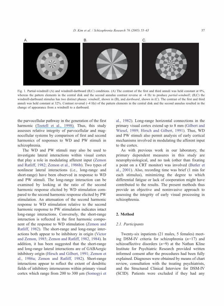
Fig. 1. Partial-windmill (A) and windmill-dartboard (B,C) conditions. (A) The contrast of the first and third annuli was held constant at 0%,
whereas the pattern elements in the central disk and the second annulus contrast reverse at ~4 Hz to produce partial-windmill; (B,C) the
windmill-dartboard stimulus has two distinct phases: windmill, shown in (B), and dartboard, shown in (C). The contrast of the first and third
annuli was held constant at 32%. Contrast reversal (~4 Hz) of the pattern elements in the central disk and the second annulus resulted in the
change of appearance from a windmill to a dartboard.
D. Kim et al. / Schizophrenia Research 76 (2005) 55–65 57
the parvocellular pathway in the generation of the first
harmonic (Tootell et al., 1998). Thus, this study
assesses relative integrity of parvocellular and mag-
nocellular systems by comparison of first and second
harmonics of responses to WD and PW stimuli in
schizophrenia.
The WD and PW stimuli may also be used to
investigate lateral interactions within visual cortex
that play a role in modulating afferent input (Zemon
and Ratliff, 1982; Zemon et al., 1986b). Two types of
nonlinear lateral interactions (i.e., long-range and
short-range) have been observed in response to WD
and PW stimuli. The long-range interaction can be
examined by looking at the ratio of the second
harmonic response elicited by WD stimulation com-
pared to the second harmonic response elicited by PW
stimulation. An attenuation of the second harmonic
response to WD stimulation relative to the second
harmonic response to PW stimulation indicates intact
long-range interactions. Conversely, the short-range
interaction is reflected in the first harmonic compo-
nent of the response to WD stimulation (Zemon and
Ratliff, 1982). The short-range and long-range inter-
actions both appear to be inhibitory in origin (Victor
and Zemon, 1985; Zemon and Ratliff, 1982, 1984). In
addition, it has been suggested that the short-range
and long-range lateral interactions are of GABAergic
inhibitory origin (Hirsch and Gilbert, 1991; Zemon et
al., 1986a; Zemon and Ratliff, 1982). Short-range
interactions appear to reflect the extent of dendritic
fields of inhibitory interneurons within primary visual
cortex which range from 200 to 300 Am (Somogyi et
al., 1982). Long-range horizontal connections in the
primary visual cortex extend up to 8 mm (Gilbert and
Wiesel, 1989; Hirsch and Gilbert, 1991). Thus, WD
and PW stimuli also permit analysis of early cortical
mechanisms involved in modulating the afferent input
to the cortex.
As with previous work in our laboratory, the
primary dependent measures in this study are
neurophysiological, and no task (other than fixating
a point on a CRT monitor) was involved (Butler et
al., 2001). Also, recording time was brief (1 min for
each stimulus), minimizing the degree to which
differential fatigue or lack of cooperation might have
contributed to the results. The present methods thus
provide an objective and noninvasive approach to
assessing the integrity of early visual processing in
schizophrenia.
2. Method
2.1. Participants
Twenty-six inpatients (21 males, 5 females) meet-
ing DSM-IV criteria for schizophrenia (n=17) and
schizoaffective disorders (n=9) at the Nathan Kline
Institute for Psychiatric Research provided written
informed consent after the procedures had been fully
explained. Diagnoses were obtained by means of chart
review, consultation with the treating psychiatrists,
and the Structured Clinical Interview for DSM-IV
(SCID). Patients were excluded if they had any

D. Kim et al. / Schizophrenia Research 76 (2005) 55–6558
neurological or ophthalmologic disorders that might
affect performance. Eight patients were taking atyp-
ical antipsychotics (olanzapine) and 18 were receiving
typical antipsychotic medications. The mean chlor-
promazine-equivalent dose was 1050.42 mg/day
(range=334–2004, S.D.=404).
Twenty-two comparison volunteers (10 males, 12
females), age-matched to the patients, provided
written informed consent after the procedures had
been fully explained. Comparison volunteers with a
history of psychiatric, neurological, and/or ophthal-
mologic disorders were excluded.
The patient and comparison groups did not differ
significantly in age (patient, mean=38.7 years,
S.D.=10.5, N=26; comparison, mean=39.2 years,
S.D.=10.7, N=22), although socioeconomic status, as
measured with the four-factor Hollingshead scale
(Hollingshead and Redlich, 1958), was significantly
lower for the patients (mean=26.6, S.D.=11.4) than for
the comparison subjects (mean=48.1, S.D.=14.1)
(t=�5.6, df=43, pb0.0005). Scores for general psy-
chopathology (BPRS), negative symptoms, and pos-
itive symptoms were mean=32.9, S.D.=8.9; mean=
26.6, S.D.=13.4; mean=7.8, S.D.=3.9, respectively.
All participants were tested in binocular and
monocular conditions (dominant and nondominant
eyes). Eye dominance was determined by asking
each participant to look through a narrow tube. The
eye selected for viewing was judged to be the
dominant one.
All participants showed better than 20/40 (=0.5)
vision for all three eye conditions. The patients had
lower visual acuity than did the controls for eye
condition. However, only the nondominant condition
showed a significant group difference (t=3.2, df=46,
p=0.003). Control analyses were, therefore, per-
formed to evaluate potential contribution of
between-group acuity to observed between-group
differences.
2.2. Apparatus
The VENUS system (Neuroscientific, Farming-
dale, NY), an all-inclusive system for creating,
modifying, and generating visual stimuli for recording
of brain electrophysiological and psychophysical
responses, was used for this study. The display was
a 14-in. Princeton Ultrasynch RGB monitor, and the
raster was set to a 16�16 cm square. The frame rate
was ~119 Hz. The space-average luminance was
approximately 100 cd/m2.
2.3. Stimuli
Two stimuli were used: windmill-dartboard and
partial-windmill (Fig. 1). The stimulus display
subtended a visual angle of 88 at the viewing
distance of 114 cm. Both stimuli consist of a central
disc (radius: 0.948) and three contiguous annuli
(radius: first ring: 1.8758; second ring: 2.8138; andthird ring: 3.758), radially divided into light and dark
segments. For both stimuli, the dynamic regions
(central disc and second annulus) are sinusoidally
contrast-reversed (from �32% to +32%) at ~4 Hz.
Each stimulus was presented for 1 min, and the same
pattern was presented repetitively throughout the
duration of the run.
2.3.1. Partial-windmill condition
In the partial-windmill (PW) condition, the first
and third annuli are set at zero contrast and thus
contain a uniform field set at the mean luminance
level. The central disc and second annulus contain
light and dark elements that contrast-reverse, which
produces the percept of apparent motion and yields a
VEP that contains a dominant second harmonic in
addition to a smaller fourth harmonic component.
2.3.2. Windmill-dartboard condition
In the windmill-dartboard (WD) condition, the
central disk and second annulus were contrast
reversed, while the first and third annuli were
fixed in contrast at the peak value of the dynamic
regions (32%). As a result, the percept alternates
between that of a windmill and that of a dartboard,
and the resulting VEP contains a dominant first
harmonic in addition to an attenuated second
harmonic component.
2.4. Visual-evoked potentials
Visual-evoked potentials (VEPs) were recorded
from the occipital site (Oz) relative to the vertex
reference site by means of gold-cup electrodes. The
ground electrode was placed at the parietal site (Pz).
The raw EEG was amplified (by 10,000), filtered with
D. Kim et al. / Schizophrenia Research 76 (2005) 55–65 59
a bandpass of 0.1–100Hz and digitized at a rate of
four times the monitor frame rate (~476 Hz).
Frequency analysis was performed for each epoch.
Amplitude and phase measures at the fundamental
(stimulus) frequency (~4 Hz) and at other harmonic
components were extracted by means of a discrete
Fourier transform (Regan, 1989).
Magnitude-squared coherence (MSC) was com-
puted to estimate the signal level for each frequency
component (Zemon et al., 1999). MSC is the ratio of
signal power to signal+noise power for a given
frequency component. When there is no noise, it is
deemed pure signal (MSC=1). The MSC decreases
from 1 and theoretically falls to 0 as the signal
decreases leaving only noise. Estimates of MSC are
biased by an amount that depends on sample size:
bias=1/q. In this study, q=10. Therefore, bias level
was 0.1.
2.5. Procedure
The participants were tested monocularly (domi-
nant and nondominant eye) and binocularly after they
were light-adapted to the mean luminance of the
display for several minutes. They were seated in a
dimly lit room and instructed to fixate on the small dot
placed in the center of the display during each run.
The experimenter monitored the gaze of each partic-
ipant during each run to ensure steady fixation. Any
runs in which gaze was unsteady were rejected, and
those runs were then repeated. In addition, if the EEG
trace contained sizable deflections from baseline or
other noise activity, the run was rejected and then
repeated. Brief rest periods were provided between
runs. The order of presentation of the two stimuli (PW
and WD) and eye conditions (binocular, dominant,
and nondominant) were counterbalanced to control for
a possible order effect.
2.6. Statistical analysis
Demographic comparisons were analyzed between
groups by means of two-tailed t tests. ssVEP MSC
responses were analyzed using repeated measures
analyses of variance with factors as described below.
In the primary analyses, data from patients with
schizophrenia and schizoaffective disorder were com-
bined because there was no significant difference
between these groups. In addition, two covariate
analyses were performed to examine the potential
contribution of the visual acuity difference observed
between the patients and controls. Post hoc one-way
ANOVAs and two-tailed t tests were applied when the
ANOVAs revealed significant main effects or inter-
actions. Data were analyzed for normality by means
of the Kolmogorov–Smirnov test and for equality of
variance by using Levene’s test. No deviations from
normality or equality of variance were observed.
3. Results
3.1. Partial-windmill condition
Between-group analyses of ssVEP responses
(MSC) were analyzed using a repeated measures
ANOVA with between-subject factor of group (schiz-
ophrenia/control) and within-subject factor of har-
monic level (first vs. second). In the PW condition
(see Fig. 2), the primary neural response occurred at
the second harmonic of the presented stimulus, as
reflected in the highly significant main effect of
harmonic across groups ( F=293.8, df=1,46,
p=0.0001). There was also a significant main effect
of group (F=10.1, df=1,46, p=0.003) and a signifi-
cant group x harmonic interaction (F=10.5, df=1,46,
p=0.002), reflecting differential generation of har-
monics across groups. Post hoc ANOVAs demon-
strated no significant difference between groups in
first harmonic generation (F=0.1, df=1,46, p=0.8),
but a significant between-group difference in second
harmonic generation (F=11.6, df=1,46, p=0.001).
The MSC score of the second harmonic for patients
was about 72% of comparison values across all three-
eye conditions. There was no significant main effect
of eye condition (dominant, nondominant, and binoc-
ular), and no significant group�eye interaction. The
between-group difference in second harmonic gener-
ation remained highly significant even following
covariation for potential between-group differences
in visual acuity (F=7.0, df=1,46, p=0.011).
3.2. Windmill-dartboard condition
In the WD condition, the primary neural response
was at the first harmonic (see Fig. 2), as reflected in a

Fig. 2. Mean magnitude-squared coherence (MSC) scores for partial-windmill (upper) and windmill-dartboard (lower) conditions. BI: binocular
eye condition; DO: dominant eye condition; ND: nondominant eye condition. Controls: comparison subjects (N=22); Scz patients:
schizophrenia patients (N=26). MSC was computed to estimate the signal level for each frequency component. MSC=signal power/signal+noise
power. Therefore, 1=pure signal; 0=no signal. Bias level was set on 0.1 (using 10 epochs). aSignificant difference between groups (t=2.8–4.1,
df=46, pb0.01); bsignificant difference between groups (t=2.2–2.6, df=46, pb0.05).
D. Kim et al. / Schizophrenia Research 76 (2005) 55–6560
significant main effect of harmonic (F=58.1, df=1,46,
p=0.0001). As in the PW condition, there was a
significant group effect (F=5.9, df=1,46, p=0.018),
and a significant group x harmonic interaction
(F=6.6, df=1,46, p=0.013). Post hoc ANOVAs
demonstrated no significant difference between
groups in first harmonic generation (F=0.3, df=1,46,
p=0.6), but a highly significant between-group differ-
ence in second harmonic generation ( F=18.9,
df=1,46, p=0.0001). The MSC score of the second
harmonic for patients was about 62% of comparison
values across all three-eye conditions. There was no
significant main effect of eye condition and no
significant group�eye interaction. As in the PW
condition, the between-group difference in second
harmonic generation remained highly significant even
following covariation for potential between-group
differences in visual acuity ( F=13.8, df=1,46,
p=0.001).
3.3. Cross-condition comparisons
In controls, the first harmonic response in the WD
condition was of approximately equal amplitude to the
second harmonic response in the PW condition,
providing an opportunity for evaluation of differential
deficits free of amplitude confound. A repeated
measure ANOVA with within-group factors of stim-
ulus type and eye condition was performed to
examine the differential deficits in first harmonic of
WD and second harmonic of PW. There was a
significant main effect of group (F=4.4, df=1,46,
p=0.041) and a significant group�harmonic interac-
tion (F=5.6, df=1,46, p=0.023), supporting the
D. Kim et al. / Schizophrenia Research 76 (2005) 55–65 61
observation of a selective impairment of second
harmonic responses to WD/PW stimuli.
3.4. Long-range lateral interactions
To examine the long-range lateral interactions,
amplitudes of the second harmonic component for
both PW and WD conditions were examined. There
was no significant group�stimulus condition inter-
action (F=2.7, df=1,46, p=0.11), showing that the
ratio of the second harmonic for the PW and WD
condition did not differ between groups.
3.5. Fourth harmonic
Two repeated measures ANOVAs were performed
to look at between-group difference in fourth har-
monics of WD and PW conditions. In the WD
condition, there was a significant main effect of group
(F=4.5, df=1,46, p=0.04), but no group x eye
condition interaction. There was also a significant
main effect of group (F=8.1, df=1,46, p=0.007) in the
PW condition. The MSC scores of the fourth
harmonics for patients in WD and PW conditions
were both about 68% of comparison values across all
three-eye conditions.
3.6. Patients with schizophrenia versus schizoaffective
disorder
There were 17 patients with schizophrenia, 9
patients with schizoaffective disorder, and 22 controls.
A MANOVA was performed to discover possible
between-group differences in harmonics generated by
PW and WD conditions. A multivariate analysis based
on the general linear model was performed to explore
the effects of group membership (control, schizophre-
nia, schizoaffective) and eye condition (dominant,
nondominant, binocular) on the six relevant response
measures (WD first, second, third, fourth and PW
second, fourth harmonic components). There was a
main effect of group membership (F=7.1, df=2,45,
p=0.002), but there was no interaction effect of group
by eye condition (F=1.8, df=4,88, p=0.14). Main
effect of eye condition was significant when all six
response measures were included (F=4.7, df=2,44,
p=0.014), and when only the PW measures were
analyzed (F=4.7, df=2,45, p=0.014), but not when
the WD measures were analyzed separately (F=1.7,
df=2,45, p=0.2).
In the PW condition, there was a significant main
effect of group in second harmonic generation across
all three-eye conditions: binocular (F=7.1, df=2,45,
p=0.002), nondominant (F=7.8, df=2,45, p=0.001),
and dominant (F=5.3, df=2,45, p=0.008). Scheffe’s
post hoc test revealed a significant difference between
controls and patients with schizophrenia ( p=0.002),
but not between controls and patients with schizo-
affective disorder ( p=0.2) or between patients with
schizophrenia and patients with schizoaffective dis-
order ( p=0.09) across all eye conditions.
In the WD condition, as in the PW condition, there
was a significant main effect of group in second
harmonic generation for binocular (F=6.1, df=2,45,
p=0.005) and nondominant eye conditions (F=8.7,
df=2,45, p=0.001), but not for first harmonic gen-
eration for all eye conditions. Scheffe’s post hoc test
revealed a significant difference between controls and
patients with schizophrenia ( p=0.007), but not
between controls and patients with schizoaffective
disorder ( p=0.1) or between patients with schizo-
phrenia and patients with schizoaffective disorder
( p=0.9) for all eye conditions.
4. Discussion
Although there has been increasing focus on the
study of visual processing deficits in schizophrenia
(Braus et al., 2002; Brenner et al., 2003; Keri et al.,
2004; Li, 2002; Schwartz et al., 1999; Slaghuis and
Thompson, 2003), underlying neurophysiological
mechanisms remain to be determined. This is the first
study to analyze dysfunction of the visual system in
schizophrenia using pan-harmonic analysis of
ssVEPs. Two types of stimuli were used—WD and
PW. Harmonics were extracted using Fourier analysis,
and responses were analyzed across groups to
compare first versus second harmonics. Integrity of
lateral interactions was analyzed using ratios of
responses to the harmonics. The primary findings
are that patients with schizophrenia show significantly
decreased magnitude of second harmonic responses in
both the PW and WD conditions, but intact first
harmonic responses indicating a deficit in early visual
processing. The differential deficit is apparent even

D. Kim et al. / Schizophrenia Research 76 (2005) 55–6562
under conditions in which controls show similar
magnitude responses, and so cannot be attributed to
psychometric artifact. Lateral interactions in the
primary visual cortex, however, appear to be intact.
On the perceptual level, the present results may be
relevant to recent studies showing impaired motion
detection in schizophrenia (Braus et al., 2002; Brenner
et al., 2003; Li, 2002). Impairments in motion
detection have been increasingly observed (Braus et
al., 2002; Chen et al., 1999; Li, 2002; O’Donnell et
al., 1996). However, the underlying substrates have
yet to be fully defined. Motion detection is decoded
primarily in dorsal stream visual areas, such as MT
(V5), but depends upon inputs primarily from the
magnocellular visual system. Behavioral studies alone
cannot determine whether motion detection deficits
observed in schizophrenia reflect dysfunction of
cortical motion areas or of low-level projection
systems.
Recent functional imaging studies have demon-
strated decreased activation in MT during motion
detection in schizophrenia, but no between-group
difference in activation of primary visual areas. This
finding has been interpreted as reflecting dysfunction
primarily within higher-order cortical motion detec-
tion regions (Braus et al., 2002). In contrast, the
finding in this study of impaired ssVEP activity to
the present stimuli indicates dysfunction within early
visual regions (precortical or V1). The failure to
detect changes within primary cortex in functional
imaging studies may reflect difficulty in differentiat-
ing motion-related from feature-related activity
within primary visual cortex using anatomically (as
opposed to neurophysiologically) based imaging, or
may indicate that functional imaging signals, in
general, reflect integrity of input as well as of local
processing (Logothetis, 2002). Thus, impaired MT
activation noted in functional imaging experiments
may reflect impaired input into higher visual regions,
rather than impaired local detection. Positron emis-
sion tomography (PET) studies in controls have
shown motion-dependent alteration in activation of
primary visual areas with incoherent motion stimuli
(McKeefry et al., 1997). Whether activation deficits
to such stimuli can be observed in schizophrenia
remains to be determined.
Long- and short-range lateral inhibitory connec-
tions within cortex play a critical role in sculpting
ssVEP responses to WD and PW stimuli (Zemon and
Ratliff, 1982, 1984). In particular, attenuation of the
second harmonic component in the WD as compared
to the PW condition and generation of a first harmonic
component by WD stimulation are thought to depend
on long-range and short-range lateral interactions
within the cortex, respectively (Grose-Fifer et al.,
1994; Sokol et al., 1992; Zemon and Ratliff, 1982).
Although second harmonic responses were reduced to
both PW and WD stimuli, the relative ratio was
unchanged, suggesting that deficits in long-range
lateral inhibitory activity cannot account for the
present pattern of results. The normal first harmonic
in patients demonstrates that they also have intact
short-range inhibitory activity.
Exact mechanisms underlying the differential
deficit in second versus first harmonic response
remain to be determined, but may involve differential
magnocellular versus parvocellular involvement in
schizophrenia. As noted above, second harmonic
responses are thought to depend preferentially on
functioning of the magnocellular visual pathways,
whereas first harmonic responses depend more heav-
ily upon parvocellular processing (Grose-Fifer et al.,
1994; McKeefry et al., 1996; Murray et al., 1983;
Zemon and Ratliff, 1982). Prior electrophysiological
(Butler et al., 2001; Doniger et al., 2002; Foxe et al.,
2001) and psychophysical (Keri et al., 2004;
Schechter et al., 2003; Schwartz et al., 1999; Slaghuis
and Thompson, 2003) studies have demonstrated
greater impairment in magnocellular, than parvocel-
lular, functioning in schizophrenia. The present results
would thus be consistent with those prior findings.
Alternatively, deficits in nonlinear mechanisms
present in cortex, such as shunting inhibition which
are important in producing responses at higher
harmonics or temporal frequencies (Carandini and
Heeger, 1994) would also result in greater attenuation
of higher harmonic responses in patients than con-
trols. Other types of studies, such as ones that utilize
two-sinusoid stimulation, may serve as effective
probes of nonlinear cortical mechanisms and thus
may provide additional information (Zemon and
Ratliff, 1984). Whatever the underlying mechanism,
the differential involvement of second versus first
harmonic responses nevertheless excludes nonspecific
factors, such as attention or cooperation in producing
the observed deficits in schizophrenia, and supports
D. Kim et al. / Schizophrenia Research 76 (2005) 55–65 63
prior studies demonstrating early visual processing
deficits in schizophrenia.
Although attentional mechanisms are known to be
impaired in schizophrenia, an underlying assumption
of much research is that deficits are driven by such
btop downQ mechanisms. In this study, it is unlikely
that attentional deficits contributed strongly to the
observed results. First, there was no behavioral
component in the study other than fixation on the
center of the display. Second, the results would show
deficits in generation of first as well as second
harmonics if there were gross attentional disturbances
(e.g., failure to maintain fixation on the screen).
Instead, this study argues that visual attentional
impairments may be bbottom-up,Q due to failure of
basic neurophysiological processes within early visual
areas. The magnocellular system, in particular,
appears to mediate automatic capture of attention by
salient (e.g., moving, flickering) stimuli, with magno-
cellularly driven attentional capture predominating
over parvocellularly driven attentional capture (Stein-
man et al., 1997). Magnocellular neurons project
preferentially to the dorsal stream, and parvocellular
neurons project preferentially to the ventral stream.
Magnocellular inputs to the dorsal stream cross over
to ventral stream areas and also play a critical role in
organizing activity within ventral stream, object
recognition areas (Doniger et al., 2000, 2002; Lamme
and Roelfsema, 2000; Schroeder et al., 1998). Patients
with schizophrenia actually show decreased, rather
than increased, distractibility to visual stimuli pre-
sented outside the focus of attention (Slaghuis and
Thompson, 2003), consistent with magnocellular
dysfunction. Magnocellular dysfunction also appears
to contribute to impaired recognition of both moving
(Schwartz et al., 1999) and fragmented (Doniger et al.,
2002) stimuli and impaired visual organization. The
present data are thus consistent with a model in which
impaired magnocellular input into the dorsal visual
stream leads to dysfunctional modulation of ventral
stream object recognition processes (Doniger et al.,
2002).
A limitation of this study is that all patients were
receiving antipsychotic medication at the time of
testing. Thus, a medication effect cannot be excluded.
Dopamine is well known to be present in the visual
system and has an impact on visual processing
including spatial frequency tuning (Bodis-Wollner
and Tzelepi, 1998). However, it is highly unlikely
that such effects play a role in the generation of
second harmonic but not first harmonic responses.
Furthermore, acute haloperidol administration had no
effect on the generation of ssVEPs (Jibiki et al.,
1993). Several visual backward masking studies also
did not find a difference in backward masking
performance in the same patients tested on and off
medication (Braff and Saccuzzo, 1982; Butler et al.,
1996, 2002; Green et al., 1999). In this study, no
significant correlations were found between CPZ
equivalents and first and second harmonics. Thus, it
is unlikely that the differential deficits shown in this
study are due to a medication effect.
For the primary analysis, results from patients with
schizophrenia and schizoaffective disorder were com-
bined. A secondary analysis evaluated relative deficits.
No significant differences were detected between
patients with schizophrenia versus schizoaffective
disorder. However, patients with schizoaffective dis-
order did not on their own differ significantly from
controls. The lack of difference may reflect differential
underlying pathophysiology, heterogeneity within the
schizoaffective group, or simply the small number of
patients studied with schizoaffective disorder. Patients
with schizophrenia and schizoaffective disorder were
also not matched for severity of illness. These issues
can be further clarified in future studies.
In summary, this study confirms prior reports of
early visual processing deficits in schizophrenia using
novel visual stimuli. Patients showed a differential
deficit with intact first harmonic responses to WD
stimuli but impaired second harmonic responses to
both PW and WD conditions. These stimuli therefore
may be useful in characterizing visual processing
deficits in schizophrenia and may suggest differential
involvement of magnocellular and paravocellular
systems. These deficits may contribute significantly
to subsequent stages, such as conscious motion
detection, attentional capture, and stimulus-driven
perceptual organization.
Acknowledgments
Presented in part at the 32nd annual meeting of the
Society for Neuroscience, Orlando, FL, Nov. 2–7,
2002 and at the 9th International Congress on
D. Kim et al. / Schizophrenia Research 76 (2005) 55–6564
Schizophrenia Research, Colorado Springs, CO,
March 29–April 2, 2003. This work was supported
by USPHS grants RO1 MH66374 (PDB), R37
MH49334, and K02 MH01439 (DCJ), and a Bur-
roughs Wellcome Translational Scientist Award (to
DCJ). We thank Dr. Glenn Wylie for helpful com-
ments on the manuscript.
References
Adler, L.E., Freedman, R., Ross, R.G., Olincy, A., Waldo, M.C.,
1999. Elementary phenotypes in the neurobiological and genetic
study of schizophrenia. Biol. Psychiatry 46, 8–18.
Bodis-Wollner, I., Tzelepi, A., 1998. The push–pull action of
dopamine on spatial tuning of the monkey retina: the effects
of dopaminergic deficiency and selective D1 and D2 receptor
ligands on the pattern electroretinogram. Vision Res. 38,
1479–1487.
Braff, D.L., Saccuzzo, D.P., 1982. Effect of antipsychotic medi-
cation on speed of information processing in schizophrenic
patients. Am. J. Psychiatry 139, 1127–1130.
Braff, D., Saccuzzo, D., Geyer, M., 1991. Information processing
dysfunctions in schizophrenia: studies of visual backward
masking, sensorimotor gating, and habituation. In: Steinhauer,
S., Gruzelier, J., Zubin, J. (Eds.), Handbook of Schizophrenia
vol. 5. Elsevier, New York, pp. 303–334.
Braus, D.F., Weber-Fahr, W., Tost, H., Ruf, M., Henn, F.A., 2002.
Sensory information processing in neuroleptic-naive first-
episode schizophrenic patients: a functional magnetic resonance
imaging study. Arch. Gen. Psychiatry 59, 696–701.
Brenner, C.A., Wilt, M.A., Lysaker, P.H., Koyfman, A., O’Donnell,
B.F., 2003. Psychometrically matched visual-processing tasks in
schizophrenia spectrum disorders. J. Abnorm. Psychology 112,
28–37.
Butler, P.D., Harkavy-Friedman, J.M., Amador, X.F., Gorman, J.M.,
1996. Backward masking in schizophrenia: relationship to
medication status, neuropsychological functioning, and dopa-
mine metabolism. Biol. Psychiatry 40, 295–298.
Butler, P.D., Schechter, I., Zemon, V., Schwartz, S.G., Greenstein,
V.C., Gordon, J., Schroeder, C.E., Javitt, D.C., 2001. Dysfunc-
tion of early-stage visual processing in schizophrenia. Am. J.
Psychiatry 158, 1126–1133.
Butler, P.D., DeSanti, L.A., Maddox, J., Harkavy-Friedman, J.M.,
Amador, X.F., Goetz, R.R., Javitt, D.C., Gorman, J.M., 2002.
Visual backward-masking deficits in schizophrenia: relationship
to visual pathway function and symptomatology. Schizophr.
Res. 59, 199–209.
Cadenhead, K.S., Serper, Y., Braff, D.L., 1998. Transient versus
sustained visual channels in the visual backward masking
deficits of schizophrenia patients. Biol. Psychiatry 43, 132–138.
Carandini, M., Heeger, D.J., 1994. Summation and division by
neurons in primate visual cortex. Science 264, 1333–1336.
Chen, Y., Levy, D.L., Nakayama, K., Matthysse, S., Palafox, G.,
Holzman, P.S., 1999. Dependence of impaired eye tracking on
deficient velocity discrimination in schizophrenia. Arch. Gen.
Psychiatry 56, 155–161.
Chen, Y., Nakayama, K., Levy, D., Matthysse, S., Holzman, P.,
2003. Processing of global, but not local, motion direction is
deficient in schizophrenia. Schizophr. Res. 61, 215–227.
Doniger, G.M., Foxe, J.J., Murray, M.A., Higgins, B.A.,
Schroeder, C.E., Javitt, D.C., 2000. Activation timecourse of
ventral visual stream object-recognition areas: high density
electrical imaging of perceptual closure processes. J. Cogn.
Neurosci. 12, 615–621.
Doniger, G.M., Foxe, J.J., Murray, M.M., Higgins, B.A., Javitt,
D.C., 2002. Impaired visual object recognition and dorsal/
ventral stream interaction in schizophrenia. Arch. Gen. Psy-
chiatry 59, 1011–1020.
Foxe, J.J., Doniger, G.M., Javitt, D.C., 2001. Early visual
processing deficits in schizophrenia: impaired P1 generation
revealed by high-density electrical mapping. NeuroReport 12,
3815–3820.
Gilbert, C.D., Wiesel, T.N., 1989. Columnar specificity of intrinsic
horizontal and corticocortical connections in cat visual cortex. J.
Neurosci. 9, 2432–2442.
Goldberg, T., Gold, J., 1995. Neurocognitive functioning in patients
with schizophrenia: an overview. In: Bloom, F.E., Kupfer, D.
(Eds.), Psychopharmacology: The Fourth Generation of Pro-
gress. Raven Press, New York, pp. 1245–1257.
Goldman-Rakic, P.S., 1994. Working memory dysfunction in
schizophrenia. J. Neuropsychiatry Clin. Neurosci. 6, 348–357.
Green, M., 1998. Schizophrenia from a Neurocognitive Perspective.
Allyn and Bacon, Boston.
Green, M.F., Nuechterlein, K.H., Mintz, J., 1994. Backward
masking in schizophrenia and mania: II. Specifying the visual
channels. Arch. Gen. Psychiatry 51, 945–951.
Green, M.F., Nuechterlein, K.H., Breitmeyer, B., Mintz, J., 1999.
Backward masking in unmedicated schizophrenic patients in
psychotic remission: possible reflection of aberrant cortical
oscillation. Am. J. Psychiatry 156, 1367–1373.
Grose-Fifer, J., Zemon, V., Gordon, J., 1994. Temporal
tuning and the development of lateral interactions in the
human visual system. Invest. Ophthalmol. Visual. Sci. 35,
2999–3010.
Hirsch, J.A., Gilbert, C.D., 1991. Synaptic physiology of
horizontal connections in the cat’s visual cortex. J. Neurosci.
11, 1800–1809.
Hollingshead, A.B., Redlich, F.L., 1958. Social Class and Mental
Illness. Wiley, New York.
Javitt, D.C., Liederman, E., Cienfuegos, A., Shelley, A.M., 1999.
Panmodal processing imprecision as a basis for dysfunction of
transient memory storage systems in schizophrenia. Schizophr.
Bull. 25, 763–775.
Jibiki, I., Kurokawa, K., Fukushima, T., Yamaguchi, N., 1993.
Acutely administered haloperidol has little effect on steady-
state visual evoked potentials from pattern-reversal stimula-
tions in treated schizophrenics. Jpn. J. Psychiatry Neurol. 47,
51–55.
Kaplan, E., 1991. The receptive field structure of retinal ganglion
cells in cat and monkey. In: Leventhal, A.G. (Ed.), Vision and
Visual Dysfunction. CRC Press, Boston, pp. 10–40.

D. Kim et al. / Schizophrenia Research 76 (2005) 55–65 65
Keri, S., Kelemen, O., Benedek, G., Janka, Z., 2004. Vernier
threshold in patients with schizophrenia and in their unaffected
siblings. Neuropsychology 18, 537–542.
Lamme, V.A., Roelfsema, P.R., 2000. The distinct modes of vision
offered by feedforward and recurrent processing. Trends Neuro-
sci. 23, 571–579.
Li, C.S., 2002. Impaired detection of visual motion in schizophrenia
patients. Prog. Neuro-psychopharmacol. Biol. Psychiatry 26,
929–934.
Logothetis, N.K., 2002. The neural basis of the blood-oxygen-level-
dependent functional magnetic resonance imaging signal.
Philos. Trans. R. Soc. Lond., B Biol. Sci. 357, 1003–1037.
McKeefry, D.J., Russell, M.H., Murray, I.J., Kulikowski, J.J., 1996.
Amplitude and phase variations of harmonic components in
human achromatic and chromatic visual evoked potentials. Vis.
Neurosci. 13, 639–653.
McKeefry, D.J., Watson, J.D., Frackowiak, R.S., Fong, K., Zeki, S.,
1997. The activity in human areas V1/V2, V3, and V5 during
the perception of coherent and incoherent motion. NeuroImage
5, 1–12.
Murray, I., MacCana, F., Kulikowski, J.J., 1983. Contribution of
two movement detecting mechanisms to central and peripheral
vision. Vision Res. 23, 151–159.
O’Donnell, B.F., Swearer, J.M., Smith, L.T., Nestor, P.G., Shenton,
M.E., McCarley, R.W., 1996. Selective deficits in visual
perception and recognition in schizophrenia. Am. J. Psychiatry
153, 687–692.
Regan, D., 1989. Human Brain Electrophysiology: Evoked Poten-
tials and Evoked Magnetic Fields in Science and Medicine.
Elsevier, New York.
Saccuzzo, D.P., Braff, D.L., 1986. Information-processing abnor-
malities: trait- and state-dependent components. Schizophr. Bull.
12, 447–459.
Schechter, I., Butler, P.D., Silipo, G., Zemon, V., Javitt, D.C., 2003.
Magnocellular and parvocellular contributions to backward
masking dysfunction in schizophrenia. Schizophr. Res. 1902,
1–11.
Schroeder, C.E., Mehta, A.D., Givre, S.J., 1998. A spatiotempo-
ral profile of visual system activation revealed by current
source density analysis in the awake macaque. Cereb. Cortex
8, 575–592.
Schwartz, B.D., Maron, B.A., Evans, W.J., Winstead, D.K., 1999.
High velocity transient visual processing deficits diminish
ability of patients with schizophrenia to recognize objects.
Neuropsychiatry Neuropsychol. Behav. Neurol. 12, 170–177.
Slaghuis, W.L., Bakker, V.J., 1995. Forward and backward visual
masking of contour by light in positive- and negative-symptom
schizophrenia. J. Abnorm. Psychology 104, 41–54.
Slaghuis, W.L., Curran, C.E., 1999. Spatial frequency masking in
positive- and negative-symptom schizophrenia. J. Abnorm.
Psychology 108, 42–50.
Slaghuis, W.L., Thompson, A.K., 2003. The effect of periph-
eral visual motion on focal contrast sensitivity in positive-
and negative-symptom schizophrenia. Neuropsychologia 41,
968–980.
Sokol, S., Zemon, V., Moskowitz, A., 1992. Development of lateral
interactions in the infant visual system. Vis. Neurosci. 8, 3–8.
Somogyi, P., Freund, T.F., Cowey, A., 1982. The axo-axonic
interneuron in the cerebral cortex of the rat, cat and monkey.
Neuroscience 7, 2577–2607.
Steinman, B.A., Steinman, S.B., Lehmkuhle, S., 1997. Transient
visual attention is dominated by the magnocellular stream.
Vision Res. 37, 17–23.
Tootell, R.B., Hadjikhani, N.K., Vanduffel, W., Liu, A.K., Mendola,
J.D., Sereno, M.I., Dale, A.M., 1998. Functional analysis of
primary visual cortex (V1) in humans. Proc. Natl. Acad. Sci. U.
S. A. 95, 811–817.
Ungerleider, L., Mishkin, M., 1982. Two cortical visual system. In:
Ingle, D., Goodale, M.A., Mansfield, R.J.W. (Eds.), Analysis of
Visual Behavior. MIT Press, Cambridge, MA, pp. 549–580.
Victor, J.D., Zemon, V., 1985. The human visual evoked potential:
analysis of components due to elementary and complex aspects
of form. Vision Res. 25, 1829–1842.
Weinberger, D.R., Gallhofer, B., 1997. Cognitive function in
schizophrenia. Int. Clin. Psychopharmacol. 12 (Suppl. 4),
S29–S36.
Zemon, V., Ratliff, F., 1982. Visual evoked potentials: evidence
for lateral interactions. Proc. Natl. Acad. Sci. U. S. A. 79,
5723–5726.
Zemon, V., Ratliff, F., 1984. Intermodulation components of the
visual evoked potential: responses to lateral and superimposed
stimuli. Biol. Cybern. 50, 401–408.
Zemon, V., Kaplan, E., Ratliff, R., 1986a. The role of GABA-
mediated intracortical inhibition in the generation of visual
evoked potentials. In: Cracco, R., Bodis-Wollner, I. (Eds.),
Evoked Potentials: Frontiers of Clinical Neuroscience, vol. 3.
Alan R. Liss, INC., New York, pp. 287–295.
Zemon, V., Victor, J.D., Ratliff, F., 1986b. Functional subsystems in
the visual pathways of humans characterized using evoked
potentials. In: Cracco, R., Bodis-Wollner, I. (Eds.), Evoked
Potentials: Frontiers of Clinical Neuroscience, vol. 3. Alan R.
Liss, New York, pp. 203–210.
Zemon, V., Buckley, S., Fitzgerald, K., Gordon, J., Hartmann, E.,
Meyer, A., 1999. Objective determination of signal from noise
in swept-parameter transient visual evoked potentials (VEPs).
Invest. Ophthalmol. Visual. Sci. 40/4, S824.