Dysfunction in different phases of working memory in schizophrenia: Evidence from ERP recordings
8
Dysfunction in different phases of working memory in schizophrenia: Evidence from ERP recordings Yan Li Zhao a, 1 , Shu Ping Tan a, 1 , Fu De Yang a , Li Li Wang b , Wen Feng Feng c , Raymond C.K. Chan d, e , Xiao Gao f, g , Dong Feng Zhou h , Bin Bin Li a , Chong Sheng Song a , Feng Mei Fan a , Yun Long Tan a , Jin Guo Zhang a , Yun Hui Wang a , Yi Zhuang Zou a, ⁎ a Center for Psychiatric Research, Beijing Huilongguan Hospital, Beijing 100096, China b State Key Laboratory of Cognitive Neuroscience and Learning, Beijing Normal University, Beijing 100875, China c Department of Neurosciences, University of California San Diego, 9500 Gilman Drive, La Jolla, CA 92093-0608, USA d Key Laboratory of Mental Health, Institute of Psychology, Chinese Academy of Sciences, Beijing 100101, China e Neuropsychology and Applied Cognitive Neuroscience Laboratory, Institute of Psychology, Chinese Academy of Sciences, Beijing 100101, China f Key Laboratory of Cognition and Personality (SWU), Ministry of Education 400715, China g School of Psychology, Southwest University, Chong Qing 400715, China h Institute of Mental Health, Peking University, Beijing 100091, China abstract article info Article history: Received 3 December 2010 Received in revised form 5 September 2011 Accepted 17 September 2011 Available online 19 October 2011 Keywords: Working memory Schizophrenia Event-related potentials Sternberg The present study combined a time-locked paradigm and high-time-resolution event-related potential (ERP) recordings to examine different phases of working memory, including early visual processing and late mem- ory-related processes of encoding, maintenance, and retrieval, in 67 adults with schizophrenia and 46 healthy controls. Alterations in ERP components were correlated with task performance. Patients performed significantly worse in the working memory task than healthy subjects, although all subjects' accuracy exceeded 80%. During encoding, the N1 and P2 component amplitudes were lower while the P300 amplitude was higher in schizophrenic patients compared to healthy controls. There were no differences between groups with respect to the mean amplitudes of the negative slow waves in the early stage (the first 400 ms) of the maintenance phase. However, in the next 500-ms time window, the patients exhibited a more negative deflection in the middle fronto-central region than the control group. Likewise, a similar pat- tern was observed in the second 500-ms period in the middle fronto-central region, although the effect was marginally significant. There were no differences between groups in the remaining 1000 ms. During retrieval, the P1, N1 and P2 amplitudes were lower while the P300 amplitude and latency were higher in schizophrenic patients. The present results indicate early visual deficits in the working memory task in adults with schizo- phrenia. Impairments in the maintenance phase were confined to the late rehearsal stage. The increased P300 amplitude at the fronto-central electrode sites along with the poorer behavioral performance suggests that schizophrenic patients have an inefficient working memory system. © 2011 Elsevier B.V. All rights reserved. 1. Introduction In the working memory system, information is represented, main- tained, and updated for a short period (Lee and Park, 2005). Working memory has 3 stages: encoding, maintenance, and retrieval. Working memory deficits are a major cognitive dysfunction in schizophrenia (Silver et al., 2003). However, the specific stages of working memory that are specifically impaired in schizophrenia and the neuronal substrates underlying such abnormalities are not well understood (Hashimoto et al., 2010). Behavioral (Hill et al., 2010; Mazhari et al., 2010; Zilles et al., 2010) and functional magnetic resonance imaging (fMRI) studies have found deficits in the maintenance stage of working memory in schizophrenic patients (Driesen et al., 2008; Henseler et al., 2009). Smith and Jonides (1998) propose a visual–verbal working memory maintenance process involving multiple cognitive processes. First, visual stimulus representations are translated into corresponding phonological representations; these phonological representations remain active and are rehearsed subvocally and refreshed. The phonological translation process is mediated by the posterior parietal area, inferior frontal cortex, and superior temporal cortex; storage of phonological representations is mediated by the posterior parietal areas, whereas rehearsal is mediated by the frontal speech areas (Smith and Jonides, 1998). At present, it re- mains unclear which component(s) of the maintenance stage are im- paired in schizophrenic patients. While some studies suggest that the Schizophrenia Research 133 (2011) 112–119 ⁎ Corresponding author at: Beijing Hui-Long-Guan Hospital, Beijing 100096, China. Tel.: +86 10 62716905; fax: +86 10 62718210. E-mail address: [email protected] (Y.Z. Zou). 1 Both authors contributed equally to this study. 0920-9964/$ – see front matter © 2011 Elsevier B.V. All rights reserved. doi:10.1016/j.schres.2011.09.017 Contents lists available at SciVerse ScienceDirect Schizophrenia Research journal homepage: www.elsevier.com/locate/schres
-
Upload
independent -
Category
Documents
-
view
0 -
download
0
Transcript of Dysfunction in different phases of working memory in schizophrenia: Evidence from ERP recordings
Schizophrenia Research 133 (2011) 112–119
Contents lists available at SciVerse ScienceDirect
Schizophrenia Research
j ourna l homepage: www.e lsev ie r .com/ locate /schres
Dysfunction in different phases of working memory in schizophrenia: Evidence fromERP recordings
Yan Li Zhao a,1, Shu Ping Tan a,1, Fu De Yang a, Li Li Wang b, Wen Feng Feng c, Raymond C.K. Chan d,e,Xiao Gao f,g, Dong Feng Zhou h, Bin Bin Li a, Chong Sheng Song a, Feng Mei Fan a, Yun Long Tan a,Jin Guo Zhang a, Yun Hui Wang a, Yi Zhuang Zou a,⁎a Center for Psychiatric Research, Beijing Huilongguan Hospital, Beijing 100096, Chinab State Key Laboratory of Cognitive Neuroscience and Learning, Beijing Normal University, Beijing 100875, Chinac Department of Neurosciences, University of California San Diego, 9500 Gilman Drive, La Jolla, CA 92093-0608, USAd Key Laboratory of Mental Health, Institute of Psychology, Chinese Academy of Sciences, Beijing 100101, Chinae Neuropsychology and Applied Cognitive Neuroscience Laboratory, Institute of Psychology, Chinese Academy of Sciences, Beijing 100101, Chinaf Key Laboratory of Cognition and Personality (SWU), Ministry of Education 400715, Chinag School of Psychology, Southwest University, Chong Qing 400715, Chinah Institute of Mental Health, Peking University, Beijing 100091, China
⁎ Corresponding author at: Beijing Hui-Long-Guan HTel.: +86 10 62716905; fax: +86 10 62718210.
E-mail address: [email protected] (Y.Z. Zou).1 Both authors contributed equally to this study.
0920-9964/$ – see front matter © 2011 Elsevier B.V. Alldoi:10.1016/j.schres.2011.09.017
a b s t r a c t
a r t i c l e i n f oArticle history:Received 3 December 2010Received in revised form 5 September 2011Accepted 17 September 2011Available online 19 October 2011
Keywords:Working memorySchizophreniaEvent-related potentialsSternberg
The present study combined a time-locked paradigm and high-time-resolution event-related potential (ERP)recordings to examine different phases of working memory, including early visual processing and late mem-ory-related processes of encoding, maintenance, and retrieval, in 67 adults with schizophrenia and 46healthy controls. Alterations in ERP components were correlated with task performance. Patients performedsignificantly worse in the working memory task than healthy subjects, although all subjects' accuracyexceeded 80%. During encoding, the N1 and P2 component amplitudes were lower while the P300 amplitudewas higher in schizophrenic patients compared to healthy controls. There were no differences betweengroups with respect to the mean amplitudes of the negative slow waves in the early stage (the first400 ms) of the maintenance phase. However, in the next 500-ms time window, the patients exhibited amore negative deflection in the middle fronto-central region than the control group. Likewise, a similar pat-tern was observed in the second 500-ms period in the middle fronto-central region, although the effect wasmarginally significant. There were no differences between groups in the remaining 1000 ms. During retrieval,the P1, N1 and P2 amplitudes were lower while the P300 amplitude and latency were higher in schizophrenicpatients. The present results indicate early visual deficits in the working memory task in adults with schizo-phrenia. Impairments in the maintenance phase were confined to the late rehearsal stage. The increased P300amplitude at the fronto-central electrode sites along with the poorer behavioral performance suggests thatschizophrenic patients have an inefficient working memory system.
ospital, Beijing 100096, China.
rights reserved.
© 2011 Elsevier B.V. All rights reserved.
1. Introduction
In the working memory system, information is represented, main-tained, and updated for a short period (Lee and Park, 2005). Workingmemory has 3 stages: encoding, maintenance, and retrieval. Workingmemory deficits are a major cognitive dysfunction in schizophrenia(Silver et al., 2003). However, the specific stages of working memorythat are specifically impaired in schizophrenia and the neuronalsubstrates underlying such abnormalities are not well understood(Hashimoto et al., 2010).
Behavioral (Hill et al., 2010; Mazhari et al., 2010; Zilles et al.,2010) and functional magnetic resonance imaging (fMRI) studieshave found deficits in the maintenance stage of working memory inschizophrenic patients (Driesen et al., 2008; Henseler et al., 2009).Smith and Jonides (1998) propose a visual–verbal working memorymaintenance process involving multiple cognitive processes. First, visualstimulus representations are translated into corresponding phonologicalrepresentations; these phonological representations remain active andare rehearsed subvocally and refreshed. The phonological translationprocess is mediated by the posterior parietal area, inferior frontal cortex,and superior temporal cortex; storage of phonological representations ismediated by the posterior parietal areas, whereas rehearsal is mediatedby the frontal speech areas (Smith and Jonides, 1998). At present, it re-mains unclear which component(s) of the maintenance stage are im-paired in schizophrenic patients. While some studies suggest that the
113Y.L. Zhao et al. / Schizophrenia Research 133 (2011) 112–119
impairment exists in the early part of maintenance (Lee and Park, 2005),others suggest there may also be deficits in the late rehearsal process(Hashimoto et al., 2010). The recording of event-related potentials(ERPs) is a high-time-resolution method that can be used to study thetime course ofmaintenance processes. ERP studies in normal subjects de-tect ERP slow waves from 200ms to several seconds post-stimulus thatare specific to information maintenance in working memory (Ruchkinet al., 1994; Mecklinger and Pfeifer, 1996). However, to our knowledge,no similar ERP studies have been performed in schizophrenic patients.
Behavioral (Hartman et al., 2003; Badcock et al., 2008) and fMRIstudies (Cairo et al., 2006; Johnson et al., 2006) also indicate impair-ments during information encoding in schizophrenic patients. In addi-tion, several ERP studies report impairments during encoding andretrieval. For example, Haenschel et al. (2007) found abnormal P300amplitude and latency during both the encoding and retrieval stagesin schizophrenic adolescents. Moreover, a growing body of evidencedemonstrates that schizophrenic patients exhibit significant deficits inearly visual processing (e.g., Chen et al., 1999a,b; Butler et al., 2009);these deficits mainly affect the subcortical magnocellular pathway(Luck et al., 2006;Martinez et al., 2008; Yeap et al., 2008). Furthermore,these deficits can be detected according to reduced P1 amplitude (Lucket al., 2006; Haenschel et al., 2007; Yeap et al., 2008). Early visual pro-cessing deficits contribute to working memory dysfunction in adoles-cents with early-onset schizophrenia (Haenschel et al., 2007) as wellas individuals with schizotypal features (Koychev et al., 2010).
The main goal of the present study was to investigate which aspectsof workingmemory are impaired in schizophrenic patients. Specifically,we examined whether patients were impaired in early visual proces-sing by using a task involving working memory. Also, if deficits existedin the maintenance phase, we determined which parts of the mainte-nance process were impaired. In addition, the present study aimed totestwhether impaired ERP componentswere correlatedwith behavior-al performance. To achieve the goals stated above, we used ERPs gener-ated by the modified Sternberg's short-term memory scanning task(SMST) (Manoach et al., 1999) in which participants were instructedto remember a set of unique digits in short-term memory (encodingphase); several seconds later (maintenance phase), a probe stimuluswas presented (retrieval phase) and the participants answeredwhetherthe probe was part of the initial sample or not. With this task, the dis-tinct phases of working memory can be distinguished and examined.
2. Materials and methods
2.1. Subjects
Sixty-seven adult inpatients of Beijing Huilongguan Hospitalin China with a DSM-IV diagnosis of schizophrenia (American
Table 1Demographic and clinical characteristic of patients with schizophrenia and controls.
Characteristics Patients
Mean age(range),y 42.15 (2Mean education years (range),y 11.67 (6Sex, no.
Male 39Female 28
Handedness, no.Left 0Right 67
Mean (SD) length of illness, y 16.31 (0Mean (SD) age at disease onset 25.73 (1Mean (SD) PANSS score 60.88 (4Neuroleptic medication use, no.a
Typical neuroleptics 6Atypical neuroleptics 52Combination of typical and atypical neuroleptics 8
Mean (SD) chlorpromazine equivalents, mg/d (Woods, 2003) 448.85 (
a Data for the neuroleptic medication use of one patient is missing value.
Psychiatric Association, 1994) were recruited in the present study.Patients with a history of substance abuse in the last 6 months or ad-ditional neuropsychiatry diagnoses were excluded. Current clinicalsymptoms were assessed using the Positive and Negative SyndromeScale (PANSS) (Kay et al., 1987). An additional 46 normal control par-ticipants were recruited from the surrounding community throughposter advertisements. Control subjects with a history of mental ill-ness or substance abuse were excluded. There were no significant dif-ferences between the 2 groups with respect to age, sex, handedness,or education (Table 1). All participants had normal or corrected tonormal vision. All participants provided written informed consent,and the protocol was approved by the ethics committee of BeijingHuilongguan Hospital.
2.2. SMST procedure
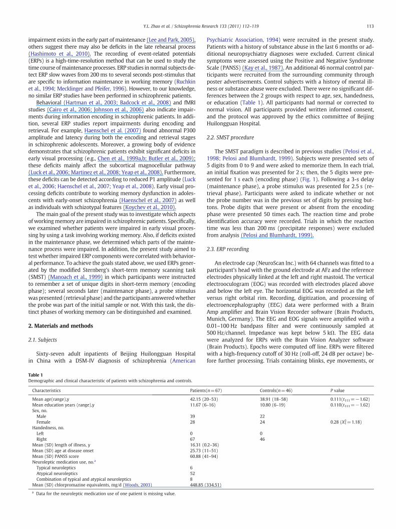
The SMST paradigm is described in previous studies (Pelosi et al.,1998; Pelosi and Blumhardt, 1999). Subjects were presented sets of5 digits from 0 to 9 and were asked to memorize them. In each trial,an initial fixation was presented for 2 s; then, the 5 digits were pre-sented for 1 s each (encoding phase) (Fig. 1). Following a 3-s delay(maintenance phase), a probe stimulus was presented for 2.5 s (re-trieval phase). Participants were asked to indicate whether or notthe probe number was in the previous set of digits by pressing but-tons. Probe digits that were present or absent from the encodingphase were presented 50 times each. The reaction time and probeidentification accuracy were recorded. Trials in which the reactiontime was less than 200 ms (precipitate responses) were excludedfrom analysis (Pelosi and Blumhardt, 1999).
2.3. ERP recording
An electrode cap (NeuroScan Inc.) with 64 channels was fitted to aparticipant's head with the ground electrode at AFz and the referenceelectrodes physically linked at the left and right mastoid. The verticalelectrooculogram (EOG) was recorded with electrodes placed aboveand below the left eye. The horizontal EOG was recorded as the leftversus right orbital rim. Recording, digitization, and processing ofelectroencephalography (EEG) data were performed with a BrainAmp amplifier and Brain Vision Recorder software (Brain Products,Munich, Germany). The EEG and EOG signals were amplified with a0.01–100 Hz bandpass filter and were continuously sampled at500 Hz/channel. Impedance was kept below 5 kΩ. The EEG datawere analyzed for ERPs with the Brain Vision Analyzer software(Brain Products). Epochs were computed off line. ERPs were filteredwith a high-frequency cutoff of 30 Hz (roll-off, 24 dB per octave) be-fore further processing. Trials containing blinks, eye movements, or
(n=67) Controls(n=46) P value
0–53) 38.91 (18–58) 0.111(t111=−1.62)–16) 10.80 (6–19) 0.110(t111=−1.62)
2224 0.28 (X1
2=1.18)
046
.2–36)1–51)1–94)
334.51)
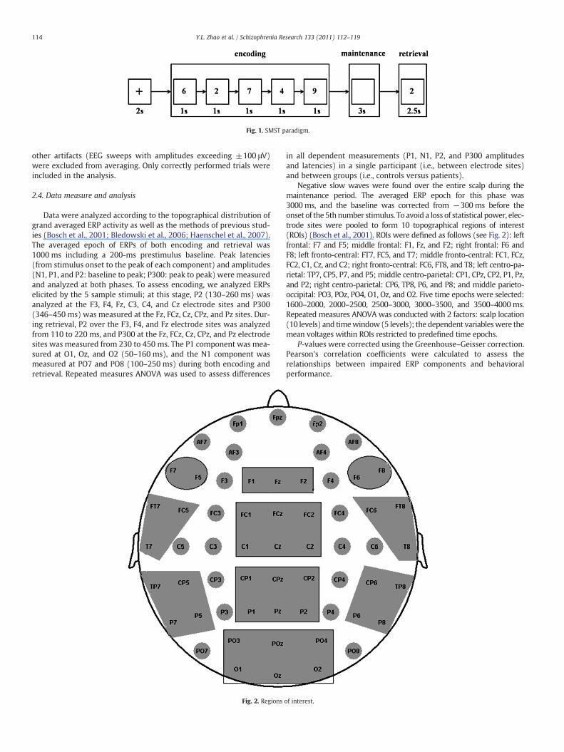
Fig. 1. SMST paradigm.
114 Y.L. Zhao et al. / Schizophrenia Research 133 (2011) 112–119
other artifacts (EEG sweeps with amplitudes exceeding ±100 μV)were excluded from averaging. Only correctly performed trials wereincluded in the analysis.
2.4. Data measure and analysis
Data were analyzed according to the topographical distribution ofgrand averaged ERP activity as well as the methods of previous stud-ies (Bosch et al., 2001; Bledowski et al., 2006; Haenschel et al., 2007).The averaged epoch of ERPs of both encoding and retrieval was1000 ms including a 200-ms prestimulus baseline. Peak latencies(from stimulus onset to the peak of each component) and amplitudes(N1, P1, and P2: baseline to peak; P300: peak to peak) were measuredand analyzed at both phases. To assess encoding, we analyzed ERPselicited by the 5 sample stimuli; at this stage, P2 (130–260 ms) wasanalyzed at the F3, F4, Fz, C3, C4, and Cz electrode sites and P300(346–450 ms) was measured at the Fz, FCz, Cz, CPz, and Pz sites. Dur-ing retrieval, P2 over the F3, F4, and Fz electrode sites was analyzedfrom 110 to 220 ms, and P300 at the Fz, FCz, Cz, CPz, and Pz electrodesites was measured from 230 to 450 ms. The P1 component was mea-sured at O1, Oz, and O2 (50–160 ms), and the N1 component wasmeasured at PO7 and PO8 (100–250 ms) during both encoding andretrieval. Repeated measures ANOVA was used to assess differences
Fig. 2. Regions
in all dependent measurements (P1, N1, P2, and P300 amplitudesand latencies) in a single participant (i.e., between electrode sites)and between groups (i.e., controls versus patients).
Negative slow waves were found over the entire scalp during themaintenance period. The averaged ERP epoch for this phase was3000 ms, and the baseline was corrected from −300 ms before theonset of the 5th number stimulus. To avoid a loss of statistical power, elec-trode sites were pooled to form 10 topographical regions of interest(ROIs) (Bosch et al., 2001). ROIs were defined as follows (see Fig. 2): leftfrontal: F7 and F5; middle frontal: F1, Fz, and F2; right frontal: F6 andF8; left fronto-central: FT7, FC5, and T7; middle fronto-central: FC1, FCz,FC2, C1, Cz, and C2; right fronto-central: FC6, FT8, and T8; left centro-pa-rietal: TP7, CP5, P7, and P5; middle centro-parietal: CP1, CPz, CP2, P1, Pz,and P2; right centro-parietal: CP6, TP8, P6, and P8; and middle parieto-occipital: PO3, POz, PO4, O1, Oz, and O2. Five time epochs were selected:1600–2000, 2000–2500, 2500–3000, 3000–3500, and 3500–4000 ms.Repeated measures ANOVA was conducted with 2 factors: scalp location(10 levels) and timewindow (5 levels); the dependent variableswere themean voltages within ROIs restricted to predefined time epochs.
P-values were corrected using the Greenhouse–Geisser correction.Pearson's correlation coefficients were calculated to assess therelationships between impaired ERP components and behavioralperformance.
of interest.
115Y.L. Zhao et al. / Schizophrenia Research 133 (2011) 112–119
3. Results
3.1. Behavioral measurements
Table 2 shows the mean response times and performance accuracyin both groups. The accuracy of all participants was above 80%. The in-dependent samples test revealed slower reaction time [t (111)=−5.043, pb0.001] and poorer accuracy [t (111)=2.990, p=0.003]in the patients compared to the controls.
3.2. ERP data analysis
3.2.1. Encoding
3.2.1.1. P1. The latency and amplitude of the P1 component were sub-mitted separately to ANOVA with 2 factors: electrode site (O1/O2/Oz)and group (control/patient). The results revealed that the P1 latencydid not exhibit a significant main effect with respect to electrodesite or group [F (2, 110)=2.261, p=0.120; F (1, 111)=0.278,p=0.599]. The relationship between electrode site and group wasalso insignificant [F (2, 110)=0.060, p=0.903]. The ANOVA of P1amplitude indicated that the main effect of group was not statisticallysignificant [F (1, 111)=1.918, p=0.169].
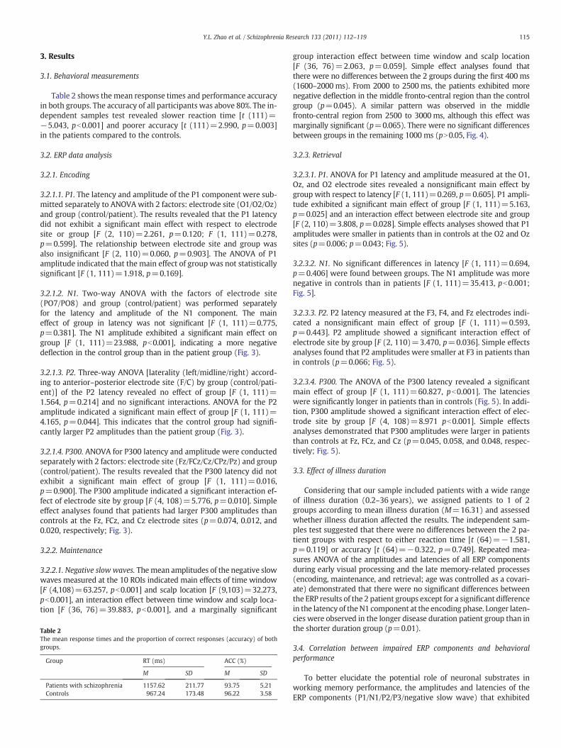
3.2.1.2. N1. Two-way ANOVA with the factors of electrode site(PO7/PO8) and group (control/patient) was performed separatelyfor the latency and amplitude of the N1 component. The maineffect of group in latency was not significant [F (1, 111)=0.775,p=0.381]. The N1 amplitude exhibited a significant main effect ongroup [F (1, 111)=23.988, pb0.001], indicating a more negativedeflection in the control group than in the patient group (Fig. 3).
3.2.1.3. P2. Three-way ANOVA [laterality (left/midline/right) accord-ing to anterior–posterior electrode site (F/C) by group (control/pati-ent)] of the P2 latency revealed no effect of group [F (1, 111)=1.564, p=0.214] and no significant interactions. ANOVA for the P2amplitude indicated a significant main effect of group [F (1, 111)=4.165, p=0.044]. This indicates that the control group had signifi-cantly larger P2 amplitudes than the patient group (Fig. 3).
3.2.1.4. P300. ANOVA for P300 latency and amplitude were conductedseparately with 2 factors: electrode site (Fz/FCz/Cz/CPz/Pz) and group(control/patient). The results revealed that the P300 latency did notexhibit a significant main effect of group [F (1, 111)=0.016,p=0.900]. The P300 amplitude indicated a significant interaction ef-fect of electrode site by group [F (4, 108)=5.776, p=0.010]. Simpleeffect analyses found that patients had larger P300 amplitudes thancontrols at the Fz, FCz, and Cz electrode sites (p=0.074, 0.012, and0.020, respectively; Fig. 3).
3.2.2. Maintenance
3.2.2.1. Negative slow waves. Themean amplitudes of the negative slowwaves measured at the 10 ROIs indicated main effects of time window[F (4,108)=63.257, pb0.001] and scalp location [F (9,103)=32.273,pb0.001], an interaction effect between time window and scalp loca-tion [F (36, 76)=39.883, pb0.001], and a marginally significant
Table 2The mean response times and the proportion of correct responses (accuracy) of bothgroups.
Group RT (ms) ACC (%)
M SD M SD
Patients with schizophrenia 1157.62 211.77 93.75 5.21Controls 967.24 173.48 96.22 3.58
group interaction effect between time window and scalp location[F (36, 76)=2.063, p=0.059]. Simple effect analyses found thatthere were no differences between the 2 groups during the first 400 ms(1600–2000 ms). From 2000 to 2500 ms, the patients exhibited morenegative deflection in the middle fronto-central region than the controlgroup (p=0.045). A similar pattern was observed in the middlefronto-central region from 2500 to 3000 ms, although this effect wasmarginally significant (p=0.065). There were no significant differencesbetween groups in the remaining 1000 ms (pN0.05, Fig. 4).
3.2.3. Retrieval
3.2.3.1. P1. ANOVA for P1 latency and amplitude measured at the O1,Oz, and O2 electrode sites revealed a nonsignificant main effect bygroup with respect to latency [F (1, 111)=0.269, p=0.605]. P1 ampli-tude exhibited a significant main effect of group [F (1, 111)=5.163,p=0.025] and an interaction effect between electrode site and group[F (2, 110)=3.808, p=0.028]. Simple effects analyses showed that P1amplitudes were smaller in patients than in controls at the O2 and Ozsites (p=0.006; p=0.043; Fig. 5).
3.2.3.2. N1. No significant differences in latency [F (1, 111)=0.694,p=0.406] were found between groups. The N1 amplitude was morenegative in controls than in patients [F (1, 111)=35.413, pb0.001;Fig. 5].
3.2.3.3. P2. P2 latency measured at the F3, F4, and Fz electrodes indi-cated a nonsignificant main effect of group [F (1, 111)=0.593,p=0.443]. P2 amplitude showed a significant interaction effect ofelectrode site by group [F (2, 110)=3.470, p=0.036]. Simple effectsanalyses found that P2 amplitudes were smaller at F3 in patients thanin controls (p=0.066; Fig. 5).
3.2.3.4. P300. The ANOVA of the P300 latency revealed a significantmain effect of group [F (1, 111)=60.827, pb0.001]. The latencieswere significantly longer in patients than in controls (Fig. 5). In addi-tion, P300 amplitude showed a significant interaction effect of elec-trode site by group [F (4, 108)=8.971 pb0.001]. Simple effectsanalyses demonstrated that P300 amplitudes were larger in patientsthan controls at Fz, FCz, and Cz (p=0.045, 0.058, and 0.048, respec-tively; Fig. 5).
3.3. Effect of illness duration
Considering that our sample included patients with a wide rangeof illness duration (0.2–36 years), we assigned patients to 1 of 2groups according to mean illness duration (M=16.31) and assessedwhether illness duration affected the results. The independent sam-ples test suggested that there were no differences between the 2 pa-tient groups with respect to either reaction time [t (64)=−1.581,p=0.119] or accuracy [t (64)=−0.322, p=0.749]. Repeated mea-sures ANOVA of the amplitudes and latencies of all ERP componentsduring early visual processing and the late memory-related processes(encoding, maintenance, and retrieval; age was controlled as a covari-ate) demonstrated that there were no significant differences betweenthe ERP results of the 2 patient groups except for a significant differencein the latency of theN1 component at the encoding phase. Longer laten-cies were observed in the longer disease duration patient group than inthe shorter duration group (p=0.01).
3.4. Correlation between impaired ERP components and behavioralperformance
To better elucidate the potential role of neuronal substrates inworking memory performance, the amplitudes and latencies of theERP components (P1/N1/P2/P3/negative slow wave) that exhibited
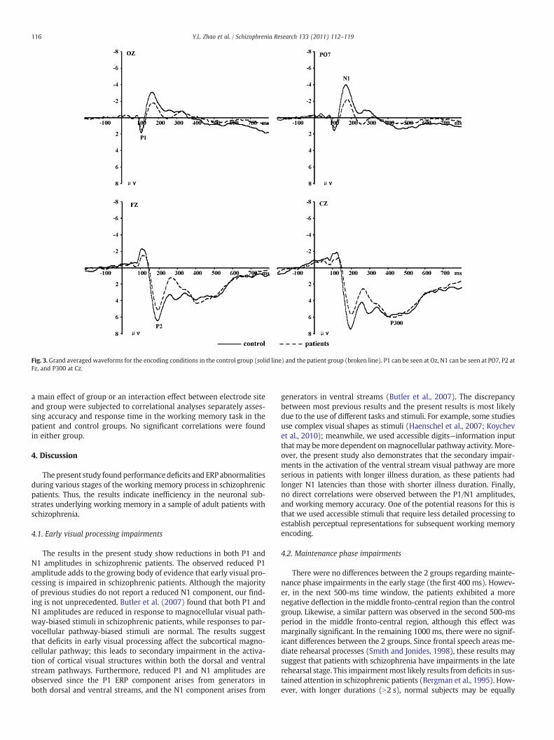
Fig. 3. Grand averaged waveforms for the encoding conditions in the control group (solid line) and the patient group (broken line). P1 can be seen at Oz, N1 can be seen at PO7, P2 atFz, and P300 at Cz.
116 Y.L. Zhao et al. / Schizophrenia Research 133 (2011) 112–119
a main effect of group or an interaction effect between electrode siteand group were subjected to correlational analyses separately asses-sing accuracy and response time in the working memory task in thepatient and control groups. No significant correlations were foundin either group.
4. Discussion
The present study foundperformancedeficits and ERP abnormalitiesduring various stages of the working memory process in schizophrenicpatients. Thus, the results indicate inefficiency in the neuronal sub-strates underlying working memory in a sample of adult patients withschizophrenia.
4.1. Early visual processing impairments
The results in the present study show reductions in both P1 andN1 amplitudes in schizophrenic patients. The observed reduced P1amplitude adds to the growing body of evidence that early visual pro-cessing is impaired in schizophrenic patients. Although the majorityof previous studies do not report a reduced N1 component, our find-ing is not unprecedented. Butler et al. (2007) found that both P1 andN1 amplitudes are reduced in response to magnocellular visual path-way-biased stimuli in schizophrenic patients, while responses to par-vocellular pathway-biased stimuli are normal. The results suggestthat deficits in early visual processing affect the subcortical magno-cellular pathway; this leads to secondary impairment in the activa-tion of cortical visual structures within both the dorsal and ventralstream pathways. Furthermore, reduced P1 and N1 amplitudes areobserved since the P1 ERP component arises from generators inboth dorsal and ventral streams, and the N1 component arises from
generators in ventral streams (Butler et al., 2007). The discrepancybetween most previous results and the present results is most likelydue to the use of different tasks and stimuli. For example, some studiesuse complex visual shapes as stimuli (Haenschel et al., 2007; Koychevet al., 2010); meanwhile, we used accessible digits—information inputthat may bemore dependent onmagnocellular pathway activity. More-over, the present study also demonstrates that the secondary impair-ments in the activation of the ventral stream visual pathway are moreserious in patients with longer illness duration, as these patients hadlonger N1 latencies than those with shorter illness duration. Finally,no direct correlations were observed between the P1/N1 amplitudes,and working memory accuracy. One of the potential reasons for this isthat we used accessible stimuli that require less detailed processing toestablish perceptual representations for subsequent working memoryencoding.
4.2. Maintenance phase impairments
There were no differences between the 2 groups regarding mainte-nance phase impairments in the early stage (the first 400 ms). Howev-er, in the next 500-ms time window, the patients exhibited a morenegative deflection in themiddle fronto-central region than the controlgroup. Likewise, a similar pattern was observed in the second 500-msperiod in the middle fronto-central region, although this effect wasmarginally significant. In the remaining 1000 ms, there were no signif-icant differences between the 2 groups. Since frontal speech areas me-diate rehearsal processes (Smith and Jonides, 1998), these results maysuggest that patients with schizophrenia have impairments in the laterehearsal stage. This impairmentmost likely results from deficits in sus-tained attention in schizophrenic patients (Bergman et al., 1995). How-ever, with longer durations (N2 s), normal subjects may be equally
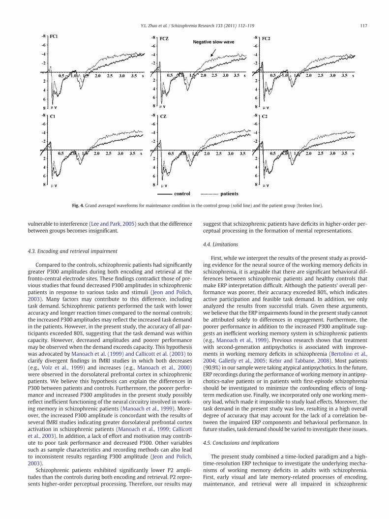
Fig. 4. Grand averaged waveforms for maintenance condition in the control group (solid line) and the patient group (broken line).
117Y.L. Zhao et al. / Schizophrenia Research 133 (2011) 112–119
vulnerable to interference (Lee and Park, 2005) such that the differencebetween groups becomes insignificant.
4.3. Encoding and retrieval impairment
Compared to the controls, schizophrenic patients had significantlygreater P300 amplitudes during both encoding and retrieval at thefronto-central electrode sites. These findings contradict those of pre-vious studies that found decreased P300 amplitudes in schizophrenicpatients in response to various tasks and stimuli (Jeon and Polich,2003). Many factors may contribute to this difference, includingtask demand. Schizophrenic patients performed the task with loweraccuracy and longer reaction times compared to the normal controls;the increased P300 amplitudes may reflect the increased task demandin the patients. However, in the present study, the accuracy of all par-ticipants exceeded 80%, suggesting that the task demand was withincapacity. However, decreased amplitudes and poorer performancemay be observed when the demand exceeds capacity. This hypothesiswas advocated by Manoach et al. (1999) and Callicott et al. (2003) toclarify divergent findings in fMRI studies in which both decreases(e.g., Volz et al., 1999) and increases (e.g., Manoach et al., 2000)were observed in the dorsolateral prefrontal cortex in schizophrenicpatients. We believe this hypothesis can explain the differences inP300 between patients and controls. Furthermore, the poorer perfor-mance and increased P300 amplitudes in the present study possiblyreflect inefficient functioning of the neural circuitry involved in work-ing memory in schizophrenic patients (Manoach et al., 1999). More-over, the increased P300 amplitude is concordant with the results ofseveral fMRI studies indicating greater dorsolateral prefrontal cortexactivation in schizophrenic patients (Manoach et al., 1999; Callicottet al., 2003). In addition, a lack of effort and motivation may contrib-ute to poor task performance and decreased P300. Other variablessuch as sample characteristics and recording methods can also leadto inconsistent results regarding P300 amplitude (Jeon and Polich,2003).
Schizophrenic patients exhibited significantly lower P2 ampli-tudes than the controls during both encoding and retrieval. P2 repre-sents higher-order perceptual processing. Therefore, our results may
suggest that schizophrenic patients have deficits in higher-order per-ceptual processing in the formation of mental representations.
4.4. Limitations
First, while we interpret the results of the present study as provid-ing evidence for the neural source of the working memory deficits inschizophrenia, it is arguable that there are significant behavioral dif-ferences between schizophrenic patients and healthy controls thatmake ERP interpretation difficult. Although the patients' overall per-formance was poorer, their accuracy exceeded 80%, which indicatesactive participation and feasible task demand. In addition, we onlyanalyzed the results from successful trials. Given these arguments,we believe that the ERP impairments found in the present study cannotbe attributed solely to differences in engagement. Furthermore, thepoorer performance in addition to the increased P300 amplitude sug-gests an inefficient working memory system in schizophrenic patients(e.g., Manoach et al., 1999). Previous research shows that treatmentwith second-generation antipsychotics is associated with improve-ments in working memory deficits in schizophrenia (Bertolino et al.,2004; Galletly et al., 2005; Kebir and Tabbane, 2008). Most patients(90.9%) in our sample were taking atypical antipsychotics. In the future,ERP recordings during the performance of workingmemory in antipsy-chotics-naïve patients or in patients with first-episode schizophreniashould be investigated to minimize the confounding effects of long-term medication use. Finally, we incorporated only one working mem-ory load, which made it impossible to study load effects. Moreover, thetask demand in the present study was low, resulting in a high overalldegree of accuracy that may account for the lack of a correlation be-tween the impaired ERP components and behavioral performance. Infuture studies, task demand should be varied to investigate these issues.
4.5. Conclusions and implications
The present study combined a time-locked paradigm and a high-time-resolution ERP technique to investigate the underlying mecha-nisms of working memory deficits in adults with schizophrenia.First, early visual and late memory-related processes of encoding,maintenance, and retrieval were all impaired in schizophrenic
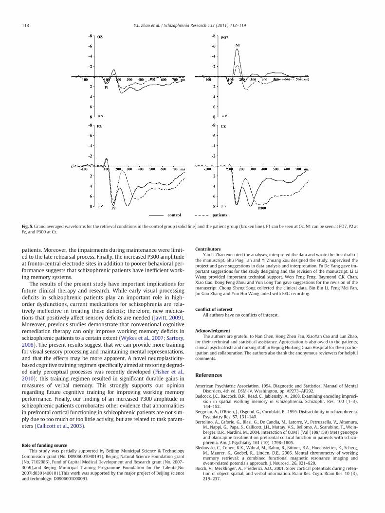
Fig. 5. Grand averaged waveforms for the retrieval conditions in the control group (solid line) and the patient group (broken line). P1 can be seen at Oz, N1 can be seen at PO7, P2 atFz, and P300 at Cz.
118 Y.L. Zhao et al. / Schizophrenia Research 133 (2011) 112–119
patients. Moreover, the impairments during maintenance were limit-ed to the late rehearsal process. Finally, the increased P300 amplitudeat fronto-central electrode sites in addition to poorer behavioral per-formance suggests that schizophrenic patients have inefficient work-ing memory systems.
The results of the present study have important implications forfuture clinical therapy and research. While early visual processingdeficits in schizophrenic patients play an important role in high-order dysfunctions, current medications for schizophrenia are rela-tively ineffective in treating these deficits; therefore, new medica-tions that positively affect sensory deficits are needed (Javitt, 2009).Moreover, previous studies demonstrate that conventional cognitiveremediation therapy can only improve working memory deficits inschizophrenic patients to a certain extent (Wykes et al., 2007; Sartory,2008). The present results suggest that we can provide more trainingfor visual sensory processing and maintaining mental representations,and that the effects may be more apparent. A novel neuroplasticity-based cognitive training regimen specifically aimed at restoring degrad-ed early perceptual processes was recently developed (Fisher et al.,2010); this training regimen resulted in significant durable gains inmeasures of verbal memory. This strongly supports our opinionregarding future cognitive training for improving working memoryperformance. Finally, our finding of an increased P300 amplitude inschizophrenic patients corroborates other evidence that abnormalitiesin prefrontal cortical functioning in schizophrenic patients are not sim-ply due to too much or too little activity, but are related to task param-eters (Callicott et al., 2003).
Role of funding sourceThis study was partially supported by Beijing Municipal Science & Technology
Commission grant (No. D0906001040191), Beijing Natural Science Foundation grant(No. 7102086), Fund of Capital Medical Development and Research grant (No. 2007–3059),and Beijing Municipal Training Programme Foundation for the Talents(No.2007id0301400101).This work was supported by the major project of Beijing scienceand technology: D0906001000091.
ContributorsYan Li Zhao executed the analyses, interpreted the data and wrote the first draft of
the manuscript. Shu Ping Tan and Yi Zhuang Zou designed the study, supervised theproject and gave suggestions in data analysis and interpretation. Fu De Yang gave im-portant suggestions for the study designing and the revision of the manuscript. Li LiWang provided important technical support. Wen Feng Feng, Raymond C.K. Chan,Xiao Gao, Dong Feng Zhou and Yun Long Tan gave suggestions for the revision of themanuscript .Chong Sheng Song collected the clinical data. Bin Bin Li, Feng Mei Fan,Jin Guo Zhang and Yun Hui Wang aided with EEG recording.
Conflict of interestAll authors have no conflicts of interest.
AcknowledgmentThe authors are grateful to Nan Chen, Hong Zhen Fan, XiaoYan Cao and Lun Zhao,
for their technical and statistical assistance. Appreciation is also owed to the patients,clinical psychiatrists and nursing staff in Beijing HuiLong Guan Hospital for their partic-ipation and collaboration. The authors also thank the anonymous reviewers for helpfulcomments.
References
American Psychiatric Association, 1994. Diagnostic and Statistical Manual of MentalDisorders, 4th ed. DSM-IV, Washington, pp. AP273–AP292.
Badcock, J.C., Badcock, D.R., Read, C., Jablensky, A., 2008. Examining encoding impreci-sion in spatial working memory in schizophrenia. Schizophr. Res. 100 (1–3),144–152.
Bergman, A., O'Brien, J., Osgood, G., Cornblatt, B., 1995. Distractibility in schizophrenia.Psychiatry Res. 57, 131–140.
Bertolino, A., Caforio, G., Blasi, G., De Candia, M., Latorre, V., Petruzzella, V., Altamura,M., Nappi, G., Papa, S., Callicott, J.H., Mattay, V.S., Bellomo, A., Scarabino, T., Wein-berger, D.R., Nardini, M., 2004. Interaction of COMT (Val (108/158) Met) genotypeand olanzapine treatment on prefrontal cortical function in patients with schizo-phrenia. Am. J. Psychiatry 161 (10), 1798–1805.
Bledowski, C., Cohen, K.K., Wibral, M., Rahm, B., Bittner, R.A., Hoechstetter, K., Scherg,M., Maurer, K., Goebel, R., Linden, D.E., 2006. Mental chronometry of workingmemory retrieval: a combined functional magnetic resonance imaging andevent-related potentials approach. J. Neurosci. 26, 821–829.
Bosch, V., Mecklinger, A., Friederici, A.D., 2001. Slow cortical potentials during reten-tion of object, spatial, and verbal information. Brain Res. Cogn. Brain Res. 10 (3),219–237.
119Y.L. Zhao et al. / Schizophrenia Research 133 (2011) 112–119
Butler, P.D., Martinez, A., Foxe, J.J., Kim, D., Zemon, V., Silipo, G., Mahoney, J., Shpaner,M., Jalbrzikowski, M., Javitt, D.C., 2007. Subcortical visual dysfunction in schizo-phrenia drives secondary cortical impairments. Brain 130 (Pt 2), 417–430.
Butler, P.D., Abeles, I.Y., Weiskopf, N.G., Tambini, A., Jalbrzikowski, M., Legatt, M.E., etal., 2009. Sensory contributions to impaired emotion processing in schizophrenia.Schizophr. Bull. 35 (6), 1095–1107.
Cairo, T.A., Woodward, T.S., Ngan, E.T., 2006. Decreased encoding efficiency in schizo-phrenia. Biol. Psychiatry 59 (8), 740–746.
Callicott, J.H., Mattay, V.S., Verchinski, B.A., Marenco, S., Egan, M.F., Weinberger, D.R.,2003. Complexity of prefrontal cortical dysfunctionin schizophrenia: more thanup or down. Am. J. Psychiatry 160, 2209–2215.
Chen, Y., Levy, D.L., Nakayama, K., Matthysse, S., Palafox, G., Holzman, P.S., 1999a. De-pendence of impaired eye tracking on deficient velocity discrimination in schizo-phrenia. Arch. Gen. Psychiatry 56, 155–161.
Chen, Y., Nakayama, K., Levy, D.L., Matthysse, S., Holzman, P.S., 1999b. Psychophysicalisolation of a motion-processing deficit in schizophrenics and their relatives and itsassociationwith impaired smooth pursuit. Proc. Natl. Acad. Sci. U. S. A. 96, 4724–4729.
Driesen, N.R., Leung, H.C., Calhoun, V.D., Constable, R.T., Gueorguieva, R., Hoffman, R.,Skudlarski, P., Goldman-Rakic, P.S., Krystal, J.H., 2008. Impairment of workingmemory maintenance and response in schizophrenia: functional magnetic reso-nance imaging evidence. Biol. Psychiatry 64 (12), 1026–1034.
Fisher, M., Holland, C., Subramaniam, K., Vinogradov, S., 2010. Neuroplasticity-basedcognitive training in schizophrenia: an interim report on the effects 6 monthslater. Schizophr. Bull. 36 (4), 869–879.
Galletly, C.A., Clark, C.R., McFarlane, A.C., 2005. Clozapine improves working memoryupdating in schizophrenia. Eur. Neuropsychopharmacol. 15 (6), 601–608.
Haenschel, C., Bittner, R.A., Haertling, F., Rotarska-Jagiela, A., Maurer, K., Singer, W., Lin-den, D.E., 2007. Contribution of impaired early-stage visual processing to workingmemory dysfunction in adolescents with schizophrenia: a study with event-related potentials and functional magnetic resonance imaging. Arch. Gen. Psychia-try 64 (11), 1229–1240.
Hartman,M., Steketee, M.C., Silva, S., Lanning, K., McCann, H., 2003.Workingmemory andschizophrenia: evidence for slowed encoding. Schizophr. Res. 59 (2–3), 99–113.
Hashimoto, R., Lee, K., Preus, A., McCarley, R.W., Wible, C.G., 2010. An fMRI study offunctional abnormalities in the verbal working memory system and the relation-ship to clinical symptoms in chronic schizophrenia. Cereb. Cortex 20 (1), 46–60.
Henseler, I., Falkai, P., Gruber, O., 2009. A systematic fMRI investigation of the brain sys-tems subserving different working memory components in schizophrenia. Eur. J.Neurosci. 30 (4), 693–702.
Hill, S.K., Griffin, G.B., Miura, T.K., Herbener, E.S., Sweeney, J.A., 2010. Salience ofworking-memory maintenance and manipulation deficits in schizophrenia. Psy-chol. Med. 1–8.
Javitt, D.C., 2009. When doors of perception close: bottom-up models of disrupted cog-nition in schizophrenia. Annu. Rev. Clin. Psychol. 5, 249–275.
Jeon, Y.W., Polich, J., 2003. Meta-analysis of P300 and schizophrenia: patients, para-digms, and practical implications. Psychophysiology 40, 684–701.
Johnson, M.R., Morris, N.A., Astur, R.S., Calhoun, V.D., Mathalon, D.H., Kiehl, K.A., Pearl-son, G.D., 2006. A functional magnetic resonance imaging study of working mem-ory abnormalities in schizophrenia. Biol. Psychiatry 60 (1), 11–21.
Kay, S.R., Fiszbein, A., Opler, L.A., 1987. The positive and negative syndrome scale(PANSS) for schizophrenia. Schizophr. Bull. 13 (2), 261–276.
Kebir, O., Tabbane, K., 2008. Working memory in schizophrenia: a review. Encephale34 (3), 289–298.
Koychev, I., Deredy, W.E., Haenschel, C., Francis, J., Deakin, W., 2010. Visual informationprocessing deficits as biomarkers of vulnerability to schizophrenia: an event-related potential study in schizotypy. Neuropsychologia 48, 2205–2214.
Lee, J., Park, S., 2005. Working memory impairments in schizophrenia: a meta-analysis.J. Abnorm. Psychol. 114 (4), 599–611.
Luck, S.J., Fuller, R.L., Braun, E.L., Robinson, B., Summerfelt, A., Gold, J.M., 2006. Thespeed of visual attention in schizophrenia: electrophysiological and behavioral ev-idence. Schizophr. Res. 85, 174–195.
Manoach, D.S., Press, D.Z., Thangaraj, V., Searl, M.M., Goff, D.C., Halpern, E., Saper, C.B.,Warach, S., 1999. Schizophrenic subjects activate dorsolateral prefrontal cortexduring a working memory task, as measured by fMRI. Biol. Psychiatry 45 (9),1128–1137.
Manoach, D.S., Gollub, R.L., Benson, E.S., Searl, M.M., Goff, D.C., Halpern, E., Saper, C.B.,Rauch, S.L., 2000. Schizophrenic subjects show aberrant fMRI activation of dorso-lateral prefrontal cortex and basal ganglia during working memory performance.Biol. Psychiatry 48, 99–109.
Martinez, A., Hillyard, S.A., Dias, E.C., Hagler Jr., D.J., Butler, P.D., Guilfoyle, D.N., Jalbrzi-kowski, M., Silipo, G., Javitt, D.C., 2008. Magnocellular pathway impairment inschizophrenia: evidence from functional magnetic resonance imaging. J. Neurosci.28 (30), 7492–7500.
Mazhari, S., Badcock, J.C., Waters, F.A., Dragovic, M., Badcock, D.R., Jablensky, A., 2010.Impaired spatial working memory maintenance in schizophrenia involves bothspatial coordinates and spatial reference frames. Psychiatry Res. 179 (3), 253–258.
Mecklinger, A., Pfeifer, E., 1996. Event-related potentials reveal topographical and tem-poral distinct neuronal activation patterns for spatial and object working memory.Brain Res. Cogn. Brain Res. 4, 211–224.
Pelosi, L., Blumhardt, L.D., 1999. Effects of age on working memory: an event-relatedpotential study. Brain Res. Cogn. Brain Res. 7 (3), 321–334.
Pelosi, L., Hayward, M., Blumhardt, L.D., 1998. Which event-related potentials reflectmemory processing in a digit-probe identification task. Brain Res. Cogn. BrainRes. 6 (3), 205–218.
Ruchkin, D.S., Grafman, J., Krauss, G.L., Johnson Jr., R., Canoune, H., Ritter, W., 1994.Event-related brain potential evidence for a verbal working memory deficit in mul-tiple sclerosis. Brain 117 (Pt 2), 289–305.
Sartory, G., 2008. Cognitive remediation therapy produces moderate improvements inworking memory in people with schizophrenia. Evid. Based Ment. Health 11 (1),18.
Silver, H., Feldman, P., Bilker, W., Gur, R.C., 2003. Working memory deficit as a coreneuropsychological dysfunction in schizophrenia. Am. J. Psychiatry 160 (10),1809–1816.
Smith, E.E., Jonides, J., 1998. Neuroimaging analyses of human working memory. Proc.Natl. Acad. Sci. U. S. A. 95 (20), 12061–12068.
Volz, H., Gaser, C., Hager, F., Rzanny, R., Ponisch, J., Mentzel, H., Kaiser, W.A., Sauer, H.,1999. Decreased frontal activation in schizophrenics during stimulation with theContinuous Performance Test—a functional magnetic resonance imaging study.Eur. Psychiatry 14, 17–24.
Woods, S.W., 2003. Chlorpromazine equivalent doses for the newer atypical antipsy-chotics. J. Clin. Psychiatry 64 (6), 663–667.
Wykes, T., Reeder, C., Landau, S., Everitt, B., Knapp, M., Patel, A., Romeo, R., 2007. Cog-nitive remediation therapy in schizophrenia: randomised controlled trial. Br. J. Psy-chiatry 190, 421–427.
Yeap, S., Kelly, S.P., Sehatpour, P., Magno, E., Garavan, H., Thakore, J.H., Foxe, J.J., 2008.Visual sensory processing deficits in schizophrenia and their relationship to dis-ease state. Eur. Arch. Psychiatry Clin. Neurosci. 258, 305–316.
Zilles, D., Gruber, E., Falkai, P., Gruber, O., 2010. Patients with schizophrenia show def-icits of working memory maintenance components in circuit-specific tasks. Eur.Arch. Psychiatry Clin. Neurosci. 260 (7), 519–525.