Overcoming cancer therapeutic bottleneck by drug repurposing
Upload
khangminh22Category
view
0download
0
biology
Article
Drug Repurposing Approach against Novel CoronavirusDisease (COVID-19) through Virtual Screening TargetingSARS-CoV-2 Main Protease
Kamrul Hasan Chowdhury 1,†, Md. Riad Chowdhury 1,†, Shafi Mahmud 2, Abu Montakim Tareq 1 ,Nujhat Binte Hanif 1, Naureen Banu 1, A. S. M. Ali Reza 1,* , Talha Bin Emran 3,* and Jesus Simal-Gandara 4,*
�����������������
Citation: Chowdhury, K.H.; Chowd-
hury, M.R.; Mahmud, S.; Tareq, A.M.;
Hanif, N.B.; Banu, N.; Reza, A.S.M.A.;
Emran, T.B.; Simal-Gandara, J. Drug
Repurposing Approach against Novel
Coronavirus Disease (COVID-19) through
Virtual Screening Targeting SARS-CoV-
2 Main Protease. Biology 2021, 10, 2.
https://dx.doi.org/10.3390/
biology10010002
Received: 23 November 2020
Accepted: 19 December 2020
Published: 23 December 2020
Publisher’s Note: MDPI stays neu-
tral with regard to jurisdictional claims
in published maps and institutional
affiliations.
Copyright: © 2020 by the authors. Li-
censee MDPI, Basel, Switzerland. This
article is an open access article distributed
under the terms and conditions of the
Creative Commons Attribution (CC BY)
license (https://creativecommons.org/
licenses/by/4.0/).
1 Department of Pharmacy, International Islamic University Chittagong, Chittagong 4318, Bangladesh;[email protected] (K.H.C.); [email protected] (M.R.C.);[email protected] (A.M.T.); [email protected] (N.B.H.); [email protected] (N.B.)
2 Microbiology Laboratory, Bioinformatics Division, Department of Genetic Engineering and Biotechnology,University of Rajshahi, Rajshahi 6205, Bangladesh; [email protected]
3 Department of Pharmacy, BGC Trust University Bangladesh, Chittagong 4381, Bangladesh4 Nutrition and Bromatology Group, Department of Analytical and Food Chemistry,
Faculty of Food Science and Technology, University of Vigo–Ourense Campus, E32004 Ourense, Spain* Correspondence: [email protected] (A.S.M.A.R.); [email protected] (T.B.E.);
[email protected] (J.S.-G.)† These authors contributed equally to this work.
Simple Summary: With the urgent necessity of potential treatment against novel coronavirus disease,we used several computational methods to search for active drugs from an extensive database.The results of our investigation suggested several established drugs that can be subjected to furtheranalysis for the treatment of novel coronavirus disease. Various methods used in this study provedthe effectiveness of the retrieved drugs. Therefore, our findings highly recommend the mentioneddrugs to be scrutinized to discover drugs against novel coronavirus.
Abstract: Novel coronavirus disease (COVID-19) was identified from China in December 2019and spread rapidly through human-to-human transmission, affecting so many people worldwide.Until now, there has been no specific treatment against the disease and repurposing of the drug.Our investigation aimed to screen potential inhibitors against coronavirus for the repurposing ofdrugs. Our study analyzed sequence comparison among SARS-CoV, SARS-CoV-2, and MERS-CoV todetermine the identity matrix using discovery studio. SARS-CoV-2 Mpro was targeted to generate anE-pharmacophore hypothesis to screen drugs from the DrugBank database having similar features.Promising drugs were used for docking-based virtual screening at several precisions. Best hits fromvirtual screening were subjected to MM/GBSA analysis to evaluate binding free energy, followed bythe analysis of binding interactions. Furthermore, the molecular dynamics simulation approacheswere carried out to assess the docked complex’s conformational stability. A total of 33 drug classeswere found from virtual screening based on their docking scores. Among them, seven potential drugswith several anticancer, antibiotic, and immunometabolic categories were screened and showedpromising MM/GBSA scores. During interaction analysis, these drugs exhibited different types ofhydrogen and hydrophobic interactions with amino acid residue. Besides, 17 experimental drugsselected from virtual screening might be crucial for drug discovery against COVID-19. The RMSD,RMSF, SASA, Rg, and MM/PBSA descriptors from molecular dynamics simulation confirmed thecomplex’s firm nature. Seven promising drugs for repurposing against SARS-CoV-2 main pro-tease (Mpro), namely sapanisertib, ornidazole, napabucasin, lenalidomide, daniquidone, indoximod,and salicylamide, could be vital for the treatment of COVID-19. However, extensive in vivo andin vitro studies are required to evaluate the mentioned drug’s activity.
Keywords: COVID-19; antiviral agents; drug repurposing; molecular docking; molecular dynamics;E-pharmacophore hypothesis; virtual screening
Biology 2021, 10, 2. https://dx.doi.org/10.3390/biology10010002 https://www.mdpi.com/journal/biology
Biology 2021, 10, 2 2 of 14
1. Introduction
Novel coronavirus disease (COVID-19) has become a threatening illness globally andwas declared as a pandemic by WHO (World Health Organization). COVID-19 startedspreading from the Wuhan seafood market in China since December 2019 [1]. As of thewriting of this paper, 1,151,622 persons have died so far in COVID-19 and confirmed casesof persons with COVID-19 are 42,662,304 [2].
Coronavirus (CoVs) are the family of Coronaviridae, which is single-stranded en-veloped positive RNA virus subdivided into alpha (α), beta (β), gamma (γ), and delta (δ).Among them, the coronavirus β (β-CoV) group is divided into severe acute respiratory syn-drome coronavirus (SARS-CoV), severe acute respiratory syndrome coronavirus-2 (SARS-CoV-2), and middle east respiratory syndrome coronavirus (MERS-CoV) [3]. These threetypes of virus are highly fatal and responsible for respiratory, liver, gastrointestinal, and cen-tral nervous system damage in humans and animals [4]. In case of respiratory disease, it canlead to severe pneumonia with several symptoms, including fever, dry cough, vomiting,fatigue, diarrhea, and shortness of breath [5,6].
SARS-CoV-2 responsible for COVID-19 is extremely infectious and more pathogenicthan SARS-CoV and MERS-CoV, which can transmit from human to human and causefatal illness [5]. As there are no potential drugs or vaccines against coronavirus until now,emergency investigations are required to establish effective treatment. Several groups ofdrugs are being investigated against COVID-19, which includes drugs, namely hydroxy-chloroquine, chloroquine, lopinavir, ritonavir, remdesivir, etc., used for the treatment ofSARS-CoV, MERS-CoV, and other viruses. However, there is an emergency requirementto establish novel, selective, and potential inhibitors against COVID-19 for the effectivetreatment [7]. In this short period of time, repurposing of drugs is a significant way tofind out potential inhibitors against COVID-19. During repurposing, virtual screening,pharmacophore modeling, and other computational methods are extensively used [8].
The main protease (Mpro/3CLpro) is an attractive drug target due to its vital functionthat regulates polyproteins translated from the viral RNA [9]. The aim of our study wasto focus on the SARS-CoV-2 main protease as a potential drug target to screen drugs forrepurposing against COVID-19. The viral protein with a bound inhibitor was subjected toE-pharmacophore and molecular docking-based virtual screening to determine promisinginhibitors of novel coronavirus. In addition, potential drugs were investigated throughMM/GBSA and binding interaction for further analysis.
2. Materials and Methods2.1. Sequence Comparison
The crystal structures of SARS-CoV-2 Mpro (PDB ID: 5R7Y) [10], SARS-CoV Mpro
(PDB ID: 2C3S) [11], and MERS-CoV Mpro (PDB ID: 5C3N) [12] were obtained from proteindata bank. Afterwards, the protein structures were transferred into discovery studio soft-ware in order to align and explore their structural sequence for comparison purposes [13].Accordingly, SARS-CoV-2 main protease (PDB ID: 5R7Y) in complex with Z45617795 at aresolution of 1.65 Å was used for further investigation.
2.2. Protein Preparation
Pre-processing of the structure of the COVID-19 main protease in complex withZ45617795 was carried out through transformation of selenomethionines into methion-ines, removal of water molecules, and addition of missing hydrogen atoms followed byminimization of the structure utilizing the OPLS3 force field [14]. All these steps wereperformed utilizing the protein preparation wizard of Schrödinger Maestro (Protein Prepa-ration Wizard; Epik, Schrödinger, LLC, New York, NY, USA; v.11.1) [15]. In a similarmanner, COVID-19 main protease in complex with inhibitor N3 at 1.7 angstrom (PDB ID:7 BQY) was prepared.
Biology 2021, 10, 2 3 of 14
2.3. Generation of E-Pharmacophore Hypothesis
Previously prepared protein was subjected to E-pharmacophore hypothesis establish-ment using phase module of Schrodinger suit (Phase, Schrödinger, LLC, New York, NY,USA; v.11.1) to generate the pharmacophoric features [16]. Different features, namely aro-matic ring (R), hydrogen bond acceptor (A), hydrogen bond donor (D), positive ion (P),negative ion (N), and hydrophobicity (H), were mapped to form the protein-ligand com-plex.
2.4. Virtual Screening using E-Pharmacophore Hypothesis
The molecular structure of 8820 drugs from the DrugBank database were screenedfor promising candidates against SARS-CoV-2 main protease (Mpro) enzyme utilizingthe pharmacophoric features from the established E-pharmacophore hypothesis [17]. E-pharmacophore-based screening was carried out by the phase ligand screening module ofSchrodinger Suit [16].
2.5. Ligand Preparation
Best hits obtained from virtual screening were prepared utilizing LigPrep module ofSchrodinger suit [18]. Selected drugs were subjected to an optimization process at targetpH (7 ± 1) in order to generate all possible states, such as tautomeric and stereo-isomericfollowed by minimization at the OPLS3 force field [14].
2.6. Virtual Screening Based on Molecular Docking
A glide grid box was generated at the receptor complex site at 15× 15× 15 Å along theX, Y, and Z axes while docking. Virtual screening using molecular docking was performedby virtual screening workflow of Schrodinger suit to carry out high-throughput virtualscreening (HTVS), standard precision (SP), and extra precision (XP) docking, respectively.Docking of each precision was filtrated and expressed as a glide score [19]. Subsequently,the ligand (Z45617795) bound to SARS-CoV-2 main protease was extracted from protein-ligand complex and subjected to re-docking through standard precision (SP) docking [20].From this, the docking score was obtained as a control (control 1) to compare the valuewith newly screened drugs. Additionally, inhibitor N3 was docked with the COVID-19Mpro in complex with inhibitor N3 (control 2).
2.7. MM/GBSA and Interaction Analysis
Promising candidates obtained from docking-based virtual screening were subjectedto MM/GBSA using Prime MM/GBSA module of Schrodinger Suit to estimate bindingfree energy and compared with control 1 (Z45617795) and control 2 (inhibitor N3) [21].Best candidates from MM/GBSA analysis were used for further interaction analysis byutilizing discovery studio software [13].
2.8. Molecular Dynamics Simulations
The molecular dynamics simulation of the main protease of SARS-CoV-2 and screenedligand complexes were further analyzed through molecular dynamics simulation to eval-uate their motion and structural integrity in the YASARA software package [22]. Here,the compound Z and the main protease complex (PDB ID: 5R7Y), and inhibitor N3 alongwith main protease protein (PDB ID: 7BQY) were considered as control 1 and control 2.The drug-protein complexes were initially cleaned in the software and the hydrogen bondnetwork of the system was optimized. The AMBER14 force field [23] was applied anda cubic simulation cell was created. The cell size was set bigger than the drug-proteincomplex by 20 Å in all cases so that each biological system can move freely. The initialenergy minimization procedure was applied through a simulated annealing method byemploying the steepest gradient approaches. The simulation cell box was neutralized withthe addition of water molecules and 0.9% NaCl at 7.4 pH. The temperature of each systemwas set as 298k and the Berendsen thermostat was used to control the cell temperature [24].

Biology 2021, 10, 2 4 of 14
The long-range electrostatic interactions of the system were calculated through PME orthe Particle Mesh Ewald method at a cut off radius 8 Å. The time-step of the simulationcell was set as 1.25 fs and each of the trajectories were saved at a 100-ps interval. Finally,the simulation was carried out for 100 ns to analyze the root mean square deviation (RMSD),root mean square fluctuation (RMSF), radius of gyration (Rg), solvent accessible surfacearea (SASA), and hydrogen bond [25–28].
The MM/PBSA method was applied to calculate the binding free energy from simula-tion trajectories. The default macro of YASARA program (md_analyzebindenergy.mcr) wasmodified and free energy calculations was performed for the seven drug-protein complexes.The following equation was used to calculate MM/PBSA binding free energy:
∆Gbind = ∆Gcomplex (minimized) − [∆Gligand (minimized) + ∆Greceptor (minimized)] (1)
∆Gbind = ∆G MM +∆G PB +∆G SA − T∆S (2)
where ∆G MM is the sum of Van der Waals and electrostatic interaction, ∆G PB and ∆G SAis the polar and non-polar solvation energies, and T∆S is the entropic contribution.
3. Results and Discussion3.1. Sequence Comparison
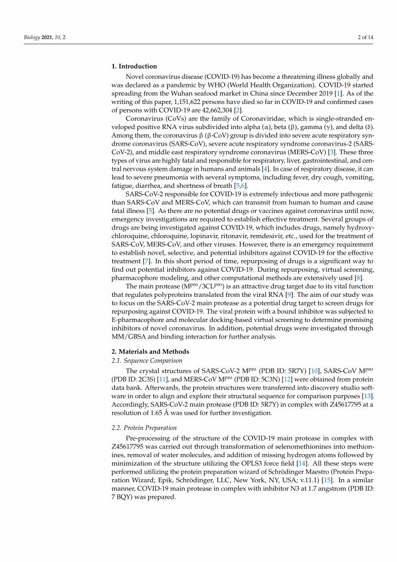
Comparison of SARS-CoV-2 Mpro (PDB ID: 5R7Y), SARS-CoV Mpro (PDB ID: 2C3S),and MERS-CoV Mpro (PDB ID: 5C3N) sequences are represented in Figure 1a. The redcolor denotes identically matching, light blue refers to as strongly matching, green colorindicates weakly matching, and sequence without any color signifies as non-matchingwith each other. The identity matrix (%) of the sequence comparison is demonstratedin Table 1. In the comparison with SARS-CoV-2, 96% and 24.2% identity matrix weredetected for SARS-CoV and MERS-CoV, respectively, and SARS-CoV-2 Mpro suggestedan attractive drug target for the treatment of COVID-19 due to its similarity with SARS-CoV and MERS-CoV. The identity matrix indicates an effective target for the screeningof promising drugs to inhibit coronavirus replication into the host cell. Previous studiessuggest that the amino acid difference between SARS-CoV-2 and SARS-CoV was 12. On theother hand, amino acid residue conservation between SARS-CoV-2 and MERS-CoV was153 [8]. Structural analysis of the SARS-CoV-2 Mpro active site revealed several amino acidresidues, namely THR-25, THR-190, THR-26, HIS-41, HIS-163, LEU-27, LEU-141, LEU-166,SER-46, PHE-140, PHE-185, PRO-168, MET-49, MET-165, TYR-54, ASN-142, ALA-191,GLY-143, GLN-189, GLN-192, GLU-166, ASP-187, and CYS-145 [29]. The primary structureof SARS-CoV-2 Mpro is presented in Figure 1b.
Table 1. Sequence comparison matrix of SARS-CoV-2 (PDB ID: 5R7Y), SARS-CoV (PDB ID: 2C3S),and MERS-CoV (PDB ID: 5C3N). Identity matrix is expressed as a percentage.
Sequence Name 5R7Y 2C3S 5C3N
5R7Y 100 96 24.22C3S 96 100 25.55C3N 24.2 25.5 100
Biology 2021, 10, 2 5 of 14Biology 2021, 10, x 5 of 14
Biology 2021, 10, x. https://doi.org/10.3390/xxxxx www.mdpi.com/journal/biology
Figure 1. Sequence comparison matrix of COVID-19 proteins. (a) Sequence comparison between SARS-CoV-2 (PDB ID: 5R7Y), SARS-CoV (PDB ID: 2C3S), and MERS-CoV (PDB ID: 5C3N). (b) Primary structure of SARS-CoV-2 Mpro (PDB ID: 5R7Y).
Table 1. Sequence comparison matrix of SARS-CoV-2 (PDB ID: 5R7Y), SARS-CoV (PDB ID: 2C3S), and MERS-CoV (PDB ID: 5C3N). Identity matrix is expressed as a percentage.
Sequence Name 5R7Y 2C3S 5C3N 5R7Y 100 96 24.2 2C3S 96 100 25.5 5C3N 24.2 25.5 100
3.2. The E-Pharmacophore Hypothesis A pharmacophore is a molecular feature required to detect a ligand molecularly.
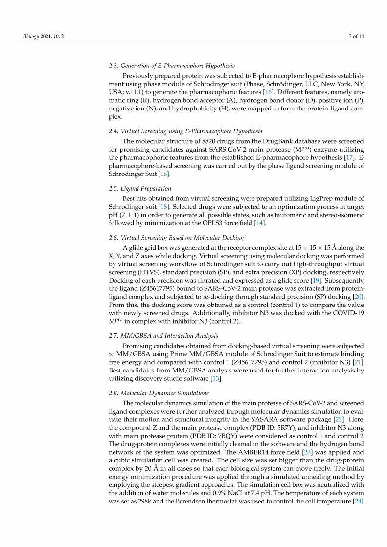
Pharmacophore models can also be used to identify new ligands that bind to the same receiver through virtual screening [30]. In the current study, the pharmacophore hypoth-esis was established using the main protease of SARS-CoV-2 (PDB ID: 5R7Y) in complex with Z45617795. The interactions of complex can lead to the generation of pharmacophore features, which can be targeted for the screening of candidates with similar features. The E-pharmacophore hypothesis displayed only one feature, which was one aromatic ring (R4). The aromatic ring (R4) at the complex site is exhibited in Figure 2.
Figure 1. Sequence comparison matrix of COVID-19 proteins. (a) Sequence comparison between SARS-CoV-2 (PDB ID:5R7Y), SARS-CoV (PDB ID: 2C3S), and MERS-CoV (PDB ID: 5C3N). (b) Primary structure of SARS-CoV-2 Mpro (PDB ID:5R7Y).
3.2. The E-Pharmacophore Hypothesis
A pharmacophore is a molecular feature required to detect a ligand molecularly.Pharmacophore models can also be used to identify new ligands that bind to the samereceiver through virtual screening [30]. In the current study, the pharmacophore hypothesiswas established using the main protease of SARS-CoV-2 (PDB ID: 5R7Y) in complex withZ45617795. The interactions of complex can lead to the generation of pharmacophorefeatures, which can be targeted for the screening of candidates with similar features. The E-pharmacophore hypothesis displayed only one feature, which was one aromatic ring (R4).The aromatic ring (R4) at the complex site is exhibited in Figure 2.
Biology 2021, 10, 2 6 of 14Biology 2021, 10, x 6 of 14
Biology 2021, 10, x. https://doi.org/10.3390/xxxxx www.mdpi.com/journal/biology
Figure 2. The E-pharmacophore feature (R4) of SARS-CoV-2 (PDB ID: 5R7Y) Mpro complex with Z45617795.
3.3. Virtual Screening Using E-Pharmacophore Hypothesis About 1000 drugs from DrugBank database were screened based on the E-pharma-
cophore hypothesis, where the candidates were grouped as approved, investigational, and experimental drugs (data not shown). Best hits in terms of high relatedness with the E-pharmacophore hypothesis were obtained from the entire database.
3.4. Virtual Screening Based on Molecular Docking To provide a more valid result of virtual screening, a protein structure is required
with high resolution [30]. The protein structure analyzed in this study has a resolution of 1.65 A° and was used for further docking-based virtual screening. Using HTVS, SP, and XP docking, 1000 drugs obtained from pharmacophore-based virtual screening were re-screened through filtering and removing low-scoring drugs at each step. Approved and investigational drugs were segregated from the HTVS, SP, and XP pool based on the dock-ing score and categorized as analgesic, antibiotic, anticonvulsants, benzene derivatives, anticoagulant, vitiligo, pyrimidines, anticancer, antiviral, cardiovascular agent, and im-munometabolic drugs (Table 2). All candidates showed higher docking scores in compar-ison with control 1 (−5.367 kcal/mol). This may conclude that the following 16 approved and investigational drugs are the most promising in terms of molecular docking. To get better outcomes, subsequently, we performed the MM/GBSA and molecular dynamics simulation for further clarification.
Table 2. Docking score of approved and investigational drugs.
Drug Bank ID Drug Name Group Category Docking Score
(kcal/mol) DB08797 Salicylamide Approved Analgesic −7.10 DB13026 Ornidazole Investigational Antibiotic −6.67 DB14575 Eslicarbazepine Approved Anticonvulsants. −6.56
DB14855 2-(aminomethyl)phe-nol
Investigational Benzene Derivatives −6.52
DB13136 Fluindione Approved Anticoagulants −6.34 DB04571 Trioxsalen Approved Vitiligo −6.34
Figure 2. The E-pharmacophore feature (R4) of SARS-CoV-2 (PDB ID: 5R7Y) Mpro complex with Z45617795.
3.3. Virtual Screening Using E-Pharmacophore Hypothesis
About 1000 drugs from DrugBank database were screened based on the E-pharma-cophore hypothesis, where the candidates were grouped as approved, investigational,and experimental drugs (data not shown). Best hits in terms of high relatedness with theE-pharmacophore hypothesis were obtained from the entire database.
3.4. Virtual Screening Based on Molecular Docking
To provide a more valid result of virtual screening, a protein structure is required withhigh resolution [30]. The protein structure analyzed in this study has a resolution of 1.65 Åand was used for further docking-based virtual screening. Using HTVS, SP, and XP docking,1000 drugs obtained from pharmacophore-based virtual screening were rescreened throughfiltering and removing low-scoring drugs at each step. Approved and investigationaldrugs were segregated from the HTVS, SP, and XP pool based on the docking score andcategorized as analgesic, antibiotic, anticonvulsants, benzene derivatives, anticoagulant,vitiligo, pyrimidines, anticancer, antiviral, cardiovascular agent, and immunometabolicdrugs (Table 2). All candidates showed higher docking scores in comparison with control 1(−5.367 kcal/mol). This may conclude that the following 16 approved and investigationaldrugs are the most promising in terms of molecular docking. To get better outcomes,subsequently, we performed the MM/GBSA and molecular dynamics simulation forfurther clarification.
In addition, 17 experimental drugs higher than the reference docking score (−5.370 kcal/mol)were separated from the results of virtual screening (Table 3). Among these drugs, DB02502,DB02309, and DB02690 showed the best docking score. Though these drugs are notapproved or evaluated for the treatment purpose, the candidates are still valuable for nobledrug discovery pipeline against COVID-19 through in vitro, in vivo, and other mechanisticinvestigations.

Biology 2021, 10, 2 7 of 14
Table 2. Docking score of approved and investigational drugs.
Drug Bank ID Drug Name Group Category Docking Score(kcal/mol)
DB08797 Salicylamide Approved Analgesic −7.10DB13026 Ornidazole Investigational Antibiotic −6.67DB14575 Eslicarbazepine Approved Anticonvulsants. −6.56DB14855 2-(aminomethyl)phenol Investigational Benzene Derivatives −6.52DB13136 Fluindione Approved Anticoagulants −6.34DB04571 Trioxsalen Approved Vitiligo −6.34DB02262 Orotic acid Investigational Pyrimidines −6.29DB12155 Napabucasin Investigational Anticancer −6.25DB11836 Sapanisertib Investigational Anticancer −6.24DB12804 Daniquidone Investigational Anticancer −6.13DB00368 Norepinephrine bitartrate Approved Cardiovascular agent −6.12DB01424 Aminophenazone Approved; withdrawn Analgesic −6.10DB12827 Indoximod Investigational Immunometabolic −6.05DB06408 Taribavirin Hydrochloride Investigational Antiviral −6.03DB00480 Lenalidomide Approved Anticancer −5.99
DB01033 Mercaptopurinemonohydrate Approved Anticancer −5.99
- Z45617795 - Control 1 −5.367- Inhibitor N3 - Control 2 −4.561
Table 3. Docking score of experimental drugs.
Drug Bank ID Drug Name Docking Score (kcal/mol)
DB02502 8-hydroxy-2′-deoxyguanosine −7.29DB02309 5-monophosphate-9-beta-D-ribofuranosyl xanthine −7.20DB02690 8-hydroxy-2-methyl-3,4-dihydroquinazolin-4-one −7.09DB03730 3,9-Dimethyladenine −6.97DB04446 Benzo[B]Thiophene-2-Carboxamidine −6.93DB02599 2,6-Diamino-8-Propylsulfanylmethyl-3h-Quinazoline-4- One −6.79DB15622 Triazavirin −6.76DB04103 3-methylcytosine −6.63DB02187 Equilin −6.61DB04312 (2,3-difluorophenyl)methanol −6.47DB13549 4-dimethylaminophenol −6.46DB07206 6-[2-(1H-indol-6-yl)ethyl]pyridin-2-amine −6.41DB04448 (2,4-difluorophenyl)methanol −6.13DB02586 4,7-dimethyl-1,10-phenanthroline −6.10DB04586 2-bromophenol −6.05DB03763 5-methyl-2′-deoxypseudouridine −6.00DB04440 Purine nucleoside −5.99
3.5. MM/GBSA and Interaction Analysis
A total of 16 approved and investigational drugs were subjected to MM/GBSA anal-ysis to evaluate binding free energy and for comparison with the control (Z45617795).There was a good correlation between MMGBSA dg Bind and binding affinity, where amore negative value suggests stronger binding affinity [31]. From the result of MM/GBSAanalysis, seven drugs were found to possess a higher MM/GBSA score than the controlZ45617795 (−28.33 kcal/mol) (Figure 3). The drugs with promising MM/GBSA dg bindscores are sapanisetrib (−36.229 kcal/mol), ornidazole (−35.832 kcal/mol), napabucasin(−34.671 kcal/mol), lenalidomide (−33.390 kcal/mol), daniquidone (−33.039 kcal/mol),indoximod (−29.631 kcal/mol), and salicylamide (−29.042 kcal/mol). The binding interac-tions of these candidates are exhibited in Table 4.

Biology 2021, 10, 2 8 of 14
Biology 2021, 10, x 8 of 14
Biology 2021, 10, x. https://doi.org/10.3390/xxxxx www.mdpi.com/journal/biology
Table 4. Binding interactions of seven drugs obtained through MM/GBSA analysis against SARS-CoV-2 Mpro.
Amino Acid Residue Compounds Hydrogen Bond Interactions Hydrophobic Interactions Sapanisertib ASN-142 HIS-41, MET-49, MET-165 Ornidazole ARG-188, GLN-189 HIS-41, MET-165
Napabucasin PRO-168 MET-165 Lenalidomide LYS-5, ARG-4 ALA-7 Daniquidone THR-25 HIS-41, MET-49, MET-165, Indoximod THR-129, GlU-166 MET-165
Salicylamide CYS-145, HIS-164, GLU-166 MET-165 Control 1 - MET-165, MET-49, HIS-41 Control 2 SER-139, GLY138 LYS-137, LYS-5
Figure 3. Best drugs from MM/GBSA analysis with MM/GBSA dg Bind score.
Sapanisertib and napabucasin are investigational anticancer drugs, which induce apoptosis of the cancer cell [32,33]. Sapanisertib demonstrated hydrogen bond interaction with ASN-142 and hydrophobic bond interactions with HIS-41, MET-49, and MET-165 (Figure 4A). On the other hand, napabucasin formed hydrogen bond interaction with Pro-168 and hydrophobic bond interaction with MET-165 (Figure 4C). In the docking-based virtual screening, sapanisertib and napabucasin provided a −6.24, and −6.25 kcal/mol docking score, respectively, against SARS-CoV-2. Daniquidone is another investigational anticancer drug that inhibits DNA replication along with the inhibition of RNA and pro-tein synthesis [34]. Daniquidone inhibits SARS-CoV-2 with a −6.13 kcal/mol docking score that exhibited a key hydrogen bond interaction with THR-25 and hydrophobic bond in-teractions with HIS-41, MET-49, and MET-165 (Figure 4E). Lenalidomide acts by destroy-ing tumor cells and possesses an immunomodulatory effect [35]. It is an approved anti-cancer drug with a −5.99 kcal/mol docking score and suggested LYS-5, ARG-4 amino acid residue interactions through the hydrogen bond and ALA-7 amino acid residue interac-tion through the hydrophobic bond (Figure 4D). Additionally, ornidazole is used against infectious disease, which interrupts the DNA replication and transcription process. It is an investigational drug that shows antibiotic activity [36]. According to this study, orni-dazole showed the second best hit in MM/GBSA analysis (−35.832 kcal/mol) with the sec-ond best docking score (−6.67 kcal/mol). Ornidazole was found to inhibit SARS-CoV-2 Mpro through hydrogen bonds, including ARG-188 and GLN-189, and hydrophobic bonds, including HIS-41 and MET-165 (Figure 4B). In addition, an investigational metabolic agent named indoximod acts by boosting immunity against infectious disease and improving response to many anticancer agents [37]. In our study, indoximod inhibited viral receptor (SARS-CoV-2) with a docking score of −6.05 kcal/mol through thehydrogen bond with THR-129 and GlU-166, and hydrophobic bond with MET-165 (Figure 4F). Lastly, salic-ylamide is an approved analgesic drug, which demonstrated the best docking score (−7.10
Figure 3. Best drugs from MM/GBSA analysis with MM/GBSA dg Bind score.
Table 4. Binding interactions of seven drugs obtained through MM/GBSA analysis against SARS-CoV-2 Mpro.
CompoundsAmino Acid Residue
Hydrogen Bond Interactions Hydrophobic Interactions
Sapanisertib ASN-142 HIS-41, MET-49, MET-165Ornidazole ARG-188, GLN-189 HIS-41, MET-165
Napabucasin PRO-168 MET-165Lenalidomide LYS-5, ARG-4 ALA-7Daniquidone THR-25 HIS-41, MET-49, MET-165,Indoximod THR-129, GlU-166 MET-165
Salicylamide CYS-145, HIS-164, GLU-166 MET-165Control 1 - MET-165, MET-49, HIS-41Control 2 SER-139, GLY138 LYS-137, LYS-5
Sapanisertib and napabucasin are investigational anticancer drugs, which induceapoptosis of the cancer cell [32,33]. Sapanisertib demonstrated hydrogen bond interactionwith ASN-142 and hydrophobic bond interactions with HIS-41, MET-49, and MET-165(Figure 4A). On the other hand, napabucasin formed hydrogen bond interaction withPro-168 and hydrophobic bond interaction with MET-165 (Figure 4C). In the docking-basedvirtual screening, sapanisertib and napabucasin provided a −6.24, and −6.25 kcal/moldocking score, respectively, against SARS-CoV-2. Daniquidone is another investigationalanticancer drug that inhibits DNA replication along with the inhibition of RNA and proteinsynthesis [34]. Daniquidone inhibits SARS-CoV-2 with a−6.13 kcal/mol docking score thatexhibited a key hydrogen bond interaction with THR-25 and hydrophobic bond interactionswith HIS-41, MET-49, and MET-165 (Figure 4E). Lenalidomide acts by destroying tumorcells and possesses an immunomodulatory effect [35]. It is an approved anticancer drugwith a−5.99 kcal/mol docking score and suggested LYS-5, ARG-4 amino acid residue inter-actions through the hydrogen bond and ALA-7 amino acid residue interaction through thehydrophobic bond (Figure 4D). Additionally, ornidazole is used against infectious disease,which interrupts the DNA replication and transcription process. It is an investigationaldrug that shows antibiotic activity [36]. According to this study, ornidazole showed the sec-ond best hit in MM/GBSA analysis (−35.832 kcal/mol) with the second best docking score(−6.67 kcal/mol). Ornidazole was found to inhibit SARS-CoV-2 Mpro through hydrogenbonds, including ARG-188 and GLN-189, and hydrophobic bonds, including HIS-41 andMET-165 (Figure 4B). In addition, an investigational metabolic agent named indoximodacts by boosting immunity against infectious disease and improving response to manyanticancer agents [37]. In our study, indoximod inhibited viral receptor (SARS-CoV-2)with a docking score of −6.05 kcal/mol through thehydrogen bond with THR-129 andGlU-166, and hydrophobic bond with MET-165 (Figure 4F). Lastly, salicylamide is anapproved analgesic drug, which demonstrated the best docking score (−7.10 kcal/mol)
Biology 2021, 10, 2 9 of 14
amongst all approved and investigational drugs in the molecular docking-based virtualscreening. Previous study suggests that salicylamide inhibits cytochromes P-450 (CYP)enzyme activity [38]. In our investigation, interaction analysis of salicylamide showedhydrogen bond interactions with CYS-145, HIS-164, and GLU-166, and hydrophobic bondinteraction with MET-165 (Figure 4G). These drugs might be repurposed as an effectiveinhibitor of COVID-19 to stop the spreading of coronavirus replication into the host cell.
3.6. Molecular Dynamics Simulations
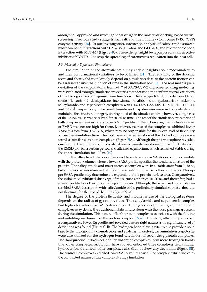
The simulation at the atomistic scale may enable insights about macromoleculesand their conformational variations to be obtained [31]. The reliability of the dockingscore and their validation largely depend on simulation data as the protein motion canbe assessed against the function of time in the simulation box [32]. The root mean squaredeviation of the c-alpha atoms from Mpro of SARS-CoV-2 and screened drug moleculeswere evaluated through simulation trajectories to understand the conformational variationsof the biological system against time functions. The average RMSD profile found fromcontrol 1, control 2, daniquidone, indoximod, lenalidomide, napabucasin, ornidazole,salicylamide, and sapanisertib complexes was 1.13, 1.09, 1.22, 1.08, 1.19, 1.194, 1.14, 1.11,and 1.17 Å, respectively. The lenalidomide and napabucasin were initially stable andmaintains the structural integrity during most of the simulation time; however, a high riseof the RMSD value was observed for 60–80 ns time. The rest of the simulation trajectories ofboth complexes demonstrate a lower RMSD profile for them; however, the fluctuation levelof RMSD was not too high for them. Moreover, the rest of the complexes exhibited lowerRMSD values from 0.8–1.4 Å, which may be responsible for the lower level of flexibilityacross the simulation time. The root mean square deviation of the docked complex werefound as similar with both complexes (Figure 5A). Although the pharmacophore revealedone feature, the complex on molecular dynamic simulation showed initial fluctuations inthe RMSD plot for a certain period and attained equilibrium, which remained stable duringthe entire simulation for 100 ns [10].
On the other hand, the solvent-accessible surface area or SASA descriptors correlatewith the protein volume, where a lower SASA profile specifies the condensed nature of theprotein. The salicylamide and main protease complex were in a stable state from 0–20 ns,but a higher rise was observed till the entire simulation time than other complexes. This up-per SASA profile may determine the expansion of the protein surface area. Comparatively,the indoximod exhibited shrinkage of the surface area from 10–20 ns and thereafter, had asimilar profile like other protein-drug complexes. Although, the sapanisertib complex re-sembled SASA descriptors with salicylamide at the preliminary simulation phase, they didnot fluctuate for the rest of the time (Figure S1A).
The degree of the protein flexibility and mobile nature of the biological systemsdepends on the radius of gyration values. The salicylamide and sapanisertib complexhad higher Rg values like SASA descriptors. The higher level of the Rg value from bothcomplexes may define the additional labile nature along with the loose packaging systemduring the simulation. This nature of both protein complexes associates with the foldingand unfolding mechanism of the protein complex [39,40]. Therefore, other complexes hada comparatively lower Rg profile and revealed a more rigid nature as no significant level ofdeviations was found (Figure S1B). The hydrogen bond plays a vital role to provide a solidbase to the biological macromolecules and systems. Therefore, the simulation trajectorieswere also utilized for the hydrogen bond calculation of seven drug-protein complexes.The daniquidone, indoximod, and lenalidomide complexes form more hydrogen bondsthan other complexes. Although these above-mentioned three complexes had a higherhydrogen bond number, other complexes also did not show any deviations (Figure 5B).The control 1 complexes exhibited lower SASA values than all the complex, which indicatesthe contracted nature of this complex during simulation.

Biology 2021, 10, 2 10 of 14Biology 2021, 10, x 10 of 14
Biology 2021, 10, x. https://doi.org/10.3390/xxxxx www.mdpi.com/journal/biology
Figure 4. 2D docking interactions of (A) Sapanisertib, (B) Ornidazole, (C) Napabucasin, (D) Lenalidomide, (E) Daniquidone, (F) Indoximod, (G) Salicylamide, and (H) Control 1 withSARS-CoV-2 Mpro (PDB ID: 5R7Y); (I) Control 2 with SARS-CoV-2 Mpro (PDB ID: 7BQY). The colors indicate the residue type: red-acidic, green-hydrophobic, purple-basic, blue-polar,light gray-other, darker gray-metal atoms. Interactions with the protein are marked with lines between ligand atoms and protein residues: solid pink—H-bonds to the protein backbone,dotted pink-H-bonds to protein side chains, green—pi-pi stacking interactions, orange-pi-cation interactions. Ligand atoms that are exposed to solvent are marked with gray spheres.The protein “pocket” is displayed with a line around the ligand, colored with the color of the nearest protein residue. The gap in the line shows the opening of the pocket.
Biology 2021, 10, 2 11 of 14
Biology 2021, 10, x 11 of 14
Biology 2021, 10, x. https://doi.org/10.3390/xxxxx www.mdpi.com/journal/biology
Figure 4. 2D docking interactions of (A) Sapanisertib, (B) Ornidazole, (C) Napabucasin, (D) Lenalidomide, (E) Daniquidone, (F) Indoximod, (G) Salicylamide, and (H) Control 1 with SARS-CoV-2 Mpro (PDB ID: 5R7Y); (I) Control 2 with SARS-CoV-2 Mpro (PDB ID: 7BQY). The colors indicate the residue type: red-acidic, green-hydrophobic, purple-basic, blue-polar, light gray-other, darker gray-metal atoms. Interactions with the protein are marked with lines between ligand atoms and protein residues: solid pink—H-bonds to the protein backbone, dotted pink-H-bonds to protein side chains, green—pi-pi stacking interactions, orange-pi-cation interactions. Ligand atoms that are exposed to solvent are marked with gray spheres. The protein “pocket” is displayed with a line around the ligand, colored with the color of the nearest protein residue. The gap in the line shows the opening of the pocket.
Figure 5. The molecular dynamics simulation study of the docked complex, (A) root mean square deviation (RMSD), and (B) hydrogen bond were assessed from the simulation trajectories.
Figure 5. The molecular dynamics simulation study of the docked complex, (A) root mean square deviation (RMSD), and (B)hydrogen bond were assessed from the simulation trajectories.
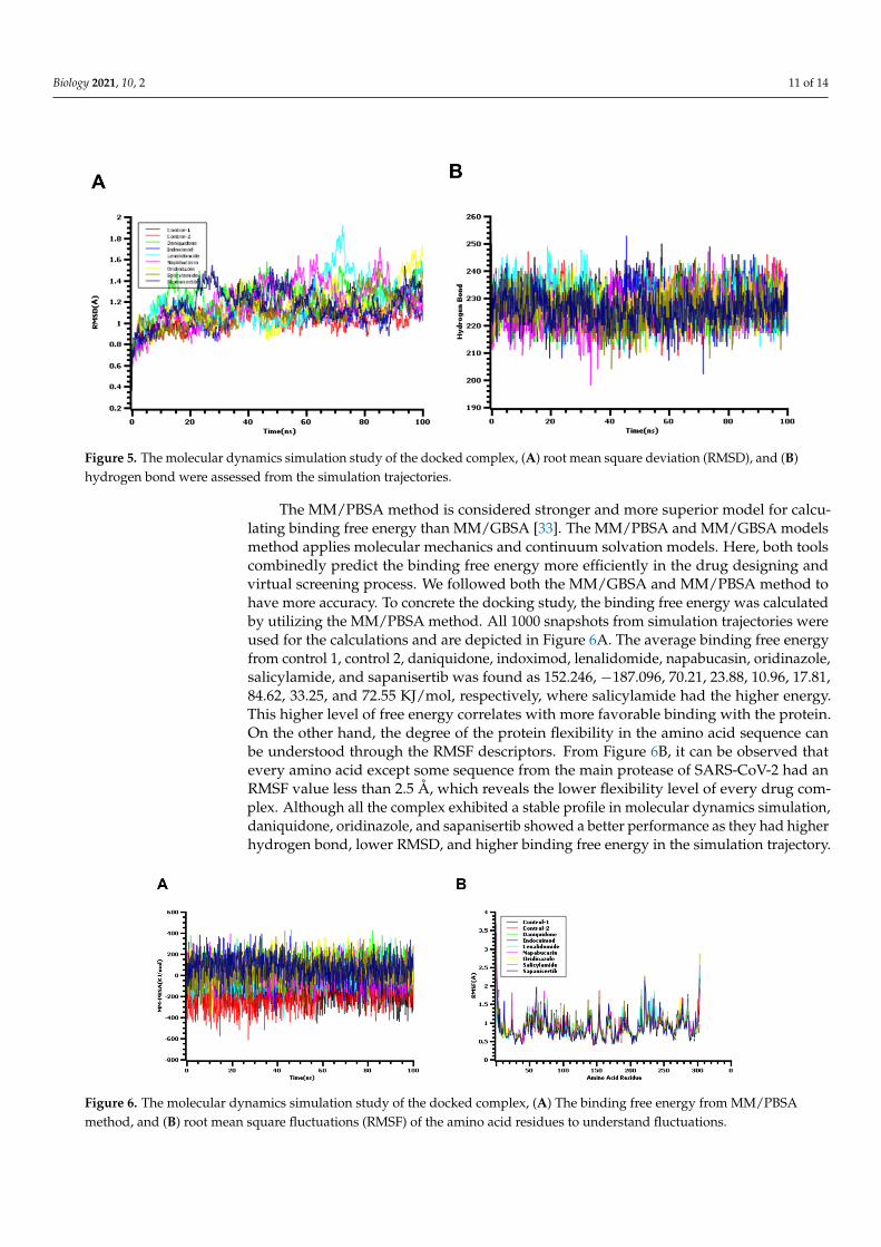
The MM/PBSA method is considered stronger and more superior model for calcu-lating binding free energy than MM/GBSA [33]. The MM/PBSA and MM/GBSA modelsmethod applies molecular mechanics and continuum solvation models. Here, both toolscombinedly predict the binding free energy more efficiently in the drug designing andvirtual screening process. We followed both the MM/GBSA and MM/PBSA method tohave more accuracy. To concrete the docking study, the binding free energy was calculatedby utilizing the MM/PBSA method. All 1000 snapshots from simulation trajectories wereused for the calculations and are depicted in Figure 6A. The average binding free energyfrom control 1, control 2, daniquidone, indoximod, lenalidomide, napabucasin, oridinazole,salicylamide, and sapanisertib was found as 152.246, −187.096, 70.21, 23.88, 10.96, 17.81,84.62, 33.25, and 72.55 KJ/mol, respectively, where salicylamide had the higher energy.This higher level of free energy correlates with more favorable binding with the protein.On the other hand, the degree of the protein flexibility in the amino acid sequence canbe understood through the RMSF descriptors. From Figure 6B, it can be observed thatevery amino acid except some sequence from the main protease of SARS-CoV-2 had anRMSF value less than 2.5 Å, which reveals the lower flexibility level of every drug com-plex. Although all the complex exhibited a stable profile in molecular dynamics simulation,daniquidone, oridinazole, and sapanisertib showed a better performance as they had higherhydrogen bond, lower RMSD, and higher binding free energy in the simulation trajectory.
Biology 2021, 10, x 12 of 14
Biology 2021, 10, x. https://doi.org/10.3390/xxxxx www.mdpi.com/journal/biology
The MM/PBSA method is considered stronger and more superior model for calculat-ing binding free energy than MM/GBSA [33]. The MM/PBSA and MM/GBSA models method applies molecular mechanics and continuum solvation models. Here, both tools combinedly predict the binding free energy more efficiently in the drug designing and virtual screening process. We followed both the MM/GBSA and MM/PBSA method to have more accuracy. To concrete the docking study, the binding free energy was calcu-lated by utilizing the MM/PBSA method. All 1000 snapshots from simulation trajectories were used for the calculations and are depicted in Figure 6A. The average binding free energy from control 1, control 2, daniquidone, indoximod, lenalidomide, napabucasin, oridinazole, salicylamide, and sapanisertib was found as 152.246, −187.096, 70.21, 23.88, 10.96, 17.81, 84.62, 33.25, and 72.55 KJ/mol, respectively, where salicylamide had the higher energy. This higher level of free energy correlates with more favorable binding with the protein. On the other hand, the degree of the protein flexibility in the amino acid sequence can be understood through the RMSF descriptors. From Figure 6B, it can be ob-served that every amino acid except some sequence from the main protease of SARS-CoV-2 had an RMSF value less than 2.5 Å, which reveals the lower flexibility level of every drug complex. Although all the complex exhibited a stable profile in molecular dynamics simulation, daniquidone, oridinazole, and sapanisertib showed a better performance as they had higher hydrogen bond, lower RMSD, and higher binding free energy in the sim-ulation trajectory.
Figure 6. The molecular dynamics simulation study of the docked complex, (A) The binding free energy from MM/PBSA method, and (B) root mean square fluctuations (RMSF) of the amino acid residues to understand fluctuations.
4. Conclusions Pharmacophore modeling, molecular docking, virtual screening, and other compu-
tational methods are widely used to repurpose known drugs. In this investigation, se-quence comparison of SARS-CoV-2 Mpro showed a high identity matrix with SARS-CoV and slide identity matrix with MERS-CoV. Further virtual screening based on molecular docking revealed 33 different drug groups of several categories. Among this, 16 approved and investigational drugs were found to inhibit SARS-CoV-2. In addition, MM/GBSA analysis reported seven drugs, namely sapanisertib, ornidazole, napabucasin, lenalido-mide, daniquidone, indoximod, and salicylamide, that showed higher dg Bind scores in comparison with the receptor complex Z45617795. Promising drugs of MM/GBSA analy-sis showed several interactions in the active site of SARS-CoV-2 Mpro. Accordingly, these drugs might be considered for a drug repurposing approach against novel coronavirus. Besides, 17 experimental drugs were found to possess a higher docking score than both the controls during virtual screening and might be vital for novel drug development against COVID-19. Furthermore, the docked complex had a strict nature in the simulated
Figure 6. The molecular dynamics simulation study of the docked complex, (A) The binding free energy from MM/PBSAmethod, and (B) root mean square fluctuations (RMSF) of the amino acid residues to understand fluctuations.
Biology 2021, 10, 2 12 of 14
4. Conclusions
Pharmacophore modeling, molecular docking, virtual screening, and other com-putational methods are widely used to repurpose known drugs. In this investigation,sequence comparison of SARS-CoV-2 Mpro showed a high identity matrix with SARS-CoVand slide identity matrix with MERS-CoV. Further virtual screening based on moleculardocking revealed 33 different drug groups of several categories. Among this, 16 approvedand investigational drugs were found to inhibit SARS-CoV-2. In addition, MM/GBSAanalysis reported seven drugs, namely sapanisertib, ornidazole, napabucasin, lenalido-mide, daniquidone, indoximod, and salicylamide, that showed higher dg Bind scores incomparison with the receptor complex Z45617795. Promising drugs of MM/GBSA analysisshowed several interactions in the active site of SARS-CoV-2 Mpro. Accordingly, these drugsmight be considered for a drug repurposing approach against novel coronavirus. Besides,17 experimental drugs were found to possess a higher docking score than both the controlsduring virtual screening and might be vital for novel drug development against COVID-19.Furthermore, the docked complex had a strict nature in the simulated environment. How-ever, these drugs required further investigation using in vitro and in vivo study to confirmtheir activity against COVID-19.
Supplementary Materials: The following are available online at https://www.mdpi.com/2079-7737/10/1/2/s1, Figure S1: The molecular dynamics simulation study of the docked complex,(A) solvent accessible surface area (SASA), and (B) radius of gyration (Rg) were assessed from thesimulation trajectories.
Author Contributions: Conceptualization, methodology, software, investigation, K.H.C., and M.R.C.;validation, formal analysis, investigation, S.M.; A.M.T., and N.B.H., data curation, writing—originaldraft preparation, K.H.C., M.R.C., N.B., T.B.E., and J.S.-G.; writing—review and editing, visualization,supervision, project administration, A.S.M.A.R., T.B.E., and J.S.-G.; All authors have read and agreedto the published version of the manuscript.
Funding: This research received no external funding.
Conflicts of Interest: The authors declare no conflict of interest.
References1. Jin, Y.; Yang, H.; Ji, W.; Wu, W.; Chen, S.; Zhang, W.; Duan, G. Virology, Epidemiology, Pathogenesis, and Control of COVID-19.
Viruses 2020, 12, 372. [CrossRef] [PubMed]2. Coronavirus Update (Live): 12,250,111 Cases and 553,972 Deaths from COVID-19 Virus Pandemic-Worldometer. Available online:
https://www.worldometers.info/coronavirus/?utm_campaign=homeAdvegas1 (accessed on 9 August 2020).3. Júnior, J.A.C.N.; Santos, A.M.; Quintans-Júnior, L.J.; Walker, C.I.B.; Borges, L.P.; Serafini, M.R. SARS, MERS and SARS-CoV-2
(COVID-19) treatment: A patent review. Expert Opin. Ther. Pat. 2020, 30, 567–579. [CrossRef]4. Xu, J.; Zhao, S.; Teng, T.; Abdalla, A.E.; Zhu, W.; Xie, L.; Wang, Y.; Guo, X. Systematic Comparison of Two Animal-to-Human
Transmitted Human Coronaviruses: SARS-CoV-2 and SARS-CoV. Viruses 2020, 12, 244. [CrossRef] [PubMed]5. Shamsi, A.; Mohammad, T.; Anwar, S.; Alajmi, M.F.; Hussain, A.; Rehman, T.; Islam, A.; Hassan, I. Glecaprevir and Maraviroc are
high-affinity inhibitors of SARS-CoV-2 main protease: Possible implication in COVID-19 therapy. Biosci. Rep. 2020, 40. [CrossRef][PubMed]
6. Singhal, T. A Review of Coronavirus Disease-2019 (COVID-19), Indian. J. Pediatr. 2020, 87, 281–286. [CrossRef]7. Mittal, L.; Kumari, A.; Srivastava, M.; Singh, M.; Asthana, S. Identification of potential molecules against COVID-19 main protease
through structure-guided virtual screening approach. J. Biomol. Struct. Dyn. 2020, 1–19. [CrossRef] [PubMed]8. Kandeel, M.; Al-Nazawi, M. Virtual screening and repurposing of FDA approved drugs against COVID-19 main protease. Life Sci.
2020, 251, 117627. [CrossRef] [PubMed]9. Rakib, A.; Sami, S.A.; Islam, M.A.; Ahmed, S.; Faiz, F.B.; Khanam, B.H.; Marma, K.K.S.; Rahman, M.; Uddin, M.M.N.; Nainu, F.;
et al. Epitope-Based Immunoinformatics Approach on Nucleocapsid Protein of Severe Acute Respiratory Syndrome-Coronavirus-2. Molecules 2020, 25, 5088. [CrossRef]
10. Fearon, D.; Powell, A.; Douangamath, A.; Owen, C.; Wild, C.; Krojer, T.; Lukacik, P.; Strain-Damerell, C.; Walsh, M.; Von Delft,F. PanDDA Analysis of COVID-19 Main Protease against the DSI-Poised Fragment Library. PDB ID: 5R7Y. Available online:https://www.wwpdb.org/pdb?id=pdb_00005r82 (accessed on 24 February 2020).
11. Xu, T.; Ooi, A.; Lee, H.C.; Wilmouth, R.; Liu, D.X.; Lescar, J. Structure of the SARS coronavirus main proteinase as an active C 2crystallographic dimer, Acta Crystallogr. Sect. F Struct. Biol. Cryst. Commun. 2005, 61, 964–966. [CrossRef]

Biology 2021, 10, 2 13 of 14
12. Ho, B.-L.; Cheng, S.-C.; Shi, L.; Wang, T.-Y.; Ho, K.-I.; Chou, C.-Y. Critical Assessment of the Important Residues Involved in theDimerization and Catalysis of MERS Coronavirus Main Protease. PLoS ONE 2015, 10, e0144865. [CrossRef]
13. Kemmish, H.; Fasnacht, M.; Yan, L. Fully automated antibody structure prediction using BIOVIA tools: Validation study.PLoS ONE 2017, 12, e0177923. [CrossRef] [PubMed]
14. Harder, E.; Damm, W.; Maple, J.R.; Wu, C.; Reboul, M.; Xiang, J.Y.; Wang, L.; Lupyan, D.; Dahlgren, M.K.; Knight, J.L.; et al.OPLS3: A Force Field Providing Broad Coverage of Drug-like Small Molecules and Proteins. J. Chem. Theory Comput. 2016, 12,281–296. [CrossRef] [PubMed]
15. Sastry, G.M.; Adzhigirey, M.; Day, T.; Annabhimoju, R.; Sherman, W. Protein and ligand preparation: Parameters, protocols,and influence on virtual screening enrichments. J. Comput. Aid. Mol. Des. 2013, 27, 221–234. [CrossRef] [PubMed]
16. Dixon, S.L.; Smondyrev, A.M.; Knoll, E.H.; Rao, S.N.; Shaw, D.E.; Friesner, R.A. PHASE: A new engine for pharmacophoreperception, 3D QSAR model development, and 3D database screening: 1. Methodology and preliminary results. J. Comput. Aided.Mol. Des. 2006, 20, 647–671. [CrossRef]
17. Wishart, D.S.; Feunang, Y.D.; Guo, A.C.; Lo, E.J.; Marcu, A.; Grant, J.R.; Assempour, N. DrugBank 5.0: A major update to theDrugBank database for 2018. Nucleic Acids Res. 2018, 46, 1077–1082. [CrossRef]
18. Shelley, J.C.; Cholleti, A.; Frye, L.L.; Greenwood, J.R.; Timlin, M.R.; Uchimaya, M. Epik: A software program for pK(a) predictionand protonation state generation for drug-like molecules. J. Comput. Aided. Mol. Des. 2007, 21, 681–691. [CrossRef]
19. Friesner, R.A.; Murphy, R.B.; Repasky, M.P.; Frye, L.L.; Greenwood, J.R.; Halgren, T.A.; Sanschagrin, P.C.; Mainz, D.T. Extra Preci-sion Glide: Docking and Scoring Incorporating a Model of Hydrophobic Enclosure for Protein−Ligand Complexes. J. Med. Chem.2006, 49, 6177–6196. [CrossRef]
20. Rakib, A.; Paul, A.; Chy, M.N.U.; Sami, S.A.; Baral, S.K.; Majumder, M.; Tareq, A.M.; Amin, M.N.; Shahriar, A.; Uddin, M.Z.; et al.Biochemical and Computational Approach of Selected Phytocompounds from Tinospora crispa in the Management of COVID-19.Molecules 2020, 25, 3936. [CrossRef]
21. Verma, P.; Tiwari, M.; Tiwari, V. In silico high-throughput virtual screening and molecular dynamics simulation study to identifyinhibitor for AdeABC efflux pump of Acinetobacter baumannii. J. Biomol. Struct. Dyn. 2018, 36, 1182–1194. [CrossRef]
22. Krieger, E.; Darden, T.; Nabuurs, S.B.; Finkelstein, A.; Vriend, G. Making optimal use of empirical energy functions: Force-fieldparameterization in crystal space. Proteins 2004, 57, 678–683. [CrossRef]
23. Dickson, C.J.; Madej, B.D.; Åge, A.S.; Betz, R.M.; Teigen, K.; Gould, I.R.; Walker, R.C. Lipid14: The amber lipid force field. J. Chem.Theory Comput. 2014, 10, 865–879. [CrossRef] [PubMed]
24. Krieger, E.; Vriend, G.; Spronk, C. YASARA–Yet Another Scientific Artificial Reality Application. 2013. Available online:http://yasara.org/ (accessed on 24 February 2020).
25. Oany, A.R.; Mia, M.; Pervin, T.; Junaid, M.; Hosen, S.; Moni, M.A. Design of novel viral attachment inhibitors of the spikeglycoprotein (S) of severe acute respiratory syndrome coronavirus-2 (SARS-CoV-2) through virtual screening and dynamics. Int. J.Antimicrob. Agents. 2020, 56, 106177. [CrossRef] [PubMed]
26. Rakib, A.; Sami, S.A.; Mimi, N.J.; Chowdhury, M.M.; Eva, T.A.; Nainu, F.; Paul, A.; Shahriar, A.; Tareq, A.M.; Emon, N.U.; et al.Immunoinformatics-guided design of an epitope-based vaccine against severe acute respiratory syndrome coronavirus 2 spikeglycoprotein. Comput. Biol. Med. 2020, 124, 103967. [CrossRef] [PubMed]
27. Islam, S.; Mahmud, S.; Sultana, R.; Dong, W. Identification and in silico molecular modelling study of newly isolated Bacillussubtilis SI-18 strain against S9 protein of Rhizoctonia solani. Arab. J. Chem. 2020, 13, 8600–8612. [CrossRef]
28. Kumar, Y.; Singh, H.; Patel, C.N. In silico prediction of potential inhibitors for the main protease of SARS-CoV-2 using moleculardocking and dynamics simulation based drug-repurposing. J. Infect. Public Health 2020, 13, 1210–1223. [CrossRef]
29. Zhang, L.; Lin, D.; Sun, X.; Curth, U.; Drosten, C.; Sauerhering, L.; Becker, S.; Rox, K.; Hilgenfeld, R. Crystal structure ofSARS-CoV-2 main protease provides a basis for design of improved a-ketoamide inhibitors. Science 2020, 368, 409–412. [CrossRef]
30. Ganser, L.R.; Lee, J.; Rangadurai, A.; Merriman, D.K.; Kelly, M.L.; Kansal, A.D.; Sathyamoorthy, B.; Al-Hashimi, H.M. High-performance virtual screening by targeting a high-resolution RNA dynamic ensemble. Nat. Struct. Mol. Biol. 2018, 25, 425–434.[CrossRef]
31. Perez-Sanchez, H.; Lightstone, F.C. A Comprehensive Docking and MM/GBSA Rescoring Study of Ligand Recognition uponBinding Antithrombin. Curr. Top. Med. Chem. 2017, 17, 1631–1639. [CrossRef]
32. Guo, N.; Azadniv, M.; Coppage, M.; Nemer, M.; Mendler, J.; Becker, M.; Liesveld, J. Effects of Neddylation and mTOR Inhibitionin Acute Myelogenous Leukemia. Transl. Oncol. 2019, 12, 602–613. [CrossRef]
33. Hubbard, J.M.; Grothey, A. Napabucasin: An Update on the First-in-Class Cancer Stemness Inhibitor. Drugs 2017, 77, 1091–1103.[CrossRef]
34. NCI Thesaurus. National Cancer Institute. Available online: https://datascience.cancer.gov/ (accessed on 21 August 2020).35. Zeldis, J.B.; Knight, R.; Hussein, M.; Chopra, R.; Muller, G. A review of the history, properties, and use of the immunomodulatory
compound lenalidomide. Ann. N. Y. Acad. Sci. 2011, 1222, 76–82. [CrossRef] [PubMed]36. Choudhuri, G.; Rangan, M. Amebic infection in humans. Indian J. Gastroenterol. 2012, 31, 153–162. [CrossRef] [PubMed]37. Fox, E.; Oliver, T.; Rowe, M.; Thomas, S.; Zakharia, Y.; Gilman, P.B.; Muller, A.J.; Prendergast, G.C. Indoximod: An im-
munometabolic adjuvant that empowers T cell activity in cancer. Front. Oncol. 2018, 8, 370. [CrossRef] [PubMed]38. MacDonald, C.J.; Ciolino, H.P.; Yeh, G.C. The Drug Salicylamide Is an Antagonist of the Aryl Hydrocarbon Receptor That Inhibits
Signal Transduction Induced by 2,3,7,8-Tetrachlorodibenzo-p-dioxin. Cancer Res. 2004, 64, 429–434. [CrossRef]

Biology 2021, 10, 2 14 of 14
39. Dror, R.O.; Dirks, R.M.; Grossman, J.; Xu, H.; Shaw, D.E. Biomolecular simulation: A computational microscope for molecularbiology. Annu. Rev. Biophys. 2012, 41, 429–452. [CrossRef]
40. Salmaso, V.; Moro, S. Bridging molecular docking to molecular dynamics in exploring ligand-protein recognition process:An overview. Front. Pharmacol. 2018, 9, 923. [CrossRef]
Copyright © 2022 FDOKUMEN