Drug Discovery - September/October 2013
32
Vol. 16 No. 5 www.dddmag.com STRATEGIES & TECHNOLOGIES DRIVING DRUG DISCOVERY TO MARKET S G S & C O OG S D V G D G D SCOV Y O DRUG DISCOVERY & DEVELOPMENT Chemical-based Technologies VIRTUAL SCREENING ADVANCES Drug Safety NEW & IMPROVED BIOMARKERS FOR KIDNEY TOXICITY Informatics DNA SEQUENCE ANALYSIS FOR NON- BIOINFORMATICIANS How It Works COCULTURING FOR BETTER TOXICOLOGY STUDIES September/October 2013 RAMPING UP PRECOMPETITIVE R&D EFFORTS page 6 I Can See Clearly Now: in vivo Imaging in Drug Discovery Providing more data points from fewer animals, in vivo imaging techniques are increasingly being used in preclinical research.
-
Upload
khangminh22 -
Category
Documents
-
view
3 -
download
0
Transcript of Drug Discovery - September/October 2013
Vol. 16 No. 5 www.dddmag.com
S T R A T E G I E S & T E C H N O L O G I E S D R I V I N G D R U G D I S C O V E R Y T O M A R K E TS G S & C O O G S D V G D G D S C O V Y O
DRUGDISCOVERY & DEVELOPMENT
� Chemical-based TechnologiesVIRTUAL SCREENING ADVANCES
� Drug Safety NEW & IMPROVED BIOMARKERS FOR KIDNEY TOXICITY
� InformaticsDNA SEQUENCE ANALYSIS FOR NON-BIOINFORMATICIANS
� How It WorksCOCULTURING FOR BETTER TOXICOLOGY STUDIES
� September/October 2013
RAMPING UP PRECOMPETITIVE
R&D EFFORTSpage 6
I Can SeeClearly Now:in vivoImaging in
Drug DiscoveryProviding more data points from fewer animals, in vivo imaging techniques are increasingly being used in preclinical research.
dd13o_01CV_digital.indd 1dd13o_01CV_digital.indd 1 10/15/2013 4:22:57 PM10/15/2013 4:22:57 PM
HuCAL® Custom Monoclonal AntibodiesWe share your values of quality, innovation, integrity and ethical behaviour. /PNOS`�ZWLJPÄJ��OPNO�HMÄUP[`�HU[PIVKPLZ�PU���^LLRZ�^P[OV\[�PTT\UPaH[PVU�VM�HUPTHSZ�
HuCAL Custom Monoclonal Antibodies Worldwide ;LS!��� ����� ��� �� ����
,THPS!�ZHSLZ�T\J'HIKZLYV[LJ�JVT
^^ �̂HIKZLYV[LJ�JVT�/\*(3�(IZ HuCAL® - Technology That Works for You!
*OVVZL�H�7HY[ULY�>OV�:OHYLZ� Your Values
/\*
(3
®�PZ�H
�YLN
PZ[L
YLK
�[YH
KLT
HYR
�VM�
4V
YWOV
:`Z�(.
DD13O_02ADS.indd 2DD13O_02ADS.indd 2 10/10/2013 4:22:29 PM10/10/2013 4:22:29 PM
DRUG Discovery & Development September/October � 3
DRUGDISCOVERY & DEVELOPMENT
September/October 2013
www.dddmag.com
Contents
� ONLINE THIS MONTH WWW.DDDMAG.COM
� CHEMICAL BASED 12 NEW APPROACHES TO VIRTUAL SCREENING
How is the pharmaceutical industry using virtual screening to identify potential bioactive molecules today?
� DRUG SAFETY16 BETTER TOOLS FOR SCREENING:
EARLY BIOMARKERS OF KIDNEY TOXICITYA new set of kidney safety biomarkers is helping to conquer the kidney toxicity problem.
� INFORMATICS20 NEXT-GENERATION SEQUENCING
FOR NON-BIOINFORMATICIANSHow can the analysis of next-generation sequencing data be simplified to support its expanded adoption in clinical research?
� HOW IT WORKS24 COCULTURING SYSTEMS
Coculture systems can provide increased sensitivity for predictive toxicology studies.
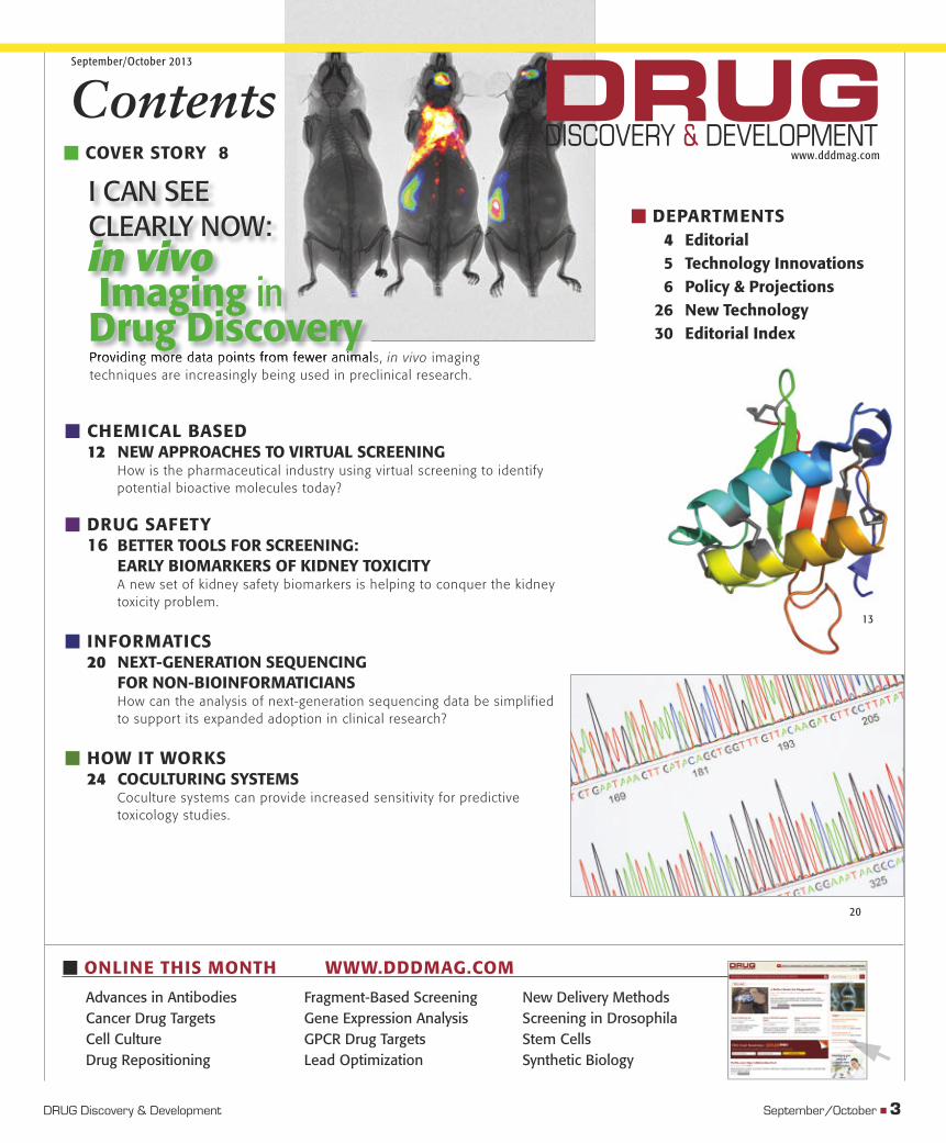
� COVER STORY 8
Providing more data points from fewer animals, in vivo imaging techniques are increasingly being used in preclinical research.
� DEPARTMENTS 4 Editorial 5 Technology Innovations 6 Policy & Projections 26 New Technology 30 Editorial Index
I CAN SEECLEARLY NOW:in vivo Imaging inDrug Discovery
Advances in AntibodiesCancer Drug TargetsCell CultureDrug Repositioning
Fragment-Based ScreeningGene Expression Analysis GPCR Drug TargetsLead Optimization
New Delivery MethodsScreening in DrosophilaStem Cells Synthetic Biology
13
20
dd13o_03TOC.indd 3dd13o_03TOC.indd 3 10/10/2013 4:23:31 PM10/10/2013 4:23:31 PM
Drug discovery and development is often described as a relatively straightforward pipeline, but those familiar with the process know that it’s never that simple. And while pharmaceutical com-panies spend billions of dollars each year on R&D, abandoned
drugs still outweigh approvals. Facing patent expirations and increased competition from generics, our industry is now faced with the challenge of doing more with less. To increase the effi ciency of their R&D programs, pharmaceutical companies must invest more in external research collabora-tions, which allow them to leverage the expertise of government agencies and academia—as well as each other—to tackle common problems.
In this issue of Drug Discovery & Development, two articles report on the benefi ts of such partnerships. In our Policy & Projections col-umn, Ted Agres reports that interest in precompetitive collaborations is indeed gaining momentum. For example, through a recently announced collaboration, Roche Holding AG (Basel, Switzerland) and AstraZeneca PLC (London) will share data and study results on early-stage drug candidates to advance their individual efforts. In another article, Dominic Eisinger, PhD, describes how a group of pharma companies, government agencies and academics formed the Predictive Safety Testing Consortium in 2006. Now, the group has developed biomarkers that have improved the detection of kidney damage in preclinical studies.
To act as a mediator for such collaborations, the National Institutes of Health (NIH) established the National Center for Advancing Translational Research in 2012. One former pharmaceutical execu-tive, however, was quick to express skepticism while testifying before a House of Representatives subcommittee on the new center. He argued that drug developers were already going to great lengths to develop new drugs, and the NIH’s initiative would do little to aid those efforts. In other words, there were already enough chefs in the kitchen. But what if you brought on a sous-chef? Their main responsibility would be to help organize the kitchen and ensure that everyone’s efforts and abilities were not wasted. Wouldn’t the kitchen be more productive as a result?
Patient foundations can also help to reduce risk and increase effi ciency in drug development. For instance, the Multiple Myeloma Research Foundation has funded basic research that has resulted in newly identi-fi ed drug targets and six approved treatments in the past decade. Just last month, the foundation launched an open-access resource that will provide detailed genomic and clinical data on 1,000 multiple myeloma patients as they receive treatment. For those working on this disease, it will be an invaluable resource. But the only way that foundations can help is for pharma to partner with them.
Engaging in such precompetitive research in no way will make pharma companies less competitive. In fact, just the opposite is pos-sible. Companies with a strong network of collaborators will be the most competitive in the 21st century. Patient foundations, government agencies and academia want to help, but pharma companies have tradi-tionally been isolationists, trying to tackle disease on their own. Drug development, however, is complicated, and we cannot unravel biology by ourselves. With a pipeline that is anything less than robust, it’s time to reevaluate our openness to sharing data and forming partnerships with academia, government agencies, patient groups and each other. �
� EDITORIAL
Doing More with Less
4 � September/October 2013
� Andrew WiecekEditor
DRUGDISCOVERY & DEVELOPMENT
www.dddmag.com100 Enterprise Drive, Suite 600, Box 912
Rockaway, NJ, 07866-0912, USAPhone: 973-920-7000 • Fax: 973-920-7542
Editorial DirectorTim Studt
EditorAndrew Wiecek
News EditorChristina Smith
Web Production SpecialistShannon Drury
Art DirectorCarol Kuchta
Production ManagerMary Jean Jones
Contracts CoordinatorPat Sikora
Audience Development ManagerGail Kirberger
Contributing EditorsTed Agres, Mike May
Group PublisherLiz Vickers
East Coast Sales ManagerGreg Renaud
973-920-7189; Fax [email protected]
West Coast Sales ManagerJordan Bender
973-920-74803; Fax [email protected]
Chief Operating Offi cer/Chief Financial Offi cerTheresa Freeburg
Vice PresidentTom Lynch
Chief Marketing Offi cer/Chief Digital Offi cerPrescott Shibles
Vice President, Human ResourcesSusanne Foulds
ReprintsThe YGS Group
Jessica Stremmel717-505-9701 x105
Advantage Business MediaScience Group
Bioscience TechnologyDrug Discovery & Development
Laboratory Design NewsLaboratory Equipment
R&DScientifi c Computing
All contents © Copyright of Advantage Business Media
dd13o_04Edit.indd 4dd13o_04Edit.indd 4 10/10/2013 4:25:31 PM10/10/2013 4:25:31 PM
Figuring Out Physiological Conditions
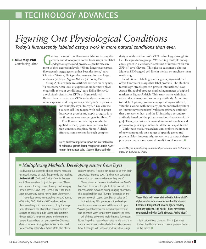
G etting the most from fl uorescent labeling in drug dis-covery and development comes from assays that label endogenous genes and provide a specifi c measure-
ment of their expression levels. “We no longer overexpress fl uorescently tagged genes, as has been the norm,” says Christian Nievera, PhD, product manager for zinc fi nger nucleases (ZFNs) at Sigma-Aldrich (St. Louis, Mo.).
Using ZFNs, which are artifi cial restriction enzymes, “a researcher can look at expression under more physi-ologically relevant conditions,” says Erika Holroyd, a technical scientist for ZFNs at Sigma-Aldrich. Researchers can also use ZFNs to analyze the impact of an experimental drug on a specifi c gene’s expression.
For example, says Holroyd, “You can use a cancer cell line tagged with red or green fl uorescent protein and apply drugs to it to see if one gene or another gets inhibited.”
This fl uorescent labeling can also be applied to many genes in a pathway for high-content screening. Sigma-Aldrich offers custom services for such complex
designs with its CompoZr ZFN technology through its Cell Design Studio group. “We can tag multiple endog-enous genes in a customer’s cell line of interest with our ZFNs,” says Nievera. This gives a customer a choice: Make a ZFN-tagged cell line in the lab or purchase them ready to go.
In addition to labeling specifi c genes, Sigma-Aldrich offers fl uorescent assays that label proteins. The Duolink technology “tracks protein-protein interactions,” says Aaron Sin, global product marketing manager of applied markets at Sigma-Aldrich. This assay works with fi xed cells and a primary and secondary antibody. According to Caleb Hopkins, product manager at Sigma-Aldrich, “Duolink works with most any [immunohistochemistry]- or [immunocytochemistry]-validated primary antibody that a researcher has, and the kit includes a secondary antibody based on [the primary antibody’s species of ori-gin]. Then, you just use a normal immunohistochemical protocol to gain single molecule–detection capabilities.”
With these tools, researchers can explore the impact of new compounds on a range of specifi c genes and proteins. Most importantly, researchers can track these processes under more natural conditions than ever. �
Mike May is a publishing consultant for science and technology
based in Lebanon, Ohio.
Green fl uorescent protein shows the internalization of epidermal growth factor receptor (EGFR) in AS49 human lung cancer cells. (Source: Sigma-Aldrich)
� Mike May, PhD Contributing Editor
� TECHNOLOGY ADVANCES
To develop fl uorescently labeled assays, research-
ers need a range of tools that provide the labeling.
Active Motif (Carlsbad, Calif.) offers its fl uores-
cent Chromeo dyes for just this purpose. “These
can be used for high-content assays and imaging-
based assays,” says Jörg Plümpe, PhD, site man-
ager at Germany-based Active Motif Chromeon.
These dyes come in several versions, Chromeo
488, 494, 505, 546 and 642—all named for
their wavelength, in nanometers, of light absorp-
tion. Moreover, the absorption can come from
a range of sources: diode lasers, light-emitting
diodes (LEDs), tungsten lamps and xenon-arc
lamps. Researchers can purchase the Chromeo
dyes in various binding chemistries or attached
to secondary antibodies. Active Motif also offers
custom options. “People can come to us with their
antibodies,” Plümpe says, “and we can conjugate
them with our dyes or whatever they want.”
These dyes can be combined with Active Motif’s
Max Stain to provide the photostability needed for
longer sample exposure during imaging or analysis.
The actual stability, says Plümpe, “depends on the
system. In certain cases, dyes bleach quite fast.”
In the future, Plümpe expects the develop-
ment of even more advanced fl uorescent dyes.
“Avoiding autofl uorescence needs improvement,
and scientists want longer term stability,” he says.
All of these advanced tools that use fl uorescent
labels help drug researchers better understand the
natural conditions under which the body operates,
how it changes with disease and ways that drugs
might battle those changes. That is just what
today’s healthcare needs to serve patients better.
in the future. �
� Multiplexing Methods: Developing Assays from Dyes
Today’s fl uorescently labeled assays work in more natural conditions than ever.
These HeLa cells were stained with Active Motif’s alpha tubulin mouse monoclonal antibody and Chromeo 488 goat anti-mouse IgG secondary antibody (green). The nuclei (blue) have been counterstained with DAPI. (Source: Active Motif)
DRUG Discovery & Development September/October 2013 � 5
dd13o_05TA.indd 5dd13o_05TA.indd 5 10/10/2013 4:26:48 PM10/10/2013 4:26:48 PM
More to Share
� Ted Agres, Contributing Editor
Facing ongoing fi scal challenges, Big Pharma companies are becoming increas-ingly willing to share precompetitive and
noncompetitive data and software in hopes of speeding drug discovery and increasing their chances of clinical success. In the latest such collaboration, Roche Holding AG (Basel, Switzerland) and AstraZeneca PLC (London) have agreed to share data and test results from selected early-stage small-chemical drug candidates.
In this new collaboration, the companies will provide databases of selected drugs to MedChemica Ltd. (Staffordshire, United Kingdom), a small startup estab-lished by former AstraZeneca scientists, which specializes in matched molecular pair analysis (MMPA). In MMPA, data are run through computer algorithms to identify possible molecular design changes to improve metabolism, pharma-cokinetics or safety without divulging confi dential chemical structure information. The goal is to identify potential new drug candidates using fewer rounds of design, synthesis and testing, thereby saving time and expense.1
“It is unique in the history of our industry that two major players are sharing their know-how at such an early stage of research,” says Luca Santarelli, head of neuroscience and small molecule research at Roche. “We believe that this transparency of small-molecule optimization knowledge, in a smart and thoughtful way, could profoundly enhance our ability to design drugs, be of benefi t for all parties involved and ultimately help bring better medicines to patients.”
“We are making these data sets available in the belief that, when paired with fi ndings
from other companies through a common platform, we can reach our patients faster with medicines that make a meaningful dif-ference to their lives,” adds Mike Snowden, AstraZeneca’s vice president for discovery sciences and innovative medicines program.
F. Hoffmann-La Roche Ltd. (Basel, Switzerland) and its subsidiary Genentech Inc. (South San Francisco, Calif.) have also agreed to join the collaboration, says Alexander
Dossetter, MedChemica’s managing director. “We aim to expand this kind of collaboration and eventually go beyond facilitating chemi-cal building blocks into chemical-lead hunting and optimization,” says Dossetter, adding that the data generated from the project will eventually be made available to the broader research community.
Open-source software In another recently announced collaborative effort, Pfi zer Inc. (New York) is making its
large-molecule modeling software available in a precompetitive, open-source basis to competitors including Bristol-Myers Squibb Co. (New York), GlaxoSmithKline PLC (Brentford, United Kingdom) and Roche. Pfi zer’s hierarchical editing language for macromolecules (HELM) software can render a wide range of biomolecules, including pro-teins, nucleotides and antibody drug conju-gates. Pfi zer originally developed the software
after fi nding that existing small-molecule and sequence-based informatics were incapable of adequately rendering large and complex molecular structures.
The software is being made available through the Pistoia Alliance, a nonprofi t collab-orative established in 2009 by informatics specialists from AstraZeneca, GSK, Novartis International AG (Basel, Switzerland) and Pfi zer. The group now includes 24 compa-nies and organizations. “When we started presenting HELM outside of Pfi zer, it became obvious that many companies across the industry were facing the same challenges we had faced,” says Sergio Rotstein, a director in Pfi zer’s research business technology organiza-tion. “By sharing this work in
a precompetitive fashion through the Pistoia Alliance, we are not only helping others to solve this problem but also fostering a tech-nical means by which companies, institutes, CROs, software vendors and IT service providers can exchange biomolecule data and information, which ultimately benefi ts everyone.”
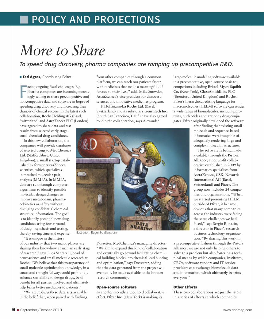
Other EffortsThese two collaborations are just the latest in a series of efforts in which companies
Illustration: Roger Schillerstrom
6 � September/October 2013 www.dddmag.com
� POLICY AND PROJECTIONS
To speed drug discovery, pharma companies are ramping up precompetitive R&D.
dd13o_06PNP.indd 6dd13o_06PNP.indd 6 10/10/2013 4:27:44 PM10/10/2013 4:27:44 PM
are working together to address common issues. For example, in September 2012, 10 Big Pharma companies established TransCelerate BioPharma Inc. (King of Prussia, Penn.), a nonprofi t aimed at accelerating the development of new drugs, beginning with improving the effi ciency of clinical trials. The 10 companies were Abbott Laboratories (North Chicago, Ill.), AstraZeneca, Boehringer Ingelheim GMBH (Ingelheim, Germany), Bristol-Myers Squibb, Eli Lilly and Co. (Indianapolis, Ind.), GlaxoSmithKline, Johnson & Johnson (New Brunswick, N.J.), Pfi zer, Genentech and Sanofi SA (Paris).
TransCelerate members identifi ed clinical study execution as its fi rst area of focus, with fi ve projects marked for funding and development: development of risk-based site monitoring approach and standards; development of a shared user interface for investigator site portals; mutual recogni-tion of study site qualifi cation and training; development of clinical data standards and establishment of a comparator drug supply model. In August 2013, the organization announced that the comparator drug supply initiative had become active.
“The current process to source and manage comparator drugs and co-thera-pies can be challenging,” said Dalvir Gill, TransCelerate’s CEO. “TransCelerate com-panies will be able to source these compara-tor drugs directly from each other, be able to secure supply when they need it in the quan-tities they need, have access to drug data and totally mitigate the risk of counterfeit drugs in that clinical trial,” he says.
In June 2013, the National Center for Advancing Translational Sciences (NCATS), which is part of the National Institutes of Health (NIH), awarded $12.7 million to nine academic research groups in its “Discovering New Therapeutic Uses for Existing Molecules” program. Here academic researchers will investigate whether exist-ing pharmaceutical molecules are effective against a specifi c disease or condition in eight areas, including Alzheimer’s disease, Duchenne muscular dystrophy and schizo-phrenia. Through the cooperative agree-ments, the researchers will conduct preclinical validation and additional safety studies.
“Public-private collaborations are crucial
DRUG Discovery & Development
Reference1. Dossetter AG, Griffen EJ, Leach AG. Matched molecular pair analysis in drug discovery. Drug discovery today. 2013;18:724-731.
Contributing editor Ted Agres, MBA, is a veteran science writer in Washington, DC. He writes frequently about the policy, politics and business aspects of life sciences.
for successful translation; no one organiza-tion can succeed alone,” says Christopher P. Austin, director of NCATS. “This initiative has created a marketplace to connect aca-demic researchers with potential new drugs, as well as template agreements that streamline the process by limiting the amount of negotia-tion required before a project can begin.” �
Reference:
1. Chatterjee R. Cases of mistaken identity. Science. 2007;315(5814):928–931.
© 2013 IDEXX Laboratories, Inc. All rights reserved. • 103166-00 • All ®/TM marks are owned by IDEXX Laboratories, Inc. or its affiliates in
the United States and/or other countries. The IDEXX Privacy Policy is available at idexx.com.
BioResearch
Fig. 1
Homo sapiens
Where 20%–30% of all cell lines are thought to be
misidentified or cross contaminated,1 how can you be sure
yours aren’t? With easy sample submission, comprehensive
analysis and expert on-call support, CellCheck™ cell line
authentication services from IDEXX RADIL™, part of IDEXX
BioResearch, offer a simple way to protect your research
and your reputation.
It’s a serious risk hiding in plain sight.
To learn more, call 1-800-544-5205, option 1,
or go to idexxbioresearch.com/cellcheck.
If only misidentified cell lines
were as obvious.
dd13o_06PNP.indd 7dd13o_06PNP.indd 7 10/10/2013 4:28:05 PM10/10/2013 4:28:05 PM
8 � September/October 2013 www.dddmag.com
I Can See Clearly Now:
in vivo Imaging in Drug Discovery
� COVER STORY
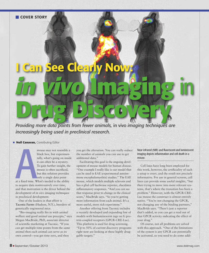
Amouse may not resemble a black box, but experimen-tally, what’s going on inside it can often be a mystery. To gain further insight, the mouse is often sacrifi ced, but this solution provides only a single data point
at a fi xed time. What’s needed is the ability to acquire data noninvasively over time, and that motivation is the driver behind the development of in vivo imaging techniques in preclinical development.
One of the leaders in that effort is Taconic Farms (Hudson, N.Y.), breeders of genetically engineered mice.
“Bio-imaging really fi ts in with animal welfare and good animal use precepts,” says Megan MacBride, PhD, associate director of scientifi c marketing at Taconic. “If you can get multiple time points from the same animal then each animal can serve as its own control—you get time zero, and then
Providing more data points from fewer animals, in vivo imaging techniques are increasingly being used in preclinical research.
� Neil Canavan, Contributing Editor
you get the alteration. You can really reduce the number of animals you can use to get additional data.”
Facilitating this goal is the ongoing devel-opment of mouse models for human disease. “One example I really like is our model that can be used in EAE (experimental autoim-mune encephalomyelitis) studies.” The EAE mouse, which models multiple sclerosis and has a glial cell luciferase reporter, elucidates infl ammatory responses. “And you can see that response prior to change in the clinical score,” MacBride says. “So you’re getting more information from each animal. It’s a more useful, more rich experiment.”
Another offering from Taconic includes a recently developed and expanding line of models with bioluminescent tags on G pro-tein-coupled receptors (GPCR-CRE-Luc). This platform is used for drug screening. “Up to 50% of current discovery programs right now are looking at these highly drug-gable targets.”
Cell lines have long been employed for this work, however, the artifi ciality of such a setup is overt, and the result not precisely informative. For use in general screens, cell lines can provide some useful insights, “but then trying to move into more relevant sys-tems, that’s where the transition has been a problem.” However, with the GPCR-CRE-Luc mouse the construct is almost entirely native. “You’re not changing the GPCR, not changing any of the binding partners,” MacBride says. “There’s just a reporter that’s added, so you can get a read out of that GPCR activity indicating the effect of your drug.”
Of course, not all problems are solved with this approach. “One of the limitations of the system is any GPCR can potentially be activated, so you need to do some addi-
Near infrared (NIR) and fl uorescent and luminescent imaging depicts infl ammation and cell death in a mouse.
dd13o_08COS.indd 8dd13o_08COS.indd 8 10/10/2013 4:29:28 PM10/10/2013 4:29:28 PM
DRUG Discovery & Development September/October 2013 � 9
tional controls to show that the effect that you’re looking at is due to a specifi c GPCR,” MacBride points out.
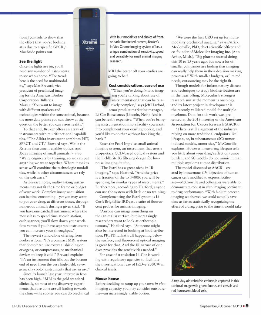
See the lightOnce the lights are on, you’ll need any number of instruments to see who’s home. “The trend here is the need for multimodal-ity,” says Mat Brevard, vice president of preclinical imag-ing for the Americas, Bruker Corporation (Billerica, Mass.). “You want to image with different markers and technologies within the same animal, because the more data points you can throw at the question the better you can assess reality.”
To that end, Bruker offers an array of instruments with multifunctional capabili-ties. “The Albira instrument combines PET, SPECT and CT,” Brevard says. While the Xtreme instrument enables optical and X-ray imaging of small animals in vivo. “We’re engineers by training, so we can put anything we want together. Where it makes sense we’ll combine the technologic modali-ties, while in other circumstances we rely on the software.”
As Brevard notes, multi-tasking instru-ments may not fi t the time frame or budget of your work. Complex image acquisition can be time consuming—yet you may want to put your drug, at different doses, through numerous animals during a given trial. “If you have one catchall instrument where the mouse has to spend time at each station, each scanner, you’ll slow down your work-fl ow versus if you have separate instruments you can increase your throughput.”
The newest stand-alone offering from Bruker is Icon. “It’s a compact MRI system that doesn’t require external shielding or cryogens, or compressors, or mechanical devices to keep it cold,” Brevard explains. “It’s an instrument that fi lls out the bottom end of need from the very high-fi eld, cryo-genically cooled instruments that are in use.”
Since its launch last year, interest in Icon has been high. “MRI is the gold standard clinically, so most of the discovery experi-ments that are done are all leading towards the clinic—the sooner you can do preclinical
MRI the better off your studies are going to be.”
Cost considerations, ease of use“When you’re doing in vivo imag-ing you’re talking about use of instrumentation that can be rela-tively complex,” says Jeff Harford,
senor product marketing manager, Li-Cor Biosciences (Lincoln, Neb.). And it can be really expensive. “When you’re bring-ing instrumentation into a facility you want it to compliment your existing toolkit, and you’d like to do that without breaking the bank.”
Enter the Pearl Impulse small animal imaging system, an instrument that uses a proprietary CCD-based optical system and the FieldBrite Xi fi ltering design for low-noise imaging in vivo.
“The Pearl has a great niche in IR imaging,” says Harford. “And the price is a fraction of the to $400K you will be spending for similar types of instruments.” Furthermore, according to Harford, anyone can use the system with little or no training.
Complementing the Pearl system is Li-Cor’s BrightSite IRDyes, a suite of fl uores-cent probes for animal imaging.
“Anyone can image something on the (animal’s) surface, but increasingly researchers want to look at orthotopic tumors,” Harford says. “Someone might also be interested in looking at biodistribu-tion, PK, PD…That’s all happening below the surface, and fl uorescent optical imaging is great for that. And the IR nature of our dyes provides the sensitivities needed.”
For ease of translation Li-Cor is work-ing with regulatory agencies to facilitate the investigational use of IRDye 800CW in clinical trials.
Mouse houseBefore deciding to ramp up your own in vivoimaging capacity you may consider outsourc-ing—an increasingly viable option.
“We were the fi rst CRO set up for multi-modality preclinical imaging,” says Patrick McConville, PhD, chief scientifi c offi cer and co-founder of Molecular Imaging Inc. (Ann Arbor, Mich.). “Big pharma started doing this 10 to 15 years ago, but now a lot of smaller companies are fi nding that imaging can really help them in their decision making processes.” With smaller budgets, or limited needs, outsourcing may be the right fi t.
Though models for infl ammatory disease and techniques to study biodistribution are in the near offi ng, Molecular’s strongest research suit at the moment is oncology, and its latest project in development is the recently validated model for multiple myeloma. Data for this work was pre-sented at the 2013 meeting of the American Association for Cancer Research (AACR).
“There is still a segment of the industry relying on more traditional endpoints like lifespan, or, in subcutaneous (SC) tumor-induced models, tumor size,” McConville explains. However, measuring lifespan tells you little about your drug’s effect on tumor burden, and SC models do not mimic human multiple myeloma tumor distribution.
The model discussed at AACR—cre-ated by intravenous (IV) injection of human cancer cells modifi ed to express lucifer-ase—McConville and colleagues were able to demonstrate robust in vivo imaging pertinent to drug performance. “With bioluminescent imaging we showed we could actually save time as far as statistically recognizing the effect of a drug prior to the time it would take
With four modalities and choice of front- or back-illuminated camera, Bruker’s In-Vivo Xtreme imaging system offers a unique combination of sensitivity, speed and versatility for small animal imaging research.
A two-day-old zebrafi sh embryo is captured in this confocal image with green fl uorescent vessels and red fl uorescent blood cells.
dd13o_08COS.indd 9dd13o_08COS.indd 9 10/10/2013 4:30:02 PM10/10/2013 4:30:02 PM
to use lifespan as a marker.”With these data in hand, McConville is
looking to extend the Molecular portfolio to include leukemia and lymphoma models.
Something fi shyIn the future, it’s possible that the work-horse of in vivo imaging for drug discovery may be not a mouse but a fi sh, specifi cally a zebrafi sh.
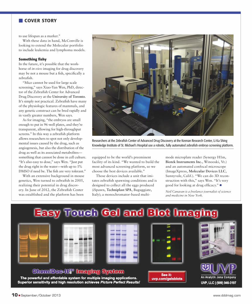
“Mice cannot be used for large scale screening,” says Xiao-Yan Wen, PhD, direc-tor of the Zebrafi sh Center for Advanced Drug Discovery at the University of Toronto. It’s simply not practical. Zebrafi sh have many of the physiologic features of mammals, and any genetic construct can be bred rapidly and in vastly greater numbers, Wen says.
As for imaging, “the embryos are small enough to put in 96-well plates, and they’re transparent, allowing for high-throughput screens.” In this way a zebrafi sh platform allows researchers to spot not only develop-mental issues caused by the drug, such as angiogenesis, but also the distribution of the drug as well as its associated metabolites—something that cannot be done in cell culture. “It’s also easy to dose,” says Wen. “Just put the drug right in the water—with up to 1% DMSO if need be. The fi sh are very tolerant.”
With an extensive background in mouse genetics, Wen turned to zebrafi sh in 2005, realizing their potential in drug discov-ery. In June of 2012, the Zebrafi sh Center was established and the platform has been
equipped to be the world’s preeminent facility of its kind. “We wanted to build the most advanced screening platform, so we choose the best devices available.”
Those devices include a unit that imi-tates zebrafi sh spawning conditions and is designed to collect all the eggs produced (iSpawn, Techniplast SPA, Buguggiate, Italy); a monochromator-based multi-
mode microplate reader (Synergy H1m, Biotek Instruments Inc., Winooski, Vt.) and an automated confocal microscope (ImageXpress, Molecular Devices LLC, Sunnyvale, Calif.). “We can do 3D recon-struction with this,” says Wen. “It’s very good for looking at drug effi cacy.” �
Neil Canavan is a freelance journalist of science and medicine in New York.
10 � September/October 2013 www.dddmag.com
� COVER STORY
Researchers at the Zebrafi sh Center of Advanced Drug Discovery at the Keenan Research Center, Li Ka Shing Knowledge Institute of St. Michael’s Hospital use a robotic, fully automated zebrafi sh embryo screening platform.
dd13o_08COS.indd 10dd13o_08COS.indd 10 10/10/2013 4:30:15 PM10/10/2013 4:30:15 PM
CAS14125 TerretAd-DrugDiscoveryDevelopment-9x10 875 indd 2 8/2/13 11:52 AM
DD13O_02ADS.indd 11DD13O_02ADS.indd 11 10/14/2013 9:58:37 AM10/14/2013 9:58:37 AM
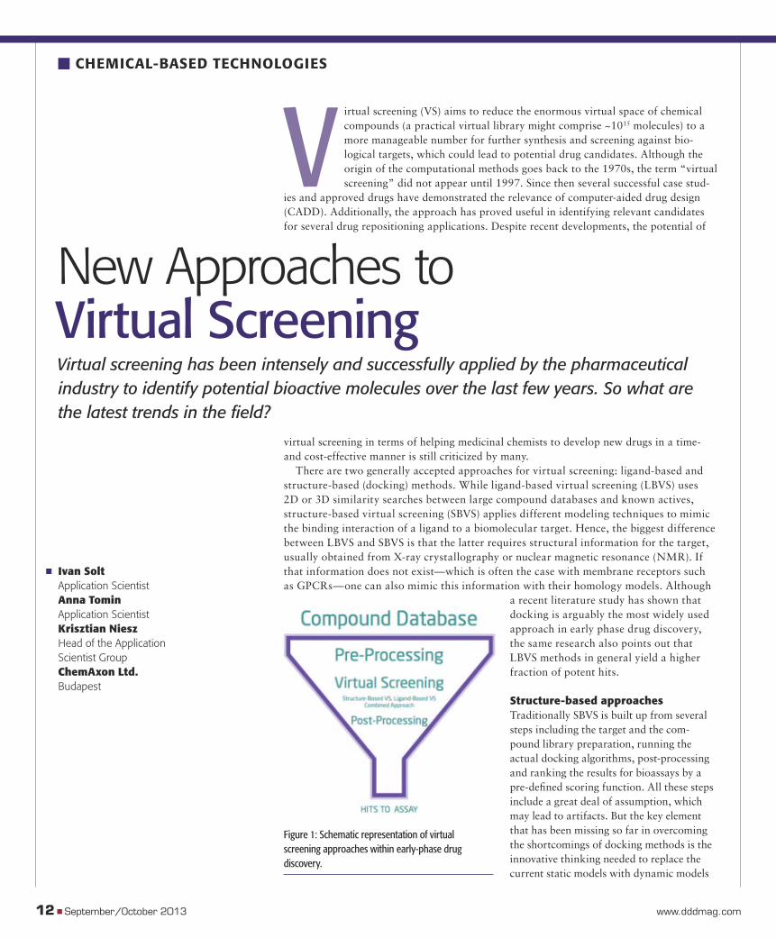
Virtual screening (VS) aims to reduce the enormous virtual space of chemical compounds (a practical virtual library might comprise ~1015 molecules) to a more manageable number for further synthesis and screening against bio-logical targets, which could lead to potential drug candidates. Although the origin of the computational methods goes back to the 1970s, the term “virtual screening” did not appear until 1997. Since then several successful case stud-
ies and approved drugs have demonstrated the relevance of computer-aided drug design (CADD). Additionally, the approach has proved useful in identifying relevant candidates for several drug repositioning applications. Despite recent developments, the potential of
virtual screening in terms of helping medicinal chemists to develop new drugs in a time- and cost-effective manner is still criticized by many.
There are two generally accepted approaches for virtual screening: ligand-based and structure-based (docking) methods. While ligand-based virtual screening (LBVS) uses 2D or 3D similarity searches between large compound databases and known actives, structure-based virtual screening (SBVS) applies different modeling techniques to mimic the binding interaction of a ligand to a biomolecular target. Hence, the biggest difference between LBVS and SBVS is that the latter requires structural information for the target, usually obtained from X-ray crystallography or nuclear magnetic resonance (NMR). If that information does not exist—which is often the case with membrane receptors such as GPCRs—one can also mimic this information with their homology models. Although
a recent literature study has shown that docking is arguably the most widely used approach in early phase drug discovery, the same research also points out that LBVS methods in general yield a higher fraction of potent hits.
Structure-based approachesTraditionally SBVS is built up from several steps including the target and the com-pound library preparation, running the actual docking algorithms, post-processing and ranking the results for bioassays by a pre-defi ned scoring function. All these steps include a great deal of assumption, which may lead to artifacts. But the key element that has been missing so far in overcoming the shortcomings of docking methods is the innovative thinking needed to replace the current static models with dynamic models
12 � September/October 2013 www.dddmag.com
� CHEMICAL-BASED TECHNOLOGIES
New Approaches to Virtual Screening Virtual screening has been intensely and successfully applied by the pharmaceutical industry to identify potential bioactive molecules over the last few years. So what are the latest trends in the fi eld?
� Ivan SoltApplication ScientistAnna TominApplication ScientistKrisztian NieszHead of the Application Scientist GroupChemAxon Ltd.Budapest
Figure 1: Schematic representation of virtual screening approaches within early-phase drug discovery.
dd13o_012CHB.indd 12dd13o_012CHB.indd 12 10/10/2013 4:32:00 PM10/10/2013 4:32:00 PM
that are applicable to the whole system. Undoubtedly, one of the greatest chal-
lenges of docking software is to consider protein fl exibility. These macromolecules are obviously not static objects and confor-mational changes are often key elements in ligand binding. Using multiple high-quality static receptor conformations as snapshots in docking runs and selecting the highest scoring conformation for further investi-gation is one way to tackle the problem. Other ways, including ensemble docking allowed by molecular dynamics simula-tions, or “soft docking,” in which the interaction of the protein and the ligand is allowed to change continuously, and 4D docking which allows ligands to be fi tted against multiple target conformations in a single run are also described in detail in the literature.
Equally important and another non-trivial issue is the handling of water molecules. Water can signifi cantly affect ligand binding through the formation of hydrogen bonds and can contribute to both the enthalpy and entropy of the binding. In general, the thermodynamics of ligand-receptor interactions are still treated simi-larly to how molecular reactions work, and often times this is not the optimal way to approach the problem. As far as account-ing for water, several approaches have been developed recently, which are complemen-tary to the experimental information from X-ray and NMR spectroscopy.
Although providing considerable enhancements, the general belief is that these methods are very target specifi c and they do not work effectively with all protein types. How we account for the water molecules is directly connected to the problem of solving the ligand-fl exibil-ity issue, also known as conformational sampling of ligand molecules within the binding site of the protein pocket dur-ing docking. In the attempt to solve this, four main methods are currently in broad usage: 1) building up the entire ligand from fragments within binding pocket; 2) generating a rotamer library, a collection of low energy conformers, and docking them individually; 3) Monte Carlo simlula-tions; 4) applying evolutionary methods, such as the traditional or Lamarckian
genetic algorithms to fi nd the local energy minimum of the system.
While today’s methods are often capable of fi nding the correct binding modes, they are quite far from giving an accurate prediction in terms of binding affi nity or potency. Current scoring methods include traditional methods, such as force fi eld-based scoring functions, which use classical force fi elds to calculate non-covalent interactions between the ligand and the target; empiri-cal-based methods, which take into account several energetic terms, polar and apolar inter-actions in a weighted fashion; and knowledge-based methods, which use the sum of distance-dependent statistical potentials. A refi nement of the above scoring functions is consensus scoring, which is a hybrid method using multiple functions.
However, since the different scoring functions are co-linear, some scientists question whether this method can signifi -cantly improve the accuracy of the process. To overcome the accuracy issue, machine learning techniques are attracting a great deal of attention currently. Neural net-works, support vector machines and the random forest technique are able to describe the nonlinear dependence of the ligand-target interactions during binding with-out taking solvation and entropic effects into account. Furthermore, the structural interaction fi ngerprint (SIFt) method, which uses the 3D structure of the protein-ligand complex to generate a 1D binary fi nger-print, is of interest. This fi ngerprint then is used to characterize ligand poses derived from the docking procedure and compare to the native substrate’s interaction map.
Ligand-based approachesIn contrast to docking methods, ligand-based approaches do not take the target structure directly into account. These techniques are based on the assumption that compounds with a similar topology have similar biological activity. There are many ways to defi ne molecular similarity; ligand-based screening in drug discovery typically uses topology-based descrip-tors involving the pharmacophoric sites
of the molecules. The descriptors of the known active molecules and the poten-tial hit molecules are compared using pre-defi ned mathematical expressions (metrics) to quantify molecular similarity. These approaches essentially neglect any information about the target biomolecule as well as the 3D structure of the ligand compounds. Nevertheless, they are very effi cient and are often applied in combina-tion with structure-based approaches to identify potential bioactive hits that can then be fed into docking experiments.
Besides the structure and traditional ligand-based methods there is a third pos-sible approach to predict the bioactivity of molecules in a virtual chemical space. Methods that belong to this third branch can be thought of as extensions of the ligand-based approach with the major dif-ference that instead of considering only the molecular topology, they create or consider 3D coordinates of both the active and the potential lead molecules for the similarity comparison, and then estimate the 3D shape similarity of these molecules. These algo-
DRUG Discovery & Development September/October 2013 � 13
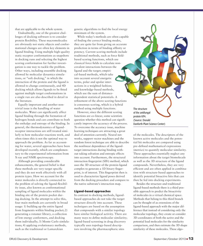
The structure of the antifungal protein KP4. (Source: Donald Danforth Plant Science Center)
dd13o_012CHB.indd 13dd13o_012CHB.indd 13 10/10/2013 4:32:11 PM10/10/2013 4:32:11 PM
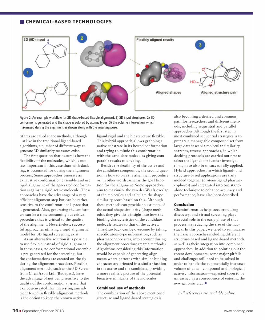
rithms are called shape methods, although just like in the traditional ligand-based algorithms, a number of different ways to generate 3D similarity measures exist.
The fi rst question that occurs is how the fl exibility of the molecules, which is not less important in this case than with dock-ing, is accounted for during the alignment process. Some approaches generate an exhaustive conformation ensemble and use rigid alignment of the generated conforma-tions against a rigid active molecule. These approaches have the advantage of a very effi cient alignment step but can be rather sensitive to the conformational space that is generated. Also, generating the conform-ers can be a time consuming but critical procedure that is critical to the quality of the alignment. Nevertheless, success-ful approaches utilizing a rigid alignment model for 3D ligand screening exist.
As an alternative solution it is possible to use fl exible instead of rigid alignment. In these cases, no conformational ensemble is pre-generated for the screening, but the conformations are created on-the-fl y during the alignment procedure. Flexible alignment methods, such as the 3D Screen from ChemAxon Ltd. (Budapest), have the advantage of not being sensitive to the quality of the conformational space that can be generated. An interesting amend-ment found in fl exible alignment methods is the option to keep the known active
ligand rigid and the hit structure fl exible. This hybrid approach allows grabbing a native substrate in its bound conformation and trying to mimic this conformation with the candidate molecules giving com-parable results to docking.
Besides the fl exibility of the active and the candidate compounds, the second ques-tion is how to bias the alignment procedure or, in other words, what is the goal func-tion for the alignment. Some approaches aim to maximize the van der Waals overlap of the molecules and calculate the shape similarity score based on this. Although these methods can provide an estimate of the actual shape similarity (shape meth-ods), they give little insight into how the binding characteristics of the candidate molecule relates to that of the actives. This drawback can be overcome by taking specifi c atom-type information, such as pharmacophore sites, into account during the alignment procedure (match methods). Algorithms considering this information would be capable of generating align-ments where patterns with similar binding character are oriented in a similar fashion in the active and the candidate, providing a more realistic picture of the potential bioactive similarity of the molecules.
Combined use of methodsThe combination of the above mentioned structure and ligand-based strategies is
also becoming a desired and common path for researchers and different meth-ods, including sequential and parallel approaches. Although the fi rst step in most combined sequential strategies is to prepare a manageable compound set from large databases via molecular similarity searches, reverse approaches, in which docking protocols are carried out fi rst to select the ligands for further investiga-tions, have also been successfully applied. Hybrid approaches, in which ligand- and structure-based applications are truly molded together (protein-ligand pharma-cophores) and integrated into one stand-alone technique to enhance accuracy and performance, have also been described.
ConclusionCheminformatics helps accelerate drug discovery, and virtual screening plays a crucial role in the early phase of that process via reducing the size of the hay-stack. In this paper, we tried to summarize the basic approaches including different structure-based and ligand-based methods as well as their integration into combined approaches. In addition to pointing out the recent developments, some major pitfalls and challenges still need to be solved in order to handle the exponentially increased volume of data—compound and biological activity information—expected soon to be unleashed as a consequence of entering the new genomic era. �
Full references are available online.
Figure 2: An example workfl ow for 3D shape-based fl exible alignment: 1) 2D input structures; 2) 3D conformer is generated and the shape is colored by atomic types; 3) the volume intersection, which maximized during the alignment, is shown along with the resulting pose.
14 � September/October 2013 www.dddmag.com
� CHEMICAL-BASED TECHNOLOGIES
dd13o_012CHB.indd 14dd13o_012CHB.indd 14 10/10/2013 4:32:24 PM10/10/2013 4:32:24 PM
DD13O_02ADS.indd 15DD13O_02ADS.indd 15 10/10/2013 4:33:20 PM10/10/2013 4:33:20 PM
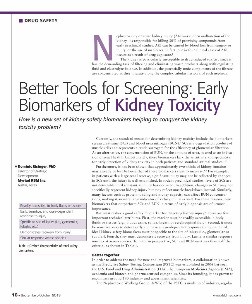
Nephrotoxicity or acute kidney injury (AKI)—a sudden malfunction of the kidney—is responsible for killing 30% of promising compounds from early preclinical studies. AKI can be caused by blood loss from surgery or injury, or the use of medicines. In fact, one in four clinical cases of AKI occurs as a result of drug exposure.1
The kidney is particularly susceptible to drug-induced toxicity since it has the demanding task of fi ltering and eliminating waste products along with regulating fl uid and electrolyte balance. In addition, the potentially toxic components of the fi ltrate are concentrated as they migrate along the complex tubular network of each nephron.
Currently, the standard means for determining kidney toxicity include the biomarkers serum creatinine (SCr) and blood urea nitrogen (BUN).2 SCr is a degradation product of muscle cells and represents a crude surrogate for the effi ciency of glomerular fi ltration. As an alternative, the concentration of BUN, or the amount of urea, is used as an indica-tion of renal health. Unfortunately, these biomarkers lack the sensitivity and specifi city for early detection of kidney toxicity in both patients and standard animal studies.1,3
Furthermore, it has been shown that approximately two-thirds of kidney function may already be lost before either of these biomarkers start to increase.4,5 For example, in patients with a large renal reserve, signifi cant injury may not be refl ected by changes in SCr until the injury is well established. In rodent preclinical studies, levels of SCr are not detectable until substantial injury has occurred. In addition, changes in SCr may not specifi cally represent kidney injury but may refl ect muscle breakdown instead. Similarly, many factors such as protein loading and kidney capacity can affect BUN concentra-tions, making it an unreliable indicator of kidney injury as well. For these reasons, new biomarkers that outperform SCr and BUN in terms of early diagnosis are of utmost importance.
But what makes a good safety biomarker for detecting kidney injury? There are fi ve important technical attributes. First, the marker must be readily accessible in body fl uids or tissues (e.g., blood, urine, saliva, breath or cerebrospinal fl uid). Second, it must be sensitive, easy to detect early and have a dose-dependent response to injury. Third, ideal kidney safety biomarkers must be specifi c to the site of injury (i.e., glomerular or tubular). Fourth, they must demonstrate recovery from injury. Lastly, a similar response must exist across species. To put it in perspective, SCr and BUN meet less than half the criteria, as shown in Table 1.
Better together In order to address the need for new and improved biomarkers, a collaboration known as the Predictive Safety Testing Consortium (PSTC) was established in 2006 between the U.S. Food and Drug Administration (FDA), the European Medicines Agency (EMA), academia and biotech and pharmaceutical companies. Since its founding, it has grown to encompass around 190 industry and government scientists.
The Nephrotoxic Working Group (NWG) of the PSTC is made up of industry, regula-
16 � September/October 2013 www.dddmag.com
� DRUG SAFETY
Better Tools for Screening: Early Biomarkers of Kidney Toxicity How is a new set of kidney safety biomarkers helping to conquer the kidney toxicity problem?
� Dominic Eisinger, PhDDirector of Strategic DevelopmentMyriad RBM Inc.Austin, Texas
Readily accessible in body fl uids or tissues
Early, sensitive, and dose-dependent response to injury
Specifi c to site of injury (i.e., glomerular, tubular, etc.)
Demonstrates recovery from injury
Similar response across species
Table 1: Desired characteristics of renal safety biomarkers.
dd13o_16DS.indd 16dd13o_16DS.indd 16 10/10/2013 4:35:36 PM10/10/2013 4:35:36 PM
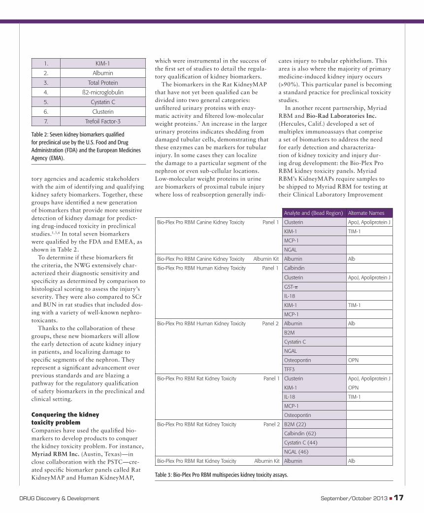
tory agencies and academic stakeholders with the aim of identifying and qualifying kidney safety biomarkers. Together, these groups have identifi ed a new generation of biomarkers that provide more sensitive detection of kidney damage for predict-ing drug-induced toxicity in preclinical studies.1,3,6 In total seven biomarkers were qualifi ed by the FDA and EMEA, as shown in Table 2.
To determine if these biomarkers fi t the criteria, the NWG extensively char-acterized their diagnostic sensitivity and specifi city as determined by comparison to histological scoring to assess the injury’s severity. They were also compared to SCr and BUN in rat studies that included dos-ing with a variety of well-known nephro-toxicants.
Thanks to the collaboration of these groups, these new biomarkers will allow the early detection of acute kidney injury in patients, and localizing damage to specifi c segments of the nephron. They represent a signifi cant advancement over previous standards and are blazing a pathway for the regulatory qualifi cation of safety biomarkers in the preclinical and clinical setting.
Conquering the kidneytoxicity problem Companies have used the qualifi ed bio-markers to develop products to conquer the kidney toxicity problem. For instance, Myriad RBM Inc. (Austin, Texas)—in close collaboration with the PSTC—cre-ated specifi c biomarker panels called Rat KidneyMAP and Human KidneyMAP,
DRUG Discovery & Development September/October 2013 � 17
which were instrumental in the success of the fi rst set of studies to detail the regula-tory qualifi cation of kidney biomarkers.
The biomarkers in the Rat KidneyMAP that have not yet been qualifi ed can be divided into two general categories: unfi ltered urinary proteins with enzy-matic activity and fi ltered low-molecular weight proteins.7 An increase in the larger urinary proteins indicates shedding from damaged tubular cells, demonstrating that these enzymes can be markers for tubular injury. In some cases they can localize the damage to a particular segment of the nephron or even sub-cellular locations. Low-molecular weight proteins in urine are biomarkers of proximal tubule injury where loss of reabsorption generally indi-
cates injury to tubular ephithelium. This area is also where the majority of primary medicine-induced kidney injury occurs (>90%). This particular panel is becoming a standard practice for preclinical toxicity studies.
In another recent partnership, Myriad RBM and Bio-Rad Laboratories Inc. (Hercules, Calif.) developed a set of multiplex immunoassays that comprise a set of biomarkers to address the need for early detection and characteriza-tion of kidney toxicity and injury dur-ing drug development: the Bio-Plex Pro RBM kidney toxicity panels. Myriad RBM’s KidneyMAPs require samples to be shipped to Myriad RBM for testing at their Clinical Laboratory Improvement
1. KIM-1
2. Albumin
3. Total Protein
4. ß2-microglobulin
5. Cystatin C
6. Clusterin
7. Trefoil Factor-3
Table 2: Seven kidney biomarkers qualifi ed for preclinical use by the U.S. Food and Drug Administration (FDA) and the European Medicines Agency (EMA).
Analyte and (Bead Region) Alternate Names
Bio- Plex Pro RBM Canine Kidney Toxicity Panel 1 Clusterin ApoJ, Apoliprotein J
KIM- 1 TIM- 1
MCP- 1
NGAL
Bio- Plex Pro RBM Canine Kidney Toxicity Albumin Kit Albumin Alb
Bio- Plex Pro RBM Human Kidney Toxicity Panel 1 Calbindin
Clusterin ApoJ, Apoliprotein J
GST- �
IL- 18
KIM- 1 TIM- 1
MCP- 1
Bio- Plex Pro RBM Human Kidney Toxicity Panel 2 Albumin Alb
B2M
Cystatin C
NGAL
Osteopontin OPN
TFF3
Bio- Plex Pro RBM Rat Kidney Toxicity Panel 1 Clusterin ApoJ, Apoliprotein J
KIM- 1 OPN
IL- 18 TIM- 1
MCP- 1
Osteopontin
Bio- Plex Pro RBM Rat Kidney Toxicity Panel 2 B2M (22)
Calbindin (62)
Cystatin C (44)
NGAL (46)
Bio- Plex Pro RBM Rat Kidney Toxicity Albumin Kit Albumin Alb
Table 3: Bio-Plex Pro RBM multispecies kidney toxicity assays.
dd13o_16DS.indd 17dd13o_16DS.indd 17 10/10/2013 4:36:02 PM10/10/2013 4:36:02 PM
Amendments (CLIA) certifi ed reference laboratory, whereas the Bio-Plex Pro RBM kidney toxicity panels are available as off-the-shelf kits for investigators worldwide to use in their laboratories.
The toxicity panels contain six of the seven markers approved by the FDA and EMEA for use in preclinical kidney toxic-ity assessment, in addition to several other markers relevant to kidney toxicity and damage research. The assays offer robust quantifi cation of proteins in human, rat and canine urine samples with consistent results and a simple workfl ow. The multi-plex format provides an effective solution for protein biomarker detection compared to traditional enzyme-linked immunosor-bent assays (ELISAs), and the premixed all-in-one kits enable drug development researchers to effi ciently test lead com-pounds in both preclinical animal models and human clinical trials.8 In addition, unlike ELISAs, they enable the detection of multiple biomarkers in a single sample,
which may reveal damage within hours of kidney injury, as shown in Table 3.
A bright future for biomarkers The efforts for identifying biomarkers that are suitable for detection and monitoring of kidney injury is an ongoing process. Fortunately, new biomarkers and assays—provided by new partnerships—are able to expedite translational development, be integrated with a pharmacokinetic or pharmacodynamic drug development strategy and contribute to more informed decisions regarding range and interval of dosing. It’s out with the old, in with the new. �
References 1. Bonventre JV, Vaidya VS, Schmouder R, Feig P, Dieterle F. Next-generation biomarkers for detecting kidney toxicity. Nat Biotechnol. 2010;28(5):436-40.
2. Slocum JL, Heung M, Pennathur S. Marking renal injury: can we move beyond serum creati-
nine? Transl Res. 2012;159(4):277-89.
3. Vaidya VS, Ozer JS, Dieterle F, et al. Kidney injury molecule-1 outperforms traditional biomarkers of kidney injury in preclinical biomarker qualification studies. Nat Biotechnol. 2010;28(5):478-85.
4. Han WK, Bailly V, Abichandani R, Thadhani R, Bonventre JV. Kidney Injury Molecule-1 (KIM-1): a novel biomarker for human renal proximal tubule injury. Kidney Int. 2002;62(1):237-44.
Dieterle F, Perentes E, Cordier A, et al. Urinary clusterin, cystatin C, beta2-microglobulin and total protein as markers to detect drug-induced kidney injury. Nat Biotechnol. 2010;28(5):463-9.
6. Ferguson MA, Vaidya VS, Bonventre JV. Biomarkers of nephrotoxic acute kidney injury. Toxicology. 2008;245(3):182-93.
7. Stephan L, Schnaars H, Marshall K, et al. Pro-filing of human, canine, and rat urine samples using Bio-Plex Pro RBM kidney toxicity assays. Available at: http://www.bio-rad.com/webroot/web/pdf/lsr/literature/Bulletin_6400. Accessed September 20, 2013.
� DRUG SAFETY
Clinically Relevant
Mouse Models of Human Cancer
JAX® Mice, Clinical and Research Services 1-800-422-6423
A diverse library of PDX models
Tumor Types appendixbladderbrainbreastcolorectalendometrialgastrichead and neckhematologickidneyliverlungovarypancreasprostatesarcomaskin
Is your cancer research not progressing as quickly as you would like?
JAX is helping scientists accelerate cancer research
with patient-derived cancer models readily
available to the research community.
www.jax.org/jaxservices/invivo/pdx.html
Patient-Derived
Xenograft ResourceJAX
dd13o_16DS.indd 18dd13o_16DS.indd 18 10/10/2013 4:36:14 PM10/10/2013 4:36:14 PM
HALOPLEXNext Generation Sequencing panels for comprehensive clinical disease research, from the Leader in NGS Target Enrichment
For Research Use Only. Not approved for use in diagnostic procedures.
© Agilent Technologies, Inc. 2013
• Confidence in genomic content
• Ease of use from sample to analysis
• Results you can trust
• HaloPlex Disease Research Panels now available:
Learn more
agilent.com/genomics/ngspanels
DISEASE RESEARCH PANELS
- HaloPlex Cancer
- HaloPlex Cardiomyopathy
- HaloPlex Arrhythmia
- HaloPlex Noonan Syndrome
- HaloPlex ICCG
- HaloPlex Connective Tissue Disorder
- HaloPlex X Chromosome
AGI-3117 HaloPlex Panels Print Ad indd 1 9/10/13 11:15 AM
DD13O_02ADS.indd 19DD13O_02ADS.indd 19 10/10/2013 4:36:42 PM10/10/2013 4:36:42 PM
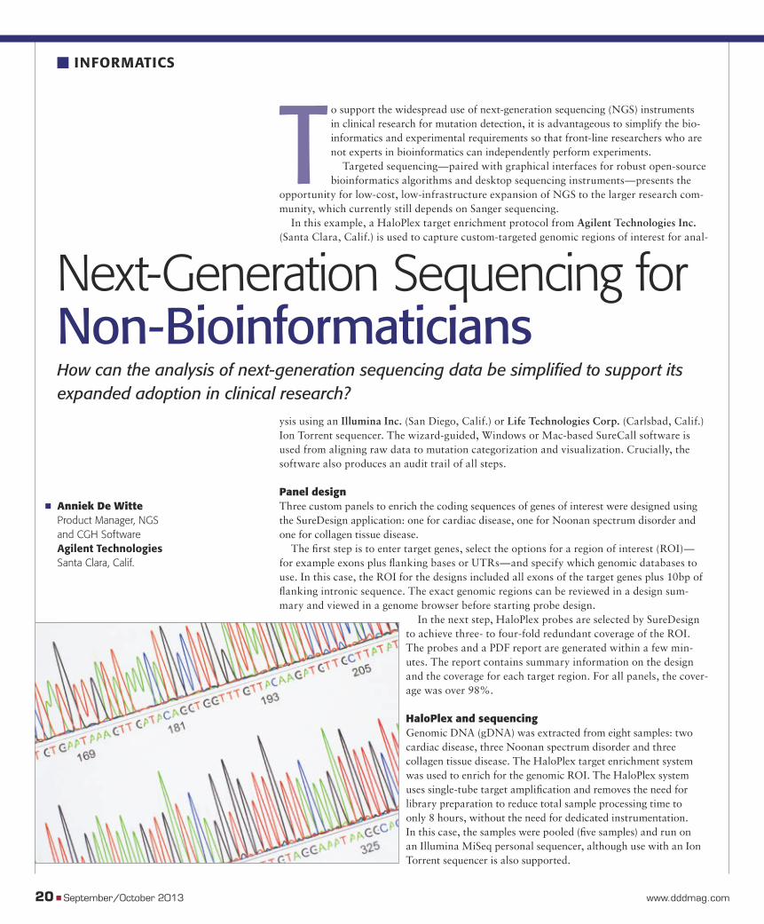
To support the widespread use of next-generation sequencing (NGS) instruments in clinical research for mutation detection, it is advantageous to simplify the bio-informatics and experimental requirements so that front-line researchers who are not experts in bioinformatics can independently perform experiments.
Targeted sequencing—paired with graphical interfaces for robust open-source bioinformatics algorithms and desktop sequencing instruments—presents the
opportunity for low-cost, low-infrastructure expansion of NGS to the larger research com-munity, which currently still depends on Sanger sequencing.
In this example, a HaloPlex target enrichment protocol from Agilent Technologies Inc. (Santa Clara, Calif.) is used to capture custom-targeted genomic regions of interest for anal-
ysis using an Illumina Inc. (San Diego, Calif.) or Life Technologies Corp. (Carlsbad, Calif.) Ion Torrent sequencer. The wizard-guided, Windows or Mac-based SureCall software is used from aligning raw data to mutation categorization and visualization. Crucially, the software also produces an audit trail of all steps.
Panel designThree custom panels to enrich the coding sequences of genes of interest were designed using the SureDesign application: one for cardiac disease, one for Noonan spectrum disorder and one for collagen tissue disease.
The fi rst step is to enter target genes, select the options for a region of interest (ROI)—for example exons plus fl anking bases or UTRs—and specify which genomic databases to use. In this case, the ROI for the designs included all exons of the target genes plus 10bp of fl anking intronic sequence. The exact genomic regions can be reviewed in a design sum-mary and viewed in a genome browser before starting probe design.
In the next step, HaloPlex probes are selected by SureDesign to achieve three- to four-fold redundant coverage of the ROI. The probes and a PDF report are generated within a few min-utes. The report contains summary information on the design and the coverage for each target region. For all panels, the cover-age was over 98%.
HaloPlex and sequencingGenomic DNA (gDNA) was extracted from eight samples: two cardiac disease, three Noonan spectrum disorder and three collagen tissue disease. The HaloPlex target enrichment system was used to enrich for the genomic ROI. The HaloPlex system uses single-tube target amplifi cation and removes the need for library preparation to reduce total sample processing time to only 8 hours, without the need for dedicated instrumentation. In this case, the samples were pooled (fi ve samples) and run on an Illumina MiSeq personal sequencer, although use with an Ion Torrent sequencer is also supported.
20 � September/October 2013 www.dddmag.com
� INFORMATICS
Next-Generation Sequencing for Non-BioinformaticiansHow can the analysis of next-generation sequencing data be simplifi ed to support its expanded adoption in clinical research?
� Anniek De WitteProduct Manager, NGS and CGH SoftwareAgilent TechnologiesSanta Clara, Calif.
dd13o_20INFO.indd 20dd13o_20INFO.indd 20 10/10/2013 4:37:18 PM10/10/2013 4:37:18 PM
© 2013 Capsugel Belgium NV All rights reserved.
Some HPMC capsules are formulated with secondary gelling agents and ionic gel promoters – which may slow down release
of active ingredients. Capsugel’s Vcaps® Plus HPMC capsules are made through a proprietary manufacturing process that
eliminates the need for gelling systems. They disintegrate and release their contents independent of pH and ionic strength of
the test media. To discover how Vcaps® Plus capsules can optimize performance, improve product stability, and reduce both
variability and timelines in your drug development, download our free whitepaper.
Visit us at booth #2012 at AAPS 2013 in San Antonio, TX
VcapsPlus.com 888-783-6361
Vcaps® Plus hypromellose capsules
for pharmaceutical products – true pH and ionic media independence
in disintegration for optimized delivery and performance.
Achieving new standards in performance takes
years of innovative R&D. This is the result.
DD13O_02ADS.indd 21DD13O_02ADS.indd 21 10/10/2013 4:37:33 PM10/10/2013 4:37:33 PM
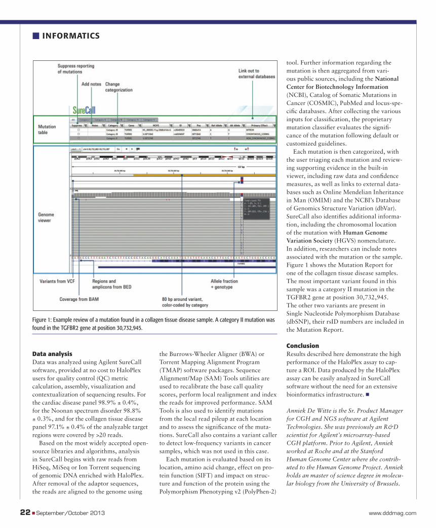
Figure 1: Example review of a mutation found in a collagen tissue disease sample. A category II mutation was found in the TGFBR2 gene at position 30,732,945.
Data analysis Data was analyzed using Agilent SureCall software, provided at no cost to HaloPlex users for quality control (QC) metric calculation, assembly, visualization and contextualization of sequencing results. For the cardiac disease panel 98.9% ± 0.4%, for the Noonan spectrum disorder 98.8% ± 0.3%, and for the collagen tissue disease panel 97.1% ± 0.4% of the analyzable target regions were covered by >20 reads.
Based on the most widely accepted open-source libraries and algorithms, analysis in SureCall begins with raw reads from HiSeq, MiSeq or Ion Torrent sequencing of genomic DNA enriched with HaloPlex. After removal of the adaptor sequences, the reads are aligned to the genome using
the Burrows-Wheeler Aligner (BWA) or Torrent Mapping Alignment Program (TMAP) software packages. Sequence Alignment/Map (SAM) Tools utilities are used to recalibrate the base call quality scores, perform local realignment and index the reads for improved performance. SAM Tools is also used to identify mutations from the local read pileup at each location and to assess the signifi cance of the muta-tions. SureCall also contains a variant caller to detect low-frequency variants in cancer samples, which was not used in this case.
Each mutation is evaluated based on its location, amino acid change, effect on pro-tein function (SIFT) and impact on struc-ture and function of the protein using the Polymorphism Phenotyping v2 (PolyPhen-2)
tool. Further information regarding the mutation is then aggregated from vari-ous public sources, including the National Center for Biotechnology Information (NCBI), Catalog of Somatic Mutations in Cancer (COSMIC), PubMed and locus-spe-cifi c databases. After collecting the various inputs for classifi cation, the proprietary mutation classifi er evaluates the signifi -cance of the mutation following default or customized guidelines.
Each mutation is then categorized, with the user triaging each mutation and review-ing supporting evidence in the built-in viewer, including raw data and confi dence measures, as well as links to external data-bases such as Online Mendelian Inheritance in Man (OMIM) and the NCBI’s Database of Genomics Structure Variation (dbVar). SureCall also identifi es additional informa-tion, including the chromosomal location of the mutation with Human Genome Variation Society (HGVS) nomenclature. In addition, researchers can include notes associated with the mutation or the sample. Figure 1 shows the Mutation Report for one of the collagen tissue disease samples. The most important variant found in this sample was a category II mutation in the TGFBR2 gene at position 30,732,945. The other two variants are present in Single Nucleotide Polymorphism Database (dbSNP), their rsID numbers are included in the Mutation Report.
ConclusionResults described here demonstrate the high performance of the HaloPlex assay to cap-ture a ROI. Data produced by the HaloPlex assay can be easily analyzed in SureCall software without the need for an extensive bioinformatics infrastructure. �
Anniek De Witte is the Sr. Product Manager for CGH and NGS software at Agilent Technologies. She was previously an R&D scientist for Agilent’s microarray-based CGH platform. Prior to Agilent, Anniek worked at Roche and at the Stanford Human Genome Center where she contrib-uted to the Human Genome Project. Anniek holds an master of science degree in molecu-lar biology from the University of Brussels.
22 � September/October 2013 www.dddmag.com
� INFORMATICS
dd13o_20INFO.indd 22dd13o_20INFO.indd 22 10/10/2013 4:37:45 PM10/10/2013 4:37:45 PM
waters.com
Pharmaceutical & Life Sciences | Food | Environmental | Clinical | Chemical Materials
©2013 Waters Corporation. Waters, Oasis and The Science of What’s Possible are registered trademarks of Waters Corporation. Simple Prep is a trademark of Waters Corporation.
When results are truly critical, run it right the fi rst time. Every time.
That’s the promise of Oasis® Sample Extraction Products. Being able to achieve
the highest technical performance is much less complicated than you think.
To learn more about how our Simple PrepTM protocol delivers the purest
samples possible, visit waters.com/simpleprep
DD13O_02ADS.indd 23DD13O_02ADS.indd 23 10/10/2013 4:37:55 PM10/10/2013 4:37:55 PM
24 � September/October 2013 www.dddmag.com
� HOW IT WORKS Coculture Systems
Coculture Systems Increase Sensitivity for Predictive Toxicology StudiesOnyi Irrechukwu, PhDScientistHepregen CorporationMedford, Mass.
Drug-induced liver injury (DILI) is of primary concern in drug development. Nearly half of all drugs entering clinical trials will fail because of some form of serious toxicity that was missed in preclinical studies, costing the industry billions of dollars.1,2 Although the liver can regenerate itself, liver injury can per-manently impair its self-regenerative capacity, thus underscoring the need for non-hepato-toxic therapeutics.
The liver is the primary site of xenobiotic control, as drugs
are metabolized by cellular
enzymes present in hepatocytes. Drugs may be detoxified or made toxic by metabolism. Liver damage may be caused or exacerbated by drug-drug interactions and drug-transporter interactions, where hepatic uptake or excre-tion of the drug or its metabolite is inhibited.
Currently, conventional cell-based in vitro culture systems used to predict drug-induced liver injury include sandwich and monolayer cultures as well as suspensions of primary hepatocytes. Primary hepatoctyes dedifferen-tiate and display a precipitous decline in phe-notypic function within days in these conven-tional culture methods, precluding their use in long-term toxicity studies and severely limiting their ability to predict clinical outcomes.
HepatoPac from Hepregen Corpora-tion (Medford, Mass.) is a bio-engineered
hepatocyte coculture platform in which pri-mary hepatocytes are organized in domains of empirically optimized dimensions and surrounded by supportive non-parenchymal cells. HepatoPac technology exploits the ability of primary hepatocytes to respond to chemical stimuli (i.e., exposure to xenobiot-ics), while extending the phenotypic and functional stability of these cells in culture. These cocultures result in a high-fidelity, in vitro tissue model that is proven to provide a range of liver-specific functions for greater than 4 weeks. Enzyme activity, cell polariza-tion, albumin and urea synthesis and expres-sion of transporter proteins are maintained throughout the culture period.1
The platform’s longevity in culture permits scientists to dose this cocul-
ture system at physiologically relevant drug concentrations over longer periods of time than was previously possible. This feature allows HepatoPac to more closely mimic the
in vivo condition, thus enabling detection and prediction of hepatoxicity due to chronic
dosing. In addition, the platform’s multi-well format can be used
with existing industry auto-mation for high-throughput screening.
The ability to mimic in vivo liver physiology lends
the system to a broad range of applications including preclini-
cal toxicity screening, metabolite iden-tification, drug stability, drug-drug interactions, drug-transporter interactions, enzyme induc-tion and clearance studies. HepatoPac can be used to identify and detect low, medium and high-turnover compounds. Furthermore, studies show that transporter uptake and efflux activities are higher and more stable in HepatoPac than observed in the current gold standard in vitro culture systems.
This technology can be implemented early in the drug development process, decreasing the incidence of adverse clinical outcomes resulting from toxic pharmaceuticals. �
References1. Kola I, Landis J. Can the pharmaceutical indus-try reduce attrition rates? Nat Rev Drug Discov. 2004;3(8):711-5.2. Neumann EK. Combining drug toxicity knowledge. Bio-IT World. July/August 2006.http://www.bio-itworld.com/. Accessed October 3, 2013.3. Khetani SR, Bhatia SN. Microscale culture of human liver cells for drug development. Nat Biotechnol. 2008;26(1):120-6.
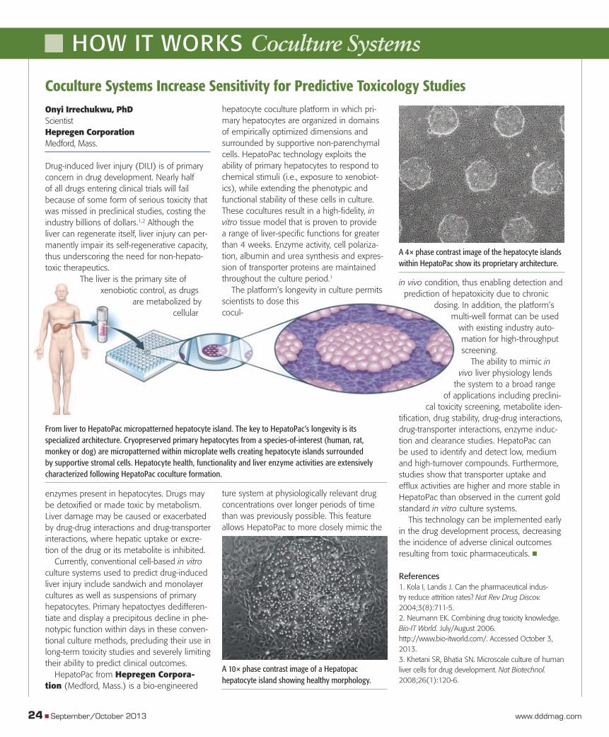
From liver to HepatoPac micropatterned hepatocyte island. The key to HepatoPac’s longevity is its specialized architecture. Cryopreserved primary hepatocytes from a species-of-interest (human, rat, monkey or dog) are micropatterned within microplate wells creating hepatocyte islands surrounded by supportive stromal cells. Hepatocyte health, functionality and liver enzyme activities are extensively characterized following HepatoPac coculture formation.
A 4× phase contrast image of the hepatocyte islands within HepatoPac show its proprietary architecture.
A 10× phase contrast image of a Hepatopac hepatocyte island showing healthy morphology.
dd13o_24HIW.indd 24dd13o_24HIW.indd 24 10/10/2013 4:38:22 PM10/10/2013 4:38:22 PM
-R�������&IGOQER�'SYPXIV�MRXVSHYGIH�XLI�½�VWX�GSQQIVGMEP�GIRXVMJYKI��ERH�[MXL�MX�E�
GSQQMXQIRX�XS�GSRWXERX�MRRSZEXMSR��7MRGI�XLIR��[I´ZI�GSRXMRYEPP]�HI½�RIH�ERH�VIHI½�RIH�
XLI�WXERHEVH�JSV�JEWX��IJ½�GMIRX��ERH�VIPMEFPI�WSPYXMSRW�XLEX�QEOI�XLI�HMWXERGI�FIX[IIR�]SY�
ERH�PMJI´W�MQTSVXERX�HMWGSZIVMIW�WLSVXIV�XLER�IZIV�
(MWGSZIV�MRRSZEXMSR�EX�MXW�½�RIWX�EX�www.beckmancoulter.com
AT THE FOREFRONT OF THE CATEGORY,SINCE BEFORE THERE WAS ONE.
DD13O_02ADS.indd 25DD13O_02ADS.indd 25 10/10/2013 4:38:31 PM10/10/2013 4:38:31 PM
Ion Mobility-Enhanced Research Mass SpectrometerThe Waters SYNAPT G2-Si mass spectrometer integrates a third dimension of resolution and sepa-ration power into a new suite of untargeted and targeted LC/MS/MS workfl ows. This tool gives researchers a means with which to gain a deeper understanding of molecular biology and disease mechanisms, to develop the next generation of healthcare treatments. SYNAPT G2-Si now com-bines the power of travelling wave (T-Wave) ion mobility separations with new data acquisition and informatics technologies, and collision cross section (CCS) measurements to bring to the toughest analytical applications, information and confi dence at a level not possible by mass and chromato-graphic separation alone. The SYNAPT G2-Si system is the fi rst MS system to elevate CCS alongside retention time and mass to charge ratio (m/z) as a robust, reliable identifi cation parameter in library-based screening.� www.waters.com
iPSC Reprogramming VectorsLife Technologies Corporation launched Epi5 Episomal iPSC reprogramming vectors, the fi rst product fully com-mercialized based on technology
that won its developers a Nobel Prize in 2012 for the ability to reprogram adult human cells into an early embryonic state. Scientists use induced pluripotent stem cell (iPSC) technol-ogy to create iPSCs from patient-derived adult somatic cells. The iPSCs can then be differenti-ated into many cell types, such as neurons and hepatocytes, to be studied in the lab. The ease-of-use and reprogramming effi ciency of Epi5 Episomal iPSC reprogramming vectors helps accelerate the process for applications in basic research and pre-clinical studies. While other reprogramming vectors have been developed in Dr. Yamanaka’s lab, none has ever been commercially available. Epi5™ Episomal iPSC Reprogramming Vectors is the result of a global, non-exclusive license agreement Life Technologies signed last year with iPS Academia Japan for its iPSC patent portfolio.� www.lifetechnologies.com
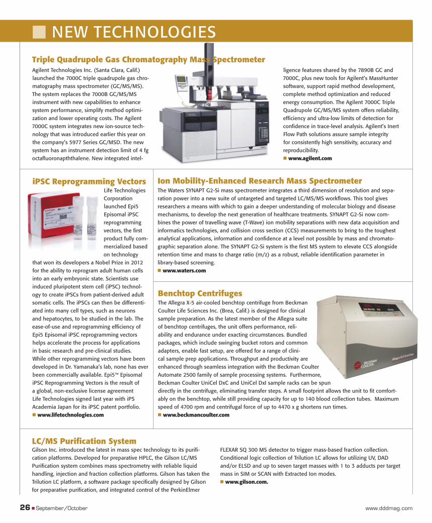
Benchtop Centrifuges The Allegra X-5 air-cooled benchtop centrifuge from Beckman Coulter Life Sciences Inc. (Brea, Calif.) is designed for clinical sample preparation. As the latest member of the Allegra suite of benchtop centrifuges, the unit offers performance, reli-ability and endurance under exacting circumstances. Bundled packages, which include swinging bucket rotors and common adapters, enable fast setup, are offered for a range of clini-cal sample prep applications. Throughput and productivity are enhanced through seamless integration with the Beckman Coulter Automate 2500 family of sample processing systems. Furthermore, Beckman Coulter UniCel DxC and UniCel DxI sample racks can be spun directly in the centrifuge, eliminating transfer steps. A small footprint allows the unit to fi t comfort-ably on the benchtop, while still providing capacity for up to 140 blood collection tubes. Maximum speed of 4700 rpm and centrifugal force of up to 4470 x g shortens run times. � www.beckmancoulter.com
Agilent Technologies Inc. (Santa Clara, Calif.) launched the 7000C triple quadrupole gas chro-matography mass spectrometer (GC/MS/MS). The system replaces the 7000B GC/MS/MS instrument with new capabilities to enhance system performance, simplify method optimi-zation and lower operating costs. The Agilent 7000C system integrates new ion-source tech-nology that was introduced earlier this year on the company’s 5977 Series GC/MSD. The new system has an instrument detection limit of 4 fg octafl uoronapththalene. New integrated intel-
ligence features shared by the 7890B GC and 7000C, plus new tools for Agilent’s MassHunter software, support rapid method development, complete method optimization and reduced energy consumption. The Agilent 7000C Triple Quadrupole GC/MS/MS system offers reliability, effi ciency and ultra-low limits of detection for confi dence in trace-level analysis. Agilent’s Inert Flow Path solutions assure sample integrity for consistently high sensitivity, accuracy and reproducibility.� www.agilent.com
26 � September/October www.dddmag.com
� NEW TECHNOLOGIES
Triple Quadrupole Gas Chromatography Mass Spectrometer
Gilson Inc. introduced the latest in mass spec technology to its purifi -cation platforms. Developed for preparative HPLC, the Gilson LC/MS Purifi cation system combines mass spectrometry with reliable liquid handling, injection and fraction collection platforms. Gilson has taken the Trilution LC platform, a software package specifi cally designed by Gilson for preparative purifi cation, and integrated control of the PerkinElmer
FLEXAR SQ 300 MS detector to trigger mass-based fraction collection. Conditional logic collection of Trilution LC allows for utilizing UV, DAD and/or ELSD and up to seven target masses with 1 to 3 adducts per target mass in SIM or SCAN with Extracted Ion modes.� www.gilson.com.
LC/MS Purifi cation System
dd13o_26NT.indd 26dd13o_26NT.indd 26 10/10/2013 4:38:52 PM10/10/2013 4:38:52 PM
PIPETMAN® inside.
Trust your pipetting to be
reliable and consistent!
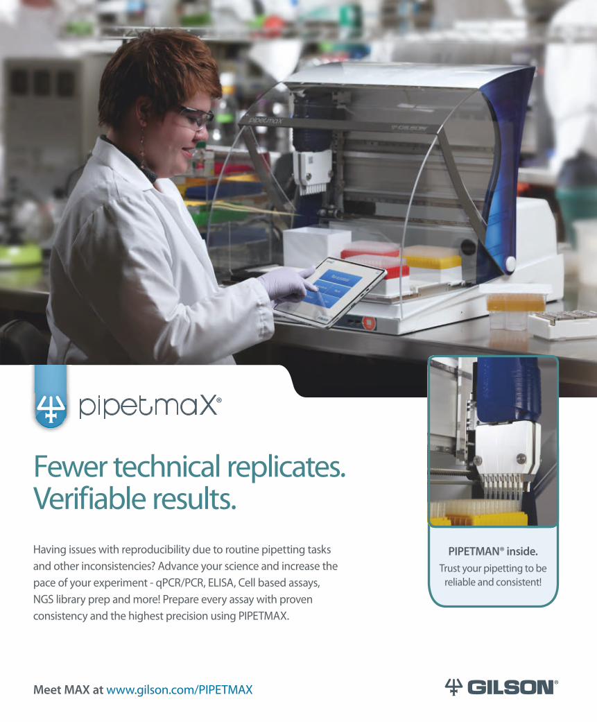
Having issues with reproducibility due to routine pipetting tasks
and other inconsistencies? Advance your science and increase the
pace of your experiment - qPCR/PCR, ELISA, Cell based assays,
NGS library prep and more! Prepare every assay with proven
consistency and the highest precision using PIPETMAX.
Meet MAX at www.gilson.com/PIPETMAX
Fewer technical replicates.Verifiable results.
PIPETMAX Ad DDD indd 1 10/9/2013 9:28:46 AM
DD13O_02ADS.indd 27DD13O_02ADS.indd 27 10/10/2013 4:39:05 PM10/10/2013 4:39:05 PM
A powerful alternative for pharmaceutical dosage formsPolymer choices in pharmaceutical dosage forms have always been a balancing act between performance and development time, and historically has been shaped by the interactions of gelatin. The first generation of HPMC capsules, which relied on a secondary gelling agent, were recognized by formulators as having issues with dissolution performance and product stability. Fortunately, new scientific discoveries in polymers and capsule manufacturing have resulted in the creation of the next generation of capsules – one that offers better performance and reduced development time compared to gelatin and first-generation capsules.
Capsugel, the market leader in research and development in this area, is now offering these second-generation HPMC capsules under the trade name, Vcaps® Plus capsules.
In a number of studies, Vcaps Plus capsules have been shown to deliver optimized compound stability and predictable in vitro dissolution while also helping to eliminate the complexity in formulation development. Known globally for their reliable and predictable performance, Vcaps Plus capsules are well suited for over-the-counter (OTC) or off-patent products as well as for new chemical entities (NCEs).
True pH and ionic media independent performanceTraditionally, HPMC capsules were created using secondary gelling agents and ionic gel promoters, which have been found to interact with dissolution media and delay compound release from the capsule. The activity of the gelling agent kappa-carrageenan, for example, is enhanced by potassium and calcium cations contained in many foods. The extent of the resulting delay in dissolution time was shown in an in vitro test in which caffeine-filled traditional HPMC capsules were dissolved in a number of dissolution media. In the simulated normal acidic environment of the stomach (pH 1.2 USP), 90% of the caffeine was dissolved within approximately 15 minutes (Figure 1). Adding 2 g/L of potassium chloride (KCl) to this medium resulted in no dissolution after 15 minutes and a caffeine dissolution between 70% and 80% after more than one hour. Increasing the KCl content to 9 g/L delayed caffeine release even further, with a dissolution rate of just over 10% in 45 minutes. Results with simulated milk fluid were equally disappointing. Similar delays in dissolution times were observed and attributed to carrageenan in an independent study (Ku et al., 2011). Of course, such long delays in capsule dissolution are unacceptable particularly for rapid-relief products.
Capsugel addressed this situation by developing a proprietary new thermal gelation manufacturing process for Vcaps
Plus capsules that eliminates the need for gelling systems all together and provides true pH and ionic media independence in disintegration. In vitro tests showed that these second-generation HPMC capsules had similar rates of dissolution at pH levels of 1.2 and 6.8 and with simulated milk fluid, achieving a nearly complete dissolution of the caffeine contents within approximately 30 minutes (Figure 2). Even adding 2 g/L or 9 g/L of KCl to the dissolution medium did not affect the performance of Vcaps Plus capsules, with dissolution of over 90% within 30 minutes, even under the most disadvantageous condition.
These findings were supported by an independent study that compared the dissolution performance of traditional and second-generation HPMC capsules (Ku et al., 2011), and underscores the superior performance of Vcaps Plus capsules.
Ideally suited for moisture sensitive compoundsWhile gelatin capsules have been effectively used for over a hundred years, due to their excellent flexibility and highly desirable dissolution properties, they are not typically the polymer choice for moisture sensitive compounds. Vcaps Plus capsules on the other hand have a three-fold lower moisture content than gelatin capsules and are less hygroscopic. That equates to fewer broken capsules due to brittleness and less of a chance of drug degradation compared to gelatin capsules.
Next generation HPMC capsules greatly expand pharmaceutical uses by Dominique Cadé, PhD
Advertorial
28 � September/October 2013 www.dddmag.com
DD13O_02ADS.indd 28DD13O_02ADS.indd 28 10/10/2013 4:39:26 PM10/10/2013 4:39:26 PM
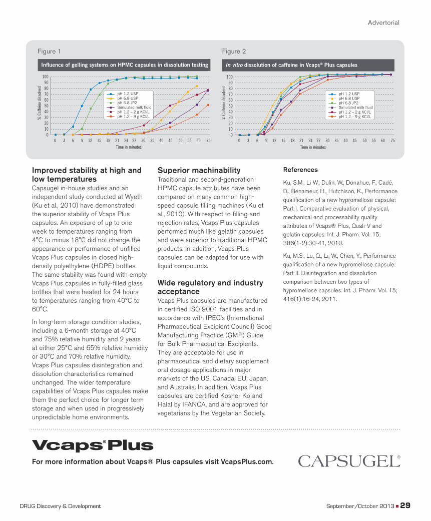
Improved stability at high and low temperaturesCapsugel in-house studies and an independent study conducted at Wyeth (Ku et al., 2010) have demonstrated the superior stability of Vcaps Plus capsules. An exposure of up to one week to temperatures ranging from 4°C to minus 18°C did not change the appearance or performance of unfilled Vcaps Plus capsules in closed high-density polyethylene (HDPE) bottles. The same stability was found with empty Vcaps Plus capsules in fully-filled glass bottles that were heated for 24 hours to temperatures ranging from 40°C to 60°C.
In long-term storage condition studies, including a 6-month storage at 40°C and 75% relative humidity and 2 years at either 25°C and 65% relative humidity or 30°C and 70% relative humidity, Vcaps Plus capsules disintegration and dissolution characteristics remained unchanged. The wider temperature capabilities of Vcaps Plus capsules make them the perfect choice for longer term storage and when used in progressively unpredictable home environments.
Superior machinabilityTraditional and second-generation HPMC capsule attributes have been compared on many common high-speed capsule filling machines (Ku et al., 2010). With respect to filling and rejection rates, Vcaps Plus capsules performed much like gelatin capsules and were superior to traditional HPMC products. In addition, Vcaps Plus capsules can be adapted for use with liquid compounds.
Wide regulatory and industry acceptanceVcaps Plus capsules are manufactured in certified ISO 9001 facilities and in accordance with IPEC’s (International Pharmaceutical Excipient Council) Good Manufacturing Practice (GMP) Guide for Bulk Pharmaceutical Excipients. They are acceptable for use in pharmaceutical and dietary supplement oral dosage applications in major markets of the US, Canada, EU, Japan, and Australia. In addition, Vcaps Plus capsules are certified Kosher Ko and Halal by IFANCA, and are approved for vegetarians by the Vegetarian Society.
References
Ku, S.M., Li W., Dulin, W., Donahue, F., Cadé, D., Benameur, H., Hutchison, K., Performance qualification of a new hypromellose capsule: Part I. Comparative evaluation of physical, mechanical and processability quality attributes of Vcaps® Plus, Quali-V and gelatin capsules. Int. J. Pharm. Vol. 15; 386(1-2):30-41, 2010.
Ku, M.S., Lu, Q., Li, W., Chen, Y., Performance qualification of a new hypromellose capsule: Part II. Disintegration and dissolution comparison between two types of hypromellose capsules. Int. J. Pharm. Vol. 15; 416(1):16-24, 2011.
0102030405060708090
100
% C
affe
ine
diss
olve
d
Time in minutes
0 3 6 9 12 15 18 21 24 27 30 35 40 45 50 55 60 75
Influence of gelling systems on HPMC capsules in dissolution testing
pH 1.2 USPpH 6.8 USPpH 6.8 JP2Simulated milk fluidpH 1.2 – 2 g KCI/LpH 1.2 – 9 g KCI/L
0102030405060708090
100
% C
affe
ine
diss
olve
d
Time in minutes
0 3 6 9 12 15 18 21 24 27 30 35 40 45 50 55 60 75
In vitro dissolution of caffeine in Vcaps® Plus capsules
pH 1.2 USPpH 6.8 USPpH 6.8 JP2Simulated milk fluidpH 1.2 – 2 g KCI/LpH 1.2 – 9 g KCI/L
For more information about Vcaps® Plus capsules visit VcapsPlus.com.
Advertorial
Figure 1 Figure 2
DRUG Discovery & Development September/October 2013 � 29
DD13O_02ADS.indd 29DD13O_02ADS.indd 29 10/10/2013 4:39:33 PM10/10/2013 4:39:33 PM
� EDITORIAL INDEXCOMPANY PAGE
� ADVERTISER INDEXCOMPANY PAGE
Abbott Laboratories . . . . . . . . . . . . . . . . . . . . . . . . . . . . . . . . . . . . . . 7Active Motif . . . . . . . . . . . . . . . . . . . . . . . . . . . . . . . . . . . . . . . . . . . . 5Agilent Technologies . . . . . . . . . . . . . . . . . . . . . . . . . . . . . . . . . 20, 26AstraZeneca PLC . . . . . . . . . . . . . . . . . . . . . . . . . . . . . . . . . . . . . . 4, 6Beckman Coulter . . . . . . . . . . . . . . . . . . . . . . . . . . . . . . . . . . . . . . . 26Bio-Rad Laboratories Inc.. . . . . . . . . . . . . . . . . . . . . . . . . . . . . . . . . 25Biotek Instruments LLC . . . . . . . . . . . . . . . . . . . . . . . . . . . . . . . . . . 10Boehringer Ingelheim GMBH . . . . . . . . . . . . . . . . . . . . . . . . . . . . . . 7Bristol-Myers Squibb Co. . . . . . . . . . . . . . . . . . . . . . . . . . . . . . . . . . . 6Bruker Corporation . . . . . . . . . . . . . . . . . . . . . . . . . . . . . . . . . . . . . . 9ChemAxon Ltd. . . . . . . . . . . . . . . . . . . . . . . . . . . . . . . . . . . . . . 12, 13Eli Lilly and Co.. . . . . . . . . . . . . . . . . . . . . . . . . . . . . . . . . . . . . . . . . . 7European Medicines Agency. . . . . . . . . . . . . . . . . . . . . . . . . . . . . . 24F. Hoffmann-La Roche Ltd. . . . . . . . . . . . . . . . . . . . . . . . . . . . . . . . . 6Genentech Inc.. . . . . . . . . . . . . . . . . . . . . . . . . . . . . . . . . . . . . . . . . . 6Gilson Inc. . . . . . . . . . . . . . . . . . . . . . . . . . . . . . . . . . . . . . . . . . . . . 26GlaxoSmithKline PLC . . . . . . . . . . . . . . . . . . . . . . . . . . . . . . . . . . . . . 6Hepregen Corp. . . . . . . . . . . . . . . . . . . . . . . . . . . . . . . . . . . . . . . . . 24Illumina Inc. . . . . . . . . . . . . . . . . . . . . . . . . . . . . . . . . . . . . . . . . . . . 20Johnson & Johnson . . . . . . . . . . . . . . . . . . . . . . . . . . . . . . . . . . . . . . 7Li-Cor Biosciences . . . . . . . . . . . . . . . . . . . . . . . . . . . . . . . . . . . . . . . 9Life Technologies Corp. . . . . . . . . . . . . . . . . . . . . . . . . . . . . . . . 20, 26MedChemica Ltd.. . . . . . . . . . . . . . . . . . . . . . . . . . . . . . . . . . . . . . . . 6Molecular Devices LLC . . . . . . . . . . . . . . . . . . . . . . . . . . . . . . . . . . 10Molecular Imaging Inc. . . . . . . . . . . . . . . . . . . . . . . . . . . . . . . . . . . . 9Myriad RBM Inc.. . . . . . . . . . . . . . . . . . . . . . . . . . . . . . . . . . . . . 24, 25National Center for Advancing Translational Sciences . . . . . . . . . . . 7National Center for Biotechnology Information . . . . . . . . . . . . . . . 22National Institutes of Health . . . . . . . . . . . . . . . . . . . . . . . . . . . . . . . 7Novartis International AG. . . . . . . . . . . . . . . . . . . . . . . . . . . . . . . . . . 6Pfizer Inc. . . . . . . . . . . . . . . . . . . . . . . . . . . . . . . . . . . . . . . . . . . . . . . 6Pistoia Alliance . . . . . . . . . . . . . . . . . . . . . . . . . . . . . . . . . . . . . . . . . . 6Predictive Safety Testing Consortium . . . . . . . . . . . . . . . . . . . . . . . 24Roche Holding AG . . . . . . . . . . . . . . . . . . . . . . . . . . . . . . . . . . . . . 4, 6Sanofi SA . . . . . . . . . . . . . . . . . . . . . . . . . . . . . . . . . . . . . . . . . . . . . . 7Sigma-Aldrich . . . . . . . . . . . . . . . . . . . . . . . . . . . . . . . . . . . . . . . . . . . 5Taconic Farms . . . . . . . . . . . . . . . . . . . . . . . . . . . . . . . . . . . . . . . . . . 8Techniplast SPA . . . . . . . . . . . . . . . . . . . . . . . . . . . . . . . . . . . . . . . . 10TransCelerate BioPharma Inc. . . . . . . . . . . . . . . . . . . . . . . . . . . . . . . 7U.S. Food and Drug Administration . . . . . . . . . . . . . . . . . . . . . . . . 24University of Toronto . . . . . . . . . . . . . . . . . . . . . . . . . . . . . . . . . . . . 10Waters Corp.. . . . . . . . . . . . . . . . . . . . . . . . . . . . . . . . . . . . . . . . . . . 26
AbD Serotec . . . . . . . . . . . . . . . . . . . . . . . . . . . . . . . . . . . . . . . . . . . . . . . . .2Agilent . . . . . . . . . . . . . . . . . . . . . . . www.agilent.com . . . . . . . . . . . . . . . .19Beckman Coulter . . . . . . . . . . www.beckmancoulter.com . . . . . . . . . . . .25Capsugel . . . . . . . . . . . . . . . . . . . . www.capsugel.com . . . . . . . . 21, 28, 29Charles River Laboratories . . . . . . . .www.criver.com. . . . . . . . . . . . . . . . .32Chemical Abstract Service . . . . . . . . . www.cas.org . . . . . . . . . . . . . . . . . .11Gilson Inc. . . . . . . . . . . . . . . . . . . . . www.gilson.com . . . . . . . . . . . . . . . .27IDEXX Laboratories Inc.. . . . . . . . . . .www.idexx.com. . . . . . . . . . . . . . . . . .7Jackson Laboratory . . . . . . . . . . . . . . . www.jax.org . . . . . . . . . . . . . . . . . .18Myriad RBM . . . . . . . . . . . . . . . . www.myriadrbm.com . . . . . . . . . . . . . .15Society for Laboratory
Automation & Screening . . . . . . .www.slas.org . . . . . . . . . . . . . . . . . .31UVP Inc. . . . . . . . . . . . . . . . . . . . . . . . www.uvp.com . . . . . . . . . . . . . . . . .10Waters Corp. . . . . . . . . . . . . . . . . . . www.waters.com . . . . . . . . . . . . . . . .23
DRUG DISCOVERY & DEVELOPMENT® (ISSN #1524-783X, USPS #021-269), (GST Reg. #844559765), is a registered trademark of and published bi-monthly by Advantage Business Media, 100 Enterprise Drive, Suite 600, Box 912, Rockaway, NJ 07866-0912. All rights reserved under the U.S.A., International, and Pan-American Copyright Conventions. No part of this publication may be reproduced, stored in a retrieval system, or transmitted in any form or by any means, mechanical, photocopying, electronic recording or otherwise, without the prior written permission of the publisher. Opinions expressed in articles are those of the authors and do not necessarily refl ect those of Advantage Business Media or the Editorial Board.Periodicals Mail postage paid at Rockaway, NJ 07866 and at additional mailing offi ces. POSTMASTER: Send return address changes to DRUG DISCOVERY & DEVELOPMENT, Box 3574, Northbrook, IL 60065. Publication Mail Agreement No. 41336030. Return undeliverable Canadian addresses to: Imex/Pitney Bowes, P.O. Box 1632, Windsor Ontario N9A 7C9.SUBSCRIPTION INQUIRIES/CHANGE OF ADDRESS: contact: Omeda Customer Service, P.O. Box 3574, Northbrook, IL 60065-3574, 847-559-7560, Fax: 847-291-4816, email: [email protected]. Change of address notices should include old as well as new address. If possible attach address label from recent issue. Allow 8 to 10 weeks for address change to become effective. Subscriptions are free to qualifi ed individuals. Subscription rates per year are $54 for U.S.A., $78 for Canada, $126 for Mexico & foreign air delivery, single copy $12 for U.S.A., $22 other locations, prepaid in U.S.A. funds drawn on a U.S.A. branch bank.NOTICE TO SUBSCRIBERS: We permit reputable companies to send announcements of their products or services to our subscribers. Requests for this privilege are examined with great care to be sure they will be of interest to our readers. If you prefer not to receive such mailings, and want your name in our fi les only for receiving the magazine, please write us, enclosing your current address mailing label. Please address your request to Omeda Customer Service, P.O. Box 3574, Northbrook, IL 60065-3574.PRINTED IN USA: Advantage Business Media does not assume and hereby disclaims any liability to any person for any loss or damage caused by errors or omissions in the material contained herein, regardless of whether such errors result from negligence, accident or any other cause whatsoever. The editors make every reasonable effort to verify the information published, but Advantage Business Media assumes no responsibility for the validity of any manufacturers’ claims or statements in items reported. Copyright ©2013 Advantage Business Media. All rights reserved.
30 � September/October www.dddmag.com
Average No. Copies Each Issue During
Preceding 12 Months
No. Copies of Single Issue Published
Nearest to Filing Date21,131 19,947
13,015 12,2350 0
36 34
0 013,051 12,269
7,901 7,5960 0
0 0
83 07,984 7,596
21,035 19,86596 82
21,131 19,94762.0% 61.8%
required and will be printed in the October 2013 issue of this publication.17. Signature and Title of Editor, Publisher, Business Manager, or Owner
g. Copies Not Distributedh. Total (Sum of 15f and g) i. Percent Paid and/or Requested Circulation (15c divided by f times 100)16. Publication of Statement of Ownership for a Requestor Publication is
1. Publication Title: DRUG DISCOVERY & DEVELOPMENT 2. Publication Number: USPS # 021-269 3. Filing Date: October 1, 2013 4. Issue Frequency: Bi-monthly 5. No. of Issues Published Annually: 6 6. Annual Subscription Price: USA $54 CAN $78 MEX & FOR AIR $126 7. Complete Mailing Address of Known Office of Publication: Advantage Business Media, 100 Enterprise Drive, Suite 600, Box 912, Rockaway, NJ 07866-0912 Contact Person: Barbara Cavallaro, Telephone: (973) 920-7477 8. Complete Mailing Address of Headquarters or General Business Office of Publisher: Advantage Business Media, 100 Enterprise Drive, Suite 600, Box 912, Rockaway, NJ 07866-0912 9. Full Names and Complete Mailing Addresses of Publisher: Elizabeth Vickers, Advantage Business Media, 100 Enterprise Drive, Suite 600, Box 912, Rockaway, NJ 07866-0912; Editor: Andrew Wiecek, Advantage Business Media, 100 Enterprise Drive, Suite 600, Box 912, Rockaway, NJ 07866-0912. 10. Owner: Advantage Business Media, LLC, 100 Enterprise Drive, Suite 600, Box 912, Rockaway, NJ 07866-0912 11. Known Bondholders, Mortgagees, and Other Security Holders Owning or Holding 1 Percent or More of Total Amount of Bonds, Mortgages, or Other Securities: none 12. Tax Status: The purpose, function, and nonprofit status of this organization and the exempt status for federal income tax purposes has not changed during preceding 12 months
15. Extent and Nature of Circulation:
(4) Non-Requested Copies Distributed Outside the Maile. Total Nonrequested Distribution (Sum of 15d (1), (2), (3) and (4)) f. Total Distribution (Sum of 15c and e)
c. Total Paid and/or Requested Circulation (Sum of 15b. (1), (2), (3) and (4))d. Nonrequested Distribution(1) Outside County Nonrequested Copies Stated on PS Form 3541(2) In-CountyNonrequested Copies Stated on PS Form 3541
(3) Non-Requested Copies Distributed Through the USPS by Other Clases of Mail
(1) Outside County Paid/Requested Mail Subscriptions Stated on PS Form 3541(2) In-County Paid/Requested Mail Subscriptions Stated on PS Form 3541(3) Sales Through Dealers and Carriers, Street Vendors, Counter Sales, and Other Paid or Requested Distribution Outside USPS ®(4) Requested Copies Distributed by Other Mail Classes Throught the USPS (e.g.
United States Postal ServiceSTATEMENT OF OWNERSHIP, MANAGEMENT, AND CIRCULATION
a. Total Number of Copies b. Legitimate Paid and/or Requested Distribution
13. Publication Title: DRUG DISCOVERY & DEVELOPMENT 14. Issue Date for Circulation Data Below: July/August 2013
dd13o_30Index_v2.indd 30dd13o_30Index_v2.indd 30 10/14/2013 9:54:25 AM10/14/2013 9:54:25 AM
Why Participate in SLAS2014, the Third Annual SLAS Conference and Exhibition?
Because it’s where more than 5,000 of the best laboratory science
and technology minds from across the globe come together to learn,
ĞdžƉůŽƌĞ�ĂŶĚ�ƐŚĂƌĞ�ƚŚĞ�ůĂƚĞƐƚ�ŬŶŽǁ�ŚŽǁ�ĂŶĚ�ŝŶŶŽǀĂƟŽŶ�ŝŶ�ůĂďŽƌĂƚŽƌLJ�
technology. Join your colleagues—innovative scientists, engineers,
researchers and technologists from academic, government and
ĐŽŵŵĞƌĐŝĂů�ůĂďŽƌĂƚŽƌŝĞƐ Ăƚ�^>�^ϮϬϭϰ�ĂŶĚ�ĚŝƌĞĐƚůLJ�ďĞŶĞĮƚ�ĨƌŽŵ�ϭϯϬ�
ŽƵƚƐƚĂŶĚŝŶŐ�ƉŽĚŝƵŵ�ƉƌĞƐĞŶƚĂƟŽŶƐ�ĐŽŵƉƌŝƐŝŶŐ�ϯϬ�ƵŶŝƋƵĞ�ƐĐŝĞŶƟĮĐ�
sessions, hundreds of posters and new and emerging technologies from
ϯϬϬн�ĞdžŚŝďŝƚŽƌƐ�ĨƌŽŵ�ĂƌŽƵŶĚ�ƚŚĞ�ǁŽƌůĚ#�WĂƌƟĐŝƉĂƟŶŐ�Ăƚ�^>�^ϮϬϭϰ�ŝƐ�
ĂŶ�ŝŶǀĂůƵĂďůĞ�ŝŶǀĞƐƚŵĞŶƚ�ƚŚĂƚ�ƉĂLJƐ�ƐŝŐŶŝĮĐĂŶƚ�ĚŝǀŝĚĞŶĚƐ�ƚŽǁĂƌĚƐ�LJŽƵƌ�
ƉĞƌƐŽŶĂů�ĂŶĚ�ŽƌŐĂŶŝnjĂƟŽŶĂů�ŐŽĂůƐ&�ĂƐ�ǁĞůů�ĂƐ�LJŽƵƌ�ƉƌŽĨĞƐƐŝŽŶĂů�ƐƵĐĐĞƐƐ#�
SLAS2014 Scientific Program Crosses a Diversity of Industries and Interests
Premier Sponsor:
Short Courses: January 18 - 19
Conference: January 20 - 22
Exhibition: January 19 - 21
Scan the code with your mobile device
to go directly to SLAS2014.org
� Assay Development and Screening
�� �ƵƚŽŵĂƟŽŶ�ĂŶĚ�,ŝŐŚ� Throughput Technologies
�� �ŝŽĂŶĂůLJƟĐĂů�dĞĐŚŶŝƋƵĞƐ
�������ŝĂŐŶŽƐƟĐƐ�ĂŶĚ��ŝŽŵĂƌŬĞƌƐ
��� Drug Target Biology
� /ŶĨŽƌŵĂƟĐƐ
����Micro/Nano Technologies
Keynote Presenters
Eric J. Topol, M.D.
Director,
^ĐƌŝƉƉƐ�dƌĂŶƐůĂƟŽŶĂů�
^ĐŝĞŶĐĞ�/ŶƐƟƚƵƚĞ
�ŚŝĞĨ��ĐĂĚĞŵŝĐ�KĸĐĞƌ&�
^ĐƌŝƉƉƐ�,ĞĂůƚŚ
WƌŽĨĞƐƐŽƌ�ŽĨ��'ĞŶŽŵŝĐƐ&�
dŚĞ�^ĐƌŝƉƉƐ�ZĞƐĞĂƌĐŚ�/ŶƐƟƚƵƚĞ
Jad Abumrad
Co-host and Creator,
ZĂĚŝŽůĂď
Robert Krulwich
Co-host,
ZĂĚŝŽůĂď
� Drug Discovery and Development
�� /ŶĨŽƌŵĂƟĐƐ
�� �ůŝŶŝĐĂů��ŝĂŐŶŽƐƟĐƐ
� Technology Transfer
�� >ĂďŽƌĂƚŽƌLJ�/ŶƐƚƌƵŵĞŶƚĂƟŽŶ
�� ^LJŶƚŚĞƟĐ��ŚĞŵŝƐƚƌLJ
�� ,ĞĂůƚŚĐĂƌĞ
SLAS2014 Educational Tracks
Register now at SLAS2014.org
Look Who’s Back!See his Journey to
DD13O_02ADS.indd 31DD13O_02ADS.indd 31 10/14/2013 9:58:14 AM10/14/2013 9:58:14 AM
Discover just how much Charles River’s employees are committed to your preclinical research.
flexibility. innovation. people who care. www.criver.com/people
DD13O_02ADS.indd 32DD13O_02ADS.indd 32 10/10/2013 4:40:32 PM10/10/2013 4:40:32 PM