Drug Delivery with Carbon Nanotubes for In vivo Cancer Treatment
10
2008;68:6652-6660. Cancer Res Zhuang Liu, Kai Chen, Corrine Davis, et al. Treatment Cancer In vivo Drug Delivery with Carbon Nanotubes for Updated version http://cancerres.aacrjournals.org/content/68/16/6652 Access the most recent version of this article at: Material Supplementary http://cancerres.aacrjournals.org/content/suppl/2008/08/13/68.16.6652.DC1.html Access the most recent supplemental material at: Cited Articles http://cancerres.aacrjournals.org/content/68/16/6652.full.html#ref-list-1 This article cites by 47 articles, 11 of which you can access for free at: Citing articles http://cancerres.aacrjournals.org/content/68/16/6652.full.html#related-urls This article has been cited by 9 HighWire-hosted articles. Access the articles at: E-mail alerts related to this article or journal. Sign up to receive free email-alerts Subscriptions Reprints and . [email protected] Department at To order reprints of this article or to subscribe to the journal, contact the AACR Publications Permissions . [email protected] Department at To request permission to re-use all or part of this article, contact the AACR Publications Cancer Research. on September 28, 2013. © 2008 American Association for cancerres.aacrjournals.org Downloaded from Cancer Research. on September 28, 2013. © 2008 American Association for cancerres.aacrjournals.org Downloaded from Cancer Research. on September 28, 2013. © 2008 American Association for cancerres.aacrjournals.org Downloaded from Cancer Research. on September 28, 2013. © 2008 American Association for cancerres.aacrjournals.org Downloaded from Cancer Research. on September 28, 2013. © 2008 American Association for cancerres.aacrjournals.org Downloaded from Cancer Research. on September 28, 2013. © 2008 American Association for cancerres.aacrjournals.org Downloaded from Cancer Research. on September 28, 2013. © 2008 American Association for cancerres.aacrjournals.org Downloaded from Cancer Research. on September 28, 2013. © 2008 American Association for cancerres.aacrjournals.org Downloaded from Cancer Research. on September 28, 2013. © 2008 American Association for cancerres.aacrjournals.org Downloaded from Cancer Research. on September 28, 2013. © 2008 American Association for cancerres.aacrjournals.org Downloaded from
-
Upload
independent -
Category
Documents
-
view
3 -
download
0
Transcript of Drug Delivery with Carbon Nanotubes for In vivo Cancer Treatment
2008;68:6652-6660. Cancer Res Zhuang Liu, Kai Chen, Corrine Davis, et al. Treatment
CancerIn vivoDrug Delivery with Carbon Nanotubes for
Updated version
http://cancerres.aacrjournals.org/content/68/16/6652
Access the most recent version of this article at:
Material
Supplementary
http://cancerres.aacrjournals.org/content/suppl/2008/08/13/68.16.6652.DC1.html
Access the most recent supplemental material at:
Cited Articles
http://cancerres.aacrjournals.org/content/68/16/6652.full.html#ref-list-1
This article cites by 47 articles, 11 of which you can access for free at:
Citing articles
http://cancerres.aacrjournals.org/content/68/16/6652.full.html#related-urls
This article has been cited by 9 HighWire-hosted articles. Access the articles at:
E-mail alerts related to this article or journal.Sign up to receive free email-alerts
Subscriptions
Reprints and
To order reprints of this article or to subscribe to the journal, contact the AACR Publications
Permissions
To request permission to re-use all or part of this article, contact the AACR Publications
Cancer Research. on September 28, 2013. © 2008 American Association forcancerres.aacrjournals.org Downloaded from
Cancer Research. on September 28, 2013. © 2008 American Association forcancerres.aacrjournals.org Downloaded from
Cancer Research. on September 28, 2013. © 2008 American Association forcancerres.aacrjournals.org Downloaded from
Cancer Research. on September 28, 2013. © 2008 American Association forcancerres.aacrjournals.org Downloaded from
Cancer Research. on September 28, 2013. © 2008 American Association forcancerres.aacrjournals.org Downloaded from
Cancer Research. on September 28, 2013. © 2008 American Association forcancerres.aacrjournals.org Downloaded from
Cancer Research. on September 28, 2013. © 2008 American Association forcancerres.aacrjournals.org Downloaded from
Cancer Research. on September 28, 2013. © 2008 American Association forcancerres.aacrjournals.org Downloaded from
Cancer Research. on September 28, 2013. © 2008 American Association forcancerres.aacrjournals.org Downloaded from
Cancer Research. on September 28, 2013. © 2008 American Association forcancerres.aacrjournals.org Downloaded from

Drug Delivery with Carbon Nanotubes for In vivo Cancer Treatment
Zhuang Liu,1Kai Chen,
2Corrine Davis,
3Sarah Sherlock,
1Qizhen Cao,
2
Xiaoyuan Chen,2and Hongjie Dai
1
1Department of Chemistry, Stanford University; 2The Molecular Imaging Program at Stanford, Department of Radiology, Biophysics andBio-X Program and 3Department of Comparative Medicine, Stanford University School of Medicine, Stanford, California
Abstract
Chemically functionalized single-walled carbon nanotubes(SWNT) have shown promise in tumor-targeted accumulationin mice and exhibit biocompatibility, excretion, and littletoxicity. Here, we show in vivo SWNT drug delivery for tumorsuppression in mice. We conjugate paclitaxel (PTX), a widelyused cancer chemotherapy drug, to branched polyethyleneglycol chains on SWNTs via a cleavable ester bond to obtaina water-soluble SWNT-PTX conjugate. SWNT-PTX affordshigher efficacy in suppressing tumor growth than clinicalTaxol in a murine 4T1 breast cancer model, owing to pro-longed blood circulation and 10-fold higher tumor PTX uptakeby SWNT delivery likely through enhanced permeability andretention. Drug molecules carried into the reticuloendothelialsystem are released from SWNTs and excreted via biliary path-way without causing obvious toxic effects to normal organs.Thus, nanotube drug delivery is promising for high treatmentefficacy and minimum side effects for future cancer therapywith low drug doses. [Cancer Res 2008;68(16):6652–60]
Introduction
A holy grail in cancer therapy is to deliver high doses of drugmolecules to tumor sites for maximum treatment efficacy whileminimizing side effects to normal organs (1, 2). Through theenhanced permeability and retention (EPR) effect, nanostructuredmaterials on systemic injection can accumulate in tumor tissues byescaping through the abnormally leaky tumor blood vessels (3–6),making them useful for drug delivery applications. As a uniquequasi one-dimensional material, single-walled carbon nanotubes(SWNT) have been explored as novel drug delivery vehicles in vitro(7–9). SWNTs can effectively shuttle various biomolecules intocells, including drugs (7–9), peptide (10), proteins (11), plasmidDNA (12), and small interfering RNA (13, 14), via endocytosis (15).The intrinsic near-IR (NIR) light absorption property of carbonnanotubes has been used to destruct cancer cells in vitro (16),whereas their NIR photoluminescence property has been used forin vitro cell imaging and probing (17). The ultrahigh surface area ofthese one-dimensional polyaromatic macromolecules allows forefficient loading of chemotherapy drugs (8). Various groups haveinvestigated the in vivo behavior of carbon nanotubes in animals(18–20). It is found that well PEGylated SWNTs i.v. injected intomice seem nontoxic over several months (21). Nanotubes accu-
mulated in the reticuloendothelial systems (RES) of mice areexcreted gradually via the biliary pathway and end up in the feces(22). Targeted tumor accumulation of SWNTs functionalized withtargeting ligands RGD peptide or antibodies has shown highefficiency (18, 20). These results set a foundation for further explo-ration of carbon nanotubes for therapeutic applications.In the current work, we show SWNT delivery of paclitaxel (PTX)
into xenograft tumors in mice with higher tumor suppressionefficacy than the clinical drug formulation Taxol. The water-insoluble PTX conjugated to PEGylated SWNTs exhibits high watersolubility and maintains similar toxicity to cancer cells as Taxolin vitro . SWNT-PTX affords much longer blood circulation time ofPTX than that of Taxol and PEGylated PTX, leading to high tumoruptake of the drug through EPR effect. The strong therapeuticefficacy of SWNT-PTX is shown by its ability to slow down tumorgrowth even at a low drug dose (5 mg/kg PTX). We observe highertumor uptake of PTX and higher ratios of tumor to normal organPTX uptake for SWNT-PTX than Taxol and PEGylated PTX, highlydesired for higher treatment efficacy and lower side effect. PTXcarried into RES organs by SWNT-PTX is released from the nano-tube carriers likely via in vivo ester cleavage and is cleared out fromthe body via the biliary pathway. The non-Cremophor compositionin our SWNT-PTX, rapid clearance of drugs from RES organs,higher ratios of tumor-to-normal organ drug uptakes, and the factthat tumor suppression efficacy can be reached at low injecteddrug dose make carbon nanotube drug delivery a very promisingnanoplatform for future cancer therapeutics.
Materials and Methods
Functionalization of SWNTs with phospholipid-branched polyeth-ylene glycol. Raw HiPco SWNTs (0.2 mg/mL) were sonicated in a 0.2
mmol/L solution of DSPE-PEG5000-4-arm-(PEG-amine) (see SupplementaryData for the synthetic chemistry) for 30 min with a cup-horn sonicator
followed by centrifugation at 24,000 � g for 6 h, yielding a suspension of
SWNTs with noncovalent phospholipid-branched polyethylene glycol (PEG)coating in the supernatant (13, 14, 18). Excess surfactant and unreacted PEG
molecules were removed by repeated filtration through a 100-kDa molecular
weight cutoff (MWCO) filter (Millipore) and extensive washing with water.
PTX conjugation. PTX (LC Laboratories) was modified by succinicanhydride (Aldrich) according to the literature, adding a carboxyl acid
group on the molecule at the C-2¶-OH position highlighted in Fig. 1A (23).
SWNTs (300 nmol/L, 0.05 mg/mL) with branched PEG-NH2 functionaliza-
tion were reacted with 0.3 mmol/L of the modified PTX (dissolved inDMSO) in the presence of 5 mmol/L 1-ethyl-3-(3-dimethylaminopropyl)
carbodiimide hydrochloride (EDC; Aldrich) and 5 mmol/L N-hydroxysulfo-
succinimide (Sulfo-NHS, Pierce). The solution was supplemented with
1� PBS at pH 7.4. After 6-h reaction, the resulting SWNT-PTX was purifiedto remove unconjugated PTX by filtration through 5-kDa MWCO filters
and extensive washing.
UV-Vis-NIR absorbance spectra of the SWNT-PTX conjugates were mea-sured by a Cary-6000i spectrophotometer. The concentration of SWNTs was
determined by the absorbance at 808 nm with a molar extinction coefficient
of 7.9 � 106 mol/L�cm�1 with an average tube length of f150 nm (16).
Note: Supplementary data for this article are available at Cancer Research Online(http://cancerres.aacrjournals.org/).
Requests for reprints: Hongjie Dai, Department of Chemistry, Stanford University,Stanford, CA 94305. Phone: 650-723-4518; Fax: 650-725-0259; E-mail: [email protected].
I2008 American Association for Cancer Research.doi:10.1158/0008-5472.CAN-08-1468
Cancer Res 2008; 68: (16). August 15, 2008 6652 www.aacrjournals.org
Research Article
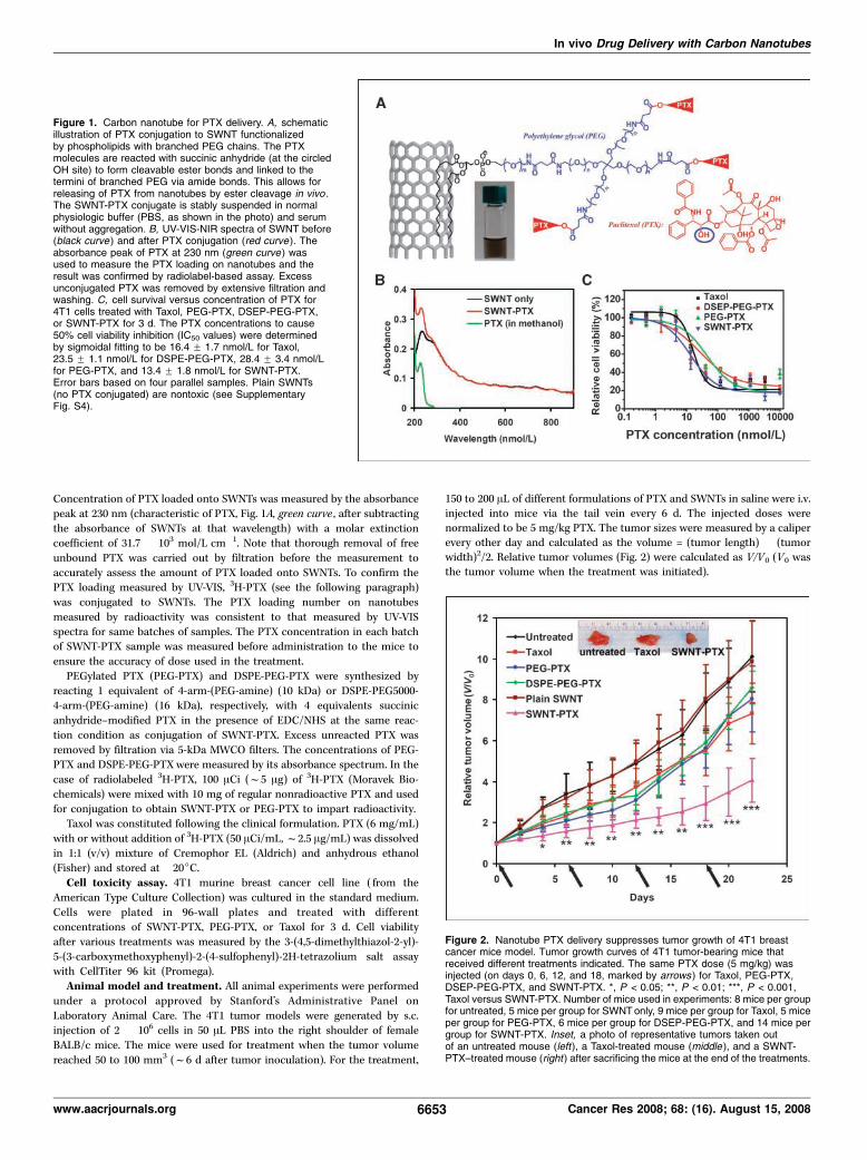
Concentration of PTX loaded onto SWNTs was measured by the absorbance
peak at 230 nm (characteristic of PTX, Fig. 1A, green curve , after subtracting
the absorbance of SWNTs at that wavelength) with a molar extinctioncoefficient of 31.7 � 103 mol/L�cm�1. Note that thorough removal of free
unbound PTX was carried out by filtration before the measurement to
accurately assess the amount of PTX loaded onto SWNTs. To confirm thePTX loading measured by UV-VIS, 3H-PTX (see the following paragraph)
was conjugated to SWNTs. The PTX loading number on nanotubes
measured by radioactivity was consistent to that measured by UV-VIS
spectra for same batches of samples. The PTX concentration in each batchof SWNT-PTX sample was measured before administration to the mice to
ensure the accuracy of dose used in the treatment.
PEGylated PTX (PEG-PTX) and DSPE-PEG-PTX were synthesized by
reacting 1 equivalent of 4-arm-(PEG-amine) (10 kDa) or DSPE-PEG5000-4-arm-(PEG-amine) (16 kDa), respectively, with 4 equivalents succinic
anhydride–modified PTX in the presence of EDC/NHS at the same reac-
tion condition as conjugation of SWNT-PTX. Excess unreacted PTX was
removed by filtration via 5-kDa MWCO filters. The concentrations of PEG-PTX and DSPE-PEG-PTX were measured by its absorbance spectrum. In the
case of radiolabeled 3H-PTX, 100 ACi (f5 Ag) of 3H-PTX (Moravek Bio-
chemicals) were mixed with 10 mg of regular nonradioactive PTX and usedfor conjugation to obtain SWNT-PTX or PEG-PTX to impart radioactivity.
Taxol was constituted following the clinical formulation. PTX (6 mg/mL)
with or without addition of 3H-PTX (50 ACi/mL,f2.5 Ag/mL) was dissolvedin 1:1 (v/v) mixture of Cremophor EL (Aldrich) and anhydrous ethanol(Fisher) and stored at �20jC.
Cell toxicity assay. 4T1 murine breast cancer cell line ( from the
American Type Culture Collection) was cultured in the standard medium.
Cells were plated in 96-wall plates and treated with differentconcentrations of SWNT-PTX, PEG-PTX, or Taxol for 3 d. Cell viability
after various treatments was measured by the 3-(4,5-dimethylthiazol-2-yl)-
5-(3-carboxymethoxyphenyl)-2-(4-sulfophenyl)-2H-tetrazolium salt assaywith CellTiter 96 kit (Promega).
Animal model and treatment. All animal experiments were performedunder a protocol approved by Stanford’s Administrative Panel on
Laboratory Animal Care. The 4T1 tumor models were generated by s.c.injection of 2 � 106 cells in 50 AL PBS into the right shoulder of female
BALB/c mice. The mice were used for treatment when the tumor volume
reached 50 to 100 mm3 (f6 d after tumor inoculation). For the treatment,
150 to 200 AL of different formulations of PTX and SWNTs in saline were i.v.injected into mice via the tail vein every 6 d. The injected doses werenormalized to be 5 mg/kg PTX. The tumor sizes were measured by a caliperevery other day and calculated as the volume = (tumor length) � (tumorwidth)2/2. Relative tumor volumes (Fig. 2) were calculated as V/V0 (V0 wasthe tumor volume when the treatment was initiated).
Figure 2. Nanotube PTX delivery suppresses tumor growth of 4T1 breastcancer mice model. Tumor growth curves of 4T1 tumor-bearing mice thatreceived different treatments indicated. The same PTX dose (5 mg/kg) wasinjected (on days 0, 6, 12, and 18, marked by arrows ) for Taxol, PEG-PTX,DSEP-PEG-PTX, and SWNT-PTX. *, P < 0.05; **, P < 0.01; ***, P < 0.001,Taxol versus SWNT-PTX. Number of mice used in experiments: 8 mice per groupfor untreated, 5 mice per group for SWNT only, 9 mice per group for Taxol, 5 miceper group for PEG-PTX, 6 mice per group for DSEP-PEG-PTX, and 14 mice pergroup for SWNT-PTX. Inset, a photo of representative tumors taken outof an untreated mouse (left), a Taxol-treated mouse (middle ), and a SWNT-PTX–treated mouse (right ) after sacrificing the mice at the end of the treatments.
Figure 1. Carbon nanotube for PTX delivery. A, schematicillustration of PTX conjugation to SWNT functionalizedby phospholipids with branched PEG chains. The PTXmolecules are reacted with succinic anhydride (at the circledOH site) to form cleavable ester bonds and linked to thetermini of branched PEG via amide bonds. This allows forreleasing of PTX from nanotubes by ester cleavage in vivo .The SWNT-PTX conjugate is stably suspended in normalphysiologic buffer (PBS, as shown in the photo) and serumwithout aggregation. B, UV-VIS-NIR spectra of SWNT before(black curve ) and after PTX conjugation (red curve). Theabsorbance peak of PTX at 230 nm (green curve ) wasused to measure the PTX loading on nanotubes and theresult was confirmed by radiolabel-based assay. Excessunconjugated PTX was removed by extensive filtration andwashing. C, cell survival versus concentration of PTX for4T1 cells treated with Taxol, PEG-PTX, DSEP-PEG-PTX,or SWNT-PTX for 3 d. The PTX concentrations to cause50% cell viability inhibition (IC50 values) were determinedby sigmoidal fitting to be 16.4 F 1.7 nmol/L for Taxol,23.5 F 1.1 nmol/L for DSPE-PEG-PTX, 28.4 F 3.4 nmol/Lfor PEG-PTX, and 13.4 F 1.8 nmol/L for SWNT-PTX.Error bars based on four parallel samples. Plain SWNTs(no PTX conjugated) are nontoxic (see SupplementaryFig. S4).
In vivo Drug Delivery with Carbon Nanotubes
www.aacrjournals.org 6653 Cancer Res 2008; 68: (16). August 15, 2008
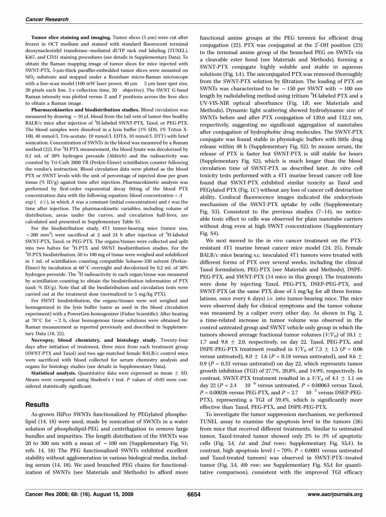
Tumor slice staining and imaging. Tumor slices (5 Am) were cut afterfrozen in OCT medium and stained with standard fluorescent terminal
deoxynucleotidyl transferase–mediated dUTP nick end labeling (TUNEL),
Ki67, and CD31 staining procedures (see details in Supplementary Data). To
obtain the Raman mapping image of tumor slices for mice injected withSWNT-PTX, 5-Am-thick paraffin-embedded tumor slices were mounted onSiO2 substrate and mapped under a Renishaw micro-Raman microscope
with a line-scan model (100 mW laser power, 40 Am � 2 Am laser spot size,
20 pixels each line, 2-s collection time, 20� objective). The SWNT G-bandRaman intensity was plotted versus X and Y positions across the liver slice
to obtain a Raman image.
Pharmacokinetics and biodistribution studies. Blood circulation wasmeasured by drawingf10 AL blood from the tail vein of tumor-free healthyBALB/c mice after injection of 3H-labeled SWNT-PTX, Taxol, or PEG-PTX.
The blood samples were dissolved in a lysis buffer (1% SDS, 1% Triton X-
100, 40 mmol/L Tris-acetate, 10 mmol/L EDTA, 10 mmol/L DTT) with briefsonication. Concentration of SWNTs in the blood was measured by a Raman
method (22). For 3H-PTX measurement, the blood lysate was decolorized by
0.2 mL of 30% hydrogen peroxide (Aldrich) and the radioactivity was
counted by Tri-Carb 2800 TR (Perkin-Elmer) scintillation counter followingthe vendor’s instruction. Blood circulation data were plotted as the blood
PTX or SWNT levels with the unit of percentage of injected dose per gram
tissue (% ID/g) against time after injection. Pharmacokinetic analysis was
performed by first-order exponential decay fitting of the blood PTXconcentration data with the following equation: blood concentration = A �exp (�t/k), in which A was a constant (initial concentration) and t was the
time after injection. The pharmacokinetic variables, including volume ofdistribution, areas under the curves, and circulation half-lives, are
calculated and presented in Supplementary Table S1.
For the biodistribution study, 4T1 tumor-bearing mice (tumor size,
f200 mm3) were sacrificed at 2 and 24 h after injection of 3H-labeledSWNT-PTX, Taxol, or PEG-PTX. The organs/tissues were collected and split
into two halves for 3H-PTX and SWNT biodistribution studies. For the3H-PTX biodistribution, 50 to 100 mg of tissue were weighed and solubilized
in 1 mL of scintillation counting compatible Soluene-350 solvent (Perkin-Elmer) by incubation at 60jC overnight and decolorized by 0.2 mL of 30%
hydrogen peroxide. The 3H radioactivity in each organ/tissue was measured
by scintillation counting to obtain the biodistribution information of PTX(unit: % ID/g). Note that all the biodistribution and circulation tests were
carried out at the treatment dose (normalized to 5 mg/kg PTX).
For SWNT biodistribution, the organs/tissues were wet weighed and
homogenized in the lysis buffer (same as used in the blood circulationexperiment) with a PowerGen homogenizer (Fisher Scientific). After heating
at 70jC for f2 h, clear homogenous tissue solutions were obtained for
Raman measurement as reported previously and described in Supplemen-
tary Data (18, 22).Necropsy, blood chemistry, and histology study. Twenty-four
days after initiation of treatment, three mice from each treatment group
(SWNT-PTX and Taxol) and two age-matched female BALB/c control mice
were sacrificed with blood collected for serum chemistry analysis andorgans for histology studies (see details in Supplementary Data).
Statistical analysis. Quantitative data were expressed as mean F SD.
Means were compared using Student’s t test. P values of <0.05 were con-sidered statistically significant.
Results
As-grown HiPco SWNTs functionalized by PEGylated phospho-lipid (14, 18) were used, made by sonication of SWNTs in a watersolution of phospholipid-PEG and centrifugation to remove largebundles and impurities. The length distribution of the SWNTs was20 to 300 nm with a mean of f100 nm (Supplementary Fig. S1;refs. 14, 18) The PEG functionalized SWNTs exhibited excellentstability without agglomeration in various biological media, includ-ing serum (14, 18). We used branched PEG chains for functional-ization of SWNTs (see Materials and Methods) to afford more
functional amine groups at the PEG termini for efficient drugconjugation (22). PTX was conjugated at the 2¶-OH position (23)to the terminal amine group of the branched PEG on SWNTs viaa cleavable ester bond (see Materials and Methods), forming aSWNT-PTX conjugate highly soluble and stable in aqueoussolutions (Fig. 1A). The unconjugated PTXwas removed thoroughlyfrom the SWNT-PTX solution by filtration. The loading of PTX onSWNTs was characterized to be f150 per SWNT with f100 nmlength by radiolabeling method using tritium 3H-labeled PTX and aUV-VIS-NIR optical absorbance (Fig. 1B ; see Materials andMethods). Dynamic light scattering showed hydrodynamic size ofSWNTs before and after PTX conjugation of 120.6 and 132.2 nm,respectively, suggesting no significant aggregation of nanotubesafter conjugation of hydrophobic drug molecules. The SWNT-PTXconjugate was found stable in physiologic buffers with little drugrelease within 48 h (Supplementary Fig. S2). In mouse serum, therelease of PTX is faster but SWNT-PTX is still stable for hours(Supplementary Fig. S2), which is much longer than the bloodcirculation time of SWNT-PTX as described later. In vitro celltoxicity tests performed with a 4T1 murine breast cancer cell linefound that SWNT-PTX exhibited similar toxicity as Taxol andPEGylated PTX (Fig. 1C) without any loss of cancer cell destructionability. Confocal fluorescence images indicated the endocytosismechanism of the SWNT-PTX uptake by cells (SupplementaryFig. S3). Consistent to the previous studies (7–14), no notice-able toxic effect to cells was observed for plain nanotube carrierswithout drug even at high SWNT concentrations (SupplementaryFig. S4).We next moved to the in vivo cancer treatment on the PTX-
resistant 4T1 murine breast cancer mice model (24, 25). FemaleBALB/c mice bearing s.c. inoculated 4T1 tumors were treated withdifferent forms of PTX over several weeks, including the clinicalTaxol formulation, PEG-PTX (see Materials and Methods), DSPE-PEG-PTX, and SWNT-PTX (14 mice in this group). The treatmentswere done by injecting Taxol, PEG-PTX, DSEP-PEG-PTX, andSWNT-PTX (at the same PTX dose of 5 mg/kg for all three formu-lations, once every 6 days) i.v. into tumor-bearing mice. The micewere observed daily for clinical symptoms and the tumor volumewas measured by a caliper every other day. As shown in Fig. 2,a time-related increase in tumor volume was observed in thecontrol untreated group and SWNT vehicle only group in which thetumors showed average fractional tumor volumes (V/V0) of 10.1 F1.7 and 9.8 F 2.0, respectively, on day 22. Taxol, PEG-PTX, andDSPE-PEG-PTX treatment resulted in V/V0 of 7.3 F 1.5 (P = 0.06versus untreated), 8.0 F 1.6 (P = 0.18 versus untreated), and 8.6 F0.9 (P = 0.33 versus untreated) on day 22, which represents tumorgrowth inhibition (TGI) of 27.7%, 20.8%, and 14.9%, respectively. Incontrast, SWNT-PTX treatment resulted in a V/V0 of 4.1 F 1.1 onday 22 (P = 2.4 � 10�6 versus untreated, P = 0.00063 versus Taxol,P = 0.00026 versus PEG-PTX, and P = 2.7 � 10�5 versus DSEP-PEG-PTX), representing a TGI of 59.4%, which is significantly moreeffective than Taxol, PEG-PTX, and DSPE-PEG-PTX.To investigate the tumor suppression mechanism, we performed
TUNEL assay to examine the apoptosis level in the tumors (26)from mice that received different treatments. Similar to untreatedtumor, Taxol-treated tumor showed only 2% to 3% of apoptoticcells (Fig. 3A, 1st and 2nd rows ; Supplementary Fig. S5A). Incontrast, high apoptosis level (f70%; P < 0.0001 versus untreatedand Taxol-treated tumors) was observed in SWNT-PTX–treatedtumor (Fig. 3A, 4th row ; see Supplementary Fig. S5A for quanti-tative comparison), consistent with the improved TGI efficacy
Cancer Research
Cancer Res 2008; 68: (16). August 15, 2008 6654 www.aacrjournals.org
(Fig. 2). The Ki67 antibody staining method has been widely usedas a cell proliferation marker to stain proliferation active cells inthe G1, G2, and S phases of the cell cycle (27). We found that cellproliferation in Taxol-treated tumor was as active as in untreatedtumor (Fig. 3B, 2nd row ; see Supplementary Fig. S5B for quan-titative comparison). In the SWNT-PTX–treated tumor case,however, only f20% of proliferation active cells were notedcompared with the number in the untreated tumor (P < 0.0001versus untreated and Taxol-treated tumors; Fig. 3B, 3rd row ;Supplementary Fig. S5B). As the control, plain SWNT without PTXshowed no effect to the tumors (Fig. 3, 3rd row), proving that thetreatment efficacy of SWNT-PTX is due to PTX carried into tumorsby nanotubes. Thus, both TUNEL staining and Ki67 staining resultsclearly confirmed the treatment efficacy of SWNT-PTX by inhi-biting proliferation and inducing apoptosis of tumor cells.To investigate the pharmacokinetics of various drug complexes,
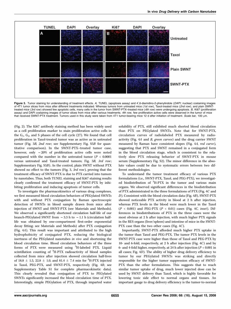
we first measured blood circulation behaviors of PEGylated SWNTswith and without PTX conjugation by Raman spectroscopicdetection of SWNTs in blood sample drawn from mice afterinjection of SWNT and SWNT-PTX (see Materials and Methods).We observed a significantly shortened circulation half-life of ourbranch-PEGylated SWNT from f3.3 h to f1.1 h (circulation half-life was obtained by one-compartment first-order exponentialdecay fitting; see Materials and Methods) after PTX conjugation(Fig. 4A). This result was important and attributed to the highhydrophobicity of conjugated PTX, reducing the biologicalinertness of the PEGylated nanotubes in vivo and shortening theblood circulation time. Blood circulation behaviors of the threeforms of PTX were measured using 3H-labeled PTX. Liquidscintillation counting of 3H-PTX radioactivity of blood samplescollected from mice after injection showed circulation half-livesof 18.8 F 1.5, 22.8 F 1.0, and 81.4 F 7.4 min for 3H-PTX injectedin Taxol, PEG-PTX, and SWNT-PTX, respectively (Fig. 4B ; seeSupplementary Table S1 for complete pharmacokinetic data).This clearly revealed that conjugation of PTX to PEGylatedSWNTs significantly increased the blood circulation time of PTX.Interestingly, simple PEGylation of PTX, through imparted water
solubility of PTX, still exhibited much shorted blood circulationthan PTX on PEGylated SWNTs. Note that for SWNT-PTX,circulation curves of radiolabeled PTX measured by radio-activity (Fig. 4A and B, green curves) and the drug carrier SWNTmeasured by Raman have consistent slopes (Fig. 4A, red curve),suggesting that PTX and SWNT remained in a conjugated formin the blood circulation stage, which is consistent to the rela-tively slow PTX releasing behavior of SWNT-PTX in mouseserum (Supplementary Fig. S2). The minor difference in the abso-lute values could be due to systematic errors between two dif-ferent methodologies.To understand the tumor treatment efficacy of various PTX
formulations (i.e., SWNT-PTX, Taxol, and PEG-PTX), we investigat-ed biodistribution of 3H-PTX in the tumor and various mainorgans. We observed significant differences in the biodistributionof PTX administrated in the three formulations of PTX (Fig. 4C andD). Consistent with the blood circulation data (Fig. 4B), SWNT-PTXshowed noticeable PTX activity in blood at 2 h after injection,whereas PTX levels in the blood were much lower in the Taxol(P < 0.001) and PEG-PTX (P < 0.01) cases (Fig. 4C, inset). Dif-ferences in biodistributions of PTX in the three cases were themost obvious at 2 h after injection, with much higher PTX signalsin the RES organs (liver/spleen) and intestine of mice in the SWNT-PTX case than the two other cases (Fig. 4C).Importantly, SWNT-PTX afforded much higher PTX uptake in
the tumor than Taxol and PEG-PTX. The tumor PTX levels in theSWNT-PTX case were higher than those of Taxol and PEG-PTX by10- and 6-fold, respectively, at 2 h after injection (Fig. 4C) and by6- and 4-fold higher, respectively, at 24 h after injection (P < 0.001 inall cases; Fig. 4D). The ability of higher drug delivery efficiency totumor by our PEGylated SWNTs was striking and directlyresponsible for the higher tumor suppression efficacy of SWNT-PTX than the other formulations. This suggests that to reachsimilar tumor uptake of drug, much lower injected dose can beused by SWNT delivery than Taxol, which is highly favorable forlowering toxic side effect to normal organs and tissues. Animportant gauge to drug delivery efficiency is the tumor-to-normal
Figure 3. Tumor staining for understanding of treatment effects. A, TUNEL (apoptosis assay) and 4¶,6-diamidino-2-phenylindole (DAPI ; nuclear) costaining imagesof 4T1 tumor slices from mice after different treatments indicated. Whereas tumors from untreated mice (1st row ), Taxol-treated mice (2nd row ), and plain SWNT-treated mice (3rd row ) showed few apoptotic cells, many cells in the tumor from SWNT-PTX–treated mice (4th row ) were undergoing apoptosis. B, Ki67 (proliferationassay) and DAPI costaining images of tumor slices from mice after various treatments. 4th row, few proliferation active cells were observed in the tumor of micethat received SWNT-PTX treatment. Tumors used in this study were taken from 4T1 tumor-bearing mice 12 d after initiation of treatment. Scale bar, 100 Am.
In vivo Drug Delivery with Carbon Nanotubes
www.aacrjournals.org 6655 Cancer Res 2008; 68: (16). August 15, 2008
organ/tissue PTX uptake ratios. We obtained significantly highertumor-to-normal organ/tissue PTX uptake ratios ( for tumor overliver, spleen, muscle, and other organs examined) in the case ofSWNT-PTX than Taxol and PEG-PTX (except at 2 h after injec-tion for spleen) at 2 and 24 h (Supplementary Table S2). This againmakes SWNT-PTX highly favorable for high tumor suppressionefficacy and low side effects.We investigated the biodistribution of SWNTs injected as SWNT-
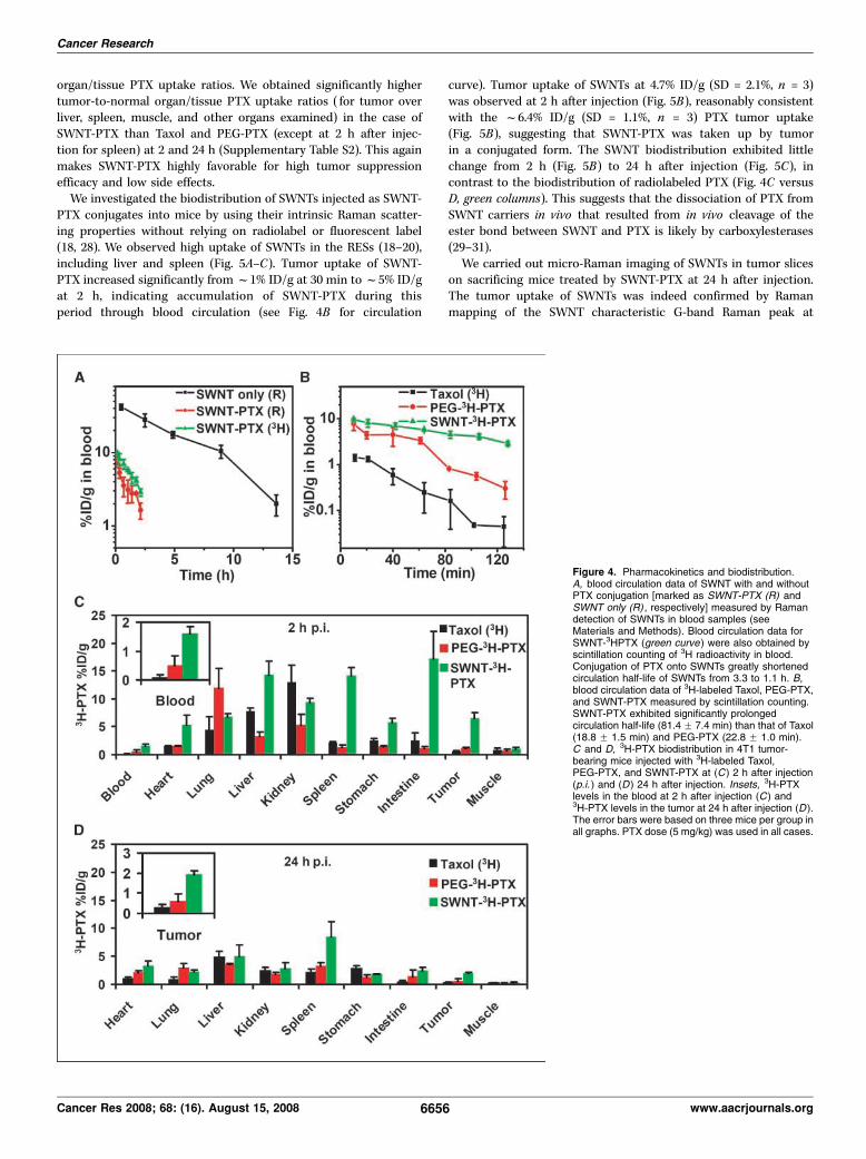
PTX conjugates into mice by using their intrinsic Raman scatter-ing properties without relying on radiolabel or fluorescent label(18, 28). We observed high uptake of SWNTs in the RESs (18–20),including liver and spleen (Fig. 5A–C). Tumor uptake of SWNT-PTX increased significantly fromf1% ID/g at 30 min tof5% ID/gat 2 h, indicating accumulation of SWNT-PTX during thisperiod through blood circulation (see Fig. 4B for circulation
curve). Tumor uptake of SWNTs at 4.7% ID/g (SD = 2.1%, n = 3)was observed at 2 h after injection (Fig. 5B), reasonably consistentwith the f6.4% ID/g (SD = 1.1%, n = 3) PTX tumor uptake(Fig. 5B), suggesting that SWNT-PTX was taken up by tumorin a conjugated form. The SWNT biodistribution exhibited littlechange from 2 h (Fig. 5B) to 24 h after injection (Fig. 5C), incontrast to the biodistribution of radiolabeled PTX (Fig. 4C versusD, green columns). This suggests that the dissociation of PTX fromSWNT carriers in vivo that resulted from in vivo cleavage of theester bond between SWNT and PTX is likely by carboxylesterases(29–31).We carried out micro-Raman imaging of SWNTs in tumor slices
on sacrificing mice treated by SWNT-PTX at 24 h after injection.The tumor uptake of SWNTs was indeed confirmed by Ramanmapping of the SWNT characteristic G-band Raman peak at
Figure 4. Pharmacokinetics and biodistribution.A, blood circulation data of SWNT with and withoutPTX conjugation [marked as SWNT-PTX (R) andSWNT only (R) , respectively] measured by Ramandetection of SWNTs in blood samples (seeMaterials and Methods). Blood circulation data forSWNT-3HPTX (green curve) were also obtained byscintillation counting of 3H radioactivity in blood.Conjugation of PTX onto SWNTs greatly shortenedcirculation half-life of SWNTs from 3.3 to 1.1 h. B,blood circulation data of 3H-labeled Taxol, PEG-PTX,and SWNT-PTX measured by scintillation counting.SWNT-PTX exhibited significantly prolongedcirculation half-life (81.4 F 7.4 min) than that of Taxol(18.8 F 1.5 min) and PEG-PTX (22.8 F 1.0 min).C and D, 3H-PTX biodistribution in 4T1 tumor-bearing mice injected with 3H-labeled Taxol,PEG-PTX, and SWNT-PTX at (C ) 2 h after injection(p.i. ) and (D ) 24 h after injection. Insets, 3H-PTXlevels in the blood at 2 h after injection (C ) and3H-PTX levels in the tumor at 24 h after injection (D ).The error bars were based on three mice per group inall graphs. PTX dose (5 mg/kg) was used in all cases.
Cancer Research
Cancer Res 2008; 68: (16). August 15, 2008 6656 www.aacrjournals.org
f1,580 cm�1 in the tumor with a spatial resolution of f1 Am(Fig. 5C, inset). To investigate the location of nanotubes in thetumor relative to the vasculature, we injected Alexa Fluor 488fluorescently labeled SWNTs into 4T1 tumor-bearing mice,sacrificed the mice, and collected the tumors for vasculature
staining and fluorescence imaging (Fig. 5D, right). We observedfluorescently labeled SWNTs both with and without overlaying withtumor vasculatures. This suggested that although most SWNTsseemed to be located in or near the tumor vasculature, a fraction ofnanotubes could leak through the tumor vessel into the tumor
Figure 5. SWNT biodistribution measured by Ramanspectroscopy. A to C, comparison of 3H-PTXbiodistribution and SWNT biodistribution in mice injectedwith SWNT-PTX(3H) at 30 min (A), 2 h (B), and 24 h (C )after injection. SWNT biodistribution was measuredby a Raman method (see Materials and Methods). Thedifferent biodistributions of PTX and SWNT carrier suggestrapid cleavage of ester bond for releasing of PTX fromSWNTs in vivo . Error bars in all graphs were based onthree mice per group. C, inset, a Raman image of thetumor slice. Strong SWNT G-band Raman signals atf1,580 cm�1 shift (green corresponds to high G-bandintensity) were observed in the tumor. Scale bar, 50 Am.D, confocal fluorescence images of tumor slices frommice injected with free Alexa Fluor 488 (AF488 ) dye (left )and Alexa Fluor 488–labeled SWNT (SWNT-AF488 ; right ).Tumor vasculature was stained by Cy3-anti-CD31.Alexa Fluor 488 fluorescence (green ) and vasculaturefluorescence (red ) were overlaid with optical images.Scale bar, 100 Am.
In vivo Drug Delivery with Carbon Nanotubes
www.aacrjournals.org 6657 Cancer Res 2008; 68: (16). August 15, 2008
interstitial space. As the control, no tumor retention of fluorescentdye was observed in mice injected with free Alexa Fluor 488 at thesame dose (Fig. 5D, left).Toxic side effects to normal organs and overall well being
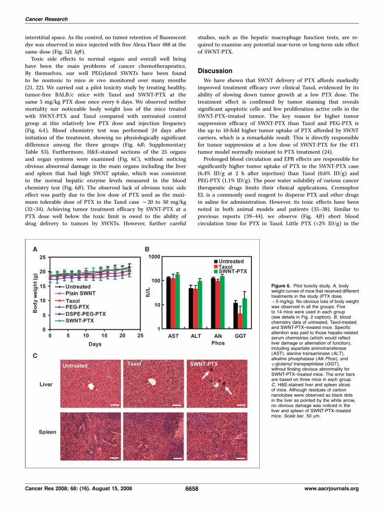
have been the main problems of cancer chemotherapeutics.By themselves, our well PEGylated SWNTs have been foundto be nontoxic to mice in vivo monitored over many months(21, 22). We carried out a pilot toxicity study by treating healthy,tumor-free BALB/c mice with Taxol and SWNT-PTX at thesame 5 mg/kg PTX dose once every 6 days. We observed neithermortality nor noticeable body weight loss of the mice treatedwith SWNT-PTX and Taxol compared with untreated controlgroup at this relatively low PTX dose and injection frequency(Fig. 6A). Blood chemistry test was performed 24 days afterinitiation of the treatment, showing no physiologically significantdifference among the three groups (Fig. 6B ; SupplementaryTable S3). Furthermore, H&E-stained sections of the 25 organsand organ systems were examined (Fig. 6C), without noticingobvious abnormal damage in the main organs including the liverand spleen that had high SWNT uptake, which was consistentto the normal hepatic enzyme levels measured in the bloodchemistry test (Fig. 6B). The observed lack of obvious toxic sideeffect was partly due to the low dose of PTX used as the maxi-mum tolerable dose of PTX in the Taxol case f20 to 50 mg/kg(32–34). Achieving tumor treatment efficacy by SWNT-PTX at aPTX dose well below the toxic limit is owed to the ability ofdrug delivery to tumors by SWNTs. However, further careful
studies, such as the hepatic macrophage function tests, are re-quired to examine any potential near-term or long-term side effectof SWNT-PTX.
Discussion
We have shown that SWNT delivery of PTX affords markedlyimproved treatment efficacy over clinical Taxol, evidenced by itsability of slowing down tumor growth at a low PTX dose. Thetreatment effect is confirmed by tumor staining that revealssignificant apoptotic cells and few proliferation active cells in theSWNT-PTX–treated tumor. The key reason for higher tumorsuppression efficacy of SWNT-PTX than Taxol and PEG-PTX isthe up to 10-fold higher tumor uptake of PTX afforded by SWNTcarriers, which is a remarkable result. This is directly responsiblefor tumor suppression at a low dose of SWNT-PTX for the 4T1tumor model normally resistant to PTX treatment (24).Prolonged blood circulation and EPR effects are responsible for
significantly higher tumor uptake of PTX in the SWNT-PTX case(6.4% ID/g at 2 h after injection) than Taxol (0.6% ID/g) andPEG-PTX (1.1% ID/g). The poor water solubility of various cancertherapeutic drugs limits their clinical applications. CremophorEL is a commonly used reagent to disperse PTX and other drugsin saline for administration. However, its toxic effects have beennoted in both animal models and patients (35–38). Similar toprevious reports (39–44), we observe (Fig. 4B) short bloodcirculation time for PTX in Taxol. Little PTX (<2% ID/g) in the
Figure 6. Pilot toxicity study. A, bodyweight curves of mice that received differenttreatments in the study (PTX dose,f5 mg/kg). No obvious loss of body weightwas observed in all the groups. Fiveto 14 mice were used in each group(see details in Fig. 2 caption). B, bloodchemistry data of untreated, Taxol-treated,and SWNT-PTX–treated mice. Specificattention was paid to those hepatic-relatedserum chemistries (which would reflectliver damage or alternation of function),including aspartate aminotransferase(AST ), alanine transaminase (ALT ),alkaline phosphatase (Alk Phos ), andg-glutamyl transpeptidase (GGT ),without finding obvious abnormality forSWNT-PTX–treated mice. The error barsare based on three mice in each group.C, H&E-stained liver and spleen slicesof mice. Although residues of carbonnanotubes were observed as black dotsin the liver as pointed by the white arrow,no obvious damage was noticed in theliver and spleen of SWNT-PTX–treatedmice. Scale bar, 50 Am.
Cancer Research
Cancer Res 2008; 68: (16). August 15, 2008 6658 www.aacrjournals.org
Taxol form remains circulating in the blood after only 11 min afterinjection at the current 5 mg/kg injected dose (Fig. 4B). PTX inTaxol is cleared from the blood and taken up by various organsespecially kidney and liver for rapid renal and fecal excretion withvery low tumor uptake (39–41, 43).Branched PEGylation of PTX via similar ester linkage as in
SWNT-PTX conjugates affords water solubility of PTX. How-ever, the blood circulation time is still short (PEG-PTX concen-tration diminished to <2% ID/g in f70 min after injection),albeit longer than Taxol. PEG-PTX remains a relatively smallmolecule that tends to be rapidly excreted via the kidney andrenal route, evidenced by the high kidney and urine signals ofradiolabeled PEG-PTX (data not shown). This leads to littleadvantage of PEGylation of PTX over Taxol in tumor uptake andtreatment efficacy, as found here and by previous PTX PEGyla-tion work (45).The water solubility of our SWNT-PTX formulation favors
prolonged blood circulation. Nevertheless, the high hydrophobicityof PTX reduces the hydrophilicity and biological inertness ofour branch PEG functionalized SWNTs, causing significantlyshortened blood circulation half-lives of the SWNT-PTX formula-tion (f1.1 h) compared with PEGylated SWNTs without PTXattachment (f3.3 h; Fig. 4A). The higher hydrophobicity led toincreased nonspecific protein absorption on the nanotube conju-gates, which accelerated the uptake by macrophages in RES organs.Compared with PEG-PTX, SWNT-PTX exhibits finite lengths(20–300 nm; mean, f100 nm; Supplementary Fig. S1), a factorthat favors long blood circulation because the average length ofthe nanotubes exceeds the threshold for renal clearance (46).Pharmacokinetics of materials with long blood circulation timesare typically desired for a drug delivery vehicle for tumor treatment(2, 47, 48) to favor high tumor accumulation from the circulat-ing blood through EPR effects. Note that our method of drugdelivery by PEGylated SWNTs should be readily applicable to awide range of hydrophobic or water-insoluble drugs. This couldlead to a general drug delivery strategy for potent but water-insoluble molecules.Tumor staining data clearly revealed apoptotic cells (Fig. 3A)
inside the tumor treated by SWNT-PTX. Nanotubes were observedin the tumor vasculature as well as leaked out of the vessels(Fig. 5D, left). Drug delivery to cancer cells through the tumorvessel walls and interstitial space is desired for high tumortreatment efficacy. SWNTs seemed to exhibit certain ability inovercoming these barriers, which could be related to the quasione-dimensional shape of these materials. An interesting featureof SWNTs is that the length of nanotubes (20–300 nm currently)could be controlled more precisely to span various size regi-mens. This could allow for investigation of length effect of one-dimensional materials to the tumor penetration and suppressionefficacy of drug complexes. This intriguing length effect will requiresystematic exploration in the future.PTX conjugation to PEGylated SWNTs clearly alters the
pharmacokinetics and biodistribution of PTX from Taxol andPEG-PTX. The up to 10 times high tumor uptake of the drugthrough SWNT-PTX and high tumor-to-normal organ/tissue PTXuptake ratios strongly favor high tumor killing efficacy and lowtoxicity to normal organs. High RES uptake is known for nano-materials in general. The high uptake of SWNT-PTX in RES organssuch as liver and spleen (18, 22) could be a cause of concernin terms of toxicity to these organs. Importantly, our biodistribu-tion studies revealed relatively low PTX levels in the RES organs
at later time points (2 and 24 h), differing from the SWNTbiodistribution (Fig. 5A–C). The difference between the biodistri-bution of SWNT and 3H-PTX (measured by radioactivity) suggestsrapid release of PTX from SWNT in the various organs and tissuesin vivo , resulted from in vivo cleavage of the ester linkage betweenPTX and PEGylated SWNT most likely by carboxylesterasesespecially those in the liver (29–31). We observed a significantPTX accumulation but proportionally lower SWNT signal in theintestine in the PTX-SWNT case at 30 min and 2 h after injection(Fig. 5A and B). We also detected strong PTX signal in the feceseven at only 30 min after injection (data not shown). These datasuggested that SWNT-PTX taken up by the RES organs wasdissociated via ester cleavage for release for excretion. Unlike theSWNT carriers, which are excreted gradually in weeks or evenmonths (22), the dissociated PTX drug molecules can be rapidlyexcreted via both feces and urine without causing noticeabletoxicity. Taken together, the uptake of drug-nanomaterial com-plexes by RES could serve as a scavenger system to eliminate toxicdrugs as well as carriers.The maximum tolerable dose of Taxol for BALB/c mice is
reported to be in the range of 20 to 50 mg/kg (32–34). Achievingtumor growth suppression by SWNT-PTX at 5 mg/kg dose onceevery 6 days suggests the promise of SWNT drug delivery foreffective cancer treatment with low side effects. More importantly,our water-soluble SWNT-PTX formulation is Cremophor-free.SWNTs have shown to be safe at least in mouse models (21, 22).The amount of SWNTs required to give 5 mg/kg PTX is onlyf4 mg/kg compared with f420 mg/kg Cremophor in the Taxolcase for the same PTX dose. Further, the same SWNT conjugationstrategy applies to many other water-insoluble drugs.SWNTs are highly promising for drug delivery due to several
factors. These materials can now be functionalized to a sufficientdegree to facilitate nearly complete excretion of SWNTs from miceover time (22). The chemical composition (purely carbon) ofcarbon nanotubes is among the safest in the inorganic nano-materials, many of which such as quantum dots have heavy metalcompositions. The unique one-dimensional structure and tunablelength provide an ideal platform to investigate size and shapeeffects in vivo . Lastly, unlike the conventional organic drug carriers,the intrinsic spectroscopic properties of nanotubes, includingRaman and photoluminescence, can provide valuable means oftracking, detecting, and imaging to understand the in vivo behaviorand drug delivery efficacy in vivo . Taken together, carbon nano-tubes are promising materials for potential multimodality cancertherapy and imaging.To our knowledge, this is the first successful report that carbon
nanotubes are used as drug delivery vehicles to achieve in vivotumor treatment efficacy with mice. This opens up further explo-ration of biomedical applications of novel carbon nanomaterialswith animals for potential translation into the clinic in the future.It is important to note that nanotube functionalization chemistrylargely determines the efficacy of SWNT drug delivery, as ourvarious other functionalization attempts have failed to give satis-factory treatment efficacy in other experiments. The treatmentefficacy of SWNT-based drug delivery vehicles can be furtherimproved because the current functionalization scheme is notyet fully optimized. Targeting ligands on nanotubes for tumor-targeted drug delivery is also expected to further enhance treat-ment efficacy. The one-dimensional shape and length of nanotubeseasily allow for targeting ligands, drugs, and multiple molecules forsynergistic effects.
In vivo Drug Delivery with Carbon Nanotubes
www.aacrjournals.org 6659 Cancer Res 2008; 68: (16). August 15, 2008
Disclosure of Potential Conflicts of Interest
No potential conflicts of interest were disclosed.
Acknowledgments
Received 4/19/2008; revised 5/27/2008; accepted 5/28/2008.
Grant support: NIH-National Cancer Institute Center for Cancer NanotechnologyExcellence Focused on Therapeutic Response at Stanford (H. Dai), Stanford Bio-XInitiative Grant, NIH-National Cancer Institute R01 grant CA135109-01, and StanfordGraduate Fellowship.
The costs of publication of this article were defrayed in part by the payment of pagecharges. This article must therefore be hereby marked advertisement in accordancewith 18 U.S.C. Section 1734 solely to indicate this fact.
We thank Josher Robinson for the help in dynamic light scattering experiments.
Cancer Research
Cancer Res 2008; 68: (16). August 15, 2008 6660 www.aacrjournals.org
References
1. Langer R. Drug delivery and targeting. Nature 1998;392:5–10.
2. Moghimi SM, Hunter AC, Murray JC. Long-circulatingand target-specific nanoparticles: theory to practice.Pharmacol Rev 2001;53:283–318.
3. Maeda H, Wu J, Sawa T, Matsumura Y, Hori K.Tumor vascular permeability and the EPR effect inmacromolecular therapeutics: a review. J Cont Rel 2000;65:271–84.
4. Gao XH, Cui YY, Levenson RM, Chung LWK, Nie SM.In vivo cancer targeting and imaging with semicon-ductor quantum dots. Nat Biotechnol 2004;22:969–76.
5. Bartlett DW, Su H, Hildebrandt IJ, Weber WA, DavisME. Impact of tumor-specific targeting on the biodis-tribution and efficacy of siRNA nanoparticles measuredby multimodality in vivo imaging. Proc Natl Acad SciU S A 2007;104:15549–54.
6. Iyer AK, Khaled G, Fang J, Maeda H. Exploiting theenhanced permeability and retention effect for tumortargeting. Drug Discov Today 2006;11:812–8.
7. Feazell RP, Nakayama-Ratchford N, Dai H, Lippard SJ.Soluble single-walled carbon nanotubes as longboatdelivery systems for platinum(IV) anticancer drugdesign. J Am Chem Soc 2007;129:8438–9.
8. Liu Z, Sun X, Nakayama N, Dai H. Supramolecularchemistry on water-soluble carbon nanotubes for drugloading and delivery. ACS Nano 2007;1:50–6.
9. Bianco A, Kostarelos K, Prato M. Applications ofcarbon nanotubes in drug delivery. Curr Opin Chem Bio2005;9:674–9.
10. Pantarotto D, Briand JP, Prato M, Bianco A.Translocation of bioactive peptides across cell mem-branes by carbon nanotubes. Chem Comm 2004;16–7.
11. Kam NWS, Dai H. Carbon nanotubes as intracellularprotein transporters: generality and biological function-ality. J Am Chem Soc 2005;127:6021–6.
12. Liu Y, Wu DC, Zhang WD, et al. Polyethylenimine-grafted multiwalled carbon nanotubes for secure non-covalent immobilization and efficient delivery of DNA.Angew Chem Int Ed 2005;44:4782.
13. Kam NWS, Liu Z, Dai H. Functionalization of carbonnanotubes via cleavable disulfide bonds for efficientintracellular delivery of siRNA and potent gene silenc-ing. J Am Chem Soc 2005;36:12492–3.
14. Liu Z, Winters M, Holodniy M, Dai H. siRNA deliveryinto human T cells and primary cells with carbon-nanotube transporters. Angew Chem Int Ed 2007;46:2023–7.
15. Kam NWS, Liu Z, Dai H. Carbon nanotubes asintracellular transporters for proteins and DNA: aninvestigation of the uptake mechanism and pathway.Angew Chem Int Ed 2006;45:577–81.
16. Kam NWS, O’Connell M, Wisdom JA, Dai H. Carbonnanotubes as multifunctional biological transportersand near-infrared agents for selective cancer celldestruction. Proc Natl Acad Sci U S A 2005;102:11600–5.
17. Welsher K, Liu Z, Daranciang D, Dai H. Selectiveprobing and imaging of cells with single walled carbonnanotubes as near-infrared fluorescent molecules. NanoLett 2008;8:586–90.
18. Liu Z, Cai W, He L, et al. In vivo biodistribution andhighly efficient tumour targeting of carbon nanotubes inmice. Nat Nanotechnol 2007;2:47–52.
19. Cherukuri P, Gannon CJ, Leeuw TK, et al. Mammalianpharmacokinetics of carbon nanotubes using intrinsicnear-infrared fluorescence. Proc Natl Acad Sci U S A2006;103:18882–6.
20. McDevitt MR, Chattopadhyay D, Kappel BJ, et al.Tumor targeting with antibody-functionalized, radio-labeled carbon nanotubes. J Nucl Med 2007;48:1180–9.
21. Schipper ML, Nakayama-Ratchford N, Davis CR, et al.Pilot toxicology study of single-walled carbon nano-tubes in a small sample of mice. Nat Nanotechnol 2008;3:216–21.
22. Liu Z, Davis C, Cai W, He L, Chen X, Dai H.Circulation and long-term fate of functionalized,biocompatible single-walled carbon nanotubes in miceprobed by Raman spectroscopy. Proc Natl Acad SciU S A 2008;105:1410–5.
23. Deutsch HM, Glinski JA, Hernandez M, et al.Synthesis of congeners and prodrugs. 3. Water-solubleprodrugs of taxol with potent antitumor activity. J MedChem 1989;32:788–92.
24. Janat-Amsbury MM, Yockman JW, Lee M, et al.Combination of local, nonviral IL12 gene therapy andsystemic paclitaxel treatment in a metastatic breastcancer model. Mol Ther 2004;9:829–36.
25. Janat-Amsbury MM, Yockman JW, Lee M, et al. Local,non-viral IL-12 gene therapy using lipopolymer ascarrier system combined with a water soluble sys-temic paclitaxel for cancer treatment. J Cont Rel 2005;101:273–85.
26. Inbal B, Cohen O, PolakCharcon S, et al. DAP kinaselinks the control of apoptosis to metastasis. Nature 1997;390:180–4.
27. Langford LA, Cooksley CS, DeMonte F. Comparisonof MIB-1 (Ki-67) antigen and bromodeoxyuridineproliferation indices in meningiomas. Hum Pathol1996;27:350–4.
28. Rao AM, Richter E, Bandow S, et al. Diameter-selective Raman scattering from vibrational modes incarbon nanotubes. Science 1997;275:187–91.
29. Satoh T, Hosokawa M. The mammalian carboxyles-terases: from molecules to functions. Annu RevPharmacol Toxicol 1998;38:257–88.
30. Guengerich FP, Peterson LA, Bocker RH. Cyto-chrome-P-450-catalyzed hydroxylation and carboxylic-acid ester cleavage of Hantzsch pyridine esters. J BiolChem 1988;263:8176–83.
31. Morgan EW, Yan B, Greenway D, Petersen DR,Parkinson A. Purification and characterization of tworat liver microsomal carboxylesterases (hydrolase A andB). Arch Biochem Biophys 1994;315:495–512.
32. Le Garrec D, Gori S, Luo L, et al. Poly(N -vinyl-
pyrrolidone)-block-poly(D,L-lactide) as a new polymericsolubilizer for hydrophobic anticancer drugs: in vitroand in vivo evaluation. J Control Release 2004;99:83–101.
33. Sugahara S, Kajiki M, Kuriyama H, Kobayashi TR.Paclitaxel delivery systems: the use of amino acid linkersin the conjugation of paclitaxel with carboxymethyldex-tran to create prodrugs. Biol Pharm Bull 2002;25:632–41.
34. Sharma A, Straubinger RM. Novel taxol formulations:preparation and characterization of taxol-containingliposomes. Pharm Res 1994;11:889–96.
35. Liebmann J, Cook JA, Mitchell JB. Cremophor EL,solvent for paclitaxel, and toxicity. Lancet 1993;342:1428.
36. Ellis AG, Crinis NA, Webster LK. Inhibition ofetoposide elimination in the isolated perfused rat liverby Cremophor EL and Tween 80. Cancer ChemotherPharmacol 1996;38:81–7.
37. Gelderblom H, Verweij J, Nooter K, Sparreboom A.Cremophor EL: the drawbacks and advantages ofvehicle selection for drug formulation. Eur J Cancer2001;37:1590–8.
38. Arbuck SG, Strauss H, Rowinsky E, et al. Areassessment of cardiac toxicity associated with Taxol.J Natl Cancer Inst Monogr 1993;15:117–30.
39. Eiseman JL, Eddington ND, Leslie J, et al. Plasmapharmacokinetics and tissue distribution of paclitaxel inCD2F1 mice. Cancer Chemother Pharmacol 1994;34:465–71.
40. Sparreboom A, van Tellingen O, Nooijen WJ, BeijnenJH. Tissue distribution, metabolism and excretion ofpaclitaxel in mice. Anticancer Drugs 1996;7:78–86.
41. Gangloff A, Hsueh WA, Kesner AL, et al. Estimationof paclitaxel biodistribution and uptake in human-derived xenografts in vivo with (18)F-fluoropaclitaxel.J Nucl Med 2005;46:1866–71.
42. Sparreboom A, van Tellingen O, Nooijen WJ, BeijnenJH. Nonlinear pharmacokinetics of paclitaxel in miceresults from the pharmaceutical vehicle Cremophor EL.Cancer Res 1996;56:2112–5.
43. Yeh TK, Lu Z, Wientjes MG, Au JL. Formulatingpaclitaxel in nanoparticles alters its disposition. PharmRes 2005;22:867–74.
44. Chen XY, Plasencia C, Hou YP, Neamati N. Synthesisand biological evaluation of dimeric RGD peptide-paclitaxel conjugate as a model for integrin-targeteddrug delivery. J Med Chem 2005;48:1098–106.
45. Li C, Yu D, Inoue T, et al. Synthesis and evaluation ofwater-soluble polyethylene glycol-paclitaxel conjugateas a paclitaxel prodrug. Anticancer Drugs 1996;7:642–8.
46. Soo Choi H, Liu W, Misra P, et al. Renal clearance ofquantum dots. Nat Biotechnol 2007;25:1165–70.
47. Allen TM, Hansen C, Rutledge J. Liposomes withprolonged circulation times: factors affecting uptake byreticuloendothelial and other tissues. Biochim BiophysActa 1989;981:27–35.
48. Gabizon A, Papahadjopoulos D. Liposome formula-tions with prolonged circulation time in blood andenhanced uptake by tumors. Proc Natl Acad Sci U S A1988;85:6949–53.