Download From: Aghalibrary.com
452
Download From: Aghalibrary.com
-
Upload
khangminh22 -
Category
Documents
-
view
0 -
download
0
Transcript of Download From: Aghalibrary.com
Download From: Aghalibrary.com

Mosby’s Textbook of Dental Nursing
For Elsevier
Content Strategist: Alison TaylorContent Development Specialist: Sally DaviesProject Manager: Julie TaylorDesigner/Design Direction: Miles HitchenIllustration Manager: Karen GiacomucciPhotographs: Max Wright Photography
Ma ry Mille r has ed it ed and writ ten severa l textbooks for den ta l nurses. She is curren t ly Genera l Manager o f the Eastman Denta l Hospita l Educa t ion Cent re , de livering pre -reg ist ra t ion programmes in den ta l nursing , denta l hygiene and den ta l therapy and is responsib le fo r provid ing pre - and post -reg ist ra t ion and o ther ce rt i ca ted courses as ou t lined in the GDC Scope of Pract ice fo r den ta l nurses.
Cr isp ia n Scu lly CBE, FMedSci, DSc, has been Presiden t of the In te rna t iona l Academy of Ora l Oncology, the European Associa t ion of Ora l Medicine , the Brit ish Socie ty for Ora l Medicine and the Brit ish Socie ty for Disab ility and Ora l Hea lth . He has writ ten and edited over 40 books, over 150 book chapte rs and over 1000 papers cited on MEDLINE. He is Founder and has been an Edito r of Ora l Oncology, Ora l Diseases and Medicina Ora l. He has medals from the Universit ies o f Helsinki, Sant iago de Composte la , and Granada ; Fe llowship of UCL; and Doctora tes from the Universit ies o f Athens, Granada , Helsinki and Pre toria .
Mosby’s Textbook of
Edinburgh London New York Oxford Philadelphia St Louis Sydney Toronto 2015
Mary Miller MA(Ed)
Genera l Manager, EDH Educa t ion Cent re , Denta l Nursing , Denta l Hygiene , Denta l Therapy, Eastman Denta l Hospita l, London, UK
Crispian Scully CBE MD PhD MDS MRCS BSc FDSRCS FDSRCPS FFDRCSI FDSRCSE FRCPathFMedSci FHEA FUCL DSc DChD DMed(HC) Dr HC
Co-Director, WHO Collabora t ing Cent re for Ora l Hea lth-Genera l Hea lthEmeritus Professor, University College London, London, UKVisit ing Professor a t Universit ies of Athens, BPP, Edinburgh , Granada and HelsinkiKing James IV Professor, Royal College of Surgeons, Edinburgh , UK
SECOND EDITION
Dental Nursing
The publisher’s
policy is to usepaper manufactured
from sustainable forests
© 2015 Elsevier Ltd. All rights reserved.
No part of this publication may be reproduced or transmitted in any form or by any means, electronic or mechanical, including photocopying, recording, or any information storage and retrieval system, without permission in writing from the publisher. Details on how to seek permission, further information about the Publisher’s permissions policies and our arrangements with organizations such as the Copyright Clearance Center and the Copyright Licensing Agency, can be found at our website: www.elsevier.com/permissions.
This book and the individual contributions contained in it are protected under copyright by the Publisher (other than as may be noted herein).
First edition 2011Second edition 2015
ISBN 978-0-7020-6237-7
NoticesKnowledge and best practice in this eld are constantly changing. As new research and experience broaden our understanding, changes in research methods, professional practices, or medical treatment may become necessary.
Practitioners and researchers must always rely on their own experience and knowledge in evaluating and using any information, methods, compounds, or experiments described herein. In using such information or methods they should be mindful of their own safety and the safety of others, including parties for whom they have a professional responsibility.
With respect to any drug or pharmaceutical products identi ed, readers are advised to check the most current information provided (i) on procedures featured or (ii) by the manufacturer of each product to be administered, to verify the recommended dose or formula, the method and duration of administration, and contraindications. It is the responsibility of practitioners, relying on their own experience and knowledge of their patients, to make diagnoses, to determine dosages and the best treatment for each individual patient, and to take all appropriate safety precautions.
To the fullest extent of the law, neither the Publisher nor the authors, contributors, or editors, assume any liability for any injury and/or damage to persons or property as a matter of products liability, negligence or otherwise, or from any use or operation of any methods, products, instructions, or ideas contained in the material herein.
Printed in China
Download From: Aghalibrary.com
v
Conten ts
Preface viiEditorial Board ixAcknowledgements xiHow to Use the Book xiii
Se ct io n ADental Nursing: Serving the Public 1Chapter 1 Dentistry and Regulation 3Chapter 2 The Dental Team 9Chapter 3 Dental Nursing Training,
Quali cations and Careers 17Chapter 4 Dental Nursing Practice and
Communication 25Chapter 5 Health Services: Ethics and
Governance 41
Se ct io n BDental Nursing: Protecting Patients and Staff 63Chapter 6 Workplace Hazards and Risk
Reduction 65Chapter 7 Infection and Control 89Chapter 8 Workplace Health and Safety 113
Se ct io n CDental Anatomy, Physio logy and Disease 119Chapter 9 Dental and Oral Anatomy
and Physiology 121Chapter 10 Dental and Oral Pathology
and Disease 143Chapter 11 Dental and Oral Imaging 165
Se ct io n DDental Disease , Prevention and Care 183Chapter 12 Dental and Oral Health
Promotion and Disease Prevention 185
Chapter 13 Dental and Oral Health Treatment Planning, Charting and Records 203
Chapter 14 Drugs in Dentistry 217Chapter 15 Pain and Anxiety Control 225Chapter 16 Dental Materials and
Equipment 243Chapter 17 Restorative Procedures
and Materials 257Chapter 18 Surgical Care 285Chapter 19 Surgical Care: Inpatients 299Chapter 20 Orthodontics 309Chapter 21 People with Vulnerability
or Special Needs 321Chapter 22 Minority Issues 335Chapter 23 Dental Emergencies 339
Se ct io n EHuman Disease , Prevention and Care 349Chapter 24 Human Anatomy and
Physiology 351Chapter 25 Human Pathology and
Diseases 369Chapter 26 Health Promotion 391Chapter 27 First Aid and Medical
Emergencies 401
Abbreviations 421Index 429
Download From: Aghalibrary.com

vii
Pre face
This textbook can be used as an aide-memoire or qualif ed dental nurses as well as or student dental nurses. The f nal stimulus to produce the f rst hugely success ul edition o this book arose rom the introduc-tion by the General Dental Council o the UK (GDC) o the requirement or dental care pro essionals (DCPs)to register. According to the GDC, the purpose or aim o their education is to produce a caring, knowledge-able, competent and skil ul DCP who is able, on quali-f cation, to accept pro essional responsibility or their role in the e ective and sa e care o patients. In realising this aim, the GDC applies the ollowing principles:
• That those quali ying as DCPs should berequired to attain the highest standards in termso knowledge and understanding, skills(including clinical and laboratory skills), and pro essional attributes, in particular recognition o their obligation to practise in the bestinterests o patients at all times.
• That DCP students should be provided withthe high-quality learning opportunities andexperiences necessary to enable them to achievethose standards, including the opportunity,where appropriate, to undertake clinical andlaboratory procedures, and acquire competenceacross a range o skills.
• That learning opportunities and experiencesshould be underpinned by adequate andappropriate support, including educational,clinical and laboratory support.
• That learning opportunities and experiencesin biomedical sciences, clinical and laboratorysubjects should be integrated over the courseo the programme.
• That learning opportunities and experiencesshould be designed to encourage a questioning,scientif c and sel -critical approach to thepractice o dentistry, and to oster the intellectualskills required or uture personal andpro essional development.
• That learning opportunities and experiencesshould enable students to develop anunderstanding o audit and clinicalgovernance.
• That learning opportunities and experiencesshould enable students o the pro essionscomplementary to dentistry to work and train aspart o the dental team.
• That learning opportunities and experiencesshould prepare students adequately or thetransition to their work role in relation to thepractice o dentistry.
• That student progress is e ectively monitored toensure that only those who comply with relevanthealth and conduct requirements are allowed tocomplete the programme.The aims o this second edition have been to
update the text to comply with the above and with the new National Examining Board or Dental Nurses (NEBDN) National Diploma in Dental Nursing Cur-riculum (March 2012), and to enhance urther the material – both text and illustrations – and user- riendliness. Additions include coverage o the new NEBDN curriculum; the new GDC Standards or the Dental Team; new GDC advice on social media; the new Equality Act; and aspects o anatomy, charting, drug allergy, minority groups, f re sa ety, security and vulnerable groups. In our task we have been impressed with the scope o the educational needs, have sought advice rom a number o sources and attempted to keep abreast o the rapidly changing legislation and guidance acing all dental pro essionals. There ore, we are most grate ul to our Editorial Board advisors who have ably assisted in many ways; however, any errors that might remain are ours. All weblinks cited in the text were active at the time o writing.
Mary MillerCrispian Scully
London 2015
Download From: Aghalibrary.com
ix
Ed itoria l Board
Sarah Bain , Director, Bristol School of Dental Care Professionals, University Hospitals, Bristol, UKFion a Beach am, Head Dental Nurse, Division of Oral Surgery and Dental Medicine, Eastman Dental Hospi-tal, University College London Hospitals, London, UKLesley Derry, Associate Director of Education and Standards, British Dental Association, London, UKDaljit Gill, Consultant Orthodontist, Great Ormond Street NHS Foundation Trust and Eastman Dental Hospital, University College London Hospitals, London, UKTin a Gorman , Director of Nursing, Dublin Dental University Hospital, Dublin, IrelandMark Gr i f th s, Honorary Research Fellow, University of Bristol; Visiting Professor, Eastman Dental Institute, London, UKCh ar lo tte Leigh , Dental practitioner, London, UKJan e Luker , Consultant in Dental and Maxillofacial Radiology, Bristol Dental Hospital; Deputy Medical Director, University Hospitals Bristol; Dental Post-graduate Dean for Health Education Southwest, Bristol, UK
Dan iel McAlon an , Head of Health & Safety and Acting Associate Director of Advisory Services, British Dental Association, London, UKGlen n Newton , Tutor Dental Nurse, Specialist Services Division, Birmingham Community Health-care NHS Trust, Birmingham Dental Hospital, UKJoan n e Rich ardson , Dental Matron, Guy’s and St Thomas’ Hospital, London, UKSaman th a Salaver , Head of Dental Nursing, Guy’s and St Thomas’ Hospital, London, UKCh r istoph er Tredwin , Professor of Restorative Den-tistry, Head of School, Plymouth University Peninsula Dental School, UK
Download From: Aghalibrary.com

xi
Acknowledgements
The illustrations and text listed below have been reproduced or adapted with permission rom the ollowing publications.Black G V, Black’s classif cation o caries lesions. Source: Jessica R. Martin, Wikimedia Commons (Fig. 10.11)Collins W J, Walsh T, Figures K, 1998, A Handbook or Dental Hygien-ists, 4th edition, Butterworth–Heinemann (Figs 9.16 and 9.17)Department o Health, 1997, The Caldicott Committee Report on the review o patient-identif able in ormation (Box 5.8)Department o Health, 2009, Health Technical Memorandum (HTM) 01-05: Decontamination in primary care dental practices (http:/ /webarchive.nationalarchives.gov.uk/20130107105354/http:/ /www.dh .gov.uk/en/Publicationsandstatistics/Publications/PublicationsPolicy AndGuidance/DH_109363), Crown Copyright (Fig. 7.6; Table 7.4)Department o Health, 2013, Decontamination: Health Technical Memorandum (HTM) 01-05: Decontamination in primary care dental practices (https:/ /www.gov.uk/government/uploads/system/uploads/attachment_data/f le/170689/HTM_01-05_2013.pd ), Crown Copy-right (Figs 7.6 and 7.7)Department o Health, 2013, The health and care system rom April 2013 (http://webarchive.nationalarchives.gov.uk/20130805112926/http:/ /healthandcare.dh.gov.uk/system-overview-diagram/), Crown Copyright (Fig. 5.2)Department o Health, 2013, Patient agreement to investigation or treatment (http://webarchive.nationalarchives.gov.uk/20130107105354/h ttp:/ /www.dh .gov.uk/prod_consum _dh /groups/dh_digitalassets/ @dh/@en/documents/digitalasset/dh_4019034.pd ), Crown Copyright (Appendix 5.1)Drake R, Vogl A W, Mitchell A, 2009, Gray’s Anatomy or Students, 2nd edition, Saunders (Figs 9.1, 9.3, 9.6, 9.8, 9.11–9.14, 9.20, 24.4–24.7, 24.10, 24.11)Florida Probe, Periodontal chart © Florida Probe Corporation (www . oridaprobe.com/) (Fig. 13.9)HSE Health and Sa ety Law – What you need to know (lea et), Crown Copyright (Fig. 8.1)Jevon P, 2006, Emergency Care and First Aid or Nurses, Churchill Livingstone (Figs 27-02AB, 27-02D–F, 27-03)Medical Protection Society, 13 March 2012, What are the criteria or training a dental nurse to assist with sedation cases? Can I train my own nurse? www.dentalprotection.org/uk/AskDPL/nurse_sedation _training. Reproduced by permission o Dental Protection Ltd.MHRA Yellow Card (https://yellowcard.mhra.gov.uk/), MHRA and Crown Copyright (Fig. 14.3)Millett D, Welbury R, 2005, Clinical Problem Solving in Orthodon-tics and Paediatric Dentistry, 2nd edition, Churchill Livingstone (Fig. 10.16)Resuscitation Council (UK), 2013, Minimum equipment list or car-diopulmonary resuscitation: Primary dental care (adapted) (Table 27.3, Airway and Breathing; Circulation)Rhind J, Greig J, 2002, Riddle’s Anatomy and Physiology Applied to the Health Pro essions, 7th edition, Churchill Livingstone (Fig. 9.15)
Scottish Government, www.scotland.gov.uk/Publications/2008/08/interimdresscode (Box 4.7)Scully C, 2010, Medical Problems in Dentistry, 6th edition, Elsevier (Tables 26.1, 26.3–26.5, 27.2; Box 6.5)Scully C M, Flint S, 1989, An Atlas o Stomatology, Martin Dunitz, London (Figs 6.8, 9.75, 9.78, 9.136 and 15.29 with permission rom Taylor & Francis; Figs 25.5, 10.13, 10.14, 12.3 and 25.2 respectively in this volume)Scully C, Flint S F, Bagan J V, Porter S R, Moos K, 2010, Oral and Maxillo acial Diseases, 4th edition, In orma (Figs 10.2, 10.15, 10.22, 10.24, 10.25, 20.8)Scully C, Wilson N, 2007, Culturally Sensitive Oral Health Care, Quintessence (Table 22.1)Shahid M, Nunhuck A, 2008, Crash Course Physiology, Mosby Ltd (Figs 24.8, 24.9, 24.13, 24.14)Standring S (ed), 2008, Gray’s Anatomy, 40th edition, Churchill Livingstone (Figs 9.5, 9.7, 9.9, 9.18, 9.19)Trevisi H, 2007, SmartClip™ Sel -Ligating Appliance System, Mosby Ltd (Fig. 20.2)Whaites E, 2006, Essentials o Dental Radiography and Radiology, 4th edition, Churchill Livingstone (Figs 10.21, 11.2–11.5, 20.1)
Fig. 10.12 has been reproduced courtesy o Dr Dimitris Malamos.
Brand names and products have been reproduced with permission rom:Ag a (Fig. 11.1)Carl Zeiss Ltd (Fig. 17.38)Clinell/GAMA Healthcare Ltd (Fig. 7.1)Colgate Palmolive (Figs 12.6, 12.10A,B,C,D, 12.11, 12.12, 12.14)GlaxoSmithKline (Fig. 12.13). CORSODYL is a registered trade mark o the GlaxoSmithKline group o companies. Copyright o the Corso-dyl Mint Mouthwash image (Fig. 12.13) is owned by the GlaxoSmith-Kline group o companies.Johnson & Johnson Ltd, Listerine (Fig. 12.11)Medline Industries Inc. (Fig. 7.5)Parkell Inc., Gentle-Pulse™ Pulp Vitality Tester (Fig. 17.37)Pearl Drops (Fig. 14.2)Periproducts Ltd (Figs 12.8AB)Philips, Philips HeartStart OnSite (HS1) (Fig. 27.6)Polyco (Fig. 16.1)Premier Healthcare & Hygiene Ltd (Fig. 7.5)Procter & Gamble, Braun, Oral-B (Figs 12.9, 12.10I,J)TePe Munhygienprodukter AB (Figs 12.5, 12.7)Wisdom Toothbrushes Ltd (Figs 12.8C, 12.10E,F,G,H)
Download From: Aghalibrary.com
xiii
How to Use the Book
This textbook has been written speci cally for pre-registration dental nurses and incorporates all aspects of the National Examining Board for Dental Nurses (NEBDN) pre-registration syllabus. Some additional related information has also been included, which we believe will help the student dental nurse care better for their patients.
The text is accompanied by several features to engage the reader and help them consider the practical aspects of the theoretical learning:
• Terms to learn: de nitions of terms used in thetext that may be unfamiliar to the reader. These
terms are given in bold at the rst mention in the book.
• Key points: key messages that the reader shouldalways remember.
• Identify and learn: tasks that aim to encouragethe reader to transfer their learning into theirworkplace by inviting them to look for variousitems and understand how they work or whatthey are used for.
• Find out more: hints on where to look forfurther information or perhaps to nd outmore about certain topics for greaterunderstanding.
Download From: Aghalibrary.com
DENTAL NURSING: SERVING THE PUBLICChapter 1 Dent ist ry and Regula t ion 3
Chapte r 2 The Denta l Team 9
Chapte r 3 Denta l Nursing Tra in ing , Qualif ca t ions and Careers 17
Chapte r 4 Denta l Nursing Pract ice and Communica t ion 25
Chapte r 5 Hea lth Services: Eth ics and Governance 41
Sect ion A

Chapter 1 Dentistry and Regulation
CHAPTER POINTS• Regulation• Standards• Bodies other than GDC particularly relevant to
dentistry
Regulation of DentistryThe General Dental Council (GDC) is the body that regulates dentistry. All healthcare professionals are subject to regulation and the GDC itself, along with the other professional bodies such as the General Medical Council (GMC), is overseen by the Pro essional Standards Authority or Health and Social Care (PSA: previously known as the Council for Healthcare Regu-latory Excellence [CHRE]) (see www.hpc-uk.org/about regist ra t io n / regu la to rs/ th eco u n cilfo rh ealth care regulatoryexcellence/).
The PSA scrutinises and oversees the work of the nine health and care regulators, shares good practice and knowledge with these regulators, conducts research and audits the regulator’s ‘ tness to practise’ process.
I a registrant’s f tness to practise is challenged, their GDC registration and the licence to work pro essionally may be under threat.
The Regulatory BodiesIn the UK all healthcare professionals are accountable to the PSA – a UK-wide organisation that oversees the regulators of healthcare professionals:
• General Dental Council (GDC)– regulates all dental professionals
• General Medical Council (GMC)– regulates doctors
• Nursing and Midwifery Council (NMC)– regulates nurses, midwives and specialist community public health nurses
• Health Professions Council (HPC)– regulates 14 professions (art therapists, biomedical scientists, chiropodists/podiatrists, clinical scientists, dieticians, occupational therapists, operating department practitioners, orthoptists, paramedics, physiotherapists,
In t ro d u ct io n
Dentistry had its origins in barbery (from Latin barba, ‘beard’) – people whose occupation was mainly to cut, dress, groom, style and shave hair. In 1462 the Barbers’ Company of London gained their rst charter, which speci cally permitted them to draw (extract) teeth. In 1540 the Barbers’ Company merged with the Fellow-ship of Surgeons to form the Company of Barber-Surgeons but no surgeon was permitted to practise barbery and barbers were restricted in surgery to extracting teeth. In the 1800s dentists, barbers and sur-geons separated as professions. Dentistry has devel-oped by leaps and bounds since those days of the ‘barber-surgeons’, with enormous advances in tech-nology (Figure 1.1) and in the dental professional healthcare workers involved.
Modern dentistry is all about teamwork and involves a clinician, often the dentist, together with a group of dental care professionals (DCPs), which may include the:
• dental nurse• dental technician• dental therapist• dental hygienist• orthodontic therapist• clinical dental technician.

4 CHAPTER • 1 • Mosby’s Textbook of Dental Nursing
The PSA is itself accountable to Parliament and its mission is to protect the public by:
• Helping regulatory bodies become better regulators
• Setting and driving standards up for professional regulation
• Ensuring greater harmonisation of regulatory practice and outcomes
• Anticipating any problems in the future.The PSA ful ls its mission by:
• Reviewing the nal stages of cases in which a healthcare professional’s tness to practise has been challenged
• Monitoring how the regulators carry out their functions
• Promoting good practice• Advising health ministers• In uencing national and international policy on
health regulation• Promoting data protection and freedom of
information (see Chapter 5).
The Ge ne ra l De nta l Counc ilThe GDC is the organisation that regulates all dental professionals training and working in the UK. Dental professionals include dentists and all dental care professionals.
The aims of the GDC are to:
• Protect patients• Promote the con dence of the patients and
public in all dental professionals• Assure the quality of dental education for all
dental professionals in the UK• Ensure dental professionals keep their
knowledge up to date• Help patients with complaints.
The GDC achieves these aims by setting the stand-ards and principles for ethical dental practice in the UK (see below, GDC Standards for the Dental Team).
practitioner psychologists, prosthetists and orthotists, radiographers, speech and language therapists)
• General Optical Council (GOC)– regulates dispensing opticians and optometrists
• General Chiropractic Council (GCC)– regulates chiropractors
• General Osteopathic Council (GOsC)– regulates osteopaths
• Royal Pharmaceutical Society of Great Britain (RPSGB)– regulates pharmacists
• Pharmaceutical Society of Northern Ireland (PSNI)– regulates pharmacists.
Each of these regulators maintains ‘registers’, which contain names and other details of healthcare profes-sionals who are considered t to practise in the UK. The regulators’ functions include:
• Setting standards of behaviour, education and ethics
• Dealing with concerns about professionals who are un t to practise because of poor health, misconduct or poor performance. Regulators can remove people from their register and therefore prevent them from practising.
Fig u re 1.1 An early dental chair.
Registe rs for den ta l p rofessiona ls in the UKThe GDC maintains ‘registers’ for dentists and dental care professionals:
• Dentists – the Dentists Register
• Dental care professionals – the Dental Care Professionals Register.
The registers include the names of all the dentists and dental care professionals who are registered to practise in the UK,

5Dentistry and Regulation
regardless of whether they work in the National Health Service (NHS), private practice or any other form of practice. Those who are registered are called registrants. Thus all dental nurses must be registered with the GDC’s Dental Care Professionals (DCPs) Register.
Dental professionals without a recognised UK quali cation may be eligible to have their quali cation assessed. If the assessment is successful, their name will be entered onto the DCPs Register. Assessment is available for the following:
• Those with a formal quali cation from an EEA (European Economic Area) member state
• Those with a formal quali cation from overseas.
GDC Standards for the Dental TeamThe GDC guidance document on ethical practice, Standards or the Dental Team (2013), applies to the whole dental team. It explains the standards that the GDC expects of dental professionals – all of whom have a responsibility to work to its nine key principles of ethical practice. New registrants get a copy of the Standards or the Dental Team guidance when they join the GDC Register.
Key princip les o f e th ica l p ract ice (from Standards for the Dental Team , GDC, 2013)
1. Put patients’ interests rst
2. Communicate effectively with patients
3. Obtain valid consent
4. Maintain and protect patients’ information
5. Maintain your professional knowledge and competence
6. Work with colleagues in a way that is in patients’ best interests
7. Maintain, develop and work within your professional knowledge and skills
8. Raise concerns if patients are at risk
9. Make sure your personal behaviour maintains patients’ con dence in you and the dental profession
Since 2013, these nine principles are to be fol-lowed by all dental professionals, including trainees, both at and outside o work. The GDC guidance sets out dental professionals’ responsibility to put patients’ interests rst and protect patients by, for example, maintaining GDC registration, working only within the scope of their knowledge and keeping accurate patient records. It also sets out the importance of treating patients with dignity and respect, being non-discriminatory, and recognising the patient’s
responsibility for making decisions, and giving them all the information they need to make decisions.
All registrants must follow this guidance, which is summarised here from Standards or the Dental Team (GDC, 2013):
1 Put patients’ interests rst1.1 Listen to your patients.1 .2 Treat every patient with dignity and respect
at all times.1 .3 Be honest and act with integrity.1 .4 Take a holistic and preventive approach to
patient care which is appropriate to the individual patient.
1 .5 Treat patients in a hygienic and safe environment.
1 .6 Treat patients fairly, as individuals and without discrimination.
1 .7 Put patients’ interests before your own or those of any colleague, business or organisation.
1 .8 Have appropriate arrangements in place for patients to seek compensation if they suffer harm.
1 .9 Find out about laws and regulations that affect your work and follow them.
2 Communicate effectively with patients2.1 Communicate effectively with patients
– listen to them, give them time to consider information and take their individual views and communication needs into account.
2 .2 Recognise and promote patients’ rights to and responsibilities for making decisions about their health priorities and care.
2 .3 Give patients the information they need, in a way they can understand, so that they can make informed decisions.
2 .4 Give patients clear information about costs.3 Obtain valid consent
3.1 Obtain valid consent before starting treatment, explaining all the relevant options and the possible costs.
3 .2 Make sure that patients (or their representatives) understand the decisions they are being asked to make.
3 .3 Make sure that the patient’s consent remains valid at each stage of investigation or treatment.
4 Maintain and protect patients’ information4.1 Make and keep contemporaneous, complete
and accurate patient records.

6 CHAPTER • 1 • Mosby’s Textbook of Dental Nursing
8.3 Make sure if you employ, manage or lead a team that you encourage and support a culture where staff can raise concerns openly and without fear of reprisal.
8 .4 Make sure if you employ, manage or lead a team that there is an effective procedure in place for raising concerns, that the procedure is readily available to all staff and that it is followed at all times.
8 .5 Take appropriate action if you have concerns about the possible abuse of children or vulnerable adults.
9 Make sure your personal behaviour maintains patients’ con dence in you and the dental profession9.1 Ensure that your conduct, both at work and
in your personal life, justi es patients’ trust in you and the public’s trust in the dental profession.
9 .2 Protect patients and colleagues from risks posed by your health, conduct or performance.
9 .3 Inform the GDC if you are subject to criminal proceedings or a regulatory nding is made against you anywhere in the world.
9 .4 Cooperate with any relevant formal or informal inquiry and give full and truthful information.
Find out moreReaders should access the GDC guidance a t www.gdc-uk.org /den ta lprofessiona ls/standards/pages/home.aspx and read the fu lle r de ta ils, though some aspects a re dea lt with more fu lly be low or e lsewhere in th is text . The GDC makes it clea r tha t den ta l professiona ls should make sure tha t they just ify the t rust p laced in them by the ir pa t ien ts, the public and co lleagues, by act ing honest ly and fa irly in a ll the ir professiona l and personal dea lings.
Bodies Other than GDC Particularly Relevant to Dentistry
See Table 1.1 for a list of other bodies relevant to dentistry.
Find out moreAll den ta l nurses should have a copy of the Standards for the Dental Team bookle t as well as addit iona l support ing documents. You can download them from the GDC website a t www.gdc-uk.org/den ta lprofessiona ls/standards/pages/standards.aspx.
4 .2 Protect the con dentiality of patients’ information and use it only for the purpose for which it was given.
4 .3 Only release a patient’s information without their permission in exceptional circumstances.
4 .4 Ensure that patients can have access to their records.
4 .5 Keep patients’ information secure at all times, whether your records are held on paper or electronically.
5 Have a clear and effective complaints procedure5.1 Make sure that there is an effective
complaints procedure readily available for patients to use, and follow that procedure at all times.
5 .2 Respect a patient’s right to complain.5 .3 Give patients who complain a prompt and
constructive response.6 Work with colleagues in a way that is in
patients’ best interests6.1 Work effectively with your colleagues and
contribute to good teamwork.6 .2 Be appropriately supported when treating
patients.6 .3 Delegate and refer appropriately and
effectively.6 .4 Only accept a referral or delegation if you
are trained and competent to carry out the treatment and you believe that what you are being asked to do is appropriate for the patient.
6 .5 Communicate clearly and effectively with other team members and colleagues in the interests of patients.
6 .6 Demonstrate effective management and leadership skills if you manage a team.
7 Maintain, develop and work within your professional knowledge and skills7.1 Provide good quality care based on current
evidence and authoritative guidance.7 .2 Work within your knowledge, skills,
professional competence and abilities.7 .3 Update and develop your professional
knowledge and skills throughout your working life.
8 Raise concerns if patients are at risk8.1 Always put patients’ safety rst.8 .2 Act promptly if patients or colleagues are at
risk and take measures to protect them.
7Dentistry and Regulation
Table 1.1 Bodies o ther than GDC part icu larly re levant to den t ist ry
Body Main funct ions URL
British Dental Association A national professional association for dentists
www.bda.org/
British Association of Dental Nurses
A national professional association for dental nurses
www.badn.org.uk/
Royal Colleges of Surgeons Professional associations for dentists and DCPs which also offer education, examinations and higher quali cations
www.rcseng.ac.uk/fdswww.rcsed.ac.uk/examinations/dental.aspxwww.rcpsg.ac.uk/dentistry.aspx
Chapter 2 The Dental Team
CHAPTER POINTS• GDC scope o practice• Direct access to dental care pro essionals
detail on the roles of dental nurses within their scope of practice in Chapter 4.
GDC Scope of PracticeThe General Dental Council states:
The Dentists Act 1984 makes it an offence for a person who is not a registered dentist or a registered dental care professional to practise dentistry, or hold themselves out – whether directly or by implication – as practising or as being prepared to practise dentistry.
By law, all registrants are individually accountable to the GDC, and dentists are additionally accountable as leaders of the team.
Clinical dental care can be provided only by GDC-registered:
• dentists• dental therapists• orthodontic therapists• dental hygienists• clinical dental technicians (CDTs).
The guidance on scopes of practice of various members outlined in Table 2.1 and given in more detail below is taken from the GDC document Scope of Practice (2013).
Dental NursesDental nurses are registered dental professionals who provide clinical and other support to registrants and patients. Dental nurses who are trained, competent and indemni ed can undertake the following:
• prepare and maintain the clinical environment, including the equipment
• carry out infection prevention and control procedures to prevent physical, chemical and microbiological contamination in the surgery or laboratory
In t ro d u ct io n
A clinician, often the dentist, together with a team make up the group of dental care professionals (DCPs), which may include (Chapter 1) the:
• dental nurse• orthodontic therapist• dental hygienist• dental therapist• dental technician• clinical dental technician.
Dental nurses must hold an appropriate quali ca-tion to register with the General Dental Council (GDC). Trainee dental nurses can work in practice without a quali cation, but they must be enrolled on an accredited course with NEBDN (National Examina-tion Board for Dental Nurses). The scope of practice of all members of the dental team is given in detail in the GDC document Scope of Practice (2013) but is outlined below and summarised in Table 2.1.
Dental nurses’ main roles are to provide chairside assistance to the dentist or other team members, as well as instrument layout, sterilising, mixing dental materials and administration. Quali ed dental nurses may also undertake further training to allow them to provide oral hygiene instruction, take radiographs and assist with general anaesthetic and conscious sedation (see Table 2.1 and see also www.bda.org/den tists/advice/career/working-in-th-uk/den tal-care -professionals.aspx).
Here the scopes of practice of all members of the dental professional team are outlined. There is more

10 CHAPTER • 2 • Mosby’s Textbook of Dental Nursing
Table 2.1 Genera l scopes o p ract ice (a lso see t ext )
Denta l p ro essional Genera l def n it ion o clin ica l p ract ice
Dentists Registered dental professionals who can carry out all of the treatments listed in the Scope of Practice (2013) document*
Dental nurses Registered dental professionals who provide clinical and other support to registrants and patients
Dental therapists Registered dental professionals who carry out certain items of dental treatment direct to patients or under prescription from a dentist
Orthodontic therapists Registered dental professionals who carry out certain parts of orthodontic treatment under prescription from a dentist
Dental hygienists Registered dental professionals who help patients maintain their oral health by preventing and treating periodontal disease and promoting good oral health practice. They carry out treatment direct to patients or under prescription from a dentist
Dental technicians Registered dental professionals who make dental devices to a prescription from a dentist or clinical dental technician. They also repair dentures direct to members of the public
Clinical dental technicians Registered dental professionals who provide complete dentures direct to patients and other dental devices on prescription from a dentist. They are also quali ed dental technicians
*General Dental Council, Scope of Practice, September 2013; www.gdc-uk.org/dentalprofessionals/standards/documents/scope% 20of% 20practice% 20september% 202013% 20(3).pdf
• record dental charting and oral tissue assessment carried out by other registrants
• prepare, mix and handle dental bio-materials• provide chairside support to the operator.
During treatment, dental nurses can:
• keep full, accurate and contemporaneous patient records
• prepare equipment, materials and patients for dental radiography
• process dental radiographs• monitor, support and reassure patients• give appropriate patient advice• support the patient and their colleagues if there
is a medical emergency• make appropriate referrals to other health
professionals.Additional skills dental nurses could develop
include:
• further skills in oral health education and oral health promotion
• assisting in the treatment of patients who are under conscious sedation (see Chapter 15)
• further skills in assisting in the treatment of patients with special needs
• further skills in assisting in the treatment of orthodontic patients
• intra- and extra-oral photography• pouring, casting and trimming study models
• shade taking• tracing cephalographs.
Additional skills carried out on prescription from, or under the direction of, another registered dental professional:
• taking radiographs• placing rubber dam• measuring and recording plaque indices• removing sutures after the wound has been
checked by a dentist• constructing occlusal registration rims and
special trays• repairing the acrylic component of removable
appliances• applying topical anaesthetic to the prescription
of a dentist• constructing mouthguards and bleaching trays to
the prescription of a dentist• constructing vacuum-formed retainers to the
prescription of a dentist• taking impressions to the prescription of a
dentist or a CDT (where appropriate)• dental nurses can apply uoride varnish either
on prescription from a dentist or direct as part of a structured dental health programme.
Dental nurses do not diagnose disease or plan treatment. All other skills are reserved to one or more of the other dental professional groups described in this chapter.
11The Dental Team
• removing sutures after the wound has been checked by a dentist.Orthodontic therapists do not:
• modify prescribed archwires• give local analgesia• remove subgingival deposits• re-cement crowns• place temporary dressings• diagnose disease• treatment plan.as these tasks are reserved to dental hygienists, dental therapists or dentists.
Orthodontic therapists do not carry out labora-tory work other than that listed above as that is reserved to dental technicians and clinical dental technicians.
Dental HygienistsDental hygienists are registered dental professionals who help patients maintain their oral health by pre-venting and treating periodontal disease and pro-moting good oral health practice. They carry out treatment direct to patients or under prescription from a dentist.
Dental hygienists who are trained, competent and indemni ed can undertake the following:
• provide dental hygiene care to a wide range of patients
• obtain a detailed dental history from patients and evaluate their medical history
• carry out a clinical examination within their competence
• complete periodontal examination and charting and use indices to screen and monitor periodontal disease
• diagnose and treatment plan within their competence
• prescribe radiographs• take, process and interpret various lm views
used in general dental practice• plan the delivery of care for patients• give appropriate patient advice• provide preventive oral care to patients and liaise
with dentists over the treatment of caries, periodontal disease and tooth wear
• undertake supragingival and subgingival scaling and root surface debridement using manual and powered instruments
• use appropriate antimicrobial therapy to manage plaque-related diseases
Orthodontic TherapistsOrthodontic therapists are registered dental profes-sionals who carry out certain parts of orthodontic treatment under prescription from a dentist.
Orthodontic therapists who are trained, compe-tent and indemni ed can undertake the following:
• clean and prepare tooth surfaces ready for orthodontic treatment
• identify, select, use and maintain appropriate instruments
• insert passive removable orthodontic appliances• insert removable appliances activated or adjusted
by a dentist• remove xed appliances, orthodontic adhesives
and cement• identify, select, prepare and place auxiliaries• take impressions• pour, cast and trim study models• make a patient’s orthodontic appliance safe in
the absence of a dentist• t orthodontic headgear• t orthodontic facebows that have been adjusted
by a dentist• take occlusal records including orthognathic
facebow readings• take intra- and extra-oral photographs• place brackets and bands• prepare, insert, adjust and remove archwires
previously prescribed or, where necessary, activated by a dentist
• give advice on appliance care and oral health instruction
• t tooth separators• t bonded retainers• carry out Index of Orthodontic Treatment Need
(IOTN) screening either under the direction of a dentist or direct to patients
• make appropriate referrals to other healthcare professionals
• keep full, accurate and contemporaneous patient records
• give appropriate patient advice.
Additional skills that orthodontic therapists could develop include:
• applying uoride varnish to the prescription of a dentist
• repairing the acrylic component part of orthodontic appliances
• measuring and recording plaque indices
12 CHAPTER • 2 • Mosby’s Textbook of Dental Nursing
• complete periodontal examination and charting and use indices to screen and monitor periodontal disease
• diagnose and treatment plan within their competence
• prescribe radiographs• take, process and interpret various lm views
used in general dental practice• plan the delivery of care for patients• give appropriate patient advice• provide preventive oral care to patients and liaise
with dentists over the treatment of caries, periodontal disease and tooth wear
• undertake supragingival and subgingival scaling and root surface debridement using manual and powered instruments
• use appropriate antimicrobial therapy to manage plaque-related diseases
• adjust restored surfaces in relation to periodontal treatment
• apply topical treatments and ssure sealants• give patients advice on how to stop smoking• take intra- and extra-oral photographs• give in ltration and inferior dental block
analgesia (see Chapter 15)• place temporary dressings and re-cement crowns
with temporary cement• place rubber dam• take impressions• care of implants and treatment of peri-implant
tissues• carry out direct restorations on primary and
secondary teeth• carry out pulpotomies on primary teeth• extract primary teeth• place pre-formed crowns on primary teeth• identify anatomical features, recognise
abnormalities and interpret common pathology• carry out oral cancer screening• if necessary, refer patients to other healthcare
professionals• keep full, accurate and contemporaneous patient
records• if working on prescription, vary the detail but
not the direction of the prescription according to patient needs – for example the number of surfaces to be restored or the material to be used.
Additional skills that dental therapists could develop include:
• adjust restored surfaces in relation to periodontal treatment
• apply topical treatments and ssure sealants• give patients advice on how to stop smoking• take intra- and extra-oral photographs• give in ltration and inferior dental block
analgesia (see Chapter 15)• place temporary dressings and re-cement crowns
with temporary cement• place rubber dam• take impressions• care of implants and treatment of peri-implant
tissues• identify anatomical features, recognise
abnormalities and interpret common pathology• carry out oral cancer screening• if necessary, refer patients to other healthcare
professionals• keep full, accurate and contemporaneous patient
records• if working on prescription, vary the detail but
not the direction of the prescription according to patient needs.Additional skills that dental hygienists might
develop include:
• tooth whitening to the prescription of a dentist• administering inhalation sedation (see
Chapter 15)• removing sutures after the wound has been
checked by a dentist.Dental hygienists do not:
• restore teeth• carry out pulp treatments• adjust unrestored surfaces• extract teeth.
Other skills are reserved to dental therapists, dental technicians, clinical dental technicians or dentists.
Dental TherapistsDental therapists are registered dental professionals who carry out certain items of dental treatment direct to patients or under prescription from a dentist.
Dental therapists who are trained, competent and indemni ed can undertake the following:
• obtain a detailed dental history from patients and evaluate their medical history
• carry out a clinical examination within their competence
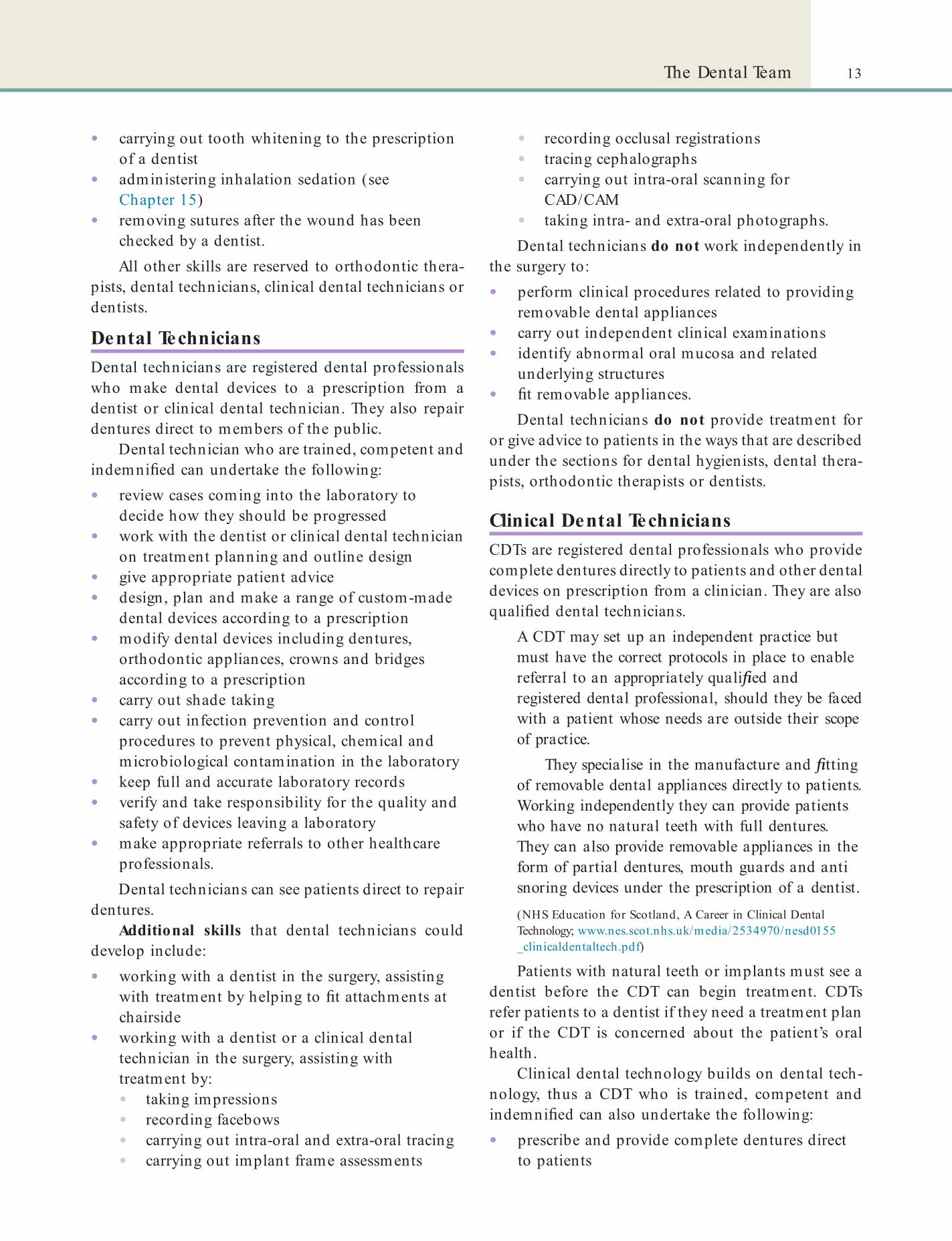
13The Dental Team
• recording occlusal registrations• tracing cephalographs• carrying out intra-oral scanning for
CAD/CAM• taking intra- and extra-oral photographs.Dental technicians do not work independently in
the surgery to:
• perform clinical procedures related to providing removable dental appliances
• carry out independent clinical examinations• identify abnormal oral mucosa and related
underlying structures• t removable appliances.
Dental technicians do not provide treatment for or give advice to patients in the ways that are described under the sections for dental hygienists, dental thera-pists, orthodontic therapists or dentists.
Clinical Dental TechniciansCDTs are registered dental professionals who provide complete dentures directly to patients and other dental devices on prescription from a clinician. They are also quali ed dental technicians.
A CDT may set up an independent practice but must have the correct protocols in place to enable referral to an appropriately quali ed and registered dental professional, should they be faced with a patient whose needs are outside their scope of practice.
They specialise in the manufacture and tting of removable dental appliances directly to patients. Working independently they can provide patients who have no natural teeth with full dentures. They can also provide removable appliances in the form of partial dentures, mouth guards and anti snoring devices under the prescription of a dentist.(NHS Education for Scotland, A Career in Clinical Dental Technology; www.nes.scot.nhs.uk/media/2534970/nesd0155_clinicaldentaltech.pdf)
Patients with natural teeth or implants must see a dentist before the CDT can begin treatment. CDTs refer patients to a dentist if they need a treatment plan or if the CDT is concerned about the patient’s oral health.
Clinical dental technology builds on dental tech-nology, thus a CDT who is trained, competent and indemni ed can also undertake the following:
• prescribe and provide complete dentures direct to patients
• carrying out tooth whitening to the prescription of a dentist
• administering inhalation sedation (see Chapter 15)
• removing sutures after the wound has been checked by a dentist.All other skills are reserved to orthodontic thera-
pists, dental technicians, clinical dental technicians or dentists.
Dental TechniciansDental technicians are registered dental professionals who make dental devices to a prescription from a dentist or clinical dental technician. They also repair dentures direct to members of the public.
Dental technician who are trained, competent and indemni ed can undertake the following:
• review cases coming into the laboratory to decide how they should be progressed
• work with the dentist or clinical dental technician on treatment planning and outline design
• give appropriate patient advice• design, plan and make a range of custom-made
dental devices according to a prescription• modify dental devices including dentures,
orthodontic appliances, crowns and bridges according to a prescription
• carry out shade taking• carry out infection prevention and control
procedures to prevent physical, chemical and microbiological contamination in the laboratory
• keep full and accurate laboratory records• verify and take responsibility for the quality and
safety of devices leaving a laboratory• make appropriate referrals to other healthcare
professionals.Dental technicians can see patients direct to repair
dentures.Additional skills that dental technicians could
develop include:
• working with a dentist in the surgery, assisting with treatment by helping to t attachments at chairside
• working with a dentist or a clinical dental technician in the surgery, assisting with treatment by:• taking impressions• recording facebows• carrying out intra-oral and extra-oral tracing• carrying out implant frame assessments

14 CHAPTER • 2 • Mosby’s Textbook of Dental Nursing
• provide and t other dental devices on prescription from a dentist
• take detailed dental history and relevant medical history
• perform technical and clinical procedures related to providing removable dental appliances
• carry out clinical examinations within their scope of practice
• take and process radiographs and other images related to providing removable dental appliances
• distinguish between normal and abnormal consequences of ageing
• give appropriate patient advice• recognise abnormal oral mucosa and related
underlying structures and refer patients to other healthcare professionals if necessary
• t removable appliances• provide sports mouthguards• keep full, accurate and contemporaneous patient
records• vary the detail but not the direction of a
prescription according to patient needs.Additional skills that CDTs could develop include:
• oral health education• re-cementing crowns with temporary cement• providing anti-snoring devices on prescription of
a dentist• removing sutures after the wound has been
checked by a dentist• prescribing radiographs• replacing implant abutments for removable
dental appliances on prescription from a dentist• providing tooth whitening treatments on
prescription from a dentist.All other skills are reserved to dental hygienists,
dental therapists, orthodontic therapists or dentists.
DentistsDentists can carry out all the treatments listed above for dental professionals within the GDC Scope of Practice (2013) document.
A dentist who is trained, competent and indemni- ed can also undertake the following:
• diagnose disease• prepare comprehensive treatment plans• prescribe and provide endodontic treatment on
adult teeth• prescribe and provide xed orthodontic treatment• prescribe and provide xed and removable
prostheses
• carry out oral surgery• carry out periodontal surgery• extract permanent teeth• prescribe and provide crowns and bridges• provide conscious sedation (see Chapter 15)• carry out treatment on patients who are under
general anaesthesia (see Chapter 15)• prescribe medicines as part of dental treatment• prescribe and interpret radiographs.
Additional skills a dentist could develop include:
• providing implants• providing non-surgical cosmetic injectables.
Re gis te re d de ntis tsAccording to the GDC, all registered dentists are legally entitled to practise any clinical aspect of dentistry, such as cosmetic surgery, provided they undertake only pro-cedures within their competence and do not use the title of ‘specialist’ unless entitled to do so.
Dentists who can the practise in the UK fall into the three groups shown in Table 2.2. They include:
• Those with a UK dental quali cation• Exempt persons with a scheduled (GDC-
recognised) dental quali cation from the EEA (European Economic Area = EU member states plus Iceland, Liechtenstein and Norway).
• Those to whom special regulations apply, as shown in Table 2.2 (mainly from outside the EEA), and
• Temporary registrants (see Table 2.2).Overseas dental graduates cannot register as DCPs.
Spe c ia lis t de ntis tsSpecialist dentists are those registered who ful l certain criteria and thus have a right to call themselves specialists in particular areas of dentistry. As of 2014, the GDC maintained 13 Specialist Lists in Distinctive Branches of Dentistry (Box 2.1) to enable patients to identify specialist dentists. Not all areas in dentistry that may be thought of as specialties are recognised as such by the GDC.
Find out moreLook a t the GDC publica t ion Preparing for Pract ice : Dental Team Learn ing Outcom es for Regist rat ion (www.gdc-uk.org/newsandpublica t ions/publica t ions/publica t ions/gdc% 20learn ing% 20outcomes.pd ) o r more about the lea rn ing ou tcomes (competencies) expected o each member o the denta l t eam with in the ir scope o p ract ice .

15The Dental Team
Table 2.2 Dent ist s who can work in the UK
Basic den ta l t ra in ing Qualif ca t ion requirements o r GDC regist ra t ion
UK university Five years undergraduate education and training, Bachelor of Dental Surgery (BDS) or other recognised dental quali cation, plus 1 year Foundation Training (FT)
GDC-recognised EEA dental school
Basic quali cation such as Doctor of Dental Surgery (DDS)
Dental school outside EEA Temporary registration allows dentists who are not eligible for full registration to practise dentistry in the UK in supervised posts for training, teaching, or research purposes only, and for a limited period. An overseas quali ed dentist can apply for temporary registration in speci c approved posts if they hold a dental quali cation from a university that is recognised by NARIC UK for the purposes of temporary registration and the overseas registration examination (ORE). UK NARIC is the UK’s National Agency responsible for providing information and opinion on academic, vocational and professional quali cations from across the world. Eligibility for certain training posts is dependent upon a UK Border Agency visaOverseas Registration Examination (ORE) registration allows dentists to practise dentistry unsupervised in the UK. The ORE tests the clinical skills and knowledge of dentists from outside the EEA whose quali cations are not eligible for full registration with the GDC in the UKOthers who can practise are those with a quali cation gained before 01/01/01 from Hong Kong, Singapore, Malaysia, South Africa, New Zealand and Australia with the exception of BChD MEDUNSA, BDS awarded between 01/01/97 to 31/12/00 and BChD Western Cape awarded before 31/12/97
Box 2.1 The GDC specia list list s in d ist inct ive branches o den t ist ry• Dental and Maxillofacial Radiology Involves all aspects
of medical imaging that provide information about anatomy, function and diseased states of the teeth and jaws.
• Dental Public Health Non-clinical specialty involving the science and art of preventing oral diseases, promoting oral health to the population rather than the individual. It involves the assessment of dental health needs and ensuring dental services meet those needs.
• Endodontics Concerned with the cause, diagnosis, prevention and treatment of diseases and injuries of the tooth root, dental pulp and surrounding tissue. [Endodontics is part of the specialty of Restorative Dentistry.]
• Oral Medicine Concerned with the oral health care of patients with chronic recurrent and medically related disorders of the mouth and with their diagnosis and non-surgical management. [Oral Medicine is the specialty of dentistry that sits at the interface between dentistry and medicine. Many Oral Medicine specialists have dental and medical quali cations, and both are now requirements for entry to training that leads to appointment as a Consultant in Oral Medicine. This re ects that the specialty had its origins in dentistry, but has evolved to formally encompass medical aspects of care.]
• Oral Microbiology Diagnosis and assessment of facial infection – typically bacterial and fungal disease. This is a clinical specialty undertaken by laboratory-based staff, who provide reports and advice based on interpretation of microbiological samples.
• Oral and Maxillofacial Pathology Diagnosis and assessment made from tissue changes characteristic of disease of the oral cavity, jaws and salivary glands. This is a clinical specialty undertaken by laboratory-based personnel.
[It includes the scienti c study of the causes and effects of disease in the oral and maxillofacial complex, an understanding of which is essential for diagnosis and for the development of appropriate treatments and preventive programmes.]
• Oral Surgery Deals with the treatment and ongoing management of irregularities and pathology of the jaw and mouth that require surgical intervention. This includes the specialty previously called Surgical Dentistry. [Oral and Maxillofacial Surgery is a specialty of medicine concerned with the diagnosis and treatment of diseases affecting the mouth, jaws, face and neck, that sits at the interface between dentistry and medicine. Oral and Maxillofacial Surgery specialists are registered on the Register of the General Medical Council but usually have dental and medical quali cations. This re ects that the specialty had its origins in dentistry, but has evolved to formally encompass surgical aspects of care.]
• Orthodontics The development, prevention and correction of irregularities of the teeth, bite and jaw.
• Paediatric Dentistry Concerned with comprehensive therapeutic oral health care for children from birth through adolescence, including care for those who demonstrate intellectual, medical, physical, psychological and/or emotional problems.
• Periodontics Diagnosis, treatment and prevention of diseases and disorders (infections and in ammatory) of the gums and other structures around the teeth. [Periodontics is part of Restorative Dentistry.]
• Prosthodontics Replacement of missing teeth and the associated soft and hard tissues by prostheses (crowns, bridges, dentures) which may be xed or removable, or may be supported and retained by implants. [Prosthodontics is part of Restorative Dentistry.]

16 CHAPTER • 2 • Mosby’s Textbook of Dental Nursing
Box 2.1 Cont inued• Restorative Dentistry Deals with the restoration of
diseased, injured, or abnormal teeth to normal function. Includes all aspects of Endodontics, Periodontics and Prosthodontics. [At the time of going to press, the GDC is seeking views on how it regulates the practice of Implant Dentistry. http://www.gdc-uk.org/dentalprofessionals/standards/pages/implantology.aspx]
• Special Care Dentistry Special Care Dentistry is concerned with the improvement of the oral health of individuals and groups in society who have a physical, sensory, intellectual, mental, medical, emotional or social impairment or disability or, more often, a combination of these factors. It pertains to adolescents and adults.
Direct Access to Dental Care Professionals
The GDC de nes ‘direct access’ as giving patients the option to see a dental care professional (DCP) without having rst seen a dentist and without a prescription from a dentist. Thus:
• Dental nurses can participate in preventive programmes without the patient having to see a dentist rst.
• Dental hygienists and dental therapists will be able to see patients direct.
• Orthodontic therapists can carry out Index of Orthodontic Treatment Need (IOTN) screening without the patient having to see a dentist
• Clinical dental technicians can see patients direct only for the provision and maintenance of full dentures.
Find out moreFor u lle r de ta ils on d irect access and a ll scopes o pract ice see : www.gdc-uk.org /Denta lpro essiona ls/Standards/Pages/d irectaccessqas.aspx.
Chapter 3 Dental Nursing Training,
Quali cations and Careers
CHAPTER POINTS• Training• Quali cations• NEBDN• CPD• Regulation• Fitness to pract ise• Career pathways• Further training• Employment opportunit ies
• good teamwork skills• positive and exible approach to work• good organisational skills.(From https:/ /nationalcareersservice.direct.gov.uk/advice/planning/jobprof les/Pages/dentalnurse.aspx)
The main legislation that governs the clinical work o the dental team, including dental nurses and dental nurse trainees, is the Dentists Act 1984 (Amendment) Order 2005. The 1984 Act provided or the regulation o dentists by the GDC. The 2005 Amendment dealt, among other matters, with:
• Giving the GDC broader powers to deal with impaired f tness to practise
• Regulating pro essionals complementary to dentistry (including dental nurses)
• Requiring registrants by law to have indemnity cover be ore registration.
Find out moreFor the fu ll t ext of the Dent ist s Act 1984 (Amendment ) Order 2005 No. 2011, Hea lth Care and Associa ted Professions: Dent ist s see : www.legisla t ion .gov.uk/uksi/2005/2011/conten ts/made .
Through the 2005 Amendment, the title ‘dental nurse’ is now protected by law. I an individual is not registered with the GDC and uses the title ‘dental nurse’, or any other title that misleadingly implies that the person is a dental nurse, they can be pro�secuted in court. Overseas dental graduates cannot register as dental care pro essionals (DCPs). This Amendment would also put at risk the registration o the dentist who is employing them.
Training to become a dental nurse in the UK requires attendance at a training centre that has been accredited by the National Examination Board or Dental Nurses (NEBDN). A ter passing the National
In t ro d u ct io n
Dental nursing carries responsibilities towards clini�cian, patient and others. The dental nurse is an invalu�able and skilled member o the dental or oral healthcare team but, like other dental pro essionals, is regulated or the protection o patients by the General Dental Council (GDC) (see Chapter 1). The GDC def nes competence in terms o learning outcomes, and this chapter mainly covers skills, training and qualif cations to achieve registration as a dental nurse. The chapter also looks brie y at the many employ�ment opportunities or the qualif ed dental nurse.
Skills, Training and Qualif cationsThe skills, interests and qualities needed to become a dental nurse, include:
• genuine interest in patient wel are• good practical skills• good eyesight• calm, conf dent and reassuring manner• ability to relate well to people, including
children and those with special needs
18 CHAPTER • 3 • Mosby’s Textbook of Dental Nursing
Key poin ts• Dental nurses must be on the GDC Register.
• It is illegal to work as a quali ed dental nurse without registration.
• It is illegal to work as an unquali ed dental nurse unless attending an accredited training centre.
Diploma in Dental Nursing examination and quali y�ing as a dental nurse, registration with the GDC is essential (see p. 20 or more details), with annual re�registration. It is illegal to work as a qualif ed dental nurse without GDC registration.
Several di erent routes lead to a qualif cation in dental nursing because a route that may be suitable or some may be less suitable or others. Appropriate pre�registration diplomas/certif cates are o ered by the National Examination Board or Dental Nurses (NEBDN).
Dental nurse training providers in England, Wales and Northern Ireland are listed in Table 3.1. For details about the Scottish Vocational Qualif cation (SVQ) in Oral Healthcare: Dental Nursing Level 3, contact the Scottish Qualif cations Authority.
The National Examination Board or Dental Nurses (NEBDN)The National Diploma in Dental Nursing o the NEBDN provides a mix o theoretical learning and
Table 3.1 Accessing den ta l nurse t ra in ing (England , Wales and Northern Ire land)
Quali ca t ion Awarding bodyDeta ils o f study and examinat ions ava ilab le from
National Diploma in Dental Nursing National Examining Board for Dental Nurses (NEBDN)
NEBDN www.nebdn.org/
City & Guilds Level 3 Award in Dental Nursing (VRQ) (England and Wales)
NEBDN/City & Guilds Care, Health & Community
NEBDNCity & Guilds
Certi cate of Higher Education in Dental Nursing
Cardiff University* School of Postgraduate Medical and Dental EducationSchool of Professionals Complementary to Dentistry
Portsmouth Dental Academy School of Professionals Complementary to Dentistry [email protected]
Foundation Degree in Dental Nursing University of Northampton [email protected]
*This programme has received provisional approval from the GDC Education Committee. Full GDC approval of new programmes is not granted until the rst batch of students has completed their studies and examinations or assessments and the programme has been inspected by the GDC. Potential applicants should contact the provider for further information about the programme.
practical teaching and experience. The diploma can be undertaken at a dental hospital or at a college o urther education.
• Dental hospitals usually provide ull� and part�time courses.
• Colleges o urther education usually provide part�time courses (mainly evening or day release).
• Full�time courses are usually work�related, and the theoretical and clinical teaching programme is combined with clinical placements in the hospital specialist departments.
• Part�time courses usually involve employment as an unqualif ed dental nurse in a general dental practice or equivalent in order to gain the practical experience, and attendance at part�time evening or day release classes. Attendance at an accredited training centre will cover an unqualif ed dental nurse until registration.The curriculum covers our domains o pro es�
sional practice, as set out by the GDC:1 . Clinical2 . Pro essional3 . Communication4 . Management and LeadershipThe competencies or each domain are expressed in terms o learning outcomes. The NEBDN curriculum has been amended to bring it in line with the GDC domains o pro essional practice and subsequent learning outcomes:
19Dental Nursing Training, Quali cations and Careers
GDC Learning OutcomesThe GDC document Preparing for Practice: Dental Team Learning Outcomes for Registration covers our main areas. The skills required o registrants are covered in the ollowing domains:
• Clinical – the range of skills required to deliver direct care, where registrants interact with patients, and also the essential technical skills, carried out in the absence of patients which support their care, for example, by dental technicians
• Communication – the skills involved in effectively interacting with patients, their representatives, the public and colleagues and recording appropriate information to inform patient care
• Professionalism – the knowledge, skills and attitudes/behaviours required to practise in an ethical and appropriate way, putting patients needs rst and promoting con dence in the dental team
• Management and Leadership – the skills and knowledge required to work effectively as a dental team, manage their own time and resources and contribute to professional practices.
GDC learning outcomes, i achieved, lead to a competent healthcare pro essional – one who can ‘practise sa ely, e ectively and pro essionally’, and one who ‘has knowledge, skills, behaviours and attitudes required to become a GDC Registrant’.
There are also f ve overarching GDC learning outcomes:
Upon registration with the GDC the registrant should be able to:• Practise safely and effectively, as set out in
the GDC Fitness to Practise guidance making the high quality long term care of patients the rst concern
• Apply an evidence-based approach to learning, practice, re ective practice and decision making
• Accurately assess own capabilities and limitations, demonstrating re ective practice, in the interest of high quality patient care and act within these boundaries
• Describe the role and responsibilities of being a registrant and demonstrate professionalism
throughout education, training and practice in accordance with GDC guidance
• Act with integrity and uphold high personal and professional values
Find out moreThe GDC document Preparing for Pract ice : Dental Team Learning Outcom es for Regist rat ion is ava ilable a t : www.gdc-uk.org /newsandpublica t ions/publica t ions/publica t ions/gdc% 20learn ing% 20outcomes.pdf
National Diploma in Dental NursingAttendance at an NEBDN�accredited training pro�gramme is required whilst working on a ull� or part�time basis as an unqualif ed dental nurse in a general dental practice, dental hospital, community dental service or other dental environment to gain practical experience at the chairside. The NEBDN holds the complete list o training providers.
Students undertaking training towards the NEBDN’s National Diploma in Dental Nursing qualif cation are required to complete a work�based assessment port olio called the Record of Experience (RoE) . Included in the RoE are (a number o ) clinical and theoretical competencies (tasks) based on the NEBDN curriculum. Students need to complete these competencies/ tasks in the workplace whilst being observed by a GDC registrant who acts as a ‘Witness’. There is a mandatory GDC requirement that all Witnesses observing the clinical activities must receive a documented, standardised level o training to ensure they are ully aware o their role and responsibilities.
The online RoE has to be completed and moder�ated be ore the application or sitting the written examination paper o the Diploma is accepted.
The curriculum is based on the GDC registration requirements or dental nurses ( or more detail see the NEBDN website). It sets out the knowledge, skills and behavioural requirements that should be developed and demonstrated. These are set out in terms o pro essional competencies, with the assess�ment method clearly outlined (e.g. how the skills and knowledge will be assessed in the f nal exami�nations – via written questions such as Multiple Choice Questions (MCQs) or Extended Matching Questions (EMQs) or via Objective Structured Clinical Examinations (OSCEs).
The examination or the National Diploma in Dental Nursing can only be sat i the RoE has been completed, verif ed and signed o . The written exami-nation consists o MCQs and EMQs. Optically Marked

20 CHAPTER • 3 • Mosby’s Textbook of Dental Nursing
With additional training and only on prescription (see below or details o training), a dental nurse may
• Take radiographs to the prescription o a clinician (Certif cate in Dental Radiography is required; see also Chapter 11)
• Apply topical anaesthetic to the prescription o a clinician
• Construct mouthguards and bleaching trays to the prescription o a clinician
• Construct vacuum� ormed retainers to the prescription o a clinician
• Take impressions to the prescription o a clinician (where appropriate).
Recognition (OMR) answer sheets are used or the Diploma examination. The answer sheet is f lled in using an HB pencil, with each answer indicated with a single line through the corresponding letter. The OMR sheets are returned to NEBDN where they are processed through an optical reader.
A ter passing the written paper, the OSCE (Objec-tive Structured Clinical Examination) is sat 1–2 months later.
Only on receipt o the National Diploma in Dental Nursing certif cate can GDC registration be applied or.
Additional Skills That a Dental Nurse Could DevelopWith additional training, dental nurses may also be involved in expanded duties such as:
• Providing oral health education and oral health promotion
• Assisting in the treatment o patients under conscious sedation
• Assisting in the treatment o patients with special needs
• Intra-oral photography• Shade taking• Placing rubber dam• Measuring and recording plaque indices (see
Chapter 13)• Pouring, casting and trimming study models• Removing sutures a ter the wound has been
checked by a clinician• Applying uoride varnish as part o a
programme that is overseen by a consultant in dental public health or registered specialist in dental public health
• Constructing occlusal registration rims and special trays
• Repairing the acrylic component o removable appliances
• Tracing cephalograms.
Key poin tDental nurses are never permitted to diagnose disease or plan the treatment.
Term to lea rnIntra/extra-oral: inside/outside the mouth.
Identify and learnIdent ify a shade gu ide , specia l t ray, occlusa l reg ist ra t ion rim, cepha logram, top ica l anaesthe t ic and vacuum-formed re ta iners in your workplace .
Dental Nurse RegistrationRegistrationThe f rst step a ter quali ying as a dental nurse is to register with the GDC. Only on receipt o the National Diploma in Dental Nursing certif cate can GDC regis�tration be applied or. GDC registration must be renewed annually.
Key poin tIt is good practice to complete the appropriate paperwork beforehand so that you can send it to the Registrar at the GDC immediately on quali cation.
A ter GDC RegistrationAs a registered dental pro essional, you must be amil�iar with and understand:
• Current standards and principles o dental care, and apply them at work, using your judgement in the light o the principles
• Relevant guidelines rom related organisations and sources o evidence that support current standards.A dental nurse must ensure their knowledge and
skills are up to date, and apply them ethically. A dental nurse must also be prepared to justi y their actions to the GDC. I an unsatis actory account o the behaviour or practice is given (in line with the principles), the dental nurse’s GDC registration may be at risk. In
21Dental Nursing Training, Quali cations and Careers
deciding to participate in the CPD. Some CPD must be on mandatory (essential) core subjects, which are the same as or clinicians:
• Medical emergencies (10 hours per f ve�year cycle)
• Disin ection and decontamination (5 hours per f ve-year cycle)
• Radiography and radiation protection (5 hours per f ve�year cycle).
Fitness to PractiseGDC registrants are expected, as demanded by the GDC Standards for the Dental Team (2013), to behave in a suitable manner both in and outside the work�place. Fitness to practise generally covers issues related to:
• Misconduct• Incompetence (poor per ormance)• Adverse health conditions (medical or mental).
I there is any evidence o poor practice by a dental nurse, according to healthcare regulation, the GDC is required to undertake an investigation, which will include processes to test any specif c doubt that a reg�istrant remains f t to be on the register, that is, f t to practise. These processes are called the ‘f tness to prac�tise system’.
The sequence has our main elements:1 . Complaint against, concern about or report
received about a particular registrant’s f tness to practise
2 . Investigation3 . Adjudication (that is, an o f cial decision), with
sanctions (penalties) where these are ound necessary
4 . Appeal.Any registrant who alls short o the GDC expecta�
tions may have their ‘f tness to practise’ questioned. This can result in
• suspension rom the Register, or• erasure rom it.Any re�registration may, under some circumstances. necessitate re�training or re�qualif cation.
Parties who could question a registrant’s f tness to practise may include:
• colleagues• employers• the pro essional regulator (usually the GDC)• the public.
other words, a dental nurse is responsible and account�able to themselves, their colleagues, their patients and the GDC, and or continuing development o their knowledge and skills. Besides the compulsory (mandatory) training, such as basic li e support and in ection control, which all members o the dental team are required to undergo, a dental nurse will be expected to participate in continuing pro essional development (CPD) to maintain their status on the GDC register.
Continuing Pro essional DevelopmentThe GDC states that:
In line with the clinicians’ CPD scheme, we recommend that [dental care professionals] DCPs involved in the care of patients should undertake Continuing Professional Development in legal and ethical issues and complaints handling.
Compulsory CPD maintains public con dence in the Dentists and Dental Care Professionals Registers by showing that clinicians and registered dental care professionals keep up to date so that they can give their patients a good standard of care.
Compulsory CPD means that dental nurses must complete and record 150 hours o CPD every f ve years, o which a third (50 hours) should be veri able (Box 3.1).
Term to lea rnVerif able course: a course should have speci c aims that clearly state what the trainee will have learnt and achieved by the end of the course. You must keep documentary evidence of attendance of such a course. For more details see the Freelance Dental Nurse website (www.freelancedentalnurse .co.uk/category/dental-courses-cpd/).
Box 3.1 Veri able CPDVeri able CPD is de ned as that which has:
• Concise educational aims and objectives
• A clear purpose or goal
• Quality controls
• Documentary proof of participation.
Most providers o CPD will issue a certif cate or proo o attendance at the verif able CPD event, but it is wise to check there will be CPD credits be ore

22 CHAPTER • 3 • Mosby’s Textbook of Dental Nursing
impaired by reason o a physical or mental condition (Box 3.3). A registrant su ering rom physical impair�ment that might jeopardise the wellbeing o patients should seek medical advice, and, i necessary, restrict the scope o his or her dental practice. Wil ully con�tinuing to practise with a physical impairment that could a ect the sa ety o patients may be regarded by the GDC as misconduct.
Per ormanceIts per ormance procedures allow the GDC to deal with cases where a dental pro essional’s pro essional per ormance may put patients at risk. The GDC has powers under the 1984 Dentists Act (as amended) to take action when an allegation is made that a dental pro essional’s f tness to practise is impaired by reason o their pro essional per ormance. Where this may be the case, the GDC will request that the dental pro es�sional undergoes a ull assessment o their per orm�ance, to f nd out where any issues may arise. The case may then be re erred to the Pro essional Per ormance Committee.
Career Pathw aysFollowing qualif cation and registration, gaining expe�rience as a qualif ed dental nurse is paramount. It may be help ul to join the British Association o Dental Nurses (BADN), which o ers guidance on matters related to dental nursing (see p. 7 or contact details).
A career in dental nursing is rewarding and involves participation in the care o a variety o patients
The GDC normally considers the ollowing our types o cases:
• Criminal o ence• Pro essional conduct• Health• Per ormance
Criminal O encesThe GDC is in ormed by the police i a registrant has been convicted or cautioned in the UK, or registrants may noti y the GDC directly. The GDC can also con�sider convictions and cautions imposed abroad which, had they been committed in the UK, would constitute a criminal o ence.
Criminal convictions can include o ences not directly connected with a registrant’s pro ession or practice or which occurred while the registrant was not registered. For example, the GDC would consider con�victions or raud whether related to dentistry or to personal f nances, drink–driving o ences, sexual or physical assault or deception.
The o ence is considered by the GDC Investigat�ing Committee as to whether it requires a re erral to a Practice Committee (PC). I re erred to a PC, the com�mittee must accept the f ndings o the court on matters o act. This means that a dental pro essional cannot claim to have been innocent o the original charges. Similarly, the PC may not question the conviction. The only duty o the PC is to decide whether (and what) urther action needs to be taken against the dental pro essional.
Pro essional ConductUnder its conduct procedures the GDC may deal with cases where a dental pro essional’s behaviour may put patients at risk. The GDC has powers under the 1984 Dentists Act (as amended) to take action when an allegation is made that a dental pro essional’s f tness to practise is impaired by reason o their conduct. Examples o issues that the GDC may deal with under these procedures include poor dental treatment, inap�propriate or unpro essional behaviour, or activities that may endanger the sa ety o patients or the public (Box 3.2).
HealthIts health procedures allow the GDC to deal with cases where a dental pro essional’s health problems may put patients at risk. The GDC has powers under the 1984 Dentists Act (as amended) to take action when an allegation is made that a dentist’s f tness to practise is
Box 3.3 Health issues tha t may impa ir tness to p ract ise• Failure to recognise limits/abilities.
• Failure to seek medical attention.
• Failure to follow medical advice.
Box 3.2 Issues of p rofessional conduct• Behavioural issues.
• Abuse, assault, bullying.
• Cheating (exams, logbooks, plagiarism, forgery).
• Dishonesty (falsifying CVs, research, or nancial).
• Drug misuse (drink–driving, drug possession, dealing, affecting clinical work).
• Criminal conviction or caution (abuse, drugs, fraud, theft).
• Inappropriate behaviour/attitude (breach of con dentiality, failure to obtain proper .consent, misleading, rudeness, sexual abuse/harassment, unlawful discrimination).
23Dental Nursing Training, Quali cations and Careers
• Expanded case studies• Evidence o competence.
It is essential that the dental nurse wishing to take one o the above courses works in an environment that specialises in that course area. For example, or undertaking the certif cate in special care, the dental nurse should be working with special care patients. The NEBDN provides a list o approved accredited centres in the UK that provide post�registration/other courses.
Other NEBDN certif cated courses on o er avail�able elsewhere include:
• Basic and advanced dental implants• In ection control• Restorative and surgical procedures.
Attending con erences also helps to gain urther knowledge and in ormation.
Employment OpportunitiesA variety o employment opportunities are available in the UK or qualif ed dental nurses:
• Armed orces• Bank o England• Corporate bodies• Dental access centres• Hospital dental service• Industry• NHS practice• Personal ( ormerly community) dental service
(PDS)• Police service• Prison service• Private practice• Retail• Specialist practice• Temping agencies• Work overseas.
Find out moreRead the Genera l Denta l Council Standards for the Dental Team (2013), which is sen t by the GDC to a ll new reg ist ran ts.
in di erent settings, ranging rom, or example, a dental practice to a dental hospital, a dental depart�ment in a general hospital, the community dental service, the armed orces, corporate organisations, universities or the prison service. Career pathways all into three main categories: clinical, management and education. For examples, see Table 3.2 and https:/ /nationalcareersservice.direct.gov.uk/advice/planning/jobprof les/Pages/dentalnurse.aspx.
Some dental nurses choose to undertake urther training in order to quali y, or example, as dental hygienists, dental therapists, orthodontic therapists, or dentists.
Further TrainingThe NEBDN has several post�registration courses that could orm part o a dental nurse’s pro essional development:
• Conscious sedation• Dental radiography• Oral health education• Orthodontic nursing• Special care.
As well as a written examination, post�registration courses involve completion o a ‘Record o Experience’ (RoE) in the workplace. This comprises:
• Log sheets verif ed by a registered dental pro essional
Table 3.2 Caree rs in den ta l nursing
Management Educat ionEduca t ion and management
Lead dental nurse
Tutor dental nurse
Principal/manager of dental nurse education and training
Practice manager Clinical tutor dental nurse
Senior dental nurse
Clinical dental nurse trainer or assessor

This page intentiona lly le ft blank
Chapter 4
Dental Nursing Practice and Communication
CHAPTER POINTS• Teamworking• Dental nurse scope of practice• The dental environment• Communication and working with patients• The dental appointment
• Providing patient care, including postoperatively, for the patient undergoing treatment.Typical roles of a dental nurse may involve both
clerical and clinical work. The clerical duties may include:
• Working at reception• Greeting and reassuring patients• Booking appointments• Taking payments.Clinical duties typically include:
• Preparing the dental surgery• Maintaining sterile and safe conditions,
following health and safety guidelines, including infection control
• Patient care, including acting as chaperone• Helping ensure that the patient is and remains
relaxed and comfortable• Helping the clinician (DCP) record information
about patients• Passing instruments to the clinician• Aspirating water and saliva from the patient’s
mouth during treatment• Preparing and manipulating dental materials, for
example, llings and impressions• Cleaning the surgery after treatment, and
decontaminating and sterilising instruments• Carrying out stock control.
The Dental Nurse’s Scope of PracticeThe GDC de nes dental nurses as ‘registered dental professionals who provide clinical and other support to [other registered DCPs] and patients’. A dental nurse who is trained, competent and indemni ed may:
• Prepare and maintain the clinical environment, including the equipment
In t ro d u ct io n
Dental nurses have been an integral part of dentistry since the early twentieth century. The dental nurse provides a key link between the dental clinician and patient and partner, family or friends, by supporting the patient as well as assisting clinically. The dental nurse may also communicate with other health pro�fessionals, for example by arranging a patient referral (Chapter 13), and also undertakes several other duties that are more fully explained in Chapters 17–22.
Teamw orkingThe dental team includes as a minimum a dentist and dental nurse but, much more commonly, a number of dental professionals as well as receptionists and secretaries. Technological advancements in clinical practice dictate that a dental nurse should be a skilled professional with a broad range of knowledge of current techniques, materials and, most importantly, patient care.
The scope of practice of the dental nurse is outlined below and in Chapter 2, but can be summarised as:
• Providing clinical and other support to other General Dental Council (GDC) registrants and to patients
• Undertaking clinical tasks in relation to the scope of work of the clinician (DCP)
26 CHAPTER • 4 • Mosby’s Textbook of Dental Nursing
your knowledge o a particular area by choosing more specialised practice.
You should only carry out a task or type o treatment or make decisions about a patient’s care i you are sure that you have the necessary skills and are appropriately trained, competent and indemnif ed. I a task, type o treatment or decision is outside your scope o practice or you do not eel that you are trained and competent to do it you must re er the patient to an appropriately trained colleague.(GDC, Scope o Practice, 2013)
The GDC’s Standards or the Dental Team (2013) state that ‘you should only delegate or refer to another member of the team if you are con dent that they have been trained and are both competent and indemni ed to do what you are asking’ (6.3.1).
GDC Guidance on Various Areas of Dental Nurse Roles Within Their Scope of Practice
Infe c tion c ontro l• Applying the principles of infection control
when:• Setting up the environment for clinical
procedures• Assisting during clinical procedures• Clearing away after clinical procedures.
• Knowing the different cleaning, disinfection and sterilisation techniques and their uses
• Understanding how to protect the patient, themselves and other members of the dental team.
• Carry out infection control procedures to prevent physical, chemical and microbiological contamination in the surgery or laboratory
• Record dental charting carried out by other appropriate registrants
• Prepare, mix and handle dental materials• Provide chairside support to the operator during
treatment• Keep full and accurate patient records• Prepare equipment, materials and patients for
dental radiography• Process and mount dental radiographs• Monitor, support and reassure patients• Give appropriate advice to patients• Support the patient and their colleagues if there
is a medical emergency• Make appropriate referrals to other health
professionals.
Terms to lea rnDisinfection: a process by which the number of viable harmful micro-organisms is reduced in an area, e.g. a worktop in a dental practice. Disinfection does not get rid of certain micro-organisms, such as some viruses, or destroy certain forms of harmful micro-organisms, such as spores or prions. Therefore it is only used for cleaning those areas of a dental surgery that only need to be acceptably safe. Disinfection can by carried out using special chemicals called disinfectants or by using heat.
Sterilisation: a process by which an object is rendered free from all viable harmful micro-organisms, including viruses and bacterial spores. Therefore this method is used to decontaminate instruments that will be used inside a patient’s mouth.
Terms to learnCharting: lling in the dental record sheet (called a chart) for a patient, for example recording which teeth are present, missing or decayed (see Chapter 13).
Mounting: the procedure of correctly displaying dental radiographs for the clinician to see, for example, ensuring the radiograph is the correct side up and the correct side is facing the clinician.
Additional skills dental nurses could develop during their careers are shown in Chapter 2, as are additional skills on prescription.
Dental nurses do not diagnose disease or plan treatment. All other skills are reserved to one or more of the other GDC registrant groups, as detailed in Chapter 2.
The GDC states that:The scope o your practice is a way o describing what you are trained and competent to do. It describes the areas in which you have the knowledge, skills and experience to practise sa ely and e ectively in the best interests o patients. …
Your scope o practice is likely to change over the course o your career, both because o changes in the technology o dentistry, and your urther training and development. [You might develop] additional skills … a ter registration to increase your scope o practice. You may expand your scope by developing additional skills, or you may deepen
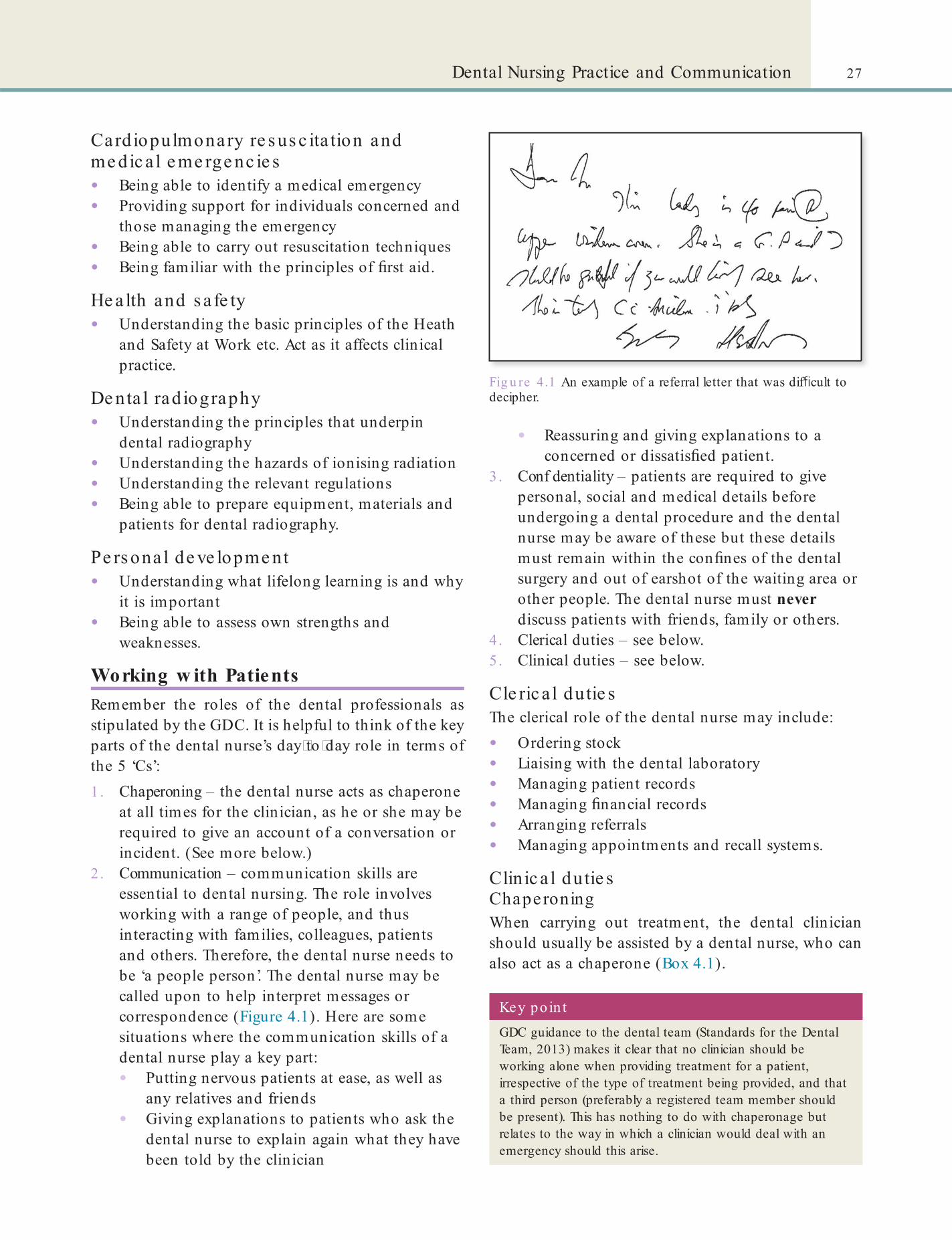
27Dental Nursing Practice and Communication
Cardiopulmonary re s us c ita tion and me dic a l e me rge nc ie s• Being able to identify a medical emergency• Providing support for individuals concerned and
those managing the emergency• Being able to carry out resuscitation techniques• Being familiar with the principles of rst aid.
He a lth and s a fe ty• Understanding the basic principles of the Heath
and Safety at Work etc. Act as it affects clinical practice.
De nta l rad iography• Understanding the principles that underpin
dental radiography• Understanding the hazards of ionising radiation• Understanding the relevant regulations• Being able to prepare equipment, materials and
patients for dental radiography.
Pe rs ona l de ve lopme nt• Understanding what lifelong learning is and why
it is important• Being able to assess own strengths and
weaknesses.
Working w ith PatientsRemember the roles of the dental professionals as stipulated by the GDC. It is helpful to think of the key parts of the dental nurse’s day�to�day role in terms of the 5 ‘Cs’:1 . Chaperoning – the dental nurse acts as chaperone
at all times for the clinician, as he or she may be required to give an account of a conversation or incident. (See more below.)
2 . Communication – communication skills are essential to dental nursing. The role involves working with a range of people, and thus interacting with families, colleagues, patients and others. Therefore, the dental nurse needs to be ‘a people person’. The dental nurse may be called upon to help interpret messages or correspondence (Figure 4.1). Here are some situations where the communication skills of a dental nurse play a key part:• Putting nervous patients at ease, as well as
any relatives and friends• Giving explanations to patients who ask the
dental nurse to explain again what they have been told by the clinician
Fig u re 4.1 An example of a referral letter that was dif cult to decipher.
• Reassuring and giving explanations to a concerned or dissatis ed patient.
3 . Conf dentiality – patients are required to give personal, social and medical details before undergoing a dental procedure and the dental nurse may be aware of these but these details must remain within the con nes of the dental surgery and out of earshot of the waiting area or other people. The dental nurse must never discuss patients with friends, family or others.
4 . Clerical duties – see below.5 . Clinical duties – see below.
Cle ric a l dutie sThe clerical role of the dental nurse may include:
• Ordering stock• Liaising with the dental laboratory• Managing patient records• Managing nancial records• Arranging referrals• Managing appointments and recall systems.
Clinic a l dutie sChaperoningWhen carrying out treatment, the dental clinician should usually be assisted by a dental nurse, who can also act as a chaperone (Box 4.1).
Key poin tGDC guidance to the dental team (Standards for the Dental Team, 2013) makes it clear that no clinician should be working alone when providing treatment for a patient, irrespective of the type of treatment being provided, and that a third person (preferably a registered team member should be present). This has nothing to do with chaperonage but relates to the way in which a clinician would deal with an emergency should this arise.
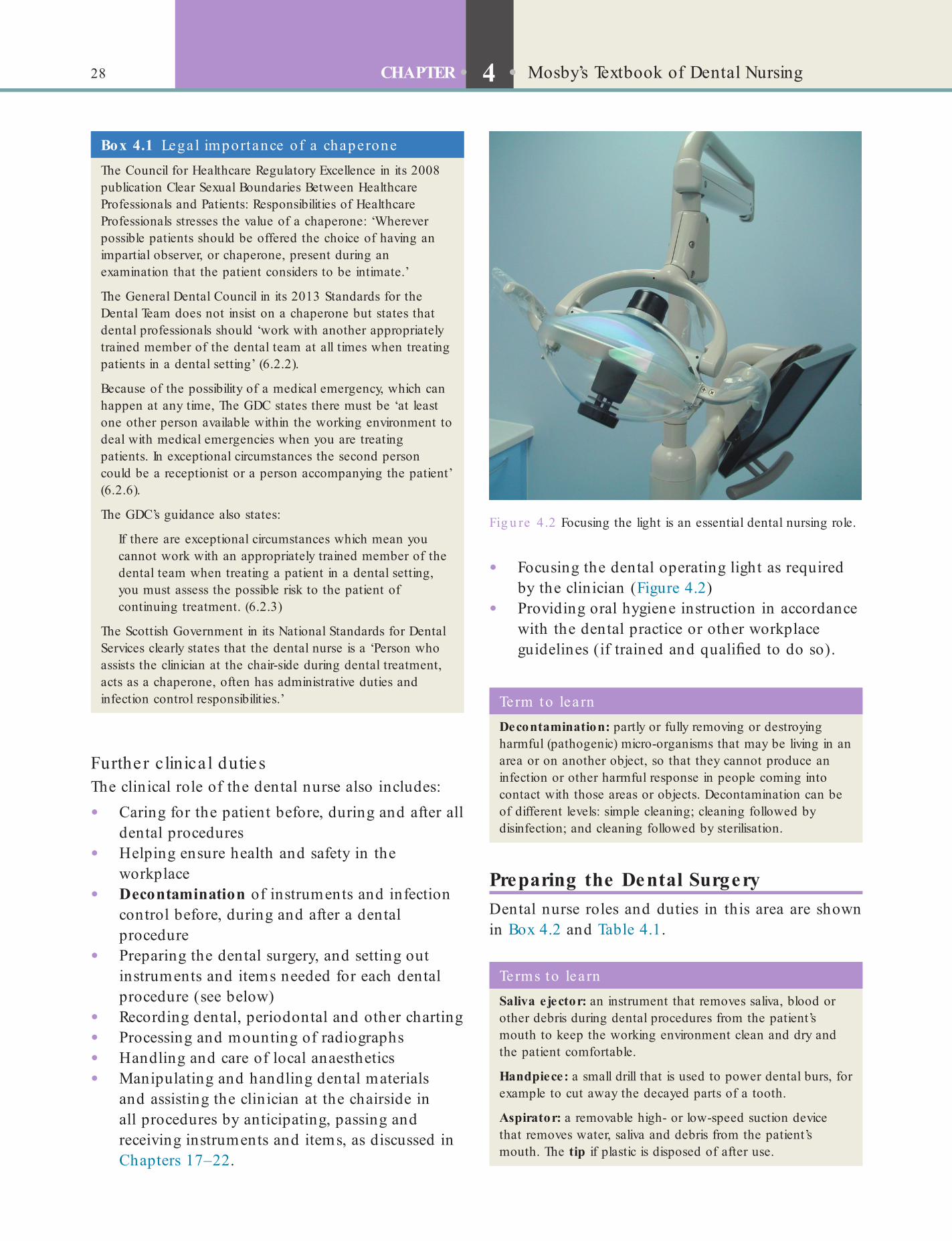
28 CHAPTER • 4 • Mosby’s Textbook of Dental Nursing
• Focusing the dental operating light as required by the clinician (Figure 4.2)
• Providing oral hygiene instruction in accordance with the dental practice or other workplace guidelines (if trained and quali ed to do so).
Box 4.1 Lega l importance of a chaperoneThe Council for Healthcare Regulatory Excellence in its 2008 publication Clear Sexual Boundaries Between Healthcare Professionals and Patients: Responsibilities of Healthcare Professionals stresses the value of a chaperone: ‘Wherever possible patients should be offered the choice of having an impartial observer, or chaperone, present during an examination that the patient considers to be intimate.’
The General Dental Council in its 2013 Standards for the Dental Team does not insist on a chaperone but states that dental professionals should ‘work with another appropriately trained member of the dental team at all times when treating patients in a dental setting’ (6.2.2).
Because of the possibility of a medical emergency, which can happen at any time, The GDC states there must be ‘at least one other person available within the working environment to deal with medical emergencies when you are treating patients. In exceptional circumstances the second person could be a receptionist or a person accompanying the patient’ (6.2.6).
The GDC’s guidance also states:
If there are exceptional circumstances which mean you cannot work with an appropriately trained member of the dental team when treating a patient in a dental setting, you must assess the possible risk to the patient of continuing treatment. (6.2.3)
The Scottish Government in its National Standards for Dental Services clearly states that the dental nurse is a ‘Person who assists the clinician at the chair-side during dental treatment, acts as a chaperone, often has administrative duties and infection control responsibilities.’
Fig u re 4 .2 Focusing the light is an essential dental nursing role.
Further clinica l dutie sThe clinical role of the dental nurse also includes:
• Caring for the patient before, during and after all dental procedures
• Helping ensure health and safety in the workplace
• Decontamination of instruments and infection control before, during and after a dental procedure
• Preparing the dental surgery, and setting out instruments and items needed for each dental procedure (see below)
• Recording dental, periodontal and other charting• Processing and mounting of radiographs• Handling and care of local anaesthetics• Manipulating and handling dental materials
and assisting the clinician at the chairside in all procedures by anticipating, passing and receiving instruments and items, as discussed in Chapters 17–22.
Term to lea rnDecontamination: partly or fully removing or destroying harmful (pathogenic) micro-organisms that may be living in an area or on another object, so that they cannot produce an infection or other harmful response in people coming into contact with those areas or objects. Decontamination can be of different levels: simple cleaning; cleaning followed by disinfection; and cleaning followed by sterilisation.
Preparing the Dental SurgeryDental nurse roles and duties in this area are shown in Box 4.2 and Table 4.1.
Terms to lea rnSaliva ejector: an instrument that removes saliva, blood or other debris during dental procedures from the patient’s mouth to keep the working environment clean and dry and the patient comfortable.
Handpiece: a small drill that is used to power dental burs, for example to cut away the decayed parts of a tooth.
Aspirator: a removable high- or low-speed suction device that removes water, saliva and debris from the patient’s mouth. The tip if plastic is disposed of after use.
29Dental Nursing Practice and Communication
Box 4.2 Preparing a den ta l surgery
At the beginning of the session• Switch on the main electricity switch, and then all the other
switches (including the dental chair) and the water supply in the dental surgery.
• Switch on the air compressor if required.
• Ensure the dental chair and light, suction and dental handpieces are working correctly.
• Flush into the sink, the 3-in-1 dental syringe and water lines for the dental handpieces for 2 minutes to cleanse the tubing.
• Clean the work surfaces, dental unit and bracket table with a detergent wipe, then wipe with an alcohol-base wipe (do not use an alcohol-base wipe to clean the dental chair).
• Clean the spittoon and ush the water.
• Place a clean disposable cup with an effervescent mouthwash tablet (e.g. avoured with thymol, menthol, orange or spearmint) on the spittoon.
• Connect the aspirator tip and saliva ejector to the tubing on the spittoon.
• Place ‘cling lm’ or equivalent on the handles of the dental light, unit and headrest of the dental chair, 3-in-1 syringe, handpiece lines and computer keyboard, if there is one.
• Collect the patient case records for the session, ensuring that all appropriate radiographs and any laboratory work or other necessary items are present.
• Lay out the dental instruments and items in a logical order for the rst patient and mount any relevant radiograph on the radiograph viewer.
• Lay out any laboratory work and necessary instruments.
• Place the dental handpieces (in sterile bags) and dental burs on the bracket table.
• Ensure the dental surgery is free from unnecessary clutter.
• Fill the steam steriliser with distilled water, test and record.
• Fill the ultrasonic bath and turn on the washer-disinfector.
End of the session• Reverse the procedure and ush aspirator tubing.
The Dental Environment
Find out moreLook a round your workplace and answer the fo llowing quest ions:• Is the wait ing a rea clear and t idy or is there
someth ing tha t cou ld be t ripped over?• Are the facilit ies in the to ile t s accep tab le?• Is the recept ion a rea t idy or p iled h igh with
pa t ien t records?• Can the pa t ient ta lk to the recep t ion ist , denta l
nurse o r clin ician without be ing overheard?• Is the route to and in the den ta l surgery clear and
t idy or is there someth ing tha t cou ld be t ripped over?
• Are a ll cab les secured and if possib le out o f sigh t?
Tidiness, Cleanliness and Hygiene
Re c e ption, wa iting a re a and to ile tsThe dental team should always ensure that the recep�tion, waiting area, facilities and work environment are maintained, clean, safe and user�friendly.
Clinic a l a re aIn the clinical area, the following points should help you consider how you may increase the comfort and safety of your patients:
• Ensure the patient will not knock their head on the dental light or trip over the foot pedal or cables or pipes.
• Are accompanying children, relatives or friends appropriate in the surgery or will they be a distraction and/or a liability? Where should they sit? Is there enough seating? Are there coat hooks?
• Ask the patient if they will be happy with the chair tilted back (some – especially older people – may have a medical condition that precludes this, or feel anxious when the chair is tilted back).
• Has the dental light been cleaned ef ciently or is there dust, or are there nger marks or stains?
• Is the dental surgery tidy?• Is the spittoon clean with fresh beaker and
mouthwash?• Is there a new clean disposable bib?
Communication and Working w ith Patients
As a dental nurse you need to have, and be able to demonstrate, an interest in people: the dental nurse works with, rather than on, patients. You will need to listen to and to explore, with the patient, the beliefs and practices that are important to them and their situation, their feelings and their concerns about healthcare. Patients vary, for example in how they wish to be addressed, and so need to be asked.
Dental nurses should:
• Understand their role within the team and in a clinical environment
• Be able to communicate with patients, their carers and other members of the team
• Understand the need for con dentiality• Be able to cope with dif cult or aggressive
patients• Understand patients’ rights and the need to
handle complaints sensitively.
30 CHAPTER • 4 • Mosby’s Textbook of Dental Nursing
Table 4.1 Roles and responsib ilit ie s o f the den ta l nurse
Roles and responsibilit ies Deta il
Comply w ith GDC Standards 1. Put patients’ interests rst2. Communicate effectively with patients3. Obtain valid consent4. Maintain and protect patients’ information5. Have a clear and effective complaints procedure6. Work with colleagues in a way that is in patients’ best interests7. Maintain, develop and work within your professional knowledge and skills8. Raise concerns if patients are at risk9. Make sure your personal behaviour maintains patients’ con dence in you and the dental
profession
At start o f day Comply with health and safetySwitch off alarmSwitch on powerCheck heating/coolingCheck appointment bookCheck answerphonesSwitch on compressorCheck all equipment workingRun and record autoclave testRe ll ultrasonic bathFlush 3-in-1 syringe and handpieces for 2 minsDisinfect working surfaces
Chairside Comply with health and safetyCheck heating/coolingGreet and seat patientApply bib and protective eyewearAct as chaperone and witnessGive preoperative instructionsEnsure any accompanying person is available and appropriateWear PPE (personal protective equipment)Prepare topical and local anaestheticAssist clinicianAnticipate clinician’s needsHave working knowledge of each instrumentEnsure instruments, records, charts, radiographs, models are availableEnsure valid consent obtainedFollow infection controlPass instruments with no-touch techniqueExplain to patient if head support needed, and provide itEnsure sharps are put in sharps bin by clinicianEnsure autoclavable items go to washer-disinfector or ultrasonic bathEnsure contaminated waste goes to hazardous waste sacksDisinfect surfacesEnsure all records completed and securely storedEnsure material for laboratories is handled correctly and sent with appropriate request forms completedControl oral soft tissues (retract) and moisture (aspirate)Assist in an emergencyMonitor patient and draw clinician’s attention to any stress, pain, airway problem, colour and bleedingArrange postoperative observationsGive postoperative instructions and items neededEnsure patient has an emergency number to callArrange future appointmentsPrepare for next patient
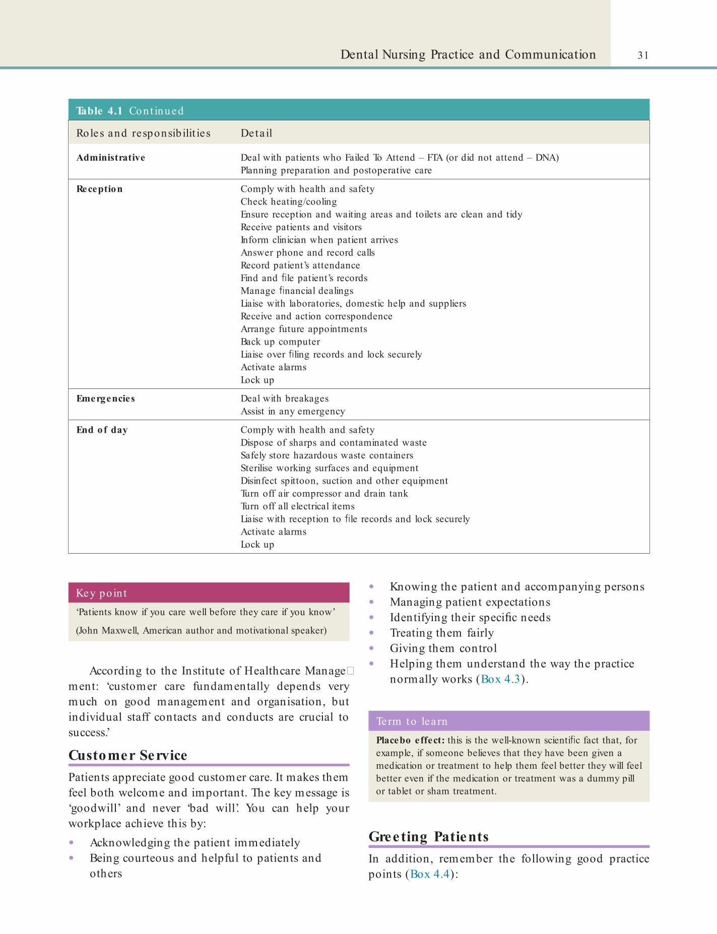
31Dental Nursing Practice and Communication
Table 4.1 Cont inued
Roles and responsibilit ies Deta il
Administrative Deal with patients who Failed To Attend – FTA (or did not attend – DNA)Planning preparation and postoperative care
Reception Comply with health and safetyCheck heating/coolingEnsure reception and waiting areas and toilets are clean and tidyReceive patients and visitorsInform clinician when patient arrivesAnswer phone and record callsRecord patient’s attendanceFind and le patient’s recordsManage nancial dealingsLiaise with laboratories, domestic help and suppliersReceive and action correspondenceArrange future appointmentsBack up computerLiaise over ling records and lock securelyActivate alarmsLock up
Emergencies Deal with breakagesAssist in any emergency
End of day Comply with health and safetyDispose of sharps and contaminated wasteSafely store hazardous waste containersSterilise working surfaces and equipmentDisinfect spittoon, suction and other equipmentTurn off air compressor and drain tankTurn off all electrical itemsLiaise with reception to le records and lock securelyActivate alarmsLock up
According to the Institute of Healthcare Manage�ment: ‘customer care fundamentally depends very much on good management and organisation, but individual staff contacts and conducts are crucial to success.’
Customer ServicePatients appreciate good customer care. It makes them feel both welcome and important. The key message is ‘goodwill’ and never ‘bad will’. You can help your workplace achieve this by:
• Acknowledging the patient immediately• Being courteous and helpful to patients and
others
Key poin t‘Patients know if you care well before they care if you know’
(John Maxwell, American author and motivational speaker)
• Knowing the patient and accompanying persons• Managing patient expectations• Identifying their speci c needs• Treating them fairly• Giving them control• Helping them understand the way the practice
normally works (Box 4.3).
Term to lea rnPlacebo effect: this is the well-known scienti c fact that, for example, if someone believes that they have been given a medication or treatment to help them feel better they will feel better even if the medication or treatment was a dummy pill or tablet or sham treatment.
Greeting PatientsIn addition, remember the following good practice points (Box 4.4):
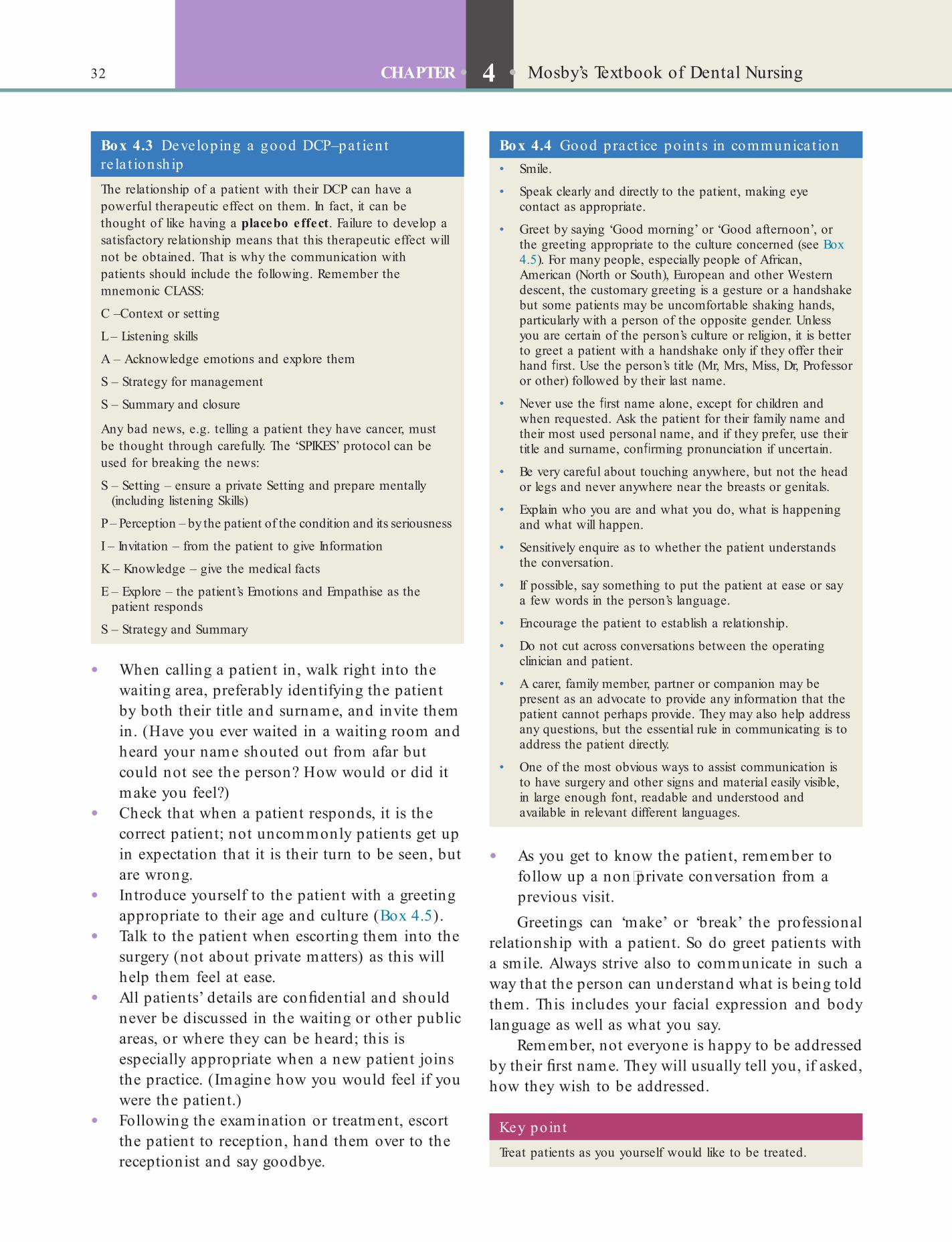
32 CHAPTER • 4 • Mosby’s Textbook of Dental Nursing
Box 4.3 Developing a good DCP–pat ien t re la t ionsh ipThe relationship of a patient with their DCP can have a powerful therapeutic effect on them. In fact, it can be thought of like having a placebo effect. Failure to develop a satisfactory relationship means that this therapeutic effect will not be obtained. That is why the communication with patients should include the following. Remember the mnemonic CLASS:
C –Context or setting
L – Listening skills
A – Acknowledge emotions and explore them
S – Strategy for management
S – Summary and closure
Any bad news, e.g. telling a patient they have cancer, must be thought through carefully. The ‘SPIKES’ protocol can be used for breaking the news:
S – Setting – ensure a private Setting and prepare mentally (including listening Skills)
P – Perception – by the patient of the condition and its seriousness
I – Invitation – from the patient to give Information
K – Knowledge – give the medical facts
E – Explore – the patient’s Emotions and Empathise as the patient responds
S – Strategy and Summary
Box 4.4 Good pract ice poin ts in communica t ion• Smile.
• Speak clearly and directly to the patient, making eye contact as appropriate.
• Greet by saying ‘Good morning’ or ‘Good afternoon’, or the greeting appropriate to the culture concerned (see Box 4.5). For many people, especially people of African, American (North or South), European and other Western descent, the customary greeting is a gesture or a handshake but some patients may be uncomfortable shaking hands, particularly with a person of the opposite gender. Unless you are certain of the person’s culture or religion, it is better to greet a patient with a handshake only if they offer their hand rst. Use the person’s title (Mr, Mrs, Miss, Dr, Professor or other) followed by their last name.
• Never use the rst name alone, except for children and when requested. Ask the patient for their family name and their most used personal name, and if they prefer, use their title and surname, con rming pronunciation if uncertain.
• Be very careful about touching anywhere, but not the head or legs and never anywhere near the breasts or genitals.
• Explain who you are and what you do, what is happening and what will happen.
• Sensitively enquire as to whether the patient understands the conversation.
• If possible, say something to put the patient at ease or say a few words in the person’s language.
• Encourage the patient to establish a relationship.
• Do not cut across conversations between the operating clinician and patient.
• A carer, family member, partner or companion may be present as an advocate to provide any information that the patient cannot perhaps provide. They may also help address any questions, but the essential rule in communicating is to address the patient directly.
• One of the most obvious ways to assist communication is to have surgery and other signs and material easily visible, in large enough font, readable and understood and available in relevant different languages.
• When calling a patient in, walk right into the waiting area, preferably identifying the patient by both their title and surname, and invite them in. (Have you ever waited in a waiting room and heard your name shouted out from afar but could not see the person? How would or did it make you feel?)
• Check that when a patient responds, it is the correct patient; not uncommonly patients get up in expectation that it is their turn to be seen, but are wrong.
• Introduce yourself to the patient with a greeting appropriate to their age and culture (Box 4.5).
• Talk to the patient when escorting them into the surgery (not about private matters) as this will help them feel at ease.
• All patients’ details are con dential and should never be discussed in the waiting or other public areas, or where they can be heard; this is especially appropriate when a new patient joins the practice. (Imagine how you would feel if you were the patient.)
• Following the examination or treatment, escort the patient to reception, hand them over to the receptionist and say goodbye.
• As you get to know the patient, remember to follow up a non�private conversation from a previous visit.Greetings can ‘make’ or ‘break’ the professional
relationship with a patient. So do greet patients with a smile. Always strive also to communicate in such a way that the person can understand what is being told them. This includes your facial expression and body language as well as what you say.
Remember, not everyone is happy to be addressed by their rst name. They will usually tell you, if asked, how they wish to be addressed.
Key poin tTreat patients as you yourself would like to be treated.
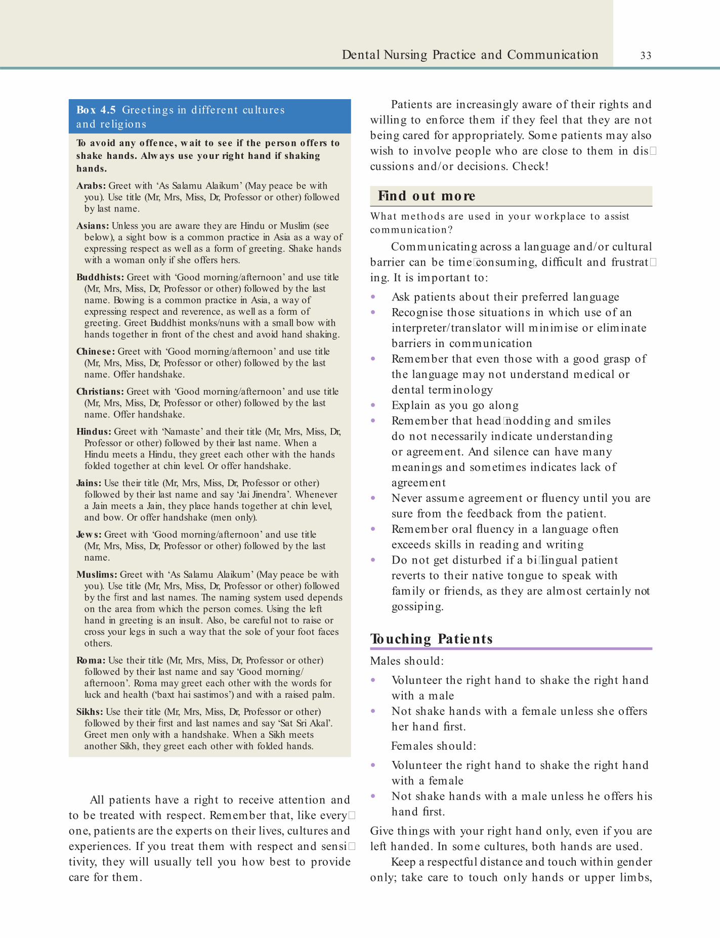
33Dental Nursing Practice and Communication
Patients are increasingly aware of their rights and willing to enforce them if they feel that they are not being cared for appropriately. Some patients may also wish to involve people who are close to them in dis�cussions and/or decisions. Check!
Find out moreWhat methods a re used in your workplace to assist communica t ion?
Communicating across a language and/or cultural barrier can be time�consuming, dif cult and frustrat�ing. It is important to:
• Ask patients about their preferred language• Recognise those situations in which use of an
interpreter/translator will minimise or eliminate barriers in communication
• Remember that even those with a good grasp of the language may not understand medical or dental terminology
• Explain as you go along• Remember that head�nodding and smiles
do not necessarily indicate understanding or agreement. And silence can have many meanings and sometimes indicates lack of agreement
• Never assume agreement or uency until you are sure from the feedback from the patient.
• Remember oral uency in a language often exceeds skills in reading and writing
• Do not get disturbed if a bi�lingual patient reverts to their native tongue to speak with family or friends, as they are almost certainly not gossiping.
Touching PatientsMales should:
• Volunteer the right hand to shake the right hand with a male
• Not shake hands with a female unless she offers her hand rst.Females should:
• Volunteer the right hand to shake the right hand with a female
• Not shake hands with a male unless he offers his hand rst.
Give things with your right hand only, even if you are left handed. In some cultures, both hands are used.
Keep a respectful distance and touch within gender only; take care to touch only hands or upper limbs,
Box 4.5 Gree t ings in d iffe ren t cu ltures and re lig ionsTo avoid any offence, w ait to see if the person offers to shake hands. Alw ays use your right hand if shaking hands.
Arabs: Greet with ‘As Salamu Alaikum’ (May peace be with you). Use title (Mr, Mrs, Miss, Dr, Professor or other) followed by last name.
Asians: Unless you are aware they are Hindu or Muslim (see below), a sight bow is a common practice in Asia as a way of expressing respect as well as a form of greeting. Shake hands with a woman only if she offers hers.
Buddhists: Greet with ‘Good morning/afternoon’ and use title (Mr, Mrs, Miss, Dr, Professor or other) followed by the last name. Bowing is a common practice in Asia, a way of expressing respect and reverence, as well as a form of greeting. Greet Buddhist monks/nuns with a small bow with hands together in front of the chest and avoid hand shaking.
Chinese: Greet with ‘Good morning/afternoon’ and use title (Mr, Mrs, Miss, Dr, Professor or other) followed by the last name. Offer handshake.
Christians: Greet with ‘Good morning/afternoon’ and use title (Mr, Mrs, Miss, Dr, Professor or other) followed by the last name. Offer handshake.
Hindus: Greet with ‘Namaste’ and their title (Mr, Mrs, Miss, Dr, Professor or other) followed by their last name. When a Hindu meets a Hindu, they greet each other with the hands folded together at chin level. Or offer handshake.
Jains: Use their title (Mr, Mrs, Miss, Dr, Professor or other) followed by their last name and say ‘Jai Jinendra’. Whenever a Jain meets a Jain, they place hands together at chin level, and bow. Or offer handshake (men only).
Jew s: Greet with ‘Good morning/afternoon’ and use title (Mr, Mrs, Miss, Dr, Professor or other) followed by the last name.
Muslims: Greet with ‘As Salamu Alaikum’ (May peace be with you). Use title (Mr, Mrs, Miss, Dr, Professor or other) followed by the rst and last names. The naming system used depends on the area from which the person comes. Using the left hand in greeting is an insult. Also, be careful not to raise or cross your legs in such a way that the sole of your foot faces others.
Roma: Use their title (Mr, Mrs, Miss, Dr, Professor or other) followed by their last name and say ‘Good morning/afternoon’. Roma may greet each other with the words for luck and health (‘baxt hai sastimos’) and with a raised palm.
Sikhs: Use their title (Mr, Mrs, Miss, Dr, Professor or other) followed by their rst and last names and say ‘Sat Sri Akal’. Greet men only with a handshake. When a Sikh meets another Sikh, they greet each other with folded hands.
All patients have a right to receive attention and to be treated with respect. Remember that, like every�one, patients are the experts on their lives, cultures and experiences. If you treat them with respect and sensi�tivity, they will usually tell you how best to provide care for them.
34 CHAPTER • 4 • Mosby’s Textbook of Dental Nursing
not the head or legs and never anywhere near the breasts or genitals.
Virtually all patients can be embarrassed by feeling exposed, particularly to the gaze of strangers or people of the opposite gender, and particular cultures may have speci c rules or concepts about:
• what areas of the body can be exposed• touching• personal space and• clothing to be worn.
Establish the patient’s wishes about opposite�gender healthcare professionals and try to comply. If it is not possible, a chaperone of the same gender as the patient should be available (see above).
Do not remove from patients any clothing, head coverings, amulets or jewellery unnecessarily and, if the items really must be removed, place them carefully in a clean receptacle and never directly on the oor. Ensure appropriate facilities for washing are available if working in a hospital.
Interpreting and TranslatingEffective communication is vital for active participa�tion and understanding of healthcare and there is also a moral and legal obligation under the Equality and Human Rights Commission for communication across
• language• culture, and• sensory impairmentsto be carried out consistently, competently and impar�tially. Interpretation and translation require time, patience and expertise.
Translation is the written conversion of one lan�guage to another. Interpretation is a much quicker process – the verbal exchange of connotative meaning between languages so that both affect and meaning are conveyed. The interpreter is someone who translates what has been said by one person in his/her language or dialect into a second language or a form that is readily understood by another person to facilitate communication between two people or groups of people. Interpreters interact directly with the different parties involved.
There are two types of interpreting – simultaneous (the interpreter speaks while the rst speaker is talking), and consecutive (the interpreter waits for appropriate pauses before speaking). The latter is slower. It is clearly essential to have an experienced, impartial and totally trustworthy interpreter. (See
www.ethnicityonline.net for guidelines on working with translators and interpreters and information on communication between cultures.)
Types of interpretation include:
• Face�to�face interpreting• Language line telephone interpreting• British Sign Language (BSL) interpreting – an
unspoken language for deaf people. Non�BSL forms of sign language include Cued Speech, Paget–Gorman Sign System, Signed English and Sign�Supported English, usually used in training and education
• Note takers• Deafblind Manual• Lip-speakers.
Important aspects include:
• Interpreters/signers are an authorised third party, privy to personal and sensitive information, bound by common laws of con dentiality and provisions under the Data Protection Act 1988 (see Chapter 5).
• Friends, family members, acquaintances and untrained staff should not be used as interpreters as this carries the risk of misunderstanding and resultant inappropriate health care/treatment, as well as containing a risk to patient con dentiality. Children under the age of 16 years must not be asked to interpret on behalf of family members.
• A professional interpreter should be used for people whose language preference or need is other than English.
• A professional interpreter/signer should be used at clinically signi cant events to ful l a legal and clinical need, e.g. assessment, consent, delivery of care plans and the introduction of new interventions.
• Independent interpreters/signers only should be used for consent.
• Professional interpreters should be made available to carers and family members to explain relevant information regarding treatment and care.
• Telephone interpreters should be used only in cases of emergency or by prior arrangement with the service user to ascertain that their day�to�day needs are being met.
• When booking an interpreter/signer make sure you build in time for brie ng and de�brie ng sessions.
35Dental Nursing Practice and Communication
for spoken language interpreting and translating (see www.screening.nhs.uk/interpreter�guidelines). Some of the main points are summarised in Box 4.6.
Personal Habits and Hygiene
Pe rsona l hygie neHygiene practices are used to reduce the incidence and spread of disease. Poor hand�washing, for example, is responsible for the spread of many diseases.
Interpreters/signers are expected to:
• Have a written and spoken command of both languages/forms
• Possess the ability to interpret and translate accurately, uently and appropriately between both languages/forms
• Understand the relevant procedures of the particular context in which they are working
• Maintain and develop their written and spoken command of English and the other language/form
• Be familiar with the cultural background of both parties
• Interpret truly and faithfully what is said, without anything being added, omitted or changed
• Disclose any dif culties encountered with dialects or technical terms and, if these cannot be satisfactorily remedied, withdraw from the assignment
• Not enter into the discussion, give advice or express opinions or reactions to any of the parties.Try not to use interpreters/advocates of different
sects, since there could be:
• Role con icts• Lack of medical vocabulary or understanding• Confused perceptions or misunderstandings• Withholding or distorting of information• Differences in health beliefs• Danger of con ict.
In some cultures and with some individuals, there can be concern and mistrust if the patient believes the interpreter may not accurately convey their mes�sages to the dental care professional (DCP). In these circumstances, the patient may prefer a different pro fessional interpreter/signer. Where indicated, use interpreters of the same gender as the patient, prefer�ably no younger than the patient – always ensuring rst that the patient is comfortable with the inter�preter. They should therefore meet, before the inter�view, which allows the interpreter also to assess the patient.
It is crucial before proceeding to take the history for the clinician to check the interpreter/signer’s understanding and to tell the interpreter what they want to achieve and ask the interpreter not to omit or to insert information.
The UK National Screening Committee have developed a set of agreed principles of good practice
Term to lea rnHygiene: this refers to cleanliness and preventive measures – the practices that are associated with the preservation of health and healthy living.
Personal hygiene is achieved by using personal hygiene products, including: soap, shampoo, tooth�brushes, dental oss, toothpaste, deodorants, nail clip�pers and les, razors and shaving cream. Things that dental nurses, who work in close proximity to patients, might wish to think about are:
• How often to bathe• Having fresh breath before working with patients
each session.Hygiene issues include:
• Body odour and mouth odour can be disconcerting to patients and colleagues.
• Odiferous foods and drinks, especially garlic, onions and durian, are best avoided.
• Chewing is considered unprofessional in a clinical environment.
• Alcohol, tobacco or recreational drugs must never be consumed during working hours, as they may affect your work practices as well as the smell of your breath. Alcohol or recreational drugs could affect your work even when used in your social life, which in turn could affect GDC Registration.
• Smoking clings to clothing, which patients/others may smell, so it advised that smoking should not be undertaken while in uniform.
• Correct clothing should be worn in the working environment in line with infection control guidelines.
• Hair should be worn off the collar or tied back or put up if long, or under a cap if long.
• Nails should be clean and short with no nail varnish.
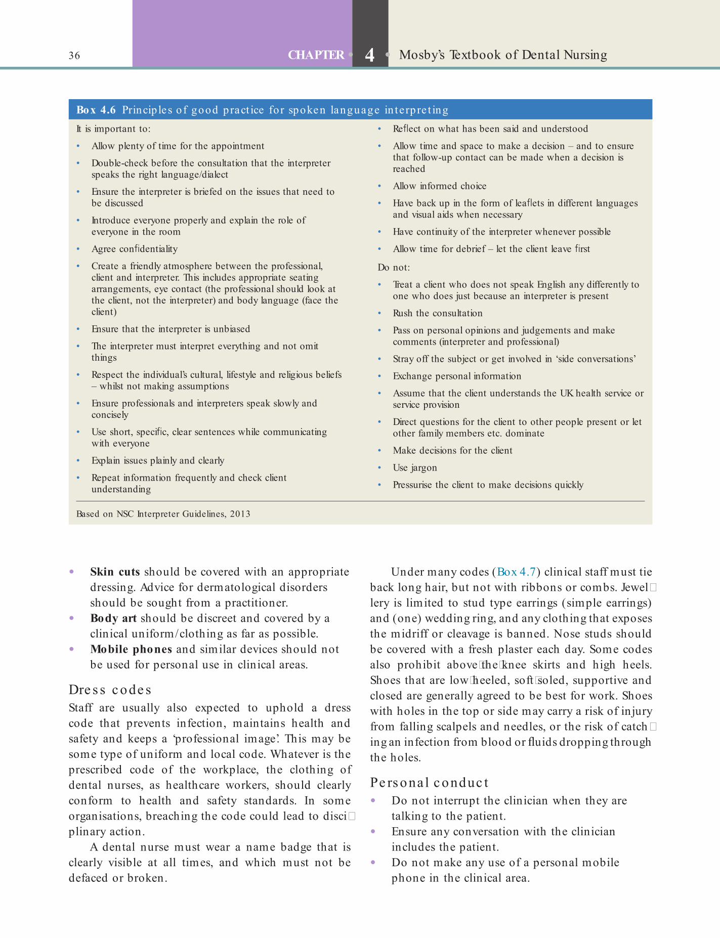
36 CHAPTER • 4 • Mosby’s Textbook of Dental Nursing
Based on NSC Interpreter Guidelines, 2013
Box 4.6 Princip les o f good pract ice for spoken language in terpre t ingIt is important to:
• Allow plenty of time for the appointment
• Double-check before the consultation that the interpreter speaks the right language/dialect
• Ensure the interpreter is briefed on the issues that need to be discussed
• Introduce everyone properly and explain the role of everyone in the room
• Agree con dentiality
• Create a friendly atmosphere between the professional, client and interpreter. This includes appropriate seating arrangements, eye contact (the professional should look at the client, not the interpreter) and body language (face the client)
• Ensure that the interpreter is unbiased
• The interpreter must interpret everything and not omit things
• Respect the individual’s cultural, lifestyle and religious beliefs – whilst not making assumptions
• Ensure professionals and interpreters speak slowly and concisely
• Use short, speci c, clear sentences while communicating with everyone
• Explain issues plainly and clearly
• Repeat information frequently and check client understanding
• Re ect on what has been said and understood
• Allow time and space to make a decision – and to ensure that follow-up contact can be made when a decision is reached
• Allow informed choice
• Have back up in the form of lea ets in different languages and visual aids when necessary
• Have continuity of the interpreter whenever possible
• Allow time for debrief – let the client leave rst
Do not:
• Treat a client who does not speak English any differently to one who does just because an interpreter is present
• Rush the consultation
• Pass on personal opinions and judgements and make comments (interpreter and professional)
• Stray off the subject or get involved in ‘side conversations’
• Exchange personal information
• Assume that the client understands the UK health service or service provision
• Direct questions for the client to other people present or let other family members etc. dominate
• Make decisions for the client
• Use jargon
• Pressurise the client to make decisions quickly
• Skin cuts should be covered with an appropriate dressing. Advice for dermatological disorders should be sought from a practitioner.
• Body art should be discreet and covered by a clinical uniform/clothing as far as possible.
• Mobile phones and similar devices should not be used for personal use in clinical areas.
Dre s s c ode sStaff are usually also expected to uphold a dress code that prevents infection, maintains health and safety and keeps a ‘professional image’. This may be some type of uniform and local code. Whatever is the prescribed code of the workplace, the clothing of dental nurses, as healthcare workers, should clearly conform to health and safety standards. In some organisations, breaching the code could lead to disci�plinary action.
A dental nurse must wear a name badge that is clearly visible at all times, and which must not be defaced or broken.
Under many codes (Box 4.7) clinical staff must tie back long hair, but not with ribbons or combs. Jewel�lery is limited to stud type earrings (simple earrings) and (one) wedding ring, and any clothing that exposes the midriff or cleavage is banned. Nose studs should be covered with a fresh plaster each day. Some codes also prohibit above�the�knee skirts and high heels. Shoes that are low�heeled, soft�soled, supportive and closed are generally agreed to be best for work. Shoes with holes in the top or side may carry a risk of injury from falling scalpels and needles, or the risk of catch�ing an infection from blood or uids dropping through the holes.
Pe rsona l c onduc t• Do not interrupt the clinician when they are
talking to the patient.• Ensure any conversation with the clinician
includes the patient.• Do not make any use of a personal mobile
phone in the clinical area.

37Dental Nursing Practice and Communication
Box 4.7 Dress codes in pract iceDress codes can be controversial. At least one hospital banned nurses from wearing Croc shoes, suggesting they might be dangerous. But some surgeons use Crocs in the operating theatre, believing they are easier to clean. Even NHS rules can vary. In England and Wales for example, bare-below-the-elbows dress code for clinicians is recommended since it ‘helps to support effective hand-washing and may reduce the risk of patients catching infections’. Other codes have caused considerable controversy and even made newspaper headlines. See the Daily Mail online archive for example, for the article ‘Don’t forget to wear socks and make sure your shoelaces match’ (31 December 2007).
The Scottish code (published on the Scottish Government website at: www.scotland.gov.uk/Publications/2008/08/interimdresscode) is reproduced below:
• Staff should dress in a manner that is likely to inspire public con dence:
• For example: in clean uniform (where uniform is a requirement), with hair tied back off the collar, with nails kept short and clean.
• Wear clear identi ers, (e.g. badges, epaulettes etc).
• Where changing facilities are available, staff should change into and out of uniform at work. In any case, staff should avoid undertaking activities in public, such as shopping, whilst wearing their uniform, except where such activities form an integral part of their duties.
• Appropriate steps should be taken to minimise the risks of infections and cross contamination for patients and the public:
• For example: staff should wear short-sleeved shirts/blouses and avoid wearing white coats or neck ties when providing patient care.
• Staff should not wear false nails or hand or wrist jewellery (other than a plain wedding ring) when providing patient care.
• All appropriate health and safety requirements for staff should be met:
• For example: staff should not wear excessive jewellery, such as necklaces, visible piercings and multiple earrings.
• Staff should wear soft-soled, closed-toe shoes.
• Staff should not carry pens or scissors in outside breast pockets.
• Be sensitive to the social, cultural and diversity and equality needs of staff and patients:
• For example: tattoos which could be deemed offensive should be covered where this does not compromise good clinical practice.
• Do not discuss other patients or people.• Do not discuss patients’ details outside the
surgery as they are con dential.• Do not eat or drink in a clinical area but rather
in another separate designated area for infection control and health and safety reasons.
• Do not chew gum in the workplace.• Do not wear an iPod or other such device.• Do not carry out personal hygiene measures
such as combing hair or blowing your nose in a clinical area.
• Do not develop non�professional relationships with patients.
Soc ia l me diaSocial networking sites and other social media are increasingly popular and offer a very effective and entertaining way of communicating and sharing infor�mation, but healthcare professionals need to be cau�tious in what they post on such sites. The GDC offers the following guidance on using social media:
Standard 4.2.3 o Standards for the Dental Team states:
‘You must not post any in ormation or comments about patients on social networking or blogging sites. I you use pro essional social media to discuss anonymised cases or the purpose o discussing best practice you must be care ul that the patient or patients cannot be identif ed.’
Social media covers a number o internet based tools that allow people to create and exchange content. It includes blogs, internet orums, content communities and social networking sites such as Twitter, YouTube, Flickr, Facebook, LinkedIn, GDPUK, Instagram and Pinterest.
When using social media, you must:a. Maintain and protect patients’ in ormation by
not publishing any in ormation which could identi y them on social media without their explicit consent.
b. Maintain appropriate boundaries in the relationships you have with patients.
c. Comply with any internet and social media policy set out by your employer.As a registrant you have a responsibility to
behave pro essionally and responsibly both online and o ine. Your online image can impact on your pro essional li e and you should not post any in ormation, including photographs and videos, which could bring the pro ession into disrepute.
It is important to remember that anything you post on social media is in the public domain and can be easily copied and redistributed without your knowledge. You should presume that
38 CHAPTER • 4 • Mosby’s Textbook of Dental Nursing
The Dental AppointmentHazards and Risks of HealthcareHealthcare aims to improve the health of patients and prevent disease, but can itself carry risks. The terms ‘hazard’ and ‘risk’ are often used interchangeably but this simple example explains the difference between the two (www.hsa.ie/eng/Topics/Hazards/). If there was a spill of water in a room then that water would present a slipping hazard to persons passing through it. If access to that area was prevented by a physical barrier then the hazard would remain though the risk would be minimised.
Dentistry is essentially very safe in healthy patients. Thus morbidity (disease) and mortality (deaths) fol-lowing dental procedures are even less excusable than when they follow, for example, more invasive surgery.
everything that you share online will be there permanently.
You should think care ully be ore accepting riend requests rom patients.
You should regularly review your privacy settings to ensure that in ormation is not accessed by unintended audiences. However, you should remember that even the strictest privacy settings do not guarantee that your in ormation will be kept secure and any in ormation that you post could be viewed by anyone including your patients, colleagues or employer.
You should remember that even i you do not identi y yoursel as a dental pro essional, you could still put your registration at risk i you display inappropriate behaviour whilst using social media.
You should not have discussions with your patients about their dental care and treatments on social media.
While online discussions about anonymised patients and best practice can have an educational and pro essional benef t you should remember that posting in ormation under another username does not guarantee your conf dentiality. You should consider how your comments re ect on you as well as how they could impact on the public’s trust in the pro ession.
I you believe patients are being put at risk by a colleague’s conduct, behaviour or decision-making, or by your working environment you should seek advice rom your employer, de ence organisation, pro essional association or Public Concern at Work. Principle 8 in Standards for the Dental Team has urther guidance on raising concerns. Social media should only be used to raise concerns as a last resort.
You must not use social media as a mechanism to raise concerns about the possible abuse o children or vulnerable adults. These concerns must be re erred to the appropriate authority such as your local Social Services Department.
You may f nd it help ul to contact your pro essional association or indemnif er or urther guidance on the responsible use o social media.GDC, Guidance on using social media (2013) www.gdc-uk.org/dentalprofessionals/standards/documents/guidance%20on%20using%20social%20media%20(sept%202013).pdf
Terms to lea rnMorbidity: the state of being unwell or having a disease. Here, it means developing a condition or ill health becoming worse after a dental or medical procedure.
Mortality: a term that pertains to death. Here it means a patient dying as a result of a dental procedure.
Drugs, particularly those that act on the central nervous system (CNS; e.g. sedatives and anaesthetic agents) are potentially dangerous and must be care�fully administered. Most dental procedures can be carried out under local anaesthesia (LA, sometimes called local analgesia) with minimal morbidity.
Conscious sedation (CS) is not as safe as LA alone. CS must be carried out:
• In appropriate facilities• By adequately trained personnel• With due consideration of the possible risks.
General anaesthesia (GA) with intravenous or inhalational agents is only permitted in a hospital with appropriate resuscitation facilities. It is not often needed for dental treatment, and then only in a hos�pital setting: because of its potential dangers it must be carried out by a quali ed anaesthetist. CS is con�siderably safer than GA, and is thus preferred.
Surgical procedures are generally the most hazard�ous. In the dental environment, procedures that involve operative interventions such as drilling teeth and cutting tissues and bone are the main ones that can be hazardous.
39Dental Nursing Practice and Communication
His to ry takingHistory taking is a con dential matter both ethically and legally and must always be done privately so as not to be heard by others.
Pe rs onal de ta ilsPersonal details ( rst and last names, the name by which the patient wishes to be known, date of birth, gender (sex), religion, occupation, relationship status, address and contact details) are necessary information for administrative purposes. This information may be collected by the receptionist or dental nurse. In fact, obtaining this information provides the patient with the opportunity for a smooth introduction to the dental team, and an opportunity for individual intro�duction suitable to the particular culture. As stated above, it is usually helpful to ask the patient how they would wish to be addressed.
Having read the previous section, you will appreci�ate that every effort has to be made to identify the medically compromised patient: for this a medical history is essential.
Medical his torySee also Chapters 13 and 27. When taking a medical history, the dental clinician will usually ask a struc�tured set of questions. Patients are often also given a form for them to supply all the information they can about their health and any medication they are receiv�ing. The history may signi cantly change with time. Therefore, it is essential to ask about any changes and update the history before each new course of treat�ment, every sedation session and especially before surgery or GA. For example, a female patient who is not pregnant at one course of treatment could well be at the next.
It is helpful for dental nurses to have basic knowl�edge about the essential components of a medical history. Many of the items below may be included on the form that is handed out to the patient to ll in before they see the dental clinician. The completion of such a form provides:
• A useful base for the clinician from which to enquire further about any concerning items on the form
Risks mainly happen when the patient is not healthy, inadequate time is taken and/or the proce�dure is invasive (tissues are disrupted), or if staff are overambitious in terms of their skill or knowledge. Work only in your area of competence.
Thus there is always a need for doing risk assess�ments and for careful peri-operative care.
Term to lea rnPeri-operative: the term that covers the period of time starting from preparation for surgery up to the time the procedure is completed and the patient can go home safely.
Patient Care during Diagnosis
Ris k as s e s s me ntAn adequate risk assessment endeavours to anticipate and to prevent trouble. This topic is covered in the context of a medical emergency in Chapter 27. This chapter explains its relevance in day�to�day practice.
At the start of a patient’s visit it is essential to:1 . Assess patient’s needs.2 . Obtain a careful medical, dental, family, social
(and sometimes developmental) history and make a risk assessment.
3 . Obtain patient’s consent to any investigations required.
4 . Obtain patient’s consent to agreed treatment plan.A patient’s ‘ tness’ for a procedure depends on
several factors (see Chapter 13, 21, 22 and 25, and see below p. 40). Many patients with life�threatening dis�eases now survive as a result of advances in surgical and medical care. Such diseases can signi cantly affect the dental management of the patient. A patient attending for dental treatment and apparently ‘ t’ may actually have a serious systemic disease. Or they may be taking drugs (including recreational drugs). Both of these might in uence the healthcare that can be deliv�ered to the patient.
Key poin tNo interventional procedure is entirely free from risk but care can be improved by making an adequate assessment based on history, clinical signs and, where appropriate, investigations, and minimising trauma and stress to the patient.
Term to lea rnSystemic: a condition that affects a system or the whole body as opposed to a distinct local site.
The risk is greatest when surgery is needed, and when GA or sedation is given. In addition, problems may be compounded if medical support is not at hand.
40 CHAPTER • 4 • Mosby’s Textbook of Dental Nursing
at the patient as a whole. A patient’s appearance, behaviour and speech, body language and inspection of the face, neck, arms and hands can reveal many signi cant conditions. The clinician needs to look for anxiety, movements and tremors, and tiredness, and for facial changes (e.g. expression, pallor, cyanosis or jaundice), dyspnoea or wheezing, or nger clubbing. The nurse can help if they notice anything untoward. However, remember that even very ill patients can look remarkably well.
The clinician will examine the neck as swollen lymph nodes are sometimes a sign of disease such as infections or cancer. Hospital in�patients always also have a full physical examination before GA and operation, which includes at least the following systems:
• Cardiovascular: pulse, blood pressure, heart sounds.
• Respiratory: respiratory rate, lung expansion, tracheal position, lung sounds.
• Gastro�intestinal: any swelling or tenderness.• Neurological: especially the cranial nerves.
Inves tiga tionsThe history and physical examination often reveal most if not all of the clinically useful information and data. However, in some patients, tests (investigations) may be needed.
Before any investigations are initiated, the clini�cian must obtain the patient’s consent.
• Useful evidence if the patient makes a complaint or medico�legal claim.
Pres enting complaint (PC)This should be recorded in the patient’s own words, e.g. ‘I have pain in my face’.
His tory of pres enting complaint (HPC)The clinician needs to ask about the timing of the complaint and its evolution. For example, if the patient has facial pain, a useful mnemonic is SOCRATES:S – siteO – onset (gradual/sudden)C – characterR – radiationA – associations (other symptoms)T – timing/durationE – exacerbating and alleviating factorsS – severity (rate the pain on a visual analogue scale of 1–10).
Re levant medical his tory (RMH)RMH is covered in Chapters 13 and 27 in detail. RMH includes nding out about any past or present medical and surgical problems. Patients are also asked if they carry a medical warning card or device (e.g. Medic�Alert and Talisman). Because patients may not know if they have a condition that may affect their dental treatment, a functional enquiry/review of systems (ROS) can be helpful. This involves asking speci cally about certain conditions.
Socia l his toryThe clinician needs to tactfully nd out about a patient’s occupation, marital status, partner’s job and health, housing, dependants, mobility, lifestyle habits (alcohol, tobacco, betel khat and recreational drugs), culture and faith. The dental nurse may help in this. In addition, it is also worth nding out if the patient has any forthcoming social engagements that are dependent on them being able to carry out their usual activities following a dental treatment (for example, wedding/examination/job interviews). This may require rescheduling of treatment.
Family his toryThe medical history of blood relatives may be required as some relevant diseases run in families (e.g. haemophilia).
Clinica l examinationIt is important for dental clinicians not merely to inspect and examine the mouth and neck, but to look
Key poin ts: Good pract ice in pa t ien t ca re• Know your patient.
• Act professionally at all times.
• Follow the prescribed dress code.
• Ensure personal hygiene.
• Keep the environment user-friendly.
• Minimise hazards in the dental surgery.
Find out moreVisit the fo llowing websites to nd ou t more about ca ring for pa t ien ts in the most appropria te way:• NHS Nat iona l Services Sco t land . Pa t ient Deta ils
Amendments (www.psd .sco t .nhs.uk/professiona ls/den ta l/pa t ient -de ta il-amendments.h tml)
• Brit ish Denta l Hea lth Founda t ion . Frequent ly asked quest ions. Pa t ien ts’ righ ts (www.denta lhea lth .o rg .uk/faqs/lea e tde ta il.php?Lea e t ID=30)
Chapter 5 Health Services: Ethics and Governance
CHAPTER POINTS• The NHS (National Health Service)• The Health and Social Care Act 2012• Ethical practice in dental nursing• Consent• Clinical governance• Clinical effectiveness• Complaints, data issues• Disability and discrimination• Evidence-based dentistry• Care pathways• Medical devices• Whistle blowing
However, a person who lives in one part of the UK can receive treatment under the relevant NHS in another UK country without being discriminated against.
In t ro d u ct io n
As with life everywhere, there is increasing regulation and bureaucracy that impacts on oral healthcare. Readers should check internet sites, since this is a very rapidly evolving scene.
The National Health Service (NHS)The NHS is the name commonly used to refer to the UK’s publicly funded healthcare system, which was set up in 1948. It is now called by different names in dif-ferent parts of the UK:
• In England, NHS England is responsible to the UK Government.
• In Ireland, Health and Social Care in Northern Ireland (‘HSC’) is responsible to the Northern Ireland Executive.
• In Scotland, NHS Scotland is responsible to the Scottish Government.
• In Wales, NHS Wales is responsible to the Welsh Assembly.
Term to lea rnNICE: the UK government agency (National Institute for Health and Care Excellence, formerly National Institute for Clinical Excellence) that is responsible for deciding which treatments or investigations are required most commonly and are the most cost-effective (www.nice.org.uk).
NHS treatment is discussed in Chapter 13. The NHS has to run as cost-effectively as possible so as to avoid wasting precious resources, while delivering the best possible care to all patients irrespective of where they live in the UK. (Difference in care in different parts of the UK has been termed ‘postcode rationing’.) The organisations that are responsible for ensuring this are:
• In England and Wales: NICE (www.nice.org.uk)• In Scotland:
• NHS Quality Improvement Scotland (www.nhshealthquality.org)
• Scottish Intercollegiate Guidelines Network (SIGN) (www.sign.ac.uk)
• Scottish Medicines Consortium (SMC) (www.scottishmedicines.org.uk).
• In Northern Ireland: the Department of Health, Social Services and Public Safety (DHSSPS) through its Commissioning Plan (www.dhsspsni.gov.uk/2014-15-commissioning-plan-direction.pdf).Such healthcare-related government agencies and
the National Patient Safety Alerting System (NPSAS; www.en glan d .n h s.uk/ o u rwork/ patien tsafety/ p sa/national-psa-system/) also come under the NHS

42 CHAPTER • 5 • Mosby’s Textbook of Dental Nursing
CQC and comply with its Essential Standards of Quality and Safety (standards that outline the ideal outcome patients should experience when using a service). Each practice will need to be able to demon-strate compliance with the regulations in the follow-ing areas:
• Care and welfare of service users• Assessing and monitoring the quality of
provision• Safeguarding vulnerable service users• Management of medicines and medical devices• Cleanliness and infection control• Meeting nutritional needs• Safety and suitability of premises• Safety, availability and suitability of equipment• Respecting and involving service users• Consent to care and treatment• Complaints• Records• Competence and suitability of workers• Staf ng• Effective management of workers• Cooperating with other providers.
The Health and Social Care Act 2012The Health and Social Care Act 2012 was a major Coalition Government reform that, in England, put clinicians at the centre of commissioning, freeing up providers to innovate, empowering patients and giving a new focus to public health. The provisions in the Act were designed to make the NHS more responsive, ef -cient and accountable. The Act puts clinicians in charge of shaping services with the aim of enabling NHS funding to be spent more effectively. Supported by the NHS Commissioning Board, new clinical com-missioning groups (CCGs) now directly commission services for their populations. These replace the former primary care trusts (PCTs) (Box 5.2). Patients’ interests are protected by additional responsibilities given to Monitor, a public body that acts as a specialist regula-tor. The Act established new patient organisations called Healthwatch at local and national level, to gather and represent the public’s views on health and social care sevices. The Act provided the underpinnings for Public Health England, a new body to drive improve-ments in the public’s health. The former structure of health and care, and its regulation, in England is depicted in Figure 5.2. The new structure is under discussion at time of writing.
umbrella. NICE makes a range of guidelines available for clinicians (for examples, see Box 5.1).
The Care Quality Commission and the Care CommissionAn independent, regulatory body that dental nurses need to be aware about is called the Care Quality Commission (CQC) in England and the Care Com-mission in Scotland. Established in 2009 to regulate and inspect health and social care services in England, the CQC oversees services provided by the National Health Service (NHS), local authorities, private com-panies and voluntary organisations – whether in hos-pitals, care homes or people’s own homes. In order to be granted registration, care providers need to demon-strate that they can meet, or are already meeting, the registration requirements. The aim of the CQC is ‘to ensure better care for everyone in hospital, in a care home and at home’. Thus the CQC ensures the safety and quality of the care provided to patients through various assessment, monitoring and inspection proce-dures, and this includes dental professionals.
In April 2010, CQC introduced a registration system for all providers of health and social care which was introduced gradually across the care sector with the aim of incorporating primary care services that provide dentistry (NHS and private sector) by 1 April 2011. Thus, in addition to standards set by GDC, it is mandatory for dental practices to register with
Box 5.1 NICE guide lines: top ics with possib le implica t ions in den ta l and ora l hea lthcare• Anaphylaxis
• Child maltreatment
• Creutzfeldt–Jakob transmission
• Dental recalls
• Head and neck cancer
• Healthcare-associated infections
• Implants for orofacial reconstruction
• Infective endocarditis
• Ozone use in caries
• Smoking cessation
• Snoring
• Trigeminal neuralgia
• Wellbeing at work
• Wisdom (third molar) tooth removal
www.nice.org.uk/

43Health Services: Ethics and Governance
Box 5.2 St ructure of the NHSThe NHS is divided into two sections: primary and secondary care.
• Primary care – this is the most common ‘front-line’ service, that is, it is the rst point of contact with the NHS for most people. Primary care is delivered by a wide range of healthcare professionals such as doctors, dentists, pharmacists and optometrists. It also includes NHS Direct (formerly NHS 111) and walk-in centres. The government bodies in charge of primary care have, until 2014, been the primary care trusts (PCTs) (see Figure 5.1). These not only provided some primary and community services but PCTs also commissioned services from other providers. They were involved in commissioning secondary care (see below). PCTs controlled 80% of the NHS budget as well as overseeing general medical (GPs) and dental practitioners. This changed in April 2014, with the introduction of a National Commissioning Board to oversee new GP Commissioning Groups (there are 211 in England). Dental commissioning is less well developed at the time of writing. The Care Quality Commission (CQC) is the regulator and each area will have a patient advocacy group – Healthwatch.
• Secondary care (also known as acute healthcare) can be either elective care or emergency care. Elective care means specialist medical care or surgery that has been planned in advance and it usually follows referral from a primary or community health professional. Emergency care is unplanned care, for example following a car accident. The bodies in charge of providing secondary care in the UK include foundation NHS trusts, acute NHS trusts, mental health NHS trusts and NHS ambulance services trusts. The NHS care trusts provide both health and social care.
Fig u re 5.1 The former structure of NHS England.
Parliament
10 Strategic HealthAuthorities
NHS TrustsHospitals, Mental Health Services,Ambulances, Learning disability
Primary Care Trusts
Foundation TrustsAccountable to members
through governors
Secretary of Statefor Health
MonitorAs regulator
Disclosure and Barring Service (DBS) (Formerly the Criminal Records Bureau)The rst rule in all health care is ‘do no harm’ and the law also more formally tries to protect patients from harm, especially serious harm, via criminal records checks (see below).
Key poin tThe rst rule in health care is ‘do no harm’.
In the UK, children and vulnerable people are offered formal protection via criminal records checks; the rules differ between England & Wales (DBS: www .gov.uk/disclosure-barring-service-check/overview), Northern Ireland (AccessNI: www.dojni.gov.uk/accessni) and Scotland (DisclosureScotland: www. disclosurescotland.co.uk/basicdisclosureonline/index. htm).
The Criminal Records Bureau (CRB) and the Inde-pendent Safeguarding Authority (ISA) were merged following passage of the Protection of Freedoms Act 2012 to become the Disclosure and Barring Service (DBS). CRB checks are now called DBS checks.
The DBS is an executive agency of the Home Of ce, which vets applications for people who want to work with children and vulnerable people. People working in the UK healthcare sector also require DBS checks. The Foster Review on the regulation of the non-medical healthcare professions, when describing ‘good character’, referred to objective tests to measure this, such as the absence of criminal convictions and adverse decisions by regulatory bodies, and the infor-mation about likely criminal activity contained in a DBS disclosure. The role of the CRB (now DBS) was enhanced by the Safeguarding Vulnerable People Act 2006 in response to the recommendations of the Bichard Inquiry (a public inquiry into child protection which identi ed failures in the vetting and barring system).
DBS disclosures are either ‘standard’ or ‘enhanced’.
Standard DBS dis c los ure sThese disclosures reveal details of any convictions, cautions, reprimands and nal warnings the applicant has received, regardless of length of time since the incidents. They also reveal details of whether that person is banned from working with children or vul-nerable adults (if these details have been requested). The DBS aims to issue Standard Disclosures within 10 days of receipt of the application.
Enhanc e d DBS dis c los ure sThese disclosures are for positions involving greater contact with children or vulnerable adults (e.g. most health professionals) and involve an additional check

44 CHAPTER • 5 • Mosby’s Textbook of Dental Nursing
with the police. The police then check if any other information is held on le that may be relevant (for instance, investigations that have not led to a criminal record). The police will decide what (if any) additional information will be added to the Disclosure. The DBS aims to issue enhanced disclosure within 28 days of receiving the application.
Find out moreVisit the DBS website (www.gov.uk/d isclosure-barring -se rvice-check/overview) to nd ou t more about DBS checks, the Bichard Inquiry and how often these checks must be comple ted .
DBS checks need repeating every three years or if you change jobs or roles. Even if on the following day!
Fig u re 5.2 The health and care system from April 2013, following implementation of the Health and Social Care Act 2012.
Key po in tPatients are increasingly better educated, much more knowledgeable, both about their rights and about healthcare in general, and have more access to information. As a consequence, many have high expectations, and want to be kept better informed and involved in decision making. The current UK government obviously supports this approach with its phrase: ‘No decisions about you, without you’.
The law requires doctors and dentists to inform the patient of the common risks of a procedure even if they are not serious, and (conversely) the serious risks of the surgery even if they are not common.
Ethical Practice in Dental NursingConsent

45Health Services: Ethics and Governance
In the UK, all competent adults, namely a person aged 18 and over who has the capacity to make their own decisions about treatment, can consent to dental treatment. They are also entitled to refuse treatment, even where it would clearly bene t their health. At age 16, a young person is regarded as an adult and can be presumed to have the capacity to consent to treatment. Consent regarding children is discussed on page 46. The Department of Health has guidance (Appendix 5.1).
Info rme d or va lid c ons e nt‘Informed’ or ‘valid’ consent means that the patient agrees to treatment based on the assumption that they are fully aware of the treatment they will undergo, including its intended bene ts, its possible risks and the level of these risks. In particular, patients must be warned about:
• Any preparation that may be required before treatment (e.g. giving an anaesthetic before a tooth extraction)
• Possible adverse effects of the treatment (e.g. prolonged numbness in the lip and cheek after a tooth extraction, most likely a lower third molar)
• Possibility of any effects that may occur straight after a procedure (e.g. swelling, bruising, bleeding, pain)
• Where they will be during their recovery• Possible use of intravenous infusions or other
invasive treatments.To give valid consent, patients must receive suf -
cient information about their condition and proposed treatment. It is the clinician’s responsibility to explain all the relevant facts to the patient, and to ascertain that they understand them. The information given to patients must, as a minimum, include:
• The nature, purpose, bene ts and risks of the treatment
• Alternative treatments and their relative bene ts and risks
• All aspects of the procedure expected to be carried out
• The consequences and prognosis if no treatment is given.
Consent for surgery or anaesthesia means that the patient gives permission to undergo the procedure, and this prevents the operator from being charged with criminal battery (and with professional negligence and/or malpractice). Informed (or valid) consent means that the patient has not only consented to the procedure, but is fully informed and knowledgeable about all its possible risks and consequences. To be able to provide informed consent, however, requires the person to be in full control of their faculties, and to be legally capable of giving such consent. Things that can make it legally dif cult for a patient to consent, include being, at the time of the consent:
• a minor• in labour• intoxicated• mentally disabled• mentally ill• semi-conscious• under great stress or in pain• under the in uence of drugs or medication.
Key poin tFor consent to be valid, the patient or proxy decision-maker must be competent, have the necessary information and make their choice freely; they are also entitled to change their mind at any time.
Consent in relation to dentistry is the expressed or implied agreement of the patient to undergo a dental examination, investigation or treatment. The law in relation to consent is evolving and there are signi cant variations between countries. However, the principles are essentially the same:
• Before a health professional examines, treats or cares for competent adult patients they must obtain the patient’s consent.
• Adults are always assumed to be competent unless demonstrated otherwise. If there are doubts about their competence, the question to ask is: ‘Can this patient understand and weigh up the information needed to make this decision?’ Unexpected decisions do not prove the patient is incompetent, but may indicate a need for further information or explanation.
• Patients may be competent to make some healthcare decisions, even if they are not competent to make others.
• Giving and obtaining consent is usually a process, not a one-off event. Patients can change their minds and withdraw consent at any time.
Term to lea rnPrognosis: a prediction of how a disease or patient’s condition may worsen or improve over time, with or without treatment.
46 CHAPTER • 5 • Mosby’s Textbook of Dental Nursing
Cons e nt fo r c hildre nChildren under the age of 16 years and minors aged 16 and 17 may have capacity to consent provided they are ‘Gillick competent’. This means they have the ability to understand the nature, purpose and possible consequences of the proposed investigation or treat-ment, as well as the consequences of non-treatment. Such children may consent to treatment without their parents’ authorisation, although their parents should ideally be involved. Legally, a parent can consent if a competent child refuses, but it is likely that taking such a serious step will be rare.
Where a child under 16 is not deemed competent to consent, a person with parental responsibility (e.g. their mother or guardian) may authorise investiga-tions or treatment that are in the child’s best interests. Generally, however, formal assent to treatment from a legal parent or guardian is sought for treatment of all children under 16 years.
Cons e nt fo r adults without c apac ityAdults without capacity cannot give consent to treat-ment. Currently, in England and Wales, no one can authorise treatment on behalf of an adult unless they are that person’s named legal attorney under a Health and Welfare Lasting Power of Attorney that has been lodged with the Of ce of the Public Guardian. However, patients without capacity to consent may receive dental treatment if it is in the patient’s best interests, with the views of relatives and carers taken into account.
In Scotland, the Adults with Incapacity (Scotland) Act 2000, which came into effect in 2002, allows a competent adult to nominate a person, known as a welfare attorney or proxy, to make medical decisions on their behalf if and when they lose the capacity to make those decisions for themselves. The Act also pro-vides for a general power to treat a patient who is unable to consent to the treatment in question. In order to bring that power into effect, the medical prac-titioner primarily responsible for treatment must have completed a certi cate of incapacity before any treat-ment is undertaken, other than in an emergency.
The Mental Health (Care and Treatment) (Scot-land) Act 2003 allows for medical/dental interven-tion to prevent serious deterioration in the patient’s mental health condition or to prevent the patient from harming themselves.
The Mental Capacity Act (England and Wales) 2005 (MCA) is central to the legal issues around treating patients over the age of 16 who lack capacity
If the patient is not offered as much information as they reasonably need to make their decision, and in a form they can understand, their consent may not be valid. For example, information for those with visual impairment may be provided in the form of audio tapes, Braille, or large print.
Form o f c ons e ntConsent can be written, oral or non-verbal. It must be obtained from all patients having an operation. The possible bene ts of the treatment must be weighed against the risks and are always discussed by the person carrying out the procedure, or, if for some reason this is not possible, a delegated person with the appropriate expertise to do so. A signature on a consent form does not itself prove the consent is valid – the point of the form is to record the patient’s decision, and also, increasingly, the discussions that have taken place.
Find out moreDoes your workplace have a po licy se t t ing ou t when the clin ician needs to ob ta in consen t in writ ing?
Te s ts o f c ons e ntThere are ways of legally testing whether the consent given by a patient was valid or not. The ‘Bolam test’ is one such way. The Bolam test states that a doctor [or health professional] who:
Acted in accordance with a practice accepted as proper by a responsible body of medical men skilled in that particular art is not negligent if he is acting in accordance with such a practice, merely because there is a body of opinion which takes a contrary view.
However, a judge may on certain occasions choose between two bodies of medical opinion, if one is to be regarded as that which cannot be logically defended (Bolitho principle). The main alternative to the Bolam test is the ‘prudent-patient test’, which is widely used in North America. According to this test, doctors (and dentists) should provide the amount of information that a ‘prudent patient’ would want.
Conf rmation o ConsentCon rmation of consent should be completed by a health professional when the patient is admitted for the procedure, if the patient has signed the form in advance.

47Health Services: Ethics and Governance
and are also supported by the following GDC statements:
• Conducting clinical trials (2008)• Responsible prescribing (2008)• Child protection and vulnerable adults (2013)all of which are available through the GDC website.
Currently, all members of the registered dental team can receive payment for dental treatment, can own dental practices/dental laboratories and can employ other members of the dental team. Also, any dental practice or group of practices can become a corporate body.
to consent to treatment. The Act is particularly signi -cant in two ways relevant to consent to medical management:
• It allows consent to be given or withheld, for the medical treatment of patients who lack capacity, by another person (typically a close relative).
• It provides statutory recognition of ‘advance directives’. These are statements made by a person while competent (i.e. while having legal capacity) about the treatment that they would want, or not want, in speci ed situations, in the future were they to lack capacity at the time the treatment would be relevant.The above information provided is from UK law.
Remember that the legal situation with regard to consent varies around the world and is subject to con-tinued debate and development.
Find out moreCheck out the websites o f the various den ta l o rganisa t ions for more de ta ils about he lp ava ilab le :• Denta l Defence Union (DDU): www.the-ddu .com• Denta l Protect ion Limited (DP):
www.denta lprotect ion .org• Medica l and Denta l Defence Union of Scot land
(MDDUS): www.mddus.com/mddus/home.aspx
Conf dentialityDental professionals have a legal and ethical duty to keep patient information con dential and use it only in the context in which it was given. Con dential information should be kept in a secure place at all times to prevent unauthorised disclosure or accidental disclosure.
If exceptional circumstances arise in which a dental professional feels that the disclosure of con -dential information is necessary for the patient’s safety, they should seek appropriate advice before any action is taken.
Other Ethical IssuesThe GDC offers guidance on all respects in which the dental team should work together in the best interests of patients (Standards for the Dental Team, 2013, Stand-ard 8). This includes dental professionals’ respon-sibility to raise matters of concern about colleagues, systems and the working environment (‘whistle blowing’; see p. 58), and how to go about this.
The nine core ethical principles that registered dental professionals must observe are listed in Stand-ards for the Dental Team (2013) (see also Chapter 1)
Term to lea rnAutonomy: the patient’s right to make decisions about their medical care. Although the healthcare provider should educate and inform the patient about factors that could affect the patient’s treatment, they must not try to in uence the patient in their decision making or make the decision themselves.
Key po in tThe care that is provided to patients should be clinically appropriate and cost-effective, and delivered with proper regard to the dignity and autonomy of the patient.
Clinical GovernanceClinical governance (now increasingly called ‘quality assurance’ or QA) is an umbrella term for everything that helps maintain and improve the standards of patient care (Box 5.3).
Clinical governance is the term given to the sets of systems and processes that are put in place to ensure that patients receive the highest quality care possible. Clinical governance was introduced to help to address
• the issues of differences in the quality of care existing throughout the UK
• the public concern about well-publicised cases of ‘poor professional performance’
• the greater expectations of the NHS and other providers by patients.Clinical governance is an NHS and CQC require-
ment – a framework for quality assurance essential for all dentists in the General Dental Services (GDS) to ensure consistent delivery of high-quality treat-ment while seeking to continuously improve it. The Department of Health has de ned clinical govern-ance in the NHS as ‘a framework through which NHS

48 CHAPTER • 5 • Mosby’s Textbook of Dental Nursing
1. Child protection2 . Clinical audit3 . Clinical record4 . Clinical staff requirements/development5 . Dental radiography6. Evidence-based practice7 . Fair and accessible care8 . Infection control9 . Patient information and feedback
10. Prevention11. Safety assessment12 . Staff involvement/development
A clinical governance framework must include the whole dental team, who must understand
• what the practice is supposed to do – what is the main objective, and
• their role in delivering the service.Clinical governance embraces:
• Policies – the basis of all actions in the practice• Procedures – how everything is carried out• Risk assessments – how risks are identi ed and
managed• Audits – how current practice compares to the
gold standard• Reviews – checks that procedures are being
followed and standards met.Monitoring systems should be put in place to assess whether goals that are set are being achieved, and processes should allow for continuous improvement and be continually reviewed.
Implementing Clinical GovernanceThe essential features of clinical governance are:
• The patient care provided should be safe and risks should be managed effectively.
• Health services are accountable for the safety, quality and effectiveness of clinical care delivered to patients.
• Healthcare staff should participate in shared learning and teamwork, and identify and improve any shortfalls in their knowledge and service.Every healthcare organisation (NHS or private) is
responsible for ensuring that clinical governance measures are in place. In the UK, the Care Quality Commission monitors the clinical governance arrange-ments of NHS organisations and rates the perform-ance of each organisation. (Its predecessors, the Healthcare Commission, Commission for Social Care
organisations are accountable for continuously improving the quality of their services and safeguard-ing high standards of care by creating an environment in which excellence in clinical care will ourish’ (‘A First Class Service’, 1998).
Hallett and Thompson, in their book Clinical Gov-ernance: A Practical Guide for Managers (2001) state:
Clinical governance is about a culture shift to a patient-centred, accountable, safe and high quality service in an open and questioning environment. Realising the potential of clinical governance involves a substantive shift in culture, questioning traditional structures and ways of doing things and creating an understanding of why a consistent and sustained approach to improving the quality of healthcare is necessary.Clinical governance can be broken down into
three components:1 . Clinical effectiveness2 . Risk management3 . Patient focus and public involvement.It represents a holistic approach to patient-centred practice and may be considered to have seven key ele-ments, which are known as ‘The 7 Pillars’, remem-bered by the mnemonic CAREPUS:C – Clinical effectivenessA – AuditR – Risk assessment, research and developmentE – Education, training and continuing personal and professional developmentP – Policy, patient and carer experience and involvementU – Using information and ITS – Staf ng and services, and staff management
Clinical Governance and DentistryThe Primary Care Dental Services Clinical Governance Framework (2006) has dental standards that focus on 12 themes as follows:
Box 5.3 Clin ica l governanceClinical governance is:
• Doing the right thing
• In the right way
• At the right time
• By the right people
• To the right people
• And being able to measure it.

49Health Services: Ethics and Governance
best available evidence. Areas that can be audited include:• Professional performance, that is,
performance of various members of staff• Risk management• Patient satisfaction• Use of resources.
• Staf ng and management. Organisations should adopt a ‘quality improvement’ approach to human resource management. This includes staff appraisals, and mechanisms to deal with poor performance (such as ‘whistle blowing’; see p. 58) rather than ignoring such practices.
• Clinical effectiveness. This includes how each organisation implements and applies effective clinical practice (see below).
• Continued professional development (CPD). See Chapter 3 for details.
• Information use. Each organisation should have information that supports clinical governance and provides information on the patient experience.
Find out moreWhat a re the contact de ta ils o f your loca l Pa t ient Advice and Lia ison Services?
Clinic a l e ffe c tive ne s sClinical effectiveness is about improving the quality of treatments and services. Health professionals, who provide the actual service, therefore will have the expertise, rst-hand knowledge and skills, and an
Inspection and the Mental Health Act Commission, ceased to exist on 31 March 2009.)
Clinical governance can be achieved by coordinat-ing its seven components, which were identi ed above as ‘The 7 Pillars’.
• Patient and public involvement. This includes use of patient satisfaction surveys to assess patients’ views on the service being provided and the Patient Advice and Liaison Services (PALS), which exist in every NHS trust.
• Risk assessment. See below.• Clinical audit (Box 5.4, Figure 5.3). This
procedure documents what the current clinical standards are with regard to a particular issue (e.g. the quality of radiographs being taken), or a particular treatment (e.g. extraction of wisdom teeth). This is then compared with a recognised external standard, which may be a local or national standard but which is evidence-based (e.g. a guideline issued by NICE), whenever this is available and appropriate. Changes are then made (if required) to ensure that the current clinical practice is in line with
Box 5.4 Steps in performing an audit (see Figure 5.3)
1. Prepare for audit
• Choose a topic of concern to staff.
• Set aside time to conduct audit.
• Ensure support of colleagues.
2. Select criteria
• Select the criteria against which the issue will be compared. Clear criteria are best obtained from available guidelines or reviews of evidence (external standards).
3. Measure performance
• Collect data on current practice. Depending on the topic, this may be from clinical records and/or department/hospital databases. Using several data sources helps overcome problems of incomplete records.
• Measure performance against the selected criteria.
4. Make improvements
• In the light of the ndings, devise a plan for implementing improvements.
• Discuss improvements with colleagues and think about potential barriers to change.
• Create policies/change practice to implement change.
5. Sustain improvements
• Monitor and reinforce the improvements, and keep up to date.
Fig u re 5 .3 The audit cycle.
Choose topic
Involvestakeholders
Set standard
Collect andanalyse data
Compare findingswith standard
Agree strategyfor change
Implementchanges
Re-audit

50 CHAPTER • 5 • Mosby’s Textbook of Dental Nursing
When a patient wishes to complain, depending on the nature of the complaint, they may contact (Figure 5.4):
• The service provider, Care Quality Commission or the Ombudsman for NHS care
• The Dental Complaints Service for private care• The GDC• The Health and Safety Executive• The Advertising Standards Authority• The Of ce of Fair Trading/Trading Standards
Of cer• The police.
People who use healthcare services mostly under-stand that mistakes sometimes happen. When some-thing goes wrong, often all the person affected wants to know is how it happened, that the persons involved are sorry, and that steps will be taken to prevent it from happening again. So the way in which an organ-isation responds to the initial contact by a person who is unhappy about their service is important. It is crucial to obtain all the information that will allow assessment of someone’s concerns correctly, resolve them quickly if possible and build a good ongoing relationship with them. Often the reason people give for being unhappy about how their complaint has been handled is poor communication by the services (Box 5.6).
There should be a person designated to handle complaints. It is more usual for a patient to complain verbally than in writing; in either event, it is wise for the dental professional involved at this stage to com-municate with their medical insurance society (e.g. the Dental Defence Union – DDU; Dental Protection – DP; and Medical and Dental Defence Union of Scot-land – MDDUS) and heed their advice.
Remember that all complaints offer the opportu-nity to review practice procedures to avoid future issues.
insight and understanding as to how the service works and how it could be improved. Their involvement in audits and improvement projects is therefore an important part of promoting good clinical practice.
Ris k as s e s s me ntRisk assessment and management is a standardised process to reduce injuries, errors, faults and accidents and at the same time improve quality. Up to 10% of patient care episodes result in harm to patients or staff, and half of these incidents are preventable. Clinical incidents and near-misses highlight the need to learn from such incidents and for action to reduce or manage risks.
Risks should be assessed as a four-stage process:1 . Identify risks using screening, checklists, etc.2 . Assess frequency and severity of risk3 . Eliminate risks where possible4 . Reduce the risk, and plan for damage limitation
where elimination of risk is impossible.The National Patient Safety Agency (NPSA) leads
and contributes to improved, safe patient care in England and Wales. It does this by providing informa-tion, support and recommendations for safer practice to NHS organisations. It has three divisions:
• Reporting and Learning Service – this division aims to improve safety by enabling NHS organisations to learn from patient safety incidents
• National Clinical Assessment Service – this division provides con dential services to help manage any concerns about the performance of healthcare practitioners
• National Research Ethics Service – this division protects the safety and dignity of research participants by promoting ethical research.To provide a quality health service, issues of
quality, quantity and cost often compete. However, through clinical governance, the best balance can be established.
ComplaintsComplaints should be avoided where possible by good practices and rapid and ef cient responses to patients’ wishes (Box 5.5).
Key poin tListen to the patient’s complaint, obtain all the information, and if need be, refer to a senior person, to resolve the issue quickly.
Box 5.5 Princip les o f good compla in ts handling• Getting it right.
• Being customer-focused.
• Being open and accountable.
• Acting fairly and proportionately.
• Putting things right.
• Seeking continuous improvement.
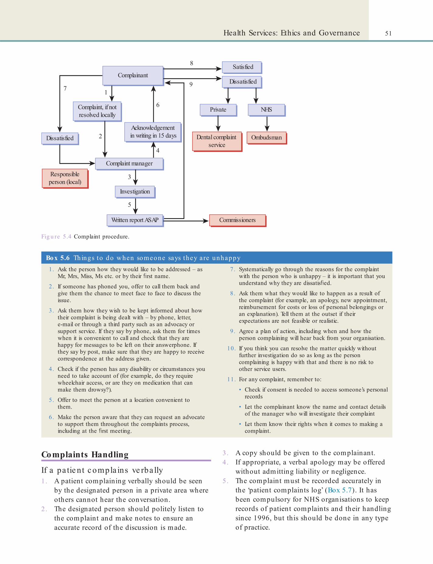
51Health Services: Ethics and Governance
Fig u re 5.4 Complaint procedure.
Complainant
1
2
3
4
5
6
9
8
7
Complaint, if notresolved locally
Acknowledgement in writing in 15 daysDissatisfied
Dissatisfied
Complaint manager
Responsibleperson (local)
Investigation
Written report ASAP Commissioners
Satisfied
Private NHS
OmbudsmanDental complaint service
Box 5.6 Th ings to do when someone says they a re unhappy1. Ask the person how they would like to be addressed – as
Mr, Mrs, Miss, Ms etc. or by their rst name.
2. If someone has phoned you, offer to call them back and give them the chance to meet face to face to discuss the issue.
3. Ask them how they wish to be kept informed about how their complaint is being dealt with – by phone, letter, e-mail or through a third party such as an advocacy or support service. If they say by phone, ask them for times when it is convenient to call and check that they are happy for messages to be left on their answerphone. If they say by post, make sure that they are happy to receive correspondence at the address given.
4. Check if the person has any disability or circumstances you need to take account of (for example, do they require wheelchair access, or are they on medication that can make them drowsy?).
5. Offer to meet the person at a location convenient to them.
6. Make the person aware that they can request an advocate to support them throughout the complaints process, including at the rst meeting.
7. Systematically go through the reasons for the complaint with the person who is unhappy – it is important that you understand why they are dissatis ed.
8. Ask them what they would like to happen as a result of the complaint (for example, an apology, new appointment, reimbursement for costs or loss of personal belongings or an explanation). Tell them at the outset if their expectations are not feasible or realistic.
9. Agree a plan of action, including when and how the person complaining will hear back from your organisation.
10. If you think you can resolve the matter quickly without further investigation do so as long as the person complaining is happy with that and there is no risk to other service users.
11. For any complaint, remember to:
• Check if consent is needed to access someone’s personal records
• Let the complainant know the name and contact details of the manager who will investigate their complaint
• Let them know their rights when it comes to making a complaint.
Complaints Handling
If a pa tie nt c ompla ins ve rba lly1. A patient complaining verbally should be seen
by the designated person in a private area where others cannot hear the conversation.
2 . The designated person should politely listen to the complaint and make notes to ensure an accurate record of the discussion is made.
3 . A copy should be given to the complainant.4 . If appropriate, a verbal apology may be offered
without admitting liability or negligence.5 . The complaint must be recorded accurately in
the ‘patient complaints log’ (Box 5.7). It has been compulsory for NHS organisations to keep records of patient complaints and their handling since 1996, but this should be done in any type of practice.
52 CHAPTER • 5 • Mosby’s Textbook of Dental Nursing
• Make a claim for judicial review if they think they have been directly affected by an unlawful act or decision of an NHS body
• Receive compensation where they have been harmed by negligent treatment.
The approach focuses on the complainant and enables NHS organisations to make a tailored response to resolve the complainant’s speci c concerns. It is based on the principles of good complaints handling (see Box 5.5), published by the Parliamentary and Health Service Ombudsman and endorsed by the Local Gov-ernment Ombudsman.
De nta l c ompla ints s e rvic eThe Dental Complaints Service (DCS) is an independ-ent complaints service funded by the GDC to help resolve complaints about private dental care.
Dis c los ure o f info rmation in handling o f pa tie nt c ompla intsCare must be taken at all times throughout the complaints procedure to follow the Caldicott Princi-ples (see Box 5.8). This means that only information about the patient relevant to the investigation of the complaint should be disclosed. Further, disclosure should only be made to those who have a demonstra-ble need to know that information in order to inves-tigate the complaint. Where a complaint is made on behalf of a patient who has not been able to give consent for someone to act for them, care must be taken not to disclose personal health information to the complainant.
Data IssuesFreedom o In ormationThe Freedom of Information Act 2000 deals with the right to access of cial information. It gives individuals or organisations the right to request information from any public authority, companies wholly owned by public authorities in England, Wales and Northern Ireland and non-devolved public bodies in Scotland. Bodies and of ces considered as public authorities for the purpose of the Act are:
• Government departments• Non-departmental government bodies – bodies
to which the government has passed on certain authority, also referred to by the acronym QUANGO (quasi-autonomous non-governmental organisation)
If a pa tie nt c ompla ins in writing1. If a complaint is in writing, its receipt must be
acknowledged (usually by the designated person) in writing (marked private and con dential) within two working days.
2 . The designated person should then investigate the complaint, keeping notes of information that is elicited.
3 . The complainant must receive, within 10 working days, a written report of these investigations, but no blame should be apportioned, nor personal comments or views expressed. An apology may be offered – this does NOT admit liability or negligence.
4 . The complainant should be given the opportunity to discuss the matter further at the practice if they choose.Whilst medical/dental records must be disclosed
where requested, the complaints log book does not need to be disclosed.
Find out moreWhere is the pa t ien t compla in ts log book in your workplace kep t?
The loc a l authority s oc ia l s e rvic e s and NHS c ompla ints (England) re gula tionsThe NHS Constitution states that:
any individual has the right to:• Have any complaint they make about NHS
services dealt with ef ciently and have it properly investigated
• Know the outcome of any investigation into their complaint
• Take their complaint to the independent Health Service Ombudsman if they are not satis ed with the way the NHS has dealt with their complaint
Box 5.7 Pa t ient compla in ts logThe patient complaints log should include:
• The time and date of complaint
• How it was received
• The nature of the complaint
• How it was handled
• Correspondence, such as with lawyers etc.

53Health Services: Ethics and Governance
identi es an individual. The privacy of data, especially of a personal nature, is important to most people, and there are several examples in the media of the serious loss of data – whether that be as hard copy, on USB sticks (universal serial bus ash memory devices), compact discs (CDs), personal digital assistants (PDAs) or even laptop or desktop computers. All these should be ‘encrypted’ or password-protected.
The Act classi es the following as ‘sensitive’ information:
• Racial and ethnic origin• Political persuasion• Religious or faith beliefs• Membership of a trade union• Physical and mental health conditions• Criminal offences or allegations.
To use ‘sensitive’ information, the organisation or workplace has to meet one of a set of eight conditions (principles) to ensure the sensitive information is used only when absolutely necessary or with the individu-al’s consent. All users of personal information are bound to comply with these principles of the Data Protection Act (DPA), as outlined below.1 . Data are obtained and processed fairly and
lawfully – the person understands the reason for obtaining the data and who will use it.
2 . Data are processed for a limited purpose – the data can only be used for the purpose which the person understands it is for.
3 . Data are adequate, relevant and not excessive – the minimum data required are obtained.
4 . Data are accurate.5 . Data are kept only as required – data should be
destroyed after the statutory legislation period expires.
6 . Data are processed in line with the rights of an individual.
7 . Data are kept physically and technically secure.8 . Data are not transferred to countries without
adequate data protection legislation.
Patient Data and Conf dentialityIn dentistry, personal information is essential for treat-ing patients. Besides the patient’s age, name, address and date of birth, clinicians require information about the dental, medical and social background of the person to aid diagnosis and treatment planning. All patients (including dental nurses when they may be patients) expect a high level of con dentiality, whether through verbal communication or personal records.
• Parliament, the Northern Ireland Assembly and the National Assembly for Wales
• The Armed Forces (but not special forces or units working with Government Communications Headquarters)
• Local authorities• NHS bodies• The police• Other bodies and of ces such as regulators and
advisory committees.Some bodies are only covered for certain sorts of infor-mation, such as the broadcasters BBC and Channel 4.
The Freedom of Information Act gives people the right to obtain information held by these authorities unless there are good reasons to keep it con dential. In other words, a person can ask for any information at all – but some information might be withheld to protect various interests which are allowed for by the Act. However, if this is the case, the public authority must tell the person that it has withheld information and why.
Any person can make a request for information under the Act – there are no restrictions on age, nation-ality, or where they live. All the person has to do is write to (or e-mail) the public authority that the person thinks holds the information they want. They should make sure that they include:
• their name• an address where they can be contacted• a description of the information that they want.Public authorities must comply with the request promptly, and should provide the information within 20 working days (around a month). If they need more time, they must write to the person and tell them when they will be able to answer the request, and why they need more time.
Key poin tIf a person asks for information about themselves, then the request will be handled under the Data Protection Act instead of the Freedom of Information Act.
Data ProtectionThe Data Protection Act 1998 regulates how personal information (whether computerised data or paper records) is used by organisations and workplaces. This helps ensure con dentiality and security of personal data such as an individual’s name, address, date of birth and bank details or any other information that
54 CHAPTER • 5 • Mosby’s Textbook of Dental Nursing
Patient records should be restricted to those delivering the care and related administration. If possible, patient data should be saved on a secure site and not saved to a computer’s hard disk.
The Caldic o tt p rinc ip le sIn 1997 a review was commissioned by the Chief Medical Of cer of England in response to concerns about ways in which patient information was being used in the NHS in England and Wales, largely due to the increasing use of IT. It was realised that electroni-cally held information could be disseminated rather widely and quite quickly. Thus it was felt that guidance was required to ensure that con dentiality was main-tained appropriately. The review was chaired by Dame Fiona Caldicott and the Caldicott Report was pub-lished in December 1997. It listed key principles relat-ing to patient con dentiality and recommended ways in which the NHS could improve the handling of patient identi able information (Box 5.8).
Key poin ts: con dent ia lity• Con dentiality is a legal and ethical obligation. It is also
contractual (part of the contract of work) for all NHS employees.
• All mail to patients should be marked ‘strictly private and con dential’.
• Envelopes should not carry logos or addresses.
• Information should not be left on answerphones other than ‘Please call [number]’.
Box 5.8 The Cald ico t t p rincip lesPrinciple 1 – Justi y the purpose(s)
Every proposed use or transfer of patient-identi able information within or from an organisation should be clearly de ned and scrutinised, with continuing uses regularly reviewed, by an appropriate guardian.
Principle 2 – Do not use patient-identif able in ormation unless it is absolutely necessary
Patient-identi able information items should not be included unless it is essential for the speci ed purpose(s) of that ow. The need for patients to be identi ed should be considered at each stage of satisfying the purpose(s).
Principle 3 – Use the minimum necessary patient-identif able in ormation
Where use of patient-identi able information is considered to be essential, the inclusion of each individual item of information should be considered and justi ed so that the minimum amount of identi able information is transferred or accessible as is necessary for a given function to be carried out.
Principle 4 – Access to patient-identif able in ormation should be on a strict need-to-know basis
Only those individuals who need access to patient-identi able information should have access to it, and they should only have access to the information items that they need to see. This may mean introducing access controls or splitting information ows where one information ow is used for several purposes.
Principle 5 – Everyone w ith access to patient-identif able in ormation should be aw are o their responsibilities
Action should be taken to ensure that those handling patient-identi able information – both clinical and non-clinical staff – are made fully aware of their responsibilities and obligations to respect patient con dentiality.
Principle 6 – Understand and comply w ith the law
Every use of patient-identi able information must be lawful. Someone in each organisation handling patient information should be responsible for ensuring that the organisation complies with legal requirements.
A key recommendation of the report was the establishment of a network of organisational guardi-ans (known as ‘Caldicott Guardians’) to oversee access to patient-identi able information. All NHS organisa-tions are now required to have a Caldicott Guardian and a lead individual to coordinate a programme of work.
Find out moreIf you work in an NHS organisa t ion , nd out who is your Ca ld icot t Guardian .
Access to Health RecordsThe record holder of health records is the clinician. The following regulations apply:
• The Access to Medical Records Act 1991 gives patients, and patients only, the right of access to their medical and dental records. However, a written request is needed.
• If any one else wishes to access a patient’s medical reports, this is governed by the Access to Medical Reports Act 1988.
• Access to the health records of living patients is governed by the Data Protection Act 1998.
• Access to the health records of a deceased person is governed by the Access to Health Records Act 1990.Any request for dental records must be made in
accordance with the Data Protection Act 1998 (section 7), and the clinician must supply the patient with dental notes, records and any dental radiographs. A fee, currently £50 maximum, can be charged. Note that only the record holder (the clinician) can grant access to records; other staff including the dental nurse
55Health Services: Ethics and Governance
Protection Act 1998. Consent should be obtained, whatever use is proposed for a patient photograph.
Compute r re c ordsThe Data Protection Act 1998 requires that there should be no unauthorised access to the data on a computer. Laptop computers and portable hard drives or USB ‘sticks’ are too frequently lost or stolen, so they must always be password-protected.
When any computer or hard drive is disposed of they must be carefully cleaned (e.g. using software such as File Shredder (www. leshredder.org), prefer-ably by a specialist contractor, or destroyed or securely stored as part of the clinical record, since documents on any hard drive are retrievable by using forensic techniques, even though the user may have thought they had ‘deleted’ them.
Re te ntion o f re c ordsAs mentioned in the previous section, the record holder of dental records is the clinician. Records are best kept as long as possible, but there are certain minimum retention times after which records can be destroyed (see Chapter 13) – but always under con -dential conditions.
All clinical records must be kept for 11 years or until the patient is aged 25, whichever is the longer. This includes records for patients who have not attended, moved to another clinician, or died. They can then be disposed of, ensuring the information is rendered unreadable. To ensure this, the records should be incinerated.
Employe e da taEmployees’ personal information should be up to date, con dential and kept in a secure place.
Othe r da taThis could include nancial data or service and sup-pliers’ contracts, and should be kept in a secure site or place. A dental nurse may well come across data such as these. It is vital the information is kept con dential and is not shared (Box 5.9). Information should not be sought out if it does not concern or involve you. There are usually local and professional regulations that relate to this.
must not hand over records without the express permission of the clinician. The record holder must respond to a request within 40 days. If the clinician does not do this, he or she will be breaking the Data Protection Act 1998. This means that the patient can make an appli-cation to the court for a judge to order disclosure of the dental records. The patient may also make a com-plaint to the GDC about the non-disclosure of dental records.
Circumstances where the clinician is legally allowed not to disclose some or all of the records are:
• Where the record disclosure would cause serious harm to the patient
• Where another person is referred to in the records and they have not given their consent to disclosure (this does not apply to where that person is a healthcare worker involved in the patient’s care)
• Where the records have a note to say that access is not to be granted in the event of a patient’s death.The written permission of the patient must be
obtained before anyone else can have access to their records. The clinician must check the identity of the person making the disclosure request before releasing the records. The patient’s name and address, and the date and size of debt can be given to a debt collector employed by the dental practice to collect debts.
Key poin tA dental nurse can be dismissed if he or she breaches con dentiality or the Data Protection Act.
Clin ic a l re c o rdsIt is crucial for dental nurses to maintain con dential-ity and to comply with the local requirements for the safe storage of clinical and other records. Patients’ record cards or other similar data should not be left anywhere else. Even at work, a patient’s record card should be placed face down. Clinical records are best never removed from the workplace but, if they must be, a lockable case or box is desirable to ensure they do not get lost or stolen. If they are transported in a car they must be safely locked away out of sight. All electronic records must be password-protected (‘encrypted’).
Clin ic a l image sClinical images automatically form part of the patient record and, as such, they are protected by the Data
Key poin tUnder the Data Protection Act 1998 and Computer Misuse Act 1990 it may also be a criminal offence to breach con dentiality of data.

56 CHAPTER • 5 • Mosby’s Textbook of Dental Nursing
Clos e d c irc uit te le vis ion (CCTV)Businesses that use a CCTV system must display notices to that effect. They may also need to register with the Information Commissioner (see p. 59 for contact details).
Employers’ Duties: Equality and Diversity
Under the Equality Act 2010 employers may not dis-criminate against people with disabilities, either as employees or as people accessing their services.
• Employment. Employers must not treat a disabled employee or job applicant less favourably than someone else.
• Accessing goods and services. Employers must make sure that disabled people are not treated less favourably and that they can access any services provided. This may require an employer to make physical changes to their premises.UK legislation prevents anyone discriminating
against another because of age, religion, disability, gender, race or sexual orientation; Standard 1.6 of the GDC’s Standards for the Dental Team adds gender reas-signment, marriage and civil partnership, pregnancy and maternity to the litigation list – the nine ‘Protected Characteristics’.
Policies promoting equality and diversity should be present in the workplace and all staff should have signed that they have read the policies and considera-tion must be given to appropriate equality and diver-sity training. This will be a requirement for a GDC registrant challenged regarding equality and diversity to be able to demonstrate that these processes are in place.
The Equality and Human Rights Commission has further information on duties under the Disability and Discrimination Act 2005 (Chapter 21).
Anyone employing staff must also comply with employment legislation including:
• The National Minimum Wage Act 1998• The Working Time Regulations 1998 (as
Amended 2003)• The Employment Rights Act 1996.
Find out moreThere a re many he lpfu l bookle t s ava ilab le on employment issues. Try to ob ta in some from your loca l JobCent re .
E-mails and s oc ia l me diaAlways remember that anything sent by e-mail or social media is as open to view as anything written on a postcard. Take great care. GDC guidance on care in the use of social media appears in Chapter 4.
There are rules precluding patients’ names and personal details being used on e-mails unless encrypted. Many employers also have protocols for use to:
• Reduce the risk of offending other staff• Ensure that the employer’s money and time is
not squandered• Reduce the risk from computer viruses.If the terms of employment require employees to adhere to such a code de ned by their employer, there may be grounds for disciplinary action if this require-ment is broken.
Data s e c urity bre ac he sIf you discover data has been tampered with or lost, report it immediately to your line manager, or if appropriate, the security information of cer. The report should include:
• Date and time• Identi cation of data• Action taken• Reason for the loss or tampering• Any follow-up action.The report should be noted in the practice incident book.
Term to lea rnIncident book: a book in which all injuries, crimes and other incidents related to patient or staff safety are recorded.
Box 5.9 Ensuring da ta security• The best policy is to treat all information as if it were your
own.
• Electronic records must be kept secured by effective password protection/encryption.
• Manual records must be kept secure.
• Filing cabinets and drawers must be kept locked and the keys kept in a locked place.
• If possible, the surgery, of ce and or department should be locked and the alarm activated when not in use. It is advised to change key codes and locks annually.
57Health Services: Ethics and Governance
The Cochrane Collaboration conducts systematic reviews of evidence from RCTs and the speci c Cochrane Oral Health Group (COHG) has conducted reviews in many aspects of dentistry.
Find out moreWhere is the Cochrane Ora l Hea lth Group based? (See www.ohg .cochrane .org)
Care Pathw ays (Clinical Guidelines)A care pathway is a statement that helps decision-making about appropriate healthcare for a speci c clinical condition. It should be based on the available best evidence, to encourage best practice and reduce unsatisfactory variations in treatment.
No guidelines are compulsory for the clinician to follow but clinicians have a responsibility to provide best practice and will need a good argument for not following guidelines. Implementation of guidelines is the clinician’s responsibility if they are self-employed, or is the responsibility of the employing NHS
Evidence-Based Dentistry (EBD)Ever-increasing amounts of published literature means there is a large amount of evidence available for various dental treatments. However, the research and the evidence is sometimes of questionable quality, therefore, practice has traditionally been based on ‘authority’, that is experience of authoritative gures in the eld. This is sometimes called ‘eminence-based healthcare’.
Evidence-based healthcare, however, relies entirely on reviews of the literature to assess the strength of evidence in favour of a particular treatment option.
Key poin tsEvidence-based healthcare:
• Is the use of the best available evidence to make a decision regarding healthcare
• Optimises the effective use of available literature
• Allows clinicians to keep up to date with rapid changes
• Raises patient and professional expectations
• More effectively regulates competing pressures on resources.
Evidence-based healthcare can be de ned on dif-ferent levels: an organisation such as NICE produces national clinical guidelines that are relevant to all, a hospital department can review the literature to produce a speci c protocol it feels is needed, or a clini-cian can undertake their own review to help guide speci c clinical decisions. The steps in doing a review for the bene t of applying evidence-based healthcare are the same whatever the scale of the review.
When evaluating the literature for the above purpose, the ‘strength’ or ‘level’ of the evidence from a particular study or review of studies will be largely in uenced by the methods used to conduct the study or review. There is a well-established hierarchy of the different types of study that can be done and thus the level of evidence they provide. The lower the number in the hierarchical table, the greater is the strength of the evidence (Box 5.10).
Terms to learnRCT: randomised controlled trial; a study in which people are allocated in a random order to receive one of two or more treatments. Usually, one of the treatments will be the current standard treatment or it may be a placebo or nothing at all (control).
Box 5.10 Leve ls o f evidence
1a . Evidence from a systematic review of several randomised controlled trials (RCTs) – this is a special kind of analysis involving complex statistical methods that considers together the results of several studies, speci cally RCTs
1b . Evidence from one well-designed RCT
2a . Evidence from a systematic review of cohort studies
2b . Evidence from one cohort study, or low-quality RCT
3a . Evidence from a systematic review of case–control studies
3b . Evidence from one case–control study
4. Evidence from non-analytical studies (case studies)
5. Opinions of expert committees or respected authorities
Cohort study: a study that includes people who are similar in most ways but different in one main characteristic (for example, all university graduates who play sport but may be smokers or non-smokers) are included to study a particular outcome of a caries treatment.
Case–control studies: a study that helps to identify risk factors for developing a disease or condition. It does this by comparing two groups of people: those with the disease or condition (cases) with those (from the same population) who do not have that disease or condition (controls).
Case study: a published report of a single example of a disease or treatment, for example the description of the characteristics and perhaps treatment of one person with a rare disease.

58 CHAPTER • 5 • Mosby’s Textbook of Dental Nursing
Terms to learnObturator: a dental appliance that is used to close an opening, for example in patients with a cleft palate or a surgical defect, to stop foods and liquids going into the nose.
Sleep apnoea: a disorder in which a person stops breathing for a short time (usually more than 10 seconds) while asleep.
organisation as part of its clinical governance plan (see next section).
Several independent and government organisa-tions in the UK have developed healthcare guidelines for use by doctors, dentists and other healthcare pro-fessionals. For example:
• National Institute for Health and Care Excellence (NICE) – for NHS in England and Wales
• Scottish Intercollegiate Guidelines Network (SIGN) – for NHS in Scotland
• Royal College of Surgeons of England – Faculty of Dental Surgery National Clinical Guidelines 1997
• British Society for Paediatric Dentistry – the Paediatric Dentistry Clinical Guidelines series
• British Society for Disability and Oral Health – relating to people with special needs
Find out moreCan you name the gu ide lines tha t have been produced by NICE for dent ist ry?
Medical DevicesAll GDC registrants who prescribe, manufacture or t dental appliances have a role to play in protecting patients from harm and in providing a safe and effec-tive standard of care. All dentists, dental technicians and clinical dental technicians should understand and know they are responsible for the decisions they make when commissioning or manufacturing dental appli-ances or other medical devices.
In the context of dentistry, medical devices are devices that can be used for diagnosis, prevention, monitoring or treatment of a dental condition (e.g. a mouthguard or a removable brace), or for compensa-tion of an injury or handicap. They may also be used for investigation purposes or replacement or modi ca-tion of the anatomy (e.g. an obturator) or of a physi-ological process (e.g. appliances to help stop habitual mouth breathing or sleep apnoea).
The Medicines and Healthcare Products Regula-tory Agency (MHRA) is the regulatory body that is
responsible for ensuring the safety of all medical devices used in the UK. If a manufacturer breaches MHRA regulations, they are usually given the oppor-tunity to correct the breach of the regulations volun-tarily. If this proves not possible or there is an immediate threat to public safety, the MHRA has the power under the Consumer Protection Act to remove the device from the market and prosecute the errant manufacturer. Penalties imposed on prosecution can be a ne of up to £5000 per offence or 6 months’ imprisonment.
Medical devices are classi ed as given in Annex IX of the European Union Council Directive 93/42/EEC (as amended). There are basically four classes, based on the level of risk of injury to staff or patients due to failure or misuse of the device:
• Class I – devices with the lowest risk, e.g. dental impression materials
• Class IIa – e.g. dental lling materials• Class IIb – e.g. permanent dental implants• Class III – devices with the highest risk, e.g.
absorbable sutures.In the EU all certi ed medical devices should have the ‘CE’ mark on the packaging, insert lea ets, etc. The packaging should also show the standard pictograms and ‘EN’ logos to indicate essential features such as instructions for use, expiry date, manufacturer, sterile, do not reuse, etc. Some dental appliances, e.g. that do not need to be sterilised, can be self-certi ed by the manufacturer. Others will need to be validated by an accredited body.
A dental appliance that is speci cally made for a particular patient is de ned as a custom-made device, and the requirements of Annex VIII of the Medical Devices Directive applies to those who wish to manu-facture these products. A statement should accompany the individual device con rming that the device con-forms to the essential requirements speci ed in Annex I of the Medical Devices Directive 93/42/EEC (as amended), which are incorporated into UK law.
Find out moreLook a t a couple of app liances tha t have just come in from a den ta l labora tory and read the sta tement con rming tha t the appliance conforms to the regula t ions.
Whistleblow ingIt is important to act to protect patients when there is reason to believe that they are threatened by a

59Health Services: Ethics and Governance
• A senior colleague• Local dental adviser• Consultant in Dental Public Health• An indemnity organisation• An appropriate professional body, such as the
NEBDN.
Duty o CandourCandour is ‘The volunteering of all relevant informa-tion to persons who have or may have been harmed by the provision of services, whether or not the infor-mation has been requested and whether or not a com-plaint or a report about that provision has been made.’ ‘Duty of Candour’ was made a contractual obligation in April 2013 and has also been written into the latest revision of the NHS Constitution. It reinforces the fundamental obligation to be open and honest in the event of an incident wherever there has been patient harm.
colleague’s conduct, performance or health. The safety of patients must come rst at all times and should override personal and professional loyalties. A dental nurse has an obligation to act pro-actively if they believe a colleague is acting in such a way that patients are being harmed or put at risk. It is important that dental staff do not attempt to complain about a col-league unjusti ably, but if you become aware of any situation that puts patients at risk, you should discuss the matter with any one of the following:
Usefu l contactsIn ormation Commissioner: Of ce of the Information
Commissioner, Wycliffe House, Water Lane, Wilmslow, Cheshire SK9 5AF.
Care Quality Commission: Citygate, Gallowgate, Newcastle upon Tyne, NE1 4PA; tel. 03000 616161; website: www.cqc.org.uk.
National Patient Sa ety Agency: 4–8 Maple Street, London W1T 5HD; tel. 020 7927 9500; website www.npsa.nhs.uk.

60 CHAPTER • 5 • Mosby’s Textbook of Dental Nursing
Appendix 5.1This is the standard form for patient agreement to investigation or treatment, including the con rmation of consent by the patient. This form is accompanied
by detailed guidance for health professionals. It is available at: www.gov.uk/government/uploads/system/uploads/attachm ent_data/ le/138296/dh_103653__ 1_.pdf

61Health Services: Ethics and Governance
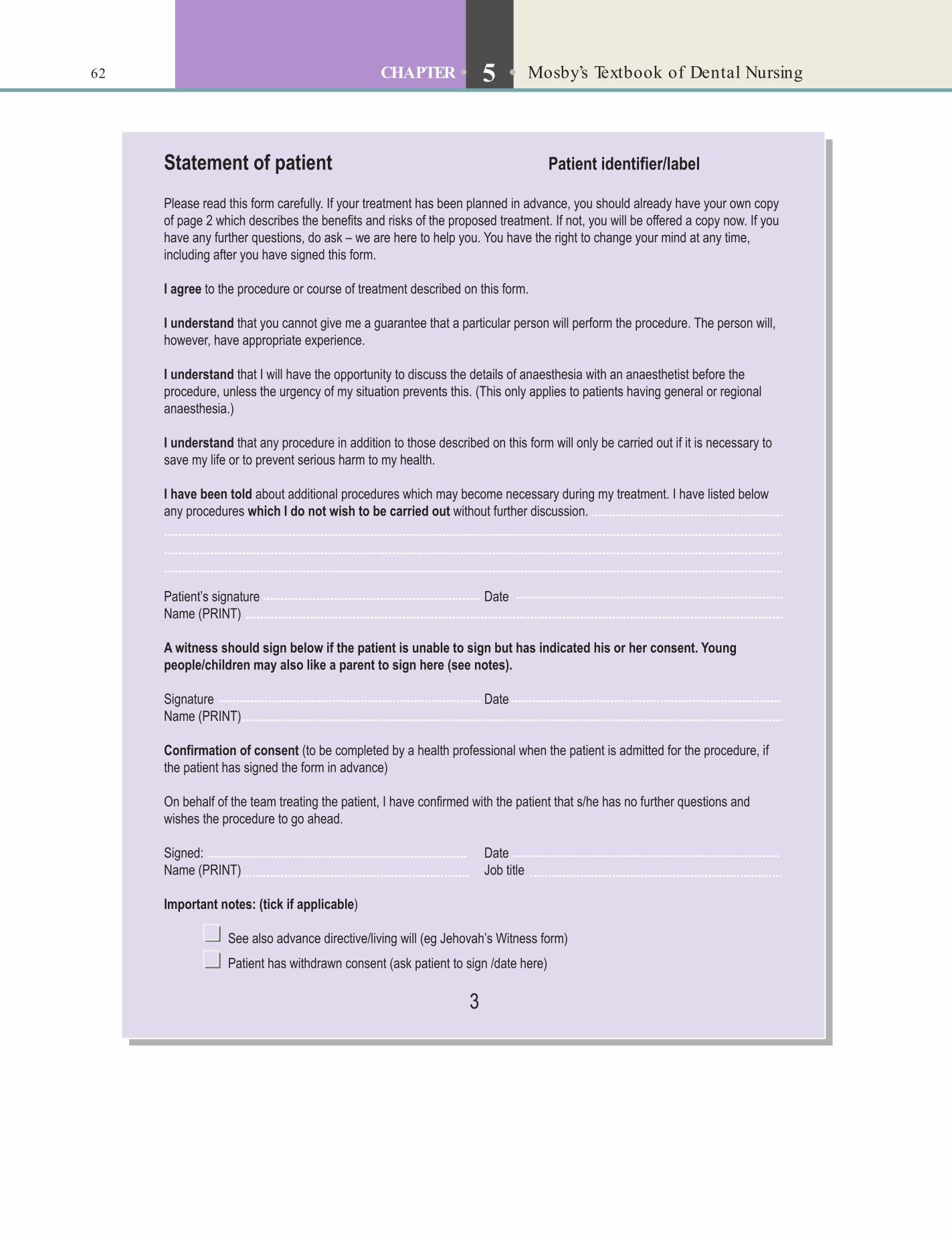
62 CHAPTER • 5 • Mosby’s Textbook of Dental Nursing

DENTAL NURSING: PROTECTING PATIENTS AND STAFFChapter 6 Workplace Hazards and Risk Reduct ion 65
Chapte r 7 In fect ion and Cont ro l 89
Chapte r 8 Workplace Hea lth and Safe ty 113
Sect ion B

This page intentiona lly le ft blank

Chapter 6 Workplace Hazards and Risk Reduction
CHAPTER POINTS• Personal protect ive equipment• Premises• Dental equipment• Work hazards• Security• Waste management
Personal Protective Equipment (PPE)Hand hygiene is crucial, and in all clinical work staff should wear PPE designed to protect the skin and mucous membranes (eyes, nose and mouth) from exposure to blood and other potentially infectious material. PPE includes:
• Protective clothing• Task-speci c gloves• Surgical masks• Protective eyewear.In t ro d u ct io n
• A hazard is anything that can cause harm. All workplaces, including the dental surgery and especially the laboratory, contain some hazards.
• A risk is the chance of someone actually being harmed by the hazard.
It is essential that dental nurses are aware of the hazards in the dental workplace and the health and safety actions to take to avoid harm. In the dental environment there should be careful use of personal protective equipment (PPE), infection control and constant supervision of children and vulnerable adults (see Table 6.1).
Safety can be enhanced by common sense plan-ning and actions, as shown in Box 6.1 and Box 6.2 and discussed below (and also in Chapter 8). Human error is responsible for most risks in a workplace.
Terms to learnCasting equipment: the equipment used in a dental laboratory to make f xed restorations such as crowns (see Chapter 17).
Acid bath: a solution containing acid that is used to stop the developing process or radiographs.
Key poin tSta and patients should always wear glasses or other eye protection during procedures involving the use o dental handpieces/burs, grinding or polishing, cutting wires, use o caustics or instrument cleansing.
PPE is discussed further in Chapter 7.
The PremisesCareful design and furnishing of clinical premises, especially ventilation and ooring, and thoughtful behaviour, such as not running, can prevent many risks and accidents (see Box 6.2 and ‘Notifying and recording accidents’, p. 69):
• Heating and ventilation• Bear in mind that patients are sitting, either
in the waiting or clinical areas, and, with little bodily movement, can get cold, particularly if they are old.
• Staff are typically dressed lightly and can get cold also.

66 CHAPTER • 6 • Mosby’s Textbook o Dental Nursing
Key poin tAll dental practices must have in place policies and procedures that guide sta how to respond appropriately when a hazard is anticipated and to reduce risks. Local policies and procedures are based on national health and sa ety regulations.
Box 6.2 Min imising acciden ts in the den ta l surgery and labora tory• Avoid unnecessary rushing about and horseplay.
• Paths and doors should not be obstructed by trailing wires or any other hazards.
• Passageways and staircases should be well lit, secure and not obstructed, and cleaned daily.
• Floor sur aces should be clean, non-slip, and have a minimum o joints.
• Furniture should be strong, sa e and secured where necessary.
• Sta should use appropriate steps or stepladders when reaching or objects in high places.
• Fires, heaters, Bunsen burners, etc. should be guarded.
• Flammables, explosives and toxic materials including domestic bleach should be sa ely stored and labelled correctly.
• There should be procedures or regular maintenance o all equipment, especially steam sterilisers and compressors because o the risk o explosion.
• Heat treatment and casting equipment should be housed adequately, and tongs used or handling heated casting rings.
• Acid baths should be properly housed and protected.
• All electrical equipment should be installed, earthed, used and connected, and maintained properly.
• Cables and tubing rom electrical and other equipment should not trail on the oor.
• All gas appliances should be installed, connected and maintained properly.
• Electrical, radiography, pressure and gas appliances should be turned o outside working hours.
• Sta and others should avoid injuries rom sharps by using appropriate waste disposal.
• Sta should wear protective attire as appropriate.
• Sta should adhere to the practice sa ety policy.
• Sta should not smoke, eat or drink on the premises except in designated areas.
• Sta should know the hazards o , and pre erably stop, smoking.
• Sa ety training and re resher courses should be taken by all appropriate sta .
• Sta should not use radio headsets and iPods and listen to overloud music, all o which isolate the wearer rom the surrounding environment, and have resulted in many accidents.
Box 6.1 HSE guidance o r hea lth and sa e tySite needs to be clean to be sa e.
Are you wearing PPE?
Falls can cause harm.
Entrances and exits must be clear.
Think about f re protection.
YOU are responsible or health and sa ety.
• Ventilation is important to keep vapours and gases at a low level (p. 75 and Box 6.3)
• Floor coverings in surgerys• Should be impervious and non-slip;
carpeting must be avoided• Should be seam-free; where seams are
present, they should be sealed• The junctions between the oor and
wall and the oor and cabinetry should be covered or sealed to prevent inaccessible areas where cleaning might be dif cult.
• Work surfaces• Should be impervious and easy to clean
and disinfect (e.g. Corian); check the manufacturer’s instructions for suitable cleaning products.
• Joins should be sealed to prevent the accumulation of contaminated matter and aid cleaning.
• Junctions should be rounded or coved to aid cleaning.
• Doors• Many accidents occur by doors opening
and hitting people behind. Doors should also be kept shut; door-closing devices help (Figure 6.1).
• Signage
• Should be clean and of appropriate size for visually challenged people.
Dental EquipmentMuch of the equipment used in dentistry constitutes some hazard to staff and sometimes to the patient or others. All equipment must therefore be carefully and regularly maintained by an appropriately trained person. This is covered in more detail below and in Table 6.1.
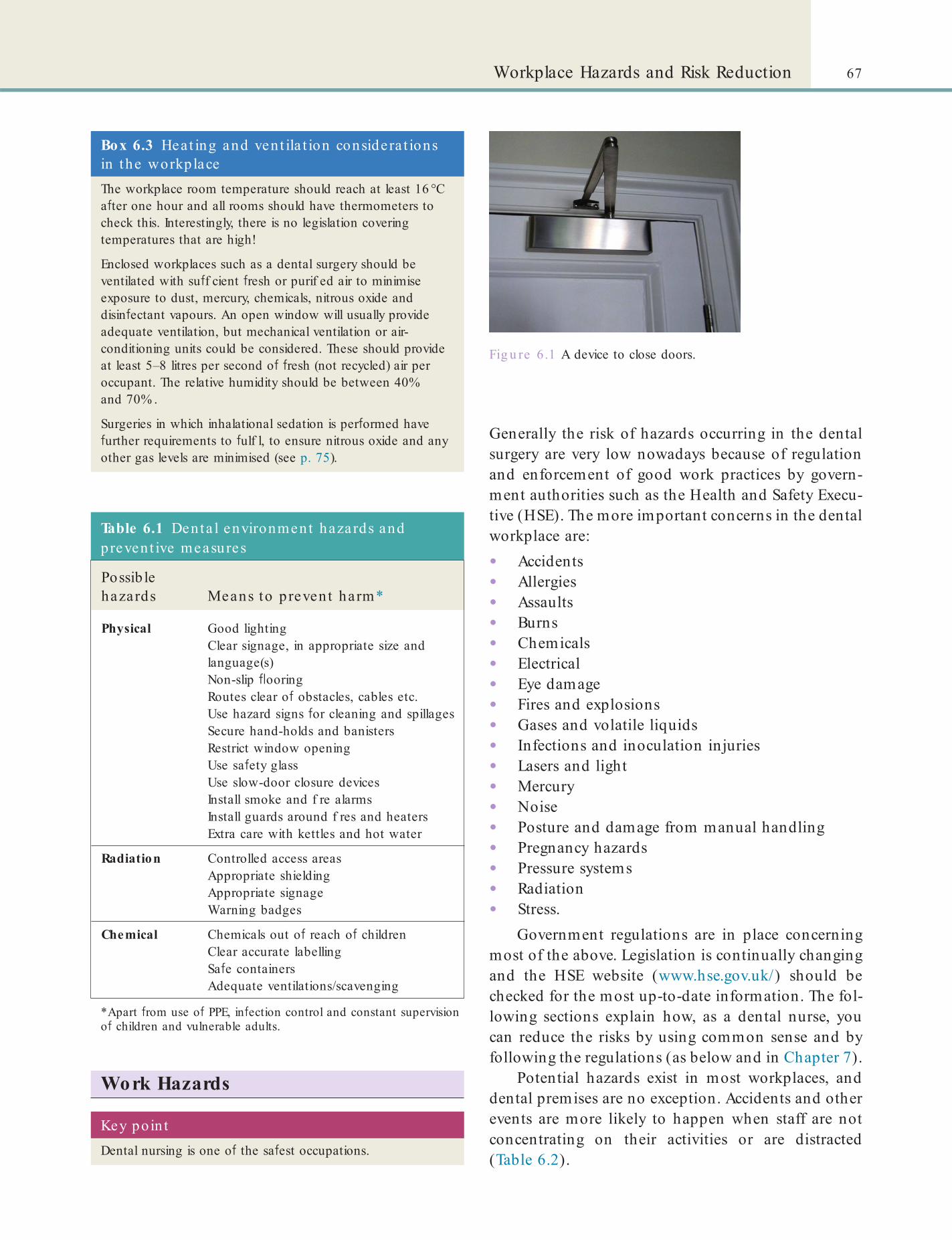
67Workplace Hazards and Risk Reduction
Fig u re 6.1 A device to close doors.
Key po in tDental nursing is one o the sa est occupations.
Box 6.3 Heat ing and vent ila t ion considera t ions in the workplaceThe workplace room temperature should reach at least 16 °C a ter one hour and all rooms should have thermometers to check this. Interestingly, there is no legislation covering temperatures that are high!
Enclosed workplaces such as a dental surgery should be ventilated with su f cient resh or purif ed air to minimise exposure to dust, mercury, chemicals, nitrous oxide and disin ectant vapours. An open window will usually provide adequate ventilation, but mechanical ventilation or air-conditioning units could be considered. These should provide at least 5–8 litres per second o resh (not recycled) air per occupant. The relative humidity should be between 40% and 70% .
Surgeries in which inhalational sedation is per ormed have urther requirements to ulf l, to ensure nitrous oxide and any other gas levels are minimised (see p. 75).
Table 6.1 Denta l environment hazards and prevent ive measures
Possib le hazards Means to prevent harm*
Physical Good lightingClear signage, in appropriate size and language(s)Non-slip ooringRoutes clear o obstacles, cables etc.Use hazard signs or cleaning and spillagesSecure hand-holds and banistersRestrict window openingUse sa ety glassUse slow-door closure devicesInstall smoke and f re alarmsInstall guards around f res and heatersExtra care with kettles and hot water
Radiation Controlled access areasAppropriate shieldingAppropriate signageWarning badges
Chemical Chemicals out o reach o childrenClear accurate labellingSa e containersAdequate ventilations/scavenging
*Apart rom use o PPE, in ection control and constant supervision o children and vulnerable adults.
Work Hazards
Generally the risk of hazards occurring in the dental surgery are very low nowadays because of regulation and enforcement of good work practices by govern-ment authorities such as the Health and Safety Execu-tive (HSE). The more important concerns in the dental workplace are:
• Accidents• Allergies• Assaults• Burns• Chemicals• Electrical• Eye damage• Fires and explosions• Gases and volatile liquids• Infections and inoculation injuries• Lasers and light• Mercury• Noise• Posture and damage from manual handling• Pregnancy hazards• Pressure systems• Radiation• Stress.
Government regulations are in place concerning most of the above. Legislation is continually changing and the HSE website (www.hse.gov.uk/) should be checked for the most up-to-date information. The fol-lowing sections explain how, as a dental nurse, you can reduce the risks by using common sense and by following the regulations (as below and in Chapter 7).
Potential hazards exist in most workplaces, and dental premises are no exception. Accidents and other events are more likely to happen when staff are not concentrating on their activities or are distracted (Table 6.2).
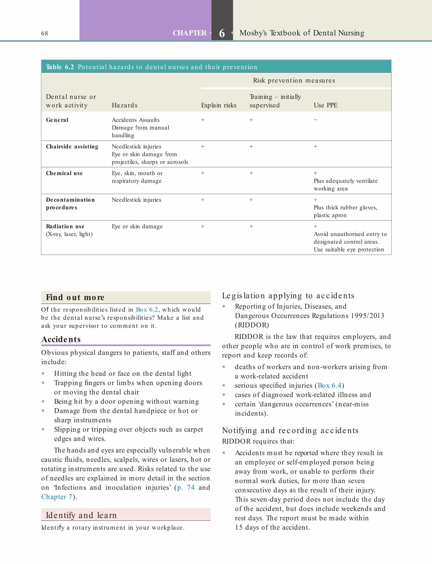
68 CHAPTER • 6 • Mosby’s Textbook o Dental Nursing
Le gis la tion applying to ac c ide nts• Reporting of Injuries, Diseases, and
Dangerous Occurrences Regulations 1995/2013 (RIDDOR)RIDDOR is the law that requires employers, and
other people who are in control of work premises, to report and keep records of:
• deaths of workers and non-workers arising from a work-related accident
• serious speci ed injuries (Box 6.4)• cases of diagnosed work-related illness and• certain ‘dangerous occurrences’ (near-miss
incidents).
Notifying and re c ord ing ac c ide ntsRIDDOR requires that:
• Accidents must be reported where they result in an employee or self-employed person being away from work, or unable to perform their normal work duties, for more than seven consecutive days as the result of their injury. This seven-day period does not include the day of the accident, but does include weekends and rest days. The report must be made within 15 days of the accident.
Find out moreO the responsib ilit ies listed in Box 6.2, which would be the den ta l nurse’s responsib ilit ies? Make a list and ask your supervisor to comment on it .
AccidentsObvious physical dangers to patients, staff and others include:
• Hitting the head or face on the dental light• Trapping ngers or limbs when opening doors
or moving the dental chair• Being hit by a door opening without warning• Damage from the dental handpiece or hot or
sharp instruments• Slipping or tripping over objects such as carpet
edges and wires.The hands and eyes are especially vulnerable when
caustic uids, needles, scalpels, wires or lasers, hot or rotating instruments are used. Risks related to the use of needles are explained in more detail in the section on ‘Infections and inoculation injuries’ (p. 74 and Chapter 7).
Identify and learnIdent i y a ro ta ry inst rument in your workplace .
Table 6.2 Po ten t ia l hazards to den ta l nurses and the ir p revent ion
Denta l nurse or work act ivity Hazards
Risk prevent ion measures
Explain risksTraining – initially supervised Use PPE
General Accidents AssaultsDamage rom manual handling
+ + −
Chairside assisting Needlestick injuriesEye or skin damage rom projectiles, sharps or aerosols
+ + +
Chemical use Eye, skin, mouth or respiratory damage
+ + +Plus adequately ventilate working area
Decontamination procedures
Needlestick injuries + + +Plus thick rubber gloves, plastic apron
Radiation use (X-ray, laser, light)
Eye or skin damage + + +Avoid unauthorised entry to designated control areas. Use suitable eye protection

69Workplace Hazards and Risk Reduction
Find out moreWhere is the acciden t book kep t in your workplace? Who supplies it ?
If an incident does not result in a reportable death or injury but clearly could have done so, it is classed as a dangerous occurrence and must also be reported immediately by completing form 2508. A full list of what are ‘dangerous occurrences’ and the employer’s responsibilities are given on the RIDDOR website (www.hse.gov.uk/riddor/dangerous-occurrences.htm).
Reports related to RIDDOR should be immedi-ately given by telephone to HSE, and a completed accident report form (F2508) sent to the HSE within 10 days. Incident reporting forms can be downloaded from the HSE website (www.hse.gov.uk/forms). A copy of the completed F2508 should also be kept by the employer.
All incidents can be reported online but a tele-phone service remains for reporting fatal and speci c (major) injuries only. Call the Incident Contact Centre on 0845 300 9923 (opening hours Monday to Friday 8.30 am to 5.00 pm). Employers must also keep records of the event.
• Accidents must be recorded, but not reported, where they result in a worker being incapacitated for more than three consecutive days.
• Non-fatal accidents to members of the public or others who are not at work must be reported if they result in an injury and the person is taken directly from the scene of the accident to hospital for treatment to that injury. Examinations and diagnostic tests do not constitute ‘treatment’ in such circumstances.All accidents and injuries to staff or patients or
visitors while on the premises of the dental practice, however apparently trivial, should be recorded in an accident book. The Stationery Of ce (TSO) publishes an ‘Accident Book’, which is suitable for use in the dental environment (see www.tsoshop.co.uk/).
In the dental practice, for example, a compressor or steam steriliser (autoclave) explosion could be noti able, as could a mercury spillage. A needlestick injury (p. 74) involving an infected patient may also fall in this category. In case of doubt of whether an injury should be reported, the advice of the local HSE of ce should be sought.
Term to lea rnSteam steriliser (autoclave): a pressure vessel in which steam at high pressure is produced. When instruments are placed in the steam steriliser or a certain amount o time, the high temperatures in the steam help to kill the harm ul micro-organisms or their spores that may be attached to the instruments.
Box 6.4 Specif ed ( o rmerly re e rred to as ‘major’) in juries occurring in a workplace tha t must be reported to HSE• Fractures, other than to f ngers, thumbs and toes
• Amputations
• Any injury likely to lead to permanent loss o sight or reduction in sight
• Any crush injury to the head or torso causing damage to the brain or internal organs
• Serious burns (including scalding) which:
• covers more than 10% o the body
• causes signif cant damage to the eyes, respiratory system or other vital organs
• Any scalping requiring hospital treatment
• Any loss o consciousness caused by head injury or asphyxia
• Any other injury arising rom working in an enclosed space which:
• leads to hypothermia or heat-induced illness
• requires resuscitation or admittance to hospital or more than 24 hours.
www.hse.gov.uk/riddor/reportable-incidents.htm
Key poin tAlthough RIDDOR applies to all places o work including dental premises it excludes accidents to ‘patients when undergoing treatment in a hospital or surgery o a doctor or dentist’. That exclusion only applies to patients when undergoing treatment – the RIDDOR rules do still have to be ollowed i , or example, a patient breaks a leg on the surgery doorstep.
Re porting o the r d is e as e s unde r RIDDORIf the employer is noti ed by a doctor that an employee has a reportable work-related disease or infection (e.g. occupational dermatitis, occupational asthma, tuber-culosis, hepatitis B, Legionnaires’ disease), the HSE must be sent a completed disease report form (F2508A; available at: www.hse.gov.uk/forms).
A summary of RIDDOR recording or noti cation requirements is given in Table 6.3.

70 CHAPTER • 6 • Mosby’s Textbook o Dental Nursing
Key poin tSome dental materials may produce allergic reactions.
Table 6.3 RIDDOR guidance
Type o incident Def n it ion Examples
Management
Where Record*
Minor No serious injury TripClean needlestick injury
Workplace In writing in accident book
Major Serious injury to person needing 24 hr hospitalisationOrSerious damage to premisesOrDiseases or dangerous occurrences
FractureEye injuryHypoxiaIn ection with HBV, HCV, HIV, MRSA or LegionellaExplosions, serious f re
Hospital Notif able to HSE*Report injuries that lead to a worker being incapacitated or more than 7 consecutive days as the result o an occupational accident or injury (not counting the day o the accident but including weekends and rest days). The report must be made within 15 days o the accident. You must keep a record o the accident i the worker has been incapacitated or more than 3 consecutive days
HBV (hepatitis B virus), HCV (hepatitis C virus), HIV (human immunodef ciency virus), MRSA (meticillin-resistant staphylococcus aureus)*Go to www.hse.gov.uk/riddor and complete the appropriate online report orm.
Find out moreVisit the HSE websit e (www.hse .gov.uk/riddor) o r more in o rmat ion about RIDDOR.
AllergiesAllergic reactions are common and usually minor but some are potentially lethal. People with asthma, eczema and some other conditions often have under-lying allergies. Sometimes allergic reactions can be very severe, even life-threatening (called anaphylaxis; see Chapter 27).
Many allergies have a hereditary component but the prevalence of allergies appears to be increasing. Table 6.4 lists some common allergens. In dentistry, latex is the main offender, but other materials/sub-stances such nickel, chlorhexidine, resins and various drugs may also cause issues.
Table 6.4 Common a lle rgens
Source o a lle rgen Examples
Food products Milk, nuts, egg, shellf sh
Drugs Aspirin, penicillins
Environmental Animal hair, dust mite, pollen
Latex Condoms, elastic bands, gloves
Dental materials Amalgam alloy, gold, mercury, resin-based materials
La te x a lle rgyLatex allergy has become a signi cant clinical problem, along with allergies to iodine, plasters (e.g. Elastoplast and Band-aid) and drugs (remember this with the acronym LIED – Latex Iodine Elastoplast Drugs). Latex products are common in the home and workplace, including clinics, wards and operating theatres. There-fore allergy is an important occupational problem, especially with handwashing using abrasive materials, which increases the risk of sensitisation. Allergic reac-tions to latex have become increasingly common since the use of protective medical/dental gloves became mandatory following the advent of HIV/AIDS. Latex exposure may occur via the skin, mucous membranes, or respiratory tract with inhalation of latex glove powder (natural rubber latex (NRL) allergens may attach to lubricating powder, and become aerosolised, causing sensitisation; or, in those who are allergic, they can cause respiratory, ocular or nasal symptoms).
‘Low-allergen’ latex gloves are available but there is little certainty that these offer any real bene t. People who have allergies to one type of substance are more likely to have allergies to others; patients with latex allergy, for example, may react to foods with
71Workplace Hazards and Risk Reduction
Term to lea rnSubcutaneous: the tissues o the body just under the skin.
Term to lea rnSensitisation: a change in response by the body to a oreign substance, usually an allergen, so that on subsequent exposures to that substance the body shows a heightened immune response.
HIV: human immunodef ciency virus, the virus that causes AIDS.
AIDS: acquired immune def ciency syndrome, a atal disease. People with AIDS have problems with their immune system and so catch in ections more easily.
Many items used in dental practice can sometimes contain latex (Box 6.5) and even equipment and laboratory work previously handled with latex gloves may elicit an allergic response.
allergen cross-reactivity such as avocado, banana, chestnut and kiwi.
Identify and learnIdent i y in your workplace a ll the den ta l mate ria ls and inst ruments marked ** in Box 6.5.
Diagnosis of an allergy is based on:
• Clinical history and presentation• Family history of allergy• Skin-prick or patch testing• Elimination diet to identify food allergens.
Patients who have had serious allergic reactions such as anaphylaxis (see Chapter 25) are also usually advised to always carry with them adrenaline for sub-cutaneous self-injection in the event of a reaction (e.g. Epipen). Affected individuals are usually advised to wear a warning emblem such as Medic-Alert.
Treatments for allergies include use of various drugs, such as antihistamines.
Box 6.5 La tex in denta l it ems*• Equipment and laboratory work previously handled with
latex gloves
• Adhesive dressings and their packaging
• Amalgam carrier tips**
• Bandages and tapes
• Chip syringes**
• Dappen pots**
• Endodontic stops**
• Gloves
• Gutta-percha and gutta-balata**
• Headgear and head positioners**
• Induction masks**
• Latex ties on ace masks
• Local anaesthetic cartridges**
• Mixing bowls**
• Needle guards
• Orthodontic elastics**
• Prophylaxis cups and polishing wheels and points**
• Protective eyewear
• Rubber dam**
• Rubber gloves
• Rubber sleeves on props, and bite blocks**
• Spatulas**
• Suction tips**
• Surgical ace masks and other protective items o clothing, e.g. gowns, overshoes
• Tourniquets and blood pressure cu s
• Wedges
*Latex is present in some rubber dental local anaesthetic cartridges, stoppers or plungers, where either the harpoon penetrates or where the at piston end o a sel -aspirating syringe rests. At the other end o the cartridge is the diaphragm, which the needle penetrates. Any o these components may contain latex. Although there are no documented reports o allergy due to the latex component o cartridges o dental LA, the UK preparation o prilocaine (Citanest) contains no latex.**See ‘Identi y and learn’ in the text.
To avoid future allergic reactions, known allergens should be avoided, which is easier said than done. This is because sensitive individuals may react to minute traces of an allergen, and because allergens can be present in the most unexpected places.
Identify and learnFind 10 items in your workplace tha t may conta in la t ex.
Latex antigens must be avoided by people who are allergic. ‘Low-allergen’ latex gloves are available but there is little certainty that these offer any real bene t. Alternative choices in gloves, medical or dental, or other products for persons with latex allergy include:
• Butadiene• Nitrile (acrylonitrile butadiene)• Polychloroprene (Neolon)• Polystyrene-poly(ethylene-butylene)-polystyrene
(Tactylon)
72 CHAPTER • 6 • Mosby’s Textbook o Dental Nursing
There is more detail on staff safety in the section on ‘Security’, see Chapter 8.
BurnsBurns can result from chemical (acids) or physical agents (heat, cold, radiation such as X-rays or lasers).
Che mic a l burnsSeveral acids (e.g. phosphoric acid, chromic acid and trichloroacetic acid) and corrosive agents (e.g. sodium hypochlorite) are used in the dental surgery and can cause burns if they are not used carefully. Many other stronger acids and corrosives are used in the dental laboratory (e.g. hydro uoric, sulphuric and nitric acids, and caustic soda or sodium bicarbonate). All acids and caustic solutions should be stored safely in appropriate and clearly labelled containers, and proper safety precautions should be taken when handling these materials.
The rmal burnsThermal burns can happen when taking out hot instruments or materials from steam sterilisers or microwaves. Several dental instruments, such as extrac-tion forceps, elevators and metal mouth gags, in par-ticular, retain heat for several minutes after being sterilised and so can cause burns to staff and to patients if used immediately after sterilisation. Handling dental handpieces that have overheated during use can also cause heat burns. This often happens because of the dif culty in gauging their temperature through the gloves that all staff are required to wear in the surgery.
Hot wax knives and Bunsen burners are also common causes of burns; long hair and gloves or clothing can catch light in a Bunsen ame. Hot air blowers can reduce this danger, although they take somewhat longer to heat objects.
Other possible causes of thermal burns in the dental environment include handling hot gutta-percha, dental composition, wax and boiling water, lasers, diathermy and even heated operating lights. Diathermy accidents have resulted from metallic parts of the dental chair becoming part of the path of the current – the localised increase in current density can then cause super cial burns of the skin.
Identify and learnIdent i y gu t ta -percha , denta l composit ion , a wax kni e , d ia thermy unit and an e leva tor in your workplace and f nd ou t what they are used or.
• Polyurethane• Styrene• Vinyl (polyvinyl chloride)• Vitrile (a blend of vinyl and nitrile).Many healthcare facilities now avoid latex altogether; this is sensible since seriously allergic people can react adversely even on entering the surgery door (Box 6.6). Since non-latex gloves have become popular allergy is decreasing.
Key poin tFor latex allergy, anything containing, or contaminated by, latex should be avoided. A latex- ree surgery is ideal.
Box 6.6 Denta l t rea tment o pa t ien ts with proven or possib le la tex a lle rgyAll patients claiming to be sensitive to latex should have their claims taken seriously. Non-latex (Vinyl, Neoprene, Neolon, nitrile-based or polymer) gloves should be used and an alternative (rubber) dam can be ashioned rom vinyl sheet or a vinyl glove. There is only a single report o a supposed allergic reaction to gutta-percha in the literature, but no def nitive proo that the patient had a true allergic reaction to it. Gutta-percha does not cross-react with latex, but some gutta-percha products include gutta-balata, which can cross-react. Occasionally, healthcare workers have reacted to gutta-percha.
However, in patients with a true immediate hypersensitivity to natural rubber latex, the patient’s doctor should be consulted prior to initiating the obturation phase o treatment.
AssaultsOccasionally dental staff are victims of assault by patients who may be drunk, stressed, have mental problems or may be drug abusers. Potentially danger-ous incidents should be defused where possible. If you are assaulted, the most effective response will be to:
• Stay calm• Avoid confrontation – verbally, by body
language or physically• Listen to the person• Empathise with the person• Seek help if the situation does not resolve.
The NHS offers con ict resolution training to NHS staff and contractors. This is available through the commissioning service.
Guidelines governing the prosecution of violent offenders who target NHS staff have been issued by the Crown Prosecution Service (CPS) and the NHS Security Management Service (NHS SMS).

73Workplace Hazards and Risk Reduction
Find out moreRead the COSSH reports o f ve hazardous materia ls tha t a re used in your denta l p ract ice . See a lso the COSSH publica t ion ‘Working with substances hazardous to hea lth’ (www.hse .gov.uk/pubns/indg136.pd ).
Identify and learnIdent i y an acid e tchant and the X-ray f xer used in your workplace .
Employers are required to:
• Assess the risks to health from chemicals and decide what controls are needed
• Use those controls and make sure staff use them• Make sure the controls are working properly• Inform staff about the risks to their health• Train staff.
Electrical HazardsThe legislation applying is:
• Electricity at Work Regulations 1989• Electrical Equipment (Safety) Regulations 1994.
The Electrical Equipment (Safety) Regulations 1994 state that all electrical equipment should be con-structed and designed for safe use when connected. This should be achieved by providing protection against electric shock through a combination of insu-lation and a protective earthing conductor. The main hazards from electrical equipment are (Box 6.7):
• Contact with live parts causing shock and burns (normal mains voltage, 230 volts AC, can kill).
• Faults that could cause res – where electricity could be the source of ignition in a potentially ammable or explosive atmosphere.
• The risk of injury from electricity is strongly linked to where and how it is used, being greatest:• In wet surroundings• In cramped spaces.Electricity plugs, cables, etc. should be inspected
every 6 months. All electrical equipment should be
Hazardous materials in general include:
• Substances used directly in work activities (e.g. adhesives, cleaning agents)
• Substances generated during work activities (e.g. fumes from soldering)
• Naturally occurring substances (e.g. dust)• Biological agents, such as bacteria and other
micro-organisms.Hazardous materials in dentistry include:
• Sodium hypochlorite• Acid etchants• Mercury• X-ray xers• Flammable materials• Certain types of clinical waste (see below, p. 84).
Term to lea rnRisk assessment: a risk assessment is simply a care ul examination o what, in the workplace, could cause harm to people. Doing a risk assessment helps employers decide whether they have taken enough precautions, or should do more, to prevent harm rom occurring to themselves, their sta or others.
Term to lea rnCryotherapy: a method o destroying abnormal cells in the skin or mucosa, usually by reezing them with an extremely cold liquid or instrument.
Co ld burnsCold burns may occur from spillage of liquid nitrogen, which is used, for example, for cryotherapy or to freeze some biopsy specimens.
Chemicals (Hazardous Substances)The legislation applying is:
• Control of Substances Hazardous to Health Regulations 2002 (COSHH).All dental surgeries and laboratories must under-
take risk assessments of all chemical and potentially hazardous substances used in the premises. The results of the assessments must be recorded in a COSHH report. The report must include:
• The hazards a material poses• How the hazards can be avoided• How to deal with a hazard should it occur.
Box 6.7 Measures to reduce e lect rica l acciden ts• Instruction o personnel in the correct use o equipment.
• Frequent periodic maintenance o apparatus and wiring.
• Earthing o all apparatus.
• Separation o mains circuits.
• Use o earth leakage circuit breakers.
74 CHAPTER • 6 • Mosby’s Textbook o Dental Nursing
Human error is responsible for most res and explosions. The main causes of re in a dental practice or similar place usually are: electrical faults (see Chapter 8); careless use of matches; incorrect use of ammable gases (e.g. oxygen) and uids; and non-electrical heating.
A commonsense approach, particularly avoiding the use of naked ames and taking care in the use and storage of ammable materials, will prevent many problems. For example:
• Gas cylinders should be securely stored so that they cannot fall, in a room separate from ammable materials.
• Steam sterilisers (autoclaves) and surgery air compressors should be used and maintained properly. All pressure vessels must be regularly maintained.Employers must comply with re safety regula-
tions. This means carrying out a re risk assessment to determine what precautions are needed and putting in place re precaution measures. If there are ve or more employees, the re risk assessment must be written down.
• Fire precaution measures could include re warnings, alarm systems and extinguishers as well as clearly signed escape routes (see also Box 6.8).
• All ammable materials (e.g. oxygen, gas cylinders, alcohol, monomer, methylated spirits) must be stored in a metal cabinet and never used near ames, or a re or explosion could result.
• Fire- ghting equipment such as extinguishers and blankets must be kept on the premises, regularly inspected and serviced by an authorised body. Staff should be aware of the location of re- ghting equipment, as well as the colour coding for use on different types of re.
• Fire exits must be marked with the appropriate green signs, kept free of any obstruction, and
regularly tested by an appropriately quali ed person at least every 2–3 years, with records kept of the test results.
Eye HazardsEyes need protection from foreign bodies, infected material, chemicals and the various forms of radiation used in dentistry – lasers, light sources for curing (ultra-violet/visible blue or white halogen light) and X-rays. These are covered in more detail on p. 76, 78, 80, 81 and Chapter 11 p. 175.
Box 6.8 Steps to take in the event o a f re
1. Activate the f re alarm or call out ‘Fire!’
2. Call the Fire and Rescue Service.
3. Locate the f re source and extinguish it i sa e to do so.
4. Evacuate all patients and sta rom danger.
5. Close the doors and windows.
6. Assemble all people at the designated assembly point.
Term to lea rnCuring: the process o hardening o tooth-coloured materials that are used mainly to f ll teeth with cavities. The materials come in paste orm and harden either by a chemical reaction or on application o a special light.
Patients must always be provided with adequate eye protection – particularly if they are being treated in the supine position (lying on the back) and for procedures being carried out under conscious sedation or general anaesthesia.
All clinical staff should always wear protective eyewear at work. Objects such as bits of llings can behave like projectiles and y out of the mouth at speeds of over 10m/s when using drills at 250 000 revolutions per minute (rpm). The ends of orthodon-tic and other wires should be held in mosquito forceps, and when cutting, the wire should always be held between two forceps so that the cut end does not y off and cause eye damage.
Special eyewear is needed for work with lasers and with curing lights. Remember that ordinary dark glasses do not lter the hazardous wavelengths of lasers and curing lights. Rather, because they absorb visible light, they can dilate the pupils (open them wider) and therefore worsen the problem because more of the hazardous light rays can now enter the eyes. Contact lens wearers should be careful not to get powders or other materials behind their lenses.
Identify and learnIdent i y a pa ir o mosquito a rte ry o rceps in your workplace .
Fires and ExplosionsThe legislation applying is:
• Regulatory Reform (Fire Safety) Order 2005• In Scotland, Part 3 of the Fire (Scotland) Act
2005 also applies.
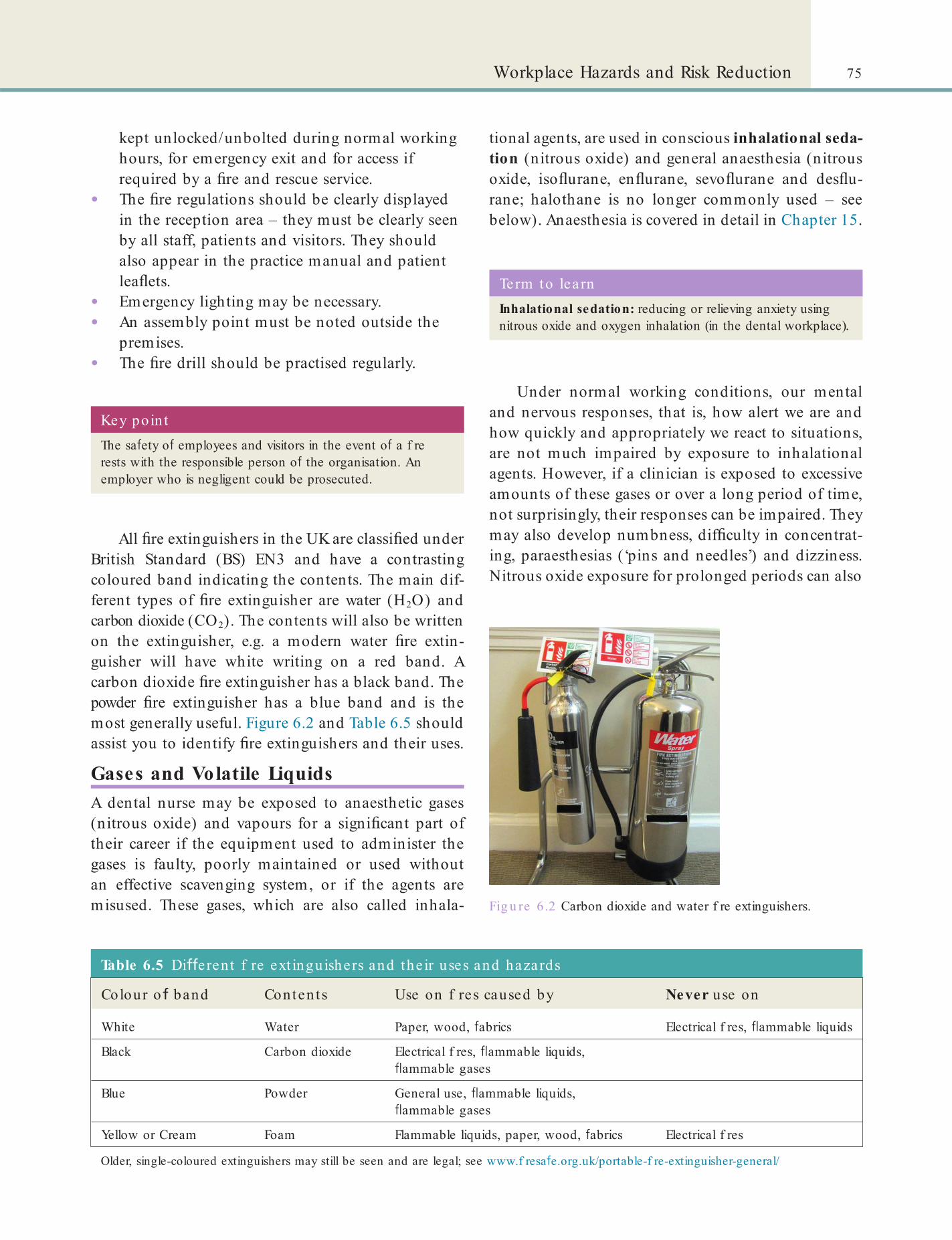
75Workplace Hazards and Risk Reduction
kept unlocked/unbolted during normal working hours, for emergency exit and for access if required by a re and rescue service.
• The re regulations should be clearly displayed in the reception area – they must be clearly seen by all staff, patients and visitors. They should also appear in the practice manual and patient lea ets.
• Emergency lighting may be necessary.• An assembly point must be noted outside the
premises.• The re drill should be practised regularly.
Key poin tThe sa ety o employees and visitors in the event o a f re rests with the responsible person o the organisation. An employer who is negligent could be prosecuted.
All re extinguishers in the UK are classi ed under British Standard (BS) EN3 and have a contrasting coloured band indicating the contents. The main dif-ferent types of re extinguisher are water (H2O) and carbon dioxide (CO2). The contents will also be written on the extinguisher, e.g. a modern water re extin-guisher will have white writing on a red band. A carbon dioxide re extinguisher has a black band. The powder re extinguisher has a blue band and is the most generally useful. Figure 6.2 and Table 6.5 should assist you to identify re extinguishers and their uses.
Gases and Volatile LiquidsA dental nurse may be exposed to anaesthetic gases (nitrous oxide) and vapours for a signi cant part of their career if the equipment used to administer the gases is faulty, poorly maintained or used without an effective scavenging system, or if the agents are misused. These gases, which are also called inhala- Fig u re 6.2 Carbon dioxide and water f re extinguishers.
Table 6.5 Di e rent f re ext inguishers and the ir uses and hazards
Colour o band Conten ts Use on f res caused by Never use on
White Water Paper, wood, abrics Electrical f res, ammable liquids
Black Carbon dioxide Electrical f res, ammable liquids, ammable gases
Blue Powder General use, ammable liquids, ammable gases
Yellow or Cream Foam Flammable liquids, paper, wood, abrics Electrical f res
Older, single-coloured extinguishers may still be seen and are legal; see www.f resa e.org.uk/portable-f re-extinguisher-general/
tional agents, are used in conscious inhalational seda-tion (nitrous oxide) and general anaesthesia (nitrous oxide, iso urane, en urane, sevo urane and des u-rane; halothane is no longer commonly used – see below). Anaesthesia is covered in detail in Chapter 15.
Term to lea rnInhalational sedation: reducing or relieving anxiety using nitrous oxide and oxygen inhalation (in the dental workplace).
Under normal working conditions, our mental and nervous responses, that is, how alert we are and how quickly and appropriately we react to situations, are not much impaired by exposure to inhalational agents. However, if a clinician is exposed to excessive amounts of these gases or over a long period of time, not surprisingly, their responses can be impaired. They may also develop numbness, dif culty in concentrat-ing, paraesthesias (‘pins and needles’) and dizziness. Nitrous oxide exposure for prolonged periods can also
76 CHAPTER • 6 • Mosby’s Textbook o Dental Nursing
Infections and Inoculation Injuries (see Chapte r 7)
Dental staff in contact with the public are commonly exposed to respiratory infections, mainly ‘colds’, and to other viral throat and chest (respiratory) infections. Other more serious respiratory hazards are ‘ u’ (in u-enza), tuberculosis (TB) and, to a much lesser extent, Legionella infection (also called Legionnaires’ disease). All these infections could also be transmitted to patients or others (see Chapter 25).
The main infectious hazard speci c to a dental practice is contact with infected body uids (blood, saliva, etc.); these infections can be transmitted via sharps injuries (needlestick injury/inoculation; see Chapter 7). This is particularly the case for those infec-tions caused by blood-borne viruses (BBV) such as hepatitis B, hepatitis C and human immunode ciency virus (HIV). Prions, which cause the rare Creutzfeldt–Jakob disease (CJD), are virtually impossible to destroy. There are is speci c UK and EU legislation regarding the use of sharp instruments in health care to require employers and their contractors to ensure that risks of sharps injuries are adequately controlled (see Chapter 8).
Some infections, such as MRSA (methicillin- resistant Staphylococcus aureus) infection are mainly found in clinical environments and are thus termed a healthcare-associated infection (HCAI).
Clearly, all dental practices should take steps to control and minimise the exposure to inhalational agents (see Box 6.9).
A no te about ve ntila tion• A dental surgery should be adequately ventilated;
often an open window will suf ce but, in some cases, especially where gases are used, it might be appropriate to install an extractor fan, or a scavenger system. Recycling air conditioning systems are not recommended.
• Systems should exhaust to the outside of the building without risk to the public or recirculation into any public building.
• Fresh air supply rate should not fall below 5–8 litres per second per occupant and should not create uncomfortable draughts.
• Mechanical systems must be regularly cleaned, tested and maintained according to the manufacturer’s recommendations to ensure they are free from anything that may contaminate the air.
Identify and learnIdent i y a vacuum e jector in your workplace .
Box 6.9 Precau t ions to t ake while using n it rous oxide• Care ul use o well-maintained equipment.
• Adequate room ventilation.
• High-volume vacuum ejectors.
• E ective scavenging systems.
Key poin tStandard (universal) in ection control procedures must be used with all patients undergoing dental treatment to avoid the risk o trans er o in ection. See Chapters 4 and 7 and the British Dental Association website (www.bda.org) or more details.
Terms to lea rnPrion: a microscopic protein particle that can cause disease.
HCAI: in ection that is caught by a patient rom another patient, sta or visitor during stay in a hospital or visit to another healthcare acility.
Term to lea rnArrhythmia: when the heart beats too slowly or too rapidly or irregularly or too early.
have adverse effects on other organs of the body, for example the heart, liver and bone marrow, and the reproductive organs.
Halothane and some other halogenated inhala-tional agents (sevo urane and des urane) can cause severe liver dysfunction (called hepatotoxicity) and problems with the beating of the heart (called arrhyth-mias). Therefore halothane is no longer recommended for use in adults. Only occasional hepatotoxicity has been reported with en urane and iso urane.
LasersLaser is the acronym for Light Ampli cation by Stimu-lated Emission of Radiation. In dentistry lasers are used in a variety of ways as a sharp cutting tool to remove decayed tooth material or take a biopsy speci-men, or to enhance the effect of a tooth-whitening material. All lasers are potentially hazardous, mostly because of the risk of eye damage, burns and re or
77Workplace Hazards and Risk Reduction
electric shock, but some lasers are less damaging than others.
Lasers used in medicine and dentistry are catego-rised into four classes according to the amount of damage they are likely to cause (see Table 6.6). The higher the number, the greater the potential for harm. The current laser classes are 1, 1M, 2, 2M, 3R, 3B and 4.
Find out moreThere is more de ta il about the classif ca t ion and sa e ty o lase rs a t www.lasermet .com/resources/class_table . php .
Class 3B and 4 lasers are most commonly used in dental practice: some examples are:
• CO2 (carbon dioxide) lasers – these are used to ‘cut’ the soft tissues (e.g. overgrown gingivae) and are potentially dangerous as they burn. In dentistry, they are mostly used in oral surgery and rarely for other kinds of dental treatment. They burn any tissues exposed to them, including the eyes or skin!
• Nd:Yag (neodymium, yttrium-, aluminium-garnet) lasers and krypton lasers – these are used for cutting dentine and soft tissues. They have wavelengths in the spectrum of visible light and are absorbed preferentially by the retina of the eye, which can then be damaged if this kind of laser is shone into the eye.
• Argon lasers – these are mainly used for curing tooth-coloured lling materials – the composite resins (see Chapter 17). They also have wavelengths in the spectrum of visible light and are absorbed preferentially by the retina, which can be damaged if a laser of this type is shone into the eye.
Table 6.6 Sa e ty concerns with la sers
Laser classes Sa e ty concerns
1 Sa e under all conditions o normal use
2 Sa e because the blink re ex will limit the exposure
3 Hazardous i the eye is exposed directly
4 Can burn the skin, or cause potentially devastating and permanent eye damage as a result o direct or di use beam viewing. Can also be a f re risk
Key poin tLasers must always be used with great care and never shone into the eyes, in unintended directions, or onto brightly plated instruments that re ect the laser.
Terms to lea rnOral surgery: the branch o dentistry involved with tooth extractions and other surgical procedures carried out within the oral cavity (mouth).
Dentine: one o the tissues o the teeth (see Chapter 9).
Retina: the lining o the posterior wall (back) o the eye that senses the light coming into the eye, which then orms the image we see.
The legislation applying to the use of lasers is:
• Care Standards Act 2000.Under this legislation:
• Al premises operating a class 3B or class 4 laser or an intense pulse light (IPL) source for the purposes of treating humans must register with the Care Quality Commission (www.healthcarecommission.org.uk). This includes all hospitals, dental surgeries, cosmetic surgerys and health centres.
• A nominated user who is skilled in the safe use of the laser should always be present whenever a class 3B or 4 laser is used medically and should also be registered with the Care Quality Commission.
• Any dental practice using lasers should have a policy about laser use and a designated laser controlled area.
• Laser warning signs must be provided at every entrance to the laser controlled area. Figure 6.3
Fig u re 6.3 A laser warning sign.
78 CHAPTER • 6 • Mosby’s Textbook o Dental Nursing
low-dose exposure over a long period of time. Although nowadays UV-A light sources have largely been replaced by the safer blue halogen light sources, these lights may still be used for some procedures such as curing some tooth- lling materials and ssure sealants.
Identify and learnIdent i y a curing light and a f ssure sea lan t in your workplace .
Blue ha loge n lightVisible blue halogen light (wavelength 400–500 nm) was developed to replace UV-A light, but even this light is not entirely safe. It may also damage the retina.
White (vis ib le ) lightVisible white light (wavelength 400–700 nm) is much safer than UV or blue light but it also contains some blue and green lights and minute amounts of UV-A. So, although the risks to the eyes are very small, pro-tective eyewear that absorbs these wavelengths should still be worn when curing dental materials with a visible light source.
MercuryMercury is discussed in Chapters 8 and 17. Clinicians who use mercury should carry out a risk assessment for dealing with mercury spillages and produce written procedures.
If there is mercury spillage, you must report the incident to a clinician and follow the steps outlined in Box 6.10.
shows a typical laser safety hazard symbol. This symbol is usually accompanied by some text depending on the kind of laser in use, for example, the sign will say that class 3B laser radiation is being used and to ‘Avoid exposure to the beam’ or that class 4 laser radiation is being used and to ‘Avoid eye or skin exposure to direct or scattered radiation’.
• All laser products must also carry clearly visible labels indicating the laser class, precautions required, maximum laser output and wavelength.
• Class 3B and 4 lasers must have a red emergency shut-off switch in a prominent, accessible position. Class 3B and 4 lasers must have a master control that will only function when the key is inserted and operated, and must give an audible or visible warning when the laser is switched on and operating or not discharged.
• Laser foot switches must be shrouded to prevent accidental operation.
Key poin tBe ore a laser is operated, the operator should orally warn sta in the vicinity that the laser is about to be f red. All sta and patients should be wearing protective eyewear suitable for the laser wavelength being used.
Key poin tNever use a vacuum cleaner or aspiration unit to clean a mercury spillage, as this will vent mercury vapour into the atmosphere.
Term to lea rnOptical density: a translucent medium allows the passage o some light and the optical density is a measure o how much light can pass through.
LightLight is used for curing some dental materials such as restorative resins and, as far as possible, staff should avoid directly viewing ultra-violet (UV) or blue halogen lights. Protective glasses should be used to lter out all light of wavelength under 500 nm. Therefore glasses with red, orange or yellow coloured lenses of suf cient optical density are recommended when using blue or other coloured lights in dental treatment.
Ultra -vio le t lightUV-A light (wavelength between 320 nm and 400 nm) is possibly the most dangerous light. It can cause long-term damage to the retina and also result in the forma-tion of cataracts – especially if there has been a
NoiseNoise-induced hearing loss is irreversible and thus prevention is crucial. Dental staff are, however, much more likely to have leisure-related hearing loss (e.g. from loud music) than they are from work-related problems.
In the past there was concern over possible hearing damage from dental handpieces and turbines and other rotary and ultrasonic instruments. However, research has not found clear evidence for damage from noise produced by air turbines, especially with the use of the quieter, modern handpieces. Nevertheless,
79Workplace Hazards and Risk Reduction
clinicians are recommended to consider their quiet-ness when choosing high-speed dental handpieces, and dental nurses should ensure handpieces are well maintained. Noise associated with the use of ultra-sonic scalers is not considered to affect the hearing of dental staff. All other unnecessary noise arising in a dental surgery should be eliminated.
Identify and learnIdent i y an a ir tu rb ine and an u lt rason ic sca le r in your workplace .
Posture and Manual HandlingDentistry is a sedentary occupation, and many staff work in a somewhat hunched position. Dentists have more neck, shoulder and lower back pain than do other healthcare practitioners; moreover, a higher percentage of women than men develop neck/ shoulder pain.
Four-handed dentistry (close support dentistry) in theory can help reduce stresses and strains. However, you should take as many short breaks as reasonable in the day, stretching and moving around as much as you can. In general, exercise is important to health (see Chapter 26).
Box 6.10 Dea ling with mercury spillagesUse a mercury spillage kit. This should include:
• disposable plastic gloves
• paper towels
• bulb aspirator or the collection o large drops o mercury
• vapour mask
• suitable plastic container f tted with a seal and lid
• mercury absorbent paste (equal parts o calcium hydroxide, owers o sulphur and water).
Steps to clear a mercury spillage:
1. Combine the droplets o mercury i possible.
2. Pick up as much mercury as you can using an aspirator or syringe (never use a vacuum cleaner).
3. Apply the absorbent paste to the a ected area.
4. Contain the waste mercury in the clearly labelled plastic container with a lid.
5. Ventilate the room well.
6. Send the waste mercury or reclaiming or disposal as toxic waste.
In the case o larger spillages, a paste o calcium hydroxide or owers o sulphur should be painted around and over the spillage, and then collected in disposable paper towels (wearing gloves).
Box 6.11 Guide lines o r good posture in the den ta l surgery• The eet should be at on the oor.
• The angle between the cal and thigh should be more than 90°, usually 90°–115°.
• The torso should be vertical.
• Sta should move about, and arch and straighten their back, rom time to time.
• Awkward movements such as twisting should be avoided.
• Heavy equipment or material should be li ted correctly.
Terms to lea rnFour-handed dentistry (close support dentistry): where two or more sta work closely as a team when providing treatment in the surgery.
Scaling: removing the hard deposits (calculus) that orm on the teeth in some individuals.
Root planing: cleaning o the sur ace o the root o a tooth as part o scaling.
Repetitive movements are common in dental work, for example, while scaling and root planing. Chronic musculoskeletal pain associated with repeti-tive movements has been reported by clinicians and also dental nurses. Carpal tunnel syndrome, which is associated with a feeling of tingling (dysaesthesia) in the upper extremities, has also been reported, espe-cially after a prolonged scaling session (Box 6.11).
Incorrect manual handling is one of the most common causes of injury – causing work-related musculoskeletal disorders (MSDs).
The legislation applying is the Manual Handling Operations Regulations 1992 (as amended 2002) (MHOR), which require employers to:
• Avoid the need for hazardous manual handling, so far as is reasonably practicable
• Assess the risk of injury from any hazardous manual handling that cannot be avoided
• Reduce the risk of injury from hazardous manual handling, so far as is reasonably practicable.Employees have duties to:
• Follow systems of work in place for their safety• Use equipment provided for their safety
properly• Cooperate with their employer on health and
safety matters
80 CHAPTER • 6 • Mosby’s Textbook o Dental Nursing
Manufacturers must, by law, provide instructions for re-processing instruments (decontamination) using pressure systems. This includes methods for cleaning and packaging the instruments before placing them in the steriliser. The instructions must also state whether there is any limit to the number of times an item can be sterilised. Failure to comply with the manufacturer’s decontamination instructions may put patients and staff at risk. It may also mean that the warranty is no longer valid and the person re-processing the item or the person who authorised the re-processing is now responsible for the risk.
Employers must ensure that there is a set of oper-ating instructions for the steriliser, safety devices and pipe work, including instructions for emergencies. They must ensure that staff are fully trained in the operation and use of the equipment and the manage-ment of steam-related injuries (scalds/burns) and also ensure regular maintenance.
An operator or owner who does not comply with the safety regulations regarding pressure systems might be held responsible for injury or damage to people or property. They could even be committing a criminal offence.
Radiation (Ionising)Radiation hazards mainly involve ionising radiation, such as X-rays, which can affect not only the patient directly but also staff and others who might be exposed to the beam or scatter. Radiographic exami-nations can provide very useful clinical information but their use must be regulated to avoid damage to people.
The legislation applying is:
• Ionising Radiations Regulations (IRR) 1999 and 2000
• Ionising Radiation (Medical Exposure) Regulations 2000 (IMER)
However, provided the rules of radiation protection are carefully followed, even radiographers or radiolo-gists – who are more exposed to X-rays than dental nurses – appear to be at no signi cant risk. See Chapter 11 for a full discussion of this topic. Here we have summarised the key points.
• Inform their employer if they identify hazardous handling activities
• Take care to make sure their activities do not put others at risk.
Essentially, if possible, avoiding manual handling or, if not, consider automation for the task.
Find out moreFor more in ormat ion regarding techniques o sa e manua l handling see www.hse .gov.uk/pubns/indg143.pd .
Pregnancy HazardsA pregnant dental nurse may be concerned about potential hazards to her unborn baby. However, current evidence suggests that there is little if any spe-ci c occupational risk in dentistry to the outcome of pregnancy. In the past the possible hazards of expo-sure to anaesthetic gases, mercury, ionising radiation and infections had raised concern.
Pressure SystemsThe legislation applying is:Pressure Systems Safety Regulations 2000which applies to benchtop sterilisers and com-pressor(s). All medical devices marketed in the Euro-pean Union (EU) carry a ‘CE’ mark, which indicates that the device satis es the requirements of the EU Directive (regulation) and is ‘ t for the intended purpose’.
Dental practices must have a written scheme of inspection and pressure vessel insurance cover against the explosion of such vessels, i.e. third-party liability insurance to cover the particular risks associ-ated with pressurised equipment and steam (e.g. steam steriliser).
A new steriliser has to be installed, commissioned and validated by an accredited engineer before use. Employers should retain all records (for 11 years) of these activities in the steriliser logbook for future refer-ence. An authorised person (AP sterilisers) can provide advice about the validation of a new steriliser, and a quali ed competent person (pressure vessels) should carry out the validation tests for a vacuum steam steriliser.
Term to lea rnIonising radiation: certain kinds o radiation, such as X-rays, that have high-energy photons, which on striking an object, cause the atoms in that object to release electrons and thus become ions.
Term to lea rnValidation: the process that demonstrates that the right conditions or sterilisation are being achieved; validation is usually undertaken by an appropriately trained engineer.
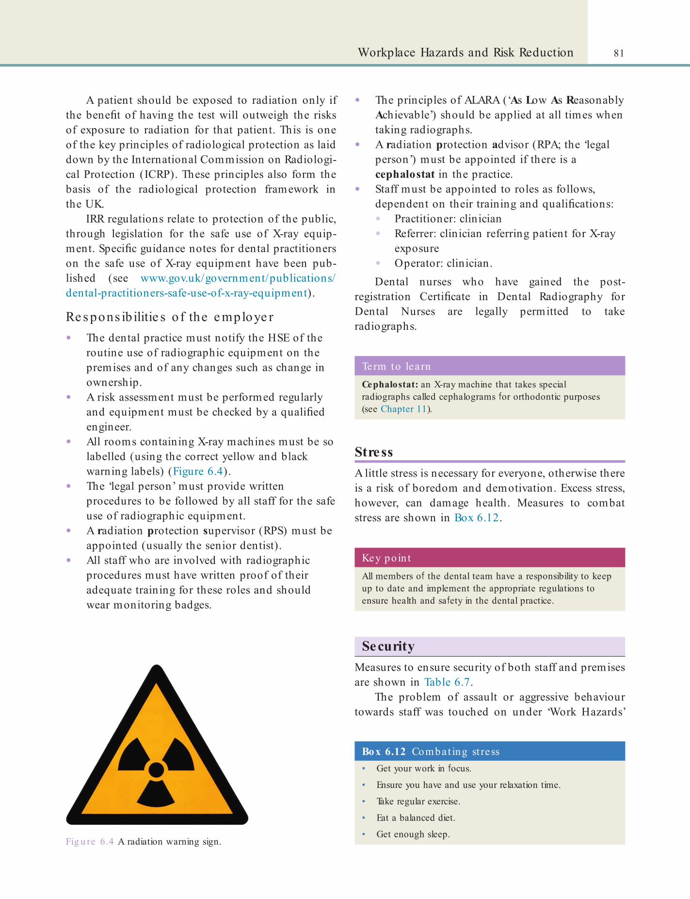
81Workplace Hazards and Risk Reduction
• The principles of ALARA (‘As Low As Reasonably Achievable’) should be applied at all times when taking radiographs.
• A radiation protection advisor (RPA; the ‘legal person’) must be appointed if there is a cephalostat in the practice.
• Staff must be appointed to roles as follows, dependent on their training and quali cations:• Practitioner: clinician• Referrer: clinician referring patient for X-ray
exposure• Operator: clinician.Dental nurses who have gained the post-
registration Certi cate in Dental Radiography for Dental Nurses are legally permitted to take radiographs.
A patient should be exposed to radiation only if the bene t of having the test will outweigh the risks of exposure to radiation for that patient. This is one of the key principles of radiological protection as laid down by the International Commission on Radiologi-cal Protection (ICRP). These principles also form the basis of the radiological protection framework in the UK.
IRR regulations relate to protection of the public, through legislation for the safe use of X-ray equip-ment. Speci c guidance notes for dental practitioners on the safe use of X-ray equipment have been pub-lished (see www.gov.uk/government/publications/dental-practitioners-safe-use-of-x-ray-equipment).
Re s pons ib ilitie s o f the e mploye r• The dental practice must notify the HSE of the
routine use of radiographic equipment on the premises and of any changes such as change in ownership.
• A risk assessment must be performed regularly and equipment must be checked by a quali ed engineer.
• All rooms containing X-ray machines must be so labelled (using the correct yellow and black warning labels) (Figure 6.4).
• The ‘legal person’ must provide written procedures to be followed by all staff for the safe use of radiographic equipment.
• A radiation protection supervisor (RPS) must be appointed (usually the senior dentist).
• All staff who are involved with radiographic procedures must have written proof of their adequate training for these roles and should wear monitoring badges.
Fig u re 6.4 A radiation warning sign.
Term to lea rnCephalostat: an X-ray machine that takes special radiographs called cephalograms or orthodontic purposes (see Chapter 11).
StressA little stress is necessary for everyone, otherwise there is a risk of boredom and demotivation. Excess stress, however, can damage health. Measures to combat stress are shown in Box 6.12.
Key poin tAll members o the dental team have a responsibility to keep up to date and implement the appropriate regulations to ensure health and sa ety in the dental practice.
Box 6.12 Combat ing st ress• Get your work in ocus.
• Ensure you have and use your relaxation time.
• Take regular exercise.
• Eat a balanced diet.
• Get enough sleep.
SecurityMeasures to ensure security of both staff and premises are shown in Table 6.7.
The problem of assault or aggressive behaviour towards staff was touched on under ‘Work Hazards’
82 CHAPTER • 6 • Mosby’s Textbook o Dental Nursing
• the police – for NHS or private healthcare workers
• the commissioning service – for NHS staff or contractors
• the Local Security Management Specialist (LSMS) – for NHS staff or contractors.
above. The security of staff is an important issue. The British Dental Association (BDA states that:
Just like anyone else, dentists and their staff (NHS or private practice) have the right to practise their profession without fear or attack. In whatever sphere of practice, there should be safe systems of work which include measures to prevent and deal with violent and aggressive behaviour. (www.bda.org/dentists/policy-campaigns/research/nat-init/staffsecurity.aspx)In the same guidance, the BDA stresses the impor-
tance of reporting incidents of violent or aggressive behaviour towards staff:
Violent or aggressive behaviour against healthcare staff is often under-reported and the number of reported incidents is believed to be much lower than the actual rate of violence or assault against NHS staff. Healthcare workers can feel uncomfortable reporting incidents because they are reluctant to see their patients prosecuted. The bene t of reporting means that the NHS (practice owner or practice manager in private practice) can understand the physical circumstances that led to or contributed to the incident and enables them to make arrangements to prevent further incidents. … Within the smaller world of dental practice, violence and aggressive behaviour should not be tolerated and reporting of incidents can help the NHS and practice owners design policies and procedures to protect their staff and premises.Any incidents of violence or aggressive behaviour
against staff should be reported to the appropriate authorities, which are:
Term to lea rnLocal Security Management Specialist (LSMS): These are sta who are trained and accredited by the NHS Security Management Service and who lead locally on security management initiatives, which includes work to tackle assaults against sta .
Table 6.7 Security measures
Area o concern Working hours Out o hours
Entrances and exits Supervise by receptionist or Closed Circuit Television (CCTV), or guard. Secure door and window protection
Supervise by CCTV or guard. Secure door and window protection. Restrict sta access, especially when sta leave employment
Fire exits Only opened rom inside Only opened rom inside
Sta protection rom assault Security screen, panic button Security screen, panic button, security alarm
Cash Out o sight, or in sa e In sa e or banked
Computers Password-protected Password-protected
Records Locked Locked
Security breaches Report (e.g. key loss) Report (e.g. break-ins)
The BDA advises that general dental practitioners who own their own premises, employ their own staff and deliver NHS care should report assaults to the police and the local commissioning service, who will share the information with the NHS Security Manage-ment Service (SMS) which monitors violence against staff across NHS service. A practice can refuse to con-tinue treating violent or abusive patients providing the police have been informed. Practitioners are also advised to establish a relationship with their LSMS. The BDA further recommends that previously salaried NHS dentists who have been divested since 2011 under Transforming Community Services but still deliver services to NHS patients should continue to report assaults to the police and their commissioning service and, where possible, should also establish a relation-ship with the LSMS.
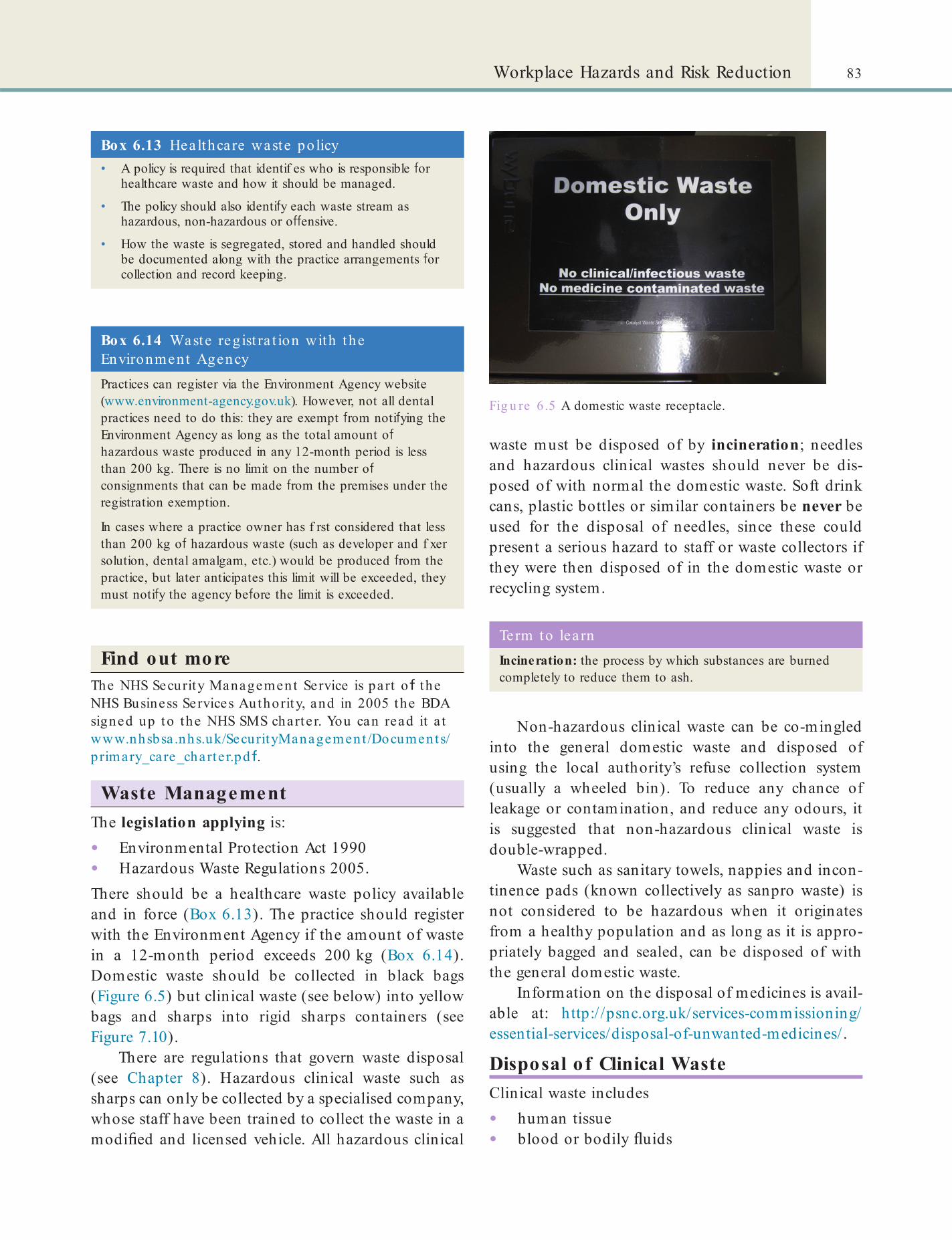
83Workplace Hazards and Risk Reduction
waste must be disposed of by incineration; needles and hazardous clinical wastes should never be dis-posed of with normal the domestic waste. Soft drink cans, plastic bottles or similar containers be never be used for the disposal of needles, since these could present a serious hazard to staff or waste collectors if they were then disposed of in the domestic waste or recycling system.
Box 6.14 Waste reg ist ra t ion with the Environment AgencyPractices can register via the Environment Agency website (www.environment-agency.gov.uk). However, not all dental practices need to do this: they are exempt rom noti ying the Environment Agency as long as the total amount o hazardous waste produced in any 12-month period is less than 200 kg. There is no limit on the number o consignments that can be made rom the premises under the registration exemption.
In cases where a practice owner has f rst considered that less than 200 kg o hazardous waste (such as developer and f xer solution, dental amalgam, etc.) would be produced rom the practice, but later anticipates this limit will be exceeded, they must noti y the agency be ore the limit is exceeded.
Box 6.13 Hea lthcare waste po licy• A policy is required that identif es who is responsible or
healthcare waste and how it should be managed.
• The policy should also identi y each waste stream as hazardous, non-hazardous or o ensive.
• How the waste is segregated, stored and handled should be documented along with the practice arrangements or collection and record keeping.
Fig u re 6.5 A domestic waste receptacle.
Find out moreThe NHS Security Management Service is part o the NHS Business Services Authority, and in 2005 the BDA signed up to the NHS SMS charte r. You can read it a t www.nhsbsa .nhs.uk/SecurityManagement /Documents/primary_care_charte r.pd .
Waste ManagementThe legislation applying is:
• Environmental Protection Act 1990• Hazardous Waste Regulations 2005.There should be a healthcare waste policy available and in force (Box 6.13). The practice should register with the Environment Agency if the amount of waste in a 12-month period exceeds 200 kg (Box 6.14). Domestic waste should be collected in black bags (Figure 6.5) but clinical waste (see below) into yellow bags and sharps into rigid sharps containers (see Figure 7.10).
There are regulations that govern waste disposal (see Chapter 8). Hazardous clinical waste such as sharps can only be collected by a specialised company, whose staff have been trained to collect the waste in a modi ed and licensed vehicle. All hazardous clinical
Term to lea rnIncineration: the process by which substances are burned completely to reduce them to ash.
Non-hazardous clinical waste can be co-mingled into the general domestic waste and disposed of using the local authority’s refuse collection system (usually a wheeled bin). To reduce any chance of leakage or contamination, and reduce any odours, it is suggested that non-hazardous clinical waste is double-wrapped.
Waste such as sanitary towels, nappies and incon-tinence pads (known collectively as sanpro waste) is not considered to be hazardous when it originates from a healthy population and as long as it is appro-priately bagged and sealed, can be disposed of with the general domestic waste.
Information on the disposal of medicines is avail-able at: http:/ /psnc.org.uk/services-commissioning/essential-services/disposal-of-unwanted-medicines/ .
Disposal of Clinical WasteClinical waste includes
• human tissue• blood or bodily uids
84 CHAPTER • 6 • Mosby’s Textbook o Dental Nursing
dental-waste/waste-containers/) until collected. The waste is collected by a suitably licensed or permit-ted waste management collector with facilities where the waste undergoes a mercury recovery process prior to nal disposal.
Particles of amalgam produced during the clean-ing, drilling and lling of teeth must be prevented from exiting with the waste water. An amalgam separa-tor is needed to remove particles of amalgam from waste water, so they can be disposed of as hazardous waste. Separators must be positioned to protect all routes by which amalgam may enter the drains and must meet the requirements of British Standard dental equipment – amalgam separators (BS EN ISO 11143:2008). Simple lters and gauze material do not comply with the current legislation.
• excretions• drugs or other pharmaceutical products• swabs or dressings, and• syringes, needles or other sharp instrumentswhich, unless rendered safe, may prove hazardous to any person coming into contact with it. Any hazardous dental clinical waste should be segregated and dis-posed of as shown in Table 6.8.
De nta l amalgamDental amalgam is a tooth- lling material that con-tains an alloy of silver and mercury. Waste dental amalgam is classi ed as hazardous/special waste. It must be stored, transported and disposed of as hazardous/special waste to make sure there is no risk to human health or the environment. Waste dental amalgam in any form and materials contaminated with amalgam include:
• Unwanted amalgam• Old amalgam llings• Teeth with amalgam llings• Grindings• Surplus amalgam that cannot be re-used• Residues containing amalgam, e.g. from
separators (see below)• Packaging such as capsules containing residues.
Key poin tDental amalgam must be treated using a mercury recovery process be ore f nal disposal.
Key po in tExtracted teeth without amalgam should be disposed o in the dental sharps box. Never put amalgam-f lled extracted teeth in the sharps container. This is because amalgam must not be incinerated. But you can dispose o these teeth with waste amalgam, provided care is taken (as the teeth will be contaminated with blood, they must f rst be decontaminated).
Waste collection agencies often provide special containers for the disposal of amalgam- lled teeth (see Table 6.8). It is also possible to send decontami-nated amalgam- lled teeth (and non- lled teeth) through the post to dental schools for teaching and research purposes but the patient’s consent must be obtained rst (and recorded in the clinical records). It is important to ensure that extracted teeth sent through the post are packaged securely to avoid the package being split open during transit. Some dental schools provide a container and disinfectant suitable for decontamination, storage and transport.
Waste amalgam must be stored in an air-tight container (often in old X-ray xer solution or a mercury suppressant solution, e.g. www.initial.co.uk/
De nta l mate ria ls , c he mic a ls and drugsDental materials, chemicals and drugs should never be disposed of down the sink or drains. Expired emergency drugs are best taken to a pharmacy for disposal.
Le ad fo ilsThe lead foil present in X-ray lms (see Chapter 11) is also hazardous waste. (Any packaging containing resi-dues of, or contaminated by, dangerous substances is classi ed as hazardous waste.)
Loc a l anae s the tic c a rtridge sLocal anaesthetic cartridges and needles are hazard-ous waste and should always be disposed of via the sharps container.
Term to lea rnLocal anaesthetic: a substance that is used to block the sensation o pain in a small area o the body, e.g. a patch o skin or just one quadrant o the mouth. It may be a gel that is just rubbed gently into the area to be anaesthetised or it may be given as an injection.
SharpsSharps waste (items that could cause cuts or puncture/inoculation/needlestick wounds) should be collected in rigid yellow-lidded receptacles (see Table 6.8), which require disposal by incineration. Sharps include
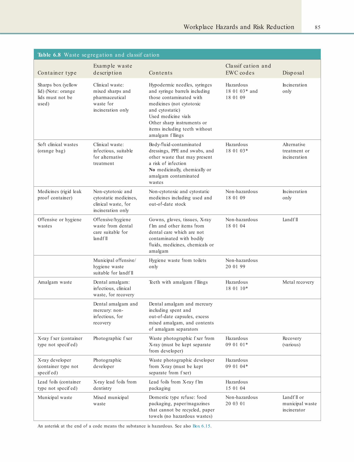
85Workplace Hazards and Risk Reduction
Table 6.8 Waste segrega t ion and classif ca t ion
Conta iner typeExample waste descrip t ion Conten ts
Classif ca t ion and EWC codes Disposa l
Sharps box (yellow lid) (Note: orange lids must not be used)
Clinical waste: mixed sharps and pharmaceutical waste or incineration only
Hypodermic needles, syringes and syringe barrels including those contaminated with medicines (not cytotoxic and cytostatic)Used medicine vialsOther sharp instruments or items including teeth without amalgam f llings
Hazardous18 01 03* and 18 01 09
Incineration only
So t clinical wastes (orange bag)
Clinical waste: in ectious, suitable or alternative treatment
Body- uid-contaminated dressings, PPE and swabs, and other waste that may present a risk o in ectionNo medicinally, chemically or amalgam contaminated wastes
Hazardous18 01 03*
Alternative treatment or incineration
Medicines (rigid leak proo container)
Non-cytotoxic and cytostatic medicines, clinical waste, or incineration only
Non-cytotoxic and cytostatic medicines including used and out-o -date stock
Non-hazardous18 01 09
Incineration only
O ensive or hygiene wastes
O ensive/hygiene waste rom dental care suitable or landf ll
Gowns, gloves, tissues, X-ray f lm and other items rom dental care which are not contaminated with bodily uids, medicines, chemicals or amalgam
Non-hazardous18 01 04
Landf ll
Municipal o ensive/hygiene waste suitable or landf ll
Hygiene waste rom toilets only
Non-hazardous20 01 99
Amalgam waste Dental amalgam: in ectious, clinical waste, or recovery
Teeth with amalgam f llings Hazardous18 01 10*
Metal recovery
Dental amalgam and mercury: non-in ectious, or recovery
Dental amalgam and mercury including spent and out-o -date capsules, excess mixed amalgam, and contents o amalgam separators
X-ray f xer (container type not specif ed)
Photographic f xer Waste photographic f xer rom X-ray (must be kept separate rom developer)
Hazardous09 01 01*
Recovery (various)
X-ray developer (container type not specif ed)
Photographic developer
Waste photographic developer rom X-ray (must be kept separate rom f xer)
Hazardous09 01 04*
Lead oils (container type not specif ed)
X-ray lead oils rom dentistry
Lead oils rom X-ray f lm packaging
Hazardous15 01 04
Municipal waste Mixed municipal waste
Domestic type re use: ood packaging, paper/magazines that cannot be recycled, paper towels (no hazardous wastes)
Non-hazardous20 03 01
Landf ll or municipal waste incinerator
An asterisk at the end o a code means the substance is hazardous. See also Box 6.15.

86 CHAPTER • 6 • Mosby’s Textbook o Dental Nursing
correctly, and that the consignee (the person who receives the waste for nal disposal) is able to take it. In each case, however, the dental practice must ensure that the carrier has completed the note correctly (noti- cation and waste details) and that the carrier is reg-istered to collect such waste. The dental practice responsibility also includes declaring that the waste is packaged and labelled correctly.
An ‘annual transfer note’ may be used to cover all the movements of regular consignments of the same waste, between the same parties.
A consignment note must state:
• The quantity of waste transferred, by weight where possible
• How it is packed• The type of container• A description of the waste.The description of the waste must provide enough information to enable subsequent holders to avoid mismanaging the waste and should include:
• The European Waste Catalogue (EWC) code (Box 6.15)
• The type of premises or business from which the waste comes
• The name of the substance or substances• The process that produced the waste• A chemical and physical analysis.
The consignment note should also state if there are any special problems, such as:
• Any special containment requirements• Type of container required, and material the
container is made of
needles, syringes with needles attached, broken glass ampoules, scalpels and other blades and some teeth (provided no amalgam is present, see above). Note that the operator is responsible for removing and disposing safely of the local anaesthetic needle or scalpel blade.
X-ray xe r and de ve lope r s o lutionsWaste X-ray xer and developer solutions (see Chapter 11) are also classi ed as hazardous. They should be collected by a suitably licensed company or waste facility for material recovery, or if recovery is not appropriate the solutions should be incinerated at suitably licensed or permitted facilities.
Waste Storage and CollectionHealthcare waste should be collected at regular inter-vals to reduce build-up on the premises. If waste is stored, the storage area must be secure and not acces-sible to interference. If patients are treated in their home, any waste produced is considered to be health-care waste of the dental professional. If hazardous, the waste should be taken back to the practice for appro-priate disposal. Healthcare waste should not, however, be moved between dental practices.
For X-ray developer and xer solutions, dental amalgam and waste containing drugs, the practice should have collection containers supplied by the licensed waste collection companies (see Table 6.8). Waste X-ray developer and xer solutions should be stored in leak-proof containers. White rigid containers with a mercury suppressant are preferred for dental amalgam waste storage.
Waste Disposal DocumentationA key element of the dental nurse’s duty of care is keeping track of waste. As the producer and holder of the waste, the dental practice is responsible for:
• Taking adequate steps to ensure that waste is managed safely and kept secure
• Transferring it only to an authorised or exempt person.
Hazardous was te c ons ignme nt no teBefore the waste is removed from the premises, the dental practice is responsible for lling in the noti ca-tion details and the description of the waste in the ‘waste consignment note’ and signing the note. The carrier may obtain these details from the dental nurse and complete them on the nurse’s behalf, which enables the carrier to take appropriate measures to ensure the wastes are packaged, labelled and handled
Box 6.15 European Waste Cata logue (EWC) codesCode description (*denotes a hazardous waste):
• 09 01 03* Solvent-based developer solutions
• 09 01 04* Fixer solutions
• 15 01 10* Packaging containing residues o or contaminated by dangerous substances (e.g. lead oil)
• 18 01 01 Sharps (except 18 01 03)
• 18 01 02 Body parts and organs including blood bags and blood preserves (except 18 01 03)
• 18 01 03* Clinical waste
• 18 01 04 O ensive waste
• 18 01 08* Cytotoxic and cytostatic medicines
• 18 01 09 Medicines other than those mentioned in 18 01 08
• 18 01 10* Dental amalgam waste

87Workplace Hazards and Risk Reduction
collected from the practice. The return should be kept with other waste records (see www.360environmental .co.uk/legislation/waste_legislation/duty_of_care/).
The return may be provided either as a separate form, or as a copy of the consignee’s copy of each consignment note, together with a description (or con rmation) of the method of disposal or recovery applied to the waste. Regulations require that the return contains information on the quantity, nature, origin, destination, frequency of collection, mode of transport, waste carrier and the disposal or recovery operation applied of the waste received.
If the dental practice does not have these returns then their records will be considered to be incomplete. If your practice’s waste contractor has not provided returns, you should request them in writing. If this is unsuccessful consider making alternative arrange-ments for waste disposal until the waste contractor complies with the law, and pass their details to the Environment Agency.
COSHH regulations are also applicable to dental healthcare waste (see above p. 85). To comply with the regulations, clinicians must, among other things:
• Assess the risks to employees and others from healthcare waste
• Make arrangements for renewing the assessment as and when necessary
• Aim to eliminate or prevent these risks; if this is not possible, adequately control the risks
• Provide suitable and suf cient information, instruction and training for employees about the identi ed risk and controls
• Offer immunisation where appropriate.
Training in Waste ManagementAll staff who work with dental waste need to be ade-quately trained. Training needs vary depending on the job and on the individual. All dental staff involved in handling healthcare waste need training, information and instruction in:
• The risks associated with healthcare waste, its segregation, handling, storage and collection
• Personal hygiene• Any procedures that apply to their particular
type of work• Procedures for dealing with spillages and
accidents• Emergency procedures• The appropriate use of personal protective
equipment (PPE).
• Can the waste be safely mixed with other wastes or are there wastes with which it should not be mixed
• Can it be safely crushed and transferred from one vehicle to another
• Can it be safely incinerated or does it require speci c minimum temperatures or combustion times
• Can it be disposed of safely to land ll with other waste
• Is it likely to change physical state during storage or transport
• Any information, advice or instructions about the handling, recovery or disposal of the waste by the waste regulators or suppliers etc.
• Details of problems previously encountered with the waste
• Changes to the description since the previous load• Anything unusual about the waste that may pose
a problem.
Key poin ts: Good pract ice in waste management• Label drums and containers with the description o the
waste.
• Copies o waste consignment notes should be retained by all parties or a minimum o 2 years.
• I you are aware o any particular handling issues with the waste you should in orm either your supervisor or the carrier about them.
Usually the carrier will be collecting more than one consignment of waste from different premises. If this is the case then a multiple collection consignment note will be used. The carrier will sign an additional note – an ‘annex’ – and give the practice a copy of both the annex and the multiple collection consignment note for the practice to keep for 3 years.
Hazardous waste collection companies are enti-tled to pass on a consignment note fee each time hazardous waste is collected.
Find out moreTo learn more , read the u ll BDA guidance on hea lthcare waste management (Advice Note 76), where there a re a lso examples o consignment no tes: www.4everlearn ing .com/pd s/Waste_management _(Jun_08).pd .
Hazardous Waste ReturnsThe waste collection company is also required to send the dental practice each quarter year a ‘waste return’, which is a record of what has happened to the waste

88 CHAPTER • 6 • Mosby’s Textbook o Dental Nursing
Information should be provided by the employer to re ect the outcomes of the COSHH assessment and should:
• Be written in a way that it can be understood by those who need to follow it, including those who may not have a good command of English
• Take account of different levels of training, knowledge and experience
• Be up to date• Be available to all staff, including part-time,
shift, temporary, agency and contract staff.
Chapter 7 Infection and Control
CHAPTER POINTS• Infection• Antiseptics and decontaminants• Hand hygiene• Personal protect ive equipment• Surface and equipment decontamination• Instrument decontamination• Decontaminating dental impressions and appliances• Biopsies sent through the post• Infectious waste management• Immunisation• Inoculation (sharps or needle) injuries• Post-exposure prophylaxis
Some micro-organisms are harmless and live with their hosts with no problems – these are termed commensals. Immune defences (immunity) are usually more than adequate to protect the person on whom the commensal lives. Nevertheless, some micro-organisms – called pathogens – can overcome immu-nity and are responsible for wide-ranging diseases, some of which are deadly. Under some circumstances, especially if the immunity of the host is reduced (immunode ciency), even commensals can cause harm – and they are then termed opportunistic infec-tions. Such infections are common in, for example, HIV/AIDS (Chapters 10 and 25).
Transmission of infections can be by many routes (Table 7.1).
Bacterial InfectionsBacterial infections are common. There is a wide range of bacterial infections recognised, and tooth-related (odontogenic) and other orofacial bacterial infections are important in dentistry (Chapter 10); most cause lesions of limited duration, but some are life-threatening. In medicine, most bacterial infections are transient, with few untoward sequelae, but some can cause serious, recurrent, disseminated or persistent lesions – especially in immunocompromised persons (particularly in neutropenic patients, those with organ transplants and in human immunode ciency virus/acquired immune de ciency syndrome [HIV/AIDS]), or can be life-threatening immediately (e.g. meningo-coccal meningitis), less immediately (e.g. diphtheria) or in the longer term (e.g. tuberculosis, syphilis). Nosocomial infections (healthcare associated infec-tions, HCAI), tetanus, puncture wounds and bites and other bacterial infections are also important because of transmissibility – the ability to be passed to another person.
In t ro d u ct io n
Concern about infectious agents such as prions (CJD), bacteria (e.g. MRSA) and viruses (e.g. hepatitis viruses or HIV) has emphasised the importance to patients, staff and sometimes visitors or tradespeople – espe-cially waste collectors – of infection control. Reports have highlighted the potential hazards in dental prac-tices – particularly needlestick and other sharps inju-ries – which have been a major issue for dental nurses.
InfectionIn ection is the invasion and multiplication of micro-organisms such as bacteria, viruses and parasites that are not normally present within the body. A micro-organism (from the Greek micros, meaning ‘small’, and organismós, meaning ‘organism’) cannot be seen without a microscope and some, such as viruses, can only be seen with an electron microscope.
90 CHAPTER • 7 • Mosby’s Textbook of Dental Nursing
hygiene is low and there is close contact with other persons or their secretions, infections are common. They thus mainly affect young children, who often thereby acquire immunity. In developed countries, children may not contract these infections and thus are non-immune, and may have a primary infection as adolescents and adults.
Viruses that affect the mouth or are found in blood, saliva and other body uids pose a hazard in healthcare because of transmissibility – the ability to be passed to another person. The main viruses of concern in dentistry include (alphabetically):Enteroviruses – multiply in the gut mucosa and are transmitted from person to person by the faecal–oral route. Most infections are in childhood, often as small epidemics. Enterovirus diseases relevant to dentistry include hepatitis A, herpangina, and hand, foot and mouth disease.Herpesviruses – transmitted mainly in saliva and other body uids, sometimes in sexual intercourse, they typically cause a short-lived primary clinical, or more often subclinical, infection, and remain latent thereafter. Reactivation is often because of immunosuppression and recrudescence can lead to protracted illness.Human immunodef ciency viruses (HIV) – transmitted mainly in sexual intercourse – saliva, semen and blood may contain HIV – or by contaminated blood, blood products and donated organs or by contaminated needles.Human papillomaviruses (HPV) – spread mainly from close contact, including sexual intercourse, or in wet environments such as changing rooms.
Bacterial infections are often diagnosed on clinical grounds, supported by smears, culture, testing for immune responses (serology) and, increasingly, by examining for nucleic acids. Antibacterial drugs can often be effective therapy but drainage of pus is often more important. Antibiotic resistance is increasingly a serious problem (e.g. Staphylococcus aureus, Clostridium di f cile, Mycobacterium tuberculosis) and is encouraged by unwarranted use of antibiotics. Immunisation against some bacteria is available, and should be taken up.
Viral InfectionsThere is a wide range of viral infections recognised; most are transient with few untoward sequelae, though many cause malaise, fever and depression of the immune system. Some viral infections can be imme-diately life-threatening (e.g. Ebola or severe acute res-piratory syndrome; SARS), others can result in tumours (e.g. hepatitis C and B and liver cancer), and others can seriously damage the immune system (e.g. HIV/AIDS; Chapter 25). Many viral infections can cause severe, recurrent, disseminated or persistent lesions in immunocompromised persons, such as those with organ transplants, or HIV/AIDS.
Viral infections are often diagnosed on clinical grounds, supported by testing for immune responses (serology) and, increasingly, by examining for viral nucleic acids. Anti-herpetic and anti-retroviral drugs can be effective but relatively few other effective antiviral agents are available. Immunisation against various viruses is available, and should be taken up.
Viral infections are often readily transmitted in saliva and other body uids, and where general
Table 7.1 Routes o f in fect ion t ransmission
Route Mechanism Examples
Direct contact Direct physical contact (body surface to body surface) transmitted then to mouth, nose, eyes or elsewhere
Sexually shared infections (e.g. syphilis, herpes, anogenital HPV, gonorrhoea)
Indirect contact Micro-organism on a surface (called fomite) transmitted then to mouth, nose or eyes
Many viruses, e.g. norovirus
Drople ts Transmitted by sneezing/coughing but possibly by touch
Respiratory viruses (e.g. u)
Aeroso ls Transmitted by microscopic particles in air Legionella, TB, several herpesviruses, measles, mumps
Vector borne Transmitted by animals or insects Malaria
Vehicle borne Transmitted via tissue or uid Blood-borne viruses
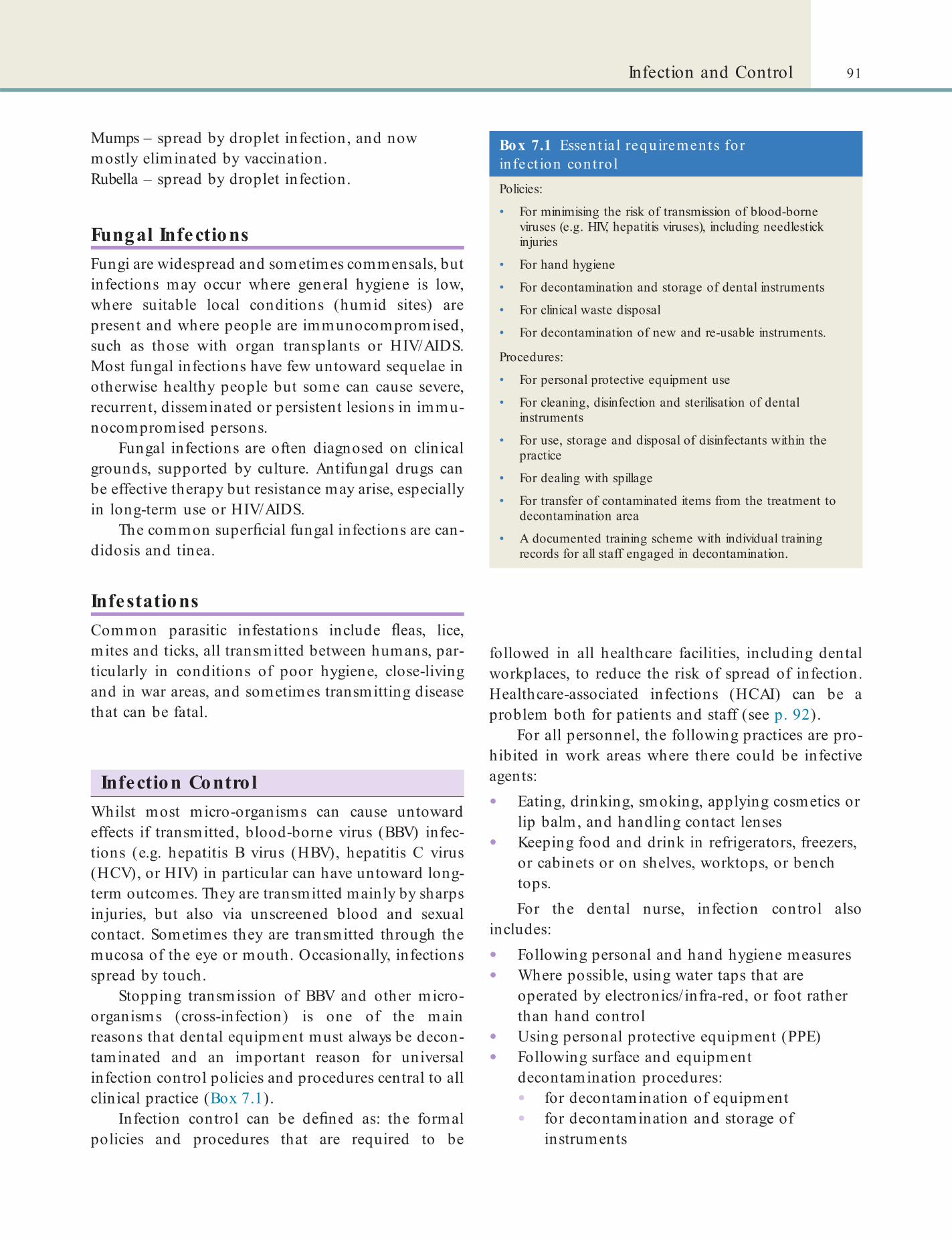
91Infection and Control
followed in all healthcare facilities, including dental workplaces, to reduce the risk of spread of infection. Healthcare-associated infections (HCAI) can be a problem both for patients and staff (see p. 92).
For all personnel, the following practices are pro-hibited in work areas where there could be infective agents:
• Eating, drinking, smoking, applying cosmetics or lip balm, and handling contact lenses
• Keeping food and drink in refrigerators, freezers, or cabinets or on shelves, worktops, or bench tops.For the dental nurse, infection control also
includes:
• Following personal and hand hygiene measures• Where possible, using water taps that are
operated by electronics/ infra-red, or foot rather than hand control
• Using personal protective equipment (PPE)• Following surface and equipment
decontamination procedures:• for decontamination of equipment• for decontamination and storage of
instruments
Mumps – spread by droplet infection, and now mostly eliminated by vaccination.Rubella – spread by droplet infection.
Fungal InfectionsFungi are widespread and sometimes commensals, but infections may occur where general hygiene is low, where suitable local conditions (humid sites) are present and where people are immunocompromised, such as those with organ transplants or HIV/AIDS. Most fungal infections have few untoward sequelae in otherwise healthy people but some can cause severe, recurrent, disseminated or persistent lesions in immu-nocompromised persons.
Fungal infections are often diagnosed on clinical grounds, supported by culture. Antifungal drugs can be effective therapy but resistance may arise, especially in long-term use or HIV/AIDS.
The common super cial fungal infections are can-didosis and tinea.
InfestationsCommon parasitic infestations include eas, lice, mites and ticks, all transmitted between humans, par-ticularly in conditions of poor hygiene, close-living and in war areas, and sometimes transmitting disease that can be fatal.
Infection ControlWhilst most micro-organisms can cause untoward effects if transmitted, blood-borne virus (BBV) infec-tions (e.g. hepatitis B virus (HBV), hepatitis C virus (HCV), or HIV) in particular can have untoward long-term outcomes. They are transmitted mainly by sharps injuries, but also via unscreened blood and sexual contact. Sometimes they are transmitted through the mucosa of the eye or mouth. Occasionally, infections spread by touch.
Stopping transmission of BBV and other micro-organisms (cross-infection) is one of the main reasons that dental equipment must always be decon-taminated and an important reason for universal infection control policies and procedures central to all clinical practice (Box 7.1).
Infection control can be de ned as: the formal policies and procedures that are required to be
Box 7.1 Essen t ia l requirements fo r in fect ion cont rolPolicies:
• For minimising the risk of transmission of blood-borne viruses (e.g. HIV, hepatitis viruses), including needlestick injuries
• For hand hygiene
• For decontamination and storage of dental instruments
• For clinical waste disposal
• For decontamination of new and re-usable instruments.
Procedures:
• For personal protective equipment use
• For cleaning, disinfection and sterilisation of dental instruments
• For use, storage and disposal of disinfectants within the practice
• For dealing with spillage
• For transfer of contaminated items from the treatment to decontamination area
• A documented training scheme with individual training records for all staff engaged in decontamination.
92 CHAPTER • 7 • Mosby’s Textbook of Dental Nursing
• Acquire micro-organisms as a result of their contact with patients, which places healthcare staff themselves at risk of infection.
Antiseptics and Decontaminating AgentsAgents that kill micro-organisms (microbicidal) or inhibit their growth (microbistatic) are available as:Antiseptics: are chemical agents used on skin, mucosa or living tissue for inhibiting or destroying micro-organisms. Examples include alcohols, chlorhexidine, chlorine, hexachlorophene, iodine, chloroxylenol (PCMX), quaternary ammonium compounds and triclosan.Disin ectants: are chemical agents used on inanimate objects (i.e., nonliving) (e.g., oors, walls, working surfaces, sinks) to destroy virtually all micro-organisms, but not necessarily all microbial forms (e.g., bacterial spores). See www.cdc.gov/OralHealth/infectioncontrol/guidelines/ppt.htm.Decontaminants: Total disinfection is a dif cult target to achieve so the term decontamination is used – destruction of pathogenic and other kinds of micro-organisms on instruments and equipment by physical or chemical means.
Examples of agents used in the UK are shown in Table 7.2 and Figure 7.1.
The 2003 US Guidelines for Infection Control in Dental Health-Care Settings suggest using an Environ-mental Protection Agency (EPA) registered hospital-grade disinfectant with low- (anti-HIV and HBV label claims) to intermediate-level (such as tuberculocidal claim) activity after each patient and to use an intermediate-level disinfectant if there is visible con-tamination with blood.
Cleaning involves attention to:
• Clinical contact surfaces – touched by contaminated hands, instruments, devices, gloves, etc. They include light handles, switches, radiograph equipment, chairside computers, reusable containers, drawer handles, tap handles, pens, telephones, and doorknobs.
• Devices or surfaces – removing visible contamination.Because of the risks involved with working with
chemicals and contaminated surfaces, gowns, masks, eye protection and chemical- and puncture-resistant utility gloves should be utilised during this task.
Antiseptics and decontaminating agents should be used carefully as they can produce adverse reactions.
The HCAI problemHCAI at any one time affected up to 8% of in-patients in the UK. During 2001–2006, methicillin-resistant Staphylococcus aureus (MRSA) was implicated in 5109 deaths and Clostridium di f cile in 13 189 deaths. However, with increasing awareness and prevention, between 2007 and 2008 male deaths because of MRSA infection decreased by 31% .
• Following water hygiene measures• Following measures to deal with blood
spillages• Appropriate waste management.
The General Dental Council (GDC) publication Maintaining Standards states that ‘Failure to employ adequate methods of cross-infection control would almost certainly render a clinician liable to a charge of serious professional misconduct’. In other words, infection control is an important duty for both you as the dental nurse and your employer to help avoid the spread of infection.
In 2008 the Department of Health document often referred to as ‘HTM 01-05’: Health Technical Memorandum 01-05: Decontamination in Primary Care Dental Practices was published. A new edition of this HTM was published in April 2009, and updated in October 2013.
Infection control guidelines to prevent transmis-sion of infections are found at www.nice.org.uk/guidance/cg139.
Find out moreHTM 01-05 forms the cornerstone of the infect ion cont ro l pract ices tha t a denta l nurse needs to know about and apply in day-to-day pract ice . You can access th is memorandum a t www.gov.uk/government /uploads/system/uploads/a t tachment_da ta / le /170689/HTM_01-05_2013.pdf.
Healthcare staff have the greatest potential to spread the micro-organisms that cause infection because their hands can:
• Transfer the patient’s own micro-organisms into sterile areas of the patient’s body during care or treatment
• Transfer micro-organisms from one patient to other patients
• Transfer micro-organisms from the environment and equipment to a patient
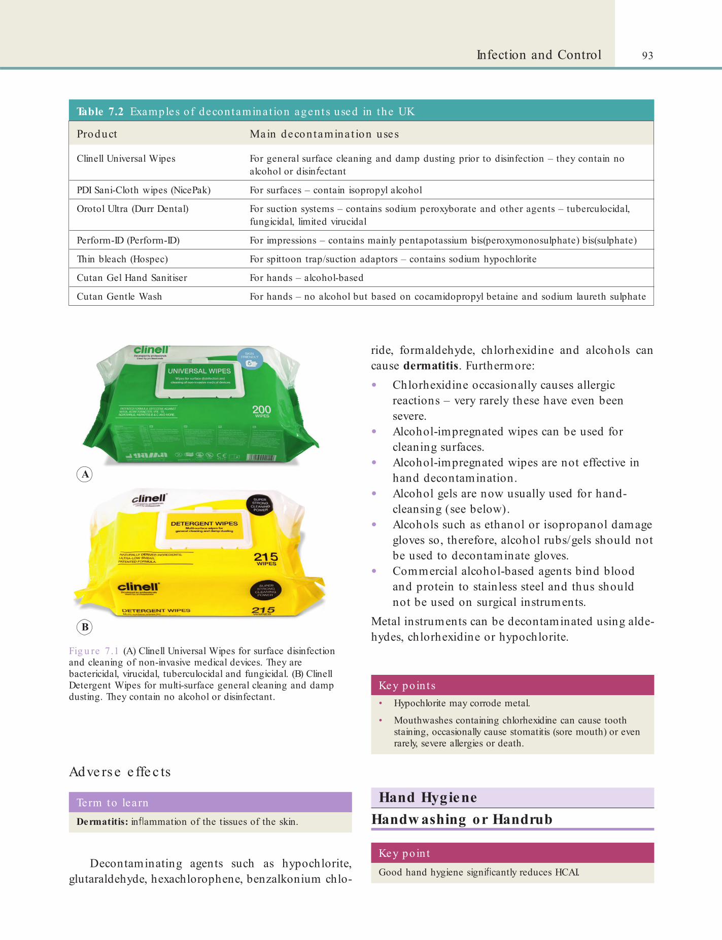
93Infection and Control
Table 7.2 Examples of decontamina t ion agent s used in the UK
Product Main decontamina t ion uses
Clinell Universal Wipes For general surface cleaning and damp dusting prior to disinfection – they contain no alcohol or disin ectant
PDI Sani-Cloth wipes (NicePak) For surfaces – contain isopropyl alcohol
Orotol Ultra (Durr Dental) For suction systems – contains sodium peroxyborate and other agents – tuberculocidal, fungicidal, limited virucidal
Perform-ID (Perform-ID) For impressions – contains mainly pentapotassium bis(peroxymonosulphate) bis(sulphate)
Thin bleach (Hospec) For spittoon trap/suction adaptors – contains sodium hypochlorite
Cutan Gel Hand Sanitiser For hands – alcohol-based
Cutan Gentle Wash For hands – no alcohol but based on cocamidopropyl betaine and sodium laureth sulphate
Fig u re 7.1 (A) Clinell Universal Wipes for surface disinfection and cleaning of non-invasive medical devices. They are bactericidal, virucidal, tuberculocidal and fungicidal. (B) Clinell Detergent Wipes for multi-surface general cleaning and damp dusting. They contain no alcohol or disinfectant.
A
B
Key poin ts• Hypochlorite may corrode metal.
• Mouthwashes containing chlorhexidine can cause tooth staining, occasionally cause stomatitis (sore mouth) or even rarely, severe allergies or death.
Term to lea rnDermatitis: in ammation of the tissues of the skin.
ride, formaldehyde, chlorhexidine and alcohols can cause dermatitis. Furthermore:
• Chlorhexidine occasionally causes allergic reactions – very rarely these have even been severe.
• Alcohol-impregnated wipes can be used for cleaning surfaces.
• Alcohol-impregnated wipes are not effective in hand decontamination.
• Alcohol gels are now usually used for hand-cleansing (see below).
• Alcohols such as ethanol or isopropanol damage gloves so, therefore, alcohol rubs/gels should not be used to decontaminate gloves.
• Commercial alcohol-based agents bind blood and protein to stainless steel and thus should not be used on surgical instruments.
Metal instruments can be decontaminated using alde-hydes, chlorhexidine or hypochlorite.
Key poin tGood hand hygiene signi cantly reduces HCAI.
Adve rs e e ffe c ts
Decontaminating agents such as hypochlorite, glutaraldehyde, hexachlorophene, benzalkonium chlo-
Hand HygieneHandw ashing or Handrub
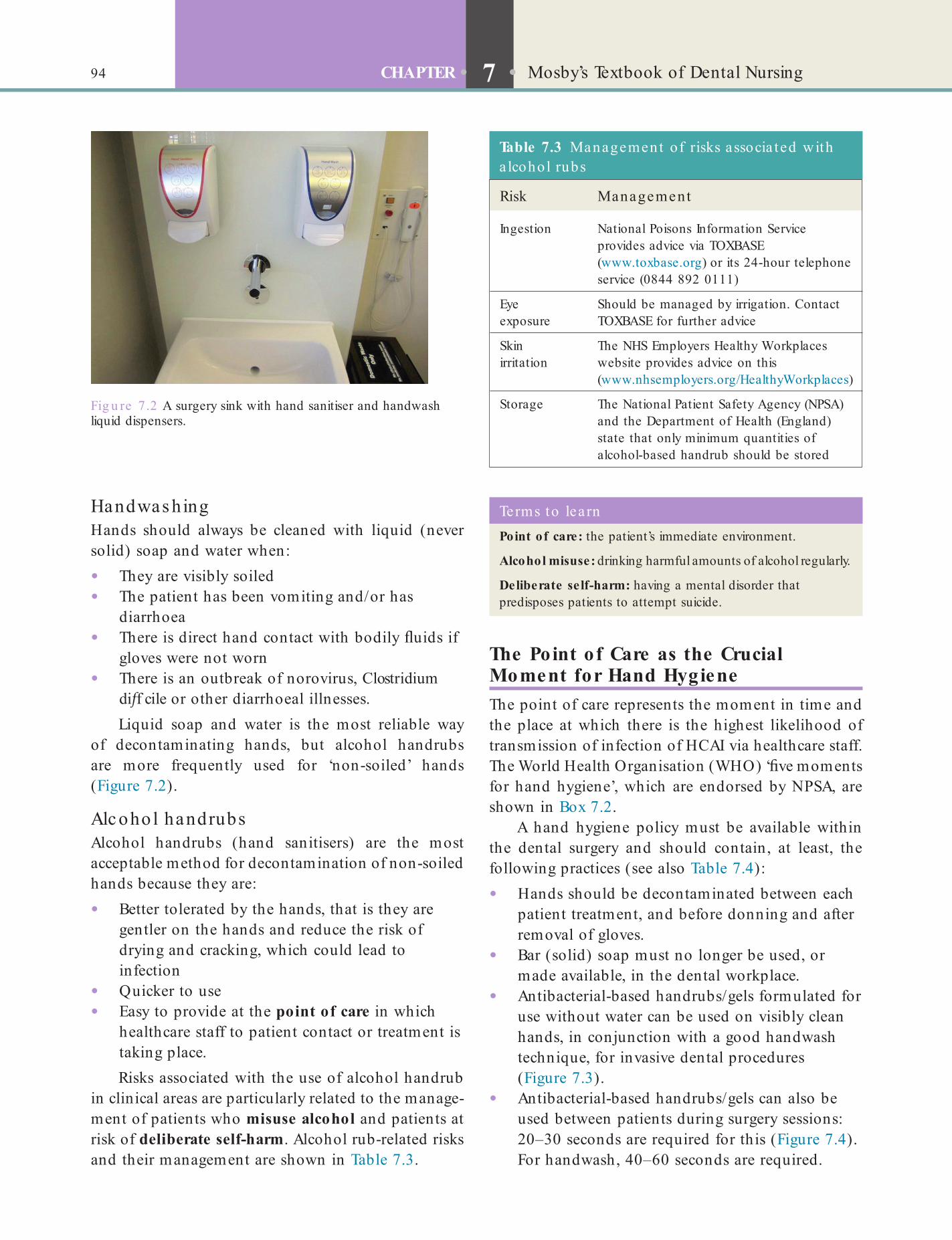
94 CHAPTER • 7 • Mosby’s Textbook of Dental Nursing
The Point of Care as the Crucial Moment for Hand HygieneThe point of care represents the moment in time and the place at which there is the highest likelihood of transmission of infection of HCAI via healthcare staff. The World Health Organisation (WHO) ‘ ve moments for hand hygiene’, which are endorsed by NPSA, are shown in Box 7.2.
A hand hygiene policy must be available within the dental surgery and should contain, at least, the following practices (see also Table 7.4):
• Hands should be decontaminated between each patient treatment, and before donning and after removal of gloves.
• Bar (solid) soap must no longer be used, or made available, in the dental workplace.
• Antibacterial-based handrubs/gels formulated for use without water can be used on visibly clean hands, in conjunction with a good handwash technique, for invasive dental procedures (Figure 7.3).
• Antibacterial-based handrubs/gels can also be used between patients during surgery sessions: 20–30 seconds are required for this (Figure 7.4). For handwash, 40–60 seconds are required.
Handwas hingHands should always be cleaned with liquid (never solid) soap and water when:
• They are visibly soiled• The patient has been vomiting and/or has
diarrhoea• There is direct hand contact with bodily uids if
gloves were not worn• There is an outbreak of norovirus, Clostridium
di f cile or other diarrhoeal illnesses.Liquid soap and water is the most reliable way
of decontaminating hands, but alcohol handrubs are more frequently used for ‘non-soiled’ hands (Figure 7.2).
Alc oho l handrubsAlcohol handrubs (hand sanitisers) are the most acceptable method for decontamination of non-soiled hands because they are:
• Better tolerated by the hands, that is they are gentler on the hands and reduce the risk of drying and cracking, which could lead to infection
• Quicker to use• Easy to provide at the point of care in which
healthcare staff to patient contact or treatment is taking place.Risks associated with the use of alcohol handrub
in clinical areas are particularly related to the manage-ment of patients who misuse alcohol and patients at risk of deliberate self-harm. Alcohol rub-related risks and their management are shown in Table 7.3.
Fig u re 7.2 A surgery sink with hand sanitiser and handwash liquid dispensers.
Terms to lea rnPoint of care: the patient’s immediate environment.
Alcohol misuse: drinking harmful amounts of alcohol regularly.
Deliberate se lf-harm: having a mental disorder that predisposes patients to attempt suicide.
Table 7.3 Management o f risks associa ted with a lcohol rubs
Risk Management
Ingestion National Poisons Information Service provides advice via TOXBASE (www.toxbase.org) or its 24-hour telephone service (0844 892 0111)
Eye exposure
Should be managed by irrigation. Contact TOXBASE for further advice
Skin irritation
The NHS Employers Healthy Workplaces website provides advice on this (www.nhsemployers.org/HealthyWorkplaces)
Storage The National Patient Safety Agency (NPSA) and the Department of Health (England) state that only minimum quantities of alcohol-based handrub should be stored

95Infection and Control
Box 7.2 The ve moments for hand hygiene
1. Before patient contactWhen?
• Clean your hands before touching a patient when approaching him/her.
Why?
• To protect the patient against harmful germs carried on your hands.
2. Before an aseptic taskWhen?
• Clean your hands immediately before any aseptic task.
Why?
• To protect the patient against harmful germs, including the patient’s own, from entering his/her body.
3. After body uid exposure riskWhen?
• Clean your hands immediately after an exposure risk to body uids (and after glove removal).
Why?
• To protect yourself and the healthcare environment from harmful patient germs.
4. After patient contactWhen?
• Clean your hands after touching a patient and his/her immediate surroundings when leaving the patient’s side.
Why?
• To protect yourself and the healthcare environment from harmful patient germs.
5. After contact w ith patient surroundingsWhen?
• Clean your hands after touching any object or furniture in the patient’s immediate surroundings when leaving – even if the patient has not been touched.
Why?
• To protect yourself and the healthcare environment from harmful patient germs.
Guidance is required on the maximum number of applications of antibacterial-based handrubs/gels that can be used on physically clean hands before hand-washing. If hands become ‘sticky’ because of a build-up of the product, they must be washed as normal using a proper hand hygiene technique.
• Alcohol-impregnated wipes used for cleaning surfaces are not effective in hand decontamination and therefore should not be used in place of handrubs/gels.
• Do not use scrub or use nail brushes because these can abrade the skin, where micro-organisms can reside.
• Nails should be kept short and clean, using a blunt ‘orange’ stick, and kept free of nail art, permanent or temporary enhancements (false nails) or varnish.
• Good-quality (soft) paper hand towels should be used after handwashing to avoid skin damage.
• Hand cream can be used after handwashing at the end of a session to avoid dryness. Cream should not be used under gloves since it may encourage the growth of micro-organisms.
• Foot- or sensor-operated waste bins should be used.
Personal Protective EquipmentAs a dental nurse, you are required to wear personal protective equipment (PPE), as are other clinicians, when carrying out duties that may involve possible exposure to blood or other body uids, or splashing from cleaning processes.
Persona l p ro tect ive equipment
Protective clothing• Gowns or coats with long sleeves in operating
environments to protect the forearms. (If outdoor clothing only is being used, short sleeves are mandatory).
• Protective clothing is changed daily, when it becomes visibly soiled, and as soon as possible if penetrated by blood.
• Protective clothing is removed before leaving the work area.
• Dirty protective clothing is placed in designated areas for disposal or washing.
Task-speci c gloves• Non-surgical, surgical, or heavy-duty utility gloves are worn
to prevent or reduce the risk of contaminating the hands with blood and to prevent or reduce the risk of cross-infecting in the clinical process (Figure 7.5).
• Non-surgical and surgical gloves are single-use items, used for only one patient and are then discarded.
• When torn or punctured, gloves are changed as soon as possible.
• Gloves may not be washed because it can lead to penetration of liquids through undetectable holes and hand contamination.
• Double gloving is acceptable for extensive oral surgical procedures.
• Heavy-duty utility gloves are worn for all instrument, equipment and environmental surface cleaning and disinfection.

96 CHAPTER • 7 • Mosby’s Textbook of Dental Nursing
Table 7.4 Levels o f hand hygiene
1: Socia l 2: Hygien ic 3: Surg ica l scrub
Why? To render the hands physically clean and to remove transient micro-organisms, picked up during social activities
In addition to level 1, to destroy micro-organisms and to provide residual effect during times when hygiene is particularly important in protecting yourself and others
In addition to level 2, to substantially reduce the numbers of resident micro-organisms that normally live on the skin during times when surgical procedures are being carried out
When? Before:• Commencing/leaving work• Using computer keyboards• Eating or handling food or
drinks• Preparing or giving
medications• Direct patient contact where
no exposure to blood or other bodily uids or non-intact skin has occurred
Before:• Aseptic procedures• Contact with
immunocompromised patients
• Wearing gloves and carrying out minor surgical or routine dental procedures
Before:Surgical or invasive procedures, oral surgery, periodontal or implant surgery (speci c policies and procedures on surgical preparation should be available at local level)
After:• Becoming visibly soiled• Visiting the toilet• Patient contact even where
no exposure to blood or other bodily uids, or non-intact skin has occurred
• Using computer keyboard• Handling laundry,
equipment or waste• Blowing, wiping or touching
nose
After:• Contact with blood,
other bodily uids, excretions, secretions, mucous membranes, non-intact skin, wound dressings, spore-forming organisms
What hand hygiene to use?
Mild liquid soap – does not need to be antibacterial or antiseptic
An approved antibacterial hand cleanser (e.g. 2–4% chlorhexidine, 5–7.5% povidone iodine, 1% triclosan or plain soap from a dispenser)
An approved antibacterial hand cleanser (e.g. chlorhexidine gluconate 4% , povidone iodine 7.5% )
Antibacterial-based handrubs/gels can be used when hands have not been soiled. Bar soap should not be used
Antibacterial-based handrubs/gels can be used following handwashing (e.g. when performing aseptic techniques) to provide further cleansing and residual effect, and may be used with plain (liquid) soap where necessary
People who are sensitive to antiseptic cleaners can wash with an approved plain liquid soap followed by two applications of an antibacterial-based handrub/gel. Skin problems should be reported and discussed with a general practitioner or occupational health adviser, and a local procedure followed
How long for? 10–15 seconds 15–30 seconds 2–3 minutes, ensuring all areas of hands and forearms are covered
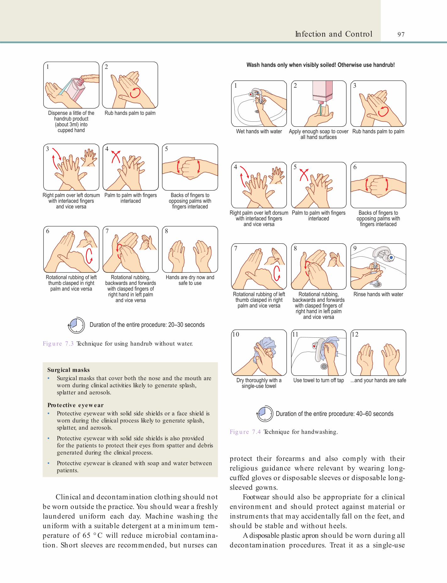
97Infection and Control
Fig u re 7.3 Technique for using handrub without water.
1 2
3 4 5
6 7 8
Fig u re 7.4 Technique for handwashing.
1 2 3
4 5 6
7 8 9
10 11 12
Surgical masks• Surgical masks that cover both the nose and the mouth are
worn during clinical activities likely to generate splash, splatter and aerosols.
Protective eyew ear• Protective eyewear with solid side shields or a face shield is
worn during the clinical process likely to generate splash, splatter, and aerosols.
• Protective eyewear with solid side shields is also provided for the patients to protect their eyes from spatter and debris generated during the clinical process.
• Protective eyewear is cleaned with soap and water between patients.
Clinical and decontamination clothing should not be worn outside the practice. You should wear a freshly laundered uniform each day. Machine washing the uniform with a suitable detergent at a minimum tem-perature of 65 ° C will reduce microbial contamina-tion. Short sleeves are recommended, but nurses can
protect their forearms and also comply with their religious guidance where relevant by wearing long-cuffed gloves or disposable sleeves or disposable long-sleeved gowns.
Footwear should also be appropriate for a clinical environment and should protect against material or instruments that may accidentally fall on the feet, and should be stable and without heels.
A disposable plastic apron should be worn during all decontamination procedures. Treat it as a single-use
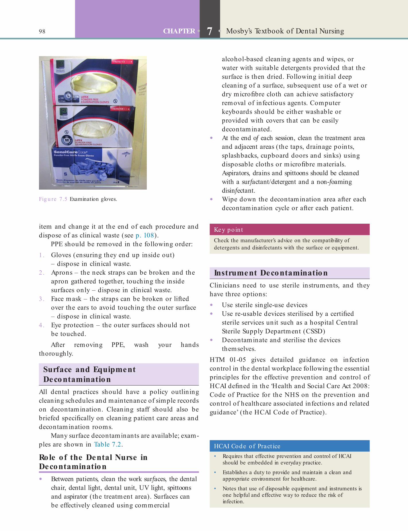
98 CHAPTER • 7 • Mosby’s Textbook of Dental Nursing
alcohol-based cleaning agents and wipes, or water with suitable detergents provided that the surface is then dried. Following initial deep cleaning of a surface, subsequent use of a wet or dry micro bre cloth can achieve satisfactory removal of infectious agents. Computer keyboards should be either washable or provided with covers that can be easily decontaminated.
• At the end o each session, clean the treatment area and adjacent areas (the taps, drainage points, splashbacks, cupboard doors and sinks) using disposable cloths or micro bre materials. Aspirators, drains and spittoons should be cleaned with a sur actant/detergent and a non- oaming disin ectant.
• Wipe down the decontamination area after each decontamination cycle or after each patient.
item and change it at the end of each procedure and dispose of as clinical waste (see p. 108).
PPE should be removed in the following order:1 . Gloves (ensuring they end up inside out)
– dispose in clinical waste.2 . Aprons – the neck straps can be broken and the
apron gathered together, touching the inside surfaces only – dispose in clinical waste.
3 . Face mask – the straps can be broken or lifted over the ears to avoid touching the outer surface – dispose in clinical waste.
4 . Eye protection – the outer surfaces should not be touched.After removing PPE, wash your hands
thoroughly.
Surface and Equipment Decontamination
All dental practices should have a policy outlining cleaning schedules and maintenance of simple records on decontamination. Cleaning staff should also be briefed speci cally on cleaning patient care areas and decontamination rooms.
Many surface decontaminants are available; exam-ples are shown in Table 7.2.
Role of the Dental Nurse in Decontamination• Between patients, clean the work sur aces, the dental
chair, dental light, dental unit, UV light, spittoons and aspirator (the treatment area). Surfaces can be effectively cleaned using commercial
Key poin tCheck the manufacturer’s advice on the compatibility of detergents and disinfectants with the surface or equipment.
Fig u re 7.5 Examination gloves.
Instrument DecontaminationClinicians need to use sterile instruments, and they have three options:
• Use sterile single-use devices• Use re-usable devices sterilised by a certi ed
sterile services unit such as a hospital Central Sterile Supply Department (CSSD)
• Decontaminate and sterilise the devices themselves.
HTM 01-05 gives detailed guidance on infection control in the dental workplace following the essential principles for the effective prevention and control of HCAI de ned in the ‘Health and Social Care Act 2008: Code of Practice for the NHS on the prevention and control of healthcare associated infections and related guidance’ (the HCAI Code of Practice).
HCAI Code of Pract ice• Requires that effective prevention and control of HCAI
should be embedded in everyday practice.
• Establishes a duty to provide and maintain a clean and appropriate environment for healthcare.
• Notes that use of disposable equipment and instruments is one helpful and effective way to reduce the risk of infection.
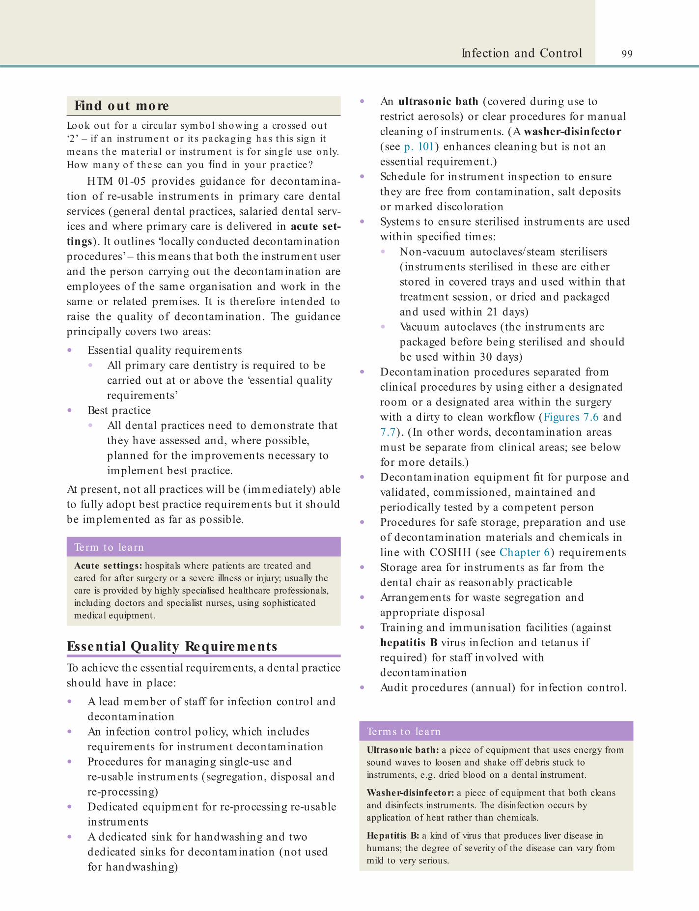
99Infection and Control
• An ultrasonic bath (covered during use to restrict aerosols) or clear procedures for manual cleaning of instruments. (A washer-disinfector (see p. 101) enhances cleaning but is not an essential requirement.)
• Schedule for instrument inspection to ensure they are free from contamination, salt deposits or marked discoloration
• Systems to ensure sterilised instruments are used within speci ed times:• Non-vacuum autoclaves/steam sterilisers
(instruments sterilised in these are either stored in covered trays and used within that treatment session, or dried and packaged and used within 21 days)
• Vacuum autoclaves (the instruments are packaged before being sterilised and should be used within 30 days)
• Decontamination procedures separated from clinical procedures by using either a designated room or a designated area within the surgery with a dirty to clean work ow (Figures 7.6 and 7.7). (In other words, decontamination areas must be separate from clinical areas; see below for more details.)
• Decontamination equipment t for purpose and validated, commissioned, maintained and periodically tested by a competent person
• Procedures for safe storage, preparation and use of decontamination materials and chemicals in line with COSHH (see Chapter 6) requirements
• Storage area for instruments as far from the dental chair as reasonably practicable
• Arrangements for waste segregation and appropriate disposal
• Training and immunisation facilities (against hepatitis B virus infection and tetanus if required) for staff involved with decontamination
• Audit procedures (annual) for infection control.
Find out moreLook out fo r a circu la r symbol showing a crossed ou t ‘2’ – if an inst rument o r it s packaging has th is sign it means the mate ria l o r inst rument is fo r sing le use only. How many of these can you nd in your p ract ice?
HTM 01-05 provides guidance for decontamina-tion of re-usable instruments in primary care dental services (general dental practices, salaried dental serv-ices and where primary care is delivered in acute set-tings). It outlines ‘locally conducted decontamination procedures’ – this means that both the instrument user and the person carrying out the decontamination are employees of the same organisation and work in the same or related premises. It is therefore intended to raise the quality of decontamination. The guidance principally covers two areas:
• Essential quality requirements• All primary care dentistry is required to be
carried out at or above the ‘essential quality requirements’
• Best practice• All dental practices need to demonstrate that
they have assessed and, where possible, planned for the improvements necessary to implement best practice.
At present, not all practices will be (immediately) able to fully adopt best practice requirements but it should be implemented as far as possible.
Term to lea rnAcute settings: hospitals where patients are treated and cared for after surgery or a severe illness or injury; usually the care is provided by highly specialised healthcare professionals, including doctors and specialist nurses, using sophisticated medical equipment.
Essential Quality RequirementsTo achieve the essential requirements, a dental practice should have in place:
• A lead member of staff for infection control and decontamination
• An infection control policy, which includes requirements for instrument decontamination
• Procedures for managing single-use and re-usable instruments (segregation, disposal and re-processing)
• Dedicated equipment for re-processing re-usable instruments
• A dedicated sink for handwashing and two dedicated sinks for decontamination (not used for handwashing)
Terms to lea rnUltrasonic bath: a piece of equipment that uses energy from sound waves to loosen and shake off debris stuck to instruments, e.g. dried blood on a dental instrument.
Washer-disinfector: a piece of equipment that both cleans and disinfects instruments. The disinfection occurs by application of heat rather than chemicals.
Hepatitis B: a kind of virus that produces liver disease in humans; the degree of severity of the disease can vary from mild to very serious.
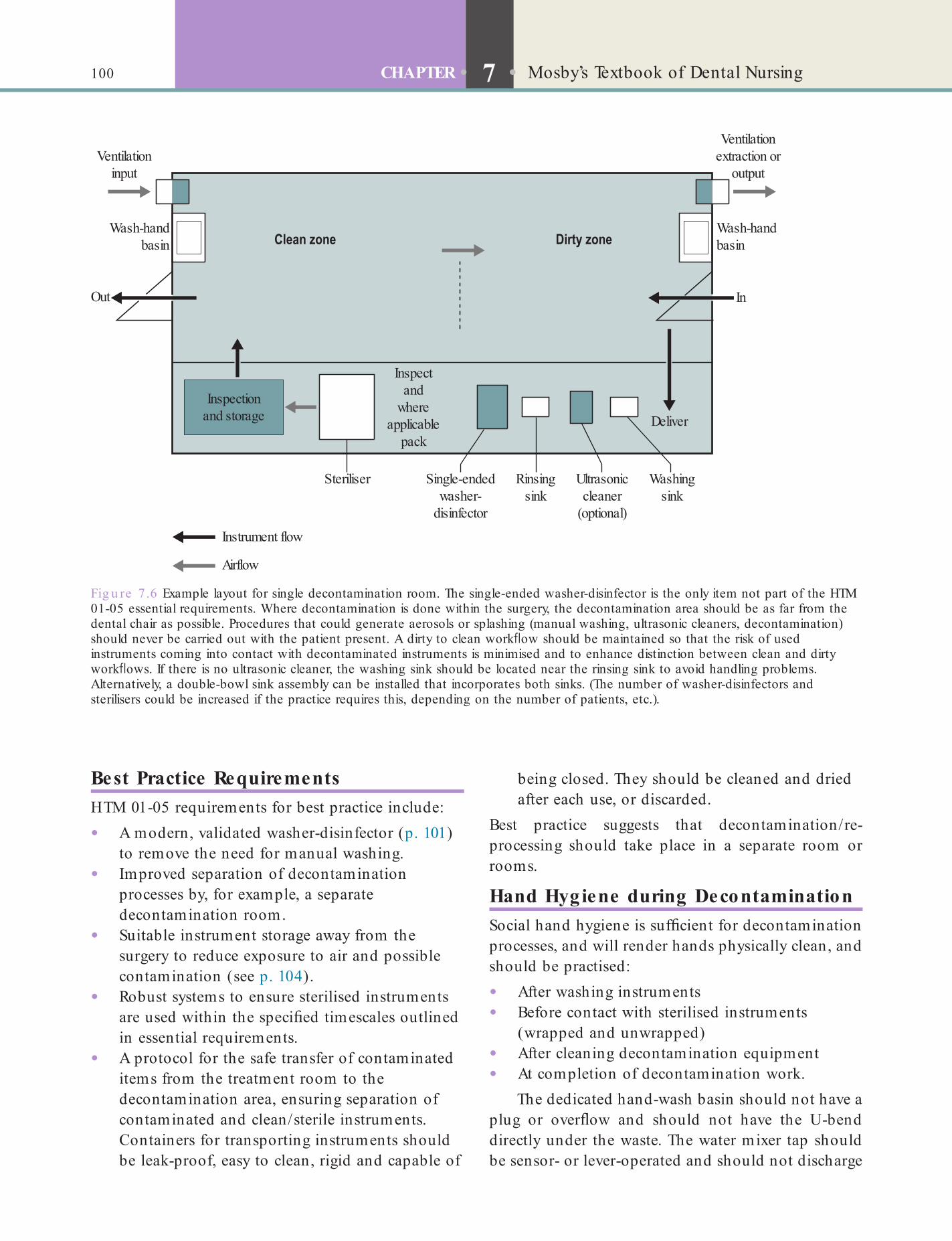
100 CHAPTER • 7 • Mosby’s Textbook of Dental Nursing
being closed. They should be cleaned and dried after each use, or discarded.
Best practice suggests that decontamination/re- processing should take place in a separate room or rooms.
Hand Hygiene during DecontaminationSocial hand hygiene is suf cient for decontamination processes, and will render hands physically clean, and should be practised:
• After washing instruments• Before contact with sterilised instruments
(wrapped and unwrapped)• After cleaning decontamination equipment• At completion of decontamination work.
The dedicated hand-wash basin should not have a plug or over ow and should not have the U-bend directly under the waste. The water mixer tap should be sensor- or lever-operated and should not discharge
Best Practice RequirementsHTM 01-05 requirements for best practice include:
• A modern, validated washer-disinfector (p. 101) to remove the need for manual washing.
• Improved separation of decontamination processes by, for example, a separate decontamination room.
• Suitable instrument storage away from the surgery to reduce exposure to air and possible contamination (see p. 104).
• Robust systems to ensure sterilised instruments are used within the speci ed timescales outlined in essential requirements.
• A protocol for the safe transfer of contaminated items from the treatment room to the decontamination area, ensuring separation of contaminated and clean/sterile instruments. Containers for transporting instruments should be leak-proof, easy to clean, rigid and capable of
Fig u re 7.6 Example layout for single decontamination room. The single-ended washer-disinfector is the only item not part of the HTM 01-05 essential requirements. Where decontamination is done within the surgery, the decontamination area should be as far from the dental chair as possible. Procedures that could generate aerosols or splashing (manual washing, ultrasonic cleaners, decontamination) should never be carried out with the patient present. A dirty to clean work ow should be maintained so that the risk of used instruments coming into contact with decontaminated instruments is minimised and to enhance distinction between clean and dirty work ows. If there is no ultrasonic cleaner, the washing sink should be located near the rinsing sink to avoid handling problems. Alternatively, a double-bowl sink assembly can be installed that incorporates both sinks. (The number of washer-disinfectors and sterilisers could be increased if the practice requires this, depending on the number of patients, etc.).
Inspectand
whereapplicable
pack
Inspectionand storage
Ventilationinput
Out In
Instrument flow
Airflow
Ventilationextraction or
output
Wash-handbasin
Wash-handbasin
Steriliser Single-endedwasher-
disinfector
Rinsingsink
Ultrasoniccleaner
(optional)
Washingsink
Deliver
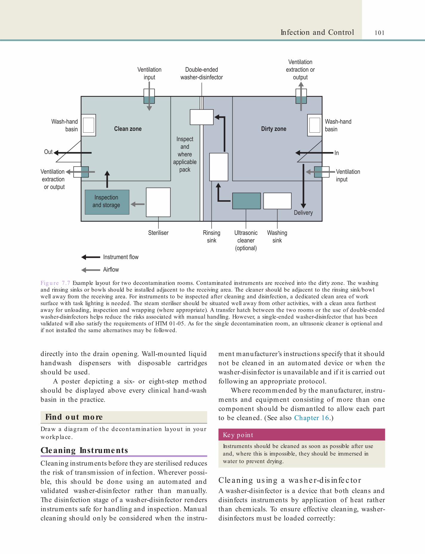
101Infection and Control
ment manufacturer’s instructions specify that it should not be cleaned in an automated device or when the washer-disinfector is unavailable and if it is carried out following an appropriate protocol.
Where recommended by the manufacturer, instru-ments and equipment consisting of more than one component should be dismantled to allow each part to be cleaned. (See also Chapter 16.)
Fig u re 7.7 Example layout for two decontamination rooms. Contaminated instruments are received into the dirty zone. The washing and rinsing sinks or bowls should be installed adjacent to the receiving area. The cleaner should be adjacent to the rinsing sink/bowl well away from the receiving area. For instruments to be inspected after cleaning and disinfection, a dedicated clean area of work surface with task lighting is needed. The steam steriliser should be situated well away from other activities, with a clean area furthest away for unloading, inspection and wrapping (where appropriate). A transfer hatch between the two rooms or the use of double-ended washer-disinfectors helps reduce the risks associated with manual handling. However, a single-ended washer-disinfector that has been validated will also satisfy the requirements of HTM 01-05. As for the single decontamination room, an ultrasonic cleaner is optional and if not installed the same alternatives may be followed.
directly into the drain opening. Wall-mounted liquid handwash dispensers with disposable cartridges should be used.
A poster depicting a six- or eight-step method should be displayed above every clinical hand-wash basin in the practice.
Find out moreDraw a d iagram of the decontamina t ion layout in your workplace .
Cleaning InstrumentsCleaning instruments before they are sterilised reduces the risk of transmission of infection. Wherever possi-ble, this should be done using an automated and validated washer-disinfector rather than manually. The disinfection stage of a washer-disinfector renders instruments safe for handling and inspection. Manual cleaning should only be considered when the instru-
Key poin tInstruments should be cleaned as soon as possible after use and, where this is impossible, they should be immersed in water to prevent drying.
Cle aning us ing a was he r-d is infe c to rA washer-disinfector is a device that both cleans and disinfects instruments by application of heat rather than chemicals. To ensure effective cleaning, washer-disinfectors must be loaded correctly:
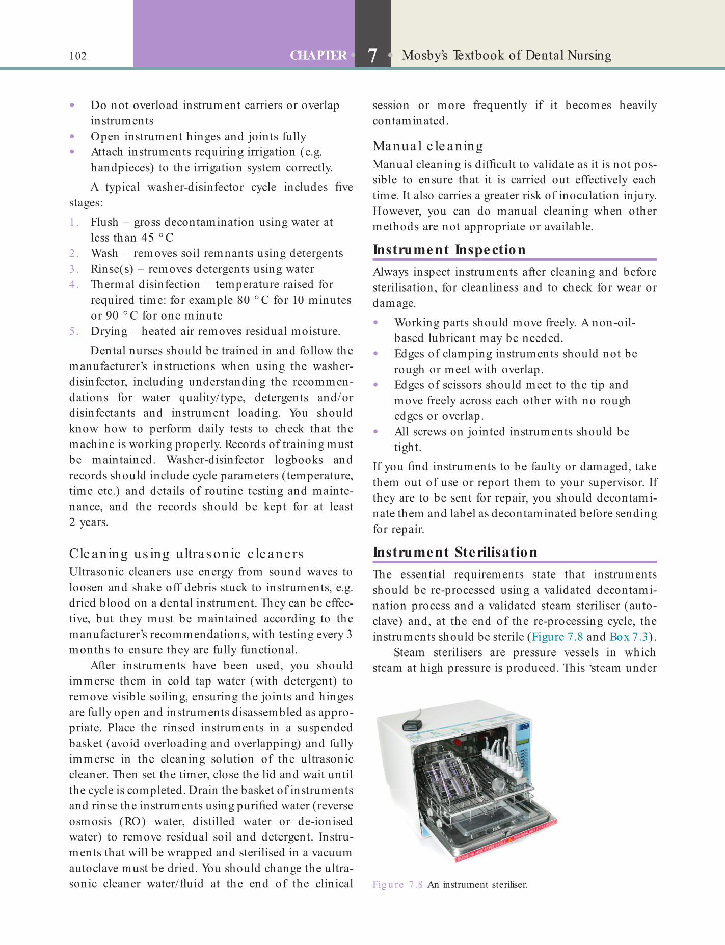
102 CHAPTER • 7 • Mosby’s Textbook of Dental Nursing
session or more frequently if it becomes heavily contaminated.
Manua l c le aningManual cleaning is dif cult to validate as it is not pos-sible to ensure that it is carried out effectively each time. It also carries a greater risk of inoculation injury. However, you can do manual cleaning when other methods are not appropriate or available.
Instrument InspectionAlways inspect instruments after cleaning and before sterilisation, for cleanliness and to check for wear or damage.
• Working parts should move freely. A non-oil-based lubricant may be needed.
• Edges of clamping instruments should not be rough or meet with overlap.
• Edges of scissors should meet to the tip and move freely across each other with no rough edges or overlap.
• All screws on jointed instruments should be tight.
If you nd instruments to be faulty or damaged, take them out of use or report them to your supervisor. If they are to be sent for repair, you should decontami-nate them and label as decontaminated before sending for repair.
Instrument SterilisationThe essential requirements state that instruments should be re-processed using a validated decontami-nation process and a validated steam steriliser (auto-clave) and, at the end of the re-processing cycle, the instruments should be sterile (Figure 7.8 and Box 7.3).
Steam sterilisers are pressure vessels in which steam at high pressure is produced. This ‘steam under
• Do not overload instrument carriers or overlap instruments
• Open instrument hinges and joints fully• Attach instruments requiring irrigation (e.g.
handpieces) to the irrigation system correctly.A typical washer-disinfector cycle includes ve
stages:1 . Flush – gross decontamination using water at
less than 45 ° C2. Wash – removes soil remnants using detergents3 . Rinse(s) – removes detergents using water4 . Thermal disinfection – temperature raised for
required time: for example 80 ° C for 10 minutes or 90 ° C for one minute
5 . Drying – heated air removes residual moisture.Dental nurses should be trained in and follow the
manufacturer’s instructions when using the washer-disinfector, including understanding the recommen-dations for water quality/type, detergents and/or disinfectants and instrument loading. You should know how to perform daily tests to check that the machine is working properly. Records of training must be maintained. Washer-disinfector logbooks and records should include cycle parameters (temperature, time etc.) and details of routine testing and mainte-nance, and the records should be kept for at least 2 years.
Cle aning us ing ultras onic c le ane rsUltrasonic cleaners use energy from sound waves to loosen and shake off debris stuck to instruments, e.g. dried blood on a dental instrument. They can be effec-tive, but they must be maintained according to the manufacturer’s recommendations, with testing every 3 months to ensure they are fully functional.
After instruments have been used, you should immerse them in cold tap water (with detergent) to remove visible soiling, ensuring the joints and hinges are fully open and instruments disassembled as appro-priate. Place the rinsed instruments in a suspended basket (avoid overloading and overlapping) and fully immerse in the cleaning solution of the ultrasonic cleaner. Then set the timer, close the lid and wait until the cycle is completed. Drain the basket of instruments and rinse the instruments using puri ed water (reverse osmosis (RO) water, distilled water or de-ionised water) to remove residual soil and detergent. Instru-ments that will be wrapped and sterilised in a vacuum autoclave must be dried. You should change the ultra-sonic cleaner water/ uid at the end of the clinical Fig u re 7.8 An instrument steriliser.
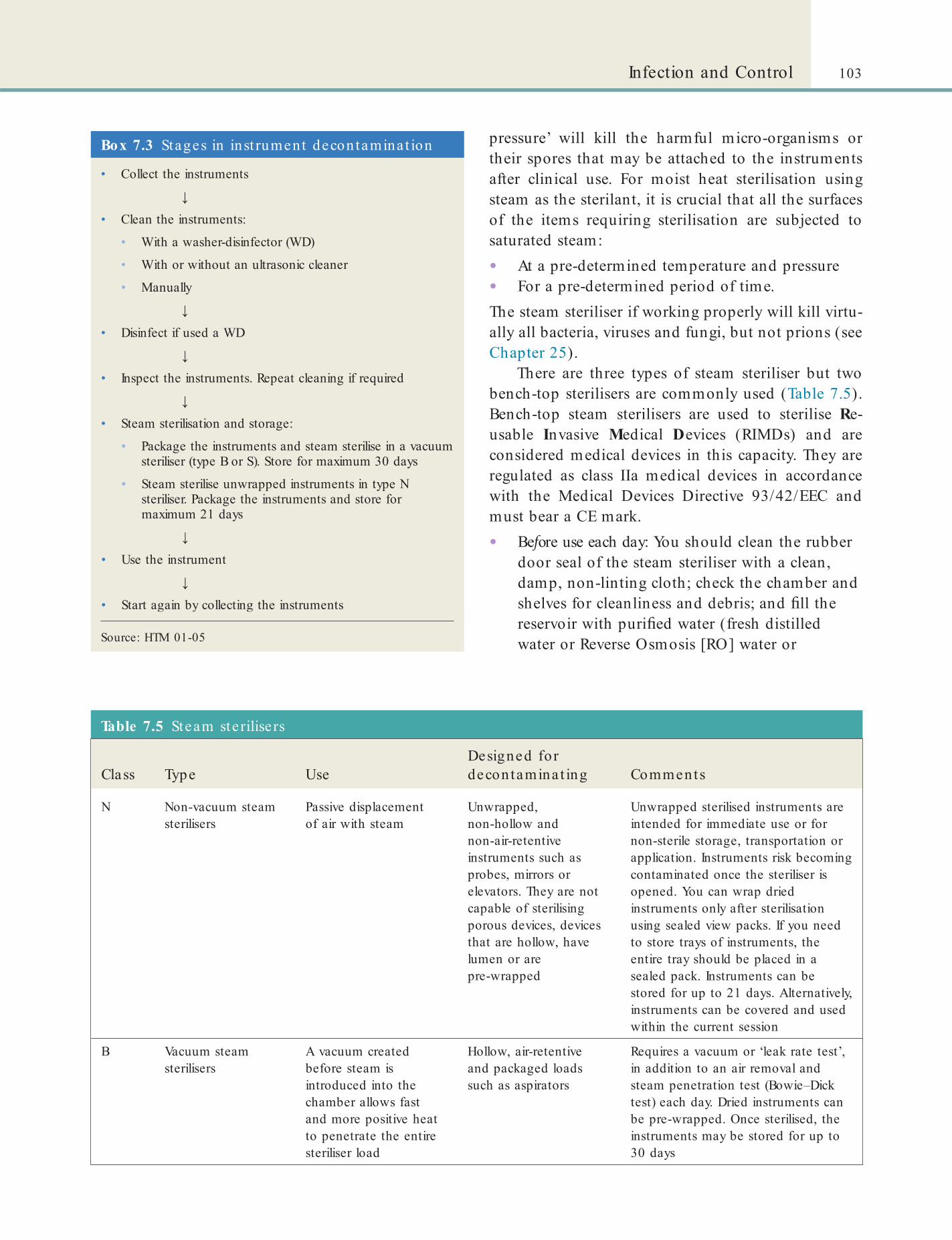
103Infection and Control
pressure’ will kill the harmful micro-organisms or their spores that may be attached to the instruments after clinical use. For moist heat sterilisation using steam as the sterilant, it is crucial that all the surfaces of the items requiring sterilisation are subjected to saturated steam:
• At a pre-determined temperature and pressure• For a pre-determined period of time.The steam steriliser if working properly will kill virtu-ally all bacteria, viruses and fungi, but not prions (see Chapter 25).
There are three types of steam steriliser but two bench-top sterilisers are commonly used (Table 7.5). Bench-top steam sterilisers are used to sterilise Re-usable Invasive Medical Devices (RIMDs) and are considered medical devices in this capacity. They are regulated as class IIa medical devices in accordance with the Medical Devices Directive 93/42/EEC and must bear a CE mark.
• Be ore use each day: You should clean the rubber door seal of the steam steriliser with a clean, damp, non-linting cloth; check the chamber and shelves for cleanliness and debris; and ll the reservoir with puri ed water (fresh distilled water or Reverse Osmosis [RO] water or
• Collect the instruments
↓
• Clean the instruments:
• With a washer-disinfector (WD)
• With or without an ultrasonic cleaner
• Manually
↓
• Disinfect if used a WD
↓
• Inspect the instruments. Repeat cleaning if required
↓
• Steam sterilisation and storage:
• Package the instruments and steam sterilise in a vacuumsteriliser (type B or S). Store for maximum 30 days
• Steam sterilise unwrapped instruments in type Nsteriliser. Package the instruments and store formaximum 21 days
↓
• Use the instrument
↓
• Start again by collecting the instruments
Box 7.3 Stages in inst rument decontaminat ion
Source: HTM 01-05
Table 7.5 Steam ste rilise rs
Class Type UseDesigned for decontamina t ing Comments
N Non-vacuum steam sterilisers
Passive displacement of air with steam
Unwrapped, non-hollow and non-air-retentive instruments such as probes, mirrors or elevators. They are not capable of sterilising porous devices, devices that are hollow, have lumen or are pre-wrapped
Unwrapped sterilised instruments are intended for immediate use or for non-sterile storage, transportation or application. Instruments risk becoming contaminated once the steriliser is opened. You can wrap dried instruments only after sterilisation using sealed view packs. If you need to store trays of instruments, the entire tray should be placed in a sealed pack. Instruments can be stored for up to 21 days. Alternatively, instruments can be covered and used within the current session
B Vacuum steam sterilisers
A vacuum created before steam is introduced into the chamber allows fast and more positive heat to penetrate the entire steriliser load
Hollow, air-retentive and packaged loads such as aspirators
Requires a vacuum or ‘leak rate test’, in addition to an air removal and steam penetration test (Bowie–Dick test) each day. Dried instruments can be pre-wrapped. Once sterilised, the instruments may be stored for up to 30 days

104 CHAPTER • 7 • Mosby’s Textbook of Dental Nursing
Caus e s o f fa ilure o f a tte mpte d s te rilis a tion us ing a s te am s te rilis e r• Use of the wrong type of steriliser.• Use of the wrong type of steriliser cycle.• Failure to follow manufacturer’s instructions.• Failure to properly install, calibrate and validate
steriliser.• Ineffective cleaning of devices to be sterilised.• Inappropriate wrapping or packaging of
devices.• Inappropriate loading of steriliser.• Supply of poor quality water or facilities.• Inadequate management of training, validation,
calibration, use, maintenance, servicing or audit.You should never use the following appliances for
sterilising instruments:
• Microwave ovens• Pressure cookers• Incubators• Ultraviolet cabinets• Boiling water units• Ultrasonic cleaners and similar appliances.Remember: These appliances will not effectively steri-lise instruments and consequently may spread infec-tious diseases.
Instrument StorageInstruments must be protected against recontamina-tion by wrapping or storing in a covered container. The steam steriliser used, however, affects the wrapping and storing options (see Table 7.5).
Disposable non-linting cloths should be used to dry instruments and then disposed of after each steri-lisation load. Storage systems must ensure easy iden-ti cation of instruments and monitoring of storage times to ensure recommended intervals are not exceeded. The area where sterilised instruments are packaged for storage should be wiped clean with detergent and alcohol wipes at the start of each session.
Instruments should be stored in a dedicated secure, dry and cool area away from direct sunlight. Best practice requires instruments to be stored in a separate environment.
Wherever possible, air ow should be from clean to dirty areas. Where the storage area is in the surgery, it is an essential requirement that it should be as far from the dental chair as reasonably practicable.
Before use, check the instrument packaging is intact; the sterilisation indicator con rms the pack has
de-ionised water). Tap water is usually full of ions from the soil (Na+, Ca2+), from pipes (Fe2+, Cu2+), and other sources. Water is usually de-ionised using an ion exchange process. De-ionised water prevents furring and scale formation but de-ionisation does not signi cantly remove uncharged organic molecules, viruses or bacteria.
• A ter f nal use o the day: Drain the chamber, clean and dry, and leave the door open. Carry out the daily tests and housekeeping tasks and record the results in the logbook. Daily tests include an automatic control test (all small sterilisers) and a steam penetration test, e.g. Helix or Bowie–Dick test (vacuum sterilisers only).
Fig u re 7 .9 Autoclave tape.
Key po in tA steriliser that fails to meet any of the test requirements should be withdrawn from service and advice sought from the manufacturer and/or maintenance contractor.
Autoclave tape is used to indicate whether a spe-ci c temperature has been reached by changing colour after exposure to decontamination temperatures used. One such tape has diagonal markings containing an ink that changes colour (usually beige to black) upon heating (Figure 7.9).
Find out moreWhich kind of da ily test s a re ca rried ou t on the steam ste rilise rs in your workplace?
105Infection and Control
Decontaminating Dental Impressions and Appliances
Immediately after removal from the mouth, rinse the impression or appliance under cool running tap water until it is visibly clean.
Prostheses and devices for oral use should be dis-infected according to the manufacturer’s instructions. You will need to use speci c cleaning materials noted in the CE marking instructions. After disinfection, the device should again be thoroughly washed in clean cool running tap water. This process should occur before any device is placed in a patient’s mouth.
If the device is to be returned to a supplier/laboratory or to be sent out of the practice for some other reason, af x a label to the package to indicate that a decontamination process has been used.
Dental Water SupplyThe legislation applying to the dental water supply includes:
• Water Supply (Water Fittings) Regulations 1999• Water Supply (Water Quality) Regulations 2000Drinking water (potable water) must meet a certain standard with respect to concentrations of contami-nants and chemicals.
been sterilised (if a type B steam steriliser has been used) and visible contamination is absent.
SummaryBox 7.3 gives an overview of the various stages in the process of decontamination of instruments following HTM best practice recommendations. Health Techni-cal Memorandum 01-05: Decontamination in primary care dental practices was updated in 2013 and readers should refer to the document available at: www.gov .uk/ govern m en t/ publication s/ decon tam in ation -in -primary-care-dental-practices.
To summariseHealth Technical Memorandum 01-05 includes the following three sections:• Section 1: Decontamination policy and
foreword• Section 2: Advice to dentists and practice staff
( local decontamination)• Section 3: Engineering, technology and
standards.It is intended to progressively raise the quality
of decontamination work in primary care dental services by covering the decontamination of reusable instruments within dental facilities. The 2013 edition of Health Technical Memorandum 01-05 re ects the consensus on patient safety in the area of storage of dental instruments.
It is recognised that potentially infectious recontamination of sterilized dental instruments is event-related rather than time-dependent. Within dental practices, there is a rapid turnaround of the most regularly used dental instruments.
The rationale for this change is that these dental instruments are used in contaminated body areas. Any environmental contamination that takes place would have a minimal impact on patient safety compared with contamination with another patient’s blood or body uid, which would be a signi cant hazard to patients. Thus, the emphasis is on ensuring effective decontamination and preventing contamination with another patient’s blood and body uid rather than on preventing environmental contamination of sterilized instruments.
The guidance document has also been updated to re ect the changes to the NHS infrastructure following the Health and Social Care Act 2012.
Term to lea rnIrrigation: the process of ‘washing’ a surgical wound or even a tooth cavity or root canal. This may be done while the clinician is drilling to ensure a clear eld of work or in between to wash out the debris.
Key po in tDuring surgical procedures that require irrigation, sterile water or sterile isotonic saline from a separate single-use source must be used.
Dental Unit Water LinesWithout regular maintenance, the quality of water delivered by dental units would not meet the standard for drinking water. This water is unsuitable for irrigat-ing surgical wounds – in such instances, sterile water or sterile isotonic saline from a separate single-use source must be used.
Although there is no evidence that water from dental unit water lines is otherwise harmful to patients, the United States Centers for Disease Control and
106 CHAPTER • 7 • Mosby’s Textbook of Dental Nursing
Term to lea rn3-in-1 syringe: an attachment on the dental unit that is used to spray water, air or a combination of air and water in the mouth.
Best p ract ice gu ide lines on the cont ro l o f LegionellaThe Health and Safety Commission’s Approved Code of Practice L8 ‘Legionnaires’ disease – the control of Legionella bacteria in water systems’ gives practical advice on how to comply with UK health and safety law with respect to the control of Legionella bacteria. Guidance is also available from the Department of Health’s Technical Memorandum (HTM) 04-01, ‘The control of Legionella, hygiene, “safe” hot water, cold water and drinking water systems’.
If a healthcare organisation, such as a dental practice, were to be prosecuted for a breach of health and safety law, and it is
Prevention (CDC) has stated that ‘Exposing patients or dental health care staff to water of uncertain micro-biological quality, despite the lack of documented adverse health effects, is inconsistent with generally accepted infection control principles.’
Ris ks as s oc ia te d with de nta l unit wa te r line sThe small diameter of dental unit water lines, com-bined with their design and ow rate, enables bacteria and other micro-organisms to form a bio lm, which coats the inside of the tubing. As water then travels through the waterlines micro-organisms may slough off, contaminating the water. Various micro-organisms can thus be found in some dental water lines.
The bacterium Legionella pneumophila and related bacteria are not only found naturally in rivers, lakes and reservoirs, usually in low numbers, but may also be found in purpose-built water systems, including dental unit water lines. Under favourable conditions Legionella may grow and, occasionally and chie y in people susceptible because of older age, illness, immu-nosuppression or smoking, it can cause a potentially fatal lung infection (Legionnaires’ disease).
Legionnaires’ disease has been reported from hotels, spas, etc. but despite evidence of infection in some dental hospitals’ water lines, disease has rarely been reported to arise from dental sources. Neverthe-less, dental practices, through their registered manager, should seek advice for guidance on dental unit water lines. Primary care trusts and representatives from pro-fessional organisations (such as the Legionella Control Association) may recommend a suitable contractor.
No currently available single method or device will completely eliminate such bio-contamination of dental unit water lines, or exclude the risk of cross-infection. Thus to reduce the risk of contamination, a combination of methods should be used.
held that it did not follow the relevant provisions of the code L8, the organisation would need to demonstrate that it had complied with the law in some other way, or a court would nd it at fault. Guidance from L8 includes:
Water systems at risk o harbouring Legionella, particularly those used with the patient, should be drained down at least at the end o each working day. Where manu acturers provide protocols or daily cleaning, these should be applied.
Remove any sel -contained water bottles (bottled water system), ush with distilled or RO water and leave them open to the air or drying overnight. The bottles should be stored inverted. Where visual contamination is present, ushing with a suitable disin ectant ollowed by thorough washing is necessary. The manu acturer’s instructions will speci y the disin ectant to be used and may also require the continuous presence o antimicrobial agents to prevent the build-up o biof lms. Sel -contained water supplies used or dental care systems should use reshly distilled or RO water. (I sel -contained water bottles are not used, a type A air gap should separate the dental unit water lines rom the mains water supply.)
Find out moreDoes your workplace have a type A a ir gap?
Dental unit water lines should be ushed for at least 2 minutes at the beginning and end of the day and after any signi cant period when they have not been used (e.g. after lunch breaks), and for at least 20–30 seconds between patients. Care should be taken to minimise the occurrence of splashing and aerosol formation.
Dental unit water lines should be periodically dis-infected. Sodium hypochlorite and isopropanol are effective in bio lm removal and reduction of micro-bacterial contamination, but should only be used where recommended by the manufacturer. Following this the dental unit water lines should be carefully ushed before being returned to clinical use. In all cases, the manufacturer’s instructions should be consulted.
Dental equipment requiring protection against back ow should have anti-retraction valves incorpo-rated on all handpieces or waterlines. Responsible persons (the dentist or infection control lead) should ensure these are tted where required. They must be regularly monitored and maintained.

107Infection and Control
ture of at least 55 ° C at the furthest draw-off point in the circulating system, and 50 ° C in the circulating system’s return connection to the calori er. Where automatic equipment is used for disinfection, it should indicate any change in the amount or concen-tration of material injected into the water so that immediate action can be taken. A regular ushing pro-gramme for all outlets should also be implemented.
The continuous chlorination of hot and cold water service systems to control the growth of Legionella is not generally recommended. Treatment using chlorine dioxide or copper/silver ionisation can be used.
The registered manager should ensure that arrange-ments are in place such that a dental practice is con-sistently compliant with these regulations.
Drinking WaterIf separate drinking water supplies are provided, refer-ence should be made to HTM 04-01. Registration with the Legionella Control Association or other recognised body is recommended.
Blood and Body Fluid SpillagesIf blood or a body uid is spilled – either from a con-tainer or as a result of an operative procedure – the spillage should be dealt with as soon as possible. The spilled liquid should be completely covered with dis-posable towels (which are then treated with 10 000 ppm sodium hypochlorite solution or with sodium dichlo-roisocyanurate granules). At least 5 minutes must elapse before the towels, etc. are cleared and disposed of as clinical waste.
The dental nurse who deals with the spillage must wear appropriate PPE, including household gloves, protective eyewear and a disposable apron. In the case of an extensive oor spillage, waterproof protective footwear must be worn.
Good ventilation in the area is essential.
Dental equipment requiring back ow protection includes:
• Dental spittoons• 3-in-1 syringes• Ultrasonic scalers• Wet-line suction apparatus• Self- lling automatic X-ray processors (where
still used).
Identify and learnIdent ify a 3-in -1 syringe and wet -line suct ion appara tus in your workplace .
Where in-line lters are used, these will require treatment using an appropriate cleansing solution at intervals recommended by the manufacturer – but always at the end of each session. This step should be performed after rst ushing the dental unit water line. If the line has disposable lters, they should be replaced daily.
Water Supply HygieneAfter any installation work, all piping, ttings and associated services used for the conveyance of water for domestic purposes must be disinfected before being brought into use. The method generally used for disinfection is chlorination, carried out in accordance with British Standard (BS) EN 806-2:2005, BS EN 806-3:2006 and BS 6700:2006 (see also HTM 04-01 Part A www.gov.uk/government/uploads/system/uploads/ attachm en t_data/ file/144147/HTM_04-01 _Part_A.pdf) and under the direct supervision of a nominated person.
To reduce the risk of outbreaks of disease related to treated water supplies, the design should eliminate:
• Direct contact with the internal parts of water pipes and structures by people, animals or birds (e.g. ensure covers are in place on storage tanks/cisterns)
• Back ow (back-siphonage) of contaminated water into systems conveying potable water (mains and storage structures).
Term to lea rnBiopsy: the procedure involving removal of a small sample of tissue from a living person for the purpose of making a diagnosis. The sample is also called a specimen and it is examined by a specialist doctor (pathologist or histopathologist).
Term to lea rnBritish Standards: the standards set by the British Standards Institute. The BSI maintains standards for almost anything that is used or manufactured in the UK.
Water TreatmentIn a properly installed and commissioned hot water system, it should be possible to maintain a tempera-
Biopsy Specimens Sent through the Post
108 CHAPTER • 7 • Mosby’s Textbook of Dental Nursing
Fixed specimens should be enclosed in a primary container and sealed securely. Following this:
• The container must be wrapped in suf cient absorbent material to absorb all leakage if it is damaged, and then sealed in a leak-proof plastic bag
• The specimen should be placed in a padded bag and labelled ‘PATHOLOGICAL SPECIMEN – FRAGILE WITH CARE’
• The bag must show the name and address of the sender to be contacted in case of damage or leakage.
Infectious Waste ManagementThe legislation applying is:
• Hazardous Waste (England and Wales) Regulations 2005
• List of Wastes Regulations 2005Dental practices have a duty of care to ensure all healthcare waste is managed and disposed of in accordance with current waste regulations (see Chapter 6).
Dental healthcare waste can consist of:
• Hazardous waste (see next section)• Non-hazardous waste (waste that is not classi ed
as hazardous)• Trade waste (any waste other than domestic
waste).The current colour coding system and classi cation for waste disposal is shown in Table 6.8.
Protective clothing (heavy-duty gloves, aprons) should be worn for the handling and movement of clinical and hazardous waste when deemed necessary by the dental practice’s COSHH assessment, and if spillages occur.
ImmunisationEmployees handling clinical waste who are not vacci-nated against hepatitis B should be offered immunisa-tion without charge. Staff must be informed of the bene ts (e.g. protection against serious illness, protec-tion against spreading illness) and drawbacks (e.g. reactions to the vaccine) of vaccination. Employers need to establish arrangements for dealing with staff who decline to accept the immunisation offered and those who do not sero-convert.
A dental practice may use Royal Mail to send non- xed specimens to pathology laboratories for diag-nostic opinion or tests. If this is the case, it must comply with the UN 602 packaging requirements, which ensure that strict performance tests (including drop and puncture tests) have been met. In practice this means:
• The outer shipping package must bear the UN packaging speci cation marking. Only rst class letter post, special delivery or data post services must be used. Parcel Post must not be used
• Every pathological specimen must be enclosed in a primary container that is watertight and leak-proof
• The primary container must be wrapped in suf cient absorbent material to absorb all uid in case of breakage
• The primary container should then be protected by placing it in a second durable watertight, leak-proof container
• Several wrapped primary containers may be placed in one secondary container provided suf cient additional absorbent material is used to cushion the primary containers
• Finally, the secondary container should be placed in an outer shipping package which protects it and its contents from physical damage and water while in transit
• The shipping package must be conspicuously labelled ‘PACKED IN COMPLIANCE WITH THE POST OFFICE INLAND LETTER POST SCHEME’
• The sender must also sign and date the package in the space provided
• Information concerning the sample, such as data forms, letters and descriptive information, should be taped to the outside of the secondary container
• A clinician sending a pathological specimen through the post without complying with the above requirements may be liable to prosecution.
Terms to learnNon- xed specimens: specimens that are simply prepared for examining under a microscope, as opposed to xed specimens.
Fixed specimens: specimens that have been immersed in a solution called a xative to denature the proteins. This is part of an elaborate process to produce high-quality specimens for examination by the pathologist.

109Infection and Control
Inoculation (Sharps or Needlestick) Injuries
Inoculation injuries include all incidents where a con-taminated object or substance enters the body through a breach in the skin or a mucous membrane or comes into contact with the eyes. Typical examples of inocu-lation injuries are:
• Sticking or stabbing with a used needle or other instrument – particularly when re-sheathing a needle
• Splashes with a contaminated substance to the eye or other open lesion
• Cuts with contaminated equipment• Bites or scratches in icted by patients.
Contact in form at ion for advice on inoculat ion in juriesEngland: the duty doctor at Public Health England, 61 Colindale
Avenue, London NW9 5EQ (tel. 020 8200 6868).
Scotland: Scottish Centre for Infection and Environmental Health (SCIEH), Clifton House, Clifton Place, Glasgow G3 7LN (tel. 0141 300 1100).
Wales: Public Health Laboratory (PHL) Cardiff, University Hospital of Wales, Heath Park, Cardiff CF14 4XW (tel. 02920 742718).
Northern Ireland: Director of Public Health at your local Health and Social Services Board.
Term to lea rnSerological surveillance: the procedure of keeping a close check on whether a person develops a disease by testing the blood at regular intervals.
Terms to lea rnViral carriage status: a check to see whether the source patient has or may have an infection such as hepatitis or HIV.
Sero-convert: when a person becomes infected by a blood-borne pathogen so that the blood is now positive for the disease on testing.
Key poin tSharps injuries are one of the main hazards for dental nurses. The clinician or nurse must dispose of needles into a special sharps container (Figure 7.10, and see Chapter 6) to avoid the risk of injury.
Fig u re 7.10 A sharps container.
Inoculation injuries must be dealt with promptly and correctly:1 . The wound should be allowed to bleed, but do
not scrub it.2 . Wash the wound thoroughly with running water
and cover with a waterproof plaster.3 . Assess hepatitis B antibody status of the injured
person and establish viral carriage status of the source patient.
If a dental nurse receives a sharps injury during a treatment session and there is reason for concern, the injured nurse should be referred to an infectious disease consultant (consultant in communicable disease control) or consultant microbiologist. It is nec-essary to follow the local procedures for seeking urgent advice, and follow-up action, including serological surveillance. Ideally, all dental practices should have formal links with an occupational health service, so that management of sharps injuries is undertaken promptly and according to accepted national proto-cols. Every practice should have a designated specialist who can be contacted for advice on post-exposure prophylaxis (PEP; see p. 110). The contact details should be displayed prominently.
Make a full record of the incident in the accident book (see Chapter 6). Include details of:
• Who was injured• How the incident occurred• What action was taken• Which clinicians were informed and when• If known, the name of the patient being treated.Both the injured person and the clinician in charge should countersign the record.
110 CHAPTER • 7 • Mosby’s Textbook of Dental Nursing
Post-exposure prophylaxis (PEP)The risk of acquiring a viral infection such as hepatitis B or HIV following an inoculation injury is usually small but on occasion it can be greater. If the source patient is infected, and if the assessment of the injury suggests that there is a signi cant risk for transmission (e.g. Box 7.4), post-exposure prophylaxis (PEP) is recommended.
In terms of hepatitis, this means immunoglobu-lins usually, or in terms of HIV it means taking a short course of anti-retroviral drugs (4 weeks), starting as soon as possible after exposure (ideally within one hour). The aim is to reduce the risk of infection with HIV following exposure. During this time, the injured person should practise safer sex, and may need to avoid exposure-prone procedures (see below). After 12 weeks following the injury, the person should have a serological test to check whether HIV transmission has occurred.
Inocula t ion in juries: key messages• A needlestick injury from a used needle can present a risk
of infection.
• The clinician should remove the needle from the device and dispose of the needle into a rigid sharps container.
Terms to learnAnti-retroviral drugs: drugs that act against a particular class of viruses called retroviruses, which includes HIV.
Safer sex: practising sex with due regard to the use of more effective methods to prevent the spread of infection, e.g. always using condoms.
Box 7.4 Factors associa ted with HIV t ransmission by needlest ick in jury• Deep injury to the healthcare worker.
• Visible blood on the device causing injury.
• Device previously placed in a blood vessel in the source patient.
• Source patient is within last 60 days of life (i.e. has late-stage AIDS).
Term to lea rnExposure-prone or invasive procedures: procedures in which a clinician’s gloved hands may be in contact with sharp instruments, needle tips and sharp tissues (spicules of bone or teeth) inside a patient’s open body cavity, wound or a con ned space within the body, where the clinician’s hands or ngertips may not be completely visible at all times.
Find out more• What a re your loca l a rrangements fo r urgen t
access to PEP?• Where is your occupa t iona l hea lth department
loca ted?• Who is your loca l consu lt ant in communicab le
d iseases?
• A needlestick injury from a used needle should be washed under running water and the needle safely discarded into a sharps container.
• A senior clinical staff member (the designated doctor) should be informed.
• The local occupational health person or accident and emergency department should be contacted immediately about PEP.
• If there has been signi cant exposure, the HIV status of the source patient should be established with 8–24 hours. Hepatitis B and C virus status may also need to be determined.
• PEP should be started within the rst 72 hours (the recommended follow-up period after occupational exposure to HIV has been shortened and is now a minimum of 12 weeks after the HIV exposure or, if PEP has been taken, a minimum of 12 weeks from when PEP was stopped).
• Injured staff may need to change their clinical work practice (but see below regarding exposure-prone procedures – EPPs).
• The accident should be recorded in the accident book (see p. 68).
If the Dental Healthcare Worker Has an InfectionThe risk of infection transmission is considered greater if an infected healthcare worker undertakes exposure-prone procedures or EPPs. These are invasive proce-dures where there is a risk that injury to the healthcare worker may result in exposure of the patient’s open tissues to the blood or body uids of the healthcare worker.
However, provided infection control is properly implemented any risk is very low; worldwide, between 1991 and 2014, only 3 cases of dental infection in patients have been documented – two with hepatitis B and one with HIV, and no cases of hepatitis C have been reported. In 2013 the UK Department of Health announced it was updating outdated rules designed to combat the threat of HIV/AIDS. The changes mean that doctors, dentists, nurses and other healthcare workers with HIV will be able to take part in certain procedures from which they were hitherto banned, provided they:
111Infection and Control
If a dental nurse is found to be infected with a blood-borne virus following a needlestick injury, his or her employer has to undertake a risk assessment. This is done to determine whether there is a risk to patients and whether the dental nurse should be per-mitted to work within the practice. The risk assessment must take into account the duties performed by the dental nurse and the likelihood that the infection could be transmitted to a patient or another member of staff.
There may be employment issues that need to be considered and the dental nurse should seek advice from an occupational health doctor.
• are on effective combination antiretroviral drug therapy,
• have an undetectable viral load, and• are regularly monitored by their treating and
occupational health physicians.
The GDC’s view on in fected den ta l sta ff and EPPAll healthcare workers have an overriding ethical and legal duty to protect the health and sa ety o their patients and those who carry out Exposure-Prone Procedures (EPP) should be immune to or non-in ectious or hepatitis B. A dental clinician who believes he or she may be in ected with a blood-borne virus or other in ection has an ethical responsibility to obtain medical advice, including any necessary testing. I a clinician is ound to be in ected, urther medical advice and counselling must be sought. Changes to clinical practice may be required and may include ceasing or restricting practice, the exclusion o exposure-prone procedures or other modif cations. An in ected clinician must not rely on his/her own assessment
o the possible risks to their patients. Failure to obtain appropriate advice or act upon the advice given would almost certainly lead to a charge o serious pro essional misconduct.
(General Dental Council (1997) Maintaining Standards)

This page intentiona lly le ft blank

Chapter 8 Workplace Health and Safety
CHAPTER POINTS• Health and Safety at Work Act 1974• Management of Health and Safety Regulations 1999• Risk assessment
and safety in the dental environment. The most important piece of legislation, which marked a major advance in health and safety, is the Health and Safety at Work etc. Act 1974. The Health and Social Care Act 2008/2012 establishes the Care Quality Commission (CQC) (see Chapter 5) as the regulator of all health and adult social care services. Every dental workplace must achieve a standard as a minimum to ensure registration.
Health and Safety at Work etc. Act 1974
The Health and Safety at Work etc. Act 1974 was a major advance and applies to all workplace premises, including dental surgeries. It makes it clear that all employers are responsible not only for the health and safety of their staff but also of anyone who might be on their premises, such as patients or suppliers. All staff and visitors to a workplace should also act in a responsible manner and prevent any hazards occur-ring that may cause injury to themselves or others. Employers are required, by law, to either display the HSE-approved law poster or to provide each of their workers with the equivalent lea et (Figure 8.1; see also www.hse.gov.uk/pubns/books/lawposter.htm).
In addition to the Health and Safety at Work etc. Act 1974, the Dentists Act 1984 makes the employing dentist accountable for all faults or omissions made by their staff (including dental nurses).
Find out moreIf there a re ve or more employees in the workplace , the employer must have a writ ten hea lth and sa fe ty po licy sta tement . Does your workplace require th is? If it does, nd ou t where it is loca ted and who is responsib le fo r it .
In t ro d u ct io n
Health and safety legislation is in place in an effort to protect everyone in the workplace from coming to harm on the premises; in the dental workplace this includes staff, patients and any visitors (including tradesmen etc.). In the UK:
• The Health and Safety Executive (HSE) is an independent body responsible for regulating health and safety in the workplace. All dental surgeries are required to be registered with the HSE.
• The General Dental Council also lays down particular regulations for dental staff, which include health and safety with regard to dental workplaces.
Terms to learnGeneral Dental Council (GDC): the body that regulates all dental professionals training and working in the UK.
Health and Safety Executive (HSE): the UK national independent watchdog for work-related health, safety and illness.
As a dental nurse, you need to be aware of health and safety regulations, as they will underpin your day-to-day actions regarding your own and others’ health
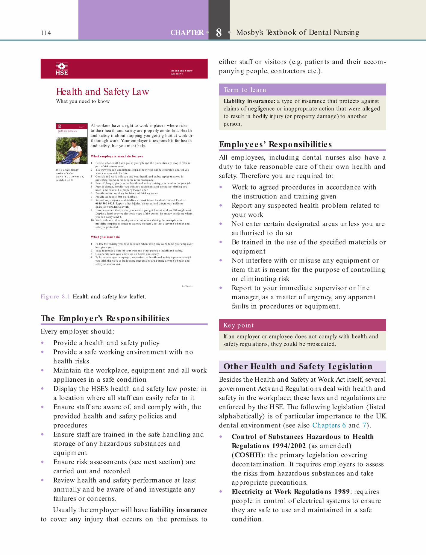
114 CHAPTER • 8 • Mosby’s Textbook of Dental Nursing
either staff or visitors (e.g. patients and their accom-panying people, contractors etc.).
Fig u re 8.1 Health and safety law lea et.
He alth and Safe ty Exe c utive
1 of 2 pages
This is a web-friendly version of lea et ISBN 978 0 7176 6501 3,published 04/09
He alth and Safe ty Exe c utive
Health and Safety LawWhat you need to know
All workers have a right to work in places where risks to their health and safety are properly controlled. Health and safety is about s topping you getting hurt at work or ill through work. Your employer is responsible for health and safety, but you must help.
What e mploye rs mus t do for you
1 Decide what could harm you in your job and the precautions to s top it. This ispart of risk assessment.
2 In a way you can understand, explain how risks will be controlled and tell youwho is responsible for this .
3 Consult and work with you and your health and safety representatives inprotecting everyone from harm in the workplace.
4 Free of charge, give you the health and safety training you need to do your job.5 Free of charge, provide you with any equipment and protective clothing you
need, and ensure it is properly looked after.6 Provide toilets , washing facilities and drinking water.7 Provide adequate firs t-aid facilities .8 Report major injuries and fatalities at work to our Incident Contact Centre:
0845 300 9923. Report other injuries , diseases and dangerous incidents online at www.hs e .gov.uk.
9 Have insurance that covers you in case you get hurt at work or ill through work.Display a hard copy or electronic copy of the current insurance certificate where you can easily read it.
10 Work with any other employers or contractors sharing the workplace orproviding employees (such as agency workers), so that everyone’s health andsafety is protected.
What you mus t do
1 Follow the training you have received when using any work items your employerhas given you.
2 Take reasonable care of your own and other people’s health and safety.3 Co-operate with your employer on health and safety.4 Tell someone (your employer, supervisor, or health and safety representative) if
you think the work or inadequate precautions are putting anyone’s health andsafety at serious risk.
Health and Safety LawWhat you need to know
Key poin tIf an employer or employee does not comply with health and safety regulations, they could be prosecuted.
Term to learnLiability insurance: a type of insurance that protects against claims of negligence or inappropriate action that were alleged to result in bodily injury (or property damage) to another person.
The Employer’s ResponsibilitiesEvery employer should:
• Provide a health and safety policy• Provide a safe working environment with no
health risks• Maintain the workplace, equipment and all work
appliances in a safe condition• Display the HSE’s health and safety law poster in
a location where all staff can easily refer to it• Ensure staff are aware of, and comply with, the
provided health and safety policies and procedures
• Ensure staff are trained in the safe handling and storage of any hazardous substances and equipment
• Ensure risk assessments (see next section) are carried out and recorded
• Review health and safety performance at least annually and be aware of and investigate any failures or concerns.Usually the employer will have liability insurance
to cover any injury that occurs on the premises to
Employees’ ResponsibilitiesAll employees, including dental nurses also have a duty to take reasonable care of their own health and safety. Therefore you are required to:
• Work to agreed procedures in accordance with the instruction and training given
• Report any suspected health problem related to your work
• Not enter certain designated areas unless you are authorised to do so
• Be trained in the use of the speci ed materials or equipment
• Not interfere with or misuse any equipment or item that is meant for the purpose of controlling or eliminating risk
• Report to your immediate supervisor or line manager, as a matter of urgency, any apparent faults in procedures or equipment.
Other Health and Safe ty LegislationBesides the Health and Safety at Work Act itself, several government Acts and Regulations deal with health and safety in the workplace; these laws and regulations are enforced by the HSE. The following legislation (listed alphabetically) is of particular importance to the UK dental environment (see also Chapters 6 and 7).
• Control of Substances Hazardous to Health Regulations 1994/2002 (as amended) (COSHH) : the primary legislation covering decontamination. It requires employers to assess the risks from hazardous substances and take appropriate precautions.
• Electricity at Work Regulations 1989: requires people in control of electrical systems to ensure they are safe to use and maintained in a safe condition.
115Workplace Health and Safety
(RIDDOR) : requires employers to notify certain occupational injuries, diseases and dangerous events
• Special Waste and Hazardous Waste Regulations (England and Wales) 2005 (see Chapter 6)
• Workplace (Health Safety and Welfare) Regulations 1992: covers a wide range of basic health, safety and welfare issues such as ventilation, heating, lighting, workstations, seating and welfare facilities (see www.legislation.gov.uk/ukpga/2008/14/contents).There are also further regulations affecting train-
ees, who are covered by the Health and Safety (Young Persons) Regulations 1997, now included in the Management of Health and Safety at Work Regulations 1999, section 19: Protection of Young Persons ( www.legisla t io n .go v.u k/ u ksi/ 1 99 9 / 32 4 2 / p d fs/uksi_19993242_en.pdf). These regulations imple-ment certain European Directive provisions concern-ing the protection of young persons – who are de ned as any person who has not reached the age of 18 – at work. They extend the requirement to carry out a risk assessment to include risks to young persons. They require certain information to be given to employees and to the parents/guardians of children.
Employers must protect young persons from any risks to their health and safety which are associated with the young person’s lack of experience, lack of awareness of existing risks, or immaturity.
Employers may not employ young persons where the work is:
• Beyond their physical or psychological capacity
• Involving exposure to toxic or carcinogenic substances
• Involving harmful exposure to radiation• Involving a risk of accidents which young
persons may not reasonably recognise due to their insuf cient attention to safety or lack of experience or training
• Involving a risk to health from extreme temperatures, noise or vibration.
An Overview of Health and Safety RegulationA review of health and safety regulation conducted by the Health and Safety Commission (HSC) in 1994 found that people were confused about the differences
• Employers’ Liability (Compulsory Insurance) Act 1969: requires employers to take out insurance against accidents and ill health to their employees.
• Environmental Protection Act 1990 (EPA)• Fire Precaution (Workplace) Regulations
(amended 1999)• Health & Safety (First Aid) Regulations 1981• Health and Safety (Display Screen
Equipment) Regulations 1992: sets out requirements for work with visual display units (VDUs)
• Health and Safety (First Aid) Regulations 1981: covers requirements for rst aid
• Health and Safety Information for Employees Regulations 1989: requires employers to display a poster telling employees what they need to know about health and safety
• Health and Safety (Sharp Instruments in Healthcare) Regulations 2013 (the Sharps Regulations): these Regulations implement (in part) EC Directive 2010/32/EU as required under European law and require healthcare employers and their contractors to ensure that risks of sharps injuries are adequately controlled
• Ionising Radiation (Medical Exposure) Regulations 2000 (IR(ME)R) : see Chapter 11
• Ionising Radiation Regulations 1999 (IRR)• Management of Health and Safety at Work
Regulations 1999: requires employers to carry out risk assessments, make arrangements to implement necessary measures, appoint competent people and arrange for appropriate information and training
• Manual Handling Operations Regulations 1992: covers the moving of objects by hand or bodily force
• Noise at Work Regulations 1989: requires employers to take action to protect employees from hearing damage
• Personal Protective Equipment at Work Regulations 1992: requires employers to provide appropriate protective clothing and equipment for their employees
• Provision and Use of Work Equipment Regulations 1998: requires that equipment provided for use at work, including machinery, is safe
• Reporting of Injuries, Diseases and Dangerous Occurrences Regulations 1995/2013
116 CHAPTER • 8 • Mosby’s Textbook of Dental Nursing
Where HSC/E consider action is necessary to supplement existing arrangements, their three main options are:
• Guidance• Approved Codes of Practice• Regulations.HSC/E try to take whichever option, or options, allows employers most exibility and costs them least, while providing proper safeguards for employees and the public.
Guidanc eHSE publishes guidance on a range of subjects. Guid-ance can be speci c to the health and safety problems of an industry or of a particular process used in a number of industries.
The main purposes of guidance are:
• to interpret – helping people to understand what the law says – including for example how requirements based on EC Directives t with those under the Health and Safety at Work Act
• to help people comply with the law• to give technical advice.
Following guidance is not compulsory and employers are free to take other action. But if they do follow guidance they will normally be doing enough to comply with the law. (Please also see the sections below on Approved Codes of Practice and regulations, which explain other ways in which employers are helped to know whether they are doing what the law requires.)
HSC/E aim to keep guidance up-to-date, because as technologies change, risks and the measures needed to address them change too.
Approve d c ode s o f prac tic eApproved Codes of Practice offer practical examples of good practice. They give advice on how to comply with the law by, for example, providing a guide to what is ‘reasonably practicable’. For example, if regulations use words like ‘suitable and suf cient’, an Approved Code of Practice can illustrate what this requires in particu-lar circumstances.
Approved Codes of Practice have a special legal status. If employers are prosecuted for a breach of health and safety law, and it is proved that they have not followed the relevant provisions of the Approved Code of Practice, a court can nd them at fault unless they can show that they have complied with the law in some other way.
between ‘guidance’, Approved Codes of Practice (ACOPs) and ‘regulations’, and how they relate to each other. The Health and Safety Executive has pro-duced guidance that aims to explain how each ts in. Health and safety regulation … a short guide (HSE, avail-able at http:/ /www.hse.gov.uk/pubns/hsc13.pdf) is for both employers and self-employed people, and will also be of interest to anyone who wants to know how health and safety law is meant to work. The following is from that guidance.
What he a lth and s a fe ty law re quire sThe basis of British health and safety law is the Health and Safety at Work etc. Act 1974. The Act sets out the general duties which employers have towards employ-ees and members of the public, and employees have to themselves and to each other.
These duties are quali ed in the Act by the princi-ple of ‘so far as is reasonably practicable’. In other words, an employer does not have to take measures to avoid or reduce the risk if they are technically impos-sible or if the time, trouble or cost of the measures would be grossly disproportionate to the risk.
What the law requires here is what good manage-ment and common sense would lead employers to do anyway: that is, to look at what the risks are and take sensible measures to tackle them.
The Management of Health and Safety at Work Regulations 1999 (the Management Regulations) gen-erally make more explicit what employers are required to do to manage health and safety under the Health and Safety at Work Act. Like the Act, they apply to every work activity.
Ac tion on he a lth and s afe ty: op tionsThe aims of the Health and Safety Commission and its operating arm, the Executive (HSC/E), are to
• Protect the health, safety and welfare of employees
• Safeguard others, principally the public, who may be exposed to risks from work activity.
HSC/E consult fully with people affected by their legislative proposals, and adopt various approaches based on assessing and controlling risk.
Among the things that can prompt action from HSC/E are:
• Changes in technologies, industries or risks• Evidence of accidents and ill health, plus public
concern• European Directives.

117Workplace Health and Safety
Doing a risk assessment helps employers decide whether they have taken enough precautions, or should do more, to prevent harm from occurring to themselves, their staff or others.
A risk assessment should:
• Identify a hazard• Consider whether anyone (e.g. especially certain
staff handling specialised equipment, older people, pregnant women, children, etc.) may be harmed by that hazard
• Evaluate the existing precautions• Take action to improve precautions and
minimise the risk of the hazard occurring• Record the ndings• Review regularly the arrangements – at least once
a year.There must be a risk assessment of all potential hazards. A risk assessment is an important step, as well as complying with the law. It helps focus on the risks that really matter – the ones with potential to cause harm. The Health and Safety Executive (HSE) suggest ve steps, shown in Box 8.1 (www.hse.gov.uk/risk/ vesteps.htm).
Examples of potential hazards that might be subject to a risk assessment in dentistry are shown in Box 8.2.
HSC consulted in 1995 on the role of Approved Codes of Practice in the health and safety system and concluded that they could still be used in support of legal duties in speci c circumstances.
Re gula tionsRegulations are law, approved by Parliament. These are usually made under the Health and Safety at Work Act, following proposals from HSC. This applies to regulations based on EC Directives as well as ‘home-grown’ ones.
The Health and Safety at Work Act, and general duties in the Management Regulations, are goal-setting (see below ‘What form do they take?’) and leave employers freedom to decide how to control risks which they identify. Guidance and Approved Codes of Practice give advice. But some risks are so great, or the proper control measures so costly, that it would not be appropriate to leave employers discretion in decid-ing what to do about them. Regulations identify these risks and set out speci c action that must be taken. Often these requirements are absolute – to do some-thing without quali cation by whether it is reasonably practicable.
How re gula tions applySome regulations apply across all companies, such as the Manual Handling Regulations which apply wher-ever things are moved by hand or bodily force, and the Display Screen Equipment Regulations which apply wherever VDUs are used. Other regulations apply to hazards unique to speci c industries, such as mining or nuclear.
What fo rm do the y take ?HSC will where appropriate propose regulations in goal-setting form: that is, setting out what must be achieved, but not how it must be done. Sometimes it is necessary to be prescriptive, that is spelling out in detail what should be done.
Risk AssessmentThe main health and safety requirement on employers is to carry out a risk assessment. The Management of Health and Safety at Work Regulations 1999 and Man-agement of Health and Safety at Work (Northern Ireland) Regulations 2000 introduced risk assessment. Employers with ve or more employees need to record the signi cant ndings of the risk assessment.
A risk assessment is simply a careful examination of what, in the workplace, could cause harm to people.
Box 8.1 Steps for a risk assessment
1. Identify the hazards.
2. Decide who might be harmed and how.
3. Evaluate the risks and decide on precautions.
4. Record your ndings and implement them.
5. Review your assessment and update if necessary.
Term to lea rnDental bur: a type of drill bit that is tted into the dental handpiece and used for cutting or grinding the hard tooth material or bone. Also used in laboratory work.
Risk assessment should be straightforward in a simple workplace such as a typical dental environ-ment. It should only be complicated if it deals with serious hazards, such as those in a laboratory.
Advantages of risk assessment include:
• Increased awareness of workplace hazards• Opportunity to identify and control workplace
hazards• Can lead to increased productivity• May prevent an injury or illness.
118 CHAPTER • 8 • Mosby’s Textbook of Dental Nursing
• Appoint competent people (often themselves or company colleagues) to help them to implement the arrangements
• Set up emergency procedures• Provide clear information and training to
employees• Work together with other employers sharing the
same workplace.Other regulations require action in response to
particular hazards, or in industries where hazards are particularly high. Many are not quali ed by ‘reasona-ble practicability’. In recent years much of Britain’s health and safety law has originated in Europe. Pro-posals from the European Commission may be agreed by Member States, who are then responsible for making them part of their domestic law.
Modern health and safety law in UK, including much of that from Europe, is based on the principle of risk assessment described above.
Identify and learnIdent ify a pa ir o f ext ract ion forceps, a handpiece , a den ta l bur and a pa ir of o rthodont ic p lie rs in your workplace .
Box 8.2 Risk assessment in den t ist ry
Handling dangerous substances• Certain dental materials used in the dental surgery and
laboratory (e.g. acids) that could cause bodily harm if not used correctly
• Substances used for developing radiographs (X-ray lms) (see Chapter 6).
Handling dangerous instruments (these may also be dangerous if hot)• Extraction forceps and other surgical instruments
• Handpieces and dental burs
• Orthodontic pliers
• Sharps – instruments, scalpels, needles (see Chapter 6).
Handling dangerous machinery/equipment• Dental handpieces
• Electrosurgery equipment
• Heating equipment
• Laboratory equipment
• Lasers (see Chapter 6)
• Steam sterilisers (autoclaves).
Besides carrying out a risk assessment, employers also need to:
• Make arrangements for implementing the health and safety measures identi ed as necessary by the risk assessment
DENTAL ANATOMY, PHYSIOLOGY AND DISEASEChapter 9 Denta l and Ora l Anatomy and Physio logy 121
Chapte r 10 Denta l and Ora l Pa thology and Disease 143
Chapte r 11 Denta l and Ora l Imaging 165
Sect ion C

This page intentiona lly le ft blank
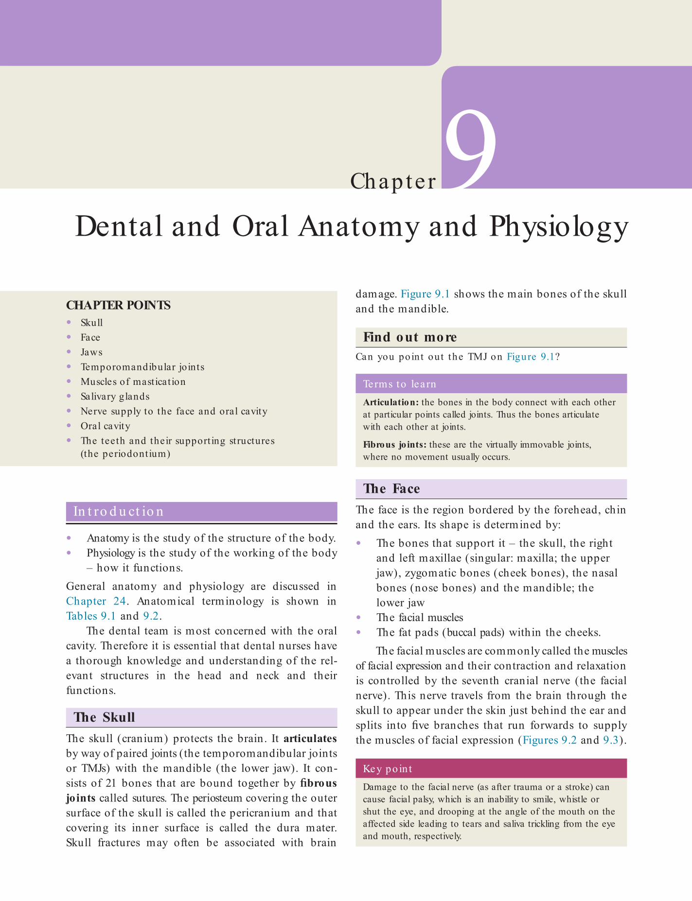
Chapter 9 Dental and Oral Anatomy and Physiology
CHAPTER POINTS• Skull• Face• Jaws• Temporomandibular joints• Muscles of mastication• Salivary glands• Nerve supply to the face and oral cavity• Oral cavity• The teeth and their supporting structures
(the periodontium)
damage. Figure 9.1 shows the main bones of the skull and the mandible.
Find out moreCan you po int out the TMJ on Figure 9.1?
In t ro d u ct io n
• Anatomy is the study of the structure of the body.• Physiology is the study of the working of the body
– how it functions.General anatomy and physiology are discussed in Chapter 24. Anatomical terminology is shown in Tables 9.1 and 9.2.
The dental team is most concerned with the oral cavity. Therefore it is essential that dental nurses have a thorough knowledge and understanding of the rel-evant structures in the head and neck and their functions.
The SkullThe skull (cranium) protects the brain. It articulates by way of paired joints (the temporomandibular joints or TMJs) with the mandible (the lower jaw). It con-sists of 21 bones that are bound together by brous joints called sutures. The periosteum covering the outer surface of the skull is called the pericranium and that covering its inner surface is called the dura mater. Skull fractures may often be associated with brain
Terms to lea rnArticulation: the bones in the body connect with each other at particular points called joints. Thus the bones articulate with each other at joints.
Fibrous joints: these are the virtually immovable joints, where no movement usually occurs.
The FaceThe face is the region bordered by the forehead, chin and the ears. Its shape is determined by:
• The bones that support it – the skull, the right and left maxillae (singular: maxilla; the upper jaw), zygomatic bones (cheek bones), the nasal bones (nose bones) and the mandible; the lower jaw
• The facial muscles• The fat pads (buccal pads) within the cheeks.
The facial muscles are commonly called the muscles of facial expression and their contraction and relaxation is controlled by the seventh cranial nerve (the facial nerve). This nerve travels from the brain through the skull to appear under the skin just behind the ear and splits into ve branches that run forwards to supply the muscles of facial expression (Figures 9.2 and 9.3).
Key poin tDamage to the facial nerve (as after trauma or a stroke) can cause facial palsy, which is an inability to smile, whistle or shut the eye, and drooping at the angle of the mouth on the affected side leading to tears and saliva trickling from the eye and mouth, respectively.
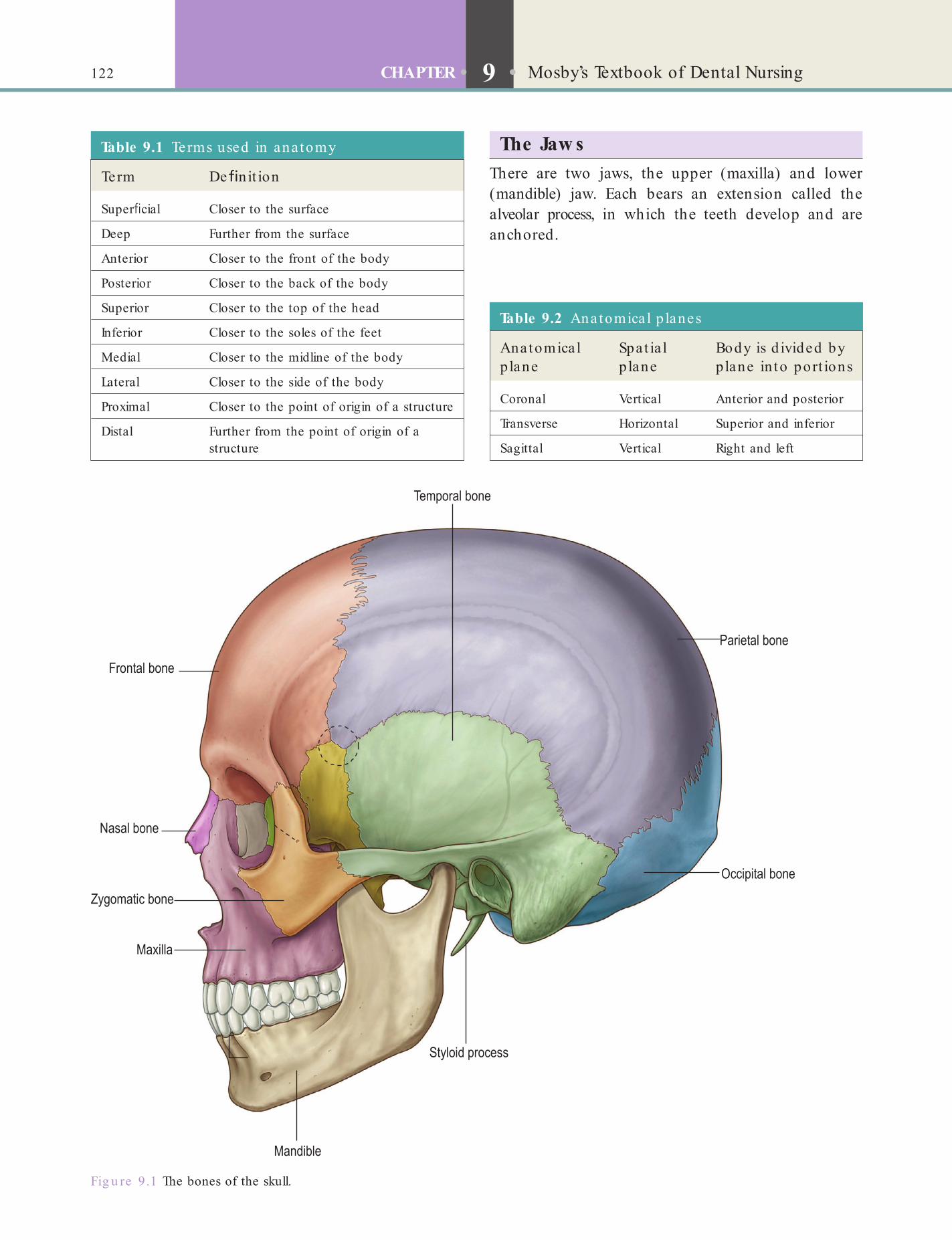
122 CHAPTER • 9 • Mosby’s Textbook of Dental Nursing
Fig u re 9.1 The bones of the skull.
Table 9.1 Terms used in ana tomy
Term De n it ion
Super cial Closer to the surface
Deep Further from the surface
Anterior Closer to the front of the body
Posterior Closer to the back of the body
Superior Closer to the top of the head
Inferior Closer to the soles of the feet
Medial Closer to the midline of the body
Lateral Closer to the side of the body
Proximal Closer to the point of origin of a structure
Distal Further from the point of origin of a structure
Table 9.2 Ana tomica l p lanes
Anatomica l p lane
Spat ia l p lane
Body is d ivided by plane in to port ions
Coronal Vertical Anterior and posterior
Transverse Horizontal Superior and inferior
Sagittal Vertical Right and left
The Jaw sThere are two jaws, the upper (maxilla) and lower (mandible) jaw. Each bears an extension called the alveolar process, in which the teeth develop and are anchored.
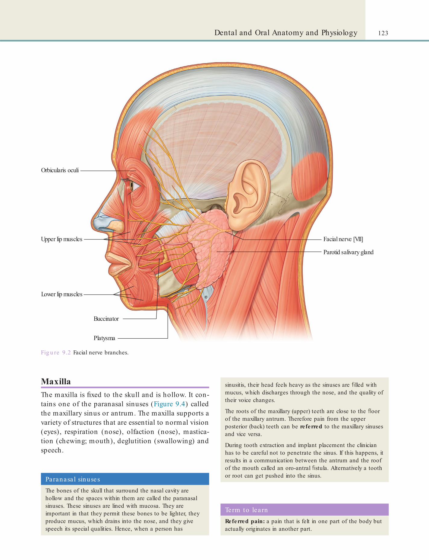
123Dental and Oral Anatomy and Physiology
Fig u re 9.2 Facial nerve branches.
Orbicularis oculi
Platysma
Upper lip muscles Facial nerve [VII]
Parotid salivary gland
Lower lip muscles
Buccinator
MaxillaThe maxilla is xed to the skull and is hollow. It con-tains one of the paranasal sinuses (Figure 9.4) called the maxillary sinus or antrum. The maxilla supports a variety of structures that are essential to normal vision (eyes), respiration (nose), olfaction (nose), mastica-tion (chewing; mouth), deglutition (swallowing) and speech.
Paranasa l sinusesThe bones of the skull that surround the nasal cavity are hollow and the spaces within them are called the paranasal sinuses. These sinuses are lined with mucosa. They are important in that they permit these bones to be lighter, they produce mucus, which drains into the nose, and they give speech its special qualities. Hence, when a person has
sinusitis, their head feels heavy as the sinuses are lled with mucus, which discharges through the nose, and the quality of their voice changes.
The roots of the maxillary (upper) teeth are close to the oor of the maxillary antrum. Therefore pain from the upper posterior (back) teeth can be referred to the maxillary sinuses and vice versa.
During tooth extraction and implant placement the clinician has to be careful not to penetrate the sinus. If this happens, it results in a communication between the antrum and the roof of the mouth called an oro-antral stula. Alternatively a tooth or root can get pushed into the sinus.
Term to lea rnReferred pain: a pain that is felt in one part of the body but actually originates in another part.

124 CHAPTER • 9 • Mosby’s Textbook of Dental Nursing
Fig u re 9.3 The main muscles of facial expression.
MandibleThe mandible is virtually solid, containing no sinuses. It consists of (Figure 9.5):
• A horizontal body• The two angles between the horizontal and
vertical parts (ramus, plural rami)• The coronoid processes• The mandibular condyles – these articulate
with the temporal bone of the skull at the glenoid fossa via the temporomandibular joints (TMJs).
The mandible is essential to chewing (mastica-tion) and to speech and swallowing (see Table 9.3).
Alveolar ProcessesThe alveolar processes of the jaws are the parts that form the sockets (alveoli; singular alveolus) for the teeth (see Figure 9.5). The bone lining an alveolus is called bundle bone. This is because the periodontal ligament bres (see p. 141) which attach the tooth roots to the bone are attached to this bone. On X-rays (radiographs), this layer appears as a more

125Dental and Oral Anatomy and Physiology
Fig u re 9.4 The main paranasal air sinuses.
Fig u re 9.5 The mandible.
radio-dense (lighter) line (that is, it appears distinctly white), called the lamina dura.
Identify and learnFind a couple o f in t ra -ora l rad iographs in your workplace and ask your supervisor to poin t ou t the lamina dura .
Key poin tA lack of the lamina dura appearance on the radiograph is considered a sign of disease.
The Temporomandibular JointsThere are two TMJs, one on either side of the face. Each TMJ is formed from two bones, the skull bone called the temporal bone, above, and the mandible below (Figure 9.6). To reduce the friction between the two bones, there is a disc of cartilage tissue between the two, called the articular disc. This disc divides the joint into an upper and lower cavity. The TMJ is one of the most frequently used joints in the body, permitting speech, chewing, yawning, swallowing and sneezing. The movements that are possible at this joint are given in Table 9.3.
Key poin tThe TMJs are the two most frequently used joints in the body.
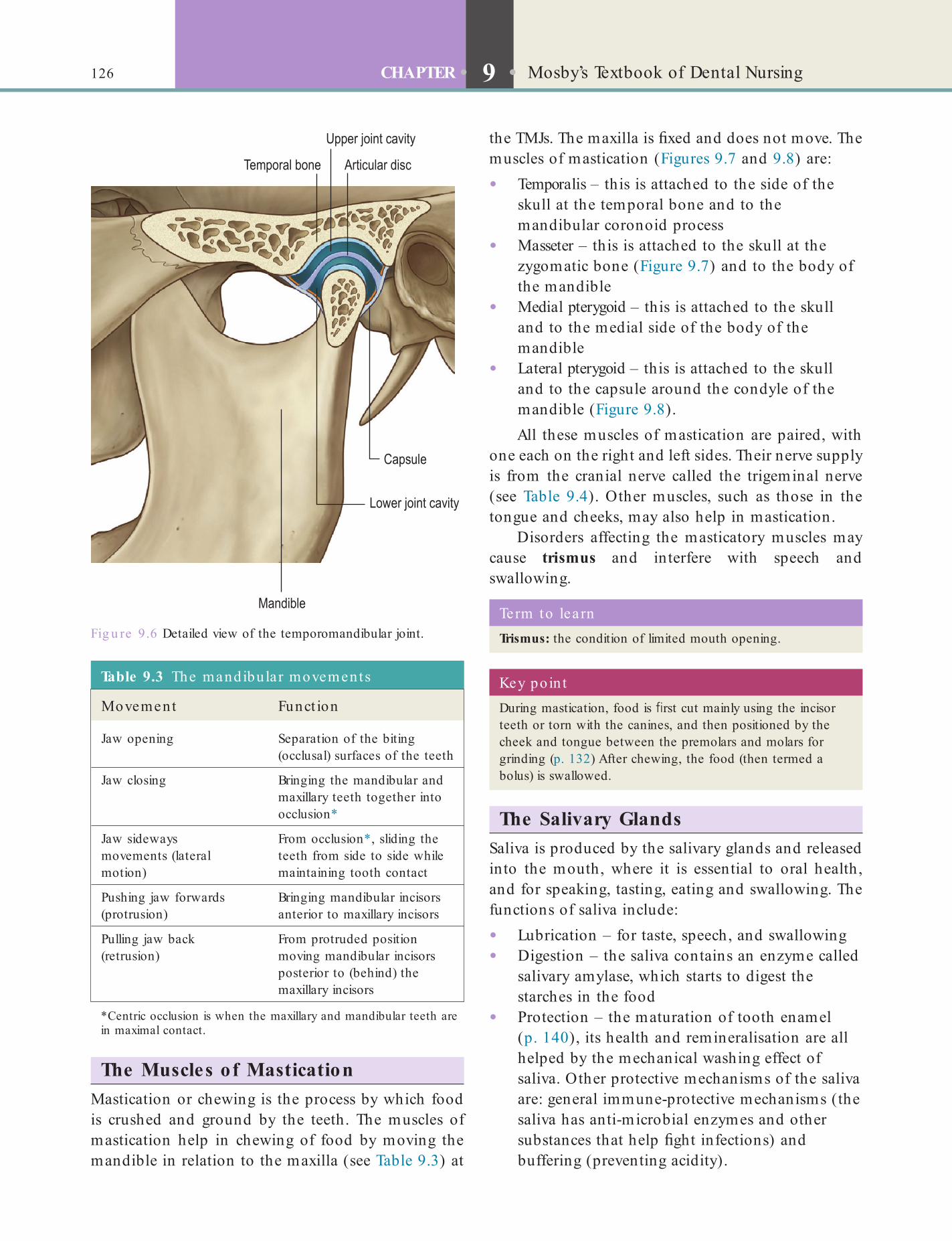
126 CHAPTER • 9 • Mosby’s Textbook of Dental Nursing
the TMJs. The maxilla is xed and does not move. The muscles of mastication (Figures 9.7 and 9.8) are:
• Temporalis – this is attached to the side of the skull at the temporal bone and to the mandibular coronoid process
• Masseter – this is attached to the skull at the zygomatic bone (Figure 9.7) and to the body of the mandible
• Medial pterygoid – this is attached to the skull and to the medial side of the body of the mandible
• Lateral pterygoid – this is attached to the skull and to the capsule around the condyle of the mandible (Figure 9.8).All these muscles of mastication are paired, with
one each on the right and left sides. Their nerve supply is from the cranial nerve called the trigeminal nerve (see Table 9.4). Other muscles, such as those in the tongue and cheeks, may also help in mastication.
Disorders affecting the masticatory muscles may cause trismus and interfere with speech and swallowing.
Fig u re 9.6 Detailed view of the temporomandibular joint.
The Muscles of MasticationMastication or chewing is the process by which food is crushed and ground by the teeth. The muscles of mastication help in chewing of food by moving the mandible in relation to the maxilla (see Table 9.3) at
Term to lea rnTrismus: the condition of limited mouth opening.
Key poin tDuring mastication, food is rst cut mainly using the incisor teeth or torn with the canines, and then positioned by the cheek and tongue between the premolars and molars for grinding (p. 132) After chewing, the food (then termed a bolus) is swallowed.
The Salivary GlandsSaliva is produced by the salivary glands and released into the mouth, where it is essential to oral health, and for speaking, tasting, eating and swallowing. The functions of saliva include:
• Lubrication – for taste, speech, and swallowing• Digestion – the saliva contains an enzyme called
salivary amylase, which starts to digest the starches in the food
• Protection – the maturation of tooth enamel (p. 140), its health and remineralisation are all helped by the mechanical washing effect of saliva. Other protective mechanisms of the saliva are: general immune-protective mechanisms (the saliva has anti-microbial enzymes and other substances that help ght infections) and buffering (preventing acidity).
Table 9.3 The mandibular movements
Movement Funct ion
Jaw opening Separation of the biting (occlusal) surfaces of the teeth
Jaw closing Bringing the mandibular and maxillary teeth together into occlusion*
Jaw sideways movements (lateral motion)
From occlusion*, sliding the teeth from side to side while maintaining tooth contact
Pushing jaw forwards (protrusion)
Bringing mandibular incisors anterior to maxillary incisors
Pulling jaw back (retrusion)
From protruded position moving mandibular incisors posterior to (behind) the maxillary incisors
*Centric occlusion is when the maxillary and mandibular teeth are in maximal contact.
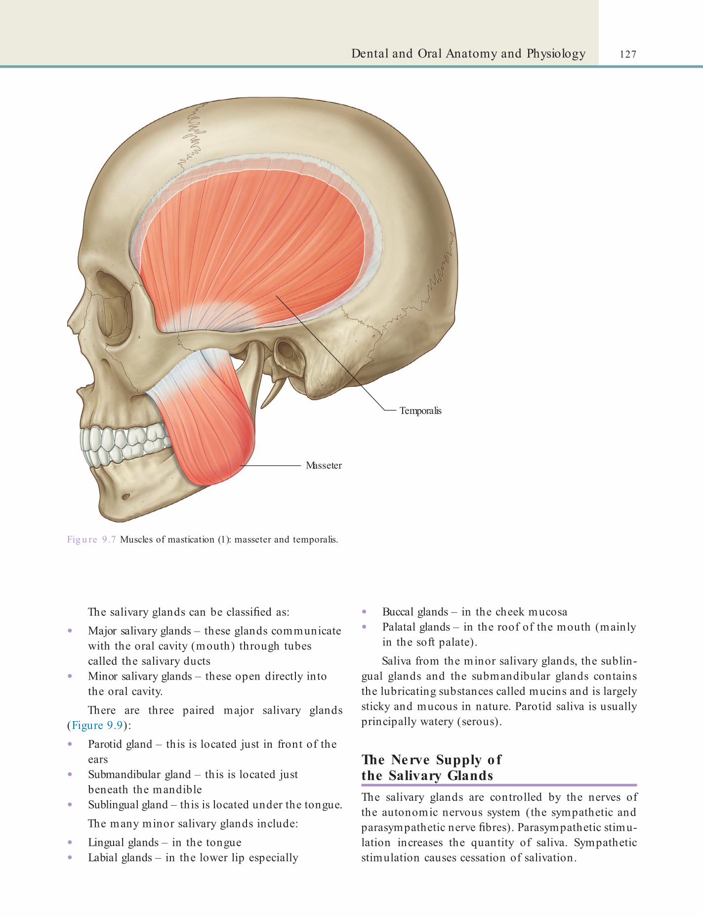
127Dental and Oral Anatomy and Physiology
• Buccal glands – in the cheek mucosa• Palatal glands – in the roof of the mouth (mainly
in the soft palate).Saliva from the minor salivary glands, the sublin-
gual glands and the submandibular glands contains the lubricating substances called mucins and is largely sticky and mucous in nature. Parotid saliva is usually principally watery (serous).
The Nerve Supply of the Salivary GlandsThe salivary glands are controlled by the nerves of the autonomic nervous system (the sympathetic and parasympathetic nerve bres). Parasympathetic stimu-lation increases the quantity of saliva. Sympathetic stimulation causes cessation of salivation.
The salivary glands can be classi ed as:
• Major salivary glands – these glands communicate with the oral cavity (mouth) through tubes called the salivary ducts
• Minor salivary glands – these open directly into the oral cavity.There are three paired major salivary glands
(Figure 9.9):
• Parotid gland – this is located just in front of the ears
• Submandibular gland – this is located just beneath the mandible
• Sublingual gland – this is located under the tongue.The many minor salivary glands include:
• Lingual glands – in the tongue• Labial glands – in the lower lip especially
Fig u re 9.7 Muscles of mastication (1): masseter and temporalis.
Temporalis
Masseter
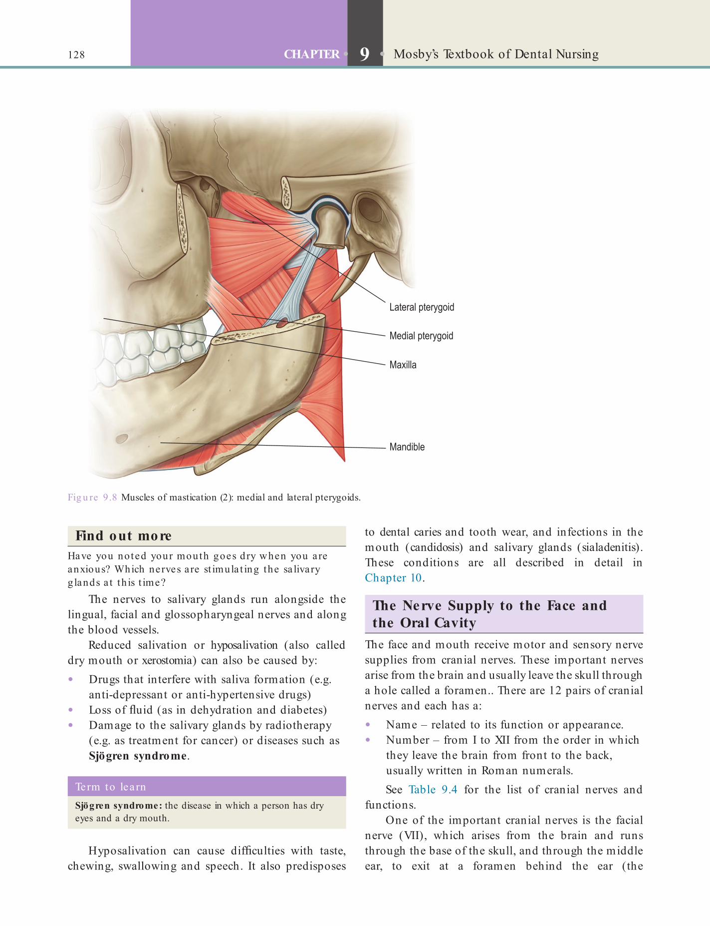
128 CHAPTER • 9 • Mosby’s Textbook of Dental Nursing
to dental caries and tooth wear, and infections in the mouth (candidosis) and salivary glands (sialadenitis). These conditions are all described in detail in Chapter 10.
The Nerve Supply to the Face and the Oral Cavity
The face and mouth receive motor and sensory nerve supplies from cranial nerves. These important nerves arise from the brain and usually leave the skull through a hole called a foramen.. There are 12 pairs of cranial nerves and each has a:
• Name – related to its function or appearance.• Number – from I to XII from the order in which
they leave the brain from front to the back, usually written in Roman numerals.See Table 9.4 for the list of cranial nerves and
functions.One of the important cranial nerves is the facial
nerve (VII), which arises from the brain and runs through the base of the skull, and through the middle ear, to exit at a foramen behind the ear (the
Find out moreHave you noted your mouth goes dry when you a re anxious? Which nerves a re st imula t ing the sa livary g lands a t th is t ime?
The nerves to salivary glands run alongside the lingual, facial and glossopharyngeal nerves and along the blood vessels.
Reduced salivation or hyposalivation (also called dry mouth or xerostomia) can also be caused by:
• Drugs that interfere with saliva formation (e.g. anti-depressant or anti-hypertensive drugs)
• Loss of uid (as in dehydration and diabetes)• Damage to the salivary glands by radiotherapy
(e.g. as treatment for cancer) or diseases such as Sjögren syndrome.
Fig u re 9.8 Muscles of mastication (2): medial and lateral pterygoids.
Term to lea rnSjögren syndrome: the disease in which a person has dry eyes and a dry mouth.
Hyposalivation can cause dif culties with taste, chewing, swallowing and speech. It also predisposes
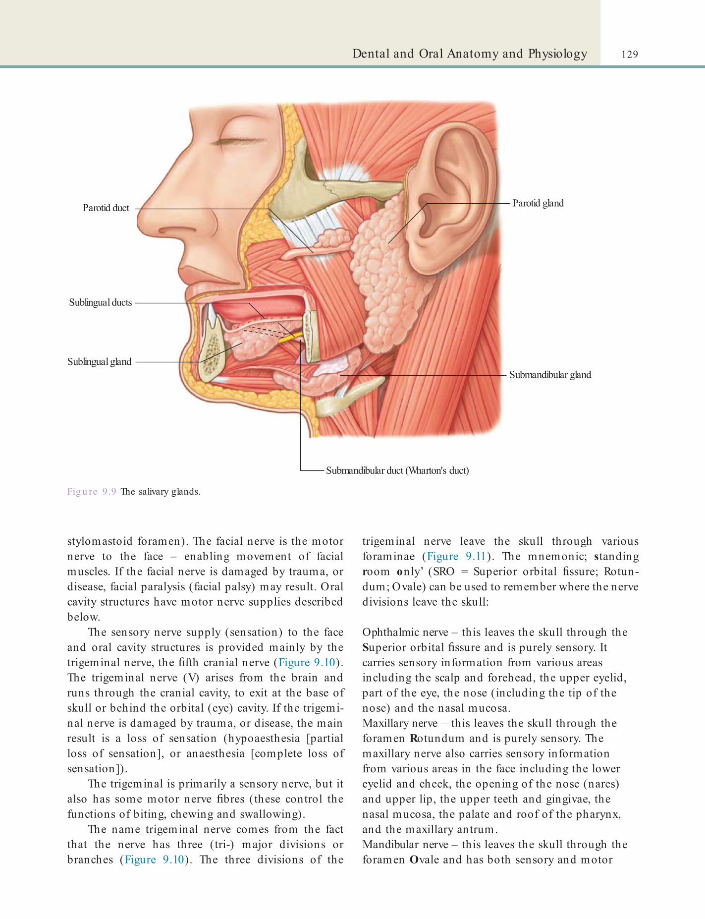
129Dental and Oral Anatomy and Physiology
stylomastoid foramen). The facial nerve is the motor nerve to the face – enabling movement of facial muscles. If the facial nerve is damaged by trauma, or disease, facial paralysis (facial palsy) may result. Oral cavity structures have motor nerve supplies described below.
The sensory nerve supply (sensation) to the face and oral cavity structures is provided mainly by the trigeminal nerve, the fth cranial nerve (Figure 9.10). The trigeminal nerve (V) arises from the brain and runs through the cranial cavity, to exit at the base of skull or behind the orbital (eye) cavity. If the trigemi-nal nerve is damaged by trauma, or disease, the main result is a loss of sensation (hypoaesthesia [partial loss of sensation], or anaesthesia [complete loss of sensation]).
The trigeminal is primarily a sensory nerve, but it also has some motor nerve bres (these control the functions of biting, chewing and swallowing).
The name trigeminal nerve comes from the fact that the nerve has three (tri-) major divisions or branches (Figure 9.10). The three divisions of the
trigeminal nerve leave the skull through various foraminae (Figure 9.11). The mnemonic; standing room only’ (SRO = Superior orbital ssure; Rotun-dum; Ovale) can be used to remember where the nerve divisions leave the skull:
Ophthalmic nerve – this leaves the skull through the Superior orbital ssure and is purely sensory. It carries sensory information from various areas including the scalp and forehead, the upper eyelid, part of the eye, the nose (including the tip of the nose) and the nasal mucosa.Maxillary nerve – this leaves the skull through the foramen Rotundum and is purely sensory. The maxillary nerve also carries sensory information from various areas in the face including the lower eyelid and cheek, the opening of the nose (nares) and upper lip, the upper teeth and gingivae, the nasal mucosa, the palate and roof of the pharynx, and the maxillary antrum.Mandibular nerve – this leaves the skull through the foramen Ovale and has both sensory and motor
Fig u re 9.9 The salivary glands.
Parotid duct
Sublingual ducts
Sublingual gland
Submandibular duct (Wharton's duct)
Submandibular gland
Parotid gland
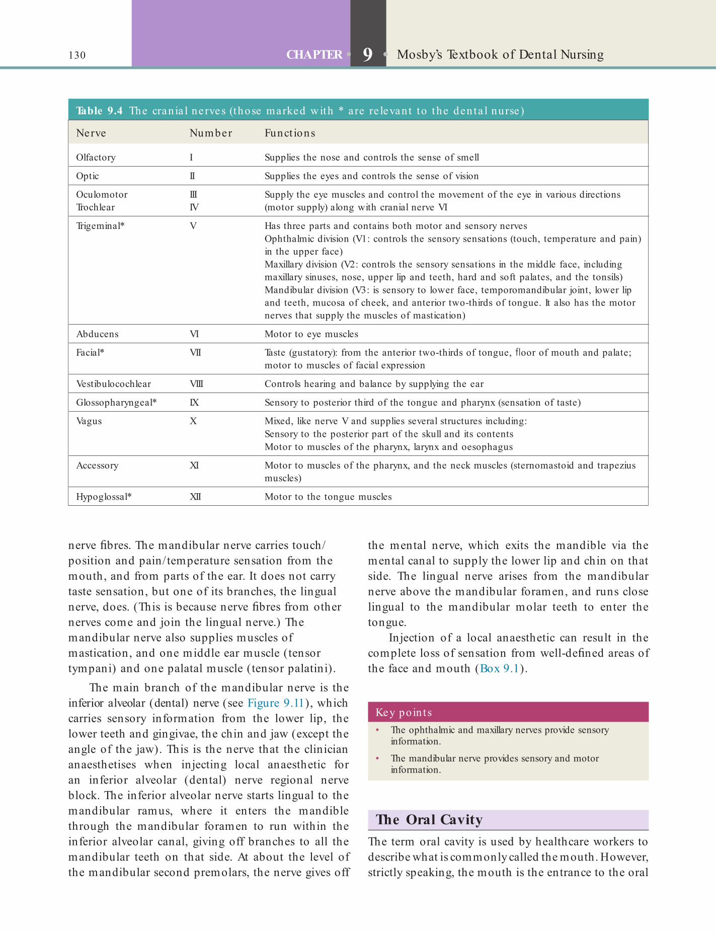
130 CHAPTER • 9 • Mosby’s Textbook of Dental Nursing
nerve bres. The mandibular nerve carries touch/position and pain/temperature sensation from the mouth, and from parts of the ear. It does not carry taste sensation, but one of its branches, the lingual nerve, does. (This is because nerve bres from other nerves come and join the lingual nerve.) The mandibular nerve also supplies muscles of mastication, and one middle ear muscle (tensor tympani) and one palatal muscle (tensor palatini).
The main branch of the mandibular nerve is the inferior alveolar (dental) nerve (see Figure 9.11), which carries sensory information from the lower lip, the lower teeth and gingivae, the chin and jaw (except the angle of the jaw). This is the nerve that the clinician anaesthetises when injecting local anaesthetic for an inferior alveolar (dental) nerve regional nerve block. The inferior alveolar nerve starts lingual to the mandibular ramus, where it enters the mandible through the mandibular foramen to run within the inferior alveolar canal, giving off branches to all the mandibular teeth on that side. At about the level of the mandibular second premolars, the nerve gives off
the mental nerve, which exits the mandible via the mental canal to supply the lower lip and chin on that side. The lingual nerve arises from the mandibular nerve above the mandibular foramen, and runs close lingual to the mandibular molar teeth to enter the tongue.
Injection of a local anaesthetic can result in the complete loss of sensation from well-de ned areas of the face and mouth (Box 9.1).
Key poin ts• The ophthalmic and maxillary nerves provide sensory
information.
• The mandibular nerve provides sensory and motor information.
The Oral CavityThe term oral cavity is used by healthcare workers to describe what is commonly called the mouth. However, strictly speaking, the mouth is the entrance to the oral
Table 9.4 The cran ia l nerves (those marked with * a re re levant to the den ta l nurse)
Nerve Number Funct ions
Olfactory I Supplies the nose and controls the sense of smell
Optic II Supplies the eyes and controls the sense of vision
Oculomotor Trochlear
IIIIV
Supply the eye muscles and control the movement of the eye in various directions (motor supply) along with cranial nerve VI
Trigeminal* V Has three parts and contains both motor and sensory nervesOphthalmic division (V1: controls the sensory sensations (touch, temperature and pain) in the upper face)Maxillary division (V2: controls the sensory sensations in the middle face, including maxillary sinuses, nose, upper lip and teeth, hard and soft palates, and the tonsils)Mandibular division (V3: is sensory to lower face, temporomandibular joint, lower lip and teeth, mucosa of cheek, and anterior two-thirds of tongue. It also has the motor nerves that supply the muscles of mastication)
Abducens VI Motor to eye muscles
Facial* VII Taste (gustatory): from the anterior two-thirds of tongue, oor of mouth and palate; motor to muscles of facial expression
Vestibulocochlear VIII Controls hearing and balance by supplying the ear
Glossopharyngeal* IX Sensory to posterior third of the tongue and pharynx (sensation of taste)
Vagus X Mixed, like nerve V and supplies several structures including:Sensory to the posterior part of the skull and its contentsMotor to muscles of the pharynx, larynx and oesophagus
Accessory XI Motor to muscles of the pharynx, and the neck muscles (sternomastoid and trapezius muscles)
Hypoglossal* XII Motor to the tongue muscles
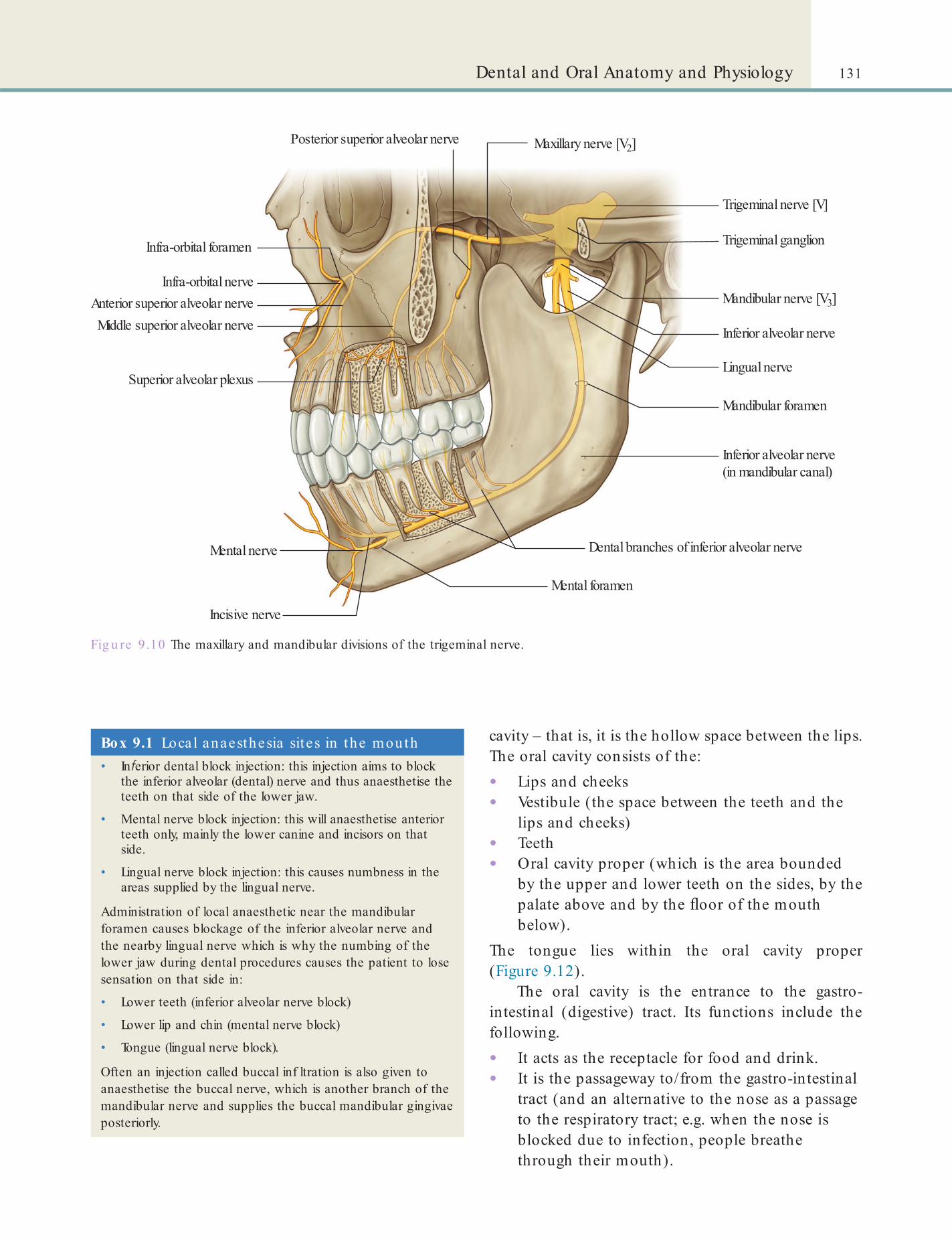
131Dental and Oral Anatomy and Physiology
cavity – that is, it is the hollow space between the lips. The oral cavity consists of the:
• Lips and cheeks• Vestibule (the space between the teeth and the
lips and cheeks)• Teeth• Oral cavity proper (which is the area bounded
by the upper and lower teeth on the sides, by the palate above and by the oor of the mouth below).
The tongue lies within the oral cavity proper (Figure 9.12).
The oral cavity is the entrance to the gastro-intestinal (digestive) tract. Its functions include the following.
• It acts as the receptacle for food and drink.• It is the passageway to/from the gastro-intestinal
tract (and an alternative to the nose as a passage to the respiratory tract; e.g. when the nose is blocked due to infection, people breathe through their mouth).
Fig u re 9.10 The maxillary and mandibular divisions of the trigeminal nerve.
Infra-orbital foramen
Infra-orbital nerveAnterior superior alveolar nerve
Posterior superior alveolar nerve
Middle superior alveolar nerve
Superior alveolar plexus
Trigeminal nerve [V]
Inferior alveolar nerve
Inferior alveolar nerve(in mandibular canal)
Mandibular foramen
Mental foramen
Dental branches of inferior alveolar nerve
Mandibular nerve [V3]
Maxillary nerve [V2]
Lingual nerve
Trigeminal ganglion
Mental nerve
Incisive nerve
Box 9.1 Local anaesthesia sites in the mouth• In erior dental block injection: this injection aims to block
the inferior alveolar (dental) nerve and thus anaesthetise the teeth on that side of the lower jaw.
• Mental nerve block injection: this will anaesthetise anterior teeth only, mainly the lower canine and incisors on that side.
• Lingual nerve block injection: this causes numbness in the areas supplied by the lingual nerve.
Administration of local anaesthetic near the mandibular foramen causes blockage of the inferior alveolar nerve and the nearby lingual nerve which is why the numbing of the lower jaw during dental procedures causes the patient to lose sensation on that side in:
• Lower teeth (inferior alveolar nerve block)
• Lower lip and chin (mental nerve block)
• Tongue (lingual nerve block).
Often an injection called buccal inf ltration is also given to anaesthetise the buccal nerve, which is another branch of the mandibular nerve and supplies the buccal mandibular gingivae posteriorly.
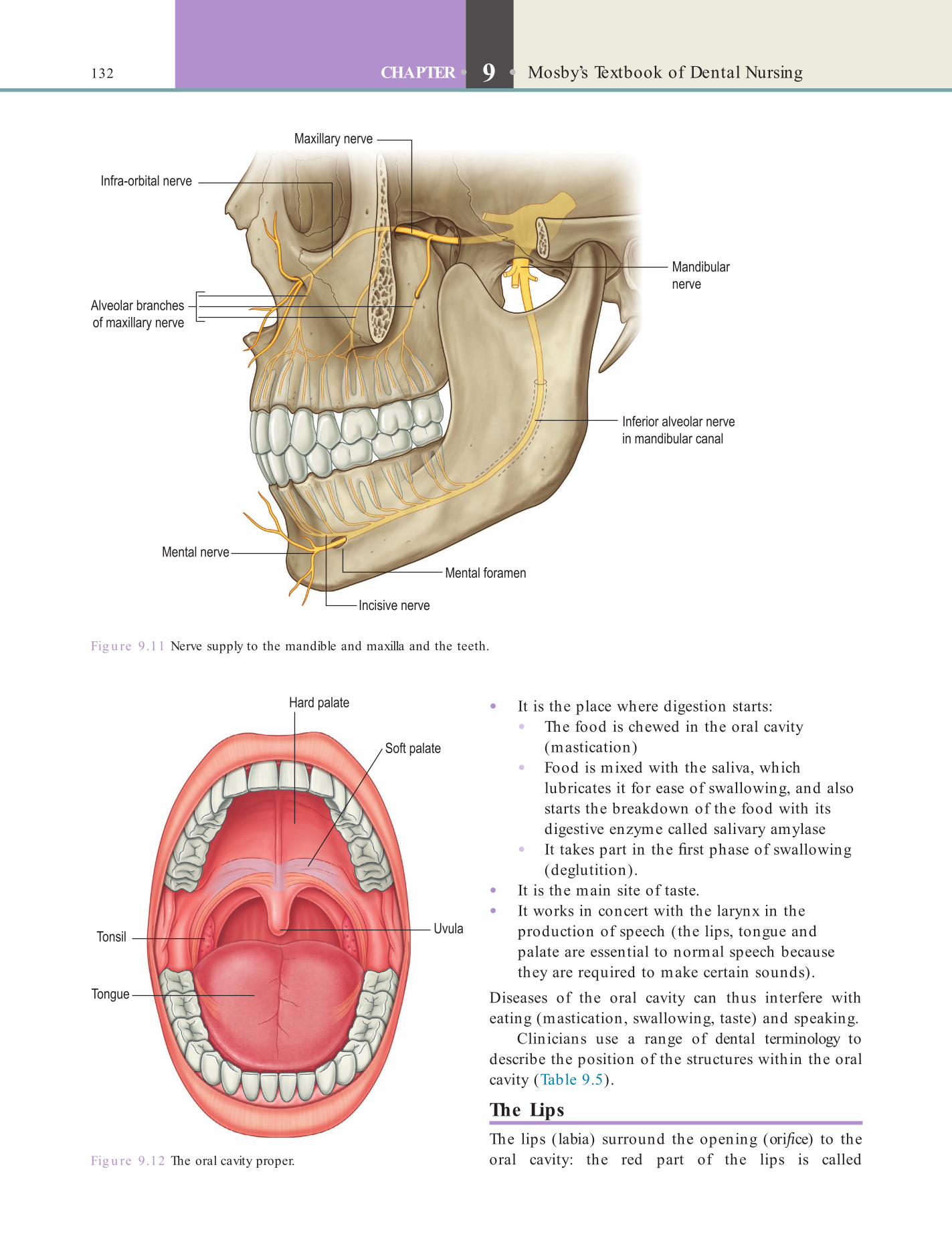
132 CHAPTER • 9 • Mosby’s Textbook of Dental Nursing
• It is the place where digestion starts:• The food is chewed in the oral cavity
(mastication)• Food is mixed with the saliva, which
lubricates it for ease of swallowing, and also starts the breakdown of the food with its digestive enzyme called salivary amylase
• It takes part in the rst phase of swallowing (deglutition).
• It is the main site of taste.• It works in concert with the larynx in the
production of speech (the lips, tongue and palate are essential to normal speech because they are required to make certain sounds).
Diseases of the oral cavity can thus interfere with eating (mastication, swallowing, taste) and speaking.
Clinicians use a range of dental terminology to describe the position of the structures within the oral cavity (Table 9.5).
The LipsThe lips (labia) surround the opening (ori ce) to the oral cavity: the red part of the lips is called
Fig u re 9.11 Nerve supply to the mandible and maxilla and the teeth.
Fig u re 9.12 The oral cavity proper.
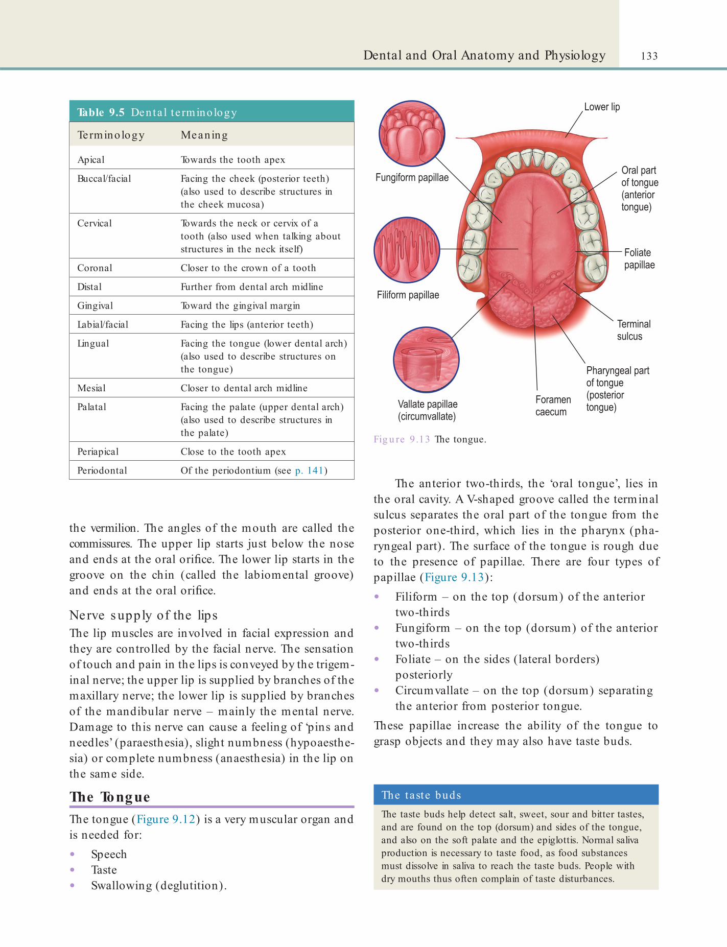
133Dental and Oral Anatomy and Physiology
Table 9.5 Denta l t e rminology
Terminology Meaning
Apical Towards the tooth apex
Buccal/facial Facing the cheek (posterior teeth) (also used to describe structures in the cheek mucosa)
Cervical Towards the neck or cervix of a tooth (also used when talking about structures in the neck itself)
Coronal Closer to the crown of a tooth
Distal Further from dental arch midline
Gingival Toward the gingival margin
Labial/facial Facing the lips (anterior teeth)
Lingual Facing the tongue (lower dental arch) (also used to describe structures on the tongue)
Mesial Closer to dental arch midline
Palatal Facing the palate (upper dental arch) (also used to describe structures in the palate)
Periapical Close to the tooth apex
Periodontal Of the periodontium (see p. 141)
the vermilion. The angles of the mouth are called the commissures. The upper lip starts just below the nose and ends at the oral ori ce. The lower lip starts in the groove on the chin (called the labiomental groove) and ends at the oral ori ce.
Nerve s upply of the lipsThe lip muscles are involved in facial expression and they are controlled by the facial nerve. The sensation of touch and pain in the lips is conveyed by the trigem-inal nerve; the upper lip is supplied by branches of the maxillary nerve; the lower lip is supplied by branches of the mandibular nerve – mainly the mental nerve. Damage to this nerve can cause a feeling of ‘pins and needles’ (paraesthesia), slight numbness (hypoaesthe-sia) or complete numbness (anaesthesia) in the lip on the same side.
The TongueThe tongue (Figure 9.12) is a very muscular organ and is needed for:
• Speech• Taste• Swallowing (deglutition).
The anterior two-thirds, the ‘oral tongue’, lies in the oral cavity. A V-shaped groove called the terminal sulcus separates the oral part of the tongue from the posterior one-third, which lies in the pharynx (pha-ryngeal part). The surface of the tongue is rough due to the presence of papillae. There are four types of papillae (Figure 9.13):
• Filiform – on the top (dorsum) of the anterior two-thirds
• Fungiform – on the top (dorsum) of the anterior two-thirds
• Foliate – on the sides (lateral borders) posteriorly
• Circumvallate – on the top (dorsum) separating the anterior from posterior tongue.
These papillae increase the ability of the tongue to grasp objects and they may also have taste buds.
Fig u re 9 .13 The tongue.
The taste budsThe taste buds help detect salt, sweet, sour and bitter tastes, and are found on the top (dorsum) and sides of the tongue, and also on the soft palate and the epiglottis. Normal saliva production is necessary to taste food, as food substances must dissolve in saliva to reach the taste buds. People with dry mouths thus often complain of taste disturbances.

134 CHAPTER • 9 • Mosby’s Textbook of Dental Nursing
The PalateThe palate is composed of:
• Hard palate (anterior two-thirds)• Soft palate (posterior one-third).
The hard palate forms the roof of the mouth and the oor of the nasal cavity. The mucosa of the hard palate is rmly bound to the underlying bone. Anteri-orly, at the tip of the palate is a papilla called incisive papilla. Lateral to the incisive papilla the mucosa is thrown into folds (rugae) which make the surface of the palate rough. The soft palate is suspended from the posterior edge of the hard palate and is therefore mobile. It is made of muscle and also has nerves, blood vessels, mucous glands and lymphatic tissue. A conical midline projection, called the uvula, hangs from its free inferior border. At the sides of the mouth, the soft palate merges with the tongue and wall of the pharynx to form the palatoglossal and palatopharyn-geal arches (fauces). The lingual tonsil lies between these arches. The palatoglossal arch represents the border between the oral cavity and oropharynx (the fauces).
Nerve s upply of the pala teThe sensory nerve supply to the hard palate and most of the soft palate is via the trigeminal nerve, and to the posterior part is via the glossopharyngeal nerve (this nerve also is motor nerve to most of the muscles in this region).
Fig u re 9.14 Sensory nerve supply to the face and oral cavity: the three divisions of the trigeminal nerve.
Key po in tTouching the posterior palate or tongue can readily cause retching (gagging).
Key po in tInfections and cancer in the mouth and related areas can be spread via the lymphatics to the cervical lymph nodes, which may become swollen (lymphadenopathy).
Key po in tThe lingual nerve runs close to the mandibular third molar (wisdom tooth). Thus it can be easily damaged during lower third molar extractions, or during jaw surgery.
The posterior third of the tongue also has a rough appearance due to the presence of the lingual tonsil. The tonsils form part of a protective ring of lymphatic tissue around the opening to the pharynx (Waldeyer’s ring), which also includes the palatine tonsils (Figure 9.12) and adenoids (in the nasopharynx).
Nerve s upply of the tongue
• The tongue muscles are controlled mainly by the hypoglossal nerve.
• The taste sensation from the anterior two-thirds is conveyed via the trigeminal nerve, and from the posterior third via the glossopharyngeal nerve.
• The main sensory nerve to the tongue is the lingual nerve, which is a branch of the mandibular division of the trigeminal nerve (Figure 9.14). Damage to this nerve can cause a feeling of ‘pins and needles’ (paraesthesia), slight numbness (hypoaesthesia) or complete numbness (anaesthesia) in the side of the tongue, as well as disturbed taste.
Blood s upply to the tongueThe main artery that supplies blood to the tongue is the lingual artery; the de-oxygenated blood (venous drainage) ows to the lingual or facial veins. The
tongue also has lymph vessels and these drain into the several lymph nodes in the neck (cervical lymph nodes). The lymph from the anterior part of the tongue ows into the anterior neck (submental) lymph nodes.
Identify and learnIdent ify the uvula on Figure 9.12.

135Dental and Oral Anatomy and Physiology
gingivae; also called gums). Healthy gingivae appear pink, and have a stippled surface (like the peel of an orange) and are tightly bound to the tooth crown, forming a close tting cuff around the cervix. The rest of the gingivae cover the alveolar bone. Between the gingivae and tooth crown is a shallow gingival crevice.
The DentitionsThe teeth erupt into an arch form called the dental arch which is U-shaped because of the combined pres-sures from the tongue on the lingual/palatal side and the buccal and facial musculature on the buccal/facial side. Within each arch, the teeth are in contact with their neighbouring teeth. These are called the contact areas and the area is called the interproximal area.
The Teeth and Their Supporting Structures (the Periodontium)
Functions o f TeethTeeth are specialised structures and form the hardest tissues in the body. They contribute towards:
• Facial appearance (aesthetics)• Chewing (mastication)• Speech• Defence.
Functionally, teeth may be divided into two types:
• Anterior teeth (incisors and canines (or cuspids)) – these are used for seizing food (or in defence)
• Posterior teeth (molars and premolars, or bicuspids) – these are used to grind food and, with the help of saliva, prepare the food bolus for swallowing.
The teeth are rmly anchored in the alveolar processes of the upper and lower jaws via the periodontal liga-ment, which along with their hard, robust structure, helps them carry out their functions.
Tooth StructureEach tooth has two parts: the crown, which is visible in the mouth; and the root, which is the part that lies within the bone (Figure 9.15).
The crown is covered by a very hard structure called enamel. Under the enamel is slightly softer, sen-sitive tissue called dentine. Within the dentine, in the middle of the tooth, is the living tissue called the pulp (sometimes called the ‘nerve’). It has nerves and blood vessels which pass out through an opening at the tip of the root, called the apical foramen, to connect with the nerves and vessels in the alveolar bone. The tooth root does not have any enamel covering. Instead it is covered by another tissue, called cementum.
The tooth is suspended in its socket by a special-ised tissue called the periodontal ligament. The bres of this ligament attach to the cementum on one side and the alveolar bone on the other. This joint is unique in the body and is called gomphosis – the only joint in which a bone does not join another bone, as teeth are not technically bone.
The junction between the crown and root of a tooth is called the neck or cervix of the tooth (cervical margin). Where a tooth is partially erupted into the mouth, only part of the full (anatomical) crown is visible. This is called the clinical crown. The rest is covered by a soft tissue called the gingiva (plural
Fig u re 9.15 Parts of a typical tooth.
Term to lea rnInterproximal area: area between the proximal surfaces of neighbouring teeth.
Humans have two dentitions, which appear in sequence and allow for growth of the face and jaws from childhood to adult.
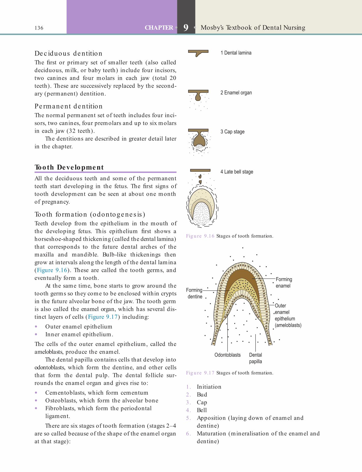
136 CHAPTER • 9 • Mosby’s Textbook of Dental Nursing
De c iduous de ntitionThe rst or primary set of smaller teeth (also called deciduous, milk, or baby teeth) include four incisors, two canines and four molars in each jaw (total 20 teeth). These are successively replaced by the second-ary (permanent) dentition.
Pe rmane nt de ntitionThe normal permanent set of teeth includes four inci-sors, two canines, four premolars and up to six molars in each jaw (32 teeth).
The dentitions are described in greater detail later in the chapter.
Tooth DevelopmentAll the deciduous teeth and some of the permanent teeth start developing in the fetus. The rst signs of tooth development can be seen at about one month of pregnancy.
Tooth fo rmation (odontoge ne s is )Teeth develop from the epithelium in the mouth of the developing fetus. This epithelium rst shows a horseshoe-shaped thickening (called the dental lamina) that corresponds to the future dental arches of the maxilla and mandible. Bulb-like thickenings then grow at intervals along the length of the dental lamina (Figure 9.16). These are called the tooth germs, and eventually form a tooth.
At the same time, bone starts to grow around the tooth germs so they come to be enclosed within crypts in the future alveolar bone of the jaw. The tooth germ is also called the enamel organ, which has several dis-tinct layers of cells (Figure 9.17) including:
• Outer enamel epithelium• Inner enamel epithelium.The cells of the outer enamel epithelium, called the ameloblasts, produce the enamel.
The dental papilla contains cells that develop into odontoblasts, which form the dentine, and other cells that form the dental pulp. The dental follicle sur-rounds the enamel organ and gives rise to:
• Cementoblasts, which form cementum• Osteoblasts, which form the alveolar bone• Fibroblasts, which form the periodontal
ligament.There are six stages of tooth formation (stages 2–4
are so called because of the shape of the enamel organ at that stage):
Fig u re 9.16 Stages of tooth formation.
Fig u re 9.17 Stages of tooth formation.
1 . Initiation2 . Bud3. Cap4. Bell5 . Apposition (laying down of enamel and
dentine)6 . Maturation (mineralisation of the enamel and
dentine)
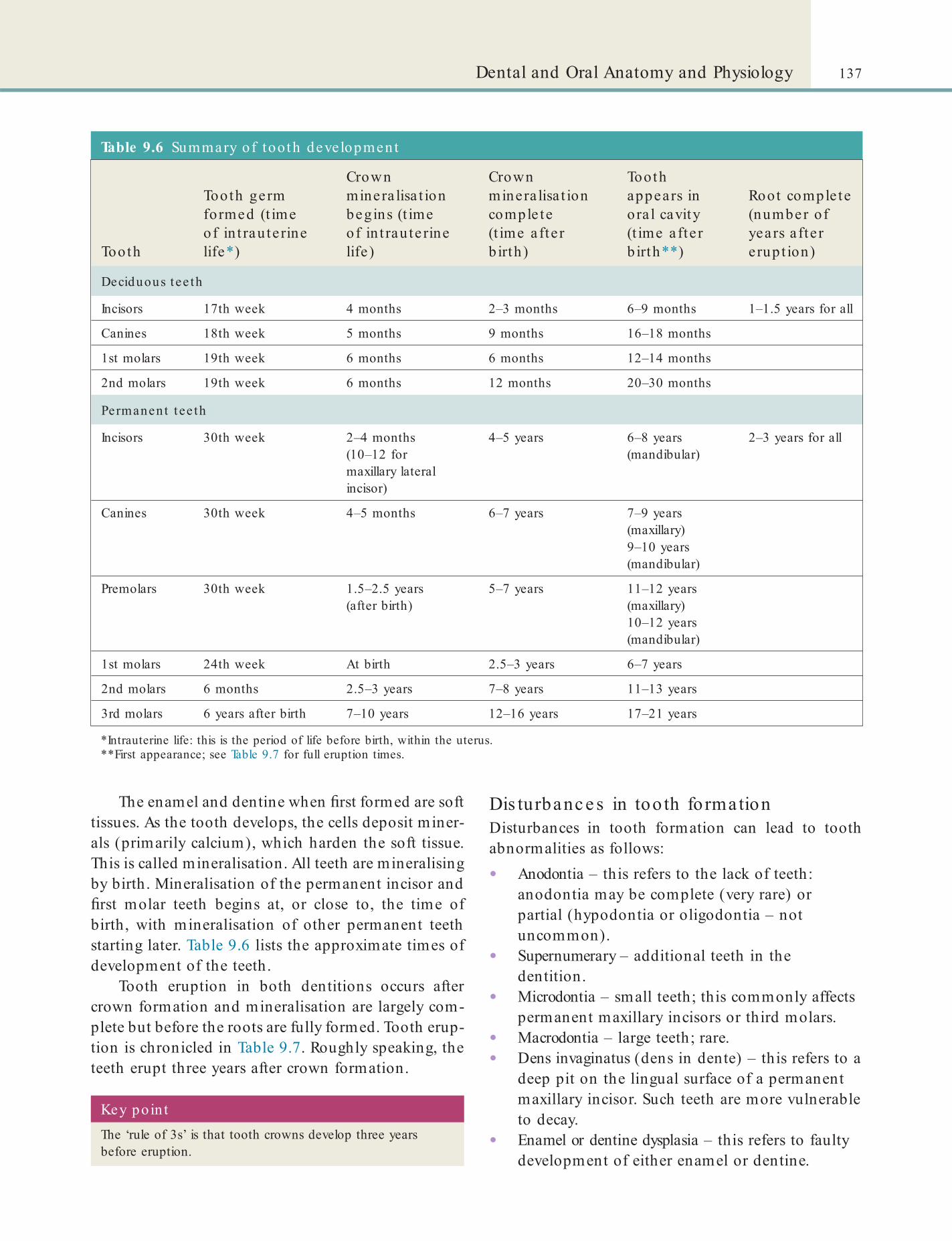
137Dental and Oral Anatomy and Physiology
Dis turbanc e s in too th fo rmationDisturbances in tooth formation can lead to tooth abnormalities as follows:
• Anodontia – this refers to the lack of teeth: anodontia may be complete (very rare) or partial (hypodontia or oligodontia – not uncommon).
• Supernumerary – additional teeth in the dentition.
• Microdontia – small teeth; this commonly affects permanent maxillary incisors or third molars.
• Macrodontia – large teeth; rare.• Dens invaginatus (dens in dente) – this refers to a
deep pit on the lingual surface of a permanent maxillary incisor. Such teeth are more vulnerable to decay.
• Enamel or dentine dysplasia – this refers to faulty development of either enamel or dentine.
The enamel and dentine when rst formed are soft tissues. As the tooth develops, the cells deposit miner-als (primarily calcium), which harden the soft tissue. This is called mineralisation. All teeth are mineralising by birth. Mineralisation of the permanent incisor and rst molar teeth begins at, or close to, the time of birth, with mineralisation of other permanent teeth starting later. Table 9.6 lists the approximate times of development of the teeth.
Tooth eruption in both dentitions occurs after crown formation and mineralisation are largely com-plete but before the roots are fully formed. Tooth erup-tion is chronicled in Table 9.7. Roughly speaking, the teeth erupt three years after crown formation.
Table 9.6 Summary of tooth development
Tooth
Tooth germ formed (t ime of in t rau terine life*)
Crown minera lisa t ion begins (t ime of in t rau te rine life )
Crown minera lisa t ion comple te (t ime a fte r b irth )
Tooth appears in ora l cavity (t ime a fte r b irth**)
Root comple te (number of years a fte r e rup t ion)
Deciduous t ee th
Incisors 17th week 4 months 2–3 months 6–9 months 1–1.5 years for all
Canines 18th week 5 months 9 months 16–18 months
1st molars 19th week 6 months 6 months 12–14 months
2nd molars 19th week 6 months 12 months 20–30 months
Permanent t ee th
Incisors 30th week 2–4 months (10–12 for maxillary lateral incisor)
4–5 years 6–8 years (mandibular)
2–3 years for all
Canines 30th week 4–5 months 6–7 years 7–9 years (maxillary)9–10 years (mandibular)
Premolars 30th week 1.5–2.5 years (after birth)
5–7 years 11–12 years (maxillary)10–12 years (mandibular)
1st molars 24th week At birth 2.5–3 years 6–7 years
2nd molars 6 months 2.5–3 years 7–8 years 11–13 years
3rd molars 6 years after birth 7–10 years 12–16 years 17–21 years
*Intrauterine life: this is the period of life before birth, within the uterus.**First appearance; see Table 9.7 for full eruption times.
Key po in tThe ‘rule of 3s’ is that tooth crowns develop three years before eruption.
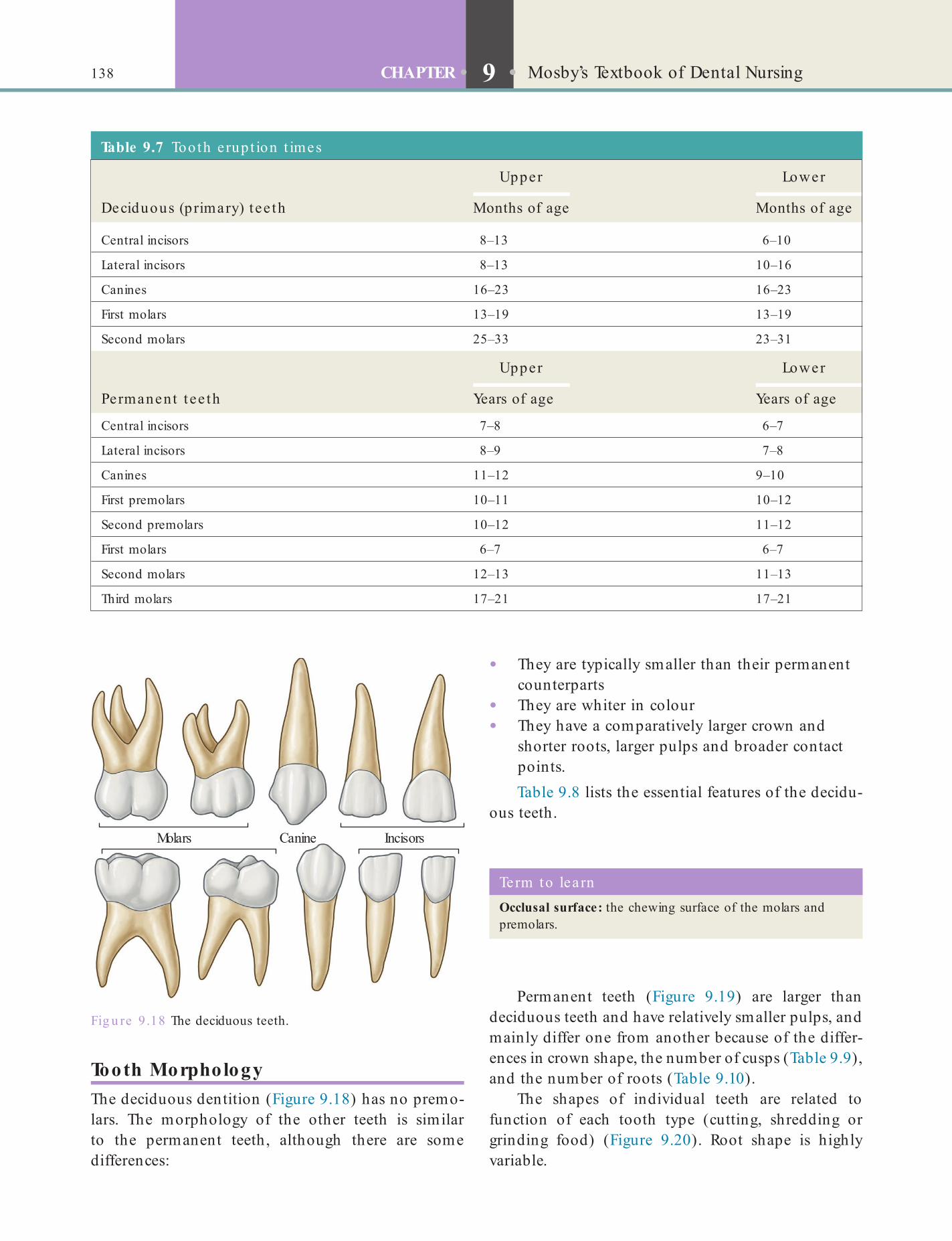
138 CHAPTER • 9 • Mosby’s Textbook of Dental Nursing
Fig u re 9 .18 The deciduous teeth.
Molars Canine Incisors
Term to lea rnOcclusal surface: the chewing surface of the molars and premolars.
Table 9.7 Tooth erup t ion t imes
Upper Lower
Deciduous (primary) tee th Months of age Months of age
Central incisors 8–13 6–10
Lateral incisors 8–13 10–16
Canines 16–23 16–23
First molars 13–19 13–19
Second molars 25–33 23–31
Upper Lower
Permanent t ee th Years of age Years of age
Central incisors 7–8 6–7
Lateral incisors 8–9 7–8
Canines 11–12 9–10
First premolars 10–11 10–12
Second premolars 10–12 11–12
First molars 6–7 6–7
Second molars 12–13 11–13
Third molars 17–21 17–21
Tooth MorphologyThe deciduous dentition (Figure 9.18) has no premo-lars. The morphology of the other teeth is similar to the permanent teeth, although there are some differences:
• They are typically smaller than their permanent counterparts
• They are whiter in colour• They have a comparatively larger crown and
shorter roots, larger pulps and broader contact points.Table 9.8 lists the essential features of the decidu-
ous teeth.
Permanent teeth (Figure 9.19) are larger than deciduous teeth and have relatively smaller pulps, and mainly differ one from another because of the differ-ences in crown shape, the number of cusps (Table 9.9), and the number of roots (Table 9.10).
The shapes of individual teeth are related to function of each tooth type (cutting, shredding or grinding food) (Figure 9.20). Root shape is highly variable.

139Dental and Oral Anatomy and Physiology
Fig u re 9.19 The permanent teeth.
Molars Premolars Canine Incisors
Table 9.8 Fea tures o f deciduous tee th
Tooth Funct ion Posit ion in den ta l a rch Morphology
Incisors Cutting These are the eight front teeth
Flat crown surfaces, a straight sharp horizontal edge and a single, conical root
Canines Tearing and shredding These are the four pointed corner teeth
Conical crown that projects beyond the level of the other teeth, single, conical root
Molars Chewing and grinding (mola is Latin for mill)
The eight molars form the back teeth
The largest of the deciduous teeth. Each molar has a at, large occlusal (upper) surface , with four pyramidal-shaped projections called cusps, and two to three roots
Table 9.9 Fea tures o f pe rmanent tee th
Tooth Funct ion Posit ion in den ta l a rch Morphology
Incisors Cutting These are the eight front teeth Flat crown surfaces, a straight sharp horizontal edge and a single long, conical root
Canines (upper canines may be called eye teeth)
Tearing and shredding These are the four pointed corner teeth
Large, conical crown that projects beyond the level of the other teeth and a single conical root, longer than the roots of all other teeth in the arch
Premolars Chewing There are eight premolars, and they are located lateral to and just behind the canines
The crowns have two cusps each and they have one to two roots each
Molars (third molars are also called wisdom teeth)
Chewing and grinding (mola is Latin for mill)
There are 12 molars, and these are the back teeth
The largest of the permanent teeth, with large and at occlusal (upper) surface and several cusps, and two to four roots
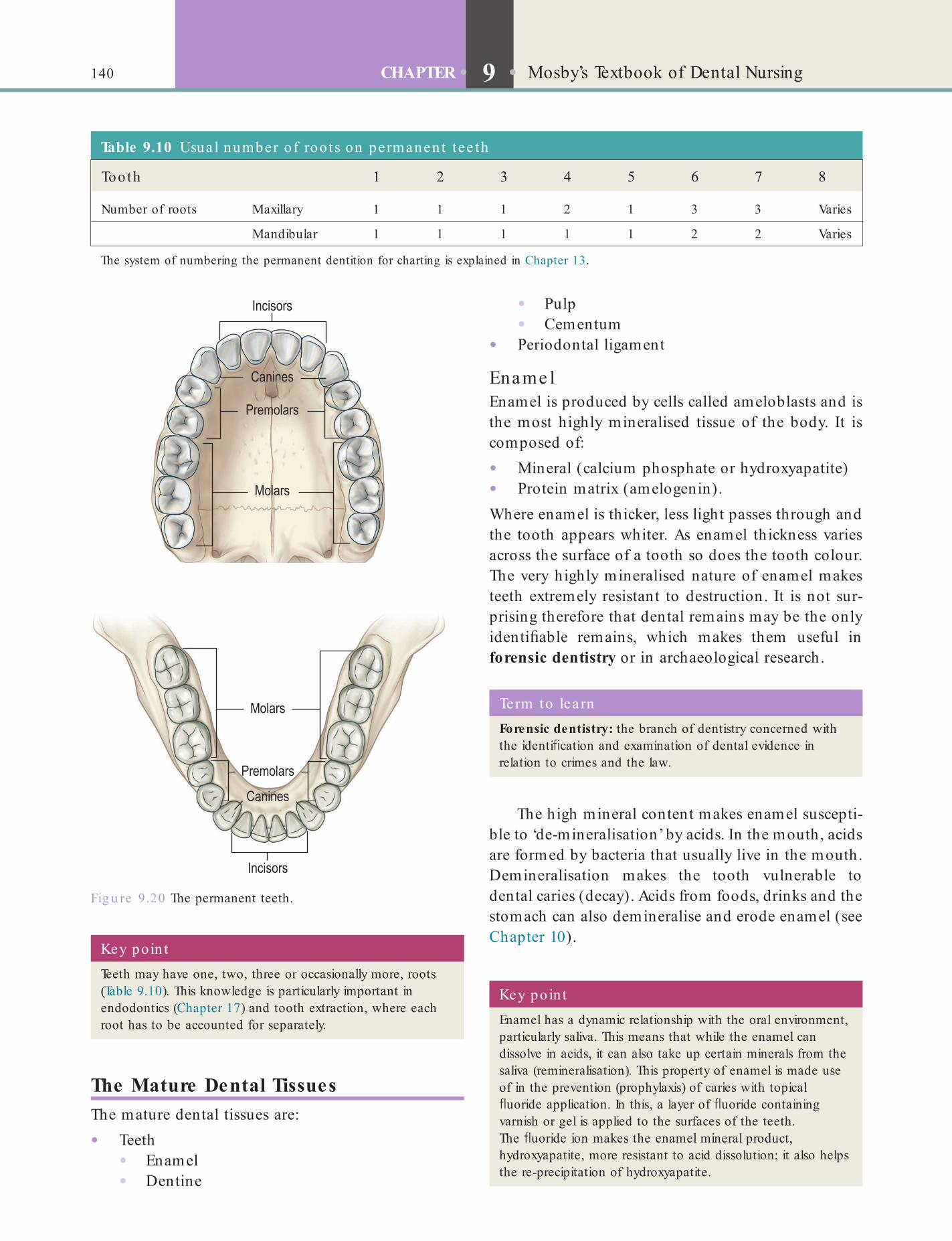
140 CHAPTER • 9 • Mosby’s Textbook of Dental Nursing
• Pulp• Cementum
• Periodontal ligament
Ename lEnamel is produced by cells called ameloblasts and is the most highly mineralised tissue of the body. It is composed of:
• Mineral (calcium phosphate or hydroxyapatite)• Protein matrix (amelogenin).Where enamel is thicker, less light passes through and the tooth appears whiter. As enamel thickness varies across the surface of a tooth so does the tooth colour. The very highly mineralised nature of enamel makes teeth extremely resistant to destruction. It is not sur-prising therefore that dental remains may be the only identi able remains, which makes them useful in forensic dentistry or in archaeological research.
Fig u re 9.20 The permanent teeth.
Key po in tEnamel has a dynamic relationship with the oral environment, particularly saliva. This means that while the enamel can dissolve in acids, it can also take up certain minerals from the saliva (remineralisation). This property of enamel is made use of in the prevention (prophylaxis) of caries with topical uoride application. In this, a layer of uoride containing varnish or gel is applied to the surfaces of the teeth. The uoride ion makes the enamel mineral product, hydroxyapatite, more resistant to acid dissolution; it also helps the re-precipitation of hydroxyapatite.
Term to lea rnForensic dentistry: the branch of dentistry concerned with the identi cation and examination of dental evidence in relation to crimes and the law.
Key poin tTeeth may have one, two, three or occasionally more, roots (Table 9.10). This knowledge is particularly important in endodontics (Chapter 17) and tooth extraction, where each root has to be accounted for separately.
Table 9.10 Usua l number of roo ts on permanent tee th
Tooth 1 2 3 4 5 6 7 8
Number of roots Maxillary 1 1 1 2 1 3 3 Varies
Mandibular 1 1 1 1 1 2 2 Varies
The system of numbering the permanent dentition for charting is explained in Chapter 13.
The Mature Dental TissuesThe mature dental tissues are:
• Teeth• Enamel• Dentine
The high mineral content makes enamel suscepti-ble to ‘de-mineralisation’ by acids. In the mouth, acids are formed by bacteria that usually live in the mouth. Demineralisation makes the tooth vulnerable to dental caries (decay). Acids from foods, drinks and the stomach can also demineralise and erode enamel (see Chapter 10).
141Dental and Oral Anatomy and Physiology
Find out moreHow does secondary dent ine crea te d if cu lt ies in endodont ics?
Pe riodontiumThe periodontium (from the Greek peri-, meaning ‘around’, and -odont, meaning ‘tooth’) is the collective term for the specialised tissues that surround and support the teeth, maintaining them in the jaw bones. The periodontium consists of the:
• Cementum• Periodontal ligament• Alveolar bone• Gingivae.
CementumCementum (or cement) is a specialised mineralised tissue that covers the tooth root. It is produced by the cementoblasts. Cementum is less mineralised than enamel or dentine, yellowish in colour and softer and thickest at the root apex. The main role is to anchor the tooth in its socket, via the periodontal ligament. Excess cementum (hypercementosis) can make tooth extraction dif cult.
Pe riodonta l ligamentThe periodontal membrane or ligament (PDL) anchors the root (via the cementum) in the alveolar bone of the tooth socket. The PDL is a brous connective tissue consisting of collagen bres which, when not under stress, have a relaxed and wavy course across the width of the periodontal space. A healthy PDL is able to withstand the not inconsiderable forces applied to teeth during use, yet provides exibility that prevents them shattering. The functions of the PDL are:
• Supportive – anchoring the tooth.• Sensory – via sensitive periodontal
mechanoreceptors. The slightest interference that causes even minimal tooth displacement is consciously felt by an individual. It is immediately followed by re ex muscle activity to try to correct the problem. For example, if an individual bites on something hard unexpectedly, the mouth immediately opens.
De ntineProduced by odontoblasts within the outermost layer of the pulp, dentine is a living tissue, very sensitive and softer than enamel but harder than bone. To a certain extent, some dentine (called secondary dentine) can continue to form for years. Dentine is made up of 70% inorganic material, chie y hydroxyapatite, 20% organic (mainly proteins called collagen) and 10% water. Dentine has a variable yellowish colour that gives teeth their colour. Dentine is readily damaged by dental caries (see Chapter 10). Where a tooth pulp has become diseased, it may lead to discoloration of dentine and such teeth have crowns that then appear darker. The mechanism whereby dentine is so exqui-sitely sensitive is still unknown. Whatever stimulus is applied to dentine, whether it is thermal, mechanical, osmotic or electrical, the only sensation perceived is that of pain.
De nta l pulpDental pulp is a vascular (lot of blood vessels), very sensitive, soft connective tissue that is the source of nourishment for a tooth and its vitality. Pulp tissue contains:
• Odontoblasts (cells that produce dentine)• Fibroblasts (produce and maintain collagen
bres and ground substance)• Defence cells.
Key poin tCavity preparation must avoid damaging (exposing) the tooth pulp. In a young tooth, the pulp horns extending beneath the cusps of teeth are prominent but, in older teeth, secondary dentine deposition means that cavity preparation is less likely to involve pulpal exposure. However, secondary dentine may also create dif culties in endodontics.
Terms to learnCollagen: the main protein of the connective tissue, which provides strength, resilience and support to various parts of the body, such as the skin, ligaments and tendons.
Ground substance: the matrix in which the cells in a tissue are embedded.
The pulp space within the crown is termed the pulp chamber and the parts that lie within the cusps are called pulp horns. The pulp space within the root(s) is called the root canal system. The nerves and vessels that serve the pulp enter principally through each root’s apical foramen. But accessory, lateral connec-tions between the pulp and the periodontal ligament may also exist.
Secondary dentine develops after root formation is complete and is produced throughout the life of a tooth at an extremely slow rate. This progressively reduces the pulp space.
142 CHAPTER • 9 • Mosby’s Textbook of Dental Nursing
margin becomes exposed as the gingivae recede and sometimes, if the dentine is exposed, the patient com-plains of pain (hypersensitivity), especially with cold uids or food.
Periodontal disease (see Chapter 10) progressively destroys the PDL and, eventually, the remaining tissue is unable to withstand normal chewing forces so that the tooth becomes mobile (loose).
GingivaeThe gingiva is the mucosa overlying the alveolar bone, to which it is tightly bound to resist the friction of food passing over. Healthy gingivae are usually coral pink, but may be pigmented. This is normal. The gin-givae surround the cervical margin of the teeth in the area of the amelo-cemental junction (junction between the enamel and cementum), providing a seal. There is a shallow sulcus or crevice between the margin of the gingivae and tooth crown just above the seal. The gingivae are divided anatomically into:
• Marginal –gingivae overlying the gingival sulcus• Attached – gingivae attached to the tooth and
bone• Interdental – gingivae in the spaces between the
teeth.The attached gingivae blend into the oral mucosa in the vestibules.
Accumulation of plaque on the tooth cervical regions may result in in ammation of the gingival margin (called gingivitis or marginal gingivitis). With age, especially if toothbrushing is incorrectly per-formed, the amount of tooth visible in the mouth is greater than the anatomical crown due to recession of the gingivae. The root surface below the cervical
Key poin ts• Teeth start to develop in utero (before birth).
• Root formation is completed after eruption.
• The full primary set of teeth has 20 teeth in total.
• The full permanent set of teeth has 32 teeth in total.
• Anterior teeth (incisors and canines) have one root only.
• Premolars usually have one or two roots.
• Lower molar teeth usually have two roots and upper molar teeth have three.
Terms to lea rnPlaque: a bio lm composed of mucus and bacteria that forms on tooth surfaces, particularly the interproximal surfaces and along the gingival margins of teeth (cervically), and in the ssures and pits on the occlusal surface.
Interproximal surface: the surfaces of adjacent teeth that face each other. In a healthy dentition, the interproximal surfaces touch each other at the contact point.
Term to lea rnMechanoreceptor: a receptor that senses mechanical pressure or distortion.
Mobile tooth: a loose tooth; mobility can be of various degrees.
Nerve Supply of the TeethThe sensory nerve supply (sensation) to the jaw, teeth, periodontium and gingivae is provided by the trigemi-nal nerve, the fth cranial nerve. The maxillary (upper) teeth are innervated by branches of the maxillary nerve; the mandibular (lower) teeth are innervated by branches of the mandibular nerve – mainly via the inferior dental (alveolar) nerve.
If the trigeminal nerve is damaged by trauma, or disease, the main result is a loss of sensation (hypo-aesthesia – partial loss of sensation, or anaesthesia – complete loss of sensation).
Chapter 10 Dental and Oral Pathology and Disease
CHAPTER POINTS• Pathology• Tooth surface loss• Periodontal disease• Other dental diseases• Mucosal disease• Salivary disease• Diseases of the jaws and joints• Cleft lip and palate• Pain• Halitosis
a dental nurse, you are required to have background knowledge about them, especially about the most common ones (Table 10.1), which are discussed in this chapter: Less common diseases are found in Chapter 24.
The dental nurse can also have an important role in oral health promotion, which is discussed in Chapter 12.
Diseases may be congenital (present at birth) or acquired (develop at some stage after birth). Most dental (odontogenic) disease is acquired and caused by the build-up and activity of micro-organisms (mainly bacteria) on the tooth surface within the dental bacterial plaque – sometimes called a ‘bio lm’ – which sticks and grows if not removed mechanically by tooth-brushing and ossing.
In t ro d u ct io n
The aim of dental care should be to have a disease-free mouth, as shown in Figures 10.1A and B. The indica-tions of a healthy mouth are:
• Teeth that are rm, clean and free of debris or staining
• Gingivae that are pink, not swollen and neither hurt nor bleed when the person brushes or osses
• No bad breath• No pain.
A disease is an abnormal condition that affects the body. It is often construed as a medical condition associated with speci c symptoms and signs. There are four main types of disease: pathogenic disease, de ciency disease, hereditary disease and physiological disease. Diseases can also be classi ed as communicable and non-communicable.
The oral (mouth) diseases described in this chapter are seen routinely in the dental surgery. Therefore, as
Term to lea rnOdontogenic: of dental origin.
The activity of the micro-organisms (mainly bac-teria) in plaque is responsible for, or may aggravate, a variety of oral diseases, in particular dental caries (tooth decay) and in ammatory periodontal disease (gingivi-tis and periodontitis), which are the most common oral diseases.
Dental Pathology and MicrobiologyThe word pathology is from the Greek pathos, meaning ‘experience’ or ‘suffering’, and logia, ‘an account of’ or ‘the study of’. Understanding the molecular and cel-lular basis of diseases is crucial for understanding disease mechanisms and for designing treatments.
Pathology is a medical science primarily con-cerned with examining of organs, tissues and bodily
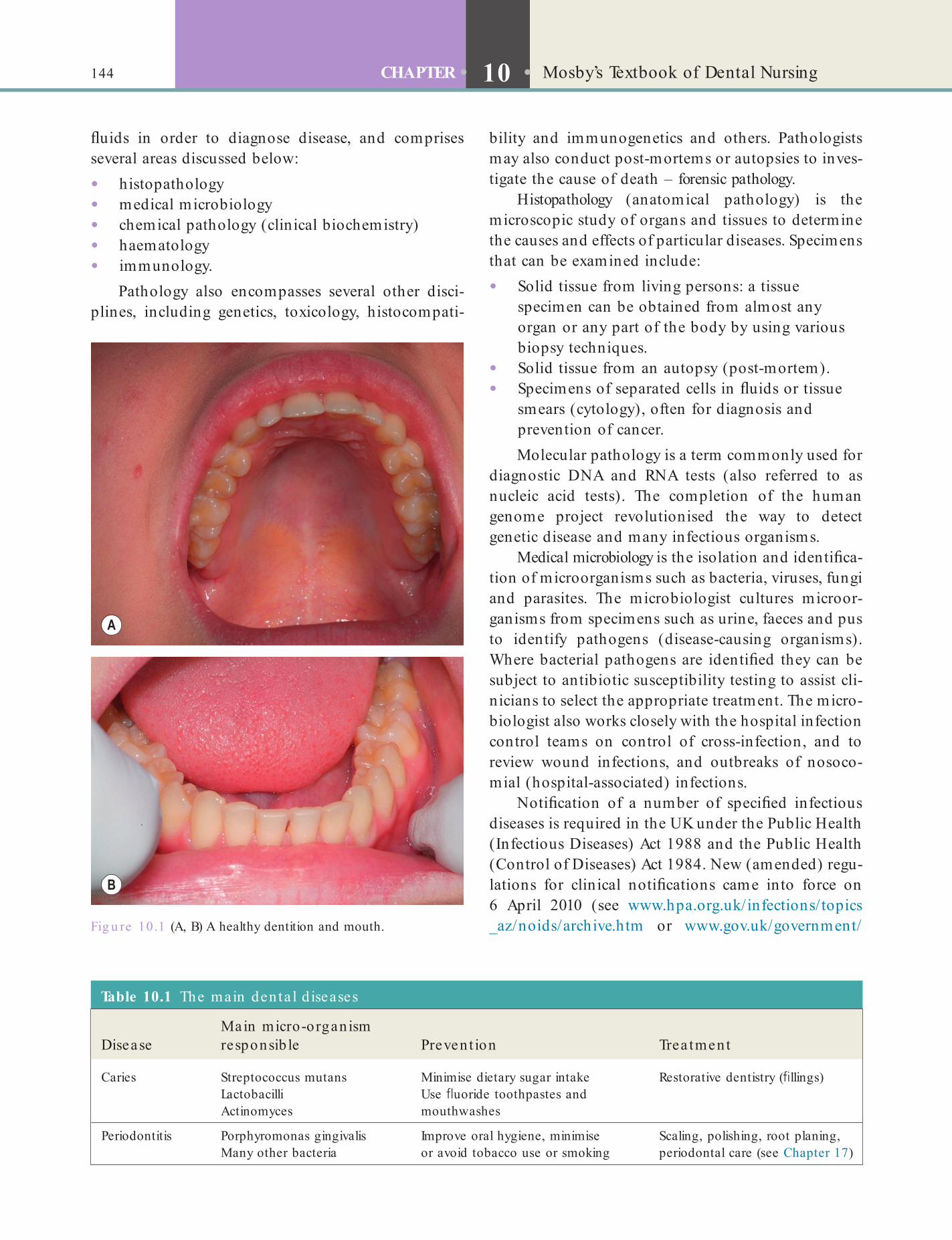
144 CHAPTER • 10 • Mosby’s Textbook of Dental Nursing
bility and immunogenetics and others. Pathologists may also conduct post-mortems or autopsies to inves-tigate the cause of death – forensic pathology.
Histopathology (anatomical pathology) is the microscopic study of organs and tissues to determine the causes and effects of particular diseases. Specimens that can be examined include:
• Solid tissue from living persons: a tissue specimen can be obtained from almost any organ or any part of the body by using various biopsy techniques.
• Solid tissue from an autopsy (post-mortem).• Specimens of separated cells in uids or tissue
smears (cytology), often for diagnosis and prevention of cancer.Molecular pathology is a term commonly used for
diagnostic DNA and RNA tests (also referred to as nucleic acid tests). The completion of the human genome project revolutionised the way to detect genetic disease and many infectious organisms.
Medical microbiology is the isolation and identi ca-tion of microorganisms such as bacteria, viruses, fungi and parasites. The microbiologist cultures microor-ganisms from specimens such as urine, faeces and pus to identify pathogens (disease-causing organisms). Where bacterial pathogens are identi ed they can be subject to antibiotic susceptibility testing to assist cli-nicians to select the appropriate treatment. The micro-biologist also works closely with the hospital infection control teams on control of cross-infection, and to review wound infections, and outbreaks of nosoco-mial (hospital-associated) infections.
Noti cation of a number of speci ed infectious diseases is required in the UK under the Public Health (Infectious Diseases) Act 1988 and the Public Health (Control of Diseases) Act 1984. New (amended) regu-lations for clinical noti cations came into force on 6 April 2010 (see www.hpa.org.uk/infections/topics _az/noids/archive.htm or www.gov.uk/government/Fig u re 10.1 (A, B) A healthy dentition and mouth.
Table 10.1 The main den ta l d iseases
DiseaseMain micro-organism responsib le Prevent ion Trea tment
Caries Streptococcus mutansLactobacilliActinomyces
Minimise dietary sugar intakeUse uoride toothpastes and mouthwashes
Restorative dentistry ( llings)
Periodontitis Porphyromonas gingivalisMany other bacteria
Improve oral hygiene, minimise or avoid tobacco use or smoking
Scaling, polishing, root planing, periodontal care (see Chapter 17)
uids in order to diagnose disease, and comprises several areas discussed below:
• histopathology• medical microbiology• chemical pathology (clinical biochemistry)• haematology• immunology.
Pathology also encompasses several other disci-plines, including genetics, toxicology, histocompati-

145Dental and Oral Pathology and Disease
collections/noti cations-of-infectious-diseases-noids). Registered medical practitioners (GMPs) in England and Wales have ‘a statutory duty to notify a “proper of cer” of the Local Authority of suspected cases of certain infectious diseases’; usually the consultant in communicable disease control (CCDC).
Chemical pathology (clinical biochemistry) is the study of chemical and biochemical mechanisms of the body in relation to disease such as diabetics, thyroid disease and on uid balance and therapeutic drug levels. These changes can be used for the diagnosis of disease and also monitoring treatment. It also pro-vides a screening service such as neonatal screening for cystic brosis and screening for drugs of abuse.
Haematology is the study of diseases that affect blood. Investigations include:
• Routine haematological testing• Clotting and bleeding studies• Transfusion services.
Immunology is the diagnosis and management of diseases arising from abnormal immune responses – either under-activity (immunode ciency) or over-activity (allergy and autoimmunity). The immune system normally functions to ght infections, and looking for antibodies to an infection (serology) is an important way of con rming recent or remote contact with the micro-organism – though molecular diagno-sis is increasingly used.
Tooth Sur ace LossDental Caries (Tooth Decay)Dental caries is a very common disease and is the main pathological cause of tooth loss in children (Figures 10.2, 10.3 and 10.4).
The ‘DMF’ (decay/missing/ lled) index is one of the most common methods for assessing caries preva-lence as well as dental treatment needs among popula-tions. The Adult Dental Survey in 2009 showed that in England, Wales and Northern Ireland about one-third of adults had obvious caries, approximately the same as that worldwide. Caries is most prevalent in Latin American countries, countries in the Middle East and South Asia, and least prevalent in China.
Caries has decreased in many developed countries, a decline usually attributed to preventive measures – particularly uoride (Chapter 12). Nonetheless, countries that have an overall decrease in caries continue to have a disparity in the distribution, with a higher prevalence in resource-poor groups. For
Fig u re 10.2 Caries at the cervical margins.
Fig u re 10.3 Caries affecting several teeth.
Fig u re 10.4 Dental caries has caused cavities on several teeth (some restored with llings), some crowns have fractured and some carious teeth have been extracted in the past.
146 CHAPTER • 10 • Mosby’s Textbook of Dental Nursing
Table 10.2 Facts you should know about den ta l ca ries
Cause Plaque bacteria, especially Streptococcus mutans, which acts on sugars to produce lactic acid, which decalci es (demineralises) the teeth
Plaque This bio lm tends to form in pits and ssures, interproximally at contact areas; and at the cervical margins (sites where caries begins)
Main sugars implicated in caries Sucrose, glucose
Sugars and sweeteners rarely implicated in caries Fructose, lactose, sorbitol, aspartame
Acidity (critical pH) below which enamel decalci cation occurs
5.5
Methods of detection Visual examinationBitewing radiographs (see Chapter 11)Transillumination (shining a light through the tooth)Electronic caries detectorsUsing a probe (but this may cause further damage; see Chapter 13)
Preventive measures Consuming less sugars in the dietUsing uorides, e.g. uoridated toothpastesUsing amorphous calcium phosphate
example, the Adult Dental Survey found more caries in adults from households with routine and manual occupations (37%) than from professional occupa-tion households (26%).
Pa tho logyCaries is caused by bacteria that act on carbohydrates (mainly sugars) on the tooth surface to produce acids that, given time, remove minerals such as calcium and phosphate (demineralise) teeth, leading eventually to a carious cavity (Table 10.2). The teeth at highest risk for carious lesions are the permanent rst and second molars due to length of time in oral cavity and pres-ence of complex surface anatomy.
The four criteria for caries to occur are:
• a tooth surface• bacteria• fermentable carbohydrates (sugars)• time.
Bacte riaThe main plaque micro-organism that causes caries is the bacterium Streptococcus mutans (also called viridans streptococci). Other bacteria, such as Lactobacillus and Actinomyces, may also play a role. Bacteria collect around and between the teeth and in pits and ssures, as well as next to the gingivae in a sticky, creamy-coloured mass called plaque. S. mutans adheres in the ‘bio lm’ on the teeth by converting sucrose into an extremely sticky substance called dextran. The plaque bio lm can cause both
• caries• gingival and periodontal diseases.
The bacteria act by converting the sugars in the diet to acids (especially lactic acid). The acids destroy (decalcify) the enamel and dentine of the teeth (see Figure 10.3). The acids cause the pH to fall and when the pH in plaque at the tooth surface drops below 5.5, tooth demineralisation proceeds faster than reminer-alisation (meaning that there is a net loss of mineral structure on the tooth’s surface). Because most plaque-retentive areas are between teeth and inside pits and ssures on chewing surfaces where brushing is dif -cult, over 80% of cavities occur inside pits and ssures. Areas that are easily cleansed with a toothbrush, however, such as the front and back surfaces (facial and lingual), develop fewer cavities.
The initial rapid drop in plaque pH to a pH of about 5.5, is when demineralisation starts (‘critical pH’) (Figure 10.5).
The more time the pH is low, the more deminer-alisation occurs; thus the worse pattern of sugar expo-sure is repeated exposures (e.g. sucking mints) whereas the best is none, or all the exposures at once (e.g. eating all the sweets at midday Saturday). Thus eating sticky sugars (e.g. toffees), repeatedly eating or drink-ing sugary foods, or sucking sweets for hours, leads to a long drop in the pH, and a lot of damage. Even more damage occurs if the sugars are eaten just before going to sleep, because saliva production falls during sleep, and therefore the natural cleaning of the mouth is reduced.

147Dental and Oral Pathology and Disease
Fig u re 10.5 Stephan curve showing falling pH (rising acidity) after sucrose intake.
m
o
r
e
a
c
i
d
6.5
6.0
p
H
5.5
5.05Minutes after sucrose rinse
10 15 20
Fluorides and amorphous calcium phosphate can remineralise demineralised teeth, and various treat-ments can be used to restore teeth to proper form, function and aesthetics (Chapter 17).
Fe rmentable carbohydrates (s ugars )Sugars in the diet are mainly found as non-milk sugars (e.g. sucrose, glucose and fructose), and lactose in milk. Lactose (milk sugar) is less cariogenic than other sugars.
The non-milk sugars include the common table or cane sugar (sucrose), glucose (dextrose) and fruit sugar (fructose) (Table 10.3) and these are the most cariogenic. The main offenders in relation to caries are the re ned sugars (sucrose) from sugar cane and sugar beet, and glucose – these are termed cariogenic sugars (Table 10.3).
Sugars are also added to many foods and drinks, in particular to re ned carbohydrates such as starch, and foods such as cakes and biscuits. Sugars may also be added to foods and drinks where one might not
Table 10.3 Cariogenic sugars
Pure sugars Mixtures
Dextrose Brown sugar
Fructose (except in fresh fruits and vegetables)
Golden syrup
Glucose Honey
Hydrolysed starch Maple syrup
Invert sugar Treacle
Maltose
Sucrose
expect them – for example breakfast cereals, canned vegetables, sauces and soups – and are found in some medications.
Dietary starch is broken down slowly by salivary enzymes to glucose and maltose, and concentrated fruit juices and dried fruits have a high concentration of sugars such as fructose so these products are also cariogenic. Fresh fruits and vegetables are not signi -cantly cariogenic (but some can be erosive).
Key poin tSugars, particularly non-milk sugars in food and drink, are major cariogenic factors.
Term to lea rnCariogenic: a factor that can cause caries.
The least tooth damage is done by:
• Avoiding consuming sugars completely• Minimising non-milk sugar intake• Eating sugar-containing products all at once only
and over a short period of time• Not eating sugars as the last thing at night.
Decalci cation produces opaque whitish areas on the tooth, which are painless. Decalci cation is also reversible to a point if the person changes their diet and reduces intake of more cariogenic carbohydrates.
The critical pH for dentine demineralisation is higher at around 6.5 (so less acid needed) and the dentine is softer than enamel, so caries spreads more rapidly once it reaches dentine. It may then spread to the pulp causing infection, in ammation and pain (pulpitis).
Table 10.2 summarises the key points you need to know about dental caries.
Fac to rs pro te c tive o f c a rie sSaliva protects against caries. Abundant saliva produc-tion or its stimulation by chewing brous foods or gum, or rinsing the mouth, will help the low pH to recover and give some protection. In people who produce a good amount of saliva the chances of devel-oping caries is far less than in those who have a dry mouth or hyposalivation. Other factors that may be protective of caries include uoride, and
• Cocoa• Coffee• Cranberries
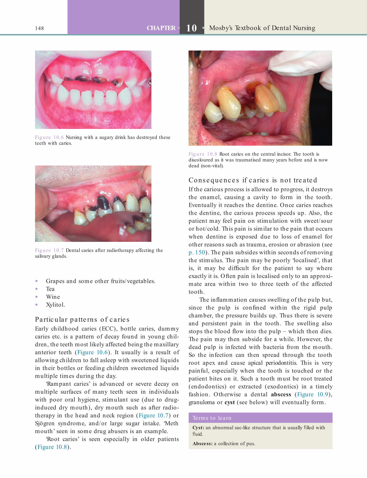
148 CHAPTER • 10 • Mosby’s Textbook of Dental Nursing
Cons e que nc e s if c a rie s is no t tre a te dIf the carious process is allowed to progress, it destroys the enamel, causing a cavity to form in the tooth. Eventually it reaches the dentine. Once caries reaches the dentine, the carious process speeds up. Also, the patient may feel pain on stimulation with sweet/sour or hot/cold. This pain is similar to the pain that occurs when dentine is exposed due to loss of enamel for other reasons such as trauma, erosion or abrasion (see p. 150). The pain subsides within seconds of removing the stimulus. The pain may be poorly ‘localised’, that is, it may be dif cult for the patient to say where exactly it is. Often pain is localised only to an approxi-mate area within two to three teeth of the affected tooth.
The in ammation causes swelling of the pulp but, since the pulp is con ned within the rigid pulp chamber, the pressure builds up. Thus there is severe and persistent pain in the tooth. The swelling also stops the blood ow into the pulp – which then dies. The pain may then subside for a while. However, the dead pulp is infected with bacteria from the mouth. So the infection can then spread through the tooth root apex and cause apical periodontitis. This is very painful, especially when the tooth is touched or the patient bites on it. Such a tooth must be root treated (endodontics) or extracted (exodontics) in a timely fashion. Otherwise a dental abscess (Figure 10.9), granuloma or cyst (see below) will eventually form.
• Grapes and some other fruits/vegetables.• Tea• Wine• Xylitol.
Partic ula r pa tte rns o f c a rie sEarly childhood caries (ECC), bottle caries, dummy caries etc. is a pattern of decay found in young chil-dren, the teeth most likely affected being the maxillary anterior teeth (Figure 10.6). It usually is a result of allowing children to fall asleep with sweetened liquids in their bottles or feeding children sweetened liquids multiple times during the day.
‘Rampant caries’ is advanced or severe decay on multiple surfaces of many teeth seen in individuals with poor oral hygiene, stimulant use (due to drug-induced dry mouth), dry mouth such as after radio-therapy in the head and neck region (Figure 10.7) or Sjögren syndrome, and/or large sugar intake. ‘Meth mouth’ seen in some drug abusers is an example.
‘Root caries’ is seen especially in older patients (Figure 10.8).
Fig u re 10.6 Nursing with a sugary drink has destroyed these teeth with caries.
Fig u re 10.7 Dental caries after radiotherapy affecting the salivary glands.
Fig u re 10.8 Root caries on the central incisor. The tooth is discoloured as it was traumatised many years before and is now dead (non-vital).
Terms to lea rnCyst: an abnormal sac-like structure that is usually lled with uid.
Abscess: a collection of pus.
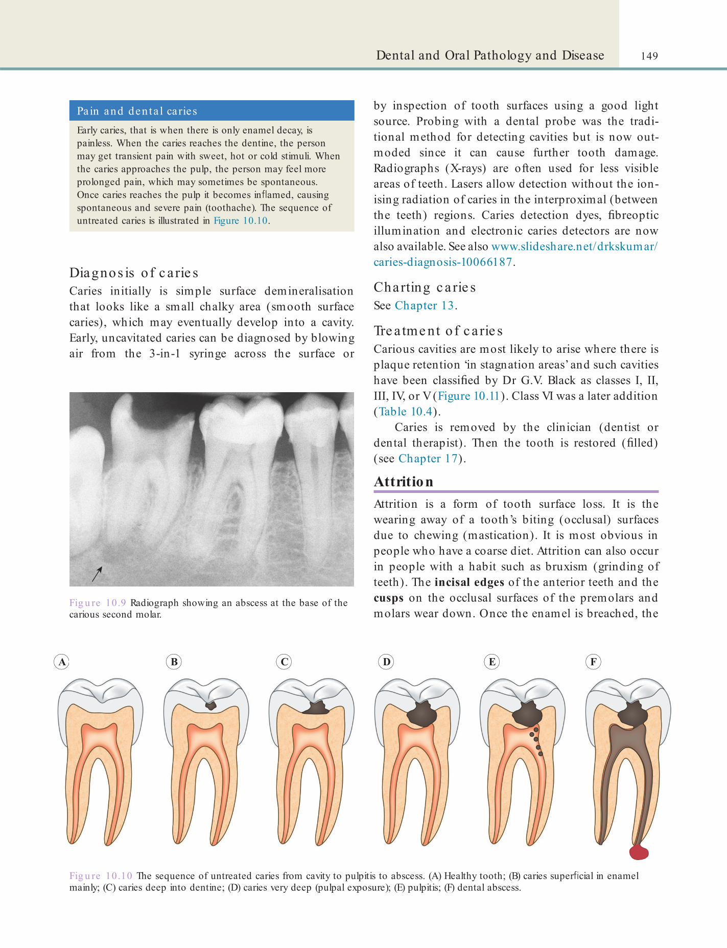
149Dental and Oral Pathology and Disease
Fig u re 10.9 Radiograph showing an abscess at the base of the carious second molar.
Pa in and denta l cariesEarly caries, that is when there is only enamel decay, is painless. When the caries reaches the dentine, the person may get transient pain with sweet, hot or cold stimuli. When the caries approaches the pulp, the person may feel more prolonged pain, which may sometimes be spontaneous. Once caries reaches the pulp it becomes in amed, causing spontaneous and severe pain (toothache). The sequence of untreated caries is illustrated in Figure 10.10.
Fig u re 10.10 The sequence of untreated caries from cavity to pulpitis to abscess. (A) Healthy tooth; (B) caries super cial in enamel mainly; (C) caries deep into dentine; (D) caries very deep (pulpal exposure); (E) pulpitis; (F) dental abscess.
A B C D E F
Diagnos is o f c a rie sCaries initially is simple surface demineralisation that looks like a small chalky area (smooth surface caries), which may eventually develop into a cavity. Early, uncavitated caries can be diagnosed by blowing air from the 3-in-1 syringe across the surface or
by inspection of tooth surfaces using a good light source. Probing with a dental probe was the tradi-tional method for detecting cavities but is now out-moded since it can cause further tooth damage. Radiographs (X-rays) are often used for less visible areas of teeth. Lasers allow detection without the ion-ising radiation of caries in the interproximal (between the teeth) regions. Caries detection dyes, breoptic illumination and electronic caries detectors are now also available. See also www.slideshare.net/drkskumar/caries-diagnosis-10066187.
Charting c a rie sSee Chapter 13.
Tre a tme nt o f c a rie sCarious cavities are most likely to arise where there is plaque retention ‘in stagnation areas’ and such cavities have been classi ed by Dr G.V. Black as classes I, II, III, IV, or V (Figure 10.11). Class VI was a later addition (Table 10.4).
Caries is removed by the clinician (dentist or dental therapist). Then the tooth is restored ( lled) (see Chapter 17).
AttritionAttrition is a form of tooth surface loss. It is the wearing away of a tooth’s biting (occlusal) surfaces due to chewing (mastication). It is most obvious in people who have a coarse diet. Attrition can also occur in people with a habit such as bruxism (grinding of teeth). The incisal edges of the anterior teeth and the cusps on the occlusal surfaces of the premolars and molars wear down. Once the enamel is breached, the
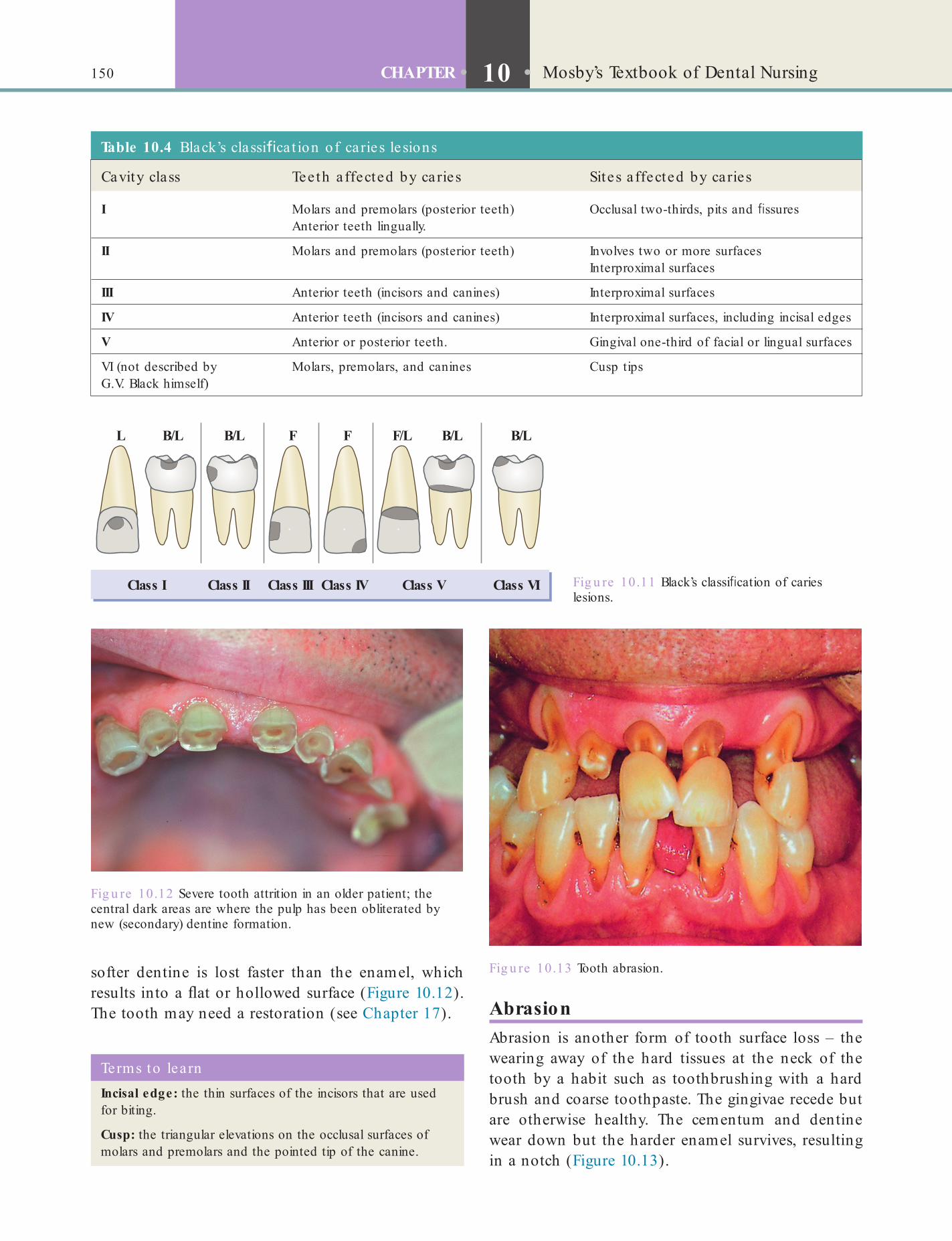
150 CHAPTER • 10 • Mosby’s Textbook of Dental Nursing
AbrasionAbrasion is another form of tooth surface loss – the wearing away of the hard tissues at the neck of the tooth by a habit such as toothbrushing with a hard brush and coarse toothpaste. The gingivae recede but are otherwise healthy. The cementum and dentine wear down but the harder enamel survives, resulting in a notch (Figure 10.13).
softer dentine is lost faster than the enamel, which results into a at or hollowed surface (Figure 10.12). The tooth may need a restoration (see Chapter 17).
Fig u re 10.11 Black’s classi cation of caries lesions.
Class I Class II Class III Class IV Class V Class VI
L F FB/L B/L B/L B/LF/L
Fig u re 10.12 Severe tooth attrition in an older patient; the central dark areas are where the pulp has been obliterated by new (secondary) dentine formation.
Terms to learnIncisal edge: the thin surfaces of the incisors that are used for biting.
Cusp: the triangular elevations on the occlusal surfaces of molars and premolars and the pointed tip of the canine.
Table 10.4 Black’s classi ca t ion of caries le sions
Cavity class Teeth a ffected by ca ries Sites a ffected by caries
I Molars and premolars (posterior teeth)Anterior teeth lingually.
Occlusal two-thirds, pits and ssures
II Molars and premolars (posterior teeth) Involves two or more surfacesInterproximal surfaces
III Anterior teeth (incisors and canines) Interproximal surfaces
IV Anterior teeth (incisors and canines) Interproximal surfaces, including incisal edges
V Anterior or posterior teeth. Gingival one-third of facial or lingual surfaces
VI (not described by G.V. Black himself)
Molars, premolars, and canines Cusp tips
Fig u re 10.13 Tooth abrasion.
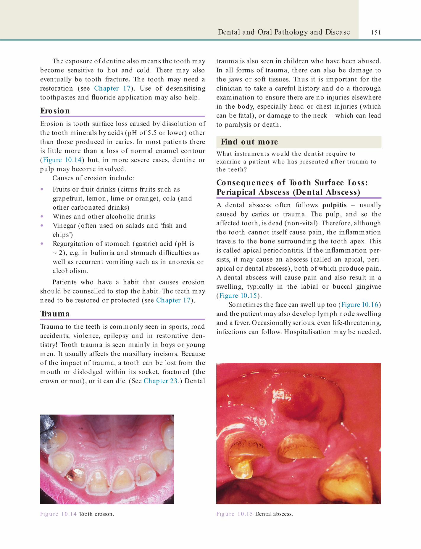
151Dental and Oral Pathology and Disease
The exposure of dentine also means the tooth may become sensitive to hot and cold. There may also eventually be tooth fracture. The tooth may need a restoration (see Chapter 17). Use of desensitising toothpastes and uoride application may also help.
ErosionErosion is tooth surface loss caused by dissolution of the tooth minerals by acids (pH of 5.5 or lower) other than those produced in caries. In most patients there is little more than a loss of normal enamel contour (Figure 10.14) but, in more severe cases, dentine or pulp may become involved.
Causes of erosion include:
• Fruits or fruit drinks (citrus fruits such as grapefruit, lemon, lime or orange), cola (and other carbonated drinks)
• Wines and other alcoholic drinks• Vinegar (often used on salads and ‘ sh and
chips’)• Regurgitation of stomach (gastric) acid (pH is
~ 2), e.g. in bulimia and stomach dif culties as well as recurrent vomiting such as in anorexia or alcoholism.Patients who have a habit that causes erosion
should be counselled to stop the habit. The teeth may need to be restored or protected (see Chapter 17).
TraumaTrauma to the teeth is commonly seen in sports, road accidents, violence, epilepsy and in restorative den-tistry! Tooth trauma is seen mainly in boys or young men. It usually affects the maxillary incisors. Because of the impact of trauma, a tooth can be lost from the mouth or dislodged within its socket, fractured (the crown or root), or it can die. (See Chapter 23.) Dental
Fig u re 10.14 Tooth erosion.
trauma is also seen in children who have been abused. In all forms of trauma, there can also be damage to the jaws or soft tissues. Thus it is important for the clinician to take a careful history and do a thorough examination to ensure there are no injuries elsewhere in the body, especially head or chest injuries (which can be fatal), or damage to the neck – which can lead to paralysis or death.
Find out moreWhat inst ruments would the den t ist requ ire to examine a pa t ien t who has p resen ted a fte r t rauma to the tee th?
Consequences o Tooth Sur ace Loss: Periapical Abscess (Dental Abscess)A dental abscess often follows pulpitis – usually caused by caries or trauma. The pulp, and so the affected tooth, is dead (non-vital). Therefore, although the tooth cannot itself cause pain, the in ammation travels to the bone surrounding the tooth apex. This is called apical periodontitis. If the in ammation per-sists, it may cause an abscess (called an apical, peri-apical or dental abscess), both of which produce pain. A dental abscess will cause pain and also result in a swelling, typically in the labial or buccal gingivae (Figure 10.15).
Sometimes the face can swell up too (Figure 10.16) and the patient may also develop lymph node swelling and a fever. Occasionally serious, even life-threatening, infections can follow. Hospitalisation may be needed.
Fig u re 10.15 Dental abscess.
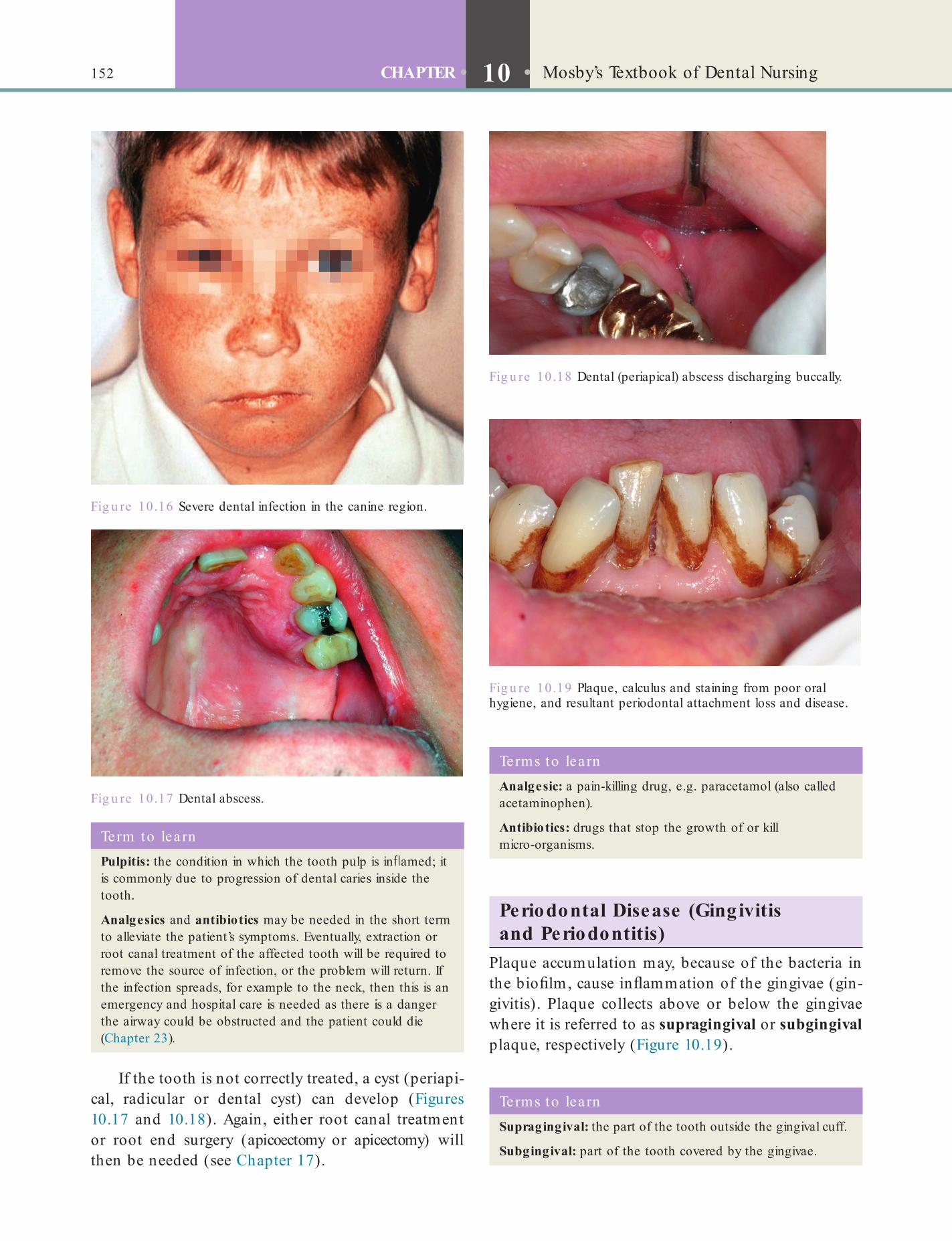
152 CHAPTER • 10 • Mosby’s Textbook of Dental Nursing
Periodontal Disease (Gingivitis and Periodontitis)
Plaque accumulation may, because of the bacteria in the bio lm, cause in ammation of the gingivae (gin-givitis). Plaque collects above or below the gingivae where it is referred to as supragingival or subgingival plaque, respectively (Figure 10.19).
If the tooth is not correctly treated, a cyst (periapi-cal, radicular or dental cyst) can develop (Figures 10.17 and 10.18). Again, either root canal treatment or root end surgery (apicoectomy or apicectomy) will then be needed (see Chapter 17).
Fig u re 10.16 Severe dental infection in the canine region.
Term to lea rnPulpitis: the condition in which the tooth pulp is in amed; it is commonly due to progression of dental caries inside the tooth.
Analgesics and antibiotics may be needed in the short term to alleviate the patient’s symptoms. Eventually, extraction or root canal treatment of the affected tooth will be required to remove the source of infection, or the problem will return. If the infection spreads, for example to the neck, then this is an emergency and hospital care is needed as there is a danger the airway could be obstructed and the patient could die (Chapter 23).
Fig u re 10.17 Dental abscess.
Fig u re 10.18 Dental (periapical) abscess discharging buccally.
Fig u re 10.19 Plaque, calculus and staining from poor oral hygiene, and resultant periodontal attachment loss and disease.
Terms to lea rnSupragingival: the part of the tooth outside the gingival cuff.
Subgingival: part of the tooth covered by the gingivae.
Terms to lea rnAnalgesic: a pain-killing drug, e.g. paracetamol (also called acetaminophen).
Antibiotics: drugs that stop the growth of or kill micro-organisms.
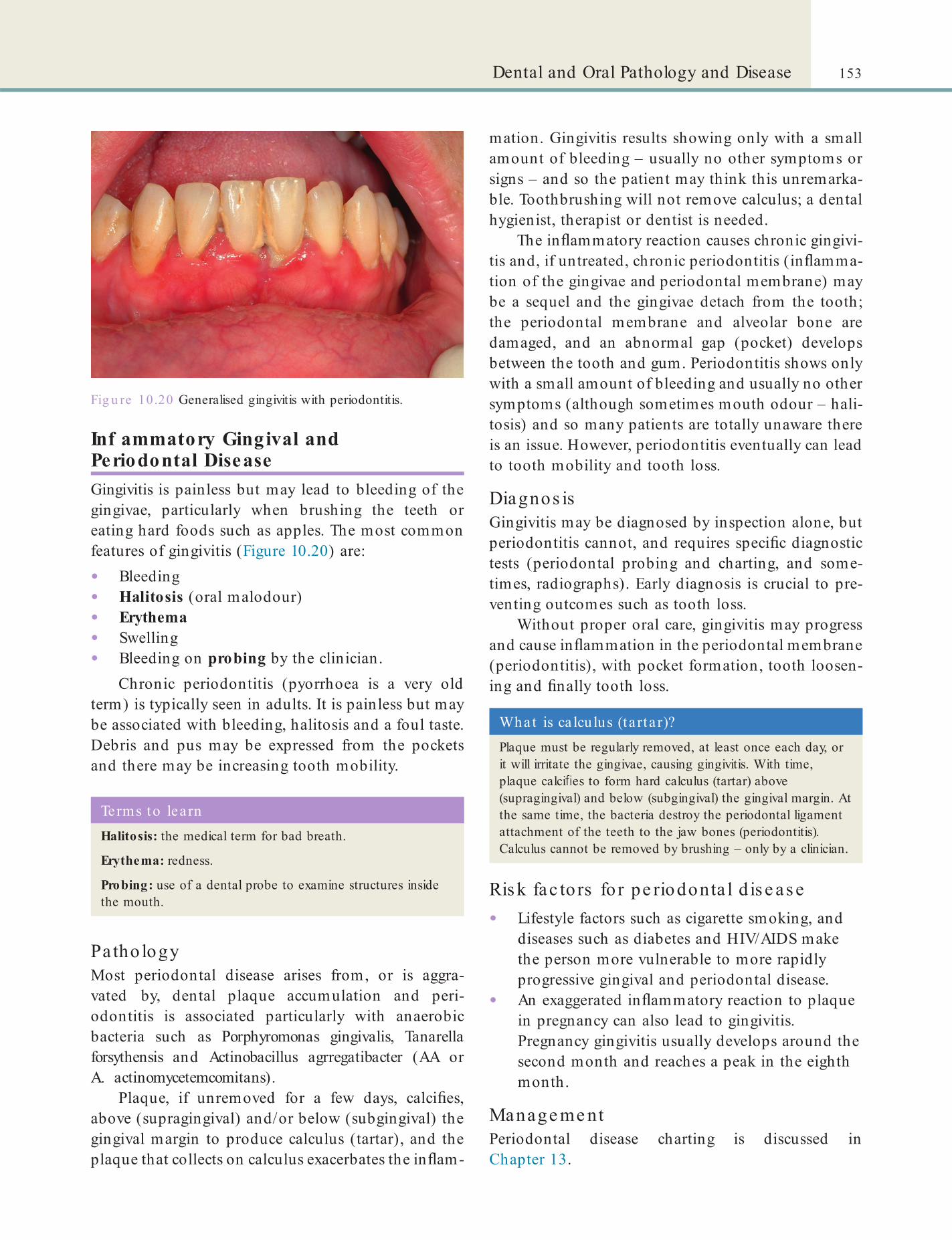
153Dental and Oral Pathology and Disease
mation. Gingivitis results showing only with a small amount of bleeding – usually no other symptoms or signs – and so the patient may think this unremarka-ble. Toothbrushing will not remove calculus; a dental hygienist, therapist or dentist is needed.
The in ammatory reaction causes chronic gingivi-tis and, if untreated, chronic periodontitis (in amma-tion of the gingivae and periodontal membrane) may be a sequel and the gingivae detach from the tooth; the periodontal membrane and alveolar bone are damaged, and an abnormal gap (pocket) develops between the tooth and gum. Periodontitis shows only with a small amount of bleeding and usually no other symptoms (although sometimes mouth odour – hali-tosis) and so many patients are totally unaware there is an issue. However, periodontitis eventually can lead to tooth mobility and tooth loss.
Diagnos isGingivitis may be diagnosed by inspection alone, but periodontitis cannot, and requires speci c diagnostic tests (periodontal probing and charting, and some-times, radiographs). Early diagnosis is crucial to pre-venting outcomes such as tooth loss.
Without proper oral care, gingivitis may progress and cause in ammation in the periodontal membrane (periodontitis), with pocket formation, tooth loosen-ing and nally tooth loss.
Fig u re 10.20 Generalised gingivitis with periodontitis.
Inf ammatory Gingival and Periodontal DiseaseGingivitis is painless but may lead to bleeding of the gingivae, particularly when brushing the teeth or eating hard foods such as apples. The most common features of gingivitis (Figure 10.20) are:
• Bleeding• Halitosis (oral malodour)• Erythema• Swelling• Bleeding on probing by the clinician.
Chronic periodontitis (pyorrhoea is a very old term) is typically seen in adults. It is painless but may be associated with bleeding, halitosis and a foul taste. Debris and pus may be expressed from the pockets and there may be increasing tooth mobility.
Terms to learnHalitosis: the medical term for bad breath.
Erythema: redness.
Probing: use of a dental probe to examine structures inside the mouth.
Pa tho logyMost periodontal disease arises from, or is aggra-vated by, dental plaque accumulation and peri-odontitis is associated particularly with anaerobic bacteria such as Porphyromonas gingivalis, Tanarella forsythensis and Actinobacillus agrregatibacter (AA or A. actinomycetemcomitans).
Plaque, if unremoved for a few days, calci es, above (supragingival) and/or below (subgingival) the gingival margin to produce calculus (tartar), and the plaque that collects on calculus exacerbates the in am-
What is ca lcu lus (ta rta r)?Plaque must be regularly removed, at least once each day, or it will irritate the gingivae, causing gingivitis. With time, plaque calci es to form hard calculus (tartar) above (supragingival) and below (subgingival) the gingival margin. At the same time, the bacteria destroy the periodontal ligament attachment of the teeth to the jaw bones (periodontitis). Calculus cannot be removed by brushing – only by a clinician.
Ris k fac to rs fo r pe riodonta l d is e as e• Lifestyle factors such as cigarette smoking, and
diseases such as diabetes and HIV/AIDS make the person more vulnerable to more rapidly progressive gingival and periodontal disease.
• An exaggerated in ammatory reaction to plaque in pregnancy can also lead to gingivitis. Pregnancy gingivitis usually develops around the second month and reaches a peak in the eighth month.
Manage me ntPeriodontal disease charting is discussed in Chapter 13.
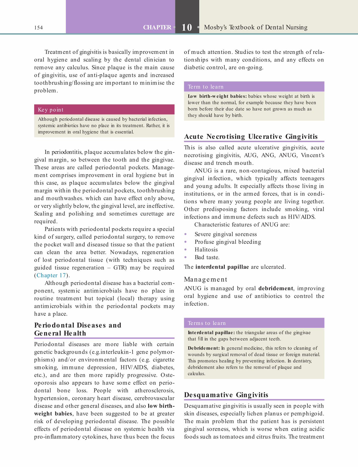
154 CHAPTER • 10 • Mosby’s Textbook of Dental Nursing
Acute Necrotising Ulcerative GingivitisThis is also called acute ulcerative gingivitis, acute necrotising gingivitis, AUG, ANG, ANUG, Vincent’s disease and trench mouth.
ANUG is a rare, non-contagious, mixed bacterial gingival infection, which typically affects teenagers and young adults. It especially affects those living in institutions, or in the armed forces, that is in condi-tions where many young people are living together. Other predisposing factors include smoking, viral infections and immune defects such as HIV/AIDS.
Characteristic features of ANUG are:
• Severe gingival soreness• Profuse gingival bleeding• Halitosis• Bad taste.The interdental papillae are ulcerated.
Manage me ntANUG is managed by oral debridement, improving oral hygiene and use of antibiotics to control the infection.
In periodontitis, plaque accumulates below the gin-gival margin, so between the tooth and the gingivae. These areas are called periodontal pockets. Manage-ment comprises improvement in oral hygiene but in this case, as plaque accumulates below the gingival margin within the periodontal pockets, toothbrushing and mouthwashes. which can have effect only above, or very slightly below, the gingival level, are ineffective. Scaling and polishing and sometimes curettage are required.
Patients with periodontal pockets require a special kind of surgery, called periodontal surgery, to remove the pocket wall and diseased tissue so that the patient can clean the area better. Nowadays, regeneration of lost periodontal tissue (with techniques such as guided tissue regeneration – GTR) may be required (Chapter 17).
Although periodontal disease has a bacterial com-ponent, systemic antimicrobials have no place in routine treatment but topical (local) therapy using antimicrobials within the periodontal pockets may have a place.
Periodontal Diseases and General HealthPeriodontal diseases are more liable with certain genetic backgrounds (e.g.interleukin-1 gene polymor-phisms) and/or environmental factors (e.g. cigarette smoking, immune depression, HIV/AIDS, diabetes, etc.), and are then more rapidly progressive. Oste-oporosis also appears to have some effect on perio-dontal bone loss. People with atherosclerosis, hypertension, coronary heart disease, cerebrovascular disease and other general diseases, and also low birth-weight babies, have been suggested to be at greater risk of developing periodontal disease. The possible effects of periodontal disease on systemic health via pro-in ammatory cytokines, have thus been the focus
Key poin tAlthough periodontal disease is caused by bacterial infection, systemic antibiotics have no place in its treatment. Rather, it is improvement in oral hygiene that is essential.
Terms to lea rnInterdental papillae: the triangular areas of the gingivae that ll in the gaps between adjacent teeth.
Debridement: In general medicine, this refers to cleaning of wounds by surgical removal of dead tissue or foreign material. This promotes healing by preventing infection. In dentistry, debridement also refers to the removal of plaque and calculus.
Term to lea rnLow birth-w eight babies: babies whose weight at birth is lower than the normal, for example because they have been born before their due date so have not grown as much as they should have by birth.
Treatment of gingivitis is basically improvement in oral hygiene and scaling by the dental clinician to remove any calculus. Since plaque is the main cause of gingivitis, use of anti-plaque agents and increased toothbrushing/ ossing are important to minimise the problem.
of much attention. Studies to test the strength of rela-tionships with many conditions, and any effects on diabetic control, are on-going.
Desquamative GingivitisDesquamative gingivitis is usually seen in people with skin diseases, especially lichen planus or pemphigoid. The main problem that the patient has is persistent gingival soreness, which is worse when eating acidic foods such as tomatoes and citrus fruits. The treatment
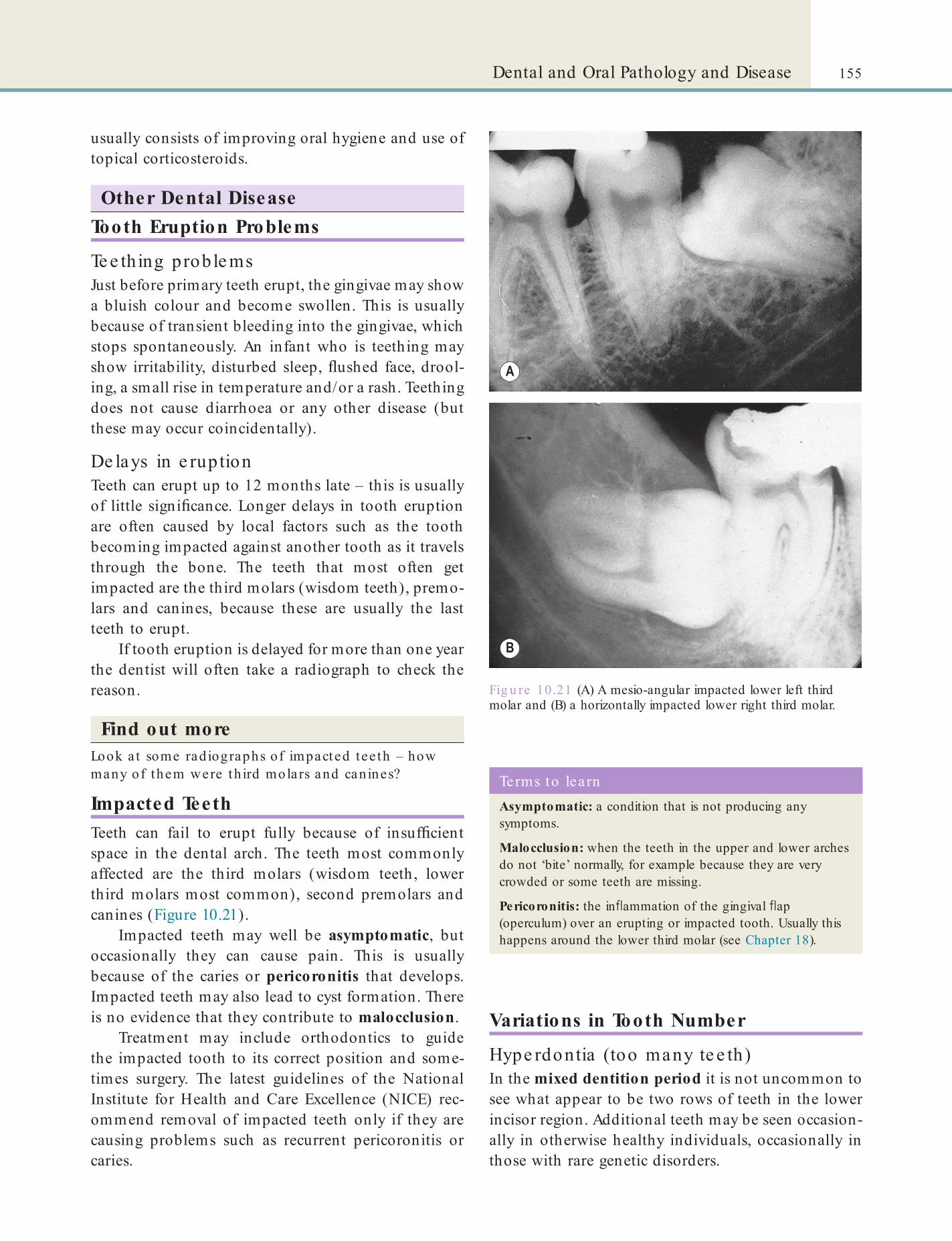
155Dental and Oral Pathology and Disease
usually consists of improving oral hygiene and use of topical corticosteroids.
Other Dental DiseaseTooth Eruption Problems
Te e thing proble msJust before primary teeth erupt, the gingivae may show a bluish colour and become swollen. This is usually because of transient bleeding into the gingivae, which stops spontaneously. An infant who is teething may show irritability, disturbed sleep, ushed face, drool-ing, a small rise in temperature and/or a rash. Teething does not cause diarrhoea or any other disease (but these may occur coincidentally).
De lays in e ruptionTeeth can erupt up to 12 months late – this is usually of little signi cance. Longer delays in tooth eruption are often caused by local factors such as the tooth becoming impacted against another tooth as it travels through the bone. The teeth that most often get impacted are the third molars (wisdom teeth), premo-lars and canines, because these are usually the last teeth to erupt.
If tooth eruption is delayed for more than one year the dentist will often take a radiograph to check the reason.
Find out moreLook a t some rad iographs of impacted tee th – how many of them were th ird molars and canines?
Impacted TeethTeeth can fail to erupt fully because of insuf cient space in the dental arch. The teeth most commonly affected are the third molars (wisdom teeth, lower third molars most common), second premolars and canines (Figure 10.21).
Impacted teeth may well be asymptomatic, but occasionally they can cause pain. This is usually because of the caries or pericoronitis that develops. Impacted teeth may also lead to cyst formation. There is no evidence that they contribute to malocclusion.
Treatment may include orthodontics to guide the impacted tooth to its correct position and some-times surgery. The latest guidelines of the National Institute for Health and Care Excellence (NICE) rec-ommend removal of impacted teeth only if they are causing problems such as recurrent pericoronitis or caries.
Fig u re 10.21 (A) A mesio-angular impacted lower left third molar and (B) a horizontally impacted lower right third molar.
Variations in Tooth Number
Hype rdontia (too many te e th)In the mixed dentition period it is not uncommon to see what appear to be two rows of teeth in the lower incisor region. Additional teeth may be seen occasion-ally in otherwise healthy individuals, occasionally in those with rare genetic disorders.
Terms to lea rnAsymptomatic: a condition that is not producing any symptoms.
Malocclusion: when the teeth in the upper and lower arches do not ‘bite’ normally, for example because they are very crowded or some teeth are missing.
Pericoronitis: the in ammation of the gingival ap (operculum) over an erupting or impacted tooth. Usually this happens around the lower third molar (see Chapter 18).

156 CHAPTER • 10 • Mosby’s Textbook of Dental Nursing
child, it may be affected by diseases, radiotherapy, drugs or infections.
Ename l hypoplas iaBetween birth and 6 years of age, the permanent inci-sors and canines are developing (see Chapter 9). If the developing tooth bud is damaged, it can produce a cosmetic problem, because the damage will be evident on smiling. Enamel hypoplasia is when the tooth crown appears opaque, or yellow-brown or deformed. Infections such as German measles (rubella), cancer treatments or jaundice may cause this type of hypo-plasia. The defects correspond to the site of tooth enamel formation at the time of the insult (‘chrono-logical’ hypoplasia).
Dis c o loure d te e thDiscoloration of several teeth is usually because of super cial (extrinsic) staining that results from:
• Poor oral hygiene (Figure 10.22)• Use of substances such as tobacco, betel nuts,
khat tea, coffee, red wine or chlorhexidine• Caries• Trauma• Tooth lling material.
Such super cial tooth discoloration affects mainly the interproximal and cervical surfaces of the teeth (where plaque also accumulates) and can be removed by the dental clinician.
Generalised ‘intrinsic’ tooth staining of a brown or grey colour is caused by the use of the drugs called tetracyclines by a pregnant or lactating mother or chil-dren under the age of 8 years. Tetracyclines can cross the placenta and then enter breast milk and are taken up by developing teeth and by bone. Intrinsic staining cannot be removed by the dental clinician. However, because of this problem, tetracyclines are no longer recommended for pregnant women and infants. Stain-ing may also be because of hypoplasia or some rare inherited tooth defect (amelogenesis imperfecta or dentinogenesis imperfecta).
Discoloration of a single tooth is usually intrinsic, that is from within the tooth, and happens because the tooth is:
• Non-vital• Heavily lled or• Carious.
Non-vital teeth progressively darken more with time, sometimes to a brownish colour (Figure 10.23), and also become more brittle.
• Extra teeth of normal shape (supplemental teeth; Figure 10.22) are uncommon, but most frequently seen in the maxillary lateral incisor, and in the premolar and third molar regions of either jaw.
• Extra teeth of abnormal form (supernumerary teeth) are also uncommon, usually small and/or conical in shape and are seen particularly in the midline of the upper arch (mesiodens).
Fig u re 10.22 A supernumerary incisor.
Term to lea rnMixed dentition period: this is when the permanent teeth start erupting, which is before all the deciduous incisors have been lost (exfoliated) so that there are both permanent and deciduous teeth in the mouth at the same time.
Hypodontia (too fe w te e th)Reasons for teeth missing from the dental arch include:
• The tooth may have failed to erupt• It may not have developed• It may have been lost prematurely.Hypodontia is not uncommon. It is most often genetic, and most frequently affects the third molars, the second premolars and the maxillary lateral incisors. Occasionally hypodontia can occur as part of a gener-alised (systemic) disorder such as ectodermal dysplasia. Rarely, all the teeth are absent (anodontia).
In hypodontia, when the permanent successor is missing, it is common for the deciduous tooth to be retained long after it should have been shed. The patient may need a restoration (see Chapter 17).
Anomalies o Tooth Size , Shape and StructureAlthough the delicate process of tooth development is generally well protected in the developing baby or
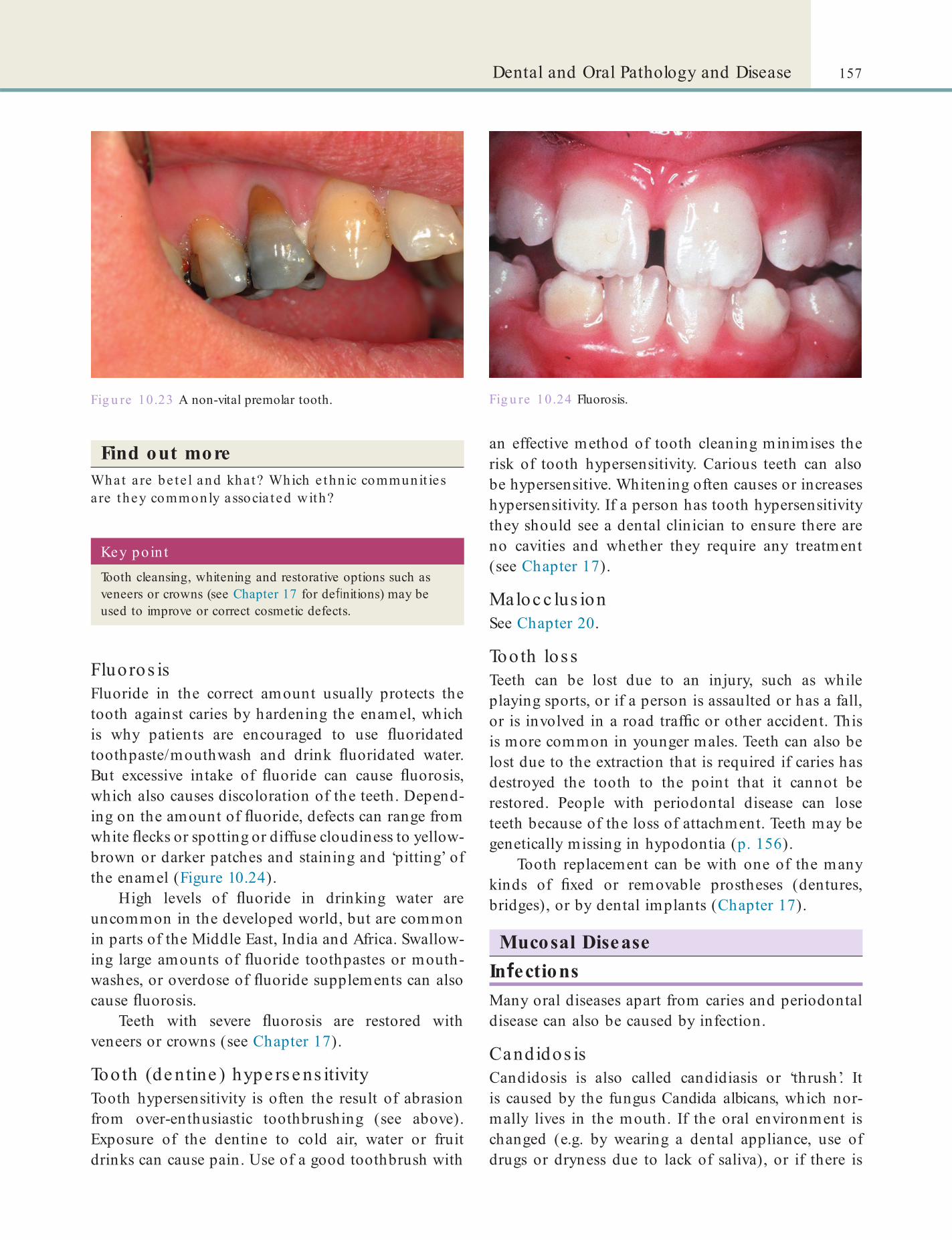
157Dental and Oral Pathology and Disease
an effective method of tooth cleaning minimises the risk of tooth hypersensitivity. Carious teeth can also be hypersensitive. Whitening often causes or increases hypersensitivity. If a person has tooth hypersensitivity they should see a dental clinician to ensure there are no cavities and whether they require any treatment (see Chapter 17).
Maloc c lus ionSee Chapter 20.
Tooth los sTeeth can be lost due to an injury, such as while playing sports, or if a person is assaulted or has a fall, or is involved in a road traf c or other accident. This is more common in younger males. Teeth can also be lost due to the extraction that is required if caries has destroyed the tooth to the point that it cannot be restored. People with periodontal disease can lose teeth because of the loss of attachment. Teeth may be genetically missing in hypodontia (p. 156).
Tooth replacement can be with one of the many kinds of xed or removable prostheses (dentures, bridges), or by dental implants (Chapter 17).
Mucosal DiseaseIn ectionsMany oral diseases apart from caries and periodontal disease can also be caused by infection.
Candidos isCandidosis is also called candidiasis or ‘thrush’. It is caused by the fungus Candida albicans, which nor-mally lives in the mouth. If the oral environment is changed (e.g. by wearing a dental appliance, use of drugs or dryness due to lack of saliva), or if there is
Find out moreWhat a re be te l and kha t? Which e thn ic communit ies a re they commonly associa ted with?
Fig u re 10.23 A non-vital premolar tooth.
Key po in tTooth cleansing, whitening and restorative options such as veneers or crowns (see Chapter 17 for de nitions) may be used to improve or correct cosmetic defects.
Fluoros isFluoride in the correct amount usually protects the tooth against caries by hardening the enamel, which is why patients are encouraged to use uoridated toothpaste/mouthwash and drink uoridated water. But excessive intake of uoride can cause uorosis, which also causes discoloration of the teeth. Depend-ing on the amount of uoride, defects can range from white ecks or spotting or diffuse cloudiness to yellow-brown or darker patches and staining and ‘pitting’ of the enamel (Figure 10.24).
High levels of uoride in drinking water are uncommon in the developed world, but are common in parts of the Middle East, India and Africa. Swallow-ing large amounts of uoride toothpastes or mouth-washes, or overdose of uoride supplements can also cause uorosis.
Teeth with severe uorosis are restored with veneers or crowns (see Chapter 17).
Tooth (de ntine ) hype rse ns itivityTooth hypersensitivity is often the result of abrasion from over-enthusiastic toothbrushing (see above). Exposure of the dentine to cold air, water or fruit drinks can cause pain. Use of a good toothbrush with
Fig u re 10.24 Fluorosis.
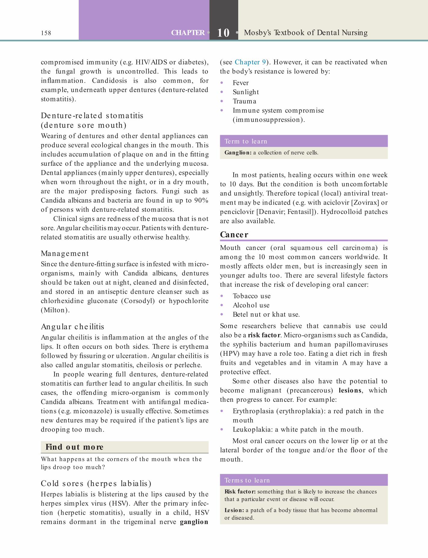
158 CHAPTER • 10 • Mosby’s Textbook of Dental Nursing
(see Chapter 9). However, it can be reactivated when the body’s resistance is lowered by:
• Fever• Sunlight• Trauma• Immune system compromise
(immunosuppression).
compromised immunity (e.g. HIV/AIDS or diabetes), the fungal growth is uncontrolled. This leads to in ammation. Candidosis is also common, for example, underneath upper dentures (denture-related stomatitis).
De nture -re la te d s tomatitis (de nture s ore mouth)Wearing of dentures and other dental appliances can produce several ecological changes in the mouth. This includes accumulation of plaque on and in the tting surface of the appliance and the underlying mucosa. Dental appliances (mainly upper dentures), especially when worn throughout the night, or in a dry mouth, are the major predisposing factors. Fungi such as Candida albicans and bacteria are found in up to 90% of persons with denture-related stomatitis.
Clinical signs are redness of the mucosa that is not sore. Angular cheilitis may occur. Patients with denture-related stomatitis are usually otherwise healthy.
ManagementSince the denture- tting surface is infested with micro-organisms, mainly with Candida albicans, dentures should be taken out at night, cleaned and disinfected, and stored in an antiseptic denture cleanser such as chlorhexidine gluconate (Corsodyl) or hypochlorite (Milton).
Angula r c he ilitisAngular cheilitis is in ammation at the angles of the lips. It often occurs on both sides. There is erythema followed by ssuring or ulceration. Angular cheilitis is also called angular stomatitis, cheilosis or perleche.
In people wearing full dentures, denture-related stomatitis can further lead to angular cheilitis. In such cases, the offending micro-organism is commonly Candida albicans. Treatment with antifungal medica-tions (e.g. miconazole) is usually effective. Sometimes new dentures may be required if the patient’s lips are drooping too much.
Find out moreWhat happens a t the corners of the mouth when the lips d roop too much?
Cold s ore s (he rpe s lab ia lis )Herpes labialis is blistering at the lips caused by the herpes simplex virus (HSV). After the primary infec-tion (herpetic stomatitis), usually in a child, HSV remains dormant in the trigeminal nerve ganglion
Terms to lea rnRisk actor: something that is likely to increase the chances that a particular event or disease will occur.
Lesion: a patch of a body tissue that has become abnormal or diseased.
Term to lea rnGanglion: a collection of nerve cells.
In most patients, healing occurs within one week to 10 days. But the condition is both uncomfortable and unsightly. Therefore topical (local) antiviral treat-ment may be indicated (e.g. with aciclovir [Zovirax] or penciclovir [Denavir; Fentasil]). Hydrocolloid patches are also available.
CancerMouth cancer (oral squamous cell carcinoma) is among the 10 most common cancers worldwide. It mostly affects older men, but is increasingly seen in younger adults too. There are several lifestyle factors that increase the risk of developing oral cancer:
• Tobacco use• Alcohol use• Betel nut or khat use.Some researchers believe that cannabis use could also be a risk factor. Micro-organisms such as Candida, the syphilis bacterium and human papillomaviruses (HPV) may have a role too. Eating a diet rich in fresh fruits and vegetables and in vitamin A may have a protective effect.
Some other diseases also have the potential to become malignant (precancerous) lesions, which then progress to cancer. For example:
• Erythroplasia (erythroplakia): a red patch in the mouth
• Leukoplakia: a white patch in the mouth.Most oral cancer occurs on the lower lip or at the
lateral border of the tongue and/or the oor of the mouth.

159Dental and Oral Pathology and Disease
Geographical TongueGeographical tongue is a common harmless genetic condition in which the liform papillae on the tongue (see Chapter 9) are lost temporarily in the presence of irregular map-like red areas. The pattern of redness changes from day to day and even within a few hours. It may cause soreness. Geographical tongue is also called erythema migrans, benign migratory glossitis and migratory stomatitis.
No effective treatment is available, so the patient just needs to be reassured.
LeukoplakiaLeukoplakia is the name given to acquired white patches on the oral mucosa. It may occur as a single, localised lesion or multiple and diffuse widespread lesions. Mostly these are smooth plaques (homogene-ous leukoplakias) seen on the lip, buccal mucosa or gingivae; others are irregular (non-homogeneous). Of these some are warty (verrucous leukoplakia).
Biopsy is required to exclude cancer. Persons with leukoplakia should be advised to stop any tobacco/alcohol/betel habits, and to consume a diet rich in fruit and vegetables. The lesion is also usually removed by surgery.
Manage me ntPlanning of cancer treatment requires a multidiscipli-nary team (MDT) consisting of a range of specialists: dentist, surgeon, anaesthetist, oncologist, nursing and other dental staff, nutritionist, speech therapist and physiotherapist etc.
Oral cancer is treated largely by surgery and/or radiotherapy.
• After surgery, there may be some cosmetic, sensory and functional problems.
• After radiotherapy, there may be functional problems such as xerostomia (dry mouth) and dif culty in opening the mouth (trismus).
• These also increase the risk of caries and infections such as candidosis and sialadenitis (salivary gland in ammation).
• The mouth becomes sore (mucositis) and the jaws may be liable to infection (osteoradionecrosis).
Fordyce SpotsFordyce spots are oil glands similar to those found in skin. They are seen as creamy-yellow dots along the border between the vermilion and the oral mucosa. They are not associated with hair follicles. Fordyce spots are benign and very common but a few patients become concerned about them. No treatment is indi-cated, other than reassurance.
Key poin tLeukoplakia is a potentially malignant disorder: that is, in up to a third of patients, it can eventually turn to cancer.
Term to lea rnBenign: a lesion that is not a cancer, but which can grow or spread locally.
Clin ica l signs of cancerCancer may appear in different ways, such as a persistent:
• Ulcer with ssuring or raised edges
• Red patch
• White patch
• Mixed white and red patch
• Lump
• Non-healing extraction socket
• Pain
• Lymph node enlargement.
Key po in tIf a single lesion lasts over three weeks, it may be a cancer. A biopsy will usually be required.
Lichen PlanusLichen planus is a chronic disorder of uncertain cause. It affects the mouth, genital areas and/or skin. The mouth lesions are typically white lesions in the buccal (cheek) mucosa on both sides.
The usual treatment is with corticosteroids. Like leukoplakia, lichen planus is a potentially malignant disorder in 1-3%.
UlcersMouth ulceration is not a speci c disease, rather it is the outcome of one of a range of disorders or causes. Local causes such as trauma, and recurrent aphthous stomatitis (RAS) are the most common, but ulcers in the mouth can also arise in patients with systemic (general) disease (especially blood diseases, infec-tions, gastro-intestinal and skin disorders), cancer, or drug reactions.
160 CHAPTER • 10 • Mosby’s Textbook of Dental Nursing
• Using drugs that stimulate salivation (sialogogues).Use of arti cial saliva (mouth-wetting agents) may
also help.
MucoceleThis is a small blister that arises when a small amount of saliva gets into the tissues or is trapped in a gland. Mucoceles may resolve themselves or require surgery for removal.
MumpsMumps is an acute infectious disease caused by the mumps virus. It is also called acute viral sialadenitis or epidemic parotitis. Typically the patient has a painful swelling in the region of the parotid gland, usually on both sides, with trismus, fever and malaise. Complications of mumps include:
• Orchitis in boys (in ammation of the testes; ensuing infertility is rare).
• Pancreatitis (in ammation in the pancreas)• Encephalitis (in ammation of the brain).
No speci c antiviral drugs are available to treat mumps, so treatment is aimed at relieving the symp-toms, and giving uids and analgesics. Mumps is seen more now because of the reduced uptake of the MMR vaccine in recent years.
Find out moreWhy have some parent s mistakenly been worried about g iving the MMR vaccine to the ir ch ild in recen t years?
RAS consists of multiple ulcers occurring through-out the mouth. It usually starts in childhood. Local treatments with corticosteroids may be used.
Salivary DiseaseDroolingDrooling is a problem for many children with cerebral palsy, intellectual disability and other neurological conditions. It also happens in adults, particularly those who have Parkinson’s disease or have had a stroke. Drooling is caused either by increased saliva ow (sialorrhoea), or by poor oral and facial muscle control (secondary sialorrhoea). For management of drooling, physiotherapy, drugs or surgery may be needed.
Xerostomia (Dry Mouth)Dry mouth, also called xerostomia or hyposalivation, often occurs as a side-effect of some drugs or as a result of radiation to the salivary glands during cancer treat-ment. Sjögren syndrome, diabetes, or occasionally HIV infection, also cause dry mouth.
Dryness of the mouth is uncomfortable for the patient because it may cause:
• Mouth soreness• Burning sensation• Dif culty eating dry foods such as biscuits (the
cracker sign)• Dif culty controlling dentures• Speech problems• Dif culty swallowing.
Term to lea rnSialadenitis: in ammation of the salivary glands or their ducts.
Key po in tAspirin should not be given to children as it may cause a dangerous reaction in the liver (Reye syndrome).
Complications of dry mouth include: caries, can-didosis or sialadenitis. Complications can be avoided or managed by:
• Avoiding sugary foods• Keeping the mouth clean• Using uorides• Using chlorhexidine mouthwashes• Stimulating salivation.
Salivation may be stimulated by:
• Chewing gum (containing xylitol or sorbitol, not sucrose)
• Eating diabetic sweets
Salivary Duct ObstructionSalivary duct obstruction is not uncommon. It is usually caused by a ‘stone’ (calculus) and is commonly seen in the submandibular duct. This is called sialolithiasis.
Typically the patient will give a history of painful salivary gland swelling just before, or at, mealtimes. Calculi are usually yellow or white and can sometimes be seen in the duct. Radiographs can be taken to check as the stone can be radiopaque. The stones are usually removed by surgery, or destroyed by breaking them with sound waves (lithotripsy).
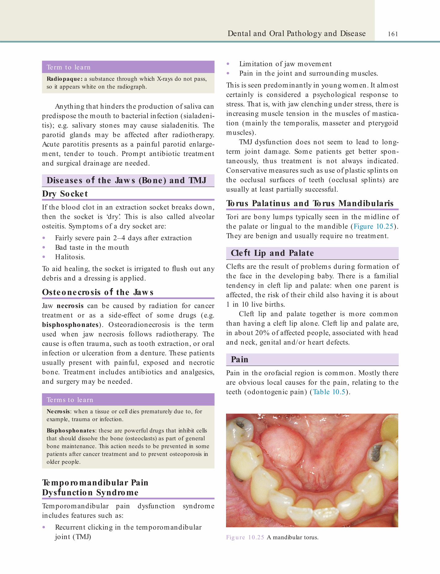
161Dental and Oral Pathology and Disease
• Limitation of jaw movement• Pain in the joint and surrounding muscles.This is seen predominantly in young women. It almost certainly is considered a psychological response to stress. That is, with jaw clenching under stress, there is increasing muscle tension in the muscles of mastica-tion (mainly the temporalis, masseter and pterygoid muscles).
TMJ dysfunction does not seem to lead to long-term joint damage. Some patients get better spon-taneously, thus treatment is not always indicated. Conservative measures such as use of plastic splints on the occlusal surfaces of teeth (occlusal splints) are usually at least partially successful.
Torus Palatinus and Torus MandibularisTori are bony lumps typically seen in the midline of the palate or lingual to the mandible (Figure 10.25). They are benign and usually require no treatment.
Cle t Lip and PalateClefts are the result of problems during formation of the face in the developing baby. There is a familial tendency in cleft lip and palate: when one parent is affected, the risk of their child also having it is about 1 in 10 live births.
Cleft lip and palate together is more common than having a cleft lip alone. Cleft lip and palate are, in about 20% of affected people, associated with head and neck, genital and/or heart defects.
PainPain in the orofacial region is common. Mostly there are obvious local causes for the pain, relating to the teeth (odontogenic pain) (Table 10.5).
Anything that hinders the production of saliva can predispose the mouth to bacterial infection (sialadeni-tis); e.g. salivary stones may cause sialadenitis. The parotid glands may be affected after radiotherapy. Acute parotitis presents as a painful parotid enlarge-ment, tender to touch. Prompt antibiotic treatment and surgical drainage are needed.
Diseases o the Jaw s (Bone) and TMJDry SocketIf the blood clot in an extraction socket breaks down, then the socket is ‘dry’. This is also called alveolar osteitis. Symptoms of a dry socket are:
• Fairly severe pain 2–4 days after extraction• Bad taste in the mouth• Halitosis.To aid healing, the socket is irrigated to ush out any debris and a dressing is applied.
Osteonecrosis o the Jaw sJaw necrosis can be caused by radiation for cancer treatment or as a side-effect of some drugs (e.g. bisphosphonates). Osteoradionecrosis is the term used when jaw necrosis follows radiotherapy. The cause is often trauma, such as tooth extraction, or oral infection or ulceration from a denture. These patients usually present with painful, exposed and necrotic bone. Treatment includes antibiotics and analgesics, and surgery may be needed.
Fig u re 10.25 A mandibular torus.
Terms to learnNecrosis: when a tissue or cell dies prematurely due to, for example, trauma or infection.
Bisphosphonates: these are powerful drugs that inhibit cells that should dissolve the bone (osteoclasts) as part of general bone maintenance. This action needs to be prevented in some patients after cancer treatment and to prevent osteoporosis in older people.
Term to lea rnRadiopaque: a substance through which X-rays do not pass, so it appears white on the radiograph.
Temporomandibular Pain Dys unction SyndromeTemporomandibular pain dysfunction syndrome includes features such as:
• Recurrent clicking in the temporomandibular joint (TMJ)
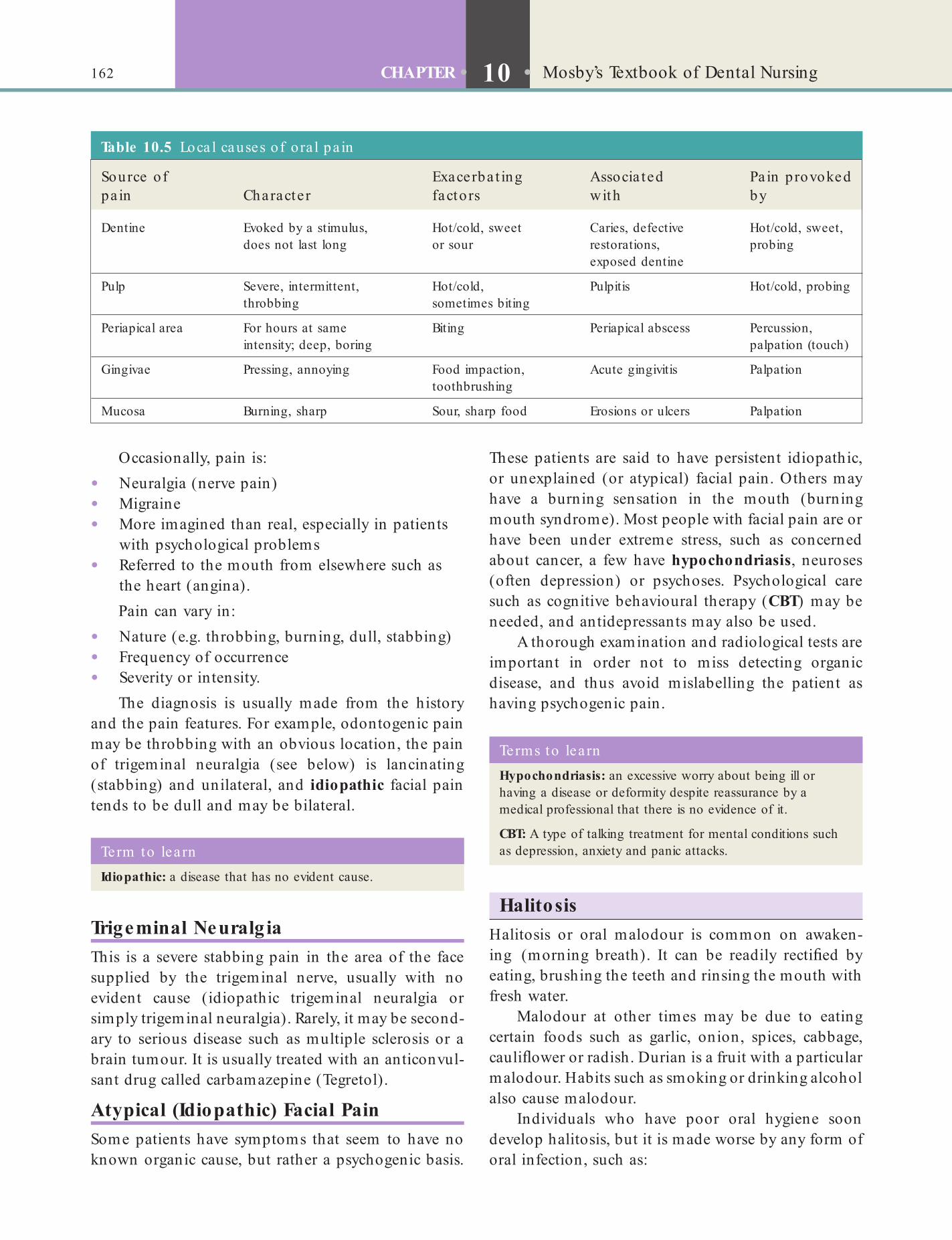
162 CHAPTER • 10 • Mosby’s Textbook of Dental Nursing
These patients are said to have persistent idiopathic, or unexplained (or atypical) facial pain. Others may have a burning sensation in the mouth (burning mouth syndrome). Most people with facial pain are or have been under extreme stress, such as concerned about cancer, a few have hypochondriasis, neuroses (often depression) or psychoses. Psychological care such as cognitive behavioural therapy (CBT) may be needed, and antidepressants may also be used.
A thorough examination and radiological tests are important in order not to miss detecting organic disease, and thus avoid mislabelling the patient as having psychogenic pain.
Occasionally, pain is:
• Neuralgia (nerve pain)• Migraine• More imagined than real, especially in patients
with psychological problems• Referred to the mouth from elsewhere such as
the heart (angina).Pain can vary in:
• Nature (e.g. throbbing, burning, dull, stabbing)• Frequency of occurrence• Severity or intensity.
The diagnosis is usually made from the history and the pain features. For example, odontogenic pain may be throbbing with an obvious location, the pain of trigeminal neuralgia (see below) is lancinating (stabbing) and unilateral, and idiopathic facial pain tends to be dull and may be bilateral.
Term to lea rnIdiopathic: a disease that has no evident cause.
Table 10.5 Loca l causes of o ra l pa in
Source of pa in Character
Exacerba t ing factors
Associa ted with
Pa in provoked by
Dentine Evoked by a stimulus, does not last long
Hot/cold, sweet or sour
Caries, defective restorations, exposed dentine
Hot/cold, sweet, probing
Pulp Severe, intermittent, throbbing
Hot/cold, sometimes biting
Pulpitis Hot/cold, probing
Periapical area For hours at same intensity; deep, boring
Biting Periapical abscess Percussion, palpation (touch)
Gingivae Pressing, annoying Food impaction, toothbrushing
Acute gingivitis Palpation
Mucosa Burning, sharp Sour, sharp food Erosions or ulcers Palpation
Terms to lea rnHypochondriasis: an excessive worry about being ill or having a disease or deformity despite reassurance by a medical professional that there is no evidence of it.
CBT: A type of talking treatment for mental conditions such as depression, anxiety and panic attacks.
Trigeminal Neuralg iaThis is a severe stabbing pain in the area of the face supplied by the trigeminal nerve, usually with no evident cause (idiopathic trigeminal neuralgia or simply trigeminal neuralgia). Rarely, it may be second-ary to serious disease such as multiple sclerosis or a brain tumour. It is usually treated with an anticonvul-sant drug called carbamazepine (Tegretol).
Atypical (Idiopathic) Facial PainSome patients have symptoms that seem to have no known organic cause, but rather a psychogenic basis.
HalitosisHalitosis or oral malodour is common on awaken-ing (morning breath). It can be readily recti ed by eating, brushing the teeth and rinsing the mouth with fresh water.
Malodour at other times may be due to eating certain foods such as garlic, onion, spices, cabbage, cauli ower or radish. Durian is a fruit with a particular malodour. Habits such as smoking or drinking alcohol also cause malodour.
Individuals who have poor oral hygiene soon develop halitosis, but it is made worse by any form of oral infection, such as:
163Dental and Oral Pathology and Disease
• Gingivitis• Periodontitis• Dental abscess• Dry (infected) extraction socket• Sinusitis• Tonsillitis.
Rarer causes of halitosis include more general con-ditions such as:
• Respiratory disease• Sinusitis• Nasal infections• Lung problems• Metabolic disease• Diabetes• Kidney disease• Liver disease• Psychiatric disease (where halitosis may be
imagined).Treatment includes improving oral hygiene and
reducing the tongue coating by gentle and regular tongue cleaning. Mouthwashes containing chlorhexi-dine gluconate, triclosan or cetylpyridinium (essential oils), may help. Toothpastes containing triclosan and a copolymer (e.g. Colgate Total toothpaste) could also be used.
Quick revision aid o f the m ain causes o f oral signs and sym ptom s
Bleeding• Haemangioma (a tumour of the cells that line the blood
vessels)
• Trauma
• Bleeding tendency or drugs
• In ammation
Blisters• Skin diseases
• Infections
• Burns
• Allergies
• Cysts
• Mucoceles
Discoloured teeth• Extrinsic discolorations (brown or black):
• Poor oral hygiene
• Smoking
• Beverages/food (e.g. tea, coffee, red wine)
• Drugs
• Betel
• Intrinsic discolorations:
• Localised: trauma; caries; restorative ( lling) materials
• Generalised: tetracyclines; excessive uoride; genetic diseases
Dry mouth (xerostomia)• Drugs
• Dehydration
• Psychogenic cause
• Salivary gland disease
Early tooth loss• Trauma
• Dental caries
• Periodontal breakdown
• Tumours
Facial sw elling• In ammation (e.g. infections or bites)
• Trauma
• Allergies
• Cysts
• Neoplasms
Halitosis• Volatile foodstuffs
• Drugs and tobacco
• Oral disease
• Systemic disease:
• Respiratory disease
• Metabolic disease
• Psychogenic cause
Late tooth eruption• Impacted teeth
• Cancer treatment
Pain• Dental disease
• Migraine and similar vascular disorders
• Trigeminal neuralgia
• Psychogenic pain
• Referred pain (e.g. angina)
Pigmentation• Racial
• Food/drugs
• Tobacco
• Betel
• Chlorhexidine
• Minocycline treatment
• Endocrinological (Addison disease)

164 CHAPTER • 10 • Mosby’s Textbook of Dental Nursing
• Red areas
• Congenital conditions:
• Haemangiomas
• Trauma
• In ammatory
• Neoplastic and possibly pre-neoplastic
Salivary sw elling• In ammatory
• Obstruction
• Neoplasm
Soreness and ulceration• Systemic disease
• Malignant disease
• Local causes
• Aphthae (recurrent aphthous stomatitis)
• Drugs
Sw ellings and lumps• Congenital
• Allergic reactions
• In ammatory lesions
• Neoplasms
• Traumatic
Trismus• Infection and in ammation near masticatory muscles
• Temporomandibular joint-dysfunction syndrome (facial arthromyalgia)
• Fractured or dislocated jaw
• Arthritis
• After radiotherapy
White lesions• Congenital conditions
• Cheek biting
• In ammatory:
• Infective (e.g. candidosis)
• Non-infective (e.g. lichen planus)
• Neoplastic and possibly pre-neoplastic:
• Keratoses (leukoplakias)
• Carcinoma
Find out moreThere a re severa l websites tha t describe ora l d iseases and condit ions for a range of readers. See for example :• Hea lth Canada (www.hc-sc.gc.ca /h l-vs/ora l-bucco/
disease-malad ie /index-eng .php)• Hea lth Cent ra l.com (www.hea lthcen t ra l.com/
channe l/408/1122.html).

Chapter 11 Dental and Oral Imaging
CHAPTER POINTS• Radiography de nit ions and basics• Radiation hazards• Dental radiographs• Processing radiographs• Digital radiography• Radiation safety• Role of the dental nurse in imaging• Other types of imaging
Ionising radiation is radiation with enough energy to interact with and affect atoms and cells, and can thus cause disease. Ionising radiation is one of four types: alpha, beta, gamma and X radiation:
• Alpha radiation travels only a short distance (a few inches) in air, and is not an external hazard. Examples of alpha emitters include: radium, radon, uranium, thorium.
• Beta radiation may travel several feet in air, is moderately penetrating and may injure skin. Examples of beta emitters include: strontium-90, carbon-14, tritium, and sulphur-35.
• Gamma radiation can travel many feet in air, many inches in human tissue, readily penetrates most materials and is sometimes called ‘penetrating’ radiation, constituting an external hazard to humans. Dense materials are needed for shielding from this radiation. Examples of gamma emitters include: iodine-131, caesium-137, cobalt-60, radium-226 and technetium-99m.
• X radiation is able to travel many feet in air and many inches in human tissue, readily penetrates most materials and is sometimes called ‘penetrating’ radiation, constituting an external hazard to humans. Dense materials are needed for shielding from penetrating radiation.X-rays involve the use of ionising radiation and
fall under legislation discussed below, especially
• The Ionising Radiation Regulations 1999 (known as IRR).
• The Ionising Radiation (Medical Exposure) Regulations 2000 (known as IR(ME)R).Dental radiography in the UK is subject to the IRR
and the IR(ME)R, enacted under the Health and Safety at Work etc. Act 1974 (see Chapter 8). There are also
In t ro d u ct io n
Dental imaging mainly consists of taking X-rays (radio-graphs) and photographs. Both of these are used rou-tinely in the dental environment as aids to diagnosis and treatment planning. More advanced imaging, such as computed tomography (CT), magnetic reso-nance imaging (MRI) and ultrasound (US), are also used, especially in surgical and hospital practice.
RadiationEnergy emitted from a source is generally called radia-tion. Examples include heat or light from the sun, microwaves from an oven, X-rays from an X-ray tube, and gamma rays from radioactive elements. Electro-magnetic radiation consists of:
• Heat waves• Radiowaves• Infrared light• Visible light• Ultraviolet light• X-rays• Gamma rays.These differ only in frequency and wavelength.
166 CHAPTER • 11 • Mosby’s Textbook of Dental Nursing
Ionising radiations, particularly X-rays, are also a potential hazard to body organs and tissues where cells are proliferating rapidly (e.g. in the fetus or a young child, or in the gonads and bone marrow). Here ionising radiation has the capacity not only to induce malignant tumours (this is called an oncogenic effect) but also to damage reproductive tissues (teratogenic effect).
Therefore, since there is always a slight risk from excessive exposure to radiation, the bene t of it must always outweigh the risk to the patient. These advan-tages and disadvantages must be discussed with the patient and the patient must give informed consent. Women should always inform their clinician if there is any possibility that they are pregnant. Exposure to ionising radiation in pregnant women must be kept to the absolute minimum and X-rays taken only when absolutely essential: bene t must well exceed any pos-sible harm.
EU Directives concerned not only with avoiding unnecessary or excessive exposure to radiation but also with improving the quality and effectiveness of medical and dental uses of radiation.
Quali ed dental nurses can process radiographs and may also mount radiographic lms. Under super-vision of an IR(ME)R operator, as a quali ed dental nurse you may press the button on the X-ray machine. However, you are only allowed to place the lms in the patient’s mouth or position the X-ray tube, and to take radiographs, if you are suitably trained with a post-registration Certi cate in Dental Radiography. Dental nurses are not under any circumstances permit-ted to interpret radiographs.
Key poin tSince no X-ray exposure can be completely free from risk, the use of radiography is accompanied by a responsibility to ensure protection.
Term to lea rnIR(ME)R operator: a person who is trained in dental radiography, for example a dentist, therapist, hygienist or a dental nurse who has undertaken a post-registration certi cate course in dental radiography. (This involves completion of a record of experience in the workplace and passing an examination.) See also p. 175.
Radiography Def nitions and Basics• Radiography – the techniques involved in
producing X-ray images.• Radiology – the interpretation of radiographic
images.X-rays, like light, are electromagnetic waves but they have more energy than light so can penetrate the body tissues to varying degrees (which affects the number of X-rays reaching the lm). When the X-rays hit a radiographic lm, they sensitise the silver crystals on the lm, which then turn black when put in a devel-oper. The image thus formed on the lm enables the clinician to see different (distinct) structures because of the large differences in absorption of X-rays by hard and soft tissues. Metals and really hard tissues such as tooth enamel appear white, other hard tissues such as dentine and bone appear grey, and soft tissues appear almost black on the lm.
Radiation HazardsX-rays are a type of ionising radiation. Thus, while radiography can be essential for diagnosis and treat-ment planning, it involves exposure of patients and, potentially, staff to ionising radiation. The problem with ionising radiation is that it can damage DNA, causing mutations that may possibly lead to cancer.
We are all constantly exposed to normal back-ground ionising radiation arising from the earth (espe-cially in areas where the rocks emit radon gas – in the UK this is mainly in mountainous areas). People are also exposed to radiation when travelling by air. So diagnostic X-rays produce radiation in addition to this background radiation. As such, the dose of X-rays that a person receives while undergoing basic dental radi-ography (intra-oral X-rays and panoramic radiogra-phy) is quite low, probably equivalent to only a few days of background radiation. However, it might still increase the risk of salivary gland and thyroid tumours. Having a CT scan means much higher exposures.
In the past, ionising radiation was also a serious occupational hazard to radiographers and clinical dental staff. Some clinicians even developed radiation-induced dermatitis or cancer of their hands from holding radiographic (X-ray) lms in the patients’ mouths during radiography. Having one’s hand in the X-ray beam gives about 4000 times the exposure com-pared with that received 2 m away from the X-ray tube (the recommended ‘safe’ distance). Therefore this practice is illegal now, and there have been tremen-dous improvements in technology and techniques resulting in greater ionising radiation safety. Speci c
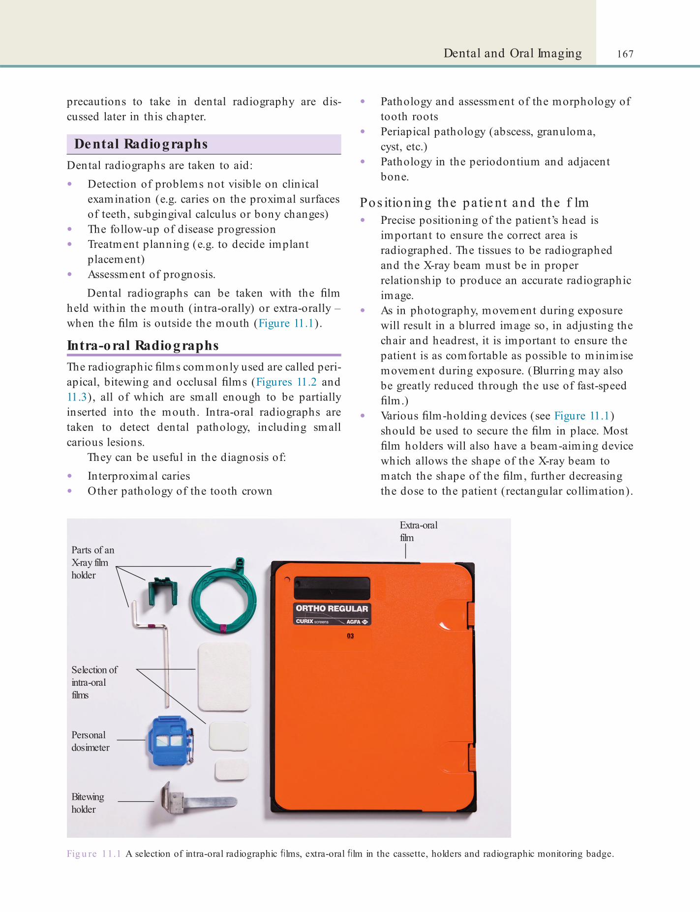
167Dental and Oral Imaging
• Pathology and assessment of the morphology of tooth roots
• Periapical pathology (abscess, granuloma, cyst, etc.)
• Pathology in the periodontium and adjacent bone.
Pos itioning the pa tie nt and the f lm• Precise positioning of the patient’s head is
important to ensure the correct area is radiographed. The tissues to be radiographed and the X-ray beam must be in proper relationship to produce an accurate radiographic image.
• As in photography, movement during exposure will result in a blurred image so, in adjusting the chair and headrest, it is important to ensure the patient is as comfortable as possible to minimise movement during exposure. (Blurring may also be greatly reduced through the use of fast-speed lm.)
• Various lm-holding devices (see Figure 11.1) should be used to secure the lm in place. Most lm holders will also have a beam-aiming device which allows the shape of the X-ray beam to match the shape of the lm, further decreasing the dose to the patient (rectangular collimation).
precautions to take in dental radiography are dis-cussed later in this chapter.
Dental RadiographsDental radiographs are taken to aid:
• Detection of problems not visible on clinical examination (e.g. caries on the proximal surfaces of teeth, subgingival calculus or bony changes)
• The follow-up of disease progression• Treatment planning (e.g. to decide implant
placement)• Assessment of prognosis.
Dental radiographs can be taken with the lm held within the mouth (intra-orally) or extra-orally – when the lm is outside the mouth (Figure 11.1).
Intra-oral RadiographsThe radiographic lms commonly used are called peri-apical, bitewing and occlusal lms (Figures 11.2 and 11.3), all of which are small enough to be partially inserted into the mouth. Intra-oral radiographs are taken to detect dental pathology, including small carious lesions.
They can be useful in the diagnosis of:
• Interproximal caries• Other pathology of the tooth crown
Fig u re 11.1 A selection of intra-oral radiographic lms, extra-oral lm in the cassette, holders and radiographic monitoring badge.
Extra-oralfilm
Parts of anX-ray filmholder
Selection ofintra-oralfilms
Personaldosimeter
Bitewingholder

168 CHAPTER • 11 • Mosby’s Textbook of Dental Nursing
Identify and learnFind an occip ito -menta l rad iograph in your workplace to see how it shows the sinuses, or look it up on the in te rne t .
Identify and learnFind a couple o f lm holders in your workplace and ask your supervisor to exp la in how they he lp a im the X-ray beam precise ly.
Extra-oral RadiographsExtra-oral radiography means using large lms to visu-alise the skull, jaws, temporomandibular joints and sinuses. These lms are used with intensifying screens in a cassette (see Figure 11.1).
Examples of the use of intra- and extra-oral radio-graphs in dentistry are given in Table 11.1.
Fig u re 11.2 (A, B) Examples of periapical radiographic lms.
Fig u re 11.3 A bitewing radiographic lm. The white areas are dental restorations.
Term to lea rnIntensi ying screen: a screen that permits a lower radiation exposure but good quality lms; digital systems can also help in the same way.
Cephalometric RadiographsThese are a type of extra-oral radiograph often used in orthodontics (see Chapter 20 for more details).
TomographsTomography (Greek tomos, meaning ‘slice’) involves taking lms of sections or slices of a part of the body. Panoramic radiography is a specialised tomographic technique that is commonly used in dentistry but the radiation dose may be higher than intra-oral lms to show the same areas under examination.
De nta l panoramic tomographsA dental panoramic tomograph (DPT or orthopanto-mograph [OPG]) is a tomographic technique used mainly to assess the lower part of the face. A DPT displays both the upper and lower teeth in a long at lm (Figure 11.5). It also gives a good overview of the maxillary sinuses, mandibular rami and the temporo-mandibular joints. It shows the number and position of all teeth, including unerupted ones. However, it does not show ne detail of the anterior part of the

169Dental and Oral Imaging
Fig u re 11.5 Examples of dental panoramic radiographs. (A) Mixed dentition period. (B) An adult with full dentition. (C) An edentulous mouth.
Fig u re 11.4 (A) Maxillary anterior occlusal lm, showing a cyst. (B) Mandibular anterior occlusal lm, showing a salivary stone.
Table 11.1 Examples of the more common denta l rad iographs and the ir main uses
Area to be examined Radiographic lm used Often used for
Whole of the mandible and the maxilla
DPT (dental panoramic tomograph; Figure 11.5)
Presence, position and pathology of teeth; jaw fractures
A single tooth or three to four teeth plus the supporting bone
Periapical lm (Figure 11.2): size varies from 35 × 22 mm to 40.5 × 30.5 mm
Assessing the periapical area, for root canal treatment and to assess root fractures
Molar/premolar region Bitewing (horizontal) (Figure 11.3): size varies from 35 × 22 mm to 54 × 27 mm
Caries detection interproximally
Bitewing (vertical) Periodontitis
Maxillary incisor/canine region Anterior occlusal (Figure 11.4): size is about 57 × 76 mm
Impacted canines; supernumerary teeth; palatal cysts; salivary duct stones
Third molars Oblique lateral, or DPT, or periapical Inspecting unerupted or impacted third molars
Sinuses DPT or occipito-mental radiograph Sinusitis, root in sinus
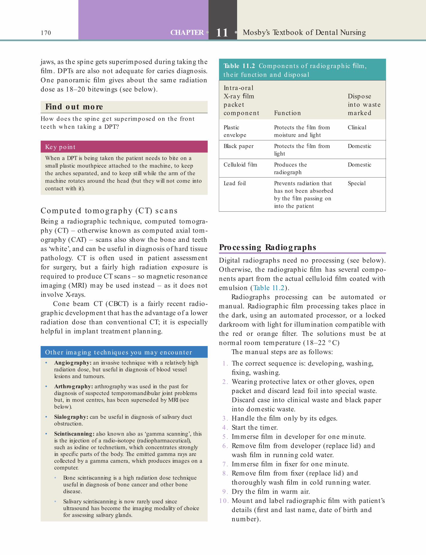
170 CHAPTER • 11 • Mosby’s Textbook of Dental Nursing
Key poin tWhen a DPT is being taken the patient needs to bite on a small plastic mouthpiece attached to the machine, to keep the arches separated, and to keep still while the arm of the machine rotates around the head (but they will not come into contact with it).
Other imaging t echniques you may encounte r• Angiography: an invasive technique with a relatively high
radiation dose, but useful in diagnosis of blood vessel lesions and tumours.
• Arthrography: arthrography was used in the past for diagnosis of suspected temporomandibular joint problems but, in most centres, has been superseded by MRI (see below).
• Sialography: can be useful in diagnosis of salivary duct obstruction.
• Scintiscanning: also known also as ‘gamma scanning’, this is the injection of a radio-isotope (radiopharmaceutical), such as iodine or technetium, which concentrates strongly in speci c parts of the body. The emitted gamma rays are collected by a gamma camera, which produces images on a computer.
• Bone scintiscanning is a high radiation dose technique useful in diagnosis of bone cancer and other bone disease.
• Salivary scintiscanning is now rarely used since ultrasound has become the imaging modality of choice for assessing salivary glands.
jaws, as the spine gets superimposed during taking the lm. DPTs are also not adequate for caries diagnosis. One panoramic lm gives about the same radiation dose as 18–20 bitewings (see below).
Find out moreHow does the sp ine ge t superimposed on the fron t tee th when taking a DPT?
Compute d tomography (CT) s c ansBeing a radiographic technique, computed tomogra-phy (CT) – otherwise known as computed axial tom-ography (CAT) – scans also show the bone and teeth as ‘white’, and can be useful in diagnosis of hard tissue pathology. CT is often used in patient assessment for surgery, but a fairly high radiation exposure is required to produce CT scans – so magnetic resonance imaging (MRI) may be used instead – as it does not involve X-rays.
Cone beam CT (CBCT) is a fairly recent radio-graphic development that has the advantage of a lower radiation dose than conventional CT; it is especially helpful in implant treatment planning.
Processing RadiographsDigital radiographs need no processing (see below). Otherwise, the radiographic lm has several compo-nents apart from the actual celluloid lm coated with emulsion (Table 11.2).
Radiographs processing can be automated or manual. Radiographic lm processing takes place in the dark, using an automated processor, or a locked darkroom with light for illumination compatible with the red or orange lter. The solutions must be at normal room temperature (18–22 ° C)
The manual steps are as follows:1 . The correct sequence is: developing, washing,
xing, washing.2 . Wearing protective latex or other gloves, open
packet and discard lead foil into special waste. Discard case into clinical waste and black paper into domestic waste.
3 . Handle the lm only by its edges.4 . Start the timer.5 . Immerse lm in developer for one minute.6 . Remove lm from developer (replace lid) and
wash lm in running cold water.7 . Immerse lm in xer for one minute.8 . Remove lm from xer (replace lid) and
thoroughly wash lm in cold running water.9 . Dry the lm in warm air.
10 . Mount and label radiographic lm with patient’s details ( rst and last name, date of birth and number).
Table 11.2 Components o f rad iographic lm, the ir funct ion and d isposa l
In t ra -ora l X-ray lm packe t component Funct ion
Dispose in to waste marked
Plastic envelope
Protects the lm from moisture and light
Clinical
Black paper Protects the lm from light
Domestic
Celluloid lm Produces the radiograph
Domestic
Lead foil Prevents radiation that has not been absorbed by the lm passing on into the patient
Special
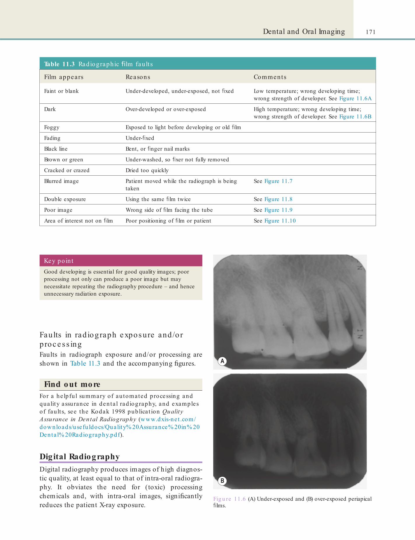
171Dental and Oral Imaging
Key poin tGood developing is essential for good quality images; poor processing not only can produce a poor image but may necessitate repeating the radiography procedure – and hence unnecessary radiation exposure.
Table 11.3 Radiographic lm fau lt s
Film appears Reasons Comments
Faint or blank Under-developed, under-exposed, not xed Low temperature; wrong developing time; wrong strength of developer. See Figure 11.6A
Dark Over-developed or over-exposed High temperature; wrong developing time; wrong strength of developer. See Figure 11.6B
Foggy Exposed to light before developing or old lm
Fading Under- xed
Black line Bent, or nger nail marks
Brown or green Under-washed, so xer not fully removed
Cracked or crazed Dried too quickly
Blurred image Patient moved while the radiograph is being taken
See Figure 11.7
Double exposure Using the same lm twice See Figure 11.8
Poor image Wrong side of lm facing the tube See Figure 11.9
Area of interest not on lm Poor positioning of lm or patient See Figure 11.10
Faults in radiograph e xpos ure and/o r proc e s s ingFaults in radiograph exposure and/or processing are shown in Table 11.3 and the accompanying gures.
Find out moreFor a he lpfu l summary of au tomated processing and qua lity assurance in den ta l rad iography, and examples o f fault s, see the Kodak 1998 publica t ion Quality Assurance in Dental Radiography (www.dxis-ne t .com/downloads/usefu ldocs/Quality% 20Assurance% 20in% 20Denta l% 20Radiography.pdf).
Digital RadiographyDigital radiography produces images of high diagnos-tic quality, at least equal to that of intra-oral radiogra-phy. It obviates the need for (toxic) processing chemicals and, with intra-oral images, signi cantly reduces the patient X-ray exposure.
Fig u re 11.6 (A) Under-exposed and (B) over-exposed periapical lms.
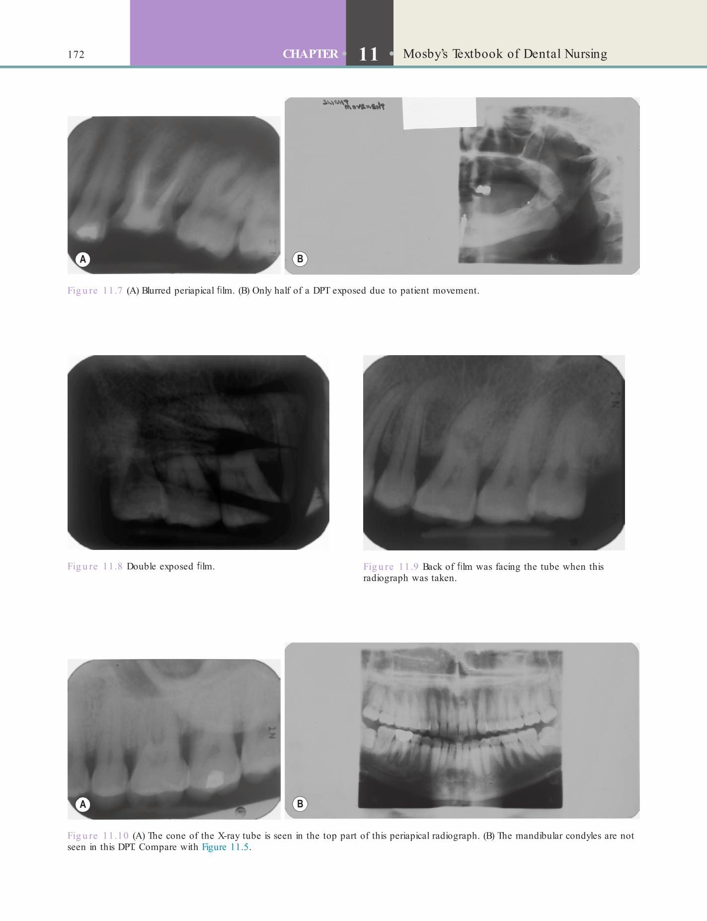
172 CHAPTER • 11 • Mosby’s Textbook of Dental Nursing
Fig u re 11.7 (A) Blurred periapical lm. (B) Only half of a DPT exposed due to patient movement.
Fig u re 11.8 Double exposed lm. Fig u re 11.9 Back of lm was facing the tube when this radiograph was taken.
Fig u re 11.10 (A) The cone of the X-ray tube is seen in the top part of this periapical radiograph. (B) The mandibular condyles are not seen in this DPT. Compare with Figure 11.5.
173Dental and Oral Imaging
Take and de ve lop f lms c o rre c tlyIt cannot be emphasised too strongly therefore that it is well worthwhile in the interests of both safety and economy to make sure that radiographic apparatus and techniques are up to the highest possible stand-ards. Films should be taken and developed correctly. Each lm should yield the maximum amount of diag-nostic information possible. To achieve this, the fol-lowing are required:
• Correct alignment and collimation (see next section) of the X-ray beam.
• Correct placing of the lm and positioning of the patient.
• Choice of the fastest lm compatible with image quality.
• Choice of the most effective intensifying screens compatible with image quality.
• Correct processing of the lm to give optimum image quality.
• Correct lm processing:• Processing solutions should be used at the
recommended temperature and changed regularly, as advised by the manufacturer.
• The developer should be changed at least once a month but preferably fortnightly.
• Processing must be carried out in a light-proof environment – either an automatic lm processor or a darkroom.
• Panoramic lms are particularly light-sensitive and a special lter is required for the dark-room safelight if they are used.
• Films once processed should be washed to remove chemicals and then dried in a dust-free atmosphere before viewing. They should be led carefully in the patient’s records afterwards.
Co llimationCollimation in radiology means focusing the X-rays into a narrow beam to reduce unnecessary exposure of other parts of the body. Rectangular collimation can achieve dose reductions of around 50%. In intra-oral radiography, beam eld size is constrained by using lm-holding devices (see Figure 11.1) with a beam alignment guide to prevent cone cuts. Rectangular
Key poin tThe need for a radiograph should be decided after weighing the total potential diagnostic bene ts against the individual detriment that the exposure might cause.
Key poin ts: Reducing rad ia t ion exposureRadiation exposure of the patient can best be minimised by:
• Taking only essential radiographs
• Ensuring high quality and useful radiographs
• Reducing eld size
• Using the fastest X-ray lms
• Processing lms properly.
Radiation Precautions in Practice
Minimis e rad iographs take n and the e xposureThe government legislation lays down controls for radiation safety (see below); but the best way to reduce exposure is to take radiographs only when and where they are absolutely essential for diagnosis or treat-ment. Therefore no patient should undergo dental radiography without having received a clinical exami-nation. The clinician will also take into account the ef cacy, bene ts and risk of available alternative tech-niques having the same objective but involving no or less exposure to X-rays. If radiography is deemed nec-essary, patient X-ray doses should be kept As Low As Reasonably Practicable (ALARP principle). Exposure can be minimised by:
• Only taking radiographs when essential• Reducing eld size• Use of fast lms or digital dental imaging.
Radiographs may also need to be taken to avoid medico-legal dif culties. For example, if a patient has pain following a tooth extraction but no dry socket is apparent, it is prudent to take a lm to exclude, for example, a fracture of the jaw.
It is vital to ensure a regular back-up of the database and not to rely on the computer’s hard disk alone. From a dento-legal standpoint, it is also important to store digital radiographs securely and within a format that cannot be corrupted. The Digital Imaging and Communications in Medicine (DICOM) standard ensures security by containing the image and information about the image, such as patient name, type of image, dimensions, and changes. However, at present, relatively few dental digital systems are DICOM-compatible.
Radiation Sa e ty

174 CHAPTER • 11 • Mosby’s Textbook of Dental Nursing
collimation for intra-oral periapical and bitewing radi-ography offers levels of thyroid protection similar to those provided by lead shielding. There is no evidence to justify routine use of abdominal (gonadal) lead protection for intra-oral dental radiography.
Find out moreWhy was lead sh ie ld ing considered important while taking den ta l rad iographs? What is meant by a cone cu t? Ask the person responsib le fo r taking rad iographs in your workplace to demonst ra te th is to you .
Mainta in rad ia tion e quipme nt c a re ullyAll radiographic equipment leaks radiation. Modern and well-maintained apparatus must therefore be used, to keep leakage to a minimum. Collimating dia-phragms must be used to restrict the useful X-ray beam to the area under study and aluminium lters to reduce skin absorption must be installed. It is also important to check that the duration of X-ray emission corresponds exactly with what is indicated on the timer switch.
Ke e p we ll away rom the X-ray s ourc eThe radiographic lm must not be held by the opera-tor during exposure and only the patient and opera-tors should be in the room during the exposure. Distance is important since trebling the distance from an X-ray source reduces the radiation dose to about one-tenth (the inverse square law). The exposure switch should be so arranged that the operator can stand at least 2 m away from the X-ray tube, out of line of the direct beam, and that the switch cannot be oper-ated accidentally. This aspect is covered in more detail in the section on the dental radiography room (p. 180).
Key poin tIt is good practice for non-essential staff to leave the room during intra-oral radiography.
Us e o inte ns i ying s c re e nsExtra-oral radiography uses cassettes containing inten-sifying screens to reduce the radiation to about one-tenth of that necessary to produce an image of the same density on wrapped packet lm. Cassettes are light-tight containers for light-sensitive lm and contain sheets of card covered with rare earth crystals that emit light when struck by X-radiation and thereby expose the lm. Unfortunately, the detail is not as good as with intra-oral lm because of the very poor edge de nition produced by the crystals’ diffuse light emission. The larger the crystals the poorer the de ni-tion – but the shorter the exposure. Rare earth screens (e.g. Kodak Lanex or 3M Trimax or Fuji RX ranges) not only further reduce the exposure but the X-ray tube also lasts longer because of the reduced load. However, for ordinary dental use there is little if any advantage because of the relatively low kilovoltage used in dental sets, and for oblique lateral lms the exposure may be made unmanageably short.
A panoramic lm may reveal unsuspected disease in the jaws or elsewhere. However, the clinician needs to bear in mind the limitations of panoramic lms especially in the anterior region and the lesser quality of detail of the periodontal and periapical tissues. Digital panoramic radiographs do not reduce the dose to the patient as much as digital intra-oral systems. This is because non-digital system panoramic radiog-raphy can use intensifying screens.
Note: Dentists were warned in 2012 against using a speci c hand-held X-ray machine on patients as it poses a signi cant health risk. The cheap imported machine, known as the Tianjie Dental Falcon (Zhengzhou Tianjie Electronic Equipment Co.), exposes users and patients to 10 times the normal level of radiation, increasing their risks of cancer and organ damage. The Medicines and Healthcare Regulatory Agency (MHRA) has asked NHS and private dentists to dispose of these devices.
Consent and Quality AssuranceInformed consent should be obtained from patients prior to imaging. Quality assurance (QA) is required to ensure consistently adequate diagnostic informa-tion. A well-designed QA programme should include:
• Image quality assessment• Practical radiographic technique assurance• Patient dose and X-ray equipment checks• Darkroom, lm, cassettes, digital sensors and
processing checks• Staff training.
Us e the as te s t f lmsFilm speed is an important aspect in determining the amount of radiation exposure. The fastest radiographic lm consistent with adequate image quality should be used: speed groups E or F are recommended because they reduce the radiation dose more than 50% com-pared with group D-speed lms. The fastest lms cur-rently available are the group F lms Kodak Ektaspeed Plus and AGFA DM4. Ektaspeed and AGFA DM2 are available for periapical, bitewing and occlusal lms. Many now recommend that E-speed lms should be used almost exclusively.
175Dental and Oral Imaging
Terms to learnSievert (Sv): this is the unit of radiation absorbed dose producing the same biological effect, in a speci ed tissue, as 1 Gray of high energy X-rays.
Gray (Gy): the unit of radiation. 1 Gy =100 rads (an older unit of radiation).
Surveys and checks should be performed accord-ing to a regular timetable, and a written log of this programme should be maintained. This ensures adherence to the programme and raises its importance among staff. A named person should be leader for the QA programme in a dental workplace.
Radiography and PregnancyWhen taking dental radiographs, the risk to the devel-oping fetus is low as the radiation dose is low. Thus there is no contraindication to dental radiography of women who are or may be pregnant provided that it is clinically justi ed. There is also usually no need to use a lead protective apron; although it is required if the X-ray beam is pointed directly towards the fetus. At other times, the use of a lead apron may actually impair the quality of the radiograph produced. Lead aprons are contraindicated for DPTs.
Key poin tFollowing the ALARP principle, the risks to the unborn child of radiation related to dentistry during pregnancy are very small.
• European Council Directive 97/43/Euratom of 30 June 1997, on health protection of individuals against the dangers of ionising radiation in relation to medical exposure (Medical Exposures Directive) (provides a high level of health protection from ionising radiation in medical exposure).
• The Ionising Radiation Regulations 1999 (known as IRR).
• The Ionising Radiation (Medical Exposure) Regulations 2000 (known as IR(ME)R).
The EU Directives are concerned not only with avoid-ing unnecessary or excessive exposure to radiation but also with improving the quality and effectiveness of medical uses of radiation.
IRR and IR(ME)RIn the UK, following the establishment of the Radio-logical Protection Act 1970, the National Radiological Protection Board (NRPB) was created as the authority primarily concerned with safety of ionising radiation. The NRPB is now part of Public Health England.
Dental radiography in the UK is subject to the IRR and the IR(ME)R, enacted under the Health and Safety at Work etc. Act 1974. The IRR and IR(ME)R Regula-tions have implications for every dental practice and cover all aspects of radiography from equipment selec-tion and installation through to radiographic proce-dures and protocols. General guidance on complying with IRR is given in the Approved Code of Practice (ACOP). Actions taken under the IR(ME)R come under criminal and not civil law.
Find out moreThe 2001 publica t ion Guidance Notes for Dental Pract it ioners on the Safe Use o f X-Ray Equipm ent o f the Department o f Hea lth and NRPB expla ins the conten t , scope and implica t ions of the ionising rad ia t ion regula t ions and the regula t ion of clin ica l pract ice as re levan t to denta l p ract ice . You can nd th is on the Gov.UK website : www.gov.uk/rad ia t ion-products-and-services.
IRRThe main aim of the Ionising Radiation Regulations 1999 (IRR) as de ned by the of cial code of practice is to ‘establish a framework for ensuring that exposure to ionising radiation arising from work activities, whether man made or natural radiation and from external radiation or internal radiation, is kept As Low As Reasonably Practicable (ALARP) and does not exceed dose limits speci ed for individuals’.
Staff rarely receive radiation doses above 1 mSv (milliSievert) per year provided the ALARP principle is applied. So special precautions for pregnant staff are not normally required – if it can be assured that the dose during pregnancy is no more than 1 mSv to the abdomen during pregnancy (which is normally the case in dental practice). However, female employ-ees should inform the employer, in writing, as soon as they discover that they are pregnant, so that the dose situation may be reviewed.
Legal AspectsThe legislation applying is:
• European Council Directive 96/29/Euratom, of 13 May 1996 – this lays down the basic safety standards for the health protection of workers and the general public against dangers arising from ionising radiation (this ensures the protection of workers exposed to ionising radiation, including clinicians and their assistants, and of members of the public).
176 CHAPTER • 11 • Mosby’s Textbook of Dental Nursing
Synopsis of IRR 99Designa ted person Usua lly
Roles in re la t ion to regula t ions Role encompasses
Legal person Practice owner, employer or CEO
Ensures workplace full compliance
Organising• A 3-year assessment of radiation safety by a
competent authority (e.g. PHE Radiation Protection Division)
• Local Rules for each machine showing• Names of RPA and RPS• Controlled area• Sign warning of ionising radiation• Summary of correct working instructions
for each controlled area• Summary of contingency plan to be
followed in event of machine malfunction• Details of dose investigation level• Arrangements in place for safety of
pregnant staff• Use of red light and audible buzzer to
indicate exposuresRadiation Protection
Adviser (RPA)Medical physicist Advises staff on staff
and public safetyEnsuring• Correct installation of all new X-ray machines• Regular maintenance and certi cation checks• Contingency plans for malfunction• Investigation of malfunctions• Designation of 1.5 m Controlled AreaAdvising on• Risk assessment and training• Staff protection• QA programmes
Radiation Protection Supervisor (RPS)
Senior dentist or DCP with post-registration quali cation in dental radiography
Ensuring• staff have suitable training• Local Rules are current• QA programmes• Staff maintain core CPDCarrying out risk assessmentsMaintaining radiation protection leRunning quality control tests
Synopsis of IR(ME)R 2000Lega l person Usually Roles in re la t ion to regula t ions
Re erer Dentist who refers patient for exposure
Provide suf cient information to allow justi cation of the treatment
Practitioner Dentist or radiographer Responsible for justifying exposureOperator Staff with suitable and authenticated
training who carry out all or part of the practical duties
Patient identi cationPositioning of lm, patient and tube headSetting exposure controlsPressing exposure buttonProcessing lmEvaluating quality (Quality Audit; QA)Carrying out test exposures for QARunning QA programmesReporting/evaluating the lm (currently DCPs are not deemed
to be trained to undertake this role)
IR(ME)R
177Dental and Oral Imaging
The IR(ME)R regulations classify dental profes-sionals involved in radiography as follows.
IR(ME)R refe rre rAn IR(ME)R referrer is a medically or dentally quali- ed person who is legally allowed to refer patients for radiographic examination for diagnostic or treatment planning purposes. They have a responsibility to ensure that the examination they request justi es the associated radiation dose. The referrer is usually a dentist.
IR(ME)R practitionerAn IR(ME)R practitioner must be dentally quali ed. It is usually the clinician who authorises a radiographic examination once they are satis ed that the radiation dose is justi ed.
IR(ME)R opera torAn IR(ME)R operator is any person carrying out any practical aspect of the exposure. Regulation 11 para-graph 1 of IR(ME)R 2000 states that ‘no practitioner or operator shall carry out a medical (or dental) expo-sure of any practical aspect without having been ade-quately trained’. Radiographs can thus be taken, provided they are trained, by a clinician or dental nurse.
Schedule 2 of the regulations details the require-ments for adequate training and states that: ‘practi-tioners and operators shall have successfully completed training, including theoretical knowledge and practi-cal experience, in a “core of knowledge” ’. This includes knowledge of: radiation production and protection; the relevant statutory obligations relating to ionising radiation; and diagnostic radiology as relevant to their speci c area of practice.
Dental nurses who are not trained in radiography are still counted as IR(ME)R operators if they are involved in any aspect of production of the X-ray image – processing, mounting of lms or QA of systems, and they need to be appropriately trained.
Key poin tThe Referrer, Practitioner and Operator can be one and the same person, i.e. the clinician. Conversely several IR(ME)R operators may be involved in a single procedure.
• Direct advice to management on legal and other matters
• Radiation measurements to assess potential hazards and to control exposure of the workforce
• Assistance in drawing up:
• Prior risk assessments• Contingency plans• Local rules• Radiation protection programmes
• Statutory testing of radiation monitors• Statutory tests for leakage of radioactive material
from sealed sources• Personal dosimetry (see below)• Critical examinations• Radiochemical analysis• Restorative action and dose assessment following
accidents or incidents• Radiation protection training• Audits of radiation protection arrangements.
Radia tion protection advis e r (RPA)Every dental practice should have an RPA to advise about observance of the regulations and other health and safety matters connected with ionising radiation, such as:
Loca l rad ia t ion sa fe ty ru lesEvery employer who undertakes work with ionising radiation must have a set of written local rules that enable staff working with ionising radiation to do so in compliance with the regulations. They also need to ensure that staff are aware of such of the rules as are relevant to them and any other persons who may be affected by them. The information in the rules should include:
• Name of the RPS (p. 177)
• Identi cation of controlled areas where X-rays will be used
• Safe working instructions:
• Switch off machines when not in use
• Keep 2 m away from the X-ray tube head
• Do not enter controlled areas when X-rays are operating
• Contingency arrangements to be followed in the event of machine failure
• The name of the ‘legal person’ – usually the employer
• Name of person responsible for contacting engineer
• Contact details of the RPA.
Radia tion protection s upervis or (RPS)All dental practices should also have an RPS, who is usually a dentally quali ed member of the practice. The RPS has a supervisory role and assists the employ-ing dentist to comply with the regulations. In a single-handed practice the RPS is usually the clinician.
The RPS must have knowledge and understanding of the requirements of the ionising radiation regula-tions and local rules. They should be directly involved
178 CHAPTER • 11 • Mosby’s Textbook of Dental Nursing
Dosimeters do not protect or shield you from radiation exposure; they merely inform how much radiation (if any) the wearer received.
with the work with ionising radiation and should undertake close supervision to ensure that the work is done in accordance with the local rules. They should also ensure that the necessary precautions are taken in the work that is being done and to what extent these precautions will restrict exposure. Although the RPS need not be present at all times, adequate supervision must be maintained in their absence.
The employer carries the ultimate responsibility for compliance with the regulations; this cannot be delegated to the RPS.
Key poin tThe RPS ensures that local rules are in place and followed by all members of the dental team.
Sta Exposure to X-raysThe European Basic Safety Standards (BSS) Directive requires designation of ‘controlled areas’ (areas subject to special rules to ensure staff safety). For panoramic and intra-oral units, the controlled area is de ned during X-ray exposure as: within 1.5 m of the X-ray tube and patient and within the primary X-ray beam until suf ciently attenuated by distance or shielding.
To ensure staff are fully aware of the precautions to be taken it is desirable that written instructions (local rules and working procedures) are in place and displayed near the X-ray equipment. These instruc-tions should include:
• The responsibility for exposure• Positioning of staff• Use of protective devices• Any restriction on primary beam direction• Personal monitoring arrangements (if
appropriate; see below).
Pe rs ona l monito ringIn the average dental practice, exposure to radiation of any staff is highly unlikely to exceed 1 mSv, even in a whole year. The doses received by staff working with dental X-ray equipment are such that there should be no need to alter normal good working practice. For comparison, on average each person living in the UK receives more than 2 mSv every year from natural radi-ation. It is, however, good practice that even for small workloads all staff wear a personal dosimeter (Figure 11.1). This provides reassurance that safe conditions continue to prevail. There are at least three types of personal dosimeters: lm ‘badges’, the new Luxel tech-nology and TLDs (thermoluminescent dosimeters).
Persona l dosimeters• Personal dosimeters can be obtained from the Radiological
Protection Service of Public Health England at Chilton, Didcot, Oxon OXJI ORQ (tel. 0235 831600) or from RRPPS, 63 Melchett Road, Kings Norton Business Centre, Birmingham, B30 3HP (tel. 0121 627 2090/1)
Key poin tAnyone who works with radiation and actually gets or might get 10% of the annual limit is recommended to wear a dosimeter. Dental practices generally are not required to provide dosimeters to staff since the exposures are low and the beam sizes small.
The guidance for wearing personal dosimeters is:
• WEAR IT when working.• DO NOT WEAR IT:
• When you are receiving X-rays for your personal healthcare
• Away from the workplace.• DO NOT:
• Share it with someone else• Tamper with it, or anyone else’s dosimeter.
• DO:
• TURN IT IN promptly on leaving employment
• REPORT A LOST/DAMAGED unit immediately
• STORE the dosimeter in a radiation-safe area and a place that is not hot.
The recorded occupational doses should be reviewed by the RPS on receipt of the dose reports, and action taken if any dose exceeds that expected, e.g. 150 exposures per week should result in a dose no greater than 0.25 mSv. A 3-month wearing period is suitable.
For dentistry, personal monitoring should usually demonstrate a dose no greater than 0.2 mSv accumu-lated during the monitoring period. The RPS should carry out a formal investigation if the cumulative dose received by any individual member of staff in particu-lar year exceeds 1 mSv.
When personal dosimeters are not in use they should be stored outside the radiography room, in a dry place away from heat.
Public Health England’s Dental X-ray Protection Services (DXPS) can provide advice on monitoring.
179Dental and Oral Imaging
averaged over an area not exceeding 100 cm 2, does not exceed 1 mGy (milliGray) in one hour. For equipment intended for dental radiography with an intra-oral lm, radiation leakage should not exceed 0.25 mGy in one hour.
• The total ltration of the beam (made up of the inherent ltration and any added ltration) should be equivalent to not less than the following:• 1.5 mm aluminium for X-ray tube voltages
up to and including 70 kV• 2.5 mm aluminium of which 1.5 mm
should be permanent for X-ray tube voltages above 70 kV.
TrainingIt is the responsibility of the ‘legal person’, that is the dental practice owner, to ensure that all staff receive adequate training. Training aims to ensure that you are aware of the dangers and potential hazards of ionising radiation and your own safety, that of your patients and members of the public.
The General Dental Council has set out the level of training in radiography that therapists, hygienists and dental nurses should receive. As a dental nurse, your basic training should include the hazards of radi-ography and instruction on lm processing, mounting and QA procedures. These elements are included in the National Diploma in Dental Nursing and in the current NVQ syllabus.
Specialist dental radiography courses are offered by a number of institutions. The Certi cate in Dental Radiography allows the holder to undertake dental radiographic procedures under the prescription of an IR(ME)R practitioner.
Under the regulations, all practitioners and opera-tors (clinicians or other dental care professionals) must undertake continuing education in dental radiol-ogy and radiation protection. On an average, practi-tioners are expected to devote 12.5 hours or 5% of their CPD to the subject of dental radiology. Attend-ance at formal courses would usually provide at least 5 hours of veri able CPD and certi ed evidence of continuation training in the area.
X-Ray Tube Voltage , Beam Size and Filtration, and Distance Contro l• Dental X-ray equipment should be designed,
constructed and installed in compliance with British Standards (e.g. BS 5724). It should be maintained in accordance with the recommendations of its manufacturer or the manufacturer’s authorised representative.
• The X-ray tube voltage should not be lower than 60 kilovolts (kV) and for intra-oral radiography should be preferably 70 kV, since lower kV values necessitate higher localised patient exposure.
• Every X-ray source assembly (comprising an X-ray tube, an X-ray tube housing and a beam-limiting device) should be constructed so that, at every rating speci ed by the manufacturer for the X-ray source assembly, the air kerma from the leakage radiation, at a distance from the focal spot of 1 m
Term to lea rnKerma: is an acronym for kinetic energy released in material.
Exposure Contro l• Exposure control (timer): timers must operate
accurately and reproducibly, and repeat exposures must not be possible without rst fully releasing the exposure switch. Older timers are likely to need to be replaced.
• Exposure switches on all dental X-ray equipment should be so arranged that exposure continues only while continuous pressure is maintained on the switch and terminates immediately the pressure is released. To guard against automatic timing failure, an additional means of termination should be provided and must be independent of the normal means. Release of the exposure switch may be regarded as the additional means when this action overrides the timer. Exposure switches should also be designed to prevent inadvertent production of X-rays. If re-setting is automatic it should be ensured that pressure on the switch has to be released completely before the next exposure can be made.
• All dental equipment control panels should be tted with a light which gives an indication, clearly visible (and preferably also an audible warning) to the operator, that an exposure is taking place. The light should be triggered by the ow of current directly responsible for the start and termination of the emission of
180 CHAPTER • 11 • Mosby’s Textbook of Dental Nursing
• There should be a radiation warning sign, together with appropriate words, on any X-ray room door that opens directly into an area where the instantaneous dose rate is greater than 7.5 µSv/h (µSv = microSievert).
• When the controlled area extends to any entrance of the X-ray room an automatic warning signal should be given at that entrance while radiation is emitted.
• If more than one X-ray set is sited in any room, e.g. in open plan accommodation, then arrangements should be made, in consultation with the RPA, to ensure that patients and staff are adequately protected.
• Since the beam is not always fully absorbed by the patient, it should be considered to extend beyond the patient until it has been attenuated by distance or intercepted by a primary protective shielding such as a brick wall.
• If it is necessary to support a disabled patient or child, this should only be done in accordance with the local rules drawn up with the advice of the RPA.
• The tube housing should never be held by hand during an exposure. The operator should stand at least 2 m away, making use of the full length of cable to the exposure switch. A protective panel should, if possible, be provided and the operator should stand behind it.
• If the advice on avoidance of the beam and the protection afforded by distance is followed for ordinary dental radiography and for panoramic tomography, the operator should be outside the controlled area and should not therefore need to be designated as a classi ed person.
• Any staff who enter a controlled area should either be classi ed persons or do so under a written system of work, which may include the need to wear a personal dosimeter.
• As mentioned earlier, the operator should check that the equipment warning light and, where provided, any audible warning signal operates at each exposure and ceases at the end of the intended exposure time. If the warning does not operate or there is reason to think that the timer is defective or that there may be some other fault (for example, signs of damage, excessive X-ray tube temperature), the equipment should be disconnected from the supply and not used again until it has been checked and, if necessary, repaired (see below).
radiation. For equipment tted with an audible warning the warning should be triggered by the same conditions. The exposure should be terminated automatically when a predetermined condition, such as a pre-set time, has been attained.
The Dental Radiography Room• Dental radiography should be carried out in a
room (the X-ray room) from which all persons whose presence is unnecessary are excluded while X-rays are being produced. This room, which may be a dental surgery or a separate examination room, should not be used for other work or as a passageway while radiography is in progress.
• The workload in most dental surgeries is not likely to exceed 300 intra-oral lms, or 50 panoramic examinations, each week. However, protective panels having a protective equivalent of not less than 0.5 mm of lead should be provided if the workload is likely to exceed this, that is:
• 150 mA minutes per week for panoramic tomography
• 30 mA minutes per week for other procedures.
• Persons in all occupied areas immediately outside the X-ray room should be adequately protected. The X-ray room should be arranged so that:
• The radiation beam is directed away from those areas
• Use is made of the natural shielding of the walls, oor, ceiling of the X-ray room where these are relatively thick or dense, e.g. of brick or concrete
• Advantage is taken of the reduction in radiation level by distance.
• If the normal structural materials do not afford suf cient shielding (e.g. a light-weight partition wall may sometimes be in the radiation beam), protective material such as lead ply should be attached to the wall concerned. The equipment should be installed so that the useful beam is directed away from any door or window, if the space immediately beyond is occupied.
• Adjacent areas, for example, those used as waiting rooms, should not be controlled or supervised areas.

181Dental and Oral Imaging
protective gloves and aprons should be worn in accordance with advice obtained from the RPA.
• The exposure factors should be checked by the operator on each occasion before an examination is made. This is particularly important when a short spacer is used after a long one and when there is more than one beam size setting. The larger apertures may be quite unsuitable for use with intra-oral lms.
Dental Panoramic Tomography (DPT)• Intensifying screens should be used with all
extra-oral lms.• If the rotational movement fails to start, or stops
before the full arc is covered, the switch should be released immediately to avoid high localised exposure of the patient.
• A lead apron is not indicated for DPT radiography, where it may interfere with the process.
• For panoramic tomography the beam size at the cassette holder should not exceed 10 mm × 150 mm. The total beam area should not exceed the area of the receiving slit of the cassette holder by more than 20%.
Panoramic Radiography w ith an Intra-oral X-ray Tube• Because of the unnecessary exposure of tissues
not being examined, intra-oral panoramic units still in use are required to be phased out as soon as practicable.
• In those still in use, beam applicators should be used to protect tissues such as the tongue which do not have to be irradiated for the production of a satisfactory radiograph.
• Care should be taken in positioning the X-ray tube in order to get satisfactory and consistent results.
Find out moreFor more in format ion on den ta l rad iography, see Radiography and Radio logy for Dental Care Pro fessionals (Whaites E, 2013, published by Elsevie r).
The British Dental Association (BDA) advice sheet A11 ‘Radiation in Dentistry’ also includes advice on radiation protection.
Role o the Dental Nurse in ImagingWith additional training and only on prescription (see above for details of training), a dental nurse may also
Term to lea rnControlled area: in radiology, this is a designated area into which entry and exit as well as the activities carried out are controlled. The aim is to ensure only the minimal, necessary occupational exposure of staff to radiation.
Key poin tThe X-ray room should be large enough to provide safe accommodation for those persons who have to be in the room during X-ray examinations.
Intra-oral Radiography
• Equipment for radiography using an intra-oral lm should be provided with a eld-de ning spacer-cone which will ensure a minimum focal spot to skin distance of not less than 20 cm for equipment operating above 60 kV and not less than 10 cm for equipment operating at lower voltages.
• When alternative spacers are available or interchangeable spacers are provided, the one most suited to the technique to be employed should be tted. The correct setting of the equipment is particularly important where interchangeable cones for different radiological techniques are available.
• The open end should be placed as close as possible to the patient’s head to minimise the size of the incident beam: beam diameters should not exceed 6 cm and preferably should be collimated to a rectangular eld. If a larger focal spot to skin distance is required, a longer spacer should be employed.
• The beam should not be directed towards the gonads. If the patient is a woman who is, or who may be, pregnant, care should be taken that the fetus is not irradiated inadvertently. Where such a beam direction cannot be avoided, the body should be covered by a protective apron having a protective equivalent of not less than 0.25 mm lead.
• The dental lm should be held by the patient only when it cannot otherwise be kept in position. It should virtually never be hand-held by anyone else. Exceptionally it may be held by someone other than the patient using a pair of forceps to avoid direct irradiation of the ngers, for example, when a child or a disabled person cannot hold it themselves. In such cases
182 CHAPTER • 11 • Mosby’s Textbook of Dental Nursing
• Implanted electric devices (e.g. heart pacemakers and de brillators, nerve stimulators, cochlear implants)
• Intracranial vascular clips, if these are ferromagnetic
• Prosthetic cardiac valves containing metal• Obesity (because of the weight limit on the
gantry and size of scanner)• Claustrophobia (unless open scanner is
available).
take radiographs to the prescription of a clinician (Certi cate in Dental Radiography is required; see also Chapter 3).
Other Types o ImagingUltrasound ScansUltrasound is the non-invasive use of sound waves to produce images that can be used to help diagnosis of diseases. It is the preferred method of imaging for diagnosing soft tissue swellings (e.g. lymph nodes, thyroid or salivary glands). There are no known con-traindications to ultrasound.
Magnetic Resonance Imaging (MRI)Magnetic resonance imaging also does not use ionising radiation (X-rays). On MR images the bone shows up as black (rather than white as in radiographic lms), and soft tissue lesions can be well visualised, including malignant lesions. The disadvantages of MRI are that it is expensive and liable to produce image artefacts where ferromagnetic metal objects are present (e.g. dental restorations, orthodontic appliances, metallic foreign bodies, joint prostheses, implants etc.). Con-traindications to MRI include:
Term to lea rnGantry: in radiology, a gantry is a device that helps to rotate the radiation source around the patient so that images can be taken from various angles and in various planes.
PhotographyPhotographs are part of the clinical record and are needed especially in orthodontics, cosmetic dentistry and after assaults, including child abuse (non-accidental injury). Written patient consent to all imaging is required.
DENTAL DISEASE, PREVENTION AND CAREChapter 12 Denta l and Ora l Hea lth Promot ion and Disease
Prevent ion 185
Chapte r 13 Denta l and Ora l Hea lth Trea tment Planning , Chart ing and Records 203
Chapte r 14 Drugs in Dent ist ry 217
Chapte r 15 Pa in and Anxie ty Cont ro l 225
Chapte r 16 Denta l Materia ls and Equipment 243
Chapte r 17 Restora t ive Procedures and Materia ls 257
Chapte r 18 Surg ica l Care 285
Chapte r 19 Surg ica l Care : Inpa t ien ts 299
Chapte r 20 Orthodont ics 309
Chapte r 21 People with Vulnerability o r Specia l Needs 321
Chapte r 22 Minority Issues 335
Chapte r 23 Denta l Emergencies 339
Sect ion D

This page intentiona lly le ft blank
Chapter 12 Dental and Oral Health Promotion
and Disease Prevention
CHAPTER POINTS• The oral health message• Role o the dental nurse in dental and oral
health promotion• Home care• Plaque control/oral hygiene• Dietary control• Reduction o demineralisation• Remineralisation• Fissure sealing• Prevention o other dental and oral disease• Pro essional care (f ssure sealing)• Habit cessation advice• Dental recall
There is a NEBDN Certif cate in Oral Health Education – a GDC-recognised post-registration qualif cation.
Find out moreThis chap te r is based on the too lkit ‘Delivering be t t e r ora l hea lth : An evidence-based toolkit o r prevent ion’ by the Department o Hea lth and Brit ish Associa t ion o r the Study o Community Dent ist ry (3rd edit ion , 2014). For the u ll too lkit see www.gov.uk/search?q=Delivering+bet te r+ora l+hea lth% 3A+An+evidence-based+toolkit+ or+prevent ion .
Tooth decay is the most common oral disease a ecting children and young people in England, yet it is largely preventable. While children’s oral health has improved over the past 20 years, almost a third (27.9%) o 5-year-olds still had tooth decay in 2012. People living in deprived communities consistently have poorer oral (and general) health than people living in richer communities.
Poor oral health impacts children and amilies’ health and wellbeing. Children who have toothache or who need treatment may have to be absent rom school. Parents may also have to take time o work to take their children to the dentist. Oral health is an integral part o overall health; when children are not healthy, this a ects their ability to learn, thrive and develop. Good oral health can contribute to school readiness. Tooth decay was the most common reason or hospital admissions in children aged 5 to 9 years old in 2012–13. Dental treatment under general anaesthesia (GA) presents a small but real risk o li e-threatening complications or children.
The aims o preventive dental health are to achieve and maintain good dental, oral and general health (Figure 12.1) and this advocates preventive and pro-phylactic measures mainly to avoid dental caries and
In t ro d u ct io n
This chapter should be read in conjunction with Chapter 10 on dental and oral pathology and disease. Most dental and oral diseases, including not only the more common problems (such as tooth sur ace loss due to caries, trauma, abrasion, erosion and periodon-tal disease) but also potentially atal diseases (such as cancer), are largely related to people’s li estyle and habits (Table 12.1). There ore these diseases are o ten preventable by care ul attention to li estyle.
The Oral Health MessageOral health promotion involves educating people about how to achieve and maintain good dental and oral health. All dental pro essionals can provide oral health advice direct to patients.

186 CHAPTER • 12 • Mosby’s Textbook of Dental Nursing
They are also required to provide or commission oral health surveys. The oral health surveys are carried out as part o the Public Health England (PHE) dental public health intelligence programme ( or-merly known as the national dental epidemiology programme).
An integrated approach with partners is needed or oral health improvement as well as ensuring all local authority services or children and young people have oral health improvement embedded at a strategic and operational level.
Role of the Dental Nurse in Dental and Oral Health Promotion
A dental nurse should be able to provide competent and complete dental and oral health promotion. This includes:
• Plaque control – instructions in toothbrushing, ossing, use o disclosing tablets and other aids
• Dietary advice• Advice on how o ten a dental/oral examination
(check-up or recall) is required• Advice to mothers on preventing nursing bottle
caries.Disclosing tablets or solutions are discussed on p. 187.
Fig u re 12.1 Healthy dentition.
Fig u re 12.2 Poor oral hygiene showing dental caries, periodontal disease and debris (plaque and debris on the gingivae [material alba]).
Term to lea rnNursing bottle caries: the decay of the upper incisors related to drinking milk through a bottle just before sleeping. The milk remains on the teeth and acts as a substrate for dental caries.
Table 12.1 Ora l d iseases re la ted to li estyle hab it s
Disease Risk actor
Abrasion Incorrect toothbrushing
Cancer Tobacco, alcohol, betel nut use
Candidosis Dry mouth, antibiotic use, HIV and other immunity problems
Caries High sugar diet, low uoride
Erosion Soft drinks, fruit juices, alcohol
Gingivitis Plaque accumulation
Halitosis Plaque accumulation, tobacco use, alcohol use, some foods e.g. onions, garlic, durian
Periodontitis Plaque and calculus accumulation, tobacco use
Trauma Alcohol use; some contact sports
periodontal disease, though other aspects, such as screening or oral disease such as cancer and smoking cessation advice, are important.
The Health and Social Care Act 2012 con erred the responsibility or health improvement, including oral health improvement, to local authorities. Many general health conditions and oral diseases share common risk actors such as smoking, alcohol misuse and poor diet. Oral diseases are largely preventable; and there is a need to develop interventions to achieve sustained and long-term improvements in oral health and reduce inequalities and avoid the consequences o ‘neglect’, as shown in Figure 12.2.
Local authorities are statutorily required to provide or commission oral health promotion programmes to improve the health o the local population, to an extent that they consider appropriate in their areas.
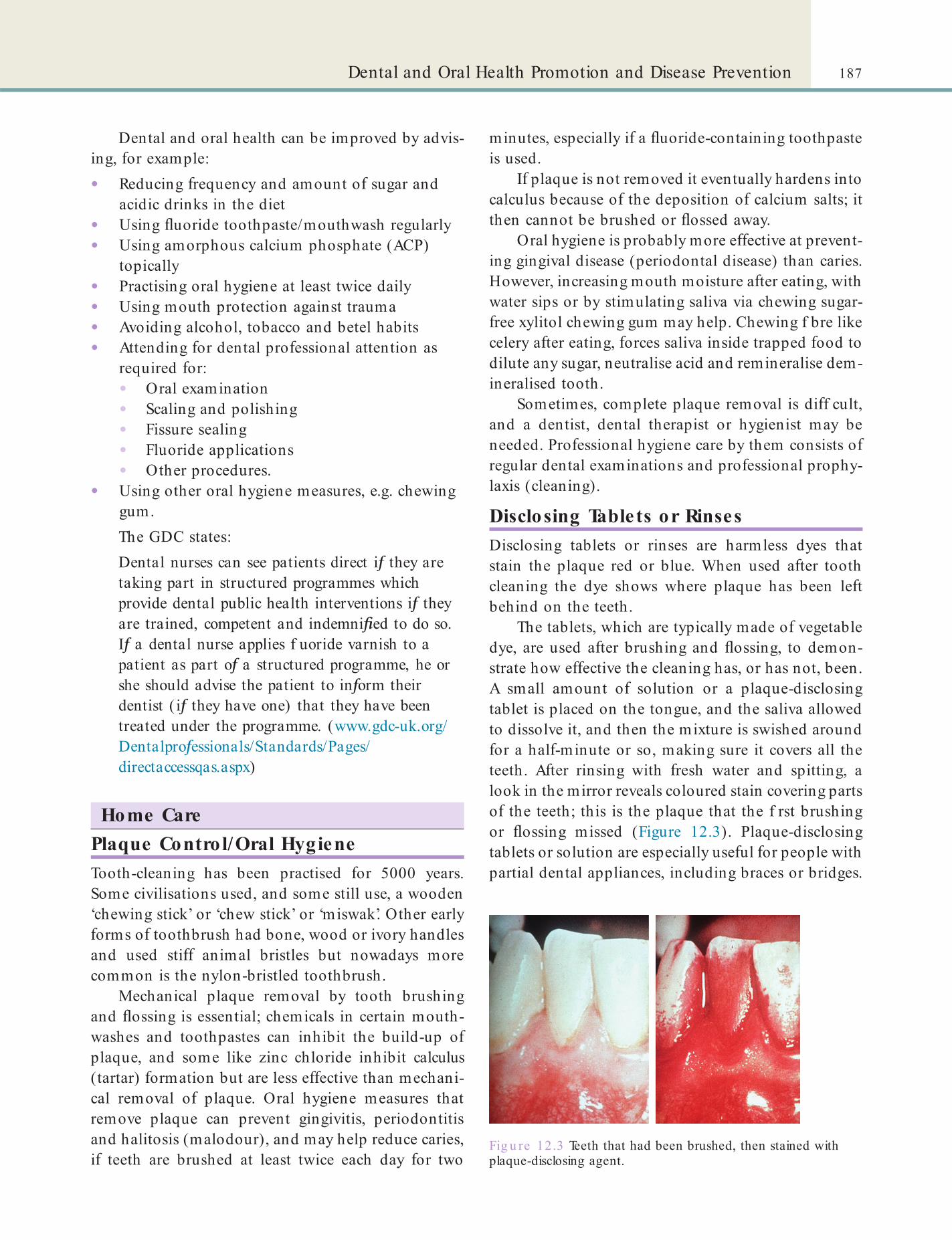
187Dental and Oral Health Promotion and Disease Prevention
minutes, especially i a uoride-containing toothpaste is used.
I plaque is not removed it eventually hardens into calculus because o the deposition o calcium salts; it then cannot be brushed or ossed away.
Oral hygiene is probably more e ective at prevent-ing gingival disease (periodontal disease) than caries. However, increasing mouth moisture a ter eating, with water sips or by stimulating saliva via chewing sugar- ree xylitol chewing gum may help. Chewing f bre like celery a ter eating, orces saliva inside trapped ood to dilute any sugar, neutralise acid and remineralise dem-ineralised tooth.
Sometimes, complete plaque removal is di f cult, and a dentist, dental therapist or hygienist may be needed. Pro essional hygiene care by them consists o regular dental examinations and pro essional prophy-laxis (cleaning).
Disclosing Table ts or RinsesDisclosing tablets or rinses are harmless dyes that stain the plaque red or blue. When used a ter tooth cleaning the dye shows where plaque has been le t behind on the teeth.
The tablets, which are typically made o vegetable dye, are used a ter brushing and ossing, to demon-strate how e ective the cleaning has, or has not, been. A small amount o solution or a plaque-disclosing tablet is placed on the tongue, and the saliva allowed to dissolve it, and then the mixture is swished around or a hal -minute or so, making sure it covers all the teeth. A ter rinsing with resh water and spitting, a look in the mirror reveals coloured stain covering parts o the teeth; this is the plaque that the f rst brushing or ossing missed (Figure 12.3). Plaque-disclosing tablets or solution are especially use ul or people with partial dental appliances, including braces or bridges.
Dental and oral health can be improved by advis-ing, or example:
• Reducing requency and amount o sugar and acidic drinks in the diet
• Using uoride toothpaste/mouthwash regularly• Using amorphous calcium phosphate (ACP)
topically• Practising oral hygiene at least twice daily• Using mouth protection against trauma• Avoiding alcohol, tobacco and betel habits• Attending or dental pro essional attention as
required or:• Oral examination• Scaling and polishing• Fissure sealing• Fluoride applications• Other procedures.
• Using other oral hygiene measures, e.g. chewing gum.The GDC states:Dental nurses can see patients direct i they are taking part in structured programmes which provide dental public health interventions i they are trained, competent and indemni ed to do so. I a dental nurse applies f uoride varnish to a patient as part o a structured programme, he or she should advise the patient to in orm their dentist ( i they have one) that they have been treated under the programme. (www.gdc-uk.org/Dentalpro essionals/Standards/Pages/directaccessqas.aspx)
Home CarePlaque Control/Oral HygieneTooth-cleaning has been practised or 5000 years. Some civilisations used, and some still use, a wooden ‘chewing stick’ or ‘chew stick’ or ‘miswak’. Other early orms o toothbrush had bone, wood or ivory handles and used sti animal bristles but nowadays more common is the nylon-bristled toothbrush.
Mechanical plaque removal by tooth brushing and ossing is essential; chemicals in certain mouth-washes and toothpastes can inhibit the build-up o plaque, and some like zinc chloride inhibit calculus (tartar) ormation but are less e ective than mechani-cal removal o plaque. Oral hygiene measures that remove plaque can prevent gingivitis, periodontitis and halitosis (malodour), and may help reduce caries, i teeth are brushed at least twice each day or two
Fig u re 12.3 Teeth that had been brushed, then stained with plaque-disclosing agent.
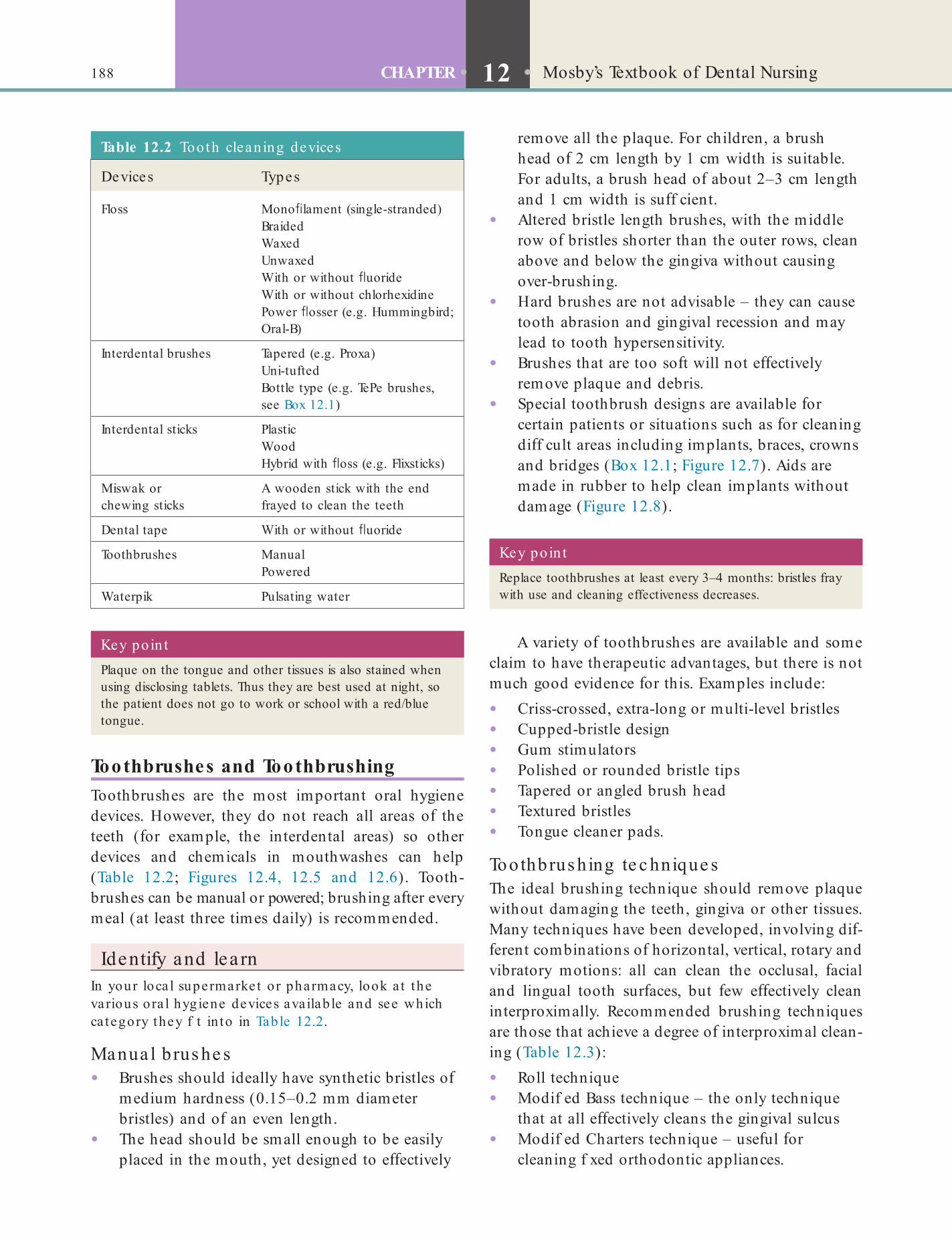
188 CHAPTER • 12 • Mosby’s Textbook of Dental Nursing
Key poin tPlaque on the tongue and other tissues is also stained when using disclosing tablets. Thus they are best used at night, so the patient does not go to work or school with a red/blue tongue.
Table 12.2 Tooth clean ing devices
Devices Types
Floss Mono lament (single-stranded)BraidedWaxedUnwaxedWith or without uorideWith or without chlorhexidinePower osser (e.g. Hummingbird; Oral-B)
Interdental brushes Tapered (e.g. Proxa)Uni-tuftedBottle type (e.g. TePe brushes, see Box 12.1)
Interdental sticks PlasticWoodHybrid with oss (e.g. Flixsticks)
Miswak or chewing sticks
A wooden stick with the end frayed to clean the teeth
Dental tape With or without uoride
Toothbrushes ManualPowered
Waterpik Pulsating water
Toothbrushes and ToothbrushingToothbrushes are the most important oral hygiene devices. However, they do not reach all areas o the teeth ( or example, the interdental areas) so other devices and chemicals in mouthwashes can help (Table 12.2; Figures 12.4, 12.5 and 12.6). Tooth-brushes can be manual or powered; brushing a ter every meal (at least three times daily) is recommended.
Identify and learnIn your loca l supermarke t o r pharmacy, look a t the various ora l hygiene devices availab le and see which ca tegory they f t in to in Table 12.2.
Manua l brus he s• Brushes should ideally have synthetic bristles o
medium hardness (0.15–0.2 mm diameter bristles) and o an even length.
• The head should be small enough to be easily placed in the mouth, yet designed to e ectively
remove all the plaque. For children, a brush head o 2 cm length by 1 cm width is suitable. For adults, a brush head o about 2–3 cm length and 1 cm width is su f cient.
• Altered bristle length brushes, with the middle row o bristles shorter than the outer rows, clean above and below the gingiva without causing over-brushing.
• Hard brushes are not advisable – they can cause tooth abrasion and gingival recession and may lead to tooth hypersensitivity.
• Brushes that are too so t will not e ectively remove plaque and debris.
• Special toothbrush designs are available or certain patients or situations such as or cleaning di f cult areas including implants, braces, crowns and bridges (Box 12.1; Figure 12.7). Aids are made in rubber to help clean implants without damage (Figure 12.8).
Key poin tReplace toothbrushes at least every 3–4 months: bristles fray with use and cleaning effectiveness decreases.
A variety o toothbrushes are available and some claim to have therapeutic advantages, but there is not much good evidence or this. Examples include:
• Criss-crossed, extra-long or multi-level bristles• Cupped-bristle design• Gum stimulators• Polished or rounded bristle tips• Tapered or angled brush head• Textured bristles• Tongue cleaner pads.
Toothbrus hing te c hnique sThe ideal brushing technique should remove plaque without damaging the teeth, gingiva or other tissues. Many techniques have been developed, involving di - erent combinations o horizontal, vertical, rotary and vibratory motions: all can clean the occlusal, acial and lingual tooth sur aces, but ew e ectively clean interproximally. Recommended brushing techniques are those that achieve a degree o interproximal clean-ing (Table 12.3):
• Roll technique• Modif ed Bass technique – the only technique
that at all e ectively cleans the gingival sulcus• Modif ed Charters technique – use ul or
cleaning f xed orthodontic appliances.

189Dental and Oral Health Promotion and Disease Prevention
Fig u re 12.4 (A) A selection of toothbrushes; (B) interdental aids; (C) dental oss; and (D) a model of the teeth that can be used to demonstrate brushing to patients.
Fig u re 12.5 TePe interdental brushes. Pink = 0.4 mm; orange = 0.45 mm; red = 0.5 mm; blue = 0.6 mm; yellow = 0.7 mm; green = 0.8 mm; purple = 1.1 mm; grey = 1.3 mm; black = 1.5 mm.
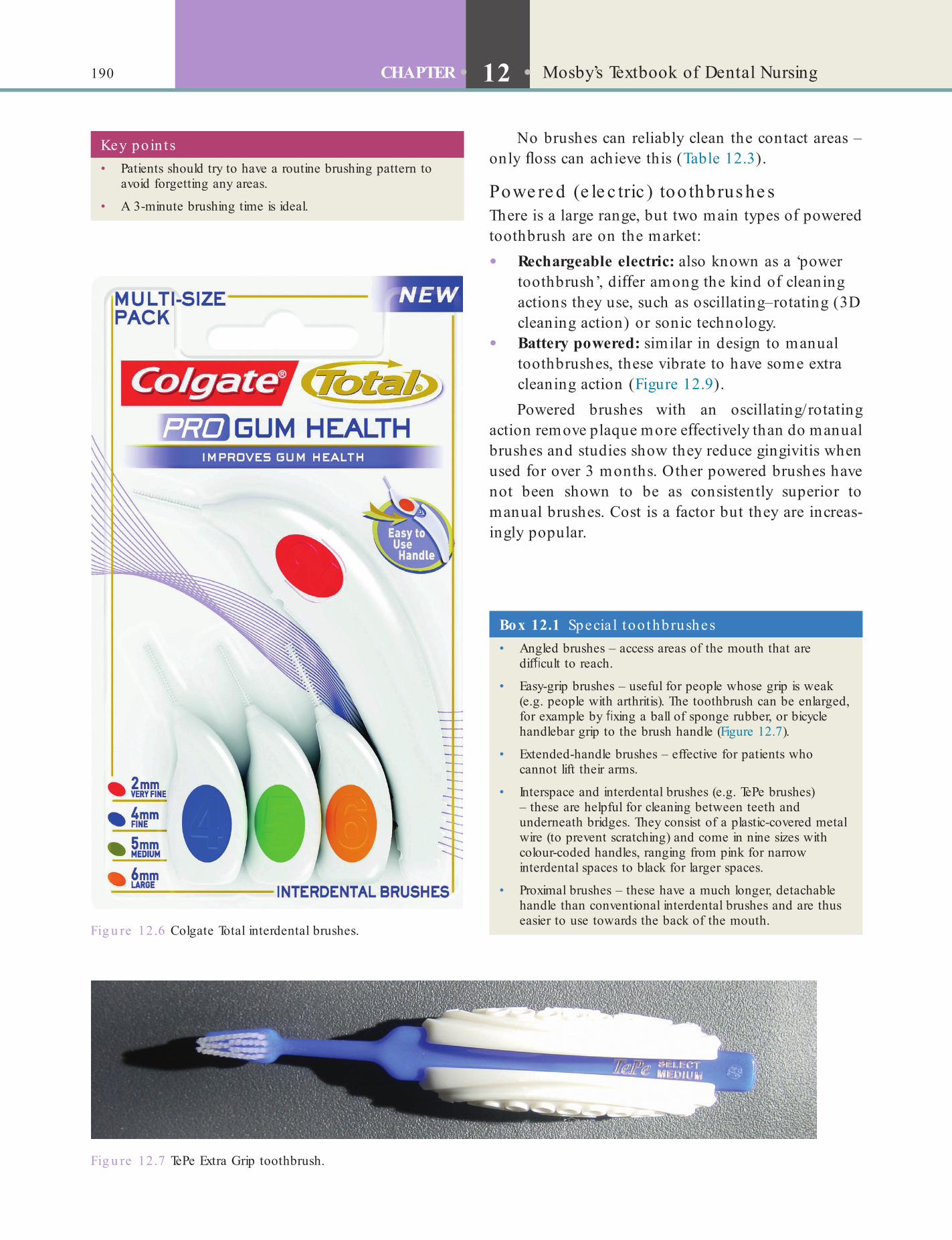
190 CHAPTER • 12 • Mosby’s Textbook of Dental Nursing
Fig u re 12.6 Colgate Total interdental brushes.
Fig u re 12.7 TePe Extra Grip toothbrush.
Box 12.1 Specia l toothbrushes• Angled brushes – access areas of the mouth that are
dif cult to reach.
• Easy-grip brushes – useful for people whose grip is weak (e.g. people with arthritis). The toothbrush can be enlarged, for example by xing a ball of sponge rubber, or bicycle handlebar grip to the brush handle (Figure 12.7).
• Extended-handle brushes – effective for patients who cannot lift their arms.
• Interspace and interdental brushes (e.g. TePe brushes) – these are helpful for cleaning between teeth and underneath bridges. They consist of a plastic-covered metal wire (to prevent scratching) and come in nine sizes with colour-coded handles, ranging from pink for narrow interdental spaces to black for larger spaces.
• Proximal brushes – these have a much longer, detachable handle than conventional interdental brushes and are thus easier to use towards the back of the mouth.
Key poin ts• Patients should try to have a routine brushing pattern to
avoid forgetting any areas.
• A 3-minute brushing time is ideal.
No brushes can reliably clean the contact areas – only oss can achieve this (Table 12.3).
Powe re d (e le c tric ) too thbrus he sThere is a large range, but two main types o powered toothbrush are on the market:
• Rechargeable electric: also known as a ‘power toothbrush’, di er among the kind o cleaning actions they use, such as oscillating–rotating (3D cleaning action) or sonic technology.
• Battery powered: similar in design to manual toothbrushes, these vibrate to have some extra cleaning action (Figure 12.9).Powered brushes with an oscillating/rotating
action remove plaque more e ectively than do manual brushes and studies show they reduce gingivitis when used or over 3 months. Other powered brushes have not been shown to be as consistently superior to manual brushes. Cost is a actor but they are increas-ingly popular.

191Dental and Oral Health Promotion and Disease Prevention
Fig u re 12.8 Interdental cleaners. (A) UltraDEX® Wire-Free Interdental Brushes with anti-bacterial coating by Periproducts. (B) Denti-Brush® Wire-Free Interdental Brushes by Periproducts. (C) Wisdom Clean Between Brushes.
A B C
Table 12.3 Toothbrush ing techniques
TechniqueIn it ia l posit ion o the brist les Brist le movements Comments
Bass (modi ed Bass)
45° to the tooth Vibrated backwards and forwards with a horizontal scrubbing movement
Cleans gingival sulcus but can be time consuming, dif cult to master and may cause mild trauma if not done properly with the correct brush. Useful for patients with gingival disease
Charters (modi ed Charters)
90° to the tooth Sweep occlusally, vibrate circularly Fairly good interproximal cleaning
Roll Apically on attached gingiva
Pressed onto the gingiva to make them spread out, then swept onto the tooth surface. Behind the anterior teeth, the brush is held vertically and pulled upwards or downwards
Particularly useful for people with healthy gingiva
Scrub 90° to tooth Horizontal brushing May cause abrasion
Fones 90° to tooth Circular brushing May cause abrasion
Leonard 90° to tooth Vertical brushing May damage gingivae
Powered toothbrushes
• are superior to manual toothbrushes or removing plaque, reducing staining and gingivitis, and avoiding gingival abrasions
• are o ten light and easy to hold, and ideal or people with limited manual dexterity
• may have timers and displays to guide brushing• may be designed or interspace use.
Identify and learnIn your local supermarket or pharmacy, look at the various powered toothbrushes available. How much more costly are they than the average manual toothbrush?
Toothbrus hing in c hildre nBrushing should start as soon as the f rst deciduous tooth erupts. The teeth should be brushed at least
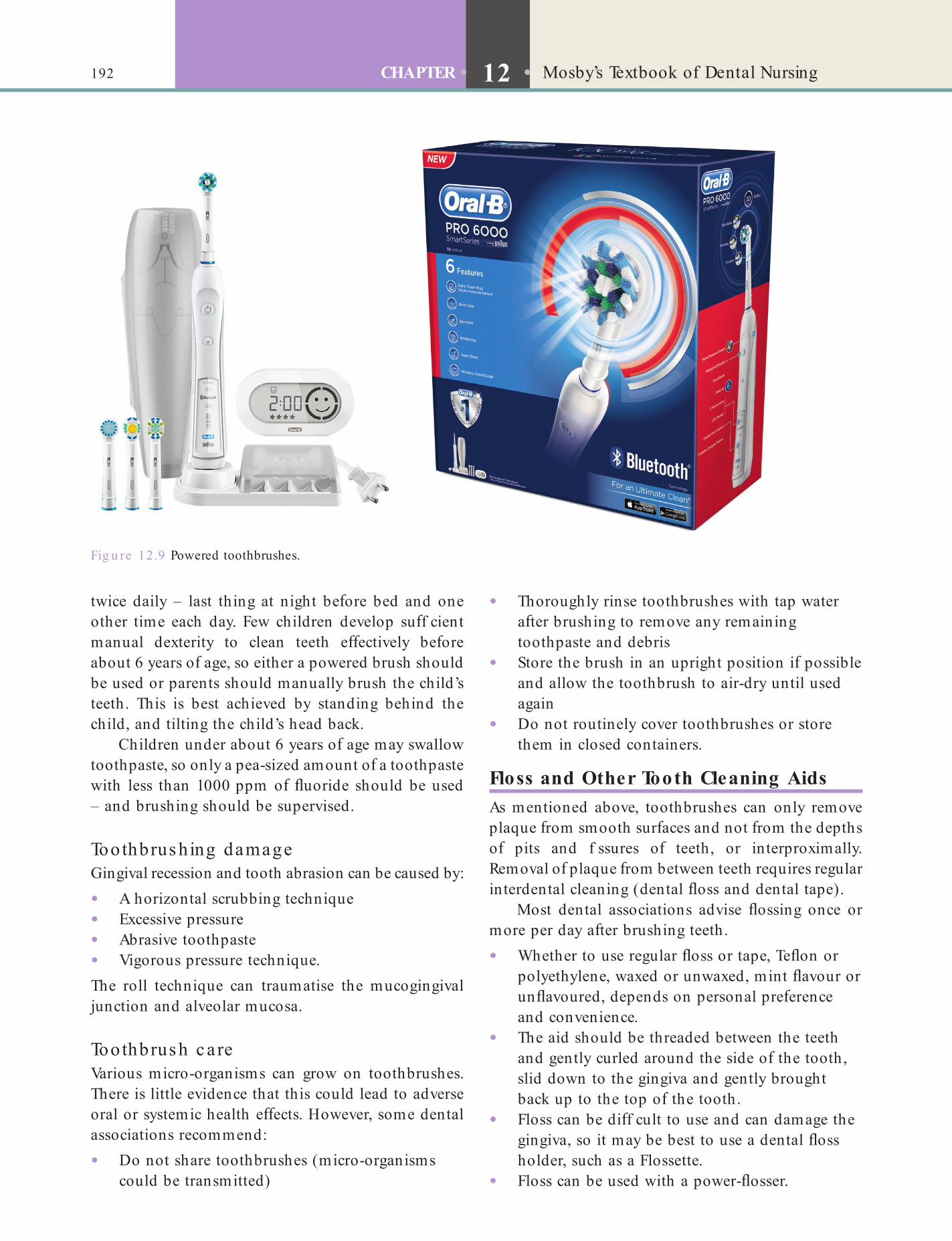
192 CHAPTER • 12 • Mosby’s Textbook of Dental Nursing
Fig u re 12.9 Powered toothbrushes.
twice daily – last thing at night be ore bed and one other time each day. Few children develop su f cient manual dexterity to clean teeth e ectively be ore about 6 years o age, so either a powered brush should be used or parents should manually brush the child’s teeth. This is best achieved by standing behind the child, and tilting the child’s head back.
Children under about 6 years o age may swallow toothpaste, so only a pea-sized amount o a toothpaste with less than 1000 ppm o uoride should be used – and brushing should be supervised.
Toothbrushing damageGingival recession and tooth abrasion can be caused by:
• A horizontal scrubbing technique• Excessive pressure• Abrasive toothpaste• Vigorous pressure technique.The roll technique can traumatise the mucogingival junction and alveolar mucosa.
Toothbrush c areVarious micro-organisms can grow on toothbrushes. There is little evidence that this could lead to adverse oral or systemic health e ects. However, some dental associations recommend:
• Do not share toothbrushes (micro-organisms could be transmitted)
• Thoroughly rinse toothbrushes with tap water a ter brushing to remove any remaining toothpaste and debris
• Store the brush in an upright position i possible and allow the toothbrush to air-dry until used again
• Do not routinely cover toothbrushes or store them in closed containers.
Floss and Other Tooth Cleaning AidsAs mentioned above, toothbrushes can only remove plaque rom smooth sur aces and not rom the depths o pits and f ssures o teeth, or interproximally. Removal o plaque rom between teeth requires regular interdental cleaning (dental oss and dental tape).
Most dental associations advise ossing once or more per day a ter brushing teeth.
• Whether to use regular oss or tape, Te on or polyethylene, waxed or unwaxed, mint avour or un avoured, depends on personal pre erence and convenience.
• The aid should be threaded between the teeth and gently curled around the side o the tooth, slid down to the gingiva and gently brought back up to the top o the tooth.
• Floss can be di f cult to use and can damage the gingiva, so it may be best to use a dental oss holder, such as a Flossette.
• Floss can be used with a power- osser.
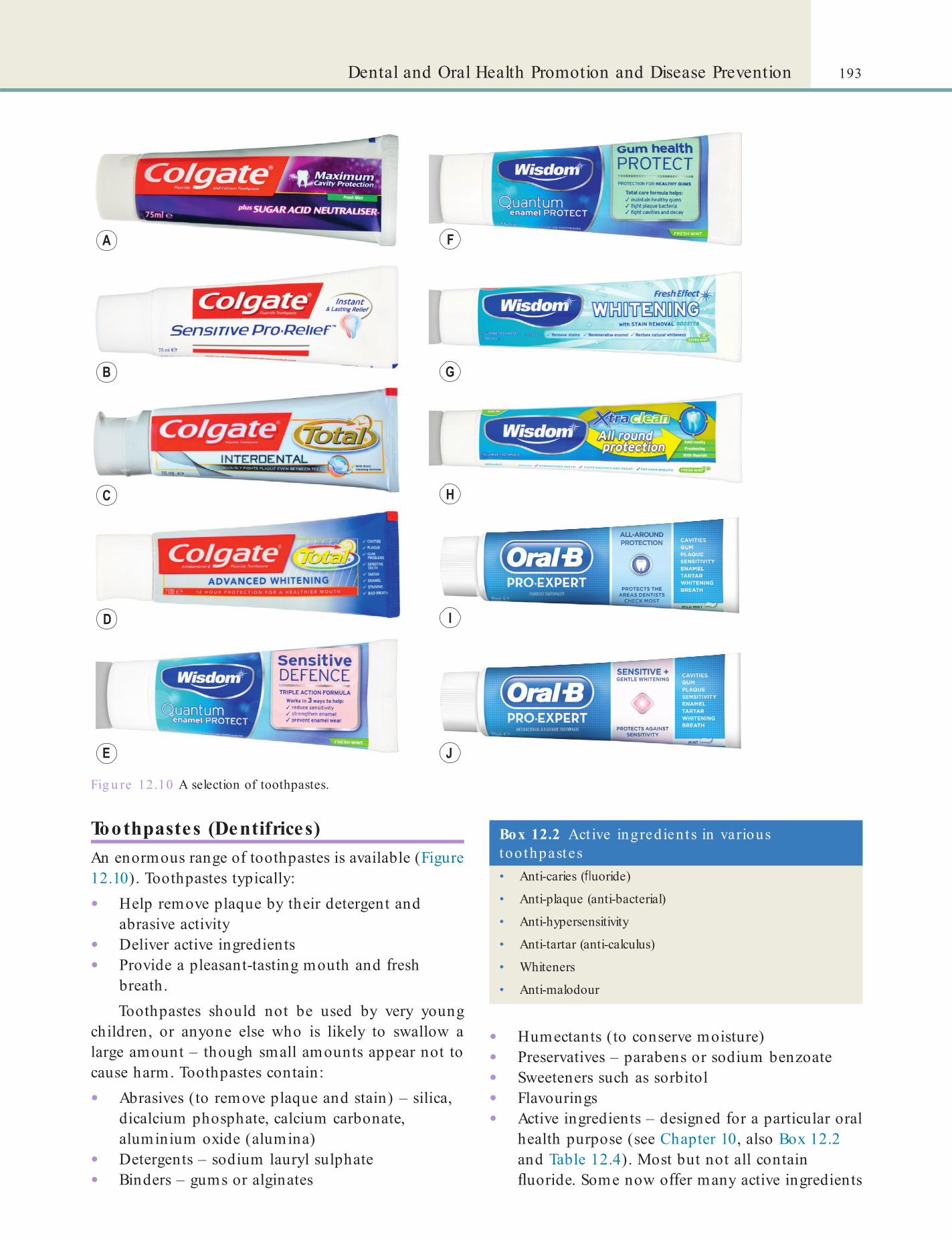
193Dental and Oral Health Promotion and Disease Prevention
Toothpastes (Dentifrices)An enormous range o toothpastes is available (Figure 12.10). Toothpastes typically:
• Help remove plaque by their detergent and abrasive activity
• Deliver active ingredients• Provide a pleasant-tasting mouth and resh
breath.Toothpastes should not be used by very young
children, or anyone else who is likely to swallow a large amount – though small amounts appear not to cause harm. Toothpastes contain:
• Abrasives (to remove plaque and stain) – silica, dicalcium phosphate, calcium carbonate, aluminium oxide (alumina)
• Detergents – sodium lauryl sulphate• Binders – gums or alginates
Fig u re 12.10 A selection of toothpastes.
Box 12.2 Act ive ingred ien ts in various too thpastes• Anti-caries ( uoride)
• Anti-plaque (anti-bacterial)
• Anti-hypersensitivity
• Anti-tartar (anti-calculus)
• Whiteners
• Anti-malodour
• Humectants (to conserve moisture)• Preservatives – parabens or sodium benzoate• Sweeteners such as sorbitol• Flavourings• Active ingredients – designed or a particular oral
health purpose (see Chapter 10, also Box 12.2 and Table 12.4). Most but not all contain uoride. Some now o er many active ingredients
194 CHAPTER • 12 • Mosby’s Textbook of Dental Nursing
Table 12.4 Examples o act ive princip les in too thbrush ing
Ant i-ca ries Ant ibacteria lsAnt i-hypersensit ivity Ant i-ta rta r Whiteners
Ant i-malodour Others
Calcium phosphate Chlorhexidine Potassium citrate Gantrez acid Benzalkonium chloride
Chlorhexidine Essential oils
Potassium uoride Fluorides Potassium chloride Potassium pyrophosphate
Calcium phosphates
Zinc citrate Permethol
Sodium uoride (NaF) 1450–1500 ppm (<600 ppm in children’s toothpaste)
Hexetidine Sodium citrate Zinc chloride Sodium bicarbonate
Triclosan Provitamin B5
Sodium mono uorophosphate
Hydrogen peroxide
Sodium uoride Zinc citrate Sodium tripolyphosphate
Zinc chloride Tocopherol
Stannous uoride Plant extracts Stannous uoride Calcium carbonate
Vitamin E
Xylitol Triclosan Strontium chloride KeratinUrea peroxide Strontium uoride PanthenolXylitolZinc citrate
and ‘total’ protection against caries, plaque and other issues.• Caries protection – uorides harden enamel
and protect against caries.• Plaque protection – triclosan (e.g. Total) and
chlorhexidine (e.g. Curaprox) have signif cant anti-plaque activity and protect against periodontitis without harm ul reactions.
• Anti-calculus activity – phosphates and phosphonates help prevent calculus but may produce un avourable mucosal reactions.
• Anti-sensitivity action: strontium uoride (e.g. Sensodyne), potassium nitrate and potassium oxalate (e.g. Protect), potassium nitrate (e.g. Emo orm), and potassium chloride help reduce tooth sensitivity.
• Anti-breath odour action: triclosan and a copolymer (e.g. Colgate Total toothpaste).
• Whitening: toothpastes o ten contain carbamide peroxide and can marginally lighten teeth, but the e ect is not great and ew have reliable supporting evidence or claims.
Mouthw ashes (Mouth Rinses)Many mouthwashes are available (Figure 12.11) and several have some e f cacy in plaque/gingivitis reduction.
• Mouthwashes in general are less use ul in oral health maintenance than are mechanical plaque-removing aids.
• Mouthwashes used by children may be accidentally swallowed, so alcohol-containing products should only be used under adult supervision.
• Mouthwashes containing alcohol may cause dryness, burning and, at least theoretically, might predispose to cancer. They can be swallowed and cause intoxication, so they should be avoided.
• Cosmetic mouthwashes are commercial Over-The-Counter (OTC) products that:• Help remove oral debris be ore or a ter
brushing• Decrease microbial counts in the mouth• Temporarily suppress halitosis• Re resh the mouth with a pleasant taste.
• Therapeutic mouthwashes have the benef ts o their cosmetic counterparts, plus an added active ingredient intended to help protect against some oral condition, usually caries, periodontal disease and/or halitosis. Therapeutic mouthwashes include those with:• Fluoride – these are e ective in caries
prevention (Figure 12.12): most contain sodium uoride. I taken excessively or swallowed they can lead over time to uoride toxicity.

195Dental and Oral Health Promotion and Disease Prevention
Fig u re 12.11 A selection of mouthwashes.
Fig u re 12.12 A uoridated mouthwash.
Fig u re 12.13 A chlorhexidine-containing mouthwash.
• Chlorhexidine (e.g. Corsodyl, Figure 12.13) – signif cantly controls plaque and periodontal disease, and also has some anti ungal activity. The 0.12% solution is as active in plaque control as is the 0.2%
solution. Chlorhexidine can a ect taste brie y and it binds tannins and thereby can cause superf cial staining i a person drinks co ee, tea, or red wine – but the staining can be cleaned by a dental pro essional.
• Triclosan (e.g. Plax) – signif cantly controls plaque and does not cause tooth-staining.

196 CHAPTER • 12 • Mosby’s Textbook of Dental Nursing
Table 12.5 Tooth riendliness o d i e ren t types o snacks and habit s
Tooth riendliness
Low High
Betel useBiscuitsCakesCarbonated drinks, including clear sodas, ginger aleCitrus fruit juicesCoffeePicklesRaisinsRed wineSports drinksSweets, especially toffee or sour sweetsTobacco use
CheeseCrispsNon-citrus fruitsNutsRaw vegetablesYogurt (plain)
• Essential oils – Listerine contains thymol, menthol, methyl salicylate and eucalyptus oils, helps control plaque and also has some anti ungal activity. It does not cause tooth staining and is available with or without alcohol.
• Cetylpyridinium chloride (CPC) – possibly helps control plaque and also has some anti ungal activity. It does not cause as much tooth staining.
Brushing the teeth a ter meals is recommended. Xylitol, a sugar that mouth bacteria do not convert into lactic acid, chewed in chewing gum, may help reduce the dental biof lm.
BabiesBreast- eeding is best or babies, and they should be weaned onto sugar- ree oods. From 6 months o age in ants should be introduced to drinking rom a cup and rom 1 year o age bottle- eeding should be dis-couraged. Drinks other than milk and water should not be given to pre-school children in eeding bottles, and should be given only at main meals. Foods should be ree o or very low in sugars other than those in resh milk and raw ruits or vegetables.
Older Children and Adults• Foods and drinks or snacking should be ree o
sugars. Because o the risk o erosion, as well as o caries, discourage requent consumption o carbonated, cola-type drinks and ruit juices such as grape ruit, apple, or orange. Children should pre erably drink water and milk. Wine and some other alcohol-containing drinks can also cause erosion.
• Chewing sugar- ree gum or cheese a ter meals can help by increasing salivary ow, as this helps wash o the acids in the plaque and on tooth sur aces.
• Most sugars in the diet come rom consuming processed oods and drinks.
• Consumption o sugary oods and drinks should be restricted to meal times and to a maximum o our times a day.
• Sugars (excluding those naturally present in whole ruit) should provide less than 10% o total energy in the diet or less than 60 g per person per day. Note that or young children this will be around 33 g per day.
• Potentially cariogenic oods and drinks include:• Sugar and chocolate con ectionery• Cakes and biscuits• Buns, pastries, ruit pies• Sponge puddings and other puddings• Table sugar• Sugared break ast cereals• Jams, preserves, honey• Ice cream• Fruit in syrup• Fresh ruit juices• Sugared so t drinks
Key poin tMost of the therapeutic mouthwashes can be safely used for up to one month (but alcohol-free preparations are preferred).
Dietary Contro lRef ned carbohydrates and sugars, particularly non-milk extrinsic sugars in items other than resh ruits and vegetables, are the major causes o dental caries (Chapter 10). For caries prevention, requency and type o sugar eaten is more important than the amount o sugar consumed. There ore, minimising snacking is recommended; it is important to limit and reduce the requency o consumption o sugary oods, such as restricting them to meal times. Also, chewy and sticky oods (such as dried ruit or to ee) tend to adhere to teeth longer, and, as a consequence, are best eaten as part o a meal.
Snacking is particularly bad since the oral pH remains low or prolonged periods, and certain oods are more cariogenic and sometimes erosive or likely to stain teeth than others (Table 12.5).
197Dental and Oral Health Promotion and Disease Prevention
FluorideFluoride can protect teeth against caries by:
• Strengthening the enamel• Inhibiting acid production by oral cariogenic
bacteria• Helping tooth remineralisation.
Wate r f uo rida tion• Water uoridation has consistently been shown
to be the most e ective, sa e means o preventing caries, resulting in a decrease o approximately 50%.
• In ormation about the uoride status o the water supplies can be obtained rom the water supplier in your area by quoting the residential postcode.
• The West Midlands is the most extensively uoridated area in the UK, ollowed by parts o the North East o England.
• In 2003 the UK law was changed to enable strategic health authorities to require water companies to uoridate water supplies as long as there was support rom the local population.
• The use o bottled non-f uoridated water in contrast may be linked with increased dental caries.
Find out moreWhat is the leve l o uoride in the drinking wate r supply in your a rea?
Fluoride s upple me nts• No uoride supplements are required i the
water supply contains more than 0.7 ppm o uoride. Where the water supply contains less than 0.3 ppm o uoride, children over the age o 6 months may benef t rom taking uoride supplements daily (see Table 12.6).
• While it is sa e or pregnant women to take uoride supplements, this has little impact on the uture dental health o the child.
• Fluoride supplements should be stored out o reach o children.
Key poin tThere are no proven health hazards from chewing gum use but it must always be disposed of carefully. It is banned in Singapore!
Sugar- ree medicinesThe National Pharmacy Association lea et ‘sugar in medicines’, and the ‘Delivering better oral health’ toolkit contain information about the sugar (fructose/glucose/sucrose) content of branded oral liquid medicines, both over-the-counter and prescription-only medicines. Products that do not contain these sugars are listed as being sugar-free. Preparations containing hydrogenated glucose syrup (Lycasin), maltitol, sorbitol or xylitol are also listed as sugar-free, since there is evidence that they are non-cariogenic.
Key po in tRemember that honey, fresh fruit juice and dried fruit all contain cariogenic sugars.
• Sugared, milk-based beverages• Sugar-containing alcoholic drinks• Dried ruits• Syrups and sweet sauces.
Key poin tExcess uoride may cause tooth discoloration ( uorosis); the risk of this is higher from the use of uoride supplements or topical uorides in water- uoridated areas.
Term to lea rnppm: Short for ‘parts per million’; 0.7 ppm is equivalent to 700 micrograms per litre of uoride.
Reduction of DemineralisationChewing can increase the production and release o saliva, which contains natural bu ers to reduce the lowering o mouth pH. Fluoride helps prevent decay by binding to the hydroxyapatite crystals in enamel making it more resistant to demineralisation and, thus, resistant to decay.
Chew ing GumChewing gum is traditionally made o a natural latex product called chicle, but many modern gums use rubber (polyisobutylene) instead. Chewing gum is available in avours such as mint, spearmint, winter-green, cinnamon and various ruits. Sugar- ree chewing gum may help contribute to:
• Oral hygiene improvement• Fresher breath• Remineralisation o tooth sur aces (those gums
containing calcium phosphate in particular)• Relie o dry mouth• Reduction o :
• Plaque• Calculus• Gingivitis• Caries.
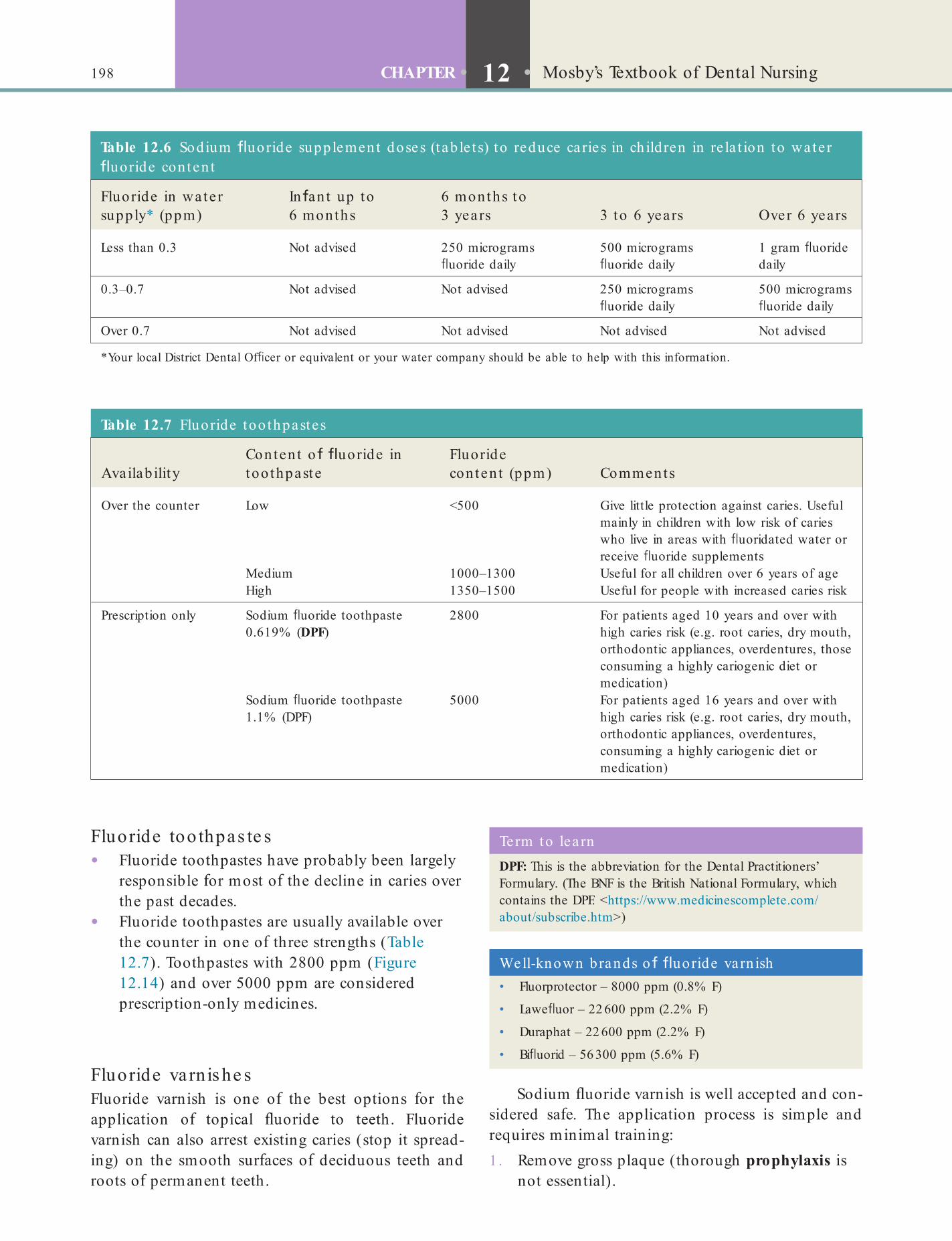
198 CHAPTER • 12 • Mosby’s Textbook of Dental Nursing
Table 12.6 Sodium uoride supplement doses (tab le t s) to reduce ca ries in ch ildren in re la t ion to wa te r uoride conten t
Fluoride in wa te r supply* (ppm)
In an t up to 6 months
6 months to 3 years 3 to 6 years Over 6 years
Less than 0.3 Not advised 250 micrograms uoride daily
500 micrograms uoride daily
1 gram uoride daily
0.3–0.7 Not advised Not advised 250 micrograms uoride daily
500 micrograms uoride daily
Over 0.7 Not advised Not advised Not advised Not advised
*Your local District Dental Of cer or equivalent or your water company should be able to help with this information.
Well-known brands o uoride varn ish• Fluorprotector – 8000 ppm (0.8% F)
• Lawe uor – 22 600 ppm (2.2% F)
• Duraphat – 22 600 ppm (2.2% F)
• Bi uorid – 56 300 ppm (5.6% F)
Term to lea rnDPF: This is the abbreviation for the Dental Practitioners’ Formulary. (The BNF is the British National Formulary, which contains the DPF. <https://www.medicinescomplete.com/about/subscribe.htm>)
Table 12.7 Fluoride too thpastes
Availab ilityConten t o uoride in too thpaste
Fluoride conten t (ppm) Comments
Over the counter Low <500 Give little protection against caries. Useful mainly in children with low risk of caries who live in areas with uoridated water or receive uoride supplements
Medium 1000–1300 Useful for all children over 6 years of ageHigh 1350–1500 Useful for people with increased caries risk
Prescription only Sodium uoride toothpaste 0.619% (DPF)
2800 For patients aged 10 years and over with high caries risk (e.g. root caries, dry mouth, orthodontic appliances, overdentures, those consuming a highly cariogenic diet or medication)
Sodium uoride toothpaste 1.1% (DPF)
5000 For patients aged 16 years and over with high caries risk (e.g. root caries, dry mouth, orthodontic appliances, overdentures, consuming a highly cariogenic diet or medication)
Fluoride too thpas te s• Fluoride toothpastes have probably been largely
responsible or most o the decline in caries over the past decades.
• Fluoride toothpastes are usually available over the counter in one o three strengths (Table 12.7). Toothpastes with 2800 ppm (Figure 12.14) and over 5000 ppm are considered prescription-only medicines.
Fluoride varnis he sFluoride varnish is one o the best options or the application o topical uoride to teeth. Fluoride varnish can also arrest existing caries (stop it spread-ing) on the smooth sur aces o deciduous teeth and roots o permanent teeth.
Sodium uoride varnish is well accepted and con-sidered sa e. The application process is simple and requires minimal training:1 . Remove gross plaque (thorough prophylaxis is
not essential).

199Dental and Oral Health Promotion and Disease Prevention
Term to lea rnProphylaxis: In the dental context, prophylaxis refers to cleaning the surface of teeth with a rotary brush to remove all plaque and ne deposits before treatment.
2 . Dry teeth with cotton rolls or a 3-in-1 syringe.3 . Use a microbrush to apply a small quantity o
uoride varnish to pits, f ssures and proximal sur aces o primary and permanent teeth.
4 . Advise the patient to avoid eating, drinking or toothbrushing or 30 minutes a ter application.
RemineralisationTeeth can be remineralised by uorides, or ACP (amor-phous calcium phosphate) (e.g. Tooth Mousse). The number o people developing caries has been declining or some years, mainly because o the protective e ect o uoride in toothpastes. Topical uoride is now more highly recommended than systemic intake such as by tablets or drops to protect the sur ace o the teeth. This may include a uoride mouthwash, paste or varnish.
Amorphous calcium phosphate (ACP), a deriva-tive o the milk protein casein and the active ingredi-ent o Tooth Mousse, works by neutralising acids and remineralising early enamel caries lesions.
Oral Health Procedures for ChildrenChildren under 3 Years• As soon as teeth erupt in the mouth, brush them
twice daily.• Brushing should occur twice daily – clean teeth
last thing at night be ore bed and at least one other time each day.
• Children need to be helped or supervised by an adult when brushing.
• Use no more than a smear o toothpaste (a thin f lm o paste covering less than three-quarters o the brush).
• Children must not be permitted to eat or lick toothpaste rom the tube.
• Use a toothpaste containing no less than 1000 ppm uoride. Family uoride toothpaste (1350–1500 ppm uoride) is indicated or maximum caries control or all children except those who cannot be prevented rom eating toothpaste.
Fig u re 12.14 A prescription only toothpaste because of the high uoride content.
Identify and learnIdent i y a microbrush in your workplace .
There is a small risk o allergy to the colophony component o Duraphat. There ore varnish applica-tion is contraindicated or children who have a history o allergies, including asthma. Duraphat is also con-traindicated in patients with ulcerative gingivitis and stomatitis.
Fluoride mouthwas he s and ge lsFluoride mouthwashes or rinses can be prescribed or patients aged 8 years and above, or daily or weekly use, in addition to twice-daily brushing with tooth-paste containing at least 1350 ppm uoride. They should be used at a di erent time to toothbrushing to maximise the topical e ect. Fluoride mouthwashes and gels are particularly recommended or people with a dry mouth (hyposalivation; see Chapter 12), who are otherwise particularly at risk o caries.
Key poin tUse of mouthwashes is reliant on patient compliance.
200 CHAPTER • 12 • Mosby’s Textbook of Dental Nursing
Find out moreThe ISO is the In te rna t iona l Organisa t ion o r Standardisa t ion . Visit the ISO websit e (www.iso .org/iso /home.h tm) to f nd out more about it s work.
Prevention of Other Dental or Oral Disease
Tooth ErosionDrinks, oods and medication with tooth erosive potential include:
• Drinks containing citric acid, e.g. orange, grape ruit, lemon, blackcurrant
• Carbonated drinks• Alcopops and designer drinks• Cider• White wine• Fruit teas (but not chamomile)• Some sports drinks that contain acid• Acidic resh ruit (lemons, oranges, grape ruit)
that are consumed with high requency• Pickles• Chewable vitamin C tablets, aspirin, some iron
preparations.Erosion prevention includes (Box 12.3):
• Reducing the amount and requency o intake o the above oods and drinks
• Avoiding gastric regurgitation and exposure to other erosive acids.
Tooth HypersensitivityHypersensitivity prevention includes avoiding abra-sion, which exposes the sensitive dentine at the neck o the tooth. The treatment includes:
• Rinsing with lots o water a ter brushing should be discouraged – spitting out excess toothpaste is pre erable.
• Fluoride varnish should be applied to teeth twice yearly.
Children betw een 3 and 6 Years• Children need to be helped or supervised by an
adult when brushing until at least 7 years o age and must not be permitted to eat or lick toothpaste rom the tube.
• Use no more than a pea-sized amount o toothpaste.
• Rinsing with lots o water a ter brushing should be discouraged – spitting out excess toothpaste is pre erable.
• Disclosing tablets can help to indicate areas that are being missed.
• Brushing is more e ective with a small-headed toothbrush with so t (ISO 8627:1987 standard 1–3), round-ended f laments, a compact, angled arrangement o long and short f laments, and a handle that is com ortable.
• Powered brushes with an oscillating/rotating action can be help ul.
• Parents should help with toothbrushing.
Children 7 Years and Older• Use a pea-sized amount or smear o uoridated
toothpaste – 1450 ppm (unless the child cannot be prevented rom eating toothpaste).
• Encourage spitting out toothpaste a ter brushing rather than rinsing.
• Fluoride varnish should be applied by the clinician three to our times yearly (2.2% NaF = 22 600 ppm uoride).
• The permanent molars should be f ssure sealed with resin sealant.
• Encourage use o toothpastes containing triclosan with copolymer to improve levels o plaque control or use toothpastes containing triclosan with zinc citrate. Chlorhexidine varnish can help.
• These children should clean interdentally using interdental brushes or oss.
• For those with active caries:• 8+ years – the clinician will prescribe daily
uoride rinse• 10+ years – the clinician will prescribe
2800 ppm uoride toothpaste• 18+ years – the clinician will consider
prescription o 5000 ppm uoride toothpaste.
Box 12.3 Advice to prevent e rosion progressing• Avoid frequent intake of acidic foods or drinks – keep them
to meal times, and use a straw for acidic drinks.
• Do not brush immediately after eating or drinking acidic food or drinks.
• Do not brush immediately after vomiting.
• Use toothpaste containing 1450 ppm uoride twice daily.
• Professional action that may be taken:
• Sensitive investigation of diet to identify the source of acid
• Investigation of habits that exacerbate effects of erosion
• Tailored, speci c advice for each individual patient.
201Dental and Oral Health Promotion and Disease Prevention
To apply sealants, the dental team involved will:
• Clean the tooth, using pumice• Wash and dry the tooth with a 3-in-1 syringe• Apply acid etchant solution or 30–60 seconds• Wash and dry again with a 3-in-1 syringe• Paint the sealant over pits and f ssures• Cure the sealant with a special blue halogen
light (some brands sel -cure chemically).Fissure sealants are especially indicated or people
at high risk or caries, such as:
• High caries rate in the deciduous dentition• Caries in f rst permanent molars• Teeth at high risk o caries (e.g. dens in dente
(a tooth within a tooth) ).Sealants must be checked regularly by dental clini-
cians since the sealants wear and degrade naturally over time but may last or f ve or more years and can then be replaced.
Preventive resin restorations are similar, but require some tooth preparation (drilling) f rst.
Find out moreUse Google image search to look o r images o ‘dens in den te ’.
Habit Cessation AdviceTobacco, alcohol and betel use are major risk actors associated with harm to oral and general health.
Dentists and their teams have an important role to play in smoking and tobacco use cessation advice (re er to the BDA/Health Development Agency publi-cation Helping Smokers Stop: A guide or the dental team, www.gserve.n ice.o rg.u k/ n icem ed ia / d o cu m en ts/helping_smokers_stop.pd ). Asking patients routinely about their tobacco use and assessing their motivation
• Modif cation o toothbrushing technique to ensure the gingiva are not damaged by excessive or inappropriate toothbrushing
• Application o desensitising agents, e.g. uoride varnish
• Daily use o a uoride mouthwash• Regular daily use o a desensitising toothpaste.
Agents apart rom uorides that may also be included in desensitisers include potassium nitrate, arginine and calcium carbonate, calcium sodium phosphosilicate, casein phosphopeptide – amorphous calcium phosphate, oxalates and glutaraldehyde.
Mouth TraumaDamage to the mouth can be avoided by:
• Care ul behaviour• Avoiding dangerous activities, violence and
assaults• Wearing protective wear such as a mouthguard
or a splint or:• Contact sports• Tooth grinding (bruxism)• Acid erosion• Radiotherapy.
CancerGiven that li estyle actors are the main risk actors or oral cancer, the clinician will take history o tobacco, betel and alcohol use. The patients will require advice and some contact details or services that will help them stop using these. Take a history o the diet and encourage them to adopt good dietary practices.
Patients should:
• Not smoke any orm o tobacco• Not use smokeless tobacco (e.g. betel, chewing
tobacco, gutkha)• Reduce alcohol consumption to moderate
(recommended) levels• Maintain good dietary practices and increase
ruit and vegetable intake to at least f ve portions per day.
Find out moreFind ou t more about o ra l cancer p revent ion a t : www.cancer.gov/cancertop ics/pdq/prevent ion /ora l/Pa t ient
Professional Care (Fissure Sealing)Tooth pits and f ssures are virtually impossible to clean thoroughly, even with vigorous toothbrushing. Thus
these areas can accumulate plaque and are sites prone to caries. Dental sealants are plastic resin coatings placed by the clinician, applied to the chewing sur- aces o molars to prevent ood rom being trapped inside pits and f ssures, preventing pit and f ssure caries. Sealants are usually applied as soon as the tooth erupts – and thus in children.
Term to lea rnAcid etchant: an acid solution that dissolves away bits of the enamel surface to create ‘pores’ into which the resin then ows to attach to the tooth. An etched tooth surface appears frosted and dull.
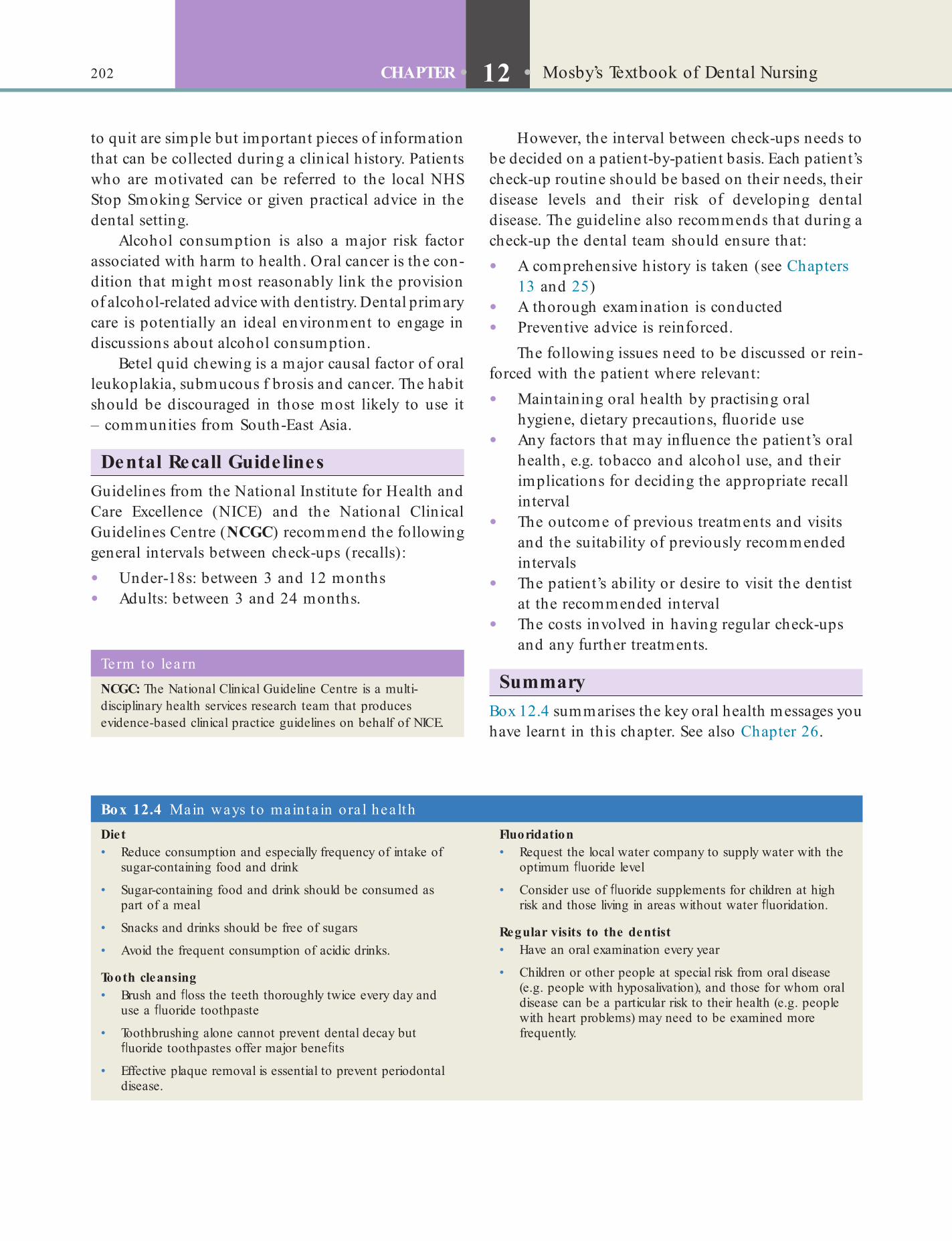
202 CHAPTER • 12 • Mosby’s Textbook of Dental Nursing
However, the interval between check-ups needs to be decided on a patient-by-patient basis. Each patient’s check-up routine should be based on their needs, their disease levels and their risk o developing dental disease. The guideline also recommends that during a check-up the dental team should ensure that:
• A comprehensive history is taken (see Chapters 13 and 25)
• A thorough examination is conducted• Preventive advice is rein orced.
The ollowing issues need to be discussed or rein- orced with the patient where relevant:
• Maintaining oral health by practising oral hygiene, dietary precautions, uoride use
• Any actors that may in uence the patient’s oral health, e.g. tobacco and alcohol use, and their implications or deciding the appropriate recall interval
• The outcome o previous treatments and visits and the suitability o previously recommended intervals
• The patient’s ability or desire to visit the dentist at the recommended interval
• The costs involved in having regular check-ups and any urther treatments.
SummaryBox 12.4 summarises the key oral health messages you have learnt in this chapter. See also Chapter 26.
to quit are simple but important pieces o in ormation that can be collected during a clinical history. Patients who are motivated can be re erred to the local NHS Stop Smoking Service or given practical advice in the dental setting.
Alcohol consumption is also a major risk actor associated with harm to health. Oral cancer is the con-dition that might most reasonably link the provision o alcohol-related advice with dentistry. Dental primary care is potentially an ideal environment to engage in discussions about alcohol consumption.
Betel quid chewing is a major causal actor o oral leukoplakia, submucous f brosis and cancer. The habit should be discouraged in those most likely to use it – communities rom South-East Asia.
Dental Recall GuidelinesGuidelines rom the National Institute or Health and Care Excellence (NICE) and the National Clinical Guidelines Centre (NCGC) recommend the ollowing general intervals between check-ups (recalls):
• Under-18s: between 3 and 12 months• Adults: between 3 and 24 months.
Term to lea rnNCGC: The National Clinical Guideline Centre is a multi-disciplinary health services research team that produces evidence-based clinical practice guidelines on behalf of NICE.
Box 12.4 Main ways to mainta in ora l hea lthDiet• Reduce consumption and especially frequency of intake of
sugar-containing food and drink
• Sugar-containing food and drink should be consumed as part of a meal
• Snacks and drinks should be free of sugars
• Avoid the frequent consumption of acidic drinks.
Tooth cleansing• Brush and oss the teeth thoroughly twice every day and
use a uoride toothpaste
• Toothbrushing alone cannot prevent dental decay but uoride toothpastes offer major bene ts
• Effective plaque removal is essential to prevent periodontal disease.
Fluoridation• Request the local water company to supply water with the
optimum uoride level
• Consider use of uoride supplements for children at high risk and those living in areas without water uoridation.
Regular visits to the dentist• Have an oral examination every year
• Children or other people at special risk from oral disease (e.g. people with hyposalivation), and those for whom oral disease can be a particular risk to their health (e.g. people with heart problems) may need to be examined more frequently.
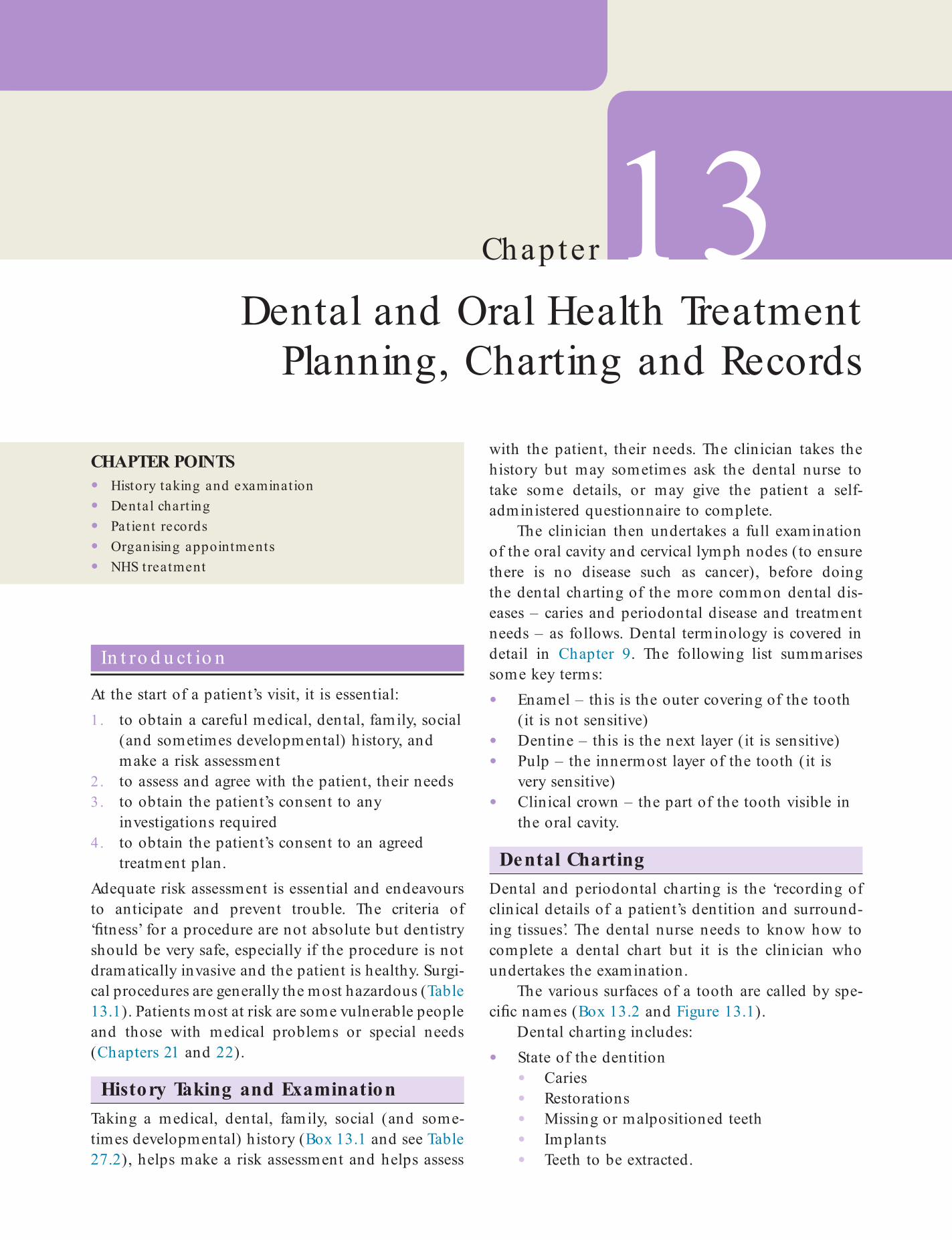
Chapter 13 Dental and Oral Health Treatment
Planning, Charting and Records
CHAPTER POINTS• History taking and examination• Dental charting• Patient records• Organising appointments• NHS treatment
with the patient, their needs. The clinician takes the history but may sometimes ask the dental nurse to take some details, or may give the patient a self-administered questionnaire to complete.
The clinician then undertakes a full examination of the oral cavity and cervical lymph nodes (to ensure there is no disease such as cancer), before doing the dental charting of the more common dental dis-eases – caries and periodontal disease and treatment needs – as follows. Dental terminology is covered in detail in Chapter 9. The following list summarises some key terms:
• Enamel – this is the outer covering of the tooth (it is not sensitive)
• Dentine – this is the next layer (it is sensitive)• Pulp – the innermost layer of the tooth (it is
very sensitive)• Clinical crown – the part of the tooth visible in
the oral cavity.
Dental ChartingDental and periodontal charting is the ‘recording of clinical details of a patient’s dentition and surround-ing tissues’. The dental nurse needs to know how to complete a dental chart but it is the clinician who undertakes the examination.
The various surfaces of a tooth are called by spe-ci c names (Box 13.2 and Figure 13.1).
Dental charting includes:
• State of the dentition• Caries• Restorations• Missing or malpositioned teeth• Implants• Teeth to be extracted.
In t ro d u ct io n
At the start of a patient’s visit, it is essential:1 . to obtain a careful medical, dental, family, social
(and sometimes developmental) history, and make a risk assessment
2 . to assess and agree with the patient, their needs3 . to obtain the patient’s consent to any
investigations required4 . to obtain the patient’s consent to an agreed
treatment plan.Adequate risk assessment is essential and endeavours to anticipate and prevent trouble. The criteria of ‘ tness’ for a procedure are not absolute but dentistry should be very safe, especially if the procedure is not dramatically invasive and the patient is healthy. Surgi-cal procedures are generally the most hazardous (Table 13.1). Patients most at risk are some vulnerable people and those with medical problems or special needs (Chapters 21 and 22).
History Taking and ExaminationTaking a medical, dental, family, social (and some-times developmental) history (Box 13.1 and see Table 27.2), helps make a risk assessment and helps assess

204 CHAPTER • 13 • Mosby’s Textbook of Dental Nursing
Box 13.2 Surfaces of the too th• Mesial – surface nearest the midline.
• Distal – surface furthest away from the midline.
• Occlusal – the biting surface of the premolars and molars.
• Incisal – the biting edge of the incisors and canines.
• Palatal – the surface nearest the palate.
• Lingual – the surface nearest the tongue.
• Buccal – the surface of the premolars and molars nearest the cheeks.
• Labial – the surface of the incisors and canines nearest the lips.
• Cervical – the part of any surface nearest the gingival margin.
Box 13.1 Essen t ia ls o f h istory taking• Personal details.
• The presenting complaint (PC).
• History of presenting complaint (HPC).
• Relevant medical history (RMH).
• Drug history.
• Social history.
• Family history.
Table 13.1 Opera t ive risk assessment and management
Risks increased by Risks reduced by
Increasing age Planning treatment properly
Medical treatments Non-invasive procedures
Surgical treatments Monitoring
Lengthy dental procedures Reassurance
Drug use – medication or recreational
• Periodontal problems• Pocket depth• Bleeding sites• Clinical attachment levels (see below)• Furcation (root) involvement• Mobile teeth.
• Other dental conditions• Erosion• Abrasion• Attrition• Developmental anomalies• Prostheses.
Term to lea rnFurcation: the area where the root of multi-rooted teeth divides into two or more roots.
Key po in tCharting provides a pictorial description of the health of a patient’s mouth.
Key poin tRemember while charting, the dental chart is opposite to your left and right.
Box 13.3 Why do denta l chart ing?A record of the condition of the patient’s teeth and surrounding tissues provides:
• Basic information for dental treatment planning
• Evidence in medico-legal cases
• Evidence in cases of identi cation of a deceased person.
It is vital therefore that dental charting is accurate.
Box 13.3 lists the main reasons for doing dental charting.
How to ChartThe teeth present in each quadrant of the mouth (see below) and their condition is recorded. You need to record these in relation to the patient’s left or right side. That is, with the patient sitting in the dental chair, a tooth on the patient’s left side (your right side) is recorded as on the ‘left’ on the chart.
Most dental charts consist of a diagrammatic rep-resentation of all the teeth, with surfaces shown as at (Figure 13.2).
Imagine the face is divided into four equal parts (quadrants):
• A line straight down the middle of the eyes, nose and chin (called the midline)
• A line straight across the middle between the maxilla and mandible
• The two upper quadrants include the maxilla• The two lower quadrants include the mandible.
Charting begins with noting the names and numbers of the teeth. Charting by hand is most ef -cient with the clinician performing the examination and the dental nurse recording the ndings on the
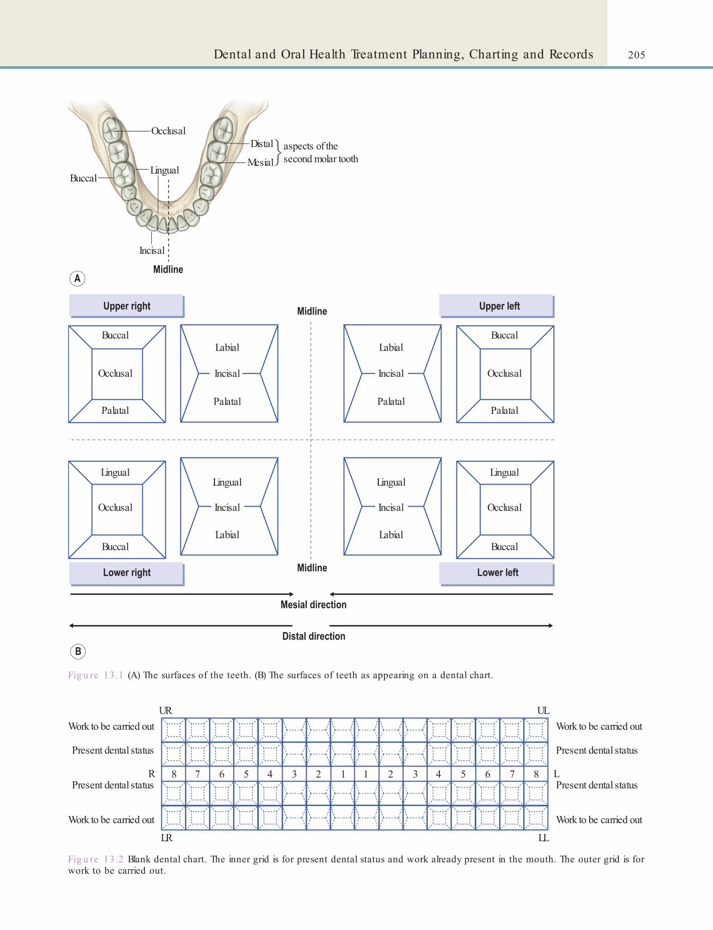
205Dental and Oral Health Treatment Planning, Charting and Records
Fig u re 13.1 (A) The surfaces of the teeth. (B) The surfaces of teeth as appearing on a dental chart.
Occlusal
BuccalLingual
Mesial
Distal
Incisal
aspects of thesecond molar tooth
Buccal
Buccal Buccal
Buccal
Occlusal
Occlusal
Occlusal
Occlusal
D
i
s
t
a
l
D
i
s
t
a
l
M
e
s
i
a
l
M
e
s
i
a
l
D
i
s
t
a
l
M
e
s
i
a
l
Palatal Palatal Palatal
Palatal
Labial
Labial Labial
Labial
Lingual LingualLingualLingual
Incisal
D
i
s
t
a
l
D
i
s
t
a
l
D
i
s
t
a
l
D
i
s
t
a
l
D
i
s
t
a
l
M
e
s
i
a
l
M
e
s
i
a
l
M
e
s
i
a
l
M
e
s
i
a
l
M
e
s
i
a
l
Incisal Incisal
Incisal
Fig u re 13.2 Blank dental chart. The inner grid is for present dental status and work already present in the mouth. The outer grid is for work to be carried out.
URWork to be carried out
Work to be carried out
Present dental status
Present dental status
Work to be carried out
Work to be carried out
Present dental status
Present dental status
UL
R L
LR LL
8 7 7 86 64 43 32 21 15 5
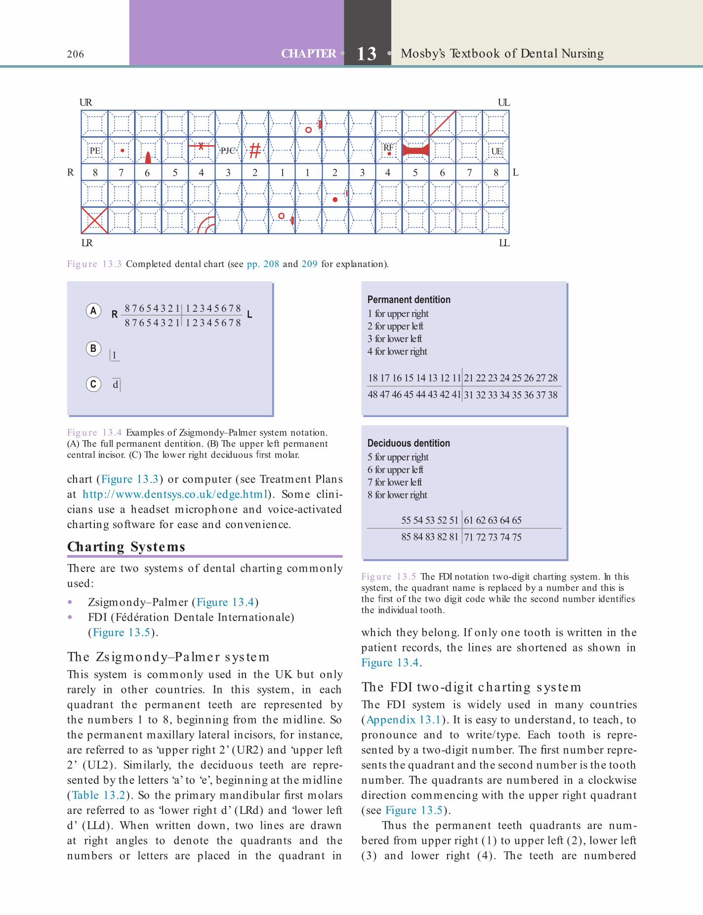
206 CHAPTER • 13 • Mosby’s Textbook of Dental Nursing
Fig u re 13.3 Completed dental chart (see pp. 208 and 209 for explanation).
UR UL
R L
LR LL
8
PE RF UEPJC
7 7 86 64 43 32 21 15 5
Fig u re 13.4 Examples of Zsigmondy–Palmer system notation. (A) The full permanent dentition. (B) The upper left permanent central incisor. (C) The lower right deciduous rst molar.
8 7 6 5 4 3 2 18 7 6 5 4 3 2 1
1
d
1 2 3 4 5 6 7 81 2 3 4 5 6 7 8
Fig u re 13.5 The FDI notation two-digit charting system. In this system, the quadrant name is replaced by a number and this is the rst of the two digit code while the second number identi es the individual tooth.
18 17 16 15 14 13 12 11
1 for upper right2 for upper left3 for lower left4 for lower right
48 47 46 45 44 43 42 4121 22 23 24 25 26 27 2831 32 33 34 35 36 37 38
55 54 53 52 51
5 for upper right6 for upper left7 for lower left8 for lower right
85 84 83 82 8161 62 63 64 6571 72 73 74 75
chart (Figure 13.3) or computer (see Treatment Plans at http:/ /www.dentsys.co.uk/edge.html). Some clini-cians use a headset microphone and voice-activated charting software for ease and convenience.
Charting SystemsThere are two systems of dental charting commonly used:
• Zsigmondy–Palmer (Figure 13.4)• FDI (Fédération Dentale Internationale)
(Figure 13.5).
The Zs igmondy–Palme r s ys te mThis system is commonly used in the UK but only rarely in other countries. In this system, in each quadrant the permanent teeth are represented by the numbers 1 to 8, beginning from the midline. So the permanent maxillary lateral incisors, for instance, are referred to as ‘upper right 2’ (UR2) and ‘upper left 2’ (UL2). Similarly, the deciduous teeth are repre-sented by the letters ‘a’ to ‘e’, beginning at the midline (Table 13.2). So the primary mandibular rst molars are referred to as ‘lower right d’ (LRd) and ‘lower left d’ (LLd). When written down, two lines are drawn at right angles to denote the quadrants and the numbers or letters are placed in the quadrant in
which they belong. If only one tooth is written in the patient records, the lines are shortened as shown in Figure 13.4.
The FDI two-dig it c harting s ys te mThe FDI system is widely used in many countries (Appendix 13.1). It is easy to understand, to teach, to pronounce and to write/ type. Each tooth is repre-sented by a two-digit number. The rst number repre-sents the quadrant and the second number is the tooth number. The quadrants are numbered in a clockwise direction commencing with the upper right quadrant (see Figure 13.5).
Thus the permanent teeth quadrants are num-bered from upper right (1) to upper left (2), lower left (3) and lower right (4). The teeth are numbered
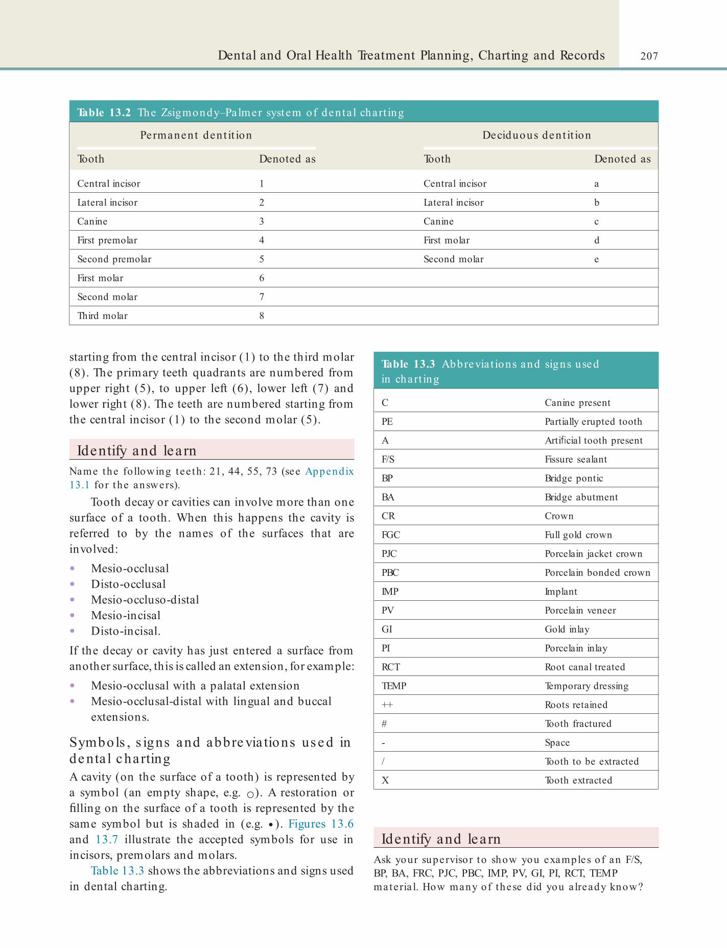
207Dental and Oral Health Treatment Planning, Charting and Records
Table 13.2 The Zsigmondy–Pa lmer system of den ta l chart ing
Permanent den t it ion Deciduous dent it ion
Tooth Denoted as Tooth Denoted as
Central incisor 1 Central incisor a
Lateral incisor 2 Lateral incisor b
Canine 3 Canine c
First premolar 4 First molar d
Second premolar 5 Second molar e
First molar 6
Second molar 7
Third molar 8
starting from the central incisor (1) to the third molar (8). The primary teeth quadrants are numbered from upper right (5), to upper left (6), lower left (7) and lower right (8). The teeth are numbered starting from the central incisor (1) to the second molar (5).
Identify and learnName the fo llowing tee th : 21, 44, 55, 73 (see Appendix 13.1 for the answers).
Tooth decay or cavities can involve more than one surface of a tooth. When this happens the cavity is referred to by the names of the surfaces that are involved:
• Mesio-occlusal• Disto-occlusal• Mesio-occluso-distal• Mesio-incisal• Disto-incisal.If the decay or cavity has just entered a surface from another surface, this is called an extension, for example:
• Mesio-occlusal with a palatal extension• Mesio-occlusal-distal with lingual and buccal
extensions.
Symbols , s igns and abbre via tions us e d in de nta l c hartingA cavity (on the surface of a tooth) is represented by a symbol (an empty shape, e.g. ○). A restoration or lling on the surface of a tooth is represented by the same symbol but is shaded in (e.g. ● ). Figures 13.6 and 13.7 illustrate the accepted symbols for use in incisors, premolars and molars.
Table 13.3 shows the abbreviations and signs used in dental charting.
Identify and learnAsk your supervisor to show you examples o f an F/S, BP, BA, FRC, PJC, PBC, IMP, PV, GI, PI, RCT, TEMP materia l. How many of these d id you a lready know?
Table 13.3 Abbrevia t ions and signs used in chart ing
C Canine present
PE Partially erupted tooth
A Arti cial tooth present
F/S Fissure sealant
BP Bridge pontic
BA Bridge abutment
CR Crown
FGC Full gold crown
PJC Porcelain jacket crown
PBC Porcelain bonded crown
IMP Implant
PV Porcelain veneer
GI Gold inlay
PI Porcelain inlay
RCT Root canal treated
TEMP Temporary dressing
++ Roots retained
# Tooth fractured
- Space
/ Tooth to be extracted
X Tooth extracted
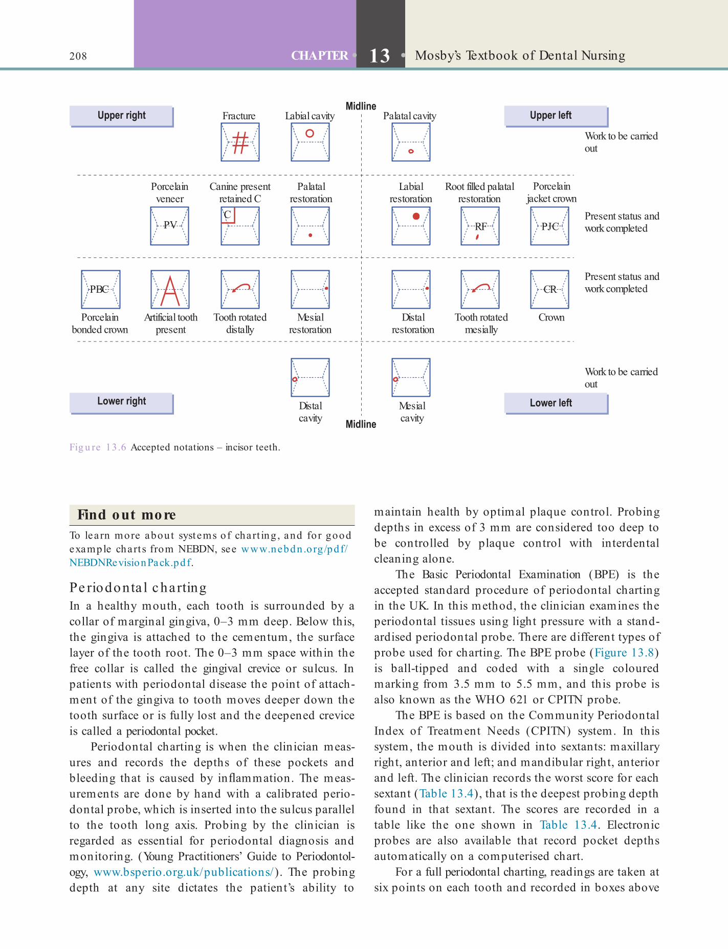
208 CHAPTER • 13 • Mosby’s Textbook of Dental Nursing
Fig u re 13.6 Accepted notations – incisor teeth.
PV
PBC
RF PJC
Fracture Labial cavity Palatal cavity
Porcelainveneer
Porcelainbonded crown
Artificial toothpresent
Tooth rotateddistally
Tooth rotatedmesially
CrownMesialrestoration
Distalrestoration
Distalcavity
Mesialcavity
Canine presentretained C
Palatalrestoration
Labialrestoration
Root filled palatalrestoration
Porcelainjacket crown
Work to be carriedout
Work to be carriedout
Present status andwork completed
Present status andwork completed
CR
C
Find out moreTo learn more about systems of chart ing , and for good example chart s from NEBDN, see www.nebdn .org /pdf/NEBDNRevisionPack.pdf.
Pe riodonta l c hartingIn a healthy mouth, each tooth is surrounded by a collar of marginal gingiva, 0–3 mm deep. Below this, the gingiva is attached to the cementum, the surface layer of the tooth root. The 0–3 mm space within the free collar is called the gingival crevice or sulcus. In patients with periodontal disease the point of attach-ment of the gingiva to tooth moves deeper down the tooth surface or is fully lost and the deepened crevice is called a periodontal pocket.
Periodontal charting is when the clinician meas-ures and records the depths of these pockets and bleeding that is caused by in ammation. The meas-urements are done by hand with a calibrated perio-dontal probe, which is inserted into the sulcus parallel to the tooth long axis. Probing by the clinician is regarded as essential for periodontal diagnosis and monitoring. (Young Practitioners’ Guide to Periodontol-ogy, www.bsperio.org.uk/publications/). The probing depth at any site dictates the patient’s ability to
maintain health by optimal plaque control. Probing depths in excess of 3 mm are considered too deep to be controlled by plaque control with interdental cleaning alone.
The Basic Periodontal Examination (BPE) is the accepted standard procedure of periodontal charting in the UK. In this method, the clinician examines the periodontal tissues using light pressure with a stand-ardised periodontal probe. There are different types of probe used for charting. The BPE probe (Figure 13.8) is ball-tipped and coded with a single coloured marking from 3.5 mm to 5.5 mm, and this probe is also known as the WHO 621 or CPITN probe.
The BPE is based on the Community Periodontal Index of Treatment Needs (CPITN) system. In this system, the mouth is divided into sextants: maxillary right, anterior and left; and mandibular right, anterior and left. The clinician records the worst score for each sextant (Table 13.4), that is the deepest probing depth found in that sextant. The scores are recorded in a table like the one shown in Table 13.4. Electronic probes are also available that record pocket depths automatically on a computerised chart.
For a full periodontal charting, readings are taken at six points on each tooth and recorded in boxes above
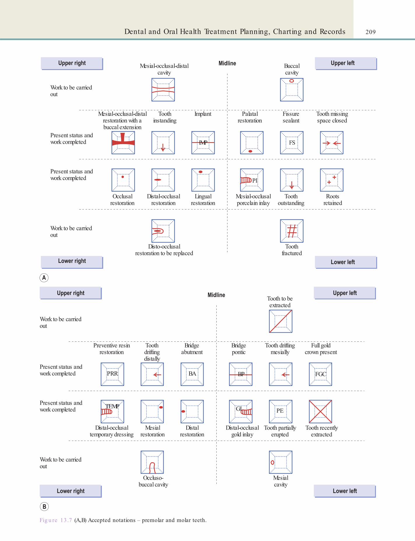
209Dental and Oral Health Treatment Planning, Charting and Records
Fig u re 13.7 (A,B) Accepted notations – premolar and molar teeth.
Work to be carriedout
Work to be carriedout
Mesial-occlusal-distalcavity
Mesial-occlusal-distalrestoration with abuccal extension
Occlusalrestoration
Distal-occlusalrestoration
Lingualrestoration
Mesial-occlusalporcelain inlay
Toothoutstanding
Rootsretained
Toothfractured
FS
PI
Disto-occlusalrestoration to be replaced
Toothinstanding
Implant Palatalrestoration
Fissuresealant
Tooth missingspace closed
Buccalcavity
Present status andwork completed
Present status andwork completed
IMP
Work to be carriedout
Work to be carriedout
Preventive resinrestoration
PRR BA BP FGC
Distal-occlusaltemporary dressing
TEMP
Mesialrestoration
Distalrestoration
Distal-occlusalgold inlay
GI
Tooth partiallyerupted
PE
Tooth recentlyextracted
Mesialcavity
Occluso-buccal cavity
Toothdriftingdistally
Bridgeabutment
Bridgepontic
Tooth driftingmesially
Tooth to beextracted
Full goldcrown present
Present status andwork completed
Present status andwork completed

210 CHAPTER • 13 • Mosby’s Textbook of Dental Nursing
Fig u re 13.8 (A) Naber’s and (B) BPE probes.
Table 13.4 BPE examinat ion codes
Code Signi cance
0 No bleeding or pockets detected
1 Bleeding on probing – no pockets >3.5 mm
2 Plaque retentive factors present – no pockets >3.5 mm
3 Pockets >3.5 mm but <5.5 mm in depth
4 Pockets >5.5 mm in depth
* Furcation involvement, or total attachment loss
A standard periodontal probe is necessary for the data collection to enable the clinician to monitor the progress of speci c sites: popular other probe types are the 10 mm Williams probe and the 15 mm UNC probe.
The ndings are recorded on a pre-printed chart, computer, of cial GDS periodontal chart, or modi ed dental record card (Figure 13.9), including as a minimum the:
• Probing depths• Gingival recession• Bleeding on probing• Plaque• Tooth mobility.
Patient RecordsIn NHS practices, clinical records are kept on form FP25. In other dental workplaces other suitable cards or computer programmes (e.g. Carestream, Curve, DentiMax, Dovetail, Software of Excellence) can be used.
Key poin tsPatient records should always be:
• Accurate
• Truthful
• Up to date (in correct date order)
• Treated with con dentiality (see Chapter 4).
Records must not be altered after any request for access has been received.
and below the teeth on the chart. This includes looking for four features:
• Bleeding• Plaque and calculus• Pocket formation• Furcation involvement.
Root furcation involvement is measured with Naber’s probe (Figure 13.8). It is graded as:
• 1 = probe enters furcation to one-third of its length
• 2 = probe enters furcation between one-third and two-thirds of its length
• 3 = probe completely penetrates the furcation.
Patient records should include the following:
• Patient details:• Last name• First name• Birth date• Address• Home, mobile and work telephone number• Practice or hospital reference number• Occupation• General medical practitioner• General dental practitioner (if patient is
being seen in hospital)• Medical history (this should be reviewed at
each recall appointment – see Chapters 25 and 27)
• Initial dental charting.

211Dental and Oral Health Treatment Planning, Charting and Records
Fig u re 13.9 Periodontal chart.
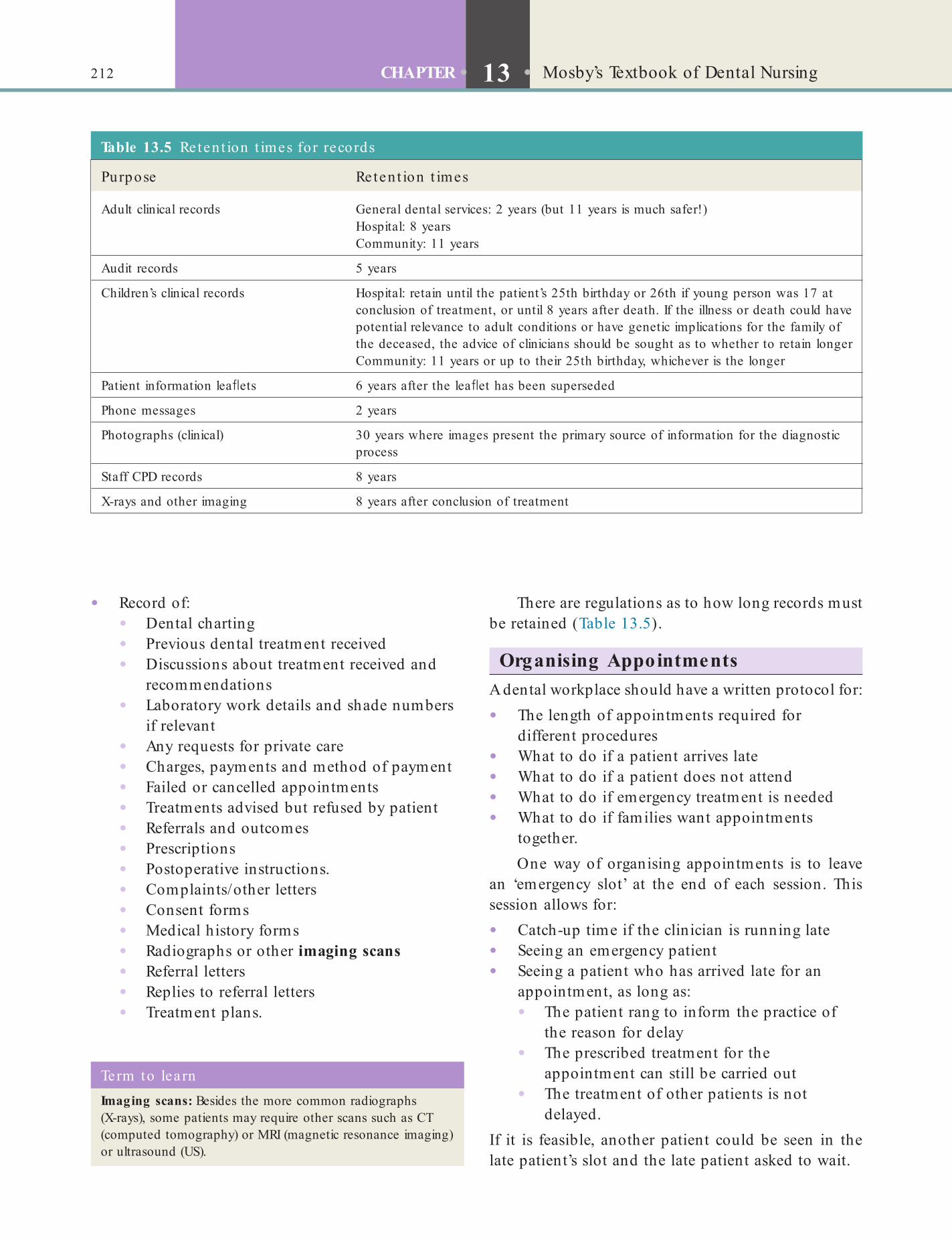
212 CHAPTER • 13 • Mosby’s Textbook of Dental Nursing
There are regulations as to how long records must be retained (Table 13.5).
Organising AppointmentsA dental workplace should have a written protocol for:
• The length of appointments required for different procedures
• What to do if a patient arrives late• What to do if a patient does not attend• What to do if emergency treatment is needed• What to do if families want appointments
together.One way of organising appointments is to leave
an ‘emergency slot’ at the end of each session. This session allows for:
• Catch-up time if the clinician is running late• Seeing an emergency patient• Seeing a patient who has arrived late for an
appointment, as long as:• The patient rang to inform the practice of
the reason for delay• The prescribed treatment for the
appointment can still be carried out• The treatment of other patients is not
delayed.If it is feasible, another patient could be seen in the late patient’s slot and the late patient asked to wait.
• Record of:• Dental charting• Previous dental treatment received• Discussions about treatment received and
recommendations• Laboratory work details and shade numbers
if relevant• Any requests for private care• Charges, payments and method of payment• Failed or cancelled appointments• Treatments advised but refused by patient• Referrals and outcomes• Prescriptions• Postoperative instructions.• Complaints/other letters• Consent forms• Medical history forms• Radiographs or other imaging scans• Referral letters• Replies to referral letters• Treatment plans.
Term to lea rnImaging scans: Besides the more common radiographs (X-rays), some patients may require other scans such as CT (computed tomography) or MRI (magnetic resonance imaging) or ultrasound (US).
Table 13.5 Reten t ion t imes for records
Purpose Reten t ion t imes
Adult clinical records General dental services: 2 years (but 11 years is much safer!)Hospital: 8 yearsCommunity: 11 years
Audit records 5 years
Children’s clinical records Hospital: retain until the patient’s 25th birthday or 26th if young person was 17 at conclusion of treatment, or until 8 years after death. If the illness or death could have potential relevance to adult conditions or have genetic implications for the family of the deceased, the advice of clinicians should be sought as to whether to retain longerCommunity: 11 years or up to their 25th birthday, whichever is the longer
Patient information lea ets 6 years after the lea et has been superseded
Phone messages 2 years
Photographs (clinical) 30 years where images present the primary source of information for the diagnostic process
Staff CPD records 8 years
X-rays and other imaging 8 years after conclusion of treatment
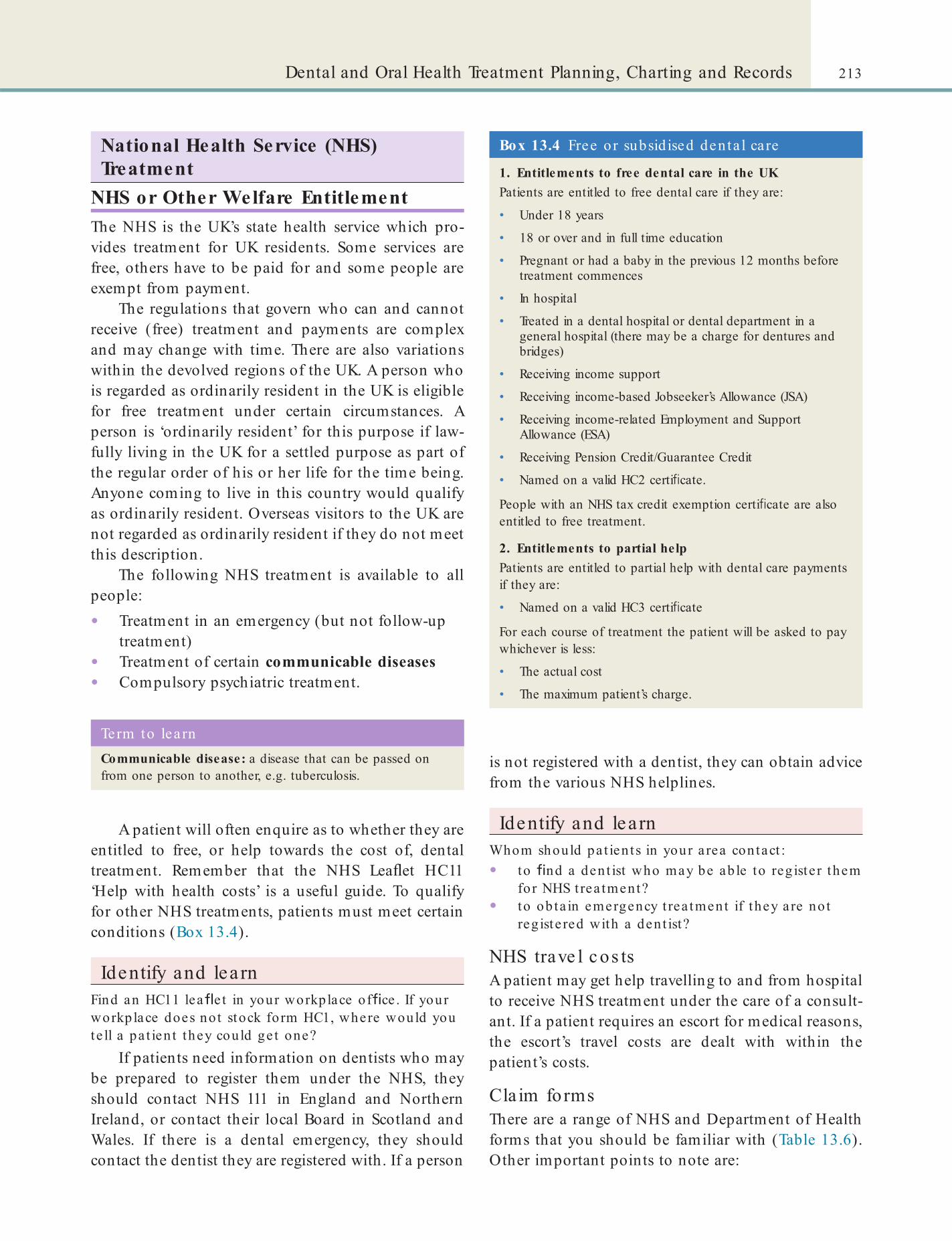
213Dental and Oral Health Treatment Planning, Charting and Records
National Health Service (NHS) Treatment
NHS or Other Welfare EntitlementThe NHS is the UK’s state health service which pro-vides treatment for UK residents. Some services are free, others have to be paid for and some people are exempt from payment.
The regulations that govern who can and cannot receive (free) treatment and payments are complex and may change with time. There are also variations within the devolved regions of the UK. A person who is regarded as ordinarily resident in the UK is eligible for free treatment under certain circumstances. A person is ‘ordinarily resident’ for this purpose if law-fully living in the UK for a settled purpose as part of the regular order of his or her life for the time being. Anyone coming to live in this country would qualify as ordinarily resident. Overseas visitors to the UK are not regarded as ordinarily resident if they do not meet this description.
The following NHS treatment is available to all people:
• Treatment in an emergency (but not follow-up treatment)
• Treatment of certain communicable diseases• Compulsory psychiatric treatment.
Term to lea rnCommunicable disease: a disease that can be passed on from one person to another, e.g. tuberculosis.
A patient will often enquire as to whether they are entitled to free, or help towards the cost of, dental treatment. Remember that the NHS Lea et HC11 ‘Help with health costs’ is a useful guide. To qualify for other NHS treatments, patients must meet certain conditions (Box 13.4).
Identify and learnFind an HC11 lea e t in your workplace of ce . If your workplace does not stock form HC1, where would you te ll a pa t ien t they could ge t one?
If patients need information on dentists who may be prepared to register them under the NHS, they should contact NHS 111 in England and Northern Ireland, or contact their local Board in Scotland and Wales. If there is a dental emergency, they should contact the dentist they are registered with. If a person
Box 13.4 Free or subsid ised den ta l care
1. Entitlements to free dental care in the UKPatients are entitled to free dental care if they are:
• Under 18 years
• 18 or over and in full time education
• Pregnant or had a baby in the previous 12 months before treatment commences
• In hospital
• Treated in a dental hospital or dental department in a general hospital (there may be a charge for dentures and bridges)
• Receiving income support
• Receiving income-based Jobseeker’s Allowance (JSA)
• Receiving income-related Employment and Support Allowance (ESA)
• Receiving Pension Credit/Guarantee Credit
• Named on a valid HC2 certi cate.
People with an NHS tax credit exemption certi cate are also entitled to free treatment.
2. Entitlements to partial helpPatients are entitled to partial help with dental care payments if they are:
• Named on a valid HC3 certi cate
For each course of treatment the patient will be asked to pay whichever is less:
• The actual cost
• The maximum patient’s charge.
is not registered with a dentist, they can obtain advice from the various NHS helplines.
Identify and learnWhom should pa t ien ts in your a rea contact :• to nd a den t ist who may be ab le to reg iste r them
for NHS t rea tment?• to ob ta in emergency t rea tment if they a re no t
reg iste red with a den t ist?
NHS trave l c os tsA patient may get help travelling to and from hospital to receive NHS treatment under the care of a consult-ant. If a patient requires an escort for medical reasons, the escort’s travel costs are dealt with within the patient’s costs.
Cla im formsThere are a range of NHS and Department of Health forms that you should be familiar with (Table 13.6). Other important points to note are:
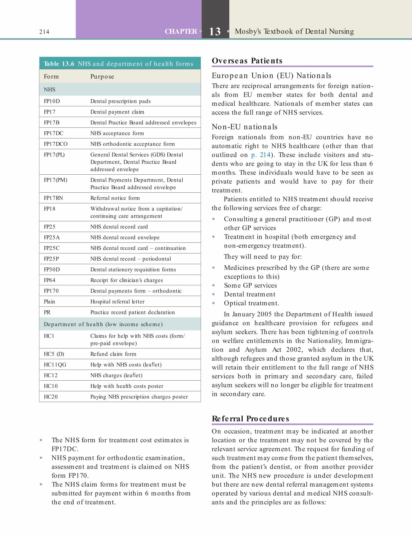
214 CHAPTER • 13 • Mosby’s Textbook of Dental Nursing
Overseas Patients
Europe an Union (EU) Nationa lsThere are reciprocal arrangements for foreign nation-als from EU member states for both dental and medical healthcare. Nationals of member states can access the full range of NHS services.
Non-EU na tionalsForeign nationals from non-EU countries have no automatic right to NHS healthcare (other than that outlined on p. 214). These include visitors and stu-dents who are going to stay in the UK for less than 6 months. These individuals would have to be seen as private patients and would have to pay for their treatment.
Patients entitled to NHS treatment should receive the following services free of charge:
• Consulting a general practitioner (GP) and most other GP services
• Treatment in hospital (both emergency and non-emergency treatment).They will need to pay for:
• Medicines prescribed by the GP (there are some exceptions to this)
• Some GP services• Dental treatment• Optical treatment.
In January 2005 the Department of Health issued guidance on healthcare provision for refugees and asylum seekers. There has been tightening of controls on welfare entitlements in the Nationality, Immigra-tion and Asylum Act 2002, which declares that, although refugees and those granted asylum in the UK will retain their entitlement to the full range of NHS services both in primary and secondary care, failed asylum seekers will no longer be eligible for treatment in secondary care.
Referral ProceduresOn occasion, treatment may be indicated at another location or the treatment may not be covered by the relevant service agreement. The request for funding of such treatment may come from the patient themselves, from the patient’s dentist, or from another provider unit. The NHS new procedure is under development but there are new dental referral management systems operated by various dental and medical NHS consult-ants and the principles are as follows:
Table 13.6 NHS and department o f hea lth forms
Form Purpose
NHS
FP10D Dental prescription pads
FP17 Dental payment claim
FP17B Dental Practice Board addressed envelopes
FP17DC NHS acceptance form
FP17DCO NHS orthodontic acceptance form
FP17(PL) General Dental Services (GDS) Dental Department, Dental Practice Board addressed envelope
FP17(PM) Dental Payments Department, Dental Practice Board addressed envelope
FP17RN Referral notice form
FP18 Withdrawal notice from a capitation/continuing care arrangement
FP25 NHS dental record card
FP25A NHS dental record envelope
FP25C NHS dental record card – continuation
FP25P NHS dental record – periodontal
FP30D Dental stationery requisition forms
FP64 Receipt for clinician’s charges
FP170 Dental payments form – orthodontic
Plain Hospital referral letter
PR Practice record patient declaration
Department of hea lth (low income scheme)
HC1 Claims for help with NHS costs (form/pre-paid envelope)
HC5 (D) Refund claim form
HC11QG Help with NHS costs (lea et)
HC12 NHS charges (lea et)
HC10 Help with health costs poster
HC20 Paying NHS prescription charges poster
• The NHS form for treatment cost estimates is FP17DC.
• NHS payment for orthodontic examination, assessment and treatment is claimed on NHS form FP170.
• The NHS claim forms for treatment must be submitted for payment within 6 months from the end of treatment.
215Dental and Oral Health Treatment Planning, Charting and Records
• Condition• Date of onset of condition• Treatment• Prognosis• Provider organisation and consultant providing
care• Provider organisation where care is requested• Reason for referral outside of existing portfolio• Any evidence base known of the intervention• Relevant clinical information and indication of
urgency• Information on any relevant pathology tests or
radiographs carried out• Whether the patient needs an interpreter.
Delivery of a two-week cancer outpatient waiting time is a priority under NHS Regulations that came into force in April 2013: www.nhs.uk/choiceintheNHS/Righ tsan dp ledges/ Waitin gtim es/ Docum en ts/ n h s-england-and-ccg-regulations.pdf (article 52).
• The general dental practitioner (GDP) should continue to play a major role in the appropriate use of resources.
• GDPs should apply the criteria as they would if managing the budget at a practice level, that is:• Is there an appropriate service available
within the contract portfolio?• Does the service provide value for money?• Is it of proven clinical effectiveness?• Does it t with current clinical priorities?
• Tertiary referrals (referrals from hospital clinicians) to non-contracted providers will require the support of a patient’s GDP.For a referral, try to give the following information:
• Patient details (within Caldicott and con dentiality guidelines [see Chapters 4 and 5]): date of birth; initials of patient; gender; daytime telephone number; NHS number
• Practice address
The FDI system of den ta l chart ingPermanent Deciduous
Upper right (1) Upper le ft (2) Upper righ t (5) Upper le ft (6)
Central incisor 11 Central incisor 21 Central incisor 51 Central incisor 61Lateral incisor 12 Lateral incisor 22 Lateral incisor 52 Lateral incisor 62Canine 13 Canine 23 Canine 53 Canine 63First premolar 14 First premolar 24 First molar 54 First molar 64Second premolar 15 Second premolar 25 Second molar 55 Second molar 65First molar 16 First molar 26Second molar 17 Second molar 27Third molar 18 Third molar 28
Lower right (4) Lower le ft (3) Lower righ t (8) Lower le ft (7)
Central incisor 41 Central incisor 31 Central incisor 81 Central incisor 71Lateral incisor 42 Lateral incisor 32 Lateral incisor 82 Lateral incisor 72Canine 43 Canine 33 Canine 83 Canine 73First premolar 44 First premolar 34 First molar 84 First molar 74Second premolar 45 Second premolar 35 Second molar 85 Second molar 75First molar 46 First molar 36Second molar 47 Second molar 37Third molar 48 Third molar 38(FDI = Federation Dentaire Internationale)
Appendix 13.1

This page intentiona lly le ft blank

Chapter 14 Drugs in Dentistry
CHAPTER POINTS• Role of the dental nurse• Factors affecting drug activity• Antimicrobials• Haemostatic agents• Drug safety and control• Cultural issues• NHS prescript ions
Role of the Dental NurseThe dental nurse is not permitted to prescribe drugs/medications but can hand oral health products to the patient. Controlled drugs, such as sedatives, need to be ordered via a prescription which is delivered to the pharmacy and a collection is arranged. A dental nurse who has approval to collect and sign for the controlled drugs can pick up the collection but this needs to be agreed and the pharmacist must be aware of these persons. Controlled drugs should be stored in a con-trolled drug cabinet and their use recorded by two professional members of staff, such as the dentist and dental nurse (see section on controlled drugs, p. 221).
Factors Affecting Drug ActivityWith all drugs and materials, double check the label and also ensure that the expiry date has not passed. Patient age, foods and drugs can affect the absorption or activity of various drugs. For example:
• Older individuals are generally more susceptible to drug effects; polypharmacy – the use of several medications – is common in this group (Figure 14.1).
• Children – are at risk from:• tetracyclines (these cause tooth staining)• aspirin and other non-steroidal analgesics
(because of the risk of a potentially lethal liver and brain disorder (Reye syndrome)
• codeine (some children have died postoperatively after being given codeine).
• Certain foods can either delay or speed up drug absorption or activity. For example:• Iron can reduce absorption of cipro oxacin• Calcium in dairy foods reduces tetracycline
absorption
In t ro d u ct io n
Drugs (medications) most commonly used in den-tistry are anaesthetics, sedatives, analgesics and anti-microbial drugs. The many different drugs and oral health products (discussed in Chapter 12) used in dentistry greatly bene t the patient. Some, however, can occasionally harm the patient, dental staff or the environment. There is guidance available from several sources, for example, Drug Prescribing for Dentistry: Dental Clinical Guidance (Scottish Dental Clinical Effectiveness Programme, 2011: www.sdcep.org.uk/? o=2334).
The hazards associated with materials commonly used in dentistry are discussed in Chapter 16. Tooth-pastes, uorides, anti-plaque and other mouthwashes, etc. are discussed in Chapter 12. Anaesthetic agents and analgesics are discussed in Chapter 15. Antiseptics and disinfectants (decontaminating agents) are dis-cussed in Chapter 7.
Antimicrobials, haemostatic agents and whitening agents are discussed here.
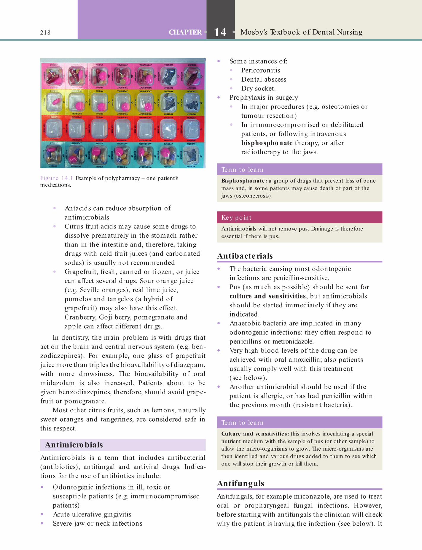
218 CHAPTER • 14 • Mosby’s Textbook of Dental Nursing
• Some instances of:• Pericoronitis• Dental abscess• Dry socket.
• Prophylaxis in surgery• In major procedures (e.g. osteotomies or
tumour resection)• In immunocompromised or debilitated
patients, or following intravenous bisphosphonate therapy, or after radiotherapy to the jaws.
• Antacids can reduce absorption of antimicrobials
• Citrus fruit acids may cause some drugs to dissolve prematurely in the stomach rather than in the intestine and, therefore, taking drugs with acid fruit juices (and carbonated sodas) is usually not recommended
• Grapefruit, fresh, canned or frozen, or juice can affect several drugs. Sour orange juice (e.g. Seville oranges), real lime juice, pomelos and tangelos (a hybrid of grapefruit) may also have this effect. Cranberry, Goji berry, pomegranate and apple can affect different drugs.
In dentistry, the main problem is with drugs that act on the brain and central nervous system (e.g. ben-zodiazepines). For example, one glass of grapefruit juice more than triples the bioavailability of diazepam, with more drowsiness. The bioavailability of oral midazolam is also increased. Patients about to be given benzodiazepines, therefore, should avoid grape-fruit or pomegranate.
Most other citrus fruits, such as lemons, naturally sweet oranges and tangerines, are considered safe in this respect.
AntimicrobialsAntimicrobials is a term that includes antibacterial (antibiotics), antifungal and antiviral drugs. Indica-tions for the use of antibiotics include:
• Odontogenic infections in ill, toxic or susceptible patients (e.g. immunocompromised patients)
• Acute ulcerative gingivitis• Severe jaw or neck infections
Fig u re 14.1 Example of polypharmacy – one patient’s medications.
Term to lea rnCulture and sensitivities: this involves inoculating a special nutrient medium with the sample of pus (or other sample) to allow the micro-organisms to grow. The micro-organisms are then identi ed and various drugs added to them to see which one will stop their growth or kill them.
Key poin tAntimicrobials will not remove pus. Drainage is therefore essential if there is pus.
Term to lea rnBisphosphonate: a group of drugs that prevent loss of bone mass and, in some patients may cause death of part of the jaws (osteonecrosis).
Antibacterials• The bacteria causing most odontogenic
infections are penicillin-sensitive.• Pus (as much as possible) should be sent for
culture and sensitivities, but antimicrobials should be started immediately if they are indicated.
• Anaerobic bacteria are implicated in many odontogenic infections: they often respond to penicillins or metronidazole.
• Very high blood levels of the drug can be achieved with oral amoxicillin; also patients usually comply well with this treatment (see below).
• Another antimicrobial should be used if the patient is allergic, or has had penicillin within the previous month (resistant bacteria).
AntifungalsAntifungals, for example miconazole, are used to treat oral or oropharyngeal fungal infections. However, before starting with antifungals the clinician will check why the patient is having the infection (see below). It

219Drugs in Dentistry
plasmin and kallikrein) or etamsylate (corrects abnormal adhesion of platelets) are needed.
Topical Haemostatic Agents• Haemostatic agents are often of animal
source (Table 14.1) and, unless they are of synthetic origin, may thus be contraindicated in some patients on cultural/religious grounds.
• Floseal and brin sealants are the most effective.
• Gelfoam swells, so a lot of the effect is a mechanical effect.
• Surgicel has a relative antimicrobial effect compared with other haemostatic agents.
• Avitene has the worst foreign body reaction of all of these particular agents.
Floseal, Avitene and brin sealants are expensive. Gelfoam, Surgicel and collagen sponges are relatively inexpensive.
Whitening AgentsTooth whitening is increasingly popular, with many products available to purchase on the high street (Figure 14.2). Tooth-whitening products include strips, gels and varnishes containing hydrogen perox-ide (H2O2) and are used for bleaching. H2O2 has been used for more than 70 years for oxygenating mouth-washes and to bleach teeth. The most common source of H2O2 used for whitening is carbamide peroxide which typically contains between 10% and 30% per-oxide (15% is recommended), roughly equivalent to 3–10% hydrogen peroxide.
is important to treat this underlying cause along with antifungal treatment.
AntiviralsMost antivirals, for example aciclovir, will be of maximum bene t if given early in the disease. They are indicated mainly for patients who have an immune defect.
Adverse Effects of Antimicrobials• Antibacterials predispose to candidosis (see
Chapter 10).• Tetracyclines are contraindicated in pregnancy
and children younger than 7–8 years. This is because they can cause tooth discoloration (see Chapter 10).
Antimicrobials and FoodsErythromycin, penicillins (some), rifampicin and tet-racyclines (except doxycycline) should be taken at least 30 minutes before food. This is because their absorption is otherwise delayed. Metronidazole may cause headaches if taken with alcohol.
Antimicrobial and Drug InteractionsMetronidazole, erythromycin, some antifungals such as miconazole, and occasionally amoxicillin, can enhance the action of warfarin, causing bleeding.
Haemostatic Agents• Haemostatic agents act to stop bleeding
(haemorrhage) – by acting at differing sites in the complex pathways of blood coagulation and brinolysis (Chapter 24).
• Most haemorrhage will cease with the use of topical haemostatic agents.
• Blood and blood products such as blood-clotting factors are required if there is serious bleeding. In the past, these were typically derived from human or animal material. This carried a potential risk of transmission of infections (e.g. HIV, hepatitis B virus infection, prion disease – see Chapter 25). Nowadays, drugs called recombinant factors are widely available, which avoid these risks and the religious objections to certain treatment (e.g. among Jehovah’s Witnesses).
• Occasionally drugs such as desmopressin (which promotes release of some blood-clotting factors), tranexamic acid (anti- brinolytic agent), aprotinin (inhibitor of proteolytic enzymes
Table 14.1 Some top ica l haemosta t ic agents
Agent Main const ituen t Orig in
Avitene Collagen Bovine
Colla-Cote Collagen Bovine
Floseal Thrombin Bovine
Gelfoam Gelatin Bovine
Helistat Collagen Bovine
Instat Collagen Bovine
Thrombinar Thrombin Bovine
Thrombogen Thrombin Bovine
Thrombostat Thrombin Bovine
Beriplast Fibrin Various
Cyclokapron Tranexamic acid Synthetic
Surgicel Cellulose Synthetic

220 CHAPTER • 14 • Mosby’s Textbook of Dental Nursing
Bleaching is not recommended in:
• Children under the age of 16• Pregnant or lactating women.
Proble ms with e xte rna l b le ac hing• Temporary soft tissue irritation and increased
tooth sensitivity, which disappear within 1–3 days of stopping or completing the whitening.
• Over-bleaching (‘over-white teeth’ or ‘hyperodonto-oxidation’).
• Chemical burns – this is a more serious problem and occurs if a high-concentration oxidising agent contacts unprotected tissues.
• Direct exposure of skin or eyes to hydrogen peroxide may cause irritation or burns, while ingestion may be irritating to the oesophagus and stomach.
Internal BleachingInternal bleaching is performed on teeth that are dis-coloured due to internal staining. This usually happens in teeth that have become non-vital from trauma or caries (see Chapter 10). Internal bleaching involves drilling a hole to the pulp chamber, cleaning and lling the root canal, and sealing a hydrogen peroxide gel into the pulp chamber for some days, and replacing this as needed (so called ‘walking-bleach’ technique).
Drug Safety and ControlDrugs are sometimes available ‘Over The Counter’ (OTC) from pharmacies or elsewhere but many are available only on prescription (POM: Prescription Only Medicines) in order to protect the public, since almost any drug may produce unwanted or unex-pected adverse reactions, some of which are life-threatening – such as anaphylaxis (Chapter 27). These reactions are often predictable. However, some are rarely predictable unless the person has previously reacted adversely.
Patients should be warned if serious adverse reac-tions are predictable and likely to occur (e.g. weight gain, hypertension and diabetes with systemic corti-costeroids). They should also be provided with the appropriate warning card to carry.
There are two methods of bleaching or whitening teeth:
• External• Internal.
External BleachingExternal bleaching can be done in two ways:
• Laser bleaching uses laser light energy to accelerate the bleaching using high-concentration agents (30% peroxide) in gels in the dental of ce.
• At-home whitening is usually done with low-concentration oxidising agents in a whitening strip or a thin mouthguard. These hold a relatively low concentration of the bleaching agent next to the teeth for long periods of several hours a day and they are usually used for from 5 to 14 days. During this period, the oxidising agent penetrates enamel porosities and oxidises inter-prismatic enamel stain deposits (dentine is also bleached).
Key poin ts• The effects of bleaching can last for several months, but
may vary depending on the patient’s lifestyle.
• Some teeth may lose the bleached effect with time (rebound effect).
Terms to learnEnamel porosities: gaps in the mineral structure (hydroxyapatite) of enamel.
Inter-prismatic: The mineral deposits in enamel are laid out in bundles called enamel prisms. The spaces between these prisms are called inter-prismatic spaces.
Fig u re 14.2 A teeth whitening kit.

221Drugs in Dentistry
used in medicines; Schedule 5 has a much lower level of control. A controlled drug is kept separate from other drugs, as there is a legal requirement to monitor and record all controlled drugs.
The real number of adverse drug reactions that occur is not known, and many adverse reactions are probably not, at present, recognised as drug-related.
Adverse drug reactions should be reported using the ‘Yellow card’ system (Figure 14.3) either via hard copy or online (see https:/ /yellowcard.mhra.gov.uk/).
Contro lled DrugsSome prescription only medicines are controlled under the Misuse of Drugs Regulations 2001, as amended by the Controlled Drugs (Supervision of Management and Use) Regulations 2006, and are called controlled medicines or controlled drugs (CDs). Strict legal controls apply to controlled drugs to prevent them being misused, and prevent people obtaining them illegally or causing harm. These legal controls govern how controlled drugs are stored, pro-duced, supplied and prescribed.
Controlled drugs are classi ed (by law) based on their bene t when used in medical treatment and their harm if misused. The Misuse of Drugs regulations include ve schedules that classify all controlled medi-cines and drugs. Schedule 1 has the highest level of control, but drugs in this group are virtually never
Fig u re 14.3 Yellow card system.
Classi ca t ion schedules of cont ro lled drugs• Schedule 1 drugs are recreational drugs not meant for
medical use (e.g. cocaine). Practitioners and pharmacists may not lawfully possess Schedule 1 drugs except under licence.
• Schedule 2 drugs also have a potential for misuse but are used as medicines also (e.g. pethidine and morphine). Record keeping is a legal requirement for Schedule 2 drugs but currently these are rarely used except in hospital practice.
• Schedule 3 drugs have decreasing potential for misuse but are useful medicines (e.g. midazolam, pentazocine and temazepam). They are exempt from safe custody requirements and can be stored on the open dispensary shelf except for temazepam, buprenorphine and diethylpropion, which must be stored in a locked CD receptacle. The government has now placed tramadol in Schedule 3 but with exemption from the safe custody requirements.
• Schedule 4 drugs are subject to minimal control. They include most of the benzodiazepines (except temazepam and midazolam, which are in Schedule 3) and zolpidem. Possession is an offence without an appropriate prescription. Possession by practitioners and pharmacists acting in their professional capacities is authorised.
• Schedule 5 drugs include preparations of certain controlled drugs (e.g. codeine, pholcodine, morphine) which are exempt from full control when present in medicinal products of low strengths, as their risk of misuse is reduced. There are no restriction on the possession, administration or destruction of these preparations and safe custody regulations do not apply. A practitioner, pharmacist or a person holding an appropriate licence may manufacture or compound any CD in Schedule 5, they are therefore exempt from virtually all CD requirements other than that invoices must be kept for a minimum of two years.
A controlled drug book must contain and record the following information:
• Date of receipt• Quantity received• Name and concentration of drug• Date of supply• Name of patient• Signature of clinician• Signature of witness.Accountability for any discrepancies lies with the clini-cal lead for that particular clinical environment.
When the use of a controlled drug is recorded, this must be checked and witnessed by two professional members of staff (dentist/dental nurse) and signed out in the controlled drug book. The record book needs
222 CHAPTER • 14 • Mosby’s Textbook of Dental Nursing
Drug DisposalPharmacies are obliged to accept back unwanted med-icines. The pharmacy will segregate the waste as required by the NHS and to comply with the Hazard-ous Waste Regulations. The NHS will make arrange-ments for a waste contractor to collect the medicines from pharmacies at regular intervals.
Find out moreMore informat ion on the d isposa l of medicines is ava ilable a t : h t tp ://psnc.org .uk/se rvices-commission ing /essen t ia l-se rvices/d isposa l-of-unwanted-medicines/.
Cultural IssuesCultural issues may affect the use of and prescription of certain drugs and dental healthcare products. However, remember that some religions, though at rst sight apparently objecting to some constituents of drugs, do not prohibit their use if the product is designed to enhance health. Alcohol and some animal products are the sources of most concern (Box 14.1). Some cultures are characterised by the use of certain drugs, for example Rastafarians often use cannabis.
Most oral healthcare products are licensed only as ‘cosmetics’. Cosmetic products are less rigorously tested than pharmacological products but must still be labelled with all active and inactive ingredients. Toothpastes fall into this category.
to be checked at least every 24 hours; this is to make sure the controlled cupboard is secure.
In dentistry, the only controlled drugs used are sedatives, such as midazolam and temazepam, which at present are Schedule 3 drugs. If these are held in a dental practice they should be stored in a controlled drug cabinet but no record in the controlled drug register is required.
Controlled drugs need to be ordered via a pre-scription, which must be completed fully and accu-rately with the number of dose units that need to be supplied. This is then delivered to the pharmacy and a collection is arranged. A dental nurse or dentist who has approval to collect and sign for the controlled drugs can pick up the collection by agreement with the pharmacist, who must be aware of the individuals who may do so.
A clinician can prescribe privately any Schedule 2 or 3 controlled drug on an FP10PCD form as long as the drug meets the dental needs of the patient. NHS and private controlled drug (Schedule 2, 3) pre-scriptions are valid for 28 days. Patients, or their rep-resentatives, collecting Schedule 2 and 3 controlled drugs must sign for them when collecting from the pharmacy.
Invoices for controlled drugs must be kept for a minimum of 2 years. All Schedule 2, and 3 controlled drugs must be denatured or rendered irretrievable before they are disposed of.
It is advisable for a dental practice to have a policy covering controlled drugs. This means that everyone who uses or has access to the drugs has a guideline on what is expected of them, from storage, stock control, supplying and prescribing through to disposal of the controlled drug.
Find out more• The webpage ‘Cont ro lled drugs gu idance for GP
pract ices’ (www.gp-t ra in ing .ne t /p ro tocol/therapeut ics/cd .h tm) g ives usefu l in format ion tha t is a lso applicab le to den ta l p ract ices about what should be included in a cont ro lled drugs reg iste r and how the drugs should be stored .
• The Care Quality Commission has in format ion on handling cont ro lled drugs a t www.cqc.org .uk/content /con t ro lled-drugs.
• The Department o f Hea lth ’s document on the 2013 Cont rolled Drugs (Supervision and Management) Regula t ions 2013 is a t : www.gov.uk/government /uploads/system/uploads/a t tachment_da ta / le /214915/15-02-2013-cont ro lled-drugs-regula t ion -informat ion .pdf
Terms to lea rnActive ingredient: the ingredients that are actually responsible for the effect produced by a substance.
Inactive ingredient: other ingredients in the substance that act as a vehicle for carrying the active ingredient or aid its absorption.
Box 14.1 Estab lish ing if a d rug is o f an imal o rig in or conta ins a lcohol• Check the drug label – ‘porcine’ may be written on it.
• Check the Summary of Product Characteristics.
• Check the patient information lea et – but this does not always detail all the components.
• Contact the manufacturer and ask their medicines information department for speci c details of the drug origin.
Some oral healthcare products are licensed as pharmacological products and must be labelled with all ingredients. For example, products such as
223Drugs in Dentistry
NHS PrescriptionsDental nurses cannot prescribe drugs. Under the Medi-cines Act 1968, a dentist can prescribe from the list in the Dental Practitioners Formulary (DPF) in NHS general practice, but in a hospital setting they can prescribe any drug in the British National Formulary (BNF). The GDC’s document Responsible Prescribing details the obligations for dentists to prescribe respon-sibly and in the best interests of patients.
NHS prescription charges have been abolished in Scotland, Wales and Northern Ireland. All prescrip-tions dispensed in Northern Ireland are free, even for patients visiting from England, Wales or Scotland. In England, NHS prescriptions are charged per item. People exempt from paying prescription charges include people who are:
• Under 16• 16 to 18 and in full-time education• 60 or over• Pregnant or have had a baby in the previous 12
months and hold a valid maternity exemption certi cate (MatEx)
• Have a speci ed medical condition and hold a valid medical exemption certi cate (MedEx)
• Have a continuing physical disability which means you cannot go out without help from another person and hold a valid medical exemption certi cate (MedEx)
• Hold a valid war pension exemption certi cate and the prescription is for your accepted disability
• An NHS inpatient.Patients will also be exempt from prescription charges if they or their partner (including civil partners) are named on, or are entitled to an NHS tax credit exemp-tion certi cate or a valid HC2 certi cate (full help with health costs), or receive:
• Income support• Income-based Jobseeker’s Allowance (JSA)• Income-related Employment and Support
Allowance (ESA)• Pension Credit/Guarantee Credit
alcohol-free chlorhexidine mouthwash, which are acceptable to all cultural groups.
Product data sheets listing all ingredients should be available for all licensed products from the original manufacturer.
Find out moreWhere a re the product da ta shee ts kep t in your workplace?
Oral healthcare products that may be of con-cern are:
• Mouthwashes – some contain colorants or excipients of animal derivation and many contain alcohol.
• Toothpastes – may contain ‘glycerine’, which further may be manufactured synthetically or derived from animal fat.
• Arti cial salivas – some of these contain animal mucin (such as in Saliva Orthana). Products containing carboxy-methyl-cellulose (e.g. Glandosane or Luborant) may be preferred by some individuals.
• Other products of concern include:• Alginates• Analgesics• Antimicrobials• Bone morphogenic proteins• Bone llers• Colorants• Drug capsules• Emulsi ers• Haemostatic materials• Periodontal membranes• Polishing (animal bristle) brushes• Prophylaxis pastes• Waxes
Key poin tAlways check the product data sheets. Bovine materials are also a concern because of prions (see Chapter 25), although there is no evidence of infection.

This page intentiona lly le ft blank

Chapter 15 Pain and Anxiety Control
CHAPTER POINTS• The role of the dental nurse• Analgesics• Local anaesthesia• Conscious sedation• General anaesthesia• Other techniques• Precautions in use of drugs for anxiety and pain
control
patients are treated success ully using LA, without any problem.
Some people, however, cannot accept injections and others have a pronounced gag ref ex or ear o ‘the drill’. In these patients LA cannot be used or will not help. Such patients may bene t rom using conscious sedation (CS), which has an e ect on the whole body and which can be induced through several routes:
• Oral• Inhalational (nitrous oxide or ‘laughing gas’)
– also called ‘relative analgesia’ or RA, or inhalational sedation
• Intravenous or IV (usually midazolam)• Intra-nasal.In patients having CS, local anaesthesia is typically required as well.
In extreme cases where the patient is unable to cooperate, or in major procedures such as maxillo a-cial surgery, general anaesthesia (GA) is indicated. However, GA can be potentially dangerous and li e-threatening and there ore must be given only by spe-cialist anaesthetists in a hospital with critical care acilities. In patients having GA, local anaesthesia is rarely also required.
In t ro d u ct io n
Any patient can become anxious or experience pain in a dental environment. They are managed accordingly by using di erent methods o anxiety and pain control. Oro acial pain is discussed in Chapter 25.
The most important aspect o pain and anxiety control is a calm, con dent and reassuring manner by the dental sta , including the dental nurse. Mental state can signi cantly inf uence our level o anxiety and pain perception. Most patients are able to accept non-invasive dental treatments with simply sympa-thetic management. However, operative treatments, especially those involving cutting or removing so t or hard tissue (surgery, implantology, endodontics, much conservative dentistry and some periodontology) can cause signi cant discom ort. In these situations, drugs may be needed or analgesia (pain relie ), sedation or anaesthesia (insensitivity to pain, arti cially induced by the injection o drugs or the administration o gases).
‘Anaesthetic’ comes rom the Greek word meaning the absence or loss o sensation. Local anaesthesia (or local analgesia; LA) – used only in a speci c area – is required or many dental procedures. Thousands o
Key poin tThe most important aspect o pain and anxiety control is a calm, conf dent and reassuring manner by the dental sta , including the dental nurse.
Term to lea rnCritical care: the care provided to patients who are critically ill and who usually require very intensive monitoring, e.g. patients in an intensive care unit (ICU or ITU or short).
The Role o f the Dental Nurse
226 CHAPTER • 15 • Mosby’s Textbook o Dental Nursing
NSAIDsThe NSAIDs:
• Produce a bleeding tendency• Produce gastric irritation and sometimes
ulceration• Can cause deterioration o kidney unction• May worsen asthma and cause f uid retention as
well as nausea, diarrhoea or tinnitus (buzzing in the ears)
• Inter ere with the action o antihypertensive drugs and diuretics.
With additional training and only on prescription, a dental nurse may apply topical anaesthetic to the pre-scription o a clinician. The dental nurse cannot give any other anaesthetic or sedation but helps by:
• Having a calm, con dent and reassuring manner that will help both patient and operator
• Preparing the dental surgery• Preparing materials, or example, the local
anaesthetic syringe• Maintaining sterile and sa e conditions• Acting as chaperone• Helping the clinician record in ormation about
patients• Passing instruments to the clinician• Retracting the patient’s mouth i required• Reminding the patient a ter local anaesthesia not
to bite or burn their lip or tongue• Cleaning the surgery a ter treatment, and
decontaminating and sterilising instruments• Carrying out stock control.Note that the operator is responsible or removing and disposing sa ely o the local anaesthetic needle. Terms to lea rn
Tinnitus: the perception o a ringing-type sound in the ears when there is no sound.
Diuretic: a drug that increases urinary ow and so reduces the amount o uids in the body when they are in excess.
Why does pa in occur?Pain occurs when there is tissue damage that leads to the release o chemicals such as prostaglandins. These chemicals are produced via an enzymatic pathway involving a chemical called cyclo-oxygenase (COX). Certain drugs, such as Non-Steroidal Anti-In ammatory Drugs (NSAIDs, or example aspirin), block COX and thus prostaglandin synthesis.
Term to lea rnTopical: when a drug, or example in a cream ormulation, is applied to a local area o the body it is called topical application.
A dental nurse student is not permitted to assist in conscious sedation (CS) but, once quali ed, may consider doing a post-quali cation course in dental sedation nursing. This will involve completing a record o experience in the workplace and sitting an examination.
Some practitioners use hypnosis or other tech-niques in an e ort to help. Physical interventions (restraint, holding still or containing) must only be employed i all alternative approaches have been con-sidered and are not possible.
Analgesics (Painkillers)Pain is probably the most important symptom sugges-tive o disease. However, the absence o pain does not mean a patient may not have disease. Di erent people respond di erently to pain. The threshold or tolerance is lowered by tiredness, psychological actors, etc.
Aspirin, however, has long been in use, and its e cacy and adverse e ects are well recognised. This is, however, not so o the newer NSAIDs. Nevertheless, aspirin is contraindicated in:
• Children under the age o 12 years (because it possibly causes Reye syndrome – a serious liver disease)
• Mothers who are breast eeding (again, because it possibly causes Reye syndrome)
• Patients with gastric disease• Patients with bleeding tendency.Paracetamol is pre erred in these situations.
Find out moreWhy is asp irin avo ided in people with a b leed ing tendency?
Paracetamol• Paracetamol, also called acetaminophen, is the
rst-choice analgesic or management o mild transient pain, especially in children and old people. It also reduces ever.
227Pain and Anxiety Control
Local AnaesthesiaLocal anaesthesia is generally a very sa e procedure. It is made sa er by ensuring that:
• Any medical problems or previous untoward reactions are noted while taking the history
• The patient has eaten meals as normal• The injection is not given intravenously by
accident• The correct dose is given• The patient is lying back when the injection is
given.
Types of Local Anaesthesia
Surfac e o r top ic a l anae s the s iaThis is the application o LA spray, solution, gel or cream, or ethyl chloride, to the skin or a mucous mem-brane, which has a short-lasting e ect restricted to the area o contact. For example:
• Paste (e.g. benzocaine [Ultracare]) and spray ormulations or a tablet that is sucked (lidocaine/benzocaine) can be used be ore taking an impression or radiograph to prevent retching.
• Gel: benzocaine gel may be used as a topical anaesthetic to numb an area be ore local anaesthetic injections. Oraquix, which is a lidocaine plus prilocaine periodontal gel, can be applied to the gingiva be ore scaling.
• Cream (e.g. EMLA, which is lidocaine plus prilocaine), gel (e.g. Ametop, which is tetracaine) and medicated plaster (e.g. Rapydan, which has lidocaine plus tetracaine) ormulations can be applied to the skin be ore injections.Ethyl chloride (chloroethane or monochlo-
roethane) is a highly volatile liquid spray that evapo-rates and produces a near reezing temperature that causes numbness: this can be used be ore a minor procedure such as the quick incision o an abscess. In dentistry, chloroethane is used as one o the means o diagnosing a non-vital or ‘dead’ tooth. A small amount o the chloroethane on a cotton wad is placed on the suspect tooth: i the tooth is vital (alive) this should cause transient mild discom ort. Chloroethane, however, is a hazard as it is f ammable and can be toxic i inhaled or i there is prolonged exposure.
Inje c te d loc a l anae s the s iaThe pre erred method o dental and oral pain control is o ten injected LA, since it is very sa e and is adequate
• It is not an NSAID but has similar analgesic properties to aspirin. However it is less irritant to the stomach than is aspirin.
• Paracetamol overdose – or repeated doses – lead to liver damage (hepatotoxicity). Paracetamol may be given in the short term to any patient with a healthy liver, but it should not be given to a heavy alcohol drinker or one who has stopped alcohol a ter chronic intake.
• Compound preparations (co-codamol) o paracetamol with codeine have no signi cant advantages; in act codeine has no place in dental analgesia since it can be very constipating and may occasionally cause dependence. Codeine should not be used or children as it converts to morphine and can depress respiration.
Patient-Contro lled Analgesia (PCA)PCA means using a special machine that allows the person in pain to control the amount o drug they are using or relie o the pain. PCA is increasingly used when pain is severe, or example cancer pain. Much stronger analgesics, such as morphine, are used in PCA.
De n it ions• Analgesia – the inability to eel pain while still conscious
( rom the Greek an-, ‘without’, plus algesis, ‘sense o pain’).
• Anaesthesia – loss o bodily sensation with or without loss o consciousness ( rom Greek anaisthe-sia, ‘lack o sensation’).
• Local anaesthesia (LA) – loss o eeling in a part o the body such as a tooth or an area o skin without a ecting consciousness.
• Topical anaesthesia – loss o eeling in a part o the body such as an area o mucosa or skin, produced by a locally applied drug; there is no e ect on consciousness.
• Regional anaesthesia – numbness o a larger part o the body, also without a ecting consciousness.
• General anaesthesia (GA) – numbness o the body, with loss o consciousness and protective re exes (e.g. cough re ex).
• Sedation – depression o a patient’s awareness to the environment and reduction o their responsiveness to external stimulation. Achieved by the administration o drugs. O ten given to acilitate a procedure under local anaesthesia. Consciousness and protective re exes (e.g. cough re ex) are retained.
• Conscious sedation (CS) – a state o sedation in which the patient remains aware o his or her person, surroundings and conditions, but without experiencing pain or anxiety. Consciousness and protective re exes (e.g. cough re ex) are retained.
• Inhalation sedation (IS) – sometimes still re erred to as relative analgesia (RA) – the relie o pain without loss o consciousness or protective re exes (e.g. cough re ex).

228 CHAPTER • 15 • Mosby’s Textbook o Dental Nursing
gingival sulcus, using a short needle, or small llings or extraction o a single tooth, or in people with a bleeding tendency.
or most procedures. When LA is injected it blocks transmission in the nerve that is in that area. This allows patients to undergo procedures without pain. It is also used in most cases where CS or GA are used. LA is given with an aspirating syringe (see Figure 15.1) to prevent the accidental injection o the LA agent into a vein or artery, which could cause the patient to col-lapse. Disposable needles are used (and never re-used) to avoid any risk o transmitting in ections. Needle-stick injuries must be care ully avoided.
Types of LA injectionThere are several ways in which LA can be given by injection. The most common techniques used in den-tistry are: in ltration, regional block and intraliga-mentary injection. Following in ltration and regional block anaesthesia there is not much systemic absorp-tion o the drug, that is, it does not get distributed in the entire body.
• In ltration anaesthesia – this is injection o LA into the tissue alongside the tooth to be anaesthetised, using a short needle. It can be e ective or deciduous teeth, permanent anterior teeth or maxillary premolars and molars. This is because the bone surrounding these teeth is thin enough to allow the solution to di use through the porous alveolar bone to the dental nerves o one or two teeth. This injection rarely works or the posterior teeth in the mandible as the bone is denser, but can be used to aid haemostasis.
• Regional block anaesthesia – this is injection o LA into the tissue in the vicinity o a major nerve, using a long needle to anaesthetise that nerve’s area of innervation. Thus a regional block o the in erior alveolar nerve (in erior dental block or ID block) will anaesthetise the lower teeth and gingiva on that side (plus chin and lower lip). The ID block is given at the back o the oral cavity so as to deposit the LA at the site o the mandibular oramen where the in erior alveolar nerve enters the mandibular bone (see Chapter 9). The injections block the incoming impulses rom all surrounding nerve branches, producing a wide area o anaesthesia. It is used during llings, endodontics and surgical procedures in the mandible. The posterior superior alveolar nerve block will do the same in the upper jaw (anaesthetise the upper teeth and gingiva on that side).
• Intraligamentary anaesthesia – this is given directly into the periodontal ligament through the
Key poin tReactions to these local anaesthetics are very rare.
Term to lea rnNerve’s area of innervation: the part o the body supplied by a particular nerve.
Term to lea rnVasoconstrictor: these substances help constrict blood vessels, which reduces the bleeding in the operative f eld and concentrates the anaesthetic in the area (there is reduced absorption by blood), thus increasing its e ect and making it last or longer.
In addition:• Intra-osseous anaesthesia – is given directly into
the bone via an access hole (only rarely used).
Local Anaesthetic SolutionsLocal anaesthetics (LAs) most commonly used or dental procedures belong to a group o drugs called amides ( or example lidocaine, prilocaine, articaine and mepivacaine).
The main LA solution that is used in the dental workplace is lidocaine ( ormerly called lignocaine). It belongs to a class o drugs called amides. Other com-monly used amides or dental procedures are prilo-caine, articaine and mepivacaine. See also Table 15.1.
The majority o LA dental cartridges also contain a vasoconstrictor (Box 15.1). The vasoconstrictor used in dental LA is either adrenaline (epinephrine) or elypressin.
• Adrenaline is a naturally occurring hormone that makes the heart beat aster and harder to supply the muscles with more blood during exercise or intense anxiety (see Chapter 21). Thus it is not used or patients with angina and high blood pressure.
• Felypressin is a hormone that constricts the blood vessels but does not make the heart beat aster, so it can be used in place o adrenaline.
Key poin tLAs have a limited shel -li e so you must always check the cartridge be ore use that it has not expired.
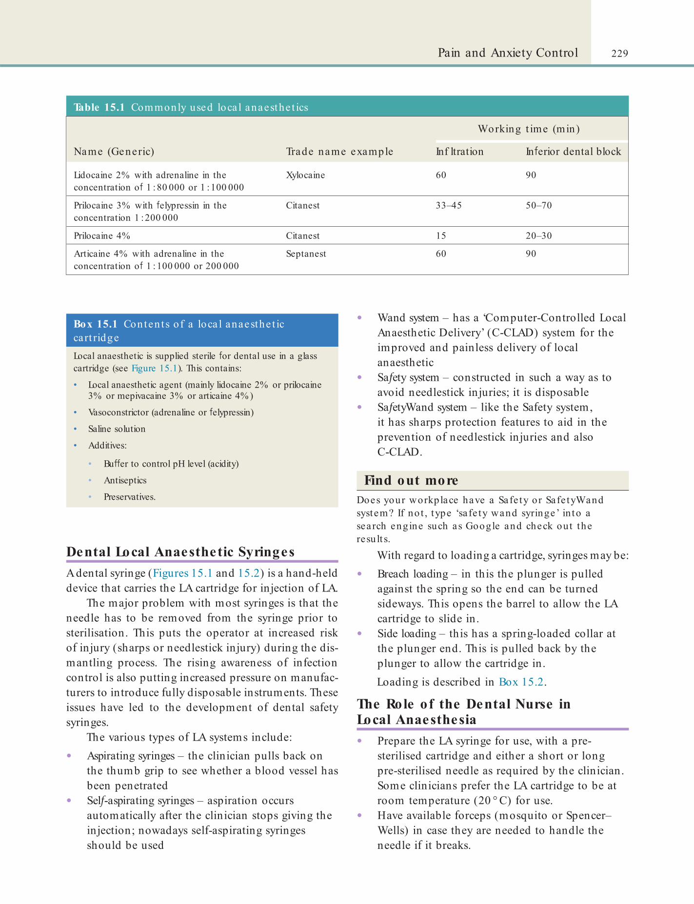
229Pain and Anxiety Control
Table 15.1 Commonly used loca l anaesthe t ics
Working t ime (min)
Name (Generic) Trade name example Inf ltration In erior dental block
Lidocaine 2% with adrenaline in the concentration o 1 : 80 000 or 1 : 100 000
Xylocaine 60 90
Prilocaine 3% with elypressin in the concentration 1 : 200 000
Citanest 33–45 50–70
Prilocaine 4% Citanest 15 20–30
Articaine 4% with adrenaline in the concentration o 1 : 100 000 or 200 000
Septanest 60 90
Box 15.1 Conten ts o f a loca l anaesthe t ic cart ridgeLocal anaesthetic is supplied sterile or dental use in a glass cartridge (see Figure 15.1). This contains:
• Local anaesthetic agent (mainly lidocaine 2% or prilocaine 3% or mepivacaine 3% or articaine 4% )
• Vasoconstrictor (adrenaline or elypressin)
• Saline solution
• Additives:
• Bu er to control pH level (acidity)
• Antiseptics
• Preservatives.
Dental Local Anaesthetic SyringesA dental syringe (Figures 15.1 and 15.2) is a hand-held device that carries the LA cartridge or injection o LA.
The major problem with most syringes is that the needle has to be removed rom the syringe prior to sterilisation. This puts the operator at increased risk o injury (sharps or needlestick injury) during the dis-mantling process. The rising awareness o in ection control is also putting increased pressure on manu ac-turers to introduce ully disposable instruments. These issues have led to the development o dental sa ety syringes.
The various types o LA systems include:
• Aspirating syringes – the clinician pulls back on the thumb grip to see whether a blood vessel has been penetrated
• Sel -aspirating syringes – aspiration occurs automatically a ter the clinician stops giving the injection; nowadays sel -aspirating syringes should be used
• Wand system – has a ‘Computer-Controlled Local Anaesthetic Delivery’ (C-CLAD) system or the improved and painless delivery o local anaesthetic
• Sa ety system – constructed in such a way as to avoid needlestick injuries; it is disposable
• Sa etyWand system – like the Sa ety system, it has sharps protection eatures to aid in the prevention o needlestick injuries and also C-CLAD.
Find out moreDoes your workplace have a Safe ty or Safe tyWand system? If no t , type ‘sa fe ty wand syringe’ in to a search engine such as Google and check out the resu lt s.
With regard to loading a cartridge, syringes may be:
• Breach loading – in this the plunger is pulled against the spring so the end can be turned sideways. This opens the barrel to allow the LA cartridge to slide in.
• Side loading – this has a spring-loaded collar at the plunger end. This is pulled back by the plunger to allow the cartridge in.Loading is described in Box 15.2.
The Role of the Dental Nurse in Local Anaesthesia• Prepare the LA syringe or use, with a pre-
sterilised cartridge and either a short or long pre-sterilised needle as required by the clinician. Some clinicians pre er the LA cartridge to be at room temperature (20 ° C) or use.
• Have available orceps (mosquito or Spencer–Wells) in case they are needed to handle the needle i it breaks.
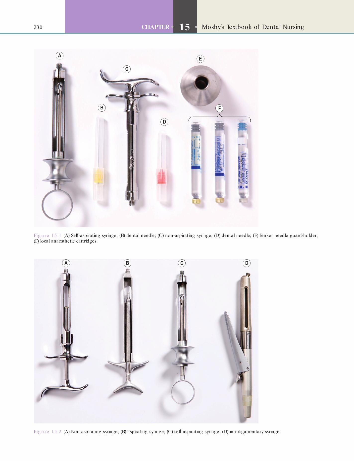
230 CHAPTER • 15 • Mosby’s Textbook o Dental Nursing
Fig u re 15.2 (A) Non-aspirating syringe; (B) aspirating syringe; (C) sel -aspirating syringe; (D) intraligamentary syringe.
Fig u re 15.1 (A) Sel -aspirating syringe; (B) dental needle; (C) non-aspirating syringe; (D) dental needle; (E) Jenker needle guard/holder; (F) local anaesthetic cartridges.
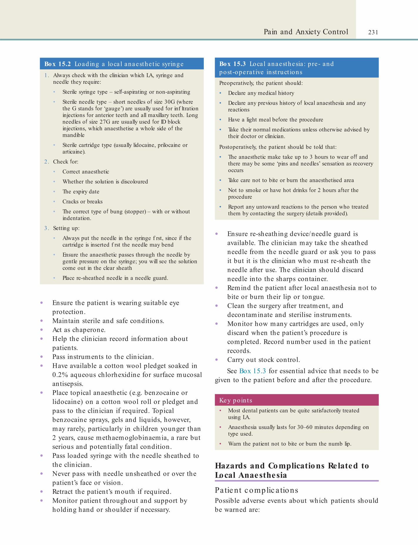
231Pain and Anxiety Control
Box 15.2 Loading a loca l anaesthe t ic syringe
1. Always check with the clinician which LA, syringe and needle they require:
• Sterile syringe type – sel -aspirating or non-aspirating
• Sterile needle type – short needles o size 30G (where the G stands or ‘gauge’) are usually used or inf ltration injections or anterior teeth and all maxillary teeth. Long needles o size 27G are usually used or ID block injections, which anaesthetise a whole side o the mandible
• Sterile cartridge type (usually lidocaine, prilocaine or articaine).
2. Check or:
• Correct anaesthetic
• Whether the solution is discoloured
• The expiry date
• Cracks or breaks
• The correct type o bung (stopper) – with or without indentation.
3. Setting up:
• Always put the needle in the syringe f rst, since i the cartridge is inserted f rst the needle may bend
• Ensure the anaesthetic passes through the needle by gentle pressure on the syringe; you will see the solution come out in the clear sheath
• Place re-sheathed needle in a needle guard.
• Ensure the patient is wearing suitable eye protection.
• Maintain sterile and sa e conditions.• Act as chaperone.• Help the clinician record in ormation about
patients.• Pass instruments to the clinician.• Have available a cotton wool pledget soaked in
0.2% aqueous chlorhexidine or sur ace mucosal antisepsis.
• Place topical anaesthetic (e.g. benzocaine or lidocaine) on a cotton wool roll or pledget and pass to the clinician i required. Topical benzocaine sprays, gels and liquids, however, may rarely, particularly in children younger than 2 years, cause methaemoglobinaemia, a rare but serious and potentially atal condition.
• Pass loaded syringe with the needle sheathed to the clinician.
• Never pass with needle unsheathed or over the patient’s ace or vision.
• Retract the patient’s mouth i required.• Monitor patient throughout and support by
holding hand or shoulder i necessary.
• Ensure re-sheathing device/needle guard is available. The clinician may take the sheathed needle rom the needle guard or ask you to pass it but it is the clinician who must re-sheath the needle a ter use. The clinician should discard needle into the sharps container.
• Remind the patient a ter local anaesthesia not to bite or burn their lip or tongue.
• Clean the surgery a ter treatment, and decontaminate and sterilise instruments.
• Monitor how many cartridges are used, only discard when the patient’s procedure is completed. Record number used in the patient records.
• Carry out stock control.See Box 15.3 or essential advice that needs to be
given to the patient be ore and a ter the procedure.
Box 15.3 Loca l anaesthesia : p re- and post -opera t ive inst ruct ionsPreoperatively, the patient should:
• Declare any medical history
• Declare any previous history o local anaesthesia and any reactions
• Have a light meal be ore the procedure
• Take their normal medications unless otherwise advised by their doctor or clinician.
Postoperatively, the patient should be told that:
• The anaesthetic make take up to 3 hours to wear o and there may be some ‘pins and needles’ sensation as recovery occurs
• Take care not to bite or burn the anaesthetised area
• Not to smoke or have hot drinks or 2 hours a ter the procedure
• Report any untoward reactions to the person who treated them by contacting the surgery (details provided).
Key po in ts• Most dental patients can be quite satis actorily treated
using LA.
• Anaesthesia usually lasts or 30–60 minutes depending on type used.
• Warn the patient not to bite or burn the numb lip.
Hazards and Complications Related to Local Anaesthesia
Patie nt c omplic a tionsPossible adverse events about which patients should be warned are:
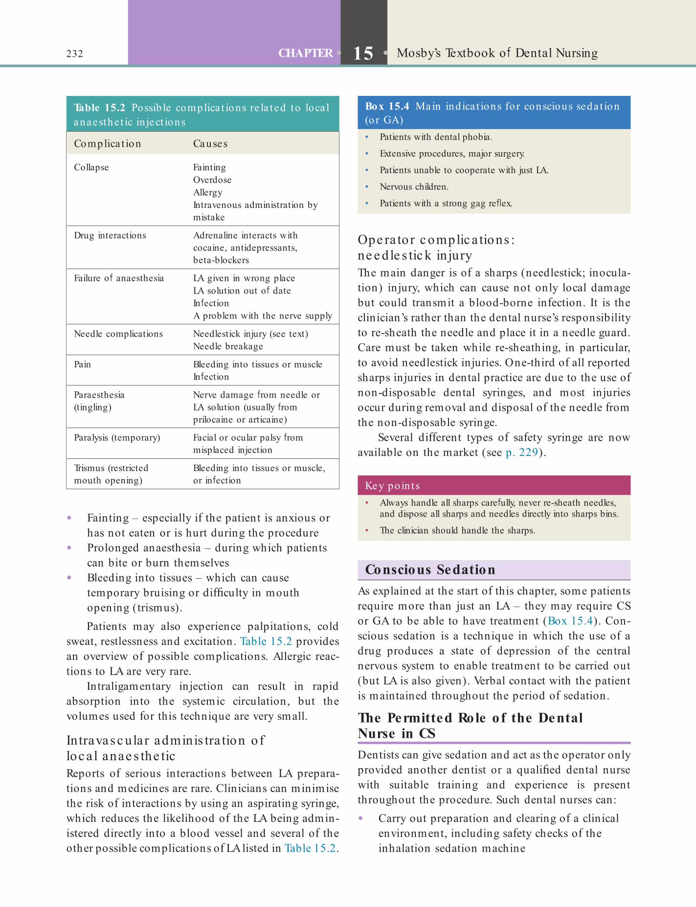
232 CHAPTER • 15 • Mosby’s Textbook o Dental Nursing
Ope ra to r c omplic a tions : ne e dle s tic k injuryThe main danger is o a sharps (needlestick; inocula-tion) injury, which can cause not only local damage but could transmit a blood-borne in ection. It is the clinician’s rather than the dental nurse’s responsibility to re-sheath the needle and place it in a needle guard. Care must be taken while re-sheathing, in particular, to avoid needlestick injuries. One-third o all reported sharps injuries in dental practice are due to the use o non-disposable dental syringes, and most injuries occur during removal and disposal o the needle rom the non-disposable syringe.
Several di erent types o sa ety syringe are now available on the market (see p. 229).
• Fainting – especially i the patient is anxious or has not eaten or is hurt during the procedure
• Prolonged anaesthesia – during which patients can bite or burn themselves
• Bleeding into tissues – which can cause temporary bruising or di culty in mouth opening (trismus).Patients may also experience palpitations, cold
sweat, restlessness and excitation. Table 15.2 provides an overview o possible complications. Allergic reac-tions to LA are very rare.
Intraligamentary injection can result in rapid absorption into the systemic circulation, but the volumes used or this technique are very small.
Intravas c ular adminis tra tion o f loc a l anae s the ticReports o serious interactions between LA prepara-tions and medicines are rare. Clinicians can minimise the risk o interactions by using an aspirating syringe, which reduces the likelihood o the LA being admin-istered directly into a blood vessel and several o the other possible complications o LA listed in Table 15.2.
Box 15.4 Main ind ica t ions for conscious seda t ion (or GA)• Patients with dental phobia.
• Extensive procedures, major surgery.
• Patients unable to cooperate with just LA.
• Nervous children.
• Patients with a strong gag re ex.
Key poin ts• Always handle all sharps care ully, never re-sheath needles,
and dispose all sharps and needles directly into sharps bins.
• The clinician should handle the sharps.
Table 15.2 Possib le complica t ions re la ted to loca l anaesthe t ic in ject ions
Complica t ion Causes
Collapse FaintingOverdoseAllergyIntravenous administration by mistake
Drug interactions Adrenaline interacts with cocaine, antidepressants, beta-blockers
Failure o anaesthesia LA given in wrong placeLA solution out o dateIn ectionA problem with the nerve supply
Needle complications Needlestick injury (see text)Needle breakage
Pain Bleeding into tissues or muscle In ection
Paraesthesia (tingling)
Nerve damage rom needle or LA solution (usually rom prilocaine or articaine)
Paralysis (temporary) Facial or ocular palsy rom misplaced injection
Trismus (restricted mouth opening)
Bleeding into tissues or muscle, or in ection
Conscious SedationAs explained at the start o this chapter, some patients require more than just an LA – they may require CS or GA to be able to have treatment (Box 15.4). Con-scious sedation is a technique in which the use o a drug produces a state o depression o the central nervous system to enable treatment to be carried out (but LA is also given). Verbal contact with the patient is maintained throughout the period o sedation.
The Permitted Role o f the Dental Nurse in CSDentists can give sedation and act as the operator only provided another dentist or a quali ed dental nurse with suitable training and experience is present throughout the procedure. Such dental nurses can:
• Carry out preparation and clearing o a clinical environment, including sa ety checks o the inhalation sedation machine
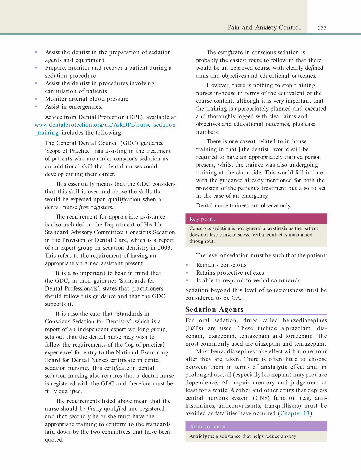
233Pain and Anxiety Control
The certi cate in conscious sedation is probably the easiest route to follow in that there would be an approved course with clearly de ned aims and objectives and educational outcomes.
However, there is nothing to stop training nurses in-house in terms of the equivalent of the course content, although it is very important that the training is appropriately planned and executed and thoroughly logged with clear aims and objectives and educational outcomes, plus case numbers.
There is one caveat related to in-house training in that [ the dentist] would still be required to have an appropriately trained person present, whilst the trainee was also undergoing training at the chair side. This would fall in line with the guidance already mentioned for both the provision of the patient’s treatment but also to act in the case of an emergency.Dental nurse trainees can observe only.
• Assist the dentist in the preparation o sedation agents and equipment
• Prepare, monitor and recover a patient during a sedation procedure
• Assist the dentist in procedures involving cannulation o patients
• Monitor arterial blood pressure• Assist in emergencies.
Advice rom Dental Protection (DPL), available at www.dentalprotection.org/uk/AskDPL/nurse_sedation _training, includes the ollowing:
The General Dental Council (GDC) guidance ‘Scope of Practice’ lists assisting in the treatment of patients who are under conscious sedation as an additional skill that dental nurses could develop during their career.
This essentially means that the GDC considers that this skill is over and above the skills that would be expected upon quali cation when a dental nurse rst registers.
The requirement for appropriate assistance is also included in the Department of Health Standard Advisory Committee: Conscious Sedation in the Provision of Dental Care, which is a report of an expert group on sedation dentistry in 2003. This refers to the requirement of having an appropriately trained assistant present.
It is also important to bear in mind that the GDC, in their guidance ‘Standards for Dental Professionals’, states that practitioners should follow this guidance and that the GDC supports it.
It is also the case that ‘Standards in Conscious Sedation for Dentistry’, which is a report of an independent expert working group, sets out that the dental nurse may wish to follow the requirements of the ‘log of practical experience’ for entry to the National Examining Board for Dental Nurses certi cate in dental sedation nursing. This certi cate in dental sedation nursing also requires that a dental nurse is registered with the GDC and therefore must be fully quali ed.
The requirements listed above mean that the nurse should be rstly quali ed and registered and that secondly he or she must have the appropriate training to conform to the standards laid down by the two committees that have been quoted.
Key poin tConscious sedation is not general anaesthesia as the patient does not lose consciousness. Verbal contact is maintained throughout.
The level o sedation must be such that the patient:
• Remains conscious• Retains protective ref exes• Is able to respond to verbal commands.Sedation beyond this level o consciousness must be considered to be GA.
Sedation AgentsFor oral sedation, drugs called benzodiazepines (BZPs) are used. These include alprazolam, dia-zepam, oxazepam, temazepam and lorazepam. The most commonly used are diazepam and temazepam.
Most benzodiazepines take e ect within one hour a ter they are taken. There is o ten little to choose between them in terms o anxiolytic e ect and, in prolonged use, all (especially lorazepam) may produce dependence. All impair memory and judgement at least or a while. Alcohol and other drugs that depress central nervous system (CNS) unction (e.g. anti-histamines, anticonvulsants, tranquillisers) must be avoided as atalities have occurred (Chapter 13).
Term to lea rnAnxiolytic: a substance that helps reduce anxiety.
234 CHAPTER • 15 • Mosby’s Textbook o Dental Nursing
able to etch anything required, carry out administra-tive duties and answer the telephone or make calls.
Precautions during CS
• Protect the airway – especially in restorative dental and surgical procedures, i.e. use rubber dam, butterf y sponges, etc. The airway must be protected because protective laryngeal ref exes are impaired, especially a ter the administration o benzodiazepines.
Requirements before Using CSThe patient must have no contraindications (Box 15.5), and must have a responsible escort with them. Also, the patient’s responsibilities (e.g. their job, night duty, caring or young children) must be such as to permit them to receive sedation sa ely.
In ormed consent or sedation must be obtained. This means that the patient must be given a ull expla-nation o the procedure, and the nature, purpose, e ects and balance o risks. The clinician should get a consent orm signed, which gives permission or a sedation technique to be used together with LA, as well as consent or the operative procedure (see also Chapter 5).
Written instructions with preoperative advice must also always have been given (see Box 15.6).
Where inhalational or IV sedation techniques are to be used it is wise to have one clinician to per om the sedation and another to carry out the dental work, but a suitably experienced practitioner may assume the responsibility o sedation o the patient, as well as operating. However, this is acceptable only provided that, as a minimum requirement, a second appropri-ate person is present throughout. As stated above, such an ‘appropriate person’ might be a suitably trained dental nurse, whose experience and training enables them to be capable o monitoring the clinical condi-tion o the patient. The second person must con orm to the de nition o a second ‘appropriate person’ and, should the occasion arise, the second appropriate person must also be capable o assisting the clinician in case o emergency. Emergency procedures should be revised with that person at regular and requent intervals.
Because the second ‘appropriate person’ must be present throughout the treatment and must not leave the surgery at any time, when patients are being sedated, a third person must also be present and avail-
Box 15.5 Main cont ra ind ica t ions for conscious seda t ion (or GA)• Unavailability o :
• Necessary equipment
• Necessary sta
• Escort.• Empty stomach (i patient has taken nothing except plain
water by mouth in the previous 6 hours).
• Medical contraindications such as:
• Respiratory disease
• Ludwig angina (in ection around throat)
• Angioedema (swelling around throat)
• Severe cardiovascular disease
• Hypertension
• Bleeding tendency
• Severe anaemia
• Metabolic or endocrine disorders, including liver disease, kidney disease; poorly controlled diabetes; hyperthyroidism (thyrotoxicosis); hypothyroidism; Addison disease (hypo-adrenalism); adrenocortical suppression.
• Specif c contraindications to GA drugs:
• Halothane sensitivity or recent anaesthesia with halothane
• Porphyria
• Suxamethonium sensitivity.• Malignant pyrexia.
• Drug usage, particularly:
• Corticosteroids
• Anticoagulants
• Alcohol or narcotics
• Antidepressants.• Cervical spine pathology (e.g. trauma, Down syndrome,
rheumatoid arthritis).
• Pregnancy.
• Myopathies.
• Multiple sclerosis.
Key po in tInhalation sedation is the CS method o choice in most cases.
CS is more usually given as:
• Inhalational sedation• IV sedation.
In the dental workplace, inhalation sedation is the method o choice in most cases; it is sometimes termed relative analgesia (RA). Minimal nitrous oxide plus oxygen (usually at least 30%) is used.

235Pain and Anxiety Control
• Protect the patient’s eyes – with eye protection during operation.
• Monitor the patient requently be ore, during and a ter the administration o sedatives – the patient should remain conscious and able to respond when directed. Monitoring may detect early signs o patient distress, such as changes in pulse, blood pressure, ventilatory status, cardiac electrical activity, and clinical and neurological status, be ore clinically signi cant problems arise. Standard monitoring includes recording:• Heart (pulse) rate• Blood pressure• Respiratory rate• Oxygen saturation.
Term to lea rnOxygen saturation: in the dental/medical context, oxygen saturation is the amount o oxygen that is bound to the haemoglobin in the blood at a particular time. It is expressed in percentage o the maximum binding capacity o the haemoglobin (normal is 95–100% ). It is commonly measured by pulse oximetry. This involves clipping or taping a small sensor on usually the tip o a f nger or a toe. The sensor measures the oxygen saturation by transmitting light waves into the body.
Continuous electrocardiogram (ECG) monitoring is reasonable, especially in high-risk patients; although the necessity or such monitoring has not been proved in good quality studies, patients who may bene t rom ECG monitoring include those who have a history o signi cant arrhythmia or cardiac dys unction, older patients and those or whom prolonged procedures are anticipated.
Inhalational SedationInhalational sedation (IS), sometimes termed relative analgesia or RA, uses two medical gases, a combina-tion o nitrous oxide (N2O) and oxygen (O2), to achieve a state o relaxation in which treatment can be carried out. Inhalational sedation is convenient both or the operator and patient, and the level o sedation can be easily controlled by withdrawing the drug as required.
Nitrous oxide and oxygen are stored in separate speci cally coloured cylinders (Table 15.3). At least 20% and more usually 30% oxygen is given.
Table 15.3 Medica l gases used in the UK
Medica l gas
ISO cylinder co lour
Pressure sto red a t (kPa)
Physica l sta te in cylinder
Oxygen Black with a white collar
13 700 Gas
Nitrous oxide
Blue 4400 Liquid
Term to lea rnISO: stands or International Organisation or Standardisation, the body that is responsible or setting standards or a range o products worldwide.
Gases used in sedation/anaesthesia are usually supplied under high pressure either in cylinders or as a piped gas supply. The cylinders are made rom molybdenum steel, in which gases and vapours are stored under pressure. The shape and colour o the plastic disc around the neck indicates when the cylin-der was last examined. The most commonly used types/sizes are:
• H – these are ree-standing and attached to the anaesthesia/sedation machine by a f exible hose. These are most economical, but reduce the mobility o the machine. Gas-speci c connectors are used that make it impossible to attach the wrong regulator or tting to the cylinder.
• E – these are attached directly to the machine via a yoke. A gas-speci c pin-index system is provided on cylinders: pins on the yoke o the machine mate with holes drilled in speci c positions on the valve o the cylinder. This provides a mechanical means o preventing incorrect connection.
Find out moreWhat is molybdenum stee l and why is it used for these gas cylinders?
Nitrous oxide (N2O) is contraindicated or pati-ents with:
• Bowel obstruction• Pneumothorax• Middle ear disease• Sinus disease.

236 CHAPTER • 15 • Mosby’s Textbook o Dental Nursing
• Nitrous oxide can be easily and rapidly discontinued i required.
• Nitrous oxide is administered and excreted through the lungs; virtually total recovery takes place within the rst 15 minutes o cessation o administration. The patient may, there ore, attend and leave surgery or hospital unaccompanied.
• Nitrous oxide provides a degree o analgesia (although LA is o ten still required).
• It also provides some degree o amnesia.• There is no signi cant hypotension or respiratory
depression.
Dis advantage s• The level o sedation is largely dependent on
psychological reassurance/back-up.• Nitrous oxide needs to be administered
continuously as long as it is required.• The patient may have amnesia or a distorted
perception o time, but this may be advantageous.
• Nitrous oxide pollution o the surgery atmosphere. This can be reduced by:• Use o scavenging equipment• Venting the suction machine outside the
building• Minimising conversation rom the patient• Testing the equipment weekly or leakage• Keeping the equipment well maintained,
with 6×-monthly servicing• Ventilating the surgeries with resh air
(e.g. open window and door an, air conditioning)
• Monitoring the air (e.g. Barnsley nitrous oxide monitor).
Es s e ntia l advic e to the pa tie ntSee Box 15.6.
Proc e dure• Check – that the inhalational sedation machine
is ready and working, that extra nitrous oxide and oxygen are available, and that you are completely amiliar with the machine. Use a scavenging system.
• Lie the patient com ortably supine in the chair with legs uncrossed.
• Check that the equipment is as unobtrusive as possible.
• Explain the procedure to the patient.
It is also contraindicated:
• Following eye operations (the use o nitrous oxide during general anaesthesia in gas- lled eyes may have serious side-e ects because o gas expansion and increased intra-ocular pressure).
• In the rst two trimesters o pregnancy (because o the e ect o nitrous oxide on DNA production and the experimental and epidemiological evidence that nitrous oxide causes undesirable reproductive outcomes).
• In immunosuppressed patients or in patients requiring multiple inhalation or general anaesthetic inductions, since nitrous oxide a ects white blood cell production and unction.
Terms to learnPneumothorax: abnormal collection o air in the space between the chest wall and the outer sur ace o the lung.
Intra-ocular pressure: the pressure exerted by the uids within the eye.
Ind ic a tions• Anxiety• Marked gagging.
Contra indic a tions• Fear or non-acceptance o the nasal mask• Inability to communicate with the patient• Nature o procedure warrants general
anaesthesia• Medical contraindications such as:
• A heavy cold (temporary contraindication) or a deviated nasal septum (permanent contraindication) or nasal obstruction
• Cyanosis at rest due to chronic cardiac (e.g. congenital cardiac disease) or respiratory (e.g. chronic bronchitis or emphysema) disease
• Severe psychiatric conditions in which cooperation is not possible
• First trimester o pregnancy• Some neuromuscular diseases.
Advantage s• Patient remains conscious and cooperative.• It is non-invasive.• No strict asting is required be orehand.• Level o sedation is easily controlled.• Protective ref exes are minimally impaired.

237Pain and Anxiety Control
it which the patient does not usually tolerate) is complete.
Monitor the patient throughout by checking the pulse and respiratory rate at requent intervals. The patient should be conscious and able to respond when directed. Dozing is sa e, but snoring indicates partial airway obstruction and must be corrected immedi-ately. Both operator and assistant should care ully monitor the patient.
Te rmina ting inha la tiona l s e da tion• Shut o the nitrous oxide f ow, so that 100%
oxygen is given or 2 minutes to counteract possible di usion hypoxia.
• Remove the ace mask.
Tas ks a fte r inha la tional s e da tionSlowly bring the patient upright over 5 minutes, a ter which they should recover over at least another 15 minutes under the direct supervision o a member o the dental team or escort. The length o the ollow-up observation is dependent upon the perceived risk to the patient. Patients may be discharged rom the post-procedure recovery area once vital signs are stable, and the patient has reached an appropriate level o consciousness.
Intravenous SedationIntravenous sedation (IV sedation), like inhalational sedation, is convenient both or operator and patient, and the level o sedation can be controlled. However, the drug cannot be withdrawn once given. Benzodiazepines (especially midazolam) are the main agents used; midazolam is commonly used or intra-venous sedation (and occasionally or intra-nasal administration).
Box 15.6 Inha la t iona l seda t ion : p re - and post -opera t ive inst ruct ionsPreoperatively, the patient should:
• Declare any medical history
• Declare any previous history o sedation or general anaesthesia and any reactions
• Not drink alcohol or 24 hours be ore the procedure
• Eat a light meal be ore the procedure
• Take their normal medications unless otherwise advised by their doctor or clinician.
Postoperatively, the patient should be told that:
• The e ects o the sedative gas normally wear o very quickly
• They will be f t to go back to work or travel home
• Although recovery is very rapid, they should avoid driving, particularly two-wheeled vehicles, immediately a ter treatment, or taking alcohol or other drugs.
The signs o inhalation sedation are positive and pleasant. The patient will:
• Feel relaxed• Feel warm• Have some tingling or numbness• Have some visual or auditory changes• Experience slurring o speech• Have slower responses (e.g. reduced requency o
blinking, delayed response to verbal instructions or questioning).Machine output f ows o between 20% and 35%
nitrous oxide in oxygen commonly allow or a state o eeling detached, as well as analgesia, without any loss o consciousness or danger o reduced ref exes. At these levels, patients are aware o operative procedures and are cooperative without being ear ul.
Key poin tI the sedated patient cannot maintain an open mouth then he or she is too deeply sedated.
A possible exception may be in the case o a disabled patient, who may be unable to maintain an open mouth even without sedation. I a prop is then used, extra care ul observation o the depth o sedation is essential.
I a ter a period o relaxation the patient becomes restless or apprehensive, this usually means the level o nitrous oxide is too high and the percentage should be dropped to a more com ortable level. The patient can then be maintained at an appropriate level o sedation until the operative procedure (or that part o
Term to lea rnAntagonist: a drug that counteracts or reduces the e ects o another drug.
Term to lea rnIntra-nasal: through the nose.
Adverse e ects o benzodiazepines include con u-sion, slurred speech, lack o coordination, dizziness, headache and nausea. In children, benzodiazepines can cause unusual behavioural reactions.
Flumazenil is a speci c benzodiazepine antago-nist, which allows rapid reversal o conscious sedation with benzodiazepines.
238 CHAPTER • 15 • Mosby’s Textbook o Dental Nursing
Es s e ntia l advic e to the pa tie ntSee Box 15.7.
Tas ks a fte r IV s e da tionFollowing intravenous sedation, patients may have a prolonged period o amnesia ( orget ulness) and/or impaired judgement and ref exes despite appearing to recover appropriately. The patient must there ore not be discharged home until:
• At least one hour has elapsed since the drug was given
• They appear ully conscious.In addition, patients must be discharged into the care o a responsible escort, a ter giving the general post-operative instructions, together with any pertaining to
Advantage s• Adequate level o sedation is attained
pharmacologically rather than with psychological back-up.
• Amnesia removes unpleasant memories.• The patient may take a light meal up to 2 hours
be ore treatment.
Dis advantage s• Once administered, the drug cannot be
‘discontinued’ or ‘switched o ’. Flumazenil injection can, however, reverse the sedation.
• There is a short period a ter injection when laryngeal ref exes may be impaired. There ore a mouth sponge/gauze or rubber dam must be used to protect against accidental inhalation o water or debris.
• The patient must be accompanied home rom surgery and may not drive, ride a bicycle or motor cycle o any kind, work machinery (including domestic appliances), make important decisions or drink alcohol or 24 hours.
Contra indic a tions• Psychological reasons: patient is rightened o
needles and injections.• Social reasons: patient will have to ul l
responsibilities a ter the treatment (e.g. caring or young children, shi t work); inability to bring an escort.
• Medical reasons, such as:• History o reaction to IV agents or any
benzodiazepine• Pregnancy (also caution during
breast eeding)• Severe psychiatric condition• Liver or kidney disease• Glaucoma• Alcohol or narcotic dependency (may render
usual doses ine ective).• Children: there is a considerable variability in
reaction to benzodiazepines.
Box 15.7 IV sedat ion: p re - and post -opera t ive inst ruct ionsPreoperatively, the patient should:
• Declare any medical history
• Declare any previous history o sedation or general anaesthesia and any reactions
• Come with a responsible adult to escort them to and rom premises
• Not drink alcohol or 24 hours be ore the procedure
• Eat a light meal 3 hours be ore the procedure
• Take their normal medications unless otherwise advised by their doctor or clinician
• Not wear tight-sleeved clothes
• Not wear nail varnish
• Wear at shoes.
Postoperatively, the patient should:
• Take private transport home
• Have an adult escort with them or the rest o the day
• Rest quietly at home or the remainder o the day.
For 24 hours, they should re rain rom:
• Drinking alcohol or taking drugs that could a ect the central nervous system
• Driving a vehicle or ying an aircra t
• Riding a bicycle or motor cycle o any kind
• Operating machinery
• Making important decisions.
Key po in tPatients given umazenil ollowing IV sedation procedures must still ollow the usual instructions given a ter sedation (i.e. no driving, operating machinery, etc.).
Terms to learnGlaucoma: an eye disease in which an increase in the pressure within the eye damages the optic nerve, which impairs vision and can sometimes progress to blindness.
Narcotic: an addictive drug, such as heroin, which reduces the eeling o pain and induces a eeling o numbness or sleep.

239Pain and Anxiety Control
the dentistry per ormed. The instructions should be given verbally and also written down or the patient to re er to later, a ter the amnesic e ect o the sedative has cleared.
Other Forms of Conscious Sedation
Intranas a l s e da tionThis also involves use o a benzodiazepine, usually midazolam.
Ora l s e da tionAgain, this is a convenient way o using a benzodi-azepine (e.g. diazepam or temazepam) to sedate the patient. However, the level o sedation cannot be con-trolled, and there can be a delay while waiting or the drug to take e ect.
Advantage s• Easy to administer.• Help ul or the moderately apprehensive patient.• Relatively sa e, since protective ref exes are
maintained.
Dis advantage s• Variability in absorption time: the patient may
become sedated too soon, possibly endangering themselves on the way to the surgery (this risk can be minimised by administering drug on arrival), or they may become sedated too late, thus delaying treatment.
• The level o sedation is unpredictable.• Unpredictable e ect o benzodiazepines in
certain patients:• Some children become hyper-excited, some
are rather resistant• Older patients may be very sensitive.
Summary of requirements be fore and a fte r administe ring a ll fo rms of conscious sedat ion• Written medical history.
• Previous dental history.
• Previous CS/GA history noted.
• Written instructions provided pre- and post-operatively.
• The medical history has been checked and acted on.
• There is written documentation o consent or sedation (consent orm).
• There is an accompanying adult present where the method o sedation requires it and suitable postoperative supervision has been arranged.
• The patient has complied with pre-treatment instructions.
• Pre-sedation assessment done.
• Records o sedation agents employed, dosages and times given, including site and method o administration.
• Records o monitoring techniques.
• Any individual specif c patient requirements pre- and post-procedurally.
• Full details o dental treatment provided.
• Post-sedation assessment done.
Find out moreTo read more about CS, see the ‘A Referra l Guide for Denta l Pract it ioners’ (websit e o f the Denta l Seda t ion Teachers Group: www.dstg .co .uk/teach ing /conc-sed /).
General AnaesthesiaAs a dental nurse, you cannot administer general anaes-thesia (GA) but may be involved in pre- and post-anaesthesia care o the patient. Box 15.8 summarises the pre- and post-operative instructions or patients undergoing GA or dental procedures or oral and max-illo acial surgery.
Box 15.8 GA: pre - and post -opera t ive inst ruct ionsPreoperatively the patient should:
• Declare any medical history
• Declare any previous history o sedation or general anaesthesia and any reactions
• Come with a responsible adult to escort them to and rom the hospital
• Not drink alcohol or 24 hours be ore the procedure
• Not eat or drink (except water) or 6 hours be ore the procedure
• Take their normal medications unless otherwise advised by their doctor or clinician
• Not wear tight sleeves or collars
• Not wear nail varnish
• Wear at shoes.
Postoperatively, the patient should:
• Take private transport home
• Have a responsible adult escort with them or the rest o the day
• Not make important decisions, drive, y aircra t, or ride a bicycle or motorcycle o any kind, or operate machinery or electrical appliances until the ollowing day
• Not drink alcohol or 24 hours a ter the procedure
• Report any untoward reactions to the person who operated on them by contacting the hospital (contact details provided).
240 CHAPTER • 15 • Mosby’s Textbook o Dental Nursing
around the pain ul area. The bene t o TENS or pain is controversial.
Complementary and Alternative Medicine (CAM)In the developed world, therapies that are not cur-rently considered an integral part o conventional allopathic medical practice are termed complementary (when used in addition to), and alternative (when used instead o ), conventional treatments. Traditional med-icine is widely practised across the world, particularly in Asian and A rican cultures.
CAM therapies include, but are not limited to, acupuncture, chiropractic, speci c or restrictive diets, aith healing, olk medicine, herbal (natural) medi-cine, homoeopathy, naturopathy, new age healing, massage and music therapy, and reliable evidence or their e cacy is not always strong.
Find out moreTo read more about hypnosis, see the Brit ish Socie ty of Clin ica l Hypnosis websit e (www.bsch .org .uk/).
Precautions in Use of Drugs for Pain and Anxie ty Control
It is important always to take a ull medical and drug history, since the medical status or medications a person may already be taking can inf uence the choice o drugs (see Chapter 21). Certain drugs may need to be avoided or doses reduced in speci c conditions. It is particularly important to ensure there is no history o allergy or untoward e ect rom the drug being considered.
The clinician can check drug doses, contraindica-tions, interactions and adverse reactions in the British National Formulary (BNF) or the special dental version called the Dental Practitioners’ Formulary (DPF).
I aspirin or paracetamol is indicated, it should be given at least 30 minutes be ore ood, since the absorp-tion o these drugs is otherwise delayed.
Special SituationsIn general, drug doses will need to be reduced in:
• Children• Older people• Liver disease• Kidney disease.
Other TechniquesHypnosisHypnosis may be used to help patients relax and control their anxiety and ears or the gagging ref ex. In other words, hypnotherapy can trans orm a scared dental patient into one who is relaxed and cooperative.
Many patients are anxious about dental treatment and the anxiety can range rom mild apprehension to extreme phobia. However, patients with extreme anxiety are rarely seen in general practice dentistry. When they have great pain or severe in ection they will usually go to their GP.
Term to lea rnPsychosomatic disorder: a disorder in which physical symptoms or disease are thought to have (or are made worse by) a mental cause such as stress and anxiety. For example, high levels o anxiety worsening eczema or causing high blood pressure or arrhythmia.
Key poin tHypnosis can be a power ul method in the treatment o anxiety states and many phobias and psychosomatic disorders.
Key po in tGA is used only in a hospital environment where there are critical care acilities.
The relaxation routines employed in the ‘talking therapies’ (such as cognitive behavioural therapy or CBT) are the same as those used to induce a hypnotic state. The mainstay o hypnotherapy is the post-hypnotic suggestion. This is an idea given to the patient while in the hypnotic state, which a terwards inf uences the patient’s behaviour in a bene cial but completely unconscious way. At the same time as dealing with a speci c phobia, suggestions can be made to boost the patient’s con dence and overall anxiety (and improve their relaxation at the next session). Un ortunately, not all patients are able to achieve a degree o relaxation with hypnosis that is therapeutically use ul.
Transcutaneous Electrical Nerve Stimulation (TENS)A TENS machine transmits gentle, electrical impulses through the skin via electrode pads positioned on or

241Pain and Anxiety Control
Pre gnanc yIn relation to pain and anxiety control, drugs should be avoided where possible in pregnancy, in case they a ect the etus.
Childre nThe ollowing drugs should be avoided:
• Aspirin – because o the risk o a potentially lethal liver and brain disorder (Reye syndrome)
Key poin tAlways check: (i) that the correct drug (and solvent) is used; this is particularly important i another person makes up the drug; and (ii) that the drug expiry date has not passed.
• Diazepam (Valium) – it may have unwanted e ects, causing overactivity rather than sedation
• Codeine – it can cause respiratory depression and, i used postoperatively, has been lethal.

This page intentiona lly le ft blank
Chapter 16 Dental Materials and Equipment
CHAPTER POINTS• Dental materials• Dental impression materials• Appliance resins• Dental cements• Dental restorative materials• Endodontic materials• Main hazards from other dental materials• Dental equipment• Care and maintenance of dental instruments and
equipment
most commonly elastic impression materials are used. Common elastic dental impression materials are:
• Alginate-type hydrocolloids (‘alginates’)• Synthetic rubber base impression materials
(‘rubber base’ or ‘elastomers’).Advantages and disadvantages of the more common impression materials are summarised in Table 16.1.
Alg ina te impre s s ion mate ria l (‘Alg ina te ’)Alginate is an irreversible hydrocolloid. Alginates are used mainly for making impressions for:
• Removable complete and partial dentures• Orthodontic appliances• Making special impression trays for rubber base
impressions for more precise procedures such as crowns and bridges.Alginate is one of the most commonly used
impression materials. It is supplied as a powder, which is mixed with tap water to make a gelatinous mass. This mass then sets (hardens) rapidly. Alginate powder must be stored in a cool dry place in a tightly closed container to protect it from absorbing moisture from the air and from contamination. Shake the container before use to loosen the powder and then leave it for a few minutes before opening to let the dust settle. Powder and water measuring cups are provided by the manufacturer. Measure out the powder into the mixing bowl and add a measured amount of tap water at room temperature (21 ° C). Alginates set fairly quickly; the best method of controlling the setting time is to slightly alter the temperature of the water used in the mix. The higher the water temperature, the faster the material will set. The lower the temperature, the slower the setting.
Alginate is a salt of alginic acid (from seaweed) (potassium alginate or sodium alginate) with a reactor
In t ro d u ct io n
The many different materials used in dentistry greatly bene t the patients. Some, however, can occasionally harm the patient, dental staff or the environment. The hazards associated with materials commonly used in dentistry are discussed also in Chapter 6. Oral health products are discussed in Chapter 14.
Dental MaterialsWith all materials, double check the label before use and also ensure that the expiry date has not passed.
Dental Impression MaterialsDental impression materials are used to record the shape of teeth and alveolar ridges. There is a wide variety of impression materials available. Each has its own advantages and disadvantages. The ideal proper-ties of an impression material are shown in Box 16.1.
Non-elastic impression materials such as zinc-oxide eugenol and impression compound (‘compo’) are occasionally used in edentulous patients. However,

244 CHAPTER • 16 • Mosby’s Textbook of Dental Nursing
To keep moist, wrap the impression in damp gauze; the cast should be poured soon after the impres-sion is removed from the mouth before the impres-sion distorts.
Us esAlginates are satisfactory for taking primary impres-sions and for many of the impressions required in prosthetic work. But they can also distort due to syner-esis (separation of liquid from the gel), imbibition etc. So for the more accurate impressions, as are required for crowns and bridges (see later), other impression materials such as elastomers are used.
Elas tome ric impre s s ion mate ria ls (e las tome rs , ‘rubbe r bas e ’)Elastomers give better impressions (more accurate and stable) than do alginates. They are used where high accuracy is needed, such as in inlay, crown and bridge, and implant work. Their advantages over alginates are that they have good tear resistance and dimensional stability. They are supplied ranging from low to high viscosity materials. ‘Light-bodied materials’ may be used in wash impressions and over the areas where high detail is required; they are used over a ‘medium’ or ‘heavy’-bodied material.
(calcium sulphate). The reactor causes the alginate to change from a sol form when mixed with water to a gel form by an irreversible chemical reaction. Alginates also contain a retarder (sodium or potassium sul-phate, oxalate, or carbonate) to give enough working time. Fillers increase the strength and stiffness of the gel. With time, the gel can lose water (syneresis, which results in shrinkage) or it can take up water (imbibi-tion, which results in expansion) or other uids. This affects the usability of alginates as the impression may change shape after setting. Set alginate impressions should be cast as soon as possible and, meantime, kept wrapped in a damp gauze.
Box 16.1 Idea l den ta l impression mate ria lAn ideal dental impression material has the following characteristics:
• Non-toxic
• Non-irritant
• Acceptable taste
• Ease of mixing
• Good working time
• Good handling
• Good setting time
• Accurate
• Surface reproducibility
• Dimensional stability
• Compatibility with model materials
• Reasonable cost
• Reasonable shelf-life.
Term to lea rnSol form: liquid form.
Advantages• Alginates are relatively cheap.• They are exible once set.• They are suf ciently elastic to return to their
original shape after the slight distortion during their removal from the mouth once set.
Dis advantages• Alginates are weak materials and may tear on
removal from deep undercuts (e.g. when teeth are tilted and the material gets under them).
• They distort with time, especially if they dry out, so they must always be kept moist.
Term to lea rnDimensional stability: impressions can distort when taken out of the mouth or shrink if they dry out. They can also expand if left in liquids too long due to absorption (imbibition).
The bases and the chemical reactors of elastomers are usually of different colours. Some products are provided in special mixing syringes. For others, you will need to lay out equal lengths of both the base material and the chemical reactor (catalyst) separately but side by side onto a special polymer paper or parch-ment pad. The reactor should not touch the base mate-rial until everything is ready to mix: then you mix the two pastes with a spatula in the prescribed time, until no streaks remain. A thin uniform layer of elastomer impression material is required to give the most accu-rate impression, so the materials are used in individu-ally designed (custom or special tray) acrylic trays. You will need to apply a tray adhesive to prevent the impression from pulling away from the tray and dis-torting. Most elastomer impressions are dimension-ally stable if stored dry, and thus they may be sent to

245Dental Materials and Equipment
Table 16.1 Advantages and d isadvantages of the d iffe ren t den ta l impression materia ls
Mate ria l Advantages Disadvantages
Alginate Non-toxic Messy to work withNon-irritantGood surface detail
Setting time dependent on operator handling
Easy to mix and use Poor dimensional stabilitySetting time can be controlled with water temperature
Incompatible with some dental stone plasters
CheapGood shelf-life
Polysulphide Extended working time Extended working timeHigh tear resistance Needs custom trayEasily read margins Stretches and distortsModerate cost Hydrophobic
MessyUnpleasant smell
Polyether Fast settingClean but has foul taste
Sets too fast for multiple preparations
Least hydrophobic StiffGood stability Absorbs waterEasy to read margins Leaches componentsShelf-life at least 2 years High cost
Burning sensation in soft tissue
Condensation silicone Clean and pleasant ShrinkageAdequate working time Low tear strengthEasily read margins Not dimensionally stableLow to moderate cost Hydrophobic (does not like water)Short shelf-life
Addition silicone (or vinyl polysiloxane)
AccurateExcellent surface detailEasy to use
Dif cult to mixSometimes dif cult to remove impression from the mouth
Fast settingDimensionally stable
Too accurate in some circumstances
Moderate tear strengthNo gas evolution HydrophobicNon-toxic and non-irritant Tears easilyGood shelf-life Poor ow on soft tissue
Putty wash separatePoor tray adhesiveDif cult to pour models
the technician without a major risk of dimensional change.
Advantages• Elastomers are exible and rubber-like once they
set, and suf ciently elastic to return to their original shape after slight distortion.
• They do not signi cantly distort after setting.• They do not tend to dry out but the cast is still
best poured within 30 minutes after an impression is made. This is because some elastomers undergo shrinkage on setting that
continues for some time after the impression is made, particularly the condensation silicone type. However, these materials are not subject to syneresis and imbibition because they are hydrophobic (water-hating).
Dis advantages• Elastomers are expensive compared with
alginates.• They are more liable than alginates to produce
allergic reactions, so should be handled carefully and sparingly.

246 CHAPTER • 16 • Mosby’s Textbook of Dental Nursing
2 Polys ulphides• Polysulphides were the rst elastomeric dental
materials.• The two component parts are a base paste
containing the polysulphide polymer and a ller of titanium dioxide and lithopone.
• In addition there is a plasticiser to give an acceptable viscosity, and a small amount of sulphur to enhance the reaction.
• The other paste, the reactor, contains lead dioxide, which takes part in the condensation polymerisation reaction.
• The bad handling properties, smell, taste and inability to provide adequate detail saw the downfall of polysulphides – which are now rarely used.
3 Polye the rsPolyethers have good accuracy but are rather stiff, and this can cause problems, particularly where the preparations are small and narrow or the undercuts deep or multiple. The lower central and lateral incisors are good examples of this. There is a risk of damage to the plaster teeth on the model as the impression is removed from it. For this reason, some dental technicians are not enthusiastic about the use of polyethers.
• Polyether materials (e.g. Impregum) use a base paste that contains polyether and ller.
• The catalyst paste contains sulphonic acid ester (which enhances polymerisation and cross-linking) and inert oils.
• When mixed, the polymer and sulphonic acid ester react to form a stiff polyether rubber.
• Setting takes about 5–6 minutes.• Heat and moisture speed this up.
Us esElastomers are used for making impressions of areas containing undercuts, especially for crowns, inlays, bridges, dental implants and removable and xed partial dentures (see Chapter 17).
TypesElastomers are hydrophobic rubber-based materials, and there are three main types:
1 Silicone-bas edThe silicone base is a paste that comes packaged in tubes. The paste reacts with another material (the chemical reactor), which may be either a paste or a bottled liquid that must be stored in a cool place. This is because it deteriorates after about 6 months. Varying the amount of chemical reactor changes the setting time of the elastomer.
There are two types of silicone impression mate-rial depending on the chemical reactor: addition cured (polyvinyl siloxane – the accuracy of these is very good) and condensation cured.
Condensation silicones• Condensation-cured silicones contain dimethyl
siloxane, which undergoes a condensation polymerisation with the liberation of ethyl alcohol.
• The slow but steady evaporation of the alcohol is the cause of the shrinkage that can cause major problems.
• So these materials are not used in situations where high accuracy is required, for example an impression for a crown.
Addition silicones (polyvinyl s iloxanes)• Addition-cured silicones (e.g. Xantropen,
Extrude) are the most recently developed and most popular impression materials.
• They set by the addition of a base paste and ller, and by-products are not produced.
• This means they are highly accurate with little or no dimensional change on setting.
• They are supplied in two pastes, or in a gun and cartridge form, as light, medium, heavy and very heavy bodied types.
• One paste contains polydimethyl siloxane polymer.
• The other paste contains a pre-polymer plus a chloroplatinic acid catalyst.
• On mixing in equal proportions, cross-linking forms a silicone rubber.
• Setting takes about 6–8 minutes.
Key poin ts• An initiator in the earlier polyether impression materials
could cause allergic reactions but the newer Impregum F has a reduced risk.
• Polyethers are used in special or stock trays with an adhesive.
• A one- or two-stage technique can be used.
• Although dimensionally stable, the impression should be stored dry, and cast within 24 hours.
Key po in tPolyether elastomeric impressions will distort if sharing a bag with a damp alginate impression.
247Dental Materials and Equipment
Key poin tSafe work practice means exposure to monomer should be kept to a minimum. Acrylic should be mixed under a glass screen with extraction, and skin contact avoided or minimised.
Term to lea rnFlash point: the minimum temperature at which a material will blow up if it is exposed to a source of ignition.
Key poin tsMany gloves are permeable to the monomer, and barrier creams may impede the setting of acrylic.
Overexposure to monomer can cause occupational asthma, chemical hepatitis and dermatitis in technicians. Monomer has a very low ash point (11 °C) and this has resulted in at least one dental technician dying in an explosion.
Find out moreVisit the freeware University of Birmingham website on Impressions for Prosthe t ic Dent ist ry fo r t ips and usefu l in format ion on impression materia ls (www.dent ist ry.bham.ac.uk/ca l/impress/navmap.h tm).
Appliance ResinsAcrylic resin is PolyMethyl Methacrylate (PMM), the most common organic material used in dentistry for construction of appliances such as dentures and orthodontic removable appliances. Polymethyl meth-acrylate is produced when a monomer methyl meth-acrylate is polymerised. Acrylic contains monomers, polymerisation inhibitors, plasticisers, ultraviolet light absorbers and activators.
Methyl methacrylate monomer may cause tran-sient nausea if inhaled in large quantities. Rarely it can cause dyspnoea and hypertension. It may also soften soft contact lenses. Handling of methyl methacrylate may occasionally cause transient paraesthesia (‘pins and needles’ in ngers and palms), pain and whiten-ing of the ngers in the cold and local neurotoxicity. Contact dermatitis is also a possibility, but is surpris-ingly rare.
There is no evidence that methyl methacrylate is toxic to the fetus. Nor is it known to have any perma-nent toxic or carcinogenic effects under conditions of normal use.
Identify and learnIdent ify acrylic monomer and polymer in your workplace .
Dental CementsCements are used for lining cavities and for cementing restorations. They include, from weakest to strongest (Table 16.2):
• Calcium hydroxide• Zinc Oxide Eugenol (ZOE)• Zinc Phosphate (ZP)• Zinc polycarboxylate• Glass Ionomer Cements (GICs; polyalkenoates)• Resins (bis-GMA or urethane acrylate)• Resin-modi ed glass ionomer cements.
Dental Restorative Materials
Provis ional (te mporary) re s to ra tions• Provisional llings (temporary dressings) are
made of ZOE (Cavit), resin-reinforced ZOE (Kalzinol), and self-curing acrylics or composites.
• Provisional crowns are made of acrylic, aluminium or stainless steel for posterior teeth (Chapter 17) and of acrylic or composites for anterior teeth.
Pe rmane nt re s to ra tions• Common direct restorative materials (directly
inserted in the cavity; see Chapter 17) are dental amalgam, composite resins and glass ionomer cements.
• Common indirect restorative materials are gold, porcelain and zirconia.Table 16.3 gives an overview of dental restorative
materials.
Dire c t re s to ra tive mate ria lsDenta l amalgamDental amalgam is a mixture of metallic mercury with an alloy of mainly silver and tin, copper and zinc. Such a mixture of metals is called an alloy. When mercury is mixed with metals it forms an amalgam. Hence the name ‘dental amalgam’.
Dental amalgam is an inexpensive, widely availa-ble restorative material that has been used in dentistry for over 150 years. It is also relatively easy to use and manipulate during placement. Other advantages are that it is strong, has a long shelf-life and it lasts longer
248 CHAPTER • 16 • Mosby’s Textbook of Dental Nursing
Table 16.2 Overview of den ta l cements
Mate ria l Propert ie s Main uses Mix
Calcium hydroxide Not as strong as many cementsNot irritant to pulpSetting accelerated by moisture
Lining for shallow cavitiesSub-lining for deep cavities
Glass slab/paper pad using a thymozine probe or Dycal applicator
Composite Strong and adheres to acid-etched tooth surface and cavities
Cement for ceramic restorations and orthodontic brackets
On paper pad with ceramic-tipped spatula. Depends on manufacturer
Glass ionomer Strong and adheres chemically to tooth structure
Cement, lining, non-load-bearing restorations
Glass slab or waxed paper pad using metal spatula
Best placed where there has not been excessive drying of the preparationSensitive to moisture immediately following mixing and during setting
Cement for composite restorationsReleases uoride Valuable in treatment of root caries
Resin-modi ed glass ionomer cements
Chemically bonds both to the metal and the toothEasy to use and mixReduced post-restorative sensitivity
Standard cements for metal and zirconia-based crowns and bridges
Glass slab or waxed paper pad using metal spatula
Zinc oxide-eugenol (ZOE)
Not as strong as some other cementsNot irritant to pulpCan affect setting of some composites and may contaminate tooth surface if used prior to use of bonding agents
Cement, lining, temporary dressing (provisional restoration), impression paste
Glass slab using metal spatula or paper pad
Zinc phosphate Strong cementIrritant to pulpAdheres only to dry surfacesReacts with, and therefore not used with, composite restorations
Cement, lining, bridge and crown cementation
On a cool thick glass slab using metal spatula
Zinc polycarboxylate Adheres chemically to tooth structureThe most adhesive cement and cement of intermediate strength
Cement, bridge and crown cementation
On glass slab or waxed paper pad using metal spatula
Table 16.3 Overview of den ta l restora t ive mate ria ls
Mate ria l Propert ie s Uses
Plastic (mouldable) Amalgam Does not adhere to tooth Non-aesthetic llingsComposites (resins) Adheres to enamel etched
with phosphoric acidAesthetic llingsResin-bonded bridgesVeneersFissure sealants
Glass ionomer cements Adheres to tooth structure via chemical (ionic) bonds
Aesthetic llings Releases uoride
Non-plastic Gold Does not adhere to tooth Non-aesthetic llingsCrownsBridges
Porcelain (ceramic) Does not adhere to tooth Aesthetic onlaysVeneersCrowns or bridges
Zirconia Does not adhere to tooth Aesthetic llingsVeneersCrownsBridges
249Dental Materials and Equipment
(about 10–12 years) than other direct restorative mate-rials, such as many composite resins (5–6 years).
When the tooth is rst lled with amalgam, the restoration is silver in colour but it may corrode and/or become darkened over time. Thus it is not aestheti-cally pleasing and is not used to restore anterior teeth in most patients. Better dental health overall coupled with increased demand for more modern cosmetic alternatives such as resin composite llings, some of which last as long or longer than amalgam, have resulted in a steady decline in dental amalgam use. However, there has been disquiet over possible hazards from amalgam. Metallic mercury can be an occupa-tional hazard if, for example it is spilt and it vaporises. Since mercury vapour is highly fat-soluble, it is absorbed through the skin and lungs, and accumulates in the brain, liver, kidneys, spleen, muscle and glands (e.g. thyroid gland, salivary glands and testes). Mercury is neurotoxic.
Term to lea rnNeurotoxic: a substance that is poisonous to the nerves or nerve cells.
Safe ty is s ues re la ted to denta l amalgam us eIn the past, before encapsulated amalgam became available, mercury for dental use was supplied in bottles and mixed by hand to make llings. Thus it frequently spilt in the dental surgery. (See Chapter 6 for what you should do in the event of a mercury spill-age and how to safely dispose of mercury.) Several surveys in the past showed mercury vapour in the dental surgery atmosphere (and levels of mercury in the blood, hair, nails and urine in clinicians and nurses) to be at levels above control levels. Some research has shown that the mercury from amalgams affected some clinicians, albeit mildly. Several large studies showed that clinicians’ performance on many cognitive and behavioural tests was slower than the normal population. One study also found that clini-cians were also more likely to have memory distur-bances or kidney disorders.
Modern mercury hygiene, however, has substan-tially reduced any hazard. There is now ample evi-dence that, provided care is taken and only capsulated amalgam used, there is little mercury contamination in dental practices, and no hazard to staff. More recent studies of clinicians and dental nurses have not shown increased levels of mercury and no evidence of mercury poisoning in them.
Nevertheless, Sweden and Denmark have banned the use of amalgam, and opponents of amalgam argue that long-term exposure to mercury vapour causes neuro-degenerative diseases, birth defects and mental disorders. Although this may be true for metallic mercury, in amalgam, the mercury is largely bound.
Term to lea rnNeuro-degenerative disease: a disease in which there is gradual but worsening decline in the structure and functions of tissues of the brain.
Moreover, considerable research con rms that amalgam has an established record of safety and effective-ness. There is no scienti c evidence linking dental amalgam as a cause of clinically signi cant toxicity, except for rare local hypersensitivity reactions. Support for amalgam safety comes from a number of ndings in research and public bodies. Research ndings are that:
• Very little free mercury is released from amalgams (except when they are placed or removed)
• The general health of people who have amalgams is no different from those who do not have amalgam llings
• Dental staff these days do not have health problems because of amalgam/mercury exposure.A selection of statements from various of cial
bodies on the current use of dental amalgam are listed below.
• The US Public Health Service: ‘… it is inappropriate at this time to recommend any restrictions on the use of dental amalgam … current scienti c evidence does not show that exposure to mercury from amalgam restorations poses a serious health risk in humans, except for an exceedingly small number of allergic reactions … [T]here is insuf cient evidence to assure the public that components of alternative restorative materials have fewer potential health effects than dental amalgam, including allergic-type reactions. … [A]malgam use is declining due to a lessening of the incidence of dental caries and the increasing use of alternative materials.’ (www.dentalwatch.org/hg/cdcfacts. html)
250 CHAPTER • 16 • Mosby’s Textbook of Dental Nursing
Mercury is released into the environment from waste dental amalgam, industrial uses, waste disposal and pollution from fossil fuels. It enters the rivers, lakes and seas and is taken up by sh. The highest levels therefore are found in tuna, sword sh and shark and, in the UK, in skate and dog sh. People who eat a lot of these sh have the highest levels of mercury in their bodies.
Concern about amalgam persists, and in 2012 the UK Department of Health and Department for Envi-ronment, Food and Rural Affairs (DEFRA) supported the EU strategy to reduce the environmental impact of mercury and support a future partial ban on amalgam use, but with exemptions, including where there are:
• Poor moisture control• Dif cult cavity access• Large cavities• Large interdental spaces requiring bridging.
The Minamata Convention on mercuryA treaty determining the future of dental amalgam was agreed by the United Nations Environmental Pro-gramme’s Intergovernmental Negotiating Committee in Geneva on 20 January 2013. The global, legally binding Minamata treaty aims to reduce environmen-tal pollution from mercury.
Find out moreWhat is the view of the Brit ish Denta l Associa t ion (BDA) on the fu ture of denta l amalgams? Visit the BDA website and read the ir sta tement on the Minamata Convent ion : h t tps://www.bda.org /den t ist s/po licy-campaigns/public-hea lth -science /den ta l -amalgam.
• The European Commission Scienti c Committee on Emerging and Newly Identi ed Health Risks (SCENIHR): ‘All the materials are considered safe to use and they are all associated with very low rates of local adverse effects with no evidence of systemic disease. There is, obviously, a greater level of aesthetic appeal with those alternatives that are tooth coloured compared to the metallic amalgam. Furthermore, these alternatives allow the use of minimally interventional adhesive techniques. These clinical trends themselves ensure that there will continue to be a sustained reduction in the use of dental amalgams in clinical practice across the European Union’ (http:/ /ec.europa.eu/health/ph_risk/committees/04_scenihr/docs/scenihr_o_016.pdf).
• The European Commission Scienti c Committee on Health and Environmental Risks (SCHER): ‘For many decades there has been a debate about the possibility that mercury-containing amalgam can cause diseases. In spite of many studies and investigations into this claim, there is no scienti c evidence that the current use of dental amalgam poses a risk of disease including kidney disease or neurological and psychological effects such as Alzheimer’s, Parkinson Disease or Multiple Sclerosis. No link has been found between the use of amalgam and the development of brain function in children. Some local adverse effects are occasionally seen with dental amalgam llings, including allergic reactions. However, these are rare and normally readily managed.’ (http:/ /ec.europa.eu/health/scienti c_committees/opinions_layman/en/dental-amalgam/l-2/6-conclusion-health-effects.htm)
• The Medicines and Healthcare Regulatory Agency (UK) (MHRA): ‘there are no plans to further restrict the addition of mercury to dental amalgam’. MHRA suggests that ‘it may be wise not to remove or place llings during pregnancy where clinically reasonable (although there is no evidence to suggest that this is harmful)’ and advises that alternatives should be used in cases of allergy and hypersensitivity (www.mhra.gov.uk/Safetyinformation/Generalsafetyinformationandadvice/Product-speci cinformationandadvice/Product -speci cinformationandadvice%E2%80% 93M%E2%80%93T/Mercuryinmedicaldevices/).
Amalgam bondingAlthough conventional amalgam does not bond to the tooth, if enamel and dentine are conditioned with 10% phosphoric acid, and 2-hydroxyethyl methacrylate (HEMA) is applied to the dentine for dentinal bonding, and then a layer of very loose-f lled composite resin is applied, amalgam condensed into the tooth while the resin is unset will cause tags of amalgam and lled resin to intermingle so that when both materials set, they are locked together, and bonded to the tooth.
Compos ite res insComposite resin llings (also called white or tooth-coloured llings) can be made to resemble the appear-ance of the natural tooth. They are strong, durable and cosmetically far superior to amalgams and have

251Dental Materials and Equipment
various chemicals from resins. Composite resin llings often contain the chemical bis-GMA (also called BPA), which theoretically might contribute to the develop-ment of breast cancer. However, there is no scienti c evidence of this. Bisphenol A is widely used in the manufacture of many consumer plastic products such as hard polycarbonate water bottles, some baby bottles, and the lining of food cans and bottle tops. The American Dental Association states that: ‘any concern about potential BPA exposure from dental sealants or composites is unwarranted [at the time of writing] …. When compared with other sources of BPA, these dental materials pose signi cantly lower exposure concerns’.
Pex (cross-linked polyethylene)-based composite materials do not contain bis-GMA.
Glas s ionomer cements (GICs )Conventional GICs are applied directly to uncondi-tioned enamel and dentine, where they dissolve some of the hydroxyapatite. Then the metallic polyalkenoate salts in the GIC combine with hydroxyapatite to chem-ically adhere the GIC to the tooth.
GICs are almost as expensive as composite resins but less aesthetic. They also do not wear as well. However, they are generally considered good materials to use for root caries and for ssure sealants, not least because they release uoride. There seem to be no signi cant safety concerns.
Several faster-setting, high-viscosity conventional GICs are available, developed for use with the atrau-matic restorative technique (ART) used in developing countries.
thus become extremely popular with patients and clinicians.
Resin-based aesthetic dentistry (RED) is now com-monplace. Besides their aesthetic advantage, the tooth preparation for composite llings requires less tooth structure removal because resins bond (adhere) to enamel (and dentine too, although not as well) via a micro-mechanical bond.
Composites bond to the enamel after it has been etched with a special acid (phosphoric acid), and are usually light cured (some are chemically cured). Adhesive bonding improves marginal seal and is very effective to enamel – though dentine-bonding agents (DBA) are generally less effective.
Modern dental composite materials are a blend of glass or ceramic particles in a photo-polymerisable, synthetic, organic resin matrix. Composites are all combinations of silane-coated inorganic ller particles (commonly based on barium, quartz or strontium glass particles) with a dimethacrylate resin, either bis-GMA or urethane dimethacrylate (UDEMA). The silane agent holds the ller and resin together. An initiator package begins the polymerisation reaction of the composite when external energy (light/heat etc.) is applied to ‘cure’ the lling.
In summary, composite resins contain:
• Bis-GMA or urethane dimethacrylate• Other chemicals, especially: acrylated expoxides
or acrylated urethanes; aliphatic acrylates• Initiator (e.g. benzoyl peroxide)• Activators (e.g. tertiary aromatic amines)• Inhibitors (e.g. hydroquinone).
‘Conventional’ composites have been superseded by the others with smaller particles (small particle, hybrid and micro ne). But even the small-particle composites are disappearing from the scene. The com-monest types used today are the hybrids, which have particles graded in size to achieve the maximum packing, thus enhancing the mechanical properties and also giving better aesthetics as they can be highly polished.
Composite resin llings in their early days were less durable than amalgam with much lower wear-resistance. They also undergo some shrinkage on curing, causing the material to pull away from the cavity walls. This makes the tooth vulnerable to mic-roleakage and recurrent decay.
Safe ty is s ues re la ted to compos itesAllergies sometimes occur and there has been concern about the potential health effects from leaching of
Term to lea rnAtraumatic restorative technique (ART): a technique of treating teeth with caries in which the carious tissue is removed using only hand instruments; the cavity is then lled with a GIC. ART is not suitable for all teeth with caries.
The term compomer, a combination of COM-Posite and ionOMER, was intended to suggest the combination of composite and glass-ionomer tech-nology. Resin-Modi ed (RM)-GIC is the current term for the combination of GIC and composite resin that hardens when light cured. It lasts longer and can achieve a better aesthetic result than GIC but not as well as composite. RM-GICs are not recommended for occlusal restorations, and the cost is similar to composites.
252 CHAPTER • 16 • Mosby’s Textbook of Dental Nursing
• Chloroform: may be used both as an ingredient in a so-called sealer and as a solvent for the gutta-percha itself but is banned in some countries due to its carcinogenic potential
• AH26: an epoxy-based sealer which contains bis-GMA. It may release formaldehyde and has a potential effect like oestrogens.
Indire c t re s to ra tive mate ria lsGoldGold llings (inlays or onlays) have excellent durabil-ity and wear well. They do not cause excessive wear of the opposing teeth, but they are unaesthetic. They are also expensive and conduct heat and cold.
CeramicsPorcelain restorations have excellent aesthetics, but are brittle and hard. Thus they can cause wear of the opposing teeth. Porcelain veneers are good for anterior teeth but porcelain is rarely recommended for poste-rior llings.
Yttria-stabilised zirconia is as aesthetic as porce-lain, and has high mechanical resistance, is highly biocompatible and is stronger than porcelain.
Historica l notesA wide range of materials has been used in the past in endodontics, including asphalt, benzene, lead oxide, phenol, phenylmercury, formaldehyde, cadmium, creosote, sulphuric acid and others. For devitalising, materials such as arsenic, phenol and cocaine were used. Many ingredients in root canal lling materials have also been regarded as environmental hazards. A few, such as N2 (a lead-containing root lling material) have been declared by some authorities not to conform to acceptable treatment standards.
Key po in tHypochlorite can cause burns if spilt on soft tissues and will bleach clothes.
Term to lea rnBiocompatible: a material that will not harm living tissues when inserted into the body.
Term to lea rnSintered: the process by which the powder particles are ‘welded’ together using heat.
Other developments of GICs include metal-reinforced GICs. These either contain silver amalgam alloy powder or silver particles, which are sintered onto the glass to increase the physical strength and provide radiopacity (e.g. cermet = ceramic–metal). However, the clinical performance of cermets is infe-rior to other restorative materials, so their use is not encouraged.
Endodontic MaterialsEndodontic materials include:
• Devitalising agents (relatively rarely used – they ‘kill’ the tissue)
• Sterilising agents (decontaminating)• Irrigation agents (used throughout canal
preparation and beyond)• Gutta-percha: most root canal llings are done
with gutta-percha (GP). This may be an issue for latex-allergic individuals. Some gutta-percha brands contain 0.6–0.7% cadmium, probably due to cadmium-based pigment. Cadmium-pigments are prohibited in some countries but may be permitted for dental materials
Main Hazards from Other Dental MaterialsThe main dangers to dental nurses from other dental materials include burns, allergies or irritant dermatitis (see also Chapter 6).
BurnsCorros ivesAcids such as phosphoric acid, chromic acid and trichloroacetic acid, and corrosives such as para-mono-chlorphenol, and sometimes even glutaralde-hyde, can cause burns to clinical dental staff.
In ammable liquidsMany alcohols, acetone and solvents and thinners are toxic and may be ammable. They can also cause der-matitis. Many are irritant to the eyes and respiratory tract and some are suspected carcinogens.
Chronic exposure to high levels of solvents may cause renal or liver damage and some, such as toluene, may be teratogenic (damage the unborn baby).
Be rylliumBeryllium is found in some metal alloys used in dental appliances; the dust from it can be a hazard in the dental laboratory. About 1–15% of all people occupa-tionally exposed to beryllium in air become sensitive to beryllium and may develop chronic beryllium
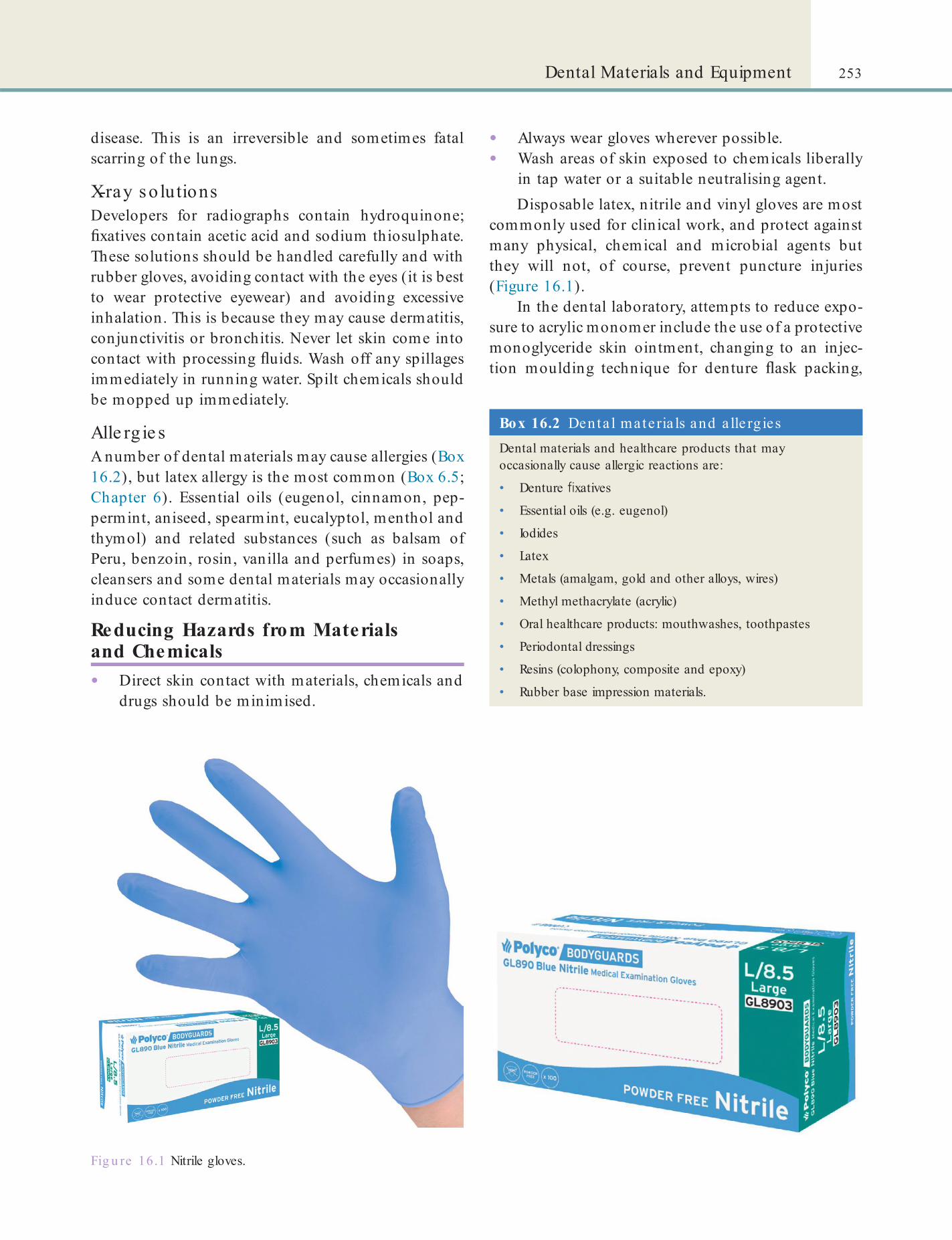
253Dental Materials and Equipment
disease. This is an irreversible and sometimes fatal scarring of the lungs.
X-ray s o lutionsDevelopers for radiographs contain hydroquinone; xatives contain acetic acid and sodium thiosulphate. These solutions should be handled carefully and with rubber gloves, avoiding contact with the eyes (it is best to wear protective eyewear) and avoiding excessive inhalation. This is because they may cause dermatitis, conjunctivitis or bronchitis. Never let skin come into contact with processing uids. Wash off any spillages immediately in running water. Spilt chemicals should be mopped up immediately.
Alle rg ie sA number of dental materials may cause allergies (Box 16.2), but latex allergy is the most common (Box 6.5; Chapter 6). Essential oils (eugenol, cinnamon, pep-permint, aniseed, spearmint, eucalyptol, menthol and thymol) and related substances (such as balsam of Peru, benzoin, rosin, vanilla and perfumes) in soaps, cleansers and some dental materials may occasionally induce contact dermatitis.
Reducing Hazards from Materials and Chemicals• Direct skin contact with materials, chemicals and
drugs should be minimised.
Box 16.2 Denta l mate ria ls and a lle rg iesDental materials and healthcare products that may occasionally cause allergic reactions are:
• Denture xatives
• Essential oils (e.g. eugenol)
• Iodides
• Latex
• Metals (amalgam, gold and other alloys, wires)
• Methyl methacrylate (acrylic)
• Oral healthcare products: mouthwashes, toothpastes
• Periodontal dressings
• Resins (colophony, composite and epoxy)
• Rubber base impression materials.
Fig u re 16.1 Nitrile gloves.
• Always wear gloves wherever possible.• Wash areas of skin exposed to chemicals liberally
in tap water or a suitable neutralising agent.Disposable latex, nitrile and vinyl gloves are most
commonly used for clinical work, and protect against many physical, chemical and microbial agents but they will not, of course, prevent puncture injuries (Figure 16.1).
In the dental laboratory, attempts to reduce expo-sure to acrylic monomer include the use of a protective monoglyceride skin ointment, changing to an injec-tion moulding technique for denture ask packing,
254 CHAPTER • 16 • Mosby’s Textbook of Dental Nursing
Key poin tDental equipment is often precision equipment and usually very expensive, so take care neither to damage nor lose any of it. It must be cleaned and maintained carefully.
Sa fe use of mate ria ls and chemica ls• Never use domestic bottles or containers to hold toxic
materials.
• Toxic agents and poisons must be stored in appropriate leak-proof containers with correct labelling stating:
• The identity of the substance
• Appropriate hazard warnings
• Information on how to handle accidental exposure to the substance.
• Drugs must be kept in a locked receptacle (the Medicines Act 1968). Any drug and material safety datasheets should be retained for information about the agent and good work practices.
• Any unmarked materials or drugs should be disposed safely: never re-label bottles incorrectly
• Spills should be readily cleaned up.
or using a no-touch technique. Polymer (especially polyethylene) gloves are used for some laboratory work. They do not completely prevent penetration of some organic solvents such as methyl methacrylate monomer. It is likely that there is little monomer pen-etration of polyethylene gloves during the short period of mixing acrylic, and, for clinical work, butyl rubber gloves seem more impervious.
Wear heavy duty rubber gloves at other times – except when re-resistant gloves are more appropriate.
Dental EquipmentWhen selecting new equipment for the workplace, as a dental nurse you can think about:
• What you want the equipment to do – will the equipment selected be t for this purpose? Is there any evidence? Is it compatible with other equipment in the surgery?
• How easy it will be to use and maintain – is it CE marked (to demonstrate it conforms to European Standards)?
• How easy it is to decontaminate – what are the manufacturer’s recommendations? When selecting new hand instruments avoid dif cult-to-clean serrated handles and check that hinges are easy to clean.
• Can the material covering the dental chair and work surfaces be cleaned and disinfected regularly without deterioration? Check with the manufacturer.
• Select foot-controlled equipment whenever possible.
• Training – is it required? Will the manufacturer provide it?
Care and Maintenance of Dental Instruments and Equipment
Key poin tInstruments should be cleared away immediately after use. If you cannot clean instruments immediately, soak them in a non-ionic solution.
Decontamination of dental instruments and equipment is discussed in Chapter 7 and has three stages:1 . Pre-sterilisation cleaning2. Sterilisation3 . Storage.
Pre -s te rilis a tion c le aningInstruments must be cleaned or the sterilisation process will not be effective. Cleaning can be done either manually or by using an ultrasonic bath or thermal washer. It is good practice to use an automated washing process with a thermal washer-disinfector whenever practical for the following reasons:
• It is a more controlled process than manual washing
• It reduces the risk in handling contaminated instruments
• It increases productivity by allowing more clinical time.
The cycle time may vary so additional instruments may be required.
The approximate cycle for a thermal washer-disinfector or sterilisation in a vacuum steriliser takes 2 hours.
Manual cleaningBefore you manually clean instruments, make yourself familiar with the written local policy for staff for manual cleaning. You will also be given appropriate training. Damaged instruments are more dif cult to clean and corrosion may reduce the life of an instrument.
1 . Place the instruments in a dedicated deep sink and not the basin used for hand washing.
2 . Take care when handling sharp instruments.

255Dental Materials and Equipment
3 . Put on protective clothing:• Disposable apron• Visor or face mask• Heavy duty gloves.
4 . Use a non-foaming detergent.5 . Use a nylon brush (which can be sterilised) or a
disposable brush.6 . Fill the sink with lukewarm water.7 . Immerse instruments and clean below the
water line.8 . Some instruments may require disassembling
rst.9 . Inspect instruments after cleaning and repeat
cleaning if required.10 . Dry instruments with a disposable paper towel
– wet instruments inhibit the sterilisation process.
11 . Place in the receptacle for sterilisation.
Mechanica l cleaningUltrasonic baths are effective for removing blood and debris from intricate, serrated and jointed instruments. They are also effective for removing cement.1 . In the deep sink, rinse off blood and debris
below the water line using lukewarm water.2 . Open jointed instruments and/or dismantle
instruments and place in the basket. Use warm water according to the manufacturer’s instructions, for example, 40 ° C for 3 minutes.
3 . Use the detergent as per manufacturer’s instructions, for example, low temperature and low foaming.
4 . Rinse thoroughly below the water line in clean water to remove detergent if the machine does not have a rinse cycle.
5 . Drain and dry instruments.6 . Inspect instruments and repeat if necessary.7 . Always operate with the lid on.
Thermal was her-dis infectorsBench-top thermal washer-disinfectors are suitable for the dental surgery. The type, size and model depends on the workload, time requirements and space.
Cycle• Pre-wash below 45 ° C• Main wash• Thermal disinfection 80–90 ° C• Post disinfection.
Cle aning of handpie c e sPre-sterilisation dental handpiece cleaning machines are recommended by the BDA. Many handpieces are thermal/disinfectant safe, but always refer to the manufacturer’s instructions.
Good pract ice when using u lt rasonic ba ths• Empty the bath every 4 hours or sooner if the solution is
contaminated.
• Empty, clean and dry at the end of the day.
• Test weekly by placing a foil strip in forceps in the centre of the bath for 3 minutes. Inspect the foil: the edges should be serrated and pitted or the centre should be perforated. The manufacturer should provide a test kit.
• A quali ed designated person should regularly maintain and test the bath.
Manual cleaning of handpieces1 . Flush handpiece with water from the water line
for 20 seconds.2 . Remove the bur.3 . Do not immerse in disinfectant or place in
ultrasonic bath.4 . Wipe outer surface with an alcohol wipe
(isopropyl alcohol).5 . Dry water channel with compressed air.
Inte rnal cleaning of handpiece1. Check oil canister has the correct attachment.2 . Shake the oil can.3 . Lay on a at surface and hold the handpiece
head with a paper towel.4 . Insert nozzle into the end of the handpiece.5 . Spray oil for approximately one second.6 . Place handpiece downwards to drain off
excess oil.7 . Clean off any excess.8 . Steam sterilise at 134–137 ° C for 3 minutes.9 . Lubricate after sterilisation if manufacturer
recommends.
Key poin tHandpieces should be lubricated with service oil after the thermal disinfector stage.

This page intentiona lly le ft blank

Chapter 17 Restorative Procedures and Materials
CHAPTER POINTS• Instrumentation• Role of the dental nurse• Gingival and periodontal disease treatment
(periodontics)• Dental impressions• Repairing damaged, diseased or missing teeth
(prosthodontics)• Dental cements• Dental restorations• Dental implants• Dentures• Management of pulp disease (endodontics)• Cosmetic (aesthetic) dental procedures• Mouthguards and nightguards
InstrumentationThe dental handpiece is a small, high-speed drill called an air rotor, used to remove caries, old or temporary llings or crowns and shape tooth structure; it is also used during endodontic, periodontic or surgical treatment.
Mechanical hand drills were originally used and then replaced by foot-powered dental drills. The rst electric dental drill invented could reach speeds of up to 3000 rpm. The modern dental drill is the air turbine (air rotor) handpiece, which operates with a 400 000 rpm ‘high-speed’ handpiece for precision work and is used along with a ‘low-speed’ contra-angled handpiece operating at a speed dictated by a micromotor (max up to 40 000 rpm), which creates the momentum for uses requiring higher torque than the high-speed handpiece can deliver. Some air rotors can work up to 800 000 rpm.
Handpieces carry a dental bur. The bur has a shank, neck and head – the latter containing blades made of carbon steel, tungsten carbide or diamond. The latter two are the most durable: tungsten carbide burs cut the tooth away, while diamond burs grind the tooth away. Speci c tasks require special burs, so that various burs are available, including round, inverted
In t ro d u ct io n
Restorative dentistry is involved in treatment of the common dental diseases – dental caries and periodon-tal disease – and their complications, including loss of teeth. Thus the specialty of restorative dentistry includes periodontics, endodontics and prosthodon-tics. This is the major part of the workload for most dental practices. Restorative dentistry is carried out by the dentist or, in the case of some procedures, by the dental therapist or dental hygienist (see Chapter 2).
Prosthodontics is also sometimes called ‘conserva-tion’ or ‘conservative dentistry’; f xed prosthodontics refers to subjects such as dental bridges while remov-able prosthodontics refers to removable partial or full (complete) dentures.
Terms to lea rnPeriodontics: the specialty of dentistry concerned with the study and management of gingival and periodontal diseases.
Endodontics: the dental specialty concerned with the management of the diseases of the pulp.
Prosthodontics: the specialty of dentistry concerned with repairing and replacing damaged, diseased or missing teeth or other oral structures.
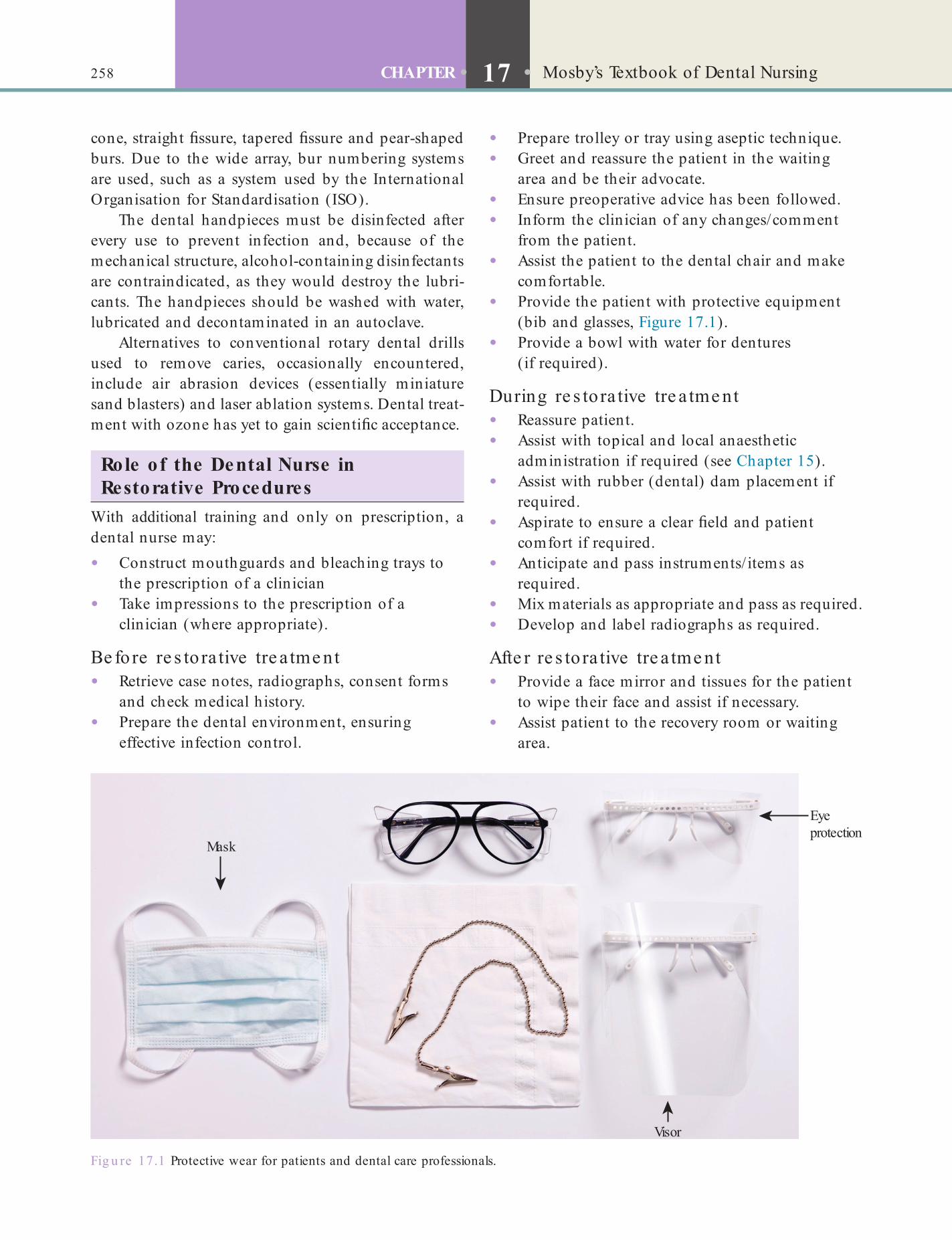
258 CHAPTER • 17 • Mosby’s Textbook of Dental Nursing
• Prepare trolley or tray using aseptic technique.• Greet and reassure the patient in the waiting
area and be their advocate.• Ensure preoperative advice has been followed.• Inform the clinician of any changes/comment
from the patient.• Assist the patient to the dental chair and make
comfortable.• Provide the patient with protective equipment
(bib and glasses, Figure 17.1).• Provide a bowl with water for dentures
(if required).
During re s to ra tive tre a tme nt• Reassure patient.• Assist with topical and local anaesthetic
administration if required (see Chapter 15).• Assist with rubber (dental) dam placement if
required.• Aspirate to ensure a clear eld and patient
comfort if required.• Anticipate and pass instruments/ items as
required.• Mix materials as appropriate and pass as required.• Develop and label radiographs as required.
Afte r re s to ra tive tre a tme nt• Provide a face mirror and tissues for the patient
to wipe their face and assist if necessary.• Assist patient to the recovery room or waiting
area.
cone, straight ssure, tapered ssure and pear-shaped burs. Due to the wide array, bur numbering systems are used, such as a system used by the International Organisation for Standardisation (ISO).
The dental handpieces must be disinfected after every use to prevent infection and, because of the mechanical structure, alcohol-containing disinfectants are contraindicated, as they would destroy the lubri-cants. The handpieces should be washed with water, lubricated and decontaminated in an autoclave.
Alternatives to conventional rotary dental drills used to remove caries, occasionally encountered, include air abrasion devices (essentially miniature sand blasters) and laser ablation systems. Dental treat-ment with ozone has yet to gain scienti c acceptance.
Role o f the Dental Nurse in Restorative Procedures
With additional training and only on prescription, a dental nurse may:
• Construct mouthguards and bleaching trays to the prescription of a clinician
• Take impressions to the prescription of a clinician (where appropriate).
Be fo re re s to ra tive tre a tme nt• Retrieve case notes, radiographs, consent forms
and check medical history.• Prepare the dental environment, ensuring
effective infection control.
Fig u re 17.1 Protective wear for patients and dental care professionals.
Visor
Eyeprotection
Mask
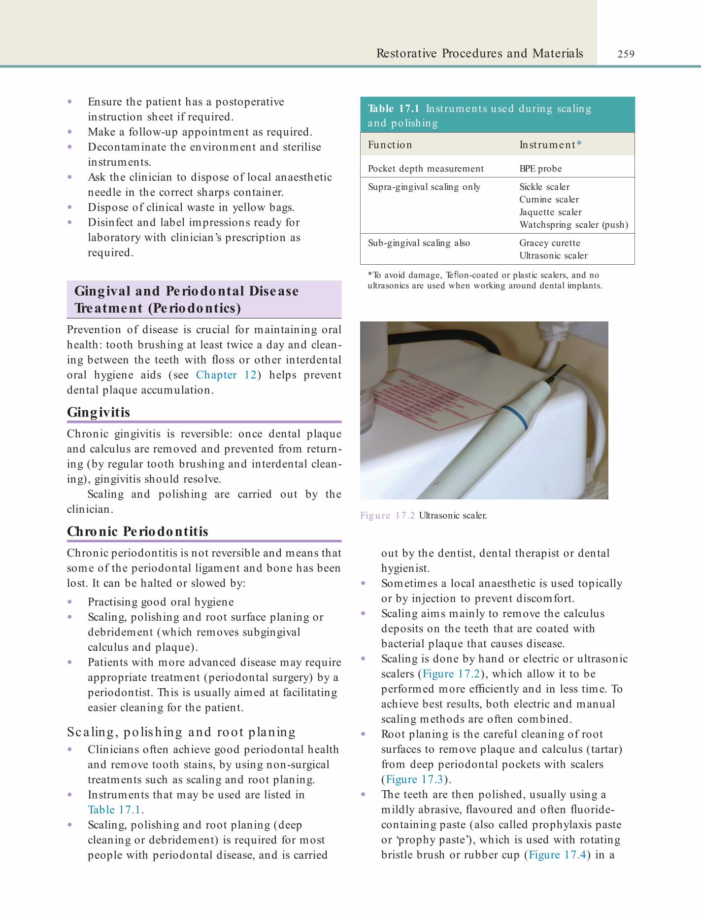
259Restorative Procedures and Materials
• Ensure the patient has a postoperative instruction sheet if required.
• Make a follow-up appointment as required.• Decontaminate the environment and sterilise
instruments.• Ask the clinician to dispose of local anaesthetic
needle in the correct sharps container.• Dispose of clinical waste in yellow bags.• Disinfect and label impressions ready for
laboratory with clinician’s prescription as required.
Gingival and Periodontal Disease Treatment (Periodontics)
Prevention of disease is crucial for maintaining oral health: tooth brushing at least twice a day and clean-ing between the teeth with oss or other interdental oral hygiene aids (see Chapter 12) helps prevent dental plaque accumulation.
GingivitisChronic gingivitis is reversible: once dental plaque and calculus are removed and prevented from return-ing (by regular tooth brushing and interdental clean-ing), gingivitis should resolve.
Scaling and polishing are carried out by the clinician.
Chronic PeriodontitisChronic periodontitis is not reversible and means that some of the periodontal ligament and bone has been lost. It can be halted or slowed by:
• Practising good oral hygiene• Scaling, polishing and root surface planing or
debridement (which removes subgingival calculus and plaque).
• Patients with more advanced disease may require appropriate treatment (periodontal surgery) by a periodontist. This is usually aimed at facilitating easier cleaning for the patient.
Sc a ling , po lis hing and roo t p laning• Clinicians often achieve good periodontal health
and remove tooth stains, by using non-surgical treatments such as scaling and root planing.
• Instruments that may be used are listed in Table 17.1.
• Scaling, polishing and root planing (deep cleaning or debridement) is required for most people with periodontal disease, and is carried
Table 17.1 Inst ruments used during sca ling and po lishing
Funct ion Inst rument*
Pocket depth measurement BPE probe
Supra-gingival scaling only Sickle scalerCumine scalerJaquette scalerWatchspring scaler (push)
Sub-gingival scaling also Gracey curetteUltrasonic scaler
*To avoid damage, Te on-coated or plastic scalers, and no ultrasonics are used when working around dental implants.
out by the dentist, dental therapist or dental hygienist.
• Sometimes a local anaesthetic is used topically or by injection to prevent discomfort.
• Scaling aims mainly to remove the calculus deposits on the teeth that are coated with bacterial plaque that causes disease.
• Scaling is done by hand or electric or ultrasonic scalers (Figure 17.2), which allow it to be performed more ef ciently and in less time. To achieve best results, both electric and manual scaling methods are often combined.
• Root planing is the careful cleaning of root surfaces to remove plaque and calculus (tartar) from deep periodontal pockets with scalers (Figure 17.3).
• The teeth are then polished, usually using a mildly abrasive, avoured and often uoride-containing paste (also called prophylaxis paste or ‘prophy paste’), which is used with rotating bristle brush or rubber cup (Figure 17.4) in a
Fig u re 17.2 Ultrasonic scaler.

260 CHAPTER • 17 • Mosby’s Textbook of Dental Nursing
Fig u re 17.3 A selection of scaling instruments. How many can you recognise?
Fig u re 17.4 (A) Latch grip rubber cup and (B) bristle brush.
low-speed dental handpiece. Air polishing works by spraying high-pressured water mixed with baking soda paste onto the tooth surfaces to remove residue and plaque while baking soda removes stains.
• In aggressive periodontal cases scaling may be followed by adjunctive therapy such as antimicrobials delivered locally.
• Most patients with chronic periodontitis require ongoing maintenance therapy for sustained good oral health.
Pe riodonta l s urge ryPeriodontal surgery may be needed to:
• Gain access to remove calculus from deep periodontal pockets
• Smooth root surfaces• Reduce the pocket depths• Arrange gingival tissue into a shape that will
enable easier access and facilitate oral hygiene practices.It is also carried out to correct gingival recession
and elongation of the clinical crown (the length of tooth visible in the mouth) of a tooth.
Periodontal surgical procedures require the use of anaesthesia, and include the following.
GingivectomyIn gingivectomy, the clinician removes and reshapes loose, diseased gingival tissue to eliminate pockets. Under local anaesthesia, the tissue is cut with laser or scalpel. The wound is dressed with a periodontal dressing, for example Coepak.
Flap s urgeryFlap surgery also allows the removal and reshaping of loose, diseased gingival tissue as well as providing access to clean deep (infrabony) pockets. The ap is replaced so as to remove pockets. Under local

261Restorative Procedures and Materials
Figure 17.5 shows a selection of periodontal surgi-cal instruments. Being surgical operations, the usual pre- and post-operative care is indicated and complica-tions can be similar.
anaesthesia, the ap is raised and tissue is removed with curettes and the ap sutured and dressed with, for example, Coepak.
Periodonta l regenera tive therapyPeriodontal regenerative therapy uses a bone graft material or bone regenerative material (e.g. Emdog-ain) to restore bony defects deep within the pockets in a technique similar to a ap operation. It may also be necessary to place a special membrane such as Gore-Tex over the graft to protect the material and to help it regenerate the tissues (guided tissue regenera-tion; GTR).
Fig u re 17.5 A selection of periodontal surgical instruments. (A) Blakes gingivectomy knife; (B) shortened Pritchard periosteal elevator; (C) Molt no. 9 periosteal elevator; (D) Goldman Fox no. 7 heavy periodontal knife; (E) Buck 5/6 periodontal knife; (F) Gracey curette 7/8; (G) Gracey curette 11/12; (H) Gracey curette 13/14; (I) Pritchard surgical curette; (J) Fedi 2 bone chisel; (K) Rhodes back action bone chisel.
Term to lea rnImpression material: a viscous mouldable material that sets to a rm consistency in the mouth to make an ‘impression’ of the teeth and surrounding structures.
Dental Impressions

262 CHAPTER • 17 • Mosby’s Textbook of Dental Nursing
Fig u re 17.6 Upper and lower impression trays.
Fig u re 17.7 Upper and lower dental models.
Primary and secondary impressionsPrimary impressions, which are the initial impressions, are often taken using alginates in a stock impression tray (see Figure 17.6). The resulting cast is then used by the dental technician to make a special tray. Then a secondary impression is made – using a more accurate impression material such as an elastomer (see Chapter 16) – in the special tray.
Terms to lea rnInlay: a restoration (such as gold or porcelain) tted into a cavity and cemented into place.
Onlay: a restoration (such as gold or porcelain) tted over the occlusal surface of a tooth and cemented into place.
Crow n: a crown is like a cap that completely or partially covers a tooth’s natural crown.
CAD-CAM dentistry: Computer-Aided Design and Computer-Aided Manufacturing in dentistry.
A dental impression is an accurate representation of part or all of a person’s dentition and adjacent tissue of the mouth. A dental impression is usually made by placing an impression material into the mouth, usually in an impression tray (Figure 17.6). The impression material then sets or hardens so that, when removed from the mouth, it retains the shape of the teeth and/or mouth. It forms a ‘negative’ of a person’s teeth and adjacent soft tissues, which is then used to make a cast or model (Figure 17.7) of the dentition, usually from dental plaster.
The model may be used either as a record of the person’s dentition (called a study model) or by the dental technician to make:
• Special impression trays that more closely t the individual patient, in preparation for making dentures
• Indirect restorations, e.g. inlays, onlays, crowns (these are all discussed later in the chapter)
• Other dental prostheses or appliances.There are several different kinds of impression
materials and their properties are described in detail below and in Chapter 16. Impressions are increasingly being made using computers – the basis of CAD-CAM (Computer-Aided Design and Computer-Aided Manu-facturing) dentistry.
Role of the Dental Nurse in Impression TakingThe dental nurse will need to offer a range of impres-sion trays of different sizes for the clinician to select for use. Differently shaped trays are available for people with or without natural teeth (see Figure 17.8). In order that the impression material once set remains rmly in position in the tray, the tray may be perfo-rated, rim locked and/or you may need to apply a special adhesive to the tray before use. You then mix the impression material and place it in the impression tray chosen. With suitable training, dental nurses can also take certain impressions.
You must wash and disinfect the impressions once they are removed from the mouth and label them before despatching to the laboratory with a prescription:1 . Rinse under running cold tap water to remove
blood/saliva.2 . Disinfect as appropriate (see Box 17.1).3 . Seal in plastic biological hazard bag with label
(name, date of return and prescription).
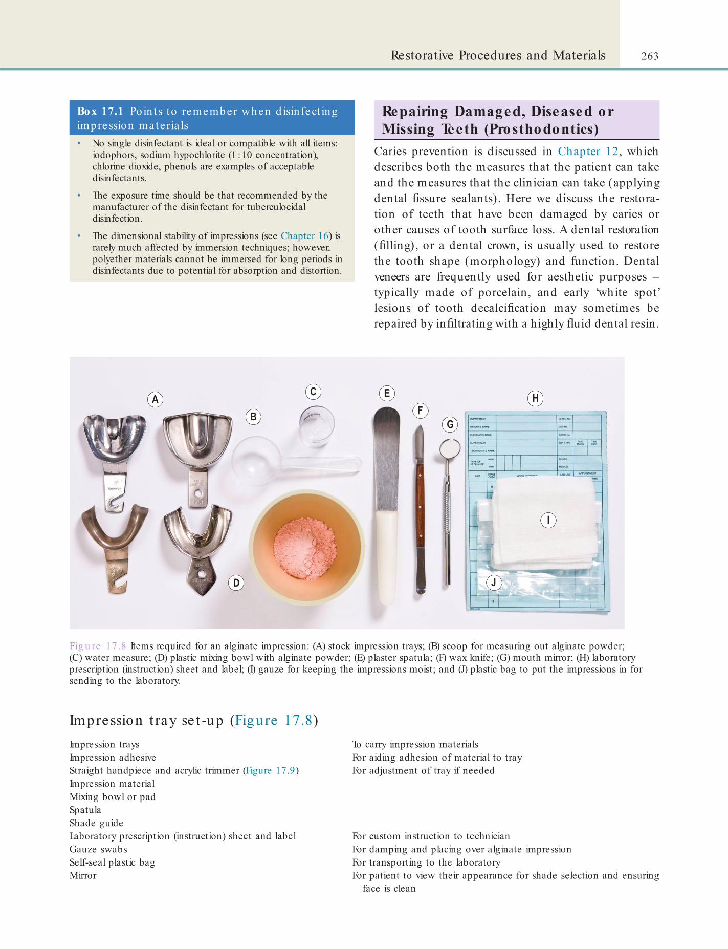
263Restorative Procedures and Materials
Box 17.1 Po in ts to remember when d isin fect ing impression materia ls• No single disinfectant is ideal or compatible with all items:
iodophors, sodium hypochlorite (1 : 10 concentration), chlorine dioxide, phenols are examples of acceptable disinfectants.
• The exposure time should be that recommended by the manufacturer of the disinfectant for tuberculocidal disinfection.
• The dimensional stability of impressions (see Chapter 16) is rarely much affected by immersion techniques; however, polyether materials cannot be immersed for long periods in disinfectants due to potential for absorption and distortion.
Impression t ray se t -up (Figure 17.8)Impression trays To carry impression materialsImpression adhesive For aiding adhesion of material to trayStraight handpiece and acrylic trimmer (Figure 17.9) For adjustment of tray if neededImpression materialMixing bowl or padSpatulaShade guideLaboratory prescription (instruction) sheet and label For custom instruction to technicianGauze swabs For damping and placing over alginate impressionSelf-seal plastic bag For transporting to the laboratoryMirror For patient to view their appearance for shade selection and ensuring
face is clean
Fig u re 17.8 Items required for an alginate impression: (A) stock impression trays; (B) scoop for measuring out alginate powder; (C) water measure; (D) plastic mixing bowl with alginate powder; (E) plaster spatula; (F) wax knife; (G) mouth mirror; (H) laboratory prescription (instruction) sheet and label; (I) gauze for keeping the impressions moist; and (J) plastic bag to put the impressions in for sending to the laboratory.
Repairing Damaged, Diseased or Missing Teeth (Prosthodontics)
Caries prevention is discussed in Chapter 12, which describes both the measures that the patient can take and the measures that the clinician can take (applying dental ssure sealants). Here we discuss the restora-tion of teeth that have been damaged by caries or other causes of tooth surface loss. A dental restoration ( lling), or a dental crown, is usually used to restore the tooth shape (morphology) and function. Dental veneers are frequently used for aesthetic purposes – typically made of porcelain, and early ‘white spot’ lesions of tooth decalci cation may sometimes be repaired by in ltrating with a highly uid dental resin.

264 CHAPTER • 17 • Mosby’s Textbook of Dental Nursing
Box 17.2 Methods of moisture cont ro l• Dental dam placed on the teeth being treated.
• High volume suction for aspiration by the dental nurse.
• Saliva ejector in the oor of the mouth.
• Cotton wool rolls placed near the openings of the major salivary gland ducts inside the cheeks, and in the oor of the mouth.
• ‘Dry guards’ placed in the buccal and lingual sulci.
Fig u re 17.9 (A) Acrylic trimmer; (B) Straight handpiece.
Composite resins and dental amalgams are com-monly used materials for llings. Crowns can be of porcelain, gold or other materials. A dental bridge, implant or denture is usually used to replace missing teeth. Dental bridges are made from resins or ceramics or are metal-based; implants are often metal (tita-nium). Dentures are usually resin-based – sometimes based on a metal base (cobalt-chrome usually).
Restorations, bridges, implants or dentures have to be custom-made for every patient, because each patient presents with a unique colour, shape and size of their teeth and relationship of their upper and lower jaws. Each patient also has their own unique aesthet-ics, depending on the colour, size and shape of their teeth, face and jaws and tone of their facial muscles.
The Role o f the Dental Nurse
Re moving the c a rie sThe dental nurse will assist the clinician, mainly by controlling moisture and debris; the clinician removes
the caries and prepares the tooth for a restoration (this can be a lling, inlay, crown or bridge).
Mois ture c ontro l and de nta l (rubbe r) damMoisture control is very important for two reasons:
• To allow the clinician to be able to see clearly what they are doing in the mouth
• To stop moisture damaging the restoration or substances needed for adherence to the tooth of cements, sealants, restorations or orthodontic devices (Table 17.2).
Moisture control is also crucial in endodontics to prevent spread of infection.
When preparing and placing llings there are several ways to control moisture (Box 17.2). Use of rubber dam (or dental dam, Figure 17.10) is the most effective, use of a saliva ejector and high vacuum suction is easier and, for simple procedures, cotton wool rolls or ‘dry guards’ placed around the operative area may suf ce.
The saliva ejector is a strawlike suction tool, placed under the tongue on the side opposite where the clini-cian is working, to remove saliva pooling in the oor of the mouth. The high vacuum suction also removes saliva and debris and may reduce aerosols.
The dental dam is a thin square of rubber (latex, or silicone for latex-sensitive patients, and of different colours and thicknesses) which isolates a tooth or teeth from saliva and the mouth, helps infection
Table 17.2 Damaging e ffects o f moisture on the se t t ing of restora t ions
Mate ria ls Effects o f moisture
Amalgam Expansion of material
Cement Fails to adhere to tooth structure; setting time altered
Composite Fails to adhere to tooth structure
Glass ionomer Fails to set properly
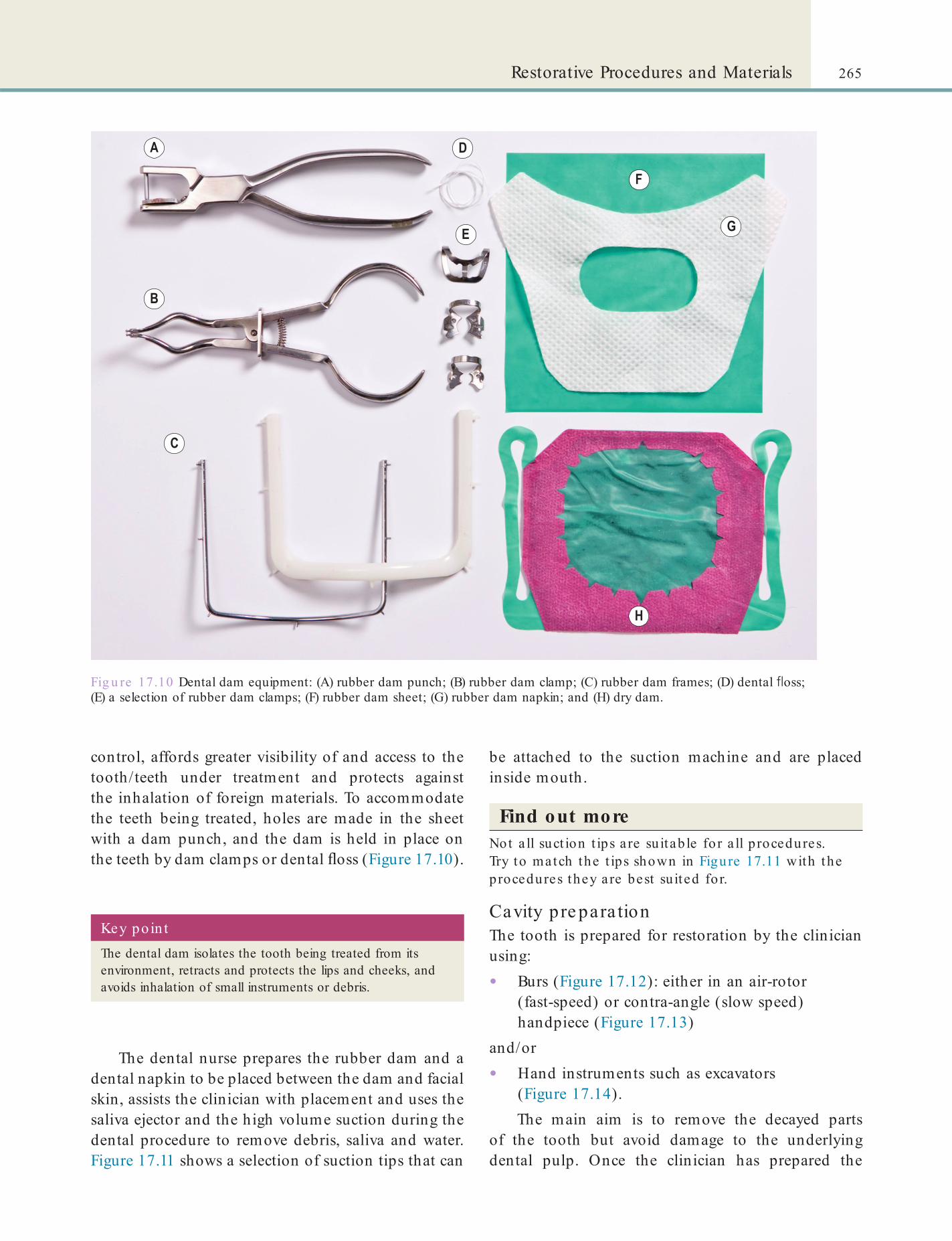
265Restorative Procedures and Materials
Fig u re 17.10 Dental dam equipment: (A) rubber dam punch; (B) rubber dam clamp; (C) rubber dam frames; (D) dental oss; (E) a selection of rubber dam clamps; (F) rubber dam sheet; (G) rubber dam napkin; and (H) dry dam.
control, affords greater visibility of and access to the tooth/teeth under treatment and protects against the inhalation of foreign materials. To accommodate the teeth being treated, holes are made in the sheet with a dam punch, and the dam is held in place on the teeth by dam clamps or dental oss (Figure 17.10).
Key poin tThe dental dam isolates the tooth being treated from its environment, retracts and protects the lips and cheeks, and avoids inhalation of small instruments or debris.
The dental nurse prepares the rubber dam and a dental napkin to be placed between the dam and facial skin, assists the clinician with placement and uses the saliva ejector and the high volume suction during the dental procedure to remove debris, saliva and water. Figure 17.11 shows a selection of suction tips that can
be attached to the suction machine and are placed inside mouth.
Find out moreNot a ll suct ion t ips a re su itab le fo r a ll procedures. Try to match the t ips shown in Figure 17.11 with the procedures they a re best su ited for.
Cavity pre para tionThe tooth is prepared for restoration by the clinician using:
• Burs (Figure 17.12): either in an air-rotor (fast-speed) or contra-angle (slow speed) handpiece (Figure 17.13)
and/or
• Hand instruments such as excavators (Figure 17.14).The main aim is to remove the decayed parts
of the tooth but avoid damage to the underlying dental pulp. Once the clinician has prepared the

266 CHAPTER • 17 • Mosby’s Textbook of Dental Nursing
Fig u re 17.11 A selection of suction tips.
Fig u re 17.12 (A) A diamond bur and (B) a steel bur.
Fig u re 17 .13 A selection of air rotor and latch-grip, contra-angle handpieces.
cavity, the pulp is protected by applying a lining cement with an instrument called a ‘plastic’ (labelled H in Figure 17.10).
De nta l c e me ntsDental cements are used to:
• Line deep cavities to protect the pulp from thermal and other types of irritation.
• Secure indirect restorations (crowns, inlays, bridges) into or on the teeth (often called luting cement).Cements are described in Chapter 16. They are
usually prepared by the dental nurse. Several cements are supplied in capsules containing pre-proportioned powder and liquid. These are not only convenient but ensure consistent powder/ liquid ratios and thus
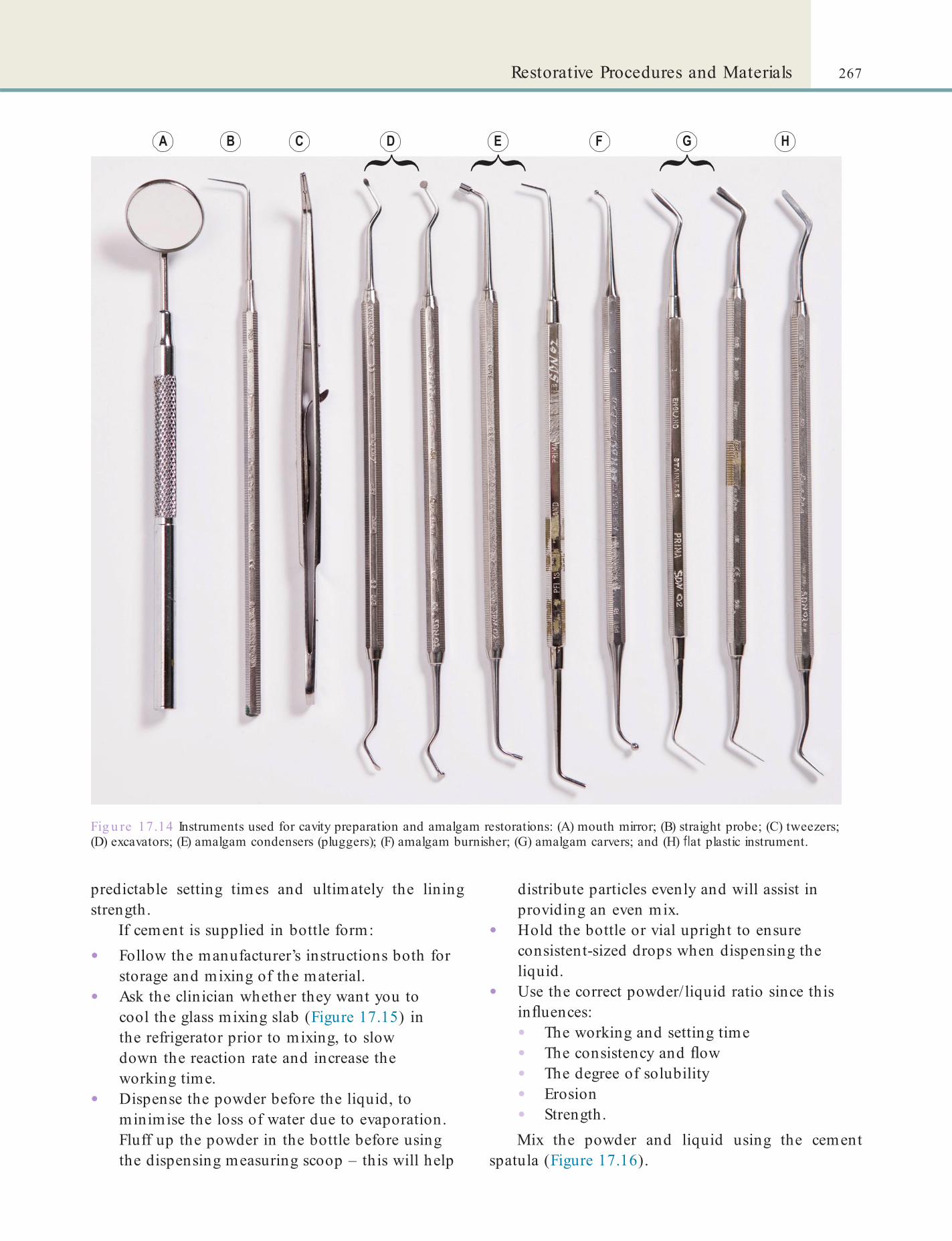
267Restorative Procedures and Materials
Fig u re 17.14 Instruments used for cavity preparation and amalgam restorations: (A) mouth mirror; (B) straight probe; (C) tweezers; (D) excavators; (E) amalgam condensers (pluggers); (F) amalgam burnisher; (G) amalgam carvers; and (H) at plastic instrument.
predictable setting times and ultimately the lining strength.
If cement is supplied in bottle form:
• Follow the manufacturer’s instructions both for storage and mixing of the material.
• Ask the clinician whether they want you to cool the glass mixing slab (Figure 17.15) in the refrigerator prior to mixing, to slow down the reaction rate and increase the working time.
• Dispense the powder before the liquid, to minimise the loss of water due to evaporation. Fluff up the powder in the bottle before using the dispensing measuring scoop – this will help
distribute particles evenly and will assist in providing an even mix.
• Hold the bottle or vial upright to ensure consistent-sized drops when dispensing the liquid.
• Use the correct powder/ liquid ratio since this in uences:• The working and setting time• The consistency and ow• The degree of solubility• Erosion• Strength.Mix the powder and liquid using the cement
spatula (Figure 17.16).

268 CHAPTER • 17 • Mosby’s Textbook of Dental Nursing
Fig u re 17.15 Selection of glass slabs and paper pads for mixing dental cements.
Fig u re 17.16 Cement spatulas.
Term to lea rnPreparation: a tooth that has been carefully shaped with a dental handpiece for receiving a crown or lling.
Factors tha t in uence the pe rformance o f cements• Incorporating too much or too little powder.
• Room or mixing slab temperature too high.
• Premature exposure of cement to moisture.
• Delay between completion of the mix and seating in restoration.
• Surface contamination of the crown or tooth preparation.
• Whether the cement is adhesive and the tooth surface has been etched.
Dental RestorationsThese can be divided into:
• Direct restorations (made in the surgery)• Indirect restorations (made in the laboratory).They can be further classi ed by their location and size, or type of material used as discussed below and in Chapter 16.
Direct dental restorations are usually made by the clinician in the surgery, by lling the cavity with a material such as amalgam or a plastic resin, which then sets hard. Indirect restorations are made by the technician in the laboratory. Then the clinician must
usually rst take an impression of the tooth cavity so that the technician can make a die on which to produce the restoration. This is then cemented by the clinician into the cavity, using cement as above. Occa-sionally, a clinician will make a wax restoration in the mouth which is then sent to the laboratory for the technician to cast in gold.

269Restorative Procedures and Materials
The main advantages of the indirect method of tooth restoration are that the materials used in these restorations have better mechanical properties (such as strength) than do those used for direct restorations. Also much of the work is done away from the dental chairside by the technician, on a die created from an impression of the teeth.
Direct RestorationsProvisional or temporary restorations are made as a tem-porary measure during the course of complex treat-ments or when diagnosing pain. For these restorations the materials used are relatively soft and easy to remove, such as zinc oxide eugenol (see Chapter 16) are applied with special applicators (Figure 17.17).
Common materials used for permanent restorations are: dental amalgam, composite resins and glass
Term to lea rnDie: this is a positive likeness of the teeth that is usually composed of a special kind of plaster called dental stone, which is much harder than plaster of Paris.
Fig u re 17.17 (A) Dycal applicator; (B) Thymozine probe.
ionomer cements. The properties of all these materials are described in Chapter 16. Here we describe the role of the dental nurse in handling of these materials in the surgery.
Amalgam re s to ra tionsThe cavity preparation for an amalgam lling is fairly destructive. This means some sound tooth structure must be removed to ensure the cavity will mechani-cally retain the lling when set. This is because amalgam does not adhere to tooth structure.
Steps in p lacing an amalgam lling1. The dental dam is placed and the cavity
prepared.2 . In teeth with loss of marginal tooth structure the
cavity may lack a wall on one or more sides. In such cases, offer the clinician a matrix band (Figure 17.18) and wedges (Figure 17.19). The clinician will use these to form a temporary wall when packing the lling. Disposable single use matrix bands should now be used if the older types are impossible to clean and decontaminate.
3 . Mix the amalgam in an amalgamator by placing a capsule (Figure 17.20) in it.
4 . Fill the amalgam carrier (Figure 17.20) with the mixed amalgam and give it to the clinician:• The clinician places the amalgam in the
cavity.• Amalgam remains plastic for a short time
so it can be packed (condensed) to ll the cavity with the amalgam plugger (see Figure 17.14E) or automatic vibrator.
• Before it hardens to a strong lling, the clinician carves it to a tooth shape using amalgam carvers (see Figure 17.14G) and may smooth the surface with cotton pledgets or a burnisher (see Figure 17.14F).
• At a later appointment, the amalgam is polished.
5 . Amalgam waste must be disposed of safely (see Chapter 6).
Identify and learnIdent ify an amalgamator in your workplace and lea rn how it works. Try t t ing a meta l mat rix band in it s ho lder.
Find out moreWhat is used for polish ing amalgam?
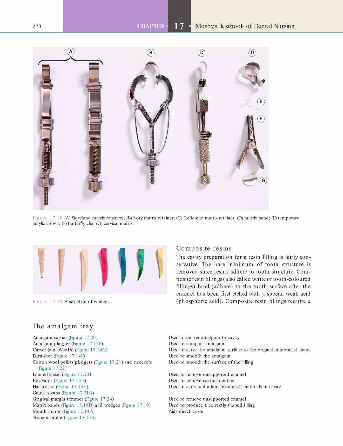
270 CHAPTER • 17 • Mosby’s Textbook of Dental Nursing
Fig u re 17.18 (A) Siqveland matrix retainers; (B) Ivory matrix retainer; (C) Tof emire matrix retainer; (D) matrix band; (E) temporary acrylic crown; (F) butter y clip; (G) cervical matrix.
Fig u re 17.19 A selection of wedges.
The amalgam t rayAmalgam carrier (Figure 17.20) Used to deliver amalgam to cavityAmalgam plugger (Figure 17.14E) Used to compact amalgamCarver (e.g. Ward’s) (Figure 17.14G) Used to carve the amalgam surface to the original anatomical shapeBurnisher (Figure 17.14F) Used to smooth the amalgamCotton wool pellets/pledgets (Figure 17.21) and tweezers
(Figure 17.22)Used to smooth the surface of the lling
Enamel chisel (Figure 17.23) Used to remove unsupported enamelExcavator (Figure 17.14D) Used to remove carious dentineFlat plastic (Figure 17.14H) Used to carry and adapt restorative materials to cavityGauze swabs (Figure 17.21A)Gingival margin trimmer (Figure 17.24) Used to remove unsupported enamelMatrix bands (Figure 17.18D) and wedges (Figure 17.19) Used to produce a correctly shaped llingMouth mirror (Figure 17.14A) Aids direct visionStraight probe (Figure 17.14B)
Compos ite re s insThe cavity preparation for a resin lling is fairly con-servative. The bare minimum of tooth structure is removed since resins adhere to tooth structure. Com-posite resin llings (also called white or tooth-coloured llings) bond (adhere) to the tooth surface after the enamel has been rst etched with a special weak acid (phosphoric acid). Composite resin llings require a

271Restorative Procedures and Materials
Fig u re 17.20 (A) Amalgam capsules; (B) glass amalgam pot; (C) straight and right-angled amalgam carriers.
Fig u re 17.21 A selection of (A) gauze swabs; (B) cotton rolls and pledgets.
Fig u re 17.22 (A) Mouth mirror; (B) straight probe; (C) tweezers.
Fig u re 17 .23 A selection of enamel chisels.
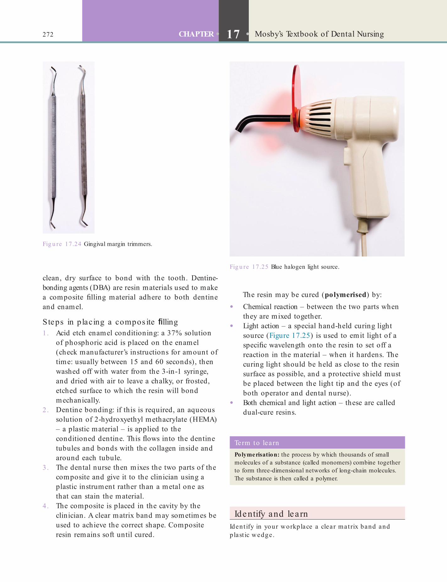
272 CHAPTER • 17 • Mosby’s Textbook of Dental Nursing
The resin may be cured (polymerised) by:
• Chemical reaction – between the two parts when they are mixed together.
• Light action – a special hand-held curing light source (Figure 17.25) is used to emit light of a speci c wavelength onto the resin to set off a reaction in the material – when it hardens. The curing light should be held as close to the resin surface as possible, and a protective shield must be placed between the light tip and the eyes (of both operator and dental nurse).
• Both chemical and light action – these are called dual-cure resins.
clean, dry surface to bond with the tooth. Dentine-bonding agents (DBA) are resin materials used to make a composite lling material adhere to both dentine and enamel.
Steps in p lacing a compos ite lling1. Acid etch enamel conditioning: a 37% solution
of phosphoric acid is placed on the enamel (check manufacturer’s instructions for amount of time: usually between 15 and 60 seconds), then washed off with water from the 3-in-1 syringe, and dried with air to leave a chalky, or frosted, etched surface to which the resin will bond mechanically.
2 . Dentine bonding: if this is required, an aqueous solution of 2-hydroxyethyl methacrylate (HEMA) – a plastic material – is applied to the conditioned dentine. This ows into the dentine tubules and bonds with the collagen inside and around each tubule.
3 . The dental nurse then mixes the two parts of the composite and give it to the clinician using a plastic instrument rather than a metal one as that can stain the material.
4 . The composite is placed in the cavity by the clinician. A clear matrix band may sometimes be used to achieve the correct shape. Composite resin remains soft until cured.
Fig u re 17.25 Blue halogen light source.
Term to lea rnPolymerisation: the process by which thousands of small molecules of a substance (called monomers) combine together to form three-dimensional networks of long-chain molecules. The substance is then called a polymer.
Fig u re 17.24 Gingival margin trimmers.
Identify and learnIdent ify in your workplace a clea r mat rix band and plast ic wedge .
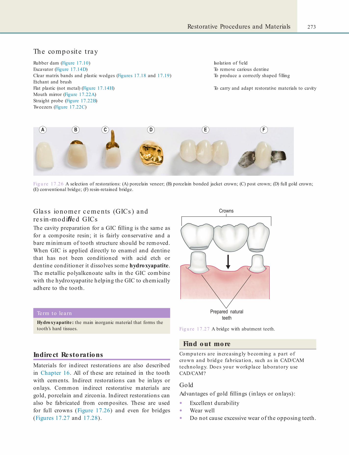
273Restorative Procedures and Materials
The composite t rayRubber dam (Figure 17.10) Isolation of eldExcavator (Figure 17.14D) To remove carious dentineClear matrix bands and plastic wedges (Figures 17.18 and 17.19) To produce a correctly shaped llingEtchant and brushFlat plastic (not metal) (Figure 17.14H) To carry and adapt restorative materials to cavityMouth mirror (Figure 17.22A)Straight probe (Figure 17.22B)Tweezers (Figure 17.22C)
Glas s ionome r c e me nts (GICs ) and re s in-modi e d GICsThe cavity preparation for a GIC lling is the same as for a composite resin; it is fairly conservative and a bare minimum of tooth structure should be removed. When GIC is applied directly to enamel and dentine that has not been conditioned with acid etch or dentine conditioner it dissolves some hydroxyapatite. The metallic polyalkenoate salts in the GIC combine with the hydroxyapatite helping the GIC to chemically adhere to the tooth.
Fig u re 17.26 A selection of restorations: (A) porcelain veneer; (B) porcelain bonded jacket crown; (C) post crown; (D) full gold crown; (E) conventional bridge; (F) resin-retained bridge.
Fig u re 17.27 A bridge with abutment teeth.
Term to lea rnHydroxyapatite: the main inorganic material that forms the tooth’s hard tissues.
Indirect RestorationsMaterials for indirect restorations are also described in Chapter 16. All of these are retained in the tooth with cements. Indirect restorations can be inlays or onlays. Common indirect restorative materials are gold, porcelain and zirconia. Indirect restorations can also be fabricated from composites. These are used for full crowns (Figure 17.26) and even for bridges (Figures 17.27 and 17.28).
Find out moreComputers a re increasing ly becoming a part o f crown and bridge fabrica t ion , such as in CAD/CAM technology. Does your workplace labora tory use CAD/CAM?
GoldAdvantages of gold llings (inlays or onlays):
• Excellent durability• Wear well• Do not cause excessive wear of the opposing teeth.

274 CHAPTER • 17 • Mosby’s Textbook of Dental Nursing
Fig u re 17.28 (A) Crown-cutting Bee bee scissors; (B) contouring pliers; (C) stainless steel temporary crowns.
Fig u re 17.29 (A) Bee bee scissors; (B) acrylic crowns for an incisor and premolar; (C) dividers.
Disadvantages:
• Unaesthetic• Expensive• Conduct heat and cold.
Porce la in (ceramic)Porcelain crowns usually have a ceramic coping of either alumina porcelain or zirconia, called the ‘core’. The porcelain crown covers the core. Porcelain is also used for veneers.
Advantages of porcelain restorations:
• Excellent aesthetics.Disadvantages:
• Brittle and hard, and can break• Can cause wear on opposing teeth because they
are harder than enamel.
CrownsTemporary crownsTemporary crowns (Figure 17.29) are used to cover the tooth while the laboratory is making the permanent gold or porcelain crowns.
Full gold crownsFull gold crowns (FGCs; Figure 17.26D) are cast from gold alloy in the laboratory, using the lost-wax technique:1 . After the clinician prepares the tooth for a
crown, he or she will take an impression.2 . This is sent to a dental laboratory where a die
is made.3 . The dental laboratory technician will then build
up on the die the pattern of the crown restoration in wax.
4 . This wax pattern is then invested in a special investment material, and placed in a furnace to burn off the wax to leave a space within the investment material.
5 . This investment pattern is then placed in a centrifuge where gold alloy is melted down and rapidly shot through into the investment to make the gold crown.
Porce la in-fus ed-to-meta l crowns• Porcelain-Fused-to-Metal (PFM; Figures
17.26B,C) crowns consist of a metal shell or coping, which is covered with feldspathic porcelain.
• The alloy used is different from that used in conventional gold crowns.

275Restorative Procedures and Materials
Term to lea rnDental implant: an arti cial tooth root (see Figure 17.30) that osseointegrates. Implants are made of inert materials, usually titanium, but zirconia has also begun to be used.
Osseointegration: fusion of an implant with the surrounding living bone due to the growth of bone tissue on the surface of the implant material.
Box 17.3 Osseo in tegra ted implan ts• These implants are embedded in the maxillary or
mandibular bone and projected through the ridge mucosa.
• This is often a two-stage procedure.
• The cylindrical, threaded, endosteal osseointegrated titanium implant is the most successful version (Brånemark; Nobel Biocare). There are many other variations in design also available.
• This alloy is able to withstand the intense temperatures of the ceramic furnace, which are necessary to bake the porcelain.
Porce la in crowns• Porcelain crowns (Figure 17.26A) are aesthetic
but they are less durable than PFM crowns.• In-ceram is an ‘all-ceramic crown’ with glass.• Procera AllCeram is a CAD/CAM-based method
that produces a crown by overlaying a very durable ceramic coping.
• The coping or core is made of either alumina or zirconium.
• The Empress system is similar to a lost-wax technique in that a hollow investment pattern is made. A specially designed pressure-injected leucite-reinforced ceramic is then pressed into the mould.CEREC is a CAD/CAM all-ceramic restoration
made by electronically capturing and storing a photo-graphic image of the prepared tooth. Using computer technology, a three-dimensional restoration design is made that conforms to all the necessary speci cations of the proposed inlay; there is no need for an impression.
Other crownsCrowns may also be made of zirconia or yttria-stabilised zirconia (see Chapter 16).
Bridge sA dental bridge (sometimes called a xed partial denture; Figures 17.26E,F) is used to replace missing teeth. However, unlike a denture (see Figure 17.32), it cannot be removed by the patient. The materials used to make bridges are composite resins, gold, PFM or, occasionally, porcelain or zirconia.
Conventional bridges are made by preparing the teeth on either side of a missing tooth or teeth. These are called the abutment teeth. The preparation helps accommodate the material to be used to restore the size and shape of the original teeth in a correct align-ment and contact with the opposing teeth. Thus a disadvantage of a bridge is the damage in icted on the adjacent abutment teeth. The arti cial teeth between abutments are called ‘pontics’.
Resin-retained bridges are often used when there is adequate enamel to bond to on the abutment teeth (Figure 17.26F). These require little or no preparation to the abutment teeth as they rely on a composite resin for retention. They are a good treatment option for
many single missing anterior teeth as they can be good aesthetically, relatively cheap, are conservative of tooth structure and well tolerated by most patients. They can also readily be replaced if they fail. One type of resin retained bridge is called a Maryland bridge.
Maintenance of RestorationsPreventative dental care is necessary after restoration placement (Chapter 12). In addition, it is important to ensure plaque removal beneath bridge pontics using a proxybrush and/or dental oss.
Dental ImplantsThe most widely accepted and successful type of dental implant is the osseointegrated (endosseous) implant, based on the discovery by Professor Per-Ingvar Brånemark that the inert metal titanium can successfully fuse to bone (Box 17.3).
Implants are now also available in stable metal compounds such as stainless steel, titanium alloy (e.g. 6% aluminium and 4% vanadium), and zirco-nium. Some cheaper metal implants on the market may leach components. Ceramic implants are also available.
The implant procedure is sophisticated and expen-sive. It involves drilling a precision hole in the jawbone, often guided by cone-beam computed tomography, while cooling the drill tip. Then the implant is placed in the drilled hole. Healing and integration of the implant(s) with the jawbone occurs over months (osseointegration; see Box 17.3). As the implant is integrated it becomes biomechanically stable and

276 CHAPTER • 17 • Mosby’s Textbook of Dental Nursing
Term to lea rnDentures (prostheses): removable prosthetic devices used to replace missing teeth; they are supported by the surrounding soft (the alveolar ridge) and/or hard tissues (usually the teeth).
Advantages of implants
• Independent of, and no need to prepare, adjacent teeth
• Feel like they belong and not foreign
• The prosthesis is well retained
• Improved chewing function
• Low risk of side effects
• Immune to caries.
Disadvantages of implants
• Invasive procedure: surgery is required
• Costly procedure
• Time-consuming procedure
• An adequate amount of bone must be present
• Must be precisely positioned.
Fig u re 17.30 Schematic drawing of an implant.
strong. After a few months the implant is uncovered in another surgical procedure, and a healing abutment or sometimes a temporary crown placed onto the exposed implant. Later, a permanent crown, bridge, or denture or other restoration will be made using the implant for retention and support.
For implants to be successful there must be enough jawbone to hold and support the implants. Bone gra t-ing is used in cases where there is inadequate bone. A wide range of grafting materials and substances may be used:
• From the patient’s own bone (autogra t):• From the hip (iliac crest)• From spare jawbone
• From processed cadaver bone (allogra t)• Using bovine bone• Using coral (xenogra t) or arti cially produced
bone-like substances (calcium sulphate or hydroxyapatite).Sinus li ting is a common surgical procedure to
thicken the resorbed part of a maxilla (atrophic maxilla) under the sinus by transplanting bone.
The success rate of dental implants is about 90–95% at 5 years. The success of an implant depends largely on the operator’s skill and experience, but other factors are discussed below.
Find out moreCone-Beam Computed Tomography (CBCT) is a specia l, advanced radio log ica l technique . Find ou t what ro le it has in dent ist ry.
Patient factors, such as oral and general health or smoking (smokers have poorer success rates because smoking impairs the healing process), can reduce implant success, but a crucial factor is the quality and quantity of the available jawbone.
Maintenance of ImplantsDental implant care is vital for long-term success. Plaque control is needed to avoid peri-implantitis; proper home care and regular professional mainte-nance are needed. Home care can include use of Tepe brushes such as their ‘Angle’, ‘Compact tuft’ or ‘Implant’ brushes. Instruments made of metal, such as stainless steel, should not to be used by the clinician to probe or scale implants; scalers are best if of plastic, or Te on.
DenturesComplete DenturesComplete (or full) dentures are used for patients who have no teeth in the upper or lower arch (Figure 17.31).
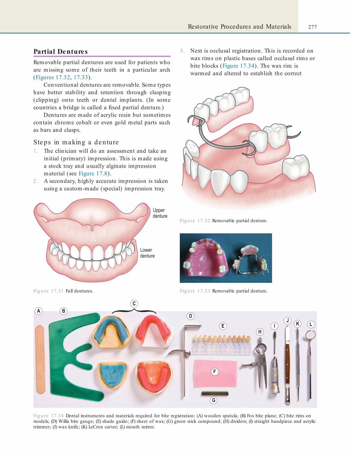
277Restorative Procedures and Materials
3 . Next is occlusal registration. This is recorded on wax rims on plastic bases called occlusal rims or bite blocks (Figure 17.34). The wax rim is warmed and altered to establish the correct
Fig u re 17.31 Full dentures.
Fig u re 17.32 Removable partial denture.
Fig u re 17.33 Removable partial denture.
Fig u re 17.34 Dental instruments and materials required for bite registration: (A) wooden spatula; (B) Fox bite plane; (C) bite rims on models; (D) Willis bite gauge; (E) shade guide; (F) sheet of wax; (G) green stick compound; (H) dividers; (I) straight handpiece and acrylic trimmer; (J) wax knife; (K) LeCron carver; (L) mouth mirror.
Partial DenturesRemovable partial dentures are used for patients who are missing some of their teeth in a particular arch (Figures 17.32, 17.33).
Conventional dentures are removable. Some types have better stability and retention through clasping (clipping) onto teeth or dental implants. (In some countries a bridge is called a xed partial denture.)
Dentures are made of acrylic resin but sometimes contain chrome cobalt or even gold metal parts such as bars and clasps.
Ste ps in making a de nture1. The clinician will do an assessment and take an
initial (primary) impression. This is made using a stock tray and usually alginate impression material (see Figure 17.8).
2 . A secondary, highly accurate impression is taken using a custom-made (special) impression tray.
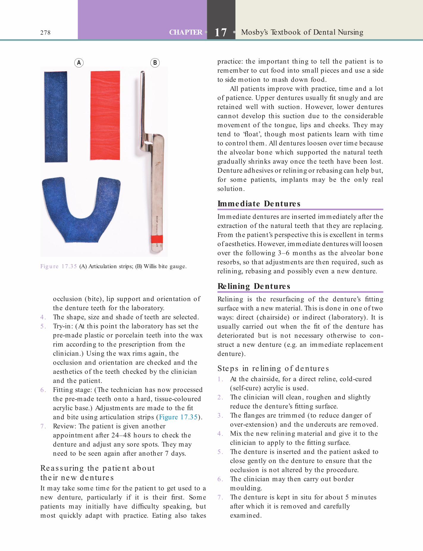
278 CHAPTER • 17 • Mosby’s Textbook of Dental Nursing
Fig u re 17.35 (A) Articulation strips; (B) Willis bite gauge.
occlusion (bite), lip support and orientation of the denture teeth for the laboratory.
4 . The shape, size and shade of teeth are selected.5 . Try-in: (At this point the laboratory has set the
pre-made plastic or porcelain teeth into the wax rim according to the prescription from the clinician.) Using the wax rims again, the occlusion and orientation are checked and the aesthetics of the teeth checked by the clinician and the patient.
6 . Fitting stage: (The technician has now processed the pre-made teeth onto a hard, tissue-coloured acrylic base.) Adjustments are made to the t and bite using articulation strips (Figure 17.35).
7 . Review: The patient is given another appointment after 24–48 hours to check the denture and adjust any sore spots. They may need to be seen again after another 7 days.
Re as s uring the pa tie nt about the ir ne w de nture sIt may take some time for the patient to get used to a new denture, particularly if it is their rst. Some patients may initially have dif culty speaking, but most quickly adapt with practice. Eating also takes
practice: the important thing to tell the patient is to remember to cut food into small pieces and use a side to side motion to mash down food.
All patients improve with practice, time and a lot of patience. Upper dentures usually t snugly and are retained well with suction. However, lower dentures cannot develop this suction due to the considerable movement of the tongue, lips and cheeks. They may tend to ‘ oat’, though most patients learn with time to control them. All dentures loosen over time because the alveolar bone which supported the natural teeth gradually shrinks away once the teeth have been lost. Denture adhesives or relining or rebasing can help but, for some patients, implants may be the only real solution.
Immediate DenturesImmediate dentures are inserted immediately after the extraction of the natural teeth that they are replacing. From the patient’s perspective this is excellent in terms of aesthetics. However, immediate dentures will loosen over the following 3–6 months as the alveolar bone resorbs, so that adjustments are then required, such as relining, rebasing and possibly even a new denture.
Relining DenturesRelining is the resurfacing of the denture’s tting surface with a new material. This is done in one of two ways: direct (chairside) or indirect (laboratory). It is usually carried out when the t of the denture has deteriorated but is not necessary otherwise to con-struct a new denture (e.g. an immediate replacement denture).
Ste ps in re lining o f de nture s1. At the chairside, for a direct reline, cold-cured
(self-cure) acrylic is used.2 . The clinician will clean, roughen and slightly
reduce the denture’s tting surface.3 . The anges are trimmed (to reduce danger of
over-extension) and the undercuts are removed.4 . Mix the new relining material and give it to the
clinician to apply to the tting surface.5 . The denture is inserted and the patient asked to
close gently on the denture to ensure that the occlusion is not altered by the procedure.
6 . The clinician may then carry out border moulding.
7 . The denture is kept in situ for about 5 minutes after which it is removed and carefully examined.

279Restorative Procedures and Materials
The den ture t ray (Figure 17.36)Laboratory workStraight handpieceStraight carbon steel acrylic trimmers For denture/tray adjustmentOcclusal registration paste For occlusal registrationArticulating paper and Miller’s forceps, or occlusal indicator wax To indicate occlusal problemsPressure-indicating cream and applicator brush To highlight high spots beneath a dentureWaxWillis bite gaugeWax knife and heat sourceShade and mould guideGauze swabs (Figure 17.21)Cotton wool rolls (Figure 17.21)Mirror For patient to view their appearance
Fig u re 17.36 Dental instruments and materials required for tting of dentures: (A) upper and lower full dentures; (B) acrylic removable partial denture; (C) chrome-cobalt removable partial denture; (D) mouth mirror; (E) Willis bite gauge; (F) dividers; (G) straight handpiece and acrylic trimmer; (H) strip of wax; (I) wax knife; (J) LeCron’s carver; (K) Willis bite gauge; (L) articulation strip; (M) shade guide; and (N) mirror.
Key poin tTissue conditioner may also be used as a reline material, but is not very durable and is only a short-term solution.
Term to lea rnBorder moulding: shaping the borders of the dentures so that they form a good seal with the mucosa and thus aid retention of the dentures.
8 . If cold-cured acrylic (see Chapter 16) was used, place the denture in a hydro ask to complete the curing and to reduce the possibility of porosity.
9 . The result is a better tting denture due to its new and well-adapted tting surface, but the new plastic tends to discolour over time.
If a laboratory (indirect) reline is to be carried out, the tting surface is cleaned, the undercuts removed and the anges shortened. Minor defects and exten-sions can be corrected with self-cured acrylic such as Total or tracing compound. A wash impression is then taken on the tting surface of the denture with impres-sion paste, with the patient in light occlusal contact.
In the laboratory, the technician replaces the impression paste with heat-cured acrylic (see
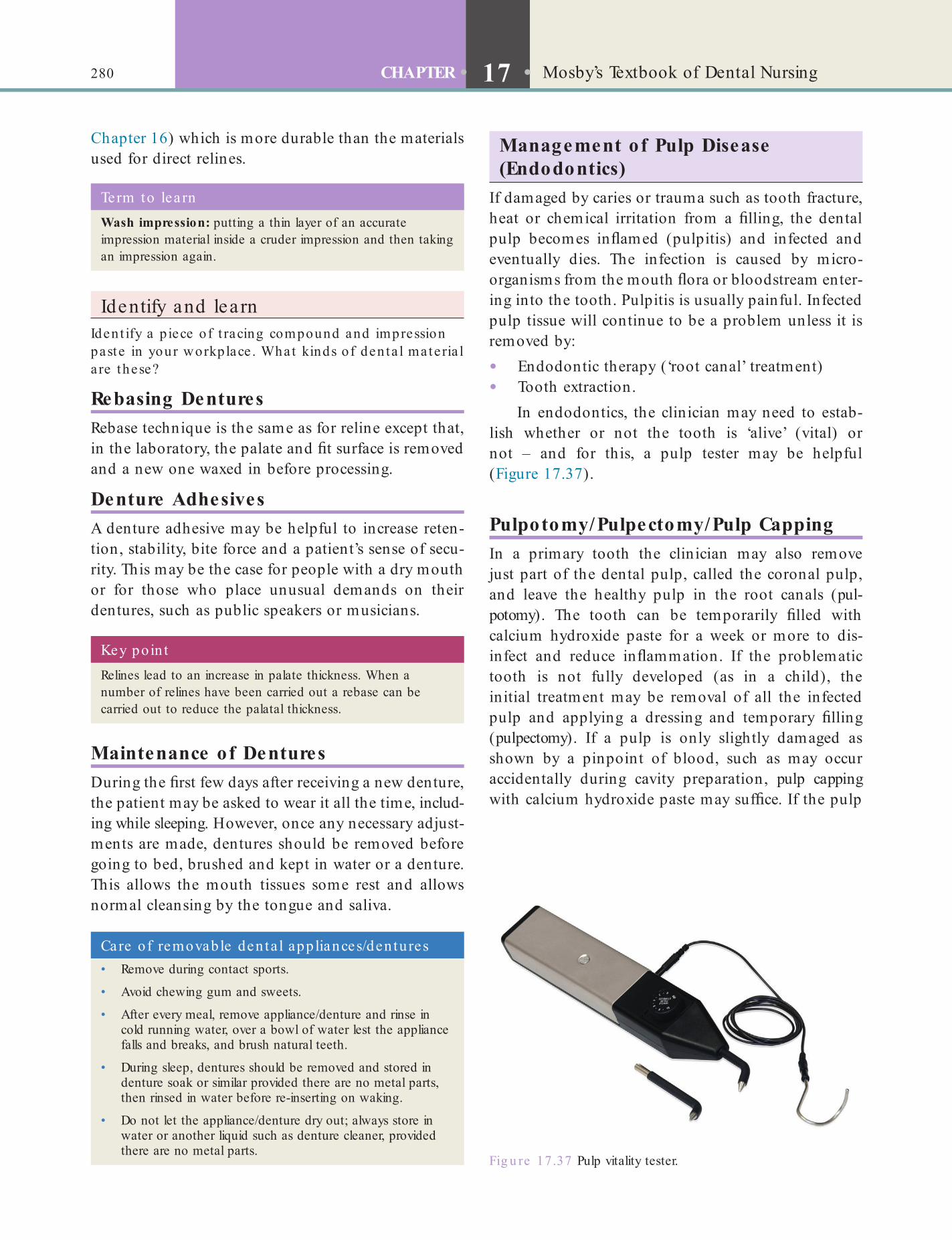
280 CHAPTER • 17 • Mosby’s Textbook of Dental Nursing
Management of Pulp Disease (Endodontics)
If damaged by caries or trauma such as tooth fracture, heat or chemical irritation from a lling, the dental pulp becomes in amed (pulpitis) and infected and eventually dies. The infection is caused by micro-organisms from the mouth ora or bloodstream enter-ing into the tooth. Pulpitis is usually painful. Infected pulp tissue will continue to be a problem unless it is removed by:
• Endodontic therapy (‘root canal’ treatment)• Tooth extraction.
In endodontics, the clinician may need to estab-lish whether or not the tooth is ‘alive’ (vital) or not – and for this, a pulp tester may be helpful (Figure 17.37).
Pulpotomy/Pulpectomy/Pulp CappingIn a primary tooth the clinician may also remove just part of the dental pulp, called the coronal pulp, and leave the healthy pulp in the root canals (pul-potomy). The tooth can be temporarily lled with calcium hydroxide paste for a week or more to dis-infect and reduce in ammation. If the problematic tooth is not fully developed (as in a child), the initial treatment may be removal of all the infected pulp and applying a dressing and temporary lling (pulpectomy). If a pulp is only slightly damaged as shown by a pinpoint of blood, such as may occur accidentally during cavity preparation, pulp capping with calcium hydroxide paste may suf ce. If the pulp
Chapter 16) which is more durable than the materials used for direct relines.
Care of removable den ta l appliances/den tures• Remove during contact sports.
• Avoid chewing gum and sweets.
• After every meal, remove appliance/denture and rinse in cold running water, over a bowl of water lest the appliance falls and breaks, and brush natural teeth.
• During sleep, dentures should be removed and stored in denture soak or similar provided there are no metal parts, then rinsed in water before re-inserting on waking.
• Do not let the appliance/denture dry out; always store in water or another liquid such as denture cleaner, provided there are no metal parts.
Key po in tRelines lead to an increase in palate thickness. When a number of relines have been carried out a rebase can be carried out to reduce the palatal thickness.
Term to lea rnWash impression: putting a thin layer of an accurate impression material inside a cruder impression and then taking an impression again.
Fig u re 17.37 Pulp vitality tester.
Identify and learnIdent ify a p iece of t racing compound and impression paste in your workplace . What kinds of den ta l mate ria l a re these?
Rebasing DenturesRebase technique is the same as for reline except that, in the laboratory, the palate and t surface is removed and a new one waxed in before processing.
Denture AdhesivesA denture adhesive may be helpful to increase reten-tion, stability, bite force and a patient’s sense of secu-rity. This may be the case for people with a dry mouth or for those who place unusual demands on their dentures, such as public speakers or musicians.
Maintenance of DenturesDuring the rst few days after receiving a new denture, the patient may be asked to wear it all the time, includ-ing while sleeping. However, once any necessary adjust-ments are made, dentures should be removed before going to bed, brushed and kept in water or a denture. This allows the mouth tissues some rest and allows normal cleansing by the tongue and saliva.
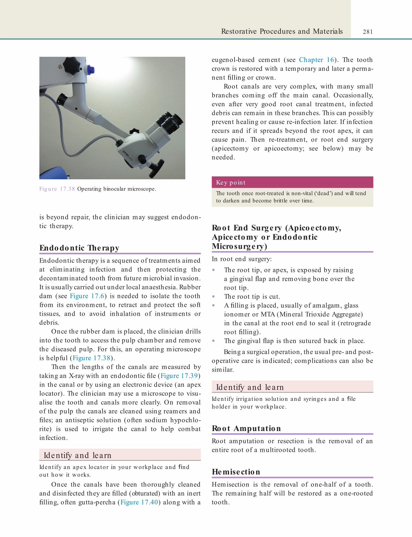
281Restorative Procedures and Materials
eugenol-based cement (see Chapter 16). The tooth crown is restored with a temporary and later a perma-nent lling or crown.
Root canals are very complex, with many small branches coming off the main canal. Occasionally, even after very good root canal treatment, infected debris can remain in these branches. This can possibly prevent healing or cause re-infection later. If infection recurs and if it spreads beyond the root apex, it can cause pain. Then re-treatment, or root end surgery (apicectomy or apicoectomy; see below) may be needed.
is beyond repair, the clinician may suggest endodon-tic therapy.
Endodontic TherapyEndodontic therapy is a sequence of treatments aimed at eliminating infection and then protecting the decontaminated tooth from future microbial invasion. It is usually carried out under local anaesthesia. Rubber dam (see Figure 17.6) is needed to isolate the tooth from its environment, to retract and protect the soft tissues, and to avoid inhalation of instruments or debris.
Once the rubber dam is placed, the clinician drills into the tooth to access the pulp chamber and remove the diseased pulp. For this, an operating microscope is helpful (Figure 17.38).
Then the lengths of the canals are measured by taking an X-ray with an endodontic le (Figure 17.39) in the canal or by using an electronic device (an apex locator). The clinician may use a microscope to visu-alise the tooth and canals more clearly. On removal of the pulp the canals are cleaned using reamers and les; an antiseptic solution (often sodium hypochlo-rite) is used to irrigate the canal to help combat infection.
Identify and learnIdent ify an apex loca tor in your workplace and nd out how it works.
Once the canals have been thoroughly cleaned and disinfected they are lled (obturated) with an inert lling, often gutta-percha (Figure 17.40) along with a
Fig u re 17.38 Operating binocular microscope. Key po in tThe tooth once root-treated is non-vital (‘dead’) and will tend to darken and become brittle over time.
Root End Surgery (Apicoectomy, Apicectomy or Endodontic Microsurgery)In root end surgery:
• The root tip, or apex, is exposed by raising a gingival ap and removing bone over the root tip.
• The root tip is cut.• A lling is placed, usually of amalgam, glass
ionomer or MTA (Mineral Trioxide Aggregate) in the canal at the root end to seal it (retrograde root lling).
• The gingival ap is then sutured back in place.Being a surgical operation, the usual pre- and post-
operative care is indicated; complications can also be similar.
Identify and learnIdent ify irriga t ion so lu t ion and syringes and a le ho lder in your workplace .
Root AmputationRoot amputation or resection is the removal of an entire root of a multirooted tooth.
HemisectionHemisection is the removal of one-half of a tooth. The remaining half will be restored as a one-rooted tooth.

282 CHAPTER • 17 • Mosby’s Textbook of Dental Nursing
Fig u re 17.39 (A): Endodontic instruments (A) Barbed broach; (B) Gates Glidden bur; (C) goose (long) neck bur; (D) endodontic hand le; (E) hand reamer; (F) nger spreader; and (G) spiral root ller. (B): A selection of other instruments and items used in endodontic treatment. Do you use them in your workplace? Try to identify as many as you can.
Endodont ic t rayBroaches – smooth and barbed (Figure 17.39) For removing pulp tissueGates Glidden bur (Figure 17.39) For cleaning and shaping the canalsEndodontic les (Flexo le, Hedstroem) (hand or handpiece driven) (Figure 17.39) For cleaning and shaping the canalsPaper points (Figure 17.40) For drying the canalIrrigation syringes Monojet syringe 27GIrrigation solutionsHypochlorite – sterile waterAntiseptic dressings Calcium hydroxide, LedermixGutta-percha (Figure 17.40) (silver points rarely used now) and cement For occluding canalFile holderApex locatorGauze swabs (Figure 17.21)Cotton wool rolls (Figure 17.21)Temporary dressing (provisional restoration)
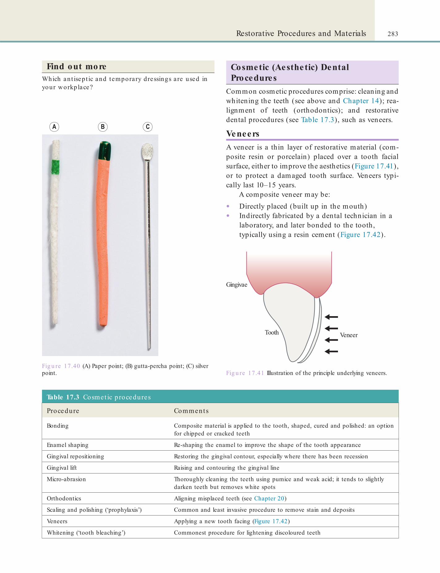
283Restorative Procedures and Materials
Fig u re 17.40 (A) Paper point; (B) gutta-percha point; (C) silver point.
Table 17.3 Cosmet ic p rocedures
Procedure Comments
Bonding Composite material is applied to the tooth, shaped, cured and polished: an option for chipped or cracked teeth
Enamel shaping Re-shaping the enamel to improve the shape of the tooth appearance
Gingival repositioning Restoring the gingival contour, especially where there has been recession
Gingival lift Raising and contouring the gingival line
Micro-abrasion Thoroughly cleaning the teeth using pumice and weak acid; it tends to slightly darken teeth but removes white spots
Orthodontics Aligning misplaced teeth (see Chapter 20)
Scaling and polishing (‘prophylaxis’) Common and least invasive procedure to remove stain and deposits
Veneers Applying a new tooth facing (Figure 17.42)
Whitening (‘tooth bleaching’) Commonest procedure for lightening discoloured teeth
Cosmetic (Aesthetic) Dental Procedures
Common cosmetic procedures comprise: cleaning and whitening the teeth (see above and Chapter 14); rea-lignment of teeth (orthodontics); and restorative dental procedures (see Table 17.3), such as veneers.
VeneersA veneer is a thin layer of restorative material (com-posite resin or porcelain) placed over a tooth facial surface, either to improve the aesthetics (Figure 17.41), or to protect a damaged tooth surface. Veneers typi-cally last 10–15 years.
A composite veneer may be:
• Directly placed (built up in the mouth)• Indirectly fabricated by a dental technician in a
laboratory, and later bonded to the tooth, typically using a resin cement (Figure 17.42).
Find out moreWhich an t isep t ic and temporary dressings a re used in your workplace?
Fig u re 17.41 Illustration of the principle underlying veneers.
Gingivae
Tooth Veneer
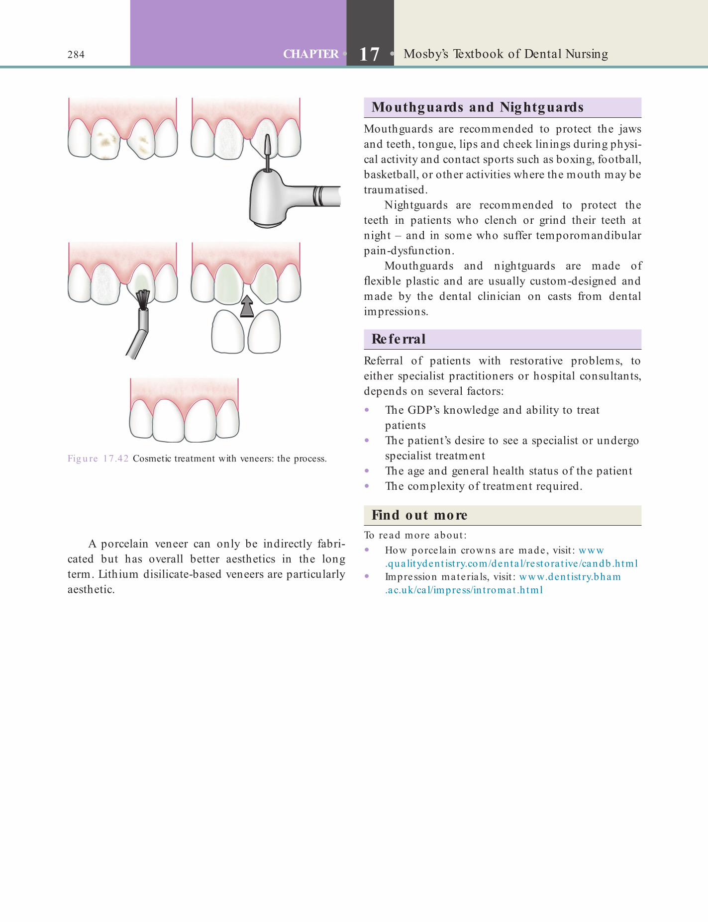
284 CHAPTER • 17 • Mosby’s Textbook of Dental Nursing
Mouthguards and NightguardsMouthguards are recommended to protect the jaws and teeth, tongue, lips and cheek linings during physi-cal activity and contact sports such as boxing, football, basketball, or other activities where the mouth may be traumatised.
Nightguards are recommended to protect the teeth in patients who clench or grind their teeth at night – and in some who suffer temporomandibular pain-dysfunction.
Mouthguards and nightguards are made of exible plastic and are usually custom-designed and made by the dental clinician on casts from dental impressions.
ReferralReferral of patients with restorative problems, to either specialist practitioners or hospital consultants, depends on several factors:
• The GDP’s knowledge and ability to treat patients
• The patient’s desire to see a specialist or undergo specialist treatment
• The age and general health status of the patient• The complexity of treatment required.
Find out moreTo read more about :• How porcela in crowns a re made , visit : www
.qua litydent ist ry.com/denta l/resto ra t ive /candb .h tml• Impression materia ls, visit : www.dent ist ry.bham
.ac.uk/ca l/impress/in t romat .h tml
A porcelain veneer can only be indirectly fabri-cated but has overall better aesthetics in the long term. Lithium disilicate-based veneers are particularly aesthetic.
Fig u re 17.42 Cosmetic treatment with veneers: the process.
Chapter 18 Surgical Care
CHAPTER POINTS• Role of the dental nurse in surgery• Common surgical procedures• Day-care (day-stay) surgery• Postoperative complications
possible risks and the level o these – and thus can make an in ormed decision.
Written informed consent must be obtained rom all patients having any surgical procedure. The possible benef ts o treatment must be weighed against the risks and always discussed by the person carrying out the procedure. I or some good reason this is not possible, a delegated person with the appropriate expertise should do so. In particular, patients must be in ormed and where necessary warned care ully and clearly about:
• Preoperative preparation that may be required• Possible adverse e ects or outcomes (e.g.
de ormity)• Postoperative sequelae (e.g. pain, swelling,
bruising)• Where the patient will be during their recovery.
In t ro d u ct io n
Surgery is required in many cases or the treatment and ongoing management o irregularities and pathol�ogy o the jaw and mouth. In the UK, oral surgery is a specialty regulated by the General Dental Council and includes the specialty previously called surgical den�tistry. Oral surgery procedures are sometimes termed dento-alveolar surgery (or minor oral surgery) and are commonly undertaken in the dental surgery.
Oral and maxillo acial surgery (sometimes termed major oral surgery) is discussed in Chapter 19. As dental nurses may work in both dental surgery and hospital theatre settings, you need to be amiliar with procedures described in both chapters.
As discussed in Chapter 5, consent is required be ore any operative procedure, but this is especially important be ore surgery and so some detail is re �peated here.
Key poin tWarnings about surgical procedures must be properly recorded in the case notes and signed by operator and patient.
Key po in tIn ormed (valid) consent means that the patient must be made ully aware o the procedure, and its intended benef ts and possible risks.
An example o a patient in ormation sheet is given in Box 18.1. The dental nurse may be delegated to give such in ormation to the patient but it is for the opera-tor to seek valid consent; the UK Department o Health (2009) states clearly:
The clinician providing the treatment or investigation is responsible for ensuring that the person has given valid consent before treatment begins, although the consultant responsible for the person’s care will remain ultimately responsible for the quality of medical care provided. (www.gov.uk/government/publications/reference -guide-to-consent-for-examination-or-treatment -second-edition)
‘Informed’ consent means that the patient must be ully aware o the procedure, its intended benef ts, its
286 CHAPTER • 18 • Mosby’s Textbook o Dental Nursing
• Periodontal surgery to acilitate oral hygiene in areas a ected by chronic periodontitis
• Implant surgery, that is, placement o endosseous implants and associated procedures such as bone gra ting and sinus li ts
• Treatment o complications arising rom diseases a ecting the teeth, periodontium or alveolar bone (such as in ections and cysts).With additional training and only on prescription,
a dental nurse may:
• Take radiographs to the prescription o a clinician (but see Chapter 11; Certif cate in Dental Radiography is required)
• Apply topical anaesthetic to the prescription o a clinician.Dental nurses in a hospital setting may also assist
in major oral and maxillo acial surgery (see Chapter 19), which deals with the e ects o :
• Trauma• Oral cancer• Oro acial de ormities or malocclusions
(orthognathic surgery).
Preparation before Surgery
Sc rubbing and gowning• Lather hands and orearms with soap or a
special solution.• Scrub with a brush or one minute, especially
the nails and hands. Vigorous scrubbing is debated as bacteria may be brought out o skin pores and increase rather than reduce skin bacterial counts.
• Lather and rinse hands and orearms vigorously or a urther 5 minutes – turn taps on with elbows.
• Rinse o soap, holding your hands at a higher level than the elbows.
• Dry with a sterile towel. It is important to prevent the towel touching unsterile skin at the elbow and then wiping the opposite hand with it.
• The sterile gown is un olded and the arms pushed into the armholes; then the arms are held up. A nurse should then pull down the shoulders and body o the gown and tie it behind. It is inadvisable to pull the sleeves up yoursel because o the risk o inadvertently touching the mask or collar.
Box 18.1 Example pa t ien t in format ion shee t for remova l of th ird molarsAs you know, we eel that your wisdom teeth should be removed. Here is some in ormation that we hope will answer some o your questions.
Wisdom teeth removal is o ten necessary because o in ection (which causes pain and swelling), decay, serious gingiva (gum) disease, the development o a cyst or because teeth are overcrowded. Wisdom teeth are removed under local anaesthetic (injection in the mouth), sedation or general anaesthetic in hospital, depending on your pre erence, the number o teeth to be removed and the di f culty o removal.
It is o ten necessary to make a small incision in the gingiva, which is stitched a terwards. A ter removal o the teeth, your mouth will be sore and swollen and mouth movements will usually be sti . Slight bleeding is also very common. These symptoms are quite normal, but can be expected to improve rapidly during the f rst week. It is quite normal or some sti ness and slight soreness to persist or two to three weeks. Pain and discom ort can be controlled with ordinary painkillers, such as paracetamol, and you might be prescribed antibiotic tablets. A clinician will be available to see you a terwards i you are worried, and will want to check that healing is satis actory.
Complications are rare, but occasionally wisdom tooth sockets become in ected, when pain, swelling and sti ness will last longer than normal. Occasionally patients have tingling or numbness o the lower lip or tongue a ter lower wisdom teeth removal. This is because nerves to these areas pass very close to the wisdom teeth and may get bruised or damaged. The numbness nearly always disappears a ter about one month, but very occasionally lasts or a year or more. Jaw racture is very rare.
Please let us know i we can give you any more in ormation.
Role o f the Dental Nurse in SurgeryAs a dental nurse, you must have the knowledge and understanding o the procedures required to:
• Assist e ectively at the chairside• Provide the patient with advice both be ore
(preoperative) and a ter (postoperative) the procedure.The dental nurse may assist the clinician under�taking dento�alveolar surgery, which may involve:
• Extraction o carious or broken teeth or roots. Third molar (wisdom tooth) removal, especially when impacted (against another tooth usually, and then impeded in eruption), is a common surgical procedure or, sometimes, the tooth crown is removed (coronectomy).
• Root end surgery (apicectomy) or recurrent periapical in ections

287Surgical Care
decreasing activity, are: chlorhexidine gluconate, iodo�phors, triclosan and plain soap.
See Chapter 7 or a detailed discussion o handwashing.
Assisting• Avoid damaging the patient, the surgeon or
assistants, or yoursel .• Many surgical instruments, or example artery
orceps and some suture holders, have a ratchet device to keep them closed. I you will be assisting, you will need to learn to be adept at opening and closing these with either hand.
• Scissors can be most accurately controlled i the thumb and ring f nger are placed in the rings and the tip o the index f nger is placed along the sha t.
• Ligatures or sutures should be cut with the ends o the blades: scissors seldom need to be opened more than 1 cm at the tip and the blades should be held at right�angles to the skin.
• Gloves are donned (know your glove size), care being taken not to touch the outside with the skin o the opposite hand.
• Therea ter, observe a ‘no touch’ technique, keeping your hands near your abdomen or chest while waiting or moving around in theatre.
Identify and learnDonning g loves is p robably the most importan t stage . Ask a senior co lleague or a thea t re nurse to show it you on your rst day in thea t re if you do no t fee l perfect ly comfortab le with your technique .
Key poin tTake care! Scissors inexpertly wielded can be a danger both to the patient and the surgeon.
How long to scrub and with wha t?
How long?Surgical personnel have traditionally been required to scrub their hands or 10 minutes preoperatively. However, studies have ound that scrubbing or 5 minutes reduces bacterial counts as e ectively. It may also help prevent skin damage associated with such lengthy handwashing.
With w hat?Surgical handwashing protocols also used to require surgical sta to scrub hands with a brush, which can also damage skin and result in increased shedding o bacteria rom the hands.
Some studies have indicated that scrubbing with a disposable sponge or combination sponge-brush is as e ective. Other studies have indicated that neither a brush nor sponge is necessary to reduce bacterial counts on the hands o surgical sta to acceptable levels.
A two-stage surgical scrub using an antiseptic detergent, ollowed by application o an alcohol-containing preparation, has been demonstrated to be e ective.
Handwas he sImmediate and persistent antimicrobial activity is the most important in determining the e f cacy o a hand�washing product. Agents used or surgical hand scrubs should:
• Substantially reduce micro�organisms on intact skin
• Contain a non�irritating antimicrobial preparation
• Have broad�spectrum activity• Be ast and long acting.
Formulations with 60–95% alcohol alone or 50–95% when combined with limited amounts o a quaternary ammonium compound, or chlorhexidine gluconate, more e ectively lower bacterial counts on the skin immediately a ter scrubbing than other agents. The next most active agents, in order o
Be fo re the proc e dure• Retrieve patient notes, radiographs, consent
orm.• Prepare the dental environmental ensuring
in ection control.• Prepare the surgical tray or trolley using an
aseptic technique (Table 18.1).• Greet and reassure the patient in the waiting
area.• Ensure preoperative advice has been ollowed.• In orm the clinician o any changes/comment
rom the patient.• Assist the patient to the dental chair.• Provide the patient with protective equipment
(bib and glasses).• Provide a bowl with water or dentures.
During the proc e dure• Reassure patient.• Assist with giving topical and local anaesthetic.• Retract the so t tissues, lips, cheek and/or tongue
as required.• Aspirate to ensure a clear f eld and patient
com ort.

288 CHAPTER • 18 • Mosby’s Textbook o Dental Nursing
• No. 11 – or incising abscesses• No. 12 – or periodontal surgery• No. 15 – or intraoral and small skin incisions,
incising the mucoperiosteum, and excision o so t tissue lesions
• No. 10 – or larger skin incisions.
• Anticipate and pass instruments/items as required.
• Mix materials as appropriate and pass as required.
• Develop radiographs i another appropriately qualif ed dental nurse is not available.
• Cut sutures as required.• Wipe patient’s ace.
Afte r the proc e dure• Provide a mirror and tissues or the patient to
wipe their ace and assist i necessary.• Assist patient to the recovery room or waiting area.• Ensure the patient has another appointment and
has the postoperative instruction sheet.• Make a ollow�up appointment as required.• Decontaminate the environment and sterilise
instruments.• Ensure disposal o the local anaesthetic needle
by the clinician in the correct sharps container.• Dispose o the clinical waste in the yellow bags.
Common Surgical ProceduresSoft Tissue SurgeryA range o instruments are used or so t tissue surgery. The disposable surgical scalpel blades used are:
Term to lea rnMucoperiosteum: the combined term or the mucous membrane and the periosteum that covers the bone.
Table 18.1 Basic surg ica l t ray (see Figure 18.4)
Inst rument Purpose
Scalpel blade (Nos. 11, 12, 15) To cut the ap
Scalpel handle (e.g. Swann Morton)
Mitchell’s trimmer To raise the ap
Periosteal elevator (e.g. Howarth’s) To raise the ap and protect so t tissues
Surgical handpiece To remove bone or cut tooth
Surgical selection o surgical burs
Retractors: ap; cheek; tongue To retract ap or visibility to operative site, and protect tissues
Surgical aspirator To remove saliva, blood, water and debris
Irrigation syringe To irrigate the site with sterile saline or water
Suture To reposition and f x mucoperiosteal ap
Needle holders To hold the suture needle
Rat-toothed tissue dissecting orceps To hold the ap while suturing
Suture scissors To cut sutures
Gauze swabs
Cotton wool rolls
Identify and learnTry to nd the various sca lpe ls listed above in your workplace and lea rn the d iffe rences and simila rit ies be tween them.
Cutting d ia the rmy and e le c tros urge ryThese procedures use an electric current to cut or cut and coagulate the tissue. They may be used or exten�sive oral incisions, or removal o so t tissue, because bleeding rom wound edges is reduced.
These procedures should be avoided in patients with arti cial cardiac pacemakers or other implanted cardiac devices.

289Surgical Care
cryoprobe. The very low temperatures – about −70 ° C – achieved with liquid N2 make this particularly use ul in the management o intractable lesions. Liquid N2 probes and sprays can cause ull�thickness skin necro�sis, and there ore must be handled very care ully.
• Local analgesia is given i required.• The clinician ensures good contact o the
cryoprobe with the lesion by coating it with a jelly such as KY jelly.
• The clinician reezes the lesion.• The reezing is turned o . The cryoprobe is le t
on the lesion until it thaws and de� rosts.• Multiple reezes, with thawing between each
reeze, maximise the e ect. The ice crystals ormed in the tissue spaces between the cells kill the cells.
Las e r s urge ryLasers can be used or surgery but can also damage normal tissues, especially the eyes. The hard laser, especially the carbon dioxide laser, is the most use ul.
AdvantagesLaser surgery usually:
• Causes little bleeding. Haemostasis occurs during vaporisation to coagulate small vessels in the wound bed
• Is ollowed by less pain and swelling than that which ollows surgical excision or cryotherapy
• Results in little postoperative scarring• Is particularly help ul or surgery close to
important anatomical structures, e.g. lesions a ecting the oor o the mouth.
Dis advantagesLaser surgery usually:
• Is best conducted under general anaesthesia (GA)
• Requires expensive equipment• Can damage the eyes, skin, mucosae and teeth.Also, because laser light travels in straight lines, access to some lesions can be di f cult.
Potentia l hazards• Damage to the eyes and other tissues
rom re ection o the laser beam rom retractors, mouth props or anaesthetic tube couplings.
• Vaporisation o anaesthetic tubes, leading to ignition o in ammable anaesthetic agents or oxygen.
Cryos urge ryThis procedure uses reezing to destroy tissue.
Advantages• It can sometimes be used without analgesia.• There is no haemorrhage.• There is little postoperative in ection.
Dis advantages• No biopsy specimen can be taken.• Depth and extent o tissue damage is di f cult to
predict.• There is substantial postoperative swelling
(oedema).
Term to lea rnOedema: the medical term or swelling that results rom excessive accumulation o watery uid in a particular part o the body.
Term to lea rnArti cial cardiac pacemaker: this is a medical device that regulates the heart beat. It is placed in people whose natural heart beat is not ast enough or in those who have a problem with the system that conducts the heart beat rom the atria to the ventricles (see Chapter 24). The pacemaker consists o electrodes that are placed in contact with the heart muscles and which send out electrical signals to the heart.
Indica tionsCryosurgery is use ul or:
• Controlling intractable acial pain (cryoanalgesia to the peripheral nerve)
• Removal o :• Leukoplakias (also called keratoses; see
Chapter 10). The diagnosis should be conf rmed by taking a biopsy and sending it or histological examination prior to cryosurgery, in case it is a malignancy
• Warts and papillomas• Mucus extravasation cysts (mucoceles)• Haemangiomas (see Chapter 10).
• Palliation (producing some relie ) o :• Severe ulcers• Cancerous lesions.
ProcedureLiquid nitrogen (N2) or nitrous oxide (N2O) is applied to the diseased area. This is done either by spraying it directly or circulating it through a probe called a

290 CHAPTER • 18 • Mosby’s Textbook o Dental Nursing
Indica tions• Leukoplakias.• Early tumours.• Haemangiomas.
ProcedureThe carbon dioxide laser is a cutting laser, which can be used to: excise, much as does a scalpel; or to ulgu�rate (destroy) the lesion. It is important to:
• Follow laser sa ety recommendations (see Chapter 6)
• Use non�in ammable general anaesthetic agents• Avoid re ections rom instruments or other
metal objects.
Surgery Involving Hard Tissues (Bone and Teeth)Bone and teeth are usually cut with rotating instru�ments (burs) in a surgical handpiece. This involves the production o heat, so simultaneous cooling by con�stant running sterile water or sterile saline (irrigation) is important. Air rotors are less commonly used as they can contaminate wounds unless using a sterile coolant, and occasionally cause surgical emphysema (see Chapter 23). Laser and ultrasonic cutting are uncommonly used. Piezosurgery is a new but expen�sive technique that cuts only hard tissues, increasingly used in apical and implant surgery as the danger o damage to nerves or arteries or the sinus membrane is less. However, this kind o surgery can generate signif �cant heat, and cutting is slower than with many high�speed drills.
Identify and learnIdent ify two surg ica l handpieces in your workplace . Note the t t ed tube for irriga t ion .
Wound c los ureIncisions are usually an integral part o surgery. The wound thus created needs to be closed so that it heals by primary intention. The wound is closed usually with cyanoacrylate tissue adhesive or tapes (e.g. Steri�Strip), or sutures (stitches) (Box 18.2). This results in a small line o scar tissue, which is the goal whenever a wound is closed.
In some circumstances, an open wound is le t to heal by secondary intention. In the mouth it is then protected by a dressing such as Coepak. Wounds in bone are sometimes protected by BIPP (bismuth iodo� orm para f n paste), or Whitehead’s varnish (com�pound iodo orm paint).
Box 18.2 Closing a wound
Skin tapes• Closure o skin wounds by adhesive tapes is convenient.
• It produces a strong wound with little in ection and with good cosmetic results.
• The skin and wound should be thoroughly cleaned as above, and all bleeding within the wound must cease be ore tapes are applied.
• The tapes are le t in place or 7–10 days and then gently removed by traction.
Sutures• Atraumatic needles are generally pre erred.
• Monof lament materials are associated with a lower rate o wound in ection compared with multif lament sutures.
• Sutures (stitches) are used or most:
• Oral wounds – 3.0 Vicryl (resorbable) or black silk (non-resorbable but pliable and easily seen)
• Facial wounds – 5.0 or 6.0 polypropylene or nylon
• Other skin wounds – 3.0 nylon or Prolene (polypropylene).
TechniqueThe simple interrupted stitch is most commonly used because it allows good approximation o the wound edges, and is easy to place. Various other suturing techniques can be used. The surgeon will o ten ask the dental nurse to cut the stitch once placed.
1. Care o sutures
• Clean wounds twice daily with chlorhexidine 0.1% aqueous solution.
• Apply Poly ax ointment (antibiotics – polymyxin and bacitracin) twice daily to prevent scab ormation, acilitate suture removal and reduce scarring.
2. Suture removal
• Facial sutures should be removed within 3–5 days, to keep scarring to a minimum.
• Mucosal sutures are usually resorbable, but otherwise are removed at 5–7 days.
• Many patients are apprehensive and need reassurance.
• Clean the wound and surrounding mucosa or skin with aqueous 0.2% chlorhexidine.
• Li t the suture with sterile orceps and cut the stitch on one side, as close to the skin or mucosa as possible (this avoids pulling contaminated suture material through the wound).
• Pull the suture out using traction on the long end, across the wound so as to avoid pulling apart the edges.
• Clean the area again with an antiseptic solution.
• Remove alternate sutures f rst to see i wound has healed adequately. I yes, remove the remaining sutures a day later.
• I wounds tend to gape a little, use tapes (but not in the mouth).

291Surgical Care
Extrac tion o f te e th (e xodontia )Ins truments us edFigures 18.1–18.3 show a selection o orceps and elevators used or extraction o teeth, both straight or�ward and impacted teeth. Figure 18.4 shows a selec-tion o other surgical instruments.
Identify and learnWrite down the names of a ll the items shown in Figure 18.4 and ge t it checked by your supervisor.
Extraction of deciduous tee thThere is seldom a case or the removal o only one deciduous tooth, unless it:
• Is close to being shed naturally• Has been retained so long that it impedes the
eruption o its successor• Is infra-occluded.
Fig u re 18.1 Forceps or extracting deciduous teeth: (A) upper straight; (B) upper root; (C) upper molar; (D) lower root; (E) lower molar.
Key poin tThe suture (stitch) should be cut about 5 mm away rom the knot.
Terms to learnHealing by primary intention: when the wound edges heal directly touching each other.
Healing by secondary intention: when the wound is le t open to f ll with granulation tissue, which will subsequently turn into scar tissue.
Find out moreLook a t some wound closure materia ls in your workplace and t ry to th ink of a situa t ion each of them could be used in .
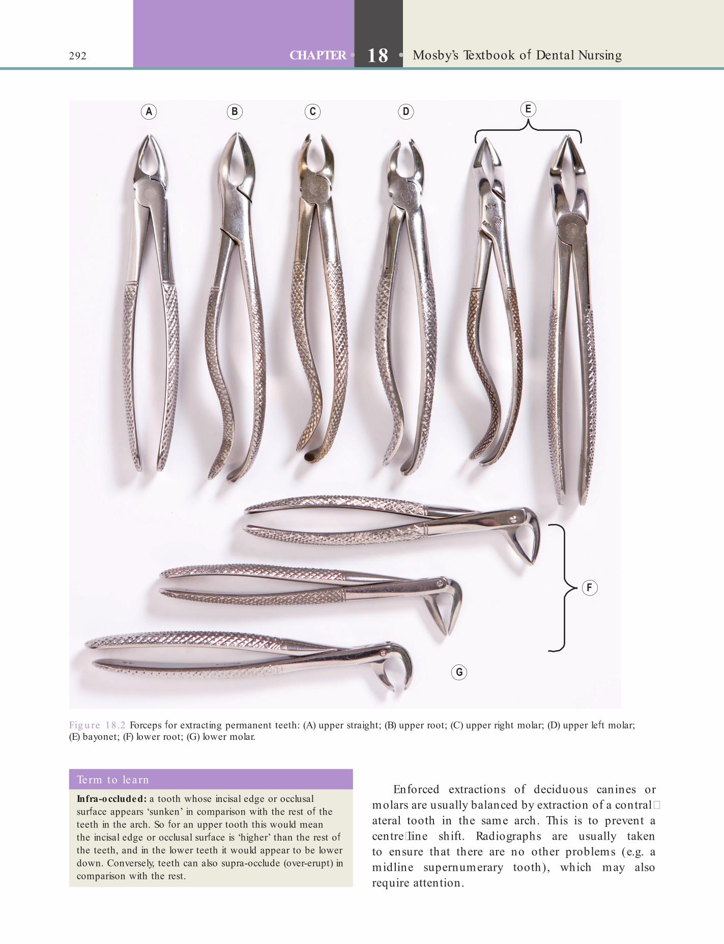
292 CHAPTER • 18 • Mosby’s Textbook o Dental Nursing
En orced extractions o deciduous canines or molars are usually balanced by extraction o a contral�ateral tooth in the same arch. This is to prevent a centre�line shi t. Radiographs are usually taken to ensure that there are no other problems (e.g. a midline supernumerary tooth), which may also require attention.
Fig u re 18.2 Forceps or extracting permanent teeth: (A) upper straight; (B) upper root; (C) upper right molar; (D) upper le t molar; (E) bayonet; (F) lower root; (G) lower molar.
Term to lea rnInfra-occluded: a tooth whose incisal edge or occlusal sur ace appears ‘sunken’ in comparison with the rest o the teeth in the arch. So or an upper tooth this would mean the incisal edge or occlusal sur ace is ‘higher’ than the rest o the teeth, and in the lower teeth it would appear to be lower down. Conversely, teeth can also supra-occlude (over-erupt) in comparison with the rest.
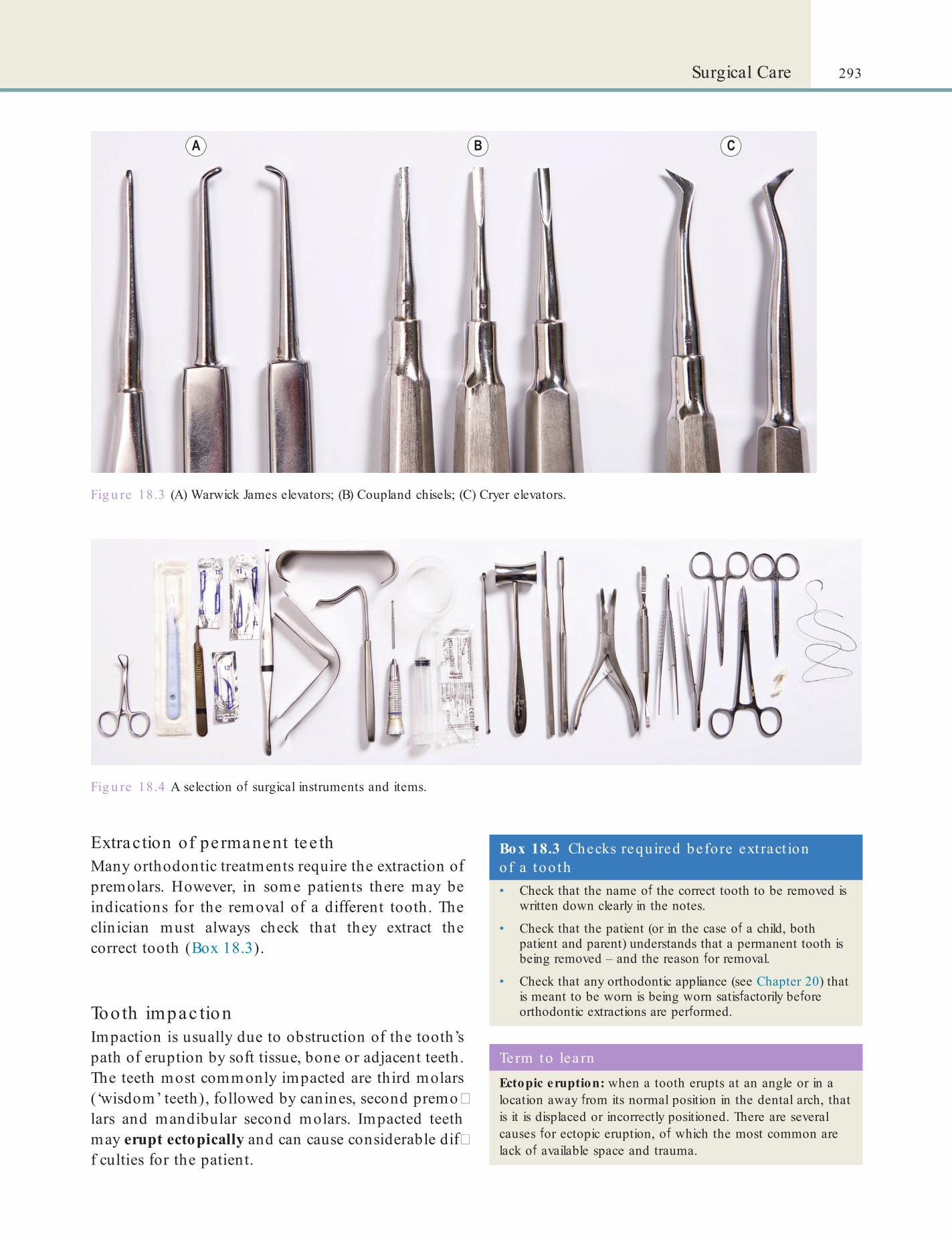
293Surgical Care
Fig u re 18.3 (A) Warwick James elevators; (B) Coupland chisels; (C) Cryer elevators.
Fig u re 18.4 A selection o surgical instruments and items.
Extraction of pe rmanent tee thMany orthodontic treatments require the extraction o premolars. However, in some patients there may be indications or the removal o a di erent tooth. The clinician must always check that they extract the correct tooth (Box 18.3).
Tooth impac tionImpaction is usually due to obstruction o the tooth’s path o eruption by so t tissue, bone or adjacent teeth. The teeth most commonly impacted are third molars (‘wisdom’ teeth), ollowed by canines, second premo�lars and mandibular second molars. Impacted teeth may erupt ectopically and can cause considerable di �f culties or the patient.
Box 18.3 Checks required before ext ract ion of a too th• Check that the name o the correct tooth to be removed is
written down clearly in the notes.
• Check that the patient (or in the case o a child, both patient and parent) understands that a permanent tooth is being removed – and the reason or removal.
• Check that any orthodontic appliance (see Chapter 20) that is meant to be worn is being worn satis actorily be ore orthodontic extractions are per ormed.
Term to lea rnEctopic eruption: when a tooth erupts at an angle or in a location away rom its normal position in the dental arch, that is it is displaced or incorrectly positioned. There are several causes or ectopic eruption, o which the most common are lack o available space and trauma.
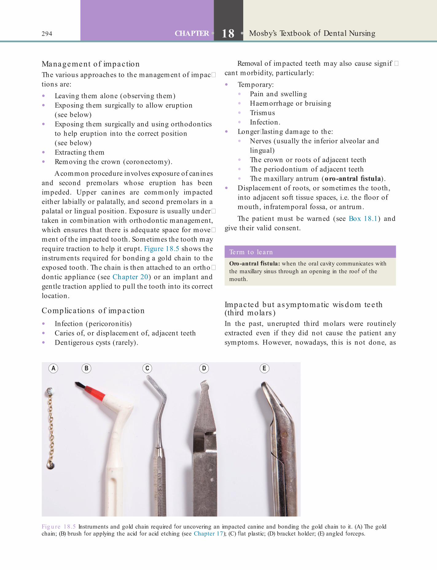
294 CHAPTER • 18 • Mosby’s Textbook o Dental Nursing
Removal o impacted teeth may also cause signif �cant morbidity, particularly:
• Temporary:• Pain and swelling• Haemorrhage or bruising• Trismus• In ection.
• Longer�lasting damage to the:• Nerves (usually the in erior alveolar and
lingual)• The crown or roots o adjacent teeth• The periodontium o adjacent teeth• The maxillary antrum (oro-antral stula).
• Displacement o roots, or sometimes the tooth, into adjacent so t tissue spaces, i.e. the oor o mouth, in ratemporal ossa, or antrum.The patient must be warned (see Box 18.1) and
give their valid consent.
Management of impactionThe various approaches to the management o impac�tions are:
• Leaving them alone (observing them)• Exposing them surgically to allow eruption
(see below)• Exposing them surgically and using orthodontics
to help eruption into the correct position (see below)
• Extracting them• Removing the crown (coronectomy).
A common procedure involves exposure o canines and second premolars whose eruption has been impeded. Upper canines are commonly impacted either labially or palatally, and second premolars in a palatal or lingual position. Exposure is usually under�taken in combination with orthodontic management, which ensures that there is adequate space or move�ment o the impacted tooth. Sometimes the tooth may require traction to help it erupt. Figure 18.5 shows the instruments required or bonding a gold chain to the exposed tooth. The chain is then attached to an ortho�dontic appliance (see Chapter 20) or an implant and gentle traction applied to pull the tooth into its correct location.
Complica tions of impaction
• In ection (pericoronitis)• Caries o , or displacement o , adjacent teeth• Dentigerous cysts (rarely).
Fig u re 18.5 Instruments and gold chain required or uncovering an impacted canine and bonding the gold chain to it. (A) The gold chain; (B) brush or applying the acid or acid etching (see Chapter 17); (C) at plastic; (D) bracket holder; (E) angled orceps.
Term to lea rnOro-antral stula: when the oral cavity communicates with the maxillary sinus through an opening in the roo o the mouth.
Impacted but as ymptomatic wis dom tee th (third molars )In the past, unerupted third molars were routinely extracted even i they did not cause the patient any symptoms. However, nowadays, this is not done, as

295Surgical Care
research has ound no evidence to support this practice; many unerupted third molars remain asymp�tomatic or years, or orever. NICE guidelines help the clinician decide on the ate o wisdom teeth (Box 18.4).
Odontoge nic infe c tionsOdontogenic in ections are in ections such as dental abscesses that arise rom teeth, particularly non�vital teeth. They also include pericoronitis (Chapter 10) and post�surgical in ections. They are:
• Commonly polymicrobial – caused by a mixture o oral bacteria, including both bacteria that grow without oxygen (anaerobes) and those that need oxygen (aerobes)
• Usually minor, and localised to the alveolus or sulcus, and resolve promptly with appropriate management – mainly this is drainage
• Sometimes severe and li e�threatening, particularly i they are necrotising or i they spread. Fascial space in ections endanger the airway and may spread to the chest. In ections may also spread via blood by entering into the blood vessels. These are a serious emergency, requiring hospital care.
Box 18.4 NICE guide lines on removal o f unerupted th ird molars• The routine practice o prophylactic removal o pathology-
ree impacted third molars should be discontinued in the NHS. The standard routine programme o dental care by dental practitioners and/or parapro essional sta need be no di erent, in general, or pathology- ree impacted third molars (those requiring no additional investigations or procedures).
• Surgical removal o impacted third molars should be limited to patients with evidence o pathology. Such pathology includes unrestorable caries, non-treatable pulpal and/or periapical pathology, cellulitis, abscess and osteomyelitis, internal/external resorption o the tooth or adjacent teeth, racture o tooth, disease o the ollicle including cyst/tumour, tooth/teeth impeding surgery or reconstructive jaw surgery, and when a tooth is involved in or within the f eld o tumour resection.
• Specif c attention is drawn to plaque ormation and pericoronitis. Plaque ormation is a risk actor but is not in itsel an indication or surgery. The degree to which the severity or recurrence rate o pericoronitis should in uence the decision or surgical removal o a third molar remains unclear. The evidence suggests that a f rst episode o pericoronitis, unless particularly severe, should not be considered an indication or surgery. Second or subsequent episodes should be considered the appropriate indication or surgery.
Term to lea rnFascial space: the actual or potential spaces between the di erent layers o tissues in a particular body region, through which in ection can spread rapidly.
Management• Establish and maintain drainage o pus; this may
be achieved via an incision, root canal treatment or tooth extraction.
• Remove the cause – o ten an unsavable tooth.• Give supportive treatment as required, which
o ten includes pain�killers (analgesics) such as paracetamol (acetaminophen) to lower any ever, and sometimes antibiotics, hydration and nutrition.
• Patients with severe infections, who have swelling, di f culty opening the mouth (trismus) and a ever (pyrexia) in excess o 39 ° C may develop signif cant airway blockage, which is potentially lethal; they thus require urgent specialist care.
Odontoge nic c ys tsAs def ned in Chapter 10, a cyst is an abnormal sac�like structure that is usually f lled with uid. Odontogenic cysts are those that occur in the jaws. They are o several types:
• Periapical cysts – these arise in relation to non�vital (carious or traumatised) teeth; these are the most common type o odontogenic cyst.
• Residual cysts – these arise when periapical cysts are incompletely removed.
• Dentigerous cysts – these are the next most common type o odontogenic cyst and arise around some unerupted teeth.
• Keratocysts (keratocystic odontogenic tumours): these are less common but have a tendency to recur.
Management• Enucleation – this involves scooping out the
cyst (complete with its lining); it is done or most periapical, residual and dentigerous cysts.
• Keratocysts require more thorough removal. I they are large and per orating bone, or have recurred many times, they should be removed and the patient ollowed up long term.
296 CHAPTER • 18 • Mosby’s Textbook o Dental Nursing
• Consent has been obtained in writing rom the patient or, in a person under 16 years o age (18 years in Australia), rom a parent or guardian, and that the patient adequately understands the nature o the operation and sequelae. Ensure that the consent orm has been signed by the patient or guardian and relevant clinician (member o sta )
• Necessary investigations are available. I permanent teeth are to be removed, check that radiographs showing complete roots are available
• Patient has had nothing by mouth or at least the previous 6 hours
• Patient has emptied their bladder• Patient has removed any contact lenses• Patient’s dentures or other removable appliances
are removed and bridges, crowns and loose teeth have been noted by the anaesthetist
• Necessary pre�medication and, where indicated, regular medication (e.g. the contraceptive pill, anticonvulsants) have been given
• Equipment and suction apparatus are working satis actorily, correct drugs are available and drug expiry date has not passed
• Emergency kit is available and drug expiry date has not passed
• A responsible assistant is present• Patient will be escorted by a responsible adult
Day-Care (Day-Stay) SurgeryPreparations
Advanc e arrange me nts• Booking a patient or day�care surgery depends
on local protocols that have been set by the hospital administration, responsible consultant, secretary and senior departmental nurse.
• The patient is booked on the appropriate operating list, and a letter o in ormation is sent to the patient conf rming details o the agreed plan.
• Patients should be clearly instructed about:• The time they should arrive• What they should bring• What to do about medication they take.Certain investigations (e.g. radiographs, blood
tests, etc.) may be indicated, depending on the actual operation that will be done and the general health o the patient.
A pre�admission appointment in the week prior to the operation is a good opportunity or the clinicians to complete the medical history, obtain in ormed consent, f nalise investigations and give advance instructions and advice.
Advanc e ins truc tionsEssential advice to patients having outpatient GA or conscious sedation (CS) should be in the orm o verbal and written instructions (see Chapter 15, and Box 18.5).
Procedures
Routine c he c ks be fore the proc e dureAs a dental nurse, you must make the ollowing checks:
• Patient’s ull name, date o birth, address and hospital number
• Nature, side and site o operation• That any teeth marked or extraction agree with
those entered in the:• Consent orm• Patient’s notes• Re erring practitioner’s notes
• Medical history, particularly o cardiorespiratory disease or bleeding tendency. Any relevant medical history must be drawn to the anaesthetist’s attention
• Availability o suitable social support on discharge
Box 18.5 Pa t ien t in format ion about day-care surgery under conscious seda t ion or genera l anaesthesiaSome drugs that you will be given be ore and during the operation may a ect you or the rest o the day and possibly longer. There ore:
• You must NOT eat or drink anything or 6 hours be ore the operation.
• You must bring a responsible adult escort, who should accompany you home and stay with you until the next morning.
• You must NOT, or the 24 hours a ter the procedure:
• Drink alcohol or take recreational drugs
• Ride a bicycle or motor cycle, or drive any vehicle
• Operate machinery
• Go to work
• Do housework or cooking
• Undertake any responsible business matters
• Sign important documents.

297Surgical Care
given prophylactically as some discom ort is inevitable.
• Aspirin is best avoided as it can be dangerous to children and, at any age, can cause a bleeding tendency (see Chapter 15). Other NSAIDs can also cause a bleeding tendency.Postoperative pain is usually present or the 24
hours or so a ter operation. At f rst it is constant, but eventually it is evident just on moving or touching the area.
I pain persists longer than 48 hours, or increases, some pathological process may be present, such as wound in ection (e.g. dry socket; see Chapter 23). The patient should then contact the surgery and may have to be seen again by the clinician to exclude pathology (e.g. dry socket or racture).
The amount o swelling (usually caused by in am�matory oedema and/or a haematoma) that occurs postoperatively:
• Depends largely on the extent o trauma• Varies between individual patients.
It can be reduced by:
• Minimising the trauma and duration o operation
• Using corticosteroids or ice packs postoperatively.
ReferralRe erral o patients with surgical problems, to either a specialist practitioner or a hospital consultant, depends on several actors:
• The dentist’s knowledge and ability to treat patients, which varies considerably
• The patient’s desire to see a specialist or undergo specialist treatment
• The age and general health status o the patient• The complexity o treatment required.
Find out moreFor more in format ion on care o f the pa t ien t before , during and a fte r surgery, visit the emedicine Periopera t ive Care a rt icles web pages (www.emedicine .com/med/PERIOPERATIVE_CARE.htm).
• Patient has been warned not to drive, operate machinery, drink alcohol or make important decisions or 24 hours a ter an operation.To ensure the correct procedure is carried out
on the correct patient, the same sa eguards should be observed as are described in Chapter 19 or inpatients.
The proc e dureTheatre procedures during day�care surgery do not di er rom those relevant to inpatients (discussed in Chapter 19). The only di erence is that the operation should be completed by early a ternoon. Only then will the patient realistically be discharged sa ely on the same day. The procedures involved in intravenous sedation are discussed in Chapter 15.
Afte r the ope ra tion• Complete the case notes and daybook
immediately (must be dated and signed by the responsible member o clinical sta ).
• Check patient, particularly or complete consciousness and clear airway (and speak to accompanying responsible adult) be ore discharge.
• Check the patient understands the postoperative instructions.
• Check the patient knows where and how to obtain advice in the event o emergency/complication.
• Give an advice sheet to complement the one given be ore the operation. This should once again outline the main instructions regarding drug e ects, and possible adverse e ects and complications.
• Consider whether analgesics and/or antibiotics need to be prescribed.
• Return any dentures, etc. to the patient.
Postoperative ComplicationsLocal pain and swelling are common a ter most surgery, so rest and analgesics are indicated. See also Chapter 23.
• Paracetamol usually provides adequate postoperative analgesia and should be

This page intentiona lly le ft blank

Chapter 19 Surgical Care: Inpatients
CHAPTER POINTS• Role of the dental nurse in surgery• Inpatient care• Safeguards for surgical operations• Systemic postoperative complications• Discharge of hospital patients
some good reason this is not possible, a delegated person with the appropriate expertise should do so. In particular, patients must be warned carefully and clearly about:
• Preoperative preparation that may be required• Possible adverse effects or outcomes (e.g.
deformity)• Postoperative sequelae (e.g. pain, swelling,
bruising)• Where the patient will be during their recovery.In t ro d u ct io n
Oral and maxillofacial surgery deals with mouth, jaws, face and neck surgery, is sometimes termed major oral surgery, and is undertaken mainly in a hospital. Oral and maxillofacial surgeons can also undertake oral surgery. In the UK, this specialty is regulated by the General Medical Council.
As discussed in Chapters 5 and 18, consent is required before any operative procedure, but this is especially important before surgery and so some detail is repeated here.
Key poin tIn ormed (valid) consent means that the patient must be made ully aware o the procedure, and its intended benef ts and possible risks.
‘Informed’ consent means that the patient must be fully aware of the procedure, its intended bene ts, its possible risks and the level of these risks – and thus can make an informed decision. Written informed consent must be obtained from all patients having any surgical procedure. The possible bene ts of treatment must be weighed against the risks and always dis�cussed by the person carrying out the procedure. If for
Key poin tWarnings about surgical procedures must be properly recorded in the case notes and signed by operator and patient.
It is for the operator to seek valid consent; the UK Department of Health (2009) states clearly:
The clinician providing the treatment or investigation is responsible for ensuring that the person has given valid consent before treatment begins, although the consultant responsible for the person’s care will remain ultimately responsible for the quality of medical care provided. (www.gov.uk/government/publications/reference -guide-to-consent-for-examination-or-treatment -second-edition)
Role of the Dental Nurse in SurgeryAs a dental nurse you must have the knowledge and understanding of the procedures required to:
• Assist effectively at the procedures• Provide the patient with advice both before
(preoperative) and after (postoperative) the procedure.

300 CHAPTER • 19 • Mosby’s Textbook o Dental Nursing
• Anaesthesia (e.g. cardiorespiratory disease, sickle cell anaemia, drug abuse)
• Disease control (e.g. unstable diabetes or epilepsy)
• Surgery (e.g. bleeding disorders)• Dental treatment indirectly, e.g.
• Immunity (e.g. HIV/AIDS, immunosuppressive therapy)
• Behaviour (e.g. some mental health disorders, some drug abusers).
Othe r re as ons• Complicated investigations required• Social reasons, e.g. some patients:
• Living alone, or with irresponsible carers, or far from medical care
• Having dif culty eating• Subject to abuse.Exceptions to admission for routine inpatient care
include patients with a communicable infection, or a recent history of contact with one, who should only be admitted if there is a good indication. In such cases, the occupational health department should be con�tacted for advice.
Patients are best admitted on the day of operation unless there are special preoperative preparations or treatment needed. Patients should be admitted early enough for the consultant to see them on the ward round before operation. Checks required are shown in Box 19.1.
The dental nurse may assist the clinician under�taking dento�alveolar surgery, which may involve:
• Extraction of teeth or roots or, sometimes, removal of the tooth crown (coronectomy)
• Root end surgery (apicectomy) for recurrent periapical infections
• Periodontal surgery to facilitate oral hygiene in areas affected by chronic periodontitis
• Implant surgery, that is, placement of endosseous implants and associated procedures such as bone grafting and sinus lifts
• Treatment of complications arising from diseases affecting the teeth, periodontium or alveolar bone (such as infections and cysts).
Dento�alveolar surgery (or minor oral surgery) is commonly undertaken in the dental surgery (see Chapter 18).
Dental nurses in a hospital setting may also assist in major oral and maxillofacial surgery, which deals with the effects of:
• Trauma• Oral cancer• Orofacial deformities or malocclusions
(orthognathic surgery).
Inpatient CareIndications for routine admission for inpatient care include:
Majo r ope ra tions• Cancer surgery• Craniofacial, orthognathic or cleft surgery• Surgery involving vascular lesions• Some surgery related to:
• Fractures• Orthodontics• Pre-prosthetics• Implants• Multiple or complicated extractions.
Box 19.1 Checks a t a rout ine hosp ita l admissionAs a dental nurse, you need to remember to:
• Check patient concerns; no admission is ‘routine’ to the patient or their partner or amily. Patients need reassurance and, i they are admitted the night be ore the operation, may well benef t rom being given a sedative. Reassure them (and partner or amily) about the various preoperative and postoperative procedures, particularly i the patient is to recover with bruising and swelling, in a strange ward (e.g. intensive care), or i nasogastric tubes, intravenous in usions, catheters, etc. will be used.
• Check the patient has given in ormed consent to the operation.
• Check that any necessary investigations have been per ormed, and the results are available.
• Check that the necessary dental items are in theatre or the operation.
• Check the patient has appropriate social support or discharge.
Term to lea rnPre-prosthetics: in some patients who require dentures, the edentulous arch presents problems that would make wearing a denture di f cult. Pre-prosthetics re ers to surgical procedures that are carried out to improve the state o the arch so that f tting a denture becomes possible.
Pa tie nts with s e rious s ys te mic d is e as ePatients will be admitted for surgery when their condi�tion may in uence the following:
301Surgical Care: Inpatients
The dif culty about talking to patients is how to explain things without frightening or confusing them. However, it is probably better to risk this than to have an apprehensive patient complaining about apparent secrecy.
Working w ith Patients, Partners and RelativesPatients’ reactions to hospitalisation and illness vary but any operation is a worrying, stressful and usually new prospect to most patients. They feel vulnerable and at a disadvantage. At the very least, their routine has been upset; they have left the security and privacy of their own homes and comfort of partners or rela�tives for an alien world, which they may often regard with fear. Many people who have been waiting to come in to hospital know that, in the next few days, they have before them discomfort and perhaps danger.
The most normal and self�suf cient individual would nd all this daunting: the vulnerable, the older patient and the very young may become overwhelmed and distressed. Patients of different cultural and ethnic backgrounds vary in their emotional response to sepa�ration from their family, and to illness, pain, operation or hospitalisation. For the patient it may well be one of the most important and stressful experiences of their life.
Passing on in format ion about pa t ien tsAlways remember the question o conf dentiality (see Chapter 5) but, with this in mind, the date and time o the operation and discharge may well be needed by caring relatives or partners.
Key po in tRemember that most patients are not interested in technical details: all they want is a simple, honest explanation, and reassurance.
Key poin tPatients may well remember or a long time everything you say or do at this stage, including your body language.
Terms to learnNasogastric (NG) tube: a tube that is passed through the nose and goes into the stomach; it is used or eeding.
Catheter: a thin, exible plastic tube that is inserted into a blood vessel or a body cavity or duct to allow, or example, administration o some drugs or uids (normal saline or blood).
Try to be understanding and show empathy to the patient and family in every way. Even if you have no further role to play in the patient’s management, being the rst person they deal with, you will probably have the greatest effect on whether this experience is going to be a manageable or hauntingly distressing one for them.
Be patient, gentle, calm and con dent, and try not to hurt or upset the patient in any way. The idea that patients are happier if they do not know what is planned for them is misfounded. Patients are entitled to be told what is going to happen to them, and in a language they can understand. Give the patient and family as much information as they want, within your scope of knowledge, and reassure them that you will try to nd out what you do not know.
Pa tie nts ’ re la tive s and partne rsRelatives and partners are usually interested and con�cerned, although occasionally intrusive or abusive. Allow the patient time with their friends and relatives – this is usually important in helping them to cope.
Dental nurses may handle phone calls from relatives and partners in the rst instance. Be careful and tactful in what you say and never give personal or medical details by phone or email, or on an answerphone.
Clerking in the PatientPatients must be clerked on the day of admission. Unclerked patients should not be on the ward for more than an hour, at the very most.
Admitting a Patient
Routine (non-urge nt) admis s ionsEnsure everyone is informed about the admission. It is courtesy to communicate with all the healthcare professionals involved in the care of the patient.
Urge nt admis s ionsEnsure everyone is informed about the admission. The responsible specialist, or their deputy, must always be informed if a patient is admitted under their care. The appropriate arrangements for a bed must be made, usually via the surgical bleep�holder (a senior hospital nurse), and the relevant ward sister must be told.
302 CHAPTER • 19 • Mosby’s Textbook o Dental Nursing
• Medical problems, particularly any cardiac or respiratory disease or bleeding tendency, should be highlighted and everyone involved should be aware of these
• Consent has been obtained in writing from the patient or, in a person under 16 years of age (18 years in Australia), from a parent/guardian, and that the patient adequately understands the nature of the operation and its sequelae
• The theatre is booked, and any special equipment needed has been prepared
• Necessary dental items such as splints, X�rays and models are available in the correct theatre
• The patient has had nothing by mouth for at least the previous 6 hours
• The patient has an empty bladder• The patient has removed any contact lenses• Dentures and other removable appliances have
been removed, and bridges, crowns and any loose teeth have been noted by the anaesthetist
• Necessary pre�medication (see following section) and, where indicated, regular medication (such as the contraceptive pill, anticonvulsants or antidepressants) has been given
• Any particular arrangements for patients with special requirements have been made
• Necessary investigations (blood tests etc.), if needed, have been completed.
Additiona l c he c ks be fo re maxillo fac ia l s urge ryIn addition to the checks needed before inpatient dental treatment under GA, the following are required preoperatively. Ensure that:
• The patient, partner and relatives are made aware that the patient may wake up in the intensive care unit after the operation, and in addition that they may have:• Swelling and possible bruising• A nasal tube tted• Intravenous cannulae inserted• Facial sutures• A tracheostomy• Intermaxillary f xation• Hair shaved
• A bed is booked in intensive care (if indicated)• Blood is available (if indicated)• A CXR is available. If the GA is likely to be
prolonged, this is useful as a baseline in the event of postoperative complications
Investigations and Checks
Pre ope ra tive inve s tiga tionsAll patients admitted for a procedure under GA should have the following routine preoperative investigations:
• Temperature• Pulse• Blood pressure• Respiratory rate• Urine.Some of these may be delegated to a dental nurse.
Further investigations may also be required:
• Most patients will also require a Full Blood Picture (FBP) and test for haemoglobin level (Hb) to exclude anaemia.
• Patients of over 50 years of age and any patient with a history of heart disease should have an ElectroCardioGram (ECG) taken to exclude coronary artery disease.
• Patients with a possible high alcohol intake should have a chest X�ray (often called CXR) to exclude tuberculosis and Liver Function Tests (often called LFTs) to exclude liver cirrhosis.
• Patients from some ethnic groups may need special investigations (e.g. SickleDex, for sickle cell anaemia in some patients of African or Indian heritage; see Chapter 19).
• Investigations relevant to the procedure may also be indicated.
Term to lea rnECG: a test that records the electrical activity o the heart, the rate and regularity o the heart beat, through electrodes attached to the skin. The result o the test is printed out in the orm o a continuous line o waves on a graph. The shapes o the waves are interpreted by the doctor and indicate i there are any problems.
Find out moreWhy do people with a h igh a lcohol in take require LFTs in part icu la r before surgery?
Che c ks be fo re inpa tie nt de nta l tre a tme nt unde r GAAs a dental nurse, you must make the following checks:
• Patient’s full name, date of birth and hospital or case number
• Nature, side, and site of operation

303Surgical Care: Inpatients
Timing of pre -me dic a tion• Pre�medication is effective for about 4 hours;
therefore, do not give it too early.• Pre�medication must not be given too late; if
only 30–60 minutes are available before the operation, the anaesthetist will instead give suitable drugs intravenously before induction. Atropine given intramuscularly takes effect within 30 minutes; morphine takes about one hour.
• If the operation is delayed for more than 3 hours, the anaesthetist will usually request a repeat prescription of atropine.
Contra indic a tions and adve rs e re ac tions to pre -me dic a tion• Atropine is contraindicated in glaucoma
(increased pressure in the eyes).• Hyoscine may cause confusion and should be
avoided in older people.• Atropine and hyoscine may cause drowsiness,
blurred vision, urine retention and dry mouth.• Morphine is contraindicated in patients who
have a head injury or respiratory disorders.• Benzodiazepines are contraindicated in
glaucoma and respiratory disorders.
• An ECG (if indicated) is available• The Hb level is known• Urea and electrolytes (U&Es), and urinalysis
have been done• Investigations relevant to the surgical procedure
are available:• Radiographs (and tracings)• Photographs• Models and templates
• You are conversant with the postoperative management
• The ward, consultant, theatre, anaesthetist, partner and relatives are informed if the patient is returning to a different ward (also inform the new ward of the patient’s details and management).
Key poin tRemember that not every patient needs pre-medication; every drug has potential problems, including adverse e ects, hypersensitivity, prolonged sedation, drug interactions, etc.
Term to lea rnIntermaxillary xation: the process o stabilising the mandible against the maxilla by running stainless steel wires or elastic bands between the two jaws, or example when the mandible has been ractured ollowing trauma. The jaws are temporarily f xed together, which obviously makes eating and speaking a challenge.
Identify and learnTry to iden t ify a templa te used for maxillofacia l surgery in your workplace . If th is is no t ava ilab le , t ry to nd ou t why it is necessary for the surgery.
Pre-medicationThe aims of giving pre�medication are to:
• Allay anxiety• Reduce cardiac excitability and the possibility of
arrhythmias• Reduce bronchial secretions• Reduce gastro�intestinal complications• Provide some analgesia (pain relief)• Aid the induction of GA• Provide some amnesia.
The responsibility for pre�medication usually rests with the anaesthetist. They will often have their own regimen.
Pre�medication (pre�med) details should be arranged with the anaesthetist and ward sister. Prob�lems of heart rhythm irregularities are most common in infants and young children, and thus most children younger than 12 years require pre�medication before inpatient GA.
Key poin ts to remember about the regular thea t re opera t ing list• The operating list should not be made too long.
• For each operation, time must be allowed or induction o anaesthesia (usually about 15 minutes per patient), any over-running and breaks.
• Arranging the patient sequence or operation is decided as ollows:
• ‘Big’ cases done f rst
• Diabetic, highly anxious patients, or those unable to cooperate are put early in the list
• ‘Day’ cases should be completed be ore 15.00 hours
• ‘Dirty’ cases (e.g. draining an abscess) should be done at the end
• Patients with blood-borne in ections (e.g. hepatitis or HIV/AIDS) should be done last o all
• The surgeon’s pre erence.
304 CHAPTER • 19 • Mosby’s Textbook o Dental Nursing
• The ward sister or deputy should be responsible for seeing that:• The correct patient is sent to theatre• The patient has signed a consent form• The patient has received the prescribed
pre-medications• Where appropriate, the side of the operation
has been marked• The correct records and radiographs
accompany the patient.• In theatre, the theatre superintendent or
deputy should be responsible for sending for patients.
• Day patients who are undergoing minor operations, and outpatients undergoing any operation under GA should be labelled in the same way as inpatients.
Ope ra ting on the wrong s ide o r a re aCaus es• Tiredness or lack of care.• Wrong information on case papers.• Illegible case papers.• Abbreviation of the words ‘right’ and ‘left’.• Mistakes in dental charting.• Failure to check the entry on the operating lists
against the notes in theatre, together with the wrong case papers or the preparation of the wrong side or area.
• Wrong radiographs provided.• No routine procedure for marking operation
side.
Prevention• It is the responsibility of the surgeon who
explains the operation to the patient to witness the patient signing the correct consent form.
• The surgeon should mark the side or area with an indelible skin pencil before the patient is sent to theatre.
• Nurses should inform the operating surgeon if they nd that a patient due to be sent to theatre has not been so marked, but they should not undertake the marking themselves.
• The words LEFT and RIGHT should always be written in full and in block letters, at least on operating theatre lists.
• When extracting teeth, especially for orthodontic reasons (where the teeth may not be carious), the clinician should count the teeth carefully,
Safeguards for Surgical Operations
Ope ra ting on the wrong pa tie ntCaus es• Tiredness or lack of care.• Notes attached to the wrong patient following
emergency admission.• Last�minute changes in operating lists.
Prevention• Taking general care.• Patients should have one identifying number,
which should always be quoted on every paper.
• All patients should have a wristband.• The wristband should bear the patient’s
surname, forenames and inpatient or accident number.
• The dental surgeon who is to operate should check the patient before operation; check that the medical or dental record relates to the patient; and ask the patient their name and the operation they are to have.
• The anaesthetist should check that the medical record relates to the patient.
• The operations list should carry the patient’s surname in full, forenames, hospital number and the operation planned.
• The list should be displayed in the surgery or theatre, in the anaesthetic room, and in every ward which has a patient on the list or is to receive a patient from the list.
• When sending from theatre for a patient, the theatre porter should bring a slip bearing the surname, forenames and number of the patient.
• The list should note the:
• Patient’s ull name, hospital number, ward, operation, side/site o operation (in block capitals) and type o anaesthetic to be given
• Name o the operator, responsible consultant surgeon and anaesthetist
• Start time
• Theatre number or name.
• The list should be sent to the theatre, ward, anaesthetist, surgeon and house o f cer on duty.
• I the order o the list changes, all must be in ormed – including the patient, partner and relatives.

305Surgical Care: Inpatients
As s is ting a t the ope ra tionGenera l points• To assist well requires an informed knowledge of
the steps in the operation, concentration, stamina and tact.
• Try not to talk unless so encouraged.• The operating theatre is a serious place and
joking is inappropriate, as are comments about the patient. Surgeons have been brought before the General Medical Council by patients who could hear what was being said during their operation, despite appearing to be unconscious!
• If you are swabbing, take care that you do not re�introduce into the mouth (and hence the larynx) any swab on which extracted roots, teeth, etc. may have been placed for disposal.
Opera tion records• Operation records are made in ink (red ink has
traditionally been favoured for this purpose, but it copies poorly, so check local preference).
• The following are recorded in the case notes:• Names of the operator and assistant• Name of the anaesthetist• Name of the nurse• Date, time and place of operation• Overall description of the operation and,
especially, any deviations from routine or any complications
• Any blood loss• Postoperative instructions given.
and double�check/con rm with a colleague. This elementary exercise can prevent the patient losing a tooth unnecessarily (which can become a medico�legal problem).
In TheatreEnsure that last minute jobs have been done, such as checking that everything for theatre is ready, and that all necessary phone calls are made. Ensure someone can answer necessary calls on your page/bleep, or mobile telephone. Then scrub and gown as described in Chapter 18.
Pre paring the pa tie ntPainting antis eptic• The eyelids should be carefully closed by the
anaesthetist and covered with gauze pads, smeared with a little Vaseline, and secured with micropore or strapping prior to antiseptic preparation of the face. Many centres also use plastic or other eye guards.
• The operation site and several centimetres around in all directions should be painted with an antiseptic, dried with a sterile swab and then painted with a bactericidal agent.
• Cetrimide solution or povidone�iodine solution (provided there is no iodine sensitivity) are most suitable for preparing the face. It is not essential to paint inside the lips and mouth prior to oral or maxillofacial surgery, although some do.
• Spirit solutions must not be used to prepare the skin around the eyes.
Draping• The anaesthetist will disconnect the air�line and
lift up the head.• Two towels are passed under the head and
pulled down behind the neck.• The top towel is folded over the patient’s
forehead or face if only the neck needs to be exposed, and secured with a towel clip. It is important to cover all areas that do not need to be exposed during the operation, as this seals off potential sources of micro�organisms.
• The lower towel is then drawn down over the shoulder on both sides, and the chest covered by another towel.
• Keep the towel edge, which will be next to the exposed skin, in view at all times by holding it between the two hands, allowing the rest of the towel to trail.
Dia thermyMonopolar diathermy is requently used or cutting through muscles and coagulating blood vessels. The diathermy point carries a positive electric charge, which runs to earth through the patient. Unless the patient is earthed by means o a large electrode bandaged to the thigh the tissue may become overheated by the electric current passing through it, leading to a severe burn. It is essential that no other part of the patient is in contact with a conductor, since the current will o ten ow through and burn the skin at this point.
Be ore draping, there ore, all theatre sta must ensure that no part o the patient is touching metal f ttings or the metal tabletop.
Bipolar diathermy does not need earthing, and may also be used or coagulation o small blood vessels. Although less e ective, it is less destructive and is pre erred in operations o the ace.
Diathermy is absolutely contraindicated i any explosive anaesthetic agents are used, or i the patient has a cardiac pacemaker.

306 CHAPTER • 19 • Mosby’s Textbook o Dental Nursing
under a surgical ap) need urgent attention by the anaesthetist and the surgeon.
Postoperative ComplicationsLocal pain and swelling are common after most surgery, so rest and analgesics are indicated. See also Chapter 23.
Systemic Postoperative ComplicationsThe most important systemic (general) postoperative complications are deep vein thrombosis (DVT), and pulmonary embolism.
DVTDVT refers to the formation of blood clots in the veins that lie deep within tissues, most commonly in deep veins of the legs. The clots can break off and travel in the blood to reach the lungs (pulmonary embolism: PE) or brain, where they can block the ow in smaller blood vessels, which can lead to paralysis of a part of the body or even death.
Postoperative Care and Recovery
• The early postoperative period is one of the most dangerous times for the patient who is recovering from GA or CS. This is because the patient’s re exes are impaired at this time.
• It is imperative to ensure that the patient’s airway is protected until the patient fully recovers their re exes. The patient must be kept in the tonsillar or head injury position with an airway in place and constantly attended by a trained person, until the cough re ex has fully recovered.
Find out moreAsk any sen ior co lleague in your workplace to demonst ra te the tonsilla r o r head in jury posit ion to you .
If the patient is slow to regain consciousness the anaesthetist should be, and remain, present. The following checks must be carried out:
• Airway and respiration• Pulse and blood pressure• Pupil diameter and reactivity.Consider whether there has been a myocardial infarc�tion or other medical complication.
When the patient’s re exes have returned:
• Remember to return any medication the patient should usually receive daily (e.g. anticonvulsants).
• Monitor the temperature, pulse, respiration and blood pressure.About 50% of patients have transient and self�
resolving drowsiness, hangover, nausea, sore throat (after intubation and/or packing), aches and pains (from suxamethonium).
Term to lea rnPulmonary embolism: when the main artery o a lung or one o its branches is blocked by a substance (such as a blood clot) that originated elsewhere in the body but has reached the lung by travelling through the bloodstream.
Term to lea rnSuxamethonium (Scoline): a drug used to paralyse muscles temporarily in order to help induce GA.
Re c ove ryEven after the re exes have recovered, a postoperative patient usually needs to spend more time (usually at least an hour) in the recovery area to ensure that the anaesthetist and surgeon have quick access to them, if need be. Airway compromise (e.g. airway collapse due to aspiration of large blood clots), or rapidly progress�ing swellings (e.g. haematoma due to arterial bleed
Predis pos ing factors• Older age.• Obesity.• Pregnancy or patient taking the contraceptive pill.• Inherited tendency to thrombosis.
ProphylaxisProphylactic treatment may be indicated in:
• Long major operations• Patients likely to be immobilised after operation• Older patients• Obese patients• Pregnant patients• Patients with a history of DVT• Patients with an inherited tendency to
thrombosis.Prophylactic treatment for DVT consists of:
• Avoiding oestrogen�based drugs (e.g. contraceptive pill)
• Using leggings for intermittent pressure• Giving anticoagulation medication: low�dose
subcutaneous heparin or low�molecular�weight heparin.
307Surgical Care: Inpatients
• Other reasons:• Sepsis• Hepatotoxic drugs• Haemolysis (haemolytic anaemias or
incompatible transfusion)• Incidental hepato�biliary disease (e.g.
gallstone disease).
Proble ms with e a tingApart from nausea and dysphagia and possibly tran�sient anorexia, some patients, especially those with intermaxillary xation, may need a special soft or liq�uidised diet. Consult the dietician.
Special diets may also be required for other reasons including:
• Religious or cultural grounds• Ethical grounds (vegetarians and vegans)• Diabetes mellitus• Medication: monoamine oxidase inhibitors
(used for certain mental health disorders)• Severe kidney disease• Severe liver disease• Food fads.
Patients with dif culty eating should be weighed daily. They need to be fed:
• through a nasogastric (NG) or orogastric tube, or through a special method called percutaneous endoscopic gastrostomy (PEG). Continuous infusion of a liquid feed is preferred, since intermittent feeding can cause diarrhoea.
• parenterally, i.e. via an intravenous catheter in the veins called the subclavian and jugular veins. This is called Total Parenteral Nutrition (TPN). TPN is best avoided but, if it is necessary, the uid balance, blood glucose, urea and electrolytes, and liver function need regular monitoring.
Shoc kShock (see also Chapter 27) is de ned as the condi�tion in which there is:
• Low blood pressure• Acidosis• Oliguria.
Diagnos isA tender, warm and oedematous leg is indicative of DVT.
Inves tiga tions
• Venography.• Doppler ultrasound.• Radio�iodine brinogen uptake.
Find out moreLook up the te rms venography, Doppler u lt rasound and radio-iod ine f brinogen up take on the in te rnet .
Pos s ible cons equences• Local pain and swelling (of the calf usually)• Pulmonary embolism (which may be lethal)• Late development of varicose veins.
Term to lea rnVaricose veins: swollen, twisted veins, commonly seen in the legs.
Term to lea rnOestrogen: the main emale sex hormone. It is produced mainly by the ovaries and also by breast tissue. Oestrogen is responsible or the emale sexual characteristics and regulates the menstrual cycle.
Management• Anticoagulation medication (see Prophylaxis
above)• Bed rest until pain and oedema resolve• Leg exercises• Leg bandaging.
If pulmonary embolism is suspected, that is, the patient has breathing dif culty or chest pain:
• Give 100% oxygen• Consult the surgeon.
Pos tope ra tive jaundic eThis may be due to:
• Liver disease:• Halothane hepatitis – this may occur if there
are repeated administrations of halothane• Gilbert syndrome – an inherited enzyme
defect in which jaundice may follow use of GA, ingestion of alcohol or starvation
• Viral hepatitis (uncommon) – may follow blood transfusion.
Terms to lea rnAcidosis: the condition in which there is too much acid in the blood or other body tissues.
Oliguria: when the urine output is less than 400 ml/24 hours ( or the average adult).
308 CHAPTER • 19 • Mosby’s Textbook o Dental Nursing
• Arrange for the patient to take or collect from their dentist or GP any necessary long�term medication.
• Give a sick note if required.• Tell the patient what they should expect; for
example, how long any pain or swelling is likely to persist, and what they should do if there are complications or uncertainty.
Shock may follow a major operation, because of severe haemorrhage, infection, allergy, etc. If untreated, shock may lead to cerebral (brain) hypoxia, acute kidney failure and death.
Management• Lay the patient at with legs raised• Maintain airway; give oxygen (10–15 l/min)• Monitor the pulse and blood pressure• Consult a doctor.
Local Oral and Dental ComplicationsThese are covered in Chapters 18 and 23.
Find out moreFor more informat ion on care o f the pa t ien t before , during and a fte r surgery, visit the emedicine Periopera t ive Care a rt icles web pages (www.emedicine .com/med/PERIOPERATIVE_CARE.htm).
Discharge of Hospital PatientsRoutine Discharge• The patient, partner and relatives should be
forewarned as accurately as possible of the date and time of intended discharge.
• Inform the Admissions Of cer and, if transport is needed, also the Ambulance Of cer, well in advance of the planned discharge (one to two days at least, where possible). The ward sister may do this for you.
• On, or before, the day of discharge, ensure that the discharge letter has been prepared and given to the patient for delivery to their general practitioner (GP) (Box 19.2).
• Arrange for community care, where necessary, with the district nurse.
• Arrange an outpatient follow�up appointment.
Term to lea rnCo-existing disease: diseases that occur together. Many people have more than one condition at a time. The term co-morbidity is also used.
Key poin tDischarge patients only with the express consent o the specialist responsible.
Box 19.2 Purpose of the d ischarge le t te rThe discharge letter brie s the GP on the patient’s condition and treatment given. It includes the ollowing in ormation:
• Date o admission
• Diagnosis on admission
• Operation carried out (and date)
• Subsequent progress
• Date o discharge
• Condition on discharge/medications
• Follow-up treatment required (e.g. suture removal)
• Any special points o note (e.g. complications)
• Date o ollow-up appointment at outpatients.
ConvalescenceRecommendation for convalescence is the responsibil�ity of the consultant in charge of the patient and should be made as early as possible. Referrals are usually made in consultation between the ward sister and the convalescence secretary or medical social worker.
The medical information given to the convales�cence secretary must be up to date with details of all treatment, and must include particulars of any co-existing disease. All drugs needed must be listed and sent with the patient to the convalescent home.
Irregular DischargeIt is not part of a dental nurse’s duty to detain patients who are mentally well against their will. However, if a patient wishes to take his or her own discharge against medical advice, you should explain the consequences politely to them, in the presence of a witness. This should then be duly recorded in the case notes.
If the patient insists on leaving, try to contact your immediate senior to see if they can be more persua�sive. If you are concerned because, for example, the patient has had an operation and is not in a t state to discharge themselves, speak immediately to the responsible specialist.
If the patient still insists on leaving, ask them to sign a statement accepting responsibility for their own discharge, in the presence of a witness. Record the event in the case notes. Occasionally, the patient may take their own discharge but refuse to sign such a declaimer. Again record the events in the case record, and also ask the witness to sign the case notes, stating that the patient is ‘leaving against medical advice’.
Chapter 20 Orthodontics
CHAPTER POINTS• Role o the dental nurse in orthodontics• Malocclusion• Orthodontic assessment and treatment• Dental orthopaedics• Orthognathic surgery• Cle t lip and palate
Most orthodontic patients are children or teenag-ers, but adults are increasingly seeking orthodontic treatment and even older individuals may seek it. Teenagers and adults are also often more interested in cosmetic alternatives such as lingual orthodontics or aesthetic brackets, to traditional orthodontic appliances.
In t ro d u ct io n
Orthodontics (from the Greek ortho, ‘straight’ and odons, ‘tooth’), discussed in this chapter, is the special-ist branch of dentistry that aims to correct malocclu-sion by producing: (i) ideal tooth positioning in relation to the lips, teeth and jaws within the face and (ii) improved tooth occlusion. These ultimately aim to achieve improved facial aesthetics (appearance) and function.
In addition this chapter brie y covers orthog-nathic surgery, dental orthopaedics and cleft lip and palate. This is because patients may have had or heard about or require treatment in these areas. Therefore you need to be aware about them.
Reasons o r do ing orthodont ic t rea tmentOrthodontic treatment is carried out largely or aesthetic reasons, and it can have signif cant psychological benef ts. It is also required or:
Term to lea rnOcclusion: this is the dental term or ‘bite’, that is, the way the upper and lower teeth bite together on closing the mouth.
• Reduction o the risk o traumatic injuries to prominent incisors (by reducing their prominence)
• Alignment o teeth
• Correction o crossbites
• Correction o deep traumatic overbites
• Correction o anterior openbite or Class III incisor relationship (see below) to improve incising unction
• To enhance the results o restorative treatment by ensuring that the teeth are positioned correctly be ore they undergo restorative treatment (multidisciplinary care)
• In preparation or orthognathic surgery.
Terms to lea rnCrossbite: the condition in which teeth bite the wrong way round, or example the upper incisors behind the lower incisors when the person bites.
Overbite: the amount o overlap o the lower anterior teeth by the upper anterior teeth when a person bites. It is normally 1–2 mm.
Overjet: the amount the upper teeth project beyond the lower.
Openbite: the condition in which there is no overlap or even a space between some o the upper and lower teeth when the person bites.
310 CHAPTER • 20 • Mosby’s Textbook o Dental Nursing
• Reverse bite – this is when the maxillary incisor teeth bite (occlude) inside the mandibular incisor teeth.
• Anterior openbite – this is when there is a gap between the anterior upper and lower teeth when the patient bites on their posterior teeth.
• Impacted teeth – this is when teeth fail to erupt due to a mechanical obstruction or simply a failure in the mechanism that is responsible for tooth eruption. An example of obstruction is that the root of another tooth may be too close and the impacted tooth may get stuck against it.Malocclusion can be accompanied by disharmony
between the jaw bones themselves, that is, where the relationship between the upper and lower jaw is not appropriate. In these patients, the dental problem of malocclusion is usually due to the skeletal disharmony.
Identify and learnTake a se t o adult upper and lower models and iden t i y the an te rio r bucca l cusp o the f rst permanent molar and ante rio r bucca l g roove o the lower f rst permanent molar.
Role o f the Dental Nurse in Orthodontics
The dental nurse plays an important support role to the orthodontist and orthodontic therapist. For example, an orthodontic nurse:
• May assist with the preparation of instruments and equipment
• Consults patients before active treatment to ensure they have a full understanding of the nature and purpose of treatment, and after treatment, to provide information on retention
• May give advice to patients on measures to prevent orthodontic problems and on appliance care
• Take orthodontic records to the prescription of a clinician, including impressions for study models, photographs and radiographs (if appropriately trained)
• Make Essix retainers and mouthguards to the prescription of a clinician (if appropriately trained).
MalocclusionMalocclusion is the term used for the misalignment of teeth and/or incorrect relationship between the maxil-lary and mandibular teeth. The most common maloc-clusions requiring orthodontic treatment are:
• Anteroposterior discrepancies – these are deviations between the maxillary and mandibular teeth in the front to back direction. For example, the maxillary teeth can be too far forward relative to the mandibular teeth or vice versa (‘increased overjet’). Depending on the severity of the problem, this can be treated using: removable functional appliances, headgear, xed appliances or orthognathic surgery to reposition the jaw bones (in the most severe cases). All these terms are explained later in the chapter.
• Crowding – this usually occurs due to insuf cient room in the jaw for the normal complement of adult teeth. A few teeth (usually premolars) may need to be extracted to make room for the remaining teeth.
• Asymmetry – this is when the centre lines of the upper and lower anterior (front) teeth do not match.
• Deep bite – this is when the maxillary teeth cover the mandibular teeth too much when the patient bites on their posterior (back) teeth.
Classif ca t ion o malocclusionThe Angle classif cation is the most common classif cation o malocclusion. It is based on the relative position o the maxillary f rst molar to the mandibular f rst molar as measured rom ront to back (the sagittal/ anterior–posterior plane).
There are three categories in this classif cation. The Class II malocclusion has two subdivisions.
Angle Class I relationshipThis is the ‘ideal relationship’ in which the anterior buccal cusp o the upper f rst permanent molar should occlude with the anterior buccal groove o the lower f rst permanent molar.
Angle Class II division 1 relationship• The anterior buccal cusp o the upper f rst permanent molar
occludes anterior to the buccal groove o the lower f rst permanent molar.
• The lower incisor edges lie posterior to the cingulum o the upper incisors, but the upper incisors are normally inclined or they are proclined.
• There is an increased overjet.
• The overbite is also requently increased.
Angle Class II division 2 relationship• The molar relationship is the same as in Class II division 1.
• The lower incisor edges lie posterior to the cingulum o the upper incisors.
• The upper central incisors are retroclined but the upper lateral incisors may be proclined.
311Orthodontics
• The lower anterior teeth are requently retroclined.
• There is an increased overbite. In severe cases the lower incisors may occlude with and damage the palatal mucosa (traumatic overbite).
Angle Class III relationship• The anterior buccal cusp o the upper f rst permanent molar
occludes posterior to the buccal groove o the lower f rst permanent molar.
• The lower incisor edges lie anterior to the cingulum o the upper incisors.
• Frequently there is a ‘reversed overjet’. This means the lower anterior teeth lie in ront o the upper anterior teeth.
• The upper incisors are o ten crowded and usually proclined.
• The lower incisors are retroclined.
Terms to learnCingulum: the bulging out portion o the lingual/palatal sur aces o the crown o anterior teeth, closest to the root.
Proclined/retroclined: protruding excessively/excessive lingual or palatal tilting.
Orthodontic Assessment and Treatment
Key poin tWhen re erring to the Angle classif cation, the class is always given in Roman numbers. The subdivisions o Class II are always written as Arabic numbers.
Apart from a full clinical assessment and Index of Orthodontic Treatment Needs evaluation (IOTN – a tool used by the clinician to help decide which patients really need orthodontic treatment; p. 312), the follow-ing are taken for the purposes of diagnosis and treat-ment planning:
• Photographs• Impressions for study models• Radiographs.
Study models (also called casts) of the upper and lower teeth are used to analyse the tooth and jaw relationships in detail. Thus they must be of good (diagnostic) quality. For this reason:
• The liquid plaster of Paris is carefully poured into the impression (mould) so that no large voids or bubbles are present.
• The models are adequately trimmed (without extra bits of plaster jutting out from the sides). A way to check this is that when the models are put together and placed on their heels, the upper
and lower teeth should remain in touch at the maximum possible points (in the mouth this is called centric occlusion).
• They are labelled and kept in protective model boxes to ensure they do not break.
• The models and boxes are labelled with the patient’s name and model box number and stored in a safe place.Study models are part of the patient record and
thus must be kept until:
• The patient’s 25th birthday• If the patient was 17 or over at conclusion of
treatment, until their 26th birthday• Until eight years after the patient’s death if
sooner.Digital study models are increasingly being used.
RadiologyThe British Orthodontic Society (BOS) has issued guidelines on the legal background of orthodontics, applications of different radiological techniques and selection criteria for the techniques. These are described in Chapter 11. Here we limit the discussion to radio-graphs speci cally taken for orthodontic purposes.
Cephalometrics is the study of lateral skull radio-graphs taken under standardised conditions. The patient is placed carefully in a machine called a cepha-lostat, which positions the head in a standardised, reproducible manner. The lm is placed 381 mm (15 inches) from the head, a standard distance for all cephalometric radiographs taken worldwide. Features seen on the X-ray lm are then traced, and various standard landmarks, lines and angles are measured and recorded (e.g. Eastman Analysis). Comparisons are made with normal values to give the diagnosis, and an assessment of growth and/or effects of treatment can be done by comparing serial f lms. Figure 20.1 shows a cephalogram and its tracing.
Cone beam CT (CBCT) is an increasingly impor-tant source of 3D data in clinical orthodontics. CBCT is increasingly used particularly in cases in which conventional radiography cannot give satisfactory diagnostic information; these may include for the assessment of impacted teeth, supernumerary teeth, root resorption, planning orthognathic surgery and treatment of patients with cleft palate.
Term to lea rnSerial lms: f lms o the same person taken in the same position over intervals o months or years.
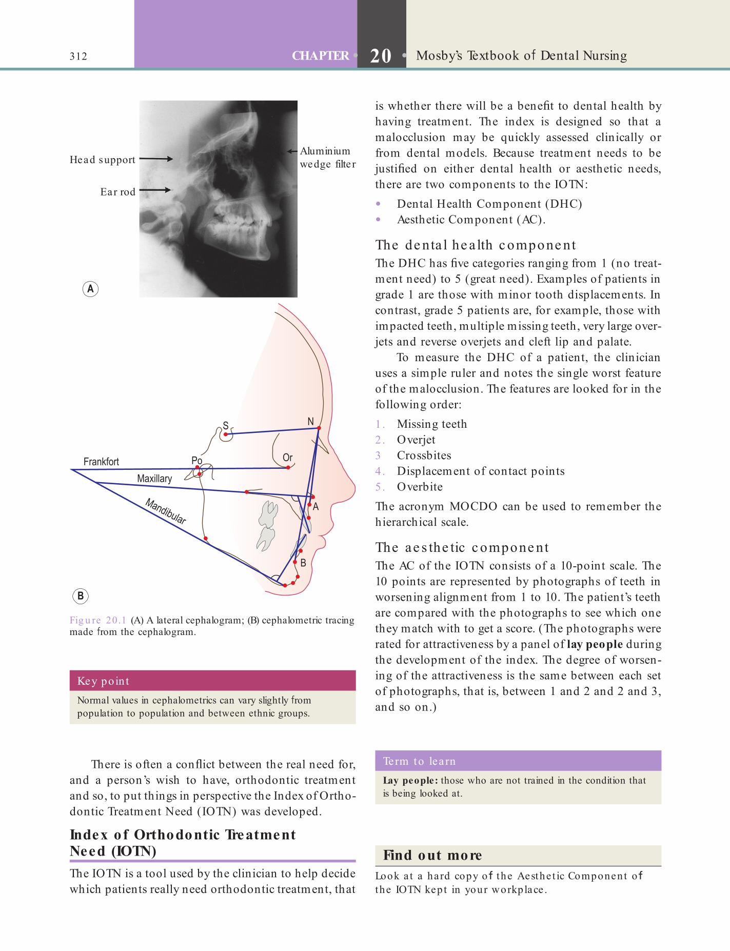
312 CHAPTER • 20 • Mosby’s Textbook o Dental Nursing
is whether there will be a bene t to dental health by having treatment. The index is designed so that a malocclusion may be quickly assessed clinically or from dental models. Because treatment needs to be justi ed on either dental health or aesthetic needs, there are two components to the IOTN:
• Dental Health Component (DHC)• Aesthetic Component (AC).
The de nta l he a lth c ompone ntThe DHC has ve categories ranging from 1 (no treat-ment need) to 5 (great need). Examples of patients in grade 1 are those with minor tooth displacements. In contrast, grade 5 patients are, for example, those with impacted teeth, multiple missing teeth, very large over-jets and reverse overjets and cleft lip and palate.
To measure the DHC of a patient, the clinician uses a simple ruler and notes the single worst feature of the malocclusion. The features are looked for in the following order:1 . Missing teeth2 . Overjet3 Crossbites4 . Displacement of contact points5 . OverbiteThe acronym MOCDO can be used to remember the hierarchical scale.
The ae s the tic c ompone ntThe AC of the IOTN consists of a 10-point scale. The 10 points are represented by photographs of teeth in worsening alignment from 1 to 10. The patient’s teeth are compared with the photographs to see which one they match with to get a score. (The photographs were rated for attractiveness by a panel of lay people during the development of the index. The degree of worsen-ing of the attractiveness is the same between each set of photographs, that is, between 1 and 2 and 2 and 3, and so on.)
There is often a con ict between the real need for, and a person’s wish to have, orthodontic treatment and so, to put things in perspective the Index of Ortho-dontic Treatment Need (IOTN) was developed.
Index of Orthodontic Treatment Need (IOTN)The IOTN is a tool used by the clinician to help decide which patients really need orthodontic treatment, that
Fig u re 20.1 (A) A lateral cephalogram; (B) cephalometric tracing made rom the cephalogram.
Aluminiumwedge filte rHead support
Ear rod
Key poin tNormal values in cephalometrics can vary slightly rom population to population and between ethnic groups.
Term to lea rnLay people: those who are not trained in the condition that is being looked at.
Find out moreLook a t a hard copy o the Aesthe t ic Component o the IOTN kept in your workplace .

313Orthodontics
• Removable appliances (ROAs) – these are used for correcting simple problems, such as moving a single tooth or expanding the dental arch. They have an acrylic baseplate with wires and springs attached (see the ROA in Figure 20.6 below).
• Functional appliances – these are usually removable appliances that are sometimes used to in uence jaw growth and improve the way the maxillary and mandibular teeth meet.
• Retainers – these are used after treatment, to keep the teeth in their new positions. Retention is life-long
Fixed Appliances
Ke y fe a ture s• Fixed appliances consist of brackets and
archwires (Figure 20.2), which are tied into the bracket slots using elastic rings (often coloured) or metal ligatures (Figure 20.3).
• The brackets are made from metal (stainless steel), plastic or ceramic materials. The ceramic brackets are clear or tooth-coloured so are less visible and usually used in adults. Usually brackets are placed on the labial and buccal surfaces of teeth. However, they can also be bonded to the lingual/palatal surfaces of the teeth (this is called ‘lingual orthodontics’).
• The brackets are most commonly attached to the teeth by bonding with resins. This requires acid etching of teeth.
• Composite resin is applied to the bracket base and works as the adhesive that bonds onto the acid-etched tooth (see Chapter 16). The teeth must be kept dry during placement of the brackets to ensure successful bonding.
Identify and learnI your workplace provides orthodont ic t rea tment , look a t some bracke ts. Otherwise f nd a den ta l ca ta logue , and check ou t the aesthe t ic d i e rence be tween meta l and ceramic bracke ts and no te the slo t in which the wire is p laced . Also no te the ‘wings’ tha t ho ld the e last ic ring or liga tures in p lace .
Find out moreWhat kind o b racke ts a re usua lly used on the last tooth in the a rch – usua lly the f rst o r second molar? What a re they ca lled?
Orthodontic TreatmentTimingMalocclusion often becomes noticeable as a child’s permanent teeth erupt, between the ages of 6 and 12 years. Treatment that begins while a child is still growing helps produce most favourable results. Thus most children should have:
• Orthodontic evaluation no later than age 7 years, by when they have a mix of primary teeth and permanent teeth
• Orthodontic treatment if required, beginning between ages 8 and 14 years.
Age is no contra-indication to treatment. Healthy teeth can move at any age.
Although the treatment plan is customised for each patient, most will need braces for one to three years, depending on what needs correcting. Treatment is followed by a period of wearing a retainer.
Fig u re 20.2 A f xed orthodontic appliance. Note the brackets on the anterior teeth and the bands with welded attachments on the molars, the archwire running through the bracket and the elastics.
Is orthodont ic t rea tment pa in u l?A little discom ort may be expected during normal orthodontic treatment. Overly rapid tooth movement may cause pain and may even cause root resorption or death (non-vitality) o a tooth. Newer materials may apply a more constant, gentle orce to move teeth and usually require ewer regular adjustments.
The main goals of orthodontic treatment were described on p. 309. This can involve the use of:
• Fixed appliances – sometimes called ‘braces’ or ‘train tracks’ (Figure 20.2).
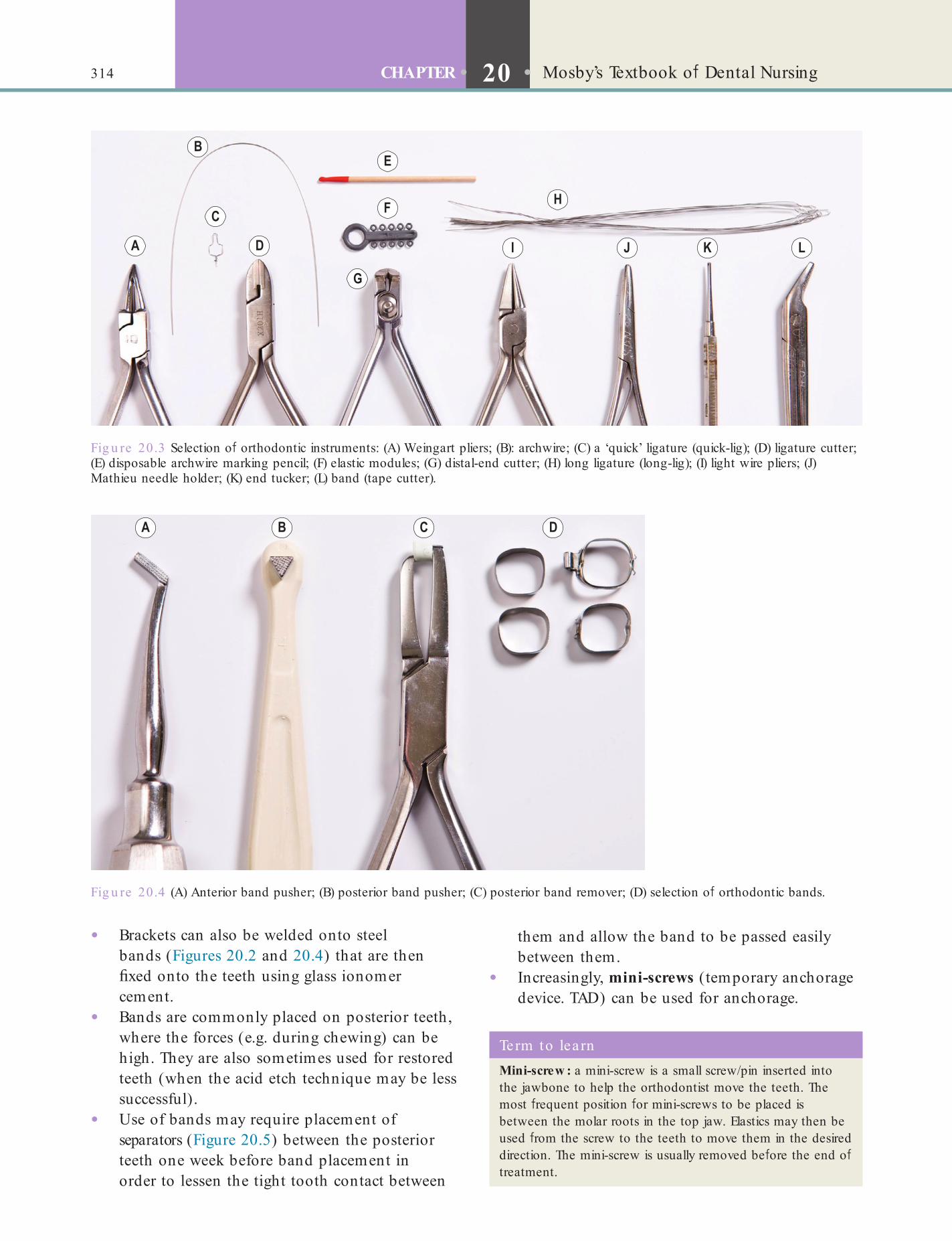
314 CHAPTER • 20 • Mosby’s Textbook o Dental Nursing
them and allow the band to be passed easily between them.
• Increasingly, mini-screws (temporary anchorage device. TAD) can be used for anchorage.
• Brackets can also be welded onto steel bands (Figures 20.2 and 20.4) that are then xed onto the teeth using glass ionomer cement.
• Bands are commonly placed on posterior teeth, where the forces (e.g. during chewing) can be high. They are also sometimes used for restored teeth (when the acid etch technique may be less successful).
• Use of bands may require placement of separators (Figure 20.5) between the posterior teeth one week before band placement in order to lessen the tight tooth contact between
Fig u re 20.3 Selection o orthodontic instruments: (A) Weingart pliers; (B): archwire; (C) a ‘quick’ ligature (quick-lig); (D) ligature cutter; (E) disposable archwire marking pencil; (F) elastic modules; (G) distal-end cutter; (H) long ligature (long-lig); (I) light wire pliers; (J) Mathieu needle holder; (K) end tucker; (L) band (tape cutter).
Fig u re 20.4 (A) Anterior band pusher; (B) posterior band pusher; (C) posterior band remover; (D) selection o orthodontic bands.
Term to lea rnMini-screw : a mini-screw is a small screw/pin inserted into the jawbone to help the orthodontist move the teeth. The most requent position or mini-screws to be placed is between the molar roots in the top jaw. Elastics may then be used rom the screw to the teeth to move them in the desired direction. The mini-screw is usually removed be ore the end o treatment.
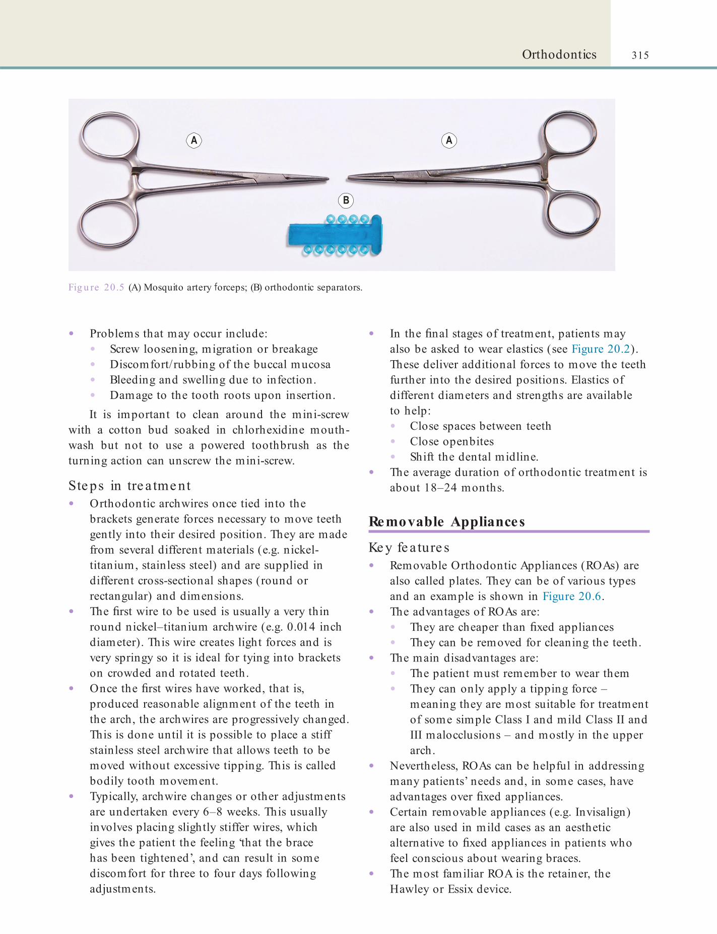
315Orthodontics
• In the nal stages of treatment, patients may also be asked to wear elastics (see Figure 20.2). These deliver additional forces to move the teeth further into the desired positions. Elastics of different diameters and strengths are available to help:• Close spaces between teeth• Close openbites• Shift the dental midline.
• The average duration of orthodontic treatment is about 18–24 months.
Removable Appliances
Ke y fe a ture s• Removable Orthodontic Appliances (ROAs) are
also called plates. They can be of various types and an example is shown in Figure 20.6.
• The advantages of ROAs are:• They are cheaper than xed appliances• They can be removed for cleaning the teeth.
• The main disadvantages are:• The patient must remember to wear them• They can only apply a tipping force –
meaning they are most suitable for treatment of some simple Class I and mild Class II and III malocclusions – and mostly in the upper arch.
• Nevertheless, ROAs can be helpful in addressing many patients’ needs and, in some cases, have advantages over xed appliances.
• Certain removable appliances (e.g. Invisalign) are also used in mild cases as an aesthetic alternative to xed appliances in patients who feel conscious about wearing braces.
• The most familiar ROA is the retainer, the Hawley or Essix device.
• Problems that may occur include:• Screw loosening, migration or breakage• Discomfort/rubbing of the buccal mucosa• Bleeding and swelling due to infection.• Damage to the tooth roots upon insertion.It is important to clean around the mini-screw
with a cotton bud soaked in chlorhexidine mouth-wash but not to use a powered toothbrush as the turning action can unscrew the mini-screw.
Ste ps in tre a tme nt• Orthodontic archwires once tied into the
brackets generate forces necessary to move teeth gently into their desired position. They are made from several different materials (e.g. nickel-titanium, stainless steel) and are supplied in different cross-sectional shapes (round or rectangular) and dimensions.
• The rst wire to be used is usually a very thin round nickel–titanium archwire (e.g. 0.014 inch diameter). This wire creates light forces and is very springy so it is ideal for tying into brackets on crowded and rotated teeth.
• Once the rst wires have worked, that is, produced reasonable alignment of the teeth in the arch, the archwires are progressively changed. This is done until it is possible to place a stiff stainless steel archwire that allows teeth to be moved without excessive tipping. This is called bodily tooth movement.
• Typically, archwire changes or other adjustments are undertaken every 6–8 weeks. This usually involves placing slightly stiffer wires, which gives the patient the feeling ‘that the brace has been tightened’, and can result in some discomfort for three to four days following adjustments.
Fig u re 20.5 (A) Mosquito artery orceps; (B) orthodontic separators.
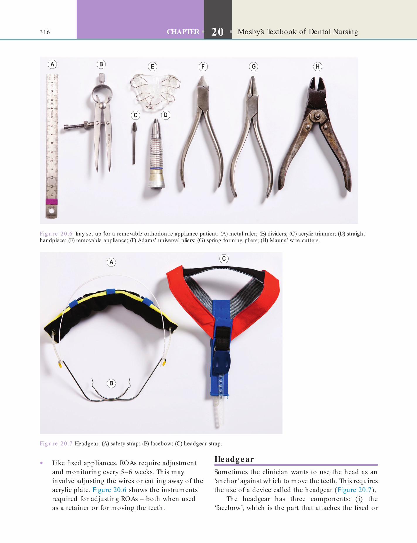
316 CHAPTER • 20 • Mosby’s Textbook o Dental Nursing
HeadgearSometimes the clinician wants to use the head as an ‘anchor’ against which to move the teeth. This requires the use of a device called the headgear (Figure 20.7).
The headgear has three components: (i) the ‘facebow’, which is the part that attaches the xed or
• Like xed appliances, ROAs require adjustment and monitoring every 5–6 weeks. This may involve adjusting the wires or cutting away of the acrylic plate. Figure 20.6 shows the instruments required for adjusting ROAs – both when used as a retainer or for moving the teeth.
Fig u re 20.6 Tray set up or a removable orthodontic appliance patient: (A) metal ruler; (B) dividers; (C) acrylic trimmer; (D) straight handpiece; (E) removable appliance; (F) Adams’ universal pliers; (G) spring orming pliers; (H) Mauns’ wire cutters.
Fig u re 20.7 Headgear: (A) sa ety strap; (B) acebow; (C) headgear strap.

317Orthodontics
Identify and learnIdent i y a Hawley and Essix re ta iner in your workplace . I not ava ilab le , search o r images on the in te rne t .
Complications of Orthodontic Treatment
• Patients wearing braces should not use chewing gum or eat sticky or hard foods, such as toffee and nuts, because they can damage the braces.
• Braces can also be damaged when playing sports – so it is important for patients to wear a mouthguard when playing sports.
• Braces and ROAs can cause ulcers in the mouth. This happens because of, for example, rubbing of a wire or bracket against the cheeks and lips. However, these heal in a few days. In the meantime the patient can use orthodontic wax or dental silicone to cover the offending part of the brace until the ulcer heals.
• A wire end at the back of the mouth can also stick out and rub against the mucosa. This can cause mild to severe ulcers and pain. Patients with such problems should again use the wax or contact the dental surgery as it may be necessary for the clinician to carefully bend the edge of the wire or clip it.
• Some people believe that orthodontic treatment may be followed by dysfunction of the temporomandibular joints (TMJs) but there is no scienti c evidence to prove this.Other potential complications of orthodontic
treatment apart from mild discomfort are:
• Decalci cation – when the mineral in the tooth surface under the brackets or bands gets dissolved due to poor oral hygiene and accumulation of food and plaque. It appears as whitish areas when the brackets are removed.
• Caries – if the decalci cation is allowed to continue, it can eventually lead to caries (see Chapter 10).
• Gingivitis – poor oral hygiene means the plaque can irritate the gingivae as well.
• Root resorption – shortening of the length of the root as the tooth moves.Although these complications are rarely serious,
dental plaque easily accumulates on and around appliances. So it is crucial to give dietary and other advice to the patients to maintain their oral hygiene
removable appliance to (ii) the strap that goes round the head and (iii) safety features to prevent injuries. Headgear is usually only worn at home and at night while sleeping.
A headgear can also be used as a dental orthopae-dic appliance.
Orthodontic Tooth MovementTooth movement happens due to the remodelling of the alveolar bone and alterations in the blood supply to the periodontal ligament. These are triggered by the forces that are applied to the teeth by the orthodontic appliances. The amount of bone remodelling depends on the magnitude, direction and duration of the applied force. Eventually, the supporting bone archi-tecture is re-established and the teeth settle down in their new positions.
Term to lea rnRemodelling: the process by which bone is constantly being removed and deposited to maintain its orm. For example, during growth or when the teeth are moved.
Retention• Teeth that have been moved orthodontically
have a natural tendency to move back to their original positions. In addition, teeth continue to move throughout life as a response to normal facial growth and changes in the soft tissues that cover the face. Therefore, after orthodontic treatment, patients have to wear retainers to maintain the corrected tooth positions.
• Retainers may be removable or xed.• Retainers are typically worn daily for one
year following xed appliance treatment and then ideally for a few nights per week inde nitely if the teeth are to be maintained in their corrected positions. Retention is life-long.
• There are several designs of removable retainer (e.g. Hawley, Begg and Essix retainers); each has advantages and disadvantages in terms of durability and aesthetic appearance.
• An advantage of a xed retainer, a wire bonded permanently to the lingual tooth surfaces, is that the patient does not have to remember to wear the appliance; but a disadvantage is that it can be dif cult to clean around the wire.
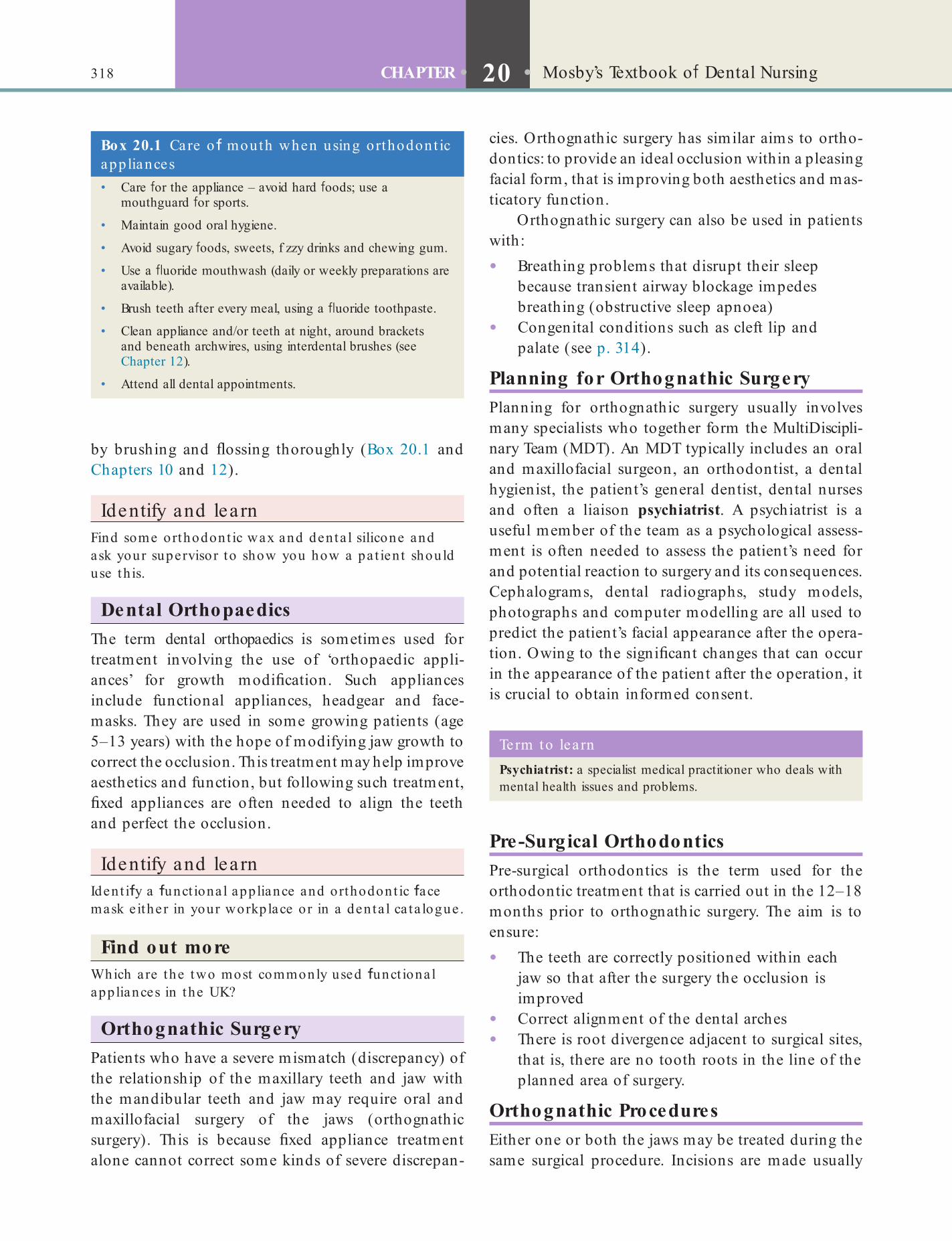
318 CHAPTER • 20 • Mosby’s Textbook o Dental Nursing
cies. Orthognathic surgery has similar aims to ortho-dontics: to provide an ideal occlusion within a pleasing facial form, that is improving both aesthetics and mas-ticatory function.
Orthognathic surgery can also be used in patients with:
• Breathing problems that disrupt their sleep because transient airway blockage impedes breathing (obstructive sleep apnoea)
• Congenital conditions such as cleft lip and palate (see p. 314).
Planning for Orthognathic SurgeryPlanning for orthognathic surgery usually involves many specialists who together form the MultiDiscipli-nary Team (MDT). An MDT typically includes an oral and maxillofacial surgeon, an orthodontist, a dental hygienist, the patient’s general dentist, dental nurses and often a liaison psychiatrist. A psychiatrist is a useful member of the team as a psychological assess-ment is often needed to assess the patient’s need for and potential reaction to surgery and its consequences. Cephalograms, dental radiographs, study models, photographs and computer modelling are all used to predict the patient’s facial appearance after the opera-tion. Owing to the signi cant changes that can occur in the appearance of the patient after the operation, it is crucial to obtain informed consent.
by brushing and ossing thoroughly (Box 20.1 and Chapters 10 and 12).
Identify and learnFind some orthodont ic wax and denta l silicone and ask your supervisor to show you how a pa t ient should use th is.
Dental OrthopaedicsThe term dental orthopaedics is sometimes used for treatment involving the use of ‘orthopaedic appli-ances’ for growth modi cation. Such appliances include functional appliances, headgear and face-masks. They are used in some growing patients (age 5–13 years) with the hope of modifying jaw growth to correct the occlusion. This treatment may help improve aesthetics and function, but following such treatment, xed appliances are often needed to align the teeth and perfect the occlusion.
Identify and learnIdent i y a unct iona l appliance and orthodont ic ace mask e ither in your workplace or in a den ta l ca ta logue .
Find out moreWhich a re the two most commonly used unct iona l appliances in the UK?
Orthognathic SurgeryPatients who have a severe mismatch (discrepancy) of the relationship of the maxillary teeth and jaw with the mandibular teeth and jaw may require oral and maxillofacial surgery of the jaws (orthognathic surgery). This is because xed appliance treatment alone cannot correct some kinds of severe discrepan-
Box 20.1 Care o mouth when using orthodont ic appliances• Care or the appliance – avoid hard oods; use a
mouthguard or sports.
• Maintain good oral hygiene.
• Avoid sugary oods, sweets, f zzy drinks and chewing gum.
• Use a uoride mouthwash (daily or weekly preparations are available).
• Brush teeth a ter every meal, using a uoride toothpaste.
• Clean appliance and/or teeth at night, around brackets and beneath archwires, using interdental brushes (see Chapter 12).
• Attend all dental appointments.
Term to lea rnPsychiatrist: a specialist medical practitioner who deals with mental health issues and problems.
Pre-Surgical OrthodonticsPre-surgical orthodontics is the term used for the orthodontic treatment that is carried out in the 12–18 months prior to orthognathic surgery. The aim is to ensure:
• The teeth are correctly positioned within each jaw so that after the surgery the occlusion is improved
• Correct alignment of the dental arches• There is root divergence adjacent to surgical sites,
that is, there are no tooth roots in the line of the planned area of surgery.
Orthognathic ProceduresEither one or both the jaws may be treated during the same surgical procedure. Incisions are made usually
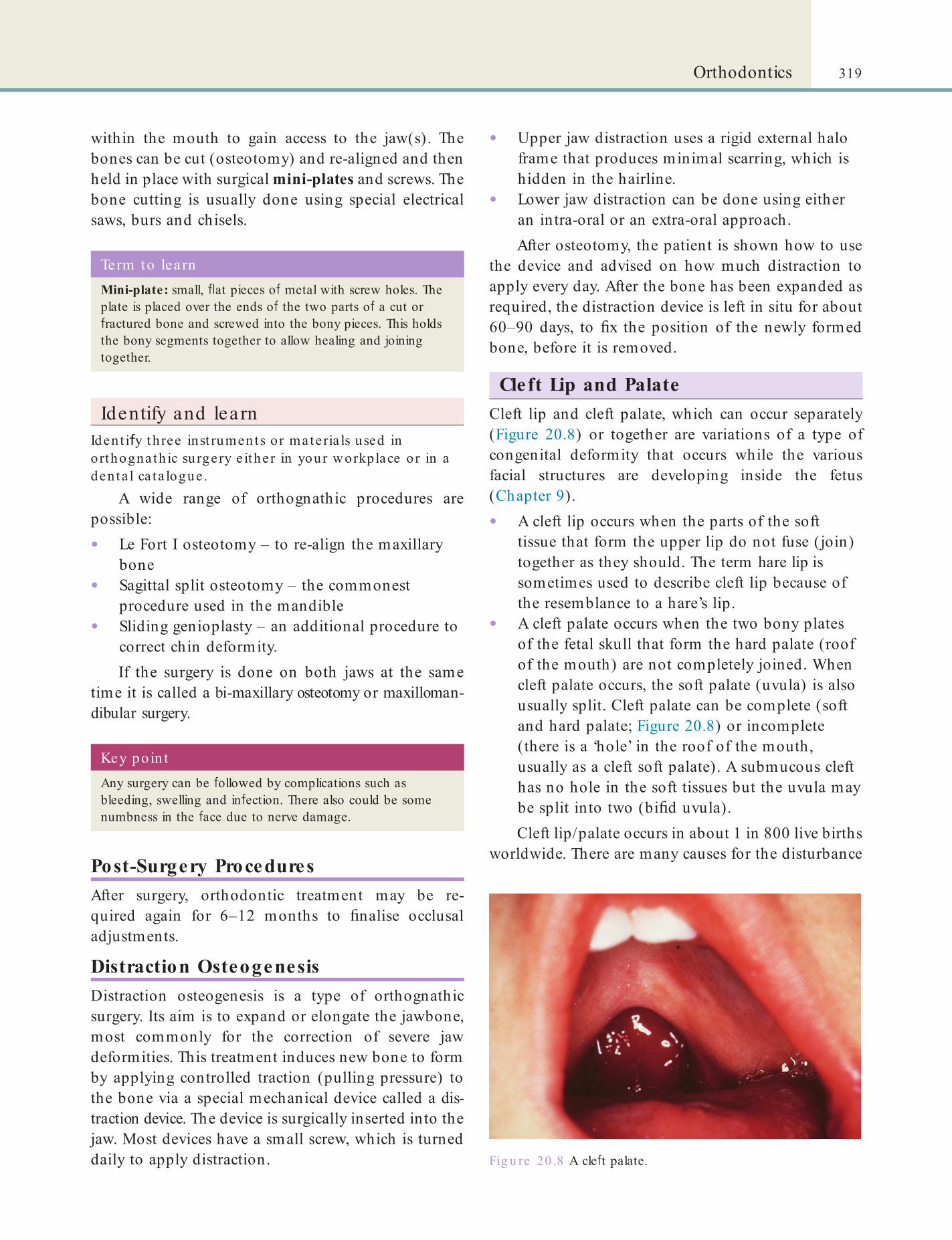
319Orthodontics
• Upper jaw distraction uses a rigid external halo frame that produces minimal scarring, which is hidden in the hairline.
• Lower jaw distraction can be done using either an intra-oral or an extra-oral approach.After osteotomy, the patient is shown how to use
the device and advised on how much distraction to apply every day. After the bone has been expanded as required, the distraction device is left in situ for about 60–90 days, to x the position of the newly formed bone, before it is removed.
Cleft Lip and PalateCleft lip and cleft palate, which can occur separately (Figure 20.8) or together are variations of a type of congenital deformity that occurs while the various facial structures are developing inside the fetus (Chapter 9).
• A cleft lip occurs when the parts of the soft tissue that form the upper lip do not fuse (join) together as they should. The term hare lip is sometimes used to describe cleft lip because of the resemblance to a hare’s lip.
• A cleft palate occurs when the two bony plates of the fetal skull that form the hard palate (roof of the mouth) are not completely joined. When cleft palate occurs, the soft palate (uvula) is also usually split. Cleft palate can be complete (soft and hard palate; Figure 20.8) or incomplete (there is a ‘hole’ in the roof of the mouth, usually as a cleft soft palate). A submucous cleft has no hole in the soft tissues but the uvula may be split into two (bi d uvula).Cleft lip/palate occurs in about 1 in 800 live births
worldwide. There are many causes for the disturbance
within the mouth to gain access to the jaw(s). The bones can be cut (osteotomy) and re-aligned and then held in place with surgical mini-plates and screws. The bone cutting is usually done using special electrical saws, burs and chisels.
Key poin tAny surgery can be ollowed by complications such as bleeding, swelling and in ection. There also could be some numbness in the ace due to nerve damage.
Term to lea rnMini-plate: small, at pieces o metal with screw holes. The plate is placed over the ends o the two parts o a cut or ractured bone and screwed into the bony pieces. This holds the bony segments together to allow healing and joining together.
Identify and learnIdent i y three inst ruments or mate ria ls used in o rthogna th ic surgery e ither in your workplace or in a denta l ca ta logue.
A wide range of orthognathic procedures are possible:
• Le Fort I osteotomy – to re-align the maxillary bone
• Sagittal split osteotomy – the commonest procedure used in the mandible
• Sliding genioplasty – an additional procedure to correct chin deformity.If the surgery is done on both jaws at the same
time it is called a bi-maxillary osteotomy or maxilloman-dibular surgery.
Post-Surgery ProceduresAfter surgery, orthodontic treatment may be re -quired again for 6–12 months to nalise occlusal adjustments.
Distraction OsteogenesisDistraction osteogenesis is a type of orthognathic surgery. Its aim is to expand or elongate the jawbone, most commonly for the correction of severe jaw deformities. This treatment induces new bone to form by applying controlled traction (pulling pressure) to the bone via a special mechanical device called a dis-traction device. The device is surgically inserted into the jaw. Most devices have a small screw, which is turned daily to apply distraction. Fig u re 20.8 A cle t palate.

320 CHAPTER • 20 • Mosby’s Textbook o Dental Nursing
To f nd ou t more about t aking rad iographs or orthodont ic t rea tment , see Orthodont ic Radiographs – Guide lines, 3rd ed it ion (KG Isaacson , AR Thom, K Horner and E Whaites, published by the Brit ish Orthodont ic Socie ty, 2008).
in facial development and they can be broadly catego-rised into genetic and environmental. A maternal diet low in folic acid and vitamins, and exposure of the pregnant mother to toxins (e.g. pesticides, retinoids, anticonvulsants, alcohol, cigarettes, nitrates, organic solvents, lead and illegal drugs, e.g. cocaine, crack cocaine, heroin) may be responsible.
The abnormal connection between the mouth and nasal cavity causes air to leak into the nose (called velopharyngeal insuf ciency), resulting in a hyper-nasal voice and impaired speech.
The aim of treatment is to improve the aesthetics and function, particularly speech. An MDT is required with oral and maxillofacial, ENT and plastic surgeons, orthodontist, restorative dentist, hygienist and speech therapist. One example of a schedule that is followed for treating a patient with a cleft of the lip or palate is shown in Table 20.1.
ReferralReferral of patients with orthodontic problems, to either specialist practitioners or hospital consultants, depends on several factors:
• The general dental practitioner’s knowledge and ability to treat malocclusion
• The patient’s desire to see a specialist or undergo specialist treatment
• The age and general health status of the patient.
Find out moreTo f nd out more about orthodont ic t rea tment , see Orthodont ics at a Glance (D Gill, pub lished by Wiley Blackwell, 2008).
Table 20.1 Schedule o r management o pa t ien ts with cle t lip and pa la te
Approximate age Schedule*
Birth Assessment. Discuss with team and parents. Feeding advice. Possible pre-surgical orthodontics
3 months Repair cle t lip
9–18 months Repair cle t palate Oral hygiene instructions given to parents
30 months Assess speech
4 years + Consider surgery to correct the lip and/or palate
5 years + Consider surgery or correction o speech de ects. Speech therapy. Preventive dentistry
8 years + Simple orthodontics
10 years + Alveolar bone gra t to f ll the bony gap in the palate
12 years + Def nitive orthodontics
16 years + Maxillo acial surgery i needed or correcting maxillary hypoplasia or nasal de ormity Restorative dentistry
*Preventive dental care is required throughout.
Chapter 21 People with Vulnerability or Special Needs
CHAPTER POINTS• People with vulnerability• Younger people• Older people• People with special needs• Disability discrimination• Pregnant women• People with impaired vision• People with impaired hearing• People with impaired mobility or physical incapacity• People with dental anxiety or phobia• People with medical problems• People with cancer• People with learning di f cult ies or special
educational needs• Mental Capacity Act
• Self-protection is being able to demonstrate behaviour that• results in defending oneself against threats of
safety, and• results in successfully meeting one’s own
basic (safety) needs.A person’s vulnerability helps inform what must be done to manage threats and assure protection.
People who may be especially vulnerable are:
• Children• Older people• People who are physically handicapped• People who are cognitively limited are
vulnerable.Regardless of age, intellect and physical capacity,
people who are highly dependent and susceptible to others, or unable to defend themselves, or cannot or will not seek help and protection, or who are so passive or withdrawn as not to be able to make his or her basic needs known, are vulnerable. Regardless of age, there are people who are not visible to be noticed and observed, or who have continuing or acute medical problems and needs, which can make them vulnerable
Vulnerable people sometimes have emotional, mental health, or behavioural problems that may irri-tate and provoke some others to avoid them, act out toward them or to abuse them.
Abuse may be:
• Physical: hitting, slapping, pushing, kicking, burning, or other show of force; misuse of medication; restraint, or inappropriate sanctions
• Sexual: including rape and sexual assault or sexual acts to which the vulnerable person has not consented, or could not consent or was pressured into consenting
In t ro d u ct io n
All dental staff should respect the uniqueness and dignity of each individual person, and respond appro-priately to their need for care, irrespective of religion, ethnic origin, gender, sexual preferences, personal attributes or nature of the health problem. There are also many people who are disadvantaged for a variety of reasons and in need of special care. This chapter discusses a range of these issues.
People w ith Vulnerability• Vulnerability means being defenceless to threats
of safety and a judgement about vulnerability is based on the capacity for self-protection.
322 CHAPTER • 21 • Mosby’s Textbook of Dental Nursing
When treating children and the adolescents, the dental clinician needs to consider:
• Medical and social history:• Medical, physical or psychiatric problems• Patient compliance• Commitment of the parent or guardian to
the treatment• Dental status:
• Is the tooth (teeth) restorable?• Is there an aesthetic problem?• Is there a problem with chewing?
• Psychological: including emotional abuse, threats of harm or abandonment, deprivation of contact, humiliation, blaming, controlling, intimidation, coercion, harassment, verbal abuse, isolation or withdrawal from services or supportive networks
• Financial or material abuse: theft, fraud, exploitation, pressure in connection with wills, property or inheritance or nancial transactions, or the misuse or misappropriation of property, possessions, assets or bene ts
• Neglect and acts of omission: ignoring medical or physical care needs, failure to provide access to appropriate health, social care or educational services, withholding of the necessities of life, such as medication, adequate nutrition and heating
• Discriminatory abuse: including racist, sexist, that based on a person’s disability, and other forms of harassment, slurs or similar treatment.
Local Safeguarding Children Boards (LSCBs) and Safe-guarding Adults Boards (SABs) are the key safety mechanisms but all organisations and employers should promote a culture where staff have support to achieve effective outcomes for vulnerable people and feel able to raise concerns and whistleblow without fear.
Find out moreYou can read more about the ramework o r sa eguard ing vu lnerab le people in the NHS Commission ing Board publica t ion Safeguard ing Vulnerable People in the Re form ed NHS (2013), a t www.england .nhs.uk/wp-conten t /up loads/2013/03/sa eguard ing-vulnerab le-people .pd .
Further protection is provided by the vetting and barring procedures applying to all people who work in contact with potentially vulnerable people, under the Safeguarding Vulnerable Groups Act 2006, as amended by the Protection of Freedoms Act 2012 (see Chapter 5).
Younger People• In the UK, people up to the age of 16 years are
regarded as children.• Paediatric dentistry is the specialty that provides
primary and comprehensive preventative and therapeutic oral healthcare for children, including those with special healthcare needs.
• Dental caries is the most prevalent disease among children aged 5–16 years.
Terms to lea rnPatient compliance: how well a patient correctly follows all the instructions given to them by a healthcare professional about a particular treatment or preventive regimen.
Regimen: a systematic plan for therapy (may include diet).
Treatment of children may take longer to plan and carry out and they may need special control of anxiety and pain. Drugs such as tetracycline (can stain teeth) and aspirin (can cause liver damage) are contraindi-cated in children.
Vulnerable ChildrenSee the ‘Guide to Social Care Services’ on the NHS Choices website (www.nhs.uk/NHSEngland/AboutNHSservices/social-care-services/Pages/children -services.aspx).
Children rely on parents or guardians for protec-tion from harm, and for discipline. Accidents are the major cause of morbidity and mortality: children are at increased risk because of a need for strenuous physi-cal activity, a desire for peer approval and increased adventurous behaviour. Abuse and neglect, as de ned in the Department of Health’s document ‘Working Together to Safeguard Children’ may be physical, emo-tional, sexual abuse or neglect. Child abuse is any act of omission or commission that endangers or impairs the physical or emotional health or development of a child. Some level of emotional abuse is involved in all types of ill-treatment of a child, though it may occur alone. Physical abuse may involve hitting, shaking, throwing, poisoning, burning or scalding, drowning, suffocating or otherwise causing physical harm to a child. A child is considered to be abused if he or she is treated in a way that is unacceptable in a given culture at a given time. For example, physical punish-ment of children has become progressively less accept-able in the UK.
323People with Vulnerability or Special Needs
functionally independent or dependent. This is assessed with speci c tests (e.g. Activities of Daily Living Scale (ADLS)).Oral healthcare may be lacking, and older people generally are more likely to have a drier mouth and a softer, more cariogenic diet than some younger people. For these reasons they may have:• Fewer teeth• Tooth surface loss – especially due to gross
caries, root caries and attrition• Periodontitis.
Children with disabilities are much more at risk of experiencing abuse of all kinds.
Recognition of abuse is important since there is a very high risk of further assaults on, or death of, the child and of siblings. The child who has been sub-jected to abuse is often cowed, may also be malnour-ished and generally neglected, and there may be evidence of previous trauma with injuries varied and are often multiple, including bite marks, bruises, black eyes, torn frenulum, hair loss, laceration, wheals, liga-ture marks, marks from a gag, burns and scalds often from cigarettes or demarcated from other objects. Lac-erations and tearing of the lip may be found. Teeth may be devitalised, broken or lost. Bone fractures are common. Head injuries are the commonest cause of death in abused children.
After genuine accidents, children are usually immediately taken for medical or dental attention, but when abuse is involved there is often considerable delay and/or injuries are incompatible with the history.
Ac tion if abus e is s us pe c te dHealthcare professionals are obliged to know and follow the local child protection procedures (for guidance, see www.cpdt.org.uk/).
Full records must be kept. The child must be fully and immediately examined to exclude serious injury and must therefore be admitted to hospital. A paedia-trician should be consulted and the general medical practitioner, social services and a child protection agency must be involved early on, but strict con den-tiality must be observed. Social services departments keep registers of non-accidental injuries, which help identify known offenders. The child may already be on the ‘at-risk’ register. The dentist should also inform their Medical Defence Society as there may later be legal involvement.
Older People• People over the age of 65 years are currently
regarded as old.• Geriatric dentistry and special care dentistry
provide primary and comprehensive preventive and therapeutic oral healthcare for older people, including those with special healthcare needs.
• A number of diseases are more common in older people – and many have chronic diseases, walk with dif culty or are house-bound or bed-ridden. Based on their capacity to carry out activities of ordinary life (to dress, to eat, to bathe, etc), older people are said to be
Key poin tHandling of older patients may require immense patience.
Term to lea rnGross caries: severe caries.
Older people need time to explain their needs and to move in and around the dental environment; do not rush them and do not use acronyms or modern jargon they may struggle to understand.
The major goals of oral healthcare in older people are:
• Preventive and conservative treatment• Elimination/avoidance of pain and oral
infections• Maintaining function, aesthetics and dignity• Supporting morale and contributing to
nutritional advice.Many older people are taking several medications
and the clinician needs to be aware of possible drug interactions with drugs used. Treatment compliance can be dif cult, not least because of forgetfulness or indifference.
Independent older people are often reluctant to demand special attention, mainly because of fear of loss of independence and hospitalisation, or through apathy. Dependent persons may need domiciliary dental care with portable dental equipment. When such patients also have signi cant medical problems, they may be best seen in a hospital environment. However, this raises issues of adequate transportation and availability of accompanying persons.
Term to lea rnDomiciliary dental care: care provided in the patient’s own home.

324 CHAPTER • 21 • Mosby’s Textbook of Dental Nursing
afford appropriate care, readily demoralises the older patient, and may tip the balance between health and disease.
It is wise to label the dentures with the name of the patient, particularly for those living in sheltered or other residential accommodation, as dentures can easily be mislaid or mixed up between patients.
Attrition and brittleness of the teeth may compli-cate treatment, and it may be necessary to provide cuspal coverage in complex or large restorations. Endodontic therapy may be more dif cult in view of secondary dentine deposition. Hypercementosis, brittle dentine, low bone elasticity and impaired tissue healing may also complicate surgical procedures.
Independent people with no serious medical problems may be treated in primary dental care. Dependent people may need domiciliary dental care with portable dental equipment. When signi cant medical problems are present, the older person may be best seen in a hospital environment, though this raises issues of adequate transportation and availabil-ity of escorts.
Prevention is of paramount importance in caring for older adults. Therefore, the most important con-siderations for dental professionals are how well the patient has compensated for his or her medical problem and the exact dental intervention needed. Non-invasive procedures in patients with minimal incapacity carry less risk than do surgical procedures. The whole range of dental treatments may be considered. Dental implants are not at all contraindi-cated, and they may help to maintain the remaining alveolar bone.
Appointment times may be conditioned by sys-temic diseases. Treatment is often best carried out with the patient sitting upright, as few older patients like reclining for treatment, and some may become breath-less and panic.
Older people frequently have dif culties in under-standing the medication and in remembering to keep to a regimen. Compliance may therefore be lacking. If there is hepatic or renal disease likely to impair drug metabolism or excretion, drug dosage must be reduced appropriately.
Vulnerable Adults and EldersBroadly speaking, a vulnerable adult is aged 18 or over, receives or may need community care services because of a disability, age or illness, and is or may be unable to take care of themselves or protect themselves against signi cant harm or exploitation.
Many older patients have dif culty in accessing healthcare and some are reluctant to seek attention, especially if they fear consequent hospitalisation. There is a greater incidence and severity of many dis-eases, especially arthritis and cerebrovascular disease, and these may cause ataxia and falls. Defects of hearing or sight increase the liability to accidents, feeling of isolation and reducing independence.
Atypical symptomatology is common: physical disease may present in a less orid and dramatic way in older people. Dementia becomes increasingly common with age. Social disabilities are common as a result of such causes as loss of the spouse, isolation from family, poverty, and lack of mobility and inde-pendence. Old people may need to be thrifty, careful with food purchasing and the heating of their accom-modation, contributing to overall self-neglect. Nutri-tion may also be defective due to apathy, mental disease or dental defects. Deterioration in sense of smell and taste may also result in a poor or unhealthy diet. Psychological disorders such as loneliness and depression may follow. Social support is crucial, preserving as much independence and dignity as possible.
Older people are often extremely anxious about treatment and should therefore be sympathetically reassured and, if necessary, sedated.
Oral cancer is mainly a problem of older people and is a further reason for regular oral examination. Atypical facial pain (often related to depressive illness), migraine, trigeminal neuralgia, zoster and oral dysaes-thesias are more common as age advances.
Many older people with remaining teeth have periodontal disease. Dental caries are usually less acute but root caries are more common. Caries may become active if there is hyposalivation, especially if there is overindulgence in sweet foods. Hyposalivation is even more likely with medications such as neurolep-tics or antidepressants. Poor salivation may contribute to a high prevalence of root caries and oral candidiasis, which especially affect hospitalised patients.
Very many older patients are edentulous and some problems of dental management are thereby greatly reduced. It seems, however, that the proportion of edentulous older patients is gradually falling and, as a consequence, more of them need restorative dentistry or surgery of various types. Many edentulous older people have little alveolar bone to support dentures, and have a dry mouth as well as frail, atrophic mucosa. Implants may be helpful. Inability to cope with den-tures, or a sore mouth for any reason, or to ability to
325People with Vulnerability or Special Needs
to social services. The disclosure must be carried out in an appropriate manner so that the patient is not put at increased risk of harm. Details of the discussions and agreement of the patient to disclosure of informa-tion to social services should be recorded in the dental records.
Disclosure of information can be made without consent in a number of situations where there is:
• Risk of death or serious harm• No competence to give consent• Information required under a court order or
another legal obligation.It is the responsibility of the healthcare profes-
sional to ‘act at all times in the best interests of the patient based on a risk–bene t assessment, accept nal responsibility for his or her actions, and be able to justify them if subsequently challenged’. Local social services are the lead agencies to which disclosures of abuse should be made. Telephone referrals should be followed up in writing within 48 hours. Dif culties or concerns should be discussed with a senior colleague and the defence organisation. As with all patient information, it is essential that all records are kept entirely con dential. Accurate contemporaneous records must be kept of all allegations or suspicions of abuse. Precise factual information of the alleged abuse must be recorded. Details of any discussions and decisions taken and reason for those decisions should be clearly recorded. There should be compre-hensive documentation of injuries.
The main organisations that can offer support to those affected include police, hospitals, general medical practitioners, carer organisations, social services/social work departments, and social care inspection bodies.
Find out moreFor u rther in o rmat ion see the NHS Choices web page on vulnerab le people : www.nhs.uk/CarersDirect /gu ide /vulnerab le -people /Pages/vu lnerab le -adult s.aspx.
People w ith Special NeedsPeople with special needs is a term used to embrace individuals who require assistance for a wide range of conditions (often referred to as disabilities) that may be physical, medical or psychological in origin. The World Health Organisation (WHO) considers disabili-ties in the following terms:
Disabilities is an umbrella term, covering impairments, activity limitations, and
Elder maltreatment includes violence in icted upon those aged 60 and older. The violence usually occurs at the hands of a caregiver or a person the elder trusts and can be prevented by:
• Listening to elders and their caregivers• Reporting abuse or suspected abuse.
Older people are vulnerable, especially those who are unwell, frail, confused and unable either to stand up for themselves or keep track of their affairs and there can be abuse in their home, in hospital, and in nursing and residential homes.
Risk factors in the vulnerable adult person may include:
• Social isolation• Dependence on abuser for essential care• Physical and cognitive deterioration• Dementia• Challenging behaviour.
Vulnerable adults include people who are open to abuse because of learning dif culties, physical dis-abilities or mental illness. Becoming dependent on someone else, whether a carer, family member, friend or professional health worker (such as a staff member in a residential or nursing home or hospital), can put vulnerable people at risk of abuse. Abusers may create a feeling of dependency and may also make the vul-nerable person feel isolated, that nobody else cares for them and that they are on their own.
Older people who are most at risk of abuse at home include:
• Those who are isolated with little contact with friends, family or neighbours
• People with memory problems• People who have dif culty communicating with
others• People who do not get on with their carer• People whose carer is addicted to drugs or
alcohol• People whose carer depends on them for a home
and nancial and emotional support.Elder abuse may be psychological, physical, nancial, sexual and neglect (see www.helptheaged.org.uk/).
Ac tion if abus e is s us pe c te dPeople who are being abused are often scared that they will not be believed. If abuse is suspected, always talk to the ‘victim’ rst and tell them what you want to do.
The patient’s consent must rst be obtained before disclosing an incident or suspected incident of abuse

326 CHAPTER • 21 • Mosby’s Textbook of Dental Nursing
Table 21.1 shows the relationship between disability and impairment. Note that many people do not like the term ‘disability’ and prefer the term ‘dif culties’.
The origin of a disability may be:
• Developmental – that is, caused by impairments that occur during development (up to age 18). These include fetal damage from infections, defects of metabolism, alcohol, or hypoxia, chromosomal abnormalities (e.g. Down syndrome), autism and childhood infections (e.g. meningitis or encephalitis).
• Acquired – caused by impairments that are not related to the body’s development. For example trauma to the brain or spinal cord, multiple sclerosis, arthritis and Alzheimer disease.If a health professional believes a patient lacks the
capacity to consent, they cannot give or withhold consent to treatment on behalf of that patient; they may carry out an investigation or treatment judged to be in that patient’s best interest. In deciding what is in the patient’s best interest, the treating clinician will consider:
• The treatment options• Any evidence of the patient’s preferences• The patient’s background• Views of family members.This is known as the principle o best interest.
participation restrictions. An impairment is a problem in body unction or structure; an activity limitation is a di f culty encountered by an individual in executing a task or action; while a participation restriction is a problem experienced by an individual in involvement in li e situations.
Disability is thus not just a health problem. It is a complex phenomenon, re ecting the interaction between eatures o a person’s body and eatures o the society in which he or she lives. Overcoming the di f culties aced by people with disabilities requires interventions to remove environmental and social barriers.
People with disabilities have the same health needs as non-disabled people – or immunisation, cancer screening etc. They also may experience a narrower margin o health, both because o poverty and social exclusion, and also because they may be vulnerable to secondary conditions, such as pressure sores or urinary tract in ections. Evidence suggests that people with disabilities ace barriers in accessing the health and rehabilitation services they need in many settings. (www.who.int/topics/disabilities/en/; italics added)
Find out moreWhat is the WHO In te rna t iona l Classif ca t ion o Funct ion ing , Disability and Hea lth? Visit the WHO website (www.who.in t /classif ca t ions/ic /en/) to f nd ou t .
A person has a disability if he or she has a physical or mental impairment that greatly limits one or more major life activities, such as:
• Breathing• Caring for oneself• Concentrating• Hearing• Interacting with other people• Learning• Lifting• Performing manual tasks• Reaching• Reading• Seeing• Speaking• Standing• Thinking• Walking• Working.
Term to lea rn‘Best interest’ decisions: decisions made on behalf of people who lack capacity and aimed to be the least restrictive of the disabled person’s basic rights and freedoms.
Table 21.1 Disab ility and impairment
Disab ilit ies Impairments
Physical MobilityRespiratory
Mental EmotionalSocial
Sensory HearingVisual
Cognitive LearningAttention
327People with Vulnerability or Special Needs
Disability Discrimination Act 1995 (known as DDA) and equality of opportunity for disabled people was promoted by the Disability Rights Commission (DRC). The DDA stated the principle that it is unlaw-ful to treat a person with disability less favourably for a reason related to that person’s disability (unless it can be justi ed). The Act de ned a person with disa-bility as ‘A person who has or has had a physical or mental impairment which has a substantial and long-term adverse effect upon his or her ability to carry out normal day-to-day activities.’ In 2010 the Equality Act came into force and repealed all previous equality legislation in England, Scotland and Wales. Under this Act disability is de ned in the same terms as in the DDA and is listed as one of nine ‘Protected Character-istics’ to which the Act applies. The DRC was subse-quently replaced by the Equality and Human Rights Commission, and disability discrimination now comes under the same umbrella as all equality law. (There is more on equality in Chapter 22.)
Find out moreVisit the GOV.UK website (www.gov.uk/equa lity-act -2010-guidance) o r more act s about the Equa lity Act in re la t ion to your work.
Service providers (an expression that includes all dental staff) have a legal duty not to discriminate against people with special needs. Service providers cannot refuse to provide good facilities or services or provide them at a lower standard or in a worse manner to people with special needs. They also cannot offer a service on worse terms than would be offered to other members of the public.
Studies have shown that, in the past, untreated caries, gingivitis and periodontal disease were more common and rates of extractions were usually higher in people with special needs. The main obstacle or barrier to good oral healthcare in people with special needs in the past was access, either to facilities or because of discrimination. It is crucial to remove bar-riers and improve access to care, and also to avoid discrimination (Box 21.1) on the grounds of:
• Age• Gender• Social exclusion• Disability.
Disability DiscriminationThe current legislation applying is the:
• Equality Act 2010• Disability Discrimination Act 1995 (in Northern
Ireland)Before the Equality Act came into force, discrimination against people with disabilities was covered by the
Box 21.1 Reducing d iscriminat ion in the den ta l p ract ice• All providers of dentistry need to take reasonable steps to
make their dental practices accessible.
• They should remove, alter or provide means of avoiding physical features that make it impossible or unreasonably dif cult for people with disabilities to use their services. This includes possible alterations to the building design or construction, the approach and access to and exit from the building – for example ramp access for wheelchairs, parking bays for those with disabilities, and modi cations to xtures and ttings, furniture and furnishings, and equipment and materials.
• A ‘no dogs’ policy needs to have a provision to allow entry to service animals such as guide dogs or hearing dogs into the premises (but not necessarily into the surgery).
• However, if taking steps to facilitate access to the premises would result in an ‘undue burden’ or fundamentally alter the nature of the services provided, then exemptions may apply.
• Dental staff must also treat the person with a disability on the same basis as they treat non-disabled patients. For example, services cannot charge extra from a person with a disability for the cost of auxiliary aids and services.
• If a person with a disability poses a ‘direct threat’ (that is a risk that cannot be eliminated using special procedures) to the health or safety of others, a dentist may refuse to admit that person, e.g. an aggressive patient. They may need to be treated in hospital under general anaesthesia.
• If the person with a disability requires a procedure for which a non-disabled patient would ordinarily be referred, the person with a disability may legally be referred elsewhere for healthcare.
Key po in tA dental nurse should respect the uniqueness and dignity of each individual patient, and respond appropriately to their need for care.
People w ith Impaired VisionPeople with visual impairments include those who:
• Have never been able to see• Had normal vision for some years before
becoming gradually or suddenly partially or totally blind
• Have other disabilities in addition to the visual loss
• Have selective impairments of parts of the visual eld.
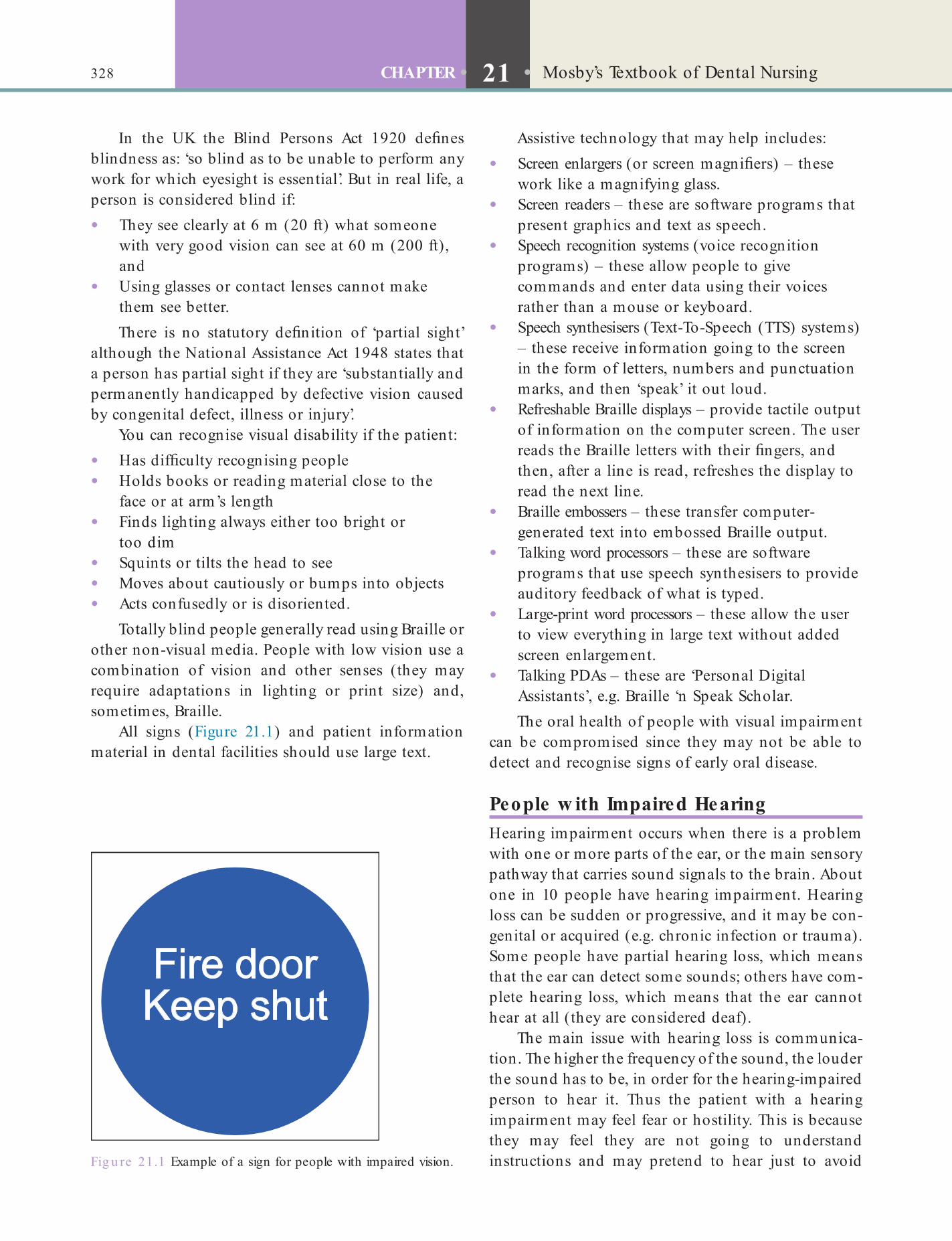
328 CHAPTER • 21 • Mosby’s Textbook of Dental Nursing
Assistive technology that may help includes:
• Screen enlargers (or screen magni ers) – these work like a magnifying glass.
• Screen readers – these are software programs that present graphics and text as speech.
• Speech recognition systems (voice recognition programs) – these allow people to give commands and enter data using their voices rather than a mouse or keyboard.
• Speech synthesisers (Text-To-Speech (TTS) systems) – these receive information going to the screen in the form of letters, numbers and punctuation marks, and then ‘speak’ it out loud.
• Refreshable Braille displays – provide tactile output of information on the computer screen. The user reads the Braille letters with their ngers, and then, after a line is read, refreshes the display to read the next line.
• Braille embossers – these transfer computer-generated text into embossed Braille output.
• Talking word processors – these are software programs that use speech synthesisers to provide auditory feedback of what is typed.
• Large-print word processors – these allow the user to view everything in large text without added screen enlargement.
• Talking PDAs – these are ‘Personal Digital Assistants’, e.g. Braille ‘n Speak Scholar.The oral health of people with visual impairment
can be compromised since they may not be able to detect and recognise signs of early oral disease.
People w ith Impaired HearingHearing impairment occurs when there is a problem with one or more parts of the ear, or the main sensory pathway that carries sound signals to the brain. About one in 10 people have hearing impairment. Hearing loss can be sudden or progressive, and it may be con-genital or acquired (e.g. chronic infection or trauma). Some people have partial hearing loss, which means that the ear can detect some sounds; others have com-plete hearing loss, which means that the ear cannot hear at all (they are considered deaf).
The main issue with hearing loss is communica-tion. The higher the frequency of the sound, the louder the sound has to be, in order for the hearing-impaired person to hear it. Thus the patient with a hearing impairment may feel fear or hostility. This is because they may feel they are not going to understand instructions and may pretend to hear just to avoid
In the UK the Blind Persons Act 1920 de nes blindness as: ‘so blind as to be unable to perform any work for which eyesight is essential’. But in real life, a person is considered blind if:
• They see clearly at 6 m (20 ft) what someone with very good vision can see at 60 m (200 ft), and
• Using glasses or contact lenses cannot make them see better.There is no statutory de nition of ‘partial sight’
although the National Assistance Act 1948 states that a person has partial sight if they are ‘substantially and permanently handicapped by defective vision caused by congenital defect, illness or injury’.
You can recognise visual disability if the patient:
• Has dif culty recognising people• Holds books or reading material close to the
face or at arm’s length• Finds lighting always either too bright or
too dim• Squints or tilts the head to see• Moves about cautiously or bumps into objects• Acts confusedly or is disoriented.
Totally blind people generally read using Braille or other non-visual media. People with low vision use a combination of vision and other senses (they may require adaptations in lighting or print size) and, sometimes, Braille.
All signs (Figure 21.1) and patient information material in dental facilities should use large text.
Fig u re 21.1 Example of a sign for people with impaired vision.

329People with Vulnerability or Special Needs
• Voice-to-text phone service or text-to-voice phone service or text telephone (TDD/TTY), a special telephone called a Telecommunications Device for the Deaf (TDD) or a TELETYPewriter (TTY)
• Internet relay service• Voice carry over (VCO)• Captioned telephones.
People w ith Impaired Mobility or Physical IncapacityPhysical disabilities include orthopaedic, neuromus-cular, cardiovascular and pulmonary disorders – which may be either congenital or acquired – typically the result of injury, or disease.
People with mobility dif culties often rely upon devices such as walking sticks, crutches, frames (e.g. Zimmer) or wheelchairs, and it would be reasonable to have available facilities in a practice (Figure 21.2).
Some may have additional hidden (non-visible) disabilities, which include other disorders such as cer-ebral palsy (Chapter 25). Communication or concen-tration dif culties may give a misleading impression of intelligence level. Other occasional complications which may need consideration include uncontrollable movements, epilepsy or abnormal swallowing and drooling due to poor control of the oral tissues and head posture.
embarrassment. Hearing loss may be managed with hearing aids (wearable miniature ampli ers which give an ampli cation of approximately +40 decibels) or a cochlear implant.
Key poin tAll discussions should be completed and the patient’s hearing device turned off before operative dental treatment is begun.
Terms to learnDecibel: a unit that measures sound.
Cued speech: use of hand symbols for each sound; it is used in conjunction with lip reading.
BSL: British Sign Language.
Communication can be helped by cued speech or use of BSL (Chapter 4). Communication can be made easier for the hearing-impaired person by:
• Using bright lighting• Reducing background noise to a minimum, that
is, turning off the high-volume evacuator, saliva ejector, radio or piped-in music. Hearing devices can be adversely affected by the high pitched tone of the handpiece or ultrasonic scaler, which may make the device useless and cause the patient to be less cooperative
• The speaker facing the patient directly and not wearing a face mask
• The speaker not moving their head around• The speaker speaking slowly, preferably one
phrase at a time• The speaker at the optimal distance from the
person (between 1 and 2 metres)• Using mirrors, models, drawings and written
information to augment communication• A sign language interpreter can also be valuable.
Find out moreHow does your workplace improve communica t ion with hearing-impaired pa t ien ts?
Hearing induction loops are helpful for the patient with hearing impairment who is wearing a hearing aid. Sometimes it is easier to use a notepad or a key-board to communicate. Other aids are:
• E-mail• Blackberry• i-Pad Fig u re 21.2 A folding wheelchair.
330 CHAPTER • 21 • Mosby’s Textbook of Dental Nursing
such as dental care. Anxiety can be generated by dental appointments, even amongst normal patients, and is a perfectly natural reaction. It is essential not to dismiss patients who will not accept a proposed treat-ment as being ‘phobic’ or ‘uncooperative’, though a small number of patients will require psychological or even psychiatric assessment and/or treatment.
Dental ‘phobia’ is more extreme than straightfor-ward anxiety, and previous frightening dental experi-ences are often cited as the major factor in their development. Patients fear the noise and vibration of the drill, the sight of the injection needle and sitting at the treatment chair (42%) especially. Patients with a true phobic neurosis about dental treatment are uncommon but, when seen, demand great patience.
Anxiety can cause several physical effects as a result of overwhelming autonomic nerve system activity releasing adrenaline, which causes apprehension, tachycardia, hyperventilation, hypertension, sweating, tremor and dilated pupils.
Persons with clinically signi cant fear tend to have poorer perceived dental health, a longer interval since their last dental appointment, a higher frequency of past fear behaviours, more physical symptoms during dental injections, and higher percentage of symptoms of anxiety and depression. They may chatter incessantly, have a history of failed appoint-ments, and appear tense and agitated (‘white knuckle syndrome’).
Dental treatment in anxiety states is usually straightforward. Early morning appointments, with pre-medication and no waiting, can help. The main aids are careful, painlessly performed dental proce-dures, psychological approaches, con dent reassur-ance, patience and, sometimes, the use of pharmacological agents, for example anxiolytics, sup-plemented if necessary with sedation during dental treatment. Treatment may require:
• Behavioural techniques• Pharmacotherapy (anxiolytics and beta-blockers)• Psychotherapy.
Psychotherapy involves talking with a mental health professional, such as a psychiatrist, psycholo-gist, social worker or counsellor, to learn how to deal with problems. Cognitive behavioural therapy (CBT) may be effective.
Drugs can play a part; including anxiolytics, such as benzodiazepines (BZPs [alprazolam, clonazepam, diazepam, lorazepam or temazepam]). Beta-blockers, such as propanolol, are helpful in certain patients.
Limited access to transport, buildings or reluc-tance of staff to provide care are their main barriers to healthcare and can lead to:
• Reduced uptake of services• Irregular attendance• Delays in receiving preventative advice or
treatment• Crisis management for pain relief• More extractions and less restorative care.
Carers often fail to provide good oral hygiene. However, preventative dental care is important. Patient or parental/carer counselling about diet, oral hygiene procedures and the use of uorides should be started early (Chapter 10). Favourable results are often achiev-able with an electric toothbrush or a modi ed handle to a normal brush – particularly in people with upper limb disabilities, who may otherwise suffer increased periodontal disease.
Accept the fact that a disability exists and approach the patient with sensitivity. If a person’s speech is dif- cult to understand, do not hesitate to ask them to repeat. When it appears that a person needs assistance, ask if help can be given. Assistance, if requested, should be provided. Always ask rst while facing a person who uses a wheelchair; never come up from behind and start to push them. Try to have conversations at the same eye level by sitting, kneeling or squatting where appropri-ate. However, a wheelchair is part of the person’s body space, so do not hang or lean on it. Sensitivity to using words like ‘walking’ or ‘running’ is inappropriate; people who have disabilities use such words.
Patients restricted to wheelchairs can sometimes be treated in their wheelchair or using a double-articulating headrest, but otherwise, it is often better to transfer them to the dental chair by carrying them, using a hoist, or by sliding them across a ‘banana’ transfer board placed between the wheelchair and dental chair, or to tilt their wheelchair. Ataxic and some other patients may need the wheelchair to be tilted backwards. Many, however, become apprehensive when this is done. If not, some surgerys provide a wheelchair tilting device such as Versatilt, Safari or Diaco.
Guidelines for oral healthcare of people with a physical disability are available at www.bsdh.org.uk/guidelines/physical.pdf.
People w ith Dental Anxie ty or PhobiaAnxiety disorders are characterised by excessive and unrealistic worry and may be speci c to certain events
331People with Vulnerability or Special Needs
also guidance on preventing oral problems occurring. This includes advice on the use of effective preventa-tive measures that they can use to improve their oral health status.
Pregnant WomenDuring the rst trimester ( rst 3 months) of preg-nancy, the organs of the fetus are still developing and liable to damage.
Key poin tsMain objectives when providing oral healthcare to people with medical problems are to:
• Enable the patient to care for their own oral health, with or without assistance
• Keep the patient free from pain and acute disease
• Maintain effective oral function
• Retain aesthetics
• Cause no harm.
Key poin tFetal development during the rst trimester is especially vulnerable to interference from infections, drugs and irradiation.
Although most developmental defects are of unknown aetiology, it is crucial to avoid exposure during dental treatment to:
• Infections (e.g. chickenpox, rubella and HIV)• Drugs (e.g. tetracyclines – which cause tooth
staining) and sedation gases• Irradiation (dental radiographs must only be
taken when clinically necessary).
Whe n to tre a t in pre gnanc yClinicians advise that non-urgent dental care is best scheduled in the second trimester or during the early part of the third trimester. During the second and third trimester, if the woman lies on her back, the growing uterus will put pressure on the inferior vena cava and decrease the blood circulation to the head. This causes light-headedness and an increase in heart rate (supine hypotensive syndrome). In the last half of the third trimester, the uterus becomes sensitive and stress can lead to a premature delivery.
De nta l proble ms pe c ulia r to pre gnanc yHormonal changes during pregnancy may cause preg-nancy gingivitis in some women. But this is not severe enough to cause tooth loss. The blood levels of several hormones such as sex hormones, prolactin and thyroid hormones rise in pregnancy. Levels of other hormones fall. All these changes affect the woman’s endocrine, cardiovascular and circulatory systems. They can also cause changes in attitude, mood or behaviour. Preg-nant women also may have a tendency to diabetes. Pregnant women who have periodontal disease may have a higher chance of a premature delivery.
Find out moreWhy are some pregnant women prone to get t ing d iabe tes?
People w ith Medical ProblemsPeople with medical problems (see Chapter 24) require not only safe and appropriate dental care, but
Box 21.2 Advantages o mult id iscip linary (MDT) working• Leads to more effective sharing of resources.
• Leads to more creative responses to problems.
• Heightens communication skills.
• Produces new approaches.
• Results in a more practical and appropriate treatment plan.
• Ensures other professionals appreciate the importance of oral healthcare, its relationship and general health.
• Helps dispel the misconception that oral disease and tooth loss are unavoidable outcomes.
The clinician is often the team leader but other dental professionals are essential to successful provi-sion of care in such patients. Such multidisciplinary care (Box 21.2) may involve the following groups:
• Parents/partners/carers• Social services/social work departments• Health visitor• General medical practitioner• Hospital specialists/paediatric consultant• School teacher and assistants• Colleagues in oral surgery, oral medicine,
maxillofacial surgery, paediatric dentistry, periodontics, endodontics, prosthodontics, orthodontics.
People w ith CancerOral problems generally become increasingly common in patients with serious illness such as cancer, espe-cially following treatments such as chemotherapy and head and neck radiotherapy. For example, mucositis, dry mouth and oral candidiasis are common in
332 CHAPTER • 21 • Mosby’s Textbook of Dental Nursing
• Mouth wetting agents (arti cial saliva) containing mucin (e.g. AS Saliva Orthana) or lactoperoxidase (e.g. Biotene Oralbalance and BioXtra), or chewing sugar-free gum are also useful
• Long-term use of acidic products (e.g. Glandosane spray, Salivix pastilles, and SST tablets are slightly acidic) may, however, demineralise tooth enamel
• Dealing with causes of dry mouth, including drugs and anxiety.
People w ith Learning Dif culties or Special Educational Needs
patients on oncology wards/in hospices. These condi-tions can impact on the quality of life by causing pain and interfering with:
• Eating and drinking• Ability to talk comfortably.
Term to lea rnLearning disability: ‘a signi cant impairment of intelligence and social functioning acquired before adulthood’ (Department of Health, UK).
Term to lea rnOncology: the medical specialty concerned with the study, diagnosis and management of cancerous conditions.
Regular mouth examination and care reduce the risk of oral problems developing. A typical plan will include:
• Control of sugar intake.• Brushing teeth twice a day with a soft
toothbrush and a uoride-containing toothpaste.
• Removal of visible debris by gently brushing the tongue or mucosal surfaces with a soft toothbrush (after meals and at night, or as often as tolerated). Foam sticks are an alternative if gentle brushing with a soft toothbrush causes pain or bleeding. Alcohol-free chlorhexidine may be helpfully used.
• Rinsing the mouth after meals and at night, with water, aqueous 0.2% chlorhexidine or 0.9% sodium chloride. (Fresh sodium chloride solution can be made for each rinse by dissolving half a teaspoon of domestic salt in 250 ml of fresh water.)
• Chewing pineapple may also help to clean the mouth – pineapple contains ananase, an enzyme that may help to break down mouth debris (unsweetened fresh or tinned pineapple can be used).
• Rinsing with a uoride mouthrinse at night.Dentures should be removed at night, cleaned
with a soft toothbrush and toothpaste, and soaked overnight in chlorhexidine or a denture solution con-taining sodium hypochlorite, and then rinsed in cool running water before use.
Dry mouthDry mouth can be managed by:
• Reversing dehydration and with frequent sips or sprays of cold water, sucking ice cubes, or eating partly frozen melon or pineapple chunks
• Petroleum jelly can help prevent sore, cracked lips
People with learning impairment or learning di -f culties may also have physical or neurological impairments (e.g. epilepsy). There has been a move away from people with learning impairment living in institutions to community-based dwellings, which has had implications for education and the provision of medical and dental services.
The Mental Capacity Act 2005The Mental Capacity Act 2005 (MCA) applies to eve-ryone involved in the care, treatment or support of people aged 16 years and over in England and Wales and who lack capacity to make all or some decisions for themselves. This Act also applies to situations where a person may lack capacity to make a decision at a particular time due to illness or drugs or alcohol. In Scotland, the equivalent is the Adults with Incapac-ity (Scotland) Act 2000.
Assessments of capacity should be time- and decision-speci c. The MCA states that a person is unable to make a particular decision if they cannot do one or more of the following:
• Understand information given them• Retain that information long enough to be able
to make the decision• Weigh up the information available to make the
decision• Communicate their decision.
A new criminal offence of ill-treatment or willful neglect of people who lack capacity also came into
333People with Vulnerability or Special Needs
Oral Health Care of People w ith Learning Disabilitie sRates of untreated caries and extractions are higher in people with learning disabilities compared with the general population. Levels of untreated caries and of gingivitis and periodontal disease are also higher.
The management of patients with learning disabil-ities depends on the severity of the disability – with some patients requiring examination and treatment under conscious sedation or general anaesthetic. The emphasis should be on preventing disease and pro-moting good oral health, by:
• Establishing good oral hygiene practices• Dietary advice• Use of ssure sealants (see Chapter 12)• Topical uoride application (see Chapter 12).
force in 2007. Within the law, ‘helping with personal hygiene’ (that would include toothbrushing) is pro-tected from liability as long as the person assisting has assessed the disabled person’s capacity and is acting in their best interests.
Term to lea rnLPA (lasting pow er of attorney): when a person authorises another person (known as an ‘attorney) to act on their behalf. There are two types of LPA (lasting power of attorney) that can be made: health and welfare, and property and nancial. The attorney can only make decisions if the person ‘lacks mental capacity’ to make their own decisions.Key po in ts: Consent and competency
For a person ‘to be competent’ or ‘to have the capacity to consent’ they must be able to reason and weigh the risks, bene ts and consequences of their decision. Some patients with learning disabilities, no matter how well the facts about treatment are explained to them, are incapable of understanding them. Or they may not understand the implications of the treatment decision they are being asked to make. They are then regarded as not competent to give consent.
Under the MCA:
• When a health professional has a signi cant concern relating to decisions taken under the authority of a Lasting Power of Attorney (LPA) about serious medical treatment, the case can be referred for adjudication to the Court of Protection, which is ultimately responsible for the proper functioning of the legislation.
• A ‘Public Guardian’ has responsibility for the registration and supervision of both LPAs and Court-appointed deputies.
• Independent Mental Capacity Advocates (IMCAs) can support particularly vulnerable incapacitated adults – most often those who lack any other forms of external support – in making certain decisions such as consent.
Key poin tsTreat patients with special needs at least as well as all patients.

This page intentiona lly le ft blank

Chapter 22 Minority Issues
CHAPTER POINTS• People from black and minority ethnic (BME)
backgrounds• People who are socially excluded• People who abuse drugs and/or alcohol• People with bariatric problems• People in custodial care
People from Black and Minority Ethnic (BME) Backgrounds
Multicultural societies are increasingly common across the world. Also, increasing numbers of BME groups are seeking access to culturally sensitive oral health-care provision.
Healthcare professionals as a group have also become much more multicultural, with graduates from different cultures or backgrounds. Most younger clinicians and dental care professionals are more aware of religious and cultural issues (Table 22.1) than some older members of the professions.
Culturally sensitive healthcare is a phrase used to describe a healthcare system that is accessible, and respects the:
• Beliefs• Attitudes• Cultural lifestyles of both the professional and
the patient.As a consequence, the care provided is sensitive to issues such as culture, race, gender, sexual orientation, social class and economic situation.
In t ro d u ct io n
This chapter covers people with disparate issues and there are clearly large difference between them, but expedience, relating to the overall book structure, necessitated putting the categories together. All dental staff should respect the uniqueness and dignity of each individual person, and respond appropriately to their need for care, irrespective of religion, ethnic origin, gender, sexual preferences, personal attributes or nature of the health problem. In today’s multicultural society, we also must be aware of, and respect, others’ cultural points of view.
The main legislation applying to all forms of discrimination is the Equality Act (2010) (www .legislation.gov.uk/ukpga/2010/15/contents). Before the Equality Act came into force, there were several pieces of legislation to cover discrimination, including:
• Sex Discrimination Act 1975• Race Relations Act 1976• Disability Discrimination Act 1995 and 2005.
Discrimination against people with disabilities is discussed in Chapter 21.
Key poin tA dental nurse should respect the uniqueness and dignity of each individual patient, and respond appropriately to their need for care, irrespective of religion, ethnic origin, gender, sexual preferences, personal attributes or nature of the health problem.
Key poin tHealthcare should be offered in a way that respects and recognises everyone’s religious and cultural needs. For example, at the most simple level, ask people for their ‘personal’ or ‘ rst’ name rather than their ‘Christian’ name.’ Never ‘stereotype ’ people.

336 CHAPTER • 22 • Mosby’s Textbook of Dental Nursing
Term to lea rnStereotyping: making assumptions about a person, based on membership of a certain group, without bothering to learn whether or not the individual ts that assumption.
Table 22.1 Main re lig ions and the ir re levance
Relig ionMain fest iva l or re lig ious occasion(s) Die tary po in ts
Main medica l and o ther concerns tha t apply to many
Buddhism Wesak Often vegetarian –
Christianity Christmas, Easter – –
Hinduism Mahashivaratri, Ram Navami, Janmastami, Diwali, Holi
Often eat no meat (particularly beef), eggs or sh. Some do not drink tea, coffee or alcohol, and others do not eat garlic or onions
Rarely vitamin B12 de cient from veganism
Islam (Muslim)
Ramadan, Mawlid, al-Nabi
Eat only Halal meat, no pork, or alcohol. During Ramadan, between sunrise and sunset, eat and drink nothing (including water), or smoke, unless ill, young, old or pregnant
Often cover much of the body and head/face. Handshakes are appropriate only between same gender; always use right hand as this is considered clean, and is used for eating etc.. Women are usually not permitted to be alone with a man who is not her husband or relative. In general, Shiites are more strict and restricted in their daily activities than Sunnis, in religious practices, food proscriptions and, especially, treatment of women. May be non-compliant with oral medication during fasts such as Ramadan. It may therefore be best to avoid elective procedures where, for example, oral analgesics or antibiotics may be required, or to change to less-frequent oral dose regimens or to parenteral drugs. Alcohol-free oral products should be used
Jehovah’s Witnesses
– – Often refuse blood transfusions
Judaism Rosh Hashanah, Yom Kippur, Pesach
Eat only kosher meat and no pork or shell sh. Fast for 25 hr from eve of Yom Kippur
No work or routine healthcare on Sabbath (Saturday)
Sikhism Vaisakhi Eat no sh or eggs, usually no beef or pork. Often vegetarian
Invariably cover head. Rarely, vitamin B12 de cient from veganism
An understanding of the culture of your practice’s patients, and of differences between a healthcare pro-fessional and recipient, is vital. Cultures can differ in a number of ways from each other, especially in religion and family values. An important difference between non-Anglo-American and Anglo-American cultures is that the latter emphasise the independence of the individual while the former emphasise the indi-vidual’s dependence on the family – where the elders
act as role models, are in control, and are respected and often the family decides where to seek healthcare. They also may decide whether to comply with appoint-ments, prescribed medication or other treatment.
In terms of health and healthcare, cultures can thus often differ signi cantly in beliefs about what is regarded as:
• Health• The cause of illness• Good healthcare.Cultures can also differ in attitudes towards:
• How symptoms are perceived• Traditional medicines and treatments• Western healing, healthcare professionals and
medicines.

337Minority Issues
For example, the homeless often have a greater rate or risk of:
• Missed dental appointments• Caries• Periodontal disease• Oral cancer.
Find out moreFind ou t what a re the socia l classes I to V.
People Who Abuse Drugs and/or Alcohol
Trends suggest an increase in the use, particularly by young people, of ‘skunk’ cannabis/marijuana, amphet-amines and Ecstasy, ‘crack’ cocaine, ‘legal highs’ and volatile substances (solvents). There are also many changing vogues of drug use; for example, in the UK, the drug mephedrone (‘meow meow’, ‘plant food’ and ‘bubbles’) has recently been in fashion. People may use drugs occasionally or regularly – the latter in par-ticular can lead on to tolerance, dependence and addiction.
Problems for delivery of healthcare to people who abuse drugs relate mainly to:
• Poor oral hygiene• High rates of smoking – which lead to increased
levels of periodontal disease• Preference for sweet foods – leading to caries• Facial and dental trauma from violence• Tooth damage and loss due to convulsions• Poorly used dental services and low compliance• High dental anxiety.
There may also be:
• Anti-social behaviour• Irregular attendance• Drug interactions.
In addition, use of certain drugs can cause dehy-dration. The resultant xerostomia (see Chapter 10) may be relieved by frequent intake of sugary drinks. Some drugs also cause bruxism and tooth attrition (e.g. ‘meth mouth’ from methylamphetamine, and ‘munchies’ with Ecstasy).
These differences have implications for healthcare. Thus it is important for the clinician, dental care pro-fessional and other staff to understand and recognise the culture of patients.
People from ethnic minority groups tend to seek dental and related oral healthcare from healthcare professionals within their group. They may have found barriers elsewhere, such as cost, fear, mistrust, the need to travel long distances at inconvenient times, or the absence of dental professionals able and willing to accommodate their cultural and ethnic needs.
The LawThe Race Relations Act, which was passed in 1965 in the UK, outlawed public discrimination, and estab-lished the Race Relations Board (RRB). The Commis-sion for Racial Equality (CRE) later replaced the RRB. The Equality and Human Rights Commission (EHRC) replaced CRE in 2007.
Other Acts forbid direct or indirect racial discrimi-nation in employment, housing and social services. Currently it is illegal in the UK to discriminate by:
• Refusing or omitting to provide services• Offering services of a lesser quality• Offering services in different ways or on different
terms.Facilities must also take account of cultural needs.
For example, the waiting area should be appropriate for accompanying people such as children or families, and if divided discretely can permit women to sit alone if need be. Toilet facilities should permit peri-neal washing for those people, such as Muslims, who prefer it. This could be a simple ancillary (bidet) shower, a bidet, or one of the more sophisticated water closets that have perineal washing facility. Separate soap and towels should be provided for use on the upper and lower parts of the body; disposable items are preferred.
Term to lea rnSocially excluded groups: these include institutionalised older people, the homeless, those engaged in substance abuse, refugees and asylum seekers.
Key po in tRespect others’ point of view, culture, background and religion. Treat patients at least as well as all patients.
People Who Are Socially ExcludedOver the years, the inequalities in oral health in, and the use of health services by, socially excluded groups have increased. The gap between the deprived (socio-economic classes IV and V) and af uent (I, II and III) social groups has increased.
338 CHAPTER • 22 • Mosby’s Textbook of Dental Nursing
Treatment for bariatric patients may include a combination of diet, exercise, behaviour modi cation and sometimes weight-loss drugs or surgery. Bariatric surgeries may increase risk for gastro-oesophageal re ux and taking a more cariogenic diet, which in turn can predispose to dental caries and erosion. Appetite suppressants can produce dry mouth.
Jaw wiring of obese patients appears to be an effective and safe way of substantially reducing weight in those in whom simpler methods fail, but relapse frequently follows. Dental treatment may be compli-cated mainly by the sheer size of the patient and dif- culties in accessing the dental chair. Bariatric dental chairs are available for all patient groups up to 1000 lb (454 kg; 71 st).
People in Custodial CareThe prison population:
• Consists primarily of men aged 15–35 years• Has lower levels of educational attainment• Has higher levels of mental illness, drug abuse,
homosexual activity and previous unemployment.As a consequence, violence and trauma, sexually
transmitted infections and blood-borne viral infec-tions such as hepatitis B, C and HIV, are more common in this population. Oral health may be lacking and the amount of untreated caries is about four times greater than in the general population from similar social backgrounds.
Alcoholics often have:
• Neglected oral health• Dental trauma• Erosion – from frequent acidic drinks and
regurgitation due to gastro-oesophageal disorders.Smoking tobacco or marijuana is also linked to
chronic airway limitation and ischaemic heart disease, which may affect dental management. Tobacco and alcohol have a synergistic effect in causing oral cancer; betel and marijuana have also been implicated in cancer (Chapter 26).
Key poin tIntravenous drug abusers have a tendency to have needle phobia. They are at increased risk of infection with blood-borne viruses, such as hepatitis B, C and HIV, as well as of bleeding if there is liver damage.
Withdrawal from drugs can lead to rebound of dental pain that was previously suppressed. Metha-done, which is used to manage opioid withdrawal, is cariogenic and erosive, and leads to sugar cravings. It is now available sugar-free.
People w ith Bariatric ProblemsBariatric medicine (from the Greek baros, ‘weight’ and –iatrics, ‘medical treatment’) is the term for the spe-cialty of caring for obese patients. Obesity and dietary problems are discussed in Chapter 25.
Chapter 23 Dental Emergencies
CHAPTER POINTS• Trauma• In ection• Operative complications• People who need urgent hospital admission
• Many other injuries occur in contact sports or recreational activities and could be prevented through the use o protective sa ety equipment such as helmets.
• Mouthguards made o so t plastic adapted to f t the shape o the upper teeth, protect both the lips and teeth. Pre- ormed guards are available, or a clinician can create a custom-f t guard.
General Management of a Patient w ith TraumaIn all cases o trauma, the medical team should assess the patient i there has been any serious injury. First the clinician will:
• Ensure that the patient is breathing reely• Exclude head injury (particularly assessing any
change o consciousness) or other serious injuries.It is also important to exclude non-accidental
injury (NAI, e.g. child abuse); images (radiographic and photographic) can help enormously when the a termath is being resolved.
In t ro d u ct io n
Most dental emergencies relate to trauma, in ection or pain rom a tooth a ecting a person at home, work, while studying, or while doing leisure activities. The conditions that commonly cause dental pain are listed in Table 23.1 and these appear to be increasing.
Other dental emergencies such as bleeding are usually related to operative procedures. These are also discussed in Chapters 18 and 19, and in this chapter. Allergies are discussed in Chapters 25 and 27.
Key poin tWith all emergencies, it is essential to keep clear and accurate records, including records of the time of events and procedures; not least because medico-legal proceedings are increasingly common.
Trauma• Trauma is especially common in young males,
particularly those who have been using alcohol or other recreational drugs. The ace, mouth and teeth are o ten involved.
• Avoiding alcohol and drugs, and routinely using sa ety measures such as seat belts and child sa ety harnesses, can reduce the risk o trauma as a result o road tra f c accidents.
Term to lea rnNon-accidental injury: an injury that is not consistent with the account given about how it occurred. It is usually seen as part of physical abuse in children.
Examination then ocuses on signs o jaw injury (displacement or racture), so t tissue injuries and the dentition. The teeth are examined or any loosening, displacement, racture or complete loss. In addition, the location o any lost tooth or other ragments should be noted.
A proper diagnosis invariably requires at least one dental radiograph and may also require tooth vitality
340 CHAPTER • 23 • Mosby’s Textbook of Dental Nursing
All traumatised patients should also be assessed by the clinician ollowing the Advanced Trauma Li e Support (ATLS) scheme:A AirwayB BreathingC Cardiovascular circulation and control o haemorrhageD Disability and neurological assessment including pupilsE Environmental control and exposure.
Records taken immediately, and every 15 minutes, include:
• State o consciousness• Blood pressure• Pulse rate• Respiration• Temperature.
Manage me nt o maxillo ac ia l In jurie sJ aw fracturesJaw ractures can be severely disf guring and can cause the patient a lot o anxiety. However, management o
Key poin ts• Keeping the patient alive is the main priority, especially
ensuring they can breathe.
• Basic life support (BLS) can be provided by dental nurses (see Chapter 27).
Table 23.1 Overview o common denta l emergencies
Emergency Def n it ionClin ica l ea tures and d iagnosis
Poten t ia l complica t ions Management
Abscess Localised bacterial infection
Localised pain and swelling. Tooth tender to touch or biting Tenderness on palpation in adjacent buccal sulcus
Cellulitis Incision and drainage with root canal treatment (RCT) or tooth extraction, analgesics, possibly antibiotics
Cellulitis Diffuse soft tissue bacterial infection
Pain, erythema and swelling
Regional spread Analgesics, antibiotics and RCT or extraction
Pericoronitis In amed gingivae over partially erupted tooth
Pain, erythema (redness), swelling and trismus
Cellulitis Irrigation under operculum, antibiotics if cellulitis or fever also present
Pulpitis: irreversible
Pulpal in ammation
Spontaneous and poorly localised pain
Periapical abscess, cellulitis
RCT or extraction
Pulpitis: reversible
Pulpal in ammation
Pain with hot, cold or sweet stimuli
Periapical abscess, cellulitis
Dress cavity with zinc oxide eugenol
Tooth avulsion
Tooth knocked out by trauma
Clinical examination Ankylosis, external resorption
Re-implantation and splinting
Tooth fracture Broken tooth Pain, clinical examination and radiography
Pulpitis and sequelae Restore, with or without RCT, or extraction
Tooth luxation
Loose tooth Clinical examination and radiography
Aspiration of tooth, pulpitis and sequelae
Splinting, with or without RCT, or extraction
testing. Photographs may also be needed. A so t diet may have to be recommended.
Maxillofacial Injuries
Ge ne ra l e xaminationImmediate li e-threatening problems in a patient with a jaw injury include:
• Airway (breathing) di f culties – ensure the airway is clear.
• Damage to the neck (cervical spine) – take care not to extend the head or the patient may become paralysed or die.
• Severe blood loss – medical attention would be urgently needed.
• Bleeding into the brain (intracranial bleeding) – medical attention would be urgently needed.
341Dental Emergencies
wrap crushed ice in a clean gauze or a clean piece o cloth, and hold it inside the cheek or lip.
It is particularly important that an experienced surgeon closes (usually with sutures) cuts that cross the vermilion border, or the patient may be le t with a very obvious cosmetic problem.
Injuries to tee thTeeth, usually the maxillary anterior teeth, can readily be injured in violence or play, especially in young males (see also Chapter 10). Care, and the use o mouthguards, can help prevent or minimise damage.
Tooth injuries are classif ed as:
• Avulsion (complete displacement o the tooth rom its socket)
• Luxation (lateral or extrusive)• Subluxation (loosening and displacement o the
tooth)• Intrusion (the tooth is pushed vertically into the
alveolar bone)• Fracture.
Avuls ed tooth (Figure 23.1)• Avulsed primary teeth should not be replanted.• Avulsed permanent anterior teeth may be
replanted success ully in a child, particularly i the root apex is not completely ormed (under 16 years); it is best to replant immediately. Teeth replanted within 15 minutes stand a 98% chance o being retained a ter urther dental attention. The younger the child and the sooner the replantation is done, the better the success.
injuries such as these is only undertaken a ter the patient’s general condition has been assessed and sta-bilised by a clinician.
Jaw ractures include:
• Fractures o the mandible – these are among the commonest jaw ractures a ter trauma. Patients with ractures o the mandible rarely have serious injuries to other parts o their body; however, alcohol or other drugs may be involved.
• Fractures o the middle or upper third o acial skeleton – these are more common a ter severe trauma (particularly road accidents or war injuries). They are more likely to be associated with li e-threatening problems because o :• Airway obstruction• Head injury• Serious trauma to other body parts,
particularly chest injuries, ruptured organs (e.g. liver or spleen), eye injuries, ractured cervical or lumbar spine, and ractured long bones with serious internal bleeding
• Alcohol or other drug use.Jaw ractures are usually managed by open reduc-
tion and internal f xation (ORIF).
Fig u re 23.1 An avulsed tooth.
Term to lea rnORIF: this is a surgical procedure to set and x a fractured bone in certain situations (not all fractures require ORIF). First, the site of fracture is opened by making an incision in the skin and tissues overlying the bone (open surgery) and the displaced segments of a fractured bone are set in place. Second, the segments are rigidly xed in place with screws and/or plates to prevent any movement and allow healing to occur.
Soft tis s ue injuriesAny lacerations in or around the mouth and ace usually bleed heavily because o the rich supply o blood to the area. However, the injuries may not be as severe as they seem at f rst sight. Cleaning the area with weak aqueous chlorhexidine or hydrogen perox-ide solution (one part hydrogen peroxide and one part water) o ten reassures the patient, relatives and health-care sta .
I there is bleeding, apply pressure with a clean gauze or at least 5 minutes under supervision. I the lip is swollen or bruised, apply a cold compress to limit swelling, bleeding, and discom ort. To do this,
342 CHAPTER • 23 • Mosby’s Textbook of Dental Nursing
Fig u re 23.2 An intruded tooth.
Fig u re 23.3 Fractured tooth.
Term to lea rnTissue adhesive: a glue-like material that is used instead of sutures to hold the edges of a wound together to help the wound heal.
What to t e ll a pa t ien t (o r pa ren t o pa t ien t ) who has an avulsed too th• Hold tooth by the crown (do not handle root as that could
damage the periodontal ligament).
• If tooth is contaminated, rinse with sterile saline or cold running tap water – but never scrub the tooth.
• Place the tooth in an isotonic uid (cool, fresh, pasteurised or long-life milk, sterile saline, or contact lens uid). Otherwise, if the child is old enough and cooperative, the tooth should be placed in the buccal sulcus. (Note: Unsuitable and slightly damaging uids are water, due to isotonic damage as a result of prolonged exposure, disinfectants, bleach, or fruit juice.)
• Reach the dental surgery as soon as possible for care by the clinician, ideally within 30 minutes.
The clinician will, i the socket contains clot, remove it with saline irrigation, replant and then splint tooth or 7–10 days to stabilise it; ‘f nger crimp-ing’ with a metal oil is another temporary measure, as is use o a tissue adhesive.
Luxated tooth: partia lly d is lodged (or extruded) toothAs long as the nerve and blood vessels remain intact, an extruded tooth may be saved without root canal treatment. This will depend, though, on how dis-placed it is.
The clinician will guide the tooth back into the right position and apply a plastic splint or orthodontic brackets and a wire to keep the tooth stable.
Intruded tooth (Figure 23.2)As long as the nerve and blood vessels remain intact, teeth that have been intruded into the alveolar bone may be saved without root canal treatment.
Fractured tee th (Figure 23.3)• Injuries to the primary teeth may appear to be o
little consequence (as regards emergency care), but even seemingly ‘mild’ injuries can damage the permanent teeth. However, injuries to permanent teeth are much more immediately important.
• Minor cracks, also called ‘craze lines’, are superf cial ractures a ecting the enamel only and rarely need treatment.
• Chips can be smoothed or cosmetically corrected. Other options include veneers, crowns and composite or other tooth-coloured restorations.
• Cusp ractures rarely a ect the pulp and are unlikely to cause signif cant pain. But they may inter ere with chewing, and i so they are repaired with composites, an onlay or a crown. Emergency care consists o placing a suitable dentine-bonding agent onto the ractured dentine (see Chapter 17).
• Serious ractures (those involving dentine and pulp) should be treated as urgent since pulpal in ection might ollow. I the pulp is damaged, the broken part o the tooth will usually bleed and there will be pain. Root canal treatment is usually required and a crown o ten needed to restore the tooth. So prompt treatment within the same working day or at least by the ollowing morning is required.
• A tooth that has split vertically into two separate parts will o ten have to be extracted.
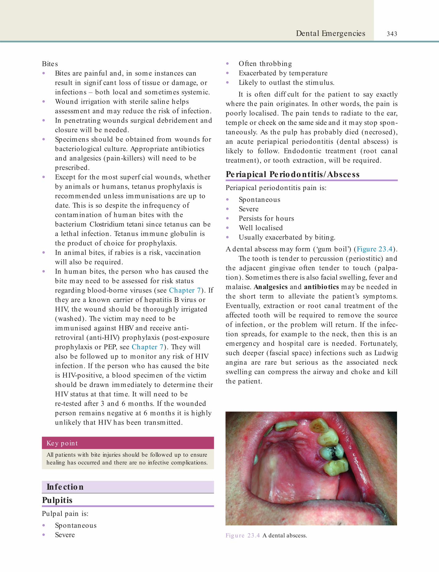
343Dental Emergencies
• O ten throbbing• Exacerbated by temperature• Likely to outlast the stimulus.
It is o ten di f cult or the patient to say exactly where the pain originates. In other words, the pain is poorly localised. The pain tends to radiate to the ear, temple or cheek on the same side and it may stop spon-taneously. As the pulp has probably died (necrosed), an acute periapical periodontitis (dental abscess) is likely to ollow. Endodontic treatment (root canal treatment), or tooth extraction, will be required.
Periapical Periodontitis/AbscessPeriapical periodontitis pain is:
• Spontaneous• Severe• Persists or hours• Well localised• Usually exacerbated by biting.A dental abscess may orm (‘gum boil’) (Figure 23.4).
The tooth is tender to percussion (periostitic) and the adjacent gingivae o ten tender to touch (palpa-tion). Sometimes there is also acial swelling, ever and malaise. Analgesics and antibiotics may be needed in the short term to alleviate the patient’s symptoms. Eventually, extraction or root canal treatment o the a ected tooth will be required to remove the source o in ection, or the problem will return. I the in ec-tion spreads, or example to the neck, then this is an emergency and hospital care is needed. Fortunately, such deeper ( ascial space) in ections such as Ludwig angina are rare but serious as the associated neck swelling can compress the airway and choke and kill the patient.
Bites• Bites are pain ul and, in some instances can
result in signif cant loss o tissue or damage, or in ections – both local and sometimes systemic.
• Wound irrigation with sterile saline helps assessment and may reduce the risk o in ection.
• In penetrating wounds surgical debridement and closure will be needed.
• Specimens should be obtained rom wounds or bacteriological culture. Appropriate antibiotics and analgesics (pain-killers) will need to be prescribed.
• Except or the most superf cial wounds, whether by animals or humans, tetanus prophylaxis is recommended unless immunisations are up to date. This is so despite the in requency o contamination o human bites with the bacterium Clostridium tetani since tetanus can be a lethal in ection. Tetanus immune globulin is the product o choice or prophylaxis.
• In animal bites, i rabies is a risk, vaccination will also be required.
• In human bites, the person who has caused the bite may need to be assessed or risk status regarding blood-borne viruses (see Chapter 7). I they are a known carrier o hepatitis B virus or HIV, the wound should be thoroughly irrigated (washed). The victim may need to be immunised against HBV and receive anti-retroviral (anti-HIV) prophylaxis (post-exposure prophylaxis or PEP, see Chapter 7). They will also be ollowed up to monitor any risk o HIV in ection. I the person who has caused the bite is HIV-positive, a blood specimen o the victim should be drawn immediately to determine their HIV status at that time. It will need to be re-tested a ter 3 and 6 months. I the wounded person remains negative at 6 months it is highly unlikely that HIV has been transmitted.
Fig u re 23.4 A dental abscess.
Key po in tAll patients with bite injuries should be followed up to ensure healing has occurred and there are no infective complications.
InfectionPulpitisPulpal pain is:
• Spontaneous• Severe
344 CHAPTER • 23 • Mosby’s Textbook of Dental Nursing
Antral Complications
Tooth/roo t in the maxilla ry antrum• The location o the tooth/root is checked on
radiographs• Antibiotics and a nasal decongestant are
prescribed• Further operation is required to retrieve the
tooth/root.
Oro-antra l f s tula (OAF)This is o ten recognised when, on drinking uids, the liquid appears in the nose!
• Patients should not blow their nose• Antibiotics and a nasal decongestant may help• I detected early, primary closure is possible• Some OAFs may need ap closure.
Ble e dingMost postoperative bleeding is a result o :
• Excessive trauma during the operation• In amed mucosa• Poor postoperative compliance (too much
exercise or hot drinks)• Post-extraction inter erence with the extraction
socket• Uncontrolled hypertension• Use o aspirin or non-steroidal anti-
in ammatory drugs (NSAIDs; see Chapter 15).Oral bleeding is more likely i the patient:
• Rinses• Disturbs the clot• Chews hard• Consumes hot drinks, alcohol, or does exercise.
Box 23.1 lists the instructions regarding post-operative bleeding that should be given to the patient.
The main way to control bleeding is to make a small pad rom a clean cotton handkerchie , or a teabag, placed directly over the socket and the teeth closed f rmly on it or 15–30 minutes. I the socket continues to bleed, clinical attention is needed. Surgicel (oxidised regenerated cellulose) or another haemostatic agent (collagen: synthetic (Instat); micro-crystalline (Avitene); or porcine) or tissue adhesives are placed in the socket by the clinician. I the bleeding continues, the socket will then most likely require sutures or a doctor will need to exclude a bleeding tendency.
Acute Periodontal AbscessThis may be seen in a patient who has periodontal disease. Symptoms include:
• Throbbing pain with erythema and swelling• Tooth tender to percussion.
I le t untreated, the abscess may rupture or, less commonly, progress to cellulitis. Drainage and debri-dement o the in ected periodontal area are indicated. Antibiotics are not usually indicated.
Acute PericoronitisAcute pericoronitis is seen where there is an impacted or partially erupted tooth and is recognised by:
• Swelling o the ap (operculum) over a partially erupted lower third molar (wisdom tooth)
• Pain• Tenderness• Halitosis• Bad taste.
Swollen lymph nodes are common, and trismus (restricted mouth opening) and ever can occur. Peri-coronitis may be caused by plaque build-up, and by trauma to the gingivae rom the opposing upper tooth. The area should be cleaned by irrigation under the ap with aqueous chlorhexidine, and any opposing tooth ground down or removed. Hot salt mouthwashes can help resolve symptoms. Penicillin and appropriate analgesia may be necessary. Eventually, the impacted tooth may need to be removed.
Find out moreHow can an upper too th cause pericoron it is a round a lower th ird molar?
Operative ComplicationsSurgical complications are described below. Medical emergencies, which are generally much more serious, are described in Chapter 27.
Management o a den ta l abscessA dental abscess should be drained to release the pus. The clinician will drain the tooth either by incising the abscess or opening the tooth to allow it to drain through the root canal/pulp. Analgesics such as paracetamol are given, and antibiotics are prescribed if there is facial swelling, fever, malaise or any threat to the airway. In the latter instance, urgent specialist care is required.
If infection extends deeply, the patient should be hospitalised and intravenous antibiotic treatment started immediately.
345Dental Emergencies
cause some postoperative pain. Minimising operative trauma will reduce pain.
Pain should be controlled with analgesics (see Chapter 15) given regularly.
Wound Infection• Wound in ection is usually obvious i the area is
in amed, swollen and tender, discharging pus and there is ever (pyrexia).
• I the wound in ection is only trivial, with no obvious pus ormation (suppuration), antibiotics may alone su f ce.
• I pus is draining, there may be no need to give antibiotics, as the in ection may settle spontaneously within a ew days. However, the clinician or dental nurse under supervision may take a sample o the pus on a swab to send or culture to identi y the organism and test sensitivity to antibiotics.
• I the wound is not draining but is uctuant, the clinician may remove one or more sutures rom the most in amed area to allow drainage o pus.
• In ection under neck aps used in cancer surgery is particularly dangerous as the carotid artery may be eroded and burst, which is then usually atal. Any suspicion o this is an emergency and the clinician must be urgently contacted.
Inhaled or Sw allow ed Foreign BodyItems that can be aspirated (or swallowed) can include restorations, restorative materials, instru-ments, implant parts, rubber dam clamps and impres-sion materials. Swallowed items usually pass naturally out o the gastro-intestinal tract, unless they are sharp and penetrate the gut wall.
Key poin tCare and the use of rubber dam can prevent many accidents. Rubber dam should always be used for conservative and endodontic dentistry.
Box 23.1 Pa t ien t in o rmat ion a te r too th ext ract ion• After a tooth has been extracted, the socket will usually
bleed for a short time, but then the bleeding stops because a healthy clot of blood forms in the tooth socket. These clots are easily disturbed and, if this happens, more bleeding will occur. To avoid disturbing the clot, DO NOT:
• Rinse the mouth for 24 hours
• Disturb the clot with the tongue or ngers
• Eat food that requires chewing (for the rest of the day)
• Chew on the affected side for at least 3 days (if both sides of the mouth are involved, have a soft diet for 3 days)
• Take hot drinks, hot baths, alcohol, exercise, talk too much or get excited or too hot.
• If the tooth socket continues to bleed after leaving the hospital, do not be alarmed; much of the liquid which appears to be blood will be saliva. Make a small pad from a clean handkerchief or cotton wool, or a teabag. Sit down and place it directly over the socket and close the teeth rmly on it. Keep up the pressure for 15–30 minutes.
• If the bleeding still does not stop, seek advice from the practice (or the hospital or resident oral surgeon).
Inhalation o a oreign body is exceedingly dan-gerous as it may:
• Block the airway and cause hypoxia and death• Cause lung collapse or in ection – lung abscess
or pneumonia.The main problem is to know whether the item
has been swallowed or inhaled, and there ore medical attention is required urgently or all patients. Each dental surgery must have a documented protocol or the ollow-up o such incidents.
PainAny operation involving so t tissues may cause some discom ort and most operations involving bone will
Term to lea rnFluctuant: compressible.
Dry SocketA dry socket is an empty and in ected and in amed extraction socket (localised osteitis). This occasionally ollows an extraction, typically a di f cult lower molar extraction, especially in a smoker and a ter extraction under LA.
I a patient develops a dry socket, they usually get the ollowing symptoms 2–4 days a ter the extraction:
• Increasing pain• Halitosis• Unpleasant taste• Empty socket• Marked tenderness to touch.
The clinician may take a radiograph to exclude other possible causes o the pain, such as retained roots, oreign bodies, or jaw racture.
346 CHAPTER • 23 • Mosby’s Textbook of Dental Nursing
In ammatory Lesions and Infections• Cervical/ acial ascial space in ections• Oral in ections i patient is ‘toxic’ or severely
immunocompromised• Necrotising fasciitis• Tuberculosis (some patients)• Some deep ungal in ections (mycoses)• Severe viral in ections, especially in the severely
immunocompromised• Severe vesiculo-bullous disorders (pemphigus
and Stevens–Johnson syndrome).
Manage me ntDry socket is treated by:
• Irrigating the socket with warm (50 ° C) saline or aqueous 0.2% chlorhexidine (Corsodyl)
• Dressing the socket with a sedative dressing, e.g. Alvogel
• Giving analgesics and/or antimicrobials, e.g. metronidazole (Flagyl).
Surgical EmphysemaSurgical emphysema is air blown into the so t tissues, It usually occurs when 3-in-1 syringes or high-speed dental handpieces are being used and the dental bur lacerates the mucosa.
Serious complications are rare but antibiotics may be prescribed.
Trismus• Trismus is when there is di f culty in opening the
mouth.• It may occur postoperatively because o bleeding
or in ammation around the muscles o mastication.
• The chances o trismus happening can be minimised by being care ul about the same actors as or swelling above, and also by minimising the stripping o muscle o the bone.
• Rest is indicated and possibly NSAIDs or, sometimes, antibiotics.
Find out moreTo read more about den ta l emergencies and the ir management see the Merck Manuals website sect ion on Denta l Emergencies (www.merck.com/mmpe/sec08/ch096/ch096a .h tml).
People Who Need Urgent Hospital Admission
Urgent hospital admission o the dental patient may be required or a number o reasons.
TraumaAn injured patient may have:
• Loss o consciousness• Shock• Head injury• Cervical spine injury• Other serious injuries• Laryngeal trauma
Terms to lea rnUndisplaced fracture: where the two segments of a fractured bone are still in their normal positions.
Zygomatic: refers to the cheek bone called the zygoma, which makes up the sides of the face.
Terms to lea rnNecrotising fasciitis: infection of the deeper layers of the skin and tissues that lie under the skin (subcutaneous tissues) that results in necrosis and can spread through the fascial planes; also called esh-eating disease.
Vesiculo-bullous disorder: a disease that is characterised by the presence of blisters on the skin and sometimes the mouth.
• Fractured jaws:• Middle acial third ractures• Mandibular racture (simple or undisplaced
may not require urgent admission)• Zygomatic ractures with danger o eye
damage.
Blood Loss• Severe or persistent haemorrhage (particularly i
bleeding tendency)• Less severe bleeding but in a highly anxious
patient.
Other Reasons• Collapse o uncertain cause• Airway obstruction• Vulnerable patients who have no social care or
support• Disturbed, severely depressed or some other
psychiatric patients• Children or others who are, or might be, being
abused

347Dental Emergencies
• They know what the planned management is• A ter the senior surgeon in charge and the
anaesthetist have been consulted• A ward bed has been arranged.
It is rare or maxillo acial operations to be done early or li e-saving reasons.
• Diabetics out o control because o oral pain or in ection.
Emergency Theatre ListEmergency lists usually run 24 hours a day but, at night, surgeons usually operate only or real emergen-cies. It is the duty o the surgeon to book an emergency case in theatre as soon as:

This page intentiona lly le ft blank

HUMAN DISEASE, PREVENTION AND CAREChapter 24 Human Anatomy and Physio logy 351
Chapte r 25 Human Pa thology and Diseases 369
Chapte r 26 Hea lth Promot ion 391
Chapte r 27 First Aid and Medica l Emergencies 401
Sect ion E

This page intentiona lly le ft blank
Chapter 24 Human Anatomy and Physiology
CHAPTER POINTS• Structure of the body• The circulatory system• The respiratory system• The digestive system• The urinary system• The integumentary system• The musculoskeletal system• The nervous system• The immune system
The CellsThere are many different cell types, but the basic design is the same for all cells (Figure 24.1):
• The cell or plasma membrane (the outer membrane) – which controls the movement of water, nutrients and waste material into and out of the cell.
• The cytoplasm – which forms the main part of the cell and contains many important structures, especially mitochondria (single: mitochondrion) – the power houses of the cell. Mitochondria produce the energy needed by the cell to function. The most important molecule that is involved in energy production is called adenosine triphosphate (ATP).
• The nucleus – the control centre of the cell. The nucleus is essential to everything the cell does, and contains the key to life itself, the DNA.DNA stands for ‘deoxyribonucleic acid’, and
makes up the genes that form the chromosomes. The genes coordinate the formation (synthesis) of all pro-teins in the body. Proteins are essential for virtually all body structures and functions. Genes are inherited from the parents and, because they control protein synthesis, they are responsible for many of the differ-ences between individuals (including differences in the susceptibility or resistance to disease). Gene abnormalities are the cause for many diseases.
In t ro d u ct io n
• Anatomy is the study of the structure of the body.• Physiology is the study of the working of the body
– how it functions.Dental and oral anatomy and physiology are dis-
cussed in Chapter 9.
The Structure of the BodyEverything in the body has a structure and a function or purpose. Virtually all body parts are necessary for health but organs such as the brain, heart, lungs and small intestine, in particular, are essential to life (Table 24.1).
Organs are complex structures that are made up of various tissues. The tissues themselves are further built up from millions of cells, which are the smallest units of life.
Terms to lea rnChromosome: the thread-like linear structure made of DNA and some other proteins. It stores the hereditary information in the genes and passes it from generation to generation by taking part in cell division.
Protein: a major class of food and the ‘building blocks’ of the body. Proteins are made of amino acids.

352 CHAPTER • 24 • Mosby’s Textbook of Dental Nursing
Fig u re 24.1 The basic parts of a single cell.
Table 24.1 Funct ions of o rgans and t issues
Organ/t issue Main funct ions Other importan t funct ions
Blood Red cells (erythrocytes) carry oxygen from lungs to tissues
Carry waste carbon dioxide from tissues to lungs
Platelets stop bleeding –White cells (leukocytes) give immunity – defend against infections
In ammation
Bones Support, and protect vital organs Facilitate joint movements
Brain Thought, memory, understanding Body control via nerves and hormones
Endocrine Glands produce hormones that control many body functions
Many
Gastro-intestinal Food digestion and absorption Excretion of solid waste (faeces)
Heart Circulates blood to lungs and brain Circulates blood to the rest of the body
Kidneys Excretes waste liquids and electrolytes Produces hormones (e.g. Vitamin D and erythropoietin which stimulates red cell production)
Liver Breaks down (metabolises) dead red blood cells, drugs, alcohol, and other substances
Produces proteins such as blood coagulation (clotting) factors, albumin (main blood protein); stores things like energy (as glycogen and fat), and stores iron, vitamin B and other important substances; helps immunity
Lungs Red blood cells carry oxygen (O2) from lungs to the tissues
Blood erythrocytes carry waste carbon dioxide (CO2) back to the lungs
Lymphoid tissue (immunocytes, lymph nodes, liver, spleen)
Defensive See Organs and Box 24.2
Muscles Allow movements Circulate blood and lymph
Nerves Motor nerves mediate muscle movementsSensory nerves mediate sensation, special senses (smell, sight, hearing, taste) Autonomic nerves (parasympathetic and sympathetic) regulate the functions of internal organs (the viscera) such as heart, stomach and intestines
–
Skin Protects against infection Sensation, temperature control
Spleen Removes dead red cells Removes read red blood cells, stores platelets; helps immunity
Humans have 23 pairs of chromosomes, one pair of which are the sex chromosomes (X and Y). The sex chromosomes determine whether a baby will be male or female; females have only X chromosomes (XX) and males have an X and a Y chromosome. Chromo-somal abnormalities can cause conditions such as Down syndrome.
Cell function and growth are controlled by signals that are sent to the cell from, for example, hormones (chemical messengers). The molecules that carry the signals bind to receptors on the cell membrane, trigger-ing molecules on the inner side of the cell membrane

353Human Anatomy and Physiology
form a system: for example the heart and blood vessels form the circulatory system, which is responsible for circulating blood throughout the body.
Terms Used in AnatomyThe special terms used in anatomy to describe the relationship of one part of the body to another are shown in Table 24.2. To help describe the position of structures in the body relative to each other and also the movement of various parts of the body in relation to each other, the body can be divided into anatomical planes (Figure 24.3) that correspond to the vertical and horizontal planes of space (Table 24.3).
The Circulatory SystemThe blood is circulated around the body to all organs, tissues and cells by the circulatory system. This system consists of:
• The blood – which carries oxygen and nutrients to all the cells of the body and removes waste products; thus its circulation is vital for survival
• The blood vessels (arteries and veins) – the network of blood vessels in which the blood circulates all around the body
to carry the signal deep into the cytoplasm and to the nucleus. In this way the cell carries out the activities it is instructed to do.
Many, but not all, cells grow and divide to produce daughter cells. Careful control of growth is essential for not just the health of the cell but the health of the entire individual. If the normal pattern of growth is disturbed, it may lead to diseases such as cancer.
Fig u re 24.2 Development of the human organism: from cells to tissues, tissues to organs, organs to systems and from systems to a complete body.
Term to lea rnCancer: the condition in which the growth of some cells becomes uncontrolled.
The TissuesCells group together to form larger structures called tissues. The four main types of tissue in the human body, each of which performs particular functions, are:
• Epithelium – this makes up the outer layer of the skin and surface of the lining of the mouth (the mucosa), gastro-intestinal and genito-urinary tract.
• Connective tissue – this makes up the supporting structures of the body, which include the tendons, ligaments, cartilage, bone and fat.
• Muscle – this tissue can contract and relax to produce movement. This includes movement of the body as a whole or just a part of the body (e.g. food is taken from one part of the digestive system to the next by the pushing movement produced by contraction and relaxation of the muscles of the digestive tract).
• Nerve tissue – this is made of special cells such as the neurones, which direct other cells in the body to perform certain functions by generating and passing on messages (signals) to them.
Find out moreWhat is the d iffe rence be tween: (a ) tendons and ligaments; (b ) ca rt ilage and bone?
The OrgansAn organ is a structure that contains at least two dif-ferent types of tissue that work together for a common purpose (Figure 24.2). Organs include the brain, heart, liver, kidneys, skin and others.
The SystemsThe main functions of the body such as breathing, circulation of blood, digestion of food require several organs to work together. Organs that function together

354 CHAPTER • 24 • Mosby’s Textbook of Dental Nursing
Fig u re 24.3 The anatomical planes.
Table 24.2 Terms used in ana tomy
Term De n it ion
Super cial Closer to the surface
Deep Further from the surface
Anterior Closer to the front of the body
Posterior Closer to the back of the body
Superior Closer to the top of the head
Inferior Closer to the soles of the feet
Medial Closer to the midline of the body
Lateral Away from the midline of the body
Proximal Closer to the point of origin of a structure
Distal Further from the point of origin of a structure
Table 24.3 Ana tomica l p lanes
Anatomica l p lane
Spa t ia l p lane
Body is d ivided by p lane in to port ions
Coronal Vertical Anterior and posterior
Transverse Horizontal Superior and inferior
Sagittal Vertical Right and left
• The heart – the pump that makes the blood ow in the blood vessels.
BloodThe blood is a special kind of tissue that consists of a variety of cells suspended in a solution called plasma.
355Human Anatomy and Physiology
Produc tion of b lood c e llsThe blood cells are produced in the bone marrow, found inside many bones. Blood cell production requires many substances called haematinics, such as iron, and vitamins – folic acid (folate) and vitamin B12. These substances are present in the food we eat and therefore a good diet is essential for blood cell production. A person who does not eat a diet that contains all the substances required for blood produc-tion may not have enough red cells and haemoglobin and the person is said to have anaemia.
Key po in tA lack of oxygen supply to the brain (called hypoxia) can cause severe brain damage and can kill a person within 3 minutes.
Term to lea rnFibrous tissue: this is a specialised tissue that contains tightly woven strands of a brous protein called collagen. Besides occurring normally in the body, it is found in scar tissue.
Box 24.1 Haemostasis and wound hea ling
HaemostasisWhen a blood vessel is damaged, the process that normally stops the blood escaping is called haemostasis. Haemostasis occurs in several steps:
1. Almost immediately the blood vessel constricts, which slows down the bleeding (vasoconstriction).
2. The platelets start to stick to the blood vessel walls around the damaged area to form a ‘platelet plug’.
3. The blood coagulation factors are activated, leading nally to the formation of a blood clot (haematoma).
4. Later, the clot is removed as part of wound healing.
Wound healingWound healing starts after the formation of the blood clot. It involves special cells called macrophages. The macrophages produce substances called ‘growth factors’ that trigger the formation of a special healing tissue called granulation tissue. Granulation tissue consists of macrophages and also another type of cell called the broblast. The broblasts produce the brous tissue that replaces the damaged tissue.
Within hours of an injury, the epithelium in the damaged area starts to regenerate as the surface cells of the skin or mucosa (called keratinocytes) migrate across the wound to cover it. Later, as the brous tissue grows stronger it produces the scarring that is seen in place of the wound.
Blood c e llsThere are three types of blood cell:
• Red blood cells (erythrocytes): these transport oxygen from the lungs to the various body tissues and organs. The oxygen binds to a pigment inside these cells called haemoglobin. When this happens the haemoglobin becomes ‘oxygenated’ and turns red in colour, so blood carrying oxygen is bright red in colour. After most of the oxygen is given off to the tissues, the haemoglobin becomes ‘de-oxygenated’ and it (and also the blood) becomes darker red in colour. If there is very little oxygen in the blood, it may even become bluish in colour (called cyanosis), and this makes certain parts of the body also appear blue, such as the lips, tongue and tips of ngers. De-oxygenated blood carries the waste carbon dioxide from the tissues back to the lungs, from where the carbon dioxide is exhaled into the air.
• White blood cells (leucocytes) – these cells are part of the body’s defence and help ght against the micro-organisms that can cause infections.
• Platelets – these play an important part in haemostasis (Box 24.1).
Term to lea rnAnaemia: when the cells do not get enough oxygen; symptoms include feeling tired easily.
Blood cell production also requires a healthy bone marrow. People whose bone marrow is damaged (e.g. because they have had radiotherapy or chemotherapy for a cancer) may lack all types of blood cells. They can then have anaemia and they also have a tendency to catch infections due to a lack of white blood cells for immunity and defence (Box 24.2) and they can also have a tendency to bleed (since platelets are also damaged).
A hormone produced in the kidney called eryth-ropoietin (EPO) stimulates the bone marrow to produce red cells. People with kidney disease may lack EPO and also develop anaemia. Some athletes use commercially available EPO to increase the oxygen-carrying capacity of their blood – but this is illegal.
Terms to lea rnChemotherapy: the use of strong chemicals (drugs) to treat cancer. Chemotherapy drugs aim to kill the cancerous cells but also have many side-effects on other normal parts of the body.
Radiotherapy: the treatment of disease (especially cancer) by exposure to an ionising radiation beam (see Chapter 14) or to a radioactive substance.
356 CHAPTER • 24 • Mosby’s Textbook of Dental Nursing
The Blood Vesse lsThere are three types of blood vessel:
• Arteries – these take the blood (usually oxygenated) away from the heart to the tissues.
• Veins – these return the blood (usually de-oxygenated) to the heart.
• Capillaries – these are the smallest branches of the blood vessel network and link the arteries to veins. These are also the vessels where the blood gives off the oxygen and takes up carbon dioxide from the cells (see p. 361).The pulmonary artery and vein are the opposite
of the rest of the arteries and veins, since the pulmo-nary artery carries de-oxygenated blood from the heart to the lungs and the pulmonary vein carries the oxygenated blood from the lungs to the heart (see Figure 24.5).
The Heart and the Flow of Blood around the BodyThe heart is the organ that is found in the centre of the chest (the thorax). It has four chambers: two large ventricles and two smaller atria (Figure 24.4).
The heart pumps de-oxygenated blood to the lungs via the pulmonary arteries. In the lungs the blood releases the carbon dioxide and becomes oxy-genated. It then travels in the pulmonary veins back to the heart entering it at the left atrium (Figure 24.5). From the left atrium blood is pumped into the left ventricle. The opening between these two chambers is controlled by a valve called the mitral valve.
Find out moreHow does the va lve cont ro l the ow of b lood – does it a llow the b lood to ow in on ly one d irect ion or bo th?
Blood is pumped out of the left ventricle into the aorta. The aortic valve controls the opening to the aorta. The aorta and its branches take the blood to all tissues and cells in the various parts of the body. Therefore the left ventricle is the most powerful heart chamber as it has to move blood all around the body. Because of this, the heart beat, which is the sound of the pumping action of this chamber, is heard and felt to the left side of the chest rather than in the centre.
The p las maThe blood plasma is made up of many kinds of protein, for example proteins called the blood coagulation factors – these work with the platelets to help form a clot to stop bleeding. Other proteins in the blood work with the white blood cells in defending the body against micro-organisms. These are called antibodies. Serum is the liquid remaining when blood clots.
Key poin tPeople with diseases (e.g. haemophilia) that affect the production or action of the platelets and the blood coagulation proteins can have serious bleeding after operations including tooth extraction. Therefore it is very important to nd out whether a dental patient may have such a condition.
Box 24.2 Immunity and in ammat ionThe immune system is responsible for protecting the body against potentially harmful substances that may cause damage or infection. The body’s response to such an attack is called the immune response or immunity.
The rst line of defence is the intact skin and mucosa (the lining of internal body cavities). When the skin or mucosa is cut or damaged, there is haemostasis, and then in ammation is induced. In ammation consists of increased blood ow to the area (with heat and redness), leakage of the plasma proteins from the in amed blood vessels into the tissues (with swelling), and the release of pain-inducing chemicals from cells. In ammation is thus recognised by the presence of:
• Heat
• Redness (erythema)
• Swelling
• Pain.
Following in ammation, special cells called macrophages and a kind of white blood cell called neutrophils are activated. These recognise, eat and kill bacteria (phagocytosis) and cleanse foreign matter from the injured site. This is the body’s second line of defence.
The other white blood cells involved in defence are the lymphocytes:
• B lymphocytes – these protect the body against bacteria by producing antibodies
• T lymphocytes – these protect mainly against viruses and fungi.
Central to the immune response are also organs such as the spleen and lymph nodes, which are together called the lympho-reticular or reticulo-endothelial system (RES). The lymph nodes are basically collections of lymphocytes and macrophages. These cells catch and deal with pathogens or other foreign materials that have escaped from the blood into the tissues and then entered the lymph (Figure 24.7).
The white blood cells also release a number of proteins called cytokines, which trigger the various events involved in in ammation and healing.
Term to lea rnAorta: the main artery of the body.
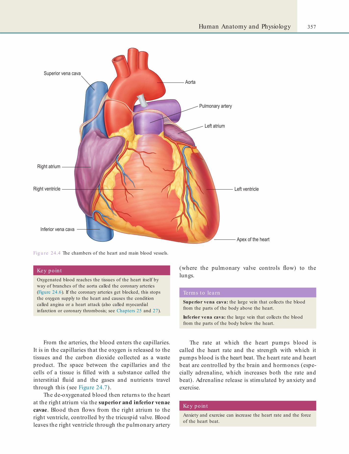
357Human Anatomy and Physiology
(where the pulmonary valve controls ow) to the lungs.
From the arteries, the blood enters the capillaries. It is in the capillaries that the oxygen is released to the tissues and the carbon dioxide collected as a waste product. The space between the capillaries and the cells of a tissue is lled with a substance called the interstitial uid and the gases and nutrients travel through this (see Figure 24.7).
The de-oxygenated blood then returns to the heart at the right atrium via the superior and inferior venae cavae. Blood then ows from the right atrium to the right ventricle, controlled by the tricuspid valve. Blood leaves the right ventricle through the pulmonary artery
Fig u re 24.4 The chambers of the heart and main blood vessels.
Key po in tOxygenated blood reaches the tissues of the heart itself by way of branches of the aorta called the coronary arteries (Figure 24.6). If the coronary arteries get blocked, this stops the oxygen supply to the heart and causes the condition called angina or a heart attack (also called myocardial infarction or coronary thrombosis; see Chapters 25 and 27).
Key po in tAnxiety and exercise can increase the heart rate and the force of the heart beat.
Terms to lea rnSuperior vena cava: the large vein that collects the blood from the parts of the body above the heart.
Inferior vena cava: the large vein that collects the blood from the parts of the body below the heart.
The rate at which the heart pumps blood is called the heart rate and the strength with which it pumps blood is the heart beat. The heart rate and heart beat are controlled by the brain and hormones (espe-cially adrenaline, which increases both the rate and beat). Adrenaline release is stimulated by anxiety and exercise.

358 CHAPTER • 24 • Mosby’s Textbook of Dental Nursing
Fig u re 24.5 The circulatory system.
The Lymphatic SystemThe lymphatic system (Figure 24.7) is the part of the circulatory system that cleanses it of impurities. It also forms an important part of the immune system (see Box 24.2). The lymphatic system consists of the fol-lowing parts:
• The lymph – this is the uid that circulates within the lymphatic vessels. It is formed by interstitial uid within the tissues (Figure 24.7).
• The lymphatic vessels – these are the vessels that transport the lymph.
• The lymph nodes – the knob-like structures that are usually found in clusters along the lymph vessels. They lter the passing lymph to catch its impurities (e.g. pathogens and other foreign material, stray cancer cells). The cells in the lymph nodes are called lymphocytes and form part of the body’s immune defence system (see Box 24.2).The lymph is not actively pumped through the
body like blood. It is moved mostly by virtue of muscle
contractions. The lymph vessels carry the lymph to the neck where it then enters the veins and eventually becomes part of the blood.
The Respiratory SystemThe respiratory system (Figure 24.8) has two parts:
• Upper respiratory tract – this is located in the head and neck and consists of the nose, paranasal sinuses (see Chapter 9), pharynx and larynx.
• Lower respiratory tract – located mainly inside the chest, this consists of the airways (trachea, bronchi and bronchioles) and the lungs.
Defence Mechanisms in the Respiratory TractThe defence mechanisms in the respiratory tract include those discussed in Box 24.2 as well as:
• The mucociliary lining: the mucosa of the respiratory system is covered by a layer of thick slippery uid called mucus. In addition, the cells also have tiny hair-like projections called cilia.
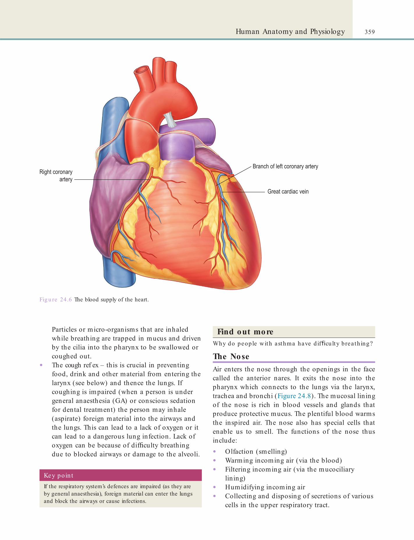
359Human Anatomy and Physiology
Fig u re 24.6 The blood supply of the heart.
Particles or micro-organisms that are inhaled while breathing are trapped in mucus and driven by the cilia into the pharynx to be swallowed or coughed out.
• The cough ref ex – this is crucial in preventing food, drink and other material from entering the larynx (see below) and thence the lungs. If coughing is impaired (when a person is under general anaesthesia (GA) or conscious sedation for dental treatment) the person may inhale (aspirate) foreign material into the airways and the lungs. This can lead to a lack of oxygen or it can lead to a dangerous lung infection. Lack of oxygen can be because of dif culty breathing due to blocked airways or damage to the alveoli.
Find out moreWhy do people with asthma have d if cu lty b rea th ing?
The NoseAir enters the nose through the openings in the face called the anterior nares. It exits the nose into the pharynx which connects to the lungs via the larynx, trachea and bronchi (Figure 24.8). The mucosal lining of the nose is rich in blood vessels and glands that produce protective mucus. The plentiful blood warms the inspired air. The nose also has special cells that enable us to smell. The functions of the nose thus include:
• Olfaction (smelling)• Warming incoming air (via the blood)• Filtering incoming air (via the mucociliary
lining)• Humidifying incoming air• Collecting and disposing of secretions of various
cells in the upper respiratory tract.
Key poin tIf the respiratory system’s defences are impaired (as they are by general anaesthesia), foreign material can enter the lungs and block the airways or cause infections.
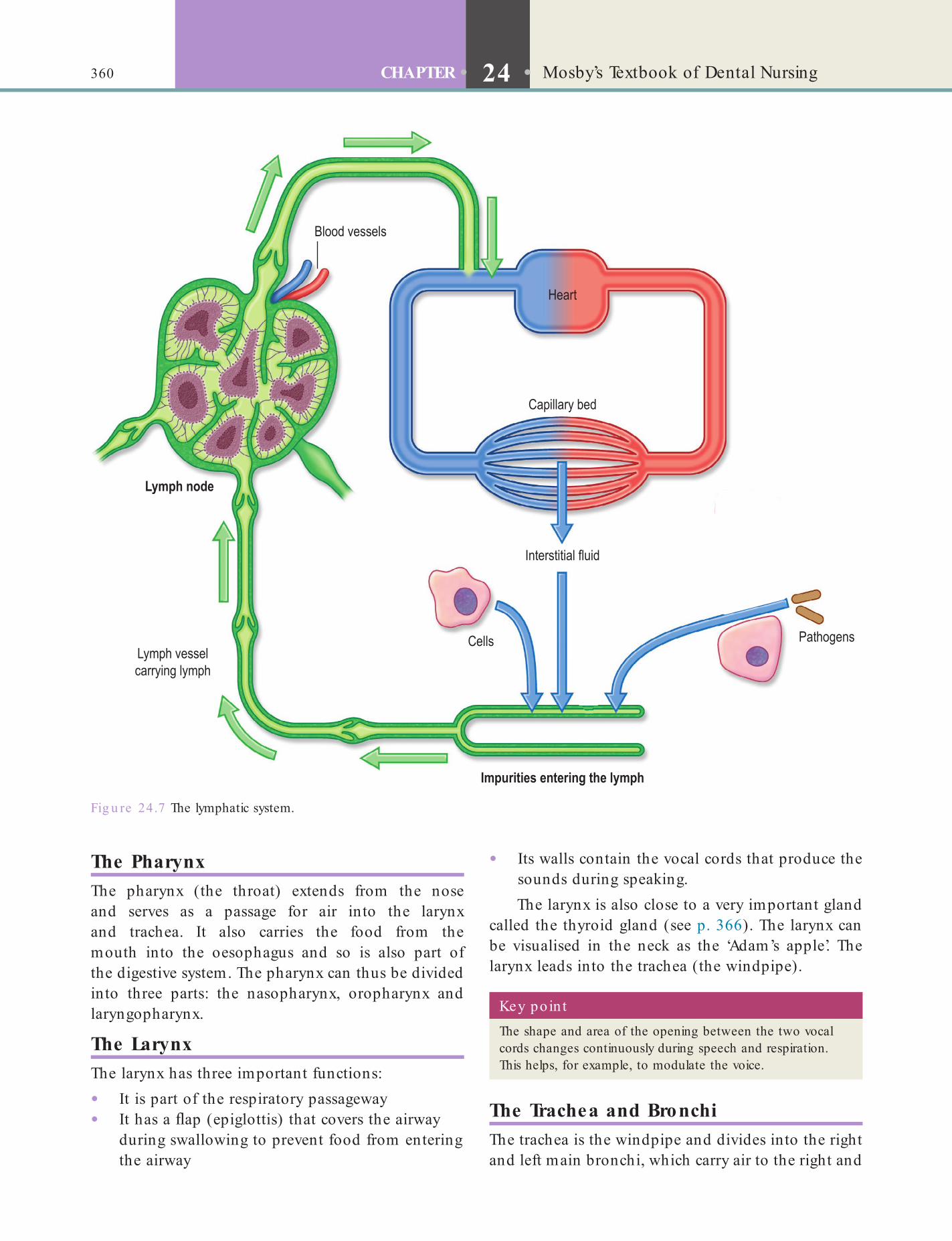
360 CHAPTER • 24 • Mosby’s Textbook of Dental Nursing
• Its walls contain the vocal cords that produce the sounds during speaking.The larynx is also close to a very important gland
called the thyroid gland (see p. 366). The larynx can be visualised in the neck as the ‘Adam’s apple’. The larynx leads into the trachea (the windpipe).
Fig u re 24.7 The lymphatic system.
The PharynxThe pharynx (the throat) extends from the nose and serves as a passage for air into the larynx and trachea. It also carries the food from the mouth into the oesophagus and so is also part of the digestive system. The pharynx can thus be divided into three parts: the nasopharynx, oropharynx and laryngopharynx.
The LarynxThe larynx has three important functions:
• It is part of the respiratory passageway• It has a ap (epiglottis) that covers the airway
during swallowing to prevent food from entering the airway
Key poin tThe shape and area of the opening between the two vocal cords changes continuously during speech and respiration. This helps, for example, to modulate the voice.
The Trachea and BronchiThe trachea is the windpipe and divides into the right and left main bronchi, which carry air to the right and

361Human Anatomy and Physiology
People with lung disorders may not be able to have treatment under GA and conscious sedation as their breathing may be compromised.
The Digestive SystemThe digestive system (Figure 24.9) starts at the mouth and ends at the anus. It is responsible for:
• Chewing, digestion and absorption of food• Removal of waste food and other waste matter.
Sw allow ingAfter being chewed (masticated) and mixed with saliva, food is swallowed. Swallowing (also called deglutition) helps carry the food into the pharynx and then into the oesophagus (the food pipe) and nally into the stomach. Swallowing is a complex process coordinated by the brain, which sends messages through several nerves. Swallowing is divided into three phases:
• Phase 1 is voluntary – it involves the collection of the chewed food into a bolus and the rst part of the swallowing of the food bolus.
left lungs, respectively. The bronchi divide into smaller tubes called bronchioles.
Fig u re 24.8 The respiratory system.
Key po in tThe right main bronchus is shorter, wider and more vertical than the left. Thus, if, for example, a small dental instrument or a part of a tooth or a lling is inhaled, it tends to enter the right bronchus because it is wider and more directly continuous with the trachea. The use of rubber dam (Chapter 17) should prevent such catastrophic accidents.
Fig u re 24.9 The digestive system.
The LungsThe lungs are made of the alveoli, which are clusters of cells at the tip of the terminal branches of the bron-chioles. This is where the oxygen and carbon dioxide exchange in the blood occurs.
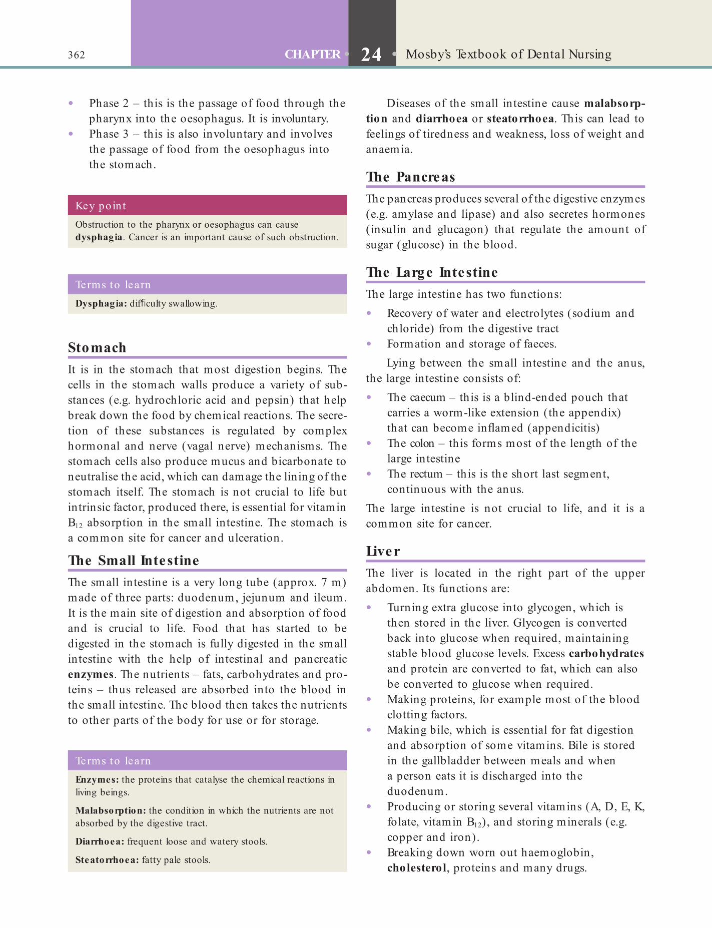
362 CHAPTER • 24 • Mosby’s Textbook of Dental Nursing
Diseases of the small intestine cause malabsorp-tion and diarrhoea or steatorrhoea. This can lead to feelings of tiredness and weakness, loss of weight and anaemia.
The PancreasThe pancreas produces several of the digestive enzymes (e.g. amylase and lipase) and also secretes hormones (insulin and glucagon) that regulate the amount of sugar (glucose) in the blood.
The Large IntestineThe large intestine has two functions:
• Recovery of water and electrolytes (sodium and chloride) from the digestive tract
• Formation and storage of faeces.Lying between the small intestine and the anus,
the large intestine consists of:
• The caecum – this is a blind-ended pouch that carries a worm-like extension (the appendix) that can become in amed (appendicitis)
• The colon – this forms most of the length of the large intestine
• The rectum – this is the short last segment, continuous with the anus.
The large intestine is not crucial to life, and it is a common site for cancer.
LiverThe liver is located in the right part of the upper abdomen. Its functions are:
• Turning extra glucose into glycogen, which is then stored in the liver. Glycogen is converted back into glucose when required, maintaining stable blood glucose levels. Excess carbohydrates and protein are converted to fat, which can also be converted to glucose when required.
• Making proteins, for example most of the blood clotting factors.
• Making bile, which is essential for fat digestion and absorption of some vitamins. Bile is stored in the gallbladder between meals and when a person eats it is discharged into the duodenum.
• Producing or storing several vitamins (A, D, E, K, folate, vitamin B12), and storing minerals (e.g. copper and iron).
• Breaking down worn out haemoglobin, cholesterol, proteins and many drugs.
StomachIt is in the stomach that most digestion begins. The cells in the stomach walls produce a variety of sub-stances (e.g. hydrochloric acid and pepsin) that help break down the food by chemical reactions. The secre-tion of these substances is regulated by complex hormonal and nerve (vagal nerve) mechanisms. The stomach cells also produce mucus and bicarbonate to neutralise the acid, which can damage the lining of the stomach itself. The stomach is not crucial to life but intrinsic factor, produced there, is essential for vitamin B12 absorption in the small intestine. The stomach is a common site for cancer and ulceration.
The Small IntestineThe small intestine is a very long tube (approx. 7 m) made of three parts: duodenum, jejunum and ileum. It is the main site of digestion and absorption of food and is crucial to life. Food that has started to be digested in the stomach is fully digested in the small intestine with the help of intestinal and pancreatic enzymes. The nutrients – fats, carbohydrates and pro-teins – thus released are absorbed into the blood in the small intestine. The blood then takes the nutrients to other parts of the body for use or for storage.
Terms to learnEnzymes: the proteins that catalyse the chemical reactions in living beings.
Malabsorption: the condition in which the nutrients are not absorbed by the digestive tract.
Diarrhoea: frequent loose and watery stools.
Steatorrhoea: fatty pale stools.
Terms to learnDysphagia: dif culty swallowing.
Key poin tObstruction to the pharynx or oesophagus can cause dysphagia. Cancer is an important cause of such obstruction.
• Phase 2 – this is the passage of food through the pharynx into the oesophagus. It is involuntary.
• Phase 3 – this is also involuntary and involves the passage of food from the oesophagus into the stomach.
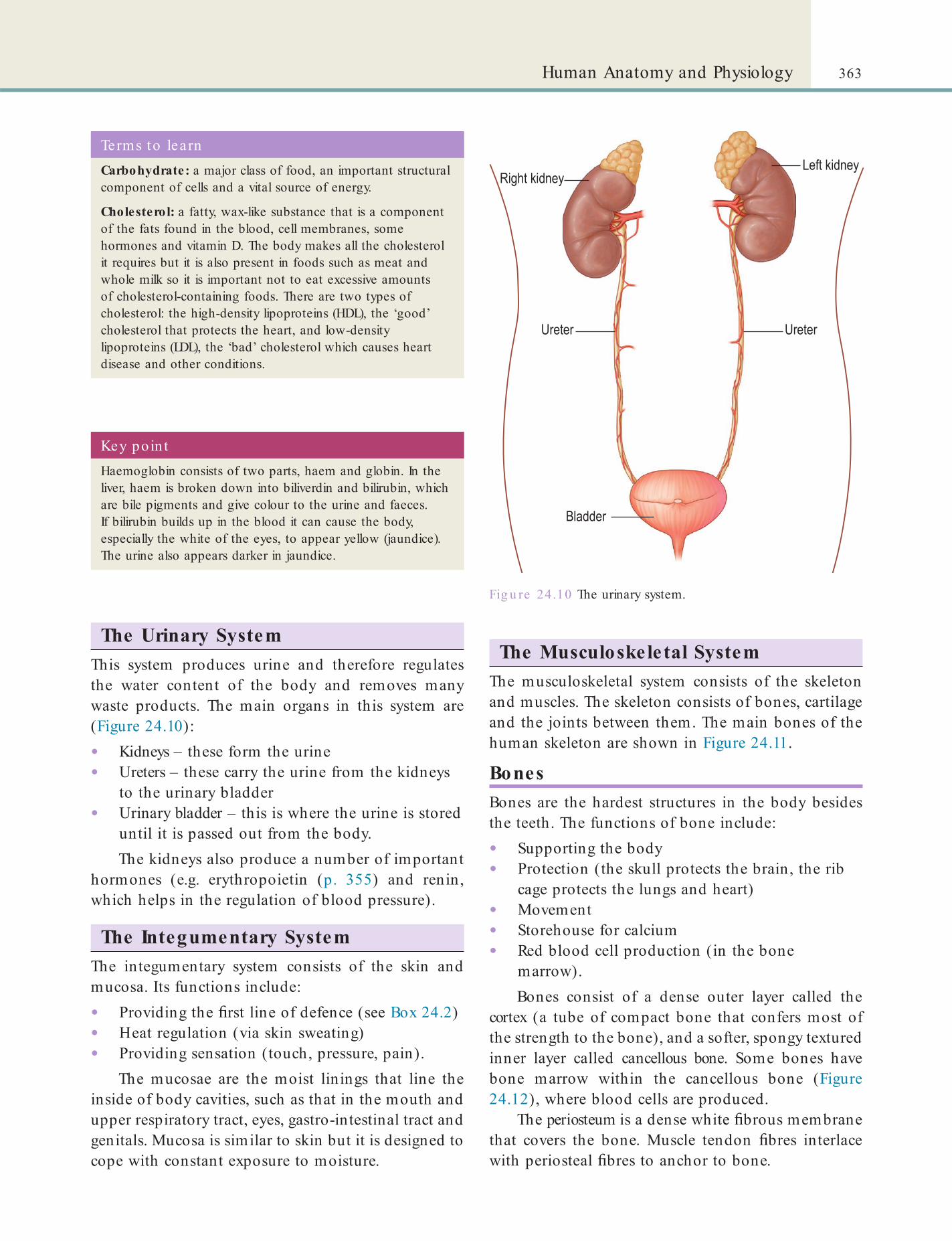
363Human Anatomy and Physiology
The Musculoskeletal SystemThe musculoskeletal system consists of the skeleton and muscles. The skeleton consists of bones, cartilage and the joints between them. The main bones of the human skeleton are shown in Figure 24.11.
BonesBones are the hardest structures in the body besides the teeth. The functions of bone include:
• Supporting the body• Protection (the skull protects the brain, the rib
cage protects the lungs and heart)• Movement• Storehouse for calcium• Red blood cell production (in the bone
marrow).Bones consist of a dense outer layer called the
cortex (a tube of compact bone that confers most of the strength to the bone), and a softer, spongy textured inner layer called cancellous bone. Some bones have bone marrow within the cancellous bone (Figure 24.12), where blood cells are produced.
The periosteum is a dense white brous membrane that covers the bone. Muscle tendon bres interlace with periosteal bres to anchor to bone.
Fig u re 24.10 The urinary system.
Key po in tHaemoglobin consists of two parts, haem and globin. In the liver, haem is broken down into biliverdin and bilirubin, which are bile pigments and give colour to the urine and faeces. If bilirubin builds up in the blood it can cause the body, especially the white of the eyes, to appear yellow (jaundice). The urine also appears darker in jaundice.
Terms to learnCarbohydrate: a major class of food, an important structural component of cells and a vital source of energy.
Cholesterol: a fatty, wax-like substance that is a component of the fats found in the blood, cell membranes, some hormones and vitamin D. The body makes all the cholesterol it requires but it is also present in foods such as meat and whole milk so it is important not to eat excessive amounts of cholesterol-containing foods. There are two types of cholesterol: the high-density lipoproteins (HDL), the ‘good’ cholesterol that protects the heart, and low-density lipoproteins (LDL), the ‘bad’ cholesterol which causes heart disease and other conditions.
The Urinary SystemThis system produces urine and therefore regulates the water content of the body and removes many waste products. The main organs in this system are (Figure 24.10):
• Kidneys – these form the urine• Ureters – these carry the urine from the kidneys
to the urinary bladder• Urinary bladder – this is where the urine is stored
until it is passed out from the body.The kidneys also produce a number of important
hormones (e.g. erythropoietin (p. 355) and renin, which helps in the regulation of blood pressure).
The Integumentary SystemThe integumentary system consists of the skin and mucosa. Its functions include:
• Providing the rst line of defence (see Box 24.2)• Heat regulation (via skin sweating)• Providing sensation (touch, pressure, pain).
The mucosae are the moist linings that line the inside of body cavities, such as that in the mouth and upper respiratory tract, eyes, gastro-intestinal tract and genitals. Mucosa is similar to skin but it is designed to cope with constant exposure to moisture.
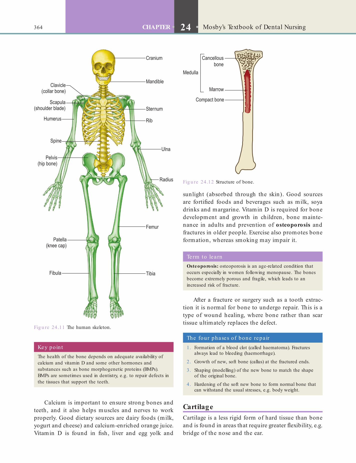
364 CHAPTER • 24 • Mosby’s Textbook of Dental Nursing
sunlight (absorbed through the skin). Good sources are forti ed foods and beverages such as milk, soya drinks and margarine. Vitamin D is required for bone development and growth in children, bone mainte-nance in adults and prevention of osteoporosis and fractures in older people. Exercise also promotes bone formation, whereas smoking may impair it.
Calcium is important to ensure strong bones and teeth, and it also helps muscles and nerves to work properly. Good dietary sources are dairy foods (milk, yogurt and cheese) and calcium-enriched orange juice. Vitamin D is found in sh, liver and egg yolk and
Fig u re 24.11 The human skeleton.
Fig u re 24.12 Structure of bone.
Key po in tThe health of the bone depends on adequate availability of calcium and vitamin D and some other hormones and substances such as bone morphogenetic proteins (BMPs). BMPs are sometimes used in dentistry, e.g. to repair defects in the tissues that support the teeth.
The four phases of bone repa ir
1. Formation of a blood clot (called haematoma). Fractures always lead to bleeding (haemorrhage).
2. Growth of new, soft bone (callus) at the fractured ends.
3. Shaping (modelling) of the new bone to match the shape of the original bone.
4. Hardening of the soft new bone to form normal bone that can withstand the usual stresses, e.g. body weight.
Term to lea rnOsteoporosis: osteoporosis is an age-related condition that occurs especially in women following menopause. The bones become extremely porous and fragile, which leads to an increased risk of fracture.
After a fracture or surgery such as a tooth extrac-tion it is normal for bone to undergo repair. This is a type of wound healing, where bone rather than scar tissue ultimately replaces the defect.
CartilageCartilage is a less rigid form of hard tissue than bone and is found in areas that require greater exibility, e.g. bridge of the nose and the ear.
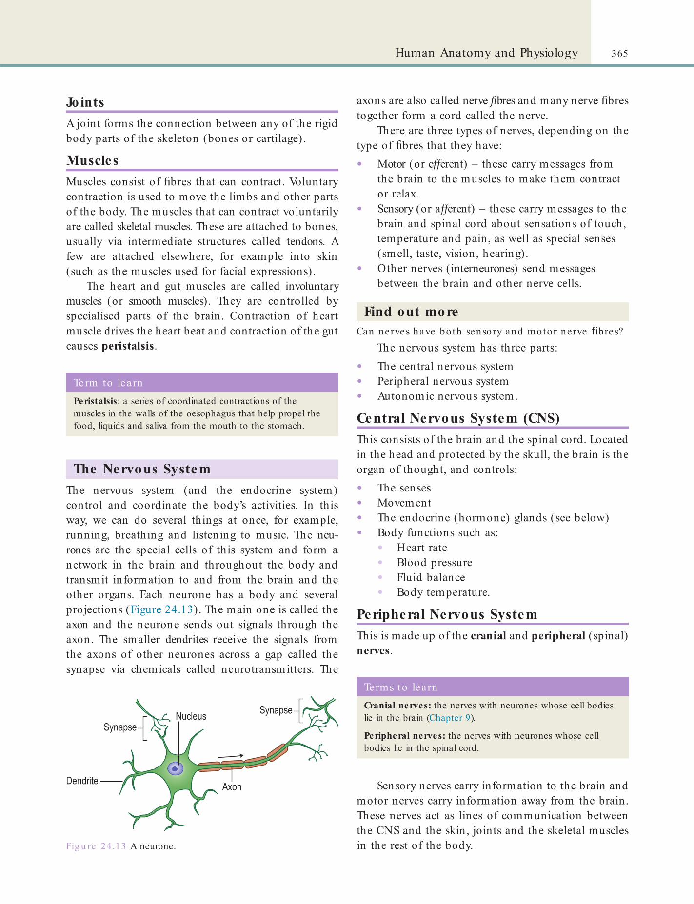
365Human Anatomy and Physiology
axons are also called nerve bres and many nerve bres together form a cord called the nerve.
There are three types of nerves, depending on the type of bres that they have:
• Motor (or e erent) – these carry messages from the brain to the muscles to make them contract or relax.
• Sensory (or a erent) – these carry messages to the brain and spinal cord about sensations of touch, temperature and pain, as well as special senses (smell, taste, vision, hearing).
• Other nerves (interneurones) send messages between the brain and other nerve cells.
Find out moreCan nerves have bo th sensory and motor nerve b res?
The nervous system has three parts:
• The central nervous system• Peripheral nervous system• Autonomic nervous system.
Central Nervous System (CNS)This consists of the brain and the spinal cord. Located in the head and protected by the skull, the brain is the organ of thought, and controls:
• The senses• Movement• The endocrine (hormone) glands (see below)• Body functions such as:
• Heart rate• Blood pressure• Fluid balance• Body temperature.
Peripheral Nervous SystemThis is made up of the cranial and peripheral (spinal) nerves.
JointsA joint forms the connection between any of the rigid body parts of the skeleton (bones or cartilage).
MusclesMuscles consist of bres that can contract. Voluntary contraction is used to move the limbs and other parts of the body. The muscles that can contract voluntarily are called skeletal muscles. These are attached to bones, usually via intermediate structures called tendons. A few are attached elsewhere, for example into skin (such as the muscles used for facial expressions).
The heart and gut muscles are called involuntary muscles (or smooth muscles). They are controlled by specialised parts of the brain. Contraction of heart muscle drives the heart beat and contraction of the gut causes peristalsis.
Fig u re 24.13 A neurone.
Term to lea rnPeristalsis: a series of coordinated contractions of the muscles in the walls of the oesophagus that help propel the food, liquids and saliva from the mouth to the stomach.
The Nervous SystemThe nervous system (and the endocrine system) control and coordinate the body’s activities. In this way, we can do several things at once, for example, running, breathing and listening to music. The neu-rones are the special cells of this system and form a network in the brain and throughout the body and transmit information to and from the brain and the other organs. Each neurone has a body and several projections (Figure 24.13). The main one is called the axon and the neurone sends out signals through the axon. The smaller dendrites receive the signals from the axons of other neurones across a gap called the synapse via chemicals called neurotransmitters. The
Terms to lea rnCranial nerves: the nerves with neurones whose cell bodies lie in the brain (Chapter 9).
Peripheral nerves: the nerves with neurones whose cell bodies lie in the spinal cord.
Sensory nerves carry information to the brain and motor nerves carry information away from the brain. These nerves act as lines of communication between the CNS and the skin, joints and the skeletal muscles in the rest of the body.
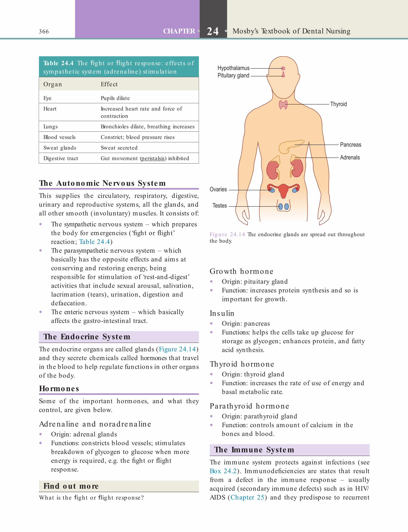
366 CHAPTER • 24 • Mosby’s Textbook of Dental Nursing
Fig u re 24.14 The endocrine glands are spread out throughout the body.
Table 24.4 The gh t o r igh t response : e ffects o f sympathe t ic system (adrenaline ) st imula t ion
Organ Effect
Eye Pupils dilate
Heart Increased heart rate and force of contraction
Lungs Bronchioles dilate, breathing increases
Blood vessels Constrict; blood pressure rises
Sweat glands Sweat secreted
Digestive tract Gut movement (peristalsis) inhibited
The Autonomic Nervous SystemThis supplies the circulatory, respiratory, digestive, urinary and reproductive systems, all the glands, and all other smooth (involuntary) muscles. It consists of:
• The sympathetic nervous system – which prepares the body for emergencies (‘ ght or ight’ reaction; Table 24.4)
• The parasympathetic nervous system – which basically has the opposite effects and aims at conserving and restoring energy, being responsible for stimulation of ‘rest-and-digest’ activities that include sexual arousal, salivation, lacrimation (tears), urination, digestion and defaecation.
• The enteric nervous system – which basically affects the gastro-intestinal tract.
The Endocrine SystemThe endocrine organs are called glands (Figure 24.14) and they secrete chemicals called hormones that travel in the blood to help regulate functions in other organs of the body.
HormonesSome of the important hormones, and what they control, are given below.
Adre na line and noradre na line• Origin: adrenal glands• Functions: constricts blood vessels; stimulates
breakdown of glycogen to glucose when more energy is required, e.g. the ght or ight response.
Find out moreWhat is the gh t o r igh t response?
Growth hormone• Origin: pituitary gland• Function: increases protein synthesis and so is
important for growth.
Ins ulin• Origin: pancreas• Functions: helps the cells take up glucose for
storage as glycogen; enhances protein, and fatty acid synthesis.
Thyro id hormone• Origin: thyroid gland• Function: increases the rate of use of energy and
basal metabolic rate.
Para thyro id hormone• Origin: parathyroid gland• Function: controls amount of calcium in the
bones and blood.
The Immune SystemThe immune system protects against infections (see Box 24.2). Immunode ciencies are states that result from a defect in the immune response – usually acquired (secondary immune defects) such as in HIV/AIDS (Chapter 25) and they predispose to recurrent
367Human Anatomy and Physiology
• Tumor – swelling• Functio laesi – discomfort and reluctance to use
the part.These are the things you can notice when you have any in amed part.
Cytokines are proteins that regulate immune responses. In response to injury, infection, trauma or malignancy, neutrophils and macrophages secrete several cytokines, which can also stimulate macro-phages to phagocytose debris. Most micro-organisms that tend to be invasive are ingested by phagocytes, including blood neutrophils (polymorphonuclear neutrophilic leukocytes) and tissue macrophages (‘phage’ = eating up), which kill most organisms.
Other cells of the immune system – immunocytes (leukocytes) – are mainly lymphocytes and they produce humoral (antibody; mainly B-cell) or cell-mediated (mainly T-cell) responses that are central to the immune defences. Antibodies are protective proteins (immunoglobulins) that combat bacteria mainly. T lymphocytes are particularly important in the defence against viruses and fungi and against cancer cells.
infections, especially of the skin, mucosae and respira-tory tract.
Innate immunity includes basic mechanisms of resistance to infection such as epithelial anatomical barriers of the skin and mucous membranes, cilia, secretions such as saliva and tears, and the innate in ammatory response – increased localised blood ow and capillary permeability (in ammation), releasing soluble factors that recruit cells called phago-cytes (neutrophils and macrophages) which restrict and engulf invasive micro-organisms.
In ammation is initiated and maintained by small protein mediators, termed complement and cytokines – that speci cally also affect the behaviour of immune cells (immunocytes).
The complement system is a plasma protein sequence (cascade) involving at least nine plasma proteins which, when activated, releases important mediators of in ammation – a protective response. In ammation is characterised by four features described from Latin words:
• Calor – heat• Rubor – redness

This page intentiona lly le ft blank

Chapter 25 Human Pathology and Diseases
CHAPTER POINTS• Common human diseases
patients such interference can cause the patient to deteriorate. A commonly used system to classify illness severity is the American Society of Anesthesiologists (ASA) system, shown in Table 25.1).
Ora l he a lthc are in ill pa tie nts• It is generally accepted that the least operative
work done should be aimed for.• All patients with medical conditions bene t
from preventive dental care:• Practising good oral hygiene• Regular dental examinations• Dietary counselling• Use of uorides to minimise the risk of
developing caries.
In t ro d u ct io n
It is important for all healthcare professionals to promote good health, so it is helpful to have back-ground knowledge of some common human diseases and general health promotion. This is also recognised by the GDC in its document Preparing or Practice, which describes the learning outcomes that must be met for registration as a dental professional (see Chapter 3).
A glossary of terms you may nd useful is pre-sented in an Appendix to this chapter.
Common Human DiseasesThe several reasons why diseases occur can be divided into (Box 25.1):
• Congenital (diseases that are present at birth, in the newborn)
• Acquired (diseases develop at or after birth):• Environmental: chemical; in ammatory;
irradiation; trauma• Lifestyle: diet; habits (use of tobacco, betel
nut, alcohol, other recreational drugs); lack of exercise.
Many diseases result from an interaction of several of these factors (Figure 25.1).
The s e ve rity o f an illne s sThe severity of an illness can in uence the type of operation or other medical care given, since in very ill
Term to lea rnMutation: alteration in the DNA sequence of a cell’s genome (the full DNA sequence of an organism), which can have several causes such as exposure to radiation, certain viruses and chemicals.
Table 25.1 ASA classi ca t ion of sta te o f hea lth
Class I Normal healthy individual
Class II Patient with mild systemic disease (not interfering with daily life)
Class III Severe systemic disease (interferes with daily life)
Class IV Incapacitating systemic disease (constant threat to life)
Class V Moribund (very seriously ill) patient with life expectancy <24 hours
370 CHAPTER • 25 • Mosby’s Textbook of Dental Nursing
Diseases are discussed alphabetically.
AlcoholismAlcohol is the most commonly misused drug. It affects the functioning of the central nervous system (CNS) as follows:
• Interfering with the cerebral cortex to release inhibitions
• Impairing the capacity to reason• Interfering with the functioning of the
cerebellum (back of brain) to cause unsteadiness of gait (ataxia) and incoordination of movements.
Fig u re 25.1 Interplay of factors in disease causation.
Term to lea rnRelease of inhibitions: when people start acting in ways or saying things they would not normally do (thus serious people start giggling, etc.).
Box 25.1 Causes of human diseases• Congenital diseases – may be genetic (due to gene
abnormalities) in origin or acquired by the fetus while in the mother’s uterus (from infections, or toxins such as alcohol, tobacco or drugs).
• Acquired diseases – may be in ammatory, neoplastic, metabolic or trauma-related.
• In ammatory and autoimmune diseases – may be caused by various infections (bacteria, viruses, fungi or parasites) or by the person’s own white blood cells attacking their tissues (autoimmune disease; rheumatoid arthritis is the best example).
• Infectious diseases – these can occur when the micro-organisms cross the skin or mucosal barrier due to, for example, a needlestick (sharps) injury, ingestion of infected food or water, inhalation of aerosol; and sexual contact (genital, anal or oral mucosae).
• Immunocompromise – some people develop disease because their white blood cells fail to protect them against infections, for example human immunode ciency virus (HIV) damages CD4 white cells. These people are said to be immunocompromised.
• Neoplastic diseases (cancer) – these are caused due to mutations in the DNA. Mutations themselves are caused by certain factors, particularly tobacco and alcohol, but other chemicals, radiation (e.g. sunlight, X-rays) and some micro-organisms (e.g. human papillomavirus) can also be responsible. All of this results in uncontrolled growth of some cells. Some cancers are more serious and the growth invades the underlying tissues. The cancer may also metastasise, that is, spread via lymphatics and blood mainly to lymph nodes, bone, brain and liver. Cancer can damage vital organs and cause organ dysfunction, pain and other symptoms. In many instances, it ultimately leads to death.
• Metabolic disorders – these, as the name suggests, are disorders of metabolism, that is, the body’s ability to produce, transport, store and distribute energy, mostly because of malfunctioning enzymes. Diabetes can be caused by malfunctioning of endocrine glands or may be due to lifestyle factors.
Table 25.2 Acute e ffects o f a lcohol
Blood a lcohol leve l (mg/d l)
<100 100–200 200–300 300–400 400–500 >500
Dry and decent Delighted and devilish
Delinquent and disgusting
Dizzy and delirious Dazed and dejected Dead drunk
Eventually, alcohol interferes with the brain centres (e.g. respiratory) and can cause unconscious-ness or even death (Table 25.2), mainly as a result of road traf c accidents and assaults. Alcoholics can also drown in their own vomit.
Alcohol and the six ‘Ls’Social dif culties from alcohol misuse are common and can affect the six ‘Ls’:
• Law – breach of the criminal, civil and/or professional codes
• Learning – intellectual dif culties
• Livelihood – job problems
• Living – housing problems
• Lover – relationship dif culties of all kinds: husband/wife; partner, employer/employee, etc.
• Lucre – (Latin lucrum = wealth) money problems.
Key poin tPeople, particularly those who are ill, often have concerns about privacy, and about the social implications of their condition. You must therefore strictly adhere to data protection and patient con dentiality principles. Reassure the patient that none of their details will be discussed, even with partners, unless permission is gained from them.

371Human Pathology and Diseases
Early symptoms and signs of an allergic reaction include:
• Breathlessness and itchy rash (hives or urticaria), within a few minutes to an hour
• Wheezing (bronchospasm)• Fall in blood pressure (hypotension)• Swelling of the face and lips (angio-oedema;
Figure 25.2) and laryngopharynx. This may be followed by life-threatening anaphylactic shock.All allergies are best managed by allergen
avoidance:
• Drugs – accurately document allergies in medical notes. Use of MedicAlert bracelets
• Food products – follow an elimination diet• House dust mites – use mite-proof bed linen
and wooden oors• Pets – avoid keeping pets• Pollen – keep windows shut and avoid grassy
spacesCommonly used drugs for treatment are:
• Anti-histamines• Bronchodilators• Corticosteroids• Adrenaline (also called epinephrine).
AnaemiaA person is said to have anaemia when their haemo-globin (Hb) level is below the normal for their age, gender and ethnic background. Haemoglobin is
Fig u re 25.2 Angio-oedema.
Key po in tsPeople at risk of anaphylaxis should carry an Epipen (syringe containing adrenaline) for self-administration.
As a dental professional, you must be familiar with the medical management of anaphylaxis within a dental setting (see Chapter 27).
Many other diseases can be caused or aggravated by alcohol, including:
• Bleeding tendency• Brain damage and epilepsy• Cancers• Gastro-intestinal disease• Heart disease• Impaired wound healing• Infections, especially pneumonia and
tuberculosis• Injuries (including maxillofacial), from accidents
or assaults• Liver disease• Malnutrition• Muscle disease• Pancreatic disease.
Alcoholic people need help in the way of admis-sion to a rehabilitation unit. Management includes ensuring abstinence, adequate nutrition and medica-tion to reduce dependence.
AllergiesAllergy is an abnormal response to an allergen (protein). Common allergens are listed in Table 25.3.
Allergies are responsible for some types of the fol-lowing diseases and conditions:
• Asthma• Eczema• Rhinitis (hayfever)• Anaphylaxis (collapse, with low blood pressure
and breathing dif culties) – this can be fatal (Chapter 27).
Table 25.3 Common a lle rgens
Allergen source Examples
Dental materials Amalgam, gold, mercury, resins, plasters (e.g. Elastoplast)
Drugs Aspirin, penicillins
Environment Animal hair, dust mites, pollens
Foods Milk, nuts (especially peanuts), eggs, shell sh
Latex Gloves, dressings, elastic bands, condoms
372 CHAPTER • 25 • Mosby’s Textbook of Dental Nursing
Key poin ts: Types of anxie ty d isorders• Panic disorder: the person panics apparently spontaneously.
• Phobia: anxiety is provoked by discrete stimuli, e.g. phobia of ying or dental treatment.
• Generalised anxiety disorder: such people worry all the time.
Key poin tAnticoagulant doses should not be altered without consulting the doctor in charge. Stopping the drug before surgery is dangerous as there can be fatal thromboses (blood clots).
Term to lea rnThrombo-embolic disorder: a disorder related to the formation of blood clots (thrombi) in one part of the body, of which either small portions or the whole clot can then become loose (emboli) and travel to other parts of the body in the blood stream, causing, e.g., pulmonary thrombosis.
Key po in tPallor of the oral mucosa, conjunctiva or hand palm creases suggests severe anaemia.
required to carry oxygen to the brain and tissues (see Chapter 24). Anaemia has many causes, as shown in Table 25.4.
Anaemia may not cause any symptoms early on but, as it worsens and the oxygen-carrying capacity of the blood falls, symptoms and signs develop (e.g. breathlessness, palpitations).
Table 25.4 Causes of anaemia
Nature of anaemia Cause
Increased loss of red blood cells (RBCs)
Menstrual blood lossGastro-intestinal blood lossHaemolysis (breakdown of the RBCs), such as in malaria or sickle disease
Reduced RBC production
Dietary de ciencyBone marrow disease, such as in leukaemia
Increased tissue requirements
PubertyPregnancy
Decreased tissue requirements
Hypothyroidism
Management of anaemia includes treatment of the underlying cause. De ciency states must be corrected with iron, folic acid, and vitamin B12 supplements. Blood transfusion may be indicated if the onset of anaemia has been rapid.
Anticoagulant-Related ProblemsAnticoagulants are drugs used to control and prevent clotting in people who have deep vein thromboses (thrombo-embolic disorders), stroke or after heart surgery. Warfarin is the most frequently prescribed anticoagulant. The anticoagulant dose needs to be con-trolled or the patient will bleed too easily. The dose is controlled by a blood test called the international nor-malised ratio (INR). An INR above 1 indicates that clotting will take longer than normal. Newer oral anti-coagulants (NOACs) do not need INR monitoring.
Surgery is the main hazard for patients on warfa-rin. Thus alternative treatments, e.g. endodontic treatment, are always considered. LA regional block injections, or those given in the oor of the mouth, may be a hazard since bleeding into the tissues of the neck can block the airway and suffocate the patient. If surgery is to be more than simple or minor, or the INR is above 3.5, the patient is usually treated in hospital or another specialist centre.
Aspirin and non-steroidal anti-in ammatory agents (NSAIDs) can cause bleeding as they inhibit platelet aggregation. Thus they should be avoided in these patients. Paracetamol is the analgesic of choice.
Anxiety and StressAnxiety is an unpleasant emotional state characterised by fear and a variety of physical symptoms. Stress raises blood levels of the adrenal hormones: cortisol, adrenaline (epinephrine) and noradrenaline (nore-pinephrine). These ‘stress hormones’ in uence many physiological functions, causing the ‘ ght or ight’ response of:
• Apprehension• Fast pulse rate (tachycardia)• Over-breathing (hyperventilation)• Raised blood pressure (hypertension)• Sweating• Tremor• Dilated pupils• Involuntary defecation• Urinary incontinence.
Management includes:
• Appropriate management of any underlying organic disease.
• Lifestyle changes to reduce stressors and avoid precipitating factors.
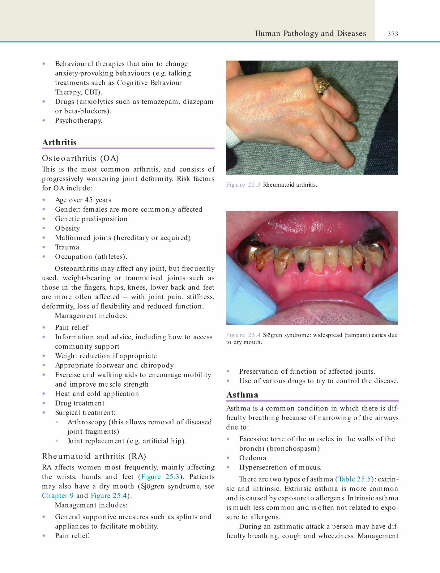
373Human Pathology and Diseases
• Preservation of function of affected joints.• Use of various drugs to try to control the disease.
AsthmaAsthma is a common condition in which there is dif- culty breathing because of narrowing of the airways due to:
• Excessive tone of the muscles in the walls of the bronchi (bronchospasm)
• Oedema• Hypersecretion of mucus.
There are two types of asthma (Table 25.5): extrin-sic and intrinsic. Extrinsic asthma is more common and is caused by exposure to allergens. Intrinsic asthma is much less common and is often not related to expo-sure to allergens.
During an asthmatic attack a person may have dif- culty breathing, cough and wheeziness. Management
Fig u re 25.3 Rheumatoid arthritis.
• Behavioural therapies that aim to change anxiety-provoking behaviours (e.g. talking treatments such as Cognitive Behaviour Therapy, CBT).
• Drugs (anxiolytics such as temazepam, diazepam or beta-blockers).
• Psychotherapy.
Arthritis
Os te oarthritis (OA)This is the most common arthritis, and consists of progressively worsening joint deformity. Risk factors for OA include:
• Age over 45 years• Gender: females are more commonly affected• Genetic predisposition• Obesity• Malformed joints (hereditary or acquired)• Trauma• Occupation (athletes).
Osteoarthritis may affect any joint, but frequently used, weight-bearing or traumatised joints such as those in the ngers, hips, knees, lower back and feet are more often affected – with joint pain, stiffness, deformity, loss of exibility and reduced function.
Management includes:
• Pain relief• Information and advice, including how to access
community support• Weight reduction if appropriate• Appropriate footwear and chiropody• Exercise and walking aids to encourage mobility
and improve muscle strength• Heat and cold application• Drug treatment• Surgical treatment:
• Arthroscopy (this allows removal of diseased joint fragments)
• Joint replacement (e.g. arti cial hip).
Rhe umato id a rthritis (RA)RA affects women most frequently, mainly affecting the wrists, hands and feet (Figure 25.3). Patients may also have a dry mouth (Sjögren syndrome, see Chapter 9 and Figure 25.4).
Management includes:
• General supportive measures such as splints and appliances to facilitate mobility.
• Pain relief.
Fig u re 25.4 Sjögren syndrome: widespread (rampant) caries due to dry mouth.
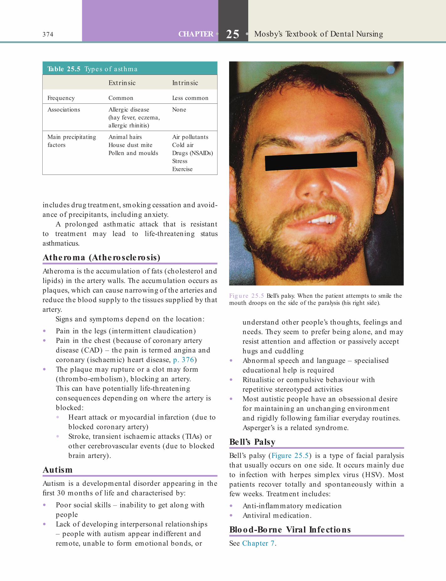
374 CHAPTER • 25 • Mosby’s Textbook of Dental Nursing
understand other people’s thoughts, feelings and needs. They seem to prefer being alone, and may resist attention and affection or passively accept hugs and cuddling
• Abnormal speech and language – specialised educational help is required
• Ritualistic or compulsive behaviour with repetitive stereotyped activities
• Most autistic people have an obsessional desire for maintaining an unchanging environment and rigidly following familiar everyday routines. Asperger’s is a related syndrome.
Bell’s PalsyBell’s palsy (Figure 25.5) is a type of facial paralysis that usually occurs on one side. It occurs mainly due to infection with herpes simplex virus (HSV). Most patients recover totally and spontaneously within a few weeks. Treatment includes:
• Anti-in ammatory medication• Antiviral medication.
Blood-Borne Viral InfectionsSee Chapter 7.
includes drug treatment, smoking cessation and avoid-ance of precipitants, including anxiety.
A prolonged asthmatic attack that is resistant to treatment may lead to life-threatening status asthmaticus.
Atheroma (Atherosclerosis)Atheroma is the accumulation of fats (cholesterol and lipids) in the artery walls. The accumulation occurs as plaques, which can cause narrowing of the arteries and reduce the blood supply to the tissues supplied by that artery.
Signs and symptoms depend on the location:
• Pain in the legs (intermittent claudication)• Pain in the chest (because of coronary artery
disease (CAD) – the pain is termed angina and coronary (ischaemic) heart disease, p. 376)
• The plaque may rupture or a clot may form (thrombo-embolism), blocking an artery. This can have potentially life-threatening consequences depending on where the artery is blocked:• Heart attack or myocardial infarction (due to
blocked coronary artery)• Stroke, transient ischaemic attacks (TIAs) or
other cerebrovascular events (due to blocked brain artery).
AutismAutism is a developmental disorder appearing in the rst 30 months of life and characterised by:
• Poor social skills – inability to get along with people
• Lack of developing interpersonal relationships – people with autism appear indifferent and remote, unable to form emotional bonds, or
Fig u re 25.5 Bell’s palsy. When the patient attempts to smile the mouth droops on the side of the paralysis (his right side).
Table 25.5 Types of asthma
Extrinsic In t rinsic
Frequency Common Less common
Associations Allergic disease (hay fever, eczema, allergic rhinitis)
None
Main precipitating factors
Animal hairs Air pollutantsHouse dust mite Cold airPollen and moulds Drugs (NSAIDs)
StressExercise
375Human Pathology and Diseases
The long-term effects of using cannabis are not yet clear, but it may affect the lungs, heart and immune system and mental health. Marijuana smokers may develop many of the breathing problems that tobacco smokers have, hypertension and increased heart rate and lowered oxygen-carrying capacity of blood. Because of this the risk of a heart attack is increased four-fold in the rst hour after smoking marijuana. Long-term use of marijuana can trigger or worsen schizophrenia (see p. 387).
Cardiac DiseaseSee also Coronary heart disease (p. 374).
Electrosurgery can cause heart pacemakers and implantable cardiac de brillators to malfunction. Some older electrically operated dental equipment may also be a risk but most modern dental equipment is safe in this respect. In the past, patients thought to be at risk from infective endocarditis were routinely given antimicrobial prophylaxis (‘cover’) before dental surgery and some other procedures, but that is no longer the case (see p. 383). Some ‘cardiac’ drugs can cause gingival swelling (drug-induced gingival over-growth, DIGO).
Find out moreSome drugs used in card iac d isease can cause the g ingivae to swell up . Can you name any?
Cerebral PalsyCerebral palsy (CP) is the most common congenital physical disability. In cerebral palsy, muscle control is abnormal because of damage to a child’s brain early in the course of development. The brain damage is caused mainly by lack of oxygen (hypoxia), trauma or infection. Physiotherapy is an important part of management.
Breast CancerAfter skin cancer, breast cancer is the most common cancer among women. It is usually seen in women over the age of 50. Risk factors include:
• Personal history of breast cancer• Family history of breast cancer• Other factors:
• Increased oestrogen levels• Late childbearing• Alcohol use• Fatty diet.
• Early breast cancer usually does not cause pain• Later breast cancer can cause:
• A lump or thickening in or near the breast or axilla
• A change in breast size or shape• Nipple discharge, tenderness or inversion• Breast ridging or pitting.There are many treatments, and which one is
offered varies from patient to patient:
• Surgery• Radiation• Chemotherapy• Hormones (tamoxifen)• Antibodies.
Cannabis (Marijuana) UseCannabis is usually smoked as a cigarette (joint, spliff or nail) or in a pipe (bong). The main active chemical THC (delta-9-tetrahydrocannabinol), works by binding to the brain. This affects the person’s:
• Sense of pleasure• Memory• Thoughts• Concentration• Sensory and time perception• Coordination of movements.
Within a few minutes of inhaling marijuana smoke, the user will often feel their heart beat increase, have a dry mouth and red eyes. In some people, mari-juana can double the normal heart rate and raise the blood pressure. Because of the effects of cannabis on perceptions and reaction times – which last for up to 24 hours – users can be involved in risky sexual behav-iour, violence or in road traf c accidents. Some users may also have acute anxiety and paranoid thoughts; rarely, a user can have severe psychotic symptoms and need emergency medical treatment.
Key poin tPeople high on marijuana may become silly and giggly, seem dizzy and have trouble walking and have a hard time remembering things that have just happened – they thus appear drunk.
Term to lea rnPsychotic symptoms: psychosis is a serious mental disorder in which a person loses touch with reality or their perception of reality is highly distorted. The classic symptoms are hallucinations and delusions and other confused thoughts. There may also be a lack of self-awareness.
376 CHAPTER • 25 • Mosby’s Textbook of Dental Nursing
• Family history• Cigarette smoking• High blood cholesterol level• Hypertension• Diabetes mellitus• Obesity• Lack of exercise.
Signs and symptoms (see also Box 25.2) may be:
• Dizziness• Shortness of breath• Decreased exercise tolerance• Chest pain (angina pectoris)• Sudden death due to a myocardial infarction
(irreversible damage to heart muscle).
Chronic Obstructive Pulmonary DiseaseChronic obstructive pulmonary disease (COPD), also called chronic obstructive airway disease (COAD), is a progressive, irreversible lung disease, most frequently a combination of:
• Chronic bronchitis – excessive production of mucus and persistent cough with sputum
• Emphysema – pathological dilatation of air spaces in the lungs with destruction of the alveoli.The most important causes of COPD are cigarette
smoking and environmental pollution.Management consists of lifestyle changes and drug
treatment:
• Smoking cessation• Exercise• Weight loss• Drug treatment – bronchodilators,
corticosteroids or antibiotics• Oxygen therapy – long-term oxygen therapy• Vaccination – pneumococcal and in uenza
vaccine.
Coeliac DiseaseCoeliac disease is an allergic reaction of the small intestine mucosa to gluten – a protein in wheat mainly (hence in bread etc.). Many affected people are asymptomatic. However, malabsorption of proteins can cause growth retardation, abdominal pain, fatty faeces (steatorrhoea) and behavioural changes.
Management includes:
• Gluten-free diet: plain meat, sh, fruits and vegetables do not contain gluten.
• Correction of nutritional de ciencies.
Term to lea rnGluten: a group of proteins found in all forms of wheat, and related grains (especially rye and barley).
Key po in tMyocardial infarction commonly presents with central chest pain similar to that of angina. However, the pain of myocardial infarction is not relieved by rest or with glyceryl trinitrate (GTN).
Box 25.2 Condit ions re la ted to coronary a rte ry d isease• Angina pectoris – this is the name given to episodes of
chest pain caused by CAD. Angina is precipitated by exercise, particularly in cold weather, and is relieved by rest or use of glyceryl trinitrate (GTN).
• Myocardial infarction (heart attack) – this results from the complete blockage of one or more arteries to the heart (coronary arteries). Fewer than 50% of patients with myocardial infarction have any preceding symptoms, but angina may progress to myocardial infarction.
Coronary (Ischaemic) Heart DiseaseCoronary artery disease (CAD) is the leading cause of death in the UK. It is caused by atherosclerosis of the coronary arteries.
Risk factors include:
• Increasing age• Gender – men are at greater risk than
premenopausal women
A person with myocardial infarction requires immediate aspirin and hospital admission (see Chapter 27). Up to 50% of patients die within the rst hour; but early treatment halves the death rate.
Lifestyle changes play a major role in treatment:
• Reduced intake of cholesterol and saturated fats• Daily exercise• Weight loss• Smoking cessation.
Medications include anticoagulant drugs (e.g. aspirin) and cholesterol-lowering drugs such as statins.
When symptoms are worsening despite this management, surgery (cardiac revascularisation tech-niques) may be considered, such as:
• Coronary angioplasty (stents)• Coronary artery bypass grafting (CABG).
377Human Pathology and Diseases
disinfectants and sterilisation. TSEs have prolonged incubation periods of months to years. Persons with vCJD develop psychiatric symptoms (severe depres-sion and behavioural disorders), followed by demen-tia and nally death.
There is no effective treatment yet for vCJD and there have been few patients (172 dead and alive cases reported in the UK by 2010).
Chemotherapy-Related ProblemsCytotoxic chemotherapy is used to treat many cancers and other malignant conditions such as leukaemia. The drugs often cause hair loss (alopecia), nausea and vomiting, and pins and needles (paraesthesia) but one of the biggest problems for the patient is a sore mouth (mucositis). Analgesics are needed, as well as a soft diet. Sucking ice cubes may help ease the pain, and many other concoctions are available to help the patient. Oral hygiene must be maintained.
Corticostero id-Related ProblemsThe adrenal hormone cortisol is released in the response to stresses such as trauma, infection, general anaesthesia or surgery. Secretion of cortisol is regu-lated via a biological feedback system that includes another hormone called adrenocorticotropic hormone (ACTH). In patients given systemic corticosteroid drugs (steroids), this feedback response may not occur. As a result, the adrenal gland is unable to produce the necessary steroid response to stress. The patient may then have a fall in blood pressure and collapse.
Key poin tSteroid supplementation (steroid cover) may need to be considered before stressful procedures such as surgery in patients taking corticosteroids. The blood pressure must be carefully monitored during the operation and recovery.
Term to lea rnBiological feedback: the system by which the body feeds back information to the various regulating organs, which then respond by either increasing or decreasing the formation of regulators, for example, hormones.
Term to lea rnStent: these are miniature wire coils put inside a blocked artery to keep it open.
Creutzfe ldt–Jakob DiseaseCreutzfeldt–Jakob disease (CJD) is a rare, progressive brain disease. It is a type of transmissible spongiform encephalopathy (TSE). Some TSEs are transmissible from animal to man; bovine spongiform encepha-lopathy (BSE; also known as ‘mad cow disease’) in cattle appears to be responsible for variant CJD (vCJD) in humans.
TSEs are caused by prions – which are abnormal cellular proteins. Prions are very resistant to normal
Key poin tCJD and related conditions raise new infection control questions because prions are much more dif cult to destroy than conventional micro-organisms. So single-use instruments are recommended. All other instruments should be decontaminated.
Key poin ts: Prions and dent ist ryPrions are proteinaceous infectious particles found in the brain and are transmitted by brain surgery or use of brain products, or even by transfusions of whole blood.
Prions are unique in their remarkable capacity to bind to steel and to resist inactivation by conventional methods: heat, most disinfectants and by ionising, ultraviolet and microwave radiations. This presents signi cant infection control problems when patients with CJD undergo medical or dental interventions. Disposable equipment is the best answer.
Prions are also a rare hazard from exposure-prone (invasive) procedures. Dental interest in prion disease (CJD) and the related conditions centres on the risk of their transmission from patient to patient in the course of dental treatment through contaminated instruments. There is no known case of this happening and appropriate dental infection control precautions will reduce the scope of the theoretical risk. It is not yet known whether CJD can be transmitted via blood or other tissues encountered during dental surgery.
Term to lea rnIncubation period: this is the time period between catching an infection and displaying the symptoms of that disease.
Find out moreTo read more about p rion d isease and hea lth and sa fe ty, see HTM 01-05 (Sect ion 1; see Chapte r 7) and Transm issib le Spongiform Encephalopathy Agents: safe w ork ing and the preven t ion o f in fect ion p roduced by the Advisory Commit tee on Dangerous Pa thogens (h t tp ://webarchive .na t iona la rch ives.gov.uk/+/www.dh .gov.uk/en/Publichea lth /Communicab led iseases/CJD/CJDgenera lin format ion/DH_4031067).
378 CHAPTER • 25 • Mosby’s Textbook of Dental Nursing
protein stores are metabolised to glucose with weight loss, muscle wasting and the production of ketone bodies, which may be detected on the breath (in par-ticular acetone). Diabetes also damages heart, kidneys, nerves and eyes. Complications of diabetes can affect almost every part of the body and include:
• Cerebrovascular disease• Coronary artery disease• Hypertension• Peripheral vascular disease.
Key po in ts: Having a ‘hypo’If the blood sugar level falls too much (hypoglycaemia; or ‘hypo’), the brain becomes starved of energy and the patient may collapse and even die.
Hypoglycaemia is avoidable by appropriate planning, such as operating mid-morning to allow the patient to have their breakfast. Furthermore, particularly before surgical procedures, the patient’s blood glucose level may be tested. More protracted procedures such as multiple extractions should only be carried out in hospital.
Term to lea rnPeripheral vascular disease: when the blood vessels that carry blood to the extremities, such as the arms and legs, become narrowed so that blood ow is compromised. This affects the health of the local tissues.
Dementia and Alzheimer DiseaseDementia is a progressive loss of intellect, memory and social abilities without clouding of consciousness. The prevalence increases with age. Alzheimer disease is the most common form – a neurodegenerative disease in which brain neurones die and plaques form.
Early symptoms are loss of short-term memory and inability to perform previously simple tasks. In advanced dementia individuals may be disoriented in time, place and person.
Management includes:
• Support• Possibly drug treatment.Long-term residential or nursing home care may become necessary.
DepressionDepression is characterised by:
• Persistent lowering of mood• Negative thinking• Feelings of hopelessness, worthlessness,
helplessness and guilt• Impaired concentration.
Biological functioning is also altered, so patients may:
• Lack energy• Have insomnia, or early-morning awakening, or
oversleep• Lose appetite and weight• Have chronic pains of ‘unknown cause’.
Depression can be triggered by adverse life events such as a bereavement or divorce and onset is typically in the third decade of life.
Management includes:
• Appropriate risk assessment for suicide and self-neglect
• Antidepressant drugs• Psychological therapies (CBT and supportive
therapy).
DiabetesDiabetes mellitus is a common condition caused by a decreased production of insulin from the pancreas or because of insulin resistance. Insulin normally con-trols blood sugar by facilitating its entry into cells for use as energy. In diabetes, glucose (sugar) accumulates in the blood (hyperglycaemia) and the urine (gluco-suria) leading to the production of large volumes of urine (polyuria) and to thirst (polydipsia). Fat and
Dietary DisordersOvereating is epidemic in the resource-rich world, and a major cause of diseases and a main cause of obesity (see p. 385). Dieting to maintain a body weight lower than needed for adequate health is less common but also increasing, particularly in adolescent girls and young women. It is also heavily promoted in some activities (e.g. modelling) and by fashion trends and sales campaigns. Eating disorders such as anorexia nervosa (self-imposed starvation) and bulimia nervosa (binge eating and dieting) can be life-threatening. Fre-quently the person also has depression or anxiety dis-orders or is using substances.
Complications may include:
• Anaemia• Hormone (endocrine) and metabolic
disturbances; infrequent or absent menstrual periods are common
• Depression• Suicide.
379Human Pathology and Diseases
There is intense dependence on the drug and severe physical or psychological effects if the drug is stopped (withdrawal syndrome). Alcohol is the main drug abused.
Management includes:
• Medical care and monitoring• Psychosocial interventions (CBT or interpersonal
psychotherapy)• Nutritional counselling.
Vegetarianism includes a range of habits: vegans are strict vegetarians, avoiding all foods of animal origin, including meat, poultry, sh, dairy products and eggs, and thus they can develop vitamin B12 de ciency.
Dow n SyndromeDown syndrome (previously called mongolism or trisomy 21) is the most frequent genetic cause of learn-ing disability. The other characteristic features include:
• Short stature• Characteristic facial features with spaced eyes
and epicanthic folds.Down syndrome also affects many, if not most, organs, including the mouth. Fissured lips, periodontitis and hypoplastic teeth are common.
Term to lea rnEpicanthic fold: a fold of the skin of the upper eyelid. It typically runs from the nose to the inner side of the eyebrow.
Long-term care is inevitably required, with protec-tion from infection and abuse.
Drug Abuse (Substance Dependence)Drug abuse is self-administration of certain drugs without any medical indication for them, despite adverse medical and social consequences (Table 25.6). The use of some drugs is illegal and people found in possession of these or, particularly, if there is any intention of dealing (supplying), can be charged.
Table 25.6 Main groups of substances abused
GroupSt imulan ts (uppers)
Depressan ts (hypnot ic/seda t ives/downers)
Hallucinogens (psychedelics) Ana lgesics
Anabolic ste ro ids
Actions Speed brain activity
Slow brain activity Distort brain activity Decrease pain Promote growth and muscle
Examples Caffeine, cocaine, crack, nicotine
Alcohol, benzodiazepines, barbiturates, inhalants
Lysergic acid (LSD), amphetamines Ecstasy, PCP, marijuana, mephedrone
Codeine, heroin, morphine
Deca-durabolin, Sustanon 250
Term to learnSepticaemia: sepsis in the blood.
Key poin tIntravenous injection, and sharing of needles or syringes carries the risk of blood-borne infections (viral hepatitis and HIV), septicaemia, liver and heart damage, and eventually death.
Term to lea rnWithdraw al syndrome: when a person stops taking a drug after having being used to regular and possibly large amounts of the drug. This can cause typical symptoms in the body.
Club drugsUsed in nightclubs, bars, rave or trance scenes, these include LSD (acid), MDMA (Ecstasy), mephedrone, (M-Kat; MEOW), GHB, GBL, ketamine (Special-K), Fentanyl, Rohypnol, amphetamines and methamphet-amine. In high doses these drugs can cause a sharp rise in body temperature (malignant hyperthermia) leading to muscle breakdown, kidney and heart failure, and death.
EpilepsyEpilepsy is a predisposition for recurrent seizures ( ts or convulsions). A seizure ( t) is a convulsion or tran-sient disturbance in consciousness caused by abnor-mal brain electrical activity. An electroencephalogram (EEG) measures brain electrical activity.

380 CHAPTER • 25 • Mosby’s Textbook of Dental Nursing
Haemophiliac patients should be under the care of a recognised haemophilia reference centre.
Epilepsy may be the result of underlying brain pathology (injury, tumours or infections). The clinical presentation is variable (see Table 25.7).
Epileptic seizures may be precipitated by sleep deprivation, concurrent illness, sensory stimuli ( ash-ing lights) and drugs.
Anti-epileptic therapy is commonly required (e.g. phenytoin) and may cause gingival swelling. See Chapter 27 for management of an epileptic t in the dental workplace.
Find out moreWhich common an t i-ep ilep t ic d rug can cause g ing iva l swelling?
GonorrhoeaGonorrhoea is a bacterial, sexually shared infection (SSI). It is caused by Neisseria gonorrhoeae. Gonorrhoea in all sites is frequently asymptomatic – which increases the chances of transmission. Pain on urinat-ing (dysuria) and urethral or vaginal discharge are common symptoms. Treatment is with antibiotics. It also includes contact tracing of sexual partners.
Term to lea rnStrain: a special variety of a particular species, for example a bacterium or a domesticated animal.
Key po in tProlonged bleeding after dental extractions may be the presenting feature of haemophilia.
Term to lea rnContact tracing: the formal method of nding out the persons who may have come into contact with an infected person.
Table 25.7 Classi ca t ion and clin ica l fea tures of ep ilepsy
Sub-type Clin ica l fea tures
Tonic–clonic (Grand mal)
Loss of consciousnessTonic phaseClonic phaseTongue bitingIncontinenceSeizure lasts <5 minutes
Absence seizure (Petit mal)
Brief period of unresponsivenessDuration of absences <30 seconds
HaemophiliaHaemophilia A and B are hereditary bleeding disor-ders that only affect males (but females can carry the defective gene). Haemophilia A (classic haemophilia) and B (Christmas disease) are de ciencies in blood clotting factors. They cause bleeding that can be dan-gerous because of either blood loss or bleeding into the brain, larynx, pharynx, joints and muscles.
The missing factors must be replaced to adequate levels during episodes of bleeding and before treat-ment. In the past, blood transfusions were used, but this sometimes transmitted hepatitis or HIV or even prions.
‘Gene therapy’ and ‘gene-delivery systems’ may be potential cures for haemophilia in the future.
Healthcare-Associated InfectionsMethicillin-resistant Staphylococcus aureus (MRSA) is an important bacterium that is resistant to common antibiotics. It is widely found in healthcare facilities. Clostridium di f cile (C. di .) is another serious cause of healthcare-associated infections (HCAIs). There are also many others!
MRSA is no more pathogenic than other strains of S. aureus but it does not colonise normal skin. It colonises the nose, axillae and perineum, and wounds, ulcers and eczematous skin. MRSA may be found in patients who are hospitalised or who have been discharged into the community. It is not usually found in the oral cavity but occasionally in oral infections.
No special precautions above standard infection control are necessary for the dental treatment of patients colonised with MRSA. However, dentists or ancillary staff colonised with MRSA should not under-take or assist with invasive procedures. A microbiol-ogist or communicable disease physician can provide treatment to end MRSA colonisation.
Hepatitis‘Hepatitis’ means liver in ammation. The most common causes are viral – hepatitis viruses A, B (HBV), C (HCV) and D are the most important.

381Human Pathology and Diseases
Key poin ts: Importan t herpesvirusesThe most important herpesviruses are:
• Herpes simplex viruses (HSV)
• Varicella zoster virus (VZV)
• Epstein–Barr virus (EBV)
• Cytomegalovirus (CMV)
• Kaposi sarcoma herpesvirus (KSHV).
Key poin tHealthcare workers should always follow standard precautions and safely handle needles and other sharps, and be vaccinated against hepatitis B.
Term to lea rnCirrhosis: when the normal liver tissue is replaced by brous scar tissue, for example after exposure to toxins such as long-term alcohol and hepatitis viral infections.
Spread of the hepa t it is virusesHBV and HCV are of special signi cance, since they are blood-borne and transmissible, especially by needlestick injuries.
Hepatitis viruses spread also via:
• Intravenous drug abuse
• Unscreened blood
• Blood product transfusions
• Tattooing/ear-piercing
• Sexual contact (especially among individuals who do not practise safer sex).
HBV and HCV can also cause chronic hepatitis – in which the infection is prolonged, sometimes lifelong. HCV in particular can lead to liver cirrhosis and liver cancer.
Prevention of hepatitis is best achieved by avoid-ing contact with the viruses by:
• Never sharing needles, syringes, water or ‘works’• Never sharing personal care items that might
have blood on them (razors, toothbrushes)• Being vaccinated against hepatitis A and B• Using condoms.
The risks from getting hepatitis via tattoos or body-piercing should be considered.
Management includes:
• Bed rest• Speci c drugs• Avoiding things such as alcohol that damage
the liver• Hepatitis B vaccine protects against HBV, but
there is no vaccine yet against HCV.
Herpesvirus InfectionsHerpesvirus infections are common. However, the viruses often remain dormant, commonly living in nerve cells (neurones). Reactivation may lead to viral shedding and disease, and spread through saliva and other body uids.
Fig u re 25.6 Acute herpes infection (the ngers are those of the patient, not healthcare worker – who would always wear gloves to avoid infection).
HSV typically rst causes oral infection with acute gingivo-stomatitis (see Chapter 10 and Figure 25.6) or ano-genital infection. It thereafter remains dormant in the sensory nerve ganglia. If reactivated it may cause lesions. Recurrent infections are often precipitated by systemic infections, sunlight, trauma, stress, menstrua-tion or immunocompromise.
VZV causes varicella (chickenpox), a highly conta-gious disease spread by droplets. VZV also remains dormant within nerve ganglia. If reactivated, as can happen in the elderly or immunocompromised, it can lead to shingles (zoster), a painful unilateral rash.
Terms to lea rnGanglia: plural of ganglion, which is a collection of nerve cells.
Viral shedding: the process of excretion of a virus via the saliva, or even via faeces or elsewhere.
EBV causes glandular fever (infectious mononu-cleosis), which consists of lymph node swelling, sore throat and fever. CMV causes similar illnesses. KSHV causes Kaposi sarcoma – most common in HIV/AIDS.
Antiviral drugs may be used.
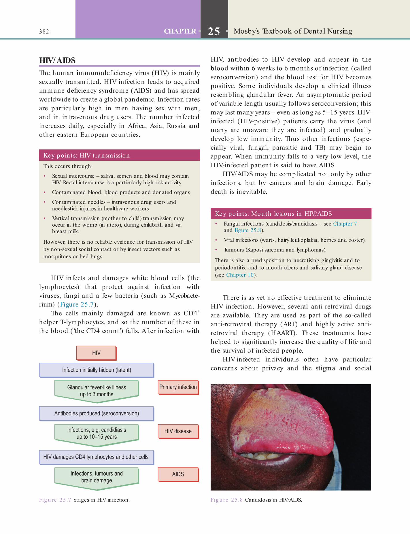
382 CHAPTER • 25 • Mosby’s Textbook of Dental Nursing
HIV, antibodies to HIV develop and appear in the blood within 6 weeks to 6 months of infection (called seroconversion) and the blood test for HIV becomes positive. Some individuals develop a clinical illness resembling glandular fever. An asymptomatic period of variable length usually follows seroconversion; this may last many years – even as long as 5–15 years. HIV-infected (HIV-positive) patients carry the virus (and many are unaware they are infected) and gradually develop low immunity. Thus other infections (espe-cially viral, fungal, parasitic and TB) may begin to appear. When immunity falls to a very low level, the HIV-infected patient is said to have AIDS.
HIV/AIDS may be complicated not only by other infections, but by cancers and brain damage. Early death is inevitable.
HIV/AIDSThe human immunode ciency virus (HIV) is mainly sexually transmitted. HIV infection leads to acquired immune de ciency syndrome (AIDS) and has spread worldwide to create a global pandemic. Infection rates are particularly high in men having sex with men, and in intravenous drug users. The number infected increases daily, especially in Africa, Asia, Russia and other eastern European countries.
Fig u re 25.7 Stages in HIV infection.
Key po in ts: Mouth lesions in HIV/AIDS• Fungal infections (candidosis/candidiasis – see Chapter 7
and Figure 25.8).
• Viral infections (warts, hairy leukoplakia, herpes and zoster).
• Tumours (Kaposi sarcoma and lymphomas).
There is also a predisposition to necrotising gingivitis and to periodontitis, and to mouth ulcers and salivary gland disease (see Chapter 10).
Key po in ts: HIV t ransmissionThis occurs through:
• Sexual intercourse – saliva, semen and blood may contain HIV. Rectal intercourse is a particularly high-risk activity
• Contaminated blood, blood products and donated organs
• Contaminated needles – intravenous drug users and needlestick injuries in healthcare workers
• Vertical transmission (mother to child) transmission may occur in the womb (in utero), during childbirth and via breast milk.
However, there is no reliable evidence for transmission of HIV by non-sexual social contact or by insect vectors such as mosquitoes or bed bugs.
HIV infects and damages white blood cells (the lymphocytes) that protect against infection with viruses, fungi and a few bacteria (such as Mycobacte-rium) (Figure 25.7).
The cells mainly damaged are known as CD4+ helper T-lymphocytes, and so the number of these in the blood (‘the CD4 count’) falls. After infection with
Fig u re 25.8 Candidosis in HIV/AIDS.
There is as yet no effective treatment to eliminate HIV infection. However, several anti-retroviral drugs are available. They are used as part of the so-called anti-retroviral therapy (ART) and highly active anti-retroviral therapy (HAART). These treatments have helped to signi cantly increase the quality of life and the survival of infected people.
HIV-infected individuals often have particular concerns about privacy and the stigma and social
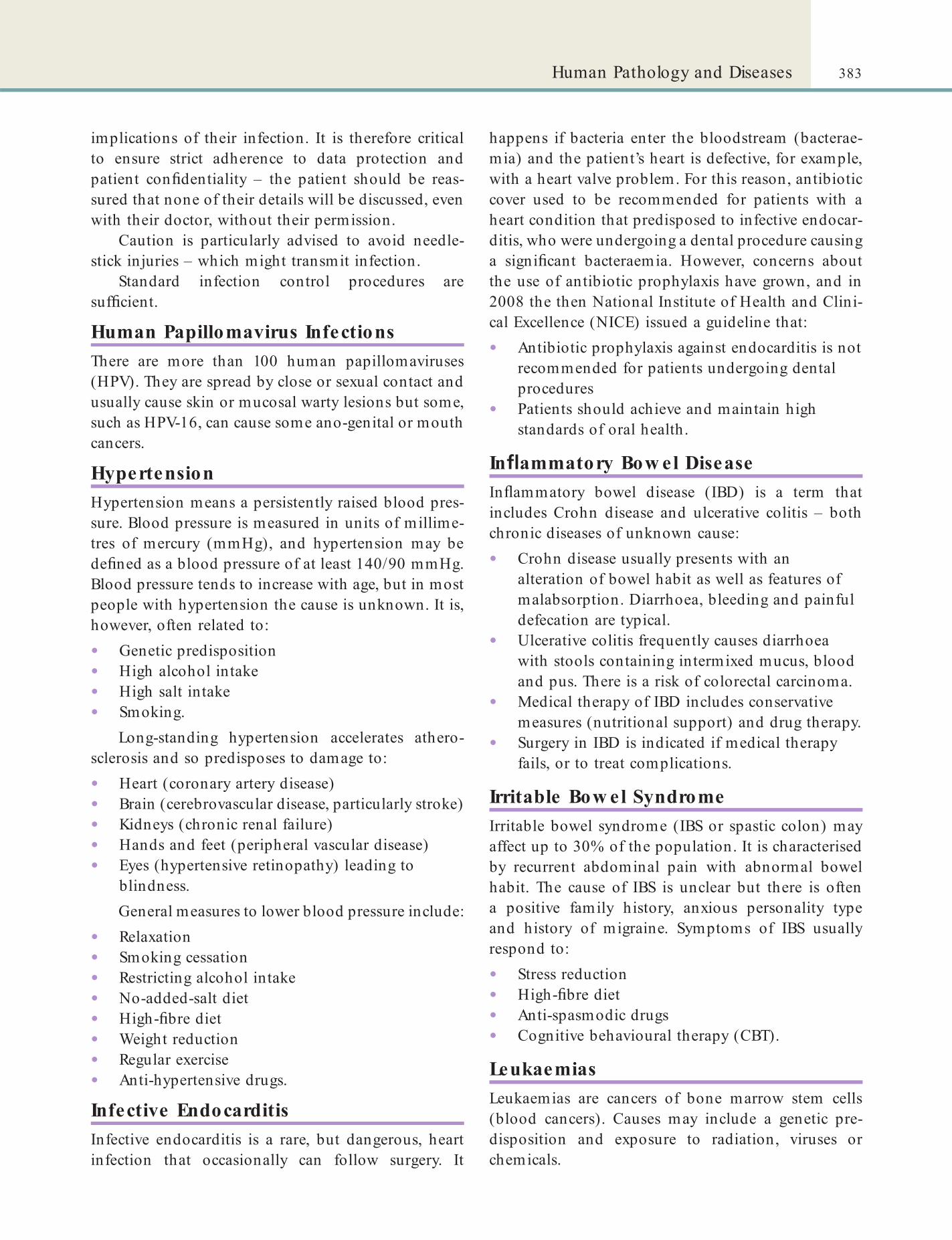
383Human Pathology and Diseases
happens if bacteria enter the bloodstream (bacterae-mia) and the patient’s heart is defective, for example, with a heart valve problem. For this reason, antibiotic cover used to be recommended for patients with a heart condition that predisposed to infective endocar-ditis, who were undergoing a dental procedure causing a signi cant bacteraemia. However, concerns about the use of antibiotic prophylaxis have grown, and in 2008 the then National Institute of Health and Clini-cal Excellence (NICE) issued a guideline that:
• Antibiotic prophylaxis against endocarditis is not recommended for patients undergoing dental procedures
• Patients should achieve and maintain high standards of oral health.
In ammatory Bow el DiseaseIn ammatory bowel disease (IBD) is a term that includes Crohn disease and ulcerative colitis – both chronic diseases of unknown cause:
• Crohn disease usually presents with an alteration of bowel habit as well as features of malabsorption. Diarrhoea, bleeding and painful defecation are typical.
• Ulcerative colitis frequently causes diarrhoea with stools containing intermixed mucus, blood and pus. There is a risk of colorectal carcinoma.
• Medical therapy of IBD includes conservative measures (nutritional support) and drug therapy.
• Surgery in IBD is indicated if medical therapy fails, or to treat complications.
Irritable Bow el SyndromeIrritable bowel syndrome (IBS or spastic colon) may affect up to 30% of the population. It is characterised by recurrent abdominal pain with abnormal bowel habit. The cause of IBS is unclear but there is often a positive family history, anxious personality type and history of migraine. Symptoms of IBS usually respond to:
• Stress reduction• High- bre diet• Anti-spasmodic drugs• Cognitive behavioural therapy (CBT).
LeukaemiasLeukaemias are cancers of bone marrow stem cells (blood cancers). Causes may include a genetic pre-disposition and exposure to radiation, viruses or chemicals.
implications of their infection. It is therefore critical to ensure strict adherence to data protection and patient con dentiality – the patient should be reas-sured that none of their details will be discussed, even with their doctor, without their permission.
Caution is particularly advised to avoid needle-stick injuries – which might transmit infection.
Standard infection control procedures are suf cient.
Human Papillomavirus InfectionsThere are more than 100 human papillomaviruses (HPV). They are spread by close or sexual contact and usually cause skin or mucosal warty lesions but some, such as HPV-16, can cause some ano-genital or mouth cancers.
HypertensionHypertension means a persistently raised blood pres-sure. Blood pressure is measured in units of millime-tres of mercury (mmHg), and hypertension may be de ned as a blood pressure of at least 140/90 mmHg. Blood pressure tends to increase with age, but in most people with hypertension the cause is unknown. It is, however, often related to:
• Genetic predisposition• High alcohol intake• High salt intake• Smoking.
Long-standing hypertension accelerates athero-sclerosis and so predisposes to damage to:
• Heart (coronary artery disease)• Brain (cerebrovascular disease, particularly stroke)• Kidneys (chronic renal failure)• Hands and feet (peripheral vascular disease)• Eyes (hypertensive retinopathy) leading to
blindness.General measures to lower blood pressure include:
• Relaxation• Smoking cessation• Restricting alcohol intake• No-added-salt diet• High- bre diet• Weight reduction• Regular exercise• Anti-hypertensive drugs.
Infective EndocarditisInfective endocarditis is a rare, but dangerous, heart infection that occasionally can follow surgery. It
384 CHAPTER • 25 • Mosby’s Textbook of Dental Nursing
Liver DiseaseLiver disease or cirrhosis may be caused by excessive alcohol intake, and viral hepatitis. Jaundice, bruising, haemorrhage and infection are common.
The management of patients with chronic liver disease includes:
• Dietary modi cation including reduction or cessation of alcohol use
• Careful drug prescribing in view of impaired drug handling and an increased risk of drug toxicity
• Treatment of underlying viral disease (e.g. hepatitis B or C)
• Prevention of and aggressive treatment of infection.
Find out moreWhy do people with liver d isease have impaired drug handling and increased risk of d rug toxicity?
Lung CancerLung cancer is the most common cancer in developed countries in males, and most frequently affects urban, adult cigarette smokers.
The main features are:
• Recurrent cough• Blood in sputum (haemoptysis)• Dyspnoea (breathlessness)• Chest pain• Chest infections.
Management is mainly by radiotherapy.
MigraineMigraine is a recurrent headache due to the brain blood vessels becoming dilated and in amed. It affects mainly women. It is a unilateral, throbbing headache (hemicrania) lasting for hours or days. Photophobia (wanting to be in a dark place), nausea and vomiting may also occur. Classic migraine is preceded by an aura of visual disturbances (forti cation spectra), sensory abnormalities (paraesthesia), motor and speech disturbances. Episodes may be precipitated by alcohol, tyramine-containing foods (e.g. chocolate, cheese, red wine, bananas), menstrual disturbances, stress and drugs.
Management includes:
• Reassurance• Avoidance of precipitating factors
Leukaemias are classi ed by:
• Cell of origin (lymphoblast or non-lymphoblast)
• Cell maturity – immature (acute) versus mature (chronic).Large numbers of white blood cells (leucocytes)
are seen under a microscope on a blood lm and on bone marrow biopsy. These cells lack the normal pro-tective capacity of healthy leucocytes, so the patient becomes vulnerable to infections. The malignant expansion of white cells into the reticulo-endothelial system causes enlarged lymph nodes, liver and spleen. When they crowd out other bone marrow cells the patient develops anaemia (lack of red blood cells) and a bleeding tendency (because of low blood platelet numbers).
Acute leukaemias account for nearly 50% of malignant disease in children. Chronic leukaemias are diseases of late adult life and are fairly benign.
The clinical features broadly re ect the degree of spread of the leukaemic cells in other tissues and the extent of bone marrow failure (Table 25.8).
Chemotherapy is usually used for treatment but the drugs damage not only proliferating leucocytes but also other cells, including hair, mucosae, bone marrow and reproductive tissues. So there are many unpleas-ant adverse effects, such as hair loss, mucositis, infec-tions and sterility.
Lichen PlanusLichen planus is a common skin and mucosal disease causing an itchy rash on the wrists. The mouth, geni-tals, nails or hair may be affected.
Management is usually with corticosteroids.
Table 25.8 Fea tures o f leukaemia
Site o f d isease Clin ica l manifesta t ion
Bone marrow failure
Anaemia Pallor, lethargy, breathlessness
Neutropenia Recurrent infection
Thrombocytopenia Mucosal bleeding and bruising
Tissue in ltration
Lymphatics Lymph node swelling
Liver and spleen Swollen liver and spleen
Bone Bone pain and fractures
385Human Pathology and Diseases
years and older are overweight. Obesity is also increas-ing among children.
A certain amount of body fat is needed for, for example, stored energy, heat insulation and shock absorption. As a rule, women have more body fat than men.
Obesity results from eating more than the body needs. What causes this imbalance between calories in and calories out differs from one person to another and other factors may also play a part, including:
• Lifestyle – such as what a person eats and their level of physical activity
• Psychology – some people eat in response to negative emotions such as boredom, sadness, or anger. Some antidepressants and other drugs can cause weight gain
• Genetic factors• Hormonal causes rarely; although many obese
people believe that they have a glandular or hormonal disorder, this is rarely the case.Associated problems include:
• It is unaesthetic and disabling• Predisposes to or aggravates several disorders:
• Chronic diseases (heart disease, diabetes, hypertension, stroke)
• Some cancers.The best way to avoid obesity is to increase exercise
and reduce calorie intake. Eat smaller portions!
OsteoporosisOsteoporosis (bone-thinning or brittle bone disease) is a very common cause of fragile bones, usually in older people. The difference between how much healthy bone is formed and the rate at which it is remodelled and removed later determines how much osteoporosis (see below) results. From age 30 years, about 1% bone is lost per year, rising to about 5% after menopause.
Risk factors for osteoporosis are shown in Box 25.4.
The main complications are fractures of the hip (neck of the femur), or wrist, or of the spine (hence the shrinking size of some older women). Low back pain is common.
The best management is to avoid falls and injuries. Exercise has a positive effect: walking, running, jogging and dancing are recommended.
Additional calcium and vitamin D may also be helpful. The main sources of calcium are dairy
• Suitable environment (a quiet dark room) during an attack
• Drugs – analgesics, anti-emetics (for the relief of nausea) and triptans. For individuals with recurrent episodes of migraine, prophylactic drug treatment is indicated.
Multiple SclerosisMultiple sclerosis (MS or disseminated sclerosis) is the most common chronic neurological disease. It is thought to be due to damage to nerve myelin sheaths (demyelination) with the development of ‘plaques’ throughout the CNS. The cause remains unknown but the diagnostic hallmark is a series of neurological de -cits distributed in time and space. For example, there may be visual disturbances, followed by paralyses and/or patches of skin numbness.
Symptoms and signs depend on the area of the nervous system affected:
• Numbness, weakness or paralysis in a limb• Brief pain, tingling or electric-shock sensations• Tremor, lack of coordination or unsteady gait• Visual disturbances and pain on eye movement
(optic neuritis)• Urinary incontinence, constipation and sexual
dysfunction• Memory loss and impaired concentration.
Multiple sclerosis can be disabling and even fatal. Care may include:
• Physical and occupational therapy• Counselling• Drug treatment.
ObesityObesity is the main nutritional problem in the devel-oped countries. Almost 25% of adults are now clini-cally obese (Box 25.3) and more than 60% aged 20
Box 25.3 De n ing obesity• Men with more than 25% and women with more than
30% body fat are termed obese
• Measuring body fat is dif cult, so healthcare providers rely on weight-for-height tables – a range of acceptable weights for a person of a given height. These tables do not distinguish between muscle and excess fat.
• Body mass index (BMI) – is weight in kilograms divided by height in metres squared
• A BMI of 25–29.9 indicates overweight, a BMI of 30 (males) or 28.6 (females) or higher indicates obesity.

386 CHAPTER • 25 • Mosby’s Textbook of Dental Nursing
There is a decrease in the level of the brain neu-rotransmitter dopamine. So management is with levodopa (L-dopa), which is converted in the brain into dopamine.
Peptic UlcerPeptic ulcer disease (PUD) develops in acid-secreting areas in the stomach (gastric ulcer) or duodenum (duodenal ulcer). Infection with Helicobacter pylori, a bacterium, is a key cause of PUD. Other factors include use of NSAIDs and corticosteroids, and smoking, alcohol and stress are also responsible.
Management includes:
• Antibiotics – if there is H. pylori infection• Dietary modi cation (frequent small meals with
no fried foods)• Smoking cessation• Alcohol moderation.Surgery is usually reserved for patients with complications.
Radiotherapy-Related DisordersRadiotherapy is one of the main treatments for head and neck cancers. It kills cancer cells but also inevita-bly damages normal tissues in the line of the beam, such as the:
• Oral mucosa (when it causes soreness from mucositis)
• Salivary glands (when it causes hyposalivation)• Bone (osteoradionecrosis).
Osteoradionecrosis (ORN) is particularly liable to occur after trauma/dental extractions in the irradiated area. This may be prevented by removing teeth no less than 10 days be ore the start of radiotherapy. Antibiot-ics and hyperbaric oxygen therapy (HBOT) may help in treatment.
Renal (Kidney) DiseaseChronic renal failure or chronic kidney disease (CKD) is de ned as a low glomerular ltration rate (GFR). It results from progressive, irreversible kidney damage due to some other disease – diabetes is the single most common cause. Hypertension and drug effects are others.
products and green vegetables. The main source of vitamin D is sun-exposure.
Drug treatment – bisphosphonates are drugs com-monly used to prevent osteoporosis. These drugs are also given intravenously for patients who have cancers affecting bone.
Terms to lea rnIdiopathic: a disease whose cause is unknown.
Neurotransmitter: a chemical that helps to transmit electrical impulses at nerve synapses (see Chapter 24).
Term to lea rnOsteonecrosis: death of bone tissue.
Key poin tBisphosphonates remain in the bone for many years and have an extremely long-lasting effect, causing susceptibility to jaw osteonecrosis – mainly in people having intravenous bisphosphonates. Patients taking bisphosphonates (especially intravenous) must therefore be counselled about risks before they have any surgery of the jaws, including implant placement.
Box 25.4 Risk factors fo r osteoporosis• Advanced age – it affects nearly one-third of the population
over 60 years.
• Female gender (1 in 3 women compared with 1 in 12 men) and early menopause.
• Positive family history.
• Modi able lifestyle factors:
• Lack of exercise
• Diet low in calcium or vitamin D
• Excess alcohol intake
• Smoking.• Drugs and disease:
• Drugs (e.g. anticonvulsants, ciclosporin, and corticosteroids)
• Malignant disease
• Chronic in ammatory states (e.g. rheumatoid arthritis)
• Endocrine disorders.
Parkinson DiseaseParkinson disease is a common but serious brain disorder. The risk of developing Parkinson disease increases with age. Mostly it is idiopathic but injuries (particularly boxing), drugs and toxins may be responsible.
Signs and symptoms include:
• Trembling• Muscle rigidity• Dif culty walking• Problems with balance and coordination.
387Human Pathology and Diseases
vacuum aspiration. In the laboratory use guards and suction
• Avoid inhaling aerosols, dusts, vapours, smoke, or fumes by provision of masks
• Ventilate the working area adequately.Face masks lter a great deal of debris but vary in
their ltering ef ciency and should preferably be changed every hour. Face protectors have the advantage of giving a ow of ltered cool air to the operator and protecting eyes and respiratory system simultaneously.
CKD may be asymptomatic until kidney function has fallen to less than 25% of normal. Then hyperten-sion, anaemia, bruising and bone disease may be seen.
Initial management aims to lower the blood pres-sure and urea levels. Dietary modi cation may include salt restriction. Waste metabolites may eventually need to be removed by:
• Continuous Ambulatory Peritoneal Dialysis (CAPD) and intermittent peritoneal dialysis – placement of a catheter into the abdominal peritoneal cavity.
• Haemodialysis – via a ‘kidney machine’. Infection control to prevent cross-infection of hepatitis viruses, HIV and other blood-borne agents is of paramount importance.
• Haemo ltration – using a highly permeable membrane.Renal transplantation may become necessary.
Respiratory InfectionsViral upper respiratory infections are commonplace. Tuberculosis (see below) and Legionella infections are also important respiratory infections. The most impor-tant preventive measures are:
• Avoid treating or being in close proximity to persons with acute respiratory infections
• Minimise production of aerosols, splatter, and dusts. In clinical work use rubber dam and high
Key poin tUse of high volume aspiration and rubber dam during restorative procedures will signi cantly reduce the amount of infected aerosol and splatter.
Term to lea rnGFR: a measure of kidney function.
Table 25.9 The most importan t sexually t ransmit ted (shared) in fect ions
Agents Diseases Caused by
Bacteria Chancroid Haemophilus ducreyiChlamydia infections (including lymphogranuloma venereum)
Chlamydia trachomatis
Gonorrhoea Neisseria gonorrhoeaeSyphilis Treponema pallidumTrichomoniasis Trichomoniasis vaginalis
Viruses Herpes Herpes simplex virus (human (alpha) herpesvirus)Hepatitis Hepatitis A, B, C or D virusesHIV/AIDS Human immunode ciency virusWarts Human papillomavirus (HPV)
Fungi Thrush Candida albicans
Parasites Fleas SiphonapteraLice (crabs) Phthirus pubisScabies Sarcoptes scabiei
SchizophreniaSchizophrenia is a chronic, disabling mental condi-tion. Mood, thoughts and behaviour are disorganised and appear irrational and exaggerated. The person also has hallucinations, delusions and thought disorder and they lack insight. Concern is initially raised by a friend or relative. Treatment includes long-term psy-chotherapy and drug therapy.
Sexually Transmitted/Shared InfectionsSexually transmitted infections (STIs or SSIs) are passed from person to person through unsafe sexual practices. The important ones are syphilis, herpes and HIV, ano-genital warts and gonorrhoea (see p. 380) (Table 25.9).

388 CHAPTER • 25 • Mosby’s Textbook of Dental Nursing
Management of sickle cell disease is to prevent sickle crises. Factors that can precipitate a crisis include:
• Trauma• Infection• Hypoxia• Acidosis• Dehydration.
Sjögren SyndromeSjögren syndrome is an autoimmune disease that affects tear (lacrimal), salivary and other glands. This leads to dry eyes and dry mouth, and sometimes gland swelling.
Management is by;
• Sipping water or sugar-free drinks• Sucking ice• Using salivary substitutes.
Occasionally, salivary stimulant drugs are used but chewing gum is often more helpful.
StrokeA stroke or cerebrovascular accident (CVA) or event is a common cause of death and disability worldwide, mainly in older people. The most common cause is atherosclerosis: plaque rupture results in brain arterial blockage. A stroke may be preceded by a transient ischaemic attack (TIA), which by de nition resolves completely within 24 hours. Approximately 45% of people with a stroke die within a month. Risk factors include:
• Increasing age• Hypertension• Ischaemic heart disease• Heart disease• Diabetes• Smoking• Alcohol• Obesity.
Typical features of stroke are:
• Visual deterioration• Speech disturbance• Hemiplegia (paralysis of one side of the body)• Impaired level of consciousness progressing to
coma or death.Stroke victims should be admitted to hospital as
soon as possible. Treatment lowers the risk of death and disability. Protection of the airway including appropriate ventilatory support is crucial. Pressure-sensitive nursing is needed to avoid the development
Having several SSIs at one time (including viral hepatitis) is not uncommon, especially in intravenous drug users. This is because of unhygienic practices and drug users often offer sex to get money to fund their habit.
Contact tracing can identify who a person was infected by, or whom they have infected.
Risk can be reduced by avoiding casual and unpro-tected sexual intercourse by ABC:
• Abstinence• Being monogamous (having only one partner)• Condom use at all times.
Certain groups of people and their sexual partners who may be at especially high risk of contracting SSIs are:
• Promiscuous heterosexuals or Men who have Sex with Men (MSM)
• Prostitutes – male or female• Drug addicts and alcoholics• Armed forces personnel• Merchant navy personnel• Aircrew• Frequent business travellers• Learning-impaired people.
Sickle Cell DiseaseSickling disorders are inherited conditions mainly affecting people of African descent. An abnormal hae-moglobin (called HbS) distorts the shape of the red blood cells, causing them to block arteries.
• Sickle cell trait is at least ten times as common as sickle disease, but it is often asymptomatic.
• Sickle cell disease is a serious disease with widespread complications – sickle crises can be caused by low oxygen tension (as in general anaesthesia, at high altitudes or in unpressurised aircraft).Painful infarcts form, mainly in the spleen, bones
and joints, brain, kidneys, lungs, retinae and skin. Breakdown of erythrocytes (haemolysis) causes anaemia and jaundice and infections. Patients may eventually die from heart failure, kidney failure, over-whelming infection or stroke.
Term to lea rnInfarct: local area of dead tissue, the cause of which is a lack of blood supply, e.g. due to obstruction of a blood vessel by an embolus.
389Human Pathology and Diseases
Any invasive dental treatment should be carried out only after consultation with the responsible doctor. Preventative dentistry is crucial to maintain a healthy mouth and to avoid infections.
In addition, steroid cover may be needed.
Trigeminal Neuralg iaTrigeminal neuralgia is intermittent, severe pain in the oral and facial regions, which lasts a few seconds only. It affects the areas supplied by the trigeminal nerve. Pain is sometimes triggered by touching areas or by certain daily activities, such as eating, talking, washing the face, shaving or cleaning the teeth.
Management includes drugs such as car-bamazepine. Surgery is reserved for recalcitrant pain.
Tuberculosis• Tuberculosis (TB) is an infection caused by the
bacterium Mycobacterium tuberculosis. It affects about a third of the world population. It is particularly widespread in developing countries, but its incidence is also increasing in Western countries (including the UK).
• Approximately 10% of cases are now antibiotic resistant – they are said to have multi-drug resistant (MDR) TB.
• Some TB is resistant to all drugs – extended resistance TB (XR-TB) – especially in patients with AIDS.
• The infection occurs most often via the respiratory tract so it is primarily a respiratory disease (pulmonary TB). Transmission is by droplet spread.
• Reactivation or progression of primary TB may result in widespread haematogenous dissemination of mycobacteria – this is called ‘miliary TB’. It affects the CNS and the cardiovascular, gastro-intestinal and genito-urinary systems.
of bed (pressure) sores. Urinary catheterisation and incontinence pads may be needed. Physiotherapy helps reduce muscle wasting.
SyphilisSyphilis is an infection caused by the bacterium Treponema pallidum. Over 80% of cases are in homo-sexual men. Syphilis is rapidly increasing.
• Primary syphilis – syphilis rst manifests as a lump, and then an ulcer (primary or Hunterian chancre), usually on the penis or vulva. These are highly infectious.
• Secondary syphilis – follows after 6–8 weeks with non-speci c features such as fever, headache, malaise, rash and lymph node enlargement. Mouth ulcers (highly infectious mucous patches and snail-track ulcers) may be seen.
• Tertiary syphilis – if untreated, syphilis progresses to a tertiary stage, 3–10 or more years after infection. At this stage it affects the heart and central nervous system. The gumma is the characteristic lesion – deep punched-out ulcers on skin or mucosa.Management is with antibiotics.
Transplantation-Related DisordersTransplants are generally needed in patients with severe disease, substantial limitation of daily activities and limited life expectancy. Transplant patients are given immunosuppressive drugs to prevent transplant or graft rejection. Therefore they develop low immu-nity and infections may spread rapidly. Bone marrow transplantation (haematopoietic stem cell transplan-tation) may additionally cause rashes, mouth ulcers and generalised problems because the graft attacks the recipient. This condition is called graft-versus-host disease (GVHD).
Term to lea rnTransplant or graft rejection: when the immune system of the person who is receiving a tissue graft or an organ transplant (the recipient) does not accept the new tissue or organ. This means that the immune system of the recipient attacks the transplanted organ or tissue by producing antibodies against it. To prevent this, powerful drugs are given to suppress the immune system, but this makes the recipient vulnerable to a variety of infections.
Term to lea rnHaematogenous: spread of a disease via the blood circulation.
The initial infection is usually silent (subclinical) causing no symptoms or signs. Active TB typically causes a chronic cough containing blood (haemopty-sis), weight loss, night sweats and fever.
TB is a noti able disease and contact tracing is an important aspect of management.
Some immunosuppressive drugs (ciclosporin) can cause gingival swelling. GVHD causes lesions such as lichen planus and Sjögren syndrome.

390 CHAPTER • 25 • Mosby’s Textbook of Dental Nursing
Burdens of disease: the impact of a health problem in an area measured by nancial cost, mortality, morbidity, or other indicators.Chronic disease: one that lasts a long time, usually at least 6 months.Contagious: can be transmitted by infectious micro-organisms.Cure: the end of a medical condition or a treatment likely to end it.Disease: any condition that impairs normal function.Disorder: a functional abnormality or disturbance.Disseminated disease: spreads to other parts.Illness: a synonym for disease.Incubation period: the time between infection and the appearance of symptoms.Latency period: the time between infection and the ability of the disease to spread to another person.Lifestyle diseases: any disease that appears to increase in frequency if the risk factors include behavioural choices.Localised disease: one that affects only one part of the body.Medical condition: a broad term that includes all diseases and disorders.Mental disorders: a broad label for illnesses that may include affective or emotional instability, behavioural dysregulation, and/or cognitive dysfunction or impairment.Morbidity: from Latin morbidus, meaning ‘sick, unhealthy’.Organic disease: one caused by a physical or physiological change to some tissue or organ.Progressive disease: a disease whose typical natural course is the worsening of the disease.Refractory disease: one that resists treatment.Remission: the disappearance, possibly temporarily, of symptoms.Social determinants of health: the social conditions in which people live that determine their health.Syndrome: the association of several signs, symptoms and/or other characteristics.Systemic disease: a disease that affects the entire body
Anti-tubercular drug treatment should be insti-tuted as early as possible. If patient compliance is considered to be poor, as is often the case since TB is common in the homeless, alcoholics and drug abusers, Directly Observed Therapy (DOT) may be indicated. This means that drugs are dispensed by and taken in the presence of a healthcare professional.
Prevention of TB by BCG vaccination (live attenu-ated Mycobacterium bovis vaccine) is advised for high-risk individuals and healthcare professionals. However, its ef cacy is questionable. Infection control is important.
Term to lea rnNoti able disease: a disease that must be reported to the relevant government authority as per the law.
Von Willebrand DiseaseVon Willebrand disease (vWD) is the most common inherited bleeding disorder. It is characterised by bleeding from mucous membranes – nose bleeds, gin-gival haemorrhage and gastro-intestinal blood loss. Both males and females can have the disorder. vWD is due to a de ciency of von Willebrand factor (vWF), which takes part in blood clotting (see Chapter 24). There are also reduced levels of factor VIII (the main feature of haemophilia).
Excessive bleeding may occur after dental treat-ment and surgery. Thus, before surgery, patients need factor VIII, or synthetic vasopressin (DDAVP; desmopressin).
Find out moreTo nd out more about human diseases the fo llowing websites a re good places to visit :• Mayo Clin ic.com: www.mayoclin ic.com/• Medline Plus: www.nlm.nih .gov/medlinep lus/
hea lth top ics.h tml• Hea lth nder.gov: www.hea lth nder.gov/
Appendix 25.1Glossary
Acute disease: a short-lived disease, like the common cold.

Chapter 26 Health Promotion
CHAPTER POINTS• Avoiding or preventing ill health• Immunisation• Lifestyle choices• Exercise• Diet
Find out moreCan you th ink of two e th ica l quest ions re la ted to gene manipula t ion?
Environmental FactorsEnvironmental factors which when avoided or minimised may prevent many diseases include the following.
• Trauma – this can be minimised by avoiding alcohol use, aggression and dangerous environments, activities and sports; and using protective sports and eye wear (e.g. helmets, mouth guards).
• Infections – these can be minimised by immunisation, and by using infection control (avoiding needlestick injuries and using barrier protection – gloves, rubber dam, eye protection, condoms). Think about infections when planning overseas visits or when having tattoos or piercings (Table 26.1). The expression of individualism through body art such as tattooing and body piercing, and the wearing of jewellery in ‘unconventional’ sites is popular. Tattooing and more invasive procedures have been common in many cultures for centuries and often have sexual connotations. Oral piercings, tongue splitting and ‘dental grills’ (also termed bling, tooth jewellery, grillz or fronts) are increasingly seen.
• Chemicals – such injuries can be minimised by ensuring containers are safe and labelled appropriately and taking care to avoid exposure to toxic agents.
• Irradiation – such injuries can be minimised by reducing exposure to sun, X-rays, lasers, damaging lights etc., and by using safety measures such as protective eyewear and screens.
In t ro d u ct io n
It is important for all healthcare professionals to promote good health.
Avoiding or Preventing Ill HealthHealth may be promoted by avoiding, or minimising, lifestyle or environmental risk factors and, possibly in the future, by gene manipulation (gene therapy).
Term to lea rnGene manipulation: the arti cial manipulation of genetic material.
Genetic DiseasesAlready, genetic disease effects such as those from hae-mophilia can be minimised by giving the genetically missing protein (blood clotting factor VIII). Genetic counselling is increasing, especially in relation to disorders such as haemophilia, Down syndrome, Huntington disease, cystic brosis and various cancers. In future, genetic manipulation will almost inevitably be more widely possible, but it has raised many ethical questions.
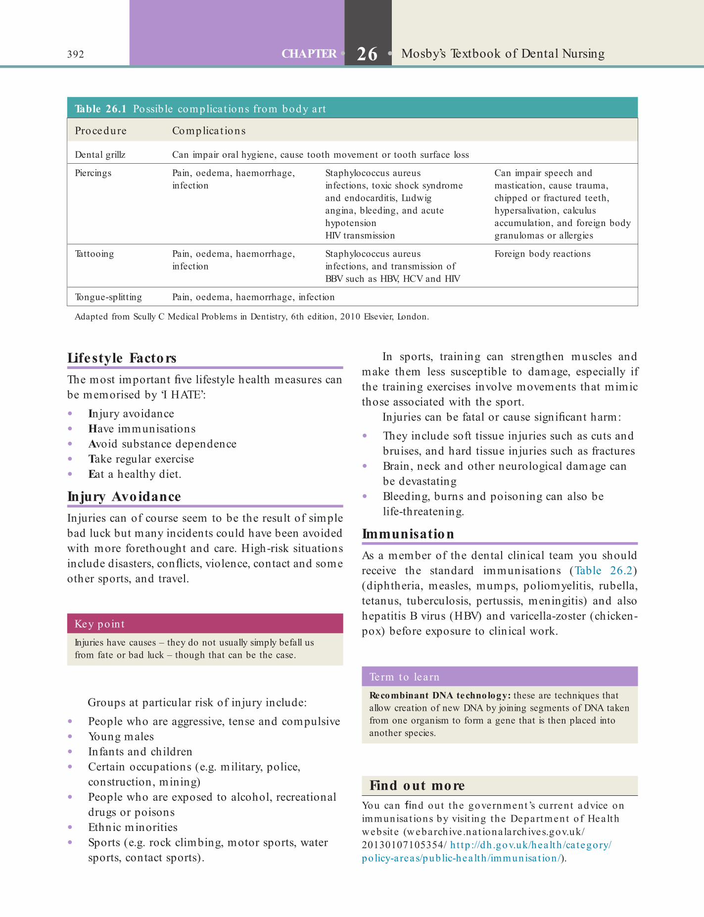
392 CHAPTER • 26 • Mosby’s Textbook of Dental Nursing
Term to lea rnRecombinant DNA technology: these are techniques that allow creation of new DNA by joining segments of DNA taken from one organism to form a gene that is then placed into another species.
Key po in tInjuries have causes – they do not usually simply befall us from fate or bad luck – though that can be the case.
Table 26.1 Possib le complica t ions from body a rt
Procedure Complica t ions
Dental grillz Can impair oral hygiene, cause tooth movement or tooth surface loss
Piercings Pain, oedema, haemorrhage, infection
Staphylococcus aureus infections, toxic shock syndrome and endocarditis, Ludwig angina, bleeding, and acute hypotensionHIV transmission
Can impair speech and mastication, cause trauma, chipped or fractured teeth, hypersalivation, calculus accumulation, and foreign body granulomas or allergies
Tattooing Pain, oedema, haemorrhage, infection
Staphylococcus aureus infections, and transmission of BBV such as HBV, HCV and HIV
Foreign body reactions
Tongue-splitting Pain, oedema, haemorrhage, infection
Adapted from Scully C Medical Problems in Dentistry, 6th edition, 2010 Elsevier, London.
Lifestyle FactorsThe most important ve lifestyle health measures can be memorised by ‘I HATE’:
• Injury avoidance• Have immunisations• Avoid substance dependence• Take regular exercise• Eat a healthy diet.
Injury AvoidanceInjuries can of course seem to be the result of simple bad luck but many incidents could have been avoided with more forethought and care. High-risk situations include disasters, con icts, violence, contact and some other sports, and travel.
Groups at particular risk of injury include:
• People who are aggressive, tense and compulsive• Young males• Infants and children• Certain occupations (e.g. military, police,
construction, mining)• People who are exposed to alcohol, recreational
drugs or poisons• Ethnic minorities• Sports (e.g. rock climbing, motor sports, water
sports, contact sports).
In sports, training can strengthen muscles and make them less susceptible to damage, especially if the training exercises involve movements that mimic those associated with the sport.
Injuries can be fatal or cause signi cant harm:
• They include soft tissue injuries such as cuts and bruises, and hard tissue injuries such as fractures
• Brain, neck and other neurological damage can be devastating
• Bleeding, burns and poisoning can also be life-threatening.
ImmunisationAs a member of the dental clinical team you should receive the standard immunisations (Table 26.2) (diphtheria, measles, mumps, poliomyelitis, rubella, tetanus, tuberculosis, pertussis, meningitis) and also hepatitis B virus (HBV) and varicella-zoster (chicken-pox) before exposure to clinical work.
Find out moreYou can nd ou t the government ’s curren t advice on immunisa t ions by visit ing the Department o f Hea lth websit e (webarchive .na t iona la rch ives.gov.uk/ 20130107105354/ h t tp ://dh .gov.uk/hea lth /ca tegory/ po licy-areas/public-hea lth /immunisa t ion /).
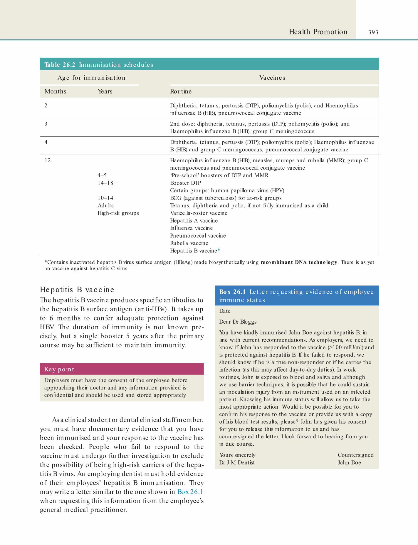
393Health Promotion
Table 26.2 Immunisa t ion schedules
Age for immunisa t ion Vaccines
Months Years Routine
2 Diphtheria, tetanus, pertussis (DTP); poliomyelitis (polio); and Haemophilus inf uenzae B (HIB), pneumococcal conjugate vaccine
3 2nd dose: diphtheria, tetanus, pertussis (DTP); poliomyelitis (polio); and Haemophilus inf uenzae B (HIB), group C meningococcus
4 Diphtheria, tetanus, pertussis (DTP); poliomyelitis (polio); Haemophilus inf uenzae B (HIB) and group C meningococcus, pneumococcal conjugate vaccine
12 Haemophilus inf uenzae B (HIB); measles, mumps and rubella (MMR); group C meningococcus and pneumococcal conjugate vaccine
4–5 ‘Pre-school’ boosters of DTP and MMR14–18 Booster DTP
Certain groups: human papilloma virus (HPV)10–14 BCG (against tuberculosis) for at-risk groupsAdults Tetanus, diphtheria and polio, if not fully immunised as a childHigh-risk groups Varicella-zoster vaccine
Hepatitis A vaccineIn uenza vaccinePneumococcal vaccineRubella vaccineHepatitis B vaccine*
*Contains inactivated hepatitis B virus surface antigen (HBsAg) made biosynthetically using recombinant DNA technology. There is as yet no vaccine against hepatitis C virus.
He pa titis B vac c ineThe hepatitis B vaccine produces speci c antibodies to the hepatitis B surface antigen (anti-HBs). It takes up to 6 months to confer adequate protection against HBV. The duration of immunity is not known pre-cisely, but a single booster 5 years after the primary course may be suf cient to maintain immunity.
Key poin tEmployers must have the consent of the employee before approaching their doctor and any information provided is con dential and should be used and stored appropriately.
As a clinical student or dental clinical staff member, you must have documentary evidence that you have been immunised and your response to the vaccine has been checked. People who fail to respond to the vaccine must undergo further investigation to exclude the possibility of being high-risk carriers of the hepa-titis B virus. An employing dentist must hold evidence of their employees’ hepatitis B immunisation. They may write a letter similar to the one shown in Box 26.1 when requesting this information from the employee’s general medical practitioner.
Box 26.1 Le t te r request ing evidence of employee immune sta tusDate
Dear Dr Bloggs
You have kindly immunised John Doe against hepatitis B, in line with current recommendations. As employers, we need to know if John has responded to the vaccine (>100 mIU/ml) and is protected against hepatitis B. If he failed to respond, we should know if he is a true non-responder or if he carries the infection (as this may affect day-to-day duties). In work routines, John is exposed to blood and saliva and although we use barrier techniques, it is possible that he could sustain an inoculation injury from an instrument used on an infected patient. Knowing his immune status will allow us to take the most appropriate action. Would it be possible for you to con rm his response to the vaccine or provide us with a copy of his blood test results, please? John has given his consent for you to release this information to us and has countersigned the letter. I look forward to hearing from you in due course.
Yours sincerely CountersignedDr J M Dentist John Doe

394 CHAPTER • 26 • Mosby’s Textbook of Dental Nursing
Key poin tIntravenous use of illegal drugs can also easily lead to life-threatening infections including endocarditis, septicaemias, hepatitis and HIV/AIDS.
Table 26.3 Possib le systemic effects o f chronic tobacco use
System Possib le e ffects
Bladder Cancer
Cardiovascular system Ischaemic heart disease (see Chapter 24). If women smokers also take oral contraceptives, they are more prone to cardiovascular and cerebrovascular diseases than are other smokers
Central nervous system Alzheimer disease, stroke (see Chapter 24)
Mouth Cancer, candidosis, dry mouth, dry socket, halitosis, implant failure, keratosis, necrotising gingivitis, chronic periodontal disease, teeth staining (see Chapters 10 and 24)
Oesophagus Cancer
Reproductive system Women who smoke generally have earlier menopause
Pregnant women who smoke cigarettes run a greater risk of having still-born or premature infants or infants with low birth weight
Respiratory system Cancer, chronic obstructive pulmonary disease, sinusitis
Growth and development Children of women who smoked while pregnant have a higher risk for developing behavioural disorders
Lifestyle ChoicesSubstance DependenceThe most common risks from abuse of many recrea-tional drugs are: injury, behavioural disturbances and psychoses, but it may also lead to a sexually shared infection (SSI) or unwanted pregnancy. High doses can cause mood swings and psychoses – including hallucinations and paranoia – and can also cause res-piratory failure and death. Combining use with other drugs or alcohol can result in nausea, dif culty breath-ing, unconsciousness and death.
Tobac c oTobacco is a major hazard to health and promotes many diseases, particularly heart disease, lung disease and cancers of the lung, oesophagus, mouth and bladder (Table 26.3). Tobacco smoke contains nico-tine, which is absorbed readily from the lungs on smoking and also when tobacco is chewed. Nicotine is highly addictive and withdrawal results in excessive anger, hostility and aggression.
Term to lea rnPassive smoking: inhaling second-hand smoke, that is, smoke produced from the use of a tobacco product by another person nearby.
Key poin ts: Ora l hea lth and tobacco useTobacco use can damage oral health by leading to:
• Oral cancer
• Keratosis
• Periodontal disease, particularly necrotising gingivitis
• Implant failure
• Dry socket
• Candidosis
• Dry mouth
• Halitosis
• Staining of teeth.
Term to lea rnChronic obstructive pulmonary disease: a disease in which the person has long-term obstruction of the airways due to the chronic presence of bronchitis and emphysema. It is most commonly seen in heavy smokers. Symptoms include dif culty breathing, wheezing and a chronic cough.
Passive smoking can cause lung cancer in adults, greatly increases the risk of respiratory illnesses in chil-dren and may cause sudden infant death.

395Health Promotion
Table 26.4 Possib le systemic effects o f chronic a lcohol use
System Possib le e ffects
Blood Pancytopenia, folate de ciency, thiamine de ciency, immune defect
Central nervous system
Intoxication, dependency, dementia, Wernicke–Korsakoff syndrome
Heart Arrhythmias, cardiomyopathy, hypertension
Intestine Glucose and vitamin malabsorption
Liver Hepatitis, fatty liver, cirrhosis, liver cancer
Mouth Tooth erosion, cancer
Musculoskeletal Myopathy, gout
Oesophagus Gastro-oesophageal re ux, Mallory–Weiss syndrome, cancer
Pancreas Pancreatitis
Reproductive system
Impotence, dysmenorrhoea, low birth-weight babies, fetal alcohol syndrome
Stomach Gastritis, ulceration, carcinoma
ManagementSmoking cessation should be gradual, because with-drawal symptoms are less severe in those who quit gradually than in those who quit quickly. Cessation aids include nicotine chewing gum or patches and various drugs.
Alc oho lIn many cultures there is nothing wrong with the occa-sional alcoholic drink, but drinking too much can cause a wide range of serious behavioural and health problems (Table 26.4).
Alcohol is also high in calories, so cutting down could help control weight in overweight people.
Key poin ts: Recommended maximum drinking amounts• Women: up to 2–3 units of alcohol a day.
• Men: up to 3–4 units a day.
These amounts appear not to cause signi cant risk to health.
(A unit is half a pint of standard strength beer, lager or cider, or a measure of spirits. A glass of wine is about 2 units and Alco pops are about 1.5 units.)
Find out moreWhat a re the consequences o f fo la te and th iamine de ciency?
Amphe tamine sAmphetamines are stimulants used for their euphoric effect, and to stave off fatigue. However, they cause dry mouth and caries (‘meth mouth’), dilated pupils, increased pulse rate (tachycardia) and breathing rate (tachypnoea), aggression, talkativeness and hal-lucinations, leading to seizures, hypertension, danger-ously high body temperature (hyperpyrexia), heart arrhythmias and collapse. Mood swings and psychoses – including hallucinations and paranoia – and respira-tory failure can cause death.
Amphetamines have no true withdrawal syndrome and, in this respect, amphetamine addiction is quite different from opioid or barbiturate dependence.
Be te lRegular chewers of betel leaf and areca (betel) nut, as in ‘pan’ or ‘paan’, have a higher risk of oral disease and staining of the teeth. Table 26.5 lists the various effects of betel chewing on various systems of the body and the oral cavity.
Table 26.5 Possib le systemic e ffects o f chronic be te l use
System Possib le e ffects
Heart Hypertension, metabolic syndrome, diabetes, coronary heart disease, obesity
Kidneys Chronic kidney disease, urinary stones
Liver Cirrhosis, cancer
Mouth Periodontitis, leukoplakia, submucous brosis, cancer
Oesophagus Cancer, submucous brosis
Pancreas Cancer
Reproductive system Lower birth-weight infants
Term to lea rnSubmucous brosis: a disease in which there is progressive brosis of the tissues underlying the mucosa. When it occurs in the mouth, it ultimately leads to severe trismus.
396 CHAPTER • 26 • Mosby’s Textbook of Dental Nursing
He ro inHeroin is a highly addictive drug, derived from mor-phine. It can be sniffed, smoked from tin-foil (‘chasing the dragon’) or injected. The short-term effects are euphoria (‘rush’) but then an alternately wakeful and drowsy state (‘on the nod’). With regular heroin use, physical dependence and addiction develop. Heroin depresses respiration and increases risk of pneumonia, lung abscesses and brosis, and infections (abscesses and cellulitis). Infectious diseases, including HIV and hepatitis, are common among users.
Withdrawal causes drug craving, restlessness, muscle and bone pain, insomnia, diarrhoea and vomiting, cold ashes with goose bumps (‘cold turkey’), kicking movements (‘kicking the habit’), and other symptoms. Sudden withdrawal is occasionally fatal.
Treatment options include behavioural therapies and medications such as methadone.
LSD (lys e rg ic ac id d ie thylamide )LSD is taken by mouth and has effects that are unpre-dictable but prolonged (~12 hours), producing several different emotions at once or swinging the user rapidly from one emotion to another within 30–90 minutes. Synaesthesia, the over ow from one sense to another when, for example, colours are heard, is common. There is often lability of mood, panic (‘bad trip’) and delusions of magical powers, such as being able to y. LSD use also causes dilated pupils, high temperatures, heart rate and blood pressure, sweating, loss of appe-tite, sleeplessness, dry mouth and tremors.
Many LSD users later (within a few days or more than a year after use) experience ashbacks, recurrence of certain aspects of a person’s experience, without the user having taken the drug again. Severe adverse effects include terrifying thoughts and feelings, and despair, occasionally leading to fatal accidents.
LSD is not considered addictive since it does not produce compulsive drug-seeking behaviour, as do cocaine, amphetamine, heroin, alcohol and nicotine. Most users of LSD voluntarily limit or stop its use over time.
Me phe drone (4-MMC, ‘MEOW’, ‘M-CAT’)Mephedrone has effects similar, but not identical to, Ecstasy, lasting for around 2–3 hours when taken orally. Effects include a desire to take it again, changes in body temperature (sweating and chills), palpitations, impaired short-term memory, insomnia, tightened jaw muscles and grinding teeth. After-effects such as insomnia may last for several hours longer.
CannabisThe main active cannabis (marijuana/‘hash’ or ‘skunk’) chemical is THC (delta-9-tetrahydrocannabinol), which binds to the brain. Adverse effects can include depression, anxiety, personality disturbances, impaired memory and learning, distorted perception, dif culty in thinking and problem solving, and impaired coor-dination; it may also trigger psychosis.
Marijuana can also lead to lung infections, affect blood pressure and heart rate and lower the oxygen-carrying capacity of blood. The risk of a heart attack may more than quadruple in the rst hour after using it. Marijuana may also cause oral cancer. Babies born to women who use marijuana during pregnancies may be affected.
Coc aineThe major routes of cocaine (‘coke’ or ‘crack’) use are snorting, injecting or smoking. Immediate effects include hyperstimulation (euphoria), reduced fatigue and mental clarity, with feelings of well-being and heightened mental activity. Physical effects include constricted peripheral blood vessels, dilated pupils and raised temperature, pulse rate and blood pressure. The cocaine addict has aptly been described as a ‘sexed-up extrovert with dilated pupils’.
Cocaine is powerfully addictive but ‘crack’ (cocaine that has been processed) is even stronger and can cause paranoia, visual hallucinations (‘snow lights’) and hallucinations of insects crawling over the skin (formication, ‘cocaine bugs’). Adverse reactions of cocaine include angina, arrhythmias, heart attack, stroke, convulsions, depressed breathing and death.
On stopping cocaine, symptoms proceed through a crash phase of depression and craving for sleep, a withdrawal phase of lack of energy and then an extinc-tion phase of recurrence of craving.
Ec s tas y (MDMA or me thyle ne -d ioxyme thamphe tamine )Ecstasy is usually taken by mouth, and causes eupho-ria and appetite suppression, muscle tension, involun-tary teeth clenching, nausea, blurred vision, rapid eye movement, faintness and chills or sweating. Ecstasy is potently hallucinogenic and can cause high body tem-perature, ataxia and seizures, heart damage, liver or kidney failure or psychological dif culties, including confusion, depression, sleep problems, drug craving, severe anxiety and paranoia – during use and some-times weeks later.
There is no physical dependence or withdrawal symptoms.
397Health Promotion
ExercisePhysical inactivity contributes to obesity, diabetes, heart disease and cancer. In contrast, when exercise is combined with a proper diet, weight can be controlled and obesity prevented. To achieve real health bene t from exercise, at the very least enough regular, moder-ately intense physical activity to burn an extra 200 calories daily is needed. This means at least 30 minutes of activities such as a brisk walk daily.
Term to lea rnEndorphins: chemicals produced in the brain that help reduce the sensation of pain and improve a person’s mood.
Key poin tObesity is a major risk factor for many diseases.
Regular exercise has a surprisingly wide range of health bene ts, from protecting against heart disease (hypertension, atherosclerosis and coronary artery disease) to reducing the chances of cancer and Alzhe-imer disease. In addition, exercise can:
• Make you feel more energetic• Increase your stamina• Improve brain function by releasing endorphins• Reduce stress, depression and anxiety• Help control weight and build and maintain
healthy bones, muscles and joints• Reduce the risk of developing diabetes• Reduce the risk of developing or dying from
some of the leading causes of illness and death – such as a heart attack, stroke, or cancer.
Ten good , scien t i ca lly proven , reasons for taking regular exercise• Exercise helps minimise obesity:
• Obesity is associated with diabetes, heart disease, high blood pressure, stroke and cancer. Obese men are more likely to die from cancer of the colon, rectum, or prostate than are non-obese men
• Obese women are more likely to die from cancer of the gallbladder, breast, uterus, cervix, or ovaries
• Other health problems from obesity include gallbladder disease and gallstones; liver disease; osteoarthritis; gout; sleep apnoea; menstrual irregularities and infertility in women; and psycho-social effects
• Exercise, by burning calories, helps minimise obesity.
• Exercise helps increase levels of HDL or ‘good’ cholesterol:
• Blood fat levels seem to predict the risk for coronary heart disease – particularly when levels of high-density lipoproteins (HDL) are low and low-density lipoproteins (LDL) are high
• Exercise helps increase HDL and lower LDL levels (bad cholesterol) and, by strengthening the heart and
lowering blood pressure, protects against atherosclerosis, heart attacks and stroke (see Chapter 25).
• Exercise helps lower high blood pressure:
• Blood pressure levels also seem to predict the risk for coronary heart disease and stroke
• High blood pressure (hypertension) can be reduced by exercise
• Physical activity also reduces obesity, which is associated with hypertension.
• Exercise helps promote healthy blood sugar levels:
• Diabetes results in high blood sugar levels – another risk factor for coronary heart disease
• High blood sugar levels can be lowered by exercise.
• Exercise helps improve the metabolic syndrome:
• The metabolic syndrome (syndrome X or insulin resistance syndrome) is a cluster of abdominal obesity (excess body fat around the waist), insulin resistance (causing high blood sugar), and increased blood pressure, which increases the risk for diabetes, heart disease and stroke. Having just one of these factors contributes to the risk of serious disease and, in combination, the risk is even greater
• Exercise reduces these factors.
• Exercise helps build muscle strength:
• Exercise, by building or preserving muscle mass and strength, and improving the ability to use calories, helps reduce body fat
• By increasing muscle strength and endurance, and improving exibility and posture, regular exercise also helps prevent back pain
• Exercise helps build muscle but this bene t fades quickly, if exercise stops for any reason – so regular exercise is needed.
• Exercise helps promote bone density:
• Regular exercise from a young age can increase bone density and help avoid or minimise osteoporosis which is seen mainly in older people, especially in menopausal women.
• Exercise helps improve mobility:
• People who exercise regularly have been shown to be more mobile and independent, compared with those with a sedentary lifestyle.
• Exercise helps boost the immune system:
• Exercise helps your immune system ght off simple bacterial and viral infections. The mechanism is unclear but it is known that exercise increases the numbers of leucocytes (white blood cells) circulating in the blood. Leucocytes are part of the body’s defence against infections (see Chapter 24).
• In contrast, when exercise is performed without food intake, and is too continuous, prolonged, or of moderate to high intensity (as in athletes) the immune function can be depressed.
• Exercise helps improve mood:
• People who exercise regularly have been shown to be generally happier and less liable to depression compared with those having a sedentary lifestyle.
398 CHAPTER • 26 • Mosby’s Textbook of Dental Nursing
health. Vitamins are co-factors essential to a range of biochemical reactions. Vitamins can be classi ed as:
• Water-soluble vitamins (vitamins B and C) – these are readily absorbed from the gastro-intestinal mucosa and needed particularly for healthy nerves and blood.
• Fat-soluble vitamins (vitamins A, D, E and K) – these require the presence of bile for absorption. Vitamin A is needed for healthy eyesight. Vitamin D is needed for healthy bones. Vitamin K is needed for the production of blood clotting factors.
DietThe two keys to a healthy diet are:
• Eating the right amount of food• Eating a balanced diet that contains a variety
of foods:• Lots of fruit, vegetables and starchy foods
such as wholemeal bread and wholegrain cereals and potatoes
• Some protein-rich foods, such as meat, sh, eggs and lentils
• Some dairy foods.
Be ne f ts o a he a lthy d ie t• Generous amounts of vegetables and fruit daily
appear to offer some protection against cancers of the stomach, colon and lung, and possibly against cancers of the mouth, larynx, cervix, bladder and breast.
• Carbohydrates as wholegrain unre ned products may offer some protection against colon cancer, diverticulitis and caries.
• A high bre diet may also offer some protection against hypertension and coronary heart disease.
• A healthy diet is also low in fat (especially saturated fat), salt and sugar.
• Minimising the intake of saturated fats (especially those from dairy sources) and partially hydrogenated vegetable fats may lower the risk of coronary artery disease and some cancers. Consumption of mono-unsaturated fats such as olive oil may be bene cial.
The ca lorie conten t of foods• The nutritional information provided on food packaging
gives the amounts of carbohydrates, fats, proteins, colourings and avourings, and a gure relating to energy content.
• Energy is usually expressed either in kilocalories or in kilojoules. A calorie is the amount of energy required to heat 1 gram of water by 1 degree centigrade, and because this is a small quantity of energy, it is more common to use the kilocalorie or Calorie, which is equivalent to 1000 calories.
• The number of calories multiplied by 4.186 = the number of joules.
• The number of kilocalories multiplied by 4.186 = the number of kilojoules.
Term to lea rnOvo-lacto vegetarianism: no meat and sh but eggs and dairy products are eaten.
Term to lea rnCo-factors: these are substances that bind to speci c types of enzyme to activate them.
VitaminsVitamins were discovered when it was noted that some diets – despite having adequate calories, essential fatty acids and minerals – were inadequate to maintain
Vitamin de ciency diseases are relatively rare, but may occur due to poor diet patterns in the young, old, people with food fads or alcoholics. Vegans, who eat no animal products, however, can readily become vitamin B12-de cient.
Mine ra ls and trac e e le me ntsA number of minerals and trace elements are essential to health. De ciencies of trace elements can result in serious health problems.
Find out moreVisit the Hea lthCheck Systems website (www.hea lthchecksystems.com/vitamins.h tm), where you will nd a tab le list ing the vit amins and minera ls, the ir ro les and sources.
Fad d ie tsMany commercial diets claim to enhance wellbeing or reduce weight. But some of these diets have resulted in frank vitamin, mineral and protein de ciency states. People have developed cardiac, renal and metabolic disorders and some deaths have resulted too.
Ve ge tarianismOvo-lacto vegetarianism is the most common form of vegetarianism. These individuals tend to live longer and to develop fewer chronic disabling conditions than their meat-eating peers. Iron de ciency is the only known risk.
399Health Promotion
• A pear as an afternoon snack• A portion of peas or other vegetables with
your evening meal.• You can choose from fresh, frozen, tinned,
dried or juiced, but remember potatoes count as a starchy food, not as portions of fruit and vegetables.
3 . Eat more sh• Aim for at least two portions of sh a week,
including a portion of oily sh (salmon, mackerel, trout, herring, fresh tuna, sardines, pilchards, eel). These are rich in omega 3 fatty acids and can keep the heart healthy. Fish is also an excellent source of protein and contains many vitamins and minerals. You can choose from fresh, frozen or canned – but remember that canned and smoked sh can be high in salt.
• Do not have more than one portion a week of shark, tuna, sword sh and marlin because of the high levels of mercury in them.
4 . Cut down on saturated fat and sugar• Having too much saturated fat (meat pies,
sausages, meat with visible white fat, hard cheese, butter and lard, pastry, cakes and biscuits, cream, soured cream and crème fraîche, coconut oil, coconut cream or palm oil) can increase the amount of blood cholesterol, which increases the chance of heart disease.
• Unsaturated fat (vegetable oils (including sun ower, rapeseed and olive oil), oily sh, avocados, nuts and seeds) lowers blood cholesterol.
• Try eating fewer foods containing added sugar, such as sweets, cakes and biscuits, and drinking fewer sugary soft and zzy drinks. Having sugary foods and drinks too often can cause dental caries (decay), especially if you have them between meals. Many foods that contain added sugar can also be high in calories, so cutting down could help you control your weight.
5 . Try to eat less salt• Eating too much salt can raise the blood
pressure. And people with high blood pressure are three times more likely to develop heart disease or have a stroke than people with normal blood pressure.
• Three-quarters (75%) of the salt we eat comes from processed food, such as some
Ve ganis mComplete absence of animal products in the diet can lead to vitamin B12 de ciency. Having said that, the latter is surprisingly uncommon, since yeast extracts and oriental-style fermented foods can provide this vitamin.
Dietary RecommendationsThese recommendations are adapted from the ‘8 tips for healthy eating’ on the NHS Choices ‘Live Well’ website (www.nhs.uk/Livewell/Goodfood/Pages/eight -tips-healthy-eating.aspx).1 . Base your meals on starchy foods
• Starchy foods such as bread, cereals, rice, pasta and potatoes are an important part of a healthy diet – a good source of energy and the main source of a range of nutrients. As well as starch, these foods contain bre, calcium, iron and B vitamins.
• Choose wholegrain varieties of starchy foods whenever you can as they contain more bre and other nutrients than white or re ned starchy foods. We also digest wholegrain foods more slowly so they can help make us feel full for longer. Wholegrain foods include:• Wholemeal and wholegrain bread, pitta
and chapatti• Whole-wheat pasta and brown rice• Wholegrain breakfast cereals.
• Starchy foods should make up about a third of the food we eat.
• Try to include at least one starchy food with each of your main meals. So you could start the day with a wholegrain breakfast cereal, have a sandwich for lunch, and potatoes, pasta or rice with your evening meal.
• Starchy foods, gram for gram, contain less than half the calories of fat. However, you need to watch the fats you add when cooking and serving these foods, because this is what increases the calories.
2 . Eat lots of fruit and vegetables• Try to eat at least ve portions of a variety of
fruit and vegetables every day. You could try adding up your portions during the day to make it easier. For example, you could have:• A glass of juice and a sliced banana with
your cereal at breakfast• A side salad at lunch
400 CHAPTER • 26 • Mosby’s Textbook of Dental Nursing
good health. Wholemeal cereal, with fruit sliced over the top, is a nutritious breakfast.
Find out moreFor more about genera l hea lth promot ion , see the Pa t ien t UK website (www.pa t ien t .co .uk/showdoc/16/). Public Hea lth England (PHE), previously the Hea lth Pro tect ion Agency (HPA), is an execut ive agency sponsored by the Department o f Hea lth to p ro tect and improve the na t ion’s hea lth and well-be ing and to reduce hea lth inequa lit ies.
breakfast cereals, soups, sauces, bread, biscuits and ready meals.
6 . Get active and try to be a healthy weight.7 . Don’t get thirsty. We need to drink about 1.2
litres of uid every day to stop us getting dehydrated. This is in addition to the uid we get from the food we eat. All non-alcoholic drinks count, but water and milk are the most healthy.
8 . Don’t skip breakfast. A healthy breakfast is an important part of a balanced diet, and provides some of the vitamins and minerals we need for

Chapter 27 First Aid and Medical Emergencies
CHAPTER POINTS• First aid• Medical emergencies• Risk assessment• Managing a medical emergency• Role o the dental nurse in medical emergencies• The emergency kit• Basic li e support• Managing specif c emergencies• Adverse drug reactions• Aggressive or disturbed patients• Bleeding• Chest pain• Collapse• Epilepsy• Fainting• Hypoglycaemia• Hypotension• Respiratory obstruction• Stroke
able. E ective prompt f rst aid can prevent minor inju-ries and occurrences becoming major ones, and can save lives.
The Health and Sa ety (First Aid) Regulations 1981 require employers to:
• Provide suitably trained personnel to administer f rst aid or employees who may become ill or injured in the workplace. To cover or temporary and exceptional absences o trained f rst aiders, the regulations allow or a competent person, known as an ‘appointed person’, to take charge (see below)
• Provide adequate and appropriate acilities and equipment
• In orm their employees o the arrangements made or f rst aid ‘including the location o equipment, acilities and personnel’.In addition, employers need to consider:
• The number o people employed – is the number o f rst aiders and appointed persons as per the recommendations o the HSE. The number o suitably trained personnel and appropriate equipment and acilities depends on the size o the practice (or hospital).
• Are all inexperienced sta trained in f rst aid – trainees, newly qualif ed clinicians, unqualif ed dental nurses and students all need special training in f rst aid.
• Are there any employees and patients with disabilities and special health problems – consider the location o equipment and the use o special equipment.
• Do employees work di erent hours? There needs to be f rst aid provision at all times o work.
• Is the workplace remote rom emergency medical services? In orm and consider special arrangements with the medical services.
In t ro d u ct io n
All dental sta and patients should be able to receive immediate f rst aid attention and management o medical emergencies and, i required, receive treat-ment rom the ambulance service. Depending on the severity o the emergency, a trained f rst aider, a dental nurse or a clinician may be the most appropriate person to act as f rst responder.
First AidFirst aid is emergency treatment administered to an injured or sick person before professional medical care is avail-
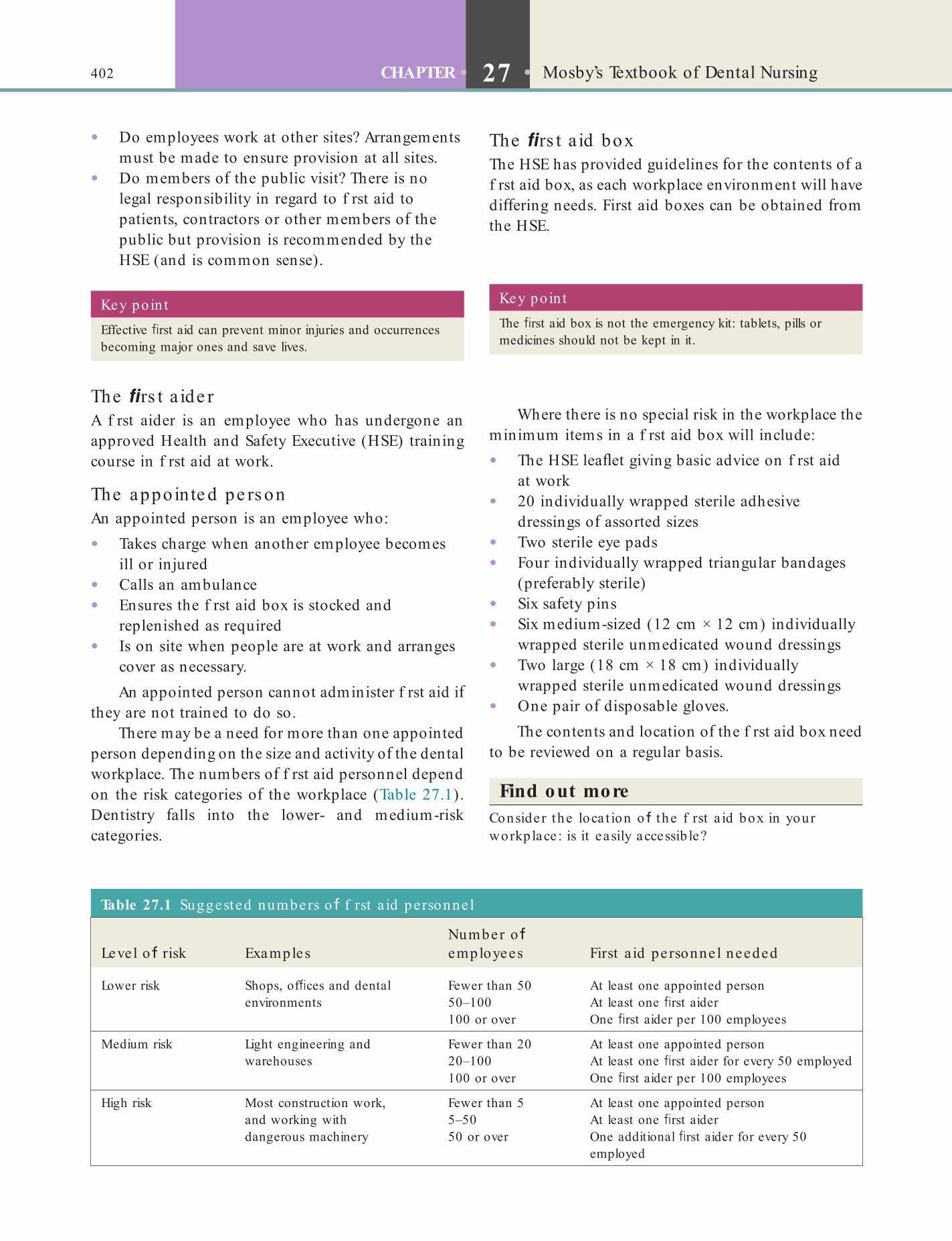
402 CHAPTER • 27 • Mosby’s Textbook of Dental Nursing
The rs t a id boxThe HSE has provided guidelines or the contents o a f rst aid box, as each workplace environment will have di ering needs. First aid boxes can be obtained rom the HSE.
• Do employees work at other sites? Arrangements must be made to ensure provision at all sites.
• Do members o the public visit? There is no legal responsibility in regard to f rst aid to patients, contractors or other members o the public but provision is recommended by the HSE (and is common sense).
Key poin tThe rst aid box is not the emergency kit: tablets, pills or medicines should not be kept in it.
Key po in tEffective rst aid can prevent minor injuries and occurrences becoming major ones and save lives.
Table 27.1 Suggested numbers o f rst a id pe rsonne l
Level o risk ExamplesNumber o employees First a id pe rsonne l needed
Lower risk Shops, of ces and dental environments
Fewer than 5050–100100 or over
At least one appointed personAt least one rst aiderOne rst aider per 100 employees
Medium risk Light engineering and warehouses
Fewer than 2020–100
At least one appointed personAt least one rst aider for every 50 employed
100 or over One rst aider per 100 employees
High risk Most construction work, and working with dangerous machinery
Fewer than 55–5050 or over
At least one appointed personAt least one rst aiderOne additional rst aider for every 50 employed
The rs t a ide rA f rst aider is an employee who has undergone an approved Health and Sa ety Executive (HSE) training course in f rst aid at work.
The appointe d pe rs onAn appointed person is an employee who:
• Takes charge when another employee becomes ill or injured
• Calls an ambulance• Ensures the f rst aid box is stocked and
replenished as required• Is on site when people are at work and arranges
cover as necessary.An appointed person cannot administer f rst aid i
they are not trained to do so.There may be a need or more than one appointed
person depending on the size and activity o the dental workplace. The numbers o f rst aid personnel depend on the risk categories o the workplace (Table 27.1). Dentistry alls into the lower- and medium-risk categories.
Where there is no special risk in the workplace the minimum items in a f rst aid box will include:
• The HSE lea et giving basic advice on f rst aid at work
• 20 individually wrapped sterile adhesive dressings o assorted sizes
• Two sterile eye pads• Four individually wrapped triangular bandages
(pre erably sterile)• Six sa ety pins• Six medium-sized (12 cm × 12 cm) individually
wrapped sterile unmedicated wound dressings• Two large (18 cm × 18 cm) individually
wrapped sterile unmedicated wound dressings• One pair o disposable gloves.
The contents and location o the f rst aid box need to be reviewed on a regular basis.
Find out moreConsider the loca t ion o the f rst a id box in your workplace : is it easily accessib le?
403First Aid and Medical Emergencies
• The role and responsibilities o employers/employees at work
• Care o the unconscious person• CPR• Disorders o the airway• Dealing with bleeding and shock• Causes o unconsciousness• Burns and scalds• Injuries to bones, muscles and joints• Poisoning• Recognition and management o major illnesses• Recognition o minor illnesses and appropriate
action• Eye injuries• Miscellaneous injuries• Record keeping and accident reporting• In ection control• Communication and delegation in an emergency• Management o transportation o a casualty
Quali ca tion• Recognised First Aider in the workplace• First Aid at Work Certif cate – valid or three
years
Firs t a id a t work re fre s he r – two-day c ours eWho is the cours e for?This course is meant or employees who need to renew a valid First Aid at Work Certif cate.
Pre-requis ite sCandidates must:
• Be physically able to carry out the procedures in the course
• Hold and produce a First Aid at Work Certif cate or First Aid at Work Re resher Certif cate. The Certif cate can be renewed three months be ore the expiry date. The unexpired months will be added on to the new certif cate.
Find out moreUse ul links:• Hea lth and Sa e ty Execut ive . Basic advice on
f rst a id a t work. 2012 Poste r. Gives p ract ica l gu idance on complying with f rst a id a t work. www.hse .gov.uk/pubns/books/f rst -a id-poste r.h tm.
• Hea lth and Sa e ty Execut ive . First a id a t work. Your quest ions answered . www.hse .gov.uk/pubns/indg214.pd .
• Hea lth and Sa e ty Execut ive Basic advice on f rst a id a t work. www.hse .gov.uk/pubns/indg347.pd .
First Aid Courses
Bas ic rs t a id fo r appointe d pe rson – one -day c ours ePre -requis itesNone, but the person must be physically able to carry out the procedures in the course contents.
Who is the cours e for?This course is or any person:
• Wishing to learn basic f rst aid• Acting as the appointed person in the
workplace• Supporting a qualif ed f rst aider• Employed in a low-risk working environment
with ewer than 50 employees.
Cours e content• Personal sa ety, assessing the incident• Priorities: danger, response: airway, breathing
check and obtaining help• Unconscious person• Resuscitation (adults)• Choking• Dealing with blood loss and shock• Burns and scalds• Fractures• Heart attack• Diabetes• Strokes• Seizures• Reporting and recording o incidents as required
by HSE• Contents o HSE approved f rst aid kit
Quali ca tionAppointed Person Certif cate – valid or three years
Firs t a id a t work – four-day c ours ePre -requis ites• Candidates must be physically able to carry out
the procedures in the course contents.• Candidates must attend all sessions to be
eligible or assessment.
Who is the cours e for?• This course is meant or employees who require
a First Aid at Work Certif cate.
Cours e content• Priorities o f rst aid• Managing an incident
404 CHAPTER • 27 • Mosby’s Textbook of Dental Nursing
operation in an older person with a heart by-pass who is being treated with anticoagulants (Box 27.2).
The taking o a medical history aims to elicit such risks, ollowing which steps are then taken to reduce the risks (Table 27.2).
Box 27.2 Factors in uencing the e ect o hea lthcare procedures on a pa t ient (the ou tcome)• Health of patient.
• Type of procedure.
• Duration of procedure.
• Degree of trauma and stress.
• Degree of urgency of procedure.
• Skill and experience of operator.
• Skill and experience of anaesthetist/sedationist.
Box 27.1 Warn ing signs o a medica l emergency• Change in mental status (e.g. unusual behaviour, confusion,
dif culty arousing, aggression, collapse).
• Chest pain.
• Continuous bleeding.
• Coughing up or vomiting blood.
• Dif culty breathing, shortness of breath or choking.
• Fainting or loss of consciousness.
• Severe or persistent vomiting.
• Sudden dizziness, weakness or change in vision.
• Sudden, severe pain anywhere in the body.
• Hea lth and Sa e ty Execut ive 2006 Elect ric shock: f rst a id p rocedures. h t tp ://books.hse .gov.uk/ hse /public/sa leproduct .js ?ca ta logueCode =9780717664337
• Health and Sa e ty Advisory Commit tee , Brit ish Associa t ion o Denta l Nurses, PO Box 4, Room 201, Hillhouse In te rna t iona l Business Cent re , Thorn ton-Cleve leys FY5 4QD (te l. 01253 338360; h t tp ://www .badn .org .uk).
Medical EmergenciesA medical emergency is an injury or illness that poses an immediate threat to a person’s li e or long-term health. Box 27.1 lists the warning signs o a medical emergency that you may encounter as a dental nurse.
Emergencies are best prevented where possible. Since emergencies are more likely to occur in people with existing medical problems, the clinician will take a medical history so that they are as orewarned as possible.
Risk AssessmentHealthcare aims to improve the health o patients but can itsel carry risks, so the f rst principle must be to do no harm (‘primum non nocere’ – rom the Latin). Dentists and dental nurses can be involved in using local anaesthesia (LA) and conscious sedation, proce-dures that are sa er than general anaesthesia (GA), which is now only carried out in hospitals with criti-cal care acilities. Most dental procedures are sa e, although surgical procedures may be less sa e. Patients with medical problems are generally more likely to have complications, including emergencies, than are healthy individuals. Thus a non-invasive procedure such as applying a f ssure sealant on a healthy child, is likely to be ar sa er than, or example, a surgical
Term to lea rnNon-invasive procedure: a procedure, usually an investigation that aids in the diagnosis of a disease, in which no instrument enters the body tissues.
Term to lea rnWarfarin: a drug used for blood thinning (an anticoagulant).
Medical HistoryThe relevant medical history (RMH) taken by the clini-cian includes any past medical and surgical problems. Patients are also asked i they carry a medical warning card or device. Care ul note should be taken o it, particularly in respect o corticosteroid or war arin use, a bleeding disorder or diabetes. It is ar more help ul to be aware o a medical condition be ore undertaking dental treatment or a patient than trying to f nd out that history during an emergency. For example, managing a f t in a known epileptic person is ar easier and less stress ul or sta than managing a f t in a person not known to be epileptic. In the latter situation, there can be considerable uncertainty about what could be causing the problem and what is happening.
The relevance o many common medical condi-tions is discussed in Chapter 25. An easy to recall alphabetical list or the RMH is shown in Table 27.2.
405First Aid and Medical Emergencies
Table 27.2 Relevance o medica l h isto ry to den t ist ry
Condit ion Relevance in den t ist ry
Allergies These range in severity from a localised rash to collapse from anaphylaxis. Common allergies are to LIED (see Chapters 6 and 25). Anaesthetics, analgesics (e.g. aspirin or codeine) and antibiotics (e.g. penicillin) and latex are the main offending agents
Bleeding disorders Bleeding and/or bruising are a signi cant hazard to any surgery
Cardiorespiratory disorders
Wheezing, cough, shortness of breath (dyspnoea), chest pain, swelling of ankles, palpitations, hypertension may be a contraindication to GA or conscious sedation
Drug treatment Drug use may be the only indication of serious underlying disease. Treatment will need to be deferred until the drugs are identi edMost serious interactions with drugs or herbal medicines are with GA or conscious sedation agentsAspirin and other similar drugs may be a hazard in anticoagulated, asthmatic, diabetic or pregnant patients, those with peptic ulcers, or children under 16 yearsRecreational drugs may cause behavioural problems
Endocrine disorders Diabetes mellitus may lead to collapse. Hypoglycaemia (low blood sugar) is the main problem
Fits or faints Fainting and epilepsy may result in injury to the patient
Gastro-intestinal disorders Crohn’s disease or coeliac disease may have oral complications such as ulceration, and gastric disorders may increase the risk of vomiting during GA or conscious sedation
Hospital admissions and attendances
Hospital admissions may indicate presence of a diseaseA history of operations may provide knowledge of possible reactions to GA, sedation and surgeryOperations on the retina (part of the eye) may use intraocular gases and such history would be a contraindication to GA or conscious sedation, which may cause rapid expansion of the ocular gas, leading to blindness
Infections The transmissibility of infections must be considered. People who have attended a surgery for sexually transmitted infections (STI), or been admitted to hospital for an infection, or who have been refused for blood donation may be at risk. Carriers of methicillin-resistant Staphylococcus aureus (MRSA), tuberculosis, or of Neisseria meningitidis may be a particular hazard to others
Jaundice and liver disorders
A history of jaundice or other liver disease may mean the patient is prone to prolonged bleeding or impaired drug metabolism, and may imply carriage of hepatitis viruses
Kidney and genitourinary disorders
Excretion of some drugs may be impaired. Tetracyclines should be avoided or given in lower doses
Likelihood of pregnancy Any essential procedures involving drugs (even aspirin), elective operative dentistry (restorative or surgical), radiography or GA should be arranged during the middle trimester
Mental state Anxiety is common before dental treatment. Anxious patients may sometimes react aggressively, and anxiety may limit extent of dental treatment that can be provided under LA
Neurological Movement disorders can signi cantly disrupt operative procedures
Managing a Medical Emergency• All clinical sta must be trained in the
management o emergencies.• All dental practices are required to have an
emergency kit readily available.• All practices should have a designated f rst
responder.• The f rst responder should designate a
specif c person to call the emergency services.
Another person should be sent to wait or the arrival o the emergency services and direct them to the location o the emergency.
• Note that trained f rst aiders should act only within the boundaries o their knowledge while awaiting help rom the appropriately qualif ed sta .
• Dental nurses can also be help ul in ensuring that onlookers are separated rom the ill patient, allowing the responder adequate space to work.

406 CHAPTER • 27 • Mosby’s Textbook of Dental Nursing
phone numbers should be posted by the phone (police, f re and rescue department, poison control centre, and ambulance services as well as the doctor’s surgery). Indeed, all sta should know when and how to call these numbers.
• Contact – the patient may have contact numbers or their workplace, partner, or neighbour or nearby riend or relative. Some may have this on their mobile phone under ‘ICE’ (In Case o Emergency).
• Planning – the dental practice must have an agreed plan o action in the event o an emergency.
• Regular practice – to stop emergencies rom becoming disasters.
Find out moreWhat is the p lan o act ion in an emergency a t your workplace? How o t en a re emergency procedures pract ised?
Fig u re 27.1 An emergency button.
Term to lea rnMedico-legal case: when a patient is not happy with advice or treatment provided by a clinician and takes them to court for compensation.
Act ions a den ta l nurse should take in a medica l emergency• Keep calm.
• Shout for help or press emergency call.
• Ask patient ‘Are you alright?’ ‘Can you hear me?’
• Maintain airway
• Know where the emergency kit is and collect if asked
• Ensure someone is coming to assist and/or has phoned 999
• Clear the area of unnecessary people.
• Keep clear and accurate records, including records of the time of events and procedures (not least because medico-legal cases are increasingly common).
Key poin tEnsure you keep the emergency kit and yourself up to date (e.g. attend resuscitation training at least once a year and make sure the drugs are not out of date).
Term to lea rnFirst responder: the person who is the rst to take action in the event of a medical emergency.
Role of the Dental Nurse in a Medical Emergency
The General Dental Council, within its Scope o Practice guidance (see Chapter 3), expects that dental nurses should be:
• Able to identi y a medical emergency• Able to provide support or the individuals
involved in a medical emergency and those managing the emergency
• Able to carry out resuscitation (see below)• Familiar with the principles o f rst aid.
The most important tool that you, as a dental nurse, will have in any emergency is the ability to remain calm and ocused. By keeping your knowledge up to date and practising procedures, you can ensure that you will act e ectively and conf dently during any emergency. Crucial to this is:
• Advanced preparation – it is essential that you are aware o how to call or help – in some acilities, this is by an emergency ‘button’ (Figure 27.1).
• Be sure you know the location o the emergency kit and the telephone number or calling an ambulance or a doctor at all times. You should also know the location and quickest route to the nearest emergency department. Emergency
The Emergency KitThe emergency kit should consist o the equipment and drugs listed in Table 27.3. Where possible, all emergency equipment should be meant or single use only and latex- ree to avoid allergies. Note that the recommended kit includes only drugs meant or injecting into the muscles (intramuscular, short orm: IM) and not into veins (intravenous, short orm: IV). This is because IM injections are generally easier and

407First Aid and Medical Emergencies
Table 27.3 Suggested min imal equipment and drugs o r emergency use in den t ist ry
Equipment Comments
Airway and brea th ing
Protective equipment Gloves, apron, eye protection
Oropharyngeal airways (sizes 1, 2, 3, and 4)
Pocket mask with oxygen port
Portable suction, e.g. Yankauer sucker Portable suction with appropriate suction catheters and tubing
Self-in ating bag with reservoir Adult and child sizes
Clear face masks for self-in ating bag (sizes 0, 1, 2, 3, 4)
Oxygen cylinder (CD size) Oxygen cylinders should be of such a size to be portable easily, but must also allow for an adequate ow rate (e.g. 15 litres per minutes) until the arrival of an ambulance (e.g. a full ‘CD’ size integral valve cylinder contains 460 l of oxygen and can deliver a ow rate of 15 l/min for approximately 30 min). Local policy should dictate whether a second cylinder is required in case the rst one is at risk of running out. If the patient regains a cardiac output and oxygen saturation levels can be measured accurately using a pulse oximeter (e.g. provided by the ambulance crew), then the concentration of inspired oxygen can be adjusted accordingly
Oxygen masks with reservoir
Oxygen tubing
Circula t ion
Automated external de brillator (AED) Type of AED and location determined by a local risk assessment. Consider facilities for paediatric use, especially for practices that treat children
Adhesive de brillator pads Spare set of pads also recommended
Razor and scissors
Other equ ipment
Automated blood glucose measuring device
Equipment for administering drugs intramuscularly
Single-use sterile syringes (2 ml and 10 ml sizes) and needles (19 and 21 gauge)
Drugs – as below
Emergency Drugs required (dosage g iven is or adult s)
Anaphylaxis Intramuscular adrenaline (0.5 ml of 1 in 1000 solution; repeat at 5 minutes if needed)
Hypoglycaemia Oral glucose solution/tablets/gel/powder (proprietary non-diet drink or 5 g glucose powder dissolved in water) – for example, GlucoGel, formerly known as Hypostop gel (40% dextrose)Glucagon injection (intramuscular 1 mg) – for example, GlucoGel, GlucaGen, HypoKit
Acute exacerbation of asthma Beta-2 agonist – for example salbutamol aerosol inhaler (100 micrograms/activation, activations directly or up to six into a spacer)
Status epilepticus Midazolam (10 mg) (buccal or intranasal use)
Angina Glyceryl trinitrate (GTN)* (two sprays of 400 micrograms/metered activation)
Myocardial infarction Dispersible aspirin (300 mg chewed)
No corticosteroid is included.Other agents (e.g. umazenil) and equipment (e.g. pulse oximeter) are needed if conscious sedation is administered in the surgery.*Do not use nitrates to relieve an angina attack if the patient has recently taken sildena l (e.g. Viagra or Cialis) as there may be an abrupt fall in blood pressure; analgesics should be used.
408 CHAPTER • 27 • Mosby’s Textbook of Dental Nursing
Key poin tAs a dental nurse you should be aware of the most up-to-date guidelines issued by the Resuscitation Council. You can nd these at the council’s website (www.resus.org.uk).
Box 27.3 Summary o basic li e support
1. Check whether the patient is conscious or unresponsive
• If conscious: monitor
• If unresponsive: shout for help.
2. Check breathing
• If breathing (conscious or unresponsive): monitor
• If not breathing:• Call the emergency services, e.g. 999
• Give 30 chest compressions
• Give two rescue breaths using a mouthpiece
• Give 30 chest compressions
• Continue the above until help arrives.
sa er to give. IM injections are best given into the side o the thigh
Basic Life Support (BLS)Regardless o the nature o an emergency, the f rst aim o managing it is to ensure basic li e support (BLS), that is, adequate oxygenation and maintenance o adequate airways and blood pressure. The standard, well-tested approach to BLS (called the primary survey) includes:1 . Rapidly assessing any Dangers, and the patient’s
Responsiveness and Airways, Breathing and Circulation (DRABC)
and2. Carrying out cardiopulmonary resuscitation
(CPR), to help the person start to breathe again and to help the person’s heart pump blood around the body.In the UK, the Resuscitation Council is responsible
or providing guidelines or how to carry out CPR. The BLS sequence described below is based on the Council’s 2005 Resuscitation Guidelines (2005). Box 27.3 provides an aide mémoire or adult BLS.
Adult BLS s e que nc e (Figure 27.2)1 . Make sure the person having the problem, any
other people around, and you are sa e.2 . Check i the person is responsive. Gently shake
them by their shoulders and ask loudly, ‘Are you all right?’
3 . (A) I the person responds:• Leave them in the position in which they
already are, provided there is no urther danger.
• Try to f nd out what is wrong with them and get help i needed.
• Reassess them regularly.(B) I the patient does not respond:• First o all shout or help.• Then lay the person on their back and
open the airway by tilting their head backwards and li ting their chin (Figure 27.2A,B).
4 . Keeping the airway open, look or chest movement and check i the person is breathing normally by listening near their mouth or breath sounds and eeling or air on your cheek (Figure 27.2C). Do this only or about 10 seconds – i you have any doubt, act as i the breathing is not normal. Remember that in the f rst ew minutes a ter a cardiac arrest (when the heart stops beating), a person may be barely breathing, or may be taking in requent, noisy, gasps (do not con use this with normal breathing).
5 . Ask someone to call or an ambulance or, i you are on your own, do this yoursel . You may need to leave the person to do this.
6 . (A) I the person is breathing normally now, turn them into the recovery position (Figure 27.3) and check again that they are breathing.(B) I the person is not breathing normally, go to step 7.
7 . Start chest compression:• Kneel by the side o the person.• Place the heel o one hand in the centre o
the person’s chest.• Place the heel o your other hand on top o
the f rst hand.• Interlock the f ngers o your hands and
ensure that you are not applying pressure on the person’s ribs, the upper abdomen or the bottom end o their sternum (breastbone) (Figure 27.2D).
• With your arms straight, press down 4–5 cm on the sternum.
• Then release all the pressure on the chest but do not li t your hands rom the sternum. Repeat the action at a rate o about 100 times a minute (which means almost but not quite two ull compressions in one
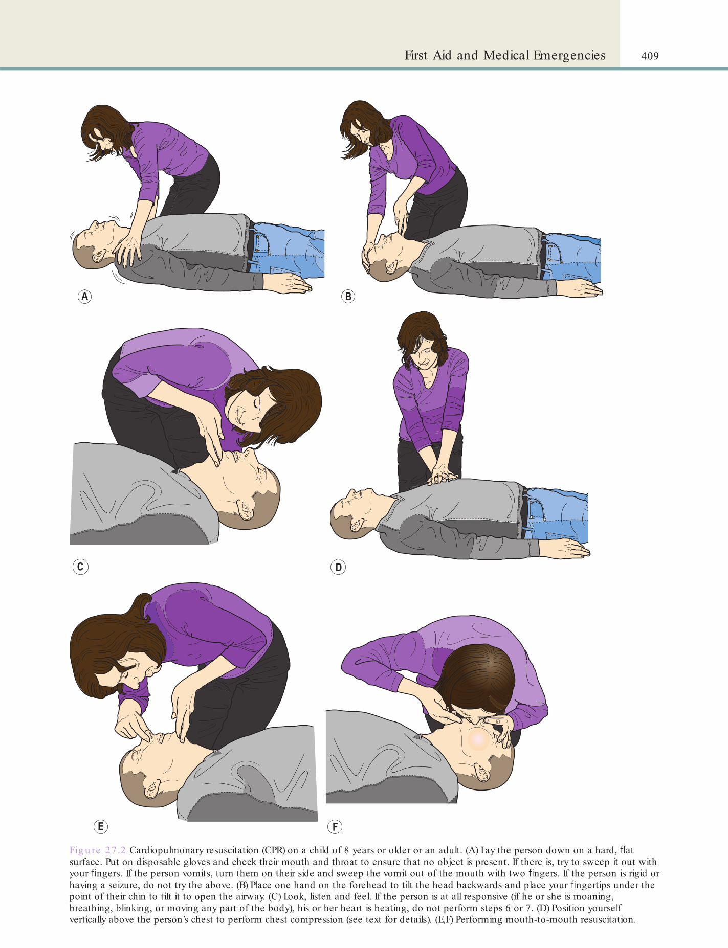
409First Aid and Medical Emergencies
Fig u re 27 .2 Cardiopulmonary resuscitation (CPR) on a child of 8 years or older or an adult. (A) Lay the person down on a hard, at surface. Put on disposable gloves and check their mouth and throat to ensure that no object is present. If there is, try to sweep it out with your ngers. If the person vomits, turn them on their side and sweep the vomit out of the mouth with two ngers. If the person is rigid or having a seizure, do not try the above. (B) Place one hand on the forehead to tilt the head backwards and place your ngertips under the point of their chin to tilt it to open the airway. (C) Look, listen and feel. If the person is at all responsive (if he or she is moaning, breathing, blinking, or moving any part of the body), his or her heart is beating, do not perform steps 6 or 7. (D) Position yourself vertically above the person’s chest to perform chest compression (see text for details). (E,F) Performing mouth-to-mouth resuscitation.
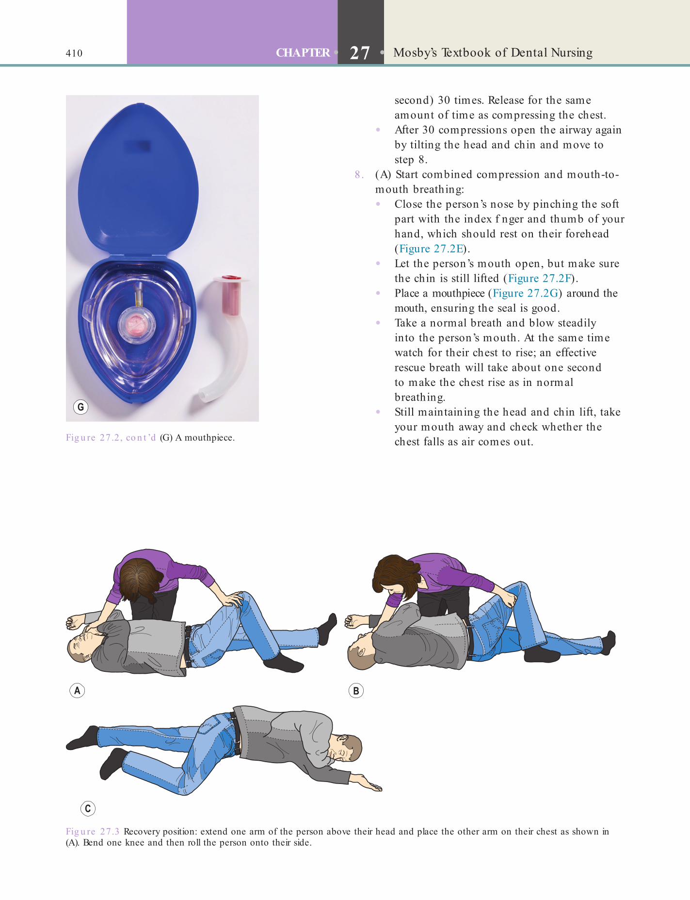
410 CHAPTER • 27 • Mosby’s Textbook of Dental Nursing
Fig u re 27.3 Recovery position: extend one arm of the person above their head and place the other arm on their chest as shown in (A). Bend one knee and then roll the person onto their side.
(G) A mouthpiece. Fig u re 27 .2, co n t ’d
second) 30 times. Release or the same amount o time as compressing the chest.
• A ter 30 compressions open the airway again by tilting the head and chin and move to step 8.
8 . (A) Start combined compression and mouth-to-mouth breathing:• Close the person’s nose by pinching the so t
part with the index f nger and thumb o your hand, which should rest on their orehead (Figure 27.2E).
• Let the person’s mouth open, but make sure the chin is still li ted (Figure 27.2F).
• Place a mouthpiece (Figure 27.2G) around the mouth, ensuring the seal is good.
• Take a normal breath and blow steadily into the person’s mouth. At the same time watch or their chest to rise; an e ective rescue breath will take about one second to make the chest rise as in normal breathing.
• Still maintaining the head and chin li t, take your mouth away and check whether the chest alls as air comes out.

411First Aid and Medical Emergencies
stopping the drug treatment. Unlike side e ects, which are usually known reactions that may occur, adverse drug reactions are not always predictable. They also do not always occur on f rst exposure to a drug. The clini-cal eatures o the reaction vary depending on the severity o the reaction. Very severe reactions involving collapse and breathing di f culties can result in anaphylaxis.
Local anaesthetics are the main cause o adverse drug reactions in dentistry. However, lidocaine ( or-merly called lignocaine), which is the most commonly used local anaesthetic in dentistry, is a very sa e drug i administered properly. But i too much is given, or i the drug is injected into a vein instead o the surrounding tissues, it may reach the brain, causing disorientation, agitation, or occasionally, collapse and f ts.
Prevention• Stick to recommended doses o local
anaesthetic.• Always use an aspirating syringe to give LA,
especially or in erior dental block injections.
Management• Stop administering the drug immediately i the
reaction occurs during that timeor
• Stop the procedure, lay the patient supine and give oxygen, reassuring them all the time.
• Summon medical help.Conscious sedation agents, such as benzodi-
azepines (e.g. midazolam), can severely depress the respiratory centre in the brain that controls breathing. Flumazenil is a drug that counteracts this reaction and should be given to patients having a respiratory arrest as a reaction to benzodiazepine administration.
General anaesthetics may cause particularly dan-gerous adverse reactions, particularly in the central nervous system (CNS).
• Repeat the process by taking a normal breath to give another e ective rescue breath, and without any delay per orm another 30 chest compressions, with your hands on the correct position on the sternum.
• Continue with chest compressions and rescue breaths in a ratio o 30 : 2.
• Do not stop unless the person starts breathing normally. However, you should not try to attempt more than two breaths be ore returning to chest compressions. I there is more than one rescuer present, switch between rescuers every 2 minutes to prevent atigue and or minimum delay during the changeover.*I the person’s chest does not rise when you give the rescue breath, be ore trying again, check:
• The person’s mouth again or any obstruction.
• There is adequate head tilt and chin li t.(B) Continuing chest only CPR. I you are not able to give rescue breaths, continue with the chest compressions at the rate mentioned in step 7, stopping only i the person starts to breathe normally.
9 . CPR should be continued until:• Qualif ed personnel arrive and take over• The person starts to breathe normally• You are too tired to continue.
CPR on c hildre nThe UK Resuscitation Council considers BLS or in ants and children (until puberty) separately. It recom-mends that in children:
• Lay rescuers should use a ratio o 30 compressions to two breaths.
• Two or more rescuers with a duty to respond should use a ratio o 15 compressions to two breaths.
Find out moreTo read more about BLS in ch ild ren and in ant s, visit the UK Resuscita t ion Council websit e (www.resus.org .uk).
Managing Speci c EmergenciesAdverse Drug ReactionsAdverse drug reactions are harm ul or unpleasant reac-tions to medicines that require altering or completely
Key poin tsAs a dental nurse, you should:
• Know where the emergency kit is in your workplace
• Know what drugs/items are in the emergency kit
• Keep the emergency kit up to date
• Know what to do in an emergency
• Keep up to date with resuscitation training.
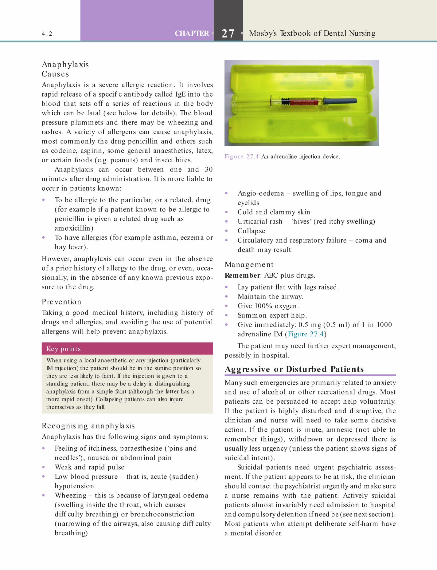
412 CHAPTER • 27 • Mosby’s Textbook of Dental Nursing
• Angio-oedema – swelling o lips, tongue and eyelids
• Cold and clammy skin• Urticarial rash – ‘hives’ (red itchy swelling)• Collapse• Circulatory and respiratory ailure – coma and
death may result.
ManagementRemember: ABC plus drugs.
• Lay patient at with legs raised.• Maintain the airway.• Give 100% oxygen.• Summon expert help.• Give immediately: 0.5 mg (0.5 ml) o 1 in 1000
adrenaline IM (Figure 27.4)The patient may need urther expert management,
possibly in hospital.
Aggressive or Disturbed PatientsMany such emergencies are primarily related to anxiety and use o alcohol or other recreational drugs. Most patients can be persuaded to accept help voluntarily. I the patient is highly disturbed and disruptive, the clinician and nurse will need to take some decisive action. I the patient is mute, amnesic (not able to remember things), withdrawn or depressed there is usually less urgency (unless the patient shows signs o suicidal intent).
Suicidal patients need urgent psychiatric assess-ment. I the patient appears to be at risk, the clinician should contact the psychiatrist urgently and make sure a nurse remains with the patient. Actively suicidal patients almost invariably need admission to hospital and compulsory detention i need be (see next section). Most patients who attempt deliberate sel -harm have a mental disorder.
AnaphylaxisCaus esAnaphylaxis is a severe allergic reaction. It involves rapid release o a specif c antibody called IgE into the blood that sets o a series o reactions in the body which can be atal (see below or details). The blood pressure plummets and there may be wheezing and rashes. A variety o allergens can cause anaphylaxis, most commonly the drug penicillin and others such as codeine, aspirin, some general anaesthetics, latex, or certain oods (e.g. peanuts) and insect bites.
Anaphylaxis can occur between one and 30 minutes a ter drug administration. It is more liable to occur in patients known:
• To be allergic to the particular, or a related, drug ( or example i a patient known to be allergic to penicillin is given a related drug such as amoxicillin)
• To have allergies ( or example asthma, eczema or hay ever).
However, anaphylaxis can occur even in the absence o a prior history o allergy to the drug, or even, occa-sionally, in the absence o any known previous expo-sure to the drug.
PreventionTaking a good medical history, including history o drugs and allergies, and avoiding the use o potential allergens will help prevent anaphylaxis.
Fig u re 27.4 An adrenaline injection device.
Key po in tsWhen using a local anaesthetic or any injection (particularly IM injection) the patient should be in the supine position so they are less likely to faint. If the injection is given to a standing patient, there may be a delay in distinguishing anaphylaxis from a simple faint (although the latter has a more rapid onset). Collapsing patients can also injure themselves as they fall.
Recognis ing anaphylaxisAnaphylaxis has the ollowing signs and symptoms:
• Feeling o itchiness, paraesthesiae (‘pins and needles’), nausea or abdominal pain
• Weak and rapid pulse• Low blood pressure – that is, acute (sudden)
hypotension• Wheezing – this is because o laryngeal oedema
(swelling inside the throat, which causes di f culty breathing) or bronchoconstriction (narrowing o the airways, also causing di f culty breathing)

413First Aid and Medical Emergencies
This may happen i the patient has a mental problem o such a nature or degree to warrant deten-tion under observation or at least a limited period. To be detained is in the interest o the patient’s own health or sa ety, and/or with a view to the protection o other people.
Alcohol-related problems do not quali y or sec-tioning under the Mental Health Act.
Hys te riaMany psychiatric emergencies lead to a crisis out o proportion to the patient’s actual problem. The stress o visiting a dentist can lead susceptible individuals to become hysterical. Symptoms such as overbreathing (hyperventilation), crying, etc. make the diagnosis obvious, but patients may still collapse.
ManagementBe calm and reassuring. Breathing with a paper bag around the mouth and nose may prevent respiratory alkalosis, but could also urther upset the patient. In unconscious patients, ollow the basic principles o ABC (see BLS, p. 408).
Term to lea rnCommon law : this refers to law that is derived from decisions taken by judges in courts and other tribunals, rather than through, for example, government legislation. In the UK, this includes judicial decisions made going back to ancient times.
Terms to learnDelusions: rm beliefs that the person hold to be true but actually are fanciful and not true.
Hallucinate: seeing or hearing imaginary things, for example people who are not really there.
Term to lea rnRespiratory alkalosis: when a person hyperventilates, that is breathes much more and much harder than the body requires, the alkalinity (pH of their blood) rises as too much carbon dioxide is lost too rapidly.
Dis turbe d or vio le nt pa tie ntsPatients with mental health problems such as schizo-phrenia or mania may be delusional or they may hal-lucinate or behave inappropriately. These patients are o ten incorrectly treated as i they had no intelligence or awareness o their surroundings. However, they may well understand what is being said to them, even though they may not respond; or they may respond inappropriately.
Management• Try to make contact with the patient.• Do not ask too many questions, but o er an
explanation o the situation.• Tell the patient where they are and what is
happening.I the patient is considered a danger to themselves
or others and re uses help, compulsory detention in a hospital will usually be required (called sectioning). The clinician will need to contact the patient’s psychia-trist, who is responsible or carrying out ormal sec-tioning procedures.
In an emergency, the clinician may have to act (detain patient) under common law. In the UK, under the Mental Health Act 1983, sectioning (detention ) o an out-patient (or sectioning an in-patient or more than 72 hours) requires the involvement o a psychia-trist. A patient who is in hospital or other reasons but is suddenly at danger because o a compromised mental state (e.g. postoperative delirium) can be detained or up to 72 hours against their will, ollow-ing the recommendation o the doctor in charge o their care (this may be a consultant).
Dis o rie nta te d o lde r pa tie ntsDisorientated older patients are best managed by having a close relative or other accompanying person constantly with them.
BleedingLi e-threatening bleeding (haemorrhage) rom dental treatment is rare, though implant placement or oral surgery may injure the blood vessels in the ace (e.g. lingual or other arteries). Patients who have under-gone a treatment called ‘radical neck dissection’ (e.g. those with oral cancer) may occasionally have sudden, li e-threatening haemorrhage. This occurs i the ap o skin covering the surgical wound in the neck gets in ected and the in ection spreads inwards and erodes the main neck artery (the carotid artery).
Manifes ta tions of bleeding• Rising pulse rate• Falling blood pressure• Collapse.

414 CHAPTER • 27 • Mosby’s Textbook of Dental Nursing
Recognis ing myocardia l infarction• Patient complains o central ‘crushing’ chest pain
that radiates to the arm, back, jaw.• They remain conscious but eel they are ‘about
to die’.• They are pale, sweaty, nauseated.• There is a rapid weak pulse and ast respiration.
Management• Ensure the airway is clear; remove any object
that may cause obstruction.• Sit the patient upright.• Summon expert help.• Give glyceryl trinitrate spray (or tablet)
sublingually – wait or 10 minutes only.• I patient does not improve, give oxygen and
aspirin (one 300 mg tablet o dispersible aspirin, to be crushed in the mouth).
• Remain with the patient, who must be admitted to hospital.
Management• Prevent urther blood loss by immediately
applying direct pressure over the wound or, i indicated, by suturing and/or packing the wound.
• Set up an IV in usion.
Term to lea rnVentricular brillation: when the heart beat becomes irregular due to rapid but uncoordinated contractions of the lower chambers of the heart (see Chapter 24).
Term to lea rnSublingual: a drug given sublingually is placed under the tongue, where it dissolves to enter the blood stream.
Term to lea rnIV infusion: also called IV drip, it involves giving of uids (drugs or blood or other uids) directly into a person’s veins.
Chest PainThere are many causes o chest pain, but severe pain raises the possibility o angina (partial block o blood supply to the heart muscle but without death o the muscle) or myocardial in arction (a ‘heart attack’– death o some heart muscle).
Angina• Central chest pain that may or may not radiate
to the arm.• Patients with angina remain conscious.• Usually there is a previous history o angina.• Pulse may be ast.
Management1 . Reassure and calm the patient.2 . Ensure the airway is clear; remove any object
that may cause obstruction.3 . Sit the patient upright.4 . Give GTN spray/tablet sublingually (two pu s o
spray or one tablet).I pain is not relieved rapidly, or i there is sweating, nausea, vomiting, breathlessness, arrhythmia, or loss o consciousness, it may be myocardial in arction. In this situation:
• Summon medical help.• Give oxygen.• Give the patient a 300 mg tablet o aspirin to
chew.
Myoc ard ia l infa rc tionMyocardial in arction (also called an ‘MI’, ‘heart attack’ or ‘coronary’ or ‘coronary thrombosis’) is the condi-tion in which the blood supply to the heart muscle gets blocked, causing part o the heart to die. It is a li e-threatening condition, and one o the main causes o death. People with a disease called arteriosclerosis and ischaemic or coronary heart disease are more prone to myocardial in arction.
Card iac a rre s tCardiac arrest is when the heart stops beating. The main sign o cardiac arrest is the absence o breathing in a non-responsive patient. The commonest cause o cardiac arrest, especially outside hospital, is ventricu-lar f brillation. This usually happens a ter a myocar-dial in arction and it can be li e-threatening. There ore urgent def brillation is required (see management below)
Recognis ing cardiac a rres tCardiac arrest has the ollowing eatures:
• Sudden loss o consciousness• Absent arterial pulses (carotid or emoral, which
is the pulse elt in the groin)• Gasping or absent respiration (a ter 15–30
seconds)• Pupils begin to dilate (a ter about 90 seconds).

415First Aid and Medical Emergencies
Use a ace mask (that is, deliver air with mouth-to-mask technique, or using an Ambu bag, ideally connected to an oxygen delivery device).
Identify and learnIdent i y a ace mask and Ambu bag in your workplace .6 . Check the carotid pulse in the side o the neck
( or up to 10 seconds):• I pulse is present, continue with 10–15
breaths per minute• I pulse is absent or inadequate, place the
patient on a f rm sur ace and start CPR (see p. 409).
Find out more• What is the procedure or de livering rescue
brea ths in BLS in your workplace?• Is there a def brilla to r? I yes, who is t ra ined to
use it?
CollapseA patient may collapse or one o many reasons. The commonest cause o collapse in a dental practice is ainting (see next section). There ore, where possible, patients should be treated in a at (supine) position, especially when giving injections or per orming surgery, because it prevents ainting.
I a patient collapses, lay them at, and reassure the patient and any accompanying person(s). Con-sider the cause; call or help i there is chest pain.
Fig u re 27.5 Giving the praecordial thump. Fig u re 27.6 A de brillator.
Term to lea rnAutomated external de brillator (AED): a machine that gives a controlled electric shock to the heart to restart or normalise the heart beat.
Key po in tAssessment of cardiac arrest should be done simultaneously with management to avoid losing any time.
ManagementThe immediate management o cardiac arrest is BLS (see p. 408), which aims to restore the ow o blood to the brain.1 . Approach the patient with care and shake the
patient gently and shout: ‘Are you all right?’2 . I the arrest is witnessed by the person providing
BLS and they are certain it is a cardiac arrest, they should give a praecordial thump (hit the sternum – centre o chest) (Figure 27.5).
3 . Call or assistance:• In general practice – rom another colleague
and an ambulance• In hospital – call the resuscitation team and
or a def brillator (Figure 27.6).
4 . Clear and maintain the airway5. Look, eel and listen or breathing (up to 10
seconds); give two e ective rescue breaths (up to f ve attempts, looking or rising o chest wall).

416 CHAPTER • 27 • Mosby’s Textbook of Dental Nursing
Find out moreWhat device is used o r expired a ir resp ira t ion in your workplace?
EpilepsyEpilepsy is the term used when a person has a seizure (a ‘f t’). There are several types o seizure or epilepsy. A major epileptic attack has several phases: it starts with the person getting a strange eeling (called an aura), ollowed by loss o consciousness, and then severe muscle spasm (tonic phase). The patient may stop breathing temporarily and become incontinent (that is, they may urinate or de ecate spontaneously) ollowed by repeated twitching and convulsions (clonic phase).
Management• Remove any objects in the mouth that could
obstruct the airway.• Protect the patient rom injury; move equipment
away.• Administer oxygen.• Wait or the seizure to stop spontaneously.
I the f t does not terminate within 5 minutes:
• Call or help and ambulance.• Start BLS (see p. 408).• During a second f t:
• Continue giving oxygen.• Prepare midazolam (a benzodiazepine drug)
or administration in the buccal sulcus/vestibule o the mouth.
• On third f t:• Give midazolam.
• When the f t stops, trans er to hospital.
FaintingFainting (syncope) is caused by a reduction o blood ow to the brain. It can be brought on by various stimuli (anxiety, pain, atigue, asting, high room tem-perature or even the sight o a needle – a common cause in dental practice). It particularly occurs in patients undergoing tooth extraction and may occur be ore or a ter the treatment.
Fainting may be prevented by:
• Ensuring the patient has eaten something be ore undergoing treatment under local anaesthesia
• Adopting a reassuring manner and not causing undue anxiety
• Laying the patient supine be ore giving any injections
Re s us c ita ting the c o llaps e d pa tie nt (ABC)Airway
• Check the airways are patent (clear).• Put the patient in the recovery position on their
side with ace down so that any uids etc. will run out o the mouth.
• Suck the airway clear and extend the neck.• Give oxygen.• I the airway is obstructed:
• Try to get the patient to cough.• Give an abdominal thrust (stand behind the
patient, put your arms around them, clench your hands together and pull upwards on the upper abdomen).
Brea thing• Listen and eel or breathing, while looking or
chest expansion. Give oxygen: i the patient is not breathing, give them 100% oxygen or use expired air respiration (but this only supplies less than 20% oxygen).
• Give rescue breaths.
Fig u re 27.7 Feeling the carotid pulse.
Terms to learnExpired air respiration: mouth-to-mouth resuscitation.
Carotid artery: the main artery (see Chapter 24) that carries the blood to the face and brain, passes through the side of the neck, where it can be palpated to check the pulse.
Circula tion
• Check the pulse at the carotid artery in the neck (Figure 27.7) and blood pressure with a sphygmomanometer.
• I there is no pulse, resuscitate as given above.• A doctor needs to treat the patient i they are in
shock (e.g. with an IV in usion).

417First Aid and Medical Emergencies
patient is suspected to be hypoglycaemic, their blood sugar level can be quickly tested by using a BM stick.
Find out moreLook or a BM st ick in your workplace and expla in how it is used .
Fe a ture s o f hypoglyc ae miaA hypoglycaemic patient may collapse but be ore-hand may:
• Show aggression and irritability• Have sweaty, warm skin• Have a rapid, bounding pulse• Be anxious and have tremors• Have tingling sensation around the mouth• Be con used and disoriented.
Management• Pre-empt the situation in patients with diabetes
(give them sugar or glucose).• I the patient does become hypoglycaemic, give
glucose, pre erably orally i the patient is not unconscious but otherwise intravenously. A sugar lump placed inside the mouth between the cheek and teeth (in the buccal sulcus), or a sugar solution or GlucoGel ( ormerly called Hypostop) can also be used. An IM injection o glucagon (a hormone) is another alternative (Figure 27.8).
• I the patient does not immediately recover, an ambulance/hospital trans er may be needed.
HypotensionHypotension is the term or low blood pressure. Rapidly bringing a patient upright rom lying down may cause a sudden all in their blood pressure and there ore a temporary reduction in the ow o blood to the brain. This is called postural hypotension. It particularly occurs a ter a person gets up suddenly a ter lying down or a long time. Older patients and
• Keeping instruments (particularly any sharps) out o the patient’s view
• Not causing the patient pain or anxiety.
Clin ic a l fe a ture s o f a fa in tThe patient may eel:
• Dizzy• Weak• Light-headed• Sick (nauseous)• Warm but the skin will be cool and sweaty
(moist or clammy).The patient may appear very pale, with a slow and thin pulse initially, which becomes aster later, and the patient may eventually lose consciousness.
Management• Immediately lay the patient at on the back with
the legs raised above the level o the head to increase the ow o blood to the heart and brain. I the ainting patient is not laid at, they may have a vaso-vagal attack. This involves slowing o the heart rate so that the blood ow to the brain reduces (called cerebral hypoxia), which leads to ainting; the patient can also have a convulsion.
• Loosen the patient’s collar.• Wait to see i they spontaneously recover (this
should only take a ew seconds).• I the patient does not immediately recover,
check the carotid pulse (see Figure 27.7):• I the pulse is slow, the patient may have had
a vaso-vagal attack. This may require an IV injection o a drug called atropine.
• I the pulse is absent, the patient is in cardiac arrest.
• I loss o consciousness occurs in the supine position or i the patient does not quickly resume consciousness when placed in the supine position, there could be other possible causes or the collapse that require urgent medical attention. There ore, call an ambulance.
HypoglycaemiaHypoglycaemia is the term used or low blood sugar (glucose). Hypoglycaemia is common in people with diabetes who have missed a meal or taken an overdose o insulin. It can also occur in anyone i too much alcohol is consumed.
A ter ainting, hypoglycaemia is the next most common cause o collapse in dental practice. I a Fig u re 27.8 A glucagon injection kit.
418 CHAPTER • 27 • Mosby’s Textbook of Dental Nursing
A relatively common error is to collect extracted teeth or tooth ragments in a swab and then by mistake use the same swab again in the patient’s mouth or haemostasis. This can trans er the removed teeth or tooth ragments back into the mouth.
Use o rubber dam in restorative dentistry (see Chapter 17) can reduce many o the mishaps that can lead to respiratory obstruction.
those taking certain medications such as antihyper-tensive drugs, tricyclic antidepressants or atropinics are particularly prone to this.
Key poin tOnly use clean swabs for postoperative haemostasis.
Term to lea rnObstructive airw ay disease: a kind of disease of the respiratory system in which the airways get obstructed, for example by over-production of mucus or constriction of the muscles in the walls of the airways. This causes dif culty breathing. The two forms of COAD are chronic bronchitis and emphysema.
Key poin tRemember that in late pregnancy, pressure from the uterus inhibits blood ow back to the heart. This happens because the main vessel that takes blood back to the heart (the inferior vena cava) is compressed when the patient lies down (this is called supine hypotensive syndrome). Therefore, women in late pregnancy should be positioned slightly sideways in the dental chair.
Terms to learnAntihypertensive drugs: drugs taken to reduce blood pressure.
Tricyclic antidepressants: one of the kinds of drugs used for the treatment of depression.
Term to lea rnHaemostasis: the process by which bleeding is stopped, for example by applying pressure on the bleeding site.
Respiratory ObstructionI breathing is obstructed in any way, it can rapidly starve the brain o oxygen (called cerebral hypoxia). I the breathing is obstructed it usually causes choking and, i or more than about 3 minutes, it can cause brain damage or death rom lack o oxygen (hypoxia). Respiratory obstruction in the dental surgery is mainly caused by:
• Mechanical obstruction o the airways by an object ( oreign body)
• Pressure on the airways• Bronchospasm (narrowing o the airways) in a
patient with chronic obstructive airway disease (COAD) – also termed chronic obstructive pulmonary disease (COPD) .
Caus es and preventionUn ortunately, despite sa ety recommendations during dental treatment, inlays, crowns, endodontic instru-ments and extracted teeth or roots or other small items continue to be inadvertently dropped into the airway (Chapter 23).
Identify and learnIdent i y the various part s o a rubber dam used in your workplace .
Management of objects tha t cannot be accounted forI a lost item such as a crown, an extracted tooth, or a broken bur cannot be ound lying ree in the mouth, it could be:
• Within the tissues o the mouth ( or example, inside the mass o the cheek)
• Somewhere in the respiratory system (the throat, the airways or the lungs)
• In the digestive tract, or it may have allen outside the body.In this situation:
• Check the mouth and throat care ully.• Check the area around the patient.• Check in the suction tubing and container.
I the object is within the throat and the patient is conscious, encourage them to cough out the object. Do not per orm a blind f nger sweep o the throat, as this may push the oreign body urther into the throat.
Young children should be held upside down. In other children, back blows or chest thrusts may assist in retrieving the object. Older children and adults may benef t rom the abdominal thrust.
I the object cannot be ound, X-rays o the neck, chest and abdomen are required. Depending where the object is ound it may need to be removed by endoscopy or open operation, unless the doctor pre-dicts it will pass in the aeces.
419First Aid and Medical Emergencies
Features• Rapidly altered level o consciousness or
cognition (mental status).• Motor def cits – that is, paralysis o one side o
the ace ( acial palsy), and either one or more limbs, and inability to speak due to tongue muscles being a ected.
• Sensory def cits – that is, loss o sensation, e.g. numbness in the f ngers.
• Airways, breathing and circulation are usually intact.
Management• Call an ambulance.• Place the patient in the recovery position.• Give oxygen.
As thmaIn asthma the airways become hyper-reactive and tend to swell up in response to a variety o stimuli, some-times allergic. This causes narrowing o the airways, di f culty breathing and hypoxia.
An asthma attack can sometimes be triggered by anxiety, or by an inhaled drug or one that is circulating in the blood. It may also be a mani estation o anaphylaxis.
Recognis ing as thmaThe patient may:
• Be breathless• Have a wheeze• Have a cough• Have di f culty speaking• Feel panic.
ManagementMild as thma• Use a salbutamol (Ventolin) inhaler.• Give oxygen.• Sit the patient upright.• Repeat the inhaler as required.• I the response within 10 minutes is poor – call
an ambulance and trans er to hospital.
Severe as thma• Call ambulance immediately.• Use the salbutamol inhaler.• Give oxygen.• Sit the patient upright.• Trans er to hospital.
Key poin tDo not slap the back of a patient sitting upright, as this might actually cause the object to fall further into the respiratory tract. However, if the obstruction is complete (e.g. the object is lying between vocal cords), slapping the back may partially free the airway.
Term to lea rnAtherosclerosis: the accumulation of fats (cholesterol and lipids) in the walls of the arteries.
Key po in tFAST – Face, Arm, Speech, Time to call the emergency services – is a simple test to help recognise the signs of stroke and understand the importance of emergency treatment. The faster a stroke patient receives treatment, the better their chances are of survival and of lesser long-term disability.
StrokeStroke may cause collapse, acial and limb paralysis on one side, and speech di f culties. Also called cerebral vascular accident (CVA) or event (CVE), stroke is more common in older people and usually occurs when a blood vessel in the brain bursts or gets blocked and causes brain damage. Atherosclerosis is the main cause o strokes, but hypertension during a stress ul situation (e.g. anxiety about dental treatment) may also lead to a stroke.

This page intentiona lly le ft blank

Abbrevia t ions
A&E accident and emergencyABC antibiotic coverACJ amelocemental junctionADA American Dental AssociationADAM Association o Dental Administrators
and ManagersADHD attention def cit hyperactivity disorderADJ amelodentinal junctionADT admissions, discharges and trans ersAED automated electronic def brillatorAF atrial f brillationAHF anti-haemophiliac actorAHP allied health pro ession(al)AIDS acquired immune def ciency
syndromeAKA also known asALARA as low as reasonably achievableALARP as low as reasonably practicableALL acute lymphocytic leukaemiaALS advanced li e supporta.m. in the morningAMA against medical adviceAMHP approved mental health pro essionalAML acute myeloid leukaemiaANK appointment not keptANS anterior nasal spineANUG acute necrotising ulcerative gingivitisAP aggressive periodontitisAP anteroposteriorAPF acidulated phosphate uorideAPL accreditation o prior learningAR autosomal recessiveARC AIDS-related complexARF acute renal ailureARF Annual Retention Fee (GDC)ART atraumatic restorative techniqueATC around the clockATLS advanced trauma li e supportATS anti-tetanus serumAUG acute ulcerative gingivitis
B/F black emaleB/M black male
BA Bachelor o ArtsBADN British Association o Dental NursesBADT British Association o Dental TherapistsBAOMS British Association o Oral and
Maxillo acial SurgeonsBASCD British Association or the Study o
Community DentistryBBV blood-borne virusbcc blind copyBChD Bachelor o Dental SurgeryBDA British Dental AssociationBdentSci Bachelor o Dental ScienceBDHA British Dental Hygienists AssociationBDHF British Dental Health FoundationBDJ British Dental JournalBDS Bachelor o Dental SurgeryBDSc Bachelor o Dental ScienceBDTA British Dental Trade Associationb.i.d. (or bd) twice a dayBIPP bismuth, iodo orm, para f n pasteb.i.w. twice a weekBLS basic li e supportBMA British Medical AssociationBME black and minority ethnicBMS burning mouth syndromeBMT bone marrow transplantationBNF British National FormularyBOP bleeding on probingBOS British Orthodontic SocietyBP blood pressureBP British PharmacopoeiaBPE basic periodontal examinationBRA bite raising applianceBRONJ bisphosphonate related osteonecrosis
o jawBSDH British Society o Disability and
Oral HealthBSDHT British Society o Dental Hygiene
and TherapyBSE bovine spongi orm encephalopathyBSL British Sign LanguageBSODR British Society or Oral and
Dental Research

Abbrevia t ions
422
BSOM British Society o Oral MedicineBSOMP British Society o Oral and
Maxillo acial PathologyBSP British Society o PeriodontologyBW bitewingBx biopsyBZP benzodiazepine
°C degree Celsius – unit used to measure temperature
c withCA carcinomaCa(OH)2 calcium hydroxideCAB/C&B choose and bookCABG coronary artery bypass gra tCAD coronary artery diseaseCADCAM computer-assisted design, computer-
assisted manu actureCADO Chie Administrative Dental O f cerCAL clinical attachment lossCAL computer-assisted learningCAMHS Child and Adolescent Mental
Health ServicesCAPD continued ambulatory peritoneal
dialysisCAT computerised axial tomography (scan)CBC complete blood count excluding red
cell countCBCT cone beam computed tomographyCBT cognitive behavioural therapycc carbon copycc chie complaintcc cubic centimetreCCF congestive cardiac ailureCCG clinical commissioning groupCCT Certif cate o Completion o TrainingCCU critical care unitCCU coronary care unitCD clinical directorCD computer discCDO Chie Dental O f cerCDS community dental servicesCDT clinical dental technician
CDU central decontamination unitCE chie executiveCEJ cementoenamel junctionCEO chie executive o f cerCeph cephalogramCG Caldicott GuardianCG clinical governanceCHM Commission on Human Medicines CHx chlorhexidineCIT clinical in ormation technologyCJD Creutz eldt–Jakob diseaseCKD chronic kidney diseaseCLL chronic lymphatic leukaemiaCLP cle t-lip palateCML chronic myeloid leukaemiaCMO Chie Medical O f cerCMV cytomegalovirusCNO Chie Nursing O f cerCNS central nervous systemC/O complaining o COAD chronic obstructive airways diseaseCoCr cobalt chromiumCOPD chronic obstructive pulmonary
diseaseCOSHH Control o Substances Hazardous
to HealthCPD continuing pro essional developmentCPITN Community Periodontal Index o
Treatment NeedCPR cardiopulmonary resuscitationCR conventional resinCRD conventional resin; dual curedCRL conventional resin; light curedCQC Care Quality CommissionCQUIN Commissioning or Quality and
InnovationCRB Criminal Records Bureau (now DBS)CSSD central sterile supplies departmentCT clinical tutorCT computerised axial tomography scanCV curriculum vitaeCVA cerebrovascular accidentCXR chest X-ray (radiograph)

423
DAC Dental Access CentredB decibelsDBA dentine bonding agentDBS Disclosure and Barring ServiceDCP dental care pro essionalDDA Disability Discrimination ActDDO District Dental O f cerDDU Dental De ence UnionDGH District General HospitalDH dental healthDH dental historyDH dental hygienistDHE dental health educationDIGO drug-induced gingival
overgrowthDip in Cons Sed Diploma in Conscious SedationDMFS decayed, missing or f lled sur acesDMFT decayed, missing or f lled teethDMT decayed, missing teethDN dental nurseDoH Department o HealthDP dental pro essionalDPA Data Protection ActDPA Dental Pro essionals AssociationDPB Dental Practice Board (now NHS
Business Services Authority)DPF Dental Practitioners’ FormularyDPL Dental Protection LimitedDPR Data Protection RegistrarDPT dental panoramic tomogram
(radiograph)DRAB dangers, response, airway, breathing,
circulationDRD Diploma in Restorative DentistryDSA dental surgery assistant (now dental
nurse)DSU day surgery unitDT dental technicianDT dental therapistDTA Dental Technologists AssociationDVT deep vein thrombosisDx diagnosisDXR deep X-ray radiation (radiotherapy)
EBD evidence-based dentistryEBL evidence-based learningECG electrocardiogramECT electroconvulsive therapyE&D equality and diversityEDC (NHS) Equality and Diversity CouncilEDH Enrolled Dental HygienistEDT Enrolled Dental TherapistEDTA ethylenediaminetetraacetateEEG electroencephalogramEHRC Equality and Human Rights Commission
( ormerly CRE)EMQ extended matching questionsENT ear, nose and throatEO extra-oralEPP exposure prone procedureEPR electronic patient record
°F degree Fahrenheit – unit used to measure temperature
FAQ requently asked questionsFB oreign bodyFBC ull blood countFDS Faculty o Dental SurgeryFDS Fellow in Dental SurgeryFGC ull gold crownFGDP Faculty o General Dental PractitionersFH amily historyFNAC f ne needle aspiration cytologyFNP Family Nurse PractitionerFOI reedom o in ormationFPD f xed partial dentureFWS reeway spaceFYI or your in ormation
GA general anaesthesiaGCP good clinical practiceGCS Glasgow Coma ScaleGCSE General Certif cate o Secondary
EducationGDC General Dental CouncilGDP general dental practitionerGDS general dental services

Abbrevia t ions
424
GI gastrointestinalGI gingival indexGIC glass ionomer cementGIT gastrointestinal tractgm gramGMC General Medical CouncilGMH general medical historyGMP General Medical PractitionerGP General PractitionerGTN glyceryl trinitrateGTR guided tissue regenerationGTT glucose tolerance test
H&N head and neckh hour (also hr)HA health authorityHAART highly active anti-retroviral
therapyHAV hepatitis A virusHb haemoglobinHBcAg hepatitis B core antigenHBeAg hepatitis B ‘e’ antigenHbS haemoglobin S (sickle-cell)HBsAg hepatitis B sur ace antigenHBV hepatitis B virusHCA health care assistantHCAI healthcare associated in ectionHCV hepatitis C virusHCW healthcare workerHDU high dependency unitHDV hepatitis D virusHep A hepatitis AHep B hepatitis BHep C hepatitis Chgt heightHHV human herpes virusHIV human immunodef ciency virusHPT health protection teamHPV human papilloma virusHR human resourceshr hour (also h)HSE Health and Sa ety ExecutiveHSV herpes simplex virus
HSW Health and Sa ety at WorkHx history
I&D incision and drainageI&O intake and outputIA intra-arterialICT in ormation and communication
technologyICU intensive care unitID identityID in erior dental (nerve)IDB in erior dental (nerve) block LAIDD insulin-dependent diabetesIDG International Dental GraduateIDN in erior dental nerveIFN inter eronIFP idiopathic acial painIg immunoglobulin (e.g. IgG, IgA, IgM,
IgE)IG in ormation governanceIM intramuscularIMF intermaxillary f xationIO intra-oralIOTN Index o Orthodontic Treatment NeedsIP inpatientIPA isopropylalcoholIRM intermediate restorative materialIRMER Ionising Radiation (Medical Exposure)
RegulationsIRR Ionising Radiations RegulationsIRT intermediate restorative treatmentISO International Organisation or
StandardisationIT in ormation technologyITU intensive treatment unitIV intravenousIVD intravenous drug misuse
jt joint
K potassiumkg kilogramKPI Key Per ormance Indicators

425
L or l litreLA local anaesthesia or analgesialab laboratoryLDS Licence/Licentiate in Dental SurgeryLEA local education authorityLIED latex, iodine, elastoplast, drugsLLSD lower labial set downLP lichen planusLP lumbar punctureLSAB Local Sa eguarding Adults BoardLSCB Local Sa eguarding Children BoardLt le t
m metreMA Master o Artsmax maximalMCA Mental Capacity ActMCQ multiple choice questionsMD Doctor o MedicineMDS Master in Dental SurgeryMDU Medical De ence UnionMFDS Member o the Faculty o
Dental SurgeryMFGDP (UK) Membership o the Faculty o General
Dental Practitioners (UK)MGDS Membership in General Dental
Surgerymg milligramMH medical historyMHA Mental Health ActMI myocardial in arct or mitral
incompetenceMID minimally invasive dentistryMMR measles, mumps and rubella
(vaccine)min minuteMPD mandibular pain–dys unctionMPS Medical Protection SocietyMRCS Member o the Royal College o
SurgeonsMRSA methicillin (meticillin)-resistant
Staphylococcus aureusMS multiple sclerosis
NAD no abnormality detectedNBM nil by mouthNDAC National Dental Advisory CommitteeNEBDN National Examining Board or
Dental Nursesneg negativeNGVB night guard vital bleachingNHD notional hal dayNHL non-Hodgkin’s lymphomaNHS National Health ServiceNHSBSA NHS Business Services AuthorityNHST NHS TrustNICE National Institute or Health and Care
( ormerly Clinical) ExcellenceNiTi nickel titaniumNME non-milk extrinsic sugarsNPO nothing by mouth (per oram)NSAIDs non-steroidal anti-in ammatory drugsNtg nitroglycerinNTN National Training NumberNTN no treatment neededNTR no treatment requiredNV next visit
O orallyOA osteo-arthritisOAF oro-antral f stulaOB overbiteObs observationsOfsted O f ce or Standards in EducationOH oral hygiene/healthOHA occupational health adviserOHE oral health educationOHI oral health/hygiene instructionOJ overjetOM occipitomental (radiograph)OMFS oral and maxillo acial surgeryOP outpatientOPD outpatient departmentOPG orthopantomograph (radiograph)OPT orthopantomograph (radiograph)ORF orthograde root f llingOSCC oral squamous cell carcinoma

Abbrevia t ions
426
OSCE observed structured clinical evaluation
OT orthodontic therapistOTC over-the-counterOVD occlusal vertical dimension
P pulsePA periapicalPA personal assistantPA posteroanterior view (radiograph)PALS patient advice and liaison servicesPAS patient administration systemPBC porcelain bonded crownPC personal computerPC politically correctPC polycarbonatePCA patient cancelled appointmentPCA patient controlled analgesiaPCD pro essionals complementary to
dentistry (now DCPs)PCR polymerase chain reactionPCT Primary Care Trustpdf portable document ormatPDH past dental historyPDP pro essional development port olioPDP personal development planPE pulmonary embolismPEP post-exposure prophylaxisPFG porcelain used to goldPFM porcelain used to metalPG postgraduatePGC porcelain to gold crownPH past historyPHE Public Health EnglandPI Plaque IndexPIDA Personal Interest Disclosure ActPIN Personal Identif cation NumberPJC porcelain jacket crownPLV porcelain laminated veneerPM practice managerPMH past medical historyPNS posterior nasal spinePO postoperative or per oram
POC polythene occlusal coverPOM prescription only medicinepos positivePP private practice/patientPPD pocket probing depthPPE personal protective equipmentppm parts per millionPROMS patient-related outcome measuresPSA pleomorphic salivary adenomaPT physiotherapyPT prothrombin timePV porcelain veneer
q.i.d. (or qd) our times a dayQOL quality o li e
RA relative analgesiaRA rheumatoid arthritisRBC red blood cell countRCPE Royal College o Physicians
o EdinburghRCS Royal College o SurgeonsRCT randomised controlled trialRCT root canal treatmentRED resin-based (a)esthetic dentistryRIDDOR Reporting Injuries, Disease and
Dangerous Occurrences RegulationsRMGIC resin modif ed glass ionomer cementRMH relevant medical historyRoE review o experienceRPA radiation protection advisorRPS radiation protection supervisorRRF retrograde root f llingRSD root scaling and debridementRT radiation therapyRt rightRTA road tra f c accidentRTT re erral to treatmentRVG radiovisiographyRx treatment
SC subcutaneousSES socio-economic status

427
SH social historySLA Service Level AgreementSLS sodium lauryl sul ateSMV submentovertex (radiograph)SpR Specialist RegistrarSRN Sta Registered NurseSS sagittal split (osteotomy)SS stainless steelSS systemic sclerosisSS Sjögren syndromeSSI sexually shared in ectionsSTD sexually transmitted diseasesSTI sexually transmitted in ectionStR Specialist Trainee RegistrarSUBQ subcutaneousSUI signif cant unexpected incidentsSWOT strengths, weaknesses, opportunities,
threats (analysis)
TB tuberculosisTBC to be conf rmedTIA transient ischaemic attackt.i.d. (or tdd) three times a dayTLC tender loving careTMD temporomandibular dys unctionTMJ temporomandibular joint
TMPD temporomandibular joint pain dys unction
TN trigeminal neuralgiaTNM tumour, node, metastasisTPR temperature, pulse and respirationTSL tooth sur ace lossTSSU theatre sterile supplies unit
U&E urea and electrolytesURI upper respiratory in ectionURTI upper respiratory tract in ectionUS ultrasonographyUSB universal serial bus
vCJD variant Creutz eldt–Jakob diseaseVS vital signs
WBC white blood countWCC white cell countW-D washer-disin ectorWHO World Health OrganisationWSL white spot lesionWTD Working-Time Directive
ZOE zinc oxide eugenolZP zinc phosphate

This page intentiona lly le ft blank

Index
AAbrasion, tooth, 150–151, 150f, 186t
causes of, 192Abscess, 148b, 149f, 340t, 343, 343f
and caries, 148management of, 344bperiapical, 151–152, 151f–152fperiodontal, acute, 344
Abusein children, recognition of, 323physical, 321
Access to Medical Records Act 1991, 54Accidents, 68–70, 68b–69b
legislation applying to, 68minimising of, in dental surgery and
laboratory, 66bnotifying and recording of, 68–69, 69b
Acid bath, 65bAcid etchant, 201bAcidosis, 307bAcquired diseases, 370Acquired immune de ciency syndrome. see
Human immunode ciency virus (HIV)/acquired immune de ciency syndrome (AIDS)
Acrylic crowns, 274fAcrylic resin, 247Acrylic trimmer, 264fActinomyces, 144t, 146Active ingredient, de nition of, 222bAcute healthcare, 43bAcute necrotising ulcerative gingivitis
(ANUG), 154Addition silicones, 245t, 246Admissions, of patient, 301
routine (non-urgent), 300b, 301urgent, 301
Adrenaline, 228, 366injection device, 412f
Adults and elders, vulnerable, 324–325, 325b
action for suspected abuse, 325Adults with Incapacity (Scotland) Act 2000,
46Advanced trauma life support (ATLS),
340Adverse drug reactions, 411–412
management of, 411, 411bprevention of, 411
Adverse effects, drugsof antimicrobials, 219of antiseptics and decontaminant agents,
93of benzodiazepines, 237
Aesthetic component, 312, 312bAggressive patients, managing, 412–413AH26, 252AIDS. see Human immunode ciency virus
(HIV)/acquired immune de ciency syndrome (AIDS)
Air gap, type A, 106b
Airwayin resuscitating collapsed patient,
416small items dropped into, 418
ALARA principles, 81Alcohol, 395, 395b
abuse, people with, 337–338acute effects of, 370tconsumption, 202maximum drinking amounts of,
recommended, 395bmisuse, 94b
alcoholism, 370–371mouthwashes containing, 194possible systemic effects of, 395tand six ‘Ls’, 370b
Alcohol handrubs, 94risk management of, 94t
Alginate impression material (‘Alginate’), 243–244, 245t
advantages of, 244disadvantages of, 244uses of, 244
Allergies, 70–72, 70b, 70t, 371, 371bcommon allergens, 371tdental materials and, 253, 253bDuraphat and, 199to latex, 70–72, 71b–72b
Allograft, 276Alpha radiation, 165Alveolar nerve, inferior, 130, 132fAlveolar osteitis, 161Alveolar processes, anatomy of,
124–125Alzheimer disease, 378Amalgam, 247–250, 269b
bonding, 250bdisposal of, 84, 84bsafety issues related to, 249–250
Amalgam capsules, 271fAmalgam carriers, 269, 271fAmalgam pot, glass, 271fAmalgam restorations, 269
amalgam tray and, 270b, 271f–272fmoisture, damaging effects of, 264tplacement of amalgam lling, 269, 269b,
270f–271fAmalgamator, 269Ameloblasts, 136, 136fAmelo-cemental junction, 142Amelogenesis imperfecta, 156American Society of Anesthesiologists (ASA),
state of health classi cation, 369tAmnesia, after IV sedation, 238–239Amorphous calcium phosphate (ACP),
remineralisation and, 199Amoxicillin, 218Amphetamines, 395Anaemia, 355, 355b, 371–372, 372b
causes of, 372tmanagement of, 372
Anaesthesia/anaestheticsde nition of, 225, 227bgases used in, 235see also General anaesthesia; Local
anaesthesiaAnalgesia/analgesics, 152b, 226–227
de nition of analgesia, 227bfor pulpitis, 152brelative, 234
Anaphylaxis, 412causes of, 412management of, 412prevention of, 412, 412brecognising, 412
Anatomy and physiology, 121–142circulatory system, 353–357, 358fde nitions, 121dental and oral
alveolar processes, 124–125face, 121, 123f–124f
nerve supply to, 128–130, 130t, 134fjaws, 122–125lips, 132–133
nerve supply of, 133mandible, 124, 125f
nerve supply to, 132fmaxilla, 123
nerve supply to, 132fmuscles of mastication, 126, 126b,
127f–128foral cavity, 130–134
nerve supply to, 128–130, 134fpalate, 134, 134b
nerve supply of, 134salivary glands, 126–128, 129f
nerve supply of, 127–128, 128bskull, 121temporomandibular joints (TMJs),
121b, 125, 125b, 126ftongue, 132f–133f, 133–134
blood supply to, 134nerve supply of, 134
digestive system, 361–362, 361fendocrine system, 366immune system, 366–367integumentary system, 363lymphatic system, 358, 360fmusculoskeletal system, 363–365, 364fnervous system, 365–366planes, 354f, 354trespiratory system, 358–361, 361fstructure of body, 351–353
cells, 351–353, 352forgans, 352t, 353, 353fsystems, 353tissues, 352t, 353, 353f
terms used in, 122t, 353, 354turinary system, 363, 363f
Angina, 414management of, 414
Angiography, 170b
Page numbers followed by ‘f’ indicate gures, ‘t’ indicate tables, and ‘b’ indicate boxes.

Index
430
Angio-oedema, 371fAngle classi cation of malocclusion,
310b–311bAngled toothbrushes, 190bAngular cheilitis, 158, 158bAnnual transfer note, 86Anodontia, 137Antagonist, de nition of, 237bAnteroposterior discrepancies, malocclusion,
310Antibacterials, 218Antibiotics, 152b
for pulpitis, 152bAnticoagulant, related problems of, 372,
372bAntifungals, 218–219Antihypertensive drugs, 418bAntimicrobials, 218–219, 218b
adverse effects of, 219drug interactions and, 219foods and, 219
Anti-retroviral drugs, 110bAntiseptics or decontaminating agents,
92–93, 93f, 93tAntivirals, 219Anxiety, 372–373
control, 225–241, 225banxiolytic, 233dental nurse role in, 225–226drug use precautions, 240–241, 241bhypnosis, 240for special situations, 240–241see also Sedation
dental, people with, 330heart rate, heart beat and, 357btypes of, 372b
Anxiolytic, de nition of, 233bAorta, 356bApical foramen, 135Apical periodontitis, 148Apicectomy. see Root end surgeryApicoectomy. see Root end surgeryAppliances
custom-made, 58decontamination of, 105 xed, 313–315, 313b, 313f–315ffunctional, 313removable, 313, 315–316, 316f
care of, 280bAppointed person, in rst aid, 402Appointments, 38–40
diagnosis, patient care during, 39–40hazards and risks of healthcare, 38–39history taking, 39–40
family, 40medical, 39–40
relevant, 40personal details, 39presenting complaint in, 40social, 40
investigations, 40organising, 212
Approved Codes of Practice, 116–117Apron, disposable plastic, 97–98
Argon lasers, 77Arrhythmia, 76bArthritis, 373Arthrography, 170bArticaine, 228, 229tArticular disc, 125Articulation, of bones, 121bArticulation strips, 278fASA. see American Society of
Anesthesiologists (ASA)Aspirator, 28bAspirin, 226, 226b
in children, 241Assaults, 72Assisting in surgery, 287–288, 287bAsthma, 373–374, 419
extrinsic, 374tintrinsic, 374tmild, 419recognising, 419severe, 419
Asymmetry, malocclusion, 310Asymptomatic condition, 155bAtheroma, 374Atherosclerosis, 419b
see also AtheromaAt-home whitening, 220Atraumatic restorative technique (ART),
251bAtropinics, 418bAttrition, tooth, 149–150, 150fAtypical facial pain, 162Atypical symptomatology, 324Audit, 49, 49b, 49fAutism, 374Autoclave tape, 104, 104fAutograft, 276Automated external de brillator (AED),
415bAutonomic nervous system, 366Autonomy, 47bAvulsion, tooth, 340t
BBacteria
dental caries from, 146–147infections of, 89–90
Bad news, breaking, 32Bariatric problems, people with, 338Basic life support (BLS), 408–411, 408b
sequence, adult, 408–411, 409f–410fsummary of, 408b
Basic periodontal examination (BPE), 208examination codes, 210tprobes, 210f
Basic Safety Standards (BSS) Directive, 178Bass technique, modi ed, of toothbrushing,
191tBDA. see British Dental Association (BDA)Bee bee scissors, 274fBehaviour, patient’s con dence and, 6Bell’s palsy, 374, 374fBenign lesion, 159bBenign migratory glossitis, 159
Benzocaine, 227Benzodiazepines (BZPs), 233
antagonist, 237intranasal, 239respiratory arrest and, 411unpredictable effect of, 239
Beryllium, 252–253Best interest, principle of, 326, 326bBest practice requirements
infection control, 100Legionella infection control, 106b
Beta radiation, 165Betel, 395
possible systemic effects of, 395tquid chewing, 202
Bichard Inquiry, 43Biocompatible material, de nition of, 252b‘Bio lm’, 143Biological feedback, 377bBiopsy, 107b
specimens, sent through the post, 107–108
Bipolar diathermy, 305bBisphosphonates, 161b, 218bBite registration instruments and materials,
277fBites, injuries in, 343, 343bBitewing radiographs, 167, 168f
radiation dose, 168–170use of, 169t
Black and minority ethnic (BME) backgrounds, people from, 335–337, 335b, 337b
Bleachingexternal, 220, 220b
problems with, 220internal, 220laser, 220
Bleeding, 413–414management of, 414manifestations of, 413postoperative, 344, 345b
Blood, 354–356blood products, 219cells, 355
production of, 355 ow of, 356–357, 356bloss, 346
Blood coagulation factors, 356Blood spillages, 107Blood vessels, 356Blood-borne infections, and drug abuse,
379bBlue halogen light, 78
source of, 272fBody uid spillages, 107Bolam test, 46Bolus, 126bBonding, 283tBones, 363–364, 364b
repair of, 364bstructure of, 364fsurgery of, 290–295thinning. see Osteoporosis

431
Bones grafting, 276Bones scintiscanning, 170bBorder moulding, de nition of, 279bBottle caries, 148BPE. see Basic periodontal examination (BPE)‘Braces’, 313, 313fBreach loading, in syringes, 229Breast cancer, 375Breast-feeding, 196Breathing, in resuscitating collapsed patient,
416Bridges, 275
with abutment teeth, 273fconventional, 275resin-retained, 275
Bristle brush, 260fBritish Association of Dental Nurses
(BADN), 22British Dental Association (BDA), 7t
staff security and, 81–82British Dental Nurses Association, 7tBritish Sign Language (BSL), 329b
interpretation of, 34British Standards, 107bBrittle bone disease. see OsteoporosisBronchi, 360–361, 361bBuccal surface, de nition of, 204bBuddhism, 336tBurns, 72–73, 252Burs, 117b, 265, 266fButte y clip, 270f
CCAD-CAM (computer-aided design and
computer-aided manufacturing) dentistry, 262
de nition of, 262bCaecum, 362Calcium, 364Calcium hydroxide cements, 248tCalculus (tartar), 153bCaldicott principles, 52, 54, 54bCancer, 353b
breast, 375lung, 384mouth, 158–159, 159b
clinical signs of, 159bmanagement of, 159prevention of, 201, 201b
people with, 331–332Candida albicans, 157–158Candidiasis. see CandidosisCandidosis, 128, 157–158
in HIV/AIDS, 382fCannabis (marijuana), 396
use of, 375, 375bCarbamazepine (Tegretol), 162Carbamide peroxide, 219Carbohydrates, 363b
fermentable, dental caries from, 147, 147b, 147t
Carbon dioxide re extinguisher, 75, 75flasers, 77, 290
Cardiac arrest, 414–415management of, 415, 415brecognising, 414, 415b
Cardiac disease, 375, 375bCardiopulmonary resuscitation (CPR), 408,
409f–410fin children, 411, 411bnurse’s role, 27see also Chest compression
Care Commission, 42Care pathways (clinical guidelines), 57–58,
58bCare Quality Commission, 42, 48–49Care Standards Act 2000, lasers and, 77,
77fCaries (tooth decay), 144t, 145–152, 145f
classi cation of, 150f, 150tdiagnosis of, 149factors protective of, 147–148gross, 323bpain and, 149b, 149fparticular patterns of, 148, 148fpathology of, 146–147, 146t, 147fpoor oral hygiene showing, 186fremoval of, 264treatment of, 149untreated, consequences of, 148
Cariogenic, de nition of, 147bCariogenic foods/drinks, 196–197, 197bCariogenic sugars, 147tCarotid artery, 416bCarotid pulse, 416fCarpal tunnel syndrome, 79Cartilage, 364Case study, 57Case-control studies, 57Casting equipment, 65bCatheter, 301bCavity, preparation of, in dental restoration,
265–266, 266f–268fCells, 351–353, 352fCellulitis, 340tCementum, 135, 135f, 141Central nervous system (CNS), 365Cephalometrics, 311, 312b
radiographs, 168Cephalostat, 81bCeramics. see Porcelain (ceramic)
restorationsCerebral palsy (CP), 375Cerebrovascular accident (CVA). see StrokeCEREC, 275Cermet, 252Certi cate in Dental Radiography, 166Cervical margin, of tooth, 135Cervical matrix, 270fCervical surface, de nition of, 204bCetrimide solution, 305Cetylpyridinium chloride (CPC), in
mouthwash, 196Chaperoning, dental nurse and, 27, 27b
legal importance of, 28bCharters technique, modi ed, of
toothbrushing, 191t
Charting. see Dental charting; Periodontal charting
Check-upsrecall guidelines, 202routine, 202
Chemical burns, 72Chemicals, 73, 73b, 391
disposal of, 84pathology, 145reducing hazards from, 253–254safe use of, 254b
Chemotherapy, 355badverse effects of, 384related problems of, 377
Chest compression, 408–410Chest pain, 414–415Chewing. see MasticationChewing gum, 197, 197bChewing sticks (miswak), 188tChildren
cardiopulmonary resuscitation in, 411, 411b
consent for, 46–47drugs precautions for pain and anxiety
control in, 241oral health procedures for, 199–200toothbrushing in, 191–192vulnerable, 322–323
action for suspected abuse, 323Chlorhexidine, 93, 93b
hand hygiene, 96tin mouthwash, 195, 195fvarnish, 200
Chloroethane, 227Chloroform, 252Cholesterol, 363bChristianity, 336tChromosomes, 351bChronic obstructive airway disease (COAD).
see Chronic obstructive pulmonary disease (COPD)
Chronic obstructive pulmonary disease (COPD), 376, 394b
Cingulum, 311bCirculation, in resuscitating collapsed
patient, 416, 416bCirculatory system, 353–357, 358f
blood around the body, 356–357lymphatic system, 358, 360fsee also Blood
Cirrhosis, 381bCLASS mnemonic, 32Cleaning
hands. see Hand hygieneof instruments, 101–102, 101b
Cleft lip and palate, 161, 319–320, 319f, 320t
Clinical commissioning groups (CCGs), 42Clinical dental technician (CDT), 13–14Clinical effectiveness, 49–50Clinical examination, 40Clinical governance, 47–50, 48b
clinical effectiveness and, 49–50dentistry and, 48

Index
432
implementation of, 48–50, 49brisk assessment and, 507 pillars of, 49
Close support dentistry, 79Closed circuit television (CCTV), 56Clostridium di f cile, 380Clostridium tetani, 343Clothing, protective, 95b–97bClub drugs, 379Cocaine, 396Cochrane Collaboration, 57Co-codamol, 227Codeine, in children, 241Coeliac disease, 376Co-existing disease, 308bCo-factors, 398bCognitive behavioural therapy (CBT), 162bCohort study, 57Cold burns, 73Cold sores, 158Collagen, 141bCollapse, 415–416
resuscitating of patient, 416Collimation, 173–174, 174bColon, 362Commissures, 132–133Common law, 413bCommunicable disease, de nition of, 213bCommunication, 27, 27f, 31b, 33b
CLASS mnemonic, 32in customer service, 31, 32beffective, 5good practice points in, 32bgreeting patients in, 31–33, 32b–33bhearing-impaired patients and, 328–329interpreting and translating in, 34–35,
36b‘SPIKES’ protocol, 32touching patients in, 33–34working with patients, 29–38
Complaints handling, 50–52, 50b, 51f, 52bCaldicott principles, 52, 54bDental Complaints Service, 52NHS, local authority social services and,
52patient complaints log, 52bthings to do when someone is unhappy,
51bverbally, 51in writing, 52
Complementary and Alternative Medicine (CAM), 240
Compliance, 322bmouthwash use, 199bolder people, 323patient with tuberculosis (TB), 390
Compomer, 251Composite cements, 248tComposite resins, 250–251
restoration, 270–272composite tray, 273b lling, placement of, 272, 272bmoisture, damaging effects of, 264tsafety issues related to, 251
Compulsory Insurance, 115Computed axial tomography (CAT). see
Computed tomography (CT)Computed tomography (CT), 170Computer Misuse Act, 55bComputer records, 55Condensation silicones, 245t, 246Cone-beam computed tomography (CBCT),
170, 276b, 311Con dentiality
Data Protection Act 1998, 53dental nurse and, 27health services and, 47patient data and, 53–54, 54b
Conscious sedation (CS), 38, 225, 232–239, 233b, 239b
advice from Dental Protection (DPL), 233
contraindications for, 234bde nition of, 227bdental nurse permitted role in,
232–233forms of, 239indications for, 232bprecautions during, 234–235requirements for
administration, before and after, 239bbefore using, 234
Consent, 44–46, 44b–45b, 60f–62ffor adults without capacity, 46–47, 47bfor children, 46–47con rmation of, 46–47for conscious sedation, 234for dental imaging, 174–175form of, 46, 46binformed, 45–46orthognathic surgery, 318tests of, 46valid, 5, 45–46
Contact tracing, 380bContinuing professional development, 21,
21bcompulsory, 21veri able, 21b
Contouring pliers, 274fControl of Substances Hazardous to Health
Regulations (COSHH), 73, 114Controlled area, in radiology, 181bControlled drugs, 221–222Convalescence, 308Coronary arteries, 357bCoronary artery disease (CAD), 376
conditions related to, 376bCoronary (ischaemic) heart disease, 376,
376bCoronary thrombosis. see Myocardial
infarction (MI)Coronectomy, 286Corrosives, 252Corticosteroid, related problems of, 377,
377bCOSHH. see Control of Substances
Hazardous to Health Regulations (COSHH)
Cosmetic (aesthetic) dental procedures, 283–284, 283t
Cotton rolls, 271fCough re ex, 359Council for Healthcare Regulatory
Excellence (CHRE), 28Coupland chisels, 293fCranial nerves, 130t, 365b‘Craze lines’, 342Creutzfeldt-Jakob disease (CJD), 377, 377bCriminal offences, dental nurse’s tness to
practise and, 22Criminal Records Bureau (CRB), 43
see also Disclosure and Barring Service (DBS)
Critical care, de nition of, 225bCrossbite, 309bCrowding, 310Crown-cutting Bee bee scissors, 274fCrowns, 135, 135f, 137b, 263, 274–275
clinical, 135, 203de nition of, 262bfull gold, 273f, 274porcelain, 275porcelain-fused-to-metal, 274–275temporary, 274, 274f
Cryosurgery, 289Cryotherapy, 73bCT. see Computed tomography (CT)Cultural issues
in dental drugs, 222–223, 223bgreetings, 32btouching patients, 34
Culture and sensitivities, 218bCuring
de nition of, 74blight, 78, 272
blue halogen, 272fhazards, 74protective eyewear, 74
Curriculum, on dental nursing, 19Cusp, 150bCustodial care, people in, 338Customer service, 31, 32bCutting diathermy and electrosurgery,
288Cyanosis, 355Cyclo-oxygenase (COX), 226Cysts, 148b
and caries, 148odontogenic, 295
DDangerous occurrence, accident, 69Data issues, 52–56, 55b
access to health records, 54–56, 55bCaldicott principles, 54, 54bdata protection, 53freedom of information, 52–53security
breaches in, 56ensuring, 56b
Data Protection Act 1998, 53, 55, 55bprinciples of, 53

433
Day-care (day-stay) surgery, 296–297after the operation, 297postoperative complications of, 297preparations of, 296
advance arrangements, 296advance instructions, 296, 296b
procedures of, 296–297referral, 297, 297broutine checks, before procedure,
296–297Debridement, 154bDecalci cation, of tooth, 147Decay/missing/ lled (DMF) index, 145Decibel, de nition of, 329bDeciduous teeth, 138f
extraction of, 291–292features of, 139t
Decontamination, 28b, 101bof appliances, 105of dental impressions, 105hand hygiene during, 100–101instrument, 98–105, 98b–99b, 103brole of dental nurse in, 98rooms, 100f–101fsurface and equipment, 98, 98b
Decontamination in Primary Care Dental Practices. see HTM 01-05
Deep bite, 310Deep vein thrombosis (DVT), 306–307,
307bDe brillator, 415fDeglutition. see SwallowingDelusions, 413bDementia, 378Demineralisation, of tooth, 140
reduction of, 197–199Dens invaginatus, 137Dental and maxillofacial radiology,
15b–16bDental and oral imaging, 165–182
consent for, 174–175magnetic resonance imaging (MRI), 182photography, 182ultrasound scans, 182
Dental care professionals (DCPs), 3, 9direct access to, 16
Dental cements, 247, 266–267factors in uence the performance of,
268bmoisture, damaging effects of, 264toverview of, 248t
Dental chair, early, 4fDental charting, 26b, 203–210, 204b, 205f
how to chart, 204–206notation
incisor, 208fpremolar and molar teeth, 209f
symbols, signs and abbreviations used in, 207–208, 207b, 207t, 208f–209f
systems, 206–210, 208bDental charts
blank, 205fcompleted, 206f
Dental Complaints Service, 52
Dental diseases, 144tprevention of, 185–202tooth size, shape, structure anomalies,
156–157variations in tooth number, 155–156
Dental emergencies, 339–347, 339bcommon, overview of, 340tinfection, 343–344maxillofacial injuries, 340–343, 340boperative complications, 344–346
antral, 344people who need urgent hospital
admission, 346–347theatre list, 347trauma, 339–343
Dental environment, 29, 29bDental equipment, 254–255
care and maintainance of, 254–255, 254bdisposal of, 66, 66b, 67t
Dental grillz, complications from, 392tDental health
component, 312promotion of, 185–202
Dental healthcare worker, infected, 110–111, 111b
Dental hygienists, 11–12Dental lamina, 136, 136fDental materials, 243–254
allergies and, 253bdisposal of, 84hazards from, 252–253
reducing, 253–254, 253fsafe use of, 254b
Dental microbiology, 143–145Dental nurse, 9–10
additional skills for, 20, 20bcareer pathways in, 22–23, 23tchaperoning and, 27, 27b
legal importance of, 28bclerical duties of, 25, 27clinical duties of, 25, 27–28
further, 28, 28fcommunication and, 27, 27fcon dentiality and, 27GDC guidance for roles of, 26–27
cardiopulmonary resuscitation, medical emergencies and, 27
dental radiography, 27health, safety and, 27infection control, 26, 26bpersonal development, 27
in medical emergencyactions of, 406brole of, 406, 406b, 406f
as registered dental professionals, 25–26
registration, 20–21, 20brole of
in dental and oral health promotion, 186–187
in imaging, 181–182in impression taking, 262main, 9in orthodontics, 310
in pain and anxiety control, 225–226permitted, in conscious sedation,
232–233in prescribing drugs/medications, 217in prosthodontics, 264–267and responsibilities, 30t–31tin restorative procedures, 258–259in surgery, 299–300
scope of practice of, 25–28working with patients, 27–28, 27f
Dental nursingdental environment and, 29, 29bdental surgery, preparation of, 28, 29b,
30t–31temployment opportunities, 23, 23bethical practice in, 44–47further training, 23GDC learning outcomes, 19, 19bNational Diploma in Dental Nursing,
19–20National Examination Board for Dental
Nurses (NEBDN), 18personal habits and hygiene, 35–38
dress codes in, 36, 37bpersonal conduct in, 36–37social media in, 37–38
practise, tness to, 21–22criminal offences, 22health and, 22, 22bperformance in, 22professional conduct, 22, 22b
registration in, 20–21, 20bafter, 20–21
scope of practice, 25–28skills, training and quali cations, 17–20,
17b–18baccess to, 18t
teamworking and, 25tidiness, cleanliness, hygiene and, 29
of clinical area, 29of reception, 29of toilets, 29of waiting area, 29
Dental panoramic tomograph (DPT), 168–170, 169f, 170b, 181
Dental pathology, 143–145Dental plaster, 262Dental Practitioners’ Formulary (DPF),
198b, 240Dental public health, 15b–16bDental recall, guidelines, 202Dental surgery, preparation of, dental nurse
in, 28, 29b, 30t–31tDental tape, 188tDental team, 9–16, 14b, 16b
clinical dental technician, 13–14dental hygienists, 11–12dental nurses, 9–10dental technicians, 13dental therapists, 12–13dentists, 14
registered, 14specialist, 14, 15b–16bwho can practise in UK, 14, 15t

Index
434
direct access to, 16GDC
scope of practice, 9–14, 10tstandards for, 5–6
orthodontic therapists, 11Dental technician, 13
clinical, 13–14Dental terminology, 133tDental therapists, 12–13Dental tissues, mature, 140–142Dental unit water lines, 105–107Dental water supply, 105–107, 105bDentifrices, 193–194Dentigerous cysts, 295Dentine, 77b, 135, 135f, 141, 141b, 203Dentine-bonding agents (DBAs), 270–272Dentinogenesis imperfecta, 156Dentistry, 3–6, 4f
clinical governance and, 48content and, 45dental team, GDC standards for, 5–6ethical practice, key principles of, 5bevidence-based, 57, 57bmedical devices and, 58modern, 3other bodies relevant to, 7tpaediatric, 15b–16bprions and, 377bregulation of, 3–4
General Dental Council, 4, 4b–5bregulatory bodies in, 3–4
relevance of medical history to, 405trestorative, 15b–16bspecial care, 15b–16bsuggested minimal equipment and drugs
for emergency use in, 407tDentists, 14
registered, 14specialist, 14, 15b–16bwho can practise in UK, 14, 15t
Dentists Act 1984 (Amendment) Order 2005, 17
health and, 22performance and, 22professional conduct and, 22
Dentition, 135–136, 186fdeciduous, 136
Dento-alveolar surgery, 300Denture-related stomatitis, 158Dentures, 264, 276–280
adhesives, 280complete, 276, 277fde nition of, 276bimmediate, 278maintenance of, 280new, reassuring patient about, 278partial, 277–278, 277frebasing, 280, 280brelining, 278–280, 279b–280b
steps in, 278–280removable, care of, 280bsteps in making, 277–278, 277f–278ftray, 279b, 279f
Depression, 378
Dermatitis, from decontaminating agents, 93, 93b
Desquamative gingivitis, 154–155Developer solutions, disposal of, 86Diabetes, 378, 378bDiagnosis, patient care during, 39–40Diarrhoea, 362bDiathermy, 305bDiazepam, in children, 241DICOM. see Digital Imaging and
Communications in Medicine (DICOM)
Die, de nition of, 269bDiet, 398–399Dietary control, 196–197, 196t
in babies, 196in older children and adults, 196–197
Dietary disorders, 378–379Dietary recommendations, 399–400, 400bDigestive system, 361–362, 361fDigital Imaging and Communications in
Medicine (DICOM), 173Dimensional stability, 244, 244bDirect access, to dental care professionals, 16Disability
discrimination, 327, 327band impairment, 326t
Disability and Discrimination Act 2005, 56Disability Discrimination Act 1995, 327Disability Rights Commission (DRC), 327Discharge, of hospital patients, 308Disclosure and Barring Service (DBS),
43–44, 43b–44benhanced disclosures, 43–44standard disclosures, 43
Discrimination, in dental practice, reducing, 327b
Discriminatory abuse, 322Disease, de nition of, 143Disinfectants, 92Disinfection, 26bDisseminated sclerosis. see Multiple sclerosis
(MS)Distal surface, de nition of, 204bDisturbed patients, managing, 412–413Diuretic, 226bDomiciliary dental care, 323, 323bDosimeters, personal, 178b
guidance for wearing, 178Down syndrome, 379Dress codes, dental nursing and, 36, 37bDrinking water, 107Drooling, 160Drug abuse, 379
intravenous use, 379b, 394bneedle phobia, 338bpeople with, 337–338, 338bsubstance abused, 379t
Drugs, 217–223activity of, factors affecting, 217–218, 218fadverse effects. see Adverse effects, drugscontrolled, 221–222, 221b–222bcultural issues in, 222–223, 222b–223bculture and sensitivities of, 218b
disposal of, 84, 222, 222bNHS prescriptions, 223role of nurse in, 217safety and control of, 220–222, 221fsugar-free medicines, 197b
Dry mouth, 332Dry socket, 161, 345–346Dual-cure resins, 272Dummy caries, 148Dysphagia, 362bDysplasia, enamel or dentine, 137
EEarly childhood caries (ECC), 148Easy-grip toothbrushes, 190b, 190fEating, problems with, 307Ecstasy (MDMA or methylene-
dioxymethamphetamine), 396Ectopic eruption, 293bElastomeric impression materials, 244–247
advantages of, 245disadvantages of, 245silicone-based, 246types of, 246–247uses of, 246
Elastomers, 244–247Electrical Equipment (Safety) Regulations
1994, 73Electrical hazards, 73–74, 73bElectricity at Work Regulations, 73, 114Electrocardiogram (ECG), 302bElevators, 293fE-mails, health services and, 56Emergencies. see Dental emergencies;
Medical emergenciesEmergency button, 406fEmergency hospital admission, 346–347Emergency kit, 406–408Emphysema, surgical, 346Employees
data, issues in, 55immune status, letter requesting evidence
of, 393bresponsibilities of, 114, 114b
Employersduties of, equality and diversity, 56, 56bresponsibilities of, 114, 114b
Employers’ Liability Act, 115Enamel, 135, 135f, 140, 140b, 203
hypoplasia, 156organ, 136, 136fporosities, 220bshaping, 283t
Enamel chisels, 271fEndocarditis, infective, 383Endocrine glands, 366, 366fEndocrine system, 366Endodontics, 15b–16b, 280–283, 280f,
281b, 283bde nition of, 257bmaterials, 252, 252bmicrosurgery. see Root end surgerytherapy for, 281, 281f–282ftray, 282b

435
Endorphins, 397bEn urane, 76Enteroviruses, infections of, 90Enucleation, 295Environmental Protection Act 1990, 83Enzymes, 362bEpicanthic fold, 379bEpilepsy, 379–380, 380b, 416
classi cation and clinical features of, 380t
management of, 416EPP. see Exposure-prone procedures (EPPs)Equality Act 2010, 56Erosion, tooth, 151, 151f
prevention of, 200, 200bErythema, 153bErythroplasia, 158Erythropoietin (EPO), 355Essential oils, in mouthwash, 196Ethical practice, key principles of, 5bEthyl chloride, 227European Waste Catalogue, codes of, 86bEvidence-based dentistry (EBD), 57, 57bExercise, 397, 397b
heart rate, heart beat and, 357bregular, reasons for taking, 397b
Exodontia, 291–293, 291b, 292f–293fExpired air respiration, 416bExposure-prone procedures (EPPs), 110,
110b–111bExtraction, of teeth, 291–293, 291b,
291f–293fpatient information after, 345bpermanent, 293
Extra-oral, de nition of, 20bExtra-oral radiographs, 168, 168b
use of, 169f, 169tEye hazards, 74, 74bEye protection
patients, 74staff, 74
FFace
anatomy of, 121, 123f–124fmuscles of facial expression, 121,
123f–124fnerve supply to, 128–130, 130t, 134f
Facebow, 316fFacial nerve, 121, 121b, 128–129, 130tFad diets, 398Fainting, 416–417
clinical features of, 417management of, 417
Fascial space infections, 295, 295bFAST stroke sign, 419bFat-soluble vitamins, 398Fédération Dentale Internationale (FDI)
two-digit charting system, of dental charting, 206–207, 206f, 207b, 215t
Felypressin, 228Fibrous joints, 121, 121bFibrous tissue, 355b
Fight or ight response, 366b, 366tFillings
root, 281tooth-coloured. see Composite resins,
restorationwhite. see Composite resins, restoration
Film speed, radiation exposure and, 174Financial or material abuse, 322Fire extinguishers, uses and hazards, 75tFires and explosions, risk reduction and,
74–75, 74b–75b, 75f, 75tFirst aid, 401–404, 402b
box, 402, 402bcourses of, 403–404
for appointed person, 403at work, 403at work refresher, 403–404,
403b–404bpersonnel, suggested numbers of, 402t
First aider, 402First responder, 406bFissure sealing, 201, 201bFistula, oro-antral, 294bFitness to practice, 21–22Flap surgery, 260–261Flash point, 247bFloss, 188t, 192Fluctuant wound, 345, 345bFlumazenil, for sedation, 238bFluoride, 197–199
gels, 199in mouthwash, 194, 195f, 199, 199bsupplements, 197, 198ttoothpastes, 198, 198t, 199fvarnishes, 198–199, 198b–199b
Fluorosis, 157, 157f, 197bFones technique, of toothbrushing, 191tFoods, calorie content of, 398bFordyce spots, 159Foreign body, inhaled or swallowed, 345,
345bForeign nationals, 214Forensic dentistry, 140bForensic pathology, 144Four-handed dentistry, 79, 79bFractures
jaws, 340–341teeth, 340t, 342, 342fundisplaced, 346bzygomatic, 346b
Freedom of information, 52–53, 53bFreedom of Information Act 2000, 52–53Fungal infections, 91Furcation, 204b
GGag re ex, 225Gamma radiation, 165Ganglia (ganglion), 158b, 381bGantry, 182bGases, as workplace hazards, 75–76, 76bGauze swabs, 271fGDC. see General Dental Council (GDC)Gene manipulation, 391b
General anaesthesia (GA), 38, 239, 240bcontraindications for, 234bde nition of, 227bindications for, 232bpre- and post-operative instructions for,
239bGeneral Chiropractic Council (GCC), 4General Dental Council (GDC), 3–4, 4b–5b,
113, 113bdental nurse registration with, 20, 20bguidance for ethical practice, 5–6, 6bkey principles of ethical practice, 3learning outcomes, 19, 19blist of specialist dentists, 15b–16bscope of practice, 9–14, 10t
in dental nurse, 26social media, guidance on using, 37–38Standards or the Dental Team, 5–6, 6b, 21,
26General Dental Services (GDS), 47–48General Medical Council (GMC), 3General Optical Council (GOC), 4General Osteopathic Council (GOsC), 4Generalised anxiety disorder, 372bGenetic diseases, 391, 391bGeographical tongue, 159, 159b‘Gillick competent’, 46Gingiva, 135, 135f, 142Gingival crevice, 208Gingival disease, treatment of, 259–261Gingival lift, 283tGingival recession, causes of, 192Gingival repositioning, 283tGingivectomy, 260Gingivitis, 142, 153, 153f, 259Glass ionomer cements (GICs), 248t,
251–252, 273resin-modi ed, 248t, 251, 273
Glaucoma, de nition of, 238bGlomerular ltration rate (GFR), 387bGloves, task-speci c, 95b–97b, 98fGlucagon injection kit, 417fGluten, 376, 376bGold restoration, 273–274
full gold crowns, 273f, 274Gomphosis, 135Gonorrhoea, 380Gowning, 286–287Graft rejection, 389bGraft-versus-host disease (GVHD), 389Gray (Gy), de nition of, 175bGreeting patients, 31–33, 32b–33bGround substance, 141bGrowth hormone, 366Guide dogs, 327Gums. see GingivaGutta-percha points, 252, 283f
HHabit cessation advice, 201–202Haematinics, 355Haematogenous widespread, 389Haematology, 145Haemoglobin, 355

Index
436
Haemophilia, 380, 380bHaemostasis, 355b, 418bHaemostatic agents, 219
topical, 219, 219tHalitosis, 153b, 162–164Hallucination, 413bHand hygiene, 93–95, 93b, 94f
during decontamination, 100–101levels of, 96tmoments for, 95bpoint of care for, 94–95
Handpieces, 28bcleaning of, 255, 255b
internal, 255manual, 255
straight, 264fHandrubs
alcohol, 94without water, technique for, 97f
Handwashing, 94, 94f, 287technique for, 97f
Hard palate, 134Hare lip, 319Hazardous waste
consignment note, 86–87, 86b–87breturns, 87
Hazardous Waste Regulations 2005, 83Headgear, 316–317, 316fHealing
by primary intention, 290, 291bby secondary intention, 290–291, 291b
Health and Safety at Work etc. Act 1974, 113–114, 113b, 114f
Health and Safety Commission/Executive (HSC/E), aims of, 116
Health and Safety Executive (HSE), 113, 113bguidance for health and safety, 66b, 116other health and safety legislation,
114–117Health and Safety Information for
Employees Regulations, 115Health and safety law, requirements of, 116Health and Safety Regulations
Display Screen Equipment, 115First Aid, 115, 401overview of, 115–117Sharp Instruments in Healthcare, 115Young Persons, 115
Health and Social Care Act 2012, 42Health Professions Council (HPC), 3–4Health promotion, 391–400
avoiding or preventing ill health, 391–400environmental factors, 391genetic diseases, 391immunisation, 392–393, 393tinjury avoidance, 392lifestyle factors, 392substance dependence avoidance,
394–396see also Oral health promotion
Health services, ethics and governance, 41–60Care Commission, 42care pathways (clinical guidelines) and,
57–58, 58b
Care Quality Commission, 42clinical governance and, 47–50, 48b–49bcomplaints and, 50–52, 50b–52b, 51fcon dentiality and, 47consent in, 44–46, 44b–47b, 60f–62fdata issues in, 52–56, 53b–56bDisclosure and Barring Service (DBS),
43–44, 43b–44bemployer’s duties and, 56, 56bethical issues in, other, 47, 47bethical practice in dental nursing, 44–47evidence-based dentistry (EBD), 57, 57bHealth and Social Care Act 2012, 42medical devices and, 58, 58bNational Health Service (NHS), 41–44,
43b, 43f–44fuseful contacts in, 59bwhistleblowing and, 58–59
Health Technical Memorandum 01-05. see HTM 01-05
Healthcarehazards and risks of, 38–39procedures, factors in uencing, 404b
Healthcare-associated infection (HCAI), 76, 76b, 92b
Healthwatch, 42Healthy diet, bene ts of, 398Heart, 356–357
beat, 357blood supply of, 359fchambers of, 357frate, 357
Heart attack. see Myocardial infarction (MI)Hemisection, 281–283Hepatitis, 380–381, 381b
B, 99bvaccine, 393, 393b
spread of, 381bHeroin, 396Herpes infection, acute, 381fHerpes labialis, 158Herpesvirus infections, 90, 381, 381bHinduism, 336tHistopathology, de nition of, 144History taking, 39–40, 40b
family, 40medical, 39–40personal details, 39presenting complaint in, 40relevant medical, 40, 404social, 40
Hormones, 366HSE. see Health and Safety Executive (HSE)HTM 01-05
on decontamination, 99, 105on infection control, 92, 92brequirements for best practice, 100
Human anatomy, 351–367terms used in , 122t, 353, 354t
Human diseasescauses of, 370bcommon, 369–390glossary of, 390interplay of factors in, 370f
Human immunode ciency virus (HIV)/acquired immune de ciency syndrome (AIDS), 71b, 90, 382–383
mouth lesions in, 382bstages in, 382ftransmission of, 382b
by needlestick injury, 110bHuman papillomavirus (HPV) infections,
90, 383Human pathology, 369–390
glossary of, 390Human physiology, 351–367Hydroxyapatite, de nition of, 273bHygiene
de nition of, 35bdental nursing and, 35–36hand, 93–95, 93b, 94f
during decontamination, 100–101levels of, 96tmoments for, 95bpoint of care for, 94–95
water supply, 107Hyperdontia, 155–156, 156fHypertension, 383Hypnosis, 226, 240, 240bHypochlorite, 93b, 252bHypochondriasis, 162bHypodontia, 156Hypoglycaemia, 417, 417b
features of, 417management of, 417
Hyposalivation, 128Hypotension, 417–418, 418bHypoxia, 355bHysteria, 413
management of, 413
I‘I HATE’ mnemonic, 392ID block injection, 228Idiopathic disease, de nition of, 162bIll health, avoiding or preventing, 391–393
environmental factors, 391genetic disease, 391immunisation, 392–393injury avoidance, 392lifestyle factors, 392
Illness, severity of, 369Imaging scans, 212bImmune system, 366–367Immunisation, 108, 392–393, 392b
schedules of, 393tImmunity, 356bImmunology, 145Impaction, 293–295
asymptomatic wisdom teeth (third molars), 294–295, 295b
complications of, 294management of, 294, 294f
Impaired hearing, people with, 328–329, 329b
Impaired mobility, people with, 329–330, 329f

437
Impaired vision, people with, 327–328, 328f
Impairment, disability and, 326tImplants, dental, 264, 275–276, 276b, 276f
de nition of, 275bmaintenance of, 276osseointegrated, 275b
Impressions, dental, 261–262, 262fdecontamination of, 105materials, 243–247, 244b, 245t, 247b,
261b, 262disinfecting, 263b
primary, 262brole of dental nurse in taking, 262secondary, 262btray, set-up of, 263b, 263f–264f
Inactive ingredient, de nition of, 222bIncident book, 56bIncident Contact Centre, 69Incineration, 83bIncisal edge, 150bIncisal surface, de nition of, 204bIncubation period, 377bIndex of Orthodontic Treatment Need
(IOTN), 312, 312bInfarct, 388bInfections, 391
bacterial, 89–90control, 91–93, 91bde nition of, 89–91fungal, 91healthcare-associated, 380infestations, 91in ammatory, 346inoculation injuries and, 76, 76bsexually transmitted/shared, 387–388,
387ttransmission of, 90tviral, 90–91
Infestations, 91In ltration anaesthesia, 228In ammable liquids, 252In ammation, 356bIn ammatory bowel disease (IBD), 383Information sheet, example of, 285, 286bInformed consent, 285, 285b
de nition of, 299, 299bin radiation, 174written, 285
Infra-occluded, tooth, 292bInhalational sedation (IS), 75b, 234b–235b,
235–237, 237badvantages of, 236contraindications of, 236de nition of, 227bdisadvantages of, 236indications of, 236pre- and post-operative instructions for,
237bprocedure in, 236–237signs of, 237tasks after, 237terminating, 237
Inhibitions, release of, 370b
Injuryavoidance of, 392, 392binoculation, 109–111, 109b–110bnon-accidental, 339b
Inlay, de nition of, 262bInnervation, nerve’s area of, 228bInpatient care, surgical, 300–306
admissions, 301clerking in patient, 301dental treatment under GA, 302discharge of hospital patients, 308investigations and checks, 302–303, 302blocal oral and dental complications, 308bmajor operations in, 300postoperative
care and recovery in, 306, 306bcomplications in, 306–308
pre-medication, 303, 303b–304bcontraindications and adverse reactions
to, 303timing of, 303
safeguards for surgical operations, 304–305on wrong patients, 304on wrong side or area, 304–305
in theatre, 305assisting at operation, 305preparing the patient, 305
working with patients, partners, and relatives, 301, 301b
Instrumentcare and maintainance of, 254–255, 254bcleaning of, 101–102, 101bdecontamination, 98–105, 98b–99bfor extraction of teeth, 291–293, 291b,
291f–293finspection of, 102sterilisation of, 102–104, 102f, 103b–104bstorage of, 104–105
Insulin, 366Integumentary system, 363Intensifying screen, 174
de nition of, 168bInterdental sticks, 188tIntermaxillary xation, 303bInternational normalised ratio (INR), 372International Organisation for
Standardisation (ISO), 200b, 235bInter-prismatic, de nition of, 220bInterproximal area, 135, 135bInterproximal surface, 142bIntraligamentary anaesthesia, 228, 230fIntra-nasal, de nition of, 237bIntranasal sedation, 239Intra-ocular pressure, 236bIntra-oral, de nition of, 20bIntra-oral radiographs, 167–168, 168f, 181
positioning patient and lm in, 167–168, 168b
use of, 169tIntra-osseous anaesthesia, 228Intravenous sedation (IV sedation),
237–239advantages of, 238contraindications of, 238
disadvantages of, 238pre- and post-operative instructions for,
238btasks after, 238–239
Invasive procedures, for infections, 110, 110bIonising radiation, 80b, 166–167Ionising Radiation Regulations (IRR), 175Ionising Radiation (Medical Exposure)
Regulations (IR(ME)R), 176–178, 176t
operator, 166b, 177, 177bpractitioner, 177, 177breferrer, 177, 177b
IRR. see Ionising Radiation Regulations (IRR)
IR(ME)R. see Ionising Radiation (Medical Exposure) Regulations (IR(ME)R)
Irradiation, 391Irregular discharge, of hospital patients, 308Irrigation, 105bIrritable bowel syndrome (IBS), 383Islam (Muslim), 336tIso urane, 76IV drip. see IV infusionIV infusion, 414bIvory matrix retainer, 270f
JJaundice, postoperative, 307Jaws, 122–125
disharmony between jaw bones, 310fractures, 340–341osteonecrosis of, 161see also Mandible; Maxilla
Jehovah’s Witnesses, 336tJoints, 365Judaism, 336t
KKeratocystic odontogenic tumours, 295Keratocysts, 295Keratoses, 289Kerma, 179bKidney disease, 386–387
chronic, 387Krypton lasers, 77
LLabial surface, de nition of, 204bLactobacillus, 144t, 146Lamina dura, 125bLanguage
barrier, 33interpretation of, 34
Large intestine, 362Larynx, 360, 360bLasers, 76–78, 77b–78b, 77f
safety concerns with, 77tsurgery, 289–290
Lasting power of attorney (LPA), 333bLatch grip rubber cup, 260fLatex
allergies to, 70–72, 71b–72bdental treatment of patients with, 72b

Index
438
Law, in minority issues, 337Lead foils, disposal of, 84Learning dif culties, people with, 332–333,
332b–333bLearning disabilities
de nition of, 332bpeople with, oral health of, 333, 333b
Legionella pneumophila, infections of, 106control of, 106b
Legionnaires’ disease, 106Leonard technique, of toothbrushing,
191tLesions, 158b
in ammatory, 346Leukaemias, 383–384
features of, 384tLeukoplakia, 159, 289Liability insurance, de nition of, 114bLichen planus, 159, 384Lifestyle choices, health promotion and,
394–400Light, workplace hazards and, 78Lingual nerve, 134bLingual surface, de nition of, 204bLingual tonsil, 134Lips, 132–133
nerve supply of, 133Liver, 362, 363b
disease, 384, 384bLocal anaesthesia (LA), 225, 227–232,
231bcartridge, contents of, 229bcommonly used, 229tcomplications related to, 231–232de nition of, 227bhazards related to, 231–232injected, 227–228
complications related to, 232ttypes of, 228
intravascular administration of, 232operator complications, 232, 232bpre- and post-operative instructions for,
231brole of dental nurse in, 229–231sites of, in mouth, 131bsolutions, 228, 228bsyringes, 229, 230f
loading of, 231btypes of, 227–228
Local anaesthetics, 84badverse drug reactions from, 411cartridges, disposal of, 84
Local Security Management Specialist (LSMS), 82b–83b
Low birth-weight babies, 154bLSD (lysergic acid diethylamide), 396Lungs, 361
cancer, 384Luxation, tooth, 340tLymph, 358Lymph nodes, 358Lymphadenopathy, 134bLymphatic system, 358, 360fLymphatic vessels, 358
MMacrodontia, 137Magnetic resonance imaging (MRI), 182Maintaining Standards, 92Malabsorption, 362bMalocclusion, 155b, 310, 310b
anteroposterior discrepancies, 310classi cation of, 310b–311b
Management of Health and Safety at Work Regulations, 115
Mandibleanatomy of, 124, 125fmovements of, 126tnerve supply to, 132f
Mandibular nerve, 129–130, 130bManual cleaning, 102
of instruments, 254–255Manual handling, and posture, 79–80, 79bManual Handling Operations Regulations,
79, 115Marijuana (cannabis), use of, 375, 375bMasseter, 126, 127fMastication, muscles of, 126, 126b,
127f–128fMatrix band, 270fMaxilla
anatomy of, 123nerve supply to, 132f
Maxillary antrum, tooth/root in, 344Maxillary nerve, 129, 130bMaxillofacial injuries, 340–343, 340b
general examination of, 340management of, 340–343
Maxillofacial surgery, 285additional checks before, 302–303, 303b
Mechanical cleaning, of instruments, 255Mechanoreceptor, 142bMedical devices, health services and, 58,
58bMedical emergencies, 404–411
dental nurse inactions of, 406brole of, 406, 406b
managing, 405warning signs of, 404b
Medical history, 39–40, 404, 405trelevant, 40
Medical insurance society, 50Medical problems, people with, 331, 331bMedical warning card or device, 40Medicines and Healthcare Products
Regulatory Agency (MHRA), 58Medico-legal case, 406bMental Capacity Act 2005 (MCA), 46–47,
332–333Mental Health (Care and Treatment)
(Scotland) Act 2003, 46Mephedrone (4-MMC, ‘MEOW,’ ‘M-CAT’),
396Mepivacaine, 228Mercury, risk reduction and, 78, 78b–79bMesial surface, de nition of, 204bMethicillin-resistant Staphylococcus aureus
(MRSA), 380
Methyl methacrylate monomer, 247Micro-abrasion, 283tMicrobiology, medical, 144Microdontia, 137Migraine, 384–385Migratory stomatitis, 159Minamata Convention, on mercury, 250,
250bMinerals, 398, 398bMinimum retention times, 55Mini-screw, 314bMinority issues, 335–338, 335b
law in, 337, 337bpeople from black and minority ethnic
(BME) backgrounds, 335–337, 335b
people in custodial care, 338people who abuse drugs and/or alcohol,
337–338, 338bpeople with bariatric problems, 338socially excluded people, 337, 337b
Miswak (chewing sticks), 188tMixed dentition period, 156bModi ed Bass technique, of toothbrushing,
188, 191tModi ed Charters technique, of
toothbrushing, 188, 191tMoisture control, 264–265, 264b–265bMolecular pathology, 144Mongolism. see Down syndromeMonochloroethane, 227Monopolar diathermy, 305bMorbidity, 38bMortality, 38bMounting, de nition of, 26bMouth
denture sore, 158dry. see Xerostomiahealthy, 143, 144flesions, in HIV/AIDS, 382brinses. see Mouthwashestrauma, prevention of, 201ulcers, 159–160
Mouthguards, 284Mouthpiece, 409f–410f, 410Mouthwashes, 194–196, 195f
cosmetic, 194 uoride, 199, 199btherapeutic, 194–196, 196b
MRI. see Magnetic resonance imaging (MRI)Mucocele, 160Mucociliary lining, 358–359Mucoperiosteum, 288bMucosal disease, 157–160Multidisciplinary team (MDT), 318
working, advantages of, 331bMultiple sclerosis (MS), 385Mumps, 91, 160, 160bMuscles, 365
of facial expression, 121, 123f–124fof mastication, 126, 126b, 127f–128f
Musculoskeletal system, 363–365, 364fMuslim (Islam), 336tMutation, 369b

439
Myocardial infarction (MI), 414management of, 414recognising, 414
NNaber’s probes, 210fName badge, 36Narcotic, de nition of, 238bNasogastric (NG) tube, 301bNational Clinical Assessment Service, of
NPSA, 50National Clinical Guideline Centre (NCGC),
202bNational Diploma in Dental Nursing, 19–20National Examination Board for Dental
Nurses (NEBDN), 18National Diploma in Dental Nursing
quali cation of, 19post-registration courses in, 23
National Health Service (NHS), 41–44, 43f–44f, 213–214, 213b
claim forms in, 213–214, 214toverseas patients and
European Union (EU) nationals, 214non-EU nationals, 214
referral procedures in, 214–215structure of, 43btravel costs in, 213
National Institute for Health and Care Excellence (NICE), 41b
guidelines for dental and oral healthcare, 42b
wisdom teeth removal, 295bNational Patient Safety Agency (NPSA), 50National Patient Safety Alerting System,
41–42National Research Ethics Service, of NPSA,
50Nd:Yag (neodymium, yttrium-,aluminum-
garnet) lasers, 77Necrosis, 161bNecrotising fasciitis, 346bNeedle phobia, 338bNeedlestick injuries, 109–111, 232, 232b
HIV transmission by, 110bNeglect and acts of omission, 322Nerve tissue, 353Nervous system, 365–366, 365bNeuro-degenerative diseases, 249, 249bNeurones, 365, 365fNeurotoxic substance, de nition of, 249bNeurotransmitter, 386bNHS. see National Health Service (NHS)NICE. see National Institute for Health and
Care Excellence (NICE)Nightguards, 284Nitrile gloves, 253, 253fNitrogen (N2), liquid, 289Nitrous oxide (N2O), 75, 235t
contraindication for, 235liquid, 289precautions for, 76b
Noise, risk reduction and, 78–79, 79bNoise at Work Regulations, 115
Non steroidal antiin ammatory drugs (NSAIDs), 226
Non-invasive procedure, de nition of, 404bNon-vital teeth, 148fNoradrenaline, 366Nose, 359Noti able disease, 390bNurse. see Dental nurseNursing and Midwifery Council (NMC), 3Nursing bottle caries, 186b
OObesity, 385, 385bObjective Structured Clinical Examination
(OSCE), 20Obstructive airway disease, 418bObturator, 58bOcclusal surface, 138b, 204bOcclusion, de nition of, 309bOccupational hazards. see Workplace hazardsOdontoblasts, 136, 136fOdontogenesis, 136–137, 136fOdontogenic, de nition of, 143bOdontogenic cysts, 295
management of, 295Odontogenic infections, 295
management of, 295Oedema, surgery, 289bOestrogen, 307bOlder people, vulnerable, 323–324, 323bOliguria, 307bOncology, 332bOnlay, de nition of, 262bOpen reduction and internal xation
(ORIF), 341bOpenbite, 309b
anterior, 310Ophthalmic nerve, 129, 130bOpportunistic infections, 89Optical density, 78bOptimally Marked Recognition (OMR)
answer sheets, 19–20Oral anatomy and physiology, 121–142Oral and maxillofacial pathology, 15b–16bOral cancer, 324Oral cavity, 130–134
functions of, 131–132nerve supply to, 128–130, 130t, 134fproper, 132f
Oral diseases, related to lifestyle habits, 186tOral health promotion, 185–202
maintenance of, 202bmessage, 185–186, 185b, 186fprocedures, for children
between 3 and 6 years, 200under 3 years, 199–2007 years and older, 200
tobacco use and, 394bOral healthcare, in ill patients, 369–370,
370bOral hygiene, 187Oral medicine, 15b–16bOral microbiology, 15b–16bOral sedation, 239
Oral signs and symptoms, 163b–164bOral surgery, 15b–16b, 77b, 285Organs, 352t, 353, 353fOro-antral stula (OAF), 294, 344Orthodontics, 15b–16b, 283t, 309–320
dental orthopaedics, 318, 318binstruments, 314f‘lingual’, 313malocclusion, 310, 310borthognathic surgery, 318–319pre-surgical, 318role of dental nurse in, 310therapists, 11tooth movement, 317, 317btreatment, 313–317, 313b
assessment and, 311–312, 311bcomplications of, 317–318, 318b xed appliance, 313–315, 313b,
313f–315fheadgear, 316–317, 316freasons for doing, 309bremovable appliances, 313, 315–316,
316fretention, 317, 317btiming in, 313
Orthognathic procedures, 318–319, 319bOrthognathic surgery, 318–319
distraction osteogenesis, 319planning for, 318, 318bpost-surgery procedures, 319procedures, 318–319, 319breferral, 320, 320b
Orthopaedics, dental, 318, 318bOrthopantomograph. see Dental panoramic
tomograph (DPT)Osseointegrated implants, 275bOsseointegration, 275bOsteoarthritis (OA), 373Osteogenesis, distraction, 319Osteonecrosis, 386b
of jaws, 161Osteoporosis, 364b, 385–386, 386b
risk factors for, 386bOverbite, 309bOvereating, 378Overjet, 309bOvo-lacto vegetarianism, 398bOxygen (O2), 235, 235t
saturation, 235b
PPacemaker, arti cial cardiac, 289bPaediatric dentistry, 15b–16bPain, 345
dental caries and, 149b, 149ffacial, atypical (idiopathic), 162occurrence of, 226bin orofacial region, 161–162, 162treferred, 123b
Pain control, 225–241, 225bprecautions in use of drugs for, 240–241,
241bspecial situations, 240–241
role of dental nurse, 225–226

Index
440
Painkillers, 226–227Palatal surface, de nition of, 204bPalate, 134, 134bPancreas, 362Panic disorder, 372bPanoramic radiography, with intra-oral x-ray
tube, 181Paper point, 283fPapillae, 133, 133f
interdental, 154bParacetamol, 226–227
overdose of, 227Paranasal sinuses, 123b, 125fParathyroid hormone, 366Parkinson disease, 386Pathology, de nition of, 143Patient-controlled analgesia (PCA),
227Patients
care of, during diagnosis, 39–40complaints log, 51, 52bdata and con dentiality, 53–54, 54b tness of, for procedure, 39greeting, 31–33, 32b–33binformation, protection of, 5–6interests of, 5touching, 33–34
People with vulnerability, 321–325, 322badults and elders, 324–325, 325bchildren, 322–323older people, 323–324, 323byounger people, 322, 322b
PEP. see Post-exposure prophylaxis (PEP)Peptic ulcer disease (PUD), 386Periapical cysts, 295Pericoronitis, 155b, 340t
acute, 344, 344bPeriodontal charting, 208–210, 210f–211f,
210tPeriodontal disease, 152–155, 152f
diagnosis of, 153general health and, 154management of, 153–154, 154bpathology of, 153risk factors for, 153treatment of, 259–261
Periodontal ligament, 135, 135f, 141–142Periodontal pocket, 208Periodontal regenerative therapy, 261,
261fPeriodontal surgery, 260–261Periodontics, 15b–16b, 259–261
de nition of, 257bPeriodontitis, 144t
apical, 148chronic, 153, 259–261periapical, 343
Periodontium, 135–142Peri-operative, de nition of, 39bPeripheral nerves, 365bPeripheral nervous system, 365Peripheral vascular disease, 378bPeristalsis, 365bPermanent dentition, 136
Permanent teeth, 138, 139f–140fextraction of, 293, 293bfeatures of, 139tnumber of roots on, 140t
Personal conduct, dental nursing and, 36–37
Personal habits and hygiene, dental nursing and, 35–38
Personal protective equipment (PPE), 65, 65b, 95–98, 95b–97b
Personal Protective Equipment at Work Regulations, 115
pH, dental caries and, 146, 147fPharmaceutical Society of Northern Ireland,
4Pharynx, 360Phobia, 372bPhobia, people with, 330Photography, 182Physical incapacity, people with, 329–330,
329fPhysiology, de nition of, 121Piercings, complications from, 392tPlacebo effect, 31b, 32Plaque, 142b
control of, 187Plasma, 356, 356bPlatelets, 355Pledgets, 271fPneumothorax, 236bPoint of care, 94b
for hand hygiene, 94–95Polishing, 259–260
instruments used during, 259tPolyethers, elastomeric impressions, 245t,
246–247, 246bPolymerisation, de nition of, 272bPolymethyl methacrylate (PMM), 247Polypharmacy, 217, 218fPolysulphides, 245t, 246Polyvinyl siloxanes, 245t, 246Porcelain (ceramic) restorations, 252, 274Post-exposure prophylaxis (PEP), 110, 110bPosture, manual handling and, 79–80,
79b–80bPovidone-iodine solution, 305PPE. see Personal protective equipment
(PPE)Ppm, de nition of, 197bPractice Committee (PC), offence referral to,
22Praecordial thump, 415fPregnancy
gingivitis, 331hazards, 80pain and anxiety control, precautions in
use of drugs for, 241radiography and, 175, 175btetracyclines and, 219
Pregnant women, 331, 331bPreparation, de nition of, 268bPre-prosthetics, 300bPresenting complaint, 40Pressure systems, 80
Pressure Systems Safety Regulations 2000, 80
Pre-sterilisation cleaning, of instruments, 254–255
Prilocaine, 228, 229tPrimary Care Dental Services Clinical
Governance Framework (2006), 48
Prions, 76, 76band dentistry, 377b
Probing, 153bProclined/retroclined, de nition of, 311bProfessional conduct, dental nurse’s tness
to practise and, 22, 22bProfessional Standards Authority for Health
and Social Care, 3public protection by, 4
Prognosis, de nition of, 45b‘Prophy paste’, 259–260Prophylaxis
de nition of, 199bpaste, 259–260
Prostheses, de nition of, 276bProsthodontics, 15b–16b, 257, 263–267
de nition of, 257b xed, 257removable, 257role of dental nurse in, 264–267
Protein, 351bProvision and Use of Work Equipment
Regulations, 115Prudent-patient test, 46Psychological abuse, 322Psychosomatic disorder, 240bPsychotic symptoms, 375bPterygoid
lateral, 126medial, 126
Public Health England, 42Pulmonary embolism, 306bPulp, dental, 135, 135f, 141, 141b, 203
capping, 280–281disease, management of, 280–283, 280fhorns, 141, 141b
Pulpectomy, 280–281Pulpitis, 152b, 280, 343
irreversible, 340treversible, 340t
Pulpotomy, 280–281
QQuality assurance (QA)
radiography, 174–175see also Clinical governance
Quality requirements, essential, 99, 99b
RRace Relations Act, 337Racial discrimination, 337Radiation, 80–81, 81f, 165–166
employer, responsibilities of, 81hazards of, 166–167, 166blegal aspects of, 175–182, 175blocal safety rules for, 177b

441
precautions in practice, 173–174, 173b–174b
safety of, 173–175, 173bRadiation protection adviser (RPA), 177Radiation protection supervisor (RPS),
177–178, 178bRadiography
consent and quality assurance in, 174–175
de nitions and basics of, 166dental, 181b
room, 180, 181bdigital, 171–173exposure control in, 179–180pregnancy and, 175, 175bprocess, 170–171, 170t, 171b
faults in, 171b, 171f–172f, 171ttraining in, 179
Radiology, 166orthodontics, 311–312, 312f
Radiopaque, 161bRadiotherapy, 355b
related disorders of, 386‘Rampant caries’, 148, 148fRandomised controlled trial (RCT), 57bRecombinant DNA technology, 392bRecombinant factors, 219Record of Experience (RoE)
for National Diploma in Dental Nursing, 19
in post-registration courses, 23Records, health
access to, 54–56, 55bclinical images, 55clinical records, 55computer records, 55retention of, 55
Recovery position, in basic life support, 410fRectangular collimation, 173–174Rectum, 362Red blood cells (erythrocytes), 355Regimen, de nition of, 322bRegional anaesthesia, de nition of, 227bRegional block anaesthesia, 228Registered dentists, 14Registers, 4Regulations, 117Regulators, functions of, 4Regulatory bodies, in dentistry, 3–4Regulatory Reform (Fire Safety) Order 2005,
74Relative analgesia (RA), 234
de nition of, 227bReligions, and dental relevance, 336tRemineralisation, 199Renal (kidney) disease, 386–387Reporting and Learning Service, of NPSA, 50Reporting of Injuries, Diseases, and
Dangerous Occurrences Regulations (RIDDOR), 68, 115
other diseases under, reporting of, 69–70, 70b, 70t
Residual cysts, 295Resin-based aesthetic dentistry (RED), 251
Resinsacrylic, 247appliance of, 247, 247bcomposite. see Composite resinscuring of, 272
Respiratory alkalosis, 413bRespiratory infections, 387, 387bRespiratory obstruction, 418–419
causes and prevention of, 418, 418bmanagement of objects in, 418, 419b
Respiratory system, 358–361, 361fRespiratory tract
defence mechanisms in, 358–359, 359blower, 358upper, 358
Restorations, 268–275damaging effects of moisture on, 264tdirect, 269–273, 269findirect, 273–275, 273b, 273fmaintenance of, 275permanent, 247provisional (temporary), 247
Restorative dentistry, 15b–16b, 257Restorative materials, 247–252, 248t
direct, 247–252indirect, 252
Restorative proceduresinstrumentation of, 257–258and materials, 257–284, 284breferral, 284role of dental nurse in, 258–259
before, 258, 258fduring, 258after, 258–259
Retainers, 313Retching, 134bRetina, 77bReverse bite, malocclusion, 310Rheumatoid arthritis (RA), 373, 373fRIDDOR. see Reporting of Injuries, Diseases,
and Dangerous Occurrences Regulations (RIDDOR)
Rinses, disclosing, 187, 187fRisk assessment, 39, 39b, 73b, 117–118, 404
in dentistry, 118boperative, 204tsteps for, 117b
Risk factor, de nition of, 158bRoll technique, of toothbrushing, 191tRoot (teeth)
amputation, 281canal system, 141caries’, 148, 148fplaning, 79b, 259–260
Root end surgery, 281, 281b, 286Routine discharge, of hospital patients, 308,
308bRoyal Colleges of Surgeons, 7tRoyal Pharmaceutical Society of Great
Britain (RPSGB), 4RPA. see Radiation protection adviser (RPA)RPS. see Radiation protection supervisor
(RPS)‘Rubber base’ impression materials, 244–247
Rubber dam, 264–265, 265b, 265fRubella, 91
SSafety system, 229SafetyWand system, 229, 229bSaliva, 126, 147–148
ejector, 28bSalivary disease, 160–161Salivary duct, obstruction of, 160–161Salivary glands, 126–128, 129f
classi cation of, 127major, 127minor, 127nerve supply of, 127–128, 128b
Salivary scintiscanning, 170bScaling, 79b, 259–260, 259f
instruments used during, 259t, 260fand polishing (‘prophylaxis’), 283t
Schizophrenia, 387Scintiscanning, 170bScrub technique, of toothbrushing,
191tScrubbing, 286–287, 287bSectioning, 413Security
closed circuit television (CCTV), 56data issues, 55, 56bincident book, 56, 56bin workplace, 81–83, 82t
Sedationagents of, 233–234de nition of, 227bgases used in, 235informed consent for, 234
Self-harm, deliberate, 94bSensitisation, 71bSepticaemia, 379bSerial lms, 311bSero-convert, 109bSerological surveillance, 109bSex, safer, infections and, 110bSexual abuse, 321Sexually transmitted infections (STIs),
387–388Shaking hands, 33Sharps
inoculation injuries, 109–111, 109bwaste management, 84–86, 85t, 109f
Shock, 307–308Sialadenitis, 128, 160bSialography, 170bSialolithiasis, 160Sickle cell disease, 388Sievert (Sv), de nition of, 175bSikhism, 336tSilicone, dental, 317Silicone-based impression materials, 246Silver point, 283fSingle-use items, 95Sintered, de nition of, 252bSinus lifting, 276Siqveland matrix retainer, 270fSjögren syndrome, 128b, 373f, 388

Index
442
Skullanatomy of, 121bones of, 122f
Sleep apnoea, 58bSmall intestine, 362Smoking, passive, 394bSocial media
dental nursing and, 37–38health services and, 56
Socially excluded people, 337, 337bSodium uoride varnish, 198–199Soft palate, 134Soft tissue
injuries, 341surgery, 288–290, 288b
Sol form, de nition of, 244bSpecial care dentistry, 15b–16bSpecial needs, people with, 325–332,
326b–327bdisability discrimination, 327, 327beducational, 332–333, 332b–333b
Specialist dentists, 14, 15b–16bSpecimens
xed, 108bnon- xed, 108b
Speech, cued, 329b‘SPIKES’ protocol, 32Standards or the Dental Team, 5–6, 6bStatus asthmaticus, 374Steatorrhoea, 362bStent, 377bStereotyping, de nition of, 336bSterilisation, 26b
of instrument, 102–104, 102f, 103b–104busing steam steriliser, failure of, 104
Sterilisers, steam, 69, 69b, 102–103, 103t, 104b
bench-top, 103–104causes of failure with, 104non-vacuum, 103tvacuum, 103t
Stomach, 362Strain, 380bStreptococcus mutans, dental caries from,
146Stress, 81, 372–373
combating, 81bStroke, 388–389, 419
features of, 419management of, 419, 419b
Study model, 262Subcutaneous tissues, 71bSubgingival, de nition of, 152bSublingual drug, 414bSubmucous brosis, 395bSubstance dependence, 379, 394–396,
394bSuction tips, 266fSugar-free medicines, 197bSugars. see Carbohydrates, fermentableSulcus, 208
terminal, 133Supernumerary teeth, 137Supragingival, de nition of, 152bSurface anaesthesia, 227
Surface decontamination, 98, 98bSurgery
assisting in, 287–288, 287bafter the procedure, 288before the procedure, 287, 288tduring the procedure, 287–288
common procedures in, 288–295day-care (day-stay), 296–297dental nurse in, role of, 286–288involving hard tissues, 290–295, 290bpreparation before, 286–287, 287bwarfarin and, 372
Surgical care, 285–297, 285binpatient, 299–308, 299b
Surgical masks, 95b–97bSurgical operations, safeguards for, 304–305Surgical tray, basic, 288tSutures, 291b
of skull, 121Suxamethonium (Scoline), 306bSwallowing, 361–362Syncope. see FaintingSyphilis, 389Syringes
aspirating, 229, 230fself-aspirating, 229, 230fside loading in, 2293-in-1, 106b–107b
Systemic condition, de nition of, 39bSystems, body, 353
TTablets, disclosing, 187, 187f, 188b‘Talking therapies’, 235Tape, dental, 188tTaste buds, 133bTattooing, complications from, 392tTeamworking, dental nursing and, 25Technician, dental, 13Teeth (tooth)
avulsed, 341–342, 341f, 342bcleaning, 187
devices, 188b, 188tdamaged, diseased, or missing, repairing
of, 263–267deciduous. see Deciduous teethdevelopment of, 136–137, 137t, 142bdiscoloured, 156–157, 157b, 157feruption of, 137, 137b, 138t
delays in, 155extruded, 342formation of, 136–137, 136ffracture, 340t, 342, 342ffunctions of, 135hypersensitivity, 157
prevention of, 200–201impacted, 155, 155b, 155f, 310injuries to, 341–343intruded, 342, 342floss of, 157luxated, 342mobile, 142bmorphology of, 138, 138f, 140bmovement, orthodontics, 317, 317bnerve supply of, 132f, 142
number, variations in, 155–156structure of, 135, 135f
size, shape and, anomalies of, 156–157surfaces of, 204b, 205f
loss of, 145–152surgery of, 290–295
Teething problems, 155Temporalis, 126, 127fTemporary crowns, 274, 274f
acrylic, 270fTemporomandibular joints (TMJs), 121b,
125, 125b, 126fTemporomandibular pain dysfunction
syndrome, 161TePe Extra Grip toothbrush, 190fTetracyclines
pregnancy and, 219tooth staining from, 156
Therapistsdental, 12–13orthodontics, 11
Thermal burns, 72, 72bThermal washer-disinfectors, 255Thrombo-embolic disorder, 372b‘Thrush.’ see CandidosisThyroid hormone, 366Tinnitus, 226bTissues, 352t, 353, 353b
adhesive, 342bTobacco, 394–395
management of, 395possible systemic effects of use of, 394t
Tof emire matrix retainer, 270fTomographs, 168–170Tongue, 132f–133f, 133–134
blood supply to, 134nerve supply of, 134
Tongue-splitting, complications from, 392tToothbrushes, 188–192, 188b, 188t,
189f–190fangled, 190bbattery powered, 190, 192fcare of, 192extended-handle, 190binterdental, 188t, 190binterspace, 190bmanual, 188, 190b, 190f–191fpowered (electric), 190–191, 191b, 192fproximal, 190brechargeable electric, 190
Toothbrushing, 188–192active principles in, 194tin children, 191–192damage in, 192techniques of, 188–190, 190b, 191t
Toothpastes, 193–194, 193b, 193f uoride, 198, 198t, 199f
Topical, de nition of, 226bTopical anaesthesia, 227
de nition of, 227bTorus mandibularis, 161, 161fTorus palatinus, 161Touching patients, 33–34Trace elements, 398, 398bTrachea, 360–361, 361b

443
Transcutaneous electrical nerve stimulation (TENS), 240
Transplant rejection, 389bTransplantation, related disorders of, 389Trauma, 339–343, 391
general management of patient with, 339–340
need urgent hospital admission, 346tooth, 151, 151b
Treatment planning, dental and oral health, 203–215
charting in, 203–210, 204b, 205f–206fhistory taking and examination in, 203,
204borganising appointments in, 212patient records in, 210–212, 210b, 212t
Triclosan, in mouthwash, 195Tricyclic antidepressants, 418bTrigeminal nerve, 126, 129, 130t
divisions of, 131f, 134fTrigeminal neuralgia, 162, 389Trismus, 126b, 346, 346bTrisomy 21. see Down syndromeTuberculosis (TB), 389–390
haematogenous spread of, 389b
UUK National Screening Committee, 35, 36bUlcerative colitis, 383Ulcers, 159–160Ultrasonic baths, 99, 99b
practice of using, 255bUltrasonic cleaners, 102Ultrasonic scaler, 259, 259fUltrasound scans, 182Ultra-violet light, 78, 78bUreters, 363Urinary bladder, 363Urinary system, 363, 363fUvula, 132f, 134, 134b
VValidation, for sterilisation, 80bVaricella zoster virus (VZV), 381Varicose veins, 307bVarnishes
Duraphat, 199 uoride, 198–199, 198b–199bsodium uoride, 198–199
Vasoconstrictor, 228, 228bVeganism, 399Vegans, 379Vegetarianism, 379, 398Vena cava
inferior, 357bsuperior, 357b
Veneers, 263, 283–284, 283f–284f, 283tVentilation, 76, 76b
heating and, considerations in, 67bVentricular brillation, 414bVermilion, 132–133Vesiculo-bullous disorder, 346bViolent patients, managing, 413Viral carriage status, 109bViral infections, 90–91
Viral shedding, 381bVitamin D, 364Vitamins, 398Volatile liquids, as workplace hazards,
75–76, 76bvon Willebrand disease (vWD), 390, 390b
WWaldeyer’s ring, 134‘Walking-bleach’ technique, 220Wand system, 229Warfarin, 404b
surgery and, 372Wash impression, de nition of, 280bWasher-disinfector, 99b
cleaning using, 101–102Waste
disposal of, 83–86, 85tdocumentation of, 86–87hazardous waste consignment note and,
86–87, 86b–87bhazardous waste returns, 87
management of, 83–88, 83b, 83fgood practice in, 87binfectious, 108training in, 87–88
storage and collection of, 86Water
uoridation, 197, 197blines, dental unit, 105–107supply
dental, 105–107hygiene, 107
treatment, 107Water re extinguisher, 75Waterpik, 188tWater-soluble vitamins, 398Wedges, 270fWhistleblowing, health services and, 58–59White blood cells (leucocytes), 355White (visible) light, 78Whitening, tooth, 283t
agents for, 219–220, 220fWillis bite gauge, 278fWipes, alcohol-impregnated, 93Wisdom teeth
impacted, 155asymptomatic, 294–295
patient information sheet, 286Withdrawal syndrome, 379bWorkplace, health and safety in, 113–118Workplace hazards, risk reduction and,
65–88, 67b–68b, 67t–68t, 81baccidents and, 66b, 68–70, 68b–70b, 70tallergies and, 70–72, 70b–72b, 70tassaults, 72burns, 72–73
chemical, 72cold, 73thermal, 72, 72b
chemicals (hazardous substances), 73, 73b
clinical waste, disposal of, 83–86, 84b, 85t
dental equipment, 66, 66b, 67t
electrical, 73–74, 73beye hazards, 74, 74b res and explosions, 74–75, 74b–75b,
75f, 75tgases and volatile liquids, 75–76, 76binfections, inoculation injuries and, 76,
76blasers, 76–78, 77b–78b, 77f, 77tlight and, 78, 78bmercury, 78, 78b–79bnoise, 78–79, 79bpersonal protective equipment (PPE), 65,
65bposture and manual handling, 79–80,
79b–80bin pregnancy, 80premises and, 65–66, 67fpressure systems, 80radiation (ionising), 80–81, 81f
employer, responsibilities of, 81security, 81–83, 82tstress, 81, 81bwaste disposal, documentation of, 86–87,
86b–87bwaste management and, 83–88, 83b, 83f,
87btraining in, 87–88
waste storage and collection, 86Workplace (Health Safety and Welfare)
Regulations, 115Wound
closure of, 290–291, 290b–291bhealing, 355binfection, 345
Written examination, for National Diploma in Dental Nursing, 19–20
Written informed consent, 299
XXenograft, 276Xerostomia, 128, 160X radiation, 165X-rays, 166
exposurecontrol in, 179–180of staff, 178, 178b
xer, disposal of, 86solutions , dental materials and, 253voltage, beam size and ltration, and
distance control, 179see also Radiation
Xylitol, 196
YYellow card system, 221, 221fYounger people, with vulnerability, 322Yttria-stabilised zirconia, 252
ZZinc oxide-eugenol (ZOE) cements, 248tZinc phosphate cements, 248tZinc polycarboxylate cements, 248tZsigmondy-Palmer system, of dental
charting, 206, 206f, 207tZygomatic fracture, 346b

This page intentiona lly le ft blank