Dose response of bone-targeted enzyme replacement for murine hypophosphatasia
20
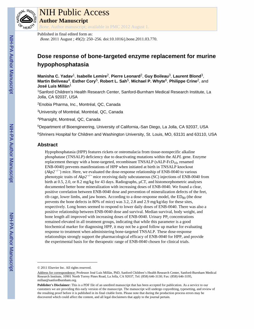
Dose response of bone-targeted enzyme replacement for murine hypophosphatasia Manisha C. Yadav 1 , Isabelle Lemire 2 , Pierre Leonard 2 , Guy Boileau 3 , Laurent Blond 3 , Martin Beliveau 4 , Esther Cory 5 , Robert L. Sah 5 , Michael P. Whyte 6 , Philippe Crine 2 , and José Luis Millán 1 1 Sanford Children’s Health Research Center, Sanford-Burnham Medical Research Institute, La Jolla, CA 92037, USA 2 Enobia Pharma, Inc., Montréal, QC, Canada 3 University of Montréal, Montréal, QC, Canada 4 Pharsight, Montreal, QC, Canada 5 Department of Bioengineering, University of California,-San Diego, La Jolla, CA 92037, USA 6 Shriners Hospital for Children and Washington University, St. Louis, MO, 63131 and 63110, USA Abstract Hypophosphatasia (HPP) features rickets or osteomalacia from tissue-nonspecific alkaline phosphatase (TNSALP) deficiency due to deactivating mutations within the ALPL gene. Enzyme replacement therapy with a bone-targeted, recombinant TNSALP (sALP-FcD 10 , renamed ENB-0040) prevents manifestations of HPP when initiated at birth in TNSALP knockout (Akp2 −/− ) mice. Here, we evaluated the dose-response relationship of ENB-0040 to various phenotypic traits of Akp2 −/− mice receiving daily subcutaneous (SC) injections of ENB-0040 from birth at 0.5, 2.0, or 8.2 mg/kg for 43 days. Radiographs, μCT, and histomorphometric analyses documented better bone mineralization with increasing doses of ENB-0040. We found a clear, positive correlation between ENB-0040 dose and prevention of mineralization defects of the feet, rib cage, lower limbs, and jaw bones. According to a dose-response model, the ED 80 (the dose prevents the bone defects in 80% of mice) was 3.2, 2.8 and 2.9 mg/kg/day for these sites, respectively. Long bones seemed to respond to lower daily doses of ENB-0040. There was also a positive relationship between ENB-0040 dose and survival. Median survival, body weight, and bone length all improved with increasing doses of ENB-0040. Urinary PP i concentrations remained elevated in all treatment groups, indicating that while this parameter is a good biochemical marker for diagnosing HPP, it may not be a good follow up marker for evaluating response to treatment when administering bone-targeted TNSALP. These dose-response relationships strongly support the pharmacological efficacy of ENB-0040 for HPP, and provide the experimental basis for the therapeutic range of ENB-0040 chosen for clinical trials. © 2011 Elsevier Inc. All rights reserved. Address for correspondence: Professor José Luis Millán, PhD, Sanford Children’s Health Research Center, Sanford-Burnham Medical Research Institute, 10901 North Torrey Pines Road, La Jolla, CA 92037, Tel: (858) 646-3130; Fax: (858) 646-3195, [email protected]. Publisher's Disclaimer: This is a PDF file of an unedited manuscript that has been accepted for publication. As a service to our customers we are providing this early version of the manuscript. The manuscript will undergo copyediting, typesetting, and review of the resulting proof before it is published in its final citable form. Please note that during the production process errors may be discovered which could affect the content, and all legal disclaimers that apply to the journal pertain. NIH Public Access Author Manuscript Bone. Author manuscript; available in PMC 2012 August 1. Published in final edited form as: Bone. 2011 August ; 49(2): 250–256. doi:10.1016/j.bone.2011.03.770. NIH-PA Author Manuscript NIH-PA Author Manuscript NIH-PA Author Manuscript
-
Upload
independent -
Category
Documents
-
view
0 -
download
0
Transcript of Dose response of bone-targeted enzyme replacement for murine hypophosphatasia
Dose response of bone-targeted enzyme replacement for murinehypophosphatasia
Manisha C. Yadav1, Isabelle Lemire2, Pierre Leonard2, Guy Boileau3, Laurent Blond3,Martin Beliveau4, Esther Cory5, Robert L. Sah5, Michael P. Whyte6, Philippe Crine2, andJosé Luis Millán1
1Sanford Children’s Health Research Center, Sanford-Burnham Medical Research Institute, LaJolla, CA 92037, USA2Enobia Pharma, Inc., Montréal, QC, Canada3University of Montréal, Montréal, QC, Canada4Pharsight, Montreal, QC, Canada5Department of Bioengineering, University of California,-San Diego, La Jolla, CA 92037, USA6Shriners Hospital for Children and Washington University, St. Louis, MO, 63131 and 63110, USA
AbstractHypophosphatasia (HPP) features rickets or osteomalacia from tissue-nonspecific alkalinephosphatase (TNSALP) deficiency due to deactivating mutations within the ALPL gene. Enzymereplacement therapy with a bone-targeted, recombinant TNSALP (sALP-FcD10, renamedENB-0040) prevents manifestations of HPP when initiated at birth in TNSALP knockout(Akp2−/−) mice. Here, we evaluated the dose-response relationship of ENB-0040 to variousphenotypic traits of Akp2−/− mice receiving daily subcutaneous (SC) injections of ENB-0040 frombirth at 0.5, 2.0, or 8.2 mg/kg for 43 days. Radiographs, μCT, and histomorphometric analysesdocumented better bone mineralization with increasing doses of ENB-0040. We found a clear,positive correlation between ENB-0040 dose and prevention of mineralization defects of the feet,rib cage, lower limbs, and jaw bones. According to a dose-response model, the ED80 (the doseprevents the bone defects in 80% of mice) was 3.2, 2.8 and 2.9 mg/kg/day for these sites,respectively. Long bones seemed to respond to lower daily doses of ENB-0040. There was also apositive relationship between ENB-0040 dose and survival. Median survival, body weight, andbone length all improved with increasing doses of ENB-0040. Urinary PPi concentrationsremained elevated in all treatment groups, indicating that while this parameter is a goodbiochemical marker for diagnosing HPP, it may not be a good follow up marker for evaluatingresponse to treatment when administering bone-targeted TNSALP. These dose-responserelationships strongly support the pharmacological efficacy of ENB-0040 for HPP, and providethe experimental basis for the therapeutic range of ENB-0040 chosen for clinical trials.
© 2011 Elsevier Inc. All rights reserved.Address for correspondence: Professor José Luis Millán, PhD, Sanford Children’s Health Research Center, Sanford-Burnham MedicalResearch Institute, 10901 North Torrey Pines Road, La Jolla, CA 92037, Tel: (858) 646-3130; Fax: (858) 646-3195,[email protected]'s Disclaimer: This is a PDF file of an unedited manuscript that has been accepted for publication. As a service to ourcustomers we are providing this early version of the manuscript. The manuscript will undergo copyediting, typesetting, and review ofthe resulting proof before it is published in its final citable form. Please note that during the production process errors may bediscovered which could affect the content, and all legal disclaimers that apply to the journal pertain.
NIH Public AccessAuthor ManuscriptBone. Author manuscript; available in PMC 2012 August 1.
Published in final edited form as:Bone. 2011 August ; 49(2): 250–256. doi:10.1016/j.bone.2011.03.770.
NIH
-PA Author Manuscript
NIH
-PA Author Manuscript
NIH
-PA Author Manuscript
Keywordsalkaline phosphatase; calcification; ENB-0040; rickets; osteomalacia
IntroductionHypophosphatasia (HPP) is the inborn-error-of-metabolism characterized by subnormalalkaline phosphatase (ALP) activity in serum and features rickets or osteomalacia [1-3] dueto loss-of-function mutation within the tissue-nonspecific alkaline phosphatase (TNSALP)gene (ALPL) [4-6]. The clinical severity of HPP ranges widely and spans complete absenceof bone mineralization and stillbirth, to spontaneous fractures and loss of teeth in adult life[2,3]. According to a traditional clinical nosology, the most severe to the mildest forms areperinatal, infantile, childhood, adult, and odontohypophosphatasia [3, 7].
TNSALP null mice (Akp2−/−) phenocopy infantile HPP extremely well [8, 9]. They are bornwith a normally mineralized skeleton, but develop rickets at about 6 days of age and diebetween day 16-20 suffering severe skeletal hypomineralization, episodes of apnea, andseizures [8-12]. The primary biochemical defect, TNSALP isozyme deficiency, leads toextracellular accumulation of its natural substrates including pyridoxal-5′-phosphate (PLP),the principal circulating form of vitamin B6, and inorganic pyrophosphate (PPi), a potentinhibitor of mineralization [13]. Impaired hydrolysis of PLP causes intracellular deficiencyof vitamin B6 in the central nervous system and thereby reduced brain levels of gamma-aminobutyric acid and seizures [10, 12]. This abnormality is evident not only in Akp2−/−
mice [8-10], but also sometimes in severely affected infants [11]. Rickets and osteomalaciain HPP are due to the accumulation in cartilage or bone matrix of extracellular PPi, causedby reduced pyrophosphatase activity of TNSALP [2, 3, 14-18].
To-date, there is no established medical treatment for HPP [2, 3]. Attempted enzymereplacement therapy (EzRT) using intravenous (IV) infusions of ALP-rich plasma fromPaget bone disease patients, purified human liver ALP, or purified human placental ALPwas followed by failure to rescue severely affected infants [19-22]. However, in 2008, wereported that newborn Akp2−/− mice receiving a daily, high-dose (8.2 mg/kg), subcutaneous(SC) injection of a bone-targeted form of recombinant TNSALP, sALP-FcD10 (renamedENB-0040), grew normally and appeared well without skeletal or dental disease, orepilepsy, demonstrating that this EzRT could prevent all of the manifestations of infantileHPP in this murine model [23; 24].
Here, we present a dose-response study that establishes the optimal dose of ENB-0040 forprevention of HPP in Akp2−/− mice, and thereby enhances the preclinical foundation forEzRT trials for HPP patients.
Materials and methodsMouse model of Infantile HPP
The Akp2−/− knockout mice, created by insertion of the Neo cassette into exon 6 of themouse TNSALP gene (Akp2) via homologous recombination, functionally inactivates theAkp2 gene resulting in no detectable TNSALP mRNA or protein [8]. These Akp2−/− mice,maintained in a 12.5% C57Bl/6 - 87.5% 129J background, closely phenocopy infantile HPP[8, 9]. Like those severely affected patients, Akp2−/− mice have global deficiency ofTNSALP activity, extracellular accumulation of the ALP substrates PPi, PLP, andphosphoethanolamine (PEA), and postnatally manifest an acquired defect in mineralizationof skeletal matrix leading to radiographically and histologically obvious rickets [9]. They
Yadav et al. Page 2
Bone. Author manuscript; available in PMC 2012 August 1.
NIH
-PA Author Manuscript
NIH
-PA Author Manuscript
NIH
-PA Author Manuscript
have stunted growth and also develop seizures and apnea, and die between postnatal days10-12 [8-10]. Pyridoxine supplementation briefly suppresses their seizures and extends theirlifespan, but only until postnatal days 18-22 [10, 12]. Therefore, all animals (breeders,nursing mothers and their pups, and weanlings) in this study were given free access tomodified laboratory rodent diet 5001 containing increased levels (325 ppm) of pyridoxine.The Akp2−/− homozygous mice were identified at birth (Day 0) by PCR of tissue biopsiesusing specific primers to exon 6 of the mouse TNSALP gene: 5′-GTCCGTGGGCATTGTGACTACCAC-3′ and 5′-TGCTGCTCCACTCACGTCG-3′.
Bone-targeted, recombinant, human TNSALPThe ENB-0040 (sALP-FcD10) fusion protein contains recombinant human soluble TNSALP(sALP), the constant region of human IgG1 Fc domain (Fc) to facilitate purification, and adeca-aspartate motif (D10) for bone mineral targeting [24]. The expression, purification, andcharacterization of this fusion protein have been published [23]. Lot number PUR012F01 ofENB-0040 [produced under Current Good Manufacturing Practices (cGMP) formulated at0.15, 0.6 and 2.5 mg/mL in 25 mmol/L sodium phosphate, 150 mmol/L sodium chloride, pH7.4] was used in the current study.
Dose-response studyAkp2−/− mice were divided into 5 groups: Group 1 (Vehicle): Akp2−/− mice treated withvehicle SC daily (n=21); Group 2 (Tx-0.5): Akp2−/− mice treated with ENB-0040 at 0.5 mg/kg SC daily (n=18); Group 3 (Tx-2.0): Akp2−/− mice treated with ENB-0040 at 2.0 mg/kgSC daily (n=20); and Group 4 (Tx-8.2): Akp2−/− mice treated with ENB-0040 at 8.2 mg/kgSC daily (n=19). Group 5 (WT): wild-type littermates of Akp2−/− mice served as referenceanimals and did not receive injections (n=33). Injections were administered between 8:00and 11:00 AM, and dose volume was set at 3.3 mL/kg body weight. The actual volumegiven was calculated and adjusted based on the daily body weight measured prior toinjection. Vehicle or ENB-0040 was injected SC into the scapular region. All treatmentsbegan on postnatal Day 1, and were repeated daily for up to 43 days or until the time ofnecropsy.
Terminal proceduresNecropsy was performed on Day 44, 24 hours after the final injection of ENB-0040 forthose animals that completed the experimental protocol, or sooner for those animals thatappeared terminally ill. All animals were euthanized by bilateral thoracotomy underisoflurane anesthesia. The necropsy consisted of a gross pathology check, with a piece of earcollected to confirm the Akp2−/− genotype. The bone samples were cleaned, fixed in 10%neutral buffered formalin for 3 days at 2 to 8°C, and then transferred to 70% ethanol forstorage at 2 to 8°C. Femur and tibia lengths were measured using a caliper.
Radiographic analysisRadiographic images were obtained with a Faxitron MX-20 DC4 (Faxitron X-rayCorporation, Wheeling, IL), using energy of 26 kV and an exposure time of 10 seconds.Defects in bone mineralization were classified in a blinded fashion by a veterinarianradiologist. Animals were “Abnormal” if at least one bone structure (including secondaryossification centers) was absent.
Histological analysesThe femora and tibiae of treated and untreated mice were fixed in 10% formalin. Sectionswere prepared according to the Kawamoto method [25] with slight modifications. Briefly,the specimens were frozen in hexane cooled with dry ice. The frozen samples were
Yadav et al. Page 3
Bone. Author manuscript; available in PMC 2012 August 1.
NIH
-PA Author Manuscript
NIH
-PA Author Manuscript
NIH
-PA Author Manuscript
immersed in 5% carboxymethyl cellulose (CMC) gel for ten minutes and completely frozenwith the CMC gel in cooled hexane. Next, the frozen blocks were fastened to thecryomicrotome (CM 1850; Leica Instruments, Germany) in the cryochamber (−22°C) andtrimmed with a disposable tungsten carbide blade (Jung TC-65A, 35° angle; LeicaInstruments). The trimmed surface was covered with a Cryofilm (FINETEC, Japan) using abrush to remove air bubbles behind the film. The sample was then cut at 5 to 7 μm thicknessand dried at room temperature. Afterwards, the film was soaked briefly in ethanol andmounted on a glass slide so that the sampling side faced upward. Alizarin red/ alcian blueand Von Kossa/ van Gieson stainings were performed as before [16]. Stained slides wereused for quantification of osteoid volume using the Bioquant Osteo software (BioquantOsteoanalysis Co., Nashville, TN, USA).
Micro-computed tomography (μCT) analysis was carried out using Skyscan 1076 (Kontich,Belgium). Hind limbs were wrapped in tissue paper that was moistened with phosphatebuffered saline (PBS) and scanned at 9 μm voxel size, applying an electrical potential of 50kVp and current of 200uA, and using a 0.5mm aluminum filter. Skyscan software,Dataviewer, CTAn (Kontich, Belgium) was used to determine bone histomorphometricparameters. Cortical bone analysis was performed on the femoral and tibial diaphysis. Asappropriate for skeletally-mature animals [26], each femur and tibia was analyzed withattention to limb length. For each limb, three regions were analyzed along the length of thelimbs for cortical bone analysis. The regions, 747 μm along the femur and 711 μm along thetibia, were located at 14.3-28.6%, 42.9-57.1% and 71.4-85.7% of the distance between thegrowth plate and the throchanteric fossa for the femur and the growth plate to the distaljunction of the tibia and fibula bone in the tibia. Cortical bone selection was done bycontouring the periosteal tissue and excluding the marrow cavity. A global threshold wasused to identify cortical bone and an erosion of one pixel was performed to eliminate partialvolume effects and calculate the following parameters: cross-sectional tissue area (T.Ar),cross-sectional cortical bone area (B.Ar), cortical bone area fraction (B.Ar/T.Ar), cross-sectional bone thickness (Cs.Th) and tissue mineral density (TMD). TMD was calibratedagainst 2mm diameter hydroxyapatite (HA) phantoms appropriate for mouse samples.Additionally, a beam hardening correction algorithm was applied prior imagereconstruction.
PPi AssayPPi concentrations were determined by differential adsorption on activated charcoal of UDP-D-[6-3H]glucose (Amersham Pharmacia) from the reaction product 6-phospho[6-3H]gluconate, as described [27].
Statistical analysesAll numerical values are presented as mean values ± standard deviation (SD) unlessmentioned otherwise. Non-parametric analysis was preferred for all parameters because ofthe small sample sizes. The Log-Rank test was used to compare survival curves. Fisher’sexact test was used to compare the distribution of normal and abnormal radiographs betweentreatments. An Anova model was used to compare the average body weights between groupsat each day, and the average bone lengths at the end of the study. For urine PPi, bone osteoidand bone μCT measurements, the results are expressed as mean ± SEM, and data wereanalyzed using student’s t test. P values less than 0.05 are considered significant.
Dose-Response AnalysisThe conceptual pharmacodynamic (PD) models used to fit dose-response data are shown inTable 1. Akaike Information (AIC) and Schwarz Bayesian (SBC) criterion were used as ameasure of goodness-of-fit to select the best model for each data set. When comparing
Yadav et al. Page 4
Bone. Author manuscript; available in PMC 2012 August 1.
NIH
-PA Author Manuscript
NIH
-PA Author Manuscript
NIH
-PA Author Manuscript
several models for a given data set, the model associated with the smallest AIC and SBCwas selected during the model discrimination process. The correlation coefficient (r2) andvisual assessments of fit (VAF), absolute residual distribution, and coefficient of variation ofPD parameters were also used for the model discrimination. Once the appropriate model wasselected, the fit was re-run using predefined residual error models (additive, Poisson orproportional). The same criteria used to choose the model were applied to select theappropriate error model for each data set. PD analysis was performed using WinNonlin®Enterprise Edition software v5.2 (Pharsight Corporation, Mountain View, CA). All analyseswere performed using full precision. Whenever possible, results were reported to 3significant figures. PD parameters and their 95% confidence intervals were determinedusing WinNonlin 5.2.
ResultsChanges in Survival
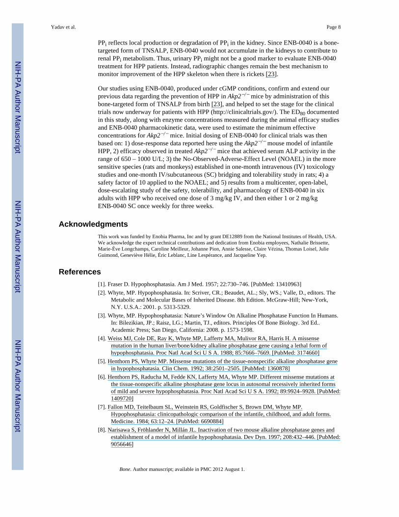
Survival of mice representing each treatment group was significantly improved whencompared to Vehicle (p<0.0001) (Fig. 1). Differences were also statistically significantwhen the survival curves of the treated groups were compared (p<0.0001). Median survivalwas 19, 24, and 31 days in the Vehicle, Tx-0.5, and Tx-2.0 groups, respectively. Only one of19 mice in Tx-8.2 died before the study was completed at age 43 days; thus, median survivalcould not be calculated for this group. However, there was a clear relationship betweenENB-0040 daily dose and survival of the Akp2−/− mice.
Changes in body weight and femur/tibial lengthFigure 2A depicts the growth curves of the mice. At the beginning of treatment, averagedaily body weights (BWs) showed no statistical difference between the newborn Akp2−/−
mice of each treatment group and newborn WT mice. However, average BWs with Vehicleand Tx-0.5 were statistically lower compared to WT mice starting on Day 7 (4.156 g, 4.220g and 4.726 g, respectively; p=0.0068 for Vehicle compared to WT, and p=0.0216 forTx-0.5 compared to WT). For the Tx-2.0 group, BWs became statistically lower than WTstarting on Day 16 (7.373 g and 8.248 g, respectively; p=0.0496). However, the averageBWs for Tx-8.2 were never statistically different compared to WT mice. The Tx-2.0 andTx-8.2 dose groups improved BWs compared to Vehicle-treated mice from Day 8 onwards,showing statistically significant differences (5.065 g for Tx-2.0 and 4.527 for Vehicle,p=0.0446; 5.065 g for Tx-8.2 and 4.527 for Vehicle, p=0.0471). Similarly, Tx-0.5 averageBWs improved compared to Vehicle after Day 18 (6.532 g for Tx-0.5 and 5.456 g forVehicle, p<0.0001). There was a clear relationship between ENB-0040 daily dose andpreservation of healthy BW.
At study end, the average length of the femur in Tx-2.0 (11.92 ± 0.97 mm) and in Tx-8.2(12.53 ± 0.77 mm) was shorter compared to the WT mice (13.35 ± 0.52 mm) (p= 0.0010 andp=0.0021, respectively) (Fig. 2B). However, the average length of the femur in Tx-2.0 wasnot statistically different compared to Tx-8.2 (p=0.1277). The average length of the tibia inTx-2.0 (15.39 ± 0.64 mm) and Tx-8.2 (15.70 ± 0.87 mm) was shorter compared to WT mice(16.61 ± 0.49 mm) (p= 0.0046 and p=0.0009, respectively). The average length of the tibiain Tx-2.0 was not statistically different compared to Tx-8.2 (p=0.4368). There was a trendtowards preservation of bone length in Akp2−/− mice with increasing doses of ENB-0040.Deaths of the Vehicle or Tx-0.5 mice precluded age-matched bone length analysis.
Improved skeletal phenotype in Akp2−/− mice receiving ENB-0040 treatmentThe radiographs of untreated Akp2−/− mice (Vehicle) showed profound skeletalabnormalities such as bowed and fractured femora and tibiae, reduced tissue mineral density
Yadav et al. Page 5
Bone. Author manuscript; available in PMC 2012 August 1.
NIH
-PA Author Manuscript
NIH
-PA Author Manuscript
NIH
-PA Author Manuscript

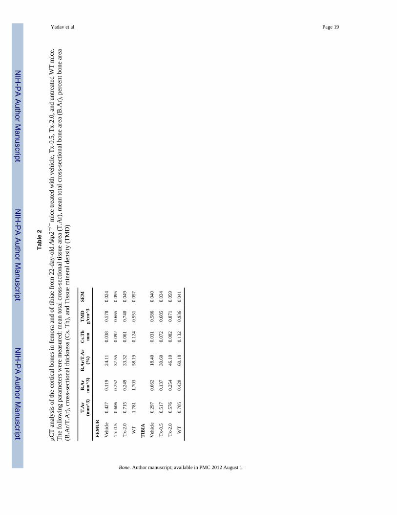
(TMD) in femora, tibiae, incisors and molars, shorter vertebrae, and increased spacesbetween adjacent vertebrae. All these abnormalities showed slight improvement with thelowest dose of ENB-0040 (Tx-0.5 group), and considerable improvement within the Tx-2.0group. There was callus formation at the sites of fractures in femur and tibia, improved bonemorphology, increased TMD, and reduced intervertebral spaces in the treated mice (Fig. 3).μCT analysis of the long bones from 22 day-old untreated Akp2−/− mice (Vehicle) andtreated (Tx-0.5 and Tx-2.0) mice showed a clear and significant improvement in the bonemorphology and TMD proportional to the treatment administered (Fig. 4; Table 2). TheAkp2−/− mice femora showed ~60% less mineral density than the WT mice. Low dose(Tx-0.5) treatment improved the TMD to ~70%, and a higher dose (Tx-2.0) improved it to~80% of the WT values (vehicle versus Tx-2.0, p=0.02, significant) and in tibiae the Tx-2.0group showed ~93% (vehicle Vs Tx-2.0, p= 0.007, significant; Tx-2.0 versus WT, p = 0.2,not significant) recovery of TMD. Morphologically, the Akp2−/− bones had only 23%cortical thickness in the tibiae compared to WT mice. However, in the Tx-2.0 treatmentgroup, cortical thickness seemed improved to 62% of WT (Tx-2.0 Vs WT, p = 0.084, butnon-significant) (Table 2).
Histological analysis of 44-day-old treated tibiae stained with alizarin red/alcian blue (Fig.5A) showed increased mineralization of the trabecular bone in the TX-8.2 group. VonKossa/Van Gieson staining (Fig. 5B) also revealed decreased amounts of unmineralizedosteoid in the Tx-8.2 group, which showed no statistically significant difference whencompared to the WT mice. The OV/BV % values obtained were as follows: Tx-2.0= 5.723 ±2.036 (N= 3); Tx-8.2= 0.013 ± 0.004 (N= 3); WT= 0.009 ± 0.0005 (N= 3); Tx-8.2 groupshowed a non-significant difference when compared to WT, p= 0.26. Of interest, urinary PPiconcentrations remained high in all treatment groups, despite the correction of plasma PPilevels and the obvious improvement of the skeletal condition in the treated mice (Fig. 6).
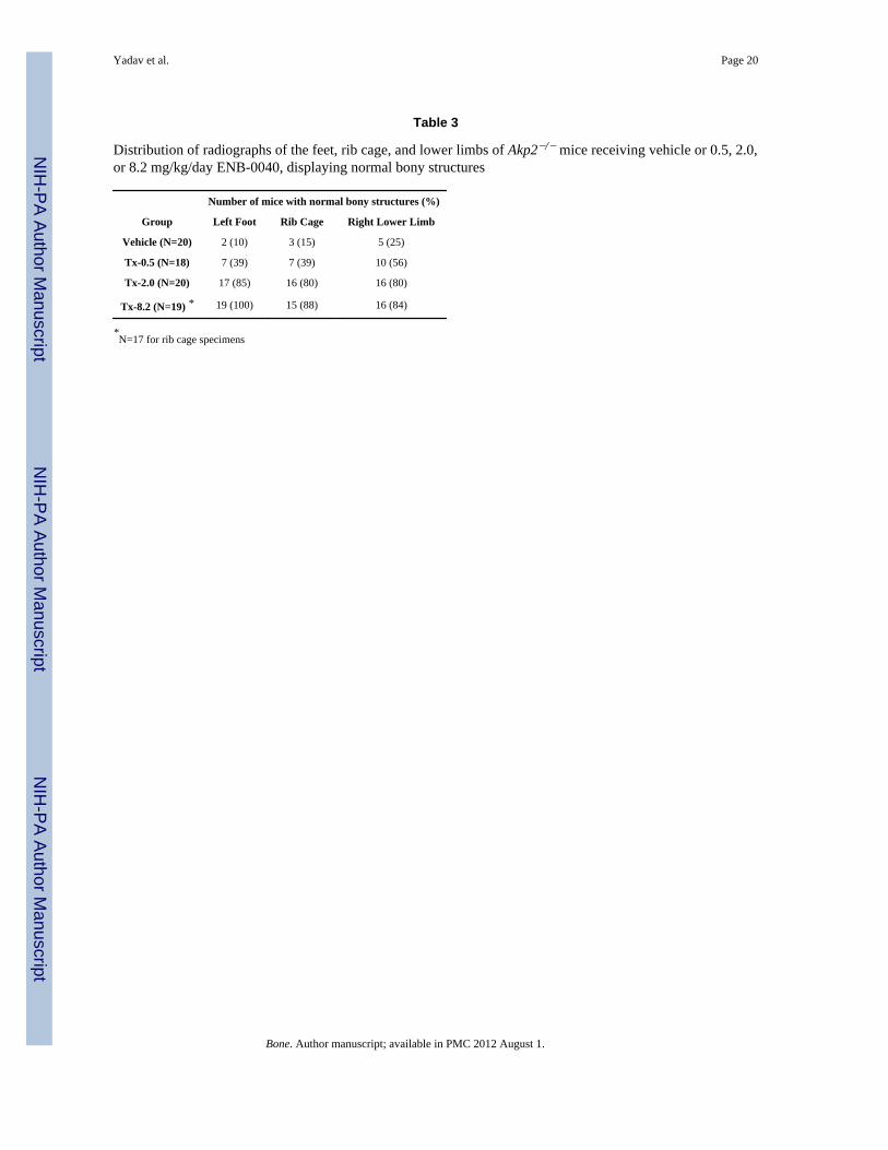
The radiographic results for each treatment group, i.e., Akp2−/− receiving vehicle aloneversus 0.5, 2.0, or 8.2 mg/kg/day ENB-0040, are summarized in Table 3. Here, thedistribution between normal and abnormal radiographic images are presented, with thepercentages given in parentheses. At necropsy, 10%, 15%, and 25% of mice receivingVehicle had normal left foot, rib cage, or lower limb radiographs, respectively, compared to100% normal appearance for the WT mouse specimens (data not shown). However, thepercentage of animals with normal mineralization of the left foot was improved from 10%with Vehicle to 39, 85, and 100% with increasing doses of ENB-0040. The efficacy of the2.0 and 8.2 mg/kg doses was statistically significant when compared to Vehicle (p<0.0001),whereas the smallest (0.5 mg/kg) dose only approached statistical significance (p=0.0577).Inter-treatment comparisons showed that Tx-2.0 and Tx-8.2 were more effective comparedto Tx-0.5 (p=0.0063 and p<0.0001, respectively). However, Tx-8.2 efficacy was not betterwhen compared to Tx-2.0 (p=0.2308).
Similarly, radiographic analysis of the rib cage and lower limbs showed the distributionpresented in Table 3. For the rib cage endpoint, the percentage of animals with normal bonystructures was increased from 15% with Vehicle to 39, 80, and 88% with increasing doses ofENB-0040. The efficacies of the 2.0 and 8.2 mg/kg doses were statistically significant whencompared to Vehicle (p<0.0001), whereas the 0.5 mg/kg level was not (p=0.1440). Inter-treatment comparisons showed that Tx-2.0 and Tx-8.2 were more effective compared toTx-0.5 (p=0.0189 and p=0.0045, respectively). The efficacy of Tx-8.2 was not differentfrom Tx-2.0 (p=0.6665). The percentage of mice with normal bony structures of the lowerlimbs increased from 25% with Vehicle to 56, 80, and 84% with increasing doses ofENB-0040 (Table 3). The efficacy of the 2.0 and 8.2 mg/kg dose was statistically significantwhen compared to Vehicle (p=0.0012 and p<0.0001, respectively), whereas 0.5 mg/kg onlyapproached statistical significance (p=0.0960). Inter-treatment comparisons showed that
Yadav et al. Page 6
Bone. Author manuscript; available in PMC 2012 August 1.
NIH
-PA Author Manuscript
NIH
-PA Author Manuscript
NIH
-PA Author Manuscript
Tx-2.0 was not more effective compared to Tx-0.5 (p=0.1643), but 8.2 mg/kg approachedstatistical significance (p=0.0789). The efficacy of Tx-8.2 was not different from Tx-2.0(p>0.9999).
We examined the dose response to ENB-0040 treatment by evaluating the effect of variousdoses on the radiographic image distribution (RID) pharmacodynamic endpoint. Accordingto Akaike Information (AIC) and Schwarz Bayesian Criterion (SBC) values, the SimpleEmax Model with Baseline fitted the Dose vs. RID relationship adequately. Visualassessments of fit and absolute residual distributions indicated that the Simple Emax with E0Model and a proportional error appeared to be the best weighting scheme for the Dose-RIDrelationship. We found a clear relationship between daily dose of ENB-0040 and percent ofmice with normal bony structures of the foot, rib cage, and lower limbs. The 80% effectivedose (ED80) was estimated to be 3.2 mg/kg/day for feet, 2.8 mg/kg/day for rib cage, and 2.9mg/kg/day for lower limbs (Fig. 7). The overall pharmacokinetics/PD relationship wascharacterized by a steep increase in the range 0 - 5 mg/kg/day, with maximum effect(~100%) observed at 6 to 8 mg/kg/day.
DiscussionIn 2008, we established that EzRT using daily SC injections of bone-targeted, recombinant,human TNSALP, i.e., sALP-FcD10 (ENB-0040), prevented infantile HPP in the Akp2−/−
mouse model [23]. We evaluated survival, growth rates, serum levels of sALP-FcD10activity, calcium, PPi, and PLP, as well as skeletal and dental manifestations of HPP byradiography, μCT, and histomorphometry. Akp2−/− mice receiving high-doses (8.2 mg/kg/day) of sALP-FcD10 for 8 weeks grew normally and appeared well without skeletal diseaseor epilepsy [23]. More recently, we demonstrated that treatment with high-dose (8.2 mg/kg/day) of sALP-FcD10 prevented hypomineralization of alveolar bone, dentin, and cementumas assessed by micro-computed tomography and histology [24].
The objective of the present study was to define the dose response relationship betweenincreasing amounts of cGMP-produced ENB-0040 using bolus SC injections and thetherapeutic response after 43 days in anticipation of clinical trials. Endpoints were survival,body weight, bone length of the tibiae and femora, and bone mineralization defects assessedusing radiographs of the feet, rib cage, and lower limbs. To-date, these parameters haverepresented good indicators of correction of the HPP phenotype in this animal model. Also,the radiographic manifestations of HPP are readily observed in affected infants and, thus, arean important endpoint in preclinical proof-of-concept studies for extrapolation from mice tohumans [2, 3]. In addition, we used μCT and histomorphometric analysis to document theimprovement in Akp2−/− bone mineralization status for representative age groupsundergoing treatment.
We documented a clear relationship between daily dose of ENB-0040 and the percent ofmice with normal bony structures of the foot, rib cage, and lower limbs. We focused onestablishing the effective dose in 80% of the mice (ED80). The 80% effective dose in micewas ~ 3.2 mg/kg/day for feet, ~ 2.8 mg/kg/day for rib cage, and ~ 2.9 mg/kg/day for lowerlimbs. These ED80 doses are consistent with our previous experience showing a betterskeleton in a short-term, 15-day treatment of Akp2−/− animals using 2 mg/kg/day ofENB-0040 [23].
Of interest, in this new study, we found that, despite the correction of plasma PPi levels byENB-0040 treatment already documented [23], urinary PPi concentrations remainedelevated for all treatment groups. This is important because measurement of urinary PPi isused as a diagnostic marker for HPP [2, 3]. We interprete this result to indicate that urinary
Yadav et al. Page 7
Bone. Author manuscript; available in PMC 2012 August 1.
NIH
-PA Author Manuscript
NIH
-PA Author Manuscript
NIH
-PA Author Manuscript
PPi reflects local production or degradation of PPi in the kidney. Since ENB-0040 is a bone-targeted form of TNSALP, ENB-0040 would not accumulate in the kidneys to contribute torenal PPi metabolism. Thus, urinary PPi might not be a good marker to evaluate ENB-0040treatment for HPP patients. Instead, radiographic changes remain the best mechanism tomonitor improvement of the HPP skeleton when there is rickets [23].
Our studies using ENB-0040, produced under cGMP conditions, confirm and extend ourprevious data regarding the prevention of HPP in Akp2−/− mice by administration of thisbone-targeted form of TNSALP from birth [23], and helped to set the stage for the clinicaltrials now underway for patients with HPP (http://clinicaltrials.gov/). The ED80 documentedin this study, along with enzyme concentrations measured during the animal efficacy studiesand ENB-0040 pharmacokinetic data, were used to estimate the minimum effectiveconcentrations for Akp2−/− mice. Initial dosing of ENB-0040 for clinical trials was thenbased on: 1) dose-response data reported here using the Akp2−/− mouse model of infantileHPP, 2) efficacy observed in treated Akp2−/− mice that achieved serum ALP activity in therange of 650 – 1000 U/L; 3) the No-Observed-Adverse-Effect Level (NOAEL) in the moresensitive species (rats and monkeys) established in one-month intravenous (IV) toxicologystudies and one-month IV/subcutaneous (SC) bridging and tolerability study in rats; 4) asafety factor of 10 applied to the NOAEL; and 5) results from a multicenter, open-label,dose-escalating study of the safety, tolerability, and pharmacology of ENB-0040 in sixadults with HPP who received one dose of 3 mg/kg IV, and then either 1 or 2 mg/kgENB-0040 SC once weekly for three weeks.
AcknowledgmentsThis work was funded by Enobia Pharma, Inc and by grant DE12889 from the National Institutes of Health, USA.We acknowledge the expert technical contributions and dedication from Enobia employees, Nathalie Brissette,Marie-Ève Longchamps, Caroline Meilleur, Johanne Pion, Annie Salesse, Claire Vézina, Thomas Loisel, JulieGuimond, Geneviève Hélie, Éric Leblanc, Line Lespérance, and Jacqueline Yep.
References[1]. Fraser D. Hypophosphatasia. Am J Med. 1957; 22:730–746. [PubMed: 13410963][2]. Whyte, MP. Hypophosphatasia. In: Scriver, CR.; Beaudet, AL.; Sly, WS.; Valle, D., editors. The
Metabolic and Molecular Bases of Inherited Disease. 8th Edition. McGraw-Hill; New-York,N.Y. U.S.A.: 2001. p. 5313-5329.
[3]. Whyte, MP. Hypophosphatasia: Nature’s Window On Alkaline Phosphatase Function In Humans.In: Bilezikian, JP.; Raisz, LG.; Martin, TJ., editors. Principles Of Bone Biology. 3rd Ed..Academic Press; San Diego, California: 2008. p. 1573-1598.
[4]. Weiss MJ, Cole DE, Ray K, Whyte MP, Lafferty MA, Mulivor RA, Harris H. A missensemutation in the human liver/bone/kidney alkaline phosphatase gene causing a lethal form ofhypophosphatasia. Proc Natl Acad Sci U S A. 1988; 85:7666–7669. [PubMed: 3174660]
[5]. Henthorn PS, Whyte MP. Missense mutations of the tissue-nonspecific alkaline phosphatase genein hypophosphatasia. Clin Chem. 1992; 38:2501–2505. [PubMed: 1360878]
[6]. Henthorn PS, Raducha M, Fedde KN, Lafferty MA, Whyte MP. Different missense mutations atthe tissue-nonspecific alkaline phosphatase gene locus in autosomal recessively inherited formsof mild and severe hypophosphatasia. Proc Natl Acad Sci U S A. 1992; 89:9924–9928. [PubMed:1409720]
[7]. Fallon MD, Teitelbaum SL, Weinstein RS, Goldfischer S, Brown DM, Whyte MP.Hypophosphatasia: clinicopathologic comparison of the infantile, childhood, and adult forms.Medicine. 1984; 63:12–24. [PubMed: 6690884]
[8]. Narisawa S, Fröhlander N, Millán JL. Inactivation of two mouse alkaline phosphatase genes andestablishment of a model of infantile hypophosphatasia. Dev Dyn. 1997; 208:432–446. [PubMed:9056646]
Yadav et al. Page 8
Bone. Author manuscript; available in PMC 2012 August 1.
NIH
-PA Author Manuscript
NIH
-PA Author Manuscript
NIH
-PA Author Manuscript

[9]. Fedde KN, Blair L, Silverstein J, Coburn SP, Ryan LM, Weinstein RS, Waymire K, Narisawa S,Millán JL, MacGregor GR, Whyte MP. Alkaline phosphatase knockout mice recapitulate themetabolic and skeletal defects of infantile hypophosphatasia. J Bone Miner Res. 1999; 14:2015–2026. [PubMed: 10620060]
[10]. Waymire KG, Mahuren JD, Jaje JM, Guilarte TR, Coburn SP, MacGregor GR. Mice lackingtissue non-specific alkaline phosphatase die from seizures due to defective metabolism of vitaminB-6. Nat Genet. 1995; 11:45–51. [PubMed: 7550313]
[11]. Baumgartner-Sigl S, Haberlandt E, Mumm S, Scholl-Bürgi S, Sergi C, Ryan L, Ericson KL,Whyte MP, Högler Wl. Pyridoxine-responsive seizures as the first symptom of infantilehypophosphatasia caused by two novel missense mutations (c.677T>C, p.M226T; c.1112C>T,p.T371I) of the tissue-nonspecific alkaline phosphatase gene. Bone. 2007; 40:1655–1661.[PubMed: 17395561]
[12]. Narisawa S, Wennberg C, Millán JL. Abnormal vitamin B6 metabolism in alkaline phosphataseknock-out mice causes multiple abnormalities, but not the impaired bone mineralization. JPathol. 2001; 193:125–133. [PubMed: 11169525]
[13]. Meyer JL. Can biological calcification occur in the presence of pyrophosphate? Arch BiochemBiophys. 1984; 231:1–8. [PubMed: 6326671]
[14]. Hessle L, Johnson KA, Anderson HC, Narisawa S, Sali A, Goding JW, Terkeltaub R, Millán JL.Tissue-nonspecific alkaline phosphatase and plasma cell membrane glycoprotein-1 are centralantagonistic regulators of bone mineralization. Proc Natl Acad Sci U S A. 2002; 99:9445–9449.[PubMed: 12082181]
[15]. Harmey D, Hessle L, Narisawa S, Johnson KA, Terkeltaub R, Millán JL. Concerted regulation ofinorganic pyrophosphate and osteopontin by Akp2, Enpp1 and Ank. An integrated model of thepathogenesis of mineralization disorders. Am J Pathol. 2004; 164:1199–1209. [PubMed:15039209]
[16]. Murshed M, Harmey D, Millán JL, McKee MD, Karsenty G. Broadly expressed genes accountsfor the special restriction of ECM mineralization to bone. Genes Dev. 2005; 19:1093–1104.[PubMed: 15833911]
[17]. Harmey D, Johnson KA, Zelken J, Camacho NP, Hoylaerts MF, Noda M, Terkeltaub R, MillánJL. Elevated osteopontin levels contribute to the hypophosphatasia phenotype in Akp2−/− mice. JBone Miner Res. 2006; 21:1377–1386. [PubMed: 16939396]
[18]. Millán, JL. Mammalian alkaline phosphatases. From biology to applications in medicine andbiotechnology. Wiley-VCH Verlag GmbH & Co; Weinheim, Germany: 2006. p. 1-322.pgs
[19]. Whyte MP, Valdes R Jr, Ryan LM, McAlister WH. Infantile hypophosphatasia: Enzymereplacement therapy by intravenous infusion of alkaline phosphatase-rich plasma from patientswith Paget bone disease. J Pediatr. 1982; 101:379–386. [PubMed: 7108657]
[20]. Whyte MP, McAlister WH, Patton LS, Magill HL, Fallon MD, Lorentz WB Jr, Herrod HG.Enzyme replacement therapy for infantile hypophosphatasia attempted by intravenous infusionsof alkaline phosphatase-rich Paget plasma: Results in three additional patients. J Pediatr. 1984;105:926–933. [PubMed: 6502342]
[21]. Weninger M, Stinson RA, Plenk H Jr, Böck P, Pollak A. Biochemical and morphological effectsof human hepatic alkaline phosphatase in a neonate with hypophosphatasia. Acta Paediatr ScandSuppl. 1989; 360:154–160. [PubMed: 2642253]
[22]. Whyte MP, Habib D, Coburn SP, Tecklenburg F, Ryan L, Fedde KN, Stinson RA. Failure ofhyperphosphatasemia by intravenous infusion of purified placental alkaline phosphatase (ALP) tocorrect severe hypophosphatasia: Evidence against a role for circulating ALP in skeletalmineralization. J Bone Miner Res. 1992; 7(Suppl 1):S155.
[23]. Millán JL, Narisawa S, Lemire I, Loisel TP, Boileau G, Leonard P, Gramatikova S, Terkeltaub R,Pleshko Camacho N, McKee MD, Crine P, Whyte MP. Enzyme replacement therapy for murinehypophosphatasia. J Bone Miner Res. 2008; 23:777–787. [PubMed: 18086009]
[24]. McKee MD, Nakano Y, Masica DL, Gray JJ, Lemire I, Heft R, Whyte MP, Crine P, Millán JL.Enzyme replacement prevents dental defects in a mouse model of hypophosphatasia. J. DentalRes. 2011 In Press.
Yadav et al. Page 9
Bone. Author manuscript; available in PMC 2012 August 1.
NIH
-PA Author Manuscript
NIH
-PA Author Manuscript
NIH
-PA Author Manuscript
[25]. Kawamoto T. Use of a new adhesive film for the preparation of multi-purpose fresh-frozensections from hard tissues, whole-animals, insects and plants. Arch. Histol. Cytol. 2003; 66:123–143. [PubMed: 12846553]
[26]. Bouxsein ML, Boyd SK, Christiansen BA, Guldberg RE, Jepsen KJ, Muller R. Guidelines forassessment of bone microstructure in rodents using micro-computed tomography. J Bone MinerRes. 2010; 25:1468–1486. [PubMed: 20533309]
[27]. Johnson K, Vaingankar S, Chen Y, Moffa A, Goldring M, Sano K, Jin-Hua P, Sali A, Goding J,Terkeltaub R. Differential mechanisms of inorganic pyrophosphate production by plasma cellmembrane glycoprotein-1 and B10 in chondrocytes. Arthritis Rheum. 1999; 42:1986–1997.[PubMed: 10513816]
Yadav et al. Page 10
Bone. Author manuscript; available in PMC 2012 August 1.
NIH
-PA Author Manuscript
NIH
-PA Author Manuscript
NIH
-PA Author Manuscript
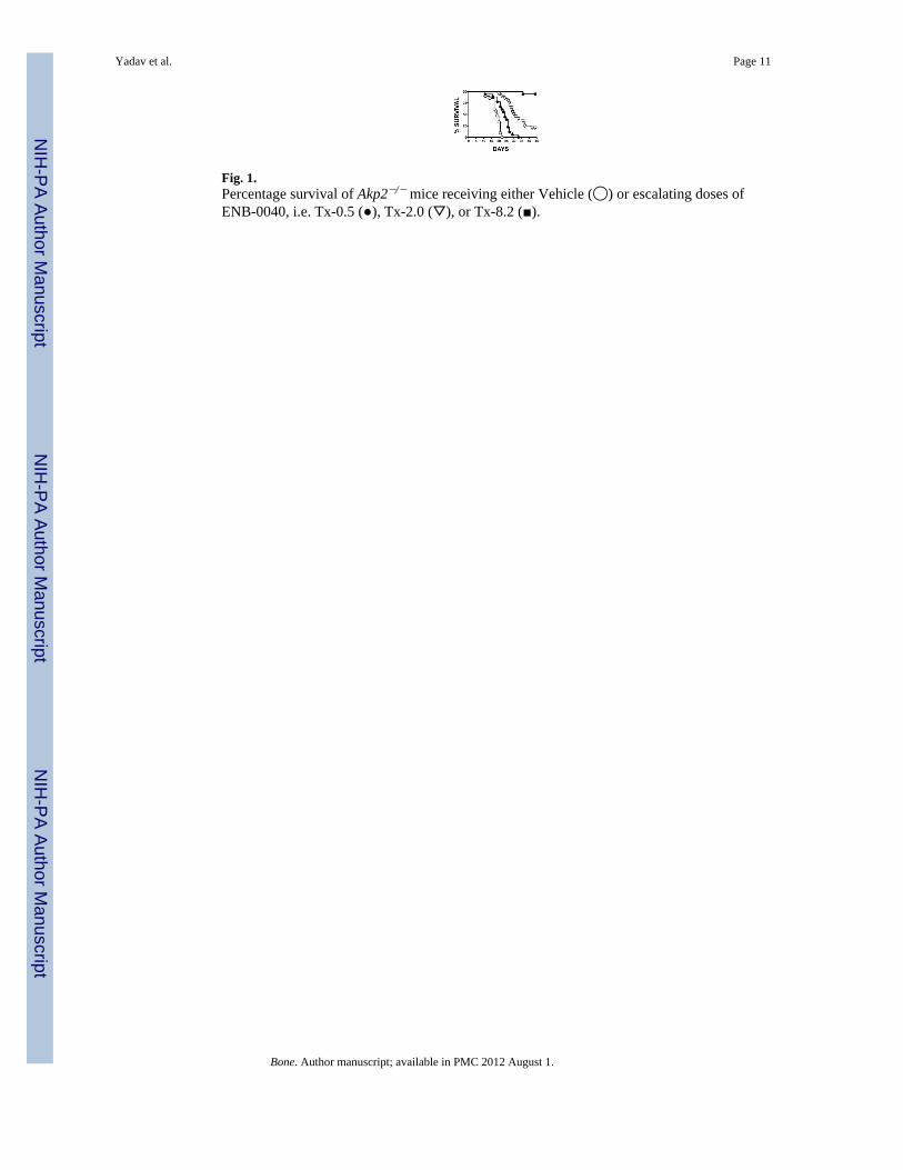
Fig. 1.Percentage survival of Akp2−/− mice receiving either Vehicle (◯) or escalating doses ofENB-0040, i.e. Tx-0.5 (●), Tx-2.0 (▽), or Tx-8.2 (■).
Yadav et al. Page 11
Bone. Author manuscript; available in PMC 2012 August 1.
NIH
-PA Author Manuscript
NIH
-PA Author Manuscript
NIH
-PA Author Manuscript
Fig. 2.Changes in body weight with age and tibiae/femora lengths as a function of treatment dose.Panel A shows average daily body weights of Akp2−/− mice, from Day 1 to the end of thestudy, treated with Vehicle (◯), Tx-0.5 (●), Tx-2.0 (▽), Tx-8.2 (■) or untreated WT mice(□). Panel B shows individual lengths of the left tibiae and femora at the end of the study forthe Tx-2.0 (▽) and Tx-8.2 (■) treatment groups compared to WT mice (□).
Yadav et al. Page 12
Bone. Author manuscript; available in PMC 2012 August 1.
NIH
-PA Author Manuscript
NIH
-PA Author Manuscript
NIH
-PA Author Manuscript
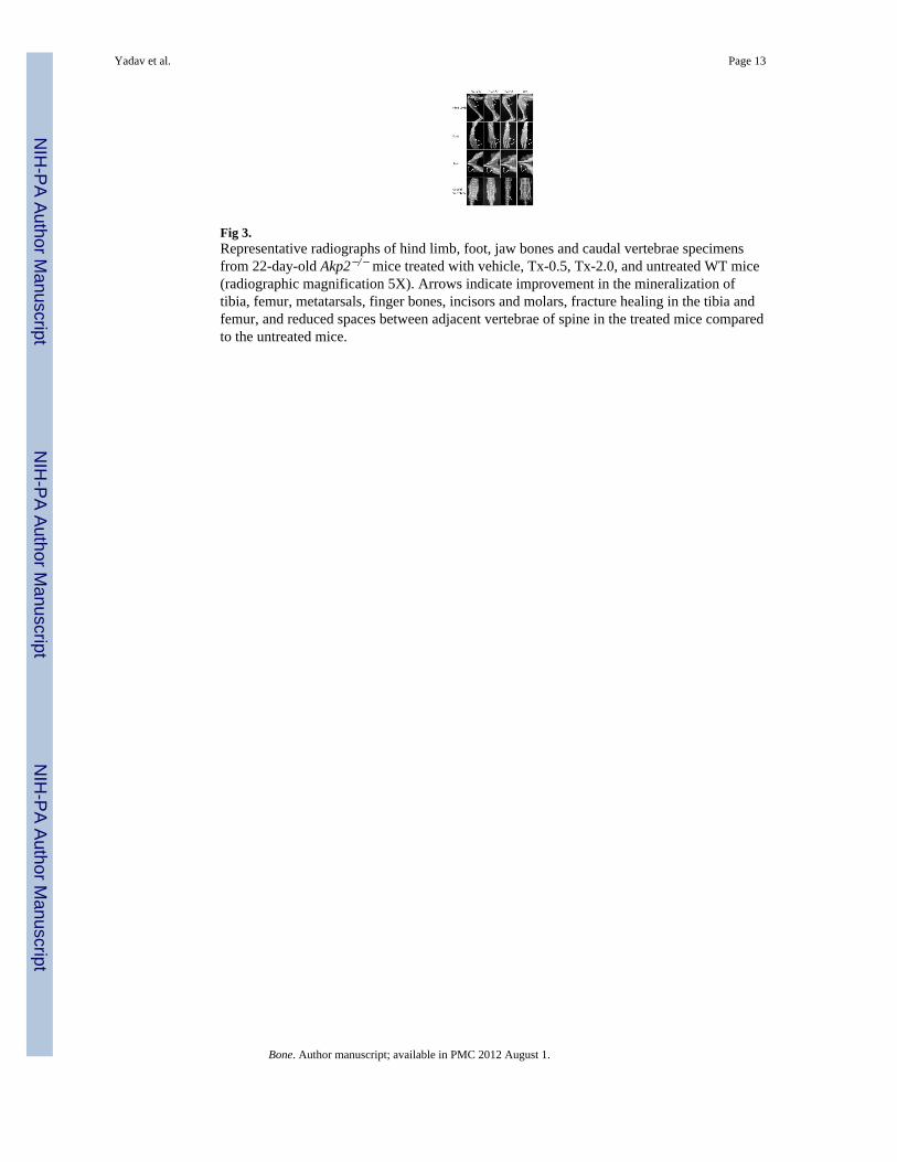
Fig 3.Representative radiographs of hind limb, foot, jaw bones and caudal vertebrae specimensfrom 22-day-old Akp2−/− mice treated with vehicle, Tx-0.5, Tx-2.0, and untreated WT mice(radiographic magnification 5X). Arrows indicate improvement in the mineralization oftibia, femur, metatarsals, finger bones, incisors and molars, fracture healing in the tibia andfemur, and reduced spaces between adjacent vertebrae of spine in the treated mice comparedto the untreated mice.
Yadav et al. Page 13
Bone. Author manuscript; available in PMC 2012 August 1.
NIH
-PA Author Manuscript
NIH
-PA Author Manuscript
NIH
-PA Author Manuscript
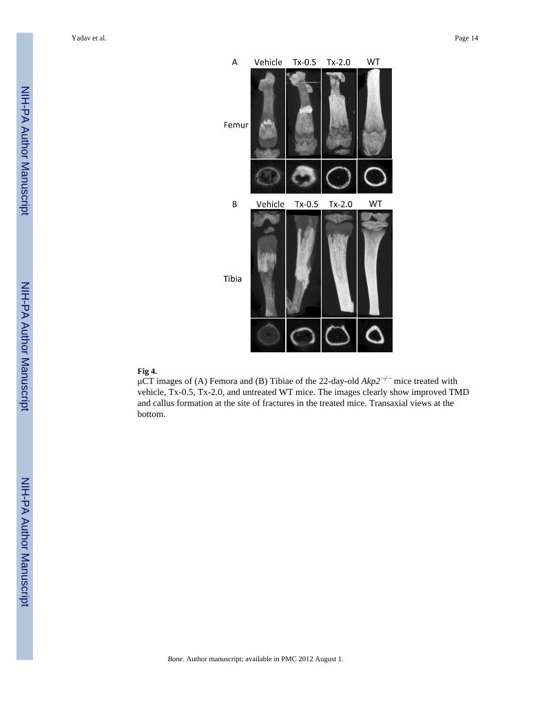
Fig 4.μCT images of (A) Femora and (B) Tibiae of the 22-day-old Akp2−/− mice treated withvehicle, Tx-0.5, Tx-2.0, and untreated WT mice. The images clearly show improved TMDand callus formation at the site of fractures in the treated mice. Transaxial views at thebottom.
Yadav et al. Page 14
Bone. Author manuscript; available in PMC 2012 August 1.
NIH
-PA Author Manuscript
NIH
-PA Author Manuscript
NIH
-PA Author Manuscript
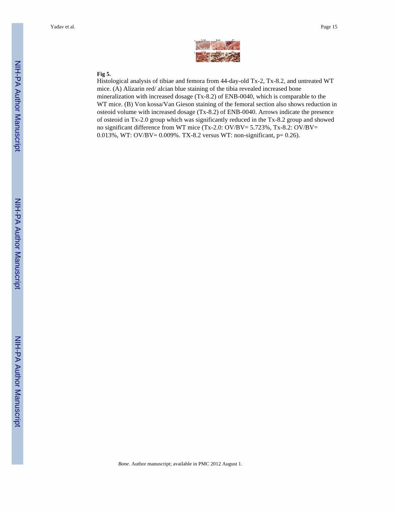
Fig 5.Histological analysis of tibiae and femora from 44-day-old Tx-2, Tx-8.2, and untreated WTmice. (A) Alizarin red/ alcian blue staining of the tibia revealed increased bonemineralization with increased dosage (Tx-8.2) of ENB-0040, which is comparable to theWT mice. (B) Von kossa/Van Gieson staining of the femoral section also shows reduction inosteoid volume with increased dosage (Tx-8.2) of ENB-0040. Arrows indicate the presenceof osteoid in Tx-2.0 group which was significantly reduced in the Tx-8.2 group and showedno significant difference from WT mice (Tx-2.0: OV/BV= 5.723%, Tx-8.2: OV/BV=0.013%, WT: OV/BV= 0.009%. TX-8.2 versus WT: non-significant, p= 0.26).
Yadav et al. Page 15
Bone. Author manuscript; available in PMC 2012 August 1.
NIH
-PA Author Manuscript
NIH
-PA Author Manuscript
NIH
-PA Author Manuscript
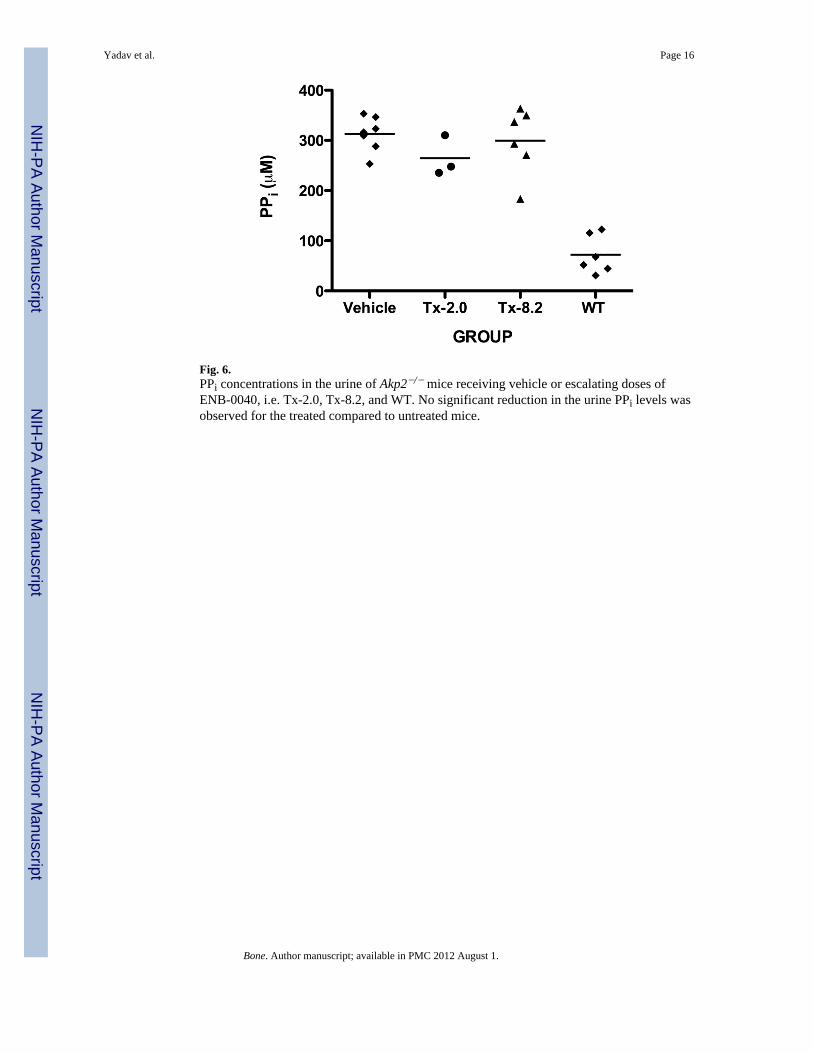
Fig. 6.PPi concentrations in the urine of Akp2−/− mice receiving vehicle or escalating doses ofENB-0040, i.e. Tx-2.0, Tx-8.2, and WT. No significant reduction in the urine PPi levels wasobserved for the treated compared to untreated mice.
Yadav et al. Page 16
Bone. Author manuscript; available in PMC 2012 August 1.
NIH
-PA Author Manuscript
NIH
-PA Author Manuscript
NIH
-PA Author Manuscript
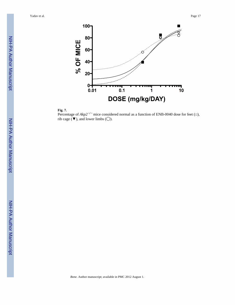
Fig. 7.Percentage of Akp2−/− mice considered normal as a function of ENB-0040 dose for feet (□),rib cage (▼), and lower limbs (◯).
Yadav et al. Page 17
Bone. Author manuscript; available in PMC 2012 August 1.
NIH
-PA Author Manuscript
NIH
-PA Author Manuscript
NIH
-PA Author Manuscript

NIH
-PA Author Manuscript
NIH
-PA Author Manuscript
NIH
-PA Author Manuscript
Yadav et al. Page 18
Table 1
Conceptual Pharmacodynamic (PD) Models
Model Equations
Linear Model E= E0 + S * C
Simple Emax Model E= (Emax * C)/(C+ED50)
Simple Emax Model with E0 E=E0+ (Emax-E0)* (C/(C+ED50))
Sigmoid Emax Model E=(Emax * C γ )/(C γ +ED50 γ)
Sigmoid Emax Model with E0 E=E0 + (Emax -E0)*( C γ /( C γ + ED50 γ))
Weibull E = Emax * (1-exp(-(C/ ED50)S))
Makoid-Banakar E = Emax *(C/Cmax)S*exp(S*(1-C/Cmax)),when C≤ Cmax, E = Emax, when C > Cmax
E= effects, C= Dose, S= Slope, γ= sigmoidicity factor, Cmax = maximal dose, Emax= maximal effects, E0= baseline effects at C=0, ED50= dosethat induced 50% of Emax
Bone. Author manuscript; available in PMC 2012 August 1.
NIH
-PA Author Manuscript
NIH
-PA Author Manuscript
NIH
-PA Author Manuscript
Yadav et al. Page 19
Tabl
e 2
μCT
anal
ysis
of t
he c
ortic
al b
ones
in fe
mor
a an
d of
tibi
ae fr
om 2
2-da
y-ol
d Ak
p2−/−
mic
e tre
ated
with
veh
icle
, Tx-
0.5,
Tx-
2.0,
and
unt
reat
ed W
T m
ice.
The
follo
win
g pa
ram
eter
s wer
e m
easu
red:
mea
n to
tal c
ross
-sec
tiona
l tis
sue
area
(T.A
r), m
ean
tota
l cro
ss-s
ectio
nal b
one
area
(B.A
r), p
erce
nt b
one
area
(B.A
r/T.A
r), c
ross
-sec
tiona
l thi
ckne
ss (C
s. Th
), an
d Ti
ssue
min
eral
den
sity
(TM
D)
T.A
r(m
m^3
)B
.Ar
mm
^3)
B.A
r/T
.Ar
(%)
Cs.T
hm
mT
MD
g/cm
^3SE
M
FEM
UR
Veh
icle
0.42
70.
119
24.1
10.
038
0.57
80.
024
Tx-0
.50.
606
0.25
237
.55
0.09
20.
665
0.09
5
Tx-2
.00.
715
0.24
933
.32
0.06
10.
740
0.04
9
WT
1.78
11.
703
58.1
90.
124
0.95
10.
057
TIB
IA
Veh
icle
0.29
70.
062
18.4
00.
031
0.58
60.
040
Tx-0
.50.
517
0.13
730
.60
0.07
20.
685
0.03
4
Tx-2
.00.
576
0.25
446
.10
0.08
20.
871
0.05
9
WT
0.70
50.
420
60.1
80.
132
0.93
60.
041
Bone. Author manuscript; available in PMC 2012 August 1.
NIH
-PA Author Manuscript
NIH
-PA Author Manuscript
NIH
-PA Author Manuscript
Yadav et al. Page 20
Table 3
Distribution of radiographs of the feet, rib cage, and lower limbs of Akp2−/− mice receiving vehicle or 0.5, 2.0,or 8.2 mg/kg/day ENB-0040, displaying normal bony structures
Number of mice with normal bony structures (%)
Group Left Foot Rib Cage Right Lower Limb
Vehicle (N=20) 2 (10) 3 (15) 5 (25)
Tx-0.5 (N=18) 7 (39) 7 (39) 10 (56)
Tx-2.0 (N=20) 17 (85) 16 (80) 16 (80)
Tx-8.2 (N=19) * 19 (100) 15 (88) 16 (84)
*N=17 for rib cage specimens
Bone. Author manuscript; available in PMC 2012 August 1.